A regulação
11
The Regulation of the p53-mediated Stress Response by MDM2 and MDM4 Mary Ellen Perry Adjunct Scientist, Laboratory of Protein Dynamics and Signaling, National Cancer Institute, National Institutes of Health, Bethesda, Maryland 20892-0189 Correspondence: [email protected] Exquisite contr ol of the activity of p53is necess ary for mammali an survival.T oo much p53is lethal, whereas too little permits tumorigenesis. MDM2 and MDM4 are structurally related proteins critical for the control of p53 activity during development, homeostasis, and the response to str ess. The se two essen tial prot eins regul ate boththe activation of p53in response to stres s and the recov eryof cel ls fol lo wing re sol uti on of the damage, ye t bot h are onc oge nic when overexpressed. Thus, multiple regulatory circuits ensure that their activities are fine- tuned to promote tumor-free survival. Numerous diverse stressors activate p53, and much research has gone into trying to find commonalities between them that would explain the mechanismby whichp53 bec omes active. It is now clear that althou gh the se divers e st ressors activ atep53 by dif fer ent bio chemi cal pa thw ay s, onecommonfeatu re is the eff ortthey dir ect, thr oug h a var iet y of means, to ward dis rup tin g the fun cti ons of both MDM2 andMDM4.This article provides an overview of the relationship between MDM2 and MDM4, features the various biochemical mechanisms by which p53 is activated through inhibition of their functions, and proposes some emerging areas for investigation of the p53-mediated stress response. R egulation of the p53-mediated stress response by the essential inhibitory proteins MDM2 and MDM4 is critical for survi val. In re- sponse to stressors such as ionizing radiation, p53 induces a number of potentially lethal but tumor -suppr essive proc esses, inclu ding cell cycle arrest, senescence, and apoptosis (reviewed by Horn and Vousden 2007). Both MDM2 and MDM4 are cri tic al to survivi ng the p53 -me dia ted stressre spon se to who le bod y ion izi ng irr adi ati on as mic e wit h re duc ed levels of either pr ote in undergo p53-dependent death after exposure to doses of ra dia tion tha t are sublethal to wil d- type mice (Mendrysa et al. 2003; Terzian et al. 2007). MDM2 and MDM4 are also required to control p53 func tion during develop ment, as sho wn by the ear ly emb ryo nic death of mic e lacki ng eit her MDM2 or MDM4, unl ess they also lack p53 (Jones et al. 1995; Montes de Oca Luna et al. 1995; Parant et al. 2001; Migliorini et al. 2002). Al though both MDM2 and MDM4 ar e essential for development, they are detrimental to long-term survival when in excess, because Editors: Arnold J. Levine and David Lane Additional Perspectives on The p53 Family available at www.cshperspectives .org Copyright # 2010 Cold Spring Harbor Laboratory Press; all rights reserved; doi: 10.1101 /cshperspect.a000968 Cite this article as Cold Spring Harb Perspect Biol 2010;2:a000968 1
-
Upload
roberto-venerando -
Category
Documents
-
view
217 -
download
0
Transcript of A regulação

8/6/2019 A regulação
http://slidepdf.com/reader/full/a-regulacao 1/11
The Regulation of the p53-mediated StressResponse by MDM2 and MDM4
Mary Ellen Perry
Adjunct Scientist, Laboratory of Protein Dynamics and Signaling, National Cancer Institute, NationalInstitutes of Health, Bethesda, Maryland 20892-0189
Correspondence: [email protected]
Exquisite control of the activity of p53 is necessary for mammalian survival. Too much p53 islethal, whereas too little permits tumorigenesis. MDM2 and MDM4 are structurally relatedproteins critical for the control of p53 activity during development, homeostasis, and theresponse to stress. These two essential proteins regulate boththe activation of p53 in response
to stress and the recoveryof cells following resolution of the damage, yet both are oncogenicwhen overexpressed. Thus, multiple regulatory circuits ensure that their activities are fine-tuned to promote tumor-free survival. Numerous diverse stressors activate p53, and muchresearch has gone into trying to find commonalities between them that would explain themechanismby whichp53 becomes active. It is now clear that although these diverse stressorsactivatep53 by different biochemical pathways, onecommonfeature is the effortthey direct,through a variety of means, toward disrupting the functions of both MDM2 and MDM4. Thisarticle provides an overview of the relationship between MDM2 and MDM4, featuresthe various biochemical mechanisms by which p53 is activated through inhibition of theirfunctions, and proposes some emerging areas for investigation of the p53-mediated stressresponse.
Regulation of the p53-mediated stress responseby the essential inhibitory proteins MDM2
and MDM4 is critical for survival. In re-sponse to stressors such as ionizing radiation,p53 induces a number of potentially lethal but
tumor-suppressive processes, including cellcycle arrest, senescence, and apoptosis (reviewed
by Horn and Vousden 2007). Both MDM2 andMDM4 are critical to surviving the p53-mediatedstress response to whole body ionizing irradiation
as mice with reduced levels of either proteinundergo p53-dependent death after exposure to
doses of radiation that are sublethal to wild-type mice (Mendrysa et al. 2003; Terzian et al.
2007). MDM2 and MDM4 are also required tocontrol p53 function during development, asshown by the early embryonic death of mice
lacking either MDM2 or MDM4, unless they also lack p53 (Jones et al. 1995; Montes de Oca
Luna et al. 1995; Parant et al. 2001; Miglioriniet al. 2002).
Although both MDM2 and MDM4 are
essential for development, they are detrimentalto long-term survival when in excess, because
Editors: Arnold J. Levine and David LaneAdditional Perspectives on The p53 Family available at www.cshperspectives.org
Copyright# 2010 Cold Spring Harbor Laboratory Press; all rights reserved; doi: 10.1101/cshperspect.a000968Cite this article as Cold Spring Harb Perspect Biol 2010;2:a000968
1

8/6/2019 A regulação
http://slidepdf.com/reader/full/a-regulacao 2/11
both are oncogenic. Both MDM2 and MDM4confer the tumorigenic phenotype on cultured
cells when experimentally overexpressed (Fak-harzadeh et al. 1991; Danovi et al. 2004). In
addition, targeted expression of MDM2 inthe mammary gland results in tumorigenesis(Lundgren et al. 1997). In people, single nucleo-
tide polymorphisms that reduce expression of
either of the orthologs of MDM2 or MDM4(also referred to as Hdm2 and Hdm4) correlatewith increased risk for breast cancer (Bond
et al. 2004; Atwal et al. 2009). Approximately 10% of human tumors have been found to
overexpress either MDM2 or MDM4 and many of these express wild-type p53 (reviewed inToledo and Wahl 2006). Because the majority
of human cancers express mutant forms of
p53, overexpression of MDM2 and MDM4 inthe subset of tumors expressing wild-type p53supports the notion that excessive MDM2 and
MDM4 promote tumorigenesis, at least in part,by blocking p53 function. Thus, limiting the
activities of MDM2 and MDM4 is importantto prevent cancer.
NATURE OF THE RELATIONSHIP BETWEENMDM2 AND MDM4
MDM2 and MDM4 are structurally related
proteins that each bind to p53 and inhibit its
activity (Fig. 1). MDM2 was discoveredthrough its ability to confer the tumorigenic
phenotype on murine cells (Fakharzadeh et al.1991) and later found to bind p53 (Momand
et al. 1992). The esoteric name “MDM2” reflectsits discovery as a murine gene amplified ondouble minute chromosomes in a spontaneously
transformed cell line (Fakharzadeh et al. 1991).
Two genes coamplified with MDM2, MDM1,and MDM3, encode proteins that are neitheroncogenic nor related structurally to MDM2
(Fakharzadeh et al. 1991). A few years afterMDM2 was discovered, MDM4, also called
MDMX, was identified through a screen for pro-teins that interact withp53 andfound to be highly homologous to MDM2 (Shvarts et al. 1996). The
human orthologs of both MDM2 and MDM4are 491- and 490-amino acid phosphopro-
teins, respectively, that bind to p53 and inhibit
its ability to transactivate gene expression. Inaddition to the amino-terminal domain that
bindsto p53, theysharea numberof structuraldo-mains including a central acidic domain and a
carboxy-terminal RING finger through whichthey form MDM2/MDM4 heterodimers (Sharpet al. 1999).
Despite their many similarities, MDM2 and
MDM4 differ functionally, as revealed by exper-iments withgeneticallyengineered mice (reviewedby Marine et al. 2006). Although mouse embryos
undergo p53-mediated death in utero whenboth copies of either MDM2 or MDM4 are
deleted from the germline, they fail to developbecause of distinct mechanisms. Embryos de-ficient in MDM2 undergo massive apoptosis
beforeimplantationanddie,whereasthosewith-
out MDM2 die postimplantation from eithergrowth arrest or apoptosis (Parant et al. 2001;Migliorini et al. 2002; Chavez-Reyes et al.
2003). The embryonic death of MDM2-deficientmice can be rescued by deletion of the pro-
apoptotic BAX protein, whereas MDM4 nullmice can be partially rescued by deletion of thecell cycle arrestprotein p21, indicating that differ-
ent p53-mediated pathways canresult in embryo-nic death (Chavez-Reyes et al. 2003; Steinman
et al. 2004). Although all mice homozygousnull for either protein die during embryogenesis,
70% of mice heterozygous for both MDM2 and
MDM4 are born live (Terzian et al. 2007),demonstrating that the two proteins synergize
to restrain p53. In some cell types, the conse-quences of MDM2 deletion differ from those
of MDM4 deletion, suggesting that these pro-teins also have some nonredundant functions.Tissue-specific deletion of MDM2 in either pro-
genitor neuronal cells or cardiomyocytes results
in embryonic lethality, whereas deletion of MDM4 in the same cell population resultsin milder tissue defects and live births (Grier
et al. 2006; Francoz et al. 2006). On a biochemicallevel, MDM2 deletion results in accumulation
of p53 and a concomitant increase in p53-dependent transactivation, whereas MDM4 dele-tion results in increased p53-dependent tran-
sactivation without p53 stabilization (Francozet al. 2006; Toledo et al. 2006). This difference
in outcomes can be ascribed to the ubiquitin
M.E. Perry
2 Cite this article as Cold Spring Harb Perspect Biol 2010;2:a000968

8/6/2019 A regulação
http://slidepdf.com/reader/full/a-regulacao 3/11
ligase (E3) function of MDM2, which is not sha-red by MDM4 (reviewed in Marine and Lozano2009). Thus, whereas both MDM2 and MDM4
can block the transcriptional activation functionof p53, only MDM2 stimulates p53 degradation
through ubiquitylation.The RING-finger domains of MDM2 and
MDM4 are major determinants of their func-
tions toward p53 and each other. The MDM2RING differs from that of MDM4 by facilita-
ting ubiquitylation of several proteins, includingMDM4 (Pan and Chen2003). MDM2-mediated
ubiquitylation of MDM4 facilitates accumu-lation of MDM4 in the nucleus, where it isdegraded (Pereg et al. 2006). MDM4 can influ-
ence the ubiquitin ligase function of MDM2
through its own RING-finger domain, whichheterodimerizes with that of MDM2 (Gu et al.2002; Kawaiet al. 2007). Under different circum-stances, MDM4 can either stimulate or inhibit
the E3 ligase function of MDM2 toward p53
(Jackson et al. 2001; Kawai et al. 2007; Barbozaet al. 2008). This is important because ubiquity-lation of p53 by MDM2 is critical for embryo-
genesis (Itahana et al. 2007), and is modulatedduring the stress response, as discussed below.
MDM2-mediated ubiquitylation of p53 regu-lates both p53 turnover and cellular localization.Monoubiquitylation of p53 results in export of
p53 to the cytoplasm and polyubiquitylationstimulates proteasome degradation (recently
reviewed by Kruse and Gu 2009). MDM2 canubiquitylate itself, and this activity increases inresponse to stress, as does its ability to ubiquity-
late MDM4.
MDM2 and MDM4 also differ at the geneticlevel. The MDM2 gene contains a p53-responseelement and its expression is induced by p53
during the stress response (Chen et al. 1994),whereas MDM4 gene expression is not regu-
lated by p53 (Shvarts et al. 1996). The spectrumof tumors overexpressing MDM2 differs fromthat overexpressing MDM4 (Toledo and Wahl
2006). For example, MDM4 is overexpressedin some colon cancers, whereas MDM2 is not.
In addition, the MDM4 gene was found to beamplified in retinoblastoma, whereas MDM2
was not (Laurie et al. 2006). These differencesin gene regulation appear to reflect differentroles for MDM2 and MDM4 in both the stress
response and tumorigenesis.
DIVERSE OUTCOMES FROM ACTIVATIONOF P53 BY DIFFERENT STRESSORS
In homeostatic tissues, most if not all functionsof p53 are undetectable and become evidentonly following a stimulus (Mendrysa et al.
2003; Hornand Vousden 2007). Whenactivated,
p53 can arrest the cell cycle, induce apoptosis,or promote senescence, exerting a protectiveeffect on the species, the organism, or the cell
(Fig. 2). Although any one stressor can stimulatemultiple p53-mediated outcomes, a particular
response to a stressor may help ensure survival.For example, exposure to ultraviolet radiationand many other DNA-damaging agents causes
stabilization and activation of p53, resulting inapoptosis, which eliminates cells with severe
DNAdamage. Prolonged, excessive, or mistimed
Mdm2
1 100 200 300 400 491
p53 binding AcidicNLS Zn RING
Mdm4
1 100 200 300 400 490
p53 binding Acidic Zn RING
Figure 1. Domain structure of human homologs of MDM2 and MDM4. The amino-termini of both proteinsbind p53. Only MDM2 has a nuclear localization signal (NLS). The central acidic region of MDM2, but notMDM4, binds ribosomal proteins. The RING finger domains are required for heterodimerization betweenMDM2 and MDM4.
MDM2 and MDM4 in the Stress Response
Cite this article as Cold Spring Harb Perspect Biol 2010;2:a000968 3

8/6/2019 A regulação
http://slidepdf.com/reader/full/a-regulacao 4/11
oncogene function leads to p53 activation andsenescence, limiting the oncogenic potential of
preneoplastic cells. Deficient ribosome functionpromotes p53 activation, promoting cell cycle
arrest until translation is restored. Although itis not entirelyclear howstressors manifest differ-entoutcomes, allof them activate p53 byaltering
the functions of MDM2 and MDM4, and thereare several mechanisms for doing so.
MDM2 and MDM4 are implicated as factorsthat influence whether p53 induces cell cyclearrest, senescence, or apoptosis. Both MDM2
and MDM4 can be detected bound to p53 onitstargetDNA,wheretheyappeartorepresstran-scription of p53-responsive genes (Tang et al.2008). Acetylation of p53 in response to some
stressors abrogates the binding of MDM2 andMDM4 to p53at most butnot allp53-responsive
promoters. By directly repressingonly thosepro-moters on which they are bound to p53, MDM2and MDM4 could influence whether cell cycle
arrest genes such as p21 are induced in favor of proapoptotic genes such as BAX (recently
reviewed by Kruse and Gu 2009). The ubiquitinligase function of MDM2 also appears to con-
tribute to the decision between growth arrestand apoptosis by stimulating the degradationof both upstream and downstream effectors of
p53. When pharmacologically released fromp53, MDM2 stimulates the turnover of hnRNP
K, a transcriptional co-factor that assists p53 in
transactivating the p21 gene (Enge et al. 2009).Under these circumstances, MDM2 also stimu-
lates the degradation of the p21 protein, suchthatthe resulting lowlevels of p21 areinsufficient
to establish growth arrest and fail to protect thecell from apoptosis. The kinase HIPK2, whichphosphorylates p53 on serine 46 in response to
some stressors, is an upstream regulator of p53
that is ubiquitylated by MDM2 (Rinaldo et al.2007). Phosphorylation of p53 on serine 46 pro-motes apoptosis, and MDM2-mediated degra-
dation of HIPK2 reduces the amount of p53phosphorylated at serine 46, inhibiting apopto-
sis. A proteosome-independent function of the ubiquitin ligase function of MDM2 is alsoinvolved in the decision between cell cycle arrest
and apoptosis. In stressed cell types prone to
undergo apoptosis, p53 is transported to themitochondria, where it stimulates apoptosis by blocking the function of the antiapoptotic Bcl2
protein. MDM2 has been shown to facilitatethis translocation through monoubiquitylation
of p53(Marchenko et al. 2007).In sum, multiple,sometimes nonredundant and sometimes over-lapping pathways converge to regulate the p53-
mediated response to stress with consequencesthat can be either dire or life-saving.
THE ROLES OF MDM2 AND MDM4 IN THE
RESPONSE TO IONIZING RADIATIONIonizing radiation is an important stressor that
activates both the growth suppressive and apop-totic functions of p53 (reviewed by Kastan
2008). p53 is a major determinant of organis-mal survival following exposure to ionizingradiation because its activation in response to
this stress results in depletion of hematopoietic
cells (Westphal et al. 1998). Whereas wild-typemice die within 10– 20 days after exposure to adose of 10 Gray, p53 null mice seem unper-
turbed. On a histological level, bone marrow and spleen from irradiated p53-null mice
do not show the radiation damage seen inthese tissues from irradiated wild-type mice(Westphal et al. 1998). The importance of
MDM2 and MDM4 in regulating p53 activity in the response to ionizing radiation is shown
by the observation that mice with reduced
Radiation
Oncogeneactivation
Ribosomedysfunction Cell culture
p53
ApoptosisGrowth arrest
Senescence
Figure 2. Multiple stressors activate p53 with severalpossible outcomes. Apoptosis, senescence, and growtharrest have each been shown to be tumor suppressive.However, if unrestrained, they can be lethal.
M.E. Perry
4 Cite this article as Cold Spring Harb Perspect Biol 2010;2:a000968

8/6/2019 A regulação
http://slidepdf.com/reader/full/a-regulacao 5/11
levels of either protein are radiosensitive, dyingfollowing exposure to doses of radiation that are
sublethal to wild-type mice (Mendrysa et al.2003; Terzian et al. 2007). Moreover, unirradiated
mice engineered to have low (30% of wild type)levels of MDM2 appear as though they havebeenirradiated (Mendrysaet al. 2003), suggesting
that p53 is poised to act in the absence of stress
and that MDM2 prevents it from doing so.The major signal for the response to ioniz-
ing radiation is transmitted by a kinase mutated
in the human condition, ataxia telangiectasia,ATM (for ataxia telangiectasia mutated) (re-
viewed by Kastan 2008). ATM phosphorylatesthe amino terminus of human p53 on serine15 such that its affinity for MDM2 and
MDM4 is reduced (Fig. 3). This modification
alone would be expected to result in p53accumulation. However, ATM guarantees anunambiguous p53 response by simultaneously
stimulating the rapid degradation of bothMDM2 and MDM4 by phosphorylating the
c-termini of both proteins (Stommel andWahl 2005;Chen et al. 2005). ATM also activatesa second kinase, c-Abl, which phosphorylates
MDM2 at tyrosine 394 (Goldberg et al. 2002)and MDM4 at tyrosine 99 (Zuckerman et al.
2009), reducing their ability to inhibit p53.Another ATM-activated kinase, CHK2, phos-
phorylates additional sites within MDM4, facil-
itating its association with 14-3-3 proteinsand accumulation in the nucleus where it is
ubiquitylated by MDM2 (Okamoto et al.2005; Pereg et al. 2006). Additionally, CHK2-
mediated phosphorylation of MDM4 disruptsits interaction with a deubiquitinating enzyme,“herpesvirus associated ubiquitin-specific pro-
tease” (HAUSP), thereby allowing unopposed
ubiquitylation and proteasomal degradation(Meulmeester et al. 2005; Pereg et al. 2006).The importance of phosphorylation of MDM4
in allowing p53 to mount a stress response toionizing radiation was recently shown by the
radioresistance of mice in which the ser-ines phosphorylated by ATM and CHK2 hadbeen mutated to alanine (Wang et al. 2009).
Compared with wild-type mice, the mutantmice mounted a diminished p53-mediated
transcriptional response, had fewer apoptotic
lymphocytes, and withstood higher doses of ionizing radiation. Thus, ATM uses a multi-
pronged approach to maximally activate p53during the response to radiation.
In cells that survive the stress response, theactivities of p53, MDM2, and MDM4 must be
temporally regulated first to allow p53 to exertits function and then to restrain p53 such that
the cell can recover. However, little is knownabout these regulatory events. It has been hypo-thesized that, in the early stages of the response
to ionizing radiation, the ubiquitin ligase func-tion of MDM2 becomes redirected from p53
to MDM4 and to MDM2 itself (Okamotoet al. 2005) with the degradation of MDM2and MDM4 augmenting the activation of p53.
p53 then exerts its growth suppressive or
apoptotic function, and the cell either under-goes growth arrest or dies. The expression of MDM2 is induced in response to ionizing radi-
ation through a p53-response element in intron2 of the MDM2 gene, upstream of the initiation
codon for full-length MDM2 (Juven et al. 1993;Wu et al. 1993; Chen et al. 1994). This negativefeedback loop is thought to be critical for the
recovery of cells following exposure to stressors,although this has not been formally shown.
Another mechanism that may be critical forthe recovery phase is the dephosphorylation of
MDM2 by the Wip1 phosphatase (Lu et al.
2007). Wip1 dephosphorylates MDM2 atserine 395, the site phosphorylated by ATM. In
this way, Wip1 facilitatesthe interactionbetweenMDM2 and p53, and suppresses the autoubi-
quitylation of MDM2. The delayed inductionof Wip1 in the damage response makes it acompelling candidate for a major determinant
of the recovery phase. Insight into the recovery
phase is likely to be accelerated by widespreadadoption of the approach of Lahav et al.(reviewed in Batchelor et al. 2009), who study
single cells to observe oscillations in the levelsand activities of p53 in the stress response
that are hidden in studies of populations. Thisappears to be a promising approach to discoverthe mechanisms underlying the activation and
deactivation of p53 in the stress response.
p53 clearly suppresses tumorigenesis in re-sponse to ionizing radiation (Kemp et al. 1994),
MDM2 and MDM4 in the Stress Response
Cite this article as Cold Spring Harb Perspect Biol 2010;2:a000968 5

8/6/2019 A regulação
http://slidepdf.com/reader/full/a-regulacao 6/11
yet the relationship between the acute, apoptosis-mediated, pathological response to ionizing
radiation and tumor suppression is not entirely clear, as revealed by Evan and colleagues, who
devised an elegant strategy to investigate thestage of the stress response at which p53 exerts
its tumor suppressive effect in irradiated tissues
(Christophorou et al.2006). They tookadvantageof mice genetically engineered to express a p53
protein that could be switched from functionalto inactive at will. They irradiated the mice
when p53 was either functional or inactive andthen allowed recovery with p53 in the inactive
state. Although only the mice that were irradiated
A Homeostasis
Mdm2
Mdm4
B Ionizing irradiation
Mdm2
Mdm4
ATM
CHK2Abl
S15
S395Y394
S20
S367S342Y99 S403
p53
p53
p53
C Ribosome dysfunction Oncogene activation
Mdm2
Mdm4
L11
D
Mdm2
Mdm4
p53
ARF
Figure 3. Pathways for regulation of p53 by MDM2 and MDM4 under different conditions. ( A) In homeostaticconditions (e.g., unstressed, adult tissues), MDM2 and MDM4 inhibit p53. (B) Following exposure to ionizingradiation, ATM phosphorylates several substratesto block theabilities of MDM2 andMDM4 to inhibit p53. (C )Ribosomal dysfunction leads to direct binding of L11 to MDM2, and to a redirection of its ubiquitin ligageactivity away from p53 to MDM4. (D) Oncogenic activation causes increased expression of ARF, which bindsto MDM2 and inhibits it from ubiquitylating p53.
M.E. Perry
6 Cite this article as Cold Spring Harb Perspect Biol 2010;2:a000968

8/6/2019 A regulação
http://slidepdf.com/reader/full/a-regulacao 7/11
when p53 was in the functional state showed apathological response to radiation (e.g., an
increase in the percentage of apoptotic lympho-cytes), they developed tumors at the same rate
as those irradiated with inactive p53. This obser-vation suggests that the immediate p53-mediatedstress response to irradiation is not tumor sup-
pressive, or is less critical than a later p53
action. Even more surprisingly, Christophorouet al. (2006) found that converting p53 to thefunctional state eight days post-irradiation pro-
tected the mice against lymphomagenesis eventhough the acute, pathological response to
irradiation was undetectable in these circum-stances. Thus, at least in this experimentalsystem, p53 exerts a tumor suppressive function
that is independent of the acute response to ioniz-
ing radiation. The mechanism behind this tumorsuppression is not understood, but preliminary results show it is dependent on expression of
the alternative reading frame (ARF) tumor sup-pressor, which inactivates MDM2 to allow p53
to function (Christophorou et al. 2006) (seesection below on oncogene stress). Additional,perhaps even more ingenious experiments, may
be necessary to elucidate the tumor suppressivefunction of p53 following irradiation.
MDM2 AND MDM4 INFLUENCE THE
RESPONSE TO ULTRAVIOLET RADIATIONUltraviolet (UV) light is another type of ra-
diation that induces a p53-mediated stressresponse that appears critical for tumor sup-
pression. The p53 gene is mutated in over 50%of human squamous cell carcinomas, a type of skin cancer caused by overexposure to UV
light (Brash et al. 1991). In an elegant study,
Ziegler et al. (1994) found that a majority of UV-induced, precancerous lesions (actinickeratoses) contained p53 mutations with the
hallmarks of UV-induce mutagenesis (e.g., Cto T and CC to TT transitions resulting from
pyrimidine dimers). In the same study, exper-imental exposure of mice to UV light causedaccumulation of p53 and apoptosis in the
epidermis. In contrast, p53-minus mice lackeda proficient apoptotic response to UV light.
More recently, p53 has been proposed to play a
central role in inducing the tanning response,which appears important in protecting against
the mutagenic effects of UV light (Cui et al.2007). Together, these studies suggest that p53
is an important determinant of both cell survi-val and cancer susceptibility following UVexposure.
UV and ionizing radiation activate p53
through different but similar signaling cas-cades. Although ionizing radiation causesmainly DNA double strand breaks, UV light in
the UVB wavelengths causes covalent adductsand single stranded breaks that block replica-
tion (reviewed in Marrot and Meunier 2007).These different types of damage are detectedby different sensors which activate different
kinases. Whereas ATM is the major transducer
of the response to ionizing radiation, the“ATM and Rad 9-related protein” (ATR) isactivated preferentially by UV light. Like ATM,
ATR phophorylates p53, MDM2, and MDM4.ATR also phosphorylates and activates CHK1,
a kinase similar to CHK2 (reviewed by Strackeret al. 2009), and CHK1 phosphorylates bothMDM2 and MDM4, resulting in degradation
of MDM2 and MDM4 and accumulation of active p53. Thus, like the response to ionizing
radiation, the UV response stimulates multiplekinases to activate p53.
The dose of UV light influences the p53-
mediated stress response as measured by thetiming and magnitude of the increases in
p53 and MDM2 (Latonen et al. 2001). At low doses, phosphorylation may be the dominant
determinant of the response, whereas athigher doses, blocks to transcription and trans-lation of MDM2 and MDM4 contribute to acti-
vate p53. UV light also results in increased
transcription of alternatively spliced MDM2and MDM4 gene products (Chandler et al.2006), the consequences of which are not com-
pletely understood. Moreover, high doses of UV light may also activate novel functions of
MDM2 or MDM4. In response to lethal butnot sublethal doses of UV light, MDM4 trans-locates to the mitochondria, where it blocks
Bcl2 function and promotes apoptosis (Manciniet al. 2009). This counterintuitive finding sug-
gests that MDM4 has pro- or anti-apoptotic
MDM2 and MDM4 in the Stress Response
Cite this article as Cold Spring Harb Perspect Biol 2010;2:a000968 7
8/6/2019 A regulação
http://slidepdf.com/reader/full/a-regulacao 8/11
functions under certain circumstances, throughwhich it could influence the probability of cell
survival. It will be important to clarify the mul-tiple roles of MDM2 and MDM4 that contrib-
ute to the initial stress response and to therecovery of cells following resolution of thedamage.
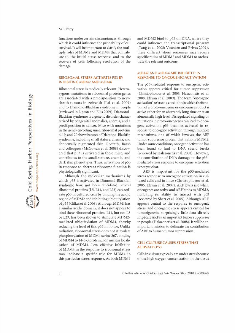
RIBOSOMAL STRESS ACTIVATES P53 BY INHIBITING MDM2 AND MDM4
Ribosomal stress is medically relevant. Hetero-zygous mutations in ribosomal protein genes
are associated with a predisposition to nervesheath tumors in zebrafish (Lai et al. 2009)and to Diamond-Blackfan syndrome in people
(reviewed in Lipton and Ellis 2009). Diamond-
Blackfan syndrome is a genetic disorder charac-terized by congenital anomalies, anemia, and apredisposition to cancer. Mice with mutations
in the genes encoding small ribosomal proteins6, 19, and 20 show features of Diamond-Blackfan
syndrome, including small stature, anemia, andabnormally pigmented skin. Recently, Barshand colleagues (McGowan et al. 2008) discov-
ered that p53 is activated in these mice, andcontributes to the small stature, anemia, and
dark skin phenotypes. Thus, activation of p53in response to aberrant ribosome function is
physiologically significant.
Although the molecular mechanisms by which p53 is activated in Diamond-Blackfan
syndrome have not been elucidated, severalribosomal proteins (L5, L11, and L23) can acti-
vate p53 in cultured cells by binding the acidicregion of MDM2 and inhibiting ubiquitylationof p53 (Gilkes et al. 2006). Although MDM4 has
a similar acidic domain, it does not appear to
bind these ribosomal proteins. L11, but not L5or L23, has been shown to stimulate MDM2-mediated ubiquitylation of MDM4, thereby
reducing the level of this p53 inhibitor. Unlikeradiation, ribosomal stress does not stimulate
phosphorylation of MDM4 serine 367, bindingof MDM4 to 14-3-3 protein, nor nuclear locali-zation of MDM4. Less effective inhibition
of MDM4 in the response to ribosomal stressmay indicate a specific role for MDM4 in
this particular stress response. As both MDM4
and MDM2 bind to p53 on DNA, where they could influence the transcriptional program
(Tang et al. 2008; Vousden and Prives 2009),these different stress responses may require
specific ratios of MDM2 and MDM4 to orches-trate the relevant outcome.
MDM2 AND MDM4 ARE INHIBITED INRESPONSE TO ONCOGENIC ACTIVATION
The p53-mediated response to oncogenic acti-
vation appears critical for tumor suppression(Christophorou et al. 2006; Halazonetis et al.
2008; Efeyan et al. 2009). The term “oncogeneactivation” refersto a conditionin which thefunc-tion of a proto-oncogene or oncogene product is
active either for an aberrantly long time or at an
abnormally high level. Disregulated signaling ormutations in proto-oncogenes can lead to onco-gene activation. p53 becomes activated in re-
sponse to oncogene activation through multiplemechanisms, one of which involves the ARF
tumor suppressor protein that inhibits MDM2.Under some conditions, oncogene activation hasbeen found to lead to DNA strand breaks
(reviewed by Halazonetis et al. 2008). However,the contribution of DNA damage to the p53-
mediated stress response to oncogene activationis not yet clear.
ARF is important for the p53-mediated
stress response to oncogene activation in cul-tured cells and in mice (Christophorou et al.
2006; Efeyan et al. 2009). ARF levels rise whenoncogenes are active and ARF binds to MDM2,
inhibiting its ability to interact with p53(reviewed by Sherr et al. 2005). Although ARFappears central to the response to oncogenic
stress, and oncogenic stress appears critical for
tumorigenesis, surprisingly little data directly implicate ARFas an important tumor suppressorin people (Halazonetis et al. 2008). It will be an
important mission to delineate the contributionof ARF to human tumor suppression.
CELL CULTURE CAUSES STRESS THAT ACTIVATES P53
Cells in culture typically are under stress because
of the high oxygen concentration in the tissue
M.E. Perry
8 Cite this article as Cold Spring Harb Perspect Biol 2010;2:a000968
8/6/2019 A regulação
http://slidepdf.com/reader/full/a-regulacao 9/11
culture incubator (DiMicco et al. 2008). p53becomes stabilized and induces cell cycle arrest
and senescence. This p53-mediated response toculture stress limits the lifespan of primary
cells, such as mouse embryo fibroblasts, whichspontaneously immortalize if they lack eitherARF or p53. Only a portion of the biochemical
pathways induced by stress in cultured cells
have been confirmed to be physiologically rel-evant. For example, although p53 inducesMDM2 expression constitutively in cultured
cells, it does not regulate basal levels of MDM2in homeostatic tissues (Mendrysa et al. 2000).
Thus, it is likely that culture stress contributesto some of the outcomes seen in studies of p53 with experimentally added stressors. For a
more in-depth comparison of results from cell
culture and animal models, see the review by Toledo and Wahl (2006).
CONCLUDING REMARKS
Despite the tens of thousands of papers pub-
lished about p53, the p53-mediated response tostress remains a complicated phenomenon that
is not completely understood. Several majorquestions remain unresolved:
What mechanisms regulate the timing of theactivation and deactivation of p53 in response
to stress?
Whatis the purpose of so manydifferent waysto
activate p53 in response to stress?
How important are the different p53-mediated
responses to stress in suppressing tumor-igenesis?
Can the lethal effects of p53 be separatedpharmacologically from its tumor suppres-
sive function?
What is the role of ARF in tumor suppression in
people?
How physiologically relevant are p53-indepen-dent functions of MDM2 and MDM4 inthe stress response?
Can MDM2 and MDM4 activities be manipu-lated safely to prevent cancer or reduce radi-
ation sickness?
The answers to these and other questions willaid in the design of strategies to target p53,
MDM2, and MDM4 for cancer prevention andtherapy (Wade and Wahl 2009).
ACKNOWLEDGMENTS
The author thanks Drs. Guillermina Lozano
of MD Anderson Medical Center and PeterJohnson and Allan Weissman of the National
Cancer Institute, National Institutes of Health(NIH), for critical and constructive comments.This work was supported by the intramural
research program of the NIH National Cancer
Institute, Center for Cancer Research.
REFERENCES
Atwal GS, Kirchhoff T, Bond EE, Monagna M, Menin C,Bertorelle R, Scaini MC, Bartel F, Bohnke A, Pempe C,et al. 2009. Altered tumor formation and evolutionary selection of genetic variants in the human MDM4 onco-gene. PNAS doi /10.1073/pnas.0901298106.
Barboza JA, IwakumaT,Terzian T, El-NaggarAK, Lozano G.2008. MDM2 and MDM4 loss regulates distinct p53activities. Mol Cancer Res 6: 947–954.
Batchelor E,LoewerA, LahavG. 2009.The upsanddowns of p53: Understanding protein dynamics in single cells. Nature Rev Cancer 9: 371–377.
Bond GL, Hu W, Bond EE, Robins H, Lutzker SG, Arva NC,Bargonetti J, Bartel F, Taubert H, Wuerl P, et al. 2004. Asinglenucleotide polymorphismin the MDM2promoterattenuates the p53 tumor suppressor pathway and accel-
erates tumor formation in humans. Cell 119: 591–602.Brash DE, Rudolph JA, Simon JA, Lin A, McKenna GJ,
Baden HP, HalperinAJ, Ponten J.1991. A rolefor sunlightin skin cancer: UV-induced p53 mutations in squamouscell carcinoma. Proc Natl Acad Sci 88: 10124–10128.
Chandler DS, Singh RK, Caldwell LC, Bitler JL, Lozano G.2006. Genotoxic stress induces coordinately regulatedalternative splicing of the p53 modulators MDM2 andMDM4. Cancer Res 66: 9502–9508.
Chavez-ReyesA, Parant JM, Amelse LL,Montes de OcaLunaR, KorsmeyerSJ, Lozano G. 2003. Switchingmechanismsof cell death in MDM2- and MDM4-null mice by deletion of p53 downstream targets. Cancer Res 63:
8664–8669.
Chen L, Gilkes DM,Pan Y, Lane WS,ChenJ. 2005.ATM andCHK2-dependent phosphorylation of MDMX contrib-
ute to p53 activation after DNA damage. EMBO J 24:3411–3422.
Chen CY, Oliner JD, Zhan Q, Fornace AJJr, Vogelstein B,Kastan MB. 1994. Interactions between p53 and MDM2in a mammalian cell cycle checkpoint pathway. Proc Natl Acad Sci 91: 2684–2688.
Christophorou MA, Ringshausen I, Ginch AJ, Brown-Swigart L, Evan GI. 2006. The pathological response to
MDM2 and MDM4 in the Stress Response
Cite this article as Cold Spring Harb Perspect Biol 2010;2:a000968 9

8/6/2019 A regulação
http://slidepdf.com/reader/full/a-regulacao 10/11
DNA damage does not contribute to p53-mediatedtumour suppression. Nature 443: 214–217.
Cui R, Widlund HR, Feige E, Lin JY, Wilensky DL, Igras VE,D’Orazio J, Fung CY, Schanbacher CF, Granter SR, et al.2007. Central role for p53 in the suntan response andpathological hyperpigmentation. Cell 128: 853–864.
Danovi D, Meulmeester E, Pasini D, Migliorini D, Capra M,Frenk R, de Graaf P, Francoz S, Gasparini P, Gobbi A,et al. 2004. Amplification of MDMX (or MDM4)directly contributes to tumor formation by inhibi-ting p53 tumor suppressor activity. Mol Cell Biol 24:5835–5843.
DiMicco R, Cicalese A, FumagalliM, Dobreva M, VerrecchiaA, Pelicci PG, diFagagna F. 2008. DNA damage responseactivation in mouseembryofibroblasts undergoing repli-cative senescence and following spontaneous immortali-zation. Cell Cycle 7: 3601–3606.
Efeyan A, Murga M, Martinez-Pastor B, Ortega-Molina A,Soria R, Collado M, Fernandez-Capetillo O, Serrano M.2009. Limited role of murine ATM in oncogene-inducedsenescence and p53-dependent tumor suppression. PLoS
ONE 4:
doi:10.1371/ journal.pone.0005475.Enge M, Bao W, Hedstrom E, Jackson SP, Moumen A,
Selivanova G. 2009. MDM2-dependent downregulationofp21 andhnRNP K provides a switchbetweenapoptosisand growth arrest induced by pharmacologically acti-vated p53. Cancer Cell 15: 171–183.
Fakharzadeh SS, Trusko SP, George DL. 1991. Tumorigenicpotential associated with enhanced expression of a genethat is amplified in a mouse tumor cell line. EMBO J 10: 1565–1569.
Francoz S, Froment P, Bogaerts S, De Clercq S, Maetens M,Doumount G, Bellefroid E, Marine J-C. 2006. MDM4and MDM2 cooperate to inhibit p53 activity in prolifer-ation and quiescent cells in vivo. Proc Natl Acad Sci 103:3232–3237
Gilkes DM, Chen L, Chen J. 2006. MDMX regulation
of p53 response to ribosomal stress. EMBO J 25:5614–5625.
Goldberg Z, Vogt Sionov R, Berger M, Zwang Y, Perets R,Van Etten RA, Oren M, Taya Y, Haupt Y. 2002. Tyrosinephosphorylation of MDM2 by c-Abl: Implications forp53 regulation. EMBO J 21: 3715–3725.
Grier J, Xiong S, Elizondo-Fraire AC, Parant J, Lozano G.2006. Tissue-specific differences of p53 inhibition by MDM2 and MDM4. Mol Cell Biol 26: 192–198.
Gu J, Kawai H, Nie L, Kitao H, Wiederschain D, JochemsenAG, Parant J, Lozano G, Yua Z-M. 2002. MutualDependence of MDM2 and MDMX in their functionalinactivation. J Biol Chem 277: 19251–19254.
Halazonetis DT, Gorgoulis VG, Bartek J. 2008. AnOncogene-induced DNA damage model for cancerdevelopment. Science 319: 1352–1355.
Horn HF, Vousden KH. 2007. Coping with stress: Multipleways to activate p53. Oncogene 26: 1306–1316.
Itahana K, Mao H, Jin A, Itahana Y, Clegg HV, Lindstro mMS, Bhat KP, Godfrey VL, Evan GI, Zhang Y. 2007.Targeted inactivation of MDM2 RING finger E3 ubiqui-tin ligase activity in the mouse reveals mechanisticinsights into p53 regulation. Cancer Cell 12: 355–366.
Jackson MW, Lindstrom MS, Berberich SJ. 2001. MDMX binding to ARF affects MDM2 protein stability and p53transactivation. J Biol Chem 276: 25336–25341.
Jones SN, Roe AE, Donehower LA, Bradley A. 1995. Rescueof embryonic lethality in MDM2-deficent mice by absence of p53 Nature 378: 206–208.
Juven T, Barak Y, Zauberman A, George DL, Oren M. 1993.Wild type p53 can mediate sequence-specific transactiva-tion of an internal promoter within the MDM2 gene.Oncogene 8: 3411–3416.
Kastan MB. 2008. DNA Damage Responses: Mechanismsand roles in human disease. Mol Cancer Res 6: 517–524.
Kawai H, Lopex-Pajares V, Kim MM, Wiederschain D, YuanZ-M. 2007. RING domain-mediated interaction is arequirement for MDM2’s E3 ligase activity. Cancer Res67: 6026–6030.
Kemp CJ, Wheldon T, Balmain A. 1994. p53-deficient miceare extremely susceptible to radiation-induced tumori-genesis. Nature Genet 8: 66–69.
Kruse J-P, Gu W. 2009. Modes of p53 regulation. Cell 137:609–622.
Lai K, Amsterdam A, FarringtonS, Bronson RT, Hopkins N,Lees JA. 2009. Many ribosomal protein mutations areassociated with growth impairment and tumorpredispo-sition in zebrafish. Dev Dyn 238: 76–85.
Latonen L, Taya Y, Laiho M. 2001. UV radiation inducesdose-dependent regulation of p53 response and modu-lates p53-HDM2 interaction in human fibroblasts.Oncogene 20: 6784–6793.
Laurie NA, Donovan SL, Shih C-S, Zhang J, Mills N, FullerC, Teunisse A, Lam S, Ramos Y, Mohan A, et al. 2006.Inactivation of the p53 pathway in retinoblastoma. Nature 444: 61–66.
Lipton JM, Ellis SR. 2009. Diamond-Blackfan anemia:Diagnosis, treatment and molecular pathogenesis.Hematol Oncol Clin North Am 23: 261–282.
Lu X, Ma O, Nguyen T-A, Jones SN, Oren M, Donehower L.
2007. The Wip1 phosphatase acts as a gatekeeper inthe p53-MDM2 autroregulatory loop. Cancer Cell 12:342–354.
Lundgren K, Montes de Oca Luna R, McNeill YB, Emerick EP, Spencer B, Barfield CR, Lozano G, Rosenberg MP,Finlay CA. 1997. Targeted expression of MDM2 un-couples S phase from mitosis and inhibits mammary gland development independent of p53. Genes Dev 11:714–725.
Mancini F, Di Conza G, Pellegrino M, Rinaldo C, ProdosmoA, Giglio S, D’Agnano I, Florenzano F, Felicioni L,Buttitta F, et al. 2009. MDM4 (MDMX) localizes atthe mitochondria and facilitates the p53-mediatedintrinsic-apoptotic pathway. EMBO J 28: 1926–1939.
Marchenko ND, Wolff S, Erster S, Becker K, Moll UM. 2007.Monoubiquitylation promotes mitochondrial p53 trans-
location. EMBO J 26: 923–934.Marine J-C, Lozano G. 2009. MDM2-mediated ubiquityla-
tion: p53and beyond. Cell Death and Differ doi:10.1038/cdd.2009.68.
Marine J-C, Francoz S, Maetens M, Wahl G, Toledo F,Lozano G. 2006. Keeping p53 in check: Essential andsynergistic functions of MDM2 and MDM4. Cell Deathand Differ 13: 927–934.
M.E. Perry
10 Cite this article as Cold Spring Harb Perspect Biol 2010;2:a000968

8/6/2019 A regulação
http://slidepdf.com/reader/full/a-regulacao 11/11
Marrot L, Meunier JR. 2008. Skin DNA photodamage andits biological consequences. J Am Acad Dermatol . 58:
139–148.
McGowan KA, Li JZ, Park CY, Beaudry V, Tabor HK, SabnisAJ, Zhang W, FuchsH, Angelis M, MyersRM, Attardi LD,Barsh GS. 2008. Ribosomal mutations cause p53-mediated dark skin and pleiotropic effects. NatureGenet 40: 963–970.
Mendrysa SM, Perry ME. 2000. The p53 tumor suppressorprotein does not regulate expression of its own inhibitor,MDM2, except under conditions of stress. Mol Cell Biol 20: 2023–2030.
Mendrysa SM, McElwee MK, Michalowski J, O’Leary KA,Young KM, Perry ME. 2003. MDM2 is critical for inhi-bition of p53 during lymphopoiesis and the response toionizing radiation. Mol Cell Biol 23: 462–473.
Meulmeester E, Maurice MM, Boutell C, Teunisse AF, OvaaH, Abraham TE, Dirks RW, Jochemsen AG. 2005. Loss of HAUSP-mediated deubiquitination contributes to DNAdamage-induced destabilization of Hdmx and Hdm2. Mol Cell 18: 565–576.
Migliorini D, Denchi EL, Danovi D, Jochemsen A, Capillo
M, Gobbi A, Helin K, Pelicci PG, Marine J-C. 2002.MDM4 (MDMX) regulates p53-induced growth arrestand neuronal cell death during early embryonic mousedevelopment. Mol Cell Biol 22: 5527–5538.
Momand J, Zambetti GP, Olson DC, George D, Levine AJ.1992. The MDM2 oncogene product forms a complex with the p53 protein and inhibits p53-mediated transac-tivation. Cell 69: 1237–1245.
Montes deOca Luna R, WagnerDS, LozanoG. 1995.Rescueof early embryonic lethality in MDM2-deficent mice by deletion of p53. Nature 378: 203–206.
Okamoto K, Kashima K, Pereg Y, Ishida M, Yamazaki S,Nota A, Teunisse A, Migliorini D, Kitabayashi I, MarineJ-C, et al. 2005. DNA damage-induced phosphorylationof MDMX at serine 367 activates p53 by targetingMDMX for MDM2-dependent degradation. Mol Cell
Biol 25: 9608–9620.Pan Y, Chen J. 2003. MDM2 promotes ubiquitination and
degradation of MDMX. Mol Cell Biol 23: 5113–5121.
Parant J, Chavez-Reyes A, Little NA, Yan W, Reinke V,Jochemsen AG, Lozano G. 2001. Rescue of embryoniclethality in MDM4-null mice by loss of Trp53 suggestsa nonoverlapping pathway with MDM2 to regulate p53. Nature 29: 92–95.
Pereg Y, Lam S, Teunisse A, Biton S, Meulmeester E,Mittelman L, Buscemi G, Okamoto K, Taya Y, Shiloh Y,et al. 2006. Differential roles of ATM- and CHK2-mediated phosphorylations of Hdmx in response toDNA damage. Mol Cell Biology 26: 6819–6831.
Rinaldo C, Prodosmo A, Mancini F, Iacovelli S, Sacchi A,Moretti F, Soddu S. 2007. MDM2-regulated degradationof HIPK2 prevents p53Ser46 phosphorylation and DNA
damage-induced apoptosis. Mol Cell 25: 739–750.Sharp DA, Kratowicz SA, Sank MJ, George DL. 1999.
Stabilization of the MDM2 oncoprotein by interactionwith the structurally related MDMX protein. J Biol Chem 274: 38189–38196.
SherrCJ, Bertwistle D,den BestenW, Kuo ML,Sugimoto M,Tago K, Williams RT, Zindy F, Roussel MF. 2005.p53-Dependent and -independent functions of the Arf tumor suppressor. Cold Spring Harbor Symp Quant Biol 70: 129–137.
Shvarts A, Steegenga WT, Riteco N, van Laar T, Dekker P,Bazuine M, van Ham RCA, van der Houven van OordtW, Hateboer G, van der Eb AJ, Jochemsen AG. 1996.MDMX: a novel p53-binding protein with some func-tional properties of MDM2. EMBO J 15: 5349–5357.
Steinman HA,SlussHK, Sands AT, Pihan G, JonesSN. 2004.Absence of p21 partially rescues MDM4 loss anduncovers an antiproliferative effect of MDM4 on cellgrowth. Oncogene 23: 303–306.
Stommel JM, Wahl GM. 2005. A new twist in the feedback loop: Stress-activated MDM2 destabilization is requiredfor p53 activation. Cell Cycle 4: 411–417.
Stracker TH, Usu T, Petrini JH. 2009. Taking the time tomake important decisions: The checkpoint effectorkinases CHK1and CHK2and theDNA damage response.DNA Repair doi: 10.1016.dnarep.2009.04.012.
Tang Y, Zhao W, Chen Y, Zhao Y, Gu W. 2008. Acetylation isindispensible for p53 activation. Cell 133: 612–626.
Terzian T, Wang Y, Van Pelt SC, Box NF, Travis EL, LozanoG. 2007. Haploinsufficiency of MDM2 and MDM4in tumorigenesis and development. Mol Cell Biol 27:5479–5485.
Toledo F, Wahl GM. 2006. Regulating the p53 pathway: Invitro hypotheses, in vivo veritas. Nature Rev Cancer 6:909–923.
Toledo F, Krummel KA, Lee CJ, Liu C-W, Luo-Wei R, TangM, Wahl GM. 2006. A mouse p53 mutant lacking theproline-rich domain rescues MDM4 deficiency and pro-vides insight into the MDM2-MDM4-p53 regulatory network. Cancer Cell 9: 273–285.
Vousden KH, Prives C. 2009. Blinded by the light: Thegrowing complexity of p53. Cell 137: 413–431.
Wade M, Wahl GM. 2009. Targeting MDM2 and MDMX incancer therapy: Better living through medicinal chem-istry. Mol Cancer Res 7: 1–11.
Wang YV, Leblanc M, Wade M, Jochemsen AG, Wahl G.2009. Increased radioresistance and accelerated B celllymphomas in mice with MDMX mutations thatprevent modifications by DNA-damage-activatedkinases. Cancer Cell 16: 33–43.
Westphal CH, Hoyes KP, Canman CE, Huang X, Kastan MB,Hendry JH, Leder P. 1998. Loss of atm radiosensitizesmultiple p53 null tissues. Cancer Res 58: 5637–5639.
Wu X, Bayle JH, Olson D, Levine AJ. 1993. The p53-MDM2autroegulatory feedback loop. Genes Dev 7: 1126–1132.
Ziegler A, Jonason AS, Leffell DJ, Simon JA, Sharma HW,Kimmelman J, Remington L, Jacks T, Brash DE. 1994.Sunburn and p53 in the onset of skin cancer. Nature
372: 773–776.Zuckerman V, Leons K, Popwicz GM, Silberman I,
Grossman T, Marine J-C, Holak TA, Jochemsen AG,HauptY. 2009. c-Abl phosphorylates Hdmxand regulatesits interaction with p53. J Biol Chem 284: 4031–4039.
MDM2 and MDM4 in the Stress Response
Cite this article as Cold Spring Harb Perspect Biol 2010;2:a000968 11