A2BR Adenosine Receptor Modulates Sweet Taste in ...
13
A2BR Adenosine Receptor Modulates Sweet Taste in Circumvallate Taste Buds Shinji Kataoka 1,2.¤a , Arian Baquero 1,3.¤b , Dan Yang 4¤c , Nicole Shultz 1,2 , Aurelie Vandenbeuch 1,3 , Katya Ravid 4 , Sue C. Kinnamon 1,3 , Thomas E. Finger 1,2 * 1 Rocky Mountain Taste & Smell Center, University of Colorado School of Medicine, Aurora, Colorado, United States of America, 2 Department of Cell & Developmental Biology, University of Colorado School of Medicine, Aurora, Colorado, United States of America, 3 Depatment of Otolaryngology, University of Colorado School of Medicine, Aurora, Colorado, United States of America, 4 Departments of Medicine and Biochemistry and Whitaker Cardiovascular Institute, Boston University School of Medicine, Boston, Massachusetts, United States of America Abstract In response to taste stimulation, taste buds release ATP, which activates ionotropic ATP receptors (P2X2/P2X3) on taste nerves as well as metabotropic (P2Y) purinergic receptors on taste bud cells. The action of the extracellular ATP is terminated by ectonucleotidases, ultimately generating adenosine, which itself can activate one or more G-protein coupled adenosine receptors: A1, A2A, A2B, and A3. Here we investigated the expression of adenosine receptors in mouse taste buds at both the nucleotide and protein expression levels. Of the adenosine receptors, only A2B receptor (A2BR) is expressed specifically in taste epithelia. Further, A2BR is expressed abundantly only in a subset of taste bud cells of posterior (circumvallate, foliate), but not anterior (fungiform, palate) taste fields in mice. Analysis of double-labeled tissue indicates that A2BR occurs on Type II taste bud cells that also express Ga14, which is present only in sweet-sensitive taste cells of the foliate and circumvallate papillae. Glossopharyngeal nerve recordings from A2BR knockout mice show significantly reduced responses to both sucrose and synthetic sweeteners, but normal responses to tastants representing other qualities. Thus, our study identified a novel regulator of sweet taste, the A2BR, which functions to potentiate sweet responses in posterior lingual taste fields. Citation: Kataoka S, Baquero A, Yang D, Shultz N, Vandenbeuch A, et al. (2012) A2BR Adenosine Receptor Modulates Sweet Taste in Circumvallate Taste Buds. PLoS ONE 7(1): e30032. doi:10.1371/journal.pone.0030032 Editor: Fabien Tell, The Research Center of Neurobiology-Neurophysiology of Marseille, France Received October 10, 2011; Accepted December 12, 2011; Published January 10, 2012 Copyright: ß 2012 Kataoka et al. This is an open-access article distributed under the terms of the Creative Commons Attribution License, which permits unrestricted use, distribution, and reproduction in any medium, provided the original author and source are credited. Funding: This work was supported by National Institutes of Health grants: from NIDCD: RO1 DC007495, RO1 DC006021, P30 DC04657; from NHLBI: RO1 HL93149. The funders had no role in study design, data collection and analysis, decision to publish, or preparation of the manuscript. Competing Interests: The authors have declared that no competing interests exist. * E-mail: [email protected] . These authors contributed equally to this work. ¤a Current address: Division of Anatomy, Department of Biosciences, Science of Health Improvement, Kyushu Dental College, Kokurakita-ku, Kitakyushu, Japan ¤b Current address: Division of Neuroscience, Oregon National Primate Research Center, Oregon Health & Science University, Beaverton, Oregon, United States of America ¤c Current address: Center for Molecular Medicine, National Heart, Lung, and Blood Institute, National Institutes of Health, Bethesda, Maryland, United States of America Introduction In the peripheral gustatory system, ATP plays a crucial role in the transmission of information from taste buds to the gustatory nerve fibers [1], [2], [3], [4]. ATP is released from taste receptor cells and activates ionotropic ATP receptors (P2X2/P2X3) on taste nerves [5], [6] as well as metabotropic (P2Y) receptors on taste cells [7], [8], [9]. The importance of purinergic transmission is evidenced by the loss of essentially all gustatory neural responses in P2X2/P2X3 double knockout (KO) mice [1]. Mature taste cells can be classified into three distinct types based on morphologic, molecular, and functional features [10–20]. Type I ‘‘glial-like’’ cells, originally termed ‘‘dark cells’’ based on ultrastructural criteria, are similar in some ways to astrocytes; they envelop the other cell types without obvious functional contacts and express the ectoATPase, ectonucleoside triphosphate diphosphohydrolase 2 (NTPDase2) and the glial glutamate/ aspartate transporter (GLAST) [19,21] which serve as molecular markers for this cell type [22]. Type II ‘‘receptor’’ cells, originally termed ‘‘light cells’’, express the G protein-coupled receptors for umami (T1R1/T1R3), sweet (T1R2/T1R3) or bitter (T2Rs) [23,24] transduction. These taste receptors are expressed in largely non-overlapping subsets of Type II (also called ‘‘receptor’’) taste cells, but all couple to the same downstream signaling effectors including phospholipase C-b2 (PLCb2), inositol 1, 4, 5-trispho- sphate receptor type 3 (IP3R3) and transient receptor potential channel M5 (TrpM5) [22]. Thus these signaling proteins serve as well-characterized markers of Type II taste cells in all taste fields. The close correspondence between Type II cell ultrastructure and expression of these signaling components has been established for both PLCb2 [25] and gustducin [26,27]. The Ga subunits vary according to receptor type and location on the tongue. While bitter and umami receptors generally couple to GaGustducin (Gagust) in both anterior and posterior tongue, taste cells that express sweet receptors co-express Gagust in anterior tongue and Ga14 in posterior tongue [28,29]. Type III (also termed ‘‘presynaptic’’, [22]) cells were originally classified as ‘‘intermedi- ate cells’’ because they have ultrastructural features intermediate PLoS ONE | www.plosone.org 1 January 2012 | Volume 7 | Issue 1 | e30032
Transcript of A2BR Adenosine Receptor Modulates Sweet Taste in ...
A2BR Adenosine Receptor Modulates Sweet Taste inCircumvallate Taste BudsShinji Kataoka1,2.¤a, Arian Baquero1,3.¤b, Dan Yang4¤c, Nicole Shultz1,2, Aurelie Vandenbeuch1,3, Katya
Ravid4, Sue C. Kinnamon1,3, Thomas E. Finger1,2*
1 Rocky Mountain Taste & Smell Center, University of Colorado School of Medicine, Aurora, Colorado, United States of America, 2 Department of Cell & Developmental
Biology, University of Colorado School of Medicine, Aurora, Colorado, United States of America, 3 Depatment of Otolaryngology, University of Colorado School of
Medicine, Aurora, Colorado, United States of America, 4 Departments of Medicine and Biochemistry and Whitaker Cardiovascular Institute, Boston University School of
Medicine, Boston, Massachusetts, United States of America
Abstract
In response to taste stimulation, taste buds release ATP, which activates ionotropic ATP receptors (P2X2/P2X3) on tastenerves as well as metabotropic (P2Y) purinergic receptors on taste bud cells. The action of the extracellular ATP isterminated by ectonucleotidases, ultimately generating adenosine, which itself can activate one or more G-protein coupledadenosine receptors: A1, A2A, A2B, and A3. Here we investigated the expression of adenosine receptors in mouse tastebuds at both the nucleotide and protein expression levels. Of the adenosine receptors, only A2B receptor (A2BR) isexpressed specifically in taste epithelia. Further, A2BR is expressed abundantly only in a subset of taste bud cells of posterior(circumvallate, foliate), but not anterior (fungiform, palate) taste fields in mice. Analysis of double-labeled tissue indicatesthat A2BR occurs on Type II taste bud cells that also express Ga14, which is present only in sweet-sensitive taste cells of thefoliate and circumvallate papillae. Glossopharyngeal nerve recordings from A2BR knockout mice show significantly reducedresponses to both sucrose and synthetic sweeteners, but normal responses to tastants representing other qualities. Thus,our study identified a novel regulator of sweet taste, the A2BR, which functions to potentiate sweet responses in posteriorlingual taste fields.
Citation: Kataoka S, Baquero A, Yang D, Shultz N, Vandenbeuch A, et al. (2012) A2BR Adenosine Receptor Modulates Sweet Taste in Circumvallate TasteBuds. PLoS ONE 7(1): e30032. doi:10.1371/journal.pone.0030032
Editor: Fabien Tell, The Research Center of Neurobiology-Neurophysiology of Marseille, France
Received October 10, 2011; Accepted December 12, 2011; Published January 10, 2012
Copyright: � 2012 Kataoka et al. This is an open-access article distributed under the terms of the Creative Commons Attribution License, which permitsunrestricted use, distribution, and reproduction in any medium, provided the original author and source are credited.
Funding: This work was supported by National Institutes of Health grants: from NIDCD: RO1 DC007495, RO1 DC006021, P30 DC04657; from NHLBI: RO1 HL93149.The funders had no role in study design, data collection and analysis, decision to publish, or preparation of the manuscript.
Competing Interests: The authors have declared that no competing interests exist.
* E-mail: [email protected]
. These authors contributed equally to this work.
¤a Current address: Division of Anatomy, Department of Biosciences, Science of Health Improvement, Kyushu Dental College, Kokurakita-ku, Kitakyushu, Japan¤b Current address: Division of Neuroscience, Oregon National Primate Research Center, Oregon Health & Science University, Beaverton, Oregon, United States ofAmerica¤c Current address: Center for Molecular Medicine, National Heart, Lung, and Blood Institute, National Institutes of Health, Bethesda, Maryland, United States ofAmerica
Introduction
In the peripheral gustatory system, ATP plays a crucial role in
the transmission of information from taste buds to the gustatory
nerve fibers [1], [2], [3], [4]. ATP is released from taste receptor
cells and activates ionotropic ATP receptors (P2X2/P2X3) on
taste nerves [5], [6] as well as metabotropic (P2Y) receptors on
taste cells [7], [8], [9]. The importance of purinergic transmission
is evidenced by the loss of essentially all gustatory neural responses
in P2X2/P2X3 double knockout (KO) mice [1].
Mature taste cells can be classified into three distinct types based
on morphologic, molecular, and functional features [10–20]. Type
I ‘‘glial-like’’ cells, originally termed ‘‘dark cells’’ based on
ultrastructural criteria, are similar in some ways to astrocytes;
they envelop the other cell types without obvious functional
contacts and express the ectoATPase, ectonucleoside triphosphate
diphosphohydrolase 2 (NTPDase2) and the glial glutamate/
aspartate transporter (GLAST) [19,21] which serve as molecular
markers for this cell type [22]. Type II ‘‘receptor’’ cells, originally
termed ‘‘light cells’’, express the G protein-coupled receptors for
umami (T1R1/T1R3), sweet (T1R2/T1R3) or bitter (T2Rs)
[23,24] transduction. These taste receptors are expressed in largely
non-overlapping subsets of Type II (also called ‘‘receptor’’) taste
cells, but all couple to the same downstream signaling effectors
including phospholipase C-b2 (PLCb2), inositol 1, 4, 5-trispho-
sphate receptor type 3 (IP3R3) and transient receptor potential
channel M5 (TrpM5) [22]. Thus these signaling proteins serve as
well-characterized markers of Type II taste cells in all taste fields.
The close correspondence between Type II cell ultrastructure and
expression of these signaling components has been established for
both PLCb2 [25] and gustducin [26,27]. The Ga subunits vary
according to receptor type and location on the tongue. While
bitter and umami receptors generally couple to GaGustducin
(Gagust) in both anterior and posterior tongue, taste cells that
express sweet receptors co-express Gagust in anterior tongue and
Ga14 in posterior tongue [28,29]. Type III (also termed
‘‘presynaptic’’, [22]) cells were originally classified as ‘‘intermedi-
ate cells’’ because they have ultrastructural features intermediate
PLoS ONE | www.plosone.org 1 January 2012 | Volume 7 | Issue 1 | e30032
between Type I and Type II cells. Type III cells are responsible for
sour taste transduction [30], and express several definitive markers
including NCAM [31], PKD2L1 [20], and carbonic anhydrase
isoenzyme 4 (Car4) [32]. Type III cells accumulate and release
several transmitters, including 5-HT, GABA, and noradrenalin
[9,3334]. Whereas Type III cells form classical synapses onto the
intragemmal nerve fibers, Type II cells do not [13,35]. Type II
cells do closely associate with nerve fibers, but lack conventional
synapses [13,25,26], and instead release ATP via gated hemi-
channels [2,3] to activate purinergic P2X2 and P2X3 receptors on
afferent nerve fibers [1].
The action of extracellular ATP is terminated by the character-
istic ectonucleotidase within taste buds, NTPDase2 expressed by
Type I taste cells [19]. NTPDase2 degrades ATP to form ADP
which can then act on local purinergic P2Y receptors [9], or be
broken down further by NTPDase2 and by ecto-59 nucleotidases
and other phosphatases [19], [36–41]. The end product of the
phosphatase activity will yield adenosine, which itself can activate
one or more G-protein coupled adenosine receptors: A1R, A2AR,
A2BR, and A3R. Here we investigated the expression of adenosine
receptors in mouse taste buds. Of the adenosine receptors, A2BR is
expressed in taste buds of the posterior tongue, and there, only in
the subset of taste cells that contain the sweet taste receptors.
Functional recordings from glossopharyngeal nerves of A2BR KO
mice show significantly reduced sweet taste responses compared to
wildtype (WT) littermates. These data suggest that A2BR functions
to potentiate transmission of sweet taste information in posterior
lingual taste fields.
Materials and Methods
AnimalsMost experiments relied on C57BL/6J mice except where
otherwise indicated. A2BR-KO mice (on a C57BL/6 background)
were generated in Boston University [42,43]. In this line, Exon 1
of the A2BR gene was replaced by b-galactosidase (b-gal), thus
animals homozygous for the transgene had genetic deletion of
A2BR protein but carried the b-gal marker in cells expressing from
the A2BR locus. Hemizygous animals, herein referred to as
A2BR-lacZ retained expression of the native A2BR protein but
also expressed the marker in the relevant cell populations. Some of
the tongue tissue obtained from these animals, as described below
was shipped to Denver from Boston Univ. for immunohistochem-
ical analysis. Other animals were bred and utilized entirely from
stocks maintained at the University of Colorado. The University of
Colorado Health Science Center Institutional Animal Care and
Use Committee approved the use of these animals in all studies
(Protocol B-07610(02)1E). In all experiments we used animals of
both sexes in the age range of 2–9 months for control, A2BR-lacZ
and A2BR KO mice.
RT-PCRAdult C57BL/6J mice (Jackson Labs) of both sexes 2–4.5
months old were euthanized by CO2 asphyxiation followed by
cervical dislocation. The tongue was removed and a protease
mixture consisting of 2.5 mg/ml dispase, 1 mg/ml collagenase,
type A, (both from Roche Products, Indianapolis, IN) and 1 mg/
ml trypsin inhibitor (Sigma, St. Louis, MO) in Tyrode’s buffer was
injected under the epithelium. Tyrode’s buffer consisted of
139 mM NaCl, 5 mM KCl, 2 mM CaCl2, 1 mM MgCl2,
10 mM Hepes, 10 mM glucose, 10 mM Na pyruvate, and
5 mM NaHCO3; pH 7.2, 318–323 mOsm. Tongues were
incubated in Ca/Mg-free Tyrode’s solution for 40 min. For Ca/
Mg-free Tyrode’s buffer, CaCl2 and MgCl2 were replaced with
2 mM EGTA and 2 mM BAPTA.
Circumvallate (CV) epithelium was peeled from underlying
tissue and placed directly into RNAlater (Applied Biosystems/
Ambion, Austin, TX) for storage. To avoid possible contamination
of taste cells, epithelium was also peeled from the ventral surface of
the tongue for a non-taste tissue control. Mouse brain total RNA
was purchased from Clontech (Mountain View, CA). Total RNA
was purified using the Qiagen Micro RNeasy Kit (Qiagen,
Valencia, CA) according to manufacturers instructions, and
incubated with DNase I to remove any contaminating genomic
DNA. First strand cDNA syntheses were performed by reverse
transcription of 1 mg of total RNA using SuperScript II Reverse
Transcriptase (Invitrogen, Carlsbad, CA). PCRs were carried out
for mouse A1R, A2AR, A2BR and A3R as well as for the
housekeeping gene glyceraldehyde -3-phosphate dehydrogenase
(GAPDH), Ga14 and GaGust. The presence of Gagust in CV
samples but not in samples of non-taste epithelium confirmed the
specificity of the tissue dissection. Primer sequences for each PCR
are listed in Table 1.
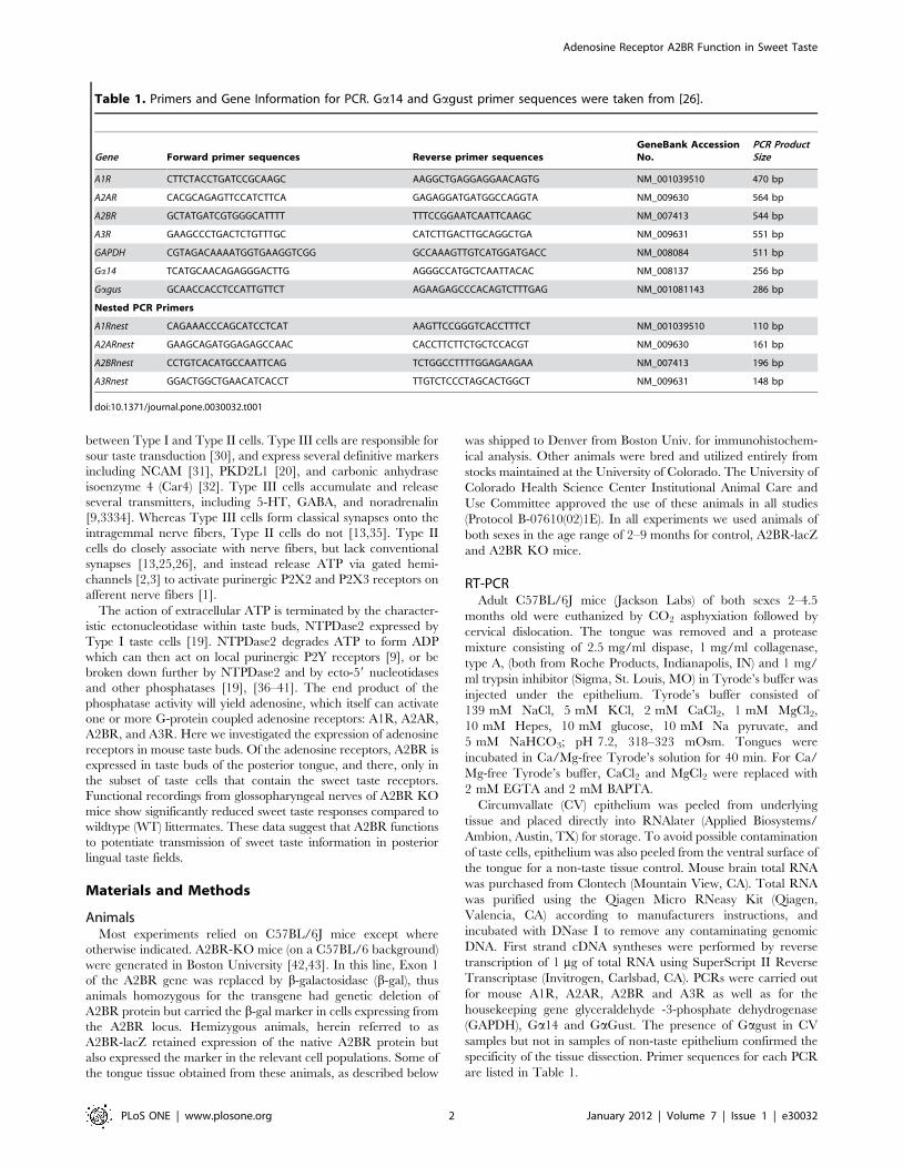
Table 1. Primers and Gene Information for PCR. Ga14 and Gagust primer sequences were taken from [26].
Gene Forward primer sequences Reverse primer sequencesGeneBank AccessionNo.
PCR ProductSize
A1R CTTCTACCTGATCCGCAAGC AAGGCTGAGGAGGAACAGTG NM_001039510 470 bp
A2AR CACGCAGAGTTCCATCTTCA GAGAGGATGATGGCCAGGTA NM_009630 564 bp
A2BR GCTATGATCGTGGGCATTTT TTTCCGGAATCAATTCAAGC NM_007413 544 bp
A3R GAAGCCCTGACTCTGTTTGC CATCTTGACTTGCAGGCTGA NM_009631 551 bp
GAPDH CGTAGACAAAATGGTGAAGGTCGG GCCAAAGTTGTCATGGATGACC NM_008084 511 bp
Ga14 TCATGCAACAGAGGGACTTG AGGGCCATGCTCAATTACAC NM_008137 256 bp
Gagus GCAACCACCTCCATTGTTCT AGAAGAGCCCACAGTCTTTGAG NM_001081143 286 bp
Nested PCR Primers
A1Rnest CAGAAACCCAGCATCCTCAT AAGTTCCGGGTCACCTTTCT NM_001039510 110 bp
A2ARnest GAAGCAGATGGAGAGCCAAC CACCTTCTTCTGCTCCACGT NM_009630 161 bp
A2BRnest CCTGTCACATGCCAATTCAG TCTGGCCTTTTGGAGAAGAA NM_007413 196 bp
A3Rnest GGACTGGCTGAACATCACCT TTGTCTCCCTAGCACTGGCT NM_009631 148 bp
doi:10.1371/journal.pone.0030032.t001
Adenosine Receptor A2BR Function in Sweet Taste
PLoS ONE | www.plosone.org 2 January 2012 | Volume 7 | Issue 1 | e30032
PCR amplifications for A1R, A2AR, A2BR, A3R, and GAPDH
were performed under the following conditions: 94uC for 30 sec,
56uC for 30 sec, 72uC for 30 sec for a total of 35 cycles and
elongation step at 72uC for 10 min. Ga14 and Gagust had
different annealing temperatures, 60uC and 58uC respectively. A
total of 100 ng/ml of each cDNA sample was used in the PCR
amplification. The reverse transcriptase step was omitted in
controls to confirm removal of all genomic DNA. Amplification
products were analyzed on 2% agarose gels and visualized with
GelRed (Biotium, Hayward, CA). All PCR products were gel-
purified (S.N.A.P. Gel Purification Kit, Invitrogen), cloned into
pGEM-T easy cloning vector (Promega) and transformed into
Mach1-T1 chemically competent cells (Invitrogen). Plasmid DNAs
were purified using concert plasmid preparation kits (Invitrogen)
and sequenced with Thermo Sequenase II dye terminator cycle
sequencing kits (Amersham-Pharmacia). The sequence reactions
were analyzed by an ABI373S DNA sequencer (Perkin Elmer).
Nested PCR was also performed to ensure PCR results were
correct products. Primer sequences for nested PCR are given in
Table 1. These amplifications were performed under the following
conditions: 94uC for 30 sec, 58uC for 30 sec, 72uC for 30 sec for a
total of 30 cycles and elongation step at 72uC for 10 min. Controls
in which Reverse Transcriptase were omitted showed no PCR
product indicating lack of contamination by genomic material.
In situ hybridizationAdult C57BL/6J mice were deeply anesthetized with chloral
hydrate or sodium pentobarbital (injected i.p.) and perfused
transcardially with 4% paraformaldehyde (PFA) in 0.1 M
phosphate buffered saline (PBS). After postfixation (1–3 h) and
cryoprotection in 20% sucrose in 0.1 M PBS overnight, tissues
were sectioned longitudinally or transversely (12 mm) onto Super-
frost Plus slides (Fisher Scientific, Hampton, NH). The frozen
sections of CV and foliate (FO) papillae obtained from five
C57BL/6J mice were hybridized with digoxigenin (DIG)-labelled
antisense riboprobes corresponding to a partial cDNA of A2BR
which were obtained by RT-PCR experiments using primers to
the 59 UTR region of each mRNA of interest. Sections were
treated in 4% PFA in PBS for 10 min and washed in PBS. For
partial proteolysis, sections were permeabilized with 1 mg/ml
proteinase K in PBS at 37uC for 10 min, followed by a 5 min wash
in PBS. Sections were refixed in 4% PFA for 10 min. For
neutralization of free formaldehyde residues, sections were washed
in 2 mg/ml glycine in PBS for 5 min. Endogenous alkaline
phosphatase activity was quenched with 0.2 M HCl for 30 min,
followed by two 5 min washes in PBS. Hybridizations were carried
out at high stringency (50% formamide, 10 mM Tris-HCl,
pH 7.6, 200 mg/mL tRNA, 16 Denhardt’s solution, 600 mM
NaCl, 0.25% SDS, and 1 mM EDTA, pH 8.0) at 58uC overnight.
After hybridization the sections were washed twice in 56SSC at
50uC for 30 min. Sections were then subjected to two high-
stringency washes: one in 26SSC at 50uC for 1 hr, followed by a
20 min wash in 0.26SSC at the same temperature. To reduce
background signals the sections were treated with RNase A
solution (0.2 mg/mL; Sigma, St. Louis, MO) at 37uC for 30 min
and then washed in 26SSC at 50uC for 60 min and in 0.26SSC
for 60 min. For detection, signals were developed using alkaline
phosphatase conjugated Fab fragments to DIG and standard
chromogenic substrates (Roche Applied Science). To detect DIG-
labeled hybridized RNA, an alkaline phosphatase (AP)-conjugated
anti-DIG Fab fragment antibody (1:500; Roche) was used. After
washing in TNT buffer (100 mM Tris-HCl, pH 7.5, 150 mM
NaCl, and 0.1% Tween 20) the sections were incubated with 4-
nitro blue tetrazoliumchloride and 5-bromo-4-chloro-3-indolyl-
phosphate (both from Roche) as chromogenic substrates.
X-Gal HistochemistryA2BR-lacZ mice were deeply anesthetized with sodium
pentobarbital (injected i.p.) and perfused transcardially with 4%
PFA in 0.1 M phosphate buffer. Extracted tissue was post fixed no
more than 30 min, followed by two 15 minute washes in 0.1 M
phosphate buffer (pH 7.4) containing 2 mM MgCl2, 5 mM
EGTA, 0.01% sodium deoxycholate and 0.02% Nonidet-40
(buffer B). Tissue was then placed in an X-Gal solution (buffer B
with 5 mM potassium ferricyanide, 5 mM potassium ferrocyanide,
and 1 mg/ml X-gal). Optimal staining in the tongue was obtained
after 24 hours at 37uC, at which time the tissue was washed in
buffer B for 30 min and placed in PBS. For long-term storage, the
tissue was moved to 4% PFA and placed at 4uC.
ImmunohistochemistrySingle immunohistochemistry. C57BL/6J mice were deeply
anesthetized and perfused with 4% PFA in 0.1 M phosphate buffer.
Following cryoprotection, frozen sections of CV papillae were
obtained and prepared for immunohistochemistry. The A2BR
antibody (1:100 dilution, rabbit anti-A2BR; AB1589P, Chemi-
con International) used was generated against the synthetic
peptide 150ATNNCTEPWDGTTNES165. This commercial anti-
body is directed against the amino acid sequence corresponding to
the 2nd extracellular domain (16 amino acids) of the human A2BR
receptor gene and has only moderate sequence similarity (62.5%
identity, 81.25% similarity) to the mouse A2BR sequence
(ATSNCTELGDGIANKS). We established the specificity of this
antiserum by comparing reactivity of WT to A2BR KO lines.
Double label immunohistochemistry. Because the anti-
body label was weak, albeit specific, we also utilized A2BR-driven
expression of b-gal as an index of A2BR expression in A2BR-lacZ
mice. For double localization studies, we detected the b-gal by
immunocytochemistry using a guinea pig anti-b-gal antibody
(1:500) previously generated against full length b-gal by our
laboratory ([44]).
Detection of other substances relied on primary antisera
generated either in rabbit (rb) or goat (gt): rb anti-PLCb2
(1:1000, SantaCruz SC-206; directed against amino acid (aa)
1170–1181 of human origin), rb anti-Gagust (1:1000, Santa Cruz
SC-395; directed against aa 93–112 of Gagust of rat origin), rb
anti-NCAM (1:500, Chemicon Ab 5032; directed against purified
chicken NCAM), rb anti-5HT (1:5000; Immunostar # 20080), gt
anti-Car4 (R&D Systems, # AF2414 directed against recombinant
mouse Car4 ectodomain [residues 18 277]),or rb Gaq/11 (1:1000;
Santa Cruz sc-392; directed against peptide VFAAVKD-
TILQLNLKEYNLV near the C-terminus in mouse, but which
cross-reacts with Ga14, [28].
All double-label assays were carried out coincidentally, since the
primary antisera are derived from guinea pig (anti-b-gal) and
another species, either rabbit or goat. Sections were placed
overnight in primary antiserum cocktail diluted in blocking solution.
Following three washes in buffer, the sections then were placed in a
cocktail of secondary antisera (all diluted 1:400) containing an anti-
gp to detect the anti-B-gal (Alexa488-conjugated goat anti-guinea
pig IgG [Molecular Probes] or donkey anti-guinea pig Dylight 549
[Jackson ImmunoResearch]) along with another secondary antise-
rum, either anti-rb or anti-gt according to the primary antisera
employed: (Alexa546-conjugated goat anti-rabbit IgG [Molecular
Probes], or donkey anti rabbit Alexa Fluor 488 [Invitrogen]). For
the Car4 primary antiserum (made in goat) we utilized donkey
anti-guinea pig Dylight 549 (Jackson ImmunoResearch) or donkey
Adenosine Receptor A2BR Function in Sweet Taste
PLoS ONE | www.plosone.org 3 January 2012 | Volume 7 | Issue 1 | e30032
anti-goat Alexa Fluor 488 (Invitrogen) as secondary antisera. Some
sections were counterstained with a fluorescent Nissl stain
(NeuroTrace 640/660; Invitrogen).
The specificity of the secondary antisera was confirmed by
omitting one or both primary antisera from the primary antiserum
cocktail and finding no cross reactivity with the two applied
secondary antisera. The specificity of b-gal immunoreactivity (IR)
was determined by substitution of buffer for the primary antibody;
this antiserum also yields no staining of WT mice, i.e. those lacking
b-galactosidase. Previous papers from our laboratory showed the
staining by the anti-Gaq/11 antibody is due to neither Gaq nor
Ga11 leaving only Ga14 as the possible source of the
immunoreactivity in taste buds [28]. We have subsequently
confirmed that this antibody shows almost no staining of CV
taste tissues from Ga14 KO animals (data not shown). In the text
below, we refer to this antiserum as Gaq/11/14.
Photomicrographs were acquired using a monochrome Q-
imaging camera on an Olympus BX41TF microscope and Q
Capture software (QImaging). Brightness and contrast were
adjusted in PhotoshopH. Laser scanning confocal images were
captured on a Olympus Fluoview confocal laser scanning
microscope (LSCM) FV300 (Olympus Corporation) using sequen-
tial scanning of the different channels to avoid inappropriate side-
band excitation of fluorophores. For some confocal images, the
median filter was applied to eliminate high frequency pixel noise
associated with confocal acquisition. Images were assembled in
Photoshop and brightness and contrast levels were adjusted to
obtain an image presentation closely mirroring the visual
appearance of the tissue.
Cell Counting. In order to estimate the degree of coincident
expression in double label immunostained sections, we counted
the number of taste cells stained by each marker in sections
through the circumvallate papillae of two A2BR-lacZ mice. The
appearance of label in these mice was not obviously different than
in the many other mice – both WT and A2BR-lacZ – examined
during the course of this study. A taste cell was considered labeled
if it exhibited label above background levels and for which a clear
nuclear profile was evident, i.e. cell profiles not containing the
nucleus were not counted. For each marker, either 7 or 8 sections
were examined and all taste buds (n.100 for all markers) in those
samples were counted. For those markers for which no co-
localization was evident (e.g. Car4, 5HT, NCAM) counts were not
made but a minimum of 100 taste buds were examined.
Chorda tympani and glossopharyngeal nerve recordingsRecordings from intact taste nerves were performed as
described previously by Finger et al. [1]. Briefly, WT and
A2BR-KO mice (8–10 wks old, 20–23 g) were anesthetized and
maintained under surgical level of anesthesia with sodium
pentobarbital (50 mg/kg). Toe-pinch was used to determine the
depth of surgical anesthesia. The trachea was cannulated and the
animal was allowed to breathe room air via the tracheotomy
during the procedure. The animal’s head was held by a clamp;
both the chorda tympani branch of the facial nerve (CN VII) and
glossopharyngeal nerve (IX) were exposed using a ventral
approach. The chorda tympani nerve (n = 6 KO, 6 WT) was
dissected free and cut near the tympanic bulla. The glossopha-
ryngeal nerve (n = 7 KO, 7 WT) was exposed by removing the
underlying tissue and cut near its entrance to the posterior
lacerated foramen. Then, the dissected free nerve was placed over
a platinum-iridium wire hook for recording. A ground electrode
was placed in a nearby muscle. The nerve signal was acquired at
200 Hz and integrated with a PAVC-1 integrator (Duck
Engineering Design) with a time constant of 1 s. Integrated nerve
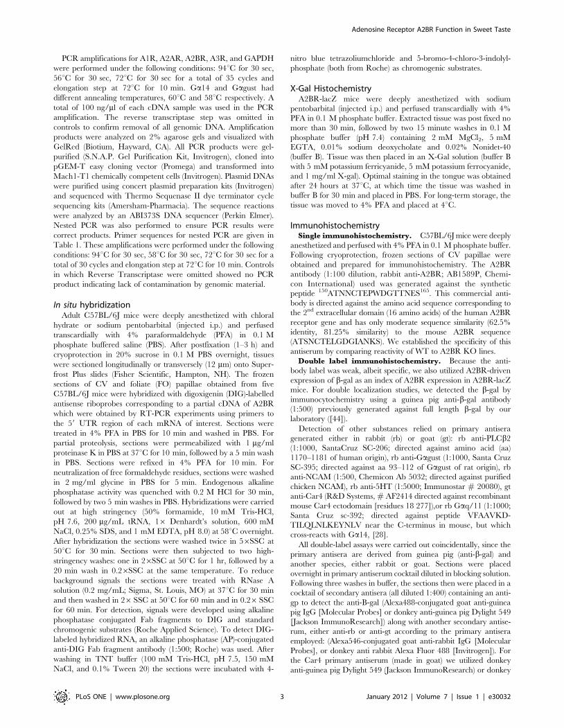
Figure 1. RT-PCR reveals expression of adenosine receptors inbrain, taste epithelium but limited expression in non-tastelingual epithelium. Top: In the brain, all 4 adenosine receptors (A1R,A2AR, A2BR and A3R) are readily detected along with Ga14. Gagust isnot detectable. Center: In posterior taste fields (CV/FO) A2BR transcriptis detected; a faint band is found for A1R in some preparations. BothGagust and Ga14 are also evident. Bottom: In non-gustatoryepithelium (NTT = Non-Taste Tissue) A2BR but not Gagust or Ga14 isdetected. A faint band for A1R is also present in some preparations as inCV/FO epithelium. PCR products were visualized using UV illuminationfollowing ethidium bromide staining. Expression of mRNA for GAPDHwas used as a positive control. The reverse transcriptase step wasomitted as a negative control [(-)CONT] to confirm removal of allgenomic DNA.doi:10.1371/journal.pone.0030032.g001
Adenosine Receptor A2BR Function in Sweet Taste
PLoS ONE | www.plosone.org 4 January 2012 | Volume 7 | Issue 1 | e30032
recording data were collected using a MP100 hardware interface
to Acknowledge 3.8 software (BIOPAC, Santa Barbara CA).
Fungiform (FU), CV and FO papillae were exposed by a small
suture sewn in the tip of the tongue. Taste stimuli were dissolved in
distilled water and delivered to the tongue under constant flow by
a Valvelink 16 perfusion system (Automate Scientific, Inc. San
Francisco, CA). Most mice were tested for tastant-induced nerve
responses to 100 mM, 200 mM, 300 mM, 500 mM, and
1000 mM sucrose, 500 mM SC45647, 30 mM sucralose,
100 mM NaCl and 30 mM quinine. Taste solutions were applied
to the tongue for 1 min followed by a rinse with distilled water. All
solutions including distilled water were maintained at tempera-
tures between 33 to 37uC. Data analysis was performed by
obtaining the area under the curve for 60 s of the integrated nerve
response to each taste stimulus. The data were normalized to
100 mM NH4Cl responses to reduce variability across recordings.
For purposes of illustration, integrated responses in glossopharyn-
geal recordings were smoothed, using a mean value of 30 points in
the Acknowledge software. The normalized nerve responses were
compared between WT and A2BR-KO mice using one-way
ANOVA with Bonferroni’s Multiple Comparison Test (p,0.05;
GraphPad Prism 5, La Jolla CA).
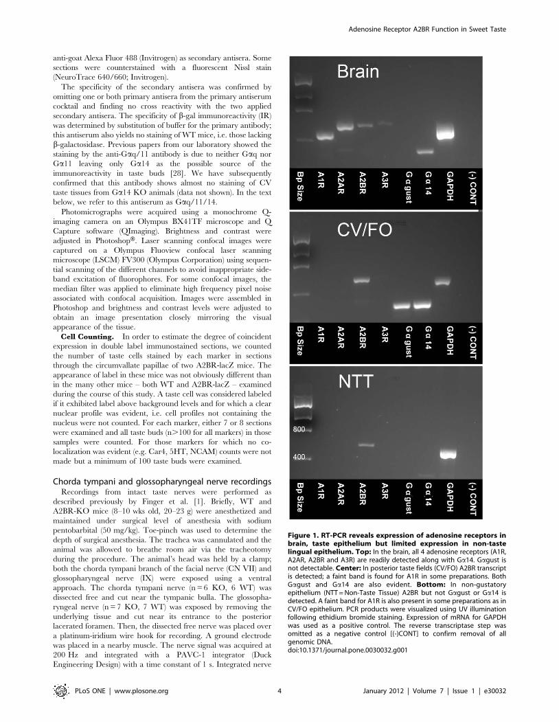
Figure 2. In situ hybridization and immunocytochemistry show restricted expression of A2BR in posterior taste epithelia. The FO (A),CV (B and C) papillae show expression of A2BR mRNA in taste buds (arrowheads). Panel C is high magnification of boxed area in B. No signal isdetectable in CV taste buds (arrowheads) when the section was hybridized with the sense probe (D). Our data show that immunoreactivity for A2BRwas seen only weakly in CV taste buds of WT mouse (E). The CV taste buds of A2BR KO/LacZ mouse did not react with A2BR antibody (F), except fornon-specific trapping of the antibodies at the surface of the epithelium and in taste pores. Scale bars = 50 mm in A (also applies to B & D); 20 mm in C& E (also applies to F).doi:10.1371/journal.pone.0030032.g002
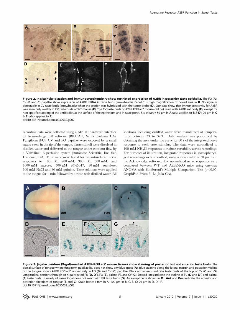
Figure 3. b-galactosidase (X-gal)-reacted A2BR-KO/LacZ mouse tissues show staining of posterior but not anterior taste buds. Thedorsal surface of tongue where fungiform papillae lie, does not show any blue spots (A). Blue staining along the lateral margin and posterior midlineof the tongue shows A2BR KO/LacZ respectively in FO (B) and CV (C) papillae. Black arrowheads indicate taste buds of the top of CV (C and G).Longitudinal sections through an X-gal-treated FU (D, D9), FO (E), palate (F), and CV (G). Dotted lines indicate the outline of FU (D and D9) and palatal(F) taste buds. In nearly all cases X-gal does not react with FU taste buds (D). An exception is shown in D9. Ant and Pos indicate the anterior andposterior directions of tongue (B and C). Scale bars = 1 mm in A; 100 mm in B, C, E, G; 20 mm in D, D9, F.doi:10.1371/journal.pone.0030032.g003
Adenosine Receptor A2BR Function in Sweet Taste
PLoS ONE | www.plosone.org 5 January 2012 | Volume 7 | Issue 1 | e30032
Results
Expression of adenosine receptors in taste epitheliumTo test for the presence of mRNAs for adenosine receptors, we
used RT-PCR in mouse CV papillae along with whole brain and
non-gustatory tongue epithelium. The amplification products were
of the expected size and sequence in all tissues where they were
detected. RT-PCR analyses showed that mRNAs for all adenosine
receptors were readily apparent in brain mRNA. Samples from
CV papillae displayed a robust signal for A2BR and occasionally a
faint band for A1R, but no detectable signal for either A2AR or
A3R (Fig. 1). Nested PCR confirms the identity of all specific
bands reported. A similar pattern was seen for the non-taste
epithelium. No specific products were detected when the reverse
transcriptase was omitted from the reverse transcription mixture,
indicating the absence of contamination by genomic DNA.
Since RT-PCR showed the presence of A2BR mRNA in both
taste and non-taste epithelia, we used in situ hybridization to
investigate the expression patterns of the A2BR genes in sections of
mouse FO and CV papillae. As shown in Fig. 2 (A, B and C),
A2BR expressing cells were clearly detectable in FO, CV taste bud
cells, whereas they were not observed in the other taste epithelia.
Further, no A2BR mRNA signal was observed in non-taste
epithelium.
We then sought to determine expression at protein level. The
rabbit anti-A2BR antibody (AB1589P; Chemicon International)
gave only weak, but repeatable staining for A2BR in CV taste buds
of the C57BL/6J mouse (Panel E in Figure 2). As a negative
control, we tested the same antibody in A2BR KO mouse. Mice
homozygous for this transgene have no intact coding sequence for
A2BR protein [42] and A2BR immunostaining was not seen in
taste buds (Panel F in Figure 2) suggesting specificity of the
antiserum. Residual signal in taste pores and trench wall
epithelium in the A2BR KO tissue is the same as in WT,
suggesting some non-specific background binding of the A2BR
antibody to these tissue elements.
In order to confirm the pattern of label produced by the A2BR
antiserum, we examined lingual tissue from A2BR-KO/(homozy-
gous) and A2BR-lacZ (hemizygous) mice with X-gal staining or
anti-b-gal antibody. Both the A2BR-KO and A2BR-lacZ mice
show the endogenous A2BR gene promoter-dependent expression
as b-gal protein in various tissues. As revealed by X-gal staining,
strong positive reaction is visible in CV and FO papillae taste buds
(Fig. 3B, C, E and G). Blue staining was rare in FU and palate
taste buds (Fig. 3 A, D, D9, F); almost all taste buds in these fields
were devoid of b-gal reaction product. In order to assess in detail
b-gal (A2BR) expressing taste cells in the posterior taste fields, we
undertook immunohistochemical analysis using antibodies against
b-gal. The immunopositive cells constitute less than half of the
population in any given taste bud. In non-taste epithelium
immunostaining for b-gal (A2BR) is negative except for occasional
small blood vessels. The presence of A2BR in these vessels likely
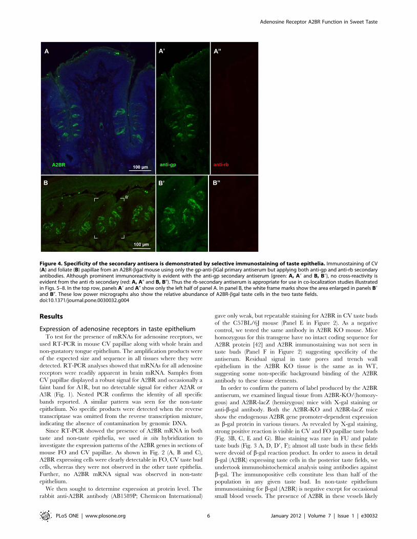
Figure 4. Specificity of the secondary antisera is demonstrated by selective immunostaining of taste epithelia. Immunostaining of CV(A) and foliate (B) papillae from an A2BR-bgal mouse using only the gp-anti-bGal primary antiserum but applying both anti-gp and anti-rb secondaryantibodies. Although prominent immunoreactivity is evident with the anti-gp secondary antiserum (green: A, A9 and B, B9), no cross-reactivity isevident from the anti rb secondary (red: A, A0 and B, B0). Thus the rb-secondary antiserum is appropriate for use in co-localization studies illustratedin Figs. 5–8. In the top row, panels A9 and A0 show only the left half of panel A. In panel B, the white frame marks show the area enlarged in panels B9and B0. These low power micrographs also show the relative abundance of A2BR-bgal taste cells in the two taste fields.doi:10.1371/journal.pone.0030032.g004
Adenosine Receptor A2BR Function in Sweet Taste
PLoS ONE | www.plosone.org 6 January 2012 | Volume 7 | Issue 1 | e30032
accounts for the positive signal obtained with PCR of non-taste
epithelium.
Differential expression of A2BR in taste cellsIn order to test whether A2BR-driven b-galactosidase was
associated with particular subpopulations of taste cells, we
undertook a series of double-label immunocytochemical experi-
ments using antisera directed against marker molecules for the
various cell types. For these experiments to be reliable, it is
necessary to demonstrate lack of cross-reactivity of secondary
antisera with antisera raised in the other species, i.e. to show that
the anti-rabbit secondary did not cross-react with guinea pig
primary antisera and vice versa. In no case did we observe cross-
labeling of a primary antiserum with the inappropriate secondary
antiserum (Fig. 4). The specificity of the secondary antisera also
was evident in tissues from fungiform and palatal taste fields where
taste buds rarely exhibit b-gal immunoreactivity, i.e. the absence
of label by one secondary in the presence of label by the other
secondary shows specificity of the double labeling (Fig. 5).
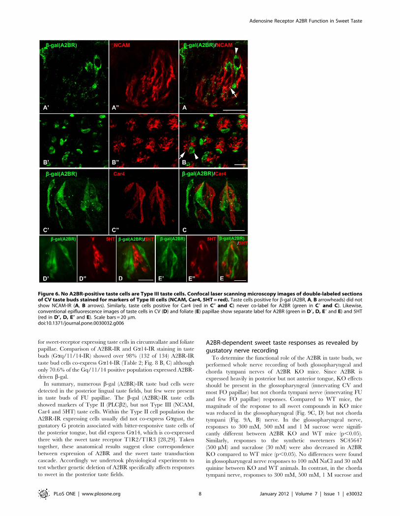
Labeling for A2BR (b-gal) correlates with markers of taste cell
type and function. Double immunostaining with b-gal (A2BR) and
markers of Type III taste cells, NCAM (Fig. 6 A, B), Car4 (Fig. 6C)
and 5HT (Fig. 6 D, E), showed no obvious overlap between b-gal
(A2BR)-IR and any marker of Type III cells (Fig. 6). Similarly, in
the hundreds of taste buds examined, no taste cells showing
immunoreactivity for type III cell markers was immunoreactive for
b-gal.
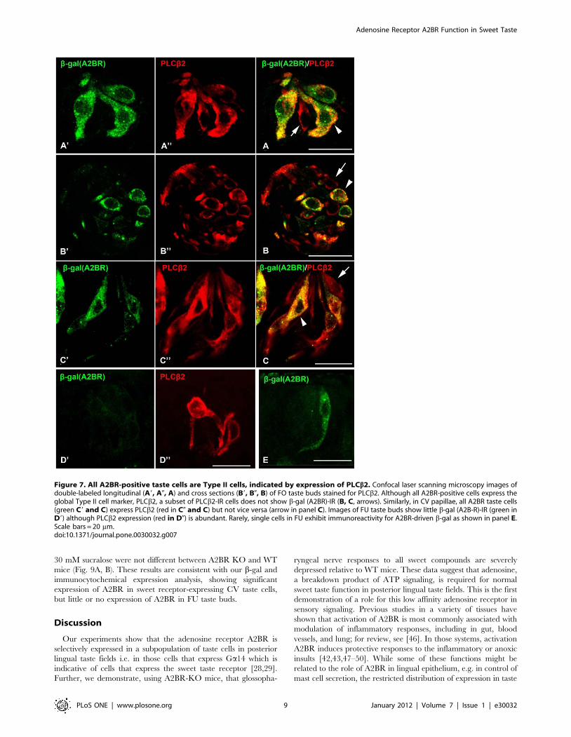
In contrast, many cells in posterior lingual taste buds that
display the characteristic marker of Type II taste cells, PLCb2-IR
[25], also express A2BR-driven b-gal (Fig. 7 A, B, C arrowheads).
Further A2BR-expressing taste cells have the classic morphological
appearance of Type II taste cells, most notably a large, round
nucleus. However, not all PLCb2-IR taste bud cells were b-gal
(A2BR)-positive (Fig. 7A, B, C arrows) indicating that A2BR is
expressed in only a subset of Type II taste cells. A quantitative
analysis of this co-expression pattern shows that all cells expressing
A2B-driven b-gal co-express PLCb2 (see Table 2) but only 43.8%
of PLCb2-IR cells express A2B-driven b-gal (n = 288). Little or no
b-gal (A2BR) IR was detected in FU or palatal taste buds (Fig. 7
D9, D0; E).
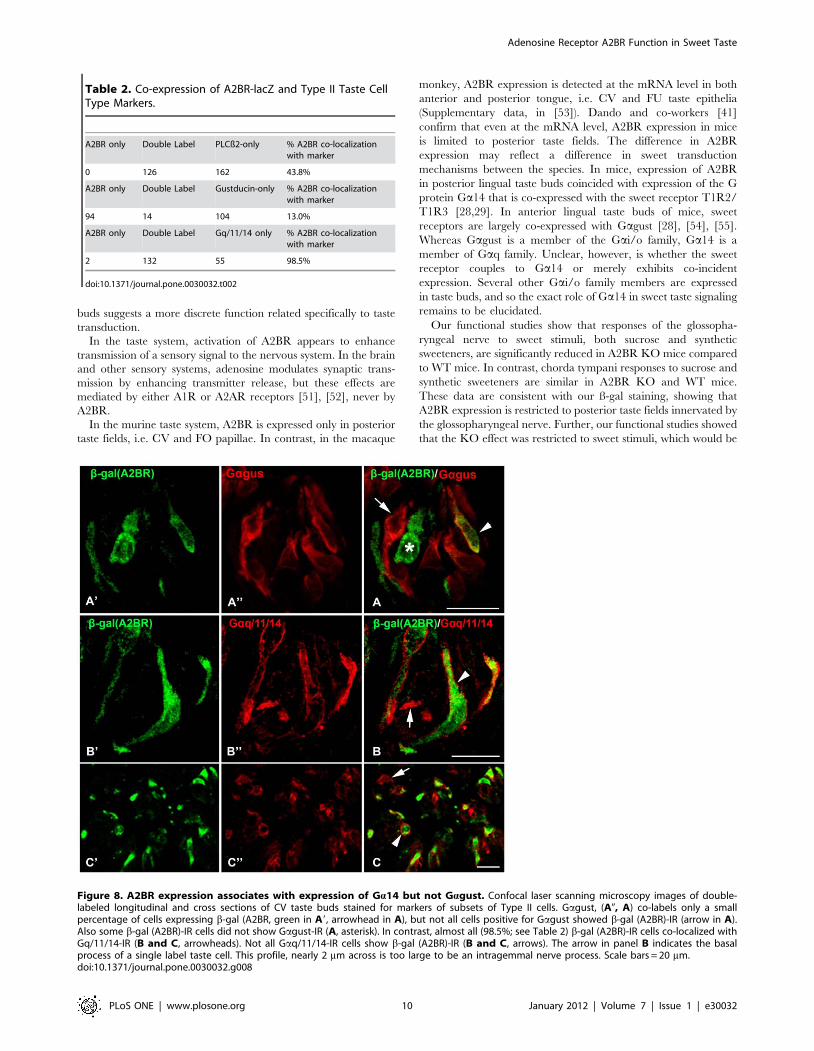
Type II cells can be subdivided according to their expression of
taste receptors and their associated G proteins. Gagust-IR taste
cells are a subset of Type II cells [27,45]. In posterior tongue,
Gagust predominantly marks bitter-responsive taste cells with
lesser expression in the umami and sweet sensitive populations
[28,29]. Double immunolabeling with b-gal (A2BR) and Gagust
revealed incomplete overlap of immunoreactivity in both CV and
FO papillae (Fig. 8A). Few (14 of 118) taste cells that expressed
Gagust-IR were positive for b-gal (A2BR)-IR (Table 2 and Fig. 8A,
arrowhead). Similarly, nearly all (94 of 108) b-gal (A2BR)-IR taste
bud cells lacked Gagust-IR (Table 2 and Fig. 8A, asterisk). Two
previous studies demonstrated that in posterior lingual taste fields,
all taste cells that express the sweet receptor T1R2/T1R3, also
express Ga14 ([28,29]). Accordingly, we used Ga14 as a marker
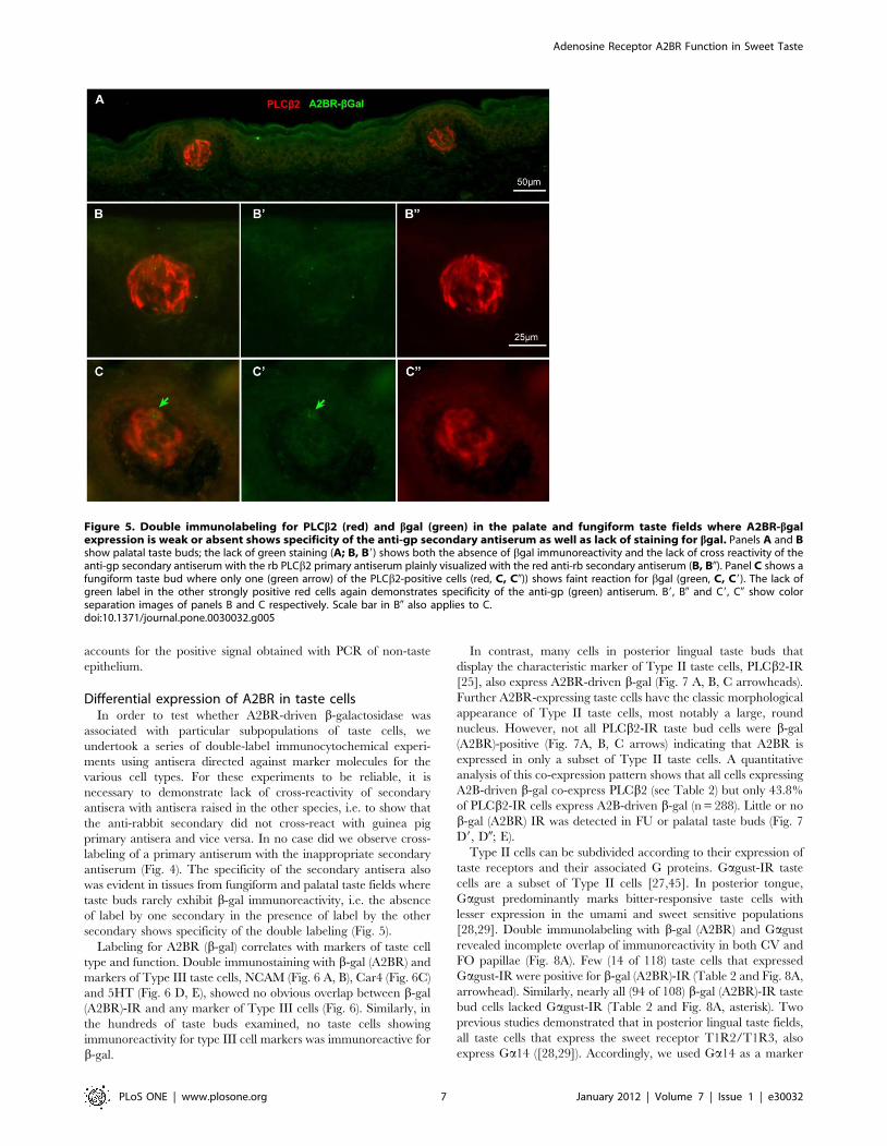
Figure 5. Double immunolabeling for PLCb2 (red) and bgal (green) in the palate and fungiform taste fields where A2BR-bgalexpression is weak or absent shows specificity of the anti-gp secondary antiserum as well as lack of staining for bgal. Panels A and Bshow palatal taste buds; the lack of green staining (A; B, B9) shows both the absence of bgal immunoreactivity and the lack of cross reactivity of theanti-gp secondary antiserum with the rb PLCb2 primary antiserum plainly visualized with the red anti-rb secondary antiserum (B, B0). Panel C shows afungiform taste bud where only one (green arrow) of the PLCb2-positive cells (red, C, C0)) shows faint reaction for bgal (green, C, C9). The lack ofgreen label in the other strongly positive red cells again demonstrates specificity of the anti-gp (green) antiserum. B9, B0 and C9, C0 show colorseparation images of panels B and C respectively. Scale bar in B0 also applies to C.doi:10.1371/journal.pone.0030032.g005
Adenosine Receptor A2BR Function in Sweet Taste
PLoS ONE | www.plosone.org 7 January 2012 | Volume 7 | Issue 1 | e30032
for sweet-receptor expressing taste cells in circumvallate and foliate
papillae. Comparison of A2BR-IR and Ga14-IR staining in taste
buds (Gaq/11/14-IR) showed over 98% (132 of 134) A2BR-IR
taste bud cells co-express Ga14-IR (Table 2; Fig. 8 B, C) although
only 70.6% of the Gq/11/14 positive population expressed A2BR-
driven b-gal.
In summary, numerous b-gal (A2BR)-IR taste bud cells were
detected in the posterior lingual taste fields, but few were present
in taste buds of FU papillae. The b-gal (A2BR)-IR taste cells
showed markers of Type II (PLCb2), but not Type III (NCAM,
Car4 and 5HT) taste cells. Within the Type II cell population the
A2BR-IR expressing cells usually did not co-express Gagust, the
gustatory G protein associated with bitter-responsive taste cells of
the posterior tongue, but did express Ga14, which is co-expressed
there with the sweet taste receptor T1R2/T1R3 [28,29]. Taken
together, these anatomical results suggest close correspondence
between expression of A2BR and the sweet taste transduction
cascade. Accordingly we undertook physiological experiments to
test whether genetic deletion of A2BR specifically affects responses
to sweet in the posterior taste fields.
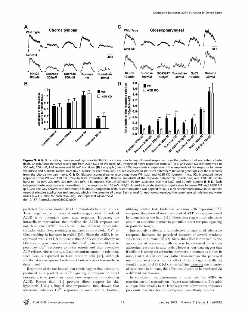
A2BR-dependent sweet taste responses as revealed bygustatory nerve recording
To determine the functional role of the A2BR in taste buds, we
performed whole nerve recording of both glossopharyngeal and
chorda tympani nerves of A2BR KO mice. Since A2BR is
expressed heavily in posterior but not anterior tongue, KO effects
should be present in the glossopharyngeal (innervating CV and
most FO papillae) but not chorda tympani nerve (innervating FU
and few FO papillae) responses. Compared to WT mice, the
magnitude of the response to all sweet compounds in KO mice
was reduced in the glossopharyngeal (Fig. 9C, D) but not chorda
tympani (Fig. 9A, B) nerve. In the glossopharyngeal nerve,
responses to 300 mM, 500 mM and 1 M sucrose were signifi-
cantly different between A2BR KO and WT mice (p,0.05).
Similarly, responses to the synthetic sweeteners SC45647
(500 mM) and sucralose (30 mM) were also decreased in A2BR
KO compared to WT mice (p,0.05). No differences were found
in glossopharyngeal nerve responses to 100 mM NaCl and 30 mM
quinine between KO and WT animals. In contrast, in the chorda
tympani nerve, responses to 300 mM, 500 mM, 1 M sucrose and
Figure 6. No A2BR-positive taste cells are Type III taste cells. Confocal laser scanning microscopy images of double-labeled sectionsof CV taste buds stained for markers of Type III cells (NCAM, Car4, 5HT = red). Taste cells positive for b-gal (A2BR, A, B arrowheads) did notshow NCAM-IR (A, B arrows). Similarly, taste cells positive for Car4 (red in C0 and C) never co-label for A2BR (green in C9 and C). Likewise,conventional epifluorescence images of taste cells in CV (D) and foliate (E) papillae show separate label for A2BR (green in D9, D, E9 and E) and 5HT(red in D0, D, E0 and E). Scale bars = 20 mm.doi:10.1371/journal.pone.0030032.g006
Adenosine Receptor A2BR Function in Sweet Taste
PLoS ONE | www.plosone.org 8 January 2012 | Volume 7 | Issue 1 | e30032
30 mM sucralose were not different between A2BR KO and WT
mice (Fig. 9A, B). These results are consistent with our b-gal and
immunocytochemical expression analysis, showing significant
expression of A2BR in sweet receptor-expressing CV taste cells,
but little or no expression of A2BR in FU taste buds.
Discussion
Our experiments show that the adenosine receptor A2BR is
selectively expressed in a subpopulation of taste cells in posterior
lingual taste fields i.e. in those cells that express Ga14 which is
indicative of cells that express the sweet taste receptor [28,29].
Further, we demonstrate, using A2BR-KO mice, that glossopha-
ryngeal nerve responses to all sweet compounds are severely
depressed relative to WT mice. These data suggest that adenosine,
a breakdown product of ATP signaling, is required for normal
sweet taste function in posterior lingual taste fields. This is the first
demonstration of a role for this low affinity adenosine receptor in
sensory signaling. Previous studies in a variety of tissues have
shown that activation of A2BR is most commonly associated with
modulation of inflammatory responses, including in gut, blood
vessels, and lung; for review, see [46]. In those systems, activation
A2BR induces protective responses to the inflammatory or anoxic
insults [42,43,47–50]. While some of these functions might be
related to the role of A2BR in lingual epithelium, e.g. in control of
mast cell secretion, the restricted distribution of expression in taste
Figure 7. All A2BR-positive taste cells are Type II cells, indicated by expression of PLCb2. Confocal laser scanning microscopy images ofdouble-labeled longitudinal (A9, A0, A) and cross sections (B9, B0, B) of FO taste buds stained for PLCb2. Although all A2BR-positive cells express theglobal Type II cell marker, PLCb2, a subset of PLCb2-IR cells does not show b-gal (A2BR)-IR (B, C, arrows). Similarly, in CV papillae, all A2BR taste cells(green C9 and C) express PLCb2 (red in C0 and C) but not vice versa (arrow in panel C). Images of FU taste buds show little b-gal (A2B-R)-IR (green inD9) although PLCb2 expression (red in D0) is abundant. Rarely, single cells in FU exhibit immunoreactivity for A2BR-driven b-gal as shown in panel E.Scale bars = 20 mm.doi:10.1371/journal.pone.0030032.g007
Adenosine Receptor A2BR Function in Sweet Taste
PLoS ONE | www.plosone.org 9 January 2012 | Volume 7 | Issue 1 | e30032
buds suggests a more discrete function related specifically to taste
transduction.
In the taste system, activation of A2BR appears to enhance
transmission of a sensory signal to the nervous system. In the brain
and other sensory systems, adenosine modulates synaptic trans-
mission by enhancing transmitter release, but these effects are
mediated by either A1R or A2AR receptors [51], [52], never by
A2BR.
In the murine taste system, A2BR is expressed only in posterior
taste fields, i.e. CV and FO papillae. In contrast, in the macaque
monkey, A2BR expression is detected at the mRNA level in both
anterior and posterior tongue, i.e. CV and FU taste epithelia
(Supplementary data, in [53]). Dando and co-workers [41]
confirm that even at the mRNA level, A2BR expression in mice
is limited to posterior taste fields. The difference in A2BR
expression may reflect a difference in sweet transduction
mechanisms between the species. In mice, expression of A2BR
in posterior lingual taste buds coincided with expression of the G
protein Ga14 that is co-expressed with the sweet receptor T1R2/
T1R3 [28,29]. In anterior lingual taste buds of mice, sweet
receptors are largely co-expressed with Gagust [28], [54], [55].
Whereas Gagust is a member of the Gai/o family, Ga14 is a
member of Gaq family. Unclear, however, is whether the sweet
receptor couples to Ga14 or merely exhibits co-incident
expression. Several other Gai/o family members are expressed
in taste buds, and so the exact role of Ga14 in sweet taste signaling
remains to be elucidated.
Our functional studies show that responses of the glossopha-
ryngeal nerve to sweet stimuli, both sucrose and synthetic
sweeteners, are significantly reduced in A2BR KO mice compared
to WT mice. In contrast, chorda tympani responses to sucrose and
synthetic sweeteners are similar in A2BR KO and WT mice.
These data are consistent with our ß-gal staining, showing that
A2BR expression is restricted to posterior taste fields innervated by
the glossopharyngeal nerve. Further, our functional studies showed
that the KO effect was restricted to sweet stimuli, which would be
Table 2. Co-expression of A2BR-lacZ and Type II Taste CellType Markers.
A2BR only Double Label PLCß2-only % A2BR co-localizationwith marker
0 126 162 43.8%
A2BR only Double Label Gustducin-only % A2BR co-localizationwith marker
94 14 104 13.0%
A2BR only Double Label Gq/11/14 only % A2BR co-localizationwith marker
2 132 55 98.5%
doi:10.1371/journal.pone.0030032.t002
Figure 8. A2BR expression associates with expression of Ga14 but not Gagust. Confocal laser scanning microscopy images of double-labeled longitudinal and cross sections of CV taste buds stained for markers of subsets of Type II cells. Gagust, (A0, A) co-labels only a smallpercentage of cells expressing b-gal (A2BR, green in A9, arrowhead in A), but not all cells positive for Gagust showed b-gal (A2BR)-IR (arrow in A).Also some b-gal (A2BR)-IR cells did not show Gagust-IR (A, asterisk). In contrast, almost all (98.5%; see Table 2) b-gal (A2BR)-IR cells co-localized withGq/11/14-IR (B and C, arrowheads). Not all Gaq/11/14-IR cells show b-gal (A2BR)-IR (B and C, arrows). The arrow in panel B indicates the basalprocess of a single label taste cell. This profile, nearly 2 mm across is too large to be an intragemmal nerve process. Scale bars = 20 mm.doi:10.1371/journal.pone.0030032.g008
Adenosine Receptor A2BR Function in Sweet Taste
PLoS ONE | www.plosone.org 10 January 2012 | Volume 7 | Issue 1 | e30032
predicted from our double label immunohistochemical studies.
Taken together, our functional studies suggest that the role of
A2BR is to potentiate sweet taste responses. However, the
intracellular mechanisms that mediate the A2BR response are
not clear, since A2BR can couple to two different intracellular
cascades; either Gaq, resulting in increases in intracellular Ca2+ or
Gas, resulting in increases in cAMP [56]. Since the A2BR is co-
expressed with Ga14, it is possible that A2BR couples directly to
Ga14, causing increases in intracellular Ca2+, which would tend to
potentiate Ca2+ responses to sweet stimuli and thus potentiate
ATP release. Alternatively, a Gas mechanism cannot be ruled out,
since Gas is expressed in taste receptor cells [57], although
whether it is co-expressed with sweet taste receptors has not been
determined.
Regardless of the mechanism, our results suggest that adenosine,
produced as a product of ATP signaling in response to sweet
tastants, acts to potentiate sweet taste responses by activating
A2BR. Recent data [41] provides direct support for this
hypothesis. Using a lingual slice preparation, they showed that
adenosine enhances Ca2+ responses to sweet stimuli. Further,
utilizing isolated taste buds and biosensor cells expressing P2X
receptors, they showed sweet taste evoked ATP release is increased
by adenosine in the bath [41]. These data suggest that adenosine
acts in an autocrine manner to potentiate sweet receptor signaling
in posterior tongue.
Interestingly, caffeine, a non-selective antagonist of adenosine
receptors, increases the perceived intensity of several synthetic
sweeteners in humans [58,59]. Since this effect is reversed by the
application of adenosine, caffeine was hypothesized to act via
adenosine receptors in taste buds. However, our data suggest that
if caffeine is acting via adenosine receptors in humans as it does in
mice, that it should decrease, rather than increase the perceived
intensity of sweeteners, i.e. the effect of the antagonist (caffeine)
should mimic the A2BR KO. Since caffeine increases the intensity
of sweeteners in humans, this effect would seem to be mediated via
a different mechanism.
In conclusion, we demonstrate a novel role for A2BR in
transduction and transmission of sweet taste information. This adds
a unique functionality to the large repertoire of protective responses
previously described for this widespread, low-affinity receptor.
Figure 9. A & B. Gustatory nerve recordings from A2BR-KO mice show specific loss of sweet responses from the posterior but not anterior tastefields. Chorda tympani nerve recordings from A2BR KO and WT mice. (A). Integrated nerve responses from WT (top) and A2BR KO (bottom) mice to300 mM, 500 mM, 1 M sucrose and 30 mM sucralose. (B) Bar graph (mean6SEM) represents comparison of the amplitude of the response betweenWT (black) and A2BR KO (white) mice (n = 4–6 mice for each stimulus). ANOVA revealed no statistical differences between genotypes for these recordsfrom the chorda tympani nerve. C & D. Glossopharyngeal nerve recordings from WT (top) and A2BR KO (bottom) mice. (C). Integrated nerveresponses from WT and A2BR KO mice to taste stimulation. (D). Relative amplitude of the response between WT (black bars) and A2BR KO (whitebars) to 100 mM, 200 mM, 300 mM, 500 mM, 1 M sucrose, 500 mM SC45647, 30 mM sucralose, 100 mM NaCl and 30 mM quinine B & D. Eachintegrated taste response was normalized to the response to 100 mM NH4Cl. Asterisks indicate statistical significance between WT and A2BR KO(p,0.05; one-way ANOVA with Bonferroni’s Multiple Comparison Test). Taste stimulation was applied for 60 s in all experiments; arrows in (A) denoteonset of stimulus application and removal, which is the same for all traces. Each animal for each group received the same taste stimulation and waterrinses (n = 4–7 mice for each stimulus). Bars represent Mean6SEM.doi:10.1371/journal.pone.0030032.g009
Adenosine Receptor A2BR Function in Sweet Taste
PLoS ONE | www.plosone.org 11 January 2012 | Volume 7 | Issue 1 | e30032
Acknowledgments
The authors thank Holger Eltzschig and Almut Grenz for providing access
to A2BR-KO/LacZ mice, Daniel Sanculi for assistance with genotyping
and breeding, and Jason Parnes for immunocytochemistry.
Author Contributions
Conceived and designed the experiments: SK KR SCK TEF. Performed
the experiments: SK AB DY NS AV SCK TEF. Analyzed the data: SK AB
NS AV SCK TEF. Contributed reagents/materials/analysis tools: KR.
Wrote the paper: SK AB KR SCK TEF.
References
1. Finger TE, Danilova V, Barrows J, Bartel DL, Vigers AJ, et al. (2005) ATP
signaling is crucial for communication from taste buds to gustatory nerves.
Science 310: 1495–1499.
2. Huang YJ, Maruyama Y, Dvoryanchikov G, Pereira E, Chaudhari N, et al.
(2007) The role of pannexin 1 hemichannels in ATP release and cell-cell
communication in mouse taste buds. Proc Natl Acad Sci U S A 104: 6436–6441.
3. Romanov RA, Rogachevskaja OA, Bystrova MF, Jiang P, Margolskee RF, et al.
(2007) Afferent neurotransmission mediated by hemichannels in mammalian
taste cells. EMBO J 26: 657–667.
4. Murata Y, Yasuo T, Yoshida R, Obata K, Yanagawa Y, et al. (2010) Action
potential-enhanced ATP release from taste cells through hemichannels.
J Neurophysiol.
5. Bo X, Alavi A, Xiang Z, Oglesby I, Ford A, et al. (1999) Localization of ATP-
gated P2X2 and P2X3 receptor immunoreactive nerves in rat taste buds.
Neuroreport 10: 1107–1111.
6. Kataoka S, Toyono T, Seta Y, Toyoshima K (2006) Expression of ATP-gated
P2X3 receptors in rat gustatory papillae and taste buds. Arch Histol Cytol 69:
281–288.
7. Baryshnikov SG, Rogachevskaja OA, Kolesnikov SS (2003) Calcium signaling
mediated by P2Y receptors in mouse taste cells. J Neurophysiol 90: 3283–3294.
8. Kataoka S, Toyono T, Seta Y, Ogura T, Toyoshima K (2004) Expression of
P2Y(1) receptors in rat taste buds. Histochem Cell Biol.
9. Huang YA, Dando R, Roper SD (2009) Autocrine and paracrine roles for ATP
and serotonin in mouse taste buds. J Neurosci 29: 13909–13918.
10. Murray RG (1973) The ultrastructure of taste buds. In: Friedmann I, ed. The
Ultrastructure of Sensory Organs. Amsterdam: North Holland Pub. Co. pp
1–81.
11. Takeda M, Hoshino T (1975) Fine structure of taste buds in the rat. Arch Histol
Jap 37: 395–413.
12. Farbman AI, Hellekant G, Nelson A (1985) Structure of taste buds in foliate
papillae of the rhesus monkey, Macaca mulatta. Am J Anat 172: 41–56.
13. Kinnamon JC, Taylor BJ, Delay RJ, Roper SD (1985) Ultrastructure of mouse
vallate taste buds. I. Taste cells and their associated synapses. J Comp Neurol
235: 48–60.
14. Delay RJ, Kinnamon JC, Roper SD (1986) Ultrastructure of mouse vallate taste
buds: II. Cell types and cell lineage. Journal of Comparative Neurology 253:
242–252.
15. Yoshie S, Wakasugi C, Teraki Y, Fujita T (1990) Fine structure of the taste bud
in guinea pigs. I. Cell characterization and innervation patterns. Arch Histol
Cytol 53: 103–119.
16. Pumplin DW, Yu C, Smith DV (1997) Light and Dark cells of Rat Vallate Taste
Buds Are Morphologically Distinct Cell Types. J Comp Neurol 378: 389–410.
17. Finger TE, Simon SA (2000) Cell Biology of Taste Epithelium. In: Finger TE,
Silver WL, Restrepo D, eds. Neurobiology of Taste & Smell, 2nd edition. New
YorkNY: Wiley Press. pp 287–314.
18. Yee CL, Yang R, Bottger B, Finger TE, Kinnamon JC (2001) ‘‘Type III’’ cells of
rat taste buds: immunohistochemical and ultrastructural studies of neuron-
specific enolase, protein gene product 9.5, and serotonin. J Comp Neurol 440:
97–108.
19. Bartel DL, Sullivan SL, Lavoie EG, Sevigny J, Finger TE (2006) Nucleoside
triphosphate diphosphohydrolase-2 is the ecto-ATPase of type I cells in taste
buds. J Comp Neurol 497: 1–12.
20. Kataoka S, Yang R, Ishimaru Y, Matsunami H, Sevigny J, et al. (2008) The
candidate sour taste receptor, PKD2L1, is expressed by type III taste cells in the
mouse. Chem Senses 33: 243–254.
21. Lawton DM, Furness DN, Lindemann B, Hackney CM (2000) Localization of
the glutamate-aspartate transporter, GLAST, in rat taste buds. Eur J Neurosci
12: 3163–3171.
22. Chaudhari N, Roper SD (2010) The cell biology of taste. J Cell Biol 190:
285–296.
23. Hoon MA, Adler E, Lindemeier J, Battey JF, Ryba NJ, et al. (1999) Putative
mammalian taste receptors: a class of taste-specific GPCRs with distinct
topographic selectivity. Cell 96: 541–551.
24. Zhang Y, Hoon MA, Chandrashekar J, Mueller KL, Cook B, et al. (2003)
Coding of sweet, bitter, and umami tastes: different receptor cells sharing similar
signaling pathways. Cell 112: 293–301.
25. Clapp TR, Yang R, Stoick CL, Kinnamon SC, Kinnamon JC (2004)
Morphologic characterization of rat taste receptor cells that express components
of the phospholipase C signaling pathway. J Comp Neurol 468: 311–321.
26. Tabata S, Crowley HH, Bottger B, Finger TE, Margolskee RF, et al. (1995)
Immunoelectron microscopical analysis of gustducin in taste cells of the rat.
Chem Senses 20: 778.
27. Yang R, Tabata S, Crowley HH, Margolskee RF, Kinnamon JC (2000)Ultrastructural localization of gustducin immunoreactivity in microvilli of type II
taste cells in the rat. J Comp Neurol 425: 139–151.
28. Tizzano M, Dvoryanchikov G, Barrows JK, Kim S, Chaudhari N, et al. (2008)
Expression of Galpha14 in sweet-transducing taste cells of the posterior tongue.
BMC Neurosci 9: 110.
29. Shindo Y, Miura H, Carninci P, Kawai J, Hayashizaki Y, et al. (2008) G
alpha14 is a candidate mediator of sweet/umami signal transduction in theposterior region of the mouse tongue. Biochem biophys res commun 376:
504–508.
30. Huang AL, Chen X, Hoon MA, Chandrashekar J, Guo W, et al. (2006) Thecells and logic for mammalian sour taste detection. Nature 442: 934–938.
31. Nelson GM, Finger TE (1993) Immunolocalization of different forms of neural
cell adhesion molecule (NCAM) in rat taste buds. J Comp Neurol 336:507–516.
32. Chandrashekar J, Yarmolinsky D, von Buchholtz L, Oka Y, Sly W, et al. (2009)
The taste of carbonation. Science 326: 443–445.
33. Huang YJ, Lu KS (1996) Immunohistochemical studies on protein gene product
9.5, serotonin and neuropeptides in vallate taste buds and related nerves of the
guinea pig. Arch Histol Cytol 59: 433–441.
34. Dvoryanchikov G, Tomchik SM, Chaudhari N (2007) Biogenic amine synthesis
and uptake in rodent taste buds. J Comp Neurol 505: 302–313.
35. Royer SM, Kinnamon JC (1991) HVEM serial-section analysis of rabbit foliatetaste buds: I. Type III cells and their synapses. J Comp Neurol 306: 49–72.
36. Iwayama T, Nada O (1967) Histochemically demonstrable ATPase activity inthe taste buds of the rat. Exp Cell Res 46: 607–608.
37. Iwayama T, Nada O (1967) Histochemical observation on the phosphatases
of the tongue, with special reference to taste buds. Arch Histol Jpn 28:151–163.
38. Zalewski AA (1968) Changes in phosphatase enzymes following denervation of
the vallate papilla of the rat. Exp Neurol 22: 40–51.
39. Barry MA (1992) Ecto-calcium-dependent ATPase activity of mammalian taste
bud cells. J Histochem Cytochem 40: 1919–1928.
40. Iwayama T, Nada O (1969) Histochemical observation on phosphatase activitiesof degenerating and regenerating taste buds. Anat Rec 163: 31–38.
41. Dando R, Dvoryanchikov G, Pereira E, Chaudhari N, Roper SD (2012)
Adenosine enhances sweet taste through A2B receptors in the taste bud.J Neurosci in press.
42. Yang D, Zhang Y, Nguyen HG, Koupenova M, Chauhan AK, et al. (2006) The
A2B adenosine receptor protects against inflammation and excessive vascularadhesion. J Clin Invest 116: 1913–1923.
43. Yang D, Chen H, Koupenova M, Carroll SH, Eliades A, et al. (2010) A new rolefor the A2b adenosine receptor in regulating platelet function. J Thromb
Haemost 8: 817–827.
44. Yee CL, Finger TE (2003) Expression Of BDNF, NGF And PGP 9.5 In TasteBuds Following Glossopharyngeal Nerve Section In Mice. Chem Senses 28: A9.
45. Clapp TR, Stone LM, Margolskee RF, Kinnamon SC (2001) Immunocyto-
chemical evidence for co-expression of Type III IP3 receptor with signalingcomponents of bitter taste transduction. BMC Neurosci 2: 6.
46. Aherne CM, Kewley EM, Eltzschig HK (2011) The resurgence of A2B
adenosine receptor signaling. Biochim Biophys Acta 1808: 1329–1339.
47. Fredholm BB, AP IJ, Jacobson KA, Klotz KN, Linden J (2001) International
Union of Pharmacology. XXV. Nomenclature and classification of adenosine
receptors. Pharmacol Rev 53: 527–552.
48. Feoktistov I, Biaggioni I (1997) Adenosine A2B receptors. Pharmacol Rev 49:
381–402.
49. Hasko G, Csoka B, Nemeth ZH, Vizi ES, Pacher P (2009) A(2B) adenosinereceptors in immunity and inflammation. Trends Immunol 30: 263–270.
50. Ryzhov S, Goldstein AE, Biaggioni I, Feoktistov I (2006) Cross-talk betweenG(s)- and G(q)-coupled pathways in regulation of interleukin-4 by A(2B)
adenosine receptors in human mast cells. Mol Pharmacol 70: 727–735.
51. Masino SA, Diao L, Illes P, Zahniser NR, Larson GA, et al. (2002) Modulationof hippocampal glutamatergic transmission by ATP is dependent on adenosine
a(1) receptors. J pharmacol exp ther 303: 356–363.
52. Sebastiao AM, Ribeiro JA (2009) Tuning and fine-tuning of synapses withadenosine. Curr Neuropharmacol 7: 180–194.
53. Hevezi P, Moyer BD, Lu M, Gao N, White E, et al. (2009) Genome-wide
analysis of gene expression in primate taste buds reveals links to diverseprocesses. PLoS One 4: e6395.
54. Stone LM, Barrows J, Finger TE, Kinnamon SC (2007) Expression of T1Rs and
gustducin in palatal taste buds of mice. Chem Senses 32: 255–262.
55. Kim MR, Kusakabe Y, Miura H, Shindo Y, Ninomiya Y, et al. (2003) Regional
expression patterns of taste receptors and gustducin in the mouse tongue.Biochem Biophys Res Commun 312: 500–506.
Adenosine Receptor A2BR Function in Sweet Taste
PLoS ONE | www.plosone.org 12 January 2012 | Volume 7 | Issue 1 | e30032
56. Cohen MV, Yang X, Downey JM (2010) A(2b) adenosine receptors can change
their spots. Br J Pharmacol 159: 1595–1597.57. Kusakabe Y, Yasuoka A, Asano-Miyoshi M, Iwabuchi K, Matsumoto I, et al.
(2000) Comprehensive study on G protein alpha-subunits in taste bud cells, with
special reference to the occurrence of Galphai2 as a major Galpha species.Chem Senses 25: 525–531.
58. Schiffman SS, Diaz C, Beeker TG (1986) Caffeine intensifies taste of certain
sweeteners: role of adenosine receptor. Pharmacol Biochem Behav 24: 429–432.
59. Schiffman SS, Gill JM, Diaz C (1985) Methyl xanthines enhance taste: evidence
for modulation of taste by adenosine receptor. Pharmacol Biochem Behav 22:
195–203.
Adenosine Receptor A2BR Function in Sweet Taste
PLoS ONE | www.plosone.org 13 January 2012 | Volume 7 | Issue 1 | e30032