ABCD Arq Bras Cir Dig Case Report - SciELO · ABCD Arq Bras Cir Dig Case Report ... Falcone C,...
3
139 ABCD Arq Bras Cir Dig Case Report 2008;21(3):139-41 ENDOSCOPIC TREATMENT OF CHOLEDOCHAL CYST TYPE III Tratamento endoscópico de cisto de colédoco do tipo III Matheus Alessi RODRIGUES, Alexandre Venâncio de SOUSA, Ciro FALCONE, Gabriel COELHO, Durval Knox da VEIGA, Mário ZAIDAN, Francisco CALLEJAS-NETO, Roberto Franchi TEIXEIRA Trabalho realizado no Departamento de Cirurgia Geral da Faculdade de Medicina de Jundiaí – FMJ , Jundiaí, SP, Brasil. Correspondência: Matheus Alessi Rodrigues, e-mail: [email protected] ABCDDV/612 INTRODUÇÃO Choledochal cysts (CC) are uncommon anomalies which affect the intramural segment of the biliary tree 8 . They were first described by Vatero and Ezler in 1723 7,11 . The classical clinical symptoms are abdominal pain, jaun- dice and fever in a pre-school children 8 . Nevertheless, it may be present in adults in 20% 8 . There is a marked fe- male preponderance among the patients with choledochal cysts and more than one-third of all cases were described in orientals 9 .They were first classified by Alonso-Lej in 1959 1 . Nowadays they are ordinarily classified by Todani’s classification and its modifications (Figure 1), as follows 4 : Type I - cysts represent the classical disease. They are in fact concentric dilatation of the common bile duct and also the most common type (80%). Type II - cysts are known as supraduodenal excentric choledochal dilatation, and they are very rare (3%). Type III - cysts or choledochoceles are normally as- sociated to ampullae anomalies. They are also rare (4%) and sometimes diagnosed in a routine endoscopy as a compression of the duodenum. Type IV - cysts are multiple dilatations affecting intra and extrahepatic tree, and occur in 10 % of the cases, with subtypes IV-A ( fusiform extra and intra-hepatic cysts); IV-B (multiple extra-hepatic cysts). Todani type V cystic disease is in fact the Caroli´s disease. Cysts are multiple and intrahepatic only. They represent 1-2% of all choledochal cysts. Rodrigus MA, Sousa AV, Falcone C, Coelho G, Veiga DK, Zaidan M, Callejas-Neto F, Teixeira RF. Endoscopic treatment of choledochal cyst type III. ABCD Arq Bras Cir Dig 2008;21(3):139-41 ABSTRACT – Background - Todani type III cysts are not very common disease. Endoscopically the choledochocele is not a challenging diagnosis. Sometimes biliary stone disease is associated and events of cholangitis and pancreatitis may occur. Normally these patients are referred for surgical treatment, mainly because there is a widespread concept that choledocal cysts are very prone to develop neoplasia and must be resected. Nevertheless surgical resection is not free of morbidity. The chance for neoplasia in such cases seems to be related to the presence of pancreaticobiliary reflux towards the common bile duct. Aim – To report a case of endoscopic treatment of choledochal cyst type III with literature review. Case Report - Young man with recurrent abdominal pain, fever and hyperamylasemia. An ERCP showed pancreaticobiliary maljunction and calculus impaction. Papillotomy was performed and complete biliary clearance was achieved. Amylase contents in the common bile duct was measured and normal. Due to absence of pancreatiobiliary reflux, a second endoscopic approach was performed and a wide communication between choledochocele and duodenum was done with diathermy (using the papillotome). The patient recovering was uneventful and in 30 months follow-up he remains asymp- tomatic. Conclusion - Since pancreatobiliary reflux is not present, surgical approach of the diverticulum seemed to be not necessary. Endoscopic drainage of choledococele was a good option for conservative treatment. HEADINGS – Choledochal cyst. Endoscopy. FIGURE 1 – Todani´s choledochal cysts classification, modified
Transcript of ABCD Arq Bras Cir Dig Case Report - SciELO · ABCD Arq Bras Cir Dig Case Report ... Falcone C,...
139
ABCD Arq Bras Cir Dig Case Report2008;21(3):139-41
ENDOSCOPIC TREATMENT OF CHOLEDOCHAL CYST TYPE III
Tratamento endoscópico de cisto de colédoco do tipo III
Matheus Alessi RODRIGUES, Alexandre Venâncio de SOUSA, Ciro FALCONE, Gabriel COELHO,Durval Knox da VEIGA, Mário ZAIDAN, Francisco CALLEJAS-NETO, Roberto Franchi TEIXEIRA
Trabalho realizado no Departamento de Cirurgia Geral da Faculdade de Medicina de Jundiaí – FMJ , Jundiaí, SP, Brasil.
Correspondência: Matheus Alessi Rodrigues, e-mail: [email protected]
ABCDDV/612
INTRODUÇÃO
Choledochal cysts (CC) are uncommon anomalies which affect the intramural segment of the biliary tree8. They were first described by Vatero and Ezler in 17237,11. The classical clinical symptoms are abdominal pain, jaun-dice and fever in a pre-school children8. Nevertheless, it may be present in adults in 20%8. There is a marked fe-male preponderance among the patients with choledochal cysts and more than one-third of all cases were described in orientals9.They were first classified by Alonso-Lej in 19591. Nowadays they are ordinarily classified by Todani’s classification and its modifications (Figure 1), as follows4:
Type I - cysts represent the classical disease. They are in fact concentric dilatation of the common bile duct and also the most common type (80%).
Type II - cysts are known as supraduodenal excentric choledochal dilatation, and they are very rare (3%).
Type III - cysts or choledochoceles are normally as-sociated to ampullae anomalies. They are also rare (4%) and sometimes diagnosed in a routine endoscopy as a
compression of the duodenum.Type IV - cysts are multiple dilatations affecting intra
and extrahepatic tree, and occur in 10 % of the cases, with subtypes IV-A ( fusiform extra and intra-hepatic cysts); IV-B (multiple extra-hepatic cysts).
Todani type V cystic disease is in fact the Caroli´s disease. Cysts are multiple and intrahepatic only. They represent 1-2% of all choledochal cysts.
Rodrigus MA, Sousa AV, Falcone C, Coelho G, Veiga DK, Zaidan M, Callejas-Neto F, Teixeira RF. Endoscopic treatment of choledochal cyst type III. ABCD Arq Bras Cir Dig 2008;21(3):139-41
ABSTRACT – Background - Todani type III cysts are not very common disease. Endoscopically the choledochocele is not a challenging diagnosis. Sometimes biliary stone disease is associated and events of cholangitis and pancreatitis may occur. Normally these patients are referred for surgical treatment, mainly because there is a widespread concept that choledocal cysts are very prone to develop neoplasia and must be resected. Nevertheless surgical resection is not free of morbidity. The chance for neoplasia in such cases seems to be related to the presence of pancreaticobiliary reflux towards the common bile duct. Aim – To report a case of endoscopic treatment of choledochal cyst type III with literature review. Case Report - Young man with recurrent abdominal pain, fever and hyperamylasemia. An ERCP showed pancreaticobiliary maljunction and calculus impaction. Papillotomy was performed and complete biliary clearance was achieved. Amylase contents in the common bile duct was measured and normal. Due to absence of pancreatiobiliary reflux, a second endoscopic approach was performed and a wide communication between choledochocele and duodenum was done with diathermy (using the papillotome). The patient recovering was uneventful and in 30 months follow-up he remains asymp-tomatic. Conclusion - Since pancreatobiliary reflux is not present, surgical approach of the diverticulum seemed to be not necessary. Endoscopic drainage of choledococele was a good option for conservative treatment.
HEADINGS – Choledochal cyst. Endoscopy.
FIGURE 1 – Todani´s choledochal cysts classification, modified
140
CASE REPORT
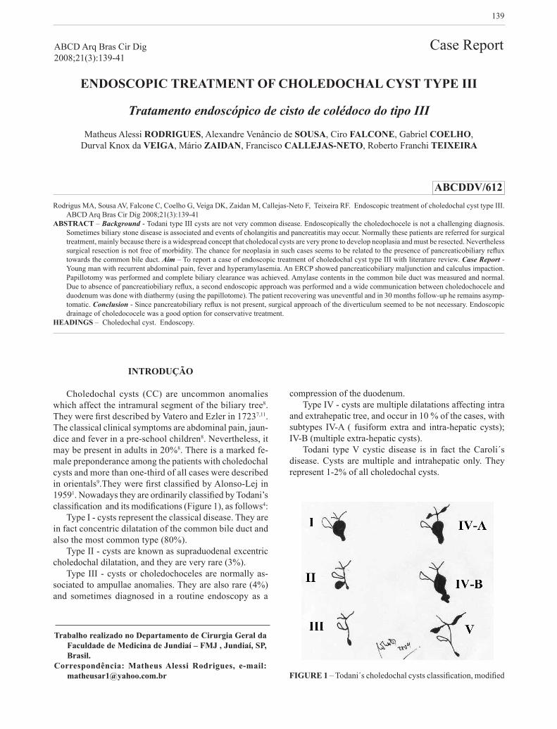
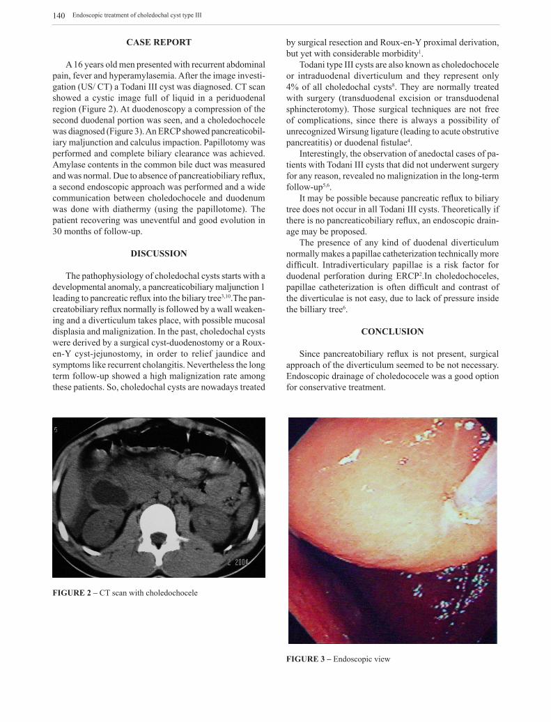
A 16 years old men presented with recurrent abdominal pain, fever and hyperamylasemia. After the image investi-gation (US/ CT) a Todani III cyst was diagnosed. CT scan showed a cystic image full of liquid in a periduodenal region (Figure 2). At duodenoscopy a compression of the second duodenal portion was seen, and a choledochocele was diagnosed (Figure 3). An ERCP showed pancreaticobil-iary maljunction and calculus impaction. Papillotomy was performed and complete biliary clearance was achieved. Amylase contents in the common bile duct was measured and was normal. Due to absence of pancreatiobiliary reflux, a second endoscopic approach was performed and a wide communication between choledochocele and duodenum was done with diathermy (using the papillotome). The patient recovering was uneventful and good evolution in 30 months of follow-up.
DISCUSSION
The pathophysiology of choledochal cysts starts with a developmental anomaly, a pancreaticobiliary maljunction 1 leading to pancreatic reflux into the biliary tree3,10.The pan-creatobiliary reflux normally is followed by a wall weaken-ing and a diverticulum takes place, with possible mucosal displasia and malignization. In the past, choledochal cysts were derived by a surgical cyst-duodenostomy or a Roux-en-Y cyst-jejunostomy, in order to relief jaundice and symptoms like recurrent cholangitis. Nevertheless the long term follow-up showed a high malignization rate among these patients. So, choledochal cysts are nowadays treated
by surgical resection and Roux-en-Y proximal derivation, but yet with considerable morbidity1.
Todani type III cysts are also known as choledochocele or intraduodenal diverticulum and they represent only 4% of all choledochal cysts8. They are normally treated with surgery (transduodenal excision or transduodenal sphincterotomy). Those surgical techniques are not free of complications, since there is always a possibility of unrecognized Wirsung ligature (leading to acute obstrutive pancreatitis) or duodenal fistulae4.
Interestingly, the observation of anedoctal cases of pa-tients with Todani III cysts that did not underwent surgery for any reason, revealed no malignization in the long-term follow-up5,6.
It may be possible because pancreatic reflux to biliary tree does not occur in all Todani III cysts. Theoretically if there is no pancreaticobiliary reflux, an endoscopic drain-age may be proposed.
The presence of any kind of duodenal diverticulum normally makes a papillae catheterization technically more difficult. Intradiverticulary papillae is a risk factor for duodenal perforation during ERCP2.In choledochoceles, papillae catheterization is often difficult and contrast of the diverticulae is not easy, due to lack of pressure inside the billiary tree6.
CONCLUSION
Since pancreatobiliary reflux is not present, surgical approach of the diverticulum seemed to be not necessary. Endoscopic drainage of choledococele was a good option for conservative treatment.
FIGURE 2 – CT scan with choledochocele
FIGURE 3 – Endoscopic view
Endoscopic treatment of choledochal cyst type III
141
Rodrigus MA, Sousa AV, Falcone C, Coelho G, Veiga DK, Zaidan M, Callejas-Neto F, Teixeira RF. Tratamento endoscópico de cisto de colédoco do tipo III. ABCD Arq Bras Cir Dig 2008;21(3):139-41
RESUMO – Introdução – Cisto de colédoco do tipo III de Todani não é doença muito comum. Eventualmente pode ocorrer a presença de doença biliar calculosa, que nestes casos estaria associada a eventos de colangite e pancreatite. Normalmente estes pacientes são conduzidos ao tratamento cirúr-gico, principalmente porque há conceito difundido de que os cistos de colédoco são muito propensos a desenvolver neoplasia e que a sua presença indicaria ressecção cirúrgica. A chance para neoplasia parece estar relacionada à presença de refluxo do conteúdo biliopancreático para o ducto biliar comum. Objetivo - Relatar um caso de tratamento endoscópico de cisto de colédoco do Tipo III, bem como realizar revisão da literatura sobre o tema. Relato do caso – Homem com 16 anos apresentou-se com dor abdominal periódica, febre e hiperamilasemia. CPRE mostrou má formação da junção biliopancreática, bem como impactação por cálculo. Foi realizada papilotomia que conseguiu clarear a árvore biliar. Os níveis de amilase no ducto biliar eram normais. Devido à ausência de refluxo de conteúdo biliopancreático, uma segunda aproximação endoscópica foi executada e comunicação larga entre o cisto e o duodeno foi realizada através do método endoscópico de diatermia. O paciente apresentou boa evolução após o procedimento e assim permanecendo em 30 meses de evolução. Conclusão – Desde que não haja a presença de refluxo de conteúdo biliopancreático, a ressecção cirúrgica do divertículo parece não ser necessária. A realização de drenagem endoscópica constitui-se em boa opção para tratamento conservador nos casos onde o refluxo biliopancreático não esteja presente.
DESCRITORES – Cisto de colédoco. Endoscopia.
REFERÊNCIAS
1. Akaraviputh T, Boonnuch W, Watanapa P, Lert-Akayamanee N, Lohsiriwat D. Surgical management of adult choledochal cysts. J Med Assoc Thai. 2005 Jul;88(7):939-43.
2. Candel MF, Albarracín A, Robles R, Guirao J, Parrilla P. Perforation of cho-ledochal cyst in a 12-year-old boy. Cir Esp. 2005 Aug;78(2):115-7.
3. Hosoki T, Hasuike Y, Takeda Y, Michita T, Watanabe Y, Sakamori R, Tokuda Y, Yutani K, Sai C, Mitomo M. Visualization of pancreaticobiliary reflux in anomalous pancreaticobiliary junction by secretin-stimulated dynamic magnetic resonance cholangiopancreatography. Acta Radiol. 2004 Jul;45(4):375-82.
4. Komuro H, Makino SI, Yasuda Y, Ishibashi T, Tahara K, Nagai H. Pancreatic complications in choledochal cyst and their surgical outcomes. World J Surg. 2001 Dec;25(12):1519-23.
5. Liras Muñoz J, Bueno Recio J, Sánchez Abuín A, García Alonso L, Solar Boga A, Pais Piñeiro E, Vela Nieto D, Gómez M. Management of choledochal cyst: laparotomy or endoscopic Cir Pediatr. 2005 Apr;18(2):73-6.
6. Naga MI, Suleiman DN. Endoscopic management of choledochal cyst. Gas-trointest Endosc. 2004 Mar;59(3):427-32.
7. O’Neill JA Jr, Templeton JM Jr, Schnaufer L, Bishop HC, Ziegler MM, Ross AJ 3rd. Recent experience with choledochal cyst. Ann Surg. 1987 May;205(5):533-40.
8. Popovici A, Mitulescu G, Hortopan M, Stanciu C. Cystic dilatation of main biliary tract Chirurgia (Bucur). 2000 Mar-Apr;95(2):139-55.
9. Sawyer MAJ, Sawyer EM, Patel TH, Varma MK, Allen AW, Murphy TF. Choledochal cyst eMedicine. 2004. Available from http://www.emedicine.com/radio/topic161.htm
10. Staatz G, Wenzl TG, Hohl C. Choledochal cyst with concurrent pancreatitis--a rare case of acute abdomen in childhood Rofo. 2006 Jan;178(1):110-2.
11. Stringer MD, Dhawan A, Davenport M, Mieli-Vergani G, Mowat AP, Howard ER. Choledochal cysts: lessons from a 20 year experience. Arch Dis Child. 1995 Dec;73(6):528-31.
Fonte de financiamento: não háConflito de interesse: não há
Recebido para publicação: 18/04/2008Aceito para publicação: 30/06/2008
ABCD Arq Bras Cir Dig 2008;21(3):139-41