ANÁLISE DO METABOLOMA SALIVAR DE CRIANÇAS POR …livros01.livrosgratis.com.br/cp125870.pdf ·...
116
UNIVERSIDADE FEDERAL DO RIO DE JANEIRO Centro de Ciências da Saúde Faculdade de Odontologia ANÁLISE DO METABOLOMA SALIVAR DE CRIANÇAS POR MEIO DA RESSONÂNCIA MAGNÉTICA NUCLEAR Tatiana Kelly da Silva Fidalgo CD Rio de Janeiro 2010
Transcript of ANÁLISE DO METABOLOMA SALIVAR DE CRIANÇAS POR …livros01.livrosgratis.com.br/cp125870.pdf ·...
UNIVERSIDADE FEDERAL DO RIO DE JANEIRO Centro de Ciências da Saúde
Faculdade de Odontologia
ANÁLISE DO METABOLOMA SALIVAR DE CRIANÇAS POR MEIO
DA RESSONÂNCIA MAGNÉTICA NUCLEAR
Tatiana Kelly da Silva Fidalgo CD
Rio de Janeiro
2010

Livros Grátis
http://www.livrosgratis.com.br
Milhares de livros grátis para download.

UNIVERSIDADE FEDERAL DO RIO DE JANEIRO Centro de Ciências da Saúde
Faculdade de Odontologia
ANÁLISE DO METABOLOMA SALIVAR DE CRIANÇAS POR MEIO
DA RESSONÂNCIA MAGNÉTICA NUCLEAR
Tatiana Kelly da Silva Fidalgo CD
Dissertação submetida ao corpo docente da Faculdade de Odontologia da Universidade Federal do Rio de Janeiro como parte dos requisitos para obtenção do título de Mestre em Odontologia (Odontopediatria). Orientadores: Profa Dra Ivete Pomarico Ribeiro de Souza Prof
a Titular da Disciplina de Odontopediatria da FO/UFRJ
Profa Dra Ana Paula Canedo Valente Prof
a Adjunta da Disciplina de Bioquímica do Instituto de
Bioquímica Médica/UFRJ
Rio de Janeiro
2010
2
ANÁLISE DO METABOLOMA SALIVAR DE CRIANÇAS POR MEIO
DA RESSONÂNCIA MAGNÉTICA NUCLEAR
Tatiana Kelly da Silva Fidalgo
CD
Orientador: Profa Dra Ivete Pomarico Ribeiro de Souza Profa Dra Ana Paula Canedo Valente
Dissertação submetida ao corpo docente da Faculdade de Odontologia da Universidade Federal do Rio de Janeiro como parte dos requisitos para obtenção do título de Mestre em Odontologia (Odontopediatria).
Comissão Examinadora:
Rio de Janeiro
2010

iv
Ficha Catalográfica
Fidalgo, Tatiana Kelly da Silva
Análise do metaboloma salivar por meio da ressonância magnética nuclear / Tatiana Kelly da Silva Fidalgo. -- Rio de Janeiro: UFRJ / Faculdade de Odontologia, 2010.
xii, 113 f. : il. ; 31 cm.
Orientadores: Ivete Pomarico Ribeiro de Souza e Ana Paula Canedo Valente Dissertação (mestrado) – UFRJ / Faculdade de Odontologia / Odontopediatria , 2010. Referências bibliográficas: f. 76-82
1. Saliva. 2. Criança. 3. Metaboloma. 4. Espectroscopia de Ressonância Magnética Nuclear. 5. Cárie. 6. Medicamentos. 7. Biodiagnóstico. - Tese. I. Souza, Ivete Pomarico Ribeiro. II. Valente, Ana Paula Canedo. III.Universidade Federal do Rio de Janeiro, Faculdade de Odontologia, Odontopediatria e Ortodontia. IV. Análise do metaboloma salivar por meio da ressonância magnética nuclear.

v
DEDICATÓRIA
À Deus
que sempre cumpre o que promete, por ter aberto portas as quais
nunca ousei sonhar. Por nunca desistir de mim e me
proporcionar bênçãos sem fim.
À minha bisavó Adália Richardelli (in memorian),
sempre muito amorosa nunca mediu esforços para que eu um dia
pudesse chegar até aqui, a você ofereço meu eterno amor.
vi
AGRADECIMENTOS
À minha avó-mãe Neuza Macedo que sempre abdicou de sua vida para
me dar o suporte, para me educar. Obrigada pelas incessantes orações,
por me ensinar o caminho a seguir ainda criança. Obrigada por me dar o
apoio para que eu avançasse, por lutar a minha luta durante todos esses
anos.
"Vós, filhos, sede obedientes a vossos pais no Senhor."
(Efésios 6:1)
À minha mãe Claudia Macedo, que mesmo distante sempre se preocupou
e me amparou nos momentos difíceis, orando, participando, torcendo por
mim e compreendendo minhas ausências.
“Honra a teu pai e a tua mãe, para que se prolonguem os teus dias na terra
que o Senhor teu Deus te dá.”
(Êxodo 20:12)
À minha tia Alece Ricahardelli que me incentivou desde o princípio, que
acreditou no meu potencial, que investiu em mim e sempre foi um exemplo
de perseverança e determinação e ao meu tio Márcio Cunha que como
um pai sempre me incentivou.
Às minhas irmãs Bianca e Bruna, que apesar da distância, sempre foram
companheiras e me perdoem por não estar tão presente.
A minha prima-irmã Alessandra Richardelli sempre torceu e orou por
mim. Creio que servi como um modelo e hoje me orgulho muito de você!
Aos meus amigos Aline Jardim, Carla Caetano, Eurico Souza, Jefferson
Moraes, Lívia Caetano e Juilberto Martins que sempre cuidaram e
oraram por mim.
“Há amigos mais chegados que um irmão.”
(Provérbios 18:24)
vii
À minha amiga Lívia Mourão, minha duplinha de faculdade que mesmo
longe nunca se esquece de mim, aprendi muito com você minha amiga
“germ-free”.
Aos amigos de faculdade Annie Mariny, Camila Gornic, Carolina Lopes,
Natália Prado, Raquel Lopes, Renata Otero, Tatiane Campos e Vicente
Teles. Obrigada pelo apoio desde a graduação, foi uma honra tê-los na
mesma turma.
“A amizade é como as estrelas. Não às vemos toda hora, mas sabemos que
existem.”
(Marina Camargo)
À Profa Dra Kátia Dias, por todo o incentivo. Desde a graduação me deu
todo o apoio quando disse que gostaria de seguir a carreira acadêmica. É
incrível sua capacidade de apoiar todos os que se mostram interessados.
Essa é a oportunidade para eu dizer que você é um exemplo de humildade,
gentileza e de amor!
“É preciso sonhar, mas com a condição de crer em nosso sonho, de observar
com atenção a vida real, de confrontar a observação com nosso sonho, de
realizar escrupulosamente nossas fantasias. Sonhos, acredite neles.
(Vladimir Lenin)
Aos amigos do mestrado Adílis Alexandria, Ana Carolina Valinoti,
Camila Nassur, Daniel Brito, Erika Kuchler, Marina Jesus, Patricia
Tannure, Rafael Pedro, Raquel Pinheiro, Renata Otero, Senda Charone
e Ticiana Medeiros com quem convivi dia após dia e dividi alegrias e
angústias. Vocês foram essenciais!
“A amizade pode existir entre as pessoas mais desiguais. Ela as torna iguais.”
(Aristóteles)
À amiga Viviane Pierro pelas palavras de amizade e apoio, te admiro
muito. Adoro nossas conversas científicas!
À amiga, que considero uma irmã, Roberta Barcelos com quem aprendi
muito desde a iniciação científica, obrigada pelo incentivo, como é bom te
ver em lugares altos. Tenho muito orgulho de você!
“Desde a antiguidade ainda não se viu, nem se ouviu um Deus que trabalha
para aqueles que nele esperam.”
(Isaias 64:4)
viii
Aos amigos do doutorado Ana Karla, Andrea Antônio, Carla Martins,
Cristiana Aroeira, Lívia Azeredo, Luciana Pomarico, Marcia Santos,
Marcia Thomas, Roberta Barcelos e Valéria Abreu, Viviane Pierro,
mesmo em meio à correria sinto carinho por cada uma de vocês.
À amiga Lizandra Ferrari, que me fez conhecer e me encantar pela
Odontopediatria quando ainda era sua aluna de iniciação científica.
Aos funcionários e amigos da disciplina de Odontopediatria: Bruna, Isabel, João Carlos, Luiza, Maria José (Zezé), Robson, Rose, Sr. Jorge, Andrea, Gina, Katia e Mary por sempre estarem por perto me auxiliando, sempre com bom humor e boa vontade, mais que funcionários, tornaram-se amigos.
Ao Prof Dr Fernando Costa e Silva Filho com quem iniciei e aprendi
muito, obrigada por me receber em seu laboratório quando era ainda
graduanda e aos companheiros de laboratório Bruno, Gustavo, Débora e
Lilian.
Aos amigos que fiz no laboratório do CNRMN: Carolina, Carolina (little) Catarina, Chico, Débora, Fabrício, Guilherme, Laura, Luciana, Thalita e Viviane que sempre estavam de bom humor, muitas vezes após um dia cansativo conseguiam me arrancar boas risadas, obrigada por tornar o fardo mais leve! Aprendo muito com vocês!
“A gargalhada é um tranqüilizante sem efeitos colaterais.”
(Arnold H. Glasow)
À amiga Renata Angeli, ela definitivamente é uma estrela! Obrigada por me ensinar os segredos do 400Mhz e por estar sempre disposta, quer seja feriado, final de semana ou férias. Sem você seria muito difícil chegar até aqui.
“Nós nascemos para a cooperação, como são os pés, as mãos, as pálpebras e o
abrir e fechar das mandíbulas.”
(Marcus Aurelius)
Ao amigo Elicardo que começou o projeto e me ensinou muito. Sempre com muita paciência e disposto a ajudar nos dava o suporte estatístico.
À psicóloga Neyde e às fonoaudiólogas Cíntia e Fernanda pela descontração e ajuda, eram constantes as palavras de ânimo, estas ajudavam a amenizar os dias durante o curso.
ix
À Profa Dra Glória Castro, sempre tão compreensiva com os alunos e muito tranqüila. Ela, de uma forma única, consegue conciliar serenidade e competência! Obrigada pelos conhecimentos transmitidos.
“Não é o cérebro que importa mais, mas sim o que o orienta: o caráter, o coração,
a generosidade, as idéias.”
(Fiodor Dostoievski)
À Profa Dra Laura Primo com quem iniciei na Odontopediatria. Quero dizer que sou muito grata por tudo. Obrigada por acreditar tanto em mim, por ter sempre palavras carinhosas, me compreendendo apenas num olhar, isso não tem preço. É sempre um prazer trabalhar com você.
“A diferença entre o vencedor e o perdedor não é a força nem o conhecimento,
mas, sim, a vontade de vencer.”
(Vincent T. Lombard)
Às Profas Nena e Fátima por tornarem o ambiente de clínica tão agradável.
Ao Prof Dr Rogerio Gleiser, Marcelo Castro e João Farinhas obrigada pela atenção, por sempre serem receptivos e dispostos a ajudar.
“Que sua colheita seja abundante e eterna e o sorriso da felicidade e do
sucesso enfeite os seus lábios.”
(Lauro Trevisan)
À Profa Dra Lucianne Cople, de quem tenho muito orgulho, um exemplo de perseverança e vontade! Ela idealizou um sonho de graduação: o nosso laboratório! Obrigada por me fazer acreditar que sempre vale a pena, que sempre podemos melhorar, obrigada por me ouvir, por me aconselhar e quando necessário fosse, por calar. Você tem uma capacidade incrível de me gerar uma inquietude que me faz avançar, obrigada por isso!
“Viver intensamente, é você chorar, rir, sofrer, participar das coisas, achar a
verdade nas coisas que faz. Encontrar em cada gesto da vida o sentido exato
para que acredite nele e o sinta intensamente.”
(Leila Diniz)
Aos pequenos pacientes, sou grata por permitirem entrar em seus
pequenos mundos.
“Pequena criança, pura e confiante, volto a ser quando meus olhos encontram
os olhos de pequenos infantes.”
(Autor desconhecido)
À CAPES, pela bolsa de estudos concedida.

x
AGRADECIMENTO ESPECIAL
À Profa Dra Liana Fernandes, sem a qual esse projeto não seria viável. Sempre
tão atenciosa e entusiasmada com minhas idéias! Obrigada por acreditar que eu
seria capaz e por me confiar esse presente o qual eu sei que é tão importante!
Até seus filhos se “voluntariaram” para amostra... Você é muito especial!
“A coragem é a primeira qualidade humana, pois garante todas as outras.”
(Aristóteles)
À Profa Dra Ana Paula Valente e ao Prof Dr Fábio Lacerda, que admiração eu
sinto! Vocês são exemplos de abnegação e de amor pelo que fazem. Não
contentes apenas com Gabi como filha, adotaram o 400, 600 e 800MHz para a
família! Obrigada por me abrir as portas do laboratório e por me confiarem esse
projeto do qual tanto me orgulho!
“Você vê coisas e diz: Por que?; mas eu sonho coisas que nunca existiram e digo:
por que não?“
(George Bernard Shaw)
À Profa Dra Ivete Pomarico, por quem tenho muita admiração, carinho e com quem tanto cresci durante esses dois anos. O que posso dizer é que muito aprendi com a senhora, uma pessoa brilhante que sempre tem uma palavra incentivadora. Muitas vezes o aprendizado não era por meio de palavras pronunciadas, mas por atitudes, o que para mim torna-se ainda mais encantador! Tenho muito orgulho de ter sido sua orientanda. Obrigada por acreditar no meu potencial e pela oportunidade ímpar!
“O grande homem é silenciosamente bom... É genial, mas não exibe gênio...
É poderoso, mas não ostenta poder... Socorre a todos, sem precipitação... É puro, mas
não vocifera contra os impuros...”
(Huberto Rohden)
xi
“É melhor tentar e falhar que ocupar-se em
ver a vida passar. É melhor tentar, ainda que em vão, que
nada fazer. Eu prefiro caminhar na chuva, em dias tristes,
a me esconder em casa. Prefiro ser feliz, embora louco, a
viver em conformidade.”
(Martin Luther King Jr.)
“Mas, como está escrito: As coisas que o olho
não viu, e o ouvido não ouviu, e não subiram
ao coração do Homem, são as que Deus
preparou para os que o amam.”
(1 Coríntios 2:9)
xii
RESUMO
FIDALGO, Tatiana Kelly da Silva. Análise do metaboloma salivar de crianças por meio da Ressonância Magnética Nuclear Rio de Janeiro, 2010. Dissertação (Mestrado em Odontologia – Área de concentração: Odontopediatria) – Faculdade de Odontologia, Universidade Federal do Rio de Janeiro, Rio de Janeiro, 2010. O objetivo do presente estudo foi analisar o perfil metabólico de saliva de crianças
por meio da espectroscopia de Ressonância Magnética Nuclear (RMN). Amostras
de saliva total não estimulada de 51 crianças saudáveis foram coletadas e
separadas de acordo com as dentições, a saber, decídua (n=16), mista (n=18) e
permanente (n=18). Para avaliação de cárie, após avaliação do índice
ceod/CPOD, amostras de saliva total não estimulada de crianças na dentição
mista sem lesões cariosas (n=18) e com lesões cariosas (n=18) foram coletadas.
Para análise da influência de um anti-histamínico sob a forma de xarope em
metabólitos salivares in vitro e in vivo, 5 voluntários saudáveis foram recrutados;
realizou-se interação do medicamento com a saliva in vitro e in vivo (bochecho
com medicamento). Todos os espectros 1H RMN foram adquiridos e processados
em um espectrômetro de 400 MHz (Bruker). Procedeu-se a análise dos
componentes principais (ACP) e da distância padrão (DP) para a distinção das
dentições decídua, mista e permanente. Para a distinção do grupo isento de
lesões de cárie e com a presença de lesões, utilizou-se ACP e IN correlacionando
o ceod/CPOD e o teste F para avaliação dos metabólitos entre os grupos.
Diversos grupos de metabólitos apresentaram diferença estatística entre as
condições avaliadas (p<0.05). Não houve diferença estatística entre a interação
de metabólitos salivares com o medicamento in vitro e in vivo. A metodologia
empregada demonstrou ser uma importante ferramenta para estudos
susceptibilidade, diagnostico e processo da doença cárie e outras doenças orais e
sistêmicas.
DESCRITORES: Saliva, Metaboloma, Cárie dental, Química Farmacêutica,
Criança, Espectroscopia de Ressonância Magnética.
xiii
SUMMARY
FIDALGO, Tatiana Kelly da Silva. Analysis of children metabolome using Nuclear Magnetic Ressonance. Rio de Janeiro, 2010. Dissertação (Mestrado em Odontologia – Área de concentração: Odontopediatria) – Faculdade de Odontologia, Universidade Federal do Rio de Janeiro, Rio de Janeiro, 2010. The aim of this study was to evaluate children salivary metabolomic profile using
Nuclear Magnetic Resonance (NMR) spectroscopy. Unstimulated whole saliva
samples from 51 health children separated according dentition stage, primary
(n=16), mixed (n=18) and permanent (n=18). For caries asses, after dmft/DMFT
index avaliation, unstimulated whole saliva samples from children in mixed
dentition without (n=18) and with presence of caries lesion (n=15) were collected.
For analysis of anti-histaminic syrup containing influence in salivary metabolites, 5
healthy volunteers were recruited; in vitro and in vivo (rinse with medicine)
interaction with antihistaminic-containing syrup was performed. All 1H NMR
spectra were acquired and processed on a 400 MHz spectrometer (Bruker).
Principal analysis component (PCA) and standard distance (SD) were applied for
distinguish dentitions. For statistical analysis was used PCA, normalcy index for
distinguishing presence or absence of caries lesion and correlated with DMFT
index and F test for assess metabolites differences between groups. Several
groups’ metabolites presented statistical difference between conditions assessed
(p<0.05). Was not found statistical difference between in vitro and in vivo salivary
metabolites and medicine interaction. The methodology employed demonstrated
to be an important tool to support susceptibility, diagnostic and disease progress
studies in dental caries, oral and other systemic disorders.
KEY-WORDS: Saliva, Metabolome, Tooth caries, Chemistry, Pharmaceutical,
Children, Magnetic Resonance Spectroscopy.

xiv
LISTA DE FIGURAS
Artigo 1
Figure 1: Representative 1H NMR spectrum of child saliva samples in the 0–
4.5ppm (A,B,C) and 5.5–10.0ppm regions (D,E,F). A, D- Primary
dentition; B, E- Mixed dentition; C, F- Permanent dentition. No
statistical differences were found among dentitions in the 5.5-
10.0ppm region. Assignments: 1- propionate, 2- ethanol, 3-
propane-1,2-diol, 4- gamma aminobutyric acid, 5- proline, 6-
valine................................................................................................ 30
Figure 2: A- Principal components analysis shows no differences among
dentitions; B- Standard distance showing low inter-individual
variability in permanent dentition.……………………………………… 31
Artigo 2
Figure 1: Representative 1H NMR spectra of whole saliva from caries-lesion
free (A) and caries-lesion (B) child subjects demonstrating
differences in metabolites (1- propionate, 2-lactate, 3- acetate, 4-
ornithine, 5- 4-hydroxyproline, 6- sucrose, 7- α–glucose and 8- 3-
D-hydroxybutyrate)............................................................................ 48
Figure 2: A- Scores plot of a two-component PCA model of 1H NMR spectra
showing sample clustering according to presence or absence of
caries-lesion. The percentage of variance captured by PC3 was
7.84% and PC4 was 10.93%. B- Standard distance showing that
caries lesion-free subjects presented lower SD values than caries-
lesion ones....................................................................................... 49

xv
Artigo 3:
Figure 1: Representative 1H NMR spectra of salivary metabolites
components before and after syrup mouthwash subjects,
demonstrating differences in regions of spectra. A,E – Spectra
before syrup mouthwash; B,F – Spectra after in vitro antihistaminic
interaction; C,G - Spectra after in vivo antihistaminic interaction;
D,H – Spectra of antihistaminic. In the 0-4.5ppm (A,B,C, and D)
could be identified metabolites that differed before and after
antihistaminic interaction (1-propionate, 2-acetate, 3- ɣ -
aminobutyrate, 4-glumatate, 5- isoleucine, 6-sucrose, 7- α-
galactose and 8- α-glucose); and 5.5-10.0ppm (E,F,G and H)
were indicated metabolites that differed before and after
antihistaminic interaction (9-histidine and 10-
phenylalanine)..................................................................................
65
Figure 2: Scores plot of a two-component PCA model of 1H NMR spectra
showing sample clustering before ( ) and in vitro ( ) and in vivo ( )
antihistaminic interaction. The percentage of variance captures by
PC1 was 80.46% and PC2 was 8.64%.…………………..…....…….. 66

xvi
LISTA DE TABELAS
Artigo 1
Table 1: Resonance assignments of unstimulated human saliva, its
metabolites components (p<0.05; ANOVA, Tukey’s test)
detectable in 400 MHz. 1H-NMR spectra comparison between
primary, mixed and permanent
dentition...…………………………………........................................... 32
Table 2: Normalcy index for classify primary, mixed and permanent
dentition…………………………………………………………………... 33
Artigo 2
Table 1: The normalcy index and the distances classifying caries lesion free
lesion and caries-lesion individuals with respectives dmft/DMFT
and number of teeth with caries lesion.............................................. 50
Table 2: Resonance assignments of unstimulated human saliva, its
metabolites components (p<0.05) detectable in 400 MHz 1H-NMR
spectra comparison between caries-lesion and caries-lesion free
children subjects................................................................................ 51
Artigo 3
Table 1: Resonance Resonance assignments of salivary metabolites
components (p<0.05) before and after syrup mouthwash
subjects……………………………………………………………..……. 67
xvii
LISTA DE SIGLAS
Abs Absorbância
ceod Cariado, perdido, obturado – dente decíduo
CPOD Cariado, perdido, obturado – dente permanente
CPMG Carr–Purcell–Meiboom–Gill
DMFT Decayed Missing Filled – tooth
D2O Deuterium oxide
DSS Sodium2,2-Dimethyl-2-Silspentane-5-Sulfonate
FO/UFRJ Faculdade de Odontologia da Universidade Federal do Rio de
Janeiro
FID Free Induction Decay
IESC Instituto de Estudos em Saúde Coletiva
IPPMG Instituto de Puericultura e Pediatria Martagão Gesteira
µl Microlitro
ml Mililitro
NMR Nuclear Magnetic Resonance
1D 1H-NMR One dimensional spectrum of Nuclear Magnetic Resonance
RMN Ressonância Magnética Nuclear
PCA Principal analysis components
DS Satandard distance
TCLE Termo de Consentimento Livre e Esclarecido
TOCSY 1H-1H total correlation
TSP Sodium 3-trimethylsilyl [2,2,3,3-2H4] propionate
xviii
LISTA DE SÍMBOLOS
δ Chemical Shift (Deslocamento químico)
α Alfa
β Beta
λ Lambda (Comprimento de onda)
= Igual
± Mais ou menos
> Maior que
< Menor que
xix
ÍNDICE
1 INTRODUÇÃO................................................................................................... 01
2 PROPOSIÇÃO................................................................................................... 07
3 DELINEAMENTO DA PESQUISA..................................................................... 08
4 DESENVOLVIMENTO DA PESQUISA.............................................................. 15
4.1 ARTIGO 1....................................................................................................... 16
4.1 ARTIGO 2....................................................................................................... 34
4.1 ARTIGO 3....................................................................................................... 53
5 DISCUSSÃO. .................................................................................................... 68
6 CONCLUSÃO.................................................................................................... 75
7 REFERÊNCIAS BIBLIOGRÁFICAS.................................................................. 76
8 ANEXOS ........................................................................................................... 82
9 APÊNDICE......................................................................................................... 91
1
1 INTRODUÇÃO
A saliva é um biofluido que desempenha importante papel na homeostase
da cavidade bucal. Fatores locais como anatomia dental e a topografia da
superfície dental podem igualmente influenciar direta ou indiretamente no
equilíbrio da cavidade bucal (Van Steijn, Amerongen et al., 2002). O
rompimento da homeostase poderá acarretar no desenvolvimento do biofilme
dental. Por sua vez, as interações do biofilme com o hospedeiro também serão
influenciadas pela dinâmica dos eventos moleculares envolvidos com o meio
ambiente exposto à saliva, que poderá propiciar o desenvolvimento de doenças
orais (Schupbach, Oppenheim et al., 2001; Yao, Berg et al., 2003).
Alterações salivares podem ser a resposta para microalterações dos
tecidos dentais, devido às modificações bioquímicas no fluido salivar
favorecendo o aumento do risco a cárie dental (Dale, Tao et al., 2006). No que
concerne ao diagnóstico dessas alterações moleculares, biomarcadores locais
e sistêmicos são objetos de estudos recentes. Na era do proteoma, conjunto de
proteínas de determinado biofluido, correlaciona-se a presença, ausência ou
modificações de proteínas às doenças sistêmicas e da cavidade bucal (Ayad,
Van Wuyckhuyse et al., 2000; Van Nieuw Amerongen, Bolscher et al., 2004;
Wong, 2006).
Desta forma, ressalta-se o relevante papel da identificação de
biomarcadores para doenças sistêmicas e bucais, a exemplo da investigação
2
de cânceres bucais, destacando-se o aumento da interleucina-6; e das
doenças auto-imunes, como a síndrome de Sjögren, sendo viável seu
diagnóstico através da detecção da alteração do perfil protéico (Nair e
Schroeder, 1986; Delaleu, Immervoll et al., 2008). Doenças sistêmicas como
esclerose e câncer de mama também podem ser identificadas através da
alteração do perfil protéico (Giusti, Bazzichi et al., 2007; Katakura, Kamiyama et
al., 2007; Shpitzer, Bahar et al., 2007; Emekli-Alturfan, Demir et al., 2008; Tan,
Sabet et al., 2008).
Além do estudo de biomarcadores para doenças, a avaliação da
composição protéica auxilia na melhor compreensão entre a relação
microrganismo-hospedeiro na cavidade oral, como a capacidade de bactérias
do biofilme dental de utilizar a mucina 5B como fonte de nutrição (Wickstrom e
Svensater, 2008). A literatura relata ainda que a variabilidade nas proteínas
salivares e suas alterações podem exercer um importante papel na
determinação de sua função protetora contra a cárie dental (Banderas-Tarabay,
Zacarias-D'oleire et al., 2002). Tem-se demonstrado ainda que a formação de
complexos entre moléculas como MG-1, amilase salivar, PRPs, e a estaterina
são determinantes na formação do biofilme e da cárie dental (Nieuw
Amerongen, Oderkerk et al., 1987). Para atividade de cárie subclínica é
possível detectar a ausência da proteína solúvel CD14, possível biomarcador
por estar envolvida na resposta imune inata (Bergandi, Defabianis et al., 2007).
Apesar da saliva ser um biofluido complexo composto por metabólitos
inorgânico e orgânico de alto e baixo peso molecular, poucos são os estudos
direcionados a avaliação do perfil de metabólitos de baixo peso molecular
(Silwood, Lynch et al., 2002; Grootveld e Silwood, 2005). O conhecimento dos
3
produtos finais que compõem o fluido salivar, denominado metaboloma da
saliva, é de extrema importância não apenas na odontologia, mas também na
área médica, para a detecção de doenças sistêmicas utilizando a saliva como
meio diagnóstico (Grootveld e Silwood, 2005; Rochfort, 2005; Bertram, Eggers
et al., 2009). O metaboloma tem por objetivo a compreensão dos produtos
metabólicos como nucleotídeos, aminoácidos, açúcares, lipídeos e outras
moléculas de amostras biológicas. Dentre sua vasta aplicabilidade destacam-
se a toxidade de drogas, biomarcadores, genoma funcional e patologia
molecular (Rochfort, 2005; Xu, Lu et al., 2007). A partir do metaboloma é
possível a detecção de alterações bioquímicas resultantes de processos
sistêmicos, tal como o diagnóstico de diferentes doenças, a exemplo da
síndrome de Sjögren (Delaleu, Immervoll et al., 2008), diversos tipos de câncer
bucais (Nagler, Bahar et al., 2006; Shpitzer, Bahar et al., 2007; Tan, Sabet et
al., 2008), diabete mellitus (Yoon, Jankowski et al., 2004) e até mesmo a AIDS
(Atkinson, Yeh et al., 1990; Coogan e Challacombe, 2000; Lin, Johnson et al.,
2003).
A saliva apresenta uma série de fatores locais e atividades bioquímicas
que exercem impacto no biofilme dental e, portanto devem ser consideradas
nos estudos de cariologia (Hay, 1995). Não obstante, os medicamentos são
considerados fatores locais que também podem atuar na alteração da
homeostase da cavidade oral, causando modificações dos componentes
bioquímicos da saliva, culminando em alterações no tecido dental. A utilização
prolongada desses medicamentos, a exemplo de doenças crônicas, aumenta o
risco à cárie dental (Maguire e Rugg-Gunn, 1994).

4
Medicamentos líquidos, pela sua forma de apresentação, entram em
contato direto com o meio bucal (dentes, tecidos moles, saliva, biofilme e
outros) e podem apresentar composição química com potencial de promover
alterações no mesmo. Os medicamentos líquidos utilizados por crianças são
normalmente adocicados, sendo a sacarose o carboidrato mais utilizado para
este fim (Tenuta, Del Bel Cury et al., 2006). Tal fato caracteriza estas
formulações como potencialmente cariogênicas, visto que a sacarose atua
como substrato para fermentação da microbiota bucal e, por conseguinte, tem
a capacidade de promover grande atividade acidogênica com a conseqüente
queda do pH da placa dental. A associação entre a utilização desses fármacos
contendo sacarose com aumento da experiência de cárie dental é bem descrita
na literatura (Shaw e Glenwright, 1989; Pierro, Abdelnur et al., 2005; Maguire,
Baqir et al., 2007).
A literatura relata que xaropes infantis com baixo pH se mostraram
capazes de provocar erosão do esmalte bovino, embora alguns deles não
tenham influenciado significativamente na rugosidade do esmalte (Pierro,
Abdelnur et al., 2005). Apesar de serem amplamente exploradas as alterações
das estruturas dentais após o contato com os medicamentos, pouco é
estudado sobre as modificações causadas diretamente na saliva após o
contato com componentes químicos (Cury, Rebelo et al., 2000; Tenuta, Del Bel
Cury et al., 2006), como medicamentos.
A alergia alimentar e rinites estão entre as patologias mais comuns na
infância (Levy, Price et al., 2004). O composto loratadine (Claritin®) é
amplamente utilizado em crianças com quadros alérgicos e sabe-se que
pacientes com alergia apresentam redução do fluxo salivar (Elad, Heisler et al.,
5
2006), além de alguns antistamínicos estarem associados à alta experiência de
cárie (Thomson, Spencer et al., 2002).
Diversas metodologias são capazes de identificar componentes salivares,
como a espectroscopia de massas, comumente utilizadas para detecção de
proteínas (Troxler, Offner et al., 1990; Hardt, Thomas et al., 2005) e os
métodos que identificam componentes distintos, como os diferentes tipos de
cromatografia: gás-líquida (GLC) (Lambert e Moss, 1972) de coluna (Vratsanos
e Mandel, 1982) e de alta performance (HPLC) (Linke e Moss, 1992). Porém,
esses métodos exigem amplo conhecimento sobre as biomoléculas a serem
analisadas, como um pré-requisito para a análise propriamente dita. Dentre os
métodos capazes de identificar detalhadamente componentes de menor peso
molecular, destaca-se a ressonância magnética nuclear (RMN) (Silwood, Lynch
et al., 2002).
A RMN vem sendo utilizada na avaliação dos metabólitos da urina
(Brindle, Antti et al., 2002; Walsh, Brennan et al., 2006), do sangue (Brindle,
Antti et al., 2002) e recentemente, da saliva (Silwood, Lynch et al., 2002). Mais
de 60 biomoléculas endógenas e exógenas da saliva podem ser analisadas,
dentre elas as provenientes do metabolismo glandular, do fluido gengival, da
dieta, de produtos relativos à saúde oral, além de produtos farmacêuticos
(Silwood, Lynch et al., 2002). Entretanto, devido à dificuldade da análise
estatística dos dados, este método foi subutilizado, e recentemente, com o
avanço da estatística vem sendo novamente empregado, especialmente com a
aplicação de métodos que utilizam análise dos componentes principais
(Takeda, Stretch et al., 2009).

6
Desta forma, torna-se relevante a identificação do perfil salivar de
indivíduos saudáveis e das possíveis alterações no padrão metabólico salivar,
para a melhor compreensão dos processos bioquímicos que ocorrem nesse
biofluido. O estabelecimento de um padrão-ouro poderá auxiliar futuramente no
biodiagnóstico por meio da saliva, visando detectar alterações que possam
causar patologias nos tecidos bucais.
7
2 PROPOSIÇÃO
2.1 OBJETIVO GERAL
Identificar o perfil metabólico de crianças saudáveis sem e com lesões
cariosas e determinar o perfil metabólico salivar de adultos após interação in
vivo e in vitro com um anti-histamínico sob a forma de xarope.
2.2 OBJETIVOS ESPECÍFICOS
Avaliar o perfil metabólico salivar de baixo peso molecular de crianças
saudáveis sem cárie, em diferentes fases de dentição, a saber, decídua,
mista e permanente;
Avaliar os metabólitos salivares de baixo peso molecular de crianças
saudáveis na dentição mista com e sem lesões cariosas;
Avaliar metabólitos salivares de baixo peso molecular de adultos
saudáveis após interação in vitro e in vivo com um anti-histamínico infantil
sob a forma de xarope.
8
3 DELINEAMENTO DA PESQUISA
O primeiro artigo deste estudo objetivou avaliar o perfil metabólico
salivar de crianças saudáveis por meio de RMN. Este estudo se caracterizou
por uma pesquisa transversal do tipo experimental. A seleção da amostra
adotou procedimentos intencionais de conveniência. Os critérios de inclusão
foram crianças de 3 a 13 anos de idade sem alterações sistêmicas e bucais,
como cáries e doença periodontal. Os participantes também não deveriam
fazer uso de agentes antimicrobianos durante 6 meses prévios à coleta salivar
e sem alimentar-se 2 horas antes da coleta. As crianças do estudo eram da
creche-escola do Instituto de Puericultura e Pediatria Martagão Gesteira
(IPPMG). Previamente à realização do estudo, obteve-se aprovação do Comitê
de Ética do IPPMG (protocolo 23/07; Anexo 1, página 83) e todos os
participantes assinaram o Termo de Consentimento Livre e Esclarecido (Anexo
2, página 84).
O segundo artigo deste estudo objetivou avaliar o perfil salivar
metabólico de crianças com e sem cárie por meio da RMN. Para tanto se
coletou saliva total de crianças de 3 a 12 anos de idade, utilizando os mesmos
critérios de inclusão do estudo 1. A amostra de crianças sem cárie eram as
mesmas provenientes do estudo 1. Já a amostra com cárie era proveniente de
crianças atendidas na clínica de Odontopediatria do Departamento de
Odontopediatria e Ortodontia da FO-URFJ. Foi realizado avaliação dos
9
metabólitos salivares em diferentes condições, tais como em estado de jejum e
após a ingestão de alimentos e das glândulas (parótida, sublingual e
submandibular) para avaliação de componentes endógenos e exógenos
(Apêndice 1, página 92). Este estudo foi realizado após a aprovação pelo
Comitê de Ética do Instituto de Estudos em Saúde Coletiva (IESC; protocolo
66/09; Anexo 3, página 85) e todos os participantes assinaram o Termo de
Consentimento Livre e Esclarecido (Anexo 4, página 86).
O terceiro artigo consistiu em uma avaliação dos metabólitos salivares in
vitro e in vivo de um anti-histamínico de uso pediátrico sob forma de xarope, o
Claritin® (1mg/ml - Lote 701; Schering-Plough, Rio de Janeiro, Brasil) por meio
da RMN. Previamente às análises por RMN, realizou-se interação da saliva in
vitro com o medicamento, utilizando espectrofotometria (Apêndice 2, página
94). Após a averiguação da alteração metabólica por meio de
espectrofotometria (p=0,02; teste t pareado), procederam-se os experimentos
com RMN. O estudo in vitro contou com a participação de 05 alunos voluntários
do programa de pós-graduação em Odontologia da FO-UFRJ. Objetivando-se a
homogeneização dos medicamentos com a saliva in vitro, adotou-se os
seguintes critérios de inclusão: sem alterações sistêmicas e bucais como cáries
e doença periodontal, sem fazer uso de medicamento no período de 6 meses
prévios à coleta salivar e sem alimentar-se 2 horas antes da coleta. O estudo in
vivo contou com os mesmos 05 alunos voluntários do programa de pós-
graduação em Odontologia da FO-UFRJ para o bochecho com os
medicamentos. Este estudo foi realizado após a aprovação pelo Comitê de
Ética do IESC (protocolo 43/08 estudo in vitro; protocolo 42/08 estudo in vivo;
Anexo 5 e 6, páginas 88 e 89, respectivamente) e todos os participantes
10
assinaram o Termo de Consentimento Livre e Esclarecido (Anexo 7 e 8
páginas 90 e 91, respectivamente).
Aos participantes dos estudos 1, 2 e 3 foi dispensado tratamento
odontológico, quando indicado, conforme as necessidades encontradas. Foram
realizados procedimentos preventivos como instruções sobre higiene bucal e
orientações dietéticas, além de medidas preventivas como fluorterapia. Aos
pais e funcionários da creche-escola do IPPMG foram oferecidas palestras
educativas sobre medidas de promoção de saúde bucal.
Todas as amostras (estudo 1, 2 e 3) foram centrifugadas (Centrifuge
5417C/5417R, Eppendorf, Hamburg-Germany) a 10.000g durante 60 minutos,
a 4º C. Esta etapa objetivou a remoção de componentes não solúveis da
amostra, além de grande parte dos microorganismos. O sobrenadante foi
transferido em alíquotas de 700µL para três tubos (Ependorffs, Hamburg-
Germany) que, por fim foram armazenadas no congelador a -80 ºC até o
momento da análise em RMN (Silwood, Lynch et al., 2002), temperatura
suficientemente baixa para que a degradação se mantenha desprezível. Para
controle interno experimental, avaliou-se o tempo de degradação da amostra,
visto que experimentos bidimensionais duram longos períodos, assim torna-se
necessário o estudo da degradação de metabólitos em função do tempo
(Apêndice 3, página 95).
A amostra final era composta de 600µL, sendo 50µL de água deuterada
(D2O; Cambridge Isotope Laboratories inc., USA) e 10µL de solução de 3-
Trimetilsilil propionato de sódio a 5mM. (TPS; Sigma-Aldrich, Milwaukee, USA)
ou para a interação com o medicamento, 10µL de solução de Dodecil Sulfonato
de Sódio a 5mM (DSS; Sigma-Aldrich, Milwaukee, USA). D2O é a referência
11
para o alinhamento do campo magnético com a amostra (lock). TSP ou DSS é
a referência para o deslocamento químico de hidrogênio, δ = 0 ppm. Os
espectros foram obtidos em um aparelho de RMN 400 MHz (Bruker Biospin,
Rheinstetten, Germany), a 25ºC.
O presente estudo avaliou o perfil metabólico salivar em diferentes
condições utilizando como ferramenta a RMN. Nos experimentos de RMN de
alta resolução com ondas pulsadas, os sinais de decaimento livre de indução
(FID - Free Induction Decay) de vários pulsos podem ser somados (Scans), a
fim de obter-se uma melhor relação sinal/ruído e, conseqüentemente, uma
melhor resolução do espectro de RMN. No presente estudo, padronizou-se
1024 scans para o hidrogênio. Utilizou-se ainda a seqüência de pulso PRESAT
para a pressaturação do sinal da água (localizado a 4.7 ppm).
Preconizou-se o protocolo utilizado por Silwood et al. 2002, cujo
assinalamento foi utilizado como referência para o presente estudo associado
ao banco de dados Human Metabolome Database1. Os dados de intensidade
de cada pico de hidrogênio do espectro 1D-1H CPMG (Carr–Purcell–Meiboom–
Gill) foram extraídos por meio de um programa computacional (AMIX, Bruker
Biospin, Rheinstetten, Germany) e processados estatisticamente. Algumas
amostras também foram submetidas à técnica 1H-1H-TOCSY para visualização
de ambigüidades.
A espectroscopia de ressonância magnética nuclear baseia-se na
medida da absorção da radiação eletromagnética por um núcleo atômico, com
1Human Metabolome database (http://www.hmdb.ca/search/spectra?type=nmr_search) é um
banco de dados que contem informações detalhadas sobre mais de 7900 metabólitos
encontrados no corpo humano. Destina-se a ser utilizado para aplicações em metabolômica,
química clínica, descoberta de biomarcadores e educação geral. O banco de dados contem link
ou dados como: 1- características químicas, 2- características clínicas e 3- biologia molecular /
bioquímica de dados.
12
spin diferente de zero, sob a influência de um campo magnético (Abrahan e
Loftus, 1978). A RMN é um fenômeno que pode ser observado em qualquer
isótopo que apresente números quânticos de spin, como por exemplo o 1H, 13C
e o 15N que possuem número de spin I=½, podendo assumir dois estados
quânticos magnéticos distintos, a saber +½ ou -½ (Abrahan e Loftus, 1978; Gil
e Geraldes, 1987). As transições entre os estados de energia podem ocorrer
por emissão, ou absorção de radiação eletromagnética de freqüência. Um
núcleo interage com uma radiação eletromagnética na qual a freqüência
depende efetivamente do campo aplicado e da natureza do núcleo.
Para que ocorra o fenômeno de RMN é necessário perturbar o sistema
através da aplicação de um pulso de radiofreqüência, perpendicular ao campo
magnético estático. Quando um núcleo, ou partícula absorve uma energia de
radiofreqüência o vetor de magnetização será rotacionado, distante do seu
estado de equilíbrio (Sanders e Hunter, 1994).
Após a excitação, a amostra voltará gradualmente ao seu estado de
equilíbrio inicial, através de uma série de processos chamados de relaxação.
Durante o intervalo de tempo entre cada pulso, um sinal de radiofreqüência, no
domínio do tempo, chamado de sinal de FID é emitido pelos núcleos à medida
que eles relaxam e retornam ao seu estado de menor energia (m = + ½)
(Abrahan e Loftus, 1978).
Ao avaliar uma molécula, os núcleos de hidrogênio localizam-se em
regiões de densidade eletrônica maior do que em outros, dessa forma alguns
prótons tendem a absorver energia em campos magnéticos de intensidades
ligeiramente diferentes, resultando em sinais de RMN em diferentes regiões do
espectro, resultando em diferentes deslocamentos químicos (Gil e Geraldes,
13
1987). Porém, a intensidade do campo em que a absorção ocorre depende
sensivelmente das ligações químicas vizinhanças de cada próton, por
modificarem de forma diferente o campo magnético.
Para um determinado campo magnético externo, um próton que está
fortemente protegido pelos elétrons não pode absorver a mesma energia que
um próton de baixa proteção. Um próton protegido ou blindado absorverá
energia num campo externo de maior intensidade, freqüências mais elevadas.
Desta forma será então necessário um campo externo mais intenso para
compensar o efeito do pequeno campo induzido (Sanders e Hunter, 1994).
O grau de proteção do próton pelos elétrons adjacentes dependerá da
densidade eletrônica em torno desse próton e esta depende da presença de
grupos vizinhos eletronegativos. A proximidade dos prótons a esses grupos
influenciará diretamente no seu grau de blindagem (proteção). Quanto mais
próximo destes grupos, menos blindado estará o próton. O próton do
hidrogênio é o mais desblindado, portanto este elemento é o que mais sofre
influência do campo magnético, sendo vantajosa a avaliação deste elemento,
além do fato de estar em abundância na natureza (99,98%).
Em um espectro de RMN, os sinais podem resultar em picos únicos ou
singletos, mas podem resultam em dupletos, tripletos, quadripletos, etc. Esta
apresentação dos picos está relacionada com o chamado acoplamento escalar
ou spin-spin. Este fenômeno ocorre quando núcleos de diferentes ambientes
eletrônicos estão próximos entre si. Os deslocamentos químicos são medidos
na escala horizontal do espectro, em Hertz (Hz), e normalmente exprimidos em
partes por milhão (ppm), pois os deslocamentos associados são muito
pequenos quando comparados com a intensidade do campo magnético

14
externo. Quanto mais para esquerda se localiza o sinal, menor é o campo
magnético sobre o núcleo (Gil e Geraldes, 1987; Sanders e Hunter, 1994).

15
4 DESENVOLVIMENTO DA PESQUISA
4.1 ARTIGO 1: NMR metabolomic analysis of whole saliva from healthy children. 4.2 ARTIGO 2: NMR salivary metabolites components analysis from children
with and without caries lesion.
4.3 ARTIGO 3: Local effect of an antihistaminic-containing syrup in salivary
metabolites. An in vitro and in vivo evaluation.

16
4.1 ARTIGO 1
NMR metabolomic analysis of whole saliva from healthy children
Tatiana Kelly da Silva Fidalgo1
Elicardo Gonsalves2
Raquel Pinheiro dos Santos1
Adriane Mara de Souza Muniz3
Fabio Lacerda de Almeida4
Ivete Pomarico Ribeiro de Souza1
Liana Bastos Freitas-Fernandes1
Ana Paula Valente4
1Department of Pediatric Dentistry and Orthodontics, School of Dentistry,
Federal University of Rio de Janeiro, Rio de Janeiro, Brazil;
2School of Physics, Federal University of Rio de Janeiro, Rio de Janeiro, Brazil;
3Department of Post-graduation, Army Physical Education School, EsEFEx,
Janeiro, Rio de Janeiro, Brazil.
4National Center for Nuclear Magnetic Resonance – Jiri Jonas, Medical
Biochemistry Institute, Federal University of Rio de Janeiro, Rio de Janeiro,
Brazil.
17
ABSTRACT
In this work we evaluated the metabolites present in saliva of healthy children in
different dentition stages by NMR. Unstimulated whole saliva samples from 50
healthy children aged from 3 to 13 years and separated according to the
dentition stage, primary (n=15), mixed (n=18) and permanent (n=17) were
analyzed. 1H NMR spectra were acquired and processed on a Bruker 400 MHz
Advance spectrometer. Principal component analysis (PCA) was used for
statistical analysis of resonance intensities. The standard distance was used to
separate dentition groups. The metabolites gamma-aminobutyric acid, proline,
ethanol, propane-1,2-diol, propionate and valine presented differences among
dentitions (p<0.05; ANOVA, Tukey’s test). The present work identified
significant differences in the metabolite profiles among dentitions. These
changes may also be influenced by the pre-pubertal period. The knowledge of
the salivary metabolite profiles of healthy children is a standard data set for
future investigations of salivary biomarkers for oral and systemic disorders.
Key words: Whole saliva, Children, Magnetic Resonance Spectroscopy,
Metabolomic, Principal component analysis.
18
INTRODUCTION
Salivary components come from salivary glands, cellular debris,
microorganisms and crevicular fluid from plasma filtration. Previous studies
have also shown that there is a direct relationship between saliva composition
and blood components (Englander et al., 1963; Yan et al., 2009). The salivary
components can be classified as high molecular weight, such as proteins and
proteoglycans and low molecular weight components or metabolites. Successful
strategies have been employed to study high molecular weight saliva showing
individual differences in protein composition (Helmerhorst and Oppenheim,
2007). These differences were also correlated to specific diseases, for instance,
Streckfus and collaborators identified increased levels of specific proteins in
breast cancer patients saliva (Streckfus et al., 2006). Therefore, saliva
components can be used as biomarkers for different oral and systemic
conditions (Helmerhorst and Oppenheim, 2007). Biodiagnostics based on
salivary biomarkers has several advantages; such as low costs, safety and is a
non-invasive method (Lindon et al., 2004).
High-resolution nuclear magnetic resonance (NMR) spectroscopy has
been employed to study complex biofluids such as plasma (Lenz et al., 2003),
urine (Keun et al., 2002) and recently human saliva (Silwood et al., 2002a;
Silwood et al., 2002b). The ability to evaluate differences without any prior
purification transformed NMR an interesting technique to search differences in
these biofluids in diverse physiological and pathological conditions.
For proper statistical analysis a robust method should be used and
Principal Component Analysis (PCA) has been successfully applied to this type
of study (Ramadan et al., 2006; Takeda et al., 2009). A useful method to
19
analyze metabolomic data is the principal component analysis (PCA). PCA is a
statistical technique used to reduce dimensionality of a data set with a large
number of interrelated variables (Jolliffe, 2002). In plasmatic components, PCA
was able to separate subjects with severe atherosclerosis and with normal
coronary arteries (Brindle et al., 2002).
Only few studies have evaluated salivary metabolite profile in different
oral or systemic condition (Atkinson and Fox, 1993; Nagler et al., 2002; Siqueira
et al., 2007). Furthermore, the investigation of salivary metabolites from children
considering the stage of dentition has not been evaluated by any biochemical
method. In an attempt to investigate modifications that may occur especially
during the pre-pubertal period, and to create a gold standard for future
comparisons, we evaluated possible differences in the salivary NMR profile of
healthy children with primary, mixed and permanent dentition using PCA.
MATERIALS AND METHODS
Sample Collection and Preparation
Fifty healthy children attending the Pediatric Dentistry Clinic at the Federal
University of Rio de Janeiro for regular dental care were recruited. These
subjects ranged in age from 3 to 13 years old, none of them had any active
periodontal disease, dental caries (dmft/DMFT=0), systemic disease and none
were taking systemic antibiotics or had used anti-bacterial toothpaste in the six
months before sample collection. The 3 mL of unstimulated whole saliva was
expectorated into a plastic universal tube in the morning period. The samples
were separated according to dentition stage, primary (n=15, mean age=4.27 ±
1.27, 11 female and 4 male), mixed (n=18, mean age=7.94 ± 2.09, 9 female
20
and 9 male) and permanent (n=17, mean age=10.88 ± 1.05, 9 female and 8
male) dentition. Each subject was also requested to refrain completely from oral
activities (i.e., eating, drinking, toothbrushing, oral rinsing, etc.) during the
period between awakening and sample collection (~2 hours) (Silwood et al.,
2002a). All samples were centrifuged at 10,000g and 4oC, and the supernatants
were stored at –80°C until NMR analysis.
The use of human materials according to an informed consent protocol
was approved by the Local Research and Ethics Committee. The legal
guardians of the children participating gave their consent.
NMR Measurements
NMR spectra were acquired using a Bruker 400 MHz Advance spectrometer
(Bruker Biospin, Rheinstetten, Germany) equipped with a Bruker 5 mm high-
resolution probe and operating at a frequency of 400.13 (1H) MHz.
Samples were prepared by mixing 0.45 mL of salivary supernatant, deuterium
oxide (0.05 mL, providing a field frequency lock) and a 500 µM solution of
sodium 3-trimethylsilyl [2,2,3,3-2H4] propionate (TSP) for chemical shift
reference of 1H spectra, δ = 0.00 ppm. A control investigation showed that the
samples of whole saliva were stable throughout the NMR acquisition period
(data not shown). High-resolution 1H NMR spectra was obtained, and based on
1H-1H total correlation (TOCSY) experiments the major signals were
unambiguously assigned consistent with earlier studies (Bertram et al., 2009;
Silwood et al., 2002a).
NMR signals were determined over a defined range of frequency
resonances called chemical shifts, defined by the differences of resonances of a
specific substance and a defined standard substance, expressed in ppm. The
21
fine adjustment was carried out, without water suppression, by inspection of the
free induction decay and the field-frequency was set by detecting the deuterium
oxide signal. Saliva was analyzed using NMR spectroscopy and identified
based on Silwood assignments (Silwood et al., 2002a) and Human Metabolome
Database. Each spectral dataset was normalized to the total sum of the
integrals to partially compensate for differences in concentrations.
Statistical analysis
The metabolite data were computed on the statistical program AMIX (Bruker
Biospin, Rheinstetten, Germany). Saliva data were analyzed according to
dentition stage: primary, mixed and permanent dentition. Each NMR spectrum
was analyzed using integrating regions of equal bucket size of 0.01 ppm. Based
on 2D experiments the major signals could be unambiguously assigned. The
water region was excluded to eliminate variation. The datasets were stored in a
matrix E with 50 rows (50 subjects) and 906 columns (906 chemical shifts). The
dataset was normalized by pareto scaling (Ramadan et al., 2006). In this scale,
the variable mean was subtracted from each variable (column of the data) and
then each variable was divided by square root of the standard deviation.
Principal component analysis (PCA) was applied to the covariance matrix S
(906x906) from E by the linear system (Jolliffe, 2002).
ppp xSx
where λ is the eigenvalues of S ranked in decreasing order and x is the
corresponding normalized eigenvectors or principal components (PCs). These
PCs are independent waveform features, representing the loading factor
applied to the corresponding sample of the original waveforms. The first PCs
correspond to the largest sources of variation, being orthogonal to each other.

22
The screen plot selected the relevant PCs for the analysis. Datasets of PC
vectors retained in the screen test were represented as data points in 2
dimensional loading plots. The PC scores of the permanent, mixed and primary
dentition individuals correspond to the product of the respective eigenvector and
the matrix E.
The standard distance proposed by Flury and Riedwyl (Flury and
Riedwyl, 1986) was calculated including the selected PC scores in matrix E.
This index represents the distance between each observation and the center of
the ellipsis in the space of the PC scores from control subjects (permanent
dentition), normalized by the variance of each parameter. To classify primary,
mixed and permanent dentition individuals, the cut-off point between standard
distance values from the permanent dentition subjects was obtained by logistic
regression. The permanent dentition was chosen as control group as the
primary and mixed dentitions constitute a transitory dentition. The classifier
performance was assessed by the leave-one-out cross-validation technique,
which provides a good indication of reliability in classification of small datasets.
Therefore, the points-out of cut-off is considered subjects in primary or mixed
dentition.
The ANOVA and Tukey’s tests were used to compare differences among
PC scores from primary, mixed and permanent dentition. The significance level
was set at p = 0.05. All signal processing procedures and statistical tests were
executed in Matlab 6.5 (The Mathworks, USA).
23
RESULTS
Resonances that correspond to salivary components were assigned
based on chemical shift reports available by Silwood (Silwood et al., 2002a) and
the Human Metabolome database (http://www.hmdb.ca/).
Fig. 1 shows the 400 MHz 1D 1H NMR spectra of human saliva samples.
There are clear differences in intensity of resonances that are related to
different concentration of components in each sample.
Fig. 2A illustrates the PCA, which quantifies the multivariable boundaries
that characterize metabolite profile that, in this case does not shown significant
group separation. The first two principal components in the PCA explained
17.9% and 11.5% of the data variation, respectively. A total of 13 PCs, which
explained 81.9% of the variation, were used. The PCA analysis showed that
saliva presented inter-individual variability that was higher in the primary
dentition.
Although PCA analysis showed that the data presented low metabolite
inter-relation, we could see that the level some components were clearly
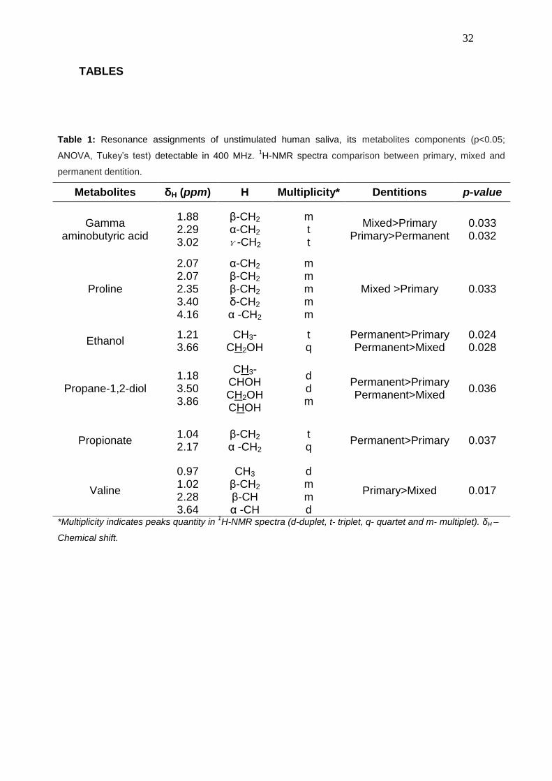
different. Table 1 summarizes the information about the metabolites identified
that changed with the dentition. The main difference between primary, mixed
and permanent dentition occurs in valine, where primary dentition has more
variation than mixed dentition (p = 0.017) followed by the ones listed in Table 1.
Table 2 and Fig. 2B show the standard distance, which was used to
assess the classifier’s performance, by computing the area under the receiver.
This index represents the distance between each observation and the center of
the ellipsis in the space of the PC scores from control subjects (permanent
dentition), normalized by the variance of each parameter (see Material and

24
Methods for details). The operating characteristic curve (AUC) was set at 0.933;
overall accuracy was set at 0.938; sensitivity (correct classification of
permanent dentition subjects) was set at 0.867 and specificity (correct
classification of permanent dentition subjects) was set at 1.000. The cut-off was
set at 4.48 for permanent dentition, the median primary dentition was 29.70 and
mixed was 16.64. Reflecting the higher variability in the primary dentition, the
standard distance show scattered and higher values, followed by the mixed and
the permanent dentition show the smallest inter-individual variability.
DISCUSSION
In the present study, metabolite data were determined by 1H NMR
spectroscopy and showed significant differences between dentitions. The saliva
assay is a growing area of research with implications for basic and clinical
settings. The proper evaluation starts with the construction of a data base in
which the profile of healthy individuals shows a pattern that can be identified as
normal. If differences in eating habits or oral hygiene interfere with the
methodology used the results would be inconclusive. But in this case, despite
the differences in age, dentitions stage, eating habits and oral hygiene the
results could be analyzed properly. In our opinion, since the difference in
dentitions was the feature able to select each group, all of them represent
“normal saliva” profile.
Furthermore, inter-individual variation was higher in primary dentition; it
was slight smaller in mixed and very small in permanent dentition. This small
variation in the permanent dentition can be related to a stabilization of oral
environment. Also, the NMR methodology alone may be not sensitive enough to
25
identify the differences and therefore, other methods could be used combined to
permit the analysis of even more aspects of children saliva from different
dentitions.
The analysis of metabolites that are different between each group gave
interesting insights: 1- propano-1,2-diol, propionate and ethanol are considered
microorganism metabolites (Arp, 1999), appear in low levels in primary dentition
and in much higher levels in permanent dentition. This difference can be
associated to eruption of permanent teeth and increased contact surfaces and
therefore elevated bacterial sites associated (Gerardu et al., 2003); 2- Valine is
an essential amino acid and probably originates from food. 3- Proline is a non-
essential amino acid that is part of collagen that is an important component of
dentine (Kukkola et al., 2003). In the present study, mixed dentition showed
higher levels of proline than primary dentition, probably because in mixed
dentition primary teeth are being reabsorbed with collagen rearrangement
leading to the presence of high levels of proline in the oral cavity. 4- gamma-
aminobutyric acid (GABA) appeared more expressively in permanent than in
primary dentition. Silwood et al (Silwood et al., 2002a) reported GABA in adults
saliva from and, in the present work, GABA was also found children saliva.
GABA is an inhibitory neurotransmitter found in the nervous systems and is the
chief inhibitory neurotransmitter in the vertebrate central nervous system.
(Mauch et al., 1993).
The ultimate trends of systems biology must be the integration of data
acquired from living organisms at the genomic, protein, and metabolite levels
and integrate the oral with the systemic environment (Lindon et al., 2006).
Through the combination of sensitive and high-resolution approaches an

26
improved understanding of an organism’s total biology will come along with a
better understanding of the causes and progression of human diseases. Hence
new and better targeted treatments can be developed giving improved
personalized healthcare.
CONCLUSION
In conclusion, differences among primary, mixed and permanent
dentition were identified. This study provides important information concerning
salivary metabolites of children in the pre-pubertal period and the differences
among healthy dentitions that could support future studies regarding oral and
systemic disorders.
ACKNOWLEDGMENTS
The authors acknowledge the funds from the International Center for Genetic
Engineering and Biotechnology (ICGEB, Trieste, Italy), Conselho Nacional de
Desenvolvimento Científico e Tecnológico (CNPq-Universal, Processo
473866/2007-2, Brazil), Fundação Carlos Chagas Filho de Amparo à Pesquisa
do Estado do Rio de Janeiro (FAPERJ, Brazil), Millennium Institute of Structural
Biology in Biotechnology and Biomedicine for the financial support.

27
REFERENCES
Arp DJ (1999). Butane metabolism by butane-grown 'Pseudomonas butanovora'. Microbiology 145 ( Pt 5):1173-80.
Atkinson JC, Fox PC (1993). Sjogren's syndrome: oral and dental considerations. J Am Dent Assoc 124(3):74-6, 78-82, 84-6.
Bertram HC, Eggers N, Eller N (2009). Potential of human saliva for nuclear magnetic resonance-based metabolomics and for health-related biomarker identification. Anal Chem 81(21):9188-93.
Brindle JT, Antti H, Holmes E, Tranter G, Nicholson JK, Bethell HW, et al. (2002). Rapid and noninvasive diagnosis of the presence and severity of coronary heart disease using 1H-NMR-based metabonomics. Nat Med 8(12):1439-44.
Chiappin S, Antonelli G, Gatti R, De Palo EF (2007). Saliva specimen: a new laboratory tool for diagnostic and basic investigation. Clin Chim Acta 383(1-2):30-40.
Englander HR, Jeffay AI, Fuller JB, Chauncey HH (1963). Glucose Concentrations in Blood Plasma and Parotid Saliva of Individuals with and without Diabetes Mellitus. J Dent Res 42(1246.
Flury BK, Riedwyl H (1986). Standard Distance in Univariate and Multivariate Analysis. Am Stat 40(3):249-251.
Gerardu VA, Buijs MJ, ten Cate JM, van Loveren C (2003). The effect of a single application of 40% chlorhexidine varnish on the numbers of salivary mutans streptococci and acidogenicity of dental plaque. Caries Res 37(5):369-73.
Helmerhorst EJ, Oppenheim FG (2007). Saliva: a dynamic proteome. J Dent Res 86(8):680-93.
Jolliffe IT (2002). Principal component analysis. . 2. ed. New York: Springer-Verlag.
Kaufman E, Lamster IB (2002). The diagnostic applications of saliva--a review. Crit Rev Oral Biol Med 13(2):197-212.
28
Keun HC, Beckonert O, Griffin JL, Richter C, Moskau D, Lindon JC, et al. (2002). Cryogenic probe 13C NMR spectroscopy of urine for metabonomic studies. Anal Chem 74(17):4588-93.
Kukkola L, Hieta R, Kivirikko KI, Myllyharju J (2003). Identification and characterization of a third human, rat, and mouse collagen prolyl 4-hydroxylase isoenzyme. J Biol Chem 278(48):47685-93.
Lenz EM, Bright J, Wilson ID, Morgan SR, Nash AF (2003). A 1H NMR-based metabonomic study of urine and plasma samples obtained from healthy human subjects. J Pharm Biomed Anal 33(5):1103-15.
Lindon JC, Holmes E, Bollard ME, Stanley EG, Nicholson JK (2004). Metabonomics technologies and their applications in physiological monitoring, drug safety assessment and disease diagnosis. Biomarkers 9(1):1-31.
Lindon JC, Holmes E, Nicholson JK (2006). Metabonomics techniques and applications to pharmaceutical research & development. Pharm Res 23(6):1075-88.
Mauch L, Abney CC, Berg H, Scherbaum WA, Liedvogel B, Northemann W (1993). Characterization of a linear epitope within the human pancreatic 64-kDa glutamic acid decarboxylase and its autoimmune recognition by sera from insulin-dependent diabetes mellitus patients. Eur J Biochem 212(2):597-603.
Nagler RM, Hershkovich O, Lischinsky S, Diamond E, Reznick AZ (2002). Saliva analysis in the clinical setting: revisiting an underused diagnostic tool. J Investig Med 50(3):214-25.
Ramadan Z, Jacobs D, Grigorov M, Kochhar S (2006). Metabolic profiling using principal component analysis, discriminant partial least squares, and genetic algorithms. Talanta 68(5):1683-91.
Silwood CJ, Lynch E, Claxson AW, Grootveld MC (2002a). 1H and (13)C NMR spectroscopic analysis of human saliva. J Dent Res 81(6):422-7.
Silwood CL, Grootveld M, Lynch E (2002b). 1H NMR investigations of the molecular nature of low-molecular-mass calcium ions in biofluids. J Biol Inorg Chem 7(1-2):46-57.
29
Siqueira WL, Siqueira MF, Mustacchi Z, de Oliveira E, Nicolau J (2007). Salivary parameters in infants aged 12 to 60 months with Down syndrome. Spec Care Dentist 27(5):202-5.
Streckfus C, Bigler L, O'Bryan T (2002). Aging and salivary cytokine concentrations as predictors of whole saliva flow rates among women: a preliminary study. Gerontology 48(5):282-8.
Streckfus CF, Bigler LR, Zwick M (2006). The use of surface-enhanced laser desorption/ionization time-of-flight mass spectrometry to detect putative breast cancer markers in saliva: a feasibility study. J Oral Pathol Med 35(5):292-300.
Tager BN (1951). Endocrine problems in orthodontics; the concept of a relative metabolic insufficiency in bone due to rapid skeletal growth and slow sexual maturation. Am J Orthod 37(11):867-75.
Takeda I, Stretch C, Barnaby P, Bhatnager K, Rankin K, Fu H, et al. (2009). Understanding the human salivary metabolome. NMR Biomed.
Wahllander A, Mohr S, Paumgartner G (1990). Assessment of hepatic function. Comparison of caffeine clearance in serum and saliva during the day and at night. J Hepatol 10(2):129-37.
Weise M, Eisenhofer G, Merke DP (2002). Pubertal and gender-related changes in the sympathoadrenal system in healthy children. J Clin Endocrinol Metab 87(11):5038-43.
Yan W, Apweiler R, Balgley BM, Boontheung P, Bundy JL, Cargile BJ, et al. (2009). Systematic comparison of the human saliva and plasma proteomes. Proteomics Clin Appl 3(1):116-134.
Zhang L, Farrell JJ, Zhou H, Elashoff D, Akin D, Park NH, et al. Salivary Transcriptomic Biomarkers for Detection of Resectable Pancreatic Cancer. Gastroenterology 138(3):949-957 e7.

30
FIGURES
Fig. 1: Representative 1H NMR spectrum of child saliva samples in the 0–4.5ppm (A,B,C) and
5.5–10.0ppm regions (D,E,F). A, D- Primary dentition; B, E- Mixed dentition; C, F- Permanent
dentition. No statistical differences were found among dentitions in the 5.5-10.0ppm region.
Assignments: 1- propionate, 2- ethanol, 3- propane-1,2-diol, 4- gamma aminobutyric acid, 5-
proline, 6-valine.

31
Fig. 2: A- Principal components analysis shows no differences among dentitions; B- Standard
distance showing low inter-individual variability in permanent dentition.
32
TABLES
Table 1: Resonance assignments of unstimulated human saliva, its metabolites components (p<0.05;
ANOVA, Tukey’s test) detectable in 400 MHz. 1H-NMR spectra comparison between primary, mixed and
permanent dentition.
Metabolites δH (ppm) H Multiplicity* Dentitions p-value
Gamma aminobutyric acid
1.88 2.29 3.02
β-CH2
α-CH2
ɣ -CH2
m t t
Mixed>Primary Primary>Permanent
0.033 0.032
Proline
2.07 2.07 2.35 3.40 4.16
α-CH2 β-CH2
β-CH2
δ-CH2
α -CH2
m m m m m
Mixed >Primary
0.033
Ethanol 1.21 3.66
CH3-
CH2OH t q
Permanent>Primary Permanent>Mixed
0.024 0.028
Propane-1,2-diol 1.18 3.50 3.86
CH3-CHOH CH2OH CHOH
d d m
Permanent>Primary Permanent>Mixed
0.036
Propionate 1.04 2.17
β-CH2
α -CH2 t q
Permanent>Primary 0.037
Valine
0.97 1.02 2.28 3.64
CH3 β-CH2
β-CH α -CH
d m m d
Primary>Mixed 0.017
*Multiplicity indicates peaks quantity in 1H-NMR spectra (d-duplet, t- triplet, q- quartet and m- multiplet). δH –
Chemical shift.
33
Table 2: Standard distance for classify primary, mixed and
permanent dentition.
Primary dentition
Mixed dentition
Permanent dentition
4,97 7,96 3,86 6,55 5,39 2,68 11,61 3,81* 3,25 9,70 9,16 2,87 6,33 7,76 3,56 23,93 16,93 2,80
111,37 7,48 2,51 6,93 7,87 3,68 7,24 7,35 3,88 6,17 11,95 3,60 3,06* 7,83 3,71 9,89 4,22* 3,88 4,59 18,50 3,82
181,86 14,83 3,86 51,36 10,63 3,85
6,49 3,87 5,83 3,27 145,60
29,70 ± 50,75** 16,64 ± 32,44** 3,47 ± 0,48** Standard distance= difference between dentitions. Cut-off point =
4.48. *Indicates subjects out of cut-off; ** Media ± Standard
deviation.
34
4.2 ARTIGO 2
NMR salivary metabolites components analysis from children with and
without caries lesion
Tatiana Kelly da Silva Fidalgo1
Renata Angeli2
Adriane Mara de Souza Muniz3
Jurandir Nadal4
Liana Bastos Freitas-Fernandes1
Fabio Lacerda de Almeida2
Ana Paula Valente2
Ivete Pomarico Ribeiro de Souza1*
1Department of Pediatric Dentistry and Orthodontics, School of Dentistry,
Federal University of Rio de Janeiro, Rio de Janeiro, Brazil;
2National Center for Nuclear Magnetic Resonance of Macromolecules, Medical
Biochemistry Institute, Federal University of Rio de Janeiro, Rio de Janeiro,
Brazil;
3Department of Post-graduation, Army Physical Education School, EsEFEx,
Janeiro, Rio de Janeiro, Brazil;
4Biomedical Engineering Program, COPPE, Federal University of Rio de
Janeiro, Rio de Janeiro, Brazil.
35
ABSTRACT
We tested the hypothesis that the Principal Component Analysis (PCA) and
standard distance (SD) distinguish salivary biomolecules of caries lesion-free
from caries-lesion subjects. Unstimulated whole saliva samples from children
(3-12 years old) were collected and separated according to presence (n=18)
and absence of caries lesion (n=15; dmft/DMFT index=0). 1H NMR spectra
were acquired on a Brucker 400 MHz spectrometer. PCA was applied and SD
was calculated and correlated with dmft/DMFT index; and F test to assess the
metabolite differences between the groups (p<0.05). PCA was able to
distinguish both groups and SD was smaller in caries lesion-free subjects.
Lactate, lysine, glucose, glutamate, glutamine, propionate, ornithine and
saturated fatty acid were higher in the caries lesion group (p<0.05). Choline and
4-hydroxyproline were higher in the caries lesion-free group (p<0.05). It was
possible to distinguish the specific biomolecules from each group and to
separate it using classificatory methods.
Key words: Whole saliva, Magnetic Resonance Spectroscopy, Principal
component analysis, Metabolomic, Dental caries.
36
INTRODUCTION
Whole saliva and its composition has an important biological function in
maintaining oral health (Zehetbauer et al., 2009). Physiological, pathological,
and environmental factor changes in salivary composition could be correlated to
disease susceptibility. Hence, human saliva represents a potential source of
novel diagnostic markers for oral diseases such as dental caries (de Almeida
Pdel et al., 2008; Dodds et al., 2005; Hardt et al., 2005; Helmerhorst and
Oppenheim, 2007). In the proteomic era most of the studies that correlate
salivary composition with dental caries etiology and other oral diseases have
used protein associations. Particularly in the cariology area, the presence or
absence of these inherited markers have potential value to evaluate dental
caries susceptibility in each human group (Hardt et al., 2005). However, saliva
is a complex biofluid that is not only composed of proteins, therefore it is
necessary to explore the other low molecular weight metabolite profiles
(Grootveld and Silwood, 2005; Silwood et al., 2002a).
Low molecular weight organic metabolites, such as glucose, amino acids,
lipids like fatty acids have been largely overlooked when using gas-liquid
chromatography, column chromatography, and high performance liquid
chromatography (Kaufman and Lamster, 2002; Tomita et al., 2008). Also these
methods need specific information of the fluid under study. However, Nuclear
Magnetic Resonance (NMR) spectroscopy besides being a non-invasive
method is able to identify many metabolites at the same time and constitutes a
reproducible method (Kaufman and Lamster, 2002; Silwood et al., 2002a;
Silwood et al., 2002b; Tomita et al., 2008).
37
A useful method to analyze metabolomic data is the principal component
analysis (PCA). PCA is a multivariate statistical technique used to reduce the
dimensionality of a data set with a large number of interrelated variables
(Jolliffe, 2002). It is used to group metabolites from different conditions and
classifies subjects (Holmes and Antti, 2002). In a previous work, we used
successfully NMR and PCA to analyze healthy children with different dentitions
(Fidalgo et al., 2010). As far as we know, salivary metabolites from caries
individuals have not been studied using NMR spectroscopy and PCA. Here, we
tested the hypothesis that Principal Component Analysis and Standard distance
distinguish 1H NMR salivary biomolecules of caries lesion-free from caries-
lesion individuals.
MATERIALS AND METHODS
Sample Collection and Preparation
Thirty three children with mixed dentition attending the Pediatric Dentistry Clinic
at the Federal University of Rio de Janeiro were recruited. These subjects
ranged in age from 3 to 12 years old, none of them had any periodontal disease
or systemic disease and had not taken any systemic antibiotics or had been
using anti-bacterial toothpaste in the six months prior to sample collection. A
visual-tactile examination of each subject was performed by a single examiner.
A primary and permanent Decayed Missing and Filled Surfaces (dmft/DMFT)
index was calculated for each subject and they were separated according to the
presence (n=18) or absence of caries lesion with dmft/DMFT index as 0 (n=15).
A 3 mL of unstimulated whole saliva was expectorated (~5 minutes) into a
plastic universal tube in the morning. Each subject was also requested to refrain
38
from oral activities (i.e., eating, drinking, toothbrushing, oral rinsing) during the
period between awakening and sample collection, about 2 hours (Silwood et al.,
2002a). All samples were centrifuged at 10,000g and 4oC, and the supernatants
were stored at –80°C until NMR analysis. The use of human materials was
approved by the proper Research Ethics Committee of Community Health
Studies.
NMR Measurements
NMR spectra were acquired using a Bruker 400 MHz Advance spectrometer
(Bruker Biospin, Rheinstetten, Germany) equipped with a Bruker 5 mm high-
resolution probe and operating at a frequency of 400.13 (1H) MHz and a probe
temperature of 25°C.
Samples were prepared by mixing 0.45 mL of salivary supernatant, deuterium
oxide (0.05 mL, providing a field frequency lock) and a 500 µM solution of
sodium 3-trimethylsilyl [2,2,3,3-2H4] propionate (TSP) for chemical shift
reference of 1H spectra, δ = 0.00 ppm. A control investigation showed that the
samples of whole saliva were stable throughout the NMR acquisitional period
(data not shown). High-resolution 1H NMR spectra could be obtained, and
based on 1H-1H total correlation (TOCSY) experiments the major signals could
be unambiguously assigned consistent with earlier studies (Bertram et al., 2009;
Silwood et al., 2002a).
NMR signals were determined over a defined range of frequency
resonances called chemical shifts expressed in ppm. Saliva was analyzed using
NMR spectroscopy and assignments were identified based on Silwood et al
(Silwood et al., 2002a) and Human Metabolome database
(http://www.hmdb.ca/).
39
Each spectral dataset was normalized to the total sum of the integrals to
partially compensate for differences in concentrations.
Statistical analysis
The metabolite data were computed on the statistical program AMIX (Bruker
Biospin, Rheinstetten, Germany). Each NMR spectrum was reduced to a
smaller number of variables, calculated by integrating regions of equal bucket
size of 0.01 ppm. The water region was excluded. The datasets were stored in
a matrix E with 33 rows (33 subjects) and 906 columns (906 chemical shifts).
The matrix was normalized by pareto scaling (accuracy= 0.94, sensibility= 0.93,
specificity= 0.94) (Ramadan et al., 2006). Principal component analysis (PCA)
was applied to the covariance matrices S (906x906) from each E, by linear
system (Jolliffe, 2002):
ppp xSx
where λ is the eigenvalues of S ranked in decreasing order and x is the
corresponding normalized eigenvectors or principal components (PCs). These
PCs are independent waveform features, representing the loading factor
applied to the corresponding sample of the original waveforms. The first PCs
correspond to the largest sources of variation, being orthogonal to each other.
The screen plot selected the relevant PCs for the analysis. Datasets of PC
vectors retained in the screen test were represented as data points in 2
dimensional loading plots. The PC scores of the permanent, mixed and primary
dentition individuals correspond to the product of the respective eigenvector and
the matrix E.
The standard distance (SD) proposed by Flury and Riedwyl (Flury and
Riedwyl, 1986) was calculated including the selected PC scores in matrix E.
40
This index represents the distance between each observation and the center of
the ellipsis in the space of the PC scores from caries lesion-free subjects. To
classify caries lesion-free and caries lesion subjects, the cut-off point between
SD values from the caries lesion-free and caries lesion subjects was obtained
by logistic regression. The classifier performance was assessed by the leave-
one-out cross-validation technique (Breiman et al., 1998). Therefore, the points-
out of this cut-off is considered subjects in primary or mixed dentition.
The Wilcoxon rank-sum test was used to compare standard values from
caries lesion-free and caries lesion subjects. The F test was performed to
compare the chemical shifts that contributed to separation between groups. The
significance level was set at 5%. All signal processing procedures and statistical
tests were executed in Matlab 6.5 (The Mathworks, USA).
RESULTS
The caries lesion-free group was made up of 18 children (mean age=7.94 ±
2.09, 9 female and 9 male) The caries lesion group was made up of 15 children
(n=15, 7.23 ± 2.01, 6 female and 9 male). All children had mixed dentition.
The 1H NMR spectra of caries lesion and caries lesion-free saliva with
several resonances identified is shown in Fig. 1. Each different chemical shift
corresponds to a salivary component. There are differences in intensity of
resonances between caries lesion-free and caries lesion subjects.
The screen test retained 10 PCs in the analysis, which explained 86.13%
of the total variation. The first third and fourth PC scores presented statistical
difference between groups. The scatter plot formed by the third PC score
versus the fourth PC score, revealed differences caries lesion-free and caries-
41
lesion samples areas (Fig. 2A). The classification was mainly carried out by the
third PC score plane when the most caries-lesion subjects were placed in the
left side of the graphic.
Ten PCs scores were used to calculate the SD. The cut-off point
calculated by logistic regression was 3.98. The caries-lesion free subjects
presented the SD mean statistically lower than caries-lesion subjects (Fig. 2B;
Table 1). Only one subject from caries-lesion group presented SD lower than
cut-off point. In the other hand, caries lesion-free subjects presented only one
subject sample upper than 3.98. In both cases, it was not found any particularity
of these individuals. Additionally, it was not found statistical correlation neither
from SD and dmft/DMFT (p=0.45) nor number of teeth with caries-lesion
(p=1.10).
The loadings of the 10 PCs which most influenced the separation of the
groups are summarized in Table 2.
DISCUSSION
To the best of our knowledge this is the first time that caries lesion and
caries lesion-free saliva samples have been evaluated using NMR based
metabolomics. High-resolution 1H NMR spectra were obtained, and based on
2D experiments, the major signals could be unambiguously assigned (Silwood
et al., 2002a). Several differences in some regions of 1H NMR spectra between
caries lesion and caries lesion-free subjects were found. The study of salivary
metabolites is growing. There is a particular interest in establishing biomarkers
related to health, which can then be used as diagnostic tools (Bertram et al.,
2009). For this reason it is important to establish the salivary metabolites of
caries lesion-free subjects.
42
The high-resolution 1H NMR spectroscopy datasets are represented as
complex matrices with hundreds of proton signals originating from the various
metabolites. These complexities can be untangled using chemometric methods
that are used to reduce the size of the 1H NMR data for visualization purposes
and to identify inherent patterns among sets of spectral measurements
(Ramadan et al., 2006). Principal components (PCs) are new variables created
from linear combinations of the starting spectra descriptors with appropriate
weighting coefficients. In the present study PC3 and PC4 contributed most to
the separation of the two groups and were able to separate caries lesion-free
subjects demonstrating a satisfactory model.
SD is a multivariate statistical method to quantify the extent by which an
experimental condition deviates from that of an unimpaired control (Schutte et
al., 2000). In our study, SD is computed using PCA as standard multivariate
statistical technique. The classifier model, calculated by the leave-one-out
approach, showed a good accuracy. Although a direct relationship between SD
and dmft/DMFT or number of teeth with caries lesion was not found. However it
is possible that the SD of the groups not only verified the presence or absence
of caries, but also determined an individual’s susceptibility to caries lesion,
when combined with other factors.
Metabolites such as propionate lactate, n-butyrate and n-caproate are
probably metabolites from oral microorganisms and for that reason caries lesion
subjects have an increase of these metabolites. On the other hand, choline and
ornithine are probably endogen metabolites. Thus, the presence or absence of
endogen biomarkers will be of great value for evaluating susceptibility of dental
caries. Sucrose has been considered the most cariogenic carbohydrate due to
43
the increased porosity of dental plaque matrix formed in its presence (van
Houte, 1994). This disaccharide is composed of glucose and fructose; several
oral microorganisms low the pH of dental plaque in presence of glucose
(Fejerskov et al., 1992; Vadeboncoeur and Pelletier, 1997). In the current study,
lower amounts of sucrose but higher amounts of α–glucose were found in the
caries lesion than the caries lesion-free subjects, indicating that sucrose was
decomposed by oral microorganisms in individuals that presented more caries
susceptibility into α–glucose for energetic metabolism (Vadeboncoeur and
Pelletier, 1997).
Hexogen metabolites were identified in saliva from both caries lesion and
caries lesion-free subjects. Although, individuals were requested to refrain from
oral activities before sample collection, sodium dodecyl sulfate was identified.
Lauryl sulfates help clean the teeth, and provide foam that helps to carry away
debris (Petersen et al., 2006). It is an ionic surfactant component that is widely
used in toothpaste (Rantanen et al., 2003). It was found in caries lesion
subjects in higher amounts, probably due to adsorption in dental biofilm and
irregular surfaces of the caries lesions.
Different salivary protein profiles between caries lesion and caries lesion-
free subjects have been described (Anderson and Mandel, 1982; Bergandi et
al., 2007). According to Ayad et al (Ayad et al., 2000), proteolytic processing of
parotid salivary proteins differs between individuals who have always been
caries lesion-free and those who have experienced dental decay. These
differences could be explained by differential expression of proteases, protease
inhibitors or polymorphisms at potential proteolytic cleavage sites. Furthermore,
Cowman et al (Cowman et al., 1979) reported that saliva from caries lesion-free
44
individuals differs both in the number and sensitivity of proteins susceptible to
bacterial degradation, compared with saliva from caries active patients. We
suggest that the same can occur with low-weight metabolites, as was seen in
the present study. On the other hand, the literature shows a relationship
between amino acids in plasma and saliva after a protein-rich test meal intake,
did not observe a significant relation between the increase in concentration of
these amino acids in venous plasma and whole saliva (Brand et al., 1997).
There is also evidence that several salivary aminoacids do not originate from
food or microorganism metabolites (Nagata et al., 2006).
According to Tomita et al (Tomita et al., 2008), lipid concentrations in
parotid saliva from caries susceptible subjects were higher than those from
caries resistant ones, and a difference in fatty acid composition was detected
between them, corroborating our data. Although the current study analyzed
whole saliva, saturated fatty acid was highest in caries lesion individuals. The
variations in the lipid levels and fatty acid composition may be associated with
those in caries development. It was put forward that fatty acid had been
demonstrated in salivary mucous glycoprotein and covalently bound. The
presence of the lipids on salivary pellicle on teeth surface increases the effect
the caries development by inhibition of acid diffusion (Slomiany et al., 1989).
Much attention has been recently dedicated to methods able to detect
small tooth lesions (Nyvad, 2004). The present study evaluated differences
between salivary metabolites from caries lesion-free and caries lesion subjects.
Caries susceptibility is an important addition to the delivery of non-surgical
preventive care such as topical fluorides, and therefore should be studied.
Further information is necessary to support the use of this methodology as a

45
susceptible, diagnostic and disease progress tool in dental caries and other oral
health disorders.
ACKNOWLEDGMENTS
The authors acknowledge the funds from the International Center for Genetic
Engineering and Biotechnology (ICGEB, Trieste, Italy), Conselho Nacional de
Desenvolvimento Científico e Tecnológico (CNPq-Universal, Processo
473866/2007-2, Brazil), Fundação Carlos Chagas Filho de Amparo à Pesquisa
do Estado do Rio de Janeiro (FAPERJ, Brazil), Millennium Institute of Structural
Biology in Biotechnology and Biomedicine for the financial support.
REFERENCES
Anderson LC, Mandel ID (1982). Salivary protein polymorphisms in caries-resistant adults. J Dent Res 61(10):1167-8. Ayad M, Van Wuyckhuyse BC, Minaguchi K, Raubertas RF, Bedi GS, Billings RJ, et al. (2000). The association of basic proline-rich peptides from human parotid gland secretions with caries experience. J Dent Res 79(4):976-82. Bergandi L, Defabianis P, Re F, Preti G, Aldieri E, Garetto S, et al. (2007). Absence of soluble CD14 in saliva of young patients with dental caries. Eur J Oral Sci 115(2):93-6. Bertram HC, Eggers N, Eller N (2009). Potential of human saliva for nuclear magnetic resonance-based metabolomics and for health-related biomarker identification. Anal Chem 81(21):9188-93. Brand HS, Jorning GG, Chamuleau RA, Abraham-Inpijn L (1997). Effect of a protein-rich meal on urinary and salivary free amino acid concentrations in human subjects. Clin Chim Acta 264(1):37-47. Breiman L, Friedman J, Olshen R, Stone C (1998). Classification and Regression Trees: Boca Raton: Chapman & Hall/CRC. Cowman RA, Schaefer SJ, Fitzgerald RJ, Rosner D, Shklair IL, Walter RG (1979). Differential utilization of proteins in saliva from caries-active and caries-free subjects as growth substrates by plaque-forming streptococci. J Dent Res 58(10):2019-27.

46
de Almeida Pdel V, Gregio AM, Machado MA, de Lima AA, Azevedo LR (2008). Saliva composition and functions: a comprehensive review. J Contemp Dent Pract 9(3):72-80. Dodds MW, Johnson DA, Yeh CK (2005). Health benefits of saliva: a review. J Dent 33(3):223-33. Fejerskov O, Scheie AA, Manji F (1992). The effect of sucrose on plaque pH in the primary and permanent dentition of caries-inactive and -active Kenyan children. J Dent Res 71(1):25-31.
Fidalgo TKS, Gonsalves E, Santos RP, Muniz AMS, Almeida FL, Souza IPR, Freitas-Fernandes LB, Valente AP. NMR Metabolomic Part I: Salivary Metabolites of Oral Healthy Children. J Dent Res (Submitted in 2010). Flury BK, Riedwyl H (1986). Standard Distance in Univariate and Multivariate Analysis. Am Stat 40(3):249-251. Grootveld M, Silwood CJ (2005). 1H NMR analysis as a diagnostic probe for human saliva. Biochem Biophys Res Commun 329(1):1-5. Hardt M, Thomas LR, Dixon SE, Newport G, Agabian N, Prakobphol A, et al. (2005). Toward defining the human parotid gland salivary proteome and peptidome: identification and characterization using 2D SDS-PAGE, ultrafiltration, HPLC, and mass spectrometry. Biochemistry 44(8):2885-99. Helmerhorst EJ, Oppenheim FG (2007). Saliva: a dynamic proteome. J Dent Res 86(8):680-93. Holmes E, Antti H (2002). Chemometric contributions to the evolution of metabonomics: mathematical solutions to characterising and interpreting complex biological NMR spectra. Analyst 127(12):1549-57. Jolliffe IT (2002). Principal component analysis. . 2. ed. New York: Springer-Verlag. Kaufman E, Lamster IB (2002). The diagnostic applications of saliva--a review. Crit Rev Oral Biol Med 13(2):197-212. Nagata Y, Higashi M, Ishii Y, Sano H, Tanigawa M, Nagata K, et al. (2006). The presence of high concentrations of free D-amino acids in human saliva. Life Sci 78(15):1677-81. Nyvad B (2004). Diagnosis versus detection of caries. Caries Res 38(3):192-8. Petersen FC, Assev S, Scheie AA (2006). Combined effects of NaF and SLS on acid- and polysaccharide-formation of biofilm and planktonic cells. Arch Oral Biol 51(8):665-71.
47
Ramadan Z, Jacobs D, Grigorov M, Kochhar S (2006). Metabolic profiling using principal component analysis, discriminant partial least squares, and genetic algorithms. Talanta 68(5):1683-91. Rantanen I, Jutila K, Nicander I, Tenovuo J, Soderling E (2003). The effects of two sodium lauryl sulphate-containing toothpastes with and without betaine on human oral mucosa in vivo. Swed Dent J 27(1):31-4. Schutte LM, Narayanan U, Stout JL, Selber P, Gage JR, Schwartz MH (2000). An index for quantifying deviations from normal gait. Gait Posture 11(1):25-31. Silwood CJ, Lynch E, Claxson AW, Grootveld MC (2002a). 1H and (13)C NMR spectroscopic analysis of human saliva. J Dent Res 81(6):422-7. Silwood CL, Grootveld M, Lynch E (2002b). 1H NMR investigations of the molecular nature of low-molecular-mass calcium ions in biofluids. J Biol Inorg Chem 7(1-2):46-57. Slomiany BL, Murty VL, Mandel ID, Zalesna G, Slomiany A (1989). Physico-chemical characteristics of mucus glycoproteins and lipids of the human oral mucosal mucus coat in relation to caries susceptibility. Arch Oral Biol 34(4):229-37. Tomita Y, Miyake N, Yamanaka S (2008). Lipids in human parotid saliva with regard to caries experience. J Oleo Sci 57(2):115-21. Vadeboncoeur C, Pelletier M (1997). The phosphoenolpyruvate:sugar phosphotransferase system of oral streptococci and its role in the control of sugar metabolism. FEMS Microbiol Rev 19(3):187-207. van Houte J (1994). Role of micro-organisms in caries etiology. J Dent Res 73(3):672-81. Zehetbauer S, Wojahn T, Hiller KA, Schmalz G, Ruhl S (2009). Resemblance of salivary protein profiles between children with early childhood caries and caries-free controls. Eur J Oral Sci 117(4):369-73.
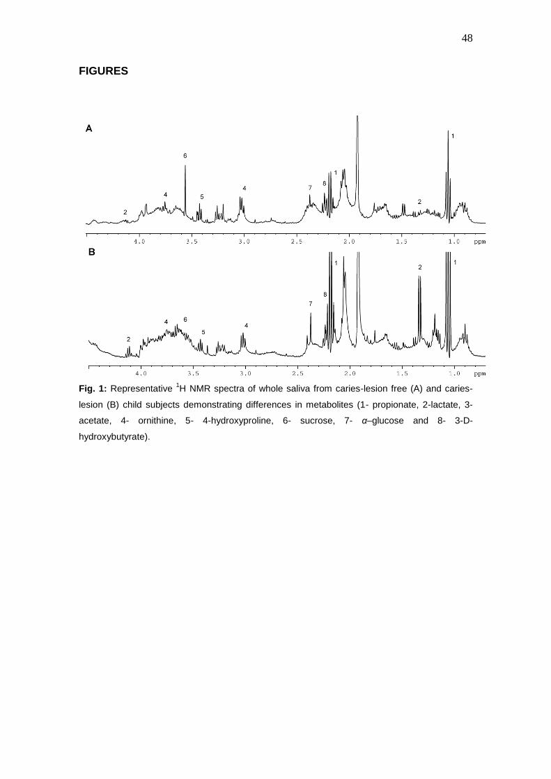
48
FIGURES
Fig. 1: Representative
1H NMR spectra of whole saliva from caries-lesion free (A) and caries-
lesion (B) child subjects demonstrating differences in metabolites (1- propionate, 2-lactate, 3-
acetate, 4- ornithine, 5- 4-hydroxyproline, 6- sucrose, 7- α–glucose and 8- 3-D-
hydroxybutyrate).
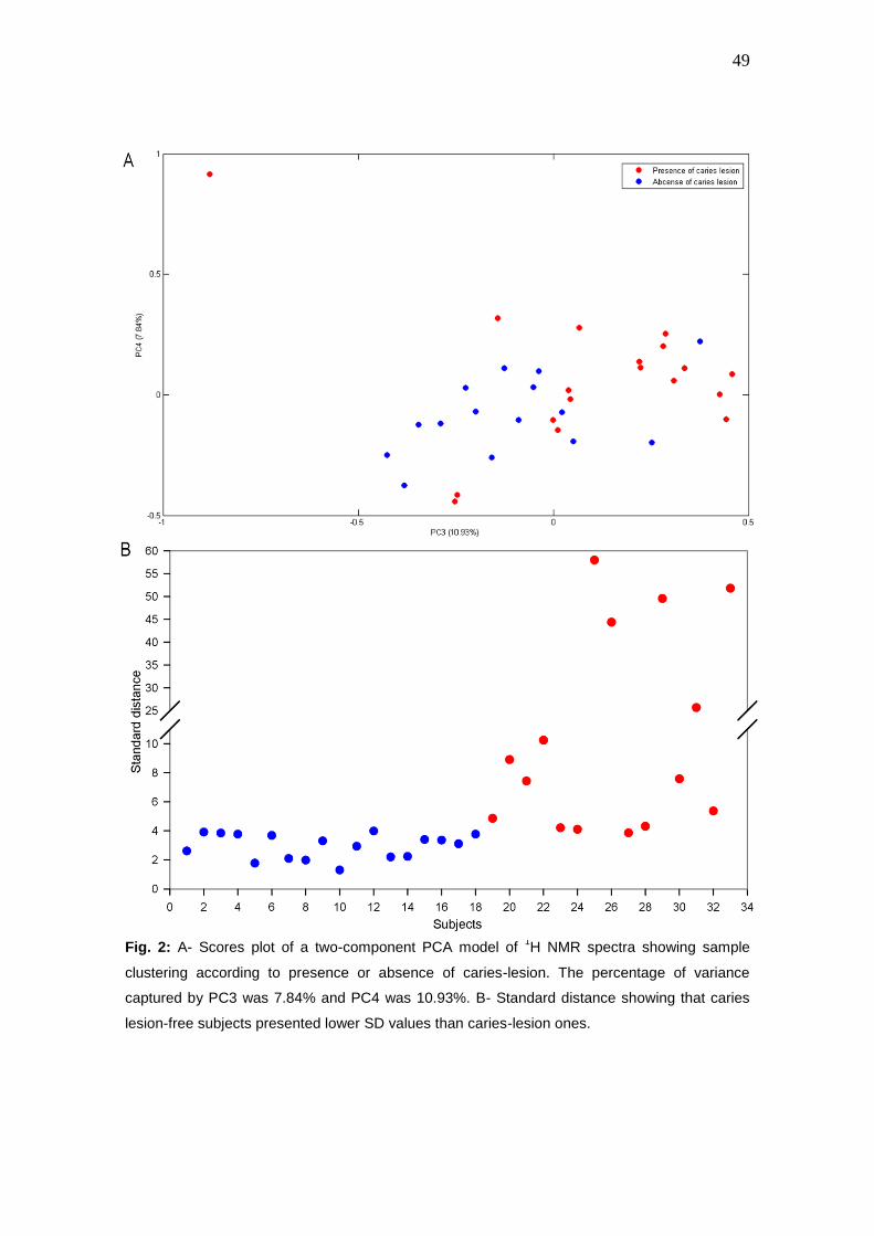
49
Fig. 2: A- Scores plot of a two-component PCA model of 1H NMR spectra showing sample
clustering according to presence or absence of caries-lesion. The percentage of variance
captured by PC3 was 7.84% and PC4 was 10.93%. B- Standard distance showing that caries
lesion-free subjects presented lower SD values than caries-lesion ones.

50
TABLES
Table 1: The standard distance and the distances classifying caries-free lesion and
caries-lesion individuals with respectives dmft/DMFT and number of teeth with
caries-lesion.
Caries-lesion
free Caries-lesion dmft/DMFT Number of teeth
with caries-lesion 3.99* 57.97 3 3 3.91 51.80 9 9
3.85 49.54 5 5
3.77 44.37 5 4
3.77 25.67 7 7
3.68 10.24 5 2
3.40 8.90 1 1
3.36 7.58 5 4
3.31 7.43 6 5
3.11 5.37 5 4
2.94 4.86 4 3
2.61 4.31 5 5
2.20 4.21 2 2
2.24 4.10 7 2
2.10 3.86** 5 4
1.98
1.78
1.30
2.96 ± 0.85§ 19.35 ± 20.58§ 4.92 ± 2.14§ 4.00 ± 2.24§ Cut-off=3.98; *Upper than cut-off ; **Lower than cut-off.
§Media ± Standard deviation
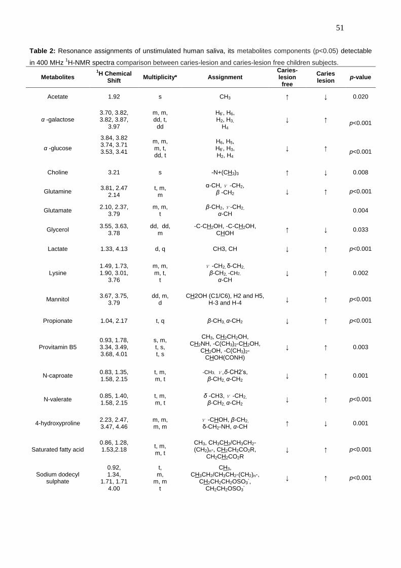
51
Table 2: Resonance assignments of unstimulated human saliva, its metabolites components (p<0.05) detectable
in 400 MHz 1H-NMR spectra comparison between caries-lesion and caries-lesion free children subjects.
Metabolites
1H Chemical
Shift Multiplicity* Assignment
Caries-lesion free
Caries lesion
p-value
Acetate 1.92 s CH3 ↑ ↓ 0.020
α -galactose 3.70, 3.82, 3.82, 3.87,
3.97
m, m, dd, t,
dd
H6’, H6, H2, H3,
H4 ↓ ↑
p<0.001
α -glucose
3.84, 3.82 3.74, 3.71 3.53, 3.41
m, m, m, t, dd, t
H6, H5, H6’, H3, H2, H4
↓ ↑
p<0.001
Choline 3.21 s -N+(CH3)3 ↑ ↓ 0.008
Glutamine 3.81, 2.47
2.14 t, m,
m
α-CH, ɣ -CH2, β -CH2
↓ ↑ p<0.001
Glutamate 2.10, 2.37,
3.79 m, m,
t β-CH2, ɣ -CH2,
α-CH 0.004
Glycerol 3.55, 3.63,
3.78 dd, dd,
m -C-CH2OH, -C-CH2OH,
CHOH ↑ ↓ 0.033
Lactate 1.33, 4.13 d, q CH3, CH ↓ ↑ p<0.001
Lysine 1.49, 1.73, 1.90, 3.01,
3.76
m, m, m, t,
t
ɣ -CH2, δ-CH2,
β-CH2, -CH2,
α-CH ↓ ↑ 0.002
Mannitol 3.67, 3.75,
3.79 dd, m,
d CH2OH (C1/C6), H2 and H5,
H-3 and H-4 ↓ ↑ p<0.001
Propionate 1.04, 2.17 t, q β-CH3, α-CH2 ↓ ↑ p<0.001
Provitamin B5 0.93, 1.78, 3.34, 3.49, 3.68, 4.01
s, m, t, s, t, s
CH3, CH2CH2OH, CH2NH, -C(CH3)2-CH2OH,
CH2OH, -C(CH3)2-CHOH(CONH)
↓ ↑ 0.003
N-caproate 0.83, 1.35, 1.58, 2.15
t, m, m, t
-CH3, ɣ ,δ-CH2’s, β-CH2, α-CH2
↓ ↑ 0.001
N-valerate 0.85, 1.40, 1.58, 2.15
t, m, m, t
δ -CH3, ɣ -CH2,
β-CH2, α-CH2 ↓ ↑ p<0.001
4-hydroxyproline 2.23, 2.47, 3.47, 4.46
m, m, m, m
ɣ -CHOH, β-CH2,
δ-CH2-NH, α-CH ↑ ↓ 0.001
Saturated fatty acid 0.86, 1.28, 1.53,2.18
t, m, m, t
CH3, CH3CH2/CH3CH2-(CH2)n-, CH2CH2CO2R,
CH2CH2CO2R ↓ ↑ p<0.001
Sodium dodecyl sulphate
0.92, 1.34,
1.71, 1.71 4.00
t, m,
m, m t
CH3, CH3CH2/CH3CH2-(CH2)n-,
CH2CH2CH2OSO3-,
CH2CH2OSO3-
CH2CH2OSO3 - - -
↓ ↑ p<0.001

52
Sucrose
3.47,3.56, 3.68, 3.76, 3.76, 3.76, 3.83, 3.83, 3.86, 3.89, 4.05, 4.22,
5.41
T, m, s, t,
m, m, m, t
m, m, t, d,
d
H4, H2, H1’, H3, H6, H6’, H6’’, H3, H5, H5, H4, H3’,
H1
↑
↓ 0.006
Thymol 1.25, 2.18, 3.18, 6.61, 6.66, 6.99
d, s, m, dd, s, dd
(CH3)2CH, CH3-Ar, (CH3)2CH, H6,
H2, H5
↓ ↑ p<0.001
3-D-hydroxybutyrate
4.13, 2.45, 2.34, 1.24
dd, dd, dd d
CH3, CH2,
CH2, CH ↑
↓ p<0.001
Ornithine 1.81, 1.95, 3.05, 3.80
m, m t, t
ɣ -CH2 β-CH2, δ-CH2, α-CH
↓ ↑ p<0.001
Xylitol 3.64, 3.64, 3.72, 3.80,
3.86
m, dd, dd, m, dd
H3, CH2OH (C1/C5), CH2OH (C1/C5), H2 and H4, CH2OH
(C1/C6) ↓ ↑ p<0.001
*Multiplicity indicates peaks quantity in 1H-NMR spectra (s, singlet; d, doublet; t, triplet; q, quartet; m, multiplet).
Were used 10 PCs that explained 86.13% of variation to find out precisely which regions of the NMR spectra have
caused the separation between groups. The respectives metabolites that presented statistical differences (p<0.05;
F test) are indicated. The metabolite α-galactose was identified in all 10 PCs.
53
4.3 ARTIGO 3
Local effect of an antihistaminic-containing syrup in salivary metabolites.
An in vitro and in vivo evaluation.
Tatiana Kelly da Silva Fidalgo1
Liana Bastos Freitas Fernandes1
Fabio Lacerda de Almeida2
Ana Paula Valente2
Ivete Pomarico Ribeiro de Souza1
1Department of Pediatric Dentistry and Orthodontics, School of Dentistry,
Federal University of Rio de Janeiro, Rio de Janeiro, Brazil;
2National Center for Nuclear Magnetic Resonance of Macromolecules, Medical
Biochemistry Institute, Federal University of Rio de Janeiro, Rio de Janeiro,
Brazil.
54
ABSTRACT
The effect of medicines-containing syrup on the tooth surface is largely studied
in literature. On the other hand, few studies evaluate salivary metabolites
changes. Therefore, the aim of this study was to evaluate in vitro and in vivo
local effect of an antihistaminic-containing syrup in salivary metabolites using
high-resolution 1H NMR spectroscopy. For in vitro interaction, unstimulated
whole saliva from 5 healthy volunteers was collected and was in vitro mixture
with an antihistaminic-containing syrup (Claritin®). For in vivo study, before
sample collection, the same volunteers were instructed to rinse the mouth with
Claritin® for 20 seconds. The in vitro and in vivo mixture were studied in 1H
NMR spectra were acquired and processed on a Bruker 400 MHz Advance
spectrometer. For statistical analysis was used principal analysis component
(PCA) and paired t test (p<0.05). The metabolites acetate, histidine, ɣ -
aminobutyrate, glutamate, isoleucine, phenylalanine, propionate, trimethylamine
and valine presented lower concentration after antihistaminic interaction. The
differences in salivary metabolites were found both in vitro and in vivo
antihistaminic interaction. In addition, it was demonstrated that in vitro assays
could be an alternative method for medicines and salivary interaction studies.
Key words: Saliva, Magnetic Resonance Spectroscopy, Chemistry,
Pharmaceutical, allergy.

55
INTRODUCTION
The human saliva is a complex mixture that carry out many important
and well-recognized functions involved in oral health and the homeostasis of
oral environment.1,2 Changes in salivary composition and others local factors
such as tooth anatomy, microbiology and adsorption on enamel topography is
associated with disease susceptibility and it progression.3
The administration of liquid medicines is an additional factor that could
influence oral homeostasis, promoting biochemistry alterations in salivary
components and affecting tooth surface.4,5 In addition, it continuous use may
play a role in the cause of hiposalivation.6 Pediatric medicines, generally, are
prescribed in liquid formulation which includes sucrose to turn it more attractive.
Although, the association between medicines-containing syrups and the caries
experience are well described in literature.5,7 In chronic diseases, the high
frequency of medicines-containing syrups increases caries susceptibility.8
Nuclear Magnetic Resonance (NMR) spectroscopy is able to identify low
molecular weight salivary metabolites. It analytical techniques are largely
employed for metabolomic studies.9 NMR is a nondestructive technique, widely
used in chemistry, that provides detailed information on molecular structure,
both for pure compounds and in complex mixtures, such as salivary fluid.10,11
Alterations in tooth surface are largely explored in literature,4,12 although
for the best of our knowledge the interaction between salivary metabolites and
medicines was not studied. Thus, the aim of this study was to assess the in vitro
and in vivo the influence of an antihistaminic-containing syrup in salivary
metabolites using high-resolution 1H NMR spectroscopy.
56
MATERIALS and METHODS
Subjects of study
For in vitro and in vivo evaluation, 5 volunteers from Dental School that were
not medically compromised were recruited. These subjects ranged in age from
24 to 30 years (mean age 26.6 ± 2.2), none of them had not taking any
systemic antibiotics or had not used anti-bacterial toothpaste six months before
sample collection. After visual-tactile examination of each by a single examiner,
was certified that none of them had any active periodontal disease or dental
caries lesion.
In vitro sample collection
We asked the patients to collect whole saliva expectorated from the
mouth (5 mL), into a plastic universal tube in the morning. Each patient was
also requested to refrain from oral activities during the short period of 2 hours
before sample collection (~ 5 min). Specimens were centrifuged at 10,000g and
4oC for 60 minutes immediately on their arrival to remove cells and debris, and
the supernatants were then stored at –80°C until NMR analysis.13 For In vitro
interaction, previous NMR tests with antihistaminic-containing syrup (Claritin®;
Schering-Plough, Rio de Janeiro, Brazil) were performed for establish ideal
concentration of antihistaminic in saliva samples. Therefore, was used 5% of
antihistaminic in a total of 0.45mL of mixture.
In vivo sample collection
Volunteers rinsed the mouth with 10ml of the antihistaminic-containing
syrup (Claritin®; Schering-Plough, Rio de Janeiro, Brazil) for 20 seconds and
then it was discarded. Immediately after it, whole saliva was expectorated from
the mouth (5 mL), into another plastic universal tube. Specimens were
57
centrifuged at 10,000g and 4oC for 60 minutes and the supernatants were
analyzed immediately.
NMR analysis
All samples of were prepared by taking 0.45 mL salivary samples, to 0.05
mL deuterium oxide (D2O; Cambridge Isotope Laboratories inc., USA) that
provides a field frequency lock and 0.01 mL of Sodium2,2-Dimethyl-2-
Silspentane-5-Sulfonate solution 5.00 mM (DSS; Sigma-Aldrich, Milwaukee,
USA) that is the chemical shift reference for 1H spectra, δ = 0.00 ppm.
NMR spectra were acquired using a Bruker 400 MHz Advance
spectrometer (Bruker Biospin, Rheinstetten, Germany) equipped with a Bruker
5 mm high-resolution probe and operating at a frequency of 400.13 (1H) MHz
and a probe temperature of 25°C. NMR signals were determined over a defined
range of resonance of frequencies called chemical shifts, defined by the
differences of resonances of a specific substance and a defined standard
substance, expressed in ppm. The fine adjustment was done with water
suppression. Saliva was analyzed using NMR spectroscopy and assignment
were identified based on Silwood et al.13
The use of human materials according to an informed consent protocol
was approved by the Research Ethics Committee of Community Health Studies
Institute from Federal University of Rio de Janeiro.
Statistical analysis
The metabolites data were analyzed on the statistical program AMIX
(Bruker Biospin, Rheinstetten, Germany). Salivary data were compared before
and after antihistaminic-containing syrup interaction. Each spectral dataset was
normalized to the total sum of the integrals to partially compensate for

58
differences in concentrations and the matrix was normalized scaling dataset by
pareto scaling.14 Each NMR spectrum was reduced to smaller number of
variables, calculated by integrating regions of equal bucket size of 0.05 ppm.
The water region was excluded to eliminate variation. The datasets were stored
in a matrix with 15 rows (15 of samples; 5 before antihistaminic interaction, 5
after in vitro and 5 after in vivo antihistaminic interaction) and 179 columns (179
chemical shifts). Principal component analysis (PCA) was applied with a
confidence interval of 95%. The firsts PCs correspond to the largest sources of
variation, being orthogonal to each other.
The paired t test was applied to compare differences among PC scores
before and after in vitro and in vivo syrup interaction. The significance level was
set to p = 0.05. All signal processing procedures and statistical tests were
implemented in statistical program SPSS 16.0 (SPSS Inc, Chicago, USA).
RESULTS
A sample 1H NMR spectrum before and after in vitro and in vivo interaction with
antihistaminic-containing syrup with several resonances identified is shown in
Fig. 1. There are differences in intensity of resonances between after and
before antihistaminic-containing syrup interaction, in demarked region. The in
vitro and in vivo interaction demonstrated few differences, as shown in Fig.1B
and Fig.1C.
PCA of 1H NMR spectra of saliva subjects before and after
antihistaminic-containing syrup interaction is presented in Fig. 2. For in vitro
and in vivo study, was used four PCs that expressed more representative
difference between groups and explained 95.13% of variation. The points are
59
mapped in the space spanned by PC1 versus PC2. The percentage of variance
captures by PC1 was 80.46% and PC2 was 8.64%. PCA revealed the existence
of differences in salivary metabolites when compared it before and after in vitro
and in vivo antihistaminic interaction.
The metabolites that presented statistical difference (p<0.05; paired t
test) were represented in Table 1. The matabolites acetate, histidine, ɣ -
aminobutyrate, glutamate, isoleucine, propionate and valine presented in lower
concentration after antihistaminic interaction. Just 4-hydroxyproline not
presented statistical difference between after anti-histaminic containing syrup
and in vitro (p=0.13).
DISCUSSION
Medicines are considered local factors that could change oral
homeostasis, which could result in alteration of tooth surface. The continuous
use of some medicines increase caries susceptibility.8 Antihistamines are
commonly used in treatment of respiratory allergies, especially in chronic
allergies.15,16 Accordingly, is important to study its interaction with salivary
metabolites. In the present study, high-resolution 1H NMR spectra
demonstrated metabolites’ alterations when it interacts with antihistaminic-
containing syrup both in vitro and in vivo evaluation.
PCA was able to separate spatially subjects with different conditions.
Supervised models can reduce data dimensionality as PCs.17 This statistical
model is useful to classify samples correctly. In the current study, PCA plotting
was able to separate metabolites before and after in vitro and in vivo
antihistaminic interaction. NMR is a sensible and reproducible technique,9,13
60
even though the present study evaluated few numbers of sample, PCA was
able to classify the different conditions.
In the present study, acetate, histidine, ɣ -aminobutyrate, glutamate,
isoleucine, propionate and valine were statistically higher before antihistaminic
in vitro and in vivo interaction. Some liquids medicines are chemically stable in
low pH, as antihistaminic-containing syrup evaluated in the current study and
generally citric acid is used for this propose.7 Citric acid commonly acts as
salivary stimulating agent, increasing salivary flow, and consequently
metabolites could be diluted in saliva.18 Even though, our study demonstrated
that also in vitro evaluation there is a decreasing in concentration of metabolites
above mentioned. For that reason, we speculate that the antihistaminic-
containing syrup may interact with those metabolites, reducing its concentration
in saliva.
Medicines for children patients are commonly prescribed in liquid
formulation. It is generally sweetened and sucrose is the carbohydrate widely
used for this purpose.7,19 This fact characterizes these formulations as
potentially cariogenic, once oral microorganisms ferment preferentially sucrose
resulting in high acidogenic activity and consequently decrease dental plaque
pH.5,7,20-23 Furthermore, sucrose has been considered the most cariogenic
carbohydrate due to the increased porosity of dental plaque matrix formed in its
presence. 24 Our study demonstrated that α–galactose, α–glucose, β–glucose,
mannitol and sucrose were statistically higher after antihistaminic-containing
syrup interaction. All those sugar are fermented by microorganisms, although
mannitol, a sugar alcohol, is slowly fermented to acid by oral bacterial.25
61
An alternative to minimize tooth damage in children that frequently use
antihistaminic-containing syrups, oral hygiene should be done after medicine
administration.26 This procedure will remove sugar from syrup and prevent
development of dental caries. In addition, the use of topical fluoride agents are
recomended.26,27 Medicines-containing syrup administration during nocturnal
period should be avoid, once salivary flow is naturally lower and it protective
capacity is reduced.1,28
In vitro studies present a difficult to reproduce the clinical environment,
due to the presence of teeth and other oral tissues that makes this environment
complex.1 In the current study, the in vitro and in vivo interaction between
human salivary components and antihistaminic-containing syrup demonstrated
to be similar. Therefore, in vitro studies could be an alternative method to study
medicines and salivary metabolites interaction.
CONCLUSION
This study demonstrated that in vitro and in vivo salivary metabolites
interaction with antihistaminic-containing syrup promotes similar changes in
salivary metabolite profile.
ACKNOWLEDGMENTS
The authors acknowledge the funds from the International Center for Genetic
Engineering and Biotechnology (ICGEB, Trieste, Italy), Conselho Nacional de
Desenvolvimento Científico e Tecnológico (CNPq, Brazil), Fundação Carlos
Chagas Filho de Amparo à Pesquisa do Estado do Rio de Janeiro (FAPERJ,
62
Brazil), Millennium Institute of Structural Biology in Biotechnology and
Biomedicine for the financial support.
REFERENCES
1. Humphrey SP, Williamson RT. A review of saliva: normal composition, flow,
and function. J Prosthet Dent 2001;85:162-169.
2. Lukacs JR, Largaespada LL. Explaining sex differences in dental caries
prevalence: saliva, hormones, and "life-history" etiologies. Am J Hum Biol
2006;18:540-555.
3. Van Steijn GJ, Amerongen AV, Veerman EC, Kasanmoentalib S, Overdijk B.
Effect of periodontal treatment on the activity of chitinase in whole saliva of
periodontitis patients. J Periodontal Res 2002;37:245-249.
4. Costa CC, Almeida IC, Costa Filho LC. Erosive effect of an antihistamine-
containing syrup on primary enamel and its reduction by fluoride dentifrice. Int J
Paediatr Dent 2006;16:174-180.
5. Maguire A, Baqir W, Nunn JH. Are sugars-free medicines more erosive than
sugars-containing medicines? An in vitro study of paediatric medicines with
prolonged oral clearance used regularly and long-term by children. Int J
Paediatr Dent 2007;17:231-238.
6. Guggenheimer J, Moore PA. Xerostomia: etiology, recognition and treatment.
J Am Dent Assoc 2003;134:61-69; quiz 118-119.
7. Pierro VS, Abdelnur JP, Maia LC, Trugo LC. Free sugar concentration and
pH of paediatric medicines in Brazil. Community Dent Health 2005;22:180-183.
8. Maguire A, Rugg-Gunn AJ. Medicines in liquid and syrup form used long-
term in paediatrics: a survey in the Northern Region of England. Int J Paediatr
Dent 1994;4:93-99.
9. Lindon JC, Holmes E, Nicholson JK. Metabonomics techniques and
applications to pharmaceutical research & development. Pharm Res
2006;23:1075-1088.
10. Lins AP, Furlan M, Gastmans JP, Emerenciano Vde P. [Application of
artificial intelligence techniques in organic chemistry. Study, by computer, of
NMR 13C of steroids]. An Acad Bras Cienc 1991;63:141-153.
63
11. Liu M, Nicholson JK, Lindon JC. High-resolution diffusion and relaxation
edited one- and two-dimensional 1H NMR spectroscopy of biological fluids.
Anal Chem 1996;68:3370-3376.
12. da Costa CC, Almeida IC, da Costa Filho LC, Oshima HM. Morphology
evaluation of primary enamel exposed to antihistamine and fluoride dentifrice--
an in vitro study. Gen Dent 2006;54:21-27.
13. Silwood CJ, Lynch E, Claxson AW, Grootveld MC. 1H and (13)C NMR
spectroscopic analysis of human saliva. J Dent Res 2002;81:422-427.
14. Ramadan Z, Jacobs D, Grigorov M, Kochhar S. Metabolic profiling using
principal component analysis, discriminant partial least squares, and genetic
algorithms. Talanta 2006;68:1683-1691.
15. Ferrer M, Morais-Almeida M, Guizova M, Khanferyan R. Evaluation of
treatment satisfaction in children with allergic disease treated with an
antihistamine: an international, non-interventional, retrospective study. Clin
Drug Investig 2010;30:15-34.
16. Chipps BE, Harder JM. Antihistamine treatment for allergic rhinitis: different
routes, different outcomes? Allergy Asthma Proc 2009;30:589-594.
17. Brindle JT, Antti H, Holmes E, Tranter G, Nicholson JK, Bethell HW et al.
Rapid and noninvasive diagnosis of the presence and severity of coronary heart
disease using 1H-NMR-based metabonomics. Nat Med 2002;8:1439-1444.
18. da Mata AD, da Silva Marques DN, Silveira JM, Marques JR, de Melo
Campos Felino ET, Guilherme NF. Effects of gustatory stimulants of salivary
secretion on salivary pH and flow: a randomized controlled trial. Oral Dis
2009;15:220-228.
19. Tenuta LM, Del Bel Cury AA, Bortolin MC, Vogel GL, Cury JA. Ca, Pi, and F
in the fluid of biofilm formed under sucrose. J Dent Res 2006;85:834-838.
20. Shaw L, Glenwright HD. The role of medications in dental caries formation:
need for sugar-free medication for children. Pediatrician 1989;16:153-155.
21. Grenby TH. The effect of glucose syrup on dental caries in the rat. Caries
Res 1972;6:52-69.
22. Fejerskov O, Scheie AA, Manji F. The effect of sucrose on plaque pH in the
primary and permanent dentition of caries-inactive and -active Kenyan children.
J Dent Res 1992;71:25-31.
64
23. Vadeboncoeur C, Pelletier M. The phosphoenolpyruvate: sugar
phosphotransferase system of oral streptococci and its role in the control of
sugar metabolism. FEMS Microbiol Rev 1997;19:187-207.
24. van Houte J. Role of micro-organisms in caries etiology. J Dent Res
1994;73:672-681.
25. Edgar WM, Dodds MW. The effect of sweeteners on acid production in
plaque. Int Dent J 1985;35:18-22.
26. McDonald JL, Jr., Stookey GK. Laboratory studies concerning the effect of
acid-containing beverages on enamel dissolution and experimental dental
caries. J Dent Res 1973;52:211-216.
27. Sorvari R, Meurman JH, Alakuijala P, Frank RM. Effect of fluoride varnish
and solution on enamel erosion in vitro. Caries Res 1994;28:227-232.
28. Dawes C. Salivary flow patterns and the health of hard and soft oral tissues.
J Am Dent Assoc 2008;139 Suppl:18S-24S.
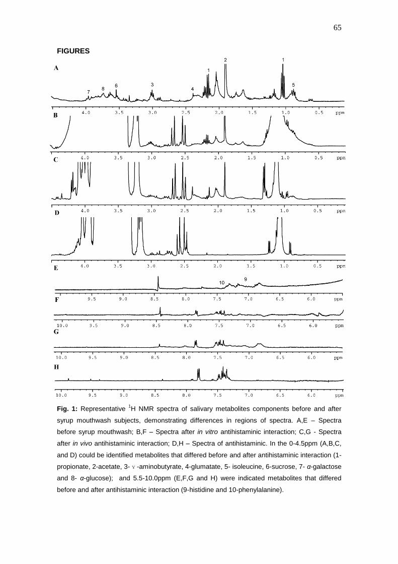
65
FIGURES
Fig. 1: Representative 1H NMR spectra of salivary metabolites components before and after
syrup mouthwash subjects, demonstrating differences in regions of spectra. A,E – Spectra
before syrup mouthwash; B,F – Spectra after in vitro antihistaminic interaction; C,G - Spectra
after in vivo antihistaminic interaction; D,H – Spectra of antihistaminic. In the 0-4.5ppm (A,B,C,
and D) could be identified metabolites that differed before and after antihistaminic interaction (1-
propionate, 2-acetate, 3- ɣ -aminobutyrate, 4-glumatate, 5- isoleucine, 6-sucrose, 7- α-galactose
and 8- α-glucose); and 5.5-10.0ppm (E,F,G and H) were indicated metabolites that differed
before and after antihistaminic interaction (9-histidine and 10-phenylalanine).

66
Fig. 2: Scores plot of a two-component PCA model of 1H NMR spectra showing sample
clustering before ( ) and in vitro ( ) and in vivo ( ) antihistaminic interaction. The percentage
of variance captures by PC1 was 80.46% and PC2 was 8.64%.
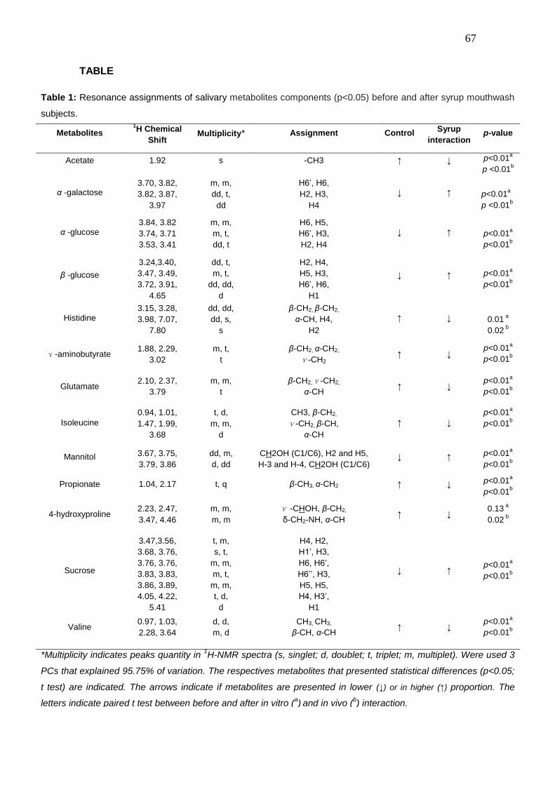
67
TABLE
Table 1: Resonance assignments of salivary metabolites components (p<0.05) before and after syrup mouthwash
subjects.
Metabolites 1H Chemical
Shift Multiplicity* Assignment Control
Syrup
interaction p-value
Acetate 1.92 s -CH3 ↑ ↓ p<0.01a
p <0.01b
α -galactose 3.70, 3.82,
3.82, 3.87,
3.97
m, m,
dd, t,
dd
H6’, H6,
H2, H3,
H4
↓ ↑
p<0.01a
p <0.01b
α -glucose
3.84, 3.82
3.74, 3.71
3.53, 3.41
m, m,
m, t,
dd, t
H6, H5,
H6’, H3,
H2, H4
↓ ↑
p<0.01a
p<0.01b
β -glucose
3.24,3.40,
3.47, 3.49,
3.72, 3.91,
4.65
dd, t,
m, t,
dd, dd,
d
H2, H4,
H5, H3,
H6’, H6,
H1
↓ ↑
p<0.01a
p<0.01b
Histidine 3.15, 3.28,
3.98, 7.07,
7.80
dd, dd,
dd, s,
s
β-CH2, β-CH2,
α-CH, H4,
H2
↑ ↓
0.01 a
0.02 b
ɣ -aminobutyrate 1.88, 2.29,
3.02
m, t,
t
β-CH2, α-CH2,
ɣ -CH2 ↑ ↓
p<0.01a
p<0.01b
Glutamate
2.10, 2.37,
3.79
m, m,
t
β-CH2, ɣ -CH2,
α-CH ↑ ↓
p<0.01a
p<0.01b
Isoleucine
0.94, 1.01,
1.47, 1.99,
3.68
t, d,
m, m,
d
CH3, β-CH2,
ɣ -CH2, β-CH,
α-CH
↑ ↓
p<0.01a
p<0.01b
Mannitol 3.67, 3.75,
3.79, 3.86
dd, m,
d, dd
CH2OH (C1/C6), H2 and H5,
H-3 and H-4, CH2OH (C1/C6) ↓ ↑ p<0.01
a
p<0.01b
Propionate 1.04, 2.17 t, q β-CH3, α-CH2 ↑ ↓ p<0.01a
p<0.01b
4-hydroxyproline 2.23, 2.47,
3.47, 4.46
m, m,
m, m
ɣ -CHOH, β-CH2,
δ-CH2-NH, α-CH ↑ ↓
0.13 a
0.02 b
Sucrose
3.47,3.56,
3.68, 3.76,
3.76, 3.76,
3.83, 3.83,
3.86, 3.89,
4.05, 4.22,
5.41
t, m,
s, t,
m, m,
m, t,
m, m,
t, d,
d
H4, H2,
H1’, H3,
H6, H6’,
H6’’, H3,
H5, H5,
H4, H3’,
H1
↓ ↑ p<0.01
a
p<0.01b
Valine 0.97, 1.03,
2.28, 3.64
d, d,
m, d
CH3, CH3,
β-CH, α-CH ↑ ↓
p<0.01a
p<0.01b
*Multiplicity indicates peaks quantity in 1H-NMR spectra (s, singlet; d, doublet; t, triplet; m, multiplet). Were used 3
PCs that explained 95.75% of variation. The respectives metabolites that presented statistical differences (p<0.05;
t test) are indicated. The arrows indicate if metabolites are presented in lower (↓) or in higher (↑) proportion. The
letters indicate paired t test between before and after in vitro (a) and in vivo (
b) interaction.
68
5 DISCUSSÃO
Atualmente, apesar do crescente interesse, a caracterização de
metabólitos para doenças ainda é pequena se comparado aos estudos sobre
marcadores, principalmente tratando-se da saliva (Yan, Apweiler et al., 2009).
A criação de métodos não invasivos que possibilitem o diagnóstico e a
avaliação de tratamentos é considerada, atualmente, uma meta relevante para
a manutenção do sistema de saúde em vários níveis (Takeda, Stretch et al.,
2009; Yan, Apweiler et al., 2009). A idéia da utilização da saliva como matriz de
análise para métodos diagnósticos existe há longo tempo e a criação de novas
metodologias pode ser pertinente e viável, devido à facilidade da coleta do
fluido e à quantidade de informações obtidas a partir dele (Silwood, Lynch et
al., 2002).
A saliva já foi estudada tanto por espectroscopia de massa para detecção
de proteínas (Cabras, Castagnola et al., 2008; Lillsunde, 2008) quanto pela
Ressonância Magnética Nuclear (Dan, Kawano et al., 1989; Antonyraj,
Karunakaran et al., 1998; Grootveld e Silwood, 2005; Walsh, Brennan et al.,
2006; Bertram, Eggers et al., 2009). A Espectrometria por RMN é uma das
técnicas mais completas para a investigação em nível molecular, permitindo
obter informação estrutural e dinâmica para qualquer estado da matéria, tanto
de compostos puros como misturas complexas (Lindon, Holmes et al., 2006).
69
Nos últimos anos a espectroscopia por RMN tem sido amplamente
empregada na identificação de diferentes biofluidos e dentre suas vantagens,
destaca-se a distinção em alta resolução dos sinais de cada molécula, sendo
um método reprodutivo, uma técnica rápida e não-destrutiva, podendo,
portanto, propiciar a utilização da amostra diversas vezes, desde que esta não
sofra degradação (Brindle, Antti et al., 2002; Silwood, Lynch et al., 2002). Pode-
se ainda associar a RMN 1-D, em uma dimensão, (Silwood, Lynch et al., 2002)
e 2-D, a RMN 2-D, em duas dimensões, (Durand, Fernandez et al., 2008; Sato,
Fukae et al., 2008) para reprodução de resultados mais fiéis, removendo
ambiguidades e sobreposição de sinais (Brindle, Antti et al., 2002; Silwood,
Lynch et al., 2002). O estudo de degradação de metabólitos em função do
tempo demonstrou haver pequena perda após a permanência da amostra em
um período de 48 horas a 25oC, demonstrando que em experimentos
bidimensionais, geralmente longos devido ao grande número de scans, há
pouca degradação (Apêndice 3, página 95).
A presença de determinadas moléculas em determinado nível de
concentração depende, em muitos casos, de sua produção e de seu transporte
até a cavidade oral. Contudo, tanto o transporte quanto a produção podem ser
comprometidos com alterações no metabolismo, tornando o monitoramento
desses metabólitos potenciais biomarcadores para indicar não apenas
desordens bucais, mas também sistêmicas (Chiappin, Antonelli et al., 2007).
No presente estudo, o perfil salivar de crianças saudáveis com ausência de
doenças bucais apresentou poucas diferenças entre os metabólitos quando
avaliado as diferentes dentições. Fato que já era esperado, visto que todos os
indivíduos eram isentos de alterações sistêmicas.
70
A dentição decídua, ou seja, indivíduos mais jovens apresentou maior
quantidade de Gamma-aminobutirato (GABA) e valina comparando-se a
crianças de idade mais avançada. O GABA é um neurotransmissor, com
função inibitória de sinapses, encontrado no sistema nervoso de diferentes
espécies. Baixas concentrações plasmáticas de GABA podem gerar sintomas
depressivos e ainda doença de Alzheimer e quadros de epilepsia (Rating,
Siemes et al., 1983; Fonteh, Harrington et al., 2007). Já a valina é um
amonoácido ditto essencial e sua deficiência está relacionada a alterações
cerebrais e desordens musculares (Binek-Singer e Johnson, 1981; Fonteh,
Harrington et al., 2007).
Já a dentição permanente, ou seja, indivíduos mais próximos a
adolescência, apresentou maior quantidade de etanol, 1,2-propanodiol e de
propionato. Tais metabólitos podem ser resultantes do metabolismo de
microorganismos (Scheie, Fejerskov et al., 1998). Sugere-se que a dentição
permanente possua maior superfície de contato devido ao maior número de
elementos dentais e uma flora bacteriana mais complexa, desta forma,
justificando-se a presença de dos metabólitos em maior quantidade nesta fase
da dentição (Hannig e Hannig, 2009).
Em crianças com lesões cariosas, observou-se maior quantidade de
lactato, n-butirato e n-caproato quando comparado ao grupo sem a presença
de lesões cariosas. Os experimentos realizados para verificação dos
metabólitos salivares em diferentes condições demonstraram redução dos
metabólitos acetato (1,92ppm) e propionato (1,04 e 2,17ppm). Provavelmente
oriundos do metabolismo de microorganismos, justificando sua maior presença
em indivíduos com lesões cariosas. Entretanto a ornitina é um componente
71
endógenos e, portanto, pode ser um importante biomarcador do metabolismo
para a cárie dental, entretanto essa possível relação deve ser mais explorada.
A sacarose é considerada o carboidrato mais cariogênico devido à sua
utilização pelos Streptococcus mutans na formação de dextrana e mutana
(Banas e Vickerman, 2003). Estas promovem a colonização bacteriana no
esmalte, fator de extrema importância na formação da cárie, formando a
película adquirida em conjunto com proteínas salivares, responsável pela
fixação e seleção dos microorganismos (Gibbons e Van Houte, 1973). A
síntese de dextrana e mutana resultam na formação de ácido láctico. Com a
produção de ácido láctico há uma redução no pH da placa, o que faz com que
a hidroxiapatita se fragmente transformando-se em fosfato tricálcico e em
seguida em moléculas de fosfato monoácido de cálcio, ocorrendo a cárie dental
(Ingram, 1990; Robinson, Shore et al., 2000). No presente estudo, a sacarose
foi encontrada em menor quantidade em indivíduos com lesões cariosas e a α–
glucose em maior quantidade, demonstrando uma intensa atividade
microbiana, ao quebrar a sacarose para gerar energia (Banas e Vickerman,
2003).
Metabólitos exógenos também foram identificados tanto em amostras de
saliva de indivíduos com cárie quanto em indivíduos sem cáries. Apesar dos
voluntários terem sido instruídos a abster-se de atividades orais, foi identificado
Lauril Sulfato de Sódio. Compostos fluoretados atuam melhor com agentes
sulfactantes, que auxiliam no processo de remineralização e na remoção física
de microorganismos e químico na desorganização de lipídeos (Petersen, Assev
et al., 2006). O Lauril Sulfato de Sódio é o surfactante iônico mais comumente
empregado na composição de pastas dentais (Rantanen, Jutila et al., 2003).
72
Provavelmente, este composto foi encontrado em maior proporção em
indivíduos com cárie devido à maior adsorção desse composto às
irregularidades da superfície dental. Ademais, não se observou diferença deste
composto entre as dentições, quando crianças sem lesões cariosas das
diferentes dentições foram avaliadas.
A literatura relata que há distinção do perfil protéico de indivíduos sem a
presença de cárie e com a presença de cárie (Bergandi, Defabianis et al.,
2007). Esta diferença pode ser explicada pela diferente atividade proteolítica
entre esses indivíduos, quer seja pela diferença na expressão de proteases,
inibidores de protease ou poliforfismo nos sítios proteolíticos de clivagem
(Ayad, Van Wuyckhuyse et al., 2000). O perfil salivar de indivíduos isentos de
lesões cariosas diverge tanto no número de proteínas quanto na
susceptibilidade destas a serem degradas por proteases bacterianas,
comparando-se com pacientes com lesões cariosas ativas (Cowman, Schaefer
et al., 1979). Sugere-se que essas diferenças também ocorram com
metabólitos de baixo peso molecular, como os do presente estudo.
De acordo com Tomita et al (Tomita, Miyake et al., 2008), a
concentração de lipídeos proveniente das glândulas parótidas foi maior em
crianças suscetíveis, corroborando com o presente estudo, apesar de ser
analisada saliva total. A variação de lipídeos pode estar associada ao
desenvolvimento da cárie, uma vez que os lipídeos se ligam covalentemente às
lesões cariosas e inibem a difusão dos ácidos bacterianos, potencializando sua
ação local (Slomiany, Murty et al., 1989) além de facilitar a adsorção de
bactérias gram positivas, como o Streptococus mutans, no substrato dental
(Ciardi, Rolla et al., 1977).
73
No estudo das etiologias das alterações da cavidade bucal, destacam-se
a ingestão de bebidas e medicamentos. Alterações na composição salivar
podem ser a resposta para microalterações dos tecidos dentais, devido às
modificações bioquímicas no fluido salivar podendo propiciar o aumento do
risco a cárie (Dale, Tao et al., 2006). Neste contexto, cita-se os medicamentos
que são utilizados cronicamente, especialmente em quadros de alergia
respiratórias (Chipps e Harder, 2009; Ferrer, Morais-Almeida et al., 2010).
Para tornar os medicamentos líquidos infantis mais agradáveis, sendo a
sacarose é comumente incorporada às formulações químicas destes fármacos
(Pierro, Abdelnur et al., 2005). No presente estudo foi observado alteração da
quantidade de açúcares tais como sacarose, glicose e galactose após a
interação in vitro e in vivo com o anti-histamínico estudado. A literatura revela
que após bochechos com solução de sacarose, há uma subsaturação de
dihidrato fosfato dicálcio na saliva o que favorece o risco à cárie dental (Van
Steijn, Amerongen et al., 2002). Além disso, a sacarose é o carboidrato mais
utilizado. Por esta razão, deve-se evitar a administração destes medicamentos
sob a forma de xarope no período noturno, quando o fluxo salivar é reduzido,
portanto sua capacidade de limpeza estará reduzida (Dawes, 2008). Ademais,
ressalta-se a importância da higienização bucal após administração destes
medicamentos (Mcdonald e Stookey, 1973; Sorvari, Meurman et al., 1994).
Por outro lado, metabólitos tal qual o acetato, histidina, ɣ -aminobutirato,
glutamato, isoleucina, propionato e valina apresentaram-se em menor
quantidade após a interação medicamentosa, tanto no estudo in vivo como no
in vitro. Somente a 4-hydroxiproline apresentou-se em menor concentração
apenas no estudo in vivo. Apesar do medicamento utilizado no presente

74
estudo possuir ácido cítrico para a estabilização do pH ácido, que atua como
agente estimulante do fluxo salivar a redução da concentração dos metabólitos
não se deve a diluição dos mesmos devido ao aumento do fluxo salivar (Pierro,
Abdelnur et al., 2005; Da Mata, Da Silva Marques et al., 2009). Esta afirmação
pôde ser comprovada no estudo in vitro, em que a interação medicamentosa foi
realizada com amostra de saliva não estimulada. A redução da concentração
do propionato é vantajosa, visto ser um ácido orgânico proveniente do
metabolismo bacteriano, sendo um dos responsáveis pela dissolução da
hidroxiapatita (Liu, Yue et al., 1998). Entretanto, outros fatores devem ser
considerados, como o fato de medicamentos possuírem baixo pH a alta acidez
titulável, além dos níveis de sacarose apresentarem-se altos, caracterizando
tais formulações como cariogênicas .
Diante da inexistência de trabalhos que avaliassem a interação
medicamentosa com metabólitos salivares, optou-se por simular o que ocorre
in vivo no modelo in vitro. Cabe ressaltar, que os resultados de estudos in vitro
devem ser cautelosamente avaliados antes de sua extrapolação para a
realidade clínica. Entretanto, o presente estudo demonstrou haver grande
semelhança entre os resultados provenientes do ensaio in vitro e in vivo,
demonstrando que experimentos in vitro podem ser modelos interessantes para
estudo da interação de medicamentos com componentes salivares.
75
6 CONCLUSÃO
Diante da metodologia empregada e das condições avaliadas, a Ressonância
Magnética Nuclear em conjunto com a Análise dos Componentes Principais foi
capaz de distinguir metabólitos salivares.
A partir das proposições e dos resultados obtidos no presente estudo, pode-se
concluir que:
Houve diferenças entre os metabólitos salivares nas dentições decídua,
mista e permanente de crianças saudáveis e isentas de lesões cariosas;
Houve diferença entre os metabólitos quando avaliada a saliva de
crianças com e sem lesões cariosas, sendo possível a separação destes
grupos por meio do PCA;
Tanto a interação in vitro quanto a in vivo de metabólitos salivares com o
anti-histamínico sob a forma de xarope resultaram em alteração dos
componentes salivares quando comparado ao momento anterior a
interação
76
7. REFERÊNCIAS BIBLIOGRÁFICAS Abrahan, R. J. e P. Loftus. In the proton and carbon-13 NMR spectroscopy - an integrates approach. John Wiley & Sons. 1978. Antonyraj, K. J., T. Karunakaran, et al. Bactericidal activity and poly-L-proline II conformation of the tandem repeat sequence of human salivary mucin glycoprotein (MG2). Arch Biochem Biophys, v.356, n.2, Aug 15, p.197-206. 1998. Atkinson, J. C., C. Yeh, et al. Elevation of salivary antimicrobial proteins following HIV-1 infection. J Acquir Immune Defic Syndr, v.3, n.1, p.41-8. 1990. Ayad, M., B. C. Van Wuyckhuyse, et al. The association of basic proline-rich peptides from human parotid gland secretions with caries experience. J Dent Res, v.79, n.4, Apr, p.976-82. 2000. Banas, J. A. e M. M. Vickerman. Glucan-binding proteins of the oral streptococci. Crit Rev Oral Biol Med, v.14, n.2, p.89-99. 2003. Banderas-Tarabay, J. A., I. G. Zacarias-D'oleire, et al. Electrophoretic analysis of whole saliva and prevalence of dental caries. A study in Mexican dental students. Arch Med Res, v.33, n.5, Sep-Oct, p.499-505. 2002. Bergandi, L., P. Defabianis, et al. Absence of soluble CD14 in saliva of young patients with dental caries. Eur J Oral Sci, v.115, n.2, Apr, p.93-6. 2007. Bertram, H. C., N. Eggers, et al. Potential of human saliva for nuclear magnetic resonance-based metabolomics and for health-related biomarker identification. Anal Chem, v.81, n.21, Nov 1, p.9188-93. 2009. Binek-Singer, P. e T. C. Johnson. The inhibition of brain protein synthesis following leucine or valine injection can be prevented. Biochem Biophys Res Commun, v.103, n.4, Dec 31, p.1209-15. 1981. Brindle, J. T., H. Antti, et al. Rapid and noninvasive diagnosis of the presence and severity of coronary heart disease using 1H-NMR-based metabonomics. Nat Med, v.8, n.12, Dec, p.1439-44. 2002. Cabras, T., M. Castagnola, et al. Carbachol-induced in vitro secretion of certain human submandibular proteins investigated by mass-spectrometry. Arch Oral Biol, Jul 8. 2008. Chiappin, S., G. Antonelli, et al. Saliva specimen: a new laboratory tool for diagnostic and basic investigation. Clin Chim Acta, v.383, n.1-2, Aug, p.30-40. 2007. Chipps, B. E. e J. M. Harder. Antihistamine treatment for allergic rhinitis: different routes, different outcomes? Allergy Asthma Proc, v.30, n.6, Nov-Dec, p.589-94. 2009.

77
Ciardi, J. E., G. Rolla, et al. Adsorption of Streptococcus mutans lipoteichoic acid to hydroxyapatite. Scand J Dent Res, v.85, n.6, Sep, p.387-91. 1977. Coogan, M. M. e S. J. Challacombe. Serum and salivary antibodies to a mycobacterial 65-kDa stress protein are elevated in HIV-positive patients and modified by oral candidiasis. Oral Microbiol Immunol, v.15, n.5, Oct, p.284-9. 2000. Cowman, R. A., S. J. Schaefer, et al. Differential utilization of proteins in saliva from caries-active and caries-free subjects as growth substrates by plaque-forming streptococci. J Dent Res, v.58, n.10, Oct, p.2019-27. 1979. Cury, J. A., M. A. Rebelo, et al. Biochemical composition and cariogenicity of dental plaque formed in the presence of sucrose or glucose and fructose. Caries Res, v.34, n.6, Nov-Dec, p.491-7. 2000. Da Mata, A. D., D. N. Da Silva Marques, et al. Effects of gustatory stimulants of salivary secretion on salivary pH and flow: a randomized controlled trial. Oral Dis, v.15, n.3, Apr, p.220-8. 2009. Dale, B. A., R. Tao, et al. Oral antimicrobial peptides and biological control of caries. BMC Oral Health, v.6 Suppl 1, p.S13. 2006. Dan, K., K. Kawano, et al. High resolution proton nuclear magnetic resonance studies of human saliva. Shika Kiso Igakkai Zasshi, v.31, n.3, Jun, p.324-8. 1989. Dawes, C. Salivary flow patterns and the health of hard and soft oral tissues. J Am Dent Assoc, v.139 Suppl, May, p.18S-24S. 2008. Delaleu, N., H. Immervoll, et al. Biomarker profiles in serum and saliva of experimental Sjogren's syndrome: associations with specific autoimmune manifestations. Arthritis Res Ther, v.10, n.1, p.R22. 2008. Durand, J., F. Fernandez, et al. DOSY technique applied to palladium nanoparticles in ionic liquids. Magn Reson Chem, v.46, n.8, Aug, p.739-43. 2008. Elad, S., S. Heisler, et al. Saliva secretion in patients with allergic rhinitis. Int Arch Allergy Immunol, v.141, n.3, p.276-80. 2006. Emekli-Alturfan, E., G. Demir, et al. Altered biochemical parameters in the saliva of patients with breast cancer. Tohoku J Exp Med, v.214, n.2, Feb, p.89-96. 2008. Ferrer, M., M. Morais-Almeida, et al. Evaluation of treatment satisfaction in children with allergic disease treated with an antihistamine: an international, non-interventional, retrospective study. Clin Drug Investig, v.30, n.1, p.15-34. 2010. Fonteh, A. N., R. J. Harrington, et al. Free amino acid and dipeptide changes in the body fluids from Alzheimer's disease subjects. Amino Acids, v.32, n.2, Feb, p.213-24. 2007. Gibbons, R. J. e J. Van Houte. On the formation of dental plaques. J Periodontol, v.44, n.6, Jun, p.347-60. 1973. Gil, V. M. S. e C. F. G. C. Geraldes. Ressonância magnética nuclear, fundamentos, métodos e aplicações. Fundação Calouste Gulbenkian, Liaboa. 1987.
78
Giusti, L., L. Bazzichi, et al. Specific proteins identified in whole saliva from patients with diffuse systemic sclerosis. J Rheumatol, v.34, n.10, Oct, p.2063-9. 2007. Grootveld, M. e C. J. Silwood. 1H NMR analysis as a diagnostic probe for human saliva. Biochem Biophys Res Commun, v.329, n.1, Apr 1, p.1-5. 2005. Hannig, C. e M. Hannig. The oral cavity--a key system to understand substratum-dependent bioadhesion on solid surfaces in man. Clin Oral Investig, v.13, n.2, Jun, p.123-39. 2009. Hardt, M., L. R. Thomas, et al. Toward defining the human parotid gland salivary proteome and peptidome: identification and characterization using 2D SDS-PAGE, ultrafiltration, HPLC, and mass spectrometry. Biochemistry, v.44, n.8, Mar 1, p.2885-99. 2005. Hay, D. I. Salivary factors in caries models. Adv Dent Res, v.9, n.3, Nov, p.239-43. 1995. Ingram, G. S. Chemical events during tooth dissolution. J Dent Res, v.69 Spec No, Feb, p.581-6; discussion 634-6. 1990. Katakura, A., I. Kamiyama, et al. Comparison of salivary cytokine levels in oral cancer patients and healthy subjects. Bull Tokyo Dent Coll, v.48, n.4, Nov, p.199-203. 2007. Lambert, M. A. e C. W. Moss. Gas-liquid chromatography of short-chain fatty acids on Dexsil 300 GC. J Chromatogr, v.74, n.2, Dec 20, p.335-8. 1972. Levy, M. L., D. Price, et al. Inadequacies in UK primary care allergy services: national survey of current provisions and perceptions of need. Clin Exp Allergy, v.34, n.4, Apr, p.518-9. 2004. Lillsunde, P. Analytical techniques for drug detection in oral fluid. Ther Drug Monit, v.30, n.2, Apr, p.181-7. 2008. Lin, A. L., D. A. Johnson, et al. Alteration in salivary function in early HIV infection. J Dent Res, v.82, n.9, Sep, p.719-24. 2003. Lindon, J. C., E. Holmes, et al. Metabonomics techniques and applications to pharmaceutical research & development. Pharm Res, v.23, n.6, Jun, p.1075-88. 2006. Linke, H. A. e S. J. Moss. Quantitation of carbohydrate sweeteners and organic acids in human oral fluid using HPLC analysis. Z Ernahrungswiss, v.31, n.2, Jun, p.147-54. 1992. Liu, L., S. Yue, et al. [Comparison of demineralization of different organic acid to enamel]. Hua Xi Kou Qiang Yi Xue Za Zhi, v.16, n.2, May, p.103-4, 113. 1998. Maguire, A., W. Baqir, et al. Are sugars-free medicines more erosive than sugars-containing medicines? An in vitro study of paediatric medicines with prolonged oral clearance used regularly and long-term by children. Int J Paediatr Dent, v.17, n.4, Jul, p.231-8. 2007.
79
Maguire, A. e A. J. Rugg-Gunn. Medicines in liquid and syrup form used long-term in paediatrics: a survey in the Northern Region of England. Int J Paediatr Dent, v.4, n.2, Jun, p.93-9. 1994. Mcdonald, J. L., Jr. e G. K. Stookey. Laboratory studies concerning the effect of acid-containing beverages on enamel dissolution and experimental dental caries. J Dent Res, v.52, n.2, Mar-Apr, p.211-6. 1973. Nagler, R., G. Bahar, et al. Concomitant analysis of salivary tumor markers--a new diagnostic tool for oral cancer. Clin Cancer Res, v.12, n.13, Jul 1, p.3979-84. 2006. Nair, P. N. e H. E. Schroeder. Duct-associated lymphoid tissue (DALT) of minor salivary glands and mucosal immunity. Immunology, v.57, n.2, Feb, p.171-80. 1986. Nieuw Amerongen, A. V., C. H. Oderkerk, et al. Role of mucins from human whole saliva in the protection of tooth enamel against demineralization in vitro. Caries Res, v.21, n.4, p.297-309. 1987. Petersen, F. C., S. Assev, et al. Combined effects of NaF and SLS on acid- and polysaccharide-formation of biofilm and planktonic cells. Arch Oral Biol, v.51, n.8, Aug, p.665-71. 2006. Pierro, V. S., J. P. Abdelnur, et al. Free sugar concentration and pH of paediatric medicines in Brazil. Community Dent Health, v.22, n.3, Sep, p.180-3. 2005. Rantanen, I., K. Jutila, et al. The effects of two sodium lauryl sulphate-containing toothpastes with and without betaine on human oral mucosa in vivo. Swed Dent J, v.27, n.1, p.31-4. 2003. Rating, D., H. Siemes, et al. Low CSF GABA concentration in children with febrile convulsions, untreated epilepsy, and meningitis. J Neurol, v.230, n.4, p.217-25. 1983. Robinson, C., R. C. Shore, et al. The chemistry of enamel caries. Crit Rev Oral Biol Med, v.11, n.4, p.481-95. 2000. Rochfort, S. Metabolomics reviewed: a new "omics" platform technology for systems biology and implications for natural products research. J Nat Prod, v.68, n.12, Dec, p.1813-20. 2005. Sanders, J. K. M. e B. K. Hunter. Modern NMR spectroscopy - A guide for chemists. Oxford University Press, Oxford. 1994. Sato, H., K. Fukae, et al. 2D Selective-TOCSY-DQFCOSY and HSQC-TOCSY NMR experiments for assignment of a homogeneous asparagine-linked triantennary complex type undecasaccharide. Carbohydr Res, v.343, n.8, Jun 9, p.1333-45. 2008. Scheie, A. A., O. Fejerskov, et al. The effects of xylitol-containing chewing gums on dental plaque and acidogenic potential. J Dent Res, v.77, n.7, Jul, p.1547-52. 1998. Schupbach, P., F. G. Oppenheim, et al. Electron-microscopic demonstration of proline-rich proteins, statherin, and histatins in acquired enamel pellicles in vitro. Eur J Oral Sci, v.109, n.1, Feb, p.60-8. 2001. Shaw, L. e H. D. Glenwright. The role of medications in dental caries formation: need for sugar-free medication for children. Pediatrician, v.16, n.3-4, p.153-5. 1989.
80
Shpitzer, T., G. Bahar, et al. A comprehensive salivary analysis for oral cancer diagnosis. J Cancer Res Clin Oncol, v.133, n.9, Sep, p.613-7. 2007. Silwood, C. J., E. Lynch, et al. 1H and (13)C NMR spectroscopic analysis of human saliva. J Dent Res, v.81, n.6, Jun, p.422-7. 2002. Slomiany, B. L., V. L. Murty, et al. Physico-chemical characteristics of mucus glycoproteins and lipids of the human oral mucosal mucus coat in relation to caries susceptibility. Arch Oral Biol, v.34, n.4, p.229-37. 1989. Sorvari, R., J. H. Meurman, et al. Effect of fluoride varnish and solution on enamel erosion in vitro. Caries Res, v.28, n.4, p.227-32. 1994. Takeda, I., C. Stretch, et al. Understanding the human salivary metabolome. NMR Biomed, v.22, n.6, Jul, p.577-84. 2009. Tan, W., L. Sabet, et al. Optical protein sensor for detecting cancer markers in saliva. Biosens Bioelectron, Apr 6. 2008. Tenuta, L. M., A. A. Del Bel Cury, et al. Ca, Pi, and F in the fluid of biofilm formed under sucrose. J Dent Res, v.85, n.9, Sep, p.834-8. 2006. Thomson, W. M., A. J. Spencer, et al. Is medication a risk factor for dental caries among older people? Community Dent Oral Epidemiol, v.30, n.3, Jun, p.224-32. 2002. Tomita, Y., N. Miyake, et al. Lipids in human parotid saliva with regard to caries experience. J Oleo Sci, v.57, n.2, p.115-21. 2008. Troxler, R. F., G. D. Offner, et al. Structural relationship between human salivary histatins. J Dent Res, v.69, n.1, Jan, p.2-6. 1990. Van Nieuw Amerongen, A., J. G. Bolscher, et al. Salivary proteins: protective and diagnostic value in cariology? Caries Res, v.38, n.3, May-Jun, p.247-53. 2004. Van Steijn, G. J., A. V. Amerongen, et al. Effect of periodontal treatment on the activity of chitinase in whole saliva of periodontitis patients. J Periodontal Res, v.37, n.4, Aug, p.245-9. 2002. Vratsanos, S. M. e I. D. Mandel. Comparative plaque acidogenesis of caries-resistant vs. caries-susceptible adults. J Dent Res, v.61, n.3, Mar, p.465-8. 1982. Walsh, M. C., L. Brennan, et al. Effect of acute dietary standardization on the urinary, plasma, and salivary metabolomic profiles of healthy humans. Am J Clin Nutr, v.84, n.3, Sep, p.531-9. 2006. Wickstrom, C. e G. Svensater. Salivary gel-forming mucin MUC5B--a nutrient for dental plaque bacteria. Oral Microbiol Immunol, v.23, n.3, Jun, p.177-82. 2008. Wong, D. T. Salivary diagnostics powered by nanotechnologies, proteomics and genomics. J Am Dent Assoc, v.137, n.3, Mar, p.313-21. 2006. Xu, G. W., X. Lu, et al. [Recent advances in metabonomics]. Zhongguo Yi Xue Ke Xue Yuan Xue Bao, v.29, n.6, Dec, p.701-11. 2007.

81
Yan, W., R. Apweiler, et al. Systematic comparison of the human saliva and plasma proteomes. Proteomics Clin Appl, v.3, n.1, Jan 1, p.116-134. 2009. Yao, Y., E. A. Berg, et al. Identification of protein components in human acquired enamel pellicle and whole saliva using novel proteomics approaches. J Biol Chem, v.278, n.7, Feb 14, p.5300-8. 2003. Yoon, M. S., V. Jankowski, et al. Characterisation of advanced glycation endproducts in saliva from patients with diabetes mellitus. Biochem Biophys Res Commun, v.323, n.2, Oct 15, p.377-81. 2004.

82
6 ANEXOS
ANEXO 1
83
ANEXO 2
FACULDADE DE ODONTOLOGIA DEPARTAMENTO DE ODONTOPEDIATRIA E ORTODONTIA DISCIPLINA DE ODONTOPEDIATRIA
TERMO DE CONSENTIMENTO LIVRE E ESCLARECIDO
Prezado responsável,
Será realizado um estudo na Odontopediatria da Faculdade de Odontologia da UFRJ, com o objetivo de analisar a saliva de seu filho através de Ressonância Nuclear Magnética. Nesta pesquisa será pedido ao seu filho(a) que cuspa em um “potinho de plástico”, o que não causará nenhum desconforto a ele(a).
Sua participação e voluntária e, caso não queira participar, sua recusa não causará nenhum prejuízo ao tratamento odontológico da criança nesta instituição. O pesquisador responsável (Raquel dos Santos Pinheiro) poderá ser acessado para esclarecimento de eventuais dúvidas, a qualquer momento, pelos telefones 2562-2100 ou 2562-2098. O responsável poderá solicitar a saída do paciente deste estudo em qualquer momento, assim como a própria criança e, neste caso, os responsáveis pelo projeto se comprometem a não utilizar as informações obtidas. Os dados individuais dos participantes serão mantidos sob sigilo, sendo manipulados apenas pelos responsáveis pela pesquisa e arquivados por um período de 5 anos. Entretanto os resultados, em sua totalidade, serão publicados em literatura científica especializada, estando também disponíveis para consulta na Biblioteca da Disciplina de Odontopediatria da FO/UFRJ localizada no anexo da Disciplina no 3º andar do Hospital Universitário Clementino Fraga Filho ou na Biblioteca Central do Centro de Ciências da Saúde (CCS/UFRJ).
Atenciosamente,
Eu, _______________________________________, identidade n.º_____________________, certifico que lendo as informações acima concordo com o que foi exposto, e autorizo a utilização dos resultados do meu filho(a) ___________________________________ para o presente estudo.
Rio de Janeiro, ______ de _____________ de 2008.
________________________________ Assinatura do responsável
_______________________________ _______________________________ Assinatura Testemunha (1) Assinatura Testemunha(2)
84
ANEXO 3
85
ANEXO 4
FACULDADE DE ODONTOLOGIA DEPARTAMENTO DE ODONTOPEDIATRIA E ORTODONTIA DISCIPLINA DE ODONTOPEDIATRIA
TERMO DE CONSENTIMENTO LIVRE E ESCLARECIDO
Prezado responsável,
Será realizado um estudo na Odontopediatria da Faculdade de Odontologia da UFRJ, com o objetivo de analisar a saliva de seu filho através de Ressonância Nuclear Magnética e por eletroforese. Nesta pesquisa será pedido ao seu filho(a) que cuspa em um “potinho de plástico”, e será realizado exame clínico bucal, o que não causará nenhum desconforto a ele (a).
Sua participação é voluntária e, caso não queira participar, sua recusa não causará nenhum prejuízo ao tratamento odontológico da criança nesta instituição. O pesquisador responsável (Profa Dra Ivete Pomarico Ribeiro de Souza) poderá ser acessado para esclarecimento de eventuais dúvidas, a qualquer momento, pelos telefones (21) 2562-2101, ramal 6. O responsável poderá solicitar a saída do paciente deste estudo em qualquer momento, assim como a própria criança e, neste caso, os responsáveis pelo projeto se comprometem a não utilizar as informações obtidas. Os dados individuais dos participantes serão mantidos sob sigilo, sendo manipulados apenas pelos responsáveis pela pesquisa e arquivados por um período de 5 anos. Entretanto os resultados, em sua totalidade, serão publicados em literatura científica especializada, estando também disponíveis para consulta na Biblioteca da Disciplina de Odontopediatria da FO/UFRJ localizada no anexo da Disciplina no 3º andar do Hospital Universitário Clementino Fraga Filho ou na Biblioteca Central do Centro de Ciências da Saúde (CCS/UFRJ). Caso você tenha dificuldade em entrar em contato com o pesquisador responsável, comunique o fato à Comissão de Ética em Pesquisa do Instituto de Estudos em Saúde Coletiva pelo telefone (21) 2598-9293 ou pelo e-mail [email protected].
Atenciosamente,
________________________________________________
Profa Dra Ivete Pomarico Ribeiro de Souza Professora Titular da Disciplina de Odontopediatria – FO/UFRJ
Professora Orientadora da pesquisa
Eu, _______________________________________, identidade n.º_____________________, responsável pelo menor _________________________________________________, certifico que lendo as informações acima concordo com o que foi exposto, e autorizo a doação da saliva para este estudo. Nome
RJ, ______ de _____________ de 2009.
________________________________ Assinatura do responsável
86
FICHA DE EXAME EPIDEMIOLÓGICO DE CÁRIE DENTÁRIA
Nome: ______________________________________________________ Data: ___/___/___ Nascimento: ___/___/___ Idade:_______ anos Sexo: __________ 55 54 53 52 51 61 62 63 64 65 18 17 16 15 14 13 12 11 21 22 23 24 25 26 27 28
Diagnóstico
Diagnóstico 48 47 46 45 44 43 42 41 31 32 33 34 35 36 37 38 85 84 83 82 81 71 72 73 74 75
C E Ei O CPOD NE HIG c e ei o ceo hig TOTAL
CONDIÇÃO DENTAL PERMANENTE: O=Hígido; 1=Cariado; 2=Restaurado com
cárie; 3=Restaurado sem cárie; 4=Perdido por cárie; 5=Perdido por outras razões; 6=Selante; 7= Apoio de ponte ou coroa; 8=Não erupcionado; 9=Dente excluído; T=Trauma(fratura).
CONDIÇÃO DENTAL DECÍDUO: A=Hígido; B=Cariado; C=Restaurado com cárie; D=Restaurado sem cárie; E=Perdido por cárie; F=Perdido por outras razões; G=Selante; H= Apoio de ponte ou coroa; K=Não erupcionado; L=Dente excluído; T=Trauma(fratura).
87
ANEXO 5

88
ANEXO 6
89
ANEXO 7
FACULDADE DE ODONTOLOGIA DEPARTAMENTO DE ODONTOPEDIATRIA E ORTODONTIA DISCIPLINA DE ODONTOPEDIATRIA
TERMO DE CONSENTIMENTO LIVRE E ESCLARECIDO
Prezado responsável,
Será realizado um estudo na Odontopediatria da Faculdade de Odontologia da UFRJ, com o objetivo de analisar a saliva de seu filho através de Ressonância Nuclear Magnética e por eletroforese de proteínas antes e após a mistura in vitro com a sacarose, Dimetapp® e Claritin®. Nesta pesquisa será pedido ao seu filho(a) que cuspa em um “potinho de plástico”, o que não causará nenhum desconforto a ela.
Sua participação é voluntária e, caso não queira participar, sua recusa não causará nenhum prejuízo ao tratamento odontológico da criança nesta instituição. O pesquisador responsável (Profa Dra Ivete Pomarico Ribeiro de Souza) poderá ser acessado para esclarecimento de eventuais dúvidas, a qualquer momento, pelos telefones (21) 2562-2101, ramal 6. O responsável poderá solicitar a saída do paciente deste estudo em qualquer momento, assim como a própria criança e, neste caso, os responsáveis pelo projeto se comprometem a não utilizar as informações obtidas. Os dados individuais dos participantes serão mantidos sob sigilo, sendo manipulados apenas pelos responsáveis pela pesquisa e arquivados por um período de 5 anos. Entretanto os resultados, em sua totalidade, serão publicados em literatura científica especializada, estando também disponíveis para consulta na Biblioteca da Disciplina de Odontopediatria da FO/UFRJ localizada no anexo da Disciplina no 3º andar do Hospital Universitário Clementino Fraga Filho ou na Biblioteca Central do Centro de Ciências da Saúde (CCS/UFRJ). Caso você tenha dificuldade em entrar em contato com o pesquisador responsável, comunique o fato à Comissão de Ética em Pesquisa do Instituto de Estudos em Saúde Coletiva pelo telefone (21) 2598-9293 ou pelo e-mail [email protected].
Atenciosamente,
________________________________________________
Profa Dra Ivete Pomarico Ribeiro de Souza
Professora Titular da Disciplina de Odontopediatria – FO/UFRJ Professora Orientadora da pesquisa
Eu, _______________________________________, identidade n.º_____________________, responsável pelo menor _________________________________________________, certifico que lendo as informações acima concordo com o que foi exposto, e autorizo a doação da saliva para este estudo.
RJ, ______ de _____________ de 2008.
________________________________
Assinatura do responsável
90
ANEXO 8
FACULDADE DE ODONTOLOGIA DEPARTAMENTO DE ODONTOPEDIATRIA E ORTODONTIA DISCIPLINA DE ODONTOPEDIATRIA
TERMO DE CONSENTIMENTO LIVRE E ESCLARECIDO
Prezado responsável,
Será realizado um estudo na Odontopediatria da Faculdade de Odontologia da UFRJ, com o objetivo de analisar a saliva do voluntário por meio de Ressonância Nuclear Magnética e por eletroforese de proteínas antes e após antes bochecho com sacarose, Claritin® e Dimetapp®, sendo mínimo os riscos de ocorrer alergia, visto que trata-se de um anti-alérgico e antitussígeno. Nesta pesquisa o voluntário deverá cuspir em um “potinho de plástico”, o que não causará nenhum desconforto.
Sua participação é voluntária e, caso não queira participar, sua recusa não causará nenhum prejuízo ao tratamento odontológico da criança nesta instituição. O pesquisador responsável (Profa Dra Ivete Pomarico Ribeiro de Souza) poderá ser acessado para esclarecimento de eventuais dúvidas, a qualquer momento, pelos telefones 2562-2101 ou 2562-2098. O participante poderá solicitar a saída do deste estudo em qualquer momento e, neste caso, os responsáveis pelo projeto se comprometem a não utilizar as informações obtidas. Os dados individuais dos participantes serão mantidos sob sigilo, sendo manipulados apenas pelos responsáveis pela pesquisa e arquivados por um período de 5 anos. Entretanto os resultados, em sua totalidade, serão publicados em literatura científica especializada, estando também disponíveis para consulta na Biblioteca da Disciplina de Odontopediatria da FO/UFRJ localizada no anexo da Disciplina no 3º andar do Hospital Universitário Clementino Fraga Filho ou na Biblioteca Central do Centro de Ciências da Saúde (CCS/UFRJ). Caso você tenha dificuldade em entrar em contato com o pesquisador responsável, comunique o fato à Comissão de Ética em Pesquisa do Instituto de Estudos em Saúde Coletiva pelo telefone 21 2598-9293 ou pelo e-mail [email protected].
Atenciosamente,
_______________________________________________
Profa Dra Ivete Pomarico Ribeiro de Souza Professora Titular da Disciplina de Odontopediatria – FO/UFRJ
Professora Orientadora da pesquisa
Eu, _______________________________________, identidade n.º_____________________, lendo as informações acima concordo com o que foi exposto, e faço a doação da saliva para este estudo.
RJ, ______ de _____________ de 2008.
________________________________
Assinatura do responsável
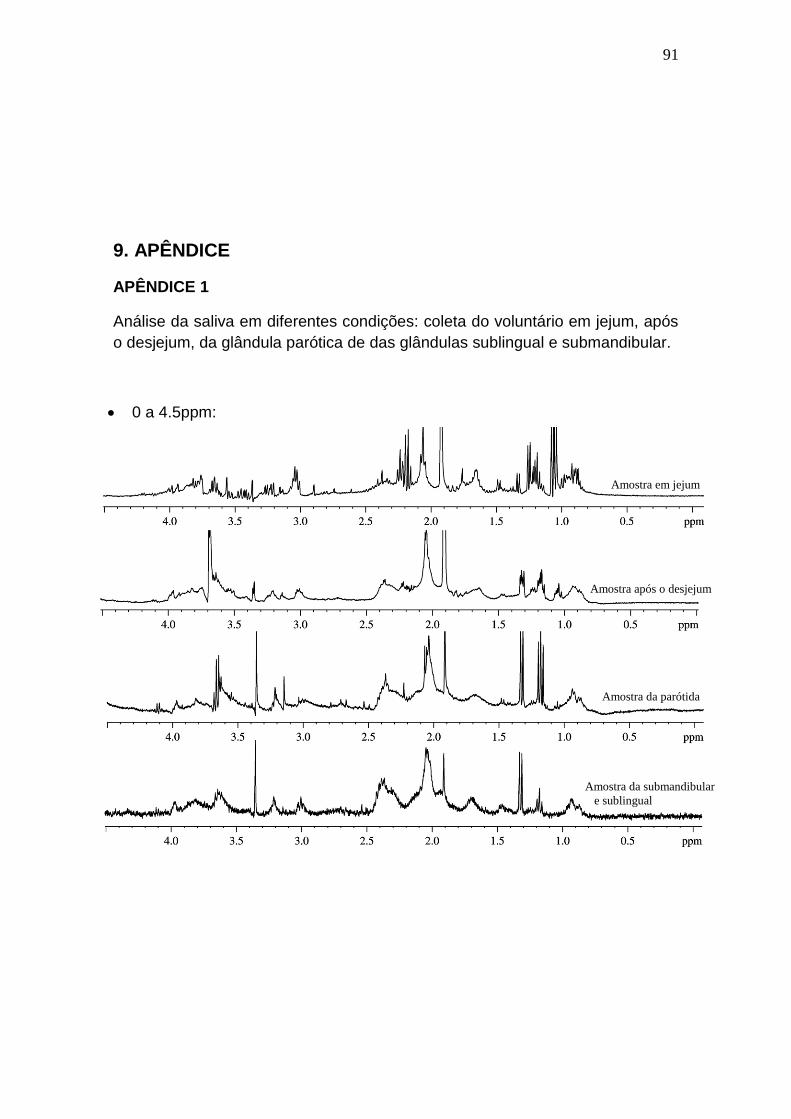
91
9. APÊNDICE
APÊNDICE 1
Análise da saliva em diferentes condições: coleta do voluntário em jejum, após
o desjejum, da glândula parótica de das glândulas sublingual e submandibular.
0 a 4.5ppm:
Amostra após o desjejum
Amostra da parótida
Amostra da submandibular
e sublingual
Amostra em jejum
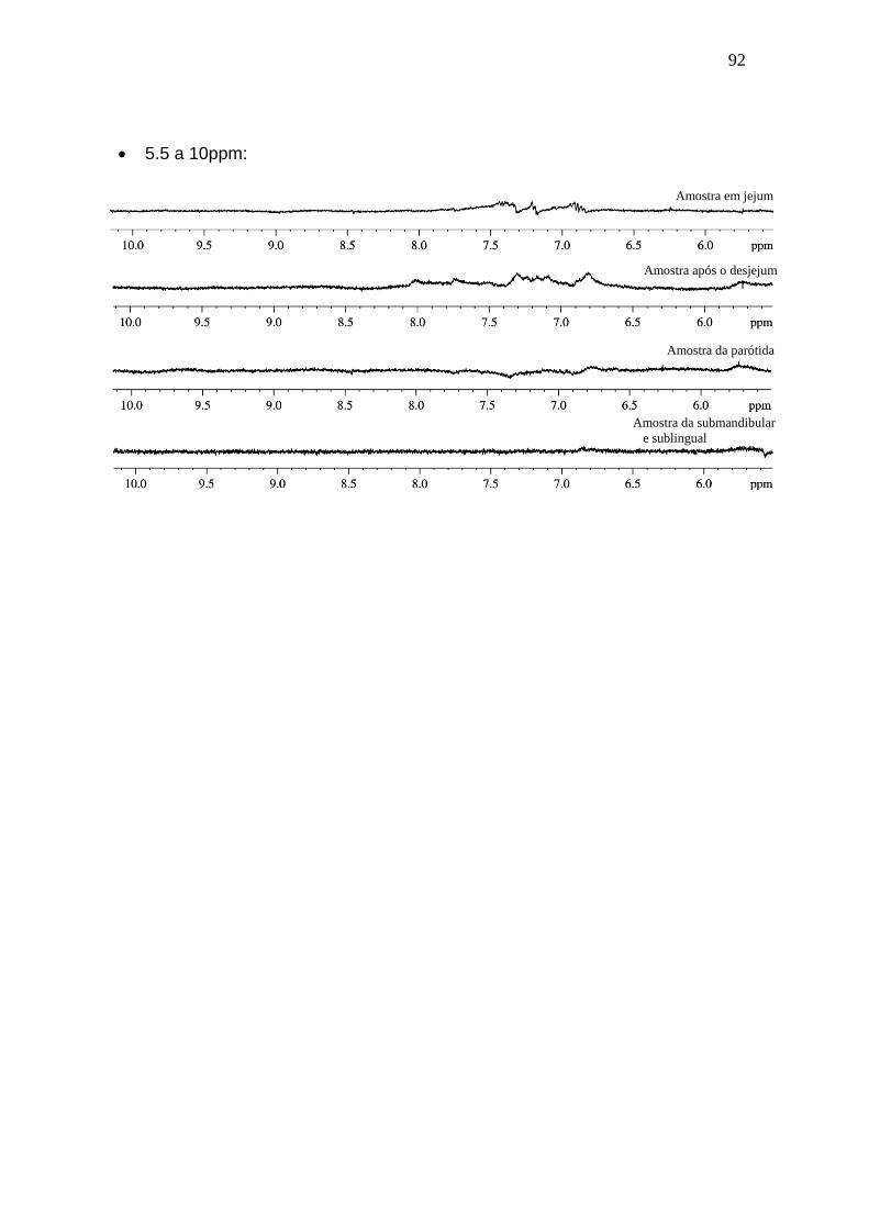
92
5.5 a 10ppm:
Amostra em jejum
Amostra após o desjejum
Amostra da parótida
Amostra da submandibular
e sublingual

93
APÊNDICE 2
Avaliação da interação protéica in vitro com o anti-histamínico sob a forma de xarope (Claritin®; Schering-Plough, Rio de Janeiro, Brazil).
Amostras Antes (Abs) Depois (Abs) Antes - Depois (Abs)
1 0,339 1,207 0,342
2 0,308 1,261 0,396
3 0,163 1,092 0,227
4 0,244 2,710 1,845
5 0,356 1,505 0,640
6 0,157 1,219 0,354
7 0,325 2,476 1,611
8 0,262 1,613 0,748
9 0,296 1,315 0,408
10 0,278 1,292 1,014
Média (±DP) 0,2728 (±0,07) 1,569 (±0,56) 0,759 (±0,56)
λ = 214nm (leitura relativa a ligação peptídica); Abs do medicamento = 0,865. P=0,02 (Teste t pareado, p<0,05); Avaliação as Abs do medicamento: 10µl de medicamento + 990µl de água; Avaliação as Abs da saliva (Antes): 10µl de saliva + 990µl de água; Avaliação as Abs após a interação (Depois): 10µl de medicamento + 10µl de saliva + 990µl de água.

94
APÊNDICE 3
Detalhe dos espectros (0 a 10ppm) de uma amostra de saliva ao longo do
tempo. Os espectros foram extraídos de uma amostra imediatamente após
descongelada, após 12 horas a 25ºC e após 48 horas a 25ºC.
Sobreposição dos espectros de análise da degradação da amostra da região de 0 a 5ppm ampliada.
Análise imediata
Análise após 12 horas
Análise após 48 horas
Análise imediata
Análise após 12 horas
Análise após 48 horas
Livros Grátis( http://www.livrosgratis.com.br )
Milhares de Livros para Download: Baixar livros de AdministraçãoBaixar livros de AgronomiaBaixar livros de ArquiteturaBaixar livros de ArtesBaixar livros de AstronomiaBaixar livros de Biologia GeralBaixar livros de Ciência da ComputaçãoBaixar livros de Ciência da InformaçãoBaixar livros de Ciência PolíticaBaixar livros de Ciências da SaúdeBaixar livros de ComunicaçãoBaixar livros do Conselho Nacional de Educação - CNEBaixar livros de Defesa civilBaixar livros de DireitoBaixar livros de Direitos humanosBaixar livros de EconomiaBaixar livros de Economia DomésticaBaixar livros de EducaçãoBaixar livros de Educação - TrânsitoBaixar livros de Educação FísicaBaixar livros de Engenharia AeroespacialBaixar livros de FarmáciaBaixar livros de FilosofiaBaixar livros de FísicaBaixar livros de GeociênciasBaixar livros de GeografiaBaixar livros de HistóriaBaixar livros de Línguas
Baixar livros de LiteraturaBaixar livros de Literatura de CordelBaixar livros de Literatura InfantilBaixar livros de MatemáticaBaixar livros de MedicinaBaixar livros de Medicina VeterináriaBaixar livros de Meio AmbienteBaixar livros de MeteorologiaBaixar Monografias e TCCBaixar livros MultidisciplinarBaixar livros de MúsicaBaixar livros de PsicologiaBaixar livros de QuímicaBaixar livros de Saúde ColetivaBaixar livros de Serviço SocialBaixar livros de SociologiaBaixar livros de TeologiaBaixar livros de TrabalhoBaixar livros de Turismo