Analysis of Syntaxin-1 Chaperones During Synaptic...
85
UNIVERSIDADE DE LISBOA FACULDADE DE CIÊNCIAS DEPARTAMENTO DE QUÍMICA E BIOQUÍMICA Analysis of Syntaxin-1 Chaperones During Synaptic Activity Tatiana Cerveira Tavares Dos Santos Dissertação Orientada pelo Prof. Doutor Thomas Südhof e Prof. Doutor Carlos Farinha MESTRADO EM BIOQUIMICA 2011
Transcript of Analysis of Syntaxin-1 Chaperones During Synaptic...

UNIVERSIDADE DE LISBOA FACULDADE DE CIÊNCIAS
DEPARTAMENTO DE QUÍMICA E BIOQUÍMICA
Analysis of Syntaxin-1 Chaperones
During Synaptic Activity
Tatiana Cerveira Tavares Dos Santos
Dissertação Orientada pelo Prof. Doutor Thomas Südhof e Prof. Doutor Carlos Farinha
MESTRADO EM BIOQUIMICA
2011

Abstract
3
ABSTRACT
Neuronal communication is based on synaptic vesicle exocytosis, which is
strongly regulated. The release of neurotransmitters from presynaptic nerve terminals
requires cycles of protein-protein interactions. SNARE and SM proteins are universally
involved in all intracellular membrane fusion reactions, and reside either on the target
membrane (syntaxin-1 and synaptosome-associated protein of 25kDa (SNAP-25)) or
on the synaptic vesicle (synaptobrevin-2).
Recent studies have identified chaperones for two SNARE proteins:
synaptobrevin-2 and SNAP-25. Since these SNARE chaperones seem essential for the
long-term functioning of synapses, the question arises which molecule(s) may
chaperone syntaxin-1. Previous studies have suggested that Munc-18 and SNAP-25
may chaperone syntaxin-1. Furthermore, it has been shown that chemical modification
of syntaxin-1 or mutation on cysteine residue 145 increases its stability. To investigate
a possible chaperone function of Munc-18 and SNAP-25 for syntaxin-1, I aimed to
clarify whether this chemical modification inhibits syntaxin-1 degradation, whether the
C145S mutation reproduces this modification, and whether this cysteine is normally
involved in ubiquitination and degradation of syntaxin-1.
To approach these aims, HEK-293T cells and neuronal cultures from wild-type
mice were used in combination with overexpression of syntaxin-1 full-length, several
truncations and its mutant C145S. Chemical agents were used to monitor syntaxin-1
levels. These experiments were analyzed by immunoprecipitation, immunoblotting or
immunocytochemistry.
Results suggest that munc-18 chaperones syntaxin-1, based on the following
observations: 1) it increases syntaxin-1 levels and inhibits syntaxin-1 degradation in co-
transfected HEK cells; 2) C145S mutation significantly stabilizes syntaxin-1 levels and
results in less degradation products. C145S also dramatically reduces ubiquitination of
syntaxin-1; 3) syntaxin-1 may be degraded via the lysosome. Lysosomal inhibitors
revealed a trend towards stabilization of syntaxin-1 whereas proteasomal inhibitors

Abstract
4
showed no change. Yet, further experiments are needed to understand the precise role
of C145S in the degradation mechanism of syntaxin-1.
KEY WORDS: SNARE complex; Syntaxin-1; Munc-18; SNAP-25; chaperones.
Resumo
5
RESUMO
O sistema nervoso apresenta como órgão central o cérebro, constituído por
neurónios que comunicam entre si através de impulsos nervosos e libertação de
neurotransmissores. A libertação de neurotransmissores para a fenda sináptica ocorre
devido à fusão da vesícula sináptica com a membrana do neurónio pré-sináptico. Esta
fusão intracelular ocorre como resposta a um potencial de acção que origina a
abertura dos canais de Ca2+.
Duas famílias de proteínas estão universalmente envolvidas no processo de
fusão intracelular, SNARE (Soluble N-ethyl-maleimide Sensitive Factor Attachment
Protein Receptor), fonte de energia para a fusão entre as duas membranas, e SM
(sec1/munc-18).
O complexo SNARE é formado por três proteínas: uma proteína vesicular (v-
SNARE): vesicle-associated membrane protein-2 (VAMP-2 ou synaptobrevin-2) e duas
proteínas transmembranares (t-SNARES): syntaxin-1 e synaptosome-associated
protein of 25 kDa (SNAP-25). As proteínas SNAREs apresentam uma sequência
conservada de ̃60 a ̃ 70 resíduos fortemente reactivos que formam o complexo
SNARE através de uma quadrupla hélice. Syntaxin-1 e synaptobrevina-2 apresentam
apenas um motivo SNARE, contrariamente, a proteína SNAP-25 é constituída por dois
motivos SNARE.
As sinapses nervosas transmitem sinais a elevada frequência, assim sendo, as
proteínas SNAREs alternam continuamente entre um estado fortemente reactivo e um
estado menos reactivo (formação do complexo vs não formação do complexo). Estas
alterações conformacionais são apontadas como a possível causa para a evolução de
chaperones tais como CSPα e α-synuclein, que mantêm as proteínas SNAREs
estáveis durante a vida do neurónio. Enquanto a proteína α-synuclein aumenta a
formação do complexo SNARE por meio da interação com synaptobrevin-2; o
complexo CSPα/Hsc70/SGT actua como chaperone da proteína SNAP-25
estabilizando-a.

Resumo
6
A proteína synatxin-1 é constituída por: uma região transmembranar
(ancoragem da proteína à membrana do neurónio); um motivo SNARE (local de
ligação entre proteínas SNARES) e um domínio Habc (local de ligação à SM proteína:
munc-18). A proteína syntaxin-1 alterna entre uma conformação aberta, onde forma o
complexo SNARE e uma conformação fechada onde se liga à proteína munc-18.
A descoberta de chaperones específicos para duas das três proteínas SNAREs
(synaptobrevin-2 e SNAP-25) aponta para a hipótese de existir(em) chaperone(s) que
estabilizem/modifiquem a proteína syntaxin-1. Assim sendo, os objectivos da presente
tese incluem a: (1) identificação de possíveis chaperone(s) da proteína syntaxin-1:
munc-18 e/ou SNAP-25; estudo dos domínios responsáveis pela
interacção/estabilização; (2) estudo da mutação na cisteína 145 para serina (C145S).
Resultados recentes apontam para a estabilização da proteína syntaxin-1 através do
resíduo C145. Deste modo, pretende-se investigar se: (a) modificações químicas
inibem a degradação da proteína syntaxin-1; (b) se a mutação C145S reproduz essas
modificações; (c) se este resíduo está envolvido na ubiquitinação e degradação da
proteína syntaxin-1.
Resultados anteriores revelaram que em cérebro homogenado NEM (N-
Ethylmaleimide) aumenta os níveis de syntaxin-1, não alterando os níveis do
complexo SNARE. Uma vez que o NEM actua no grupo thiol da cisteína, a mutação na
cisteína 145 foi generada. De forma a dar resposta aos objectivos propostos, variantes
da proteína synatxin-1 wild-type e mutante (C145S) foram clonadas em diferentes
vectores (pCMV5, FUW e FSW) com diferente tags (myc e HA) e diferentes domínios
presentes: 1-264, 180-288 e 180-264. As diferentes variantes da proteína syntaxin-1
foram expressas em linhas celulares HEK 293T de modo a verificar se a proteína
SNAP-25 e/ou munc-18 aumentam os níveis de syntaxin-1, bem como identificar os
locais de ligação. As amostras foram analisadas por immunobloting,
immunoprecipitação e imunocitoquímica.
Os resultados obtidos demonstram que ambas as proteínas aumentam os
níveis de syntaxin-1. Contudo, na presença de munc-18 os níveis de syntaxin-1 são
mais elevados. Concluindo-se ainda que a variante C145S da proteína syntaxin-1 é

Resumo
7
mais estável que a variante wild-type. Os níveis de expressão da variante C145S são
similares aos níveis de expressão da proteína syntaxin-1 wild-type quando munc-18
está presente.
A análise das proteínas syntaxin-1 180-264 e syntaxin-1 180-288 permitiu concluir
que: (a) o domínio Habc é necessário para que a proteína munc-18 estabilize a proteína
syntaxin-1; (b) na presença do motivo SNARE os níveis de syntaxin-1 aumentam
drasticamente quando SNAP-25 está presente; (c) a região transmembranar é
importante para estabilizar a proteína syntaxin-1 sendo os níveis desta proteína
reduzidos na ausência desta região.
De forma a avaliar se a estabilidade da proteína syntaxin-1 é alterada na
presença ou ausência da proteína munc-18, bem como se esta estabilidade é
diferente quando a mutação C145S está presente, células HEK 293T foram
transfectadas e sujeitas a tratamento químico com cicloheximida (inibidor da tradução)
às 0h, 6h, 12h e 24h. Os resultados obtidos sugerem que a proteína syntaxin-1 C145S
é significativamente mais estável que syntaxin-1 wild-type. Por outro lado, quando
syntaxin-1 é expressa na presença de munc-18, um aumento na estabilidade desta
proteína na variante wild-type é observado.
De forma a inferir se a estabilidade da proteína syntaxin-1 é dependente da
actividade sináptica, culturas neuronais foram incubadas com silenciadores (APV e
TTX) e potenciadores da actividade sináptica (Ca2+ e K+). Os resultados obtidos
mostram uma tendência para a diminuição dos níveis da proteína syntaxin-1 quando a
actividade sináptica é aumentada. Quando a actividade sináptica é bloqueada os
níveis da proteína syntaxin-1 não sofrem alteração. O facto de, durante a actividade
sináptica ocorrer a fusão de várias vesículas e consequente reciclagem, pode explicar
os níveis reduzidos de syntaxin-1. Estudos em culturas neuronais que não expressem
munc-18 são sugeridos como trabalho futuro, de forma a clarificar o papel de munc-18
na estabilidade da proteína syntaxin-1 durante a actividade sináptica.
A análise dos produtos de degradação da proteína syntaxin-1, demonstram que
a variante wild-type apresenta níveis mais elevados de degradação do que a variante

Resumo
8
C145S; da mesma forma, quando a proteína syntaxin-1 é transfectada com a proteína
munc-18 os produtos de degradação diminuem significativamente. Assim sendo, o
passo seguinte foi estudar se o resíduo C145 tem um papel activo na degradação da
proteína syntaxin-1 e se está envolvido na ubiquitinação. Desta forma, ensaios de
imunoprecipitação com syntaxin-1, foram efectuados e os níveis de ubiquitina
analisados. Os resultados mostram que a proteína syntaxin-1 wild-type é
significativamente mais ubiquitinada do que a proteína mutada. Este resultado
pressupõe duas hipóteses: (1) syntaxin-1 é ubiquitinada no resíduo C145, ainda que a
ubiquitinação nos resíduos de cisteína não seja termodinamicamente favorável; (2)
este resíduo é importante para sinalizar à célula que a proteína deve ser degradada.
Sendo a proteína syntaxin-1 uma proteína membranar e sendo esta
ubiquitinada, a questão coloca-se: é a proteina syntaxin-1 degradada via lisossoma ou
proteossoma? Culturas neuronais incubadas durante 36 horas com inibidores do
proteosoma (Epoxomicin, MG132, Clasto-lactocystin) e com inibidores do lisossoma
(leupeptina/pepstatina; PMSF) sugerem que a proteína syntaxin-1 é degradada via
lisossoma, contudo os resultados não são conclusivos.
Em suma: os resultados apresentados sugerem munc-18 como chaperone da
proteína syntaxin-1, sendo os níveis desta proteína mais elevados e os produtos de
degradação menores quando syntaxin-1 é transfectada na presença de munc-18.
Futuras experiências em culturas neuronais que não expressem a proteína munc-18
são essenciais para confirmação dos resultados. A mutação C145S estabiliza a
proteína synatxin-1, aumentando significativamente os níveis de expressão da
proteína; diminuindo os produtos de degradação; bem como os níveis de
ubiquitinação.
Os resultados obtidos sugerem que o mecanismo de degradação da proteína
syntaxin-1 se processa via lisossoma, contudo futuras experiencias são necessárias.
Palavras chave: Complexo SNARE, Syntaxin-1, Munc-18, SNAP-25, Chaperone.

Acknowledgments
9
ACKNOWLEDGMENTS
Firstly, I express my gratitude towards the host institutions, Stanford University
‒ School of Medicine, Stanford Institute for Neuro-Innovation and Translational
Neurosciences; and Faculty of Sciences University of Lisbon represented by Professor
Carlos Farinha. I also thank the Fulbright Commission, which enrolled me in their grant
program. Without the grant, this work would not have been possible.
My thanks go directly to Doctor Thomas Südhof for welcoming me in his
laboratory during this MSc; for all the hours spent in the subgroup meetings discussing
my results and try to figure out the best way to approach them. Next I would like to give
my most sincere thanks to Doctor Jacqueline Burré, for all the support during this MSc,
for all the time she dedicated to teaching me new techniques, answer my questions,
discussing results, as well as for all the support given as a friend. Last but not least,
also an important person during my MSc thesis was Doctor Manu Sharma, to whom I
express my most sincere thanks for helping me in so many different situations, for all
the scientific discussions and all the answers that you gave me.
I also thank Doctor Rob Malenka, Doctor Lu Chen and the entire Sudhof,
Malenka and Chen’s laboratory group for making me feel part of the laboratory from
the very beginning and for helping me in so many situations.
A special thanks to my Stanford friends for helping me to have a balance in my
life. To my Portuguese friends, specially my best friend Rute, my friend Maria Joao
Negrao, Barata, GPI, JO, Pina, Teresa, Helena, that even miles away, managed to be
“present” in my life and therefore supporting me. One special thanks to my friend Nuno
Raimundo for all the scientific discussions and most important for making me believe
that I can do it.
Last but not least to all my family. A special thanks to my brother Edgar for all
the time spent listening to me complaining about my lack of results; about my problems
with my proteins, even without understanding half of what I was saying. Now I would

Acknowledgments
10
like to express all my gratitude to my parents, which have been supporting me in all my
decisions along the years, therefore, without your help I would not be where I am
today, so THANK YOU!
Contents
11
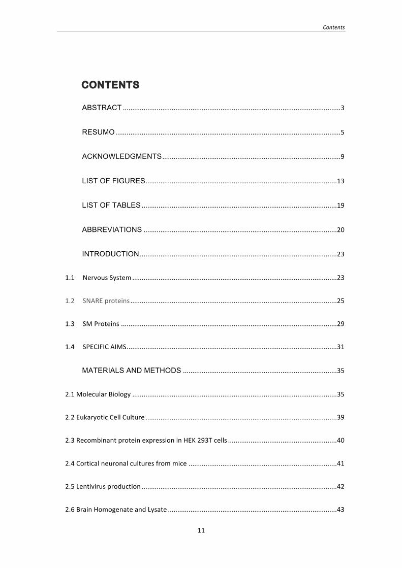
CONTENTS
ABSTRACT .................................................................................................................... 3
RESUMO ........................................................................................................................ 5
ACKNOWLEDGMENTS ............................................................................................... 9
LIST OF FIGURES ...................................................................................................... 13
LIST OF TABLES ........................................................................................................ 19
ABBREVIATIONS ....................................................................................................... 20
INTRODUCTION ......................................................................................................... 23
1.1 Nervous System ............................................................................................................. 23
1.2 SNARE proteins .............................................................................................................. 25
1.3 SM Proteins ................................................................................................................... 29
1.4 SPECIFIC AIMS ................................................................................................................ 31
MATERIALS AND METHODS .................................................................................. 35
2.1 Molecular Biology ............................................................................................................. 35
2.2 Eukaryotic Cell Culture ...................................................................................................... 39
2.3 Recombinant protein expression in HEK 293T cells .......................................................... 40
2.4 Cortical neuronal cultures from mice ............................................................................... 41
2.5 Lentivirus production ........................................................................................................ 42
2.6 Brain Homogenate and Lysate .......................................................................................... 43
Contents
12
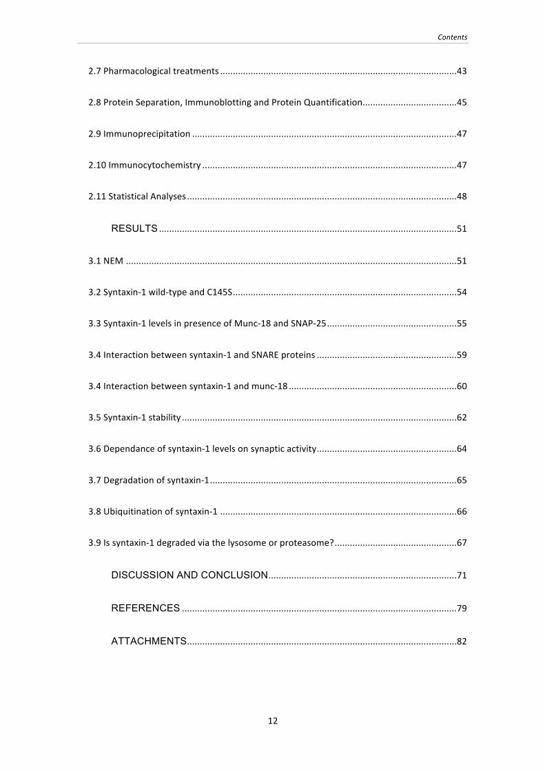
2.7 Pharmacological treatments ............................................................................................. 43
2.8 Protein Separation, Immunoblotting and Protein Quantification ..................................... 45
2.9 Immunoprecipitation ........................................................................................................ 47
2.10 Immunocytochemistry .................................................................................................... 47
2.11 Statistical Analyses .......................................................................................................... 48
RESULTS ..................................................................................................................... 51
3.1 NEM .................................................................................................................................. 51
3.2 Syntaxin-‐1 wild-‐type and C145S ........................................................................................ 54
3.3 Syntaxin-‐1 levels in presence of Munc-‐18 and SNAP-‐25 ................................................... 55
3.4 Interaction between syntaxin-‐1 and SNARE proteins ....................................................... 59
3.4 Interaction between syntaxin-‐1 and munc-‐18 .................................................................. 60
3.5 Syntaxin-‐1 stability ............................................................................................................ 62
3.6 Dependance of syntaxin-‐1 levels on synaptic activity ....................................................... 64
3.7 Degradation of syntaxin-‐1 ................................................................................................. 65
3.8 Ubiquitination of syntaxin-‐1 ............................................................................................. 66
3.9 Is syntaxin-‐1 degraded via the lysosome or proteasome? ................................................ 67
DISCUSSION AND CONCLUSION .......................................................................... 71
REFERENCES ............................................................................................................ 79
ATTACHMENTS .......................................................................................................... 82

List of Figures
13
LIST OF FIGURES
Figure 1- Vesicle proteins (from 8). ........................................................................................ 24
Figure 2 - Trafficking of synaptic vesicles in the nerve terminal (from 9). ........................ 25
Figure 3 ‒ SNARE complex (from14) .................................................................................. 26
Figure 4 - Synaptobrevin-2 structure (adapted from 17) ...................................................... 27
Figure 5 - SNAP-25 structure (adapted from 17). ................................................................. 28
Figure 6 ‒ A) Closed conformation of syntaxin-1 (from14), B) Syntaxin-1 structure;
(Adapted from14). .............................................................................................................. 29
Figure 7 ‒ A) Binding of the SM protein munc-18 to the “closed” conformation of
syntaxin-1. B) The “open” conformation of a t-SNARE complex, consisting of a t-
SNARE and its cognate SM protein bound to the N-peptide of syntaxin’s Habc
domain. C) SNARE and SM proteins form the universal fusion machinery (From 4).
............................................................................................................................................ 30
Figure 8 ‒ A) Structure of Munc18-1; B) Structure of syntaxin-1/Munc18 complex.
(From14) .............................................................................................................................. 31
Figure 9 ‒ Effect of NEM on synaptobrevin-2, SNAP-25, syntaxin-1 and SNARE
complex levels in brain homogenate. Equal amounts of brain homogenate were
treated with 2 mM NEM or vehicle control (ethanol) over night at 37°C. Reaction
was stopped by addition of 5x laemmli sample buffer containing DTT. A) Samples
were analyzed by immunblotting for levels of SNARE-complexes, syntaxin-1 (Synt-
1), SNAP-25 and synaptobrevin-2 (Syb-2). B) Protein levels were quantitated
using 125I-labeled secondary antibody and were normalized to the vehicle control.
** p < 0.01 using student’s T-test (n = 3). ..................................................................... 51
Figure 10 ‒ NEM reaction with the thiol group of a cysteine. ............................................. 52

List of Figures
14
Figure 11 - Syntaxin-1A sequence alignment. Yellow boxes highlight the evolutionary
conservation of C145 among different species. ........................................................... 52
Figure 12 ‒ Effect of NEM on levels of syntaxin-1 WT and mutated C145S in
transfected HEK cells treated with 5 mM NEM for 5min at 37°C. Reaction was
stopped by washing cells with PBS and solubilization in 0.1% Triton-X 100 (TX-
100). Insoluble material was removed by centrifugation for 20 min at 10,000g.
Supernatant was collected and 5% Laemmli sample buffer containing DTT was
added. A) Samples were analyzed by immunblotting for levels of syntaxin-1 WT
and C145S and GFP, which was used as transfection control. B) Protein levels
were quantitated using 125I-labeled secondary antibody and were first normalized
to GFP levels and then to syntaxin-1 WT without NEM treatment. ** p < 0.01 using
student’s T-test (n = 3). ................................................................................................... 53
Figure 13 - Effect of different NEM concentrations on syntaxin-1 levels in neuronal
cultures treated with a NEM concentration of 10 µM to 5mM. Neuronal cultures at
14 days in vitro were incubated for 5 min with NEM or vehicle control (ethanol).
Syntaxin-1 levels were analyzed by immunblotting. .................................................... 53
Figure 14 ‒ Effect of vehicle control and 5 mM NEM on syntaxin-1 levels in neuronal
culture. Neuronal cultures at 14 days in vitro were incubated for 5 min with 5 mM
NEM, vehicle control or were kept untreated. B) Munc-18, syntaxin-1 (Stx-1) and
rab3A levels were analyzed by immunblotting; B) Proteins levels were quantitated
using 125I-labeled secondary antibodies. Syntaxin-1 levels were normalized to
rab3A levels and quantitated as percent of levels of untreated neurons. **** p<
0.0001, using Student’s T-test (n = 9). .......................................................................... 54
Figure 15 ‒ Targeting of syntaxin-1 wild-type and C145S mutation in transfected HEK
cells. 48h after transfection, HEK cells were washed fixed for 15 min in 4%
paraformaldehyde and permeabilized for 5min in 0.1% Triton X-100. Cells were
then blocked in 5% BSA and were incubated with primary antibody HPC-1 in 1%
BSA overnight at 4°C. Cultures were washed and blocked, and an anti-mouse
Alexa-488 secondary antibody (1:500 in 1% BSA) was added for 1h in the dark.

List of Figures
15
Coverslips were mounted on glass slides in Fluoromount-G. Laser scanning
confocal microscopy was performed with excitation at 488nm on a Leica TCS SP-
2 inverted microscope. .................................................................................................... 55
Figure 16 ‒ Syntaxin-1 levels in co-transfected HEK cells. HEK cells were transfected
with syntaxin-1 (Stx-1) wild-type (WT) or C145S full length and with α-synuclein1-
95, SNAP-25 or munc-18. 48hours after transfection, cells were solubilized with
0.1% Triton-X 100 (TX-100) and insoluble material was analyzed by immunblotting
for levels of syntaxin-1 WT and C145S (A). B) Protein levels were quantitated
using 125I-labeled secondary antibody and were normalized to β-actin and GFP. * p
<0.05 ** p <using student’s T-test (n = 3). .................................................................... 56
Figure 17 ‒ Syntaxin-1 levels in co-transfected HEK cells. HEK cells were transfected
with syntaxin-1 (Stx-1) wild-type (WT) or C145S 1-264 and with α-synuclein1-95,
SNAP-25 or munc-18. 48hours after transfection, cells were solubilized with 0.1%
Triton-X 100 (TX-100) and insoluble material was analyzed by immunblotting for
levels of syntaxin-1 WT and C145S (A). B) Protein levels were quantitated using
125I-labeled secondary antibody and were normalized to β-actin and GFP. ** p <
0.01; **** p < 0.0001 using student’s T-test (n = 3). .................................................... 57
Figure 18 ‒ Syntaxin-1 levels in co-transfected HEK cells. HEK cells were transfected
with syntaxin-1 (Stx-1) wild-type (WT) or C145S 180-288 and with α-synuclein1-95,
SNAP-25 or munc-18. 48hours after transfection, cells were solubilized with 0.1%
Triton-X 100 (TX-100) and insoluble material was analyzed by immunblotting for
levels of syntaxin-1 WT and C145S (A). B) Protein levels were quantitated using 125I-labeled secondary antibody and were normalized to β-actin and GFP. **** p <
0.0001 using student’s T-test (n = 3). ............................................................................ 58
Figure 19 ‒ Syntaxin-1 levels in co-transfected HEK cells. HEK cellswere transfected
with syntaxin-1 (Stx-1) wild-type (WT) or C145S 180-264 and with α-synuclein1-95,
SNAP-25 or munc-18. 48hours after transfection, cells were solubilized with 0.1%
Triton-X 100 (TX-100) and insoluble material was analyzed by immunblotting for
levels of syntaxin-1 WT and C145S (A). B) Protein levels were quantitated using
List of Figures
16
125I-labeled secondary antibody and were normalized to β-actin and GFP. ** p <
0.01 using student’s T-test (n = 3). ................................................................................ 59
Figure 20 ‒ Immunoprecipitation of SNARE proteins and munc-18 in brain lysate.
Solubilized mouse brain was incubated at 4°C for 1h with 15 µl syntaxin-1
antibodies. As a negative control, samples were incubated with pre-immune
serum. Samples were then incubated for 2h at 4°C with 50 µl protein-A sepharose.
Sepharose was washed 3x with 1% TX-100 and bound proteins were eluted with
2x Laemmli sample buffer containing DTT. Samples were analyzed by SDS-PAGE
and immunoblotting for munc-18, syntaxin-1 (Stx-1), SNAP-25 and synaptobrevin-
2 (Syb-2). ........................................................................................................................... 59
Figure 21 ‒ Immunoprecipitations of transfected HEK cells to map the binding domain
of munc-18 and syntaxin-1. HEK cells were co-transfected with munc-18 and A)
full length syntaxin-1 wild-type (WT) and C145S, B) syntaxin-1 wild-type and
C145S lacking the transmembrane region, C) syntaxin-1 wild-type lacking the Habc
domain, and D) syntaxin-1 wild-type lacking the Habc domain and the
transmembrane region. Solubilized HEK 293T cells were incubated at 4°C for 1h
with 15 µl primary antibodies. As a negative control, samples were incubated
without antibody for immunoprecipitations in case of monoclonal antibodies, or
with pre-immune serum for polyclonal antibodies. Samples were then incubated
for 2h at 4°C with 50 µl protein-G sepharose (for monoclonal immunoglobulins) or
50 µl protein-A sepharose (for polyclonal rabbit sera). Sepharose was washed
with 1% TX-100 and bound proteins were eluted with 2x Laemmli sample buffer
containing DTT. Samples were analyzed by SDS-PAGE and immunoblotting for
munc-18 and syntaxin-1 (Stx-1). .................................................................................... 61
Figure 22 ‒ Analysis of syntaxin-1 stability using a cycloheximide chase experiment.
HEK cells co-transfected with syntaxin-1 (Stx-1) wild-type (WT) or C145S and α-
synuclein1-95 (a-syn1-95) were treated with 0.1g/L cycloheximide and analyzed 0h,
6h, 12h, and 24h after treatment. Cells were solubilized with 0.1% Triton-X 100
and soluble material was analyzed by SDS-PAGE and immunblotting for syntaxin-

List of Figures
17
1 (A). B) Protein levels were normalized to beta actin levels and were quantitated
using 125I-labeled secondary antibody. * p < 0.03 using student’s T-test (n = 3). ... 62
Figure 23 ‒ Analysis of syntaxin-1 stability using a cycloheximide chase experiment.
HEK cells co-transfected with syntaxin-1 (Stx-1) wild-type (WT) or C145S, α-
synuclein1-95 (a-syn1-95) and munc-18 were treated with 0.1g/L cycloheximide and
analyzed 0h, 6h, 12h, and 24h after treatment. Cells were solubilized with 0.1%
Triton-X 100 and soluble material was analyzed by SDS-PAGE and immunblotting
for syntaxin-1 (A). B) Protein levels were normalized to beta actin levels and were
quantitated using 125I-labeled secondary antibody. p < n.s. using student’s T-test (n
= 3). .................................................................................................................................... 63
Figure 24 ‒ Analysis of syntaxin-1 half-life in neuronal culture. Neuronal cultures at 11
days in vitro were incubated with 0.1g/L cycloheximide. (A) Syntaxin-1 (Stx-1)
levels at 0h, 3h, 24h, 48h, and 72h were analyzed by immunblotting (B) and were
quantitated using 125I-labeled secondary antibodies, normalized to the 0h levels
(n=3). .................................................................................................................................. 64
Figure 25 - Effect of synaptic activity on syntaxin-1 levels. Neuronal cultures at 12 days
in vitro were incubated for 36h with 20 μM APV, 5 μM tetrodotoxin (TTX), 25 mM
K+, 4mM Ca2+ or were kept untreated. Syntaxin-1 (Stx-1) and valosin-containing
protein (VCP) levels were analyzed by immunblotting (A) and were quantitated
using 125I-labeled secondary antibodies (B). Syntaxin-1 levels were normalized to
vcp and quantitated as percent of levels of untreated neurons(n = 3). .................... 65
Figure 26 ‒ Analysis of syntaxin-1 degradation A) Syntaxin-1 wild-type (WT), B)
Syntaxin-1 C145S; C) Syntaxin-1 WT plus munc-18; were transfected into HEK
cells. 48h after transfection, cells were solubilized with 0.1% Triton-X 100 and
soluble material was analyzed by immunblotting for syntaxin-1. D) Full-length and
degraded protein levels were quantitated using 125I-labeled secondary antibody,
with degradation products expressed as percent of total syntaxin-1 protein. * p <
0.03 and ** p < 0.01 using student’s T-test (n = 3). .................................................... 66
List of Figures
18
Figure 27 ‒ Analysis of ubiquitination in HEK cells transfected with syntaxin-1 wild-type
and C145S. Immunoprecipitations were performed with lysates from HEK 293T
cells transfected with syntaxin-1 (Stx-1) wild-type (WT) or C145S. Solubilized HEK
293T cells were incubated at 4°C for 1h with 15 µl primary antibody (438B). As a
negative control, samples were incubated with pre-immune serum. Samples were
then incubated for 2h at 4°C with 50 µl protein-A sepharose. Sepharose was
washed with 1% TX-100 and bound proteins were eluted with 2x Laemmli sample
buffer containing DTT. A) Samples were analyzed by SDS-PAGE and
immunoblotting for ubiquitin (top) and syntaxin-1 (bottom). B) Ubiquitination was
quantitated using 125I-labeled secondary antibodies, normalized to the efficiency of
the syntaxin-1 immunoprecipitation. **** p< 0.0001 using Student’s T-test (n = 3).
............................................................................................................................................ 67
Figure 28 - Effect of proteases inhibitors on syntaxin-1 levels in neuronal culture.
Neuronal cultures at 12 days in vitro were incubated for 36h with 10 μM
Epoxomicin, 10 μM MG132, 10 μM Clasto-lactacystin, 10 mg/L
Leupeptin/Pepstatin, 4 mM PMSF or were kept untreated. Syntaxin-1 (Stx-1) and
β-actin levels were analyzed by immunblotting (A) and were quantitated using 125I-
labeled secondary antibodies (B). Syntaxin-1 levels were normalized to β-actin
and quantitated as percent of levels of untreated neurons (n = 2). .......................... 68
Figure 29 - Model depicting the stabilization of syntaxin-1 by munc-18 and the C145S
mutation. Syntaxin-1may be degraded via ubiquitiniation and the lysosome.
Ubiquitination of syntaxin-1 may happen on the cysteine residue mutated in the
C145S mutant, or this cysteine reside may signal the cell to degrade syntaxin-1. 76

List of Tables
19
LIST OF TABLES
Table 1 ‒ Syntaxin-1 constructs ............................................................................................. 35
Table 2 - Syntaxin-1 constructs (continuation) ..................................................................... 36
Table 3 ‒ PCR primers, annealing temperature and number of cycles ............................ 36
Table 4 ‒ Sequencing primers ................................................................................................ 39
Table 5 ‒ Components of polyacrylamide gels .................................................................... 45
Table 6 ‒ Primary Antibodies .................................................................................................. 48
Table 7- Primary Antibodies (continuation) .......................................................................... 49

Abbreviations
20
ABBREVIATIONS
APS Amonium persulfate APV (2R)-amino-5-phosphonovaleric acid ATCC American Type Culture Collection BCA Bicinchoninic acid BSA Bovine Serum Albumin C Cysteine CaCl2 Calcium chloride CO2 Carbon Dioxide CHX Cycloheximide C145S Cysteine 145 Serine DIV Days in vitro DMEM Dulbecco’s Modified Eagle Medium DMSO Dimethyl Sulfoxide DNA Deoxyribonucleic Acid dNTP Deoxynucleoside triphosphate DPBS Dulbecco’s Phosphate Buffered Saline DTT Dithiothreitol ECL Enhanced Chemiluminescence E. col i Escherichia coli EDTA Ethylenediamine tetraacetic acid FBS Fetal Bovine Serum GFP Green Fluorescent Protein HEK Human Embryonic Kidney HEPES 4-(2-hydroxyethyl)-1-piperazineethanesulfonic acid HIV Humane immunodeficiency virus HRP Horseradish peroxidase IC Immunocytochemistry IRES Internal r ibosome entry site IP Immunoprecipitation KCl Potassium Chloride Leup/Pept Leupeptin/Pepstantin LB Lysogeny broth MDRS Methylation-dependent restriction systems MgCl2 Magnesium chloride NaCl Sodium chloride NaHCO3 Sodium bicarbonate NEM N-Ethylmaleimide NSF N-ethylmaleimide-sensitive factor
Abbreviations
21
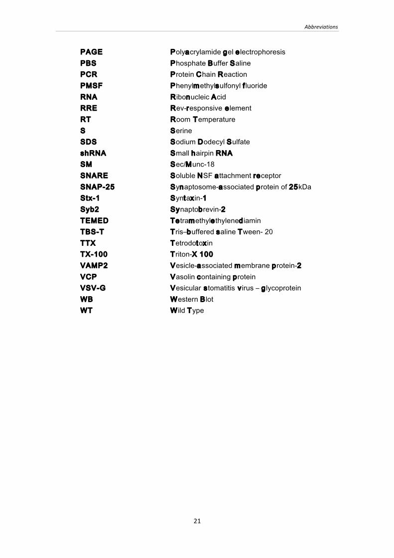
PAGE Polyacrylamide gel electrophoresis PBS Phosphate Buffer Saline PCR Protein Chain Reaction PMSF Phenylmethylsulfonyl f luoride RNA Ribonucleic Acid RRE Rev-responsive element RT Room Temperature S Serine SDS Sodium Dodecyl Sulfate shRNA Small hairpin RNA SM Sec/Munc-18 SNARE Soluble NSF attachment receptor SNAP-25 Synaptosome-associated protein of 25kDa Stx-1 Syntaxin-1 Syb2 Synaptobrevin-2 TEMED Tetramethylethylenediamin TBS-T Tris‒buffered saline Tween- 20 TTX Tetrodotoxin TX-100 Triton-X 100 VAMP2 Vesicle-associated membrane protein-2 VCP Vasolin containing protein VSV-G Vesicular stomatitis virus ‒ glycoprotein WB Western Blot WT Wild Type


Chapter 1
23
CHAPTER 1
INTRODUCTION
“The brain is the human body’s most mysterious organ. It learns, it changes, it adapts,
it tells us what we see, what we hear, it let us feel love, I think it holds our soul.”
Shonda Rhimes
1.1 Nervous System
The brain is the center of the nervous system; it weighs approximately 1.3 kg in
an adult human. There are about 1011 nerve cells, called neurons1. Neurons
communicate with each other through direct contacts (electrical synapses) and mostly
through non-continuous connections known as chemical synapses, the principal
computational unit of the nervous system2. Neurons are classified according to: 1)
function (e.g. motor, sensorial, interneuron), 2) localization (e.g. cortical, spinal), 3)
shape (e.g. pyramidal, granule, mitral), and 4) nature of the transmitter synthetized and
released (e.g. excitatory, inhibitory, neuromodulatory). In the human brain, neurons are
connected by 1014 synapses1. Synaptic transmission occurs when an actions potential
triggers neurotransmitter release from a presynaptic nerve terminal, resulting in
synaptic vesicle exocytosis3. Thus, many pre-synaptic and post-synaptic proteins are
needed to transmit the information from one neuron to another.
1.1.1 Synaptic vesicle cycle
Membrane fusion is one of the vital processes in life, and happens when two
separate membranes merge into a continuous bilayer. Fusion can occur as constitutive
Chapter 1
24
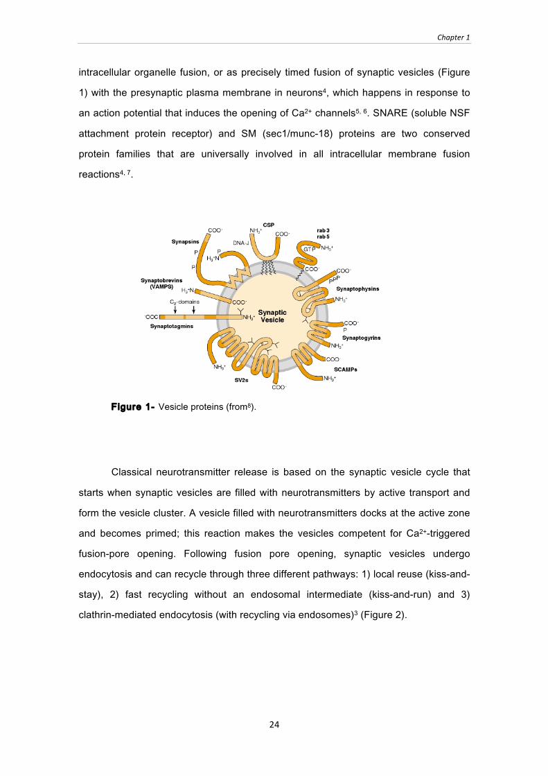
intracellular organelle fusion, or as precisely timed fusion of synaptic vesicles (Figure
1) with the presynaptic plasma membrane in neurons4, which happens in response to
an action potential that induces the opening of Ca2+ channels5, 6. SNARE (soluble NSF
attachment protein receptor) and SM (sec1/munc-18) proteins are two conserved
protein families that are universally involved in all intracellular membrane fusion
reactions4, 7.
Figure 1- Vesicle proteins (from8).
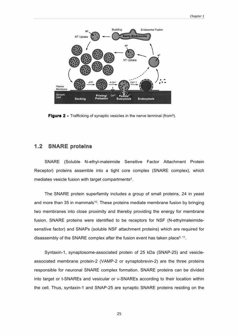
Classical neurotransmitter release is based on the synaptic vesicle cycle that
starts when synaptic vesicles are filled with neurotransmitters by active transport and
form the vesicle cluster. A vesicle filled with neurotransmitters docks at the active zone
and becomes primed; this reaction makes the vesicles competent for Ca2+-triggered
fusion-pore opening. Following fusion pore opening, synaptic vesicles undergo
endocytosis and can recycle through three different pathways: 1) local reuse (kiss-and-
stay), 2) fast recycling without an endosomal intermediate (kiss-and-run) and 3)
clathrin-mediated endocytosis (with recycling via endosomes)3 (Figure 2).
Chapter 1
25
Figure 2 - Trafficking of synaptic vesicles in the nerve terminal (from9).
1.2 SNARE proteins
SNARE (Soluble N-ethyl-maleimide Sensitive Factor Attachment Protein
Receptor) proteins assemble into a tight core complex (SNARE complex), which
mediates vesicle fusion with target compartments4.
The SNARE protein superfamily includes a group of small proteins, 24 in yeast
and more than 35 in mammals10. These proteins mediate membrane fusion by bringing
two membranes into close proximity and thereby providing the energy for membrane
fusion. SNARE proteins were identified to be receptors for NSF (N-ethylmaleimide-
sensitive factor) and SNAPs (soluble NSF attachment proteins) which are required for
disassembly of the SNARE complex after the fusion event has taken place5, 11.
Syntaxin-1, synaptosome-associated protein of 25 kDa (SNAP-25) and vesicle-
associated membrane protein-2 (VAMP-2 or synaptobrevin-2) are the three proteins
responsible for neuronal SNARE complex formation. SNARE proteins can be divided
into target or t-SNAREs and vesicular or v-SNAREs according to their location within
the cell. Thus, syntaxin-1 and SNAP-25 are synaptic SNARE proteins residing on the

Chapter 1
26
presynaptic plasma membrane; synaptobrevin-2 resides on the synaptic vesicle
membrane4, 7, 11.
The importance of SNARE proteins for neurotransmission became apparent in
studies using botulinum toxins (BoNT), proteins produced by the bacterium Clostridium
botulinum. They are considered to be the most powerful neurotoxins ever discovered
and specifically cleave SNARE proteins, preventing synaptic vesicles from
docking/fusing with pre-synaptic membranes and therefore blocking neurotransmitter
release12, 13.
SNARE motifs can also be structurally distinguished into R-SNAREs and Q-
SNAREs. The Q encodes for the amino acid arginine; thus, Q-SNARE proteins have
an arginine residue as central amino acid in the SNARE domain, whereas R-SNAREs
have a glutamine residue in the center of the SNARE motif. According to the position of
their SNARE motif-containing domains within the SNARE complex and by their
sequence similarities, Q and R SNAREs can be distinguished into four classes: 1) R-
SNARE motif (VAMPs), 2) Qa-SNARE motif (syntaxins), 3) Qb-SNARE motif
(homologs of the N-terminus of SNAP-25), 4) Qc-SNARE motif (homologs of the C-
terminus of SNAP-25). Since R-SNAREs correspond to v-SNAREs, and Q-SNAREs
correspond to t-SNAREs, all SNARE complexes contain one member of each class4, 10.
SNARE proteins contain a conserved ̃60 to ̃ 70 residues SNARE repeat, a
highly reactive sequence that assembles into the SNARE complex by forming a four
helical bundle4, 7 (Figure 3).
Figure 3 ‒ SNARE complex (from14)
Synaptobrevin-‐
2
Syntaxin-‐1 SNAP-‐25
Chapter 1
27
Most of the SNARE proteins contain one SNARE motif, except for SNAP-class
SNAREs which contain two SNARE motifs4,7. Synapses transmit signals at high
frequencies. Thus, SNARE proteins continuously cycle through a highly reactive, non-
assembled state, and a less reactive, assembled state15. These conformational
changes are probably the reason for the evolution of chaperones such as CSPα and α-
synuclein, which keep SNARE proteins stable throughout the life of a neuron6. While α-
synuclein increases SNARE complex assembly by binding to the v-SNARE
synaptobrevin-215, the CSPα/Hsc70/SGT chaperone complex binds to monomeric
SNAP-25 and stabilizes this protein16.
1.2.2 Synaptobrevin-2
Synapobrevin-2 is essential for fast synaptic vesicle endocytosis; absence of
synaptobrevin-2 in synapses reveal an altered shape and size of synaptic vesicle, and
stimulus-dependent endocytosis was delayed17.
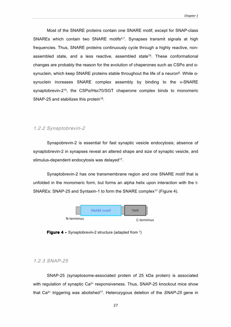
Synaptobrevin-2 has one transmembrane region and one SNARE motif that is
unfolded in the monomeric form, but forms an alpha helix upon interaction with the t-
SNAREs: SNAP-25 and Syntaxin-1 to form the SNARE complex17 (Figure 4).
Figure 4 - Synaptobrevin-2 structure (adapted from 7)
1.2.3 SNAP-25
SNAP-25 (synaptosome-associated protein of 25 kDa protein) is associated
with regulation of synaptic Ca2+ responsiveness. Thus, SNAP-25 knockout mice show
that Ca2+ triggering was abolished17. Heterozygous deletion of the SNAP-25 gene in
SNARE motif TMR
N-‐terminus C-‐terminus

Chapter 1
28
mice results in a hyperactive phenotype similar to attention deficit hyperactivity disorder
(ADHD)18.
SNAP-25 protein has two SNARE motifs and is palmitoylated at cysteine
residues between the SNARE motifs, allowing SNAP-25 to anchor to the plasma
membrane, since this SNARE protein does not have a transmembrane region17 (Figure
5).
Figure 5 - SNAP-25 structure (adapted from17).
1.2.3 Syntaxin-1
Syntaxin-1 (Stx-1) expression starts in early embryonic development, and its
levels are intensely up-regulated during synapse formation and brain maturation19.
Syntaxin-1 has been linked to long-term potentiation, learning and memory, and it has
been associated with several neurodegenerative and psychiatric diseases such as
schizophrenia, Alzheimer’s disease, and Creutzfeldt-Jakob disease19.
Syntaxin-1 contains an N-terminal Habc domain which has been shown to bind to
munc-184 and which is connected to the SNARE motif by a short linker sequence. The
protein is membrane-anchored by its C-terminal transmembrane region (TMR). SNARE
motif and TMR occupy less than half of the sequence4, 10 (Figure 6).
The N-terminal part of the protein, with the three helix bundles is flexible and
allows syntaxin-1 to alter between two conformations: a “closed” conformation, where
the Habc domain and SNARE motif bind intramolecularly and thereby prevent its
engagement into the SNARE complex, and an “open” conformation where the SNARE
motif is exposed and can participate in SNARE complex formation4,10.
N-‐terminus C-‐terminus
SNARE motif SNARE motif

Chapter 1
29
Figure 6 ‒ A) Closed conformation of syntaxin-1 (from14), B) Syntaxin-1 structure; (Adapted from14).
1.3 SM Proteins
The SM protein superfamily is composed of only a few proteins: 4 conserved
subfamilies have been described in eukaryotes, which are essential for exocytosis
(Sec1/Munc18); endocytosis (Vps45); protein biosynthesis (Sly1); degradation
(Vps33)20. SM are highly conserved among different organisms and show a highly
conserved overall fold20.
SM proteins are hydrophilic proteins of 60-70kDa that share homology evenly
throughout their sequence, indicating that no particular domain is associated with their
primary function. I.e., it is not clear how specificity for vesicle attachment or fusion is
mediated7,10,20. SM proteins fold into an arch-shaped “clasp” structure containing three
domains (called domains 1-3)20, with a large cavity on one side, and a deep groove on
the opposite side7,10,20 The arch-shape as well as the deep groove have been
implicated in interactions with SNARE proteins 20.
SM proteins are part of all membrane fusion reactions and are as essential as
SNARE proteins for the fusion process14. The reduced number of SM proteins
SNARE motif TMR
1 180 264 288
Ha Hb Hc
28 62 71 104 111 144
N-‐terminus C-‐terminus
Linker
A
B
Chapter 1
30
compared to SNARE proteins suggests that these proteins are versatile fusion agents
that function in multiple reactions10.
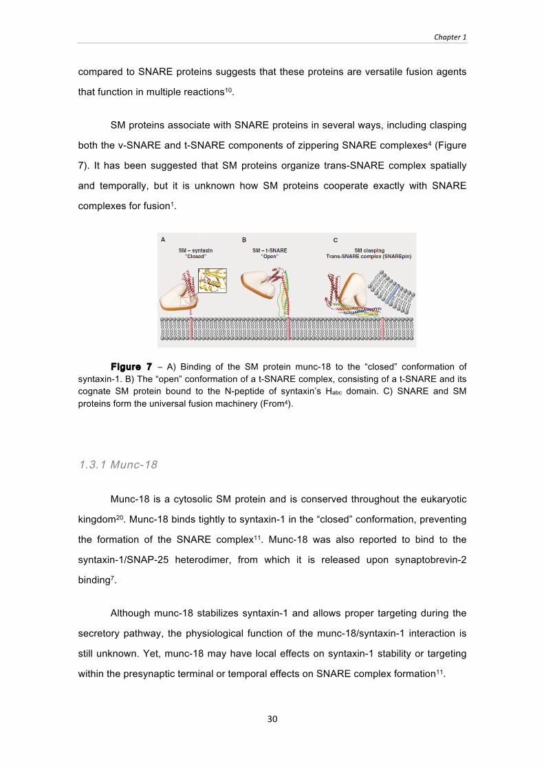
SM proteins associate with SNARE proteins in several ways, including clasping
both the v-SNARE and t-SNARE components of zippering SNARE complexes4 (Figure
7). It has been suggested that SM proteins organize trans-SNARE complex spatially
and temporally, but it is unknown how SM proteins cooperate exactly with SNARE
complexes for fusion1.
Figure 7 ‒ A) Binding of the SM protein munc-18 to the “closed” conformation of syntaxin-1. B) The “open” conformation of a t-SNARE complex, consisting of a t-SNARE and its cognate SM protein bound to the N-peptide of syntaxin’s Habc domain. C) SNARE and SM proteins form the universal fusion machinery (From4).
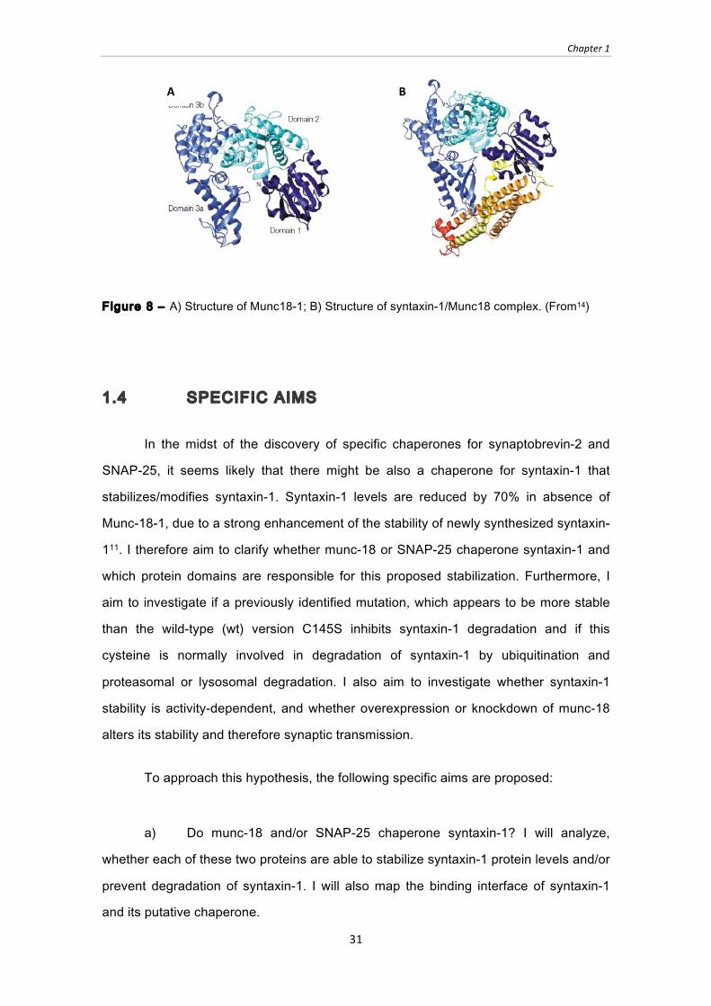
1.3.1 Munc-18
Munc-18 is a cytosolic SM protein and is conserved throughout the eukaryotic
kingdom20. Munc-18 binds tightly to syntaxin-1 in the “closed” conformation, preventing
the formation of the SNARE complex11. Munc-18 was also reported to bind to the
syntaxin-1/SNAP-25 heterodimer, from which it is released upon synaptobrevin-2
binding7.
Although munc-18 stabilizes syntaxin-1 and allows proper targeting during the
secretory pathway, the physiological function of the munc-18/syntaxin-1 interaction is
still unknown. Yet, munc-18 may have local effects on syntaxin-1 stability or targeting
within the presynaptic terminal or temporal effects on SNARE complex formation11.
Chapter 1
31
Figure 8 ‒ A) Structure of Munc18-1; B) Structure of syntaxin-1/Munc18 complex. (From14)
1.4 SPECIFIC AIMS
In the midst of the discovery of specific chaperones for synaptobrevin-2 and
SNAP-25, it seems likely that there might be also a chaperone for syntaxin-1 that
stabilizes/modifies syntaxin-1. Syntaxin-1 levels are reduced by 70% in absence of
Munc-18-1, due to a strong enhancement of the stability of newly synthesized syntaxin-
111. I therefore aim to clarify whether munc-18 or SNAP-25 chaperone syntaxin-1 and
which protein domains are responsible for this proposed stabilization. Furthermore, I
aim to investigate if a previously identified mutation, which appears to be more stable
than the wild-type (wt) version C145S inhibits syntaxin-1 degradation and if this
cysteine is normally involved in degradation of syntaxin-1 by ubiquitination and
proteasomal or lysosomal degradation. I also aim to investigate whether syntaxin-1
stability is activity-dependent, and whether overexpression or knockdown of munc-18
alters its stability and therefore synaptic transmission.
To approach this hypothesis, the following specific aims are proposed:
a) Do munc-18 and/or SNAP-25 chaperone syntaxin-1? I will analyze,
whether each of these two proteins are able to stabilize syntaxin-1 protein levels and/or
prevent degradation of syntaxin-1. I will also map the binding interface of syntaxin-1
and its putative chaperone.
A B
Chapter 1
32
b) What are the molecular changes in syntaxin-1 caused by the C145S
mutation, i.e. is syntaxin-1 C145S more stable than wild type syntaxin-1? First, I aim to
analyze whether the cysteine to serine mutation of syntaxin-1 is more stable than the
wild-type. Then, I aim to clarify whether degradation of syntaxin-1 happens via the
ubiquitin/proteasome pathway or the lysosomal pathway, and whether expression of
munc-18 alters this process. Finally, I will analyze if the cysteine 145 is involved in the
degradation process of syntaxin-1, e.g. prevents or slows down degradation of
syntaxin-1.
c) Does syntaxin-1 stability depend on synaptic activity? What happens to
synaptic transmission in presence or absence of the postulated chaperones?


Chapter 2
35
CHAPTER 2
MATERIALS AND METHODS
2.1 Molecular Biology
2.1.1 Plasmid Vectors
A c-myc (N-EQKLISEEDL-C) epitope with a linker (AA) was added to the N-
terminus of a rat syntaxin-1A cDNA to generate myc-tagged syntaxin-1 wild-type (WT)
and syntaxin-1 cysteine to serine mutation (C145S), using as a template an already
existing HA-tagged (YPYDVPDYA) syntaxin-1A cDNA. The two different tags are
needed in order to distinguish between WT and C145S when transfected in the same
cell. For syntaxin-1A truncation constructs, a stop codon was introduced at residue
265. Syntaxin-1A WT and C145S full length and 1-264 truncations were cloned into
pCMV5, FUW and FSW vectors, respectively. Rat SNAP-25 cDNA and rat munc-18-1
cDNA constructs were cloned into FUW and FSW vectors. The following other
constructs I used were already generated in the Sudhof lab: pCMV5 HA-syntaxin-1
WT180-264, pCMV5 HA-syntaxin-1 WT180-288, pCMV5-SNAP-25, pCMV5-munc-18-1 and
lentiviral munc-18 shRNA constructs. Syntaxin-1 constructs are shown in Table 1 and
2.
Table 1 ‒ Syntaxin-1 constructs
Vectors/tags Syntaxin-1 Structure Included domains pCMV5-HA or myc FUW-myc FSW-myc
WT and C145S full length
Habc domain SNARE motif Transmembrane domain
pCMV5-HA WT 180-288
SNARE motif Transmembrane domain
SNARE motif TM
1 180 264 288 N-‐terminus C-‐terminus
SNARE motif TM
180 264 288 N-‐terminus C-‐terminus

Chapter 2
36
Table 2 - Syntaxin-1 constructs (continuation)
pCMV5-HA WT 180-264
SNARE motif
2.1.2 DNA amplification
Syntaxin-1, Munc-18 and SNAP-25 cDNA was amplified by polymerase chain
reaction (PCR), using with the following reagents: 1) PfuUltra™ HF DNA polymerase
(Roche); 2) Dimethyl sulfoxide (DMSO) was used in the PCR reaction in order to inhibit
secondary structures within the DNA template or within the primers, minimizing
interloping reactions22; 3) Bovine serum albumin (BSA) (Sigma-Aldrich) works as a
stabilizing agent in enzymatic reactions and enhances enzymatic activity23; 4)
Deoxynucleoside triphosphates (dNTPs) (NEB) are necessary for DNA polymerase to
synthetize new DNA; 5) DNA template; 6) Primers. Number of cycles, annealing
temperature and primers used are listed in Table 3.
Table 3 ‒ PCR primers, annealing temperature and number of cycles
Protein Primer Sense Primer Anti-sense T / cycles
Myc-Stx-1A
1-288
5’GATCGCCACCATGGATGGAGCAGAAGCTGATCAGCGAGGAGGACCTGGCCGA AAGGACCGAACCCAGCT 3’
5’GTCGAATTCCTATCCAAAGATGCCCCCGAT 3’
50°C
cycles:
30x
Myc-Stx-1A
1-264
5’GATCGCCACCATGGATGGAGCAGAAGCTGAT
CAGCGAGGAGGACCTGGCCGA
AAGGACCGAACCCAGCT 3’
5’GTCGAATTCCTACTTCCTGCGTGCCTT 3’
52°C
cycles:
33x
SNAP-25 5’CTAGGAATTCACCGCCATGGCCGAAGACGCA
GACATG3’
5’GTCGAATTCTTAACCACTT
CCCAGCATCTTTG 3’
50°C
cycles:
30x
Munc-18 5’CTAGGAATTCACCGCCATGGCCCCCATTGGC
CTC3’
5’GTCGAATTCTTAACTGCTT
ATTTCTTCGTC 3’
50°C
cycles:
30x
SNARE motif
180 264
N-‐terminus C-‐terminus
Chapter 2
37
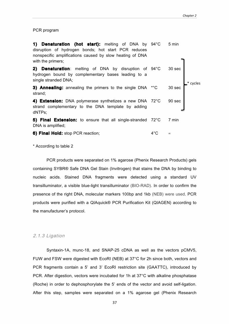
PCR program
1) Denaturation (hot start): melting of DNA by disruption of hydrogen bonds; hot start PCR reduces nonspecific amplifications caused by slow heating of DNA with the primers;
94°C 5 min
2) Denaturation: melting of DNA by disruption of hydrogen bound by complementary bases leading to a single stranded DNA;
94°C 30 sec
3) Annealing: annealing the primers to the single DNA strand;
*°C 30 sec
4) Extension: DNA polymerase synthetizes a new DNA strand complementary to the DNA template by adding dNTPs;
72°C 90 sec
5) Final Extension: to ensure that all single-stranded DNA is amplified;
72°C 7 min
6) Final Hold: stop PCR reaction; 4°C ∞
* According to table 2
PCR products were separated on 1% agarose (Phenix Research Products) gels
containing SYBR® Safe DNA Gel Stain (Invitrogen) that stains the DNA by binding to
nucleic acids. Stained DNA fragments were detected using a standard UV
transilluminator, a visible blue-light transilluminator (BIO-RAD). In order to confirm the
presence of the right DNA, molecular markers 100bp and 1kb (NEB) were used. PCR
products were purified with a QIAquick® PCR Purification Kit (QIAGEN) according to
the manufacturer’s protocol.
2.1.3 Ligation
Syntaxin-1A, munc-18, and SNAP-25 cDNA as well as the vectors pCMV5,
FUW and FSW were digested with EcoRI (NEB) at 37°C for 2h since both, vectors and
PCR fragments contain a 5’ and 3’ EcoRI restriction site (GAATTC), introduced by
PCR. After digestion, vectors were incubated for 1h at 37°C with alkaline phosphatase
(Roche) in order to dephosphorylate the 5’ ends of the vector and avoid self-ligation.
After this step, samples were separated on a 1% agarose gel (Phenix Research
* cycles
Chapter 2
38
Products) in order to isolate the cut DNA, followed by gel extraction and purification
using QIAEX II ® Gel Extraction Kit (QIAGEN) according to the manufacturer’s
protocol. For incorporation of cDNA into the vector, cut vector and PCR fragments
were ligated for 1h at room temperature using: 1) 1 µl T4 DNA ligase (NEB); 2) 1 µl 10x
DNA ligation buffer; 3) 8 µl insert plus vector at a ratio of insert:vector = 3:1.
2.1.4 Transformation
The bacterial strain used for molecular biology was Escherichia coli DH10B.
This strain was designed for the propagation of large insert DNA library clones, which
takes advantage of properties such as high DNA transformation efficiency and
maintenance of large plasmids, the lack of methylation-dependent restriction systems
(MDRS), and colony screening via lacZ-based α-complementation24-25.
Transformation, a process that allows DNA to enter the cell25, was done by
heat-shock. First, bacteria strain E. coli DH10B was incubated with DNA for 20 minutes
on ice, followed by a heat-shock at 42°C for 45 sec and recovery for 2 min on ice,
allowing the DNA to enter the bacteria. To allow bacteria to express the ampicillin
resistance introduced by the vector, bacteria were incubated in LB medium (Lysogeny
broth Medium (1% tryptone (BD); 0.5% yeast extract (BD); 0.5% NaCl (BD)) at 37°C for
1h. Bacteria were plated on LB plates containing ampicillin (1% tryptone (BD); 0.5%
yeast extract (BD); 0.5% NaCl (BD); 1.5% agar (BD); 100ug/mL ampicillin (Sigma))
overnight at 37°C to select for clones carrying the ampicillin resistance introduced by
transformation. Inoculation of a single colony was performed in LB medium with 10
µg/ml ampicillin (Invitrogen) over night at 37°C.
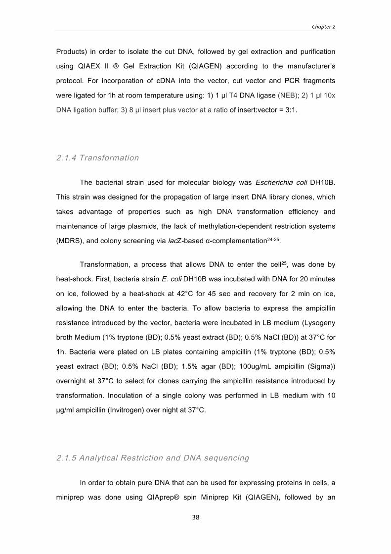
2.1.5 Analytical Restriction and DNA sequencing
In order to obtain pure DNA that can be used for expressing proteins in cells, a
miniprep was done using QIAprep® spin Miniprep Kit (QIAGEN), followed by an
Chapter 2
39
analytical restriction to ensure that the selected colonies carry the correct DNA, and
carry the DNA in the right orientation since the same restriction enzyme was used for 5’
and 3’ insertion. The following reagents were mixed in a total volume of 20 µl: 1) 5 µl
mini-prepped DNA; 2) 1 µl restriction enzyme: BamHI (NEB) was used for syntaxin-1A
and XmaI (NEB) for SNAP-25 and munc-18 restriction analysis; 3) 2 µl 10x buffer
(according to NEB catalogue); 4) 2 µl 1mg/mL BSA; each performed for 1h at 37°C.
(Attachment A: vector illustrations).
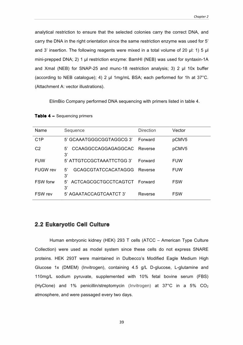
ElimBio Company performed DNA sequencing with primers listed in table 4.
Table 4 ‒ Sequencing primers
Name Sequence Direction Vector
C1P 5’ GCAAATGGGCGGTAGGCG 3’ Forward pCMV5
C2 5’ CCAAGGCCAGGAGAGGCAC 3’
Reverse pCMV5
FUW 5’ ATTGTCCGCTAAATTCTGG 3’ Forward FUW
FUGW rev 5’ GCAGCGTATCCACATAGGG 3’
Reverse FUW
FSW forw 5’ ACTCAGCGCTGCCTCAGTCT 3’
Forward FSW
FSW rev 5’ AGAATACCAGTCAATCT 3’ Reverse FSW
2.2 Eukaryotic Cell Culture
Human embryonic kidney (HEK) 293 T cells (ATCC ‒ American Type Culture
Collection) were used as model system since these cells do not express SNARE
proteins. HEK 293T were maintained in Dulbecco’s Modified Eagle Medium High
Glucose 1x (DMEM) (Invitrogen), containing 4.5 g/L D-glucose, L-glutamine and
110mg/L sodium pyruvate, supplemented with 10% fetal bovine serum (FBS)
(HyClone) and 1% penicillin/streptomycin (Invitrogen) at 37°C in a 5% CO2
atmosphere, and were passaged every two days.
Chapter 2
40
For cell passaging, HEK 293 T cells were washed twice with 1x Dulbecco’s
Phosphate Buffered Saline (DPBS) (Invitrogen), and were then incubated with 0.05%
trypsin-EDTA (Invitrogen) for 2 min to detach cells from the culture dish by digesting
cellular integrins. Trypsin digestion was stopped by addition of DMEM. After
dissociation, cells were resuspended and redistributed in 6 or 24 wells plate, 10cm
plates or T75 flasks.
2.3 Recombinant protein expression in HEK 293T cells
HEK 293T cells were transfected using FuGene-6 (Roche), a reagent with
cationic polymers that binds to negatively charged DNA, making a complex that is
taken up by the cell via endocytosis. Transfection was performed at a ratio of DNA to
Fugene of 1:3.
1) Syntaxin-1A expression experiments: HEK 293T cells (ATCC) were
co-transfected with pCMV5-syntaxin1A (wt, C145S or truncations), pCMV5-munc-18-1,
pCMV5-SNAP-25 or pCMV5-α-synuclein1-95 (α-synuclein1-95 is used to balance the
number of munc-18 or SNAP-25 plasmids transfected into HEK 293T cells (ATCC))
and pCMV5-emerald to control for transfection efficiency. Transfections were
performed at a ratio of 1:3:1 for syntaxin-1:munc-18/SNAP-25/ α-synuclein1-95:emerald.
2) Cycloheximide chase experiments: HEK 293T cells (ATCC) were co-
transfected with: a) pCMV5-syntaxin1A (wt or C145S) and pCMV5-α-synuclein1-95 at a
1:1 ratio; b) pCMV5-syntaxin1A (wt or C145S), pCMV5-munc-18 and pCMV5-α-
synuclein1-95 at a 1:5:1 ratio.
3) Immunoprecipitat ions (IP): HEK 293T cells (ATCC) were co-transfected
with pCMV5-syntaxin-1A (wt, C145S or truncations) and pCMV5-munc-18 at a 1:1
ratio.
4) Immunocytochemistry: HEK 293T cells (ATCC) were transfected with
pCMV5-syntaxin-1 wt or C145S.

Chapter 2
41
HEK 239T cells (ATCC) were harvested 48 hours after transfection; except for
cycloheximide experiments where cells were harvested at different time points (48h
plus 0h, 3h, 6h, 12h, 24h and 36h). For harvesting, cells were washed 3x with PBS and
solubilized with 0.1% Triton-X 100 (TX-100) (Sigma). After solubilization, insoluble
material was removed by centrifugation for 20 min at 10,000g. The supernatant was
collected and 5% Laemmli sample buffer (10% sodium dodecyl sulfate (SDS); 5%
glycerol; 0.006% bromophenol blue in ethanol; 0.4M Tris-Cl pH 6.8; 77mg/ml
dithiothreitol (DTT)) was added. To disrupt SNARE-complexes into SNARE protein
monomers, samples were boiled for 20 min at 100°C.
2.4 Cortical neuronal cultures from mice
Mouse cortical neurons were cultured from mouse pups at P0 (< 24hours after
birth). Brain regions were dissected on ice, and were incubated in ice-cold Hank’s
Balanced Salt Solution (HBS) with Hanks Balanced Salts without calcium chloride,
magnesium sulfate and sodium bicarbonate (Sigma), pH 7.4. This buffer contains
350mg/L sodium bicarbonate (NaHCO3) and 1mM HEPES (4-(2-hydroxyethyl)-1-
piperazineethanesulfonic acid) to stabilize the pH. Brains were digested in 2% papain
solution with 0.5M EDTA pH 8.0 and 1M CaCl2 in HBS for 20 min at 37°C to dissociate
cells. Brains were then triturated with a pipette in plating medium (MEM) (Invitrogen)
supplemented with 10% FBS (HyClone), 0.2M L-glutamine solution (Invitrogen),
0.25g/L insulin (Sigma). Cells were plated either onto a 12mm coverslip coated with
1mg/mL poly-L-lysine (Sigma) in 0.1M borate buffer (3.1g/L boric acid, 4.8g/L sodium
tetraborate, pH 8.5) for imaging, or in a 24well plastic dish for biochemical experiments.
After 1 day, plating medium was replaced with growth medium (0ARA-C) containing
5% FBS (HyClone), 0.2M glutamine solution (Invitrogen), 2% B-27 supplement
(Invitrogen). Neuronal cultures were kept in growth medium (2ARA-C) containing 5%
FBS (HyClone), 0.2M glutamine solution (Invitrogen) 2% B-27 supplement (Invitrogen),
2 uM cytosine arabinose (Sigma).
Chapter 2
42
2.5 Lentivirus production
Lentivirus is a class of retrovirus that can introduce a significant amount of
genetic information into animal cells by insertion of their DNA into the host
chromosomal DNA, thereby increasing the efficiency by which a modified gene can be
stably expressed in animal cells. Lentivirus is the only one among the retrovirus class,
which is able to replicate in non-dividing cells1.
Before transfection, HEK 293T cells (ATCC) were washed twice with DPBS
(Invitrogen), and medium was changed to neuronal growth medium 0ARA-C. For
overexpression of proteins, FUW and FSW vectors containing cDNA for munc-18 and
synaxin-1A variants were co-transfected with Δ8.9 vector (human immunodeficiency
virus (HIV-1) packing vectors that are highly efficient vehicles for in vivo gene delivery26
and carry all the major genes except for the major viral envelope protein1) and VSV-G
(a vector carrying the gene for the glycoprotein of the vesicular stomatitis virus, an
envelope glycoprotein which can readily replace the normal lentivirus envelope
protein1) in a 1:1:1 molar ratio into HEK 293T cells (ATCC) using Fugene-6 (Roche) as
described in 2.1.
A third generation lentivirus was used to generate munc-18 knockdown virus.
Here, lentiviral L309 vector carrying the shRNA and the two packing vectors REV and
RRE (Rev-responsive element) were co-transfected with a vector carrying the
envelope protein VSV-G in a 1:1:1:1 molar ratio into HEK 293T cells (ATCC) using
Fugene-6 (Roche) as described in 2.1.
Medium containing the viral particles was collected 48 hours later and
centrifuged for 5min at 500rpm to remove any cellular debris. The supernatant
containing the virus was added to cortical neuronal cultures at 5 days in vitro (DIV). For
the L309 vector, the expression of the recombinant proteins could be monitored using
GFP fluorescence since this vector contains an IRES (internal ribosome-entry site)-
driven GFP.

Chapter 2
43
2.6 Brain Homogenate and Lysate
A 8-10 week old stripped mouse brain (Pel-Freez) was homogenized in ice-cold
phosphate buffered saline (PBS) (Sigma) with protease inhibitors and was then
incubated for 2h at 4°C in 1% Triton-X 100 (Sigma) in PBS (Sigma) with protease
inhibitors to solubilize membranes. The brain lysate was centrifuged for 20 min at
10,000g at 4°C to remove TX-100 insoluble material. In order to do quantitation of total
protein present, a detergent-compatible formulation based on bicinchoninic acid (BCA)
was used which is based on a colorimetric detection (Pierce® BCA Protein Assay Kit;
Thermo SCIENTIFIC).
2.7 Pharmacological treatments
2.7.1 Protein degradation
N-ethylmaleimide (NEM) is an irreversible inhibitor of all cysteine peptidases
with the capability of blocking vesicular transport. NEM was used to treat neuronal
cultures for 5min at 5mM final concentration (Sigma). Neurons were then dissolved
directly in 2x Laemmli sample buffer.
2.7.2 Protein turn-over
Cycloheximide (CHX), a protein synthesis inhibitor, blocks eukaryotic
translation in the elongation phase, by blocking peptidil transferases27. CHX at 0.1g/L
(Sigma) final concentration was added to HEK 293T cells 12h after transfection, and
cells were harvested 0h, 6h, 12h and 24h after treatment. In neuronal cultures, CHX
0.1g/L (Sigma) was added at different time points (0h, 3h, 24h, 48h, 72h) starting at
11DIV. Neurons were dissolved directly in 2x Laemmli sample buffer, HEK293T cells
were washed 3x with PBS and solubilized with 0.1% Triton-X 100 (TX-100) (Sigma).

Chapter 2
44
After solubilization, insoluble material was removed by centrifugation for 20 min at
10,000g. The supernatant was collected, and 5x Laemmli sample buffer was added.
2.7.3 Silencing and enhancing synaptic activity
Cultured cortical neurons were incubated at 12 DIV for 36 in 0.5μM tetrodotoxin
(TTX) (Calbiochem), which blocks action potentials in neurons by binding to the
voltage-gated, fast sodium channels28. Alternatively, neurons were incubated in 20μM
AP5 (Sigma), a selective NMDA receptor antagonist that competitively inhibits the
interaction between glutamate and NMDA receptors29. In order to enhance synaptic
activity, neurons were incubated in medium containing 25mM KCl or 4mM CaCl2.
2.7.4 Protease inhibition
Leupeptin (Sigma) is a protease inhibitor that inhibits cysteine, serine and
threonine peptidases and was used at 10mg/L final concentration. Pepstatin (Sigma),
is a potent inhibitor of aspartyl proteases and was used at a final concentration of 10
mg/L. MG132 (Sigma) is a specific and reversible proteasome inhibitor, which reduces
the degradation of ubiquitin-conjugated proteins by the 26S complex without affecting
its ATPase or isopeptidase activities, and was used at a final concentration of 10 µM.
Clasto-lactacystin β-lactone (Calbiochem) is a highly specific inhibitor, does not affect
cysteine or serine proteases, but appears to be the active inhibitor that reacts with the
N-terminal threonine of the proteasome β-subunit X30.This chemical was added at a
final concentration of 10 µM. All chemicals were added to neuronal cultures at 12 DIV
for 36h. Phenylmethylsulfonyl fluoride (PMSF) (Sigma) is a serine protease inhibitor
that binds specifically to the serine residue in the active site of serine proteases. It does
not bind to any other serine residues in the protein. This chemical was used at a final
concentration of 4 mM. Epoxomicin, a natural occurring selective proteasome inhibitor
with anti-inflammatory activity, was added at a final concentration of 10 µM.

Chapter 2
45
2.8 Protein Separation, Immunoblott ing and Protein
Quantif ication
2.8.1 Protein Separation
Gel electrophoresis was used in order to perform macromolecular separation of
proteins from HEK 293T cells (ATCC) and neuronal culture samples. Molecular
separation is based on gel filtration and on electrophoretic mobility of proteins: proteins
are separated as a function of the length of a polypeptide chain or molecular weight,
due to the binding of sodium dodecyl sulfate (SDS) which gives identical charge per
unit mass1. Samples were subjected to polyacrylamide gel electrophoresis (PAGE)
using the mixtures described in Table 5.
Table 5 ‒ Components of polyacrylamide gels
For 6 gels 15% separation gel 4% stacking gel
30% acrylamide (Bio-Rad) 22.5mL 2.5mL
Water 5.4mL 5.1mL
Tris-Cl pH 8.8 (Sigma) 16.8mL n.a.
Tris-Cl pH 6.8 (Sigma) n.a. 7.5mL
20% SDS (Sigma) 225 µL 75 µL
Tetramethylethylenediamin (TEMED)
(Sigma)
45 µL 15 µL
10% Amonium persulfate (APS) (Sigma); 225 µL 75 µL
Protein separation occurs by application of an electric field (120milivolts) and
the negatively-charged proteins migrate towards the anode (positive electrode). Each
protein moves differently through the gel, according to its size: small proteins migrate
more, since they fit more easily through the pores; larger proteins encounter more
resistance; thus, they migrate less1.

Chapter 2
46
2.8.2 Coomassie Brilliant Blue Staining
Acrylamide gels were stained for 15min at RT in an orbital shaker with
Coomassie Brilliant Blue R-250 solution (1g/L R-250 Comassie (Sigma); 50% methanol
(Sigma); Water). Coomassie binds non-specifically to hydrophobic amino acids and
thereby stains the proteins in the gel1. To decrease background staining, the gel was
destained for 1-2 days in a solution with 5% methanol (Sigma) and 7,5% acetic acid
(Sigma); Water).
2.8.3 Western Blot
For western blotting, acrylamide gels were transferred onto 0.45μm pore size
nitrocellulose membranes (Whatman). For experiments with pCMV5-α-synuclein1-95, a
small protein (10kDa), the nitrocellulose membrane was completely dried after transfer
and then incubated for 15 min at room temperature in 0.2% glutaraldeyde (TCI
America) in PBS (Sigma) in order to fix the proteins to the membrane.
After transfer, membranes were incubated with 0.5% Ponceau-S (Sigma) in 1%
acetic acid (Sigma) in water to visualize that the proteins have been transferred to the
membrane. In order to block non-specific binding of antibody to the nitrocellulose,
membranes were incubated in an orbital shaker for 30min at room temperature in 3%
non-fat dried milk in Tris‒buffered saline containing 0.1% Tween- 20 (TBS-T) (Sigma)
supplemented with 2% FBS (HyClone). Three series of 5min washes were done in
TBS-T. Afterwards, the blots were incubated in primary antibody in 1% BSA in PBS
(Table 6 e 7) for 1h-2h at room temperature or overnight at 4°C, followed by 3 washes
with blocking solution. Washed membranes were incubated in blocking solution
containing either an anti-mouse or anti-rabbit horseradish peroxidase (HRP)-
conjugated secondary antibody (MP biomedicals 1:5000) for 2h at room temperature.
HRP catalyzes the oxidation of luminol to 3-aminophthalate via several intermediates.
This reaction is accompanied by emission of low-intensity light at 428 nm. The intensity
of light is a measure of the number of enzyme molecules reacting and thus of the
Chapter 2
47
amount of hybrid1. Modified phenols can be used as enhancers of light emission -
enhanced chemiluminescence (ECL) (GE healthcare).
To quantitate the levels of proteins, the blots were incubated with 125I-labeled
secondary antibody (Perkin Elmer, 1:1000) overnight at room temperature, followed by
a series of TBS-T washes. 125I blots were exposed to a phosphorimager screen
(Amersham) for 1-2 days and scanned using a Typhoon scanner (GE healthcare),
followed by quantification with ImageQuant software (GE healthcare). In order to have
accurate values, the background was subtracted.
2.9 Immunoprecipitation
Immunoprecipitations were performed with lysates from transfected HEK 293T
cells (ATCC) or from brain homogenate (Pel-Freez). Triton X-100 solubilized HEK
293T cells or solubilized mouse brain were incubated at 4°C for 1h with 15 µl primary
antibodies according to Table 6 e 7. As a negative control, samples were incubated
without antibody for immunoprecipitations with monoclonal antibodies, or with pre-
immune serum for polyclonal antibodies. Then, samples were incubated for 2h at 4°C
with 50 µl protein-G sepharose (GE healthcare) (for monoclonal immunoglobulins) or
50 µl protein-A sepharose (GE healthcare) (for polyclonal rabbit sera). Sepharose
was washed 5 times with 1% TX-100 in PBS (Sigma) and bound proteins were eluted
with 2x Laemmli sample buffer containing DTT. Samples were analyzed by SDS-PAGE
and immunoblotting.
2.10 Immunocytochemistry
Immunocytochemistry was performed either on HEK 293T cells (ACCT), which
were transfected with syntaxin-1A wt, and C145S. Cells were washed 3 times with
37°C-warm PBS (Sigma) supplemented with 1 mM MgCl2, and were then fixed for 15
min at room temperature in 4% paraformaldehyde (Thermo Scientific) in PBS. Fixed
Chapter 2
48
cultures were washed three times with PBS with 1 mM MgCl2 and permeabilized for
5min in 0.1% Triton X-100 (Sigma) in PBS. Cells were then washed 3x with PBS with
1 mM MgCl2 and were blocked in 5% BSA (sigma) in PBS for 30 min at room
temperature. Cultures were incubated with primary antibodies (Table 6 e 7) in 1% BSA
in PBS overnight at 4°C. The next day, cultures were washed 3x in PBS and were
blocked in 5% BSA (sigma) in PBS for 30 min at room temperature. Then, anti-mouse
Alexa-488 and anti-rabbit Alexa-633 secondary antibodies (each 1:500 in 1% BSA in
PBS) were added for 1h in the dark. Finally, cells were washed 3x in PBS, and
coverslips were mounted on glass slides in Fluoromount-G (SouthernBiotech) and
stored at 4°C. Laser scanning confocal microscopy was performed to compare
localization, with serial excitation at 633nm and 488nm, on a Leica TCS SP-2 inverted
microscope.
2.11 Statist ical Analyses
Statistical analyses were performed using Prism software. Quantitative results
are shown as means +/- SEM of n observations. In order to compare two sets of data,
an unpaired Student’s t test was used.
Table 6 ‒ Primary Antibodies
Antibody Clone Company Dilution Protein
size
(kDa)
β-actin AC-74 Sigma Monoclonal 1:1000 45
c-myc 9E 10-a
Santa Cruz Monoclonal 1:1000 n.a.
GFP JL-8 Clontech Monoclonal 1:2000 27
Golgi (GM-
130)
EP892
Y
Abcam Monoclonal 1:500 130

Chapter 2
49
Table 7- Primary Antibodies (continuation)
HA.11 16B12 Convance Monoclonal 1:1000 n.a.
Munc-18 31 BD Monoclonal 1:1000 68
Munc-18 K329 Made in house Polyclonal 1:1000 68
Rab3 T957 Made in house Polyclonal 1:1000 23
Rab3a 42.1 Synaptic
Systems
Monoclonal 1:1000 23
SNAP-25 71.1 Synaptic
Systems
Monoclonal 1:1000 25
SNAP-25 P913 Made in house Polyclonal 1:1000 25
Synaptobrevin-
2
69.1 Synaptic
Systems
Monoclonal 1:1000 18
Synaptogamin-1
41.1 Synaptic
Systems
Monoclonal 1:1000 65
Syntaxin-1 HPC-1 Synaptic
Systems
Monoclonal 1:1000 35
Syntaxin-1 438B Made in house Polyclonal 1:1000 25
Ubiquitin P4D1 Santa Cruz Monoclonal 1:200 7.5
VCP K331 Made in house Polyclonal 1:1000 100


Chapter 3
51
CHAPTER 3
RESULTS
3.1 NEM
3.1.1 NEM: Previous results
Unpublished results have shown that out of the three neuronal SNARE proteins,
NEM (N-Ethylmaleimide) specifically increases the levels of monomeric syntaxin-1.
Despite this more than two-fold increase in syntaxin-1 levels, levels of SNARE
complexes were unaffected (Figure 9).
Figure 9 ‒ Effect of NEM on synaptobrevin-2, SNAP-25, syntaxin-1 and SNARE complex levels in brain homogenate. Equal amounts of brain homogenate were treated with 2 mM NEM or vehicle control (ethanol) over night at 37°C. Reaction was stopped by addition of 5x laemmli sample buffer containing DTT. A) Samples were analyzed by immunblotting for levels of SNARE-complexes, syntaxin-1 (Synt-1), SNAP-25 and synaptobrevin-2 (Syb-2). B) Protein levels were quantitated using 125I-labeled secondary antibody and were normalized to the vehicle control. ** p < 0.01 using student’s T-test (n = 3).
** A
B
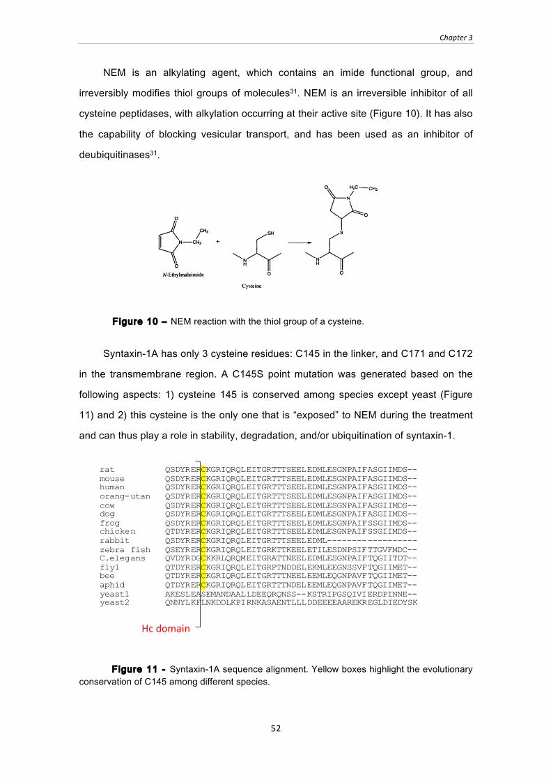
Chapter 3
52
NEM is an alkylating agent, which contains an imide functional group, and
irreversibly modifies thiol groups of molecules31. NEM is an irreversible inhibitor of all
cysteine peptidases, with alkylation occurring at their active site (Figure 10). It has also
the capability of blocking vesicular transport, and has been used as an inhibitor of
deubiquitinases31.
Figure 10 ‒ NEM reaction with the thiol group of a cysteine.
Syntaxin-1A has only 3 cysteine residues: C145 in the linker, and C171 and C172
in the transmembrane region. A C145S point mutation was generated based on the
following aspects: 1) cysteine 145 is conserved among species except yeast (Figure
11) and 2) this cysteine is the only one that is “exposed” to NEM during the treatment
and can thus play a role in stability, degradation, and/or ubiquitination of syntaxin-1.
Figure 11 - Syntaxin-1A sequence alignment. Yellow boxes highlight the evolutionary conservation of C145 among different species.
Hc domain
rat QSDYRERCKGRIQRQLEITGRTTTSEELEDMLESGNPAIFASGIIMDS-- mouse QSDYRERCKGRIQRQLEITGRTTTSEELEDMLESGNPAIFASGIIMDS-- human QSDYRERCKGRIQRQLEITGRTTTSEELEDMLESGNPAIFASGIIMDS-- orang-utan QSDYRERCKGRIQRQLEITGRTTTSEELEDMLESGNPAIFASGIIMDS-- cow QSDYRERCKGRIQRQLEITGRTTTSEELEDMLESGNPAIFASGIIMDS-- dog QSDYRERCKGRIQRQLEITGRTTTSEELEDMLESGNPAIFASGIIMDS-- frog QSDYRERCKGRIQRQLEITGRTTTSEELEDMLESGNPAIFSSGIIMDS-- chicken QTDYRERCKGRIQRQLEITGRTTTSEELEDMLESGNPAIFSSGIIMDS-- rabbit QSDYRERCKGRIQRQLEITGRTTTSEELEDML------------------ zebra fish QSEYRERCKGRIQRQLEITGRKTTKEELETILESDNPSIFTTGVFMDC-- C.elegans QVDYRDGCKKRLQRQMEITGRATTNEELEDMLESGNPAIFTQGIITDT-- fly1 QTDYRERCKGRIQRQLEITGRPTNDDELEKMLEEGNSSVFTQGIIMET-- bee QTDYRERCKGRIQRQLEITGRTTTNEELEEMLEQGNPAVFTQGIIMET-- aphid QTDYRERCKGRIQRQLEITGRTTTNDELEEMLEQGNPAVFTQGIIMET-- yeast1 AKESLEASEMANDAALLDEEQRQNSS--KSTRIPGSQIVIERDPINNE-- yeast2 QNNYLKFLNKDDLKPIRNKASAENTLLLDDEEEEAAREKREGLDIEDYSK

Chapter 3
53
Interestingly, C145S mutation in syntaxin-1 abolished the NEM-mediated effect
on syntaxin-1 levels (Figure 12). Furthermore, the cysteine to serine mutation resulted
in syntaxin-1 levels similar to NEM-treated WT syntaxin-1 (Figure 12). This points to a
de-stabilization of WT syntaxin-1 in absence of NEM, which can be abolished by
introduction of the cysteine to serine mutation.
Figure 12 ‒ Effect of NEM on levels of syntaxin-1 WT and mutated C145S in transfected HEK cells treated with 5 mM NEM for 5min at 37°C. Reaction was stopped by washing cells with PBS and solubilization in 0.1% Triton-X 100 (TX-100). Insoluble material was removed by centrifugation for 20 min at 10,000g. Supernatant was collected and 5% Laemmli sample buffer containing DTT was added. A) Samples were analyzed by immunblotting for levels of syntaxin-1 WT and C145S and GFP, which was used as transfection control. B) Protein levels were quantitated using 125I-labeled secondary antibody and were first normalized to GFP levels and then to syntaxin-1 WT without NEM treatment. ** p < 0.01 using student’s T-test (n = 3).
3.1.2 NEM experiments in neuronal cultures
In order to analyze the effect of NEM in situ, neuronal cultures were incubated
with a range of NEM concentrations between 10 µM to 5mM. In this range, NEM
concentrations do not show differences in syntaxin-1 levels (Figure 13).
Figure 13 - Effect of different NEM concentrations on syntaxin-1 levels in neuronal cultures treated with a NEM concentration of 10 µM to 5mM. Neuronal cultures at 14 days in vitro were incubated for 5 min with NEM or vehicle control (ethanol). Syntaxin-1 levels were analyzed by immunblotting.
Syntaxin-1
5mM
2mM
1mM
800µM
500µM
100µM
80µM
50µM
30µM
10µM
-‐ -‐ NEM
NEM -‐ + -‐ +
WT C1
45S
Sxt-‐1
GFP
A
Syntaxin-1 levels (%
)
** **
B
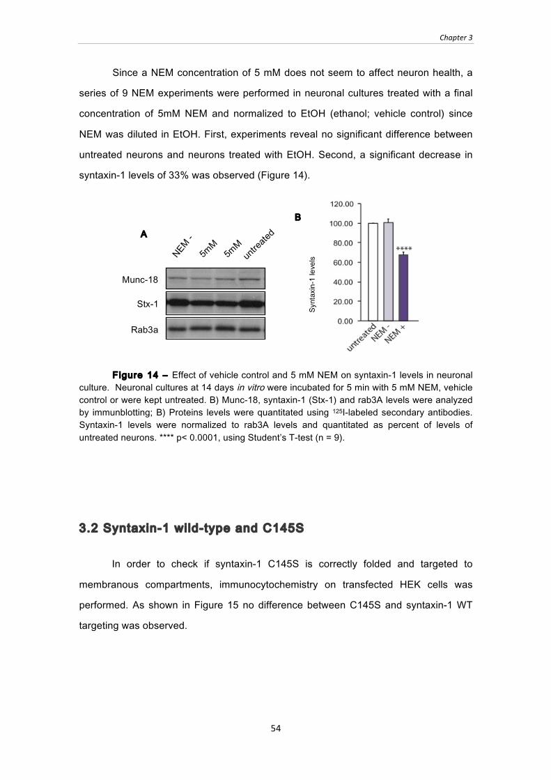
Chapter 3
54
Since a NEM concentration of 5 mM does not seem to affect neuron health, a
series of 9 NEM experiments were performed in neuronal cultures treated with a final
concentration of 5mM NEM and normalized to EtOH (ethanol; vehicle control) since
NEM was diluted in EtOH. First, experiments reveal no significant difference between
untreated neurons and neurons treated with EtOH. Second, a significant decrease in
syntaxin-1 levels of 33% was observed (Figure 14).
Figure 14 ‒ Effect of vehicle control and 5 mM NEM on syntaxin-1 levels in neuronal culture. Neuronal cultures at 14 days in vitro were incubated for 5 min with 5 mM NEM, vehicle control or were kept untreated. B) Munc-18, syntaxin-1 (Stx-1) and rab3A levels were analyzed by immunblotting; B) Proteins levels were quantitated using 125I-labeled secondary antibodies. Syntaxin-1 levels were normalized to rab3A levels and quantitated as percent of levels of untreated neurons. **** p< 0.0001, using Student’s T-test (n = 9).
3.2 Syntaxin-1 wild-type and C145S
In order to check if syntaxin-1 C145S is correctly folded and targeted to
membranous compartments, immunocytochemistry on transfected HEK cells was
performed. As shown in Figure 15 no difference between C145S and syntaxin-1 WT
targeting was observed.
B
Syntaxin-1 levels
(%)
untreated
NEM -
5mM
5mM
Munc-18
Rab3a
Stx-1
A

Chapter 3
55
Figure 15 ‒ Targeting of syntaxin-1 wild-type and C145S mutation in transfected HEK cells. 48h after transfection, HEK cells were washed fixed for 15 min in 4% paraformaldehyde and permeabilized for 5min in 0.1% Triton X-100. Cells were then blocked in 5% BSA and were incubated with primary antibody HPC-1 in 1% BSA overnight at 4°C. Cultures were washed and blocked, and an anti-mouse Alexa-488 secondary antibody (1:500 in 1% BSA) was added for 1h in the dark. Coverslips were mounted on glass slides in Fluoromount-G. Laser scanning confocal microscopy was performed with excitation at 488nm on a Leica TCS SP-2 inverted microscope.
3.3 Syntaxin-1 levels in presence of Munc-18 and SNAP-25
In order to analyze a possible chaperone for syntaxin-1, munc-18 or SNAP-25
were co-transfected with syntaxin-1 wild type and C145S. α-synuclein1-95 was used in
syntaxin-1 transfections in order to balance the amount of transfected munc-18 or
SNAP-25 DNA.
3.3.1 Syntaxin-1 wild-type and C145S full length
For full-length syntaxin-1, a significant increase of 76% can be observed when
munc-18 is present. As shown previously, syntaxin-1 C145S reveals higher expression
levels than syntaxin-1 wild-type (a significant increase of 90%). An increase is also
observed when SNAP-25 is co-transfected; however, this increase is lower when
compared to transfections with munc-18 (Figure 16).
A Syntaxin-1 WT B Syntaxin-1 C145S
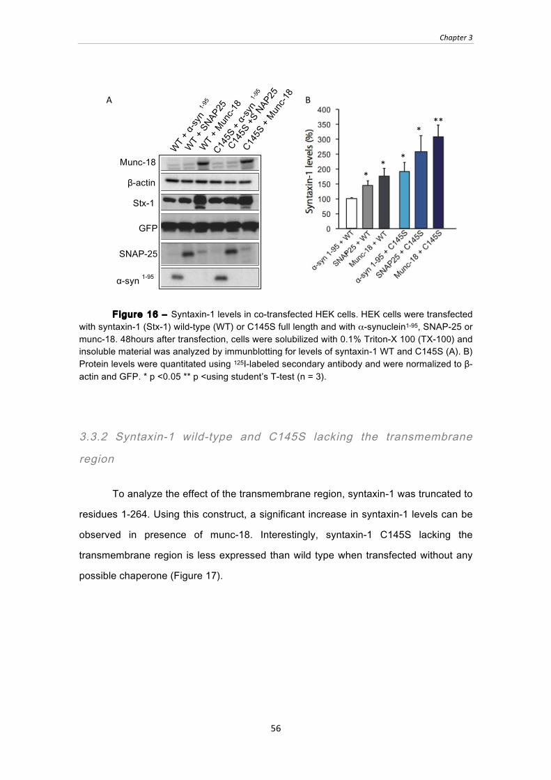
Chapter 3
56
Figure 16 ‒ Syntaxin-1 levels in co-transfected HEK cells. HEK cells were transfected with syntaxin-1 (Stx-1) wild-type (WT) or C145S full length and with α-synuclein1-95, SNAP-25 or munc-18. 48hours after transfection, cells were solubilized with 0.1% Triton-X 100 (TX-100) and insoluble material was analyzed by immunblotting for levels of syntaxin-1 WT and C145S (A). B) Protein levels were quantitated using 125I-labeled secondary antibody and were normalized to β-actin and GFP. * p <0.05 ** p <using student’s T-test (n = 3).
3.3.2 Syntaxin-1 wild-type and C145S lacking the transmembrane
region
To analyze the effect of the transmembrane region, syntaxin-1 was truncated to
residues 1-264. Using this construct, a significant increase in syntaxin-1 levels can be
observed in presence of munc-18. Interestingly, syntaxin-1 C145S lacking the
transmembrane region is less expressed than wild type when transfected without any
possible chaperone (Figure 17).
* *
*
* **
B
Munc-18
Stx-1
α-syn 1-95
GFP
β-actin
SNAP-25
WT + α
-syn
1-95
WT + S
NAP25
WT + M
unc-1
8 C145S + M
unc-1
8
C145S + α
-syn
1-95
C145S +S NAP25
A

Chapter 3
57
Figure 17 ‒ Syntaxin-1 levels in co-transfected HEK cells. HEK cells were transfected with syntaxin-1 (Stx-1) wild-type (WT) or C145S 1-264 and with α-synuclein1-95, SNAP-25 or munc-18. 48hours after transfection, cells were solubilized with 0.1% Triton-X 100 (TX-100) and insoluble material was analyzed by immunblotting for levels of syntaxin-1 WT and C145S (A). B) Protein levels were quantitated using 125I-labeled secondary antibody and were normalized to β-actin and GFP. ** p < 0.01; **** p < 0.0001 using student’s T-test (n = 3).
3.3.3 Syntaxin-1 lacking the N-terminal Habc domain
Munc-18 has been shown to interact with the Habc domain of syntaxin-1. To
analyze syntaxin-1 stability without Habc domain, syntaxin-1 was co-transfected with
SNAP-25 or munc-18. Results show a 419.9% increase of syntaxin-1 levels when
SNAP-25 is present. No changes were observed in presence of munc-18 (Figure 18).
B
Munc-18
Stx-1
α-syn 1-95
GFP
β-actin
SNAP-25
WT + α
-syn
1-95
WT +SNA
P25
WT + M
unc-1
8 C145S + M
unc-1
8
C145S + α
-syn
1-95
C145S
+SNA
P25
A

Chapter 3
58
Figure 18 ‒ Syntaxin-1 levels in co-transfected HEK cells. HEK cells were transfected with syntaxin-1 (Stx-1) wild-type (WT) or C145S 180-288 and with α-synuclein1-95, SNAP-25 or munc-18. 48hours after transfection, cells were solubilized with 0.1% Triton-X 100 (TX-100) and insoluble material was analyzed by immunblotting for levels of syntaxin-1 WT and C145S (A). B) Protein levels were quantitated using 125I-labeled secondary antibody and were normalized to β-actin and GFP. **** p < 0.0001 using student’s T-test (n = 3).
3.3.4 Syntaxin-1 WT lacking the Habc domain and the transmembrane
region
To investigate the effect of a lack of Habc domain and transmembrane region,
syntaxin-1180-264 was generated. This truncation construct expresses the SNARE motif
only. Results show that this truncation is only expressed when SNAP-25 is present
resulting in an increase of syntaxin-1 levels to 882.2% (Figure 19).
Munc-18
Stx-1
α-syn 1-95
GFP
β-actin
SNAP-25 WT + α
-syn
1-95
WT + S
NAP25
WT + M
unc-1
8 A B
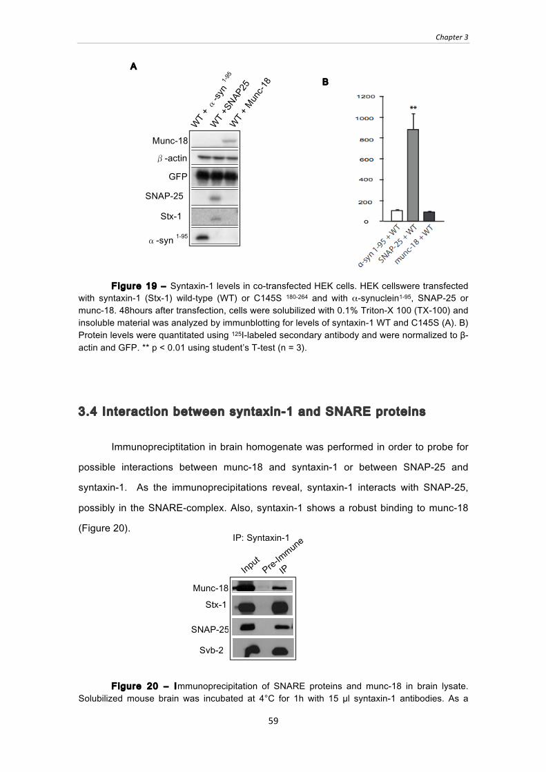
Chapter 3
59
Figure 19 ‒ Syntaxin-1 levels in co-transfected HEK cells. HEK cellswere transfected with syntaxin-1 (Stx-1) wild-type (WT) or C145S 180-264 and with α-synuclein1-95, SNAP-25 or munc-18. 48hours after transfection, cells were solubilized with 0.1% Triton-X 100 (TX-100) and insoluble material was analyzed by immunblotting for levels of syntaxin-1 WT and C145S (A). B) Protein levels were quantitated using 125I-labeled secondary antibody and were normalized to β-actin and GFP. ** p < 0.01 using student’s T-test (n = 3).
3.4 Interaction between syntaxin-1 and SNARE proteins
Immunopreciptitation in brain homogenate was performed in order to probe for
possible interactions between munc-18 and syntaxin-1 or between SNAP-25 and
syntaxin-1. As the immunoprecipitations reveal, syntaxin-1 interacts with SNAP-25,
possibly in the SNARE-complex. Also, syntaxin-1 shows a robust binding to munc-18
(Figure 20).
Figure 20 ‒ Immunoprecipitation of SNARE proteins and munc-18 in brain lysate. Solubilized mouse brain was incubated at 4°C for 1h with 15 µl syntaxin-1 antibodies. As a
Munc-18
Stx-1
α-syn 1-95
GFP
β-actin
SNAP-25 WT + α
-syn
1-95
WT +SNA
P25
WT + M
unc-1
8
A B
Input
Pre-Im
mune
IP
IP: Syntaxin-1
Munc-18
Stx-1
SNAP-25
Syb-2

Chapter 3
60
negative control, samples were incubated with pre-immune serum. Samples were then incubated for 2h at 4°C with 50 µl protein-A sepharose. Sepharose was washed 3x with 1% TX-100 and bound proteins were eluted with 2x Laemmli sample buffer containing DTT. Samples were analyzed by SDS-PAGE and immunoblotting for munc-18, syntaxin-1 (Stx-1), SNAP-25 and synaptobrevin-2 (Syb-2).
3.4 Interaction between syntaxin-1 and munc-18
According to the results shown above, syntaxin-1 full-length and 1-264 levels
are increased when munc-18 is present. However, when the same experiment is
performed with a truncation that lacks the Habc domain, the levels of syntaxin-1 do not
increase. Immunoprecipitations from brain lysate have shown that munc-18 and SNAP-
25 interact with syntaxin-1. Yet, these experiments do not reveal a direct interaction,
since binding could be mediated by a bridging protein. To probe for a direct interaction
and to map the syntaxin-1/munc-18 binding interface, a series of immunoprecipitations
(IP) was performed.
The results of the immunoprecipitation reveal an interaction for munc-18 with
syntaxin-1 wild-type or C145S full length, as well as for the truncation that lacks the
transmembrane region (1-264). Separate immunoprecipitations were performed with
antibodies to syntaxin-1 or munc-18 to corroborate the result. For truncations lacking
the Habc domain and hence an epitope for the antibody to bind, the immunoprecipitation
was performed using an HA antibody. As expected from the stabilization data,
truncation 180-288 does not bind to munc-18. Truncation 180-264 that lacks the Habc
domain and transmembrane region was not expressed in presence of munc-18 (Figure
21).
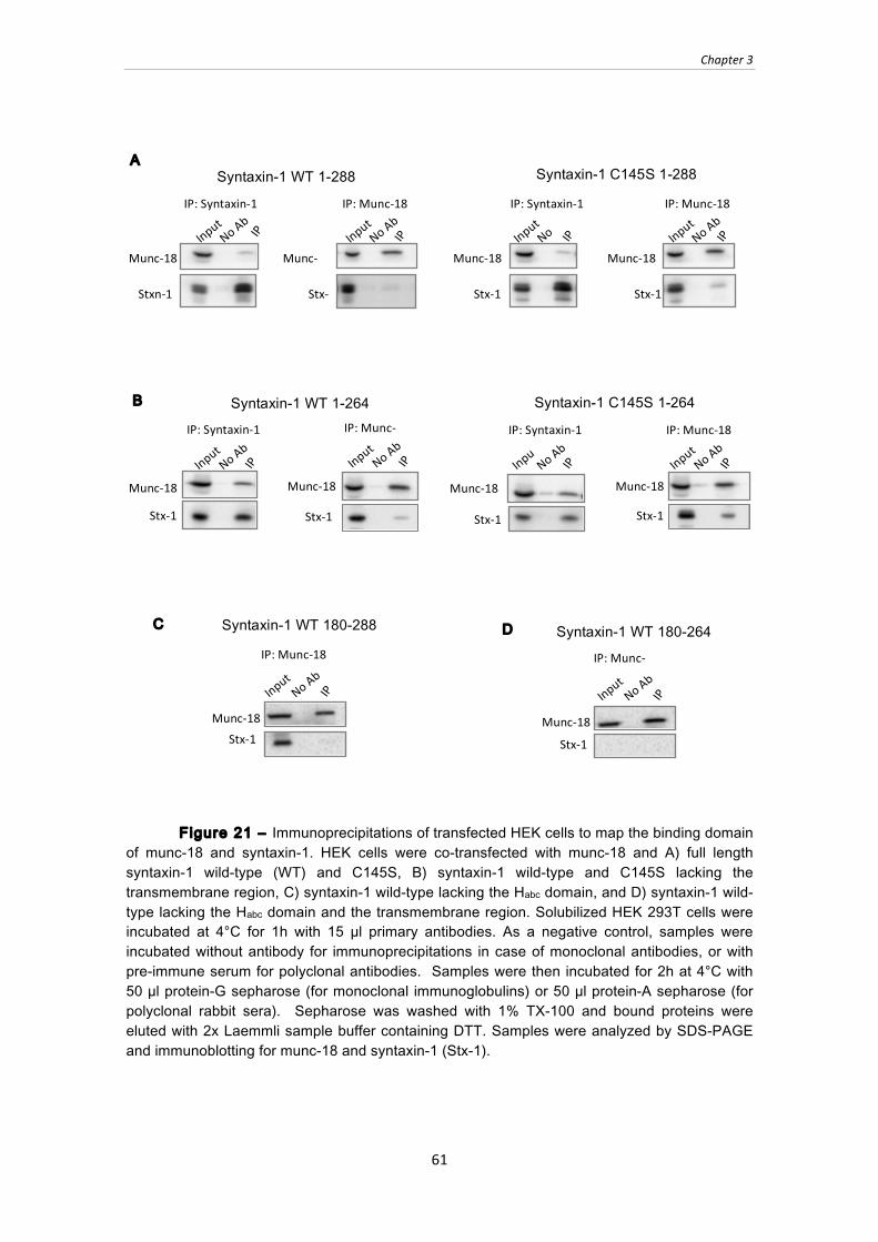
Chapter 3
61
Figure 21 ‒ Immunoprecipitations of transfected HEK cells to map the binding domain of munc-18 and syntaxin-1. HEK cells were co-transfected with munc-18 and A) full length syntaxin-1 wild-type (WT) and C145S, B) syntaxin-1 wild-type and C145S lacking the transmembrane region, C) syntaxin-1 wild-type lacking the Habc domain, and D) syntaxin-1 wild-type lacking the Habc domain and the transmembrane region. Solubilized HEK 293T cells were incubated at 4°C for 1h with 15 µl primary antibodies. As a negative control, samples were incubated without antibody for immunoprecipitations in case of monoclonal antibodies, or with pre-immune serum for polyclonal antibodies. Samples were then incubated for 2h at 4°C with 50 µl protein-G sepharose (for monoclonal immunoglobulins) or 50 µl protein-A sepharose (for polyclonal rabbit sera). Sepharose was washed with 1% TX-100 and bound proteins were eluted with 2x Laemmli sample buffer containing DTT. Samples were analyzed by SDS-PAGE and immunoblotting for munc-18 and syntaxin-1 (Stx-1).
Input No Ab
IP
IP: Syntaxin-‐1
Munc-‐18
Stxn-‐1
Input No Ab
IP
IP: Munc-‐18
Munc-‐18
Stx-‐1
Input No
Ab IP
IP: Syntaxin-‐1
Munc-‐18
Stx-‐1
Input No Ab
IP
IP: Munc-‐18
Munc-‐18
Stx-‐1
Syntaxin-1 C145S 1-288 Syntaxin-1 WT 1-288 A
Syntaxin-1 C145S 1-264 Syntaxin-1 WT 1-264
Input No Ab
IP
IP: Syntaxin-‐1
Munc-‐18
Stx-‐1
Input No Ab
IP IP: Munc-‐18
Munc-‐18
Stx-‐1
Inpu
t No Ab
IP
IP: Syntaxin-‐1
Munc-‐18
Stx-‐1
Input No Ab
IP
IP: Munc-‐18
Munc-‐18
Stx-‐1
B
Input No Ab
IP
IP: Munc-‐18
Munc-‐18 Stx-‐1
Syntaxin-1 WT 180-288 C
Input No Ab
IP
IP: Munc-‐18
Munc-‐18 Stx-‐1
Syntaxin-1 WT 180-264 D
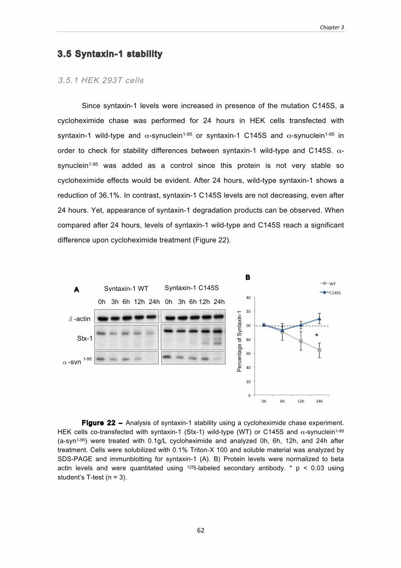
Chapter 3
62
3.5 Syntaxin-1 stabil i ty
3.5.1 HEK 293T cells
Since syntaxin-1 levels were increased in presence of the mutation C145S, a
cycloheximide chase was performed for 24 hours in HEK cells transfected with
syntaxin-1 wild-type and α-synuclein1-95 or syntaxin-1 C145S and α-synuclein1-95 in
order to check for stability differences between syntaxin-1 wild-type and C145S. α-
synuclein1-95 was added as a control since this protein is not very stable so
cycloheximide effects would be evident. After 24 hours, wild-type syntaxin-1 shows a
reduction of 36.1%. In contrast, syntaxin-1 C145S levels are not decreasing, even after
24 hours. Yet, appearance of syntaxin-1 degradation products can be observed. When
compared after 24 hours, levels of syntaxin-1 wild-type and C145S reach a significant
difference upon cycloheximide treatment (Figure 22).
Figure 22 ‒ Analysis of syntaxin-1 stability using a cycloheximide chase experiment. HEK cells co-transfected with syntaxin-1 (Stx-1) wild-type (WT) or C145S and α-synuclein1-95 (a-syn1-95) were treated with 0.1g/L cycloheximide and analyzed 0h, 6h, 12h, and 24h after treatment. Cells were solubilized with 0.1% Triton-X 100 and soluble material was analyzed by SDS-PAGE and immunblotting for syntaxin-1 (A). B) Protein levels were normalized to beta actin levels and were quantitated using 125I-labeled secondary antibody. * p < 0.03 using student’s T-test (n = 3).
α-syn 1-95
Stx-1
β-actin
0h 3h 6h 12h 24h
Syntaxin-1 WT A
0h 3h 6h 12h 24h
Syntaxin-1 C145S B

Chapter 3
63
Does munc-18 change syntaxin-1 stability, and does it affect only wild-type or
wild-type and C145S similarly? To address this, HEK cells 293T were co-transfected
with munc-18 and syntaxin-1 wild-type or C145S. An increase in syntaxin-1 wild-type
levels was observed in presence of munc-18. In contrast, syntaxin-1 C145S stability
shows no munc-18 dependent changes, although the levels of degradation products
decrease (Figure 23).
Figure 23 ‒ Analysis of syntaxin-1 stability using a cycloheximide chase experiment. HEK cells co-transfected with syntaxin-1 (Stx-1) wild-type (WT) or C145S, α-synuclein1-95 (a-syn1-95) and munc-18 were treated with 0.1g/L cycloheximide and analyzed 0h, 6h, 12h, and 24h after treatment. Cells were solubilized with 0.1% Triton-X 100 and soluble material was analyzed by SDS-PAGE and immunblotting for syntaxin-1 (A). B) Protein levels were normalized to beta actin levels and were quantitated using 125I-labeled secondary antibody. p < n.s. using student’s T-test (n = 3).
3.5.2 Neuronal Cultures
To replicate the effect seen in transfected HEK cells, a cycloheximide chase
was performed for 72 hours in neuronal cultures. This experiment reveals that the
syntaxin-1 half-life is approximately 50 hours (Figure 24).
To analyze munc-18 dependent syntaxin-1 stability, cycloheximide chase
experiments should have been performed with overexpression or knockdown of munc-
0h 6h 12h 24h Syntaxin-1 C145S
α-syn 1-95
Stx-1
β-actin
Munc-18
0h 6h 12h 24h Syntaxin-1 WT
A B

Chapter 3
64
18. Due to time limitations and due to problems with virus production, this aim could
not be achieved.
Figure 24 ‒ Analysis of syntaxin-1 half-life in neuronal culture. Neuronal cultures at 11 days in vitro were incubated with 0.1g/L cycloheximide. (A) Syntaxin-1 (Stx-1) levels at 0h, 3h, 24h, 48h, and 72h were analyzed by immunblotting (B) and were quantitated using 125I-labeled secondary antibodies, normalized to the 0h levels (n=3).
3.6 Dependence of syntaxin-1 levels on synaptic activity
Syntaxin-1 is part of the SNARE complex and therefore shuttles between a
folded conformation in the SNARE complex and an unfolded and highly unstable
conformation as a monomer. Its levels may thus be influenced by synaptic activity. To
investigate this, neuronal cultures were treated with synaptic activity blockers (APV and
tetrodotoxin - TTX) and synaptic activity enhancers (K+; Ca2+). The results reveal a
trend towards an increase in syntaxin-1 levels with synaptic silencing and a decrease
in syntaxin-1 levels with synaptic activity, but do not reach significance (Figure 25).
Also here, it would have been interesting to analyze whether lack of munc-18
enhances this effect. However, due to reasons give above, this experiment was not
performed.
0h 3h 24h 48h 72h
Stx-1
A B
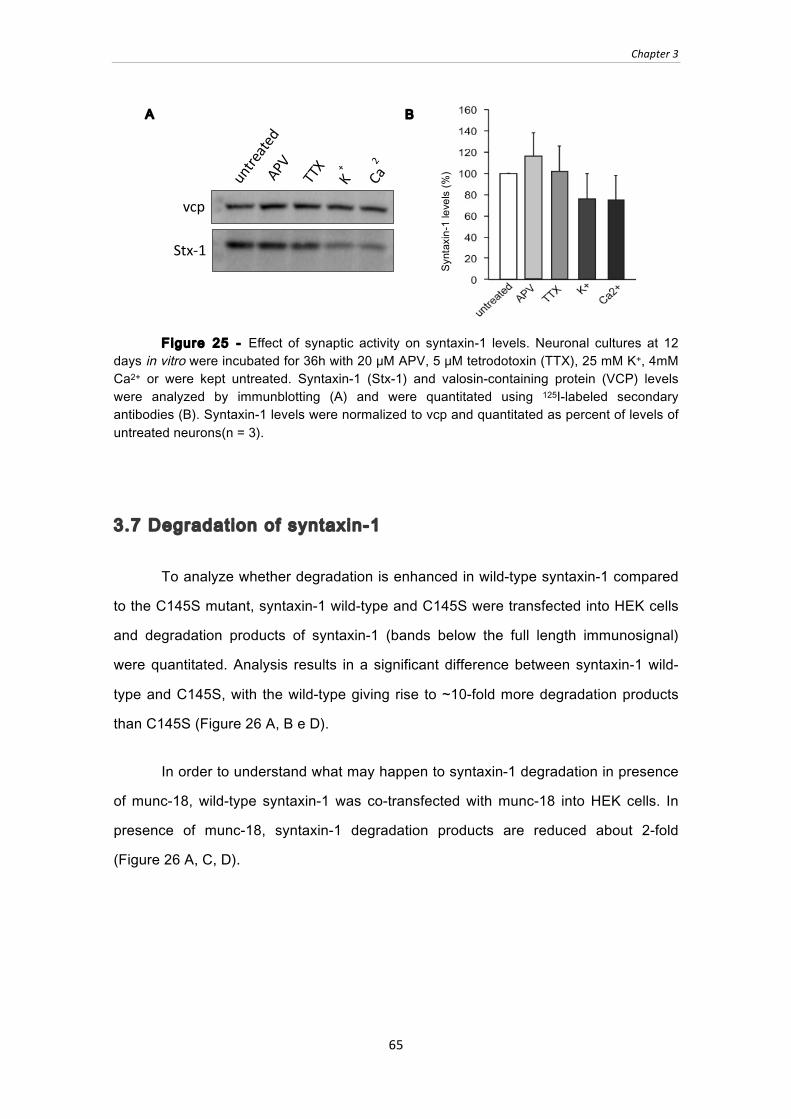
Chapter 3
65
Figure 25 - Effect of synaptic activity on syntaxin-1 levels. Neuronal cultures at 12 days in vitro were incubated for 36h with 20 μM APV, 5 μM tetrodotoxin (TTX), 25 mM K+, 4mM Ca2+ or were kept untreated. Syntaxin-1 (Stx-1) and valosin-containing protein (VCP) levels were analyzed by immunblotting (A) and were quantitated using 125I-labeled secondary antibodies (B). Syntaxin-1 levels were normalized to vcp and quantitated as percent of levels of untreated neurons(n = 3).
3.7 Degradation of syntaxin-1
To analyze whether degradation is enhanced in wild-type syntaxin-1 compared
to the C145S mutant, syntaxin-1 wild-type and C145S were transfected into HEK cells
and degradation products of syntaxin-1 (bands below the full length immunosignal)
were quantitated. Analysis results in a significant difference between syntaxin-1 wild-
type and C145S, with the wild-type giving rise to ̃10-fold more degradation products
than C145S (Figure 26 A, B e D).
In order to understand what may happen to syntaxin-1 degradation in presence
of munc-18, wild-type syntaxin-1 was co-transfected with munc-18 into HEK cells. In
presence of munc-18, syntaxin-1 degradation products are reduced about 2-fold
(Figure 26 A, C, D).
Syntaxin-1 levels (%
)
B A
vcp
Stx-‐1 untreate
d APV
TTX
K+
Ca2
+

Chapter 3
66
Figure 26 ‒ Analysis of syntaxin-1 degradation A) Syntaxin-1 wild-type (WT), B) Syntaxin-1 C145S; C) Syntaxin-1 WT plus munc-18; were transfected into HEK cells. 48h after transfection, cells were solubilized with 0.1% Triton-X 100 and soluble material was analyzed by immunblotting for syntaxin-1. D) Full-length and degraded protein levels were quantitated using 125I-labeled secondary antibody, with degradation products expressed as percent of total syntaxin-1 protein. * p < 0.03 and ** p < 0.01 using student’s T-test (n = 3).
3.8 Ubiquit ination of syntaxin-1
Next we asked whether syntaxin-1 degradation happens via ubiquitination. To
approach this question, transfected HEK cells were subjected to an
immunoprecipitation with syntaxin-1 antibody and samples were probed for ubiquitin.
Analysis shows a significant difference in ubiquitination levels between wild-type
syntaxin-1 and C145S (Figure 27).
B Syntaxin-1 C145S
Munc-‐18
Stx-‐1
C Syntaxin-1 WT + Munc-18
A Syntaxin-1 WT
Syntaxin-1 levels (%
)
D

Chapter 3
67
Figure 27 ‒ Analysis of ubiquitination in HEK cells transfected with syntaxin-1 wild-type and C145S. Immunoprecipitations were performed with lysates from HEK 293T cells transfected with syntaxin-1 (Stx-1) wild-type (WT) or C145S. Solubilized HEK 293T cells were incubated at 4°C for 1h with 15 µl primary antibody (438B). As a negative control, samples were incubated with pre-immune serum. Samples were then incubated for 2h at 4°C with 50 µl protein-A sepharose. Sepharose was washed with 1% TX-100 and bound proteins were eluted with 2x Laemmli sample buffer containing DTT. A) Samples were analyzed by SDS-PAGE and immunoblotting for ubiquitin (top) and syntaxin-1 (bottom). B) Ubiquitination was quantitated using 125I-labeled secondary antibodies, normalized to the efficiency of the syntaxin-1 immunoprecipitation. **** p< 0.0001 using Student’s T-test (n = 3).
3.9 Is syntaxin-1 degraded via the lysosome or proteasome?
Ubiquitination can result in protein degradation via the lysosome or proteasome.
In order to analyze which pathway is mediating syntaxin-1 degradation, neuronal
cultures were incubated with 5 different inhibitors, 3 proteasome inhibitors (MG132,
epoxomicin, clasto-lactacystin) and 2 lysosome inhibitors (leupeptin/pepstatin and
PMSF). A trend towards a stabilization of syntaxin-1 using lysosomal inhibitors can be
observed. However, this change does not reach significance yet (n = 2). In contrast,
proteasome inhibitors reveal only very modest changes (Figure 28).
A
Input
Pre-‐Im
mune
IP
IP: Syntaxin-1
Ub
Stx -1
WT
Input Pre
-Immune
IP
C145S
****
B

Chapter 3
68
Figure 28 - Effect of proteases inhibitors on syntaxin-1 levels in neuronal culture. Neuronal cultures at 12 days in vitro were incubated for 36h with 10 μM Epoxomicin, 10 μM MG132, 10 μM Clasto-lactacystin, 10 mg/L Leupeptin/Pepstatin, 4 mM PMSF or were kept untreated. Syntaxin-1 (Stx-1) and β-actin levels were analyzed by immunblotting (A) and were quantitated using 125I-labeled secondary antibodies (B). Syntaxin-1 levels were normalized to β-actin and quantitated as percent of levels of untreated neurons (n = 2).
untreate
d Leu/P
ept
MG132
Epoxom
icin
Clasto-Lacta
PMSF
β-actin
Stx-1
A B


Chapter 4
71
CHAPTER 4
DISCUSSION AND CONCLUSION
Although NEM experiments in brain homogenate showed a reliable increase of
syntaxin-1 levels in presence of NEM, NEM experiments performed in neuronal
cultures revealed that independently of the concentration used (10µM to 5mM), NEM
reduced the levels of syntaxin-1. The reduction in syntaxin-1 levels observed with 5mM
NEM is neither due to NEM toxicity nor neuronal death. In fact, the observed decrease
can be due to the fact that neuronal cultures where made from P0 mice and the
chemical treatment was performed at day 14 whereas previous results in brain were
performed on old mice. Possibly, NEM shows its effect on syntaxin-1 levels only in later
stages when the entire SNARE machinery may be more compromised due to heavy
usage, like reported for SNAP-25 and synaptobrevin-215,16. Consequently, an option
would be to perform NEM treatment on neuronal cultures at a later time point, e.g. at
day 21. Additionally, since munc-18 interacts with and stabilizes syntaxin-1, it may be
worthwhile to perform the NEM treatment on neuronal cultures upon knockdown of
munc-18. In this case, syntaxin-1 may be more unstable and the stabilizing effect of
NEM may be more prominent.
Experiments performed in transfected HEK cells for the analysis of syntaxin-1
levels and interaction domain mapping reveal that both syntaxin-1 wild type and C145S
full-length bind to munc-18. Due to this interaction munc-18 increases syntaxin-1
levels, which makes munc-18 a very strong candidate for a syntaxin-1 chaperone. On
the other hand, mutation of cysteine 145 to serine results in a stabilization of syntaxin-1
levels. Consequently, levels of syntaxin-1 C145S are similar to levels of syntaxin-1
wild-type when co-transfected with munc-18.

Chapter 4
72
It is reported in the literature that SNAP-25 binds syntaxin-1 in the SNARE
motif17. This is in agreement with results presented here which show that the interaction
between SNAP-25 and syntaxin-1 increases syntaxin-1 levels. This increase is not as
high as the increase observed when munc-18 is present; yet, at this point, SNAP-25
cannot be excluded as a syntaxin-1 chaperone.
Even in absence of the transmembrane region (syntaxin-1 1-264), munc-18 and
SNAP-25 increase the levels of syntaxin-1. Since syntaxin-1 1-264 still has the SNARE
motif and the Habc domain, munc-18 and SNAP-25 can bind to syntaxin-1 and increase
syntaxin-1 levels. Also here, munc-18 has a bigger effect on syntaxin-1 levels
compared to SNAP-25, making munc-18 a more likely chaperone for syntaxin-1.
Compared to wild-type levels, syntaxin-1 C145S lacking the transmembrane
region reveals a decrease in syntaxin-1 levels. This decrease can be due to the fact
that cytosolic syntaxin-1 is per se more unstable and the C145S mutation by itself may
be actually more unstable than the wild-type when lacking the membrane anchor.
Analyses of syntaxin-1 truncations that do not express the Habc domain (180-
264 and 180-288) show no stabilizing effect by munc-18. This reveals that munc-18
needs the Habc domain to chaperone syntaxin-1, supported by the fact that this domain
is the binding interface for munc-18 as shown by the immunoprecipitations.
Interestingly, when these two constructs were co-transfected with SNAP-25, syntaxin-1
levels increased with both constructs (180-264 and 180-288), presumably because the
SNARE motif was still present.
The observed difference between syntaxin-1 180-264 and syntaxin-1 180-288
constructs reveal, that the transmembrane region is important for syntaxin-1 stability.
Thus, when syntaxin-1 is cytosolic and not membrane anchored, syntaxin-1 180-264
(SNARE motif only) expression is observed only in presence of SNAP-25. In contrast,
when syntaxin-1 180-288 (SNARE motif plus transmembrane region) is expressed, the
transmembrane region stabilizes syntaxin-1 and this syntaxin-1 truncation is expressed
not only when SNAP-25 is present but also in presence of munc-18 or α-synuclein1-95.

Chapter 4
73
To summarize: 1) The C145S mutation increases syntaxin-1 levels; 2) The
transmembrane domain is important for the stability of syntaxin-1; 3) Munc-18
increases syntaxin-1 levels only when the Habc domain is present; 4) When the SNARE
motif is present, SNAP-25 increases syntaxin-1 level.
Since munc-18 shows a predominant effect on syntaxin-1 levels, further
experiments were performed only with munc-18. However, more experiments
addressing the syntaxin-1/SNAP-25 interaction and stabilization should be performed.
Possibly, even SNAP-25 may stabilize syntaxin-1 levels and act as a syntaxin-1
chaperone.
To further analyze the effect of munc-18 and the C145S mutation on syntaxin-1
levels, cycloheximide chase experiments in HEK 293T cells were performed, showing
that syntaxin-1 wild-type is less stable than syntaxin-1 C145S. Thus, this cysteine has
an important role in stabilization of syntaxin-1. Furthermore, when munc-18 is present,
syntaxin-1 wild-type levels are stable even after 24 hours. An explanation for the
observation that syntaxin-1 levels reach values higher than 100% after adding a
translation inhibitor cannot be due to different levels of syntaxin-1 at the starting point,
since the cycloheximide treatment starts at the same time for all cells. Thus, the
increase in syntaxin-1 levels can only be due to HEK 293T cell division during the
chase. Additionally, cycloheximide allows a first round of translation before blocking
protein synthesis27, which may also result in levels higher than 100%. One way to
overcome this problem would be to increase the cycloheximide concentration.
However, increasing cycloheximide concentration resulted in cell death after 24hours
(data not shown). Additionally, increasing the time course of cycloheximide treatment
up to 36h also resulted in cell death (data not shown). Therefore, cycloheximide chase
experiments should be performed in neuronal cultures with knockdown or
overexpression of munc-18 together with analysis of endogenous syntaxin-1 levels.
Moreover, to investigate the effect of the C145S mutation, both, tagged syntaxin-1 wild-
type and C145S should be expressed, together with knockdown or overexpression of
munc-18.
Chapter 4
74
Syntaxin-1 is a pre-synaptic protein with an active role during synaptic activity,
and is repeatedly folded and unfolded during the SNARE cycle, which may make it
vulnerable regarding synaptic activity. The results investigating changes in syntaxin-1
levels during synaptic activity are not conclusive. Yet, they indicate a trend for a
decrease in syntaxin-1 levels when synaptic activity is enhanced. During synaptic
activity, more vesicle fusion occurs and therefore more vesicles are recycled. As a
consequence, syntaxin-1 changes very fast between a SNARE complex form and an
unfolded and highly reactive monomeric form, making it more prone to degradation. On
the other hand, when synaptic activity is blocked, syntaxin-1 levels remain stable.
Here, synaptic vesicles are primed on the pre-synaptic membrane, waiting for the Ca2+-
trigger. Syntaxin-1 folding/unfolding cycle is stopped and therefore, the levels of
syntaxin-1 do not change. In order to understand if syntaxin-1 stability is affected by
the presence or absence of munc-18, neuronal cultures with overexpression and
knockdown of munc-18 are proposed as a future plan.
Analysis of syntaxin-1 degradation products corroborated previous results
showing that C145S stabilizes syntaxin-1: degradation products are less in syntaxin-1
C145S compared to wild-type syntaxin-1. Munc-18 did not only stabilize syntaxin-1 but
reduced also its degradation, leading to the next question: is this degradation due to
ubiquitination?
Cysteine residues can be modified in numerous ways: oxidation,
glutathionylation, nitrosylation, acylation, and ubiquitination, resulting frequently in
modulation of protein activity. Since cysteines have a thiol group, they can form thiol
esters, which in turn can be ubiquitinated. Although protein ubiquitination at cysteine
residues has been described32-34, it is thermodynamically the least favorable event.
Also, a thiol ester bond appears not to be the most efficient way to link ubiquitin to a
protein, unless the aim is to create an activated form of ubiquitin32. Therefore, the result
showing that syntaxin-1 wild-type is significantly more ubiquitinated than syntaxin-1
C145S could mean that syntaxin-1 is ubiquitinated on that specific residue of cysteine,
or that this cysteine residue is important for signaling that the protein should be
degraded. In this case, ubiquitination would occur on lysine residues, for instance on
Chapter 4
75
lysine residue 155, which is next to cysteine 145. In order to clarify if ubiquitination is
occurring on this specific cysteine residue, as a future plan, mass spectrometry should
be performed.
Since syntaxin-1 wild-type is more ubiquitinated than syntaxin-1 C145S and
since syntaxin-1 is a transmembrane protein and consequently probably sorted to the
inner membranes of endosomes and lysosome after ubiquitination as many membrane
proteins35, neuronal cultures were treated with proteasome inhibitors and lysosome
inhibitors in order to understand if syntaxin-1 protein is degraded via lysosome or
proteasome. Results obtained are not conclusive yet, and more experiments have to
be performed. However a slight increase with lysosome inhibitors pepstatin/leupeptin
and PMSF was observed. In contrast, no changes were observed upon application of
proteasome inhibitors epoximicin, clastro/lactocystin, and MG132, suggesting that
syntaxin-1 may be degraded via the lysosome.
To conclude: 1) Altogether the results shown in this study suggest munc-18 as
a chaperone for syntaxin-1. It increases syntaxin-1 levels when co-expressed and it
inhibits the degradation of syntaxin-1 in HEK cells when co-transfected. Yet,
experiments in neuronal cultures upon knockdown and overexpression of munc-18 are
essential for a final confirmation; 2) Mutation of C145 to serine significantly stabilizes
syntaxin-1 levels. Syntaxin-1 levels are higher, syntaxin-1 is degraded much slower
and less degradation products can be observed. Also, replacement of the cysteine by
serine dramatically reduces ubiquitination of the protein. However, the precise role of
this residue in the degradation pathway needs further experiments; 3) Syntaxin-1 may
be degraded via the lysosome. Lysosomal inhibitors revealed a trend towards
stabilization of the protein, whereas proteasomal inhibitors showed no change.
However, more experiments, e.g. analyzing proteasomal and lysosomal inhibitors
during synaptic activity, are needed to confirm this hypothesis (Figure 29).

Chapter 4
76
Figure 29 - Model depicting the stabilization of syntaxin-1 by munc-18 and the C145S mutation. Syntaxin-1may be degraded via ubiquitiniation and the lysosome. Ubiquitination of syntaxin-1 may happen on the cysteine residue mutated in the C145S mutant, or this cysteine reside may signal the cell to degrade syntaxin-1.


References
79
REFERENCES
(1) Lodish H, Molecular Cell Biology. New York, W.H. Freeman, 6th edition (2008).
(2) De Camilli P, Takei K, Molecular Mechanism in Synaptic Vesicle Endocytosis and
Recycling. Neuron 16 (3), 481-486, (1996).
(3) Sϋdhof TC, The synaptic vesicle cycle revisited. Annu Rev Neurosci. 3(8): 641-
53 (2002).
(4) Sϋdhof TC, Rothman JE, Membrane Fusion: Grappling with SNARE and SM
Proteins. Science Review 323, (2009).
(5) Chandra S, Gallardo G, Fernández-Chacón R, Schlϋter OM, Sϋdhof TC, α-
Synuclein cooperates with CSPα in Preventing Neurodegeneration. Cell 123, 383-396
(2005).
(6) Sϋdhof TC, The Synaptic Vesicle Cycle. Annu. Rev. Neurosci. 27, 509-47
(2004).
(7) Dulubova I, et al., Munc18-1 binds directly to the neuronal SNARE complex.
PNAS 104(8), 2697-2702 (2006).
(8) Siegel GJ, et al., Basic Neurochemistry: Molecular, Cellular and Medical
Aspects. Lippincott-Raven; 6 th edition. Philadelphia (1999).
(9) Fernández-Chacón R, Sϋdhof TC, Genetics of synaptic vesicle function:
Towards the complete Functional Anatomy of an organelle. Annu. Rev. Physiol. 61:
753-76 (1999).
(10) Jahn R, Sϋdhof TC, Membrane Fusion. Cell 112, 519-533 (2003).
(11) Toonen RFG, Vries KJ, Zalm R, Sϋdhof TC, Verhage M, Munc18-1 stabilizes
syntaxin 1, but is not essential for syntaxin 1 targeting and SNARE complex formation.
J. Neurosci. 93, 1393-1400 (2005).
(12) Kukreja R, Sungh BR, Botulinum Neurotoxins: Structure and Mechanism of
Action. Microbial Toxins: Current Research and Future Trends. Caister Academic
Press (2009).
(13) Arnon SS, et al., Botulinum Toxin as a Biological Weapon: Medical and Public
Health Management. JAMA 285(8), 1059-70 (2001).
(14) Rizo J, Südhof TC, Snares and Munc18 In Synaptic Vesicle Fusion. Nature
Reviews Neroscience 3, 641-653 (2002).
References
80
(15) Burré J, et al., α-Synuclein Promotes SNARE-Complex Assembly in Vivo and in
Vitro. Science 10, 1126 (2010).
(16) Sharma M, Burré J, Sϋdhof TC, CSPα Promotes SNARE-Complex Assembly
by Chaperoning SNAP-25 during Synaptic Activity. Nat Cell Biol 13, 30-39 (2001).
(17) Brunger AT, Structure and function of SNARE-interacting proteins. Q REV
Biophys 38(1), 1-47 (2005).
(18) Brophy K, et al., Synaptosomal-associated protein 25 (SNAP-25) and attention
deficit hyperactivity disorder (ADHD): evidence of linkage and association in the Irish
population. Mol Psychiatry 7(8), 913-7 (2002).
(19) Chin LS, Vavalle JP, Lian L, Staring, a Novel E3 Ubiquitin-Protein Ligase That
Targets Syntaxin 1 for Degradation. J. Bio. Chem. 277 (38), 35071‒35079 (2002).
(20) Déak F, et al., Munc18-1 binding to the neuronal SNARE complex controls
synaptic vesicle priming. J. Cell Biol. 184(5), 751-764 (2009).
(21) Hata Y, Sϋdhof TC, A novel ubiquitous form of Munc-18 interacts with multiple
syntaxins. Use of the yeast two-hybrid system to study interactions between proteins
involved in membrane traffic. J Biol Chem 270 (22), 132022-8 (1995).
(22) Chakrabarti R, Schutt CE, The enhancement of PCR amplification by low
molecular-weight sulfones. Genes 274(1-2), 293-298 (2001).
(23) Nagai M, Yoshida A, Sato N, Additive effects of Bovine Serum albumin,
dithiothreitol, and glycerol on PCR. Biochem Mol Biot Int 44(1), 157-63 (1998).
(24) Durfee T, et al., The complete Genome Sequence of Escherichia coli DH10B:
Insights into the Biology of a Laboratory Workhorse. Journal of Bacteriology, 190(7)
2597-2602 (2008).
(25) Grant SG, et al., Differential plasmid rescue from transgenic mouse DNAs into
Escherichia coli methylation-restriction mutants. Proc Natl Acad Sci 87, 4645-9 (1990).
(26) Dull T, et al., A third-Generation Lentivirus Vector with a conditional Packaging
system. Ameri Soc Microbiology 72(11), 8463-8471.
(27) Schneider-Poestsch T, et al., Inhibition of eukaryotic translation elongation by
cycloheximide and lactimidomycin. Nat Chem Biol 6(3), 209-217 (2010).
(28) Hwang DF, Noguchi T, Tetrodotoxin poisoning. Adv. Food Nutr Res 52, 141-
236 (2007).
References
81
(29) Morris RG, Synaptic plasticity and learning: selective impairment of learning
rats and blockade of long-term potentiation in vivo by the N-methyl-D-aspartate
receptor antagonist AP5. Journ Neuros 9(9), 3040-57 (1989).
(30) Craiu A, Lactacystin and clasto-lactacystin beta-lactone modify multiple
proteasome beta-subunits and inhibit intracellular protein degradation and major
histocompatibility complex class I antigen presentation. J Biol Chem 272(20), 13437-
57 (1997).
(31) N-Ethylmaleimide at Sigma-Aldrich (2011).
(32) Cadwell K, Coscoy L, The specificities of Kaposi’s Sarcoma-Associated
Herpesvirus-Encoded E3 Ubiquitin Ligases are determined by the Positions of Lysine
or Cysteine Residues within the Intracytoplasmic Domains of their targets. J. Virol.
82(8), 4184-4189 (2008).
(33) Grou C, et al., Properties of the Ubiquitin-Pex5p Thiol Ester Conjugate*. The
Journal of Biological Chemistry 284(16), 10504-10513 (2009).
(34) Cadwell K, Coscoy L, Ubiquitination on Nonlysine residues by a viral E3
Ubiquitin ligase. Science 309, 127-130 (2005).
(35) Schulze H, Kolter T, SandhoffK, Principles of lysosomal membrane degradation
Cellular topology and biochemistry of lysosomal lipid degradation. Biochimica et
Biophysica Acta 1793, 674‒683 (2009).

Attachments
82
ATTACHMENTS
Plasmid Vector maps
pCMV5 plasmid vector showing EcoRI site

Attachments
83
FUW plasmid vector showing EcoRI site
FUW

Attachments
84
FSW plasmid vector showing EcoRI site
FSW
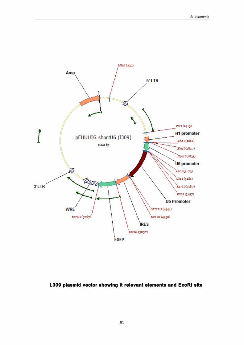
Attachments
85
L309 plasmid vector showing i t relevant elements and EcoRI site