Anti-inflammatory and antinociceptive effects of ... · Anti-inflammatory and antinociceptive...
9
Anti-inflammatory and antinociceptive effects of phonophoresis in animal models: a randomized experimental study L.C.P. Cardoso 1 , N.B. Pinto 2 , M.E.P. Nobre 3 , M.R. Silva 4 , G.M. Pires 5 , M.J.P. Lopes 6 , G.S.B. Viana 4 and L.M.R. Rodrigues 1 1 Programa de Pós-Graduac ¸ão Stricto Sensu em Ciências da Saúde, Faculdade de Medicina do ABC, Santo André, SP, Brasil 2 Unidade Acadêmica Ciências da Vida, Universidade Federal de Campina Grande, Cajazeiras, PB, Brasil 3 Faculdade de Medicina, Universidade Federal de Cariri, Barbalha, CE, Brasil 4 Departamento de Farmacologia, Universidade Federal do Ceará, Fortaleza, CE, Brasil 5 Departamento de Ciências da Computac ¸ão, Inteligência e Processamento de Imagens, Universidade Federal Rural de Pernambuco, Serra Talhada, PE, Brasil 6 Laboratório de Fisiologia, Faculdade de Medicina Estácio de Juazeiro do Norte, Juazeiro do Norte, CE, Brasil Abstract The aim of this study was to evaluate the therapeutic effects of ultrasound (US)-mediated phonophoresis alone or in association with diclofenac diethylammonium (DCF) administered topically in animal models of inflammation. A pre-clinical, prospective, and randomized experimental study of quantitative and qualitative nature was carried out. Phonophoresis was performed using a therapeutic ultrasound apparatus in two distinct models of acute inflammation. Edema was induced by an intraplantar injection of carrageenan and measured by plethysmography. The Hargreaves test was used to evaluate the antinociceptive activity and investigate the action of phonophoresis on tumor necrosis factor (TNF)-a production. A histological analysis with hematoxylin- eosin was used to evaluate tissue repair, and the expression of COX-2 was determined by immunohistochemical analysis. At the peak of inflammatory activity (3 h), treatment with US, US+DCF, and DCF significantly reduced edema formation compared to the control group. Treatment with US+DCF was more effective than treatment with US alone at both analyzed times. In the analysis of the antinociceptive activity, the treatments significantly increased the latency time in response to the thermal stimulus. Histopathological analysis revealed a reduction of the inflammatory infiltrates and immunohistochemistry demonstrated that the association was effective in reducing COX-2 expression compared to the control group. The association of DCF with US produced anti-inflammatory and antinociceptive effects in rat models of inflammation, which may be associated with inhibition of COX-2 and TNF-a production. Key words: Phonophoresis; Ultrasonic therapy; Inflammation; Nociception; Physiotherapy Introduction The skin is the largest organ of the human body. Its main function is to coat and protect tissues, limiting the interaction between the internal structures and the external environment. The use of the transdermal route for therapeutic purposes has numerous benefits, including maintaining stable plasma levels of drugs, avoiding gastrointestinal degradation, and limiting hepatic effects due to the absence of first-pass metabolism (1–3). Phonophoresis can be described as a non-invasive technique that uses piezoelectric potential by converting electrical energy into mechanical energy. In practice, this effect is obtained from the production of high-frequency oscillation sound waves, directed by the therapeutic ultra- sound apparatus (US). This action provides a controlled and safe way to potentiate the transdermal absorption of a wide variety of ionizable drugs without causing significant discomfort. Thus, the action of many drugs, such as anti- inflammatories and analgesics that are administered to the outer layer of the skin, is potentiated as they penetrate in the deeper underlying tissue (4–7). Previous studies have suggested that the mechanism underlying the deposition of the drug with this process is cavitation. In cavitation, the action of the US results in the formation of gaseous microbubbles in the outer layer of Correspondence: L.C.P. Cardoso: <[email protected]> Received May 26, 2018 | Accepted October 30, 2018 Braz J Med Biol Res | doi: 10.1590/1414-431X20187773 Brazilian Journal of Medical and Biological Research (2019) 52(2): e7773, http://dx.doi.org/10.1590/1414-431X20187773 ISSN 1414-431X Research Article 1/9
Transcript of Anti-inflammatory and antinociceptive effects of ... · Anti-inflammatory and antinociceptive...
Anti-inflammatory and antinociceptive effects ofphonophoresis in animal models: a randomized
experimental study
L.C.P. Cardoso1, N.B. Pinto2, M.E.P. Nobre3, M.R. Silva4, G.M. Pires5, M.J.P. Lopes6,G.S.B. Viana4 and L.M.R. Rodrigues1
1Programa de Pós-Graduacão Stricto Sensu em Ciências da Saúde, Faculdade de Medicina do ABC, Santo André, SP, Brasil2Unidade Acadêmica Ciências da Vida, Universidade Federal de Campina Grande, Cajazeiras, PB, Brasil
3Faculdade de Medicina, Universidade Federal de Cariri, Barbalha, CE, Brasil4Departamento de Farmacologia, Universidade Federal do Ceará, Fortaleza, CE, Brasil
5Departamento de Ciências da Computacão, Inteligência e Processamento de Imagens,Universidade Federal Rural de Pernambuco, Serra Talhada, PE, Brasil
6Laboratório de Fisiologia, Faculdade de Medicina Estácio de Juazeiro do Norte, Juazeiro do Norte, CE, Brasil
Abstract
The aim of this study was to evaluate the therapeutic effects of ultrasound (US)-mediated phonophoresis alone or in associationwith diclofenac diethylammonium (DCF) administered topically in animal models of inflammation. A pre-clinical, prospective,and randomized experimental study of quantitative and qualitative nature was carried out. Phonophoresis was performed usinga therapeutic ultrasound apparatus in two distinct models of acute inflammation. Edema was induced by an intraplantar injectionof carrageenan and measured by plethysmography. The Hargreaves test was used to evaluate the antinociceptive activity andinvestigate the action of phonophoresis on tumor necrosis factor (TNF)-a production. A histological analysis with hematoxylin-eosin was used to evaluate tissue repair, and the expression of COX-2 was determined by immunohistochemical analysis.At the peak of inflammatory activity (3 h), treatment with US, US+DCF, and DCF significantly reduced edema formationcompared to the control group. Treatment with US+DCF was more effective than treatment with US alone at both analyzedtimes. In the analysis of the antinociceptive activity, the treatments significantly increased the latency time in response to thethermal stimulus. Histopathological analysis revealed a reduction of the inflammatory infiltrates and immunohistochemistrydemonstrated that the association was effective in reducing COX-2 expression compared to the control group. The associationof DCF with US produced anti-inflammatory and antinociceptive effects in rat models of inflammation, which may be associatedwith inhibition of COX-2 and TNF-a production.
Key words: Phonophoresis; Ultrasonic therapy; Inflammation; Nociception; Physiotherapy
Introduction
The skin is the largest organ of the human body. Itsmain function is to coat and protect tissues, limiting theinteraction between the internal structures and the externalenvironment. The use of the transdermal route for therapeuticpurposes has numerous benefits, including maintainingstable plasma levels of drugs, avoiding gastrointestinaldegradation, and limiting hepatic effects due to the absenceof first-pass metabolism (1–3).
Phonophoresis can be described as a non-invasivetechnique that uses piezoelectric potential by convertingelectrical energy into mechanical energy. In practice, thiseffect is obtained from the production of high-frequency
oscillation sound waves, directed by the therapeutic ultra-sound apparatus (US). This action provides a controlledand safe way to potentiate the transdermal absorption of awide variety of ionizable drugs without causing significantdiscomfort. Thus, the action of many drugs, such as anti-inflammatories and analgesics that are administered tothe outer layer of the skin, is potentiated as they penetratein the deeper underlying tissue (4–7).
Previous studies have suggested that the mechanismunderlying the deposition of the drug with this process iscavitation. In cavitation, the action of the US results in theformation of gaseous microbubbles in the outer layer of
Correspondence: L.C.P. Cardoso: <[email protected]>
Received May 26, 2018 | Accepted October 30, 2018
Braz J Med Biol Res | doi: 10.1590/1414-431X20187773
Brazilian Journal of Medical and Biological Research (2019) 52(2): e7773, http://dx.doi.org/10.1590/1414-431X20187773ISSN 1414-431X Research Article
1/9
the skin that can rupture violently, favoring the penetrationof the drug. Additionally, cavitation causes changes in theorganization of the lipids in the stratum corneum, increasingits permeability, which contributes to several physiologicaland therapeutic effects, including activation of blood flow,improvement of tissue nutrition, oxygenation, and metabo-lism, as well as rearrangement and regeneration of collagenfibers in connective tissues (8–10). Therefore, due to itspromising therapeutic effects, phonophoresis has beenwidely employed in clinical physiotherapy for the treatmentof musculoskeletal disorders, especially in the rehabilitationof patients with acute and chronic osteomioarticular lesions,bone inflammation, pain, and edema (11,7).
Currently, there are some studies demonstrating theeffects of the association between drugs and phonophor-esis at different conditions of exposure to ultrasonic waves(e.g., intensity, frequency, duration, and continuity) (12).However, the effectiveness of this technique in improvingthe therapeutic effect of drugs remains to be better investi-gated (4).
Therefore, the objective of this work was to evaluatethe therapeutic effects of the ultrasound-mediated phono-phoresis alone or in association with diclofenac diethyl-ammonium topically administered in experimental modelsof inflammation.
Material and Methods
DesignThis was a preclinical, prospective, and randomized
experimental study of quantitative and qualitative nature.
AnimalsThis study was carried out using male Wistar rats
weighing 180–250 g, divided into 5–6 groups of 6 animalsin two different protocols (total n=66 animals) as describedbelow. The animals were housed in boxes at 24±2°Cunder a 12/12 h light/dark cycle with free access to thestandard diet (Purina Chow) and potable water. Theywere deprived of food (but not water) for 8 h prior to the
experiments. The experiments were carried out accordingto the guidelines of Animal Research: Reporting of in vivoExperiments (ARRIVE), the UK Animals (1986), and asso-ciated guidelines, and the Committee on Ethics in the Useof Animals (CEUA) of the Faculdade de Medicina Estáciodo Juazeiro do Norte (FMJ), which approved this study(protocol No. 2015.1-007).
InterventionAn acute inflammatory process was induced using the
paw edemamodel. Briefly, 0.1 mL carrageenan (1% w/v) insaline was administered in the subplantar area of the righthind paw. Diclofenac diethylammonium (DCF) at 10 mg/gwas administered topically in the form of a gel before theapplication of phonophoresis. This method was performedusing a microcontrolled ultrasound device (SonopulseIbramed, Brazil), with the following specifications: ultra-sound frequency of 1.0 Mhz; adjustable intensity of 1.0 W/cm2 with maximum output power of 3.5 W; effectiveradiation area of 5 cm2 (ultrasound and transducer), modeof emission of pulsed ultrasound (the most indicated foracute processes (1); pulse ratio=1: 5–20%, frequency of100 Mhz), method of coupling: direct and steady withoscillatory movements, for 1 min, in a single application.The time of 1 min was determined from pilot tests in whichmore representative results were obtained when the appli-cation was performed for 1 min, compared with the timeof 30 s. The animals were placed on a table in dorsaldecubitus, the paw was immobilized, and the ultrasoundwas applied perpendicularly (90°) on the shaved skin in theright plantar fascia.
To evaluate the effects of phonophoresis alone oninflammation and nociception, two different protocolswere used. Protocol 1 was used to evaluate the anti-inflammatory and antinociceptive effects of phono-phoresis in the paw edema test. Protocol 2 was usedto evaluate the anti-inflammatory and antinociceptiveeffects of the phonophoresis associated with 1 mg/kgpentoxifylline (PENTOX 1) in paw edema tests, asdescribed in Tables 1 and 2.
Table 1. Protocol 1 for evaluation of the anti-inflammatory and antinociceptive effects of phonophoresis in the paw edema test.
GROUP DRUG/DOSE
1) CONT: Negative control, carrageenan injurywithout therapy.
No therapy
2) IND: Positive control, injury Indomethacin (20 mg/kg, po)
3) US: lesion Phonophoresis with aqueous commercial gel (1 mHz, pulsed, 1.0 w/cm2 ultrasoundfor 1 min). Topical route.
4) DCF: lesion Diclofenac diethylammonium (10 mg/g). Topical route.
5) US+DCF: lesion Phonophoresis with aqueous commercial gel (1 mHz, pulsed, 1.0 w/cm2 ultrasoundfor 1 min). Topical route.
Cont: Control; IND: indomethacin; US: ultrasound; DCF: diclofenac diethylammonium.
Braz J Med Biol Res | doi: 10.1590/1414-431X20187773
Anti-inflammatory effects of phonophoresis 2/9
Outcome measuresThe analyses were performed from randomized trials
in which the acute edema of the hind paw was measuredafter administration of carrageenan, a phlogistic agentcommonly used in models of acute inflammation. Thetreatments were performed 1 h prior to carrageenaninjection, as follows: indomethacin was administered orally(po) and pentoxifylline intraperitoneally (ip) as detailed inTables 1 and 2.
Primary outcome. Edema was defined as the differencebetween the volumes of the right and left paws from thefirst to the fourth hour after administration of carrageenanand the basal levels of the edema were determined beforethe application of the stimulus. All measurements wererecorded using a plethysmograph (Ugo Basile, Italy).
Secondary outcome. The Hargreaves test was used toevaluate the antinociceptive activity. Briefly, a radiantsource of infrared light was administered to the centralregion of the hind paw of rats, causing warming. This testmeasures the effect of the drug on a peripherally mediatedresponse to thermal stimulation in the rat (spontaneousmovement). The nociceptive behavior was defined as heatsensitivity (thermal hypernociception) after application of alight ray, and the nociceptive response was determinedaccording to the latency time until paw withdrawal. Ofnote, this response is usually inhibited by cyclooxygenase(COX) inhibitors (13). The oral treatments were performed1 h before the intraplantar injection of carrageenan.The treatments (DCF and US) were performed topicallyimmediately after application of carrageenan and the painresponse was evaluated 1 h after the administration of thisinflammatory agent. In this test, a group of animals thatreceived no treatment or inflammatory stimulus was usedas control. The pawwithdrawal latency was quantified aftertriggering the infrared light using a time counter system,which ceases with the paw withdrawal response.
After intervention, the animals were intraperitoneallyanesthetized with thiopental sodium (50 mg/kg). Then,the tissues were removed from the plantar region of the
right hind paw. While still anesthetized, the animalswere euthanized by cervical dislocation. After collection,the samples were placed in cassettes with filter paperand dipped in 10% non-buffered formaldehyde for 24 h.Histological sections were embedded in paraffin andsubsequently stained with hematoxylin-eosin (HE). Thehistological sections were analyzed microscopically forevaluation of tissue repair.
The slides were fixed in 10% formaldehyde for 24 h,treated with 70% alcohol solution and embedded inparaffin. Histological sections of 5 mm thickness wereplaced on glass slides and prepared for immunohisto-chemistry for COX-2. Three slides of three different animalswere made for each group and kept in a histological oven at58°C for 10 min before the beginning of the experiments.
Data analysisThe results are reported as means±SE. For multiple
comparison of parameters, one-way ANOVA followed byStudent-Newman-Keuls or Tukey’s test as post hoc testswere used. Analyses were performed using the GraphPad Prism 5.0 software (USA), with a confidence intervalof 95%. Differences with a Pp0.05 were consideredsignificant. Histological analyses were performed using aBX-41 optical microscope (Olympus, Japan) with a coupledcamera (Olympus) and photomicrographs were analyzedusing the ImageJ software (NIH, USA). These data wereanalyzed based on the technique of segmentation to selectin the images the parts corresponding to the nuclei of cellscorresponding to leukocyte infiltrates in the dermis, throughthe relative frequency.
Results
Measurement of edema and inflammatory activityIn the experimental model of paw edema induced by
carrageenan, the peak of inflammatory activity occursthree hours after the challenge and is orchestrated byprostaglandins that increase vascular permeability (14).
Table 2. Protocol 2 for evaluation of the anti-inflammatory and antinociceptive effect of phonophoresis associated with pentoxifylline inpaw edema tests.
GROUP DRUG/DOSE
1) CONT: Negative control, carrageenaninjury without therapy
No therapy
2) IND: Positive control, injury Indomethacin (20 mg/kg, po)
3) PENTOX 1: Injury Pentoxifylline at the dose of 1 mg/kg (ip)4) PENTOX 1 + US: Injury Pentoxifylline at the dose of 1 mg/kg (ip) + US (US 1 mHz, pulsed, 1.0 w/cm2 per 1 min)5) PENTOX 1 + US + DCF: Injury Pentoxifylline at the dose of 1 mg/kg (ip)+ US (US 1 mHz, pulsed, 1.0 w/cm2 per 1 min)+
diclofenac diethylammonium (10 mg/g). Topical route.
6) PENTOX 1 + DCF: Injury Pentoxifylline at the dose of 1 mg/kg (ip)+ US (US 1 mHz, pulsed, 1.0 w/cm2 per 1 min)+diclofenac diethylammonium (10 mg/g). Topical route.
Cont: Control; IND: indomethacin; PENTOX: pentoxifylline; US: ultrasound; DCF: diclofenac diethylammonium.
Braz J Med Biol Res | doi: 10.1590/1414-431X20187773
Anti-inflammatory effects of phonophoresis 3/9
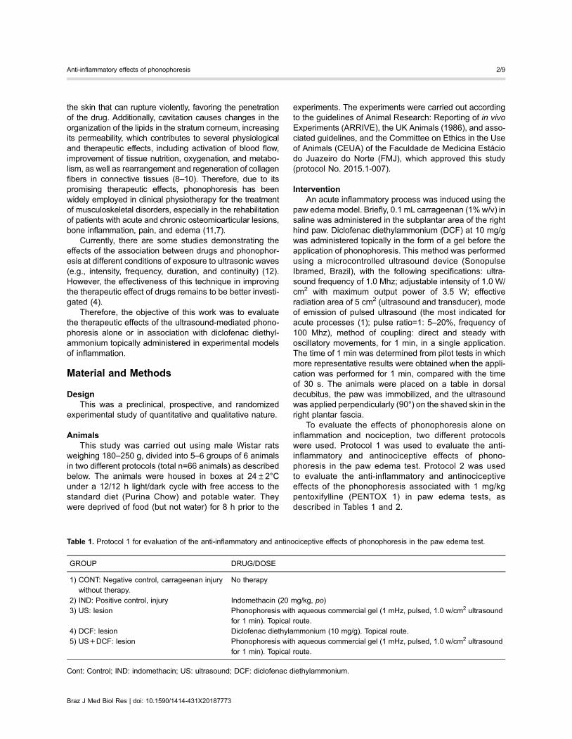
In the present study, treatment with US (0.54±0.09),US+DCF (0.29±0.07), and DCF (0.43±0.00) signifi-cantly decreased (35, 65, and 48%, respectively) the pawedema induced by carrageenan compared to the controlgroup (0.83±0.09). In addition, treatment with US+DCF(Po0.001) was significantly more effective than treatmentwith US alone (Po0.05) (Figure 1) and presented aneffectiveness that was comparable to that of the positivecontrol (INDO 20). These results indicated that the treat-ments presented antiedematogenic effects and that pho-nophoresis improved the antiedematogenic effect ofdiclofenac in this experimental model.
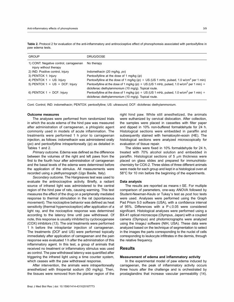
The groups described in Table 2 were used to evaluatethe effect of pentoxifylline associated with US and DCF oncarrageenan-induced paw edema. The use of pentoxifyllineaimed to determine the action of phonophoresis on TNF-aproduction as well as to evaluate the synergism betweenthe treatments. An acute treatment with diclofenac asso-ciated or not with ultrasound and pentoxifylline significantlyreduced the volume of paw edema 3 h after the challenge.Animals treated with PENTOX 1+US+DCF (0.560±0.06),PENTOX 1 (1.187±0.08), PENTOX 1+US (1.102±0.14),and PENTOX 1+DCF (0.843±0.057) presented signifi-cantly lower values of edema (67, 30, 35, and 50%, res-pectively) compared to the negative control. Indomethacin(INDO) was used as a positive control and it decreased theedema volume by 57% (Figure 2).
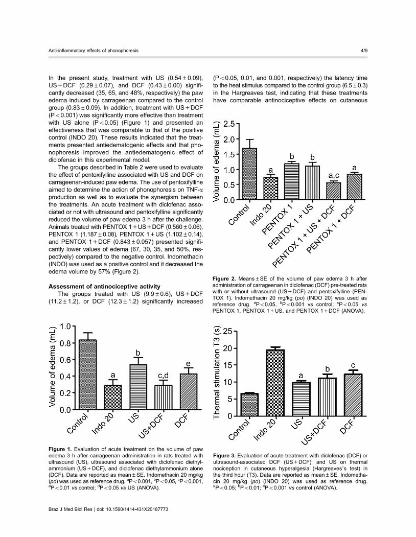
Assessment of antinociceptive activityThe groups treated with US (9.9±0.6), US+DCF
(11.2±1.2), or DCF (12.3±1.2) significantly increased
(Po0.05, 0.01, and 0.001, respectively) the latency timeto the heat stimulus compared to the control group (6.5±0.3)in the Hargreaves test, indicating that these treatmentshave comparable antinociceptive effects on cutaneous
Figure 1. Evaluation of acute treatment on the volume of pawedema 3 h after carrageenan administration in rats treated withultrasound (US), ultrasound associated with diclofenac diethyl-ammonium (US+DCF), and diclofenac diethylammonium alone(DCF). Data are reported as mean±SE. Indomethacin 20 mg/kg(po) was used as reference drug. aPo0.001, bPo0.05, cPo0.001,ePo0.01 vs control; dPo0.05 vs US (ANOVA).
Figure 2. Means±SE of the volume of paw edema 3 h afteradministration of carrageenan in diclofenac (DCF) pre-treated ratswith or without ultrasound (US+DCF) and pentoxifylline (PEN-TOX 1). Indomethacin 20 mg/kg (po) (INDO 20) was used asreference drug. aPo0.05, bPo0.001 vs control; cPo0.05 vsPENTOX 1, PENTOX 1+US, and PENTOX 1+DCF (ANOVA).
Figure 3. Evaluation of acute treatment with diclofenac (DCF) orultrasound-associated DCF (US+DCF), and US on thermalnociception in cutaneous hyperalgesia (Hargreaves’s test) inthe third hour (T3). Data are reported as mean±SE. Indometha-cin 20 mg/kg (po) (INDO 20) was used as reference drug.aPo0.05; bPo0.01; cPo0.001 vs control (ANOVA).
Braz J Med Biol Res | doi: 10.1590/1414-431X20187773
Anti-inflammatory effects of phonophoresis 4/9
hyperalgesia. Indomethacin (19.5±0.9), which was usedas a standard control drug, increased the latency time tothermal stimulation by 3-fold (Figure 3).
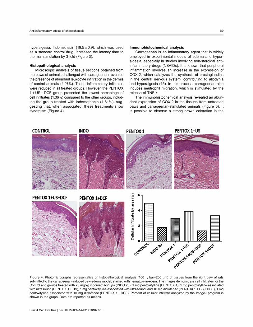
Histopathological analysisMicroscopic analysis of tissue sections obtained from
the paws of animals challenged with carrageenan revealedthe presence of abundant leukocyte infiltration in the dermisof control animals (4.97%). These inflammatory infiltrateswere reduced in all treated groups. However, the PENTOX1+US+DCF group presented the lowest percentage ofcell infiltrates (1.36%) compared to the other groups, includ-ing the group treated with indomethacin (1.81%), sug-gesting that, when associated, these treatments showsynergism (Figure 4).
Immunohistochemical analysisCarrageenan is an inflammatory agent that is widely
employed in experimental models of edema and hyper-algesia, especially in studies involving non-steroidal anti-inflammatory drugs (NSAIDs). It is known that peripheralinflammation involves an increase in the expression ofCOX-2, which catalyzes the synthesis of prostaglandinsin the central nervous system, contributing to allodyniaand hyperalgesia (15). In this process, carrageenan alsoinduces neutrophil migration, which is stimulated by therelease of TNF-a.
The immunohistochemical analysis revealed an abun-dant expression of COX-2 in the tissues from untreatedpaws and carrageenan-stimulated animals (Figure 5). Itis possible to observe a strong brown coloration in the
Figure 4. Photomicrographs representative of histopathological analysis (100�, bar=200 mm) of tissues from the right paw of ratssubmitted to the carrageenan-induced paw edema model, stained with hematoxylin-eosin. The images demonstrate cell infiltrates for theControl and groups treated with 20 mg/kg indomethacin, po (INDO 20), 1 mg pentoxifylline (PENTOX 1), 1 mg pentoxifylline associatedwith ultrasound (PENTOX 1+US), 1 mg pentoxifylline associated with ultrasound, and 10 mg diclofenac (PENTOX 1+US+DCF), 1 mgpentoxifylline associated with 10 mg diclofenac (PENTOX 1+DCF). Percent of cellular infiltrate analyzed by the ImageJ program isshown in the graph. Data are reported as means.
Braz J Med Biol Res | doi: 10.1590/1414-431X20187773
Anti-inflammatory effects of phonophoresis 5/9
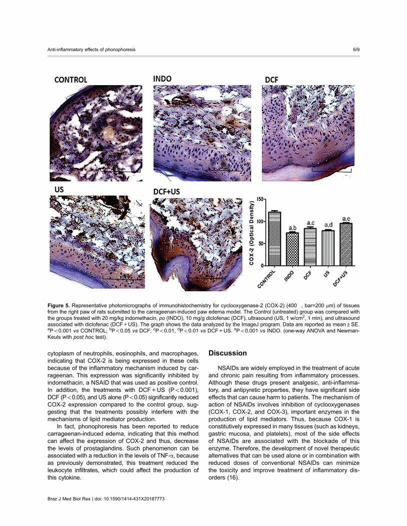
cytoplasm of neutrophils, eosinophils, and macrophages,indicating that COX-2 is being expressed in these cellsbecause of the inflammatory mechanism induced by car-rageenan. This expression was significantly inhibited byindomethacin, a NSAID that was used as positive control.In addition, the treatments with DCF+US (Po0.001),DCF (Po0.05), and US alone (Po0.05) significantly reducedCOX-2 expression compared to the control group, sug-gesting that the treatments possibly interfere with themechanisms of lipid mediator production.
In fact, phonophoresis has been reported to reducecarrageenan-induced edema, indicating that this methodcan affect the expression of COX-2 and thus, decreasethe levels of prostaglandins. Such phenomenon can beassociated with a reduction in the levels of TNF-a, becauseas previously demonstrated, this treatment reduced theleukocyte infiltrates, which could affect the production ofthis cytokine.
Discussion
NSAIDs are widely employed in the treatment of acuteand chronic pain resulting from inflammatory processes.Although these drugs present analgesic, anti-inflamma-tory, and antipyretic properties, they have significant sideeffects that can cause harm to patients. The mechanism ofaction of NSAIDs involves inhibition of cyclooxygenases(COX-1, COX-2, and COX-3), important enzymes in theproduction of lipid mediators. Thus, because COX-1 isconstitutively expressed in many tissues (such as kidneys,gastric mucosa, and platelets), most of the side effectsof NSAIDs are associated with the blockade of thisenzyme. Therefore, the development of novel therapeuticalternatives that can be used alone or in combination withreduced doses of conventional NSAIDs can minimizethe toxicity and improve treatment of inflammatory dis-orders (16).
Figure 5. Representative photomicrographs of immunohistochemistry for cyclooxygenase-2 (COX-2) (400�, bar=200 mm) of tissuesfrom the right paw of rats submitted to the carrageenan-induced paw edema model. The Control (untreated) group was compared withthe groups treated with 20 mg/kg indomethacin, po (INDO), 10 mg/g diclofenac (DCF), ultrasound (US, 1 w/cm2, 1 min), and ultrasoundassociated with diclofenac (DCF+US). The graph shows the data analyzed by the ImageJ program. Data are reported as mean±SE.aPo0.001 vs CONTROL; bPo0.05 vs DCF; cPo0.01, dPo0.01 vs DCF+US. ePo0.001 vs INDO. (one-way ANOVA and Newman-Keuls with post hoc test).
Braz J Med Biol Res | doi: 10.1590/1414-431X20187773
Anti-inflammatory effects of phonophoresis 6/9
In the present study, the anti-inflammatory and anal-gesic effects of phonophoresis alone or in association withdiclofenac diethylammonium was evaluated using an experi-mental model of carrageenan-induced paw edema in rats.Carrageenan is a sulfated polysaccharide extracted fromseaweed that has been widely used in animal models ofinflammation as a tool in the screening of novel anti-inflammatory drugs (17).
An intraplantar injection of carrageenan inducesan inflammatory response that is characterized by atime-dependent generation of paw edema, neutrophilinfiltration, and increased levels of nitrite/nitrate and pros-taglandin E2 in the exudate (18). It has been demon-strated that in this model, the edema is maximal after 3 hand remains elevated for 10 h after administration of carra-geenan. Nitric oxide (NO), produced by the enzyme nitricoxide synthase (NOS), is involved in the development ofinflammation shortly after administration of carrageenan,and the activation of the inducible form of this enzyme(iNOS) generates additional NO that maintains the inflam-matory response. In addition, carrageenan increases thelevels of many cytokines, such as TNF-a, IL-1b, and IL-6in the inflamed paw (19). The acute vascular responsetriggered by carrageenan occurs in three distinct phasesorchestrated by specific mediators. The first phase occurs1 h after carrageenan administration and is characterizedby an increased vascular permeability that is mediated byhistamine and serotonin; the second phase develops 2 hafter the challenge and is orchestrated by kinins, suchas bradykinin; the third and last phase generates themost significant edema. It develops 3 h after carrageenanadministration and is mediated by prostaglandins. There-fore, substances that inhibit the effects of carrageenanin this phase are thought to present anti-inflammatoryproperties (14).
This research demonstrated that treatment with US+DCF was more effective than treatment with US alone.The use of topical anti-inflammatory drugs such as diclo-fenac in the form of a gel associated with ultrasoundis widely used in clinical physiotherapy, because thiscombination facilitates the absorption of the drug (20).However, the pharmacodynamic mechanisms under-lying the effects of this combination remain to be fullyelucidated. Importantly, the results obtained in this studyindicated that in addition to facilitating the penetrationof the drug in the tissue, the phonophoresis possiblyinhibits the release of inflammatory mediators, such asprostaglandins.
The association of pentoxifylline with US+DCFproduced significant and synergistic effects on edemaformation, suggesting that, in addition to lipid media-tors (prostaglandins), this association can affect theproduction of cytokines, such as TNF-a. In fact, someauthors (21,22) have demonstrated that pentoxifyl-line inhibits the production of TNF-a by monocytes andT cells in vitro.
In the Hargreaves test, hyperalgesia is triggered, atleast in part, by the central and peripheral release ofprostaglandins in response to tissue injury. In this test,the results showed that treatment with US+DCF waseffective in increasing the latency time of paw withdrawalafter the stimulus, evidencing an antinociceptive effect ofthis association, which may be correlated with reduction ofprostaglandin synthesis.
The histological and immunohistochemical analysisdemonstrated that therapeutic ultrasound treatmentassociated with phonophoresis reduced the inflammatorycell infiltrates and the expression of COX-2. Here, itis suggested that because these treatments reduceleukocyte infiltrates, lower levels of TNF-a and othercytokines (such as IL-1b) are found in the tissue,affecting the expression of COX-2 (14). Therefore, theanti-inflammatory and antinociceptive effects of theassociation of US and phonophoresis may result frominhibition of COX-2 expression and reduction of prosta-glandin production.
Previous studies have suggested that the synergismobtained in the association of US with DCF is due to afacilitation of transcutaneous penetration of DCF as aresult of the phenomenon of cavitation and changes inthe blood flow (20,23–27). However, the data obtainedin this study suggest that this synergism is associatedwith potentiation of the anti-inflammatory mechanismsof both treatments. Clinically, these data are extremelyrelevant because this association could be used toreduce the dose of NSAIDs required for the treatment ofinflammatory disorders, and thus, minimize their toxiceffects.
The acoustic vibrations produced by therapeutic ultra-sound devices induce cellular changes by altering the con-centration gradient of calcium and potassium ions, affectingthe transport of metabolites through the cell membrane.This phenomenon induces several tissue alterations, suchas increased protein synthesis and secretion by mast cellsand changes in fibroblast mobility. Therefore, alterations inthe membrane permeability result in cell activation andcontribute to tissue repair (10,11,28).
Therefore, it is hypothesized that the mechanisminvolved in the potentiation of drug absorption through theskin caused by phonophoresis is associated with the phe-nomenon of cavitation. Cavitation induces the formation ofgaseous microbubbles in the outer layer of the skin (stratumcorneum) that trigger microvascular hemodynamic modifi-cations. This can lead to increased perfusion, granulationtissue formation, tissue repair in response to fibroblasticproliferation, and increase of precursor cells and disorga-nization of the lipids in the corneum layer. These changesincrease the permeability, favoring the penetration of thedrug (29–32).
In addition, this work demonstrated scientifically thatthe use of topical anti-inflammatory drugs in the form of gelassociated with therapeutic ultrasound, which has been
Braz J Med Biol Res | doi: 10.1590/1414-431X20187773
Anti-inflammatory effects of phonophoresis 7/9
practiced almost empirically (especially by physiotherapyprofessionals), is effective.
In conclusion, the ultrasound-mediated phonophore-sis alone or in association with topical diclofenac haspotential for application in clinical practice, because thesetreatments present anti-inflammatory and analgesic effectsthat might be dependent on inhibition of COX-2 expressionand TNF-a production.
Acknowledgments
The authors are grateful for the support of theFaculdade de Medicina Estácio de Juazeiro do Norte forsubsidizing the research in the premises of the Biophys-iology Laboratory. The authors also thank the ScientificInitiation students Ney Turbano Isidro Filho and MariaLissandra Bezerra for technical assistance.
References
1. Ebrahimi S, Abbasnia K, Motealleh A, Kooroshfard N,Kamali F, Ghaffarinezhad F. Effect of lidocaine phonophor-esis on sensory blockade: pulsed or continuous mode oftherapeutic ultrasound? Physiotherapy 2012; 98: 57–63,doi: 10.1016/j.physio.2011.01.009.
2. Lombry C, Dujardin N, Préat V. Transdermal delivery ofmacromolecules using skin electroporation. Pharm Res2000; 17: 32–37, doi: 10.1023/A:1007510323344.
3. Saliba S, Mistry DJ, Perrin DH, Gieck J, Weltman A. Phono-phoresis and the absorption of dexamethasone in the presenceof an occlusive dressing. J Athl Train 2007; 42: 349–354.
4. Oliveira AS, Guaratini MI, Castro CES. A iontoforese naprática fisioterapêutica. Fisio Bras 2007; 8: 430–435.
5. Machet L, Boucaud A. Phonophoresis: efficiency, mech-anisms and skin tolerance. Int J Pharm 2002; 243: 1–15,doi: 10.1016/S0378-5173(02)00299-5.
6. Byl NN. The use of ultrasound as an enhancer for trans-cutaneous drug delivery: phonophoresis. Phys Ther 1995;75: 539–553, doi: 10.1093/ptj/75.6.539.
7. Farcic TS, Lima RMCB, Machado AFP, Baldan CS, VillicevCM, Junior IE, Masson IFB. Aplicacão do ultrassom tera-pêutico no reparo tecidual do sistema musculoesquelético.Arq Bras Ciênc Saúde 2012; 37: 149–153.
8. Agne JE. Eletrotermoterapia – Teoria e Prática. 1 ed. SantaMaria: Orium, 2013.
9. Ay S, Doğan SK, Evcik D; Baser OC. Comparison the efficacyof phonophoresis and ultrasound therapy in myofascial painsyndrome. Rheumatol Int 2011; 31: 1203–1208, doi: 10.1007/s00296-010-1419-0.
10. Oliveira PD, Oliveira DAAP, Martinago CC, Frederico RCP,Soares CP, Oliveira RF. Efeito da terapia ultrassônica debaixa intensidade em cultura celular de fibroblastos. FisioterPesqui 2015; 22: 112–118, doi: 10.590/1809-2950/12860222022015.
11. Robertson VJ, Baker KG. A review oj therapeutic ultra-saoud: effectiveness studies. Physical Therapy 2001; 81:1339–1350, doi: 10.1093/ptj/81.7.1351.
12. Guaratini MI, Oliveira AS, Castro CES. Fundamentacãoteórica para iontoforese. Rev Bras Fisioter 2005; 9: 1–7.
13. Hargreaves K, Dubner R, Brown F, Flores C, Joris J. A newand sensitive method for measuring thermal nociceptionin cutaneous algesia. Pain 1988; 32: 77–88, doi: 10.1016/0304-3959(88)90026-7.
14. Kawahara K, Hohjoh H, Inazumi T, Tsuchiya S, Sugimoto Y.Prostaglandin E2-induced inflammation: Relevance of pros-taglandin E receptors. Biochim Biophys Acta 2015; 1851:414–421, doi: 10.1016/j.bbalip.2014.07.008.
15. Guay J, Bateman K, Gordon R, Mancini J, Riendeau D.Carrageenan-induced paw edema in rat elicits a predomi-nant prostaglandin E2 (PGE2) response in the centralnervous system associated with the induction of microsomalPGE2 synthase-1. J Biol Chem 2004; 279: 24866–24872,doi: 10.1074/jbc.M403106200.
16. Katzung GB. Farmacologia: Básica e Clınica.12. GuanabaraKoogan: 2014.
17. Srivastava A, Kumar R. Synthesis and characterization ofacrylic acid-g-(-carrageenan) copolymer and study of itsapplication. Int J Carbohydr Chem 2013; 892615: 1–8,doi: 10.1155/2013/892615.
18. Salvemini D, Wang Z, Wyatt PS, Bourdon DM, Marino MH,Manning PT, et al. Nitric oxide: a key mediator in the earlyand late phase of carrageenan-induced rat paw inflamma-tion. Br J Pharmacol 1996; 118: 829–838, doi: 10.1111/j.1476-5381.1996.tb15475.x.
19. Loram LC. Cytokine profiles during carrageenan-inducedinflammatory hyperalgesia in rat muscle and hind paw.J Pain 2007; 8: 127–136, doi: 10.1016/j.jpain.2006.06.010.
20. Rosim GC. Análise da influência do ultra-som terapêutico napenetracão transcutânea de diclofenaco sódico em huma-nos sadios. Master’s thesis, Bioengenharia, Universidadede São Paulo, São Carlos, 2003, doi: 10.11606/D.82.2003.tde-29082003-111047.
21. Antunes AS, Teixeira MCB, Júnior AG. Efeitos da pentox-ifilina na anemia resistente à eritropoetina em pacientessob hemodiálise. Rev Bras Hematol Hemoter 2008; 30:303–308, doi: 10.1590/S1516-84842008000400014.
22. Renke M, Tylicki L, Rutkowski P, Knap N, Zietkiewicz M,Neuwelt A, et al. Effect of pentoxifylline on proteinuria,markers of tubular injury and oxidative stress in non-diabeticpatients with chronic kidney disease – placebo controlled,randomized, cross-over study. Acta Biochim Pol 2010; 57:119–123.
23. Byl N, Mckenzie A, Halliday B, Wong T, O’Connell J. Theeffects of phonophoresis with corticosteroids: a controlledpilot study. J Orthop Sports Phys Ther 1993; 18: 590–600,doi: 10.2519/jospt.1993.18.5.590.
24. McElnay JC, Benson HAE, Harland R, Hadgraft J. Phono-phoresis of methyl nicotinate: a preliminary study to elucidatethe mechanism of action. Pharm Res 1993; 10: 1726–1731,doi: 10.1023/A:1018918013489.
25. Mitragotri S, Blankschtein D, Langer R. Ultrasound mediatedtransdermal protein delivery. Science 1995; 269: 850–853,doi: 10.1126/science.7638603.
Braz J Med Biol Res | doi: 10.1590/1414-431X20187773
Anti-inflammatory effects of phonophoresis 8/9
26. Asano J, Suisha F, Takada M, Kawasaki N, Miyazaki S. Effectof pulsed output ultrasound on the transdermal absorption ofindomethacin from an ointment in rats. Biol Pharm Bull 1997;20: 288–291, doi: 10.1248/bpb.20.288.
27. Robinson SE, Buono MJ. Effect of continuous-wave ultra-sound on blood flow in skeletal muscle. Phys Ther 1994; 75:145–149, doi: 10.1093/ptj/75.2.145.
28. Low J, Reed A. Ultra-som terapêutico. In: Eletroterapiaaplicada: princıpios e prática. São Paulo: Manole, 2001.
29. Parizotto NA. Utilizacão da fonoforese em desordens músculo-esquelética: uma meta-análise. Rev Bras Fisiot 2003; 7: 49–55.
30. Miller MW. Gene transfection and drug delivery. UltrasoundMed Biol 2000; 26: S59–S62, doi: 10.1016/S0301-5629(00)00166-6.
31. Fang JY, Fang CL, Sung KC, Chen HY. Effect of lowfrequency ultrasound on the vitro percutaneous absorptionof clobetasol 17-propionate. Int J Pharm 1999; 191: 33–42,doi: 10.1016/S0378-5173(99)00230-6.
32. Ueda H, Ogihara M, Sugibayashi K, Morimoto Y. Changein the electrochernical properties of skin and the lipid packingin stratum comeum by ultrasonic irradiation. Int J Pharm1996; 137: 217–224, doi: 10.1016/0378-5173(96)04523-1.
Braz J Med Biol Res | doi: 10.1590/1414-431X20187773
Anti-inflammatory effects of phonophoresis 9/9