AVALIAÇÃO BIOQUÍMICA, MORFOLÓGICA E FUNCIONAL DO...
63
Campus de Botucatu Instituto de Biociências PG-BGA AVALIAÇÃO BIOQUÍMICA, MORFOLÓGICA E FUNCIONAL DO MÚSCULO ESTRIADO ESQUELÉTICO DE RATOS NA INSUFICIÊNCIA CARDÍACA Raquel Santilone Bertaglia Dissertação apresentada ao Instituto de Biociências, Câmpus de Botucatu, UNESP, para obtenção do título de Mestre no Programa de Pós-Graduação em Biologia Geral e Aplicada, Área de concentração Biologia Celular, Estrutural e Funcional. Orientadora: Dra Maeli Dal Pai Silva Botucatu – SP 2011
Transcript of AVALIAÇÃO BIOQUÍMICA, MORFOLÓGICA E FUNCIONAL DO...
Campus de Botucatu
Instituto de
Biociências
Biociências
PG-BGA
AVALIAÇÃO BIOQUÍMICA, MORFOLÓGICA E FUNCIONAL DO
MÚSCULO ESTRIADO ESQUELÉTICO DE RATOS NA
INSUFICIÊNCIA CARDÍACA
Raquel Santilone Bertaglia
Dissertação apresentada ao Instituto de Biociências,
Câmpus de Botucatu, UNESP, para obtenção do título
de Mestre no Programa de Pós-Graduação em Biologia
Geral e Aplicada, Área de concentração Biologia
Celular, Estrutural e Funcional.
Orientadora: Dra Maeli Dal Pai Silva
Botucatu – SP
2011
UNIVERSIDADE ESTADUAL PAULISTA
“Julio de Mesquita Filho”
INSTITUTO DE BIOCIÊNCIAS DE BOTUCATU
“AVALIAÇÃO BIOQUÍMICA, MORFOLÓGICA E FUNCIONAL DO
MÚSCULO ESTRIADO ESQUELÉTICO DE RATOS NA
INSUFICIÊNCIA CARDÍACA”
Raquel Santilone Bertaglia
Orientadroa: Dra Maeli Dal Pai Silva
Dissertação apresentada ao Instituto de Biociências,
Câmpus de Botucatu, UNESP, para obtenção do título
de Mestre no Programa de Pós-Graduação em Biologia
Geral e Aplicada, Área de concentração Biologia
Celular, Estrutural e Funcional.
Orientadora: Dra Maeli Dal Pai Silva
Botucatu – SP
2011

FICHA CATALOGRÁFICA ELABORADA PELA SEÇÃO DE AQUIS. E TRAT. DA INFORMAÇÃO DIVISÃO TÉCNICA DE BIBLIOTECA E DOCUMENTAÇÃO - CAMPUS DE BOTUCATU - UNESP
BIBLIOTECÁRIA RESPONSÁVEL: ROSEMEIRE APARECIDA VICENTE
Bertaglia, Raquel Santilone.
Avaliação bioquímica, morfológica e funcional do músculo estriado
esquelético de ratos na insuficiência cardíaca / Raquel Santilone Bertaglia. -
Botucatu, 2011
Dissertação (mestrado) – Instituto de Biociências de Botucatu, Universidade
Estadual Paulista, 2011
Orientador: Maeli Dal Pai Silva
Capes: 20603002
1. Insuficiência cardíaca. 2. Rato como animal de laboratório. 3. Músculos.
Palavras-chave: Estresse oxidativo; Insuficiência cardíaca; Miogenina;
Monocrotalina; Músculo estriado esquelético; MyoD.

Dedicatória
Dedico este trabalho...
À Deus, que com toda certeza está sempre ao meu lado guiando meus passos e iluminando meu
caminho.
Aos meus queridos e amados pais, Nilson e Marilda, pelo amor, carinho e compreensão. Por não
medirem esforços para a minha educação e formação profissional e por acreditarem em mim,
tornando possível a realização dos meus sonhos.
Ao meu marido Osvaldo Cesar, por estar ao meu lado, sempre me incentivando e acalmando,
renovando minha esperança a cada dia.

Agradecimentos
À Deus, por me acompanhar, me proteger e me amparar sempre. Obrigada por estar ao meu lado
e por permitir a presença de pessoas especiais na minha vida.
À orientadora, mestre e amiga Dra. Maeli Dal Pai Silva, pela competência, paciência e carinho
com que acompanhou todas as etapas desse trabalho e por ter confiado em mim. Obrigada por ter
dividido comigo um pouquinho do brilho, dessa infinita luz da sabedoria, que existe dentro de
você.
Aos meus pais, referências de sabedoria e perseverança. Obrigada por acreditarem que o
conhecimento é a melhor herança e por me apoiarem em todos os momentos da minha vida
pessoal e profissional.
Ao meu marido Osvaldo Cesar pelo companheirismo e paciência. Seu incentivo, carinho e amor
foram essenciais para eu trilhar esse caminho até o fim.
Aos meus irmãos Daniel e Angélica por todo amor, carinho e amizade, em todos os momentos.
Vocês são motivos de muito orgulho e inspiração.
Aos meus familiares pela grande ajuda, compreensão e força, que encontrei, nos momentos
difíceis.
À Professora Dra. Márcia Gallacci e aos amigos Walter, Cicília, Fábio e Fernanda, do
departamento de farmacologia, que com muita dedicação me ajudaram com a análise funcional.
Agradeço pela amizade, colaboração e troca de conhecimentos.
À Professora Dra. Ana Angélica Henrique Fernandes, do departamento de Química e
Bioquímica, pelo auxilio nas análises bioquímicas.

Aos Professores Dr. Carlos Roberto Padovani e Sergio Augusto Rodrigues do Departamento de
Bioestatística-UNESP-Botucatu pelo auxílio estatístico.
Ao Dr. Carlos Antônio Cicogna por sua orientação em diversas etapas do desenvolvimento desta
pesquisa e por compartilhar suas experiências.
Aos membros da banca de defesa: Dra. Maeli Dal Pai Silva, Dr. Jesus Carlos Andreo e Dr.
Walter Cavalcante pela disponibilidade e atenção para participarem desta avaliação.
À Professora Dra. Ana Angélica Henrique Fernandes, à Professora Dra. Márcia Gallacci e ao
Professor Dr. Robson Francisco Carvalho por comporem minha banca de qualificação ao
mestrado, pela atenção e principalmente pelos ensinamentos transmitidos.
Aos professores do Programa de Mestrado em Biologia Geral e Aplicada que participaram,
positivamente, na minha formação científica durante estes dois anos de mestrado, agradeço pelo
aprendizado e por compartilhar suas experiências profissionais.
Aos meus amigos do Laboratório de Biologia do Músculo Esquelético (LBME): Airton, Aline,
Andreo, Bruno, Caroline, Cassiane, Edson, Eduardo, Fernanda Carani, Fernanda Alves, Fernanda
Losi, Flávia, Francis, Henrique, Ivan, Joyce, Juliana, Luana, Ludmila, Paula, Rachel, Robson,
Rodrigo e Warlen pelo convívio agradável, conhecimento compartilhado e constante apoio.
Aos técnicos de laboratório, José Eduardo e Ricardo pelo auxílio profissional e amizade.
Aos funcionários do departamento de Morfologia-IBB-UNESP-Botucatu por serem prestativos e
estarem sempre presentes.
À Luciana, secretária do Departamento de Morfologia-IBB-UNESP-Botucatu, pela atenção e por
todo auxílio.

Aos funcionários da secretaria de pós-graduação pelo profissionalismo e pela disposição em
sempre querer ajudar.
À Capes pelo suporte financeiro, viabilizando o desenvolvimento desse trabalho.
À todos que direta ou indiretamente contribuíram para o desenvolvimento e conclusão desse
trabalho.
Muito obrigada!
Sumário
Resumo .......................................................................................................................... 09
Abstract ........................................................................................................................ 11
I – INTRODUÇÃO ....................................................................................................... 12
1.1. Características Gerais do Músculo Estriado Esquelético ....................................... 12
1.2. Plasticidade do Músculo Esquelético ..................................................................... 17
1.3. Insuficiência Cardíaca ............................................................................................ 17
1.4. Alterações nas fibras do Músculo Esquelético na Insuficiência Cardíaca ............. 18
II – OBJETIVO ............................................................................................................. 22
III- MATERIAIS E MÉTODOS .................................................................................... 23
IV – REFERÊNCIAS BIBLIOGRÁFICAS ................................................................. 31
V – CAPÍTULO 1: Differential morphofunctional characteristics in fast and slow muscles in a
monocrotaline-induced heart failure .............................................................................. 40
9
Resumo:
A insuficiência cardíaca (IC) está associada à miopatia dos músculos esqueléticos dos membros,
com perda da massa muscular, diminuição na proporção da cadeia pesada das miosinas do tipo I
(MHCI), aumento na proporção da cadeia pesada das miosinas do tipo II (MHCII), decréscimo
do metabolismo oxidativo e alterações nos fatores de regulação miogênica (MRFs). Na IC
também ocorre aumento do estresse oxidativo na musculatura esquelética, o qual está relacionado
às mudanças estruturais, morfológicas e funcionais. Nesse estudo, nós investigamos e
comparamos as características morfofuncionais dos músculos Sóleo (SOL), lento e com
predomínio de fibras oxidativas e Extensor Digitorum Longus (EDL), rápido e com predomínio
de fibras glicolíticas em ratos com IC induzida pela monocrotalina. Foram utilizados ratos
Wistar, machos (90 a 100 g), divididos em 2 grupos: controle (CT) e insuficiência cardíaca (IC),
induzida pela injeção de dose única de monocrotalina (MCT, 30mg/Kg i.p.). Após 22 dias da injeção
da MCT, quando os animais apresentaram sinais de IC, todos os animais foram sacrificados e pesados. Os
músculos SOL e EDL foram retirados, pesados e processados para as análises morfológicas, moleculares,
bioquímicas e funcionais. A expressão gênica da MyoD e miogenina foram determinadas usando qRT-
PCR, as isoformas de MHC foram determinadas por eletroforese em gel de poliacrilamida, a freqüência e
área de secção transversal dos tipos de fibra foram analisadas pela reação histoquímica de ATPase
miofibrilar (mATPase). Foram realizados estudos bioquímicos para a determinação do hydroperóxido de
lipídeo (HL), da glutationa peroxidase (GSH-Px) e da superóxido dismutase (SOD) e catalase (CAT); o
estudo miográfico foi realizado para determinar a força máxima de contração, o tempo de contração e de
relaxamento e a resistência à fadiga. Todos os ratos tratados com MCT mostraram sinais de IC (hipertrofia
de átrio e ventrículo direitos, derrame pleural, ascite e hepatomegalia) e atrofia dos músculos SOL e EDL,
confirmada pela diminuição da área de secção transversal dos tipos de fibras (Tipos I, IC, IIC e IIA no
SOL e I, IIC, IIA, IIA/D e IIDB no EDL). No grupo com IC, a freqüência das fibras IIC diminuiu e o
tempo de relaxamento da contração muscular aumentou no músculo SOL; a expressão gênica de
Miogenina diminuiu no músculo SOL enquanto a de MyoD diminuiu no músculo EDL. A
concentração de Hidroperóxido de lipídio (HL) aumentou e a atividade das enzimas antioxidantes
Glutationa Peroxidase (GSH-Px) e Superóxido dismutase (SOD) diminuiu apenas no músculo
SOL dos animais com IC; no músculo EDL, não houve alteração nas propriedades contráteis, na
expressão gênica, na concentração de HL e na atividade das enzimas antioxidantes. Nos grupos
estudados, não houve alteração nas MHCs nos músculos SOL e EDL. Em conclusão, a IC
induzida por monocrotalina induziu alterações morfológicas, bioquímicas e funcionais

10
principalmente no músculo SOL, músculo de contração lenta e constituído predominantemente
por fibras oxidativas. Embora novos estudos sejam necessários para melhor esclarecer os
mecanismos envolvidos na fisiopatologia da insuficiência cardíaca, nossos resultados contribuem
para a compreensão das alterações músculo-específicas que ocorrem nessa síndrome.

11
Abstract
Heart failure (HF) is characterized by a limited exercise tolerance, skeletal muscle myopathy with atrophy,
shift toward fast muscle fiber and myogenic regulatory factors (MRFs) changes. Reactive oxygen species
(ROS) also contribute to target organ damage in the heart failure syndrome. In this study, we investigated
and compared the morphofunctional characteristics in SOL, a slow oxidative muscle and EDL, a fast
glycolytic muscle in a monocrotaline-induced heart failure. Two groups of rats were studied: control (CT)
and Heart Failure (HF), induced by a single intraperitoneal injection of monocrotaline (MCT, 30mg/Kg).
MyoD and myogenin expression were determined by using qRT-PCR, MHC isoforms were studied by
using polyacrylamide gel electrophoresis, muscle fiber-type frequency and cross sectional area (CSA)
were analyzed by myofibrillar adenosine triphosphatase (mATPase). Biochemical study were performed
to determine: lipid hydroperoxide (LH), glutathione peroxidase (GSH-Px) and superoxide dismutase
(SOD); myographic study was performed to analyze: amplitude, rise time, fall time and fatigue resistance
in SOL and EDL muscles. All monocrotaline treated rats showed signs of HF (atrium and right ventricular
hypertrophies, pleural and pericardial effusions, and congested liver). HF group showed SOL and EDL
muscles atrophy, confirmed by CSA decreased in muscle fiber types (types I, IC, IIC and IIA in SOL and
I, IIC, IIA, IIA/D and IIDB in EDL muscles); the frequency of IIC fiber type and the fall time of muscle
contraction increased only in SOL muscle; he myogenin mRNA expression was lower only in the SOL
muscle and the MyoD mRNA expression decreased only in EDL muscle. HF group also presented the
concentration of lipid hydroperoxide increased, superoxide-dismutase and glutathione peroxidase activity
reduced only in SOL muscle; EDL muscle showed the contractile properties, concentration of HL and
antioxidant enzyme activity not changed. In the groups studied, the percentage distribution of MHCs did
not alter in both muscles. In conclusion, our results indicate that the HF induced by monocrotaline
promoted biochemical, morphological and functional changes, more prominent in the SOL, a slow-twitch
muscle. Although further experiments are required for better determine the mechanisms involved in the
pathophysiology of heart failure, our results contributes to the understanding of the muscle-specific
changes that occur in this syndrome.

12
I - INTRODUÇÃO
1.1. Características Gerais do Músculo Estriado Esquelético
O músculo estriado esquelético é um tecido dinâmico e possui características peculiares
de adaptação morfológica, metabólica e funcional frente aos mais variados estímulos (Pette e
Staron, 2000). Esse tecido é constituído por células especializadas, as fibras musculares, que são
alongadas e multinucleadas e os núcleos estão localizados na região periférica da fibra, abaixo da
membrana plasmática (Figura 1). As fibras musculares estão imersas em uma matriz extracelular
rica em carboidratos e proteínas, que constituem o tecido conjuntivo do músculo. Esse tecido está
organizado em três bainhas distintas: o epimísio, que circunda todo o músculo; o perimísio, que
divide o músculo em fascículos e o endomísio, que circunda cada fibra muscular (Craig, 1994;
Sanes, 1994). A disposição altamente organizada dos diferentes tipos de fibras musculares
confere a este tecido uma ampla diversidade estrutural, metabólica e funcional (Schiaffino e
Reggiani, 1994; Pette e Staron, 2000).
Figura 1 – Corte transversal das fibras do músculo sóleo de ratos. Fibras musculares (F); Perimísio (seta
descontínua); Endomísio (seta contínua) e Mionúcleos (ponta da seta). Coloração HE (40x).

13
A regulação do processo de formação dos músculos esqueléticos envolve a ativação,
proliferação e diferenciação de linhagens de células miogênicas e depende da expressão e
atividade de vários fatores transcricionais, entre eles, os fatores de regulação miogênica (do
inglês, myogenic regulatory factors ou MRFs).
Os MRFs são membros da família dos fatores transcricionais “basic helix-loop-helix”
(bHLH), da qual fazem parte a MyoD, Miogenina, Myf5 e o MRF4, (do inglês, myogenic
regulatory factors ou MRFs) (Patapoutian et al., 1995; Rawls et al., 1995; Zhang et al., 1995,
Yoon et al., 1997).
A Miogenina e a MyoD têm função primária na miogênese e estão envolvidas na
manutenção do fenótipo da fibra muscular adulta, rápida ou lenta; a Miogenina é expressa em
níveis superiores aos da MyoD em músculos lentos, enquanto que o oposto é verdadeiro para
músculos rápidos (Murre et al., 1989; Hughes et al., 1993; Voytik et al., 1993; Megeney &
Rudnicki, 1995). No entanto, estudos têm demonstrado que a Miogenina está mais relaciona com
o metabolismo do músculo do que com as mudanças na composição das MHCs (Hughes et al.,
1999; Siu et al., 2004).
Morfologicamente as fibras musculares são constituídas por estruturas repetidas, os
sarcômeros, unidade contrátil fundamental da fibra muscular (Huxley, 1969). Cada sarcômero é
formado por várias proteínas: as proteínas contráteis miosina (filamento grosso) e actina
(filamento fino), além das proteínas estruturais, responsáveis pela organização e integridade
funcional do sarcômero. O filamento fino é formado pela actina e duas proteínas reguladoras, a
troponina e tropomiosina (McComas, 1996). O filamento grosso é formado pela polimerização de
200 a 300 moléculas de miosina da classe II. A molécula de miosina é um hexâmero formado por
duas cadeias pesadas de miosina (do inglês, myosin heavy chain ou MHC), enroladas em α-
hélice, e quatro cadeias leves de miosina (do inglês, myosin light chain ou MLC) (Lowey et al.,
1969; Weeds & Lowey, 1971; Elliot & Offer, 1978; Warrick & Spudich, 1987). Cada cadeia
pesada pode ser separada em duas porções: meromiosina leve, em forma de bastão, e
meromiosina pesada, conhecida como porção globosa da miosina, a qual apresenta o sítio de
ligação com a actina e a região capaz de ligar-se à molécula de ATP e hidrolisá-la (atividade
ATPásica) (Huxley, 1969; Lowey et al., 1969) (Figura 2).

14
Figura 2 – Esquema da molécula de miosina da classe II. Cadeias pesadas de miosina (MHC), cadeias
leves de miosina (MLC), meromiosina leves (LMM), meromiosina pesadas (HMM), e porção globosa
S1 e S2 da HMM. (Dal Pai-Silva et al., 2005).
Os primeiros estudos envolvendo o tecido muscular classificavam os músculos em
“vermelhos” ou “brancos” (Ranvier, 1873). A cor vermelha está relacionada com a presença do
pigmento mioglobina e com o grau de vascularização do músculo. Com a utilização de técnicas
histoquímicas, observou-se que a maioria dos músculos estriados dos mamíferos é constituída por
uma população heterogênea de fibras, que apresentam características genéticas, morfológicas,
bioquímicas e fisiológicas distintas (Dubowitz e Pearse, 1960). Inicialmente as fibras musculares
foram classificadas em vermelhas, intermediárias e brancas (Ogata, 1958). Posteriormente, três
tipos principais de fibras musculares foram descritas, sendo denominadas de fibras dos tipos I,
IIA e IIB, de acordo com o padrão de reação para a atividade da ATPase miofibrilar (mATPase)
na porção globular da miosina (mATPase) (Brooke e Kaiser, 1970).
A velocidade de contração de um músculo é diretamente proporcional a atividade da
mATPase (Talmadge e Roy, 1993). Este evento foi demonstrado em análises de fibras isoladas,
que revelaram uma alta correlação entre o tipo de fibra, baseado na atividade da mATPase, com a
especificidade da miosina de cadeia pesada (MHC) (Barany, 1967; Pette e Staron, 2000). A MHC
capaz de rápida hidrólise do ATP é característica das fibras do tipo II, que são fibras de contração
rápida. Já a MHC de lenta hidrólise do ATP é encontrada nas fibras do tipo I, de contração lenta
(Kelly & Rubinstein, 1994).

15
Neste contexto, Pette e Staron (2001) classificaram os tipos de fibras musculares em
fibras de contração lenta – Tipo І (Slow Fibers), expressando MHCІ e fibras de contração rápida
– Tipo ІІ (Fast Fibers), subdivididas em tipo ІІA, expressando MHCІІa; tipo ІІD, expressando
MHCІІd e tipo ІІB, expressando MHCІІb. As fibras do tipo ІІD (MHCІІd) se equivalem as fibras
ІІX (MHCІІx), descritas em ratos (Schiaffino e Reggiani, 1994) (Figura 3).
Figura 3- Separação elétroforética das isoformas de miosina de cadeia pesada (MHC) em diferentes
músculos de ratos (R) e camundongos (C). Expressão das Isoformas rápidas MHCIIa, MHCIId e MHCIIb e
da Isoforma lenta MHCI. TA, tibial anterior; PS, porção profunda do psoas; AM, adutor magno; GA,
gastrocnêmio; VL, vasto lateral; TA, tibial anterior. Eletroforese em gel de poliacrilamida - SDS-PAGE.
(adaptado de Hamalainen e Pette, 1993).
As fibras do tipo I, IIA, IID/X e IIB são classificadas como fibras puras (Pette & Staron,
1997; Staron et al., 1999). Porém, além das fibras puras, que expressam apenas um tipo de RNA
mensageiro para a MHC, há fibras que co-expressam diferentes genes para a MHC (Biral et al.,
1988; Aigner et al, 1993; Schiaffino & Reggiani, 1994; Caiozzo et al., 2003). Essas fibras são
classificadas de acordo com o tipo de MHC predominante: Tipo ІC (MHC І > MHC ІІa), Tipo
ІІC (MHC ІІa > MHC І ), Tipo ІІAD (MHC ІІa > MHC ІІd), Tipo ІІDA (MHCІІd > MHCІІa),
Tipo ІІDB (MHC ІІd > MHC ІІb) e Tipo ІІBD (MHC ІІb > MHCІІd) (Di Maso et al.,2000; Pette
e Staron, 2000; Pette e Staron, 2001). Deste modo, assume-se uma organização seqüencial das
fibras puras, as quais são intermediadas por fibras híbridas (I ↔ ІC ↔ ІІC ↔ ІІA ↔ ІІAD ↔ ІІD
↔ ІІDB ↔ ІІB) (Pette & Staron, 2000) (Figura 4).

16
Figura 4– Corte transversal do músculo sóleo de rato. Fibras musculares puras (I e IIA) e híbridas (IC e
IIC). Reação da ATPase miofibrilar, pH 4,35.
A natureza dinâmica dos tipos de fibras nos músculos de mamíferos, demonstra que
cada tipo de fibra apresenta diferentes atividades das enzimas metabólicas. A análise
histoquímica do músculo para as atividades de enzimas mitocondriais e mATPase permite a
identificação das fibras como de contração lenta e metabolismo oxidativo; de contração rápida e
metabolismo glicolítico e de contração rápida e metabolismo oxidativo e glicolítico (Peter et al.,
1972).
As fibras com metabolismo oxidativo apresentam muitas mitocôndrias, pouco
glicogênio e maior número de capilares por fibra. Recebem maior teor de oxigênio e metabólitos,
possuem elevada atividade de fosforilação oxidativa e a remoção dos produtos do metabolismo é
mais rápida. As fibras com metabolismo glicolítico apresentam poucas mitocôndrias, muito
glicogênio e o número de capilares por fibras é menor, sendo o aporte de oxigênio e metabólitos
mais reduzido (Silau and Branchero, 1978; Gray et al., 1983; Sanger and Stoiber, 2001).
A identificação das características contráteis das fibras musculares é importante, pois
como os músculos são compostos por vários tipos de fibras musculares, suas propriedades
refletem a soma das características das fibras que o constituem.
17
A especificidade na expressão das diferentes isoformas de MHCs, de acordo com o tipo
de fibra e músculo analisado, revela uma ampla diversidade estrutural, funcional e metabólica do
músculo esquelético (Pette & Staron, 2000).
1.2. Plasticidade Muscular
A diversidade funcional e metabólica dos tipos de fibras confere ao músculo esquelético
uma alta capacidade para realizar uma variedade de demandas funcionais (Campos et al., 2002).
Contudo, as fibras musculares exibem uma alta plasticidade, o que habilita este tecido a alterar
suas características morfológicas, metabólicas e funcionais, como resposta a estímulos
específicos (Pette e Staron, 2000; Magaudda et al., 2004). Esta plasticidade muscular tem sido
demonstrada em diferentes estímulos, tais como exercício de resistência (aeróbico), treinamento
contra resistência (força), estimulação elétrica, desnervação e reinervação, imobilização,
microgravidade, intervenção nutricional, hipoxia, envelhecimento, fatores hormonais (Pette e
Staron, 2000; Fluck e Hoppeler, 2003) e frente à algumas patologias. Os estímulos sucessivos
podem provocar ajustes específicos no fenótipo das fibras musculares, a fim de suprir as
necessidades do organismo, e, adaptar a função muscular (Buller et al., 1960; Gutmann et al.,
1972; Simoneau & Pette, 1988; Klitgaard et al., 1990; Frontera et al., 1991; Caiozzo et al., 1992;
Caiozzo et al., 1996; Adams et al., 1999; Demirel et al., 1999; Williamson et al., 2000; Fitts et al.,
2001; Sharman et al., 2001; Moura et al., 2002; Parcell et al., 2003; Willoughby & Rosene, 2003;
Harber et al., 2004; Siu et al., 2004).
Dentre as patologias que podem causar alterações nas características do músculo
esquelético destaca-se a Insuficiência Cardíaca (IC) (Poole-Wilson & Ferrari, 1996; Wilson,
1996; Bigard et al., 1998).
1.3. Insuficiência Cardíaca
A insuficiência cardíaca (IC) é um estado fisiopatológico na qual o coração é incapaz de
bombear sangue de acordo com as necessidades metabólicas teciduais, ou pode fazê-lo
adequadamente à custa da elevação da pressão de enchimento ventricular (Braunwald et al.,
18
2001; Givertz et al., 2005). De acordo com Cohn (1988), a IC é uma síndrome clínica associada à
disfunção cardíaca, diminuição da expectativa de vida e intolerância aos exercícios físicos.
A IC constitui um importante problema clínico devido à gravidade de suas
manifestações e à sua grande prevalência. Estudos de prevalência estimam que aproximadamente
23 milhões de pessoas no mundo têm IC e que dois milhões de casos novos são diagnosticados
anualmente (Sociedade Brasileira de Cardiologia, 1999; Neto, 2004; Barreto et al., 1998). Dados
obtidos nos Estados Unidos e na Europa mostram que a incidência média de IC é de 1 a 5 casos
por 1000 habitantes/ano, e sua prevalência é de aproximadamente 1% a 2% da população (Cowie
et al., 1997). A IC encontra-se entre as principais causas de internação do Sistema Único de
Saúde (Albanesi Filho, 1998; Rossi Neto, 2004).
As principais causas da IC são isquemias, inflamações agudas, hipertensão arterial e
alterações das valvas cardíacas (Francis, 2001) e entre os principais sintomas da IC encontram-se:
dispnéia, fraqueza e fadiga de membros inferiores com conseqüente redução da atividade
locomotora, intolerância para realizar exercícios físicos e piora da qualidade de vida (Poole-
Wilson & Ferrari, 1996; Wilson, 1996; Bigard et al., 1998). Estudos demonstraram pobre
correlação entre débito cardíaco, fluxo sanguíneo e intolerância ao exercício, sugerindo como
principais contribuintes para a incapacidade funcional, as alterações periféricas musculares
(Wilson, 1995; Vescovo et al., 1998c; Cicoira, 2002; De Sousa et al., 2002).
1.4. Alterações nas fibras do Músculo Esquelético na Insuficiência Cardíaca
Na IC observa-se alterações musculo esqueléticas incluindo aumento do metabolismo
glicolítico, decréscimo do metabolismo oxidativo, e menor resistência à fadiga (Simonini et
al.,1996; Ventura-Clapier et al., 2003), mudança fenotípica de fibras do Tipo I para do Tipo II,
modificações nas miosinas de cadeia pesada e presença de atrofia da musculatura esquelética
(Mancini et al., 1992; Harrington et al., 1997; Poehman, 1999; De Sousa, et al., 2000; Carvalho et
al., 2003) contribuem para as limitações funcionais (Sullivan et al., 1990; Mancini et al., 1992,
Simonini et al., 1996; Bigard et al., 1998; Vescovo et al., 1998a, De Sousa et al., 2000).
Carvalho et al. (2003) demonstraram em ratos com IC induzida por estenose aórtica, que
na fase de hipertrofia cardíaca (18 semanas) o músculo SOL já apresenta mudança para um
padrão fenotípico mais rápido.

19
Embora vários fatores tenham sido descritos como responsáveis pelo desenvolvimento
de fadiga nos pacientes com IC, sua etiopatogenia ainda não está completamente esclarecida.
Esse fenômeno é decorrente, em parte, das alterações metabólicas, com aumento do metabolismo
glicolítico, decréscimo do metabolismo oxidativo e menor resistência à fadiga (Simonini et
al.,1996; Lunde et. al., 200; Ventura-Clapier et al., 2003).
Na IC, diferentes vias podem regular as mudanças fenotípicas no músculo esquelético
(Allen et al., 2001; Carvalho et al., 2006; Filippatos et al., 2005; Spangenburg et al., 2002),
incluindo os MRFs (Hughes et al., 1993; Hughes et al., 1999). Como descrito anteriormente,
vários estudos têm sugerido que a MyoD é expressa em níveis superiores em músculos
glicolíticos/rápidos, enquanto a miogenina é encontrada principalmente em músculos
oxidativos/lentos (Hughes et al., 1993). Estudos têm demonstrado que a miogenina está mais
envolvida com o metabolismo do músculo do que com as mudanças nas características contráteis
(Hughes et al., 1999, Ekmark et al., 2003, Siu et al., 2004).
Estudos têm demonstrado que as alterações na expressão dos MRFs estão diretamente
envolvidas no controle fenotípico muscular e nas alterações metabólicas, em resposta a várias
condições como alterações hormonais, microgravidade e o exercício físico (Mozdiziak et. al.,
1998, Mozdiziak et. al., Hughes et. al., 1999). Entretanto há poucas informações na literatura a
respeito do papel dos fatores de regulação miogênica na transição dos tipos de fibras musculares
e das isoformas de cadeia pesada de miosina que ocorre nos portadores de IC.
Hughes et al. (1999) demonstraram em animais transgênicos, a atuação da Miogenina na
transição do metabolismo de glicolítico para oxidativo, sem alterar as MHCs. Siu et al. (2004)
demonstraram no músculo SOL de ratos, submetidos a um programa de exercício aeróbico por 8
semanas, que a Miogenina está linearmente relacionada com adaptações das enzimas do
metabolismo oxidativo.
Dados do nosso laboratório evidenciaram a participação dos MRFs na transição
fenotípica do músculo diafragma de ratos em modelo de IC induzido por monocrotalina. Houve
uma diminuição da expressão de MHC rápidas, associada a uma diminuição da MyoD; sem
alterar a expressão da miogenina e do MRF4 (Lopes et al., 2008).
Na IC ocorre aumento de espécies reativas de oxigênio (EROs) que contribuem com as
alterações estruturais, morfológicas e funcionais da musculatura estriada esquelética (Lapu-Bula,
2007; Conraads et al., 2008). O consumo de oxigênio no metabolismo celular resulta na produção
20
de EROs (Feuers, 1998). Vários fatores podem estar envolvidos neste processo. Primeiramente,
uma deficiência de oxigênio ou na demanda de substrato para o músculo, pode levar à hipóxia e
reoxigenação resultando na produção de EROs. No entanto, a oferta de oxigênio prejudicada não
pode ser a única causa desses transtornos, pois as anormalidades metabólicas são detectados
mesmo na presença de fluxo sanguíneo adequado. Por outro lado, vários fatores neuro-humorais,
incluindo as catecolaminas, angiotensina II, e citocinas também podem envolvidos na produção
de EROs (Drexler, 1992).
Para se proteger das EROs o músculo esquelético contém um sistema de enzimas
antioxidantes, dentre as quais encontram-se: superóxido dismutase (SOD), glutationa peroxidase
(GPx) e catalase (CAT), que protegem as células contra a ação das EROs (Powers et al., 1999). O
estresse oxidativo é um desequilíbrio entre a produção de EROs e os componentes que as
neutralizam, em favor do primeiro (Nishiyama et al., 1998). Durante esse processo algumas das
EROs podem produzir danos celulares em lipídios, proteínas e ácidos nucléicos.
Músculos de metabolismos diferentes apresentam defesa antioxidante distinta
(Hollander et al., 1999). Os músculos com predomínio de fibras oxidativas/lentas possuem maior
atividade das enzimas antioxidantes, devido ao maior consumo de oxigênio (Semba et al., 2007).
Já, nos músculos com predomínio de fibras musculares Tipo IIA e IIB, a atividade destas enzimas
é menor (Ji et al., 1992; Hollander et al., 1999).
Recentemente, o papel do estresse oxidativo no músculo esquelético tem sido explorado
como mecanismo de progressão da insuficiência cardíaca (Dalla Libera et al., 2005;. Kinugawa et
al., 2000;. Tsutsui et al., 2001;. Tsutsui et al., 2008;. Vescovo et al., 2008) e tem sido relacionada
à intolerância ao exercício em pacientes com IC (Nishiyama et al., 1998).
Em humanos com IC, alterações no metabolismo oxidativo no músculo Vasto Lateral
foram observadas, estando associadas à redução da capacidade e menor tolerância ao esforço
(menor VO2 máx). Os autores sugerem a relação dessas alterações com aumento do estresse
oxidativo (Vescovo et al., 2008).
Tsutsui et al. (2001) demonstraram no músculo SOL e gastrocnêmio de ratos com IC
induzida por infarto do miocárdio, diminuição da atividade mitocondrial e aumentou a produção
de EROs. No entanto, a atividade das enzimas antioxidantes, incluindo superóxido dismutase,
catalase e glutationa peroxidase, não alterou entre os grupos.

21
De acordo com Dalla Libera et al. (2005), no modelo de insuficiência cardíaca induzida
por monocrotalina em ratos, a diminuição da função muscular e capacidade física foram devido à
oxidação das proteínas actina miosina e tropomiosina. Durante a IC, o dano oxidativo pode
ocorrer na isoforma de MHC e isso pode contribuir, em parte, à disfunção muscular esquelética,
que ocorre nesta síndrome (Coirault et al., 2007).
Atualmente pouco é conhecido sobre as alterações promovidas pelo estresse oxidativo
nas características morfofuncionais em músculos com padrões contráteis e metabólicos distintos
na IC.

22
II- OBJETIVO
A hipótese desse trabalho é que na insuficiência cardíaca, o estresse oxidativo pode
causar alterações morfofuncionais distintas em músculos oxidativos e glicolíticos
O objetivo deste trabalho foi investigar e comparar as características morfofuncionias
dos músculos Sóleo (SOL) (com predomínio lento e oxidativo) e Extensor Digitorum Longus
(EDL) (com predomínio rápido e glicolítico) na IC induzida por monocrotalina em ratos.

23
III- MATERIAIS E MÉTODOS
Animais e Protocolo Experimental
Esse estudo foi realizado no Laboratório de Biologia do Músculo Estriado do
Departamento de Morfologia, Instituto de Biociências (IB), UNESP. Foram utilizados ratos
Wistar, machos, com peso corporal de 90 a 100g, provenientes do Centro do Biotério Central –
UNESP, Botucatu. Dois grupos experimentais foram utilizados:
Grupo controle (CT)
Grupo insuficiência cardíaca (IC)
Foram realizados dois experimentos idênticos devido à grande quantidade de análises
que foram realizadas. Foram utilizados 23 animais (10 CT e 13 IC) em cada experimento.
A IC foi induzida pela injeção de monocrotalina (MCT – SIGMA, C-2401), um
alcalóide extraído da leguminosa Crotalaria spectabilis, que induz às lesões nos vasos
pulmonares, com severa hipertensão arterial pulmonar, seguida de HVD e IC, sem acarretar, per
si, alterações na musculatura esquelética (Reindel et al., 1990; Vescovo et al., 1998).
A monocrotalina foi injetada intraperitonealmente numa única dose (30 mg/kg i.p.).
Esses animais receberam ração e água ad libitum. No grupo CT, foi injetada solução salina e
esses receberam a mesma quantidade de ração consumida, no dia anterior, pelo grupo IC. Todos
os animais foram mantidos em gaiolas individuais, à temperatura de 23 °C e com ciclos de
luminosidade de 12 horas, no Biotério de Pequenos Mamíferos do Departamento de Morfologia –
IB – UNESP – Botucatu.
Os animais foram observados diariamente para a detecção dos sinais compatíveis com o
diagnóstico clínico de IC, como taquipnéia e pelos arrepiados. A presença de IC foi confirmada,
no sacrifício dos ratos, pela presença de derrame pleural, ascite, trombo em átrio direito e
hipertrofia do ventrículo direito (Dalla Libera et al., 1999; Carvalho et al., 2003).

24
Sacrifício dos animais
Quando os sinais de insuficiência cardíaca foram visualizados, o que ocorre, neste
modelo, após 22 dias da injeção de monocrotalina, os animais foram pesados e posteriormente
sacrificados por decapitação.
Estudo dos parâmetros anatômicos
Os músculos SOL e EDL dos membros inferiores direito e esquerdo foram dissecados e
pesados. As razões SOL/PC, EDL/PC foram utilizadas como índice de atrofia muscular (Vescovo
et al., 1998). O ventrículo esquerdo (VE) e direito (VD) também foram dissecados e pesados
separadamente. As relações entre o peso dos ventrículos esquerdo (PVE) e direito (PVD) sobre o
peso corporal (PC) dos ratos (PVE/PC e PVD/PC) foram utilizadas como índices de hipertrofia
ventricular.
Análise morfológica e histoquímica
Após a retirada e pesagem dos músculos SOL e EDL, fragmentos do terço medial direito
foram congelados, utilizando-se isopentano a –70 °C, previamente resfriado em nitrogênio
líquido. O material congelado foi armazenado em Freezer a –80 °C para posterior análise. Cortes
histológicos com 10 µm foram obtidos em micrótomo criostato e submetidos à coloração HE para
a avaliação da morfologia geral das fibras musculares; outros cortes foram submetidos à reação
histoquímica de ATPase miofibrilar (Brooke e Kaiser, 1970) para a identificação dos tipos de
fibras e análise de área.
Análise da Cadeia Pesada das Miosinas (MHC)
A análise da cadeia pesada das miosinas (MHC) foi realizada por meio de eletroforese
em gel de poliacrilamida duodecilsulfato de sódio (SDS-PAGE). Foram utilizados 12 cortes
transversais (12 µm de espessura) de músculo, colocados em 0,5mL de uma solução contendo
glicerol 10 % (w/vol), 2-mercaptoetanol 5 % (vol/vol), SDS 2.3 % (w/vol), Tris HCl 0.9 %

25
(w/vol) por 10 minutos, a 60 °C. Procedeu-se a eletroforese de pequenas quantidades dos extratos
(5-8 µl), em gel de poliacrilamida (gradiente de 7-10 %) com um gel de empacotamento a 4 %,
com 26 horas de corrida a 180 V, e em seguida, corado com Coomassie Blue. As isoformas das
cadeias pesadas das miosinas foram identificadas de acordo com seus pesos moleculares, os géis
fotografados, as imagens capturadas por VDS Software (Pharmacia Biotech) e a porcentagem
relativa quantificada por densitometria usando Image Master VDS Software (version 3.0).
Análise Bioquímica
Fragmentos dos músculos SOL e EDL pesando aproximadamente 200 mg foram
homogeneizados em 5 ml de fosfato de sódio tampão a 0,1 M, pH de 7,4, contendo 1 mM de
ácido etilenodiaminotetracético (EDTA). O tecido homogeneizado foi preparado em um
homogeneizador Teflon Glass Potter Elvehjem por 1 minuto a 100 rpm., sendo centrifugado a
10.000 rpm por 15 minutos. O sobrenadante foi usado para determinar a concentração das
proteínas totais. Também foi determinada a concentração hidroperóxido de lipídio (HL), a
atividade das enzimas antioxidantes glutationa-peroxidase (GSH-Px) e superóxido dismutase
(SOD).
O hidroperóxido de lipídio foi determinado através da oxidação do Fe2+ (sulfato ferroso
amoniacal) em condições ácidas (Jiang et al., 1991). As atividades enzimáticas foram
determinadas com a utilização de leitor de microplacas (Bio-Tech Instruments Inc., Winooski,
VT, Estados Unidos), e as determinações espectrofotométricas feitas com espectrofotômetro
Pharmacia Biotech (974213, Cambridge, Inglaterra).
A atividade da glutationa peroxidase foi determinada por meio da oxidação da glutationa
em presença de peróxido de hidrogênio (Hopkins and Tudhope, 1973); a atividade da superóxido
dismutase foi determinada, tendo como base a capacidade da enzima inibir a redução do
nitroblue-tetrazólico (NBT) por radicais livres gerados pela hidroxilamina em meio alcalino
(Ewing and Janero, 1995).

26
Avaliação quantitativa da expressão gênica por Reação em Cadeia da Polimerase em Tempo
Real após Transcrição Reversa (qRT-PCR).
a) Extração de RNA com TRIzol (Invitrogen Life Technologies, Carlsbad, CA, EUA)
No momento da extração de RNA, fragmentos musculares, previamente congelados em
nitrogênio líquido, foram homogeneizados com homogeneizador de tecidos (IKA UltraTurrax/T-
25) em 1 ml de TRIzol/50-100 mg de tecido. O material insolúvel resultante da homogeneização
foi retirado por centrifugação a 12.000 g por 10 minutos a 4 °C. Essa solução foi coletada e
transferida para um tubo de 1,5 ml e incubada durante cinco minutos à temperatura ambiente para
permitir a completa dissociação dos complexos núcleo-protéicos. Após esse período, foram
acrescentados 0,2 ml de clorofórmio, por ml de TRIzol utilizado, homogeneizado vigorosamente
e incubado novamente, por três minutos a temperatura ambiente. Após essa segunda incubação o
material foi centrifugado a 12.000 g por 15 minutos a 4 °C.
Para a precipitação do RNA, a fase aquosa formada, após a centrifugação do material,
foi separada e precipitada por intermédio da incubação com 0,5 ml de isopropanol (por ml de
TRIzol utilizado inicialmente) por 10 minutos à temperatura ambiente. Após esse período, o
material foi novamente centrifugado a 12.000 g por 10 minutos a 4 °C. O sedimento formado foi
lavado com 1 ml de etanol 75 % (por mL de TRIzol utilizado inicialmente) e centrifugado a
7.500 g por cinco minutos a 4 °C. O sobrenadante foi removido cuidadosamente e, após a retirada
do excesso de líquido do fundo do tubo, o pellet foi seco à temperatura ambiente durante cerca de
5 minutos. O RNA total foi dissolvido em 30 μl água destilada e autoclavada (tratada com
Dietilpirocarbonato-Sigma®
- DEPC, a 0,01 %), incubado por 10 minutos à temperatura de 60 ºC
e, finalmente, armazenado a – 80 ºC. O RNA foi quantificado por espectrofotometria a 260 nm,
usando-se o fator de correção próprio para o RNA. Foi também determinada a razão entre as
medidas espectrofotométricas a 260 e 280 nm (razão entre a quantidade de RNA e proteínas), o
que nos forneceu uma estimativa da qualidade da extração. Serão utilizadas as amostras com
razão 1.8.

27
b) Gel analítico de agarose para RNA
A qualidade do RNA foi avaliada pela presença das bandas correspondentes aos RNAs
ribossomais 28S e 18S, após eletroforese de 2 g de RNA em gel de agarose a 1 %, não
denaturante, corado com SYBR Safe (Invitrogen Life Technologies, Carlsbad, CA, EUA)
c) Tratamento do RNA com DNase
Conforme as instruções do protocolo DNase I - Amplification Grade (Invitrogen Life
Technologies, Carlsbad, CA, EUA), o RNA total destinado à reação de transcrição reversa foi
transferido para microtubo estéril, onde foi acrescentado 1 l de tampão DNase, 1 l de DNase I
(1 unidade/ml) e água destilada tratada com DEPC e autoclavada na quantidade suficiente para
completar 10 µl de solução. Essa solução permaneceu à temperatura ambiente durante 15 minutos
e, em seguida, foi acrescido de 1 µl de EDTA (25mM) e incubado a 65 ºC por 10 minutos.
d) Reação de Transcrição Reversa (RT)
A transcrição reversa do RNA total obtido do músculo esquelético foi realizada em
alíquotas contendo 1 g de RNA total ao qual foi adicionado 1 l de Randon Primers (50 ng/l),
1 l de dNTP (10 mM) e o volume ajustado para 10 l com H2O com DEPC (dietil
pirocarbonato). Cada amostra foi incubada a 65 °C por 5 minutos e, incubada no gelo por pelo
menos 1 minuto. Em seguida, foi adicionado 9 l de uma mistura composta de 2 l de tampão de
transcriptase reversa (10X RT buffer), 4 l de MgCl2 25 mM, 2 l de DTT 0.1 M e 1 l de
inibidor de ribonuclease recombinante RNaseOUT e, a mistura incubada a 25 °C por 2 minutos.
Posteriormente, foi adicionado em cada tubo 1 l de enzima de transcriptase reversa (50
unidades) e, a mistura incubada a 42 °C durante uma hora, seguida de um período de 15 minutos
a 75 °C. Foi adicionado, em cada tubo, 1 l da enzima RNase H e incubado por 20 minutos a 37
°C.
e) Reação em cadeia da polimerase em Tempo Real (qPCR)
Alíquotas de 5 l da reação de RT foram adicionadas a uma mistura contendo, 25 l de
Platinum
SYBR
Green qPCR Supermix-UDG, 1l de primers “sense” e “anti-sense” (10 µM)
e o volume completado para 50 l com água tratada com DEPC, para cada amostra. Os primers
para os genes analisados (Tabela 1) foram obtidos através do programa para desenho de primers
OligoPerfectTM
Designer disponível no endereço eletrônico (www.invitrogen.com/oligos), a partir

28
de seqüências publicadas no GenBank (www.pubmed.com), para garantir a especificidade das
seqüências-alvo, evitar a formação de estruturas secundárias dos primers e a dimerização dentro
de cada primer e entre os primers sense e anti-sense. Os fragmentos dos produtos de qPCR
formam 80-250 pares de bases, confirmados por gel de agarose 1 % em tampão TAE (1x) corado
com SYBR safe (Invitrogen Life Technologies, Carlsbad, CA, EUA). Obtivemos concentração
ótima dos primers (100 a 500 nM) comprovada pela análise da curva de anelamento para
identificação da formação de dímeros dos primers e pela especificidade da reação. As condições
dos ciclos da reação do qPCR foram padronizadas de acordo com as instruções do fabricante do
equipamento adquirido. Foram analisados por meio do software Genorm quatro genes
constitutivos: ARBP (Acidic ribosomal phosphoprotein), HPRT (Hypoxanthine-guanine
phosphoribosyltransferase), TBP (Tata Box Binding protein) e GAPDH (Glyceraldehyde 3-
phosphate dehydrogenase) dos quais os mais estáveis foram utilizados como genes de referência.
Para o modelo e músculos estudados os genes constitutivos mais estáveis foram HPRT e ARBP.
O fator de normalização gerado pelo software foi utilizado para normalizar os dados de expressão
dos genes alvo.
Tabela 1: Genes analisados pela reação de qRT-PCR.
Genes N° de acesso Seqüência (5’ – 3’)
MyoD NM_176079.1 TTTTTCATGCGACTCACAGC
GAAGGCAGGGCTTAAGTGTG
Myogenin M24393.1 T GCCACAAGCCAGACTACCCACC
CGGGGCACTCACTGTCTCTCAA
ARBP NM_022402 CCTGCACACTCGCTTCCTAGAG
CAACAGTCGGGTAGCCAATCTG
HPRT NM_012583
CTCATGGACTGATTATGGACAGGAC
GCAGGTCAGCAAAGAACTTATAGCC
N° de acesso: número de acesso às seqüências publicadas no GenBank.

29
Estudo Funcional
a) Preparação músculo extensor longo dos dedos e músculo sóleo de camundongo
Os músculos SOL e EDL foram rapidamente removidos e umedecidos
intermitentemente com solução nutriente Ringer com a seguinte composição (mM): NaCl (135);
KCl (5); MgCl2 (2); NaHCO3 (15); CaCl2 (2); Na2HPO4 (1) e glicose (11). Em seguida os
músculos foram montados verticalmente em cubas para órgão isolado, contendo 25 ml de solução
nutriente, constantemente borbulhada com carbogênio (95 % O2 e 5 % CO2) segundo Gallacci e
Oliveira (1994). Os tendões dos músculos SOL ou EDL foram conectados a um suporte de vidro
em forma de L e a um transdutor de tensão isométrica (Grass, FT03), acoplado a um amplificador
de sinal (Gould Systems, 13-6615-50). Os registros foram efetuados em um computador, por
meio de um sistema de aquisição de dados (Gould Sytems, Summit ACQuire e Summit
DataViewer). A seguir, cada músculo foi posicionado entre os polos de um eletrodo bipolar de
platina, e este foi acoplado a um estimulador elétrico (Grass, S88). Para evitar alguma
contribuição da contração indireta, pancurônio (2x10⁻⁶
M), um clássico bloqueador de receptores
nicotínicos pós-sinápticos, foi adicionado à solução nutriente.
b) Protocolo Experimental do Estudo Funcional
Em todos os experimentos, as preparações foram mantidas a 27 ± 2 ºC e submetidas a
um período de estabilização de 10 minutos (duração de 4 ms, freqüência de 0,2 Hz e 30 V). No
final deste período, a tensão de base foi estabelecida individualmente, de modo a fornecer a
tensão máxima para cada preparação. Em seguida, foram realizados estímulos tetânicos de 2
segundos de duração e com intervalos de 3 minutos (músculos SOL e EDL nas freqüências de
120 Hz e 200 Hz, respectivamente), para estabelecer a voltagem ideal para cada preparação. A
seguir, foram registrados 45 minutos de contrações controle.
c) Resistência à Fadiga e Propriedades Contráteis
O teste de resistência à fadiga foi realizado por meio de 30 contrações tetânicas. Para o
músculo EDL foi utilizada freqüência de 200 Hz, e para o músculo SOL 120 Hz. A duração do
tétano foi 0.9 s para o músculo EDL e 1.5 s para o músculo SOL. Essas combinações de
freqüência e duração do tétano resultam no mesmo número de estímulos em cada tétano para os
30
dois músculos (Barclay, 1992). A resistência à fadiga foi determinada por meio da razão (%) da
amplitude da contração tetânica final em relação à inicial.
As propriedades contráteis, observadas na primeira contração do experimento de fadiga,
foram: força máxima de contração, tempo de contração e tempo de relaxamento das contrações
tetânicas. O tempo contração foi determinado entre 10 % e 90 % do desenvolvimento da força
máxima de contração e o tempo de relaxamento entre 90 % e 10 % após o pico de contração
máxima. Após as gravações, os músculos foram secos e pesados. A força máxima de contração
foi normalizada pela a área de secção transversa do músculo.
Análise estatística
Para a análise do peso corporal, do peso dos músculos SOL e EDL, do índice de atrofia
muscular (SOL/PC e EDL/PC), dos dados bioquímicos e valores das propriedades contráteis foi
utilizado o teste t de Student. Os dados foram expressos em média e desvio padrão.
Para a análise do peso das câmaras cardíacas (AT, VE e VD), do índice de hipertrofia
cardíaca (AT/PC, VE/PC e VD/PC), da frequência dos tipos de fibras, análise funcional de
resistência à Fadiga e da expressão gênica foi utilizado o teste não paramétrico de Mann-Whitney
(Zar, 1999). Os valores foram expressos em mediana, valor máximo e valor mínimo. Uma
diferença de 5 % foi considerada estatisticamente significativa.
31
IV- REFERÊCNIAS BIBLIOGRÁFICAS
Adams GR, McCue SA, Zeng M, Baldwin KM (1999) Time course of myosin heavy chain
transitions in neonatal rats: importance of innervation and thyroid state. Am J Physiol 276:R954–
R961.
Aigner S, Gohlsch B, Hämäläinen N, Staron RS, Uber A, Wehrle U, Pette D (1993) Fast myosin
heavy chain diversity in skeletal muscles the rabbit: heavy chain IId, not IIb predominates. Eur J
Biochem 211:367-372.
Albanesi Filho FM (1998) Insuficiência cardíaca no Brasil. Arq Bras Cardiol 71:561-562.
Allen DL, Sartorius CA, Sycuro LK, Leinwands LA. (2001) Different pathway regulate of the
skeletal myosin heavy chain. J Biol Chem 274: 43524-43533.
Barany M (1967) ATPase activity of myosin correlated with speed of muscle hortening. J Gen
Physiol 50:197-218.
Barclay CJ (1992) Effects of fatigue on rate of isometric force development in mouse fast- and
slow- twitch muscle Am J Physiol 263 (Cell Physiol. 32): C1065-C1072.
Barretto ACP, Nobre MCR, Wajngarten M, Canesin MF, Ballas D, Serro-Azul JB. (1998)
Insuficiência cardíaca em grande hospital terciário de São Paulo. Arq Bras Cardiol 71 (1):15-20.
Bigard X, Boehm E, Veksler V, Mateo P, Anflous K, Ventura–Clapier R (1998) Muscle
unloading induces slow to fast transitions in myofibrillar but not mitochondrial properties.
Relevance to skeletal muscle abnormalities in heart failure. J Mol Cell Cardiol 30:2391-2401.
Biral D, Betto R, Danieli-Betto D, Salviati G (1988) Myosin heavy chain composition of single
fibres from normal human muscle. Biochem J 250:307-308.
Braunwald E, Zipes DP, Libby P (2001) Heart disease: a textbook of cardiovascular medicine
(6thed). W.B. Saunders Company, Philadelphia.
Brooke MH, Kaiser KK (1970) Three “myosin adenosine triphosphatase” systems: the nature of
their pH lability and sulfhydryl dependence. J Histochem Cytochem 18:670-672.
Buller AJ, Eccles JC, Eccles RM (1960) Interactions between motoneurones and muscles in
respect of the characteristic speed of their responses. J Physiol (Lond) 150:417–439.
Caiozzo VJ, Herrick RE, Baldwin KM (1992) Response of slow and fast muscle to
hypothyroidism - maximal shortening velocity and myosin isoforms. Am J Physiol 263:C86–
C94.
Caiozzo VJ, Haddad F, Baker MJ, Herrick RE, Prietto N, Baldwin KM (1996) Microgravity-
induced transformations of myosin isoforms and contractile properties of skeletal muscle J Appl
Physiol 81:123-132.

32
Caiozzo VJ, Baker MJ, Huang K, Chou H, Wu YZ, Baldwin KM (2003) Single-fiber myosin
heavy chain polymorphism: how many patterns and what propotions? Am J Physiol Regul Integr
Comp Physiol 285:R570-580.
Campos GE, Luecke TJ, Wendeln HK, Toma K, Hagerman FC, Murray TF, Ragg KE, Ratamess
NA, Kraemer WJ, Staron RS (2002) Muscular adaptations in response to three different
resistance-training regimens: specificity of repetition maximum training zones. Eur J Appl
Physiol 88:50–60.
Carvalho RF, Cicogna AC, Campos GE, De Assis JM, Padovani CR, Okoshi MP, Pai-Silva MD.
(2003) Myosin heavy chain expression and atrophy in rat skeletal muscle during transition from
cardiac hypertrophy to heart failure. Int J Exp Pathol. 84(4):201-6.
Carvalho RF, Cicogna AC, Campos GE, Lopes FS, Sugizaki MM, Nogueira CR, Pai-Silva MD.
(2006) Heart failure alters MyoD and MRF4 expression in rat skeletal muscle. Int J Exp
Pathol..87:219-225.
Cicoira M (2002) Muscle changes and exercise intolerance in congestive heart failure: main role
of the periphery. Ital Heart J Suppl 3(9):908-912.
Cohn JN (1988) Current therapy of the failing heart.Circulation. 78 (5 Pt 1):1099-107.
Coirault C, Guellich A, Barbry T, Samuel JL, Riou B, Lecar-pentier Y (2007) Oxidative stress of
myosin contributes to skeletal muscle dysfunction in rats with chronic heart failure. Am J Physiol
Heart Circ Physiol 292:H1009–H1017.
Conraads VM, Hoymans VY, Vrints CJ (2008) Heart failure and cachexia: insights offered from
molecular biology Front Biosci 13:325-335.
Cowie MR, Mosterd A, Wood DA, Deckers JW, Poole-Wilson PA, Suton GC, Grobbee DE
(1997) The epidemiology of heart failure. Eur Heart J 18:208-225.
Craig R (1994) The structure of the contractile filaments. In: Engel AG, Franzini-Armstrong C.
Myology. New York: McGraw-Hill :134-175.
Dal Pai-Silva M, Dal Pai V, Carvalho RF (2005) Célula Muscular Estriada Esquelética. In:
Carvalho HF, Collares-Buzato CB (Eds) Células: uma abordagem multidisciplinar. Editora
Manole, São Paulo: 83-94
Dalla Libera L, Zennaro R, Sandri M, Ambrosio GB, Vescovo G (1999) Apoptosis and atrophy
in rat slow skeletal muscle in chronic heart failure. Am J Physiol 277:C982-C986.
Dalla Libera L, Ravara B, Gobbo V, Betto DD, Germinario E, Angelini A, Vescovo G. (2005)
Skeletal muscle myofibrillar protein oxidation in heart failure and the protective of Carvedilol. J.
Molecular and Cellular Cardiology 38:803-807.

33
De Sousa E, Veksler V, Bigard X, Mateo P, Ventura-Clapier R (2000) Heart failure affects
mitochondrial but not intrinsic properties of skeletal muscle. Circulation 102:1847-1853.
De Sousa E, Lechenê P, Fortin D, Guessan BN, Belmadani S, Bigard X, Veksler V, Ventura-
Clapier R (2002) Cardiac and skeletal muscle energy metabolism in heart failure: benefical
effects of voluntary activity. Cardiovascular Research 56: 260-268.
Demirel HA, Powers SK, Naito H, Hughes M, Coombes JS (1999) Exercise-induced alterations
in skeletal muscle myosin heavy chain phenotype: dose-response relationship Am Physiol Soc
86:1002-1008.
Di Maso NA, Caiozzo VJ, Baldwin KM (2000) Single-fiber myosin heavy chain polymorfism
during postnatal development: modulation by hyperthyroidism. Am J Physiol Regul Integ Comp
Physiol 278:1099-1106.
Drexler H (1992) Skeletal muscle failure in heart failure. Circulation. 85:1621–1623.
Dubowitz V, Pearse AG (1960) A comparative histochemical study of oxidative enzyme and
phosphorylase activity in skeletal muscle. Histochem cell biol 2:105-117.
Ekmark M, Gronevik E, Schjerling P, Gundersen K (2003) Myogenin induces higher oxidative
capacity in pre-existing mouse muscle fibres after somatic DNA transfer. J Physiol 548
(Pt1):259-269.
Elliott A, Offer G (1978) Shape and flexibility of the myosin molecule. J Mol Biol 123(4):505-
519.
Ewing JF, Janero DR (1995). Microplate superoxide dismutase assay employing a
nonenzymatic superoxide generation. Annals of Biochemistry 232: 243-248.
Feuers RJ, (1998) The effects of dietary restriction on mitochondrial dysfunction in aging. Annals
of the New York Academy of Sciences 125: 192–201.
Filippatos GS, Anker SD, Kremastinos DT (2005) Pathophysiology of peripheral muscle wasting
in cardiac cachexia. Curr Opin Clin Nutr Metab Care 8(3):249-254.
Fitts RH, Riley DR, Widrick JJ (2001) Functional and structural adaptations of skeletal muscle to
microgravity The Journal of Experimental Biology 204: 3201-3208.
Fluck M & Hoppeler H (2003) Molecular basis of skeletal muscle plasticity-from gene to form
and function. Rev Physiol Biochem Pharmacol 146:160-161.
Francis GS (2001) Pathophysiology of chronic heart failure. Am J Med 110:37S-46S.
Frontera WR, Hughes VR, Lutz KJ, Evans WJ (1991) A cross-sectional study of muscle strength
and mass in 45- to 75-yr-old men and women J Appl Physiol 71: 644–650.

34
Gallacci M, Oliveira AC, (1994) Pre- and postsynaptic mechanisms involved in tetanic fade
induced by pancuronium in the isolated rat muscle. Pharmacology 49, 265–270.
Givertz MM, Colucci WS, Braunwald E (2005) Clinical Aspects Of Heart Failure; Pulmonary
Edema, High-Output Failure.In:Zipe DP,Libby P,Bonow RO, Braunwald E (Eds). Braunwald´s
heart disease: a text book of cardiovascular medicine (7th
ed.)Elsevier Saunders, Philadelphia:
539-568.
Gray SD, McDonagh PF, Gore RW (1983) Comparison of functional and total capillary densities
in fast and slow muscles of the chicken. Pflugers Arch 397: 209-13.
Gutmann E, Melichna J, Syrovy´ I (1972) Contraction properties and ATPase activity in fast and
slow muscle of the rat during denervation. Exp Neurol 36:488–497.
Hamalainen N, Pette D (1993) The Histochemical Profiles of Fast Fiber Types IIB, ID, and IIA in
Skeletal Muscles of Mouse, Rat, and Rabbit. J Histochem Cytochem 41(5):733-743.
Harber MP, Fry AC, Rubin MR, Smith JC, Weiss LW (2004) Skeletal muscle and hormonal
adaptations to circuit weight training in untrained men. Scand J Med Sci Sports 14:176–185.
Harrington D, Anker SD, Chua TP, Webb-Peploe KM, Ponikowski PP, Poole-Wilson PA, Coats
AJ. (1997) Skeletal muscle function and its relation to exercise tolerance in chronic heart failure.
J Am Coll Cardiol. 30(7):1758-64.
Hollander J, Fiebig R, Gore M, Bejma J, Ookawara T, et al. (1999) Superoxide dismutase gene
expression in skeletal muscle: fiber-specific adaptation to endurance training. Am J Physiol 277:
R856–862.
Hopkins J, Tudhope GR (1973) Glutathione peroxidase in human red cells in health and disease.
British Journal of Haematology 25:563-575.
Hughes SM, Taylor JM, Tapscott SJ, Gurley CM, Carter WJ, Peterson CA (1993) Selective
accumulation of MyoD and Miogenin mRNAs in fast and slow muscle is controlled by
innervation and hormones. Development 118:1137-1147.
Hughes SM, Chi MM, Lowry OH, Gundersen K (1999) Myogenin induces a shift of enzyme
activity from glycolytic to oxidative metabolism in muscles of transgenic mice. J Cell Biol 145:
633-642.
Huxley HE (1969) The mechanism of muscular contraction. Science 164(886):1356-1365.
Ji LL, Fu R, Mitchell EW (1992) Glutathione and antioxidant enzymes in skeletal muscle: effects
of fiber type and exercise intensity. J Appl Physiol 73:1854–1859.
Jiang ZY, Woollard ACS, Wolf SP (1991) Lipid hydroperoxide measurement by oxidation of
Fe2+ in the presence of xylenol orange. Comparison with the TBA assay and an iodometric
method. Lipids, 26: 853-856.

35
Kelly AM, Rubinstein NA (1994) The diversity of muscle fiber types and its origin during
development. In: Engel AG, Franzini-Armstrong C. Myology (2nd ed). McGraw-Hill, London
:119-133.
Kinugawa S, Tsutsui H, Hayashidani S, Ide T, Suematsu N, Satoh S, Utsumi H, Takeshita A
(2000) Treatment with dimethylthiourea prevents left ventricular remodeling and failure after
experimental myocardial infarction in mice: role of oxidative stress. Circ Res 87:392–398.
Klitgaard H, Mantoni M, Schiaffino S, Ausoni S, Gorza L, Laurent-Winter C, Schnohr P, Saltin,
B (1990) Function, morphology and protein expression of ageing skeletal muscle: a cross-
sectional study of elderly men male with different training backgrounds. Acta Physiol. Scand.
140: 41–54.
Lapu-Bula, Ofili E (2007) From hypertension to heart failure: role of nitric oxide-mediated
endothelial dysfunction and emerging insights from myocardial contrast echocardiography. Am J
Cardiol 26:7–14.
Lopes FS, Carvalho RF, Campos GER, Sugizaki MM, Padovani CR, Nogueira CR, Cicogna AC,
Dal Pai-Silva M (2008) Down-regulation of MyoD gene expression in rat diaphragm muscle with
heart failure. Int J Exp Path, 89:216–222.
Lowey S, Slayter HS, Weeds AG, Baker H (1969) Substructure of the myosin molecule.I.
Subfragments of myosin by enzymic degradation. J Mol Biol 42(1):1-29.
Lunde PK, Sjaastad I, Schiotz THM (2001) Skeletal muscle disorders in heart failure. Acta
Physiol Scand 171:227-294
Magaudda L, Mauro DD, Trimarchi F, Anastasi G (2004) Effects of physical exercises on
skeletal muscle fiber: ultrastructural and molecular aspects. Basic Appl Myol 14(1):17-21.
Mancini DM, Walter G, Reichek N, Lenkinski R, McCully KK, Mullen JL, Wilson JR (1992)
Contribution of skeletal muscle atrophy to exercise intolerance and altered muscle metabolism in
heart failure Circulation 85:1364-1373.
McComas AJ (1996) Skeletal Muscle: Form and Function. Champaign, III: Human Kinetics.
Megeney LA, Rudnicki MA (1995) Determination versus differentiation and the MyoD family of
transcription factors. Biochem Cell Biol 73(9-10):723-732.
Moura IMW, Dos Santos FF, Moura JAA, Curi R, Fernandes LC (2002) Creatine
supplementation induces alteration in cross-sectional área in skeletal muscle fibers of wistar rats
after swimming training. J Sports Sci and Med (1):87-95.
Mozdiziak PE, Geaser ML, Schultz E (1998) Myogenin, MyoD, and myosin expression after
pharmacologically and surgically induced hypertrophy. J Apply Physiol 84: 1359-1364.

36
Murre C, Mccaw PS, Vaessin H, Caudy M, Jan LY, Yan JN, Cabrera CV, Buskin JN, Hauschka
SD, Lassar AB, Weintraub H, Baltimore D (1989) Interactions between heterologous helix-loop-
helix proteins generate complexes that bind specifically to a common DNA sequence. Cell
58:537-544.
Neto JMR. A dimensão do problema da insuficiência cardíaca do Brasil e do Mundo. Rev
SOCESP. 2004; 14 (1): 1-7.
Nishiyama Y, Ikeda H, Haramaki N, Yoshida N, Imaizumi T (1998) Oxidative stress is related to
exercise intolerance in patients with heart failure. Am Heart J 135:115–120.
Ogata TA (1958) Histochemical studies on red and white muscle fibres. Part III. Activity of the
diphosphopyridine nucleotide diaphorase and triphosphopyridine nucleotide diaphorase in muscle
fibres. Acta Med Okayama 12:233-240.
Parcell AC, Sawyer RD, Poole RC (2003) Single muscle fiber myosin heavy chain distribution in
elite female track athletes. Med Sci Sports Exerc 35(3):434-438.
Patapoutian A, Yoon JK, Miner JH, Wang S, Stark K, Wold B (1995). Disruption of the mouse
MRF4 gene identifies multiple waves of myogenesis in the myotome. Development
121(10):3347-3358.
Peter JB, Barnard VR, Edgerton VR, Gillespie CA, Stempel KE (1972) Metabolic profiles of
three fiber types of skeletal muscles in guinea pigs and rabbits. Biochemistry 11:2627-2633.
Pette D, Staron RS (1997) Mammalian skeletal muscle fiber type transitions. Int Rev Cytol
170:143-223.
Pette D, Staron RS (2000) Myosin isoforms, muscle fiber types, and transitions. Microsc Res
Tech 50:500-509.
Pette D & Staron RS (2001) Transitions of muscle fiber phenotypic profiles. Histochem cell biol
115:359-372.
Poehlman ET (1999) Special considerations in design of trials with elderly subjects: unexplained
weight loss, body composition and energy expenditure. J Nutr. v.129 p(1S Suppl): 260S-263S.
Poole-Wilson PA, Ferrari R. (1996) Role of skeletal muscle in the syndrome of chronic heart
failure. J Mol Cell Cardiol 28:2275-2285.
Powers SK, Hamilton K (1999) Antioxidants and exercise. Clin Sports Med 18(3):525-36.
Ranvier L (1873) Properties et structures differents des muscles rouges et des muscles blancs
chez les lapins et chez les raies. CR Hebd Seances Acad Sci 7:2062-2072.

37
Rawls A, Morris JH, Rudnicki M, Braun T, Arnold HH, Klein WH, Olson EN (1995) Myogenin's
functions do not overlap with those of MyoD or Myf-5 during mouse embryogenesis. Dev Biol
172(1):37-50
Reindel JF, Ganey JG, Wagner Rf, Slocombe RF, Roth AR (1990) Development of morphologic,
hemodynamic, and biochemical changes in lung of rats given monocrotaline pirrole. Toxicol.
Appl. Pharmacol 106:179-200.
Rossi Neto JM (2004) A dimensão do problema da insuficiência cardíaca do Brasil e do mundo.
Rev Soc Cardiol Estado de São Paulo 14:1-9.
Sanes JR (1994) The extracelular matriz. In: Engel AG, Franzini-Armstrong C. Myology. New
York: McGraw-Hill :242-243.
Sanger AM, Stoiber W (2000) Muscle fiber diversity and plasticity In: Johnston IA. Muscle
Development and Growth. Fish Physiology Series. San Diego: Academic Press; 2001. :187- 250.
Schiaffino S & Reggiani C (1994) Myosin isoforms in mammalian skeletal muscle. J Appl
Physiol 77(2):493–501.
Semba RD, Lauretani F, Ferrucci L (2007) Carotenoids as protection against sarcopenia in older
adults. Archi ves of Biochemistry and Biophysics, 458:141-145.
Sharman MJ, Newton RU, Triplett-McBride T, McGuigan MRM, McBride JM, Hakkinen A,
Hakkinen K, Kraemer WJ (2001) Changes in myosin heavy chain composition with heavy
resistance training in 60-to 75-year old men and women. Eur J Appl Physiol 84:127-132.
Silau AH, Branchero, N (1978) Skeletal muscle fiber size and capillarity. Proc Soc Exp Biol Med
158: 288-91.
Simoneau J-A & Pette D (1988) Species-specific effects of chronic nerve stimulation upon
tibialis anterior muscle in mouse, rat, guinea pig, and rabbit. Pflugers Arch Eur J Physiol 412:86–
92.
Simonini A, Massie BM, Long CS, Qi M, Samarel AM (1996) Alterations in skeletal muscle
gene expression in the rat with chronic congestive heart failure. J Mol Cell Cardiol 28:1683-
1691.
Siu PM, Donley DA, Bryner RW, Alway SE (2004) Myogenin and oxidative enzyme gene
expression levels are elevated in rat soleus muscles after endurance training. J. Appl. Physiol.
97:277-285.
Sociedade Brasileira de Cardiologia. (1999) II Diretrizes da Sociedade Brasileira de Cardiologia
para o diagnóstico e tratamento da insuficiência cardíaca. Arq Bras Cardiol. 72 (supl 1): 4-30.
Spangenburg EE, Chakravarthy MV, Booth FW (2002) p27Kip1: a key regulator of skeletal
muscle satellite cell proliferation. Clin Orthop (403):S221-S227.

38
Staron RS, Kraemer WJ, Hikida RS, Fry AC, Murray JD, Campos GE (1999) Fiber type
composition of four hindlimb muscles of adult Fisher 344 rats. Histochem Cell Biol 111:117-123.
Sullivan MJ, Green HJ, Cobb FR (1990) Skeletal muscle biochemistry and histology in
ambulatory patients with long-term heart failure. Circulation 81:518-527.
Talmadge RJ & Roy RR (1993) Eletrophoretic separation of rat skeletal muscle myosin heavy-
chain isoforms. J Appl Physiol 75(5): 2337-2340.
Tsutsui H, Ide T, Hayashidani S, Suematsu N, Shiomi T, Wen J, Nakamura K, Ichikawa K,
Utsumi H, Takeshita A (2001) Enhanced Generation of Reactive Oxygen Species in the Limb
Skeletal Muscles From a Murine Infarct Model of Heart Failure Circulation 104:134-136.
Tsutsui H, Kinugawa S, Matsushima S (2008) Oxidative stress and mitochondrial DNA damage
in heart failure Suppl A: A31-A37.
Ventura-Clapier R, Garnier A, Veksler V (2003) Energy metabolism in heart failure. J Physiol
555:1-13.
Vescovo G, Serafini F, Dalla Libera L, Leprotti C, Facchin L, Tenderini P, Ambrosio B (1998)
Skeletal muscle myosin heavy chain in heart failure: correlation between magnitude of isozyme
shift, exercise capacity, and gas exchange measurements. Am Heart J 135(1):130-137.
Vescovo G, Ravara B, Libera LD (2008) Skeletal muscle myofibrillar protein oxidation and
exercise capacity in heart failure. Basic Res Cardiol, 103(3): 285-290.
Voytik SL, Przyborski M, Badylak SF, Konieczny SF (1993) Differential expression of muscle
regulatory factor genes in normal and denervated adult rat hindlimb muscle. Dev Dynam 198:
214-224.
Warrick HM, Spudich JA (1987) Myosin structure and function in cell motility. Annu Rev Cell
Biol 3:379-421
Weeds AG & Lowey S (1971) Substructure of the myosin molecule. II. The light chains of
myosin. J Mol Biol 3:379-421.
Williamson DL, Godard MP, Porter DA, Costill DL, Trappe, SW (2000) Progressive resistance
training reduces myosin heavy chain co-expression in single muscle fibers from older men. J
Appl. Physiol 88: 627-633.
Willoughby DS & Rosene JM (2003) Effects of oral creatine and resistance training on myogenic
regulatory factor expression. Med Sci Sports Exerc 35(6):923-929.
Wilson JR (1995) Exercise intolerance in heart failure. Circulation 91:559-561.
39
Wilson JR (1996) Evaluation of skeletal muscle fatigue in patients with heart failure. J Mol Cell
Cardiol 28:2287–2292.
Yoon JK, Olson EN, Arnold HH, Wold BJ (1997) Different MRF4 knockout alleles differentially
disrupt Myf-5 expression: cis-regulatory interactions at the MRF4/Myf-5 locus. Dev Biol
188(2):349-62.
Zar JH (1999) Biostatistical analysis, 4thed. Prentice-Hall New Jersey, 633p.
Zhang W, Behringer RR, Olson EN (1995) Inactivation of the myogenic bHLH gene MRF4
results in up-regulation of myogenin and rib anomalies. Genes Dev 9(11):1388-1399.

40
V- CAPTULO I – Artigo
Differential morphofunctional characteristics in fast and slow muscles in a
monocrotaline-induced heart failure.
Abstract
Heart failure (HF) is characterized by a limited exercise tolerance, skeletal muscle myopathy with
atrophy, shift toward fast muscle fiber and myogenic regulatory factors (MRFs) changes.
Reactive oxygen species (ROS) also contribute to target organ damage in the heart failure
syndrome. In this study, we investigated and compared the morphofunctional characteristics in
SOL, a slow oxidative muscle and EDL, a fast glycolytic muscle in a monocrotaline-induced
heart failure. Two groups of rats were studied: control (CT) and Heart Failure (HF), induced by a
single intraperitoneal injection of monocrotaline (MCT, 30mg/Kg). MyoD and myogenin
expression were determined by using qRT-PCR, MHC isoforms were determined by using
polyacrylamide gel electrophoresis, muscle fiber-type frequency and cross sectional area (CSA)
were analyzed by myofibrillar adenosine triphosphatase (mATPase). Were performed
biochemical study to determine: lipid hydroperoxide (LH), glutathione peroxidase (GSH-Px) and
superoxide dismutase (SOD); myographic study was performed to determine: amplitude, rise
time, fall time and fatigue resistance in SOL and EDL muscles. All monocrotaline treated rat
showed signs of HF (atrium and right ventricular hypertrophies, pleural and pericardial effusions,
and congested liver), SOL and EDL muscles atrophy, confirmed by CSA decreased in muscle
fiber types (types I, IC, IIC and IIA in SOL and I, IIC, IIA, IIA/D and IIDB in EDL muscles).
Muscle fiber frequency decreased in IIC type and the fall time of muscle contraction increased
only in SOL muscle. The mRNA expression of myogenin was lower in SOL muscle and mRNA
expression of MyoD was decreased in EDL muscle. The concentration of lipid hydroperoxide
was increased, and superoxide-dismutase and glutathione peroxidase activity reduced only in
SOL muscle of animals with heart failure. The percentage distribution of MHCs, contractile
properties, concentration of HL and antioxidant enzyme activity were not different in EDL
muscle. In conclusion, our results indicate that the HF induced by monocrotaline promoted
biochemical, morphological and functional changes, more prominent in the SOL, a slow-twitch

41
muscle. Although further experiments are required for better determine the mechanisms involved
in the pathophysiology of Heart Failure, our results contributes to the understanding of the
muscle-specific changes that occur in this syndrome.
Keywords: Skeletal muscle, Heart failure, Oxidative stress, Myosin, MRFs, Monocrotaline
Introduction
Heart failure (HF) is a clinical syndrome characterized by a limited exercise tolerance
with early appearance of dyspnea and fatigue (Coats et al. 1994) and by high mortality. It has
been shown that the increased levels of circulating inflammatory cytokines together with
neuroendocrine activation and catabolic/anabolic imbalance produces a skeletal muscle
myopathy, characterized by muscle wastage, reduced oxidative capacity, a shift from slow fatigue
resistant type I to fast less fatigue resistant type II fibers and atrophy (Dalla Libera et al. 2004; De
Sousa et al. 2000; Lipkin et al. 1988; Mancini et al. 1992; Vescovo et al. 1998). These changes
further depress exercise capacity.
In HF, different pathways regulate phenotypic changes in skeletal muscle (Allen et al.
2001; Carvalho et al. 2006; Filippatos et al. 2005; Spangenburg et al. 2002), including the
myogenic regulatory factors (MRFs), a family of transcriptional factors that control the
expression of several skeletal muscle specific genes (Hughes et al. 1993; Hughes et al. 1999).
The family has four members: MyoD, myogenin, Myf5, and MRF4. MRFs form dimers with
ubiquitous E proteins (e.g. E12 or E47) resulting in heterodimeric complexes that bind to the E-
box consensus DNA sequence (5´-CANNTG-3´) found in the regulatory region of many muscle-
specific genes (Murre et al. 1989). During embryogenesis, MRFs are critical for establishing
myogenic lineage and controlling terminal differentiation of myoblasts (Parker et al. 2003).
Several studies have suggested that MyoD transcript is prevalent in fast glycolytic muscle,
whereas the myogenin transcript is mainly found in slow-oxidative muscle (Hughes et al. 1993).
Studies have shown that myogenin is more involved with oxidative gene expression and
metabolic enzyme activity than contractile characteristics (Ekmark et al. 2003; Hughes et al.
1999; Siu et al. 2004).

42
Reactive oxygen species (ROS) also contribute to target organ damage in the heart failure
syndrome (Lapu-Bula 2007). The use of oxygen to oxidative metabolism result in ROS
production (Feuers 1998). Several possible factors might be involved in this process. First, an
impaired oxygen or substrate delivery to the muscle could lead to hypoxia and reoxygenation
resulting in the generation of ROS. However, impaired oxygen delivery cannot be the sole cause
of these derangements because the metabolic abnormalities are detected even in the presence of
adequate blood flow. Second, various neurohumoral factors including catecholamines,
angiotensin II, and cytokines also can activate the generation of ROS (Drexler 1992).
Nevertheless, skeletal muscle contains an enzymatic antioxidative system encompassing
superoxide dismutase (SOD), glutathione peroxidase (GPX), and catalase (Cat), which protect the
cells from attacks by ROS (Powers et al. 1999). Oxidative stress is an imbalance between the
oxidants and antioxidants systems in favor of the former (Nishiyama et al.1998). Recently, the
role of oxidative stress in skeletal muscle has been explored as such a mechanism of HF
progression (Dalla Libera et al. 2005; Kinugawa et al. 2000; Tsutsui et al. 2001; Tsutsui et al.
2008; Vescovo et al. 2008) and has been related to exercise intolerance in patients with HF
(Nishiyama et al. 1998).
Tsutsui et al. (2001) showed in the soleus and gastrocnemius muscles in rats with heart
failure induced by myocardial infarction, mitochondrial activity decreased and increased ROS
production. However, antioxidant enzyme activities, including superoxide dismutase, catalase,
and glutathione peroxidase, were similar between groups.
According to Dalla Libera et al. (2005), in the model of heart failure induced by
monocrotaline in rats, the decrease in muscle function and exercise capacity were due to the
oxidation of the proteins actin, myosin and tropomyosin. During HF, oxidative damage can occur
in the myosin heavy chain isoform and this may contribute, in part, to skeletal muscle
dysfunction that occurs in this syndrome (Coirault et al. 2007). These results support the
hypothesis that oxidative stress may cause (at least in part) skeletal muscle dysfunction in heart
failure.
However, little is known about the oxidative stress damage on the morphofunctional
characteristics in the oxidative and glycolytic muscles during HF. In this study, we investigated

43
and compared the morphofunctional characteristics in SOL, a slow oxidative muscle and EDL, a
fast glycolytic muscle in a monocrotaline-induced heart failure.
Materials and methods
Experimental model
Twenty weaned male Wistar rats (3–4 weeks old; 80 - 100 g) were obtained from the
Central Animal House at São Paulo State University. Heart failure (HF) was experimentally
induced in ten rats (HF group) by a single intraperitoneal (i.p., 30 mg/kg) injection of
monocrotaline (MCT – SIGMA, C-2401), a widely accepted heart failure model (Dalla Libera et
al. 2001; Leineweber et al. 2002; van Albada et al. 2010; Vescovo et al. 1998). MCT is a
pyrrolizidine alkaloid that induces pulmonary vascular disease with severe right ventricle
hypertrophy and failure (Reindel et al. 1990; Vescovo et al. 1989) without itself producing
changes in skeletal muscle (Vescovo et al. 1998). Preliminary experiments conducted in our lab
revealed that 30 mg/kg i.p. was an appropriate dose of MCT for the animals with regard to
survival and HF induction (Carvalho et al. 2006; Carvalho et al. 2010). MCT-treated rats were
allowed to eat freely from a supply of standard rat cubes. Ten control rats (CT group) were
injected with saline and were given the same quantity of food as consumed on the previous day
by the treated rats. HF and CT rats were studied 22 days after monocrotaline administration when
the HF group had developed overt heart failure. At the end of the experimental period, the
animals were sacrificed by decapitation and body weight (BW), SOL and EDL muscle weight
were evaluated. The EDL/BW and SOL/BW ratio were used as index of muscle atrophy. Left
ventricle weight (LVW), right ventricle weight (RVW), and atrium weight (ATW) normalized by
body weight (LVW/BW, RVW/BW and ATW/BW, respectively) were used as indexes of heart
hypertrophy. This experiment was approved by the Ethics Committee of Instituto de Biociências,
UNESP, Botucatu, SP, Brazil (Protocol. Number 103/2009-CEEA).

44
Histochemical and morphometric procedures
SOL and EDL muscle were removed and the middle portion frozen in liquid nitrogen at -
156 °C. Samples were kept at -80 °C until use. Histological sections (12 µm thick) were obtained
in a cryostat (JUNG CM1800, Leica Germany) at –24 °C to determine muscle fiber-type
frequency and cross sectional area (CSA), using myofibrillar adenosine triphosphatase
(mATPase) histochemistry after preincubation at pH 4.2 and 4.5 (Brooke and Kaiser 1970; Guth
and Samaha 1969) muscle fiber types were classified as types I, IC, IIC, and IIA in SOL and I,
IIC, IIA, IIAD and IID/B in EDL muscles (Staron et al. 1999) (Fig. 1). Fiber cross-sectional area
for each fiber type and fiber-type frequency was determined using Image Analysis System
Software (Leica, Germany). These parameters were calculated in two random fields per animal
using a 20x objective.
Fig. 1. Cross sections of SOL and EDL muscles showing fiber-type distribution using myofibrillar
adenosine triphosphatase (mATPase) reaction after preincubation at pH 4.5. SOL (I, type I; IC, type IC;
IIC, type IIC; A, type IIA); EDL (I, type I; IIA, type IIA; AD, type IIAD; DB, type IID + type IIB) muscle
fibers. Bar 40µm.
EDL SOL

45
Electrophoretic separation of Myosin heavy chain (MHC)
MHC isoform analyze was performed by sodium dodecyl sulphate polyacrylamide gel
electrophoresis (SDS-PAGE) in duplicate (maximum 5 % of variation). Ten serial cross sections (12 µm
thick) were collected from each muscle sample and placed in a solution (0.5 mL) containing glycerol 10 %
(w/vol), 2-mercaptoethanol 5 % (vol/vol) and sodium dodecylsulfate (SDS) 2.3 % (w/vol) in a Tris/HCl
buffer 0.9 % (pH 6.8) (w/vol). The final solution was shaken for 1 minute and heated for 10 minutes at 60
°C (Campos et al. 2002). Small quantities (30 µL) of the extracts were submitted to electrophoresis
reaction (SDS-PAGE 7-10 %), using a 4 % stacking gel, for 26 hours at 180 V; the maximum current was
limited to 13 mA. The gels were stained with Coomassie Blue (Bar and Pette 1988) and used to identify
the isoforms according to their molecular weight. EDL muscle showed bands at the MHCI, MHCIIa,
MHCIId and MHCIIb levels and SOL muscle showed bands at the MHCI and MHCIIa levels (Fig. 2). The
gels were photographed, the images captured by VDS Software (Pharmacia Biotech) and their relative
percentages were quantified by densitometry using Image Master VDS Software (version 3.0).
Identification of MHC isoforms was accomplished by comigration of plantaris muscle samples using a
control animal as pattern.
Fig. 2. Representative SDS-PAGE gel used to quantify MHC isoforms in SOL and EDL muscles from
control (CT) and heart failure (HF) groups.
46
RNA isolation and analysis
Total RNA was extracted from SOL and EDL muscles with TRIzol Reagent (Invitrogen,
USA). Frozen muscles were mechanically homogenized on ice in 1 ml ice-cold TRIzol reagent.
Total RNA was solubilized in nuclease-free H2O, incubated in DNase I (Invitrogen, USA) to
remove any DNA present in the sample, and quantified by measuring the optical density (OD) at
260 nm. RNA purity was ensured by obtaining a 260/280 nm OD ratio of ~2.0. Degradation of
RNA samples was monitored by the observation of appropriate 28S to 18S ribosomal RNA ratios
as determined by GelRed staining of the agarose gels.
Reverse transcription
For each sample, cDNA was synthesized from 2 μg of total RNA by using components
from the High Capacity cDNA Reverse Transcription Kit (Applied Biosystems, USA). The
reaction contained 10 μl 10x Reverse Transcription Buffer, 4 μl 25x dNTPs, 10 μl 10x random
primers, 100 units of RNase inhibitor (Applied Biosystems, USA), 250 units of MultiScribeTM
Reverse Transcriptase, and the final volume was adjusted to 100 μl with nuclease-free H2O. The
primers were allowed to anneal for 10 min at 25 °C before the reaction proceeded for 2 h at 37
°C. Control „„No RT‟‟ reactions were performed by omitting the RT enzyme. These reactions
were then PCR amplified to ensure that DNA did not contaminate the RNA. The resulting cDNA
samples were aliquoted and stored at -20 °C.
Real-Time qPCR
Two microliters of cDNA, corresponding to 20 ng of total RNA, from the Reverse
transcription reaction were used as a template in the real-time qPCR, performed in a 7300 Real-
Time PCR System (Applied Biosystems, USA). Cycling conditions were as follows: 95 °C for 10
min followed by 40 cycles of 95 °C for 15 s and 60 °C for 1 min. The reactions were run in
duplicate using 0.4 μM of each primer and 2X Power SYBR Green PCR master mix (Applied
Biosystems, USA) in a final volume of 25 μl. Primer sequences were designed using the Primer
Express v3.0 software (Applied Biosystems, USA) and are listed in the Table 1. Melting

47
dissociation curves and agarose gel electrophoresis were performed to confirm that only a single
product was amplified. Control reactions were run lacking cDNA template to check for reagent
contamination. Relative gene expression was calculated using the Comparative CT Method
(Livak and Schmittgen 2001). The gene expression and the most stable reference genes were
obtained using geNorm (version 3.5, written by Vandesompele et al. 2002).
Table 1. Oligonucleotide primers used for Real-Time PCR amplification of reverse transcribed
RNA
Product Accession N° Sequence (5’ – 3’)
PCR
Length
(bp)
MyoD NM_176079.1 TTTTTCATGCGACTCACAGC 137
GAAGGCAGGGCTTAAGTGTG
Myogenin M24393.1
T GCCACAAGCCAGACTACCCACC
246
CGGGGCACTCACTGTCTCTCAA
Acidic ribosomal
phosphoprotein NM_022402
CCTGCACACTCGCTTCCTAGAG 74
CAACAGTCGGGTAGCCAATCTG
Hypoxanthine
guanine
phosphoribosyl
NM_012583 CTCATGGACTGATTATGGACAGGAC 123
GCAGGTCAGCAAAGAACTTATAGCC
Accession N°, GenBank accession number; bp base pairs
Attention:
Biochemical study
Ten animals from each group were used for the biochemical study. Samples of 200 mg,
SOL and EDL muscle were weighed and homogenized in 5 ml of cold phosphate buffer (0.1 M,
pH 7.4) containing 1 mM ethylenediaminetetraacetic acid (EDTA). Tissue homogenates were
prepared in a motor-driven Teflon glass Potter Elvehjem tissue homogenizer (1 min. 100 rpm)
immersed in ice water. The homogenate was centrifuged at 10,000 rpm for 15 min and
supernatant was used to determine: Total protein (TP), lipid hydroperoxide (LH), glutathione
peroxidase (GSH-Px), superoxide dismutase (SOD), and catalase (CAT). LH was measured
through hydroperoxide-mediated Fe2+ oxidation under acid conditions (Jiang et al. 1991).
Samples were added to reaction mixtures containing 100μM xylenol orange, 250μM FeSO4,

48
25μM H2SO4, and 4mM butylated hydroxytoluene (BHT) in 90% (v/v) methanol. The mixtures
were incubated for 30 min at room temperature prior to measurement at 560nm. GSH-Px was
assayed using 0.15 M, pH 7.0 phosphate buffer containing 5mM EDTA, 0.1 ml 0.0084M
NADPH, 0.005 ml GSSG-reductase (Sigma), 0.01ml 1.125M NaN3 (sodium azide), and 0.1 ml
0.15M GSH (Hopkins and Tudhope 1973). Superoxide dismutase activity was determined by the
ability of the enzyme to inhibit reduction of nitro blue tetrazolium (NBT; Sigma). NBT reduction
rate in the absence of tissue was used as a reference. One unit of SOD was defined as the amount
of protein needed to decrease the reference rate to 50% maximum inhibition. All data were
expressed in SOD units per mg protein (Ewing and Janero 1995). Enzyme activities were
determined using a microplate reader (Bio-tech Instruments INC USA). Spectrophotometric
determinations were performed in a Pharmacia Biotech spectrophotometer (974213, Cambridge,
England). All reagents were from Sigma (Sigma. St. Louis, MO, USA). The extinction
coefficient for NADH at 340 nm was 6.22 μmol-1 cm-1 and for DTNB at 412 nm was 13.6
mmol-1 cm-1.
Fatigue Resistance and Contractile Properties
The EDL muscle or SOL muscle preparations were removed and mounted for myographic
recording in vitro, according to Gallacci and Oliveira (1994). The preparation was mounted
vertically in a conventional isolated organ-bath chamber containing 25 mL of physiological
solution of the following composition (mmol/L): NaCl, 135; KCl, 5; MgCl2, 1; CaCl2, 2;
NaHCO3, 15; NaH2PO4, 1; glucose, 11. This solution was bubbled with carbogen (95 % O2 and
5 % CO2), which kept the pH at 7.4 to 7.5, and maintained at 27 °C. The preparations were
attached to isometric force transducers (Grass, FT03) for recording the twitch tension. The
transducer signal outputs were amplified and recorded on computer via a transducer signal
conditioner (Gould, 13-6615-50) with an AcquireLab Data Acquisition System (Gould). Once
mounted in the test apparatus, muscles were allowed 10 min to equilibrate to the experimental
temperature. A series of brief tetanic contractions were used to establish the muscle length that
produces the maximum tetanic force. The isometric contractile properties of muscles and their
resistance to fatigue were assessed in sequence. Direct contractions were evoked by
supramaximal pulses (0.2 Hz, 4 ms and 30 V), delivered from an electrical stimulator (Grass,

49
S88), through a bipolar electrode positioned on opposite sides of the muscle. To avoid the
interference of indirect contractions, these experiments were performed in the presence of
pancuronium bromide (2x10⁻⁶ M). Fatigue resistance protocol consisted of 30 tetanic
contractions. For EDL muscles the frequency was 200 Hz and for SOL muscle 120 Hz. The
duration of tetani was 0.9 s for EDL and 1.5 s for SOL muscle. These combinations of pulse
frequency and tetanus duration resulted in both muscles receiving the same number of stimulus
pulses in each tetanus (Barclay 1992). Fatigue resistance was defined as the force developed in
the last contraction of a series relative to the force in the first contraction and was expressed as a
percentage. The contractile properties studied were amplitude, rise time and fall time of the first
tetanic contraction. The rise time of contraction was determined between 10 % and 90 % of the
development of maximum amplitude and the fall time was determined between 90 % and 10 %
after maximum amplitude. Following recordings, muscles were dried and weighed. Force was
normalized for muscle cross-sectional area.
Statistical analysis
Anatomical data, muscle fiber cross-sectional area, biochemical data and contractile
properties values were expressed as mean ± standard deviation (SD). Comparisons between
groups were made using the Student´s unpaired t-test. Muscle fiber frequency, MHC isoform
content values, gene expression and fatigue resistance values were expressed as median,
maximum and minimum value. Comparisons between groups were using Mann-Whitney test
(Zar 1999). Differences were considered to be significant when P< 0.05.
Results
Presence of heart failure in the monocrotaline-treated rats
After 22 days, all monocrotaline treated rats showed heart failure at post-mortem,
confirmed by atrium and right ventricular hypertrophies (RVW/BW >0.80), pleural and
pericardial effusions, and congested liver. No alterations were found in the control rats.

50
There was no significant difference in BW between HF and CT groups. Heart weight was
increased in HF compared to CT, as demonstrated by LVW, RVW, and ATW and by the heart
hypertrophy indexes (RVW/BW and ATW/BW) (Table 2).
Table 2. Anatomical data of CT and HF
Values are means ± SD (BW, SOL, SOL/BW, EDL, EDL/BW) and median (maximum – minimum value)
(RVW, RVW/BW, LVW, LVW/BW, ATW, ATW/BW); n: number of animals; CT: control group; HF: heart
failure group; BW: body weight; LVW: left ventricle weight; RVW: right ventricle weight; ATW: atrium
weight; EDL: extensor digitorum longus weight; SOL: soleus weight; * P<0.05 and **P<0.001;
Statistical significance versus control group.
Degree of muscles, fiber type frequency and atrophy
SOL and EDL atrophy index (SOL/BW; EDL/BW) were significantly decreased in the HF
compared to the CT group (Table 2). Using the myofibrillar adenosine triphosphatase (mATPase)
histochemical reaction, after preincubation at pH 4.2 and 4.5, in both muscles, all muscle fiber
types showed CSA decreased in the HF compared to the CT group (P< 0.05) (Fig. 3 and 4).
Muscle fiber frequency decreased only in IIC type in SOL muscle of HF compared to the CT
group (Tables 3 and 4).
Experimental groups
CT (n=10) HF (n=13)
BW (g) 166.83 ± 11.94 173.79 ± 15.66
RVW (mg) 133 (116; 153) 382 (346; 460) **
RVW/BW (mg) 0.79 (0.69; 0.80) 2.21(1.87; 2.64) **
LVW (mg) 395.00 (365; 462) 489.00 (417; 559) *
LVW/BW (mg) 2.38 (2.25; 2.71) 2.85 (2.29; 3.30) *
ATW (mg) 59.00 (46.00; 74.00) 129.00 (71; 169) **
ATW/BW (mg) 0.35 (0.26; 0.46) 0.85 (0.39; 0.99) **
SOL (mg) 91.00 ± 17.00 81.00 ± 11.00
SOL/BW (mg) 0.54 ± 0.08 0.46 ± 0.05 *
EDL (mg) 89.00 ± 8.00 76.00 ± 11.00 *
EDL/BW (mg) 0.54 ± 0.04 0.44 ± 0.04 **
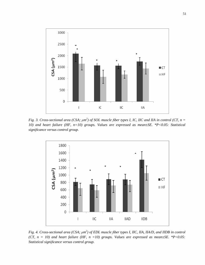
51
Fig. 3. Cross-sectional area (CSA; μm2) of SOL muscle fiber types I, IC, IIC and IIA in control (CT, n =
10) and heart failure (HF, n=10) groups. Values are expressed as mean±SE. *P<0.05: Statistical
significance versus control group.
Fig. 4. Cross-sectional area (CSA; μm2) of EDL muscle fiber types I, IIC, IIA, IIA/D, and IIDB in control
(CT, n = 10) and heart failure (HF, n =10) groups. Values are expressed as mean±SE. *P<0.05:
Statistical significance versus control group.
*
* *
*
* *
*
*
*

52
Table 3. Fiber-type frequency (I, IC, IIC, IIA) in SOL muscle from control (CT) and heart failure (HF)
groups
Group I IC IIC IIA
CT 70,4(68,56-84,62) 7,2 (1,13-11,21) 2,8(2,02-9,72) 14,4 (10,98-22,50)
HF 77,5(66,18-81,87) 3,1(0,18-3,89) 1,7(0,18-2,43)* 17,1(13,20-31,56)
Values are median (maximum – minimum value) * P<0.05; Statistical significance versus control group.
Table 4. Fiber-type frequency (I, IIC, IIDB, IIAD e IIA) in EDL muscle from control (CT) and heart
failure (HF) groups
Group I IIC IIA IIAD IIDB
CT 4,6 (1,3-5,9) 0,9 (-9,7-3,3) 15,1 (7,6-21,5) 5,2 (2,2-11,5) 74,8 (70,6-77,4)
HF 4,0 (1,9-5,5) 0,6 (0-2,3) 16,7 (12,8-20,8) 3,8 (2,9-5,2) 75,2 (69,2-78,9)
Values are median (maximum – minimum value) * P<0.05; Statistical significance versus control group.
The representative SDS-PAGE gel used to quantify MHC isoforms is shown in the Fig. 2
and data from the two groups are summarized in the Tables 5 and 6. SOL muscle showed MHCI
and MHCIIa isoforms; EDL muscle presented MHCI, MHCIIa, MHCIId and MHCIIb isoforms.
The percentage distribution of MHCs was not different between groups (p> 0.05).
Table 5. Percentage distribution of myosin heavy chain (MHC), I and IIa in SOL muscle from control
(CT) and heart failure (HF) groups
Groups MHC I MHC IIA
CT 80.5 (72.7-88.1) 19.5 (11.9-27.3)
HF 78.9 (66.3-89.14) 21.1 (10.9-33.7)
Values are median (maximum – minimum value) * P<0.05; Statistical significance versus control group.

53
Table 6. Percentage distribution of myosin heavy chain (MHC), I, IIa, IId and IIb in EDL muscle from
control (CT) and heart failure (HF) groups.
Groups MHC I MHC IIA MHC IID MHC IIB
CT 6.7 (3.0-16,5) 7.0 (3.5-10.2) 26.2 (21.9-28.3) 57.4 (51.5-10.7)
HF 5.6 (3.2-12.2) 7.6 (3.9-9.9) 24.0 (14.8-31.6) 59.5 (51.4-75.8)
Values are median (maximum – minimum value) * P<0.05; Statistical significance versus control group.
Gene Expression Analysis by RT-qPCR
The measurement of gene expression by RT-qPCR revealed that SOL muscle mRNA
expression of Myogenin was significantly lower in the HF comparing to CT group. EDL muscle
mRNA expression of MyoD was significantly decreased in the HF comparing to CT group (Fig. 5
and 6).
Fig. 5. mRNA abundance estimated by RT-qPCR of MyoD and Myogenin (Myog) in SOL muscle of the
control (CT, n=10) and heart failure (HF, n=10) groups. Data were expressed as minimum, 1st quartile,
median, 3rd
quartile and maximum values. Gene expression was normalized to the reference gene
Hypoxanthine-guanine phosphoribosyltransferase (HPRT) and acidic ribosomal phosphoprotein
(ARBP) from the same RT product. Normalized data are expressed in arbitrary units (AU). *P<0.05:
statistical significance versus control group.

54
Fig. 6. mRNA abundance estimated by RT-qPCR of MyoD and Myogenin (Myog) in EDL muscle of the
control (CT, n=10) and heart failure (HF, n=10) groups. Data were expressed as minimum, 1st quartile,
median, 3rd
quartile and maximum values. Gene expression was normalized to the reference gene
Hypoxanthine-guanine phosphoribosyltransferase (HPRT) and acidic ribosomal phosphoprotein
(ARBP) from the same RT product. Normalized data are expressed in arbitrary units (AU). *P<0.05:
statistical significance versus control group.
Biochemical analysis
SOL muscle in HF group showed increased lipid hydroperoxide, reduced superoxide-
dismutase and glutathione peroxidase activity in comparison to CT group. In EDL muscle, lipid
hydroperoxide, superoxide-dismutase and glutathione peroxidase activity was similar between
groups (Table 7).
Table 7. Oxidative stress in SOL and EDL muscles in control (CT) and heart failure (HF) groups
SOL EDL
CT (n=10) HF (n=10) CT (n=10) HF (n=10)
GSH-Px
(U/mg of tissue)
100.26 ± 19.25 73.56 ± 15.69* 47.16 ± 13.53 50.04 ± 12.69
SOD
(U/mg of tissue)
32.77 ± 3.95 24.66 ± 5.17* 33.3 ± 1.32 31.26 ± 3.60
HL
(nmol/mg
of tissue)
362.78 ± 49.29 429.14 ± 58.02* 460.47 ± 27.11 452.7 ± 42.07
Means ± SD; n - number of animals; GSH-Px - glutathione peroxidase; SOD - superoxide-dismutase; LH
- lipid hydroperoxide. * P<0.05; Statistical significance versus control group.

55
Contractile Properties and Fatigue Resistance Tests
The fall time was prolonged in the HF group comparing to the control group, only in the SOL
muscle (Table 8). However, there were no significant changes in the amplitude, rise time and the fatigue
resistance of EDL and SOL muscles in the HF group comparing to control (Tables 8 and 9).
Table 8. Contractile properties of directly evoked twitches in SOL and EDL muscles in control (CT) and
heart failure (HF) groups
SOL EDL
RT (ms) FT (ms) TA
(g/mm2)
RT (ms) FT (ms) TA (g/mm
2)
CT
(n=10)
28.5
± 5.0
89.5
± 10.3
29.3
± 13.7
12.52
± 0.39
25.68
± 4.41
21.77
± 9.53
HF
(n=10)
30.0
± 6.1
119.5
± 32.0*
25.7
± 11.3
12.50
± 0.25
27.3
± 5.07
24.18
± 5.07
Means ± SD; n - number of animals; RT – Rise time of twitch; FT – Fall time of twitch; TA – Twitch
amplitude * P<0.05; Statistical significance versus control group.
Table 9. Fatigue resistance evaluated from the decrease of 30 direct contractions in SOL and EDL muscle
in control (CT) and heart failure (HF) groups
Fatigue resistance
SOL EDL
CT (n=10) 0.36 (0.47-0.25) 0.36 (0.57-0.22)
HF (n=10) 0.35 (0.71-0.24) 0.34 (0.50-0.27)
Median (maximum and minimum value); n - number of animals; Amplitude of directly evoked tetanus
were expressed as a percentage to the initial amplitude * P<0.05; Statistical significance versus control
group.
Discussion
The aim of this study was to investigate and to compare the morphofunctional characteristics in
SOL and EDL muscles in a monocrotaline-induced heart failure. The major finding in this study is that
monocrotaline-induced heart failure promoted distinct alterations in skeletal muscles with distinct
morphofunctional characteristics. These alterations were higher in oxidative (SOL) than in glycolytic
(EDL) muscles. We showed atrophy in SOL and EDL muscles, oxidative stress, down-regulation in
myogenin mRNA expression and prolongation of fall time of tetanic contraction only in SOL muscle;
down-regulation in MyoD mRNA expression occurred only in EDL muscle.
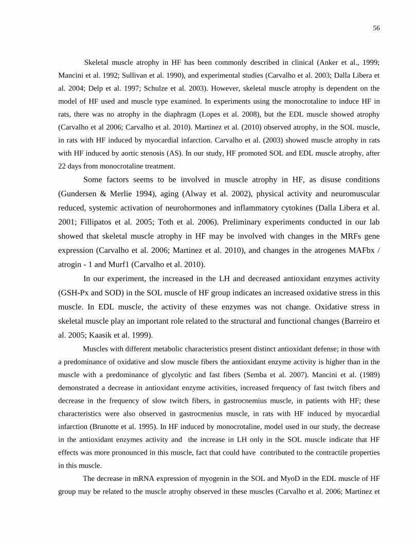
56
Skeletal muscle atrophy in HF has been commonly described in clinical (Anker et al., 1999;
Mancini et al. 1992; Sullivan et al. 1990), and experimental studies (Carvalho et al. 2003; Dalla Libera et
al. 2004; Delp et al. 1997; Schulze et al. 2003). However, skeletal muscle atrophy is dependent on the
model of HF used and muscle type examined. In experiments using the monocrotaline to induce HF in
rats, there was no atrophy in the diaphragm (Lopes et al. 2008), but the EDL muscle showed atrophy
(Carvalho et al 2006; Carvalho et al. 2010). Martinez et al. (2010) observed atrophy, in the SOL muscle,
in rats with HF induced by myocardial infarction. Carvalho et al. (2003) showed muscle atrophy in rats
with HF induced by aortic stenosis (AS). In our study, HF promoted SOL and EDL muscle atrophy, after
22 days from monocrotaline treatment.
Some factors seems to be involved in muscle atrophy in HF, as disuse conditions
(Gundersen & Merlie 1994), aging (Alway et al. 2002), physical activity and neuromuscular
reduced, systemic activation of neurohormones and inflammatory cytokines (Dalla Libera et al.
2001; Fillipatos et al. 2005; Toth et al. 2006). Preliminary experiments conducted in our lab
showed that skeletal muscle atrophy in HF may be involved with changes in the MRFs gene
expression (Carvalho et al. 2006; Martinez et al. 2010), and changes in the atrogenes MAFbx /
atrogin - 1 and Murf1 (Carvalho et al. 2010).
In our experiment, the increased in the LH and decreased antioxidant enzymes activity
(GSH-Px and SOD) in the SOL muscle of HF group indicates an increased oxidative stress in this
muscle. In EDL muscle, the activity of these enzymes was not change. Oxidative stress in
skeletal muscle play an important role related to the structural and functional changes (Barreiro et
al. 2005; Kaasik et al. 1999).
Muscles with different metabolic characteristics present distinct antioxidant defense; in those with
a predominance of oxidative and slow muscle fibers the antioxidant enzyme activity is higher than in the
muscle with a predominance of glycolytic and fast fibers (Semba et al. 2007). Mancini et al. (1989)
demonstrated a decrease in antioxidant enzyme activities, increased frequency of fast twitch fibers and
decrease in the frequency of slow twitch fibers, in gastrocnemius muscle, in patients with HF; these
characteristics were also observed in gastrocmenius muscle, in rats with HF induced by myocardial
infarction (Brunotte et al. 1995). In HF induced by monocrotaline, model used in our study, the decrease
in the antioxidant enzymes activity and the increase in LH only in the SOL muscle indicate that HF
effects was more pronounced in this muscle, fact that could have contributed to the contractile properties
in this muscle.
The decrease in mRNA expression of myogenin in the SOL and MyoD in the EDL muscle of HF
group may be related to the muscle atrophy observed in these muscles (Carvalho et al. 2006; Martinez et

57
al. 2010). These MRFs are involved in the embryonic and adult myogenesis, controlling the myoblasts or
satellite cells activity, respectively. MRFs also are involved in the maintenance of the muscle fibers
phenotype fast and slow; the myogenin is more expressed in slow muscles, whereas MyoD is more
expressed in fast muscles (Hughes et al. 1993; Megeney & Rudnicki, 1995; Murre et al. 1989; Voytik et
al. 1993). Thus, the significant decrease in mRNA expression of MyoD and myogenin in EDL and SOL
muscles may indicate a change in the satellite cells proliferation and differentiation that in turn culminated
with the muscles atrophy.
In the HF, changes in the skeletal muscle phenotypic characteristics frequently occur
(Dalla Libera et al. 2009; Vescovo et al. 1998) with a decrease in the slow and increase in the fast
type fibers, which makes the muscle faster. In our study, there was a significant decrease in the
frequency of hybrid-type IIC fibers in the SOL muscle. This decrease was not accompanied by a
significant increase in the frequency of fiber type IIA. Electrophoresis analysis showed a
tendency to decrease the relative percentage of MHC I and increase in the relative percentage of
MHC IIA. Therefore, we believe that in the acute model of HF used in our study, there was a
tendency of the SOL muscle in acquiring the fast phenotype. This statement is in according to the
gene expression results.
In relation to the contractile properties, there were no changes in the fatigue resistance
and muscular maximum amplitude in the muscles studied in HF. De Sousa et al. (2001) showed
no changes in contractile properties in SOL muscle in rats with HF induced by aortic stenosis.
However, the fall time of contraction in the SOL muscle in HF group was higher compared to the
control group. This finding is in according to Ertunc et al. (2009) which showed that HF leads the
prolongation of twitch fall time only in SOL muscle. Kuno et al. (1988) reported that muscles
with higher proportions of type II fibers (fast twitch and glycolytic metabolism) have higher fall
time compared to muscles with a predominance of type I fibers (slow twitch and oxidative
metabolism). Thus, our findings regarding the SOL muscle confirm the trend in purchasing the
faster phenotype in this muscle, in the monocrotaline-induced HF.
In conclusion, our results indicate that the HF induced by monocrotaline promoted biochemical,
morphological and functional changes, more prominent in the SOL, a slow-twitch muscle. Although
further experiments are required for better determine the mechanisms involved in the pathophysiology of
Heart Failure, our results contributes to the understanding of the muscle-specific changes that occur in this
syndrome.

58
References
Allen DL, Sartorius CA, Sycuro LK, Leinwands LA. (2001) Different pathway regulate of the
skeletal myosin heavy chain. J Biol Chem 274: 43524-43533.
Alway S.E., Degens H., Lowe D.A., Krishnamurthy G. (2002) Increased myogenic repressor Id
mRNA and protein levels in hindlimb muscles of aged rats. Am. J. Physiol. Regul. Integr.Comp.
Physiol. 282:R411–R422.
Anker SD, Ponikowski PP, Clark AL, Leyva F, Rauchhaus M, Kemp M, Teixeira MM, Hellewell
PG, Hooper J, Poole-Wilson PA, Coats AJ (1999) Cytokines and neurohormones relating to body
composition alterations in the wasting syndrome of chronic heart failure. Eur Heart J 20:683-693
Barclay CJ (1992) Effects of fatigue on rate of isometric force development in mouse fast- and
slow- twitch muscle Am J Physiol 263 (Cell Physiol. 32): C1065-C1072.
Barreiro E, de la Puente B, Minguella J, Corominas JM, Serrano S, Hussain SNA, Gea, J (2005)
Oxidative stress and respiratory muscle dysfunction in severe chronic obstructive pulmonary
disease. Am J Respir Crit Care Med 171:1116–1124.
Bärr A, Pette D (1988) Three fast myosin heavy chain in adult rat skeletal muscle. FEBS Lett
233:153-155.
Brooke MH, Kaiser KK (1970) Three “myosin adenosine triphosphatase” systems: the nature of
their pH lability and sulfhydryl dependence. J Histochem Cytochem 18:670-672.
Brunotte F, Thompson CH, Adamopoulos S, Coats A, Unitt J, Lindsay D, Kaklamanis L, Radda
GK, Rajagopalan B (1995) Rat skeletal muscle metabolism in experimental heart failure: effects
of physical training. Acta Physiol Scand. 154:439-47.
Campos GE, Luecke TJ, Wendeln HK, Toma K, Hagerman FC, Murray TF, Ragg KE, Ratamess
NA, Kraemer WJ, Staron RS (2002) Muscular adaptations in response to three different
resistance-training regimens: specificity of repetition maximum training zones. Eur J Appl
Physiol 88:50–60.
Carvalho RF, Cicogna AC, Campos GE, De Assis JM, Padovani CR, Okoshi MP, Dal Pai-Silva
M (2003) Myosin heavy chain expression and atrophy in rat skeletal muscle during transition
from cardiac hypertrophy to heart failure. Int J Exp Pathol. 84:201-6.
Carvalho RF, Cicogna AC, Campos GE, Lopes FS, Sugizaki MM, Nogueira CR, Dal Pai-Silva M
(2006) Heart failure alters MyoD and MRF4 expression in rat skeletal muscle. Int J Exp Pathol
87:219-225.
Carvalho RF, Castan EP, Coelho CA, Lopes FS, Almeida FL, Michelin A, de Souza RW, Araújo
JP Jr, Cicogna AC, Dal Pai-Silva M (2010) Heart failure increases atrogin-1 and MuRF1 gene
expression in skeletal muscle with fiber type-specific atrophy J Mol Histol 41:81-7.

59
Coats AJS, Clark AL, Piepoli M, Volterrani M, Poole-Wilson PA. (1994) Symptoms and quality
of life in heart failure. The muscle hypothesis. Br Heart J 72:36–9.
Coirault C, Guellich A, Barbry T, Samuel JL, Riou B, Lecar-pentier Y (2007) Oxidative stress of
myosin contributes to skeletal muscle dysfunction in rats with chronic heart failure. Am J Physiol
Heart Circ Physiol 292:H1009–H1017.
Dalla Libera L, Sabbadini R, Renken C, Ravara B, Sandri M, Betto R, Angelini A, Vescovo G.
(2001) Apoptosis in the skeletal muscle of rats with heart failure is associated with increased
serum levels of TNF-alpha and sphingosine. J Mol Cell Cardiol 33:1871-1878.
Dalla Libera L, Ravara B, Volterrani M, Gobbo V, Della Barbera M, Angelini A. (2004)
Beneficial effects of GH/IGF-1 on skeletal muscle atrophy and function in experimental heart
failure. Am J Physiol Cell Physiol 286:C138-144.
Dalla Libera L, Ravara B, Gobbo V, Betto DD, Germinario E, Angelini A, Vescovo G. (2005)
Skeletal muscle myofibrillar protein oxidation in heart failure and the protective of Carvedilol. J.
Molecular and Cellular Cardiology 38:803-807.
Dalla Libera L, Ravara B, Gobbo V, Betto DD, Germinario E, Angelini A, Evangelista S,
Vescovo G. (2010) Skeletal muscle in chronic right heart failure in rat: can different beta-
blockers prevent it to the same degree? Int J Cardiol 143:192-199.
De Sousa E, Veksler V, Bigard X, Mateo P, Ventura-Clapier R (2000) Heart failure affects
mitochondrial but not intrinsic properties of skeletal muscle. Circulation 102:1847-1853.
De Sousa E, Veksler V, Bigard X, Mateo P, Serrurier B, Ventura-Clapier R (2001) Dual
influence of disease and increased load on diaphragm muscle in heart failure. J Mol Cell Cardiol
33:699-710.
Delp M, Duan C, Mattson JP, Musch TI (1997) Changes in skeletal muscle biochemistry and
histology relative to fiber type in rats with heart failure. J Appl Physiology 83:1291-1299.
Drexler H (1992) Skeletal muscle failure in heart failure. Circulation. 85:1621–1623.
Ekmark M, Gronevik E, Schjerling P, Gundersen K (2003) Myogenin induces higher oxidative
capacity in pre-existing mouse muscle fibres after somatic DNA transfer. J Physiol 548
(Pt1):259-269.
Ertunc M, Sara Y, Onur R (2009) Differential Contractile Impairment of fast-and slow-twitch
skeletal muscle in a rat model of doxorubicin-induced congestive heart failure. Pharnacology
84:240-248.
Ewing JF & Janero DR (1995). Microplate superoxide dismutase assay employing a
nonenzymatic superoxide generation. Annals of Biochemistry 232: 243-248.
60
Feuers, RJ, (1998) The effects of dietary restriction on mitochondrial dysfunction in aging.
Annals of the New York Academy of Sciences 125, 192–201.
Filippatos G.S., Anker S.D., Kremastinos. (2005) Pathophysiology of peripheral muscle wasting
in cardiac cachexia. Curr Opin Clin Nutr Metab Care 8(3):249-254.
Gallacci M, Oliveira AC, (1994) Pre- and postsynaptic mechanisms involved in tetanic fade
induced by pancuronium in the isolated rat muscle. Pharmacology 49, 265–270.
Gundersen K, Merlie JP (1994) Id-1 as a possible transcriptional mediator of muscle disuse
atrophy. Proc Natl Acad Sci 91:3647–3651.
Guth L, Samaha FJ (1969) Qualitative differences between acto myosin ATPase of slow and fast
mammalian muscle. Exp Neurol 25:138–152.
Hopkins J, Tudhope GR (1973) Glutathione peroxidase in human red cells in health and disease.
British Journal of Haematology 25:563-575.
Hughes SM, Taylor JM, Tapscott SJ, Gurley CM, Carter WJ, Peterson CA (1993) Selective
accumulation of MyoD and Miogenin mRNAs in fast and slow muscle is controlled by
innervation and hormones. Development 118:1137-1147.
Hughes S.M., Chi M.M., Lowry O.H., Gundersen K. (1999) Myogenin induces a shift of enzyme
activity from glycolytic to oxidative metabolism in muscles of transgenic mice. J Cell Biol
145:633-642.
Jiang ZY, Woollard ACS, Wolf SP (1991) Lipid hydroperoxide measurement by oxidation of
Fe2+ in the presence of xylenol orange. Comparison with the TBA assay and an iodometric
method. Lipids, 26: 853-856.
Kaasik A, Minajeva A, De Sousa E, Ventura-Clapier R, Veksler V. (1999) Nitric oxide inhibits
cardiac energy production via inhibition of mitochondrial creatine kinase. FEBS Lett 444:75–77.
Kinugawa S, Tsutsui H, Hayashidani S, Ide T, Suematsu N, Satoh S, Utsumi H, Takeshita A
(2000) Treatment with dimethylthiourea prevents left ventricular remodeling and failure after
experimental myocardial infarction in mice: role of oxidative stress. Circ Res 87:392–398.
Kuno S, Katsuta S, Anno I, Matsumoto K, Akisada M. (1988) Relationship between MR
relaxation time and muscle fiber composition. Radiology 169:567-68.
Lapu-Bula, Ofili E (2007) From hypertension to heart failure: role of nitric oxide-mediated
endothelial dysfunction and emerging insights from myocardial contrast echocardiography. Am J
Cardiol 26:7–14.
Leineweber K, Brandt K, Wludyka B, Beilfuss A, Ponicke K, Heinroth-Hoffmann I, Brodde OE
(2002) Ventricular hypertrophy plus neurohumoral activation is necessary to alter the cardiac
beta-adrenoceptor system in experimental heart failure. Circ Res 91:1056–1062

61
Lipkin DP, Jones DA, Round JM, Poole-Wilson PA (1988) Abnormalities of skeletal muscle in
patients with chronic heart failure. Int J Cardiol 18:187-195.
Lopes FS, Carvalho RF, Campos GER, Sugizaki MM, Padovani CR, Nogueira CR, Cicogna AC,
Dal Pai-Silva M (2008) Down-regulation of MyoD gene expression in rat diaphragm muscle with
heart failure. Int J Exp Path, 89:216–222.
Mancini DM, Coyle E, Coggan A, Beltz J, Ferraro N, Montain S, Wilson JR (1989) Contribution
of intrinsic skeletal muscle changes to 31P NMR skeletal muscle metabolic abnormalities in
patients with chronic heart failure. Circulation 80:1338-46.
Mancini DM, Walter G, Reichek N, Lenkinski R, McCully KK, Mullen JL, Wilson JR (1992)
Contribution of skeletal muscle atrophy to exercise intolerance and altered muscle metabolism in
heart failure Circulation 85:1364-1373.
Martinez PF, Okoshi K, Zornoff LA, Carvalho RF, Oliveira Junior SA, Lima AR, Campos DH,
Damatto RL, Padovani CR, Nogueira CR, Dal Pai-Silva M, (2010) Okoshi MP. Chronic heart
failure-induced skeletal muscle atrophy, necrosis, and changes in myogenic regulatiory factors
Med Sci Monit. 16:BR374-383.
Megeney LA, Rudnicki MA (1995) Determination versus differentiation and the MyoD family of
transcription factors. Biochem Cell Biol 73:723-732.
Murre C, Mccaw PS, Vaessin H, Caudy M, Jan LY, Yan JN, Cabrera CV, Buskin JN, Hauschka
SD, Lassar AB, Weintraub H, Baltimore D (1989) Interactions between heterologous helix-loop-
helix proteins generate complexes that bind specifically to a common DNA sequence. Cell
58:537-544.
Nishiyama Y, Ikeda H, Haramaki N, Yoshida N, Imaizumi T (1998) Oxidative stress is related to
exercise intolerance in patients with heart failure. Am Heart J 135:115–120.
Parker MH, Seale P, Rudnicki MA (2003) Looking back to the embryo: defining transcriptional
networks in adult myogenesis. Nat Rev Genet 4:497-507.
Powers SK, Ji LL, Leeuwenburgh C (1999) Exercise training-induced alterations in skeletal
muscle antioxidant capacity: a brief review. Med Sci Sports Exerc. 31:987–997.
Reindel JF, Ganey JG, Wagner Rf, Slocombe RF, Roth AR (1990) Development of morphologic,
hemodynamic, and biochemical changes in lung of rats given monocrotaline pirrole. Toxicol.
Appl Pharmacol 106:179-200.
Schulze P.C, Gielen S, Adams V, Linke A, Möbius-Winkler S, Erbs S, Kratzsch J, Hambrecht R,
Schuler G. (2003) Muscular levels of proinflammatory cytokines correlate with a reduced
expression of insulin-like growth factor-1 in chronic heart failure. Basic Res Cardiol 98:267-274.
Semba RD, Lauretani F, Ferrucci L (2007) Carotenoids as protection against sarcopenia in older
adults. Archives of Biochemistry and Biophysics, 458:141-145.

62
Siu PM, Donley DA, Bryner RW, Alway SE (2004) Myogenin and oxidative enzyme gene
expression levels are elevated in rat soleus muscles after endurance training. J Appl Physiol
97:277-285.
Sullivan MJ, Green HJ, Cobb FR (1990) Skeletal muscle biochemistry and histology in
ambulatory patients with long-term heart failure. Circulation 81:518-527.
Spangenburg EE, Talmadge RJ, Musch TI, Pfeifer PC, McAllister RM, Williams JH (2002)
Changes in skeletal muscle myosin heavy chain isoform content during congestive heart failure.
Eur J Appl Physiol 87:182-186.
Staron RS, Kraemer WJ, Hikida RS, Fry AC, Murray JD, Campos GE (1999) Fiber type
composition of four hindlimb muscles of adult Fisher 344 rats. Histochem Cell Biol 111:117-123.
Toth MJ, Palmer BM, Lewinter MM (2006) Effect of heart failure on skeletal muscle
myofibrillar protein content, isoform expression and calcium sensitivity. Int J Cardiol 107:211-
219.
Tsutsui H, Ide T, Hayashidani S, Suematsu N, Shiomi T, Wen J, Nakamura K, Ichikawa K,
Utsumi H, Takeshita A (2001) Enhanced Generation of Reactive Oxygen Species in the Limb
Skeletal Muscles From a Murine Infarct Model of Heart Failure Circulation; 104:134-136.
Tsutsui H, Kinugawa S, Matsushima S (2008) Oxidative stress and mitochondrial DNA damage
in heart failure Suppl A: A31-A37.
Van Albada ME, Bartelds B, Wijnberg H, Mohaupt S, Dickinson MG, Schoemaker RG, Kooi
KA, Gerbens F, Berger RM (2010) Gene expression profile in flow-associated pulmonary arterial
hypertension with neointimal lesions. Am J Physiol Lung Cell Mol Physiol 298:L483–L491
Vandesompele J, De Preter K, Pattyn F, Poppe B, Van Roy N, De Paepe A, Speleman F (2002)
Accurate normalization of Real-Time quantitative RT-PCR by geometric averaging of multiple
internal control genes. Genome Biol 3:34.
Vescovo G, Harding SE, Jones M, Dalla Libera L, Pessina AC, Poole-Wilson PA (1989)
Contractile abnormalities of single right ventricular myocytes isolated from rats with right
ventricular hypertrophy. J Mol Cell Cardiol 21(Suppl 5):103–111
Vescovo G, Ceconi C, Bernocchi P, Ferrari R, Carraro U, Ambrosio GB, Libera LD (1998)
Skeletal muscle myosin heavy chain expression in rats with monocrotaline-induced cardiac
hypertrophy and failure. Relation to blood flow and degree of muscle atrophy. Cardiovasc Res
39:233–241
Vescovo G, Ravara B, Libera LD (2008) Skeletal muscle myofibrillar protein oxidation and
exercise capacity in heart failure. Basic Res Cardiol 103:285-290.

63
Voytik SL, Przyborski M, Badylak SF, Konieczny SF (1993) Differential expression of muscle
regulatory factor genes in normal and denervated adult rat hindlimb muscle. Dev Dynam
198:214-224.
Zar JH (1999) Biostatistical analysis, 4thed. Prentice-Hall New Jersey, 633p.