Caso Clínico Case Report - scielo.mec.pt · Revista Portuguesa de Pneumologia887 Vol XIV N.º 6...
5
Revista Portuguesa de Pneumologia 887 Vol XIV N.º 6 Novembro/Dezembro 2008 Caso Clínico Case Report Resumo Os autores apresentam os aspectos na tomografia com- putadorizada de alta resolução de um doente com he- morragia pulmonar difusa por leptospirose. O achado predominante consistiu de opacidades em vidro des- polido difusas, com espessamento de septos interlobu- lares sobrepostos, resultando no padrão de crazy-pa- ving. O doente morreu, e na necrópsia foi observada extensa hemorragia alveolar, com sangue infiltrando os septos interlobulares. É realçada a correlação entre os Abstract The authors present the high-resolution CT find- ings of a patient with diffuse pulmonary hemor- rhage due to leptospirosis. The main finding consisted of extensive ground-glass opacities superimposed on mild interlobular septal thic- kening, resulting in the appearance termed “cra- zy-paving”. The patient died and the necropsy showed extensive haemorrhage filling the air- space and blood infiltrating the interlobular sep- Padrão de crazy-paving na leptospirose pulmonar: Correlação entre a tomografia computadorizada de alta resolução e os achados anatomopatológicos Leptospirosis of the lung presenting with crazy-paving pattern: Correlation between the high-resolution CT and pathological findings Recebido para publicação/received for publication: 07.12.27 Aceite para publicação/accepted for publication: 08.07.24 Edson Marchiori 1 Taisa Davaus Gasparetto 2 Dante L Escuissato 3 Gláucia Zanetti 4 1 Professor Titular e Chefe do Departamento de Radiologia da Universidade Federal Fluminense (UFF). Coordenador Adjunto do Curso de Pós-Graduação em Radiologia da Universidade Federal do Rio de Janeiro (UFRJ) / Full Professor, Head, Department of Radiology of the University Fluminense (UFF). Associate Coordinator, Post-Graduate Course in Radiology, University of Rio de Janeiro (UFRJ). 2 Médica Pós-Graduanda do Departamento de Radiologia da UFF / Post-Graduate Doctor, Department of Radiology, UFF. 3 Professor Adjunto de Radiologia da Universidade Federal do Paraná, Curitiba, Brasil / Associate Professor of Radiology, University of Paraná, Brazil. 4 Doutoranda em Radiologia da UFRJ / PhD in Radiology, UFRJ. Departamento de Radiologia da Universidade Federal do Paraná, Curitiba, Brasil. Director: Prof. Dante L. Escuissato. Rua General Carneiro, 181. CEP 80.000-000. Curitiba, Paraná, Brasil / Departments of Radiology of the University Fluminense (EM, TD) and University of Rio de Janeiro (EM, GZ), Rio de Janeiro, Brazil, and University of Paraná (DLE), Curitiba, Brazil Departamento de Radiologia da Universidade Federal Fluminense (UFF) Director : Prof. Edson Marchiori Av. Marquês do Paraná, 330. Centro. Niterói – Rio de Janeiro – Brasil Serviço de Radiodiagnóstico do Hospital Universitário Clementino Fraga Filho (HUCFF), da Universidade Federal do Rio de Janeiro (UFRJ). Director: Prof. Hilton Koch Av. Brigadeiro Trompowsky, s/n. Ilha do Fundão. Rio de Janeiro – Brasil. Correspondência / Correspondence address: Edson Marchiori – Rua Thomaz Cameron, 438. Valparaiso. CEP 25685.120 Petrópolis, Rio de Janeiro, Brasil E-mail: [email protected]
Transcript of Caso Clínico Case Report - scielo.mec.pt · Revista Portuguesa de Pneumologia887 Vol XIV N.º 6...

R e v i s t a P o r t u g u e s a d e P n e u m o l o g i a 887
Vol XIV N.º 6 Novembro/Dezembro 2008
Caso ClínicoCase Report
ResumoOs autores apresentam os aspectos na tomografia com-putadorizada de alta resolução de um doente com he-morragia pulmonar difusa por leptospirose. O achado predominante consistiu de opacidades em vidro des-polido difusas, com espessamento de septos interlobu-lares sobrepostos, resultando no padrão de crazy-pa-ving. O doente morreu, e na necrópsia foi observada extensa hemorragia alveolar, com sangue infiltrando os septos interlobulares. É realçada a correlação entre os
AbstractThe authors present the high-resolution CT find-ings of a patient with diffuse pulmonary hemor-rhage due to leptospirosis. The main finding consisted of extensive ground-glass opacities super imposed on mild interlobular septal thic-kening, resulting in the appearance termed “cra-zy-paving”. The patient died and the necropsy showed extensive haemorrhage filling the air-space and blood infiltrating the interlobular sep-
Padrão de crazy-paving na leptospirose pulmonar: Correlação entre a tomografia computadorizada de alta resolução e os achados anatomopatológicos
Leptospirosis of the lung presenting with crazy-paving pattern: Correlation between the high-resolution CT and pathological findings
Recebido para publicação/received for publication: 07.12.27Aceite para publicação/accepted for publication: 08.07.24
Edson Marchiori1
Taisa Davaus Gasparetto2
Dante L Escuissato3
Gláucia Zanetti4
1 Professor Titular e Chefe do Departamento de Radiologia da Universidade Federal Fluminense (UFF). Coordenador Adjunto do Curso de Pós-Graduação em Radiologia da Universidade Federal do Rio de Janeiro (UFRJ) / Full Professor, Head, Department of Radiology of the University Fluminense (UFF). Associate Coordinator, Post-Graduate Course in Radiology, University of Rio de Janeiro (UFRJ).
2 Médica Pós-Graduanda do Departamento de Radiologia da UFF / Post-Graduate Doctor, Department of Radiology, UFF. 3 Professor Adjunto de Radiologia da Universidade Federal do Paraná, Curitiba, Brasil / Associate Professor of Radiology, University of Paraná, Brazil.4 Doutoranda em Radiologia da UFRJ / PhD in Radiology, UFRJ. Departamento de Radiologia da Universidade Federal do Paraná, Curitiba, Brasil. Director: Prof. Dante L. Escuissato. Rua General Carneiro, 181. CEP 80.000-000.
Curitiba, Paraná, Brasil / Departments of Radiology of the University Fluminense (EM, TD) and University of Rio de Janeiro (EM, GZ), Rio de Janeiro, Brazil, and University of Paraná (DLE), Curitiba, Brazil
Departamento de Radiologia da Universidade Federal Fluminense (UFF) Director : Prof. Edson Marchiori Av. Marquês do Paraná, 330. Centro. Niterói – Rio de Janeiro – Brasil Serviço de Radiodiagnóstico do Hospital Universitário Clementino Fraga Filho (HUCFF), da Universidade Federal do Rio de Janeiro (UFRJ). Director: Prof. Hilton Koch
Av. Brigadeiro Trompowsky, s/n. Ilha do Fundão. Rio de Janeiro – Brasil. Correspondência / Correspondence address: Edson Marchiori – Rua Thomaz Cameron, 438. Valparaiso. CEP 25685.120 Petrópolis, Rio de Janeiro, Brasil E-mail: [email protected]
Pneumologia 14-6 - Miolo - 4ª PROVA.indd 887Pneumologia 14-6 - Miolo - 4ª PROVA.indd 887 11-11-2008 9:52:3711-11-2008 9:52:37

R e v i s t a P o r t u g u e s a d e P n e u m o l o g i a888
Vol XIV N.º 6 Novembro/Dezembro 2008
achados anatomopatológicos e na tomografia compu-tadorizada de alta resolução.
Rev Port Pneumol 2008; XIV (6): 887-891
Palavras-chave: Leptospirose, hemorragia pulmonar, padrão de crazy-paving, tomografia computadorizada de alta resolução.
ta. The correlation between the high-resolution CT and pathological findings is emphasised.
Rev Port Pneumol 2008; XIV (6): 887-891
Key-words: Leptospirosis, pulmonary haemorrhage, crazy-paving pattern, high resolution CT.
IntroductionLeptospirosis is a zoonosis caused by Lep-tospira interrogans1. Humans become infe-cted when mucous membranes or abraded skin come into direct contact with the urine of infected animals, especially rats. The ex-posure to soil, water, or other matter con-tamined with the infected urine is also com-monly related as a cause of human infection by the L. interrogans1,2,3.Leptospirosis produces several clinical find-ings, but two presentations are usually seen. In the less severe and generally non-fatal form, often called anicteric lesptospirosis, the illness commonly begins abruptly and presents with headache, myalgias, conjunc-tival suffusion, fever, nausea, vomiting, and meningismus. In addition to these features, the more severe form of leptospirosis, called icteric leptospirosis or Weill's disease, is commonly associated with jaundice, renal impairment, and major haemorrhagic com-plications1. Pulmonary involvement is fre-quent, but often mild and of uncomplicated clinical significance. However, over the last two decades, an increasing number of cases with pulmonary haemorrhage as a promi-nent feature have been reported2.
The diagnosis of leptospirosis is based on the clinical findings, exposure to infested animals in endemic areas, and positive sero-logical tests3. Although the chest radio-graphic findings of this entity have been described, the high-resolution CT features of this infection were rarely demonstrated1,4. In addition, the “crazy paving” pattern and the correlation of pathological and high-resolution CT findings were not previously described in patients with leptospirosis in-volving the lungs.The authors aim to report a case of pulmo-nary haemorrhage secondary to leptospiro-sis, emphasising the presence of “crazy pa-ving” pattern of the high-resolution CT, as well as the correlation between this finding and the pathological features.
Case reportA 31-year-old female patient presented with a five-days history of fever, rigors and severe calf and thigh pain. Two days before admis-sion she began to expectorate small amounts of blood and became progressively breath-less. The patient had a history of immersion in dirty water during a recent urban flood,
Padrão de Crazy-paving na Leptospirose Pulmonar: Correlação entre a tomografia computadorizada de alta resolução e os achados anatomopatológicos
Edson Marchiori, Taisa Davaus Gasparetto, Dante L Escuissato, Gláucia Zanetti
Pneumologia 14-6 - Miolo - 4ª PROVA.indd 888Pneumologia 14-6 - Miolo - 4ª PROVA.indd 888 11-11-2008 9:52:3711-11-2008 9:52:37
R e v i s t a P o r t u g u e s a d e P n e u m o l o g i a 889
Vol XIV N.º 6 Novembro/Dezembro 2008
and reported the presence of rodents in the surroundings of her home. On admission, she was febrile, dyspneic, and had cyanotic extremities.Chest radiographs revealed extensive bila-teral alveolar infiltrates in the lower thirds of lungs. High-resolution CT scan demons-trated diffuse ground-glass attenuation in both lungs, with superimposed mild inter-lobular septal thickening in the antero-supe-rior regions (“crazy paving” pattern) (Fig. 1).Blood cultures demonstrated growing L. in-terrogans serovar Copenhageni, and treat-ment was initiated with intravenous benzil-
penicillin and hydrocortisone, associated with O2 by a nasal catheter. The patient also required blood transfusion. Despite treat-ment, she developed worsening dyspnea and massive haemoptysis, requiring intuba-tion and positive pressure ventilation. Two days after admission, she had a cardiac ar-rest and died despite reanimation attempts.The necropsy showed congestion and hae-morrhage in the kidneys, liver, spleen and lungs. Leptospiras were found in the kidney’s sections. In the lungs, petechiae were ob-served on the pleural surfaces, and the cut sections of the parenchyma were grossly
Fig. 1 – 31 year-old female patient with leptospirosis. High-resolution CT at the level of the aortic arch (A) and lower pulmo-nary veins (B) demonstrates bilateral areas of ground-glass attenuation with superimposed interlobular septal thickening (“crazy-paving” pattern)
BA
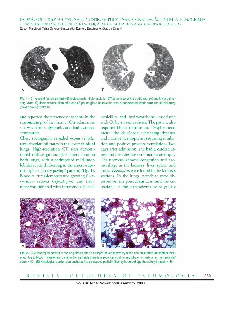
Fig. 2 – (A) Histological section of the lung shows diffuse fi lling of the air-spaces by blood and an interlobular septum thick-ened due to blood infi ltration (arrows). In the right side there is a secondary pulmonary lobule normally aired (hematoxylin/eosin × 40). (B) Histological section demonstrates the air-spaces partially fi lled by haemorrhage (hematoxylin/eosin × 40)
A B
Padrão de Crazy-paving na Leptospirose Pulmonar: Correlação entre a tomografia computadorizada de alta resolução e os achados anatomopatológicosEdson Marchiori, Taisa Davaus Gasparetto, Dante L Escuissato, Gláucia Zanetti
Pneumologia 14-6 - Miolo - 4ª PROVA.indd 889Pneumologia 14-6 - Miolo - 4ª PROVA.indd 889 11-11-2008 9:52:3711-11-2008 9:52:37

R e v i s t a P o r t u g u e s a d e P n e u m o l o g i a890
Vol XIV N.º 6 Novembro/Dezembro 2008
haemorrhagic. Histologically, there was hae-morrhage filling the air-spaces and infiltration of the interlobular septa by blood (Fig. 2).
DiscussionThe patients with leptospirosis usually manifest two main clinical presentations. The milder anicteric form accounts for 90-95% of cases, and those patients commonly refer fever, myalgia, conjunctival suffusion and mild gastrointestinal upset. The other 5-10% of the patients presents the icteric form, also known as Weil’s syndrome, which is characterised by jaundice, renal dysfunc-tion, haemorrhagic manifestations, pulmo-nary involvement, and a high mortality rate2. Our case presented with the lather form on the infection, with involvement of the lungs, kidneys, spleen and liver.Im et al.5 described the chest radiographic findings in 58 patients with leptospirosis. The predominant abnormality consisted of poorly defined and diffuse ground-glass opacities, which were seen in 27% of cases. They postulated that this pattern was a re-sult of pulmonary haemorrhage. Marchiori and Müller4 presented the high-resolution CT findings in a series of five patients with leptospirosis involving the lungs. All pa-tients showed ground-glass opacities and four presented areas of air-space consolida-tion. The authors suggested that those fin-dings were secondary to pulmonary hemor-rhage, which was seen in bronchoalveolar lavage and autopsy studies. Although ground-glass opacities and air-space con-solidations may be seen in patients with dif-fuse pulmonary haemorrhage, these find-ings are usually non-specific. The differential dia gnosis should include cardiogenic and
non-cardiogenic pulmonary edema, as well as infectious pneumonia4.The “crazy-paving” pattern is described in the high-resolution CT scan as areas of ground-glass attenuation with superimposed interlobular septal thickening. This pattern was initially described by Murch and Carr6 as pathognomonic of pulmonary alveolar proteinosis. Since then, several diseases, such as bronchioloalveolar carcinoma, sarcoido-sis, nonspecific interstitial pneumonia, cryp-togenic organising pneumonia and infec-tious diseases have demonstrated this finding, making the differential diagnosis by high-resolution CT sometimes difficult7,8. To our knowledge, the “crazy-paving” pattern was not previously described in patients with leptospirosis.The macroscopical study of the lungs of pa-tients with leptospirosis generally demons-trates extensive pulmonary haemorrhage, with numerous foci of bleeding with diffe-rent sizes9,10. Some patients may show pleu-ral-based lung lesions with pyramidal form, suggesting haemorrhagic infarcts9,10. The his-tological sections usually demonstrate pul-monary congestion, several foci of interstitial and intra-alveolar bleeding, and pulmonary oedema, with different degrees of severity3,9,10. In the present case, the histological examina-tion demonstrated haemorrhage filling the air-spaces, which were seen as areas of ground-glass attenuation on the high-resolution CT, and infiltration of the interlobular septa by blood, justifying the interlobular septal thick-ening seen on the CT scan.In conclusion, leptospirosis should be in-cluded in the differential diagnosis of lung lesions presenting with “crazy-paving” pat-tern on the high-resolution CT, particularly in patients with suggestive epidemiological
Padrão de Crazy-paving na Leptospirose Pulmonar: Correlação entre a tomografia computadorizada de alta resolução e os achados anatomopatológicos
Edson Marchiori, Taisa Davaus Gasparetto, Dante L Escuissato, Gláucia Zanetti
Pneumologia 14-6 - Miolo - 4ª PROVA.indd 890Pneumologia 14-6 - Miolo - 4ª PROVA.indd 890 11-11-2008 9:52:3911-11-2008 9:52:39

R e v i s t a P o r t u g u e s a d e P n e u m o l o g i a 891
Vol XIV N.º 6 Novembro/Dezembro 2008
history. In addition, the study of the histo-logical sections of our case demonstrated that the areas of ground-glass attenuation are related to the filling of the air-spaces by haemorrhage and the interlobular septal thickening is secondary to infiltration of those septa by blood.
Bibliography1. Luks AM, Lakshminarayanan S, Hirsch mann JV. Leptospirosis presenting as diffuse alveolar hemorrhage: case report and literature review. Chest 2003; 123(2):639-43.2. Arokianathan D, Trower K, Pooboni S, Sosnowski A, Moss P, Thaker H. Leptospirosis: a case report of a pa-tient with pulmonary haemorrhage successfully ma-naged with extra corporeal membrane oxygenation. J Infect 2005;50(2):158-62.3. Carvalho JE, Marchiori ES, Guedes e Silva JB, Souza Netto BA, Tavares W, Paula AV. Pulmonary compro-misse in leptospirosis. Rev Soc Bras Med Trop 1992; 25(4): 21-30.
4. Marchiori E, Muller NL. Leptospirosis of the lung: high-resolution computed tomography findings in five patients. J Thorac Imaging 2002;17(2):151-153.5. Im JG, Yeon KM, Han MC, Kim CW, Webb WR, Lee JS, et al. Leptospirosis of the lung: radiographic findings in 58 patients. AJR 1989; 152(5):955-9.6. Murch CR, Carr DH. Computed tomography ap-pearances of pulmonary alveolar proteinosis. Clin Ra-diol 1989; 40(3):240-3.7. Johkoh T, Itoh H, Müller NL, Ichikado K, Nakamura H, Ikezoe J, et al. Crazy paving appearance at thin-sec-tion CT: spectrum of disease and pathologic findings. Radiology 1999; 211(1):155-60.8. Murayama S, Murakami J, Yabuuchi H, Soeda H, Masuda K. Crazy paving appearance on high resolution CT in various diseases. J Comput Assist Tomogr 1999; 23(5): 749-52.9. Nicodemo AC, Duarte MI, Alves V, Takakura CFH, Santos RTM, Nicodemo EL. Lung lesions in human leptospirosis: microscopic, immunohistochemical, and ultrastructural features related to thrombocytopenia. Am J Trop Med Hyg 1997; 56(2):181-7.10. Arean VM. The pathologic anatomy and pathoge-nesis of fatal human leptospirosis (Weil’s disease). Am J Pathol 1962;40: 393-423.
Padrão de Crazy-paving na Leptospirose Pulmonar: Correlação entre a tomografia computadorizada de alta resolução e os achados anatomopatológicosEdson Marchiori, Taisa Davaus Gasparetto, Dante L Escuissato, Gláucia Zanetti
Pneumologia 14-6 - Miolo - 4ª PROVA.indd 891Pneumologia 14-6 - Miolo - 4ª PROVA.indd 891 11-11-2008 9:52:3911-11-2008 9:52:39