Child with Syncope Criança com síncope Niño con...
38
Child with Syncope Criança com síncope Niño con síncope Andrés Ricardo Pérez Riera, MD Chief of Electovectorcardiography Sector - Cardiology Discipline - ABC Faculty – ABC Foundation – Santo André – São Paulo – Brazil [email protected]
Transcript of Child with Syncope Criança com síncope Niño con...
Child with SyncopeCriança com síncope
Niño con síncope
Andrés Ricardo Pérez Riera, MDChief of Electovectorcardiography Sector - Cardiology Discipline - ABC Faculty – ABC Foundation – Santo André – São Paulo – [email protected]
Case reportBasic information: Name: RSL, 3yo, masculine, Caucasian, been born in Santo André, São Paulo, Brazil. Second son of an offspring of two. Chief complaints: his Mother refer that three days ago he had an event manifested by fainting spells (syncope) triggers by an exposure to loud noises auditory stimuli (telephone ring). Common childhood diseases absent. like, gastro-intestinal infections, respiratory illness, diabetes, jaundice, ingestion or inhalation of foreign bodies or others.Prenatal History: without important intrauterine, or perinatal events, there is no history of drug intake, x-ray exposure and viruses during first trimester or pregnancy. Parents not related by blood.Natal History: full term normal delivery supervised by doctor, child cried immediately after birth and was pink in colour. Post Natal History: 3,5kg to the birth, 49cm high, Normal Apgar score (8.). There is no history of cyanosis, jaundice. Refer two episodes of Seizures the last Month without explanation.Normal developmental history related to gross motor, fine, language.Family history: Positivev family history of cardiac arrest and sudden death, at a young age, in his major brother at 9 yo.Socio economic status: LowDietary History: Normal. Normal duration of breast feeding, Physical: Normal. Only excessive bradycardia for his age.
Informaciones básicas: nombre RSL, masculino, blanco, nascido en Santo André, SãoPaulo Brasii. Seundo hijo de una prole de dos.
Queja principal: la madre refiere que hace tres dias el niño tuvo un evento manifestado porpérdida súbita de la conciencia (síncope) desencadenado por un ruido alto de un llamadotelefónico. Antecedentes negativos para enfermedades comunes de la infancia como infecciones gastro-intestinales y enfermedades respiratórias. Tambien negativo para diabetes, ictericia, ingestion o inalación de cuerpos extraños o otros.
História prenatal sin datos de importancia en referencia a eventos intrauterinos o perinatales. No existe história de ingestion de drogas o exposición a Rx, o virosis materna durante el primer trimesntre gestacional. Padres no consanguineos.
Historia al Nascimiento: niño nascido a termino, atendido por médico, lloró inmediatamentedespués del nascimiento y el color de la piel era rosado.História post-natal: con 3,5kg al nascimiento, 49cm de altura y con una escala de Apgar de 8. Ausencia de cianose, ictericia. Refere dos episódios convulsivos(?) el último mes sin explicaciónDesarrollo normal en relación a la parte motora fina e gruesa asi como el lenguaje
História familiar: el hermano mayor con 9 años tuvo una parada cardiaca y muerte súbita.
Condición Socio económica: Baja.
Condición nutricional: normal
Examen físico: normal excepto bradicardia excesiva en relación a la edad.
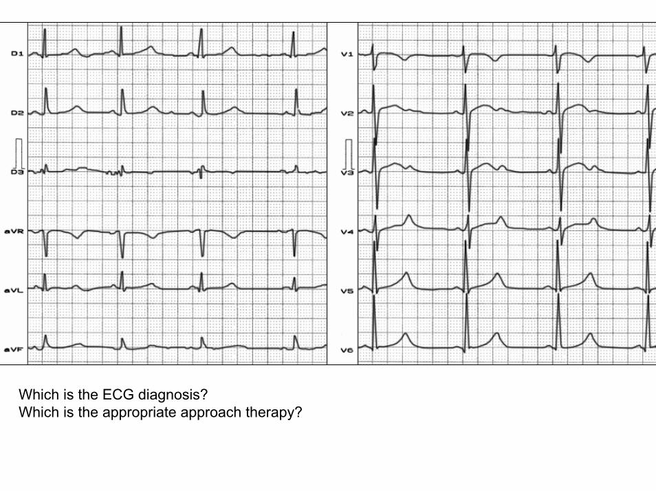
Which is the ECG diagnosis?Which is the appropriate approach therapy?

Colleagues opinions
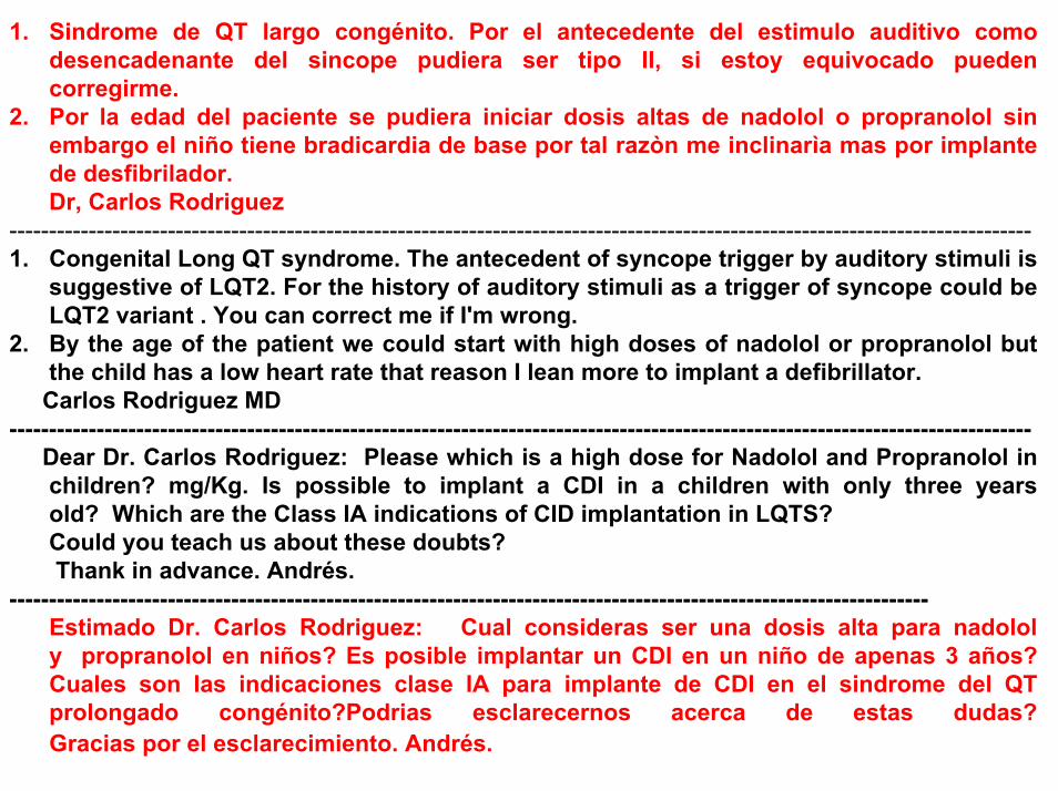
1. Sindrome de QT largo congénito. Por el antecedente del estimulo auditivo comodesencadenante del sincope pudiera ser tipo II, si estoy equivocado puedencorregirme.
2. Por la edad del paciente se pudiera iniciar dosis altas de nadolol o propranolol sin embargo el niño tiene bradicardia de base por tal razòn me inclinarìa mas por implantede desfibrilador.Dr, Carlos Rodriguez
---------------------------------------------------------------------------------------------------------------------------------1. Congenital Long QT syndrome. The antecedent of syncope trigger by auditory stimuli is
suggestive of LQT2. For the history of auditory stimuli as a trigger of syncope could be LQT2 variant . You can correct me if I'm wrong.
2. By the age of the patient we could start with high doses of nadolol or propranolol but the child has a low heart rate that reason I lean more to implant a defibrillator.
Carlos Rodriguez MD---------------------------------------------------------------------------------------------------------------------------------
Dear Dr. Carlos Rodriguez: Please which is a high dose for Nadolol and Propranolol in children? mg/Kg. Is possible to implant a CDI in a children with only three years old? Which are the Class IA indications of CID implantation in LQTS?Could you teach us about these doubts?Thank in advance. Andrés.
--------------------------------------------------------------------------------------------------------------------Estimado Dr. Carlos Rodriguez: Cual consideras ser una dosis alta para nadololy propranolol en niños? Es posible implantar un CDI en un niño de apenas 3 años? Cuales son las indicaciones clase IA para implante de CDI en el sindrome del QT prolongado congénito?Podrias esclarecernos acerca de estas dudas?Gracias por el esclarecimiento. Andrés.
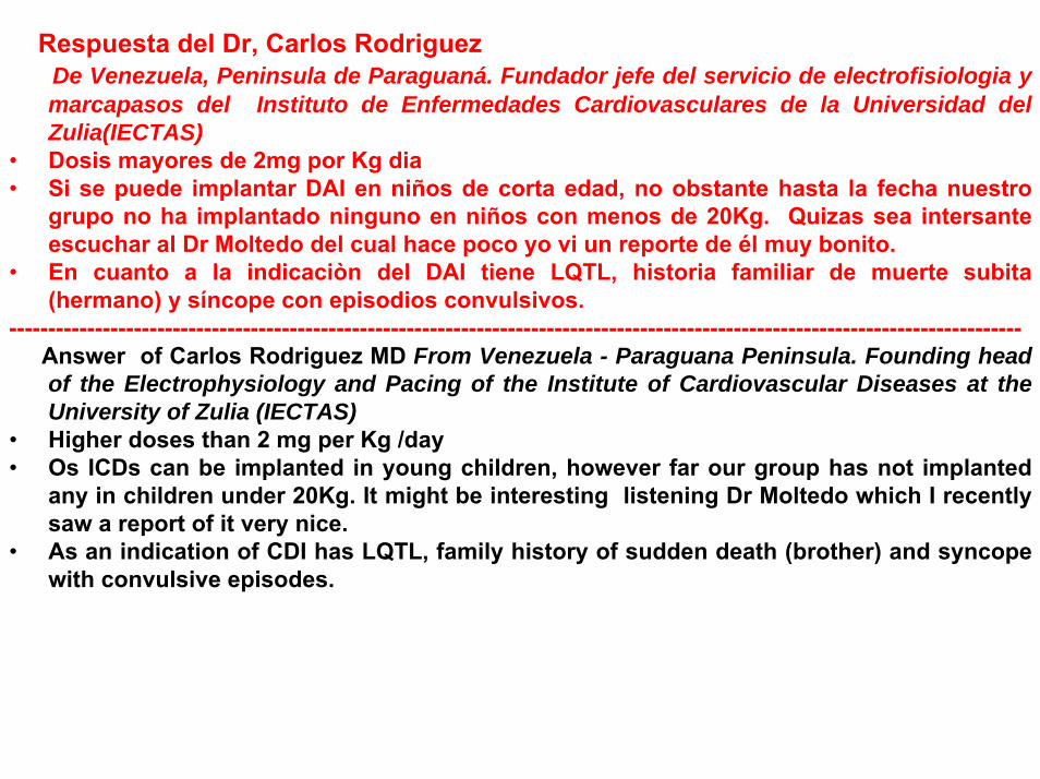
Respuesta del Dr, Carlos RodriguezDe Venezuela, Peninsula de Paraguaná. Fundador jefe del servicio de electrofisiologia y marcapasos del Instituto de Enfermedades Cardiovasculares de la Universidad del Zulia(IECTAS)
• Dosis mayores de 2mg por Kg dia• Si se puede implantar DAI en niños de corta edad, no obstante hasta la fecha nuestro
grupo no ha implantado ninguno en niños con menos de 20Kg. Quizas sea intersanteescuchar al Dr Moltedo del cual hace poco yo vi un reporte de él muy bonito.
• En cuanto a la indicaciòn del DAI tiene LQTL, historia familiar de muerte subita(hermano) y síncope con episodios convulsivos.
----------------------------------------------------------------------------------------------------------------------------------Answer of Carlos Rodriguez MD From Venezuela - Paraguana Peninsula. Founding head of the Electrophysiology and Pacing of the Institute of Cardiovascular Diseases at the University of Zulia (IECTAS)
• Higher doses than 2 mg per Kg /day• Os ICDs can be implanted in young children, however far our group has not implanted
any in children under 20Kg. It might be interesting listening Dr Moltedo which I recently saw a report of it very nice.
• As an indication of CDI has LQTL, family history of sudden death (brother) and syncope with convulsive episodes.
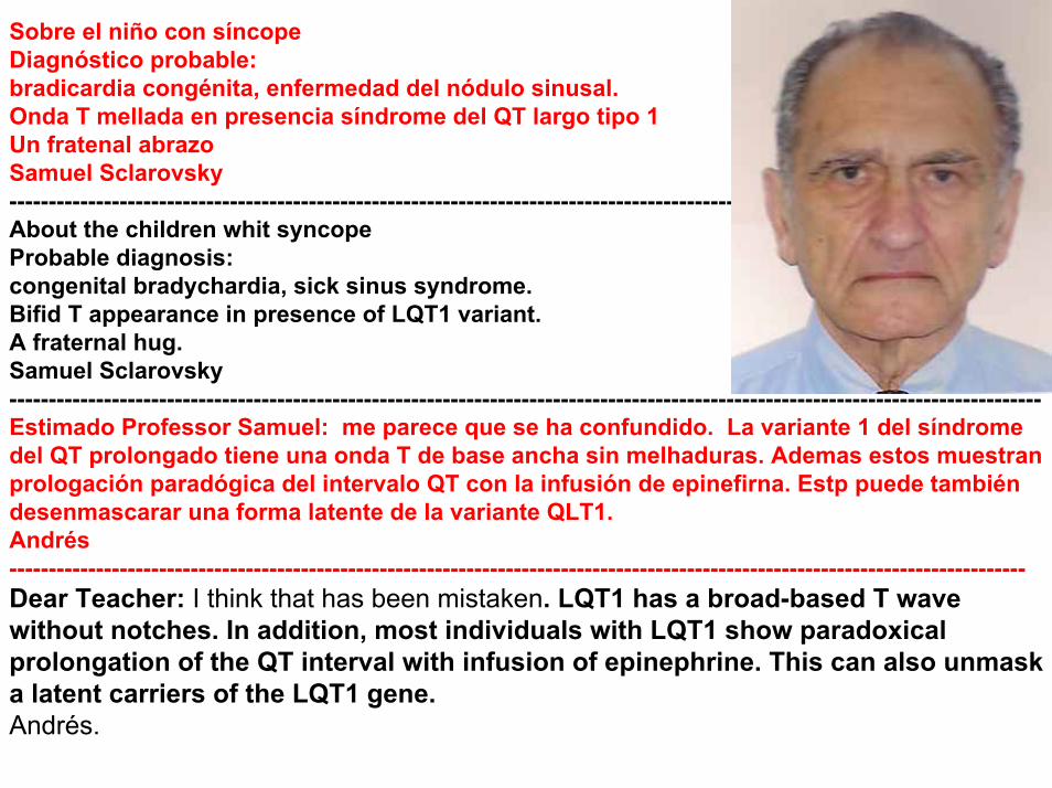
Sobre el niño con síncope Diagnóstico probable: bradicardia congénita, enfermedad del nódulo sinusal. Onda T mellada en presencia síndrome del QT largo tipo 1Un fratenal abrazo Samuel Sclarovsky-----------------------------------------------------------------------------------------------------------------------------------About the children whit syncopeProbable diagnosis: congenital bradychardia, sick sinus syndrome. Bifid T appearance in presence of LQT1 variant.A fraternal hug.Samuel Sclarovsky-----------------------------------------------------------------------------------------------------------------------------------Estimado Professor Samuel: me parece que se ha confundido. La variante 1 del síndromedel QT prolongado tiene una onda T de base ancha sin melhaduras. Ademas estos muestranprologación paradógica del intervalo QT con la infusión de epinefirna. Estp puede tambiéndesenmascarar una forma latente de la variante QLT1.Andrés---------------------------------------------------------------------------------------------------------------------------------Dear Teacher: I think that has been mistaken. LQT1 has a broad-based T wave without notches. In addition, most individuals with LQT1 show paradoxical prolongation of the QT interval with infusion of epinephrine. This can also unmask a latent carriers of the LQT1 gene.Andrés.
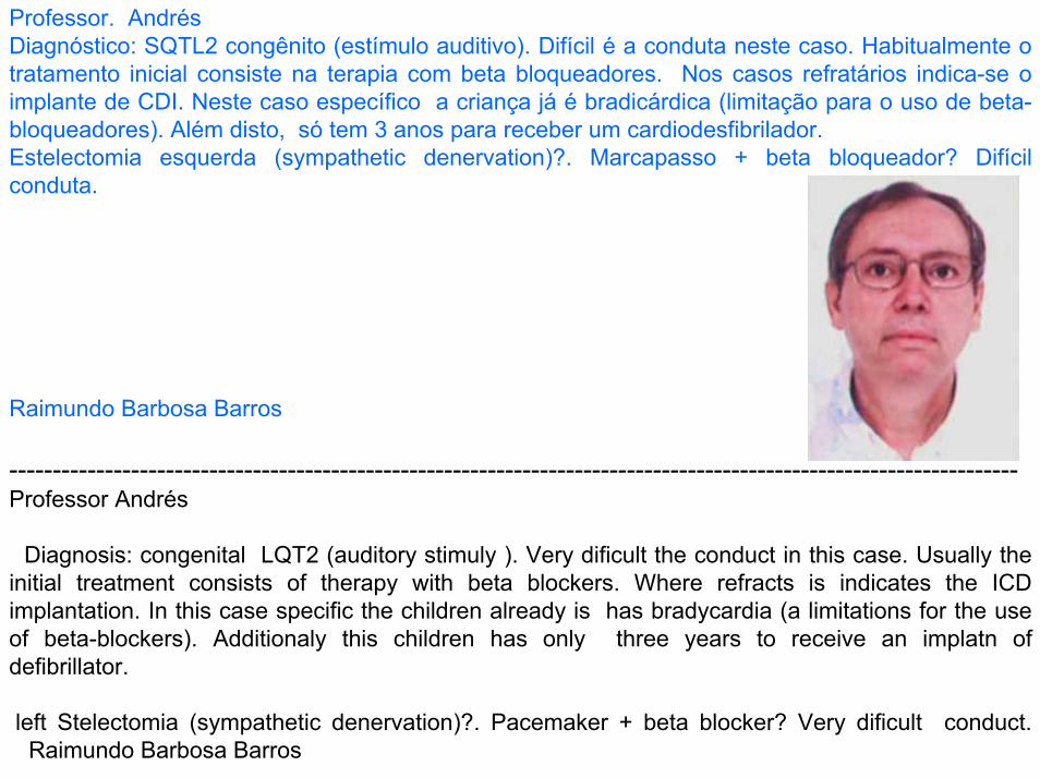
Professor. AndrésDiagnóstico: SQTL2 congênito (estímulo auditivo). Difícil é a conduta neste caso. Habitualmente o tratamento inicial consiste na terapia com beta bloqueadores. Nos casos refratários indica-se o implante de CDI. Neste caso específico a criança já é bradicárdica (limitação para o uso de beta-bloqueadores). Além disto, só tem 3 anos para receber um cardiodesfibrilador. Estelectomia esquerda (sympathetic denervation)?. Marcapasso + beta bloqueador? Difícilconduta.
Raimundo Barbosa Barros
--------------------------------------------------------------------------------------------------------------------Professor Andrés
Diagnosis: congenital LQT2 (auditory stimuly ). Very dificult the conduct in this case. Usually the initial treatment consists of therapy with beta blockers. Where refracts is indicates the ICD implantation. In this case specific the children already is has bradycardia (a limitations for the use of beta-blockers). Additionaly this children has only three years to receive an implatn of defibrillator.
left Stelectomia (sympathetic denervation)?. Pacemaker + beta blocker? Very dificult conduct.Raimundo Barbosa Barros
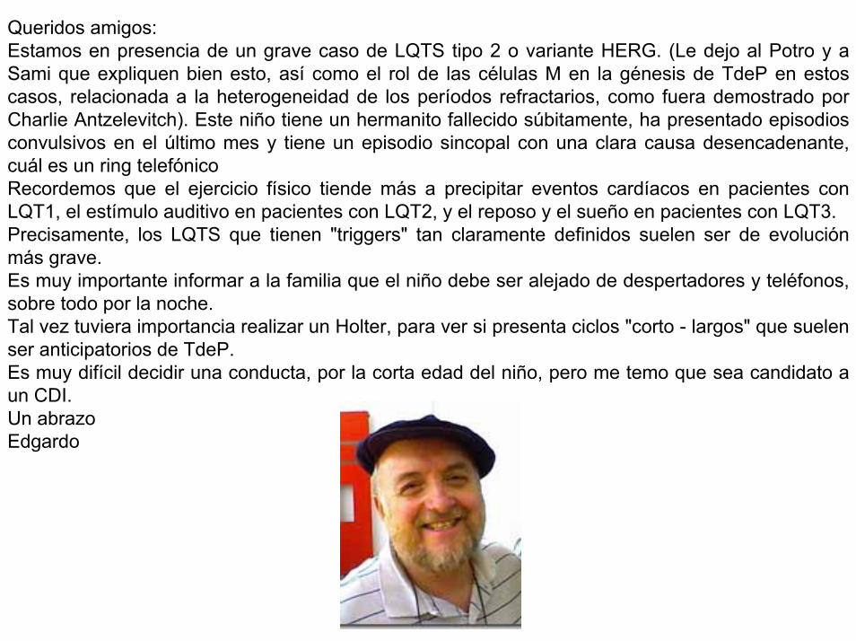
Queridos amigos:Estamos en presencia de un grave caso de LQTS tipo 2 o variante HERG. (Le dejo al Potro y a Sami que expliquen bien esto, así como el rol de las células M en la génesis de TdeP en estoscasos, relacionada a la heterogeneidad de los períodos refractarios, como fuera demostrado porCharlie Antzelevitch). Este niño tiene un hermanito fallecido súbitamente, ha presentado episodiosconvulsivos en el último mes y tiene un episodio sincopal con una clara causa desencadenante, cuál es un ring telefónicoRecordemos que el ejercicio físico tiende más a precipitar eventos cardíacos en pacientes con LQT1, el estímulo auditivo en pacientes con LQT2, y el reposo y el sueño en pacientes con LQT3.Precisamente, los LQTS que tienen "triggers" tan claramente definidos suelen ser de evoluciónmás grave.Es muy importante informar a la familia que el niño debe ser alejado de despertadores y teléfonos, sobre todo por la noche. Tal vez tuviera importancia realizar un Holter, para ver si presenta ciclos "corto - largos" que suelenser anticipatorios de TdeP.Es muy difícil decidir una conducta, por la corta edad del niño, pero me temo que sea candidato a un CDI.Un abrazoEdgardo
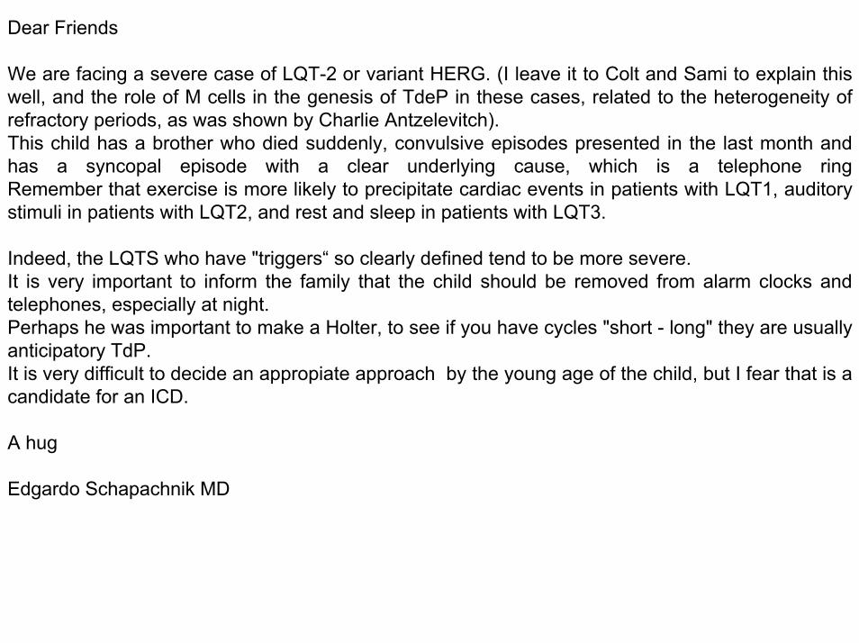
Dear Friends
We are facing a severe case of LQT-2 or variant HERG. (I leave it to Colt and Sami to explain this well, and the role of M cells in the genesis of TdeP in these cases, related to the heterogeneity of refractory periods, as was shown by Charlie Antzelevitch). This child has a brother who died suddenly, convulsive episodes presented in the last month and has a syncopal episode with a clear underlying cause, which is a telephone ringRemember that exercise is more likely to precipitate cardiac events in patients with LQT1, auditory stimuli in patients with LQT2, and rest and sleep in patients with LQT3.
Indeed, the LQTS who have "triggers“ so clearly defined tend to be more severe.It is very important to inform the family that the child should be removed from alarm clocks and telephones, especially at night.Perhaps he was important to make a Holter, to see if you have cycles "short - long" they are usually anticipatory TdP.It is very difficult to decide an appropiate approach by the young age of the child, but I fear that is a candidate for an ICD.
A hug
Edgardo Schapachnik MD
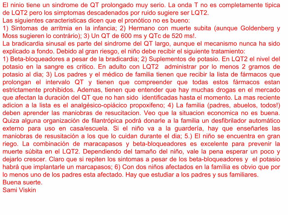
El ninio tiene un sindrome de QT prolongado muy serio. La onda T no es completamente tipicade LQT2 pero los simptomas descadenados por ruido sugiere ser LQT2.Las siguientes caracteristicas dicen que el pronótico no es bueno:1) Sintomas de arritmia en la infancia; 2) Hermano con muerte subita (aunque Goldenberg y Moss sugieren lo contrário); 3) Un QT de 600 ms y QTc de 520 ms!.La bradicardia sinusal es parte del sindrome del QT largo, aunque el mecanismo nunca ha sidoexplicado a fondo. Debido al gran riesgo, el niño debe recibir el siguiente tratamiento:1) Beta-bloqueadores a pesar de la bradicardia; 2) Suplementos de potasio. En LQT2 el nivel del potasio en la sangre es critico. En adulto con LQT2 administrar por lo menos 2 gramos de potasio al dia; 3) Los padres y el médico de familia tienen que recibir la lista de fármacos queprolongan el intervalo QT y tienen que compreender que todas estos fármacos estanestrictamente prohibidos. Ademas, tienen que entender que hay muchas drogas en el mercadoque afectan la duración del QT que no han sido identificadas hasta el momento. La mas recienteadicion a la lista es el analgésico-opiácico propoxifeno; 4) La familia (padres, abuelos, todos!) deben aprender las maniobras de resucitacion. Veo que la situacion economica no es buena. Quiza alguna organización de filantrópica podrá donarle a la familia un desfibrilador automáticoexterno para uso en casa/escuela. Si el niño va a la guardería, hay que enseñarles lasmaniobras de resusitación a los que lo cuidan durante el dia; 5.) El niño se encuentra en granriego. La combinación de maracapasos y beta-bloqueadores es excelente para prevenir la muerte súbita en el LQT2. Dependiendo del tamaño del niño, vale la pena esperar un poco y dejarlo crescer. Claro que si repiten los sintomas a pesar de los beta-bloqueadores y el potasiohabrá que implantarle un marcapasos; 6) Con dos niños afectados en la família es obvio que porlo menos uno de los padres esta afectado. Hay que estudiar a los padres y sus familiares.Buena suerte.Sami Viskin
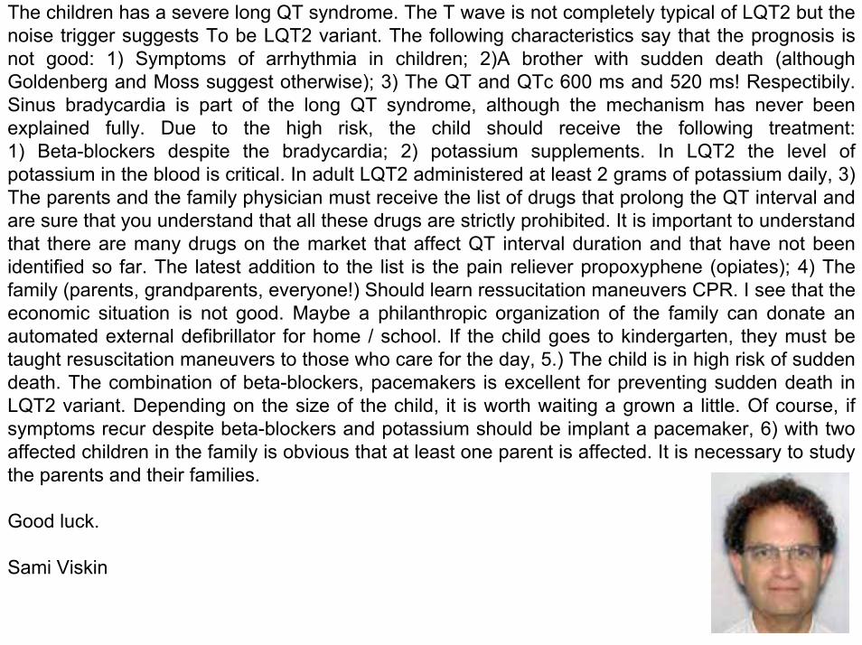
The children has a severe long QT syndrome. The T wave is not completely typical of LQT2 but the noise trigger suggests To be LQT2 variant. The following characteristics say that the prognosis is not good: 1) Symptoms of arrhythmia in children; 2)A brother with sudden death (although Goldenberg and Moss suggest otherwise); 3) The QT and QTc 600 ms and 520 ms! Respectibily.Sinus bradycardia is part of the long QT syndrome, although the mechanism has never been explained fully. Due to the high risk, the child should receive the following treatment:1) Beta-blockers despite the bradycardia; 2) potassium supplements. In LQT2 the level of potassium in the blood is critical. In adult LQT2 administered at least 2 grams of potassium daily, 3) The parents and the family physician must receive the list of drugs that prolong the QT interval and are sure that you understand that all these drugs are strictly prohibited. It is important to understand that there are many drugs on the market that affect QT interval duration and that have not been identified so far. The latest addition to the list is the pain reliever propoxyphene (opiates); 4) The family (parents, grandparents, everyone!) Should learn ressucitation maneuvers CPR. I see that the economic situation is not good. Maybe a philanthropic organization of the family can donate an automated external defibrillator for home / school. If the child goes to kindergarten, they must be taught resuscitation maneuvers to those who care for the day, 5.) The child is in high risk of sudden death. The combination of beta-blockers, pacemakers is excellent for preventing sudden death inLQT2 variant. Depending on the size of the child, it is worth waiting a grown a little. Of course, if symptoms recur despite beta-blockers and potassium should be implant a pacemaker, 6) with two affected children in the family is obvious that at least one parent is affected. It is necessary to study the parents and their families.
Good luck.
Sami Viskin
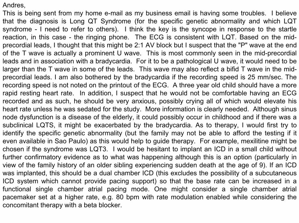
Andres,This is being sent from my home e-mail as my business email is having some troubles. I believethat the diagnosis is Long QT Syndrome (for the specific genetic abnormality and which LQT syndrome - I need to refer to others). I think the key is the syncope in response to the startlereaction, in this case - the ringing phone. The ECG is consistent with LQT. Based on the mid-precordial leads, I thought that this might be 2:1 AV block but I suspect that the "P" wave at the endof the T wave is actually a prominent U wave. This is most commonly seen in the mid-precordialleads and in association with a bradycardia. For it to be a pathological U wave, it would need to belarger than the T wave in some of the leads. This wave may also reflect a bifid T wave in the mid-precordial leads. I am also bothered by the bradycardia if the recording speed is 25 mm/sec. The recording speed is not noted on the printout of the ECG. A three year old child should have a more rapid resting heart rate. In addition, I suspect that he would not be comfortable having an ECG recorded and as such, he should be very anxious, possibly crying all of which would elevate hisheart rate unless he was sedated for the study. More information is clearly needed. Although sinusnode dysfunction is a disease of the elderly, it could possibly occur in childhood and if there was a subclinical LQTS, it might be exacerbated by the bradycardia. As to therapy, I would first try to identify the specific genetic abnormality (but the family may not be able to afford the testing if it even available in Sao Paulo) as this would help to guide therapy. For example, mexilitine might bechosen if the syndrome was LQT3. I would be hesitant to implant an ICD in a small child withoutfurther confirmatory evidence as to what was happening although this is an option (particularly in view of the family history of an older sibling experiencing sudden death at the age of 9). If an ICD was implanted, this should be a dual chamber ICD (this excludes the possibility of a subcutaneousICD system which cannot provide pacing support) so that the base rate can be increased in a functional single chamber atrial pacing mode. One might consider a single chamber atrial pacemaker set at a higher rate, e.g. 80 bpm with rate modulation enabled while considering the concomitant therapy with a beta blocker.
If there is concomitant sinus node dysfunction, beta blockade alone may aggravate the problemwith a further lengthening of the QT interval in association with a further slowing of the sinus rate. One could also consider a subcutaneous ICD with subcutaneous leads (Cameron Health) if such is available in Brasil and this would then be rescue only. I await further information and the results of any additional testing.
PaulPaul A. Levine MD, FHRS, FACC25876 The Old RoadStevenson Ranch, CA 91381Email: [email protected]
Andrés,Esto está siendo enviado desde mi casa el correo electrónico como mi correo electrónico de negocio está teniendo algunos problemas. Creo que el diagnóstico es Síndrome de QT largo (por la anormalidad genética específica y que el síndrome LQT - Tengo que hacer referencia a otros). Creo que la clave es el síncope en respuesta a la reacción de sobresalto, en este caso - el timbre del teléfono. El ECG es compatible con LQT. Sobre la base de los cables de media precordial, pensé que esto podría ser bloqueo AV 2:1 pero sospecho que la onda "P" al final de la onda T es en realidad una onda U prominente. Esto se observa más comúnmente en las derivaciones precordiales mediados de los años y en asociación con una bradicardia. Para que sea una patología onda U, tendría que ser mayor que la onda T en algunos de los conductores. Esta onda también puede reflejar una onda T bífida en las derivaciones precordiales mediados de los años. También estoy preocupado por la bradicardia si la velocidad de grabación es de 25 mm / seg. La velocidad de grabación no se observa en la impresión del ECG. Un niño de tres años de edad deben tener un más rápido ritmo cardíaco en reposo. Además, sospecho que no se sentirían cómodos con un ECG registrado y, como tal, debe ser muy ansiosos, tal vez llorar todo lo cual eleva su ritmo cardíaco a menos que estuviera sedado para el estudio. Más información es claramente necesario. Aunque la disfunción del nódulo sinusal es una enfermedad de los ancianos, que podría ocurrir en la infancia y si había un SQTL subclínica, que puede ser exacerbada por la bradicardia. En cuanto a la terapia, en primer lugar tratar de identificar la anomalía genética específica (pero la familia no puede pagar la prueba, si aún disponibles en Sao Paulo), ya que esto ayudará a guiar la terapia. Por ejemplo, mexilitine podría ser elegido si el síndrome se LQT3. Yo dudaría de implantar un DAI en un niño pequeño sin pruebas de confirmación más de lo que estaba pasando, aunque esto es una opción (en particular en vista de la historia de la familia de un hermano mayor que experimentan la muerte repentina a la edad de 9). Si se implantó un DAI, ésta debe ser una doble cámara de CIE (esto excluye la posibilidad de un tejido subcutáneo CIE que no pueden proporcionar apoyo de estimulación) de modo que el tipo de base se puede aumentar en un modo de funcionamiento de una cámara de estimulación auricular.
Se podría considerar un marcapasos auricular unicameral fijado en una tasa más alta, por ejemplo, 80 lpm, con velocidad de modulación permitió al considerar la terapia concomitante con un bloqueador beta. Si hay una disfunción del nódulo sinusal concomitante, el bloqueo beta solo puede agravar el problema con una ampliación ulterior del intervalo QT en asociación con una disminución adicional de la frecuencia sinusal. También se podría considerar un ICD subcutánea con cables subcutánea (Cameron de la Salud) en caso de que se disponga en Brasil y esto sería sólo de rescate. Espero más información y los resultados de cualquier prueba adicional.
PaulPaul A. Levine MD, FHRS, FACC25876 The Old RoadStevenson Ranch, CA 91381Email: [email protected]
FINAL DIAGNOSIS AND MANAGEMENT
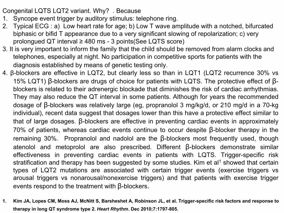
Congenital LQTS LQT2 variant. Why? . Because1. Syncope event trigger by auditory stimulus: telephone ring.2. Typical ECG : a) Low heart rate for age; b) Low T wave amplitude with a notched, bifurcated
biphasic or bifid T appearance due to a very significant slowing of repolarization; c) veryprolongued QT interval ≥ 480 ms - 3 points(See LQTS score)
3. It is very important to inform the family that the child should be removed from alarm clocks and telephones, especially at night. No participation in competitive sports for patients with the diagnosis established by means of genetic testing only.
4. β-blockers are effective in LQT2, but clearly less so than in LQT1 (LQT2 recurrence 30% vs 15% LQT1) β-blockers are drugs of choice for patients with LQTS. The protective effect of β-blockers is related to their adrenergic blockade that diminishes the risk of cardiac arrhythmias. They may also reduce the QT interval in some patients. Although for years the recommended dosage of β-blockers was relatively large (eg, propranolol 3 mg/kg/d, or 210 mg/d in a 70-kg individual), recent data suggest that dosages lower than this have a protective effect similar to that of large dosages. β-blockers are effective in preventing cardiac events in approximately 70% of patients, whereas cardiac events continue to occur despite β-blocker therapy in the remaining 30%. Propranolol and nadolol are the β-blockers most frequently used, though atenolol and metoprolol are also prescribed. Different β-blockers demonstrate similar effectiveness in preventing cardiac events in patients with LQTS. Trigger-specific risk stratification and therapy has been suggested by some studies. Kim et al1 showed that certain types of LQT2 mutations are associated with certain trigger events (exercise triggers vsarousal triggers vs nonarousal/nonexercise triggers) and that patients with exercise trigger events respond to the treatment with β-blockers.
1. Kim JA, Lopes CM, Moss AJ, McNitt S, Barsheshet A, Robinson JL, et al. Trigger-specific risk factors and response to therapy in long QT syndrome type 2. Heart Rhythm. Dec 2010;7:1797-805.
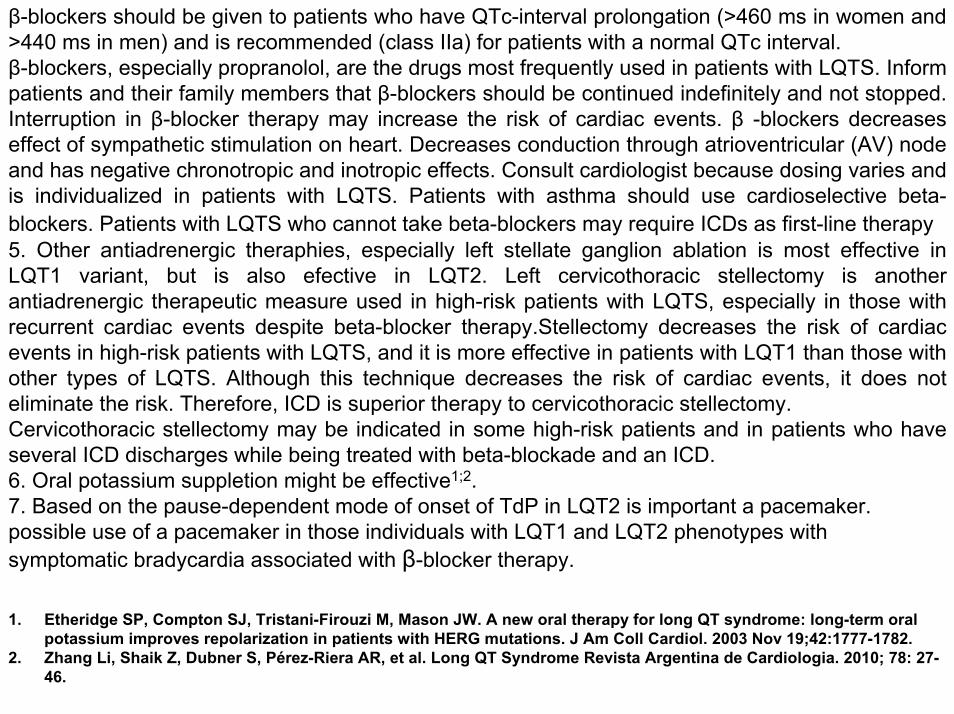
β-blockers should be given to patients who have QTc-interval prolongation (>460 ms in women and >440 ms in men) and is recommended (class IIa) for patients with a normal QTc interval.β-blockers, especially propranolol, are the drugs most frequently used in patients with LQTS. Inform patients and their family members that β-blockers should be continued indefinitely and not stopped. Interruption in β-blocker therapy may increase the risk of cardiac events. β -blockers decreases effect of sympathetic stimulation on heart. Decreases conduction through atrioventricular (AV) node and has negative chronotropic and inotropic effects. Consult cardiologist because dosing varies and is individualized in patients with LQTS. Patients with asthma should use cardioselective beta-blockers. Patients with LQTS who cannot take beta-blockers may require ICDs as first-line therapy5. Other antiadrenergic theraphies, especially left stellate ganglion ablation is most effective in LQT1 variant, but is also efective in LQT2. Left cervicothoracic stellectomy is another antiadrenergic therapeutic measure used in high-risk patients with LQTS, especially in those with recurrent cardiac events despite beta-blocker therapy.Stellectomy decreases the risk of cardiac events in high-risk patients with LQTS, and it is more effective in patients with LQT1 than those with other types of LQTS. Although this technique decreases the risk of cardiac events, it does not eliminate the risk. Therefore, ICD is superior therapy to cervicothoracic stellectomy. Cervicothoracic stellectomy may be indicated in some high-risk patients and in patients who have several ICD discharges while being treated with beta-blockade and an ICD.6. Oral potassium suppletion might be effective1;2.7. Based on the pause-dependent mode of onset of TdP in LQT2 is important a pacemaker.possible use of a pacemaker in those individuals with LQT1 and LQT2 phenotypes with symptomatic bradycardia associated with β-blocker therapy.
1. Etheridge SP, Compton SJ, Tristani-Firouzi M, Mason JW. A new oral therapy for long QT syndrome: long-term oral potassium improves repolarization in patients with HERG mutations. J Am Coll Cardiol. 2003 Nov 19;42:1777-1782.
2. Zhang Li, Shaik Z, Dubner S, Pérez-Riera AR, et al. Long QT Syndrome Revista Argentina de Cardiologia. 2010; 78: 27-46.
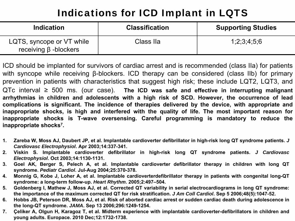
Indications for ICD Implant in LQTS Indication Classification Supporting Studies
LQTS, syncope or VT while receiving β -blockers
Class IIa 1;2;3;4;5;6
ICD should be implanted for survivors of cardiac arrest and is recommended (class IIa) for patients with syncope while receiving β-blockers. ICD therapy can be considered (class IIb) for primary prevention in patients with characteristics that suggest high risk; these include LQT2, LQT3, and QTc interval ≥ 500 ms. (our case). The ICD was safe and effective in interrupting malignant arrhythmias in children and adolescents with a high risk of SCD. However, the occurrence of lead complications is significant. The incidence of therapies delivered by the device, with appropriate and inappropriate shocks, is high and interfered with the quality of life. The most important reason for inappropriate shocks is T-wave oversensing. Careful programming is mandatory to reduce the inappropriate shocks7.
1. Zareba W, Moss AJ, Daubert JP, et al. Implantable cardioverter defibrillator in high-risk long QT syndrome patients. J Cardiovasc Electrophysiol. Apr 2003;14:337-341.
2. Viskin S. Implantable cardioverter defibrillator in high-risk long QT syndrome patients. J CardiovascElectrophysiol. Oct 2003;14:1130-1131.
3. Goel AK, Berger S, Pelech A, et al. Implantable cardioverter defibrillator therapy in children with long QT syndrome. Pediatr Cardiol. Jul-Aug 2004;25:370-378.
4. Monnig G, Kobe J, Loher A, et al. Implantable cardioverterdefibrillator therapy in patients with congenital long-QT syndrome: a long-term follow-up. Heart Rhythm. 2005;2:497–504.
5. Goldenberg I, Mathew J, Moss AJ, et al. Corrected QT variability in serial electrocardiograms in long QT syndrome: the importance of the maximum corrected QT for risk stratification. J Am Coll Cardiol. Sep 5 2006;48(5):1047-52.
6. Hobbs JB, Peterson DR, Moss AJ, et al. Risk of aborted cardiac arrest or sudden cardiac death during adolescence in the long-QT syndrome. JAMA. Sep 13 2006;296:1249-1254.
7. Çeliker A, Olgun H, Karagoz T, et al. Midterm experience with implantable cardioverter-defibrillators in children and young adults. Europace. 2010 Dec;12:1732-1738.
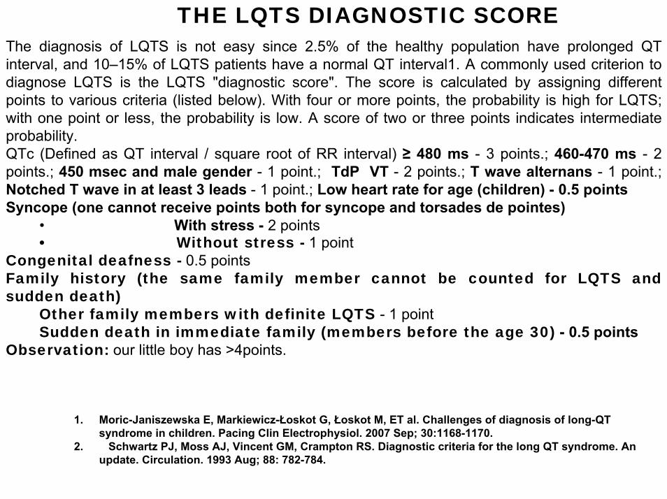
THE LQTS DIAGNOSTIC SCOREThe diagnosis of LQTS is not easy since 2.5% of the healthy population have prolonged QT interval, and 10–15% of LQTS patients have a normal QT interval1. A commonly used criterion to diagnose LQTS is the LQTS "diagnostic score". The score is calculated by assigning different points to various criteria (listed below). With four or more points, the probability is high for LQTS; with one point or less, the probability is low. A score of two or three points indicates intermediate probability.QTc (Defined as QT interval / square root of RR interval) ≥ 480 ms - 3 points.; 460-470 ms - 2 points.; 450 msec and male gender - 1 point.; TdP VT - 2 points.; T wave alternans - 1 point.; Notched T wave in at least 3 leads - 1 point.; Low heart rate for age (children) - 0.5 points Syncope (one cannot receive points both for syncope and torsades de pointes)
• With stress - 2 points • Without stress - 1 point
Congenital deafness - 0.5 points Family history (the same family member cannot be counted for LQTS and sudden death)
Other family members with definite LQTS - 1 point Sudden death in immediate family (members before the age 30) - 0.5 points
Observation: our little boy has >4points.
1. Moric-Janiszewska E, Markiewicz-Łoskot G, Łoskot M, ET al. Challenges of diagnosis of long-QT syndrome in children. Pacing Clin Electrophysiol. 2007 Sep; 30:1168-1170.
2. Schwartz PJ, Moss AJ, Vincent GM, Crampton RS. Diagnostic criteria for the long QT syndrome. An update. Circulation. 1993 Aug; 88: 782-784.
Schwartz et. al1. sought to determine the characteristics of the LQTS patients receiving an ICD, the indications, and the aftermath. The prospective study population included 233 patients. Female patients (77%) and LQT3 patients (22% of genotype positive) were overrepresented; mean QTc was 516±65 ms; mean age at implantation was 30±17 years; and genotype was known in 59% of patients. Unexpectedly, 9% of patients were asymptomatic before implantation. Asymptomatic patients, almost absent among LQT1 and LQT2 patients, represented 45% of LQT3 patients. Patients with cardiac symptoms made up 91% of all study participants, but only 44% had cardiac arrest before ICD implantation. In addition, 41% of patients received an ICD without having first been on LQTS therapy. During follow-up, 4.6±3.2 years, at least 1 appropriate shock was received by 28% of patients, and adverse events occurred in 25%. Appropriate ICD therapies were predicted by age <20 years at implantation, a QTc >500 ms, prior cardiac arrest, and cardiac events despite therapy; within 7 years, appropriate shocks occurred in no patients with none of these factors and in 70% of those with all factors. Reflecting previous concepts, ICDs were implanted in some LQTS patients whose high risk now appears questionable. Refined criteria for implantation, reassessment of pros and cons, ICD reprogramming, and consideration for other existing therapeutic options are necessary.The implantation method for a CDI in children is poorly standardized because of obvious features related to size and predisposing cardiac disease in children presenting with malignant VT. An alternative method of implanting a CDI without the need for associated thoracotomy, based on the subxiphoidal insertion of an epicardial bipolar ventricular pacing and sensing lead, an active can placed in the abdomen, and a subcutaneous array tunneled along the left thoracic wall as a shock electrode2. This technique offers the advantage of an effective and minimally invasive ICD with wide applicability for children, independent of their size and cardiac status.
1. Schwartz PJ, Spazzolini C, Priori SG, et al . Who are the long-QT syndrome patients who receive an implantable cardioverter-defibrillator and what happens to them?: data from the European Long-QT Syndrome Implantable Cardioverter-Defibrillator (LQTS ICD) Registry. Circulation. 2010 Sep 28;122:1272-1282.
2. Bové T, François K, De Caluwe W, Suys B, De Wolf D. Effective cardioverter defibrillator implantation in children without thoracotomy: a valid alternative. Ann Thorac Surg. 2010 Apr;89:1307-1309.
In an intact rabbit heart model of LQT2 and LQT3 as well as in a computer model of the rabbit cardiac myocyte, inhibition of the Na+/Ca2+ exchanger (NCX) is effective in preventing TdP due to a suppression of EADs, a reversion of AP prolongation, and a reduction of dispersion of repolarization. These observations suggest a therapeutic benefit of selective NCX inhibition in LQT21.
1. Milberg P, Pott C, Fink M, Frommeyer G, Matsuda T, Baba A, et al. Inhibition of the Na+/Ca2+ exchanger suppresses torsades de pointes in an intact heart model of long QT syndrome-2 and long QT syndrome-3. Heart Rhythm. 2008 Oct;5:1444-1452.
FREQUENCY %: The LQT2 type is the second most common gene location that is affected in long QT syndrome, making up 25% to 30% of all cases (polemic). Others authors confer a more high percentage 40% to 45%
OMIM NUMBER: 152427.
GENE: hERG (Kv11.1) the human ether-a-go-go related gene (HERG)1. identified a novel human cDNA from a hippocampal cDNA library by homology to the Drosophila 'ether-a-go-go' (eag) gene, which encodes a Ca(2+)-modulated potassium channel. The authors called the cDNA HERG (human ether-a-go-go-related gene). The HERG gene (also known as KCNH2) is part of the rapid component of the potassium rectifying current (IKr). alpha subunit of the rapid delayed rectifier potassium channel (HERG + MiRP1). HERG gene allows protection against early after depolarizations (EADs).
ALTERNATIVE TITLES; SYMBOLS• HUMAN ETHER-A-GO-GO-RELATED GENE; HERG • ETHER-A-GO-GO-RELATED GENE, HUMAN • ERG1.
INHERITANCE PATTERN: Autosomal dominant
CHROMOSOME: 7q35-q36.
LQT2
1. Warmke JW, Ganetzky B. A family of potassium channel genes related to eag in Drosophila and mammals. Proc NatlAcad Sci U S A. 1994 Apr 12;91:3438-3442.
PROTEIN: Kv11.1
PROTEIN FUNCTION: alpha subunit of IKr channel.
MUTATION EFFECT: caused by a reduction in repolarizing current. Reduced outward K+ rectifier current. The opposite effect of Short QT 1 syndrome (A gain of function.) In 2 families with short QT syndrome-1 (SQT1; 609620), previously reported by Gaita et al Gaita et al (2003), Brugada et al1. identified respective 1764C-G and 1764C-A (152427.0018) transversionsin exon 7 of the KCNH2 gene, leading to the same asn588-to-lys (N588K) substitution in the S5-P loop region of the cardiac I(Kr) channel. The mutation was present in all affected family members and in none of the unaffected individuals. In a family with short QT syndrome, originally reported by Gussak 2;Hong et al 3 identified the 1764C-G transversion in the KCNH2 gene and concluded that N588K is a hotspot for this familial form of short QT syndrome.
1. Brugada, R., Hong, K., Dumaine, R., Cordeiro, J., Gaita, F., Borggrefe, M., Menendez, T. M., Brugada, J., Pollevick, G. D., Wolpert, C., Burashnikov, E., Matsuo, K., Wu, Y. S., Guerchicoff, A., Bianchi, F., Giustetto, C., Schimpf, R., Brugada, P., Antzelevitch, C. Sudden death associated with short-QT syndrome linked to mutations in HERG. Circulation.2004; 109: 30-35.
2. Gussak et al. (Gussak, I., Brugada, P., Brugada, J., Wright, R. S., Kopecky, S. L., Chaitman, B. R., Bjerregaard, P. Idiopathic short QT interval: a new clinical syndrome? Cardiology 2000; 94: 99-102.
3. Hong et al. (Hong, K., Bjerregaard, P., Gussak, I., Brugada, R. Short QT syndrome and atrial fibrillation caused by mutation in KCNH2. J. Cardiovasc. Electrophysiol. 2005; 16: 394-396.
CHANNEL AFFECTED: (IKr) IKr delayed rectifier Potassium current.Phase of Action Potential affected: Phase 3. The IKr current is mainly responsible for the termination of the cardiac action potential, and therefore the length of the QT interval.
ECG CHARACTERISTIC: Frequent low heart rate for age. Low-amplitude with a notched, bifurcated alternant, biphasic or bifid T appearance due to a very significant slowing of repolarization.KCNH2 on L413P and L559H mutations are associated with bifid T wave. Long. QTc > 0.47 affected QTc = 0.45 to 0.47considered border line QTc < 0.45.non affected. With significant dynamic changes during heart rate variations or on exertion. Prominent or giant (QU) more evident form V4 to V6.Stress testing: Eventually prolongation the QT interval. Rarely are triggers for cardiac events (only in 13% of cases). The variant has moderate dependence of QT interval to heart rate.Although QTc interval is eventually normal on surface ECG, the presence of notched T-waves and QT prolongation on Holter recording help in establishing the correct diagnosis.
TRIGGERS: arrhythmic events after an emotional event, exercise, or exposure to auditory stimuli noise (eg, door bells, telephone ring).
CLINICAL CONSIDERATIONS: Most drugs that cause LQTS do so by blocking the IKr current via the HERG gene. These include erythromycin, terfenadine, and ketoconazole. The HERG channel is very sensitive to unintended drug binding due to two aromatic amino acids, the tyrosine at position 652 and the phenylalanine at position 656. These amino acid residues are poised so a drug binding to them will block the channel from conducting current. Other potassium channels do not have these residues in these positions and are therefore not as prone to blockage.
EPICARDIUM
MESOCARDIUM
ENDOCARDIUM
300 ms
300 ms
800 ms
Ito
Ito
0
0
0
1
1
2
2
2
3
3
34
4
4
ABUNDANTCONCENTRATION OF Ito CHANNEL
PROMINENT NOTCH
IN PHASE 1
ABSENCE OFIto CHANNEL
ABSENCE OF NOTCH IN PHASE 1
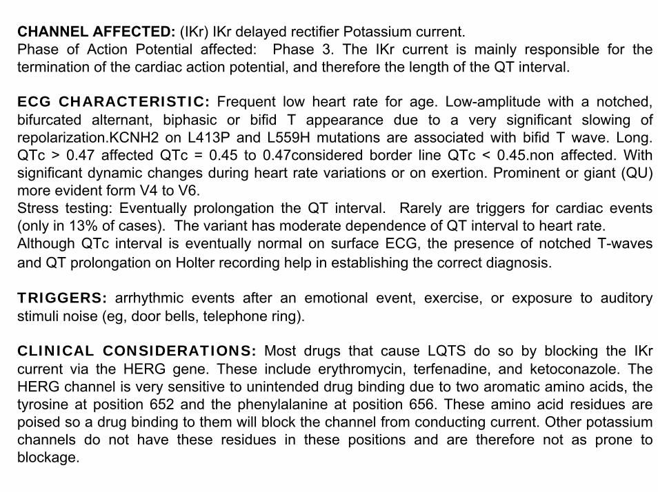
EPI, MESO AND ENDOCARDIUM: HETEROGENEITY IN VENTRICULAR WALL THICKNESS
1. Antzelevitch C. Heterogeneity and cardiac arrhythmias: an overview. Heart Rhythm. 2007 Jul;4:964-972.
Epicardium
M-Cells
Endocardium
ECG Surface
LEFT VENTRICLEFREE WALL
Tp-Te
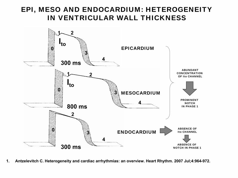
NORMAL TRANSMURAL DISPERSION OF REPOLARIZATION
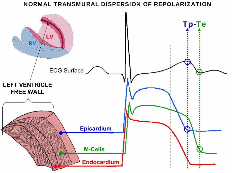
RVLV
CHARACTERISTICS OF ACTION POTENTIAL OF “M”CELLS OF THE VENTRICULAR MIDMYOCARDIUM
0
1
3
4
Prolonged Phase 2 WIDE PHASE 0 GREATER
THAN ENDO AND EPICARDIAL AND
SOMEHOW SMALLER THAN PURKINJE CELLS.
Stable Phase 4
PHASE 3 MUCH MORE SENSITIVE TO CLASS III
ANTIARRHYTHMIC AGENTS BY WEAK IKS
PHASE 1 WITH SIGNIFICANT NOTCH: SIGNIFICANT ITO CHANNEL
TIME
2
800 ms
AP DURATION: MUCH GREATER THAN ENDO AND EPICARDIAL
1. Antzelevitch C. M cells in the human heart. Circ Res. 2010 Mar 19;106:815-817.
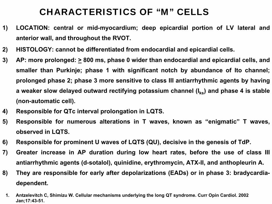
CHARACTERISTICS OF “M” CELLS1) LOCATION: central or mid-myocardium; deep epicardial portion of LV lateral and
anterior wall, and throughout the RVOT.
2) HISTOLOGY: cannot be differentiated from endocardial and epicardial cells.3) AP: more prolonged: > 800 ms, phase 0 wider than endocardial and epicardial cells, and
smaller than Purkinje; phase 1 with significant notch by abundance of Ito channel; prolonged phase 2; phase 3 more sensitive to class III antiarrhythmic agents by having a weaker slow delayed outward rectifying potassium channel (Iks) and phase 4 is stable (non-automatic cell).
4) Responsible for QTc interval prolongation in LQTS.5) Responsible for numerous alterations in T waves, known as “enigmatic” T waves,
observed in LQTS.6) Responsible for prominent U waves of LQTS (QU), decisive in the genesis of TdP.7) Greater increase in AP duration during low heart rates, before the use of class III
antiarrhythmic agents (d-sotalol), quinidine, erythromycin, ATX-II, and anthopleurin A.8) They are responsible for early after depolarizations (EADs) or in phase 3: bradycardia-
dependent.
1. Antzelevitch C, Shimizu W. Cellular mechanisms underlying the long QT syndrome. Curr Opin Cardiol. 2002 Jan;17:43-51.
CHARACTERISTICS OF “M” CELLS
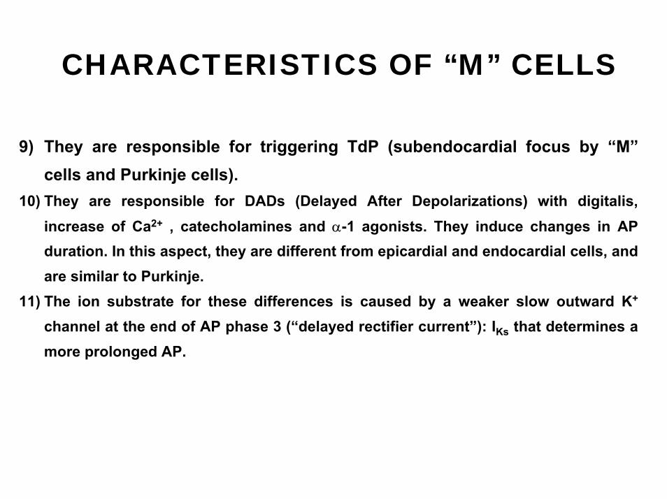
9) They are responsible for triggering TdP (subendocardial focus by “M”cells and Purkinje cells).
10) They are responsible for DADs (Delayed After Depolarizations) with digitalis, increase of Ca2+ , catecholamines and α-1 agonists. They induce changes in AP duration. In this aspect, they are different from epicardial and endocardial cells, and are similar to Purkinje.
11) The ion substrate for these differences is caused by a weaker slow outward K+
channel at the end of AP phase 3 (“delayed rectifier current”): IKs that determines a more prolonged AP.
4
3
210
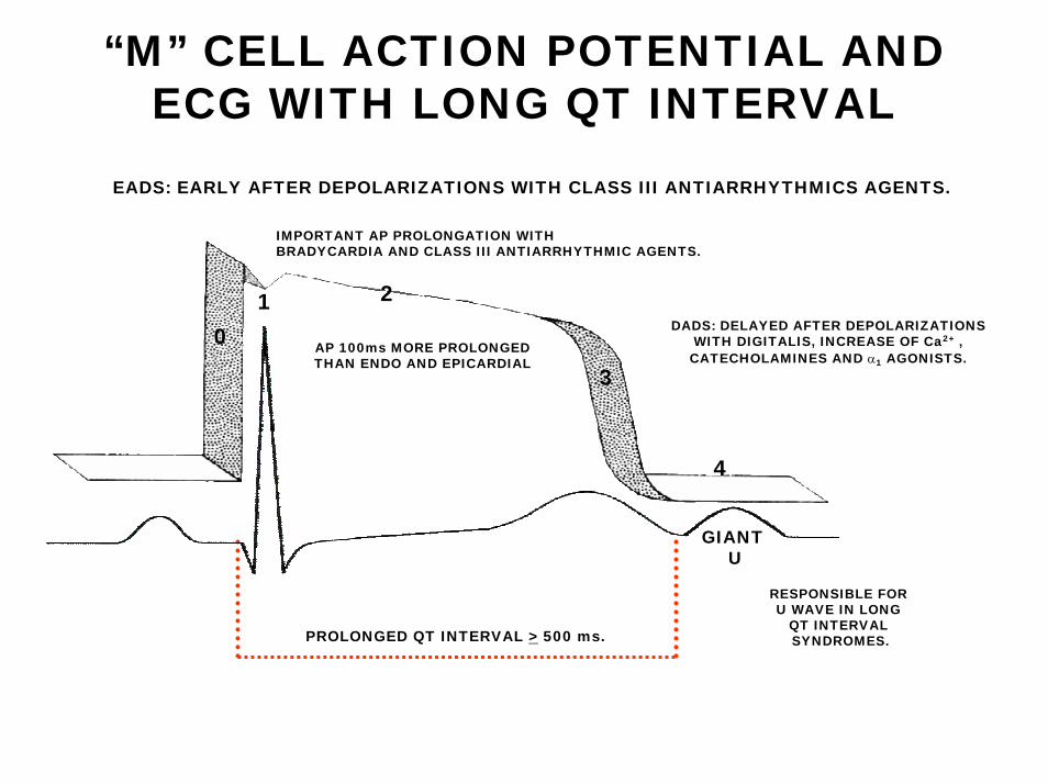
PROLONGED QT INTERVAL > 500 ms.
GIANT U
“M” CELL ACTION POTENTIAL AND ECG WITH LONG QT INTERVAL
IMPORTANT AP PROLONGATION WITH BRADYCARDIA AND CLASS III ANTIARRHYTHMIC AGENTS.
RESPONSIBLE FOR U WAVE IN LONG
QT INTERVAL SYNDROMES.
AP 100ms MORE PROLONGEDTHAN ENDO AND EPICARDIAL
DADS: DELAYED AFTER DEPOLARIZATIONSWITH DIGITALIS, INCREASE OF Ca2+ ,CATECHOLAMINES AND α1 AGONISTS.
EADS: EARLY AFTER DEPOLARIZATIONS WITH CLASS III ANTIARRHYTHMICS AGENTS.
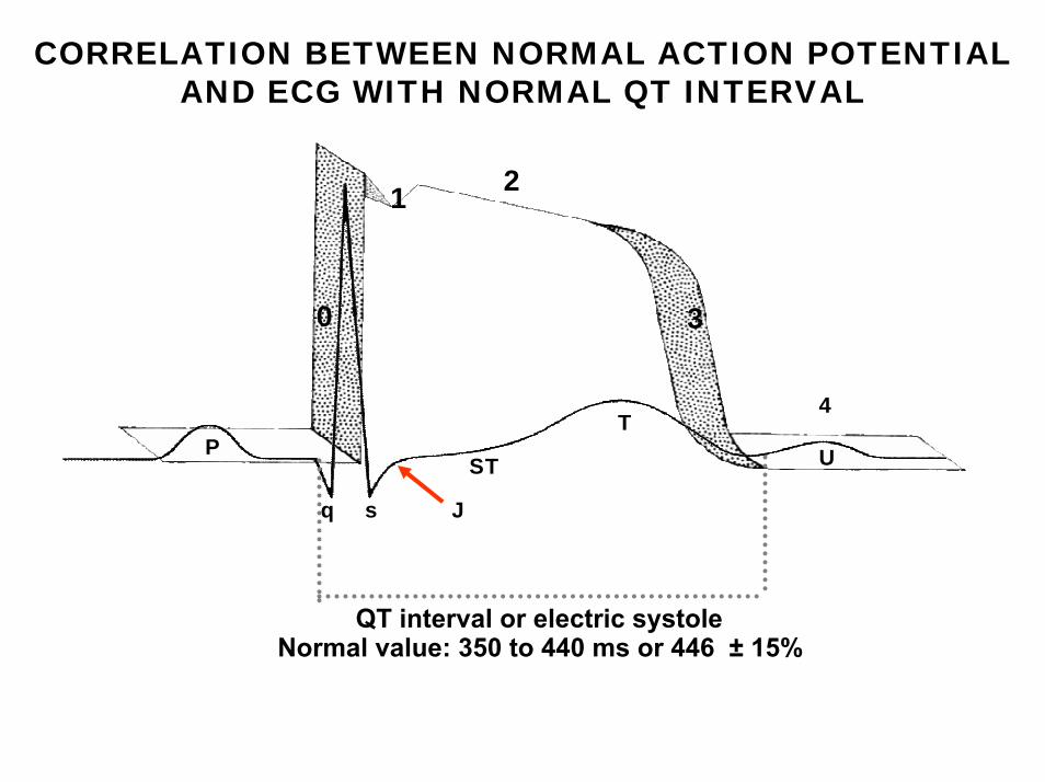
P
q s J
ST
TU
2
4
3
QT interval or electric systoleNormal value: 350 to 440 ms or 446 ± 15%
0
1
CORRELATION BETWEEN NORMAL ACTION POTENTIAL AND ECG WITH NORMAL QT INTERVAL
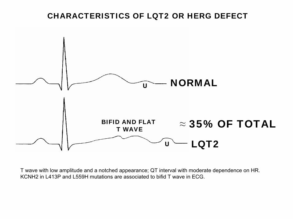
CHARACTERISTICS OF LQT2 OR HERG DEFECT
NORMAL
LQT2
BIFID AND FLAT T WAVE
UU
UU
≈ 35% OF TOTAL
T wave with low amplitude and a notched appearance; QT interval with moderate dependence on HR. KCNH2 in L413P and L559H mutations are associated to bifid T wave in ECG.
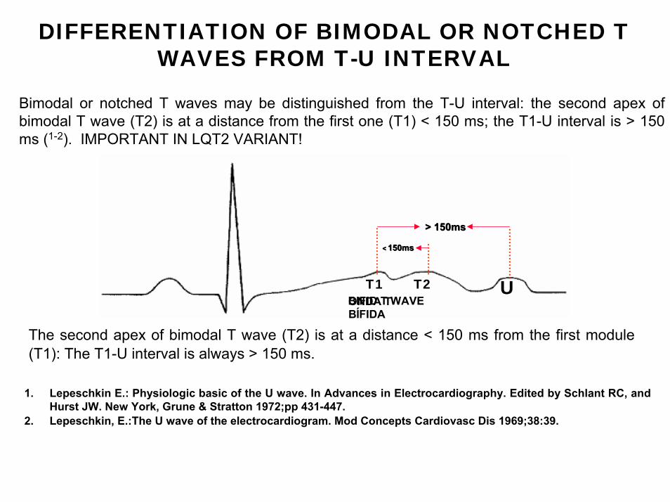
DIFFERENTIATION OF BIMODAL OR NOTCHED T WAVES FROM T-U INTERVAL
Bimodal or notched T waves may be distinguished from the T-U interval: the second apex of bimodal T wave (T2) is at a distance from the first one (T1) < 150 ms; the T1-U interval is > 150 ms (1-2). IMPORTANT IN LQT2 VARIANT!
The second apex of bimodal T wave (T2) is at a distance < 150 ms from the first module (T1): The T1-U interval is always > 150 ms.
ONDA T BIFID T WAVEBÍFIDA
> 150ms> 150ms
< < 150ms150ms
T1 T2 U
1. Lepeschkin E.: Physiologic basic of the U wave. In Advances in Electrocardiography. Edited by Schlant RC, and Hurst JW. New York, Grune & Stratton 1972;pp 431-447.
2. Lepeschkin, E.:The U wave of the electrocardiogram. Mod Concepts Cardiovasc Dis 1969;38:39.
TORSADE DE POINTES (TdP)Torsade de pointes (TdP), is defined as a polymorphic ventricular tachycardia (PVT) with a twisting QRS morphology associated with a congenital or acquired prolonged QT interval (exceptionally with normal QT interval) and/or increased U wave amplitude in the ECG. Literally meaning twisting of points, is a distinctive form of PVT characterized by a gradual change in the amplitude and twisting of the QRS complexes around the isoelectric line. TdP (torsade). Rapidly the axis of VT changes 180º. TdP usually terminates spontaneously but frequently recurs and may degenerate into ventricular fibrillation (VF). Ventricular complexes present at least, two different morphologies within the same tachyarrhythmic event. Observing the twelve leads may be necessary, because it may not be possible to detect the polymorphism just by one lead or in the monitor lead.Typical PVT, of the TdP variant occurs in patients with prolonged QT interval, slow basic rhythm: bradycardia dependent (acquired) or triggered by strain: adrenergic-dependent (congenital). The coupling of the initial premature ventricular contraction is prolonged or telediastolic. HEART RATE: from 150 to 300 bpm (usually 200 to 250 bpm).
USUAL DURATION: from 5 to 20 complexes.
CARDINAL SIGN: polymorphic QRS. Rotation of QRS apices of up to 1800 along the baseline: swinging pattern of Marriot.
CLINICAL REPERCUSSIONS: asymptomatic, presyncope, syncope or degeneration into VF with cardiorespiratory arrest.
ONSET: by extrasystole of long, delayed or telediastolic coupling; however, with R on T phenomenon. Frequent after pauses by “long-short” sequence or in bradyarrhythmias, complete atrioventricular (AV) block and sudden PR interval prolongation.
END: spontaneous or rarely, it degenerates into VF.
MOST COMMON CAUSES: severe bradyarrhythmia, hypopotasemia, drugs.
ELECTROPHYSIOLOGICAL MECHANISM: onset of EADs by triggered activity and maintained by reentry secondary to repolarization dispersion, where heterogeneous response of ventricular myocardium thickness cells stands out, especially the so-called M cells of the mid and deep myocardium. While triggered activity may initiate TdP, another mechanism may be responsible for the maintenance of TdP: The re-entrant mechanism. In myocardium without structural heart disease a significant inhomogeneities of repolarization are manifest in the presence of IKr or IKs blockade. Large repolarization gradients between subepicardial and midmyocardial cells formed zones of conduction block responsible for sustained reentrant TdP. Additionally, Gap junction proteins responsible for intercellular coupling between subepicardial and midmyocardial cells could be reduced which may maintain arrhythmogenic gradients of repolarization. Therefore, the mechanism of TdP is multi-factorial and related to triggered activity and spatial inhomogeneities of ion channel expression (M cells) combined with regional expression patterns of gap junctions.
EFFECTIVE MEASURES: β-blockers, bretylium tosylate, diphenylhydantoin, association of β-blockers and diphenylhydantoin or β-blockers associated to permanent pacemaker. In refractory cases, left sympathectomy or implantable cardioverter defibrillators.