DENISE MARIA SÁ MACHADO DINIZ PADRÃO REGIONAL ......Universidade Federal de Pernambuco, como...
96
DENISE MARIA SÁ MACHADO DINIZ PADRÃO REGIONAL DE VENTILAÇÃO PULMONAR DURANTE AS TÉCNICAS DO BREATH-STACKING E INSPIRÔMETRO DE INCENTIVO AVALIADO PELA INALAÇÃO DE RADIOAEROSSOL Dissertação apresentada ao Colegiado do Mestrado em Fisiologia do Departamento de Fisiologia e Farmacologia do Centro de Ciências Biológicas da Universidade Federal de Pernambuco, como requisito parcialpara a obtenção do título de Mestre em Fisiologia. Orientadora: Prof.ª Dr.ª Armèle Dornelas de Andrade RECIFE 2003
Transcript of DENISE MARIA SÁ MACHADO DINIZ PADRÃO REGIONAL ......Universidade Federal de Pernambuco, como...

DENISE MARIA SÁ MACHADO DINIZ
PADRÃO REGIONAL DE VENTILAÇÃO PULMONAR DURANTE
AS TÉCNICAS DO BREATH-STACKING E INSPIRÔMETRO DE
INCENTIVO AVALIADO PELA INALAÇÃO DE
RADIOAEROSSOL
Dissertação apresentada ao Colegiado do Mestrado em Fisiologia do Departamento de Fisiologia e Farmacologia do Centro de Ciências Biológicas da Universidade Federal de Pernambuco, como requisito parcialpara a obtenção do título de Mestre em Fisiologia.
Orientadora: Prof.ª Dr.ª Armèle Dornelas de Andrade
RECIFE
2003

2
DENISE MARIA SÁ MACHADO DINIZ
PADRÃO REGIONAL DE VENTILAÇÃO PULMONAR DURANTE AS
TÉCNICAS DO BREATH-STACKING E INSPIRÔMETRO DE
INCENTIVO PELA INALAÇÃO DE RADIOAEROSSOL
Dissertação apresentada ao Colegiado do Mestrado em Fisiologia do Departamento de Fisiologia e Farmacologia do Centro de Ciências Biológicas da Universidade Federal de Pernambuco, como requisito parcial para a obtenção do título de Mestre em Fisiologia.
Aprovado em: 30 /12 /03
BANCA EXAMINADORA
_______________________________________________________________
Prof.ª Dr.ª Ana Maria Santos Cabral
DEPARTAMENTO DE FISIOLOGIA E FARMACOLOGIA DA UFPE/PE
_______________________________________________________________
Prof. Dr. Ricardo de Oliveira Guerra
DEPARTAMENTO DE FISIOTERAPIA DA UFRN/RN
_______________________________________________________________
Prof. Dr. Adriano Jorge Teixeira Lopes
INSTITUTO DE MEDICINA NUCLEAR DE FORTALEZA/CE

3
D585p Diniz, Denise Maria Sá Machado Padrão regional de ventilação pulmonar durante as técnicas do Breath- stacking e Inspirômetro de Incentivo pela inalação de radioaerossol/ Denise Maria Sá Machado Diniz. – Recife, 2003.
95f. ; 30cm. Dissertação (Mestrado). – Universidade Federal de Pernambuco. Departamento de Fisiologia e Farmacologia do Centro de Ciências Biológicas. Área de concentração: Fisiologia Orientadora: Prof.ª Dr.ª Armèle Dornelas de Andrade
1. Terapia respiratória 2. Respiração artificial I. Título.
CDD 615.836 CDD 361.37
4
Dedico este trabalho
Aos meus pais, Francisco e Elvira Machado, pelo inesgotável esforço, incentivo e amor
devotados a minha formação.
Ao meu marido, Gabriel Diniz, pelo amor, carinho e cumplicidade em todos os
momentos da nossa desafiante jornada e pelo incentivo conferido no decorrer deste
curso.
Ao meu filho Hugo Leonardo, que, através de seu amor incondicional, me ensinou a
superar todas as adversidades.
A minha tia-mãe Christina Cavalcante Sá, pela firmeza e determinação nos conselhos,
pelo carinho, dedicação e disponibilidade para ficar com meu filho nas infindáveis horas
de ausência para estudo.
5
AGRADECIMENTOS
A Deus, por me fortalecer de fé, a qual possibilitou a superação de todos os obstáculos.
À professora doutora Armèle Dornelas de Andrade, pela orientação firme, carinho e
dedicação durante a elaboração deste trabalho; pelo incentivo, confiança, amizade e
valiosos conhecimentos repassados nestes anos de convivência, os quais possibilitaram
a realização de mais um dos meus ousados sonhos.
À professora mestre Maria da Glória Rodrigues Machado, pelo exemplo de profissional,
pelo incentivo e imensa disponibilidade durante todas as etapas de elaboração deste
trabalho.
Ao Dr. Adriano Lopes, pela credibilidade e confiança em disponibilizar o Instituto de
Medicina Nuclear para realização de todos os exames cintilográficos, pelos
conhecimentos passados sobre Medicina Nuclear e pela enorme ajuda na metodologia
do trabalho.
Ao Dr. Maurício Mendes, pela imensa paciência e disponibilidade na análise das
cintilografias, nos ajustes metodológicos; e pelos valiosos conhecimentos repassados
sobre cintilografias pulmonares.
Aos pacientes, motivo de meu constante interesse para pesquisa.

6
Ao meu querido primo Rodrigo, pelo carinho e inestimável ajuda no transporte dos
equipamentos para o Instituto de Medicina Nuclear e para o laboratório da Faculdade
Integrada do Ceará.
A todos os funcionários do Instituto de Medicina Nuclear de Fortaleza, pela
cordialidade e atenção durante os meses de convivência.
À FUNCAP – Fundação Cearense de Desenvolvimento Científico e Tecnológico, pelo
valioso incentivo financeiro que possibilitou a realização deste trabalho.
À Elenir, da FUNCAP, pela educação e cordialidade.
Ao Dr.Valber, da Universidade Federal de Minas Gerais por fornecer todos os kits de
DTPA necessários à realização dos exames.
À Dr.ª Simone, pela agilidade e interesse na entrega do DTPA.
À professora Grace Maia, pelo incentivo e pela disponibilização dos equipamentos para
realização dos experimentos.
À professora Tereza Morano, pelo carinho e incentivo constantes durante a realização
da avaliação cardiorrespiratória.
Ao Dr. Cláudio Pimentel e ao Dr. Jessé Holanda, pelo enorme incentivo dado a esta
pesquisa.
7
À professora Hermelinda Maia, pela amizade, incentivo e ensinamentos recebidos na
formatação deste trabalho e pelo exemplo de competência e dignidade.
À professora Tereza Câmara, pelo carinho e imensa disponibilidade na realização das
provas de função pulmonar.
Ao professor Vasco Diógenes, pela amizade e incentivo para a realização deste trabalho.
À professora Patrícia Érika Marinho, por sua doçura e carinho, valiosos nos momentos
de cansaço e ansiedade.
À professora Graça Araújo, pelo carinho e constante incentivo durante minha
permanência no Recife.
A todos os professores do Mestrado em Fisiologia da Universidade Federal de
Pernambuco, pelos valiosos ensinamentos.
Aos colegas de turma do Mestrado em Fisiologia, pela troca de conhecimentos,
experiência e cumplicidade nos difíceis momentos juntos no Recife.
À Neida, pelo exemplo de determinação, paciência, força de vontade e pela companhia
nas longas permanências no Recife.
A todos os alunos voluntários, pela paciência e disponibilidade nas prolongadas horas
de realização dos exames.

8
À técnica em radiologia Estrela (Leda), pela amizade e incentivo, principalmente pela
disponibilidade e paciência na preparação do radioaerossol e na manipulação da gama
câmara.
A acadêmica Emanuele, pela inestimável ajuda nos desenhos e contagens das RÓIs.

9
“O futuro pertence àqueles que acreditam na beleza
de seu sonho”
Eleonor Roosevelt

10
LISTA DE SIGLAS E ABREVIATURAS EM PORTUGUÊS
bpm- Batimentos por minuto
BS- Breath-stacking
C- Controle
CI- Capacidade inspiratória
cmH2O- Centímetros de água
CPAP- Pressão positiva contínua nas vias aéreas
CRF- Capacidade residual funcional
CV- Capacidade vital
CVF- Capacidade vital forçada
FC- Freqüência cardíaca
FR- Freqüência respiratória
ID- Índice de deposição
IDCP- Índice de deposição na região central em relação à periférica
II- Inspirômetro de incentivo
IMC- Índice de massa corpórea
ipm- Incursões respiratórias por minuto
Kg- Quilogramas
Kr-81m- Criptônio 81 metaestável
Kr-85m- Criptônio 85 metaestável
L/s- Litros por segundo
Lmin- Litros por minuto
m- Metro

11
m2- Metro ao quadrado
mGy- mili Gray
ml- mililitros
mmHg- Milímetros de mercúrio
PA (máx)- Pressão arterial máxima
PA (min)- Pressão arterial mínima
PF- Pico de fluxo
Pimáx- Pressão inspiratória máxima
ROIs- Regiões de interesse
RPPI- Respiração com pressão positiva intermitente
SMI- Sustentação máxima da inspiração
SO2- Saturação sangüínea de oxigênio
VC- Volume corrente
VEF1- Volume expirado forçado no 1º segundo
VM- Volume-minuto
VR- Volume residual
VRE- Volume de reserva expiratório
Xe-127- Xenônio 127
Xe-133- Xenônio 133
ÄV/ÄP- Variação do volume pela variação da pressão
µm- micrômetro
mGy- mili Gray
µGy- micro Gray

12
LISTA DE SIGLAS E ABREVIATURAS EM INGLÊS
Beats/min- beats / minute
BP- Blood pressure
BR- Breathing rate
BS- Breath-stacking
CPAP- continuos positive airway pressure
DI- Deposition index
FEV1- Forced expiratory volume in the first second
FVC- Forced vital capacity
HR- Heart rate
IBM- Index of body mass
IPPB- Intermittent positive pressure breathing
IS- Incentive spirometry
MV- Minute volume
Pi max- Maximum inspiratory pressure
Resp/rate/minute- respiratory / rate / minute
SO2- Blood oxygen saturation
SB- Spontaneous breath
TV- Tidal Volume
13
RESUMO
A manobra de sustentação máxima da inspiração (SMI) é utilizada para melhorar a ventilação através de inspirômetros de incentivos (II). O “breath-stacking” (BS) técnica alternativa ao II não necessita da cooperação do paciente. Os objetivos deste estudo foram analisar o padrão regional de deposição pulmonar, usando as técnicas de BS e II e correlacionar o volume máximo alcançado com o índice de deposição (ID) do radioaerossol. Foram estudados 18 voluntários, saudáveis, com idade de 22,72±2,96 no Instituto de Medicina Nuclear de Fortaleza e na Faculdade Integrada do Ceará. Foram medidos o volume pulmonar alcançado e a captação das imagens cintilográficas, durante as técnicas. O radioaerossol utilizado foi o 99mTc-DTPA. Após a inalação, foram obtidas imagens na câmara de cintilação. Foi realizada uma medida cintilográfica em respiração espontânea, considerada imagem-controle (C). Foram delimitadas regiões de interesse (ROIS) e analisados o ID nos gradientes vertical e horizontal. Para análise estatística, utilizou-se ANOVA, teste t-Student pareado e correlação de Pearson. Para o grupo total, o II favoreceu a deposição em terço médio(p=0,03) e região central(p<0,001) e o BS em inferior(p=0,03) e periférica(p<0,001). No masculino, a deposição em terço superior(p=0,04) foi favorecida pelo II. Não houve correlação entre o volume alcançado e a deposição durante BS e II, nem alteração no volume pulmonar alcançado durante as técnicas. Os resultados sugerem que a técnica de II proporciona um padrão regional de deposição do radioaerossol em vias aéreas centrais, enquanto a BS em vias aéreas de pequeno calibre.
Palavras chaves: Inspirômetro de incentivo, breath-stacking, sustentação máxima da
inspiração, ventilação pulmonar, 99mTc-DTPA, cintilografia e aerossol.

14
ABSTRACT
The sustained maximal inspiration (SMI) maneuver was utilyzed in oreder to improve ventilation through the incentive spirometries (IS). The breath-stacking (BS) technique which is an alternative to the IS and it does not require the patients acquaintance. The aim of this study was to analyze the regional pattern of the lung deposition, using the BS and IS techniques and correlate the maximum volume reached with the index of radioaerosol deposition (DI). Eighteen healthy volunteers aging 22,72±2,96 years, were studied in Universidade Federal de Pernambuco, Instituto de Medicina Nuclear de Fortaleza and Faculdade Integrada do Ceará. We have measured the pulmonar volume reached and the capture of scintigraphic images, during the application of the techniques. The radioaerosol used was the 99mTcDTPA. After inhalation, one obtained images from the scitigraph camera. A scintigraph measure of spontaneous respiration, referred to as control image (C), was performed. Regions of interest were delimited (ROIs) and analysed the vertical and horizontal gradients. For the statistical analysis one has used ANOVA test t-student paired, and Pearson correlation. For the total group the IS technique favored the deposition in the middle third (p=0.03) and central region (p<0.001), and the BS in the lower (p=0.04) and peripheral (p<0.001). For male subjects the deposition in the upper third (p=0.04) was favored by the IS. It did occur correlation between the reached lung volume during the application of the techniques. Our results suggest that the IS technique provides a radioaerosol deposition regional pattern in the central airways, while the BS in the small caliber airways. Key words: Incentive spirometry, breath-stacking, sustained maximal inspiration,
pulmonary ventilation, 99mTc-DTPA, scintigraphic and aerosol
15
LISTA DE TABELAS
1 Mean value and standard deviation (SD) of age, weight, height, index of
body mass (IBM) of the volunteers evaluated..................................................
60
2 Mean and standard deviation (SD) of breathing rate (BR), heart rate (HR),
oxygen saturation (SO2), peak flow (PF), maximum inspiratory pressure
(Pimax), minute volume (MV), tidal volume (TV), forced vital capacity
(FVC), and forced expiratory volume in the first second (FEV1) data for the
cardiorespiratory evaluation.............................................................................
61
3 Comparison of the radioaerosol deposition mean values in the upper,
middle, and lower lung thirds, during the breath-stacking (BS), incentive
spirometry (IS), and control techniques for the total group and for the
female and male subjects..................................................................................
63
4 Variables related to the cardiorespiratory monitoring: heart rate (HR),
oxygen saturation (SO2), and blood pressure (BP) during the application of
the incentive spirometry technique (IS), breath-stacking (BS) and
spontaneous breath (SB)……...........................................................................
67

16
LISTA DE GRÁFICOS
1 Deposition Index (DI) of the radioaerosol in the lung: upper, middle, and
lower thirds, during the techniques of breath-stacking (BS), incentive
spirometry (IS), and control (C) for the total group.........................................
62
2 Deposition Index (DI) of the radioaerosol in the lung: upper, middle, and
lower thirds, during the techniques of breath-stacking (BS), incentive
spirometry (IS), and control for the male subjects............................................
62
3 Radioaerosol deposition index (DI) in the lung central, intermediate, and
peripheral regions, during the breath-stacking (BS), incentive spirometry
(IS), and control techniques for the total group................................................
64
4 Radioaerosol deposition index (DI) in the lung central, intermediate, and
peripheral regions, during the breath-stacking (BS), incentive spirometry
(IS), and control techniques for the male subjects............................................
64
5 Radioaerosol deposition index (DI) in the lung central, intermediate, and
peripheral regions, during the breath-stacking (BS), incentive spirometry
(IS), and control techniques for the female subjects.........................................
65

17
6 Radioaerosol deposition index (DI) in the central region when compared
to the intermediate plus the peripheral, during the breath-stacking (BS),
incentive spirometry (IS), and control techniques for the total group, male,
and female subjects..........................................................................................
66

18
SUMÁRIO
1 INTRODUÇÃO............................................................................................... 19
1.1 Hipótese........................................................................................................... 22
1.2 Objetivos.......................................................................................................... 22
1.2.1 Objetivo Geral................................................................................................... 22
1.2.2 Objetivos Específicos....................................................................................... 22
2 REVISÃO DA LITERATURA...................................................................... 24
2.1 Ventilação Pulmonar...................................................................................... 24
2.2 Manobras Respiratórias Utilizadas para Expansão Pulmonar.................. 27
2.2.1 Sustentação Máxima da Inspiração (SMI)........................................................ 28
2.2.2 Incentivadores Inspiratórios (II)....................................................................... 29
2.2.3 Breath-Stacking (BS)........................................................................................ 32
2.3 Cintilografia de Inalação Pulmonar............................................................. 35
2.4 Inalação Pulmonar de Radioaerossóis.......................................................... 36
2.4.1 Fatores que Influenciam a Deposição do Radioaerossol.................................. 40
2.4.2 Clearance do Radioaerossol............................................................................. 42
3 REFERÊNCIAS BIBLIOGRÁFICAS.......................................................... 44
4 ARTIGO SUBMETIDO AO PERIÓDICO:................................................ 50
Physical Therapy............................................................................................... 50
5 CONCLUSÕES............................................................................................... 78
6 PERSPECTIVAS............................................................................................ 79
APÊNDICE.....................................................................................................
80
ANEXOS.......................................................................................................... 83

19
1 INTRODUÇÃO
Os pulmões, através da ventilação alveolar, promovem constantemente um
adequado suprimento de oxigênio para os tecidos corporais e uma eficiente remoção do
dióxido de carbono do sangue para o ambiente externo. Esta ventilação é mantida pela
atividade dos músculos respiratórios e pelas propriedades mecânicas do pulmão e da caixa
torácica (AIRES, 1999; MACHADO, 1996; RUPPEL, 2000).
Para manter uma ventilação adequada, os músculos respiratórios devem vencer a
complacência e a resistência pulmonar e torácica (RUPPEL, 2000). Em indivíduos
hígidos, esse trabalho é realizado durante a inspiração, visto que em repouso a expiração é
uma manobra passiva que utiliza a energia armazenada durante a inspiração (ROUSSOS,
1982). A interação do pulmão com a caixa torácica ocorre por meio do recolhimento
elástico do pulmão que tende ao colapso, e o da caixa torácica que tende à expansão.
Essas duas forças estão em constante oposição, sendo beneficiada a que apresentar melhor
desempenho, o que poderá promover uma adequada ventilação pulmonar ou o
colabamento alveolar em caso de alguma disfunção, provocando ineficiente troca gasosa e
conseqüentemente a redução do volume pulmonar (ALDERSON, 1980; RUPPEL, 2000).
Dessa forma, distúrbios ventilatórios podem acontecer em decorrência de alterações no
mecanismo de interdependência alveolar e da produção de surfactante pulmonar
(ALDERSON, 1980).
Má distribuição da ventilação freqüentemente ocorre em doenças e complicações
pulmonares decorrentes do ato cirúrgico, podendo levar a disfunção da troca gasosa,
redução no volume de reserva expiratório (VRE), no volume residual (VR) e
conseqüentemente na capacidade residual funcional (CRF), gerando hipoxemia e
hipercapnia (GLAISTER, 1967). Esses fatores, isolados ou em conjunto, contribuem para

20
o alto índice de morbidade e mortalidade, dos quais atelectasisas, pneumonias e disfunção
diafragmática prolongam o tempo de internação (AIRES, 1999).
Na tentativa de evitar ou até mesmo tratar essas complicações, manobras
respiratórias têm sido rotineiramente utilizadas (CRAIG, 1981; O’DONOHUE, 1985;
ROS, 1981; THOMAS 1994). Dentre elas, a sustentação máxima da inspiração (SMI), a
respiração com pressão positiva intermitente (RPPI), a continuos positive airway pressure
(CPAP) e o inspirômetro de incentivo (II) (THOMAS, 1994; CRAIG, 1981;
O’DONOHUE, 1985; ROS, 1981).
Em decorrência, contudo, da SMI e do II serem de orientação e execução difíceis, os
resultados dos estudos comparando seu uso a outras técnicas de expansão pulmonar têm
se apresentado contraditórios (HALL et al, 1991; O’DONOHUE, 1985; OIKKONEN et
al.; SCHWIEGER et al.; THOMAS e MCINTOSH, 1994).
Dessa forma, Baker, Lamb e Marini (1990) testaram uma nova modalidade de SMI
alternativo ao II, o “breath-stacking” (BS), o qual vem atingindo resultados satisfatórios
em produzir aumento do volume alveolar e da duração da inspiração (BAKER, LAMB e
MARINI, 1990; CAMPANHA et al., 2002; POMPONELI et al., 2002; RODRIGUES-
MACHADO et al., 2003; SILVA et al., 2002; STRIDER et al., 1994).
Baker et al. (1990) aventaram a hipótese de que altos volumes pulmonares
originados durante a técnica do breath-stacking poderiam potencialmente melhorar a
ventilação colateral e promover a entrada de ar nos alvéolos quando estes estivessem
atelectasiados. Como grandes volumes de ar permanecem nos pulmões durante a
execução destas duas técnicas, uma redistribuição deste poderia acontecer em virtude de
alterações no tônus muscular e na complacência pulmonar. Sem a documentação do
comportamento da ventilação regional pulmonar durante a execução destas técnicas

21
porém, a alteração na distribuição de ar nos pulmões permanece obscura (BAKER,
LAMB, MARINI, 1990).

22
1.1 Hipótese
As técnicas do breath-stacking e inspirômetro de incentivo apresentam diferenças na
distribuição regional da ventilação dos pulmões. O uso das técnicas do BS e do II
resultam em diferenças no padrão regional de distribuição da ventilação nos pulmões.
1.2 Objetivos
1.2.1 Objetivo Geral
Avaliar o padrão regional de deposição pulmonar, através da cintilografia de
inalação, durante a execução das técnicas do breath-stacking e inspirômetro de incentivo
em indivíduos normais.
1.2.2 Objetivos Específicos
a) Mensurar a deposição do radioaerossol nas regiões pulmonares central,
intermediária, periférica e nos terços superior, médio e inferior durante a realização das
técnicas do breath-stacking e inspirômetro de incentivo;
b) aferir o volume pulmonar alcançado durante a realização das técnicas do BS e II;

23
c) correlacionar o volume pulmonar alcançado com o índice de deposição central e
periférico (IDCP) durante as técnicas do BS e II
d) medir a freqüência cardíaca no momento da realização das técnicas do BS e II;
e) medir a pressão arterial no momento da realização das técnicas do BS e II;
f) mensurar a saturação de O2 durante a realização das técnicas do BS e II.

24
2 REVISÃO DA LITERATURA
2.1 Ventilação Pulmonar
A ventilação pulmonar regional é influenciada por fatores estruturais e
mecânicos, sendo os mais importantes a complacência alveolar local e a resistência da
via aérea (ALDERSON, LINE, 1980).
A complacência do alvéolo determina seu volume e sua troca de ar, sendo que
esse volume é estabelecido pelo equilíbrio entre a pressão de insuflação e o
recolhimento elástico do pulmão. A complacência é medida pela alteração de volume
em resposta a uma dada alteração de pressão, expressa como uma relação da variação
do volume pela variação da pressão (ÄV/ÄP) (BRYAN et al., 1964).
O segundo fator determinante da ventilação no pulmão é a resistência regional
da via aérea, que é inversamente proporcional ao fluxo de ar, ou seja, quanto maior a
resistência, menor o fluxo ( ALDERSON, LINE, 1980).
São outros fatores que afetam a distribuição da ventilação o volume-minuto, a
freqüência respiratória, o volume pulmonar e a posição do paciente (ALDERSON,
LINE, 1980).
A freqüência respiratória de inalação e o volume pulmonar prévio alteram a
distribuição do gás inalado. Quando a inalação é feita com baixa freqüência respiratória,
a ventilação é primariamente influenciada pela complacência da região, porém, quando

25
a inalação é feita com freqüência respiratória alta, predomina a resistência da via aérea
(ALDERSON, LINE, 1980).
O volume pulmonar pré-inspiratório interfere na distribuição da ventilação, o
que é evidenciado quando o indivíduo inala após uma expiração forçada a volume
residual. Nesta situação, ocorre fechamento das vias aéreas inferiores e a próxima
inspiração ventilará apenas as zonas superiores ( ALDERSON, LINE, 1980).
A utilização de gases radioativos inertes para estudo da ventilação tem mostrado
que a distribuição do ar não ocorre de forma homogênea nos dois pulmões (AMIS,
JONES, HUGES, 1984; BATES et al., 1966; BRYAN et al., 1964; MILIC-EMILI et al.,
1966). Estudos sugerem que a possível causa da desigualdade da ventilação pulmonar é
a gravidade. Dessa forma, se esta interfere na distribuição regional da ventilação,
mudanças de postura podem levar a alterações significativas na ventilação (GLAISTER,
1967).
Glaister (1967) observou que zonas dependentes do pulmão ventilam melhor do
que as zonas superiores, em decorrência do gradiente vertical ventilatório presente na
posição de pé e sentado. Nestas posições, a ventilação por unidade de volume é,
respectivamente, 1,6 e 1,5 vezes maior no ápice do que na base do pulmão (AMIS,
JONES, HUGHES, 1984). Este fato possivelmente ocorre por diferenças de pressão
intrapleural presentes do ápice para a base, conseqüentes à ação da gravidade e não
pelas propriedades mecânicas intrínsecas do pulmão (BATES et al., 1966; KANEKO et
al., 1966).
Medidas de pressão intrapleural feitas em várias posições mostram que o
gradiente de pressão segue a direção da gravidade, sendo bem menos subatmosférica em
26
regiões dependestes do pulmão, variando 0,2 cmH2O por centímetro de altura pulmonar
(KANEKO et al 1966).
Na posição sentada, a pressão intrapleural torna-se progressivamente mais
subatmosférica da base (-2,5 cmH2O) onde o pulmão é comprimido pelo seu próprio
peso, para o ápice (-10 cmH2O), região em que o peso do pulmão tem menor influência
(ALDERSON, LINE, 1980).
A pressão intrapleural é mais negativa no ápice e distende o pulmão, tornando os
alveólos mais largos e menos complacentes (ALDERSON, LINE, 1980). Assim, a
relação ventilação/perfusão nos alvéolos do ápice é menor quando comparados a
alvéolos menos distendidos, porém mais complacentes, da base pulmonar.
Em decúbito lateral e dorsal, a ventilação cranial excede a caudal. Na posição
ventral, ocorre uma distribuição mais uniforme quando comparada às posturas lateral e
sentada, caso em que há um predomínio da ventilação em região caudal (TATSIS et al.,
1986; ORPHANIDOU et al., 1986).
A distribuição horizontal do gás inspirado na posição dorsal é influenciada pelo
fechamento da via aérea em regiões pulmonares próximo do diafragma, provavelmente
em decorrência do gradiente hidrostático do abdômen, visto que o peso deste desloca ao
máximo a porção dependente do diafragma, tornando a via aérea perto da base do
pulmão mais suscetível ao fechamento (ENGEL, PREFAUT, 1981).
Medidas indiretas não invasivas da distribuição da ventilação mostram que não
existem diferenças significativas nos índices de heterogeneidade entre as posturas prono
e supino (RODRIGUEZ-NIETO et al., 2002).
27
Alterações na complacência alveolar ou na resistência da via aérea podem
decorrer de processos patológicos. Dentre estes processos, podem ser citados: secreção,
broncoespasmo, compressão extrínseca da via aérea, perda de tecido de sustentação e
corpo estranho, os quais comprometem a ventilação pulmonar (RUPPEL, 2000).
2.2 Manobras Respiratórias Utilizadas para Expansão Pulmonar
A principal causa de morbidade e mortalidade pós-operatória está nas
complicações pulmonares decorrentes do ato cirúrgico, dentre elas, as atelectasias, as
pneumonias e as disfunções diafragmáticas, que podem prolongar o tempo de internação
(BARTLETT, et al., 1973; CELLI, 1984).
Para minimizar ou reverter estas complicações pós-operatórias, é necessário o
uso de modalidades de manobras respiratórias, tanto terapêuticas quanto profiláticas,
para tratamento das desordens respiratórias pré, per e pós-operatórias (CRAIG, 1981;
O’DONOHUE, 1985; ROS, VINCENT, KAHN, 1981; THO MAS, MCINTOSH, 1994).
Ainda não é consenso, no entanto, a utilização de tais procedimentos para prevenir e
tratar as desordens respiratórias que conduzem a déficit na ventilação, podendo causar
hipoxemia ou hipercapnia (O’DONOHUE, 1985; SCHWIERGER, 1986; WEINDLER,
KIEFER, 1994).

28
2.2.1 Sustentação Máxima da Inspiração (SMI)
A sustentação máxima da inspiração (SMI) é uma manobra respiratória que
produz altas pressões de insuflação pulmonar, conseqüentes a pressão intrapleural
negativa, aplicada por longo tempo e produzindo volumes inspiratórios máximos. A
SMI pode prevenir ou até mesmo tratar atelectasias e outras complicações respiratórias
que produzam uma redução no volume pulmonar e colapso alveolar (BARTLETT,
GAZZANIGA, GERAGHTY, 1971).
Para execução da SMI, o indivíduo deverá inspirar de forma lenta e prolongada,
semelhante ao mecanismo fisiológico de bocejo ou suspiro (BAKOW, 1977).
Em pessoas saudáveis, o suspiro fisiológico ocorre a cada 5 ou 10 minutos,
mantendo a expansão alveolar e favorecendo a troca gasosa. Na ausência deste
mecanismo, que produz inspirações profundas periódicas, colapso alveolar de
reversibilidade difícil pode acontecer em poucas horas (BARTLETT et al., 1973).
Os componentes primários que promovem a expansão de alvéolos colapsados
são gradiente de pressão transpulmonar e manutenção deste gradiente por um tempo
apropriado. Esta diferença na pressão transpulmonar, produzida pela SMI, sozinha não é
suficiente para expandir alvéolos colapsados, sendo necessário prolongar o tempo de
duração da inspiração para produzir insuflação (BAKOW, 1977).
Na tentativa de assegurar as condições de insuflação pulmonar promovida pelos
suspiros fisiológicos, Bartlett et al. (1971) idealizaram um dispositivo para quantificar o
efeito fisiológico da SMI, o inspirômetro de incentivo (II), o qual assegurava a

29
reprodutibilidade da manobra de SMI, pois produzia um sinal luminoso que registrava
cada manobra de inspiração executada pelo paciente (BAKOW, 1977).
2.2.2 Incentivadores Inspiratórios (II)
O sucesso do uso do inspirômetro de incentivo está no fato de que este promove
tanto o aumento da pressão transpulmonar para atingir altos volumes de insuflação,
quanto sua manutenção por vários segundos, favorecendo a expansão de unidades
pulmonares colapsadas e mantendo a estabilidade alveolar (BAKOW, 1977; ROS,
VINCENT, KAHN, 1981).
O inspirômetro de incentivo idealizado por Bartlett, Gazzaniga e Geraghty
(1970) continha um pistão que se movia quando o indivíduo inalava, acionando uma luz
assim que o volume pulmonar preestabelecido fosse alcançado, promovendo um
feedback visual. Um contador registrava cada esforço inspiratório que fosse suficiente
para produzir o sinal luminoso. Para manter a lâmpada acesa, o indivíduo era orientado
a sustentar a inspiração pelo maior tempo possível. Quando não se conseguia manter a
insuflação pulmonar, o pistão descia e o sinal luminoso desaparecia.
Desde a construção do primeiro inspirômetro de incentivo em 1970, vários tipos
de incentivadores inspiratórios foram desenvolvidos. Em geral, os inspirômetros de
incentivo são ativados por um esforço inspiratório, o qual é visualizado pela elevação de
esferas contidas em um cilindro transparente durante a manutenção da inspiração. Uma
escala presente no dispositivo marca o volume inspirado (inspirômetro a volume) ou o
fluxo gerado (inspirômetro a fluxo). Apesar do difundido uso dos incentivadores
30
inspiratórios, pouco se sabe a respeito das diferenças na construção e no funcionamento
dos vários modelos desses instrumentos e do seu efeito na eficácia da terapia (MANG,
WEINDLER, ZAPF, 1989; WEIDLER, KIEFER, 2001).
Os incentivadores a fluxo promovem fluxo turbulento, o qual depende do tempo
de realização da manobra, além de alterar o trabalho respiratório e o padrão ventilatório
assumido pelo paciente durante a realização do exercício. Quanto mais elevado o fluxo,
maior turbulência nas vias aéreas e maior trabalho respiratório, no entanto, têm como
vantagem possuírem baixo custo (WEINDLER, KIERFER, 2001).
Dentre os incentivadores a fluxo mais conhecidos, destacam-se RESPIREX,
INSPIRIX, TRIFLO, MEDIFLO e RESPIRON. Os incentivadores inspiratórios a
volume são considerados mais fisiológicos, visto que proporcionam fluxo aéreo laminar
e mantêm constante o volume até o paciente atingir a capacidade pulmonar total ou o
volume pulmonar previamente estabelecido. Têm como desvantagem, contudo, um
custo financeiro elevado. Os modelos de incentivadores a volume mais conhecidos são
VOLUPACK, VOLDYNE e COACH (DORNELAS DE ANDRADE et al., 1999;
WEINDLER, KIERFER, 2001).
Um levantamento dos dispositivos para expansão pulmonar mais utilizados nos
hospitais dos Estados Unidos revelou que o II é o mais utilizado nos cuidados pós-
operatórios (O’DONOHUE, 1985).
Vários estudos utilizam o inspirômetro de incentivo como forma de reproduzir a
manobra de SMI para reverter as complicações pulmonares ocorrentes no pós-
operatório (BARTLETT; GAZZANIGA; GERAGHTY, 1971; BARTLETT et al., 1973;
BARTLETT, GAZZANIG, GERAGHTY, 1973; CRAIG, 1981; CROWE, BRADLEY,
1997; ROS et al., 1981). Em decorrência, porém, da sustentação máxima da inspiração

31
(SMI) ser de ensinamento e execução difíceis, pois é exercida pelo indivíduo através de
uma ação voluntária, os resultados dos estudos comparando o uso do inspirômetro de
incentivo com outras técnicas de expansão pulmonar (tais como a respiração com
pressão positiva intermitente (RPPI), exercícios de respiração profunda e continous
positive airway pressure (CPAP) para prevenir as complicações pulmonares pós-
operatórias) apresentam resultados contraditórios (CELLI, 1984; CROWE, BRADLEY,
1997; HALL, 1991; OIKKONEN et al., 1991; SCHWIEGER et al., 1986; THOMAS,
MCINTOSH, 1994).
O inspirômetro de incentivo é muito utilizado para estimular os pacientes a
alcançarem respiração profunda e sustentada em única inspiração e reverter o colapso
alveolar. Situações de dor, fraqueza muscular, dispnéia e queda no estado de
consciência, no entanto, impossibilitam o paciente de exercer esforço inspiratório
sustentado por períodos prolongados (GALE, SANDERS, 1980). Em vista dessas
dificuldades, o uso do inspirômetro de incentivo como técnica para se conseguir
expansão pulmonar, e tanto prevenir quanto tratar as situações clínicas que promovem o
colapso alveolar, não tem atingido seus objetivos (OIKKONEN et al., 1991;
SCHWIEGER, 1986).
Provavelmente em decorrência dessas limitações na execução da técnica é que,
em revisão sobre o efeito do inspirômetro de incentivo nas complicações pulmonares
pós-operatórias, evidenciou-se que o dispositivo não reduz estas complicações seguintes
às cirurgias cardíacas e de abdômen superior (GOSSELINK et al., 2000; OVEREND et
al., 2001).
A escolha do tipo de incentivador e o conhecimento de suas propriedades são
importantes para otimizar a execução da técnica de inspirômetro de incentivo
33
do volume pulmonar (GODFREY e CAMPBELL, 1968). Quando a expiração é
bloqueada de forma seletiva, o influxo de ar aumenta, acompanhando os esforços
respiratórios e levando a aumento do volume pulmonar. O ar continua a entrar no
pulmão até os esforços respiratórios tornarem-se insuficientes para sobrepujar a pressão
de recolhimento elástico. Dessa forma, sucessivas respirações são acumuladas
(stacking) nos espaços aéreos de forma involuntária e continuando até não se visualizar
nenhum movimento torácico ( BAKER, LAMB, MARINI, 1990).
Para verificar se o uso de uma válvula que bloqueie a expiração, permitindo
somente a inspiração, promoveria um aumento tanto na profundidade quanto na duração
da SMI, Baker et al. (1990) mediram a capacidade inspiratória (CI) de 26 pacientes
cooperativos , em pós-operatório, com dor, trauma e outras patologias, utilizando o BS e
o II. Esses autores verificaram que tanto o BS quanto o II poderiam ser utilizados para
medir a CI e que o uso do BS promovia aumento tanto do volume pulmonar alcançado
quanto do tempo de duração da inspiração, quando comparado ao II. Dessa forma, a
técnica de BS poderia ser utilizada para promover a expansão pulmonar da mesma
forma que o II (BAKER, LAMB, MARINI, 1990).
Baker et al. (1990), utilizando a técnica do breath-stacking (BS), observaram
que esta poderia ser mais efetiva do que a técnica do inspirômetro de incentivo em
prevenir atelectasias e melhorar a troca gasosa de pacientes no período pós-operatório
(BAKER, LAMB, MARINI, 1990). Posteriormente, Strider et al. (1994) mediram o
shunt pulmonar de 17 indivíduos no pós-operatório de cirurgia para enxerto da artéria
coronária após a execução da técnica de BS, observando uma redução do mesmo e
hipotetizaram que se o colapso alveolar era conseqüente ao “shunt” a redução deste
evitaria o aparecimento de atelectasias.
32
(DEWAN, RAO, 1996; MANG, WEINDLER, ZAPF, 1989; RODRIGUES-
MACHADO et al., 2001; WEINDLER, KIEFER, 2001).
2.2.3 Breath-Stacking (BS)
Ainda sem uma tradução adequada para o português, o breath-stacking pode ser
considerado como “respirações acumuladas” no pulmão.
O alcance de respirações profundas e sustentadas por um longo período é meta
importante para conseguir-se expansão de áreas pulmonares colapsadas (BAKER,
LAMB, MARINI, 1990). A técnica do breath-stacking é uma modalidade de
sustentação máxima da inspiração, utilizada como método alternativo ao II ( BAKER,
LAMB, MARINI, 1990). Foi inicialmente proposta por Marini et al. (1986) para estimar
a capacidade vital (CV) de forma involuntária, em 50 indivíduos: 30 saudáveis e 20 com
desordens cardiorrespiratórias ou neuromusculares. Nesse estudo, Marini concluiu que o
breath-stacking (BS) pode ser útil para estimar a CV de indivíduos pouco cooperativos,
em que a metodologia convencional desta medida não possa ser aplicada em
decorrência da falta de cooperação do paciente, o que poderia comprometer os dados
obtidos.
Para realização da técnica de BS, utiliza-se uma válvula unidirecional
permitindo a inspiração e bloqueando a expiração, de forma a forçar o acúmulo
(stacking) de ar no pulmão. A oclusão desta válvula impedindo a expiração evoca
mecanismos compensatórios de manutenção do volume corrente, causando um estimulo
(drive) neural endógeno progressivo ao centro respiratório, proporcionando um aumento

34
Estudos subseqüentes foram realizados para avaliar a CI medida pela técnica do
BS e do II no pós-operatório de cirurgia de enxerto da artéria coronária. Estes estudos
observaram que ambas as técnicas podem ser utilizadas para avaliar a CI e que, com a
técnica de BS, otimiza-se tanto o volume pulmonar quanto o tempo de duração da
inspiração (SILVA et al., 2002).
Em outro estudo, Pomponeli et al. (2002) mediram a excursão diafragmática,
através do ultra-som, durante as técnicas do BS e do II, não observando diferença no
deslocamento diafragmático. Os resultados desse estudo hipotetizaram uma diferença no
padrão de recrutamento muscular entre as técnicas, visto que, apesar do maior volume
pulmonar alcançado durante a técnica de BS, o deslocamento do diafragma foi o mesmo
tanto para técnica do II quanto para a do BS (2002).
Para testar esta hipótese, Campanha et al. (2002), mediram a atividade do
diafragma e dos escalenos pela eletromiografia de superfície, observando não haver
diferença no padrão de recrutamento destes músculos durante a execução das técnicas
do BS e do II. Como não se verificou alteração no padrão de recrutamento da
musculatura durante a realização das duas técnicas, sugeriu-se que o aumento do
volume pulmonar que ocorre durante a técnica de BS pode ter decorrido do aumento no
tempo de duração desta técnica (2002).
Em recente estudo de caso, Rodrigues-Machado et al. (2003), através da
tomografia computadorizada (TC), verificaram que tanto a técnica do BS quanto a do II
são mais efetivas para aumentar a fração de insuflação pulmonar do que a manobra de
SMI, executada apenas por meio de comando verbal, em paciente no pós-operatório de
cirurgia para enxerto de artéria coronária.
35
Assim, poucos trabalhos na literatura elucidaram aspectos referentes ao BS,
visto que até o momento não se conheçe a forma de distribuição da ventilação em
virtude do aumento do volume pulmonar.
2.3 Cintilografia de Inalação Pulmonar
Dentre os métodos de estudo do sistema respiratório, a cintilografia é
rotineiramente usada na prática clínica como método para diagnóstico, e também
permite avanços na área de estudos da fisiologia da distribuição de ar e sangue nos
pulmões (ALDERSON, 1987; DORNELAS DE ANDRADE, 1999; FOK et al., 1999;
KOHLER, 1983; SMART et al.,1985), principalmente pelo uso de gases radioativos
para avaliar a distribuição regional da ventilação (AMIS, JONES, HUGHES, 1984;
BATES et al., 1966; BRYAN et al., 1964; GLAISTER, 1967).
Os gases radioativos mais utilizados para estudo da ventilação pulmonar
regional são: oxigênio-15, nitrogênio-15, xenônio (Xe-133 e Xe-127) e criptônio (Kr-
81m e Kr-85m), sendo o Xe-133 o mais utilizado (MILLER, O’DOHERTY , 1992).
Estes gases oferecem excelente suporte visual e quantitativo da ventilação, motivo por
que são os mais difundidos (HAYES, TAPLIN, 1980).
Normalmente a aquisição de imagens pela cintilografia é feita após a inalação de
radiofármacos, que acontece de forma contínua através de um bocal e com a utilização
de um clipe nasal, visto que nestas condições as partículas se vão depositando
uniformemente no pulmão (WILLIAMS et al., 1998).
36
O indivíduo é colocado sob o detector da câmara de cintilação ou gama câmara,
ambos computadorizados, permitindo a aquisição de imagens planas nos estacionários e
em cortes tridimensionais nos tomográficos. Por tal motivo, permitem a quantificação
global ou regional da ventilação e/ou perfusão (GIORGI, TERRA FILHO,
MENEGHETTI, 1995).
A análise das imagens cintilográficas em geral utiliza a projeção posterior, pois
engloba a maior parte do volume pulmonar. O terço superior da imagem representa os
lobos superiores e os dois terços inferiores retratam os lobos inferiores (SILVERA et
al., 2003). Esta imagem mostra diminuição na emissão de raios gama em poucas horas
pelos efeitos combinados do clearance mucociliar e do decaimento do radionuclídeo.
Este entra na corrente sangüínea através do leito vascular pulmonar e apenas de 1% a
2% da quantidade inalada é clareada pelo sistema linfático (FOSTER, STETKIEWICZ,
FREED, 1997), no entanto esta fração pode aumentar quando houver injúria da
membrana alvéolo-capilar (COATES, O'BRODOVICH, 1987; KOHN et al., 1990;
OBERDORSTER et al., 1986).
2.4 Inalação Pulmonar de Radioaerossóis
Aerossol é definido como qualquer mistura de partículas sólidas ou líquidas
estáveis como suspensão no meio gasoso (O’DOHERTY, MILLER, 1993; STUART,
1973). Ultimamente, os aerossóis radioativos são largamente utilizados, visto que são
mais convenientes e oferecem baixo custo (O’BRODOVICH et al., 1989; GIORGIO,

37
TERRA FILHO, MENEGHETTI, 1995) com a mesma informação dos estudos com
gases (HAYES, TAPLIN, 1980; PITYN et al., 1995).
O uso de radioisótopos para experiências em humanos iniciou-se em 1939, e a
partir de 1945, compostos marcados são muito utilizados, tendo-se tornado uma
ferramenta indispensável à investigação clínica (STANBURY, 1970). Pircher et al.
(1965) utilizaram a inalação de aerossóis radioativos para diagnóstico, como forma
alternativa de avaliar a ventilação pulmonar e localizar estreitamentos nas vias aéreas
(HAYES, TAPLIN, 1980).
Para estudo da ventilação, os principais aerossóis utilizados são o
dietilenotriamino penta-acetato marcado com tecnécio (99mTc-DTPA) e enxofre coloidal
marcado com tecnécio (99mTc-Enx.Col.) (COATES et al., 1985; HAYES, TAPLIN,
1980; SILVEIRA et al., 2003). Estes aerossóis são úteis para o estudo da distribuição
regional da ventilação, pois assemelham-se aos que usam gases radioativos,
principalmente o xenônio (CHAMBERLAIN, MORGAN, VINITSKI, 1983; GRAHAM
et al., 1990). Além disso, o 99mTc é um elemento químico emissor de radiação gama de
baixa energia (144KeV) e possui um tempo de meia vida física de 6 horas, ou seja, após
um dia tem-se apenas cerca de 6% da radiação inicial (DOLOVICH, 2001), razão pela
qual é muito utilizado para analisar a integridade da membrana alvéolo-capilar, visto
que esta é altamente permeável a esse composto (COATES et al., 1985; COATES,
1986).
Enquanto isso o 99mTc-Enx.Col não atravessa a membrana alvéolo-capilar e sua
eliminação ocorre por intermédio do transporte mucociliar, quando sua deposição
sucede no epitélio ciliado ( COATES et al., 1985; PAVIA et al., 1985).
39
A sedimentação ou deposição gravitacional é o segundo mais importante
mecanismo de deposição de partículas, acontecendo quando elas são capazes de se
depositarem nas pequenas vias aéreas e alvéolos (partículas entre 2 e 5µm).
A sedimentação ocorre a partir da separação das partículas da suspensão por
ação da gravidade, sendo favorecida pelo decorrer do tempo e por baixos fluxos
inspiratórios. Assim, uma pausa inspiratória de 10 segundos após a inalação favorece a
sedimentação dessas partículas e sua permanência no pulmão, atingindo a partir da 12ª
geração de vias aéreas, sendo em torno de 70% para partículas de 2µm e de 50% para
aquelas com diâmetro aproximado de 5µm. Ela é proporcional ao diâmetro
aerodinâmico da partícula, à constante gravitacional, à viscosidade e à densidade do ar
(O’DOHERTY, MILLER, 1993; STUART, 1972).
A deposição por difusão Browniana ocorre com partículas que chegam à região
alveolar e possuem baixa massa, colidindo com moléculas do gás transportador e assim
depositando-se sobre as superfícies circundantes. Ocorre com partículas menores que
3µm e, independentemente da densidade destas, se depositam nas vias aéreas a partir da
18ª geração, ao passo que as partículas menores que 1µm tendem a ser estáveis,
permanecendo suspensas e por isso tendem a ser eliminadas com o gás expirado
(STUART, WASH, 1972).
Os três mecanismos de deposição citados há pouco dependem da anatomia da
via aérea, da taxa de fluxo aéreo dentro de cada sucessiva geração do trato respiratório,
dos parâmetros ventilatórios, da natureza físico-química do aerossol e do tempo de
permanência da partícula nesta via aérea (BOUCHIKHI et al., 1988).
40
2.4.1 Fatores que Influenciam a Deposição do Radioaerossol
Vários fatores influenciam a deposição de aerossóis dentro do trato respiratório,
dos quais se mencionam modo de inalação, propriedades do aerossol e fatores
relacionados às características físicas da via aérea e mecânica respiratória do paciente
(NEWMAN, CLARKE, 1983).
No que concerne ao padrão respiratório, uma melhor deposição do aerossol é
obtida com o paciente usando inspiração lenta, profunda e sustentada (maior volume
corrente inalado) com o objetivo de tornar o fluxo laminar e favorecer a deposição em
regiões mais periféricas dos pulmões (LOUBE et al., 1989). A inspiração rápida tende a
produzir fluxo turbulento, promovendo maior impacto das partículas do aerossol em
vias aéreas superiores (DOLOVICH, 2000; NEWMAN, 1983; PAVIA et al., 1977). O
fluxo ideal para inalação varia em função do dispositivo gerador de aerossol que está
sendo usado.
Uma apneuse ou pausa inspiratória pós-inalação do aerossol favorece a
deposição das partículas pelo efeito gravitacional. A duração da pausa deve ter
aproximadamente 10 segundos e a expiração deve ser realizada ao nível da capacidade
residual funcional (CRF) antes de iniciar a inspiração. A expiração ao nível do volume
residual (VR) leva ao colapso de algumas vias aéreas, reduzindo assim a deposição
pulmonar. Fluxos baixos e a presença da pausa inspiratória aumentam a resposta a
broncodilatadores (CLAY et al., 1983; DOLOVICH, 2000).
O uso da respiração por via nasal deve ser evitado na aerossolterapia, uma vez
que as vibrissas presentes nas narinas servem normalmente para filtrar, umidificar e

38
Os colóides de albumina humana, partículas de poliestireno e hemácias ( PAVIA
et al., 1985), todos marcados com tecnécio, também são utilizados como radiotraçadores
para estudo de índice de deposição e do transporte mucociliar (MATTHYS, KOHLER,
WURTEMBERGER, 1987; PETERSON, JAMES, MCLARTY, 1988).
A deposição de aerossóis no trato respiratório apresenta uma aparência
uniforme, com as margens pulmonares bem definidas, indicando considerável deposição
periférica do aerossol (COATES, 1986) estando muito relacionada ao padrão
ventilatório adotado pelo indivíduo durante a inalação e às propriedades físicas da
partícula do aerossol (CHAMBERLAIN, MORGAN, VINITSKI, 1983; DOLOVICH,
2000).
São três principais mecanismos físicos que determinam a deposição de partículas
no pulmão: impactação inercial, sedimentação e difusão (BECQUEMIN et al., 1988;
BOUCHIKHI et al., 1988; LAUBE et al., 1989; STUART, 1972).
A impactação inercial é a tendência da partícula em movimento resistir à
mudança de velocidade e direção, ocorrendo com a maioria das partículas inaladas. Esse
é o principal mecanismo de deposição de grandes partículas nas primeiras gerações da
via aérea (10ª a 12ª gerações). Fluxos elevados e alterações bruscas de direção, como
ocorre, por exemplo, na bifurcação da traquéia e nas divisões dos grandes brônquios,
afetam a deposição por inércia. Quanto maior o fluxo aéreo, maior tendência das
partículas impactarem, em decorrência da massa e da velocidade destas. A deposição
por impactação ocorre em 33% das partículas de 7µm de diâmetro, 10% das de 3µm e
1% das de 1µm. Este mecanismo afeta principalmente partículas maiores (diâmetro >
8µm), depositando-as no nariz, boca e garganta (STUART, 1972).

41
aquecer o ar. No caso do aerossol, esta função é dispensável, pois pode alterar as
características das partículas do aerossol, a filtração leva à retenção de partículas e a
umidificação contribui para o aumento do tamanho delas. Além disso, as narinas
possuem passagens estreitas e tortuosas que conduzem à impactação das partículas. Por
todos estes fatores, a respiração na aerossolterapia deve ser procedida por via oral.
A interface do gerador do aerossol com o paciente está diretamente ligada à
respiração nasal. Dessa forma, os bucais (boquilhas) são preferíveis ao uso de máscara
quando se usa nebulização. Quando a nebulização é realizada com a respiração nasal, há
uma redução de 50% da deposição pulmonar do aerossol. Além disso, há referências de
que a deposição de drogas broncodilatadoras no globo ocular, decorrente do uso de
nebulização com máscaras, pode levar a efeitos colaterais indesejados, tais como
irritação da mucosa e alteração da pressão ocular (McPECK et al., 1997; EVERARD et
al., 1995).
O uso de boquilha apresentou aumento no VEF1 em crianças asmáticas quando
comparada à utilização de máscara (KISHIDA, 2002).
Outro aspecto importante na deposição do aerossol é o tamanho das partículas,
pois quando apresentam maior tamanho aerodinâmico, tendem a depositar-se em
grandes vias aéreas, como a orofaringe e o nariz (DORNELAS DE ANDRADE et al.,
1999; STUART, 1972). Os dispositivos utilizados para nebulização também podem
alterar o tamanho dessas partículas (BOSCO, RHEM, DOLOVICH, 2002; DOLOVICH
et al., 2000).
Além dos fatores há pouco mencionados, é importante ressaltar que as variações
anatômicas da via aérea, tais como presença de bifurcações, ramificações dos brônquios,
processo obstrutivo, alterações no parênquima pulmonar e na mecânica respiratória do
42
paciente, influenciam na deposição do aerossol, visto que a ação da gravidade e o
calibre da via aérea são determinantes da deposição do aerossol. Assim, na asma ou
patologias outras com presença de obstrução brônquica, o aerossol é depositado nas vias
aéreas onde o fluxo inspiratório encontra menor resistência, ou seja, a deposição será
heterogênea, ficando principalmente nas vias aéreas de maior calibre (NEWMAN,
1983; PAVIA, 1985).
Outras situações em que a deposição pulmonar do aerossol pode ser
comprometida pela mecânica respiratória configuram-se nos casos em que há uma
redução no fluxo inspiratório (ROUSSOS, 1982).
2.4.2 Clearance do Radioaerossol
O epitélio pulmonar possui basicamente dois tipos de barreiras: uma formada
por uma camada muito fina (0,5µm) junto ao endotélio capilar, que constitui os
alvéolos, e outra mais espessa (4 a 20µm), formadora das vias aéreas de condução.
Estudos supõem, entretanto, ainda duas barreiras adicionais, a formada pela camada de
surfactante alveolar (COATES, 1986) e a outra pela camada de muco nos condutos
aéreos, que se interpõem à permeabilidade de partículas. Os mecanismos de clearance
pulmonar incluem solubilização, absorção, tosse, transporte mucociliar e mecanismos
alveolares que incluem absorção e fagocitose pelos macrófagos alveolares
(O'DOHERTY, MILLER, 1993).
A depuração do 99mTc-DTPA acontece através da membrana alvéolo-capilar
pulmonar, sendo eliminado através da tosse, do transporte mucociliar, do movimento da

43
fase gás-liquido, da fagocitose e dos rins (HENKIN et al, 1995 apud FRANÇA, 2003).
Quando o 99mTc-DTPA atinge o espaço vascular, ocorre um rápido equilíbrio com o
espaço extracelular corporal total, sendo simultaneamente filtrado pelos rins (COATES,
1986; PETERSON, JAMES, MCLARTY, 1988). Dessa forma, fornece baixa irradiação
para o pulmão em virtude da pequena meia vida biológica que varia de 60 a 80 minutos
(GIORGI, 1995). Como o complexo 99mTc-DTPA, porém, liga-se fortemente à camada
de muco do epitélio pulmonar de humanos, sua difusão fica comprometida, por isso seu
clearance do pulmão para o sangue é mais lento em vias aéreas de condução do que nos
alvéolos (BARROWCLIFFE et al., 1987).
Em recente estudo, França (2003) mediu a dose de exposição de 13 indivíduos
submetidos a cintilografia de inalação, utilizando 99mTc-DTPA, observando que o valor
ao qual o indivíduo foi exposto (0,031µGy) era inferior à dose anual recomendada para
o público em geral, que varia de 1mGy a 5mGy.

44
3 REFERÊNCIAS BIBLIOGRÁFICAS
AIRES, M. Fisiologia. Rio de Janeiro: Guanabara Koogan, 1999.
ALDERSON, P. O. Scintigraphic Evaluation of Pulmonary-Embolism. European Journal of Nuclear Medicine, v. 13, Suppl: S6-10, 1987.
___, LINE, B. R. Scintigraphic Evaluation of Regional Pulmonary Ventilation. Seminars in Nuclear Medicine, v. 10, n. 3, p. 218-242, 1980.
AMIS, T. C., JONES, H. A., HUGHES, J. M. B. Effect of Posture on Inter-Regional Distribution of Pulmonary Ventilation in Man. Respiration Physiology, v. 56, n. 2, p. 145-167, 1984.
BAKER, W. L., LAMB, V. J., MARINI, J. J. Breath-Stacking Increases the Depth and Duration of Chest Expansion by Incentive Spirometry. American Review of Respiratory Disease, v. 141, n. 2, p. 343-346, 1990.
BAKOW, E. D. Sustained Maximal Inspiration: a Rationale for its use. Respiratory Care, v. 22, n. 4, p. 379-382, 1977. BARROWCLIFFE, M. P. The relative permeabilities of human conducting and terminal airways to 99m Tc DTPA. Eur J Respir Dis., v. 71, p. 196-199, 2003. BARTLETT, R. H. The yawn maneuver: prevention and treatment of postoperative pulmonary complications. Surgical Forum, v. 22, p. 196-199, 1971.
___, BRENNAN, M. L., GAZZANIG, A. B., HANSON, E. L. Studies on Pathogenesis and Prevention of Postoperative Pulmonary Complications. Surgery Gynecology & Obstetrics, v. 137, n. 6, p. 925-933, 1973.
___, GAZZANIG, A. B., GERAGHTY, T. R. Respiratory Maneuvers to Prevent Postoperative Pulmonary Complications: Critical Review. Jama, v. 224, n. 7, p. 1017-1021, 1973.
BATES, D. V., KANEKO, K., HENDERSO, J. A. et al. Recent Experimental and Clinical Experience in Studies of Regional Lung Function. Scandinavian Journal of Respiratory Diseases, Suppl: S15, 1966.
BECQUEMIN, M. H., BOUCHIKHI, A., ROY, M., TEILLAC, A. Lung Modeling - Influence of Ventilatory Parameters on Total Particle Deposition in the Normal Human Respiratory-Tract. Journal of Physiology, London, v. 406, n. 188, 1988.
BOSCO, A. P., RHEM, R., DOLOVICH, M. B. Predicting in vivo aerosol delivery using simulated breathing patterns. Journal of Allergy and Clinical Immunology, v. 109, n. 1, Suppl: 244, 2002.
BOUCHIKHI, A., BECQUEMIN, M. H., BIGNON, J., et al. Particle-Size Study of 9 Metered Dose Inhalers, and Their Deposition Probabilities in the Airways. European Respiratory Journal, v. 1, n. 6, p. 547-552, 1988.
45
BRYAN, A. C., et al. Factors Affecting Regional Distribution of Ventilation + Perfusion in Lung. Journal of Applied Physiology, v. 19, n. 3, p. 395, 1964.
CAMPANHA L., DORNELAS DE ANDRADE, A., RODRIGUES-MACHADO, M. G. Patter of respiratory muscle recruitment during incentive spirometry and breath-stacking. European Respiratory Journal, v. 20, n. 38, p. 180, 2002.
CELLI, B. R., RODRIGUEZ, K. S., SNIDER, G. L. A controlled trial of intermittent positive pressure breathing, incentive spirometry, and deep breathing exercises in preventing pulmonary complications after abdominal surgery. Am Rev Respir Dis., v. 130, p. 12-15, 1984.
CHAMBERLAIN, M. J., MORGAN, W. K. C., VINITSKI, S. Factors Influencing the Regional Deposition of Inhaled Particles in Man. Clinical Science, v. 64, n. 1, p. 69-78, 1983.
CLAY, M. M, et al. Factors Influencing the Size Distribution of Aerosols from Jet Nebulizers. Thorax, v. 38, n. 10, p. 755-759, 1983.
COATES, G., et al. Ventilation Scanning with Technetium Labeled Aerosols - Dtpa Or Sulfur Colloid. Clinical Nuclear Medicine, v. 10, n. 12, p. 835-838, 1985.
___, OBRODOVICH, H. M. Extrapulmonary Radioactivity in Lung Permeability Measurements. Journal of Nuclear Medicine, v. 28, n. 5, p. 903-906, 1987.
___. The Contribution of Lymphatic Drainage to the Clearance of Inhaled Tc-99M-DTPA from the Lungs. Clinical and Investigative Medicine-Medecine Clinique et Experimentale, v. 9, n. 1, p. 15-20, 1986.
EVERARD, M. L. et al. Factors affecting total and respirable dose delivered by a salbutamol metered dose inhaler. Thorax, v. 50, n.7, p. 746-794.
CRAIG, D. B. Postoperative Recovery of Pulmonary-Function. Anesthesia and Analgesia, v. 60, n. 1, p. 46-52, 1981.
CROWE, J. M., BRADLEY, C. A. The effectiveness of incentive spirometry with physical therapy for high-risk patients after coronary artery bypass surgery. Physical Therapy, v. 77, n. 3, p. 260-268, 1997.
DEWAN, A. K., RAO, N. Incentive spirometry as screening pulmonary test. Journal of Surgical Oncology, v. 63, n. 3, p. 209, 1996.
DOLOVICH, M. B. Influence of inspiratory flow rate, particle size, and airway caliber on aerosolized drug delivery to the lung. Respiratory Care, v. 45, n. 6, p. 597-608, 2000.
DOLOVICH, M. B. Measuring total and regional lung deposition using inhaled radiotracers. Journal of Aerosol Medicine, v. 14, Suppl: S35-44, 2001.
___, MACINTYRE, N. R., et al. Consensus statement: Aerosols and delivery devices. Journal of Aerosol Medicine-Deposition Clearance and Effects in the Lung, v. 13, n. 3, p. 291-300, 2000.

46
DORNELAS DE ANDRADE, A., FRANÇA, E. E. T., Analisis of the variation inspiratory muscle strengt using flow and volumetric spirometers. 13º INTERNATIONAL CONGRESS OF THE WORLD CONFEDERATION FOR PHYSICAL THERAPY, 1999, Yokohama. Proceedings. Japan: [s.n.], 1999.
DORNELAS DE ANDRADE, A., MARINHO, P. E. Influence of rate flow variation on dead volume from diferent jet nebulizers. 13º INTERNATIONAL CONGRESS OF THE WORLD CONFEDERATION FOR PHYSICAL THERAPY, 1999, Yokohama. Proceedings. Japan: [s.n.], 1999.
ENGEL, L. A., PREFAUT, C. Cranio-Caudal Distribution of Inspired Gas and
Perfusion in Supine Man. Respiration Physiology, v. 45, n. 1, p. 43-53, 1981.
FOK, T. F., et al. Estimation of pulmonary deposition of aerosol using gamma scintigraphy. Journal of Aerosol Medicine-Deposition Clearance and Effects in the Lung, v. 12, n. 1, p. 9-15, 1999.
FOSTER, W. M., STETKIEWICZ, P. T., FREED, A. N. Retention of soluble Tc-99m-DTPA in the human lung: 24-h postdeposition. Journal of Applied Physiology, v. 82, n. 4, p. 1378-1382, 1997.
FRANÇA, E. E. T. Nebulização a jato associada à ventilação não invasiva: análise cintilográfica da ventilação pulmonar pela deposição do radioaerossol. Dissertação - Mestrado em Ciências Biológicas. Recife: UFPE, 2003.
GALE, G. D., SANDERS, D. E. Incentive Spirometry - Its Value After Cardiac-
Surgery. Canadian Anaesthetists Society Journal, v. 27, n. 5, p. 475-480, 1980. GIORGI, M. C. P., TERRA FILHO, M., MENEGHETTI, J. C. Medicina Nuclear. In:
COSTA, A. J. Assistência Ventilatória Mecânica. São Paulo: Atheneu, 1995. p. 407.
GLAISTER, D. H. Effect of Posture on Distribution of Ventilatilation and Blood Flow in Normal Lung. Clinical Science, v. 33, n. 2, p. 391-&, 1967.
GOSSELINK, R., et al. Incentive spirometry does not enhance recovery after thoracic surgery. Critical Care Medicine, v. 28, n. 3, p. 679-683, 2000.
GRAHAM, D. R., et al. Inhaled Particle Deposition and Body Habitus. British Journal of Industrial Medicine, v. 47, n. 1, p. 38-43, 1990.
HALL, J. C., et al. Incentive Spirometry Versus Routine Chest Physiotherapy for Prevention of Pulmonary Complications After Abdominal Surgery. Lancet, v. 337, n. 8747, p. 953-956, 1991.
HAYES, M., TAPLIN, G.V. Lung Imaging with Radioaerosols for the Assessment of Airway Disease. Seminars in Nuclear Medicine, v. 10, n. 3, p. 243-251, 1980.
KANEKO, K., et al. Regional Distribution of Ventilation and Perfusion as a Function of Body Position. Journal of Applied Physiology, v. 21, n. 3, p. 767-&, 1966.

47
KISHIDA, M., et al. Mouthpiece versus facemask for delivery of nebulized salbutamol in exacerbated childhood asthma. Journal of Asthma, v. 39, n. 4, p. 337-339, 2002.
KOHLER, D., et al. Ventilation Scintigraphy of the Lung with Tc-99M-DTPA or with Tc-99M-Sulfur Colloid. Nuklearmedizin, v. 22, n. 2, p. 115-119, 1983.
KOHN, H., et al. Urine Excretion of Inhaled Technetium-99M-Dtpa - An Alternative Method to Assess Lung Epithelial Transport. Journal of Nuclear Medicine, v. 31, n. 4, p. 441-449, 1990.
LAUBE, B. L., et al. Homogeneity of Bronchopulmonary Distribution of Tc-99M Aerosol in Normal Subjects and in Cystic-Fibrosis Patients. Chest, v. 95, n. 4, p. 822-830, 1989.
MACHADO, M.G.R. Função respiratória em indivíduos normais e asmáticos em decorrência da utilização de pressão expiratória positiva. 1996. 696 f. Dissertação - Mestrado em Fisiologia e Biofísica. Universidade Federal de Minas Gerais, Belo Horizonte, 1996.
MANG, H., WEINDLER, J., ZAPF, C.L. Incentive Spirometry in Postoperative Respiratory Care. Anaesthesist, v. 38, n. 4, p. 200-205, 1989.
MATTHYS, H., KOHLER, D., WURTEMBERGER, G. Deposition of Aerosols and Bronchial Clearance Measurements. European Journal of Nuclear Medicine, v. 13, p. S53-S57, 1987.
McPECK et al. Aerosol delivery during continuous nebulization. Chest, v. 111, n. 5, p. 1200-1205, 1997.
MILICEMI, J., HENDERSO, J.A., et al. Regional Distribution of Inspired Gas in Lung. Journal of Applied Physiology, v. 21, n.3, p. 749-&, 1966.
MILLER, R.F., ODOHERTY, M.J. Pulmonary Nuclear-Medicine. European Journal of Nuclear Medicine, v. 19, n. 5, p. 355-368, 1992.
NEWMAN, S.P. Therapeutic aerosols 1 - Physical and practical considerations. Thorax, v. 38, p. 881-886, 1983. NEWMAN, S.P., KILLIP, M., et al. Do Particle-Size and Airway-Obstruction Affect the Deposition of Pressurized Inhalation Aerosols. Thorax, v. 38, n. 3, p. 233, 1983.
O'BRODOVICH, H. Simultaneous measurement of lung clearance rates for Tc- and In-DTPA in normal and damaged lungs. Appl. Physiol., v. 66, n. 5, p. 2293-2297, 1989. O'DONOHUE. National survey of the usage of lung expansion modalities for the prevention and treatment of postoperative atelectasis following abdominal and thoracic surgery. Chest, v. 87, n.1, 1985. OBERDORSTER, G., et al. Bronchial and Alveolar Absorption of Inhaled Tc-99m-DTPA. American Review of Respiratory Disease, v. 134, n. 5, p. 944-950, 1986.

48
ODOHERTY, M.J., MILLER, R. F. Aerosols for Therapy and Diagnosis. European Journal of Nuclear Medicine, v. 20, n. 12, p. 1201-1213, 1993.
OIKKONEN, M., et al. Comparison of Incentive Spirometry and Intermittent Positive Pressure Breathing After Coronary-Artery Bypass Graft. Chest, v. 99, n. 1, p. 60-65, 1991.
ORPHANIDOU, D., et al. Tomography of Regional Ventilation and Perfusion Using Krypton 81M in Normal Subjects and Asthmatic-Patients. Thorax, v. 41, n. 7, p. 542-551, 1986.
OVEREND, T.J., et al. The effect of incentive spirometry on postoperative pulmonary complications - A systematic review. Chest, v. 120, n. 3, p. 971-978, 2001.
PAVIA, D., et al. Tracheo-Bronchial Mucociliary Clearance in Asthma - Impairment During Remission. Thorax, v. 40, n. 3, p. 171-175, 1985.
PAVIA, D., THOMSON, M.L., et al. Effect of Lung-Function and Mode of Inhalation on Penetration of Aerosol Into Human Lung. Thorax, v. 32, n. 2, p.194-197, 1977.
PETERSON, B.T., et al. Effects of Lung-Volume on Clearance of Solutes from the Air Spaces of Lungs. Journal of Applied Physiology, v. 64, n. 3, p.1068-1075, 1988.
PITYN, P., et al. Differences in Particle Deposition Between the 2 Lungs. Respiratory Medicine, v. 89, n. 1, p. 15-19, 1995.
POMPONELI, K. et al. Ultra-sound evaluation of the diaphram excursion during "breath-stacking" and incentive spirometry performance. European Respiratory Journal , v. 20, 2002. PREFAUT, C., ENGEL, L. A. Vertical-Distribution of Perfusion and Inspired Gas in Supine Man. Respiration Physiology, v. 43, n. 3, p. 209-219, 1981.
RODRIGUES-MACHADO. Comparison of maximal inspiratory volume reached during breath-stacking and incentive spirometry in pneumonia children, American Journal of Respiratory and Critical Care Medicine, v. 163, n. 5, 2001. ____. Lung computed tomography evaluation during incentive spirometry artery bypass graftingpatients. Disponível em: www.wcpt.org/abstracts/common/abstracts/1892.html>.Acesso em: 2003. RODRIGUEZ-NIETO, M.J., et al. Similar ventilation distribution in normal subjects prone and supine during tidal breathing. Journal of Applied Physiology, v. 92, n. 2, p. 622-626, 2002.
ROS, A M. Prevention of pulmonary complications after abdominal surgery. Acta Anaesthesiologica, n. 2, p. 167-174, 1981. ROUSSOS, C., MACKLEM, P. T. The Respiratory Muscles. New England Journal of Medicine, v. 307, n. 13, p. 786-797, 1982.

49
RUPPEL, G. L. Ventilação. In: SCANLAN, G.L.; WILKINS, R. L.; STOLLER, J. K. Fundamentos da Terapia Respiratória de EGAN. São Paulo: Manole, 2000. p. 205-225.
SCHWIEGER, I., et al. Absence of Benefit of Incentive Spirometry in Low-Risk Patients Undergoing Elective Cholecystectomy - A Controlled Randomized Study. Chest, v. 89, n. 5, p. 652-656, 1986.
SILVA, M. et al. Longitudinal study of the inspiratory capacity evaluated by the incentive spirometry and breath-stacking technique after coronary artery by pass sugerry. European Respiratory Journal, v. 20, n. 38, p. 180, 2002. SILVEIRA, C.M., et al. Evaluation of two Tc-99m-DTPA radioaerosols with different characteristics in lung ventilation studies. Brazilian Journal of Medical and Biological Research, v. 36, n. 10, p. 1333-1340, 2003.
SMART, R.C., et al. A Combined Procedure for Tc-99M Aerosol Ventilation and Perfusion Imaging. European Journal of Nuclear Medicine, v. 11, n. 2-3, p. 65-68, 1985.
STANBURY, J.B. On Use of Radioisotopes in Human Experimentation. Journal of Nuclear Medicine, v. 11, n. 10, p. 586-590, 1970.
STRIDER, D., et al. Stacked Inspiratory Spirometry Reduces Pulmonary Shunt in Patients After Coronary-Artery Bypass. Chest, v. 106, n. 2, p. 391-395, 1994.
STUART, B.O. Deposition of Inhaled Aerosols. Arch. Intern. Med., v. 131, p. 60-72, 1972. TATSIS, G., et al. Comparison Between Several Parameters from the Maximal Expiratory Volume-Time and Flow-Volume Curves During Bronchodilatation in Bronchial-Asthma. Bulletin Europeen de Physiopathologie Respiratoire-Clinical Respiratory Physiology, v. 22, Suppl: S121, 1986.
THOMAS, J.A., MCINTOSH, J.M. Are Incentive Spirometry, Intermittent Positive Pressure Breathing, and Deep Breathing Exercises Effective in the Prevention of Postoperative Pulmonary Complications After Upper Abdominal-Surgery - A Systematic Overview and Metaanalysis. Physical Therapy, v. 74, n. 1, p.3-10, 1994.
WEINDLER, J., KIEFER, R.T. The efficacy of postoperative incentive spirometry is influenced by the device-specific imposed work of breathing. Chest, v.119, n. 6, p. 1858-1864, 2001.
WILLIAMS, D. A., et al. Technetium-99m DTPA aerosol contamination in lung ventilation studies. Editors, v. 26, n. 1, p. 43-44, 1998.

50
4 ARTIGO SUBMETIDO AO PERIÓDICO:
Physical Therapy
51
PATTERN OF THE REGIONAL LUNG DEPOSITION DURING THE
APPLICATION OF THE BREATH-STACKING AND INCENTIVE
SPIROMETRY TECHNIQUE BY USING INHALED RADIOTRACERS
Machado-Diniz, D. M. S., Dornelas de Andrade, A., Rodrigues-Machado, M. G. R.,
Lopes, A. J. T., Mendes, A. M. S., Macena, R. H. M., Aguiar, R. C., Bellaguarda, E. A.
L.
ABSTRACT
The sustained maximal inspiration (SMI) maneuver was utilyzed in order to improve
ventilation through the incentive spirometries (IS). The breath-stacking (BS) technique
which is an alternative to the IS and it does not require the patients acquaintance. The
aim of this study was to analyze the regional pattern of the lung deposition, using the
BS and IS techniques and correlate the maximum volume reached with the index of
radioaerosol deposition (DI). Eighteen healthy volunteers aging 22,72±2.96 years, were
studied. We have measured the pulmonar volume reached and the acquisition of
scintigraphic images, during the application of the techniques. The radioaerosol used
was the 99mTcDTPA. After inhalation, one obtained images from the scitigraph camera.
A scintigraph measure of spontaneous respiration, referred to as control image (C), was
performed. Regions of interest were delimited (ROIs) and analysed the vertical and
horizontal gradients. For the statistical analysis one has used ANOVA test t-student
paired, and Pearson correlation. For the total group the IS technique favored the
deposition in the middle third (p=0.03) and central region (p<0.001), and the BS in the
lower (p=0.04) and peripheral (p<0.001). For male subjects the deposition in the upper
third (p=0.04) was favored by the IS. It did occur correlation between the reached
pulmonar volume during the application of the techniques. Our results suggest that the

52
IS technique provides a radioaerosol deposition regional pattern in the central airways,
while the BS in the small caliber airways.
Key words: Incentive Spirometry, Breath-stacking, Sustained Maximal Inspiration,
Pulmonary Ventilation, 99mTc-DTPA, Scintigraphic and Aerosol
INTRODUCTION
The main causes of postoperation morbidity and mortality are pulmonary
complications owing to the surgical procedure including, atelectasis, pneumonia, and
diafragmatic disfunction, which lead to a prolonged recouvery time of patient (1;2).
Routinely, either therapeutic or prophylactic respiratory breathing maneuvers are used
for the treatment of pre, peri, and postoperatory respiratory desorders (3;4;5). However,
its is not yet a common sense the choice and utilization of these technique to prevent
and treat respiratory desorders which may produce a deficit in ventilation (5;7). With a
sustained maximal inspiration (SMI), one can either prevent or even more treat
atelectasis and other respiratory complications which yield a reduction in the lung
volume and an aveolar collapse (8;9). The incentive spirometry (IS) is a technique to
reproduce a SMS maneuver in order to reverse the lung complications which occur
during the postoperatory period (4;10;12;13). Nevertheless, owing to the fact that the
SMS is of difficult learning and application, the results of the previous studies of the
use of IS in comparison with other lung expansion techniques, such as Intermittent
Positive Pressure Breathing (IPPB), some exercises of deep breath and Continuous
Positive airway Pressure (CPAP) in order to prevent postoperatory lung complications
have produced contradictory results (2;3;7;14;15). Thus, Baker, Lamb and Marini
(1990), have tested a new SMS technique alternative to IS, the so called “breath

53
stacking” (BS). This new technique, first proposed by Marini et al (1986), to estimate
the vital capacity (VC), obtains reasonable results as a method that produces aveolar
volume expansion as well as longer duration breathing process (8;16;19). However, by
now there is no reports in the literature which point out lung ventilatory regional
pattern produced during the application of the two techniques.
METHODOLOGY
In this study, 18 normal volunteers were randomly chosen, aging from 21 to 30
years old, of both sexes, with 05 men and 13 women, nonsmoking, healthy, sedentary
but with no previuos history of cardiorespiratory or neuromuscular diseases, students of
physical therapy course of the Faculdade Integrada do Ceará. All the female volunteers
were confirmed to be no pregnant. They all have been submitted to physiological tests
and presented normal spirometer lung function.
The experimental protocol was submitted and approved by the ethical research
committee of the Universidade Federal de Pernambuco. All volunteers have been
informed and have freely signed an authorization accepting to participate in the study.
INITIAL EVALUTION
The volunteers were submitted to a preliminary evaluation where blood pressure (blood
pressure gauge Mark of fitness) and heart rate (stethoscope Littman), and oxygen blood
saturation (SO2) (oxymeter ONYX – Nonin Medical inc., Plymouth, Minesota USA)
were measured. The respiratory rate was obtained in a direct form during one minute
period and measured by a digital chronograph (Condor), peak flow (Peak flow meter
54
Astech), maximum inspiratory pressure (Pimax) (Manovacuometer-CRK) Minute
ventilation (MV) and tidal volume (Wright spirometer MARK 8-Ferraris). The
spirometrics measurement was gauged with a Master Screen Diffusion 1997). The
hemodynamic and respiratory evaluation was carried out in the same period of the day,
in order to avoid a circadian variation of the lung function which usually occurs in
healthy adults (20). This initial evalution were performed in physiology laboratories of
Universidade Federal de Pernambuco and Faculdade Integrada do Ceará.
TECHNIQUES PERFORMANCE
Fase 1: Application of the techniques with no additional air flow
The techniques were applied observing a minimum time interval of 30 minutes
of measurements in the initial evaluation or in a different day, in order to prevent
muscular fatigue.
Six successive maneuvers for each volunteer were performed, bearing in mind
the randomness criterion in the sequency of the techniques aplication, being three
maneuvers with the BS technique and three with the IS. In the process, it has been
established a 5 minute time interval between maneuvers in order to prevent fatigue and
as a result compromising the technique performance. This stage in average last for about
60 minutes.
One has chosen the orthostatic position in order to apply the techniques, so that
the scintilation chamber did not allow the image aquisition with a seated individual. For
the proper application of the techniques, the volunteers were submitted to it several
times prior to the measurements, so that they get used to it (21). All volunteers were
supervised by the same investigator in order to avoid variations in the protocol and

55
measurements alterations. For the application of the incentive spirometry technique, one
has utilized a flow-oriented spirometry (Respirex-DHD Medical Products, Canastota,
New York). The volunteers wearing nose clips pieces and were asked to breath in using
the mouth piece of the equipment in a very slowly manner and taking deep breaths and
holding as much as they could through a visual feedback provided by the floating of the
sphere of the incentive spirometer, which should be floating for a maximum possible
time.
For the breath-stacking they wore a silicon face mask (vital signs, USA)
connected to a unidiretional valve (New Med, SP). The exit valve was halted such that
the volunteer could only breathe in , while his breathe out was blocked. The volunteer
was asked to breathe through the mask in a physiological way and remain breathing in
up to the point that no air could reach his lungs. At that moment the researcher
unconnected the mask after a hand signal given by the volunteer.
The lung volume, blood pressure (BP), heart rate (HR) and the blood oxygen
saturation (SO2) were measured for both techniques. During the application of the
breath-stacking technique, the time duration of the technique and the breathing rate
were also measured.
Fase 2. Maneuvers repetition with the aerosol circuit coupling
This fase was performed in Instituto de Medicina Nuclear of Fortaleza. The
same six measurements of the volume being 03 for each technique were performed, in
the same manner as for the fase 1, but now adding up to the devices a 8l/min air-flow,
the same produced by a nebulizer utilized in inhalation procedures, but with no addition
of saline solution. In this way one has tried to simulate a process used in the application

56
of the scintigraph technique. This procedure would allow one to measure any impact in
the ventilation by the presence of such additional air-flow furnished by the nebulizer.
Evaluation During the Inhalation Scintigraphy
For the scintigraph study we have utilized the 99mTc-DTPA in the activity of 30
mCi, the solution was completed with the addition of saline solution 0.9% resulting in a
total of 5 ml of solution. An air-torpedo was coupled to the nebulizer and adjusted to
provide a continuous air-flow of 8l/min.
Fase 3: Maneuvers with radioaerosol inhalation
For the lung radioaerosol deposition analysis, each individual was submitted to a
three stage inhalation process: with an associated BS, with the IS, and spontaneously
breathing, here considered as control (C). The later was carried out using a nose clip and
a mouthpiece, coupled to a nebulizer with the same connecting system used for both
techniques. This was used in order to maintain the same impactation of the aerosol
particles.
The inhalation was performed just once for each technique and for Control,
through a jet nebulizer system for radioisotopes VENCITS II (Ventibox/CIS) which was
coupled to the IS, from BS and to C, utilizing the same circuit and the same position as
in stages 1 and 2. A time interval of 60 hours, which is equivalent to ten half-lives of the
99mTc, assuring a complete decay of the radionucleous. In order to show the time decay
for all techniques including the Control, a lung-field image was performed in the
volunteer prior to the inhalation of the radioaerosol.

57
Each technique was applied to the volunteers during an average period of three
minutes, with the BS being the first one. The volunteer should be wearing the mask up
to the very last breathe in effort. The mean duration time of the BS techniques in the
first and second stages was not used, in order to prevent any interference in the
application of them, which presents differences between individuals. The time duration
of the application of the BS technique was measured by a digital stopwatch and the
individual performed a number of maneuvers within a three minute. The maneuvers
were carried out with resting periods of thirty seconds between them. Within this time
interval, the air-flow was interrupted avoiding the dissipation of the droplest through the
environment and then returning to the individual by the end of the 30 seconds and as a
result reaching a total of 03 minutes inhalation time.
Both IS and C were conducted with the same breath-stacking maneuver
duration. This procedure prevented the volunteer to inhale in time intervals different
for each technique and C, interfering in the amount of deposited radioaerosol.
An inhalation was performed in a separate room other than the gamma camera,
avoiding of the environment contamination in the precise moment of the circuit
coupling and detachment. Aiming to avoid aerosol droplest deposited in the volunteer´s
clothing, and in doing so interfering in the amount of counts in the image capture time,
each individual wearing a disposable clothing, which was removed at the moment the
volunteer finished inhalation and moved to the gamma camera.
The volunteer was continuosly monitored before, during and after the
application of each technique was carried out through the measurement of HR, BR, BP
and SO2.

58
Acquiring the Scintigraphic Images
Prior to inhalation of the radioaerosol a gamma camera was turned on to obtain
signals of the background present in the room for 5 minutes, the same time utilized to
acquire the images using the two techniques and C, aiming to measure a radiation count
present in the room and subsequently subtract the lung deposition count.
Immediately after inhalation, a lung image of the volunteers was acquired by a
gamma camera Genesys (Vertex Single Head) detector with a EPIC and 128x128x16
matrix from ADAC Laboratories, MILPITAS, California, 1999 in the posterior
incidence.
During the image data acquisition one asked the volunteer to adopt a physiologic
respiration pattern, to maintain himself still for 5 minutes, and remain with the thorax as
close as possible to the gamma camera.
Scintigraphic Image Processing
The image data was stored in a personal computer (Pegasys Adac) connected to
the gamma camera for subsequent radioaerosol deposition counting.
In the experiment , only the right lung posterior images were analyzed, owing to
the fact that during the inhalation process could occur radioaerosol ingestion with the
left lung image being contaminated by the activity present in the stomach.

59
Radioaerosol Deposition Counting
For the counting we have considered six ROIs: upper, middle, lower thirds, and
central, intermediate and peripheral regions. The analysis was performed considering
the number of pixels and counts for each ROI. The deposition index (DI) for each
region was obtained using the following expression (22):
DIn=region count/total lung count
where n represents the region of interest. In this study we have also considered an
additional horizontal gradient analysis, which uses the relation:
DICP=counts in region CT/counts in region IT+PR
where CT stands for a central ROI, IT is the intermediate and PR the peripheral.
The rolls drawing was conducted considering the six regions, following the
protocol described by CHAMBERLAIN et al. (1983) and GRAHAM et al (1990).
However, we have not utilized as an lower limit the horizontal line above the
diaphragmatic dome. This was done in order to exclude the lung regions which
correspond to costophrenic angles and cardiophrenic angles, maintaining in this way the
lung anatomy shape. The ROIs of both techniques and of the C were delimited and the
images stored for posterior comparison.
STATISTICAL ANALYSIS
The experimental data was encoded and stored using the SSPS software. It was
analyzed using the repeated measurements variance technique (ANOVA), using the
Tamhane “pos-hoc” test for the inhomogeneous variances and Turkey for the
homogeneous ones.

60
When the analysis exhibited significance, one utilized the t-student test for
paired samples, in the comparison of the cardio-respiratory parameters, during the
application of techniques BS and IS. The test was also used to compare the average
counts of the radioaerosol deposition in the several regions of the lung and in the whole
lung for both techniques and the control. The linear correlation evaluation was carried
out employing the Pearson correlation for the lung volume reached during the
techniques, the volunteer’s height and the time duration of the BS technique with the
DICP. All data was expressed as a mean value plus/minus the standard deviation and
considered statistically significant differences for p• 0.05.
RESULTS
The mean and standard deviation of the age values and anthropometric data of
the initial evaluation are presented in table 1.
Table 1. Mean value and standard deviation (SD) of age, weight, height, index of body mass (IBM) of the volunteers evaluated. Anthropometric Data
Age (years)
Weight (Kg)
Height (m)
IBM (Kg/m2)
Mean 22,7 62,5 1,64 22,8 Standard Deviation
2,9 17,5 0,12 3,1
Table 2 presents the data for the cardiorespiratory evaluation.

61
Table 2. Mean and standard deviation (SD) of breathing rate (BR), heart rate (HR), oxygen saturation (SO2), peak flow (PF), maximum inspiratory pressure (Pimax), minute volume (MV), tidal volume (TV), forced vital capacity (FVC), and forced expiratory volume in the first second (FEV1) data for the cardiorespiratory evaluation. Cardio-pulmonary evaluation
BR Resp/ rate/ minute
HR Beats/ minute
SO2 %
PF L/s
Pimáx cmH2O
TV ml
MV L/min
CVF ml % predito
VEF1 ml % predict
Mean 18,4 73,6 98,2 435,09 82,03 449,3 8,03 95,8 98,4
Standard Deviation
3,7 7,6 0,8 101,03 29,92 152,5 2,8 15,3 17,1
In the analysis of the results one has considered the total group of volunteers and
also divided into two groups: female and male separately.
6.1 COMPARATIVE ANALYSIS OF THE RADIOAREROSOL DEPOSITION
INDEX (DI) FOR THE LUNG REGIONS UPPER, MIDDLE AND LOWER
Considering the total group occur a larger DI in the middle region for the IS
technique and control group (p=0.03) and in the lower for the BS technique (p=0.03),
when compared to the control did not occur any change in the DI for the upper in
anyone situation studied (techniques and control) (Figure 1).
When analyzed separately, the male group presented larger deposition in the
upper third when the IS (p=0.04) was used, in the middle third when control and IS
technique (p=0.05) was used, and in the lower when one utilized the BS related to the IS
(p=0.049) and C (p=0.04), respectively (Figure 2).
There was no difference in the radioaerosol DI of the female volunteers
considering the upper, middle and lower thirds neither for the techniques (BS and IS)
nor for the control.

62
0,00
0,10
0,20
0,30
0,40
0,50
0,60
upper middle lower
Dep
ositi
on In
dex
BS IS C
Figure 1. Deposition Index (DI) of the radioaerosol in the lung: upper, middle, and lower thirds, during the techniques of breath-stacking (BS), incentive spirometry (IS), and control (C) for the total group.
0,00
0,10
0,20
0,30
0,40
0,50
0,60
upper middle lower
Dep
ositi
on In
dex
BS IS C
Figure 2. Deposition Index (DI) of the radioaerosol in the lung: upper, middle, and lower thirds, during the techniques of breath-stacking (BS), incentive spirometry (IS), and control for the male subjects.
Table 3 shows a comparison of the mean values of the radioaerosol deposition
index in the upper, middle and lower lung thirds during the breath-stacking (BS),
p=0.03
p=0.04
p=0.05 p=0.049
p=0.03
p=0.04
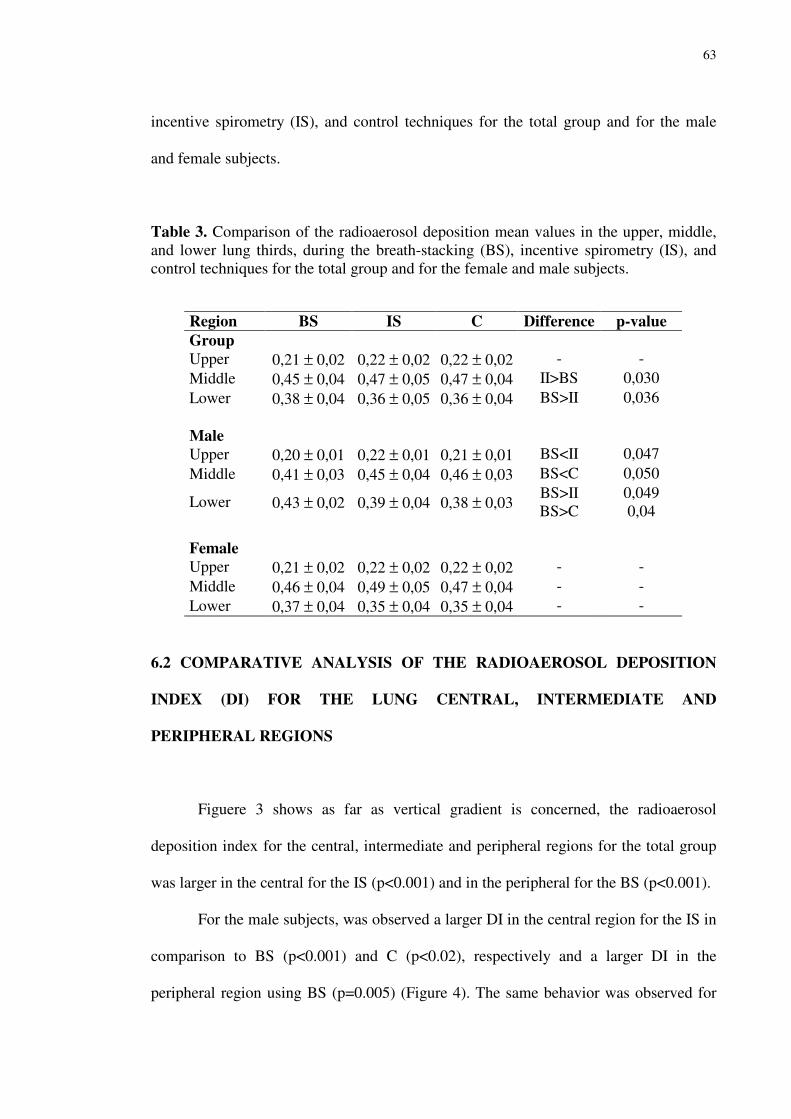
63
incentive spirometry (IS), and control techniques for the total group and for the male
and female subjects.
Table 3. Comparison of the radioaerosol deposition mean values in the upper, middle, and lower lung thirds, during the breath-stacking (BS), incentive spirometry (IS), and control techniques for the total group and for the female and male subjects.
Region BS IS C Difference p-value Group Upper 0,21 ± 0,02 0,22 ± 0,02 0,22 ± 0,02 - - Middle 0,45 ± 0,04 0,47 ± 0,05 0,47 ± 0,04 II>BS 0,030 Lower 0,38 ± 0,04 0,36 ± 0,05 0,36 ± 0,04 BS>II 0,036 Male Upper 0,20 ± 0,01 0,22 ± 0,01 0,21 ± 0,01 BS<II 0,047 Middle 0,41 ± 0,03 0,45 ± 0,04 0,46 ± 0,03 BS<C 0,050
Lower 0,43 ± 0,02 0,39 ± 0,04 0,38 ± 0,03 BS>II BS>C
0,049 0,04
Female Upper 0,21 ± 0,02 0,22 ± 0,02 0,22 ± 0,02 - - Middle 0,46 ± 0,04 0,49 ± 0,05 0,47 ± 0,04 - - Lower 0,37 ± 0,04 0,35 ± 0,04 0,35 ± 0,04 - -
6.2 COMPARATIVE ANALYSIS OF THE RADIOAEROSOL DEPOSITION
INDEX (DI) FOR THE LUNG CENTRAL, INTERMEDIATE AND
PERIPHERAL REGIONS
Figuere 3 shows as far as vertical gradient is concerned, the radioaerosol
deposition index for the central, intermediate and peripheral regions for the total group
was larger in the central for the IS (p<0.001) and in the peripheral for the BS (p<0.001).
For the male subjects, was observed a larger DI in the central region for the IS in
comparison to BS (p<0.001) and C (p<0.02), respectively and a larger DI in the
peripheral region using BS (p=0.005) (Figure 4). The same behavior was observed for
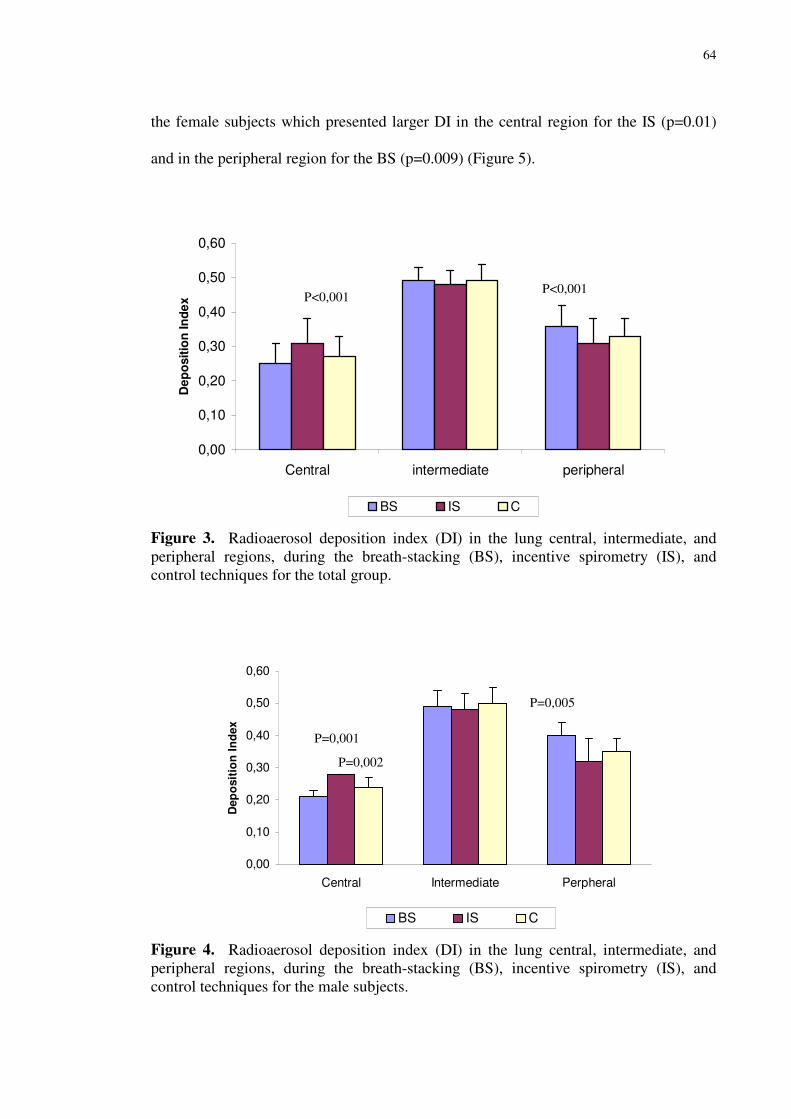
64
the female subjects which presented larger DI in the central region for the IS (p=0.01)
and in the peripheral region for the BS (p=0.009) (Figure 5).
0,00
0,10
0,20
0,30
0,40
0,50
0,60
Central intermediate peripheral
Dep
ositi
on In
dex
BS IS C
Figure 3. Radioaerosol deposition index (DI) in the lung central, intermediate, and peripheral regions, during the breath-stacking (BS), incentive spirometry (IS), and control techniques for the total group.
0,00
0,10
0,20
0,30
0,40
0,50
0,60
Central Intermediate Perpheral
Dep
ositi
on In
dex
BS IS C
Figure 4. Radioaerosol deposition index (DI) in the lung central, intermediate, and peripheral regions, during the breath-stacking (BS), incentive spirometry (IS), and control techniques for the male subjects.
P<0,001 P<0,001
P=0,001
P=0,002
P=0,005

65
0,00
0,10
0,20
0,30
0,40
0,50
0,60
Central Intermediate Peripheral
Dep
ositi
on In
dex
BS IS C
Figure 5. Radioaerosol deposition index (DI) in the lung central, intermediate, and peripheral regions, during the breath-stacking (BS), incentive spirometry (IS), and control techniques for the female subjects.
6.3 ANALYSIS OF THE LUNG RADIOAEROSOL DEPOSITION INDEX (DI)
DURING THE TECHNIQUES BS, IS AND C FOR THE CENTRAL REGION
COMPARED TO THE INTERMEDIATE AND PERIPHERAL ONES.
For this analysis, one has considered the relation involving the central (CT),
intermediate (IT) regions plus the peripheral through the equation DICP = CT/ (IT
+ PR).
With this approach, it was observed a larger DICP for both IS and C. On the other
hand, it was observed a smaler DICP when the BS was used. It is evident that for the
total group a larger DICP for the IS when compared to BS (p<0.001) and C (p=0.018),
respectively. In the male and female subjects, the DICP presented the same behavior as
in the total group. For the male larger DICP for IS, related to the BS(p=0,001) and C
(p=0.032), respectively. In the female group, larger DICP for IS (p=0.008) related to BS
(Figure 6).
P=0,01 P=0,009

66
0,00
0,10
0,20
0,30
0,40
0,50
0,60
Total group male female
Dep
ositi
on in
dex
BS IS C
Figure 6. Radioaerosol deposition index (DICP) in the central region when compared to the intermediate plus the peripheral, during the breath-stacking (BS), incentive spirometry (IS), and control techniques for the total group, male, and female subjects.
6.4 CORRELATION BETWEEN THE MAXIMUM REACHED RESPIRATORY
VOLUME DURING THE BS AND IS TECHNIQUES, AND THE PULMONARY
RADIOAEROSOL PENETRATION INDEX.
It has not been observed any positive correlation between the pulmonary
breathe in volume reached during the breath-stacking and incentive spirometry
techniques with the radioaerosol deposition index (DICP) in the lung.
P<0,001
P=0,001
P=0,018
P=0,032
P=0,008
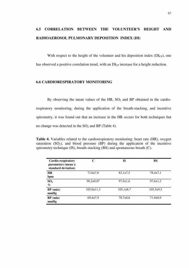
67
6.5 CORRELATION BETWEEN THE VOLUNTEER’S HEIGHT AND
RADIOAEROSOL PULMONARY DEPOSITION INDEX (DI)
With respect to the height of the volunteer and his deposition index (DICP), one
has observed a positive correlation trend, with an DICP increase for a height reduction.
6.6 CARDIORESPIRATORY MONITORING
By observing the mean values of the HR, SO2 and BP obtained in the cardio-
respiratory monitoring, during the application of the breath-stacking, and incentive
spirometry, it was found out that an increase in the HR occurs for both techniques but
no change was detected in the SO2 and BP (Table 4).
Table 4. Variables related to the cardiorespiratory monitoring: heart rate (HR), oxygen saturation (SO2), and blood pressure (BP) during the application of the incentive spirometry technique (IS), breath-stacking (BS) and spontaneous breath (C).
Cardio-respiratory parameters (mean ± standard deviation)
C IS BS
HR bpm
73,6±7,6 82,1±7,5 78,4±7,1
SO2 %
98,2±0,87 97,9±1,6 97,6±1,2
BP (máx) mmHg
105,0±11,3 105,1±8,7 105,5±9,5
BP (min) mmHg
69,4±7,9 70,7±8,6 71,9±8,9
68
DISCUSSION
The postoperative pulmonary complications and the atelectasis can be prevented
or treated through the use of several kinds of pulmonar expansion (5) techniques of
which, sustained maximum inspiration (SMI) (23), incentive spirometry (IS) (4;5;11),
the intermittent positive pressure breathing (IPPB) (2;3) and continuous positive airway
pressure (CPAP) (24) have been mostly employed, in addition to the BS which is an
alternative to the IS. Recently, some investigations concerning the use of these
techniques brought about some controversy with respect to their actual efficiency in the
reduction of pulmonary complications owing to cardiac and chest surgical procedures
(2;6;9;25;26).
Contradictory results have recently been observed in several reports concerning
the comparison of the IS, IPPB, CPAP, and standard physiotherapic techniques,
probably owing to the methodological limitations of the studies, where the effect
attributed to the IS can not be identified. This is due to fact that one has not considered
the effects of each one technique, leadind to either dubious or not random, besides the
observation of other therapeutic procedures including contamination due to the
application of the procedure to the experimental group, different criteria to define post-
surgical pulmonary complications, non-adequate population for the protocol,
inappropriate statistical analysis, a great number of volunteers who give up the study
and data comparison for different days with a different number of volunteers
(2;3;6;15;17;27).
The two IS and BS techniques are very important for the improvement of the
pulmonar ventilation, because they provide both lung volume enhancement and
maintain them as such for a prolonged period of time (16;18;27). However, it is very
69
difficult to apply the IS technique owing to either patient’s comprehention of the
technique or pain itself, dyspnea, muscular weakness and alteration in the diaphragm
mechanics. These effects may contribute to negatively affect the performance of the
technique leading to unsatisfactory results (7;13;15;28;30).
A reduced number of reports have utilyzed the BS technique with a small
number of individuals. In addition, the patients also felt unconfortable wearing the mask
provided for the experiment. Askanazi in 1980 observed that the use of components
such as: masks, mouthpiece, nasal clip, may alter the tidal volume. In the study herein
reported, one has used a mask for the BS and mouthpiece coupled to nasal clip for the
IS and C, the alterations in the inspiratory flow could be due to the alterations in the
tidal volume produced by the utilization of such devices.
In our investigation, we have compared the two techniques, analyzing the
radioaerosol deposition index (DI). In our study, we have analyzed the right lung, since
the proximity of the stomach and the left lung could lead misinterpretations owing to the
activity interposition eventually ingested during inhalation (31). Thus, the right lung
analysis produced more reliable results, since the group investigated consisted of
individuals with no detectable pathology.
Recent reports indicate that the radioaerosol lung deposition can exhibit different
behaviors owing to the sex, height, and weight, and as a result in the body mass index of
the individual (MBI) (22;32;33). For this reason we have opted for the DI analysis in
the total group and the individualyzed form for the male and female subjects.
Our results show that the deposition distribution is different between techniques
as far as vertical gradient is concerned. Thus, it was observed that using the IS
technique, there exists a larger deposition in the middle third, while the BS favours a
lower third deposition. This behavior is assigned to the high flow generated by the
70
individual during the use of IS (6;29;34), which contributes to the larger impaction of
the aerosol particles in the central region, corresponding to the larger caliber airways
(35). On the other hand, by using the BS technique, one obtained a lower flow or maybe
a laminated one (36) which could favor a radioaerosol deposition in distant regions and
as such depositing higher quantities in the lower third.
Due to the fact that was not possible to couple the volume incentive spirometer
(IS) to the nebulizer, and owing to the possibility of risky room contamination, in this
work we have used the IS flow-oriented (RESPIREX), which produces turbid airflow
leading to an increase in the respiratory work (6;37). A turbid airflow, larger than
30L/min, increase the particle deposition by impacting in fork airways and turbulent
flow passages (38). O’doherty and Miller in 1993 observed that as the airflow increases
the particles impacting increases as well in the central airways, explaining the results
herein presented. Although the volume IS technique it is advantageous, one points out
that in practice the use of the flow IS is very common since its presents a lower cost
than the volume counterpart. Thus, we believe it is important to elucidate the ventilation
behavior using such device.
Besides the flow, another factor that may have interfered in the results has to do
with the physiology of the respiratory mechanics itself, owing to the fact that the
ventilation per unit of volume decreases from the apex to the base of the lung which
resulted from regional intrapleural pressure differences generated by gravitation (39).
As the gravitational and the retractable forces of the pulmonary apex are in the
same direction, the intrapleural pressure presents more sub-atmospheric values. On the
other hand, in the base the gravitational force and the retractable forces are in opposite
direction, which favors a lower expansion and larger alveolar complacence in the base,
and in an opposite way for the apex alveolus with larger expansion and lower

71
complacence (21). This behavior explains the radioaerosol deposition during the BS
technique in the peripheral lung regions when compared to the IS. This is probably due
to larger air amount that reached the alveolus and by the air renewal itself, since
occurred increase in the radioaerosol particles which have been deposited (8).
When one analyses a group of male subjects in the horizontal gradient the IS
technique produces a grater radioaerosol deposition in the upper third and for the
spontaneous respiration (C) in the middle third, suggesting a possible influence of the
volunteer’s height in the deposition pattern (33). In the present study, the male group
presented significantly higher stature than the female group. Similar results have been
reported by Graham et al. (1990). The BS technique maintained the same behavior for
the total group, indicating that the stature only plays an important role in the
radioaerosol deposition pattern for the IS and the spontaneous respiration.
For the female group one did not observe any difference in the DI of the aerosol
in the studied regions neither for the techniques nor for the control group, suggesting
that the deposition preference for different regions of the lung may reside in the
interference caused by the stature of the individual, as described elsewhere by Gerrity
(1983) in his particle retention model. A similar pattern has also been observed by
Machado (1996) in six healthy and six asthmatic women. The results suggest that
independently of the ventilation pattern adopted the radioaerosol behavior it is
unaltered. However, as we investigated only a small number of male volunteers, one can
not infer a reliable conclusion, since the there exist differences in the respiratory
functional parameters for each sex group (6).
For the horizontal gradient the DI analysis was performed for the central,
intermediate and peripheral regions, and we have observed a larger DI in the central
region for the IS and in the peripheral for the BS, with the total group being considered.

72
This behavior is similar to the observed in the vertical gradient, since it also favor the
deposition in the large caliber airways for the IS technique and in the peripheral airways
for the BS one. However, Amis et al., 1984 and Newhouse et al., 1986 did not observed
any alveolar ventilation vertical gradient per unit of volume in the erectus position
(standstill position) (39; 40). Thus, The neural drive roused during the application of the
BS with the accumulation of successive inspirations for prolonged time may have
produced a uniform distribution of particles through the gravitational sedimentation
mechanism (40).
The sedimentation occur when the particles are capable of deposit themselves in
the airways and alveolus (35), which happens from the suspension particles separation
by gravity action. This separation mechanism is favored by timing and low inspiratory
flows. O’Doherty and Miller (1993) observed that the 10 seconds inspiration pause after
inhalation favors the particles sedimentation and their maintenance in the lung. The BS
technique may have caused a similar effect as the pause one, producing a larger index of
radioaerosol deposition in the distant and small caliber airways.
When the separate sexes were analyzed, the same behavior as the total group
was observed, suggesting that the stature of the individual influences only the
deposition in the horizontal gradient, probably due to the uniformity of the intrapleural
pressure in the horizontal gradient (39).
A second analysis in the horizontal gradient considering the DICP as the ratio of
the deposition in the central region and the deposition in the intermediate plus the
peripheral (DI=CT/IT+PR), one has obtained a larger DICP for the IS technique when
compared to the BS and spontaneous respiration. These results are similar to the ones
above cited which favor the deposition of particles during the BS technique in the large
caliber airways, in the pulmonary hilow, while during the BS occurs a predominant
73
deposition in the small caliber airways, suggesting larger penetration of the particles in
the distant portions (alveolus) (8).
In our research, we have observed an increase in the heart rate (HR) during the
application of the BS and IS techniques, probably due to the intrapleural
subathmospheric pressure levels that produces an increase in the venous feedback and
as a consequence an increase of HR (16). However, the arterial blood pressure (BP)
remained unchanged, suggesting that an alteration in FC was not enough to produce
larger hemodynamic effects in BP.
It did not occur correlation between the reached inspiration volume and the
radioaerosol DI. These findings indicate that the larger DI in the peripheral regions of
the lung for the BS technique are not related to the volume or duration of the technique,
but probably are due to the BS itself. Our findings agreed quite well with the ones
reported by Strider et al. (1994), where it was verified that the reduction in the
pulmonary shunt with the use of BS technique, did not correlates with the volume and
time duration of the technique application, but due to the large alveolar expansion
produced by it (34).
CONCLUSION
The results herein presented indicate that the utilized techniques in this research,
reach distinct lung regions as far as pulmonary ventilation is concerned, and that they
can useful in clinic practices. The breath-stacking technique can be used for the re-
expansion of the lower third and peripheral regions of the lung, particularly in
pathologies of the base and distant airways. On the other hand, the incentive spirometry
technique may be useful to expand the middle third and treat alterations that affect the
ventilation of intermediate regions of the lung.
74
REFERENCES
(1) Askanazi J, Silverberg PA, Foster RJ, Hyman AI, Milicemili J, Kinney JM. Effects of Respiratory Apparatus on Breathing Pattern. Journal of Applied Physiology 1980; 48(4):577-580.
(2) Baker WL, Lamb VJ, Marini JJ. Breath-Stacking Increases the Depth and Duration of Chest Expansion by Incentive Spirometry. American Review of Respiratory Disease 1990; 141(2):343-346.
(3) BAKOW ED. Sustained Maximal Inspiration - a Rationale for its use. Respiratory Care 22 nº 4, 379-382. 1977.
(4) Bartlett RH, Brennan ML, GAZZANIG.AB, Hanson EL. Studies on Pathogenesis and Prevention of Postoperative Pulmonary Complications. Surgery Gynecology & Obstetrics 1973; 137(6):925-933.
(5) Bartlett RH, GAZZANIG.AB, Geraghty TR. Respiratory Maneuvers to Prevent Postoperative Pulmonary Complications - Critical Review. Jama-Journal of the American Medical Association 1973; 224(7):1017-1021.
(6) Bastin R, Moraine JJ, Bardocsky G, Kahn RJ, Melot C. Incentive spirometry performance - A reliable indicator of pulmonary function in the early postoperative period after lobectomy? Chest 1997; 111(3):559-563.
(7) Becklake MR, Toyota B, Stewart M, Hanson R, Hanley J. Lung Structure As A Risk Factor in Adverse Pulmonary Responses to Asbestos Exposure - A Case-Referent Study in Quebec Chrysotile Miners and Millers. American Review of Respiratory Disease 1983; 128(3):385-388.
(8) CAMPANHA L. Patter of respiratory muscle recruitment during incentive spirometry and breath-stacking. DORNELAS DE ANDRADE A, RODRIGUES-MACHADO, editors. European Respiratory Journal 20 Nº 38. 2002.
(9) CELLI BR. A controlled trial of intermittent positive pressure breathing, incentive spirometry, and deep breathing exercises in preventing pulmonary complications after abdominal surgery. RODRIGUEZ KS, SNIDER GL, editors. Am Rev Respir Dis 130, 12-15. 1984.
(10) Craig DB. Postoperative Recovery of Pulmonary-Function. Anesthesia and Analgesia 1981; 60(1):46-52.
(11) Crowe JM, Bradley CA. The effectiveness of incentive spirometry with physical therapy for high-risk patients after coronary artery bypass surgery. Physical Therapy 1997; 77(3):260-268.
75
(12) DORNELAS DE ANDRADE A. Analisis of the variation inspiratory muscle strengt using flow and volumetric spirometers. FRANÇA EET, editor. 13º iNTERNATIONAL cONGRESS OF THE WORLD CONFEDERATION FOR PHYSICAL THERAPY Anais. 1999.
(13) DORNELAS DE ANDRADE A. Influence of rate flow variation on dead volume from diferent jet nebulizers. MARINHO PE, editor. 13º iNTERNATIONAL cONGRESS OF THE WORLD CONFEDERATION FOR PHYSICAL THERAPY Anais. 1999.
(14) Dull JL, Dull WL. Are Maximal Inspiratory Breathing Exercises Or Incentive Spirometry Better Than Early Mobilization After Cardiopulmonary Bypass. Physical Therapy 1983; 63(5):655-659.
(15) Gerrity TR, Garrard CS, Yeates DB. A Mathematical-Model of Particle Retention in the Air-Spaces of Human Lungs. British Journal of Industrial Medicine 1983; 40(2):121-130.
(16) GIORGI MCP. MEDICINA NUCLEAR. TERRA FILHO M, MENEGHETTI JC, editors. Assistência Ventilatória Mecânica COSTA AJ Ed. Atheneu SP, 407. 1995.
(17) Gosselink R, Schrever K, Cops P, Witvrouwen H, De Leyn P, Troosters T et al. Incentive spirometry does not enhance recovery after thoracic surgery. Critical Care Medicine 2000; 28(3):679-683.
(18) Graham DR, Chamberlain MJ, Hutton L, King M, Morgan WKC. Inhaled Particle Deposition and Body Habitus. British Journal of Industrial Medicine 1990; 47(1):38-43.
(19) Hall JC, Tarala R, Harris J, Tapper J, Christiansen K. Incentive Spirometry Versus Routine Chest Physiotherapy for Prevention of Pulmonary Complications After Abdominal-Surgery. Lancet 1991; 337(8747):953-956.
(20) Jenkins RT, Rooney PJ, Coates G, Hunt RH, Bienenstock J. Increased Intestinal Permeability in Rheumatoid-Arthritis - Disease Or Drug Effect. Clinical and Investigative Medicine-Medecine Clinique et Experimentale 1985; 8(3):A216.
(21) Kaneko K, MILICEMI.J, Dolovich MB, Dawson A, Bates DV. Regional Distribution of Ventilation and Perfusion As A Function of Body Position. Journal of Applied Physiology 1966; 21(3):767-&.
(22) Machado MGR. Função respiratória em indivíduos normais e asmáticos em decorrência da utilização de pressão expiratória positiva. Tese de Mestrado em Fisiologia e Biofísica da UFMG . 1996.
76
(23) Marini JJ, Rodriguez RM, Lamb VJ. Involuntary Breath-Stacking - An Alternative Method for Vital Capacity Estimation in Poorly Cooperative Subjects. American Review of Respiratory Disease 1986; 134(4):694-698.
(24) Melendez JA, Alagesan R, Reinsel R, Weissman C, Burt M. Postthoracotomy Respiratory Muscle Mechanics During Incentive Spirometry Using Respiratory Inductance Plethysmography. Chest 1992; 101(2):432-436.
(25) O'DOHERTY MJ, Miller RF. Aerosols for therapy and diagnosis. Eur J Nucl Med 20 Nº 12, 1201-1213. 1993.
(26) O'DONOHUE. National survey of the usage of lung expansion modalities for
the prevention and treatment of postoperative atelectasis following abdominal and thoracic surgery. Chest 87 Nº1. 1985.
(27) Oikkonen M, Karjalainen K, Kahara V, Kuosa R, Schavikin L. Comparison of Incentive Spirometry and Intermittent Positive Pressure Breathing After Coronary-Artery Bypass Graft. Chest 1991; 99(1):60-65.
(28) Overend TJ, Anderson CM, Lucy SD, Bhatia C, Jonsson BI, Timmermans C. The effect of incentive spirometry on postoperative pulmonary complications - A systematic review. Chest 2001; 120(3):971-978.
(29) Pavia D, Bateman JRM, Sheahan NF, Agnew JE, Clarke SW. Tracheo-Bronchial Mucociliary Clearance in Asthma - Impairment During Remission. Thorax 1985; 40(3):171-175.
(30) ROS A M. Prevention of pulmonary complications after abdominal surgery. VINCENT JL, KAHN RJ, editors. Acta Anaesthesiologica nº2, 167-174. 1981.
(31) Schwieger I, Gamulin Z, Forster A, Meyer P, Gemperle M, Suter PM. Absence of Benefit of Incentive Spirometry in Low-Risk Patients Undergoing Elective Cholecystectomy - A Controlled Randomized Study. Chest 1986; 89(5):652-656.
(32) SILVA M. Longitudinal study of the inspiratory capacity evaluated by the incentive spirometry and breath-stacking technique after coronary artery by pass sugerry. RODRIGUES-MACHADO MG, editor. 2002.
(33) Spengler CM, Shea SA. Endogenous circadian rhythm of pulmonary function in healthy humans. American Journal of Respiratory and Critical Care Medicine 2000; 162(3):1038-1046.
(34) Strider D, Turner D, Egloff MB, Burns SM, Truwit JD. Stacked Inspiratory Spirometry Reduces Pulmonary Shunt in Patients After Coronary-Artery Bypass. Chest 1994; 106(2):391-395.
(35) Thomas JA, Mcintosh JM. Are Incentive Spirometry, Intermittent Positive Pressure Breathing, and Deep Breathing Exercises Effective in the Prevention of

77
Postoperative Pulmonary Complications After Upper Abdominal-Surgery - A Systematic Overview and Metaanalysis. Physical Therapy 1994; 74(1):3-10.
(36) Weindler J, Kiefer RT. The efficacy of postoperative incentive spirometry is influenced by the device-specific imposed work of breathing. Chest 2001; 119(6):1858-1864.
(37) Weiner P, Man A, Weiner M, Rabner M, Waizman J, Magadle R et al. The effect of incentive spirometry and inspiratory muscle training on pulmonary function after lung resection. Journal of Thoracic and Cardiovascular Surgery 1997; 113(3):552-557.
(38) WEST JB. Fisiologia Respiratória Moderna: Fundamentos e Aplicações. Interlivros RJ . 1990.
(39) AMIS, T. C., JONES, H. A., HUGHES, J. M. B. Effect of Posture on Inter-
Regional Distribution of Pulmonary Ventilation in Man. Respiration Physiology, v. 56, n. 2, p. 145-167, 1984.
(40) NEWHOUSE, M.; DOLOVICH, M. Aerosol therapy of asthma: principles and applications. Respiration, v. 50, Suppl: S2, p. 123-130.

78
5 CONCLUSÕES
Nossos resultados sugerem que as técnicas utilizadas atingem regiões
pulmonares distintas em termos de ventilação pulmonar, podendo ser úteis na prática
clínica.
A técnica do “breath-stacking” pode ser utilizada para reexpansão do terço
inferior e regiões periféricas do pulmão, principalmente em patologias que acometem
base e vias aéreas distais.
A técnica do inspirômetro de incentivo poderá ser útil para expandir terço médio
e tratar alterações que comprometam a ventilação de regiões intermediárias do pulmão.

79
6 PERSPECTIVAS
Ressalta-se a necessidade, de estudos do padrão regional de deposição pulmonar
de radioaerossóis durante a execução das técnicas do Breath-Stacking e Inspirômetro de
Incentivo na população dos pacientes que apresentam redução da ventilação pulmonar,
verificando-se as repercussões destas técnicas em longo prazo, e a validade terapêutica
das mesmas em reduzir as alterações ventilatórias freqüentes nos pós-operatórios e nos
períodos de hospitalização prolongados.
Outro aspecto que merece novos estudos é a análise do comportamento
perfusional para avaliar a relação ventilação/perfusão em ambas as técnicas.

80
APÊNDICE Apêndice A- Modelo do formulário de avaliação pulmonar inicial
NOME:__________________________________________________ Nº______ SEXO:________
TELEFONE:____-____._______ CEL.:_________________DATA:___/___/___IDADE:_____anos
DATA NASC.:___/___/___/ ALTURA: ____________m PESO:______Kg HORA: _______Hs
1-Anamnese: 1.1.Fumo: _______________________________________________________________________ 1.2.Doença pulmonar: _____________________________________________________________ 1.3.Doença cardíaca: ______________________________________________________________ 1.4.Doença neuromuscular: _________________________________________________________ 1.5.Pratica atividade física: _________________________________________________________ 1.6.Ausculta Pulmonar: ____________________________________________________________
2-P.A.: ___________mmHg FC.: _______bpm F.R.: _______irm Saturação Basal: ___________% 3-Pressões Respiratórias (Data:__/__/__/ Hora:_____hs):
3.1.Pimáx: 1ª__________mmHg Melhor: ______________mmHg 2ª__________mmHg Média Pimáx: _________mmHg 3ª__________mmHg 3.2.Pemáx: 1ª__________mmHg Melhor: ______________mmHg 2ª__________mmHg Média Pimáx: _________mmHg 3ª__________mmHg
4-Ventilometria (Data:__/__/__/ Hora:_____hs):____________ml F.R.:_____irm VC:____________ml 5-Peak-Flow (Data:__/__/__/ Hora:_____hs):
1ª__________L/min. Melhor:______________L/min. 2ª__________L/min. Média Peak-Flow: ________L/min. 3ª__________L/min.
6-Espirometria (Data:__/__/__/ Hora:_____hs):
CVF:___________ FEV1:___________ FEF25 :___________ FEF75:___________ 7-Execução das Técnicas (RESUMO):
Fase 1 (sem o circuito do aerosol) Data:__/__/__/ Hora:_____hs Breath-Stacking Inspirômetro de Incentivo
Volume: Saturação: Volume: Saturação: F.R.: F.C.: F.R.: F.C.: Tempo: P.A.: Tempo: P.A.:
Fase 2 (com o circuito do aerosol) Data:__/__/__/ Hora:_____hs Breath-Stacking Inspirômetro de Incentivo
Volume: Saturação: Volume: Saturação: F.R.: F.C.: F.R.: F.C.: Tempo: P.A.: Tempo: P.A.:
Fase 3 (durante a realização da cintilografia) Data:__/__/__/ Hora:_____hs
Breath-Stacking Inspirômetro de Incentivo Contagem: Saturação: Contagem: Saturação: F.R.: F.C.: F.R.: F.C.: Tempo: P.A.: Tempo: P.A.:
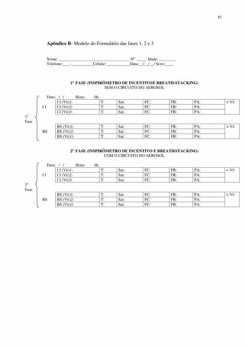
81
Apêndice B- Modelo do Formulário das fases 1, 2 e 3 Nome: __________________________________ Nº _____ Idade: ________ Telefone:____-__________Celular: ___________Data:__/__/__/ Sexo:____
1ª FASE (INSPIRÔMETRO DE INCENTIVO E BREATH-STACKING) SEM O CIRCUITO DO AEROSOL
Data: / / Hora: Hs
I I (VE)1: T: Sat: FC: FR: PA: > VE I I I I (VE)2: T: Sat: FC: FR: PA: I I (VE)3: T: Sat: FC: FR: PA: 1a
Fase
BS (VE)1: T: Sat: FC: FR: PA: > VE BS BS (VE)2: T: Sat: FC: FR: PA: BS (VE)3: T: Sat: FC: FR: PA:
2ª FASE (INSPIRÔMETRO DE INCENTIVO E BREATH-STACKING) COM O CIRCUITO DO AEROSOL
Data: / / Hora: Hs
I I (VE)1: T: Sat: FC: FR: PA: > VE I I I I (VE)2: T: Sat: FC: FR: PA: I I (VE)3: T: Sat: FC: FR: PA: 2a
Fase
BS (VE)1: T: Sat: FC: FR: PA: > VE BS BS (VE)2: T: Sat: FC: FR: PA: BS (VE)3: T: Sat: FC: FR: PA:

82
3ª FASE (CINTILOGRAFIA PULMONAR)
EXAME _______________ (se for o exame padrão não tem manobra)
MANOBRA___________________________ DATA:___/___/___/ HORA:______Hs
NOME:____________________________________________________Nº________
CONTAGEM TOTAL
PULMÃO DIREITO PULMÃO ESQUERDO Nº de pixels Max counts Min counts Mean counts Total counts
CONTAGENS EM TERÇOS SUPERIOR, MÉDIO E INFERIOR
PULMÃO DIREITO 1/3 SUPERIOR 1/3 MÉDIO 1/3 INFERIOR Nº de pixels Max counts Min counts Mean counts Total counts PULMÃO ESQUERDO 1/3 SUPERIOR 1/3 MÉDIO 1/3 INFERIOR Nº de pixels Max counts Min counts Mean counts Total counts
CONTAGENS EM ZONAS HILAR, CENTRAL E PERIFÉRICA
PULMÃO DIREITO HILAR CENTRAL PERIFÉRICA Nº de pixels Max counts Min counts Mean counts Total counts PULMÃO ESQUERDO HILAR CENTRAL PERIFÉRICA Nº de pixels Max counts Min counts Mean counts Total counts Observações:__________________________________________________________________________________________________________________________________________________________________________________________________
83
ANEXOS
Anexo A- Modelo de Termo de Compromisso do orientador
TERMO DE COMPROMISSO
Pelo presente instrumento, eu Armèle de Fátima Dornelas de Andrade,
CREFITO 4772-F, assumo a orientação da dissertação de mestrado da mestranda em
Fisiologia Denise Maria Sá Machado, cujo tema é “Padrão regional de ventilação
pulmonar durante as técnicas do breath-stacking e inspirômetro de incentivo pela
inalação de radioaerossol”, e me responsabilizar por cumprir os termos necessários
desde a elaboração até a conclusão do mesmo.
Recife, de de 2002
_____________________________
Profa. Dra. Armèle Dornelas de Andrade SIAPE 11132476/ CREFITO 4772-F

84
Anexo B- Modelo de Termo de compromisso do aluno
TERMO DE COMPROMISSO
Pelo presente termo, eu Denise Maria Sá Machado, CREFITO 13866-F,
mestranda em Fisiologia, me responsabilizo a só iniciar a coleta de dados da dissertação
de mestrado intitulada, “Padrão regional de ventilação pulmonar durante as técnicas do
breath-stacking e inspirômetro de incentivo pela inalação de radioaerossol”, mediante a
aprovação pelo Comitê de Ética em Pesquisa do Centro de Ciências da Saúde.
Recife, de de 2002
_____________________________
Denise Maria Sá Machado CREFITO: 13866-F

85
Anexo C- Modelo do Termo de consentimento do voluntário
TERMO DE CONSENTIMENTO
Eu,_____________________________________________________________
maior de idade, portador da cédula de identidade n.º ________________,SSP________, PF____________________,profissão________________________________________, residente___________________________________________________nº__________, bairro____________________________,cidade_______________________,UF_____, abaixo assinado, concordo em participar como voluntário no Projeto de Pesquisa: “Padrão regional de ventilação pulmonar durante as técnicas do breath-stacking e inspirômetro de incentivo pela inalação de radioaerossol”, a ser desenvolvido pela mestranda Denise Maria Sá Machado, no Laboratório de Fisioterapia Cardiopulmonar do Departamento de Fisioterapia da Universidade Federal de Pernambuco e no Departamento de Medicina Nuclear do Hospital das Clínicas de Pernambuco. Declaro, também, que recebi por escrito, anexado a este termo, os objetivos da presente pesquisa, a metodologia a ser aplicada, os riscos e benefícios inerentes, a relevância social e científica do trabalho e estou ciente de meu direito de solicitar novas informações, verbalmente ou por escrito, de todos os procedimentos experimentais, que não serei identificado e que poderei a qualquer momento e por qualquer motivo, cancelar o presente consentimento e interromper minha participação neste projeto de pesquisa.
Recife, de 2002
______________________________ Voluntário

86
Anexo D- Modelo de termo de consentimento livre e esclarecido
TERMO DE CONSENTIMENTO LIVRE ESCLARECIDO PARA PARTICIPAÇÃO EM ESTUDO
CLÍNICO
TÍTULO: Padrão regional de ventilação pulmonar durante as técnicas do breath-stacking e inspirômetro de incentivo pela inalação de radioaerossol. Investigador: Nome: Denise Maria Sá Machado Armèle Dornelas de Andrade Nome dos locais de estudo: # Laboratório de Fisioterapia Cardiorrespiratória Departamento de Fisioterapia – UFPE Endereço: Cidade Universitária Recife-PE Fone: 32718496 # Laboratório de Fisioterapia Cardiorrespiratória Curso de Fisioterapia FIC – CE Endereço: Vicente Linhares, 308 Aldeota Fortaleza-CE # Instituto de Medicina Nuclear Endereço: Carlos Vasconcelos, 977 Aldeota Fortaleza-CE
Este termo de consentimento pode conter palavras que você não entenda. Por favor, pergunte ao profissional ou a equipe que acompanha o estudo a respeito de qualquer palavra ou informação que você não entenda claramente.
O nosso estudo tem por objetivo avaliar a forma como o ar entra e se distribui nos pulmões durante a respiração realizada com um inspirômetro de incentivo (aparelho utilizado para expansão pulmonar durante o tratamento fisioterápico) e o breath-stacking (várias respirações acumuladas no pulmão, também durante o tratamento fisioterápico).
Essas duas manobras fisioterapêuticas serão avaliadas pela cintilografia pulmonar (exame que demonstra de que forma o ar inspirado se distribui nos dois pulmões).
No estudo haverá uma avaliação da função respiratória, da história clínica e dos dados vitais que durará aproximadamente uma hora. Numa segunda etapa o (a) senhor (a) irá ao Instituto de Medicina Nuclear de Fortaleza submeter-se ao exame de cintilografia pulmonar por três vezes num intervalo mínimo de sete dias entre eles, tendo cada exame a duração mínima de nove minutos.
O elemento químico inalado durante o exame de cintilografia pulmonar será o tecnécio (substância que possibilita a visualização de como o ar está chegando nos pulmões). A inalação será realizada na posição sentada, com a utilização de uma máscara facial. O (a) senhor (a) será orientado a usar uma respiração lenta e profunda. Durante a inalação serão verificados o número de batimentos cardíacos, o número de respirações por minuto e o nível de oxigênio presente no sangue. Para avaliar a deposição do tecnécio no pulmão será feito um exame em uma gama câmara (aparelho normalmente utilizado para realizar exames de cintilografia) por nove minutos, sendo adquiridas diversas imagens das regiões pulmonares.
RISCOS E DESCONFORTOS/BENEFÍCIOS O desconforto está associado à dificuldade de adaptação a máscara facial durante a inalação do tecnécio. Se
o (a) senhor (a) não se adaptar poderá ser retirado do estudo. Os riscos são referentes à emissão de radiação, no entanto, a dosagem utilizada no exame se associa a uma
outra substância (DTPA) que minimiza o período de permanência do tecnécio no corpo, diminuindo os riscos. O benefício para o senhor (a) será proporcionado pela avaliação da função respiratória e se encontrada alterações, será devidamente encaminhado para o Serviço de Fisioterapia Respiratória da FISIOFIC, Clínica de Fisioterapia da FIC.
As informações obtidas através deste estudo serão tratadas rigorosamente com confidencialidade. Os resultados desta pesquisa serão divulgados publicamente, entretanto sua identidade jamais será revelada.
A sua participação neste estudo é voluntária. E você pode recusar-se a participar ou parar a qualquer momento da pesquisa.
CONSENTIMENTO DO PACIENTE Li e entendi as informações precedentes descrevendo este estudo e todas as minhas dúvidas em relação ao
estudo e a minha participação no mesmo foi respondida satisfatoriamente. Dou livremente meu consentimento em participar deste estudo até que decida o contrário.
Autorizo a liberação dos meus registros médicos para o Laboratório de Fisioterapia cardiorrespiratória. ________________________________________ ________________________________________ _________
Nome do Voluntário (letra de forma) Assinatura do Voluntário DATA ________________________________________ ________________________________________ _________
Nome da Testemunha (letra de forma) Assinatura da Testemunha DATA ________________________________________ ________________________________________ _________
Nome da Testemunha (letra de forma) Assinatura da Testemunha DATA DENISE MARIA SÁ MACHADO ________________________________________ _________
Nome do Investigador (letra de forma) Assinatura do Investigador DATA
87
Anexo E- Protocolo de encaminhamento do estudo ao CEP para aprovação
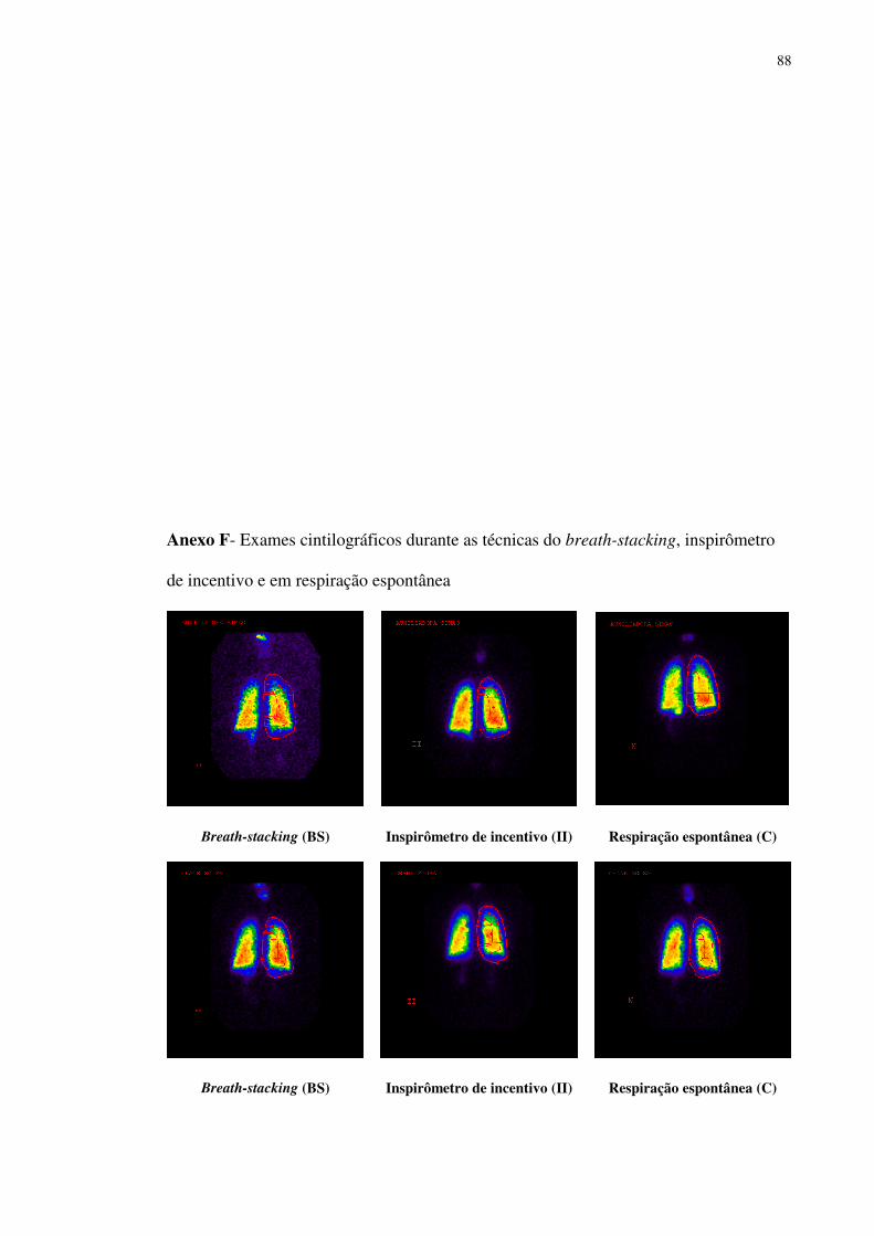
88
Anexo F- Exames cintilográficos durante as técnicas do breath-stacking, inspirômetro
de incentivo e em respiração espontânea
Breath-stacking (BS) Inspirômetro de incentivo (II) Respiração espontânea (C)
Breath-stacking (BS) Inspirômetro de incentivo (II) Respiração espontânea (C)

89
Breath-stacking (BS) Inspirômetro de incentivo (II) Respiração espontânea (C)
Breath-stacking (BS) Inspirômetro de incentivo (II) Respiração espontânea (C)
Breath-stacking (BS) Inspirômetro de incentivo (II) Respiração espontânea (C)
Breath-stacking (BS) Inspirômetro de incentivo (II) Respiração espontânea (C)

90
Breath-stacking (BS) Inspirômetro de incentivo (II) Respiração espontânea (C)
Breath-stacking (BS) Inspirômetro de incentivo (II) Respiração espontânea (C)
Breath-stacking (BS) Inspirômetro de incentivo (II) Respiração espontânea (C)
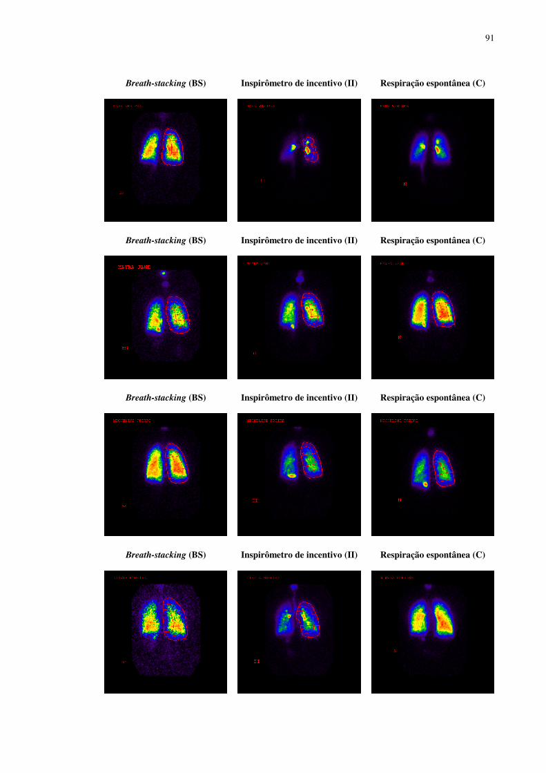
91
Breath-stacking (BS) Inspirômetro de incentivo (II) Respiração espontânea (C)
Breath-stacking (BS) Inspirômetro de incentivo (II) Respiração espontânea (C)
Breath-stacking (BS) Inspirômetro de incentivo (II) Respiração espontânea (C)
Breath-stacking (BS) Inspirômetro de incentivo (II) Respiração espontânea (C)
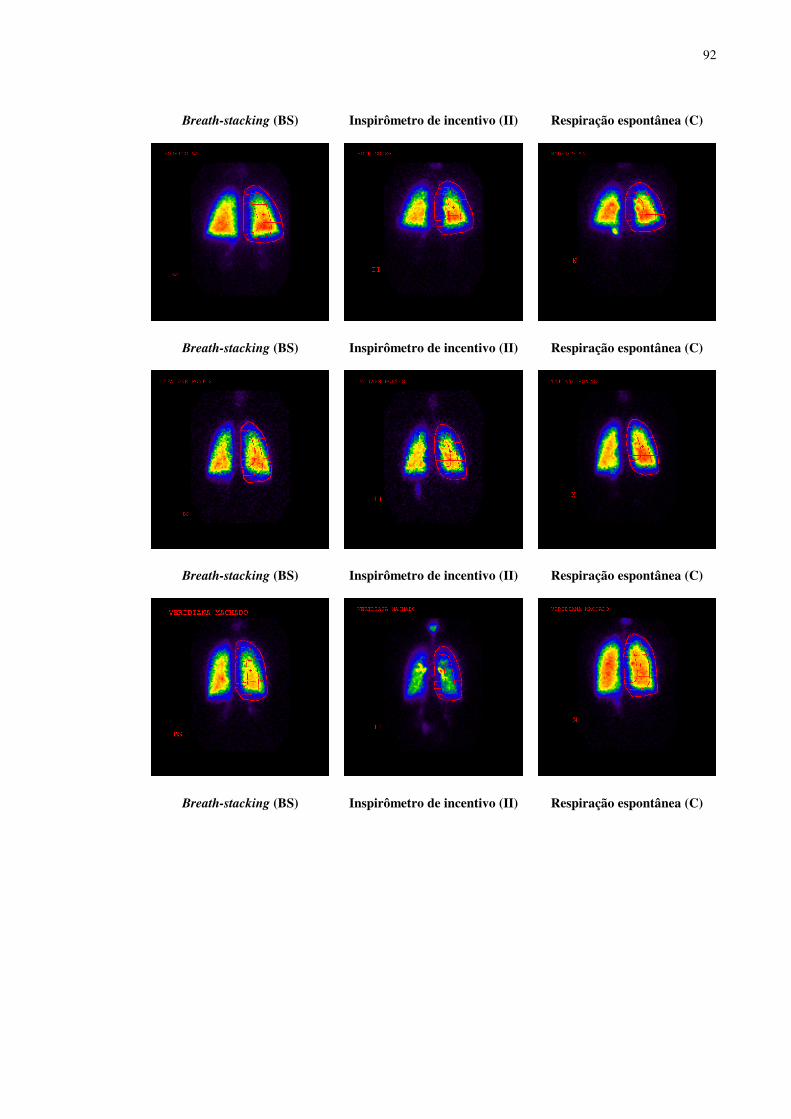
92
Breath-stacking (BS) Inspirômetro de incentivo (II) Respiração espontânea (C)
Breath-stacking (BS) Inspirômetro de incentivo (II) Respiração espontânea (C)
Breath-stacking (BS) Inspirômetro de incentivo (II) Respiração espontânea (C)
Breath-stacking (BS) Inspirômetro de incentivo (II) Respiração espontânea (C)
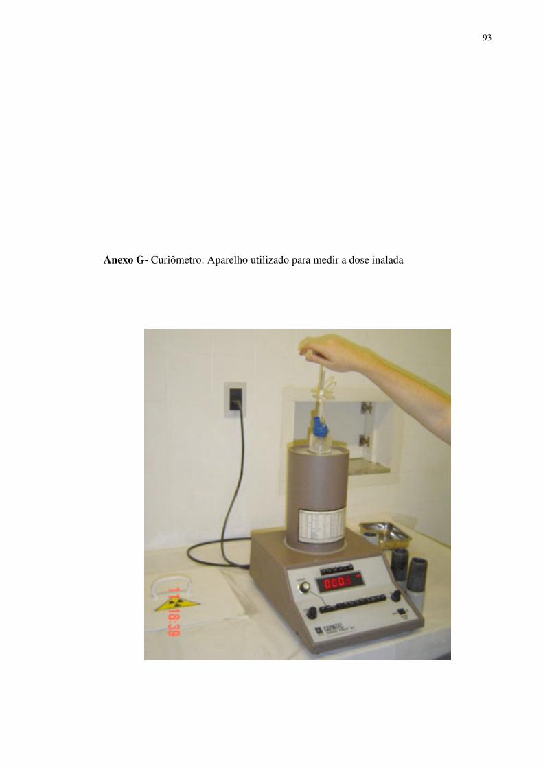
93
Anexo G- Curiômetro: Aparelho utilizado para medir a dose inalada

94
Anexo H- Inalação do radioaerossol em respiração espontânea e durante as técnicas do
breath-stacking e inspirômetro de incentivo
Respiração espontânea (C) Breath-stacking (BS) Inspirômetro de incentivo (II)
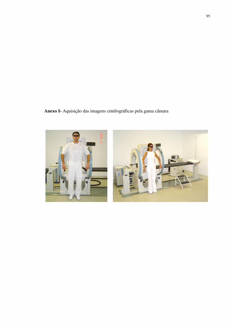
95
Anexo I- Aquisição das imagens cintilográficas pela gama câmara