Duarte João Neves Guerreiro -...
58
UNIVERSIDADE DE LISBOA FACULDADE DE CIÊNCIAS DEPARTAMENTO DE BIOLOGIA VEGETAL Characterization of Klebsiella pneumoniae bacteriocalins Duarte João Neves Guerreiro Mestrado em Microbiologia Aplicada Dissertação orientada por: Professor Miguel Valvano (Queen’s University Belfast) Professora Doutora Mónica Vieira Cunha (FCUL) 2016
Transcript of Duarte João Neves Guerreiro -...
UNIVERSIDADE DE LISBOA
FACULDADE DE CIÊNCIAS
DEPARTAMENTO DE BIOLOGIA VEGETAL
Characterization of Klebsiella pneumoniae bacteriocalins
Duarte João Neves Guerreiro
Mestrado em Microbiologia Aplicada
Dissertação orientada por:
Professor Miguel Valvano (Queen’s University Belfast)
Professora Doutora Mónica Vieira Cunha (FCUL)
2016
Characterization of Klebsiella pneumoniae bacteriocalins
Duarte João Neves Guerreiro
2016
This thesis was fully performed at Wellcome-Wolfson Institute for Experimental Medicine in
Queen’s University Belfast under the direct supervision of Professor Miguel Valvano in the
scope of the Master in Applied Microbiology of the Faculty of Sciences of the University of
Lisbon. This thesis was also supervised by Professor Mónica Vieira Cunha as the internal
supervisor at FCUL.

I
ACKNOWLEGDMENTS
First and foremost I would like to thank Professor Miguel Valvano for allowing me to
develop this thesis under his guidance at his lab and for all the patience and good will that he
showed, even when the odds for “good” results were statistically close to zero.
I thank Professor Laura Hobley for she had the patient to teach me about the not-so-
cooperative-Klebsiella pneumoniae and for showing many lab methodologies.
Also, I need to thank to my internal supervisor, Professor Monica Cunha, for her help and
for the ideia of a B plan that also worked in the end.
To my work group, Valvano’s lab team, for the team work, laughs, awesome environment
and share of knowledge during my stay.
To the future Professor Nuno Lopes for he pushed me to become more ambicious and
persue even more difficult challenges. Also, for those times that we procrastinated along with
other Civilizations.
And a special thanks to Ana Marques for she gave me the strength and support to
continue to work on this thesis even when everything seemed about to collapse. For she, even
from a distance of 1785 km or less than 3, made me laugh and kept me focus on the objective
that made me go away.
II
RESUMO Desde a descoberta dos primeiros antibióticos em meados do seculo XX, que estes têm
vindo a ser utilizados, frequentemente, de forma indiscriminada para combater infeções
bacterianas, o que tem sido associado a um aumento do número de infeções causadas por
bactérias multirresistentes (MDRs). Apesar de várias medidas terem sido já implementadas para
evitar a incidência de microrganismos multirresistentes, estas têm-se mostrado ineficazes,
ficando a comunidade científica responsável por encontrar novas formas de combater os
mecanismos de resistência bacterianos.
Um dos mecanismos recentemente descritos envolve a actuação de proteínas
denominadas lipocalinas bacterianas (BCNs, anteriormente conhecidas por YceI). Inicialmente
descritas em Burkholderia cenocepacia K56-2, estas proteínas são secretadas pelas bactérias
capturando antibióticos hidrofóbicos no meio extracelular, impedindo o antibiótico de atuar sobre
a bactéria, aumentando assim a concentração mínima inibitória (CIM) do respetivo antibiótico.
Este mecanismo não aparente ser restrito às bactérias produtoras de BCNs, exercendo
actividade e proteção sobre a comunidade bacteriana envolvente.
As BCNs consistem em proteínas de baixa massa molecular, altamente conservadas
entre as bactérias, apresentando uma conformação característica tridimensional (3D) de barril-
β, seguida por uma hélice-α. Geralmente secretadas para o meio periplasmático, livres ou
ancoradas à membrana plasmática, estas proteinas podem ainda surgir no meio extracelular ou,
ainda, no citosol da bactéria. As BCNs descritas até à data ainda se encontram muito pouco
caracterizadas funcionalmente, desconhecendo-se a capacidade de ligação de proteínas
ortologas das descritas originalmente em alguns organismos aos antibioticos. Desta forma, esta
tese focou-se no estudo do homólogo de BCN em Klebsiella pneumoniae kp52.145 designado
BcnK. Esta bactéria Gram-negativa, pertencente à família das Enterobacteriaceae, é um
importante agente patogénico responsável por surtos de pneumonia, entres outras infeções, em
ambientes hospitalares e na comunidade. Tal como se verifica com outros agentes patogénicos,
o número de estirpes de K. pneumoniae multirresistentes tem vindo a aumentar. Os principais
mecanismos de resistência presentes nas estirpes clínicas desta espécie consistem em: (i)
produção de β-lactamases de largo espectro de atuação, capazes de hidrolisar cefalosporinas e
antibióticos do grupo dos monobactâmicos, ou (ii) produção de carbapenemases, que possuem
a capacidade de hidrolisar um largo espectro de antibióticos, incluindo carbapenemes. Estes
genes possuem a capacidade de se propagar horizontalmente entre estirpes da mesma ou outras
espécies, tendo sido detetadas em várias regiões do mundo, constituindo assim uma ameaça
para a saúde pública.
Assim, a fim de se caracterizar funcionalmente a proteína BcnK, o gene correspondente
(bcnK) foi expresso por clonagem em Escherichia coli no vetor pDA-CTHis, contendo uma cauda
de seis histidinas na extremidade C-terminal, dando origem a pDG1. A proteína recombinante
expressa foi purificada com sucesso por cromatografia de afinidade. A verificação da expressão
desta proteína foi efectuada através de SDS-PAGE e Western-blot. Apesar de se ter verificado
que a expressão de BcnK conferiu um aumento da CMI, traduzido por uma diferença de
III
crescimento de cerca de 65.5% OD600 a uma concentração de 1.0 µg/mL de polimixina B, em
testes de proteção com Pseudomonas aeruginosa PAO1, não foi possível repetir este ensaio
devido à agregação da proteína observada durante a diálise em tampão PBS, o que
impossibilitou a quantificação da proteína e a sua utilização em ensaios subsequentes. Especula-
se que a agregação observada se deveu, possivelmente, à componente lipídica N-acil-S-sn-1,2-
diacilcerilcisteína presente na exterminade N-terminal de BcnK. Desta forma, procedeu-se a nova
tentativa de clonagem de bcnK no mesmo vetor, removendo-se a sequência do péptido sinal.
Contudo, não se obtiveram quantidades suficientes da proteína produzida para se proceder à
sua purificação. Assim, alternativamente, clonou-se bcnK no vetor induzível por IPTG, pET-28a
(+), contendo caudas de histidina nas extremidades N- e C- terminal (pDG7) e, em paralelo,
clonou-se no mesmo vetor, usando apenas uma cauda de histidinas na extremidade N-terminal
(pDG8). A expressão do gene bcnK nestes vetores levou à produção de quantidades suficientes
de proteína para purificação. No entanto, a expressão da construção genética em pDG7 conduziu
a elevados níveis de agregação da proteína durante a diálise, possivelmente devido à presença
das caudas de histidina. Contrariamente ao esperado, a expressão de BcnK em pDG8 não
conduziu ao aumento da CMI nos ensaios de proteção. Desta forma, admitiu-se a hipótese de
que a proteína recombinante produzida sem péptido sinal teria uma conformação incorreta para
o exercício da sua função biológica ou de que a cauda de histidinas presente na extremidade N-
terminal pudesse gerar interferência com a atividade da proteína. Um novo plasmídeo foi
construído utilizando o vetor pUC19 contendo uma caude de histidinas na extremidade C-
terminal. Contudo, a expressão neste plasmídeo também não produziu quantidades suficientes
de proteína para purificação. Uma nova abordagem será realizada ao clonar bcnK num vector
contendo um péptido sinal, secretando BcnK para o espaço perisplásmico sem a componente
lipidica N-acil-S-sn-1,2-diacilcerilcisteína, permitindo a solubilização da proteína.
De forma a investigar o papel de BcnK na resistência a antibióticos exibida por K.
pneumoniae, tentou-se inativar bcnK no genoma bacteriano por duas metodologias, através de
mutagénese dirigida não marcada, o que permitiria a obtanção de um mutante de eliminação
isogénico, e através de mutação por inserção, por recurso aos plasmídeos construídos neste
trabalho, pDG2 e pDG9, respetivamente. No entanto, não foram obtidos mutantes por quaiquer
dos métodos, tendo-se obtido mutantes merodiplóides apenas na estratégia de inativação por
mutagénese dirigida não marcada. Assim, em alternativa, procurou-se testar a essencialidade
de BcnK por expressão de bcnK sob o controlo de um promotor induzível por ramnose. Contudo,
foi necessário testar a funcionalidade deste promotor em K. pneumoniae. Para o efeito, utilizou-
se o vetor pSCrhaB2-e-GFP, que tem um promotor induzível por ramnose fundido
transcricionalmente com o gene GFP (green fluorescent protein), o qual permite a deteção da
sua expressão por fluorescência. Conjugou-se em K. pneumoniae, a qual foi crescida em 0.2%
e 0.5% de ramnose e 0.5% glucose, respetivamente, tendo-se registado fluorescência na
presença das diferentes concentrações de ramnose e, por outro lado, a ausência de
fluorescência na presença de glucose, o que sugere o correto funcionamento do promotor no
hospedeiro K. pneumoniae. Um fragmento de bcnK foi então clonado no vetor suicida contendo
IV
um promotor induzível por ramnose pSC200, dando origem a pDG10, sendo este posteriormente
conjugado em K. pneumoniae. Os transconjugantes obtidos foram crescidos em meio M9
contendo 0.5% de ramnose (condições permissivas) ou glucose (condições não permissivas).
Em ambos os meios, observou-se crescimento, sugerindo que bcnK não é um gene essencial à
viabilidade de K. pneumoniae, pensando-se ainda que a expressão de bcnK na presença de
glucose, ainda que em níveis basais, poderá ser suficiente para suportar a viabilidade de K.
pneumoniae. Informação recolhida durante a pesquisa bibliografica sugere que BcnK intervem
ao nivel da cadeia de transporte eletrónico, sendo a expressão de BcnK suprimida em
anaerobiose. Desta forma, numa tentativa de se desligar a cadeia de transporte eletrónico,
tentou-se mutagenizar bcnK em condições de anaerobiose.
Alternativamente, testou-se o papel de BcnK na resposta da célula ao stress oxidativo,
usando para o efeito um plasmídeo com um gene repórter que codifica para uma proteína
luminescente, a luciferase. Os resultados preliminares mostram o aumento da expressão de
PbcnK::luxCDABE e PoxyR::luxCDABE (controlo positivo) nas condições testadas, sem grandes
diferenças aparentes. No entanto, pretende-se utilizar no futuro a construção PwaaE::luxCDABE
como controlo negativo, pois é previsto que a expressão de waaE não seja induzida por stress
oxidativo.
O mutante de B. cenocepacia ΔBcnAΔBcnB foi complementado com o plasmídeo pDG1
expressando BcnK para verificação da possibilidade de recuperação do fenótipo em relação à
estirpe selvagem. Verificou-se que a complementação com BcnK conduz a níveis de CMI
similares aos verificados com a estirpe selvagem, no entanto o mesmo resultado foi obtido
quando se introduziu apenas o vetor (controlo), sugerindo que um efeito inespecífico. No entanto,
bcnK será clonado no vetor pSCrhaB2, vetor este utilizado originalmente nos estudos de
complementação em B. cenocepacia.
O alinhamento das sequências aminoacídicas de BcnK e BcnA por recurso à ferramenta
Clustal Omega demonstrou que os resíduos Val107 e Glu118 de BcnK parecem corresponder
aos resíduos Asp82 e Asp93 de BcnA, respetivamente, que foram demonstrados como
essenciais para a ligação de BcnA a antibióticos. Apesar destas evidências in silico, a intervenção
dos resíduos correspondentes em BcnK ao nível da ligação com antibióticos permanece por
demonstrar experimentalmente.
A análise de polimorfismos de BcnK por pesquisa de homologia nas bases de dados
internacionais, restringindo a busca ao género Klebsiella, seguida pelo alinhamento das
respetivas sequências aminoacídicas, demonstrou que BcnK é altamente conservada neste
género (99 a 81% de homologia), contudo desconhece-se ainda a influência das diferenças
registadas na função biológica exercida pela proteína a nível celular.
O estudo da conservação de genes vizinhos de bcnK por análise de sintenia foi realizado
a partir da ferramenta SyntTax. Os resultados obtidos permitiram observar a conservação do
locus genético deste gene entre as várias espécies testadas. Em algumas espécies, o gene de
BCNs encontra-se associado ao gene que codifica para o citocromo b561 (CybB). Contudo, não
foi encontrada sintenia para Enterococcus faecium, Streptococcus pyogenes e Acetobacterium
V
woodii, nos quais se registou a ausência de BCNs e CybB. Estas espécies bacterianas são
conhecidas por não possuírem cadeia de transporte eletrónico funcional e por serem anaeróbios
restritos. Assim, considerando a globalidade dos resultados obtidos, propõe-se um modelo da
funcionalidade das BCNs, sugerindo-se que estas proteínas participam no transporte ou
sequestro de compostos hidrofóbicos, tais como quinonas isoprenóides ou vitamina E,
especificamente no meio extracelular, por ação de BcnA, ou no meio periplásmico, por ação de
BcnB ou BcnK. Estes compostos hidrofóbicos são transportados até CybB, onde são reduzidos,
e posteriormente transportados para o meio extracelular e/ou periplásmico, atuando como
agentes antioxidantes.
Fica, no entanto, por demonstrar experimentalmente este papel.
Palavras-chave: BCNs; YceI; Klebsiella pneumoniae; kp52.145; Proteína sequestradora
VI
ABSTRACT
Antibiotic resistant bacteria have become one of the greatest threats to modern society,
especially those bacteria that resist multiple antibiotics (referred to as multidrug resistant; MDRs).
Although most well known resistance mechanisms operate within bacterial cells, recent evidence
suggests extracellular mechanisms. One of such mechanisms involves bacterial lipocalins
(BCNs), which are secreted proteins that capture hydrophobic antibiotics in the extracellular
space. BCNs are widely distributed in bacteria. Klebsiella pneumoniae, a Gram-negative enteric
bacterium, possesses a BCN ortholog. Klebsiella species cause hospital and community-acquired
infections and antibiotic resistance is in part due to the spread of β-lactamases. In this thesis, I
cloned, expressed and purified a K. pneumoniae kp52.145 BCN (BcnK). Recombinant BcnK
proteins were employed in antibiotic protection assays using Pseudomonas aeruginosa PAO1
against polymyxin B (PmB) as a model system. Full-length recombinant BcnK was unstable and
formed aggregates that complicated its quantification. However, this protein caused an increase
of 65.5% in the OD600 of P. aeruginosa in the presence of sublethal amount of PmB. Other bcnK
constructs were made, but either lacked activity or could not be purified. A bcnK chromosomal
deletion was attempted using protocol to proceed unmarked deletion and another one to mutate
by inserting a polar gene cassette. No mutants were obtained in both cases. K. pneumoniae
kp52.145 bcnK gene expression was placed under control of a rhamnose-inducible promoter, but
the resulting constructs did exhibit the expected growth defect, showing the same growth
phenotype irrespective of the presence of rhamnose (permissive condition) or glucose (non-
permissive condition), suggesting that bcnK is not essential for K. pneumoniae viability. I also
investigated the regulation of bcnK gene expression. Preliminary results suggest that bcnK
expression is upregulated under different concentrations of paraquat, a compound that stimulates
the production of oxygen radicals. Recombinant bcnK was used to complement a ΔbcnAΔbcnB
Burkholderia cenocepacia mutant by assessing the restoration of rifampicin resistance to parental
levels. However, increased resistance could only be attributed to the plasmid vector control but
not to the plasmid expressing BcnK. Alignments done using amino acid sequence for BcnK and
BcnA from B. cenocepacia J2315 showed two residues, Val107 and Glu118 of BcnK to
correspond to Asp82 and Asp93 of BcnA, respectively. Previous reports have shown that these
residues, in BcnA, are these residues were shown to be crucial for antibiotic binding. BCN
genomic studies showed a highly conserved protein (99 to 81% homology) among Klebsiella
species. Synteny and BLASTp results showed that in some species BCNs are associated with a
cytochrome b561 (cybB) gene. However, both BCNs and cybB genes are absent in strict
anaerobes. I suggest a model of BCNs cellular function that involves the hijacking of hydrophobic
compounds, such as isoprenoid quinones, and their transport to the membrane where these
compounds are reduced and further transported in the extracellular and/or periplasmic space
acting as antioxidants.
Keywords: BCNs; YceI; Klebsiella pneumoniae; kp52.145; Antibiotic resistance; hijacker protein
VII
LIST OF CONTENTS
ACKNOWLEGDMENTS ................................................................................................................ I
RESUMO ....................................................................................................................................... II
ABSTRACT .................................................................................................................................. VI
LIST OF CONTENTS .................................................................................................................. VII
LIST OF TABLES ........................................................................................................................ IX
LIST OF FIGURE .......................................................................................................................... X
LIST OF ABBREVIATIONS ........................................................................................................ XII
CHAPTER I – INTRODUCTION ................................................................................................... 1
1.1. The Antibiotic Crisis ........................................................................................................ 1
1.2. A new resistance mechanism ......................................................................................... 3
1.2.1. Extracellular antibiotic hijacking ............................................................................ 3
1.2.2. Bacteriocalins (BCNs) .............................................................................................. 4
1.3. A rising multidrug resistance bacterium ....................................................................... 6
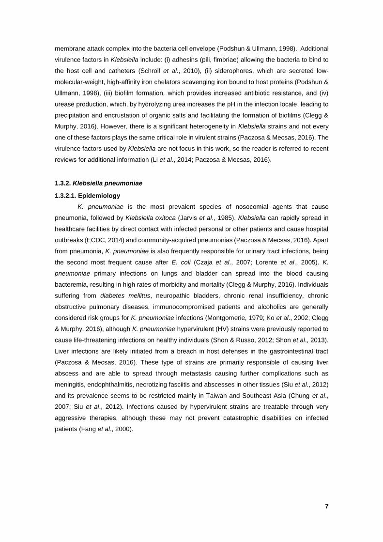
1.3.1. The Klebsiella genus ................................................................................................ 6
1.3.2. Klebsiella pneumoniae ............................................................................................. 7
1.3.2.1. Epidemiology ..................................................................................................... 7
1.3.2.2. K. pneumoniae MDRs overview ....................................................................... 8
1.4. The present work objectives ........................................................................................... 9
CHAPTER II – MATERIALS AND METHODS ............................................................................. 9
2.1. General protocols ............................................................................................................ 9
2.1.1. Bacterial strains and growth conditions ................................................................ 9
2.1.2. General molecular techniques .............................................................................. 10
2.1.3. Biparental conjugation ........................................................................................... 10
2.2. K. pneumoniae BCN studies ......................................................................................... 11
2.2.1. Cloning, expression and purification of K. pneumoniae kp52.145 BCN ........... 11
2.2.2. SGS-PAGE & Western-blot .................................................................................... 12
2.2.3. P. aeruginosa polymyxin B (PmB) protection assays ........................................ 12
2.3. K. pneumoniae mutagenesis ........................................................................................ 12
2.3.1. bcnK deletion in K. pneumoniae kp52.145 .......................................................... 12
VIII
2.3.2. Rhamnose conditional promoter and essentiality assessment ........................ 13
2.4. Oxidative stress studies ................................................................................................ 13
2.4.1. Transcriptional fusions to luxCDABE and luminescence assays ..................... 13
2.5. B. cenocepacia BCN complementation and MIC assays ........................................... 14
2.6. BcnK genomic studies .................................................................................................. 14
2.7. Computational methods ................................................................................................ 14
CHAPTER III – RESULTS AND DISCUSSION .......................................................................... 15
3.1. Activity of purified BcnK in an antibiotic protection assay ....................................... 15
3.2. The K. pneumoniae bcnK gene seems to be essential for bacterial viability .......... 17
3.3. bcnK regulation under various stress conditions ...................................................... 19
3.4. B. cenocepacia BCN complementation ....................................................................... 20
3.5. BCN in silico structural and genomic characterization of BCNs .............................. 20
3.5.1. BcnA vs BcnK secondary structure comparison ................................................ 20
3.5.2. Klebsiella spp. BcnK genomic comparison ......................................................... 22
3.5.3. bcnK neighborhood studies .................................................................................. 22
3.8. A general hypothesis for BCNs cellular function ....................................................... 22
4. Conclusions and perspectives ........................................................................................ 25
CHAPTER IV - BIBLIOGRAFY ................................................................................................... 26
CHAPTER V – APPENDIXES ................................................................................................... XV
5.1 – SUPPLEMENTARY TABLES ...................................................................................... XV
5.2 – SUPPLEMENTARY FIGURES ..................................................................................... XX
IX
LIST OF TABLES
Table 3.1 – Rifampicin MIC determination of non-complemented B. cenocepacia (WT); non-
complemented B. cenocepacia ΔbcnAΔbcnB (OME4); B. cenocepacia ΔbcnAΔbcnB
complementation with bcnK (pDG1) and B. cenocepacia ΔbcnAΔbcnB complemented with pDG1
backbone (pDA-CTHis). Results expressed in growth (+) and no growth (-) (n=9, 3 independent
experiments)……………………………………………………………………………………………. 20
Table 3.2 – Presence/absence of BCN and CybB in the genome of the indicated species based
on BLASTp results. ETC presence/absence based on bibliography. cybB association with BCN
gene obtained from synteny results. Among Enterobacter species only Enterobacter sp. and
Enterobacter cloacae have cybB associated to BCN. NA = Not Applicable……………………… 24
SUPPLEMENTARY TABLES
Table S1 – Strains, mutants and vectors used or created in this study. The following abbreviations
stands for the respective antibiotic resistance: TpR: trimethoprim; TetR: tetracycline; AmpR:
ampicillin; KmR: kanamycin; EmR: erythromycin………………………………………………….... XV
Table S2 – Primes generated for this study and respective restriction enzymes. Underlined bases
represent the restriction site. Bases in bold represents the homology region with pGSVTp-lux
vector…………………………………………………………………………………………………... XVI
Table S3 – List of genomic and protein sequences obtained from NCBI used in this thesis with
information referring species, strain, type of sequence (nucleotide or protein), accession number,
description of the sequence and entry date………………………………………………………… XVI
Table S4 – Composition of the solution used for protein purification protocols. (A) Lysis buffer;
(B) Equilibration buffer; (C) Washing and elution buffers used for BcnK purification………….. XVII
Table S5 – List of bacterial strains possessing a BCNs homologous obtained from BLASTp
results, deploying BcnK as query and used to construct the cladogram from Figure S3. The
species code, strains, accession number and entry date are associated with the attributed cluster
(1, 2 or 3). Similar protein sequences are grouped in MULTISPECIES (more than one species)
or MULTISTRAINS (more than one strain). Klebsiella pneumoniae (Kp), Klebsiella oxytoca (Ko),
Klebsiella variicola (Kv), Klebsiella quasipneumoniae (Kqp)……………………………………. XVIII
X
LIST OF FIGURE
Figure 1.1 – Timelines of antibiotic introduction (above) and bacterial antibiotic resistance
(below). *Not in clinical use; Ampicillin hydrolyzing (AmpC); Cefotaximase (CTX-M);
Imipenemase (IMP); Klebsiella pneumoniae carbapenemase (KPC); Methicillin resistant
Staphylococcus aureus (MRSA); New Delhi metallo-β-lactamase (NDM); Penicillin resistant S.
aureus (PRSA); β-lactam hydrolyzing enzymes (TEM, SHV, OXA); Vancomycin resistant
enterococci (VRE); Vancomycin resistant S. aureus (VRSA); Verona integron encoded metallo β-
lactamase (VIM)…………………………………………………………………………………………. 2
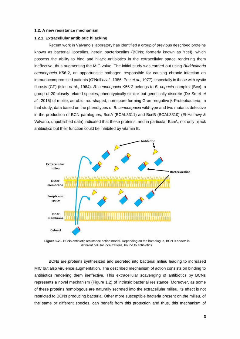
Figure 1.2 – BCNs antibiotic resistance action model. Depending on the homologue, BCN is
shown in different cellular localizations, bound to antibiotics………………………………………. 3
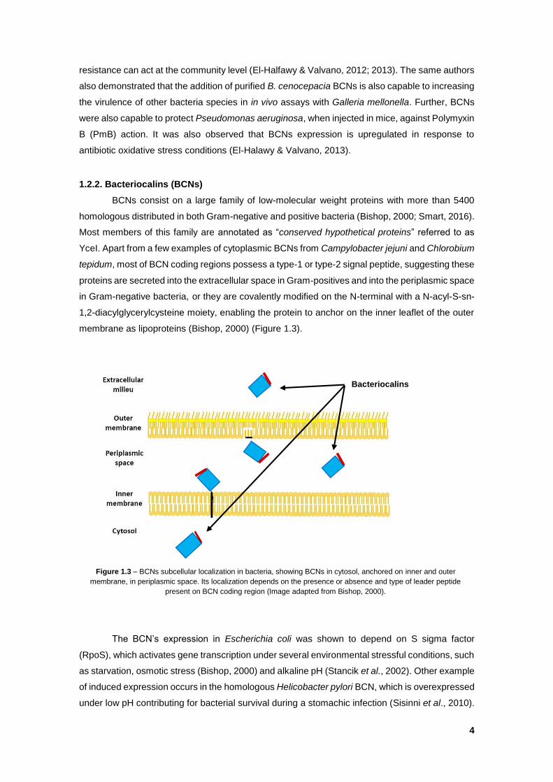
Figure 1.3 – BCNs subcellular localization in bacteria, showing BCNs in cytosol, anchored on
inner and outer membrane, in periplasmic space. Its localization depends on the presence or
absence and type of leader peptide present on BCN coding region (Image adapted from Bishop,
2000)…………………………………………………………………………………………………….... 4
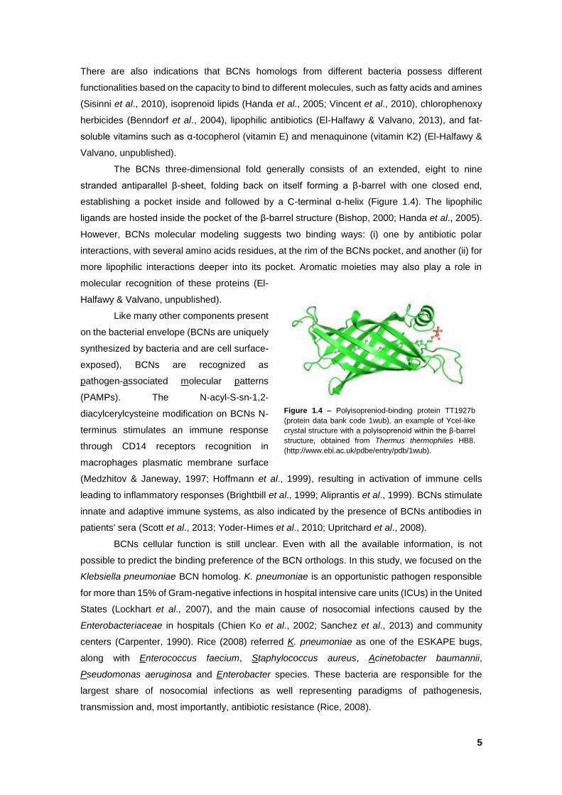
Figure 1.4 – Polyisopreniod-binding protein TT1927b (protein data bank code 1wub), an example
of YceI-like crystal structure with a polyisoprenoid within the β-barrel structure, obtained from
Thermus thermophiles HB8. (http://www.ebi.ac.uk/pdbe/entry/pdb/1wub)................................... 5
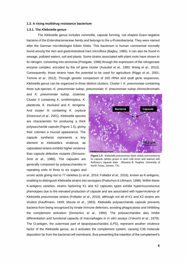
Figure 1.5 - Klebsiella pneumoniae (dark violet) surrounded by its capsule (white) grown in skim
milk broth and stained with Anthony’s capsule stain. (Roxana B. Hughes, University of North
Texas, Denton, TX)…………………………………………………………………………………….... 6
Figure 1.6 - Epidemiological incidence of several types of KPCs producer types by country of
origin (Munoz-Price et al. 2013)………………………………………………………………………… 8
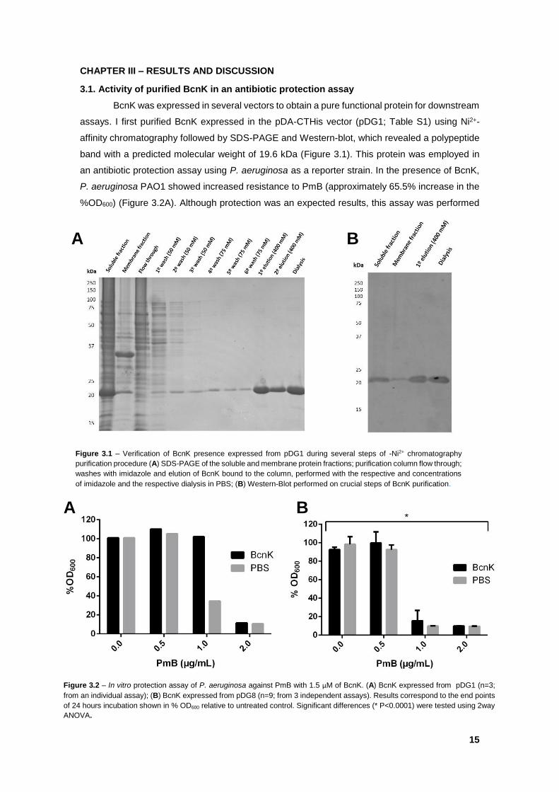
Figure 3.1 – Verification of BcnK presence expressed from pDG1 during several steps of -Ni2+
chromatography purification procedure (A) SDS-PAGE of the soluble and membrane protein
fractions; purification column flow through; washes with imidazole and elution of BcnK bound to
the column, performed with the respective and concentrations of imidazole and the respective
dialysis in PBS; (B) Western-Blot performed on crucial steps of BcnK purification…………...... 15
Figure 3.2 – In vitro protection assay of P. aeruginosa against PmB with 1.5 µM of BcnK. (A)
BcnK expressed from pDG1 (n=3; from an individual assay); (B) BcnK expressed from pDG8
(n=9; from 3 independent assays). Results correspond to the end points of 24 hours incubation
shown in % OD600 relative to untreated control. Significant differences (* P<0.0001) were tested
using 2way ANOVA…………………………………………………………………………………….. 15
Figure 3.3 – cPCR performed of K. pneumoniae trimethoprim sensitive colonies obtained after
the second conjugation of bcnK of unmarked deletion using pDG2. K. pneumoniae merodiploid
colonies 1 and 2 were compared against K. pneumoniae WT purified chromosome and the
purified knockout plasmid pDG2……………………………………………………………………… 17
Figure 3.4 – Microscopy of K. pneumoniae exconjugants expressing eGFP under the rhamnose
inducible promoter. Bacteria were grown at the indicated concentrations of rhamnose and
glucose………………………………………………………………………………………………….. 18
XI
Figure 3.5 – K. pneumoniae bcnK essentiality assessment using the obtained exconjugants
containing rhamnose inducible promoters (DNG9-11). K. pneumoniae wild type was
complemented with pSCrhaB2 (kp52.145/pSCrhaB2) using rhamnose inducible promoter as
control. XOA12 and XOA10 were used as rhamnose-dependent and rhamnose-independent
control mutant (Ortega et al., 2007). Bacteria were grown on the indicated rhamnose and glucose
concentrations………………………………………………………………………………………….. 19
Figure 3.6 – 3D prediction of BcnK (left) and BcnA (right) visualized in Jmol. In red, are shown
the corresponding residues for BcnK’s Val107 and BcnA’s Asp82 and in blue the corresponding
BcnK’s Glu118 and BcnA’s Asp93……………………………………………………………………. 21
Figure 3.7 – BcnK and BcnA amino acid sequence alignment obtained from Clustal Omega. In
blue is BcnK’s residue, Val107, corresponding to BcnA’s Asp82 and in red is BcnK’s residue,
Glu118, corresponding to BcnA’s Asp93. The following symbols stand for (*) identical residues;
(:) identical charge; (.) identical property (hydrophobic or hydrophilic)…………………………… 21
Figure 3.8 – Example of electron transport chain during aerobic respiration from Paracoccus
denitrificans, a model organism for respiration studies, (Mandigan et al., 2010)……………….. 23
Figure 3.9 – Schematic model of BCN cellular function. 1 – Extracellular BcnA or BcnK/BcnB on
the periplasmic space can bind hydrophobic compounds such as quinones or vitamin E. 2 – BCNs
deliver these compounds to cytochrome b561 (CybB). 3 – ETC substrates are oxidized on the
cytosol by CybB that transports electron across the membrane reducing the compound present
on BCNs. 4 – the reduced compound is transported in the periplasmic space acting as an
antioxidant or 5 – Reduced isoprenoid quinones are transported into the inner membrane where
they enter in the ETC or act as antioxidants…………………………………………………………. 25
SUPPLEMENTARY FIGURES
Figure S1 – BcnK amino acid sequence displaying its putative signal peptide (green), predicted
to be cleaved between Alanine45 and Alanine46 (SQA-AA)……………………………………… XX
Figure S2 – Constructed vector pDG11 showing lacZα gene fragment and N-region of bcnK
coding region with the respective primers for point mutation assays. The primers display the
adenine to be added aiming to fuse both genes……………………………………………………. XX
Figure S3 – Cladogram of Klebsiella genus amino acidic comparison. K. pneumoniae kp52.145
BcnK is shown in red letters on Cluster 1. BCNs are separated in three main cluster 1 (Blue), 2
(Red) and 3 (Green). Cladogram constructed using Clustal Omega v1.2.2 and FigTree v1.4.2.
The code of each sequence in displayed on Table S4…………………………………………….. XXI
Figure S4 – SyntTax report obtain for BcnK. At the center, in blue and bold, stands bcnK, each
arrow represents conserved a gene with an associated color. Genes with no synteny are
presented in white genes………………………………………………………………………...….. XXII
XII
LIST OF ABBREVIATIONS
MDRs – Multidrug resistant
MIC - Minimum inhibitory concentration
AmpC – Ampicillin hydrolysing
CTX-M – Cefotaximase
IMP – Imipenemase
MRSA – Methicillin resistant Staphylococcus aureus
NDM – New Delhi metallo-β-lactamase
PRSA – Penicillin resistant Staphylococcus aureus
VRE – Vancomycin resistant Enterococci
VRSA – Vancomycin resistant Staphylococcus aureus
VIM – Verona integron encoded metallo β-lactamase
BCNs – Bacteriocalins
CF – Cystic fibrosis
Bcc – Burkholderia cepacia complex
PmB - Polymyxin B
RpoS – S sigma factor
PAMPs – Pathogen-associated molecular patterns
ICUs – Intensive care units
ESKAPE – Enterococcus faecium, Staphylococcus aureus, Klebsiella pneumoniae,
Acinetobacter baumannii, Pseudominas aeruginosa and Enterobacter species
rRNA – Ribosomal RNA
LPS – Lipopolysaccharides
HV – Hypervirulent
ESBLs – Extended-spectrum β-lactamases
KPCs – K. pneumoniae carbapenemases
MBL – Metalle-β-lactamases
CRE – Carbapenem-resistant Enterobacteriaceae
MLST – Multilocus sequence typing
LB – Lysogeny broth
PCR – Polymerase chain reaction
cPCR – Colony-PCR
NCBI – National Center for Biotechnology Information
BLASTp – Basic Local Alignment Search Tool for proteins
His-tag – Hexahistidine tag
IPTG – Isopropyl thiol-β-D-galactoside
PBS – Phosphate-buffered saline
TBS – Tris-buffered saline
WT – Wild type
XIII
CDD – Conserved Domain Database
STRING – Search Tool for the Retrieval of Interacting Genes/Proteins
3D – Tridimensional
Phyre2 – Protein Homology/analogy Recognition Engine
SD – Shine-Dalgarno
IBs – Inclusion bodies
GFP – Green fluorescent protein
IQ – Isoprenoid quinone
ETC – Electron-transport chain
ATP – Adenosine triphosphate
ROS – Reactive oxygen species
FNR – Fumarate and Nitrate reductase
sRNA – Small RNAs
Na2HPO4 – Disodium phosphate
NaCl – Sodium chloride
NH4Cl – Ammonium chloride
CaCl2 – Calcium chloride
MgSO4 – Magnesium sulfate
Mg2+ - Magnesium
Ca2+ - Calcium
DAP – Diaminopimelic acid
Ni2+ - Nickel
CO2 – Carbon dioxide
H2 - Hydrogen
N2 – Nitrogen
mM – Millimolar
µM – Micromolar
µg/mL – Micrograms per milliliter
mg/L – Milligrams per liter
w/v – weigh per volume
˚C – Celsius
s – Seconds
min – Minutes
h – Hour
mL – Milliliter
µL – Microliter
rpm – Rotations per minute
g – Gravity
ksi – Kilopound per square inch
GPa - GigaPascal

XIV
µm - Micrometer
V - Volt
mV - Millivolt
A – Ampere
bp – Base pairs
OD600 – Optical density at 600 nm
nm – Nanometer
%OD600 – Percentage of OD600
RLU/OD600 – Relative luminescence unites per OD600
kDa - Kilodalton
% - Percent
1
CHAPTER I – INTRODUCTION
1.1. The Antibiotic Crisis
Since the discovery of penicillin, the first known antibiotic, by Alexander Fleming in 1928,
mankind has relied on this and other antibacterial molecules for the treatment of bacterial
infections. The antibiotics are molecules used in the treatment and prevention of infections caused
by bacteria. However, the “antibiotic era” might come to an end as the majority of the clinically
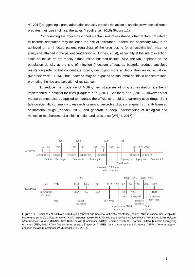
available antibiotics are becoming useless to treat bacterial infections (Figure 1.1) caused by
multidrug resistant (MDRs) Gram-positive and Gram-negative strains (Llor & Cots, 2009). This
scenario, named the “post-antibiotic era” is considered one of the greatest threats for mankind
(WHO, 2014; WEF, 2015). Despite the investment and incentives to research to identify new
antimicrobial molecules in the last years (Piddock, 2012), there are considerable challenges to
bring them to the market resulting on a long and fastidious process (Nathan, 2004; Wright, 2015).
Aggravating this situation, the interest of pharmaceutical companies to search for new molecules
has decreased in the last years, resulting in only a few newly approved and reliable drugs
(Spellberg, 2011).
Antibiotic molecules inhibit the growth or kill bacteria with minimum impact on the human
body. They have different mechanisms of action for which they are categorized in different
classes. For instance, (i) β-lactams interfere with cell wall synthesis, (ii) macrolides inhibit protein
synthesis, (iii) fluoroquinolones interfere with nucleic acids synthesis, (iv) trimethoprim inhibits
metabolic pathways and (v) polymyxins disrupt bacterial membrane. In some cases, bacteria are
capable of overcoming this toxicity through several mechanisms which give rise to resistant
strains. The antibiotic therapy represents a stressful environment for a sensitive bacterial
community, and resistant subpopulations are selected and ultimately able to prevail and
proliferate in the community giving rise to one or more resistant strains. In clinical microbiology, a
strain is defined as resistant, susceptible or intermediate by comparing the minimum inhibitory
concentration (MIC). This is the minimal antibiotic concentration at which bacterial growth is
inhibited under standardized conditions in vitro (Turnidge & Paterson, 2007), with the predefined
susceptibility “breakpoint” of the tested species.
In the past, the bulk of research efforts in antibiotic resistance focused on bacterial cellular
functions associated with decreased susceptibility. These included (i) modification of the
antibiotics target due to chromosomal mutations, making it unrecognizable to the antibiotic, (ii)
production of enzymes that breakdown or modify antibiotic molecules inactivating them, (iii)
extrachromosomal elements from other bacteria, such as plasmids, transposons and integrons,
which can be accumulated on a single or several strains expressing proteins that inactivate the
antibiotic affect, (iv) efflux pumps responsible for expelling several types of antibiotics from inside
the cell (v) and decreasing membrane permeability to antibiotics reducing the access to their
targets (Levy & Marshall 2004; Tenover, 2006; Alekshun et al., 2007). DNA analysis of human
bacterial microbiota revealed identical genes harbored by major bacterial pathogens (Sommer et
al., 2009) and similar genes responsible for the present modern antibiotic resistance were found
in the environment and in samples dating back millions of years (Forsberg et al., 2012; Bhullar et
2
al., 2012) suggesting a great adaptation capacity to resist the action of antibiotics whose existence
predates their use in clinical therapies (Iredell et al., 2016) (Figure 1.1).
Compounding the above-described mechanisms of resistance, other factors not related
to bacteria adaptation may influence the rise of resistance. Indeed, the necessary MIC to be
achieved on an infected patient, regardless of the drug dosing (pharmacokinetics), may not
always be attained in the patient (Andersson & Hughes, 2014), especially at the site of infection,
since antibiotics do not readily diffuse inside inflamed tissues. Also, the MIC depends on the
population density at the site of infection (inoculum effect), as bacteria produce antibiotic
resistance proteins that concentrate locally, destroying more antibiotic than an individual cell
(Martínez et al., 2015). Thus, bacteria may be exposed to sub-lethal antibiotic concentrations,
promoting the rise and selection of resistance.
To reduce the incidence of MDRs, new strategies of drug administration are being
implemented in hospital facilities (Baquero et al., 2011; Spellberg et al., 2013). However other
measures must also be applied to increase the efficiency of old and currently used drugs. So it
falls on scientific community to research for new antimicrobial drugs or augment currently licensed
antibacterial drugs (Piddock, 2012) and generate a deep understanding of biological and
molecular mechanisms of antibiotic action and resistance (Wright, 2015).
Figure 1.1 – Timelines of antibiotic introduction (above) and bacterial antibiotic resistance (below). *Not in clinical use; Ampicillin
hydrolyzing (AmpC); Cefotaximase (CTX-M); Imipenemase (IMP); Klebsiella pneumoniae carbapenemase (KPC); Methicillin resistant
Staphylococcus aureus (MRSA); New Delhi metallo-β-lactamase (NDM); Penicillin resistant S. aureus (PRSA); β-lactam hydrolyzing
enzymes (TEM, SHV, OXA); Vancomycin resistant Enterococci (VRE); Vancomycin resistant S. aureus (VRSA); Verona integron
encoded metallo β-lactamase (VIM) (Iredell et al., 2016).
3
1.2. A new resistance mechanism
1.2.1. Extracellular antibiotic hijacking
Recent work in Valvano’s laboratory has identified a group of previous described proteins
known as bacterial lipocalins, herein bacteriocalins (BCNs; formerly known as YceI), which
possess the ability to bind and hijack antibiotics in the extracellular space rendering them
ineffective, thus augmenting the MIC value. The initial study was carried out using Burkholderia
cenocepacia K56-2, an opportunistic pathogen responsible for causing chronic infection on
immunocompromised patients (O’Neil et al., 1986; Poe et al., 1977), especially in those with cystic
fibrosis (CF) (Isles et al., 1984). B. cenocepacia K56-2 belongs to B. cepacia complex (Bcc), a
group of 20 closely related species, phenotypically similar but genetically discrete (De Smet et
al., 2015) of motile, aerobic, rod-shaped, non-spore forming Gram-negative β-Proteobacteria. In
that study, data based on the phenotypes of B. cenocepacia wild-type and two mutants defective
in the production of BCN paralogues, BcnA (BCAL3311) and BcnB (BCAL3310) (El-Halfawy &
Valvano, unpublished data) indicated that these proteins, and in particular BcnA, not only hijack
antibiotics but their function could be inhibited by vitamin E.
BCNs are proteins synthesized and secreted into bacterial milieu leading to increased
MIC but also virulence augmentation. The described mechanism of action consists on binding to
antibiotics rendering them ineffective. This extracellular scavenging of antibiotics by BCNs
represents a novel mechanism (Figure 1.2) of intrinsic bacterial resistance. Moreover, as some
of these proteins homologous are naturally secreted into the extracellular milieu, its effect is not
restricted to BCNs producing bacteria. Other more susceptible bacteria present on the milieu, of
the same or different species, can benefit from this protection and thus, this mechanism of
Figure 1.2 – BCNs antibiotic resistance action model. Depending on the homologue, BCN is shown in
different cellular localizations, bound to antibiotics.
4
resistance can act at the community level (El-Halfawy & Valvano, 2012; 2013). The same authors
also demonstrated that the addition of purified B. cenocepacia BCNs is also capable to increasing
the virulence of other bacteria species in in vivo assays with Galleria mellonella. Further, BCNs
were also capable to protect Pseudomonas aeruginosa, when injected in mice, against Polymyxin
B (PmB) action. It was also observed that BCNs expression is upregulated in response to
antibiotic oxidative stress conditions (El-Halawy & Valvano, 2013).
1.2.2. Bacteriocalins (BCNs)
BCNs consist on a large family of low-molecular weight proteins with more than 5400
homologous distributed in both Gram-negative and positive bacteria (Bishop, 2000; Smart, 2016).
Most members of this family are annotated as “conserved hypothetical proteins” referred to as
YceI. Apart from a few examples of cytoplasmic BCNs from Campylobacter jejuni and Chlorobium
tepidum, most of BCN coding regions possess a type-1 or type-2 signal peptide, suggesting these
proteins are secreted into the extracellular space in Gram-positives and into the periplasmic space
in Gram-negative bacteria, or they are covalently modified on the N-terminal with a N-acyl-S-sn-
1,2-diacylglycerylcysteine moiety, enabling the protein to anchor on the inner leaflet of the outer
membrane as lipoproteins (Bishop, 2000) (Figure 1.3).
The BCN’s expression in Escherichia coli was shown to depend on S sigma factor
(RpoS), which activates gene transcription under several environmental stressful conditions, such
as starvation, osmotic stress (Bishop, 2000) and alkaline pH (Stancik et al., 2002). Other example
of induced expression occurs in the homologous Helicobacter pylori BCN, which is overexpressed
under low pH contributing for bacterial survival during a stomachic infection (Sisinni et al., 2010).
Figure 1.3 – BCNs subcellular localization in bacteria, showing BCNs in cytosol, anchored on inner and outer
membrane, in periplasmic space. Its localization depends on the presence or absence and type of leader peptide
present on BCN coding region (Image adapted from Bishop, 2000).
Bacteriocalins
5
There are also indications that BCNs homologs from different bacteria possess different
functionalities based on the capacity to bind to different molecules, such as fatty acids and amines
(Sisinni et al., 2010), isoprenoid lipids (Handa et al., 2005; Vincent et al., 2010), chlorophenoxy
herbicides (Benndorf et al., 2004), lipophilic antibiotics (El-Halfawy & Valvano, 2013), and fat-
soluble vitamins such as α-tocopherol (vitamin E) and menaquinone (vitamin K2) (El-Halfawy &
Valvano, unpublished).
The BCNs three-dimensional fold generally consists of an extended, eight to nine
stranded antiparallel β-sheet, folding back on itself forming a β-barrel with one closed end,
establishing a pocket inside and followed by a C-terminal α-helix (Figure 1.4). The lipophilic
ligands are hosted inside the pocket of the β-barrel structure (Bishop, 2000; Handa et al., 2005).
However, BCNs molecular modeling suggests two binding ways: (i) one by antibiotic polar
interactions, with several amino acids residues, at the rim of the BCNs pocket, and another (ii) for
more lipophilic interactions deeper into its pocket. Aromatic moieties may also play a role in
molecular recognition of these proteins (El-
Halfawy & Valvano, unpublished).
Like many other components present
on the bacterial envelope (BCNs are uniquely
synthesized by bacteria and are cell surface-
exposed), BCNs are recognized as
pathogen-associated molecular patterns
(PAMPs). The N-acyl-S-sn-1,2-
diacylcerylcysteine modification on BCNs N-
terminus stimulates an immune response
through CD14 receptors recognition in
macrophages plasmatic membrane surface
(Medzhitov & Janeway, 1997; Hoffmann et al., 1999), resulting in activation of immune cells
leading to inflammatory responses (Brightbill et al., 1999; Aliprantis et al., 1999). BCNs stimulate
innate and adaptive immune systems, as also indicated by the presence of BCNs antibodies in
patients' sera (Scott et al., 2013; Yoder-Himes et al., 2010; Upritchard et al., 2008).
BCNs cellular function is still unclear. Even with all the available information, is not
possible to predict the binding preference of the BCN orthologs. In this study, we focused on the
Klebsiella pneumoniae BCN homolog. K. pneumoniae is an opportunistic pathogen responsible
for more than 15% of Gram-negative infections in hospital intensive care units (ICUs) in the United
States (Lockhart et al., 2007), and the main cause of nosocomial infections caused by the
Enterobacteriaceae in hospitals (Chien Ko et al., 2002; Sanchez et al., 2013) and community
centers (Carpenter, 1990). Rice (2008) referred K. pneumoniae as one of the ESKAPE bugs,
along with Enterococcus faecium, Staphylococcus aureus, Acinetobacter baumannii,
Pseudomonas aeruginosa and Enterobacter species. These bacteria are responsible for the
largest share of nosocomial infections as well representing paradigms of pathogenesis,
transmission and, most importantly, antibiotic resistance (Rice, 2008).
Figure 1.4 – Polyisopreniod-binding protein TT1927b
(protein data bank code 1wub), an example of YceI-like
crystal structure with a polyisoprenoid within the β-barrel
structure, obtained from Thermus thermophiles HB8.
(http://www.ebi.ac.uk/pdbe/entry/pdb/1wub).
6
1.3. A rising multidrug resistance bacterium
1.3.1. The Klebsiella genus
The Klebsiella genus includes nonmotile, capsule forming, rod shaped Gram-negative
bacteria of the Enterobacteriaceae family and belongs to the γ-Proteobacteria. They were named
after the German microbiologist Edwin Klebs. This bacterium is human commensal normally
found among the skin and gastrointestinal tract microflora (Bagley, 1985). It can also be found in
sewage, polluted waters, soil and plants. Some strains associated with plant roots have shown to
fix nitrogen, converting into ammonia (Postgate, 1998) through the expression of the nitrogenase
enzyme complex, encoded by the nif gene cluster (Ausubel et al., 1983; Wang et al., 2013).
Consequently, those strains have the potential to be used for agriculture (Riggs et al., 2001;
Temme et al., 2012). Through genetic comparison of 16S rRNA and rpoB gene sequences,
Klebsiella genus can be organized in three distinct clusters. Cluster I: K. pneumoniae containing
three sub-species; K. pneumoniae subsp. pneumoniae; K. pneumoniae subsp rhinoscleromatis
and K. pneumoniae subsp. ozaenea.
Cluster II containing K. ornithinolytica, K.
planticola, K. trevisanii and K. terrigena.
And cluster III containing K. oxytoca
(Drancourt et al., 2001). Klebsiella species
are characteristic for producing a thick
polysaccharide capsule (Figure 1.5), giving
their colonies a mucoid appearance. The
capsule synthesis represents a key
element to Klebsiella’s virulence, as
capsulated strains exhibits higher virulence
than capsule defective mutants (Simoons-
Smit et al., 1986). The capsules are
generally composed by polysaccharides in
repeating units of three to six sugars and
uronic acids giving rise to 77 varieties (Li et al., 2014; Follador et al., 2016), known as K-antigens,
enabling to distinguish Klebsiella strains into serotypes (Podschun & Ullmann, 1998). Within these
K-antigens varieties, strains harboring K1 and K2 capsules types exhibit hypermucoviscous
phenotypes due to the elevated production of capsule and are associated with hypervirulence of
Klebsiella pneumoniae strains (Follador et al., 2016), although not all of K1 and K2 strains are
virulent (Kauffmann, 1949; Mizuta et al., 1983). Klebsiella polysaccharide capsule prevents
bacteria from being recognized by innate immune defenses, avoiding phagocytosis and inhibiting
the complement activation (Domenico et al., 1994). The polysaccharides also inhibit
differentiation and functional capacity of macrophages in in vitro assays (Yokochi et al., 1979).
The O-antigen, the outermost part of lipopolysaccharides (LPS), represent another virulence
factor of the Klebsiella genus, as it activates the complement system, causing C3b molecule
deposition far from the bacterial cell membrane, thus preventing the insertion of the complement’s
Figure 1.5 - Klebsiella pneumoniae (dark violet) surrounded by
its capsule (white) grown in skim milk broth and stained with
Anthony’s capsule stain. (Roxana B. Hughes, University of
North Texas, Denton, TX)
Bacteria Capsule
7
membrane attack complex into the bacteria cell envelope (Podshun & Ullmann, 1998). Additional
virulence factors in Klebsiella include: (i) adhesins (pili, fimbriae) allowing the bacteria to bind to
the host cell and catheters (Schroll et al., 2010), (ii) siderophores, which are secreted low-
molecular-weight, high-affinity iron chelators scavenging iron bound to host proteins (Podshun &
Ullmann, 1998), (iii) biofilm formation, which provides increased antibiotic resistance, and (iv)
urease production, which, by hydrolyzing urea increases the pH in the infection locale, leading to
precipitation and encrustation of organic salts and facilitating the formation of biofilms (Clegg &
Murphy, 2016). However, there is a significant heterogeneity in Klebsiella strains and not every
one of these factors plays the same critical role in virulent strains (Paczosa & Mecsas, 2016). The
virulence factors used by Klebsiella are not focus in this work, so the reader is referred to recent
reviews for additional information (Li et al., 2014; Paczosa & Mecsas, 2016).
1.3.2. Klebsiella pneumoniae
1.3.2.1. Epidemiology
K. pneumoniae is the most prevalent species of nosocomial agents that cause
pneumonia, followed by Klebsiella oxitoca (Jarvis et al., 1985). Klebsiella can rapidly spread in
healthcare facilities by direct contact with infected personal or other patients and cause hospital
outbreaks (ECDC, 2014) and community-acquired pneumonias (Paczosa & Mecsas, 2016). Apart
from pneumonia, K. pneumoniae is also frequently responsible for urinary tract infections, being
the second most frequent cause after E. coli (Czaja et al., 2007; Lorente et al., 2005). K.
pneumoniae primary infections on lungs and bladder can spread into the blood causing
bacteremia, resulting in high rates of morbidity and mortality (Clegg & Murphy, 2016). Individuals
suffering from diabetes mellitus, neuropathic bladders, chronic renal insufficiency, chronic
obstructive pulmonary diseases, immunocompromised patients and alcoholics are generally
considered risk groups for K. pneumoniae infections (Montgomerie, 1979; Ko et al., 2002; Clegg
& Murphy, 2016), although K. pneumoniae hypervirulent (HV) strains were previously reported to
cause life-threatening infections on healthy individuals (Shon & Russo, 2012; Shon et al., 2013).
Liver infections are likely initiated from a breach in host defenses in the gastrointestinal tract
(Paczosa & Mecsas, 2016). These type of strains are primarily responsible of causing liver
abscess and are able to spread through metastasis causing further complications such as
meningitis, endophthalmitis, necrotizing fasciitis and abscesses in other tissues (Siu et al., 2012)
and its prevalence seems to be restricted mainly in Taiwan and Southeast Asia (Chung et al.,
2007; Siu et al., 2012). Infections caused by hypervirulent strains are treatable through very
aggressive therapies, although these may not prevent catastrophic disabilities on infected
patients (Fang et al., 2000).
8
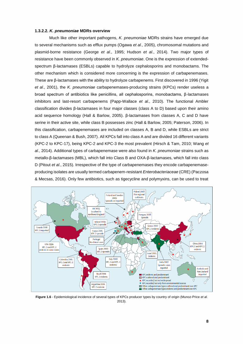
1.3.2.2. K. pneumoniae MDRs overview
Much like other important pathogens, K. pneumoniae MDRs strains have emerged due
to several mechanisms such as efflux pumps (Ogawa et al., 2005), chromosomal mutations and
plasmid-borne resistance (George et al., 1995; Hudson et al., 2014). Two major types of
resistance have been commonly observed in K. pneumoniae. One is the expression of extended-
spectrum β-lactamases (ESBLs) capable to hydrolyze cephalosporins and monobactams. The
other mechanism which is considered more concerning is the expression of carbapenemases.
These are β-lactamases with the ability to hydrolyze carbapenems. First discovered in 1996 (Yigit
et al., 2001), the K. pneumoniae carbapenemases-producing strains (KPCs) render useless a
broad spectrum of antibiotics like penicillins, all cephalosporins, monobactams, β-lactamases
inhibitors and last-resort carbapenems (Papp-Wallace et al., 2010). The functional Ambler
classification divides β-lactamases in four major classes (class A to D) based upon their amino
acid sequence homology (Hall & Barlow, 2005). β-lactamases from classes A, C and D have
serine in their active site, while class B possesses zinc (Hall & Barlow, 2005; Paterson, 2006). In
this classification, carbapenemases are included on classes A, B and D, while ESBLs are strict
to class A (Queenan & Bush, 2007). All KPCs fall into class A and are divided 16 different variants
(KPC-2 to KPC-17), being KPC-2 and KPC-3 the most prevalent (Hirsch & Tam, 2010; Wang et
al., 2014). Additional types of carbapenemase were also found in K. pneumoniae strains such as
metallo-β-lactamases (MBL), which fall into Class B and OXA-β-lactamases, which fall into class
D (Pitout et al., 2015). Irrespective of the type of carbapenemases they encode carbapenemase-
producing isolates are usually termed carbapenem-resistant Enterobacteriaceae (CRE) (Paczosa
& Mecsas, 2016). Only few antibiotics, such as tigecycline and polymyxins, can be used to treat
Figure 1.6 - Epidemiological incidence of several types of KPCs producer types by country of origin (Munoz-Price et al.
2013).
9
infections by CRE bacteria, but with variable degree of success (Urban et al., 2008). Therefore,
these bacteria cause a high mortality rate among patients with bloodstream infections (Munoz-
Price, 2013; Clegg & Murphy, 2016).
KPC genes possess a great potential to spread horizontally as some of them are encoded
on transposons and often found present on several types of plasmids (Queenan & Bush, 2007).
Horizontal transfer is not restricted to Klebsiella as KPC genes have been reported on other
Enterobacteriaceae, including E. coli, Enterobacter species, Salmonella enterica, Proteus
mirabilis, Citrobabacter freundii (Queenan & Bush, 2007; Bush et al., 1995; Villegas et al., 2005)
and Pseudomonas species (Munoz-Price et al., 2013). K. pneumoniae KPC ST258, defined by
multilocus sequence typing (MLST) of 7 loci, played a major role in disseminating its associated
KPC enzymes worldwide (Munoz-Price et al., 2013). More detailed information on ST258 and
genes concerned in MRDs dissemination can be found in recent reviews (Iredell et al., 2016;
Paczosa & Mecsas., 2016).
KPC strains incidence has been steadily increasing worldwide (Munoz-Price et al., 2013;
Iredell et al., 2016) (Figure 1.6), as well as ESBLs producing K. pneumoniae (Boucher et al.,
2009), making urgent to find new ways to successfully treat infections caused by these MDRs
bacteria.
1.4. The present work objectives
In this dissertation, I will explore the role of BCNs ortholog of K. pneumoniae kp52.145
virulent strain, to confer an antibiotic resistance mechanism as described for B. cenocepacia (El-
Halfawy & Valvano, 2013). In doing so, I aim to demonstrate that BCNs provide general resistance
mechanism that can be exploited by important antibiotic-resistant pathogens. Initially I will assess
the protection capacity of a recombinant K. pneumoniae BCNs protein. Simultaneously, I will
attempt to delete BCNs gene from K. pneumoniae chromosome. Also, the regulation properties
of the same gene under oxidative stress will be evaluated. Finally, I will perform in silico studies
of K. pneumoniae BCNs by comparing with other BCNs from different bacteria. Additionally,
synteny studies will be performed.
Here, I will describe all the developed work done until the submission of this dissertation.
Additional experimental work is still underway and new data generated will be presented during
the public defense.
CHAPTER II – MATERIALS AND METHODS
2.1. General protocols
2.1.1. Bacterial strains and growth conditions
Strains and plasmids used for this thesis are listed on Table S1 (see Supplementary data
on CHAPTER V). Bacteria were grown at 37˚C, 180 rpm, in Difco™ LB broth. Rhamnose
conditional mutants were grown on M9 minimal medium (42 mM Na2HPO4, 8 mM NaCl, 10 mM
NH4Cl supplemented with Casamino Acids (80 µg/mL), vitamin B1 (10 µg/mL), tryptophan (40
µg/mL), CaCl2 (20 µM), MgSO4 (200 µM), and 0.5% (w/v) glucose or 0.5% (w/v) rhamnose when
10
required. Mueller-Hinton media cation adjusted with 10 mg/L Mg2+ and 20 mg/L Ca2+, final
concentration, was used for MIC determinations. Each medium were added with antibiotics
trimethoprim (50 µg/mL for E. coli strains; 100 µg/mL for K. pneumoniae), ampicillin (100 µg/mL),
tetracycline (100 µg/mL for B. cenocepacia; 30 µg/mL for E. coli; 12.5 µg/mL for K. pneumoniae),
kanamycin (40 µg/mL) final concentrations when required.
2.1.2. General molecular techniques
K. pneumoniae kp52.145 genomic DNA extraction was carried out using Genomic DNA
Mini kit (Invitrogen), chromosomal amplicons were generated by PCR using HotStar Hifidelity
polymerase Kit (Qiagen), using 20% Q-solution final concentration and thermocycled at the
following temperatures: 5 min at 95˚C; 35 cycles of 30 s at 95˚C, 30 s at 55˚C and 1 min 72˚C
and final extension 10 min at 72˚C and purified by QIAquick PCR purification kit (Qiagen). Plasmid
extractions were carried out using QIAprep Spin Miniprep Kit (Qiagen). DNA restriction
endonuclease digestions, plasmid dephosphorylation, ligations reactions and agarose gel
electrophoresis were performed according to standard techniques (Sambrook et al., 1990).
Restriction enzymes and Antarctic phosphatase were purchased from New England BioLabs, T4
DNA ligase from Roche Diagnostics. DNA transformation with E. coli strains was carried out by
calcium chloride method (Cohen et al., 1972). Colony-PCR (cPCR) were carried out with Taq
polymerase kit (Qiagen) with the following parameters: 3 min at 95˚C; 45 cycles of 15 s at 95˚C,
30 s at 56˚C and 1 min 72˚C; and a final extension at 72˚C for 10 min. PCR products were
screened on 0.7% (w/v) agarose gels. Gene sequence of positive transformants was verified by
sequencing. All designed primers and respective restriction enzymes are listed on Table S2.
2.1.3. Biparental conjugation
The various constructed vectors from all other procedures, which were transformed in E.
coli DH-5α and E. coli GT115, were extracted and transformed into diaminopimelic acid (DAP)
dependent E. coli β2163, capable to mobilize the vectors into the recipient strains, such as K.
pneumoniae or B. cenocepacia, by biparental conjugation. The biparental conjugations were
carried out using 2,6-Diaminopimelic acid bought from Sigma-Aldrich®.
Mobilization of the vectors was performed by growing overnight of recipient strain, with
180 rpm orbital shaking and the donor strain without shaking. Next day, both strains were pelleted
by centrifugation at 4000 rpm for 20 min, washed in 5 mL of 10 mM MgSO4, pelleted again and
resuspended in 500 µL 10 mM MgSO4. A mixture of 100 µL of each strain patched in LB agar
supplemented with 0.3 mM of DAP final concentration, incubated overnight at 37˚C. Next day,
serial dilutions were made, until 10-4, from the recovered patched biomass and plated on LB
without the addition of DAP and with the appropriate antibiotic. The grown colonies were screened
by cPCR and/or luminescence on UVP (BioSpectrum® AC Imaging System).
11
2.2. K. pneumoniae BCN studies
2.2.1. Cloning, expression and purification of K. pneumoniae kp52.145 BCN
K. pneumoniae kp52.145 yceI nucleotide sequence (GenBank ID: FO834906.1) and YceI
amino acid sequence (GenBank ID: CDO15049.1) was retrieved from National Center for
Biotechnology Information (NCBI) using Basic Local Alignment Search Tool for proteins
(BLASTp) algorithm, deploying as query sequence the protein sequence of the putative exported
protein from B. cenocepacia J2315 (GenBank: CAR53634.1) (Table S3).
To facilitate the differentiation from other BCN orthologues, such as B. cenocepacia’s
BcnA and BcnB, the gene encoding K. pneumoniae’s BCN (yceI) will be referred to as bcnK and
its respective encoded protein will be mentioned as BcnK.
bcnK was amplified by PCR with (primers Q-775 and Q-776) and without (Q-873 and Q-
776) its signal peptides, cloned into pDA-CTHis, which contains an N-terminal hexahistidine tag
(His-tag), originating pDG1 and pDG5, respectively. To clone into pET28a (+) isopropyl thiol-β-D-
galactoside (IPTG) inducible vector, bcnK was amplified without its signal peptide and encoding
an N- and C-terminal His-tag (Q-873 and Q-880) giving rise to pDG7. Also, bcnK was amplified
without signal peptide and coding for a N-terminal His-tag (Q-873 and Q-895), and cloned into
pET28a (+). Primers (Q-907 and Q-908) for bcnK cloning into pUC19 inducible vector were design
without bcnK’s signal peptide and to contain a His-tag followed by a STOP codon on bcnK’s C-
terminal giving rise to pDG11. This last vector was point mutated by amplifying (Q-961 and Q-
962) to add an adenine base upstream of bcnK start codon and digested with DpnI overnight at
37˚C afterwards, transformed into E. coli and the resulting colonies were selected in ampicillin.
The generated amplicons and respective vectors were digested with restriction enzymes, listed
on Table S2, ligated and transformed into E. coli DH-5α. IPTG inducible vectors were transformed
into E. coli BL2. Overnight cultures induction were carried out using 0.05 mM IPTG, final
concentration and further incubated for 3 h at 25˚C, centrifuged at 10,000 x g for 15 min at 4˚C,
washed with Tris-buffer 50 mM, pH 7.4 and pelleted again, resuspended in Lysis buffer (Table S4
A) and passed through One Shot (E1061, Constant System) at 18 ksi (124.1 GPa). The resulting
lysate was centrifuged at 15,000 x g for 20 min at 4˚C for cell debris removal and to obtain the
total protein fraction. Soluble and membrane protein fractions were obtained by centrifuging the
total protein fraction at 30,000 x g for 45 min at 4˚C. BcnK purification was carried out by mixing
the soluble fraction with coated Ni2+ Chelating Sepharose™ Fast Flow (GE Healthcare) beads
overnight at 4˚C previously treated with equilibration buffer (Table S4 B). Next day, the
supernatants were collected, labelled as Flow through, and the beads were washed with
increasing concentration of imidazole (50 mM and 75 mM), eluted in 400 mM (Table S4 C) and
dialyzed in 4 L of phosphate-buffered saline (PBS) overnight at 4˚C. In each step an aliquot was
collected for further analysis. The dialyzed protein was filter sterilized with 0.45 µm Whatman™
(SPARTAN Syringe Filter) and conserved at -80ºC until used. The presence of the protein was
confirmed by SDS-PAGE and Western-Blot.
12
2.2.2. SGS-PAGE & Western-blot
The various protein fractions obtained were boiled at 100˚C for 10 min, loaded in a 16%
SDS-PAGE gel, run for 75 V for 35 min and 130 V for 2 h. For SDS-PAGE staining the gel were
dyed with PAGE-Blue™ (Thermo Scientific) for 2 h and distained overnight.
For Western-blot assays, the protein transfer was carried out using Biorad Trans-Blot®
Turbo™ Kit into a nitrocellulose membrane for 20 min with 1.3 A and 25 V, blocked overnight at
4˚C with Blocker™ Casein in TBS (Thermo Scientific). Next day, the membrane was washed with
Tris-buffered saline (TBS), the primary antibody Anti-His Antibody (GE Healthcare Life Sciences)
was added diluted 1:3000 and incubated at 4˚C for 2 h, washed three times with TBS, added the
secondary antibody AlexaFluor® 680 anti-mouse IgG (Life Technologies) diluted 1:20000,
incubated for 45 min, washed three times and checked at Li-cor (Odyssey®) at the wavelength
of 700 nm.
2.2.3. P. aeruginosa polymyxin B (PmB) protection assays
The purified and dialyzed BcnK obtained from the expression of the various constructed
vectors, were concentrated if required using Vivaspin 500 (3000 MWCO PES, Sartorius Stedim
Biotech), quantified by NanoVue Plus™ Spectrophotometer. Overnight cultures of P. aeruginosa
PAO1 were subcultured for 2 hours, OD600 adjusted to 0.04, loaded in a 100 well honeycomb
plate along with 2; 1; 0.5; 0 µg/mL final concentration of PmB and 1.5 µM, final concentration, of
purified BcnK. Controls were performed with the same antibiotic concentrations using PBS
instead of purified BcnK. Each antibiotic concentration, with and without BcnK, was tested within
triplicates. The OD600 was read each hour at 37ºC for 24 h on Bioscreen C (Oy Growth Curves
Ab Ltd.).
2.3. K. pneumoniae mutagenesis
2.3.1. bcnK deletion in K. pneumoniae kp52.145
Unmarked deletion method was performed as previously described (Flannagan et al.,
2008). To delete bcnK, PCR amplifications of ≈ 300 bp flanking regions bcnK were performed (Q-
786 and Q-787; Q-788 and Q-789). Amplicons were digested with XbaI-XhoI and XhoI-EcoRI
respectively and cloned into pGPI-SceI-2 digested with XbaI-EcoRI giving rise to pDG4.
Simultaneously, it was also created a vector containing ≈ 1000 bp flanking regions bcnK (Q-810
and Q-811; Q-812 and Q-813), the amplicons were joined together using Ex Taq® DNA
polymerase (TaKaRa) and ligated into pGEM®-TEasy resulting on the vector ΔyceI-pGEMT. The
vector was digest with EcoRI, gel purified and ligated with pGPI-SecI-2 giving rise to pDG2. Each
plasmid was introduced to wild type (WT) strain of K. pneumoniae kp52.145 by conjugation,
separately, and selected with trimethoprim. The resulting conjugants were subjected to a new
conjugation with pGPI-SceI-SacB and selected through tetracycline resistance.
Insertional inactivation was performed by cloning an bcnK internal fragment with 297 bp
(Q-902 and Q-903) into suicide vector pGPΩTp (Flannagan et al., 2007) which contains dhfr
13
flanked with Ω-fragments, which when conjugated into K. pneumoniae, creates a polar mutation
stopping bcnK transcription. The constructed vector was named pDG9.
The procedures described above were carried out in both aerobic and anaerobic
conditions. For anaerobic conditions, samples were manipulated and incubated in Whitley A35
Anaerobic Workstation under an atmosphere composed by 10% CO2, 10% H2 and 80% N2.
2.3.2. Rhamnose conditional promoter and essentiality assessment
The vector pSCrhaB2-e-GFP (Cardona et al., 2006) was conjugated into K. pneumoniae
kp52.145 WT. The exconjugants were selected in LB supplemented with trimethoprim. Ten
colonies were picked and each grown overnight at 37ºC in M9 medium supplemented with 0.5%,
0.2% of rhamnose or 0.5% of glucose. Next day, the bacterial suspensions were deposited in
slides covered with thin layer of 0.8% (w/v) agarose and observed by light and fluorescent
microscopy. The images were acquired using Axioscope 10 (Carl Zeiss) microscope coupled to
a camera AxioCam MRm (Zeiss) and an endow GFP bandpass emission filter set with the 470 ±
25 nm emission range, 525 ± 25 nm excitation. Images were digitally processed using ZEN 2012
(Blue Edition) Service Pack 1 image software.
The rhamnose conditional promoter construct was performed as previously described
(Ortega et al., 2007) by cloning a 306 bp fragment (Q-775 and Q-904) spanning from the 5’ region
of bcnK into pSC200, giving rise to pDG10. The resulting vector possesses a rhamnose-inducible
PrhaB upstream of the multiple cloning site, enabling to drive the expression of bcnK in the
presence of rhamnose and repressing it in glucose. The vector’s conjugation was performed in
LB supplemented with 0.5% rhamnose and selected in LB with 0.5% rhamnose and trimethoprim.
The conditional mutants were grown overnight at 37ºC in M9 medium with 0.5% rhamnose, spun
down and washed three times with PBS, resuspended in PBS and adjusted to an OD600 of 1 (Neat
solution). Drops (10 µL) of Neat, 10-1, 10-2, 10-3 and 10-4 dilutions were plated in M9 agar square
plates supplement with 0.5% (w/v) rhamnose or 0.5% (w/v) glucose and incubated at 37ºC. The
strains XOA10 and XOA12 were used as a negative and positive control, respectively.
2.4. Oxidative stress studies
2.4.1. Transcriptional fusions to luxCDABE and luminescence assays
Promoter region of bcnK, was amplified by PCR (Q-909 and Q-910). The amplicon with
≈900 bp was digested and cloned into the digested and dephosphorylated pGSVTp-lux suicide
vector and transformed into E. coli GT115. The primers for oxyR and waaE (Q-841 and Q-842;
Q-852 and Q-853, respectively) were designed after ELIC method (Koskela & Frey, 2015)
containing ≈25 bp homologous with the vector’s region adjacent to region where it will be cloned
in pGSVTp-lux. The amplicons and digested vector were quantified in NanoVue Plus™
Spectrophotometer, mixed in a ratio of 3:1, respectively, in a final volume of 10 µL of Mili-Q water
and incubated for 1 h at room temperature and transformed into E. coli GT115. The transformants
colonies were selected in LB with trimethoprim, grown overnight and checked for luminescence
on POLARstar® Omega for 12 h at 37ºC treated with and without paraquat 1.5 µM final
14
concentration. From the colonies that displayed luminescence, three of each construct were
chosen and the vector was extract and transformed into E. coli β2163 and conjugated into K.
pneumoniae and selected in LB with trimethoprim. The obtained exconjugants were checked for
luminescence, grown overnight and luminescence and OD600 were measured in the
presence/absence of serial dilution of paraquat (10; 5; 2.5 and 0 µM) for 12 h at 37ºC on
POLARstar® Omega. The expression levels of each gene of interest in the different strains
background were calculated as Relative Luminescence Units per OD600 (RLU/OD600) for each
condition.
2.5. B. cenocepacia BCN complementation and MIC assays
B. cenocepacia ΔBcnAΔBcnB were complemented with pDG1 and pDA-CTHis through
biparental conjugation and selected in LB with tetracycline giving rise to DNG4 and DNG5,
respectively. B. cenocepacia ΔBcnAΔBcnB; DNG4 and DNG5 MIC’s were tested accordingly with
(Wiegand et al., 2008). The strains were grown overnight in Mueller-Hinton cation adjusted, OD600
adjusted to 0.005, inoculated in 100 well honeycomb plate with serial dilutions from 2048 µg/mL
to 8 µg/mL of rifampicin. The plates were incubated for 24 h at 37ºC on Bioscreen C. Triplicates
were performed for this assay.
2.6. BcnK genomic studies
The previously retrieved BcnK amino acid sequence from NCBI was used to perform the
following BCN genomic studies. K. pneumoniae kp52.145 BcnK amino acid sequence was
compared against Klebsiella genus (taxid: 570) using BLASTp and selecting non-redundant
protein sequences search. Amino acids sequence alignment and phylogenetic data were
generated by Clustal Omega v1.2.2 (Sievers et al., 2011). Neighbour-joining cladograms were
generated from the alignment data using FigTree v1.4.2 software. The domain structure of BcnK
was accessed on Conserved Domain Database (CDD) tool on NCBI. Neighbourhood gene
studies were performed by SyntTax (Oberto, 2013). Interaction of BCNs (YceI’s) protein with other
proteins and molecules was accessed using the Search Tool for the Retrieval of Interacting
Genes/Proteins (STRING) (Szklarczyk et al., 2014). Amino acid sequence from B. cenocepacia
CybB (GenkBank ID: CAR53635.1) was also retrieved from NCBI.
2.7. Computational methods
K. pneumoniae kp52.145 BcnK tridimensional (3D) folding prediction was generated in
Protein Homology/analogy Recognition Engine V 2.0 (Phyre2) (Kelley et al., 2015) using amino
acidic sequence and visualized in Jmol v13.0. BcnK molecular weight was predicted on ExPASy,
Bioinformatics Resource Portal translation tool (Gasteiger et al., 2005). BcnK signal peptide
prediction was performed on SignalP 4.1 Server in CBS website (Petersen et al., 2011). DNA
sequence analysis and vectors figures were generated in SnapGene™ 1.1.3. Statistical analysis
was performed with GraphPad Prim 6.
15
CHAPTER III – RESULTS AND DISCUSSION
3.1. Activity of purified BcnK in an antibiotic protection assay
BcnK was expressed in several vectors to obtain a pure functional protein for downstream
assays. I first purified BcnK expressed in the pDA-CTHis vector (pDG1; Table S1) using Ni2+-
affinity chromatography followed by SDS-PAGE and Western-blot, which revealed a polypeptide
band with a predicted molecular weight of 19.6 kDa (Figure 3.1). This protein was employed in
an antibiotic protection assay using P. aeruginosa as a reporter strain. In the presence of BcnK,
P. aeruginosa PAO1 showed increased resistance to PmB (approximately 65.5% increase in the
%OD600) (Figure 3.2A). Although protection was an expected results, this assay was performed
A B
Figure 3.1 – Verification of BcnK presence expressed from pDG1 during several steps of -Ni2+ chromatography
purification procedure (A) SDS-PAGE of the soluble and membrane protein fractions; purification column flow through;
washes with imidazole and elution of BcnK bound to the column, performed with the respective and concentrations
of imidazole and the respective dialysis in PBS; (B) Western-Blot performed on crucial steps of BcnK purification.
Figure 3.2 – In vitro protection assay of P. aeruginosa against PmB with 1.5 µM of BcnK. (A) BcnK expressed from pDG1 (n=3;
from an individual assay); (B) BcnK expressed from pDG8 (n=9; from 3 independent assays). Results correspond to the end points
of 24 hours incubation shown in % OD600 relative to untreated control. Significant differences (* P<0.0001) were tested using 2way
ANOVA.
A B
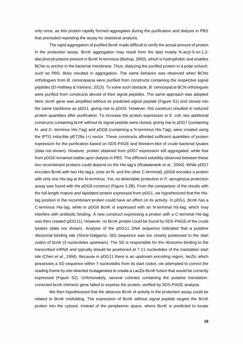
16
only once, as this protein rapidly formed aggregates during the purification and dialysis in PBS
that precluded repeating the assay for statistical analysis.
The rapid aggregation of purified BcnK made difficult to verify the actual amount of protein
in the protection assay. BcnK aggregation may result from the lipid moiety N-acyl-S-sn-1,2-
diacylcerylcysteine present in BcnK N-terminus (Bishop, 2000), which is hydrophobic and enables
BCNs to anchor in the bacterial membrane. Thus, dialyzing the purified protein in a polar solvent,
such as PBS, likely resulted in aggregation. The same behavior was observed when BCNs
orthologues from B. cenocepacia were purified from constructs containing the respective signal
peptides (El-Halfawy & Valvano, 2013). To solve such obstacle, B. cenocepacia BCN orthologues
were purified from constructs devoid of their signal peptides. The same approach was adopted
here; bcnK gene was amplified without its predicted signal peptide (Figure S1) and cloned into
the same backbone as pDG1, giving rise to pDG5. However, this construct resulted in reduced
protein quantities after purification. To increase the protein expression in E. coli, two additional
constructs containing bcnK without its signal peptide were cloned, giving rise to pDG7 (containing
N- and C- terminus His-Tag) and pDG8 (containing a N-terminus His-Tag), were created using
the IPTG inducible pET28a (+) vector. These constructs afforded sufficient quantities of protein
expression for the purification based on SDS-PAGE and Western-blot of crude bacterial lysates
(data not shown). However, protein obtained from pDG7 expression still aggregated, while that
from pDG8 remained stable upon dialysis in PBS. The different solubility observed between these
two recombinant proteins could depend on the His-tag’s (Woestenenk et al., 2004). While pDG7
encodes BcnK with two His-tag’s, (one on N- and the other C-terminal), pDG8 encodes a protein
with only one His-tag at the N-terminus. Yet, no detectable protection in P. aeruginosa protection
assay was found with the pDG8 construct (Figure 3.2B). From the comparison of the results with
the full-length mature and lapidated protein expressed from pDG1, we hypothesized that the His-
tag position in the recombinant protein could have an effect on its activity. In pDG1, BcnK has a
C-terminus His-tag, while in pDG8 BcnK is expressed with an N-terminal his-tag, which may
interfere with antibiotic binding. A new construct expressing a protein with a C-terminal His-tag
was then created (pDG11). However, no BcnK protein could be found by SDS-PAGE of the crude
lysates (data not shown). Analysis of the pDG11 DNA sequence indicated that a putative
ribosomal binding site (Shine-Dalgarno, SD) sequence was too closely positioned to the start
codon of bcnK (3 nucleotides upstream). The SD is responsible for the ribosome binding to the
transcribed mRNA and typically should be positioned at 7-11 nucleotides of the translation start
site (Chen et al., 1994). Because in pDG11 there is an upstream encoding region, lacZα, which
possesses a SD sequence within 7 nucleotides from its start codon, we attempted to correct the
reading frame by site-directed mutagenesis to create a LacZα-BcnK fusion that would be correctly
expressed (Figure S2). Unfortunately, several colonies containing the putative translation-
corrected bcnK chimeric gene failed to express the protein, verified by SDS-PAGE analysis.
We then hypothesized that the absence BcnK of activity in the protection assay could be
related to BcnK misfolding. The expression of BcnK without signal peptide targets the BcnK
protein into the cytosol, instead of the periplasmic space, where BcnK is predicted to locate
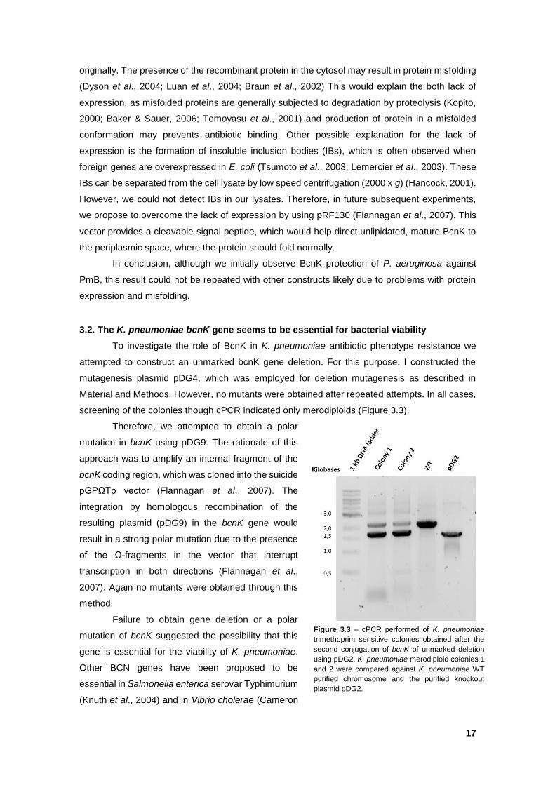
17
originally. The presence of the recombinant protein in the cytosol may result in protein misfolding
(Dyson et al., 2004; Luan et al., 2004; Braun et al., 2002) This would explain the both lack of
expression, as misfolded proteins are generally subjected to degradation by proteolysis (Kopito,
2000; Baker & Sauer, 2006; Tomoyasu et al., 2001) and production of protein in a misfolded
conformation may prevents antibiotic binding. Other possible explanation for the lack of
expression is the formation of insoluble inclusion bodies (IBs), which is often observed when
foreign genes are overexpressed in E. coli (Tsumoto et al., 2003; Lemercier et al., 2003). These
IBs can be separated from the cell lysate by low speed centrifugation (2000 x g) (Hancock, 2001).
However, we could not detect IBs in our lysates. Therefore, in future subsequent experiments,
we propose to overcome the lack of expression by using pRF130 (Flannagan et al., 2007). This
vector provides a cleavable signal peptide, which would help direct unlipidated, mature BcnK to
the periplasmic space, where the protein should fold normally.
In conclusion, although we initially observe BcnK protection of P. aeruginosa against
PmB, this result could not be repeated with other constructs likely due to problems with protein
expression and misfolding.
3.2. The K. pneumoniae bcnK gene seems to be essential for bacterial viability
To investigate the role of BcnK in K. pneumoniae antibiotic phenotype resistance we
attempted to construct an unmarked bcnK gene deletion. For this purpose, I constructed the
mutagenesis plasmid pDG4, which was employed for deletion mutagenesis as described in
Material and Methods. However, no mutants were obtained after repeated attempts. In all cases,
screening of the colonies though cPCR indicated only merodiploids (Figure 3.3).
Therefore, we attempted to obtain a polar
mutation in bcnK using pDG9. The rationale of this
approach was to amplify an internal fragment of the
bcnK coding region, which was cloned into the suicide
pGPΩTp vector (Flannagan et al., 2007). The
integration by homologous recombination of the
resulting plasmid (pDG9) in the bcnK gene would
result in a strong polar mutation due to the presence
of the Ω-fragments in the vector that interrupt
transcription in both directions (Flannagan et al.,
2007). Again no mutants were obtained through this
method.
Failure to obtain gene deletion or a polar
mutation of bcnK suggested the possibility that this
gene is essential for the viability of K. pneumoniae.
Other BCN genes have been proposed to be
essential in Salmonella enterica serovar Typhimurium
(Knuth et al., 2004) and in Vibrio cholerae (Cameron
Figure 3.3 – cPCR performed of K. pneumoniae
trimethoprim sensitive colonies obtained after the
second conjugation of bcnK of unmarked deletion
using pDG2. K. pneumoniae merodiploid colonies 1
and 2 were compared against K. pneumoniae WT
purified chromosome and the purified knockout
plasmid pDG2.
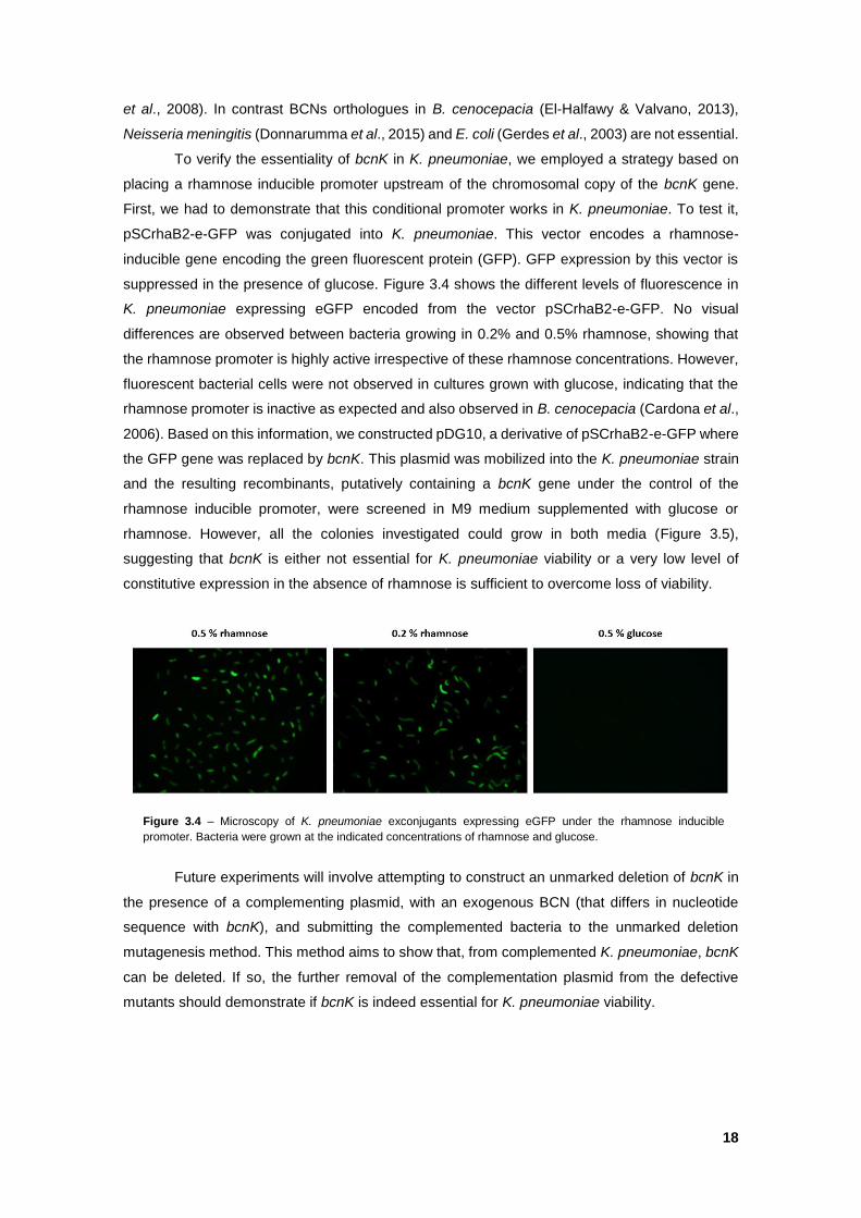
18
et al., 2008). In contrast BCNs orthologues in B. cenocepacia (El-Halfawy & Valvano, 2013),
Neisseria meningitis (Donnarumma et al., 2015) and E. coli (Gerdes et al., 2003) are not essential.
To verify the essentiality of bcnK in K. pneumoniae, we employed a strategy based on
placing a rhamnose inducible promoter upstream of the chromosomal copy of the bcnK gene.
First, we had to demonstrate that this conditional promoter works in K. pneumoniae. To test it,
pSCrhaB2-e-GFP was conjugated into K. pneumoniae. This vector encodes a rhamnose-
inducible gene encoding the green fluorescent protein (GFP). GFP expression by this vector is
suppressed in the presence of glucose. Figure 3.4 shows the different levels of fluorescence in
K. pneumoniae expressing eGFP encoded from the vector pSCrhaB2-e-GFP. No visual
differences are observed between bacteria growing in 0.2% and 0.5% rhamnose, showing that
the rhamnose promoter is highly active irrespective of these rhamnose concentrations. However,
fluorescent bacterial cells were not observed in cultures grown with glucose, indicating that the
rhamnose promoter is inactive as expected and also observed in B. cenocepacia (Cardona et al.,
2006). Based on this information, we constructed pDG10, a derivative of pSCrhaB2-e-GFP where
the GFP gene was replaced by bcnK. This plasmid was mobilized into the K. pneumoniae strain
and the resulting recombinants, putatively containing a bcnK gene under the control of the
rhamnose inducible promoter, were screened in M9 medium supplemented with glucose or
rhamnose. However, all the colonies investigated could grow in both media (Figure 3.5),
suggesting that bcnK is either not essential for K. pneumoniae viability or a very low level of
constitutive expression in the absence of rhamnose is sufficient to overcome loss of viability.
Future experiments will involve attempting to construct an unmarked deletion of bcnK in
the presence of a complementing plasmid, with an exogenous BCN (that differs in nucleotide
sequence with bcnK), and submitting the complemented bacteria to the unmarked deletion
mutagenesis method. This method aims to show that, from complemented K. pneumoniae, bcnK
can be deleted. If so, the further removal of the complementation plasmid from the defective
mutants should demonstrate if bcnK is indeed essential for K. pneumoniae viability.
Figure 3.4 – Microscopy of K. pneumoniae exconjugants expressing eGFP under the rhamnose inducible
promoter. Bacteria were grown at the indicated concentrations of rhamnose and glucose.
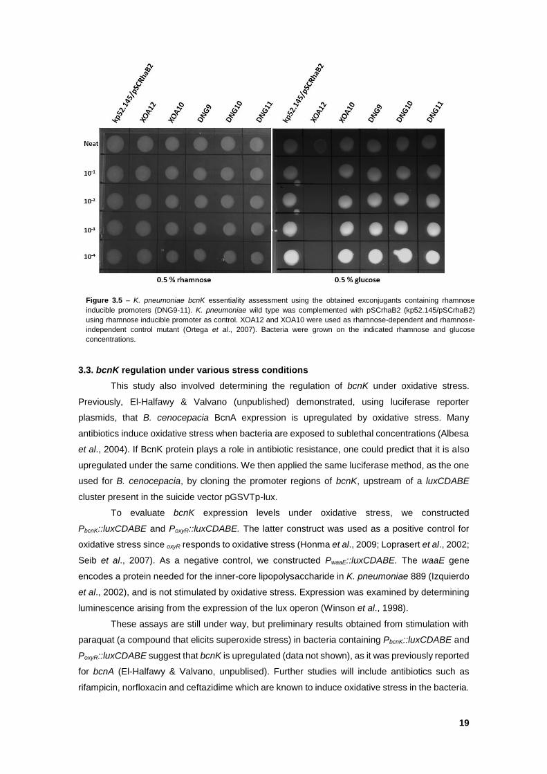
19
3.3. bcnK regulation under various stress conditions
This study also involved determining the regulation of bcnK under oxidative stress.
Previously, El-Halfawy & Valvano (unpublished) demonstrated, using luciferase reporter
plasmids, that B. cenocepacia BcnA expression is upregulated by oxidative stress. Many
antibiotics induce oxidative stress when bacteria are exposed to sublethal concentrations (Albesa
et al., 2004). If BcnK protein plays a role in antibiotic resistance, one could predict that it is also
upregulated under the same conditions. We then applied the same luciferase method, as the one
used for B. cenocepacia, by cloning the promoter regions of bcnK, upstream of a luxCDABE
cluster present in the suicide vector pGSVTp-lux.
To evaluate bcnK expression levels under oxidative stress, we constructed
PbcnK::luxCDABE and PoxyR::luxCDABE. The latter construct was used as a positive control for
oxidative stress since oxyR responds to oxidative stress (Honma et al., 2009; Loprasert et al., 2002;
Seib et al., 2007). As a negative control, we constructed PwaaE::luxCDABE. The waaE gene
encodes a protein needed for the inner-core lipopolysaccharide in K. pneumoniae 889 (Izquierdo
et al., 2002), and is not stimulated by oxidative stress. Expression was examined by determining
luminescence arising from the expression of the lux operon (Winson et al., 1998).
These assays are still under way, but preliminary results obtained from stimulation with
paraquat (a compound that elicits superoxide stress) in bacteria containing PbcnK::luxCDABE and
PoxyR::luxCDABE suggest that bcnK is upregulated (data not shown), as it was previously reported
for bcnA (El-Halfawy & Valvano, unpublised). Further studies will include antibiotics such as
rifampicin, norfloxacin and ceftazidime which are known to induce oxidative stress in the bacteria.
Figure 3.5 – K. pneumoniae bcnK essentiality assessment using the obtained exconjugants containing rhamnose
inducible promoters (DNG9-11). K. pneumoniae wild type was complemented with pSCrhaB2 (kp52.145/pSCrhaB2)
using rhamnose inducible promoter as control. XOA12 and XOA10 were used as rhamnose-dependent and rhamnose-
independent control mutant (Ortega et al., 2007). Bacteria were grown on the indicated rhamnose and glucose
concentrations.
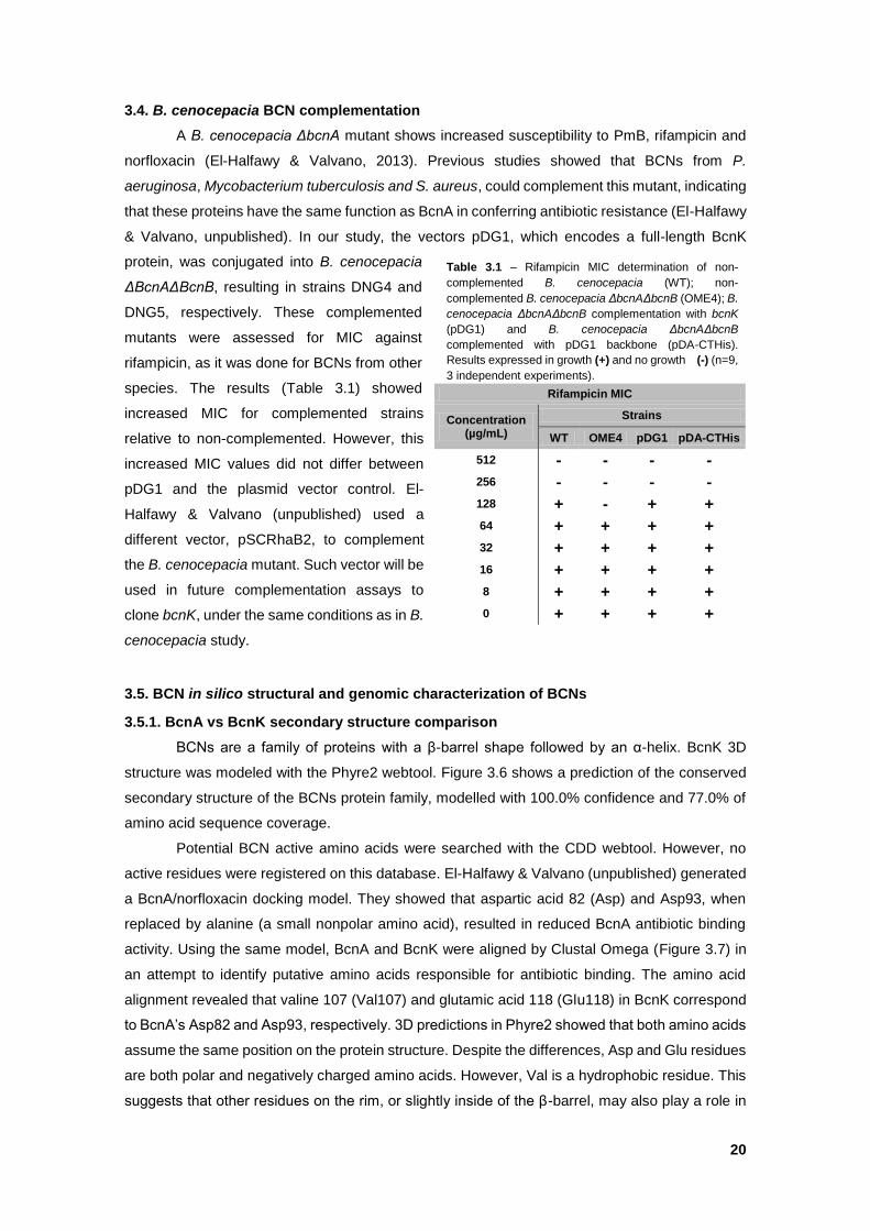
20
3.4. B. cenocepacia BCN complementation
A B. cenocepacia ΔbcnA mutant shows increased susceptibility to PmB, rifampicin and
norfloxacin (El-Halfawy & Valvano, 2013). Previous studies showed that BCNs from P.
aeruginosa, Mycobacterium tuberculosis and S. aureus, could complement this mutant, indicating
that these proteins have the same function as BcnA in conferring antibiotic resistance (El-Halfawy
& Valvano, unpublished). In our study, the vectors pDG1, which encodes a full-length BcnK
protein, was conjugated into B. cenocepacia
ΔBcnAΔBcnB, resulting in strains DNG4 and
DNG5, respectively. These complemented
mutants were assessed for MIC against
rifampicin, as it was done for BCNs from other
species. The results (Table 3.1) showed
increased MIC for complemented strains
relative to non-complemented. However, this
increased MIC values did not differ between
pDG1 and the plasmid vector control. El-
Halfawy & Valvano (unpublished) used a
different vector, pSCRhaB2, to complement
the B. cenocepacia mutant. Such vector will be
used in future complementation assays to
clone bcnK, under the same conditions as in B.
cenocepacia study.
3.5. BCN in silico structural and genomic characterization of BCNs
3.5.1. BcnA vs BcnK secondary structure comparison
BCNs are a family of proteins with a β-barrel shape followed by an α-helix. BcnK 3D
structure was modeled with the Phyre2 webtool. Figure 3.6 shows a prediction of the conserved
secondary structure of the BCNs protein family, modelled with 100.0% confidence and 77.0% of
amino acid sequence coverage.
Potential BCN active amino acids were searched with the CDD webtool. However, no
active residues were registered on this database. El-Halfawy & Valvano (unpublished) generated
a BcnA/norfloxacin docking model. They showed that aspartic acid 82 (Asp) and Asp93, when
replaced by alanine (a small nonpolar amino acid), resulted in reduced BcnA antibiotic binding
activity. Using the same model, BcnA and BcnK were aligned by Clustal Omega (Figure 3.7) in
an attempt to identify putative amino acids responsible for antibiotic binding. The amino acid
alignment revealed that valine 107 (Val107) and glutamic acid 118 (Glu118) in BcnK correspond
to BcnA’s Asp82 and Asp93, respectively. 3D predictions in Phyre2 showed that both amino acids
assume the same position on the protein structure. Despite the differences, Asp and Glu residues
are both polar and negatively charged amino acids. However, Val is a hydrophobic residue. This
suggests that other residues on the rim, or slightly inside of the β-barrel, may also play a role in
Rifampicin MIC
Concentration (µg/mL)
Strains
WT OME4 pDG1 pDA-CTHis
512 - - - -
256 - - - -
128 + - + +
64 + + + +
32 + + + +
16 + + + +
8 + + + +
0 + + + +
Table 3.1 – Rifampicin MIC determination of non-
complemented B. cenocepacia (WT); non-
complemented B. cenocepacia ΔbcnAΔbcnB (OME4); B.
cenocepacia ΔbcnAΔbcnB complementation with bcnK
(pDG1) and B. cenocepacia ΔbcnAΔbcnB
complemented with pDG1 backbone (pDA-CTHis).
Results expressed in growth (+) and no growth (-) (n=9,
3 independent experiments).
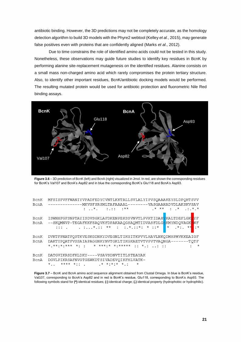
21
antibiotic binding. However, the 3D predictions may not be completely accurate, as the homology
detection algorithm to build 3D models with the Phyre2 webtool (Kelley et al., 2015), may generate
false positives even with proteins that are confidently aligned (Marks et al., 2012).
Due to time constrains the role of identified amino acids could not be tested in this study.
Nonetheless, these observations may guide future studies to identify key residues in BcnK by
performing alanine site-replacement mutagenesis on the identified residues. Alanine consists on
a small mass non-charged amino acid which rarely compromises the protein tertiary structure.
Also, to identify other important residues, BcnK/antibiotic docking models would be performed.
The resulting mutated protein would be used for antibiotic protection and fluorometric Nile Red
binding assays.
Figure 3.6 – 3D prediction of BcnK (left) and BcnA (right) visualized in Jmol. In red, are shown the corresponding residues
for BcnK’s Val107 and BcnA’s Asp82 and in blue the corresponding BcnK’s Glu118 and BcnA’s Asp93.
Val107
Asp93
Asp82
Glu118
BcnA BcnK
V E
D D
:
Figure 3.7 – BcnK and BcnA amino acid sequence alignment obtained from Clustal Omega. In blue is BcnK’s residue,
Val107, corresponding to BcnA’s Asp82 and in red is BcnK’s residue, Glu118, corresponding to BcnA’s Asp93. The
following symbols stand for (*) identical residues; (:) identical charge; (.) identical property (hydrophobic or hydrophilic).
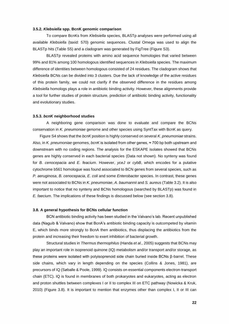
22
3.5.2. Klebsiella spp. BcnK genomic comparison
To compare BcnKs from Klebsiella species, BLASTp analyses were performed using all
available Klebsiella (taxid: 570) genomic sequences. Clustal Omega was used to align the
BLASTp hits (Table S5) and a cladogram was generated by FigTree (Figure S3).
BLASTp revealed proteins with amino acid sequence homologies that varied between
99% and 81% among 100 homologous identified sequences in Klebsiella species. The maximum
difference of identities between homologous consisted of 24 residues. The cladogram shows that
Klebsiella BCNs can be divided into 3 clusters. Due the lack of knowledge of the active residues
of this protein family, we could not clarify if the observed difference in the residues among
Klebsiella homologs plays a role in antibiotic binding activity. However, these alignments provide
a tool for further studies of protein structure, prediction of antibiotic binding activity, functionality
and evolutionary studies.
3.5.3. bcnK neighborhood studies
A neighboring gene comparison was done to evaluate and compare the BCNs
conservation in K. pneumoniae genome and other species using SyntTax with BcnK as query.
Figure S4 shows that the bcnK position is highly conserved on several K. pneumoniae strains.
Also, in K. pneumoniae genomes, bcnK is isolated from other genes, ≈ 700 bp both upstream and
downstream with no coding regions. The analysis for the ESKAPE isolates showed that BCNs
genes are highly conserved in each bacterial species (Data not shown). No synteny was found
for B. cenocepacia and E. feacium. However, yceJ or cybB, which encodes for a putative
cytochrome b561 homologue was found associated to BCN genes from several species, such as
P. aeruginosa, B. cenocepacia, E. coli and some Enterobacter species. In contrast, these genes
were not associated to BCNs in K. pneumoniae, A. baumannii and S. aureus (Table 3.2). It is also
important to notice that no synteny and BCNs homologous (searched by BLASTp) was found in
E. faecium. The implications of these findings is discussed below (see section 3.8).
3.8. A general hypothesis for BCNs cellular function
BCN antibiotic binding activity has been studied in the Valvano’s lab. Recent unpublished
data (Naguib & Valvano) show that BcnA’s antibiotic binding capacity is outcompeted by vitamin
E, which binds more strongly to BcnA then antibiotics, thus displacing the antibiotics from the
protein and increasing their freedom to exert inhibition of bacterial growth.
Structural studies in Thermus thermophilus (Handa et al., 2005) suggests that BCNs may
play an important role in isoprenoid quinone (IQ) metabolism and/or transport and/or storage, as
these proteins were isolated with polyisoprenoid side chain buried inside BCNs β-barrel. These
side chains, which vary in length depending on the species (Collins & Jones, 1981), are
precursors of IQ (Søballe & Poole, 1999). IQ consists on essential components electron-transport
chain (ETC). IQ is found in membranes of both prokaryotes and eukaryotes, acting as electron
and proton shuttles between complexes I or II to complex III on ETC pathway (Nowicka & Kruk,
2010) (Figure 3.8). It is important to mention that enzymes other than complex I, II or III can
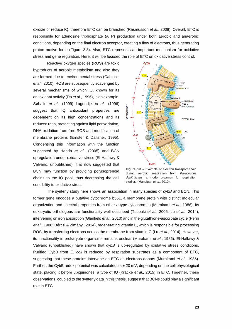
23
oxidize or reduce IQ, therefore ETC can be branched (Rasmusson et al., 2008). Overall, ETC is
responsible for adenosine triphosphate (ATP) production under both aerobic and anaerobic
conditions, depending on the final electron acceptor, creating a flow of electrons, thus generating
proton motive force (Figure 3.8). Also, ETC represents an important mechanism for oxidative
stress and gene regulation. Here, it will be focused the role of ETC on oxidative stress control.
Reactive oxygen species (ROS) are toxic
byproducts of aerobic metabolism and also they
are formed due to environmental stress (Cabiscol
et al., 2010). ROS are subsequently scavenged by
several mechanisms of which IQ, known for its
antioxidant activity (Do et al., 1996), is an example.
Søballe et al., (1999) Lagendijk et al., (1996)
suggest that IQ antioxidant properties are
dependent on its high concentrations and its
reduced ratio, protecting against lipid peroxidation,
DNA oxidation from free ROS and modification of
membrane proteins (Ernster & Dallaner, 1995).
Condensing this information with the function
suggested by Handa et al., (2005) and BCN
upregulation under oxidative stress (El-Halfawy &
Valvano, unpublished), it is now suggested that
BCN may function by providing polyisoprenoid
chains to the IQ pool, thus decreasing the cell
sensibility to oxidative stress.
The synteny study here shows an association in many species of cybB and BCN. This
former gene encodes a putative cytochrome b561, a membrane protein with distinct molecular
organization and spectral properties from other b-type cytochromes (Murakami et al., 1986). Its
eukaryotic orthologous are functionality well described (Tsubaki et al., 2005; Lu et al., 2014),
intervening on iron absorption (Glanfield et al., 2010) and in the glutathione-ascorbate cycle (Perin
et al., 1988; Bérczi & Zimányi, 2014), regenerating vitamin E, which is responsible for processing
ROS, by transferring electrons across the membrane from vitamin C (Lu et al., 2014). However,
its functionality in prokaryote organisms remains unclear (Murakami et al., 1986). El-Halfawy &
Valvano (unpublished) have shown that cybB is up-regulated by oxidative stress conditions.
Purified CybB from E. coli is reduced by respiration substrates as a component of ETC,
suggesting that these proteins intervene on ETC as electrons donors (Murakami et al., 1986).
Further, the CybB redox potential was calculated as + 20 mV, depending on the cell physiological
state, placing it before ubiquinones, a type of IQ (Kracke et al., 2015) in ETC. Together, these
observations, coupled to the synteny data in this thesis, suggest that BCNs could play a significant
role in ETC.
Figure 3.8 – Example of electron transport chain
during aerobic respiration from Paracoccus
denitrificans, a model organism for respiration
studies, (Mandigan et al., 2010).
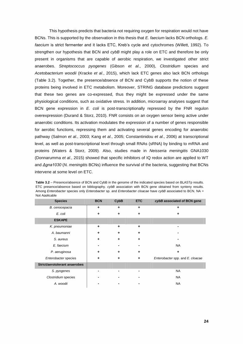
24
This hypothesis predicts that bacteria not requiring oxygen for respiration would not have
BCNs. This is supported by the observation in this thesis that E. faecium lacks BCN orthologs. E.
faecium is strict fermenter and it lacks ETC, Kreb’s cycle and cytochromes (Willett, 1992). To
strengthen our hypothesis that BCN and cybB might play a role on ETC and therefore be only
present in organisms that are capable of aerobic respiration, we investigated other strict
anaerobes. Streptococcus pyogenes (Gibson et al., 2000), Clostridium species and
Acetobacterium woodii (Kracke et al., 2015), which lack ETC genes also lack BCN orthologs
(Table 3.2). Together, the presence/absence of BCN and CybB supports the notion of these
proteins being involved in ETC metabolism. Moreover, STRING database predictions suggest
that these two genes are co-expressed, thus they might be expressed under the same
physiological conditions, such as oxidative stress. In addition, microarray analyses suggest that
BCN gene expression in E. coli is post-transcriptionally repressed by the FNR regulon
overexpression (Durand & Storz, 2010). FNR consists on an oxygen sensor being active under
anaerobic conditions. Its activation modulates the expression of a number of genes responsible
for aerobic functions, repressing them and activating several genes encoding for anaerobic
pathway (Salmon et al., 2003; Kang et al., 2005; Constantinidou et al., 2006) at transcriptional
level, as well as post-transcriptional level through small RNAs (sRNA) by binding to mRNA and
proteins (Waters & Storz, 2009). Also, studies made in Neisseria meningitis GNA1030
(Donnarumma et al., 2015) showed that specific inhibitors of IQ redox action are applied to WT
and Δgna1030 (N. meningitis BCNs) influence the survival of the bacteria, suggesting that BCNs
intervene at some level on ETC.
Species BCN CybB ETC cybB associated of BCN gene
B. cenocepacia + + + +
E. coli + + + +
ESKAPE
K. pneumoniae + + + -
A. baumanni + + + -
S. aureus + + + -
E. faecium - - - NA
P. aeruginosa + + + +
Enterobacter species + + + Enterobacter spp. and E. cloacae
Strict/aerotolerant anaerobes
S. pyogenes - - - NA
Clostridium species - - - NA
A. woodii - - - NA
Table 3.2 – Presence/absence of BCN and CybB in the genome of the indicated species based on BLASTp results.
ETC presence/absence based on bibliography. cybB association with BCN gene obtained from synteny results.
Among Enterobacter species only Enterobacter sp. and Enterobacter cloacae have cybB associated to BCN. NA =
Not Applicable
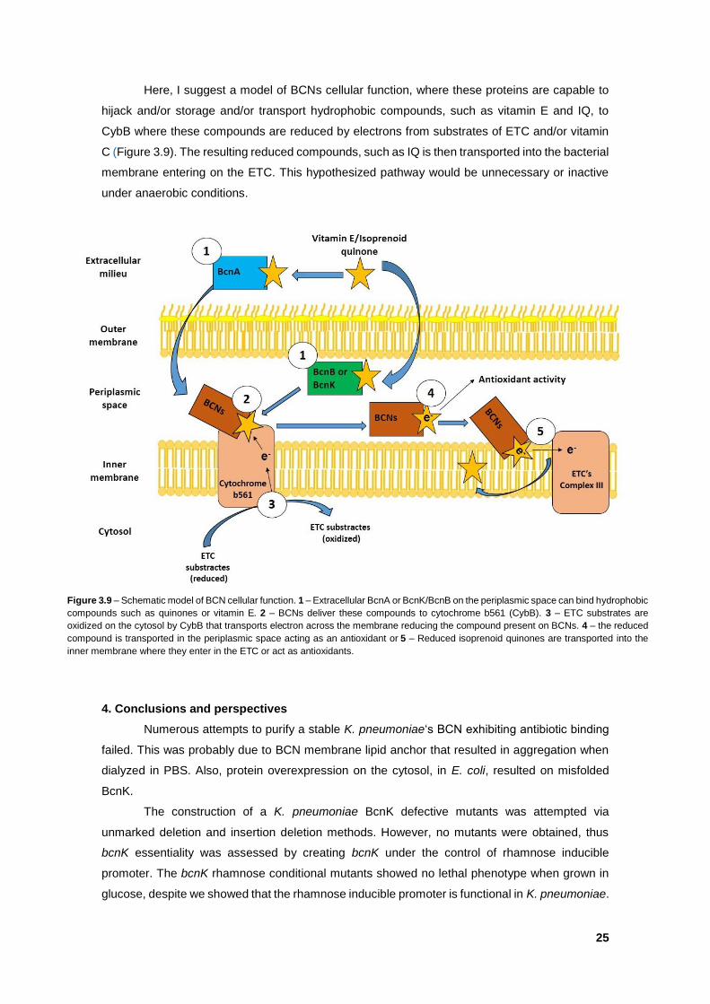
25
Here, I suggest a model of BCNs cellular function, where these proteins are capable to
hijack and/or storage and/or transport hydrophobic compounds, such as vitamin E and IQ, to
CybB where these compounds are reduced by electrons from substrates of ETC and/or vitamin
C (Figure 3.9). The resulting reduced compounds, such as IQ is then transported into the bacterial
membrane entering on the ETC. This hypothesized pathway would be unnecessary or inactive
under anaerobic conditions.
4. Conclusions and perspectives
Numerous attempts to purify a stable K. pneumoniae‘s BCN exhibiting antibiotic binding
failed. This was probably due to BCN membrane lipid anchor that resulted in aggregation when
dialyzed in PBS. Also, protein overexpression on the cytosol, in E. coli, resulted on misfolded
BcnK.
The construction of a K. pneumoniae BcnK defective mutants was attempted via
unmarked deletion and insertion deletion methods. However, no mutants were obtained, thus
bcnK essentiality was assessed by creating bcnK under the control of rhamnose inducible
promoter. The bcnK rhamnose conditional mutants showed no lethal phenotype when grown in
glucose, despite we showed that the rhamnose inducible promoter is functional in K. pneumoniae.
Figure 3.9 – Schematic model of BCN cellular function. 1 – Extracellular BcnA or BcnK/BcnB on the periplasmic space can bind hydrophobic
compounds such as quinones or vitamin E. 2 – BCNs deliver these compounds to cytochrome b561 (CybB). 3 – ETC substrates are
oxidized on the cytosol by CybB that transports electron across the membrane reducing the compound present on BCNs. 4 – the reduced
compound is transported in the periplasmic space acting as an antioxidant or 5 – Reduced isoprenoid quinones are transported into the
inner membrane where they enter in the ETC or act as antioxidants.
26
This may be due to a low level of constitutive read-through transcription of bcnK, which would
lead to a low level of protein expression that might be sufficient to overcome lethality. Alternatively,
bcnK may not be an essential gene for K. pneumoniae, but this is not supported by the deletion
and insertion mutagenesis results that consistently failed despite many repetitions.
Preliminary results from oxidative stress assays suggest that bcnK is upregulated under
oxidative stress conditions induced by paraquat. However, future assays using non-“regulated”
gene by oxidative stress will be assessed as a negative control.
B. cenocepacia defective on BCNs production was complemented with K. pneumoniae
BCN and assessed for antibiotic MIC restoration. The MIC is indeed restored, however, it appears
to be due to the used vector and not BcnK itself. Future assays will be addressed by cloning bcnK
into the same vector as was used for B. cenocepacia studies.
Structural modeling of BcnK in comparison to BcnA revealed two putative residues
involved in antibiotic binding, Val107 and Glu118, which corresponded to BcnA Asp82 and Asp
93, respectively. However, the hydrophobicity characteristics of Asp and Glu residues are
different and it remains unknown how these residues participate in antibiotic binding.
Nonetheless, this information could be useful for future protein antibiotic docking models and
prediction of important restudies for site directed mutagenesis.
Klebsiella spp. BCNs genomic comparisons indicated that these proteins are highly
conserved despite they can be separated into three main clusters.
BCNs synteny and BLASTp studies showed that BCNs are associated with cybB and in
some bacteria, while both genes are absent in all examined strict anaerobes. This information
allows us to suggest a model of BCNs cellular function whereby BCN acts to capture or
transporter hydrophobic compounds that will be reduced by CybB, thus contributing to antioxidant
activities of bacteria under aerobic conditions.
CHAPTER IV - BIBLIOGRAFY
Albesa, I., Becerra, M. C., Battán, P. C., & Páez, P. L. (2004). Oxidative stress involved in the
antibacterial action of different antibiotics. Biochemical and biophysical research communications,
317(2), 605-609.
Alekshun, M. N., & Levy, S. B. (2007). Molecular mechanisms of antibacterial multidrug
resistance. Cell, 128(6), 1037-1050.
Aliprantis, A. O., Yang, R. B., Mark, M. R., Suggett, S., Devaux, B., Radolf, J. D., Klimpel, G.
R., Godowski, P. & Zychlinsky, A. (1999). Cell activation and apoptosis by bacterial lipoproteins
through toll-like receptor-2. Science, 285(5428), 736-739.
Andersson, D. I., & Hughes, D. (2014). Microbiological effects of sublethal levels of antibiotics.
Nature Reviews Microbiology, 12(7), 465-478.
Aubert, D. F., Hamad, M. A., & Valvano, M. A. (2014). A markerless deletion method for
genetic manipulation of Burkholderia cenocepacia and other multidrug-resistant gram-negative
bacteria. Host-Bacteria Interactions: Methods and Protocols, 311-327.
Ausubel, F. M., Ow, D. W., & Sundaresan, V. (1983). Conservation of the Regulatory
Mechanisms Regulatng the Expression of Nitrogen Fixation Genes in R. meliloti and K.
pneumoniae. In Structure and Function of Plant Genomes (pp. 253-262). Springer New York.
Bagley, S. T. (1985). Habitat association of Klebsiella species. Infection control, 6(02), 52-58.
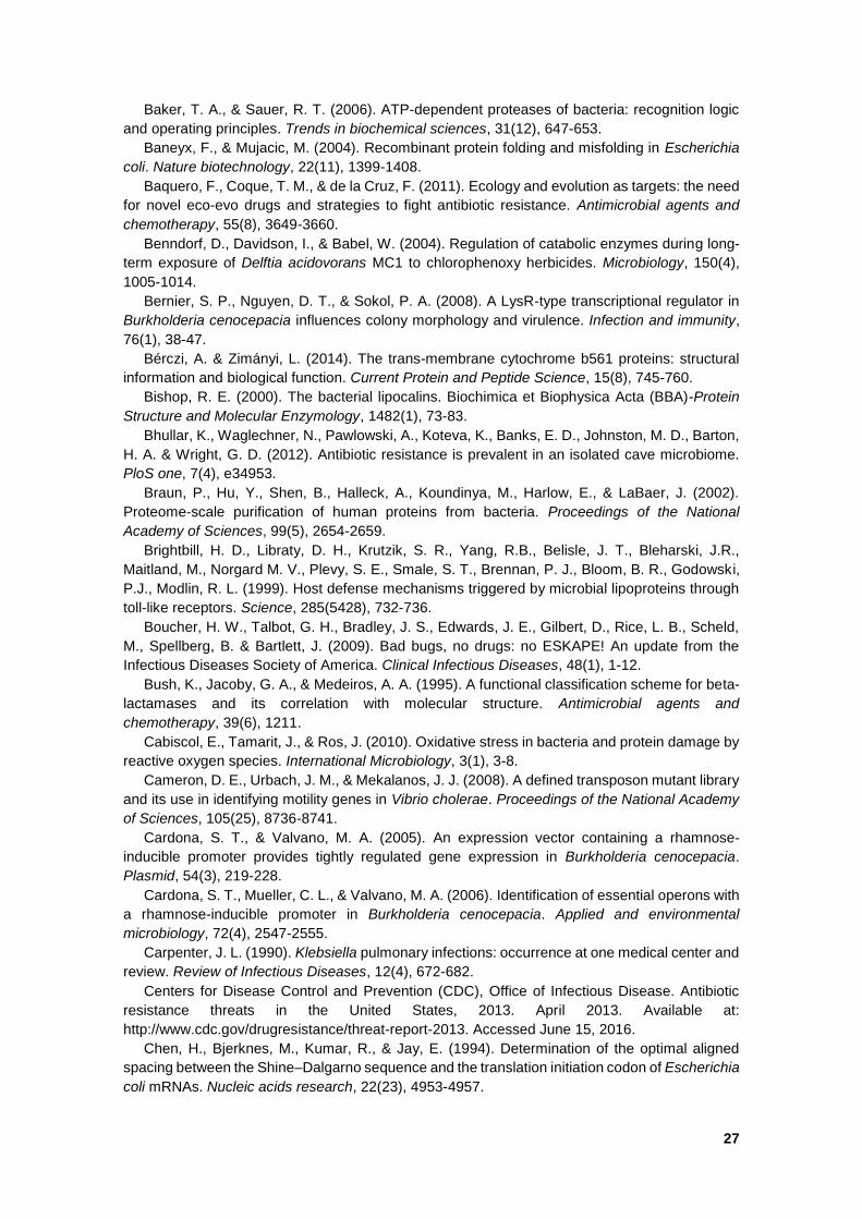
27
Baker, T. A., & Sauer, R. T. (2006). ATP-dependent proteases of bacteria: recognition logic
and operating principles. Trends in biochemical sciences, 31(12), 647-653.
Baneyx, F., & Mujacic, M. (2004). Recombinant protein folding and misfolding in Escherichia
coli. Nature biotechnology, 22(11), 1399-1408.
Baquero, F., Coque, T. M., & de la Cruz, F. (2011). Ecology and evolution as targets: the need
for novel eco-evo drugs and strategies to fight antibiotic resistance. Antimicrobial agents and
chemotherapy, 55(8), 3649-3660.
Benndorf, D., Davidson, I., & Babel, W. (2004). Regulation of catabolic enzymes during long-
term exposure of Delftia acidovorans MC1 to chlorophenoxy herbicides. Microbiology, 150(4),
1005-1014.
Bernier, S. P., Nguyen, D. T., & Sokol, P. A. (2008). A LysR-type transcriptional regulator in
Burkholderia cenocepacia influences colony morphology and virulence. Infection and immunity,
76(1), 38-47.
Bérczi, A. & Zimányi, L. (2014). The trans-membrane cytochrome b561 proteins: structural
information and biological function. Current Protein and Peptide Science, 15(8), 745-760.
Bishop, R. E. (2000). The bacterial lipocalins. Biochimica et Biophysica Acta (BBA)-Protein
Structure and Molecular Enzymology, 1482(1), 73-83.
Bhullar, K., Waglechner, N., Pawlowski, A., Koteva, K., Banks, E. D., Johnston, M. D., Barton,
H. A. & Wright, G. D. (2012). Antibiotic resistance is prevalent in an isolated cave microbiome.
PloS one, 7(4), e34953.
Braun, P., Hu, Y., Shen, B., Halleck, A., Koundinya, M., Harlow, E., & LaBaer, J. (2002).
Proteome-scale purification of human proteins from bacteria. Proceedings of the National
Academy of Sciences, 99(5), 2654-2659.
Brightbill, H. D., Libraty, D. H., Krutzik, S. R., Yang, R.B., Belisle, J. T., Bleharski, J.R.,
Maitland, M., Norgard M. V., Plevy, S. E., Smale, S. T., Brennan, P. J., Bloom, B. R., Godowski,
P.J., Modlin, R. L. (1999). Host defense mechanisms triggered by microbial lipoproteins through
toll-like receptors. Science, 285(5428), 732-736.
Boucher, H. W., Talbot, G. H., Bradley, J. S., Edwards, J. E., Gilbert, D., Rice, L. B., Scheld,
M., Spellberg, B. & Bartlett, J. (2009). Bad bugs, no drugs: no ESKAPE! An update from the
Infectious Diseases Society of America. Clinical Infectious Diseases, 48(1), 1-12.
Bush, K., Jacoby, G. A., & Medeiros, A. A. (1995). A functional classification scheme for beta-
lactamases and its correlation with molecular structure. Antimicrobial agents and
chemotherapy, 39(6), 1211.
Cabiscol, E., Tamarit, J., & Ros, J. (2010). Oxidative stress in bacteria and protein damage by
reactive oxygen species. International Microbiology, 3(1), 3-8.
Cameron, D. E., Urbach, J. M., & Mekalanos, J. J. (2008). A defined transposon mutant library
and its use in identifying motility genes in Vibrio cholerae. Proceedings of the National Academy
of Sciences, 105(25), 8736-8741.
Cardona, S. T., & Valvano, M. A. (2005). An expression vector containing a rhamnose-
inducible promoter provides tightly regulated gene expression in Burkholderia cenocepacia.
Plasmid, 54(3), 219-228.
Cardona, S. T., Mueller, C. L., & Valvano, M. A. (2006). Identification of essential operons with
a rhamnose-inducible promoter in Burkholderia cenocepacia. Applied and environmental
microbiology, 72(4), 2547-2555.
Carpenter, J. L. (1990). Klebsiella pulmonary infections: occurrence at one medical center and
review. Review of Infectious Diseases, 12(4), 672-682.
Centers for Disease Control and Prevention (CDC), Office of Infectious Disease. Antibiotic
resistance threats in the United States, 2013. April 2013. Available at:
http://www.cdc.gov/drugresistance/threat-report-2013. Accessed June 15, 2016.
Chen, H., Bjerknes, M., Kumar, R., & Jay, E. (1994). Determination of the optimal aligned
spacing between the Shine–Dalgarno sequence and the translation initiation codon of Escherichia
coli mRNAs. Nucleic acids research, 22(23), 4953-4957.
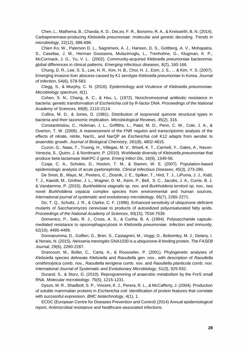
28
Chen, L., Mathema, B., Chavda, K. D., DeLeo, F. R., Bonomo, R. A., & Kreiswirth, B. N. (2014).
Carbapenemase-producing Klebsiella pneumoniae: molecular and genetic decoding. Trends in
microbiology, 22(12), 686-696.
Chien Ko, W., Paterson D. L., Sagnimeni, A. J., Hansen, D. S., Gottberg, A. V., Mohapatra,
S., Casellas, J. M., Herman Goossens, Mulazimoglu, L., Trenholme, G., Klugman, K. P.,
McCormack, J. G., Yu, V. L. (2002). Community-acquired Klebsiella pneumoniae bacteremia:
global differences in clinical patterns. Emerging infectious diseases, 8(2), 160-166.
Chung, D. R., Lee, S. S., Lee, H. R., Kim, H. B., Choi, H. J., Eom, J. S., ... & Kim, Y. S. (2007).
Emerging invasive liver abscess caused by K1 serotype Klebsiella pneumoniae in Korea. Journal
of Infection, 54(6), 578-583.
Clegg, S., & Murphy, C. N. (2016). Epidemiology and Virulence of Klebsiella pneumoniae.
Microbiology spectrum, 4(1).
Cohen, S. N., Chang, A. C., & Hsu, L. (1972). Nonchromosomal antibiotic resistance in
bacteria: genetic transformation of Escherichia coli by R-factor DNA. Proceedings of the National
Academy of Sciences, 69(8), 2110-2114.
Collins, M. D., & Jones, D. (1981). Distribution of isoprenoid quinone structural types in
bacteria and their taxonomic implication. Microbiological Reviews, 45(2), 316.
Constantinidou, C., Hobman, J. L., Griffiths, L., Patel, M. D., Penn, C. W., Cole, J. A., &
Overton, T. W. (2006). A reassessment of the FNR regulon and transcriptomic analysis of the
effects of nitrate, nitrite, NarXL, and NarQP as Escherichia coli K12 adapts from aerobic to
anaerobic growth. Journal of Biological Chemistry, 281(8), 4802-4815.
Cuzon, G., Naas, T., Truong, H., Villegas, M. V., Wisell, K. T., Carmeli, Y., Gales, A., Navon-
Venezia, S., Quinn, J. & Nordmann, P. (2010). Worldwide diversity of Klebsiella pneumoniae that
produce beta-lactamase blaKPC-2 gene. Emerg Infect Dis, 16(9), 1349-56.
Czaja, C. A., Scholes, D., Hooton, T. M., & Stamm, W. E. (2007). Population-based
epidemiologic analysis of acute pyelonephritis. Clinical Infectious Diseases, 45(3), 273-280.
De Smet, B., Mayo, M., Peeters, C., Zlosnik, J. E., Spilker, T., Hird, T. J., LiPuma, J. J., Kidd,
T. J., Kaestli, M., Ginther, J. L., Wagner, D. M., Keim, P., Bell, S. C., Jacobs, J. A., Currie, B. J.
& Vandamme, P. (2015). Burkholderia stagnalis sp. nov. and Burkholderia territorii sp. nov., two
novel Burkholderia cepacia complex species from environmental and human sources.
International journal of systematic and evolutionary microbiology, 65(7), 2265-2271.
Do, T. Q., Schultz, J. R., & Clarke, C. F. (1996). Enhanced sensitivity of ubiquinone-deficient
mutants of Saccharomyces cerevisiae to products of autoxidized polyunsaturated fatty acids.
Proceedings of the National Academy of Sciences, 93(15), 7534-7539.
Domenico, P., Salo, R. J., Cross, A. S., & Cunha, B. A. (1994). Polysaccharide capsule-
mediated resistance to opsonophagocytosis in Klebsiella pneumoniae. Infection and immunity,
62(10), 4495-4499.
Donnarumma, D., Golfieri, G., Brier, S., Castagnini, M., Veggi, D., Bottomley, M. J., Delany, I.
& Norais, N. (2015). Neisseria meningitis GNA1030 is a ubiquinone-8 binding protein. The FASEB
Journal, 29(6), 2260-2267.
Drancourt, M., Bollet, C., Carta, A., & Rousselier, P. (2001). Phylogenetic analyses of
Klebsiella species delineate Klebsiella and Raoultella gen. nov., with description of Raoultella
ornithinolytica comb. nov., Raoultella terrigena comb. nov. and Raoultella planticola comb. nov.
International Journal of Systematic and Evolutionary Microbiology, 51(3), 925-932.
Durand, S., & Storz, G. (2010). Reprogramming of anaerobic metabolism by the FnrS small
RNA. Molecular microbiology, 75(5), 1215-1231.
Dyson, M. R., Shadbolt, S. P., Vincent, K. J., Perera, R. L., & McCafferty, J. (2004). Production
of soluble mammalian proteins in Escherichia coli: identification of protein features that correlate
with successful expression. BMC biotechnology, 4(1), 1.
ECDC (European Centre for Diseases Prevention and Control) (2014) Annual epidemiological
report, Antimicrobial resistance and healthcare-associated infections.
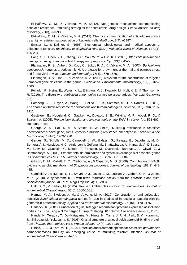
29
El-Halfawy, O. M., & Valvano, M. A. (2012). Non-genetic mechanisms communicating
antibiotic resistance: rethinking strategies for antimicrobial drug design. Expert opinion on drug
discovery, 7(10), 923-933.
El-Halfawy, O. M., & Valvano, M. A. (2013). Chemical communication of antibiotic resistance
by a highly resistant subpopulation of bacterial cells. PloS one, 8(7), e68874.
Ernster, L., & Dallner, G. (1995). Biochemical, physiological and medical aspects of
ubiquinone function. Biochimica et Biophysica Acta (BBA)-Molecular Basis of Disease, 1271(1),
195-204.
Fang, C. T., Chen, Y. C., Chang, S. C., Sau, W. Y., & Luh, K. T. (2000). Klebsiella pneumoniae
meningitis: timing of antimicrobial therapy and prognosis. Qjm, 93(1), 45-53.
Flannagan, R. S., Aubert, D., Kooi, C., Sokol, P. A., & Valvano, M. A. (2007). Burkholderia
cenocepacia requires a periplasmic HtrA protease for growth under thermal and osmotic stress
and for survival in vivo. Infection and immunity, 75(4), 1679-1689.
Flannagan, R. S., Linn, T., & Valvano, M. A. (2008). A system for the construction of targeted
unmarked gene deletions in the genus Burkholderia. Environmental microbiology, 10(6), 1652-
1660.
Follador, R., Heinz, E., Wyres, K. L., Ellington, M. J., Kowarik, M., Holt, K. E., & Thomson, N.
R. (2016). The diversity of Klebsiella pneumoniae surface polysaccharides. Microbial Genomics
2(8).
Forsberg, K. J., Reyes, A., Wang, B., Selleck, E. M., Sommer, M. O., & Dantas, G. (2012).
The shared antibiotic resistome of soil bacteria and human pathogens. Science, 337(6098), 1107-
1111.
Gasteiger, E., Hoogland, C., Gattiker, A., Duvaud, S. E., Wilkins, M. R., Appel, R. D., &
Bairoch, A. (2005). Protein identification and analysis tools on the ExPASy server (pp. 571-607).
Humana Press
George, A. M., Hall, R. M., & Stokes, H. W. (1995). Multidrug resistance in Klebsiella
pneumoniae: a novel gene, ramA, confers a multidrug resistance phenotype in Escherichia coli.
Microbiology, 141(8), 1909-1920.
Gerdes, S., Scholle, M. D., Campbell, J. W., Balazsi, G., Ravasz, E., Daugherty, M. D.,
Somera, A. I., Hyrpides, N. C., Anderson, I. Gelfang, M., Bhattacharya, A., Kapatral, V., D´Sousa,
M., Baev, M., Grachkin, Y., Mseed, F., Fonstein, M., Overbeek., Barabasi., A., Oltvai., Z. &
Bhattacharya, A. (2003). Experimental determination and system level analysis of essential genes
in Escherichia coli MG1655. Journal of bacteriology, 185(19), 5673-5684.
Gibson, C. M., Mallett, T. C., Claiborne, A., & Caparon, M. G. (2000). Contribution of NADH
oxidase to aerobic metabolism of Streptococcus pyogenes. Journal of bacteriology, 182(2), 448-
455.
Glanfield, A., McManus, D. P., Smyth, D. J., Lovas, E. M., Loukas, A., Gobert, G. N., & Jones,
M. K. (2010). A cytochrome b561 with ferric reductase activity from the parasitic blood fluke,
Schistosoma japonicum. PLoS Negl Trop Dis, 4(11), e884.
Hall, B. G., & Barlow, M. (2005). Revised Ambler classification of β-lactamases. Journal of
Antimicrobial Chemotherapy, 55(6), 1050-1051.
Hamad, M. A., Skeldon, A. M., & Valvano, M. A. (2010). Construction of aminoglycoside-
sensitive Burkholderia cenocepacia strains for use in studies of intracellular bacteria with the
gentamicin protection assay. Applied and environmental microbiology, 76(10), 3170-3176.
Hancock, K. (2001). Purification of (His) 6-tagged recombinant proteins expressed as inclusion
bodies in E. coli using a Ni2+-charged HiTrap Chelating HP column. Life science news, 8, 2001.
Handa, N., Terada , T., Doi-Katayama, Y., Hirota, H., Tame, J. R. H., Park, S. Y., Kuramitsu,
S., Shirouzu, M., Yokoyama. S, (2005). Crystal structure of a novel polyisoprenoid-binding protein
from Thermus thermophilus HB8. Protein science, 14(4), 1004-1010.
Hirsch, E. B., & Tam, V. H. (2010). Detection and treatment options for Klebsiella pneumoniae
carbapenemases (KPCs): an emerging cause of multidrug-resistant infection. Journal of
Antimicrobial Chemotherapy, dkq108.
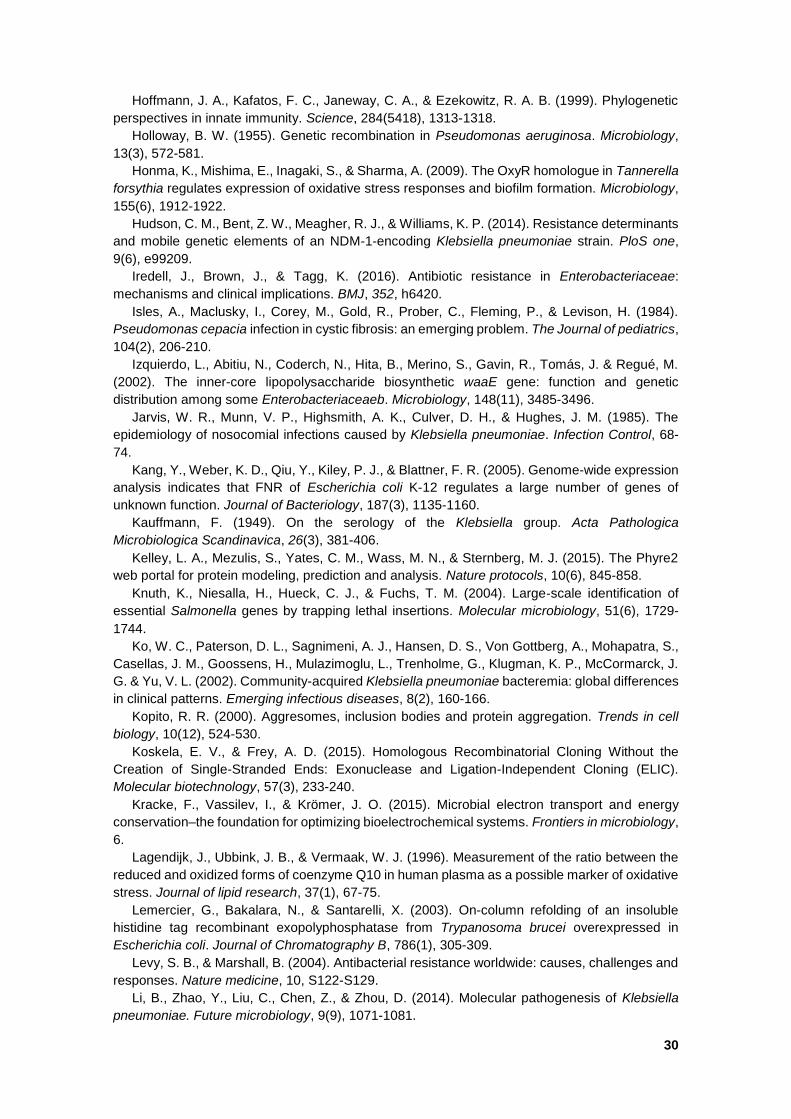
30
Hoffmann, J. A., Kafatos, F. C., Janeway, C. A., & Ezekowitz, R. A. B. (1999). Phylogenetic
perspectives in innate immunity. Science, 284(5418), 1313-1318.
Holloway, B. W. (1955). Genetic recombination in Pseudomonas aeruginosa. Microbiology,
13(3), 572-581.
Honma, K., Mishima, E., Inagaki, S., & Sharma, A. (2009). The OxyR homologue in Tannerella
forsythia regulates expression of oxidative stress responses and biofilm formation. Microbiology,
155(6), 1912-1922.
Hudson, C. M., Bent, Z. W., Meagher, R. J., & Williams, K. P. (2014). Resistance determinants
and mobile genetic elements of an NDM-1-encoding Klebsiella pneumoniae strain. PloS one,
9(6), e99209.
Iredell, J., Brown, J., & Tagg, K. (2016). Antibiotic resistance in Enterobacteriaceae:
mechanisms and clinical implications. BMJ, 352, h6420.
Isles, A., Maclusky, I., Corey, M., Gold, R., Prober, C., Fleming, P., & Levison, H. (1984).
Pseudomonas cepacia infection in cystic fibrosis: an emerging problem. The Journal of pediatrics,
104(2), 206-210.
Izquierdo, L., Abitiu, N., Coderch, N., Hita, B., Merino, S., Gavin, R., Tomás, J. & Regué, M.
(2002). The inner-core lipopolysaccharide biosynthetic waaE gene: function and genetic
distribution among some Enterobacteriaceaeb. Microbiology, 148(11), 3485-3496.
Jarvis, W. R., Munn, V. P., Highsmith, A. K., Culver, D. H., & Hughes, J. M. (1985). The
epidemiology of nosocomial infections caused by Klebsiella pneumoniae. Infection Control, 68-
74.
Kang, Y., Weber, K. D., Qiu, Y., Kiley, P. J., & Blattner, F. R. (2005). Genome-wide expression
analysis indicates that FNR of Escherichia coli K-12 regulates a large number of genes of
unknown function. Journal of Bacteriology, 187(3), 1135-1160.
Kauffmann, F. (1949). On the serology of the Klebsiella group. Acta Pathologica
Microbiologica Scandinavica, 26(3), 381-406.
Kelley, L. A., Mezulis, S., Yates, C. M., Wass, M. N., & Sternberg, M. J. (2015). The Phyre2
web portal for protein modeling, prediction and analysis. Nature protocols, 10(6), 845-858.
Knuth, K., Niesalla, H., Hueck, C. J., & Fuchs, T. M. (2004). Large‐scale identification of
essential Salmonella genes by trapping lethal insertions. Molecular microbiology, 51(6), 1729-
1744.
Ko, W. C., Paterson, D. L., Sagnimeni, A. J., Hansen, D. S., Von Gottberg, A., Mohapatra, S.,
Casellas, J. M., Goossens, H., Mulazimoglu, L., Trenholme, G., Klugman, K. P., McCormarck, J.
G. & Yu, V. L. (2002). Community-acquired Klebsiella pneumoniae bacteremia: global differences
in clinical patterns. Emerging infectious diseases, 8(2), 160-166.
Kopito, R. R. (2000). Aggresomes, inclusion bodies and protein aggregation. Trends in cell
biology, 10(12), 524-530.
Koskela, E. V., & Frey, A. D. (2015). Homologous Recombinatorial Cloning Without the
Creation of Single-Stranded Ends: Exonuclease and Ligation-Independent Cloning (ELIC).
Molecular biotechnology, 57(3), 233-240.
Kracke, F., Vassilev, I., & Krömer, J. O. (2015). Microbial electron transport and energy
conservation–the foundation for optimizing bioelectrochemical systems. Frontiers in microbiology,
6.
Lagendijk, J., Ubbink, J. B., & Vermaak, W. J. (1996). Measurement of the ratio between the
reduced and oxidized forms of coenzyme Q10 in human plasma as a possible marker of oxidative
stress. Journal of lipid research, 37(1), 67-75.
Lemercier, G., Bakalara, N., & Santarelli, X. (2003). On-column refolding of an insoluble
histidine tag recombinant exopolyphosphatase from Trypanosoma brucei overexpressed in
Escherichia coli. Journal of Chromatography B, 786(1), 305-309.
Levy, S. B., & Marshall, B. (2004). Antibacterial resistance worldwide: causes, challenges and
responses. Nature medicine, 10, S122-S129.
Li, B., Zhao, Y., Liu, C., Chen, Z., & Zhou, D. (2014). Molecular pathogenesis of Klebsiella
pneumoniae. Future microbiology, 9(9), 1071-1081.
31
Llor, C., & Cots, J. M. (2009). The sale of antibiotics without prescription in pharmacies in
Catalonia, Spain. Clinical Infectious Diseases, 48(10), 1345-1349.
Lockhart, S. R., Abramson, M. A., Beekmann, S. E., Gallagher, G., Riedel, S., Diekema, D. J.,
Quinn, J. P. & Doern, G. V. (2007). Antimicrobial resistance among Gram-negative bacilli causing
infections in intensive care unit patients in the United States between 1993 and 2004. Journal of
clinical microbiology, 45(10), 3352-3359.
Loprasert, S., Sallabhan, R., Whangsuk, W., & Mongkolsuk, S. (2002). The Burkholderia
pseudomalleioxy R gene: expression analysis and mutant characterization. Gene, 296(1), 161-
169.
Lorente, G. J., Placer, S. J., Salvadó, C. M., Segura, A. C., & Gelabert-Mas, A. (2005).
Antibiotic resistance transformation in community-acquired urinary infections. Revista clinica
espanola, 205(6), 259-264.
Lu, P., Ma, D., Yan, C., Gong, X., Du, M., & Shi, Y. (2014). Structure and mechanism of a
eukaryotic transmembrane ascorbate-dependent oxidoreductase. Proceedings of the National
Academy of Sciences, 111(5), 1813-1818.
Luan, C. H., Qiu, S., Finley, J. B., Carson, M., Gray, R. J., Huang, W., Johnson, D., Tsao, J.,
Reboul, J., Vaglio, P., Hill, D. E., Vidal, M., DeLucas, L. J. & Hill, D. E. (2004). High-throughput
expression of C. elegans proteins. Genome research, 14(10b), 2102-2110.
Madigan, M. T., Clark, D. P., Stahl, D., & Martinko, J. M. (2010). Brock Biology of
Microorganisms 13th edition. Benjamin Cummings.
Marks, D. S., Hopf, T. A., & Sander, C. (2012). Protein structure prediction from sequence
variation. Nature biotechnology, 30(11), 1072-1080.
Martínez, J. L., Coque, T. M., & Baquero, F. (2015). What is a resistance gene? Ranking risk
in resistomes. Nature reviews microbiology, 13(2), 116-123.
Medzhitov, R., & Janeway, C. A. (1997). Innate immunity: the virtues of a nonclonal system of
recognition. Cell, 91(3), 295-298.
Mizuta, K., Ohta, M., Mori, M., Hasegawa, T., Nakashima, I., & Kato, N. (1983). Virulence for
mice of Klebsiella strains belonging to the O1 group: relationship to their capsular (K) types.
Infection and Immunity, 40(1), 56-61.
Montgomerie, J. Z. (1979). Epidemiology of Klebsiella and hospital-associated infections.
Reviews of infectious diseases, 1(5), 736-753.
Munoz-Price, L., Poirel, L., Bonomo, R., Schwaber, M., Daikos, G., Cormican, M., Cornaglia,
G., Garau, J., Gniadkowski, M., Hayden, M., Kumarasamy, K., Livermore, D., Maya, J.,
Nordmann, P., Patel, J., Paterson, D., Pitout, L., Villegas, M., Wang, H., Woodford, N. &
Kumarasamy, K. (2013). Clinical epidemiology of the global expansion of Klebsiella pneumoniae
carbapenemases. The Lancet infectious diseases, 13(9), 785-796.
Murakami, H., Kita, K., & Anraku, Y. (1986). Purification and properties of a diheme
cytochrome b561 of the Escherichia coli respiratory chain. Journal of Biological Chemistry,
261(2), 548-551.
Nassif, X. A. V. I. E. R., Fournier, J. M., Arondel, J., & Sansonetti, P. J. (1989). Mucoid
phenotype of Klebsiella pneumoniae is a plasmid-encoded virulence factor. Infection and
immunity, 57(2), 546-552.
Nathan, C. (2004). Antibiotics at the crossroads. Nature, 431(7011), 899-902.
Nowicka, B., & Kruk, J. (2010). Occurrence, biosynthesis and function of isoprenoid quinones.
Biochimica et Biophysica Acta (BBA)-Bioenergetics, 1797(9), 1587-1605.
Oberto, J. (2013). SyntTax: a web server linking synteny to prokaryotic taxonomy. Bmc
Bioinformatics, 14(1), 1.
Ogawa, W., Li, D. W., Yu, P., Begum, A., Mizushima, T., Kuroda, T., & Tsuchiya, T. (2005).
Multidrug resistance in Klebsiella pneumoniae MGH78578 and cloning of genes responsible for
the resistance. Biological and Pharmaceutical Bulletin, 28(8), 1505-1508.
O’Neil, K. M., Herman, J. H., Modlin, J. F., Moxon, E. R. & Winkelstein, J. A. (1986)
Pseudomonas cepacia: An emerging pathogen in chronic granulomatous disease. The journal of
paediatrics, 108(6), 940-942.

32
Ortega, X. P., Cardona, S. T., Brown, A. R., Loutet, S. A., Flannagan, R. S., Campopiano, D.
J., Govan, J. R. W. & Valvano, M. A. (2007). A putative gene cluster for aminoarabinose
biosynthesis is essential for Burkholderia cenocepacia viability. Journal of bacteriology, 189(9),
3639-3644.
Paczosa, M. K., & Mecsas, J. (2016). Klebsiella pneumoniae: Going on the Offense with a
Strong Defense. Microbiology and Molecular Biology Reviews, 80(3), 629-661.
Papp-Wallace, K. M., Bethel, C. R., Distler, A. M., Kasuboski, C., Taracila, M., & Bonomo, R.
A. (2010). Inhibitor resistance in the KPC-2 β-lactamase, a preeminent property of this class A β-
lactamase. Antimicrobial agents and chemotherapy, 54(2), 890-897.
Paterson, D. L. (2006). Resistance in gram-negative bacteria: Enterobacteriaceae. The
American journal of medicine, 119(6), S20-S28.
Penesyan, A., Gillings, M., & Paulsen, I. T. (2015). Antibiotic discovery: combatting bacterial
resistance in cells and in biofilm communities. Molecules, 20(4), 5286-5298.
Perin, M. S., Fried, V. A., Slaughter, C. A., & Südhof, T. C. (1988). The structure of cytochrome
b561, a secretory vesicle-specific electron transport protein. The EMBO journal, 7(9), 2697.
Petersen, T. N., Brunak, S., von Heijne, G., & Nielsen, H. (2011). SignalP 4.0: discriminating
signal peptides from transmembrane regions. Nature methods, 8(10), 785-786.
Piddock, L. J. (2012). The crisis of no new antibiotics—what is the way forward?. The Lancet
infectious diseases, 12(3), 249-253.
Pitout, J. D., Nordmann, P., & Poirel, L. (2015). Carbapenemase-producing Klebsiella
pneumoniae, a key pathogen set for global nosocomial dominance. Antimicrobial agents and
chemotherapy, 59(10), 5873-5884.
Podschun, R., & Ullmann, U. (1998). Klebsiella spp. as nosocomial pathogens: epidemiology,
taxonomy, typing methods, and pathogenicity factors. Clinical microbiology reviews, 11(4), 589-
603.
Poe, R. H., Marcus, H. R., & Emerson, G. L. (1977). Lung Abscess Due to Pseudomonas
cepacia 1, 2. American Review of Respiratory Disease, 115(5), 861-865.
Postgate, J. (1998) Nitrogen Fixation (3rd Edition). Cambridge University Press.
Queenan, A. M., & Bush, K. (2007) Carbapenemases: the versatile beta-lactamases. Clinical
microbiology reviews, 20(3), 440-458.
Rasmusson, A. G., Geisler, D. A., & Møller, I. M. (2008). The multiplicity of dehydrogenases
in the electron transport chain of plant mitochondria. Mitochondrion, 8(1), 47-60.
Rice, L. B. (2008). Federal funding for the study of antimicrobial resistance in nosocomial
pathogens: no ESKAPE. Journal of Infectious Diseases, 197(8), 1079-1081.
Riggs, P. J., Chelius, M. K., Iniguez, A. L., Kaeppler, S. M., & Triplett, E. W. (2001). Enhanced
maize productivity by inoculation with diazotrophic bacteria. Functional Plant Biology, 28(9), 829-
836.
Salmon, K., Hung, S. P., Mekjian, K., Baldi, P., Hatfield, G. W., & Gunsalus, R. P. (2003).
Global gene expression profiling in Escherichia coli K12 the effects of oxygen availability and
FNR. Journal of Biological Chemistry, 278(32), 29837-29855.
Sambrook, J., Fritsh, E., & Maniatis, T. (1989). Molecular cloning: A laboratory manual 2nd
editon, Cold Spring Harbor Press New York.
Sanchez, G. V., Master, R. N., Clark, R. B., Fyyaz, M., Duvvuri, P., Ekta, G., & Bordon, J.
(2013). Klebsiella pneumoniae antimicrobial drug resistance, United States, 1998–2010. Emerg
Infect Dis, 19(1), 133-6.
Schroll, C., Barken, K. B., Krogfelt, K. A., & Struve, C. (2010). Role of type 1 and type 3 fimbriae
in Klebsiella pneumoniae biofilm formation. BMC microbiology, 10(1), 1.
Scott, N. E., Hare, N. J., White, M. Y., Manos, J., & Cordwell, S. J. (2013). Secretome of
transmissible Pseudomonas aeruginosa AES-1R grown in a cystic fibrosis lung-like environment.
Journal of proteome research, 12(12), 5357-5369.
Seib, K. L., Wu, H. J., Srikhanta, Y. N., Edwards, J. L., Falsetta, M. L., Hamilton, A. J., ... &
Jennings, M. P. (2007). Characterization of the OxyR regulon of Neisseria gonorrhoeae.
Molecular microbiology, 63(1), 54-68.
33
Shon, A. S., & Russo, T. A. (2012). Hypervirulent Klebsiella pneumoniae: the next superbug?.
Future microbiology, 7(6), 669-671.
Shon, A. S., Bajwa, R. P., & Russo, T. A. (2013). Hypervirulent (hypermucoviscous) Klebsiella
pneumoniae: a new and dangerous breed. Virulence, 4(2), 107-118.
Simoons-Smit, A. M., Verweij-van Vught, A. M. J. J., & MacLaren, D. M. (1986). The role of K
antigens as virulence factors in Klebsiella. Journal of medical microbiology, 21(2), 133-137.
Sievers, F., Wilm, A., Dineen, D., Gibson, T. J., Karplus, K., Li, W., Lopez, R., McWilliam, H.,
Remmert, M., Soding, J., Thompson, J. & Higgins, D. (2011). Fast, scalable generation of high‐
quality protein multiple sequence alignments using Clustal Omega. Molecular systems biology,
7(1), 539.
Sisinni, L., Cendron, L., Favaro, G., & Zanotti, G. (2010). Helicobacter pylori acidic stress
response factor HP1286 is a YceI homolog with new binding specificity. FEBS journal, 277(8),
1896-1905.
Siu, L. K., Yeh, K. M., Lin, J. C., Fung, C. P., & Chang, F. Y. (2012). Klebsiella pneumoniae
liver abscess: a new invasive syndrome. The Lancet infectious diseases, 12(11), 881-887.
Søballe, B., & Poole, R. K. (1999). Microbial ubiquinones: multiple roles in respiration, gene
regulation and oxidative stress management. Microbiology, 145(8), 1817-1830.
Smart.embl-heidelberg.de. (2016). SMART: Main page. Available at: http://smart.embl-
heidelberg.de/. Accessed 26 July 2016.
Sommer, M. O., Dantas, G., & Church, G. M. (2009). Functional characterization of the
antibiotic resistance reservoir in the human microflora. Science, 325(5944), 1128-1131.
Spellberg, B. (2011). The antibiotic crisis: can we reverse 65 years of failed stewardship?
Archives of internal medicine, 171(12), 1080.
Spellberg, B., M.D., John G. Bartlett, M.D., and David N. Gilbert, M.D. (2013). The future of
Antibiotics and Resistance. The New England Journal of Medicine, 368, 299-302.
Stancik, L. M., Stancik, D. M., Schmidt, B., Barnhart, D. M., Yoncheva, Y. N., & Slonczewski,
J. L. (2002). pH-dependent expression of periplasmic proteins and amino acid catabolism in
Escherichia coli. Journal of bacteriology, 184(15), 4246-4258.
Sun, F., Qu, F., Ling, Y., Mao, P., Xia, P., Chen, H., & Zhou, D. (2013). Biofilm-associated
infections: antibiotic resistance and novel therapeutic strategies. Future microbiology, 8(7), 877-
886.
Szklarczyk, D., Franceschini, A., Wyder, S., Forslund, K., Heller, D., Huerta-Cepas, J.,
Simonovic, M., Roth, A., Santos, A., Tsafou, K., Kuhn, M., Bork, P., Jensen, L. & Mering, C.
(2014). STRING v10: protein–protein interaction networks, integrated over the tree of life. Nucleic
acids research, gku1003.
Temme, K., Zhao, D., & Voigt, C. A. (2012). Refactoring the nitrogen fixation gene cluster from
Klebsiella oxytoca. Proceedings of the National Academy of Sciences, 109(18), 7085-7090.
Tenover, F. C. (2006). Mechanisms of antimicrobial resistance in bacteria. The American
journal of medicine, 119(6), S3-S10.
Tomoyasu, T., Mogk, A., Langen, H., Goloubinoff, P., & Bukau, B. (2001). Genetic dissection
of the roles of chaperones and proteases in protein folding and degradation in the Escherichia
coli cytosol. Molecular microbiology, 40(2), 397-413.
Tsubaki, M., Takeuchi, F., & Nakanishi, N. (2005). Cytochrome b561 protein family: expanding
roles and versatile transmembrane electron transfer abilities as predicted by a new classification
system and protein sequence motif analyses. Biochimica et Biophysica Acta (BBA)-Proteins and
Proteomics, 1753(2), 174-190.
Tsumoto, K., Ejima, D., Kumagai, I., & Arakawa, T. (2003). Practical considerations in refolding
proteins from inclusion bodies. Protein expression and purification, 28(1), 1-8.
Turnidge, J., & Paterson, D. L. (2007). Setting and revising antibacterial susceptibility
breakpoints. Clinical microbiology reviews, 20(3), 391-408.
Upritchard, H. G., Cordwell, S. J., & Lamont, I. L. (2008). Immunoproteomics to examine cystic
fibrosis host interactions with extracellular Pseudomonas aeruginosa proteins. Infection and
immunity, 76(10), 4624-4632.
34
Urban, C., Bradford, P. A., Tuckman, M., Segal-Maurer, S., Wehbeh, W., Grenner, L., Colon-
Urban, R., Mariano, M. & Rahal, J. J. (2008). Carbapenem-resistant Escherichia coli harboring
Klebsiella pneumoniae carbapenemase β-lactamases associated with long-term care facilities.
Clinical Infectious Diseases, 46(11), e127-e130.
Villegas, M. V., Lolans, K., Correa, A., Kattan, J. N., Lopez, J. A., & Quinn, J. P. (2007).
Colombian Nosocomial Resistance Study Group First identification of Pseudomonas aeruginosa
isolates producing a KPC-type carbapenem-hydrolyzing β-lactamase. Antimicrob Agents
Chemother, 51(4), 1553-1555.
Vincent, F., Dal Molin, D., Weiner, R. M., Bourne, Y., & Henrissat, B. (2010). Structure of a
polyisoprenoid binding domain from Saccharophagus degradans implicated in plant cell wall
breakdown. FEBS letters, 584(8), 1577-1584.
Wang, D., Chen, J., Yang, L., Mou, Y., & Yang, Y. (2014). Phenotypic and enzymatic
comparative analysis of the KPC variants, KPC-2 and its recently discovered variant KPC-15.
PloS one, 9(10), e111491.
Wang, X., Yang, J. G., Chen, L., Wang, J. L., Cheng, Q., Dixon, R., & Wang, Y. P. (2013).
Using synthetic biology to distinguish and overcome regulatory and functional barriers related to
nitrogen fixation. PloS one, 8(7), e68677.
Waters, L. S., & Storz, G. (2009). Regulatory RNAs in bacteria. Cell, 136(4), 615-628.
WEF (World Economic Forum), (2015), Golbal Riscks 2015 10th Edition An Initiative of the
Risk Response Network, 1-69.
WHO (World Health Organization), (2014) Antibiotic resistance: Global Report on
Surveillance. 1-257.
Wiegand, I., Hilpert, K., & Hancock, R. E. (2008). Agar and broth dilution methods to determine
the minimal inhibitory concentration (MIC) of antimicrobial substances. Nature protocols, 3(2),
163-175.
Willett, H. P. (1992). Energy metabolism. Zinsser microbiology. 20th ed. East Norwalk (CT):
Appleton & Lange, 53-75.
Winson, M. K., Swift, S., Hill, P. J., Sims, C. M., Griesmayr, G., Bycroft, B. W., Williams, P. &
Stewart, G. S. (1998). Engineering the luxCDABE genes from Photorhabdus luminescens to
provide a bioluminescent reporter for constitutive and promoter probe plasmids and mini-Tn5
constructs. FEMS microbiology letters, 163(2), 193-202.
Wright, G. D. (2015). Solving the antibiotic crisis. ACS Infectious Diseases, 1(2), 80-84.
Woestenenk, E. A., Hammarström, M., van den Berg, S., Härd, T., & Berglund, H. (2004). His
tag effect on solubility of human proteins produced in Escherichia coli: a comparison between
four expression vectors. Journal of structural and functional genomics, 5(3), 217-229.
Yigit, H., Queenan, A. M., Anderson, G. J., Domenech-Sanchez, A., Biddle, J. W., Steward,
C. D., Alberdi, S., Bush, K. & Tenover, F. C. (2001). Novel carbapenem-hydrolyzing β-lactamase,
KPC-1, from a carbapenem-resistant strain of Klebsiella pneumoniae. Antimicrobial agents and
chemotherapy, 45(4), 1151-1161.
Yoder-Himes, D. R., Konstantinidis, K. T., & Tiedje, J. M. (2010). Identification of potential
therapeutic targets for Burkholderia cenocepacia by comparative transcriptomics. PLoS One,
5(1), e8724.
Yokochi, T., Nakashima, I., & Kato, N. (1979). Further studies on generation of macrophages
in in vitro cultures of mouse spleen cells and its inhibition by the capsular polysaccharide of
Klebsiella pneumoniae. Microbiology and immunology, 23(6), 487-499.
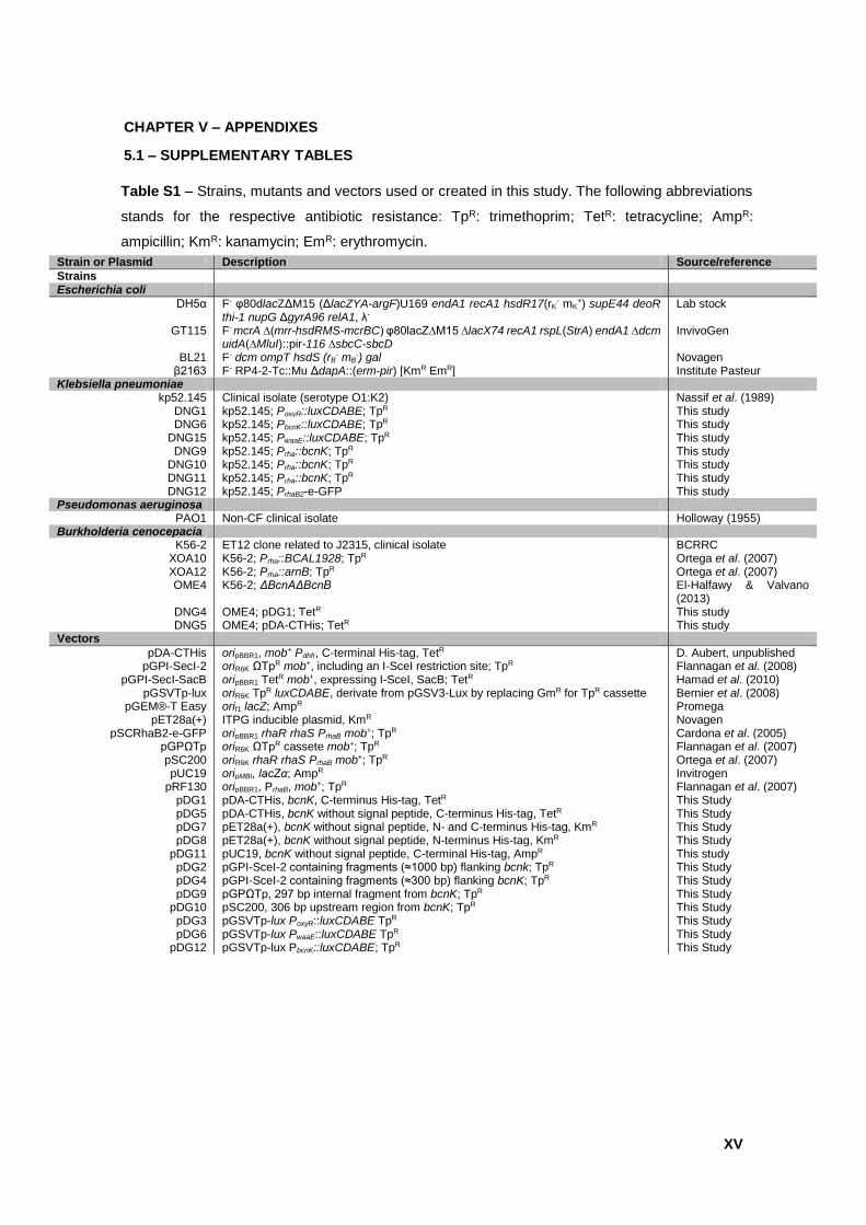
XV
CHAPTER V – APPENDIXES
5.1 – SUPPLEMENTARY TABLES
Strain or Plasmid Description Source/reference
Strains Escherichia coli
DH5α F- φ80dlacZΔM15 (ΔlacZYA-argF)U169 endA1 recA1 hsdR17(rK- mK
+) supE44 deoR thi-1 nupG ΔgyrA96 relA1, λ-
Lab stock
GT115 F- mcrA ∆(mrr-hsdRMS-mcrBC) φ80lacZ∆M15 ∆lacX74 recA1 rspL(StrA) endA1 ∆dcm uidA(∆MluI)::pir-116 ∆sbcC-sbcD
InvivoGen
BL21 F- dcm ompT hsdS (rB- mB
-) gal Novagen β2163 F- RP4-2-Tc::Mu ΔdapA::(erm-pir) [KmR EmR] Institute Pasteur
Klebsiella pneumoniae kp52.145 Clinical isolate (serotype O1:K2) Nassif et al. (1989)
DNG1 kp52.145; PoxyR::luxCDABE; TpR This study DNG6 kp52.145; PbcnK::luxCDABE; TpR This study
DNG15 kp52.145; PwaaE::luxCDABE; TpR This study DNG9 kp52.145; Prha::bcnK; TpR This study
DNG10 kp52.145; Prha::bcnK; TpR This study DNG11 kp52.145; Prha::bcnK; TpR This study DNG12 kp52.145; PrhaB2-e-GFP This study
Pseudomonas aeruginosa PAO1 Non-CF clinical isolate Holloway (1955)
Burkholderia cenocepacia K56-2 ET12 clone related to J2315, clinical isolate BCRRC
XOA10 K56-2; Prha::BCAL1928; TpR Ortega et al. (2007) XOA12 K56-2; Prha::arnB; TpR Ortega et al. (2007) OME4 K56-2; ΔBcnAΔBcnB El-Halfawy & Valvano
(2013) DNG4 OME4; pDG1; TetR This study DNG5 OME4; pDA-CTHis; TetR This study
Vectors
pDA-CTHis oripBBR1, mob+ Pdhfr, C-terminal His-tag, TetR D. Aubert, unpublished pGPI-SecI-2 oriR6K ΩTpR mob+, including an I-SceI restriction site; TpR Flannagan et al. (2008)
pGPI-SecI-SacB oripBBR1 TetR mob+, expressing I-SceI, SacB; TetR Hamad et al. (2010) pGSVTp-lux oriR6K TpR luxCDABE, derivate from pGSV3-Lux by replacing GmR for TpR cassette Bernier et al. (2008)
pGEM®-T Easy orif1 lacZ; AmpR Promega pET28a(+) ITPG inducible plasmid, KmR Novagen
pSCRhaB2-e-GFP oripBBR1 rhaR rhaS PrhaB mob+; TpR Cardona et al. (2005) pGPΩTp oriR6K ΩTpR cassete mob+; TpR Flannagan et al. (2007) pSC200 oriR6K rhaR rhaS PrhaB mob+; TpR Ortega et al. (2007) pUC19 oripMBI, lacZα; AmpR Invitrogen
pRF130 oripBBR1, PrhaB, mob+; TpR Flannagan et al. (2007) pDG1 pDA-CTHis, bcnK, C-terminus His-tag, TetR This Study pDG5 pDA-CTHis, bcnK without signal peptide, C-terminus His-tag, TetR This Study pDG7 pET28a(+), bcnK without signal peptide, N- and C-terminus His-tag, KmR This Study pDG8 pET28a(+), bcnK without signal peptide, N-terminus His-tag, KmR This Study
pDG11 pUC19, bcnK without signal peptide, C-terminal His-tag, AmpR This study pDG2 pGPI-SceI-2 containing fragments (≈1000 bp) flanking bcnk; TpR This Study pDG4 pGPI-SceI-2 containing fragments (≈300 bp) flanking bcnK; TpR This Study pDG9 pGPΩTp, 297 bp internal fragment from bcnK; TpR This Study
pDG10 pSC200, 306 bp upstream region from bcnK; TpR This Study pDG3 pGSVTp-lux PoxyR::luxCDABE TpR This Study pDG6 pGSVTp-lux PwaaE::luxCDABE TpR This Study
pDG12 pGSVTp-lux PbcnK::luxCDABE; TpR This Study
Table S1 – Strains, mutants and vectors used or created in this study. The following abbreviations
stands for the respective antibiotic resistance: TpR: trimethoprim; TetR: tetracycline; AmpR:
ampicillin; KmR: kanamycin; EmR: erythromycin.
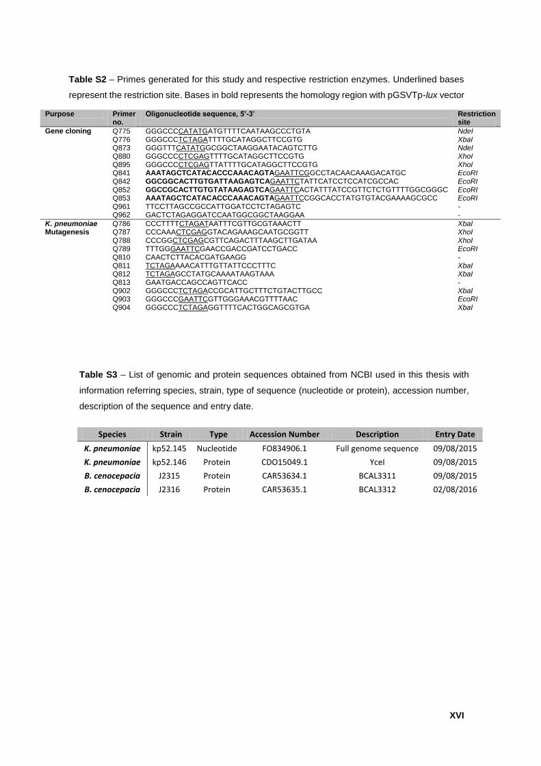
XVI
Purpose Primer no.
Oligonucleotide sequence, 5’-3’ Restriction site
Gene cloning Q775 GGGCCCCATATGATGTTTTCAATAAGCCCTGTA NdeI Q776 GGGCCCTCTAGATTTTGCATAGGCTTCCGTG XbaI
Q873 GGGTTTCATATGGCGGCTAAGGAATACAGTCTTG NdeI Q880 GGGCCCCTCGAGTTTTGCATAGGCTTCCGTG XhoI Q895 GGGCCCCTCGAGTTATTTTGCATAGGCTTCCGTG XhoI Q841 AAATAGCTCATACACCCAAACAGTAGAATTCGGCCTACAACAAAGACATGC EcoRI Q842 GGCGGCACTTGTGATTAAGAGTCAGAATTCTATTCATCCTCCATCGCCAC EcoRI Q852 GGCCGCACTTGTGTATAAGAGTCAGAATTCACTATTTATCCGTTCTCTGTTTTGGCGGGC EcoRI Q853 AAATAGCTCATACACCCAAACAGTAGAATTCCGGCACCTATGTGTACGAAAAGCGCC EcoRI Q961 TTCCTTAGCCGCCATTGGATCCTCTAGAGTC - Q962 GACTCTAGAGGATCCAATGGCGGCTAAGGAA -
K. pneumoniae Q786 CCCTTTTCTAGATAATTTCGTTGCGTAAACTT XbaI Mutagenesis Q787 CCCAAACTCGAGGTACAGAAAGCAATGCGGTT XhoI Q788 CCCGGCTCGAGCGTTCAGACTTTAAGCTTGATAA XhoI Q789 TTTGGGAATTCGAACCGACCGATCCTGACC EcoRI Q810 CAACTCTTACACGATGAAGG - Q811 TCTAGAAAACATTTGTTATTCCCTTTC XbaI Q812 TCTAGAGCCTATGCAAAATAAGTAAA XbaI Q813 GAATGACCAGCCAGTTCACC - Q902 GGGCCCTCTAGACCGCATTGCTTTCTGTACTTGCC XbaI Q903 GGGCCCGAATTCGTTGGGAAACGTTTTAAC EcoRI Q904 GGGCCCTCTAGAGGTTTTCACTGGCAGCGTGA XbaI
Species Strain Type Accession Number Description Entry Date
K. pneumoniae kp52.145 Nucleotide FO834906.1 Full genome sequence 09/08/2015
K. pneumoniae kp52.146 Protein CDO15049.1 YceI 09/08/2015
B. cenocepacia J2315 Protein CAR53634.1 BCAL3311 09/08/2015
B. cenocepacia J2316 Protein CAR53635.1 BCAL3312 02/08/2016
Table S2 – Primes generated for this study and respective restriction enzymes. Underlined bases
represent the restriction site. Bases in bold represents the homology region with pGSVTp-lux vector
Table S3 – List of genomic and protein sequences obtained from NCBI used in this thesis with
information referring species, strain, type of sequence (nucleotide or protein), accession number,
description of the sequence and entry date.
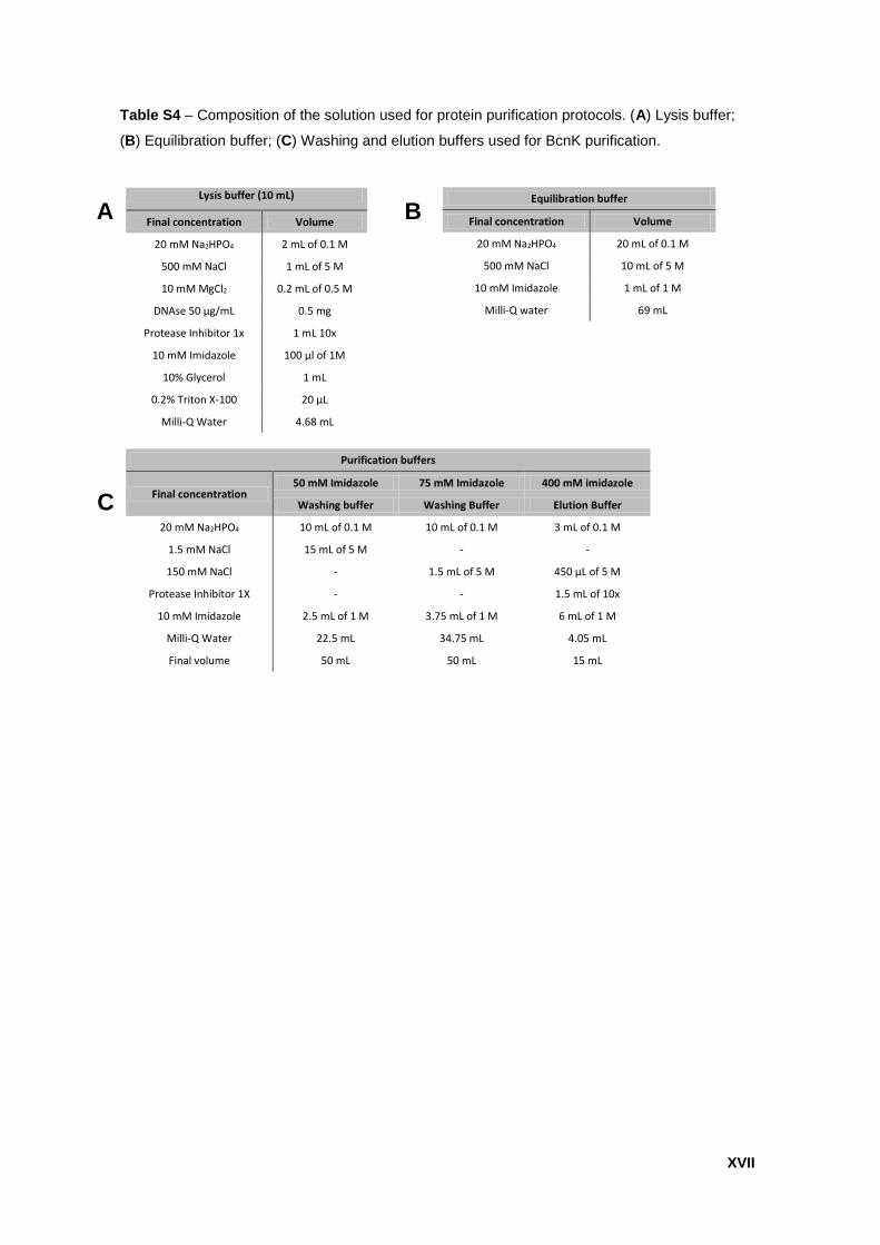
XVII
C
Equilibration buffer
Final concentration Volume
20 mM Na2HPO4 20 mL of 0.1 M
500 mM NaCl 10 mL of 5 M
10 mM Imidazole 1 mL of 1 M
Milli-Q water 69 mL
Lysis buffer (10 mL)
Final concentration Volume
20 mM Na2HPO4 2 mL of 0.1 M
500 mM NaCl 1 mL of 5 M
10 mM MgCl2 0.2 mL of 0.5 M
DNAse 50 µg/mL 0.5 mg
Protease Inhibitor 1x 1 mL 10x
10 mM Imidazole 100 µl of 1M
10% Glycerol 1 mL
0.2% Triton X-100 20 µL
Milli-Q Water 4.68 mL
Purification buffers
Final concentration 50 mM Imidazole 75 mM Imidazole 400 mM imidazole
Washing buffer Washing Buffer Elution Buffer
20 mM Na2HPO4 10 mL of 0.1 M 10 mL of 0.1 M 3 mL of 0.1 M
1.5 mM NaCl 15 mL of 5 M - -
150 mM NaCl - 1.5 mL of 5 M 450 µL of 5 M
Protease Inhibitor 1X - - 1.5 mL of 10x
10 mM Imidazole 2.5 mL of 1 M 3.75 mL of 1 M 6 mL of 1 M
Milli-Q Water 22.5 mL 34.75 mL 4.05 mL
Final volume 50 mL 50 mL 15 mL
Table S4 – Composition of the solution used for protein purification protocols. (A) Lysis buffer;
(B) Equilibration buffer; (C) Washing and elution buffers used for BcnK purification.
A B
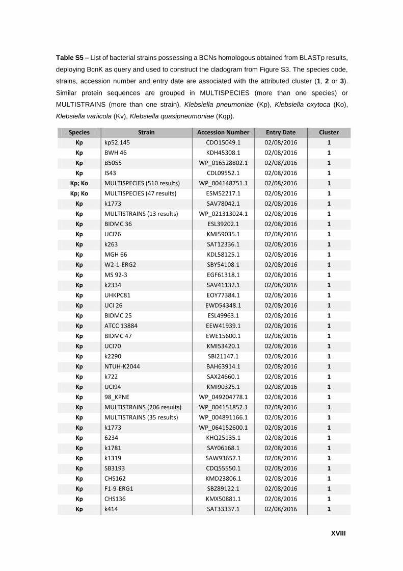
XVIII
Species Strain Accession Number Entry Date Cluster
Kp kp52.145 CDO15049.1 02/08/2016 1
Kp BWH 46 KDH45308.1 02/08/2016 1
Kp B5055 WP_016528802.1 02/08/2016 1
Kp IS43 CDL09552.1 02/08/2016 1
Kp; Ko MULTISPECIES (510 results) WP_004148751.1 02/08/2016 1
Kp; Ko MULTISPECIES (47 results) ESM52217.1 02/08/2016 1
Kp k1773 SAV78042.1 02/08/2016 1
Kp MULTISTRAINS (13 results) WP_021313024.1 02/08/2016 1
Kp BIDMC 36 ESL39202.1 02/08/2016 1
Kp UCI76 KMI59035.1 02/08/2016 1
Kp k263 SAT12336.1 02/08/2016 1
Kp MGH 66 KDL58125.1 02/08/2016 1
Kp W2-1-ERG2 SBY54108.1 02/08/2016 1
Kp MS 92-3 EGF61318.1 02/08/2016 1
Kp k2334 SAV41132.1 02/08/2016 1
Kp UHKPC81 EOY77384.1 02/08/2016 1
Kp UCI 26 EWD54348.1 02/08/2016 1
Kp BIDMC 25 ESL49963.1 02/08/2016 1
Kp ATCC 13884 EEW41939.1 02/08/2016 1
Kp BIDMC 47 EWE15600.1 02/08/2016 1
Kp UCI70 KMI53420.1 02/08/2016 1
Kp k2290 SBI21147.1 02/08/2016 1
Kp NTUH-K2044 BAH63914.1 02/08/2016 1
Kp k722 SAX24660.1 02/08/2016 1
Kp UCI94 KMI90325.1 02/08/2016 1
Kp 98_KPNE WP_049204778.1 02/08/2016 1
Kp MULTISTRAINS (206 results) WP_004151852.1 02/08/2016 1
Kp MULTISTRAINS (35 results) WP_004891166.1 02/08/2016 1
Kp k1773 WP_064152600.1 02/08/2016 1
Kp 6234 KHQ25135.1 02/08/2016 1
Kp k1781 SAY06168.1 02/08/2016 1
Kp k1319 SAW93657.1 02/08/2016 1
Kp SB3193 CDQ55550.1 02/08/2016 1
Kp CHS162 KMD23806.1 02/08/2016 1
Kp F1-9-ERG1 SBZ89122.1 02/08/2016 1
Kp CHS136 KMX50881.1 02/08/2016 1
Kp k414 SAT33337.1 02/08/2016 1
Table S5 – List of bacterial strains possessing a BCNs homologous obtained from BLASTp results,
deploying BcnK as query and used to construct the cladogram from Figure S3. The species code,
strains, accession number and entry date are associated with the attributed cluster (1, 2 or 3).
Similar protein sequences are grouped in MULTISPECIES (more than one species) or
MULTISTRAINS (more than one strain). Klebsiella pneumoniae (Kp), Klebsiella oxytoca (Ko),
Klebsiella variicola (Kv), Klebsiella quasipneumoniae (Kqp).
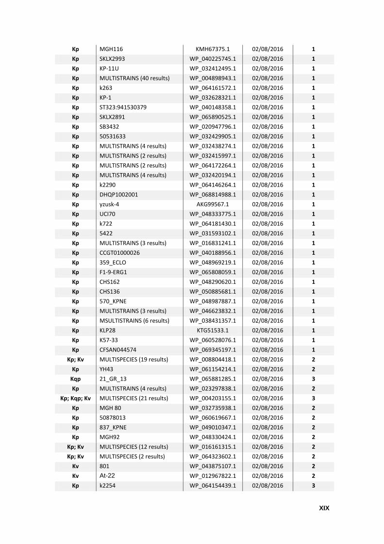
XIX
Kp MGH116 KMH67375.1 02/08/2016 1
Kp SKLX2993 WP_040225745.1 02/08/2016 1
Kp KP-11U WP_032412495.1 02/08/2016 1
Kp MULTISTRAINS (40 results) WP_004898943.1 02/08/2016 1
Kp k263 WP_064161572.1 02/08/2016 1
Kp KP-1 WP_032628321.1 02/08/2016 1
Kp ST323:941530379 WP_040148358.1 02/08/2016 1
Kp SKLX2891 WP_065890525.1 02/08/2016 1
Kp SB3432 WP_020947796.1 02/08/2016 1
Kp 50531633 WP_032429905.1 02/08/2016 1
Kp MULTISTRAINS (4 results) WP_032438274.1 02/08/2016 1
Kp MULTISTRAINS (2 results) WP_032415997.1 02/08/2016 1
Kp MULTISTRAINS (2 results) WP_064172264.1 02/08/2016 1
Kp MULTISTRAINS (4 results) WP_032420194.1 02/08/2016 1
Kp k2290 WP_064146264.1 02/08/2016 1
Kp DHQP1002001 WP_068814988.1 02/08/2016 1
Kp yzusk-4 AKG99567.1 02/08/2016 1
Kp UCI70 WP_048333775.1 02/08/2016 1
Kp k722 WP_064181430.1 02/08/2016 1
Kp 5422 WP_031593102.1 02/08/2016 1
Kp MULTISTRAINS (3 results) WP_016831241.1 02/08/2016 1
Kp CCGT01000026 WP_040188956.1 02/08/2016 1
Kp 359_ECLO WP_048969219.1 02/08/2016 1
Kp F1-9-ERG1 WP_065808059.1 02/08/2016 1
Kp CHS162 WP_048290620.1 02/08/2016 1
Kp CHS136 WP_050885681.1 02/08/2016 1
Kp 570_KPNE WP_048987887.1 02/08/2016 1
Kp MULTISTRAINS (3 results) WP_046623832.1 02/08/2016 1
Kp MSULTISTRAINS (6 results) WP_038431357.1 02/08/2016 1
Kp KLP28 KTG51533.1 02/08/2016 1
Kp K57-33 WP_060528076.1 02/08/2016 1
Kp CFSAN044574 WP_069345197.1 02/08/2016 1
Kp; Kv MULTISPECIES (19 results) WP_008804418.1 02/08/2016 2
Kp YH43 WP_061154214.1 02/08/2016 2
Kqp 21_GR_13 WP_065881285.1 02/08/2016 3
Kp MULTISTRAINS (4 results) WP_023297838.1 02/08/2016 2
Kp; Kqp; Kv MULTISPECIES (21 results) WP_004203155.1 02/08/2016 3
Kp MGH 80 WP_032735938.1 02/08/2016 2
Kp 50878013 WP_060619667.1 02/08/2016 2
Kp 837_KPNE WP_049010347.1 02/08/2016 2
Kp MGH92 WP_048330424.1 02/08/2016 2
Kp; Kv MULTISPECIES (12 results) WP_016161315.1 02/08/2016 2
Kp; Kv MULTISPECIES (2 results) WP_064323602.1 02/08/2016 2
Kv 801 WP_043875107.1 02/08/2016 2
Kv At-22 WP_012967822.1 02/08/2016 2
Kp k2254 WP_064154439.1 02/08/2016 3
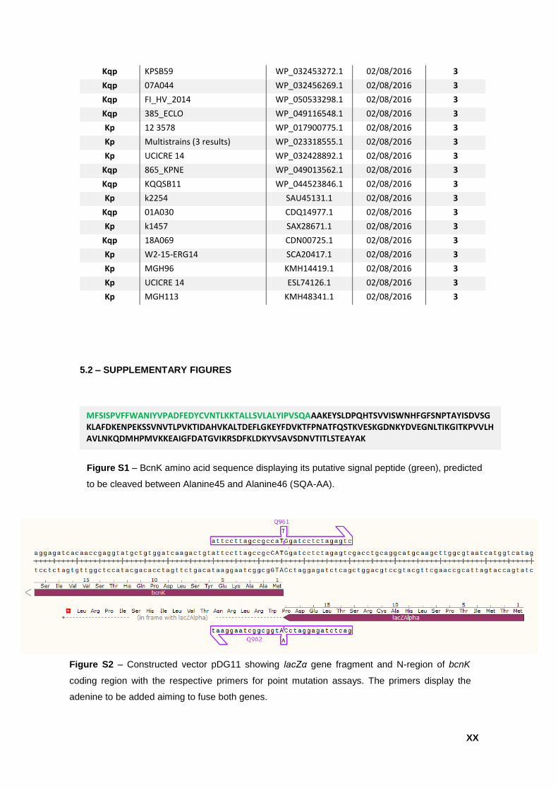
XX
5.2 – SUPPLEMENTARY FIGURES
Kqp KPSB59 WP_032453272.1 02/08/2016 3
Kqp 07A044 WP_032456269.1 02/08/2016 3
Kqp FI_HV_2014 WP_050533298.1 02/08/2016 3
Kqp 385_ECLO WP_049116548.1 02/08/2016 3
Kp 12 3578 WP_017900775.1 02/08/2016 3
Kp Multistrains (3 results) WP_023318555.1 02/08/2016 3
Kp UCICRE 14 WP_032428892.1 02/08/2016 3
Kqp 865_KPNE WP_049013562.1 02/08/2016 3
Kqp KQQSB11 WP_044523846.1 02/08/2016 3
Kp k2254 SAU45131.1 02/08/2016 3
Kqp 01A030 CDQ14977.1 02/08/2016 3
Kp k1457 SAX28671.1 02/08/2016 3
Kqp 18A069 CDN00725.1 02/08/2016 3
Kp W2-15-ERG14 SCA20417.1 02/08/2016 3
Kp MGH96 KMH14419.1 02/08/2016 3
Kp UCICRE 14 ESL74126.1 02/08/2016 3
Kp MGH113 KMH48341.1 02/08/2016 3
MFSISPVFFWANIYVPADFEDYCVNTLKKTALLSVLALYIPVSQAAAKEYSLDPQHTSVVISWNHFGFSNPTAYISDVSGKLAFDKENPEKSSVNVTLPVKTIDAHVKALTDEFLGKEYFDVKTFPNATFQSTKVESKGDNKYDVEGNLTIKGITKPVVLHAVLNKQDMHPMVKKEAIGFDATGVIKRSDFKLDKYVSAVSDNVTITLSTEAYAK
Figure S1 – BcnK amino acid sequence displaying its putative signal peptide (green), predicted
to be cleaved between Alanine45 and Alanine46 (SQA-AA).
Figure S2 – Constructed vector pDG11 showing lacZα gene fragment and N-region of bcnK
coding region with the respective primers for point mutation assays. The primers display the
adenine to be added aiming to fuse both genes.
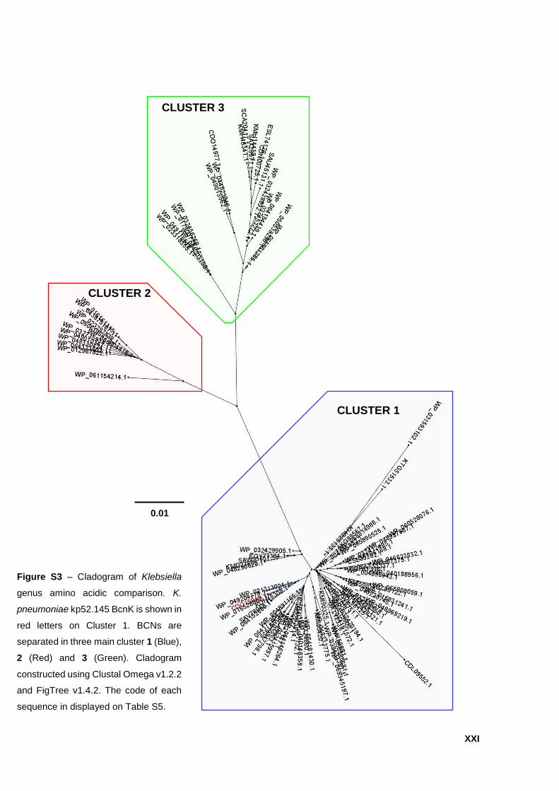
XXI
Figure S3 – Cladogram of Klebsiella
genus amino acidic comparison. K.
pneumoniae kp52.145 BcnK is shown in
red letters on Cluster 1. BCNs are
separated in three main cluster 1 (Blue),
2 (Red) and 3 (Green). Cladogram
constructed using Clustal Omega v1.2.2
and FigTree v1.4.2. The code of each
sequence in displayed on Table S5.
0.01
CLUSTER 1
CLUSTER 3
CLUSTER 2
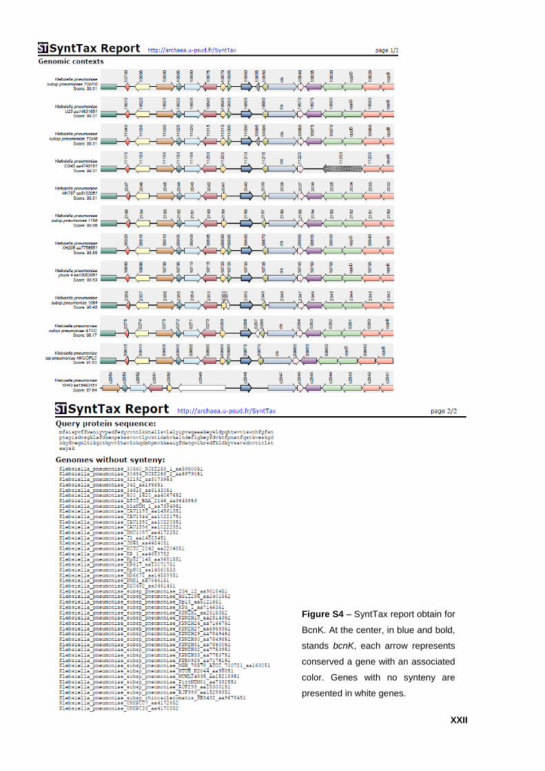
XXII
Figure S4 – SyntTax report obtain for
BcnK. At the center, in blue and bold,
stands bcnK, each arrow represents
conserved a gene with an associated
color. Genes with no synteny are
presented in white genes.


![Leah Braemel [0.5 Proteção Hauberk] Primeira Noite](https://static.fdocumentos.com/doc/165x107/55cf9212550346f57b933867/leah-braemel-05-protecao-hauberk-primeira-noite.jpg)