[email protected] Scanning Electron Microscopy in Rabbit Corneas with Intrastromal...
12
[email protected] Scanning Electron Microscopy in Rabbit Corneas with Intrastromal Corneal Segment(ICS) Eduardo Andreghetti(1) Maria Rosa Bet Moraes Silva(2) Maria Aparecida Domingues(3) Victor Andrigheti Coronado Antunes(4) 1,4-Instituto de Oftalmologia de Assis & Botucatu Medical School(UNESP) 2,3-Botucatu Medical School(UNESP) -The authors have no financial interest in this study
-
Upload
geoffrey-hamilton -
Category
Documents
-
view
238 -
download
0
Transcript of [email protected] Scanning Electron Microscopy in Rabbit Corneas with Intrastromal...
Scanning Electron Microscopy in Rabbit Corneas with Intrastromal Corneal
Segment(ICS)
Eduardo Andreghetti(1)
Maria Rosa Bet Moraes Silva(2)
Maria Aparecida Domingues(3)
Victor Andrigheti Coronado Antunes(4)
1,4-Instituto de Oftalmologia de Assis & Botucatu Medical School(UNESP)
2,3-Botucatu Medical School(UNESP)
-The authors have no financial interest in this study
I – PurposesI – Purposes
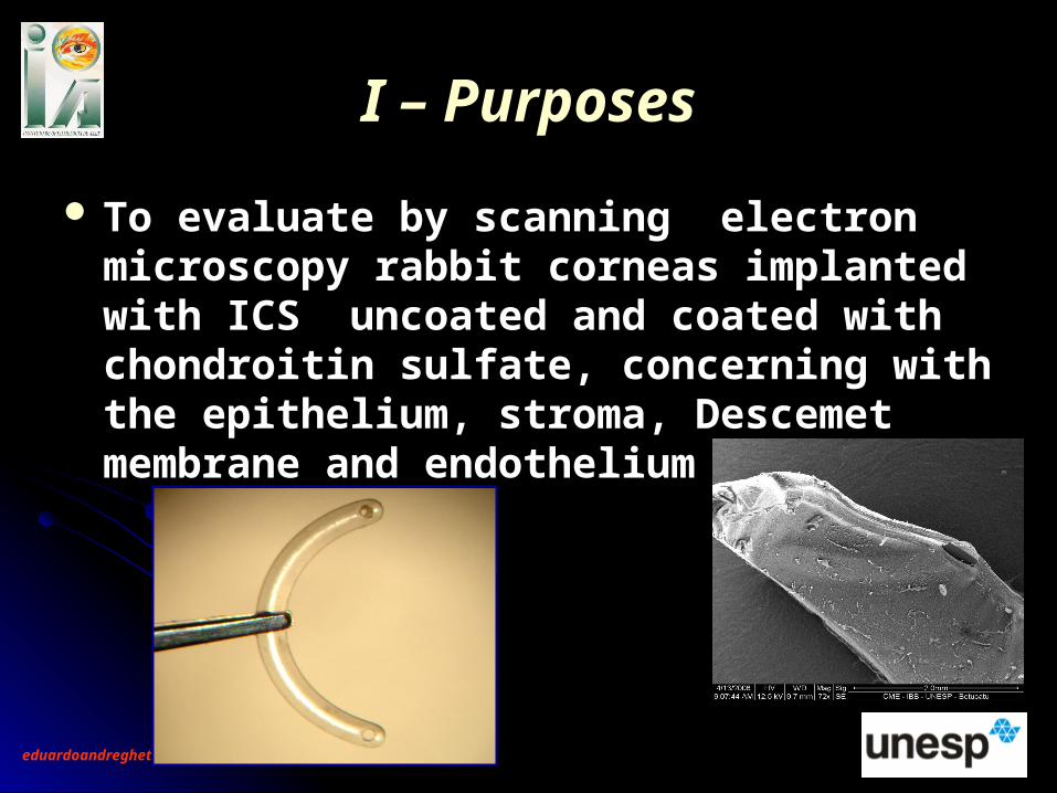
To evaluate by scanning electron To evaluate by scanning electron microscopy rabbit corneas implanted with microscopy rabbit corneas implanted with ICS uncoated and coated with chondroitin ICS uncoated and coated with chondroitin sulfate, concerning with the epithelium, sulfate, concerning with the epithelium, stroma, Descemet membrane and stroma, Descemet membrane and endotheliumendothelium
II – MethodsII – Methods 30 albino adult Norfolk rabbits30 albino adult Norfolk rabbits Proceeding from: Central Vivarium Botucatu Medical School -UNESP Proceeding from: Central Vivarium Botucatu Medical School -UNESP
Aproved by Ethical Comission for Animal Research Botucatu Medical Aproved by Ethical Comission for Animal Research Botucatu Medical School – UNESP (Botucatu , SP –Brazil)School – UNESP (Botucatu , SP –Brazil)
Group G1 – ICS PMMA ClassicGroup G1 – ICS PMMA Classic(Grupo G2 – ICS PMMA+ Chondroitin Sulfate coating(Grupo G2 – ICS PMMA+ Chondroitin Sulfate coating(Right Eye)(Right Eye)
G1 + G2 Left Eyes – Control GroupG1 + G2 Left Eyes – Control Group Surgery Technic: Oliveira CS et al, 2004Surgery Technic: Oliveira CS et al, 2004 (01) (01)
Posoperatory drug:Ofloxacino 0.3% eyedrops each 6 hours – 20 daysPosoperatory drug:Ofloxacino 0.3% eyedrops each 6 hours – 20 days
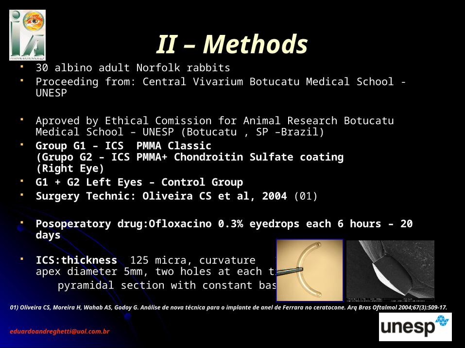
ICS:thicknessICS:thickness 125 micra, curvature 150º , 125 micra, curvature 150º ,apex diameter 5mm, two holes at each tip, apex diameter 5mm, two holes at each tip,
pyramidal section with constant base linepyramidal section with constant base line
01) Oliveira CS, Moreira H, Wahab AS, Godoy G. Análise de nova técnica para o implante de anel de Ferrara no ceratocone. Arq Bras Oftalmol 2004;67(3):509-17.
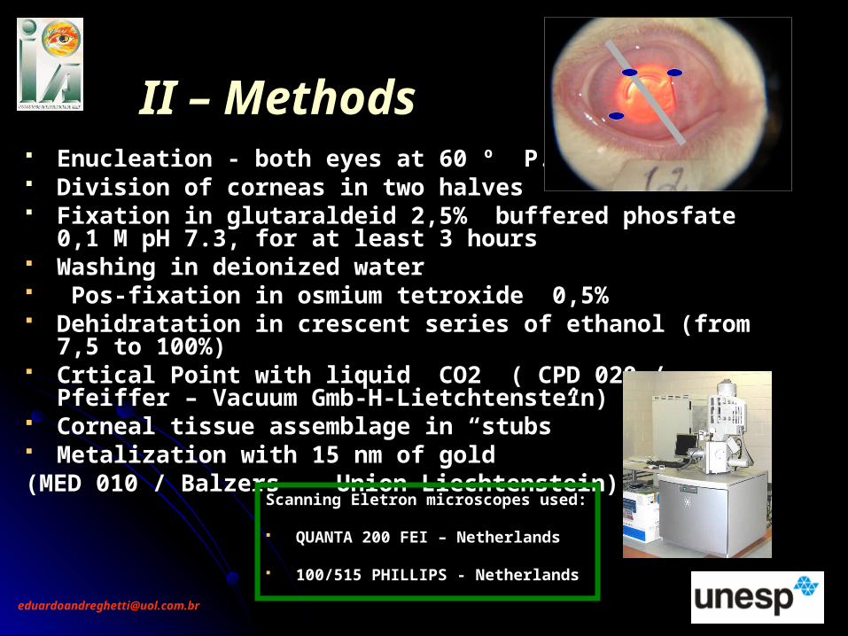
II – MethodsII – Methods Enucleation - both eyes at 60 º P.O. dayEnucleation - both eyes at 60 º P.O. day Division of corneas in two halvesDivision of corneas in two halves Fixation in glutaraldeid 2,5% buffered phosfate 0,1 M pH 7.3, Fixation in glutaraldeid 2,5% buffered phosfate 0,1 M pH 7.3,
for at least 3 hoursfor at least 3 hours Washing in deionized water Washing in deionized water Pos-fixation in osmium tetroxide 0,5% Pos-fixation in osmium tetroxide 0,5% Dehidratation in crescent series of ethanol (from 7,5 to 100%)Dehidratation in crescent series of ethanol (from 7,5 to 100%) Crtical Point with liquid CO2 ( CPD 020 / Pfeiffer – Vacuum Crtical Point with liquid CO2 ( CPD 020 / Pfeiffer – Vacuum
Gmb-H-Lietchtenstein)Gmb-H-Lietchtenstein) Corneal tissue assemblage in “stubs” Corneal tissue assemblage in “stubs” Metalization with 15 nm of gold Metalization with 15 nm of gold (MED 010 / Balzers- Union-Liechtenstein)(MED 010 / Balzers- Union-Liechtenstein)
Scanning Eletron microscopes used:Scanning Eletron microscopes used: QUANTA 200 FEI – NetherlandsQUANTA 200 FEI – Netherlands
100/515 PHILLIPS - Netherlands100/515 PHILLIPS - Netherlands
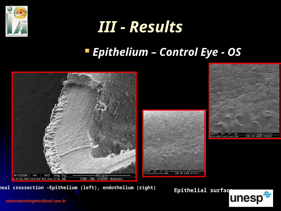
III - ResultsIII - Results Epithelium – Control Eye - OS
Corneal crossection –Epithelium (left), endothelium (right) Epithelial surface
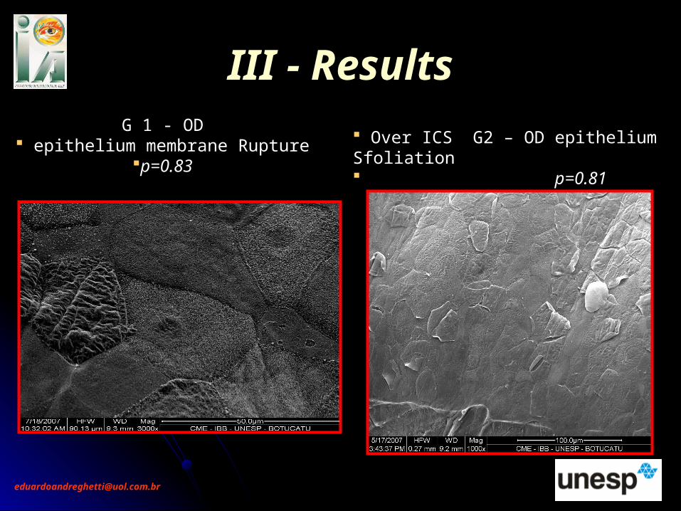
Over ICS G2 – OD epithelium Sfoliation p=0.81
III - ResultsIII - ResultsG 1 - OD
epithelium membrane Rupturep=0.83
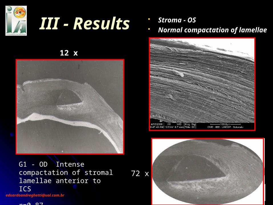
12 x
72 xG1 - OD Intense compactation of stromal lamellae anterior to ICS p=0.87
III - ResultsIII - Results Stroma - OS Normal compactation of lamellae
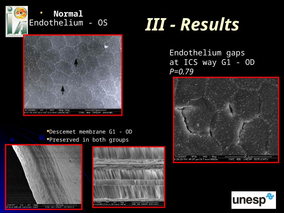
Normal Normal Endothelium - OSEndothelium - OS
III - ResultsIII - Results
Endothelium gapsat ICS way G1 - OD P=0.79
Descemet membrane G1 - ODPreserved in both groups
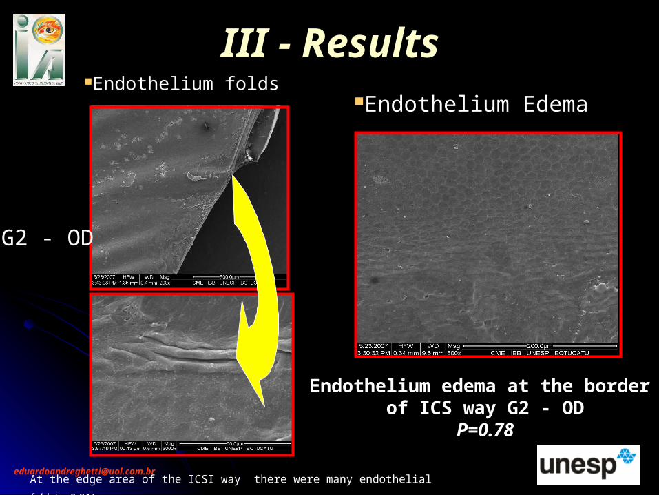
III - ResultsIII - ResultsEndothelium Edema
Endothelium edema at the border of ICS way G2 - OD
P=0.78
Endothelium folds
At the edge area of the ICSI way there were many endothelial folds(p=0.81)
G2 - OD
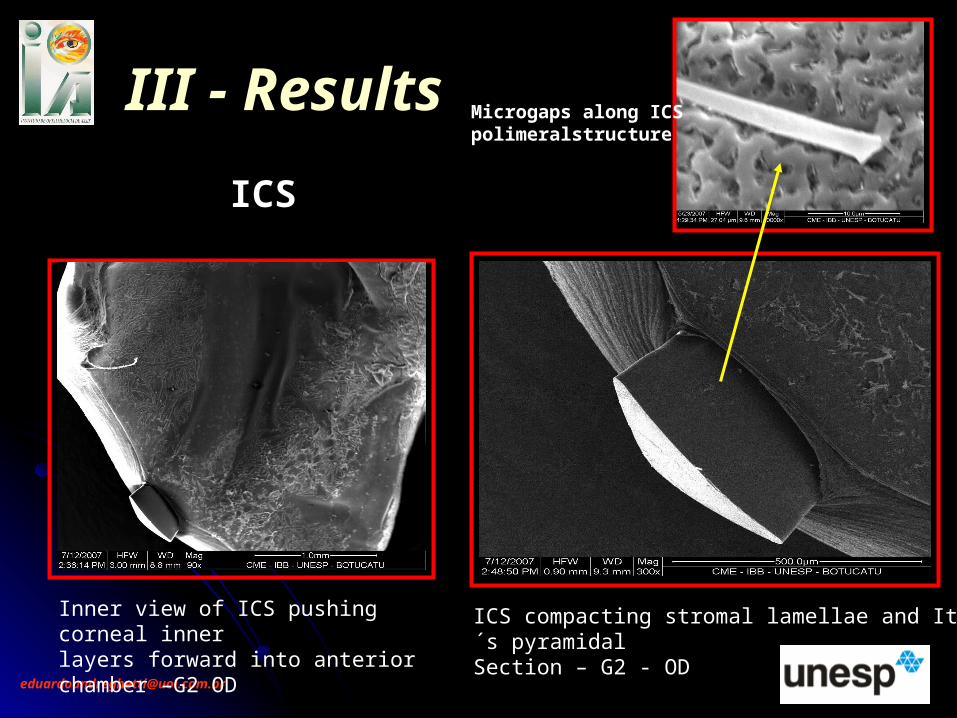
ICS
III - ResultsIII - Results
Inner view of ICS pushing corneal inner layers forward into anterior chamber –G2 OD
ICS compacting stromal lamellae and It´s pyramidalSection – G2 - OD
Microgaps along ICS polimeralstructure
IV- CommentIV- Comment
Epithelial sfoliation, compactness of stromal Epithelial sfoliation, compactness of stromal lamellae, mechanical folding of endothelium with lamellae, mechanical folding of endothelium with different intensities and gaps between different intensities and gaps between endothelium cells by disrupture of intercels endothelium cells by disrupture of intercels bridges were the most important alterations in bridges were the most important alterations in this study and were present in both G1 and G2 this study and were present in both G1 and G2 groups (with no diferent significance).groups (with no diferent significance).
ICS scanning microstructure showed a great ICS scanning microstructure showed a great amount of gaps that may have some clinical amount of gaps that may have some clinical importanceimportance
V - CONCLUSIONV - CONCLUSION
There are indications that leads to There are indications that leads to necessity of mechanical improve on the necessity of mechanical improve on the ICSs studied as the indications that could ICSs studied as the indications that could be studied concerning to standard surgical be studied concerning to standard surgical technic used in this study.technic used in this study.
The ICS in both ( uncoated and coated) The ICS in both ( uncoated and coated) experimental groups had a very similar experimental groups had a very similar behavior concerning the studied behavior concerning the studied parametersparameters