EFEITO DO DIABETES TIPO 1 NA NEOFORMAÇÃO ÓSSEA...
71
PONTIFÍCIA UNIVERSIDADE CATÓLICA DO RIO GRANDE DO SUL FACULDADE DE ODONTOLOGIA PROGRAMA DE PÓS-GRADUAÇÃO EM ODONTOLOGIA ÁREA DE CONCENTRAÇÃO: CIRURGIA E TRAUMATOLOGIA BUCOMAXILOFACIAL NATÁLIA PRADELLA CIGNACHI EFEITO DO DIABETES TIPO 1 NA NEOFORMAÇÃO ÓSSEA EM DEFEITOS CRITICOS EM FÊMUR DE CAMUNDONGOS: RELEVÂNCIA DOS RECEPTORES B 1 DAS CININAS Porto Alegre 2014
Transcript of EFEITO DO DIABETES TIPO 1 NA NEOFORMAÇÃO ÓSSEA...

1
PONTIFÍCIA UNIVERSIDADE CATÓLICA DO RIO GRANDE DO SUL FACULDADE DE ODONTOLOGIA
PROGRAMA DE PÓS-GRADUAÇÃO EM ODONTOLOGIA ÁREA DE CONCENTRAÇÃO: CIRURGIA E TRAUMATOLOGIA BUCOMAXILOFACIAL
NATÁLIA PRADELLA CIGNACHI
EFEITO DO DIABETES TIPO 1 NA NEOFORMAÇÃO ÓSSEA EM DEFEITOS CRITICOS EM FÊMUR DE CAMUNDONGOS: RELEVÂNCIA DOS RECEPTORES B1 DAS CININAS
Porto Alegre 2014
2
NATÁLIA PRADELLA CIGNACHI
EFEITO DO DIABETES TIPO 1 NA NEOFORMAÇÃO ÓSSEA EM DEFEITOS CRITICOS EM FÊMUR DE CAMUNDONGOS: RELEVÂNCIA DOS RECEPTORES B1 DAS CININAS
Linha de pesquisa: Diagnótico e Terapêutica Aplicadas
Dissertação apresentada como requisito
parcial para a obtenção do título de Mestre
em Odontologia, na área de CTBMF, pelo
Programa de Pós-Graduação em
Odontologia, da Pontifícia Universidade
Católica do Rio Grande do Sul.
Orientadora: Profa. Dra. Maria Martha Campos
Porto Alegre
2014

3
Dedicatória
À minha família, meu bem mais sagrado. Lauro, tu foi o maior incentivador para a inscrição no mestrado. Pai, se estou
completando hoje esta etapa é por tua causa e por tua ajuda. Irdes, tu é o pilar da família, sempre que precisamos de algo, lá está a mãezinha,
que de pequena só tem a altura. Forte e pronta para tudo. E a neném da família, Amanda, minha irmã, minha amiga, minha sócia e colega de
profissão. Quem acompanha teus passos sabe quanto tu cresce a cada dia que passa e o quanto posso contar contigo.

4
AGRADECIMENTOS ESPECIAIS
À Profa. Dra. Maria Martha Campos que além de minha orientadora, teve um
papel especial de confidente em alguns momentos pessoais de dificuldade. Agradeço
pela paciência em desmistificar todas as dúvidas que me assombravam em relação
ao nosso trabalho. Por me mostrar a existência desse caminho maravilhoso, que é a
pesquisa, tanto em animais quanto a pesquisa laboratorial. Sorte a minha de tê-la
como orientadora.
À Profa. Dra. Adriana Etges por me receber na UFPEL de braços abertos,
mesmo com muitos compromissos na sua agenda, conseguiu me ajudar de uma
forma muito gentil na condução de uma parte do nosso trabalho.
À Profa. Dra. Karen Cherubini pelo acolhimento em seu laboratório.
Ao Prof. Dr. Vinícius Duval pela orientação na aquisição de imagens de
imunohistoquimica.
À Profa. Dra. Fernanda B. Morrone, o meu agradecimento.
Ao Prof. Dr. Ricardo Smidt, que acompanha minha trajetória. Obrigada pelo
incentivo para seguir o caminho acadêmico.
As meninas, Kesiane M. Costa, Helena Filipinni, Natália F. Nicoletti, Paula
Seadi, Priscila Pail e Raquel Freitas obrigada pela ajuda nos experimentos e pelas
horas de conversas e momentos de risadas.
Aos meninos, André Santos Junior, Gustavo B. Machado, Izaque Maciel e
Rodrigo Braccini agradeço o apoio nos assuntos em que eu não tinha conhecimento.
Ao meu avô, Ângelo P. Pradella, que partiu antes do término deste trabalho.
Agradeço o apoio das minhas amigas de infância assim como daquelas que a
Odontologia me presenteou.

5
AGRADECIMENTOS
À Faculdade de Odontologia da Pontifícia Universidade Católica do Rio
Grande do Sul, representado pelo seu Diretor Prof. Dr. Alexandre Bahlis pela
excelente estrutura e qualidade de ensino proporcionada.
À Coordenação do Curso de Pós-Graduação em Odontologia da PUCRS em
nome da Profa. Dra. Ana Maria Spohr, pelas oportunidades durante o curso de
Mestrado.
À Secretaria de Graduação e Pós-Graduação da Faculdade de Odontologia da
PUCRS.
Ao Instituto de Toxicologia e Farmacologia da PUCRS, pela utilização das
dependências para realização dos experimentos com animais.
À Luciana A. Ferreira responsável pelo processamento das amostras e ajuda
da confecção das lâminas.
À Ivana N. Hanemann, da UFPEL pela ajuda na realização do procedimento de
imunohistoquimica nas lâminas.
À Raquel M. Oliveira pela disponibilidade de tempo para ajudar no processo
de coloração das lâminas.
Aos colegas de Mestrado e Doutorado de Odontologia pela amizade e apoio
durante o curso.
À CAPES, pelo apoio financeiro disponibilizado através da bolsa no último ano
do Mestrado, indispensável para o término deste curso.

6
RESUMO
Os efeitos da deleção gênica do receptor B1 das cininas na regeneração óssea
foram avaliados em camundongos com diabetes tipo 1 induzida por
estreptozotocina, submetidos a um modelo de defeito crítico no fêmur. Como
resultado da indução de diabetes nos camundongos wild-type C57/BL6, houve um
decréscimo do peso corporal e hiperglicemia em relação ao grupo não-diabético da
mesma cepa. Os animais diabéticos, com ausência do receptor B1 apresentaram
perda do peso corporal e, uma prevenção parcial da hiperglicemia. Camundongos
diabéticos do tipo 1 tiveram um atraso na regeneração óssea, apresentando um
tecido conectivo desorganizado na região correspondente ao defeito crítico, quando
comparados a extensas áreas de tecido ósseo recém-formado em camundongos WT
C57/BL6 não diabéticos. Camundongos B1R nocaute, diabéticos ou não diabéticos,
exibiram níveis de regeneração óssea semelhantes aos observados no grupo
controle WT C57/BL6. A melhora na regeneração óssea nos animais sem o
receptor B1 foi confirmada pela análise da quantidade de colágeno.
Camundongos WT C57/BL6 diabéticos apresentaram uma redução acentuada da
distribuição de colágeno na região do defeito ósseo, enquanto que os animais
B1RKO diabéticos exibiram níveis de colágeno similares àqueles observados nos
camundongos não diabéticos, tanto WT C57/BL6, quanto B1RKO. A melhora da
regeneração óssea nos camundongos diabéticos sem o receptor B1 não parece
estar associado à atividade osteoclástica diminuída. Ademais, nenhuma
diferença marcante foi encontrada nos níveis de fosfatase ácida resistente ao
tartarato (TRAP) ou, na imunomarcação para as proteínas do sistema
RANK/RANKL/OPG, em todos grupos experimentais avaliados. Os resultados

7
deste trabalho fornecem novas evidências a respeito da relevância dos
receptores B1 no diabetes do tipo 1, especialmente no que diz respeito ao seu
papel na regeneração óssea após procedimentos cirúrgicos.

8
DESCRITORES1 Cininas, osso, Diabetes Mellitus tipo 1, camundongos.
1DeCS- Descritores em Ciências da Saúde, disponível em http:/decs.bvs.br

9
ABSTRACT The effects on kinin B1 receptor (B1R) deletion were examined on bone
regeneration in streptozotocin (STZ)-type-1 diabetic mice, subjected to a model of
femoral critical-size defect. Diabetes induction in wild-type C57/BL6 (WT C57/BL6)
mice was allied to decrease of body weight and hyperglycemia, in relation to the
non-diabetic group of the same strain. The lack of B1R did not affect STZ-elicited
body weight loss, although it partially prevented hyperglycemia. Type-1 diabetic
mice presented a clear delay in bone regeneration, with large areas of loose
connective tissue within the region corresponding to the defects, when compared to
wide areas of newly-formed woven bone in non-diabetic WT C57/BL6 mice. Notably,
either non-diabetic or diabetic B1R knockout (B1RKO) mice displayed bone
regeneration levels comparable to that seen in control WT C57/BL6 mice. The
improved bone regeneration in animals lacking B1R was further confirmed by
analysis of collagen contents. WT C57/BL6 STZ-diabetic mice presented a marked
reduction of collagen contents within the bone defect gap, whereas diabetic B1RKO
displayed collagen levels comparable to those observed in non-diabetic WT C57/BL6
or B1RKO mice. The enhanced bone regeneration in diabetic mice lacking B1R does
not seem to be associated to lessened osteoclast activity, as no prominent difference
was detected in the levels of tartrate-resistant acid phosphatase (TRAP) positivity, or
even in the immunolabeling for the proteins of the RANK/RANKL/OPG system
thoughout all the experimental groups. Data brings novel evidence on the relevance
of B1R under type-1 diabetes, especially concerning its role in bone regeneration
after surgical procedures.

10
DESCRIPTORS2 Bradykinin B1 Receptor, bone regeneration, type 1 Diabetes Mellitus, mice.
2MeSH- Medical Subject Headings, avaiable at: www.nlm.nih.gov/mesh

11
LISTA DE FIGURAS E IMAGENS
Imagem 1: Área correspondente ao defeito ósseo confeccionado no fêmur de camundongos da linhagem C57/BL6. (A) Localização da área antes do procedimento; (B) Defeito ósseo crítico instalado. Figure 1. Body weight gain and blood glucose levels in WT and B1RKO in STZ-induced type 1 diabetes. The weight gain was calculated at different time-points, as the percentage of increase in relation to the individual body weight at the beginning of experiments. The blood glucose levels are provided in mg/dl. Data is presented as the mean ± standard error mean of 7-10 animals per group. *P<0.05; **P<0.01 denotes significance in relation to the respective control group (ANOVA followed by Bonferroni’s post-hoc test). Figure 2. Representative images of H&E staining throughout different experimental groups (upper panels), and semi-quantitative analysis of the percentage of newly-formed bone in comparison to the total analyzed area (lower panel). Data is presented as the mean ± standard error mean of 7-10 animals per group. *P<0.05 denotes significance in relation to the respective control group (ANOVA followed by Bonferroni’s post-hoc test). Figure 3. Representative images of Masson’s trichrome staining throughout different experimental groups (upper panels), and semi-quantitative analysis of the percentage of newly-formed bone in comparison to the total analyzed area (lower panel). Data is presented as the mean ± standard error mean of 7-10 animals per group. *P<0.05 denotes significance in relation to the respective control group (ANOVA followed by Bonferroni’s post-hoc test). Figure 4. Representative images of tartrate-resistant acid phosphatase (TRAP) staining throughout the four different evaluated experimental groups. The arrows indicate positive labeling. The images are representative of analysis of 7-10 animals per group. Figure 5. Representative images of immunohistochemical analysis for RANK expression throughout different experimental groups (upper panels), and quantitative analysis of the number of positive cells/field (lower panel). Data is presented as the mean standard error mean of 7-10 animals per group. Figure 6. Representative images of immunohistochemical analysis for RANKL expression throughout different experimental groups (upper panels), and quantitative analysis of the number of positive cells/field (lower panel). Data is presented as the mean standard error mean of 7-10 animals per group. Figure 7. Representative images of immunohistochemical analysis for OPG expression throughout different experimental groups (upper panels), and quantitative analysis of the number of positive cells/field (lower panel). Data is presented as the mean ± standard error mean of 7-10 animals per group.

12
LISTA DE ABREVIATURAS
# Osteoblasto- OB
# Osteoclastos- OC
# Ativador nuclear Kappa-b- RANK
# Ligante do ativador nuclear Kappa-b- RANKL
# Osteoprotegerina- OPG
# Diabetes Mellitus- DM
# Estreptozotocina- STZ
# Bradicinina- BK
# Enzima Conversora da Angiotensina- ECA
# Hematoxilina & Eosina- H&E
# Fosfatase Ácida Resistente ao Tartarato- TRAP

13
SUMÁRIO
INTRODUÇÃO ............................................................................................................ 14
OBJETIVOS ................................................................................................................. 25
Objetivo geral ............................................................................................................ 25
Objetivos específicos .................................................................................................. 25
ARTIGO DE PESQUISA ................................................................................................ 26
Página inicial .............................................................................................................. 27
Abstract ...................................................................................................................... 28
Introduction ................................................................................................................ 29
Methods ..................................................................................................................... 31
Results ........................................................................................................................ 35
Discussion ................................................................................................................... 38
References .................................................................................................................. 43
Legend to Figures ....................................................................................................... 50
CONSIDERAÇÕES FINAIS ........................................................................................... 57
REFERÊNCIAS BIBLIOGRÁFICAS GERAIS .................................................................... 60
ANEXO A- Carta de aprovação pela Comissão Científica e de Ética da Faculdade de
Odontologia/PUCRS ................................................................................................... 68
ANEXO B- Carta de aprovação pelo Comitê de Ética em Pesquisa com Animais/PUCRS
.................................................................................................................................... 69
ANEXO C- Comprovante de submissão do manuscrito ao Periódico PNAS ............... 70

14
INTRODUÇÃO
O osso é classificado como um tecido conjuntivo especializado, formado por
60% a 70% de material inorgânico e, 30% a 35% de material orgânico, dos quais, 90%
são representados pelas fibras colágenas (1). Sua principal função é de resistência,
dependente não somente da quantidade de tecido ósseo, mas, também, de sua
qualidade.
O tecido ósseo é renovado frequentemente em resposta a uma série de
estímulos do processo de remodelação. Este processo não está totalmente
entendido, mas inclui danos ao osso em resposta ao desgaste normal, a mudanças
no peso corporal, atividade física, além da liberação de citocinas ou fatores de
crescimento ,devido a alterações nos níveis hormonais (2).
A osteogênese é determinada por uma sequência de eventos que iniciam
pelas células osteoprogenitoras e sua diferenciação em pré-osteoblastos. Estes, por
sua vez, tornam-se osteoblastos maduros, com altos índices de fosfatase alcalina,
osteocalcina e colágeno. Dentre os fatores responsáveis pela formação dos
osteoblastos estão as proteínas ósseas morfogenéticas (BMPs) (3,4).
Quando os osteoblastos estão em intensa atividade sintética, suas formas
modificam-se, lembrando um cubo, tornando-se achatados, com redução da
basofilia citoplasmática. Uma vez aprisionado pela matriz óssea recém-sintetizada, o
osteoblasto recebe o nome de osteócito (5-6). Os osteócitos são células com forma
estrelada, que estão inseridos na matriz óssea mineralizada, mas permanecem em
contato com as outras células ósseas, por um processo altamente controlado (5-6).
Portanto, essas células derivadas dos osteoblastos são residentes em lacunas da
matriz óssea. Apesar dos osteócitos não apresentarem a função de secretar a matriz

15
óssea, eles permanecem produzindo as substâncias necessárias à manutenção do
osso.
Os osteoclastos são células especializadas na reabsorção da matriz óssea e se
originam de monócitos hematopoiéticos e macrófagos (7-8). Os fatores reguladores
da função dos osteoclastos são: fator estimulador da colônia de monócitos (CFS-1),
fator de diferenciação dos osteoclastos (ODF), interleucinas (IL), vitamina D3, fator
de necrose tumoral (TNF) e, partículas ósseas mineralizadas contendo osteocalcina
(7, 9). O ODF é um membro da super-família do fator de necrose tumoral e foi
denominado mais recentemente de TNFSF-11; porém, possui outras denominações,
tais como TRANCE, RANK-L ou ODF/TNFSF-11 (10-11).
O RANKL, produzido por células de linhagem osteoblástica e por linfócitos T
ativados, é o fator essencial para a formação, fusão, ativação e sobrevivência dos
osteoclastos, levando à reabsorção óssea (10). Os efeitos do RANKL são
contrabalanceados através da OPG que atua como um receptor solúvel neutralizador
(10). RANKL e OPG (receptor osteoprotegerina) são regulados por vários hormônios
(glicocorticóides, vitamina D, estrogênio), citocinas (TNF , IL-1, IL-4, IL-6, IL-11 e IL-
17) e, fatores de transcrição mesenquimal (tais como cbfa-1). Anormalidades do
sistema RANKL/OPG implicam na patogênese da osteoporose pós-menopausa,
artrite reumatoide, doença de Paget, doença periodontal, tumores ósseos benignos
e malignos, metástases ósseas e hipercalcemia. De forma interessante, a
administração do OPG é capaz de prevenir ou atenuar esses transtornos em modelos
animais (10).
A maioria dos fatores que induz a expressão de RANKL pelos osteoblastos
também regula a expressão de OPG (12). Entretanto, mesmo que alguns dados

16
sejam contraditórios, no geral, quando a expressão de RANKL está sobre- regulada,
OPG é regulada ou não induzida com a mesma intensidade como RANKL, de tal
modo que mudanças na relação RANKL/OPG favorecem a osteoclastogênese (13,14).
A matriz orgânica é formada de colágeno, principalmente do tipo 1,
proteoglicanas e glicoproteínas adesivas. Por outro lado, a matriz inorgânica é
composta por íons fosfato, cálcio e, em menor quantidade, bicarbonato, magnésio,
potássio, sódio e citrato. A união do fostato e do cálcio forma cristais com estrutura
de hidroxiapatita que, quando associados às fibras colágenas, fornecem a resistência
e a dureza características do tecido ósseo (15). O colágeno do tipo 1, além de ser o
maior componente da matriz óssea, está intimamente associado à cicatrização e à
regeneração. Portanto, mudanças na estrutura do colágeno podem afetar as
propriedades mecânicas do osso e aumentar a susceptibilidade a fratura. (16).
Vários fatores locais e sistêmicos controlam a formação e a reabsorção óssea.
Dentre os sistêmicos, destacam-se os hormônios de crescimento e hormônios
tireoidianos, os glicocorticoides, o paratormônio, a calcitonina e a vitamina D. Entre
os fatores locais, podem-se citar a prostaglandina E2 (PGE2), a IL-1β, o interferon-
gama (INF ), o fator de crescimento de transformação beta (TGFβ), o fator de
crescimento semelhante à insulina (IGF), o fator de crescimento epidérmico (EGF), o
fator de crescimento derivado das plaquetas (PDGF), o fator de crescimento
fibroblástico (FGF) e o fator de crescimento derivado do esqueleto (SGF) (9).
As anormalidades na remodelação óssea ocorrem em algumas das doenças
mais comuns, tais como osteoporose, doença periodontal, artrite reumatoide,
insuficiência renal crônica, osteólise induzida por tumores e osteopetrose (1,6).
Embora estas alterações sejam comuns, na maioria dos casos, pouco se sabe sobre

17
os mecanismos responsáveis pela disfunção da remodelação óssea que as
caracteriza (9).
O aumento nos níveis de paratormônio, produzido pela paratireoide
(hiperparatireoidismo), leva a um aumento do número de osteoclastos com
consequente estímulo à reabsorção óssea (1). Já, a calcitonina, produzida pela
tireoide, inibe a atividade osteoclástica com diminuição da reabsorção óssea (1). A
integridade estrutural do osso pode estar comprometida pela necessidade do
metabolismo normal do cálcio e por estados patológicos, alterando assim, estrutura
e massa ósseas. Este fenômeno pode ser especialmente notado na estrutura óssea
no período pós-menopausa, em que ocorre um decréscimo dos níveis de estrogênio.
Como o osso perde massa, as comunicações entre o trabeculado ósseo também são
perdidas. Dentre os marcadores mais utilizados para a caracterização de atividade
osteoclástica, destaca-se o TRAP (do inglês, tartrate-resistant acid phosphatase) (17).
O diabetes é uma doença metabólica resultante de defeitos na secreção de
insulina e/ou em sua ação (18). De acordo com a Organização Mundial da Saúde,
mais de 350 milhões de pessoas em todo o mundo poderão ser diagnosticadas com
Diabetes Mellitus até o ano de 2025 (19, Pan et al., 2014). Os sintomas como
poliúria, polidpsia, perda de peso, polifagia e visão turva são resultantes das
variações dos níveis glicêmicos. A hiperglicemia crônica está associada ao dano,
disfunção e falência de vários órgãos, especialmente olhos, rins, nervos, coração e
vasos sanguíneos (20).
O diabetes pode ser classificado como tipo 1 ou tipo 2. No diabetes do tipo 1,
ocorre destruição das células beta do pâncreas, usualmente por um processo
autoimune (forma autoimune; tipo IA) ou menos comumente por causas

18
desconhecidas (forma idiopática; tipo IB) (21,22). Na forma autoimune, há um
processo de insulinite e estão presentes auto-anticorpos circulantes (anticorpos anti-
descarboxilase do ácido glutâmico, anti-ilhotas e anti-insulina). Como não há insulina
na circulação em concentrações ideais, a absorção de glicose fica prejudicada, sendo
necessário fazer a reposição de insulina (23).
No diabetes do tipo 2 a associação entre um fator hereditário e a obesidade
apresentam maior importância do que no tipo 1. Apesar desses pacientes
produzirem insulina normalmente suas células são incapazes de usar toda essa
insulina secretada pelo pâncreas, fazendo com que seus níveis permaneçam altos no
sangue, o que é conhecido como resistência à insulina (24). O principal motivo que
faz os níveis de glicose permanecerem altos é a incapacidade das células musculares
e adiposas em utilizarem toda insulina secretada pelo pâncreas. Os sintomas do
diabetes tipo 2 são pronunciados, sendo que o tratamento deve ser cauteloso, em
virtude dos riscos de complicações cardiovasculares em longo prazo (25).
O pâncreas é o órgão responsável pela produção de insulina. Este hormônio
faz a regulação dos níveis de glicose no sangue (glicemia) (26). As células β-
pancreáticas são responsáveis por sintetizar e secretar a insulina. Visando manter a
glicemia constante, o pâncreas também produz outro hormônio antagônico à
insulina, o glucagon. Quando os níveis glicêmicos ficam baixos, mais glucagon é
secretado, visando restabelecer os níveis de glicose na circulação. A insulinoterapia
causa alterações no hormônio da paratireoide, no metabolismo da vitamina D e na
absorção do cálcio (27).
Independente do tipo, os pacientes diabéticos têm um baixo potencial de
cicatrização dos tecidos moles e ósseos, devido à redução do metabolismo de
19
proteínas e aos prejuízos da função neutrofílica (26). A perda óssea não está
vinculada apenas a um aumento da atividade osteoclástica. A hiperglicemia inibe a
diferenciação osteoblástica e altera o hormônio da paratireoide, que regula o
metabolismo do fósforo e do cálcio (28). Isso produz um efeito deletério na matriz
óssea, afetando a aderência, o crescimento e acúmulo da matriz extracelular (29).
Existem substâncias químicas (aloxana e estreptozotocina) capazes de
produzir radicais livres que destroem as células β-pancreáticas, induzindo diabetes
do tipo 1 (30). O antibiótico estreptozotocina (STZ) causa degranulação das células
produtoras de insulina, promovendo o aparecimento do diabetes. O núcleo glicérico,
presente na estrutura das células beta, permite a entrada da STZ e o grupo
nitrosúreia, também presente nas células, promove o acúmulo de substâncias
tóxicas e, consequente morte destas células (31).
A indução de diabetes pela STZ pode ser constatada em 24 h após a
administração intraperitoneal deste agente. As alterações fisiológicas manifestadas
são hiperglicemia, glicosúria, polidipsia, polifagia e poliúria. A hiperglicemia
plasmática aumenta a absorção de glicose pelos néfrons dos rins, levando a um
aumento da excreção de glicose na urina (glicosúria), gerando um aumento da
pressão osmótica e maior retenção de água na urina (poliúria). Consequentemente,
a ingestão de água é aumentada (polidipsia). O aumento do consumo de ração
(polifagia) pode ser associado ao comprometimento do transporte de glicose para as
células em virtude da falta de insulina, sendo que o animal aumenta a ingestão de
ração para suprir a falta de energia celular; mesmo assim, ocorre perda progressiva
de peso corporal (32).
20
No inicio da doença, a redução da formação óssea em diabéticos está mais
relacionada com um menor número de osteoblatos, os quais funcionam
normalmente. Em se tratando dos animais diabéticos, uma relação normal e
diretamente proporcional é observada entre os eventos de formação da matriz
óssea e de mineralização óssea. Porém, ao longo do tempo, a menor produção de
matriz óssea, devido ao menor número de osteoblastos presentes no osso, gera
distúrbios na atividade dos osteoblastos, fato observado nas alterações das taxas da
síntese da matriz do colágeno e/ou mineralização óssea (33).
GRAVES et al. (2005) (34) observaram um aumento da expressão de
mediadores inflamatórios em ratos CD-1 que se tornaram diabéticos pelo
tratamento com STZ e ratos dB/dB, que desenvolvem espontaneamente diabetes
tipo 1, em comparação com ratos normoglicêmicos. Verificou-se uma resposta
inflamatória mais prolongada em ambos os modelos de diabetes, pela ação de duas
quimiocinas, a proteína quimiotática de macrófagos (MCP)-1 e a proteína
inflamatória do macrófago (MIP)-2, que atraem macrófagos e células
polimorfonucleares, como também, estimulam a liberação de TNF . Resultados
semelhantes foram obtidos por WETZLER et al. (2000) e LU et al. (2004) (35,36).
As cininas são peptídeos biologicamente ativos que participam da resposta
inflamatória, promovendo vasodilatação, aumento da permeabilidade vascular,
extravasamento plasmático e migração celular (36-39). Estão presentes em
condições como sepse, dano pós-isquêmico, asma, pancreatite, cistite, alergia,
diabetes, artrite reumatóide, colite, gastrite e câncer, além de causarem dor e
hiperalgesia (39-46). Também apresentam ações fisiológicas, participando do

21
controle da pressão arterial, relaxamento e contração da musculatura lisa e
natriurese (40-46).
A cascata de formação das cininas compreende mecanismos bem
caracterizados. As cininas são formadas a partir de α-globulinas chamadas
cininogênios. São conhecidos três tipos de cininogênios que diferem em tamanho,
função e estrutura. O cininogênio de alto peso molecular (High Molecular Weight
Kininogen, HMWK) é uma proteína plasmática com massa molecular de 120 kDa e dá
origem à bradicinina (BK). O cininogênio de baixo peso molecular (Low Molecular
Weight Kininogen, LMWK) tem massa molecular de 66 kDa e origina a calidina (Lys-
BK), além da BK, estando amplamente distribuído nos tecidos, em fibroblastos e em
outras estruturas celulares do tecido conjuntivo. O terceiro tipo de cininogênio, tipo
T, corresponde ao HMWK e é encontrado apenas em ratos (47,48).
Os cininogênios são clivados por proteases chamadas calicreínas que são
encontradas no sangue (calicreína plasmática) ou nas glândulas exócrinas (calicreína
tecidual). A calicreína plasmática é produzida no fígado e circula na sua forma
inativada, chamada de pré-calicreína ou fator de Fletcher. Após sua clivagem,
dependente da ativação do fator de Hagemann (fator XII da coagulação sangüinea),
é formada a enzima ativa. A calicreína plasmática age então sobre o HMWK
liberando BK. Este processo está aumentado durante a resposta inflamatória (48).
A BK tem uma meia-vida plasmática muito curta, que varia entre 10 e 50
segundos. As cininases pertencem a um grupo de enzimas responsáveis pelo
metabolismo e degradação das cininas. A cininase II, conhecida também como
enzima conversora da angiotensina (ECA), é encontrada na membrana das células
endoteliais e age sobre as cininas, removendo o dipeptídeo da porção C-terminal e
22
originando metabólitos inativos. A endopeptidase neutra e a aminopeptidase
plasmática também exercem um papel importante no metabolismo das cininas. A
primeira está presente nas células epiteliais e utiliza um mecanismo semelhante ao
da cininase II para inativar a BK (47,49). Já a aminopeptidase é capaz de converter a
Lys-BK em BK, através da clivagem da porção N-terminal (50).
A cininase I, conhecida como arginina carboxipeptidase, é representada pela
carboxipeptidase N (plasma) e carboxipeptidase M (membrana) e apresenta um
papel menor na degradação da BK. Entretanto, essa enzima é responsável pela
remoção da arginina da porção C- terminal da BK e da Lys-BK gerando os metabólitos
ativos des-Arg9-BK e Lys-des-Arg9-BK, respectivamente. A cininase II possui maior
afinidade pela BK e pela Lys-BK do que a cininase I, o que sugere que a formação dos
metabólitos ativos não ocorre sob condições fisiológicas. De fato, a formação desses
metabólitos está presente em exsudatos inflamatórios, onde a formação de fibrina
aumenta a atividade da cininase I em relação à cininase II (51,52).
Depois de liberados, a BK e seus metabólitos podem ativar dois subtipos de
receptores acoplados a proteína G, chamados de B1 e B2 (45, 53-56). A existência dos
receptores B1 e B2 foi confirmada por estudos de clonagem e de deleção gênica
(45,55, 57).
Os receptores B2 são expressos constitutivamente na maior parte dos tecidos
e apresentam alta afinidade pela BK e pela Lys-BK. Por outro lado, os receptores B1
não são comumente expressos em condições normais, com exceção do sistema
nervoso central, mas são rapidamente induzidos após estímulos como inflamação,
infecção ou trauma e apresentam afinidade pelos metabólitos ativos des-Arg9-BK e
Lys-des-Arg9-BK.

23
Considerando o perfil de indução, é possível inferir que os receptores B1
representam alvos de grande importância para o desenvolvimento de drogas com
potencial antiinflamatório e que poderiam ser úteis para o tratamento de doenças
crônicas como asma, artrite, osteoartrite, neuropatias e doença periodontal, entre
outras (56, 58). O receptor B1 tem um baixo nível de expressão nos tecidos
saudáveis, tendo sua expressão aumentada após a exposição a citocinas pró-
inflamatótias, endotoxinas bacterianas ou níveis elevados de glicose (59,60).
De forma interessante, tem sido demonstrado que os receptores B1 para as
cininas estão envolvidos em várias complicações observadas no diabetes do tipo 1,
especialmente nos quadros de neuropatia, nefropatia e retinopatia. Por exemplo,
um estudo de Dias et al. (61) demonstrou que o receptor B1 exibe um papel
patológico no estágio inicial da diabetes, aumentando o estresse oxidativo e os
mediadores pró-inflamatórios envolvidos na retinopatia e na neuropatia diabéticas.
Um trabalho recente conduzido por Masao KA et al. (62) demonstrou que a deleção
gênica dos receptores B1 e B2 das cininas resulta em uma redução marcante da
densidade óssea, tanto em ratos diabéticos, como não-diabéticos, evidenciando a
importância das cininas e seus receptores na mineralização óssea. Por outro lado,
estudos prévios in vitro demonstraram que a estimulação dos receptores B1 e B2 em
osteoblastos aumenta a expressão do RANKL, o que indicaria a participação destes
receptores nos processos de osteoclastogênese e reabsorção óssea (63, 64,65). Foi
demonstrado mais recentemente que a deleção gênica do receptor B1 resultou em
um aumento da perda óssea em um modelo de periodontite induzida por ligadura,
em camundongos não diabéticos (Gonçalves-Zillo et al 2013). Curiosamente, ainda
não há trabalhos que tenham investigado a correlação entre neoformação óssea e
24
diabetes tipo 1 e receptores B1 das cininas. Desta forma, o presente estudo
investigou os efeitos da deleção gênica do receptor B1 das cininas em animais
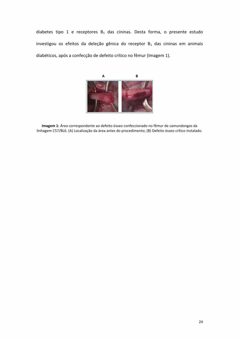
diabéticos, após a confecção de defeito crítico no fêmur (Imagem 1).
Imagem 1: Área correspondente ao defeito ósseo confeccionado no fêmur de camundongos da linhagem C57/BL6. (A) Localização da área antes do procedimento; (B) Defeito ósseo crítico instalado.
A B

25
OBJETIVOS
Objetivo geral
O presente estudo teve como objetivo avaliar a neoformação óssea em
animais com diabetes do tipo 1, bem como, verificar a relevância dos receptores B1
para as cininas neste processo.
Objetivos específicos
a) Avaliar a neoformação óssea em animais wild-type diabéticos em
relação aos controles.
b) Verificar a neoformação óssea em animais diabéticos com deleção
gênica dos receptores B1 para as cininas em comparação ao grupo
controle B1.
c) Avaliar marcadores de reabsorção óssea nos diferentes grupos
experimentais.

26
ARTIGO DE PESQUISA
O artigo a seguir intitula-se “Kinin B1 receptor lack influences bone healing in a
mouse model of femur critical-size defect in streptozotocin-induced type-1
diabetes” e foi submetido a Proceedings of the National Academy of Sciences- PNAS
(Fator de impacto 9,737; Qualis A1 Internacional, Área de Odontologia, CAPES).
27
Kinin B1 receptor lack influences bone healing in a mouse model of femur critical-
size defect in streptozotocin-induced type-1 diabetes
Short Title: B1 receptor and bone healing in type-I diabetes
Natália P. Cignachia, João B. Pesquerob, Rogério B. Oliveiraa, Adriana Etgesc, Maria M.
Camposa,d
aSchool of Dentistry and dInstitute of Toxicology and Pharmacology, Pontifical
Catholic University of Rio Grande do Sul, Avenida Ipiranga, 6681, Partenon, 90619-
900, Porto Alegre, RS, Brazil.
bDepartment of Biophysics, Federal University of São Paulo, Rua Pedro de Toledo,
669, CEP 04039-032, São Paulo, SP, Brazil.
cDepartment of Oral Pathology, School of Dentistry, Universidade Federal de Pelotas
(UFPel), Rua Gonçalves Chaves, 457, 96015-560, Pelotas, RS, Brazil.
Corresponding Author: Maria Martha Campos, School of Dentistry/Institute of
Toxicology and Pharmacology, Pontifícia Universidade Católica do Rio Grande do Sul,
Avenida Ipiranga, 6681, Partenon, 90619-900, Porto Alegre, RS, Brazil. Phone
number: 55 51 3320 3562; Fax number: 5551 3320 3626. E-mail:

28
ABSTRACT The effects on kinin B1 receptor (B1R) deletion were examined on bone
regeneration in streptozotocin (STZ)-type-1 diabetic mice, subjected to a model of
femoral critical-size defect. Diabetes induction in wild-type C57/BL6 (WT C57/BL6)
mice was allied to decrease of body weight and hyperglycemia, in relation to the
non-diabetic group of the same strain. The lack of B1R did not affect STZ-elicited
body weight loss, although it partially prevented hyperglycemia. Type-1 diabetic
mice presented a clear delay in bone regeneration, with large areas of loose
connective tissue within the region corresponding to the defects, when compared to
wide areas of newly-formed woven bone in non-diabetic WT C57/BL6 mice. Notably,
either non-diabetic or diabetic B1R knockout (B1RKO) mice displayed bone
regeneration levels comparable to that seen in control WT C57/BL6 mice. The
improved bone regeneration in animals lacking B1R was further confirmed by
analysis of collagen contents. WT C57/BL6 STZ-diabetic mice presented a marked
reduction of collagen contents within the bone defect gap, whereas diabetic B1RKO
displayed collagen levels comparable to those observed in non-diabetic WT C57/BL6
or B1RKO mice. The enhanced bone regeneration in diabetic mice lacking B1R does
not seem to be associated to lessened osteoclast activity, as no prominent difference
was detected in the levels of tartrate-resistant acid phosphatase (TRAP) positivity, or
even in the immunolabeling for the proteins of the RANK/RANKL/OPG system
thoughout all the experimental groups. Data brings novel evidence on the relevance
of B1R under type-1 diabetes, especially concerning its role in bone regeneration
after surgical procedures.

29
Type-1 insulin-dependent diabetes represents an autoimmune disease,
affecting more than 350-million people around the world. It commonly manifests in
childhood or adolescence, but a series of complications allied to long-term glucose
variations are present in the adult life, making the disease a continuous challenge
(1). Despite the great advances achieved during the last decades on the
comprehension of the pathophysiological mechanisms underlying type-1 diabetes, in
addition to improvements of treatment strategies, its global incidence is
continuously growing, by reasons that still remain elusive (2; 3). Therefore, research
on type-1 diabetes and its associated pathological consequences is a current matter
of high interest.
One of the relevant consequences of type-1 diabetes is the altered bone
metabolism, with the occurrence of osteopenia and osteoporosis, widely affecting
the life quality of patients (4; 5). Remarkably, type-1 diabetes has been related to
increased risks of fractures and delayed fracture healing (6; 7; 1). The mechanisms
responsible for bone alterations under type-1 diabetes are not completely clarified,
but inflammatory changes, including the increase of both serum and bone cytokines,
appear to be of high importance in this context (8; 9).
Kinins are peptide mediators implicated in a number of physiological and
pathological processes, including pain and inflammation It is renowned that kinins
act by the activation of two distinct G-protein-coupled receptors, called B1 (B1R) and
B2 (B2R) (10; 11). These receptors display very peculiar characteristics, mainly
concerning their pattern of expression. In this regard, the B2R subtype is
constitutively expressed throughout many organs, whereas B1R is not generally
present under normal situations, but its expression is induced during inflammatory

30
states, by means of cytokine-related mechanisms (12; 13). Of high interest, B1R and
B2R have been pointed out as relevant players in several complications associated to
type-1 diabetes, including diabetic nephropathy, retinopathy and pain neuropathy
(14; 15; 16; 17). Concerning the bone biology, there is controversial data on the role
of kinins and their receptors. In this regard, it was demonstrated that agonist
stimulation of either B1R or B2R leads to bone resorption in the neonatal mouse
bone calvaria, and the activation and expression of both receptors is able to
potentiate cytokine-induced inflammatory changes in osteoblastic cell lines and in
mouse calvarial bone (18; 19; 20; 21; 22). Otherwise, a study conducted by Kakoki et
al. (23) demonstrated that double B1R/B2R gene deletion resulted in enhanced
kidney injury, neuropathy, and bone mineral density loss, as evaluated in Akita
diabetic mice. Furthermore, a protective role for kinin receptors has been also
suggested more recently (24), by demonstrating that B1R knockout (B1RKO) mice
presented an increase of bone resorption following ligature-induced periodontal
disease, although the influence of diabetes has not been examined by the authors.
Considering the abovementioned pieces of data, the present study was devoted to
evaluate whether the genic deletion of the inflammation-related receptor B1R might
influence the bone healing process in a model of femoral critical-size defect, in type-
1 diabetic mice. Our results brings novel and interesting evidence, indicating that
lack of B1R helps bone healing in type-1 diabetes. It is tempting to suggest that local
measures, such as the local use of B1R antagonists could improve bone regeneration
and implant integration in diabetes
31
Methods
Animals. Male C57/BL6 wild-type (WT; total N=17) or kinin B1 receptor knockout
(B1RKO; total N = 17) mice (25 to 32 g) were used in this study. C57/BL6 mice were
obtained from Universidade Federal de Pelotas (UFPEL; Pelotas, RS, Brazil) and
B1RKO were supplied by Department of Biophysics, Universidade Federal de São
Paulo (UNIFESP-EPM, São Paulo, Brazil). These mice were crossed for ten
generations with C57/BL6 mice (39). The animals were housed in groups of four per
cage and maintained in controlled temperature (22±2ºC) and humidity (60-70%),
under a 12 h light-dark cycle, with food and water ad libitum. All the experimental
procedures were carried the out in accordance with the Guidelines for the Use and
Care with Laboratorial Animals from National Institute of Health and ethical
guidelines for investigations of experimental pain in conscious animal, and were
approved by the Local Animal Ethics Committee (protocol number 13/00037). The
number of animals and the intensity of noxious stimuli were the minimum necessary
to demonstrate the consistent effects of the experiments.
Induction of type-1 diabetes. WT C57/BL6 and B1RKO mice were divided in four
experimental groups: (1) WT C57/BL6 control (N=7); (2) WT C57/BL6 diabetic (N=10);
B1RKO control (N=7); (4) B1RKO diabetic (N=10). Type-1 diabetes was induced by five
injections of streptozotocin (STZ; 50 mg/kg), once a day, given intraperitoneally (i.p.),
dissolved in citrate buffer (50 mM; pH 4.5) (27). Non-diabetic groups received citrate
buffer vehicle alone i.p., at the same schedule of administration. The body weight
gain was measured periodically throughout the experiments on the basis of
individual body weight (in g) at the onset of experiments, considering that B1RKO
32
mice are leaner in relation to age-matched control WT C57/BL6 (25). At the end of
experimental procedures (33 days after beginning STZ treatment), the animals were
euthanized by isoflurane inhalation. The blood was collected and glucose levels were
expressed in mg/dl (Accutrend ; Roche Diagnostics).
Femur critical-size defect. All the surgical procedures were performed under aseptic
conditions, following seven days of the end of diabetes induction protocol (at the
12nd day), considering the last application of STZ (5th day). The critical-size defects
were created according to the methodology previously described by Srouji et al. (40),
with slight modifications. Briefly, the animals were anesthetized by an i.p. injection
of a mixture of xylazine (10 mg/kg) and ketamine (100 mg/kg). After anesthesia
confirmation, the access to the left mouse femur was made through a skin incision (6
to 8 mm in length), followed by muscle divulsion and periosteum detachment. The
osteotomy was carried out under irrigation, by using a surgical contra-angle hand-
piece (800 rpm), to create a mono-cortical bone defect (4-mm in length and 1-mm in
diameter). Following the creation of defect, the soft tissues were sutured in separate
layers, and the animals received antibiotic and analgesic post-operative medication.
The procedures were performed by a single researcher calibrated before. After 21
days of surgery (at the 33rd day after onset of experimental sessions), the animals
were euthanized as described before, and the femurs were removed immediately,
cleaned from connecting tissues, and maintained in 4 %-buffered formaldehyde
solution during 48 h, for subsequent histological procedures.

33
General histological procedures. Following fixation procedures, the pieces were
decalcified in a 17-% ethylenediaminetetraacetic acid (EDTA) solution, for 6 days,
with daily changes, and embedded in paraffin. Six consecutive 4-μm-thick
longitudinal sections were obtained from each femur, for different sets of analysis.
Three slides were separated to immunohistochemical analysis, as described in the
next section. The remaining three slides were stained with Hematoxyllin-Eosin
(H&E), Masson’s trichrome (Accustain Mallory’s stain kit, Sigma-Aldrich, USA), and
tartrate-resistant acid phosphate (Leukocyte TRAP kit, Sigma-Aldrich, USA),
respectively. The images were taken with a microscope (Axio Imager A1) coupled to
an image capture system (Axio Vision Rel. 4.4 Software Multimedia), both from
Imaging Solution Carl Zeiss (Hallbergmoos, Germany). The NIH Image J 1.36b
Software was used to semi-quantitatively determine the percentual area of bone
formation in relation to the total area in H&E-stained slides. The same software was
used for analyzing blue-colored collagen fibers in Mallory-stained slides (200-x
magnification, for both stainings). TRAP-stained activated osteoclasts were analyzed
only qualitatively under 400-x magnification. The examinations were performed in a
blinded manner, on two representative fields corresponding to the area of the
defect gap.
Immunohistochemical analysis. Analysis of the RANKL/RANK/OPG system was
carried out by the immunoperoxidase method, using paraffin-embedded sections.
Briefly, antigen retrieval was performed by immersion of slides in a water bath at
95–98º C in 10 mM trisodium citrate buffer, pH 6.0, for 45 min. The nonspecific
binding was blocked by incubating sections for 1 h with goat normal serum diluted in

34
PBS. As primary antibodies OPG (sc-21038, Santa Cruz Biotechnology), RANK (sc-
7626, Santa Cruz Biotechnology) and RANKL (sc-7627, Santa Cruz Biotechnology)
were used. After overnight incubation at 4ºC with primary antibodies, the slides
were washed with PBS and incubated with the secondary antibody Envision plus
(Dako Cytomation, Houston, TX, USA), ready to use, for 1 h at room temperature.
The sections were washed in PBS, and the visualization was completed by using 3, 3’-
diaminobenzidine (Dako Cytomation) in chromogenic solution and counterstained
lightly with Harris’s Hematoxylin solution. The images were captured by a digital
camera (CoolSNAP™ Media Cybernetics, Inc.) connected to an optical microscope
(Zeiss Axioskop DS-5 M-L1, Nikon, NY, USA), at 200-x magnification for counting the
number of positive brown-marked cells.
Statistical Analysis. Results are presented as the mean SEM of 7 to 10 animals,
depending on the experimental group. The statistical comparison between the
values was performed by one-way analysis of variance followed by Bonferroni’s
post-test. P values less than 0.05 (p < 0.05) were considered as indicative of
significance.
35
Results
Body weight and glycaemia in type-1 diabetes streptozotocin (STZ) model.
Extending literature data (25), control mice lacking B1R were leaner in relation to WT
C57/BL6 animals, according to the evaluation at 33 days of experimental sessions.
STZ-induced type-1 diabetes was associated with a progressive loss of body weight
gain, in WT C57/BL6 and B1RKO mice, when compared to non-diabetic animals of
either strain (Fig. 1, upper panel). Of note, WT C57/BL6 STZ-treated mice had a
significant increase of glucose levels (P<0.01), an effect that was partially reversed by
B1R deletion (Fig. 1; lower panel).
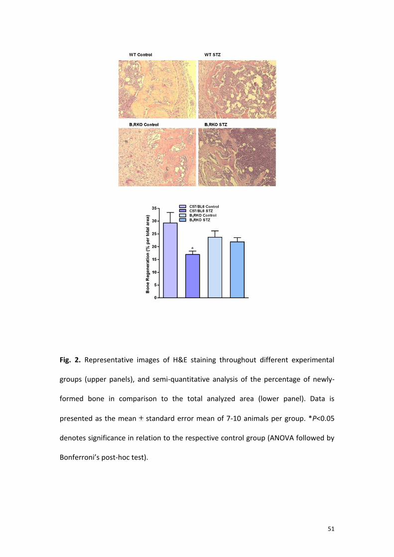
B1R genic deletion and general improvement of bone healing in type-1 diabetes.
The evaluation of Hematoxyllin & Eosin (H&E)-stained slides demonstrated the
predominance of woven bone, filling the area correspondent to the defect gap in
non-diabetic WT C57/BL6 mice, in comparison to wide areas of disorganized loose
connective tissue in WT C57/BL6 STZ-type-1 diabetic mice. Of high note, the bone
formation observed in either non-diabetic or diabetic B1RKO mice was very similar to
that seen in control WT C57/BL6 animals, with large areas of immature bone matrix,
pointing out the occurrence of active regeneration process (Fig. 2, upper panels).
The semi-quantitative analysis of H&E stained slides confirmed the qualitative
examination of images, clearly indicating larger areas of newly-formed bone in
control WT C57/BL6 mice, and in both non-diabetic and diabetic B1RKO mice, in
comparison to WT C57/BL6 STZ-type-1 diabetic animals (Fig. 2, lower panel; P<0.05).
To gain further insights into the potential differences in bone regeneration between
WT C57/BL6 and B1RKO mice during type-1 diabetes, we also carried out an analysis

36
of the percentage of collagen contents in the area of the defect gap, by using
Masson’s trichrome staining. The qualitative evaluation clearly revealed that STZ-
treated WT C57/BL6 mice presented less areas filled by collagen, which is indicative
of delayed bone regeneration and low tissue organization, in comparison to the
other experimental groups (Fig. 3, upper panels). The quantification of collagen-rich
blue-colored regions by using NIH Image J 1.36b Software corroborated the reduced
number of collagen levels in diabetic WT C57/BL6 animals, when compared to
control non-diabetic WT C57/BL6 mice (P<0.05). In this case, genic deletion of B1R
was also allied to improved bone healing, as demonstrated by the absence of
differences between non-diabetic and diabetic B1RKO mice (Fig. 3, lower panel).
Deletion of B1R and osteoclast-related tartrate-resistant acid phosphatase (TRAP)
in type-1 diabetes. In this study, we have also performed qualitative evaluation of
osteoclast activity, by analyzing TRAP staining. An overall analysis revealed that TRAP
activity in WT C57/BL6 diabetic mice was generally similar to that observed in non-
diabetic WT C57/BL6 animals, following examination of areas corresponding to the
bone defect. Furthermore, a similar profile regarding TRAP staining was noted in
non-diabetic and diabetic mice lacking B1R (Fig. 4). Thus, the differences of bone
regeneration between diabetic and non-diabetic WT C57/BL6 mice, and the
improvement of bone healing in diabetic mice lacking B1R does not seem to rely on
distinct osteoclast activity profiles among the experimental groups.

37
Lack of B1R does not affect RANK/RANKL/OPG axis in critical-size defects under
type-1 diabetes. Next, we evaluated the expression of the proteins of the
RANK/RANKL/OPG system throughout the four distinct experimental groups, by
immunohistochemistry. This set of data did not reveal any significant difference
among the experimental groups, regarding the number of immunopositive cells/field
for RANK (Fig. 5), RANKL (Fig 6) or OPG (Fig. 7). There were no significant differences
when comparing control WT C57/BL6 versus STZ-treated WT C57/BL6 mice, or even
when comparing non-diabetic and diabetic mice of both evaluated animals strains.
38
Discussion
The onset and progression of type-1 diabetes is associated to a series of
complications, compromising multiple tissues. Both clinical and pre-clinical studies
have broadly demonstrated the increased risk of bone fractures under type-1
diabetes, what is attributed to reduced bone mineral density and alterations of bone
matrix proteins (9). Interestingly, it has been suggested that inflammation displays a
relevant role on type-1 diabetes-allied bone alterations (8; 26). In this concern, kinins
are mediators associated to several pathological alterations, including chronic
inflammatory diseases, cancer and diabetes, among others (12; 10; 15). As far we
know, there is only one study investigating the relationship between kinin receptors
and bone characteristics in diabetes, by using double B1R/B2RKO mice (23). Herein,
we examined whether the lack of the inflammatory-related kinin B1R might affect
bone regeneration in a model of femoral critical-size defect, in STZ type-1 diabetic
mice. Our data shed new lights on the role of kinin receptors in diabetes and bone
alterations.
As expected, the induction of type-1 diabetes by the repeated administration
of STZ resulted in a marked decrease of body weight gain and significant
hyperglycemia, in comparison to control WT C57/BL6 mice (27). Concerning the non-
diabetic B1RKO mice, they were leaner than control WT C57/BL6 mice, as evaluated
at the end of the experimental procedures. This data extends previous evidence
showing that B1R deletion results in a slight reduction of body weight, in relation to
WT C57/BL6 control mice (25). Otherwise, the genic deletion of B1R did not protect
from reduction in body weight gain under STZ-induced type-1 diabetes, whereas it
prevented the hyperglycemia observed in this model. A previous publication (28)

39
demonstrated that genic deletion of B1R was able to evidently prevent the thermal
nociception allied to STZ-induced type 1 diabetes, although the authors had not
investigated the influence of B1R lacking on body weight loss, or glucose levels after
type-1 diabetes induction. Nevertheless, it was demonstrated prior that
pharmacological inhibition of B1R by the selective antagonists R-715 or SSR240612
displayed transitory, but significant effects on STZ-induced hyperglycemia in rats
(29), supporting our data on B1RKO mice and the control of glucose levels.
As the main goal of our study, we evaluated whether B1R deletion might
interfere with bone regeneration in type-1 diabetic mice, presenting a critical-size
femoral defect. It is well defined that type-1 diabetes is associated with increased
rates of bone fractures, likely due to reduced bone mineral density, as a
consequence of poor glycemic control and oxidative stress (7; 6; 4; 9). Herein, we
demonstrate that STZ-type-1 diabetic WT C57/BL6 mice presented delayed bone
regeneration, as it can be observed on the basis of H&E-stained sections, which
demonstrates the presence of great areas filled by loose disorganized tissue in this
experimental group. In contrast, WT C57/BL6 mice presented extensive areas of
woven bone within the region correspondent to the critical defect, a clear indicative
of new bone formation. Some previous reports have provided similar data indicating
that type-1 diabetes impaired the bone healing around implants, in both rodent and
non-rodent experimental models (30; 31; 32; 33), what points out the clinical
relevance of further studies in this area. Of high note, we provide novel evidence
showing that diabetic mice lacking B1R displayed a pattern of bone regeneration
similar to that observed in non-diabetic WT C57/BL6 or B1RKO animals. Therefore,
we might suggest that B1R activation and/or up-regulation could be associated to

40
deleterious consequences of type-1 diabetes on bone remodeling. This hypothesis
can be supported by a series of earlier publications showing the ability of kinins in
stimulating bone resorption-related mechanisms, either in vivo or in vitro, via
activation of B1R or B2R (18; 19; 20; 21; 22). Nonetheless, our data is somewhat
contradictory to the previous publication by Kakoki et al. (23) showing that double
B1R/B2R deletion resulted in increased bone mineral losses in animals with
diabetogenic Akita mutation. This divergence might be explained by several reasons,
including (i) the chemical model of type-1 diabetes adopted by us; (ii) the use of
B1RKO mice instead of double B1R/B2R KO animals; and (iii) the evaluation of active
bone remodeling process, following the creation of a critical-size defect. More
recently, it was shown that B1RKO mice displayed increased bone resorption in a
model of ligature-induced periodontitis; however the authors did not assess the
influence of diabetes, and there is a clear infectious component in this experimental
approach (24), which is not the case of our study.
In the present study, we also examined the levels of collagen throughout the
different experimental groups. It is well-known that collagen is essential to provide
the ideal properties of bone, such as the mechanical resistance to fractures (34). In
this regard, a reduced bone expression of type 1 collagen was described in STZ-type-
1 diabetic mice subjected to a model of tibia marrow ablation (35). Similarly, the
contents of type I collagen were found diminished in alloxan-type-1 diabetic rabbits
after maxillary sinus augmentation (31). Furthermore, it has been recently
demonstrated that either genetic or chemical induction of type-1 diabetes leads to
decreased expression of collagen II, what is linked to impaired cartilage formation in
mice (36). Herein, Masson’s trichrome staining revealed a prominent reduction of

41
collagen levels in WT C57/BL6 STZ-diabetic mice, following the examination of the
area corresponding to the bone defect gap, in contrast to non-diabetic control WT
C57/BL6 animals. Strikingly, collagen reduction was not evident in diabetic mice
presenting B1R gene deletion, being comparable to that seen in control B1RKO mice.
This experimental evidence helps to explain the differences in bone regeneration
between diabetic mice of WT and B1RKO strains, allowing suggesting a possible role
for B1R in the formation of bone organic matrix and mineralization. It is worth
mentioning that B1R deletion prevented both cardiac inflammation and fibrosis in
STZ-elicited type-1 diabetes in mice (37).
As an additional approach, we decided to examine to what extent the lack of
B1R might alter some markers of osteoclast differentiation under type-1 diabetes.
Firstly, we evaluated TRAP-stained sections, as an indicative of osteoclast activity.
This set of data did not reveal any expressive difference on TRAP staining among all
the evaluated groups, independent on diabetes induction or mouse strain. In fact, it
was possible to observe positive TRAP staining in most evaluated sections, indicating
a great level of bone turnover in our model of femoral critical-size defect. As revised
by McCabe et al. (9), the osteoclast markers can be found unaltered, decreased, or
even increased in mouse models of type-1 diabetes, what might support our data.
For instance, TRAP activity was not found altered in bone tibia obtained from STZ-
treated rats, in relation to control-matched animals (38). When analyzed in the light
of literature data, our results permit to propose that delayed bone regeneration in
STZ-type-1 diabetic animals is likely related to decreased levels of osteoblastic
factors (such as type 1 collagen), rather than increased osteoclast activity, at least at
the experimental conditions adopted by us. Moreover, the positive effects of B1R

42
deletion on diabetes-induced delayed regeneration can be interpreted as the result
of anti-hyperglycemic effects, and the consequent improvement of bone matrix
remodeling, without alterations of osteoclast activity. In this paper, we have also
assessed to what extent the expression of the RANK/RANKL/OPG system might be
altered in the areas corresponding to the defect gap under STZ-induced diabetes, in
WT C57/BL6 or B1RKO mice. Extending data on TRAP staining, we were not able to
detect any significant difference among the expression of the three evaluated
proteins in non-diabetic and diabetic mice of both strains. Concerning B1R, the study
conducted by Gonçalves-Zillo et al. (24) demonstrated an increased number of
osteoclasts in animals lacking this receptor, by inducing in vitro differentiation of
bone marrow cells by M-CSF and RANKL, contrasting to some degree with our data.
In fact, the deleterious or potential effects of kinins and their receptor appear to
greatly differ, depending on the target organs and pathological conditions that are
evaluated (10). Indeed, further studies devoted to explore the influence of B1R
activation on osteoblasts and osteoclast functions remain to be carried out, by using
other molecular markers of bone turnover.
Altogether, data present in this study extends previous evidence on the
relevance of kinin B1R under type-1 diabetes, and provide novel evidence on the role
of this receptor in bone regeneration. We hope this data might help, in a near
future, to develop new therapeutic strategies to provide superior outcomes for type-
1 diabetic patients subjected to bone surgeries.

43
1. Pan H, Wu N, Yang T, He W. Association between bone mineral density and
type 1 diabetes mellitus: a meta-analysis of cross-sectional studies. Diabetes
Metab Res Rev. 2013; doi: 10.1002/dmrr.2508.
2. Egro FM. Why is type 1 diabetes increasing? J Mol Endocrinol. 2013; 51(1):R1-
13.
3. Vehik K, Ajami NJ, Hadley D, Petrosino JF, Burkhardt BR. The changing
landscape of type 1 diabetes: recent developments and future frontiers. Curr
Diab Rep. 2013; 13(5):642-50.
4. Blakytny R, Spraul M, Jude EB. Review: The diabetic bone: a cellular and
molecular perspective. Int J Low Extrem Wounds. 2011; 10(1):16-32.
5. Kurra S, Siris E. Diabetes and bone health: the relationship between diabetes
and osteoporosis-associated fractures. Diabetes Metab Res Rev. 2011;
27(5):430-5.
6. Vestergaard P, Rejnmark L, Mosekilde L. Diabetes and its complications and
their relationship with risk of fractures in type 1 and 2 diabetes. Calcif Tissue
Int. 2009; 84(1):45-55.
7. Khazai NB, Beck GR Jr, Umpierrez GE. Diabetes and fractures: an
overshadowed association. Curr Opin Endocrinol Diabetes Obes. 2009;
16(6):435-45.

44
8. Motyl KJ, Botolin S, Irwin R, Appledorn DM, Kadakia T, Amalfitano A,
Schwartz RC, McCabe LR. Bone inflammation and altered gene expression
with type 1 diabetes early onset. J Cell Physiol. 2009; 218(3):575-83.
9. McCabe L, Zhang J, Raehtz S. Understanding the skeletal pathology of type 1
and 2 diabetes mellitus. Crit Rev Eukaryot Gene Expr. 2011; 21(2):187-206.
10. Regoli D, Plante GE, Gobeil F Jr. Impact of kinins in the treatment of
cardiovascular diseases. Pharmacol Ther. 2012; 135(1):94-111.
11. Whalley ET, Figueroa CD, Gera L, Bhoola KD. Discovery and therapeutic
potential of kinin receptor antagonists. Expert Opin Drug Discov. 2012;
7(12):1129-48.
12. Calixto JB, Medeiros R, Fernandes ES, Ferreira J, Cabrini DA, Campos MM.
Kinin B1 receptors: key G-protein-coupled receptors and their role in
inflammatory and painful processes. Br J Pharmacol. 2004; 143(7):803-18.
13. Campos MM, Leal PC, Yunes RA, Calixto JB. Non-peptide antagonists for kinin
B1 receptors: new insights into their therapeutic potential for the
management of inflammation and pain. Trends Pharmacol Sci. 2006;
27(12):646-51.
14. Webb JG. The kallikrein/kinin system in ocular function. J Ocul Pharmacol
Ther. 2011; 27(6):539-43.
15. Talbot S, Couture R. Emerging role of microglial kinin B1 receptor in diabetic
pain neuropathy. Exp Neurol. 2012; 234(2):373-81.

45
16. Tomita H, Sanford RB, Smithies O, Kakoki M. The kallikrein-kinin system in
diabetic nephropathy. Kidney Int. 2012; 81(8):733-44.
17. Liu J, Feener EP. Plasma kallikrein-kinin system and diabetic retinopathy. Biol
Chem. 2013; 394(3):319-28.
18. Ljunggren O, Vavrek R, Stewart JM, Lerner UH. Bradykinin-induced burst of
prostaglandin formation in osteoblasts is mediated via B2 bradykinin
receptors. J Bone Miner Res. 1991; 6(8):807-15.
19. Brechter AB, Lerner UH. Bradykinin potentiates cytokine-induced
prostaglandin biosynthesis in osteoblasts by enhanced expression of
cyclooxygenase 2, resulting in increased RANKL expression. Arthritis Rheum.
2007; 56(3):910-23.
20. Brechter AB, Persson E, Lundgren I, Lerner UH. Kinin B1 and B2 receptor
expression in osteoblasts and fibroblasts is enhanced by interleukin-1 and
tumour necrosis factor-alpha. Effects dependent on activation of NF-kappaB
and MAP kinases. Bone. 2008; 43(1):72-83.
21. Suzuki Y, Kodama D, Goto S, Togari A. Involvement of TRP channels in the
signal transduction of bradykinin in human osteoblasts. Biochem Biophys Res
Commun. 2011; 410(2):317-21.
22. Souza PP, Brechter AB, Reis RI, Costa CA, Lundberg P, Lerner UH. IL-4 and IL-
13 inhibit IL-1β and TNF-α induced kinin B1 and B2 receptors through a
STAT6-dependent mechanism. Br J Pharmacol. 2013; 169(2):400-12.

46
23. Kakoki M, Sullivan KA, Backus C, Hayes JM, Oh SS, Hua K, Gasim AM, Tomita
H, Grant R, Nossov SB, Kim HS, Jennette JC, Feldman EL, Smithies O. Lack of
both bradykinin B1 and B2 receptors enhances nephropathy, neuropathy, and
bone mineral loss in Akita diabetic mice. Proc Natl Acad Sci U S A. 2010;
107(22):10190-5.
24. Gonçalves-Zillo TO, Pugliese LS, Sales VM, Mori MA, Squaiella-Baptistão CC,
Longo-Maugéri IM, Lopes JD, de Oliveira SM, Monteiro AC, Pesquero JB.
Increased bone loss and amount of osteoclasts in kinin B1 receptor knockout
mice. J Clin Periodontol. 2013; 40(7):653-60.
25. Mori MA, Sales VM, Motta FL, Fonseca RG, Alenina N, Guadagnini D,
Schadock I, Silva ED, Torres HA, dos Santos EL, Castro CH, D'Almeida V,
Andreotti S, Campaña AB, Sertié RA, Saad MJ, Lima FB, Bader M, Pesquero JB.
Kinin B1 receptor in adipocytes regulates glucose tolerance and
predisposition to obesity. PLoS One. 2012; 7(9):e44782.
26. Schett G, David JP. The multiple faces of autoimmune-mediated bone loss.
Nat Rev Endocrinol. 2010; 6(12):698-706.
27. Zauli G, Toffoli B, di Iasio MG, Celeghini C, Fabris B, Secchiero P. Treatment
with recombinant tumor necrosis factor-related apoptosis-inducing ligand
alleviates the severity of streptozotocin-induced diabetes. Diabetes.
2010;59(5):1261-5.

47
28. Gabra BH, Merino VF, Bader M, Pesquero JB, Sirois P. Absence of diabetic
hyperalgesia in bradykinin B1 receptor-knockout mice. Regul Pept. 2005;
127(1-3):245-8.
29. Talbot S, Chahmi E, Dias JP, Couture R. Key role for spinal dorsal horn
microglial kinin B1 receptor in early diabetic pain neuropathy. J
Neuroinflammation. 2010; 7(1):36.
30. Margonar R, Sakakura CE, Holzhausen M, Pepato MT, Alba Jr. RC,
Marcantonio Jr. E. The influence of diabetes mellitus and insulin therapy on
biomechanical retention around dental implants: a study in rabbits. Implant
Dent. 2003; 12(4):333-9.
31. Von Wilmowsky C, Stockmann P, Harsch I, Amann K, Metzler P, Lutz R, Moest
T, Neukam FW, Schlegel KA. Diabetes mellitus negatively affects peri-implant
bone formation in the diabetic domestic pig. J Clin Periodontol. 2011;
38(8):771-9.
32. Mao L, Tamura Y, Kawao N, Okada K, Yano M, Okumoto K, Kaji H. Influence of
diabetic state and vitamin D deficiency on bone repair in female mice. Bone.
2013; S8756-3282(13)00546-2.
33. Molon RS, Morais-Camilo JA, Verzola MH, Faeda RS, Pepato MT, Marcantonio
E Jr. Impact of diabetes mellitus and metabolic control on bone healing
around osseointegrated implants: removal torque and histomorphometric
analysis in rats. Clin Oral Implants Res. 2013; 24(7):831-7.

48
34. Viguet-Carrin S, Garnero P, Delmas PD. The role of collagen in bone strength.
Osteoporos Int. 2006; 17(3):319-36.
35. Lu H, Kraut D, Gerstenfeld LC, Graves DT. Diabetes interferes with the bone
formation by affecting the expression of transcription factors that regulate
osteoblast differentiation. Endocrinology. 2003; 144(1):346-52.
36. Coe LM, Zhang J, McCabe LR. Both spontaneous Ins2(+/-) and streptozotocin-
induced type 1 diabetes cause bone loss in young mice. J Cell Physiol. 2013;
228(4):689-95.
37. Westermann D, Walther T, Savvatis K, Escher F, Sobirey M, Riad A, Bader M,
Schultheiss HP, Tschöpe C. Gene deletion of the kinin receptor B1 attenuates
cardiac inflammation and fibrosis during the development of experimental
diabetic cardiomyopathy. Diabetes. 2009; 58(6):1373-81.
38. Hie M, Iitsuka N, Otsuka T, Tsukamoto I. Insulin-dependent diabetes mellitus
decreases osteoblastogenesis associated with the inhibition of Wnt signaling
through increased expression of Sost and Dkk1 and inhibition of Akt
activation. Int J Mol Med. 2011; 28(3):455-62.
39. Quintão NL, Passos GF, Medeiros R, Paszcuk AF, Motta FL, Pesquero JB,
Campos MM, Calixto JB. Neuropathic pain-like behavior after brachial plexus
avulsion in mice: the relevance of kinin B1 and B2 receptors. J Neurosci. 2008;
28(11):2856-63.

49
40. Srouji S, Ben-David D, Kohler T, Müller R, Zussman E, Livne E. A model for
tissue engineering applications: femoral critical size defect in
immunodeficient mice. Tissue Eng Part C Methods. 2011;17(5):597-606.

50
Figure Legends
Fig. 1. Body weight gain and blood glucose levels in WT and B1RKO in STZ-induced
type 1 diabetes. The weight gain was calculated at different time-points, as the
percentage of increase in relation to the individual body weight at the beginning of
experiments. The blood glucose levels are provided in mg/dl. Data is presented as
the mean standard error mean of 7-10 animals per group. *P<0.05; **P<0.01
denotes significance in relation to the respective control group (ANOVA followed by
Bonferroni’s post-hoc test).
51
Fig. 2. Representative images of H&E staining throughout different experimental
groups (upper panels), and semi-quantitative analysis of the percentage of newly-
formed bone in comparison to the total analyzed area (lower panel). Data is
presented as the mean standard error mean of 7-10 animals per group. *P<0.05
denotes significance in relation to the respective control group (ANOVA followed by
Bonferroni’s post-hoc test).

52
Fig. 3. Representative images of Masson’s trichrome staining throughout different
experimental groups (upper panels), and semi-quantitative analysis of the
percentage of collagen fibers in comparison to the total analyzed area (lower panel).
Data is presented as the mean standard error mean of 7-10 animals per group.
*P<0.05 denotes significance in relation to the respective control group (ANOVA
followed by Bonferroni’s post-hoc test).

53
Fig. 4. Representative images of tartrate-resistant acid phosphatase (TRAP) staining
throughout the four different evaluated experimental groups. The arrows indicate
positive labeling. The images are representative of analysis of 7-10 animals per
group.

54
Fig. 5. Representative images of immunohistochemical analysis for RANK expression
throughout different experimental groups (upper panels), and quantitative analysis
of the number of positive cells/field (lower panel). Data is presented as the mean
standard error mean of 7-10 animals per group.

55
Fig. 6. Representative images of immunohistochemical analysis for RANKL expression
throughout different experimental groups (upper panels), and quantitative analysis
of the number of positive cells/field (lower panel). Data is presented as the mean
standard error mean of 7-10 animals per group.

56
Fig. 7. Representative images of immunohistochemical analysis for OPG expression
throughout different experimental groups (upper panels), and quantitative analysis
of the number of positive cells/field (lower panel). Data is presented as the mean
standard error mean of 7-10 animals per group.

57
CONSIDERAÇÕES FINAIS
Como visto na literatura atualmente, o aumento da prevalência de Diabetes
Mellitus na população mundial tem preocupado os profissionais da saúde, inclusive
cirurgiões-dentistas, pela repercussão dessa doença, em nível bucal, com
manifestações mais severas na periodontite, assim como, interferências no processo
de osseointegração de implantes.
Tem-se notado uma diferença de cicatrização, tanto de tecidos moles,
quanto ósseos, até mesmo, quando os níveis glicêmicos desses pacientes
encontram-se controlados. Os sinais e sintomas do diabetes tipo 1 são mais
evidentes que do tipo 2; normalmente, os pacientes desse primeiro grupo
desenvolveram a doença precocemente, na infância ou adolescência. Porém, as
complicações vão se agravando ao longo da vida.
O diabetes interfere ao nível ósseo, pois ocorre uma desregulação hormonal,
como, também uma deficiência na absorção do cálcio e da vitamina D.
Notavelmente, o diabetes tipo 1 tem sido associado com um aumento no risco de
fraturas e atraso na cicatrização das mesmas. Visto que a perda óssea não está
vinculada apenas a um aumento da atividade osteoclástica, tem-se buscado estudar
o processo de remodelação óssea de forma integral.
O evento de neoformação e reabsorção óssea é controlado pelos
osteoblastos e osteoclastos, respectivamente, através da ativação ou bloqueio do
receptor presente na membrana dos osteoblastos (RANK). Curiosamente, os
mecanismos relacionados com o desequilíbrio desse sistema ainda não foram
totalmente esclarecidos.
Como o diabetes é uma doença metabólica e inflamatória, é possível sugerir
a sua interação com peptídeos biologicamente ativos que participam desse
processo. Esses peptídeos, chamados cininas, estão presentes em condições
inflamatórias decorrentes de algum dano tecidual, como em situações fisiológicas.
A bradicinina e seus metabólitos podem ativar dois tipos de receptores
acoplados a proteína G, chamados de B1 e B2. No presente trabalho, optou-se
estudar o papel do receptor B1, por ele ser expresso após um processo de
inflamação, infecção ou trauma. Considerando esse perfil de indução, os receptores

58
B1 são alvos para o desenvolvimento de drogas com potencial antiinflamatório que
poderiam ser úteis para doenças crônicas como o diabetes. Este receptor está
associado com diversas complicações associadas ao diabetes tipo 1, como nefropatia
diabética, retinopatia e dor neuropática.
Quando se trata de biologia óssea, existem dados contraditórios sobre o
papel das cininas e seus receptores. Portanto esse trabalho está voltado em avaliar
de que forma a deleção gênica do receptor B1 pode influenciar a cicatrização óssea
em um modelo de defeito crítico em fêmur de camundongos.
A realização do procedimento cirúrgico para a confecção do defeito ósseo no
fêmur dos camundongos foi adaptado e, baseado em diferentes protocolos já
estabelecidos anteriormente. Modelos em calvária de camundongos têm sido
amplamente empregados, assim como os de fraturas na região do fêmur. Optou-se
pela localização femoral (porém, sem o envolvimento das duas corticais para não
utilização de qualquer forma de fixação) por apresentar uma maior quantidade
óssea que a calota craniana.
Foi fornecida uma nova evidência neste trabalho; a regeneração óssea
(conferida no padrão de coloração H&E) nos animais B1RKO diabéticos deu-se da
mesma forma que outros dois grupos: não-diabéticos WT C57/BL6 e B1RKO. Sendo
assim, podemos sugerir que a ativação e/ou aumento da expressão B1, pode estar
associado a conseqüências deletérias do diabetes tipo 1 na remodelação óssea. Esse
dado pode ser confirmado por outras publicações que mostram a habilidade das
cininas em estimular mecanismos de reabsorção óssea tanto in vivo quanto in vitro.
No presente modelo, parece haver um desequilíbrio do processo de
remodelação óssea, predominando a diferenciação osteoblática, muito mais do que
osteoclática nos animais diabéticos C57/BL6. Por outro lado, os animais com deleção
gênica parecem estar protegidos desses efeitos.
Visto nossa dificuldade em conduzir um tratamento adequado aos pacientes
desse grupo (diabéticos), compreender, que, o desequilíbrio no processo de
regeneração óssea está relacionado à neoformação, abre novos caminhos para
tentarmos otimizar o processo, levando a um equilíbrio.
Pode-se concluir que o bloqueio de receptores B1 pode representar
estratégicas terapêuticas atraentes para melhorar a formação óssea em pacientes

59
com diabetes tipo 1, que requerem procedimentos cirúrgicos ósseos assim como a
instalação de implantes osseointegrados.

60
REFERÊNCIAS BIBLIOGRÁFICAS GERAIS
1. Bord S, Horner A, Hembry RM, Reynolds JJ, Compston JE. Production of
collagenase by human osteoblasts and osteoclasts in vivo. Bone. 1996; 19(1): 35-40.
2. Boyce BF, Xing L. Functions of RANKL/RANK/OPG in bone modeling and
remodeling. Arch Biochem Biophys. 2008 15; 473(2): 139-46.
3. Marie PJ. Transcription factors controlling osteoblastogenesis. Arch Biochem
Biophys. 2008; 15;473(2):98-105.
4. Rosen V, Thies RS, Lyons K. Signaling pathways in skeletal formation: a role for
BMP receptors. Ann N Y Acad Sci. 1996; 8;785:59-69.
5. Robinson JA, Harris SA, Riggs BL, Spelsberg TC. Estrogen regulation of human
osteoblastic cell proliferation and differentiation. Endocrinology. 1997; 138(7): 2919-
27.
6. Turner CH. Homeostatic control of bone structure: An application of feedback
theory. Bone 1991; 12:203-7.
7. Perkins SL, Kling SJ. Local concentrations of macrophage colony-stimu- lating
factor mediate osteoclast differentiation. Am J Physiol Endocrinol Metab 1995;
269:E1024-E1030.
8. Long MW, Robinson JA, Ashcraft EA, Mann KG. Regulation of human bone
marrow-derived osteoprogenitor cells by osteogenic growth factors. J Clin Invest.
1995; 95(2):881-7.
9. Marks SC. The origin of osteoclast: Evidence, clinical implications and in-
vestigative challenges of an extra-skeletal source. J Pathol 1983; 12:226-56.

61
10. Young MF, Ibaraki K, Kerr JM. Molecular and cellular biology of the major
noncollagenous proteins in bone. In: Noda M (ed). Cellular and molecular biology of
bone. New York: Academic Press; 1993:191-234.
11. Pinero GJ, Farach-Carson MC, Devoll RE, Aubin JE, Brunn JC, Butler WT. Bone
matrix proteins in osteogenesis and remodelling in the neonatal rat mandible as
studied by immunolocalization of osteopontin, bone sialoprotein, alpha 2HS-
glycoprotein and alkaline phosphatase. Arch Oral Biol. 1995; 40(2):145-55.
12. Boyce BF, Xing L. Functions of RANKL/RANK/OPG in bone modeling and
remodeling. Arch Biochem Biophys; 2008; 473(2): 139-46.
13. Kearns AE, Khosla S, Kostenuik PJ. Receptor activator of nuclear factor kappaB
ligand and osteoprotegerin regulation of bone remodeling in health and disease.
Endocr Rev. 2008; 29(2):155-92.
14. Theoleyre S, Wittrant Y, Tat SK, Fortun Y, Redini F, Heymann D. The molecular
triad OPG/RANK/RANKL: involvement in the orchestration of pathophysiological
bone remodeling. Cytokine Growth Factor Rev. 2004; 15(6):457-75.
15. Junqueira LC, Carneiro J. Tecido ósseo. In: Junqueira LC, Carneiro J. Histologia
básica. Rio de Janeiro: Editora Guanabara Koogan; 2004. p. 111-128.
16. Viguet-Carrin S, Garnero P, Delmas PD. The role of collagen in bone strength.
Osteoporos Int. 2006; 17(3):319-36.
17. Doré-Savard L, Otis V, Belleville K, Lemire M, Archambault M, Tremblay L,
Beaudoin JF, Beaudet N, Lecomte R, Lepage M, Gendron L, Sarret P. Behavioral.
Medical imaging and histopathological features of a new rat model of bone cancer
pain. PLOS One. 2010; 29; 5(10).
18. The Expert Committee on the diagnosis and classification of diabetes mellitus.
Report of the Expert Committee on the diagnosis and classification of diabetes
mellitus. Diabetes Care 1997; 20:1183-97.
http://www.ncbi.nlm.nih.gov/pubmed?term=Kostenuik%20PJ%5BAuthor%5D&cauthor=true&cauthor_uid=18057140

62
19. Hornick T, Aron DC: Preventing and managing diabetic complications in
elderlypatients. Cleve Clin J Med 2008, 75:153-158.
20. UK Prospective Diabetes Study (UKPDS) Group. Intensive blood-glucose control
with sulphonylureas or insulin compared with conventional treatment and risk of
complications in patients with type 2 diabetes (UKPDS 33). Lancet 1998; 352:837-53.
21. Atkinson MA, Maclaren NK. The pathogenesis of insulin dependent diabetes. N
Engl J Med 1994; 331:1428-36.
22. Imagawa A, Hanafusa T, Miyagawa J-I, Matsuzawa Y, for the Osaka IDDM Study
Group. A novel subtype of type 1 diabetes mellitus characterized by a rapid onset
and an absence of diabetes-related antibodies. N Engl J Med 2000; 342:301-7.
23. Ward DT, Yau SK, Mee AP, Mawer EB, Miller CA, Garland HO, Riccardi D.
Functional, molecular, and biochemical characterization of streptozotocin-induced
diabetes. J Am Soc Nephrol. 2001; 12:779-90.
24. Figueiredo, D. M.; Rabelo, F. L. A. Diabetes insipidus: principais aspectos e análise
comparativa com diabetes mellitus. Cienc. Biol. Saúde, Londrina, 2009 v. 30, n. 2, p.
155- 162.
25. Spanheimer RG, Umpierrez GE, Stumpf V. Decreased collagen production in
diabetic rats. Diabetes. 1988; 37:371-6.
26. Peled M, Ardekian L, Tagger-Green N, Gutmacher Z, Machtei EE. Dental implants
in patients with type 2 diabetes mellitus: a clinical study. Implant Dent. 2003;
12:116-22.
27. Shires R, Teitelbaum SL, Bergfeld MA, Fallon MD, Slatopolsky E, Avioli LV. The
effect of streptozotocin-induced chronic diabetes mellitus on bone and mineral
homeostasis in the rat. J Lab Clin Med. 1981; 97:231-40.

63
28. Santana RB, Xu L, Chase HB, Amar S, Graves DT, Trackman PC. A role for
advanced glycation end products in diminished bone healing in type 1 diabetes.
Diabetes. 2003; 52:1502-10.
29. Weiss RE, Gorn AH, Nimni ME. Abnormalities in the biosynthesis of cartilage and
bone proteoglycans in experimental diabetes. Diabetes. 1981; 30:670-7.
30. Lekholm U. The use of osseointegrated implants in growing jaws. Int J Oral
Maxillofac Implants. 1993; 8: 243-4.
31. Murata M, Takahashi A, Saito I, Kawanishi S. Site-specific DNA methylation and
apoptosis: induction by diabetogenic streptozotocin. Biochem Pharmacol. 1999; 57:
8817.
32. Wong KK, Tzeng ES. Appearance of different diabetic symptoms after
streptozocin administration: a comparison study. Biochem Mol Biol Int. 1993;
30:1035-41.
33. Goodman WG, Hori MT. Diminished bone formation in experimental diabetes.
Relationship to osteoid maturation and mineralization. Diabetes. 1984; 33: 825-31.
34. Graves DT, Naguib G, Lu H, Leone C, Hsue H, Krall E. Inflammation is more
persistent in type 1 diabetic mice. J Dent Res. 2005; 84(4):324-8.
35. Wetzler HP, Snyder JW. Linking pharmacy and laboratory data to assess the
appropriateness of care in patients with diabetes. Diabetes Care. 2000; 23(11):1637-
41.
36. Lu H, Kraut D, Gerstenfeld LC, Graves DT. Diabetes interferes with the bone
formation by affecting the expression of transcription factors that regulate
osteoblast differentiation. Endocrinology. 2003; 144: 346-52.
37. Marceau F, Regoli D. Bradykinin receptor ligands: therapeutic perspectives. Nat
Rev Drug Discov. 2004; 3:845-852.

64
38. Hu SW, Huang CH, Huang HC, Lai YY, Lin YY. Transvascular dissemination of
Porphyromonas gingivalis from a sequestered site is dependent upon activation of
the kallikrein/kinin pathway. J Periodontal Res. 2006; 41:200-207.
39. Schulze-Topphoff U, Prat A, Bader M, Zipp F, Aktas O. Roles of the kallikrein/kinin
system in the adaptive immune system. Int Immunopharmacol. 2008; 8(2):155-60.
40. Calixto JB, Medeiros R, Fernandes ES, Ferreira J, Cabrini DA, Campos MM. Kinin
B1 receptors: key G-protein-coupled receptors and their role in inflammatory and
painful processes. Br J Pharmacol. 2004; 143(7):803-18
41. Marceau F, Regoli D. Bradykinin receptor ligands: therapeutic perspectives. Nat
Rev Drug Discov. 2004; 3(10):845-52.
42. Stewart JM. Bradykinin antagonists: discovery and development. Peptides 2004;
25:527-532.
43. Ferreira J, Beirith A, Mori MA, Araujo RC, Bader M, Pesquero JB, Calixto JB.
Reduced nerve injury-induced neuropathic pain in kinin B1 receptor knock-out mice.
J Neurosci. 2005; 25(9):2405-12.
44. Fox A, Kaur S, Li B, Panesar M, Saha U, Davis C, Dragoni I, Colley S, Ritchie T,
Bevan S, Burgess G, McIntyre P. Antihyperalgesic activity of a novel nonpeptide
bradykinin B1 receptor antagonist in transgenic mice expressing the human B1
receptor. Br J Pharmacol. 2005; 144(7):889-99.
45. Leeb-Lundberg LM, Marceau F, Muller-Esterl W, Pettibone DJ, Zuraw BL.
International union of pharmacology. XLV. Classification of the kinin receptor family:
from molecular mechanisms to pathophysiological consequences. Pharmacol Rev.
2005; 57(1):27-77.

65
46. Wang H, Ehnert C, Brenner GJ, Woolf CJ. Bradykinin and peripheral sensitization.
Biol Chem. 2006; 387:11-4.
47. Bhoola KD, Figueroa CD, Worthy K. Bioregulation of kinins: kallikreins,
kininogens, and kininases. Pharmacol Rev. 1992; 44(1):1-80.
48. Mclean PG, Perreti M, Ahluwalia A. Kinin B1 receptors and the cardiovascular
system: regulation of expression and function. Cardiovasc Res. 2000; 48:194-210.
49. Gafford JT, Skidgel RA, Erdos EG, Hersh LB. Human kidney "enkephalinase", a
neutral metalloendopeptidase that cleaves active peptides. Biochemistry. 1983;
21;22(13):3265-71.
50. Guimarães JA, Borges DR, Prado ES, Prado JL. Kinin-converting aminopeptidase
from human serum. Biochem Pharmacol. 1973; 15;22(24):3157-72.
51. Campbell W, Yonezu K, Shinohara T, Okada H. An arginine carboxypeptidase
generated during coagulation is diminished or absent in patients with reumathoid
arthritis. J Lab Clin Invest. 1990; 115:610-612.
52. Hendriks D, Wang W, Scharpé S, Lommaert MP, van Sande M. Purification and
characterization of a new arginine carboxypeptidase in human serum. Biochim
Biophys Acta. 1990; 1034(1):86-92.
53. Regoli D, Barabé J, Park WK. Receptors for bradykinin in rabbit aortae. Can J
Physiol Pharmacol. 1977; 55(4):855-67.
54. Regoli D, Barabe J. Pharmacology of bradykinin and related kinins. Pharmacol
Rev. 1980; 32:1-46.
66
55. Calixto JB, Cabrini DA, Ferreira J, Campos MM. Kinins in pain and inflammation.
Pain. 2000; 87(1): 1-5.
56. Campos MM, Leal PC, Nunes RA, Calixto JB. Non-peptide antagonists for kinin B1
receptors: new insight sinto their therapeutic potential for the management of
inflammation and pain. Trends Pharmacol Sci. 2006; 27(12): 646-651.
57. Pesquero JB, Bader M. Genetically altered animal models in the kallikrein-kinin
system. Biol Chem. 2006; 387(2): 119-26.
58. Kuduk SD, Bock MG. Bradykinin B1 receptor antagonists as novel analgesics: a
retrospective of selected medicinal chemistry developments. Curr Top Med Chem.
2008; 8(16): 1420-30.
59. Marceau F, Hess JF, Bachvarov DR. The B1 receptors for kinins. Pharmacol Rev.
1998; 50(3): 357-86.
60. Couture R, Girolami JP. Putative roles of kinin receptors in the therapeutic effects
of angiotensin 1-converting enzyme inhibitors in diabetes mellitus. Eur J Pharmacol
2004, 500:467-485.
61. Dias JP, Talbot S, Sénécal J, Carayon P, Couture R. Kinin B1 receptor enhances the
oxidative stress in a rat model of insulin resistance: outcome in hypertension,
allodynia and metabolic complications. Plos One, 2010; 7; 5(9): e12622.
62. Kakoki M, Sullivan KA, Backus C, Hayes JM, Oh SS, Hua K, Gasim AM, Tomita H,
Grant R, Nossov SB, Kim HS, Jennette JC, Feldman EL, Smithies O. Lack of both
bradykinin B1 and B2 receptors enhances nephropathy, neuropathy, and bone
mineral loss in Akita diabetic mice. Proc Natl Acad Sci U S A. 2010; 1;107(22):10190-
5.

67
63. Brechter AB, Persson E, Lundgren I, Lerner UH. Kinin B1 and B2 receptor
expression in osteoblasts and fibroblasts is enhanced by interleukin-1 and tumour
necrosis factor-alpha. Effects dependent on activation of NF-kappaB and MAP
kinases. Bone. 2008; 43(1): 72-83.
64. Brechter AB, Lerner UH. Bradykinin potentiates cytokine-induced prostaglandin
biosynthesis in osteoblasts by enhanced expression of cyclooxygenase 2, resulting
in increased RANKL expression. Arthritis Rheum. 2007; 56(3): 910-23.
65. Kondo A, Togari A. Activation of osteoblastic functions by a mediator of pain,
bradykinin. Biochem Pharmacol. 2004; 1;68(7): 1423-31.
66. Zauli G, Toffoli B, di Iasio MG, Celeghini C, Fabris B, Secchiero P. Treatment with
recombinant tumor necrosis factor-related apoptosis-inducing ligand alleviates the
severity of streptozotocin-induced diabetes. Diabetes. 2010; 59(5):1261-5.
67. Saueressig F, 2005 Avaliação microscópica do processo de reparo ósseo na
presença de enxerto ósseo humano liofilizado mineralizado e desmineralizado e
membrana de cortical bovina em calota craniana de ratos [Dissertação] Porto Alegre:
Pontifícia Universidade Católica do Rio Grande do Sul; 2005. 121 p. Mestrado em
Odontologia.

68
ANEXO A- Carta de aprovação pela Comissão Científica e de Ética da Faculdade de
Odontologia- PUCRS

69
ANEXO B- Carta de aprovação pelo Comitê de Ética em Pesquisa com Animais- PUCRS

70
ANEXO C- Comprovante de submissão do manuscrito ao Periódico PNAS

71