Efeito do LPS e de anti-inflamatórios sobre a secreção de ...
118
UNIVERSIDADE FEDERAL DO RIO GRANDE DO SUL INSTITUTO DE CIÊNCIAS BÁSICAS DA SAÚDE PROGRAMA DE PÓS-GRADUAÇÃO EM CIÊNCIAS BIOLÓGICAS: BIOQUÍMICA MARIA CRISTINA BARÉA GUERRA Orientador: Carlos Alberto Saraiva Gonçalves Efeito do LPS e de anti-inflamatórios sobre a secreção de S100B em cultura de astrócitos Porto Alegre 2014
Transcript of Efeito do LPS e de anti-inflamatórios sobre a secreção de ...

UNIVERSIDADE FEDERAL DO RIO GRANDE DO SUL
INSTITUTO DE CIÊNCIAS BÁSICAS DA SAÚDE
PROGRAMA DE PÓS-GRADUAÇÃO EM CIÊNCIAS BIOLÓGICAS: BIOQUÍMICA
MARIA CRISTINA BARÉA GUERRA
Orientador: Carlos Alberto Saraiva Gonçalves
Efeito do LPS e de anti-inflamatórios sobre a secreção de S100B em
cultura de astrócitos
Porto Alegre
2014
II
MARIA CRISTINA BARÉA GUERRA
Efeito do LPS e de anti-inflamatórios sobre a secreção de S100B em
cultura de astrócitos
Tese apresentada ao Programa de Pós-graduação em Ciências Biológicas: Bioquímica, da Universidade Federal do Rio Grande do Sul como requisito parcial à obtenção do grau de Doutorado em Bioquímica
Área de Concentração: Ciências Biológicas
Orientador: Carlos Alberto Saraiva Gonçalves
Porto Alegre
2014

III
“A satisfação está no esforço e não apenas na realização final”.
Mahatma Gandhi (1869-1948), filósofo indiano

IV
Dedicatória
Dedico esse trabalho aos meus pais, a minha querida avó, meus irmãos, minha tia Perpétua, meu amado esposo e minha amada filha. Muito obrigada por todo o incentivo, confiança e amor. Vocês são minha vida!!
V
Agradecimentos
Primeiramente agradeço a Deus, sem ele nada aconteceria.
Aos meus pais, por uma vida inteira de dedicação e amor aos seus filhos. Sem eles nada
seríamos.
Aos meus irmãos Caroline, Fábio e Silvana por todo amor e respeito que nos une.
À minha amada avó Anda, a qual eu devo o melhor de mim.
À minha amada e inesquecível tia Perpétua “in memória”, sei que estás sempre ao meu
lado...
Ao meu amado esposo, meu melhor amigo, meu incentivador, enfim, meu grande
companheiro, amor da minha vida.
À minha filha Maria Clara, minha joia preciosa, meu anjo, minha vida inteira, te amo
demais!
À Marina, minha querida amiga e irmã de coração, a qual eu agradeço sempre pela vida
ter me concedido o privilégio de sua amizade. Eu nunca terei como agradecer o
suficiente por todo o tempo, paciência, dedicação e colaboração incansável que tu foste
capaz de doar para este projeto e para a minha trajetória profissional, além do teu
enorme conhecimento que teve a generosidade de compartilhar comigo. Te adoro!!
À minha querida amiga Fernanda, que a vida sempre mantenha nossa bela amizade.
À adorável Fafá, amiga querida e grande companheira de trabalho, além de uma pessoa
que admiro muito.
À doce e amável Pati Sester, minha amiga querida que admiro muito. Muito obrigada
pelo carinho!
À Carol e Elisa, minhas eternas bolsistas, amigas queridas, que deram um significado
especial para a palavra grupo. Muito obrigada sempre!
Às fofas Lili e Fezinha. Muito obrigada pelo carinho!
Aos queridos amigos Lari e André. Muito obrigada sempre pelo carinho e amizade!!
Às queridas Pati, Caro, Dani, Lucas e Adri pelo carinho de sempre.
A todos os amigos e colegas dos labs 31 e 33, muito obrigada por fazerem parte desta
fase tão especial da minha vida. Muito obrigada pelas discussões científicas (e também
por aquelas não tão científicas assim, mas sempre divertidas) e pelos momentos de
descontração, que sem dúvida foram importantíssimos para a realização desse trabalho.
VI
Ao professor CA, uma das pessoas mais incríveis que tive a felicidade de conhecer.
Muito obrigada pelo carinho, pela compreensão, pela confiança, enfim, por tudo. Nunca
terei palavras suficientes para te agradecer por esta oportunidade única na minha vida.

VII
Sumário
PARTE I ........................................................................................................................... 1
Resumo ......................................................................................................................... 2
Abstract ......................................................................................................................... 3
Lista de Abreviaturas .................................................................................................... 4
1. Introdução ............................................................................................................. 6
1.1. Neuroinflamação ............................................................................................ 6
1.2. Mecanismos moleculares da neuroinflamação ............................................... 7
1.3. Mecanismos celulares da neuroinflamação .................................................. 10
1.3.1. Microglia .................................................................................................. 11
1.3.2. Astrócitos .................................................................................................. 12
1.4. Mecanismos moleculares da neuroinflamação em astrócitos ...................... 14
1.5. LPS como um modelo de neuroinflamação ................................................. 18
1.6. Anti-inflamatórios e neuroinflamação ......................................................... 21
1.6.1. Uso de anti-inflamatórios para patologias do SNC .................................. 23
2. Objetivos ............................................................................................................. 27
2.1. Objetivo geral .................................................................................................. 27
2.2. Objetivos Específicos ...................................................................................... 27
PARTE II ........................................................................................................................ 28
Capítulo I .................................................................................................................... 29
Capítulo II ................................................................................................................... 41
Capítulo III ................................................................................................................. 70
PARTE III ...................................................................................................................... 82
3. Discussão ............................................................................................................ 83
4. Conclusões .......................................................................................................... 94
5. Referências Bibliográficas .................................................................................. 96

1
PARTE I

2
Resumo
As respostas inflamatórias no cérebro são mediadas principalmente pela microglia, mas evidências crescentes sugerem uma importância crucial dos astrócitos. A S100B, uma proteína ligante de cálcio e secretada por astrócitos, tem propriedades neurotróficas e de citocina inflamatória. No entanto, não se sabe se sinais primários que ocorrem durante a indução de uma resposta inflamatória como, por exemplo, lipopolissacarídeo (LPS) modulam diretamente a S100B. A neuroinflamação está implicada na patogênese ou na progressão de uma variedade de distúrbios neurodegenerativos e muitos estudos procuram uma conexão entre S100B e doenças degenerativas, incluindo a doença de Alzheimer e esquizofrenia. O uso terapêutico de fármacos anti-inflamatórios não-esteroidais (AINEs) para estas doenças tem aumentado. No entanto, existem poucos estudos sobre o efeito desses fármacos em relação à proteína S100B. Neste trabalho, nós avaliamos se os níveis de S100B no líquido cefalorraquidiano (LCR) e soro de ratos Wistar são afetados por injeção de LPS administrado por via intraperitoneal (IP) ou intracerebroventricular (ICV), bem como se as culturas primárias de astrócitos respondem diretamente ao LPS. Além disso, nós avaliamos o conteúdo e a secreção de S100B medido por ELISA (bem como o conteúdo de GFAP e secreção de TNF-α) em culturas primárias de astrócitos expostos a dexametasona e quatro classes químicas diferentes de AINEs (ácido acetilsalicílico, ibuprofeno, diclofenaco e nimesulida) durante 24 h. Os nossos dados sugerem que a secreção de S100B no tecido cerebral é estimulada rapidamente e persistentemente (durante pelo menos 24 h) por administração ICV de LPS. Este aumento da S100B no LCR foi transitório quando o LPS foi administrado IP. Em contraste com estes resultados de S100B, observou-se um aumento nos níveis de TNF-α no soro, mas não no LCR, após a administração IP de LPS. Em astrócitos isolados e em fatias de hipocampo frescas, observou-se uma estimulação direta da secreção de S100B por LPS numa concentração de 10 ug/ml. Um envolvimento de TLR4 foi confirmado pelo uso de antagonistas específicos deste receptor. No entanto, baixas concentrações de LPS em culturas de astrócitos foram capazes de induzir uma diminuição na secreção de S100B após 24 h, sem alteração significativa no conteúdo intracelular de S100B. Além disso, após 24 horas de exposição ao LPS, observou-se um decréscimo na glutationa e um aumento na proteína ácida fibrilar glial. Também foi observado que os AINEs apresentam diferentes efeitos sobre parâmetros gliais. O ácido acetilsalicílico e o diclofenaco foram capazes de aumentar a GFAP, enquanto que a nimesulida, um inibidor seletivo de COX-2, e a dexametasona diminuiram a secreção de S100B. No entanto, todos os AINEs reduziram os níveis de PGE2. Juntos, esses dados contribuem para a compreensão dos efeitos de LPS em astrócitos, especialmente sobre a secreção de S100B, e nos ajuda a interpretar mudanças nesta proteína no LCR e soro em doenças neuroinflamatórias. Além disso, tecidos periféricos que expressam S100B talvez devam ser regulados diferentemente, uma vez que a administração IP de LPS não foi capaz de aumentar os níveis séricos de S100B. Em relação aos AINEs, a PGE2 parece estar envolvida no mecanismo de secreção de S100B, mas vias adicionais, não claras neste momento, necessitam de uma maior caracterização. O papel inflamatório de S100B em doenças degenerativas, onde também são observados níveis elevados da COX-2 e PGE2, poderia ser atenuado por inibidores de COX-2.
Palavras-chave: astrócitos, LPS, anti-inflamatórios e S100B.
3
Abstract Inflammatory responses in brain are primarily mediated by microglia, but
growing evidence suggests a crucial importance of astrocytes. S100B, a calcium-binding protein secreted by astrocytes, may act as a neurotrophic or an inflammatory cytokine. However, it is not known whether primary signals occurring during induction of an inflammatory response (e.g. lipopolysaccharide, LPS) directly modulate S100B. Neuroinflammation has been implicated in the pathogenesis or progression of a variety of neurodegenerative disorders and several studies have looked for a connection of S100B, and degenerative diseases including Alzheimer’s disease and schizophrenia. The therapeutic use of non-steroid anti-inflammatory drugs (NSAID) to these diseases has growth up. However, there are few reports about the effect of these drugs on S100B. In this work, we evaluated whether S100B levels in cerebrospinal fluid (CSF) and serum of Wistar rats are affected by LPS administered by intraperitoneal (IP) or intracerebroventricular (ICV) injection, as well as whether primary astrocyte cultures respond directly to lipopolysaccharide. Moreover we evaluated S100B content and secretion measured by ELISA (as well as GFAP content and TNF-α secretion) in primary astrocyte cultures exposed to dexamethasone and 4 different chemical classes of NSAID (acetyl salicylic acid, ibuprofen, diclofenac and nimesulide) for 24 h. Our data suggest that S100B secretion in brain tissue is stimulated rapidly and persistently (for at least 24 h) by ICV LPS administration. This increase in CSF S100B was transient when LPS was IP administered. In contrast to these S100B results, we observed an increase in in TNFα levels in serum, but not in CSF, after IP administration of LPS. In isolated astrocytes and in acute hippocampal slices, we observed a direct stimulation of S100B secretion by LPS at a concentration of 10 μg/mL. An involvement of TLR4 was confirmed by use of specific inhibitors. However, lower levels of LPS in astrocyte cultures were able to induce a decrease in S100B secretion after 24 h, without significant change in intracellular content of S100B. In addition, after 24 h exposure to LPS, we observed a decrease in astrocytic glutathione and an increase in astrocytic glial fibrillary acidic protein. We also observe that NSAIDs have distinct effects on glial parameters. ASA and diclofenac are able to increase GFAP, while nimesulide, a selective COX-2 inhibitor, and dexamethasone were able to decrease S100B secretion. However, all anti-inflammatories were able to reduce levels of PGE2. Together, these data contribute to the understanding of the effects of LPS on astrocytes, particularly on S100B secretion, and help us to interpret cerebrospinal fluid and serum changes for this protein in neuroinflammatory diseases. Moreover, non-brain S100B-expressing tissues may be differentially regulated, since LPS administration did not lead to increased serum levels of S100. With respect to NSAIDs, PGE2 is possibly involved in the mechanism of S100B secretion but additional pathways, unclear at this moment, demand further characterization. The inflammatory role of S100B in degenerative diseases, where also is observed elevated levels of COX-2 and PGE2, could be attenuated by COX-2 inhibitors in which elevated levels of COX-2.
Key words: astrocytes, LPS, anti-inflammatories and S100B.
4
Lista de Abreviaturas
AAS - ácido acetilsalicílico
AINEs - anti-inflamatórios não esteroidais
AMPc - adenosina monofosfato cíclico
AP1 - ativador de proteína1
ATP - adenosina trifosfato
BHE - barreira hemato-encefálica
BHL – barreira hemato-liquórica
COX - ciclooxigenase
DAMP - padrão molecular associado a dano
ERK - cinase regulada por sinal extracelular
EROs – espécies reativas do oxigênio
GFAP - proteína ácida fibrilar glial
GSH - glutationa reduzida
ICAM 1 - proteína molecular de adesão intercelular 1
IFN - interferon
IL - interleucina
iNOS - oxido nítrico sintase induzível
IRAK - cinase serina-treonina associada IL-1R
JNK - cinase terminal Jun-N
LBP - proteína ligante de LPS
LCR - líquor
LPS - lipopolissacarídeo
MAL - proteína semelhante a MYD88
MAPK - cinase ativada por mitógeno
MyD88 - proteína adaptadora fator 88 de diferenciação mieloide
NF-κB - fator nuclear κB
NIK - cinase indutora de NF-κB

5
NLRs - receptors “NOD-like”
PAMP - padrão molecular associado a patógeno
PG - prostaglandina
PLA2 - fosfolipase A2
PRR - receptor de reconhecimento de padrão
RAGE - receptor para produtos terminais de glicação avançada
SNC - sistema nervoso central
SRs – receptores “scavenger”
TGF-β - fator de transformação do crescimento beta
TIR - receptor “Toll/interleukin-1”
TLRs – receptores “toll-like”
TNF-α – fator de necrose tumoral α
TRAF - fator associado ao receptor do fator de necrose tumoral
TX - tromboxano
UTP - uridina trifosfato
VCAM 1 - proteína de adesão celular vascular 1

6
1. Introdução
1.1. Neuroinflamação
Por muito tempo o sistema nervoso central (SNC) foi visto como um local
privilegiado imunologicamente com completa ausência de vigilância imunológica. Uma
vez que a homeostase desse tecido é um pré-requisito para a comunicação adequada das
células neuronais, a barreira hemato-encefálica (BHE) e a barreira hemato-liquórica
(BHL) isolam o SNC das mudanças contínuas nos tecidos periféricos. Atualmente sabe-
se que, apesar da presença dessas barreiras, o SNC está sujeito a eventos patogênicos e
vigilância imunológica (Ransohoff & Brown 2012). Estudos em modelo animal para
esclerose múltipla têm mostrado que células T podem atravessar a BHE e a BHL
intactas e acessar o fluido cerebroespinhal, espaços perivascular e subaracnóide
(Engelhardt 2010).
A neuroinflamação é uma complexa resposta celular e molecular no SNC em
resposta a um estresse na tentativa de conter o dano ou infecção através da eliminação
do patógeno e morte ou dano de células hospedeiras, auxiliando na recuperação da área
danificada. A neuroinflamação tem sido implicada em muitas doenças do SNC,
incluindo doenças degenerativas crônicas e dano cerebral agudo. Entre essas condições
estão a isquemia cerebral, esclerose múltipla, doença de Alzheimer, doença de
Parkinson e esclerose lateral amiotrófica (Glass et al. 2010).
No processo neuroinflamatório, componentes imunes celulares tais como
macrófagos especializados (microglia), neurônios e astrócitos, bem como citocinas,
quimiocinas, sistema do complemento, receptor de reconhecimento de padrão (PRR) e
outras vias de sinalização contribuem para o processo (Shastri et al. 2013). Estes
mediadores proinflamatórios podem ser produzidos dentro do SNC ou serem recrutados
do sistema periférico em consequência de dano a BHE. Isto por sua vez, leva à ativação
7
de células gliais tais como microglia e astrócitos. O efeito da neuroinflamação é
considerado neuroprotetor quando a atividade neuroinflamatória atinge um período
curto de tempo, enquanto a inflamação crônica está associada com consequências
prejudiciais para o SNC (Streit et al. 2004).
1.2. Mecanismos moleculares da neuroinflamação
A imunidade inata é a primeira linha de defesa do organismo contra a invasão de
diferentes patógenos e é efetiva no controle da infecção até que as células do sistema
imune adaptativo sejam capazes de iniciar uma resposta específica contra o antígeno.
Entre os componentes envolvidos na resposta imunológica estão PRRs, como os
receptores “toll-like” (TLRs), receptores ligantes de nucleotídeos tais como adenosina
trifosfato (ATP) e uridina trifosfato (UTP) (P2) (Davalos et al. 2005), (Franke et al.
2006), NOD-like receptors (NLRs) (Kawai & Akira 2009), “scavenger receptors” (SRs)
como o receptor para produtos terminais de glicação avançada (RAGE) (Fang et al.
2010) entre outros. Estes receptores reconhecem não somente padrão molecular
associado a patógeno (PAMP), como por exemplo, lipídeos derivados de bactérias como
lipopolissacarídeo (LPS), mas também moléculas endógenas chamadas padrão
molecular associado a dano (DAMP) como, por exemplo, proteínas de choque térmico e
alarminas.
Nos últimos anos tornou-se evidente que o sistema imune inato e em particular os
PRRs tais como os receptores TLRs são moléculas chave na imunidade inata, tendo o
papel principal na infecção, mas também em doenças não infecciosas no SNC (Lehnardt
2010). TLRs representam uma família de PRRs presente nas células de mamíferos que
ligam motivos estruturais altamente conservados chamados PAMP, os quais são
essenciais para a sobrevivência do respectivo patógeno (Kawai & Akira 2006).
Atualmente, 10 TLRs foram identificados em humanos e 13 em camundongos. Os
8
TLRs são proteínas transmembranas com um domínio extracelular rico em leucina e um
domínio citoplasmático, o qual contém uma região conservada conhecida como domínio
“Toll/interleukin-1 receptor” (TIR) comum também ao receptor de interleucina 1 (IL1).
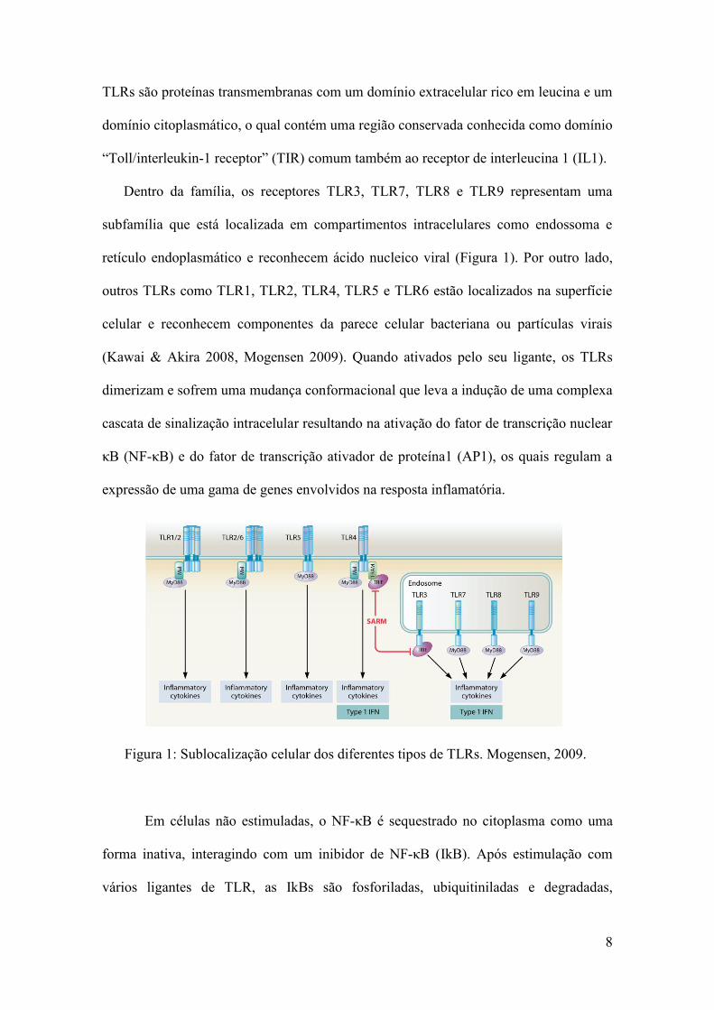
Dentro da família, os receptores TLR3, TLR7, TLR8 e TLR9 representam uma
subfamília que está localizada em compartimentos intracelulares como endossoma e
retículo endoplasmático e reconhecem ácido nucleico viral (Figura 1). Por outro lado,
outros TLRs como TLR1, TLR2, TLR4, TLR5 e TLR6 estão localizados na superfície
celular e reconhecem componentes da parece celular bacteriana ou partículas virais
(Kawai & Akira 2008, Mogensen 2009). Quando ativados pelo seu ligante, os TLRs
dimerizam e sofrem uma mudança conformacional que leva a indução de uma complexa
cascata de sinalização intracelular resultando na ativação do fator de transcrição nuclear
κB (NF-κB) e do fator de transcrição ativador de proteína1 (AP1), os quais regulam a
expressão de uma gama de genes envolvidos na resposta inflamatória.
Em células não estimuladas, o NF-κB é sequestrado no citoplasma como uma
forma inativa, interagindo com um inibidor de NF-κB (IkB). Após estimulação com
vários ligantes de TLR, as IkBs são fosforiladas, ubiquitiniladas e degradadas,
Figura 1: Sublocalização celular dos diferentes tipos de TLRs. Mogensen, 2009.

9
permitindo que o NF-κB seja translocado para o núcleo ativando a transcrição gênica.
A ativação do fator de transcrição AP-1 na sinalização por TLR é principalmente
mediada por cinases ativadas por mitógenos (MAPK) como a cinase terminal Jun-N
(JNK), p38 e cinase regulada por sinal extracelular (ERK).
Em resumo, a ativação da maior parte dos receptores TLRs resulta no recrutamento
da proteína adaptadora fator 88 de diferenciação mieloide (MyD88), cinase serina-
treonina associada IL-1R (IRAK), fator associado ao receptor do fator de necrose
tumoral (TRAF), proteína adaptadora TRAF6, cinase indutora de NF-κB (NIK) e cinase
IκB (IKK), portanto levando a translocação nuclear do NF-κB. O MyD88 é crítico para
a sinalização através de todos os TLRs exceto TLR3. Entretanto, moléculas adaptadoras
alternativas incluindo proteína associada ao TIR (TIRAP) e a proteína semelhante a
MYD88 (MAL), transduzem sinais de TLRs através de uma via independente de
MyD88 (Akira et al. 2006). A via de sinalização resulta na regulação positiva de genes
proinflamatórios alvos que codificam citocinas, quimiocinas, enzimas e outras
moléculas essenciais para a eliminação do patógeno (Figura 2).
No SNC, astrócitos e microglia são as primeiras células a responder através de
receptores TLRs a dano, perturbação e estresse celular, bem como infecções causadas
por patógenos, induzindo a produção e liberação de sinais moleculares que iniciam a
resposta glial podendo levar a excitoxicidade, inflamação, neurodegeneração e apoptose
(Holm et al. 2012).

10
1.3. Mecanismos celulares da neuroinflamação
A neuroglia, ou simplesmente glia, que compreende além de astrócitos, microglia e
oligodendrócitos, foi vista como tendo uma única função de proporcionar orientação,
suporte estrutural e servir como fonte de nutrientes e isolamento para os neurônios (Van
Eldik & Wainwright 2003), pois nenhum de seus tipos celulares apresentam axônio e
nem a habilidade de formar potenciais de ação (Wang & Bordey 2008). Contudo, nas
últimas décadas, novas pesquisas demonstraram que os astrócitos não são somente
suportes passivos da arquitetura e homeostase neuronal sem papel na integração de
Figura 2: Via de Sinalização ativada por TLR4. Mogensen 2009.

11
sinal, mas sim participantes ativos com neurônios em todas as funções essenciais do
encéfalo (Sofroniew & Vinters 2010).
A imunidade inata é um componente constitutivo do SNC e depende fortemente de
células residentes como a microglia, entretanto, estudos mostram que os astrócitos
participam da resposta imunológica, desencadeada por uma variedade de insultos. Deste
modo, os astrócitos juntamente com a microglia apresentam um papel fundamental no
processo neuroinflamatório e são reconhecidos como participantes ativos em várias
condições patológicas ou doenças neurodegenerativas crônicas (Liu et al. 2011).
1.3.1. Microglia
A microglia é um fagócito mononuclear, derivado de progenitores da linhagem
mielóide e está envolvida na resposta imune e inflamatória (Ransohoff & Cardona
2010). Estas células, vistas como macrófagos residentes do SNC, são derivadas do
saco vitelino durante a embriogênese e colonizam o cérebro em desenvolvimento
onde persistem ao longo da vida do indivíduo (Perry & Holmes 2014).
As células microgliais constituem cerca de 10% do total de células do SNC de um
humano adulto com considerável heterogeneidade na sua densidade
entre as regiões desse tecido. No SNC saudável, a microglia apresenta uma morfologia
complexa composta de processos altamente ramificados que se estendem de um corpo
celular compacto e que continuamente exploram o ambiente entrando em contato com
sinapses e regiões extra-sinápticas e também com astrócitos (Tremblay et al. 2010). O
microambiente do SNC tem um efeito importante no fenótipo microglial. Vários
ligantes expressos em neurônios, astrócitos e oligodendrócitos ligam-se em receptores
expressos na microglia, sinalizando através destes receptores a inibição da ativação
microglial (Ransohoff & Perry 2009).

12
Na presença de um insulto, a microglia torna-se ativada e responde em poucos
minutos a áreas muito restritas de dano tão pequenas como um único processo neuronal.
Nesta condição esta célula retrai seus processos adquirindo uma forma mais
amebóide, sendo capaz de migrar para o local do dano, proliferar, participar na
apresentação de antígenos, fagocitar debris celulares e eliminar sinapses anormais
(Tremblay et al. 2010). A microglia expressa, entre outros receptores, os TLRs de 1
ao 9 e, quando ativada, libera uma variedade de citocinas, incluindo fator de
necrose tumoral alfa (TNF-α), IL-1β, IL-6, IL-10, interferon-α (IFN-α), IFN-β,
além de prostanóides, componentes do complemento e substâncias neurotóxicas
como espécies reativas de oxigênio (EROs) e NO, sendo uma fonte importante de
fatores proinflamatórios cerebrais.
1.3.2. Astrócitos
Astrócitos são células gliais especializadas, abundantes no SNC, com características
fenotípicas e de citoarquitetura únicas que idealmente os posicionam a sentir e
responder a mudanças no seu microambiente. Eles estendem numerosos processos
formando domínios anatômicos altamente organizados com pouca sobreposição entre
células adjacentes e são interconectados em redes funcionais através de junções gap
(Kirchhoff et al. 2001). Alguns processos astrocíticos os quais expressam uma ampla
gama de receptores e canais iônicos circundam as sinapses, enquanto outros estão em
contato próximo com vasos sanguíneos intraparenquimais através de processos
especializados chamados pés terminais. Deste modo, os astrócitos apresentam um papel
importante no acoplamento neurometabólico e neurovascular (Allaman et al. 2011). Os
astrócitos apresentam uma morfologia heterogênea sendo dividida em duas
subpopulações baseadas em sua localização e morfologia: astrócitos fibrosos, que são

13
encontrados na substancia branca e apresentam processos não ramificados, finos e
longos envolvendo os nodos de Ranvier e astrócitos protoplasmáticos, os quais se
localizam na substância cinzenta e possuem muitos processos ramificados que
envolvem as sinapses e os pés terminais que envolvem os vasos sanguíneos (Wang &
Bordey 2008). Entre algumas funções astrocíticas estão: promoção da maturação
neuronal, formação das sinapses (Christopherson et al. 2005), regulação da angiogênese
(Laterra & Goldstein 1991), indução e manutenção da barreira hemato-encefálica
(Abbott et al. 2006), tamponamento de íons do meio extracelular como o potássio,
metabolismo e captação de neurotransmissores (GABA e glutamato) (Schousboe et al.
1992, Allaman et al. 2011) suporte metabólico (Magistretti 2009), detoxificação,
protegendo neurônios contra excitotoxicidade, e manutenção do microambiente viável
para neurônios (Wang & Bordey 2008).
Um dos papeis mais importantes dos astrócitos no SNC é a vigilância imunológica,
respondendo a lesões, patógenos ou outros insultos ativando uma resposta inflamatória
crítica para a defesa ao hospedeiro (Van Eldik & Wainwright 2003). Essas células
formam uma glia limitante ao redor dos vasos sanguíneos restringindo o acesso de
células imunes do sangue para o parênquima cerebral, e ao contrário de outras células
do SNC, são resistentes à morte por apoptose induzida por receptor, mostrando-se bem
equipados para sobreviver a insultos inflamatórios (Farina et al. 2007). Semelhante às
células microgliais, os astrócitos tornam-se ativados em resposta a vários estímulos, de
mudanças sutis no microambiente a dano tecidual intenso. Este processo é conhecido
como astrogliose reativa, o qual é uma resposta não homogênea e finamente graduada
que varia de acordo com o tipo, gravidade e duração do insulto (Sofroniew & Vinters
2010).
14
1.4. Mecanismos moleculares da neuroinflamação em astrócitos
Histopatologicamente os astrócitos reativos apresentam mudanças como
proliferação e sobreposição de domínios astrocíticos, podendo resultar na formação de
cicatriz astroglial, além de hipertrofia e aumento na expressão da proteína ácida fibrilar
glial (GFAP) (Pekny & Pekna 2004). A GFAP é a uma proteína de aproximadamente 50
KDa, insolúvel, regulada por fosforilação e considerada a principal proteína de
filamentos intermediários em astrócitos maduros e também um importante componente
do citoesqueleto durante o desenvolvimento destas células. Estudos realizados em ratos
transgênicos indicaram que a expressão de GFAP não é essencial para a aparência e
função normal da maioria dos astrócitos no SNC saudável, mas é essencial para o
processo de astrogliose reativa e formação de “cicatriz” glial. Além disso, o aumento no
imunoconteúdo de GFAP, independentemente de haver ou não proliferação astroglial, é
comumente usado como medida de astrogliose. Estudos mostram que a GFAP não está
presente em todo o citoplasma astrocítico e a imunoistoquímica para GFAP não marca
todas as partes da célula, mas somente as principais ramificações, estando ausente nos
processos finamente ramificados e frequentemente não detectável no corpo celular
(Sofroniew & Vinters 2010). Estudos in vivo e in vitro mostram outras funções da
GFAP, como migração celular, funcionamento adequado da BHE, vias de transdução de
sinal e interações neurônio-glia (Middeldorp & Hol 2011).
Os astrócitos expressam vários tipos de PRRs incluindo os recepetores TLRs (TLRs
2, 3, 4, 5 e 9) (Farina et al. 2007, Bowman et al. 2003). Através da ligação a estes
receptores, os astrócitos respondem a insultos como, por exemplo, LPS tanto in vivo
como in vitro com aumento da secreção de citocinas proinflammatórias como IL-6,
TNF-α, IL-12, citocinas anti-inflamatórias como IL-10, fatores neurotróficos como fator
de transformação do crescimento beta (TGF-β), enzimas como oxido nítrico sintase

15
induzível (iNOS), quimiocinas como CCL2, CCL3, CCL4, CCL5, a proteína molecular
de adesão intercelular 1 (ICAM 1) e proteína de adesão celular vascular 1 (VCAM 1)
podendo ativar e atrair a infiltração de células imunes como leucócitos, células T e
monócitos da periferia para o SNC (Carpentier et al. 2005). Também foi visto que
ligantes de TLRs em cultura de astrócitos são mais potentes indutores de funções da
imunidade inata, enquanto citocinas como INF-γ e TNF-α são indutores mais potentes
de funções da imunidade adaptativa como, por exemplo, moléculas do complexo
principal de histocompatibilidade de classe II (Carpentier et al. 2005).
Entre as células neurais, os astrócitos são as células mais resistentes ao estresse
oxidativo e proporcionam proteção aos neurônios principalmente devido a seu alto
conteúdo de glutationa (GSH), um tripeptídeo formado por glutamato, cisteína e glicina
e também considerado o principal antioxidante cerebral (Aoyama et al. 2008). Além
disso, a GSH é secretada por astrócitos e, quando clivada no dipeptídeo cisteína-glicina
no espaço extracelular, serve de substrato para síntese da GSH pelos neurônios
(Dringen et al. 2000). A habilidade para reduzir ou sintetizar GSH é um importante
fator que determina como se encontra o estado redox celular, visto que o estresse
oxidativo tem sido associado com o desenvolvimento de condições patológicas como as
doenças neuroinflamatórias.
Devido a sua função na resposta imunológica no SNC, nos últimos anos, os
astrócitos têm sido amplamente citados pela sua contribuição no início e progressão da
maioria das doenças neurodegenerativas (Parpura et al. 2012). Um importante marcador
de ativação astroglial e ou morte em várias condições de dano cerebral é a proteína
S100B.
A proteína S100B é um membro da família de proteínas S100 assim denominadas
por serem solúveis em uma solução 100% saturada de sulfato de amônio (Moore 1965).
16
Essas proteínas são ligantes de cálcio do tipo EF hand e compreendem 25 membros,
expressos de maneira específica para cada tipo celular. Como a maioria dos outros
membros da família S100, a S100B têm uma estrutura homodimérica onde cada
monômero beta é de aproximadamente 10.5 kDa, tendo dois sítios de ligação ao cálcio
do tipo EF hand e sítios independentes para ligação ao zinco, e também duas pontes
dissulfeto, mas a estrutura dimérica é mantida independente deste aspecto (Donato
2001). A S100B é expressa em um restrito número de tipos celulares como: astrócitos,
linfócitos, oligodendrócitos maduros, células progenitoras neurais, certas populações
neuronais, adipócitos entre outras, e sua expressão também está aumentada em vários
tumores, no cérebro de idosos, no cérebro de pacientes afetados pela doença de
Alzheimer, esquizofrenia, epilepsia crônica, infecção por HIV entre outras condições
patológicas cerebrais (Donato et al. 2009). O gene humano que codifica a S100B está
no cromossomo 21q22.3 com consequente super expressão da proteína na síndrome de
Down (Mrak & Griffin 2004).
A S100B no SNC é principalmente expressa e predominantemente secretada por
astrócitos constitutivamente e em resposta a estímulos com vários agentes (Pinto et al.
2000), incluindo o fator de necrose tumoral (TNF-α) (Edwards & Robinson 2006) e
IL1β, envolvendo a via das MAPK e sinalização via NF-κB (de Souza et al. 2009). A
S100B possui efeitos parácrinos e autócrinos nas células gliais, neurônios e microglia.
Esta proteína tem muitos alvos intracelulares estando envolvida na regulação de
proteínas do citoesqueleto (GFAP, por exemplo) (Frizzo et al. 2004), moduladores do
ciclo celular (p53) (Lin et al. 2001) e a proteína fosfatase calcineurina (Leal et al. 2004).
Além de alvos intracelulares, a S100B apresenta efeitos extracelulares observados em
culturas neuronais dependendo de sua concentração, uma vez que em concentrações
picomolar e nanomolar apresenta efeito trófico, enquanto em concentrações
17
micromolar, efeito apoptótico. Estes efeitos envolvem ativação via receptor RAGE de
vias de sinalização como ERK e NF-κB (Donato et al. 2009). Um aspecto importante a
ser considerado é que expressão genética, a expressão protéica e a secreção da proteína
S100B podem ser eventos independentes (Swarowsky et al. 2008). Estudos em culturas
de astrócitos de ratos indicam que variações da S100B extracelular não são
necessariamente acompanhadas por mudanças no conteúdo intracelular desta proteína
como visto com altos níveis de amônia a qual aumentou a secreção de S100B sem
alterar seu conteúdo intracelular (Leite et al. 2006). Por outro lado, cultura de astrócitos
em meio com altos níveis de glicose mostrou uma diminuição na secreção e no
conteúdo intracelular de S100B (Nardin et al. 2007).
A S100B tem sido vista como um útil marcador molecular e periférico de dano
cerebral (Gonçalves et al. 2008) em várias situações agudas ou crônicas. Um dos
motivos para este conceito vem do fato da S100B estar implicada no ciclo das citocinas,
uma teoria proposta para a doença de Alzheimer (Griffin 2006). Em situações que
levem à injúria no SNC como, por exemplo, um processo inflamatório, astrócitos e
microglia, estão ativados. A microglia ativada superexpressa a citocina IL-1ß, a qual
ativa os astrócitos, levando a um aumento da expressão de S100B por estas células
(Sheng et al. 1996). Estudos mostram que a ligação da S100B ao receptor RAGE na
linhagem microglial BV-2 resulta na ativação dos fatores de transcrição NF-κB e AP-1
os quais cooperam para estimular IL-1ß, TNF-α e a enzima ciclooxigenase-2 (COX-2),
contribuindo assim para a neuroinflamação (Bianchi et al. 2008).
A enzima COX-2 é a enzima limitante na síntese das prostaglandinas mediadoras da
inflamação como a prostaglandina E2 (PGE2). Sua expressão no cérebro é altamente
regulada em condições fisiológicas, e tem sido mostrado que sua expressão é dividida
em duas fases: precocemente em neurônios e mais tarde em células não neuronais, como

18
astrócitos (Takemiya et al. 2007). Estudos mostram que culturas de astrócitos de ratos
tratadas com LPS aumentam a produção de prostaglandinas via ativação de COX-2 com
um evidente envolvimento de NF-κB (Pistritto et al. 1999).
Vários estudos têm considerado a elevação nos níveis de S100B como um
componente de resposta neuroinflamatória, particularmente em doenças
neurodegenerativas como a doença de Alzheimer, porém é muito difícil a comparação
dos níveis de S100B em cultura de células com aqueles encontrados no líquor ou soro,
uma vez que estes níveis refletem várias possíveis fontes de S100B. No entanto estudos
recentes mostram que a secreção de S100B pode se modulada por citocinas
proinflamatórias como IL6 (de Souza et al. 2013) e IL1β (de Souza et al. 2009)
provavelmente através da via da MAPK.
1.5. LPS como um modelo de neuroinflamação
O LPS é uma molécula de lipopolissacarídeo que constitui a bicamada lipídica de
bactérias gram-negativas, como por exemplo, a bactéria Escherichia coli, sendo crítico
na estabilidade da membrana. Sua molécula é composta por três domínios distintos:
uma cadeia lateral de polissacarídeo (Antígeno O) ligada à região do “core”, um
oligossacarídeo (2-ceto-3-ácido deoxioctônico), que está ligado a uma molécula lipídica
(Lipídeo A). O antígeno O varia entre as espécies de bactérias gram-negativas tanto em
composição como em comprimento, enquanto o core e o lipídeo A são mais
conservados entre as diferentes espécies destas bactérias. O domínio lipídeo A é o
componente bioativo e tóxico da endotoxina reconhecido durante a infecção humana
(Leon et al. 2008).

19
O LPS é uma molécula essencial, uma vez que sua presença é crítica na estabilidade
da membrana bacteriana e tem um papel proeminente na intensificação da resposta
imunológica (Janeway & Medzhitov 2002); ele tem sido implicado como a molécula
responsável por uma variedade de patologias que vão de leves como febre a letais como
choque séptico e morte. Esta molécula desencadeia a liberação de muitas citocinas
como TNF-α, IL-1ß e IL-6 e é provavelmente o estímulo mais frequentemente usado
para desencadear vias de sinalização envolvidas na resposta imunológica (Glezer et al.
2007).
A via de sinalização intracelular desencadeada por LPS envolve primeiramente sua
ligação ao receptor TLR4 (Figura 3). O LPS se liga com a proteína ligante de LPS
(LBP) e também à proteína CD14 ancorada na membrana da célula alvo, a qual
transfere o LPS para o receptor TLR4 de uma maneira dependente da glicoproteína
extracelular MD-2; através do domínio TIR, ocorre o recrutamento do MyD88 que se
associa com a proteína cinase IRAK4 que interage com IRAK, a qual ativa o TRAF6
seguindo da ativação do NF-κB e do AP-1 (Glezer et al. 2007).
Embora muitos estudos usem o LPS como modelo de sepse (Takaoka et al. 2014,
Kaplanski et al. 2014, Mihaylova et al. 2014), a qual é caracterizada por uma resposta
inflamatória sistêmica desencadeada por fatores infecciosos (Salomao et al. 2012), o
LPS tem sido o ativador glial mais extensivamente utilizado por pesquisadores em
modelos de neuroinflamação in vivo e in vitro. Estudos in vitro mostram que cultura de
astrócitos neonatais de diferentes regiões cerebrais exibe diferenças na expressão de
fatores pro-inflamatórios sob estímulo com LPS (Kipp et al. 2008), assim como em
outro estudo usando o mesmo modelo in vitro mostrou que o LPS induz uma regulação
positiva de enzimas responsáveis pela síntese de PGE2 e uma regulação negativa de
enzimas que catalisam a sua degradação (Johann et al. 2008).

20
Um estudo in vivo usando injeção intracerebroventricular de LPS para mimetizar um
quadro de inflamação cerebral em camundongos, mostrou ocorrer um recrutamento e
adesão de leucócitos a microvasculatura cerebral e que este recrutamento é mediado por
células que apresentam a via de sinalização TLR4/CD14 como a microglia, e que a
sinalização por TNF-α é critica para o recrutamento de leucócitos (Zhou et al. 2006).
Entretanto ainda não há estudos mostrando o efeito desta molécula sobre a secreção
e o imunoconteúdo da proteína S100B, uma vez que esta também tem sido mostrada
uma peça importante no processo inflamatório.
Figura 3: Sinalização por LPS

21
1.6. Anti-inflamatórios e neuroinflamação
Uma vez que a neuroinflamação tem sido implicada na patogênese ou progressão de
uma variedade de doenças neurodegenerativas, nos últimos anos vem crescendo o
interesse nos efeitos benéficos de compostos anti-inflamatórios não esteroidais (AINEs)
na neurodegeneração particularmente na doença de Alzheimer (McGeer & McGeer
2007, Vlad et al. 2008), doença de Parkinson (Wahner et al. 2007) e esquizofrenia
(Laan et al. 2009).
O principal mecanismo de ação dos AINEs é inibir a ação da enzima COX também
conhecida como prostaglandina G/H sintase. A COX tem um papel crucial no processo
inflamatório. Esta é uma enzima de aproximadamente 70 kDa, bifuncional e associada à
membrana que age sobre o ácido araquidônico, o qual é um ácido graxo insaturado de
20 carbonos que sobre uma variedade de estímulos é liberado da membrana
fosfolipídica celular sobre ação da enzima fosfolipase A2 (PLA2). A COX possui duas
atividades enzimáticas que ocorrem em dois sítios catalíticos distintos (Vane et al.
1998, Simmons et al. 2004), catalisando duas reações sequenciais: atividade da
ciclooxigenase que catalisa a oxidação e ciclização do ácido araquidônico até o
hidroperóxido PGG2 e atividade de peroxidase que catalisa a peroxidação da PGG2 até o
hidroperóxido prostaglandina H2 (PGH2), a qual é convertida por várias sintases e
isomerases específicas celulares produzindo cinco prostanóides biologicamente ativos:
PGD2, PGE2, PGF2 α, prostaciclina (PGI2) e tromboxano A2 (TXA2). Estes produtos
agem como mensageiros interagindo com receptores prostanóides acoplados a proteína
G além de outros receptores (Rao & Knaus 2008).
Existem duas isoformas da enzima COX: COX1 e COX2. Estas apresentam alta
similaridade na cinética, mecanismo catalítico e na estrutura do sítio ativo, mas

22
apresentam uma diferença estrutural crucial neste último ítem que é a substituição da
isoleucina 523 na COX1 por uma valina 523 na COX2, o que torna o sítio da COX2
maior, essa diferença é importante na seleção do tipo de droga que poderá se ligar a
cada isoforma. Além disso, elas apresentam diferentes reatividades a substratos
lipídicos. A COX1 apresenta alosterismo negativo em baixas concentrações de ácido
araquidônico, sendo a COX2 capaz de se ligar mais facilmente ao ácido araquidônico do
que a COX1 quando expressas na mesma célula (Smith et al. 2000).
A enzima COX1 é expressa na maioria dos tecidos e tipos celulares e é
constitutivamente ativa, tendo um papel crucial na manutenção e homeostase da
fisiologia celular em geral, através da síntese de precursores prostanóides (Dubois et al.
1998). A COX1 apresenta uma função importante nas plaquetas, onde é responsável por
produzir precursores para a síntese de tromboxanos, responsáveis pela agregação
plaquetária. Outra importante ação desta isoforma é na proteção da mucosa gástrica,
onde produz PGE2 responsável pela produção de muco e secreção de bicarbonato e
também prostaciclina capaz de alterar o fluxo sanguíneo na microcirculação da mucosa
gástrica (Miller 2006). No SNC esta isoforma é expressa predominantemente em células
microgliais e imediatamente ativa a síntese de prostaglandinas em resposta à ativação
microglial, sendo importante na modulação da fase inicial da resposta inflamatória (Aid
& Bosetti 2011). Tem sido mostrado que a deleção genética ou farmacológica da COX1
atenua a ativação microglial, bem como, alterações na BHE em resposta ao LPS (Choi
et al. 2008). No entanto o mecanismo exato pela qual a COX1 está envolvida na
neuroinflamação não está claro.
A COX2 é a isoforma induzível e inflamatória, rapidamente expressa em vários
tipos celulares em resposta a uma variedade de estímulos como, por exemplo, estímulos
pró-inflamatórios. No SNC a COX2 é constitutivamente expressa em astrócitos,
23
microglia, células endoteliais e principalmente em neurônios glutamatérgicos corticais e
hipocampais, onde tem um papel chave na atividade sináptica, na plasticidade sináptica
de longa duração (Yang & Chen 2008) e acoplamento neurovascular durante a
hiperemia funcional, mas seu papel nas doenças cerebrais não está bem estabelecido.
Enquanto alguns estudos mostram que sua inibição proporciona efeitos benéficos contra
dano isquêmico e morte neuronal (Candelario-Jalil & Fiebich 2008) ao contrário em
doenças neurodegenerativas como a doença de Alzheimer, inibidores da COX2 podem
não ser sempre protetores (McGeer & McGeer 2007). Em cultura de astrócitos, o LPS
induz fortemente a expressão de COX2 e a produção de PGE2 mediado pela via de NF-
κB dependente de MYD88 e pela via das MAPKs além de também regular
negativamente a expressão de COX1. Além disso, células deficientes em COX1
produzem mais PGE2 do que células controle indicando algum efeito negativo de COX1
na produção de PGE2 dependente de COX2 em astrócitos na presença de LPS (Font-
Nieves et al. 2012).
1.6.1. Uso de anti-inflamatórios para patologias do SNC
A inflamação é uma reação de autodefesa com o objetivo de eliminar ou neutralizar
estímulos prejudiciais e restaurar a integridade tecidual. Nas doenças
neurodegenerativas a inflamação ocorre como uma resposta local conduzida
primariamente por células microgliais na ausência de infiltração de leucócitos para o
parênquima cerebral. Assim como a inflamação periférica, a neuroinflamação torna-se
um processo nocivo, sendo amplamente aceito que deva contribuir para a patogênese de
muitas doenças do SNC incluindo as doenças neurodegenerativas crônicas.

24
O uso de anti-inflamatórios para a doença de Alzheimer (McGeer & Rogers 1992),
foi primeiro proposto nos início dos anos 90. A doença de Alzheimer é a mais comum
causa de demência relacionada à idade e uma das mais bem caracterizadas doenças
neurodegenerativas crônica. Duas características da doença são a formação de placas
senis, que são depósitos extracelulares de peptídeo β-amiloide e emaranhados
neurofibrilares, que consistem de agregados intracelulares de proteína tau hiper
fosforilada nos neurônios. Em torno das placas senis, a microglia se torna ativada com
consequente aumento de proteínas do complemento, citocinas, quimiocinas e radicais
livres, fazendo com que a neuroinflamação se torne um ciclo vicioso, no qual vários
fatores como agregados proteicos e componentes celulares ou sinapses anormais ativem
mais a microglia, que por sua vez exacerba o depósito β-amiloide e o dano neuronal
(Mrak & Griffin 2005). Desde então, vários estudos tem sido realizados para testar esta
hipótese (Akiyama et al. 2000, Kotilinek et al. 2008, Heneka et al. 2005, Morihara et al.
2005),
Estudos em cultura de células astrogliais mostram que a aspirina é capaz de reduzir
níveis proteicos e de mRNA de GFAP, sendo o NF-κB o principal alvo da aspirina nesta
regulação (Bae et al. 2006). Foi mostrado que aspirina bloqueia NF-κB devido á
inibição específica da cinase IKK-β (Grilli et al. 1996, Yin et al. 1998). Outro estudo
mostra que a aspirina é capaz de atenuar a produção de metabólitos do ácido
araquidônico em um modelo de neuroinflamação induzido por LPS em ratos (Basselin
et al. 2011). Em um modelo de cultura neuronal para a doença de
Parkinson, aspirina parece aumentar a sobrevivência de neurônios dopaminérgicos
(Carrasco & Werner 2002) e igualmente ao ibuprofeno protege neurônios
dopaminérgicos da toxicicidade por glutamato, um importante fator implicado em
doenças neurodegenerativas, em cultura de mesencéfalo de rato (Casper et al. 2000).

25
Em um estudo no qual neurônios corticais de ratos em cultura expostos a privação de
glicose e oxigênio, a aspirina não foi capaz de alterar a captação de glutamato, mas sim
inibir a liberação deste neurotransmissor via recuperação dos níveis de ATP (De
Cristobal et al. 2002).
Estudos em cultura primária de astrócitos estimulada com citocinas pró-
inflamatórias como um modelo de ativação astrocítica mostram que o diclofenaco é
capaz de aumentar a produção de NO (Kakita et al. 2009), e a expressão da proteína
aquaporina 4 (Asai et al. 2013) através da sinalização por NK-κB, além de aumentar a
atividade fagocítica de culturas microglias via produção de NO (Kakita et al. 2013). Em
um modelo de doença de Parkinson induzido por rotenona em ratos, o uso de
ibuprofeno foi capaz de apresentar efeitos antidepressivo e antioxidante, associado ao
aumento dos níveis de GSH (Zaminelli et al. 2014) e também da inibição da NOX2,
uma enzima envolvida na geração de EROs pertencente à família NOX de NADPH
oxidases, em um modelo de doença de Alzheimer (Wilkinson et al. 2012). Além disso,
ele foi capaz de amenizar a astrogliose através da diminuição de GFAP em um modelo
de demência com camundongos trangênicos expressando corpos de Lewy (Sekiyama et
al. 2012). Em modelo de isquemia cerebral, o ibuprofeno proporcionou neuroproteção
ao dano neuronal excitotóxico em culturas corticais mistas de astrócitos e neurônios
(Iwata et al. 2010).
A Nimesulida, inibidor seletivo da COX2, confere proteção ao dano cerebral
isquêmico através de redução do dano oxidativo (Candelario-Jalil et al. 2003), além de
ser capaz de atenuar a expressão de GFAP e produção de mediadores proinflamatórios
em modelo de neuroinflamação utilizando LPS (Niranjan et al. 2010).
Em relação aos anti-inflamatórios esteroidais, a dexametasona parece ser capaz de
modular a sinalização de cálcio em astrócitos (Simard et al. 1999) e também pode

26
proteger neurônios da toxicidade por glutamato através da diminuição do cálcio
intracelular (Suwanjang et al. 2013). O uso de dexametasona em culturas microgliais na
presença de LPS diminui a produção de espécies reativas de oxigênio via regulação
positiva de MKP-1 (Huo et al. 2011). Em co-cultura de astrócito e microglia, a
dexametasona foi capaz de aumentar a comunicação celular por junção gap,
aumentando a expressão da proteína conexina 43 (Hinkerohe et al. 2011). Em um
modelo de meningite bacteriano de co-cultura com LPS, dexametasona foi capaz de
diminuir a ativação microglial e aumentar conexina 43 (Hinkerohe et al. 2010).
27
2. Objetivos
2.1. Objetivo geral
O objetivo deste trabalho foi investigar o efeito do LPS e de anti-inflamatórios
sobre parâmetros gliais envolvidos na neuroinflamação na presença e ausência
de LPS.
2.2. Objetivos Específicos
2.2.1. Avaliar o efeito da exposição a diferentes concentrações de LPS sobre a
secreção de S100B e o conteúdo de GSH em culturas primárias de
astrócitos, bem como em fatias frescas hipocampais de ratos Wistar.
2.2.2. Avaliar o efeito da exposição a diferentes concentrações de LPS sobre o
conteúdo de GFAP e S100B e sobre a secreção de TNF-α em culturas
primárias de astrócitos.
2.2.3. Avaliar o efeito da administração central (injeção ICV) ou periférica
(injeção IP) de LPS sobre as concentrações de S100B e TNF-α no soro e
líquor de ratos Wistar.
2.2.4. Avaliar o efeito de anti-inflamatórios esteroidais e não esteroidais sobre
diferentes parâmetros gliais (imunoconteúdo e secreção de S100B,
conteúdo de GSH, imunoconteúdo de GFAP e secreção de TNF-α) em
culturas primárias de astrócitos.
2.2.5. Avaliar o efeito de anti-inflamatórios esteroidais e não esteroidais na
presença de LPS sobre diferentes parâmetros gliais (imunoconteúdo e
secreção de S100B, conteúdo de GSH, imunoconteúdo de GFAP e secreção
de TNF-α) em culturas primárias de astrócitos.

28
PARTE II
29
Capítulo I
Lipopolysaccharide modulates astrocytic S100B secretion: a study in cerebrospinal fluid
and astrocyte cultures from rats
Artigo publicado no periódico “Journal of Neuroinflammation”.
RESEARCH Open Access
Lipopolysaccharide modulates astrocytic S100Bsecretion: a study in cerebrospinal fluid andastrocyte cultures from ratsMaria Cristina Guerra†, Lucas S Tortorelli†, Fabiana Galland, Carollina Da Ré, Elisa Negri, Douglas S Engelke,Letícia Rodrigues, Marina C Leite* and Carlos-Alberto Gonçalves
Abstract
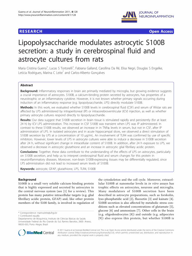
Background: Inflammatory responses in brain are primarily mediated by microglia, but growing evidence suggestsa crucial importance of astrocytes. S100B, a calcium-binding protein secreted by astrocytes, has properties of aneurotrophic or an inflammatory cytokine. However, it is not known whether primary signals occurring duringinduction of an inflammatory response (e.g. lipopolysaccharide, LPS) directly modulate S100B.
Methods: In this work, we evaluated whether S100B levels in cerebrospinal fluid (CSF) and serum of Wistar rats areaffected by LPS administered by intraperitoneal (IP) or intracerebroventricular (ICV) injection, as well as whetherprimary astrocyte cultures respond directly to lipopolysaccharide.
Results: Our data suggest that S100B secretion in brain tissue is stimulated rapidly and persistently (for at least24 h) by ICV LPS administration. This increase in CSF S100B was transient when LPS was IP administered. Incontrast to these S100B results, we observed an increase in in TNFa levels in serum, but not in CSF, after IPadministration of LPS. In isolated astrocytes and in acute hippocampal slices, we observed a direct stimulation ofS100B secretion by LPS at a concentration of 10 μg/mL. An involvement of TLR4 was confirmed by use of specificinhibitors. However, lower levels of LPS in astrocyte cultures were able to induce a decrease in S100B secretionafter 24 h, without significant change in intracellular content of S100B. In addition, after 24 h exposure to LPS, weobserved a decrease in astrocytic glutathione and an increase in astrocytic glial fibrillary acidic protein.
Conclusions: Together, these data contribute to the understanding of the effects of LPS on astrocytes, particularlyon S100B secretion, and help us to interpret cerebrospinal fluid and serum changes for this protein inneuroinflammatory diseases. Moreover, non-brain S100B-expressing tissues may be differentially regulated, sinceLPS administration did not lead to increased serum levels of S100B.
Keywords: astrocyte, GFAP, glutathione, LPS, TLR4, S100B
BackgroundS100B is a small very soluble calcium-binding proteinthat is highly expressed and secreted by astrocytes inthe central nervous system (see [1] for a review). Thisprotein has many putative intracellular targets (e.g. glialfibrillary acidic protein, GFAP) and, like other proteinmembers of the S100 family, is involved in regulation of
the cytoskeleton and the cell cycle. Moreover, extracel-lular S100B at nanomolar levels in in vitro assays hastrophic effects on astrocytes, neurons and microglia.Many modulators of S100B secretion have beendescribed in astrocyte preparations, such as forskolin,lyso-phosphatidic acid [2], fluoxetin [3] and kainate [4].S100B secretion is also affected by metabolic stress con-ditions such as elevated concentrations of glutamate [5],glucose [6] and ammonium [7]. Other cells in the brain(e.g. oligodendrocytes [8]) and outside (e.g. adipocytes[9]) also express this protein, but whether S100B is
* Correspondence: [email protected]† Contributed equallyDepartamento de Bioquímica, Instituto de Ciências Básicas da Saúde,Universidade Federal do Rio Grande do Sul, Ramiro Barcelos, 2600- Anexo,90035-003, Porto Alegre, Brazil
Guerra et al. Journal of Neuroinflammation 2011, 8:128http://www.jneuroinflammation.com/content/8/1/128
JOURNAL OF NEUROINFLAMMATION
© 2011 Guerra et al; licensee BioMed Central Ltd. This is an Open Access article distributed under the terms of the Creative CommonsAttribution License (http://creativecommons.org/licenses/by/2.0), which permits unrestricted use, distribution, and reproduction inany medium, provided the original work is properly cited.
Ina
Texto digitado
30
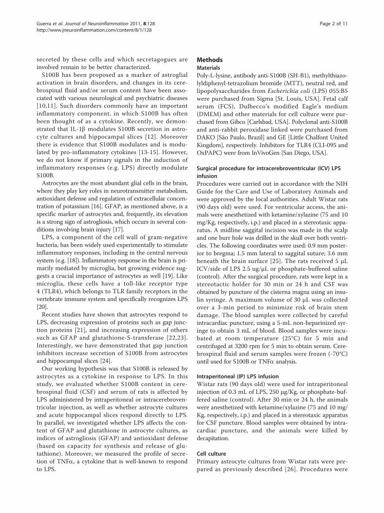
secreted by these cells and which secretagogues areinvolved remain to be better characterized.S100B has been proposed as a marker of astroglial
activation in brain disorders, and changes in its cere-brospinal fluid and/or serum content have been asso-ciated with various neurological and psychiatric diseases[10,11]. Such disorders commonly have an importantinflammatory component, in which S100B has oftenbeen thought of as a cytokine. Recently, we demon-strated that IL-1b modulates S100B secretion in astro-cyte cultures and hippocampal slices [12]. Moreoverthere is evidence that S100B modulates and is modu-lated by pro-inflammatory cytokines [13-15]. However,we do not know if primary signals in the induction ofinflammatory responses (e.g. LPS) directly modulateS100B.Astrocytes are the most abundant glial cells in the brain,
where they play key roles in neurotransmitter metabolism,antioxidant defense and regulation of extracellular concen-tration of potassium [16]. GFAP, as mentioned above, is aspecific marker of astrocytes and, frequently, its elevationis a strong sign of astrogliosis, which occurs in several con-ditions involving brain injury [17].LPS, a component of the cell wall of gram-negative
bacteria, has been widely used experimentally to stimulateinflammatory responses, including in the central nervoussystem (e.g. [18]). Inflammatory response in the brain is pri-marily mediated by microglia, but growing evidence sug-gests a crucial importance of astrocytes as well [19]. Likemicroglia, these cells have a toll-like receptor type4 (TLR4), which belongs to TLR family receptors in thevertebrate immune system and specifically recognizes LPS[20].Recent studies have shown that astrocytes respond to
LPS, decreasing expression of proteins such as gap junc-tion proteins [21], and increasing expression of otherssuch as GFAP and glutathione-S-transferase [22,23].Interestingly, we have demonstrated that gap junctioninhibitors increase secretion of S100B from astrocytesand hippocampal slices [24].Our working hypothesis was that S100B is released by
astrocytes as a cytokine in response to LPS. In thisstudy, we evaluated whether S100B content in cere-brospinal fluid (CSF) and serum of rats is affected byLPS administered by intraperitoneal or intracerebroven-tricular injection, as well as whether astrocyte culturesand acute hippocampal slices respond directly to LPS.In parallel, we investigated whether LPS affects the con-tent of GFAP and glutathione in astrocyte cultures, asindices of astrogliosis (GFAP) and antioxidant defense(based on capacity for synthesis and release of glu-tathione). Moreover, we measured the profile of secre-tion of TNFa, a cytokine that is well-known to respondto LPS.
MethodsMaterialsPoly-L-lysine, antibody anti-S100B (SH-B1), methylthiazo-lyldiphenyl-tetrazolium bromide (MTT), neutral red, andlipopolysaccharides from Escherichia coli (LPS) 055:B5were purchased from Sigma [St. Louis, USA]. Fetal calfserum (FCS), Dulbecco’s modified Eagle’s medium(DMEM) and other materials for cell culture were pur-chased from Gibco [Carlsbad, USA]. Polyclonal anti-S100Band anti-rabbit peroxidase linked were purchased fromDAKO [São Paulo, Brazil] and GE [Little Chalfont UnitedKingdom], respectively. Inhibitors for TLR4 (CLI-095 andOxPAPC) were from InVivoGen [San Diego, USA].
Surgical procedure for intracerebroventricular (ICV) LPSinfusionProcedures were carried out in accordance with the NIHGuide for the Care and Use of Laboratory Animals andwere approved by the local authorities. Adult Wistar rats(90 days old) were used. For ventricular access, the ani-mals were anesthetized with ketamine/xylazine (75 and 10mg/Kg, respectively, i.p.) and placed in a stereotaxic appa-ratus. A midline saggital incision was made in the scalpand one burr hole was drilled in the skull over both ventri-cles. The following coordinates were used: 0.9 mm poster-ior to bregma; 1.5 mm lateral to saggital suture; 3.6 mmbeneath the brain surface [25]. The rats received 5 μLICV/side of LPS 2.5 ug/μL or phosphate-buffered saline(control). After the surgical procedure, rats were kept in astereotactic holder for 30 min or 24 h and CSF wasobtained by puncture of the cisterna magna using an insu-lin syringe. A maximum volume of 30 μL was collectedover a 3-min period to minimize risk of brain stemdamage. The blood samples were collected by carefulintracardiac puncture, using a 5-mL non-heparinized syr-inge to obtain 3 mL of blood. Blood samples were incu-bated at room temperature (25°C) for 5 min andcentrifuged at 3200 rpm for 5 min to obtain serum. Cere-brospinal fluid and serum samples were frozen (-70°C)until used for S100B or TNFa analysis.
Intraperitoneal (IP) LPS infusionWistar rats (90 days old) were used for intraperitonealinjection of 0.3 mL of LPS, 250 μg/Kg, or phosphate-buf-fered saline (control). After 30 min or 24 h, the animalswere anesthetized with ketamine/xylazine (75 and 10 mg/Kg, respectively, i.p.) and placed in a stereotaxic apparatusfor CSF puncture. Blood samples were obtained by intra-cardiac puncture, and the animals were killed bydecapitation.
Cell culturePrimary astrocyte cultures from Wistar rats were pre-pared as previously described [26]. Procedures were
Guerra et al. Journal of Neuroinflammation 2011, 8:128http://www.jneuroinflammation.com/content/8/1/128
Page 2 of 11
Ina
Texto digitado
Ina
Texto digitado
Ina
Texto digitado
31

carried out in accordance with the NIH Guide for theCare and Use of Laboratory Animals and were approvedby the local authorities. Briefly, cerebral cortices of new-born Wistar rats (1-2 days old) were removed andmechanically dissociated in Ca2+- and Mg2+-freebalanced salt solution, pH 7.4, containing (in mM): 137NaCl; 5.36 KCl; 0.27 Na2HPO4; 1.1 KH2PO4 and 6.1 glu-cose. The cortices were cleaned of meninges andmechanically dissociated by sequential passage through aPasteur pipette. After centrifugation at 1400 RPM for5 min the pellet was resuspended in DMEM (pH 7.6)supplemented with 8.39 mM HEPES, 23.8 mM NaHCO3,0.1% amphotericin, 0.032% gentamicin and 10% fetal calfserum (FCS). Cultures were maintained in DMEM con-taining 10% FCS in 5% CO2/95% air at 37°C, allowed togrow to confluence, and used at 15 days in vitro.
Hippocampal slicesHippocampal slices were prepared as previouslydescribed [27]. Procedures were carried out in accor-dance with the NIH Guide for the Care and Use ofLaboratory Animals and were approved by the localauthorities. Thirty-day old Wistar rats were killed bydecapitation and the brains were removed and placed incold saline medium with the following composition (inmM): 120 NaCl; 2 KCl; 1 CaCl2; 1 MgSO4; 25 HEPES;1 KH2PO4, and 10 glucose, adjusted to pH 7.4 and pre-viously aerated with O2. The hippocampi were dissectedand transverse slices of 0.3 mm were obtained using aMcIlwain Tissue Chopper. Slices were then transferredimmediately into 24-well culture plates, each well con-taining 0.3 ml of physiological medium and only oneslice. The medium was changed every 15 min with freshsaline medium at room temperature (maintained at25°C). Following a 120-min equilibration period, themedium was removed and replaced with physiologicalsaline with or without LPS for 60 min at 30°C on awarm plate. Afterwards, media were collected and storedat -70°C until used for assay of S100B or TNFa.
S100B measurementS100B was measured by ELISA, as previously described[28]. Briefly, 50 μl of sample plus 50 μl of Tris bufferwere incubated for 2 h on a microtiter plate previouslycoated with monoclonal anti-S100B. Polyclonal anti-S100was incubated for 30 min and then peroxidase-conju-gated anti-rabbit antibody was added for a further30 min. Color reaction with o-phenylenediamine wasmeasured at 492 nm. The standard S100B curve rangedfrom 0.002 to 1 ng/ml.
GFAP measurementELISA for GFAP was carried out, as previously described[29], by coating microtiter plates with 100 μL samples for
24 h at 4°C. Incubation with a polyclonal anti-GFAPfrom rabbit for 1 h was followed by incubation with asecondary antibody conjugated with peroxidase for 1 h,at room temperature. A colorimetric reaction with o-phe-nylenediamine was measured at 492 nm. The standardhuman GFAP (from Calbiochem) curve ranged from 0.1to 5 ng/mL.
MTT reduction assayCells were treated with 50 μg/mL Methylthiazolyldiphe-nyl-tetrazolium bromide (MTT) for 30 min in 5% CO2/95% air at 37°C. Afterwards, the media was removed andMTT crystals were dissolved in DMSO. Absorbance valueswere measured at 560 and 650 nm. The reduction of MTTwas calculated as (absorbance at 560 nm) - (absorbance at650 nm).
Neutral red uptakeNeutral red incorporation was carried out as previouslydescribed [24] with modifications. Cells were treated with50 μg/mL neutral red (NR) for 30 min in 5% CO2/95% airat 37°C. Afterwards, the cells were rinsed twice with PBSfor 5 min each and NR dye taken up by viable cells wasextracted with 500 μL of acetic acid/ethanol/water (1/50/49). Absorbance values were measured at 560 nm.
Lactate dehydrogenase (LDH) assayLactate dehydrogenase assay was carried out in 50 μL ofextracellular medium, using a commercial colorimetricassay from Doles (Goiânia, Brazil).
Glutathione contentGlutathione content was determined as previouslydescribed [30]. Briefly, hippocampal slices or astrocyte cul-tures were homogenized in sodium phosphate buffer(0.1 M, pH 8.0) containing 5 mM EDTA and protein wasprecipitated with 1.7% meta-phosphoric acid. Supernatantwas assayed with o-phthaldialdehyde (1 mg/mL of metha-nol) at room temperature for 15 min. Fluorescence wasmeasured using excitation and emission wavelengths of350 and 420 nm, respectively. A calibration curve was per-formed with standard glutathione solutions (0-500 μM).
Tumor necrosis factor a (TNFa) measurementThis assay was carried out in 100 μL of CSF, serum orextracellular medium, using a rat TNFa ELISA fromeBioscience (San Diego, USA).
Statistical analysisParametric data are reported as mean ± standard errorand were analyzed by Student’s t test (when two groupswere considered) or by one-way analysis of variance(ANOVA) followed by Duncan’s test, in the SPSS-16.0.Data from GFAP, S100B and TNFa measurements were
Guerra et al. Journal of Neuroinflammation 2011, 8:128http://www.jneuroinflammation.com/content/8/1/128
Page 3 of 11
Ina
Texto digitado
32
Ina
Texto digitado
log-transformed to satisfy the assumption of the statisti-cal tests when necessary. Tests are specified in thelegends, with level of significance set at p < 0.05.
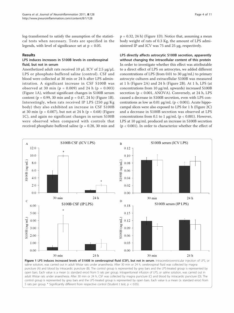
ResultsLPS induces increases in S100B levels in cerebrospinalfluid, but not in serumAnesthetized adult rats received 10 μL ICV of 2.5 μg/μLLPS or phosphate-buffered saline (control). CSF andblood were collected at 30 min or 24 h after LPS admin-istration. A significant increase in CSF S100B wasobserved at 30 min (p = 0.009) and 24 h (p = 0.003)(Figure 1A), without significant changes in S100B serumcontent (p = 0.99, 30 min and p = 0.47, 24 h) (Figure 1B).Interestingly, when rats received IP LPS (250 μg/Kgbody) they also exhibited an increase in CSF S100Bat 30 min (p = 0.007), but not at 24 h (p = 0.68) (Figure1C), and again no significant changes in serum S100Bwere observed when compared with controls thatreceived phosphate-buffered saline (p = 0.28, 30 min and
p = 0.32, 24 h) (Figure 1D). Notice that, assuming a meanbody weight of rats of 0.3 Kg, the amount of LPS admi-nistered IP and ICV was 75 and 25 μg, respectively.
LPS directly affects astrocytic S100B secretion, apparentlywithout changing the intracellular content of this proteinIn order to investigate whether this effect was attributableto a direct effect of LPS on astrocytes, we added differentconcentrations of LPS (from 0.01 to 30 μg/mL) to primaryastrocyte cultures and extracellular S100B was measuredat 1 h (Figure 2A) and 24 h (Figure 2B). At 1 h, LPS (atconcentrations from 10 μg/mL upwards) increased S100Bsecretion (p < 0.001, ANOVA). Conversely, at 24 h, LPScaused a decrease in S100B secretion, even with LPS con-centrations as low as 0.01 μg/mL (p < 0.001). Acute hippo-campal slices were also exposed to LPS for 1 h (Figure 2C)and a decrease in S100B secretion was observed at LPSconcentrations from 0.1 to 1 μg/mL (p < 0.001). However,LPS at 10 μg/mL produced an increase in S100B secretion(p < 0.001). In order to characterize whether the effect of
Figure 1 LPS induces increased levels of S100B in cerebrospinal fluid (CSF), but not in serum. Intracerebroventricular injection of LPS, orsaline solution, was carried out in adult Wistar rats under anaesthesia. After 30 min or 24 h, cerebrospinal fluid was collected by magnapuncture (A) and blood by intracardic puncture (B). The control group is represented by grey bars and the LPS-treated group is represented byopen bars. Each value is a mean (± standard error) from 5 rats per group. Intraperitoneal infusion of LPS, or saline solution, was carried out inadult Wistar rats under anaesthesia. After 30 min or 24 h, CSF was collected by magna puncture (C) and blood by intracardic puncture (D). Thecontrol group is represented by grey bars and the LPS-treated group is represented by open bars. Each value is a mean (± standard error) from5 rats per group. * Significantly different from respective control (Student t test, p < 0.05).
Guerra et al. Journal of Neuroinflammation 2011, 8:128http://www.jneuroinflammation.com/content/8/1/128
Page 4 of 11
Ina
Texto digitado
33

Figure 2 S100B secretion is modified by LPS in astrocyte cultures and acute hippocampal slices. Rat cortical astrocytes were cultured inDMEM containing 10% FCS. After confluence, the medium was replaced by DMEM without serum in the presence or absence of LPS (from 0.01to 30 μg/mL). S100B was measured by ELISA at 1 h (A) and 24 h (B). Each value is a mean (± standard error) of at least 5 independentexperiments performed in triplicate. Means indicated by different letters are significantly different, assuming p < 0.05. (C) Adult Wistar rats werekilled by decapitation and 0.3 mm hippocampal slices were obtained using a McIlwain chopper. After a metabolic recovery period, hippocampalslices were exposed to LPS (from 0.1 to 10 μg/mL) and the extracellular content of S100B measured by ELISA at 1 h. Each value is the mean (±standard error) of at least 5 independent experiments performed in triplicate. Means indicated by different letters are significantly different (oneway ANOVA followed by Duncan’s test, with a significance level of p < 0.05).
Guerra et al. Journal of Neuroinflammation 2011, 8:128http://www.jneuroinflammation.com/content/8/1/128
Page 5 of 11
Ina
Texto digitado
34

LPS is mediated by TLR4, we incubated astrocytes withspecific inhibitors for this receptor (Cli-095 and OxPAPC,at 1 μM and 30 μg/mL, respectively). Both CLI-095(Figure 3) and OxPAPC (data not shown) abolished theeffect of LPS. It is important to mention that OxPAPC perse increased S100B secretion and therefore it is difficult toaffirm that this inhibitor prevented the effect induced byLPS.After 24 h of exposure to LPS, we measured S100B
and GFAP content in lysed preparations of astrocytecultures (Figure 4A and 4B, respectively). No significantchanges were observed in S100B content (p = 0.85), butinterestingly an increase in GFAP content was observedat all concentrations of LPS (p = 0.04).
LPS decreases glutathione content, but does not affectcell viability and integrityAnother parameter analyzed to evaluate astroglial activitywas intracellular content of glutathione. After exposure ofastrocytes to LPS (at concentrations from 0.01 to 30 μg/mL), we observed a decrease in intracellular content ofglutathione after 24 h (p = 0.011), but not at 1 h (p = 0.49)(Figure 5A and 5B). Hippocampal slice preparations alsoexhibited a decrease in glutathione content after LPSexposure for 1 h (p = 0.015) (Figure 5C).In order to detect a possible toxic effect of LPS in our
preparations, we evaluated their capacities for MTT reduc-tion, neutral red incorporation and LDH release. Nochanges in MTT reduction assay (p = 0.25) (Figure 6A) orin neutral red assay (p = 0.37) (Figure 6B) were induced inastrocyte cultures exposed to LPS (from 0.01 to 30 μg/mL).
In addition, no changes in LDH release were seen (datanot shown). Similar assays were also carried out in slicepreparations confirming cell viability and integrity (datanot shown).
LPS induces an increase in TNFa in serum, but not in CSFFinally, we measured the response of the classic inflam-matory cytokine, TNFa, to LPS in vivo to confirm theactivity of this compound and to compare this responseto that of S100B protein. In contrast to results for S100B,at 30 min and 24 h after IP administration of LPS(approximately 75 μg) we observed an increase in TNFain serum (p = 0.04, 30 min and p = 0.04, 24 h), but not inCSF (p = 0.15, 30 min and p = 0.34, 24 h) (Table 1).When LPS (25 μg) was administered ICV we found anearly and transient increase in TNFa in serum (p <0.001) (at 30 min) and a later increase in CSF (p = 0.006)(at 24 h) (Table 2). In addition, we observed an increasein LPS-induced TNFa release from astrocyte cultures at
Figure 3 The LPS-induced decrease in S100B secretion isabolished by inhibition of TLR4. Rat cortical astrocytes werecultured in DMEM containing 10% FCS. After confluence, themedium was replaced by DMEM without serum in the presence orabsence of 0.1 μg/mL LPS and 1 μM CLI-095, an inhibitor of TLR4.S100B was measured by ELISA at 24 h. Each value is a mean (±standard error) of at least 5 independent experiments performed intriplicate. Means indicated by different letters are significantlydifferent (one way ANOVA followed by Duncan’s test, with asignificance level of p < 0.05).
Figure 4 Intracellular GFAP content is modified by LPS withoutchange in intracellular S100B content in astrocytes. Rat corticalastrocytes were cultured in DMEM containing 10% FCS. Afterconfluence, the medium was replaced by DMEM without serum in thepresence or absence of LPS (from 0.01 to 30 μg/mL). Cells were lysedand intracellular contents of S100B (A) and GFAP (B) were measuredby ELISA. Each value is the mean (± standard error) of at least 5independent experiments performed in triplicate. Means indicated bydifferent letters are significantly different (one way ANOVA followed byDuncan’s test, with a significance level of p < 0.05).
Guerra et al. Journal of Neuroinflammation 2011, 8:128http://www.jneuroinflammation.com/content/8/1/128
Page 6 of 11
Ina
Texto digitado
35

1, 6 and 24 h after exposure to LPS (Figure 7, p < 0.001).We were not able to detect TNFa release in acute hippo-campal slices.
DiscussionS100B has been proposed as a marker of brain injuryand its elevation in CSF has been interpreted as a signalof astroglial activation [10,11]. Moreover, it has beenassumed that S100B from CSF easily crosses the bloodbrain barrier and that a S100B increment in peripheralblood is indicative of brain injury. However, in some
Figure 5 GSH content is modified by LPS in astrocyte culturesand hippocampal slices. Rat cortical astrocytes were cultured inDMEM containing 10% FCS. After confluence, the medium wasreplaced by DMEM without serum in the presence or absence ofLPS (from 0.01 to 30 μg/mL). Cells were lysed in 1 h (A) or 24 h (B)and intracellular GSH content was measured. Each value representsthe mean (± standard error) of at least 5 independent experimentsperformed in triplicate. Means indicated by different letters aresignificantly different (one way ANOVA followed by Duncan’s test,with a significance level of p < 0.05). (C) Adult Wistar rats werekilled by decapitation and 0.3 mm hippocampal slices wereobtained using a McIlwain chopper. After a metabolic recoveryperiod, hippocampal slices were exposed to LPS (from 0.1 to 10 μg/mL) and intracellular content of S100B was measured by ELISA at 1h. Each value is the mean (± standard error) of at least 5independent experiments performed in triplicate. Means indicatedby different letters are significantly different (one way ANOVAfollowed by Duncan’s test, with a significance level of p < 0.05).
Figure 6 LPS does not affect cell viability. Rat cortical astrocyteswere cultured in DMEM containing 10% FCS. Confluent astrocyteswere exposed to LPS (from 0.01 to 30 μg/mL), during 24 h. At theend, cells were incubated with MTT (A) or neutral red (B). Eachvalue is the mean (± standard error) of at least 5 independentexperiments performed in triplicate. Statistical analysis wasperformed by one way ANOVA.
Table 1 Serum and CSF TNFa levels after IPadministration of LPS in rats
Control LPSa P
Serum (30 min) 3.4 ± 1.0 192.1 ± 97.2 0.046*
Serum (24 h) 1.1 ± 0.4 2.6 ± 0.2 0.021*
CSF (30 min) 2.7 ± 1.0 1.1 ± 0.5 0.145
CSF (24 h) 8.3 ± 5.5 2.5 ± 1.0 0.34
Values are mean (pg/mL) ± standard error (n = 5). Statistical analysis wasperformed using Student’s t test, * indicates p < 0.05; a 250 μg/Kg.
Guerra et al. Journal of Neuroinflammation 2011, 8:128http://www.jneuroinflammation.com/content/8/1/128
Page 7 of 11
Ina
Texto digitado
36

pathophysiological conditions other interpretations arepossible and, consequently, an intense debate has beendeveloped, mainly because there are extra-cerebralsources of S100B [31].Serum levels of S100B after exposure to LPS have
been measured in some studies. S100B protein bloodlevels in fetal sheep were found to be significantly higher1 h after LPS administration (intravenous [IV], 5 mg/Kg)and to return to baseline between 12 and 72 h afterexposure [32]. Similarly, in Sprague-Dawley rats, thisquantity of LPS is able to induce an increase in serumS100B 5h later [33]. In our study, ICV (2.5 ng) or IP.administration (0.25 mg/Kg) of LPS to Wistar rats didnot alter serum S100B levels, measured 30 min and 24h after exposure. This discrepancy could be due to thedifferent quantities of LPS employed, to its method ofadministration, or to the type of animal. Importantly,LPS (IV 2 ng/Kg), when given to humans, is not able toinduce significant changes in serum S100B at 1 h or 8 hpost treatment [34].In addition to measuring serum S100B, we also evalu-
ated S100B levels in CSF, astrocyte cultures and acutehippocampal slices of rats exposed to LPS. Astrocytesare thought of as active cells in the immune response,because they have receptors for this response (e.g TLR4)and are able to secrete cytokines [19,35]. We found anincrease in CSF S100B after LPS both for ICV (earlyand persistent response) and for IP administration (earlyand transient response). Notice that LPS is potentiallyable to cross the blood-brain barrier [36]. Clearly noimmediate increment in serum S100B occurred in eithercondition. This suggests brain-specific, LPS-inducedrelease of S100B, i.e., peripheral immune cells stimulatedby LPS did not release or cause a detectable S100Brelease from potential extra-cerebral sources of S100B(e.g. adipocytes). In other words, these data suggest dif-ferent LPS-sensitivities for S100B secretion in centraland peripheral S100B-expressing cells. Conversely, weobserved an immediate serum TNFa increase after LPSadministration by both ICV and IP routes. It has beenproposed that TNFa is able to mediate S100B secretionin astrocytes [37]. However, under LPS stimulation, ourresults regarding the profiles of increases in TNFa and
S100B in serum and CSF suggest independent responses(Table 3).Other aspects must be emphasized. The increase in CSF
S100B levels that we found was not accompanied or
Table 2 Serum and CSF TNFa after ICV administration ofLPS in rats
Control LPSb p
Serum (30 min) 0.7 ± 0.3 121.6 ± 40.0 0.001*
Serum (24 h) 1.1 ± 0.3 1.0 ± 0.3 0.945
CSF (30 min) 38.8 ± 9.8 75.1 ± 24.9 0.215
CSF (24 h) 1.7 ± 1.2 19.6 ± 4.5 0.006*
Values are mean (pg/mL) ± standard error (n = 5). Statistical analysis wasperformed using Student’s t test, * indicates p < 0.05; b 25 μg
Figure 7 TNFa secretion is modified by LPS in astrocytecultures. Rat cortical astrocytes were cultured in DMEM containing10% FCS. After confluence, the medium was replaced by DMEMwithout serum in the presence or absence of LPS (from 0.01 to 30μg/mL). TNFa was measured by ELISA at 1 h (A) and 6 h (B). Eachvalue is a mean (± standard error) of at least 5 independentexperiments performed in triplicate. Means indicated by differentletters are significantly different (one way ANOVA followed byDuncan’s test, with a significance level of p < 0.05).
Guerra et al. Journal of Neuroinflammation 2011, 8:128http://www.jneuroinflammation.com/content/8/1/128
Page 8 of 11
Ina
Texto digitado
37

followed by an increase in serum S100B levels, at least inmeasurements made at the evaluated times (30 min and24 h after LPS). This increase in CSF S100B was rapid (i.e.detected in 15 min) and lasting (for at least 24 h). Noticethat control animals for the experiments involving ICVadministration of LPS exhibited higher levels of CSFS100B (Figure 1A) than did controls for IP administration(Figure 1C), suggesting a response to the invasiveprocedure.Astrocytes in culture secreted S100B directly in response
to LPS (from 10 μg/mL upward) at 1h, but at 24 h adecrease in secretion (dependent on LPS concentration)was observed even at lower concentrations. This suggestsa biphasic response, i.e. an increase in S100B secretion, fol-lowed by a decrease. This profile has been observed inastrocyte cultures under other conditions, such as expo-sure to beta-hydroxybutyrate [38]. This rapid and transientstimulation of S100B secretion in astrocyte cultures wasalso observed for the cytokine IL-1b, but without adecrease at 24 h [12]. This finding could suggest that theLPS effect is direct and independent of secondarily-released IL-1b. Other studies have reported an increase incell content of S100B in C6 glioma cells after 24 h of expo-sure to IL-1b [39] or no change in astrocyte cultures after48 h [40] and a decrease in S100B content in culturedastrocytes after 3 days of exposure to TNFa [37]. How-ever, these studies did not measure S100B secretion ade-quately and it is not possible to speculate about asecondary effect of these two cytokines on S100B secretionafter long-term LPS exposure under the conditions usedhere. Therefore, in agreement with our working hypoth-esis, it appears that LPS is able to directly modulate S100Bsecretion.In addition, when we used acute hippocampal slices to
evaluate S100B secretion at 1 h, we also observed anincrease in S100B secretion with LPS at 10 μg/mL, butconversely we observed a decrease in LPS at 0.1 or 1 μg/mL. These preparations are complex from a cellular view,i.e. in addition to astrocytes, they contain active microglia
and neurons, which makes interpretation of the control ofS100B release difficult. However, a similar result, obtainedin response to endothelin-1, has also been observed [24].This compound, due to its blocking effect on gap junc-tions, increases S100B secretion in astrocyte cultures inthe first hour, but after 6 hours decreases S100B secretion.Similarly, in acute hippocampal slices, endothelin-1decreases S100B secretion at 1 h. Potentially, both LPSand endothelin-1 down-regulate gap junction proteins.Although we have no doubt about the effects of LPS andendothelin-1 on S100B secretion in acute hippocampalslices, we have no explanation for this effect, when com-pared to that observed in isolated astrocytes, at thismoment.Secreted S100B is a very small part of total cell content
(less than 0.5% is found in the medium of astrocyte cul-tures at 24 h) and changes in S100B secretion are notnecessarily accompanied by changes in the cell content[31]. In fact, in our experiments LPS changed S100B secre-tion without affecting cell content of this protein. On theother hand, GFAP content was increased by all concentra-tions of LPS used, indicating astroglial activation. This isin agreement with previous reports about the effects ofLPS on astrocyte cultures [22,23]. This reinforces the ideathat GFAP and S100B have distinct regulatory mechan-isms of expression and that astrogliosis (as assessed byGFAP increment) can either be accompanied or notaccompanied by changes in cell S100B content [41].Another interesting aspect of our findings is decreased
glutathione content after LPS exposure. The decrease inglutathione content in astrocytes at 24 h (but not at 1 h)is possibly associated with up-regulation of glutathione-S-transferase, as observed very recently [22]. Part of thedecrease could involve an intense exportation of this pep-tide, since it serves as an extracellular antioxidant, andalso provides substrates for neuronal synthesis of glu-tathione [42]. In addition, we also found a decrease inglutathione content in acute hippocampal slices exposedto LPS.In spite of this decrease in antioxidant defense, both
preparations exhibited excellent viability and integrity,based on MTT reduction assays, neutral red incorpora-tion and LDH release. These assays, performed in paral-lel to assays for S100B measurements, allowed us to beemphatic throughout the text about S100B secretion,instead of S100B release.Although S100B has cytokine-like actions (e.g. [43]),
some caution should be taken in the categorization ofS100B as a cytokine. In contrast to classical cytokines,S100B is not produced exclusively for secretion; only avery small part is exported. More recently, some authorshave suggested that S100B, like other members of theS100 family, may act as an alarmin or damage-associatedmolecular pattern (see [44] for a review). However,
Table 3 Qualitative comparison of TNFa and S100B levelsin serum and CSF after LPS administration
TNFa S100B
LPS IP Serum (30 min) ↑ –
Serum (24 h) ↑ –
CSF (30 min) – ↑
CSF (24 h) – –
LPS ICV Serum (30 min) ↑ –
Serum (24 h) – –
CSF (30 min) – ↑
CSF (24 h) ↑ ↑
↑ indicates a significant increase compared to control, with a significance levelof p < 0.05; – indicates no significant difference compared to control. SeeTable 1, Table 2 and Figure 1 for details.
Guerra et al. Journal of Neuroinflammation 2011, 8:128http://www.jneuroinflammation.com/content/8/1/128
Page 9 of 11
Ina
Texto digitado
38

independently of these conceptions, our data suggest thatS100B secretion is modulated by LPS. In fact, secretionof S100B might be protective during the initial phase ofLPS challenge. In contrast, prolonged LPS treatmentresults in a dose-dependent decrease in S100B secretionfrom astrocytes. This indicates that one potential effectof long-lasting exposure to LPS might be decreasedsecretion of trophic factors from astrocytes.It should be noted that some aspects of the effect of LPS
remain unclear. Firstly, is the effect of LPS mediated exclu-sively by TLR-4 in astrocytes? We cannot rule out otherpossibilities at this moment, since LPS could be acting onother receptors (e.g. CD14 and LBP [45]. Secondly, it isstill not clear whether LPS can affect S100B secretion inother S100-expressing cells. There are many extracerebralS100B-expressing cells that affect serum S100B levels [46]and these, apparently, were not mobilized under our con-ditions of LPS stimulation. However, further studies mustinvestigate specific extracerebral sources of S100B. Forexample, it is known that enteroglia respond to LPS byincreasing levels of S100B mRNA [47]. Third, whethergram-negative infectious agents could cause similar effectson S100B secretion, mediated by LPS release, is not clearat the moment. Interestingly, serum S100B was found tobe increased in patients with cerebral and extracerebralinfectious disease [48]. In that study, S100B elevation wasgenerally higher in patients with cerebral infections thanin extracerebral infections. However, specific and chroniceffects of gram-negative bacteria on central and peripheralS100B deserve further investigation.
ConclusionsOur data suggest that S100B secretion in brain tissue isstimulated rapidly and persistently (at least for 24 h) byICV administration of LPS. Moreover, no changes wereobserved in serum levels of this protein. This profile isquite different from that of TNFa, a canonical inflam-matory cytokine. In isolated astrocytes and acute hippo-campal slices, we observed a direct stimulation of S100Bsecretion by LPS at a concentration of 10 μg/mL,mediated by TLR4. However, in astrocyte cultures,lower levels of LPS were able to induce a decrease inS100B secretion 24 h afterwards, without significantchanges in the intracellular content of S100B. In addi-tion, after 24 h of exposure of astrocytes to LPS, weobserved a decrease in glutathione and an increase inGFAP. Together, these data contribute to our under-standing of the effect of LPS on astrocytes, particularlyon S100B secretion, and help us to interpret cerebrosp-inal fluid and serum changes of this protein in neuroin-flammatory diseases and brain disorders in general.Moreover, S100B-expressing tissues may be differentiallyregulated, since LPS did not lead to increases in serumS100B.
AcknowledgementsThis work was supported by the Conselho Nacional de DesenvolvimentoCientífico e Tecnológico (CNPq), Coordenação de Aperfeiçoamento dePessoal de Nível Superior (CAPES), FINEP/Rede IBN 01.06.0842-00 and INCT-National Institute of Science and Technology for Excitotoxicity andNeuroprotection. We would like to thank Ms. Gisele Souza for technicalsupport with cell culture.
Authors’ contributionsConception and design of experiments: MCG, LST, MCL and CAGAcquisition, analysis and interpretation of data: MCG, LST, MCL, FG, CDR, EN,DSE and LRWriting and/or critical review of article: MCG, LST, MCL and CAGAll authors have read and approved the final version of the manuscript.
Competing interestsThe authors declare that they have no competing interests.
Received: 2 December 2010 Accepted: 4 October 2011Published: 4 October 2011
References1. Donato R, Sorci G, Riuzzi F, Arcuri C, Bianchi R, Brozzi F, Tubaro C,
Giambanco I: S100B’s double life: intracellular regulator and extracellularsignal. Biochim Biophys Acta 2009, 1793:1008-1022.
2. Pinto SS, Gottfried C, Mendez A, Goncalves D, Karl J, Goncalves CA,Wofchuk S, Rodnight R: Immunocontent and secretion of S100B inastrocyte cultures from different brain regions in relation tomorphology. FEBS Lett 2000, 486:203-207.
3. Tramontina AC, Tramontina F, Bobermin LD, Zanotto C, Souza DF, Leite MC,Nardin P, Gottfried C, Goncalves CA: Secretion of S100B, an astrocyte-derived neurotrophic protein, is stimulated by fluoxetine via amechanism independent of serotonin. Prog Neuropsychopharmacol BiolPsychiatry 2008, 32:1580-1583.
4. Sakatani S, Seto-Ohshima A, Shinohara Y, Yamamoto Y, Yamamoto H,Itohara S, Hirase H: Neural-activity-dependent release of S100B fromastrocytes enhances kainate-induced gamma oscillations in vivo. JNeurosci 2008, 28:10928-10936.
5. Buyukuysal RL: Protein S100B release from rat brain slices during andafter ischemia: comparison with lactate dehydrogenase leakage.Neurochem Int 2005, 47:580-588.
6. Nardin P, Tramontina F, Leite MC, Tramontina AC, Quincozes-Santos A, deAlmeida LM, Battastini AM, Gottfried C, Goncalves CA: S100B content andsecretion decrease in astrocytes cultured in high-glucose medium.Neurochem Int 2007, 50:774-782.
7. Leite MC, Brolese G, de Almeida LM, Pinero CC, Gottfried C, Goncalves CA:Ammonia-induced alteration in S100B secretion in astrocytes is notreverted by creatine addition. Brain Res Bull 2006, 70:179-185.
8. Steiner J, Bernstein HG, Bielau H, Berndt A, Brisch R, Mawrin C, Keilhoff G,Bogerts B: Evidence for a wide extra-astrocytic distribution of S100B inhuman brain. BMC Neurosci 2007, 8:2.
9. Haimoto H, Kato K, Suzuki F, Nagura H: The ultrastructural changes of S-100 protein localization during lipolysis in adipocytes. Animmunoelectron-microscopic study. Am J Pathol 1985, 121:185-191.
10. Rothermundt M, Peters M, Prehn JH, Arolt V: S100B in brain damage andneurodegeneration. Microsc Res Tech 2003, 60:614-632.
11. Sen J, Belli A: S100B in neuropathologic states: the CRP of the brain? JNeurosci Res 2007, 85:1373-1380.
12. de Souza DF, Leite MC, Quincozes-Santos A, Nardin P, Tortorelli LS,Rigo MM, Gottfried C, Leal RB, Goncalves CA: S100B secretion is stimulatedby IL-1beta in glial cultures and hippocampal slices of rats: Likelyinvolvement of MAPK pathway. J Neuroimmunol 2009, 206:52-57.
13. Kim SH, Smith CJ, Van Eldik LJ: Importance of MAPK pathways formicroglial pro-inflammatory cytokine IL-1 beta production. NeurobiolAging 2004, 25:431-439.
14. Bianchi R, Giambanco I, Donato R: S100B/RAGE-dependent activation ofmicroglia via NF-kappaB and AP-1 Co-regulation of COX-2 expression byS100B, IL-1beta and TNF-alpha. Neurobiol Aging 2010, 31:665-677.
15. Ponath G, Schettler C, Kaestner F, Voigt B, Wentker D, Arolt V,Rothermundt M: Autocrine S100B effects on astrocytes are mediated viaRAGE. J Neuroimmunol 2007, 184:214-222.
Guerra et al. Journal of Neuroinflammation 2011, 8:128http://www.jneuroinflammation.com/content/8/1/128
Page 10 of 11
Ina
Texto digitado
39

16. Belanger M, Magistretti PJ: The role of astroglia in neuroprotection.Dialogues Clin Neurosci 2009, 11:281-295.
17. Eng LF, Ghirnikar RS, Lee YL: Glial fibrillary acidic protein: GFAP-thirty-oneyears (1969-2000). Neurochem Res 2000, 25:1439-1451.
18. Hauss-Wegrzyniak B, Lukovic L, Bigaud M, Stoeckel ME: Brain inflammatoryresponse induced by intracerebroventricular infusion oflipopolysaccharide: an immunohistochemical study. Brain Res 1998,794:211-224.
19. Farina C, Aloisi F, Meinl E: Astrocytes are active players in cerebral innateimmunity. Trends Immunol 2007, 28:138-145.
20. Carpentier PA, Duncan DS, Miller SD: Glial toll-like receptor signaling incentral nervous system infection and autoimmunity. Brain Behav Immun2008, 22:140-147.
21. Liao CK, Wang SM, Chen YL, Wang HS, Wu JC: Lipopolysaccharide-inducedinhibition of connexin43 gap junction communication in astrocytes ismediated by downregulation of caveolin-3. Int J Biochem Cell Biol 2010,42:762-770.
22. Vergara D, Martignago R, Bonsegna S, De Nuccio F, Santino A, Nicolardi G,Maffia M: IFN-beta reverses the lipopolysaccharide-induced proteomemodifications in treated astrocytes. J Neuroimmunol 2010, 221:115-120.
23. Brahmachari S, Fung YK, Pahan K: Induction of glial fibrillary acidic proteinexpression in astrocytes by nitric oxide. J Neurosci 2006, 26:4930-4939.
24. Leite MC, Galland F, de Souza DF, Guerra MC, Bobermin L, Biasibetti R,Gottfried C, Goncalves CA: Gap junction inhibitors modulate S100Bsecretion in astrocyte cultures and acute hippocampal slices. J NeurosciRes 2009.
25. Rodrigues L, Biasibetti R, Swarowsky A, Leite MC, Quincozes-Santos A,Quilfeldt JA, Achaval M, Goncalves CA: Hippocampal alterations in ratssubmitted to streptozotocin-induced dementia model are prevented byaminoguanidine. J Alzheimers Dis 2009, 17:193-202.
26. Gottfried C, Valentim L, Salbego C, Karl J, Wofchuk ST, Rodnight R:Regulation of protein phosphorylation in astrocyte cultures by externalcalcium ions: specific effects on the phosphorylation of glial fibrillaryacidic protein (GFAP), vimentin and heat shock protein 27 (HSP27). BrainRes 1999, 833:142-149.
27. Nardin P, Tortorelli L, Quincozes-Santos A, de Almeida LM, Leite MC,Thomazi AP, Gottfried C, Wofchuk ST, Donato R, Goncalves CA: S100BSecretion in Acute Brain Slices: Modulation by Extracellular Levels of Ca(2+) and K (+). Neurochem Res 2009.
28. Leite MC, Galland F, Brolese G, Guerra MC, Bortolotto JW, Freitas R,Almeida LM, Gottfried C, Goncalves CA: A simple, sensitive and widelyapplicable ELISA for S100B: Methodological features of themeasurement of this glial protein. J Neurosci Methods 2008, 169:93-99.
29. Tramontina F, Leite MC, Cereser K, de Souza DF, Tramontina AC, Nardin P,Andreazza AC, Gottfried C, Kapczinski F, Goncalves CA: Immunoassay forglial fibrillary acidic protein: antigen recognition is affected by itsphosphorylation state. J Neurosci Methods 2007, 162:282-286.
30. Browne RW, Armstrong D: Reduced glutathione and glutathione disulfide.Methods Mol Biol 1998, 108:347-352.
31. Goncalves CA, Concli Leite M, Nardin P: Biological and methodologicalfeatures of the measurement of S100B, a putative marker of braininjury. Clin Biochem 2008, 41:755-763.
32. Garnier Y, Berger R, Alm S, von Duering MU, Coumans AB, Michetti F,Bruschettini M, Lituania M, Hasaart TH, Gazzolo D: Systemic endotoxinadministration results in increased S100B protein blood levels andperiventricular brain white matter injury in the preterm fetal sheep. Eur JObstet Gynecol Reprod Biol 2006, 124:15-22.
33. Rosengarten B, Wolff S, Klatt S, Schermuly RT: Effects of inducible nitricoxide synthase inhibition or norepinephrine on the neurovascularcoupling in an endotoxic rat shock model. Crit Care 2009, 13:R139.
34. van den Boogaard M, Ramakers BP, van Alfen N, van der Werf SP, Fick WF,Hoedemaekers CW, Verbeek MM, Schoonhoven L, van der Hoeven JG,Pickkers P: Endotoxemia-induced inflammation and the effect on thehuman brain. Crit Care 2010, 14:R81.
35. Gorina R, Font-Nieves M, Marquez-Kisinousky L, Santalucia T, Planas AM:Astrocyte TLR4 activation induces a proinflammatory environmentthrough the interplay between MyD88-dependent NFkappaB signaling,MAPK, and Jak1/Stat1 pathways. Glia 2011, 59:242-255.
36. Banks WA, Robinson SM: Minimal penetration of lipopolysaccharideacross the murine blood-brain barrier. Brain Behav Immun 2010,24:102-109.
37. Edwards MM, Robinson SR: TNF alpha affects the expression of GFAP andS100B: implications for Alzheimer’s disease. J Neural Transm 2006,113:1709-1715.
38. Leite M, Frizzo JK, Nardin P, de Almeida LM, Tramontina F, Gottfried C,Goncalves CA: Beta-hydroxy-butyrate alters the extracellular content ofS100B in astrocyte cultures. Brain Res Bull 2004, 64:139-143.
39. Sheng JG, Ito K, Skinner RD, Mrak RE, Rovnaghi CR, Van Eldik LJ, Griffin WS:In vivo and in vitro evidence supporting a role for the inflammatorycytokine interleukin-1 as a driving force in Alzheimer pathogenesis.Neurobiol Aging 1996, 17:761-766.
40. Hinkle DA, Harney JP, Cai A, Hilt DC, Yarowsky PJ, Wise PM: Basic fibroblastgrowth factor-2 and interleukin-1 beta regulate S100 beta expression incultured astrocytes. Neuroscience 1998, 82:33-41.
41. Buffo A, Rite I, Tripathi P, Lepier A, Colak D, Horn AP, Mori T, Gotz M: Originand progeny of reactive gliosis: A source of multipotent cells in theinjured brain. Proc Natl Acad Sci USA 2008, 105:3581-3586.
42. Dringen R, Hirrlinger J: Glutathione pathways in the brain. Biol Chem 2003,384:505-516.
43. Liu L, Li Y, Van Eldik LJ, Griffin WS, Barger SW: S100B-induced microglialand neuronal IL-1 expression is mediated by cell type-specifictranscription factors. J Neurochem 2005, 92:546-553.
44. Bianchi ME: DAMPs, PAMPs and alarmins: all we need to know aboutdanger. J Leukoc Biol 2007, 81:1-5.
45. Peri F, Piazza M, Calabrese V, Damore G, Cighetti R: Exploring the LPS/TLR4signal pathway with small molecules. Biochem Soc Trans 2010,38:1390-1395.
46. Goncalves CA, Leite MC, Guerra MC: Adipocytes as an Important Sourceof Serum S100B and Possible Roles of This Protein in Adipose Tissue.Cardiovasc Psychiatry Neurol 2010, 2010:790431.
47. Cirillo C, Sarnelli G, Esposito G, Grosso M, Petruzzelli R, Izzo P, Cali G,D’Armiento FP, Rocco A, Nardone G, et al: Increased mucosal nitric oxideproduction in ulcerative colitis is mediated in part by the enteroglial-derived S100B protein. Neurogastroenterol Motil 2009, 21:1209-e1112.
48. Unden J, Christensson B, Bellner J, Alling C, Romner B: Serum S100B levelsin patients with cerebral and extracerebral infectious disease. Scand JInfect Dis 2004, 36:10-13.
doi:10.1186/1742-2094-8-128Cite this article as: Guerra et al.: Lipopolysaccharide modulatesastrocytic S100B secretion: a study in cerebrospinal fluid and astrocytecultures from rats. Journal of Neuroinflammation 2011 8:128.
Submit your next manuscript to BioMed Centraland take full advantage of:
• Convenient online submission
• Thorough peer review
• No space constraints or color figure charges
• Immediate publication on acceptance
• Inclusion in PubMed, CAS, Scopus and Google Scholar
• Research which is freely available for redistribution
Submit your manuscript at www.biomedcentral.com/submit
Guerra et al. Journal of Neuroinflammation 2011, 8:128http://www.jneuroinflammation.com/content/8/1/128
Page 11 of 11
Ina
Texto digitado
40
41
Capítulo II
Nimesulide decreases S100B secretion is astrocyte cultures
Artigo em Preparação, a ser submetido no periódico “Journal of Neuroinflammation”.

42
Nimesulide decreases S100B secretion is astrocyte cultures
Maria Cristina Guerra, Elisa Negri, Fabiana Galland, Carollina Da Ré, Liliane
Strapazzon, Lucas Tortoreli, Marina Concli Leite and Carlos-Alberto Gonçalves
Departamento de Bioquímica, Instituto de Ciências Básicas da Saúde, Universidade
Federal do Rio Grande do Sul, Ramiro Barcelos, 2600- Anexo, 90035-003, Porto
Alegre, Brazil
§ Corresponding author: Carlos Alberto Saraiva Gonçalves
Dept Bioquímica, ICBS, UFRGS
Ramiro Barcelos, 2600-anexo
90035-003 Porto Alegre, RS Brazil
E-mail: [email protected]
Fax: 55-51-3308 5535
Author’s e-mail Maria Cristina Guerra: [email protected] Elisa Negri: [email protected] Fabiana Galland: [email protected] Carolina Da Ré: [email protected] Liliane Strapazzon: [email protected] Lucas Tortorelli: [email protected] Marina Concli Leite: [email protected] Carlos-Alberto Gonçalves: [email protected]
Ina
Texto digitado
Ina
Texto digitado

43
Abstract
Many studied have looked for a connection of S100B, an astrocyte derived protein and
degenerative diseases including Alzheimer’s disease and schizophrenia. The therapeutic
use of non-steroid anti-inflammatory drugs (NSAID) to these diseases has growth up.
However, there are few reports about the effect of these drugs on S100B. Herein, we
evaluated S100B content and secretion measured by ELISA (as well as GFAP content
and TNF-α secretion) in primary astrocyte cultures exposed to dexamethasone and four
different chemical classes of NSAID (acetyl salicylic acid, ibuprofen, diclofenac and
nimesulide) for 24 h. Our data suggest that NSAIDs have differentiated effect on glial
parameters. ASA and diclofenac can increase GFAP while nimesulide, a selective
COX-2 inhibitor, and dexamethasone were able to decrease S100B secretion. However,
all anti-inflammatories were able to reduce levels of PGE2. Therefore, PGE2 is possibly
involved in the mechanism of S100B secretion but additional pathways, unclear at this
moment, demand further characterization. The inflammatory role of S100B in
degenerative diseases, where also is observed elevated levels of COX-2 and PGE2,
could be attenuated by COX-2 inhibitors.
Key-words: Astrocytes, Alzheimer, COX-1/2, NSAID, S100B, Schizophrenia,
44
Background
The inflammatory basis of many neurodegenerative diseases including Alzheimer’s
disease, Parkinson’s disease, multiple sclerosis, schizophrenia and major depression has
guided a pharmacological strategy using anti-inflammatories, whose main effect (but
not exclusively) is inhibition of cyclooxygenases 1 and 2 (COX-1/2) [1, 2]. The
neuroprotective action of COX-2 inhibitors against Alzheimer´s disease has been
proposed based on some epidemiological evidence e.g. [3] and experimental use of
these compounds in transgenic models [4, 5] and streptozotocin-induced models of
Alzheimer’s disease [6]. Moreover, clinical trials with the anti-inflammatories naproxen
and celecoxib suggest that the prevention is more efficient when compared to
therapeutic intervention [7, 8]. In addition, for other inflammatory-based brain diseases
seems that COX-2 inhibitors have a more favorable effect in the early stages.
Recent studies have suggested that glial cells, particularly astrocytes, are essential
elements and putative therapeutic targets in the brain inflammation [9]. Astrocytes are
functionally coupled to the glutamatergic synapses by glutamate uptake and release of
glutamine [10, 11], as well as they in an inflammatory scenario directly release
glutamate, which could modulate synaptic transmission [12]. In fact, Volterra’s group
showed that prostaglandins, the downstream products of COX-1/2, can stimulate
glutamate release by astrocytes [13]. Up-regulation of GFAP or changes in other
astrocyte markers have been used to characterize “reactive astrocytes” in the
neuroinflammatory process [14]. One of them is S100B, a 21 kDa calcium binding
protein produced and secreted by astrocytes [15]. Peripheral levels (in serum or CSF) of
this protein have been used as indicative of neural damage and/or astrocyte activity [16].
In fact, cultured astrocytes acutely exposed to LPS secrete S100B [17].

45
Many works have looked for a connection of S100B and degenerative diseases [18],
particularly Alzheimer’s disease [19]. Gene for S100B protein is found in the same
region of chromosome 21, where is located the amyloid precursor protein (APP) [20]
and, in addition, elevated levels of S100B can induce APP expression [21]. However,
there is little or controversial information about anti-inflammatories affecting COX-1/2
and S100B. A pioneer work showed that dexamethasone, a steroid anti-inflammatory,
inhibits S100B production in C6 glioma cells [22]. In addition, dexamethasone also
decreased S100B in astrocyte cultures [23] and hippocampal tissue [24]. Among non-
steroid anti-inflammatory drugs (NSAID) flufenamic acid [25], NS-398 [26] and
parecoxib [27], which are COX-2 inhibitors, apparently modulate extracellular levels of
S100B.
In this work, we evaluated S100B content and secretion (as well as GFAP content and
TNF-α secretion) in primary astrocyte cultures exposed to dexamethasone and four
different chemical classes of NSAID, chosen by their effect in some other particular
astroglial parameter already measured in glial cultures (see Table 1). The in vivo effect
on glial parameters also was investigated. We observed varied effect of different anti-
inflammatory compounds and discussed the possible implications to neuroinflammation
and brain related diseases.
Methods
Materials. Poly-L-lysine, antibody anti-S100B (SH-B1), methylthiazolyldiphenyl-
tetrazolium bromide (MTT) and neutral red were obtained from Sigma Aldrich (St.
Louis, USA). Fetal calf serum (FCS), Dulbecco’s modified Eagle’s medium (DMEM)
and other materials for cell culture were purchased from Gibco (Carlsbad, USA).
Polyclonal anti-S100B and anti-rabbit peroxidase linked were obtained from DAKO

46
(Sao Paulo, Brazil) and GE (Little Chalfont, United Kingdom), respectively. Anti-
COX1 and anti-COX2 were purchased from Santa Cruz Biotechnology (Dallas, USA).
Anti-inflammatory drugs (ASA, diclofenac, ibuprofen, dexamethasone and nimesulide)
were obtained from Sigma Aldrich (St. Louis, USA).
Anti-inflammatory intracerebroventricular (ICV) injection. Adult male Wistar rats
weighing 270-310 g, 60 days old, were used for ventricular access in stereotaxic
surgery. Procedures were carried out in accordance with the NIH Guide for the Care and
Use of Laboratory Animals and were approved by the local authorities. Animals were
anesthetized with ketamine/xylazine (75 and 10 mg/Kg, respectively, i.p.) and
positioned in a stereotaxic frame. A midline sagittal incision was made in the scalp and
one burr hole was drilled in the skull over right side ventricle. The following
coordinates were used: 0.9 mm posterior to bregma; 1.5 mm lateral to sagittal suture;
3.6 mm beneath the brain surface. Unilateral ICV injection of diclofenac (n = 5) or
acetylsalicylic acid (ASA) (n = 5), dissolved in DMSO 10 %, was slowly injected 5 µl
using Hamilton microsyringe. Assuming a CSF volume of 200 µL (excluding
subarachnoid space) final diclofenac and ASA concentration were 100 µM. Control
group (n = 5) was injected the same volume of DMSO 10%. After 24 h, cisterna magna
was accessed for CSF puncture in a stereotactic holder with an insulin syringe. A
maximum volume of 30 μL was collected over a 3-min period to minimize the risk of
brain stem damage. The blood samples were collected by careful intracardiac puncture,
using a 5-mL non-heparinized syringe to obtain 3 mL of blood. Blood samples were
incubated at room temperature (25°C) for 5 min and centrifuged at 3200 rpm for 5 min
to obtain serum. Hippocampal tissue was frozen (-70°C) until used for GFAP analysis.
Cell culture. Primary astrocyte cultures from Wistar rats were prepared as previously
described [28]. Procedures were carried out in accordance with the NIH Guide for the
47
Care and Use of Laboratory Animals and were approved by the local authorities.
Briefly, cerebral cortices of newborn Wistar rats (1-2 days old) were removed and
mechanically dissociated in Ca2+- and Mg2+-free balanced salt solution, pH 7.4,
containing (in mM): 137 NaCl; 5.36 KCl; 0.27 Na2HPO4; 1.1 KH2PO4 and 6.1
glucose. The cortices were cleaned of meninges and mechanically dissociated by
sequential passage through a Pasteur pipette. After centrifugation at 1400 RPM for 5
min the pellet was resuspended in DMEM (pH 7.6) supplemented with 8.39 mM
HEPES, 23.8 mM NaHCO3, 0.1% amphotericin, 0.032% gentamicin and 10% fetal calf
serum (FCS). Cultures were maintained in DMEM containing 10% FCS in 5%
CO2/95% air at 37°C, allowed to grow to confluence, and used at 15 days in vitro.
S100B measurement. S100B was measured by ELISA, as previously described [29].
Briefly, 50 μl of sample plus 50 μl of Tris buffer were incubated for 2 h on a microtiter
plate previously coated with monoclonal anti-S100B. Polyclonal anti-S100 was
incubated for 30 min and then peroxidase-conjugated anti-rabbit antibody was added for
a further 30 min. Color reaction with o-phenylenediamine was measured at 492 nm. The
standard S100B curve ranged from 0.002 to 1 ng/ml.
GFAP measurement. ELISA for GFAP was carried out as previously described [30], by
coating microtiter plates with 100 μL samples for 24 h at 4°C. Incubation with a
polyclonal anti-GFAP from rabbit for 1 h was followed by incubation with secondary
antibody conjugated with peroxidase for 1 h, at room temperature. A colorimetric
reaction with o- phenylenediamine was measured at 492 nm. The standard human
GFAP (from Calbiochem) curve ranged from 0.1to 5 ng/mL.
MTT reduction assay. Cells were treated with 50 μg/mL methylthiazolyldiphenyl-
tetrazolium bromide (MTT) for 30 min in 5% CO2/ 95% air at 37°C. Afterwards, the

48
medium was removed, and MTT crystals were dissolved in DMSO. Absorbance values
were measured at 560 and 650 nm. The reduction of MTT was calculated as
(absorbance at 560 nm) - (absorbance at 650 nm).
Neutral red uptake. Neutral red incorporation was carried out as previously described
[25] with modifications. Cells were treated with 50 μg/mL neutral red (NR) for 30 min
in 5% CO2/95% air at 37°C. Afterwards, the cells were rinsed twice with PBS for 5 min
each and NR dye taken up by viable cells was extracted with 500 μL of acetic
acid/ethanol/water (1/50/49). Absorbance values were measured at 560 nm.
Lactate dehydrogenase (LDH) assay. Lactate dehydrogenase assay was carried out in
50 μL of the extracellular medium, using a commercial colorimetric assay from Doles
[Goiânia, Brazil].
Glutathione content. Glutathione content was determined as previously described [31].
Briefly, astrocyte cultures were homogenized in sodium phosphate buffer (0.1 M, pH
8.0) containing 5 mM EDTA and protein was precipitated with 1.7% meta-phosphoric
acid. Supernatant was assayed with o-phthaldialdehyde (1 mg/mL of methanol) at room
temperature for 15 min. Fluorescence was measured using excitation and emission
wavelengths of 350 and 420 nm, respectively. A calibration curve was performed with
standard glutathione solutions (0-500 μM).
Tumor necrosis factor a (TNF-α) measurement. This assay was carried out in 100 μL of
CSF, serum or extracellular medium, using a rat TNFa ELISA from eBioscience [San
Diego, USA].
Prostaglandin E2 (PGE2) measurement. This assay was carried out in 100 μL of the
extracellular medium, using a rat PGE2 ELISA from Enzo Life Sciences [East
Farmingdale, USA].

49
Western blot analysis. Equal amounts (20 μg) of proteins from each sample were boiled
in sample buffer (0.0625MTris–HCl, pH 6.8, 2% (w/v) SDS, 5% (w/v) β-
mercaptoethanol, 10% (v/v) glycerol, 0.002% (w/v) bromophenol blue) and
electrophoresed in 10% (w/v) SDS-polyacrylamide gel. The separated proteins were
blotted onto a nitrocellulose membrane. Equal loading of each sample was confirmed
with Ponceau S staining. Antibodies anti-COX1 andanti-COX2 were used at a dilution
of 1:5000. After incubating with the primary antibody for 1 h at 4ºC, membranes were
washed and incubated with peroxidase-conjugated anti-goat immunoglobulin (IgG) at a
dilution of 1:5000 during 1 h at 4ºC. The chemiluminescence signal was detected using
an ECL kit from Amersham.
Statistical analyzes. Parametric data are reported as mean ± standard error and were
analyzed by Student’s t test (when two groups were considered) or by one-way analysis
of variance (ANOVA) followed by Dunnet test, in the SPSS-16.0, assuming p< 0.05.
Results
S100B secretion was reduced by nimesulide and dexamethasone
In the first experiment we investigated the S100B secretion in astrocyte culture exposed
to anti-inflammatories (ASA, ibuprofen, diclofenac, nimesulide or dexamethasone) for
24h. The concentrations were chosen based on previous studies in glial cell cultures
(See Table 1). Among these compounds, only dexamethasone and nimesulide
decreased S100B secretion (Fig 1A). Dexamethasone decreased 50% (p < 0.001) of the
basal S100B secretion, while nimesulide about 20% (p = 0.001). On the other hand,
secretion of TNFα, a canonical pro-inflammatory cytokine, was stimulated by ibuprofen
(350%, p < 0.001) (Fig 1B). Moreover, other NSAIDs did not reduce TNFα.
Dexametahose significantly decreased TNFα secretion to 10% (p = 0.002).

50
Changes in S100B secretion were not accompanied by changes in the intracellular
S100B content
The next step was to investigate whether these compounds, in our experimental
conditions, modified the cellular S100B content or GFAP content, the most widely used
marker of astrogliosis. Neither dexamethasone nor NSAID altered S100B content (Fig
2A). However, ASA and diclofenac increased the GFAP content (Fig 2B), about 70%
(p = 0.010 and p = 0.012 for ASA and diclofenac, respectively).
Dexamethasone and NSAID did not affect astrocyte morphology in culture
Despite changes in S100B secretion or GFAP content, no significant alterations was
observed in astrocyte morphology in astrocyte cultures (data not shown). Cell
morphology was analyzed by phase contrast microscopy (at 1 h and 24h), as well
immunocytochemistry for GFAP, S100B or actin (at 24h).
Ibuprofen and other anti-inflammatories did not modify GSH content or astroglial
viability
We also investigated the content of reduced glutathione (GSH) in astrocyte cultures
exposed to anti-inflammatories, especially because many antioxidant compounds (to
who is attributed anti-inflammatory activity) affect GSH content. Dexamethasone or
NSAID were not able to modify GSH levels in astrocyte culture (Fig 3A). Moreover,
based on MTT reduction assay is also possible to evaluate that these anti-inflammatory
compounds did not impair cell viability (Figure 3B). Indeed, it is important to mention
that extracellular LDH activity was not modified by exposure to anti-inflammatories
(data not shown).

51
The effect of acetyl salicylic acid on GFAP was also observed in vivo
In order to investigate the increase of GFAP observed in culture induced by ASA and
diclofenac were injected by ICV route, at indicated concentrations, to measured GFAP
content in the hippocampus and cerebral cortex 24 h afterwards. An increment of GFAP
was observed in the cerebral cortex induced by ASA (p=0.028), but not diclofenac
(p=0.776) (Fig 4A). In the hippocampus no changes were observed (Fig 4B).
A PGE2 decrease could explain, at least in part, the effect on S100B secretion
In Figure 5A is shows an immunoblot of COX-1 and COX-2 in astrocytes cultures basal
condition or 24 h after exposure to nimesulide or dexamethasone. Moreover, basal
levels of the downstream product of the COX-2, the prostaglandin E2 (PGE2), are
reduced after exposure to dexamethasone and all NSAIDs investigated (Figure 5B) (p <
0.05).
Discussion
The S100B protein, in the central nervous system, is mainly (but not exclusively)
expressed and secreted by astrocytes, where it has many intracellular targets and
functions [15]. However, the mechanism the secretion is unknown and this protein has
been thought as a marker of glial activation in brain disorders [16] and its extracellular
role is permanently in debate. Due to its trophic activity on neurons was regarded as a
growth factor, particularly for serotoninergic neurons [32]. It was also considered as a
cytokine and due to its trophic activity on neurons was labeled as neurotrophic cytokine
[33]. However, due to its release in inflammatory conditions also has been considered
as an inflammatory cytokine, able to respond and induce pro-inflammatory canonical
cytokines [34, 35]. More recently, other authors also have mentioned S100B as a
damage-associated molecular pattern (DAMP) [36]. Herein, we present evidence that

52
anti-inflammatories affect S100B secretion, arguing whether COX-2 and PGE2 are
putatively involved in the mechanism of secretion of S100B.
Basal S100B secretion was reduced by nimesulide, a selective COX-2 inhibitor,
and dexamethasone. Other NSAIDs, COX-1/2 inhibitors, were not able to affect S100B
secretion. However, TNFα secretion was not affected by NSAIDs, except ibuprofen,
which caused an increment in the secretion of this cytokine. Therefore, these NSAIDS
(at indicated concentrations) were not able to block the astrocyte secretion of TNFα, a
primary inflammatory cytokine. Ibuprofen caused an increment of basal TNFα. This
TNFα increment could induce a transient S100B secretion, but not in 24 h, as observed
in C6 glioma cells [37].
Assuming that the chronic increment of S100B is maybe underlying
degenerative processes [15], nimesulide and dexamethasone would be interesting
neuroprotective compounds. On the other hand, in acute conditions of brain injury, in
which S100B appears to play an important role in brain recovery, maybe these
compounds are not so important [38].
The decrease of S100B secretion induced by nimesulide or dexamethasone was
not accompanied by changes in intracellular S100B. This reinforces the idea that
changes in secretion are not necessarily accompanied by changes in intracellular content
of S100B [16]. Moreover, the decrease of S100B secretion induced by dexamethasone
or nimesulide, as well as, an increase of secretion induced by lysophosphatidic acid in
vitro [39], are not associated to cell morphology. Both ASA and diclofenac induced and
increment of GFAP in astrocyte cultures, and only ASA also able to induce an
increment of GFAP in brain cortical tissue 24 h after ICV infusion. Differently from our
results, a GFAP decrease in astrocyte cultures was described using high concentration

53
(5 mM) of ASA [40]. Moreover, is also important to emphasize that the in vivo
increment of GFAP observed in the cerebral cortex did not occur in hippocampal tissue,
suggesting the brain region dependence.
Several studies have reported that antioxidant compounds may exhibit anti-
inflammatory properties likely because their ability to modulate redox-sensitive proteins
in inflammatory cascades of signaling, such as MAPK and NF-kappaB. Therefore, we
investigated the opposite way, if these anti-inflammatory compounds affect redox cell.
Not any affect the cell content of GSH, the main antioxidant compound produced by
astrocytes. Moreover, astrocyte viability was not altered after anti-inflammatory
exposure (evaluated by MTT reduction assay), as well as cell integrity (assessed by
LDH and neutral red uptake assay) (data not shown).
Astrocytes in culture express receptors to respond directly to danger stimulus (e.g.,
LPS) [17], as well as all protein machinery to respond and release inflammatory
cytokines and prostaglandins. Astrocytes express and in vivo COX 1 and 2, in brain
tissue and cultures [41, 42]. Our results confirm the presence of both proteins, as well as
basal levels of PGE2 in astrocyte cultures. Considering that cAMP modulates S100B
secretion [39, 43] and PGE2, in an autocrine manner, stimulates G receptors coupled to
adenylyl cyclase, we hypothesized that COX-1/2 inhibitors would reduce S100B
secretion. However, only dexamethasone and nimesulide (a selective COX-2 inhibitor)
produced this effect. In addition, NS-398, a selective inhibitor of COX-2, reduced the
communication S100B/RAGE mediated in an experimental model of myasthenia gravis
[26] and patients that receive parecoxib for analgesia exhibited lower levels of serum
S100B [27]. Our results with nimesulide corroborate with those findings. However,
those studies did not directly evaluate S100B secretion.
54
This study has some limitations. Firstly, the study assumed S100B secretion as basal but
is was evaluated in cell culture, where cells are in a mild “inflammatory state,”
expressing high levels of COX-2 and PGE2. Secondly, as was performed to observe the
effect of anti-inflammatories in vivo on GFAP, it would be interesting in further studies
to analyze cerebrospinal fluid S100B. Thirdly, the effect of anti-inflammatories on
S100B secretion should be analyzed under an inflammatory condition (e.g. LPS).
Despite that, our results suggest that NSAIDs display different effect on glial
parameters GFAP and S100B. ASA and diclofenac can increase GFAP, while
nimesulide (as well as dexamethasone) was able to decrease S100B secretion. However,
all anti-inflammatories were able to reduce levels of PGE2. Therefore, PGE2 is possibly
involved in the mechanism of S100B secretion but additional pathways, unclear at this
moment, demand further characterization.
As we mentioned before, the chronic increment of S100B maybe underlying
degenerative processes such as AD and schizophrenia. In fact, COX-2 inhibitors have
been proposed as relevant therapeutic strategy for schizophrenia [44]. The pro-
inflammatory role of S100B in AD, in which elevated levels of COX-2 and PGE2 are
observed, also could be attenuated by COX-2 inhibitors. On the other hand, in acute
conditions of brain injury, where S100B appears to play an important role in brain
recovery, maybe these compounds do not afford beneficial effects and may be harmful
[38]. This potential decrease of extracellular S100B in acute conditions, such as
traumatic brain injury and stroke, reinforces the idea about risks and consequences of
using COX-2 under these conditions. However, this idea requires support of additional
epidemiological, clinical and experimental data.

55
References
1. Esposito E, Di Matteo V, Benigno A, Pierucci M, Crescimanno G, Di Giovanni
G: Non-steroidal anti-inflammatory drugs in Parkinson's disease. Exp Neurol 2007,
205:295-312.
2. Halliday G, Robinson SR, Shepherd C, Kril J: Alzheimer's disease and
inflammation: a review of cellular and therapeutic mechanisms. Clin Exp Pharmacol
Physiol 2000, 27:1-8.
3. McGeer PL, McGeer E, Rogers J, Sibley J: Anti-inflammatory drugs and
Alzheimer disease. Lancet 1990, 335:1037.
4. Varvel NH, Bhaskar K, Kounnas MZ, Wagner SL, Yang Y, Lamb BT, Herrup
K: NSAIDs prevent, but do not reverse, neuronal cell cycle reentry in a mouse model of
Alzheimer disease. J Clin Invest 2009, 119:3692-3702.
5. Choi YH, Back KO, Kim HJ, Lee SY, Kook KH: Pirfenidone attenuates IL-
1beta-induced COX-2 and PGE2 production in orbital fibroblasts through suppression
of NF-kappaB activity. Exp Eye Res 2013, 113:1-8.
6. Dhull DK, Jindal A, Dhull RK, Aggarwal S, Bhateja D, Padi SS:
Neuroprotective effect of cyclooxygenase inhibitors in ICV-STZ induced sporadic
Alzheimer's disease in rats. J Mol Neurosci 2012, 46:223-235.
7. Group AR, Lyketsos CG, Breitner JC, Green RC, Martin BK, Meinert C,
Piantadosi S, Sabbagh M: Naproxen and celecoxib do not prevent AD in early results
from a randomized controlled trial. Neurology 2007, 68:1800-1808.

56
8. Group AR, Martin BK, Szekely C, Brandt J, Piantadosi S, Breitner JC, Craft S,
Evans D, Green R, Mullan M: Cognitive function over time in the Alzheimer's Disease
Anti-inflammatory Prevention Trial (ADAPT): results of a randomized, controlled trial
of naproxen and celecoxib. Arch Neurol 2008, 65:896-905.
9. Carpentier PA, Begolka WS, Olson JK, Elhofy A, Karpus WJ, Miller SD:
Differential activation of astrocytes by innate and adaptive immune stimuli. Glia 2005,
49:360-374.
10. Magistretti PJ, Pellerin L: Astrocytes Couple Synaptic Activity to Glucose
Utilization in the Brain. News Physiol Sci 1999, 14:177-182.
11. Allaman I, Belanger M, Magistretti PJ: Astrocyte-neuron metabolic
relationships: for better and for worse. Trends Neurosci 2011, 34:76-87.
12. Agulhon C, Petravicz J, McMullen AB, Sweger EJ, Minton SK, Taves SR,
Casper KB, Fiacco TA, McCarthy KD: What is the role of astrocyte calcium in
neurophysiology? Neuron 2008, 59:932-946.
13. Bezzi P, Carmignoto G, Pasti L, Vesce S, Rossi D, Rizzini BL, Pozzan T,
Volterra A: Prostaglandins stimulate calcium-dependent glutamate release in astrocytes.
Nature 1998, 391:281-285.
14. Liberto CM, Albrecht PJ, Herx LM, Yong VW, Levison SW: Pro-regenerative
properties of cytokine-activated astrocytes. J Neurochem 2004, 89:1092-1100.
15. Donato R, Sorci G, Riuzzi F, Arcuri C, Bianchi R, Brozzi F, Tubaro C,
Giambanco I: S100B's double life: intracellular regulator and extracellular signal.
Biochim Biophys Acta 2009, 1793:1008-1022.
57
16. Goncalves CA, Concli Leite M, Nardin P: Biological and methodological
features of the measurement of S100B, a putative marker of brain injury. Clin Biochem
2008, 41:755-763.
17. Guerra MC, Tortorelli LS, Galland F, Da Re C, Negri E, Engelke DS, Rodrigues
L, Leite MC, Goncalves CA: Lipopolysaccharide modulates astrocytic S100B secretion:
a study in cerebrospinal fluid and astrocyte cultures from rats. J Neuroinflammation
2011, 8:128.
18. Rothermundt M, Peters M, Prehn JH, Arolt V: S100B in brain damage and
neurodegeneration. Microsc Res Tech 2003, 60:614-632.
19. Mrak RE, Griffinbc WS: The role of activated astrocytes and of the neurotrophic
cytokine S100B in the pathogenesis of Alzheimer's disease. Neurobiol Aging 2001,
22:915-922.
20. Seidl R, Cairns N, Lubec G: The brain in Down syndrome. J Neural Transm
Suppl 2001:247-261.
21. Mori T, Koyama N, Arendash GW, Horikoshi-Sakuraba Y, Tan J, Town T:
Overexpression of human S100B exacerbates cerebral amyloidosis and gliosis in the
Tg2576 mouse model of Alzheimer's disease. Glia 2010, 58:300-314.
22. Selinfreund RH, Barger SW, Welsh MJ, Van Eldik LJ: Antisense inhibition of
glial S100 beta production results in alterations in cell morphology, cytoskeletal
organization, and cell proliferation. J Cell Biol 1990, 111:2021-2028.
23. Niu H, Hinkle DA, Wise PM: Dexamethasone regulates basic fibroblast growth
factor, nerve growth factor and S100beta expression in cultured hippocampal astrocytes.
Brain Res Mol Brain Res 1997, 51:97-105.
58
24. Bruschettini M, van den Hove DL, Gazzolo D, Steinbusch HW, Blanco CE:
Lowering the dose of antenatal steroids: the effects of a single course of betamethasone
on somatic growth and brain cell proliferation in the rat. Am J Obstet Gynecol 2006,
194:1341-1346.
25. Leite MC, Galland F, de Souza DF, Guerra MC, Bobermin L, Biasibetti R,
Gottfried C, Goncalves CA: Gap junction inhibitors modulate S100B secretion in
astrocyte cultures and acute hippocampal slices. J Neurosci Res 2009.
26. Mu L, Zhang Y, Sun B, Wang J, Xie X, Li N, Zhang J, Kong Q, Liu Y, Han Z,
et al: Activation of the receptor for advanced glycation end products (RAGE)
exacerbates experimental autoimmune myasthenia gravis symptoms. Clin Immunol
2011, 141:36-48.
27. Li JZ, Li XZ, Wang XM, Wang MS, Yu HF, Shi F, Miao D, Bi YL: [Effects of
parecoxib sodium analgesia on serum concentrations of neuron-specific enolase and S-
100beta and postoperative cognitive function of elderly patients undergoing acute
replacement of femoral head]. Zhonghua Yi Xue Za Zhi 2013, 93:2152-2154.
28. Gottfried C, Valentim L, Salbego C, Karl J, Wofchuk ST, Rodnight R:
Regulation of protein phosphorylation in astrocyte cultures by external calcium ions:
specific effects on the phosphorylation of glial fibrillary acidic protein (GFAP),
vimentin and heat shock protein 27 (HSP27). Brain Res 1999, 833:142-149.
29. Leite MC, Galland F, Brolese G, Guerra MC, Bortolotto JW, Freitas R, Almeida
LM, Gottfried C, Goncalves CA: A simple, sensitive and widely applicable ELISA for
S100B: Methodological features of the measurement of this glial protein. J Neurosci
Methods 2008, 169:93-99.

59
30. Tramontina F, Leite MC, Cereser K, de Souza DF, Tramontina AC, Nardin P,
Andreazza AC, Gottfried C, Kapczinski F, Goncalves CA: Immunoassay for glial
fibrillary acidic protein: antigen recognition is affected by its phosphorylation state. J
Neurosci Methods 2007, 162:282-286.
31. Browne RW, Armstrong D: Reduced glutathione and glutathione disulfide.
Methods Mol Biol 1998, 108:347-352.
32. Whitaker-Azmitia PM, Murphy R, Azmitia EC: Stimulation of astroglial 5-
HT1A receptors releases the serotonergic growth factor, protein S-100, and alters
astroglial morphology. Brain Res 1990, 528:155-158.
33. Griffin WS, Yeralan O, Sheng JG, Boop FA, Mrak RE, Rovnaghi CR, Burnett
BA, Feoktistova A, Van Eldik LJ: Overexpression of the neurotrophic cytokine S100
beta in human temporal lobe epilepsy. J Neurochem 1995, 65:228-233.
34. de Souza DF, Leite MC, Quincozes-Santos A, Nardin P, Tortorelli LS, Rigo
MM, Gottfried C, Leal RB, Goncalves CA: S100B secretion is stimulated by IL-1beta
in glial cultures and hippocampal slices of rats: Likely involvement of MAPK pathway.
J Neuroimmunol 2009, 206:52-57.
35. Schmitt KR, Kern C, Lange PE, Berger F, Abdul-Khaliq H, Hendrix S: S100B
modulates IL-6 release and cytotoxicity from hypothermic brain cells and inhibits
hypothermia-induced axonal outgrowth. Neurosci Res 2007, 59:68-73.
36. Bernardini C, Lattanzi W, Businaro R, Leone S, Corvino V, Sorci G, Lauro G,
Fumagalli L, Donato FR, Michetti F: Transcritpional effects of S100B on
neuroblastoma cells: perturbation of cholesterol homeostasis and interference on the cell
cycle. Gene Expr 2010, 14:345-359.

60
37. de Souza DF, Wartchow K, Hansen F, Lunardi P, Guerra MC, Nardin P,
Goncalves CA: Interleukin-6-induced S100B secretion is inhibited by haloperidol and
risperidone. Prog Neuropsychopharmacol Biol Psychiatry 2013, 43:14-22.
38. Kleindienst A, Meissner S, Eyupoglu IY, Parsch H, Schmidt C, Buchfelder M:
Dynamics of S100B release into serum and cerebrospinal fluid following acute brain
injury. Acta Neurochir Suppl 2010, 106:247-250.
39. Pinto SS, Gottfried C, Mendez A, Goncalves D, Karl J, Goncalves CA, Wofchuk
S, Rodnight R: Immunocontent and secretion of S100B in astrocyte cultures from
different brain regions in relation to morphology. FEBS Lett 2000, 486:203-207.
40. Bae MK, Kim SR, Lee HJ, Wee HJ, Yoo MA, Ock Oh S, Baek SY, Kim BS,
Kim JB, Sik Y, Bae SK: Aspirin-induced blockade of NF-kappaB activity restrains up-
regulation of glial fibrillary acidic protein in human astroglial cells. Biochim Biophys
Acta 2006, 1763:282-289.
41. Aid S, Bosetti F: Targeting cyclooxygenases-1 and -2 in neuroinflammation:
Therapeutic implications. Biochimie 2011, 93:46-51.
42. Font-Nieves M, Sans-Fons MG, Gorina R, Bonfill-Teixidor E, Salas-Perdomo
A, Marquez-Kisinousky L, Santalucia T, Planas AM: Induction of COX-2 enzyme and
down-regulation of COX-1 expression by lipopolysaccharide (LPS) control
prostaglandin E2 production in astrocytes. J Biol Chem 2012, 287:6454-6468.
43. Goncalves D, Karl J, Leite M, Rotta L, Salbego C, Rocha E, Wofchuk S,
Goncalves CA: High glutamate decreases S100B secretion stimulated by serum
deprivation in astrocytes. Neuroreport 2002, 13:1533-1535.
61
44. Miuller N, Schwarz MJ: The immunological basis of glutamatergic disturbance
in schizophrenia: towards an integrated view. J Neural Transm Suppl 2007:269-280.
45. Aleong R, Aumont N, Dea D, Poirier J: Non-steroidal anti-inflammatory drugs
mediate increased in vitro glial expression of apolipoprotein E protein. Eur J Neurosci
2003, 18:1428-1438.
46. Iwata Y, Nicole O, Zurakowski D, Okamura T, Jonas RA: Ibuprofen for
neuroprotection after cerebral ischemia. J Thorac Cardiovasc Surg 2010, 139:489-493.
47. Asai H, Kakita H, Aoyama M, Nagaya Y, Saitoh S, Asai K: Diclofenac
enhances proinflammatory cytokine-induced aquaporin-4 expression in cultured
astrocyte. Cell Mol Neurobiol 2013, 33:393-400.
48. Niranjan R, Rajasekar N, Nath C, Shukla R: The effect of guggulipid and
nimesulide on MPTP-induced mediators of neuroinflammation in rat astrocytoma cells,
C6. Chem Biol Interact 2012, 200:73-83.
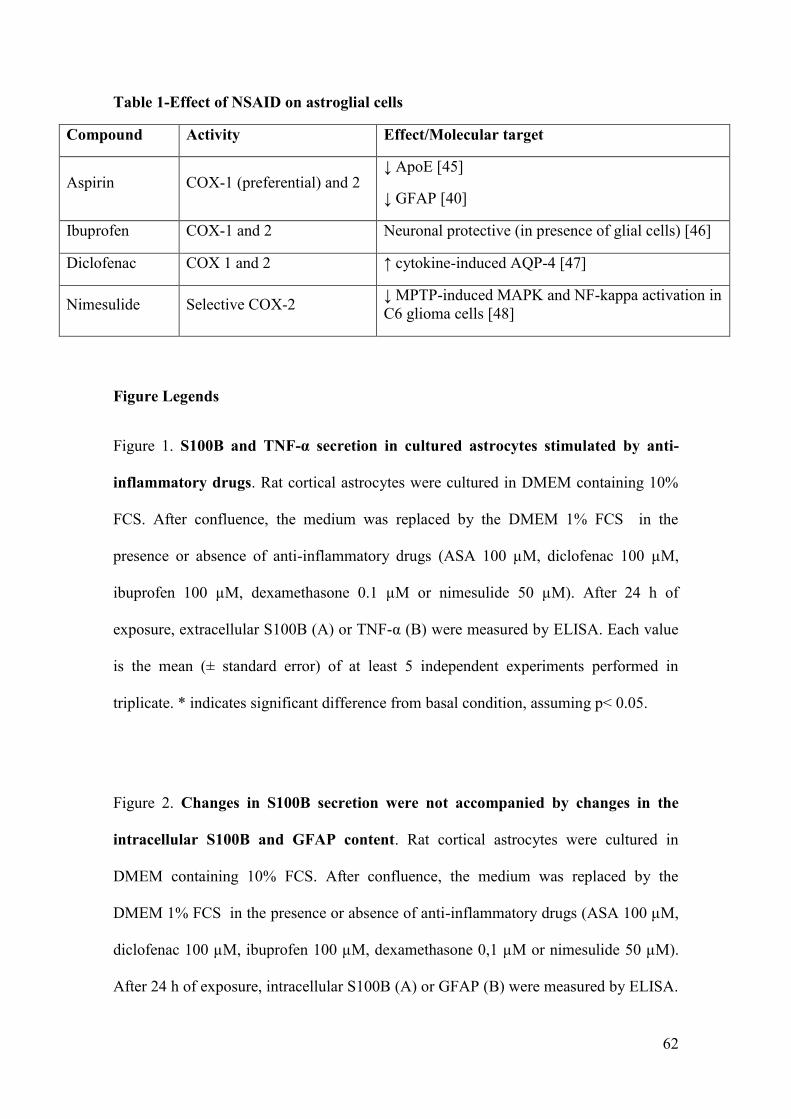
62
Table 1-Effect of NSAID on astroglial cells
Compound Activity Effect/Molecular target
Aspirin COX-1 (preferential) and 2 ↓ ApoE [45]
↓ GFAP [40]
Ibuprofen COX-1 and 2 Neuronal protective (in presence of glial cells) [46]
Diclofenac COX 1 and 2 ↑ cytokine-induced AQP-4 [47]
Nimesulide Selective COX-2 ↓ MPTP-induced MAPK and NF-kappa activation in C6 glioma cells [48]
Figure Legends
Figure 1. S100B and TNF-α secretion in cultured astrocytes stimulated by anti-
inflammatory drugs. Rat cortical astrocytes were cultured in DMEM containing 10%
FCS. After confluence, the medium was replaced by the DMEM 1% FCS in the
presence or absence of anti-inflammatory drugs (ASA 100 µM, diclofenac 100 µM,
ibuprofen 100 µM, dexamethasone 0.1 µM or nimesulide 50 µM). After 24 h of
exposure, extracellular S100B (A) or TNF-α (B) were measured by ELISA. Each value
is the mean (± standard error) of at least 5 independent experiments performed in
triplicate. * indicates significant difference from basal condition, assuming p< 0.05.
Figure 2. Changes in S100B secretion were not accompanied by changes in the
intracellular S100B and GFAP content. Rat cortical astrocytes were cultured in
DMEM containing 10% FCS. After confluence, the medium was replaced by the
DMEM 1% FCS in the presence or absence of anti-inflammatory drugs (ASA 100 µM,
diclofenac 100 µM, ibuprofen 100 µM, dexamethasone 0,1 µM or nimesulide 50 µM).
After 24 h of exposure, intracellular S100B (A) or GFAP (B) were measured by ELISA.
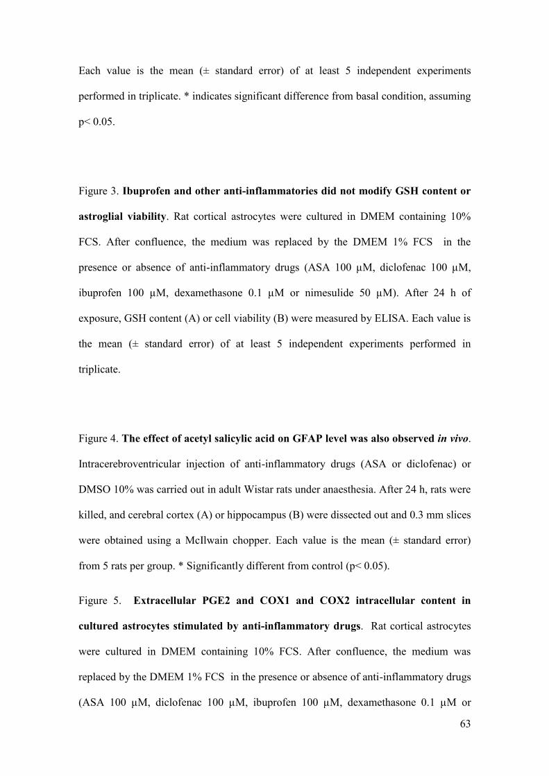
63
Each value is the mean (± standard error) of at least 5 independent experiments
performed in triplicate. * indicates significant difference from basal condition, assuming
p< 0.05.
Figure 3. Ibuprofen and other anti-inflammatories did not modify GSH content or
astroglial viability. Rat cortical astrocytes were cultured in DMEM containing 10%
FCS. After confluence, the medium was replaced by the DMEM 1% FCS in the
presence or absence of anti-inflammatory drugs (ASA 100 µM, diclofenac 100 µM,
ibuprofen 100 µM, dexamethasone 0.1 µM or nimesulide 50 µM). After 24 h of
exposure, GSH content (A) or cell viability (B) were measured by ELISA. Each value is
the mean (± standard error) of at least 5 independent experiments performed in
triplicate.
Figure 4. The effect of acetyl salicylic acid on GFAP level was also observed in vivo.
Intracerebroventricular injection of anti-inflammatory drugs (ASA or diclofenac) or
DMSO 10% was carried out in adult Wistar rats under anaesthesia. After 24 h, rats were
killed, and cerebral cortex (A) or hippocampus (B) were dissected out and 0.3 mm slices
were obtained using a McIlwain chopper. Each value is the mean (± standard error)
from 5 rats per group. * Significantly different from control (p< 0.05).
Figure 5. Extracellular PGE2 and COX1 and COX2 intracellular content in
cultured astrocytes stimulated by anti-inflammatory drugs. Rat cortical astrocytes
were cultured in DMEM containing 10% FCS. After confluence, the medium was
replaced by the DMEM 1% FCS in the presence or absence of anti-inflammatory drugs
(ASA 100 µM, diclofenac 100 µM, ibuprofen 100 µM, dexamethasone 0.1 µM or

64
nimesulide 50 µM). After 24 h of exposure, intracellular content of COX1 and COX2
was analyzed by immunoblot (A) and extracellular PGE2 was measured by ELISA (B).
Each value is the mean (± standard error) of at least 5 independent experiments
performed in triplicate. * indicates significant difference from basal condition, assuming
p < 0.05.
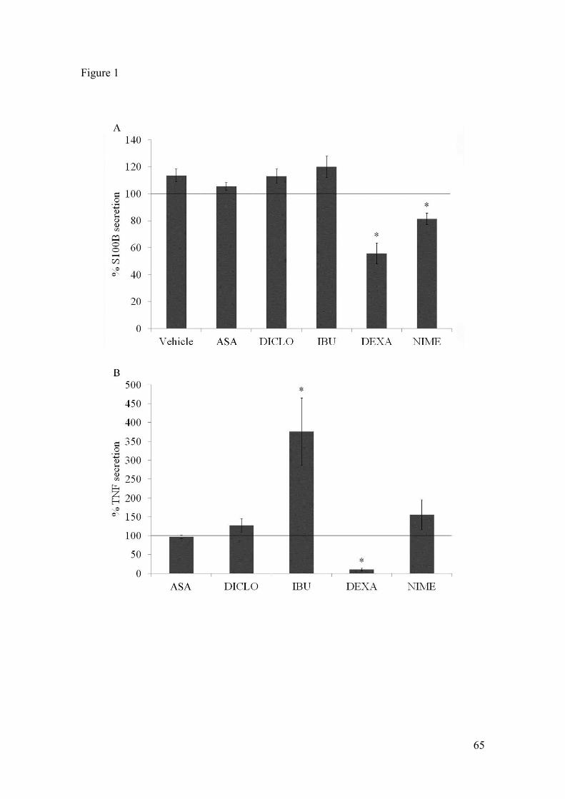
65
Figure 1
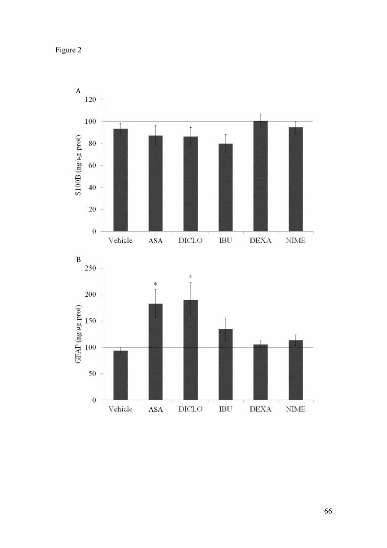
66
Figure 2
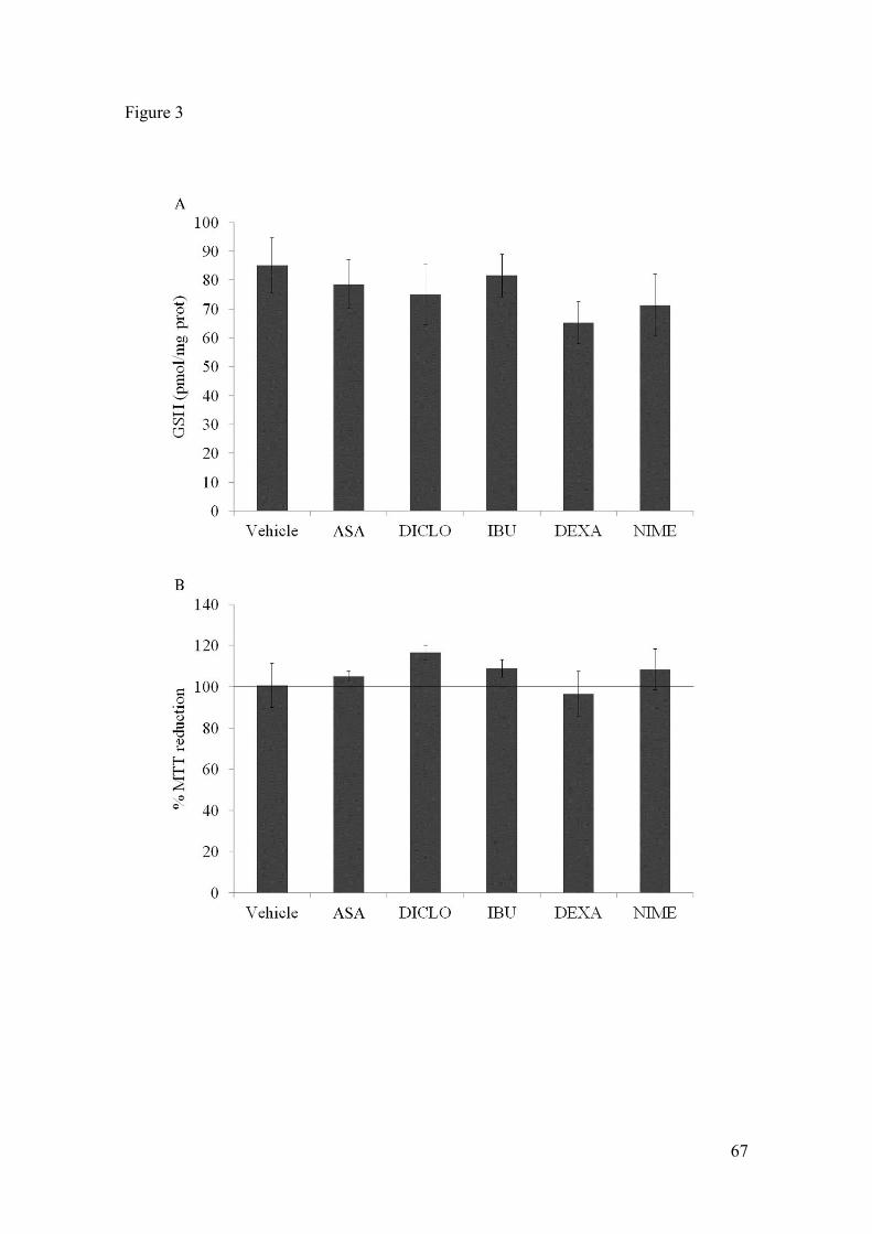
67
Figure 3

68
Figure 4

69
Figure 5
70
Capítulo III
Efeito de anti-inflamatórios em astrócitos estimulados com LPS sobre diferentes
parâmetros de ativação glial
Dados preliminares
71
Justificativa
Uma vez descrita a importância do processo neuroinflamatório em várias doenças
neurodegenerativas, o envolvimento ativo de células astrogliais neste processo e a
relevância da proteína S100B como um possível marcador de dano cerebral, nós
resolvemos desenvolver um estudo com o objetivo de reunir algumas observações que
ajude a compreender o papel da proteína S100B na neuroinflamação.
No capítulo I desta tese nós investigamos o efeito do LPS sobre a secreção e
imunoconteúdo da proteína S100B em culturas astrogliais e também seu efeito in vivo e
ex vivo e observamos que o LPS é capaz de modular a secreção dessa proteína, bem
como de alterar outros parâmetros de ativação glial.
Além disso, sabendo da importância do uso de anti-inflamatórios contribuindo no
tratamento de doenças neurodegenerativas, no capítulo II desta tese, nós observamos
que estes fármacos modulam principalmente a secreção de S100B, sendo esta
modulação específica para cada fármaco independente da classe em que se encontram.
Tendo em vista os efeitos do LPS e dos anti-inflamatórios sobre os astrócitos,
observados anteriormente, o objetivo do capítulo III desta tese é investigar o efeito dos
anti-inflamatórios em culturas primárias de astrócitos estimuladas com LPS sobre os
diferentes parâmetros testados nos outros estudos.
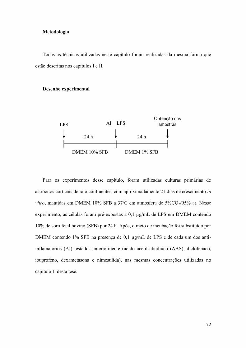
72
Metodologia
Todas as técnicas utilizadas neste capítulo foram realizadas da mesma forma que
estão descritas nos capítulos I e II.
Desenho experimental
Para os experimentos desse capítulo, foram utilizadas culturas primárias de
astrócitos corticais de rato confluentes, com aproximadamente 21 dias de crescimento in
vitro, mantidas em DMEM 10% SFB a 37ºC em atmosfera de 5%CO2/95% ar. Nesse
experimento, as células foram pré-expostas a 0,1 µg/mL de LPS em DMEM contendo
10% de soro fetal bovino (SFB) por 24 h. Após, o meio de incubação foi substituído por
DMEM contendo 1% SFB na presença de 0,1 µg/mL de LPS e de cada um dos anti-
inflamatórios (AI) testados anteriormente (ácido acetilsalicíliuco (AAS), diclofenaco,
ibuprofeno, dexametasona e nimesulida), nas mesmas concentrações utilizadas no
capítulo II desta tese.
DMEM 10% SFB DMEM 1% SFB
LPS AI + LPS
24 h 24 h
Obtenção das amostras
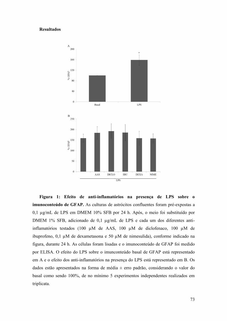
73
Resultados
Figura 1: Efeito de anti-inflamatórios na presença de LPS sobre o
imunoconteúdo de GFAP. As culturas de astrócitos confluentes foram pré-expostas a
0,1 µg/mL de LPS em DMEM 10% SFB por 24 h. Após, o meio foi substituído por
DMEM 1% SFB, adicionado de 0,1 µg/mL de LPS e cada um dos diferentes anti-
inflamatórios testados (100 µM de AAS, 100 µM de diclofenaco, 100 µM de
ibuprofeno, 0,1 µM de dexametasona e 50 µM de nimesulida), conforme indicado na
figura, durante 24 h. As células foram lisadas e o imunoconteúdo de GFAP foi medido
por ELISA. O efeito do LPS sobre o imunconteúdo basal de GFAP está representado
em A e o efeito dos anti-inflamatórios na presença do LPS está representado em B. Os
dados estão apresentados na forma de média ± erro padrão, considerando o valor do
basal como sendo 100%, de no mínimo 5 experimentos independentes realizados em
triplicata.

74
Figura 2: Efeito de anti-inflamatórios na presença de LPS sobre o
imunoconteúdo de S100B. As culturas de astrócitos confluentes foram pré-expostas a
0,1 µg/mL de LPS em DMEM 10% SFB por 24 h. Após, o meio foi substituído por
DMEM 1% SFB, adicionado de 0,1 µg/mL de LPS e cada um dos diferentes anti-
inflamatórios testados (100 µM de AAS, 100 µM de diclofenaco, 100 µM de
ibuprofeno, 0,1 µM de dexametasona e 50 µM de nimesulida), conforme indicado na
figura, durante 24 h. As células foram lisadas e o imunoconteúdo de S100B foi medido
por ELISA. O efeito do LPS sobre o imunconteúdo basal de S100B está representado
em A e o efeito dos anti-inflamatórios na presença do LPS está representado em B. Os
dados estão apresentados na forma de média ± erro padrão, considerando o valor do
basal como sendo 100%, de no mínimo 5 experimentos independentes realizados em
triplicata.

75
Figura 3: Efeito de anti-inflamatórios na presença de LPS sobre a secreção de
S100B. As culturas de astrócitos confluentes foram pré-expostas a 0,1 µg/mL de LPS
em DMEM 10% SFB por 24 h. Após, o meio foi substituído por DMEM 1% SFB,
adicionado de 0,1 µg/mL de LPS e cada um dos diferentes anti-inflamatórios testados
(100 µM de AAS, 100 µM de diclofenaco, 100 µM de ibuprofeno, 0,1 µM de
dexametasona e 50 µM de nimesulida), conforme indicado na figura, durante 24 h. O
meio de incubação foi coletado e a secreção de S100B foi medida por ELISA. O efeito
do LPS sobre a secreção basal de S100B está representado em A e o efeito dos anti-
inflamatórios na presença do LPS está representado em B. Os dados estão apresentados
na forma de média ± erro padrão, considerando o valor do basal como sendo 100%, de
no mínimo 5 experimentos independentes realizados em triplicata. * representa
diferença significativa em comparação com o basal em A (teste T de Student) e em
comparação com o LPS em B (ANOVA de uma via seguida de pós-teste de Dunnet)
para um p<0,05.

76
Figura 4: Efeito de anti-inflamatórios na presença de LPS sobre a secreção de
TNF-α. As culturas de astrócitos confluentes foram pré-expostas a 0,1 µg/mL de LPS
em DMEM 10% SFB por 24 h. Após, o meio foi substituído por DMEM 1% SFB,
adicionado de 0,1 µg/mL de LPS e cada um dos diferentes anti-inflamatórios testados
(100 µM de AAS, 100 µM de diclofenaco, 100 µM de ibuprofeno, 0,1 µM de
dexametasona e 50 µM de nimesulida), conforme indicado na figura, durante 24 h. O
meio de incubação foi coletado e a secreção de TNF-α foi medida por ELISA. O efeito
do LPS sobre a secreção basal de TNF-α está representado em A e o efeito dos anti-
inflamatórios na presença do LPS está representado em B. Os dados estão apresentados
na forma de média ± erro padrão, considerando o valor do basal como sendo 100% (em
A) e o LPS (em B), de no mínimo 5 experimentos independentes realizados em
triplicata.* representa diferença significativa em comparação com o basal em A (teste T
de Student) e em comparação com o LPS em B (ANOVA de uma via seguida de pós-
teste de Dunnet) para um p<0,05.

77
Figura 5: Efeito de anti-inflamatórios na presença de LPS sobre a viabilidade
celular. As culturas de astrócitos confluentes foram pré-expostas a 0,1 µg/mL de LPS
em DMEM 10% SFB por 24 h. Após, o meio foi substituído por DMEM 1% SFB,
adicionado de 0,1 µg/mL de LPS e cada um dos diferentes anti-inflamatórios testados
(100 µM de AAS, 100 µM de diclofenaco, 100 µM de ibuprofeno, 0,1 µM de
dexametasona e 50 µM de nimesulida), conforme indicado na figura, durante 24 h.

78
Após, a viabilidade celular foi testada por incorporação de vermelho neutro em A,
redução de MTT em B e liberação de LDH em C. Os dados estão apresentados na forma
de média ± erro padrão, considerando o valor do basal como sendo 100%, de no mínimo
5 experimentos independentes realizados em triplicata.* representa diferença
significativa em comparação com o basal (ANOVA de uma via seguida de pós-teste de
Dunnet) para um p<0,05.

79
Figura 6: Efeito de anti-inflamatórios na presença de LPS sobre o conteúdo de
glutationa reduzida. As culturas de astrócitos confluentes foram pré-expostas a 0,1
µg/mL de LPS em DMEM 10% SFB por 24 h. Após, o meio foi substituído por DMEM
1% SFB, adicionado de 0,1 µg/mL de LPS e cada um dos diferentes anti-inflamatórios
testados (100 µM de AAS, 100 µM de diclofenaco, 100 µM de ibuprofeno, 0,1 µM de
dexametasona e 50 µM de nimesulida), conforme indicado na figura, durante 24 h. As
células foram lisadas e o conteúdo de GSH foi medido por uma técnica fluorimétrica. O
efeito do LPS sobre o conteúdo basal de GSH está representado em A e o efeito dos
anti-inflamatórios na presença do LPS está representado em B. Os dados estão
apresentados na forma de média ± erro padrão, considerando o valor do basal como
sendo 100%, de no mínimo 5 experimentos independentes realizados em triplicata.*
representa diferença significativa em comparação com o basal em A (teste T de Student)
e em comparação com o LPS em B (ANOVA de uma via seguida de pós-teste de
Dunnet) para um p<0,05.
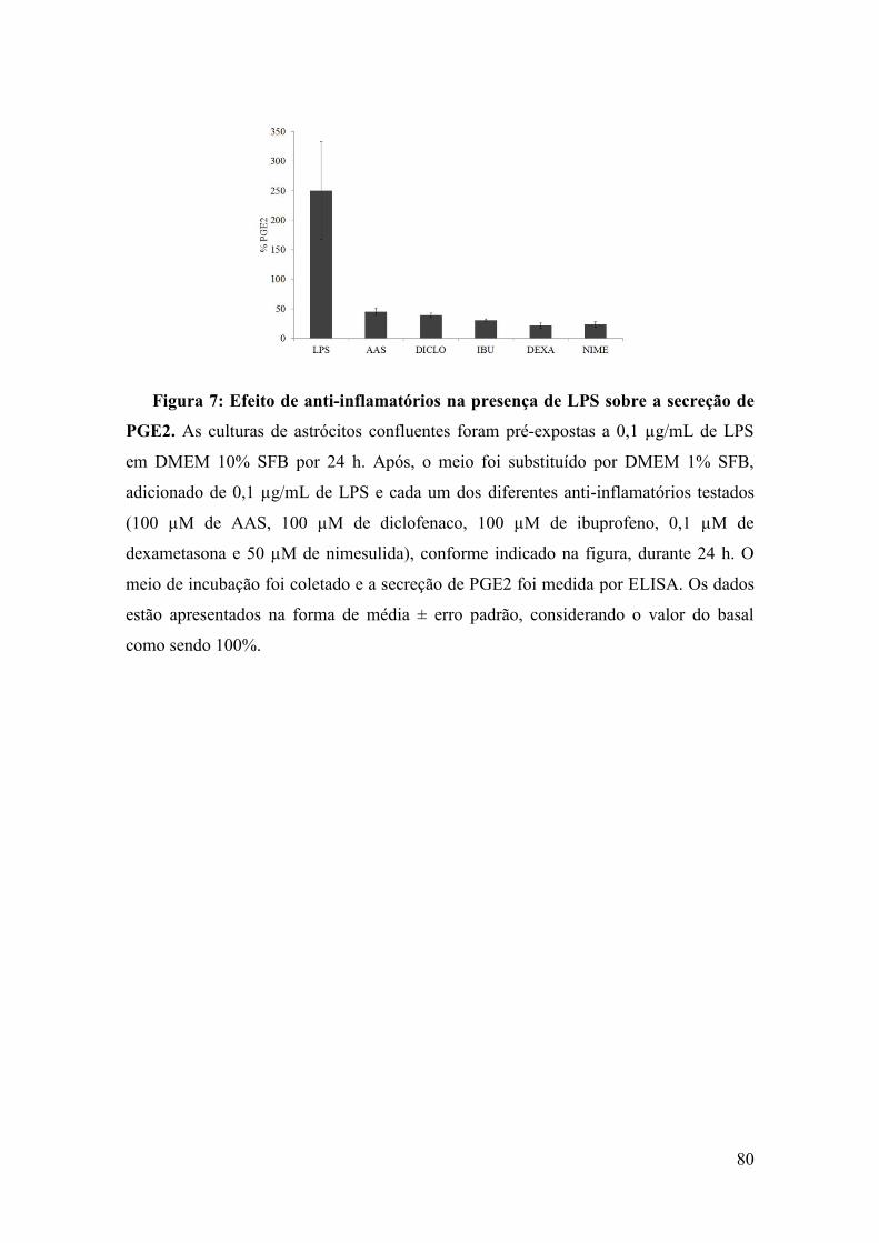
80
Figura 7: Efeito de anti-inflamatórios na presença de LPS sobre a secreção de
PGE2. As culturas de astrócitos confluentes foram pré-expostas a 0,1 µg/mL de LPS
em DMEM 10% SFB por 24 h. Após, o meio foi substituído por DMEM 1% SFB,
adicionado de 0,1 µg/mL de LPS e cada um dos diferentes anti-inflamatórios testados
(100 µM de AAS, 100 µM de diclofenaco, 100 µM de ibuprofeno, 0,1 µM de
dexametasona e 50 µM de nimesulida), conforme indicado na figura, durante 24 h. O
meio de incubação foi coletado e a secreção de PGE2 foi medida por ELISA. Os dados
estão apresentados na forma de média ± erro padrão, considerando o valor do basal
como sendo 100%.
81
Descrição dos resultados
Nós avaliamos o conteúdo da proteína GFAP e da proteína S100B na presença de
LPS e anti-inflamatórios e nenhuma mudança no conteúdo dessas proteínas foi
observada (Figuras 1 e 2).
Outro parâmetro avaliado foi a secreção de S100B em 24 h de exposição ao LPS e
na presença de anti-inflamatórios. Nós observamos uma diminuição na secreção de
S100B na presença de dexametasona. O ibuprofeno, na presença de LPS, aumentou a
secreção de S100B (Figura 3).
Em relação à liberação de TNF-α, somente a dexametasona foi capaz de reverter o
aumento observado nas culturas expostas ao LPS (Figura 4).
Em relação às defesas antioxidantes, ibuprofeno e nimesulida foram capazes de
reverter a diminuição de GSH observada na presença de LPS (Figura 5).
Em relação à viabilidade e integridade celular, nenhum comprometimento foi
observado. Estes resultados são mostrados com base nos ensaios de redução de MTT,
incorporação de vermelho neutro e liberação de LDH. Nós observamos uma redução da
liberação de LDH na presença de dexametasona (Figura 6).
O LPS foi capaz de aumentar a liberação de PGE2, enquanto que a exposição a
todos os anti-inflamatórios testados foi capaz de reverter esse aumento (Figura 7).

82
PARTE III

83
3. Discussão
A reação inflamatória dentro do SNC difere consideravelmente de outros tecidos,
uma vez que não apresenta células dendríticas do sistema imune e a permeabilidade de
vasos para a saída de grandes moléculas e células sanguíneas é reduzida em comparação
com o resto do corpo pela presença da BHE. Outro aspecto é a dificuldade em ativar a
cascata do complemento e recrutar células envolvidas na resposta imune adaptativa
como leucócitos, com exceção de células T ativadas, as quais rapidamente penetram a
BHE (Ransohoff & Brown 2012). Isto mostra que as células imunes inatas no SNC não
são tão eficazes em recrutar a maquinaria da resposta imune adaptativa, como as células
dendríticas nos tecidos periféricos. Outra importante diferença na inflamação dentro ou
fora do parênquima cerebral são os tipos celulares envolvidos como, por exemplo, os
astrócitos e microglia no SNC.
Apesar das células microgliais, entre outras funções (Salter & Beggs 2014), serem
consideradas as células inflamatórias residentes no SNC, os astrócitos também
apresentam-se como importantes mediadores celulares no processo inflamatório.
Similar à microglia, os astrócitos tornam-se ativados em resposta a vários estímulos,
processo conhecido como astrogliose reativa. Nesta condição os astrócitos são capazes
de liberar uma ampla variedade de mediadores como, por exemplo, citocinas pró-
inflamatórias.
Um importante parâmetro de ativação astroglial é a proteína S100B (Rothermundt et
al. 2003), a qual é predominantemente expressa e secretada por astrócitos no SNC e
exerce efeitos autócrinos e parácrinos em neurônios e células gliais (Donato 2001,
Donato et al. 2009). Esta proteína é capaz de atravessar a BHE e sua concentração no
soro ou LCR parece refletir o grau de dano nas doenças cerebrais, sendo considerada
como um marcador de dano cerebral (Goncalves et al. 2008). A S100B parece ser capaz

84
de modular a secreção de citocinas e também ser modulada por estas (de Souza et al.
2009, Schmitt et al. 2007). Por se comportar muitas vezes como uma citocina, a S100B
já foi considerada como tal, mas ao contrário das citocinas clássicas, a S100B não é
produzida exclusivamente para secreção, somente uma pequena parte é exportada,
portanto esta ideia é controversa.
Estudos tem mostrado que astrócitos são capazes de responder ao LPS, o qual é um
estímulo amplamente usado nos modelos de neuroinflamação. O LPS é capaz de
diminuir a expressão de algumas proteínas como as proteínas de junção gap (Liao et al.
, Liao et al. 2010) e aumentar a expressão de outras como a proteína GFAP e glutationa-
S-transferase (Vergara et al. 2010, Brahmachari et al. 2006).
Uma hipótese é a S100B ser um marcador de dano cerebral e o aumento desta
proteína no sangue periférico pode ser um indicativo de dano. Entretanto dependendo da
condição patofisiológica, outras interpretações são possíveis, principalmente pela
existência de fontes extracerebrais de S100B (Goncalves et al. 2008, Goncalves et al.
2010).
Alguns estudos trazem a medida dos níveis de S100B no soro após exposição ao
LPS (Garnier et al. 2006, Rosengarten et al. 2009). No nosso estudo, a administração
de LPS ICV ou IP em ratos Wistar não alterou os níveis de S100B no soro medida 30
min e 24 h após a exposição. Esta divergência pode ser devida à
quantidades diferentes de LPS utilizado, do seu modo de administração, ou a linhagem
do animal.
É importante ressaltar que LPS (IV 2 ng/kg), quando administrado a seres humanos,
não é capaz de induzir mudanças significativas no S100B sérica em 1h ou 8h
pós-tratamento (van den Boogaard et al. 2010). Além de medir S100B no soro, nós
também avaliamos os níveis de S100B no LCR e encontramos um aumento de S100B
85
após LPS tanto para a administração ICV (uma resposta precoce e persitente) como IP
(uma resposta precoce e transitória). Claramente nenhum aumento imediato ocorreu em
ambas as condições. Isto sugere uma liberação de S100B cérebro específica induzida
por LPS, mostrando talvez que células da periferia que secretam S100B como
adipócitos não liberam ou causam uma liberação detectável de S100B quando
estimuladas por LPS. Isso mostra que a secreção de S100B responde de maneiras
diferentes a estímulo por LPS em células que expressam S100B na periferia e no SNC.
Ao contrário da S100B, nós observamos um aumento imediato de TNF-α no soro,
após a administração de LPS tanto IP quanto ICV. Tem sido proposto que TNF-α é
capaz de mediar a secreção de S100B em astrócitos (Edwards & Robinson 2006),
Entretanto nossos resultados de avaliação dos níveis de TNF e S100B sugerem
respostas independentes em relação ao LPS. Outro aspecto a ser enfatizado é que o
aumento dos níveis de S100B no LCR não foi acompanhado por um aumento nos níveis
no soro, pelo menos nos tempos avaliados (30 min e 24 h após LPS). O aumento da
S100B no líquor foi rápido (dectectada em 30 min) e persistente (por pelo menos 24 h).
Os animais controles envolvidos no experimento envolvendo administração de LPS
ICV apresentaram níveis maiores de S100B no líquor do que os controles envolvidos na
administração de LPS IP, sugerindo uma reposta a um procedimento invasivo.
Em cultura de astrócitos nós observamos um aumento na secreção de S100B em 1 h
de exposição ao LPS em concentrações acima de 10 µg/mL, mas em 24 h nós
observamos uma diminuição da secreção mesmo nas concentrações mais baixas de LPS.
Isto sugere uma resposta bifásica, isto é, um aumento seguido por uma diminuição na
secreção de S100B. Este perfil tem sido observado em cultura de astrócitos sobre outras
condições, como exposição ao beta-hidroxibutirato (Leite et al. 2004). Esta rápida e
transitória estimulação da secreção em cultura de astrócitos também foi observada para

86
a IL1β, mas sem a diminuição em 24 h. Estes dados podem sugerir que o efeito do LPS
é direto e independe da liberação secundária de IL-1β. Portanto, de acordo com nossa
hipótese de trabalho, parece que o LPS é capaz de modular diretamente a secreção de
S100B.
Além de cultura de astrócitos, nós também avaliamos a secreção de S100B em fatias
hipocampais em 1 h de exposição ao LPS e observamos um aumento na secreção de
S100B com LPS na concentração de 10 µg/mL, mas ao contrário nas concentrações de
0,1 µg/mL e 1 µg/ houve uma diminuição na secreção. Em relação a esta metodologia, a
interpretação dos resultados é difícil devido a preparação apresentar mais de um tipo
celular como a presença de astrócitos, microglia ativada e neurônios. Entretanto, um
resultado similar tem sido observado em relação à endotelina-1 (Leite et al. 2009). Este
composto bloqueia junções gap e aumenta a secreção de S100B em 1 h, mas diminui
após 6 h. De forma semelhante, a endotelina-1 diminui a secreção de S100B em 1 h em
fatias hipocampais fresca. Potencialmente, ambos LPS e endotelina-1 inibem proteínas
de junção gap. Embora nós não tenhamos dúvidas sobre os efeitos do LPS e da
endotelina-1 na secreção de S100B em fatias hipocampais fresca, nós não temos neste
momento uma explicação para este efeito quando comparado a astrócitos isolados.
O conteúdo de S100B proveniente da sua secreção corresponde a uma pequena
fração do conteúdo desta proteína nas células (menos que 0,5% é encontrado no meio de
cultura de astrócitos em 24 h) e mudanças na secreção de S100B não necessariamente
são acompanhadas por mudanças no conteúdo celular (Goncalves et al. 2008). De fato,
nos nossos experimentos o LPS mudou a secreção sem alterar o conteúdo intracelular de
S100B. Por outro lado o conteúdo de GFAP aumentou em todas as concentrações de
LPS testadas, indicando ativação astroglial, estando de acordo com estudos prévios
sobre os efeitos de LPS em cultura de astrócitos (Vergara et al. 2010, Brahmachari et al.

87
2006). Estes resultados reforçam a ideia que GFAP e S100B têm mecanismos
regulatórios de expressão distintos e que a astrogliose (medida por o aumento de GFAP)
pode ser acompanhada ou não por mudanças no conteúdo intracelular de S100B.
Outro interessante resultado é a diminuição do conteúdo de glutationa após
exposição ao LPS em cultura de astrócitos em 24 h e não em 1 h, possivelmente
associado com a regulação positiva da glutationa-S-transferase (Vergara et al. 2010).
Parte da diminuição de GSH pode estar envolvendo uma intensa exportação deste
peptídeo, uma vez que ele serve como um antioxidante extracelular e também
proporciona substratos para a síntese neuronal de glutationa (Dringen & Hirrlinger
2003). Além disso, nós também encontramos uma diminuição no conteúdo de glutationa
em fatias hipocampais expostas ao LPS.
Apesar da diminuição nas defesas antioxidantes, ambas as preparações não
apresentam nenhum comprometimento na viabilidade e integridade celular, mostrado
com base nos ensaios de redução de MTT, incorporação de vermelho neutro e liberação
de LDH. Estes ensaios executados em paralelo com os ensaios para medir S100B, nos
permite ser enfático em relação à secreção e não à liberação de S100B.
Recentemente alguns autores sugerem que a S100B deve agir como uma alarmina
ou padrão molecular associado a dano (Bianchi 2007). Entretanto independente de
qualquer conceito, nossos dados sugerem que a secreção de S100B é modulada por
LPS, podendo ser protetora durante a fase inicial de exposição ao LPS. Ao contrário sua
diminuição a uma exposição mais prolongada de astrócitos ao LPS dose dependente
pode estar relacionada a diminuição de fatores tróficos por astrócitos nestas condições.
Como já comentado anteriormente, o receptor TLR4 é essencial no reconhecimento
do LPS. Então, para caracterizar se o efeito do LPS é via TLR4, nós estimulamos os
astrócitos com dois antagonistas específicos desse receptor (Cli-095 e OxPAPC) e

88
ambos foram capazes de abolir o efeito do LPS. Esse resultado nos indica que a
modulação de S100B por LPS é dependente da união do LPS ao seu receptor TLR4,
uma vez que, estudos recentes mostram que TLRs podem compartilhar vias de
sinalização e ligantes com o receptor RAGE como, por exemplo, o LPS (Ibrahim et al.,
2013). Este é capaz de se ligar ao RAGE e esta ligação desencadeia uma resposta
similar à observada com TLR4, mas parece não necessitar das moléculas necessárias
para a ligação ao TLR4 (Ibrahim et al. 2013).
Embora estes dados tenham contribuído para o nosso entendimento sobre o efeito do
LPS na secreção de S100B, alguns aspectos permanecem pouco claros, como por
exemplo, a possibilidade do LPS estar agindo sobre outro receptor que não o TLR4 nos
astrócitos, ou o LPS estar modulando a secreção em outras células extracerebrais que
expressam S100B afetando os níveis séricos desta proteína. Deste modo efeitos
específicos e crônicos de bactérias gram-negativas sobre a proteína S100B tanto no
SNC como sistema periférico merece maior investigação.
Como já descrito a proteína S100B extracelular é conhecida por afetar atividades
microgliais, neuronais e astrocíticas com diferentes efeitos dependendo de sua
concentração. Enquanto em baixas concentrações exerce efeitos tróficos em neurônios e
astrócitos, em concentrações relativamente altas, pode causar apoptose neuronal e ativar
astrócitos e microglia, então representando um fator endógeno implicado na
neuroinflamação. Essa proteína é amplamente usada como parâmetro de ativação e ou
morte astroglial em várias condições de dano cerebral e variações dessa proteína no soro
e LCR são propostas para avaliar resultados clínicos nestas situações (Gonçalves et al.
2008).
Uma vez que o aumento de S100B no SNC ocorre em diversas doenças
neurodegenerativas como, por exemplo, doença de Alzheimer (Mori et al. 2010),
89
doença de Parkinson (Sathe et al. 2012), esquizofrenia (Rothermundt et al. 2004,
Steiner et al. 2006), e vários estudos mostram o possível efeito de fármacos anti-
inflamatórios sobre estas patologias, nós consideramos interessante avaliar o efeito dos
anti-inflamatórios não esteroidais como aspirina, diclofenaco, ibuprofeno e nimesulida
e esteroidais como a dexametasona sobre a secreção e o conteúdo intracelular da
proteína S100B em cultura de astrócitos e no soro e LCR de ratos Wistar na ausência de
estímulo inflamatório.
Primeiramente nós avaliamos a proteína GFAP conhecida por ser um marcador de
astrogliose e podendo também ser observada em astrócitos durante processos
neuropatológicos. Entretanto, o mecanismo molecular preciso que leva a esta indução
durante processos neuropatológicos permanece pouco esclarecido. Nós observamos um
aumento no conteúdo de GFAP em cultura de astrócitos após 24 h de exposição ao AAS
e diclofenaco. Quando analisamos estes anti-inflamatórios in vivo, observamos também
um aumento de GFAP em resposta à exposição ao AAS no córtex e uma tendência à
diminuição no hipocampo, mostrando efeitos diferentes para regiões distintas do SNC.
Nenhum resultado com diclofenaco foi significativo nas regiões testadas.
Em relação ao efeito do AAS na proteína GFAP, um estudo em glioblastoma
humano mostra uma diminuição na expressão de GFAP na presença de aspirina, um
resultado dependente de concentração (2 e 5 mM) (Bae et al. 2006) ao contrário do
nosso resultado em cultura primária, mas compatível com nosso resultado in vivo, onde
tivemos uma tendência de diminuição de GFAP no hipocampo. Esta diferença
observada neste estudo em relação ao nosso resultado na modulação da GFAP pode
estar relacionada à dose de AAS utilizada. No trabalho citado a concentração do
fármaco é muito mais alta em relação a que utilizamos podendo estar sendo tóxica para
as células. Além disso, o modelo utilizado no estudo é uma linhagem de astrócitos, que

90
embora seja um modelo muito utilizado para estudar essas células, apresenta limitações
biológicas em relação à cultura primária, que podem influenciar no resultado observado.
Estes resultados mostram que os anti-inflamatórios podem apresentar diferentes efeitos
dependendo da condição a que são expostos.
O efeito inibitório da aspirina sobre a GFAP pode ser via NFκ-B, uma vez que
estudos mostram que a aspirina é capaz de inibir a ativação de NFκ-B (Grilli et al.
1996), o qual é um regulador da expressão gênica de GFAP em células astrogliais (Bae
et al. 2006). Quando testados em relação ao conteúdo intracelular de S100B nenhum
dos anti-inflamatórios foi capaz de alterar o conteúdo desta proteína.
Um resultado interessante foi o efeito da dexametasona e nimesulida sobre a
secreção de S100B e liberação de TNF-α após 24 h de tratamento. Tanto dexametasona
como a nimesulida são inibidores específicos da COX2 e foram capazes de diminuir
tanto a secreção de S100B, quanto a produção de TNF-α nas culturas astrogliais. A
dexametasona é um anti-inflamatório esteroidal capaz de inibir a expressão gênica de
proteínas proinflamatórias como TNF-α e a enzima COX2. A inibição da síntese de
COX2 consequentemente inibe a síntese de prostaglandinas, principais prostanóides
inflamatórios. Entre estes prostanóides está a PGE2, um dos mais importantes
moduladores da inflamação e parece contribuir para a resposta inflamatória na doença
de Alzheimer (Cudaback et al. 2014). A PGE2 age em seu receptor EP2 acoplado a
proteína G em astrócitos promovendo o aumento de adenosina monofosfato cíclico
(AMPc) intracelular (Bos et al. 2004). Uma vez que AMPc e cálcio estão envolvidos no
aumento da secreção de S100B, a diminuição observada nos níveis desta proteína
poderiam ser via PGE2.
Os experimentos não apresentam nenhum comprometimento na viabilidade e
integridade celular, mostrado com base nos ensaios de redução de MTT, incorporação

91
de vermelho neutro e liberação de LDH. A diminuição na incorporação de vermelho
neutro observada na presença de ibuprofeno não nos parece ser um efeito significativo,
visto que, os outros ensaios não mostraram nenhuma alteração. A diminuição
significativa da liberação de LDH na presença de dexametasona pode ser devido a
alguma interferência na técnica utilizada. Estes ensaios executados em paralelo com os
ensaios para medir S100B, nos permite ser enfático em relação à secreção e não
liberação de S100B.
Em relação às defesas antioxidantes, nós não observamos nenhuma alteração no nos
níveis de GSH com os anti-inflamatórios testados.
Como observamos que o LPS é capaz de modular a secreção de S100B e que cada
um dos anti-inflamatórios estudados pode ou não ter efeito na secreção de S100B, nós
resolvemos investigar como a secreção e o imunoconteúdo da proteína S100B em
culturas astrogliais estimuladas com LPS irão se comportar na presença desses anti-
inflamatórios.
Nós avaliamos o conteúdo da proteína GFAP na presença de LPS e anti-
inflamatórios. Nenhum anti-inflamatório foi capaz de reverter o aumento de GFAP
causado pelo LPS. Curiosamente, AAS e diclofenaco, que sozinhos foram capazes de
aumentar o conteúdo de GFAP, não tiveram um efeito aditivo na presença de LPS.
Tanto na presença como na ausência de LPS, nenhum dos anti-inflamatórios testados foi
capaz de modular o conteúdo intracelular de S100B.
Outro parâmetro avaliado foi a secreção de S100B em 24 h de exposição ao LPS e
na presença de anti-inflamatórios. Nós observamos uma diminuição na secreção de
S100B na presença de dexametasona, o mesmo resultado obtido na ausência do
estímulo por LPS. Este resultado mostra que a dexametasona, um anti-inflamatório
esteroidal é capaz de inibir a secreção de S100B também em uma condição inflamatória

92
e como já discutido podendo esta inibição estar relacionada com a via das
prostaglandinas. O mesmo perfil foi observado com a liberação de TNF-α, pois a
dexametasona foi capaz de diminuir a liberação desta citocina na ausência e na presença
de LPS. Este resultado nos faz pensar na S100B tendo um papel de citocina com caráter
próinflamatório.
Interessantemente a nimesulida que em culturas não estimuladas por LPS foi capaz
de diminuir a secreção de S100B, na presença de estímulo, o efeito desapareceu, não
sendo capaz de reverter o efeito do LPS sobre a secreção de S100B. Na secreção de
TNF- α, a nimesulida apresentou o mesmo perfil nos dois desenhos experimentais
utilizados, uma tendência a redução.
O ibuprofeno que em culturas não estimuladas não apresentou nenhum efeito sobre
a secreção de S100B, na presença de LPS curiosamente aumentou a secreção de S100B.
Em relação a liberação de TNF-α, o ibuprofeno sozinho aumento a liberação de TNF-α,
mas na presença de LPS este efeito desapareceu.
Em relação à viabilidade e integridade celular, tanto nas culturas astrogliais não
estimuladas, quanto às estimuladas por LPS, nenhum comprometimento foi observado.
Estes resultados são mostrados com base nos ensaios de redução de MTT, incorporação
de vermelho neutro e liberação de LDH. A redução observada na incorporação de
vermelho neutro na presença de diclofenaco em culturas não estimuladas desapareceu
na presença de LPS, sugerindo que esse resultado não implica uma redução na
viabilidade celular. A redução da liberação de LDH observada na presença de
dexametasona em culturas não estimuladas se manteve nas culturas expostas ao LPS,
corroborando a hipótese de uma possível interferência na técnica utilizada. Estes ensaios
executados em paralelo com os ensaios para medir S100B, nos permite ser enfático em
relação à secreção e não liberação de S100B.

93
Em relação às defesas antioxidantes, interessantemente ibuprofeno e nimesulida
foram capazes de reverter a redução do conteúdo de GSH observada na presença de
estímulo inflamatório, sem ser observado efeito isolado destes fármacos. De acordo com
nossos resultados, estudos mostram que a nimesulida é capaz de conferir proteção
antioxidante através do aumento de GSH em modelo de isquemia (Candelario-Jalil et al.
2003) e também em astrocitoma e linhagem C6 estimuladas com LPS (Niranjan et al.
2010). Embora outros mecanismos possam estar envolvidos, a capacidade antioxidante
da nimesulida pode ser por sua inibição seletiva da COX2 e consequentemente de
prostaglandinas, considerando que o metabolismo do ácido araquidônico por
ciclooxigenases é uma das principais fontes de espécies reativas de oxigênio.

94
4. Conclusões
De acordo com os resultados obtidos neste trabalho, nós observamos que o LPS
causa uma resposta inflamatória no SNC diferente da resposta inflamatória no sistema
periférico, uma vez que é capaz de modular a liberação de S100B e TNF-α de maneiras
diferentes nos dois sistemas. O LPS é capaz de estimular a secreção de S100B no SNC,
mas células periféricas que expressam S100B quando estimuladas por LPS não parecem
liberar essa proteína, diferentemente do perfil observado para a liberação de TNF-α, o
qual apresenta um aumento tanto no SNC quanto na periferia. Em culturas de astrócitos,
o LPS é capaz de estimular a secreção de S100B, portanto sendo capaz de modular
diretamente esta proteína. Juntos estes dados contribuem para entender o efeito de LPS
em astrócitos particularmente na secreção de S100B e nos ajuda a interpretar mudanças
nesta proteína que ocorrem no LCR e no soro em doenças neuroinflamatórias e
desordens cerebrais em geral.
Em relação ao estudo com anti-inflamatórios em parâmetros gliais, nós observamos
que estes fármacos apresentam diferentes efeitos em diferentes condições. Todos os
anti-inflamatórios estudados são capazes de atravessar a BHE e, portanto podem
apresentar diferentes efeitos no SNC. Além da COX, outras proteínas estão envolvidas
no processo inflamatório e os anti-inflamatórios podem agir sobre essas proteínas. Por
exemplo, sabe-se que ácido flufenâmico, um anti-inflamatório não-esteroidal e inibidor
de junção gap, é capaz de aumentar a secreção de S100B em cultura de astrócitos e
nimesulida diminui a ativação de NF-kβ e MAPK.
Nossos resultados confirmam a presença de COX 1 e 2, bem como níveis basais de
PGE2. Diferentemente do resultado esperado, onde todos os anti-inflamatórios poderiam
reduzir a secreção de S100B, uma vez que AMPc modula a secreção de S100B e a
PGE2 estimula receptores acoplados a proteína G e a adenilato ciclase,

95
interessantemente somente a nimesulida e dexametasona foram capazes de reduzir a
secreção basal de S100B e TNF-α. Além disso, AAS e diclofenaco aumentam GFAP em
cultura de astrócitos, mas somente o AAS foi capaz de reproduzir o mesmo efeito in
vivo. Todos os anti-inflamatórios foram capazes de reduzir os níveis de PGE2. Portanto
a PGE2 pode estar envolvida no mecanismo de secreção de S100B, no entanto vias
bioquímicas envolvidas, não tão claras neste momento, necessitam ser melhor
caracterizadas.

96
5. Referências Bibliográficas
Abbott, N. J., Ronnback, L. and Hansson, E. (2006) Astrocyte-endothelial interactions
at the blood-brain barrier. Nat Rev Neurosci, 7, 41-53.
Aid, S. and Bosetti, F. (2011) Targeting cyclooxygenases-1 and -2 in
neuroinflammation: Therapeutic implications. Biochimie, 93, 46-51.
Akira, S., Uematsu, S. and Takeuchi, O. (2006) Pathogen recognition and innate
immunity. Cell, 124, 783-801.
Akiyama, H., Barger, S., Barnum, S. et al. (2000) Inflammation and Alzheimer's
disease. Neurobiology of aging, 21, 383-421.
Allaman, I., Belanger, M. and Magistretti, P. J. (2011) Astrocyte-neuron metabolic
relationships: for better and for worse. Trends in neurosciences, 34, 76-87.
Aoyama, K., Watabe, M. and Nakaki, T. (2008) Regulation of neuronal glutathione
synthesis. J Pharmacol Sci, 108, 227-238.
Asai, H., Kakita, H., Aoyama, M., Nagaya, Y., Saitoh, S. and Asai, K. (2013)
Diclofenac enhances proinflammatory cytokine-induced aquaporin-4 expression
in cultured astrocyte. Cellular and molecular neurobiology, 33, 393-400.
Bae, M. K., Kim, S. R., Lee, H. J. et al. (2006) Aspirin-induced blockade of NF-kappaB
activity restrains up-regulation of glial fibrillary acidic protein in human
astroglial cells. Biochimica et biophysica acta, 1763, 282-289.
Basselin, M., Ramadan, E., Chen, M. and Rapoport, S. I. (2011) Anti-inflammatory
effects of chronic aspirin on brain arachidonic acid metabolites. Neurochem Res,
36, 139-145.
Bianchi, M. E. (2007) DAMPs, PAMPs and alarmins: all we need to know about
danger. J Leukoc Biol, 81, 1-5.

97
Bianchi, R., Giambanco, I. and Donato, R. (2008) S100B/RAGE-dependent activation
of microglia via NF-kappaB and AP-1 Co-regulation of COX-2 expression by
S100B, IL-1beta and TNF-alpha. Neurobiol Aging.
Bos, C. L., Richel, D. J., Ritsema, T., Peppelenbosch, M. P. and Versteeg, H. H. (2004)
Prostanoids and prostanoid receptors in signal transduction. Int J Biochem Cell
Biol, 36, 1187-1205.
Bowman, C. C., Rasley, A., Tranguch, S. L. and Marriott, I. (2003) Cultured astrocytes
express toll-like receptors for bacterial products. Glia, 43, 281-291.
Brahmachari, S., Fung, Y. K. and Pahan, K. (2006) Induction of glial fibrillary acidic
protein expression in astrocytes by nitric oxide. J Neurosci, 26, 4930-4939.
Candelario-Jalil, E., Alvarez, D., Merino, N. and Leon, O. S. (2003) Delayed treatment
with nimesulide reduces measures of oxidative stress following global ischemic
brain injury in gerbils. Neuroscience research, 47, 245-253.
Candelario-Jalil, E. and Fiebich, B. L. (2008) Cyclooxygenase inhibition in ischemic
brain injury. Current pharmaceutical design, 14, 1401-1418.
Carpentier, P. A., Begolka, W. S., Olson, J. K., Elhofy, A., Karpus, W. J. and Miller, S.
D. (2005) Differential activation of astrocytes by innate and adaptive immune
stimuli. Glia, 49, 360-374.
Carrasco, E. and Werner, P. (2002) Selective destruction of dopaminergic neurons by
low concentrations of 6-OHDA and MPP+: protection by acetylsalicylic acid
aspirin. Parkinsonism & related disorders, 8, 407-411.
Casper, D., Yaparpalvi, U., Rempel, N. and Werner, P. (2000) Ibuprofen protects
dopaminergic neurons against glutamate toxicity in vitro. Neurosci Lett, 289,
201-204.
98
Choi, S. H., Langenbach, R. and Bosetti, F. (2008) Genetic deletion or pharmacological
inhibition of cyclooxygenase-1 attenuate lipopolysaccharide-induced
inflammatory response and brain injury. FASEB journal : official publication of
the Federation of American Societies for Experimental Biology, 22, 1491-1501.
Christopherson, K. S., Ullian, E. M., Stokes, C. C. et al. (2005) Thrombospondins are
astrocyte-secreted proteins that promote CNS synaptogenesis. Cell, 120, 421-
433.
Cudaback, E., Jorstad, N. L., Yang, Y., Montine, T. J. and Keene, C. D. (2014)
Therapeutic implications of the prostaglandin pathway in Alzheimer's disease.
Biochem Pharmacol, 88, 565-572.
Davalos, D., Grutzendler, J., Yang, G., Kim, J. V., Zuo, Y., Jung, S., Littman, D. R.,
Dustin, M. L. and Gan, W. B. (2005) ATP mediates rapid microglial response to
local brain injury in vivo. Nature neuroscience, 8, 752-758.
De Cristobal, J., Cardenas, A., Lizasoain, I., Leza, J. C., Fernandez-Tome, P., Lorenzo,
P. and Moro, M. A. (2002) Inhibition of glutamate release via recovery of ATP
levels accounts for a neuroprotective effect of aspirin in rat cortical neurons
exposed to oxygen-glucose deprivation. Stroke, 33, 261-267.
de Souza, D. F., Leite, M. C., Quincozes-Santos, A., Nardin, P., Tortorelli, L. S., Rigo,
M. M., Gottfried, C., Leal, R. B. and Goncalves, C. A. (2009) S100B secretion
is stimulated by IL-1beta in glial cultures and hippocampal slices of rats: Likely
involvement of MAPK pathway. J Neuroimmunol, 206, 52-57.
de Souza, D. F., Wartchow, K., Hansen, F., Lunardi, P., Guerra, M. C., Nardin, P. and
Goncalves, C. A. (2013) Interleukin-6-induced S100B secretion is inhibited by
haloperidol and risperidone. Progress in neuro-psychopharmacology &
biological psychiatry, 43, 14-22.
99
Donato, R. (2001) S100: a multigenic family of calcium-modulated proteins of the EF-
hand type with intracellular and extracellular functional roles. Int J Biochem Cell
Biol, 33, 637-668.
Donato, R., Sorci, G., Riuzzi, F., Arcuri, C., Bianchi, R., Brozzi, F., Tubaro, C. and
Giambanco, I. (2009) S100B's double life: intracellular regulator and
extracellular signal. Biochimica et biophysica acta, 1793, 1008-1022.
Dringen, R., Gutterer, J. M. and Hirrlinger, J. (2000) Glutathione metabolism in brain
metabolic interaction between astrocytes and neurons in the defense against
reactive oxygen species. Eur J Biochem, 267, 4912-4916.
Dringen, R. and Hirrlinger, J. (2003) Glutathione pathways in the brain. Biol Chem,
384, 505-516.
Dubois, R. N., Abramson, S. B., Crofford, L., Gupta, R. A., Simon, L. S., Van De Putte,
L. B. and Lipsky, P. E. (1998) Cyclooxygenase in biology and disease. FASEB
journal : official publication of the Federation of American Societies for
Experimental Biology, 12, 1063-1073.
Edwards, M. M. and Robinson, S. R. (2006) TNF alpha affects the expression of GFAP
and S100B: implications for Alzheimer's disease. J Neural Transm, 113, 1709-
1715.
Engelhardt, B. (2010) T cell migration into the central nervous system during health and
disease: Different molecular keys allow access to different central nervous
system compartments. Clinical and Experimental Neuroimmunology, 1, 79-93.
Fang, F., Lue, L. F., Yan, S. et al. (2010) RAGE-dependent signaling in microglia
contributes to neuroinflammation, Abeta accumulation, and impaired
learning/memory in a mouse model of Alzheimer's disease. FASEB journal :
100
official publication of the Federation of American Societies for Experimental
Biology, 24, 1043-1055.
Farina, C., Aloisi, F. and Meinl, E. (2007) Astrocytes are active players in cerebral
innate immunity. Trends Immunol, 28, 138-145.
Font-Nieves, M., Sans-Fons, M. G., Gorina, R., Bonfill-Teixidor, E., Salas-Perdomo,
A., Marquez-Kisinousky, L., Santalucia, T. and Planas, A. M. (2012) Induction
of COX-2 enzyme and down-regulation of COX-1 expression by
lipopolysaccharide (LPS) control prostaglandin E2 production in astrocytes. The
Journal of biological chemistry, 287, 6454-6468.
Franke, H., Krugel, U. and Illes, P. (2006) P2 receptors and neuronal injury. Pflugers
Arch, 452, 622-644.
Frizzo, J. K., Tramontina, F., Bortoli, E., Gottfried, C., Leal, R. B., Lengyel, I., Donato,
R., Dunkley, P. R. and Goncalves, C. A. (2004) S100B-mediated inhibition of
the phosphorylation of GFAP is prevented by TRTK-12. Neurochem Res, 29,
735-740.
Garnier, Y., Berger, R., Alm, S. et al. (2006) Systemic endotoxin administration results
in increased S100B protein blood levels and periventricular brain white matter
injury in the preterm fetal sheep. European journal of obstetrics, gynecology,
and reproductive biology, 124, 15-22.
Glass, C. K., Saijo, K., Winner, B., Marchetto, M. C. and Gage, F. H. (2010)
Mechanisms underlying inflammation in neurodegeneration. Cell, 140, 918-934.
Glezer, I., Simard, A. R. and Rivest, S. (2007) Neuroprotective role of the innate
immune system by microglia. Neuroscience, 147, 867-883.

101
Goncalves, C. A., Concli Leite, M. and Nardin, P. (2008) Biological and
methodological features of the measurement of S100B, a putative marker of
brain injury. Clin Biochem, 41, 755-763.
Goncalves, C. A., Leite, M. C. and Guerra, M. C. (2010) Adipocytes as an Important
Source of Serum S100B and Possible Roles of This Protein in Adipose Tissue.
Cardiovascular psychiatry and neurology, 2010, 790431.
Griffin, W. S. (2006) Inflammation and neurodegenerative diseases. Am J Clin Nutr, 83,
470S-474S.
Grilli, M., Pizzi, M., Memo, M. and Spano, P. (1996) Neuroprotection by aspirin and
sodium salicylate through blockade of NF-kappaB activation. Science, 274,
1383-1385.
Heneka, M. T., Sastre, M., Dumitrescu-Ozimek, L. et al. (2005) Acute treatment with
the PPARgamma agonist pioglitazone and ibuprofen reduces glial inflammation
and Abeta1-42 levels in APPV717I transgenic mice. Brain : a journal of
neurology, 128, 1442-1453.
Hinkerohe, D., Smikalla, D., Schoebel, A., Haghikia, A., Zoidl, G., Haase, C. G.,
Schlegel, U. and Faustmann, P. M. (2010) Dexamethasone prevents LPS-
induced microglial activation and astroglial impairment in an experimental
bacterial meningitis co-culture model. Brain Res, 1329, 45-54.
Hinkerohe, D., Wolfkuhler, D., Haghikia, A., Meier, C., Faustmann, P. M. and
Schlegel, U. (2011) Dexamethasone differentially regulates functional
membrane properties in glioma cell lines and primary astrocytes in vitro.
Journal of neuro-oncology, 103, 479-489.

102
Holm, T. H., Draeby, D. and Owens, T. (2012) Microglia are required for astroglial
Toll-like receptor 4 response and for optimal TLR2 and TLR3 response. Glia,
60, 630-638.
Huo, Y., Rangarajan, P., Ling, E. A. and Dheen, S. T. (2011) Dexamethasone inhibits
the Nox-dependent ROS production via suppression of MKP-1-dependent
MAPK pathways in activated microglia. BMC Neurosci, 12, 49.
Ibrahim, Z. A., Armour, C. L., Phipps, S. and Sukkar, M. B. (2013) RAGE and TLRs:
relatives, friends or neighbours? Molecular immunology, 56, 739-744.
Iwata, Y., Nicole, O., Zurakowski, D., Okamura, T. and Jonas, R. A. (2010) Ibuprofen
for neuroprotection after cerebral ischemia. The Journal of thoracic and
cardiovascular surgery, 139, 489-493.
Janeway, C. A., Jr. and Medzhitov, R. (2002) Innate immune recognition. Annu Rev
Immunol, 20, 197-216.
Johann, S., Kampmann, E., Denecke, B., Arnold, S., Kipp, M., Mey, J. and Beyer, C.
(2008) Expression of enzymes involved in the prostanoid metabolism by cortical
astrocytes after LPS-induced inflammation. J Mol Neurosci, 34, 177-185.
Kakita, H., Aoyama, M., Hussein, M. H., Kato, S., Suzuki, S., Ito, T., Togari, H. and
Asai, K. (2009) Diclofenac enhances proinflammatory cytokine-induced nitric
oxide production through NF-kappaB signaling in cultured astrocytes.
Toxicology and applied pharmacology, 238, 56-63.
Kakita, H., Aoyama, M., Nagaya, Y., Asai, H., Hussein, M. H., Suzuki, M., Kato, S.,
Saitoh, S. and Asai, K. (2013) Diclofenac enhances proinflammatory cytokine-
induced phagocytosis of cultured microglia via nitric oxide production.
Toxicology and applied pharmacology, 268, 99-105.

103
Kaplanski, J., Nassar, A., Sharon-Granit, Y., Jabareen, A., Kobal, S. L. and Azab, A. N.
(2014) Lithium attenuates lipopolysaccharide-induced hypothermia in rats.
European review for medical and pharmacological sciences, 18, 1829-1837.
Kawai, T. and Akira, S. (2006) TLR signaling. Cell Death Differ, 13, 816-825.
Kawai, T. and Akira, S. (2008) Toll-like receptor and RIG-I-like receptor signaling.
Annals of the New York Academy of Sciences, 1143, 1-20.
Kawai, T. and Akira, S. (2009) The roles of TLRs, RLRs and NLRs in pathogen
recognition. International immunology, 21, 317-337.
Kipp, M., Norkute, A., Johann, S. et al. (2008) Brain-region-specific astroglial
responses in vitro after LPS exposure. J Mol Neurosci, 35, 235-243.
Kirchhoff, F., Dringen, R. and Giaume, C. (2001) Pathways of neuron-astrocyte
interactions and their possible role in neuroprotection. Eur Arch Psychiatry Clin
Neurosci, 251, 159-169.
Kotilinek, L. A., Westerman, M. A., Wang, Q. et al. (2008) Cyclooxygenase-2
inhibition improves amyloid-beta-mediated suppression of memory and synaptic
plasticity. Brain : a journal of neurology, 131, 651-664.
Laan, W., Smeets, H., de Wit, N. J., Kahn, R. S., Grobbee, D. E. and Burger, H. (2009)
Glucocorticosteroids associated with a decreased risk of psychosis. Journal of
clinical psychopharmacology, 29, 288-290.
Laterra, J. and Goldstein, G. W. (1991) Astroglial-induced in vitro angiogenesis:
requirements for RNA and protein synthesis. J Neurochem, 57, 1231-1239.
Leal, R. B., Frizzo, J. K., Tramontina, F., Fieuw-Makaroff, S., Bobrovskaya, L.,
Dunkley, P. R. and Goncalves, C. A. (2004) S100B protein stimulates
calcineurin activity. Neuroreport, 15, 317-320.
104
Lehnardt, S. (2010) Innate immunity and neuroinflammation in the CNS: the role of
microglia in Toll-like receptor-mediated neuronal injury. Glia, 58, 253-263.
Leite, M., Frizzo, J. K., Nardin, P., de Almeida, L. M., Tramontina, F., Gottfried, C. and
Goncalves, C. A. (2004) Beta-hydroxy-butyrate alters the extracellular content
of S100B in astrocyte cultures. Brain Res Bull, 64, 139-143.
Leite, M. C., Brolese, G., de Almeida, L. M., Pinero, C. C., Gottfried, C. and
Goncalves, C. A. (2006) Ammonia-induced alteration in S100B secretion in
astrocytes is not reverted by creatine addition. Brain Res Bull, 70, 179-185.
Leite, M. C., Galland, F., de Souza, D. F., Guerra, M. C., Bobermin, L., Biasibetti, R.,
Gottfried, C. and Goncalves, C. A. (2009) Gap junction inhibitors modulate
S100B secretion in astrocyte cultures and acute hippocampal slices. J Neurosci
Res.
Leon, C. G., Tory, R., Jia, J., Sivak, O. and Wasan, K. M. (2008) Discovery and
development of toll-like receptor 4 (TLR4) antagonists: a new paradigm for
treating sepsis and other diseases. Pharm Res, 25, 1751-1761.
Liao, C. K., Wang, S. M., Chen, Y. L., Wang, H. S. and Wu, J. C. Lipopolysaccharide-
induced inhibition of connexin43 gap junction communication in astrocytes is
mediated by downregulation of caveolin-3. Int J Biochem Cell Biol.
Liao, C. K., Wang, S. M., Chen, Y. L., Wang, H. S. and Wu, J. C. (2010)
Lipopolysaccharide-induced inhibition of connexin43 gap junction
communication in astrocytes is mediated by downregulation of caveolin-3. Int J
Biochem Cell Biol, 42, 762-770.
Lin, J., Blake, M., Tang, C., Zimmer, D., Rustandi, R. R., Weber, D. J. and Carrier, F.
(2001) Inhibition of p53 transcriptional activity by the S100B calcium-binding
protein. J Biol Chem, 276, 35037-35041.

105
Liu, W., Tang, Y. and Feng, J. (2011) Cross talk between activation of microglia and
astrocytes in pathological conditions in the central nervous system. Life Sci, 89,
141-146.
Magistretti, P. J. (2009) Role of glutamate in neuron-glia metabolic coupling. Am J Clin
Nutr, 90, 875S-880S.
McGeer, P. L. and McGeer, E. G. (2007) NSAIDs and Alzheimer disease:
epidemiological, animal model and clinical studies. Neurobiol Aging, 28, 639-
647.
McGeer, P. L. and Rogers, J. (1992) Anti-inflammatory agents as a therapeutic
approach to Alzheimer's disease. Neurology, 42, 447-449.
Middeldorp, J. and Hol, E. M. (2011) GFAP in health and disease. Prog Neurobiol, 93,
421-443.
Mihaylova, S., Schweighofer, H., Hackstein, H. and Rosengarten, B. (2014) Effects of
anti-inflammatory vagus nerve stimulation in endotoxemic rats on blood and
spleen lymphocyte subsets. Inflammation research : official journal of the
European Histamine Research Society ... [et al.], 63, 683-690.
Miller, S. B. (2006) Prostaglandins in health and disease: an overview. Seminars in
arthritis and rheumatism, 36, 37-49.
Mogensen, T. H. (2009) Pathogen recognition and inflammatory signaling in innate
immune defenses. Clinical microbiology reviews, 22, 240-273, Table of
Contents.
Moore, B. W. (1965) A soluble protein characteristic of the nervous system. Biochem
Biophys Res Commun, 19, 739-744.
106
Mori, T., Koyama, N., Arendash, G. W., Horikoshi-Sakuraba, Y., Tan, J. and Town, T.
(2010) Overexpression of human S100B exacerbates cerebral amyloidosis and
gliosis in the Tg2576 mouse model of Alzheimer's disease. Glia, 58, 300-314.
Morihara, T., Teter, B., Yang, F., Lim, G. P., Boudinot, S., Boudinot, F. D., Frautschy,
S. A. and Cole, G. M. (2005) Ibuprofen suppresses interleukin-1beta induction
of pro-amyloidogenic alpha1-antichymotrypsin to ameliorate beta-amyloid
(Abeta) pathology in Alzheimer's models. Neuropsychopharmacology : official
publication of the American College of Neuropsychopharmacology, 30, 1111-
1120.
Mrak, R. E. and Griffin, W. S. (2004) Trisomy 21 and the brain. J Neuropathol Exp
Neurol, 63, 679-685.
Mrak, R. E. and Griffin, W. S. (2005) Glia and their cytokines in progression of
neurodegeneration. Neurobiology of aging, 26, 349-354.
Nardin, P., Tramontina, F., Leite, M. C., Tramontina, A. C., Quincozes-Santos, A., de
Almeida, L. M., Battastini, A. M., Gottfried, C. and Goncalves, C. A. (2007)
S100B content and secretion decrease in astrocytes cultured in high-glucose
medium. Neurochem Int, 50, 774-782.
Niranjan, R., Kamat, P. K., Nath, C. and Shukla, R. (2010) Evaluation of guggulipid
and nimesulide on production of inflammatory mediators and GFAP expression
in LPS stimulated rat astrocytoma, cell line (C6). Journal of
ethnopharmacology, 127, 625-630.
Parpura, V., Heneka, M. T., Montana, V. et al. (2012) Glial cells in (patho)physiology. J
Neurochem, 121, 4-27.
Pekny, M. and Pekna, M. (2004) Astrocyte intermediate filaments in CNS pathologies
and regeneration. J Pathol, 204, 428-437.
107
Perry, V. H. and Holmes, C. (2014) Microglial priming in neurodegenerative disease.
Nature reviews. Neurology, 10, 217-224.
Pinto, S. S., Gottfried, C., Mendez, A., Goncalves, D., Karl, J., Goncalves, C. A.,
Wofchuk, S. and Rodnight, R. (2000) Immunocontent and secretion of S100B in
astrocyte cultures from different brain regions in relation to morphology. FEBS
Lett, 486, 203-207.
Pistritto, G., Franzese, O., Pozzoli, G., Mancuso, C., Tringali, G., Preziosi, P. and
Navarra, P. (1999) Bacterial lipopolysaccharide increases prostaglandin
production by rat astrocytes via inducible cyclo-oxygenase: evidence for the
involvement of nuclear factor kappaB. Biochem Biophys Res Commun, 263,
570-574.
Ransohoff, R. M. and Brown, M. A. (2012) Innate immunity in the central nervous
system. The Journal of clinical investigation, 122, 1164-1171.
Ransohoff, R. M. and Cardona, A. E. (2010) The myeloid cells of the central nervous
system parenchyma. Nature, 468, 253-262.
Ransohoff, R. M. and Perry, V. H. (2009) Microglial physiology: unique stimuli,
specialized responses. Annual review of immunology, 27, 119-145.
Rao, P. and Knaus, E. E. (2008) Evolution of nonsteroidal anti-inflammatory drugs
(NSAIDs): cyclooxygenase (COX) inhibition and beyond. Journal of pharmacy
& pharmaceutical sciences : a publication of the Canadian Society for
Pharmaceutical Sciences, Societe canadienne des sciences pharmaceutiques, 11,
81s-110s.
Rosengarten, B., Wolff, S., Klatt, S. and Schermuly, R. T. (2009) Effects of inducible
nitric oxide synthase inhibition or norepinephrine on the neurovascular coupling
in an endotoxic rat shock model. Critical care, 13, R139.
108
Rothermundt, M., Peters, M., Prehn, J. H. and Arolt, V. (2003) S100B in brain damage
and neurodegeneration. Microsc Res Tech, 60, 614-632.
Rothermundt, M., Ponath, G. and Arolt, V. (2004) S100B in schizophrenic psychosis.
Int Rev Neurobiol, 59, 445-470.
Salomao, R., Brunialti, M. K., Rapozo, M. M., Baggio-Zappia, G. L., Galanos, C. and
Freudenberg, M. (2012) Bacterial sensing, cell signaling, and modulation of the
immune response during sepsis. Shock, 38, 227-242.
Salter, M. W. and Beggs, S. (2014) Sublime microglia: expanding roles for the
guardians of the CNS. Cell, 158, 15-24.
Sathe, K., Maetzler, W., Lang, J. D. et al. (2012) S100B is increased in Parkinson's
disease and ablation protects against MPTP-induced toxicity through the RAGE
and TNF-alpha pathway. Brain : a journal of neurology, 135, 3336-3347.
Schmitt, K. R., Kern, C., Lange, P. E., Berger, F., Abdul-Khaliq, H. and Hendrix, S.
(2007) S100B modulates IL-6 release and cytotoxicity from hypothermic brain
cells and inhibits hypothermia-induced axonal outgrowth. Neuroscience
research, 59, 68-73.
Schousboe, A., Westergaard, N., Sonnewald, U., Petersen, S. B., Yu, A. C. and Hertz,
L. (1992) Regulatory role of astrocytes for neuronal biosynthesis and
homeostasis of glutamate and GABA. Progress in brain research, 94, 199-211.
Sekiyama, K., Fujita, M., Sekigawa, A., Takamatsu, Y., Waragai, M., Takenouchi, T.,
Sugama, S. and Hashimoto, M. (2012) Ibuprofen ameliorates protein
aggregation and astrocytic gliosis, but not cognitive dysfunction, in a transgenic
mouse expressing dementia with Lewy bodies-linked P123H beta-synuclein.
Neurosci Lett, 515, 97-101.

109
Shastri, A., Bonifati, D. M. and Kishore, U. (2013) Innate immunity and
neuroinflammation. Mediators of inflammation, 2013, 342931.
Sheng, J. G., Ito, K., Skinner, R. D., Mrak, R. E., Rovnaghi, C. R., Van Eldik, L. J. and
Griffin, W. S. (1996) In vivo and in vitro evidence supporting a role for the
inflammatory cytokine interleukin-1 as a driving force in Alzheimer
pathogenesis. Neurobiology of aging, 17, 761-766.
Simard, M., Couldwell, W. T., Zhang, W., Song, H., Liu, S., Cotrina, M. L., Goldman,
S. and Nedergaard, M. (1999) Glucocorticoids-potent modulators of astrocytic
calcium signaling. Glia, 28, 1-12.
Simmons, D. L., Botting, R. M. and Hla, T. (2004) Cyclooxygenase isozymes: the
biology of prostaglandin synthesis and inhibition. Pharmacological reviews, 56,
387-437.
Smith, W. L., DeWitt, D. L. and Garavito, R. M. (2000) Cyclooxygenases: structural,
cellular, and molecular biology. Annual review of biochemistry, 69, 145-182.
Sofroniew, M. V. and Vinters, H. V. (2010) Astrocytes: biology and pathology. Acta
neuropathologica, 119, 7-35.
Steiner, J., Bielau, H., Bernstein, H. G., Bogerts, B. and Wunderlich, M. T. (2006)
Increased cerebrospinal fluid and serum levels of S100B in first-onset
schizophrenia are not related to a degenerative release of glial fibrillar acidic
protein, myelin basic protein and neurone-specific enolase from glia or
neurones. Journal of neurology, neurosurgery, and psychiatry, 77, 1284-1287.
Streit, W. J., Mrak, R. E. and Griffin, W. S. (2004) Microglia and neuroinflammation: a
pathological perspective. J Neuroinflammation, 1, 14.
110
Suwanjang, W., Holmstrom, K. M., Chetsawang, B. and Abramov, A. Y. (2013)
Glucocorticoids reduce intracellular calcium concentration and protects neurons
against glutamate toxicity. Cell Calcium, 53, 256-263.
Swarowsky, A., Rodrigues, L., Biasibetti, R. et al. (2008) Glial alterations in the
hippocampus of rats submitted to ibotenic-induced lesion of the nucleus basalis
magnocellularis. Behav Brain Res, 190, 206-211.
Takaoka, Y., Goto, S., Nakano, T., Tseng, H. P., Yang, S. M., Kawamoto, S., Ono, K.
and Chen, C. L. (2014) Glyceraldehyde-3-phosphate dehydrogenase (GAPDH)
prevents lipopolysaccharide (LPS)-induced, sepsis-related severe acute lung
injury in mice. Scientific reports, 4, 5204.
Takemiya, T., Matsumura, K. and Yamagata, K. (2007) Roles of prostaglandin
synthesis in excitotoxic brain diseases. Neurochem Int, 51, 112-120.
Tremblay, M. E., Lowery, R. L. and Majewska, A. K. (2010) Microglial interactions
with synapses are modulated by visual experience. PLoS biology, 8, e1000527.
van den Boogaard, M., Ramakers, B. P., van Alfen, N. et al. (2010) Endotoxemia-
induced inflammation and the effect on the human brain. Critical care, 14, R81.
Van Eldik, L. J. and Wainwright, M. S. (2003) The Janus face of glial-derived S100B:
beneficial and detrimental functions in the brain. Restor Neurol Neurosci, 21,
97-108.
Vane, J. R., Bakhle, Y. S. and Botting, R. M. (1998) Cyclooxygenases 1 and 2. Annual
review of pharmacology and toxicology, 38, 97-120.
Vergara, D., Martignago, R., Bonsegna, S., De Nuccio, F., Santino, A., Nicolardi, G.
and Maffia, M. (2010) IFN-beta reverses the lipopolysaccharide-induced
proteome modifications in treated astrocytes. J Neuroimmunol, 221, 115-120.
111
Vlad, S. C., Miller, D. R., Kowall, N. W. and Felson, D. T. (2008) Protective effects of
NSAIDs on the development of Alzheimer disease. Neurology, 70, 1672-1677.
Wahner, A. D., Bronstein, J. M., Bordelon, Y. M. and Ritz, B. (2007) Nonsteroidal anti-
inflammatory drugs may protect against Parkinson disease. Neurology, 69,
1836-1842.
Wang, D. D. and Bordey, A. (2008) The astrocyte odyssey. Prog Neurobiol, 86, 342-
367.
Wilkinson, B. L., Cramer, P. E., Varvel, N. H., Reed-Geaghan, E., Jiang, Q., Szabo, A.,
Herrup, K., Lamb, B. T. and Landreth, G. E. (2012) Ibuprofen attenuates
oxidative damage through NOX2 inhibition in Alzheimer's disease. Neurobiol
Aging, 33, 197 e121-132.
Yang, H. and Chen, C. (2008) Cyclooxygenase-2 in synaptic signaling. Current
pharmaceutical design, 14, 1443-1451.
Yin, M. J., Yamamoto, Y. and Gaynor, R. B. (1998) The anti-inflammatory agents
aspirin and salicylate inhibit the activity of I(kappa)B kinase-beta. Nature, 396,
77-80.
Zaminelli, T., Gradowski, R. W., Bassani, T. B., Barbiero, J. K., Santiago, R. M.,
Maria-Ferreira, D., Baggio, C. H. and Vital, M. A. (2014) Antidepressant and
Antioxidative Effect of Ibuprofen in the Rotenone Model of Parkinson's Disease.
Neurotoxicity research.
Zhou, H., Lapointe, B. M., Clark, S. R., Zbytnuik, L. and Kubes, P. (2006) A
requirement for microglial TLR4 in leukocyte recruitment into brain in response
to lipopolysaccharide. J Immunol, 177, 8103-8110.