Efeito protetor do exercício físico nas alterações ... › laboratorios › bioex › wp-content...
97
UNIVERSIDADE FEDERAL DE SANTA MARIA CENTRO DE CIÊNCIAS NATURAIS E EXATAS PROGRAMA DE PÓS-GRADUAÇÃO EM CIÊNCIAS BIOLÓGICAS: BIOQUÍMICA TOXICOLÓGICA Efeito protetor do exercício físico nas alterações bioquímicas e cognitivas iniciais e tardias induzidas pelo traumatismo cranioencefálico em ratos DISSERTAÇÃO DE MESTRADO Fernando da Silva Fiorin Santa Maria, RS, Brasil 2014
Transcript of Efeito protetor do exercício físico nas alterações ... › laboratorios › bioex › wp-content...
UNIVERSIDADE FEDERAL DE SANTA MARIA CENTRO DE CIÊNCIAS NATURAIS E EXATAS
PROGRAMA DE PÓS-GRADUAÇÃO EM CIÊNCIAS BIOLÓGICAS: BIOQUÍMICA TOXICOLÓGICA
Efeito protetor do exercício físico nas alterações bioquímicas e cognitivas iniciais e tardias induzidas pelo traumatismo
cranioencefálico em ratos
DISSERTAÇÃO DE MESTRADO
Fernando da Silva Fiorin
Santa Maria, RS, Brasil
2014
Efeito protetor do exercício físico nas alterações bioquímicas e
cognitivas iniciais e tardias induzidas pelo traumatismo
cranioencefálico em ratos
Por
Fernando da Silva Fiorin
Dissertação apresentada ao Programa de Pós-Graduação em Ciências Biológicas: Bioquímica Toxicológica, da Universidade Federal de Santa Maria (UFSM,RS) como requisito para obtenção do grau de Mestre em Bioquímica
Toxicológica
Orientador: Prof. Dr. Luiz Fernando Freire Royes
Santa Maria, RS, Brasil
2014

UNIVERSIDADE FEDERAL DE SANTA MARIA CENTRO DE CIÊNCIAS NATURAIS E EXATAS
PROGRAMA DE PÓS-GRADUAÇÃO EM CIÊNCIAS BIOLÓGICAS: BIOQUÍMICA TOXICOLÓGICA
A comissão examinadora, abaixo assinada, aprova a Dissertação de Mestrado
Efeito protetor do exercício físico nas alterações bioquímicas e cognitivas iniciais e tardias induzidas pelo traumatismo
cranioencefálico em ratos
elaborada por
Fernando da Silva Fiorin
como requisito parcial para obtenção do grau de
Mestre em Bioquímica
COMISSÃO EXAMINADORA:
_______________________________ Luiz Fernando Freire Royes
(Orientador)
_______________________________ Profa. Dra. Maria Rosa Chitolina Schetinger (UFSM)
_______________________________ Dra. Ana Paula de Oliveira Ferreira (UFSM)
Santa Maria, 23 de agosto de 2014

Dedico esta dissertação à minha família.

A concentração é a raiz de todas as grandes habilidades do homem.
(Bruce Lee)
AGRADECIMENTOS
Agradeço primeiro aos meus pais, que são meus maiores exemplos e
que sempre me apoiaram nesta caminhada e minha irmã que sempre esteve
ao meu lado mesmo de longe. Agradeço a meu cunhado que se tornou um
irmão e meu querido sobrinho João Enrique que chegou nos trazendo
felicidade. Amo todos vocês.
Agradeço a minha namorada por sempre me escutar e estar ao meu
lado mesmo de longe.
Agradeço aos professores Luiz Fernando e Micheli pela oportunidade,
aprendizado e amizade que sempre proporcionaram.
Agradeço a todos os colegas do Bioex que me ajudaram de alguma
forma na minha formação e principalmente pela amizade.
Agradeço a todos do labneuro pelo aprendizado e amizade.
Obrigado de coração.

Resumo
RESUMO
Dissertação de Mestrado Programa de Pós-Graduação em Ciências Biológicas: Bioquímica Toxicológica
Universidade Federal de Santa Maria, RS, Brasil.
Efeito protetor do exercício físico nas alterações bioquímicas e cognitivas
iniciais e tardias induzidas pelo traumatismo cranioencefálico em ratos
Autor: Fernando da Silva Fiorin
Orientador: Luiz Fernando Freire Royes
Co-orientadora: Michele Rechia Fighera
Local e data de defesa: Santa Maria, 23 de agosto de 2014.
O traumatismo cranioencefálico (TCE) é uma das maiores causas de morte e morbidade nos países
industrializados podendo levar ao comprometimento motor e déficits cognitivos. Evidências demonstram que
o exercício físico é neuroprotetor na recuperação após o TCE. Porém, os efeitos do exercício físico antes do
TCE na função cognitiva não são totalmente conhecidos. Sabe-se da participação da excitotoxicidade e do
estresse oxidativo na cascata do dano secundário após o TCE, entretanto até o momento não foi
demonstrado qual a relação da fase inicial após o TCE com os déficits cognitivos tardios. Portanto, no
presente estudo, nós propomos que a melhora cognitiva tardia induzida pelo exercício prévio em ratos após o
TCE pode estar associada com a neuroproteção da fase inicial após o dano. Para demonstrar esta hipótese,
ratos adultos praticaram treinamento de natação durante 6 semanas e posteriormente foram submetidos a
cirurgia para o TCE. Nós avaliamos as alterações motoras iniciais, a captação de glutamato e a defesa
antioxidante em 24 horas (24 h) e 15 dias após o TCE. Aquisição da memória foi avaliada pela tarefa de
reconhecimento de objetos em 15 dias após o TCE. Além disso, nós avaliamos o fator neurotrófico derivado
do encéfalo (BDNF) para avaliar a plasticidade sináptica.
No presente estudo, nós mostramos que o TCE induzido pela lesão de percussão de fluido (LPF) em
ratos Wistar machos adultos induziu déficit motor inicial 24 h, seguido por déficit de aprendizagem (15 dias
após o dano neuronal). O treinamento de natação prévio melhorou a memória na tarefa de reconhecimento
de objeto per se e protegeu contra desabilidades relacionadas ao LPF. Embora o LPF não tenha alterado a
expressão dos transportadores de glutamato (EAAT1/EAAT2) e de BDNF, causou uma alteração no estado
redox, caracterizado pela oxidação de DCFH-DA e inibição da atividade da SOD. O LPF também causou
prejuízo acentuado da funcionalidade de proteínas (inibição da atividade da enzima Na+, K+-ATPase) e inibição
da captação de glutamato 24 h após o dano neuronal em ratos sedentários lesionados. De fato, o aumento
inicial do fator de transcrição Nrf2 (relação pNrf2/Nrf2), 24 h após o TCE, seguido por um mecanismo de
reparo (expressão da proteína Hsp70), 24 h e 15 dias após o dano neuronal, sugerem que a transdução de
sinal induzida pelo LPF pode exercer um efeito compensatório em processos patofisiológicos. Neste trabalho,
nós mostramos que o exercício físico prévio induziu o aumento do imunoconteúdo dos transportadores de
glutamato (EAAT1/EAAT2), relação pNrf2/Nrf2, enzima SOD e a proteína Hsp70 per se, além de prevenir
contra inibição da atividade da Na+, K+-ATPase, inibição da captação de glutamato e oxidação de DCFH-DA
induzida pelo LPF, 24 h após o dano neuronal. O aumento do imunoconteúdo hipocampal de pNrf2/Nrf2 e
Hsp70 em ratos treinados e lesionados quando comparado com ratos sedentários, sugerem que a modulação
da expressão das proteínas associadas às defesas antioxidantes induzidas pelo exercício físico prévio preveniu
contra a excitotoxicidade induzida pelo TCE. O significante aumento nos níveis de BDNF em ratos treinados e
lesionados 24 h e 15 dias, reforçam fortemente a ideia que a atividade física altera a função neuronal e assim
retarda ou previne as cascatas do dano secundário que levam a desabilidade neuronal após o TCE.
Palavras chave: Traumatismo cranioencefálico, estresse oxidativo, exercício físico, Nrf2, Hsp70,
BDNF.

Abstract
ABSTRACT
Master Dissertation Graduating Program in Toxicological Biochemistry
Federal University of Santa Maria, RS, Brazil
Protective effect of exercise on cognitive and biochemical early and late changes-induced by
traumatic brain injury in rats
Author: Fernando da Silva Fiorin
Advisor: Luiz Fernando Freire Royes
Co-adivisor: Michele Rechia Fighera
Date and place of defense: Santa Maria, August, 23, 2014
Traumatic brain injury (TBI) is a major cause of morbidity and mortality in industrialized countries
leading to the motor and cognitive deficits. Evidence demonstrated that exercise is neuroprotective in
traumatic brain injury. However, the effects of exercise before of the TBI at the cognitive function are
unknown. Role of excitotoxicity and oxidative damage in secondary damage of TBI, however, until this
moment, were not demonstrated if exists a relationship between early phase of damage and the late
cognitive deficit. In the current study, we proposed that improvement cognitive response induced by exercise
prior in rats after a TBI can be associated with the neuroprotection of early phase after injury. To demonstrate
this hypotheses, adult rats practice swimming exercise during 6 weeks followed for TBI operation. We
assessed the motor alterations of early phase, the glutamate uptake and antioxidant defense in twenty four
hours (24 h) and 15 days after TBI. Acquisition of memory was assessed by recognition object task on days 15
post TBI. Moreover, we evaluated the brain-derived neurotrophic factor (BDNF) to assessement the synaptic
plastic. In the present study, we showed that TBI induced by fluid percussion injury (FPI) in adult male Wistar
rats induced early motor impairment 24 h, followed by learning retention deficit (2 weeks after neuronal
injury). Previous swimming training improved the memory in object recognition task per se and protected
against FPI-related disabilities. Although the FPI did not alter hippocampal expression of glutamate
transporters (EAAT1 / EAAT2) and brain-derived neurotrophic factor (BDNF), the alterations in the redox
status, herein characterized by DCFH-DA oxidation and SOD activity inhibition, led to marked impairment of
protein functionally (Na+, K+-ATPase activity inhibition) and glutamate uptake inhibition 24 h after neuronal
injury in sedentary injured rats. Indeed, the early increase of nuclear factor erythroid 2-related factor
(pNRF2/NRF2 ratio) followed by a repair mechanism (protein HSP70 expression), 24 h and 2 weeks after
neuronal injury, suggests that FPI-induced signal transduction may exert compensatory effect on
pathophysiological processes. In this report we showed that previous physical exercise induced the increase
of immune content of glutamate transporters (EAAT1/ EAAT2), pNrf2/Nrf2 ratio, SOD enzyme and HSP70 per
se besides preventing against FPI-induced Na+, K+ - ATPase activity, glutamate uptake inhibition DCFH-DA
oxidation 24 h after neuronal injury. The enhancement of hippocampal pNrf2/Nrf2 and HSP70 immune
content in trained injured when compared with sedentary rats suggest that protein expression modulation
associated to antioxidant defense elicited by previous physical exercise prevent against toxicity induced by
TBI. The significant increase of BDNF levels in trained injured rats 24 h and 2 weeks strongly reinforce the idea
that physical activity alters neuronal functions and thus delays or prevents secondary cascades that leave the
neurobehavioral disability after TBI.
Key words: Traumatic brain injury, oxidative stress, physical exercise, Nrf2, Hsp70, BDNF.

Lista de Figuras e Tabelas
LISTA DE FIGURAS E TABELAS
Tabela 1 – Escala de Coma de Glasgow 18
Figura 1 – Representação esquemática da conclusão da presente dissertação 79

Lista de Abreviaturas
LISTA DE ABREVIATURAS
4-HNE 4-hidroxinonenal
AMPA Ácido α-amino-3-hidroxi-5-metil-4-isoxazolopropionico
AMPc Monofosfato cíclico de adenosina
ARE Elemento de resposta antioxidante
ATP Adenosina trifosfato
BHE Barreira hematoencefálica
BDNF Fator neurotrófico derivado do encéfalo
CamKII Proteína cinase dependente de cálcio-calmodulina II
CAT Catalase
CDC Centro para Controle e Prevenção de Doenças
COA Coenzima A
CREB Proteína responsiva a adenosina monofosfato cíclico
DNA Ácido desoxirribonucleico
EAAT Transportador de aminoácido excitatório
ECG Escala de coma de Glasgow
eNOS Oxido nítrico sintase endotelial
ERNs Espécies reativas de nitrogênio
EROs Espécies reativas de oxigênio
EUA Estados Unidos da América
FCE Fluido cérebro-espinhal
GLUT Transportador de glicose
GSH Glutationa reduzida
GPx Glutationa peroxidase
GR Glutationa redutase

Lista de Abreviaturas
GSSG Glutationa oxidada
Hsp Proteína de choque térmico
LAD Lesão axonal difusa
LPF Lesão por percussão de fluido
LPO Peroxidação lipídica
MAPK Cinase ativada por mitógeno
MDA Malondialdeido
NGF Fator de crescimento neural
NADPH Nicotinamida adenina dinucleotídeo fosfato
NMDA N-Metil-D-Aspartato
nNOS Oxido nítrico sintase neuronal
Nrf2 Fator nuclear eritróide 2 relacionado ao fator 2
ON Oxido nítrico
PGC-1α Coativador 1α do receptor ativado por proliferador de peroxissoma
PKA Proteína cinase dependente de AMPc
cPLA2 Fosfolipase A2 citosólica
PLC Fosfolipase C
PTM Poro de transição mitocondrial
RL Radical livre
SNC Sistema nervoso central
SOD Superóxido dismutase
TBARS Espécies reativas ao ácido tiobarbitúrico
TCE Traumatismo cranioencefálico

Sumário
SUMÁRIO
1. INTRODUÇÃO..................................................................................................... 15
1.1. TRAUMATISMO CRÂNIOENCEFÁLICO ......................................................... 15
1.1.1. Definição e quadro epidemiológico ............................................................... 15
1.1.2. Classificação do TCE ................................................................................... 17
1.1.2.1. Classificação quanto à gravidade da lesão ............................................... 18
1.1.2.2. Classificação quanto ao mecanismo da lesão ........................................... 19
1.1.2.3. Classificação quanto à distribuição das lesões.......................................... 20
1.1.2.4. Classificação quanto à progressão das lesões .......................................... 20
1.1.3. Lesão axonal difusa (LAD) ............................................................................ 21
1.1.4. Disfunções relacionadas ao TCE .................................................................. 22
1.1.5. Modelos experimentais de TCE .................................................................... 23
1.2. Glutamato ........................................................................................................ 24
1.2.1. Transportadores de glutamato ...................................................................... 25
1.3. TCE e excitotoxicidade ..................................................................................... 26
1.3.1. TCE e estresse oxidativo .............................................................................. 28
1.4. Na+,K+-ATPase ................................................................................................ 31
1.5. Sistema Nrf2-ARE ............................................................................................ 32
1.5.1. Hsp70 ........................................................................................................... 34
1.5.2. BDNF............................................................................................................ 36
1.6. Exercício físico ................................................................................................. 38
1.6.1. Exercício físico e TCE .................................................................................. 40
2. OBJETIVOS ........................................................................................................ 42
2.1. Objetivo Geral .................................................................................................. 42
2.2. Objetivos Específicos ....................................................................................... 42
3. ARTIGO CIENTÍFICO .......................................................................................... 43
4. DISCUSSÃO ....................................................................................................... 73
5. CONCLUSÕES.................................................................................................... 78
5.1. CONCLUSÃO FINAL ....................................................................................... 79
6. REFERÊNCIAS BIBLIOGRÁFICAS .................................................................... 80
Apresentação
APRESENTAÇÃO
No item INTRODUÇÃO está descrita uma breve revisão sobre os temas
abordados nesta dissertação.
Os resultados que fazem parte desta dissertação estão sob a forma de
artigo, o qual se encontra no item ARTIGO CIENTÍFICO. As seções Materiais e
Métodos, Resultados, Discussão e Referências Bibliográficas encontram-se no
próprio artigo e representam a íntegra deste trabalho.
Os itens DISCUSSÃO e CONCLUSÕES encontrados no final desta
dissertação, apresentam interpretações e comentários gerais sobre o artigo
científico contido neste trabalho.
O item REFERÊNCIAS BIBLIOGRÁFICAS refere-se somente as
citações que aparecem nos itens INTRODUÇÃO, REVISÃO BIBLIOGRÁFICA
E DISCUSSÃO desta dissertação.

Introdução
15
1. INTRODUÇÃO
1.1. TRAUMATISMO CRÂNIOENCEFÁLICO
1.1.1. Definição e quadro epidemiológico
O traumatismo cranioencefálico (TCE) é uma das causas mais
frequentes de lesão encefálica e de morte no adulto jovem, podendo ser
definido como um conjunto de processos que, sozinhos ou em combinação,
podem danificar o encéfalo causando uma lesão de natureza física produzida
por ação violenta (SILVER et al., 2005). Déficits e incapacidades temporárias e
permanentes podem desabilitar o indivíduo lesionado pelo TCE de suas
capacidades diárias (SILVER et al., 2005; ATKIN et al., 2009), além de causar
distúrbios emocionais ou comportamentais (SMITH e WINKLER, 1994).
Dados históricos já destacavam o TCE como sendo um importante fator
de óbito em suas vítimas, tomando proporções cada vez maiores, até atingir os
atuais índices de morbidade e mortalidade. Um estudo desenvolvido por
Benton (1971) apresenta documentos antigos que atestavam o conhecimento
entre transtornos da linguagem e lesões cerebrais. Por exemplo, na
antiguidade, os médicos hipocráticos estavam cientes da inervação
contralateral e da associação entre déficit motor no hemicorpo direito e
transtorno da linguagem. Galeno afirmava que uma lesão na cabeça podia
levar à perda da memória das palavras. Médicos renascentistas levantaram a
hipótese, diante de um caso de afasia após lesão cerebral, de que o transtorno
era provocado por fragmentos da calota craniana que penetrariam no cérebro.
Dentre os principais documentos apresentados por Benton, desta-se o trabalho
Amnésia da Palavra do médico alemão Johann A. P. Gesner (1738-1801) onde
o mesmo sugere que a ideação e a memória das palavras são duas coisas
distintas. A ideação é evocada pela percepção dos objetos físicos e pela ação
dos nervos sensoriais e a evocação das palavras segue a ideação que para ser
produzida requer uma energia nervosa ou ação nervosa adicional. Desta forma,
é compreensível que certas enfermidades do cérebro afetem a memória verbal,
deixando intacta a ideação, de tal forma que o paciente não chegue a

Introdução
16
pronunciar o nome de um objeto, ainda que seja capaz de reconhecê-lo e de
compreender o seu significado.
De acordo com um estudo epidemiológico realizado nos Estados Unidos
da América (EUA) em 1990, a incidência de TCE foi estimada em 500 mil por
ano na população de 250 milhões de habitantes naquele período, onde a taxa
de mortalidade nos TCE chegou ao índice de 50 mil casos por ano, em torno
de 10%, com o mesmo índice referente à morbidade, da inabilidade à
dependência total. A causa principal foi o acidente automobilístico, em torno de
50%, onde quando associado com a ingestão de bebida alcoólica chegou a
72%, ressaltando-se, portanto, a combinação de ambos. Outras causas
também foram relacionadas a acidentes envolvendo veículos de duas rodas,
motos e bicicletas, quedas de alturas, agressões, acidentes nos esportes e nas
indústrias (GUIAS DE MEDICINA AMBULATORIAL e HOSPITALAR).
Atualmente, o TCE é a maior causa de mortalidade nas comunidades,
sendo considerado um grande problema de saúde pública nos EUA, onde a
estimativa é de que 1,5 a 8 milhões de pessoas sobreviventes de TCE vivam
em estado alterado de condição física e/ou cognitiva (SIGNORETTI et al.,
2011). Neste mesmo país, a incidência de TCE foi estimada em 1,7 milhões de
casos por ano, levando a casos de morte de aproximadamente 52 mil pessoas,
275 mil hospitalizações e em torno de 1,4 milhões que foram tratados e
liberados de um serviço de emergência (FAUL et al., 2007). Esses dados norte-
americanos, mostram uma maior incidência de TCE em indivíduos masculinos
na faixa etária de 14 a 24 anos de idade e de pessoas na senescência, próximo
a 64 anos de idade (SILVER et al.,2005). Estudos recentes mostram que as
principais causas de TCE nos EUA se diferenciam do quadro estimado no
período anterior, onde, as quedas (35%) estão na frente de acidentes
automobilísticos (17%), logo atrás estão os acidentes de trabalho e práticas
esportivas (16,5%), assaltos (10%), seguido de fatores desconhecidos (21%)
(FAUL et al., 2010).
No Brasil, existem poucos estudos epidemiológicos relacionados ao
TCE. Cabe salientar que os poucos que existem são separados por regiões, o
que os tornam menos compreensivos. Apesar disto, estima-se que 74% dos
TCE estejam relacionados a acidentes de trânsito (OLIVEIRA et al., 2012),
onde os maiores envolvidos são adultos jovens com idade média de 34 anos

Introdução
17
(DE OLIVEIRA THAIS et al., 2012), levando a casos de morbidade para
aqueles que dele sobrevivem (OLIVEIRA et al., 2012). Os autores sugerem que
os dados de mortalidade sejam expressivos devido a um trânsito mais
conturbado, uso de álcool associado à direção, falta de padronização no
atendimento do traumatizado, demora no socorro efetivo, manuseio da vítima e
falta de recursos financeiros.
Em geral, 30% da mortalidade está relacionada à hipóxia cerebral, que
ocorre frequentemente desde o momento do acidente até a chegada ao
hospital. O choque hipovolêmico determina o índice de 28% de mortalidade. O
bom atendimento no local do acidente reduz o índice de mortalidade em 20%
nos acidentes automobilísticos (GUIAS DE MEDICINA AMBULATORIAL e
HOSPITALAR).
De uma forma global, o panorama epidemiológico mostra que cerca de 10
milhões de pessoas sofrem um novo episódio de TCE todos os anos e que
cerca de 5,3 milhões de pessoas vivem com sequelas causadas pela lesão, o
que custou no ano 2000, cerca de US$ 406 bilhões em custos diretos e
indiretos no mundo (FEIGIN et al., 2013). Dentre todos estes dados
relacionados ao TCE, estudos epidemiológicos que tragam estimativas
completas de lesões cerebrais são essenciais para orientar a prevenção,
identificar as melhores práticas terapêuticas e planejar futuros tratamentos que
tenham menores custos e sejam mais efetivos (BARKER-COLLO e FEIGIN,
2009).
1.1.2. Classificação do TCE
O traumatismo pode ser classificado em leve, moderado ou grave, podendo
ser do tipo fechado ou penetrante oriundo de um impacto por contato ou
movimento de aceleração/desaceleração (teoria da deformação do crânio e da
rotação ou aceleração da cabeça de Holbourn). Dentre as lesões estão as
focais e as difusas e a progressão da lesão cerebral é classificada de forma
primária e secundária.
Introdução
18
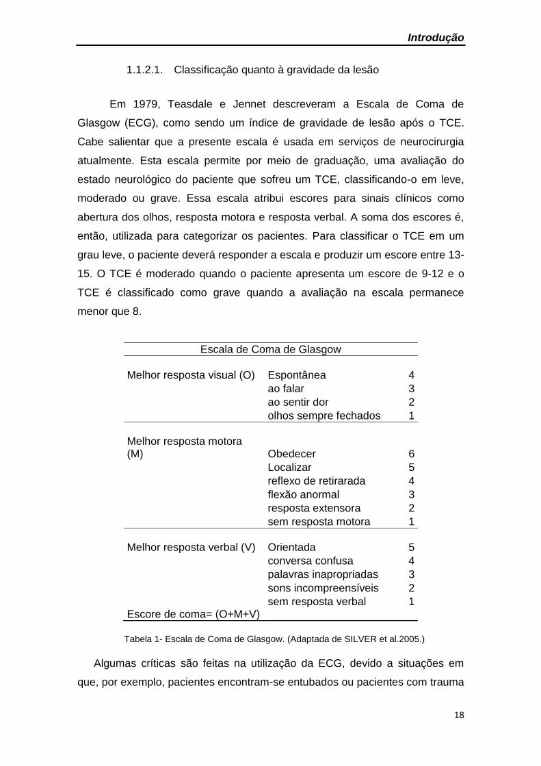
1.1.2.1. Classificação quanto à gravidade da lesão
Em 1979, Teasdale e Jennet descreveram a Escala de Coma de
Glasgow (ECG), como sendo um índice de gravidade de lesão após o TCE.
Cabe salientar que a presente escala é usada em serviços de neurocirurgia
atualmente. Esta escala permite por meio de graduação, uma avaliação do
estado neurológico do paciente que sofreu um TCE, classificando-o em leve,
moderado ou grave. Essa escala atribui escores para sinais clínicos como
abertura dos olhos, resposta motora e resposta verbal. A soma dos escores é,
então, utilizada para categorizar os pacientes. Para classificar o TCE em um
grau leve, o paciente deverá responder a escala e produzir um escore entre 13-
15. O TCE é moderado quando o paciente apresenta um escore de 9-12 e o
TCE é classificado como grave quando a avaliação na escala permanece
menor que 8.
Escala de Coma de Glasgow
Melhor resposta visual (O) Espontânea
4
ao falar 3
ao sentir dor 2
olhos sempre fechados 1
Melhor resposta motora (M) Obedecer 6
Localizar 5
reflexo de retirarada 4
flexão anormal 3
resposta extensora 2
sem resposta motora 1
Melhor resposta verbal (V) Orientada 5
conversa confusa 4
palavras inapropriadas 3
sons incompreensíveis 2
sem resposta verbal 1 Escore de coma= (O+M+V)
Tabela 1- Escala de Coma de Glasgow. (Adaptada de SILVER et al.2005.)
Algumas críticas são feitas na utilização da ECG, devido a situações em
que, por exemplo, pacientes encontram-se entubados ou pacientes com trauma
Introdução
19
de face e inchaço palpebral, onde nestes casos alguns padrões não podem ser
avaliados (SILVER et al., 2005; MAAS et al., 2008). De qualquer forma, tais
situações não invalidam o método, e a escala segue sendo um bom parâmetro
na avaliação inicial do TCE (SILVER et al., 2005).
Do ponto de vista clínico, o trauma leve corresponde ao que se chama de
comoção ou concussão, em que o paciente pode perder a consciência por um
breve período, no máximo 30 minutos, e com recuperação completa do estado
neurológico. Esses pacientes às vezes apresentam alteração temporária
neurovegetativa incluindo vômitos, cefaléia, sudorese e mal-estar em geral. Se
a perda de consciência perdurar por um período maior que 30 minutos após a
lesão, e que outras características não se enquadrem em trauma grave, pode-
se classificar o TCE de grau moderado. No TCE grave, a perda de consciência
ocorre por um período maior que 24 horas (FREY, 2003). Neste quadro o
paciente apresenta-se mal, com alterações autonômicas e respiratórias,
podendo entrar em estado de rigidez de descerebração ou decorticação,
sudorese abundante e alterações pressóricas (grau 4 na ECG).
Vários métodos têm sido utilizados ao longo das últimas décadas para
avaliar o grau do dano cerebral, incluindo uma proposta mais recente para
classificar a lesão cerebral usando tomografia computadorizada (TC) (GUIAS
DE MEDICINA AMBULATORIAL e HOSPITALAR).
1.1.2.2. Classificação quanto ao mecanismo da lesão
O entendimento etiológico sobre o tipo de impacto do TCE é de extrema
importância uma vez que possibilita prever que tipo de lesão que
provavelmente resultará desta injuria cerebral (SAATMAN et al., 2008). Neste
contexto, dois principais tipos de impactos cerebrais foram classificados, o
impacto por contato e o impacto por aceleração/desaceleração (NORTJE e
MENON, 2004).
O impacto por contato se caracteriza quando algum objeto entra em contato
com o crânio, ou o crânio colide com algum objeto (SAATMAN et al., 2008).
Este tipo de impacto frequentemente associa-se à quedas e perfurações,
podendo ocorrer fratura, afundamento de crânio, contusão e laceração
Introdução
20
(GENNARELLI e GRAHAM, 2005). Já no impacto de aceleração/desaceleração
ocorre um movimento inercial do encéfalo em relação ao crânio, onde a lesão
não necessariamente é causada por algum contato externo. Assim, o
movimento leva ao atrito da superfície do encéfalo com acidentes ósseos da
base do crânio, do teto da órbita e da asa (clivo) do esfenóide. Este movimento
também é responsável por lesão estrutural de neurônios e vasos, pela ruptura
de veias-ponte e consequente formação do hematoma subdural. As lesões
derivadas do impacto por aceleração/desaceleração geralmente são
características de acidentes automobilísticos e quedas de grandes alturas
(GENNARELLI e GRAHAM, 2005).
1.1.2.3. Classificação quanto à distribuição das lesões
Segundo critério topográfico, as lesões do TCE são classificadas em focais
e difusas. A lesão focal é frequentemente produzida por um impacto por
contato, e são associadas à contusão cerebral, hemorragia, infarto do tronco
encefálico, lacerações, fratura de crânio, hematoma intracraniano, hematoma
extradural, e extensiva morte celular local principalmente por necrose
(GENNARELLI e GRAHAM, 2005; ANDRIESSEN, JACOBS e VOS, 2010). Já a
lesão difusa é associada ao impacto por aceleração/desaceleração
(GENNARELLI e GRAHAM, 2005). São características deste tipo de lesão a
lesão axonal difusa, lesão cerebral hipóxica, tumefação cerebral difusa e lesão
vascular focal múltipla (ANDRIESSEN, JACOBS e VOS, 2010). Ambos os tipos
de lesões podem causar déficits cognitivos e motores (ANDRIESSEN, JACOBS
e VOS, 2010).
1.1.2.4. Classificação quanto à progressão das lesões
Outro tipo de classificação das lesões associadas ao TCE ocorre em
relação ao momento do impacto até danos posteriores, sendo divididas em
dano primário e dano secundário. O dano primário é aquele que se processa
no momento do trauma, sobre o couro cabeludo, osso da caixa craniana,
meninges e encéfalo (GENNARELLI e GRAHAM, 2005; WERNER e

Introdução
21
ENGELHARD, 2007; SAATMAN et al., 2008). O dano primário é irreversível,
porém pode-se evitar ou diminuir os danos causados pelo impacto ao crânio
com o uso de cinto de segurança, air-bag, capacete, educação no trânsito e
combate a violência (XIONG, MAHMOOD e CHOPP, 2013).
Em consequência do dano primário, inicia-se uma cascata de eventos
moleculares e celulares que é denominado de dano secundário. Este dano
abrange todos os eventos moleculares e celulares, onde a progressão dessa
lesão pode perdurar por dias, meses ou anos após a lesão cerebral levando a
déficits funcionais aos indivíduos acometidos (GENNARELLI e GRAHAM,
2005). Dentre as alterações neuroquímicas encontradas no dano secundário
estão o dano cerebral isquêmico, excitotoxicidade, estresse oxidativo,
disfunção mitocondrial e consequente déficit de adenosina trifosfato (ATP),
inflamação, necrose e apoptose (AARABI e SIMARD, 2009).
1.1.3. Lesão axonal difusa (LAD)
A LAD é considerada um fator muito importante na determinação da
morbidade e da mortalidade no TCE, e o substrato morfológico da
inconsciência de instalação imediata (STRICH, 1956). Interessantemente, LAD
não é uma característica exclusiva do trauma grave ocorrendo desde o trauma
brando ao trauma grave (BLUMBERGS et al., 1995).
Apesar de a lesão cerebral difusa levar a axotomia primária (JOHNSON
et al., 2013), acredita-se que o principal mecanismo no momento da lesão seja
o estiramento axonal decorrente do movimento de rotação do encéfalo
decorrente do trauma, ocasionando um dano ao axolema. Em consequência,
ocorre uma alteração na permeabilidade da membrana axonal próximo aos
nódulos de Ranvier, com isso, os axônios perdem a capacidade de manter o
gradiente iônico fisiológico resultando em mudanças nas concentrações de
cálcio, potássio, sódio e cloreto, dentro do axoplasma, ocasionando um
aumento no influxo de Ca2+ e inchaço mitocondrial (BUKI et al., 2000).
Proteases específicas passam, então, a degradar proteínas estruturais como a
tubulina e outras proteínas associadas aos microtúbulos e neurofilamentos,
prejudicando o transporte axonal normal (MCGINN et al., 2009). Outra

Introdução
22
possibilidade, é que a lesão cerebral possa romper as subunidades de
neurofilamentos, interrompendo o transporte axoplasmático, e sendo o axônio
desprovido de ribossomos, leva ao início do processo de degeneração (STONE
et al., 2004).
Embora não ocorra necessariamente a morte neuronal com a
degeneração do axônio, ocorre o rompimento ou desconexão do axônio
danificado (SINGLETON et al., 2002), estas culminam com uma alteração nas
sinapses, levando as células a uma reorganização sináptica, que pode ser
adaptativa ou inadequada (JOHNSON, STEWART e SMITH, 2013).
1.1.4. Disfunções relacionadas ao TCE
A cada ano o TCE contribui para um número substancial de mortes e
casos de incapacidades permanentes. Grande parte dos sobreviventes do TCE
apresenta distúrbios neurológicos que os afetam em muitas de suas atividades
diárias prejudicando a qualidade de vida destes sujeitos (SILVER et al., 2005;
FAUL et al., 2007). Neste contexto, o TCE resulta em uma variedade de
distúrbios motores e neuropsiquiátricos que vão desde déficits sutis a graves
distúrbios emocionais e intelectuais, incluindo déficits motores, déficits
cognitivos, declínio emocional e epileptogênese (PITKANEN e MCINTOSH,
2006). O comprometimento motor é uma sequela comum decorrente da lesão
do TCE (SILVER et al., 2005). O sistema motor consiste em uma complexa
rede de áreas corticais (áreas motoras primárias e secundárias) e subcorticais
(gânglios da base e cerebelo), em que populações neuronais interagem entre
si, através de mecanismos excitatórios e inibitórios (VANDER et al., 2001).
Neste contexto, estudos mostram, que a produção de espécies reativas de
oxigênio (EROs) e de espécies reativas de nitrogênio (ERNs), a exacerbação
do processo inflamatório e a morte celular estão envolvidas no mecanismo de
desenvolvimento do déficit motor e cognitivo após o TCE (VANDER et al.,
2001).
O prejuízo cognitivo, por sua vez, é a mais frequente e persistente
sequela encontrada nos pacientes de TCE (CICERONE et al., 2000). Em
humanos, a cognição é definida como o processo que inclui a discriminação, a

Introdução
23
seleção, a aquisição, a compreensão e a retenção, bem como a expressão e a
aplicação de informações em situações apropriadas. O déficit cognitivo inclui
qualquer redução da eficiência ou velocidade deste processo ou no
desempenho de atividades rotineiras (CICERONE et al., 2000).
Estudos experimentais utilizando ratos ou camundongos também
descrevem algumas alterações neuroquímicas como responsáveis pelo déficit
cognitivo associado ao TCE. Neste sentido, as mudanças na neurotransmissão
glutamatérgica (SCHWARZBACH et al., 2006; HAN, et al., 2009), e a produção
de EROs ou ERNs (SINGLETON et al., 2010) mostraram-se implicadas na
referida sequela.
1.1.5. Modelos experimentais de TCE
Devido a heterogeinidade da situação clínica do TCE, tem sido
desenvolvido numerosos modelos animais que simulam tal dano. Modelos
recentes têm mostrado melhor compreensão da complexa cascata molecular
iniciada após o traumatismo craniano. Dentre os modelos mais recentes, estão
a lesão por percussão de fluido (LPF) (DIXON et al., 1987), o impacto cortical
controlado (ICC) (DIXON et al., 1991), a lesão em cabeça fechada (LCF)
conhecido em inglês como “close head injury” ou “weight-drop” (MARMOROU
et al.,1994) e o modelo de lesão por ondas de explosão conhecido em inglês
como “blast injury” (LEUNG et al., 2008).
Em modelos de LPF, uma lesão é ocasionada pelo contato de um
pêndulo com um cilindro preenchido com fluido, onde o impacto causa uma
pressão de fluido sobre a dura máter intacta, que foi previamente exposta por
meio de uma craniectomia. A craniectomia pode ser realizada centralmente a
linha média, entre o bregma e o lambda (MCINTOSHI et al., 1987), ou
lateralmente sobre o osso occipital entre o bregma e o lambda (MCINTOSHI et
al., 1989). A percussão produz um breve deslocamento e deformação do tecido
cerebral, e a gravidade da lesão depende da força do impulso ocasionado pelo
pêndulo (MCINTOSHI et al., 1989). Modelos de traumatismo por LPF
reproduzem o mesmo tipo de TCE encontrado na clínica, porém sem a
ocorrência de fratura de crânio (THOMPSON et al., 2005). O TCE em seres

Introdução
24
humanos de severidade moderado a grave, é frequentemente associado a
fratura de crânio e contusões em vários giros, características estas que não
podem ser replicadas no modelo de LPF. Porém, o LPF pode replicar
características fisiopatológicas encontradas em TCE ocorridos em seres
humanos como hemorragia intracraniana, inchaço cerebral e danos
progressivos a massa cinzenta (GRAHAM et al., 2000).
Com base na posição da craniectomia em relação a distância da sutura
sagital, os modelos de LPF podem ser divididos em linha média, onde a
craniectomia é central a sutura sagital, parassagital (<3,5 mm lateral a linha
média) e os modelos laterais (>3,5 mm lateral a linha média) (MCINTOSH et
al., 1989; SANDERS et al., 1999; FLOYD et al., 2002). Na região perilesional
do córtex, ocorre um aumento de células gliais, que continua a se expandir em
torno de um ano após a lesão devido a contínua morte celular (BRAMLETT e
DIETRICH, 2002).
1.2. Glutamato
No sistema nervoso central (SNC), o glutamato é o principal aminoácido
excitatório e desempenha um importante papel na função do aprendizado e
memória (CHEN, et al., 2009). Sabe-se que a potenciação de longo tempo
(LTP, do inglês Long-term potentiation), é o melhor mecanismo sugerido para a
formação de memória no cérebro de mamíferos, ocorrendo principalmente no
hipocampo através do receptor glutamatérgico N-metil-D-aspartato (NMDA)
(GALVAN et al., 2000).
A larga proporção do glutamato cerebral é oriundo da glicose plasmática
(LAJTHA et al., 1959) que é transportada para o cérebro através da família de
moléculas transportadoras de glicose (GLUT) presentes em células endoteliais,
astrócitos e neurônios (QUTUB e HUNT, 2005).
Embora os neurônios sejam considerados a principal fonte de glutamato
extracelular, alguns estudos sugerem que os astrócitos podem também liberar
glutamato na fenda sináptica (KIMELBERG et al., 1995).
Introdução
25
1.2.1. Transportadores de glutamato
A captação de glutamato do espaço extracelular é realizada por proteínas
transportadoras de alta afinidade (do inglês excitatory amino acids transportes,
EAATs), na superfície das células astrocitárias e neuronais. Até o momento,
cinco subtipos de transportadores de glutamato foram identificados: GLAST
(EAAT1), GLT-1 (EAAT2), EAAC1 (EAAT3), EAAT4 e EAAT5 (DANBOLT,
2001). EAAT1 e EAAT2 são predominantemente localizados nos astrócitos
(LEHRE et al., 1995; REYE et al., 2002), enquanto que EAAT3, EAAT4 e
EAAT5 parecem estar principalmente em neurônios (KUGLER e SHIMITT,
1999). Dentre os transportadores gliais, o EAAT2 é o responsável por cerca de
90% da atividade total do transporte de glutamato no cérebro (TANAKA et al.,
1997; PEGHINI et al., 1997).
Os transportadores de glutamato (EAATs) movem o glutamato para dentro
da célula usando um gradiente eletroquímico de Na+ e K+ através da
membrana. Este transporte ocorre da maneira que quando uma molécula de
glutamato é captada, ocorre a entrada de íons Na+ e um próton juntamente
para o interior da célula, e a saída de um K+ (DANBOLT, 2001), com o gasto de
uma molécula de ATP. Portanto, este processo é dependente de energia, e os
transportadores de glutamato são proteínas dependentes de Na+ que
dependem do gradiente sódio e potássio gerado principalmente pela Na+,K+-
ATPase. Por exemplo, a administração de ouabaína, um inibidor da Na+,K+-
ATPase, reduziu a captação de [3H]glutamato, [3H]aspartato e rubidium-86 em
sinaptossomas de ratos, sugerindo uma relação funcional entre a Na+,K+-
ATPase e os transportadores de glutamato (ROSE et al., 2009). Além disso,
estas cinco proteínas também funcionam como canais de Cl- (WADICHE et al.,
1995). O influxo de Cl- é ativado pelo glutamato, mas não é
termodinamicamente acoplado ao transporte (FAIRMAN et al., 1995). EAAT4 e
EAAT5 parecem estar mais envolvidos na condutância de Cl-. Enquanto isso,
EAAT2 é a mais abundante proteína transportadora, e representa cerca de 1%
do total de proteínas de membrana no cérebro (HAUGETO et al., 1996),
mostrando alta concentração no hipocampo e córtex cerebral. Já EAAT1,
parece estar em maior concentração nas glias de Bergmann na camada
molecular cerebelar (LEHRE et al., 1995).

Introdução
26
1.3. TCE e excitotoxicidade
Já está bem descrito na literatura que o acúmulo de glutamato no
espaço extracelular, bem como a excessiva ativação de seus receptores
podem levar ao quadro conhecido como excitotoxicidade, originalmente
mostrado por Olney (1969). Cabe salientar que este evento possui um papel
central no dano e morte neuronal após o TCE (LAU e TYMIANSKI, 2010).
Neste contexto, estudos clínicos ou modelos experimentais de TCE tem
evidenciado elevados níveis de glutamato após a lesão cerebral (FADEN et al.,
1989; GLOBUS et al., 1995; PALMER et al., 1993; BULLOCK et al., 1998). O
aumento de glutamato intersticial pode surgir de maneiras diferentes após o
dano cerebral.
Outra questão importante a ser ressaltada é a isquemia, um dos
principais eventos secundários após o TCE (GENNARELLI, 1993;
GENNARELLI e GRAHAM, 2005). Nesse processo, a falta de oxigênio leva a
célula a utilizar o metabolismo anaeróbico em maior quantidade na tentativa de
suprir a falta de ATP requerida por ATPases, principalmente a Na+,K+-ATPase
que aumenta sua atividade na tentativa de manter o equilíbrio iônico
(KAWAMATA et al., 1995). A homeostase iônica comprometida desencadeia
uma excessiva despolarização e liberação de aminoácidos excitatórios
(GAETZ, 2004). Esta diminuição da produção de ATP e comprometimento da
homeostase iônica, também diminui a captação de glutamato que é feita por
um sistema de cotransporte com Na+ e de maneira indireta depende de ATP
(ROSE et al., 2009). Já o rompimento da barreira hematoencefálica (BHE)
aumenta a concentração de glutamato no tecido lesionado (YI e HAZELL,
2006). Estudos post morten de pacientes que sofreram TCE, mostraram uma
diminuição na quantidade de EAAT1 e EAAT2 (VAN LANDEGHEM et al.,
2006). Os estudos experimentais, demonstraram que EAAT2, diminuiu
significamente a concentração no córtex, hipocampo e tálamo em 6 horas após
o TCE em ratos, e que esta diminuição se manteve no córtex cerebral após 24
horas (YI et al., 2005). Diminuição nos níveis de EAAT1 e EAAT2 tem sido
mostrado no córtex cerebral ipsilateral após um impacto cortical controlado,
concomitante com a redução na atividade de ligação em [3H]-D-aspartato (RAO
et al., 1998). Além disso, tem sido evidenciado que infusão de antisense para

Introdução
27
GLT-1 aumentou a perda neuronal no hipocampo sete dias após a lesão (RAO
et al., 2001).
Uma das maiores causas do dano relacionado a excitotoxicidade após o
TCE é provavelmente o excessivo influxo de Ca2+ nas células cerebrais, que
pode causar morte neuronal principalmente após um impacto de grau
moderado ou severo (FAROOQUI e HOOCKS, 1994). Esta hipótese é baseada
em estudos que mostram que o Ca2+ no fluido cérebro espinhal diminui de 1
para 0,01 mM após um traumatismo craniano (NILSSON et al., 1993), e que a
captação total no tecido de 45Ca2+ é aumentada após o dano in vivo (FINEMAN
et al., 1994).
Em neurônios, o aumento nos níveis de Ca2+ intracelular e a diminuição
na produção de ATP podem levar ao processo de necrose devido a ativação da
calpaína (SAATMAN, CREED e RAGHUPATHI, 2010). Diversos elementos do
citoesqueleto servem de substrato para as calpaínas, incluindo as tubulinas,
tau, proteínas associadas aos microtúbulos e neurofilamentos (KERMER et al.,
1999). As calpaínas também parecem estar envolvidas na degradação de
proteínas de membrana como os receptores e transportadores de glutamato
(TAKAHASHI, 1990), moléculas de adesão, enzimas e proteínas cinases e
fosfatases (TAKAHASHI, 1990).
A mitocôndria pode ser considerada uma organela que controla a
concentração de Ca2+ na célula. Porém, quando a mitocôndria é exposta a uma
sobrecarga de Ca2+, pode levar ao aumento da permeabilidade do poro de
transição mitocondrial (PTM), um aumento da permeabilidade da membrana
interna para íons e solutos com massa molecular até 1500 Da (BERNARDI,
2006). A abertura persistente dos poros pode direcionar a célula para a morte
celular por uma combinação de eventos: despolarização da membrana interna,
que causa cessamento da fosforilação oxidativa e produção de EROs, inchaço
da matrix e desdobramento das cristas. Em consequência do dano
mitocondrial, ocorre a liberação dos estoques de Ca2+, EROs e de proteínas
apoptogênicas, como o citocromo c para o citosol (BERNARDI et al., 2006,
ZOROV, et al., 2009). No citosol, o citocromo c incia o processo de apoptose
(SULLIVAN et al., 2005).
Outro mecanismo de dano celular pelo aumento de Ca2+ após o TCE é a
ativação de fosfolipases, principalmente a PLC e a fosfolipase A2 citosólica
Introdução
28
(cPLA2). Ambas PLC (DHILLON et al., 1999) e cPLA2 (SHOHAMI et al., 1989)
aumentam após TCE. A ativação de cPLA2 leva a uma liberação e acúmulo de
ácido araquidônico livre (AA), que pode alterar a permeabilidade da membrana
e aumentar a produção de EROs (SLEMMER et al., 2008). Além disso, o
excesso de Ca2+ pode exacerbar a produção de óxido nítrico (ON) por ativar
enzimas dependentes de Ca2+ como a óxido nítrico sintase endotelial (eNOS) e
a óxido nítrico sintase neuronal (nNOS) que forma ON a partir de L-arginina
usando NADPH e oxigênio molecular (BIAN e MURAD, 2003).
1.3.1. TCE e estresse oxidativo
Estresse oxidativo é caracterizado por uma oxidação de biomoléculas
que pode levar a perda de suas funções biológicas (HALLIWELL e
WHITEMAN, 2004) e ocorre quando a produção de radicais livres ou EROs
estão aumentados em relação as defesas antioxidantes presentes no
organismo. Os radicais livres são átomos ou moléculas que ao se formarem
possuem um ou mais elétrons desemparelhados na sua camada de valência
(SOUTHORN e POWIS, 1988; HALLIWELL, 1989), enquanto que as EROs e
ERNs são compostos igualmente reativos porém não possuem o
desemparelhamento nos elétrons (DROGE, 2002). Mesmo não estando os
elétrons desemparelhados, as EROs e ERNs são altamente reativas e buscam
estabilidade durante sua breve existência, reagindo com outras moléculas
causando danos a membranas celulares, proteínas e DNA (HALLIWELL,
2012). A produção de radicais livres e EROs fazem parte do metabolismo
normal do cérebro, pois em condições normais, esses radicais livres e EROs
são mantidos sob controle por mecanismos de defesa celular.
A fosforilação oxidativa é a principal formadora de EROs, onde o
primeiro radical livre a ser formado é o superóxido (O2−•), principalmente no
complexo I (NADH Desidrogenase) e no complexo III (ubiquinona citocromo c
oxidase) da cadeia respiratória, onde a coenzima mononucleotídeo de Flavina
(FNM) e o ubiquinol reduzem o oxigênio univalentemente formando o radical
livre (NAVARO e BOVERIS, 2007). A partir do O2−• podem ser formadas EROs,
como peróxido de hidrogênio (H2O2), e outros radicais livres, como o radical
Introdução
29
hidroxil (-OH●), que é o mais reativo. Além disso, o H2O2 formado pode reagir
com metais como o cobre (Cu+1) ou ferro (Fe+2) e (Fe+3) na chamada reação de
Fenton (LIANG, HO e PATEL, 2000), formando o -OH●, que pode atuar como
um eletrófilo ou nucleófilo, atacando moléculas orgânicas pela abstração de
hidrogênio ou acoplando-se em duplas ligações e anéis aromáticos
(hidroxilação), inclusive em posições substituídas causando reações como
desaminação (FUKUSHIMA et al., 2000) e descarboxilação (CHEN et al.,
2002). O H2O2 pode reagir também com o próprio O2−• na chamada reação de
Haber-Weiss, formando o -OH● (LIANG, HO e PATEL, 2000). O O2−• pode
reagir com o ON formado pela óxido nítrico sintase e formar peroxinitrito
(ONOO-), uma ERN que causa dano nas membranas, proteínas e DNA celular
(LAU e TYMIANSKI, 2010).
Para controlar a formação destas EROs, existe um sistema de defesa
antioxidante composto por antioxidantes enzimáticos, como a superóxido
dismutase (SOD), a catalase (CAT) e a glutationa peroxidase (GPx) e que
enovolvem todo um sistema de moléculas auxiliares. Existem também os
antioxidantes não-enzimáticos (vitaminas A, C, D e E e glutationa reduzida)
(HALLIWELL e GUTTERIDGE, 1995). Existem dois tipos de SOD, a SOD1 que
se localiza principalmente no citoplasma, mas também é encontrada nos
peroxissomos, lisossomos e no espaço intermembrana mitocondrial e utilizam
como cofatores o cobre e o zinco, e a SOD2 ou SODmn que se localiza na
matriz mitocondrial e utiliza manganês como cofator (CHANCE, SIES e
BOVERIS, 1979). Estas duas enzimas catalisam a reação de dismutação do
O2−• formando H2O2. Já as enzimas catalase e a glutationa peroxidase
catalisam a reação de degradação de H2O2 em H2O e O2. A enzima GPx
encontra-se em duas formas, uma que está presente na mitocôndria e no
citosol e que utiliza selênio como cofator, e a outra localiza-se somente no
citosol e não utiliza selênio como cofator. A GPx utiliza glutationa reduzida
(GSH) para a reação, formando glutationa oxidada (GSSG) e o produto da
reação do hidroperóxido (MILLS, 1960). Para restaurar a molécula de GSH, a
enzima GPx funciona acoplada a glutationa redutase (GR), que reduz a
molécula de GSSG para GSH utilizando NADPH (MAIORINO et al., 1990).
A excitotoxicidade esta diretamente relacionada com a formação de
radicais livres, EROs e ERNs, produzidas em consequência da ativação de

Introdução
30
enzimas dependentes de cálcio, como a fosfolipase A2 (MILLER et al., 2010) e
a xantina oxidase (MARIET et al., 2012). Dentre os diversos danos resultantes
da produção exacerbada de radicias livres destaca-se a peroxidação lipídica
(MARIET et al., 2012). A peroxidação lipídica produz aldeídos, gases
hidrocarbonados e vários resíduos químicos como o malondialdeído (MDA),
dienos conjugados e 4-hidroxinonenal (4-HNE) (HOTZ et al., 1987). Estudos
em modelos experimentais de TCE tem evidenciado um dano aos lipídeos de
membrana logo após o TCE, tendo um aumento máximo em duas horas e
continuando aumentado em até 48 horas após o trauma (VAGNOZZE et al.,
1999). Já estudos clínicos revelam que o aumento nos níveis de espécies
reativas ao ácido tiobarbitúrico (TBARS) no fluido cérebro espinhal (FCE) estão
relacionado com a piora do quadro clínico após o TCE (KASPRZAK et al.,
2001).
Dentro deste contexto, tem sido demonstrado que a superestimulação
dos receptores glutamatérgicos é o principal mediador de estresse oxidativo em
diversos modelos de doenças neurológicas (KUMAR et al., 2011). A redução
de enzimas antioxidantes no cérebro como a SOD, CAT e GPx contribuem
para o desenvolvimento de uma vasta gama de doenças neurodegenerativas
através da produção de radicais livres em condições patológicas. Neste
contexto, achados experimentais tem evidenciado o papel protetor de
antioxidantes em modelos animais de doença de Huntington, Parkinson’s e
Alzheimer (KUMAR et al., 2012). Sugerindo o papel dos radicais livres nestas
doenças, EROs também inibem a captação de [3H] glutamato em culturas de
astrócitos e reduzem o atual transporte associado a voltagem com menor ou
nenhum efeito sobre o potencial de membrana de repouso (VOLTERRA et al.,
1994).
Diferentes isoformas de transportadores de glutamato mostram
sensibilidade para oxidantes biológicos e podem compartilhar um ou mais sítios
vulneráveis para oxidantes. De fato, a atividade de transporte de EAAT1,
EAAT2 e EAAC1 são igualmente inibidas por oxidantes por uma direta ação
nas proteínas de transporte de glutamato. Um comum alvo primário para O2−•,
H2O2, ONOO- ou o subproduto -OH● é a oxidação de grupos cisteína sulfidril
(RADI et al., 1991). Além disso, pela decomposição em pH neutro, ONOO- gera
o íon nitrônio e dióxido de nitrogênio, que promovem a nitrosilação e/ou

Introdução
31
nitração de resíduos de tirosina, e altera o processo de fosforilação nestes
resíduos.
1.4. Na+,K+-ATPase
A Na+,K+-ATPase é uma enzima integral de membrana, responsável
pelo gradiente eletroquímico celular através do transporte ativo de três íons Na+
para o meio extracelular e dois íons K+ para o meio intracelular, utilizando uma
molécula de ATP para realizar sua função (HORISBERGER, 2004). A enzima é
constituída por três cadeias polipeptídicas denominadas α, β e γ, sendo que a
unidade funcional da enzima é constituída pela subunidade α e pela
subunidade β (KAPLAN, 2002). Em repouso, a Na+,K+-ATPase é responsável
pelo consumo de cerca de 30% de todo o ATP utilizado pelos mamíferos
(CLAUSEN e EVERTS, 1991). No cérebro, a atividade da Na+,K+-ATPase
contribui de maneira crucial para a manutenção do gradiente eletroquímico
responsável pelos potencias de repouso e de ação, e também pela captação e
liberação de neurotransmissores (STAHL e HARRIS, 1986). Por isso,
alterações na atividade da Na+,K+-ATPase podem afetar diretamente a
sinalização celular através de neurotransmissores, a atividade neuronal, e
também o comportamento do animal (MOSELEY et al., 2007). Neste contexto,
um prejuízo no funcionamento da Na+,K+-ATPase ocasiona aumento ou
diminuição da excitabilidade neuronal, dependendo do grau de inibição
induzido e do tipo neuronal afetado (GRISAR et al., 1992). Trabalhos mostram
que a ouabaína, um inibidor da Na+,K+-ATPase, aumenta o influxo de Ca2+ em
“slices” de cérebro de ratos (FUJISAWA et al., 1965), causa convulsões em
camundongos (JAMME et al., 1995), induz uma reversão dos transportadores
de glutamato dependentes de Na+, causando uma liberação de glutamato (Li e
Stys, 2001), e morte celular no hipocampo de ratos (LEES et al., 1990). Além
disso, a supressão genética da Na+,K+-ATPase causa prejuízo no aprendizado
espacial e aumento no comportamento típico de ansiedade em camundongos
(MOSELEY et al., 2007). A administração de ouabaína diminui a captação de
[3H]glutamato, [3H]aspartato e rubidium-86 em sinaptossomas de ratos (ROSE,
et al., 2009).
Introdução
32
Estudos mostram que a atividade da enzima Na+,K+-ATPase é mais
prejudicada pelo ataque de EROs em lipídeos de membrana do que por
alterações estruturais na proteína em si (JAMME et al., 1995). No SNC,
estudos tem demonstrado que variações no estado redox do neurônio podem
modular a atividade desta enzima (PETRUSHANKO, 2007). Da mesma forma,
estudos do nosso grupo tem sugerido uma relação da Na+,K+-ATPase e EROs
após o TCE. Outro trabalho mostrou uma inibição da enzima Na+,K+-ATPase
em 24 horas após o TCE no cérebro de ratos (MOTA et al., 2012), sugerindo
que a Na+,K+-ATPase pode ser alvo de radicais livres e EROs, e que sua
alteração após o TCE esteja relacionada ao aumento do estresse oxidativo.
1.5. Sistema Nrf2-ARE
Durante a evolução, as células desenvolveram sistemas de defesa
contra processos prejudiciais endógenos e substâncias exógenas. Diversos
fatores de transcrição são envolvidos em aumentar as defesas das células. Um
complexo sistema de genes de fase I, II e III envolvem a ativação de enzimas
antioxidantes. O principal regulador dos genes de fase II e alguns genes de
fase III de enzimas antioxidantes é o nuclear factor erythroid 2 related factor 2
(Nrf2) (ITOH et al., 1997). O princípio do sistema é manter a proteína Nrf2
baixa em condições normais da célula, com a possibilidade de indução rápida
no caso de aumento de oxidação celular. Nrf2 é membro da família de fatores
de transcrição, que inclui Nrf1, Nrf3, p45 NF-E2, Bach1 e Bach2 (MOTOHASHI
et al., 2002). Nrf2 é composto de seis domínios funcionais conhecido como
Nrf2-ECH homologias (Neh) e designados Neh1-6, respectivamente. Cada
domínio Neh é designado para sua própria função (BAIRD e DINKOVA-
KOSTOVA, 2011). Em condições quiescentes, Nrf2 é mantido por um repressor
citosóloico, Keap1 (do inglês Kelch-like ECH-associated protein 1), uma
proteína do citoesqueleto que ancora e reprime a atividade de transcrição
(ITOH et al., 1999; TONG et al., 2007). Keap1 promove a rápida degradação
proteossomal de Nrf2 através de uma ubiquitinação e também age como um
sensor de estresse oxidativo e eletrofílico (ITOH et al., 1999).

Introdução
33
Tem sido sugerido que alterações na estrutura de Keap1 leva para a
dissociação do complexo Nrf2-Keap1 (MOTOHASHI e YAMAMOTO, 2004),
mas uma modificação específica no sítio de Keap1 pode também causar uma
função alterada da ubiquitina ligase E3 e consequente redução na degradação
de Nrf2 (TONG et al., 2007). Recentes estudos também propõem uma via de
ubiquitinação e degradação de Nrf2 independente de Keap1 (ROJO et al.,
2012). No presente modelo, GSK-3 fosforila resíduos de serina no domínio Neh
6 em Nrf2 (RADA et al., 2012), e Neh 6 fosforilada pode ser reconhecida por
uma ubiquitina ligase E3.
A dissociação do complexo Nrf2-Keap1 no citoplasma é um mecanismo
amplamente aceito para a ativação de Nrf2. Em síntese, a oxidação de
resíduos de cisteína modifica a conformação de Keap1, e assim inicia a
ativação de Nrf2 (TONG et al., 2007). Desta maneira, produtos oxidativos
melhoram a estabilidade de Nrf2 e aumentam a expressão dos genes de fase II
(MCMAHON et al., 2006). A fosforilação nos resíduos de serina/treonina em
Nrf2 pode ser um distinto mecanismo pelo qual Nrf2 se dissocia de Keap1
(SURH et al., 2008), e quinases, incluindo, PKC, c-jun N-terminal kinase (JNK),
extracellular signal-regulated kinase (ERK), casein kinase 2 (CK2), p38
mitogen-activated protein kinase (p38 MAPK) e phosphoinositide 3-kinase
(PI3K), parecem modular a importação e exportação de Nrf2 do núcleo (JAIN e
JAISWAL, 2007, SURH et al., 2008). Portanto, oxidação dos resíduos de
cisteína sensíveis ao estado redox em Keap1 constitui a base molecular de
ativação de Nrf2 (TONG et al., 2007).
A indução de enzimas de defesa antioxidantes de fase II pelo Nrf2 é
regulada através do antioxidant response elemento (ARE), ou electrophile
response elemento (EpRE) (ITOH et al., 1997, VENUGOPAL e JAISWAL,
1998). A ligação Nrf2-ARE pode iniciar a transcrição de centenas de genes
citoprotetores, incluindo a SOD, CAT (CHO et al., 2011) e uma série de
antioxidantes de fase II que inclui o sistema de defesa da glutationa, NA(D)PH
quinone oxidoreductase 1 (NQO1) e heme oxigenasse 1 (HO-1) (ZHANG et al.,
2012).
Cabe salientar que Nrf2 não está envolvido somente em aumentar a
capacidade antioxidante, mas também a expressão de outros tipos de
proteínas protetoras como o brain derived neurotrophic factor (BDNF) (SAKATA

Introdução
34
et al., 2012), o anti-apoptótico B-cell lymphoma (Bcl-2) (NITURE e JAISWAL,
2012), a interleucina anti-inflamatória (IL-10), o mitochondrial transcription (co)-
factors Nrf1 e peroxissome proliferator-activated receptor gamma coactivator 1-
alpha (PGC-1 α) (PIANTADOSE et al., 2011), o iron exporter ferroportin 1
(HARADA et al., 2011) e a proteína autofágica p62 (KOMATSU et al., 2010).
Nrf2 no cérebro pode agir como uma das mais importantes defesas contra o
estresse oxidativo por meio da modulação da micróglia (ROJO et al., 2010),
proteger astrócitos (VARGAS e JOHNSON, 2009) e neurônios (LEE et al.,
2003) de insultos tóxicos, regulando a expressão de marcadores inflamatórios
(INNAMORATO et al., 2008) e enzimas antioxidantes (SHAH et al., 2007, YAN
et al., 2009). Neste contexto, tem sido bem proposto que Nrf2 desempenha um
papel protetor em desordens neurodegenerativas, incluindo Parkinson’s
(CUADRADO et al., 2009), Alzheimer’s (KANNINEN et al., 2008), e doença de
Huntington’s (STACK et al., 2010) e também o TCE (YAN et al., 2009).
Na medida em que o estresse oxidativo desempenha um papel importante
no dano neuronal após o TCE, a ativação de Nrf2 após o trauma tem gerado
recente interesse. O TCE aumenta significativamente os níveis de Nrf2 e
enzimas de fase II como a NQO1 e HO-1 (YAN et al., 2009), sugerindo que a
via Nrf2-ARE é um fator de adaptação endógeno compensatório contra o TCE.
Estudos mostram que administração intraperitoneal de sulforaphane (SFN), um
ativador de Nrf2, é capaz de reduzir a morte neuronal, o volume de contusão e
a disfunção neurológica 7 dias após o TCE em ratos (HONG et al., 2010). De
acordo com estes resultados, camundongos knockout para Nrf2 exibiram
déficits exacerbados em funções neurológicas e dano oxidativo. Os inibidores
de “histone deacetylase” (WANG et al., 2012) e “tert-butylhydroquinone” (tBHQ)
(HATIC et al., 2011) podem também proteger do dano neuronal ocasionado por
TCE por ativação de Nrf2, sugerindo um relevante papel do Nrf2 na
recuperação neuronal após o TCE.
1.5.1. Hsp70
As células podem sofrem alterações moleculares quando expostas a
temperaturas acima das consideradas normais. Estas alterações também

Introdução
35
podem ser devidas a alterações fisiológicas no organismo como o “estresse”.
Isso é caracterizado pela produção de proteínas de choque térmico (Hsp), (do
inglês, heat shock protein), que são uma família de proteínas de estresse
onipresente e altamente induzível. Foram primeiramente demonstradas por
Ritossa (1962) com o efeito do calor em Drosophilas. Uma variedade de
condições, incluindo hipertermia, alterações no pH, depleção de energia,
variação na concentração de cálcio, produção anormal de proteínas e
desacoplamento da fosforilação oxidativa levam a indução de proteínas de
estresse. Além disso, EROs também causam aumento de Hsp (KUKREJA et
al., 1994). O papel das Hsp é de proteger o processo de montagem e
dobramento de proteínas dentro da célula alterada e agir como uma chaperona
molecular. O estímulo intracelular da indução de Hsp é o dano a proteína, e
rápida expressão de Hsp pode ser vista logo após a uma exposição sub-letal a
um estressor (PERDRIZET, 1997).
Hsps possuem um número de diferentes formas e isoformas, que são
distinguidas pelo seu peso molecular e são categorizadas em famílias. A
família de 70 kiloDalton (kDa) de Hsp são as mais altamente conservadas
(HUNT e MARIMOTO, 1985), e considerada a mais induzível por estresse de
todas as chaperonas. Apesar de ter sido proposto uma série de estímulos
aferentes para a síntese de Hsp, como hipertermia e produção de EROs, o
sinal eferente responsivo para esta resposta ao estresse não é totalmente
entendido. Estudos mostram que a regulação da síntese de Hsp é controlada
através de um sistema envolvendo um fator de transcrição de choque térmico
(HSF) (do inglês, heat shock transcription factors). Em condições normais da
célula, HSF está na forma de um monômero formando um complexo com a
Hsp no citoplasma e no núcleo (LOCKE, 1998). Após uma exposição a um
estresse físico-químico, HSF é convertido em um estado trimérico por
fosforilação podendo então fazer a atividade de ligação ao DNA (BALER et al.,
1993), um processo conhecido como ativação de HSF. A ativação de HSF foi
identificada entre minutos após o choque térmico (KIM et al., 1995). Durante
processos de estresse, Hsp interagem com proteínas desnaturadas permitindo,
assim, que HSF interaja com elementos de choque térmico (HSE), (do inglês,
heat chock elemento), localizados na região do gene de Hsp, assim
promovendo sua transcrição (NOBLE e AUBREY, 1999). Quando os níveis de

Introdução
36
Hsp sobem, eles voltam a se ligar em HSF e assim regulam sua própria síntese
(NOBLE e AUBREY, 1999).
Neste contexto, vários estudos mostram que Hsp70 pode proteger
contra vários insultos de estresse, incluindo, choque térmico (MOSSER et al.,
1997), estresse oxidativo (BELLMANN et al., 1996), estímulos apoptóticos
(JAATTELA et al., 1998) e isquemia (MESTRIL et al., 1994). Hsp70 é também
eficaz contra insultos isquêmicos e isquemia cerebral tal como privação de
oxigênio-glicose (LEE et al., 2001). Uma proteção contra oclusão permanente
da artéria cerebral média também tem sido observada em um modelo de
camundongos transgênicos para superexpressão de Hsp70 (RAJDEV et al.,
2000). Além disso, camundongos deficientes para Hsp70 tiveram aumento no
tamanho do infarto e tiveram uma piora no quadro após um modelo
experimental de stroke (LEE et al., 2001).
Outro trabalho mostra que a superexpressão de Hsp70 foi associada com
uma diminuição da expressão de genes pró-inflamatórios (TNF-α e IL-1β) que
são dependentes de NFκB em um modelo experimental de stroke (ZHENG et
al., 2008). Em um modelo de hemorragia intracerebral, um aumento de Hsp70
diminui a expressão de TNF-α e atenua a disfunção da barreira hemato
encefálica (BHE), formação de edema e disfunção neurológica (MANAENKO et
al., 2010). Diversos estudos demonstraram que a superexpressão de Hsp70
pode desempenhar um papel neuroprotetor em diversos modelos de doenças
neurodegenerativas (GIFFARD et al., 2008). Seus efeitos benéficos podem ser
devidos tanto a sua função de chaperona quanto da sua capacidade de
proteger a célula contra vários tipos de fatores potencialmente tóxicos.
1.5.2. BDNF
O BDNF é considerado a principal neurotrofina do cérebro, estando
envolvida na modulação de diversas funções sinápticas, induzindo estímulo a
maturação, nutrição, crescimento e integridade neuronal, assim como a
manutenção da sobrevivência de algumas populações de neurônios durante o
desenvolvimento e na fase adulta (TYLER e POZZO-MILLER, 2003). Os efeitos
do BDNF podem variar conforme a fase de desenvolvimento. No início da fase

Introdução
37
fetal, o BDNF está envolvido na formação e maturação dos neurônios, e na
fase adulta seu principal papel é o de consolidar a memória episódica (POST,
2007).
A síntese de BDNF ocorre através de um processo proteolítico de seu
precursor pró-BDNF (REICHARDT, 2006). Após a transcrição, o pró-BDNF se
desloca ao retículo endoplasmático. Dentro do complexo de Golgi é
empacotado em vesículas secretoras, que podem ser agrupadas para liberação
espontânea ou liberado por algum estímulo (CHEN et al., 2006). O pró-BDNF
liberado é convertido em BDNF maduro por proteases extracelulares (PANG et
al., 2004). Além de regular a síntese de BDNF, o pró-BDNF pode se ligar ao
receptor p75, o que leva a morte celular e acarreta a depressão de longa
duração, LTD (do inglês, long-term depression) (REICHARDT, 2006). O BDNF
liga-se ao receptor TrkB promovendo uma cascata de eventos intracelulares,
como a ativação das vias MAPK, PI-3K e PLC (REICHARDT, 2006). Estudos
mostram que ativação de TrkB pelo BDNF é necessária e facilita a LTP
dependente do receptor NMDA em diversas áreas do cérebro (MEIS et al.,
2012; REICHARDT, 2006). Além disso, BDNF pode sustentar a fase tardia da
LTP (MEI et al., 2011). Além das fortes evidências para o papel de BDNF-TrkB
na indução da LTP, também existe uma relação do BDNF com a LTD, de modo
que a aplicação exógena de BDNF inibe a indução de LTD (LU et al., 2005).
Entre todas as NTs, BDNF é a mais expressa no cérebro (MURER et al., 2001),
regulando diversas funções biológicas, inclusive crescimento axonal e
sobrevivência neuronal (QIAN et al., 2007).
Dado seu envolvimento com a plasticidade, o BDNF está provavelmente
associado com os eventos envolvidos na recuperação funcional após o TCE.
Em resposta a lesão traumática cerebral, provavelmente iniciam-se
mecanismos de reparo que estimulam a neuroregeneração (VARON et al.,
1991). Prontamente disponíveis, fatores neurotróficos após o TCE podem
melhorar a sobrevivência neuronal, estimular a brotação de neuritos
(plasticidade neuronal), induzir reparo neuronal e reestabelecer as ligações
funcionais no cérebro. Aumento agudo nas concentrações de NGF foram
mostrados após penetrante dano cerebral (NIETO-SAMPEDRO et al., 1982) e
ablação cortical (WITTEMORE et al., 1985). Outro estudo mostrou que a
expressão de RNAm de BDNF hipocampal aumentou significativamente no giro
Introdução
38
denteado em 3 horas, enquanto que a expressão de RNAm de NT-3 diminuiu
no giro denteado em 6 e 24 horas após TCE experimental em ratos (HICKS et
al., 1997). Além disso, foi visto que após um impacto cortical controlado severo,
o RNAm para NGF e BDNF estavam inicialmente e significativamente
aumentados, enquanto que o fator neurotrófico ciliar, CNTF (do inglês, ciliary
neurotrophic fator) foi menos amplificado na área de lesão e na região remota
(OYESIKU et al., 1999). Seus respectivos receptores foram também
analisados, mostrando que RNAm de TrkA e TrkB foram significativamente
elevados, enquanto o receptor α para CNTF (CNTFRα) foi significantemente
diminuído. Após o TCE, estratégias terapêuticas com a administração de
fatores neurotróficos são protetoras. Por exemplo, a infusão central de BDNF
atenuou a morte celular neuronal após dano seletivo cerebral em roedores
(HOFER e BARDE, 1988). Assim, estudos em modelos animais sugerem que o
potencial terapêutico de fatores neurotróficos poderiam ser avaliados em
tentativas clínicas para a neuroproteção no TCE.
1.6. Exercício físico
Já estão bem descritos os efeitos do exercício físico sobre a saúde geral
principalmente sobre o sistema cardiovascular e o metabolismo (BOOTH et al.,
2002). Nas últimas décadas, os efeitos benéficos da atividade física sobre o
SNC têm sido evidenciados. Esses estudos mostram que a atividade física
pode melhorar a qualidade de vida e prevenir de demência senil e declínios
cognitivos (MILLER et al., 2012). Além disso, o exercício pode estar associado
a um papel protetor contra uma variedade de distúrbios neurológicos (VAN
PRAAG, 2009).
Alguns trabalhos têm se direcionado ao entendimento das bases
neurobiológicas dos benefícios associados ao exercício físico. Dessa forma,
vários autores mostraram que o exercício físico voluntário e aeróbico
aumentam a expressão de fatores neurotróficos, incluindo BDNF e NGF
(DIETRICH, ANDREWS e HORVATH, 2008; GRIESBACH et al., 2009) e que, a
ativação desses fatores tróficos está relacionada com a sobrevivência e
diferenciação celular, assim como o aumento da resistência ao estresse

Introdução
39
oxidativo (KLUMPP e LIPOWSKY, 2005; LEEDS et al., 2005). Sabe-se que o
exercício físico sinaliza uma cascata responsável pela biogênese mitocondrial
no cérebro, principalmente pela ativação do fator nuclear PGC-1α, esta
ativação de biogênese mitocondrial está envolvida na regulação de enzimas
antioxidantes, uma vez que a PGC-1 α é necessária para a tradução de SOD,
CAT e GPx (ST-PIERRE et al., 2006; STEINER et al., 2011).
Estudos evidenciam uma relação do exercício com benefícios na saúde
contra doenças crônicas (BOOTH et al., 2008), incluindo desordens
neurodegenerativas relacionadas com a idade como a doença de Alzheimer
(LAUTENSCHLAGER et al., 2008) e doença de Parkinson (WEINTRAUB e
MORGAN, 2011). De acordo com estes dados, a inatividade física e um estilo
de vida sedentário são considerados fatores significantes de risco para o
desenvolvimento de demência e neurodegeneração (RADAK et al., 2010).
As evidências de estudos com animais confirmam que o treinamento de
resistência induz alterações moleculares cerebrais que melhoram o
aprendizado e a função cognitiva (RADAK et al., 2008). Além disso, o
treinamento de resistência retarda algumas das alterações cerebrais
associadas com o processo da idade (RADAK et al., 2008), e facilita a
recuperação funcional do dano cerebral (GRIESBACH et al., 2009; SZABO et
al., 2010). Importantemente, o exercício também protege do início de doenças
crônicas incluindo desordens neurodegenerativas (VAN PRAAG, 2008). As
evidências sugerem que exercício físico voluntário reduz os níveis de peptídeos
β-amilóide no córtex e hipocampo em um modelo de camungongo para a
doença de Alzheimer (DA) (UM, et al., 2008). A atividade física crônica estimula
o crescimento e desenvolvimento de novas células cerebrais, sendo
neuroregenerativa e desenvolve um efeito neuroprotetor (DISHMAN et al.,
2006). De fato, exercício físico parece regular a neurogênese, proporcionando
benefícios quantitativos e qualitativos para o tecido cerebral (VAN PRAAG et
al., 2008). O exercício é associado com o aumento de proteínas envolvidas na
plasticidade sináptica incluindo a proteína do citoesqueleto α-internexina e
algumas chaperonas moleculares no hipocampo, uma região do cérebro
relacionada com o aprendizado e memória (DING et al., 2006). A atividade
física regular também aumenta a proliferação de células endoteliais no cérebro
(COTMAN et al., 2007), e portanto influência a vasculatura cerebral por

Introdução
40
melhorar a capacidade metabólica através do aumento de nutrientes e
fornecimento de oxigênio (LOPEZ-LOPEZ et al., 2004).
Além disso, o exercício induz a produção de Hsp70 (NOBLE, 2002).
Porém, conflitos de dados apontam discordâncias entre exercício agudo e de
longa duração. Um estudo animal demonstrou que uma única sessão de
exercício exaustivo leva a um aumento na síntese de Hsp70 no músculo
esquelético, linfócitos e baço de ratos (LOCKE et al., 1990). Interessantemente,
foi mostrado que repetidas sessões de exercício aumentam os níves
endógenos de Hsp70, no músculo, mas em menor extensão do que a primeira
sessão. O nível extracelular e muscular de Hsp70, diminuiu antes da segunda
sessão de exercício quando comparado com amostras antes do exercício
(THOMPSON et al., 2002; MARSHALL et al., 2006). O aumento de Hsp70 em
longo prazo indica um papel na montagem de proteínas no interior das células
do tecido muscular. Dessa forma, pesquisadores sugerem que Hsp70
desempenha um papel na proteção contra a inflamação induzida pelo exercício
(PALSEN et al., 2007). Similarmente, Hung et al., (2005) mostraram que
exercício de longa duração em ratos induz a produção de Hsp70 e atenua a
superprodução de citocinas no tecido como fator de necrose tumoral alpha
(TNF-α).
1.6.1. Exercício físico e TCE
Diante de todos estes trabalhos mostrando os benefícios do exercício
físico ao SNC, alguns estudos têm demonstrado que a atividade física após o
TCE modula moléculas importantes para promover a neuroplasticidade,
facilitando os mecanismos de reparo endógenos e melhorando a recuperação
funcional após o TCE experimental (GRIESBACH et al., 2008; GRIESBACH et
al., 2009; ITOH et al., 2011a; b). Estudos com animais têm evidenciado que o
exercício físico tem forte ligação com a proteção neuronal, sendo capaz de
aumentar a neurogênese (VAN PRAAG et al., 1999) e melhorar a memória
(SAMORAJSKI et al., 1985; VAN PRAAG et al., 1999). Outro estudo mostrou
que o exercício durante seis semanas antes da lesão, aumentou a
sobrevivência dos neurônios do cerebelo após lesão traumática por impacto

Introdução
41
cortical controlado (ICC) (SEO et al., 2010). Além disso, outro trabalho mostrou
que, após o TCE causado por LPF, ratos submetidos ao exercício voluntário
aumentaram as neurotrofinas BDNF, CREB e sinapsina I no hipocampo,
aumento este associado com melhor desempenho nos testes de memória e
aprendizado quando comparado com animais sedentários (GRIESBACH et al.,
2004). Essa melhora na função cognitiva após o TCE foi associada ao BDNF,
uma vez que, bloqueando-se os receptores para o BDNF antes do exercício a
melhora cognitiva não foi mais encontrada (GRIESBACH et al., 2009). Além do
aumento de neurotrofinas, o exercício físico também demonstrou propriedades
neuroprotetoras diminuindo marcadores de estresse oxidativo. Estudos
demonstram que tanto o exercício físico profilático como terapêutico protege de
marcadores do dano oxidativo (GRIESBACH et al., 2008; LIMA et al., 2009),
demonstrando que tanto a atividade física aplicada de forma profilática quanto
recuperativa protege de marcadores do dano oxidativo, como aumento de
proteína carbonilada (GRIESBACH et al., 2008; LIMA et al., 2009). Além disso,
animais submetidos a sete dias de atividade física forçada após o TCE, tiveram
uma inibição da degeneração neuronal e morte celular por apoptose em torno
da área danificada, diminuindo o volume da lesão cortical (ITOH et al., 2011b).
Outro trabalho também mostrou que o exercício físico antes de um evento
isquêmico reduz a lesão cerebral, melhorando a função da BHE (GUO et al.,
2008). Portanto, muitos estudos têm evidenciado os efeitos terapêuticos do
exercício físico diante do dano secundário e da reabilitação neurológica após a
lesão cerebral traumática (ANG e GOMEZ-PINILLA, 2007; ITOH et al., 2011;
GRIESBACH et al, 2009; VAN PRAAG et al., 1999). Porém, poucos estudos
abordam o efeito profilático do exercício físico na cascata de eventos
relacionadas ao dano secundário decorrentes do TCE.
Diante disto, sabendo que a excitotoxicidade e o estresse oxidativo podem
contribuir para a exacerbação das lesões secundárias associadas ao TCE, e
desta maneira podem estar associadas com déficits funcionais, como o
comprometimento motor e déficit de memória, torna-se extremamente
importante verificar os benefícios profiláticos do exercício físico sobre estes
processos decorrentes do TCE.
Objetivos
42
2. OBJETIVOS
2.1. Objetivo Geral
Verificar se possíveis alterações no sistema antioxidante estão envolvidos
no efeito profilático exercido pelo treinamento físico aeróbico no modelo de
traumatismo crânioencefálico em ratos.
2.2. Objetivos Específicos
a) Avaliar se o presente protocolo de treinamento físico aeróbico prévio
protege do comprometimento motor e déficit cognitivo induzido pelo
TCE.
b) Avaliar se a alteração na cascata de eventos moleculares caracterizada
pela captação de glutamato, o imunoconteúdo dos transportadores de
glutamato (EAAT1 e EAAT2), FosfNrf-2, Hsp70, BDNF e SOD2,
atividade das enzimas Na+,K+,-ATPase e superóxido dismutase, bem
como a produção de espécies reativas em hipocampo de ratos estão
envolvidas na comprometimento motor e déficit cognitivo induzidos pelo
TCE.
c) Avaliar se o presente protocolo de treinamento físico prévio altera a
cascata de eventos moleculares descrita acima e se estas alterações
estão envolvidas no seu possível efeito profilático.

Artigo Científico
43
3. ARTIGO CIENTÍFICO
The impact of previous physical training on redox signaling in rats with traumatic
brain injury: behavioral and neurochemical approach
Fernando da Silva Fiorina; Luiz Fernando Almeida Silva
a; Mauro Robson Torres de
Castroc; Luís Roberto Hart da Silva
c; Mauro Eduardo Porto da Silveira Junior
c; Ana
Paula Pegoraro Zemolina; Fernando Dobrashinski
a; Sara Marchesan
a; Jeferson Luis
Francoa; Ana Flavia Furian
b,d, Mauro Schneider Oliveira
a,b, Michele Rechia Fighera
a,b,
Luiz Fernando Freire Royes a, c*
a Biological Sciences Postgraduate Program: Biochemistry
b Postgraduate Program in Pharmacology
c Sports and Physical Education Centre, Biochemistry and Exercise Laboratory
d Rural Science Centre, Department of Science and Food Technology
Federal University of Santa Maria, 9097105-900.
Santa Maria, RS, Brazil.
*Corresponding author: PhD Luiz Fernando Freire Royes
Department of Sports’ Methods and Techniques
Sports and Physical Education Centre
Federal University of Santa Maria,
97105-900 - Santa Maria, RS, BRAZIL.
Fax: +55 55 3220 8031
E-mail: [email protected]

Artigo Científico
44
Abstract
The beneficial effects of physical activity on neurorehabilitation after traumatic
brain injury are well-known; however the mechanisms underlying this prophylactic role
are still unknown. In the present study, we showed that TBI induced by fluid percussion
injury (FPI) in adult male Wistar rats induced early motor impairment (24 hrs),
followed by learning retention deficit (2 weeks after neuronal injury). Previous
swimming training improved the memory in object recognition task per se and
protected against FPI-related disabilities. Although the FPI did not alter hippocampal
expression of glutamate transporters (EAAT1 / EAAT2) and brain-derived neurotrophic
factor (BDNF), the alterations in the redox status, herein characterized by DCFH-DA
oxidation and SOD activity inhibition, led to marked impairment of protein functionally
(Na+, K
+-ATPase activity inhibition) and glutamate uptake inhibition 24 hrs after
neuronal injury in sedentary injured rats. Indeed, the early increase of nuclear factor
erythroid 2-related factor (pNRF2/NRF2 ratio) followed by a repair mechanism (protein
HSP70 expression), 24 hrs and 2 weeks after neuronal injury, suggests that FPI-induced
signal transduction may exert compensatory effect on pathophysiological processes. In
this report we showed that previous physical exercise induced the increase of immune
content of glutamate transporters (EAAT1/ EAAT2), pNrf2/Nrf2 ratio, SOD enzyme
and HSP70 per se besides preventing against FPI-induced Na+, K
+ - ATPase activity,
glutamate uptake inhibition DCFH-DA oxidation 24 hrs after neuronal injury. The
enhancement of hippocampal pNrf2/Nrf2 and HSP70 immune content in trained injured
when compared with sedentary rats suggest that protein expression modulation
associated to antioxidant defense elicited by previous physical exercise prevent against
toxicity induced by TBI. The significant increase of BDNF levels in trained injured rats
24 hrs and 2 weeks strongly reinforce the idea that physical activity alters neuronal
functions and thus delays or prevents secondary cascades that leave the neurobehavioral
disability after TBI.
Keywords – oxidative stress, glutamate transporters; swimming training; traumatic
brain injury, erythroid 2-related factor (Nrf2); BDNF; HSP70.
Artigo Científico
45
Introduction
Traumatic brain injury (TBI) is a pathological condition often resulting from
occupational activity in civil population (sports or accidents), and is the leading cause of
death and disability for people under the age of 45 (Langlois et al., 2003; Ashman et al.,
2006). Despite improvement in acute care accompanied by higher survival rates, the
majority of survivors of TBI present chronic neurobehavioral sequelae including motor
and cognitive deficits (Archer et al., 2012). Approximately 1.5 million people currently
suffer from TBI-related disabilities, including learning and memory deficits (Kim et al.,
2010). Such condition is characterized by a combination of immediate mechanical
dysfunction of brain tissue and secondary damage developed over a period of hours to
days following injury.
At the cellular level, the impaired regulation of cerebral blood flow (CBF) and
metabolism induced by TBI leave to ATP depletion, ionic gradient homeostasis, and
release of excitatory neurotransmitters (Cheng et al., 2012). Physiologically, glutamate
levels are mainly controlled by the astrocytic glutamate transporters GLAST (EAAT1)
and GLT-1 (EAAT2) (Van Landeghem et al., 2006). Regarding TBI, an early (6 h) loss
of EAAT1 and EAAT2 transporter in the ipsilateral cortex sustained for up to 24 h after
neuronal injury suggests the involvement of these transporters in the neurological
damage induced by TBI (Smith et al., 1997). Reduced astrocytic glutamate transport
capacity leads to accumulation of glutamate in the extracellular space. Subsequently,
activation of glutamate receptors, in particular the NMDA receptors, leads to neuronal
damage through glutamate-mediated excitotoxicity and thus contributes to secondary
brain damage (Choi and Rothman, 1990; Castillo et al., 1996; Dirnagl et al.,1999;
Hurtado et al., 2005). However, the lack of success with glutamate receptor antagonists
as a potential source of clinical intervention treatment following TBI has resulted in the
necessity for a better understanding of the mechanisms that underlie the process of
excitotoxicity, including the function and regulation of glutamate transporters (Yi and
Hazzel, 2006).
Recently, a considerable body of evidence has suggested that oxidative stress
and functional deficits occurring after TBI are interrelated events (Lima et al., 2008;
Marklund et al., 2001). Consequently, the oxidative stress-related cascades resulting
from TBI have been implicated in altered signal transduction (Hall et al., 2004; Bayir et
al., 2007). Studies in different neurodegeneration paradigms have indicated that

Artigo Científico
46
manipulation of nuclear factor erythroid 2-related factor (Nrf2) pathway can
dramatically attenuate multiple pathophysiological processes, including oxidative stress,
mitochondrial dysfunction and inflammation (Sandberg et al., 2014). This redox
sensitive transcription factor is involved in the transcriptional regulation of many
antioxidant genes, including superoxide dismutase (SOD). Specifically in TBI, Miller
and colleagues (2014) have demonstrated that hippocampal Nrf2 mRNA increase
coincides with glial fibrillary acidic protein (GFAP). Furthermore, the protection
against TBI-induced blood-brain-barrier dysfunction exerted by prototypical Nrf2-ARE
activator (sulforaphane) (Zhao et al., 2005; 2007; Jim et al., 2009) provide a
comprehensive and pleiotropic approach to more fully attenuate oxidative damage after
TBI (Hong et al., 2010, Kim et al., 2013; Miller et al., 2014). Another impressive
molecule involved in many stressful stimuli (Kelly et al., 2002 and Yenari et al., 2005)
is heat shock protein (HsPs). Heat shock protein 70 is a key member of the molecular
chaperone system, which is considered to provide a molecular defense against
proteotoxic stress and suppress the accumulation of damaged proteins in several models
of neurological diseases (Hendrick and Hartl, 1993) including TBI ( Zheng et al., 2008;
Kim et al., 2013).
Considering that TBI is one of the most common neurologic disorders (Fujimoto
et al., 2004), detailed information about secondary brain injury is essential for brain
rehabilitation enabling us to establish scientific rehabilitative strategies. One of these
strategies may be the physical exercise by reducing the degree of initiatory damage,
limiting the degree of secondary neuronal death and promoting neural repair and
behavioral rehabilitation (Lima et al., 2009; Kim et al., 2010; Motta et al., 2012).
Evidence accumulated over the years illustrates the beneficial action of physical activity
to keep and to improve neural function in humans (Kramer et al., 1999) and animals
(Fordyce and Farrar, 1991). Running has been proved to increase the expressions of
hippocampal BDNF and synapsin-I, which contribute to the spatial memory
performance in normal rats (Park and Höke, 2014). The physical activity after TBI also
can benefit Central Nervous System (CNS) by reducing the deleterious effects of
oxidative damage on synaptic plasticity and apoptotic biochemical cascade (Gomez-
Pinilla, 2008; Griesbach et al., 2004; Szabo et al., 2009). For example, the swimming
training play a beneficial role in model of stress by up-regulating HSP70 levels and
down-regulation o iNOS level in brain. Some studies also have reported that moderate
exercise training for an extended period enhance Nrf2 signaling and thereby increase its

Artigo Científico
47
targets antioxidants (SOD) in rat heart and kidney (Gounder et al., 2013; Asghar et al.,
2007; Muthusamy et al., 2012) as well as human skeletal muscle (Safdar et al., 2011). It
provides unique no-pharmacology intervention that that promotes an alleviation of
neuronal deficits to greater or lesser extended through a variety of actions that include
anti-apoptosis, neurogenesis and neuroreparation (Archer et al., 2012).
Although the physical exercise produces prophylactic on attendant brain
damage, such limiting the infarct size, following forebrain ischemia (Endres et al.,
2003) and inducing transoperative benefits in animal models of stroke (Hicks et al.,
1998), little information is available regarding the prophylactic role of physical exercise
on TBI-induced spatial impairment as well as mechanism involved in this possible
protection. Thus, the purpose of the present study was: a) to determine the involvement
of glutamatergic response (EAAT1 and EAAT2 transporters), endogenous anti-
oxidation processes (SOD, HSP70 and Nrf2 ), target of free radical damage (Na+, K
+-
ATPase activity), and signaling trough brain-derived neurotrophic factor (BDNF) on
FPI-induce motor and memory function impairment; b) to investigate whether an
interference in the secondary injury development elicited by previous physical exercise
breaks the progression of motor and cognitive dysfunction after TBI.
Materials and Methods
Experimental design
This study consists of two independents experiments. In both of them, the
animals were randomly assigned to sedentary and exercise groups. Twenty four hours
(24 h) after the last session of training the animals were submitted to surgery for fluid
percussion injury model (FPI), resulting in 4 groups: sedentary/sham, sedentary/FPI,
exercise/sham and exercise/FPI. The animals were subjected to neuromotor evaluation
twenty four hours after injury. Immediately after this behavioral test, the animals were
killed for biochemical analysis in ipsilateral hippocampus (experiment 1). In another
subset of animals we performed memory tests fifteen (15) days after FPI. Immediately
after this behavioral test the animals were killed by decapitation for biochemical
analysis in ipsilateral hippocampus (experimental 2, Fig. 1).
Animals and Reagents
The study used adult male Wistar rats (250-350g) kept in a controlled lighting
environment (12:12 h light-dark cycle, 24 1oC, 55% relative humidity) with free

Artigo Científico
48
access to food (Guabi, Santa Maria, Brazil) and water. All experimental protocols were
planned to reduce the suffering and the number of animals. Everything was conducted
in accordance with national and international legislation (Brazilian College of Animal
Experimentation (COBEA) and U.S. Public Health Service (PHS) Policy on Humane
Care and Use of Laboratory Animals guidelines) and approved by the Ethics Committee
on Animal Research of the Federal University of Santa Maria. All reagents were
purchased from Sigma (St. Louis, USA).
Swim training protocol and lactate threshold assay
The swim training protocol was carried out as previously described by (Souza et
al., 2009). Briefly, the animals were placed in a cylindrical pool with 1.05 m diameter
by 60 cm high, the swimming training protocol lasted six weeks with sixty minute
sessions five times per week, always performed in water at a temperature of 32 °C
between 9 a.m. and 11 a.m. All animals underwent a swimming adaptation period
without weights during the first week of training. After the swimming adaptation
period, the rats were submitted to swimming training with workload made of lead
pellets placed in a backpack (5% of body weight) to improve endurance (Gobatto et al.,
2001). Along with the training session, sedentary rats were placed in the bottom of a
separate tank with shallow water (5 cm) at 32°C for 30 min, five days per week, without
workload. After 6 weeks of training (24 h after the last session of training), a test
protocol was used to determine the lactate threshold (LT) in sedentary (n = 6) and
trained rats (n = 6). The LT test was carried out according to the protocol described by
Marquezi et al. (2003) and consisted of swimming exercises with progressive overload
through weights attached to the animal’s tail, corresponding to 4%, 5%, 6%, 7%, and
8% of body weight of each animal for 3-min periods, separated by 1-min resting
periods. During the resting periods, 25 μl blood samples were collected from the tail
vein into heparinized capillary tubes for determination of lactate concentration. The LT
for each animal was calculated based on the point of inflection of the graph when
plotting lactate concentration against the corresponding exercise workload.
Traumatic brain injury
Twenty four hours (24 h) after the lactate threshold assay, the sedentary and the
trained rats were subjected to lateral FPI. The FPI was carried out as previously

Artigo Científico
49
described (D'Ambrosio et al., 2004). Briefly, the animals were anesthetized with a
single i.p. injection of Equithesin (6 ml/kg), mixture of sodium pentobarbital (58
mg/kg), chloral hydrate (60 mg/kg), magnesium sulfate (127.2 mg/kg), propylene glycol
(42.8%), and absolute ethanol (11.6%), and placed in a rodent stereotaxic apparatus. A
unilateral burr hole of 3-mm diameter was drilled on the right convexity over the
parietal cortex (2 mm posterior to the bregma and 3 mm lateral to the midline); close
attention was paid to keep the dura mater intact. A plastic injury cannula was cemented
into the craniotomy with dental cement. When the dental cement hardened, the cannula
was filled with chloramphenicol, closed with a proper plastic cap, by this time, the
animal was removed from the stereotaxic device and returned to its home cage. After
twenty-four hours the animals were anesthetized with isoflurane, and had the injury
cannula attached to the fluid percussion device and placed in a heating pad at 37 ± 0.2
ºC. The FPI was produced by a fluid percussion apparatus developed in our laboratory.
A brief (10 - 15 ms) transient pressure fluid pulse (4 ± 0.10 atm) impact was applied
against the exposed dura. The pressure pulses were measured extracranially by a
transducer (Fluid Control Automação Hidráulica, Belo Horizonte, MG, Brazil) and
recorded on a storage oscilloscope (Gould Ltd., Essex, UK). Sham-operated animals
underwent identical procedures excepting the FPI. Immediately after these procedures,
the cannula was covered with dental cement. During surgery and subsequent recovery,
body temperature was maintained with a circulating water heating pad.
Motor Function Assessment
The neuroscore (NS) test was employed to assess motor function at 24 h post-
injury or sham-injury. The NS test was performed based on McIntosh et al. (1989). In
brief, the animals were allowed to walk on an open wire grid for 2 min, during which a
qualitative assessment of the number of foot-faults was performed to establish whether
an observable motor deficit was present, defined as the inability of the animal to
immediately retract its paw after falling through the grid. This evaluation (deficit/no
deficit) has been shown to sensitively detect both forelimb and hindlimb deficits (Laurer
et al. 2001). Subsequently, forelimb and hindlimb functions were evaluated by
suspending the animals by the tail and observing how the animal grasp the top of the
cage when they are lowered toward it (for the forelimbs) and the pattern of to espread
and hindlimb extension during the suspension (for hindlimbs). Finally, the animals were
tested for vestibulomotor function applying a wire-grip test accordingly (Hall et al.,
Artigo Científico
50
1988). Rats were placed on a metal wire 40 cm long, suspended 40 cm above a foam
mat between 2 vertical bars, and introduced to the wire so that both front paws came in
contact with the wire, and there was equal chance at grasping the wire. The latency that
a rat remained on the wire within a 60 s interval was measured. Animals were scored
from 4 (normal) to 0 (severely impaired) for each of the following indices, and the
maximum score for each animal was 12.
Object recognition task
To evaluate the effects of previous physical exercise on FPI-induced cognitive
impairment, 2 weeks after the FPI procedure, the animals were trained to object recognition
task. The field of the test consisted of a circular arena with an open top, painted black, 60
cm of diameter. The arena was dimly illuminated and surrounded by a black curtain so that
the environment was visually uniform. Twenty-four hours before testing, the animals were
allowed to explore the testing arena for 10 min to reduce neophobic responses and habituate
to the stimuli present in the empty arena. During this habituation, the open-field test was
performed. This test was carried out to identify motor disabilities and lasted for 10 min.
During this time, an observer manually recorded the number of crossing and rearing
responses. In the first trial, two identical objects were placed in the testing arena. Rats were
placed in the arena for 10 min and exploratory activity was manually recorded. After a
delay of 24 h, rats were reintroduced into the same cage in which one of the objects was
replaced by a new one, for 10 min. In order to avoid olfactory stimuli, the objects to be
discriminated were cleaned carefully initially and washed with an ethanol solution (30% in
water) after each individual session. The cumulative time spent by the rat at each of the
objects was manually recorded. Exploration of an object was defined as follows: directing
the nose to the object or touching it with the nose; turning around or sitting on the object
was not considered as exploratory behavior. The percentage of the total exploration time
that the animal spent investigating the novel object was the measure of recognition
memory, defined by the Discrimination index (DI). This was calculated using the following
formula: [(time spent in investigating the new object) – (time spent in investigating the
known object)/(time spent in investigating both objects)]x100. This measure takes into
account individual differences in the total amount of exploration time (Todd Roach et al.,
2004).

Artigo Científico
51
Na+, K
+-ATPase activity assessment
The Na+,K
+-ATPase activity was measured in the ipsilateral hippocampus
according to Silva et al. (2013). Briefly, the reaction medium consisted of 30 mM Tris–
HCl buffer (pH 7.4), 0.1 mM EDTA, 50 mM NaCl, 5 mM KCl, 6 mM MgCl2, and 50
μg protein in the presence or absence of ouabain (1 mM) to 350μL final volume. The
reaction was started by adding adenosine triphosphate (ATP) to 5 mM final
concentration. After 30 min at 37 °C, reaction was stopped by adding 70 μL
trichloroacetic acid (50 %). Appropriate controls were included in the assays for ATP
non-enzymatic hydrolysis. The amount of inorganic phosphate released was quantified
by the colorimetric method described by Fiske and Subbarow and the Na+,K+-ATPase
activity was calculated by subtracting the ouabain-insensitive activity from the overall
activity (in the absence of ouabain).
[H]-glutamate uptake assay
Glutamate uptake was performed in the ipsilateral hippocampus according to
Frizzo et al. (2002) with some modifications. Briefly, glutamate uptake was performed
at 35 °C by adding 100 μM unlabeled L-glutamate and 1μM (0.33μCi/mL) [3H]-
glutamate. The reaction was stopped after 5 (hippocampus) min by washing twice with
1 mL of cold ACSF, immediately followed by the addition of 0.5 N NaOH, which was
kept overnight. Sodium-independent uptake was determined using choline chloride in-
stead of sodium chloride, which was subtracted from the total uptake to obtain the
sodium-dependent uptake. Incorporated radioactivity was determined with a Packard
scintillator (TRI CARB 2100 TR — Perkin Elmer, Waltham, MA, USA).
Western blot analyses
Western blotting was performed in the ipsilateral hippocampus according to
Franco et al., (2010) with minor modifications. The animals hippocampus were
homogenized at 4 °C in 300 µL of buffer (pH 7.0) containing 50 mM Tris, 1 mM
EDTA, 0.1 mM phenylmethyl sulfonyl fluoride, 20 mM Na3VO4, 100 mM sodium
fluoride and protease inhibitor cocktail (Sigma, MO). The homogenates were
centrifuged at 1000 × g for 10 min at 4 °C and the supernatants (S1) collected. After
total protein determination (Bradford, 1976) using bovine serum albumin as standard),
β-mercaptoethanol was added to samples to a final concentration of 8%. Then samples

Artigo Científico
52
were frozen at −80 °C for further analysis. The proteins were separated by SDS-PAGE
and transferred to nitrocellulose membranes. Then, membranes were incubated with
specific primary antibodies for the determination of EAAT1, EAAT2, Nrf2, phospNrf2,
SOD2, Hsp70, BDNF, and β-actin protein expression. The blots were developed using
secondary antibody linked to peroxidase and luminescence was captured in a
Carestream Image Station 4000MM PRO molecular imaging system.
Superoxide Dismutase (MnSOD) Activity
The hippocampal MnSOD enzyme activity was determined according to the
method proposed by Misra and Fridovich (1972). This method is based on the capacity
of MnSOD in inhibiting autoxidation of adrenaline to adrenochrome. In Brief, the
supernatant fraction (100 ll) was added to a medium containing sodium bicarbonate–
carbonate buffer (50 mM; pH 10.2) and adrenaline (0.4 mM). The kinetic analysis of
MnSOD was started after adrenaline addition, and the color reaction was measured at
480 nm.
Estimation of ROS Production
Production of ROS was estimated in ipsilateral hippocampus with the
fluorescent probe, 2′,7′-dichlorofluorescein diacetate (DCFH-DA), as described by Ali
et al., (1992). Briefly, tissues were homogenized in 2.5 ml of saline solution (0.9%
NaCl). Aliquots of 2.5 ml were incubated in the presence of DCFH-DA (5 μm) at 37 °C
for 60 min. The DCFH-DA is enzymatically hydrolyzed by intracellular esterases to
form nonfluorescent DCFH, which is then rapidly oxidized to form highly fluorescent
2′,7′-dichlorofluorescein (DCF) in the presence of ROS. DCF fluorescence intensity is
proportional to the amount of ROS that is formed. Fluorescence was measured using
excitation and emission wavelengths of 480 and 535 nm, respectively. A calibration
curve was established with standard DCF (0.1 nm to 1 μm), and ROS levels were
expressed as percentages of control.
Protein determination
Protein content was measured colorimetrically by the method that uses bovine
serum albumin (1 mg/ml) as a standard (Bradford, 1976).

Artigo Científico
53
Statistical analyses
Data were expressed as means ± S.E.M. or median ± interquartile range. The
analysis of data was carried out by one or two-way analyses of variance (ANOVA) or
by non-parametric tests such as Kruskal–Wallis depending on the experimental design.
Post-hoc analyses were performed with the Newman–Keuls test, or Dunn test when
appropriate. p <0.05 was considered significant.
Results
The study showed a clear stabilization of the blood lactate concentration in the
trained group compared to the sedentary one in the lactate threshold test [F(1,15)=7.90;
p<0.05; Figure 2A], it indicates that the training program increased the aerobic
resistance of the animals (Souza et al., 2009; Rambo et al., 2009). Statistical analyses
revealed a significant decrease in total body weight in the trained rats compared to the
controlled ones throughout the six weeks swimming training [F(1,15)=10.90; p<0.05;
Figure 2B].
Using the composite neuroescores (NS), there were no pre-injury, baseline
deficits among any of the groups (data not shown). On the other hand, statistical
analysis revealed that FPI induced motor impairment [F(1,61)= 10,58; p<0,05; Figure 3]
and that previous physical training protected against this neurological impairment 24
hours after neuronal injury. Neurochemical data also revealed that the present protocol
of swimming training was able to prevent the decrease of glutamate uptake
[F(1,12)=7.901; p<0.05; Figure 4A] and Na+, K
+ - ATPase activity induced by FPI
[F(1,24)= 5,643; p<0,05; Figure 4B] when analyzed 24 hours after neuronal injury. The
results presented in this report also revealed that the present protocol of swimming
training increased per se the immune content of EAAT1 [(1,16)= 4.941; p<0.05; Figure
4C] and EAAT2 [F(1,16)= 4.913; p<0.05; Figure 4D] in hippocampus when compared
with sedentary control rats. It is important to note that FPI did not alter expression of
EAAT1 [F(1,16)= 0.2165; p>0.05; Figure 4C] and EAAT2 [(1,16)= 0.2447; p>0.05;
Figure 4D] glutamate transporters in sedentary rats when analyzed 24 hours after
neuronal injury.
Considering that alterations in the redox state of regulatory sulfhydryl groups in
selected targets, such as Na+, K
+-ATPase (Morel et al., 1998) and glutamate transporters
(Trotti et al., 1997), increase cellular excitability after TBI (Rao et al.,2008), we

Artigo Científico
54
evaluated whether the protective effect of exercise was due to an increase on antioxidant
defense pathway. For this purpose, the parameters that indicate the antioxidant status in
the cell were determined in ipsilateral hippocampus 24 hours after neuronal injury.
These include NRF2, SOD immunoreactivity, as well as DCFH-DA oxidation and SOD
activity. Statistical analysis revealed that six weeks of swimming training induced SOD
immunoreactivity increase [F(1,15)= 5,252; p<0.05; Figure 5C] when compared with
sedentary control rats. The present protocol of swimming training also protected against
the FPI-induced SOD activity inhibition [F(1,20)= 4,426; p<0.05; Figure 5D] and
DCFH-DA oxidation [F(1,23)= 8,587; p<0.05; Figure 5B]. Previous physical exercise
[F (1,8)= 32.01; p<0.05; Figure 5A] as well as FPI [F(1,8)= 59.37; p<0.05; Figure 5A]
induced significant increase of pNRF2/NRF2 when analyzed 24 hours after neuronal
injury. Statistical analysis also showed that this protocol of physical training potentiated
the pNRF2/NRF2 [F(3,8)= 30.83; p<0.05; Figure 5A] immune content when compared
with sedentary/FPI group. The animals subjected to FPI [F(1,12)= 16.80; p<0.05; Figure
6A] and previous physical training [F(1,12)= 16.34; p<0.05; Figure 6A also increased
the expression of Hsp70 when compared with control/group. Although the FPI did not
alter hippocampal BDNF [F(1,12)= 1,17 ;p.>0,05; Figure 6B] levels in sedentary
animals, the previous physical exercise induced hippocampal BDNF increase in rats
[F(1,12)= 4,860;p<0,05; Figure 6B] 24 hours after neuronal injury.
The effect of physical exercise and TBI on memory characterized here by object
place preference task is demonstrated in figure 7. The present protocol of physical
training improved object recognition memory [F(1,32)= 8.358; p<0.05] when compared
with sedentary group and protected against FPI-induced DI reduction [F(3,33)= 7.923;
p<0.05] 2 weeks after neuronal injury. It is important to note that swimming training
neither TBI altered the number of crossing [F(1,35)= 0,2285;p>0,05; Table 1] or rearing
responses [F(1,34)= 0,4290;p>0,05; Table 1] in open field test, indicating that the
impaired performance in object recognition elicited by FPI was unrelated to motor
disabilities.
Our neurochemical data showed that previous physical exercise neither FPI alter
glutamate uptake [F(1,12)=0.05626; p>0.05; Figure 8A], Na+, K
+ - ATPase activity
[F(1,24)= 2.236; p>0.05; Figure 8B], GLAST [F(1,16)= 3,808; p>0.05; Figure 8C],
GLT-1 [F(1,16)= 1.259; p>0.05; Figure 8D] and phospNRF2 [F(1,8)= 3,194;p>0,05;
Figure 9A] expression when analyzed 2 weeks after neuronal injury. On the other hand,
statistical analysis revealed that FPI induced an increase in immune content of Hsp70

Artigo Científico
55
[F(1.16)= 7.245; p<0.05; Figure 9B] in sedentary and trained injured rats. Although the
FPI did not alter hippocampal BDNF [F(1,12)= 2,27; p.>0,05; Figure 9B] levels in
sedentary animals, the present protocol of swimming training induced BDNF
immunoreactivity increase [F(1,12)= 6.590; p<0.05; Figure 9C] in trained injured rats
when analyzed 2 weeks after neuronal injury.
Discussion
In the present study we demonstrated that FPI induced neurological dysfunction
characterized by early neuromotor dysfunction followed by memory impairment 15
days (2 weeks) after brain injury. Since TBI is one of the most common neurologic
disorders causing disability (Fujimoto et al., 2004), detailed information about motor
recovery is essential for brain rehabilitation because it enables us to establish scientific
rehabilitative strategies and predict motor outcome (Jang, 2009). In this context, the
chronic cognitive impairments such as learning and memory deficits are frequently
reported as consequences of TBI (Whiting and Hamm, 2006; Cohen et al., 2007;
Salmond et al., 2005; Griesbach et al., 2009). However, the mechanisms underlying the
development of cognitive deficits after TBI remain elusive. In regard to neurological
impairment induced by TBI, a considerable body of evidence has demonstrated that
cognitive impairment after neuronal injury may, at least partially, result from alterations
in surviving neurons (Cohen et al., 2007; Ansari et al., 2008; Azouvi et al., 2009) and
dysfunction of the initial stages after neuronal injury (DeLuca et al., 2000; Whiting and
Hamm, 2006). Our data agree with this assumption and add a new insight into the
neurochemical mechanism involved in the TBI-secondary damage.
In the present study we demonstrated that this model of TBI did not alter
hippocampal expression of glutamate transporters (EAAT1 / EAAT2), although it has
induced a significant Na+,K
+-ATPase activity and glutamate uptake inhibition 24 hours
after neuronal injury. Although the glutamate excitotoxicity plays an important role in
the development of secondary damage, it is important to note that TBI is a multifaceted
disorder. In this sense, it is plausible to propose that the loss in functional activity of
glutamate transporters after TBI may not all account for the loss in protein expression.
Furthermore, an early alteration in the redox state may play crucial role in secondary
damage after TBI (Hall et al., 1994; Singh et al., 2006; Lima et al., 2008; 2009). For
example, accumulation of Na+ and decreased levels of K
+ in post-traumatic tissue
Artigo Científico
56
(Soares et al., 1992) indicates a loss in the Na+/K
+ gradient and hence diminished
glutamate transport capacity, a possible early consequence of TBI. In line of this view,
previous studies from our group have demonstrated that acute FPI induces neuroescore
(Motta et asl., 2012) and spatial learning impairment followed by oxidative stress and
Na+, K
+-ATPase inhibition (Lima et al., 2008; 2009).
Arguably the most validated component of the post-TBI secondary injury
cascade, oxidative stress is thought to involve an imbalance in the ratio of harmful
reactive oxygen and nitrogen species (ROS/RNS) and protective endogenous
antioxidant defense enzymes (Radak et al., 2013). The results presented in this report
agree with this idea and suggest that a loss of synaptic function (glutamate uptake
inhibition) caused by increased oxidative stress (DCFH-DA oxidation increase) and
antioxidant defense decrease (SOD activity inhibition), lead to marked impairment of
protein functionally (Dalla-Donne et al., 2003) characterized here by hippocampall Na+,
K+-ATPase activity inhibition. Considering that alterations in the redox state of
regulatory sulfhydryl groups in selected targets, such as Na+, K
+-ATPase (Morel et al.,
1998) and glutamate transporters (Trotti et al., 1997), increases cellular excitability after
TBI (Rao et al.,2008), our data suggest that the ROS-induced Na+, K
+-ATPase
inhibition may contribute to FPI-induced neurological dysfunction characterized by
early neuromotor dysfunction (Mota et al., 2012).
Recently, a considerable body of evidence has demonstrated that the oxidative
stress-related cascades resulting from TBI have been implicated in altered signal
transduction (Hall et al., 2004; Bayir et al., 2007). Our experimental data confirm this
assumption since FPI induced a significant increase of pNRF2/NRF2 ratio and HSP70
expression in sedentary rats 24 hours after neural injury. Furthermore, the HSP70
expression increase evidenced in sedentary rats 15 days after neuronal injury reinforces
the idea that heath-shock proteins (HSPs) are involved in protecting the brain from
variety of insults including TBI (Yenari et al., 2005; Kim et al., 2013). Our data also
extend the observations of DeKosky et al., (2004) by showing that antioxidant
responses occur outside the contusion site (lesion penumbra), and can actually be more
robust in the hippocampus than in the cortical penumbra. In this sense, it is plausible to
propose that the increase of endogenous brain defense (pNrf2/Nrf2 ratio) and repair
mechanism (HSP70) are essential for the development of selective and effective trauma-
related therapies (Yenari et al., 2005; Alfieri et al., 2011).

Artigo Científico
57
Maintaining brain health and plasticity throughout life is an important public
health goal and, thus, the beneficial effects of exercise on the brain are becoming
increasingly evident (Ang and Gomez-Pinilla, 2007). Evidence accumulated over the
years have demonstrated that regular exercise, with moderate intensity and duration,
exerts a wide range of beneficial effects on the body including the fact that it improves
cardio-vascular function and may reduce the incidence of several neurological diseases
(Radak et al., 2008). The clear stabilization of the blood lactate concentration in the
trained group when compared with sedentary group for the lactate threshold assay in
this report confirmed the assumption that the training program increased aerobic
resistance of the animals (Gobatto et al., 2001; Rambo et al., 2009; Lima et al., 2009;
Souza et al., 2009). It is also important to point out that a significant increase in total
body weight in sedentary versus trained rats was observed along the 6 weeks of the
swimming training. The difference in body weight between sedentary and trained rats
may be explained by changes in body composition. For instance, a decrease in
subcutaneous adipose tissue of trained rats may explain why body mass was lower in
this group. Since we have not determined body composition in the present study, this
explanation remains speculative in nature, and further studies are necessary to determine
the mechanisms involved.
In the present study we demonstrated that previous physical training reduced the
trauma-induced motor disability. We also showed, for the first time, that this protocol of
6 weeks swimming training exerts a prophylactic effect on cognitive impairment when
analyzed 15 days after neuronal injury. This behavior result suggests that any
interference in secondary injury development induced by previous physical exercise
breaks the progression of motor and cognitive dysfunction in this model of TBI. The
present protocol of swimming training also was able to increase the immune content of
glutamate transporters (EAAT1 and EAAT2) per se and prevented against FPI-induced
Na+, K
+ - ATPase activity and glutamate uptake inhibition 24 hours after neuronal
injury. Although a pre-injury regimen for humans may not be the most effective
treatment because the injury time cannot be predicted (Vaynman and Gomez-Pinilla,
2005), the effective increase of endogenous proteins induced by physical exercise is
particularly interesting. It supports the assumption that physical activity alters neuronal
functions and thus delays or prevents secondary cascades that lead to long-term cell
damage and neurobehavioral disability after TBI (Gomez-Pinilla et al., 2008; Griesbach
et al., 2004). In this context, our experimental data revealed that hippocampal Nrf2

Artigo Científico
58
immune content increase induced by previous physical training was potentiated in
injury rats.
It is well know that the activation of the antioxidant response element (ARE)
induces expression of genes involved in decreasing oxidative stress, inflammatory
damage, and accumulation of toxic metabolites (Zhao et al., 2007; Alfieri et al., 2011).
One of these transcriptional factors is nuclear factor-erythroid 2-related (Nrf2), a key
transcription factor that plays an indispensable role in induction antioxidant enzymes
against oxidative stress (Hong et al., 2010). Since that notably, the oxidative stress-
related cascades after TBI have been implicated in altered signal transduction (Hall et
al., 2004; Bayir et al., 2007), we suggest that the physical exercise-induced Nrf2
expression increase protects against early glutamate-induced toxicity.
In this line of thinking, the oxidation of regulatory sulfhydryl groups in glutamate
transporters decrease its activity (Trotti et al., 1997) and a reduction in Na+,K
+-ATPase
activity also decreases glutamate uptake, since it depends on Na+ gradients across cell
membrane. This is particularly important considering that the present protocol of
swimming training induced SOD immunoreactivity increase per se and protected
against DCFH-DA oxidation increase and SOD activity inhibition when analyzed 24
hours after FPI. The inhibition of SOD activity after neuronal injury induces to
overproduced superoxide anions into oxygen and hydrogen peroxide (DeKosky et al.,
2004). The hydrogen peroxide generated could become further reduced by ferrous iron
to hydroxyl radicals which are even more reactive and capable of initiating
lipoperoxidation. Considering that the SOD is a scavenger of ROS and adaptive
responses to regular exercise involve oxygen uptake increment and mitochondrial
biogenesis (Boveris and Navarro, 2008, Packer and Cadenas, 2007), we suggest that
specific molecular system induced by previous physical training delays or prevents
secondary cascades that lead to neurobehavioral disability after TBI (Gomez-Pinilla et
al., 2008).
The present study showed that, this protocol of physical training improved
object recognition memory when compared with sedentary group and protected against
FPI-induced DI reduction. It is important to note that physical exercise-induced memory
enhancement was maintained 15 days after the conclusion of physical training. At the
same time, the increase in hippocampus BDNF protein expression reinforces the idea
that enriched environments, including access to physical exercise, has a positive effect
on neuronal growth and on the neural systems that are involved in learning and memory

Artigo Científico
59
(Meeusen, 2014). Furthermore, the maintenance of BDNF expression increase in trained
FPI/group 15 days after neuronal injury suggests that BDNF under the actions of
physical exercise may serve to support cognition and synaptic plasticity after neuronal
injury. It is evident that BDNF is a critical compound of the molecular mechanism by
which physical exercise impacts neuroplasticity (Molteni et al., 2002, Gu et al., 2014).
However, given the broad physiological meaning of exercise, it is likely that multiple
molecular systems can be involved. In this context, the hippocampal HSP70 expression
increase in trained injury rats 15 days after neuronal injury reinforces the idea that
during the first days following injury, the brain undergoes a period when the molecules
involved in gene function and synaptic transmission are upregulated. Specifically,
BDNF acts to protect tissue from insult and enables post-trauma neuronal plasticity
(Binder and Scharfman, 2004, Chuang, 2004 and Gonzalez et al., 2004), whereas
HSP70 acts as a neuroprotective agent by suppressing early inflammatory and
oxidative stress responses (Kim et al., 2013). On the other hand, it is important to
consider that events following brain injury are not linear and that additional studies are
necessary to clarify the involvement of these pathways in the prophylactic effect elicited
by physical exercise.
Conclusion
The Swimming exercise is one of the most common training methods of using large
muscle groups. It has some beneficial impacts on the brain such as improving memory,
decreasing the level of reactive oxygen species or increasing the production of
molecules involved in gene function and synaptic transmission (Radak et al., 2006).
However, to the best of our knowledge, there have been few studies regarding the
effects of regular swimming exercise on the changes of stress resistance in the
hippocampus (Liu et al., 2010). In this context, the results presented in this report
suggest that any interference in secondary injury development induced by previous
physical exercise breaks the progression of motor and cognitive dysfunction in this
model of TBI. Particularly, neuroprotective mechanism triggered by previous physical
exercise includes beneficial adaptations to oxidative stress that lead to marked
impairment of protein functionally and altered signal transduction after TBI. Therefore,
approaches that counteracts or attenuates secondary damage developed over a period of
hours to days after injury such as physical exercise may have preventive and/or
therapeutic potential.
Artigo Científico
60
Acknowledgments
This work was supported by FAPERGS / CNPq (grants: Pronem: 11/2082-4).
F.S. Fiorin, M.E.P Silveira, M.R. Fighera and L.F.F. Royes are recipients of CNPq
fellowships. L.R.H Silva is recipient of FAPERGS fellowship. All authors inform that
there is no competing financial conflict of interest.
References
Ali, S.F., LeBel, C.P., Bondy, S.C., 1992. Reactive oxygen species formation as a
biomarker of methylmercury and trimethyltin neurotoxicity. Neurotoxicology 13,637–
648.
Alfieri, A.,2011. Targeting the Nrf2-Keap1 antioxidant defence pathway for
neurovascular protection in stroke. J Physiol. 589, 4125-4136.
Ang, E.T., Gomez-Pinilla, F., 2007. Potential therapeutic effects of exercise to the
brain.Cur Med Chem. 14, 2564-2571.
Ansari, M.N., Bhandari, U., 2008. Protective effect of Embelia ribes Burm on
methionine-induced hyperhomocysteinemia and oxidative stress in rat brain. Indian
Journal of Experimental Biology. 7, 521-527.
Archer, T., Oscar-Berman, M., Blum, K., Gold, M., 2012. Neurogenetics and
Epigenetics in Impulsive Behaviour: Impact on Reward Circuitry. J Genet Syndromes
Gene Therapy. 3,3.
Ashman, T. A., Gordon, W. A., Cantor, J.B., Hibbard, M.R., 2006. Neurobehavioral
consequences of traumatic brain injury. Mt Sinai J Med. 73, 999-1005.
Azouvi1, P., Vallat-Azouvi, C., Belmont, A., 2009.Cognitive deficits after traumatic
coma. Progress in Brain Research. 177, 89–110.
Bayir, H., Kagan, V.E., Clark, R.S., Janesko-Feldman, K., Rafikov, R., Huang, Z.,
Zhang, X., Vagni, V., Billiar, T.R., Kochanek, P.M., 2007. Neuronal NOS-mediated
nitration and inactivation of manganese superoxide dismutase in brain after
experimental and human brain injury. Journal of Neurochemistry. 1, 168-181.
Binder, D.K., Scharfman, H.E., 2004a.Brain-derived neurotrophic factor. Growth
Factors. 22, 123-131.
Binder, D.K.,Scharfman, H.E., 2004b. Recent Advances in Epilepsy Research. New
York,Kluwer Press.
Boveris, A., Navarro, A., 2008.Brain mitochondrial dysfunction in aging .IUBMB Life.
60,308-314.

Artigo Científico
61
Bradford, M.M., 1976. A rapid and sensitive method for the quantitation of microgram
quantities of protein utilizing the principle of protein-dye binding. Analytical
Biochemistry. 72, 248-254.
Chuang, D.M., 2004. Neuroprotective and neurotrophic actions of the mood stabilizer
lithium: can it be used to treat neurodegenerative diseases?. Crit Rev Neurobiol. 16, 83-
90.
Cheng, C.,Alexander, R., Min, R.,Leng, J., Yip, K. Y., Rozowsky, J., Yan, K., Dong
X.,Djebali, S., Ruan, Y., Davis, C.A., Carninci, P., Lassman, T., Gingeras, T.R., Guigó,
R., Birney,E., Weng, Z., Snyder, M., Gerstein, M. 2012.Understanding transcriptional
regulation by integrative analysis of transcription factor binding data.Genome Res. Sep.
22, 1658-1667.
Cohen, B.A., Inglese, M., Rusinek, H., Babb, J.S., Grossman, R.I., Gonen, O.
2007.Proton MR spectroscopy and MRI-volumetry in mild traumatic brain injury.
AJNR Am J Neuroradiol. 28, 907–913.
Dalle-Donne, I., 2003.Protein carbonyl groups as biomarkers of oxidative stress. Clinica
Chimica Acta. 329, 23-38.
Miller, D.M., Wang, J.A., Buchanan, A.K., Hall, E.D. 2014.Temporal and Spatial
Dynamics of Nrf2-Antioxidant Response Elements Mediated Gene Targets in Cortex
and Hippocampus after Controlled Cortical Impact Traumatic Brain Injury in Mice. J
Neurotrauma.
D’Ambrosio, R., Fairbanks, J.P., Fender, J.S., Born, D.E., Doyle, D.L. Miller, J. W.
Post-traumatic epilepsy following fluid percussion injury in the rat. Brain. 127, 304-
314.
DeLuca, J., 2000. Acquisition versus retrieval deficits in traumatic brain injury:
implications for memory rehabilitation. Arch Phys Med Rehavil. 81,1327-1333.
DeKosky, S.T., Taffe, K.M., Abrahamson, E.E., Dixon, C.E., Kochanek, P.M.,
Ikonomovic, M.D.2004.Time course analysis of hippocampal nerve growth factor and
antioxidant enzyme activityfollowing lateral controlled cortical impact brain injury in
the rat. J Neurotrauma. 21, 491–500.
Endres, M., Gertz, K., Lindauer, U., Katchanov, J., Schultze, J., Schröck, H., Nickenig ,
G., Kuschinsky, W., Dirnagl, U., Laufs, U.2003. Mechanisms of stroke protection by
physical activity.Ann Neurol. 54, 582–590.
Fordyce, D. E., Farrar, R. P., 1991. Enhancement of spatial learning in F344 rats by
physical activity and related learning-associated alterations in hippocampal and cortical
cholinergic functioning. Behav Brain Res. 46, 123-133.
Frizzo, M.E., Lara, D.R., Prokopiuk, A.S., Vargas, C.R., Salbego, C.G., Wajner, M.,
Souza, D.O.2002.Guanosine enhances glutamate uptake in brain cortical slices at
normal and excitotoxic conditions. Cell MolNeurobiol. 22, 353-363.
Franco, J.L., Posser, T., Gordon, S.L., Bobrovskaya, L., Schneider, J.J., Farina, M.,
Dafre, A.L., Dickson, P.W., Dunkley, P.R.2010. Expression of tyrosine hydroxylase
increases the resistance of human neuroblastoma cells to oxidative insults. ToxicolSci.
113, 150-157.

Artigo Científico
62
Fujimotoa, S.T., Longhia, L., Saatmana, K.E., McIntosha, T.K. 2004. Motor and
cognitive function evaluation following experimental traumatic brain injury.
NeurosciBiobehav Rev. 4, 365-378.
Gobatto, C.A., de Mello, M.A., Sibuya, C.Y., de Azevedo, J.R., dos Santos, L.A.,
Kokubun, E. 2001. Maximal lactate steady state in rats submitted to swimming exercise.
Comp Biochem Physiol A Mol Integr Physio. 130, 21-27.
Gonzalez-Lima, F.,Bruchey, A.K.2004.Extinction memory improvement bythe
metabolic enhancer methylene blue. Learn Mem. 11,633–640.
Gomez-Pinilla, F.. 2008. Brain-derived neurotrophic factor functions as a
metabotrophin to mediate the effects of exercise on cognition. Eur J Neurosci. 28, 2278-
2287.
Griesbach, G.S., Gomez-Pinilla, F., Hovda, D.A. 2004. The upregulation of plasticity-
related proteins following TBI is disrupted with acute voluntary exercise. Brain
Research. 2,154-162.
Griesbach, G.S., Hovda, D.A., Gomez-Pinilla, F. 2009. Exercise-induced improvement
in cognitive performance after traumatic brain injury in rats is dependent on BDNF
activation. Brain Research. N, 105-115.
Gu, Y.L., Zhang, L.W., Ma, N., Ye, L.L., Wang, X., Gao, X. 2014. Cognitive
improvement of mice induced by exercise prior to traumatic brain injury is associated
with cytochrome c oxidase.Neuroscience Letters. 570, 86–91.
Hall, E.D., Yonkers, P.A., McCall, J.M., Braughler, J.M.1988. Effects of the 21-
aminosteroid U74006F on experimental head injury in mice. J Neurosurg. 68, 456-461.
Hall, E.D., Andrus, P.K., Yonkers, P.A., Smith, S.L., Zhang, J.R., Taylor, B.M., Sun,
F.F. 1994. Generation and Detection of Hydroxyl Radical Following Experimental
Head Injury.Annals of the New York Academy of Sciences. The Neurobiology of NO*
and *OH. 15–24.
Hall, E.D., Detloff, M.R., Johnson, K., Kupina, N.C. 2004. Peroxynitrite-mediated
protein nitration and lipid peroxidation in a mouse model of traumatic brain injury.
Journal of Neurotrauma. 1, 9-20.
Hendrick, J. P.,Hartl, F.. 1993.Molecular Chaperone Functions of Heat-Shock Proteins
Annual Review of Biochemistry. 349-384.
Hicks, R.R., Boggs, A., Leider, D., Kraemer, P., Brown, R., Scheff, S.W., Seroogy,
K.B., 1998.Effects of Exercise Following Lateral Fluid Percussion Brain Injury in
Rats.RestorNeurolNeurosci. 12, 41-47.
Hong, Y., Chen, S., Zhang, J.M. 2010. Hydrogen as a selective antioxidant: a review of
clinical and experimental studies. J Int Med Res. 38, 1893–1903.
Jang, S.H., 2009. A review of the ipsilateral motor pathway as a recovery mechanism in
patients with stroke. NeuroRehabilitation 24, 315-320.
Jin, W., Wang, H., Yan, W., Zhu, L., Hu, Z., Ding, Y., Tang, K., 2009. Role of Nrf2 in
protection against traumatic brain injury in mice. J Neurotrauma. 26, 131-139.

Artigo Científico
63
Kelly, S., Zhang, Z.J., Zhao, H., Xu, L., Giffard, R.G., Sapolsky, R.M., Yenari, M.A.,
Steinberg, G.K., 2002. Gene transfer of HSP72 protects cornu ammonis 1 region of the
hippocampus neurons from global ischemia: influence of Bcl-2. Ann Neurol. 52, 160–
167.
Kim, D. H., Ko, I.G., Kim, B.K., Kim, T.W., Kim, S.E., Shin, M.S., Kim, C.J., Kim, H.,
Kim, K.M., Baek, S.S., 2010. Treadmill exercise inhibits traumatic brain injury-induced
hippocampal apoptosis. Physiol Behav. 101, 660-665.
Kim, M.G., Jung Cho, E., Won Lee, J., Sook Ko, Y., Young Lee, H., Jo, S.K., Cho,
W.Y., Kim, H.K., 2013. The heat-shock protein-70–induced renoprotective effect is
partially mediated by CD4+CD25
+Foxp3
+ regulatory T cells in ischemia/reperfusion-
induced acute kidney injury. Kidney Int. 85, 62–71.
Kim, N., Branch, C.A., Kim, M., Lipton, M.L., 2013. Whole brain approaches for
identification of microstructural abnormalities in individual patients: comparison of
techniques applied to mild traumatic brain injury. PloS One 8, e59382.
Kim, H.G., Lee, J.S., Han, J.M., Lee, J.S., Choi, M.K., Son, S.W., Kim, Y.K., Son,
C.G., 2013. Myelophil attenuates brain oxidative damage by modulating the
hypothalamus-pituitary-adrenal (HPA) axis in a chronic cold-stress mouse model. J
Ethnopharmacol. 148, 505–514.
Kramer, A.F., Hahn, S., Cohen, N.J., Banich, M.T., McAuley, E., Harrison, C.R.,
Chason, J., Vakil, E., Bardell, L., Boileau, R.A., Colcombe, A., 1999. Ageing, fitness
and neurocognitive function. Nature 400, 418-419.
Langlois, J.A., Kegler, S.R., Butler, J.A., Gotsch, K.E., Johnson, R.L., Reichard, A.A.,
Webb, K.W., Coronado, V.G., Selassie, A.W., Thurman, D.J., 2003. Traumatic brain
injury-related hospital discharges. Results from a 14-state surveillance system, 1997.
MMWR Surveill Summ. 52, 1-20.
Laurer, H.L., Bareyre, F.M., Lee, V.M., Trojanowski, J.Q., Longhi, L., Hoover, R.,
Saatman, K.E., Raghupathi, R., Hoshino, S., Grady, M.S., McIntosh, T.K., 2001. Mild
head injury increasing the brain's vulnerability to a second concussive impact. J
Neurosurg. 95, 859-870.
Lima, F. D., Souza, M.A., Furian, A.F., Rambo, L.M., Ribeiro, L.R., Martignoni, F.V.,
Hoffmann, M.S., Fighera, M.R., Royes, L.F., Oliveira, M.S., de Mello, C.F., 2008.
Na+,K+-ATPase activity impairment after experimental traumatic brain injury:
relationship to spatial learning deficits and oxidative stress. Behav Brain Res. 193, 306-
310.
Lima, F.D., Oliveira, M.S., Furian, A.F., Souza, M.A., Rambo, L.M., Ribeiro, L.R.,
Silva, L.F., Retamoso, L.T., Hoffmann, M.S., Magni, D.V., Pereira, L., Fighera, M.R.,
de Mello, C.F., Royes, L.F., 2009. Adaptation to oxidative challenge induced by chronic
physical exercise prevents Na+,K+-ATPase activity inhibition after traumatic brain
injury. Brain Res. 1279, 147-155.
Marklund N., Clausen, F., Lewander, T., Hillered, L., 2001. Monitoring of reactive
oxygen species production after traumatic brain injury in rats with microdialysis and the
4-hydroxybenzoic acid trapping method. J Neurotrauma. 18, 1217–1227.
Artigo Científico
64
Marquezi, M.L., Roschel, H.A., dos Santa Costa, A., Sawada, L.A., Lancha, A.H. Jr.,
2003. Effect of aspartate and asparagine supplementation on fatigue determinants in
intense exercise. Int J Sport Nutr Exerc Metab. 13, 65-75.
Mclntosh, T.K.,Vink, R., Noble, L., Yamakami, I., Fernyak, S., Soares, H., Faden, A.L.,
1989. Traumatic brain injury in the rat: characterization of a lateral fluid-percussion
model. Neuroscience 28, 233-244.
Misra, H.P., Fridovich, I., 1972. The role of superoxide anion in the autoxidation of
epinephrine and simple assay for superoxide dismutase. J Biol Chem. 247, 3170–3175.
Molteni, R., Barnard, R.J., Ying, Z., Roberts, C.K., Gómez-Pinila, F., 2002. A high-fat,
refined sugar diet reduces hippocampal brain-derived neurotrophic factor, neuronal
plasticity, and learning. Neuroscience 112, 803-814.
Morel, P., Tallineau, C., Pontcharraud, R., Piriou, A., Huguet, F., 1998. Effects of 4-
hydroxynonenal, a lipid peroxidation product, on dopamine transport and Na+/K+
ATPase in rat striatal synaptosomes, Neurochem Int. 33, 531 – 540.
Mota, B.C., Pereira, L., Souza, M.A., Silva, L.F., Magni, D.V., Ferreira, A.P., Oliveira,
M.S., Furian, A.F., Mazzardo-Martins, L., Silva, M.D., Santos, A.R., Ferreira, J.,
Fighera, M.R., Royes, L.F., 2012. Exercise pre-conditioning reduces brain inflammation
and protects against toxicity induced by traumatic brain injury: behavioral and
neurochemical approach. Neurotox Res. 21, 175-184.
Packer, L., Cadenas, E., 2007. Oxidants and Antioxidants Revisited. New Concepts of
Oxidative Stress. Free Radic Res. 41, 951-952.
Park, J.S., Höke, A., 2014. Treadmill Exercise Induced Functional Recovery after
Peripheral Nerve Repair Is Associated with Increased Levels of Neurotrophic Factors.
PLoS One 9, e90245.
Radak, Z., Chung, H.Y., Goto, S., 2008. Systemic adaptation to oxidative challenge
induced by regular exercise. Free Radic Biol Med. 44, 153-159.
Radak, Z., Zhao, Z., Koltai, E., Ohno, H., Atalay, M., 2013. Oxygen Consumption and
Usage During Physical Exercise: The Balance Between Oxidative Stress and ROS-
Dependent Adaptive Signaling. Antioxid Redox Signal. 18, 1208–1246.
Rambo, L.M., Ribeiro, L.R., Oliveira, M.S., Furian., A.F., Lima, F.D., Souza, M.A.,
Silva, L.F., Retamoso, L.T., Corte, C.L., Puntel, G.O., de Avila, D.S., Soares, F.A.,
Fighera, M.R., Mello, C.F., Royes, L.F., 2009. Additive anticonvulsant effects of
creatine supplementation and physical exercise against pentylenetetrazol-induced
seizures. Neurochem Int. 55, 333-340.
Rao, V., Spiro, J., Vaishnavi, S., Rastogi, P., Mielke, M., Noll, K., Cornwell, E.,
Schretlen, D., Makley, M., 2008. Prevalence and types of sleep disturbances acutely
after traumatic brain injury. Brain Inj. 22, 381-386.
Meeusen, R., 2014. Exercise, Nutrition and the Brain. Sports Med. 44, 47–56.
Salmond, C.H., Chatfield, D.A., Menon, D.K., Pickard, J.D., Sahakian, B.J., 2005.
Cognitive sequelae of head injury: involvement of basal forebrain and associated
structures. Brain 128, 189–200.
Artigo Científico
65
Sandberg, M., Patil, J., D’Angelo, B., Weber, S.G., Mallard, C., 2014. NRF2-regulation
in brain health and disease: Implication of cerebral inflammation. Neuropharmacology
79, 298-306.
Silva, L.F., Hoffmann, M.S., Gerbatin, R.da R.., Fiorin, F.da S., Dobrachinski, F., Mota,
B.C., Wouters, A.T., Pavarini, S.P., Soares, F.A., Fighera, M.R., Royes, L.F., 2013.
Treadmill exercise protects against pentylenetetrazol-induced seizures and oxidative
stress after traumatic brain injury. J Neurotrauma. 30, 1278-1287.
Singh, I.N., Sullivan P.G., Deng, Y., Mbye, L.H., Hall, E.D., 2006. Time course of post-
traumatic mitochondrial oxidative damage and dysfunction in a mouse model of focal
traumatic brain injury: implications for neuroprotective therapy. J Cereb Blood Flow
Metab. 26, 1407-1418.
Smith, D.H., Chen, X.H., Pierce, J.E., Wolf, J.A., Trojanowski, J.Q., Graham, D.I.,
McIntosh, T.K., 1997. Progressive atrophy and neuron death for one year following
brain trauma in the rat. J Neurotrauma. 14, 715-727.
Soares, H.D.,Thomas, M., Cloherty, K., McIntosh, T.K., 1992. Development of
prolonged focal cerebral edema and regional cation changes following experimental
brain injury in the rat. J Neurochem. 58, 1845–1852.
Souza, M.A., Oliveira, M.S., Furian, A.F., Rambo, L.M., Ribeiro, L.R., Lima, F.D.,
Dalla Corte, L.C., Silva, L.F., Retamoso, L.T., Dalla Corte, C.L., Puntel, G.O., de
Avila, D.S., Soares, F.A., Fighera, M.R., de Mello, C.F., Royes, L.F., 2009. Swimming
training prevents pentylenetetrazol-induced inhibition of Na+, K+-ATPase activity,
seizures, and oxidative stress. Epilepsia 50, 811-823.
Todd Roach, J., Volmar, C.H., Dwivedi, S., Town, T., Crescentini, R., Crawford, F.,
Tan, J., Mullan, M., 2004. Behavioral effects of CD40-CD40L pathway disruption in
aged PSAPP mice. Brain Res. 23, 161-168.
Trotti, D., Rizzini, B.L., Rossi, D., Haugeto, O., Racagni, G., Danbolt, N.C., Volterra,
A., 1997. Neuronal and glial glutamate transporters possess an SH-based redox
regulatory mechanism. Eur J Neurosci. 9, 1236-1243.
Van Landeghem, F.K., Weiss, T., Oehmichen, M., von Deimling, A., 2006. Decreased
expression of glutamate transporters in astrocytes after human traumatic brain injury. J
Neurotrauma. 23, 1518-1528.
Vaynman, S., Gomez-Pinilla, F., 2005. License to run: exercise impacts functional
plasticity in the intact and injured central nervous system by using neurotrophins.
Neurorehabil Neural Repair. 19, 283–295.
Yenari, M.A., Liu, J., Zheng, Z., Vexler, Z.S., Lee, J.E., Giffard, R.G., 2005.
Antiapoptotic and anti-inflammatory mechanisms of heat-shock protein protection. Ann N Y Acad Sci. 1053, 74–83.
Yi, J.H., Pow, D.V., Hazell, A.S., 2005. Early loss of the glutamate transporter splice-
variant GLT-1v in rat cerebral cortex following lateral fluid-percussion injury. Glia 49,
121-133.

Artigo Científico
66
Whiting, M.D., Hamm, R.J., 2006. Traumatic brain injury produces delay-dependent
memory impairment in rats. J Neurotrauma. 23, 1529-1534.
Zhao, J., Moore, A.N., Clifton G.L., Dash, P.K., 2005. Sulforaphane enhances
aquaporin-4 expression and decreases cerebral edema following traumatic brain injury. J Neurosci Res. 82, 499–506.
Zhao, J., Moore, A.N., Redell, J.B., Dash, P.K., 2007. Enhancing expression of Nrf2-
driven genes protects the blood brain barrier after brain injury. J Neurosci. 27, 10240–
10248.
Zheng, Z., Kim, J.Y., Ma, H., Lee, J.E., Yenari, M.A., 2008. Anti-inflammatory effects
of the 70 kDa heat shock protein in experimental stroke. J Cereb Blood Flow Metab 28,
53-63.
Legends
Figure 1.
Schematic representation of experimental design with exercise training protocol. In the
experiment 1 (A), the animals underwent a swimming adaptation period without
weights during the first week of training. After the swimming adaptation, they trained
with an extra overload equivalent to 5% of their body weight during five weeks. One
day after the last section of exercise, the animals were submitted to lactate threshold
test. Twenty four hours (24 h) after the lactate assay, the sedentary and the trained rats
were subjected to lateral FPI. One day after this procedure, the motor function were
assessed by the neuroescore test (experiment 1). Immediately after behavioral analysis,
the animals were euthanized for biochemical analysis in ipsilateral hippocampus. In
experiment 2, the experimental protocol of physical training and FPI procedure used
was the same as described in the experiment 1, except that, the object recognition
memory was carried out fifteen (15) days after FPI. Immediately after this behavioral
test the animals were euthanized for biochemical analysis in ipsilateral hippocampus.

Artigo Científico
67
Figure 2
Effect of six weeks of swimming training on lactate threshold test (A) and body weight
(B). *P < 0.05 compared to the trained group (F test for simple effect). Data are
expressed as mean ± S.E.M. for n = 6 in each group.

Artigo Científico
68
Figure 3
Effect of previous physical training on neuromotor impairment induced by FPI 24 h
after neuronal injury. Values represent median and interquartile range for n=10-11 per
group. *P < 0.05 compared to the sedentary/sham group (Dunn’s multiple comparison
test).
Figure 4
Effect of previous physical training and TBI on the glutamate uptake (A), Na+,
K+-
ATPase activity (B), immune content of EAAT1 (C) and EAAT2 (D) 24 h after

Artigo Científico
69
neuronal injury. All data are expressed as mean ± S.E.M. for n=6-7. *P < 0.05
compared to the sedentary/sham group. #P < 0.05 compared to the sedentary/tbi group
(Newman-Keuls Multiple Comparison Test).
Figure 5
Effect of previous physical exercise and TBI on the phosphorylation Nrf2
immunocontent (A), oxidation of DCFH-DA (B), immune content (C) and activity of
SOD enzyme (D) 24 h after neuronal injury. Data are expressed as mean ± S.E.M. for
n= 5-6 per group.*P < 0.05 compared to the sedentary/sham group. #
P < 0.05 compared
to the sedentary/tbi group. ##
P < 0.05 compared to all other groups (Newman-Keuls
Multiple Comparison Test).

Artigo Científico
70
Figure 6
Effect of previous physical exercise and TBI on the Hsp70 (A) and BDNF (B)
expression 24 h after neuronal injury. Data are expressed as mean ± S.E.M. for n=6 per
group. *P < 0.05 compared to the sedentary/sham group. ##
P < 0.05 compared to all
other groups (Newman-Keuls Multiple Comparison Test).
Groups Crossing Rearing
Sedentary / Sham 41.5 ± 5.1 24.8 ± 4.9
Exercise / Sham 37.2 ± 4.6 29.2 ± 6.4
Sedentary / TBI 35.6 ± 5.8 22.8 ± 3.9
Exercise / TBI 38.1 ± 4.7 25.0 ± 3.8
Table 1
The tested treatments did not alter the locomotor and exploratory activity of rats. Data
are mean ± S.E.M. for n = 8-11 per group.

Artigo Científico
71
Figure 7
Effect of six week of swimming training on recognition memory impairment induced by
FPI 15 days after neuronal injury. Values represent mean ± S.E.M. for n=8-11 per
group. *P < 0.05 compared to the sedentary/sham group. #P < 0.05 compared to the
sedentary/tbi group (Newman-Keuls multiple comparision test).
Figure 8

Artigo Científico
72
Effect of previous physical exercise and TBI on the glutamate uptake (A), Na+,
K+-
ATPase activity (B), immune content of EAAT1 (C) and EAAT2 (D) 15 days after TBI.
All data are expressed as mean ± S.E.M. for n=5-7.
Figure 9
Effect of previous physical exercise and TBI on the phosphorylation Nrf2 (A), Hsp70
(B) and BDNF (C) immunocontent. Values represent mean ± S.E.M. for n=5-6 per
group. *P < 0.05 compared to the sedentary/sham group.

Discussão
73
4. DISCUSSÃO
No Brasil, os acidentes e a violência configuram um problema de saúde
pública de grande magnitude e transcendência, com forte impacto na
morbidade e na mortalidade da população. No conjunto das lesões decorrentes
das causas externas, o TCE destaca-se em termos de magnitude tanto entre
mortos quanto feridos, sendo uma das lesões mais frequentes (MINISTÉRIO
DA SAÚDE, 2000). Entretanto, a magnitude bem como os diferentes tipos de
trauma dificulta um melhor entendimento dos mecanismos envolvidos no dano
secundário após um TCE. Neste sentido, estudos com modelos experimentais
são importantes para orientar a prevenção, identificar as melhores práticas
terapêuticas e planejar futuros tratamentos que tenham menores custos e
sejam mais efetivos (JANG, 2009).
Recentemente, diversos estudos utilizando modelos experimentais tem
sugerido que os déficits cognitivos após dano neuronal estão relacionados com
a perda de neurônios (AZOUVI et al., 2009) e que o dano secundário inicial
pode desencadear uma cascata de eventos que ocasionam tal morte
(WHITING e HAMM, 2006). Cabe salientar que o dano secundário corresponde
a uma cascata progressiva de danos moleculares subsequentes ao dano
primário (SILVER et al., 2005). Esses eventos moleculares que se iniciam no
momento do impacto desenvolvem-se durante horas a anos. Em geral o dano
secundário envolve falha nos sistemas energéticos neuronais, disfunção glial,
inflamação, excitotoxicidade, perda da homeostase iônica e estresse oxidativo
(PARK et al., 2008). Este processo progressivo e com origem identificável nos
permite uma janela de oportunidades terapêuticas, bem como uma esperança
de uma intervenção significativa.
Neste contexto, os resultados apresentados no presente estudo
demonstraram que o modelo de LPF induziu uma disfunção motora e que o
treinamento prévio de natação de 6 semanas atenuou a disfunção motora
induzida pelo LPF em ratos. No mesmo período (24 horas após o TCE) foi
observado uma diminuição significativa da captação de glutamato e da
atividade da Na+,K+,-ATPase. Porém o LPF não alterou a expressão dos
transportadores de glutamato neste mesmo período. Considerando que a

Discussão
74
excitotoxicidade está diretamente relacionada com o dano secundário após o
TCE (LAU and TYMIANSKI, 2010) e que a funcionalidade dos transportadores
de glutamato dependem de um gradiente eletroquímico na membrana
(DANBOLT, 2001), é plausível propor que o acúmulo de Na2+ e a diminuição
nos níveis de K+ após o traumatismo, sejam um indicativo de perda do
gradiente eletroquímico após o TCE (SOARES et al., 1992). O resultado disso
é uma diminuição da captação de glutamato. O glutamato em excesso inicia
um influxo maciço de Ca2+ e Na+ para os neurônios e células gliais (PANDYA et
al., 2007). Na tentativa de manter a homeostase citoplasmática de Ca2+ a
mitocôndria começa a sequestrar este íon, entretanto o excesso de Ca2+
intramitocondrial diminui a produção de ATP, aumenta a produção de EROs
além de induzir a abertura dos PTM.
Considerando que os transportadores de glutamato podem alterar sua
função em caso de aumento de radicais livres e EROs por sofrerem ação
oxidante (RADI et al., 1991) nos sugerimos que uma alteração no estado redox
da célula pode ser crucial após o TCE (LIMA et al., 2009). De fato, resultados
apresentados neste estudo mostraram que a diminuição na atividade da
Na+,K+-ATPase ocorreu inicialmente após o trauma. Além disso, nossos dados
sugerem que uma diminuição da captação de glutamato pode ter sido causada
por aumento de estresse oxidativo em animais sedentários caracterizado neste
estudo pelo aumento da oxidação de DCFH-DA e uma diminuição significativa
da atividade da enzima superóxido dismutase. Desta forma, uma inibição da
atividade da enzima Na+,K+-ATPase pode ter sido ocasionada pelo aumento de
EROs contribuindo para uma disfunção motora 24 horas após o TCE (MOTA et
al., 2012).
Recentemente, uma grande variedade de trabalhos tem demonstrado
que o TCE pode alterar a sinalização de moléculas de reparo induzidas pelo
estresse oxidativo (BAYIR et al., 2007). Neste contexto, nossos dados
experimentais mostraram um aumento significante na relação de pNrf2/Nrf2 e a
quantidade de Hsp70 no hipocampo de ratos sedentários 24 horas após o TCE.
Cabe salientar que o aumento de moléculas de defesa antioxidante
(pNrf2/Nrf2) e moléculas de reparo (Hsp70) são essenciais para uma melhor
recuperação após o trauma (ALFIERI et al., 2011).
Discussão
75
Assim, manter a saúde cerebral e a plasticidade ao longo da vida é um
importante objetivo de saúde pública. Sendo assim, os efeitos benéficos do
exercício físico no cérebro estão sendo cada vez mais estudados (ANG e
GOMEZ-PINILLA, 2007). Trabalhos mostram que exercício físico regular,
exerce uma variedade de efeitos benéficos, incluindo uma melhora na função
cardiovascular e pode diminuir a incidência de diversas doenças neurológicas
(RADAK et al., 2008).
No presente estudo, nós demonstramos que o exercício físico atenuou a
desabilidade motora induzida pelo trauma. Este resultado mostra que de
alguma forma o exercício físico exerceu uma proteção ao dano inicial
ocasionado pelo trauma. Assim, o estudo mostrou que o exercício, por si só,
aumentou a quantidade dos transportadores de glutamato (EAAT1 e EAAT2),
evitou a diminuição da captação de glutamato e reverteu a diminuição da
atividade da Na+,K+-ATPase induzidas pelo trauma em animais sedentários, 24
horas após o dano. Portanto, visto esta proteção do dano inicial, um aumento
de proteínas endógenas de reparo induzidas pelo exercício físico é de grande
interesse. Neste contexto, nosso trabalho mostrou que o exercício, por si só,
aumentou a relação de pNrf2/Nrf2, a quantidade de Hsp70 e de BDNF no
hipocampo de ratos. Além disso, nossos dados mostraram que os animais que
praticaram exercício tiveram um aumento maior de pNrf2/Nrf2 e Hsp70 após o
trauma. Isto suporta a ideia de que o exercício físico altera a função neuronal e
assim diminui as cascatas induzidas pelo dano secundário após o TCE
(GOMEZ-PINILLA et al., 2008; GRIESBACH et al., 2004).
Sabe-se que a ativação do sistema Nrf2-ARE está diretamente
relacionada ao aumento de defesas antioxidantes, diminuição do estresse
oxidativo, dano inflamatório e acúmulo de metabólicos tóxicos (ALFIERI et al.,
2011). Portanto, Nrf2 é um fator chave na indução do aumento de enzimas
antioxidantes contra estresse oxidativo (HONG et al., 2010). Assim,
notavelmente as cascatas relacionadas ao estresse oxidativo após o TCE têm
sido implicadas em alterações na transdução do sinal (BAYIR et al., 2007).
Portanto, nós sugerimos no presente trabalho que o aumento de Nrf2 induzido
pelo exercício físico protegeu contra a excitotoxicidade, aumentando as
defesas antioxidantes, mantendo a atividade da enzima Na+,K+-ATPase, e
evitando a diminuição da captação de glutamato no hipocampo de ratos que
Discussão
76
sofreram o trauma em 24 horas após a lesão cerebral. Neste contexto, é
importante dizer que o treinamento de 6 semanas de natação, por si só,
aumentou o imunoconteúdo da SOD2, e que animais que praticaram o
exercício tiveram uma proteção da diminuição da atividade da enzima
superóxido dismutase e do aumento da oxidação de DCFH-DA na fase inicial
após o TCE.
Neste estudo, nós também mostramos que o treinamento de natação de
6 semanas em ratos exerceu um efeito profilático no déficit cognitivo induzido
pelo trauma. Assim, nós podemos sugerir que a proteção inicial exercida pelo
exercício físico no trauma cerebral pode ter atenuado o déficit de memória
avaliado em 15 dias após o TCE. Além disso, sabe-se que o BDNF é uma
molécula que atua na melhora da memória e que está relacionada com a
neuroplasticidade exercida pelo exercício (GU et al., 2014). Neste contexto,
nosso trabalho mostrou que o protocolo de natação de 6 semanas em ratos
melhorou a capacidade de reconhecimento de objeto novo quando comparado
com os animais sedentários. Já o modelo de LPF ocasionou um déficit de
memória em animais sedentários avaliado pelo reconhecimento de objeto novo,
e o exercício físico protegeu deste déficit de memória. Neste mesmo período,
15 dias após o trauma, os animais que praticaram exercício tiveram um
aumento de BDNF no hipocampo. Assim sugerimos que a melhora cognitiva
induzida pelo exercício físico pode estar relacionada com o aumento nos níveis
de BDNF comparado com os animais sedentários. Cabe salientar que os
animais que praticaram exercício físico e que sofreram o trauma também
mantiveram altos os níveis de BDNF quando comparado com animais
sedentários. Estes dados reforçam a ideia de que o aumento de BDNF está
agindo de forma a melhorar a memória e reduzir o déficit cognitivo induzido
pelo trauma. Porém, sabe-se que existe uma variedade de sistemas
moleculares envolvidos nos efeitos do exercício físico. Assim, o BDNF pode ter
sido um fator exercido pelo treinamento físico para a melhora da memória.
Também, sabe-se que o BDNF age para proteger o tecido do insulto e permite
a plasticidade neuronal após o trauma (BINDER e SCHARFMAN, 2004).
Porém, devido ao amplo sistema molecular que o exercício físico envolve,
outras moléculas podem estar envolvidas na melhor recuperação dos animais

Discussão
77
treinados após o trauma. No nosso trabalho, nós mostramos que a Hsp70 se
manteve elevada em 15 dias após o trauma.
É importante ressaltar que mesmo sabendo dos efeitos benéficos do
exercício físico frente a diferentes insultos, ainda não há um consenso na
literatura sobre qual o melhor período após lesões cerebrais para se iniciar a
prática de atividade física (GRIESBACH et al., 2009; ITOH et al., 2011b).
Estudos mostram que exercício físico aplicado de maneira aguda após o TCE,
induz uma pronunciada piora no aprendizado e memória, comparados com
animais sedentários (GRIESBACH et al., 2004). Entretanto, o exercício
voluntário quando iniciado duas semanas após o TCE leve, aumenta os
marcadores da plasticidade sináptica, concomitante com a melhora no
desempenho dos animais em testes de aprendizado e memória (GRIESBACH
et al., 2007), sugerindo desta maneira, que o exercício tem um valor
reabilitativo significante quando realizado com uma janela temporal apropriada
após o TCE. Por outro lado, nosso estudo sugere que uma vida ativa com
praticas regulares de exercício físico além de diminuir a incidência de diversas
doenças neurológicas (RADAK et al., 2008) pode alterar cascatas moleculares
que, de certa maneira, atenuam o dano secundário induzido pelo TCE.
Porém, sabe-se que os eventos após o trauma são amplos, e maiores
estudos são necessários para investigar o envolvimento do exercício físico
profilático após o trauma cerebral.
Deste modo, podemos supor que o exercício físico modula uma série de
moléculas no SNC capazes de proteger inicialmente o cérebro de um
traumatismo, e que esta diminuição do dano secundário inicial leva a uma
melhor recuperação neurológica e atenua os déficits cognitivos induzidos pelo
TCE em ratos.

Conclusões
78
5. CONCLUSÕES
Tendo em vista os resultados obtidos no presente estudo, pode-se
concluir:
a) A prática de exercício físico aeróbico de alta intensidade durante 6
semanas reduziu o dano motor inicial ocasionado pelo TCE. O exercício
físico aumentou EAAT1, EAAT2, pNrf2, SOD2, Hsp70 e BDNF, e 24
horas após o TCE protegeu contra o dano oxidativo, protegendo da
oxidação de DCFH-DA e da diminuição da atividade da enzima
superóxido dismutase. Além disso, protegeu da diminuição da atividade
da enzima Na+,K+-ATPase e diminuição da captação de glutamato,
mostrando que o exercício pode estar atenuando déficits iniciais
induzidos pelo TCE por diminuir o dano secundário na fase inicial.
b) O exercício físico protegeu do déficit de memória induzido pelo TCE em
15 dias após a lesão. Nos animais treinados, os níveis de BDNF
permaneceram elevados em relação ao controle. Além disso, os
animais que praticaram exercício físico e sofreram o trauma,
mantiveram aumentados os níveis de Hsp70 e BDNF em 15 dias após a
lesão. Assim, sugere-se que o efeito protetor inicial do exercício
perdurou, mantendo moléculas protetoras no cérebro e diminuindo os
déficits cognitivos tardios induzidos pelo TCE.
c) Desta forma, visto que o exercício físico também protegeu do déficit de
memória em 15 dias após o TCE, pode-se sugerir que o exercício físico
prévio ao TCE protege do dano secundário inicial evitando assim a
progressão da lesão e diminuindo os déficits cognitivos induzidos pelo
TCE em ratos.

Conclusões
79
5.1. CONCLUSÃO FINAL
Figura 1 – Representação esquemática da conclusão da presente dissertação.
O exercício físico estimula no hipocampo a ativação de Nrf2 que se desloca
para o núcleo aumentando a síntese de enzimas antioxidantes. Além disso, o
exercício aumenta Hsp70, que está envolvida no reparo de proteínas, e BDNF
que induz a plasticidade sináptica, levando a melhora da memória. O TCE
aumenta em excesso as EROs, porém o exercício físico protege desse
aumento devido a síntese prévia das defesas antioxidantes, evitando o ataque
das EROs a enzima Na+,K+-ATPase e protegendo da diminuição da captação
de glutamato. Desta forma, o exercício físico protege do dano inicial induzido
pelo TCE, evitando déficits cognitivos e mantendo uma melhor plasticidade
sináptica. Portanto, o exercício físico de forma profilática pode exercer um
papel importante contra TCE, embora mais estudos nessa área sejam
necessários.

Referências Bibliográficas
80
6. REFERÊNCIAS BIBLIOGRÁFICAS
AARABI, B., SIMARD, J. M. Traumatic brain injury. Curr Opin Crit Care, v. 15, n. 6, p. 548-553, 2009.
ABRAHAM, W. C., MASON, S. E. Effects of the NMDA receptor/channel antagonists CPP and MK801 on hippocampal field potentials and long-term potentiation in anesthetized rats. Brain Res; 462: 40–6, 1988. ALFIERI, A. Targeting the Nrf2-Keap1 antioxidant defence pathway for neurovascular protection in stroke. J Physiol. 589, 4125-4136, 2011.
ALONSO, M., MEDINA, J. H., ANDPOZZO-MILLER, L. ERK1/2 activationis necessary for BDNF to increase dendritic spine density in hippocampal CA1 pyramidalneurons. Learn.Mem. Anat. Embryol. (Berl) 198, 13–30, 2004.
ANDRIESSEN, T. M., JACOBS, B., VOS, P. E. Clinical characteristics and pathophysiological mechanisms of focal and diffuse traumatic brain injury. J Cell Mol Med, v. 14, n. 10, p. 2381-2392, 2010.
ANG, E. T., GOMEZ-PINILLA, F. Potential therapeutic effects of exercise to the brain. Curr Med Chem, v. 14, n. 24, p. 2564-2571, 2007.
ATKINS, C. M., et al. Deficits in ERK and CREB activation in the hippocampus after traumatic brain injury. Neuroscience Letters, v. 459, n. 2, p. 52-56, 2009.
AZOUVI, P., VALLAT-AZOUVI, C., BELMONT, A. Cognitive deficits after traumatic coma. Progress in Brain Research. 177, 89–110. 2009.
BAIRD, L., DINKOVA-KOSTOVA, A. T. The cytoprotective role of the Keap1-Nrf2 pathway. Archives of Toxicology 85, 241–272, 2011.
BALER, R., DAHL, G., VOELLMY, R. Activation of human heat shock genes is accompanied by oligomerization, modification, and rapid translocation of heat shock transcription factor HSF1. Mol. Cell Biol., 13: 2486-2496, 1993.
BARKER-COLLO, S. L., FEIGIN, V. L. Capturing the Spectrum: Suggested Standards for Conducting Population-Based Traumatic Brain Injury Incidence Studies. Neuroepidemiology, v. 32, n. 1, p. 1-3, 2009.
BAYIR, H., et al. Neuronal NOS-mediated nitration and inactivation of manganese superoxide dismutase in brain after experimental and human brain injury. Journal of Neurochemistry. 1, 168-181, 2007.
BELLMANN, K. et al. Heat shock protein hsp70 overexpression confers resistance against nitric oxide. FEBS Lett. 391: 185–188, 1996. BENTON, A. L., (1971). Introducción a la neuropsicología. Barcelona: Fontanella. Frey, L.C. Epidemiology of posttraumatic epilepsy: a critical review. Epilepsia 44(suppl. 10):11-17, 2003.
Referências Bibliográficas
81
BERNARDI, P., et al. The mitochondrial permeability transition from in vitro artifact to disease target, FEBS J. 273 2077–2099, 2006.
BIAN, K., MURAD, F. Nitric oxide (NO) biogeneration, regulation and relevance to human diseases. Frontiers. Biosci. 8, 264–278, 2003.
BINDER, D. K., SCHARFMAN, H. E. Brain-derived neurotrophic factor. Growth Factors. 22, 123-131, 2004.
BLUMBERGS, P.C. et al. Topography of axonal injury as defined by amyloid precursor protein and the sector scoring method in mild and severe closed head injury. J. Neurotrauma 12:565-622, 1995.
BOOTH, F. W., et al. Waging war on physical inactivity: using modern molecular ammunition against an ancient enemy. Journal of Applied Physiology, v. 93, n. 1, p. 3-30, 2002.
BOOTH, F. W., et al. Reduced physical activity and risk of chronic disease: the biology behind the consequences. European Journal of Applied Physiology 102, 381–390, 2008.
BRAMLETT, H. M., DIETRICH, W. D. Quantitative structural changes in white and gray matter 1 year following traumatic brain injury in rats. Acta Neuropathol. 103, 607–614, 2002.
BUKI, A., et al. Cytochrome c release and caspase activation in traumatic axonal injury. J Neurosci, v. 20, n. 8, p. 2825-2834, 2000.
BULLOCK, R. et al. Glial swelling following human cerebral contusion: an ultrastructural study. J Neurol Neurosurg Psychiatry, v. 54, n. 5, p. 427-434, 1991.
BULLOCK, R., et al. Factors affecting excitatory amino acid release following severe human head injury. J. Neurosurg. 89, 507–518, 1998.
CHANCE, B., SIES, H., BOVERIS, A. Hydroperoxide metabolism in mammalian organs. Physiological Reviews, v. 59, n. 3, p. 527-605, 1979.
CHAUDHRY, F.A., et al. Genetic variant BDNF (Val66Met) polymorphism alters anxiety-related behavior. Science.; 314(5796):140-3, 2006.
CHEN, F., et al. Fenton degradation of malachite green catalyzed by aromatic additives. J. Phys. Chem. A. 106, 9485, 2002. CHEN et al. Genetic variant BDNF (Val66Met) polymorphism alters anxiety-related behavior. Science. 314(5796):140-3, 2006. CHEN, Q. Y., ALARCON, S., THARP, A. Perceptual variation in umami taste and polymorphisms in TAS1R taste receptor genes. Am. J. Clin. Nutr. 90 (suppl), 770S–779S, 2009.

Referências Bibliográficas
82
CHO, B. O., et al. Blackberry extract attenuates oxidative stress through up-regulation of Nrf2-dependent antioxidant enzymes in carbon tetrachloride-treated rats. J. Agric. Food Chem. 59, 11442–11448, 2011.
CICERONE, K. D., et al. Evidence-based cognitive rehabilitation: recommendations for clinical practice. Arch Phys Med Rehabil, v. 81, n. 12, p. 1596-1615, 2000.
CLAUSEN, T., EVERTS, M. E. K(+)-induced inhibition of contractile force in rat skeletal muscle: role of active Na(+)-K+ transport. American Journal of Physiology, v. 261, n. 5 Pt 1, p. C799-807, 1991.
COTMAN, C.W., BERCHTOLD, N.C., CHRISTIE, L.A., Exercise builds brain health: key roles of growth factor cascades and inflammation. Trends in Neurosciences 30, 464–472, 2007.
CUADRADO, A., MORENO-MURCIANO, P., PEDRAZA-CHAVERRI, J. The transcription factor Nrf2 as a new therapeutic target in Parkinson’s disease. Expert Opin Ther Targets 13, 319–329, 2009.
DANBOLT, N. C. Glutamate uptake. Prog. Neurobiol. 65, 1–105, 2001.
DE OLIVEIRA THAIS, M. E., et al. Limited predictive power of hospitalization variables for long-term cognitive prognosis in adult patients with severe traumatic brain injury. J Neuropsychol, 2012.
DERKACH, V., BARRIA, A., SODERLING, T. R. Ca2+/calmodulin-kinase II enhances channel conductance of alpha-amino-3-hydroxy-5-methyl-4-isoxazolepropionate type glutamate receptors. Proc Natl Acad Sci USA; 96: 3269–3274,1999.
DHILLON, H. S., CARMAN, H.M., PRASAD, R. M. Regionalactiv- ities ofphospholipaseCafterexper- imental braininjuryintherat. Neu- rochem.Res. 24, 751–755, 1999.
DIETRICH, M. O., ANDREWS, Z. B., HORVATH, T. L. Exercise-induced synaptogenesis in the hippocampus is dependent on UCP2-regulated mitochondrial adaptation. J Neurosci, v. 28, n. 42, p. 10766-10771, 2008. DING, Q., et al. Exercise affects energy metabolism and neural plasticity-related proteins in the hippocampus as revealed by proteomic analysis. The European Journal of Neuroscience 24, 1265–1276, 2006. DIXON, C. E., et al. A fluid percussion model of experimental brain injury in the rat. J. Neurosurg. 67, 110–119, 1987. DIXON, C. E., et al. A controlled cortical impact model of traumatic brain injury in the rat. J. Neurosci. Methods 39, 253–262, 1991.

Referências Bibliográficas
83
DROGE, W. Free radicals in the physiological control of cell function. Physiological Reviews, v. 82, n. 1, p. 47-95, 2002.
ENGERT, F, BONHOEFFER, T. Dendritic spine changes associated with hippocampal long-term synaptic plasticity. Nature. 399: 66–70, 1999.
FADEN, A.I., et al. The role of excitatory aminoacids and NMDA receptors in traumatic brain injury. Science 244, 798–800, 1989.
FAIRMAN, W. A., et al. An excitatory amino-acid transporter with properties of a ligand-gated chloride channel. Nature 375, 599–603, 1995.
FAROOQUI, A. A., HORROCKS, L. A. Involvement of glutamate receptors, lipases, and phospholipases in long-term potentiation and neurodegeneration. J. Neurosci.Res. 38, 6–11,1994.
FAUL, M., et al. Using a cost-benefit analysis to estimate outcomes of a clinical treatment guideline: testing the Brain Trauma Foundation guidelines for the treatment of severe traumatic brain injury. Journal of Trauma, v. 63, n. 6, p. 1271-1278, 2007.
FAUL, M., et al. Traumatic Brain Injury in the United States: Emergency Department Visits, Hospitalizations and Deaths 2002–2006. Atlanta (GA): Centers for Disease Control and Prevention, National Center for Injury Prevention and Control, 2010.
FEIGIN, V. L., et al. Incidence of traumatic brain injury in New Zealand: a population-based study. Lancet Neurol, v. 12, n. 1, p. 53-64, 2013.
FINEMAN, I., et al. Concussive brain injury is associated with a prolonged accumulation of calcium: a 45 Ca auto radiographic study. Brain Res. 624, 94–102, 1994.
FLOYD, C. L., et al. Craniectomy position affects morris water maze performance and hippocampal cell loss after parasagittal fluid percussion. J. Neurotrauma 19, 303–316, 2002.
FREY, L. C. Epidemiology of posttraumatic epilepsy: a critical review. Epelepsia 44: 11-17, 2003.
FUJISAWA, H., et al. Movement of radioactive calcium in brain slices and influences on it of protoveratrine, ouabain, potassium chloride and cocaine. Jpn Journal of Pharmacology, v. 15, n. 4, p. 327-334, 1965.
FUKUSHIMA, M., et al. The fate of aniline after a photo-Fenton reaction in an aqueous system containing iron(III), humic acid, and hydrogen peroxide. Sci. Technol. 34, 2006, 2000. GAETZ, M. The neurophysiology of brain injury. Clin Neurophysiol, v. 115, n. 1, p. 4-18, 2004.

Referências Bibliográficas
84
GALVAN, C. D., et al. Blockade of neuronal activity during hippocampal development produces a chronic focal epilepsy in the rat. Journal of Neuroscience. 20, 2904-16, 2000.
GENNARELLI, T. A. Mechanisms of brain injury. J Emerg Med, v. 11 Suppl 1, p. 5-11, 1993.
GENNARELLI, T. A., GRAHAM, D. I. Neuropathology. In: SILVER, J. M. (Ed.). Textbook of traumatic brain injury. Washington DC: American Psychiatric Publishing, Inc, v.1, . p.27-50, 2005.
GIFFARD, R. G., et al. Regulation of apoptotic and inflammatory cell signaling in cerebral ischemia: the complex roles of heat shock protein 70. Anesthesiology;109:339-48, 2008.
GLOBUS, M. Y., et al. Glutamate release and free radical production following brain injury: effects of posttraumatic hypothermia. J. Neurochem. 65, 1704–1711, 1995.
GOMEZ-PINILLA, F. Brain-derived neurotrophic factor functions as a metabotrophin to mediate the effects of exercise on cognition. Eur J Neurosci. 28, 2278-2287, 2008.
GRAHAM, D. I., et al. Recent advances in neurotrauma. J. Neuropathol. Exp. Neurol. 59, 641–651, 2000.
GRIESBACH, G. S., GOMEZ-PINILLA, F., HOVDA, D. A. The upregulation of plasticity-related proteins following TBI is disrupted with acute voluntary exercise. Brain Research. 2,154-162, 2004.
GRIESBACH, G. S.; GOMEZ-PINILLA, F.; HOVDA, D. A. Time window for voluntary exercise-induced increases in hippocampal neuroplasticity molecules after traumatic brain injury is severity dependent. Journal of Neurotrauma, v. 24, n. 7, p. 1161-1171, 2007.
GRIESBACH, G. S., et al. Voluntary exercise or amphetamine treatment, but not the combination, increases hippocampal brain-derived neurotrophic factor and synapsin I following cortical contusion injury in rats. Neuroscience, v. 154, n. 2, p. 530-540, 2008. GRIESBACH, G. S., HOVDA, D. A., GOMEZ-PINILLA, F. Exercise-induced improvement in cognitive performance after traumatic brain injury in rats is dependent on BDNF activation. Brain Research, v. 1288, n., p. 105-115, 2009. GRISAR, T., GUILLAUME, D., DELGADO-ESCUETA, A. V. Contribution of Na+,K(+)-ATPase to focal epilepsy: a brief review. Epilepsy Research, v. 12, n. 2, p. 141-149, 1992.
Referências Bibliográficas
85
GUIAS DE MEDICINA AMBULATORIAL e HOSPITALAR. UNIFESP / ESCOLA PAULISTA DE MEDICINA. NEUROCIRURGIA. Fernando Menezes Braga / Paulo M. Porto de Melo. GU, Y.L., et al. Cognitive improvement of mice induced by exercise prior to traumatic brain injury is associated with cytochrome c oxidase.Neuroscience Letters. 570, 86–91, 2014.
GUO, M., et al. Pre-ischemic exercise reduces matrix metalloproteinase-9 expression and ameliorates blood-brain barrier dysfunction in stroke. Neuroscience, v. 151, n. 2, p. 340-351, 2008. GURDJIAN, E. S., et al. Studies on experimental concussion: relation of physiologic effect to time duration of intracranial pressure increase at impact. Neurology 4, 674–681 1954, 2005.
HALLIWELL, B. Oxidants and the central nervous system: some fundamental questions. Is oxidant damage relevant to Parkinson's disease, Alzheimer's disease, traumatic injury or stroke? Acta Neurol Scand Suppl, v. 126, n., p. 23-33, 1989.
HALLIWELL, B., GUTTERIDGE, J. M. The definition and measurement of antioxidants in biological systems. Free Radical in Biology and Medicine, v. 18, n. 1, p. 125-126, 1995.
HALLIWELL, B., WHITEMAN, M. Measuring reactive species and oxidative damage in vivo and in cell culture: how should you do it and what do the results mean? British Journal of Pharmacology, v. 142, n. 2, p. 231-255, 2004.
HALLIWELL, B. Free radicals and antioxidants: updating a personal view. Brazilian Journal of nutrition, v. 70, n. 5, p. 257-265, 2012.
HAN, R. Z., et al. NMDA receptor antagonist MK-801 reduces neuronal damage and preserves learning and memory in a rat model of traumatic brain injury. Neurosci Bull, v. 25, n. 6, p. 367-375, 2009.
HARADA, N., et al. Nrf2 regulates ferroportin 1-mediated iron efflux and counteracts lipopolysaccharide-induced ferroportin 1 mRNA suppression in macrophages. Arch Biochem Biophys 508:101-109, 2011. HATIC, H., et al. Modulation of transcription factor Nrf2 in an in vitro model of traumatic brain injury. Journal of Neurotrauma, 2011.
HAUGETO, O., et al. Brain glutamate transporter proteins form homomultimers. J. Biol. Chem. 271, 27715–27722, 1996.
HICKS, R. R., et al. Alterations in BDNF and NT-3 mRNAs in rat hippocampus after experimental brain trauma. Mol. Brain Res. 48, 401-406, 1997.
Referências Bibliográficas
86
HOFER, M. M., BARDE, Y. A. Brain-derived neurotrophic factor prevents neuronal death in vivo. Nature 331, 261-262, 1988.
HONG, Y., et al., 2010. The role of Nrf2 signaling in the regulation of antioxidants and detoxifying enzymes after traumatic brain injury in rats and mice. Acta Pharmacologica Sinica 31, 1421–1430, 2010.
HORISBERGER, J. D. Recent insights into the structure and mechanism of the sodium pump. Physiology (Bethesda), v. 19, n., p. 377-387, 2004.
HOTZ, P., HOET, P., LAUWERYS, R. [Lipid peroxidation in human pathology: evaluation of data in literature]. Pathologie Biologie (Paris), v. 35, n. 7, p. 1067-1073, 1987.
HOUBOUM, A. H. S. “ Mechanics of head injuries ”. Lancet. 2:438-41, 1943.
HUNG, C. H., et al. Progressive exercise preconditioning protects against circulatory shock during experimental heatstroke. Shock Augusta Ga 23:426–433, 2005.
HUNT, C., MORIMOTO, R. I. Conserved features of eukaryotic HSP70 genes revealed by comparison with the nucleotide sequence of human HSP70. Proc. Natl. Acad. Sci., 82: 6455-6459, 1985.
INNAMORATO, N. G., et al. The transcription factor Nrf2 is a therapeutic target against brain inflammation. J Immunol 181, 680–689, 2008.
ITOH, K., et al. An Nrf2/small Maf heterodimer mediates the induction of phase II detoxifying enzyme genes through antioxidant response elements. Biochem Biophys Res Commun 236:313-322, 1997.
ITOH, K., et al. Keap1 represses nuclear activation of antioxidant responsive elements by Nrf2 through binding to the amino-terminal Neh2 domain. Genes Dev 13, 76–86, 1999.
ITOH, T., et al. Exercise inhibits neuronal apoptosis and improves cerebral function following rat traumatic brain injury. J Neural Transm, v. 118, n. 9, p. 1263-1272, 2011a.
ITOH, T., et al. Exercise increases neural stem cell proliferation surrounding the area of damage following rat traumatic brain injury. J Neural Transm, v. 118, n. 2, p. 193-202, 2011b.
JAATTELA, M., et al. Hsp70 exerts its anti-apoptotic function downstream of caspase-3-like proteases. EMBO J. 17: 6124–6134, 1998.
JAIN, A. K., JAISWAL, A. K. GSK-3β acts upstream of Fyn kinase in regulation of nuclear export and degradation of NF-E2 related factor 2. J Biol Chem 282, 16502–16510, 2007.
Referências Bibliográficas
87
JAMME, I., et al. Modulation of mouse cerebral Na+,K(+)-ATPase activity by oxygen free radicals. NeuroReport, v. 7, n. 1, p. 333-337, 1995.
JANG, S.H. A review of the ipsilateral motor pathway as a recovery mechanism in patients with stroke. NeuroRehabilitation 24, 315-320, 2009.
JOHNSON, V. E., STEWART, W., SMITH, D. H. Axonal pathology in traumatic brain injury. Exp Neurol, v. 246, p. 35-43, 2013.
KANNINEN, K., et al. Nuclear factor erythroid 2-related factor 2 protects against beta amyloid. Mol Cell Neurosci 39, 302–313, 2008.
KAPLAN, J. H. Biochemistry of Na,K-ATPase. Annual Review of Biochemistry, v. 71, n., p. 511-535, 2002.
KASPRZAK, H. A., et al. Enhanced lipid peroxidation processes in patients after brain contusion. Journal of Neurotrauma, v. 18, n. 8, p. 793-797, 2001.
KATAYAMA, Y., et al. Massive increases in extracellular potassium and the indiscriminate release of glutamate following concussive brain injury. J.Neurosurg. 73, 889–900, 1990.
KAUER, J. A., MALENKA, R. C., NICOLL, R. A. A persistent postsynaptic modification mediates long-term potentiation in the hippocampus. Neuron; 1:911–7,1988.
KAWAMATA, T., et al. Lactate accumulation following concussive brain injury: the role of ionic fluxes induced by excitatory amino acids. Brain Res, v. 674, n. 2, p. 196-204, 1995.
KERMER, P., KLOCKER, N., BAHR, M. Neuronal death after brain injury. Models, mechanisms, and therapeutic strategies in vivo. Cell Tissue Res. 298, 383–395, 1999.
KIM, D., OUYANG, H., LI, G. C. 1995. Heat shock protein HSP70 accelerates the recovery of heat-shocked mammalian cells through it‟s modulation of heat shock transcription factor HSF1. Proc. Natl. Acad. Sci., 92: 2126-2130, 1995.
KIM, J. Y., PARK, J. H. ROS-dependent caspase-9 activation in hypoxic cell death. FEBS Lett, v. 549, n. 1-3, p. 94-98, 2003.
KIMELBERG, H. K., et al. Astrocytic swelling due to hypotonic or high K1 medium causes inhibition of glutamate and aspartate uptake and increases their release. J Cereb Blood Flow Metab 15:409–416,1995.
KLUMPP, S., LIPOWSKY, R. Active diffusion of motor particles. Phys Rev Lett, v. 95, n. 26, p. 268102, 2005.
Referências Bibliográficas
88
KOIZUMI, H., et al. Effects of mild hypothermia on cerebral blood flow-independent changes in cortical extracellular levels of amino acids following contusion trauma in the rat. Brain Res. 747, 304–312, 1997. KOMATSU, M., et al. The selective autophagy substrate p62 activates the stress responsive transcription factor Nrf2 through inactivation of Keap1. Nat Cell Biol 12:213-223, 2010. KUGLER, P., SCHMITT, A. Glutamate transporter EAAC1 is expressed in neurons and glial cells in the rat nervous system. Glia 27, 129–142, 1999.
KUKREJA, R. C., et al. Oxidant stress increases heat shock protein 70 mRNA in isolated perfused rat heart. Am J Physiol 267:H2213–H2219, 1994.
KUMAR, P., KALONIA, H., KUMAR, A. Role of LOX/COX pathways in 3- nitropropionic acid induced Huntington’s disease-like symptoms in rats: protective effect of licofelone. Br. J. Pharmacol. 164 (2b), 644–654, 2011.
KUMAR, P., KALONIA, H., KUMAR, A. Possible GABAergic mechanism in the neuroprotective effect of gabapentin and lamotrigine against 3-nitropropionic acid induced neurotoxicity. Eur. J. Parmacol. 674 (2-3), 265–274, 2012.
LAJTHA, A., BERL, S., WAELSCH, H. Amino acid and protein metabolism of the brain. IV. The metabolism of glutamic acid. J Neurochem 3:322–332, 1959.
LAU, A., TYMIANSKI, M. Glutamate receptors, neurotoxicity and neurodegeneration. Pflugers Arch, v. 460, n. 2, p. 525-542, 2010.
LAUTENSCHLAGER, N. T. Effect of physical activity on cognitive function in older adults at risk for Alzheimer disease: a randomized trial. The Journal of the American Medical Association 300, 1027–1037, 2008. LEE, J., et al. Differential neuroprotection from human heat shock protein 70 overexpression in in vitro and in vivo models of ischemia and ischemia-like conditions. Exp. Neurol. 170: 129–139, 2001.
LEE, S. H. et al. Targeted hsp70.1 disruption increases infarction volume after focal cerebral ischemia in mice. Stroke 32: 2905–2912, 2001.
LEE, J. M., et al. NF-E2-related factor-2 mediates neuroprotection against mitochondrial complex I inhibitors and increased concentrations of intracellular calcium in primary cortical neurons. J Biol Chem 278, 37948–37956, 2003.
LEEDS, P., et al. Neurotrophins protect against cytosine arabinoside-induced apoptosis of immature rat cerebellar neurons. Neurochem Int, v. 46, n. 1, p. 61-72, 2005.
Referências Bibliográficas
89
LEES, G. J., et al. The neurotoxicity of ouabain, a sodium-potassium ATPase inhibitor, in the rat hippocampus. Neuroscience Letters, v. 120, n. 2, p. 159-162, 1990. LEHRE, K. P., et al. Differential expression of two glial glutamate transporters in the rat brain: quantitative and immunocytochemical observations. J. Neurosci. 15, 1835–1853, 1995.
LEUNG, L. Y., et al. Blast related neurotrauma: a review of cellular injury. Mol. Cell. Biomech. 5, 155–168, 2008.
LI, S., STYS, P. K. Na(+)-K(+)-ATPase inhibition and depolarization induce glutamate release via reverse Na(+)-dependent transport in spinal cord white matter. Neuroscience, v. 107, n. 4, p. 675-683, 2001.
LIANG, L. P., HO, Y. S., PATEL, M. Mitochondrial superoxide production in kainate-induced hippocampal damage. Neuroscience, v. 101, n. 3, p. 563-570, 2000.
LIMA, F.D., et al. Adaptation to oxidative challenge induced by chronic physical exercise prevents Na+,K+-ATPase activity inhibition after traumatic brain injury. Brain Res. 1279, 147-155, 2009.
LOCKE, M., NOBLE, E. G. Atkinson, B. G. Exercising mammals synthesize stress proteins. Am J Physiol 258:C723–729, 1990.
LOCKE, M. The cellular stress response to exercise: role of stress proteins. Int. J. Sports Med., 19: 105-143, 1998.
LOPEZ-LOPEZ, C., LEROITH, D., TORRES-ALEMAN, I. Insulin-like growth factor I is required for vessel remodeling in the adult brain. Proceedings of the National Academy of Sciences of the United States of America 101, 9833–9838, 2004. LU, B., Pang, P. T., Woo, N. H. The yin and yang of neurotrophin action. Nat. Rev. Neurosci. 6, 603e614, 2005.
MAAS, A. I. R., et al. Current recommendations for neurotrauma. Curr.Opin.Crit. Care 6, 281–292, 2000.
MAAS, A. I., STOCCHETTI, N., BULLOCK, R. Moderate and severe traumatic brain injury in adults. The Lancet Neurology, v. 7, n. 8, p. 728-741, 2008.
MAIORINO, M., GREGOLIN, C., URSINI, F. Phospholipid hydroperoxide glutathione peroxidase. Methods Enzymology, v. 186, n., p. 448-457, 1990.
MALGAROLI, A., TSIEN, R. W. Glutamate-induced long-term potentiation of the frequency of miniature synaptic currents in cultured hippocampal neurons. Nature; 357: 134–139,1992.
Referências Bibliográficas
90
MANAENKO, A., et al. Heat shock protein 70 upregulation by geldanamycin reduces brain injury in a mouse model of intracerebral hemorrhage. Neurochem Int. 57:844-50, 2010. MARIET. Glutathione S-transferase omega genes in Alzheimer and Parkinson disease risk, age-at-diagnosis and brain gene expression: an association study with mechanistic implications. Mol. Neurodegeneration 7, 1–13, 2012.
MARMAROU, A., et al. A new model of diffuse brain injury in rats. Part I:pathophysiology and biomechanics. J. Neurosurg. 80, 291–300, 1994.
MARSHALL, H. C., FERGUSON, R. A., NIMMO, M. A. Human resting extracellular heat shock protein 72 concentration decreases during the initial adaptation to exercise in a hot, humid environment. Cell Stress Chaperones 11:129–134, 2006.
MCGINN, M. J. et al. Biochemical, structural, and biomarker evidence for calpain-mediated cytoskeletal change after diffuse brain injury uncomplicated by contusion. J Neuropathol Exp Neurol, v. 68, n. 3, p. 241-249, 2009.
MCINTOSH, T. K., et al. Traumatic brain injury in the rat: characterization of a midline fluid-percussion model. Cent. Nerv. Syst. Trauma 4, 119–134,1987.
MCINTOSH, T. K. et al. Traumatic brain injury in the rat: characterization of a lateral fluid-percussion model. Neuroscience, v. 28, n. 1, p. 233-244, 1989.
MCMAHON, M., et al. Dimerization of substrate adaptors can facilitate cullin-mediated ubiquitylation of proteins by a tethering mechanism: a two-site interaction model for the Nrf2-Keap1 complex. Journal of Biological Chemistry 281, 24756–24768, 2006.
MEI, F., et al. BDNF facilitates L-LTP maintenance in the absence of protein synthesis through PKMzeta. PLoS One 6, e21568, 2011.
MEIS, S., ENDRES, T., LESSMANN, V. Postsynaptic BDNF signalling regulates longterm potentiation at thalamo-amygdala afferents. J. Physiol. 590, 193e208, 2012.
MESTRIL, R., et al. Expression of inducible stress protein 70 in rat heart myogenic cells confers protection against simulated ischemia-induced injury. J. Clin. Invest. 93: 759–767, 1994.
MILLER, J. P., et al. Matrix metalloproteinases are modifiers of huntingtin proteolysis and toxicity in Huntington’s disease. Neuron 67, 199–212, 2010.
MILLER, D. I., et al. Measuring the impact of exercise on cognitive aging: methodological issues. Neurobiology of Aging 33 (3), 622.e29–622.e43, 2012.

Referências Bibliográficas
91
MILLS, G. C. Glutathione Peroxidase and the Destruction of Hydrogen Peroxide in Animal Tissues. Archives of Biochemistry and Biophysics, v. 86, n. 1, p. 1-5, 1960.
MINISTÉRIO DA SAÚDE (BR). Secretaria de Políticas de Saúde. Política nacional de redução da morbimortalidade por acidentes e violência. Rev Saúde Pública; 34(4):427-30, 2000.
MOSELEY, A. L., CARATI, C. J., PILLER, N. B. A systematic review of common conservative therapies for arm lymphoedema secondary to breast cancer treatment. Annals of Oncology, v. 18, n. 4, p. 639-646, 2007.
MOSSER, D. D., et al. Role of the human heat shock protein hsp70 in protection against stress-induced apoptosis. Mol. Cell. Biol. 17: 5317–5327, 1997.
MOTA, B. C., et al. Exercise pre-conditioning reduces brain inflammation and protects against toxicity induced by traumatic brain injury: behavioral and neurochemical approach. Neurotoxicity Research, v. 21, n. 2, p. 175-184, 2012.
MOTOHASHI, H., et al. Integration and diversity of the regulatory network composed of Maf and CNC families of transcription factors. Gene 294, 1–12, 2002.
MOTOHASHI, H., YAMAMOTO, M. Nrf2-Keap1 defines a physiologically important stress response mechanism. Trends Mol Med 10, 549–557, 2004.
MURER, M. G., YAN, Q., RAISMAN-VORAZI, R. Brain-derived neurotrophic factor in the control human brain, and in Alzheimer’s disease and Parkinson’s disease. Prog Neurobiol. 63 (1): 71-124, 2001.
NAVARRO, A., BOVERIS, A. Brain mitochondrial dysfunction in aging: conditions that improve survival, neurological performance and mitochondrial function. Frontiers in Bioscience, v. 12, n., p. 1154-1163, 2007.
NIETO-SAMPEDRO, M. Brain injury causes a time-dependent increase in neuronotrophic activity at the lesion site. Science 217, 860-861, 1982.
NILSSON, P., et al. Regional changes in interstitial K+ and Ca2+ levels following cortical compression contusion trauma in rats. J. Cereb.BloodFlow Metab. 13, 183–192, 1993.
NITURE, S. K., JAISWAL, A. K. Nrf2 protein up-regulates antiapoptotic protein Bcl-2 and prevents cellular apoptosis. J Biol Chem 287:9873-9886, 2012.
NOBLE, E. G., AUBREY, F. K. Stress proteins and the adaptive response to muscle loading. In Biochemistry of Exercise X (Edited by M. Hargreaves and M. Thompson). Champaign, IL.: Human Kinetics. pp. 329-337, 1999.

Referências Bibliográficas
92
NOBLE, E. Heat shock proteins and their induction with exercise. In: Locke MN E (ed) Exercise and stress response, CRC, Boca Raton, pp 43–78, 2002.
NORTJE, J., MENON, D. K. Traumatic brain injury: physiology, mechanisms, and outcome. Current Opinion in Neurology, v. 17, n. 6, p. 711-718, 2004.
OKUBO, Y., et al. Imaging extrasynaptic glutamate dynamics in the brain. Proc. Nat. Acad. Sci. 107, 6526–6531, 2010.
OLIVEIRA, R. A., et al. Glasgow outcome scale at hospital discharge as a prognostic index in patients with severe traumatic brain injury. Arq Neuropsiquiatr, v. 70, n. 8, p. 604-8, 2012.
OLNEY, J. W. Brain lesions, obesity, and other disturbances in mice treated with monosodium glutamate. Science 164, 719–721, 1969.
OYESIKU, N. M., et al. Regional changes in the expression of neurotrophic factors and their receptors following acute traumatic brain injury in the adult rat brain. Brain Res. 833, 161-172, 1999.
PALMER, A. M., et al. Traumatic brain injury-induced excitotoxicity assessed in a controlled cortical impact model. J. Neurochem. 61, 2015– 2024, 1993.
PALMER, A. M., et al. Increased transmitter aminoacid concentration in human ventricular CSF after brain trauma. Neuroreport 6, 153–156, 1994.
PANDYA, J. D., et al. Post-Injury Administration of Mitochondrial Uncouplers Increases Tissue Sparing and Improves Behavioral Outcome following Traumatic Brain Injury in Rodents. Journal of Neurotrauma, v. 24, n. 5, p. 798-811, 2007.
PANG, P. T., et al. Cleavage of proBDNF by tPA/plasmin is essential for long-term hippocampal plasticity. Science. 306(5695):487-91, 2004.
PARK, E.; BELL, J. D.; BAKER, A. J. Traumatic brain injury: can the consequences be stopped? Canadian Medical Association Journal, v. 178, n. 9, p. 1163-1170, 2008.
PAUSEN, G., et al. Maximal eccentric exercise induces a rapid accumulation of small heat shock proteins on myofibrils and a delayed HSP70 response in humans. Am J Physiol Regul Integr Comp Physiol 293: R844–R853, 2007.
PEGHINI, P., JANZEN, J., STOFFEL, W. Glutamate transporter EAAC-1 deficient mice develop dicarboxylic aminoaciduria and behavioral abnormalities, 1997.
PERDRIZET, G. The heat shock response. In: Heat shock response and organ preservation: models of stress conditioning. Springer, Heidelberg, pp 27–69, 1997.

Referências Bibliográficas
93
PETRUSHANKO, I. Y., et al. Oxygen-induced Regulation of Na/K ATPase in cerebellar granule cells. J Gen Physiol, v. 130, n. 4, p. 389-398, 2007.
PIANTADOSI, C. A., et al. Heme oxygenase-1 couples activation of mitochondrial biogenesis to anti-inflammatory cytokine expression. J Biol Chem 286:16374-16385, 2011.
PITKANEN, A., MCINTOSH, T. K. Animal models of post-traumatic epilepsy. Journal of Neurotrauma, v. 23, n. 2, p. 241-261, 2006.
POST, R. M. Role of BDNF in bipolar and unipolar disorder: clinical and theoretical implications. J Psychiatr Res. 41(12):979-90, 2007.
PRATICO, D., DELANTY, N. Oxidative injury in diseases of the central nervous system: focus on Alzheimer's disease. American Journal of Medicine, v. 109, n. 7, p. 577-585, 2000.
QIAN, L., et al. Brain-derived neurotrophic factor and risk of schizophrenia: an association study and meta-analysis. Biochem Biophys Res. Commun. 353 (3) 738-743, 2007.
QUTUB, A. A., HUNT, C. A. Glucose transport to the brain: A systems model. Brain Res Brain Res Rev 49:595–617, 2005.
RADA, P., et al. Structural and functional characterization of Nrf2 degradation by the GSK-3/b-TrCP axis. Molecular and Cellular Biology, 2012.
RADAK, Z., CHUNG, H. Y., GOTO, S. Systemic adaptation to oxidative challenge induced by regular exercise. Free Radical Biology & Medicine 44, 153– 159, 2008.
RADAK, Z., et al. Exercise plays a preventive role against Alzheimer’s disease. Journal of Alzheimer’s Disease 20, 777–783, 2010.
RADI, R., et al. Peroxynitrite-induced membrane lipid peroxidation: the cytotoxic potential of superoxide and nitric oxide. Arch Biochem Biophys, v. 288, n. 2, p. 481-487, 1991. RAJDEV, S., et al. Mice overexpressing rat heat shock protein 70 are protected against cerebral infarction. Ann. Neurol. 47: 782–791, 2000.
RAO, V. L., et al. Traumatic brain injury down-regulates glial glutamate transporter (GLT-1 and GLAST) proteins in rat brain. J. Neurochem. 70, 2020–2027, 1998.
RAO, V. L., et al. Antisense knockdown of the glial glutamate transporter GLT-1 exacerbates hippocampal neuronal damage following traumatic injury to rat brain.Eur. J. Neurosci. 13, 119–128, 2001.

Referências Bibliográficas
94
REICHARDT, L. F. Neurotrophin-regulated signalling pathways. Philos. Trans. R. Soc. Lond. B Biol. Sci. 361, 1545e1564, 2006.
REYE, P., et al. Distribution of two splice variants of the glutamate transporter GLT-1 in rat brain and pituitary. Glia 38, 246–255. 2002. RITOSSA, F. A new puffing induced by temperature shock and DNP in Drosophilia. Experientia, 15, 571, 1962.
ROJO, A. I., et al. Nrf2 regulates microglial dynamics and neuroinflammation in experimental Parkinson’s disease. Glia 58, 588–598, 2010.
ROJO, A. I., et al. Signaling pathways activated by the phytochemical nordihydroguaiaretic acid contribute to a Keap1-independent regulation of Nrf2 stability: Role of glycogen synthase kinase-3. Free Radical Biology and Medicine 52, 473–487, 2012.
ROSE, E. M., et al. Glutamate transporter coupling to Na,K-ATPase. J Neurosci, 29(25), 8143-8155, 2009.
SAATMAN, K. E., et al. Classification of traumatic brain injury for targeted therapies. Jornal of Neurotrauma, v. 25, n. 7, p. 719-738, 2008.
SAATMAN, K. E., CREED, J., RAGHUPATHI, R. Calpain as a therapeutic target in traumatic brain injury. Neurotherapeutics, v. 7, n. 1, p. 31-42, 2010.
SAKATA, H., et al. Minocycline-preconditioned neural stem cells enhance neuroprotection after ischemic stroke in rats. J Neurosci 32:3462-3473, 2012.
SAMORAJSKI, T., et al. Effect of exercise on longevity, body weight, locomotor performance, and passive-avoidance memory of C57BL/6J mice. Neurobiol Aging, v. 6, n. 1, p. 17-24, 1985. SANDERS, M. J., DIETRICH, W. D., GREEN, E. J. Cognitive function following traumatic brain injury: effects of injury severity and recovery period in a parasagittal fluid-percussive injury model. J. Neurotrauma 16, 915–925, 1999.
SCHWARZBACH, E., et al. Mechanisms underlying the inability to induce area CA1 LTP in the mouse after traumatic brain injury. Hippocampus, v. 16, n. 6, p. 541-550, 2006.
SEO, T. B., et al. Effect of treadmill exercise on Purkinje cell loss and astrocytic reaction in the cerebellum after traumatic brain injury. Neurosci Lett, v. 481, n. 3, p. 178-182, 2010. SHAH, Z. A., et al. Role of reactive oxygen species in modulation of Nrf2 following ischemic reperfusion injury. Neuroscience 147, 53–59, 2007.

Referências Bibliográficas
95
SHOHAMI, E., et al. Brain phospholipase A2 is activated after experimental closed head injury in the rat. J. Neurochem. 53, 1541–1546, 1989.
SIGNORETTI, S., et al. The pathophysiology of concussion. PM&R: Physical medicine and rehabilitation, v. 3, n. 10 Suppl 2, p. S359-368, 2011.
SILVER, J. M., et al. Test Book of Traumatic Brain Injury. London, England: APPl Press, 2005.
SMITH, S. S., WINKLER, P. A. Traumatismos cranianos. In Umphred, Darcy Ann. Fisioterapia neurológica. 2.ed. São Paulo: Manole, 1994.
SINGLETON, R. H. et al. Traumatically induced axotomy adjacent to the soma does not result in acute neuronal death. J Neurosci, v. 22, n. 3, p. 791-802, 2002.
SINGLETON, R. H., et al. Resveratrol attenuates behavioral impairments and reduces cortical and hippocampal loss in a rat controlled cortical impact model of traumatic brain injury. J Neurotrauma, v. 27, n. 6, p. 1091-1099, 2010.
SLEMMER, J. E., et al. Antioxidants and free radical scavengers for the treatment of stroke, traumatic brain injury and aging. Curr.Med. Chem. 15, 404–414, 2008.
SOARES, H.D., et al. Development of prolonged focal cerebral edema and regional cation changes following experimental brain injury in the rat. J Neurochem. 58, 1845–1852, 1992.
SOUTHORN, P. A., POWIS, G. Free radicals in medicine. I. Chemical nature and biologic reactions. Mayo Clinic Proceedings, v. 63, n. 4, p. 381-389, 1988.
STACK, C., et al. Triterpenoids CDDO-ethyl amide and CDDO-trifluoroethyl amide improve the behavioral phenotype and brain pathology in a transgenic mouse model of Huntington’s disease. Free Radic Biol Med 49, 147–158, 2010. STAHL, W. L., HARRIS, W. E. Na+,K+-ATPase: structure, function, and interactions with drugs. Advances in Neurology, v. 44, n., p. 681-693, 1986.
STEINER, J. L., et al. Exercise training increases mitochondrial biogenesis in the brain. Journal of Applied Physiology, v. 111, n. 4, p. 1066-1071, 2011.
STORM-MATHISEN, J. Glutamate transporters in glial plasma membranes: highly differentiated localizations revealed by quantitative ultrastructural immunocytochemistry. Neuron 15, 711–720, 1995.
STONE, J. R. et al. Impaired axonal transport and altered axolemmal permeability occur in distinct populations of damaged axons following traumatic brain injury. Exp Neurol, v. 190, n. 1, p. 59-69, 2004.

Referências Bibliográficas
96
ST-PIERRE, J., et al. Suppression of reactive oxygen species and neurodegeneration by the PGC-1 transcriptional coactivators. Cell, v. 127, n. 2, p. 397-408, 2006.
STRICH, S. J. “Diffuse degeneration of the cerebral white matter in severe dementia following head injury”. J. Neurol. Neurosurg. Psychiatry. 19:163-85, 1956.
SULLIVAN, P. G., et al. Mitochondrial permeability transition in CNS trauma: cause or effect of neuronal cell death? Journal of Neuroscience Research, v. 79, n. 1-2, p. 231-239, 2005.
SURH, Y. J., KUNDU, J. K., NA, H. K. Nrf2 as a master redox switch in turning on the cellular signaling involved in the induction of cytoprotective genes by some chemopreventive phytochemicals. Planta Med 74, 1526–1539, 2008.
SZABO, Z., et al. 2010. Voluntary exercise may engage proteasome function to benefit the brain after trauma. Brain Research 1341, 25– 31. TAKAHASHI, T., SVOBODA, K., MALINOW, R. Experience strengthening transmission by driving AMPA receptors into synapses. Science; 299: 1585–1588, 2003.
TAKAHASHI, K. “Calpainsubstrate specificity,”in Intracellular Calcium- dependent Proteolysis, edsR.L.Mell- grenandT.Murachi(BocaRaton, FL: CRCPress),55–74, 1990.
TANAKA, K., et al. Epilepsy and exacerbation of brain injury in mice lacking the glutamate transporter GLT-1. Science 276, 1699–1702, 1997.
TEASDALE, G., JENNETT, B. Assessment of coma and impaired consciousness. A practical scale. The Lancet, v. 2, n. 7872, p. 81-84, 1974.
THOMPSON, H. S., et al. The repeated bout effect and heat shock proteins: intramuscular HSP27 and HSP70 expression following two bouts of eccentric exercise in humans, 2002.
THOMPSON, H. J. et al. Lateral fluid percussion brain injury: a 15-year review and evaluation. J Neurotrauma, v. 22, n. 1, p. 42-75, 2005.
TONG, K. I., et al. Different electrostatic potentials define ETGE and DLG motifs as hinge and latch in oxidative stress response.Mol Cell Biol 27, 7511–7521, 2007.
TYLER, W. J., POZZO-MILLER, L. Miniature synaptic transmission and BDNF modulate dentritic spine growth and form in rat CA1 neurones. J Physiol. 553:497-509, 2003.

Referências Bibliográficas
97
UM, H. S., et al. Exercise training acts as a therapeutic strategy for reduction of the pathogenic phenotypes for Alzheimer’s disease in an NSE/APPsw-transgenic model. International Journal of Molecular Medicine 22, 529–539, 2008. VAGNOZZI, R., et al. Changes of cerebral energy metabolism and lipid peroxidation in rats leading to mitochondrial dysfunction after diffuse brain injury. Journal of Neurotrauma, v. 16, n. 10, p. 903-913, 1999.
VAN LANDEGHEM, F. K. H., et al. Decreased expression of glutamate transporters in astrocytes after human traumatic brain injury. Journal of Neurotrauma, v. 23, n. 10, p. 1518-1528, 2006.
VAN PRAAG, H., et al. Running enhances neurogenesis, learning, and long-term potentiation in mice. Proceedings of the National Academy of Sciences of the United States of America 96, 13427–13431, 1999.
VAN PRAAG, H. Neurogenesis and exercise: past and future directions. Neuromolecular Medicine 10, 128–140, 2008.
VAN PRAAG, H. Exercise and the brain: something to chew on. Trends in Neurosciences, v. 32, n. 5, p. 283-290, 2009.
VANDER, A. J., SHERMAN, J. H., LUCIANO, D. S. Human physiology the mechanisms of body function. Boston: McGraw-Hill, 2001.
VARGAS, M. R., JOHNSON, J. A. The Nrf2-ARE cytoprotective pathway in astrocytes. Expert Rev Mol Med 11, e17, 2009.
VARON, S., HAGG, T., MANTHORPE, M. Nerve growth factor in CNS repair and regeneration. Adv. Exp. Biol. Med. 296, 267-276, 1991. VENUGOPAL, R., JAISWAL, A. K. Nrf2 and Nrf1 in association with Jun proteins regulate antioxidant response element-mediated expression and coordinated induction of genes encoding detoxifying enzymes. Oncogene 17:3145-3156, 1998.
VOLTERRA, A., et al. Glutamate uptake is inhibited by arachidonic acid and oxygen radicals via two distinct and additive mechanisms. Mol. Pharmacol. 46, 986–992, 1994.
WANG, B., et al. Histone deacetylase inhibition activates transcription factor Nrf2 and protects against cerebral ischemic damage. Free Radical Biology and Medicine 52, 928–936, 2012.
WATKINS, B. P., PETERSEN, S. L., RYAN, P. G. Interactions between seabirds and deep water hake trawl gear: an assessment of impacts in South African waters. Anim. Conserv. 11, 247–254, 2008.

Referências Bibliográficas
98
WEINTRAUB, D., MORGAN, J. C. Both the body and brain benefit from exercise: potential win-win for Parkinson’s disease patients. Movement Disorders 26, 607, 2011.
WHITTEMORE, S. R., et al. Neuronotrophic factors for mammalian brain neurons: injury induction in neonatal, adult and aged rat brain. Brain Res. 352, 169- 178, 1985.
WHITING, M. D., HAMM, R. J. Traumatic brain injury produces delay-dependent memory impairment in rats. J Neurotrauma. 23, 1529-1534, 2006.
XIONG, Y., MAHMOOD, A., CHOPP, M. Animal models of traumatic brain injury. Nat Rev Neurosci, v. 14, n. 2, p. 128-142, 2013.
YAN, W., et al. The xpression of NF-E2-related factor 2 in the rat brain after traumatic brain injury. Journal of Trauma 66, 1431–1435, 2009.
YI, J. H., POW, D. V.; HAZELL, A. S. Early loss of the glutamate transporter splice-variant GLT-1v in rat cerebral cortex following lateral fluid-percussion injury. Glia, v. 49, n. 1, p. 121-133, 2005.
YI, J. H., HAZELL, A. S. Excitotoxic mechanisms and the role of astrocytic glutamate transporters in traumatic brain injury. Neurochemistry International, v. 48, n. 5, p. 394-403, 2006.
ZHANG, F., et al. Pharmacological induction of heme oxygenase- 1 by a triterpenoid protects neurons against ischemic injury. Stroke 43, 1390– 1397, 2012.
ZHENG, K., et al. Anti-inflammatory effects of the 70 kDa heat shock protein in experimental stroke. J Cereb Blood Flow Metab. 28:53-63, 2008.
ZOROV, D. B., et al., Regulation and pharmacology of the mitochondrial permeability transition pore. Cardiovasc. Res. 83 213–225, 2009.