Embryonic remnants of intercentra and cervical ribs …...Embryonic remnants of intercentra and...
5
Embryonic remnants of intercentra and cervical ribs in turtles Ingmar Werneburg 1,2, *, Wolfgang Maier 3 and Walter G. Joyce 1,4 1 Geowissenschaften, Universita ¨t Tu ¨ bingen, Ho ¨ lderlinstrasse 12, 72076 Tu ¨ bingen, Germany 2 Pala ¨ ontologisches Institut und Museum, Universita ¨t Zu ¨ rich, Karl-Schmid-Strasse 4, 8006 Zu ¨ rich, Switzerland 3 Zoologisches Institut, Spezielle Zoologie, Universita ¨t Tu ¨ bingen, Auf der Morgenstelle 28, 72076 Tu ¨ bingen, Germany 4 Department fu ¨ r Geowissenschaften, Universita ¨ t Fribourg, Chemin du Muse ´ e 6, 1700 Fribourg, Switzerland *Author for correspondence ([email protected]) Biology Open 2, 1103–1107 doi: 10.1242/bio.20135439 Received 9th May 2013 Accepted 9th July 2013 Summary A broad sample of extant turtles possesses a series of paired bones in the neck that are situated between the cervical vertebrae. These paired bones were originally proposed to be cervical rib remnants, but have more recently been interpreted as vestiges of intercentra. Here, we document, for the first time, the neck development of a pleurodire turtle, Emydura subglobosa, and identify blastematous structures, which partially recapitulate the ribs and intercentra of the plesiomorphic tetrapod condition. We identify blastematous ‘‘bridges’’ between intercentra and the corresponding ribs, which we homologize with the vestiges visible in extant turtles and with the remnant parapophyseal articulation processes of the intercentra of some stem taxa. Only the unpaired, median part of the intercentrum of the atlas is retained in adult turtles, but intercentra are recapitulated along the entire vertebral column during development; they are embedded in the cervical myosepta and serve as attachment sites for neck musculature. We also identify two rib rudiments in the occipital region, which may indicate that at least two vertebrae are integrated into the cranium of turtles in particular, and of amniotes in general. ß 2013. Published by The Company of Biologists Ltd. This is an Open Access article distributed under the terms of the Creative Commons Attribution License (http://creativecommons.org/ licenses/by/3.0), which permits unrestricted use, distribution and reproduction in any medium provided that the original work is properly attributed. Key words: Testudines, Development, Blastema, Ribs, Vertebrae, Pleurodira Introduction The fossil record of turtles reveals that unambiguous representatives of the turtle stem lineage, such the Late Triassic Proganochelys quenstedti, had well-developed cervical ribs (Fig. 1D,K,L) (Gaffney, 1985; Gaffney, 1990; Li et al., 2008), whereas the ancestral crown turtle had extremely reduced cervical ribs (Joyce, 2007; Anquetin, 2012; Sterli and de la Fuente, 2013). A broad sample of extant turtles (listed by Williams, 1959), nevertheless, possesses a series of paired osseous structures that are associated with the intercentral joints of the neck (Fig. 1E–J; stippled) and that have puzzled embryologists and palaeontologists alike. The most extensive study of these bones was undertaken by Williams, who homologized these structures with the proximal parts of the ancestral, bicipital rib (Williams, 1959). Williams was furthermore able to demonstrate that these structures occur in a much broader sample of extant turtles than had previously been anticipated (i.e. all turtles to the exception of Trionychia) and that most osteological specimens in museums lack them, because they are easily lost during preparation (Williams, 1959). Gaffney more recently homologized these paired ossifications with the enlarged parapophyses of the basal fossil turtle Meiolania platyceps (Gaffney, 1985). Given that M. platyceps is otherwise known to have well-developed cervical ribs, Gaffney interpreted the paired bones as remnants of the intercentra instead (Gaffney, 1985). The presence of paired intercentral structures in turtles is unusual, however, as intercentra are only known to occur as unpaired structures in adult tetrapods (Fischer, 2010). The two primary groups of extant turtles, Pleurodira and Cryptodira, differ in the way they retract their necks. Pleurodires withdraw their necks along the horizontal plane, whereas cryptodires fold their necks along a vertical plane (Joyce, 2007). It is to be expected that the reduction or loss of cervical ribs could be functionally connected with these retraction mechanisms. Further, considering that these two types of retraction most probably evolved independently, it is to be expected that the specific modes of reduction were in some ways different as well. However, all previous studies focused on cryptodires only, but this may have simply been a problem of sampling. The purpose of this study is therefore to describe, for the first time, aspects of the neck development of a pleurodire. Materials and Methods We studied the embryonic neck of the pleurodire Emydura subglobosa during the blastematous stage of cervical development (Fig. 2) and compared our findings to those of Williams (Williams, 1959). Meaningful specimens are difficult to obtain because axial development takes place during a narrow temporal window (compare to SES-stages of Werneburg et al., 2009). The histological sections used in this study were stained with Azan after Haidenhain and are housed in the Zoological Collection of W.M. (Fachbereich Biologie at Universita ¨t Tu ¨bingen: specimen 1: carapace length (CL)56.5 mm; specimen 2: crown rump length (CRL)510.5 mm; specimen 3: CL57.5 mm). A three-dimensional reconstruction of the neck of E. subglobosa (specimen 3) was built using a modified plate reconstruction technique using Research Article 1103 Biology Open by guest on March 2, 2020 http://bio.biologists.org/ Downloaded from
Transcript of Embryonic remnants of intercentra and cervical ribs …...Embryonic remnants of intercentra and...
Embryonic remnants of intercentra and cervical ribs inturtles
Ingmar Werneburg1,2,*, Wolfgang Maier3 and Walter G. Joyce1,4
1Geowissenschaften, Universitat Tubingen, Holderlinstrasse 12, 72076 Tubingen, Germany2Palaontologisches Institut und Museum, Universitat Zurich, Karl-Schmid-Strasse 4, 8006 Zurich, Switzerland3Zoologisches Institut, Spezielle Zoologie, Universitat Tubingen, Auf der Morgenstelle 28, 72076 Tubingen, Germany4Department fur Geowissenschaften, Universitat Fribourg, Chemin du Musee 6, 1700 Fribourg, Switzerland
*Author for correspondence ([email protected])
Biology Open 2, 1103–1107doi: 10.1242/bio.20135439Received 9th May 2013Accepted 9th July 2013
SummaryA broad sample of extant turtles possesses a series of paired
bones in the neck that are situated between the cervical
vertebrae. These paired bones were originally proposed to be
cervical rib remnants, but have more recently been interpreted
as vestiges of intercentra. Here, we document, for the first time,
the neck development of a pleurodire turtle, Emydura
subglobosa, and identify blastematous structures, which
partially recapitulate the ribs and intercentra of the
plesiomorphic tetrapod condition. We identify blastematous
‘‘bridges’’ between intercentra and the corresponding ribs,
which we homologize with the vestiges visible in extant turtles
and with the remnant parapophyseal articulation processes of
the intercentra of some stem taxa. Only the unpaired, median
part of the intercentrum of the atlas is retained in adult turtles,
but intercentra are recapitulated along the entire vertebral
column during development; they are embedded in the cervical
myosepta and serve as attachment sites for neck musculature.
We also identify two rib rudiments in the occipital region, which
may indicate that at least two vertebrae are integrated into the
cranium of turtles in particular, and of amniotes in general.
� 2013. Published by The Company of Biologists Ltd. This is an
Open Access article distributed under the terms of the Creative
Commons Attribution License (http://creativecommons.org/
licenses/by/3.0), which permits unrestricted use, distribution
and reproduction in any medium provided that the original
work is properly attributed.
Key words: Testudines, Development, Blastema, Ribs, Vertebrae,
Pleurodira
IntroductionThe fossil record of turtles reveals that unambiguous
representatives of the turtle stem lineage, such the Late Triassic
Proganochelys quenstedti, had well-developed cervical ribs
(Fig. 1D,K,L) (Gaffney, 1985; Gaffney, 1990; Li et al., 2008),
whereas the ancestral crown turtle had extremely reduced cervical
ribs (Joyce, 2007; Anquetin, 2012; Sterli and de la Fuente, 2013).
A broad sample of extant turtles (listed by Williams, 1959),
nevertheless, possesses a series of paired osseous structures that
are associated with the intercentral joints of the neck (Fig. 1E–J;
stippled) and that have puzzled embryologists and palaeontologists
alike. The most extensive study of these bones was undertaken by
Williams, who homologized these structures with the proximal
parts of the ancestral, bicipital rib (Williams, 1959). Williams was
furthermore able to demonstrate that these structures occur in a
much broader sample of extant turtles than had previously been
anticipated (i.e. all turtles to the exception of Trionychia) and that
most osteological specimens in museums lack them, because they
are easily lost during preparation (Williams, 1959). Gaffney more
recently homologized these paired ossifications with the enlarged
parapophyses of the basal fossil turtle Meiolania platyceps
(Gaffney, 1985). Given that M. platyceps is otherwise known to
have well-developed cervical ribs, Gaffney interpreted the paired
bones as remnants of the intercentra instead (Gaffney, 1985). The
presence of paired intercentral structures in turtles is unusual,
however, as intercentra are only known to occur as unpaired
structures in adult tetrapods (Fischer, 2010).
The two primary groups of extant turtles, Pleurodira and
Cryptodira, differ in the way they retract their necks. Pleurodires
withdraw their necks along the horizontal plane, whereas
cryptodires fold their necks along a vertical plane (Joyce,
2007). It is to be expected that the reduction or loss of cervical
ribs could be functionally connected with these retraction
mechanisms. Further, considering that these two types of
retraction most probably evolved independently, it is to be
expected that the specific modes of reduction were in some ways
different as well. However, all previous studies focused on
cryptodires only, but this may have simply been a problem of
sampling. The purpose of this study is therefore to describe, for
the first time, aspects of the neck development of a pleurodire.
Materials and MethodsWe studied the embryonic neck of the pleurodire Emydura subglobosa during the
blastematous stage of cervical development (Fig. 2) and compared our findings to
those of Williams (Williams, 1959). Meaningful specimens are difficult to obtainbecause axial development takes place during a narrow temporal window (compare
to SES-stages of Werneburg et al., 2009). The histological sections used in this study
were stained with Azan after Haidenhain and are housed in the Zoological Collection
of W.M. (Fachbereich Biologie at Universitat Tubingen: specimen 1: carapace
length (CL)56.5 mm; specimen 2: crown rump length (CRL)510.5 mm; specimen
3: CL57.5 mm). A three-dimensional reconstruction of the neck of E. subglobosa
(specimen 3) was built using a modified plate reconstruction technique using
Research Article 1103
Bio
logy
Open
by guest on March 2, 2020http://bio.biologists.org/Downloaded from
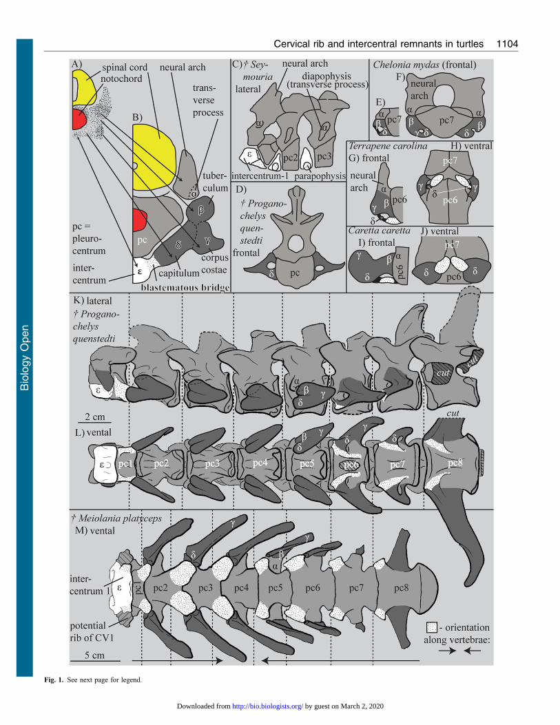
Fig. 1. See next page for legend.
Cervical rib and intercentral remnants in turtles 1104
Bio
logy
Open
by guest on March 2, 2020http://bio.biologists.org/Downloaded from
polystyrene foam boards of 2 mm thickness; the drawing is based on this model(Fig. 2M). All blastematous condensations were delineated from the sections byW.M. without any knowledge of turtle neck anatomy. The results are therefore notthe circular confirmation of a priori expectations. The interpretation is furthersupported by the work of Howes and Swinnerton (Howes and Swinnerton, 1901).They found comparable embryonic cell condensations in the vertebral column of thetuatara, a species which processes intercentra in the adult.
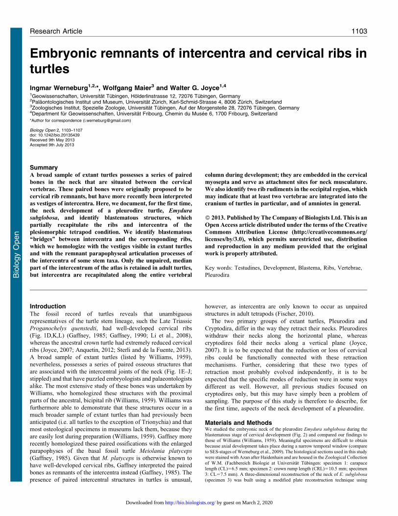
Results and DiscussionThe embryonic ribs of Emydura subglobosa consist of three parts
(Fig. 1A): tuberculum (b), corpus costae (c), and capitulum (d).
Each rib is dorsally articulated with the transverse process of the
neural arch (a) and ventrally connected with the intercentrum (e)via a blastematous bridge, which is more continuous with e than
with d (Fig. 1B, Fig. 2L). The transverse process (a) is part of the
neural arch. The most differentiated ribs of the neck are clearly
bicipital and situated in the middle third of the cervical column
(Fig. 2L,M). Whereas the cartilages of the neural arch and
pleurocentrum are clearly distinct, all blastematous structures
characteristically grade into one another. Well-pronounced
concentrations in cell density nevertheless exist. Based on
topological criteria we interpret these as vestigial skeletal
elements (sensu Howes and Swinnerton, 1901).
Our reconstruction shows that there may be at least two,
perhaps three vertebrae, which fuse to the occipital region of the
skull to contribute to the parachordal region. Posteroventral to
the two foramina nervi hypoglossi (Gaffney, 1972), two small,
ventrolateral, cartilaginous processes are visible, which we
homologize with capituli (d) using topological criteria; the last
projection still appears to be loosely connected with a short,
unisegmental neck muscle (Fig. 2F,G; arrows).
The anlage of the first cervical vertebra shows a similar
process, which, however, is broader when compared to the
slender capituli (d) associated with the occipital cranium. The
capituli of the first cervical ribs are furthermore associated with
the intercentrum of the first vertebra. The pleurocentrum of the
first vertebrae is situated posterior to the intercentrum. Dorsally,
the neural arches of the first and the second vertebrae appear to
be fused. Additionally, neural arch 1 is fused to intercentrum 1
and not to the pleurocentrum, as in the other vertebrae.
The ribs of the second and eighth vertebrae show the typical
differentiation into capitulum, tuberculum, and corpus costae.
Ventromedially, they are connected to the intercentrum via a
blastematous bridge. The capituli (d) seem to be the last elements to
be formed during development (Fig. 2A–C). The ribs of vertebrae
three to five are also well differentiated, but they do not directly
contact the related intercentra. The rib of vertebra six is very
similar to the second rib, but the connection with the intercentrum
(e) is rather slender when compared to the other neck segments. Rib
seven is ventrally connected to the intercentrum, its attachment to
the neural arch is loose, and the tuberculum (b) is absent.
The ribs of the first and second dorsal (trunk) vertebrae are
more differentiated (Fig. 2M), particularly their corpora costae
(c). Whereas the first trunk rib retains contact with itsintercentrum, the second and the following trunk ribs are
completely detached from their intercentra. Compared to thecervical ribs, the trunk ribs are situated more dorsally and areassociated with the developing carapace (Fig. 2D,E,M). Exceptfor the intercentrum of the atlas, no intercentrum or rib elements
were documented in post-hatching specimens of E. subglobosa
(Werneburg et al., 2009; Werneburg, 2011).
In the plesiomorphic, rhachitomous tetrapod condition (Fig. 1C),
an anterior intercentrum (synonym: hypocentrum) and an almostequally sized posterior pleurocentrum form the vertebral body(Fischer, 2010; Pierce et al., 2013). The intercentra of early
reptiliomorphs are reduced to 40% or less of the length of thepleurocentra (Lee and Spencer, 1997) (Fig. 1C) and theintercentrum is completely reduced or only rudimentarily presentin amniotes (Fischer, 2010). The ribs of tetrapods usually show two
articulations: the capitulum (d) articulates with the intercentrum (e)whereas the tubercle (b) articulates with the diapophysis (transverseprocess) of the neural arch (a); when the intercentrum has
disappeared completely, the capitulum may be in contact with theparapophysis (Fischer, 2010). In adult turtles, only the intercentrumof the atlas is unambiguously present and only the pleurocentra
form the bodies (‘‘centra’’) of all other vertebrae (Gaffney, 1990).
Williams recognized up to three fragmentary ‘rib’ elements insome adult turtle specimens, which he homologized withblastematous rib parts he observed in cryptodiran embryos (cf.
Fig. 1A,B) (Williams, 1959). According to Williams, the transverseprocess (our a) is either formed by a separate chondrification oremerges from the same chondrification as the neural arch,
depending on the species (Williams, 1959). For the pleurodirestudied herein, we can confirm a common cartilaginous origin of thetransverse process with the neural arch, which indicates that the
former does not represent a fused rib element. Williams alsoidentified a middle part of the rib (our c) and a ventral part (our d),which can persist as separated ossicles into adulthood (Fig. 1G,H)
(Williams, 1959). We identified two further blastematous structuresin the embryo, the anlage of the intercentrum (e) and the closelyassociated blastematous bridge. The ventral ‘rib’ part described byWilliams most likely represents the paired remnant of the
blastematous bridges based on topological criteria (Williams, 1959).
The blastematous rib anlagen of our pleurodire embryos showdiverse anatomical differentiations along the neck. Williams
described only one stage (Williams, 1959), which could eitherindicate that synchronous rib development occurs in cryptodiresor that the author described a somewhat more advanced
ontogenetic state in which the other blastematous parts werealready reduced. In the former case, a functional correlation withthe retraction mode may be the cause. The blastematous rib of thesecond cervical vertebra in the pleurodire we studied closely
resembles the cryptodire embryonic rib shape documented byWilliams with its b, c, and d parts (Williams, 1959). A majordifference is found in the expansion of the ventral-most part of
the rib anlage. Whereas it ends with part d in cryptodires(Williams, 1959), it connects to the intercentrum (e) in thepleurodire via a blastematous bridge. By positional criteria and
its close continuity to the intercentrum (e), we suggest homologyof these ‘‘bridges’’ with the parapophyseal processes of theintercentrum and with the paired bones seen in many adult
specimens of different taxa (Fig. 1C,G–J); however, paired boneshave not yet been documented for adult specimens of E.
subglobosa specimens per se (Werneburg, 2011).
Fig. 1. Comparison of neck vertebrae. (A,B) Scheme of the development ofthe vertebral region in a turtle with the terminology defined herein. (C) The firstthree neck vertebrae of Seymouria sp. (plesiomorphic amniote condition) (afterStarck, 1979). Panels A,B,D–I were modified after Williams (Williams, 1959).(E–J) ‘Rib’ rudiments in different adult cryptodiran turtles. (K–M) Stem turtle
necks (after Gaffney, 1985; Gaffney, 1990). The parapophyses of theintercentra are dotted. Whether a rib is actually present at the atlas ofMeiolania platyceps is not certain (M, Gaffney, 1985) and is illustrated as‘‘potential rib’’ herein.
Cervical rib and intercentral remnants in turtles 1105
Bio
logy
Open
by guest on March 2, 2020http://bio.biologists.org/Downloaded from
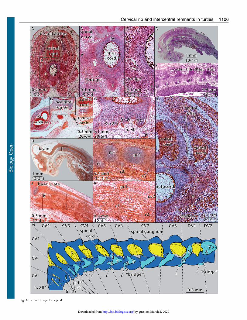
Fig. 2. See next page for legend.
Cervical rib and intercentral remnants in turtles 1106
Bio
logy
Open
by guest on March 2, 2020http://bio.biologists.org/Downloaded from
We furthermore homologize the blastematous bridges with the
articulation (parapophyseal) processes of the intercentrum of
early tetrapods. Although the unpaired intercentra are reduced in
the stem turtle Meiolania platyceps, the processes are still present
in that taxon as expanded parapophyses that articulate with the
capitulum (d) of the rib (Fig. 1M). Gaffney homologized the
parapophyses of M. platyceps with the paired bones seen in the
necks of many extant turtles (Gaffney, 1985) and we agree with
that assessment based on our embryological evidence.
The variable modes of fusion of the intercentral parapophyses
with the pleurocentra of M. platyceps support our proposed
homology: the intercentrum and its associated parapophyses are
formed ventrally and ventrolaterally between the adjacent
pleurocentra (Fig. 2), and, after reduction of the intercentra, the
parapophyses later fuse to the anterior (M. platyceps: vertebrae 1–3)
or to the posterior, developmentally related (M. platyceps: cervical
vertebrae 5–8) pleurocentra, or they remain as separated structures
in extant taxa (Fig. 1G–J). The original ‘‘articulation’’ of the rib to
the intercentrum (Fischer, 2010) is only recapitulated in the first
neck segment (atlas), which retains its unpaired intercentrum.
The parapophyses of the intercentra appear to be the most
important remainders of the vertebral appendages (‘ribs’ sensu
Williams, 1959) in extant turtles. The musculi longus colli consist of
numerous muscle bundles and are attached to these parapophyseal
remnants (Werneburg, 2011). We speculate that the ‘‘ossified
bridge-parts’’ are retained in reduced form among many extant
turtles because they mechanically serve as insertion points within
the highly mobile neck of living turtles or as developmental
‘‘aggregation points’’ for muscle fibres during development
(Fig. 2H–J). Because ribs are by definition skeletal structures of
the myosepta, it is not surprising that the rib blastemata are laterally
continued by the mesenchyme of their myosepta (cf. Fig. 2K).
It is apparent that fully formed cervical ribs, such as those
developed in Proganochelys quenstedti, would be incompatible
with any retraction mechanism of the neck; it therefore represents a
plesiomorphic evolutionary state. Conversely, turtles reduced the
cervical ribs to achieve any kind of neck retraction. Current
phylogenies of fossil turtles (Joyce, 2007; Anquetin, 2012; Sterli
and de la Fuente, 2013) agree that adult specimens of at least some
basal crown turtles (e.g. representatives of Xinjiangchelyidae;
W.G.J., personal observations) still possessed reduced cervical
ribs. It is therefore almost certain that extant cryptodires and
pleurodires reduced their cervical ribs independently. However,
with the present state of knowledge and scarcity of embryonic data,
it seems premature to speculate about minor details of convergence
and on features of heterochrony.
The developmental contribution of vertebrae to the formation
of the primary neurocranium of vertebrates is generallyaccepted, mainly due to the numbers of roots of thehypoglossal nerve (Furbringer, 1897; Starck, 1979). The
primary neurocranium in actinopterygian fishes has recentlybeen shown to include three occipital segments (Britz andJohnson, 2010). To our knowledge, occipital ribs have only been
documented within Tetrapoda for the penguin Spheniscus
demersus (Crompton, 1953). The presence of at least twooccipital vestigial rib-homologues in the pleurodiran turtle E.
subglobosa therefore suggests that at least two neck vertebrae
are fused to the skull in sauropsids.
AcknowledgementsThe embryonic specimens of Emydura subglobosa were collected bythe late Stefan Eßwein. This contribution was partially funded by aDFG grant to W.G.J. (JO 928/1-1). We also acknowledge support byDeutsche Forschungsgemeinschaft and Open Access PublishingFund of Universitat Tubingen.
Competing InterestsThe authors have no competing interests to declare.
ReferencesAnquetin, J. (2012). Reassessment of the phylogenetic interrelationships of basal turtles
(Testudinata). Journal of Systematic Palaeontology 10, 3-45.Britz, R. and Johnson, G. D. (2010). Occipito-Vertebral Fusion in Actinopterygians:
Conjecture, Myth and Reality. Part 1: Non-Teleosts. In Origin and Phylogenetic
Interrelationships of Teleosts: Honoring Gloria Arratia (ed. J. S. Nelson, H.-P.Schultze and M. V. H. Wilson). Munchen: Verlag Dr. Friedrich Pfeil.
Crompton, A. W. (1953). The development of the chondrocranium of Spheniscus
demersus with special reference to the columella auris of birds. Acta Zoologica 34,71-146.
Fischer, M. S. (2010). Bewegungsapparat: Postcraniales Skelett und Muskulatur. InWirbel- oder Schadeltiere (Spezielle Zoologie, Teil 2), (ed. W. Westheide andG. Rieger). Heidelberg; Berlin: Spektrum Akademischer Verlag.
Furbringer, M. (1897). Ueber die spino-occipitalen Nerven der Selachier undHolocephalen und ihre vergleichende Morphologie. In Festschrift zum
Siebenzigsten Geburtstage von Carl Gegenbaur, pp. 349-788. Leipzig: Engelmann.Gaffney, E. S. (1972). An illustrated glossary of turtle skull nomenclature. American
Museum Novitates 2486, 1-33.Gaffney, E. S. (1985). The cervical and caudal vertebrae of the cryptodiran turtle,
Meiolania platyceps, from the pleistocene of Lord Howe Island, Australia. American
Museum Novitates 2805, 1-29.Gaffney, E. S. (1990). The comparative osteology of the Triassic turtle Proganochelys.
Bulletin of the American Museum of Natural History 194, 1-263.Howes, G. B. and Swinnerton, H. H. (1901). On the development of the skeleton of
the tuatara, Sphenodon punctatus; with remarks on the egg, on the hatching, and on thehatched young. The Transactions of the Zoological Society of London 16, 1-84.
Joyce, W. G. (2007). Phylogenetic Relationships of Mesozoic Turtles. Bulletin of the
Peabody Museum of Natural History 48, 3-102.Lee, M. S. Y. and Spencer, P. S. (1997). Crown-Clades, Key Characters
and Taxonomic Stability: When is an Amniote not an Amniote? In Amniote
Origins: Completing the Transition to Land (ed. S. S. Sumida and K. L. M. Martin).San Diego; London; Boston; Sydney; Tokyo; Toronto; New York: AcademicPress.
Li, C., Wu, X. C., Rieppel, O., Wang, L. T. and Zhao, L. J. (2008). An ancestral turtlefrom the Late Triassic of southwestern China. Nature 456, 497-501.
Pierce, S. E., Ahlberg, P. E., Hutchinson, J. R., Molnar, J. L., Sanchez, S.,
Tafforeau, P. and Clack, J. A. (2013). Vertebral architecture in the earliest stemtetrapods. Nature 494, 226-229.
Starck, D. (1979). Das Skeletsystem: Allgemeines, Skeletsubstanzen, Skelet der
Wirbeltiere einschließlich Lokomotionstypen (Vergleichende Anatomie der
Wirbeltiere auf Evolutionsbiologischer Grundlage, Bd. 2). Berlin, Heidelberg, NewYork: Springer-Verlag.
Sterli, J. and de la Fuente, M. S. (2013). New evidence from the Palaeocene ofPatagonia (Argentina) on the evolution and palaeo-biogeography of Meiolaniformes(Testudinata, new taxon name). Journal of Systematic Palaeontology [Epub ahead ofprint].
Werneburg, I. (2011). The cranial musculature of turtles. Palaeontologica Electronica
14, 15A.Werneburg, I., Hugi, J., Muller, J. and Sanchez-Villagra, M. R. (2009). Embryogenesis
and ossification of Emydura subglobosa (Testudines, Pleurodira, Chelidae) and patternsof turtle development. Dev. Dyn. 238, 2770-2786.
Williams, E. E. (1959). Cervical ribs in turtles. Breviora 101, 1-12.
Fig. 2. Embryonic neck anatomy of Emydura subglobosa. Embryonicvertebrae and ribs in specimens of (A,B) CL56.5 mm carapace length, (C,D)CRL510.5 mm, and (F–M) CL57.5 mm. (M) Redrawn 3 d reconstruction:dark blue 5 cartilaginous neck vertebrae, light blue 5 blastematouscondensations. Numbers under scale bars indicate section numbers. (A–C)Cross sections through a cervical vertebra with partly developed embryonicribs; (D,E) sagittal section through the whole body with (E) a focus on the
neural arches/ribs; (F,G) cross section through the left ear capsule, the occipitaland the anterior cervical region with (G) a focus on the occipital skull region.(H–K) Sagittal section through the anterior part of the body with (I,J) a focus onthe first cervical vertebrae, H/I 5 mid sagittal and J/K 5 more lateral; (L) crosssection through cervical vertebra six, compare to panel M. CV, cervicalvertebra; n. XII, branches of nervus hypoglossus. For further abbreviations see
Fig. 1B. Arrows in panels F,G indicate unisegmental neck muscle attaching tothe posterior most occipital rib (d-1).
Cervical rib and intercentral remnants in turtles 1107
Bio
logy
Open
by guest on March 2, 2020http://bio.biologists.org/Downloaded from


![Brunner’s Gland Hamartoma of the Duodenum: A Literature …Brunner’s glands appear from the 13th to 14th weeks of embryonic develop-ment [2, 3]. Prevalence of Brunner’s glands](https://static.fdocumentos.com/doc/165x107/6147f9f3a830d0442101c8b3/brunneras-gland-hamartoma-of-the-duodenum-a-literature-brunneras-glands-appear.jpg)