ESCOLA SUPERIOR DE TECNOLOGIA DA SAÚDE DO...
85
Transcript of ESCOLA SUPERIOR DE TECNOLOGIA DA SAÚDE DO...

ESCOLA SUPERIOR DE TECNOLOGIA DA SAÚDE DO PORTO
INSTITUTO POLITÉCNICO DO PORTO
Ana Luísa Teixeira Ferreira
Setembro 2011
Dissecting the Role of CLASP1 in Mammalian Development
Dissertação submetida à Escola Superior de
Tecnologia da Saúde do Porto para cumprimento dos
requisitos necessários à obtenção do grau de Mestre
em Tecnologia Bioquímica em Saúde, realizada sob a
orientação científica de Helder Maiato, PhD e co-
orientação de Ana Lúcia Pereira, PhD e Rúben
Fernandes, PhD.

"Uma boa recordação talvez seja cá na Terra mais autêntica do que a felicidade."
Alfred de Musset
Dedicada à minha avó.

ACKNOWLEDGEMENTS
Acknowledgements
I would like to thank those who, directly or indirectly, contributed to this work and
helped me during this journey. I would like to express my special gratitude to:
Helder Maiato (supervisor)
Thank you for giving me so many opportunities and for being constantly showing me the
bright side of science, even when things do not go as expected. Thank you for being
always available when I needed, despite your busy schedule. Thank you for teaching me
the importance of the persistence and hard work in science (which I’m still learning) and
for your comprehension and patience with which you’ve guided me since I joined your
group.
Ana Lúcia Pereira (co-supervisor)
Well, I think you’re the person who I have to thank most. I couldn’t expect a better tutor.
You have taught me everything I have learned so far and I hope to deserve to be at your
side in the future, because I really need you to teach me and to say “WORK”! Thank you
for all the times that you “woke me up” when I got less focused and for being a reference
of the hard work and perfectionism. Thank you for being always by my side as tutor and
mostly (I hope I can say) as my friend
Rúben Fernandes (co-supervisor from ESTSP) and Cristina Prudêncio
I would like to thank you both for the opportunity of being “your” master’s student and for
your constant availability to help me every time I needed. A special thank you to Rúben
Fernandes, who has always believed in me since I was a first year student in Escola
Superior de Tecnologia da Saúde do Porto.
Lab colleagues
I would like to express my gratitude to all my colleagues of the Chromosome Instability
and Dynamics Laboratory, with who I’ve been learning a lot. I would like to express a
special gratitude to Elsa Logarinho, Rita Maia and Zaira Garcia for their amazing expertise
and kind way of teaching me.

Horácio Scigliano
I’m also thankful to my anatomical pathology teacher, who gave me his passion for
histopathology and for have giving me his specialized opinion during this work.
Jimmy
A special thank you to my “informatics heroe”, who has been always available to fix my
crashed computer when I most need him. Thank for that and for the recent friendship with
which you’ve supported me during the last tough months.
Family
I would like to thank my family for all the patience and support. I cannot express my
gratitude by words.
Father
Words for what?! Thank for everything. Thank you for being the most important person in
my life, who will never let me down.
Friends
My deepest gratitude to my friends for being so supportive and for have encouraged me
during the less easy moments. A special thank you to Sergio Dias, Joana Almeida, Rute
Fernandes, Joana Barreto, Miguel Silva, Luís Rodrigues, Rui Quintão, Sali Meireles,
Miguel Cizeron and Nelson Dias.
Aims of the work
CLIP-associated proteins (CLASPs) are microtubule-associated proteins that
accumulate at the microtubule plus-ends. These proteins are highly conserved in
eukaryotes and play important roles both in interphase and mitosis.
In Drosophila, yeast, Xenopus and C. elegans, CLASPs were shown to be
important in mitosis for chromosome segregation, maintenance of spindle bipolatity and
regulation of microtubules at the kinetochore level. CLASPs are also implicated in a
variety of cellular processes in interphase cells, namely in microtubule stabilization and
cell polarity.
Mammals have two paralogues, CLASP1 and CLASP2, whose functions have been
considered partially redundant and little is known about their individual roles. The recent
generation of Clasps KO mammalian models will further improve our knowledge about
these proteins in a physiological context. In this work we took advantage of a Clasp1 KO
mouse model to study the CLASP1 role in mammalian physiology.
Abstract
Mitosis is a complex cellular event through which cells correctly divide two copies
of their DNA content to daughter cells. This process is mediated by a cellular machine
called mitotic spindle, a bipolar structure built by microtubules (MTs). MT dynamics is
regulated by MT-associated proteins (MAPs), such as MT plus-end tracking proteins
(+TIPs). CLIP-associated proteins (CLASPs) belong to this family and are highly
conserved among eukaryotes. These proteins interact with MTs regulating mitotic spindle
bipolarity, chromosome segregation and MT behavior at the kinetochore. Thus, CLASPs
have been described as essential factors for genetic integrity during cell division.
A Clasp1 knockout (KO) mouse strain, previously generated in our lab, is an ideal
tool to uncover the physiological role of CLASP1 in mammals. All Clasp1 KO mice
display a lethal phenotype, dying a few minutes after birth, with an acute lung failure.
Upon histological analysis, we found that KO lungs exhibit a morphogenic delay.
However, molecular analysis of lung late maturation markers have shown that type I and
type II pneumocytes, the cells responsible for the gas-exchange, are differentiated in KO
mice at the moment of their birth. Nevertheless, an underlying lung defect cannot yet be
excluded. High amounts of glycogen in KO pulmonary parenchyma were found, which
might be, not only a sign of lung immaturity, but also deficiencies in surfactant lipid
component. Regarding CLASP1 expression, we observed that this protein is transiently
expressed among different organs throughout mouse development, being particularly
strong in brain, a fact that might explain its roles in neuronal biology. CLASP1 is also
ubiquitously expressed in adult mice, suggesting that this protein is also important in
mature tissues. Moreover, it is not clear whether the morphological delay observed in KO
lungs may explain the newborn lethality observed in Clasp1 KO mice. At this stage, the
biological meaning of CLASP1 in mammals’ physiology is not clear. So far, no Clasp1
KO mice was able to survive ex uterus, suggesting that this protein is important during late
developmental stages in mammals.
Key words: CLASP1, knockout, lungs and development.
Sumário
A mitose é o evento celular, através do qual uma células transmite uma cópias do
seu DNA às células filhas. Este processo é mediado pelo fuso mitótico, o qual consiste
numa rede bipolar microtubulos. A dinâmica dos microtubulos é regulada por proteínas
associadas a estes (MAPs – Microtubule-Associated Proteins), tais como as proteínas
associadas às extremidades positivas dos microtubulos (+TIPs – Plus-ends Tracking
proteins). As proteínas associadas às CLIPs (CLASPs – CLIP-associated proteins)
pertencem a esta família e estão altamente conservadas nos eucariotas. Estas interagem
com os microtubulos regulando o fuso mitótico, a segregação dos cromossomas e o
comportamento dos microtubulos ao nível do cinetocoro. Assim, as CLASPs têm sido
descritas como essenciais à manutenção da integridade genética durante a divisão celular.
Um modelo animal knockout para o gene Clasp1 é uma ferramenta indispensável à
descoberta do papel da CLASP1 a nível fisiológico. Nos animais knockout foi observado
um fenótipo letal, no qual 100% dos recém-nascidos morreram poucos minutos após o
nascimento, no decurso de falência respiratória. Após análise histopatológica, observamos
que os pulmões dos animais knockout apresentam um atraso no desenvolvimento. Porém, a
análise da expressão de marcadores de diferenciação celular, mostrou que os pneumócitos
tipo I e II estão presente e diferenciados nos animais knockout aquando do seu nascimento.
No entanto, um defeito primário a nível pulmonar ainda não pode ser excluído. Níveis
elevados de glicogénio no parênquima alveolar dos animais knockout sugerem imaturidade
pulmonar ou deficiente produção do líquido surfactante. Adicionalmente, ainda não está
esclarecido de que forma pode este atraso explicar a letalidade observada nos recém-
nascidos knockout. Verificamos também que expressão de CLASP1 é transiente ao longo
do desenvolvimento, sendo particularmente elevada no cérebro, o que pode explicar o seu
papel já descrito na biologia dos neurónios. A CLASP1 é ubiquamente expressa em
mamíferos adultos, o que sugere que esta proteína é também importante em tecidos
diferenciados. Nesta fase, o significado biológico da CLASP1 em mamíferos ainda não foi
descortinado. No entanto, nenhum animal knockout para Clasp1 foi capaz de sobreviver ex
uterus, o que sugere um papel fundamental desta proteína na fase final do desenvolvimento
dos mamíferos.
Palavras-chave: CLASP1, knockout, pulmões e desenvolvimento.
Índex
Aims of the work
Abtract
Sumário
CHAPTER I - General Introduction .................................................................................................................. 1
1. The Eukaryotic Cell Cycle .......................................................................................................... 1
2. The Cell-Cycle Control System .................................................................................................. 3
3. The Mitotic Apparatus ................................................................................................................ 4
3.1. Microtubules ....................................................................................................................... 4
3.2. The Centrosomes ................................................................................................................ 6
3.3. The Kinetochore.................................................................................................................. 8
3.4. MT-associated proteins ..................................................................................................... 10
4. Spindle Assembly ..................................................................................................................... 11
4.1. Kinetochore-MT attachment ............................................................................................. 14
4.2. Error correction in kinetochore-microtubule attachment .................................................. 16
5. Mechanisms of Chromosome Movement ................................................................................. 18
5.1. Chromosome congression ................................................................................................. 18
5.2. Chromosome Segregation ................................................................................................. 19
6. CLIP-associated proteins – CLASPs ........................................................................................ 20
6.1. Microtubule-plus end tracking proteins ............................................................................ 20
6.2. Structure of CLASPs ........................................................................................................ 21
6.3. Cellular distribution of CLASPs ....................................................................................... 23
6.4. Functions of CLASPs. ...................................................................................................... 23
7. Aneuploidy ............................................................................................................................... 25
8. Mouse models of embryonic development ............................................................................... 26
8.1. Mouse models ................................................................................................................... 26
8.2. Clasp1 knockout mouse model ......................................................................................... 27
9. Mose development .................................................................................................................... 27
9.1. Lung development and physiology ................................................................................... 28

EXPERIMENTAL WORK
CHAPTER I - Characterization of Clasp1 Knockout Mice ............................................................................ 33
CHAPTER II - CLASP1 Expression Profile Throughout Development ......................................................... 48
CHAPTER III - Optimization of CLASP1 Immunostaining for Histological Samples............................ .......52
GENERAL DISCUSSION ............................................................................................................................ 598
REFERENCES………………………………………………………………………………………………..73

GENERAL
INTRODUCTION
Dissecting the Role of CLASP1 in Mammalian Development
1
CHAPTER I - General Introduction
All cells are the product of the cell division of a pre-existent cell (omnis celllula e
cellula, Rudolf Virchow, 1859). The earliest mentions about cell division are from 1766,
when Trembly observed for the first time the process in Diatom Synedra. Since then, cell
division has fascinated the scientific world, not only because it is the key event which
perpetuates biogenesis, but also because it triggers the most complex changes in all cell
cycle. Since Walter Fleming’s assumptions, in which he describes mitosis with a
remarkable detail, this cellular process has been the target of a huge scientific investment
in the cellular biology niche. However, despite all the advances in this field, a lot of
molecular and biophysical events related to the regulation of the distribution of the genetic
material through the daughter cells remain unknown.
1. The Eukaryotic Cell Cycle
The eukaryotic cell cycle is a highly regulated process, in which each cell ensures
the transmission of the correct copy of its genome to each daughter cell. It is divided in two
main parts: the interphase, which is the most prolonged period of all cell cycle, and the M
phase, that includes mitosis (nuclear division) and cytokinesis (cytoplasm division).
Additionally, there are four fundamental events during the cell cycle that are particularly
important for the maintenance of the correct DNA content, which are: cell growth, DNA
duplication, chromosome segregation and cell division (Morgan, 2007). Interphase is a
period of growth and DNA replication divided in three stages: gap phase 1(G1 phase),
synthesis phase (S phase) and gap phase 2 (G2 phase). These gap phases are transition
periods in which an important portion of the regulatory machinery of the cell controls the
cell cycle. G1 phase is the period prior to the synthesis of DNA. In this phase, the cell
increases in mass and prepares itself to undergo cell division. In S phase DNA is
synthesized and duplicated. Lastly, G2 phase is the period between DNA synthesis and the
start of M phase, in which the cell synthesizes proteins and continues to increase in size. In
late interphase, the cell still has nucleoli present, the nucleus is bounded by a nuclear
envelope and chromosomes have duplicated but not condensed. (J. M. Mitchison, 1971;
Murray, 1993).
Dissecting the Role of CLASP1 in Mammalian Development
2
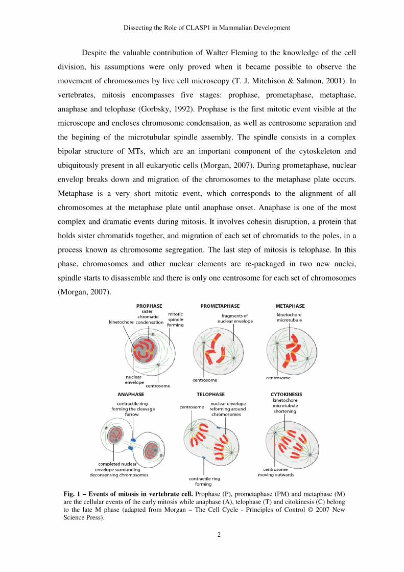
Despite the valuable contribution of Walter Fleming to the knowledge of the cell
division, his assumptions were only proved when it became possible to observe the
movement of chromosomes by live cell microscopy (T. J. Mitchison & Salmon, 2001). In
vertebrates, mitosis encompasses five stages: prophase, prometaphase, metaphase,
anaphase and telophase (Gorbsky, 1992). Prophase is the first mitotic event visible at the
microscope and encloses chromosome condensation, as well as centrosome separation and
the begining of the microtubular spindle assembly. The spindle consists in a complex
bipolar structure of MTs, which are an important component of the cytoskeleton and
ubiquitously present in all eukaryotic cells (Morgan, 2007). During prometaphase, nuclear
envelop breaks down and migration of the chromosomes to the metaphase plate occurs.
Metaphase is a very short mitotic event, which corresponds to the alignment of all
chromosomes at the metaphase plate until anaphase onset. Anaphase is one of the most
complex and dramatic events during mitosis. It involves cohesin disruption, a protein that
holds sister chromatids together, and migration of each set of chromatids to the poles, in a
process known as chromosome segregation. The last step of mitosis is telophase. In this
phase, chromosomes and other nuclear elements are re-packaged in two new nuclei,
spindle starts to disassemble and there is only one centrosome for each set of chromosomes
(Morgan, 2007).
Fig. 1 – Events of mitosis in vertebrate cell. Prophase (P), prometaphase (PM) and metaphase (M) are the cellular events of the early mitosis while anaphase (A), telophase (T) and citokinesis (C) belong to the late M phase (adapted from Morgan – The Cell Cycle - Principles of Control © 2007 New Science Press).
Dissecting the Role of CLASP1 in Mammalian Development
3
2. The Cell-Cycle Control System
Cell cycle is a highly controlled phenomenon regulated by a complex network of
effectors, which control the order and timing of cell-cycle events. A series of regulatory
proteins active specific effectors, while others are inhibited. However, cell-cycle regulators
work as single units in a complex orchestra, in which the perfect synchrony is required for
the efficiency of this fundamental biological process.
This complex set of players, which is active at a specific point, allows cells to know
when it is time to divide, or to maintain in a sleeping stage until extracellular signals
trigger cell division.
The most important regulators of the cell cycle are the cyclin-dependent kinases
(Cdks), which upon activation by the respective cyclin, activates a set of downstream
pathways, which regulate cell-cycle events. Cdks concentrations is constant throughout cell
cycle, but its phosphorylation status changes as a consequence of oscillations of their
cyclin partners. These proteins bind specifically to each Cdk, triggering its catalytic
activity. Different Cdks are activated at each point of the cell cycle, while others are silent.
The efficiency of these phosphorylation switches is a fundamental requirement to ensure
the correct sequence of events during cellcycle and cell division (Morgan, 2007).
Focusing only on the cyclins that regulate Cdks activity involved in cell cycle
regulation, we may enclose them in four main classes - G1/S cyclins, S cyclins, M-cyclins
– which are directly involved in control of cell cycle events- and G1 cyclins, the family
involved in control of cell-cycle entry in response to extracellular signals.
To ensure the correct distribution of the genetic material to their progeny, cells
make use of regulatory steps termed checkpoint controls. When cells sense some errors,
checkpoints delay cell cycle progression, preventing the transmission of compromised
genome integrity. The first checkpoint called Start occurs when the regulatory machinery
checks if all conditions are ideal for cell proliferation, G1/S and S phase cyclin-Cdk
complexes are active and trigger DNA replication, centrosome duplication, as well as other
early cell-cycle events. Ultimately, G1/S and S- active Cdks promote the activation of M-
Cdks, which drive progression to the next checkpoint. The second checkpoint occurs prior
to cell entry into mitosis and is termed G2/M checkpoint. This regulatory step responds to
DNA damage (Pearce & Humphrey, 2001) or other injurious influences (Mikhailov, Cole,
& Rieder, 2002), acting by inhibiting the master mitotic regulator, the cyclin B/CDK1
complex. The third checkpoint controls the metaphase-to-anaphase transition. The
Dissecting the Role of CLASP1 in Mammalian Development
4
satisfaction of this point of control promotes sister chromatid segregation, completion of
mitosis and cytokinesis. This checkpoint counts on Cdc20 and Cdh1, as well as Mad and
Bud family proteins present on unattached, but not on attached kinetochores to prevent the
activation of the anaphase-promoting complex (APC) (Vigneron, et al., 2004; Zhou, Yao,
& Joshi, 2002). The metaphase-to-anaphase checkpoint is also termed spindle assembly
checkpoint (SAC) because it monitors if all kinetochores are attached to the spindle MTs.
Once all kinetochores are bioriented at the metaphase plate and attached to the spindle
MTS, SAC is satisfied, anaphase onset takes place and cells start exiting mitosis (Rieder &
Maiato, 2004). The APC is a large ubiquitin-ligase that targets securin (Nasmyth, Peters, &
Uhlmann, 2000) and cyclin B (Hagting, et al., 2002) to proteolysis. The respective
degradation of both proteins allows chromosome segregation and completion of mitosis
(telophase). Cyclins degradation leads to the inactivation of Cdks in the cell, which allows
phosphatases to dephosphorylate Cdks substrates. Those dephosphorylations are required
for spindle disassembly, completion of mitosis and cytokinesis. Recently, a new
checkpoint has been proposed, which delays cytokinesis in the presence of lagging
chromosomes. This point of control is mediated by Aurora B/Ipl1 kinase activation in the
presence of chromatin at the midzone in posttelophase stages, which leads to furrow
regression and thereby preventing aneuploidy (Mendoza, et al., 2009; Steigemann, et al.,
2009)
3. The Mitotic Apparatus
During mitosis, chromosome segregation is carried out by a complex machine
known as mitotic spindle. This structure pulls each set of chromosomes apart toward the
poles of the cell during chromosome segregation. The mitotic apparatus consists of:
microtubules (MTs), centrosomes (or MT self-organizing centers when cells are not
provided of centrosomes), kinetochores and MT-associated motor and non-motor proteins
that organize them into two antiparallel arrays of MTs.
3.1. Microtubules
MTs are the main structural component of the mitotic spindle, whose basic building
block is a heterodimer of α and β-tubulin (fig. 2; a). Stable α/β-tubulin heterodimers are
Dissecting the Role of CLASP1 in Mammalian Development
5
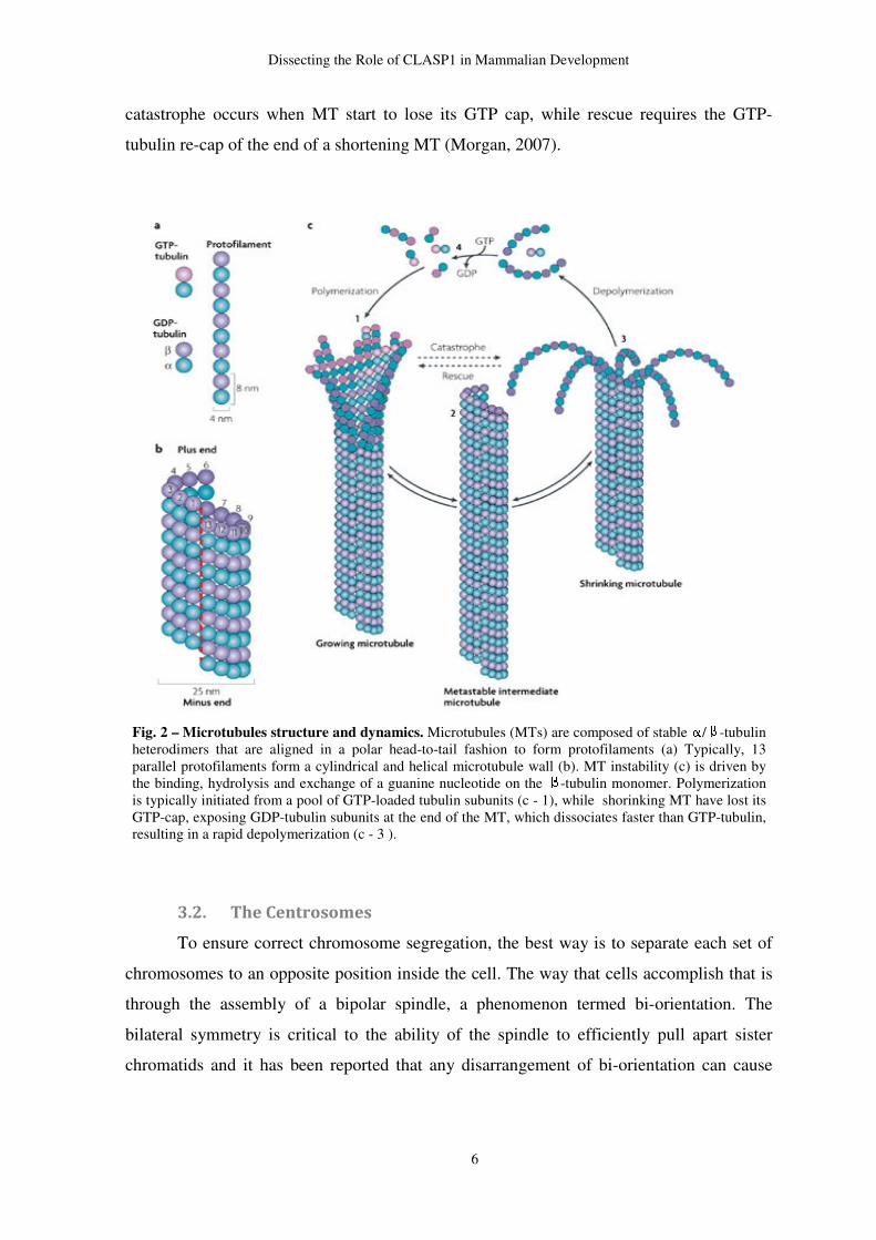
aligned in a polar head-to-tail fashion to form protofilaments (Akhmanova & Steinmetz,
2008) (fig. 2; a). Typically, a MT is made up of thirteen protofilaments, which associate
laterally to each other to form a hollow tube about 25 nm in diameter (fig. 2; b)
(Akhmanova & Steinmetz, 2008; Alberts, 2002; Morgan, 2007).
A molecule of guanosine triphosphate (GTP) binds to each unit of α-tubulin and β-
tubulin. According to the model currently accepted, the GTP of β-tubulin is hydrolyzed by
the moment that a new heterodymer subunit is incorporated at the growing end of the
growing MT. However, the GTP molecule of the β-tubulin that is exposed at the growing
tip remains intact, forming a structure called GTP-cap (McNally, 1999). β-tubulin GTP
hydrolysis results in a change of conformation of the heterodimer, which plays a
significant role in the dynamic turnover of MTs, an essential property for their fast growth
and shrinkage. The fast exchange from one state to another is called MT instability (fig. 2;
c) (Desai & Mitchison, 1997; Morgan, 2007).
The rearrangement of MT subunits results in two opposite MT ends with different
characteristics. The end that exposes the β-tubulin GTP is called plus end, while the
opposite end, called minus end, exposes the α-tubulin subunit. So, MTs have two clearly
distinct ends and the regulation of the dynamics of both ends occurs independently of each
other. The fact that MTs are polar structures, rearranged in a head-to-tail fashion, allows
them to act like rail to motor molecules, which are peculiar molecular organizers of the
intracellular space and cytoskeleton (Morgan, 2007). The MT plus ends have the capacity
to grow fast in vitro and, in vivo and are provided of a fascinating ability to exchange fast
between periods of growth and shrinkage. (Akhmanova & Steinmetz, 2008). The MT
dynamic instability model, based on in vitro experiments, dictates that MT exist in
persistent phases of either growth or shortening, with abrupt transition between both states
(Desai & Mitchison, 1997). These abrupt exchanges are termed catastrophe, when occurs
the switching from growing to shortening, and rescue, when the opposite happen (fig. 2; c).
During polymerization, MT ends often have a sheet-like extension in which some
protofilaments have grown longer than others (fig. 2; c1). On the other hand, when MT
depolymerize, individual protofilaments peel away from the polymer lattice (Desai &
Mitchison, 1997) (fig. 2, c3). As it was previously mentioned, The GTP-cap is the
molecular key of the MT dynamics. Shortening MTs have lost its GTP-cap, exposing
GDP-tubulin subunits at the end of the MT, which dissociates fifty times faster than GTP-
tubulin, resulting in a rapid depolymerization (Morgan, 2007). In a very simple away,
Dissecting the Role of CLASP1 in Mammalian Development
6
catastrophe occurs when MT start to lose its GTP cap, while rescue requires the GTP-
tubulin re-cap of the end of a shortening MT (Morgan, 2007).
3.2. The Centrosomes
To ensure correct chromosome segregation, the best way is to separate each set of
chromosomes to an opposite position inside the cell. The way that cells accomplish that is
through the assembly of a bipolar spindle, a phenomenon termed bi-orientation. The
bilateral symmetry is critical to the ability of the spindle to efficiently pull apart sister
chromatids and it has been reported that any disarrangement of bi-orientation can cause
Fig. 2 – Microtubules structure and dynamics. Microtubules (MTs) are composed of stable / -tubulin heterodimers that are aligned in a polar head-to-tail fashion to form protofilaments (a) Typically, 13 parallel protofilaments form a cylindrical and helical microtubule wall (b). MT instability (c) is driven by the binding, hydrolysis and exchange of a guanine nucleotide on the -tubulin monomer. Polymerization is typically initiated from a pool of GTP-loaded tubulin subunits (c - 1), while shorinking MT have lost its GTP-cap, exposing GDP-tubulin subunits at the end of the MT, which dissociates faster than GTP-tubulin, resulting in a rapid depolymerization (c - 3 ).
Dissecting the Role of CLASP1 in Mammalian Development
7
potentially lethal errors in chromosome segregation (Morgan, 2007; Wadsworth &
Khodjakov, 2004).
Although all normal spindles are bipolar, the structure from which MTs nucleate
may differ between some organisms. The centrosome is the primary microtubule-
organizing centre (MTOC) in animal cells, which regulates cell motility, adhesion and
polarity in interphase, and facilitates the organization of the spindle poles during mitosis
(Bettencourt-Dias & Glover, 2007). However, some cell types, such as the ones existing in
higher plants and oocytes of many vertebrates, are not provided with centrosomes and the
organization of the mitotic spindle depends on the self-organizing capacity of the MTs. So,
the acentrosomal pathway relies on many MT-associated proteins to generate two spindle
poles (Gadde & Heald, 2004; Morgan, 2007).
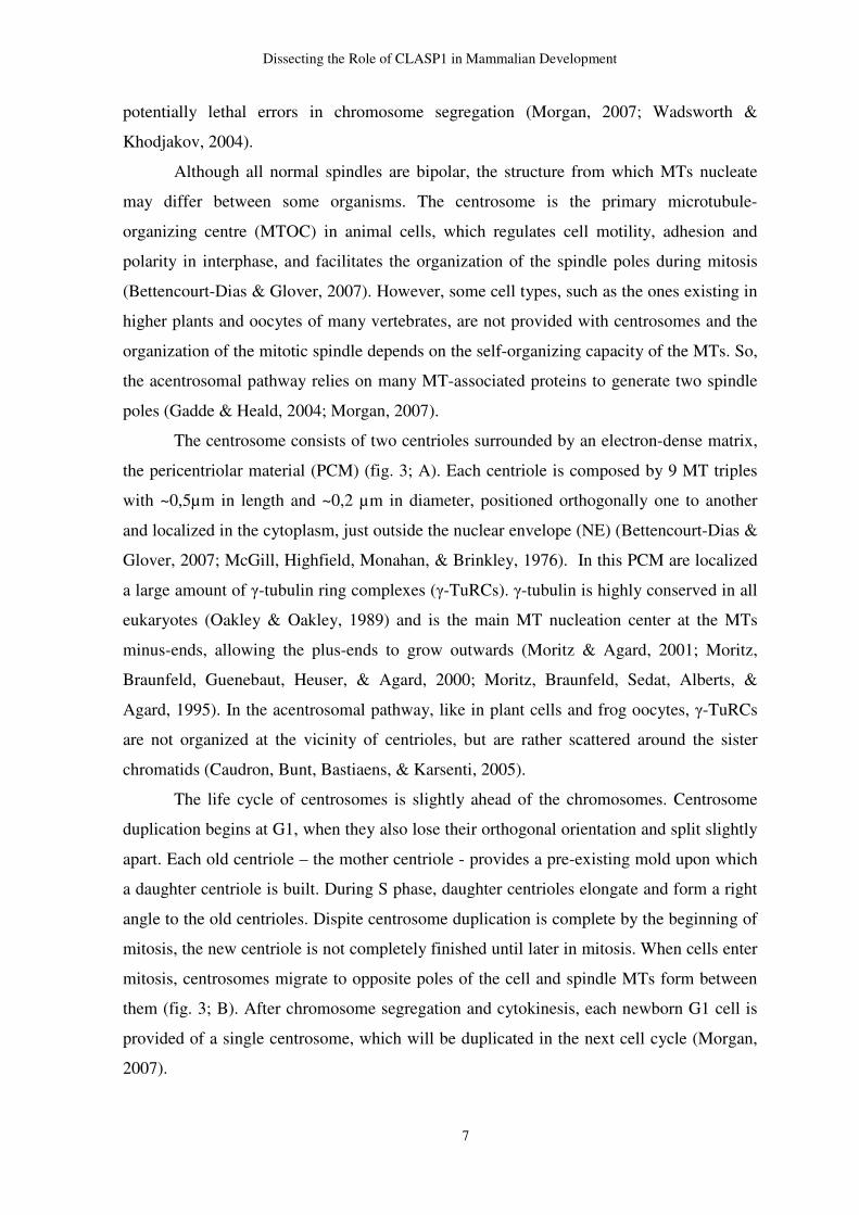
The centrosome consists of two centrioles surrounded by an electron-dense matrix,
the pericentriolar material (PCM) (fig. 3; A). Each centriole is composed by 9 MT triples
with ~0,5µm in length and ~0,2 µm in diameter, positioned orthogonally one to another
and localized in the cytoplasm, just outside the nuclear envelope (NE) (Bettencourt-Dias &
Glover, 2007; McGill, Highfield, Monahan, & Brinkley, 1976). In this PCM are localized
a large amount of γ-tubulin ring complexes (γ-TuRCs). γ-tubulin is highly conserved in all
eukaryotes (Oakley & Oakley, 1989) and is the main MT nucleation center at the MTs
minus-ends, allowing the plus-ends to grow outwards (Moritz & Agard, 2001; Moritz,
Braunfeld, Guenebaut, Heuser, & Agard, 2000; Moritz, Braunfeld, Sedat, Alberts, &
Agard, 1995). In the acentrosomal pathway, like in plant cells and frog oocytes, γ-TuRCs
are not organized at the vicinity of centrioles, but are rather scattered around the sister
chromatids (Caudron, Bunt, Bastiaens, & Karsenti, 2005).
The life cycle of centrosomes is slightly ahead of the chromosomes. Centrosome
duplication begins at G1, when they also lose their orthogonal orientation and split slightly
apart. Each old centriole – the mother centriole - provides a pre-existing mold upon which
a daughter centriole is built. During S phase, daughter centrioles elongate and form a right
angle to the old centrioles. Dispite centrosome duplication is complete by the beginning of
mitosis, the new centriole is not completely finished until later in mitosis. When cells enter
mitosis, centrosomes migrate to opposite poles of the cell and spindle MTs form between
them (fig. 3; B). After chromosome segregation and cytokinesis, each newborn G1 cell is
provided of a single centrosome, which will be duplicated in the next cell cycle (Morgan,
2007).
Dissecting the Role of CLASP1 in Mammalian Development
8
3.3. The Kinetochore
Early cytologists observed that metaphase and anaphase cells had an achromatic
region or constriction that was localized within individual chromosomes. They also found
out that those regions were constantly establishing connections with the spindle (Agar,
1912). Throughout the following years, these regions were also termed “spindle
attachment region” by Metzner in 1845 (Shrader, 1944) and “kinetic bodies” by
Nawaschin (L. Sharp, 1934). Later on, J. A. Moore called “kinetochores” to this structures
that had the ability to move the chromosomes (L. Sharp, 1934). The first ultrastructural
studies of the kinetochore showed that these bodies were 0.2-0.3 µm trilaminar disk
adjacent to the chromatin (B. Brinkley & Stubblefield, 1970; B. R. Brinkley &
Stubblefield, 1966; Jokelainen, 1967). McEwen described kinetochore morphology as a
structure composed by an inner plate of 20-40 nm granular material and an outer plate of
30-40 nm structure of irregular and regular 10-20 nm thick fibrillar components (Maiato &
Sunkel, 2004; McEwen, Arena, Frank, & Rieder, 1993). Such different composition
Fig. 3 - The centrosome structure and cycle. (A) Schematic view of the centrosome. The centrosome consists of two centrioles orthogonally positioned to each other and surrounded by an electron-dense pericentriolar matrix. (B) The centrosomes life cycle in Caenorhabditis elegans. Despite differences in the structure, the centriole cycle of this specie seems to be regulated in a similar way to the humans. Nucleation of daughter centrioles happens in S phase and elongate during G2 phase and mitosis. When cells enter mitoses, centrosomes migrate to opposite poles of the cell and spindle MTs form between them (Bettencourt-Dias & Glover, 2007).
Dissecting the Role of CLASP1 in Mammalian Development
9
suggests different functional domains. Between both plates, there is an intermediate region
of 15-35 nm wide of loosely organized fibrillar material that appears clear in electron
micrographs (Maiato & Sunkel, 2004)
In eukaryotes, accurate chromosome segregation requires each chromosome to
interact appropriately with spindle MTs, upon which chromosome segregation occurs. This
interaction is mediated by the kinetochore, a macro-molecular complex composed by more
than 90 proteins that assembles at the centromeric region of each chromosome only during
mitosis (Gascoigne & Cheeseman, 2011) However, cells treated with antibodies against
kinetochore antigens (Moroi, Peebles, Fritzler, Steigerwald, & Tan, 1980; Tan, et al.,
1980) stained discrete nuclear spots along the decondensed chromatin, suggesting that
there is a “kinetochore organizer” or “presumptive kinetochore” in interphase chromatin
(Brenner, Pepper, Berns, Tan, & Brinkley, 1981; Rieder, 1982). True kinetochore pairs are
only visible at prophase as a “spheric ball” (0.6-0.8 µm in diameter) of fibrillar material
inserted into a dense “cup” (Rieder, 1982). As soon as cells enter prometaphase, MTs
attach to the kinetochores, their conformation changes from a “ball and cup” structure to a
plate-like structure and the lighter staining corona becomes visible (fig. 4).
Centromere epigenetic marks, such as the presence of centromere associated
protein A (CENP-A) containing nucleosomes, chromatin structure, and DNA sequence
properties mark the site for kinetochore formation (Dalal & Bui, 2010). CENP-A is a
histone H3 variant that occurs predominantly in centromeres and is required for
kinetochore assembly (Gascoigne & Cheeseman, 2011). Additional proteins are also found
constitutively at the human centromere throughout the cell cycle, in particular a group of
15 proteins known as Constitutive Centromere Associated Network (CCAN). Together,
these proteins form a stable base for dynamic kinetochore assembly, as well as promote the
recruitment of new CENP-A (Hori, et al., 2008). The outer kinetochore plate and fibrous
corona assemble upon entry into mitosis, and contain proteins with direct microtubule
binding activity (Cheeseman & Desai, 2008). Among the outer kinetochore proteins with
MT binding activity, there is the KMN (KNL1, Mis12 and Ndc80) network (Cheeseman &
Desai, 2008), ska1 complex ( (Welburn, et al., 2009) and CENP-E (Wood, Sakowicz,
Goldstein, & Cleveland, 1997), as well as other transient factors. The master player of the
outer kinetochore is the Ndc80 complex, which is formed by four conserved proteins,
Ndc80/Hec1, Nuf2, Spc24 and Spc25 (DeLuca, et al., 2005). Together with Dam1
complex, a ten-subunit complex, and other MT-regulating and motor proteins, are essential
Dissecting the Role of CLASP1 in
for MT attachment and regulation of MT plus
Harrison, 2005).
3.4. MT-associated
A complex repertoire of
spindle dynamics and architecture
according to its ability to move along the spindle
broad range of proteins that promote MTs
proteins are associated to the
non-motor MAPs. Kinesin-
induces catastrophe events by triggering conformational changes th
interactions between protofilaments. The inverse phen
factors, such as XMAP215 family (TOG in humans) that bind to the plus ends, thus
blocking the binding of destabilizing
2004). Stabilizing proteins are associated
involved in the association of
cortex. Within the stabilizing
(CLASPs), which will be highli
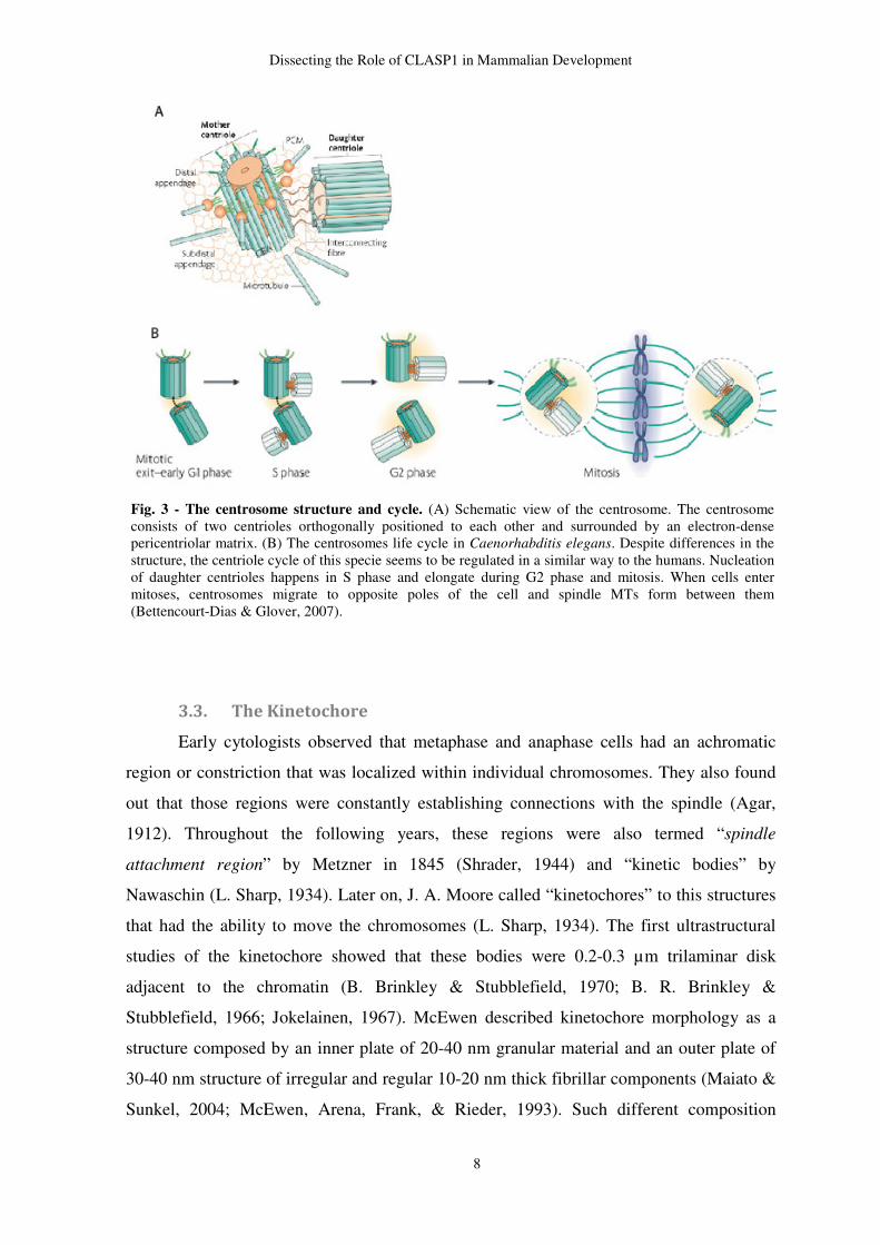
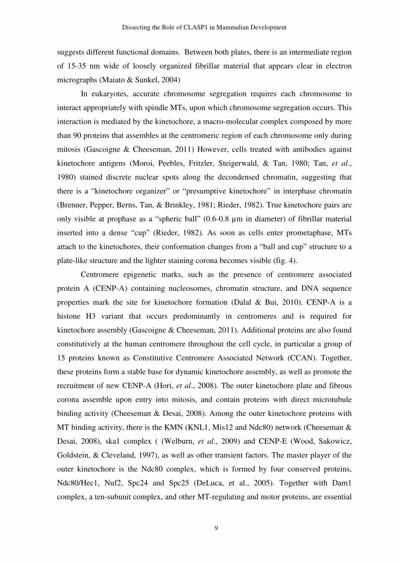
Fig. 4 - Organization of the animal kinetochore.that assemble in specific sites of the centromeric region. It consists of an inner plate that assembles in the centromere, a outer plate and the fibrous corona that contain proteins with direct microtubule binding activity. the interzone (Maiato, DeLuca, Salmon, & Earnshaw, 2004)
secting the Role of CLASP1 in Mammalian Development
10
for MT attachment and regulation of MT plus-end behavior (Miranda, De Wulf, Sorger, &
associated proteins
A complex repertoire of MT-associated proteins (MAPs) is required to
dynamics and architecture. MAPs are divided in two fundamental
ability to move along the spindle. MT dynamic instability depends on a
broad range of proteins that promote MTs stabilization or destabilization.
associated to the MT plus or minus ends, but do not move along the spindle
-13 family is an important group of non-motor
induces catastrophe events by triggering conformational changes th
filaments. The inverse phenomenon is promoted by stabilizing
, such as XMAP215 family (TOG in humans) that bind to the plus ends, thus
cking the binding of destabilizing factors, such as kinesin-13 family
ilizing proteins are associated not only to MT growth, but
involved in the association of the plus ends to another cellular structures, such as
the stabilizing proteins group, are also found CLIP-
(CLASPs), which will be highlighted in this thesis.
Organization of the animal kinetochore. Mature kinetochores are platethat assemble in specific sites of the centromeric region. It consists of an inner plate that assembles in the centromere, a outer plate and the fibrous corona that contain proteins with direct microtubule binding activity. Between both plates, a loosely organized fibrilar r
(Maiato, DeLuca, Salmon, & Earnshaw, 2004)
Miranda, De Wulf, Sorger, &
required to orchestrate
ivided in two fundamental classes,
MT dynamic instability depends on a
bilization. Usually, these
do not move along the spindle –
motor proteins, which
induces catastrophe events by triggering conformational changes that disrupt lateral
omenon is promoted by stabilizing
, such as XMAP215 family (TOG in humans) that bind to the plus ends, thus
ily (Gadde & Heald,
not only to MT growth, but they are also
the plus ends to another cellular structures, such as the cell
-associated proteins
ores are plate-like structures that assemble in specific sites of the centromeric region. It consists of an inner plate that assembles in the centromere, a outer plate and the fibrous corona that contain proteins with direct
etween both plates, a loosely organized fibrilar region constitute
Dissecting the Role of CLASP1 in Mammalian Development
11
Additionally, there is another group of MAPs able to travel along the MTs – motor
MAPs. Through hydrolyzis of ATP, motor proteins generate force and movement of the
spindle. Most kinesins or kinesins-related protein motors move along MTs towards the
plus ends. However, some members of this family move in the opposite direction. Local
regulation of MTs is driven by motor proteins. These mechanochemical ATPases can
move MTs unidirectionally towards the plus or minus ends, transporting a molecular cargo
or linking MTs into force-generating arrays, such as spindle (Gatlin & Bloom, 2010). The
first motor protein described was dynein, a minus-end-directed motor protein (Gatlin &
Bloom, 2010). Some plus-end directed kinesins localize to chromosome arms
(chromokinesins) contribute to chromosome attachment and movement towards the
metaphase plate, while cytoplasmic dynein in the cortex can contribute to astral MTs
organization (Gadde & Heald, 2004; Gatlin & Bloom, 2010; Morgan, 2007). Several plus-
end-directed proteins, such as Eg5, Mklp1/CHO1 and chromokinesis/KIF4, interact with
MTs, promoting MT antiparallel sliding that drives spindle pole separation during
anaphase B. In general, kinesin-15 works to promote increased spindle length (outward
directed forces), whereas minus end motors, such as dynein and kinesin-14 function to
promote spindle shortening (inwards directed forces) (T. J. Mitchison, et al., 2005;
Saunders, Lengyel, & Hoyt, 1997; D. J. Sharp, et al., 2000). Closer to the poles, where
parallel MT orientation is more prevalent, dynein mediated minus end clustering forces
dominate (Braun, Drummond, Cross, & McAinsh, 2009).
The mechanisms that shape the spindle start to be clarified, especially concerning
the motors associated to the kinetochores. Nevertheless, a long avenue has to be explored
to get the full picture of where and how these proteins interact with MTs and their partners
to control such a complex set of molecular events.
4. The Spindle Assembly
The mitotic spindle assembly occurs in early mitosis and faces two important
challenges: to build a bipolar structure and to properly attach chromosomes in order to
establish a MT array with the correct bi-orientation. In mammals, the bipolar spindle is
highly dependent on the correct positioning of the centrosomes, which are motor-
dependent pulled apart to by motor proteins the opposite sides of the prophase nucleus.
Dissecting the Role of CLASP1 in Mammalian Development
12
During this mitotic stage, the nuclear envelope breakdown (NEB) allows MTs to invade
the nuclear space and contact with chromosomes.
The absence of centrosomes does not obstruct the formation of a bipolar spindle in
acentrosomal systems. In these cases, spindle assembly relies on MT self-organization,
whereby MTs form in the vicinity of sister chromatids and then become organized by
motors into a bipolar array (Gatlin & Bloom, 2010) Eventually, spindle acquires by itself a
fusiform architecture with interdigitated plus ends near the chromosomes and minus ends
focused at the poles (Gaglio, Dionne, & Compton, 1997; Heald, Tournebize, Habermann,
Karsenti, & Hyman, 1997). However, it was previously observed that chromosome-
directed nucleation is not exclusive to the acentrosomal pathways. It was also found that
animal somatic cells also nucleate MTs near chromosomes (Maiato, Rieder, & Khodjakov,
2004).
An evolutionary question involves the biological purpose of the centrosomes, since
the acentrosomal pathway is able to efficiently self-organize a bipolar spindle. In fact, the
presence of centrosomes seems to facilitate MT nucleation because they provide a MT
bipolar mold upon which they polymerize. In addition, centrosomes nucleate astral MTs,
which are an advantage in terms of connection of the spindle to the cell cortex and spindle
positioning within the cell (Wadsworth & Khodjakov, 2004).
Dissecting the Role of CLASP1 in Mammalian Development
13
Spindle assembly and function is regulated by a huge amount of proteins. Among
them, Ran is an important regulatory protein involved in activation of numerous spindle
assembly-promoting factors around the chromosomes. Ran is a GTPase, whose activity
depends on GTP binding, carried out by an activation protein called RanGAP and a
guanine-nucleotide exchange factor called RCC1. Inside the nucleous, Ran-GTP binds to
importin, a nuclear transporter protein, that inhibits several proteins involved in spindle
assembly. RCC1, which is highly concentrated near chromosomes, activates Ran-GTP in
the immediate vicinity (Maiato, DeLuca, et al., 2004; Morgan, 2007). Ran-GTP triggers
dissociation of importin from regulatory proteins, allowing spindle formation around
chromosomes (Kalab, Pu, & Dasso, 1999; Kalab, Weis, & Heald, 2002; Morgan, 2007).
Ran GTpases exists in a concentration gradient around mitotic chromosomes (Carazo-Salas
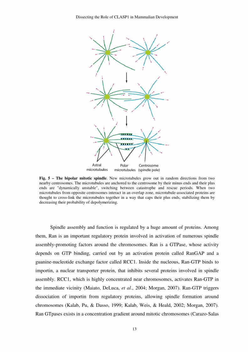
Fig. 5 – The bipolar mitotic spindle. New microtubules grow out in random directions from two nearby centrosomes. The microtubules are anchored to the centrosome by their minus ends and their plus ends are "dynamically unstable", switching between catastrophe and rescue periods. When two microtubules from opposite centrosomes interact in an overlap zone, microtubule-associated proteins are thought to cross-link the microtubules together in a way that caps their plus ends, stabilizing them by decreasing their probability of depolymerizing.
Dissecting the Role of CLASP1 in Mammalian Development
14
et al., 1999), which promps spindle assembly through two mechanisms. One involves the
stimulation of MT nucleation by chromatin (Heald, 2006), while the other works through
the generation of a local concentration of MT stabilizing factors near the chromosome that
promote the capture of MTs. Besides Ran-GTP gradient, it was shown in xenopus extracts
that aurora B kinase, together with its partners, Inner Centromere Protein (INCENP),
borealin and survivin, forms the Chromosomal Passenger Complex (CPC), interacts with
chromosomes in early mitosis, promoting local stability of MTs by inhibiting the
catastrophe factor centromere-associated kinesin (MCAK) (Klein, Nigg, & Gruneberg,
2006; Sampath, et al., 2004). CPC is a key factor in attachment error correction before
anaphase onset which will be discussed below (Adams, Carmena, & Earnshaw, 2001;
Gassmann, et al., 2004).
4.1. Kinetochore-MT attachment
In 1986, Kirschner and Mitchison proposed an explicative model of the process by
which MTs attach to kinetochores. According to this model, upon NEB in animal cells,
highly dynamic MTs invade the tree-dimensional cytoplasmic space until find a
kinetochore. Once MTs encounter a kinetochore, become stabilized, whereas those that do
not, soon depolimerize (Hayden, Bowser, & Rieder, 1990; T. Mitchison, Evans, Schulze,
& Kirschner, 1986). The kinetochore-microtubule interface is highly fluid and kinetochore
itself is remarkably dynamic, changing its makeup upon MT attachment. Indeed, prophase
spherical shaped kinetochores turn into a plate-like structure upon MT binding (Rieder,
1982)
Probably, the foremost question regarding kinetochore-microtubule binding is how
can MTs remain firmly attached to the kinetochores when they are continuously growing
and shrinking, allowing chromosomes to move back and forth on the spindle. Several
studies have revealed that this complex is crucial for the stable kinetochore-microtubule
attachments, required to sustain the centromere tensions involved in the achieving of the
proper chromosome alignment in higher eukaryotes (DeLuca, Moree, Hickey, Kilmartin, &
Salmon, 2002; Howe, McDonald, Albertson, & Meyer, 2001). Until recently, no evident
direct interaction between Ncd80 complex and the MTs was found. Thus, another protein
would connect both. In yeast, kinetochore-microtubule attachment requires Dam1 complex
(also known as DASH complex) because some members of this complex bind directly to
MTs, while others bind to Ncd80 complex (Westermann, et al., 2003). Dam1 seems to
Dissecting the Role of CLASP1 in Mammalian Development
15
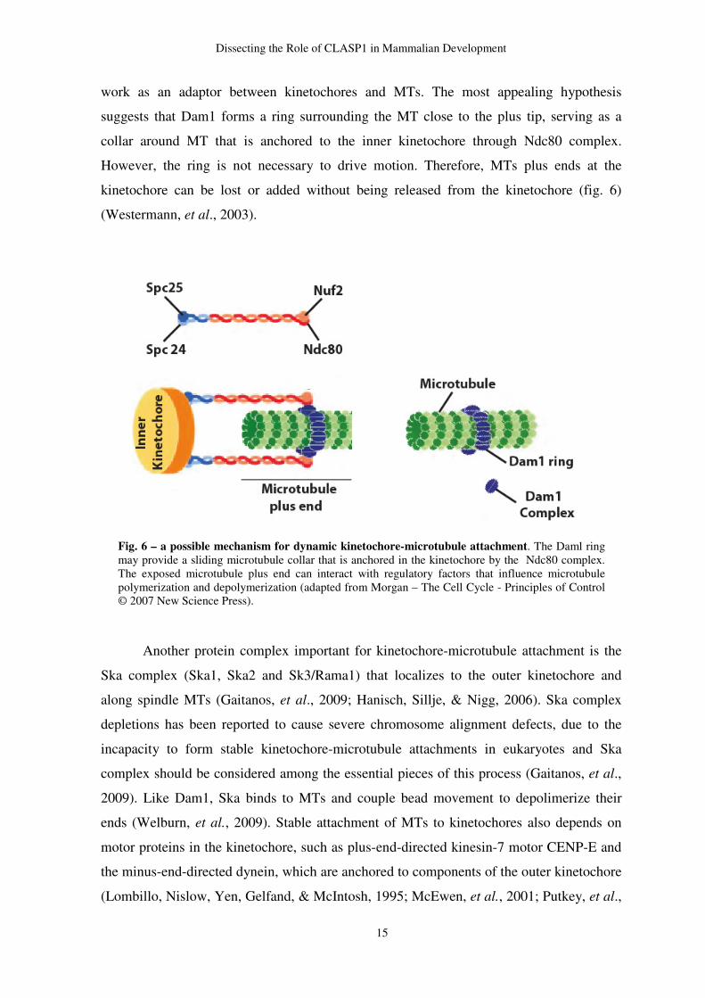
work as an adaptor between kinetochores and MTs. The most appealing hypothesis
suggests that Dam1 forms a ring surrounding the MT close to the plus tip, serving as a
collar around MT that is anchored to the inner kinetochore through Ndc80 complex.
However, the ring is not necessary to drive motion. Therefore, MTs plus ends at the
kinetochore can be lost or added without being released from the kinetochore (fig. 6)
(Westermann, et al., 2003).
Another protein complex important for kinetochore-microtubule attachment is the
Ska complex (Ska1, Ska2 and Sk3/Rama1) that localizes to the outer kinetochore and
along spindle MTs (Gaitanos, et al., 2009; Hanisch, Sillje, & Nigg, 2006). Ska complex
depletions has been reported to cause severe chromosome alignment defects, due to the
incapacity to form stable kinetochore-microtubule attachments in eukaryotes and Ska
complex should be considered among the essential pieces of this process (Gaitanos, et al.,
2009). Like Dam1, Ska binds to MTs and couple bead movement to depolimerize their
ends (Welburn, et al., 2009). Stable attachment of MTs to kinetochores also depends on
motor proteins in the kinetochore, such as plus-end-directed kinesin-7 motor CENP-E and
the minus-end-directed dynein, which are anchored to components of the outer kinetochore
(Lombillo, Nislow, Yen, Gelfand, & McIntosh, 1995; McEwen, et al., 2001; Putkey, et al.,
Fig. 6 – a possible mechanism for dynamic kinetochore-microtubule attachment. The Daml ring may provide a sliding microtubule collar that is anchored in the kinetochore by the Ndc80 complex. The exposed microtubule plus end can interact with regulatory factors that influence microtubule polymerization and depolymerization (adapted from Morgan – The Cell Cycle - Principles of Control © 2007 New Science Press).
Dissecting the Role of CLASP1 in Mammalian Development
16
2002). These proteins interact mostly with MTs to promote attachment and generate force
to move chromosomes along the spindle. Although these two proteins are not essential to
MT-kinetochore attachment, dynein, which is released from kinetochores upon MT
attachment (Hoffman, Pearson, Yen, Howell, & Salmon, 2001), has been reported as an
important factor required for the inactivation of the SAC, whereas CENP-E located in the
fibrous corona, is implicated in the initial encounter between kinetochores and MTs during
prometaphase (Cooke, Schaar, Yen, & Earnshaw, 1997; Yao, Anderson, & Cleveland,
1997). CENP-E is also involved in anchoring kinetochores to shortening MT in vitro
(Lombillo, Stewart, & McIntosh, 1995) and MT efficient binding to its kinetochores
(McEwen, et al., 2001; Putkey, et al., 2002).
4.2. Error correction in kinetochore-microtubule attachment
During prometaphase, MTs search and capture kinetochores and attachment errors
are common. However, cells are provided of molecular machinery that senses certain
incorrect kinetochore-microtubule attachments, delaying activation of APC/C and,
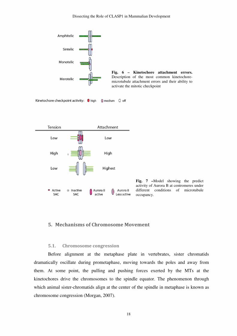
therefore, anaphase onset. During this mitotic stage, chromosomes can be mono-oriented
by one (monotelic attachment) or both sister kinetochores (synthelic attachment) (fig. 6)
(reviewed by Maiato et al., 2004). Over the years, two conditions have been proposed to be
keys for SAC signaling: lack of kinetochore-microtubule attachment and lack inter-
kinetochoreo tension (Nezi & Musacchio, 2009). Thus, SAC senses the lack of kinetochore
ocupancy in monotelic attachments, as well as the low tension at the kinetochores in
syntelic attachments (Nicklas & Koch, 1969; Nicklas, Waters, Salmon, & Ward, 2001).
The accurate chromosome segregation requires an amphitelic attachment, where one sister
kinetochore is attached to MTs solely from one pole, whereas the other is attached to MTs
solely from the opposite pole (reviewed by Maiato et al., 2004). (fig. 6). Sometimes, it may
happen that one or both sister kinetochores has MT attachments to both poles (fig. 6). This
kind of error is known as merotelic attachment and escape from checkpoint because the
level of kinetochore occupancy is high, as well as the tension exerted at the kinetochores
(fig. 7) (Cimini, Fioravanti, Salmon, & Degrassi, 2002; Cimini, et al., 2001). Over the last
few years, some investigators have tried to find a connection between tension-based error
correction pathway and the SAC response (reviewd by Nezi & Musacchio, 2009). A few
studies have shown that Aurora B/Ipl1 kinase is a crucial element of the correction system
(Lampson, Renduchitala, Khodjakov, & Kapoor, 2004; Tanaka, et al., 2002) and has been
Dissecting the Role of CLASP1 in Mammalian Development
17
reported that syntelic and merotelic attachments are not corrected when Aurora B is
inhibited (Bakhoum, Thompson, Manning, & Compton, 2009; Lampson, et al., 2004)
One key factor in error correction before anaphase onset is CPC, which is involved
in several places during mitosis (Ruchaud, Carmena, & Earnshaw, 2007) In the beginning
of mitosis, CPC localizes at the chromosome arms and centromere and is required for the
recruitment of several SAC proteins to the kinetochore (A. Carvalho, Carmena, Sambade,
Earnshaw, & Wheatley, 2003; Lens & Medema, 2003; Lens, et al., 2003; Murata-Hori &
Wang, 2002; Vigneron, et al., 2004). In anaphase, CPC leaves kinetochores and relocalizes
at the midzone (Kelly & Funabiki, 2009). Aurora B is probably the most important player
in error correction. In vertebrates, this protein is required for mediating proper attachments
(Hauf, et al., 2003) and loss of its activity results in a remarkable increase in the overall
stability of kinetochore MTs (Cimini, Wan, Hirel, & Salmon, 2006; Zhang & Walczak,
2006), suggesting that Aurora B is promping detachment of incorrect attached
microtubules by changing dynamics of kMTs. The exact mechanism by which Aurora B is
able to adjust the incorrect attachments is not yet clear, but has been proposed that Aurora
B corrects kinetochore–MT mal-attachment through regulation of MCAK and Ndc80/Hec1
(Cheeseman et al., 2002, 2006; Andrews et al., 2004; Lan et al., 2004; Ohi et al., 2004;
Deluca et al., 2006). An emerging “attachment-only” hypothesis proposes that SAC
satisfaction is the only crucial parameter monitores by SAC (Nezi & Musacchio., 2009).
This model implies that tension-dependent errors correction mechanisms depends on
Aurora B that generates unattached kinetochores, which are sensed by the SAC (Pinsky,
Kung, Shokat, & Biggins, 2006). This model conceptualizes an error correction pathway,
of which Aurora B/Ipl1 is the most authoritative representative, as being crucial for bi-
orientation, but also as being physically and functionally distinct to SAC. Under this
concept, Aurora B is exclusively implicated in the correction of improper kinetochore-
microtubule attachments and is not a SAC component because it is not required for SAC
signaling per se (Nezi & Musacchio., 2009).
Dissecting the Role of CLASP1 in Mammalian Development
18
5. Mechanisms of Chromosome Movement
5.1. Chromosome congression
Before alignment at the metaphase plate in vertebrates, sister chromatids
dramatically oscillate during prometaphase, moving towards the poles and away from
them. At some point, the pulling and pushing forces exerted by the MTs at the
kinetochores drive the chromosomes to the spindle equator. The phenomenon through
which animal sister-chromatids align at the center of the spindle in metaphase is known as
chromosome congression (Morgan, 2007).
Fig. 6 – Kinetochore attachment errors. Description of the most common kinetochore-microtubule attachment errors and their ability to activate the mitotic checkpoint
Fig. 7 –Model showing the predict activity of Aurora B at centromeres under different conditions of microtubule occupancy.
Dissecting the Role of CLASP1 in Mammalian Development
19
Two main forces act on chromosomes to move them back and forth during
chromosome congression. The first generated by the plus-end depolimerization of the MTs
attached to the kinetochore (Khodjakov, Cole, Bajer, & Rieder, 1996; Khodjakov &
Rieder, 1996; Skibbens, Skeen, & Salmon, 1993). The mechanism through which this
force occurs is not yet fully understood, but it has been suggested that depolimerizing plus-
ends at the kinetochore, forms a outward curling structure of the MT that generates a force
that pushes against the collar toward the poles, while remaining attached to the kinetochore
(Khodjakov, et al., 1996; Khodjakov & Rieder, 1996; Skibbens, et al., 1993). The
depolimerization of the plus-ends at the kinetochores generates poleward movement
creating high tension between sister chromatids, which is counterbalanced by the switch to
a polymerization state, that moves the chromosome away from the poles, decreasing
tension between sister chromatids. This equilibrium between pushing and pulling forces
generated at the kinetochores is known as directional instability (Skibbens, et al., 1993).
Additionally, a second type of forces acts at the chromosome arms. A force known as
“polar ejection” or “polar wind” pushes chromosome arms away from poles (Rieder et al.,
1986; reviewed by Ault et al., 1994; Rieder et al., 1994; and Inoue et al., 1995). The
mechanism of the “polar ejection” forces are not well established, but it has been suggested
that non-kinetochore MTs act at the chromosome arms, pushing them away from the poles.
These pulling and pushing forces work together in a very coordinated manner in order to
place the chromosomes at the spindle equator at metaphase.
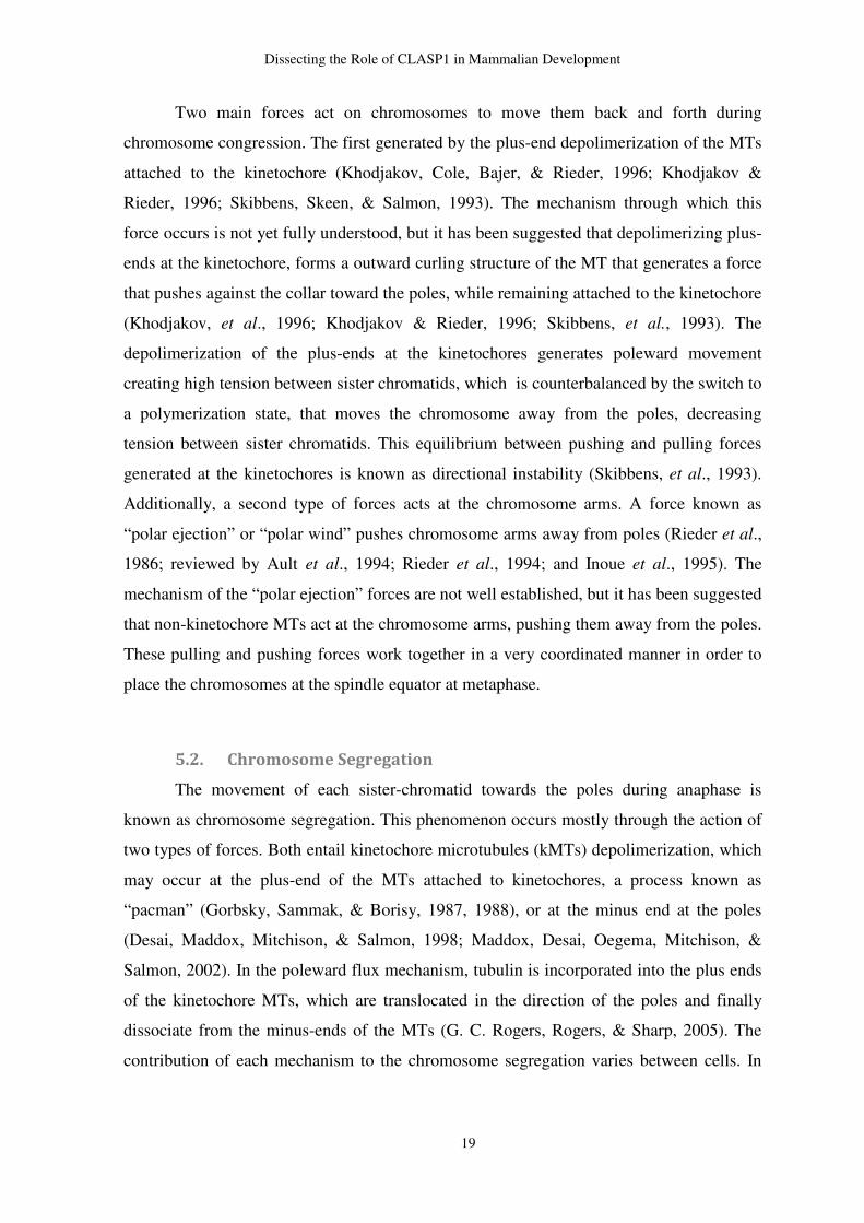
5.2. Chromosome Segregation
The movement of each sister-chromatid towards the poles during anaphase is
known as chromosome segregation. This phenomenon occurs mostly through the action of
two types of forces. Both entail kinetochore microtubules (kMTs) depolimerization, which
may occur at the plus-end of the MTs attached to kinetochores, a process known as
“pacman” (Gorbsky, Sammak, & Borisy, 1987, 1988), or at the minus end at the poles
(Desai, Maddox, Mitchison, & Salmon, 1998; Maddox, Desai, Oegema, Mitchison, &
Salmon, 2002). In the poleward flux mechanism, tubulin is incorporated into the plus ends
of the kinetochore MTs, which are translocated in the direction of the poles and finally
dissociate from the minus-ends of the MTs (G. C. Rogers, Rogers, & Sharp, 2005). The
contribution of each mechanism to the chromosome segregation varies between cells. In
Dissecting the Role of CLASP1 in Mammalian Development
20
vertebrates, approximately 70% of chromatid migration occurs via “pacman” and only
30% is due to flux (T. J. Mitchison & Salmon, 1992)
The activity of molecular motor proteins at kinetochores is also crucial for
chromosome segregation. Dynein is a key factor to the flux mechanism because it delivers
components of the flux mechanism, such as Kif2A, a depolimerizing kinesin. Members of
the kinesins-13 family are also implicated in depolimerization of MTs at the kinetochore in
Drosophila, therefore contributing for “pacman” (S. L. Rogers, Wiedemann, Hacker,
Turck, & Vale, 2004).
CLASPs, that will be discussed in more detail below, have been described as
important players in the incorporation of tubulin at the kinetochore, contributing to flux.
Depletion of CLASP in Drosophila blocks flux because kinetochore MTs do not
incorporate tubulin (Maiato, Khodjakov, & Rieder, 2005).
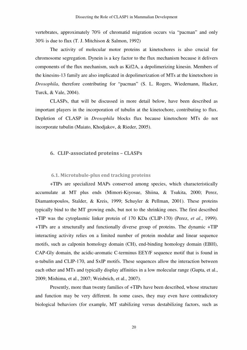
6. CLIP-associated proteins – CLASPs
6.1. Microtubule-plus end tracking proteins
+TIPs are specialized MAPs conserved among species, which characteristically
accumulate at MT plus ends (Mimori-Kiyosue, Shiina, & Tsukita, 2000; Perez,
Diamantopoulos, Stalder, & Kreis, 1999; Schuyler & Pellman, 2001). These proteins
typically bind to the MT growing ends, but not to the shrinking ones. The first described
+TIP was the cytoplasmic linker protein of 170 KDa (CLIP-170) (Perez, et al., 1999).
+TIPs are a structurally and functionally diverse group of proteins. The dynamic +TIP
interacting activity relies on a limited number of protein modular and linear sequence
motifs, such as calponin homology domain (CH), end-binding homology domain (EBH),
CAP-Gly domain, the acidic-aromatic C-terminus EEY/F sequence motif that is found in
α-tubulin and CLIP-170, and SxIP motifs. These sequences allow the interaction between
each other and MTs and typically display affinities in a low molecular range (Gupta, et al.,
2009; Mishima, et al., 2007; Weisbrich, et al., 2007).
Presently, more than twenty families of +TIPs have been described, whose structure
and function may be very different. In some cases, they may even have contradictory
biological behaviors (for example, MT stabilizing versus destabilizing factors, such as
Dissecting the Role of CLASP1 in Mammalian Development
21
CLASPs and OP18, respectively) However, in some other cases, they share characteristic
features of +TIPs (Akhmanova & Steinmetz, 2008; Morgan, 2007).
Although the majority of the +TIPs interact with the growing MTs, some can bind
to depolymerizing MTs, such as Dam1/DASH complex (Salmon, 2005) and XMPA215
(Brouhard, et al., 2008). The most presumptive explanation for +TIPs accumulation at the
growing ends relies on structural differences between the growing tips and the remaining
tube. The presence of the GTP-cap is probably one of the most important molecular
peculiarities in the core of this process. A few hypotheses have been proposed to explain
the plus-tracking mechanism. Among them, some +TIPs have been claimed to recognize
tubulin sheets and bind to the MT lattice seam (Sandblad, et al., 2006). Other +TIPs, such
as CLIPs, co-polymerize with tubulin and are gradually released from the older lattice
(Browning, Hackney, & Nurse, 2003; Rickard & Kreis, 1990). This type of interaction
assumes transiently immobilization. Dissociation of these +TIPS occurs by spontaneous
dissociation or by conformational changes in the MTs, a mechanism known as
“treadmilling” (Perez et al., 1999). Unlike “treadmilling”, a mechanism termed processive
transport implies long distance motor-based movement along the cytoskeleton filament
without dissociation. Proteins such as XMAP215, MCAK and dynactin seem to track
growing MT ends processively, remaining at the same MT end during multiple rounds of
tubulin subunit addition (Brouhard, et al., 2008). Several +TIPs are transported to growing
MT ends by plus-end-directed motors proteins. The MT targeting by association with MT-
binding partners is known as “hitchhiking”, which is a common plus-tracking mechanism
for many +TIPs (P. Carvalho, Tirnauer, & Pellman, 2003).
Due to attachment to the plus ends, +TIPs influence the structure of the MTs and
accessibility for interaction with other proteins. A majority of MT stabilization is promoted
by +TIPs, which act either by reducing the catastrophe levels, either by promoting rescue
(Akhmanova, et al., 2005).
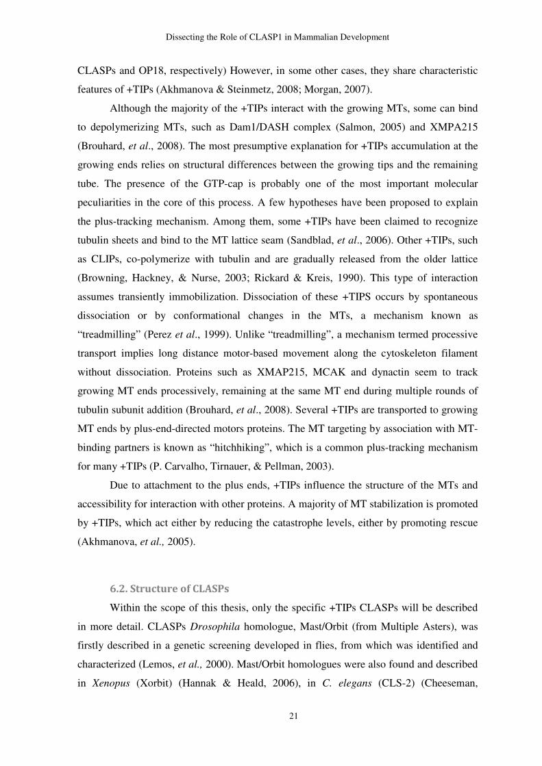
6.2. Structure of CLASPs
Within the scope of this thesis, only the specific +TIPs CLASPs will be described
in more detail. CLASPs Drosophila homologue, Mast/Orbit (from Multiple Asters), was
firstly described in a genetic screening developed in flies, from which was identified and
characterized (Lemos, et al., 2000). Mast/Orbit homologues were also found and described
in Xenopus (Xorbit) (Hannak & Heald, 2006), in C. elegans (CLS-2) (Cheeseman,
Dissecting the Role of CLASP1 in Mammalian Development
22
MacLeod, Yates, Oegema, & Desai, 2005), S. cerevisiae (Stu1p) (Pasqualone & Huffaker,
1994) and S. pombe (Peg1) (Grallert, et al., 2006). The CLASP1 and CLASP2 paralogues
were found in mammals as molecular partners of CLIPs-115 and 170 (Akhmanova, et al.,
2001).
Human CLASP1 and CLASP2 are encoded by chromosome 2 and 3, respectively.
In mice Clasp1 gene localizes in chromosome 1 and Clasp2 localizes in chromosome 9.
Alternative splicing may occur originating several CLASPs isoforms. So far, only one
biologically active isoform for CLASP1 has been described, known as CLASP1α (~170
KDa), while CLASP2 counts with three active isoforms, including CLASP2α (~170 KDa),
CLASP2β (~140 KDa) and CLASP2γ (~140 KDa) (Akhmanova, et al., 2001). The two
paralogues share approximately 77% of homology, albeit their expression varies between
different cells and tissues (Akhmanova, et al., 2001).
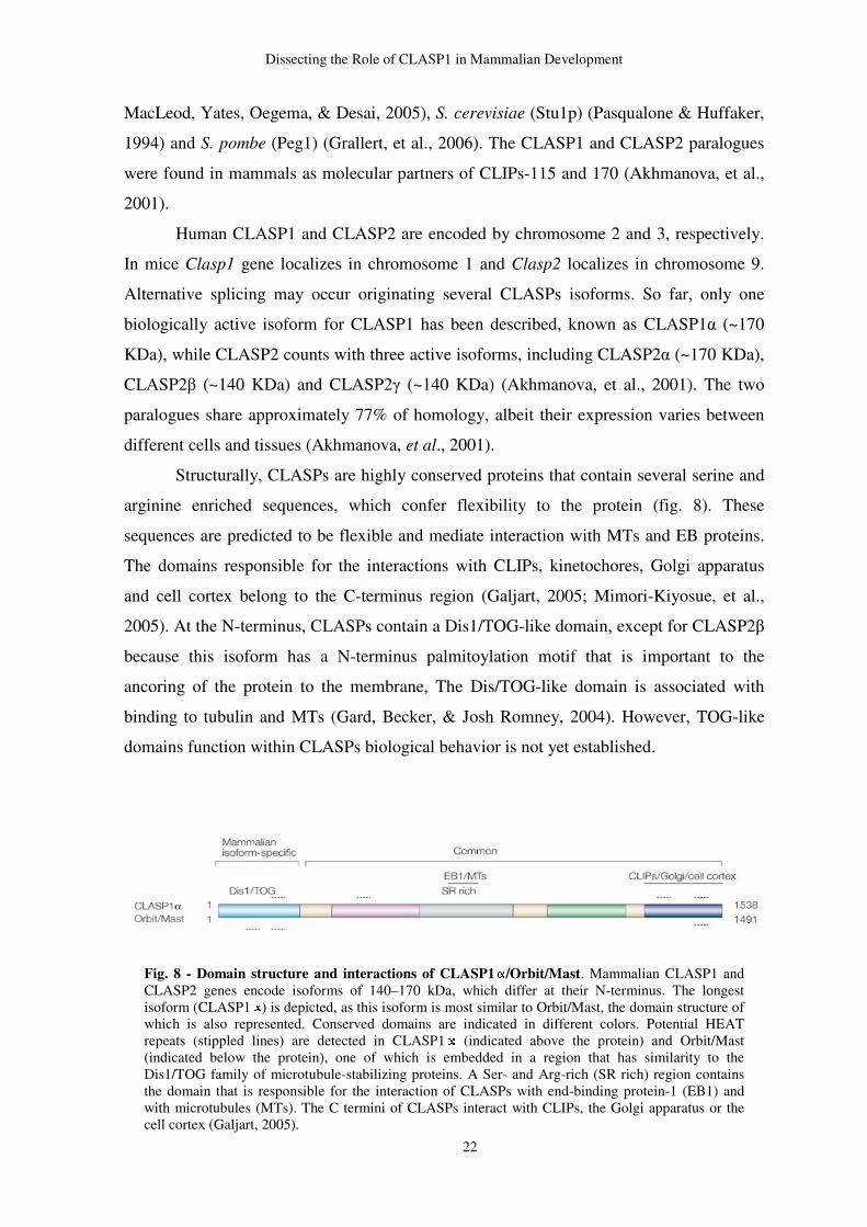
Structurally, CLASPs are highly conserved proteins that contain several serine and
arginine enriched sequences, which confer flexibility to the protein (fig. 8). These
sequences are predicted to be flexible and mediate interaction with MTs and EB proteins.
The domains responsible for the interactions with CLIPs, kinetochores, Golgi apparatus
and cell cortex belong to the C-terminus region (Galjart, 2005; Mimori-Kiyosue, et al.,
2005). At the N-terminus, CLASPs contain a Dis1/TOG-like domain, except for CLASP2β
because this isoform has a N-terminus palmitoylation motif that is important to the
ancoring of the protein to the membrane, The Dis/TOG-like domain is associated with
binding to tubulin and MTs (Gard, Becker, & Josh Romney, 2004). However, TOG-like
domains function within CLASPs biological behavior is not yet established.
Fig. 8 - Domain structure and interactions of CLASP1 /Orbit/Mast. Mammalian CLASP1 and CLASP2 genes encode isoforms of 140–170 kDa, which differ at their N-terminus. The longest isoform (CLASP1 ) is depicted, as this isoform is most similar to Orbit/Mast, the domain structure of which is also represented. Conserved domains are indicated in different colors. Potential HEAT repeats (stippled lines) are detected in CLASP1 (indicated above the protein) and Orbit/Mast (indicated below the protein), one of which is embedded in a region that has similarity to the Dis1/TOG family of microtubule-stabilizing proteins. A Ser- and Arg-rich (SR rich) region contains the domain that is responsible for the interaction of CLASPs with end-binding protein-1 (EB1) and with microtubules (MTs). The C termini of CLASPs interact with CLIPs, the Golgi apparatus or the cell cortex (Galjart, 2005).
Dissecting the Role of CLASP1 in Mammalian Development
23
6.3. Cellular distribution of CLASPs
In interphase cells, CLASPs localize to centrosomes and the Golgi apparatus, as
well as in growing MT plus ends (Akhmanova, et al., 2001). Studies have proven that
CLASPs co-localize with CLIPs, enforcing the previous assumptions about their
partnership (Akhmanova, et al., 2001). However, it was also reported that CLASPs do not
only bind to MTs through CLIPs, but also through EB1-related proteins (Mimori-Kiyosue,
et al., 2005). Moreover, in D. melanogaster, Mast seems to conserve the same interaction
with CLIPs (Mathe, Inoue, Palframan, Brown, & Glover, 2003; S. L. Rogers, et al., 2004).
However, the role of CLASPs on MT dynamics appears to be independent of CLIPs, since
in vitro studies have shown that CLASPs can bind tubulin per se (Grallert, et al., 2006;
Lansbergen, et al., 2006).
During mitosis, mammalian CLASPs, as well as its Drosophila homologue Mast
localize at centrosomes, kinetochores, spindle midzone and midbody (Lemos, et al., 2000;
Maiato, et al., 2003; A. L. Pereira, et al., 2006). CLASPs also tip-track astral MTs (Maiato,
et al., 2003) and CLASP1 localizes to the fibrous corona at the kinetochore in a MT
independent manner (Maiato, et al., 2003).
6.4. Functions of CLASPs.
The localization of CLASPs in key structures within the cell gives a clue about their
presumable functions, regarding the regulation of MT dynamics in the mitotic spindle and
interphase MTs (Bratman & Chang, 2008; Gard, et al., 2004). During interphase, CLASPs
have been reported to mediate the selective stabilization of MTs in fibroblasts (Akhmanova
et al., 2001). This finding was further supported by RNAi for CLASP1 and CLASP2,
which resulted in a decreased MT stability, accompanied by a significant reduction in MT
density, a phenotype that was not observed in single depletion for CLASP1 and CLASP2
(Mimori-Kyosue et al., 2005). Together with the similar localizations of both CLASPs, the
hypothesis of a functional redundancy in mammalian cells during interphase came over.
Additionally, it was demonstrated that CLASPs act as stabilizing factors at the edge of the
cell, either by reducing catastrophe, or by promoting rescue (Mimori-Kiyosue et al., 2005).
Also in interphase cells, CLASPs interact with the cell cortex through a complex with
LL5β by interaction with IQGAP1 (Watanabe et al., 2009: Lansbergen et al., 2006).
Importantly, CLASPs have been shown to play an important part in cell polarization,
Dissecting the Role of CLASP1 in Mammalian Development
24
through the polarization of cytoplasmic arrays of MTs in migrating cells, towards the
leading edge of the cell (Baas & Qiang, 2005).
It was also shown that CLASP2 is negatively regulated by glycogen synthase
kinase 3β (GSK3β), which modulates CLASP-microtubule association and lamella MT
attachment (Kumar, et al., 2009). In recent years, CLASP1 and CLASP2 were shown to
interact through the plus-ends with the actin filaments, functioning like crosslinkers in
interphase cells (Tsvelov et al., 2007). CLASPs also mediate MT nucleation from the
Golgi apparatus by interacting with the trans-Golgi network protein GCC185, leading to
the asymmetry of the MT array nucleated at the Golgi (Efimov et al., 2007). Studies in
neurons reported a very active role of CLASPs in this cell population (Jaworski,
Hoogenraad, & Akhmanova, 2008). Furthermore, CLASPs display a role in intracellular
transport (Baas, Vidya Nadar, & Myers, 2006), neuronal development including migration
(Kholmanskikh, et al., 2006), formation, growth and guidance of axons (Lee, et al., 2004).
CLASPs are also known to play important roles in mitotic cells. The pioneer
studies regarding CLASPs role in mitosis were performed in Drosophila, where it was
proven their essential role. CLASPs depletion by RNAi lead to failure in chromosome
segregation and formation of multipolar spindles, as well as monopolar spindles caused by
shortening of kinetochore MTs, due to spindle collapse (Lemos et al., 2000; Maiato et al.,
2002; Maiato et al., 2005). These results were also supported by CLASP1 inhibition with
anti-CLASP1 antibodies, which resulted in the generation of monopolar spindles (Maiato
et al., 2003). Fluorescence recovery after photobleaching (FRAP) experiments showed that
CLASP depletion blocked poleward flux (Maiato et al., 2005). In yeast, the CLASP
homologue Stu1p was shown to be associated with β-tubulin at the kinetochore level,
which allows flux in mature kinetochore microtubules (Yin et al., 2002). Recently, it was
shown in U2OS and Hela cells that CLASP1 forms complexes with Kif2b and Astrin at the
outer kinetochores. CLASP1-Kif2b localizes at the outer kinetochore during prometaphase,
being replaced by CLASP1-Astrin in metaphase (Manning, et al., 2010). It was also shown
that Astrin stabilizes MTs at the outer kinetochore, while Kif2b destabilizes them in
prometaphase, but not in metaphase (Manning, et al., 2010) The mutually exclusive
localization of CLASP1-Kif2b and CLAS1-Astrin complexes might be used as a switch to
regulate the temporal changes in kMTs stabilization during prometaphase-to-metaphase
transition (Manning, et al., 2010). Together, these findings suggest that CLASPs are
required, not only for the assembly of functional kinetochore-microtubule attachments, but
Dissecting the Role of CLASP1 in Mammalian Development
25
also to maintain spindle bipolarity through the regulation of MT dynamics at the
kinetochore of the mitotic cells (reviewed by Maiato et al., 2004).
Double RNAi experiments in Hela cells have shown that depletion 70% of CLASPs
is sufficient to cause mitotic defects, such as multipolar and monopolar spindles, among
others (Mimori-Kiyosue et al., 2005; (A. L. Pereira, et al., 2006). Interestingly, the single
depletion of CLASP1, did not cause such mitotic defects, which may suggest a redundancy
between CLASPs also during mitosis. However, the literature so far only describes cellular
phenotypes and functions regarding both mammalian CLASPs. In order to shed light on
the physiological roles of these proteins, as well as to discriminate in more detail their
cellular roles, knockout (KO) mouse models for CLASP1 and CLASP2 were made. So far,
it was shown that Clasp2 KO animals are viable, but the develop maturation/differentiation
defects, dying early in life (Pereira, 2009). In this thesis we intend to initiate the
characterization of a Clasp1 KO mouse model in order to uncover its role in mammals’
physiology.
7. Aneuploidy
The mitotic defects in small proportions of Clasp2 KO MEFs, are likely to be in the
basis for the observed chromosomal instability in these cells (A. L. Pereira, et al., 2006).
Despite the mitotic defects observed in these cells, they are viable, but the mitotic fidelity
is strongly compromised. Over time, the cumulative effects due to the absence of Clasp2
KO during embryonic development may contribute to chromosomal instability in adults.
Considering the previously suggested partial redundancy between CLASP1 and CLASP2,
one possible consequence of CLASP1 depletion in mammals would be the malignant
transformation and cancer.
In many cancers, mutations are accompanied by numerical and structural
chromosomal instability (CIN). Karyotypic abnormalities have been extensively
recognized has one of the main hallmarks of solid tumors. However, it is not clearly
established if CIN is a cause or a consequence of oncogenic transformation. CIN is
associated with problems during mitotic chromosome segregation, which is highly
correlated with a compromised SAC (Kops, Weaver, & Cleveland, 2005). Actually, it
would be expected that inactivation of mitotic checkpoint genes would directly lead to
miss-segregation events. However, loss-of-function mutations in mitotic genes appear to be
Dissecting the Role of CLASP1 in Mammalian Development
26
rare in human cancers (Draviam, Xie, & Sorger, 2004; Weaver & Cleveland, 2006).
Furthermore, targeted inactivation of mitotic checkpoint genes in mouse has been
associated with embryonic lethality, rather than cancer and partial loss of function
generally results in very mild tumorigenesis phenotypes. In fact, is more predominant a
premature senescence than cancer.
Although mitotic checkpoint robustness is essential to the accuracy of chromosome
segregation, sometimes it cannot detect errors, such as merotelic attachments. The efficient
transmission of the right number of chromosomes also depends on correct centrosome
duplication, establishment of spindle polarity and correct chromosome-MT attachment via
kinetochores.
The accurate duplication of centrosomes and its migration to opposite poles is a
geometrical requirement for the spindle bipolarity. Depletion or mutation of proteins
involved in centrosome biogenesis, leads to the formation of multi-polar mitotic spindles,
gross defects in chromatin disjunction and numerical CIN. Moreover, many solid tumors
have abnormal centrosome numbers (Duensing & Munger, 2001; Nigg, 2006). Another
condition for accurate chromosome segregation is bi-orientation. It means that a pair of
sister chromatids bind to MT emanating from opposite poles. Attachment of chomatids to
MTs is regulated by kinetochores. It has been reported that components of kinetochores
CENPs exhibit abnormal expression in human cancer, suggesting that abnormal
kinetochore composition might be related to tumorigenesis (Yuen, Montpetit, & Hieter,
2005).
As it was previously mentioned, CLASP1 localizes also at the outer kinetochore, as
well as, at the centrosomes. The role of CLASP1 in kinetochore and centrosomes is not
clear yet, but it might be actively preventing mitotic defects. Since mono and multipolar
spindles have been observed in Clasp2 KO MEFs, we could predict that mutations in
Clasps genes may lead to aneuploidy and, ultimately, to cancer.
8. Mouse models of embryonic development
8.1. Mouse models
Although in vitro studies have proven to be an essential tool to the comprehension
of the behavior of CLASPs inside the cell, their functions, particularly CLASP1, have
Dissecting the Role of CLASP1 in Mammalian Development
27
never been accessed in a complex living organism, such as mammals. Mice are, par
excellence, the chosen model to reproduce a given perturbation of a living system, either
by inducing exogenously a lesion/disease, or by changing its genome such is the case of
transgenic and KOs.
On the other hand, it has been widely accepted that in vitro and in vivo studies are
not always coincident and the studies based only cellular models do not always reproduce
what is happening in a living organism. On explanation relies on the huge amount of
extracellular signaling factors, secreted not only by a single cell type, but also by thousands
of different cell types, whose secretion is regulated by the endocrine system. Such amounts
of factors that are present in a physiological environment are not possible to be gathered in
a cell culture medium. To understand the physiological role of CLASP1 in mammals, a KO
model for it was generated (Pereira, 2009).
8.2. Clasp1 knockout mouse model
A KO mouse is a genetically engineered mouse in which a given gene is replaced
or disrupted with an artificial piece of DNA. The loss of gene activity often causes changes
in a mouse phenotype, which includes appearance, behavior and other observable physical
and biochemical characteristics. The Clasp1 KO mouse was not generated by the ablation
of the gene, but rather by interruption of the gene by insertion of a specific cassette that
prevent gene transcription. The targeting protocol and the Clasp1 KO generation of this
animal model is beyond the scope of this thesis.
9. Mouse development
Mouse embryonic development is highly similar to humans. For this reason, many
researchers have been studying the embryology of the mouse in order to understand what
happens throughout embryonic and fetal development in humans.
Mouse development comprises five fundamental stages: The first one, termed pre-
embryonic development, involves fertilization, in which a male spermatocyte penetrates
the cellular membrane of a female oocyte. From nuclear fusion of both gametocytes results
the zygote, which undergoes mitosis. Cleavage of the zygote continues until it becomes a
morula (a growing mass of undifferentiated cells) after the second day. On the third day,
Dissecting the Role of CLASP1 in Mammalian Development
28
morula becomes a blastocysts with 16 to 40 cells surrounding a blastocoele (a fluid filled
cavity). On day four occurs the implantation of the blastocyst and 24 hours after the
blastocysts implantation a placenta forms. The second stage is the elongation period, which
starts by day five, the blastocyst elongates and forms an egg cylinder. The placenta that has
started to grow begins to provide maternal blood to the embryo. Inside the placenta, the
egg starts to differentiate into early embryonic and extra-embryonic regions. By the end of
the sixth day, gastrulations, or true differentiation starts (Tam & Rossant, 2002).
Gastrulation is the third step of mouse embryonic development. A fold starts to form at the
tail end by the seventh day. This structure will turn into the coelemic fold, and later the end
of the gastrointestinal canal. Within the next 24 hours forms the foregut, an early precursor
of the stomach. At the same time, a very simple delineation of the cardiovascular system
appears, which includes branchials, or gill-like structures used to breathe in uterus. Optic
pits, which will give raise to the optical system start to form. Organogenesis occurs at day
ten to twelve and is characterized by many morphogenical changes that drive the growing
embryo to the fetus stage (Tam & Rossant, 2002). At the organogenesis stage, the early
brain continues to develop and grow, as well as the optic pits. Hind and forelimb buds and,
a short while later, hand plates (the precursors of feet) become visible. Mandibles and
maxilla begin to develop. Nasal pits form below the pit. The tail elongates and the foregut
extends into a slightly more developed digestive tract. Also, sex determination occurs
during this phase (Tam & Rossant, 2002). The last stage of mouse’s life in uterus is termed
the post-embryonic development. Here, the fetus becomes a fully formed newborn mouse.
At this stage, the development of the signs of individual digits occurs on the front and hind
limbs. The pinna (cartilaginous portion of the outer ear) becomes discernible and develops
at a right angle to the head. The cardiovascular system develops further to include a heart
and lungs. The lungs in mammals are not fully functional until after birth, when they are
exposed to air for the first time. In the few days prior to birth, skin thickens and whiskers
mature (Tam & Rossant, 2002).
9.1. Lung development and physiology
Mammalian lungs consist of a highly branched network containing thousands to
millions of airways arrayed in intricate patterns that are essential for respiration. The lungs
arise from the foregut endoderm at Embryonic Day 9.5 (E9.5). At this stage, the primary
buds consist of an inner, apparently unpatterned, epithelium surrounded by loosely packed
Dissecting the Role of CLASP1 in Mammalian Development
29
mesenchyme and a thin mesothelial layer (Rawlins, 2008). The lung buds undergo repeated
rounds of branching and outgrowth. During the pseudoglandular stage (~E10.5- E14.5), the
conductive airways are formed and lined with a mixture of secretory (Clara cells),
neuroendocrine (NE) and basal cells (Rawlins, 2008). Blood vessels and lymphatics extend
along the tubules (deMello, Sawyer, Galvin, & Reid, 1997) and smooth muscle cells
differentiate and coat the bronchial tubules (Mailleux, et al., 2005). Nerves can also be
distinguished side to side to the vascular structures, giving rise to the primordial lung nerve
system. Cartilage precursor tissue also starts to differentiate along the ventral region of
trachea and bronchi. The unpatterned distal buds of the lungs then elongate at the
cannalicular stage (~E14.5-E16.5) and ultimately give rise to the terminal sacs containing
type I and II epithelial cells (Rawlins, 2008; Whitsett, Wert, & Weaver, 2010). The
saccular stage, which occurs between E17.5 to fifth day of postnatal development in mice,
is a particular lung morphogenesis stage in which lung switch from a fluid-filled to an air-
filled structure, upon which survival depends following birth. This stage is marked by
dilatation of peripheral airspaces, resembling sacs, differentiation of the respiratory
epithelium, increasing vascularity and surfactant synthesis. Undifferentiated cuboidal cells
turn into type I and II epithelial cells. Type II epithelial cells (or type II pneumocytes)
contain lamellar bodies, which are intracellular storage units of surfactant (Mercurio &
Rhodin, 1976). Some authors have suggested that type II pneumocytes appear earlier than
type I. Thus, Type II pneumocytes might give rise to type I pneumocytes (Mercurio &
Rhodin, 1976). Type II pneumocytes are crucial for normal lung development and function
as they are the producers of surfactant and its secretion is essential for lung function after
birth. Type I pneumocytes line the greater part of the alveolar surface and are the
responsible for the gas-exchange. While type II pneumocytes are round cells without
extensions and contain “lamellar inclusions”, type I partners exhibit long cytoplasmic
extensions (Campiche, Gautier, Hernandez, & Reymond, 1963).
Postnataly, the primitive saccules enlarge and are subdivided into smaller units by a
process of septation called “alveolization” (Maeda, Dave, & Whitsett, 2007; Warburton,
Gauldie, Bellusci, & Shi, 2006). Alveolization occurs in late gestation in humans and
postanatally in mice. This greatly increases the surface area, and lung compliance.
The pulmonary surfactant system is one of the latest systems developed in lung
maturation. Surfactant is miscellaneous substance composed by phospholipids (80%),
neutral lipids (12%), and proteins (8%). The protein part of the surfactant system is made
by surfactant protein – A (SP-A), B (SP-B), C (SP-C) and D (SP-D). However, all
Dissecting the Role of CLASP1 in Mammalian Development
30
surfactant proteins have their specific roles along gestation, The more important surfactant
proteins for the life after birth are B and C partners. Surfactant is secreted to the alveolar
lumen and coats the intra-alveolar wall decreasing the tension at the air-liquid interface and
so facilitating expansion of the alveoli during respiration (Boyden, 1977).
9.1.1. Lung defects
The first, and probably the biggets extrauterine challenge, is the first breath of air.
When pups are being born, they become cyanotic at the first couple of minutes. This
cyanosis corresponds to an interval between ceasing of the placental oxygen support and
the time that breathing takes over. After a few minutes, normal pups recover their pinkish
color. However, any abnormality that affects different physiological system might be life-
threatening.
Partial or total lung collapse is one of the most common hallmarks of respiratory
diseases. However, it may occur in the absence of a primary lung defect, but due to other
morfophysiological system defects, such as cardiovascular defects, neuromuscular defects,
skeletal defects, among others (Turgeon & Meloche, 2009). Primary lung defects that have
associated lung collapse are usually related to insufficient maturation of the lung tissue
after delivery. In humans, the most common cause of pulmonary syndrome distress in
newborns relies on insufficient synthesis of surfactant or genetic mutations in one
surfactant protein B (Nkadi, Merritt, & Pillers, 2009). Type II pneumocytes may not be
mature enough by the time that newborns have to breathe by their own, and surfactant
synthesis and secretion is not sufficient to allow the proper lung expansion. In what
concerns lung maturation, a large amount of gene disruption may lead to lung collapse.
Nfib, Pdn and Nds1 knockout mice, albeit make visible efforts to breathe, inflation is not
sufficient to provide the proper gas-exchange and these mutants die within a few minutes
after birth. Pdn deficient mice have abnormal distal saccules with blockade in type I
pneumocytes differentiation. However, surfactant is high, which indicates that maturation
of type II does occur normally (Ramirez, et al., 2003). On the contrary, type II
pneumocytes differentiation seems to be compromised in Nds1 deficient animals, resulting
in impaired surfactant production. These mice also develop lung failure within ten hours
after delivery and die (Fan, et al., 2000). A huge amount of genetic defects, even though
not directly related with lung tissue, result in lung collapse. Thus, it becomes clear that
respiration is a physiological phenomenon that counts, not only with lungs per ser, but also

Dissecting the Role of CLASP1 in Mammalian Development
31
with other organs/tissues that work together to achieve the proper breathing movements
and gas-exchange.
EXPERIMENTAL
WORK
Dissecting the Role of CLASP1 in the Mammalian Development
33
CHAPTER I
Characterization of Clasp1 Knockout Mice
1 . Introduction
Cellular models have brought important insights for the understanding of many
particularities of many proteins, such as CLASPs, especially concerning their intracellular
localization, which provided several clues about their potential functions.. The most
significant insights about the role of CLASPs came from studies in yeast, Carnorhabditis
elegans, Drosophila, and Xenopus, which only have one single CLASP orthologue that is
essencial for mitosis and organism viability (Gonczy, et al., 2000; Hannak & Heald, 2006;
Inoue, et al., 2000; Lemos, et al., 2000; Pasqualone & Huffaker, 1994). Clasp2 KO mouse
embryonic and adult fibroblasts (MEFs and MAFs, respectively) have revealed an increase
of chromosome segregation defects, namely monopolar and multipolar spindles and
lagging chromosomes (Pereira, et al., 2006). Although these cells are viable, their mitotic
fidelity is compromised. Interestingly, partial normal phenotype was recovered upon
ectopic expression of both GFP-CLASP1 and GFP-CLASP2. Moreover, the rescue
efficiency was higher upon ectopic expression of GFP-CLASP2 (Pereira, et al., 2006).
Together, these finding support the hypothesis that both CLASP1 and CLASP2 cooperate
to ensure mitotic fidelity, being partially redundant.
The outcomes from cellular models do not always corroborate the behavior of
proteins within a living organism. The physiological environment is a complex system of
molecular signaling, which cannot be reproduced in a petri dish. The first attempt to
understand the role of CLASPs in vivo involved Clasp2 KO animals. Although these
animals are viable, they frequently dye early within the first six months of life exhibiting
maturation/differentiation defects in high-proliferative tissues, such as the hematopoietic
and reproductive systems (Pereira, 2009). Particularly, absence of CLASP2 impairs
erythropoietic differentiation. Clasp2 KO mice have shown a huge reduction in number of
progenitors in bone marrow, which means that depletion of CLASP2 plays an important
role at the early progenitors level.
Dissecting the Role of CLASP1 in the Mammalian Development
34
Our group is taking the first steps to uncover the physiological role of CLASP1 in a
mammalian model. As it was aforementioned, we generated a Clasp1 KO mouse model,
which is the ideal tool to understand the physiological impact of the absence of this protein
in mammals.
2. Materials and Methods
2.1. Animals
C57Bl/6 background animals were bred in the animal facility of the Institute for
Molecular and Cell Biology, according to protocols approved by the Ethics Committee of
the mentioned institution. Animal were housed in groups of 2-4 animals per cage, with free
access to distillated tap water and fed ad libitum. Rooms were acclimatized and room
temperature was surrounding 20-24º C; 45-65% humidity; ventilation of 15-20
changes/hour and light/dark cycles of 12/12 hours.
2.2. Breeding and detection of KO mice
Breeding entailed housing one heterozygous male with 2 heterozygous females
overnight. This breeding resulted in littermates with WT/HT/KO proportions non-
concordant with the expected ratio of 1:2:1. These proportions did not obey rule and also
oscillated among litters. Mating was ascertained in the next morning by the presence of a
vaginal plug and this time was designated embryonic day 0,5. All offspring were routinely
genotyped for Clasp1 gene using tail tissue. Genotyping was performed by PCR using the
following conditions primers:
Gen1: GTCCGCCACTCTCTCCTTTATTGCC
Gen3: CCTCATGTCCTTCCCCCATAACC
#261: CGGCATCAGAGCAGCCGATTG
Annealing temperature: 55ºC
Dissecting the Role of CLASP1 in the Mammalian Development
35
2.3. Delivery and newborn handling
In average, pregnancy of C57/Bl6 takes between 18.5-20 days. Pups that were born
by normal delivery were considered postnatals. Some litters were removed from the
females by c-section.
2.4. Overall analysis of the newborn pups
Newborn pups were closely monitored during their birth and evaluated for injuries
caused by the delivery process. Once detected any cannibalistic intention, pups were
carefully separated from the mother. Pups were carefully handled and observed, with
special attention paid to the color and texture of the skin, breathing movements, gasping,
reflex stimuli, posture and movement of the limbs and weight.
2.5. Water-floating test
This test consisted of releasing the lung samples into the saline solution/water and
analyze their floating capacity (whether they were inflated or collapsed, respectively).
2.6. Histological analysis
Adult mice were anesthetized with volatile isoflurane (Merck) and then sacrificed
by cervical dislocation Histological samples were taken and fixed in 10% neutral buffered
formalin for 24-48h, at room temperature. Specimens were dehydrated in a graded series of
ethanol and cleared with Clear-Rite (Merk). Finally, samples were embedded in paraffin
wax (Merk). The formalin-fixed and paraffin-embedded (FFPE) samples were sectioned
with 3 µm thick and mounted in glass slides coated with 3-aminopropyltriethoxysilane
(APES). Sections were deparaffinized in xylene and rehydrated in a graded series of
ethanol until water. Representative samples of all organs and tissues were routinely stained
for hematoxylin and eosin. Embryos/fetus and postnatals were sacrificed by decapitation.
The whole body integrity was preserved by fixation with 10% neutral buffered formalin for
48 hours, at room temperature. The skin was removed in order to ensure the proper
fixation. After fixation, transversal, longitudinal and coronal sections of the whole body
were made for routine staining in FFPE samples with 3 µm thick.
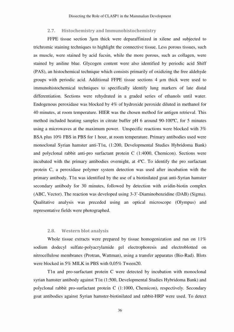
Dissecting the Role of CLASP1 in the Mammalian Development
36
2.7. Histochemistry and Immunohistochemistry
FFPE tissue section 3µm thick were deparaffinized in xilene and subjected to
trichromic staining techniques to highlight the connective tissue. Less porous tissues, such
as muscle, were stained by acid fucsin, while the more porous, such as collagen, were
stained by aniline blue. Glycogen content were also identified by periodic acid Shiff
(PAS), an histochemical technique which consists primarily of oxidizing the free aldehyde
groups with periodic acid. Additional FFPE tissue sections 4 µm thick were used to
immunohistochemical techniques to specifically identify lung markers of late distal
differentiation. Sections were rehydrated in a graded series of ethanols until water.
Endogenous peroxidase was blocked by 4% of hydroxide peroxide diluted in methanol for
40 minutes, at room temperature. HIER was the chosen method for antigen retrieval. This
method included heating samples in citrate buffer pH 6 around 90-100ºC, for 5 minutes
using a microwaves at the maximum power. Unspecific reactions were blocked with 3%
BSA plus 10% FBS in PBS for 1 hour, at room temperature. Primary antibodies used were
monoclonal Syrian hamster anti-T1α, (1:200, Developmental Studies Hybridoma Bank)
and polyclonal rabbit anti-pro surfactant protein C (1:4000, Chemicon). Sections were
incubated with the primary antibodies overnight, at 4ºC. To identify the pro surfactant
protein C, a peroxidase polymer system detection was used after incubation with the
primary antibody. T1α was identified by the use of a biotinilated goat anti-Syrian hamster
secondary antibody for 30 minutes, followed by detection with avidin-biotin complex
(ABC, Vector). The reaction was developed using 3-3’-Diaminobenzidine (DAB) (Sigma).
Qualitative analysis was preceded using an optical microscope (Olympus) and
representative fields were photographed.
2.8. Western blot analysis
Whole tissue extracts were prepared by tissue homogenization and run on 11%
sodium dodecyl sulfate-polyacrylamide gel electrophoresis and electroblotted on
nitrocellulose membranes (Protran, Wattman), using a transfer apparatus (Bio-Rad). Blots
were blocked in 5% MILK in PBS with 0,05% Tween20.
T1α and pro-surfactant protein C were detected by incubation with monoclonal
syrian hamster antibody against T1α (1:500, Developmental Studies Hybridoma Bank) and
polyclonal rabbit pro-surfactant protein C (1:1000, Chemicon), respectively. Secondary
goat antibodies against Syrian hamster-biotinilated and rabbit-HRP were used. To detect
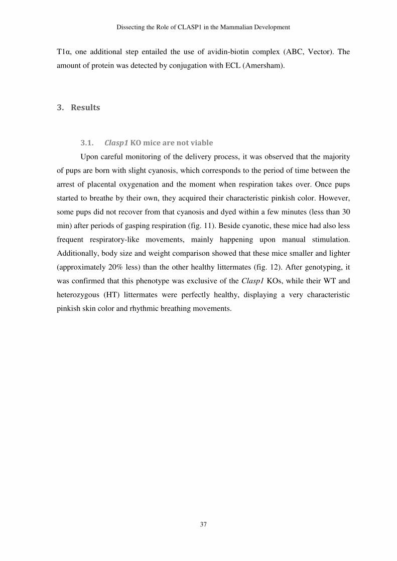
Dissecting the Role of CLASP1 in the Mammalian Development
37
T1α, one additional step entailed the use of avidin-biotin complex (ABC, Vector). The
amount of protein was detected by conjugation with ECL (Amersham).
3. Results
3.1. Clasp1 KO mice are not viable
Upon careful monitoring of the delivery process, it was observed that the majority
of pups are born with slight cyanosis, which corresponds to the period of time between the
arrest of placental oxygenation and the moment when respiration takes over. Once pups
started to breathe by their own, they acquired their characteristic pinkish color. However,
some pups did not recover from that cyanosis and dyed within a few minutes (less than 30
min) after periods of gasping respiration (fig. 11). Beside cyanotic, these mice had also less
frequent respiratory-like movements, mainly happening upon manual stimulation.
Additionally, body size and weight comparison showed that these mice smaller and lighter
(approximately 20% less) than the other healthy littermates (fig. 12). After genotyping, it
was confirmed that this phenotype was exclusive of the Clasp1 KOs, while their WT and
heterozygous (HT) littermates were perfectly healthy, displaying a very characteristic
pinkish skin color and rhythmic breathing movements.
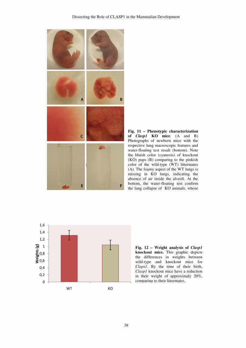
Dissecting the Role of CLASP1 in the Mammalian Development
38
0
0,2
0,4
0,6
0,8
1
1,2
1,4
1,6
WT KO
We
igh
ts (
g)
Fig. 12 – Weight analysis of Clasp1 knockout mice. This graphic depicts the differences in weights between wild-type and knockout mice for Claps1. By the time of their birth, Clasp1 knockout mice have a reduction in their weight of approximaly 20%, comparing to their littermates.
Fig. 11 – Phenotypic characterization of Clasp1 KO mice. (A and B) Photographs of newborn mice with the respective lung macroscopic features and water-floating test result (bottom). Note the bluish color (cyanosis) of knockout (KO) pups (B) comparing to the pinkish color of the wild-type (WT) littermates (A). The foamy aspect of the WT lungs is missing in KO lungs, indicating the absence of air inside the alveoli. At the bottom, the water-floating test confirm the lung collapse of KO animals, whose lung samples immediately sunk upon
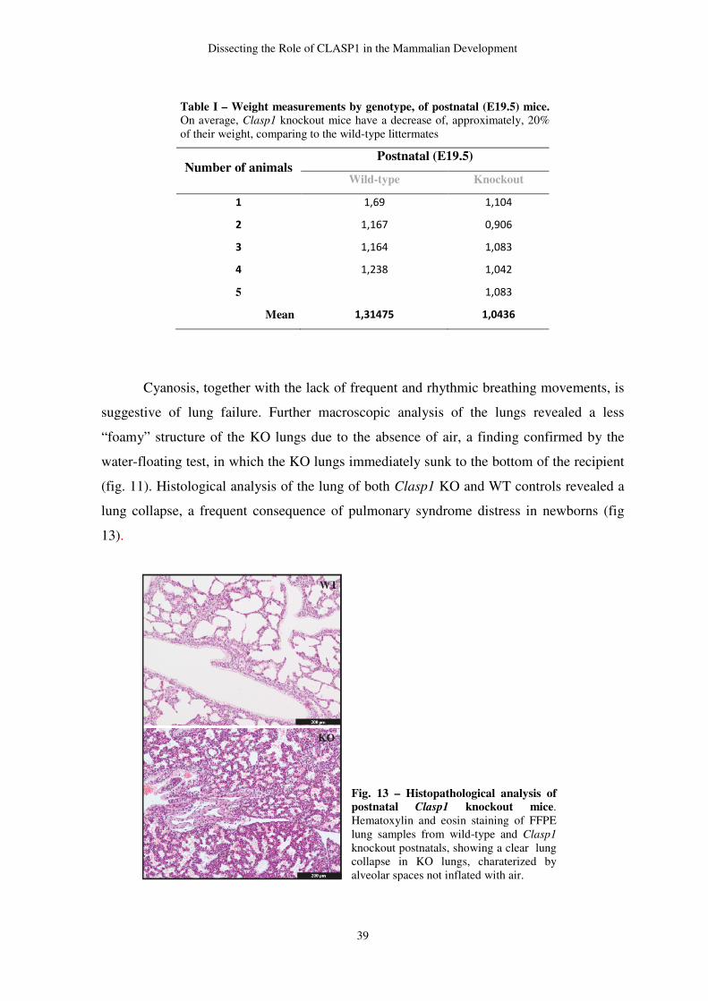
Dissecting the Role of CLASP1 in the Mammalian Development
39
Cyanosis, together with the lack of frequent and rhythmic breathing movements, is
suggestive of lung failure. Further macroscopic analysis of the lungs revealed a less
“foamy” structure of the KO lungs due to the absence of air, a finding confirmed by the
water-floating test, in which the KO lungs immediately sunk to the bottom of the recipient
(fig. 11). Histological analysis of the lung of both Clasp1 KO and WT controls revealed a
lung collapse, a frequent consequence of pulmonary syndrome distress in newborns (fig
13).
Number of animals Postnatal (E19.5)
Wild-type Knockout
1 1,69 1,104
2 1,167 0,906
3 1,164 1,083
4 1,238 1,042
5 1,083
Mean 1,31475 1,0436
Table I – Weight measurements by genotype, of postnatal (E19.5) mice. On average, Clasp1 knockout mice have a decrease of, approximately, 20% of their weight, comparing to the wild-type littermates
Fig. 13 – Histopathological analysis of postnatal Clasp1 knockout mice. Hematoxylin and eosin staining of FFPE lung samples from wild-type and Clasp1 knockout postnatals, showing a clear lung collapse in KO lungs, charaterized by alveolar spaces not inflated with air.
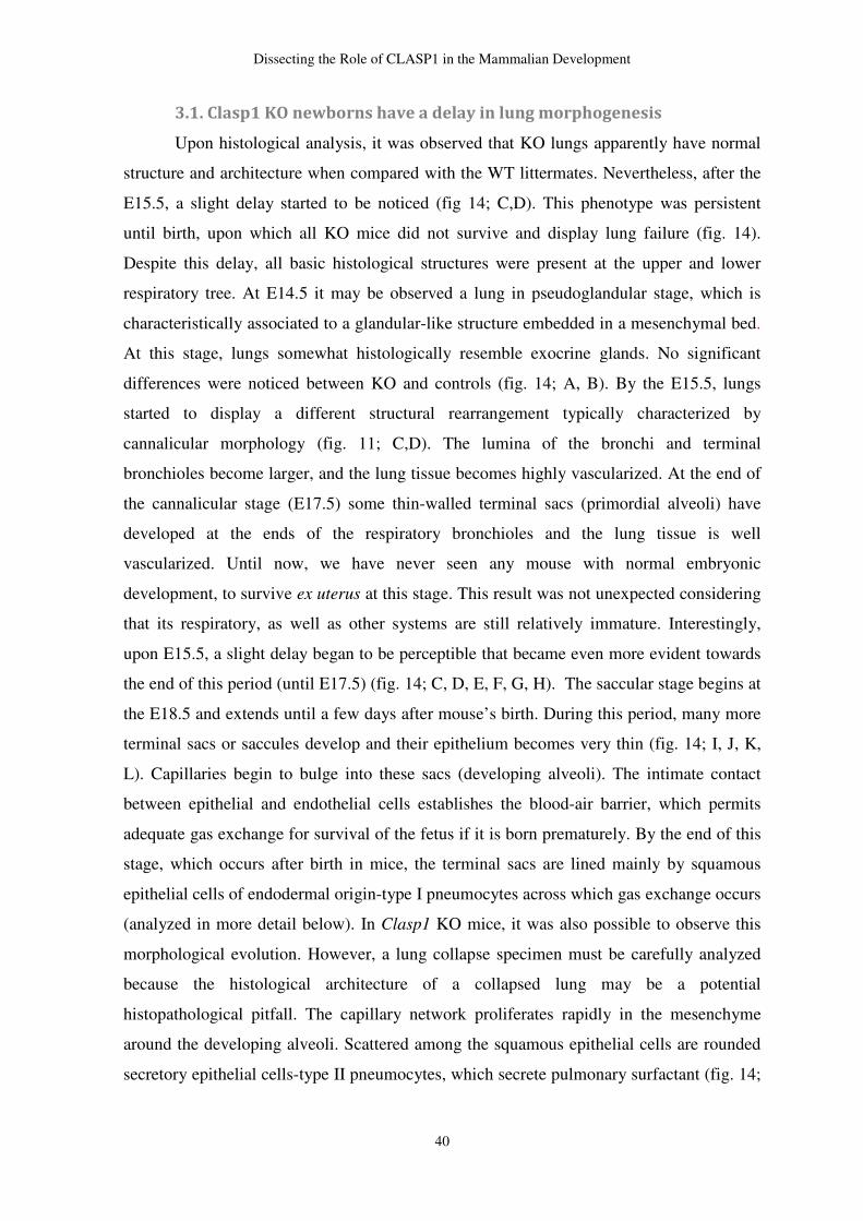
Dissecting the Role of CLASP1 in the Mammalian Development
40
3.1. Clasp1 KO newborns have a delay in lung morphogenesis
Upon histological analysis, it was observed that KO lungs apparently have normal
structure and architecture when compared with the WT littermates. Nevertheless, after the
E15.5, a slight delay started to be noticed (fig 14; C,D). This phenotype was persistent
until birth, upon which all KO mice did not survive and display lung failure (fig. 14).
Despite this delay, all basic histological structures were present at the upper and lower
respiratory tree. At E14.5 it may be observed a lung in pseudoglandular stage, which is
characteristically associated to a glandular-like structure embedded in a mesenchymal bed.
At this stage, lungs somewhat histologically resemble exocrine glands. No significant
differences were noticed between KO and controls (fig. 14; A, B). By the E15.5, lungs
started to display a different structural rearrangement typically characterized by
cannalicular morphology (fig. 11; C,D). The lumina of the bronchi and terminal
bronchioles become larger, and the lung tissue becomes highly vascularized. At the end of
the cannalicular stage (E17.5) some thin-walled terminal sacs (primordial alveoli) have
developed at the ends of the respiratory bronchioles and the lung tissue is well
vascularized. Until now, we have never seen any mouse with normal embryonic
development, to survive ex uterus at this stage. This result was not unexpected considering
that its respiratory, as well as other systems are still relatively immature. Interestingly,
upon E15.5, a slight delay began to be perceptible that became even more evident towards
the end of this period (until E17.5) (fig. 14; C, D, E, F, G, H). The saccular stage begins at
the E18.5 and extends until a few days after mouse’s birth. During this period, many more
terminal sacs or saccules develop and their epithelium becomes very thin (fig. 14; I, J, K,
L). Capillaries begin to bulge into these sacs (developing alveoli). The intimate contact
between epithelial and endothelial cells establishes the blood-air barrier, which permits
adequate gas exchange for survival of the fetus if it is born prematurely. By the end of this
stage, which occurs after birth in mice, the terminal sacs are lined mainly by squamous
epithelial cells of endodermal origin-type I pneumocytes across which gas exchange occurs
(analyzed in more detail below). In Clasp1 KO mice, it was also possible to observe this
morphological evolution. However, a lung collapse specimen must be carefully analyzed
because the histological architecture of a collapsed lung may be a potential
histopathological pitfall. The capillary network proliferates rapidly in the mesenchyme
around the developing alveoli. Scattered among the squamous epithelial cells are rounded
secretory epithelial cells-type II pneumocytes, which secrete pulmonary surfactant (fig. 14;
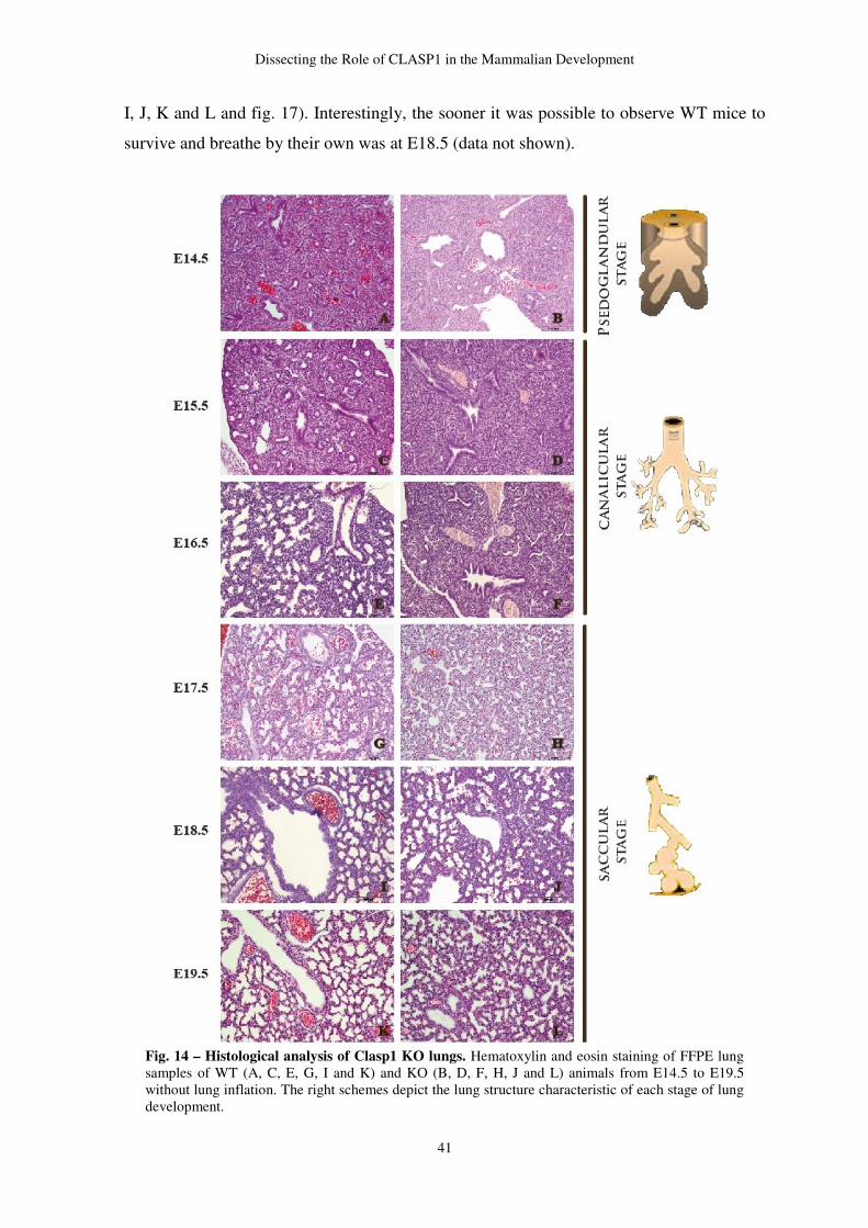
Dissecting the Role of CLASP1 in the Mammalian Development
41
I, J, K and L and fig. 17). Interestingly, the sooner it was possible to observe WT mice to
survive and breathe by their own was at E18.5 (data not shown).
Fig. 14 – Histological analysis of Clasp1 KO lungs. Hematoxylin and eosin staining of FFPE lung samples of WT (A, C, E, G, I and K) and KO (B, D, F, H, J and L) animals from E14.5 to E19.5 without lung inflation. The right schemes depict the lung structure characteristic of each stage of lung development.
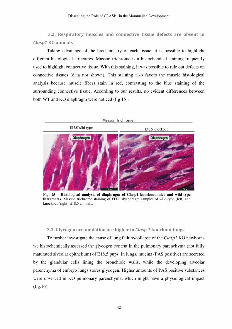
Dissecting the Role of CLASP1 in the Mammalian Development
42
3.2. Respiratory muscles and connective tissue defects are absent in
Clasp1 KO animals
Taking advantage of the biochemistry of each tissue, it is possible to highlight
different histological structures. Masson trichrome is a histochemical staining frequently
used to highlight connective tissue. With this staining, it was possible to rule out defects on
connective tissues (data not shown). This staining also favors the muscle histological
analysis because muscle fibers stain in red, contrasting to the blue staining of the
surrounding connective tissue. According to our results, no evident differences between
both WT and KO diaphragm were noticed (fig 15).
3.3. Glycogen accumulation are higher in Clasp 1 knockout lungs
To further investigate the cause of lung failure/collapse of the Clasp1 KO newborns
we histochemically assessed the glycogen content in the pulmonary parenchyma (not fully
maturated alveolar epithelium) of E18.5 pups. In lungs, mucins (PAS positive) are secreted
by the glandular cells lining the bronchiole walls, while the developing alveolar
parenchyma of embryo lungs stores glycogen. Higher amounts of PAS positive substances
were observed in KO pulmonary parenchyma, which might have a physiological impact
(fig.16).
Fig. 15 – Histological analysis of diaphragm of Clasp1 knockout mice and wild-type littermates. Masson trichrome staining of FFPE dyaphragm samples of wild-type (left) and knockout (right) E18.5 animals.
Dissecting the Role of CLASP1 in the Mammalian Development
43
3.4. Alveolar epithelial cells responsible for the gas-exchange are
differentiated in Clasp1 knockout lungs at birth
Next, we assessed the level of distal maturation by staining lung samples with type
I and II pneumocytes differentiation markers, T1α and pro surfactant protein C,
respectively. In pulmonary cell population, only type I pneumocytes express T1α, while
pro surfactant protein C is exclusive to the type II pneumocytes. According to the
immunostaining results, both alveolar epithelial cells are differentiated in the moment of
the KO mice birth. Both in KO mice and controls, type I pneumocytes are differentiated
and lining the alveolar sacs, while type II pneumocytes, a smaller cell population, are
scattered all over the alveolar parenchyma (fig. 17). Western blot analysis has shown that
both markers are already expressed in lungs at E16.5 and are persistently expressed since
then (fig. 18). Expression profile of both markers did not vary significantly between WT
and KO animals. Regarding these findings, it is reasonable to assume that KO lungs have
the proper alveolar epithelium content to take over respiration upon the discontinuation of
the placental oxygen supply.
Fig. 16 – Histological analysis of lungs of Clasp1 KO mice and WT littermates. Periodic acid Shiff staining of FFPE lung samples of WT (left) and KO (right) postnatals. The lower panel corresponds to a higher magnification of lungs, showing the pulmonary parenchyme in more detail. B – bronchiole; S – saccule; V – vessel.
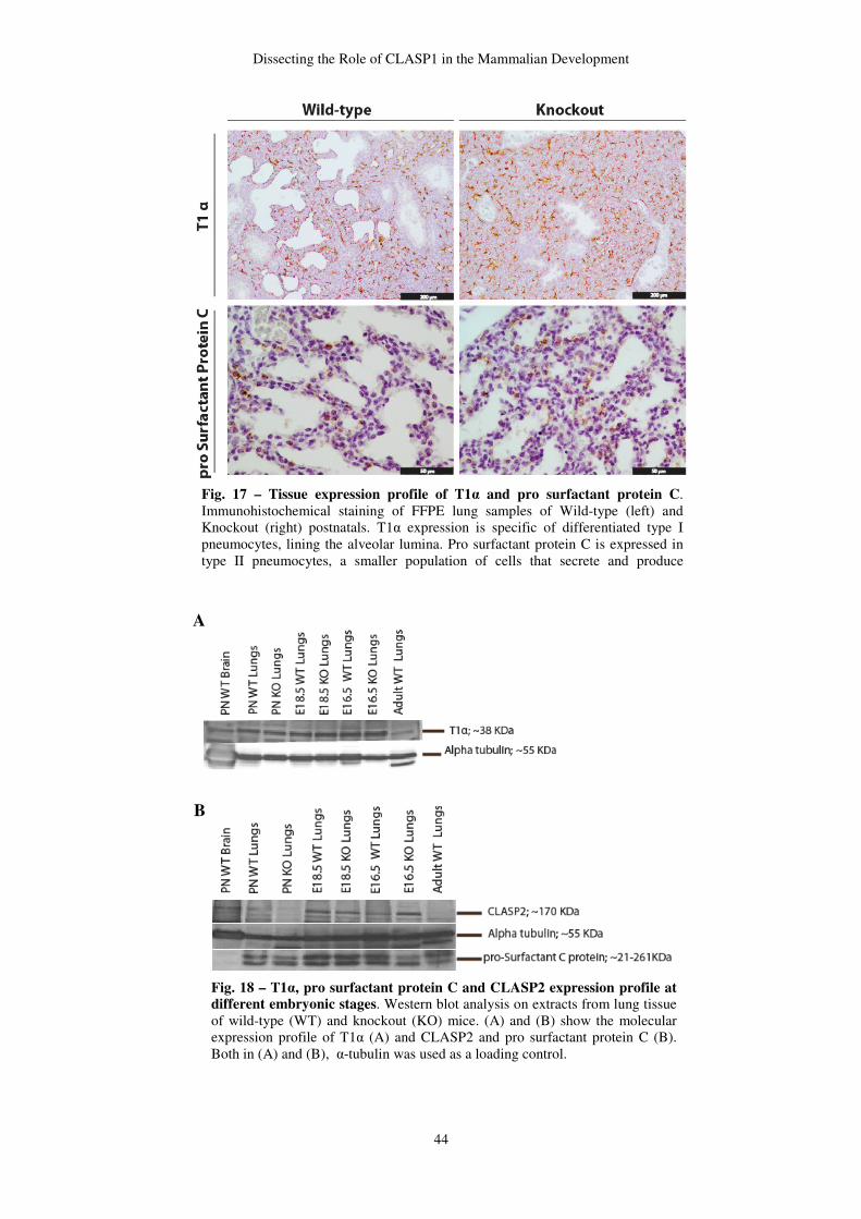
Dissecting the Role of CLASP1 in the Mammalian Development
44
Fig. 17 – Tissue expression profile of T1α and pro surfactant protein C. Immunohistochemical staining of FFPE lung samples of Wild-type (left) and Knockout (right) postnatals. T1α expression is specific of differentiated type I pneumocytes, lining the alveolar lumina. Pro surfactant protein C is expressed in type II pneumocytes, a smaller population of cells that secrete and produce surfactant liquid.
Fig. 18 – T1α, pro surfactant protein C and CLASP2 expression profile at different embryonic stages. Western blot analysis on extracts from lung tissue of wild-type (WT) and knockout (KO) mice. (A) and (B) show the molecular expression profile of T1α (A) and CLASP2 and pro surfactant protein C (B). Both in (A) and (B), α-tubulin was used as a loading control.
A
B
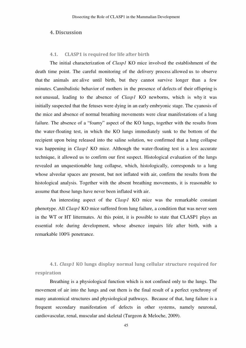
Dissecting the Role of CLASP1 in the Mammalian Development
45
4. Discussion
4.1. CLASP1 is required for life after birth
The initial characterization of Clasp1 KO mice involved the establishment of the
death time point. The careful monitoring of the delivery process allowed us to observe
that the animals are alive until birth, but they cannot survive longer than a few
minutes. Cannibalistic behavior of mothers in the presence of defects of their offspring is
not unusual, leading to the absence of Clasp1 KO newborns, which is why it was
initially suspected that the fetuses were dying in an early embryonic stage. The cyanosis of
the mice and absence of normal breathing movements were clear manifestations of a lung
failure. The absence of a “foamy” aspect of the KO lungs, together with the results from
the water-floating test, in which the KO lungs immediately sunk to the bottom of the
recipient upon being released into the saline solution, we confirmed that a lung collapse
was happening in Clasp1 KO mice. Although the water-floating test is a less accurate
technique, it allowed us to confirm our first suspect. Histological evaluation of the lungs
revealed an unquestionable lung collapse, which, histologically, corresponds to a lung
whose alveolar spaces are present, but not inflated with air, confirm the results from the
histological analysis. Together with the absent breathing movements, it is reasonable to
assume that those lungs have never been inflated with air.
An interesting aspect of the Clasp1 KO mice was the remarkable constant
phenotype. All Clasp1 KO mice suffered from lung failure, a condition that was never seen
in the WT or HT littermates. At this point, it is possible to state that CLASP1 plays an
essential role during development, whose absence impairs life after birth, with a
remarkable 100% penetrance.
4.1. Clasp1 KO lungs display normal lung cellular structure required for
respiration
Breathing is a physiological function which is not confined only to the lungs. The
movement of air into the lungs and out them is the final result of a perfect synchrony of
many anatomical structures and physiological pathways. Because of that, lung failure is a
frequent secondary manifestation of defects in other systems, namely neuronal,
cardiovascular, renal, muscular and skeletal (Turgeon & Meloche, 2009).
Dissecting the Role of CLASP1 in the Mammalian Development
46
The respiratory muscles are extremely important for ventilation movements
(Turgeon & Meloche, 2009). So, defects in diaphragm, the master muscle of respiration,
could be a reason of lung failure, as well as defects in connective tissue. So, by staining
postnatal lung samples with Masson trichrome, it was possible to rule out structural muscle
and connective tissue defects in Clasp1 KO mice. Nevertheless, we must note that more
quantitative measurements must be performed to confirm and validate this result.
Shortly before birth, alveolar type II cells must be able to efficiently secrete
surfactant. This material is a complex mixture of lipids and proteins essential to reduce the
surface tension at the air/liquid interface. The lack of sufficient amounts of surfactant is
one of the main causes of pulmonary syndrome distress in premature newborns. The most
abundant lipid specie in surfactant is phosphatidylcholine (PC) with dipalmitoyl-PC being
the surface-active reagent. Undifferentiated pulmonary epithelium has large glycogen
stores, which are depleted as the surfactant starts do be synthesized (Brehier & Rooney,
1981; Goerke, 1998). Moreover, these glycogen accumulations are absent in fully
differentiated type II cells (Ridsdale, Tseu, Wang, & Post, 2001). Biochemically, temporal
relationships between glycogen and phospholipids have suggested that glycogen serves as
carbon source for surfactant lipids. However, this relationship has not yet been proved.
Glycogen content was assessed by PAS. Although it is not specific for glycogen it is a very
cheap and was used as a screening technique to detect differences in glycogen storage
between KOs and WT controls. The first perception upon PAS staining was that KO had
higher amounts of glycongen/mucins in the pulmonary parenchyma. However, the lower
specificity of the technique and the higher number of cells per area due to the lung collapse
must be carefully analyzed. Considering that glycogen may serve as surfactant lipid
backbone during the lung development, a higher accumulation of glycogen in KO lungs
may stand for a delay in lung development, confirming the outcome obtained by previous
histological evaluation. One alternative explanation for higher amounts of glycogen in KO
lungs may be associated with a blockage on the surfactant lipid synthesis from the
glycogen stores.
Having this first clue for type II penumocytes immaturity, we immunostained lung
samples for pro surfactant protein C, to rule out surfactant secretion deficiency. Both KO
and control lung samples exhibit the same lung expression pattern, which means that type
II pneumocytes are differentiated in postnatals. T1α expression profile was also assessed to
exclude defects in type I pneumocytes maturation and, once again, no significant
differences were found between Clasp1 KOs and controls. The molecular expression

Dissecting the Role of CLASP1 in the Mammalian Development
47
profile of both markers was also evaluated by Western blotting and the outcome was
concordant with the immunohistochemistry results. Although the histological analyzes
seem to indicate a morphological delay in Clasp1 KO lung development, the alveolar
epithelial cells, intervenient in the gas-exchange, are differentiated. However, it is not clear
whether this slight delay impairs type I and II pneumocytes function. Moreover, the effect
of CLASP1 depletion in alveolar epithelial cells has never been assessed. So, it is not
known in which extent CLASP1 absence may deregulate their biological behaviour.
Dissecting the Role of CLASP1 in the Mammalian Development
48
CHAPTER II
CLASP1 Expression Profile Throughout Development
1. Introduction
Is has been shown that CLASP homologues are not only important, but essential for
yeast, Drosophila and C. elegans viability. However, Clasp2 KO mice are viable, which
suggests that CLASP1 may partially cover CLASP2 absence. CLASP1 expression profile
has already been assessed in wild-type adult mice and it has been shown that CLASP1 is
ubiquously expressed in adult mice, while CLASP2 is mostly expressed in brain (A. L. F.
Pereira, 2009). Regarding the lethal phenotype observed in our Clasp1 KO mice model, we
evaluated CLASP1 expression profile throughout embryonic development in order to find
any relationship between the expression profile and the newborns’ phenotype.
2. Materials and Methods
2.1. Western blot analysis
Whole tissue extracts were prepared by tissue homogenization and run on 11%
sodium dodecyl sulfate-polyacrylamide gel electrophoresis and electroblotted on
nitrocellulose membranes (Protran, Wattman), using a transfer apparatus (Bio-Rad). Blots
were blocked in 5% MILK in PBS with 0,05% Tween20.
T1α and pro-surfactant protein C were detected by incubation with monoclonal
syrian hamster antibody against T1α (1:200, Developmental Studies Hybridoma Bank) and
polyclonal rabbit pro-surfactant protein C (1:4000, Chemicon), respectively. Secondary
goat antibodies against Syrian hamster-biotinilated and rabbit-HRP were used. To detect
T1α, one additional step entailed the use of avidin-biotin complex (ABC, Vector). The
amount of protein was detected by conjugation with ECL (Amersham).
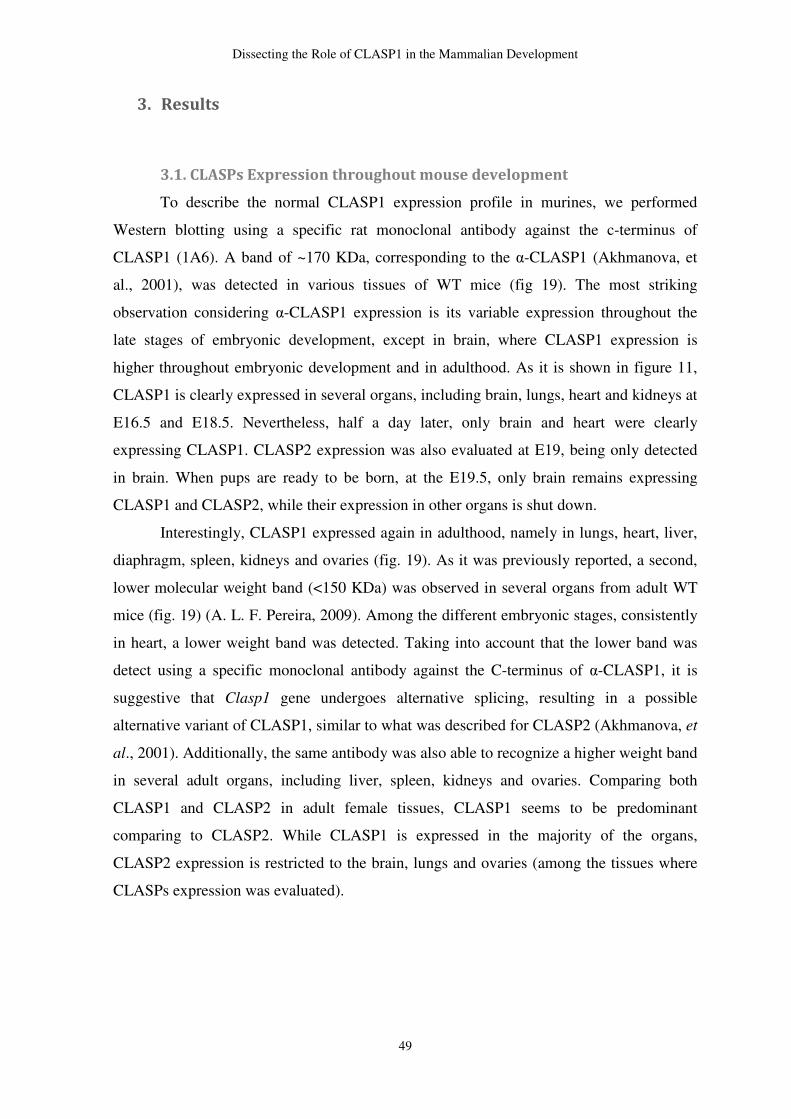
Dissecting the Role of CLASP1 in the Mammalian Development
49
3. Results
3.1. CLASPs Expression throughout mouse development
To describe the normal CLASP1 expression profile in murines, we performed
Western blotting using a specific rat monoclonal antibody against the c-terminus of
CLASP1 (1A6). A band of ~170 KDa, corresponding to the α-CLASP1 (Akhmanova, et
al., 2001), was detected in various tissues of WT mice (fig 19). The most striking
observation considering α-CLASP1 expression is its variable expression throughout the
late stages of embryonic development, except in brain, where CLASP1 expression is
higher throughout embryonic development and in adulthood. As it is shown in figure 11,
CLASP1 is clearly expressed in several organs, including brain, lungs, heart and kidneys at
E16.5 and E18.5. Nevertheless, half a day later, only brain and heart were clearly
expressing CLASP1. CLASP2 expression was also evaluated at E19, being only detected
in brain. When pups are ready to be born, at the E19.5, only brain remains expressing
CLASP1 and CLASP2, while their expression in other organs is shut down.
Interestingly, CLASP1 expressed again in adulthood, namely in lungs, heart, liver,
diaphragm, spleen, kidneys and ovaries (fig. 19). As it was previously reported, a second,
lower molecular weight band (<150 KDa) was observed in several organs from adult WT
mice (fig. 19) (A. L. F. Pereira, 2009). Among the different embryonic stages, consistently
in heart, a lower weight band was detected. Taking into account that the lower band was
detect using a specific monoclonal antibody against the C-terminus of α-CLASP1, it is
suggestive that Clasp1 gene undergoes alternative splicing, resulting in a possible
alternative variant of CLASP1, similar to what was described for CLASP2 (Akhmanova, et
al., 2001). Additionally, the same antibody was also able to recognize a higher weight band
in several adult organs, including liver, spleen, kidneys and ovaries. Comparing both
CLASP1 and CLASP2 in adult female tissues, CLASP1 seems to be predominant
comparing to CLASP2. While CLASP1 is expressed in the majority of the organs,
CLASP2 expression is restricted to the brain, lungs and ovaries (among the tissues where
CLASPs expression was evaluated).
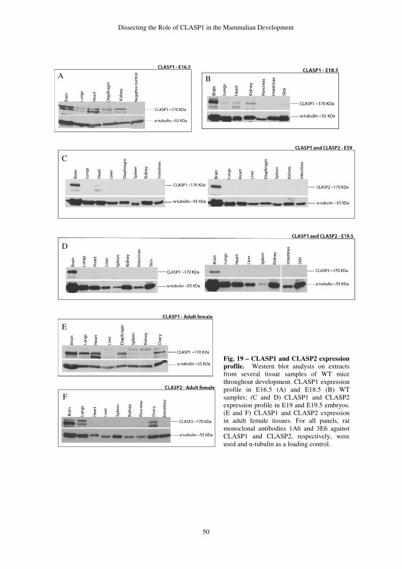
Dissecting the Role of CLASP1 in the Mammalian Development
50
Fig. 19 – CLASP1 and CLASP2 expression profile. Western blot analysis on extracts from several tissue samples of WT mice throughout development. CLASP1 expression profile in E16.5 (A) and E18.5 (B) WT samples; (C and D) CLASP1 and CLASP2 expression profile in E19 and E19.5 embryos. (E and F) CLASP1 and CLASP2 expression in adult female tissues. For all panels, rat monoclonal antibodies 1A6 and 3E6 against CLASP1 and CLASP2, respectively, were used and α-tubulin as a loading control.
A B
C
D
E
F
Dissecting the Role of CLASP1 in the Mammalian Development
51
4. Discussion
4.1. CLASP1expression is variable throughout development
CLASP1 expression throughout mouse development seems to be transient among
different organs namely lungs, heart, diaphragm and kidneys. In the brain, CLASP1 and
CLASP2 are strongly expressed from the early embryonic stages to adulthood, being the
organ with the higher expression of this protein. +TIPs have been intensively studied in
neurons. Neurons are highly differentiated cells specialized in signal transduction,
sometimes through long distance and requires a well-organized cytoskeleton. CLASPs, as
well as other +TIPs, have been reported to regulate cellular polarity (Baas, Karabay, &
Qiang, 2005; Baas, et al., 2006) and intracellular transport in which MTs act as railways
for motor-based transport. They have also been shown to be important during several
stages of neuronal development including migration (Kholmanskikh, et al., 2006),
formation, growth and guidance of axons (Lee, et al., 2004). The chemical signaling
requires proper communication between mature neuronal cells, which is also dependent on
+TIPs, due to their role in positioning of organelles, receptors and channels (Gu, et al.,
2006). The strong expression of CLASPs in brain may explain the wide range of roles that
both proteins might play in neurons. However, a low expression is not meaningless. For
instance, in Clasp2 KO mice, hematopoiesis is affected, regardless of a low CLASP2
expression in bone marrow extracts.
Dissecting the Role of CLASP1 in the Mammalian Development
52
CHAPTER III
Optimization of CLASP1 Immunostaining for Histological Samples
1. Introduction
The use of antibodies to specifically detect proteins began with Albert H. Coons in
1941, who described a revolutionary technique to visualize tissue components using an
antibody labeled with a fluorescent dye (John D. Bancroft, 2008). Since then, stainings
based on antigen-antibody interaction (immunostainings) have boosted cellular biology.
Presently, it is possible to localize a given protein (or a group of antigens) within a cell, as
well as track its translocations and interactions with other molecular partners (John D.
Bancroft, 2008). Immunohistochemistry refers to the immunostainings applied to tissue
samples. Using mono or polyclonal antibodies, it is possible to detect antigens and assess
the morphological detail at the same time.
Polyclonal antibodies have been used to detect the expression of CLASP1.
However, the use of polyclonal antibodies may arise drawbacks regarding the specificity of
the immunostainings. Polyclonal antibodies are generated by immunizing an animal,
whose humoral system activates numerous clones of plasma cells (polyclonal). Each clone
will produce specific immunoglobulins, which react with specific epitopes. The smallest
variation between immunoglobulins of an anti-serum, may generate cross-reactivity
between the protein of interest and similar proteins (John D. Bancroft, 2008). One way to
increase the specificity of an immunostaining, is to use monoclonal antibodies, by using
hybridoma cell lines (Eichmann, 2004; John D. Bancroft, 2008). This method involves the
fusion of plasma cells with an immortalized myeloma cell line. This hybrid cell line only
produces on type of immunoglobulin. The result is a constant, more specific and reliable
source of pure antibodies, with higher specificity. Monoclonal antibodies against CLASP1
and CLASP2 were generated and well characterized previously (Maffini, et al., 2009;
Pereira, 2009). These antibodies have shown to be highly specific for immunofluorescence
techniques in fixed cultured cells and western blot. However, the staining of CLASPs in
FFPE histological samples has not been successfully achieved. The characterization of the
recently generated Clasp1 and Clasp2 KO mice strains have lead us to optimize these
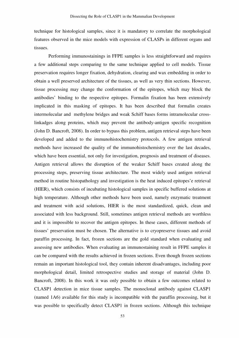
Dissecting the Role of CLASP1 in the Mammalian Development
53
technique for histological samples, since it is mandatory to correlate the morphological
features observed in the mice models with expression of CLASPs in different organs and
tissues.
Performing immunostainings in FFPE samples is less straightforward and requires
a few additional steps comparing to the same technique applied to cell models. Tissue
preservation requires longer fixation, dehydration, clearing and wax embedding in order to
obtain a well preserved architecture of the tissues, as well as very thin sections. However,
tissue processing may change the conformation of the epitopes, which may block the
antibodies’ binding to the respective epitopes. Formalin fixation has been extensively
implicated in this masking of epitopes. It has been described that formalin creates
intermolecular and methylene bridges and weak Schiff bases forms intramolecular cross-
linkadges along proteins, which may prevent the antibody-antigen specific recognition
(John D. Bancroft, 2008). In order to bypass this problem, antigen retrieval steps have been
developed and added to the immunohistochemistry protocols. A few antigen retrieval
methods have increased the quality of the immunohistochemistry over the last decades,
which have been essential, not only for investigation, prognosis and treatment of diseases.
Antigen retrieval allows the disruption of the weaker Schiff bases created along the
processing steps, preserving tissue architecture. The most widely used antigen retrieval
method in routine histopathology and investigation is the heat induced epitopes’e retrieval
(HIER), which consists of incubating histological samples in specific buffered solutions at
high temperature. Although other methods have been used, namely enzymatic treatment
and treatment with acid solutions, HIER is the most standardized, quick, clean and
associated with less background. Still, sometimes antigen retrieval methods are worthless
and it is impossible to recover the antigen epitopes. In these cases, different methods of
tissues’ preservation must be chosen. The alternative is to cryopreserve tissues and avoid
paraffin processing. In fact, frozen sections are the gold standard when evaluating and
assessing new antibodies. When evaluating an immunostaining result in FFPE samples it
can be compared with the results achieved in frozen sections. Even though frozen sections
remain an important histological tool, they contain inherent disadvantages, including poor
morphological detail, limited retrospective studies and storage of material (John D.
Bancroft, 2008). In this work it was only possible to obtain a few outcomes related to
CLASP1 detection in mice tissue samples. The monoclonal antibody against CLASP1
(named 1A6) available for this study is incompatible with the paraffin processing, but it
was possible to specifically detect CLASP1 in frozen sections. Although this technique
Dissecting the Role of CLASP1 in the Mammalian Development
54
requires further improvements, it was possible, for the first time, to detect CLASP1 in
histological samples.
2. Materials and Methods
2.1. Frozen Sections Preparation
Adult mice were anesthetized with volatile isoflurane (Merck) and then sacrificed
by cervical dislocation. Brain samples were taken and fixed in 10% neutral buffered
formalin for 24-48h, at room temperature. Specimens were embedded in a 50% (m/v)
sucrose solution until they sunk to the bottom of the recipient. The same procedure was
performed using a 70% (m/v) sucrose solution. Samples were embedded in a gel-like
medium consisting of water-soluble glycols and resins. An optimum cutting temperature
(OCT) compound (Sakura) was used to provide a convenient specimen matrix for cryostat
sectioning at temperatures of aproximately -25º C. Samples were cryosectioned with 10
µm thick and mounted in glass slides coated with APES (SIGMA).
2.2. Immunostaining of CLASP1 detection in cryopreserved tissues
Cryosections of 10 µm thick were melted at room temperature and washed in PBS
0.05% Triton X-100 (). Endogenous peroxidase was blocked by 4% of hydroxide peroxide
diluted in methanol for 40 minutes, at room temperature. Unspecific reactions were
blocked with 3% BSA plus 10% FBS in PBS for 1 hour, at room temperature. The primary
antibody used was the 1A6 rat monoclonal anti-mouse CLASP1, with 1:5 dilution in 3%
BSA plus 10% FBS. CLASP1 was identified by the use of a Biotinylated Rabbit Anti-Rat
IgG (1:50, SIGMA) for 30 minutes, followed by detection with ExtrAvidin Peroxidase
(1:100, SIGMA). The reaction was developed using DAB (SIGMA). Qualitative analysis
was preceded using a NIKON (ver ataf) and representative fields were captured.
Dissecting the Role of CLASP1 in the Mammalian Development
55
2.3. Immunostaining of CLASP1 detection in paraffin sections
FFPE tissue sections 4 µm thick were used to evaluate CLASP1 expression in wild
type brain tissue. Sections were rehydrated in a graded series of ethanols until water.
Endogenous peroxidase was blocked by 4% of hydroxide peroxide diluted in methanol for
40 minutes, at room temperature. Two antigen retrieval methods were tested in FFPE
tissue samples: HIER and treatment with an acid solution. HIER protocol included heating
samples in citrate buffer pH 6 around 90-100ºC, for 5 minutes using a microwaves at the
maximum power. The acid treatment protocol was performed be incubating samples in a
10% HCL solution for 30 minutes, at room temperature, followed by neutralization with
1M Bórax, for 5 minutes. Unspecific reactions were blocked with 3% BSA plus 10% FBS
in PBS for 1 hour, at room temperature. The following steps were the same as it was
previously described for the immunostaining of CLASP1 in frozen sections. (inverter a
ordem de 2.2 e 2.3)
3. Results
3.1. CLASP1 immunostaining using 1A6 monoclonal antibody against
CLASP1
After the first approach using FFPE tissue samples, in which different antigen
retrieval methods were used, as well as broad range of antibody’s dilutions, one possible
assumption is that, either formalin fixation, or paraffin processing, damage the antigen
epitopes in such an extensive way that is impossible to recover them. In FFPE samples it
was never possible to detect CLASP1 expression in positive controls (brain samples).
However, a satisfactory outcome was achieved in cryosections without requiring additional
steps for antigen retrieval. Detection of CLASP1 expression in a few very specific brain
cell populations supports the specificity of the antibody. Moreover, even after such a
prolonged formalin fixation (24-48 hours), frozen section did not require antigen retrieval
to satisfactorily stain for CLASP1. Hence, one possible reason for the negative results in
FFPE tissue samples is the irreversible destruction of the antigenic determinants during the
paraffin processing.
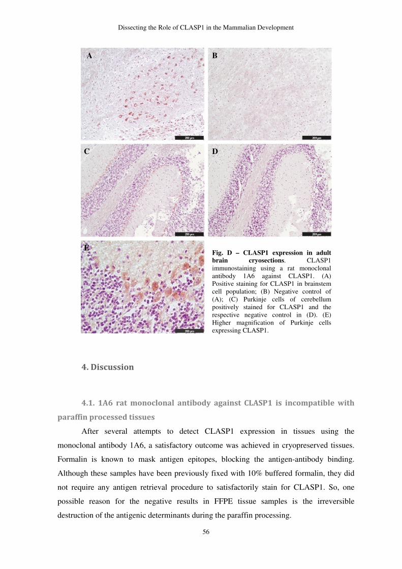
Dissecting the Role of CLASP1 in the Mammalian Development
56
4. Discussion
4.1. 1A6 rat monoclonal antibody against CLASP1 is incompatible with
paraffin processed tissues
After several attempts to detect CLASP1 expression in tissues using the
monoclonal antibody 1A6, a satisfactory outcome was achieved in cryopreserved tissues.
Formalin is known to mask antigen epitopes, blocking the antigen-antibody binding.
Although these samples have been previously fixed with 10% buffered formalin, they did
not require any antigen retrieval procedure to satisfactorily stain for CLASP1. So, one
possible reason for the negative results in FFPE tissue samples is the irreversible
destruction of the antigenic determinants during the paraffin processing.
Fig. D – CLASP1 expression in adult brain cryosections. CLASP1 immunostaining using a rat monoclonal antibody 1A6 against CLASP1. (A) Positive staining for CLASP1 in brainstem cell population; (B) Negative control of (A); (C) Purkinje cells of cerebellum positively stained for CLASP1 and the respective negative control in (D). (E) Higher magnification of Purkinje cells expressing CLASP1.
A
B
C D
E
Dissecting the Role of CLASP1 in the Mammalian Development
57
4.2. CLASP1 expression in adult brain
Regarding CLASP1 expression profile in adult brain, it is specifically expressed in
a few populations of the brainstem and Purkinje cells. Its negative staining in other areas of
the brain confirms the specificity of the antibody. Interestingly, the neuronal command of
breathing movements localizes in the brainstem in a few specific populations termed
respiratory centers. It is possible, since CLASP1 is strongly expressed in brainstem, that its
absence might impair the neuronal functions that control respiration. This hypothetic
situation is compatible to the absence of air in the KO lungs because, in a situation of
absence of neuronal stimuli, respiratory muscles do not contract or relax properly to let the
air enter into the lungs.

GENERAL
DISCUSSION
Dissecting the Role of CLASP1 in the Mammalian Development
59
General Discussion
CLASPs are highly conserved proteins among species, which have been described
to play a role in microtubule dynamics, both in interphase and mitosis. During the last few
years, CLASPs orthologues have been studied in a few organisms (Grallert, et al., 2006;
Hannak & Heald, 2006; Inoue, et al., 2000; Lemos, et al., 2000; Pasqualone & Huffaker,
1994), including mammals (Akhmanova, et al., 2001; Inoue, et al., 2000; Lemos, et al.,
2000). However, while in the first three there is one single CLASP orthologue, in
mammals two paralogues have been identified (Akhmanova, et al., 2001; Inoue, et al.,
2000; Lemos, et al., 2000). Depletion of CLASP in organisms with a single orthologue is
incompatible with life. However, it has been shown that Clasp2 KO mice are viable
(Drabek, et al., 2006; Pereira, 2009). One possible explanation for the survival of
mammals with only CLASP1 expression might be related to the separation of the functions
of one single protein by two paralogues and previous work in cellular models has already
pointed toward a partial redundancy between both CLASPs (Pereira, et al., 2006).
However, their specific roles, both in cellular and physiological level, require further
investigation. So far, Clasp2 KO mice are viable, but develop what seems to be
maturation-related dysfunctions, mainly in the hematopoietic system. These mice display
bone marrow aplasia with severe anemia and dye early in life (six months on average)
when compared with WT littermates. Absence of CLASP2 seems to impair the proper
erythroblast differentiation, which means that depletion of CLASP2 plays an important
role at the early progenitors level. CLASP2 depletion was also described to cause
maturation problems in the reproductive system (Clasp2 KO are infertile) (Drabek, et al.,
2006; Pereira, et al., 2006; Pereira, 2009). Interestingly, CLASP2 depletion affects mostly
tissues with high cellular turnover, which might also be related to the role of CLASPs in
mitosis. Thus, CLASP2 is suggested to play a role in cellular differentiation and tissue
maturation in mammals. However, contrary to what is observed with Clasp2 KO, our
Clasp1 KO model is not viable after birth. To uncover CLASP1 specific functions at the
physiological level, a Clasp1 KO mouse model was generated (A. L. F. Pereira, 2009).
Until now, it was thought that Clasp1 KO mice were dying during embryonic
development, due to the absence of KO animals in the offspring (A. L. F. Pereira, 2009).
Cannibalistic behavior was observed in the females after delivery of KOs. Because of that,
we closely monitored the delivery process and pups were separated from the female if any
Dissecting the Role of CLASP1 in the Mammalian Development
60
cannibalistic intention was noticed. Afterwords, we observed that some pups, which were
turning cyanotic immediately after birth, were also dying within a few minutes, while
others were apparently healthy. After genotyping, it was possible to conclude that all and
only Clasp1 KO mice were displaying perinatal lethality. In fact, Clasp1 KO mice are
viable until birth, although, they cannot survive ex uterus. Phenotype resulting from
mutations can be very challenging. This is especially true for mutants dying perinatally,
which represent a substantial proportion of lethal phenotypes in the mouse. Unless the gene
is expressed in a tissue-specific manner or is conditionally inactivated in specific tissues,
determining which specific physiological defects is responsible for lethality may require
considerable effort.
Cyanosis is a hallmark of lung failure, which is also one of the most frequent
complications in human preterm newborns, as well as low birth weight that was also
observed in Clasp1 KO pups. So, the next step was be macro and microscopic analysis of
Clasp1 KO lungs. Histological analysis revealed a lung collapse, which was confirmed by
the water-floating test. The absence of clear alveolar spaces, together with the absence of
air, suggest that Clasp1 KO pups were not able to inflate their lungs. Lung collapse, or
pneumothorax, may be a result of an underlying lung disease, but defects in other organs or
systems may also be a cause. Histological analysis of lung samples from the E14.5 E14.5
to the E19.5 revealed a slight delay in lung development in Clasp1 KO animals. Regarding
this delay, the next step was to determine if Clasp1 KO mice would be functionally
prepared to undergo gas-exchange after after.
Respiratory tract consists of a remarkable, highly branched, tubular structure that
leads to millions of alveolar sacs. The alveolar surface is lined by type II and type I
alveolar epithelial cells that are in direct contact with respiratory gases, creating collapsing
forces at the air-liquid interface. To maintain lung expansion, these surface forces are
mitigated by the presence of pulmonary surfactant that is synthesized and secreted onto the
alveolar space by type II epithelial cells. Lack of pulmonary surfactant in preterm infants
with respiratory distress syndrome causes atelectasis leading to respiratory failure.
Considering CLASP2 role in cell differentiation and the Clasp1 KO mice lung failure
phenotype, our first hypothesis would be defects in lung maturation. Thus, we checked if
Clasp1 KO lungs were functionally prepared to undergo respiration. The presence of
mature type I and type II pneumocytes would tell if Clasp1 KO lungs were prepared to
undergo gas-exchange. Then, we searched for molecular markers of type II and type I
pneumocytes and stained for pro surfactant protein C and T1α, respectively, to check if
Dissecting the Role of CLASP1 in the Mammalian Development
61
these cells were, actually, differentiated in KO lungs. In fact, these markers were present
not only in WT lungs, but also in KOs. We also stained for pro surfactant protein C in early
embryonic stages and we observed that this protein starts to be clearly expressed in WT
lungs by the E15.5, but KO animals only show a clear expression by the E16.5 (data not
shown). This means that, in fact, there is a delay in lung development of, at least, ~1
gestation day. However, this one day of delay (premature) would not be enough to explain
the lethal phenotype because WT E18.5 embryos survive upon premature extraction from
the mother by C-section. This means that, physiologically, this lung failure represents more
than a delay of 1 gestation day. A closer observation of the histological architecture of KO
lungs, allowed us to note that this delay was becoming more evident along the later
gestation steps. For instance, KO lungs at the E16.5 were more alike to the WT littermates
at the E15.5, but some the lungs of E19.5 KOs were more similar to the WT controls at the
E17.5.
In late gestation, glycogen-laden type II pneumocytes shift their metabolic program
toward the synthesis of surfactant, of which phosphatidylcholine (PC) is, by far, the most
abundant lipid component of surfactant. So, abnormal high accumulation of glycogen in
lungs would suggest that both type II pneumocytes immaturity or defects in glycogen
metabolism, could lead to insuficient surfactant production. PAS staining results in E18.5
lung samples have shown an apparent higher accumulation of PAS positive substances in
the KO alveolar parenchyma (excluding glandular cells with PAS positive neutral mucins).
However, this histochemical procedure is not totally specific for glycogen and is not a
direct measurement of the lipidic component of surfactant. Moreover, we only tested for
pro surfactant protein C, while the protein component of surfactant are composed by
surfactant A, B, C and D proteins.
Regarding CLASP1 expression profile during development, the most striking
observations were its strong and consistent expression in brain throughout development
and its variable expression among the other organs. At the early embryonic stages (E16.5
and E18.5), CLASP1 was expressed in many organs, such as brain, lungs, heart and
kidneys. Nevertheless, by the time of birth, only brain was clearly expressing CLASP1 and
CLASP2, while their expression in the others organs is shut down. In adult mice, CLASP1
is ubiquously expressed. These results do not allow us to directly compare the amount of
proteins expressed in each organ/tissue due to differences in the loading. Protein extraction
procedures are less efficient in tissues samples. The amount of protein isolated is usually
lower, comparing to cell extracts. Moreover, tissues with high content of connective tissue
Dissecting the Role of CLASP1 in the Mammalian Development
62
and muscle are harder to disrupt. Thus, further improvements in protein extraction
techniques are one of the future prospects of this work.
Collectively, this work has given the first steps to uncover CLASP1 role in living
mammals. So far, we were able to show that Clasp1 depletion causes death after birth in
mice. Clasp1 KO mice are, indeed, viable until the moment of their birth, but they cannot
survive more than a few minutes after it. Although an underlying lung defect is not
completely ruled out, we proved that KO lungs express molecular markers of late lung
morphogenesis. Thus, Clasp1 KO lungs seem to be ready to undergo gas-exchange after
birth, even exhibiting a delay in lung architecture comparing to control littermates. We also
cannot exclude defects in surfactant production yet. The apparent higher amounts of
glycogen corroborates the histological findings, which suggests lung immaturity. Muscular
and ribs defects were apparently absent in KO animals. However, taking into account the
technical limitations of the histochemical techniques, this part of the work requires further
investigation.
In a near future, it will be important to explore, in more detail, other systems
directly related to respiration. Neuronal defects have been described as cause of a very
similar phenotype to the Clasp1 KO animals. Defects in neurons controlling respiration
may block the signal transmission to the respiratory muscles, impairing the proper
rhythmic movements and shutting down ventilation. Considering the aforementioned role
of CLASPs in neuronal biology, an underlying neuronal cause may be shutting down
respiration, leading to the Clasp1 KO mice death right after birth. So far, we have
characterized the Clasp1 KO phenotype and some possible causes of lung failure have
been ruled out. Although we have shown that Clasp1 KO mice cannot inflate their lungs,
the primary physiological failure remains unclear. Nevertheless, the most important
conclusion regarding CLASP1 impact in the physiology environment of mammals is that
CLASP1 is, unquestionably, required for mammals’ life.

REFERENCES
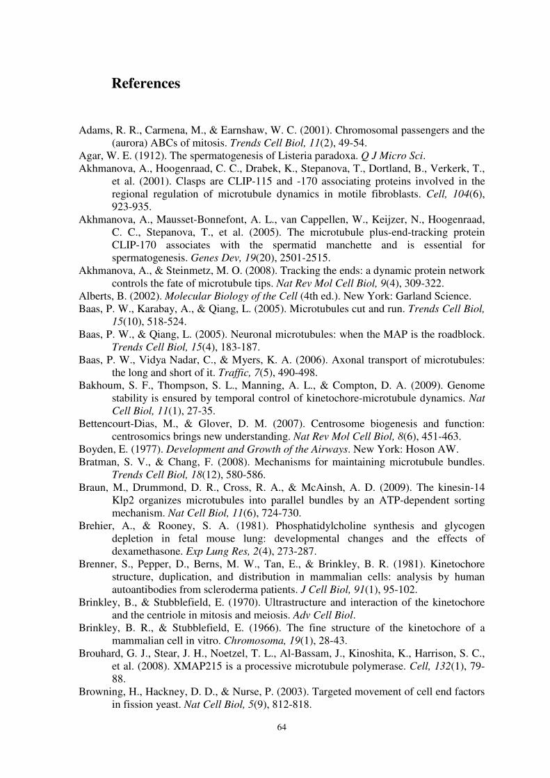
64
References
Adams, R. R., Carmena, M., & Earnshaw, W. C. (2001). Chromosomal passengers and the
(aurora) ABCs of mitosis. Trends Cell Biol, 11(2), 49-54. Agar, W. E. (1912). The spermatogenesis of Listeria paradoxa. Q J Micro Sci. Akhmanova, A., Hoogenraad, C. C., Drabek, K., Stepanova, T., Dortland, B., Verkerk, T.,
et al. (2001). Clasps are CLIP-115 and -170 associating proteins involved in the regional regulation of microtubule dynamics in motile fibroblasts. Cell, 104(6), 923-935.
Akhmanova, A., Mausset-Bonnefont, A. L., van Cappellen, W., Keijzer, N., Hoogenraad, C. C., Stepanova, T., et al. (2005). The microtubule plus-end-tracking protein CLIP-170 associates with the spermatid manchette and is essential for spermatogenesis. Genes Dev, 19(20), 2501-2515.
Akhmanova, A., & Steinmetz, M. O. (2008). Tracking the ends: a dynamic protein network controls the fate of microtubule tips. Nat Rev Mol Cell Biol, 9(4), 309-322.
Alberts, B. (2002). Molecular Biology of the Cell (4th ed.). New York: Garland Science. Baas, P. W., Karabay, A., & Qiang, L. (2005). Microtubules cut and run. Trends Cell Biol,
15(10), 518-524. Baas, P. W., & Qiang, L. (2005). Neuronal microtubules: when the MAP is the roadblock.
Trends Cell Biol, 15(4), 183-187. Baas, P. W., Vidya Nadar, C., & Myers, K. A. (2006). Axonal transport of microtubules:
the long and short of it. Traffic, 7(5), 490-498. Bakhoum, S. F., Thompson, S. L., Manning, A. L., & Compton, D. A. (2009). Genome
stability is ensured by temporal control of kinetochore-microtubule dynamics. Nat
Cell Biol, 11(1), 27-35. Bettencourt-Dias, M., & Glover, D. M. (2007). Centrosome biogenesis and function:
centrosomics brings new understanding. Nat Rev Mol Cell Biol, 8(6), 451-463. Boyden, E. (1977). Development and Growth of the Airways. New York: Hoson AW. Bratman, S. V., & Chang, F. (2008). Mechanisms for maintaining microtubule bundles.
Trends Cell Biol, 18(12), 580-586. Braun, M., Drummond, D. R., Cross, R. A., & McAinsh, A. D. (2009). The kinesin-14
Klp2 organizes microtubules into parallel bundles by an ATP-dependent sorting mechanism. Nat Cell Biol, 11(6), 724-730.
Brehier, A., & Rooney, S. A. (1981). Phosphatidylcholine synthesis and glycogen depletion in fetal mouse lung: developmental changes and the effects of dexamethasone. Exp Lung Res, 2(4), 273-287.
Brenner, S., Pepper, D., Berns, M. W., Tan, E., & Brinkley, B. R. (1981). Kinetochore structure, duplication, and distribution in mammalian cells: analysis by human autoantibodies from scleroderma patients. J Cell Biol, 91(1), 95-102.
Brinkley, B., & Stubblefield, E. (1970). Ultrastructure and interaction of the kinetochore and the centriole in mitosis and meiosis. Adv Cell Biol.
Brinkley, B. R., & Stubblefield, E. (1966). The fine structure of the kinetochore of a mammalian cell in vitro. Chromosoma, 19(1), 28-43.
Brouhard, G. J., Stear, J. H., Noetzel, T. L., Al-Bassam, J., Kinoshita, K., Harrison, S. C., et al. (2008). XMAP215 is a processive microtubule polymerase. Cell, 132(1), 79-88.
Browning, H., Hackney, D. D., & Nurse, P. (2003). Targeted movement of cell end factors in fission yeast. Nat Cell Biol, 5(9), 812-818.
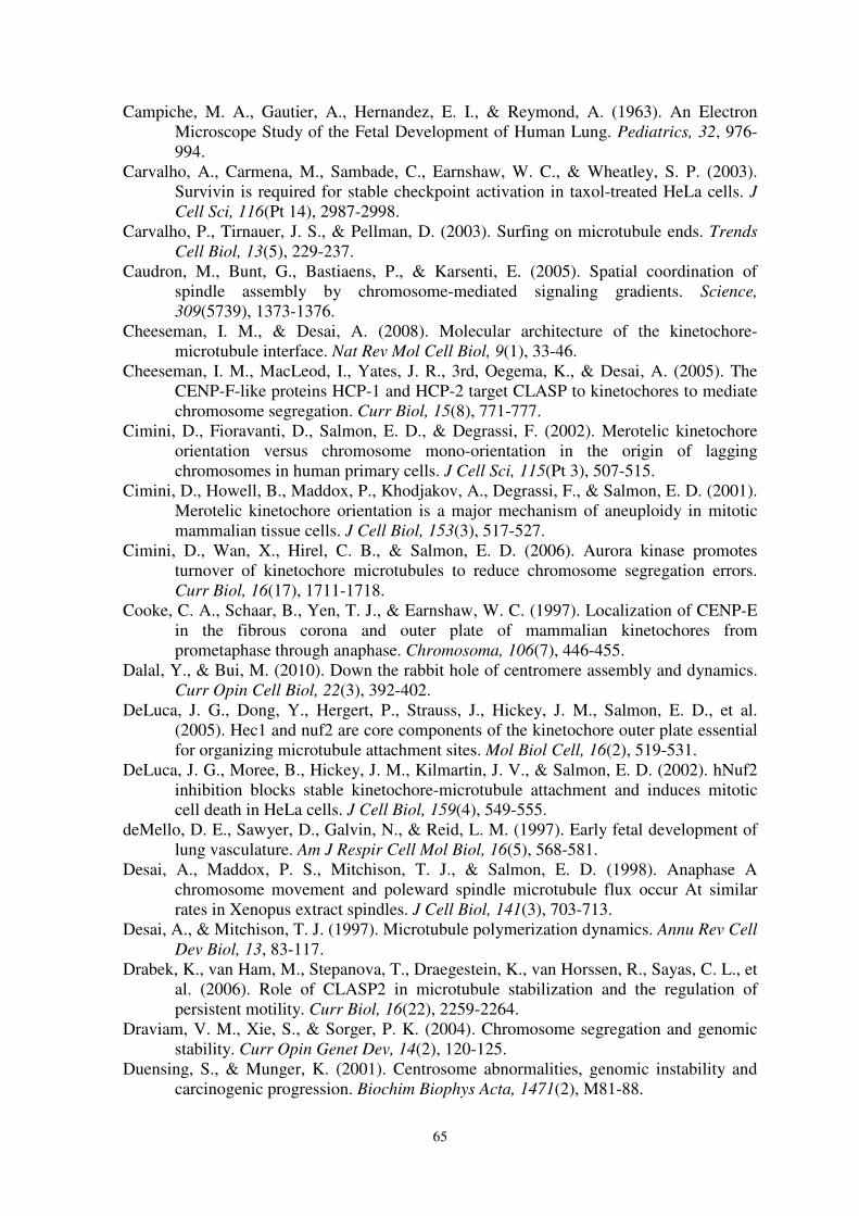
65
Campiche, M. A., Gautier, A., Hernandez, E. I., & Reymond, A. (1963). An Electron Microscope Study of the Fetal Development of Human Lung. Pediatrics, 32, 976-994.
Carvalho, A., Carmena, M., Sambade, C., Earnshaw, W. C., & Wheatley, S. P. (2003). Survivin is required for stable checkpoint activation in taxol-treated HeLa cells. J
Cell Sci, 116(Pt 14), 2987-2998. Carvalho, P., Tirnauer, J. S., & Pellman, D. (2003). Surfing on microtubule ends. Trends
Cell Biol, 13(5), 229-237. Caudron, M., Bunt, G., Bastiaens, P., & Karsenti, E. (2005). Spatial coordination of
spindle assembly by chromosome-mediated signaling gradients. Science,
309(5739), 1373-1376. Cheeseman, I. M., & Desai, A. (2008). Molecular architecture of the kinetochore-
microtubule interface. Nat Rev Mol Cell Biol, 9(1), 33-46. Cheeseman, I. M., MacLeod, I., Yates, J. R., 3rd, Oegema, K., & Desai, A. (2005). The
CENP-F-like proteins HCP-1 and HCP-2 target CLASP to kinetochores to mediate chromosome segregation. Curr Biol, 15(8), 771-777.
Cimini, D., Fioravanti, D., Salmon, E. D., & Degrassi, F. (2002). Merotelic kinetochore orientation versus chromosome mono-orientation in the origin of lagging chromosomes in human primary cells. J Cell Sci, 115(Pt 3), 507-515.
Cimini, D., Howell, B., Maddox, P., Khodjakov, A., Degrassi, F., & Salmon, E. D. (2001). Merotelic kinetochore orientation is a major mechanism of aneuploidy in mitotic mammalian tissue cells. J Cell Biol, 153(3), 517-527.
Cimini, D., Wan, X., Hirel, C. B., & Salmon, E. D. (2006). Aurora kinase promotes turnover of kinetochore microtubules to reduce chromosome segregation errors. Curr Biol, 16(17), 1711-1718.
Cooke, C. A., Schaar, B., Yen, T. J., & Earnshaw, W. C. (1997). Localization of CENP-E in the fibrous corona and outer plate of mammalian kinetochores from prometaphase through anaphase. Chromosoma, 106(7), 446-455.
Dalal, Y., & Bui, M. (2010). Down the rabbit hole of centromere assembly and dynamics. Curr Opin Cell Biol, 22(3), 392-402.
DeLuca, J. G., Dong, Y., Hergert, P., Strauss, J., Hickey, J. M., Salmon, E. D., et al. (2005). Hec1 and nuf2 are core components of the kinetochore outer plate essential for organizing microtubule attachment sites. Mol Biol Cell, 16(2), 519-531.
DeLuca, J. G., Moree, B., Hickey, J. M., Kilmartin, J. V., & Salmon, E. D. (2002). hNuf2 inhibition blocks stable kinetochore-microtubule attachment and induces mitotic cell death in HeLa cells. J Cell Biol, 159(4), 549-555.
deMello, D. E., Sawyer, D., Galvin, N., & Reid, L. M. (1997). Early fetal development of lung vasculature. Am J Respir Cell Mol Biol, 16(5), 568-581.
Desai, A., Maddox, P. S., Mitchison, T. J., & Salmon, E. D. (1998). Anaphase A chromosome movement and poleward spindle microtubule flux occur At similar rates in Xenopus extract spindles. J Cell Biol, 141(3), 703-713.
Desai, A., & Mitchison, T. J. (1997). Microtubule polymerization dynamics. Annu Rev Cell
Dev Biol, 13, 83-117. Drabek, K., van Ham, M., Stepanova, T., Draegestein, K., van Horssen, R., Sayas, C. L., et
al. (2006). Role of CLASP2 in microtubule stabilization and the regulation of persistent motility. Curr Biol, 16(22), 2259-2264.
Draviam, V. M., Xie, S., & Sorger, P. K. (2004). Chromosome segregation and genomic stability. Curr Opin Genet Dev, 14(2), 120-125.
Duensing, S., & Munger, K. (2001). Centrosome abnormalities, genomic instability and carcinogenic progression. Biochim Biophys Acta, 1471(2), M81-88.
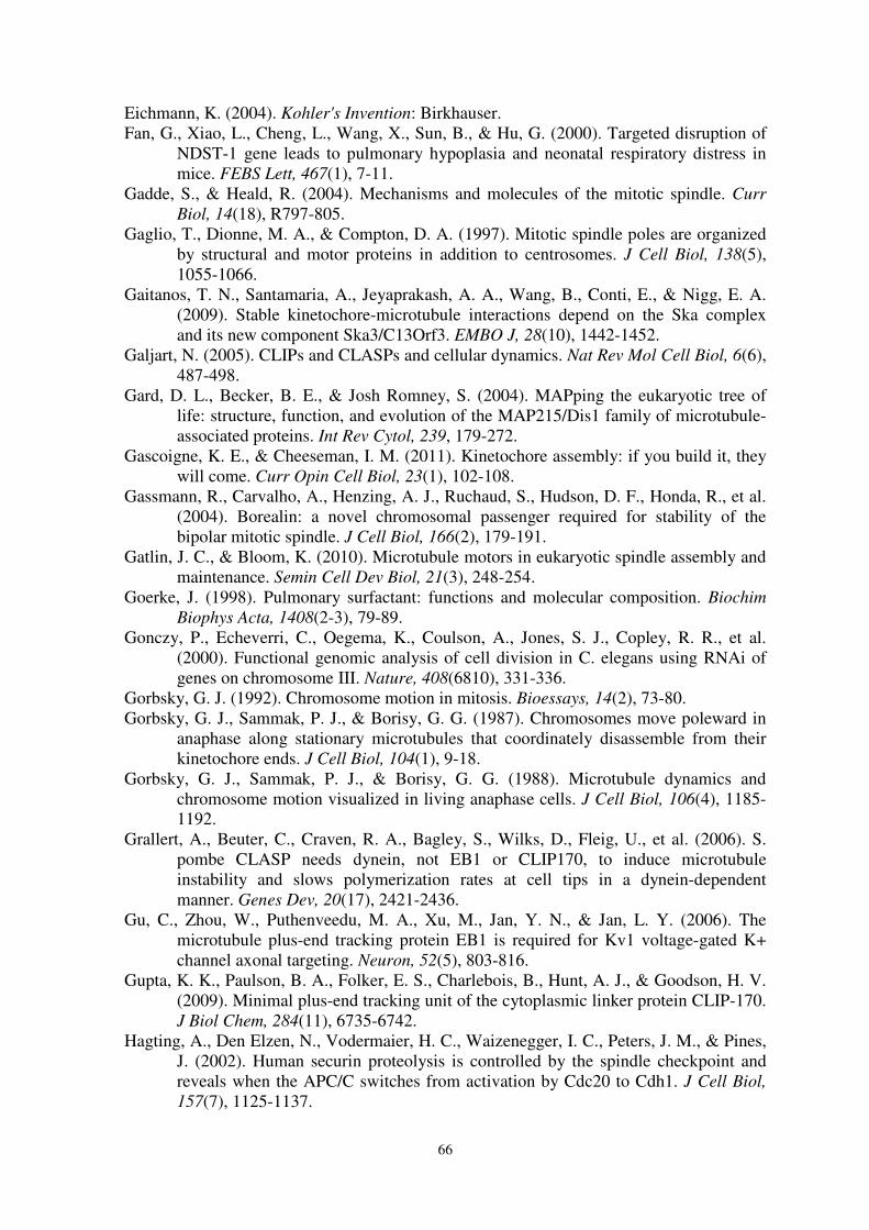
66
Eichmann, K. (2004). Kohler's Invention: Birkhauser. Fan, G., Xiao, L., Cheng, L., Wang, X., Sun, B., & Hu, G. (2000). Targeted disruption of
NDST-1 gene leads to pulmonary hypoplasia and neonatal respiratory distress in mice. FEBS Lett, 467(1), 7-11.
Gadde, S., & Heald, R. (2004). Mechanisms and molecules of the mitotic spindle. Curr
Biol, 14(18), R797-805. Gaglio, T., Dionne, M. A., & Compton, D. A. (1997). Mitotic spindle poles are organized
by structural and motor proteins in addition to centrosomes. J Cell Biol, 138(5), 1055-1066.
Gaitanos, T. N., Santamaria, A., Jeyaprakash, A. A., Wang, B., Conti, E., & Nigg, E. A. (2009). Stable kinetochore-microtubule interactions depend on the Ska complex and its new component Ska3/C13Orf3. EMBO J, 28(10), 1442-1452.
Galjart, N. (2005). CLIPs and CLASPs and cellular dynamics. Nat Rev Mol Cell Biol, 6(6), 487-498.
Gard, D. L., Becker, B. E., & Josh Romney, S. (2004). MAPping the eukaryotic tree of life: structure, function, and evolution of the MAP215/Dis1 family of microtubule-associated proteins. Int Rev Cytol, 239, 179-272.
Gascoigne, K. E., & Cheeseman, I. M. (2011). Kinetochore assembly: if you build it, they will come. Curr Opin Cell Biol, 23(1), 102-108.
Gassmann, R., Carvalho, A., Henzing, A. J., Ruchaud, S., Hudson, D. F., Honda, R., et al. (2004). Borealin: a novel chromosomal passenger required for stability of the bipolar mitotic spindle. J Cell Biol, 166(2), 179-191.
Gatlin, J. C., & Bloom, K. (2010). Microtubule motors in eukaryotic spindle assembly and maintenance. Semin Cell Dev Biol, 21(3), 248-254.
Goerke, J. (1998). Pulmonary surfactant: functions and molecular composition. Biochim
Biophys Acta, 1408(2-3), 79-89. Gonczy, P., Echeverri, C., Oegema, K., Coulson, A., Jones, S. J., Copley, R. R., et al.
(2000). Functional genomic analysis of cell division in C. elegans using RNAi of genes on chromosome III. Nature, 408(6810), 331-336.
Gorbsky, G. J. (1992). Chromosome motion in mitosis. Bioessays, 14(2), 73-80. Gorbsky, G. J., Sammak, P. J., & Borisy, G. G. (1987). Chromosomes move poleward in
anaphase along stationary microtubules that coordinately disassemble from their kinetochore ends. J Cell Biol, 104(1), 9-18.
Gorbsky, G. J., Sammak, P. J., & Borisy, G. G. (1988). Microtubule dynamics and chromosome motion visualized in living anaphase cells. J Cell Biol, 106(4), 1185-1192.
Grallert, A., Beuter, C., Craven, R. A., Bagley, S., Wilks, D., Fleig, U., et al. (2006). S. pombe CLASP needs dynein, not EB1 or CLIP170, to induce microtubule instability and slows polymerization rates at cell tips in a dynein-dependent manner. Genes Dev, 20(17), 2421-2436.
Gu, C., Zhou, W., Puthenveedu, M. A., Xu, M., Jan, Y. N., & Jan, L. Y. (2006). The microtubule plus-end tracking protein EB1 is required for Kv1 voltage-gated K+ channel axonal targeting. Neuron, 52(5), 803-816.
Gupta, K. K., Paulson, B. A., Folker, E. S., Charlebois, B., Hunt, A. J., & Goodson, H. V. (2009). Minimal plus-end tracking unit of the cytoplasmic linker protein CLIP-170. J Biol Chem, 284(11), 6735-6742.
Hagting, A., Den Elzen, N., Vodermaier, H. C., Waizenegger, I. C., Peters, J. M., & Pines, J. (2002). Human securin proteolysis is controlled by the spindle checkpoint and reveals when the APC/C switches from activation by Cdc20 to Cdh1. J Cell Biol,
157(7), 1125-1137.
67
Hanisch, A., Sillje, H. H., & Nigg, E. A. (2006). Timely anaphase onset requires a novel spindle and kinetochore complex comprising Ska1 and Ska2. EMBO J, 25(23), 5504-5515.
Hannak, E., & Heald, R. (2006). Xorbit/CLASP links dynamic microtubules to chromosomes in the Xenopus meiotic spindle. J Cell Biol, 172(1), 19-25.
Hauf, S., Cole, R. W., LaTerra, S., Zimmer, C., Schnapp, G., Walter, R., et al. (2003). The small molecule Hesperadin reveals a role for Aurora B in correcting kinetochore-microtubule attachment and in maintaining the spindle assembly checkpoint. J Cell
Biol, 161(2), 281-294. Hayden, J. H., Bowser, S. S., & Rieder, C. L. (1990). Kinetochores capture astral
microtubules during chromosome attachment to the mitotic spindle: direct visualization in live newt lung cells. J Cell Biol, 111(3), 1039-1045.
Heald, R. (2006). Cell biology. Serving up a plate of chromosomes. Science, 311(5759), 343-344.
Heald, R., Tournebize, R., Habermann, A., Karsenti, E., & Hyman, A. (1997). Spindle assembly in Xenopus egg extracts: respective roles of centrosomes and microtubule self-organization. J Cell Biol, 138(3), 615-628.
Hoffman, D. B., Pearson, C. G., Yen, T. J., Howell, B. J., & Salmon, E. D. (2001). Microtubule-dependent changes in assembly of microtubule motor proteins and mitotic spindle checkpoint proteins at PtK1 kinetochores. Mol Biol Cell, 12(7), 1995-2009.
Hori, T., Amano, M., Suzuki, A., Backer, C. B., Welburn, J. P., Dong, Y., et al. (2008). CCAN makes multiple contacts with centromeric DNA to provide distinct pathways to the outer kinetochore. Cell, 135(6), 1039-1052.
Howe, M., McDonald, K. L., Albertson, D. G., & Meyer, B. J. (2001). HIM-10 is required for kinetochore structure and function on Caenorhabditis elegans holocentric chromosomes. J Cell Biol, 153(6), 1227-1238.
Inoue, Y. H., do Carmo Avides, M., Shiraki, M., Deak, P., Yamaguchi, M., Nishimoto, Y., et al. (2000). Orbit, a novel microtubule-associated protein essential for mitosis in Drosophila melanogaster. J Cell Biol, 149(1), 153-166.
Jaworski, J., Hoogenraad, C. C., & Akhmanova, A. (2008). Microtubule plus-end tracking proteins in differentiated mammalian cells. Int J Biochem Cell Biol, 40(4), 619-637.
John D. Bancroft, M. G. (2008). Theory and practice of histological techniques: Elsevier Health Sciences.
Jokelainen, P. T. (1967). The ultrastructure and spatial organization of the metaphase kinetochore in mitotic rat cells. J Ultrastruct Res, 19(1), 19-44.
Kalab, P., Pu, R. T., & Dasso, M. (1999). The ran GTPase regulates mitotic spindle assembly. Curr Biol, 9(9), 481-484.
Kalab, P., Weis, K., & Heald, R. (2002). Visualization of a Ran-GTP gradient in interphase and mitotic Xenopus egg extracts. Science, 295(5564), 2452-2456.
Kelly, A. E., & Funabiki, H. (2009). Correcting aberrant kinetochore microtubule attachments: an Aurora B-centric view. Curr Opin Cell Biol, 21(1), 51-58.
Khodjakov, A., Cole, R. W., Bajer, A. S., & Rieder, C. L. (1996). The force for poleward chromosome motion in Haemanthus cells acts along the length of the chromosome during metaphase but only at the kinetochore during anaphase. J Cell Biol, 132(6), 1093-1104.
Khodjakov, A., & Rieder, C. L. (1996). Kinetochores moving away from their associated pole do not exert a significant pushing force on the chromosome. J Cell Biol,
135(2), 315-327.
68
Kholmanskikh, S. S., Koeller, H. B., Wynshaw-Boris, A., Gomez, T., Letourneau, P. C., & Ross, M. E. (2006). Calcium-dependent interaction of Lis1 with IQGAP1 and Cdc42 promotes neuronal motility. Nat Neurosci, 9(1), 50-57.
Klein, U. R., Nigg, E. A., & Gruneberg, U. (2006). Centromere targeting of the chromosomal passenger complex requires a ternary subcomplex of Borealin, Survivin, and the N-terminal domain of INCENP. Mol Biol Cell, 17(6), 2547-2558.
Kops, G. J., Weaver, B. A., & Cleveland, D. W. (2005). On the road to cancer: aneuploidy and the mitotic checkpoint. Nat Rev Cancer, 5(10), 773-785.
Kumar, P., Lyle, K. S., Gierke, S., Matov, A., Danuser, G., & Wittmann, T. (2009). GSK3beta phosphorylation modulates CLASP-microtubule association and lamella microtubule attachment. J Cell Biol, 184(6), 895-908.
Lampson, M. A., Renduchitala, K., Khodjakov, A., & Kapoor, T. M. (2004). Correcting improper chromosome-spindle attachments during cell division. Nat Cell Biol, 6(3), 232-237.
Lansbergen, G., Grigoriev, I., Mimori-Kiyosue, Y., Ohtsuka, T., Higa, S., Kitajima, I., et al. (2006). CLASPs attach microtubule plus ends to the cell cortex through a complex with LL5beta. Dev Cell, 11(1), 21-32.
Lee, H., Engel, U., Rusch, J., Scherrer, S., Sheard, K., & Van Vactor, D. (2004). The microtubule plus end tracking protein Orbit/MAST/CLASP acts downstream of the tyrosine kinase Abl in mediating axon guidance. Neuron, 42(6), 913-926.
Lemos, C. L., Sampaio, P., Maiato, H., Costa, M., Omel'yanchuk, L. V., Liberal, V., et al. (2000). Mast, a conserved microtubule-associated protein required for bipolar mitotic spindle organization. EMBO J, 19(14), 3668-3682.
Lens, S. M., & Medema, R. H. (2003). The survivin/Aurora B complex: its role in coordinating tension and attachment. Cell Cycle, 2(6), 507-510.
Lens, S. M., Wolthuis, R. M., Klompmaker, R., Kauw, J., Agami, R., Brummelkamp, T., et al. (2003). Survivin is required for a sustained spindle checkpoint arrest in response to lack of tension. EMBO J, 22(12), 2934-2947.
Lombillo, V. A., Nislow, C., Yen, T. J., Gelfand, V. I., & McIntosh, J. R. (1995). Antibodies to the kinesin motor domain and CENP-E inhibit microtubule depolymerization-dependent motion of chromosomes in vitro. J Cell Biol, 128(1-2), 107-115.
Lombillo, V. A., Stewart, R. J., & McIntosh, J. R. (1995). Minus-end-directed motion of kinesin-coated microspheres driven by microtubule depolymerization. Nature,
373(6510), 161-164. Maddox, P., Desai, A., Oegema, K., Mitchison, T. J., & Salmon, E. D. (2002). Poleward
microtubule flux is a major component of spindle dynamics and anaphase a in mitotic Drosophila embryos. Curr Biol, 12(19), 1670-1674.
Maeda, Y., Dave, V., & Whitsett, J. A. (2007). Transcriptional control of lung morphogenesis. Physiol Rev, 87(1), 219-244.
Maffini, S., Maia, A. R., Manning, A. L., Maliga, Z., Pereira, A. L., Junqueira, M., et al. (2009). Motor-independent targeting of CLASPs to kinetochores by CENP-E promotes microtubule turnover and poleward flux. Curr Biol, 19(18), 1566-1572.
Maiato, H., DeLuca, J., Salmon, E. D., & Earnshaw, W. C. (2004). The dynamic kinetochore-microtubule interface. J Cell Sci, 117(Pt 23), 5461-5477.
Maiato, H., Fairley, E. A., Rieder, C. L., Swedlow, J. R., Sunkel, C. E., & Earnshaw, W. C. (2003). Human CLASP1 is an outer kinetochore component that regulates spindle microtubule dynamics. Cell, 113(7), 891-904.
69
Maiato, H., Khodjakov, A., & Rieder, C. L. (2005). Drosophila CLASP is required for the incorporation of microtubule subunits into fluxing kinetochore fibres. Nat Cell Biol,
7(1), 42-47. Maiato, H., Rieder, C. L., & Khodjakov, A. (2004). Kinetochore-driven formation of
kinetochore fibers contributes to spindle assembly during animal mitosis. J Cell
Biol, 167(5), 831-840. Maiato, H., & Sunkel, C. E. (2004). Kinetochore-microtubule interactions during cell
division. Chromosome Res, 12(6), 585-597. Mailleux, A. A., Kelly, R., Veltmaat, J. M., De Langhe, S. P., Zaffran, S., Thiery, J. P., et
al. (2005). Fgf10 expression identifies parabronchial smooth muscle cell progenitors and is required for their entry into the smooth muscle cell lineage. Development, 132(9), 2157-2166.
Manning, A. L., Bakhoum, S. F., Maffini, S., Correia-Melo, C., Maiato, H., & Compton, D. A. (2010). CLASP1, astrin and Kif2b form a molecular switch that regulates kinetochore-microtubule dynamics to promote mitotic progression and fidelity. EMBO J, 29(20), 3531-3543.
Mathe, E., Inoue, Y. H., Palframan, W., Brown, G., & Glover, D. M. (2003). Orbit/Mast, the CLASP orthologue of Drosophila, is required for asymmetric stem cell and cystocyte divisions and development of the polarised microtubule network that interconnects oocyte and nurse cells during oogenesis. Development, 130(5), 901-915.
McEwen, B. F., Arena, J. T., Frank, J., & Rieder, C. L. (1993). Structure of the colcemid-treated PtK1 kinetochore outer plate as determined by high voltage electron microscopic tomography. J Cell Biol, 120(2), 301-312.
McEwen, B. F., Chan, G. K., Zubrowski, B., Savoian, M. S., Sauer, M. T., & Yen, T. J. (2001). CENP-E is essential for reliable bioriented spindle attachment, but chromosome alignment can be achieved via redundant mechanisms in mammalian cells. Mol Biol Cell, 12(9), 2776-2789.
McGill, M., Highfield, D. P., Monahan, T. M., & Brinkley, B. R. (1976). Effects of nucleic acid specific dyes on centrioles of mammalian cells. J Ultrastruct Res, 57(1), 43-53.
McNally, F. J. (1999). Microtubule dynamics: Controlling split ends. Curr Biol, 9(8), R274-276.
Mendoza, M., Norden, C., Durrer, K., Rauter, H., Uhlmann, F., & Barral, Y. (2009). A mechanism for chromosome segregation sensing by the NoCut checkpoint. Nat
Cell Biol, 11(4), 477-483. Mercurio, A. R., & Rhodin, J. A. (1976). An electron microscopic study on the type I
pneumocyte in the cat: differentiation. Am J Anat, 146(3), 255-271. Mikhailov, A., Cole, R. W., & Rieder, C. L. (2002). DNA damage during mitosis in human
cells delays the metaphase/anaphase transition via the spindle-assembly checkpoint. Curr Biol, 12(21), 1797-1806.
Mimori-Kiyosue, Y., Grigoriev, I., Lansbergen, G., Sasaki, H., Matsui, C., Severin, F., et al. (2005). CLASP1 and CLASP2 bind to EB1 and regulate microtubule plus-end dynamics at the cell cortex. J Cell Biol, 168(1), 141-153.
Mimori-Kiyosue, Y., Shiina, N., & Tsukita, S. (2000). The dynamic behavior of the APC-binding protein EB1 on the distal ends of microtubules. Curr Biol, 10(14), 865-868.
Miranda, J. J., De Wulf, P., Sorger, P. K., & Harrison, S. C. (2005). The yeast DASH complex forms closed rings on microtubules. Nat Struct Mol Biol, 12(2), 138-143.
70
Mishima, M., Maesaki, R., Kasa, M., Watanabe, T., Fukata, M., Kaibuchi, K., et al. (2007). Structural basis for tubulin recognition by cytoplasmic linker protein 170 and its autoinhibition. Proc Natl Acad Sci U S A, 104(25), 10346-10351.
Mitchison, J. M. (1971). The Biology of the Cell Cycle. Cambridge: Cambridge University Press.
Mitchison, T., Evans, L., Schulze, E., & Kirschner, M. (1986). Sites of microtubule assembly and disassembly in the mitotic spindle. Cell, 45(4), 515-527.
Mitchison, T. J., Maddox, P., Gaetz, J., Groen, A., Shirasu, M., Desai, A., et al. (2005). Roles of polymerization dynamics, opposed motors, and a tensile element in governing the length of Xenopus extract meiotic spindles. Mol Biol Cell, 16(6), 3064-3076.
Mitchison, T. J., & Salmon, E. D. (1992). Poleward kinetochore fiber movement occurs during both metaphase and anaphase-A in newt lung cell mitosis. J Cell Biol,
119(3), 569-582. Mitchison, T. J., & Salmon, E. D. (2001). Mitosis: a history of division. Nat Cell Biol,
3(1), E17-21. Morgan, D. O. (2007). The Cell Cycle: New Science Press Ltd. Moritz, M., & Agard, D. A. (2001). Gamma-tubulin complexes and microtubule
nucleation. Curr Opin Struct Biol, 11(2), 174-181. Moritz, M., Braunfeld, M. B., Guenebaut, V., Heuser, J., & Agard, D. A. (2000). Structure
of the gamma-tubulin ring complex: a template for microtubule nucleation. Nat
Cell Biol, 2(6), 365-370. Moritz, M., Braunfeld, M. B., Sedat, J. W., Alberts, B., & Agard, D. A. (1995).
Microtubule nucleation by gamma-tubulin-containing rings in the centrosome. Nature, 378(6557), 638-640.
Moroi, Y., Peebles, C., Fritzler, M. J., Steigerwald, J., & Tan, E. M. (1980). Autoantibody to centromere (kinetochore) in scleroderma sera. Proc Natl Acad Sci U S A, 77(3), 1627-1631.
Murata-Hori, M., & Wang, Y. L. (2002). The kinase activity of aurora B is required for kinetochore-microtubule interactions during mitosis. Curr Biol, 12(11), 894-899.
Murray, A. W. (1993). The Cell Cycle: An Introduction. New York: Harvard University Press.
Nasmyth, K., Peters, J. M., & Uhlmann, F. (2000). Splitting the chromosome: cutting the ties that bind sister chromatids. Science, 288(5470), 1379-1385.
Nezi, L., & Musacchio, A. (2009). Sister chromatid tension and the spindle assembly checkpoint. Curr Opin Cell Biol, 21(6), 785-795.
Nicklas, R. B., & Koch, C. A. (1969). Chromosome micromanipulation. 3. Spindle fiber tension and the reorientation of mal-oriented chromosomes. J Cell Biol, 43(1), 40-50.
Nicklas, R. B., Waters, J. C., Salmon, E. D., & Ward, S. C. (2001). Checkpoint signals in grasshopper meiosis are sensitive to microtubule attachment, but tension is still essential. J Cell Sci, 114(Pt 23), 4173-4183.
Nigg, E. A. (2006). Origins and consequences of centrosome aberrations in human cancers. Int J Cancer, 119(12), 2717-2723.
Nkadi, P. O., Merritt, T. A., & Pillers, D. A. (2009). An overview of pulmonary surfactant in the neonate: genetics, metabolism, and the role of surfactant in health and disease. Mol Genet Metab, 97(2), 95-101.
Oakley, C. E., & Oakley, B. R. (1989). Identification of gamma-tubulin, a new member of the tubulin superfamily encoded by mipA gene of Aspergillus nidulans. Nature,
338(6217), 662-664.
71
Pasqualone, D., & Huffaker, T. C. (1994). STU1, a suppressor of a beta-tubulin mutation, encodes a novel and essential component of the yeast mitotic spindle. J Cell Biol,
127(6 Pt 2), 1973-1984. Pearce, A. K., & Humphrey, T. C. (2001). Integrating stress-response and cell-cycle
checkpoint pathways. Trends Cell Biol, 11(10), 426-433. Pereira, A. L., Pereira, A. J., Maia, A. R., Drabek, K., Sayas, C. L., Hergert, P. J., et al.
(2006). Mammalian CLASP1 and CLASP2 cooperate to ensure mitotic fidelity by regulating spindle and kinetochore function. Mol Biol Cell, 17(10), 4526-4542.
Pereira, A. L. F. (2009). Generation and Functional Analysis of Clasp Knockout Mice. Universidade do Porto, Porto.
Perez, F., Diamantopoulos, G. S., Stalder, R., & Kreis, T. E. (1999). CLIP-170 highlights growing microtubule ends in vivo. Cell, 96(4), 517-527.
Pinsky, B. A., Kung, C., Shokat, K. M., & Biggins, S. (2006). The Ipl1-Aurora protein kinase activates the spindle checkpoint by creating unattached kinetochores. Nat
Cell Biol, 8(1), 78-83. Putkey, F. R., Cramer, T., Morphew, M. K., Silk, A. D., Johnson, R. S., McIntosh, J. R., et
al. (2002). Unstable kinetochore-microtubule capture and chromosomal instability following deletion of CENP-E. Dev Cell, 3(3), 351-365.
Ramirez, M. I., Millien, G., Hinds, A., Cao, Y., Seldin, D. C., & Williams, M. C. (2003). T1alpha, a lung type I cell differentiation gene, is required for normal lung cell proliferation and alveolus formation at birth. Dev Biol, 256(1), 61-72.
Rawlins, E. L. (2008). Lung epithelial progenitor cells: lessons from development. Proc
Am Thorac Soc, 5(6), 675-681. Rickard, J. E., & Kreis, T. E. (1990). Identification of a novel nucleotide-sensitive
microtubule-binding protein in HeLa cells. J Cell Biol, 110(5), 1623-1633. Ridsdale, R., Tseu, I., Wang, J., & Post, M. (2001). CTP:phosphocholine
cytidylyltransferase alpha is a cytosolic protein in pulmonary epithelial cells and tissues. J Biol Chem, 276(52), 49148-49155.
Rieder, C. L. (1982). The formation, structure, and composition of the mammalian kinetochore and kinetochore fiber. Int Rev Cytol, 79, 1-58.
Rieder, C. L., & Maiato, H. (2004). Stuck in division or passing through: what happens when cells cannot satisfy the spindle assembly checkpoint. Dev Cell, 7(5), 637-651.
Rogers, G. C., Rogers, S. L., & Sharp, D. J. (2005). Spindle microtubules in flux. J Cell
Sci, 118(Pt 6), 1105-1116. Rogers, S. L., Wiedemann, U., Hacker, U., Turck, C., & Vale, R. D. (2004). Drosophila
RhoGEF2 associates with microtubule plus ends in an EB1-dependent manner. Curr Biol, 14(20), 1827-1833.
Ruchaud, S., Carmena, M., & Earnshaw, W. C. (2007). The chromosomal passenger complex: one for all and all for one. Cell, 131(2), 230-231.
Salmon, E. D. (2005). Microtubules: a ring for the depolymerization motor. Curr Biol,
15(8), R299-302. Sampath, S. C., Ohi, R., Leismann, O., Salic, A., Pozniakovski, A., & Funabiki, H. (2004).
The chromosomal passenger complex is required for chromatin-induced microtubule stabilization and spindle assembly. Cell, 118(2), 187-202.
Sandblad, L., Busch, K. E., Tittmann, P., Gross, H., Brunner, D., & Hoenger, A. (2006). The Schizosaccharomyces pombe EB1 homolog Mal3p binds and stabilizes the microtubule lattice seam. Cell, 127(7), 1415-1424.
Saunders, W., Lengyel, V., & Hoyt, M. A. (1997). Mitotic spindle function in Saccharomyces cerevisiae requires a balance between different types of kinesin-related motors. Mol Biol Cell, 8(6), 1025-1033.
72
Schuyler, S. C., & Pellman, D. (2001). Microtubule "plus-end-tracking proteins": The end is just the beginning. Cell, 105(4), 421-424.
Sharp, D. J., Brown, H. M., Kwon, M., Rogers, G. C., Holland, G., & Scholey, J. M. (2000). Functional coordination of three mitotic motors in Drosophila embryos. Mol Biol Cell, 11(1), 241-253.
Sharp, L. (1934). Introduction to Cytology. Neu York and London: McGraw-Hill. Shrader, F. (1944). Mitosis. The Movement of Chromosomes in Cell Division. New York:
Columbia University Press. Skibbens, R. V., Skeen, V. P., & Salmon, E. D. (1993). Directional instability of
kinetochore motility during chromosome congression and segregation in mitotic newt lung cells: a push-pull mechanism. J Cell Biol, 122(4), 859-875.
Steigemann, P., Wurzenberger, C., Schmitz, M. H., Held, M., Guizetti, J., Maar, S., et al. (2009). Aurora B-mediated abscission checkpoint protects against tetraploidization. Cell, 136(3), 473-484.
Tam, P. T., & Rossant, J. (2002). Mouse Development: ELSEVIER SCIENCE. Tan, E. M., Rodnan, G. P., Garcia, I., Moroi, Y., Fritzler, M. J., & Peebles, C. (1980).
Diversity of antinuclear antibodies in progressive systemic sclerosis. Anti-centromere antibody and its relationship to CREST syndrome. Arthritis Rheum,
23(6), 617-625. Tanaka, T. U., Rachidi, N., Janke, C., Pereira, G., Galova, M., Schiebel, E., et al. (2002).
Evidence that the Ipl1-Sli15 (Aurora kinase-INCENP) complex promotes chromosome bi-orientation by altering kinetochore-spindle pole connections. Cell,
108(3), 317-329. Turgeon, B., & Meloche, S. (2009). Interpreting neonatal lethal phenotypes in mouse
mutants: insights into gene function and human diseases. Physiol Rev, 89(1), 1-26. Vigneron, S., Prieto, S., Bernis, C., Labbe, J. C., Castro, A., & Lorca, T. (2004).
Kinetochore localization of spindle checkpoint proteins: who controls whom? Mol
Biol Cell, 15(10), 4584-4596. Wadsworth, P., & Khodjakov, A. (2004). E pluribus unum: towards a universal mechanism
for spindle assembly. Trends Cell Biol, 14(8), 413-419. Warburton, D., Gauldie, J., Bellusci, S., & Shi, W. (2006). Lung development and
susceptibility to chronic obstructive pulmonary disease. Proc Am Thorac Soc, 3(8), 668-672.
Weaver, B. A., & Cleveland, D. W. (2006). Does aneuploidy cause cancer? Curr Opin Cell
Biol, 18(6), 658-667. Weisbrich, A., Honnappa, S., Jaussi, R., Okhrimenko, O., Frey, D., Jelesarov, I., et al.
(2007). Structure-function relationship of CAP-Gly domains. Nat Struct Mol Biol,
14(10), 959-967. Welburn, J. P., Grishchuk, E. L., Backer, C. B., Wilson-Kubalek, E. M., Yates, J. R., 3rd,
& Cheeseman, I. M. (2009). The human kinetochore Ska1 complex facilitates microtubule depolymerization-coupled motility. Dev Cell, 16(3), 374-385.
Westermann, S., Cheeseman, I. M., Anderson, S., Yates, J. R., 3rd, Drubin, D. G., & Barnes, G. (2003). Architecture of the budding yeast kinetochore reveals a conserved molecular core. J Cell Biol, 163(2), 215-222.
Whitsett, J. A., Wert, S. E., & Weaver, T. E. (2010). Alveolar surfactant homeostasis and the pathogenesis of pulmonary disease. Annu Rev Med, 61, 105-119.
Wood, K. W., Sakowicz, R., Goldstein, L. S., & Cleveland, D. W. (1997). CENP-E is a plus end-directed kinetochore motor required for metaphase chromosome alignment. Cell, 91(3), 357-366.
73
Yao, X., Anderson, K. L., & Cleveland, D. W. (1997). The microtubule-dependent motor centromere-associated protein E (CENP-E) is an integral component of kinetochore corona fibers that link centromeres to spindle microtubules. J Cell Biol, 139(2), 435-447.
Yuen, K. W., Montpetit, B., & Hieter, P. (2005). The kinetochore and cancer: what's the connection? Curr Opin Cell Biol, 17(6), 576-582.
Zhang, X., & Walczak, C. E. (2006). Chromosome segregation: correcting improperly attached chromosomes. Curr Biol, 16(17), R677-679.
Zhou, J., Yao, J., & Joshi, H. C. (2002). Attachment and tension in the spindle assembly checkpoint. J Cell Sci, 115(Pt 18), 3547-3555.


![griflex.magriflex.ma/file_download/9/catalogue+general.pdfl]tTP01 .2034 .2542 .3254 5083 .6310 701 01 701 701 01 701 001 .003 .011 Couche externe en pp-R (couleur per-met un excellent](https://static.fdocumentos.com/doc/165x107/5f0b2d957e708231d42f3b96/generalpdf-lttp01-2034-2542-3254-5083-6310-701-01-701-701-01-701-001-003.jpg)