ESTUDO DAS GLÂNDULAS SALIVARES DE FÊMEAS E DE ...
265
ESTUDO DAS GLÂNDULAS SALIVARES DE FÊMEAS E DE MACHOS DE CARRAPATOS Rhipicephalus sanguineus (LATREILLE, 1806) (ACARI, IXODIDAE): CARACTERIZAÇÃO DO CICLO SECRETOR COM ÊNFASE NO PROCESSO DE DEGENERAÇÃO KARIM CHRISTINA SCOPINHO FURQUIM Tese apresentada ao Instituto de Biociências do Campus de Rio Claro, Universidade Estadual Paulista, como parte dos requisitos para obtenção do título de Doutor em Ciências Biológicas (Biologia Celular e Molecular). Rio Claro Estado de São Paulo – Brasil Abril - 2007 PROGRAMA DE PÓS-GRADUAÇÃO EM CIÊNCIAS BIOLÓGICAS (BIOLOGIA CELULAR E MOLECULAR) UNIVERSIDADE ESTADUAL PAULISTA “JÚLIO DE MESQUITA FILHO” INSTITUTO DE BIOCIÊNCIAS - RIO CLARO
Transcript of ESTUDO DAS GLÂNDULAS SALIVARES DE FÊMEAS E DE ...

ESTUDO DAS GLÂNDULAS SALIVARES DE FÊMEAS E DE MACHOS DE
CARRAPATOS Rhipicephalus sanguineus (LATREILLE, 1806) (ACARI, IXODIDAE):
CARACTERIZAÇÃO DO CICLO SECRETOR COM ÊNFASE NO PROCESSO DE
DEGENERAÇÃO
KARIM CHRISTINA SCOPINHO FURQUIM
Tese apresentada ao Instituto de Biociências do Campus de Rio Claro, Universidade Estadual Paulista, como parte dos requisitos para obtenção do título de Doutor em Ciências Biológicas (Biologia Celular e Molecular).
Rio Claro Estado de São Paulo – Brasil
Abril - 2007
PROGRAMA DE PÓS-GRADUAÇÃO EM CIÊNCIAS BIOLÓGICAS (BIOLOGIA CELULAR E MOLECULAR)
UNIVERSIDADE ESTADUAL PAULISTA “JÚLIO DE MESQUITA FILHO”
INSTITUTO DE BIOCIÊNCIAS - RIO CLARO

ESTUDO DAS GLÂNDULAS SALIVARES DE FÊMEAS E DE MACHOS DE
CARRAPATOS Rhipicephalus sanguineus (LATREILLE, 1806) (ACARI, IXODIDAE):
CARACTERIZAÇÃO DO CICLO SECRETOR COM ÊNFASE NO PROCESSO DE
DEGENERAÇÃO
KARIM CHRISTINA SCOPINHO FURQUIM
Tese apresentada ao Instituto de Biociências do Campus de Rio Claro, Universidade Estadual Paulista, como parte dos requisitos para obtenção do título de Doutor em Ciências Biológicas (Biologia Celular e Molecular).
PROGRAMA DE PÓS-GRADUAÇÃO EM CIÊNCIAS BIOLÓGICAS (BIOLOGIA CELULAR E MOLECULAR)
Orientadora: Profa. Dra. Maria Izabel Camargo-Mathias
UNIVERSIDADE ESTADUAL PAULISTA “JÚLIO DE MESQUITA FILHO”
INSTITUTO DE BIOCIÊNCIAS - RIO CLARO
Co-Orientador: Prof. Dr. Gervásio Henrique Bechara
Rio Claro Estado de São Paulo – Brasil
Abril - 2007

595.42 Furquim, Karim Christina Scopinho. F989e Estudo das glândulas salivares de fêmeas e machos de carrapatos Rhipicephalus sanguineus (Latreille, 1806) (Acari, Ixodidae): Caracterização do ciclo secretor com ênfase no processo de degeneração / Karim Christina Scopinho Furquim. Rio Claro: [s.n.], 2007 237 f.: il., figs., tabs. Tese (doutorado) – Universidade Estadual Paulista, Instituto de Biociências de Rio Claro Orientador: Maria Izabel Camargo Mathias 1. Ácaro. 2. Carrapato-do-cão. 3. Atividade celular. 4. Apoptose. I. Título.
Ficha Catalográfica elaborada pela STATI – Biblioteca da UNESP Campus de Rio Claro/SP

i
À minha mãe e de forma muito
carinhosa ao meu pai, que faleceu há
três meses e não pôde ver esta obra
finalizada, pelo apoio, incentivo e
dedicação...
Ao meu querido filho pelo amor e por ter
me mostrado que devo realizar minhas
atividades profissionais com muito mais
empenho e responsabilidade...
Dedico esta tese...

ii
Agradeço de modo muito especial,
À Deus por ter me guiado e ajudado a superar as dificuldades...
À minha mãe e principalmente ao meu pai, a quem não tive tempo de agradecer, dizer e
fazer muitas coisas, por tudo que fizeram por mim...
À Profa. Dra. Maria Izabel Camargo Mathias e ao Prof. Dr. Gervásio Henrique
Bechara pela orientação, atenção e amizade.
“Êxito e derrota são duas bandejas que
retêm matérias-primas diferentes, mas que
nos conduzem ao mesmo legado sublime: o
aprendizado”...
(Batuíra, trecho do livro “Conviver e
Melhorar”)

iii
AGRADECIMENTOS
Agradecimento especial à CAPES (Coordenação de Aperfeiçoamento de Pessoal
de Nível Superior) pelo apoio financeiro concedido para realização deste trabalho.
Aos docentes do Departamento de Biologia da UNESP campus Rio Claro, por
estarem sempre dispostos a esclarecer minhas dúvidas.
Aos Profs. Drs. Flavio Henrique Caetano e Antenor Zanardo (este último
responsável pelo laboratório de Microscopia do Departamento de Petrologia e
Metalogenia do Instituto de Geociências e Ciências Exatas da UNESP de Rio Claro)
pela utilização de fotomicroscópio para registro de parte dos resultados.
Aos técnicos dos laboratórios do Instituto de Biociências- UNESP de Rio Claro
Anderson Rodrigues, Antônio Teruyoshi Yabuki, Gerson de Mello Souza, Mônika
Iamonte, Rogilene Aparecida Prado, Ronaldo Del Vecchio (UNESP de Jaboticabal),
pela cooperação e suporte no envio e/ou processamento do material.
À secretária do Departamento de Biologia Lucila de Lourdes Segalla Franco e
em especial à Cristiane Márcia Milléo por toda ajuda na confecção das pranchas e
esquemas.
Aos colegas da Pós-Graduação pelo companheirismo e por estarem sempre
prontos ao esclarecimento das dúvidas.
Às alunas e ex-alunas da Pós-Graduação Débora Caperucci Gracias, Erika
Takagi Nunes, Cintya Aparecida Christofoletti, Gabriela Ortiz, Giovana Tomaino
Gomes, Giselly Pereira da Silva, Gislaine Cristina Roma, Marielle Schineider, Rogilene
Aparecida Prado, Sandra Eloisi Denardi e Thaisa Cristina Roat pela confiança, amizade
e pelos momentos “engraçados”.
À Michelle Ribeiro Dejuste pela grande ajuda, realizando a tosa dos coelhos, e à
Carolina Del Roveri por todo auxílio e atenção.

iv
ÍNDICE
RESUMO E ABSTRACT...............................................................................................1
I. INTRODUÇÃO GERAL.............................................................................................6
II. OBJETIVOS.............................................................................................................15
III. MATERIAL E MÉTODOS...................................................................................17
III.1. MATERIAL........................................................................................................18
III.1.1. Construção da Câmara Alimentadora..........................................................18
III.1.2. Fixação da Câmara Alimentadora no Hospedeiro.......................................19
III.1.3. Alocação dos Casais de Rhipicephalus sanguineus na Câmara
Alimentadora..........................................................................................................19
III.2. MÉTODOS.........................................................................................................20
III.2.1. Análise Morfológica....................................................................................20
III.2.1.1. Técnica da Hematoxilina de Harris-Eosina Aquosa.............................20
III.2.2. Análise Histoquímica..................................................................................20
III.2.2.1. Reação pelo PAS e Contra-Coloração com Verde de Metila...............20
III.2.3. Análise Citoquímica....................................................................................21
III.2.3.1. Detecção da Atividade da ATPase.......................................................21
III.2.3.2. Análise da Viabilidade Celular e Detecção de Células Apoptóticas e/ou
Necróticas............................................................................................................22
III.2.3.3. Detecção da Atividade da Fosfatase Ácida..........................................22
III.2.3.4. Reação de Feulgen................................................................................23
III.2.3.5. Técnica da Variante da Concentração Crítica de Eletrólitos (CEC)....24
IV. RESULTADOS........................................................................................................25
CAPÍTULO 1: Morpho-histochemical changes in salivary glands of female ticks
of Rhipicephalus sanguineus (LATREILLE, 1806) (Acari, Ixodidae) during
feeding. Description of new cell types……...........................................................28

v
CAPÍTULO 2: Salivary glands of females of the tick Rhipicephalus sanguineus
(LATREILLE, 1806) (Acari, Ixodidae). Degenerative morphological changes
detected at the end and after the feeding period.....................................................51
CAPÍTULO 3: Death by apoptosis in salivary glands of females of the tick
Rhipicephalus sanguineus (LATREILLE, 1806) (Acari, Ixodidae).......................79
CAPÍTULO 4: Morpho-histochemical characterization of salivary gland cells of
males of the tick Rhipicephalus sanguineus (LATREILLE, 1806) (Acari,
Ixodidae) at different feeding stages. Description of new cell types....................108
CAPÍTULO 5: Degeneration of salivary glands of males of the tick
Rhipicephalus sanguineus (LATREILLE, 1806) (Acari, Ixodidae).....................131
CAPÍTULO 6: The process of cell death in salivary glands of males of the tick
Rhipicephalus sanguineus (LATREILLE, 1806) (Acari, Ixodidae).....................155
CAPÍTULO 7: Cytoplasmic and nuclear changes detected cytochemically during
the degeneration of salivary glands of the tick Rhipicephalus sanguineus
(LATREILLE, 1806) (Acari, Ixodidae)...............................................................182
V. DISCUSSÃO GERAL............................................................................................204
VI. CONCLUSÕES.....................................................................................................219
VII. REFERÊNCIAS...................................................................................................222

Resumo e Abstract

Resumo e Abstract
2
RESUMO
As glândulas salivares de carrapatos fêmeas: em jejum, com dois e quatro dias
de alimentação (em ingurgitamento), alimentadas (ingurgitadas), com três e sete dias
pós-alimentação (pós-ingurgitamento); e de machos: em jejum, com dois, quatro e sete
dias de infestação e com três e sete dias pós-remoção do hospedeiro, da espécie
Rhipicephalus sanguineus, foram analisadas morfológica, histoquímica e
citoquimicamente.
Nas fêmeas elas são compostas pelos ácinos I (agranulares), II e III (granulares),
e nos machos pelos ácinos I (agranulares), II, III e IV (granulares). Em ambos os sexos
também foram observados ácinos Indeterminados, assim chamados devido ao processo
degenerativo onde perderam suas características e não puderam ser identificados.
Histologicamente os ácinos do tipo I sempre apresentaram uma célula central
maior e várias periféricas menores, os do tipo II nas fêmeas apresentaram células
"indiferenciadas", indefinidas 1 e 2, a, b e c1 a c6 e nos machos as "indiferenciadas",
indefinidas 1 e 2, a, b e c1 a c8, tendo sido as "indiferenciadas", as indefinidas 1 e 2 e
as c5 a c8 descritas pela primeira vez. As células aqui denominadas de indeterminadas
foram observadas nos ácinos II em estágio de degeneração. Os ácinos do tipo III
apresentaram as células d, e e f e os do tipo IV células g.
Quanto ao estágio, durante todo o processo de alimentação, tanto nas fêmeas
quanto nos machos, os ácinos I sofreram apenas alterações no tamanho.
Nos ácinos II de fêmeas em jejum apenas as células “indiferenciadas”,
indefinidas 1 e 2 e a, c1 e c3 foram observadas; nos ácinos III os três tipos descritos
estavam presentes.
Nos machos em jejum os ácinos II e III tinham as mesmas características
observadas nas fêmeas em jejum. Nos ácinos IV todas as células (g) estavam pouco
ativas.
Com o iniciar da alimentação, as glândulas passaram a secretar ativamente,
sendo que nas fêmeas com dois dias de alimentação observaram-se nos ácinos II todos
os tipos celulares, exceto as células “indiferenciadas” e indefinidas 1 e 2, não
ocorrendo alteração nos III, exceto pela presença de secreção nas células f.

Resumo e Abstract
3
Nos machos com dois dias de infestação, os ácinos II apresentaram todos os
tipos de células, exceto as “indiferenciadas” e indefinidas 1 e 2, além das f estarem
com citoplasma reduzido. Nos ácinos IV todas as células estavam repletas de secreção.
Nos ácinos II de fêmeas com quatro dias de alimentação as células c5 não foram
mais observadas e nos III as f mudaram de forma e função.
Nos ácinos II de machos com quatro dias de infestação as células c6 não foram
mais observadas, bem como as f dos III.
Nas fêmeas alimentadas todas as células dos ácinos granulares estavam em
processo de morte, porém, ainda foi possível identificar nos ácinos II as células a, c1 e
c3 e nos III as d, e e f. Naquelas com três dias pós-alimentação nos II as a e c3. Fêmeas
com sete dias pós-alimentação tinham suas glândulas completamente degeneradas.
Nos machos com sete dias de infestação os ácinos granulares estavam em
degeneração, embora os II apresentassem ainda algumas células c1 e c8 íntegras. Nos
machos com três dias pós-remoção do hospedeiro todas as células estavam em
degeneração e as f não foram mais observadas. A mesma situação aconteceu com
aqueles com sete dias pós-remoção, porém nos ácinos II apenas as c2 não foram mais
observadas, permanecendo os III igual ao descrito para o estágio anterior.
Os resultados mostraram que durante a degeneração das glândulas salivares de
R. sanguineus as células morrem por apoptose atípica, sendo que as alterações celulares
ocorrem na seguinte ordem: a) presença de poucos grânulos, rompimento ou ainda
ausência dos mesmos, b) aumento de RNA, c) alterações nucleares quanto à forma,
tamanho, grau de condensação e disposição da cromatina, d) retração citoplasmática e
perda da forma celular, e) queda da atividade ATPásica, f) perda da integridade da
membrana celular, g) perda dos limites celulares, h) aumento da atividade fosfatásica e
i) vacuolização citoplasmática, tudo isso provocando a desorganização e o rompimento
dos ácinos, com conseqüente liberação de corpos apoptóticos.
Resumo e Abstract
4
ABSTRACT
The salivary glands of the tick Rhipicephalus sanguineus were analyzed
morphologically, histochemically and cytochemicallt at the following conditions: unfed,
two and four-day fed, engorged females and females at day three and seven post-
engorgement; and unfed males and males at day two, four and seven days post-
attachment, and at day three and seven post-detachment from the rabbits.
In females, these glands consist of types I (agranular), II and III (granular) acini
and in males, of types I (agranular), II, III and IV (granular) acini. In both sexes,
Indeterminate acini were also observed, which due to the degeneration process, have
lost their characteristics and could not be identified.
Histologically, type I acinus always exhibited a large central cell and several
smaller peripheral ones. In females, type II acini are composed of “undifferentiated”,
undefined 1 and 2, a, b and c1–c6 ; and in males, “undifferentiated”, undefined 1 and
2, a, b and c1–c8, with “undifferentiated”, undefined 1 and 2, and c5–c8 being
described here for the first time. The cells termed in this study as indeterminate were
observed in degenerating type II acini. Type III acinus exhibited cells d, e and f, while
type IV acini, cells g.
Regarding the feeding stage, throughout the entire process, in females as well as
males, type I acinus only underwent changes in size.
In type II acinus of unfed females, only “undifferentiated”, undefined 1 and 2
and a, c1, and c3 cells were observed, while in type III acinus, the three cell types
described were present.
In unfed males, type II and III acini exhibited the same characteristics observed
in unfed females. In type IV acinus, all cells (g) were little active.
With the start of feeding, glands began to secrete actively. In two-day fed
females, all cell types were observed in type II acinus, except cells types
“undifferentiated”, undefined 1 and 2. Type III acinus did not exhibit changes, except
by the presence of secretion in cells f.
In males at day two post-attachment, type II acinus presented all cell types,
except “undifferentiated”, undefined 1 and 2; also cells f exhibited reduced
cytoplasm. In type IV acinus, all cells were filled with secretion.

Resumo e Abstract
5
In type II acinus of four-day fed females, cells c5 were no longer observed; and
in type III acinus, changes in shape and function were observed in cells f.
In males at day four post-attachment, cells c6 of type II acinus were no longer
observed, as well as cells f of type III acinus.
In engorged females, all cells of granular acini were undergoing cell death.
However, cells a, c1, and c3 of type II acinus, and d, e, and f of type III acinus could
still be identified. This was also possible for cells a and c3 of type II acinus of females
at day three post-engorgement. At day seven post-engorgement, female salivary glands
were completely degenerated.
In males at day seven post-attachment, granular acini were undergoing
degeneration, although type II acinus still exhibited some intact c1 and c8 cells. In
males at day three post-detachment from the host, all cells were undergoing
degeneration and cells f were no longer observed. The same was observed in males at
day seven post-detachment. In type II acinus of the latter, however, only cells c2 were
no longer observed, while type III acinus remained unchanged compared to the previous
stage.
The results showed that during the degeneration of salivary glands of R.
sanguineus, cells die by atypical apoptosis, with cell changes occurring in the following
order: a) presence of few granules, rupture or absence of granules, b) increase in RNA,
c) nuclear changes in: shape, size, arrangememt and condensation of chromatin, d)
cytoplasmic shrinkage and loss of cell shape, e) decrease in ATPase activity, f) loss of
integrity of the cell membrane, g) loss of cell boundaries, h) increase in phosphatase
activity and i) cytoplasmic vacuolation; all causing disorganization and breakdown of
acini with consequent release of apoptotic bodies.

Introdução Geral
Introdução Geral
7
I. INTRODUÇÃO GERAL
Os carrapatos Rhipicephalus sanguineus, popularmente conhecidos como
carrapatos do cão, são cosmopolitas das regiões tropicais e temperadas (REY, 1973;
WALKER, 1994) e têm ampla distribuição geográfica pelas Américas, Europa, África,
Ásia e Austrália (RIBEIRO et al., 1996). Muito embora o cão seja seu hospedeiro
principal (REY, 1973; WALKER, 1994), ele pode também ser encontrado em outros
mamíferos, inclusive no homem (REY, 1973).
A espécie R. sanguineus é caracterizada por ser trioxena (ciclo biológico
desenvolvido em três hospedeiros), parasitando um novo hospedeiro em cada fase da
vida (larva, ninfa e adulto) e voltando ao solo sempre que completado seu repasto
sanguíneo. As fêmeas em quatro ou cinco dias depois da alimentação começam a
ovipositar, podendo colocar até três mil ovos no período de 15 dias, os quais eclodirão
em três semanas. Em quatro ou cinco dias as larvas estarão aptas a instalar-se no seu
primeiro hospedeiro e o ciclo de vida se completará em dois ou três meses, exceto nas
regiões temperadas, onde pode haver hibernação na fase ninfal ou na adulta. A
longevidade dos adultos é de aproximadamente um ano (REY, 1973).
No processo de alimentação os carrapatos primeiramente caminham sobre a pele
do hospedeiro, tocando-a com a extremidade dos palpos maxilares, onde localizam-se
estruturas sensoriais. Assim que é encontrado o ponto adequado, prendem-se
firmemente e forçam o hipostômio, que possui fileiras de dentes quitinosos dirigidos
para trás contra a pele do hospedeiro, penetrando-a lentamente e funcionando como um
órgão de fixação ao animal durante todo o repasto sanguíneo. As mandíbulas também
penetram na pele e com movimentos cortantes dilaceram-na (REY, 1973).
Introdução Geral
8
As glândulas salivares dos ixodídeos (família Ixodidae) são órgãos vitais para o
sucesso biológico deste grupo, pois apresentam grande diversidade de funções, como a
produção de substâncias necessárias à fixação e à alimentação dos parasitas
(BINNINGTON, 1978; WALKER et al., 1985; GILL; WALKER, 1987).
A presença destes órgãos e sua atuação nos hospedeiros dão hoje a estes
parasitas o “status” de um dos mais importantes grupos dentro dos artrópodos. As
glândulas salivares são também responsáveis pela transmissão de agentes infecciosos a
outros grupos de animais, atribuindo a esses parasitas grande importância médico-
veterinária (BALASHOV, 1983). A própria alimentação dos carrapatos causa no
hospedeiro perda de sangue, refletindo em prejuízo econômico, principalmente para as
cadeias de produção do leite e da carne, no caso dos bovinos.
Segundo Sonenshine (1991), a saliva é uma mistura complexa atuando numa
variedade de funções durante os períodos de parasitismo e não parasitismo:
1. Aumentando o fluxo sanguíneo (circulação) na região da picada no hospedeiro,
através da secreção de agentes vasoativos (FAWCETT et al, 1986; SAUER et al.,
2000);
2. Introduzindo anticoagulantes que possibilitam que o sangue do hospedeiro permaneça
fluido (RIBEIRO et al., 1985; FAWCETT et al., 1986; SAUER et al., 2000);
3. Inibindo o processo inflamatório no hospedeiro (RIBEIRO et al., 1985; FAWCETT
et al., 1986; SAUER et al., 2000)
4. Imunossuprimindo o hospedeiro e possibilitando aos carrapatos a fixação ao
hospedeiro sem que este último desenvolva rejeição (RIBEIRO et al., 1985; WIKEL,
1999; SAUER et al., 2000);
5. Fixando o carrapato à pele do hospedeiro através da secreção de cemento para
formação do cone (FAWCETT et al, 1986);
6. Excretando o excesso de água e íons provenientes da alimentação (sangue) (SAUER
et al., 2000);
7. Secretando solução higroscópica, a qual fica depositada na região bucal e absorve a
água atmosférica, hidratando o parasita nos períodos de não parasitismo (SAUER et al.,
2000; BOWMAN; SAUER, 2004);
8. Produzindo secreções que lubrificam o espermatóforo durante sua transferência na
cópula (FELDMAN-MUHSAM et al., 1970 apud FAWCETT et al., 1986);

Introdução Geral
9
9. Liberando toxinas que causam paralisia no hospedeiro (FAWCETT et al., 1986);
10. Liberando antígenos (FAWCETT et al, 1986);
11. Veiculando agentes patogênicos ao hospedeiro (FAWCETT et al., 1986; WIKEL,
1999; SAUER et al., 2000; BOWMAN; SAUER, 2004);
As glândulas salivares, tanto dos machos quanto das fêmeas dos carrapatos, são
estruturas pares (SCHUMAKER; SERRA FREIRE, 1991; SONENSHINE, 1991) que
se estendem antero-lateralmente na porção ventral da cavidade corpórea, desembocando
na cavidade oral (OLIVIERI; SERRA-FREIRE, 1992; TILL, 1961; WALKER et al.,
1985).
Elas são constituídas por uma porção secretora e uma excretora, sendo
desprovidas de um reservatório para armazenamento da secreção. A porção secretora é
formada por diferentes tipos de ácinos, I, II, III e IV, este último presente só nos machos
(BINNINGTON, 1978; WALKER et al., 1985; FAWCETT et al., 1986; GILL;
WALKER, 1987; SONENSHINE, 1991; OLIVIERI; SERRA-FREIRE, 1992; SERRA-
FREIRE; OLIVIERI, 1993). Nas fêmeas de R. appendiculatus foi registrado um total de
1400 ácinos/glândula, sendo aproximadamente 250 do tipo I, 300 do II e 850 do III. Nos
machos um total de 1350 ácinos/glândula, sendo 150 do tipo I, 200 do II, 600 do III e
400 do IV (WALKER et al., 1985). Segundo Sonenshine (1991), os ácinos I são
agranulares e os II, III e IV granulares por conterem secreção na forma de grânulos no
citoplasma de suas células.
A porção excretora é composta por um sistema de dutos ramificados, havendo
um principal ou excretor comum, tubo central longo de maior calibre, que leva a
secreção para a cavidade bucal do carrapato. Deste partem dutos intermediários ou
secundários (calibre menor), que se subdividem ao longo do comprimento da glândula
em pequenos canalículos ou dutos acinares que coletam diretamente do ácino a secreção
nele produzida (TILL, 1961; BALASHOV, 1979; WALKER et al, 1985; FAWCETT et
al, 1986; NUNES et al., 2005). Todos os dutos são histologicamente semelhantes
(OLIVIERI; SERRA-FREIRE, 1992).
Os ácinos distribuem-se de forma regular ao longo do sistema de dutos. Os do
tipo I estão ligados na porção anterior e mediana do duto principal. Os II, conectados
aos dutos intermediários, distribuem-se nas regiões anterior e mediana da glândula. Os
III estão ligados à extremidade das ramificações dos dutos intermediários e distribuem-
Introdução Geral
10
se na região mediana-periférica da glândula (OLIVIERI; SERRA-FREIRE, 1992). Os
IV, presentes somente nos machos, encontram-se próximos aos III (SONENSHINE,
1991; WALKER et al., 1985; GILL; WALKER, 1987).
Vários autores, por meio da utilização de técnicas de microscopia, atestaram a
complexidade dos ácinos das glândulas salivares de carrapatos por meio da utilização de
técnicas histológicas associadas às histoquímicas, as quais possibilitam a identificação
dos mesmos (TILL, 1961; BINNINGTON, 1978; BALASHOV, 1983; WALKER et al.,
1985; FAWCETT et al., 1986; GILL; WALKER, 1987; SONENSHINE, 1991;
MARZOUK; DARWISH, 1994). Uma das técnicas mais utilizadas é a reação do PAS,
que além de mostrar o tamanho e a forma dá aos grânulos secretores diferentes
intensidades de coloração.
De acordo com a classificação dos ácinos, os agranulares estão envolvidos com
o balanço hídrico do animal e os granulares com a alimentação (OLIVIERI; SERRA-
FREIRE, 1992) e com a osmorregulação do carrapato na fase de grande consumo de
sangue (KAUFMAN; SAUER, 1982; FAWCETT et al., 1986; SONENSHINE, 1991).
O ácino do tipo I, agranular, é responsável pela eliminação do excesso d’água
vindo do sangue na alimentação, além de secretar, em períodos de não parasitismo,
soluções higroscópicas (BINNINGTON,1978; WALKER et al., 1985), e é composto
por uma grande célula central, rodeada por várias periféricas menores
(BINNINGTON, 1978; FAWCETT et al., 1986; WALKER et al., 1985; GILL;
WALKER, 1987; OLIVIERI; SERRA-FREIRE, 1992; SERRA-FREIRE; OLIVIERI,
1993).
O do tipo II, granular, segundo a literatura, é constituído por diferentes tipos de
células secretoras: a, b, c1, c2, c3 e c4 (BINNINGTON, 1978). Sabe-se que as a estão
envolvidas com a secreção do cemento para construção do cone de fixação
(BINNINGTON, 1978; WALKER et al., 1985; FAWCETT et al., 1986; GILL;
WALKER, 1987), e as b e c com as várias funções que têm sido atribuídas à saliva na
manipulação da resposta do hospedeiro (BINNINGTON, 1978; WALKER et al., 1985).
O do tipo III, também granular, é formado por três tipos celulares, d, e e f
(BINNINGTON, 1978; WALKER et al., 1985; GILL; WALKER, 1987; MARZOUK;
DARWISH, 1994). As células d e e secretam componentes do cemento durante a
fixação (BINNINGTON, 1978; WALKER et al., 1985; GILL; WALKER, 1987). As

Introdução Geral
11
células f têm duas funções: secretora e osmorreguladora, juntamente com as células
epiteliais abluminais (BINNINGTON, 1978; WALKER et al., 1985; COONS;
LAMOREAUX, 1986; GILL; WALKER, 1987).
O ácino IV, granular, exclusivo dos machos, é constituído por um único tipo de
célula, o g. Em alguns ixodídeos seu produto participa da secreção do cemento
(FAWCETT et al., 1986) e pode também produzir outra secreção importante na
transferência do espermatóforo para a fêmea (FELDMAN-MUHSAM et al., 1970 apud
FAWCETT et al., 1986).
As glândulas salivares, como qualquer outro órgão envolvido na produção de
secreção, apresentam ciclo secretor bem definido, marcado por uma fase de produção e
outra de liberação da secreção, seguida posteriormente pela degeneração do órgão. Esse
ciclo secretor é determinado pelo estado fisiológico do carrapato, que pode ser
classificado como jejum, em alimentação (semi-ingurgitado) e alimentado
(ingurgitado). O estudo do ciclo secretor das glândulas salivares já foi realizado em
carrapatos de outros gêneros e espécies, mas não em R. sanguineus.
As glândulas salivares de carrapatos adultos machos e fêmeas apresentam
diferenças, que se tornam mais pronunciadas à medida que se dá a alimentação (TILL,
1961), provavelmente devido ao fato de haver diferenças comportamentais entre ambos
os sexos (BINNINGTON, 1978). As fêmeas se fixam e se alimentam somente uma vez,
já os machos se fixam e alimentam-se várias vezes (BINNINGTON, 1978).
Durante a alimentação, o tecido glandular sofre rápida transformação estrutural e
funcional. Os ácinos do tipo I, em ambos os sexos, sofrem apenas mudanças no
tamanho. Os do tipo II aumentam no tamanho e na atividade secretora, sendo que nas
fêmeas são os ácinos dominantes na produção de secreção no final do estágio alimentar
(WALKER et al., 1985). Os dos tipos III e IV sofrem mudanças significativas
(FAWCETT et al., 1986). Os IV nos machos em jejum são inclusive denominados de
“indiferenciados” (BINNINGTON, 1978; WALKER et al., 1985; FAWCETT et al.,
1986; GILL; WALKER, 1987; SERRA-FREIRE; OLIVIERI, 1993). Após a fixação
dos carrapatos as células glandulares hipertrofiam, deixando estes ácinos maiores que os
II e III (FAWCETT et al., 1995).
Nos carrapatos em jejum as glândulas salivares se encontram em fase pré-
secretora. A fixação desses animais ao hospedeiro é o estímulo para que elas iniciem

Introdução Geral
12
seu desenvolvimento, que só se completa quando o carrapato inicia a alimentação
(WALKER et al., 1985). Assim, durante o repasto sanguíneo as glândulas se encontram
em plena atividade secretora. No caso das fêmeas, após o final da alimentação e
desprendimento do hospedeiro, as glândulas em degeneração vão diminuindo
gradualmente sua capacidade secretora até a oviposição da fêmea, quando o órgão já
estará completamente desativado, restando somente o sistema de dutos (TILL, 1961;
SONENSHINE, 1991). Nos machos, devido aos comportamentos de fixação e de
desprendimento várias vezes do hospedeiro, as glândulas salivares conseguem ainda
manter-se ativas (WALKER et al., 1985).
Na literatura, trabalhos específicos sobre o processo de degeneração das
glândulas salivares de carrapatos são escassos. L’Amoreaux et al. (2003) e Nunes et al.
(2006a, b) realizaram um estudo aprofundado e específico da degeneração da glândula
salivar de fêmeas de Dermacentor variabilis e R. (Boophilus) microplus,
respectivamente, revelando indícios de morte apoptótica nas células glandulares.
Segundo Lomas et al. (1998), a degeneração das glândulas salivares é controlada
hormonalmente por um esteróide. Estes autores sugeriram que a regulação se daria em
parte pela ecdisona, hormônio este que provocaria a degeneração deste tecido
(HARRIS; KAUFMAN, 1985; LINDSAY; KAUFMAN, 1988). Sua síntese e liberação
iniciar-se-iam no começo do período alimentar com um pico de produção logo após o
desprendimento (LOMAS, 1993). A liberação de ecdisteróides para a hemolinfa, até
onde se sabe, teria relação com dois fatores: o chamado “peso crítico” atingido pelo
carrapato (WEISS; KAUFMAN, 2001) e o “fator de macho/ingurgitamento”, liberado
pelas gônadas do macho, o qual seria transferido para a fêmea durante a cópula
(WEISS; KAUFMAN, 2004).
A degeneração das glândulas salivares das fêmeas, além de ser um processo
hormonalmente controlado (LOMAS et al., 1998; HARRIS; KAUFMAN, 1985;
LINDSAY; KAUFMAN, 1988), seria também programado (BOWMAN; SAUER,
2004). Segundo Nunes et al. (2006b), proporcionaria aos carrapatos uma economia
energética, visto que estas estruturas não lhes seriam mais necessárias depois de
finalizada a alimentação.
Segundo a literatura, no processo de degeneração de forma geral ocorreriam dois
tipos de morte celular geneticamente programados: a apoptose e a morte celular

Introdução Geral
13
autofágica (CLARKE, 1990; BOWEN, 1993; JIANG et al., 1997; ZAKERI; AHUJA,
1997), ambas com características morfológicas e citoquímicas típicas, podendo, no
entanto, haver a sobreposição de eventos nos dois tipos, mesmo que em momentos
diferentes (ZAKERI et al., 1995).
A apoptose seria caracterizada pelo colapso nuclear precoce (KERR et al., 1995;
ZAKERI et al., 1995; LOCKSHIN; ZAKERI, 1996), onde o DNA, através da ação de
endonucleases, seria clivado nas regiões internucleossômicas (BOWEN, 1993; ZAKERI
et al., 1995; LOCKSHIN; ZAKERI, 1996; ZAKERI; AHUJA, 1997; HÄCKER, 2000).
Segundo Häcker (2000), esta clivagem do DNA condensaria e marginalizaria a
cromatina, além de agir na formação das bolhas do envoltório nuclear (KERR et al.,
1995; ZAKERI et al., 1995; HÄCKER, 2000). Também na apoptose ocorreria retração
citoplasmática, devido à perda de água (CLARKE, 1990; BOWEN, 1993; KERR et al.,
1995; ZAKERI; AHUJA, 1997) e formação de corpos apoptóticos, resultado da
fragmentação celular, os quais, porém, ainda permaneceriam interligados por
membranas, sendo a seguir fagocitados (BOWEN; BOWEN, 1990; KERR et al., 1995;
LOCKSHIN; ZAKERI, 1996; ZAKERI; AHUJA, 1997; HÄCKER, 2000).
De acordo com Bowen e Bowen (1990), a apoptose seria um processo
dependente de ATP, por isto as bombas de íons da membrana plasmática continuariam
funcionando. Contudo, no estágio final da apoptose, após a formação dos corpos
apoptóticos, observar-se-ia queda de ATP e da atividade da ATPase, com conseqüente
perda da integridade (funcionamento) da membrana plasmática (BOWEN; BOWEN;
1990; KERR et al., 1995; MCGAHON et al., 1995).
Segundo Clarke (1990) e Zakeri et al. (1995), alguns tecidos, durante o processo
apoptótico, poderiam sofrer a ação de hidrolases (autofagia), justificando as discussões
e controvérsias sobre qual seria o real papel exercido pelas hidrolases ácidas durante a
apoptose (BOWEN; BOWEN, 1990; BOWEN, 1993).
A morte autofágica, comumente discutida em insetos, principalmente durante a
metamorfose, seria caracterizada primariamente pelo aumento no nível da atividade de
hidrolases ácidas (fosfatase ácida) e surgimento de extensos e numerosos vacúolos
autofágicos, causando por conseqüência a destruição da célula (PIPAN; RAKOVEC,
1980; ARMBRUSTER et al., 1986; CLARKE, 1990; CUMMINGS; BOWEN, 1992;
ZAKERI et al., 1995; LOCKSHIN; ZAKERI, 1996; JOCHOVÁ et al., 1997;
Introdução Geral
14
GREGORC et al., 1998). Outras características da morte autofágica seriam a ocorrência
tardia de colapso nuclear (BOWEN, 1993; ZAKERI et al., 1995; LOCKSHIN;
ZAKERI, 1996), bem como a remoção dos restos celulares por heterofagia (PAUTOU;
KIENY, 1971 apud CLARKE, 1990; KRSTIC; PEXIEDER, 1973 apud CLARKE,
1990).
Embora existam amplos estudos dos processos de morte celular, nem sempre é
possível determinar o tipo exato que ocorre num tecido, pois as alterações celulares não
são exclusivas de nenhum dos tipos conhecidos ou ainda pode haver sobreposição de
alterações num mesmo tecido, caracterizando por exemplo morte apoptótica com
envolvimento de hidrolases (CLARKE, 1990; ZAKERI et al., 1995; YAMAMOTO et
al., 2000).
Nos invertebrados a caracterização e classificação dos processos de morte
celular é ainda muito mais complexa que nos vertebrados. A literatura disponível relata
diferentes formas de morte, das quais algumas apresentam alterações comuns à
apoptose e à autofagia (BOWEN et al., 1996; LEVY; BAUTZ, 1985; GREGORC;
BOWEN, 1997; DAÍ; GILBERT, 1997; JIANG et al., 1997; JONES; BOWEN, 1993;
JOCHOVÁ et al., 1997), características de apoptose atípica e de necrose (SILVA de
MORAES; BOWEN, 2000), bem como características de apoptose, de autofagia e de
necrose (FURQUIM et al., 2004). Nos insetos, por exemplo a morte celular é do tipo
autofágica (ZAKERI et al., 1995; GREGORC et al., 1998; PIPAN; RAVOC, 1980;
JOCHOVÁ et al., 1997) ou apoptótica atípica, com envolvimento de autofagia (DAÍ;
GILBERT, 1997; BOWEN et al., 1996; GREGORC; BOWEN, 1997; JIANG et al.,
1997; LEVY; BAUTZ, 1985; ZAKERI et al., 1995).
No processo de morte celular por apoptose nem sempre todas as características
deste tipo estão presentes, podendo, portanto, haver ainda outras formas de morte
celular, como por exemplo a morte induzida experimentalmente, onde surgem as
diferenças fenotípicas dependendo do estímulo ou tratamento recebido pela célula
(HÄCKER, 2000).

Objetivos

Objetivos
16
II. OBJETIVOS
O presente trabalho teve por objetivos estudar nas glândulas salivares de fêmeas
e machos de Rhipicephalus sanguineus:
a) o ciclo secretor, identificando os diferentes tipos de células presentes em cada tipo de
ácino, bem como estabelecer quando e como cada um atuaria durante a alimentação
destes carrapatos;
b) o processo de degeneração glandular, determinando o momento em que este se inicia,
considerando os diversos estados de alimentação aos quais os indivíduos analisados
foram submetidos (jejum, em alimentação e alimentados);
b) o tipo de morte celular que estaria ocorrendo no tecido glandular;
c) em qual tipo de ácino, de célula e em que seqüência a degeneração ocorreria;

Material e Métodos

Material e Métodos
18
III. MATERIAL E MÉTODOS
III.1. MATERIAL
Para a realização deste trabalho foram utilizados machos e fêmeas adultos de
carrapatos Rhipicephalus sanguineus submetidos ao jejum, a alimentação e pós-
alimentação. Os indivíduos em jejum foram cedidos pelo Prof. Dr. Gervásio Henrique
Bechara do Departamento de Patologia Veterinária da UNESP campus de Jaboticabal
(SP), obtidos a partir de colônia mantida em laboratório em condições controladas (29o
C, 80% de umidade e fotoperíodo de 12 horas) em estufa BOD. Parte dos indivíduos,
em jejum, foi utilizada para realização das diferentes metodologias, e parte foi
depositada no hospedeiro (coelho), ou seja, utilizada para a infestação.
Para o desenvolvimento deste trabalho foram realizadas 12 infestações, segundo
o procedimento abaixo, de acordo com a técnica descrita por Bechara et al. (1995):
III.1.1. Construção da Câmara Alimentadora (BECHARA et al., 1995)
Um círculo de borracha fina de 9 cm de diâmetro foi cortado e revestido com
tecido de algodão (ficou em contato com a pele do hospedeiro). Em seguida um círculo
de 3,5 cm de diâmetro foi retirado do centro do círculo de 9 cm de diâmetro. Na borda
deste foi fixado com cola plástica um tubo plástico de 2 cm de altura, que foi vedado
internamente também com a mesma cola e externamente com esparadrapo. Esse tubo
plástico recebeu uma tampa com três furos revestidos internamente com tela de nylon,
para que os carrapatos fossem supridos com ar e não escapassem pelos orifícios.

Material e Métodos
19
III.1.2. Fixação da Câmara Alimentadora no Hospedeiro (BECHARA et al.,
1995)
O hospedeiro teve uma área da região dorsal tosada, a qual recebeu uma camada
de cola atóxica (Britannia Adhesives-Unit 4, Inglaterra). Da mesma forma a câmara
alimentadora (a região revestida com tecido de algodão) recebeu uma camada desta
cola, a qual foi fixada na pele do coelho. A fixação foi reforçada com esparadrapo, que
cobriu parte da câmara e da região tosada.
Depois de fixada a câmara alimentadora permaneceu 24 horas destampada para
eliminar o odor da cola, para então serem depositados os carrapatos.
III.1.3. Alocação dos Casais de Rhipicephalus sanguineus na Câmara
Alimentadora (BECHARA et al., 1995)
Este procedimento, bem como toda a observação (em média sete dias) deram-se
na residência do pesquisador em Rio Claro (SP) responsável pelo desenvolvimento do
projeto.
Após decorridas 24 horas da fixação da câmara, os 20 casais de carrapatos foram
colocados no interior da câmara.
A primeira observação realizou-se 8 horas após (tempo necessário para a
acomodação dos parasitas), e a partir daí as seguintes deram-se a cada 3 horas. As
fêmeas se fixaram e não se desprenderam mais até o final da alimentação (em média
sete dias), já os machos se fixaram, se alimentaram por um período pequeno e se
desprenderam, repetindo este comportamento muitas vezes, para poderem nos intervalos
da alimentação copular com as fêmeas.
Visto que o termo “alimentação” não é válido para os machos, pois estes não se
alimentam continuamente, sua presença no hospedeiro se alimentando foi denominada
de “infestação”. As fêmeas foram analisadas em jejum, com dois e quatro dias de
alimentação (em ingurgitamento), alimentadas (ingurgitadas) e com três e sete dias pós-
alimentação (pós-ingurgitamento). Já os machos foram analisados em jejum, com dois,
quatro e sete dias de infestação e com três e sete dias pós-remoção do hospedeiro.

Material e Métodos
20
Depois disso, nas dependências do Laboratório de Histologia do Departamento
de Biologia da UNESP campus de Rio Claro, machos e fêmeas foram anestesiadas
através de choque térmico e, então, as glândulas salivares foram retiradas em solução
salina (7,5 g de NaCl + 2,38 g de Na2HPO4 + 2,72 g de KH2PO4 + 1000 mL de água
destilada) para a aplicação das diferentes técnicas.
III.2. MÉTODOS
III.2.1. Análise Morfológica
III.2.1.1. Técnica da Hematoxilina de Harris-Eosina Aquosa (JUNQUEIRA, 1983)
Para realização dessa técnica a fixação das glândulas salivares deu-se em
formalina neutra tamponada 10% (pH 7- 7,4) e acetona, na proporção de 9:1, durante 1
hora e 30 minutos, a 4o C. Então o material foi desidratado em concentrações crescentes
de álcool (70%, 80%, 90% e 95%), banhos de 15 minutos cada, transferido para resina
de embebição, incluído e seccionado. A embebição e a inclusão foram efetuadas em
resina Leica. Os cortes, com espessura de 3 µm, foram recolhidos em lâminas de vidro,
reidratados em água destilada por 1 minuto e corados, por 10 minutos, em Hematoxilina
e lavados em água. Na seqüência, foram corados com Eosina por 10 minutos,
novamente lavado e as lâminas foram secas. A montagem final deu-se em bálsamo do
Canadá com posterior observação ao microscópio de luz.
III.2.2. Análise Histoquímica
III.2. 2.1. Reação pelo PAS (Ácido Periódico- Schiff) (McManus, 1946) e
Contra-Coloração com Verde de Metila
Para realização deste procedimento as glândulas foram fixadas em formalina
neutra tamponada 10% (pH 7- 7,4) e acetona, na proporção de 9:1, durante 1 hora e 30
minutos, a 4o C. Na seqüência foram desidratadas em concentração crescente de álcool
(70%, 80%, 90% e 95%), banhos de 15 minutos cada, transferidas para resina de

Material e Métodos
21
embebição, incluídas em resina Leica e seccionadas. Os cortes, com 3 µm, foram
recolhidos em lâminas de vidro e reidratados por 1 minuto em água destilada para então
serem transferidos para solução de ácido periódico por 10 minutos. Novamente foram
lavados em água destilada por 1 minuto e na seqüência colocados, por 1 hora, no
reagente de Schiff. A seguir foram lavados, por 30 minutos, em água corrente e contra-
corados, por 20 segundos com Verde de Metila, lavados, secos e montados em Bálsamo
do Canadá para posterior observação ao microscópio de luz.
III.2.3. Análise Citoquímica
III.2.3.1. Detecção da Atividade da ATPase (HUSSEIN et al., 1990)
Para realização dessa técnica as glândulas foram fixadas em glutaraldeído 0,5%
em tampão cacodilato de sódio (0,2M, pH 7,2), a 4 oC durante 1 hora. Na seqüência
foram lavadas em tampão cacodilato de sódio (0,2 M, pH 7,2) a 4o C e incubadas por 45
minutos a 37o C no seguinte meio: Tris-Maleato (200mM, pH 7,2), ATP (5mM),
MgSO4 (5mM), KCl (15mM), CaCl2 (10mM), acetato de chumbo (4mM) e sacarose
(160mM). O acetato de chumbo foi dissolvido no tampão Tris-Maleato com o auxílio
do ultra-som, adicionou-se o restante dos reagentes, acrescentou-se o ATP (no momento
da incubação), e então completou-se com a outra parte do tampão Tris-Maleato.
Após a incubação o material foi lavado em tampão Tris-Maleato (200 mM, pH
7,2) a 4o C e pós-fixado em formalina neutra tamponada 10% (pH 7- 7,4) e acetona, na
proporção de 9:1, por 40 minutos, a 4o C. O controle foi realizado excluindo-se o
substrato (ATP, 5 Mm) do meio de incubação.
Procedeu-se a desidratação em concentrações crescentes de álcool (70%, 80%,
90% e 95%), com banhos de 15 minutos cada, e transferiu-se para resina de embebição
para posterior inclusão em resina Leica e secção dos blocos. Os cortes, com 7 µm,
foram recolhidos em lâminas de vidro, reidratados por 1 minuto em água destilada e
lavados por 4 minutos em solução de sulfeto de amônia 1%, para revelação do produto
da reação da ATPase com o ATP (substrato).
Material e Métodos
22
Então as lâminas foram lavadas rapidamente em água destilada, coradas por 2
minutos com Hematoxilina de Harris, secas e montadas em Bálsamo do Canadá para
posterior observação ao microscópio de luz.
III.2.3.2. Análise da Viabilidade Celular e Detecção de Células Apoptóticas
e/ou Necróticas (MACGAHON et al., 1995)
Para realização dessa técnica as glândulas salivares foram depositadas sobre
lâminas de vidro e receberam duas gotas da mistura de de Acridine Orange (100 µg/mL)
e de Brometo de Etídio (100 µg/mL), ambos diluídos em PBS. As lâminas foram
cobertas com lamínula, mantidas no escuro e imediatamente observadas ao microscópio
de fluorescência com filtro de excitação de 488 nm.
O Acridine Orange e o Brometo de Etídio têm afinidade com DNA e RNA, o
Acridine Orange cora DNA em verde e o RNA em vermelho alaranjado, e o Brometo de
Etídio DNA em laranja e RNA em vermelho. A membrana celular é permeável apenas
ao Acridine Orange.
Desta forma, as células integras apresentaram coloração verde homogênea
(citoplasma e núcleo) ou núcleo verde homogêneo e citoplasma vermelho alaranjado; as
apoptóticas iniciais apresentaram citoplasma verde ou vermelho alaranjado e núcleo
verde com blocos de cromatina condensada em verde brilhante; as células apoptóticas
tardias, citoplasma vermelho e o núcleo com áreas de cromatina condensada em laranja
brilhante e as necróticas citoplasma vermelho e núcleo laranja homogêneo.
III.2.3.3. Detecção da Atividade da Fosfatase Ácida (HUSSEIN et al., 1990)
Para realização dessa técnica as glândulas foram fixadas em formalina neutra
tamponada 10% (pH 7- 7,4) e acetona, na proporção de 9:1, durante 1 hora e 30
minutos, a 4º C. Na seqüência foram lavadas em tampão acetato de sódio (0,05M, pH
4,8) e incubadas por 45 minutos a 37o C no seguinte meio: naftol AS-TR fosfato,
DMSO (dimetil sulfoxido), tampão acetato de sódio (0,05M, pH 4,8), MnCl2.4H2O 10%
e sal vermelho violeta.

Material e Métodos
23
Para o preparo do meio de incubação foram dissolvidos 3 mg do substrato naftol
AS-TR fosfato em duas gotas de DMSO e, em seguida, adicionados 10 mL de tampão
acetato de sódio. Então acrescentou-se 0,2 mL de cloreto de manganês 10% mais 6 mg
do sal vermelho violeta e, para finalizar, a solução final foi vigorosamente misturada.
O controle foi realizado excluindo-se o substrato (3 mg de naphtol AS-TR
fosfato) do meio de incubação.
O material foi desidratado em concentrações crescentes de álcool (70%, 80%,
90% e 95%), banhos de 15 minutos cada, transferido para resina de embebição, incluído
em resina Leica e seccionado. Os cortes de 7 µm foram recolhidos em lâminas de vidro
e reidratados por 1 minuto em água destilada, contracorados por 1 minuto com
Hematoxilina de Harris, secos e montados em Bálsamo do Canadá para observação ao
microscópio de luz.
Após a desidratação procedeu-se a montagem total de algumas glândulas, as
quais foram observadas ao microscópio de luz.
III.2.3.4. Reação de Feulgen (FEULGEN e ROSSENBECK, 1924)
Para realização dessa técnica o material foi fixado em mistura de álcool etílico e
ácido acético na proporção de 3:1 por 12 minutos, desidratado em concentrações
crescentes de álcool (70%, 80%, 90% e 95%), banhos de 15 minutos cada, transferido
para resina de embebição e incluído. A embebição e a inclusão foram efetuadas em
resina Leica. O material foi seccionado com espessura de 3 µm e os cortes recolhidos
em lâminas de vidro.
Então, as lâminas contendo as secções permaneceram por 11 minutos em
solução de HCl 1N a 60o C. Na seqüência o material foi lavado em água destilada e
colocado no reativo de Schiff por 2 horas. Então o material foi lavado por 5 minutos em
água corrente.
Em seguida os cortes foram contra-corados com Eosina aquosa durante 5
minutos, lavados em água corrente, secos e montados em Balsamo do Canadá, para
posterior observação ao microscópio de luz.

Material e Métodos
24
III.2.3.5. Técnica da Variante da Concentração Crítica de Eletrólitos (CEC)
(MELLO et al., 1993)
Para realização dessa técnica o material foi fixado em mistura de álcool etílico e
ácido acético na proporção de 3:1 por 12 minutos, desidratado em concentração
crescente de álcool (70%, 80%, 90% e 95%), banhos de 15 minutos cada, transferido
para resina de embebição e incluído em resina Leica. O material foi seccionado com 3
µm e os cortes recolhidos em lâminas de vidro.
As lâminas foram coradas com solução de Azul de Toluidina 0,025% em tampão
McIlvane (pH 4,0) durante 20 minutos à temperatura ambiente. Na seqüência foram
levadas em solução aquosa de MgCl2 0,05M, onde permaneceram diferentes tempos, 5,
7 e 10 minutos, então foram lavadas em água destilada, secas e montadas em Entellan,
para que a coloração do meio de montagem não interferisse na coloração da técnica.
Posteriormente foram observadas ao microscópio de luz.

Resultados

Resultados
26
IV. RESULTADOS Os resultados obtidos no presente estudo são apresentados na forma de artigos
submetidos para publicação em revistas especializadas:
Capítulo 1: “Morpho-histochemical changes in salivary glands of
female ticks of Rhipicephalus sanguineus (LATREILLE, 1806) (Acari, Ixodidae)
during feeding. Description of new cell types.” Artigo submetido à Journal for
Parasitology em Abril de 2007.
Capítulo 2: “Salivary glands of females of the tick Rhipicephalus
sanguineus (LATREILLE, 1806) (Acari, Ixodidae). Degenerative morphological
changes detected at the end and after the feeding period.” Artigo submetido à Journal
for Parasitology em Abril de 2007.
Capítulo 3: “Death by apoptosis in salivary glands of females of the
tick Rhipicephalus sanguineus (LATREILLE, 1806) (Acari, Ixodidae).” Artigo
submetido à Veterinary Parasitology em Abril de 2007.
Capítulo 4: “Morpho-histochemical characterization of salivary gland
cells of males of the tick Rhipicephalus sanguineus (LATREILLE, 1806) (Acari,
Ixodidae) at different feeding stages. Description of new cell types.” Artigo submetido à
Veterinary Parasitology em Abril de 2007.

Resultados
27
Capítulo 5: “Degeneration of salivary glands of males of the tick
Rhipicephalus sanguineus (LATREILLE, 1806) (Acari, Ixodidae).” Artigo submetido à
Experimental Parasitology em Abril de 2007.
Capítulo 6: “The process of cell death in salivary glands of males of the
tick Rhipicephalus sanguineus (LATREILLE, 1806) (Acari, Ixodidae).” Artigo
submetido à Parasitology International em Abril de 2007.
Capítulo 7: “Cytoplasmic and nuclear changes detected cytochemically
during the degeneration of salivary glands of the tick Rhipicephalus sanguineus
(LATREILLE, 1806) (Acari, Ixodidae).” Artigo submetido à Micron em Abril de 2007.

Capítulo 1

Capítulo 1
29
CAPÍTULO 1
TITLE: Morpho-histochemical changes in salivary glands of female ticks of
Rhipicephalus sanguineus (LATREILLE, 1806) (Acari, Ixodidae) during feeding.
Description of new cell types.
AUTHORS: Karim Christina Scopinho Furquim a, Gervásio Henrique Bechara b and
Maria Izabel Camargo Mathias a, *
a Departamento de Biologia, Instituto de Biociências, UNESP, Av. 24 A, nº 1515, Cx.
Postal 199, CEP: 13506-900, Rio Claro, S.P., Brazil b Departamento de Patologia Veterinária, FCAV, UNESP, Via de Acesso Prof. Paulo
Castellane, s/n, CEP: 14884-900, Jaboticabal, S.P., Brazil
* Corresponding author. Fax: +55 19 35340009.
E-mail address: [email protected]

Capítulo 1
30
RESUMO
Fêmeas do carrapato Rhipicephalus sanguineus em jejum, com dois e quatro dias
de alimentação tiveram suas glândulas salivares analisadas histológica e
histoquimicamente. Os resultados obtidos demonstraram as alterações pelas quais estes
órgãos passaram durante o período de alimentação, em comparação com os indivíduos
em jejum. Nas glândulas destas fêmeas foram encontrados todos os tipos celulares
descritos na literatura, ou seja, no ácino I, a célula central e as periféricas, no II as
células a, b, c1 à c4 e no III as células d, e e f, porém o presente estudo vem ainda
descrever novos tipos de células que foram classificados como: indefinidas 1 e 2
presentes nos ácinos II de fêmeas em jejum e c5 e c6 nos ácinos II de fêmeas em
alimentação. Os dados mostraram que com o início da alimentação há um
desenvolvimento das glândulas salivares, com intensas modificações apenas nos ácinos
II e III, onde células antes indiferenciadas nas fêmeas em jejum sofreram severas
modificações, tais como aumento dos grânulos de secreção. Especificamente no ácino II
das fêmeas em jejum apenas algumas células (a, c1 e c3) estavam desenvolvidas e
permaneceram ativas nas fêmeas com dois dias de alimentação juntamente com as
células b, c2, c4, c5 e c6. No ácino II, nas fêmeas com quatro dias de alimentação, as
células c6 se tornaram inativas. O ácino III das fêmeas em jejum apresentou as células d
e e desenvolvidas e as f indiferenciadas e apenas nas fêmeas com dois dias de
alimentação, estas últimas se desenvolveram e apresentaram grânulos de secreção, os
quais não foram mais observados nas fêmeas com quatro dias de alimentação.
PALAVRAS-CHAVE: Rhipicephalus sanguineus; carrapatos; fêmea; glândula salivar;
morfologia; histoquímica; ciclo secretor; novos tipos celulares.
Capítulo 1
31
ABSTRACT
The salivary glands of unfed, two and four-days fed females of the tick
Rhipicephalus sanguineus were examined histologically and histochemically. The
results describe the changes undergone by these organs during feeding. All cell types
described in the literature were observed in the glands of the examined females: In type
I acinus, central and peripheral cells; in type II, a, b, c1-c4 cells; and in type III
acinus, d, e and f cells. This study also describes new cell types here termed: undefined
1 and 2 present in type II acini of unfed females, and c5 and c6 in type II acini of two
and four-days fed females. The data show that as the tick starts to feed, the salivary
glands develop; only type II and III acini undergo remarkable changes, as their cells,
undifferentiated in unfed females, undergo important changes with increase in the
number of secretion granules. Especially in type II acini of unfed females, only few
cells (a, c1 and c3) exhibited secretion granules and remained active in two-days fed
females along with cells b, c2, c4, c5 and c6. In four-days fed females, c5 cells in type
II acinus become inactive. Type III acinus of unfed females exhibited d and e cells filled
with granules, and undifferentiated f cells. The latter exhibited secretion granules only
in two-days fed females; in four-days ones granules were no longer observed.
KEY WORDS: Rhipicephalus sanguineus; ticks; female; salivary gland; morphology,
histochemistry; secretory cycle; new cells types.
Capítulo 1
32
INTRODUCTION
The brown dog tick, Rhipicephalus sanguineus, is a species widely distributed in
tropical and temperate regions (Walker, 1994) and is found in the Americas, Europe,
Africa and Australia (Ribeiro et al., 1996).
The salivary glands are vital organs to the biological success of ixodid ticks,
producing several compounds, mainly substances involved in the attachment and
feeding of these parasites (Binnington, 1978; Gill and Walker, 1987; Walker et al.,
1985). They consist of an excretory and a secretory portion, and lack a reservoir to store
its secretion. The excretory portion is composed of a duct system that includes a
common excretory duct, intermediary and acinar ducts (Binnington, 1978; Balashov,
1983; Fawcett et al., 1986; Till, 1961; Walker et al., 1985). In females, the secretory
portion comprises three types of acini: I, II and III. Type I exhibits a central cell
surrounded by smaller peripheral cells (Fawcett et al., 1986; Gill and Walker, 1987;
Marzouk and Darwish, 1994; Olivieri and Serra-Freire, 1992). Type II consists of
secretory cells, termed a, b, c1, c2, c3, c4 (Binnington, 1978; Fawcett et al., 1986;
Sonenshine, 1991). Type III comprises three cell types, d, e, f, (Binnington, 1978; Gill
and Walker, 1987; Marzouk and Darwish, 1994; Walker et al., 1985).
The morphological variation found in the salivary glands of ticks reflects their
functional complexity. Type I acini are involved in water balance during non-parasitic
periods (McMullen et al., 1976 apud Fawcett et al., 1986; Rudolph and Knülle, 1974,
1978 apud Fawcett et al., 1986), while types II and III play a role in the production and
secretion of substances that manipulate host responses, such as: increase in vascular
permeability, inhibition of inflammatory processes and blood coagulation (Fawcett et
al., 1986), and immunosuppression (Wikel, 1981), as well as the production of the
cement cone that attaches the tick to the host skin (Binnington, 1978; Fawcett et al.,
1986; Walker et al., 1985), allowing it to feed.
The salivary glands of ticks present a well-defined secretory cycle that parallels
a sequence of events: attachment to the host, formation of the tick feeding lesion,
feeding, matting, and loss of characteristics associated to the parasitic phase
(Sonenshine, 1991). Thus, before attachment or feeding, salivary gland cells of adult
ticks are still inactive. Salivary gland development starts with tick attachment to the host

Capítulo 1
33
and it is completed with feeding (Walker et al., 1985), during this process, glands
exhibit high secretory activity and undergo rapid structural and functional
transformations (Binnington, 1978; Gill and Walker, 1987; Marzouk and Darwish,
1994; Walker et al., 1985).
The identification and classification of cells of salivary glands of ticks have been
a subject of great controversy (Binnington, 1978). This study aims at describing the
morpho-histochemical changes undergone by these glands, as well as identifying and
characterizing their cell types in female ticks of Rhipicephalus sanguineus.
MATERIAL AND METHODS
In this study, were utilized unfed, two and four-days fed females of the tick
Rhipicephalus sanguineus. Unfed ticks were provided by Dr. Gervásio Henrique
Bechara of the Department of Animal Pathology, UNESP, Jaboticabal (SP) campus,
from a laboratory colony maintained under controlled conditions (29o C, 80% humidity,
and 12h photoperiod) in BOD incubators. Ticks were separated into two groups. One
consisted of unfed ticks, was subjected to histological procedures. The second was
placed with males inside a feeding chamber previously glued with an atoxic and non-
lesive preparation (Britannia Adhesives-Unit 4, UK) to the shaved back of the host
(rabbit) according to technique described elsewhere (Bechara et al., 1995) for
monitoring the feeding process (observations every three hours).
Following the feeding period, ticks were collected and salivary glands were
removed in saline solution, fixed in a 10 % neutral buffered formalin and acetone
solution (9:1) for one hour and thirty minutes at 4o C. After fixation, the material was
dehydrated in a series of increasing concentrations of alcohol (70%, 80%, 90% and
95%), embedded in resin (Leica), and sectioned at 3 µm. Sections were placed on glass
slides and stained with Hematoxylin-Eosin and PAS (Periodic Acid Schiff), McManus
(1946), for detection of polysaccharides, and counterstained with Methyl Green. Slides
were mounted with Canada balsam and examined under light microscope.

Capítulo 1
34
RESULTS
The cell types of acini in salivary glands of Rhipicephalus sanguineus were
described following the system developed and adopted by Binnington (1978) for
Boophilus microplus. In addition, we described new cell types not previously observed
by this author.
For a better comparison, tables 1, 2 and 3 summarize the results obtained.
Capítulo 1
35
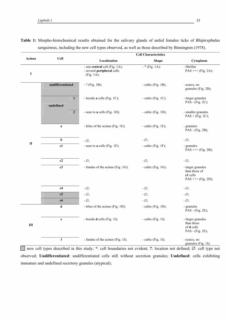
Table 1: Morpho-histochemical results obtained for the salivary glands of unfed females ticks of Rhipicephalus
sanguineus, including the new cell types observed, as well as those described by Binnington (1978). Cell Characteristics
Acinus Cell Localization Shape Cytoplasm
I
- one central cell (Fig. 1A); - several peripheral cells (Fig. 1A);
- * (Fig. 1A); - fibrillar PAS +++ (Fig. 2A);
undifferentiated - ? (Fig. 1B); - cubic (Fig. 1B); - scarce, no granules (Fig. 2B);
1 - beside a cells (Fig. 1C); - cubic (Fig. 1C); - larger granules PAS - (Fig. 2C);
undefined
2 - near to a cells (Fig. 1D); - cubic (Fig. 1D); - smaller granules PAS + (Fig. 2C);
a - hilus of the acinus (Fig. 1E); - cubic (Fig. 1E); - granules PAS - (Fig. 2B);
b - ∅; - ∅; - ∅;
c1 - near to a cells (Fig. 1F); - cubic (Fig. 1F); - granules PAS +++ (Fig. 2B);
c2 - ∅; - ∅; - ∅;
c3 - fundus of the acinus (Fig. 1G); - cubic (Fig. 1G); - larger granules than those of c1 cells PAS +++ (Fig. 2D);
c4 - ∅; - ∅; - ∅;
c5 - ∅; - ∅; - ∅;
II
c6 - ∅; - ∅; - ∅;
d - hilus of the acinus (Fig. 1H); - cubic (Fig. 1H); - granules PAS - (Fig. 2E);
e - beside d cells (Fig. 1I); - cubic (Fig. 1I); - larger granules than those of d cells PAS - (Fig. 2E);
III
f - fundus of the acinus (Fig. 1I); - cubic (Fig. 1I); - scarce, no granules (Fig. 1I);
new cell types described in this study; *: cell boundaries not evident; ?: location not defined; ∅: cell type not
observed; Undifferentiated: undifferentiated cells still without secretion granules; Undefined: cells exhibiting
immature and undefined secretory granules (atypical);

Capítulo 1
36
Table 2: Morpho-histochemical results obtained for the salivary glands of two-days fed females ticks of Rhipicephalus
sanguineus, including the new cell types observed, as well as those described by Binnington (1978). Cell Characteristics
Acinus Cell Localization Shape Cytoplasm
I - one central cell (Fig. 1J);
- several peripheral cells (Fig. 1J); - * (Fig. 1J); - fibrillar
PAS + (Fig. 2F);
undifferentiated - ∅; - ∅; - ∅;
1 - ∅; - ∅; - ∅; undefined 2 - ∅; - ∅; - ∅;
a - hilus of the acinus (Fig. 1K); - cubic (Fig. 1K); - granules PAS - (Fig. 2G);
b - beside a cells (Fig. 1L); - cubic (Fig. 1L); - elliptic and heterogeneous granules PAS+ and PAS ++ (Fig. 2H);
c1 - near a cells (Fig. 1M); - cubic (Fig. 1M); - granules PAS +++ (Fig. 2I);
c2 - near a cells (Fig. 1N); - cubic (Fig. 1N); - granules PAS + (Fig. 2J);
c3 - fundus of the acinus (Fig. 1K); - cubic (Fig. 1K); - larger granules than those of c1 cells PAS +++ (Fig. 2I);
c4 - fundus of the acinus (Fig. 1O); - cubic (Fig. 1O); - elliptic granules PAS - (Fig. 2K);
c5 - near a cells (Fig. 1O); - cubic (Fig. 1O); - smaller granules than those of a cells and larger than those of c3 cells PAS ++ (Fig. 2L);
II
c6 - near c5 cells (Fig. 2L); - cubic (Fig. 2L); - fine granules PAS ++ (Fig. 2L);
d - hilus of the acinus (Fig. 1P); - cubic (Fig. 1P); - granules PAS - (Fig. 2M);
e - beside d cells (Fig. 1P) ; - cubic (Fig. 1P); - larger granules than those of d cells PAS - (Fig. 2M);
III
f - fundus of the acinus (Fig. 1Q); - cubic (Fig. 1Q); - smaller granules than those of d cells PAS + (Fig. 2M);
new cell types described in this study; *: cell boundaries not evident; ∅: cell type not observed; Undifferentiated:
undifferentiated cells still without secretion granules; Undefined: cells exhibiting immature and undefined secretory
granules (atypical);
Capítulo 1
37
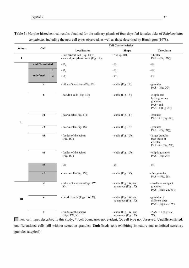
Table 3: Morpho-histochemical results obtained for the salivary glands of four-days fed females ticks of Rhipicephalus
sanguineus, including the new cell types observed, as well as those described by Binnington (1978). Cell Characteristics
Acinus Cell Localization Shape Cytoplasm
I - one central cell (Fig. 1R);
- several peripheral cells (Fig. 1R); - * (Fig. 1R); - fibrillar
PAS + (Fig. 2N);
undifferentiated - ∅; - ∅; - ∅;
1 - ∅; - ∅; - ∅;
undefined 2 - ∅; - ∅; - ∅;
a - hilus of the acinus (Fig. 1S); - cubic (Fig. 1S); - granules PAS - (Fig. 2O);
b - beside a cells (Fig. 1S); - cubic (Fig. 1S); - elliptic and heterogeneous granules PAS+ and PAS ++ (Fig. 2P);
c1 - near a cells (Fig. 1T); - cubic (Fig. 1T); - granules PAS +++ (Fig. 2O);
c2 - near a cells (Fig. 1S); - cubic (Fig. 1S); - granules PAS + (Fig. 2Q);
c3 - fundus of the acinus (Fig. 1U);
- cubic (Fig. 1U); - larger granules than those of c1 cells PAS +++ (Fig. 2R);
c4 - fundus of the acinus (Fig. 1U);
- cubic (Fig. 1U); - elliptic granules PAS - (Fig. 2O);
c5 - ∅; - ∅; - ∅;
II
c6 - near a cells (Fig. 1V); - cubic (Fig. 1V); - fine granules PAS + (Fig. 2S);
d - hilus of the acinus (Figs. 1W, X);
- cubic (Fig. 1W) and squamous (Fig. 1X);
- small and compact granules PAS - (Figs. 2T, W);
e - beside d cells (Figs. 1W, X); - cubic (Fig. 1W) and squamous (Fig. 1X);
- granules of different sizes PAS - (Figs. 2U, W);
III
f - fundus of the acinus (Figs. 1W, X);
- cubic (Fig. 1W) and squamous (Fig. 1X);
- PAS +++ (Fig. 2V, W);
new cell types described in this study; *: cell boundaries not evident; ∅: cell type not observed; Undifferentiated:
undifferentiated cells still without secretion granules; Undefined: cells exhibiting immature and undefined secretory
granules (atypical);

FIGURES

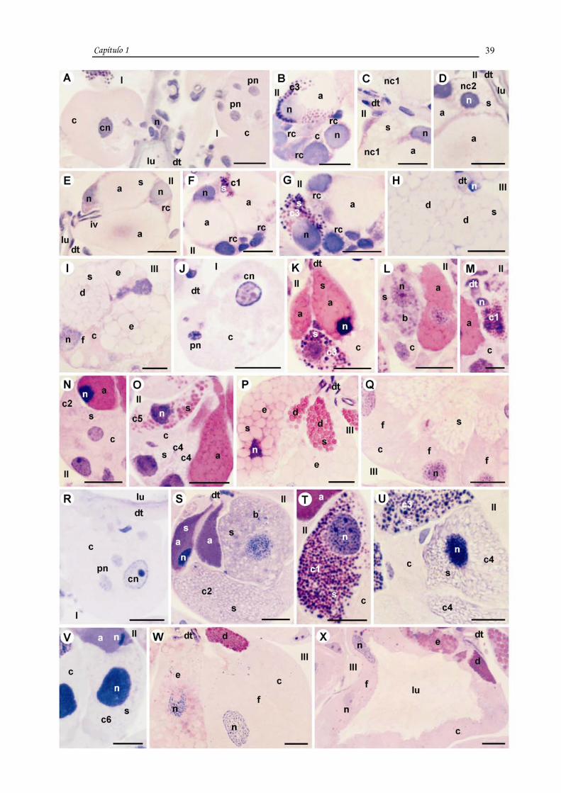
FIGURE 1:
Histological sections of the acini I, II and III of salivary glands of unfed, two and four-days fed
females of Rhipicephalus sanguineus, stained with Hematoxylin-Eosin.
A-I. Unfed females.
J-Q. Two-days fed females.
R-X. Four-days fed females.
I-III: types acini; rc: undifferentiated cell; nc1: undefined cell 1; nc2: undefined cell 2; a: a
cell; b: b cell; c1: c1 cell; c2: c2 cell; c3: c3 cell; c4: c4 cell; c5: c5 cell; c6: c6 cell; d: d cell;
e: e cell; f: f cell; c: cytoplasm; cn: central cell nucleus; pn: peripheral cell nucleus; n:
nucleus; dt: duct; lu: lumen; s: secretion; iv: intraacinar valve.
Bars: M= 10 µm; A-L and N-X= 20 µm.
Capítulo 1
39

FIGURE 2:
Histological sections of the acini I, II and III of salivary glands of unfed, two and four-days fed
females of Rhipicephalus sanguineus, stained with PAS and counterstained with Methyl Green.
A-E. Unfed females.
F-M. Two-days fed females.
N-W. Four-days fed females.
I-III: types acini; rc: undifferentiated cell; nc1: undefined cell 1; nc2: undefined cell 2; a: a
cell; b: b cell; c1: c1 cell; c2: c2 cell; c3: c3 cell; c4: c4 cell; c5: c5 cell; c6: c6 cell; d: d cell;
e: e cell; f: f cell; fc: fibrillar cytoplasm; pn: peripheral cell nucleus; n: nucleus; c: cytoplasm;
dt: duct; lu: lumen; s: secretion; cn: central cell nucleus;
Bars: A-W= 20 µm.

Capítulo 1
40

Capítulo 1
41
DISCUSSION
The present study on salivary glands of females of Rhipicephalus sanguineus
confirmed the information available in the literature for ixodid ticks, and described the
new cell types found in these organs.
According to Fawcett et al. (1986), the different salivary gland cells observed in
ticks are distinguished under light microscope in accordance to morphological
characteristics and metachromasia of secretion granules. We followed this
methodology, using the same morphological characteristics described for the glands of
Boophilus microplus (Binnington, 1978). The results revealed the presence of new cell
types in type II acini, termed: undefined 1 and 2 in unfed females; and c5 and c6 in two
and four-days fed females.
The size and morphology of type I acini were similar in unfed and two and four-
days fed females, as observed by Binnington (1978) and Walker et al. (1985).
According to these authors the morphology of type I acini remained the same
throughout the entire feeding process. Needham et al. (1983) and Barker et al. (1984),
however, reported an increase in the size of type I acini during early stages of tick
feeding, although during rapid feeding, acini reduced to the size of those of unfed ticks.
In type II acini of R. sanguineus, several changes were observed in the three
feeding stages assessed in our study. We identified in unfed females a, c1 and c3 cells
already described in the literature; “undifferentiated” cells probably programmed to
develop with the onset of feeding; as well as undefined cells 1 and 2 with unstained
cytoplasmic granules. These did not persisted in two and four-days fed females,
undergoing changes characterized by decrease in size and condensation of granule
content, suggesting a maturation process (Junqueira and Carneiro, 2004).
As feeding progressed, all cells of type II acini of R. sanguineus exhibited
intense activity of synthesis and secretion, and granules containing substances involved
in tick attachment, maintenance, and consumption of blood. This corroborates with the
observed by Walker et al. (1985) that suggested that salivary glands are not completely
active until ticks start to feed.
In two-days fed females, cells in type II acini were filled with secretion granules,
unlike the observed in unfed females. However, there was no difference in size between

Capítulo 1
42
acini of these groups. Also, we observed eight cell types (a, b, c1 to c6) in two-days fed
females instead of the six described in the literature. In four-days fed females, type II
acini were larger compared to those of two-days fed females, due to accumulation of
secretion granules in cells, indicating henceforth an increase in gland activity associated
with the consumption of blood by the parasite. In this group, c5 cells were not observed,
suggesting that they might no longer be functional and atrophied.
In unfed females of R. sanguineus, a cells in type II acini were already filled
with secretion granules, as also observed in two-days fed females, despite changes in
staining patterns which suggests that granules might be undergoing a maturation process
(Junqueira and Carneiro, 2004). In four-days fed females, these cells became even more
active, indicated by the increase in size compared to those of previous groups. This
finding partially confirms the observed by Binnington (1978) and Walker et al. (1985)
in B. microplus and R. appendiculatus, respectively. Both authors reported granules in a
cells of unfed females, however, Binnington (1978) observed that, unlike the obtained
in our study, these cells became non-functional 72 hours after attachment to the host.
This period of time coincides with the time necessary for the formation of the cement
cone. On the other hand, Walker et al. (1985) found that the same cells became less
active, although still functional as feeding progressed.
The secretion granules of a cells of R. sanguineus exhibited the same staining
pattern in all groups, although they were stained more intensely in two and four-days
fed females. The secretion was not stained by PAS, indicating the absence of
polysaccharides in its composition, but it was stained by Eosin, confirming the observed
by Binnington (1978), Walker et al. (1985), and Sonenshine (1991). The two latter
studies reported the presence of basic proteins in the composition of granules, although
according to Binnington (1978), granules contain lipoproteins.
Our results suggest that a cells of R. sanguineus play a role in the production of
cement precursors, as proposed by Binnington (1978), Walker et al. (1985), Fawcett et
al., (1986), Sonenshine (1991). However, R. sanguineus species, these cells remained
functional for a longer period than the described for B. microplus (Binnington, 1978).
This might indicate that the time necessary for the formation of the cement cone in R.
sanguineus is over 72 hours (Binnington, 1978).

Capítulo 1
43
In type II acinus, b cells were not active in unfed females; two-days fed females
exhibited cells with few granules, while cells were filled with granules in four-days fed
females, as observed by Walker et al. (1985) in R. appendiculatus. These authors have
shown that these cells are functional in early stages of tick feeding, decreasing their
activity in seven-days fed females. Binnington (1978), however, reported that in B.
microplus these cells were active in all feeding stages, and granules were already
present in unfed females. After 72 hours of feeding, most granules were eliminated.
In B. microplus as feeding progressed, the composition of the secretion of b cells
remained unchanged, maintaining the same staining patterns in two and four-days fed
females. Granules exhibited PAS stained and not stained regions, as well as portions of
acidic nature. Binnington (1978) and Walker et al. (1985) demonstrated that the
secretion of these cells consisted of glycoproteins, and suggested they play a role in the
manipulation of the host response. In R. sanguineus, although these cells were active
only after the onset of feeding (two days), they became important especially in four-
days fed females for the continuity of the feeding process and/or maintenance of the tick
on the host.
In this study, type c cells were classified in six subtypes, from c1 to c6; c5 and
c6 were described for the first time. Only c1 and c3 were active in unfed females. In
two-days fed females, all six cell types were active, and c1, c3, c5 and c6 contained the
largest amounts of secretion granules. In four-days fed females, c1, c2, c3, c4 and c6
cells remained active, but c1, c2 and c4 increased in size as a consequence of larger
amounts of granules. The aspect of the cytoplasm of c3 cells remained the same during
the secretory cycle, while c6 cells exhibited fewer granules, indicating a lower activity.
Cells c5 were no longer observed in four-days fed females, suggesting a loss of
functionality.
Cells type c were associated with the manipulation of host response (Walker et
al., 1985), and some of them secrete glycoproteins acting as anticoagulants (Sonenshine,
1991).
The results for c1 cells partially confirm those obtained by Binnington (1978). It
was observed the presence of secretion granules in unfed females, however the period
of cell activity differs from the obtained by Binnington (1978), which suggested that
these cells remain functional for approximately 72 hours of feeding. According to

Capítulo 1
44
Walker et al. (1985), these cells are inactive in unfed females, and as observed in the
present study, develop during feeding.
Cells c1 of salivary glands of R. sanguineus played a major role in all phases of
the secretory cycle, as they were already active before the first contact between parasite
and host (unfed females), and remained active as feeding progressed. It is worth
pointing out that these cells were most active in four-days fed females, when blood
consumption increased.
Our results on c2 cells confirm those obtained by Walker et al. (1985). These
authors observed that these cells were active in early stages of tick feeding. As feeding
progressed, cells exhibited a moderate increase in size and amount of granules.
Binnington (1978) observed few granules in c2 cells of unfed females, and an increase
in the number of granules after 12-24 hours of feeding. These cells remained functional
during the 24-72 hour period after the attachment of the tick to the host. In R.
sanguineus, these cells were inactive in unfed females and filled with granules in four-
days fed females. Binnington (1978) proposed that c2 cells play a role in blood
consumption. This might also occur in R. sanguineus females, as these cells developed
in parallel with the increase in blood consumption (four days).
Regarding c3 cells, we observed the presence of secretion granules in unfed
females confirming the obtained by Binnington (1978). However, according to this
author, these cells remained active for 72 hours after attachment, which was not
observed in our study. Binnington (1978) demonstrated that c3 cells play a general role
in the feeding process. This could also be the case in R. sanguineus, as these cells were
functional in all phases assessed and exhibited granules with contents similar to those
observed in B. microplus.
Cells c4 exhibited low activity in unfed females, as opposed to the observed in
four-days fed females. This result differs from the obtained by Binnington (1978) that
reported secretion granules in cells of unfed females, which were released after 72 hours
of feeding. This author also suggested that in B. microplus, c4 cells might play a role in
enzyme secretion. Considering this information and the histochemical similarities, this
could occur in c4 cells of R. sanguineus.
The composition of secretion granules of c cells in the stages examined in R.
sanguineus remained the same. Cells c1-c3 contained acids and polysaccharides,
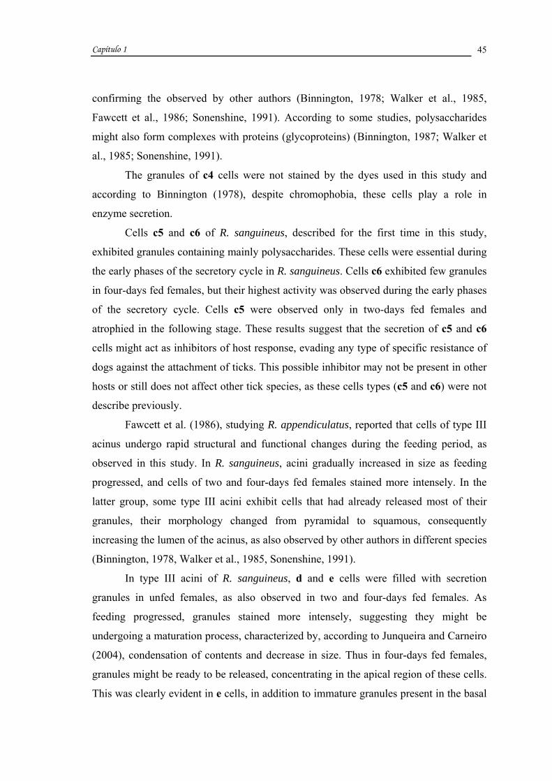
Capítulo 1
45
confirming the observed by other authors (Binnington, 1978; Walker et al., 1985,
Fawcett et al., 1986; Sonenshine, 1991). According to some studies, polysaccharides
might also form complexes with proteins (glycoproteins) (Binnington, 1987; Walker et
al., 1985; Sonenshine, 1991).
The granules of c4 cells were not stained by the dyes used in this study and
according to Binnington (1978), despite chromophobia, these cells play a role in
enzyme secretion.
Cells c5 and c6 of R. sanguineus, described for the first time in this study,
exhibited granules containing mainly polysaccharides. These cells were essential during
the early phases of the secretory cycle in R. sanguineus. Cells c6 exhibited few granules
in four-days fed females, but their highest activity was observed during the early phases
of the secretory cycle. Cells c5 were observed only in two-days fed females and
atrophied in the following stage. These results suggest that the secretion of c5 and c6
cells might act as inhibitors of host response, evading any type of specific resistance of
dogs against the attachment of ticks. This possible inhibitor may not be present in other
hosts or still does not affect other tick species, as these cells types (c5 and c6) were not
describe previously.
Fawcett et al. (1986), studying R. appendiculatus, reported that cells of type III
acinus undergo rapid structural and functional changes during the feeding period, as
observed in this study. In R. sanguineus, acini gradually increased in size as feeding
progressed, and cells of two and four-days fed females stained more intensely. In the
latter group, some type III acini exhibit cells that had already released most of their
granules, their morphology changed from pyramidal to squamous, consequently
increasing the lumen of the acinus, as also observed by other authors in different species
(Binnington, 1978, Walker et al., 1985, Sonenshine, 1991).
In type III acini of R. sanguineus, d and e cells were filled with secretion
granules in unfed females, as also observed in two and four-days fed females. As
feeding progressed, granules stained more intensely, suggesting they might be
undergoing a maturation process, characterized by, according to Junqueira and Carneiro
(2004), condensation of contents and decrease in size. Thus in four-days fed females,
granules might be ready to be released, concentrating in the apical region of these cells.
This was clearly evident in e cells, in addition to immature granules present in the basal

Capítulo 1
46
region. These findings confirm the observed by Binnington (1978), Walker et al. (1985)
and Fawcett et al. (1986), regarding the presence of granules already in unfed females.
Our results, however, differ from those of Binnington (1978), Fawcett et al. (1986) and
Sonenshine (1991) regarding the period when granules were present in the cells.
According to Binnington (1978), these cells release most of their granules after 72 hours
of feeding, while the other authors report that granules are released for the formation of
the cement cone after tick attachment, and cells atrophy as feeding progresses.
Cells d and e in ticks are thought to secrete cement precursors (Binnington,
1978; Fawcett et al., 1986; Walker et al., 1985; Sonenshine, 1991; Bishop et al., 2002).
According to Walker et al. (1985), these cells secrete aminopeptidase, which
participates in the infiltration of cement in the host’s skin; while Binnington (1978)
suggests that phenol and phenol oxidase might play a role in the process of cement
hardening. In R. sanguineus, these cells might play the same roles, as the presence of
granules before attachment to the host and the composition of granules support the
observed by Binnington (1978), Walker et al. (1985), Fawcett et al. (1986) and
Sonenshine (1991), despite the longer period of cell activity observed in this study.
In type III acini of R. sanguineus, f cells did not exhibit granules in unfed
females, unlike the observed in two-days fed females, which presented granules mainly
in the apical region. In four-days fed females, all granules observed in the previous
stage were released, pyramidal cells became squamous, and the cytoplasm was strongly
stained by PAS, as also observed by other authors. According to Binnington (1978) and
Coons and L’amoreaux (1986), cells became active and filled with granules in a period
of 12-24 hours following attachment to the host. After approximately 72 hours of
attachment, granules were released, and cells lost their secretory function. Fawcett et al.
(1986) and Sonenshine (1991) also report that granules are first observed in f cells of
other species only after ticks start feeding, and cells actively secrete their products
during a period of two days. After this period, organelles undergo autophagy,
consequently exhibiting changes in cell structure. These cells then loose their secretory
function and transport fluids from the hemolymph to the saliva (Binnington, 1978;
Coons and L’amoreaux, 1986; Fawcett et al., 1986; Sonenshine, 1991).
In this study, f cells exhibited granules containing polysaccharides, as observed
by Binnington (1978) in B. microplus females, and according to this author, the role of
Capítulo 1
47
these cells would be associated with the consumption of blood by the tick, as high
activity was observed in the glands of adult females of B. microplus. Thus, considering
the period of activity (early stages of feeding) of these cells in some species
(Binnington, 1978; Fawcett et al., 1986; Sonenshine, 1991), as well as in R. sanguineus,
the participation of f cells was clearly evident in early stages of blood consumption, as
they were no longer active in four-days fed females. According to Walker et al. (1985),
this is a transitory role played by these cells in early stages of feeding; following this
period, they begin to absorb fluids from the hemolymph and eliminate them through the
saliva (Kaufman and Sauer, 1982; Walker et al., 1985, Coons and L’amoreaux, 1986;
Sonenshine, 1991).
ACKNOWLEDGMENTS
This research has been supported by CAPES (Coordenação de Aperfeiçoamento
de Pessoal de Nível Superior) and FAPESP (Fundação de Amparo à Pesquisa do Estado
de São Paulo) Grant nº 05/59208-3. The authors thank Gerson Mello Souza and Ronaldo
Del Vecchio for the technical support. Part of this work has been facilitated through the
Integrated Consortium on Ticks and Tick-borne Diseases (ICTTD-3) supported by the
an European Union under contract number 510561-INCO.

Capítulo 1
48
REFERENCES
Balashov, Y.S., 1983. Salivary glands. In: Raikhel, A.S., Hoogstraal, I.I. (Eds.), An
atlas of ixodid tick ultrastructure. Entomological Society of America, Lanham
Barker, D.M., Ownby, C.L., Krolak, J.M., Claypool, P.L., Sauer, J.R., 1984. The effects
of attachment, feeding and mating on the morphology of the I alveolus of salivary
glands of the lone star tick, Amblyomma americanum. J. Parasitol. 70, 99-113
Bechara, G.H., Szabó, M.P.J., Ferreira, B.R., Garcia, M.V., 1995. Rhipicephalus
sanguineus tick in Brazil: feeding and reproductive aspects under laboratorial
conditions. Brazil. J. Vet. Parasitol. 4, 61-66
Binnington, K.C., 1978. Sequential changes in salivary gland structure during
attachment and feeding of the cattle tick, Boophilus microplus. Int. J.Parasitol. 8, 97-
115
Bishop, R., Lambson, B., Wells, C., Pandit, P., Osaso, J., Nkonge, C., Morzaria, S.,
Musoke, A., Nene, V., 2002. A cement protein of the tick Rhipicephalus
appendiculatus, located in the secretory e cell granules of the type III salivary gland
acini, induces strong antibody responses in cattle. Int. J. Parasitol. 32, 833-842
Coons, L.B., L’amoreaux, W.J., 1986. Developmental changes in the salivary glands of
male and female Dermacentor variabilis (Say) during feeding. In: Borovsky, D.,
Spielman, A. (Eds.), Host regulated developmental mechanism in vector arthropods.
University of Florida-IFAS, Vero Beach
Fawcett, D.W., Binnington, K., Voigt, W.P., 1986. The cell biology of the ixodid tick
salivary gland. In: Sauer, J.R., Hair, A. (Eds.), Morphology, physiology, and behavioral
biology of ticks. Ellis Horwood, Chichester

Capítulo 1
49
Gill, H.S., Walker, A.R., 1987. The salivary glands of Hyalomma anatolicum
anatolicum: structural changes during attachment and feeding. Int. J. Parasitol. 17 (8),
1381-1392
Junqueira, L.C., Carneiro, J., 2004. Histologia básica- Texto/Atlas, 10 ed. Guanabara
Koogan, Rio de Janeiro
Kaufman, W.R., Sauer, J.R., 1982. Ion and water balance in feeding ticks: mechanism
of tick excretion. In: Obenchain, F.D., Galun, R. (Eds.), Physiology of ticks. Pergamon
Press, Oxford
Marzouk, A.S., Darwish, Z.E.A., 1994. Changes in the salivary glands of female
Hyalomma (Hyalomma) dromedarii during and after feeding. J. Egypt. Soc. Parasitol.
24 (1), 39-57
McManus, J.F.A., 1946. Histological demonstration of mucin after periodic acid. Nature
158, 202
McMullen, H.L., Sauer, J.R., Burton, R.L., 1976. Possible role in uptake of water
vapour by ixodid ticks salivary glands. J. Insect Physiol. 22, 1281-1285
Needham, G.R., Greenwald, L., Freda, T.J., Coons, L.B., 1983. The ultrastructure of the
tick (Amblyomma americanum) salivary gland acini responsible for secretion of the
higroscopic fluid and localization of Na+/K+ -ATPase. Am. Zool. 23, 1013
Olivieri, J.A., Serra-Freire, N.M., 1992. Structure of the salivary glands of the unfed
female tick Amblyomma cajennense (Fabricius) (Acarina: Ixodidae). Mem. Inst.
Oswaldo Cruz 87, suppl. I, 167-174
Ribeiro, A.L., Faccini, J.L.H., Daemon, E., 1996. Estudo das variações morfológicas de
Rhipicephalus sanguineus (Latreille, 1806) (Acari: Ixodidae) no Brasil. Rev. Univ.
Rural, Ser. Cienc. Vida 18 (1-2), 25-33

Capítulo 1
50
Rudolph, D., Knülle, W., 1974. Site and mechanism of water vapour uptake from the
atmosphere in ixodid ticks. Nature 149, 84-85
Rudolph, D., Knülle, W., 1978. Uptake of water vapour from air: process, site, and
mechanism in ticks. In: Schmidt-Mielsen, K., Bolio, L., Maddrell, S.H.P. (Eds.),
Comparative physiology: Water, ions and fluid mechanics. Cambridge University Press,
Cambridge
Sonenshine, D.E., 1991. Biology of ticks. Oxford University Press, New York
Till, W.M., 1961. A contribution to the anatomy and histology of the brown ear tick
Rhipicephalus appendiculatus Neumann. Memoirs of the Entomological Society of
Southern Africa, Pretoria 6, 1-124
Walker, A., 1994. Arthropods of humans and domestic animals. A guide to preliminary
identification. Chapman & Hall, London
Walker, A.R., Fletcher, J.D., Gill, H., 1985. Structural and histochemical changes in the
salivary glands of Rhipicephalus appendiculatus during feeding. Int. J. Parasitol. 15 (1),
81-100
Wikel, S.K., 1981. The induction of host resistance to tick infestation with a salivary
gland antigen. Am. J. Trop. Med. Hyg. 30, 284-288

Capítulo 2
Capítulo 2
52
CAPÍTULO 2
TITLE: Salivary glands of females of the tick Rhipicephalus sanguineus
(LATREILLE, 1806) (Acari, Ixodidae). Degenerative morphological changes detected
at the end and after the feeding period.
AUTHORS: Karim Christina Scopinho Furquim a, Gervásio Henrique Bechara b and
Maria Izabel Camargo Mathias a, *
a Departamento de Biologia, Instituto de Biociências, UNESP, Av. 24 A, nº 1515, Cx.
Postal 199, CEP: 13506-900, Rio Claro, S.P., Brazil b Departamento de Patologia Veterinária, FCAV, UNESP, Via de Acesso Prof. Paulo
Castellane, s/n, CEP: 14884-900, Jaboticabal, S.P., Brazil
* Corresponding author. Fax: +55 19 35340009.
E-mail address: [email protected]

Capítulo 2
53
RESUMO
Fêmeas de carrapatos Rhipicephalus sanguineus ingurgitadas e com três e sete
dias pós-ingurgitamento tiveram suas glândulas salivares analisadas morfo-
histoquimicamente. Nas ingurgitadas detectou-se diminuição na capacidade secretora,
marcada pelo surgimento do processo degenerativo, sendo observados nas glândulas
tanto ácinos íntegros (I) como em degeneração (II e III). Naquelas com três dias pós-
ingurgitamento o processo degenerativo se intensificou, revelando a presença tanto de
ácinos em degeneração quanto em processo de fragmentação e formação de corpos
apoptóticos. Nas fêmeas com sete dias pós-ingurgitamento a glândula salivar havia
degenerado por completo.
Sendo assim, dos oito tipos de células (a, b, c1, c2, c3, c4, c5 e c6) descritos nos
ácinos II de fêmeas de R. sanguineus em alimentação (dados não publicados), somente
as a, c1 e c3 foram observadas nas fêmeas ingurgitadas e nos III todos os tipos (d, e e f).
No estágio de três dias pós-ingurgitamento, nos ácinos I as células central e periféricas
estavam presentes, nos II somente as a e c3 e os ácinos III não foram mais observados.
Durante o processo degenerativo das glândulas salivares das fêmeas ingurgitadas
e com três dias pós-ingurgitamento foram observadas nas células alterações
citoplasmáticas, algumas delas caracterizadas pela diminuição dos grânulos de secreção,
devido a sua eliminação, outras pela alteração dos mesmos, dando origem a uma massa
amorfa, pela retração e vacuolização do citoplasma, além da perda da forma e dos
limites celulares. Além das alterações citoplasmáticas foram observadas severas
alterações nucleares, como polimorfismo em células e dos núcleos de um mesmo ácino,
bem como na disposição e grau de condensação da cromatina.
PALAVRAS-CHAVE: Rhipicephalus sanguineus; carrapatos; fêmea; glândula salivar;
atividade celular; apoptose; morfologia.
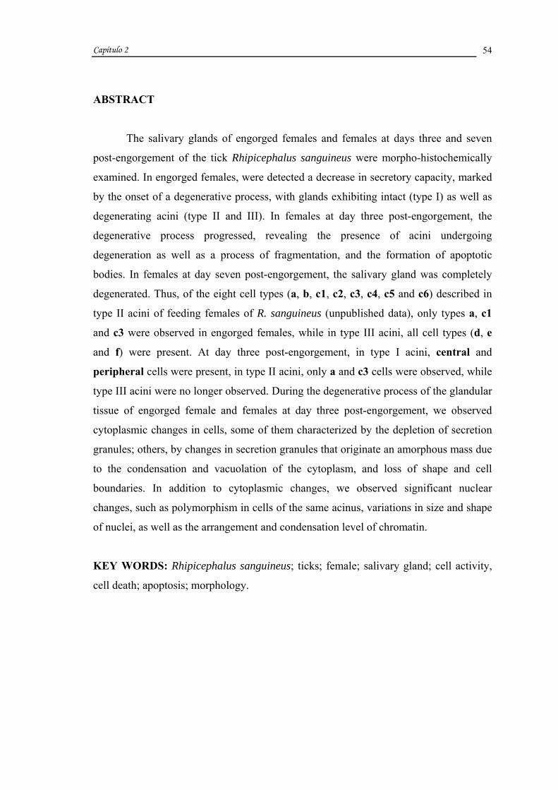
Capítulo 2
54
ABSTRACT
The salivary glands of engorged females and females at days three and seven
post-engorgement of the tick Rhipicephalus sanguineus were morpho-histochemically
examined. In engorged females, were detected a decrease in secretory capacity, marked
by the onset of a degenerative process, with glands exhibiting intact (type I) as well as
degenerating acini (type II and III). In females at day three post-engorgement, the
degenerative process progressed, revealing the presence of acini undergoing
degeneration as well as a process of fragmentation, and the formation of apoptotic
bodies. In females at day seven post-engorgement, the salivary gland was completely
degenerated. Thus, of the eight cell types (a, b, c1, c2, c3, c4, c5 and c6) described in
type II acini of feeding females of R. sanguineus (unpublished data), only types a, c1
and c3 were observed in engorged females, while in type III acini, all cell types (d, e
and f) were present. At day three post-engorgement, in type I acini, central and
peripheral cells were present, in type II acini, only a and c3 cells were observed, while
type III acini were no longer observed. During the degenerative process of the glandular
tissue of engorged female and females at day three post-engorgement, we observed
cytoplasmic changes in cells, some of them characterized by the depletion of secretion
granules; others, by changes in secretion granules that originate an amorphous mass due
to the condensation and vacuolation of the cytoplasm, and loss of shape and cell
boundaries. In addition to cytoplasmic changes, we observed significant nuclear
changes, such as polymorphism in cells of the same acinus, variations in size and shape
of nuclei, as well as the arrangement and condensation level of chromatin.
KEY WORDS: Rhipicephalus sanguineus; ticks; female; salivary gland; cell activity,
cell death; apoptosis; morphology.

Capítulo 2
55
INTRODUCTION
In general, ticks prefer to feed on mammals, for being endothermic (Oliveira et
al., 2005). In the case of the tick Rhiphicephalus sanguineus, dogs are the most common
host (Rey, 1973; Walker, 1994), although it can also be found in cats, rabbits, buffalos,
camels, cattle, goats, horses, sheep, bats, birds, reptiles (Flechtmann, 1973 apud
Oliveira et al., 2005) and humans (Rey, 1973).
Ticks are members of one of the most important groups among arthropods, due
to the role played by their salivary glands. These organs are responsible for the
transmission of infectious agents, making these parasites subjects of great veterinary-
medical importance (Balashov, 1983). Salivary glands also allow the feeding of the tick,
leading to blood loss for the host. In cattle, both problems result in economic losses,
especially in the milk and meat industry.
According to the literature, the activity of glands of feeding ticks can be
summarized in three stages: 1) preparatory phase, a 24-35 hour period in which females
insert their mouthparts, form a feeding lesion, and secrete cement to attach themselves
to the host, 2) slow-feeding phase, a 7-10 day period during which the female gradually
increases 10 times its unfed weight, 3) rapid-feeding phase, a period of approximately
24 hours, during which the female rapidly increases in weight (Balashov, 1972 apud
Weiss e Kaufman, 2001). The first two stages and the early phase of the third stage are
characterized by an intense gland activity (synthesis and secretion of components),
while during the final phase of the third stage, the depletion of secretion granules,
inactivity, and the early stages of cell degeneration are observed (Marzouk and
Darwish, 1994).
The degeneration of salivary glands is hormonally controlled by a steroid
(Lomas et al., 1998). The synthesis and release of this hormone begins during early
feeding stages and increases until the post-engorgement period (Lomas, 1993, PhD
thesis).
During feeding, as soon as females reach their “critical weight”, there is an
increase in ecdysteroid synthesis (Weiss and Kaufman, 2001). This increase signals the
degeneration of salivary glands (Weiss and Kaufman, 2001), which will be initiated
only after engorgement (Kaufman and Lomas, 1996). Another factor probably

Capítulo 2
56
stimulating this increase in ecdysteroids in the hemolymph (Lomas e Kaufman, 1992),
as well as the rapid engorgement of females, is the male/engorgement factor (Weiss and
Kaufman, 2004), which also accelerates gland degeneration (Lomas and Kaufman,
1992; Weiss e Kaufman, 2004).
According to Nunes et al. (2005), the degenerative process of salivary glands of
semi-fed females of R. (Boophilus) microplus is a result of a series of morpho-
histochemical changes in gland cells, in which secretory cells initially loose all or
almost all secretion granules, followed by severe changes in the cytoplasm and nucleus,
and form apoptotic bodies.
Once the process of salivary gland degeneration begins, the complete
degeneration of this organ will be reached only after oviposition, remaining only the
duct system and a mass of connective tissue (Till, 1961). This residue of salivary gland
is the final result of cells comprising the gland tissue, due to an event that causes a
series of morphological and histochemical changes in gland cells.
Based on these informations and considering that little is known about the
morphology and histochemistry of the salivary gland degeneration in ticks, this study
aimed at describing the changes undergone by salivary glands at the end of the feeding
period (fully engorged females), as well as three and seven days after feeding (females
at day three and seven post-engorgement).
MATERIAL AND METHODS
To conduct this study, we utilized engorged females and females at day three
and seven post-engorgement of R. sanguineus. Unfed individuals were provided by Dr.
Gervásio Henrique Bechara of the Department of Veterinary Pathology of UNESP,
Jaboticabal campus (São Paulo), from a colony maintained under controlled conditions
(29o C, 80% humidity, and 12 hour photoperiod) in BOD incubator. These individuals
were placed with some males, in a feeding chamber previously glue with an atoxic and
non-lesive preparation (Britannia Adhesives-Unit 4, UK) to the shaved back of the host
(rabbit) according to technique described elsewhere (Bechara et al., 1995) for complete
feeding. A group of engorged females was utilized for histological procedures while
another group was maintained alive in closed containers with breathing holes for three

Capítulo 2
57
and seven days to be later analyzed. After the completion of the periods of time
examined in this study, salivary glands were removed in saline solution and fixed in
10% buffered neutral formalin and acetone (9:1), during one hour and thirty minutes, at
4o C. Following fixation, the material was dehydrated in increasing concentrations of
ethanol (70%, 80%, 90% and 95%), embedded and included in Leica resin, and
sectioned at a thickness of 3 µm. Sections were placed in glass slides, stained with
Hematoxilyn and Eosin, and PAS (Periodic Acid Schiff), McManus (1946), to
demonstrate polysaccharides, and counterstained with Methyl Green for nucleus
staining, and mounted in Canada balsam for observation under light microscope.
RESULTS
The identification of the different cell types in acini of salivary glands of R.
sanguineus is based on the system described and adopted by Binnington (1978). For a
visual comparison, results are summarized in tables 1, 2 and 3.
In the salivary glands of engorged females, of the three types of acini present in
the gland tissue (I, II, and III) only type I acini are intact, exhibiting a regular shape
(Fig. 1A). The central and peripheral cells present a fibrillar PAS positive cytoplasm
(Fig. 3A) and their nuclei do not exhibit changes (Figs. 1A, 3A).
The results obtained for degenerating type II and III acini are presented in Table
1. Some acini could not be identified, as they have lost their original granular
characteristics, they where termed here as Indeterminate 1 (Fig. 1R). They present an
irregular shape and lack cell boundaries (Fig. 1R). Thus, their contents are characterized
by a vacuolated cytoplasmic mass with secretion residues (Fig. 1R). This cytoplamic
mass surrounds some irregular nuclei (Fig. 1R).
In females at day three post-engorgement, the entire acinar structure is
undergoing degeneration and details are described in Tables 2 and 3.
In females at day seven post-engorgement, the salivary glands are not longer
observed, as the gland tissue is completely degenerated.
All changes observed in acini and gland cells during and after feeding are
described in Figure 4.

Capítulo 2
58
Table 1: Morpho-histochemical results of acini observed undergoing degeneration in salivary glands of
engorged females of Rhipicephalus sanguineus. Shape Acini Cell
Acinus Cells Cytoplasm Nucleus
a - regular (Figs. 1B-D); - cubic (Figs. 1B-D); - granules PAS – (Fig. 3B);
- irregular (Fig. 1B); - ∆ (Fig. 3B);
b - ∅; - ∅; - ∅; - ∅;
c1 - regular (Figs. 1B, C); - irregular (Figs. 1B, C); - granules PAS +++ (Fig. 3C);
- !! (Fig. 1C);
c2 - ∅; - ∅; - ∅; - ∅;
c3 - regular (Figs. 1D, E); - irregular (Figs. 1D, E); - larger granules than those of c1 PAS +++ (Figs. 3B, C);
- ∆ (Fig. 3B); - !! (Fig. 1D);
c4 - ∅; - ∅; - ∅; - ∅; c5 - ∅; - ∅; - ∅; - ∅; c6 - ∅; - ∅; - ∅; - ∅;
1 - regular (Figs. 1E, F); - irregular (Figs. 1G, H);
- irregular (Figs. 1E-H); - condensed (Figs. 1E-H);
- ✤ (Fig. 1E);
- ★ (Fig. 1F);
- ★★ (Fig. 1F); - ∆ (Figs. 1G, H); - !! (Figs. 1F, H);
II
indeterminate
2 - regular (Figs. 1F, I); - irregular (Fig. 1J);
- irregular (Figs. 1F, I, J);
- condensed (Figs. 1F I, J);
- ✤✤ and irregular (Figs. 1F, I, J); - ∞ (Fig. 1J);
d - regular (Fig. 1N); - irregular (Figs. 1K-M, P);
- cubic (Figs. 1K-N, P); - granules PAS – (Fig. 3D);
- irregular (Figs. 1K-M, P, 3D);
e - irregular (Figs. 1K-M, O);
- cubic (Figs. 1K, O); - irregular (Figs. 1K- M);
- ! (Figs. 1K-M, O) PAS – (Figs. 3D, E);
- irregular (Fig. 1K); - irregular and ✤ (Fig. 1M); - ∞ (Figs. 1L, 3D);
f - irregular (Fig. 1M); - irregular (Fig. 1M); - condensed (Fig. 1M) and fine granulation PAS ++ (Fig. 3E);
- irregular (Fig. 3E); - ∞ (Fig. 1M);
III
- regular (Fig. 1N); - irregular (Figs. 1O-Q);
- # (Figs. 1N-Q); - ? (Figs. 1N, O); - * (Fig. 1P); - * with residues of granules of d or e (Fig. 1Q);
- ✤ and irregular (Figs. 1N, O); - ∞ (Figs. 1O, Q); - !! (Fig. 1P);
new cell types described in feeding females of R. sanguineus (unpublished data); indeterminate: cells that lost their original granular
characteristics and therefore could not be identified; ∅: cell types not observed; #: loss of cell boundaries; !: granule disorganization; ?:
heterogeneous cytoplasmic mass; *: cytoplasmic mass; ✤ : picnotic; ★ : beginning of chromatin margination; ★★ : chromatin margination; ∆:
blebs; ∞: undergoing fragmentation; !!: fragmented; ✤ ✤ : dilated.

Capítulo 2
59
Table 2: Morpho-histochemical results of acini I and II observed undergoing degeneration in salivary glands
of Rhipicephalus sanguineus females at day three post-engorgement. Shape Acini Cell
Acinus Cells Cytoplasm Nucleus
I Central and Peripheral
- regular (Fig. 2A); ** (Fig. 2A); - ?? PAS +++ (Fig. 3F);
- ✤✤ and irregular (Figs. 2A, 3F);
a - irregular (Fig. 2B); - irregular (Fig. 2B); - granules PAS - (Figs. 3G-J);
- irregular (Figs. 3G, H) - !! (Figs. 2B, 3G);
b - ∅; - ∅; - ∅; - ∅;
c1 - ∅; - ∅; - ∅; - ∅;
c2 - ∅; - ∅; - ∅; - ∅;
c3 - irregular (Fig. 2C); - irregular (Fig. 2C); - granules PAS +++ (Fig. 3H);
- irregular (Fig. 2C); - !! (Fig. 3H);
c4 - ∅; - ∅; - ∅; - ∅;
c5 - ∅; - ∅; - ∅; - ∅;
c6 - ∅; - ∅; - ∅; - ∅;
1 - ∅; - ∅; - ∅; - ∅; indeterminate 2 - ∅; - ∅; - ∅; - ∅;
II
- irregular (Figs. 2B, C); - # (Figs. 2B, C); - ?? PAS +++ (Figs. 3G-J);
- irregular (Figs. 2B, C, 3G, H); - ✤✤ and irregular (Figs. 2B, 3I); - ✤ (Fig. 3G);
- ★★ (Fig. 3J); - !! (Fig. 2C);
new cell types described in feeding females of R. sanguineus (unpublished data); indeterminate: cells that lost their original granular
characteristics and therefore could not be identified; **: cell boundaries not evident; ∅: cell types not observed; #: loss of cell boundaries; ??:
granular cytoplasmic mass; ✤✤ : dilated; !!: fragmented; ✤ : picnotic; ★★ :chromatin margination.

Capítulo 2
60
Table 3: Morpho-histochemical results of acini in advanced stages of salivary gland degeneration of
Rhipicephalus sanguineus females at day three post-engorgement.
Degeneration Stages Cell Shape Cytoplasm Nucleus
1
(Indeterminate Acini
With Irregular Shape)
- # (Figs. 2D-I); - ∇ (Figs. 2D-I, 3K-M) and ?? PAS +++ (Figs. 3K-M); - secretion residues PAS - (Figs. 3K-M);
- ✤ (Fig. 2D);
- ★★ (Figs. 2E, F); - !! (Figs. 2G, 3K); - irregular (Figs. 2D-G, I); - ✤✤ and irregular (Figs. 2H, 3K); - ∞ (Figs. 2H, 3L);
A - # (Fig. 2J); - ∇ (Figs. 2J, 3N) and ?? PAS +++ (Fig. 3N); - secretion residues (Figs. 2J, 3N);
- !! (Figs. 2J, 3N); - irregular (Fig. 3N);
2
(Indeterminate Acini In Early Stages of Fragmentation) B - # (Fig. 2K); - residues of cytoplasm
(Fig. 2K);
- not observed;
3
(Indeterminate Acini
Undergoing Fragmentation)
- # (Figs. 2L, M); - ∇ (Figs. 2L, M, 3O) and ?? PAS +++ (Fig. 3O); - secretion residues (Fig. 2L);
- irregular (Figs. 2L, 3O); - ★★ (Fig. 2M); - !! (Fig. 3O);
4
(Apoptotic Bodies Formed From The
Fragmentation Of Acini)
- cell fragments (Figs. 2N, O);
- residues of cytoplasm with or without granules (Figs. 2N, O, 3P);
- !! (Figs. 2N, O, 3P);
#: loss of cell boundaries; ∇: vacuolated cytoplasmic mass; ??:granular cytoplasmic mass; ✤✤ : dilated; !!: fragmented; ✤ : picnotic;
★★ :chromatin margination; ∞: undergoing fragmentation.

FIGURES

FIGURE 1:
Histological sections of salivary glands of engorged females of Rhipicephalus sanguineus
stained with Hematoxylin and Eosin. A. Intact type I acini (I). B-D. Type II acini (II) with
characteristics of degeneration, exhibiting in: B, a cell (a) filled with secretion granules (s) and
irregular nucleus (in); C, irregular c1 cells (ic1) with secretion granules (s) and nuclear
fragments (nf) and D, irregular c3 cell (ic3) with secretion granules (s) and nuclear fragments
(nf). E-J. Type II acini (II) with characteristics of more advanced stages of degeneration and a
(a) and c3 (c3) cells filled with secretion (s), and without secretion and irregular shape or
indeterminate 1 (iin1) and indeterminate 2 (iin2). E-H. Irregular indeterminate 1 cells (iin1)
with shrinkage of the cytoplasm (sc) and gradual nuclear changes, characterized by: E, picnotic
nucleus (pn); F, onset of chromatin marginalization (arrow) and total chromatin
marginalization (double arrow); G and H, blebs (bb) and F and H, fragments of nuclei (fn); F,
I and J. Irregular indeterminate 2 cells (iin2) with shrinkage of the cytoplasm (sc) and gradual
nuclear changes, characterized by: F, I and J, dilated and irregular nuclei (din); J, nucleus
undergoing fragmentation (triple arrow). K-M. Type III acini (III) with characteristics of
degeneration, exhibiting d cells (d) filled with secretion granules (s) and irregular nuclei (in)
and irregular e cells (ie) with secretion resembling an amorphous mass ( ) and gradual nuclear
changes, such as: K, irregular nuclei (in); L, nucleus undergoing fragmentation (triple arrow)
and M, irregular and picnotic nucleus (arrow head); M. Irregular f cells (if) with shrinkage of
the cytoplasm (sc) and nuclei undergoing fragmentation (triple arrow). N-Q. Type III acini
(III) in more advanced stages of degeneration. N and O. Cells filled with secretion surrounded
by a heterogeneous cytoplasmic mass (hm) with nuclei exhibiting various morphological
features, characterized by: N and O, picnotic and irregular nuclei (arrow head) and O, nucleus
undergoing fragmentation (triple arrow). P. d cells (d) filled with secretion granules (s),
irregular nucleus (in), and surrounded by a cytoplasmic mass (cm) with nuclear fragments (nf).
Q. Acinus composed of only cytoplasmic mass (cm) and nuclei undergoing fragmentation
(triple arrow). R. Indeterminate acinus at stage 1 of degeneration (Ind1) consisted of
vacuolated cytoplasmic mass ( ) and irregular nucleus (in).
c: cytoplasm; nc: nucleus of central cell; np: nucleus of peripheral cell, dt: duct; lu: lumen;
iv: intraacinar valve; iac: irregular-shaped acinus; d: d cell; e: e cell; rs: residue of secretion;
va: vacuole.
Bars: A-R= 20 µm.
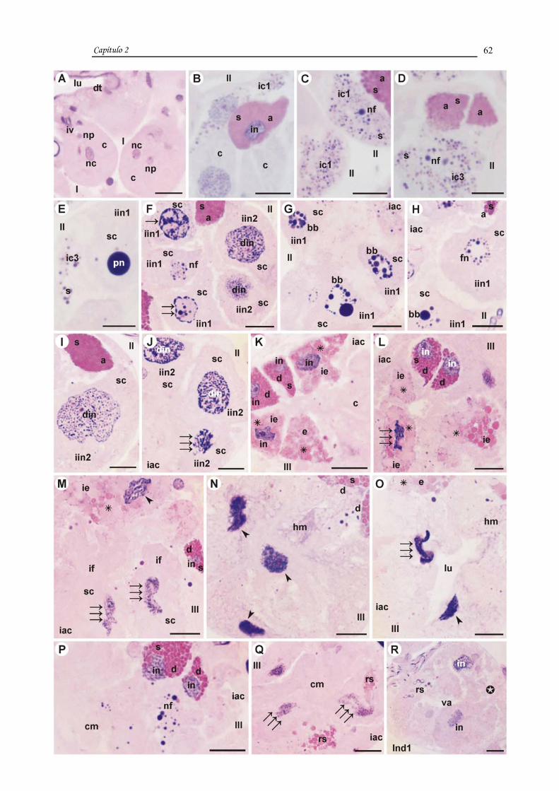
Capítulo 2
62

FIGURE 2:
Histological sections of salivary glands of Rhipicephalus sanguineus females at day three post-
engorgement stained with Hematoxylin and Eosin. A. Type I acini (I) with characteristics of
degeneration, containing dilated and irregular nuclei of central (dashed arrow) and peripheral
(double dashed arrow) cells. B and C. Type II acinus (II) with characteristics of more
advanced stages of degeneration, exhibiting in: B, irregular a cell (ia) with secretion granules
(s) and nuclear fragment (nf) surrounded by cytoplasmic mass (cm) and irregular nuclei (in)
and dilated and irregular nuclei (din); C, irregular c3 cell (ic3) with secretion granules (s) and
irregular nucleus (in) surrounded by cytoplasmic mass (cm) with irregular nucleus (in) and
fragments of nuclei (fn). D-M. Acini in late stages of degeneration, which were not identified
and therefore were termed Indeterminate. D-I. Indeterminate acini at stage 1 of degeneration
(Ind1) with irregular shape, consisted of vacuolated cytoplasmic mass ( ) and in: D, picnotic
nucleus (pn) and irregular nucleus (in); E and F, irregular nuclei (in) and nuclei with chromatin
margination (double arrow); G, nuclear fragments (nf) and irregular nucleus (in); H,
fragmenting nucleus (triple arrow) and irregular and dilated nuclei (din) and I, irregular nuclei
(in). J and K. Indeterminate acini at stage 2 of degeneration divided into sub-types,
Indeterminate acinus 2 A (Ind2A) and 2 B (Ind2B), which are in early stages of
fragmentation. J. Indeterminate acinus in stage 2A (Ind2A) exhibits vacuolated cytoplasmic
mass ( ) and nuclear fragments (nf). K. Indeterminate acinus in stage 2B (Ind2B) with only
cytoplasmic residues (cr). L and M. Indeterminate acini in stage 3 of degeneration (Ind3)
undergoing fragmentation and consisted of vacuolated cytoplasmic mass ( ), presenting: L,
irregular nuclei (in) and M, nucleus with chromatin marginalization (double arrow). N and O.
Apoptotic bodies (ab) containing cytoplasm (c) or secretion (s) and nuclear fragments (nf), note
in: O, apoptotic bodies (ab) containing secretion granules (s) of c3 cells or only cytoplasm (c).
dt: ducto; rs: residue of secretion; va: vacuole.
Bars: A-O= 20 µm.

Capítulo 2
63
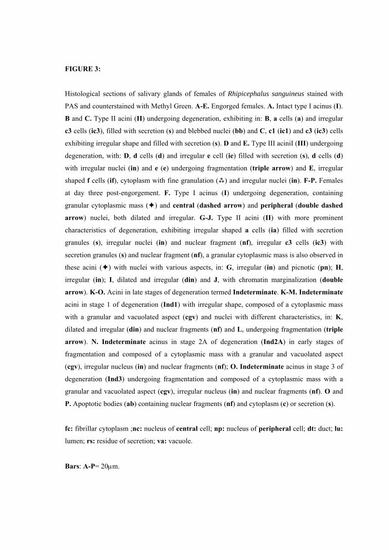
FIGURE 3:
Histological sections of salivary glands of females of Rhipicephalus sanguineus stained with
PAS and counterstained with Methyl Green. A-E. Engorged females. A. Intact type I acinus (I).
B and C. Type II acini (II) undergoing degeneration, exhibiting in: B, a cells (a) and irregular
c3 cells (ic3), filled with secretion (s) and blebbed nuclei (bb) and C, c1 (ic1) and c3 (ic3) cells
exhibiting irregular shape and filled with secretion (s). D and E. Type III aciniI (III) undergoing
degeneration, with: D, d cells (d) and irregular e cell (ie) filled with secretion (s), d cells (d)
with irregular nuclei (in) and e (e) undergoing fragmentation (triple arrow) and E, irregular
shaped f cells (if), cytoplasm with fine granulation ( ) and irregular nuclei (in). F-P. Females
at day three post-engorgement. F. Type I acinus (I) undergoing degeneration, containing
granular cytoplasmic mass ( ) and central (dashed arrow) and peripheral (double dashed
arrow) nuclei, both dilated and irregular. G-J. Type II acini (II) with more prominent
characteristics of degeneration, exhibiting irregular shaped a cells (ia) filled with secretion
granules (s), irregular nuclei (in) and nuclear fragment (nf), irregular c3 cells (ic3) with
secretion granules (s) and nuclear fragment (nf), a granular cytoplasmic mass is also observed in
these acini ( ) with nuclei with various aspects, in: G, irregular (in) and picnotic (pn); H,
irregular (in); I, dilated and irregular (din) and J, with chromatin marginalization (double
arrow). K-O. Acini in late stages of degeneration termed Indeterminate. K-M. Indeterminate
acini in stage 1 of degeneration (Ind1) with irregular shape, composed of a cytoplasmic mass
with a granular and vacuolated aspect (cgv) and nuclei with different characteristics, in: K,
dilated and irregular (din) and nuclear fragments (nf) and L, undergoing fragmentation (triple
arrow). N. Indeterminate acinus in stage 2A of degeneration (Ind2A) in early stages of
fragmentation and composed of a cytoplasmic mass with a granular and vacuolated aspect
(cgv), irregular nucleus (in) and nuclear fragments (nf); O. Indeterminate acinus in stage 3 of
degeneration (Ind3) undergoing fragmentation and composed of a cytoplasmic mass with a
granular and vacuolated aspect (cgv), irregular nucleus (in) and nuclear fragments (nf). O and
P. Apoptotic bodies (ab) containing nuclear fragments (nf) and cytoplasm (c) or secretion (s).
fc: fibrillar cytoplasm ;nc: nucleus of central cell; np: nucleus of peripheral cell; dt: duct; lu:
lumen; rs: residue of secretion; va: vacuole.
Bars: A-P= 20µm.

Capítulo 2
64

FIGURE 4:
Schematic representation of changes undergone by type I, II, and III acini of the salivary glands
of Rhpicephalus sanguineus females during and after feeding.
A. Unfed females;
B. Two-days fed females;
C. Four-days fed females;
D. Engorged females;
E. Females at day three post-engorgement;
rc: undifferentiated cell; nc1: undefined cell 1; nc2: undefined cell 2; a: a cell; b: b cell; c1:
c1 cell; c2: c2 cell; c3: c3 cell; c4: c4 cell; c5: c5 cell; c6: c6 cell; d: d cell; e: e cell; f: f cell;
iin: indeterminate cell; dt: duct; ld: lumen of the duct; iv: intraacinar valve; fc: fibrillar
cytoplasm; cn: central cell nucleus; pn: peripheral cell nucleus; gm: granular cytoplasmic
mass; n: nucleus; gm: granular cytoplasmic mass; c: cytoplasm; din: dilated and irregular
nucleus; pn: picnotic nucleus; chm: chromatin margination; bb: blebs; fn: fragmented
nucleus; ab: apoptotic bodies; s: secretion; cm: cytoplasmic mass; in: irregular nucleus; va:
vacuole; rs: residue of secretion.

Capítulo 2
65

Capítulo 2
66
DISCUSSION
The present study demonstrated morphological changes in the salivary glands of
Rhipicephalus sanguineus females as a result of the degenerative process at the end and
after the feeding period, confirming and complementing the information available in the
literature.
The salivary glands of engorged females of R. sanguineus exhibited a prominent
decrease in secretory capacity, marked by the loss of cytoplasmic granules and the
appearance of characteristics of degeneration. In these females, these changes make
their feeding process unfeasible, forcing them to stop feeding, as observed by Harris and
Kaufman (1984). These authors reported that the gland degeneration of Amblyomma
hebraeum is characterized by a reduction of over 90% in the maximum secretory
capacity of this tissue.
We observed in engorged females, intact and therefore functional type I acini;
type II and III acini in different stages of degeneration; and acini that could not be
identified due to their advanced stages of degeneration. The latter were here termed
Indeterminate. In type II and III acini, among acini of the same type, we observed
variations in the intensity of the degenerative process, with some acini exhibiting cells
with granules while others, cells with few or no granules. Among cells of the same
acinus, in type II and III acini, a variation in the intensity of the degenerative process
was also observed, with cells still with granules, although already exhibiting
degenerative characteristics, coexisting with cells without granules, therefore in an
advanced degenerative stage.
In R. sanguineus females at day three post-engorgement, we observed a
progression of the degenerative process compared to the observed in engorged females,
exhibiting type I and II acini undergoing degeneration, Indeterminate acini, as well as
several apoptotic bodies. These results support Till’s observations (1961) in R.
appendiculatus that degeneration is intensified after the end of feeding, especially two
days after engorgement, with the depletion of most secretion granules.
Our data, however, on the characteristics of degeneration in the glands of
engorged females contradict those obtained by Lomas et al. (1998), L’Amoreaux et al.
(2003) and Nunes et al. (2005), in females of A. hebraeum, Dermacentor variabilis, and

Capítulo 2
67
R. (Boophilus) microplus, respectively. According to these authors, salivary gland
degeneration begins 24 hours following engorgement in A. hebraeum, five days after
engorgement in D. variabilis, and already in semi-engorged females of R. (Boophilus)
microplus. This process is complete within 4 days following engorgement in A.
hebraeum (Lomas et al. (1998) and in approximately 33 days after engorgement in D.
variabilis (L’Amoreaux et al., 2003). Although the salivary glands of R. sanguineus
females at day three post-engorgement present characteristics of advanced stages of
degeneration, this process is probably not complete in four days, as reported for A.
hebraeum (Lomas et al., 1998), but it would not last as long as the observed for D.
variabilis (L’Amoreaux et al., 2003).
Thus, these data suggest that the onset of degeneration observed in glands of
engorged females of R. sanguineus occurs earlier than in A. hebraeum (Lomas et al.,
1998) and D. variabilis (L’Amoreaux et al., 2003). On the other hand, it occurs later
than that of semi-engorged females of R. (B.) microplus (Nunes et al., 2005), and ends
within about seven days post-engorgement, later than in A. hebraeum (Lomas et al.,
1998).
Changes in cells of degenerating acini of salivary glands detected in the two
situations examined in our study clearly indicate that salivary gland degeneration in R.
sanguineus is asynchronous among different acini, as well as among cells of the same
acini. This supports the results obtained by Till (1961), which described the
asynchronous degeneration of the gland tissue of R. appendiculatus females, and that
type II and III acini are the first ones to degenerate; by L’Amoreaux et al. (2003) that
demonstrated that type II and III acini of D. variabilis females degenerate earlier than
type I acini; and by Nunes et al. (2006), which described an asynchronous degeneration
of acini and salivary gland cells of semi-engorged females of R. (Boophilus) microplus.
Thus, based on the different stages of degeneration among acini of the same type
and the location of acini in the glands of Ixodidae in general, our results suggest that the
degeneration process in R. sanguineus females begins in the posterior region of the
gland, where the first signs of degeneration were detected, while intact acini were
observed in the anterior region. This indicates that the degeneration of salivary glands
of R. sanguineus females follows the same pattern described by most insects studied
(Silva de Moraes, 1998, PhD thesis; Abreu, 2004).

Capítulo 2
68
Regarding size, type I acini of engorged females of R. sanguineus did not exhibit
differences compared to those of females during the period of large blood consumption
(unpublished data). Also, central and peripheral cells were intact, as observed by
Binnington (1978) and Nunes et al. (2006).
In glands of R. sanguineus females at day three post-engorgement,
characteristics of degeneration were detected in central as well as peripheral cells,
unlike the observed by Bowman and Sauer (2004), which reported that in Ixodes ricinus
at day 12 post-engorgement, these acini do not exhibit signs of cell death, while two
other types are completely degenerated; and L’Amoreaux et al. (2003) that observed the
degeneration of these structures only after day 13 post-engorgement.
The late degeneration of type I acini might be associated with their role in glands
of females that completed feeding. According to Bowman and Sauer (2004), in I.
ricinus, these acini remain functional to maintain the hydration of the female during egg
laying. L’amoreaux et al. (2003) agree with this role, however, they report that this
occurs during the period of pre-oviposition, since these structures undergo cells death
after the first week of oviposition. This supports the data obtained in our study, since we
observed the first signs of degenerative characteristics in type I acini only in the
beginning of the oviposition period.
Type II acini of engorged females of R. sanguineus slightly decreased in size
compared to those of females during the period of large blood consumption
(unpublished data). This was observed by Binnington (1978), which reported that in
females of B. microplus, at the end of engorgement, acini decrease in size. However,
our results contradict those obtained by Marzouk and Darwish (1994), which reported
an increase in the size of these structures in engorged females of Hyalomma
(Hyalomma) dromedarii.
In engorged females of R. sanguineus, these acini exhibited characteristics of
degeneration, and in the same acinus, degenerating cells were observed with secretion
granules, coexisting with degenerating cells with few granules. The different levels of
degeneration might be the result of a functional asynchronism of cells, in which they
may be active in different moments during the feeding process (Binnington, 1978;
Walker et al., 1985; Gil and Walker, 1987; Marzouk and Darwish, 1994) as well as
inactive in different moments. The data are in agreement with those obtained by Nunes

Capítulo 2
69
et al. (2006), which proposed that these are typical characteristics of the apoptotic
process, with cells in different stages coexisting in the same tissue.
In this study, a, c1 and c3 cells still containing secretion granules but already
exhibiting nuclear changes were observed in type II acini. Additionally, were also found
in the same acinus, cells with few or no secretion granules, reduced size, and severe
signs of degeneration, especially in the nucleus. These cells could not be identified and
were termed indeterminate. The depletion of most secretion granules in the different
cells types and consequently the presence of cells with atypical characteristics
(indeterminate cells) are in agreement with the results obtained by Binnington (1978).
This author reported that at the end of engorgement, type II acini of B. microplus
females undergo drastic changes, such as the decrease in size and loss of granules,
making their identification difficult.
The presence of cells with granules in R. sanguineus, however, contradicts the
data obtained by Binnington (1978), Nunes et al. (2006), Walker et al. (1985) and
Marzouk and Darwish (1994). In B. microplus at the end of engorgement, the only cells
observed containing small amounts of granules were a, b, c1 and c3; in R. (B)
microplus, a, b, c2 and c3; and in R. appendiculatus, at the final feeding stage, only a,
b, c1 and c2 remained, while during this period, c1 hypertrophied. In engorged females
of H. (Hyalomma) dromedarii, a, b and c cells contained fewer secretion granules when
compared to feeding females, and c cells exhibited large cytoplasmic vacuoles.
This clearly indicates that for each tick species, or depending on their
requirements, the active period of each cell type may vary and consequently each cell
type does not play exactly the same role in the secretory cycle of salivary glands.
The present study also showed that in R. sanguineus, a, c1 and c3 cells of
engorged females were the last ones to degenerate, due to their role in the feeding
process. In addition, they were the first ones to become active, indicated by the presence
of secretion granules (unpublished data) and the last ones to lose their function.
The cells termed here as indeterminate exhibited characteristics that clearly
indicated cytoplasmic and nuclear changes, as a consequence of the advanced stage of
degeneration. Since these cells exhibit different cytoplasmic and nuclear aspects, they
were termed indeterminate 1 and 2.

Capítulo 2
70
In type II acini of females at day three post-engorgement, the degenerative
process was more prominent when compared to those of engorged females. In the
former, few type II acini were observed, indicating that many of them presented cells in
advanced stages of the degeneration. The few type II acini observed did not seem to
exhibit changes in size compared to those of engorged females. Marzouk and Darwish
(1994), on the contrary, showed that in H. (Hyalomma) dromedarii type II acini in
females after engorgement increase in size. Some type II acini do not contain granules,
thus the increase in size might be associated with the hypertrophy of these acinar
structures, as a result of the degenerative process.
Type II acini of R. sanguineus also exhibited some a and c3 cells, being the
former the most abundant. These two cell types contained secretion, although they
already exhibited signs of degeneration. In addition to a and c3 cells, were also
observed a cytoplasmic mass without cell boundaries containing changed nuclei,
characterizing a very advanced stage of degeneration of cells of these acini.
Compared to other species, in R. sanguineus a and c3 cells were the last to
degenerate; while in B. microplus, b and c3 cells (Binnington, 1983); in R. (Boophilus)
microplus, a, b, c2 and c3 (Nunes et al., 2006); and in R. appendiculatus, a cells
remained, although reduced in size and with few granules, until the tick detachment the
(Walker et al., 1985).
Type III acini of engorged females of R. sanguineus did not exhibit changes in
size when compared to females during the period of large blood consumption
(unpublished data), unlike the observed by Marzouk and Darwish (1994) that
demonstrated a significant increase in engorged females of H. (Hyalomma) dromedarii.
These acini in engorged females of R. sanguineus exhibited a decrease in the
diameter of the lumen, and cells, previously squamous, became cubic again in the
beginning of the period of large blood consumption (unpublished data). These data
support Till’s observations (1961) that in females of R. appendiculatus, the lumen of
these acini greatly increase during the period of large blood consumption. This,
however, occurs in the end of the feeding cycle, during which consumption was
probably lower, the diameter of lumen decreased and acini returned to their original
shape (small lumen and cubic cells). On the other hand, Binnington (1978) and Gill and
Walker (1987) reported in females of B. microplus and H. anatolicum anatolicum,

Capítulo 2
71
respectively, the presence of type III acini with squamous cells and enlarged lumen at
the end of the feeding period.
In addition to the morphological changes described here for type III acini,
changes in d, e and f cells were also observed as a result of the degenerative process,
which became more prominent in some cell types of the same acinus, characterizing an
asynchronous degeneration. These data support those obtained by Nunes et al. (2006)
that also observed an asynchronous degeneration of d, e and f cells of type III acini of
semi-engorged females of R. (Boophilus) microplus. In these acini, additionally to d, e
and f cells, a cytoplasmic mass with no cell boundaries was also observed containing
fragments of nuclei.
Although cells d and e contained secretion, in some cases, not in the form of
granules, but as an amorphous content, these cells exhibited other cytoplasmic and
nuclear changes that characterized cell death. These results support Binnington’s
observations (1978) that d and e cells of B. microplus females at the end of
engorgement exhibit few granules and picnotic nuclei; and Marzouk and Darwish’s
(1994) findings that d and e cells contain disperse granules due to the increase in size of
the acinus.
Our results obtained for d and e cells of R. sanguineus are partially in agreement
with those reported by Nunes et al. (2006) These authors observed d cells with reduced
size, residual secretion and picnotic nuclei in semi-engorged females of R. (Boophilus)
microplus, as observed in our study. However e cells were not observed by these
authors. This might be due to the complete or advanced stages of degeneration, as also
reported by Gill and Walker (1987) in H. anatolicum anatolicum females in the final
phase of feeding, when these cells are extremely compressed between well developed
interstitial cells.
In engorged females examined in this study, f cells did not contain secretion
granules, but exhibited a cytoplasm with a fine granular material, as well as nuclear
changes suggestive of degeneration. Nunes et al. (2006) also observed f cells with
cytoplasm with a fine granular material, but apparently preserved nuclei. On the other
hand, f cells of D. variabilis females during the rapid feeding phase already show signs
of degeneration (Coons and L’Amoreaux, 1986).

Capítulo 2
72
Cell changes observed in type III acini of engorged females of R. sanguineus
indicate that f cells might be the first to degenerate, since they loose their function due
to a decrease in the need to eliminate water and ions present in the hemolymph from the
blood meal. Thus, the commonly observed cytoplasmic mass, containing changed nuclei
and surrounded by other cells (d or e) in III type acinus, probably indicate an advanced
stage of degeneration of f cells. Cells d seem to be the last ones to degenerate, since: 1)
they still contained secretion granules and coexisted with e cells, whose granules were
disorganized caused by cell degeneration, 2) they were the only cells present in the
same acinus in advanced stages of degeneration, and were surrounded by a mass of
cytoplasmic cells with nuclear fragments.
The results obtained in the present study indicate that type III acini were the first
to degenerate and in females at day three post-engorgement, the degenerative process
progressed. Thus, in these females, type III acini completely lost their morphological
characteristics and histochemical properties and could not be identified and/or were
completely degenerated, and their fragments (apoptotic bodies) became a residual mass
commonly observed in the gland tissue. These data support the results obtained by
L’Amoreaux et al. (2003), which reported that in D. variabilis females in different post-
engorgement stages, type III acini are the most affected by the degenerative process and
the first to be eliminated from the gland tissue.
Since the degenerative process of salivary glands of females of the tick R.
sanguineus was asynchronous along the gland, in engorged females, intact acini were
observed at the same time as acini exhibiting characteristics of advanced stages of
degeneration. Some could be identified while others could not. The latter were
classified as Indeterminate.
The presence of Indeterminate acini already in engorged females of R.
sanguineus supports the findings by Nunes et al. (2006) that also reported the presence
of residues of acini, the result of advanced stages of degeneration in salivary glands of
semi-engorged females of R. (Boophilus) microplus. In the present study were observed
an increase of these acini in females at day three post-engorgement, due to the
progression of the degenerative process in this situation.
Thus, salivary glands of R. sanguineus underwent changes throughout the
degenerative process that caused changes in acini (Indeterminate). In females at day

Capítulo 2
73
three post-engorgement, acini were extremely irregular and fragmented, resulting in the
formation of apoptotic bodies. These data support those obtained by Nunes et al. (2005)
that also showed the presence of apoptotic bodies in the final phase of the degenerative
process of salivary glands of semi-engorged females of R. (Boophilus) microplus.
According to Till (1961) in R. appendiculatus, the final result of the degeneration
process is a mass of connective tissue and the duct system.
During the process of salivary gland degeneration of engorged females and
females at day three post-engorgement of R. sanguineus, changes were not the same in
different cells. In some of them, we observed a decrease in secretion granules, which
were eliminated, while in others, granules were disorganized and became a mass. Both
cytoplasmic changes were followed by severe nuclear changes, which varied in size,
shape, condensation level, and chromatin arrangement, even in cells of the same acinus.
Thus the degenerative process intensified resulting in other changes, such as
condensation of cytoplasm, loss of cell boundaries, loss of shape of cell and cytoplasmic
vacuolation. All these cell changes caused a disorganization of acini and consequently
the formation of apoptotic bodies. Nunes et al. (2005, 2006) also reported significant
changes in the cytoplasm and nucleus, in addition to the formation of apoptotic bodies
in R. (Boophilus) microplus.
The results obtained in this study and the information available in the literature
(L’Amoreaux et al., 2003; Bowman and Sauer, 2004; Nunes et al., 2005, 2006a, b)
suggest that the salivary gland degeneration of R. sanguineus females occurs by
apoptosis.
Nunes et al. (2006) concluded that in R. (Boophilus) microplus the gland tissue
degenerates due to the ontogenetic development of individuals, in which cells are
programmed to be deactivated in certain moments. In the present study, we observed
that salivary glands not only degenerate to complete the feeding process of ticks, but
also to avoid an investment and waste of energy in the maintenance of an organ that has
lost all or most of its functions.

Capítulo 2
74
ACKNOWLEDGMENTS
This research has been supported by CAPES (Coordenação de Aperfeiçoamento
de Pessoal de Nível Superior) and FAPESP (Fundação de Amparo à Pesquisa do Estado
de São Paulo) Grant nº 05/59208-3. The authors thank Cristiane Marcia Milléo, Gerson
Mello Souza, and Ronaldo Del Vecchio for the technical support. Part of this work has
been facilitated through the Integrated Consortium on Ticks and Tick-borne Diseases
(ICTTD-3) supported by the European Union under contract number 510561-INCO.

Capítulo 2
75
REFERENCES
Abreu, R.M.M., Silva de Moraes, R.L.M., Taboga, S.R., Camargo-Mathias, M.I., 2004.
Cytochemical studies of the nuclei of the venom glands’ cells of Apis mellifera
(Hymenoptera, Apidae). Acta Histochem. Cytochem. 37 (5), 195-299
Balashov, Y.S., 1983. Salivary glands. In: Raikhel, A.S., Hoogstraal, I.I. (Eds.), An
atlas of ixodid tick ultrastructure. Entomological Society of America, Lanham
Balashov, Y.S., 1972. Bloodsucking ticks (Ixodoidea)- vectors of diseases of man and
animals. Misc. Publ. Entomol. Soc Am. 8, 161-376
Bechara, G.H., Szabó, M.P.J., Ferreira, B.R., Garcia, M.V., 1995. Rhipicephalus
sanguineus tick in Brazil: feeding and reproductive aspects under laboratorial
conditions. Brazil. J. Vet. Parasitol. 4, 61-66
Binnington, K.C., 1978. Sequential changes in salivary gland structure during
attachment and feeding of the cattle tick, Boophilus microplus. Int. J. Parasitol. 8, 97-
115
Bowman, A.S., Sauer, J.R., 2004. Tick salivary glands: function, physiology and future.
Parasitology 129, S67-S81
Coons, L.B., L’amoreaux, W.J., 1986. Developmental changes in the salivary glands of
male and female Dermacentor variabilis (Say) during feeding. In: Borovsky, D.,
Spielman, A. (Eds.), Host regulated developmental mechanism in vector arthropods.
University of Florida-IFAS, Vero Beach
Flechtmann, C.H.W., 1973. Ácaros de importância médico-veterinária. Editora Livraria
Nobel S.A., São Paulo

Capítulo 2
76
Gill, H.S., Walker, A.R., 1987. The salivary glands of Hyalomma anatolicum
anatolicum: structural changes during attachment and feeding. Int. J. Parasitol. 17 (8),
1381-1392
Harris, R.A. Kaufman, W.R., 1984. Neural involvement in the control of salivary gland
degeneration in the ixodid tick Amblyomma Hebraeum. J. Exp. Biol. 109, 281-290
Kaufman, W.R., Lomas, L.O., 1996. “Male factor” in ticks: their role in feeding and egg
development. Invertebr. Reprod. Dev. 30, 191-198
L’Amoreaux, W.J., Junaid, L., Trevidi, S., 2003. Morphological evidence that salivary
gland degeneration in the American dog tick, Dermacentor variabilis (Say), involves
programmed cell death. Tissue Cell 35, 95-99
Lomas, O.L., Gelman, D., Kaufman, W. R., 1998. Ecdysteroid regulation of salivary
gland degeneration in the ixodid tick, Amblyomma hebraeum: a reconciliation of in vivo
and in vitro observations. Gen. Comp. Endocrionol. 109, 200-211
Lomas, L.O., Kaufman, W.R., 1992. An indirect mechanism by which a protein from
the male gonad hastens salivary gland degeneration in the female ixodid tick,
Amblyomma hebraeum. Arch Insect Biochem. Physiol. 21, 169-178
Marzouk, A.S., Darwish, Z.E.A., 1994. Changes in the salivary glands of female
Hyalomma (Hyalomma) dromedarii during and after feeding. J. Egypt. Soc. Parasitol.
24 (1), 39-57
McManus, J.F.A., 1946. Histological demonstration of mucin after periodic acid. Nature
158, 202

Capítulo 2
77
Nunes, E.T., Bechara, G.H., Saito, K.C., Denardi, S.E., O liveira, P.R., Camargo-
Mathias, M.I., 2005. Morphological, histological, and ultrastructural characterization of
degeneration salivary glands in females of the cattle-tick Rhipicephalus (Boophilus)
microplus (Canestrini, 1887) (Acari: Ixodidae). Micron 36, 437-447
Nunes, E.T., Camargo-Mathias, M.I., Bechara, G.H., 2006 a. Rhipicephalus (Boophilus)
microplus (Canestrine, 1887) (Acari: Ixodidae): acid phosphatase and ATPase activities
localization in salivary glands of females during the feeding period. Exp. Parasitol. 114,
109-117
Nunes, E.T., Camargo-Mathias, M.I., Bechara, G.H., 2006b. Structural and
cytochemical changes in the salivary glands of the Rhipicephalus (Boophilus) microplus
(Canestrini, 1887) (Acari: Ixodidae) tick female during feeding. Vet. Parasitol. 140,
114-123
Oliveira, P.R., Bechara, G.H., Denardi, S.E., Saito, K.C., Nunes, E.T., Szabó, M.P.J.,
Camargo-Mathias, M.I., 2005. Comparison of the external morphology of
Rhipicephalus sanguineus (Latreille, 1806) (Acari: Ixodidae) ticks from Brazil and
Argentina. Vet. Parasitol. 129, 139-147
Rey, L., 1973. Parasitologia. Guanabara Koogan, Rio de Janeiro
Till, W.M., 1961. A contribution to the anatomy and histology of the brown ear tick
Rhipicephalus appendiculatus Neumann. Memoirs of the Entomological Society of
Southern Africa, Pretoria, 6, 1-124
Walker, A., 1994. Arthropods of humans and domestic animals. A guide to preliminary
identification. Chapman & Hall, London
Walker, A.R., Fletcher, J.D., Gill, H., 1985. Structural and histochemical changes in the
salivary glands of Rhipicephalus appendiculatus during feeding. Int. J. Parasitol. 15 (1),
81-100

Capítulo 2
78
Weiss, L.B., Kaufman, W.R., 2001. The relationship between ‘critical weight’ and 20-
hydroxiecdysone in the female ixodid tick, Amblyomma Hebraeum. J. Insect Physiol.
47, 1261-1267
Weiss, L.B., Kaufman, W.R., 2004. Two feeding-induced proteins from the male gonad
trigger engorgement of the female tick Amblyomma hebraeum. Proc. Natl. Acad. Sci.
S.A. 101 (16), 5874-5879

Capítulo 3

Capítulo 3
80
CAPÍTULO 3
TITLE: Apoptosis in salivary glands of females of the tick Rhipicephalus sanguineus
(LATREILLE, 1806) (Acari, Ixodidae)
AUTHORS: Karim Christina Scopinho Furquim a, Gervásio Henrique Bechara b and
Maria Izabel Camargo Mathias a, *
a Departamento de Biologia, Instituto de Biociências, UNESP, Av. 24 A, nº 1515, Cx.
Postal 199, CEP: 13506-900, Rio Claro, S.P., Brazil b Departamento de Patologia Veterinária, FCAV, UNESP, Via de Acesso Prof. Paulo
Castellane, s/n, CEP: 14884-900, Jaboticabal, S.P., Brazil
* Corresponding author. Tel.: +55 19 35264135;
Fax: +55 19 35340009.
E-mail address: [email protected]

Capítulo 3
81
RESUMO
As glândulas salivares de fêmeas de carrapatos Rhipicephalus sanguineus nos
estados de jejum, ingurgitado e com três dias pós-ingurgitamento foram submetidas a
testes citoquímicos para análises enzimática e da viabilidade celular. No estudo
comparativo do tecido glandular nestas três situações observou-se que a marcação nas
células dos vários tipos de ácinos foi diferente, em especial para os ácinos III, II e I nas
situações onde estava havendo degeneração, os quais foram nesta seqüência afetados
pela morte celular. Este estudo, também revelou: alterações nos núcleos, na intensidade
da marcação da fosfatase ácida e da ATPase e na permeabilidade da membrana
plasmática, sendo que a positividade para fosfatase foi inversamente proporcional à da
ATPase e a positividade da ATPase foi sempre proporcional à integridade da
membrana. O tecido glandular das fêmeas em jejum apresentou alta atividade
metabólica com células com núcleo e membrana plasmática íntegros. Conclui-se que a
presença de fosfatase ácida nestes indivíduos faz parte da fisiologia de alguns ácinos,
visto que este tecido não se encontra em degeneração. Nas fêmeas ingurgitadas foi
observada membrana íntegra nas células dos ácinos I e II, bem como alterações
nucleares, queda na atividade da ATPase e maior marcação para fosfatase ácida,
alterações estas que fazem parte do processo degenerativo. Naquelas com três dias pós-
ingurgitamento o processo degenerativo já em estágio mais avançado revelou perda da
integridade da membrana na maioria das células (de alguns ácinos I, da maioria dos II e
de todos os III), intensa alteração nuclear, redução da atividade ATPásica, forte
marcação para fosfatase ácida, culminando com a formação de corpos apoptóticos. Na
morte celular destas glândulas as alterações nucleares precederam as citoplasmáticas na
seguinte seqüência: alteração do núcleo, perda da atividade da ATPase, perda da
integridade da membrana plasmática, maior atividade da fosfatase ácida e finalmente
formação de corpos apoptóticos. A presença de fosfatase ácida com papel secundário
(tardio) na morte celular, degradando os resíduos celulares finais, caracterizaram o
processo de morte celular em glândulas de fêmeas de R. sanguineus como sendo
apoptose atípica ou não clássica.

Capítulo 3
82
PALAVRAS-CHAVE: Rhipicephalus sanguineus; glândula salivar; carrapatos; fêmea;
estágio de alimentação; atividade enzimática; morte celular.

Capítulo 3
83
ABSTRACT
The salivary glands of females of the tick Rhipicephalus sanguineus at three
feeding stages: unfed, engorged, and at day three post-engorgement, were subjected to
cytochemical methods of enzymatic analysis and cell viability. Comparing gland tissues
at these stages, we observed distinct staining patterns in cells of different types of acini,
specially in degenerating types III, II, I acini, which were affected in this sequence by
cell death. This study also revealed changes in: nuclei, staining intensity for acid
phosphatase and ATPase activities, and permeability of the plasmic membrane. Acid
phosphatase activity was inversely proportional to that of ATPase, while ATPase
activity was always proportional to membrane integrity. The gland tissue of unfed
females exhibited high metabolic activity and cells with intact nucleus and plasmic
membrane, suggesting that the presence of acid phosphatase detected in these
individuals may participate in the normal physiology of some acini, as they were not
undergoing degeneration. In cells of types I and II acini of engorged females, we
observed intact membranes, as well as changes associated with the degenerative
process, characterized by nuclear changes, decrease in ATPase activity, and stronger
acid phosphatase activity. At day three post-engorgement, degeneration progressed to
more advanced stages, loss of membrane integrity was observed in most cells (of some
type I acini, most type II acini, and all type III acini), as well as prominent nuclear
changes, decrease in ATPase activity, and intense acid phosphatase activity, resulting in
the formation of apoptotic bodies. During the death of cells in these glands, nuclear
changes preceded cytoplasmic ones in the following sequence: nuclear changes, loss of
ATPase activity, loss of integrity of the plasmic membrane, increase in acid
phosphatase activity, and finally formation of apoptotic bodies. The presence of acid
phosphatase with a secondary role (late) during cell death, degrading final cell
remnants, characterized this process in the glands of R. sanguineus females as atypical
or non-classic apoptosis.
KEY WORDS: Rhipicephalus sanguineus; salivary gland; ticks; female; feeding stage;
enzymatic activity; cell death.

Capítulo 3
84
INTRODUCTION The onset of characteristics associated with salivary gland degeneration of
females of the tick Rhipicephalus sanguineus leads to the completion of the feeding
process of these individuals (Furquim, 2005). This degenerative process is triggered
when the gland tissue has already fulfilled its role in allowing the female to feed and
reproduce (Oliver, 1986). Feeding then ends and a new activity of the female organism,
egg laying, begins (Bowman and Sauer, 2004).
According to some authors, salivary gland degeneration in ticks does not occur
by necrotic or pathological death, but rather a programmed and regulated physiological
process (programmed cell death) (Bowman and Sauer, 2004). In ticks, gland
degeneration is regulated by an ecdysteroid hormone (Lomas et al., 1998), as also
observed in different insect organs (Jiang et al., 1997; Pelt-Verkuil, 1979).
Studies have also demonstrated that the binding of this ecdysteroid to its
corresponding receptors located on the surface of cells of salivary glands signals the
degenerative process in female ticks (Mao et al., 1995; Mao and Kaufman, 1999). This
binding then triggers a cascade of chemical reactions in the cytoplasm and nucleus of
gland cells, resulting in their death.
In the literature, two main types of genetically programmed cell death have been
described: apoptotic and autophagic one (Clarke, 1990; Bowen, 1993; Zakeri and
Ahuja, 1997; Jiang et al., 1997). The latter is commonly observed in insect tissues,
especially during metamorphosis with the participation of acid hydrolases (acid
phosphatase) playing an essential role (Pipan and Rakovec, 1980; Armbruster et al.,
1986; Cummings and Bowen, 1992; Lockshin and Zakeri, 1996; Jochová et al., 1997;
Gregorc et al., 1998).
L’Amoreaux et al. (2003), Nunes et al. (2005, 2006a, b), however, studying
salivary gland degeneration in female ticks of Dermacentor variabilis and R.
(Boophilus) microplus, respectively, suggested that the degeneration of acini may occur
by apoptotic cell death. Also, Bior et al. (2002) reported in the gland tissue of unfed and
feeding Amblyomma americanum males, the presence of a gene expressing a protein
that inhibits the apoptotic process, demonstrating the possibility of apoptosis in gland
cells.

Capítulo 3
85
Zakeri et al. (1995) and Clarke (1990) reported that apoptotic cell death does not
always exhibit a classic apoptosis profile, characterized by nuclear and cytoplasmic
breakdown, absence of hydrolases and, therefore, of autophagy, resulting in the
formation of apoptotic bodies that are later phagocytized by macrophages or other
neighboring cells (heterophagy) (Bowen and Bowen, 1990; Kerr et al., 1995; Lockshin
and Zakeri, 1996; Zakeri and Ahuja, 1997; Häcker, 2000). According to Clarke (1990)
and Zakeri et al. (1995), hydrolases (autophagy) may influence apoptosis in some cells
of certain tissues, but much controversy still remains regarding its exact role during the
process of apoptotic cell death (Bowen and Bowen, 1990; Bowen, 1993).
According to Bowen and Bowen (1990), apoptosis is an ATP-dependent
process. However, at the final stages a decrease in ATP levels would be observed and
consequently a decrease or even absence of ATPase activity. This could be used as an
indication of cells in late stages of apoptosis (Mullarkey, 1987 apud Bowen and Bowen,
1990; Bowen et al., 1988 apud Bowen and Bowen, 1990).
Another indication of cells in these stages is loss of integrity (of functionality) of
the plasmic membrane due to changes in ion pumps (McGahon et al., 1995), which
would occur only when energy production systems in the cell fail (Walker and Lucas,
1972 apud Silva de Moraes 1998; Rosenau, 1973 apud Silva de Moraes 1998). Thus,
according to McGahon et al. (1995), cells with intact membranes may be differentiated
from those with changed membranes by utilizing permeable and non-permeable stains.
The latter only penetrates cells in advanced stages of apoptosis or dead cells, in which
ion pumps are deficient or inactive.
Based on the presented information, the purpose of this study was to describe the
cell changes due the salivary gland degeneration of engorged females and females at
day three post-engorgement of R. sanguineus, comparing with healthy gland tissues of
unfed females, and identifing the type of cell death in these organs.

Capítulo 3
86
MATERIAL AND METHODS
In this study, we utilized unfed, engorged females and females at day three post-
engorgement of the tick R. sanguineus. Unfed individuals were provided by Dr.
Gervásio Henrique Bechara of the Department of Veterinary Pathology of UNESP,
Jaboticabal campus (São Paulo), from a colony maintained under controlled conditions
(29o C, 80% humidity, and 12 hour photoperiod) in BOD incubator. These individuals
were placed with some males, in a feeding chamber previously glue with an atoxic and
non-lesive preparation (Britannia Adhesives-Unit 4, UK) to the shaved back of the host
(rabbit) according to technique described elsewhere (Bechara et al., 1995) for complete
feeding.
A group of unfed individuals was assigned to cytochemical methods, while
another group was placed with males in a feeding chamber attached to the host (rabbit)
to complete feeding. A group of engorged females was assigned to cytochemical
procedures, and another group was maintained alive in closed containers with small
breathing holes for three days to be later analyzed.
After the completion of the time periods examined in this study, salivary glands
were removed in saline solution and processed according to the following methods
described below for observation under light and fluorescence microscopy.
For light microscopy, the material was fixed in 10% buffered neutral formalin
and acetone (9:1) for one hour and thirty minutes at 4o C, then processed according to
the methods described by Hussein et al. (1990) for detection of acid phosphatase and
ATPase activities. The material was then dehydrated in increasing concentrations of
ethanol (70%, 80%, 90% and 95%), embedded in Leica resin, and sectioned at a
thickness of 7 µm. Sections were placed on glass slides, counterstained with
Hematoxylin for 2 minutes, and mounted in Canada balsam for later examination under
light microscope. For the demonstration of acid phosphatase activity, whole mount
preparations of some glands were also examined.
In both enzymatic experiments, control samples were incubated without
substrate.
For fluorescence microscopy, after dissection, glands were placed on glass slides
and received two drops of Acridine Orange (100 µg/mL) and Ethidium Bromide (100

Capítulo 3
87
µg/mL) both in PBS, according to MacGahon et al. (1995), to demonstrate cell viability
and detection of apoptotic and/or necrotic cells. Slides were then covered with cover
glasses and immediately examined under fluorescence microscope with a 488 nm
excitation filter. Healthy cells are homogeneously green (cytoplasm and nucleus) or the
nucleus is homogeneously green and the cytoplasm orange/red. Cells in early stages of
apoptosis exhibit green or red-orange cytoplasm and green nucleus with clusters of
bright green condensed chromatin. Cells in late stages of apoptosis present red
cytoplasm and nucleus with bright orange condensed chromatin. Necrotic cells exhibit
red cytoplasm and homogeneously orange nucleus.
RESULTS
Unfed females
The salivary glands of unfed females of R. sanguineus exhibit healthy cells
strongly stained for RNA (Figs. 2A1–A3, A5) and ATPase (Figs. 1A1-A5), and weakly
stained for acid phosphatase (Figs. 3A1-A6). The plasmic membrane and nuclei are
intact (Figs. 2A1-A5).
Type I acinus
The basal membrane is strongly stained for ATPase (Fig. 1A1) and the plasmic
membrane of cells is intact (Fig. 2A1). The cytoplasm is strongly positive for RNA (Fig.
2A1), moderately positive for ATPase (Fig. 1A1) and acid phosphatase (Fig. 3A4).
Type II acinus
The basal membrane of the acinus and the plasmic membrane of cells are
strongly positive for ATPase (Figs. 1A2, A3), the cytoplasm is strongly positive for
RNA (undifferentiated cells) (Figs. 2A2, A3), weakly positive for acid phosphatase
(Fig. 3A5) and, in most cells, strongly positive for ATPase (Fig. 1A2).
Type III acinus
The basal membrane of the acinus and the plasmic membrane of cells are
strongly positive for ATPase (Figs. 1A4, A5). The cytoplasm is moderately positive for

Capítulo 3
88
RNA (Fig. 2A4, A5), weakly positive for acid phosphatase (Fig. 3A6), and in most cells,
strongly positive for ATPase (Fig. 1A4).
Engorged females
Here, degenerative characteristics are already observed (Figs. 1B4-C2, C4, D1,
D3, 2B2-C2, 3B3-B5, C2-D1). Only type I acini are intact (Figs. 1B2, B3, 2B1, 3B2, 3C1).
Degenerative characteristics are most frequently observed in type III (Figs. 1C4, D1,
2B4, C1, 3B4, C3) and Indeterminate acini (Figs. 1D3, 2C2, 3D1). In general, acini
exhibit weaker staining for ATPase (Figs. 1B3-C2, C4, D1, D3) and stronger staining for
acid phosphatase (Figs. 3B2-D1).
Type I acinus
The basal membrane of most acini is strongly positive for ATPase (Fig. 1B2) and
the plasmic membrane of all cells is intact (Fig. 2B1). The cytoplasm is strongly positive
for RNA (Fig. 2B1), moderately positive for acid phosphatase (Fig. 3C1), and in most
cells, moderately positive for ATPase (Fig. 1B3). Nuclei are intact (Fig. 2B1).
Type II acinus
The basal membrane of most acini is strongly positive for ATPase (Figs. 1B4-C1)
while the plasmic membrane of most cells is negative (Figs. 1B5, C1). All cell
membranes are intact (Figs. 2B2, B3). The cytoplasm of cells is moderately positive for
acid phosphatase (Fig. 3C2), and in most cells, moderately positive for ATPase (Figs.
1B5-C2). Nuclei exhibit changes (Figs. 2B2, B3).
Type III acinus (reduced lumen)
In most type III acini, the lumen is reduced (Figs. 1C3-D1) and they basal
membrane is strongly positive (Fig. 1C3) or negative for ATPase in some regions,
exhibiting an irregular staining pattern (Fig. 1D1). The plasmic membrane of all cells is
negative for ATPase (Figs. 1C3-D1) and has lost its integrity (Figs. 2B4, C1). The
cytoplasm of cells is strongly positive for acid phosphatase (Fig. 3C3), and in most cells,
strongly positive for ATPase (Fig. 1C3). Nuclei exhibit changes (Figs. 2B4, C1).

Capítulo 3
89
Type III acinus (dilated lumen)
The basal membrane of acini is strongly positive for ATPase while the plasmic
membrane of all cells is negative for ATPase (Fig. 1D2) and non-functional (Figs. 2B4,
C1). The cytoplasm of cells is strongly positive for ATPase (Fig. 1D2) and acid
phosphatase (Fig. 3C4). Nuclei exhibit changes (Figs. 2B4, C1).
Indeterminate acinus
The basal membrane of acini is moderately positive for ATPase (Fig. 1D3). The
plasmic membrane of all cells is negative for ATPase (Fig. 1D3) and has already lost its
integrity (Fig. 2C2). The cytoplasm is strongly positive for acid phosphatase (Fig. 3D1),
and in most cells, negative for ATPase (Fig. 1D3). Nuclei exhibit changes (Fig. 2C2).
Females at day three post-engorgement
In these ticks, degeneration has progressed into more advanced stages. Types II
(Figs. 1E6, F1, 2D3, 3D4, E3, E4), III (Figs. 1F2, F3, 2E1, 3E1, E5, F1), and Indeterminate
(Figs. 1G1-G3, 2E2, 3F2, F3) acini exhibit more prominent changes. Apoptotic bodies
(Figs. 1G3, 2E3, 3F4) are present. Stronger staining for acid phosphatase (Figs. 3D3-F3)
and weaker staining for ATPase (Figs. 1E1-G3) are observed. Apoptotic bodies exhibit
only strong staining for acid phosphatase (Figs. 1G3, 3F4) and the plasmic membrane is
not intact (Fig. 2E3).
Type I acinus
The basal membrane of most acini is moderately positive for ATPase (Fig. 1E1) and the
plasmic membrane of most cells is intact (Fig. 1C3). The cytoplasm of cells is strongly
positive for RNA (Fig. 1C3), weakly positive (Fig. 1E1) or negative (Figs. 1E2, E3) for
ATPase, and strongly positive for acid phosphatase (Fig. 3E2). Nuclei exhibit changes
(Fig. 1D1).
Type II acinus
The basal membrane of most acini is moderately positive for ATPase (Figs. 1E4,
E5), and in most cells, the plasmic membrane is negative for ATPase (Figs. 1E5-F1) and
has lost its integrity (Fig. 2D3). The cytoplasm of cells is strongly (Fig. 3E4) or
extremely positive (Fig. 3E3) for acid phosphatase, and in most cells, is negative for
ATPase (Figs. 1E4-F1). Nuclei exhibit changes (Figs. 2D2, D3).

Capítulo 3
90
Type III acinus
The basal membrane is weakly positive (Fig. 1F2) or negative (Fig. 1F3) for
ATPase. The plasmic membrane is negative for ATPase (Figs. 1F2, F3) and has lost its
integrity (Fig. 2E1). In the cytoplasm of most cells, ATPase activity is absent (Figs. 1F2,
F3) while acid phosphatase activity is intense (Fig. 3E5, F1). Nuclei exhibit changes (Fig.
2E1).
Indeterminate acinus
The basal membrane of most acini is negative for ATPase (Figs. 1G2, G3). The
plasmic membrane of all cells is negative for ATPase (Figs. 1F4-G3) and has lost its
integrity (Fig. 2E2). The cytoplasm of most cells is negative for ATPase (Figs. 1F4-G3)
and strongly positive for acid phosphatase (Fig. 3F3). Nuclei exhibit changes (Fig. 2E2).
To better compare the results, the data are summarized in Table 1.

Capítulo 3
91
Table 1: Enzymatic activity and viability of cells of salivary glands of unfed, engorged, and at day three post-
engorgement females of Rhipicephalus sanguineus.
Type I Acini Type II Acini Type III Acini Indeterminate Acini
Basal Membrane of Acinus
- ATPase +++ (Fig. 1A1); - ATPase +++ (Figs. 1A2, A3); - ATPase +++ (Figs. 1A4, A5);
Plasmic Membrane
- intact (Fig. 2A1); - ATPase +++ (Figs. 1A2, A3); - intact (Figs. 2A2, A3);
- ATPase +++ (Figs. 1A4, A5); - intact (Figs. 2A4, A5);
Cytoplasm - ATPase ++ (Fig. 1A1); - fa ++ (Fig. 3A4);
- ATPase +++ (Fig. 1A2) and – (Figs. 1A2, A3); - fa + (Fig. 3A5);
- ATPase +++ (Fig. 1A4) and – (Figs. 1A4, A5); - fa + (Fig. 3A6);
U N F E D
Nucleus - intact (Fig. 2A1); - intact (Figs. 2A2, A3); intact (Figs. 2A4, A5);
Basal
Membrane of Acinus
- ATPase +++ (Fig. 1B2) and ++ (Fig. 1B3);
- ATPase +++ (Figs. 1B4-C1) and ++ (Fig. 1C2);
reduced lumen: - ATPase +++ (Fig. 1C3), ++ (Fig. 1C4) and +/– (Fig. 1D1); dilated lumen: - ATPase +++ (Fig. 1D2);
- ATPase ++ (Fig. 1D3);
Plasmic Membrane
- intact (Fig. 2B1); - ATPase ++ (Figs. 1B4, C2) and – (Figs. 1B5, C1); - intact (Figs. 2B2, B3);
- ATPase – (Figs. 1C3-D2); - not intact (Figs. 2B4, C1);
- ATPase – (Fig. 1D3); - not intact (Fig. 2C2);
Cytoplasm - ATPase ++ (Fig. 1B3) and – (Fig. 1B2); - fa ++ (Fig. 3C1);
- ATPase + + (Fig. 1B5-C2) and – (Fig. 1B4, C2); - fa ++ (Fig. 3C2);
reduced lumen: - ATPase +++ (Fig. 1C3), ++ (Fig. 1C4) and + (Fig. 1D1); - fa +++ (Fig. 3C3); dilated lumen: - ATPase +++ (Fig. 1D2); - fa +++ (Fig. 3C4);
- ATPase + (Fig. 1D3) and – (Fig. 1D3); - fa +++ (Fig. 3D1);
E N G O R G E D
Nucleus - intact (Fig. 2B1); - # (Figs. 2B2, B3); - ? (Figs. 2B4, C1) and ! (Fig. 2B4);
- ? (Fig. 2C2);
Basal Membrane of Acinus
- ATPase ++ (Fig. 1E1), + (Fig. 1E2) and +/– (Fig. 1E3);
- ATPase ++ (Figs. 1E4, E5), +/– (Fig. 1E6) and – (Fig. 1F1);
- ATPase + (Fig. 1F2) and – (Fig. 1F3);
- ATPase ++ (Fig. 1F4), +/– (Fig. 1G1) and – (Figs. 1G2, G3);
Plasmic Membrane
- > intact (Fig. 2C3); - < not intact (Fig. 2D1);
- ATPase ++ (Fig. 1E4) and – (Figs. 1E5-F1); - > not intact (Fig. 2D3); - < intact (Fig. 2D2);
- ATPase – (Figs. 1F2, F3); - not intact (Fig. 2E1);
- ATPase – (Figs. 1F4-G3)- not intact (Fig. 2E2);
Cytoplasm - ATPase + (Fig. 1E1) and – (Figs. 1E2, E3); - fa +++ (Fig. 3E2);
- ATPase ++ (Fig. 1E4), + (Figs. 1E6, F1) and – (Fig. 1E5- fa ++++ (Fig. 3E3) and +++ (Fig. 3E4);
- ATPase + (Figs. 1F2, F3) and – (Figs. 1F2, F3); - fa ++++ (Figs. 3E5, F1) and +++ (Fig. 3E5);
- ATPase ++ (Fig. 1G1), + (Fig. 1F4) and – (Figs. 1G2, G3); - fa ++++ (Fig. 3F2) and +++ (Fig. 3F3);
3
D A Y S
A F T E R
Nucleus - # (Fig. 2D1); - # (Fig. 2D2), ? (Fig. 2D3), ! (Fig. 2D3) and ** (Fig. 2D2);
- #, ? and ! (Fig. 2E1); - ? and * (Fig. 2E2);
(++++) extremely positive; (+++) strongly positive; (++) moderately positive; (+) weakly positive; (–) negative; (af) acid phosphatase; (>) most cells; (<): some
cells; (+/-) irregular staining; (#) nucleus with clusters of condensed chromatin; (?) picnotic nucleus; (!) blebs; (**) fragmenting nucleus; (*) fragmented nucleus.

FIGURES

FIGURE 1:
ATPase activity in the female salivary glands of Rhipicephalus sanguineus. A1-B1. Unfeds:
observe that types I (I), II (II), III (III), and Indeterminate (Ind) acini are strongly stained for
ATPase (arrow). B1. Negative control. B2-D4. Engorged: observe weaker staining for ATPase
(arrow) in types I (I), II (II) and III (III) acini. Note in: B5 and C2, weaker staining for ATPase
(arrow) in the basal membrane of type II acini (II); B4-C2, weaker staining for ATPase (arrow)
in the plasmic membrane and cytoplasm of cells of type II acini (II); B5 and C1, plasmic
membrane of some cells of type II acini (II) with no ATPase activity. Observe in: C4, lower
ATPase activity (arrow) in the basal membrane and in the cytoplasm of cells of type III acini
(III) and D1, irregular staining for ATPase (arrow) in the basal membrane of type III acinus
(III). D4. Negative control. E1-G4. Day three post-engorgement: note even weaker staining for
ATPase activity in types I (I), II (II), III (III), and Indeterminate (Ind) acini. Observe in E3,
type I acinus (I) with irregular staining for ATPase along the basal membrane and no staining in
the cytoplasm of cells. Note in: E6, irregular staining for ATPase in the basal membrane of type
II acinus (II); F1, absence of ATPase activity in the basal membrane of type II acinus (II) and
E5-F1, absence of staining for ATPase in the plasmic membrane of cells of type II acini (II).
Note in: F3, absence of ATPase activity in the basal membrane of type III acinus (III) and F2
and F3, no staining for ATPase in the plasmic membrane of cells of type III acinus (III). In G1,
irregular ATPase activity in the basal membrane of Indeterminate acinus (Ind); G2 and G3, no
staining for ATPse in the basal membrane of Indeterminate acinus (Ind); G1 and G3, absence
of ATPase activity in the membrane of cells of Indeterminate acinus (Ind). Note in F4-G3, no
ATPase staining in the membrane of apoptotic body (ab). G4. Negative control.
I-III: types acini; Ind: Indeterminate acinus; arrow: staining for ATPase; n: staining for
ATPase in the nucleus; dt: duct.
Bars: E1 and E2= 12, 5 µm; A5-B3, C3-E6 and F4= 25 µm; A1-A4, B4-D4, F1-F3 and G1-G4= 50
µm.

Capítulo 3
93

FIGURE 2:
Total preparation of female salivary glands of Rhipicephalus sanguineus stained with Ethidium
Bromide and Acridine Orange. A1-A5. Unfeds: Observe healthy cells of types I (I), II (II), and
III (III) acini with intact plasmic membrane and nuclei (tn). B1-C2. Engorged: in B1, intact type
I acinus (I). Note in: B2 and B3, type II acini (II) undergoing degeneration with cells exhibiting
nuclear changes and membrane still intact; B4 and C1, type III acini (III) undergoing
degeneration with nuclear changes and loss of plasmic membrane integrity, and C2,
Indeterminate acinus (Ind) with cells exhibiting nuclear changes and loss of plasmic
membrane integrity. C3-E3. Day three post-engorgement: in C3, type I acinus (I) undergoing
degeneration and exhibiting plasmic membrane still intact. Observe in: D1, type I acinus (I)
undergoing degeneration with cells exhibiting nuclear changes and loss of membrane integrity;
D3, cells of type II acinus (II) exhibiting nuclear changes and loss of plasmic membrane
integrity; E1, Type III acinus (III) undergoing degeneration with cells exhibiting nuclear and
membrane changes; E2, Indeterminate acinus (Ind) exhibiting degenerating cells with nuclei
and membrane changed, and E3, apoptotic bodies (ab) with loss membranes integrity.
I-III: types acini; Ind: Indeterminate acinus; arrow: cytoplasmic RNA; tn: intact nucleus;
dashed arrow: nucleus with clusters of condensed chromatin; pn: picnotic nucleus; bb: blebs;
∗: fragmenting nucleus; fn: fragmented nucleus.
Bars: A1-A4 and B1= 12, 5 µm; A5 and B2-E3= 25 µm.

Capítulo 3
94

FIGURE 3
Acid phosphatase activity in the females salivary glands of Rhipicephalus sanguineus. A1-B1.
Unfeds: Observe in: A1-A3, total preparation of types I (I), II (II), and III (III) acini showing
low acid phosphatase activity (arrow) and A4-A6, histological sections of types I (I), II (II), and
III (III) acini exhibiting weak staining for acid phosphatase (arrow) in the cytoplasm of cells.
B1. Negative control. B2-D2. Engorged: in B2-B5, total preparation of types I (I), II (II), and III
(III) acini showing stronger staining for acid phosphatase (arrow). Note in B4 and B5,
prominent staining for acid phosphatase (arrow) in the cytoplasm of cells of type III acinus
(III). C1-D1. Histological sections of types I (I), II (II), III (III), and Indeterminate (Ind) acini.
Observe in C3, C4 and D1, cytoplasm of cells of types III (III) and Indeterminate (Ind) acini
showing stronger staining for acid phosphatase (arrow). D2. Negative control. D3-F5. Day three
post-engorgement: in D3-E1, total preparation of types I (I), II (II), and III (III) acini, note
stronger staining for acid phosphatase (arrow) in the cytoplasm of cells of the three types of
acini (I, II, and III). Observe in D4 and E1, the most prominent staining for acid phosphatase
(arrow). In E2-F4. Histological sections of types I (I), II (II), III (III), and Indeterminate (Ind)
acini, as well as apoptotic bodies (ab). Note in: E3, E5-F2, extremely strong staining for acid
phosphatase activity (arrow) in cells of types II (II), III (III), and Indeterminate (Ind) acini,
and E3-F3, presence of vacuoles (va) in the cytoplasm of cells of types II (II), III (III), and
Indeterminate (Ind) acini. F5. Negative control.
arrow: staining for acid phosphatase; dt: duct; lu: lumen.
Bars: A1= 10 µm; F4= 12, 5 µm; A2, A3, B1-B3, D3-E1 and F5= 20 µm; A4-A6, C1, E2, E4 and
E5= 25 µm; B4, B5 and D2= 40 µm; C2-D1, E3 and F1-F3= 50 µm.
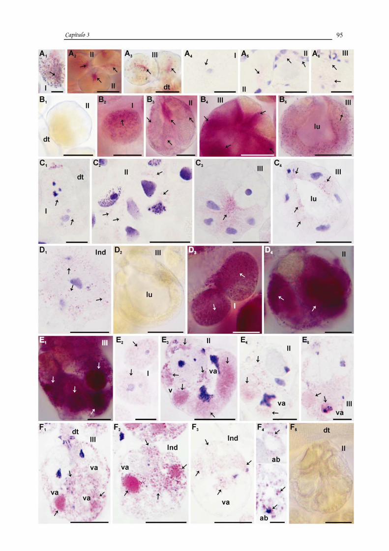
Capítulo 3
95

Capítulo 3
96
DISCUSSION
The present study revealed structural and enzymatic aspects of the salivary
glands of unfed, and engorged, and day three post-engorgement females of
Rhipicephalus sanguineus. In the latter two stages, changes undergone by cells
characterized the degeneration of this organ.
In unfed females of R. sanguineus, the presence of ATPase in different cell
compartments confirmed the need of a large consumption of energy for metabolic
processes in these areas, in addition to contributing to the maintenance of the
intracellular environment, since the ion pump of the membrane is functional. The
ATPase staining observed in the nucleus might be associated with transcription activity,
supporting the results obtained by Azeredo-Oliveira and Mello (1986), which reported
the presence of ATPase in the euchromatin and nucleolus of cells of Malphigian tubules
of Triatoma infestans.
In this study, acid phosphatase staining was also observed in unfed females,
especially in cells of types I and II acini. In these cells, this enzyme might be involved
in the normal cell metabolism. According to the literature, the activity of this enzyme is
not always associated with cell destruction. Costa and Cruz-Landim (2001) and Britto
and Caetano (2006) suggested that it may participate in the metabolism of different
stages of the secretory cycle of some glands.
Binnington (1978), Walker et al. (1985), and Nunes et al. (2006) also reported
acid phosphatase in active gland cells of feeding females of Boophilus microplus, R.
appendiculatus, and R. (Boophilus) microplus, respectively. The latter authors
demonstrated the participation of these enzymes in the cell metabolism during the
production and discharge of secretion (Nunes et al., 2006). In unfed females examined
in this study, the presence of acid phosphatase in type I acini might be associated with
protein synthesis, since large quantities of RNA were observed, as reported by Costa
and Cruz-Landim (2001) in hypopharyngeal glands of Scaptotrigona postica. In type II
and III acini, where cells are in pre-secretory phase, the participation of acid
phosphatase might be associated with the production of secretion, as observed in other
studies (Costa and Cruz-Landim, 2001).
The salivary gland cells of R. sanguineus females, engorged and at day three
post-engorgement, underwent structural (nucleus and plasmic membrane) and

Capítulo 3
97
enzymatic changes (ATPase and acid phosphatase) that resulted in their death,
confirming an asynchrony in the process of cellular and acinar degeneration. Types II
and III acini seem the first to be affected and the degenerative process intensifies in
females at day three post-engorgement.
In engorged females, we observed a decrease in ATPase and increase of acid
phosphatase activities in type I acinus, the only intact, and types II, III, and
Indeterminate acini. In types III and Indeterminate acini, the plasmic membrane lost
its integrity and did not exhibit ATPase activity, confirming the observed by Nunes et
al. (2006). These authors detected a decrease in enzymatic activity (ATPase and acid
phosphatase) in semi-engorged females of R. (Boophilus) microplus, as well as a
heterogeneous staining pattern for ATPase throughout the gland tissue.
The intense ATPase and acid phosphatase activities observed in the cytoplasm of
cells of type III acinus (with dilated lumen) of glands of engorged females of R.
sanguineus are probably not associated with cell death, but rather with a re-structuring
of cells and shape of acini, returning from squamous to cubic (Till, 1961). This process
requires a lot of energy, as indicated by the intense staining for ATPase. These data
support those obtained by Walker et al. (1985) that correlated the presence of acid
phosphatase with changes in shape of cells during the phase of large blood consumption
of the tick.
At day three post-engorgement, gland cells of R. sanguineus females exhibited
more prominent enzymatic changes, as the degeneration process progressed.
In engorged females as well as those at day three post-engorgement, we
observed ATPase staining in the basal membrane of different acini, as reported by
Nunes et al. (2006). In some acini, ATPase activity was irregular, characterized by
stained and non-stained areas. In the latter, loss of membrane integrity was observed.
Type II acinus of engorged females and females at day three post-engorgement
exhibited a characteristic not observed in other types of acini. The cells of some of type
II acini presented an intact plasmic membrane, but absence of ATPase activity. One
possible explanation might be the presence of a functional plasmic membrane only in
the basal portion that could not be distinguished from the basal membrane of the acinus.
Thus, only an ATPase positive halo was observed around it.
Capítulo 3
98
Our findings regarding acid phosphatase activity were inversely proportional to
those of ATPase in engorged females and those at day three post-engorgement. This
was also observed by Weber (1969) that reported a simultaneous increase in acid and
basic phosphatase activities and a decrease in ATPase activity during degeneration.
The decrease in ATPase activity and increase in acid phosphatase activity
observed in our study during gland degeneration were accompanied by the following
morphological changes reported by Furquim (2005): a) decrease in the amount of
secretion in cells, b) nuclear breakdown, c) cytoplasmic shrinkage, d) loss of shape and
cell boundaries (cell individuality), e) vacuolation, and f) cell fragmentation. The
decrease in the amount of secretion and nuclear breakdown precede all other events.
These changes, in addition to bodies containing cytoplasmic and nuclear remnants,
resulted from cytoplasm shrinkage and cell fragmentation and enclosed by membranes,
characterize an apoptotic cell death (Clarke, 1990; Bowen, 1993; Kerr et al., 1995;
Lockshin and Zakeri, 1996; Häcker, 2000). In this study, the decrease and/or absence of
ATPase activity in the plasmic membrane, as well as the loss of integrity observed in
engorged females and females at day three post-engorgement contradict the observed by
Bowen and Bowen (1990). These authors suggested that apoptosis is an ATP-
dependent process and its decrease and that of ATPase occur only in advanced stages of
the process, with a functional plasmic membrane present until cell fragmentation
(Bowen and Bowen, 1990; Bowen, 1993; Kerr et al., 1995; Lockshin and Zakeri, 1996).
In R. sanguineus salivary glands, the absence of ATPase activity and loss of
integrity of the plasmic membrane occurred mainly in females at day three post-
engorgement. At this stage, unlike the described for apoptotic death, we observed intact
and fragmenting acini, in addition to ATPase-negative apoptotic bodies lacking intact
membranes. Although these changes occurred after nuclear changes, cytoplasmic
shrinkage, and loss of cell shape, they were observed before than the normally reported
for apoptosis, which is before the formation of apoptotic bodies, since the latter still
exhibit a functional membrane soon after they are formed (Bowen and Bowen; 1990;
Kerr et al., 1995).
In R. sanguineus, acid phosphatase activity was more intense in engorged
females than in unfed ones, and was intensified in those at day three post-engorgement.
According to the literature, a process of cell death associated with acid phosphatase

Capítulo 3
99
(acid hydrolases) is characterized as “autophagic death” (Zakeri et al., 1995; Lockshin
and Zakeri, 1996; Cummings and Bowen, 1992; Gregorc et al., 1998; Clarke, 1990;
Bowen, 1993; Pipan and Ravoc, 1980; Jochová et al., 1997). Several authors, however,
disagree on the real role of acid hydrolases in apoptotic death (Bowen and Bowen,
1990; Clarke, 1990; Bowen, 1993).
In degenerating salivary glands of R. sanguineus females, acid phosphatase
occurred after other changes. This enzyme was detected mainly in acini in late stages of
degeneration, in which several cytoplasmic vacuoles were also observed. Furquim
(2005) also reported the presence of these structures. Cytoplasmic vacuolation, in
addition to a high acid phosphatase activity in females at day three post-engorgement,
would strongly suggest an autophagic death, as commonly observed during insect
metamorphosis (Zakeri et al., 1995; Lockshin and Zakeri, 1996). According to
Cummings and Bowen (1992) and Gregorc et al. (1998), the presence of this enzyme
might be considered a prelude to autophagic cell death, with early destruction of the
cytoplasm (extensive vacuolation) and later nuclear breakdown (Clarke, 1990; Bowen,
1993; Lockshin and Zakeri, 1996; Zakeri et al., 1995; Pipan and Ravoc, 1980; Jochová
et al., 1997). However, it is important to point out that in the present study the nuclear
breakdown was observed before an intense acid phosphatase activity, and therefore,
before cytoplasmic vacuolation. This suggests that acid phosphatase might have a role
in later stages, degrading part of the cytoplasm, and therefore not characterizing the cell
death in the glands of R. sanguineus females as autophagic. One possible hypothesis
might be an apoptotic death combined with autophagy, corroborating Levy and Bautz
(1985), Clarke (1990), Zakeri et al. (1995), Bowen et al. (1996), Dai and Gilbert (1997),
Lockshin and Zakeri (1996), Gregorc and Bowen (1997), Jochová et al. (1997) and
Yamamoto et al. (2000). These authors have suggested that the process of cell death
may exhibit characteristics of more than one type.
The early decrease of ATPase activity observed in this study as well as loss of
membrane integrity, and intense acid phosphatase activity did not characterize a classic
apoptotic death. Bowen et al. (1993) reported that during cell death of salivary gland
cells of Calliphora vomitoria, ATPase activity ceased at some point, and massive
cytoplasmic vacuolation was observed, indicating that in programmed cell death,

Capítulo 3
100
ATPase and preservation of membrane integrity until late stages of the process are not
always observed, as suggested by Bowen and Bowen (1990) and Kerr et al. (1995).
Regarding whether or not autophagy is involved in apoptotic death in
invertebrates, some studies have demonstrated that programmed death exhibit
characteristics distinct from those of classic apoptosis in insects, as vacuolation in cells
occurs due to autophagy (Zakeri et al., 1995; Dai and Gilbert 1997; Bowen et al., 1996;
Gregorc and Bowen, 1997; Levy and Bautz, 1985). In addition, cells increase in size
instead of shrinking (Bowen et al., 1996). Studying cell death in prothoracic glands of
Manduca sexta during metamorphosis, Dai and Gilbert (1997) reported changes similar
to those observed in the present study and classified it as apoptotic death. According to
Hurle and Hinchliffe (1978) apud Bowen and Bowen (1990) and Levy and Bautz
(1985), the participation of acid hydrolases in the apoptotic process might be associated
with cell fragmentation. This could also be the case in our study, as strong acid
phosphatase activity was observed in fragmenting acini, as well as in apoptotic bodies.
Zakeri et al. (1995) also reported that apoptosis and autophagic death might overlap in
some aspects, including regarding acid hydrolase activity.
Thus, the cell death process leading to the degeneration of the salivary gland
tissue of R. sanguineus females exhibits features of classic apoptosis combined with
autophagy, characterizing an atypical apoptosis, with acid phosphatase participating in
the removal of cytoplasmic remnants and cell fragmentation. In R. sanguineus females,
this enzyme may have an endogenous origin through de novo synthesis commonly
observed in autophagic death, although it may and also occur during apoptotic death
(Zakeri et al., 1995), or an exogenous origin, from the hemolymph. Levy and Bautz
(1985), Armbruster et al. (1986), Jones and Bowen (1993) reported the presence of
hemocytes containing large amounts of acid phosphatase adhered to degenerating
tissues. Nunes et al. (2006) suggested that acid phosphatase detected in the gland tissue
of R. (Boophilus) microplus females may come from the hemolymph. Therefore, these
findings suggest that a) the gradual increase in acid phosphatase in cells of salivary
glands of females, from unfed to three day post-engorgement, indicates that the
synthesis of this enzyme begins in engorged females, and b) the absence of hemocytes
around or in the gland tissue suggests that the origin of acid phosphatase in R.
sanguineus is exclusively endogenous.

Capítulo 3
101
The results presented in this study, in addition to those reported by Furquim
(2005) in females of this species, clearly indicate that the salivary gland tissue of R.
sanguineus females dies by atypical apoptosis, characterized by events that occur in the
following order: a) early nuclear breakdown, chromatin condensation and margination,
bleb formation in the nuclear envelope, and finally nuclear fragmentation, b)
cytoplasmic shrinkage and loss of cell shape, c) decrease and/or absence of ATPase
activity, loss of membrane integrity, and loss of cell boundaries, d) intense acid
phosphatase activity, resulting in cytoplasmic vacuolation and cell fragmentation, and e)
fragmentation of acini with release of apoptotic bodies. The latter are not removed by
heterophagy from the hemolymph, unlike the observed by Till (1961), Kerr et al.
(1995), Bowen et al. (1993), Bowen and Bowen (1990), and Häcker (2000), but rather
through the participation of the acid phosphatase synthesized by salivary gland cells,
characterizing an apoptotic process involving autophagy.
ACKNOWLEDGEMENTS This research has been supported by CAPES (Coordenação de Aperfeiçoamento
de Pessoal de Nível Superior) and FAPESP (Fundação de Amparo à Pesquisa do Estado
de São Paulo) Grant nº 05/59208-3. The authors thank Antonio Teruyoshi Yabuki,
Monika Souza and Ronaldo Del Vecchio for the technical support, and Prof. Dr. Flávio
Henrique Caetano for allowing the use of the photomicroscope (FAPESP, Grant nº
04/13327-9). Part of this work has been facilitated through the Integrated Consortium
on Ticks and Tick-borne Diseases (ICTTD-3) supported by the European Union under
contract number 510561-INCO.
Capítulo 3
102
REFERENCES Armbruster, L., Levy, M., Mathieu, N., Bautz, A.M., 1986. Acid phosphatase activity in
the hemolynph, hemocytes, fat body and salivary glands during larval and prepupal
development in Calliphora erythrocephala (Diptera: Calliphoridae). Comparative
Biochemistry and Physiology 84B (3), 349-354.
Azeredo-Oliveira, M.T.V., Mello, M.L.S., 1986. Mg2+-dependent ATPase activity
investigated in heterochromatin, euchromatin and nucleoli of Triatoma infestans.
Revista Brasileira de Genética IX (2), 223-229.
Bechara, G.H., Szabó, M.P.J., Ferreira, B.R. Garcia, M.V., 1995. Rhipicephalulus
sanguineus tick in Brazil: feeding and reproductive aspects under laboratorial
conditions, Brazilian Journal of Veterinary Parasitology 4, 61-66.
Binnington, K.C., 1978. Sequential changes in salivary gland structure during
attachment and feeding of the cattle tick, Boophilus microplus. International Journal for
Parasitology 8, 97-115.
Bior, A.D., Essemberg, R.C., Sauer, J.R., 2002. Comparison of differentially expressed
genes in the salivary glands of male ticks, Amblyomma americanum and Dermacentor
andersoni. Insect Biochemistry and Molecular Biology 32, 645-655.
Bowen, I.D., 1993. Apoptosis or programmed cell death? Cell Biology International 17
(4), 365-380.
Bowen, I.D., Bowen, S.M., 1990. Programmed Cell Death in Tumours and Tissues.
Chapman and Hall, London, pp. 15-66.
Bowen, I.D., Morgan, S.M., Mullarkey, K., 1993. Cell death in the salivary glands of
metamorphosing Calliphora vomitoria. Cell Biology International 17 (1), 13-34.

Capítulo 3
103
Bowen, I.D., Mullarkey, K., Morgan, S.M., 1996. Programmed cell death during
metamorphosis in the blow-fly Calliphora vomitoria. Microscopy Research and
Technique 34, 202-217.
Bowen, I.D., Worrill, N.A., Winters, C.A., Mullarkey, K., 1988. The use of
backscattered electron imaging, X-ray microscopy in demonstrating physiological cell
death. Scanning Microscopy 2, 1453-1462.
Bowman, A.S., Sauer, J.R., 2004. Tick salivary glands: function, physiology and future.
Parasitology 129, S67-S81.
Britto, F.B., Caetano, F.H., 2006. Morphological features and occurrence of
degenerative characteristics in the hypopharyngeal glands of the paper wasp Polistes
versicolor (Olivier) (Hymenoptera: Vespedae). Micron 37, 742-747.
Clarke, P.G.H., 1990. Developmental cell death: morphological diversity and multiple
mechanisms. Anatomy and Embryology 181, 195-213.
Costa, R.A.C., Cruz-Landim, C., 2002. Distribution of acid phosphatases in the
hypopharyngeal glands from workers, queens and males of a Brazilian stingless bee
Scaptotrigona postica Latreille: an ultrastructural cytochemical study. Histochemical
Journal 33, 653-662.
Cummings, L.A., Bowen, I.D., 1992. The localization of acid phosphatase as a prelude
to cell death in the blow-fly Calliphora erithrocephala. Micron and Microscopica Acta
23 (3), 357-358.
Dai, J., Gilbert, L.I., 1997. Programmed cell death of the prothoracic glands of
Manduca sexta during pupal-adult metamorphosis. Insect Biochemistry and Molecular
Biology 27 (1), 69-78.

Capítulo 3
104
Furquim, K.C.S., 2005. Processos degenerativos em glândulas salivares de carrapatos
Rhipicephalus sanguineus (LATREILLE, 1806) (ACARI, IXODIDAE) em jejum, em
alimentação e alimentados, 2º Relatório Científico CAPES, Instituto de Biociências-
UNESP, Rio Claro (S.P.), Brasil, 40 p.
Gregorc, A., Bowen, I.D., 1997. Programmed cell death in the honey-bee (Apis
mellifera L.) larvae midgut. Cell Biology International 21 (3), 151-158.
Gregorc, A., Bowen, I.D., Pogacnik, A., 1998. Acid phosphatase activity in the midgut
of honeybee (Apis melliphera L.) larvae. Apidologie 29, 579-584.
Häcker, G., 2000. The morphology of apoptosis. Cell and Tissue Research 301, 5-17.
Hurle, J., Hinchliffe, J.R., 1978. Cell death in the posterior necrotic zone (PNZ) of the
chick wing-bud: a stereoscan and ultrastructural survey of autolysis and cell
fragmentation. Journal of Embryology and Experimental Morphology 43, 123-136.
Hussein, M.A., Bowen, I.D., Lewis, G.H., 1990. The histochemical localization of
ATPase, cholinesterase and acid phosphatase activity in Culex pipiens (Diptera,
Culicidae) larvae using a methacrylate embedding technique. Cell Biology International
Reports 14 (9), 775-781.
Jochová, J., Zakeri, Z., Lockshin, R.A., 1997. Rearrangement of the tubulin and actin
cytoskeleton during programmed cell death in Drosophila salivary glands. Cell Death
and Differentiation 4, 140-149.
Jiang, C., Baehrecke, E.B., Thummel, C.S., 1997. Steroid regulated programmed cell
death during Drosophila metamorphosis. Development 124, 4673-4683.
Jones, H.E., Bowen, I.D., 1993. Acid phosphatase activity in the larval salivary glands
of developing Drosophila melanogaster. Cell Biology International 17 (3), 305-315.

Capítulo 3
105
Kerr, J.F.R., Gobé, G.C., Winterford, C.M., Harmon, B., 1995. Anatomical Methods in
Cell Death. In: Schwartz, L.M., Osborne, B.A. (Eds.), Cell Death. Academic Press, San
Diego, pp. 1-27.
L’Amoreuax, W.J., Junaid, L., Trevidi, S., 2003. Morphological evidence that salivary
gland degeneration in the American dog tick, Dermacentor variabilis (Say), involves
programmed cell death. Tissue & Cell 35, 95-99.
Levy, M., Bautz, A.M., 1985. Degeneration of larval salivary glands during
metamorphosis of the blowfly, Calliphora erythrocephala Meigen (Diptera:
Calliphoridae). International Journal of Insect Morphology & Embryology 14 (5), 281-
290.
Lockshin, A.R., Zakeri, Z., 1996. The Biology of Cell Death and its Relationship to
Aging. In: Holbrook, N., Martin, G.R., Lockshin, R.A. (Eds.), Cellular Aging and Cell
Death. Wiley-Liss, New York, pp. 167-180.
Lomas, O.L., Gelman, D., Kaufman, W. R., 1998. Ecdysteroid regulation of salivary
gland degeneration in the ixodid tick, Amblyomma hebraeum: a reconciliation of in vivo
and in vitro observations. General and Comparative Endocrionology 109, 200-211.
Mao, H., MacBlain, W.A., Kaufman, W.R., 1995. Some properties of the ecdysteroid
receptor in the salivary gland of the ixodid tick, Amblyomma hebraeum. General and
Comparative Endocrinology 99, 340-348.
Mao, H., Kaufman, W.R., 1999. Profile of the ecdysteroid hormone and its receptor in
the salivary gland of the adult female tick, Amblyomma hebraeum. Insect Biochemistry
and Molecular Biology 29, 33-42.
MacGahon, A.J., Martin, S.J., Bissonnette, R.P., Mahboubi, A., Shi, Y., Mogil, R.J.,
1995. The End of the (Cell) Line: Methods for the Study of Apoptosis in Vitro. In:

Capítulo 3
106
Schwartz, L.M., Osborne, B.A. (Eds.), Cell Death. Academic Press, San Diego, pp. 153-
185.
Mullarkey, K., 1987. Gene expression in the salivary gland of the blowfly (Calliphora
vomitoria) larva. PhD Thesis, University of Exerter, Exerter, UK.
Nunes, E.T., Bechara, G.H., Saito, K.C., Denardi, S.E., Oliveira, P.R., Camargo-
Mathias, M.I., 2005. Morphological, histological and ultrastructural characterization of
degenerating salivary glands in females of the cattle-tick Rhipicephalus (Boophilus)
microplus (Canestrini, 1887) (Acari: Ixodidae). Micron 36, 437-447.
Nunes, E.T., Camargo-Mathias, M.I., Bechara, G.H., 2006 a. Rhipicephalus (Boophilus)
microplus (Canestrini, 1887) (Acari: Ixodidae): acid phosphatase and ATPase activities
localization in salivary glands of females during the feeding period. Experimental
Parasitology 114, 109-117.
Nunes, E.T., Camargo-Mathias, M.I., Bechara, G.H., 2006 b. Structural and
cytochemical changes in the salivary glands of the Rhipicephalus (Boophilus) microplus
(Canestrini, 1887) (Acari: Ixodidae) tick female during feeding. Veterinary Parasitology
140, 114-123.
Oliver, J.H., Jr., 1986. Relationship among feeding, gametogenesis, mating and
syngamy in ticks. In: Borovsky, D., Spielman, A. (Eds.), Host Regulated
Developmental Mechanism in Vector Arthropods. University of Florida-IFAS, Vero
Beach, pp. 93-99.
Pelt-Verkuil, E.V., 1979. Hormone mediated induction of acid phosphatase activity in
the fat body of Calliphora erythrocephala prior to metamorphosis. Journal of Insect
Physiology 25, 965-973.
Pipan, N., Rakovec, V., 1980. Cell death in the midgut epithelium of the worker honey
bee (Apis mellifera carnica) during metamorphosis. Zoomorphologie 97, 217-224.

Capítulo 3
107
Rosenau, W., Goldberg, M.L., Burke, G.C., 1973. Early biochemical alterations induced
by lymphotoxin in target cells. The Journal of Immunology 111, 1128-1135.
Silva de Moraes, R.L.M., 1998. Morte celular na glândulas hipofaríngeas de Apis
mellifera (Hymenoptera, Apidae). Tese de Livre Docência, Instituto de Biociências-
Unesp, Rio Claro, 97 p.
Till, W.M., 1961. A Contribution to the Anatomy and Histology of the Brown Ear Tick
Rhipicephalus appendiculatus. Memoirs of the Entomological Society of Southern
Africa, Pretoria, 6, 1-124.
Walker, A.R., Fletcher, J.D., Gill, H., 1985. Structural and histochemical changes in the
salivary glands of Rhipicephalus appendiculatus during feeding. International Journal
for Parasitology 15 (1), 81-100.
Walker, S.M., Lucas, Z.J., 1972. Cytochemical activity of lymphocytes: II. Studies on
mechanism of lymphotoxin-mediated cytotoxicity. The Journal of Immunology 109,
1233-1244.
Weber, R., 1969. Tissue Involution and Lysosomal Enzimes During Anuran
Metamorphosis. In: Dingle, J.T., Fell, H.B. (Eds.), Lysosomes in Biology and
Pathology, vol. 2. North Holland Publishing Co., Amsterdam and London, pp. 437-461.
Yamamoto, S., Sawada, K., Shimomura, H., Kawamura, K., James, T.N., 2000. On the
nature of cell death during remodeling of hypertrophied human myocardium. Journal of
Molecular and Cellular Cardiology 32, 161-175.
Zakeri, Z.F., Ahuja, H.S., 1997. Cell death/apoptosis: normal, chemically induced, and
teratogenic effect. Mutation Research 396, 149-161.
Zakeri, Z., Bursch, W., Tenniswood, M., Lockshin, R.A., 1995. Cell death:
programmed, apoptosis, necrosis, or other? Cell death and Differentiation 2, 87-96.

Capítulo 4

Capítulo 4
109
CAPÍTULO 4
TITLE: Morpho-histochemical characterization of salivary gland cells of males of the
tick Rhipicephalus sanguineus (LATREILLE, 1806) (Acari, ixodidae) at different
feeding stages. Description of new cell types.
AUTHORS: Karim Christina Scopinho Furquim a, Gervásio Henrique Bechara b and
Maria Izabel Camargo Mathias a, *
a Departamento de Biologia, Instituto de Biociências, UNESP, Av. 24 A, nº 1515, Cx.
Postal 199, CEP: 13506-900, Rio Claro, S.P., Brazil b Departamento de Patologia Veterinária, FCAV, UNESP, Via de Acesso Prof. Paulo
Castellane, s/n, CEP: 14884-900, Jaboticabal, S.P., Brazil
* Corresponding author. Tel.: +55 19 35264135;
Fax: +55 19 35340009.
E-mail address: [email protected]

Capítulo 4
110
RESUMO
O estudo morfo-histoquímico das células das glândulas salivares de machos de
R. sanguineus em jejum e em alimentação (dois e quatro dias de infestação) mostrou as
alterações que nelas ocorreram durante a alimentação destes carrapatos, bem como
auxiliou na descrição de novos tipos celulares.
As glândulas nos machos em jejum encontram-se pouco desenvolvidas, sendo
observados os ácinos I, os II contendo células “indiferenciadas”, indefinidas 1 e 2
(estas últimas com grânulos atípicos), a, c1 e c3, nos ácinos III as células d e e, e nos IV
as g.
Nos machos com dois dias de infestação, portanto em início do ciclo secretor, os
ácinos I possuem a mesma morfologia encontrada nos indivíduos em jejum. Nos ácinos
II, III e IV existe aumento no tamanho, devido às células estarem repletas de grânulos
de secreção, que em alguns casos ainda estão em processo de maturação. Nos ácinos II
observam-se as células a, b e c1-c8. As c7 e c8 são aqui descritas pela primeira vez. As
c7 foram assim chamadas devido à adição de polissacarídeos na constituição da
secreção (nos indivíduos em jejum eram chamadas de indefinidas 1). Os ácinos III
apresentaram as células d e e repletas de grânulos e os IV as g com grânulos em vários
estágios de maturação.
Nos machos com quatro dias de infestação, onde as glândulas salivares
encontravam-se no final do ciclo secretor, os ácinos I não sofreram alterações. Os
granulares continham células com menos grânulos de secreção e já maduros. Nos ácinos
II estavam presentes as células a, b, c1-c5, nos III as d e e e nos IV as g com pouca ou
nenhuma secreção.
O presente estudo mostrou que nas glândulas salivares de machos de R.
sanguineus: as células a, c1 e c3 do ácino II e as d e e do ácino III não apresentaram
diminuição da secreção, permanecendo continuamente ativas durante todo o período de
alimentação, indicando que no intervalo entre os estágios de alimentação as células
glandulares readquiriram as mesmas características encontradas nos indivíduos em
jejum, sugerindo que as mesmas sofrem reprogramação para serem reativadas no
próximo ciclo.

Capítulo 4
111
PALAVRAS-CHAVE: Rhipicephalus sanguineus; carrapato; glândula salivar;
morfologia; ciclo celular; reprogramação celular.

Capítulo 4
112
ABSTRACT
This morpho-histochemical study describes the changes undergone by cells of
the salivary glands of unfed and feeding (at day two and four post-attachment) R.
sanguineus males, as well as new cell types.
The glands of unfed males are little developed. Types I and II acini are observed
with cells “undifferentiated”, undefined 1 and 2 (the latter, with atypical granules), a,
c1 and c3; type III acini are composed of cells d and e; and type IV acini present cells g.
In males at day two post-attachment, in which the secretory cycle is beginning,
type I acini exhibit the same morphology of unfed individuals. An increase in size is
observed in types II, III, and IV acini, as cells are filled with secretion granules. Some
granules are still undergoing maturation. In type II acinus, cells a, b and c1–c8 are
observed. Cells c7 and c8 are described for the first time. Cells c7 are termed as such
due to the addition of polysaccharides in the composition of the secretion granules (in
unfed individuals, they are termed undefined 1). Type III acini exhibit cells d and e
completely filled with granules, and in type IV acini, cells g contain granules in several
stages of maturation.
In males at day four post-attachment, salivary glands are in the late stages of the
secretory cycle, and type I acini do not exhibit changes. Granular acini exhibit cells with
fewer secretion granules, which are already mature. In type II acini, cells a, b, c1–c5 are
present, type III acini exhibit cells d and e, and type IV contain cells g with little or no
secretion.
The present study shows that in the salivary glands of R. sanguineus males: cells
a, c1, and c3 of type II acinus, and cells d and e of type III acinus do not exhibit
changes in granular content, remaining continuously active during the entire feeding
period. This indicates that during the intervals among feeding stages, gland cells
reacquire the same characteristics found in unfed individuals, suggesting that they
undergo reprogramming to be active in the next cycle.
KEY WORDS: Rhipicephalus sanguineus; tick; salivary gland; morphology; cell
cycle; cell reprogramming.

Capítulo 4
113
INTRODUCTION
The morphological structure of the salivary gland of male ticks is, in general,
similar to those of females (Till, 1961), differing only in the presence of one additional
type of acinus, termed type IV acinus (Binnington, 1978; Walker et al., 1985; Gill and
Walker, 1987).
In Ixodidae, this gland is essential for the maintenance of life as well as the
reproductive capacity of individuals (Sanders et al., 1996), as it is a multifunctional
organ that allows these parasites to feed.
The different types of acini found in the glands of ticks are associated with the
several roles played by these organs: type I acini maintain hydration levels and
osmoregulation in these parasites (Walker et al., 1985; Binnington, 1978; Serra-Freire
and Olivieri, 1993; Gill and Walker, 1987; McMullen et al., 1976 apud Fawcett et al.,
1986; Gaede and Knulle, 1997, apud Bowman and Sauer, 2004); types II and III secrete
compounds necessary for the attachment of the tick to the host (formation of cement
cone) and to manipulate the host, allowing the parasite to feed on blood (Binnington,
1978; Walker et al., 1985; Gill and Walker, 1987); the function of type IV acinus is not
completely understood. Some authors suggest a role in mating (Feldman-Meshsam et
al., 1970) while according to other, these acini might also be involved in the formation
of the cement cone (Fawcett et al., 1986).
In the salivary glands of ticks, the structure of cells is unique for each type of
acinus. Type I acini are agranular and consist of a central cells surrounded by several
peripheral ones. Type II, III, and IV acini are granular; type II acini are composed of
cells a, b, c1–c4; type III acini exhibit cells d, e and f; and type IV acini consist of only
cells g (Binnington, 1978; Walker et al., 1985; Gill and Walker, 1987). A recent study
on the salivary glands of Rhipicephalus sanguineus females in different feeding stages
reanalyzed the cell types and showed that in unfed females, type II acini are also
comprised of cells undefined 1 and 2, and cells c5 and c6 are observed in feeding
females (Furquim, 2004).
Secretory cells of salivary glands of ticks are characterized by specific granules
of varied composition, such as proteins, glycoproteins, lipoproteins, and enzymes,

Capítulo 4
114
among others (Binnington, 1978; Walker et al., 1985; Marzouk and Darwish, 1994).
The secretion produced by all cells comprises the tick saliva.
Regarding its release, secretion production in the salivary glands of male ticks is
not continuous as in females, since males do not feed continuously and even when they
cease feeding and detach from the host, salivary gland cells remain functionally ready
for the next reattachments, maintenance in the host, and blood consumption.
Sanders et al. (1996), studying feeding males of Amblyomma americanum,
suggested that the stimulus for the development of salivary glands is the contact with
the host and during attachment and feeding, there is an increase in the amount and types
of protein synthesized by the gland, resulting in an increase in size of the organ.
Thus, the present study aimed at identifying the different cell types present in the
salivary glands of R. sanguineus males, as well as defining the periods when these cells
are active during feeding.
MATERIAL AND METHODS
For this study, were utilized male ticks of Rhipicephalus sanguineus in the
following feeding stages: unfed, and at day two and four post-attachment to the host.
Unfed individuals were provided by Dr. Gervásio Henrique Bechara of the Department
of Veterinary Pathology of UNESP, Jaboticabal campus (São Paulo), from a colony
maintained under controlled conditions (29o C, 80% humidity, and 12 hour photoperiod)
in BOD incubator. A group of unfed individuals was assigned to histological methods
while another group was placed with some females in a feeding chamber previously
glue with an atoxic and non-lesive preparation (Britannia Adhesives-Unit 4, UK) to the
shaved back of the host (rabbit) according to technique described elsewhere (Bechara et
al., 1995) to monitor the feeding process.
Upon completion of the feeding periods, males were collected and salivary
glands were removed in saline solution and fixed in a 10 % neutral buffered formalin
and acetone solution (9:1) for one hour and thirty minutes at 4o C. After fixation, the
material was dehydrated in a series of increasing concentrations of alcohol (70%, 80%,
90% and 95%), embedded in resin (Leica), and sectioned at 3 µm. Sections were
mounted on glass slides and stained with Hematoxylin-Eosin and PAS (Periodic Acid
Capítulo 4
115
Schiff), McManus (1946), for detection of polysaccharides, and counterstained with
Methyl Green. Slides were mounted with Canada balsam and examined under light
microscope.
RESULTS For the identification of the different cell types of type II acinus of R. sanguineus
salivary gland, were utilized the system described and adopted for females of the same
species (Furquim, 2004). Additionally, we characterize new cell types not previously
observed and described in R. sanguineus females.
For comparison of results, the data obtained are summarized in Tables 1, 2,
and 3.
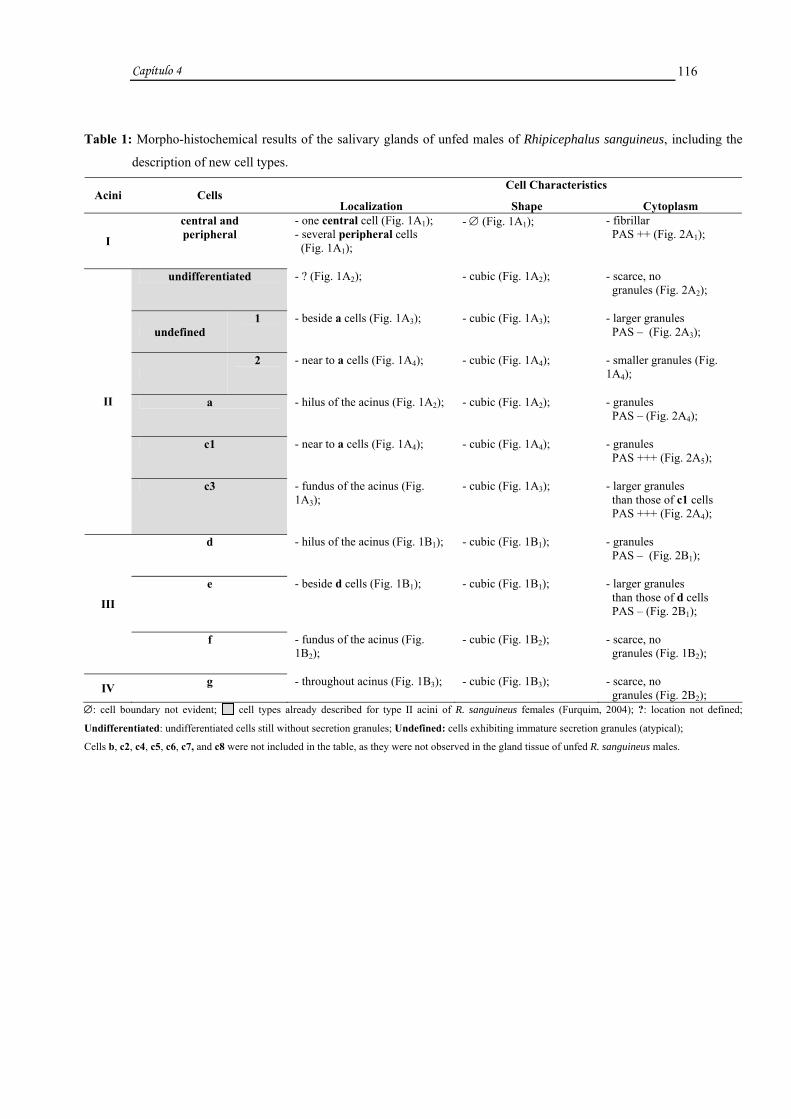
Capítulo 4
116
Table 1: Morpho-histochemical results of the salivary glands of unfed males of Rhipicephalus sanguineus, including the
description of new cell types. Cell Characteristics
Acini Cells Localization Shape Cytoplasm
I
central and peripheral
- one central cell (Fig. 1A1); - several peripheral cells (Fig. 1A1);
- ∅ (Fig. 1A1); - fibrillar PAS ++ (Fig. 2A1);
undifferentiated - ? (Fig. 1A2); - cubic (Fig. 1A2); - scarce, no granules (Fig. 2A2);
undefined 1 - beside a cells (Fig. 1A3); - cubic (Fig. 1A3); - larger granules
PAS – (Fig. 2A3);
2 - near to a cells (Fig. 1A4); - cubic (Fig. 1A4); - smaller granules (Fig.
1A4);
a - hilus of the acinus (Fig. 1A2); - cubic (Fig. 1A2); - granules PAS – (Fig. 2A4);
c1 - near to a cells (Fig. 1A4); - cubic (Fig. 1A4); - granules PAS +++ (Fig. 2A5);
II
c3 - fundus of the acinus (Fig. 1A3);
- cubic (Fig. 1A3); - larger granules than those of c1 cells PAS +++ (Fig. 2A4);
d - hilus of the acinus (Fig. 1B1); - cubic (Fig. 1B1); - granules PAS – (Fig. 2B1);
e - beside d cells (Fig. 1B1); - cubic (Fig. 1B1); - larger granules than those of d cells PAS – (Fig. 2B1);
III
f - fundus of the acinus (Fig. 1B2);
- cubic (Fig. 1B2); - scarce, no granules (Fig. 1B2);
IV g - throughout acinus (Fig. 1B3); - cubic (Fig. 1B3); - scarce, no granules (Fig. 2B2);
∅: cell boundary not evident; cell types already described for type II acini of R. sanguineus females (Furquim, 2004); ?: location not defined;
Undifferentiated: undifferentiated cells still without secretion granules; Undefined: cells exhibiting immature secretion granules (atypical);
Cells b, c2, c4, c5, c6, c7, and c8 were not included in the table, as they were not observed in the gland tissue of unfed R. sanguineus males.

Capítulo 4
117
Table 2: Morpho-histochemical results of the salivary glands of Rhipicephalus sanguineus males at day two post-
attachment, including the description of new cell types.
Cell Characteristics Acini Cells
Localization Shape Cytoplasm
I central and peripheral
- one central cell (Fig. 1B4); - several peripheral cells (Fig. 1B4);
- ∅ (Fig. 1B4); - fibrillar PAS ++ (Fig. 2B3);
a - hilus of the acinus (Fig. 1B5); - cubic (Fig. 1B5); - granules PAS – (Fig. 2B4);
b - beside a cells (Fig. 1C1); - cubic (Fig. 1C1); - elliptic and heterogeneous granules
PAS+ and PAS ++ (Fig. 2C1);
c1 - near to a cells (Fig. 1C2); - cubic (Fig. 1C2); - granules
PAS +++ (Fig. 2C2);
c2 - near to a cells (Fig. 1C2); - cubic (Fig. 1C2); - granules PAS + (Fig. 2C3);
c3 - fundus of the acinus (Fig. 1C3); - cubic (Fig. 1C3); - larger granules than those of c1 cells PAS +++ (Fig. 2C3);
c4 - fundus of the acinus (Fig. 1C4); - cubic (Fig. 1C4); - elliptic granules PAS – (Fig. 2C4);
c5 - near a cells (Fig. 1C5); - cubic (Fig. 1C5); - smaller granules than those of a cells and larger than those of c3 cells PAS ++ (Fig. 2C5);
c6 - near a cells (Fig. 2D1); - cubic (Fig.2D1); - fine granules PAS ++ (Fig. 2D1);
c7 - near a cells (Fig. 1C6); - cubic (Fig. 1C6); - larger granules than those of a cells PAS ++ (Fig. 2D2);
II
c8 - near a cells (Fig. 1D1); - cubic (Fig. 1D1); - larger granules than those of c3 cells PAS + (Fig. 2D3);
d - hilus of the acinus (Fig. 1D2); - cubic (Fig. 1D2); - granules PAS – (Fig. 2D4);
III e - beside d cells (Fig. 1D2); - cubic (Fig. 1D2); - larger granules than those of d cells PAS – (Fig. 2D4);
IV
g - throughout acinus (Fig. 1D3); - cubic (Fig. 1D3); - homogeneous granules PAS ++ (fig. 2E1) and heterogeneous granules PAS + and PAS ++ (Fig. 2E2);
∅: cell boundary not evident; cell types already described for type II acini of R. sanguineus females (Furquim, 2004); new cell types described in
the present study; Cells undifferentiated, undefined 1 and 2, and f were not included in the table, as they were not observed in the gland tissue of R.
sanguineus males at day two post-attachment.

Capítulo 4
118
Table 3: Morpho-histochemical results of the salivary glands of Rhipicephalus sanguineus males at day four post-
attachment, including the description of new cell types.
Cell Characteristics Acini Cells
Localization Shape Cytoplasm
I
central and peripheral
- one central cell (Fig. 1D4); - several peripheral cells (Fig. 1D4);
- ∅ (Fig. 1D4); - fibrillar PAS – (Fig. 2E3);
a - hilus of the acinus (Fig. 1D5); - cubic (Fig. 1D5); - granules PAS – (Fig. 2E4);
b
- beside a cells (Fig. 1D5); - cubic (Fig. 1D5); - elliptic and heterogeneous granules PAS++ and PAS +++ (Fig. 2E4);
c1 - near to a cells (Fig. 1E1); - cubic (Fig. 1E1); - granules
PAS +++ (Fig. 2F1);
c2 - near to a cells (Fig. 1E2); - cubic (Fig. 1E2); - granules PAS + (Fig. 2F2);
c3 - fundus of the acinus (Fig. 1E2); - cubic (Fig. 1E2); - larger granules than those of c1 cells PAS +++ (Fig. 2F3);
c4 - fundus of the acinus (Fig. 1E3); - cubic (Fig. 1E3); - elliptic granules PAS – (Fig. 2F4);
II
c5 - near a cells (Fig. 1E4); - cubic (Fig. 1E4); - smaller granules than those of a cells and larger than those of c3 cells PAS ++ (Fig. 2F5);
d - hilus of the acinus (Fig. 1F1); - cubic (Fig. 1F1); - granules PAS – (Fig. 2G1);
III e - beside d cells (Fig. 1F1); - cubic (Fig. 1F1); - larger granules than those of d cells PAS – (Fig. 2G1);
IV g - throughout acinus (Fig. 1F2, F3); - cubic (Fig. 1F2, F3); - granules PAS +++ (fig. 2G1, G2);
∅: cell boundary not evident; cell types already described for type II acini of R. sanguineus females (Furquim, 2004);
Cells undifferentiated, undefined 1 and 2, c6, c7, c8, and f were not included in the table, as they were not observed in the gland tissue of R. sanguineus
males at day four post-attachment.

FIGURES
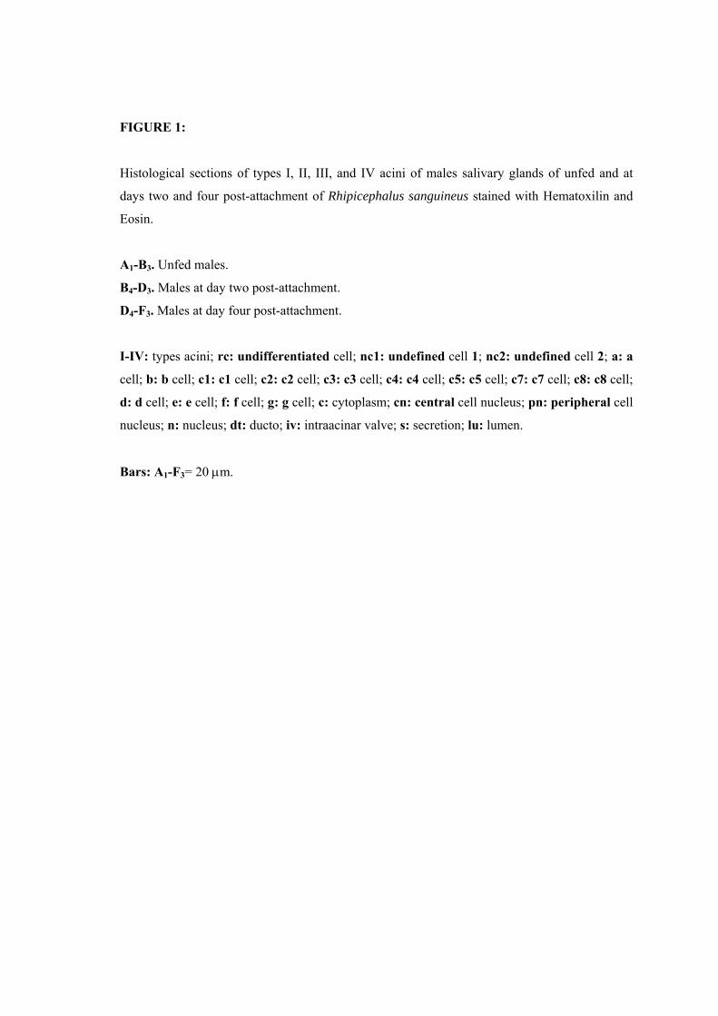
FIGURE 1:
Histological sections of types I, II, III, and IV acini of males salivary glands of unfed and at
days two and four post-attachment of Rhipicephalus sanguineus stained with Hematoxilin and
Eosin.
A1-B3. Unfed males.
B4-D3. Males at day two post-attachment.
D4-F3. Males at day four post-attachment.
I-IV: types acini; rc: undifferentiated cell; nc1: undefined cell 1; nc2: undefined cell 2; a: a
cell; b: b cell; c1: c1 cell; c2: c2 cell; c3: c3 cell; c4: c4 cell; c5: c5 cell; c7: c7 cell; c8: c8 cell;
d: d cell; e: e cell; f: f cell; g: g cell; c: cytoplasm; cn: central cell nucleus; pn: peripheral cell
nucleus; n: nucleus; dt: ducto; iv: intraacinar valve; s: secretion; lu: lumen.
Bars: A1-F3= 20 µm.
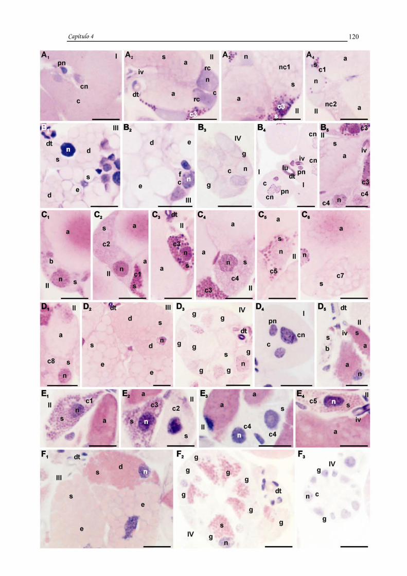
Capítulo 4
120

FIGURE 2:
Histological sections of types I, II, III, and IV acini of males salivary glands of unfed and at
days two and four post- attachment of Rhipicephalus sanguineus stained with PAS and
counterstained with Methyl Green.
A1-B2. Unfed males.
B3-E2. Males at day two post-attachment.
E3-G2. Males at day four post-attachment.
I-IV: types acini; rc: undifferentiated cell; nc1: undefined cell 1; a: a cell; b: b cell; c1: c1
cell; c2: c2 cell; c3: c3 cell; c4: c4 cell; c5: c5 cell; c6: c6 cell; c7: c7 cell; c8: c8 cell; d: d cell;
e: e cell; g: g cell; fc: fibrillar cytoplasm; cn: central cell nucleus; pn: peripheral cell
nucleus;c: cytoplasm; n: nucleus; s: secretion; dt: duct; iv: intraacinar valve; hm:
homogeneous granules; ∗: heterogeneous granules.
Bars: B2= 10 µm; A1-B1 and B3-G2= 20 µm.

Capítulo 4
121

Capítulo 4
122
DISCUSSION
The histology of the salivary glands of ticks has been described by several
authors especially in females (Till, 1961; Binnington, 1978; Walker et al., 1985;
Sonenshine, 1991; Serra-Freire and Olivieri, 1993), as they, unlike males, feed
continuously until engorgement, ingesting larger quantities of blood and causing more
damages to the host.
For this study, we utilized the classification system developed by Binnington
(1978), Walker et al. (1985), Gill and Walker (1987) for types I, III, and IV acini. For
type II acinus, we adopted the system proposed by Furquim (2004) described for
Rhipicephalus sanguineus females that includes four new cell types: undefined 1 and 2,
c5 and c6. In addition to these, we described in males, for the first time, two additional
cell types, c7 and c8. These might be present only in males of this species, or if present
in females, they may be active only during the first day of attachment to the host
(Furquim, 2004).
In this study, we observed that gland cells of males were activated with the onset
of feeding, supporting Sanders et al. (1996) that in Amblyomma americanum reported
that the beginning of feeding promoted the development and increase of production of
different proteins in the salivary glands.
In feeding males, the authors observed that as soon as the secretory cycle ended,
the aspect of these organs returned to the observed in unfed individuals. This was also
reported by Wang et al. (1999), which described a reprogramming in the expression of
the gland tissue of R. appendiculatus when feeding ceased and that was maintained until
the next attachment. In R. sanguineus, only cells a, c1, c3, d, and e were continuously
active and therefore, exhibited mature granules in the cytoplasm since the first secretory
cycle. In the remaining cell types, granules were released at the end of each cycle,
restarting the secretory activity when reattachment occurred.
Our findings indicate that the glands of males at day two post-attachment were
in early stages of the secretory cycle while at day four post-attachment, glands were in
late stages of one of the several cycles of this organ. Wang et al. (1999), studying R.
appendiculatus females and males, showed that the salivary gland cycle in male ticks is
based on an inversely proportional relationship between the amount of blood consumed

Capítulo 4
123
and the difficulty of the gland to reprogram its expression. Since males do not consume
large volumes of blood due to the physical resistance caused by the presence of the
dorsal scutum, they do not reach the “critical weight” (Weiss and Kaufman, 2001) and
glands still remain active during intervals between one cycle and the next, allowing
feeding in the next attachment to the host.
The histological data presented in our study showed that type I acinus did not
undergo structural or size changes during feeding, as reported by Till (1961), Walker et
al. (1985), Binnington (1978), and Furquim (2004), in males of R. appendiculatus and
B. microplus, and females of R. sanguineus, respectively.
In R. sanguineus, the most developed type II acinus and with more cell types
were observed in males at day two post-attachment. Ten types of cells were observed,
unlike Walker et al. (1985) and Gill and Walker (1987) that reported five types (a, b,
c1–c3), and Binnington (1978) that described six types (a, b, c1–c4).
In unfed males, cells a were already filled with immature secretion granules; at
day two post-attachment, cells became larger and secretion granules was maturing; and
at day four, cells were smaller and with mature granules. These results confirm those
obtained by Binnington (1978), Walker et al. (1985), and Gill and Walker (1987).
In R. sanguineus, the secretion present in cells a does not contain
polysaccharides, unlike the observed by Serra-Freire and Olivieri (1993) that reported
this compound in A. cajennense. According to Binnington (1978), Walker et al. (1985),
Fawcett et al. (1986), Gill and Walker (1987), and Sonenshine (1991), these cells might
be involved in the secretion of cement precursors, a likely role played by this type of
cell in the individuals examined in this study.
Cells b were not observed in unfed males, contrary to Binnington (1978) and
Gill and Walker (1987), which reported in B. microplus and H. anatolicum anatolicum,
respectively, the presence of secretion in the cytoplasm of these cells in unfed males. In
males at day two post-attachment, cells b were filled with heterogeneous granules,
including the presence of polysaccharides (PAS staining was observed in one area of the
granule, while staining was absent in another). At day four post-attachment, these
granules did not undergo histochemical changes, but were present in fewer numbers, as
also reported by Walker et al. (1985). These authors detected activity in cells b of R.
appendiculatus during early stages, which decreased throughout feeding. In addition,

Capítulo 4
124
Walker et al. (1985) suggested that the secretion synthesized by cells b may be involved
in the manipulation of the host response, and this could also be the case in R.
sanguineus males.
Cells c1 were active in unfed males; in those at day two post-attachment, they
were more developed and remained unchanged in those at day four post-attachment.
According to Gill and Walker (1987), the secretory activity of these cells increases
during feeding and most granules are released around 72 hours of feeding, while Walker
et al. (1985) reported a hypertrophy of these cells during feeding.
Previous studies have shown that the secretion produced by cells c1 in R.
sanguineus females might be fully active in individuals after four days of feeding, the
beginning of a phase characterized by the largest consumption of blood (Furquim,
2004). Thus, cells c1 might also play a similar role in males, participating in the
consumption of blood.
The present study showed that cells c2 of unfed males exhibited secretion
granules, unlike the observed by Gill and Walker (1987). At day two post-attachment,
cells were filled with granules and still active at day four post-attachment, confirming
the obtained by Binnington (1978), Walker et al. (1985), and Gill and Walker (1987).
The secretion of cells c2 analyzed in our study might play a role in the
consumption of blood by the tick, as also described in B. microplus (Binnington, 1978).
In unfed males, cells c3 already contained secretion, contrary to the reported by
Walker et al. (1985), which demonstrated that in R. appendiculatus, these cells are
inactive in individuals at this stage. In the individuals here examined, at day two post-
attachment, cells c3 were more developed and remained active at day four post-
attachment. These results also contradict those obtained by Walker et al. (1985) that
observed hypertrophy in these cells in more advanced stages of feeding.
Cells c4 of unfed R. sanguineus males did not exhibit secretion granules in the
cytoplasm, unlike the observed by Binnington (1978) in unfed B. microplus males. In R.
sanguineus at day two post-attachment, these cells were developed; and at day four
post-attachment, they were less active. The morphological and histochemical
characteristics of the granules were similar to those found in B. microplus (Binnington,
1978), suggesting that in R. sanguineus, cells c4 also produce and secrete enzymes
assisting in feeding, promoting the consumption of blood by the tick.
Capítulo 4
125
The cells c5 and c6 were not observed on unfed R. sanguineus males, confirming
the observed in females of the same species (Furquim, 2004). At day two post-
attachment, these cells were developed and at day four post-attachment, cells c5
remained active while c6 were no longer observed, contrary to the observed in females
after four days of feeding, in which c6 were still active (Furquim, 2004).
Based on the results reported in a previous study on R. sanguineus females, the
secretion of cells c5 and c6 may play a role in the initial phase of the secretory cycle,
inhibiting the host response in order to overcome any specific resistance to the
attachment of the parasite (Furquim, 2004).
In R. sanguineus males, cell types c7 and c8 were not observed in unfed
individuals, but were present in those at day two post-attachment, and at day four post-
attachment, these cells were no longer observed, suggesting the end of their activities.
As the secretory cycle progressed, polysaccharides were demonstrated in the granules of
cells undefined 1, previously observed only in unfed males. This histochemical change
in composition characterized these cells as a new cell type: c7. If cells c7 and c8 were
only present in R. sanguineus males, they probably would play specific roles in these
individuals, such as during mating. If they were also present in females, they might be
involved in the formation of the feeding lesion that occurs during the first two days of
attachment (Balashov, 1972 apud Kaufman and Lomas, 1996).
The type III acinus of males examined here, unlike the observed in females
(Furquim, 2004), did not undergo prominent morphologic changes during feeding, since
males consumed small amounts of blood, and thus the participation of these acini was
not needed in the excretion of water and ions. In R. sanguineus males, we only observed
changes in size of these acini. In males at day two post-attachment, type III acini were
larger than those of males at day four post-attachment.
Cells d and e of type III acinus of unfed R. sanguineus males already contained
secretion, confirming the reported by Binnington (1978), Walker et al. (1985), Gill and
Walker (1987), and Serra-Freire and Olivieri (1993). These cells became even more
developed at day two post-attachment, but a decrease in activity was observed at day
four post-attachment. Binnington (1978), Walker et al. (1985), and Gill and Walker
(1987), however, did not observe changes in secretory activity during feeding. The
histochemical results on the secretion of these cells confirm those obtained by
Capítulo 4
126
Binnington (1978) and Walker et al. (1985). These authors demonstrated the presence
of: lipoproteins, as cement precursors (Binnington,1978; Walker et al., 1985; Bishop et
al., 2002, Gill and Walker, 1987); aminopeptidases participating in the infiltration of
cement in the host’s skin (Walker et al., 1985); and phenol and phenoloxidase,
associated with the process of hardening of the cement cone (Binnington, 1978). These
functions might also be performed by these cell types in the males here examined.
Cells f of R. sanguineus males in the three conditions examined were inactive, as
reported by Binnington (1978) and Gill and Walker (1987). This was probably due to
the low ingestion of blood, since there was no need for the cells to secrete compounds
exclusively active in the early phase of blood consumption (Furquim, 2004), or in
osmoregulation (Binnington, 1978; Walker et al., 1985; Coons and Lamureaux, 1986;
Gill and Walker, 1987), unlike the observed by Walker et al. (1985).
Type IV acini of unfed R. sanguineus males were “undifferentiated”, confirming
the results obtained by Binnington (1978), Walker et al. (1985), Gill and Walker (1987),
and Serra-Freire and Olivieri (1993). At day two post-attachment, these acini became
larger, as described by Walker et al. (1985) and Gill and Walker (1987), while at day
four post-attachment, the secretory activity decreased, contrary to the reported by
Binnington (1978), which observed an increase in the secretory activity during feeding
and cells filled with secretion in males after approximately 96 hours of feeding.
According to the literature, the role of type IV acinus is to produce cement in
some Ixodidae (Fawcett et al., 1986), as well as to facilitate the transfer of the
spermatophore to the females (Feldman-Meshsam et al., 1970 apud Fawcett et al.,
1986). Our findings, however, do not suggest that these acini are involved in the
formation of the cone, since males exhibit all cell types present in females (Furquim,
2004). Consequently, a specific structure for the construction of the cone is not
necessary. Thus, type IV acini of R. sanguineus might participate in the lubrication of
the spermatophore as well as in physiological and behavioral changes in females after
mating. Weiss and Kaufman (2004) have demonstrated that the proteins produced in the
gonads of A. hebraeum males and that are transferred to females through mating are not
capable of trigger all physiological and behavioral responses, such as the development
of the ovary.

Capítulo 4
127
The results obtained in the present study suggest that the salivary glands of R.
sanguineus males do not exhibit histological differences compared to females, except
by the presence of type IV acinus and cells c7 and c8. If they in fact are only present in
males, these cells might provide lubrication of the spermatophore and trigger responses
in females that allow them to reproduce.
ACKNOWLEDGEMENTS
This research has been supported by CAPES (Coordenação de Aperfeiçoamento
de Pessoal de Nível Superior) and FAPESP (Fundação de Amparo à Pesquisa do Estado
de São Paulo) Grant nº 05/59208-3. The authors thank Gerson Mello Souza and Ronaldo
Del Vecchio for the techinical support. Part of this work has been facilitated through the
Integrated Consortium on Ticks and Tick-borne Diseases (ICTTD-3) supported by the
European Union under contract number 510561-INCO.

Capítulo 4
128
REFERENCES
Balashov, Y.S., 1972. Bloodsucking ticks (Ixodidae)- vectors of disease of man and
animals. Hoogstraal, H., Tachell, R.J. (Eds.), Misc. Publ. Entomol. Soc. Am. 8, 161-
376.
Bechara, G.H., Szabó, M.P.J., Ferreira, B.R., Garcia, M.V., 1995. Rhipicephalus
sanguineus tick in Brazil: feeding and reproductive aspects under laboratorial
conditions. Brazil. J. Vet. Parasitol. 4, 61-66.
Binnington, K.C., 1978. Sequential changes in salivary gland structure during
attachment and feeding of the cattle tick, Boophilus microplus. Int. J. Parasitol. 8, 97-
115.
Bishop, R., Lambson, B., Wells, C., Pandit, P., Osaso, J., Nkonge, C., Morzaria, S.,
Musoke, A., Nene, V., 2002. A cement protein of the tick Rhipicephalus
appendiculatus, located in the secretory e cell granules of the type III salivary gland
acini, induces strong antibody responses in cattle. Int. J. Parasitol. 32, 833-842.
Bowman, A.S., Sauer, J.R., 2004. Tick salivary glands: function, physiology and future.
Parasitology 129, S67-S81.
Coons, L.B., L’amoreaux, W.J., 1986. Developmental changes in the salivary glands of
male and female Dermacentor variabilis (Say) during feeding. In: Borovsky, D.,
Spielman, A. (Eds.), Host Regulated Developmental Mechanism in Vector Arthropods.
University of Florida-IFAS, Vero Beach, pp. 86-92.
Fawcett, D.W., Binnington, K., Voigt, W.P., 1986. The cell biology of the ixodid tick
salivary gland. In: Sauer, J.R., Hair, A. (Eds.), Morphology, Physiology, and Behavioral
Biology of Ticks. Ellis Horwood, Chichester, pp. 22-45.

Capítulo 4
129
Feldman-Muhsam, B., Borut, S., Saliternik-Givant, S., 1970. Salivary secretion of the
male tick during copulation. J. Insect Physiol. 16, 1945-1949.
Furquim, K.C.S., 2004. Processos degenerativos em glândulas salivares de carrapatos
Rhipicephalus sanguineus (LATREILLE, 1806) (ACARI, IXODIDAE) em jejum, em
alimentação e alimentados. 1º Relatório Científico CAPES, Instituto de Biociências,
UNESP, Rio Claro (S.P.), Brasil, 37 pp.
Gaede, H., Knulle, W., 1997. On the mechanism of water vapour sorption from
unsaturated atmospheres by ticks. J. Exp. Biol. 200, 1491-1498.
Gill, H.S., Walker, A.R., 1987. The salivary glands of Hyalomma anatolicum
anatolicum: structural changes during attachment and feeding. Int. J. Parasitol. 17 (8),
1381-1392.
Kaufman, W.R., Lomas, L.O., 1996. “Male factors” in ticks: their role in feeding and
egg development. Invertebr. Reprod. Develop. 30 (1-3), 191-198.
Marzouk, A.S., Darwish, Z.E.A., 1994. Changes in the salivary glands of female
Hyalomma (Hyalomma) dromedarii during and after feeding. J. Egypt. Soc. Parasitol.
24 (1), 39-57.
McMullen, H.L., Sauer, J.R., Burton, R.L., 1976. Possible role in uptake of water
vapour by ixodid tick salivary glands. J. Insect Physiol. 22, 1281-1285.
McManus, J.F.A., 1946. Histological demonstration of mucin after periodic acid. Nature
158, 202.
Sanders, M.L., Scott, A.L., Glass, G.E., Schwartz, B.S., 1996. Salivary gland changes
and host antibody responses associated with feeding of male lone star ticks (Acari:
Ixodidae). J. Med. Entomol. 33 (4), 628-634.
Capítulo 4
130
Serra-Freire, N.M., Olivieri, J.A., 1993. Structure of the salivary glands of the unfed
male tick Amblyomma cajennense (Fabricius) (Acari: Ixodidae). Mem. Inst. Oswaldo
Cruz 88 (2), 207-213.
Sonenshine, D.E., 1991. Biology of Ticks. Oxford University Press, New York, pp 141-
158.
Till, W.M., 1961. A Contribution to the Anatomy and Histology of the Brown Ear Tick
Rhipicephalus appendiculatus Neumann. Memoirs of the Entomological Society of
Southern Africa, Pretoria, 6, 1-124.
Walker, A.R., Fletcher, J.D., Gill, H., 1985. Structural and histochemical changes in the
salivary glands of Rhipicephalus appendiculatus during feeding. Int. J. Parasitol. 15 (1),
81-100.
Wang, H., Henbest, P.J., Nuttall, P.A., 1999. Successful interrupted feeding of adult
Rhipicephalus appendiculatus (Ixodidae) is accompanied by re-programming of
salivary gland protein expression. Parasitology 119, 143-149.
Weiss, B.L., Kaufman, W.R., 2001. The relationship between “critical weight” and 20-
hydroxyecdysone in the female ixodid tick, Amblyomma hebraeum. J. Insect Physiol.
47, 1261-1267.
Weiss, B.L., Kaufman, W.R., 2004. Two feeding-induced proteins from the male gonad
trigger engorgement of the female tick Amblyomma hebraeum. Proc. Natl. Acad. Sci.
S.A. 101 (16), 5874-5879.

Capítulo 5

Capítulo 5
132
CAPÍTULO 5
TITLE: Degeneration of salivary glands of males of the tick Rhipicephalus sanguineus
(LATREILLE, 1806) (Acari, Ixodidae).
AUTHORS: Karim Christina Scopinho Furquim a, Gervásio Henrique Bechara b and
Maria Izabel Camargo Mathias a, *
a Departamento de Biologia, Instituto de Biociências, UNESP, Av. 24 A, nº 1515, Cx.
Postal 199, CEP: 13506-900, Rio Claro, S.P., Brazil
b Departamento de Patologia Veterinária, FCAV, UNESP, Via de Acesso Prof. Paulo
Castellane, s/n, CEP: 14884-900, Jaboticabal, S.P., Brazil
* Corresponding author. Tel.: +55 19 35264135;
Fax: +55 19 35340009.
E-mail address: [email protected]

Capítulo 5
133
RESUMO
O presente estudo analisou morfo-histoquímicamente as glândulas salivares de
machos de Rhipicephalus sanguineus com sete dias de infestação, três e sete dias pós-
remoção do hospedeiro. Houve degeneração do órgão nas três situações analisadas, que
se intensificou com o decorrer do tempo fora do hospedeiro, sendo maior nos machos
com sete dias pós-remoção que naqueles com sete dias de infestação. Somente nestes
últimos os ácinos I apresentaram-se íntegros e os outros com características de
degeneração. Nos indivíduos com sete dias de infestação os ácinos II tiveram
identificadas as células a, c1-c5, c8 e indeterminadas, onde apenas as c1 e c8
eventualmente estavam íntegras. As outras estavam em degeneração assim como nos
ácinos III todas as células d, e e f e nos IV todas as g.
Todas as células (a, c1-c5, c8 e indeterminadas) observadas nos ácinos II de
machos com três dias pós-remoção do hospedeiro, as d e e nos III e as g nos IV estavam
degenerando. Em alguns dos ácinos Indeterminados ainda pode-se observar o limite de
algumas células e em outros somente uma massa citoplasmática. Nos indivíduos com
três dias pós-remoção foram observados corpos apoptóticos.
Nos machos com sete dias pós-remoção do hospedeiro o processo degenerativo
se intensificou, foram observadas as células a, c1, c3-c5, c8 e indeterminadas nos
ácinos II e as d e e nos III, todas em degeneração. Nos IV, ainda havia resíduo de
secreção, e nos Indeterminados somente uma massa citoplasmática. Nos indivíduos
com sete dias pós-remoção também se observou a presença de corpos apoptóticos.
O presente estudo mostrou ainda que as células das glândulas salivares de
machos de R. sanguineus quando em degeneração sofrem as seguintes alterações: a)
diminuição da produção da secreção com ou sem rompimento dos grânulos, b) mudança
da morfologia nuclear, c) retração citoplasmática, d) perda da forma celular, e) perda
dos limites celulares e f) vacuolização citoplasmática. Todas elas somadas resultam na
fragmentação celular com liberação de corpos apoptóticos.
PALAVRAS-CHAVE: Rhipicephalus sanguineus, glândula salivar, carrapato,
degeneração, estágio de alimentação, morfologia.

Capítulo 5
134
ABSTRACT
The present morpho-histochemical study examined the salivary glands of
Rhipicephalus sanguineus males at day seven post-attachment, and at days three and
seven post-detachment from the host. Degeneration of this organ occurred in the three
stages analyzed and it advanced as time away from the host progressed. Thus,
characteristics of degeneration were more prominent in males at day seven post-
detachment than in males at day seven post-attachment. In the latter, type I acini were
intact; while in other stages these acini exhibited signs of degeneration. In type II acini
of individuals at day seven post-attachment, cells a, c1–c5, c8, and indeterminate were
identified. Only c1 and c8 were intact. The remaining cell types were undergoing
degeneration, as well as all cells d, e and f in type III acini, and all cells g in type IV
acini.
In males at day three post-detachment from the host, all cells (a, c1–c5, c8 and
indeterminate) observed in type II acini, cells d and e in type III acini, and g in type IV
acini were undergoing degeneration. In some Indeterminate acini, the boundaries of
cells still could be distinguished, while in others, only a cytoplasmic mass was
observed. At day three post-detachment, apoptotic bodies were observed.
In males at day seven post-detachment from the host, the degeneration process
progressed. All cells a, c1, c3–c5, c8 and indeterminate in type II acini, and d and e in
type III acini were undergoing degeneration. Type IV acini still contained remnants of
secretion and in Indeterminate acini, only a cytoplasmic mass could be observed. At
this stage, apoptotic bodies were also present.
In addition, the present study revealed that cells of salivary glands of R.
sanguineus males when degenerating the following changes: a) decrease in secretion
production with or without granule breakage, b) changes in nuclear morphology, c)
cytoplasm shrinkage, d) loss of cell shape, e) loss of cell boundaries, and e) cytoplasmic
vacuolation. Together, these changes result in cell fragmentation with release of
apoptotic bodies.
KEY WORDS: Rhipicephalus sanguineus, salivary gland, tick, degeneration, feeding
stage, morphology.

Capítulo 5
135
INTRODUCTION
Male ticks present salivary glands morphologically similar to those of females
(Till, 1961), and as the latter, require compounds in the saliva to assist in the
manipulation of the host (Sanders et al., 1996).
The salivary glands of these individuals consist of types I, II, III and IV acini
(Binnington, 1978; Walker et al., 1985; Gill and Walker, 1987). In R. sanguineus, type I
acini are composed of one central cell and several peripheral ones; while type II acini
contain cells a, b, c1–c8; type III acini, cells d, e and f; and type IV acini, cells g
(Furquim, 2006).
Since males exhibit several feeding stages (attach and detach several times from
the host), salivary glands undergo many secretory cycles during infestation (Wang et al.,
1999). Previous studies conducted with unfed and feeding R. sanguineus males have
also demonstrated that in each secretory cycle, the gland undergo the following stages:
1) intense gland activity with synthesis and storage of secretion, 2) secretion maturation,
3) secretion discharge, and 4) acquisition of characteristics observed in unfed males,
allowing the gland to restart a new secretory cycle through cell reprogramming
(Furquim, 2006).
Walker et al. (1985), studying the salivary glands of R. appendiculatus males,
reported that the gland tissue of these individuals do not undergo degeneration and
remain functional after the end of each feeding period.
In female ticks, gland degeneration is signaled by the ecdysteroid hormone
(Lomas et al., 1998). In Amblyomma hebraeum males exposed to 20-hydroxyecdysone
(ecdysteroid hormone) in vitro for four days, the secretory capacity of the gland
decreased without inducing the formation of autophagic vacuoles in the cytoplasm, not
increasing the synthesis of this ecdysteroid when comparing feeding males (seven and
fourteen days) to those after detachment from the host (Kaufman, 1990). Clearly, these
studies are not conclusive to confirm whether the ecdysteroid hormone signals gland
degeneration in A. hebraeum males, as observed in females of the same species (Lomas
et al., 1998).
Due to the scarce information available in the literature regarding gland
degeneration in ticks, this study aimed at describing morphologically and

Capítulo 5
136
histochemically the degenerating salivary glands of R. sanguineus males at day seven
post-attachment, and at day three and seven post-detachment.
MATERIAL AND METHODS
For this study, were utilized males of the tick Rhipicephalus sanguineus at day
seven post-attachment to the host, and at day three and seven post-detachment from the
host (rabbit).
Unfed individuals were provided by Dr. Gervásio Henrique Bechara of the
Department of Veterinary Pathology of UNESP, Jaboticabal campus (São Paulo, Brazil)
from a colony maintained under controlled conditions (29o C, 80% humidity, and 12
hour photoperiod) in BOD incubator.
Males were placed with some females inside a feeding chamber previously glue
with an atoxic and non-lesive preparation (Britannia Adhesive-Unit 4, UK) to the shave
back of the host (rabbit ) according to technique described elsewhere (Bechara et al.,
1995). After seven days post-attachment, males were collected and a group was
assigned to histological procedures. Another group was maintained alive for three and
seven days in closed containers with breathing holes to be later analyzed
morphologically and histochemically.
Upon completion of the feeding periods, males were collected and salivary
glands were removed in saline solution and fixed in a 10 % neutral buffered formalin
and acetone solution (9:1) for one hour and thirty minutes at 4o C. After fixation, the
material was dehydrated in a series of increasing concentrations of alcohol (70%, 80%,
90% and 95%), embedded in resin (Leica), and sectioned at 3 µm thickness. Sections
were mounted on glass slides and stained with Hematoxylin-Eosin and PAS (Periodic
Acid Schiff) McManus (1946) for detection of polysaccharides. Samples were
counterstained with Methyl Green to demonstrate chromatin, later mounted in Canada
balsam, and observed under light microscope.

Capítulo 5
137
RESULTS
Males at day seven post-attachment
The salivary glands of these individuals exhibit intact type I acinus (Figs. 1, 53),
while types II (Figs. 2-10, 54-59), III (Figs. 11-16, 60) and IV (Figs. 17-19, 61) acini
are undergoing degeneration.
Intact acini present regular shape (Figs. 1, 53); cells containing fibrillar
structures in their cytoplasm (Fig. 53) and nuclei of central cells as well as peripheral
ones do not exhibit changes (Figs. 1, 53).
Type II acini may be regular (Figs. 3, 8, 10, 54, 55, 57, 59) or irregular (Fig. 2,
4-7, 9, 56, 58), some cells c1 and c8 are intact (Figs. 55, 57) while others are
undergoing degeneration and exhibit nuclear changes (Figs. 54, 3). Degenerating cells
are, a: cubic (Figs. 2, 3, 54-56, 58, 59) or irregular (Fig. 4) cells exhibiting intact (Figs.
2, 54-56, 58) or broken (Figs. 3, 4) granules and nuclear changes (Figs. 3); c1: cubic
cells exhibiting intact granules (Figs. 54, 55) and intact (Fig. 55) or changed (Fig. 54)
nuclei; c2: cubic (Figs. 5, 55) or irregular (Fig. 6) cells exhibiting intact granules (Figs.
5, 6, 55) and nuclear changes (Figs. 5); c3: cubic cells exhibiting broken granules and
nuclear changes (Figs. 7, 54, 55); c4: cubic (Figs. 8, 56) or irregular (Fig. 9) cells
exhibiting intact granules (Figs. 8, 9, 56) and nuclear changes (Figs. 9 and 56); c5:
cubic cells exhibiting broken granules (Figs. 2, 6, 56-58) and nuclear changes (Figs. 2,
57, 58) and c8: cubic cells exhibiting intact granules (Figs. 3, 57, 59) and intact (Fig.
57) or changed (Figs. 3) nuclei. Were also observed some cells that could not be
identified due to advanced stages of degeneration. These cells were termed
indeterminate: cubic (Figs. 4, 59) or irregular (Figs. 10, 58) cells exhibiting remnants
of secretion (Figs. 4, 10, 58, 59) and nuclear changes (Figs. 10, 58, 59). Included in this
group are cells b, c6, and c7 that either already degenerated or are in late stages of
degeneration, and are no longer observed.
Type III acini may be irregular (Figs. 12, 15, 16) and some, fragmenting (Fig.
13). Degenerating cells are all cells d: cubic (Figs. 11, 14, 60), irregular (Fig. 12) or
fragmenting (Fig. 13) cells exhibiting broken granules (Figs. 11-14, 60) and nuclear
changes (Figs. 11-13); e: cubic (Figs. 14, 60), irregular (Fig. 15) or fragmenting (Fig.
Capítulo 5
138
13) cells exhibiting broken granules (Fig. 13-15, 60) and nuclear changes (Figs. 15 and
60), and f: irregular cells exhibiting nuclear changes (Figs. 16).
In type IV acini, cells g are cubic (Figs. 17, 61), irregular (Fig. 18) or
fragmenting (Fig. 19) and present broken granules (Figs. 17-19, 61), cytoplasm
shrinkage (Fig. 18) and nuclear changes (Figs. 17, 18, 61).
Males at day three post-detachment
In these individuals, all acini exhibit more prominent degenerative
characteristics (Figs. 20-35, 62-67). Acini with cells that have lost their original
granular characteristics are also observed, and could not be identified: these were
termed Indeterminate (Figs. 33, 34). Apoptotic bodies are formed (Fig. 35).
Type I acini are regular (Figs. 20, 62) with cells exhibiting fibrillar structures in
the cytoplasm (Fig. 62), in addition to nuclear changes (Figs. 20, 62).
Type II acini may be irregular (Figs. 22-24, 63-65). Cells a: cubic (Figs. 21, 22,
24, 64, 65) or irregular (Figs. 23, 63) cells exhibiting intact (Figs. 21, 63-65) or broken
(Figs. 22-24) granules and nuclear changes (Figs. 21, 63); c1: cubic cells exhibiting
broken granules (Figs. 24, 64) and nuclear changes (Figs. 64); c2: cubic (Fig. 24) or
irregular cells (Fig. 25) exhibiting intact granules and nuclear changes (Figs. 24, 25);
c3: cubic (Figs. 21, 24, 64) or irregular (Figs. 22, 25) cells exhibiting broken granules
(Fig. 21, 22, 24, 25, 64) and nuclear changes (Figs. 21, 22, 24); c4: irregular cells
exhibiting intact granules, and nuclear changes (Figs. 23); c5: cubic cells exhibiting
broken granules (Figs. 23, 26, 63-65), and c8: irregular cells exhibiting broken granules
(Figs. 23, 64, 65) and intact (Fig. 64) or changed (Fig. 65) nuclei are undergoing
degeneration. indeterminate cells: irregular (Figs. 27, 64, 65) cells exhibiting remnants
of secretion (Figs. 64, 65) are observed in higher numbers than in the previous feeding
stage (Figs. 4, 10, 58, 59).
Type III acini are regular (Figs. 28, 66), irregular (Fig. 29) or fragmenting (Fig.
30). Degenerating cells are, d: cubic (Figs. 28, 66), irregular (Fig. 29) or fragmenting
(Fig. 30) cells exhibiting broken granules (Figs. 28-30, 66) and nuclear changes (Figs.
28, 30) and e: cubic (Figs. 28, 66), irregular (Fig. 29) or fragmenting (Fig. 30) cells
Capítulo 5
139
exhibiting broken granules (Figs. 28-30, 66) and nuclear changes (Fig. 66). Cells f are
no longer observed.
In type IV acini, cells g are irregular (Figs. 31, 67) or fragmenting (Fig. 32) and
exhibit broken granules (Figs. 31, 32, 67), cytoplasmic shrinkage (Fig. 31), and nuclear
changes (Figs. 31, 67).
Indeterminate acini are irregular (Figs. 33, 34), showing cytoplasmic shrinkage
(Fig. 33), and nuclear changes (Fig. 33). Some acini still contain a vacuolated
cytoplasmic mass and some changed nuclei (Fig. 34).
Males at day seven post-detachment from the host
At this stage (Figs. 36-52, 68-75), more prominent degenerative characteristics
are observed than at day three post-detachment from the host (Figs. 20-35, 62-67), such
as indeterminate cells (Figs. 39, 42-44, 69, 70, 72), Indeterminate acini (Fig. 51), and
more abundant apoptotic bodies (Fig. 52).
Type I acini are regular (Figs. 36, 68) and the cytoplasm of cells contain fibrillar
structures (Fig. 68) and changed nuclei (Figs. 36, 68).
Type II acini may be irregular (Figs. 37, 41-45, 70, 72). Cells exhibit the
following degenerative changes, a: cubic (Figs. 37, 38, 41, 42, 44, 69, 70, 72) or
irregular (Figs. 39, 43) cells exhibiting broken granules (Figs. 37-39, 41-44, 69, 70, 72)
and nuclear changes (Figs. 38, 69, 70); c1: cubic cells exhibiting broken granules (Fig.
38, 39, 70) and nuclear changes (Figs. 39, 70); c3: cubic (Figs. 39, 40, 43, 70, 71) or
irregular (Fig. 41) cells exhibiting broken granules (Figs. 39-41, 43, 44, 70, 71) and
nuclear changes (Figs. 41, 71); c4: irregular cells exhibiting intact granules and nuclear
changes (Figs. 38, 71); c5: cubic cells exhibiting broken granules (Figs. 42, 69, 72) and
nuclear changes (Figs. 42, 69); c8: irregular cells exhibiting broken granules (Figs. 38,
69, 71, 72) and nuclear changes (Fig. 69), and indeterminate: cubic (Figs. 39, 42, 43,
69, 70, 72) or irregular (Fig. 44) cells exhibiting remnants of secretion (Figs. 44, 69, 70,
72) and nuclear changes (Figs. 39, 42, 43, 44, 69). Some type II acini appear as a
cytoplasmic mass (Figs. 41, 45) containing remnants of secretion (Fig. 45) and changed
nuclei (Figs. 41, 45).
Type III acini are regular (Fig. 46), irregular (Figs. 47, 73, 74) or fragmenting
(Fig. 48). Cells d and e are cubic (Fig. 46), irregular (Figs. 47, 73, 74) or fragmenting

Capítulo 5
140
(Fig. 48) and exhibit broken granules (Figs. 46-48, 73, 74) and nuclear changes (Figs.
46-48, 73).
In irregular (Figs. 49, 75) or fragmenting (Fig. 50) type IV acini, only a
cytoplasmic mass with secretion is observed (Figs. 49, 50, 75). Nuclei exhibit changes
(Figs. 49, 50, 75).
Indeterminate acini are irregular, vacuolated, with remnants of secretion in the
cytoplasmic mass (Fig. 51). Nuclei exhibit changes (Fig. 51).
To better compare the results, the data are summarized in Tables 1, 2, and 3.

Capítulo 5
141
Table 1: Morpho-histochemical aspects of cells and acini of salivary glands of Rhipicephalus sanguineus
males at day seven post-attachment. Cell Characteristics
Acini Cells Shape Cytoplasm Nucleus
I central and peripheral
- ∅ (Figs. 1, 53); - fibrillar structures PAS ++ (Fig. 53);
- no changes (Figs. 1, 53);
a - cubic (Figs. 2, 3, 54-56, 58, 59); - irregular (Fig. 4);
- granules PAS – (Figs. 2, 54-56, 58) - * (Figs. 3, 4);
- picnotic (Fig. 3);
c1 - cubic (Figs. 54, 55); - granules PAS +++ (Figs. 54, 55);
- intact (Fig. 55); - enlarged (Fig. 54);
c2 - cubic (Figs. 5, 55); - irregular (Fig. 6);
- granules PAS + (Figs. 5, 6, 55);
- enlarged and irregular (Fig. 5);
c3 - cubic (Figs. 7, 54, 55); - * (Figs. 7, 54, 55); - picnotic (Fig. 7); - enlarged (Figs. 54, 55);
c4 - cubic (Figs. 8, 56) - irregular (Fig. 9);
- elliptic granules PAS – (Figs. 8, 9, 56);
- enlarged (Fig. 9); - irregular (Fig. 56);
c5 - cubic (Figs. 2, 6, 56-58); - * (Figs. 2, 6, 56-58); - enlarged and irregular (Fig. 2); - enlarged (Figs. 57, 58);
c8 - cubic (Figs. 3, 57, 59); - larger granules than those of c3 cells PAS + (Figs. 3, 57, 59);
- irregular (Fig. 3); - intact (Fig. 57);
II
indeterminate - cubic (Figs. 4, 59); - irregular (Figs. 10, 58);
- remnants of secretion (Fig. 4, 10, 58, 59);
- picnotic (Fig. 10); - enlarged (Figs. 58, 59);
d - cubic (Figs. 11, 14, 60); - irregular (Fig. 12); - fragmenting (Fig. 13);
- * (Figs. 11, 12-14, 60); - picnotic (Figs. 11, 12); - irregular (Fig. 13);
e - cubic (Figs. 14, 60); - irregular (Fig. 15); - fragmenting (Fig. 13);
- * (Figs. 13, 14, 15, 60); - enlarged and irregular (Fig. 15); - picnotic (Fig. 60); III
f - irregular (Fig. 16); - scarce (Fig. 16); - irregular (Fig. 16);
IV g - cubic (Figs. 17, 61);
- irregular (Fig. 18); - fragmenting (Fig. 19);
- * (Figs. 17-19, 61); - cytoplasmic shrinkage (Fig. 18);
- picnotic (Figs. 17, 61); - enlarged (Fig. 18); - fragmenting (Fig. 18);
∅: cell boundaries not evident; *: broken secretion granules;

Capítulo 5
142
Table 2: Morpho-histochemical aspects of cells and acini of salivary glands of Rhipicephalus sanguineus
males at day three post-detachment. Cell Characteristics
Acini Cells Shape Cytoplasm Nucleus
I central and peripheral
- ∅ (Figs. 20, 62); - fibrillar structures PAS +++ (Fig. 62);
- chromatin margination (Figs. 20, 62);
a - cubic (Figs. 21, 22, 24, 64, 65); - irregular (Fig. 23, 63);
- granules PAS – (Figs. 21, 63-65); - * (Figs. 22-24);
- fragmented (Fig. 21); - picnotic (Fig. 63);
c1 - cubic (Figs. 24, 64); - * (Figs. 24, 64); - enlarged (Fig. 64);
c2 - cubic (Fig. 24); - irregular (Fig. 25);
- granules (Figs. 24, 25); - enlarged (Figs. 24, 25);
c3 - cubic (Figs. 21, 24, 64); - irregular (Figs. 22, 25);
- * (Figs. 21, 22, 24, 25, 64); - picnotic (Fig. 21); - enlarged (Figs. 22, 24);
c4 - irregular (Fig. 23); - elliptic granules PAS – (Fig. 23);
- irregular (Fig. 23); - enlarged and irregular (Fig. 23);
c5 - cubic (Figs. 23, 26, 63-65); - * (Figs. 23, 26, 63-65); - not observed;
c8 - irregular (Figs. 23, 64, 65); - * (Figs. 23, 64, 65); - intact (Fig. 64); - irregular (Fig. 65);
II
indeterminate - irregular (Figs. 27, 64, 65); - remnants of secretion (Figs. 64, 65);
- not observed;
d - cubic (Figs. 28, 60); - irregular (Fig. 29); - fragmenting (Fig. 30);
- * (Figs. 28-30, 66); - irregular (Fig. 28); - picnotic (Fig. 30);
III e - cubic (Figs. 28, 66); - irregular (Fig. 29); - fragmenting (Fig. 30);
- * (Figs. 28-30, 66); - picnotic (Fig. 66);
IV g - irregular (Figs. 31, 67);
- fragmenting (Fig. 32); - * (Figs. 31, 32, 67); - cytoplasmic shrinkage (Fig. 31);
- irregular (Fig. 31); - picnotic (Fig. 67);
- irregular (Fig. 33); - cytoplasmic shrinkage (Fig. 33);
- fragmented (Fig. 33); Indeterminate
- # (Fig. 34); - ! vacuolated (Fig. 34); - enlarged (Fig. 34);
∅: cell boundaries not evident; *: broken secretion granules; #: loss of cell boundaries; !: cytoplasmic mass;

Capítulo 5
143
Table 3: Morpho-histochemical aspects of cells and acini of salivary glands of Rhipicephalus sanguineus males at day seven post-detachment.
Cell Characteristics Acini Cells Shape Cytoplasm Nucleus
I central and peripheral
- ∅ (Figs. 36, 68); - fibrillar structures PAS +++ (Fig. 68);
- enlarged and with chromatin margination (Figs. 36, 68);
a - cubic (Figs. 37, 38, 41, 42, 44, 69, 70, 72); - irregular (Figs. 39, 43);
- * (Figs. 37-39, 41-44, 69, 70, 72);
- picnotic (Figs. 38, 69, 70);
c1 - cubic (Figs. 38, 39, 70); - * (Figs. 38, 39, 70); - enlarged (Fig. 39); - picnotic (Fig. 70);
c3 - cubic (Figs. 39, 40, 43, 70, 71); - irregular (Fig. 41);
- * (Figs. 39-41, 43, 44, 71); - enlarged and irregular (Fig. 41); - enlarged (Fig. 71);
c4 - irregular (Figs. 38, 71); - elliptic granules PAS – (Figs. 38, 71);
- irregular (Fig. 38); - picnotic (Fig. 38); - enlarged (Fig. 71);
c5 - cubic (Figs. 42, 69, 72); - * (Figs. 42, 69, 72); - enlarged and irregular (Fig. 42); - enlarged (Fig. 69);
c8 - irregular (Figs. 38, 69, 71, 72); - * (Figs. 38, 69, 71, 72); - picnotic (Figs. 69);
indeterminate - cubic (Figs. 39, 42, 43, 69, 70, 72); - irregular (Fig. 44);
- remnants of secretion (Figs. 44, 69, 70, 72);
- enlarged (Figs. 39, 43, 69); - enlarged and irregular (Figs. 42, 44);
II
- # (Figs. 41, 45); - ! with (Fig. 45) or without (Fig. 41) remnants of secretion;
- irregular (Fig. 41); - enlarged (Fig. 41); - enlarged and irregular (Fig. 45);
d - cubic (Fig. 46); - irregular (Figs. 47, 73, 74); - fragmenting (Fig. 48);
- * (Figs. 46-48, 73); - picnotic (Figs. 47, 48); - enlarged and irregular (Fig. 73);
III e - cubic (Fig. 46); - irregular (Figs. 47, 73, 74); - fragmenting (Fig. 48);
- * (Figs. 46-48, 74); - enlarged and irregular (Fig. 46); - irregular (Fig. 47); - picnotic (Figs. 46, 47, 48);
IV
- # (Figs. 49, 50, 75); - cytoplasmic mass with * (Figs. 49, 50, 75);
- fragmenting (Figs. 49, 50); - enlarged and irregular (Fig. 75); - picnotic (Fig. 75);
Indeterminate
- # (Fig. 51); - ! vacuolated and with remnants of secretion (Fig. 51);
- irregular (Fig. 51); - enlarged (Fig. 51); - enlarged and irregular (Fig. 51)
∅: cell boundaries not evident; *: broken secretion granules; #: loss of cell boundaries; !: cytoplasmic mass;

FIGURES
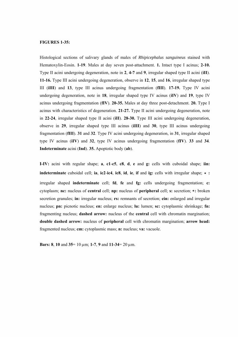
FIGURES 1-35:
Histological sections of salivary glands of males of Rhipicephalus sanguineus stained with
Hematoxylin-Eosin. 1-19. Males at day seven post-attachment. 1. Intact type I acinus; 2-10.
Type II acini undergoing degeneration, note in 2, 4-7 and 9, irregular shaped type II acini (iII).
11-16. Type III acini undergoing degeneration, observe in 12, 15, and 16, irregular shaped type
III (iIII) and 13, type III acinus undergoing fragmentation (fIII). 17-19. Type IV acini
undergoing degeneration, note in 18, irregular shaped type IV acinus (iIV) and 19, type IV
acinus undergoing fragmentation (fIV). 20-35. Males at day three post-detachment. 20. Type I
acinus with characteristics of degeneration. 21-27. Type II acini undergoing degeneration, note
in 22-24, irregular shaped type II acini (iII). 28-30. Type III acini undergoing degeneration,
observe in 29, irregular shaped type III acinus (iIII) and 30, type III acinus undergoing
fragmentation (fIII). 31 and 32. Type IV acini undergoing degeneration, in 31, irregular shaped
type IV acinus (iIV) and 32, type IV acinus undergoing fragmentation (fIV). 33 and 34.
Indeterminate acini (Ind). 35. Apoptotic body (ab).
I-IV: acini with regular shape; a, c1-c5, c8, d, e and g: cells with cuboidal shape; iin:
indeterminate cuboidal cell; ia, ic2-ic4, ic8, id, ie, if and ig: cells with irregular shape; ⋆ :
irregular shaped indeterminate cell; fd, fe and fg: cells undergoing fragmentation; c:
cytoplasm; nc: nucleus of central cell; np: nucleus of peripheral cell; s: secretion; ∗: broken
secretion granules; in: irregular nucleus; rs: remnants of secretion; ein: enlarged and irregular
nucleus; pn: picnotic nucleus; en: enlarge nucleus; lu: lumen; sc: cytoplasmic shrinkage; fn:
fragmenting nucleus; dashed arrow: nucleus of the central cell with chromatin margination;
double dashed arrow: nucleus of peripheral cell with chromatin margination; arrow head:
fragmented nucleus; cm: cytoplasmic mass; n: nucleus; va: vacuole.
Bars: 8, 10 and 35= 10 µm; 1-7, 9 and 11-34= 20 µm.
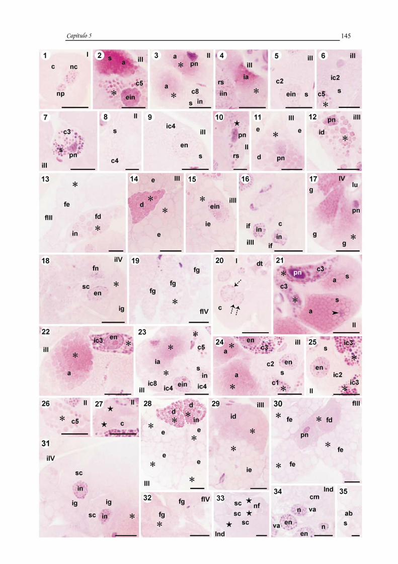
Capítulo 5
145

FIGURES 36-52:
Histological sections of salivary glands of Rhipicephalus sanguineus males at day seven post-
detachment stained with Hematoxylin-Eosin. 36. Type I acinus undergoing degeneration. 37-45.
Type II acini undergoing degeneration, note in 37 and 41-45, irregular shaped type II acini (iII).
46-48. Type III acini undergoing degeneration, observe in 47, irregular shaped type III acinus
(iIII) and 48, type III undergoing fragmentation (fIII). 49 and 50. Type IV undergoing
degeneration, in 49, irregular shaped type IV acinus (iIV) and 50, Type IV acinus undergoing
fragmentation (fIV). 51. Indeterminate acinus (Ind). 52. Apoptotic bodies (ab).
I-III: acini with regular shape; a, c1, c3, c5, d and e: cells with cuboidal shape; iin:
indeterminate cuboidal cell; ia, ic3, ic4, ic8, id and ie: cells with irregular shape; ⋆ : irregular
shaped indeterminate cell; fd and fe: cells undergoing fragmentation; c: cytoplasm; arrow:
enlarged nucleus of the central cell with chromatin margination; double arrow: enlarged
nucleus of peripheral cell with chromatin margination; s: secretion; ∗: broken secretion
granules; in: irregular nucleus; pn: picnotic nucleus; iv: intraacinar valve; en: enlarged nucleus;
ein: enlarged and irregular nucleus; cm: cytoplasmic mass; rs: remnants of secretion; fn:
fragmenting nucleus; va: vacuole.
Bars: 37= 10 µm; 36 and 38-52= 20 µm.

Capítulo 5
146

FIGURES 53-74:
Histological sections of salivary glands of Rhipicephalus sanguineus males stained with PAS
and counterstained with Methyl Green. 53-61. Males at day seven post-attachment. 53. Intact
type I acinus. 54-59. Type II acini undergoing degeneration, note in 56 and 58 irregular shaped
type II acini (iII). Observe in 55, intact cell c1 (c1) and in 57, intact cell c8 (c8). 60. Type III
acinus undergoing degeneration. 61. Type IV acinus undergoing degeneration. 62-67. Males at
day three post-detachment. 62. Type I acinus undergoing degeneration. 63-65. Type II acini
undergoing degeneration and with irregular shape (iII). 66. Type III acinus undergoing
degeneration. 67. Type IV acinus undergoing degeneration and with irregular shape (iIV). 68-
75. Males at day seven post-detachment. 68. Type I acinus undergoing degeneration. 69-72.
Type II acini undergoing degeneration, note in 70 and 72, irregular shaped type II acini (iII). 73
and 74. Type III acini undergoing degeneration and with irregular shape (iIII). 75. Type IV
acinus undergoing degeneration and with irregular shape (iIV).
I-IV: acinis with regular shape; a-c5, c8, d, e and g: cells with cuboidal shape; iin:
indeterminate cuboidal cell; ⋆ : irregular shaped indeterminate cell; ig: irregular shaped g
cell; fc: fibrillar cytoplasm; nc: nucleus of central cell; np: nucleus of peripheral cell; dt:
ducto; lu: lumen; s: secretion; en: enlarged nucleus; n: nucleus; in: irregular nucleus; ∗: broken
secretion granules; rs: remnants of secretion; dashed arrow: nucleus of the central cell with
chromatin margination; double dashed arrow: nucleus of peripheral cell with chromatin
margination; pn: picnotic nucleus; iv: intraacinar valve; double arrow: enlarged nucleus of
peripheral cell with chromatin margination; s: secretion; ein: enlarged and irregular nucleus;
cm: cytoplasmic mass.
Bars: 59= 10 µm; 66= 15 µm; 53-58, 60-65 and 67-75= 20 µm.

Capítulo 5
147

Capítulo 5
148
DISCUSSION
This morphological and histochemical study detected degenerative
characteristics in the cells of salivary glands of Rhipicephalus sanguineus males at day
seven post-attachment, and at day three and seven post-detachment from the host,
shedding light on the changes undergone by this organ during the degenerative process.
The onset of this process in the individuals examined caused a reduction in the
secretory capacity of salivary glands, indicated by a decrease in secretion in cells. This
was also observed in Amblyomma hebraeum and R. sanguineus females, as reported by
Harris and Kaufman (1984) and Furquim (2005), respectively.
These results revealed that the degenerative process in the glands of R.
sanguineus males is asynchronous among different types of acini, with more prominent
changes observed in types III and IV acini, as well as acini of the same type.
Asynchrony among cell types in the same acini was also observed. Cells at different
stages of degeneration coexisted and some of them could not be identified, as reported
by Till (1961), L’Amoreaux et al. (2003), Nunes et al. (2006), and Furquim (2005) for
females of R. appendiculatus, D. variabilis, R. (Boophilus) microplus and R.
sanguineus, respectively.
The present study clearly showed that the degenerative process in glands of R.
sanguineus males followed the same pattern observed in females of the same species
(Furquim, 2005). The onset of degeneration began in the posterior region of the gland,
first affecting types II, III, and IV acini, which were more distally located. Gland
degeneration advanced as time away from the host progressed. More prominent changes
were therefore observed in males at day seven post-detachment from the host than in
those at day seven post-attachment, as in the latter, type I acini were intact, while types
II, III, and IV were undergoing degeneration. Thus, if R. sanguineus males at days three
and seven post-detachment were allowed to reattach to a host, they would probably not
resume feeding, since their salivary glands were in advanced stages of degeneration.
Kaufman (1990), on the other hand, observed a reduction of 62% in the secretory
capacity of glands of A. hebraeum males removed from the host for four days compared
to those examined immediately after removal. However, despite being reduced, the
secretory capacity was reversed when males resumed feeding.

Capítulo 5
149
The advance in the process of salivary gland degeneration in R. sanguineus
males as time away from the host progressed confirmed the data obtained by Furquim
(2005) for females of this species. In the latter, more prominent characteristics of gland
degeneration were observed at day three post-engorgement than in engorged
individuals; and at day seven post-engorgement, the salivary gland was completely
degenerated (Furquim, 2005). In males, gland degeneration less intense than in females,
as female glands at day seven post-engorgement were already degenerated (Furquim,
2005) while in males at day seven post-detachment, some secretory cells were still
observed, although the entire tissue was undergoing degeneration.
Also, the salivary glands of R. sanguineus males exhibited less prominent
changes than those of females (Furquim, 2005). In the latter, one of the factors
triggering the onset of gland degeneration is the “critical weight” (Weiss and Kaufman,
2001). This does not occur in males, since the small amount of blood ingested does not
allow the tick to reach the weight (“critical weight”) to induce gland degeneration.
Therefore, salivary gland degeneration in these individuals is not controlled by the
volume of blood consumed. In females, degeneration may also be triggered by
hormones (Lomas et al., 1998) synthesized and released in function of the “critical
weight” (Weiss and Kaufman, 2001) and the release of the “male factor” (Weiss and
Kaufman, 2004), which is transferred from the male to the female during matting
(Weiss and Kaufman, 2004). The synthesis and release of hormones in females increase
until the post-engorgement phase (Lomas, 1993 apud Lomas et al., 1998). In males, on
the other hand, hormonal levels are constant, regardless of the feeding condition
(Kaufman, 1990), and therefore without a stimulus to maintain the degenerative
process.
In R. sanguineus males at day seven post-attachment examined in this study,
type I acinus did not exhibited changes, unlike the observed in those at day three and
seven post-detachment from the host. Morphological changes, such as changes in size,
were observed. At day seven post-detachment from the host, acini were larger than
those of males at day seven post-detachment, confirming the data obtained for females
of the same species at day three post-engorgement (Furquim, 2005).
Type II acinus of R. sanguineus males at day seven post-attachment and at day
three post-detachment exhibited both intact and changed cells, suggesting a functional

Capítulo 5
150
asynchrony among them. These cells may be active in different moments during
feeding, as well as become inactive at different stages, as a result of several degrees of
cell degeneration.
In males at day seven post-attachment, among identified cells, c1 and c8 were
intact or undergoing degeneration. All cells a and c2–c5 were undergoing degeneration,
unlike the observed in females of the same species. In the latter, type II acini did not
exhibit intact cells, and only cells a, c1 and c3 were observed (Furquim, 2005). At day
three and seven post-detachment from the host, males exhibited cells a, c1–c5 and c8,
and a, c1, c3–c5 and c8, respectively, unlike the observed in females at day three post-
engorgement, which presented only cells a and c3 (Furquim, 2005). The cell types still
present in type II acini at the end of the degeneration process in the males examined in
our study might be associated with the distinct behavior between males and females,
confirming their distinct needs regarding the activity of different secretory cell types of
their salivary glands.
In some type III acini of males at day seven post-attachment, the presence of
degenerating and fragmenting cells d, e and f partially agree with the results obtained in
engorged R. sanguineus females (Furquim, 2005). Contrary to the observed in the
present study, fragmentation of type III acinus and broken secretion granules in cells d
were not reported in these females. In males with three and seven days post-detachment
from the host, type III acini were still undergoing degeneration, unlike females at day
three post-engorgement. Type III acini of the latter were in such advanced stages of
degeneration that cells could no longer be identified (Furquim, 2005).
Type IV acinus of males examined in the three conditions exhibited changes that
became more prominent as time away from the host progressed, contrary to the reported
by Gill and Walker (1987). These authors described in Hyalomma anatolicum
anatolicum only morphological changes in the secretion granules of cells in final
feeding stages (120 to 144 hours of feeding).
In general, the results obtained in the present study showed that the degenerative
changes in the salivary glands of male ticks were not the same in different cells. Some
exhibited reduction in secretion (interruption of synthesis) and in some cases, broken
granules that appeared as an amorphous mass. Thereafter, we observed changes in
shape and size of nuclei of cells, as well as condensation level of chromatin, followed

Capítulo 5
151
by cytoplasmic shrinkage, loss of cell shape, loss of cell boundaries, and cytoplasmic
vacuolation and consequently breakdown of acini, formation and release of apoptotic
bodies. These characteristics clearly indicate that the cells of salivary glands of R.
sanguineus males most likely die by apoptosis, similarly to glands of females of this
species (Furquim, 2005).
ACKNOWLEDGMENTS
This research has been supported by CAPES (Coordenação de Aperfeiçoamento
de Pessoal de Nível Superior) and FAPESP (Fundação de Amparo à Pesquisa do Estado
de São Paulo) Grant nº 05/59208-3. The authors thank Gerson Mello Souza and Ronaldo
Del Vecchio for the techinical support. Part of this work has been facilitated through the
Integrated Consortium on Ticks and Tick-borne Diseases (ICTTD-3) supported by the
European Union under contract number 510561-INCO.

Capítulo 5
152
REFERENCES
Bechara, G.H., Szabó, M.P.J., Ferreira, B.R., Garcia, M.V., 1995. Rhipicephalus
sanguineus tick in Brazil: feeding and reproductive aspects under laboratorial conditons.
Brazil. J. Vet. Parasitol. 4, 61-66.
Binnington, K.C., 1978. Sequential changes in salivary gland structure during
attachment and feeding of the cattle tick, Boophilus microplus. Int. J. Parasitol. 8, 97-
115.
Furquim, K.C.S., 2005. Processos degenerativos em glândulas salivares de carrapatos
Rhipicephalus sanguineus (LATREILLE, 1806) (ACARI, IXODIDAE) em jejum, em
alimentação e alimentados. 2º Relatório Científico CAPES, Instituto de Biociências,
UNESP, Rio Claro (S.P.), Brasil, 40 p.
Furquim, K.C.S., 2006. Processos degenerativos em glândulas salivares de carrapatos
Rhipicephalus sanguineus (LATREILLE, 1806) (ACARI, IXODIDAE) em jejum, em
alimentação e alimentados. 3º Relatório Científico CAPES, Instituto de Biociências,
UNESP, Rio Claro (S.P.), Brasil, 27 p.
Gill, H.S., Walker, A.R., 1987. The salivary glands of Hyalomma anatolicum
anatolicum: structural changes during attachment and feeding. Int. J. Parasitol. 17 (8),
1381-1392.
Harris, R.A., Kaufman, W.R., 1984. Neural involvement in the control of salivary gland
degeneration in the ixodid tick Amblyomma hebraeum. J. Exp. Biol. 109, 281-290.
Kaufman, W.E., 1990. Effect of 20-hydroxiecdysone on the salivary glands of the male
tick, Amblyomma hebraeum. Exp. Appl. Acarol. 9, 87-95.

Capítulo 5
153
L’Amoreaux, W.J., Junaid, L., Trevidi, S., 2003. Morphological evidence that salivary
gland degeneration in the American dog tick, Dermacentor variabilis (Say), involves
programmed cell death. Tissue Cell 35, 95-99.
Lomas, L.O., 1993. The influence of a male-derived protein factor on salivary gland
degeneration in the ixodid tick, Amblyomma hebraeum Koch. PhD thesis, University of
Alberta, Edmond, Canada.
Lomas, O.L., Gelman, D., Kaufman, W. R., 1998. Ecdysteroid regulation of salivary
gland degeneration in the ixodid tick, Amblyomma hebraeum: a reconciliation of in vivo
and in vitro observations. Gen. Comp. Endocrinol. 109, 200-211.
McManus, J.F.A., 1946. Histological demonstration of mucin after periodic acid. Nature
158, 202.
Nunes, E.T., Camargo-Mathias, M.I., Bechara, G.H., 2006. Structural and cytochemical
changes in the salivary glands of the Rhipicephalus (Boophilus) microplus (Canestrini,
1887) (Acari: Ixodidae) tick female during feeding. Vet. Parasitol. 140, 114-123.
Sanders, M.L., Scott, A.L., Glass, G.E., Schwartz, B.S., 1996. Salivary gland changes
and host antibody responses associated with feeding of male lone star ticks (Acari:
Ixodidae). J. Med. Entomol. 33 (4), 628-634.
Till, W.M., 1961. A Contribution to the Anatomy and Histology of the Brown Ear Tick
Rhipicephalus appendiculatus Neumann. Memoirs of the Entomological Society of
Southern Africa, Pretoria, 6, 1-124.
Walker, A.R., Fletcher, J.D., Gill, H., 1985. Structural and histochemical changes in the
salivary glands of Rhipicephalus appendiculatus during feeding. Int. J. Parasitol. 15 (1),
81-100.

Capítulo 5
154
Wang, H., Henbest, P.J., Nuttall, P.A., 1999. Successful interrupted feeding of adult
Rhipicephalus appendiculatus (Ixodidae) is accompanied by re-programming of
salivary gland protein expression. Parasitology 119, 143-149.
Weiss, L.B., Kaufman, W.R., 2001. The relationship between ‘critical weight’ and 20-
hydroxiecdysone in the female ixodid tick, Amblyomma hebraeum. J. Insect Physiol. 47,
1261-1267.
Weiss, L.B., Kaufman, W.R., 2004. Two feeding-induced proteins from the male gonad
trigger engorgement of the female tick Amblyomma hebraeum. Proc. Natl. Acad. Sci.
S.A. 101 (16), 5874-5879.

Capítulo 6

Capítulo 6
156
CAPÍTULO 6
TITLE: The process of cell death in salivary glands of males of the tick Rhipicephalus
sanguineus (LATREILLE, 1806) (Acari, Ixodidae).
AUTHORS: Karim Christina Scopinho Furquim a, Gervásio Henrique Bechara b and
Maria Izabel Camargo Mathias a, *
a Departamento de Biologia, Instituto de Biociências, UNESP, Av. 24 A, nº 1515, Cx.
Postal 199, CEP: 13506-900, Rio Claro, S.P., Brazil b Departamento de Patologia Veterinária, FCAV, UNESP, Via de Acesso Prof. Paulo
Castellane, s/n, CEP: 14884-900, Jaboticabal, S.P., Brazil
* Corresponding author. Tel.: +55 19 35264135;
Fax: +55 19 35340009.
E-mail address: [email protected]

Capítulo 6
157
RESUMO
As glândulas salivares de machos de carrapatos Rhipicephalus sanguineus nos
estados de jejum, sete dias de infestação, três e sete dias pós-remoção do hospedeiro
foram submetidas a testes citoquímicos para análises enzimática e da viabilidade
celular. No estudo comparativo do tecido glandular nestas quatro situações observou-se
que a marcação nas células dos ácinos IV, III, II e I foi diferente, afetados pela
degeneração nesta sequência. Este estudo, também revelou: alterações nucleares, na
intensidade da marcação da fosfatase ácida e de ATPase e na permeabilidade da
membrana plasmática, sendo que a positividade da reação para fosfatase foi
inversamente proporcional à da ATPase e a positividade da reação para a ATPase foi
proporcional à integridade da membrana.
O tecido glandular dos machos em jejum apresentou alta atividade metabólica
com células com núcleo e membrana plasmática íntegros. Conclui-se que a presença de
fosfatase ácida nestes indivíduos faz parte da fisiologia de alguns ácinos, visto que este
tecido não está em degeneração. Nos machos com sete dias de infestação foi observada
membrana íntegra nas células dos ácinos I, II e na maioria dos III e IV, bem como maior
marcação para fosfatase ácida, alterações nucleares e queda na atividade da ATPase,
alterações que fazem parte do processo degenerativo. Naqueles com três e sete dias pós-
remoção do hospedeiro o processo degenerativo estava mais avançado, com perda da
integridade da membrana nas células de alguns ácinos I e II e na maioria dos III e IV
nos machos com três dias pós-remoção do hospedeiro, nas de poucos ácinos I e II, na
maioria dos III e em todos os ácinos IV naqueles com sete dias pós-remoção do
hospedeiro. Houve também alteração nuclear, marcante redução da atividade ATPásica,
aumento da atividade da fosfatase ácida naqueles com três dias e diminuição desta nos
com sete dias pós-remoção do hospedeiro.
Nestas glândulas a morte celular ocorreu numa seqüência onde as alterações
nucleares precederam as citoplasmáticas, como segue: a) alteração no núcleo, b) perda
da atividade da ATPase, c) perda da integridade da membrana plasmática e d) aumento
da atividade da fosfatase ácida. Esta última estaria relacionada com a degradação tardia
de porções citoplasmáticas, caracterizando assim o processo de morte celular em
glândulas de machos de R. sanguineus como apoptose atípica ou não clássica.

Capítulo 6
158
PALAVRAS-CHAVE: Rhipicephalus sanguineus, glândulas salivares, carrapatos,
machos, atividade enzimática, apoptose.

Capítulo 6
159
ABSTRACT
The salivary glands of males of the tick Rhipicephalus sanguineus at four
feeding stages: unfed, at day seven post-attachment, and at day three and seven post-
detachment from the host were subjected to cytochemical methods of enzymatic
analysis and cell viability. Comparing gland tissues at these stages, we observed
different staining patterns in the cells of the types IV, III, II acini, which were affected
by degeneration in this sequence. This study also revealed changes in: nuclei, staining
intensity for acid phosphatase and ATPase activities, and permeability of the plasmic
membrane. Acid phosphatase activity was inversely proportional to that of ATPase,
while ATPase activity was proportional to membrane integrity.
The salivary gland tissue of unfed males exhibited high metabolic activity and
cells with intact nucleus and plasmic membrane, suggesting that the presence of acid
phosphatase detected in these individuals may participate in the normal physiology of
some acini, as they were not undergoing degeneration. In males at day seven post-
attachment, we observed intact membranes in the cells of types I, II and most types III
and IV acini, as well as stronger staining for acid phosphatase, nuclear changes, and
decrease in ATPase activity. These changes were associated with the degenerative
process. At day three and seven post-detachment from the host, degeneration progressed
to more advanced stages. Loss of membrane integrity was observed in the cells of a few
types I and II acini and most types III and IV acini of males at day three post-
detachment; and few types I and II acini, most type III acini, and all type IV acini of
males at day seven post-detachment from the host. Nuclear changes, prominent decrease
in ATPase activity, and increase in acid phosphatase activity were also observed at day
three post-detachment, while acid phosphatase activity decreased at day seven post-
detachment from the host.
During the death of cells in these glands, alterations in the nucleus preceded
cytoplasmic ones in the following sequence: a) nuclear changes, b) loss of ATPase
activity, c) loss of integrity of the plasmic membrane, and d) increase in acid
phosphatase activity. The latter might be associated with the late degradation of
cytoplasmic remnants, characterizing the process of cell death in glands of R.
sanguineus males as atypical or non-classic apoptosis.

Capítulo 6
160
KEY WORDS: Rhipicephalus sanguineus, salivary glands, ticks, males, enzymatic
activity, apoptosis.

Capítulo 6
161
INTRODUCTION
The onset of characteristics associated with salivary gland degeneration in males
of the tick Rhipicephalus sanguineus results in a decrease in the secretory capacity of
this organs, and the progression of this process leads to the degradation of the glandular
tissue (Furquim et al., paper in preparation).
The process of gland degeneration in males exhibits similarities and differences
with that of females of the same species, as in the former, gland degeneration is less
prominent than in females (Furquim et al., paper in preparation).
According to some authors, the degenerative process is regulated by the
ecdysteroid hormone in both male and female ticks [1, 2]. In males, however, the
synthesis of this hormone does not increase during and after the completion of feeding
[1], causing a delay in the process of gland degeneration and less prominent cell
changes compared to those observed in females (Furquim et al., paper in preparation).
This might be due to the fact that males, in addition to not reaching the “critical
weight”, do not receive the “male factor”, a substance transferred to the female during
mating [3]. Both conditions promote the synthesis of ecdysteroids and therefore are
instrumental in gland degeneration [3, 4].
It was reported that salivary gland degeneration in female ticks is genetically
programmed [5], which may also occur in the males examined in our study. The
literature has shown that cells die by two main overlapping processes: apoptosis or
autophagy [6]. The latter has been commonly observed during insect metamorphosis [7,
8, 9-12].
In ticks, studies have demonstrated that cells of salivary glands die by apoptosis
[13, 14], and in R. sanguineus females, apoptotic death involves acid phosphatase to
completely degrade the cytoplasm, characterizing an atypical apoptosis (Furquim et al.,
paper in preparation).
Several specific methodologies have been described to detect and characterize
the process of cell death. ATPase activity is a good indicator of cells in late stages of
apoptosis; nuclear breakdown indicates cells in early stages of apoptosis while loss of
membrane integrity signals late stages [15, 16]. Acid phosphatase activity is a good
Capítulo 6
162
indicator of autophagic death, since it plays an essential role in this type of death [7, 8,
9, 6, 10, 11, 12].
Based on the presented informations, the purpose of this study was to describe
the cell changes resulted from salivary gland degeneration in R. sanguineus males at
day seven post-attachment, and at days three and seven post-detachment from the host,
comparing them with unfed males, and identifing the type of cell death in these organs.
MATERIAL AND METHODS
For this study, were utilized males of the tick Rhipicephalus sanguineus in the
following feeding stages: unfed, at day seven post-attachment, and at day three and
seven post-detachment from the host.
Unfed individuals were provided by Dr. Gervásio Henrique Bechara of the
Department of Veterinary Pathology of UNESP, Jaboticabal campus (São Paulo), from
a colony maintained under controlled conditions (29o C, 80% humidity, and 12 hour
photoperiod) in BOD incubator.
A group of unfed individuals was assigned to cytochemical methods, while
another group was placed with females in a feeding chamber previously glued to the
shaved back of the host (rabbit) according to technique described elsewhere [17]. At
day seven post-attachment, males were collected and a group was assigned to
cytochemical procedures. Another group was maintained alive for three and seven days
in closed containers with breathing holes to be later analyzed.
After the completion of the time periods examined in this study, salivary glands
were removed in saline solution and processed according to the following methods
described below for observation under light and fluorescence microscope.
For light microscopy, the material was fixed in 10% buffered neutral formalin
and acetone (9:1) for one hour and thirty minutes at 4o C, then processed according to
the methods described for detection of acid phosphatase and ATPase activities [18]. The
material was then dehydrated in increasing concentrations of ethanol (70%, 80%, 90%
and 95%), embedded in Leica resin, and sectioned at a thickness of 7 µm. Sections were
placed on glass slides, counterstained with Hematoxylin for 2 minutes, and mounted in

Capítulo 6
163
Canada balsam for later examination under light microscope. For the demonstration of
acid phosphatase activity, total preparations of some glands were also examined.
In both enzymatic experiments, control samples were incubated without substrate.
For fluorescence microscopy, after dissection, salivary glands were placed on
glass slides and received two drops of Acridine Orange (100 µg/mL) and Ethidium
Bromide (100 µg/mL) both in PBS [16], to demonstrate cell viability and detection of
apoptotic and/or necrotic cells. Slides were then covered with cover glasses and
immediately examined under fluorescence microscope with a 488 nm excitation filter.
Healthy cells are homogeneously green (cytoplasm and nucleus) or the nucleus is
homogeneously green and the cytoplasm orange/red. Cells in early stages of apoptosis
exhibit green or red-orange cytoplasm and green nucleus with clusters of bright green
condensed chromatin. Cells in late stages of apoptosis present red cytoplasm and nucleus
with bright orange condensed chromatin. Necrotic cells exhibit red cytoplasm and
homogeneously orange nucleus.
RESULTS
Unfed males
In the salivary glands of unfed R. sanguineus males, cells are intact, strongly
stained for RNA (Figs. 2A1, A2) and ATPase (Figs. 1A1-B1), and in general, weakly
stained for acid phosphatase (Figs. 3A1-B3). The plasmic membrane and nuclei are
intact (Figs. 2A1-A4).
Type I acinus
The acini basal membrane is strongly stained for ATPase (Fig. 1A1) and the
plasmic membranes are intact (Fig. 2A1). The cytoplasm is strongly positive for RNA
(Fig. 2A1), weakly positive for ATPase (Fig. 1A1), and moderately positive for acid
phosphatase (Fig. 3A6).

Capítulo 6
164
Type II acinus
The acini basal membrane and the plasmic membranes are strongly positive for
ATPase (Figs. 1A2-A4). The cytoplasm is strongly positive for RNA (undifferentiated
cells) (Fig. 2A2), and in most cells, strongly positive for ATPase (Figs. 1A2, A3) and
negative for acid phosphatase (Fig. 3B1).
Type III acinus
The acini basal membrane and plasmic membranes are strongly positive for
ATPase (Figs. 1A5, A6). The cytoplasm is negative for RNA (Fig. 2A3) and, in most
cells, strong staining for ATPase (Figs. 1A5, A6) and no staining for acid phosphatase
(Fig. 3B2) are observed.
Type IV acinus
The acini basal membrane is strongly stained for ATPase (Fig. 1B1) and the
plasmic membranes are intacts (Fig. 2A4). The cytoplasm is negative for RNA (Fig.
2A4), moderately positive for ATPase (Fig. 1B1), and weakly positive for acid
phosphatase (Fig. 3B3).
Males at day seven post-attachment
Degenerative characteristics are observed in R. sanguineus males at day seven
post-attachment (Figs. 1B5-D1, 2B1-C1, 3B6-C1, 3C3-C5). Type I acini are intact (Figs.
1B3, B4, 2A5, 3B5, C2) and types II (Figs. 1B5-B8, 3B6, C3), III (Figs. 1C1-C4, 2B2, B3,
3B7, C4), and IV (Figs. 1C5, D1, 2B4, C1, 3C1, C5) exhibit changes. In general, staining
for ATPase is weaker (Figs. 1B4-D1), while for acid phosphatase is stronger (Figs. 3B5-
C5).
Type I acinus
The acini basal membranes of most acini is moderately positive for ATPase (Fig.
1B4). The plasmic membranes of all cells are intacts (Fig. 3A5). The cytoplasm is
weakly positive for ATPase (Figs. 1B3, B4) and moderately positive for acid
phosphatase (Fig. 3C2). Nuclei are intact (Fig. 2A5)

Capítulo 6
165
Type II acinus
The acini basal membrane of most acini is moderately stained for ATPase (Figs.
1B7, B8), while the plasmic membranes of most cells are negative for ATPase (Figs.
1B6, B8) and intacts (Fig. 2B1). The cytoplasm of most cells is weakly positive for
ATPase (Figs. 1B5, B6, B8) and moderately positive for acid phosphatase (Fig. 3C3).
Type III acinus
The acini basal membrane of most acini is strongly positive for ATPase (Figs.
1C1, C2). In most cells, the plasmic membranes are moderately positives for ATPase
(Figs. 1C1, C3) and intacts (Fig. 2B2). The cytoplasm of most cells is weakly positive for
ATPase (Fig. 1C2, C3) and moderately positive for acid phosphatase (Fig. 3C4). Nuclei
exhibit changes (Fig. 2B2).
Type IV acinus
The acini basal membrane of most acini is moderately positive for ATPase (Figs.
1C5). The plasmic membranes are negatives for ATPase (Figs. 1C5, D1) and intacts in
most cells (Fig. 2B4). The cytoplasm is moderately positive for acid phosphatase (Fig.
3C5) and, in most cells, weakly positive for ATPase (Fig. D1). Nuclei exhibit changes
(Figs. 2B4, C1).
Males at day three post-detachment
Degeneration has progressed to more advanced stages in types I (Figs. 1D4, 2C2,
C3, 3D2, E1), II (Figs. 1D5-E1, 2D1, 3D3, E2), III (Figs. 1E3, E4, 2D3, 3D4, E3), and IV
(Figs. 1F1, 2E1, 3D5, E4) acini. Types III (Figs. 1E3-E4, 2D3, 3D4, E3) and IV (Figs. 1F1,
2E1, 3D5, E4) acini are the most affected. In general, staining is weaker for ATPase
(Figs. 1D4-E1, E3, E4, F1) and stronger for acid phosphatase (Figs. 2D2-E4).
Type I acinus
The acini basal membrane of most acini is moderately positive for ATPase (Fig.
1D4) and in most cells, the plasmic membranes are intacts. (Fig. 2C2) The cytoplasm is
strongly positive for acid phosphatase (Fig. 3E1) and, in most cells, no staining for
ATPase is observed (Fig. 1D4). Nuclei exhibit changes (Figs. 2C2, C3).

Capítulo 6
166
Type II acinus
The acini basal membrane of most acini is moderately positive for ATPase
(Figs. 1D6, E1). In most cells, the plasmic membranes are negatives for ATPase (Figs.
1D5, D6, E1) and intacts (Fig. 2C4), and the cytoplasm is negative for ATPase (Figs.
1D5, D6) and strongly positive for acid phosphatase (Fig. 3E2). Nuclei exhibit changes
(Fig. 2D1).
Type III acinus
The acini basal membrane of most acini is strongly positive for ATPase (Fig.
1E2). In most cells, the plasmic membranes are negatives for ATPase (Figs. 1E3, E4) and
has lost their integrity (Fig. 2D3); the cytoplasm is negative for ATPase (Figs. 1E2-E4)
and strongly positive for acid phosphatase (Fig. 3E3). Nuclei exhibit changes (Figs. 2D2,
D3).
Type IV acinus
The acini basal membrane of most acini is moderately stained for ATPase in
some areas and not stained in others (Fig. 1F1). The plasmic membranes are negatives
for ATPase (Figs. 1E5, F1) and in most cells, has lost their integrity (Figs. 2D4, E1). The
cytoplasm is strongly positive for acid phosphatase (Fig. 3E4) and in most cells,
negative for ATPase (Fig. 1F1). Nuclei exhibit changes (Fig. 2E1).
Males at day seven post-detachment
In these males, degeneration has progressed and types III (Figs. 1H1, 2F2, F3)
and IV (Fig. 1H3, 2F4) are the most affected acini. In general, weaker staining for
ATPase (Figs. 1F5, G2, H1, H3) and acid phosphatase (Figs. 3F2-G4) are observed.
Type I acinus
The acini basal membrane of most acini is moderately positive for ATPase (Fig.
1F4) and in most cells, the plasmic membranes are intacts (Fig. 2E2). The cytoplasm is
moderately positive for acid phosphatase (Fig. 3G1) and, in most cells, weakly positive
for ATPase (Figs. 1F3, F4). Nuclei exhibit changes (Figs. 2E2, E3).

Capítulo 6
167
Type II acinus
The acini basal membrane of most acini is moderately positive for ATPase (Fig.
1G1). In most cells, the plasmic membranes are negatives for ATPase (Figs. 1G1, G2)
and intacts (Fig. 2E4), and the cytoplasm is negative for ATPase (Figs. 1F6-G2) and acid
phosphatase (Fig. 3G2). Nuclei exhibit changes (Figs. 2E4, E5)
Type III acinus
The acini basal membrane of most acini is strongly positive for ATPase (Figs.
1G3, G4). In most cells, the plasmic membranes are negatives for ATPase (Figs. 1G4,
H1) and has lost their integrity (Figs. 2F2, F3); and no ATPase (Figs. 1G3-H1) and acid
phosphatase (Fig. 3G3) activity is observed in the cytoplasm. Nuclei exhibit changes
(Figs. 2F1, F2).
Type IV acinus
The acini basal membrane of most acini is moderately stained for ATPase in
some areas and not stained in others (Fig. 1H3). The plasmic membranes are negatives
for ATPase (Figs. 1H2, H3) and is not intacts (Fig. 2F4). The cytoplasm of most cells is
negative for ATPase (Figs. 1H2, H3) and acid phosphatase (Fig. 3G4). Nuclei exhibit
changes (Fig. 2F4).
To better compare the results, the data are summarized in Tables 1 and 2.

Capítulo 6
168
Table 1: Enzymatic activity and cell viability of salivary glands of unfed Rhipicephalus sanguineus males
and males at day seven post-attachment. Type I Acini Type II Acini Type III Acini Type IV Acini
Basal Membrane of Acinus
- ATPase +++ (Fig. 1A1);
- ATPase +++ (Figs. 1A2-A4);
- ATPase +++ (Figs. 1A5, A6);
- ATPase +++ (Fig. 1B1);
Plasmic Membrane
- intact (Fig. 2A1); - ATPase +++ (Figs. 1A2-A4); - intact (Fig. 2A2);
- ATPase +++ (Figs. 1A5, A6); - intact (Fig. 2A3);
- intact (Fig. 2A4);
Cytoplasm - ATPase + (Fig. 1A1), - fa ++ (Fig. 3A6);
- ATPase +++ (Figs. 1A2, A3) and – (Figs. 1A2, A4); - fa + (Fig. 3B1) and – (Fig. 3B1);
- ATPase +++ (Fig. 1A5, A6) and – (Fig. 1A5, A6); - fa + (Fig. 3B2) and – (Fig. 3B2);
ATPase ++ (Fig. 1B1); - fa + (Fig. 3B3);
Unfed
Nucleus - intact (Fig. 2A1);
- intact (Fig. 2A2); - intact (Fig. 2A3); - intact (Fig. 2A4);
Basal Membrane of Acinus
- ATPase +++ (Fig. 1B3) and ++ (Fig. 1B4);
- ATPase +++ (Figs. 1B5, B6) and ++ (Figs. 1B7, B8);
- ATPase +++ (Figs. 1C1, C2) and ++ (Figs. 1C3, C4);
- ATPase ++ (Fig. 1C5) and +/– (Fig. 1D1);
Plasmic Membrane
- intact (Fig. 2A5); - ATPase ++ (Figs. 1B5, B7) and – (Figs. 1B6, B8); - intact (Fig. 2B1);
- ATPase ++ (Figs. 1C1, C3) and – (Figs. 1C2, C4); - > intact (Fig. 1B2); - < not intact (Fig. 1B3);
- ATPase – (Figs. 1C5, D1); - > intact (Fig. 2B4); - < not intact (Fig. 2C1)
Cytoplasm - ATPase + (Figs. 1B3, B4); - fa ++ (Fig. 3C2);
- ATPase +++ (Fig. 1B5), ++ (Fig. 1B7), + (Figs. 1B5, B6, B8) and – (Figs. 1B5, B6, B8); - fa ++ (Fig. 3C3) and – (Fig. 3C3);
- ATPase + (Figs. 1C2, C3) and – (Figs. 1C1-C4); - fa ++ (Fig. 3C4) and – (Fig. 3C4);
- ATPase ++ (Fig. 1C5) and + (Fig. 1D1); - fa ++ (Fig. 3C5);
Day 7 Post-Attachment
Nucleus - intact (Fig. 2A5); - not observed; - ! (Fig. 2B2); - ! (Figs. 2B4, C1) and ? (Fig. 2C1);
(+++) strongly positive; (++) moderately positive; (+) weakly positive; (–) negative; (+/–) irregular staining; (af) acid
phosphatase; (>) most cells; (<): some cells; (!) irregular nucleus; (#) nucleus with clusters of condensed chromatin; (?) picnotic
nucleus.
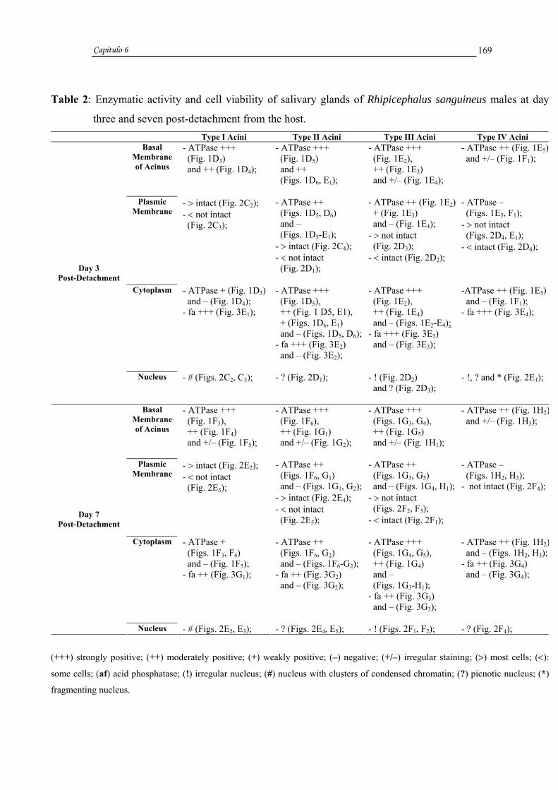
Capítulo 6
169
Table 2: Enzymatic activity and cell viability of salivary glands of Rhipicephalus sanguineus males at day
three and seven post-detachment from the host. Type I Acini Type II Acini Type III Acini Type IV Acini
Basal Membrane of Acinus
- ATPase +++ (Fig. 1D3) and ++ (Fig. 1D4);
- ATPase +++ (Fig. 1D5) and ++ (Figs. 1D6, E1);
- ATPase +++ (Fig. 1E2), ++ (Fig. 1E3) and +/– (Fig. 1E4);
- ATPase ++ (Fig. 1E5) and +/– (Fig. 1F1);
Plasmic Membrane
- > intact (Fig. 2C2); - < not intact (Fig. 2C3);
- ATPase ++ (Figs. 1D5, D6) and – (Figs. 1D5-E1); - > intact (Fig. 2C4); - < not intact (Fig. 2D1);
- ATPase ++ (Fig. 1E2) + (Fig. 1E3) and – (Fig. 1E4); - > not intact (Fig. 2D3); - < intact (Fig. 2D2);
- ATPase – (Figs. 1E5, F1); - > not intact (Figs. 2D4, E1); - < intact (Fig. 2D4);
Cytoplasm - ATPase + (Fig. 1D3) and – (Fig. 1D4); - fa +++ (Fig. 3E1);
- ATPase +++ (Fig. 1D5), ++ (Fig. 1 D5, E1), + (Figs. 1D6, E1) and – (Figs. 1D5, D6);- fa +++ (Fig. 3E2) and – (Fig. 3E2);
- ATPase +++ (Fig. 1E2), ++ (Fig. 1E4) and – (Figs. 1E2-E4); - fa +++ (Fig. 3E3) and – (Fig. 3E3);
-ATPase ++ (Fig. 1E5) and – (Fig. 1F1); - fa +++ (Fig. 3E4);
Day 3 Post-Detachment
Nucleus - # (Figs. 2C2, C3);
- ? (Fig. 2D1); - ! (Fig. 2D2) and ? (Fig. 2D3);
- !, ? and * (Fig. 2E1);
Basal Membrane of Acinus
- ATPase +++ (Fig. 1F3), ++ (Fig. 1F4) and +/– (Fig. 1F5);
- ATPase +++ (Fig. 1F6), ++ (Fig. 1G1) and +/– (Fig. 1G2);
- ATPase +++ (Figs. 1G3, G4), ++ (Fig. 1G5) and +/– (Fig. 1H1);
- ATPase ++ (Fig. 1H2) and +/– (Fig. 1H3);
Plasmic Membrane
- > intact (Fig. 2E2); - < not intact (Fig. 2E3);
- ATPase ++ (Figs. 1F6, G1) and – (Figs. 1G1, G2);- > intact (Fig. 2E4); - < not intact (Fig. 2E5);
- ATPase ++ (Figs. 1G3, G5) and – (Figs. 1G4, H1); - > not intact (Figs. 2F2, F3); - < intact (Fig. 2F1);
- ATPase – (Figs. 1H2, H3); - not intact (Fig. 2F4);
Cytoplasm - ATPase + (Figs. 1F3, F4) and – (Fig. 1F5); - fa ++ (Fig. 3G1);
- ATPase ++ (Figs. 1F6, G2) and – (Figs. 1F6-G2);- fa ++ (Fig. 3G2) and – (Fig. 3G2);
- ATPase +++ (Figs. 1G4, G5), ++ (Fig. 1G4) and – (Figs. 1G3-H1); - fa ++ (Fig. 3G3) and – (Fig. 3G3);
- ATPase ++ (Fig. 1H2) and – (Figs. 1H2, H3);- fa ++ (Fig. 3G4) and – (Fig. 3G4);
Day 7 Post-Detachment
Nucleus - # (Figs. 2E2, E3); - ? (Figs. 2E4, E5); - ! (Figs. 2F1, F2); - ? (Fig. 2F4);
(+++) strongly positive; (++) moderately positive; (+) weakly positive; (–) negative; (+/–) irregular staining; (>) most cells; (<):
some cells; (af) acid phosphatase; (!) irregular nucleus; (#) nucleus with clusters of condensed chromatin; (?) picnotic nucleus; (*)
fragmenting nucleus.

FIGURES

FIGURE 1:
ATPase activity in the salivary glands of Rhipicephalus sanguineus males. A1-B2. Unfed male.
Observe intense staining for ATPase (arrow) in the basal membrane and cells of types I (I), II
(II), III (III), and IV (IV) acini. B2. Negative control. B3-D2. Male at day seven post-attachment.
Note decrease in ATPase activity (arrow) in the four types of acini (I, II, III, and IV). In B6, B8,
C2, C4-D1, absence of ATPase activity in the plasmic membranes of cells of types II (II), III (III),
and IV (IV) acini. Note in D1, irregular staining for ATPase (arrow) in the basal membrane of
type IV acinus (IV). D2. Negative control. D3-F2. Male at day three post-detachment. Observe
weaker staining for ATPase (arrow) in types I (I), II (II), III (III), and IV (IV) acini. Note in: E4
and F1, irregular staining for ATPase (arrow) along the basal membrane of cells of types III (III)
and IV (IV) acini; D5-E1 and E3-F1, absence of ATPase activity in the membrane of cells of types
II (II), III (III) and IV (IV) acini. F2. Negative control. F3-H4. Male at day seven post-
detachment. Observe even weaker staining for ATPase (arrow). Note in: F5, G2, H1, and H3,
irregular staining for ATPase (arrow) along the basal membrane of types I (I), II (II), III (III),
and IV (IV) acini, and G2, G4, H1-H3, no staining for ATPase in the plasmic membrane of all cells
of types II (II), III (III), and IV (IV) acini. H4. Negative control.
I-IV: types acini; arrow: staining for ATPase; n: staining for ATPase in the nucleus; dt: duct.
Bars: B1, B2, D3 and D4= 12, 5 µm; A1-A6, B3-D2, D5-E2 and F1-H4= 25 µm; E3-E5= 50 µm.

Capítulo 6
171

FIGURE 2:
Total preparation of salivary glands of Rhipicephalus sanguineus males stained with Ethidium
Bromide and Acridine Orange. A1-A4. Unfed male. Note healthy cells of types I (I), II (II), III
(III), and IV (IV) acini with intact plasmic membrane and nuclei (tn). A5-C1. Male at day seven
post-attachment. Observe in: A5, intact type I acinus (I); B1, type II acinus (II) undergoing
degeneration, although the plasmic membrane is still intact; B2 and B3, type III acinus (III)
undergoing degeneration with cells in B3 exhibiting loss of membrane integrity; B4 and C1, type
IV acinus (IV) undergoing degeneration with cells in C1 exhibiting loss of cell membrane
integrity. C2-E1. Male at day three post-detachment. Observe types I (I), II (II), III (III), and IV
(IV) acini undergoing degeneration. Note in: C3, D1, and D3, loss of integrity of the plasmic
membrane of cells of types I (I), II (II), and III (III) acini; D4, loss of membrane integrity of
some cells of type IV acinus (IV), and E1, loss of membrane integrity of all cells of type IV
acinus (IV). E2-F4. Male at day seven post-detachment. Observe in E3, E5 and F2-F4, loss of
integrity of the plasmic membrane of cells of types I (I), II (II), III (III), and IV (IV) acini.
I-IV: types acini; arrow: cytoplasmic RNA; tn: intact nucleus; in: irregular nucleus; n:
nucleus; pn: picnotic nucleus; dashed arrow: nucleus with clusters of condensed chromatin; ∗:
fragmenting nucleus.
Bars: A1-A4= 12, 5 µm; A5-F4= 25 µm.

Capítulo 6
172

FIGURE 3:
Acid phosphatase activity in the salivary glands of Rhipicephalus sanguineus males. A1-B4.
Unfed male. In A1-A5, whole mount preparation of types I (I), II (II), III (III), and IV (IV) acini
showing low phosphatase activity (arrow). A6-B3. Histological sections of types I (I), II (II), III
(III), and IV (IV) acini exhibiting weak staining for acid phosphatase (arrow) in the cytoplasm
of cells. B4. Negative control. B5-D1. Male at day seven post-attachment. B5-C1. Total
preparation of types I (I), II (II), III (III), and IV (IV) acini showing more intense phosphatase
activity (arrow). C2-C5. Histological sections of types I (I), II (II), III (III), and IV (IV) acini.
D1. Negative control. D2-F1. Male at day three post-detachment. D2-D5. Total preparation of
types I (I), II (II), III (III), and IV (IV) acini, note even more intense phosphatase activity
(arrow). E1-E4. Histological sections of types I (I), II (II), III (III), and IV (IV) acini. Observe
in E1-E4, cytoplasm of cells of types I (I), II (II), III (III), and IV (IV) acini strongly stained for
acid phosphatase (arrow). F1. Negative control. F2-G5. Male at day seven post-detachment. F2-
F5. Total preparation of types I (I), II (II), III (III), and IV (IV) acini. G1-G4. Histological
sections of types I (I), II (II), III (III), and IV (IV) acini. Note decrease in intensity of staining
for acid phosphatase. G5. Negative control.
Bars: A3, A5, B4 and B5= 10 µm; B3= 12, 5 µm; A1, A2, A4, B6-C1 and D1= 20 µm; A6-B2, C2-C5
and D2-G5= 25 µm.

Capítulo 6
173

Capítulo 6
174
DISCUSSION
The comparative analysis of the salivary glands of unfed Rhipicephalus
sanguineus males, at day seven post-attachment, and at days three and seven post-
detachment revealed structural and enzymatic changes as a result of the process of gland
degeneration.
The glandular tissue of unfed individuals was intact and metabolically active, as
confirmed by the integrity of nuclei, intense staining for ATPase in the plasmic
membrane and cytoplasm of cells. The latter was also strongly positive for RNA and
weakly positive for acid phosphatase, confirming the observed in R. sanguineus females
at the same feeding stage (Furquim et al., paper in preparation).
In this study, the ATPase activity detected in different compartments of the cells
indicated a consumption of energy for the maintenance of the integrity of plasmic
membrane, contributing for the functioning of its ion pumps, as well as for metabolic
processes, such as protein synthesis in types I and II acini.
Acid phosphatase was detected in the four types of acini of unfed R. sanguineus
males. The most intense staining for acid phosphatase was observed in type I acinus, as
reported for females of the same species (Furquim et al., paper in preparation). Acid
phosphatase activity in type IV might be associated with the normal metabolism of
cells. In type I acinus, this enzyme may be involved in the synthesis of structural
proteins, as suggested by the large quantity of RNA, confirming the observed in
hypopharyngeal glands of Scaptotrigona postica [19] and in R. sanguineus females by
Furquim et al. (paper in preparation). In types II and III acini, which exhibit cells in pre-
secretory phase, acid phosphatase might be involved in secretion maturation, as
observed in hypopharyngeal glands of Scaptotrigona postica [19] and in in R.
sanguineus females (Furquim et al., paper in preparation).
The salivary gland cells of R. sanguineus males at day seven post-attachment,
and three and seven days post-detachment from the host underwent changes in the
nucleus, cytoplasm (acid phosphatase and ATPase), and plasmic membrane
(maintenance of integrity). These changes became more prominent as time away from
the host progressed, suggesting that the degenerative process in salivary gland R.
sanguineus males is asynchronous among different types of acini. Types III and IV

Capítulo 6
175
acini were the most affected, as also reported for females of the same species (Furquim
et al., paper in preparation).
In males at day seven post-attachment, and at days three and seven post-
detachment from the host, we observed a gradual decrease in ATPase activity. At day
seven post-attachment and day three post-detachment, acid phosphatase activity
increased. These changes were accompanied by a gradual loss of the integrity of the
plasmic membrane, which started at day seven post-attachment. Similar results obtained
elsewhere [20] showed a simultaneous increase in acid and basic phosphatase activities
and a decrease in ATPase activity. These results also support those obtained in
engorged and at day three post-engorgement females of R. sanguineus, in which an
increase in acid phosphatase and decrease in ATPase were observed, in addition to loss
of integrity of the plasmic membrane (Furquim et al., paper in preparation).
In males at day seven post-detachment from the host, the decrease in acid
phosphatase probably occurred because it was utilized and not synthesized during the
degenerative process. Some reports [1], shows in males, the hormone (ecdysteroid)
controlling gland degeneration does not reach levels as high as those of females
(Lomas, 1993, PhD thesis), and therefore, the stimulus is not as intense to maintain
gland degeneration and consequently, the synthesis of acid phosphatase. This
hypothesis was confirmed by the less intense degenerative process observed in the
males examined in the present study compared to the observed for females of the same
species. In the latter, acid phosphatase activity was much higher, resulting in more
prominent degradation (vacuolation) of the cytoplasm (Furquim et al., paper in
preparation), as well as decrease or absence of ATPase activity and loss of membrane
integrity, affecting more cells and acini during the degenerative process.
In the present study, we observed irregular staining patterns for ATPase in the
basal membrane in type IV acinus of males at day seven post-attachment, types III and
IV acini of males at day three post-detachment from the host, and types II, III and IV
acini of males at day seven post-detachment. In these acini, stained areas alternated with
non-stained ones, confirming the results obtained for engorged and at day three post-
engorgement females of R. sanguineus (Furquim et al., paper in preparation).
Type II acinus of R. sanguineus males at day seven post-attachment, and at days
three and seven post-detachment, and type IV acinus of males at day seven post-

Capítulo 6
176
attachment and at day three post-detachment from the host exhibited a characteristic not
observed in the other two types of acini, which was also reported for type II acinus of
females of the same species (Furquim et al., paper in preparation). In the cells of some
types II and IV acini, the plasmic membrane was intact, but ATPase activity was absent.
This could be due to the presence of a functional cell membrane only in the basal
portion that still could not be distinguished from the basal membrane of the acinus.
Thus, only an ATPase positive halo was observed around it.
The enzymatic changes observed in the present study were accompanied by
morphological ones (Furquim et al., paper in preparation), such as a) decrease in the
amount of secretion in the cells, b) nuclear breakdown, c) cytoplasmic shrinkage, d) loss
of cell shape, e) loss of cell boundaries (cell individuality), cytoplasmic vacuolation,
and f) cell fragmentation resulting in apoptotic bodies enclosed by membrane,
containing cytoplasmic and nuclear remnants. The decrease in the amount of secretion
and nuclear breakdown preceded all other morphological and enzymatic changes,
characterizing a classic apoptosis, as also observed elsewhere [21, 22, 23, 10, 24].
However, early loss of ATPase activity and integrity of the plasmic membrane
during the degenerative process and the presence of acid phosphatase do not
characterize a classic apoptotic death. In fact, participation of acid phosphatase has been
associated with autophagic death [7, 21, 9, 6, 10, 11, 12], in which the plasmic
membrane is still functional in newly formed apoptotic bodies [25, 23].
On the other hand, it was reported that during cell death in the salivary glands of
Calliphora vomitoria, ATPase activity ceased at some point and a massive cytoplasmic
vacuolation was observed [24]. This indicates that ATPase activity and preservation of
the integrity of the membrane is not always observed until the late stages of the process,
as suggested by other author [23, 25]. Similarly, several studies have described the
presence of hydrolytic enzymes (acid phosphatase) during apoptotic death [26, 21, 6,
27, 28, 10, 29, 11, 30]. Based on this information, our findings indicate that the salivary
gland cells of all acini in R. sanguineus males exhibited characteristics of classic
apoptosis and autophagic death. The latter included the involvement of hydrolytic
enzymes and the presence cytoplasmic vacuoles, characterizing the cell death as an
atypical apoptosis. This was also proposed for females of the same species, in which the
late involvement of acid phosphatase was associated with the removal of cytoplasmic
Capítulo 6
177
remnants, as well as fragmentation of gland cells (Furquim et al., paper in preparation).
In the present study, acid phosphatase activity was low and did not degrade large areas
of cytoplasm.
In this case, acid phosphatase probably had an endogenous origin, as we did not
observe hemocytes adhered to the tissue, as described for Calliphora erythrocephala
[26, 8]. However, it was suggested that acid phosphatase detected in gland cells of R.
(Boophilus) microplus females might come from the hemolymph [14].
Thus, these results, supported by those obtained previously by Furquim et al.
(paper in preparation), indicate that the degenerative process of salivary glands of R.
sanguineus males occurs by atypical apoptosis, characterized by a) early nuclear
breakdown, mainly by chromatin condensation, b) loss of cell shape, c) decrease and/or
absence of ATPase activity and loss of integrity of the plasmic membrane, d) cytoplasm
shrinkage and loss of cell boundaries, e) low acid phosphatase activity, resulting in
limited degradation (vacuolation) of the cytoplasm, and f) fragmentation of acini with
release of apoptotic bodies.
ACKNOWLEDGMENTS
This research has been supported by CAPES (Coordenação de Aperfeiçoamento
de Pessoal de Nível Superior) and FAPESP (Fundação de Amparo à Pesquisa do Estado
de São Paulo) Grant nº 05/59208-3. The authors thank Antonio Teruyoshi Yabuki,
Monika Souza and Ronaldo Del Vecchio for the technical support, and Prof. Dr. Flávio
Henrique Caetano for allowing the use of the photomicroscope (FAPESP, Grant nº
04/13327-9). Part of this work has been facilitated through the Integrated Consortium on
Ticks and Tick-borne Diseases (ICTTD-3) supported by the European Union under
contract number 510561-INCO.

Capítulo 6
178
References
[1] Kaufman WE. 1990. Effect of 20-hydroxiecdysone on the salivary glands of the
male tick, Amblyomma hebraeum. Exp Appl Acarol 1990; 9: 87-95.
[2] Lomas OL, Gelman D, Kaufman W R. Ecdysteroid regulation of salivary gland
degeneration in the ixodid tick, Amblyomma hebraeum: a reconciliation of in vivo and
in vitro observations. Gen Comp Endocrinol 1998; 109: 200-211.
[3] Weiss LB, Kaufman WR. The relationship between ‘critical weight’ and 20-
hydroxiecdysone in the female ixodid tick, Amblyomma hebraeum. J Insect Physiol
2001; 47: 1261-1267.
[4] Weiss LB, Kaufman WR. Two feeding-induced proteins from the male gonad
trigger engorgement of the female tick Amblyomma hebraeum. Proc Natl Acad Sci SA
2004; 101 (16): 5874-5879.
[5] Bowman AS, Sauer JR. Tick salivary glands: function, physiology and future.
Parasitology 2004; 129: S67-S81.
[6] Zakeri Z, Bursch W, Tenniswood M, Lockshin RA. Cell death: programmed,
apoptosis, necrosis, or other? Cell death Differ 1995; 2: 87-96.
[7] Pipan N, Rakovec V. Cell death in the midgut epithelium of the worker honey bee
(Apis mellifera carnica) during metamorphosis. Zoomorphologie 1980; 97: 217-224.
[8] Armbruster L, Levy M, Mathieu N, Bautz AM. Acid phosphatase activity in the
hemolymph, hemocytes, fat body and salivary glands during larval and prepupal
development in Calliphora erythrocephala (Diptera: Calliphoridae). Comp Biochem
Physiol 1986; 84B (3): 349-354.

Capítulo 6
179
[9] Cummings LA, Bowen ID. The localization of acid phosphatase as a prelude to cell
death in the blow-fly Calliphora erithrocephala. Micron Microsc Acta 1992; 23 (3):
357-358.
[10] Lockshin AR, Zakeri Z. The Biology of Cell Death and Its Relationship to Aging.
In: Holbrook N, Martin GR, Lockshin RA, editors. Cellular Aging and Cell Death. New
York: Wiley-Liss, 1996: 167-180.
[11] Jochová J, Zakeri Z, Lockshin RA. Rearrangement of the tubulin and actin
cytoskeleton during programmed cell death in Drosophila salivary glands. Cell Death
Differ 1997; 4: 140-149.
[12] Gregorc A, Bowen ID, Pogacnik A. Acid phosphatase activity in the midgut of
honeybee (Apis melliphera L.) larvae. Apidologie 1998; 29: 579-584.
[13] L’Amoreuax WJ, Junaid L, Trevidi S. Morphological evidence that salivary gland
degeneration in the American dog tick, Dermacentor variabilis (Say), involves
programmed cell death. Tissue Cell 2003; 35: 95-99.
[14] Nunes ET, Camargo-Mathias MI, Bechara GH. Rhipicephalus (Boophilus)
microplus (Canestrini, 1887) (Acari: Ixodidae): acid phosphatase and ATPase activities
localization in salivary glands of females during the feeding period. Exp Parasitol 2006;
114: 109-117.
[15] Bowen ID, Worrill NA, Winters CA, Mullarkey K. The use of backscattered
electron imaging, X-ray microscopy in demonstrating physiological cell death.
Scanning Microsc 1988; 2: 1453-1462.
[16] MacGahon AJ, Martin SJ, Bissonnette RP, Mahboubi A, Shi Y, Mogil RJ. The End
of the (Cell) Line: Methods for the Study of Apoptosis in vitro. In: Schwartz LM,
Osborne BA, editors. Cell Death. San Diego: Academic Press, 1995: 153-185.

Capítulo 6
180
[17] Bechara GH, Szabó MPJ, Ferreira BR, Garcia MV. Rhipicephalus sanguineus tick
in Brazil: feeding and reproductive aspects under laboratorial conditions. Brazil J Vet
Parasitol1995; 4: 61-66.
[18] Hussein MA, Bowen ID, Lewis GH. The histochemical localization of ATPase,
cholinesterase and acid phosphatase activity in Culex pipiens (Diptera, Culicidae) larvae
using a methacrylate embedding technique. Cell Biol Int Rep 1990; 14 (9): 775-781.
[19] Costa RAC, Cruz-Landim C. Distribution of acid phosphatases in the
hypopharyngeal glands from workers, queens and males of a Brazilian stingless bee
Scaptotrigona postica Latreille: an ultrastructural cytochemical study. Histochem
Journal 2001; 33: 653-662.
[20] Weber R. Tissue Involution and Lysosomal Enzimes During Anuran
Metamorphosis. In: Dingle JT, Fell HB, editors. Lysosomes in Biology and Pathology,
vol. 2. North Amsterdam and London: Holland Publishing Co, 1969: 437-461.
[21] Clarke PGH. Developmental cell death: morphological diversity and multiple
mechanisms. Anat Embryol 1990; 181: 195-213.
[22] Bowen ID. Apoptosis or programmed cell death? Cell Biol Int 1993; 17 (4): 365-
380.
[23] Kerr JFR, Gobé GC, Winterford CM, Harmon B. Anatomical Methods in Cell
Death. In: Schwartz LM, Osborne BA, editors. Cell Death. San Diego: Academic Press,
1995: 1-27.
[24] Häcker G. The morphology of apoptosis. Cell Tissue Res 2000; 301: 5-17.
[25] Bowen ID, Bowen SM. Programmed Cell Death in Tumours and Tissues,
Chapman and Hall: London, 1990.

Capítulo 6
181
[26] Levy M, Bautz AM. Degeneration of larval salivary glands during metamorphosis
of the blowfly, Calliphora erythrocephala Meigen (Diptera: Calliphoridae). Int J Insect
Morphol Embryol 1985; 14 (5): 281-290.
[27] Bowen ID, Mullarkey K, Morgan SM. Programmed cell death during
metamorphosis in the blow-fly Calliphora vomitoria. Micros Res Tech 1996; 34: 202-
217.
[28] Dai J, Gilbert LI. Programmed cell death of the prothoracic glands of Manduca
sexta during pupal-adult metamorphosis. Insect Biochem Mol Biol 1997; 27 (1): 69-78.
[29] Gregorc A, Bowen ID. Programmed cell death in the honey-bee (Apis mellifera L.)
larvae midgut. Cell Biol Int 1997; 21 (3): 151-158.
[30] Yamamoto S, Sawada K, Shimomura H, Kawamura K, James TN. On the nature of
cell death during remodeling of hypertrophied human myocardium. J Mol Cell Cardiol
2000; 32: 161-175.

Capítulo 7

Capítulo 7
183
CAPÍTULO 7
TITLE: Cytoplasmic and nuclear changes detected cytochemically during the
degeneration of salivary glands of the tick Rhipicephalus sanguineus (LATREILLE,
1806) (Acari, Ixodidae).
AUTHORS: Karim Christina Scopinho Furquim a, Gervásio Henrique Bechara b and
Maria Izabel Camargo Mathias a, *
a Departamento de Biologia, Instituto de Biociências, UNESP, Av. 24 A, nº 1515, Cx.
Postal 199, CEP: 13506-900, Rio Claro, S.P., Brazil b Departamento de Patologia Veterinária, FCAV, UNESP, Via de Acesso Prof. Paulo
Castellane, s/n, CEP: 14884-900, Jaboticabal, S.P., Brazil
* Corresponding author. Fax: +55 19 35340009.
E-mail address: [email protected]

Capítulo 7
184
RESUMO
O presente estudo analisou citoquimicamente glândulas salivares de fêmeas (em
jejum, ingurgitadas e com três dias pós-ingurgitamento) e de machos (em jejum, com
sete dias de infestação, três e sete dias pós-remoção do hospedeiro) de carrapatos
Rhipicephalus sanguineus. Os resultados revelaram a ocorrência de alterações nucleares
nas células secretoras de fêmeas em estágios ingurgitado e com três dias pós-
ingurgitamento e nos machos em todos os estágios (exceto em jejum), sendo mais
intensas nas fêmeas. Além disso demonstrou a presença de alterações citoplasmáticas
nas células de todos os ácinos de fêmeas e de machos.
Nos ácinos II e III de fêmeas ingurgitadas houve alterações na forma do núcleo
(arredondados, irregulares, com “blebbs”, fragmentando-se ou fragmentados), no
tamanho (normal, dilatados ou reduzidos), na disposição da cromatina e no grau de
condensação da mesma (por todo o núcleo, marginalizada ou em blebbs). Os nucléolos
também sofreram alterações na forma (arredondados, fragmentando-se ou
fragmentados), no tamanho (normais ou dilatados), na localização (centrais, marginais
ou em blebbs), e alguns ainda estavam compactados ou desorganizados. Nas fêmeas
com três dias pós-ingurgitamento todos os ácinos apresentaram alterações semelhantes
às verificadas nas fêmeas ingurgitadas. A marcação para presença de RNA foi maior
nas células das fêmeas ingurgitadas do que nas com três dias após-ingurgitamento.
Nos machos com sete dias de infestação os ácinos II, III e IV apresentaram
células com tamanho do núcleo e grau de condensação da cromatina alterados
semelhantes às das fêmeas. Por outro lado com forma arredondada, irregular ou
fragmentando-se e a cromatina disposta por todo núcleo ou marginalizada. As
alterações no nucléolo foram semelhantes às das fêmeas, isto é no tamanho e na
organização, já a forma arredondada permaneceu e a localização foi central. Nos
machos com três dias pós-remoção do hospedeiro todas as células de todos os ácinos
apresentaram alterações nucleares semelhantes às dos machos com sete dias de
infestação, além da fragmentação do nucléolo. Naqueles com sete dias pós-remoção do
hospedeiro as alterações ocorreram em todos os ácinos semelhante às verificadas
naqueles com sete dias de infestação. Quanto a presença de RNA citoplasmático, este

Capítulo 7
185
foi fortemente marcado nos machos com sete dias de infestação e fracamente naqueles
com sete dias pós-remoção do hospedeiro.
Tanto nas fêmeas quanto nos machos as diferentes intensidades de marcação do
RNA no citoplasma, bem como as alterações nucleares caracterizaram a ocorrência de
morte celular do tipo apoptótica.
PALAVRAS-CHAVE: Rhipicephalus sanguineus, glândulas salivares, fêmeas,
machos, cromatina, nucléolo, apoptose.

Capítulo 7
186
ABSTRACT
The present study reports cytochemistry data about salivary gland of females
(unfed, engorged, and at days three post-engorgement) and males (unfed, at day seven
post-attachment, and at day three and seven post-detachment from the host) of the tick
Rhipicephalus sanguineus. The results revealed nuclear changes in engorged females
and at day three post-engorgement, and in males in all stages (except unfed). These
changes were more prominent in females. Cytoplasmic changes were also observed in
cells of all acini of males and females.
In types II and III acini of engorged females, nuclear changes were observed in
the shape (round, irregular, with blebs, fragmenting or fragmented), size (normal,
enlarged or reduced), and arrangement and condensation level of chromatin (throughout
the nucleus, marginal or as blebs). Changes were also detected in nucleoli, regarding
their shape (round, fragmenting or fragmented), size (normal or enlarged), and location
(central, marginal or as blebs). Some nucleoli were also compacted or disorganized. In
females at day three pos-engorgement, all acini exhibited similar changes to those
observed in engorged females. RNA staining was stronger in cells of engorged females
than those at day three post-engorgement.
In males at day seven post-attachment, cells of types II, III, IV acini presented
changes in the size of the nucleus and condensation level of chromatin similar to those
of females. The shape of the nucleus was round, irregular or undergoing fragmentation,
and the chromatin was located marginal or throughout the nucleus. The changes in the
nucleolus were similar to those of females, regarding size and organization, although
round-shaped and in the center location. In males at day three post-detachment, cells of
all acini exhibited nuclear changes similar to those of males at day seven post-
attachment, in addition to the fragmentation of the nucleolus. At day seven post-
detachment, changes were detected in all acini similar to the observed in males at day
seven post-attachment. Regarding cytoplasmic RNA, staining was prominent in males
at day seven post-attachment and weak in those at day seven post-detachment from the
host.
In females as well as males, different RNA staining patterns in the cytoplasm
and nuclear changes characterized apoptotic cell death.

Capítulo 7
187
KEY WORDS: Rhipicephalus sanguineus, salivary glands, females, males, chromatin,
nucleolus, apoptosis.

Capítulo 7
188
INTRODUCTION
Female and male ticks present salivary glands that secrete substances
responsible for the attachment of the parasite to the host to allow feeding (Binnington,
1978; Walker et al., 1985; Gill and Walker, 1987).
Previous studies on the tick Rhipicephalus sanguineus have demonstrated that
the salivary glands undergo periods of intense and low secretory activity. During low
activity, the morphology and cytochemistry of these gland cells revealed degenerative
features characterizing an atypical type of apoptosis observed in males as well as
females of this species, although more prominent in the latter (Furquim, 2006).
Data available in the literature indicate that during apoptotic death, nuclear
breakdown is one of the first changes observed, characterized by fragmentation,
compression, and margination of chromatin, appearance of blebs and finally
fragmentation of the nucleus itself (Bowen and Bowen, 1990; Bowen, 1993; Kerr et al.,
1995; Häcker, 2000).
Apoptotic death may also require RNA and protein synthesis (Bowen and
Bowen, 1995; Zakeri et al., 1995; Lockshin and Zakeri, 1996; Tata, 1966 apud Häcker,
2000) during the early stages of the process (de novo synthesis) (Bowen and Bowen,
1990)
Due to the nuclear changes during apoptotic death (Bowen, 1990; Bowen and
Bowen, 1990; Clarke, 1990; Kerr et al., 1995; Zakeri et al., 1995; Lockshin and Zakeri,
1996; Häcker, 2000), as well as de novo synthesis (Bowen and Bowen, 1995; Zakeri et
al., 1995; Lockshin and Zakeri, 1996; Tata, 1966 apud Häcker, 2000), the utilization of
specific techniques is instrumental to examine the nucleus and nucleolus, the organelles
affected by the degenerative process, as well as the RNA present in the cytoplasm of
these cells.
The application of cytochemical techniques such as the Feulgen reaction
(Feulgen and Rossenbeck, 1924) and a variant of the Critical Electrolyte Concentration
(CEC) (Mello et al., 1993) to examine the nucleus and nucleolus, respectively, have
been widely reported in the literature. The Feulgen reaction is utilized to visualize
characteristics of the chromatin (Mello, 1983; Mello et al., 1995; Vidal et al., 1998;
Mello and Vidal, 2000; Moraes et al., 2005), as well as in studies on cell death (Mello et
Capítulo 7
189
al., 2003; Nunes et al., 2006). The variant of the Critical Electrolyte Concentration
(CEC) has been used in studies involving the identification, morphometry, and changes
in the structure of the nucleolus (Mello et al., 1993); the identification of different
stages of mitosis (Mello, 1995); cells in high secretory activity or undergoing
degeneration in the venom glands of Apis mellifera (Abreu et al., 2004).
Thus, the purpose of this study was to examine, using cytochemical techniques,
the cytoplasmic RNA and nuclear changes in cells of salivary glands of females (unfed,
engorged, and at day three post-engorgement) and males (unfed, at day seven post-
attachment, at days three and seven post-detachment from the host) of the tick
Rhipicephalus sanguineus.
MATERIAL AND METHODS
In this study, were utilized females and males of the tick Rhipicephalus
sanguineus. Were examined unfed, engorged, and at days three post-engorgement
females; and unfed, at day seven post-attachment, and at day three and seven post-
detachment from the host males. Unfed individuals were provided by Dr. Gervásio
Henrique Bechara of the Department of Veterinary Pathology of UNESP, Jaboticabal
campus (São Paulo), from a colony maintained under controlled conditions (29o C, 80%
humidity, and 12 hour photoperiod) in BOD incubator.
A group of unfed adult ticks (females as well as males) was assigned to
cytochemical methods, while another group was placed in a feeding chamber previously
glue with an atoxic and non-lesive preparation (Britannia Adhesive-Unit 4, UK) to the
shaved back of the host (rabbit) according to technique described elsewhere (Bechara et
al., 1995). A group of engorged females and males removed from the host after seven
days post-attachment was assigned for cytochemical procedures, while another group
was maintained alive in closed containers with breathing holes for three days (females
and males), and seven days (only males) to be later analyzed.
After the completion of the time periods examined in this study, salivary glands
were processed according to the seven conditions analyzed, removed in saline solution
(NaCl 7.5 g/L, Na2HPO4 2.38 g/L and KH2PO4 2.72 g/L, pH 7.2), fixed in ethanol and
acetic acid (3:1) at room temperature for 12 minutes. The material was then dehydrated

Capítulo 7
190
in increasing concentrations of ethanol (70%, 80%, 90% and 95%), embedded and
included in Leica resin, and sectioned at a thickness of 3 µm. Sections were placed on
glass slides and processed for the variant of the Critical Electrolyte Concentration
(CEC) (Mello et al., 1993) and the Feulgen reaction (Feulgen and Rossenbeck, 1924).
For the variant of the CEC, slides were stained with 0.025% Toluidine Blue in
McIlvane’s buffer (pH 4.0) for 20 minutes. The material was then immersed in an
aqueous solution of 0.05M MgCl for 2, 5, 7, and 10 minutes to detect the ideal time at
which metachromasy is abolished, due to the removal of Toluidine Blue molecules
bound to the chromatin by MgCl2. At this point, only RNA metachromasy is maintained,
consequently staining the nucleolus and demonstrating the presence of cytoplasmic
RNA (violet color) (Mello et al., 1993). Slides were then washed with distilled water
and mounted in Permount for later observation under light microscope.
For the Feulgen reaction, slides were immersed in 1N HCl solution at 60o C for
11 minutes, then washed in distilled water and stained with Schiff’s reagent for two
hours in the dark. Slides were counterstained with eosin for 5 minutes and mounted in
Canada balsam for later observation under light microscope.
RESULTS
The results obtained with the CEC method revealed that the point in which
metachromasy is abolished (chromatin stains green and nucleolus violet) is 5 minutes
(Fig. 1).
1. Content of cytoplasmic RNA
In unfed females (Figs. 1A1-A3) and males (Figs. 1D5-E3) of R. sanguineus,
RNA is observed mainly in the cytoplasm of undifferentiated cells of type II acini
(Figs. 1A2, E1) and cells of type III acini (Figs. 1A3, E2). In general, RNA staining is
more prominent in the gland tissue of females (Figs. 1A1-A3) than those of males (Figs.
1D5-E3).
In engorged females, RNA staining is observed in the cytoplasm of different
cells of all acini (Figs. 1A4-C1). Staining is stronger than that observed in the previous
condition (Figs. 1A1-A3).

Capítulo 7
191
At day three post-engorgement, staining is less prominent (Figs. 1C2-D4) than
that of cells in the previous condition (Figs. 1A4-C1), as it is only observed in the cells
or cytoplasmic mass of some acini (Figs. 1C2, C4, D1, D4).
In males at day seven post-attachment (Figs. 1E4-F2), and at days three (Figs.
1F3-G1) and seven (Figs. 1G2-G4) post-detachment from the host, RNA is detected in
the cytoplasm of cells of types II and type III acini. In males at day seven post-
attachment (Figs. 1E4-F2), staining is stronger than that of males at day seven post-
detachment (Figs. 1G2-G4).
2. Nuclear Characteristics
The changes observed in the nuclei of salivary glands of females and males are
characterized by changes in shape (Figs. 2B1, B4, C1, C5, D3, E1-E3, F3, 3B3, C1, C2, D2,
D3, D6, E2, G1, G2, G5, I1, I2), size (Figs. 2B1-B3, C1, C5, D1, D2, E1-E3, F3, 3B2-B4, C2,
C4, D3, E2, E6, F6-G2, H1, H2, I2), condensation level (Figs. 2B3, B4, D1, D2, 3B4, C3, D4,
E1, E3, E6, E7, F2, F4, G2, G3, H1, H3, I3, I4) and arrangement of the chromatin (Figs. 2B2,
C2, C3, D2-D4, E3, F1-F3, 3B5, B6, C4, D5, D6, E4, F5, F6, H2, H4, I3, I4), as well as
fragmentation of the nucleus (Figs. 2C1, C4, E3, F2, F3, 3C3-C5, E5, F1, F2, G4, H5, H6). In
the nucleolus, we observed changes in shape (Figs. 1B4, G1) and size (Figs. 1B3, C4, F1,
F2, F4, F5, G3, G4), disorganization of its structure (Figs. 1B2-B4, C3, C4, E5, F2, F5, G4)
as well as fragmentation (Figs. 1B5, C1, C5, D1-D4).
In unfed females and males, cells of types I (Figs. 1A1, D5, 2A1, 3A1), II (Figs.
1A2, E1, 2A2, 3A2), and III (Figs. 1A3, E2, 2A3, 3A3) acini exhibit intact nuclei and
nucleoli, with normal size, round shape, and uncondensed chromatin. In type IV acinus
of males, nuclei present reduced size and condensed chromatin (Fig. 3A4), and nucleoli
were not observed (Fig. 1E3).
In engorged females, only cells of type I acinus exhibit round nuclei (Fig. 2A4)
with intact nucleoli (Fig. 1A4), normal size, and uncondensed chromatin (Fig. 2A4).
At day three post-engorgement, types I (Fig. 2D4), II (Fig. 2E1) and
Indeterminate (Figs. 1C3-D4, 2E2-F3) acini exhibit nuclear changes. Type III acini are
not observed.

Capítulo 7
192
In males at day seven post-attachment, type I acini do not present nuclear
changes (Figs. 1E4, 3B1), unlike types II (Figs. 1E5, 3B2-B6), III (Figs. 1F1, F2, 3C1-C5),
and IV (Figs. 3D1-D5) acini.
In males at days three (Figs. 1F4, F5, G1, 3D6-F5) and seven (Figs. 1G3, G4, 3F6-
I4) post-detachment from the host, all types of acini exhibit nuclear changes.

FIGURES

FIGURE 1:
Histological sections of salivary glands of Rhipicephalus sanguineus submitted to a variant of
the Critical Electrolyte Concentration (CEC) A1-A3. Unfed female. Observe intact nucleoli (nu)
in types I (I), II (II), and III (III) acini. A4-C1. Engorged female. Note that among females, the
most prominent staining for RNA was observed in engorged females (∗) in the cytoplasm of
cells of types I (I), II (II), and III (III) acini. C2-D4. Female at day three post-engorgement. D5-
E3. Unfed male. Note in: D5-E2, nucleoli (nu) of cells of types I (I), II (II), and III (III) acini
with no changes, and E3, nuclei (n) of cells of type IV acinus (IV) without clearly visible
nucleolus. E4-F2. Male at day seven post-attachment. Note that among males, the most
prominent staining for RNA was observed at seven days post-attachment (∗) in the cytoplasm of
cells of types I (I), II (II), III (III), and IV (IV) acini. F3-G1. Male at day three post-detachment.
G2-G4. Male at day seven post-detachment.
I-IV: types acini; Ind: Indeterminate acinus; ∗: cytoplasmic RNA; nu: intact nucleolus; :
disorganized nucleolus; ✪ : enlarged and disorganized nucleolus; curved arrow: nucleolus
undergoing fragmentation; fnu: fragmented nucleolus; double dashed arrow: nucleolar
fragments located in the margin of the nucleus; double arrow: nucleolar fragments in blebs in
the nuclear envelope; enu: enlarged nucleolus.
Bars: A1-G4= 12, 5 µm.

Capítulo 7
194

FIGURE 2:
Histological sections of salivary glands of Rhipicephalus sanguineus females submitted to the
Feulgen reaction. A1-A3. Unfed: observe intact nuclei (n) of cells of types I (I), II (II) and III
(III) acini. A4-D3. Engorged: note in: A4, cell of type I acinus (I) with intact nucleus (n), and
B1-D3, nuclei of cells of types II (II) and III (III) acini with changes. D4-F3. Day three post-
engorgement: observe changes in nuclei of cells of types I (I), II (II), and Indeterminate (Ind)
acini.
I-III: types acini; Ind: Indeterminate acinus; n: intact nucleus; en: enlarged nucleus; ein:
enlarged and irregular nucleus; arrow: enlarged nucleus with chromatin margination; ⋆ :
regular sized nucleus and condensed chromatin; arrow head: enlarged nucleus with condensed
chromatin; pn: picnotic nucleus; in: irregular nucleus; fn: fragmenting nucleus; chm: normal
sized nucleus and with chromatin margination; bb: blebs; dashed arrow: fragmented nucleus.
Bars: A1, A2, B1, B2, B4, C2-D2, D4 and F2= 12, 5 µm; A3, A4, B3, C1, D3, E1-F1 and F3= 25 µm.

Capítulo 7
195

FIGURE 3:
Histological sections of salivary glands of Rhipicephalus sanguineus males submitted to the
Feulgen reaction. A1-A4. Unfed: observe in A1-A3, absence of changes in nuclei (n) of cells of
types I (I), II (II), and III (III) acini, and A4, cells of type IV acinus (IV) with condensed
chromatin (star). B1-D5. Day seven post-attachment: note in B1, type I acinus (I) with intact
nuclei (n) and B2-D5, nuclei of cells of types II (II), III (III) and IV (IV) acini exhibiting
changes. D6-F5. Day three post-detachment: note nuclei of cells of types I (I), II (II), III (III),
and IV (IV) acini exhibiting changes. F6-I4. Day seven post-detachment: observe nuclei of types
I (I), II (II), III (III), and IV (IV) acini exhibiting changes.
I-IV: types acini; n: intact nucleus; cch: nucleus with condensed chromatin; en: enlarged
nucleus; ein: enlarged and irregular nucleus; arrow head: enlarged nucleus with condensed
chromatin; chm: normal sized nucleus and with chromatin margination; arrow: enlarged
nucleus with chromatin margination; in: irregular nucleus; ⋆ : regular-sized nucleus and
condensed chromatin; fn: fragmenting nucleus; pn: picnotic nucleus; dashed arrow:
fragmented nucleus.
Bars: A1-I4= 12, 5 µm.

Capítulo 7
196

Capítulo 7
197
DISCUSSION
The present study revealed changes in the nucleus and RNA content of the
salivary gland tissue of unfed, engorged females and at day three post-engorgement; and
males at day seven post-attachment, and at days three and seven post-detachment of the
tick Rhipicephalus sanguineus.
In gland cells of unfed females and males of R. sanguineus, there were no
changes in the nucleus, the chromatin was uncondensed (granular texture) and intact
nuclei and nucleoli were observed in types I, II, and III acini. These nuclear
characteristics indicate synthesis of mRNA and rRNA, which was confirmed by RNA
staining observed in the cytoplasm of some cells of types I, II and III acini, as also
reported by other studies on R. sanguineus (Furquim, 2006). These RNA molecules
might be utilized in the synthesis of acid phosphatase in unfed individuals (Furquim,
2006), and/or be stored for later use in the secretory phase, a period during which a high
synthesis of proteins occurs (Binnington, 1978, Walker et al., 1985).
In type IV acinus of unfed males of R. sanguineus, the presence of reduced
nuclei, condensed chromatin, and absence of nucleoli and cytoplasmic RNA supports
the results obtained by Furquim (2006) with males of R. sanguineus, indicating absence
of transcription and cell inactivity (Till, 1961; Binnington, 1978; Walker et al., 1985;
Fawcett et al., 1986; Gill and Walker, 1987; Sonenshine, 1991; Serra-Freire and
Olivieri, 1993).
The most prominent staining of cytoplasmic RNA was observed in engorged
females and males at day seven post-attachment, as observed by Bowen and Bowen
(1995), Zakeri et al. (1995), Lockshin and Zakeri (1996), and Tata (1966) apud Hacker
(2000). These authors reported that apoptosis might require synthesis of RNA and
proteins during the early stages of the apoptotic process (Bowen and Bowen, 1990).
RNA molecules might also be used in the synthesis of acid phosphatase, as the most
intense staining for this enzyme was detected in gland cells of females at day three post-
engorgement and males at day three post-detachment from the host (Furquim, 2006).
This supports the hypothesis suggested in previous studies that acid phosphatase

Capítulo 7
198
detected in the degenerating salivary glands of females and males of R. sanguineus
might have an endogenous origin.
In the gland cells of females and males examined in this study, the nuclear
changes became more prominent as time progressed after the tick was detached from
the host, especially in types II, III, and IV acini. These changes were more evident in
females than males.
In engorged females, only type I acinus did not exhibit changes. At day three
post-engorgement, cells of types I, II and Indeterminate acini presented nuclear
changes, while type III acini were no longer observed, confirming the obtained by
Furquim (2005) that reported histological changes in nucleoli.
In males at day seven post-attachment, only cells of type I acinus did not present
nuclear changes, unlike those at days three and seven days post-detachment from the
host, in which all cells exhibited changes.
Our findings on the structural organization of the nucleus of cells of
degenerating salivary glands of males and females of R. sanguineus confirm the
obtained by Nunes et al. (2006) that reported in females of R. (Boophilus) microplus,
enlarged nuclei with irregular shape, different condensation levels and margination of
the chromatin, and fragmentation.
In this study, the presence of condensed and marginal chromatin, blebs, and
nuclear fragmentation, as well as fragmentation of the nucleolus confirmed the results
obtained by Kerr et al. (1995). These authors described nuclear changes in apoptotic
cells, emphasizing that fragments of the nucleolus are found in nuclei or in their
fragments, as observed in our study. In the cells of degenerating gland tissues examined,
the presence of enlarged nucleus with uncondensed chromatin and dilated and or
disorganized nucleoli clearly demonstrates the breakdown of chromatin, including the
DNA regions responsible for the organization of the nucleolus (DNAr). This might be
due to DNA cleavage, a characteristic commonly observed during apoptotic death
(Bowen and Bowen, 1990; Bowen, 1993; Lockshin and Zakeri, 1996; Zakeri and Ahuja,
1997; Häcker, 2000).
Lockshin and Zakeri (1996), and Häcker (2000) reported that chromatin
fragmentation (DNA cleavage) is an early sign of the apoptotic process associated with

Capítulo 7
199
the morphological changes in the nucleus (Häcker, 2000). The several nuclear changes
observed in the present study may be the result of biochemical and morphological
changes in the nucleus during apoptotic death (Bowen and Bowen, 1990; Bowen, 1993;
Lockshin and Zakeri, 1996; Zakeri and Ahuja, 1997; Häcker, 2000). These changes
might be due to asynchronism of the degeneration process among cells of a same acinus
as well as among different acini (Furquim, 2005), indicating that nuclei with
uncondensed chromatin and enlarged and/or disorganizing nucleoli may represent
nuclear changes previous to condensation and margination of chromatin, formation of
blebs, and nuclear fragmentation.
ACKNOWLEDGMENTS
This research has been supported by CAPES (Coordenação de Aperfeiçoamento
de Pessoal de Nível Superior) and FAPESP (Fundação de Amparo à Pesquisa do Estado
de São Paulo) Grant nº 05/59208-3. The authors thank Gerson Mello Souza and Ronaldo
Del Vecchio for the technical support. Part of this work has been facilitated through the
Integrated Consortium on Ticks and Tick-borne Diseases (ICTTD-3) supported by the
European Union under contract number 510561-INCO.
Capítulo 7
200
REFERENCES
Abreu, R.M.M., Silva de Maraes, R.L.M., Taboga, S.R., Camargo-Mathias, M.I., 2004.
Cytochemical studies of the nuclei of the venom glands’ cells of Apis mellifera
(Hymenoptera, Apidae). Acta Histochemica Cytochemica 37 (5), 295-299.
Bechara, G.H., Szabó, M.P.J., Ferreira, B.R., Garcia, M.V., 1995. Rhipicephalus
sangineus tick in Brazil: feeding and reproductive aspects under laboratorial conditions.
Brazilian Journal of Veterinary Parasitology 4, 61-66.
Binnington, K.C., 1978. Sequential changes in salivary gland structure during
attachment and feeding of the cattle tick, Boophilus microplus. International Journal for
Parasitology 8, 97-115.
Bowen, I.D., 1993. Apoptosis or programmed cell death ? Cell Biology International 17
(4), 365-380.
Bowen, I.D., Bowen, S.M., 1990. Programmed Cell Death in Tumours and Tissues.
Chapman and Hall, London.
Clarke, P.G.H., 1990. Developmental cell death: morphological diversity and multiple
mechanisms. Anatomy and Embryology 181, 195-213.
Fawcett, D.W., Binnington, K., Voigt, W.P., 1986. The cell biology of the ixodid tick
salivary gland. In: Sauer, J.R., Hair, A. (Eds.), Morphology, physiology, and behavioral
biology of ticks. Ellis Horwood, Chichester, pp. 22-45.
Feulgen, R., Rossenbeck, H., 1924. Mikroskopisch- Chemischer Nachweis einer
Nucleinsaure vom Typus der Thymonucleinsaure und die Darauf Beruhend Elective
Farbung von Zillkernen in Mikroskopischen Praparaten Hoppe-Seylers. Zeitschrift fur
Physiologische Chemie 135, 203-248.

Capítulo 7
201
Furquim, K.C.S., 2005. Processos degenerativos em glândulas salivares de carrapatos
Rhipicephalus sanguineus (LATREILLE, 1806) (ACARI, IXODIDAE) em jejum, em
alimentação e alimentados, 2º Relatório Científico CAPES, Instituto de Biociências-
UNESP, Rio Claro (S.P.), Brasil, 40 p.
Furquim, K.C.S., 2006. Processos degenerativos em glândulas salivares de carrapatos
Rhipicephalus sanguineus (LATREILLE, 1806) (ACARI, IXODIDAE) em jejum, em
alimentação e alimentados. 3º Relatório Científico CAPES, Instituto de Biociências,
UNESP, Rio Claro (S.P.), Brasil, 27 p.
Gill, H.S., Walker, A.R., 1987. The salivary glands of Hyalomma anatolicum
anatolicum: structural changes during attachment and feeding. International Journal for
Parasitology 17 (8), 1381-1392.
Häcker, G., 2000. The morphology of apoptosis. Cell and Tissue Research 301. 5-17.
Kerr, J.F.R., Gobé, G.C., Winterford, C.M., Harmon, B., 1995. Anatomical Methods in
Cell Death. In: Schwartz, L.M., Osborne, B.A. (Eds.), Cell Death. Academic Press, San
Diego, pp. 1-27.
Lockshin, A.R., Zakeri, Z., 1996. The Biology of Cell Death and Its Relationship to
Aging. In: Holbrook, N., Martin, G.R., Lockshin, R.A. (Eds.), Cellular Aging and Cell
Death. Wiley-Liss, New York, pp. 167-180.
Mello, M.L.S., 1983. Cytochemical properties of euchromatin and heterochromatin.
Histochemical Journal 15, 739-751.
Mello, M.L.S., Vidal, B.C.V., 2000. Chromatin organization in normal and cancer cells.
Journal of Women’s Cancer 2 (2), 73-78.

Capítulo 7
202
Mello, M.L.S., Vidal, B.C., Dantas, M.M., Monteiro, A.L.P. 1993. Discrimination of
the nucleolus by a critical eletrolyte concentration method. Acta Histochemica et
Cytochemica 26 (1), 1-3.
Mello, M.L.S., Contente, S., Vidal, B.C., Planding, W., Schenck, U. 1995. Modulation
of ras transformation affecting chromatin supraorganization as assessed by image
analysis. Experimental Cell Research 220, 374-382.
Mello, M.L.S., Barbisan, L.F., Lareef, M.H., Russo, J., Vidal, B.C. 2004. Cell death
evaluation in benzo[α]pyrene-transformed human breast epithelial cells after microcell-
mediated transfer of chromosomes 11 and 17. Mutation Research 546 (1-2), 39-43.
Moraes, A.S., Vidal, B.C., Guaraldo, A.M.A., Mello, M.L.S. 2005. Chromatin
supraorganization and extensibility in mouse hepatocytes following starvation and
refeeding. Cytometry Part A 63A, 94-107.
Nunes, E.T., Camargo-Mathias, M.I., Bechara, G.H., 2006. Structural and cytochemical
changes in the salivary glands of the Rhipicephalus (Boophilus) microplus (Canestrini,
1887) (Acari: Ixodidae) tick female during feeding. Veterinary Parasitology 140, 114-
123.
Serra-Freire, N.M., Olivieri, J.A., 1993. Structure of the salivary glands of the unfed
male tick Amblyomma cajennense (Fabricius) (Acari: Ixodidae). Memórias do Instituto
Oswaldo Cruz 88 (2), 207-213.
Sonenshine, D.E., 1991. Biology of Ticks. Oxford University Press, New York.
Tata, J.R., 1966. Requirement for RNA and protein synthesis for induced regression of
the tadpole tail in organ culture. Developmental Biology 13, 77-94.

Capítulo 7
203
Till, W.M., 1961. A Contribution to the Anatomy and Histology of the Brown Ear Tick
Rhipicephalus appendiculatus. Memoirs of the Entomological Society of Southern
Africa, 6, 1-124.
Vidal, B.C., Russo, J., Mello, M.L.S. 1998. DNA content and chromatin texture of
benzo[α]pyrene-transformed human breast epithelial cells as assessed by image
analysis. Experimental Cell Research 244, 77-82.
Walker, A.R., Fletcher, J.D., Gill, H., 1985. Structural and histochemical changes in the
salivary glands of Rhipicephalus appendiculatus during feeding. International Journal
for Parasitology 15 (1), 81-100.
Zakeri, Z.F., Ahuja, H.S., 1997. Cell death/apoptosis: normal, chemically induced, and
teratogenic effect. Mutation Research 396, 149-161.
Zakeri, Z., Bursch, W., Tenniswood, M., Lockshin, R.A., 1995. Cell death:
programmed, apoptosis, necrosis, or other? Cell death and Differentiation 2, 87-96.

Discussão Geral

Discussão Geral
205
V. DISCUSSÃO GERAL
O presente estudo comparou as glândulas salivares de fêmeas e de machos de
carrapatos Rhipicephalus sanguineus em diferentes condições de alimentação: fêmeas
em jejum, semi-alimentadas (com dois e quatro dias de ingurgitamento), alimentadas
(ingurgitadas) e pós-alimentadas (com três e sete dias pós-ingurgitamento); machos em
jejum, com dois, quatro e sete dias de infestação e com três e sete dias pós-remoção do
hospedeiro, registrando as alterações sofridas desde o início do ciclo secretor até a
completa degeneração do órgão.
Nos indivíduos de ambos os sexos, em jejum, observou-se alguma atividade
celular, pois apenas poucas células continham secreção, enquanto outras já estavam com
o citoplasma reduzido, supostamente células em baixa atividade de síntese ou mesmo
inativas. Já nas fêmeas com dois dias de alimentação e nos machos com dois dias de
infestação as glândulas estavam com suas células ativas, corroborando dados obtidos
por Sanders et al. (1996) em Amblyomma americanum, que demonstraram que o início
da alimentação estimularia o desenvolvimento das glândulas, aumentando nas células
principalmente a síntese de vários tipos de proteínas.
Nas fêmeas com quatro dias de alimentação as glândulas se mostraram ainda
mais desenvolvidas, ao contrário do observado nos machos com quatro dias de
infestação, sugerindo que, diferentemente das fêmeas, nestes últimos estes órgãos
estariam no final de um dos ciclos secretores pelos quais passam devido às sucessivas
refixações ao hospedeiro.
O presente estudo mostrou que em R. sanguineus os ácinos I, tanto de fêmeas
quanto de machos, independentemente do estágio de alimentação do indivíduo, não
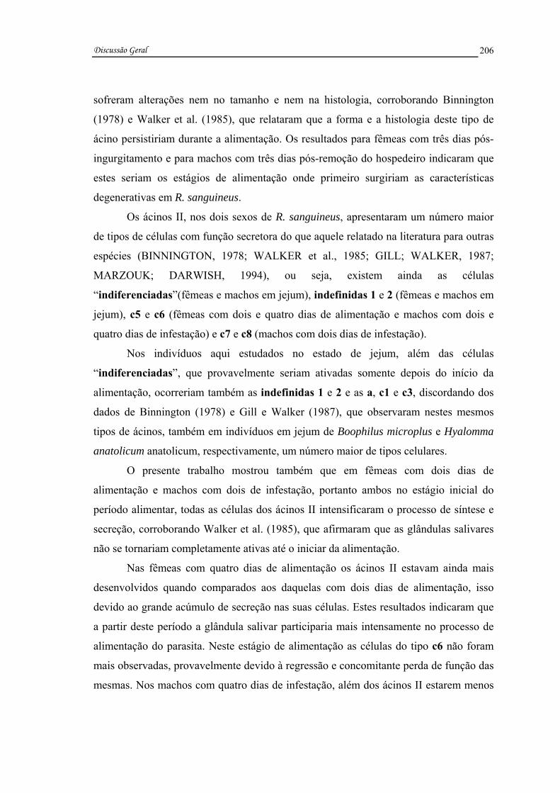
Discussão Geral
206
sofreram alterações nem no tamanho e nem na histologia, corroborando Binnington
(1978) e Walker et al. (1985), que relataram que a forma e a histologia deste tipo de
ácino persistiriam durante a alimentação. Os resultados para fêmeas com três dias pós-
ingurgitamento e para machos com três dias pós-remoção do hospedeiro indicaram que
estes seriam os estágios de alimentação onde primeiro surgiriam as características
degenerativas em R. sanguineus.
Os ácinos II, nos dois sexos de R. sanguineus, apresentaram um número maior
de tipos de células com função secretora do que aquele relatado na literatura para outras
espécies (BINNINGTON, 1978; WALKER et al., 1985; GILL; WALKER, 1987;
MARZOUK; DARWISH, 1994), ou seja, existem ainda as células
“indiferenciadas”(fêmeas e machos em jejum), indefinidas 1 e 2 (fêmeas e machos em
jejum), c5 e c6 (fêmeas com dois e quatro dias de alimentação e machos com dois e
quatro dias de infestação) e c7 e c8 (machos com dois dias de infestação).
Nos indivíduos aqui estudados no estado de jejum, além das células
“indiferenciadas”, que provavelmente seriam ativadas somente depois do início da
alimentação, ocorreriam também as indefinidas 1 e 2 e as a, c1 e c3, discordando dos
dados de Binnington (1978) e Gill e Walker (1987), que observaram nestes mesmos
tipos de ácinos, também em indivíduos em jejum de Boophilus microplus e Hyalomma
anatolicum anatolicum, respectivamente, um número maior de tipos celulares.
O presente trabalho mostrou também que em fêmeas com dois dias de
alimentação e machos com dois de infestação, portanto ambos no estágio inicial do
período alimentar, todas as células dos ácinos II intensificaram o processo de síntese e
secreção, corroborando Walker et al. (1985), que afirmaram que as glândulas salivares
não se tornariam completamente ativas até o iniciar da alimentação.
Nas fêmeas com quatro dias de alimentação os ácinos II estavam ainda mais
desenvolvidos quando comparados aos daquelas com dois dias de alimentação, isso
devido ao grande acúmulo de secreção nas suas células. Estes resultados indicaram que
a partir deste período a glândula salivar participaria mais intensamente no processo de
alimentação do parasita. Neste estágio de alimentação as células do tipo c6 não foram
mais observadas, provavelmente devido à regressão e concomitante perda de função das
mesmas. Nos machos com quatro dias de infestação, além dos ácinos II estarem menos

Discussão Geral
207
ativos, as células c6, c7 e c8 também não foram mais observadas, sugerindo interrupção
ou finalização de suas funções.
Ainda nos ácinos II de fêmeas alimentadas e com três dias pós-alimentação
observou-se o início da degeneração celular, sendo possível identificar: nas primeiras
apenas os tipos celulares a, c1 e c3, nas com três dias pós-alimentação somente os a e
c3 e nas com sete dias pós-ingurgitamento não se observaram mais ácinos do tipo II.
Nos machos com sete dias de infestação somente algumas células dos tipos c1 e c8
estavam íntegras; naqueles com três e sete dias pós-remoção todos os tipos estavam em
degeneração e especificamente naqueles com sete dias pós-remoção os c2 já tinham
degenerado, uma vez que não foram mais observados, dados estes primeiramente
relatados para machos de carrapatos.
Os ácinos III das fêmeas, diferentemente do observado nos machos, aumentaram
progressivamente de tamanho com o decorrer da alimentação e suas células,
principalmente na fase de grande consumo de sangue do carrapato, sofreram rápidas
transformações, passando de piramidais a pavimentosas, provocando assim um aumento
no lúmen do ácino e corroborando Binnington (1978), Walker et al. (1985), Fawcett et
al. (1986) e Sonenshine (1991). No caso específico dos machos a ausência de alterações
nos ácinos III dar-se-ia devido ao pequeno volume de sangue por eles consumido.
Ainda nos ácinos III dos carrapatos aqui estudados as células f, ao contrário das
d e e, estavam com citoplasma reduzido. Na condição de alimentação as células d e e
permaneceram repletas de secreção e as f apresentaram diferenças dependendo do
estágio alimentar considerado, como por exemplo: nas fêmeas com dois dias de
alimentação elas estavam ativas e nas com quatro dias não continham mais secreção,
provavelmente devido à perda de atividade secretora e atuação agora na
osmorregulação, corroborando dados de Binnington (1978) e Gill e Walker (1987). Já
nos machos na situação de alimentação, estas células apresentaram a mesma morfologia
encontrada nos machos em jejum, sugerindo a não participação das mesmas nem no
processo de secreção e nem no de osmorregulação, tendo sido consideradas por
Binnington (1978) como inativas, ao contrário do que ocorreria nas fêmeas.
Os ácinos III começaram a apresentar sinais de degeneração nas fêmeas
alimentadas, embora neles ainda pudessem ser identificados todos os tipos celulares
descritos na literatura. Naquelas com três dias pós-alimentação poucos ácinos III foram

Discussão Geral
208
identificados. Aqueles com sete dias pós-alimentação não apresentaram mais os ácinos
III. Nestes ácinos de machos o início da degeneração ocorreu nos indivíduos com sete
dias de infestação, onde estavam presentes os três tipos celulares e naqueles com três e
sete dias pós-remoção as células f não foram mais observadas.
No caso dos ácinos IV, específicos dos machos, em R. sanguineus as células
começaram a sintetizar e secretar intensamente apenas com o iniciar da alimentação,
enquanto naqueles com quatro dias de infestação, onde as glândulas estavam
completando um dos seus ciclos secretores, observou-se uma queda nestes processos.
Os indícios de degeneração surgiram, portanto, primeiramente nos machos com sete
dias de infestação. Os dados aqui obtidos corroboram parcialmente aqueles de
Binnington (1978), Walker et al. (1985), Fawcett et al. (1986), Gill e Walker (1987) e
Sonenshine (1991).
O acompanhamento da atividade secretora das glândulas salivares de fêmeas e
machos de R. sanguineus mostrou que estes órgãos começaram a degenerar após a
secreção de substâncias que fixariam o carrapato ao hospedeiro e que manipulariam a
resposta imunológica deste último. Este estudo revelou ainda que a degeneração
glandular se intensificar-se-ia à medida que o parasita passasse mais tempo fora do
hospedeiro, ocorrendo nas fêmeas as alterações mais significativas.
De forma geral o processo de degeneração glandular em R. sanguineus foi
assincrônico entre os diferentes ácinos, bem como entre os diferentes tipos de células de
um mesmo ácino. Os I foram os últimos afetados, permanecendo, portanto, íntegros nas
fêmeas ingurgitadas e nos machos com sete dias de infestação, corroborando os dados
de Till (1961), L’Amoreaux et al. (2003) e Nunes et al. (2006b), em R. appendiculatus,
D. variabilis e R (Boophilus) microplus, respectivamente. Os resultados confirmaram
que o processo de degeneração em R. sanguineus teve início na região posterior da
glândula salivar, onde as características de degeneração foram primeiro observadas. Na
anterior, no entanto, ainda foram encontrados ácinos íntegros, padrão este também
descrito para a degeneração glandular na maioria dos insetos (Silva de Moraes, 1998;
Abreu et al., 2004).
Os dados aqui obtidos também indicaram que as glândulas salivares perderiam
sua capacidade secretora, mostrada pela diminuição de grânulos e pelo surgimento de
características degenerativas nas fêmeas alimentadas e nos machos com sete dias de

Discussão Geral
209
infestação. Harris e Kaufman (1984) também relataram para A. hebraeum a redução de
mais de 90% da capacidade secretora máxima de glândulas de fêmeas desta espécie.
O estudo da degeneração glandular em R. sanguineus, sob o aspecto
citoquímico, mostrou que as células morreriam por apoptose, corroborando
L’Amoreaux et al. (2003), Bowman e Sauer (2004), Nunes et al. (2005) e Nunes et al.
(2005, 2006a, b), apresentando também características de morte autofágica, confirmada
pela presença da fosfatase ácida, a qual neste caso atuaria secundariamente, auxiliando
na remoção de restos citoplasmáticos das células glandulares, bem como fragmentando
e contribuindo, desta forma, para a desorganização dos ácinos e formação de corpos
apoptóticos, o que caracterizaria a apoptose atípica.
A forte marcação RNA positiva no citoplasma das células glandulares das
fêmeas alimentadas e dos machos com sete dias de infestação foi a primeira indicação
da ocorrência de morte por apoptose nestes indivíduos. Segundo alguns autores a síntese
de RNA e as proteínas surgiriam já no início do processo apoptótico, sendo estas
últimas consideradas as promotoras da apoptose (BOWEN; BOWEN, 1990; BOWEN;
BOWEN, 1995; ZAKERI et al., 1995; LOCKSHIN; ZAKERI, 1996; TATA, 1966 apud
HÄCKER, 2000).
Outros indicativos aqui observados, que confirmariam a apoptose nas glândulas
salivares, seriam as alterações nucleares precoces, bem como a retração citoplasmática
sofrida por suas células secretoras (CLARKE, 1990; BOWEN, 1993; KERR et al.,
1995; LOCKSHIN; ZAKERI, 1996; HÄCKER, 2000).
Por outro lado, o presente estudo revelou, além da queda precoce da atividade
ATPásica, perda da integridade da membrana e aumento da atividade da fosfatase ácida,
dados que não caracterizariam no entanto a apoptose clássica. Mullarkey (1987), apud
Bowen e Bowen (1990); Bowen et al. (1988), apud Bowen e Bowen (1990); Bowen e
Bowen (1990); Kerr et al. (1995) afirmaram que na morte apoptótica a perda da
atividade ATPásica e da integridade da membrana seriam eventos observados somente
nas fases muito avançadas da degeneração ou após a formação dos corpos apoptóticos e
segundo Pipan e Ravoc (1980), Clarke (1990), Cummings e Bowen (1992), Bowen
(1993), Zakeri et al. (1995), Lockshin e Zakeri (1996), Jochová et al. (1997) e Gregorc
et al. (1998) a fosfatase ácida estaria principalmente envolvida com a morte autofágica.
Os dados aqui obtidos sobre a atividade da ATPase e da fosfatase ácida concordaram

Discussão Geral
210
com Bowen et al. (1993), que estudando o processo de morte nas glândulas salivares de
Calliphora vomitoria detectaram um momento em que a atividade da ATPase cessaria,
indicando que na morte celular programada nem sempre ocorreria atividade ATPásica e
preservação da integridade da membrana até os estágios tardios do processo.
Neste estudo, nas fêmeas alimentadas e nos machos com sete dias de infestação
detectou-se aumento da atividade da fosfatase ácida, a qual se intensificou nas fêmeas
com três dias pós-alimentação, provocando maior vacuolização nas células. Ao
contrário, nos machos com três dias pós-remoção do hospedeiro houve um aumento na
atividade da fosfatase ácida, que diminuiu naqueles com sete dias pós-remoção. A
queda da fosfatase nas glândulas dos machos ocorreu devido a sua utilização e não mais
produção no processo de degeneração, pois nestes indivíduos, ao contrário do que
ocorreria nas fêmeas, a degeneração não seria hormonalmente estimulada (Kaufman,
1990; Lomas, 1993 apud Lomas et al., 1998).
De forma geral o presente estudo com as glândulas salivares de carrapatos da
espécie R. sanguineus, além de confirmar algumas informações já existentes na
literatura, trouxe à luz alterações que as mesmas sofrem no decorrer do processo de
alimentação, como as resumidas nas páginas 211 à 218.

Discussão Geral
211
ÁCINO I Jejum
idem
Jejum idem
2 Dias de Alimentação
idem
2 Dias de Infestação
idem
4 Dias de Alimentação
idem
4 Dias de Infestação
idem
Alimentada idem
7 Dias de Infestação
idem
3 Dias após idem, início da degeneração com alterações nucleares, pouca ou nenhuma atividade ATPásica e grande atividade fosfatásica
3 Dias após idem, início da degeneração com alterações nucleares, pouca ou nenhuma atividade ATPásica e grande atividade fosfatásica
F Ê M E A S
7 Dias após degenerado
MA C H O S
7 Dias após idem, com pouca atividade fosfatásica

Discussão Geral
212
ÁCINO I Jejum célula central e
células periféricas
Jejum célula central e células periféricas
2 Dias de Alimentação
idem
2 Dias de Infestação
idem
4 Dias de Alimentação
idem
4 Dias de Infestação
idem
Alimentada idem
7 Dias de Infestação
não descrito na literatura
3 Dias após não descrito na literatura
3 Dias após não descrito na literatura
F Ê M E A S
7 Dias após não descrito na literatura
MA C H O S
7 Dias após não descrito na literatura
idem: refere-se sempre ao estágio alimentar anterior;
descrição segundo Binnington (1978);e morte
dados apresentados neste estudo;
dados apresentados neste estudo referentes ao processo de morte celular;

Discussão Geral
213
ÁCINO II Jejum
células indiferenciadas, indefinidas 1 e 2, a, c1 e c3
Jejum células indiferenciadas, indefinidas 1 e 2, a, c1 e c3
2 Dias de Alimentação
células a, b, c1-c6
2 Dias de Infestação
células a, b, c1-c8
4 Dias de Alimentação
c5 regrediu e o restante persistiu
4 Dias de Infestação
c6, c7 e c8 regrediram e o restante persistiu
Alimentada presença de a, c1, c3 e indeterminadas em degeneração, com alterações nucleares, pouca atividade ATPásica e grande atividade fosfatásica
7 Dias de Infestação
c1 e c8 ou integras ou em degeneração a, c2-c5 e indeterminadas em degeneração, com alterações nucleares, pouca atividade ATPásica e grande atividade fosfatásica
3 Dias após presença de a, c3 e indeterminadas em degeneração, com pouca ou nenhuma atividade ATPásica, grande atividade fosfatásica e corpos apoptóticos
3 Dias após c1-c5, c8 e indeterminadas também em degeneração, com alterações nucleares, pouca ou nenhuma atividade ATPásica, grande atividade fastásica e corpos apoptóticos
F Ê M E A S
7 Dias após degenerado
MA C H O S
7 Dias após c2 ausente a, c3-c5 e indeterminadas persistiram pouca atividade fosfatásica
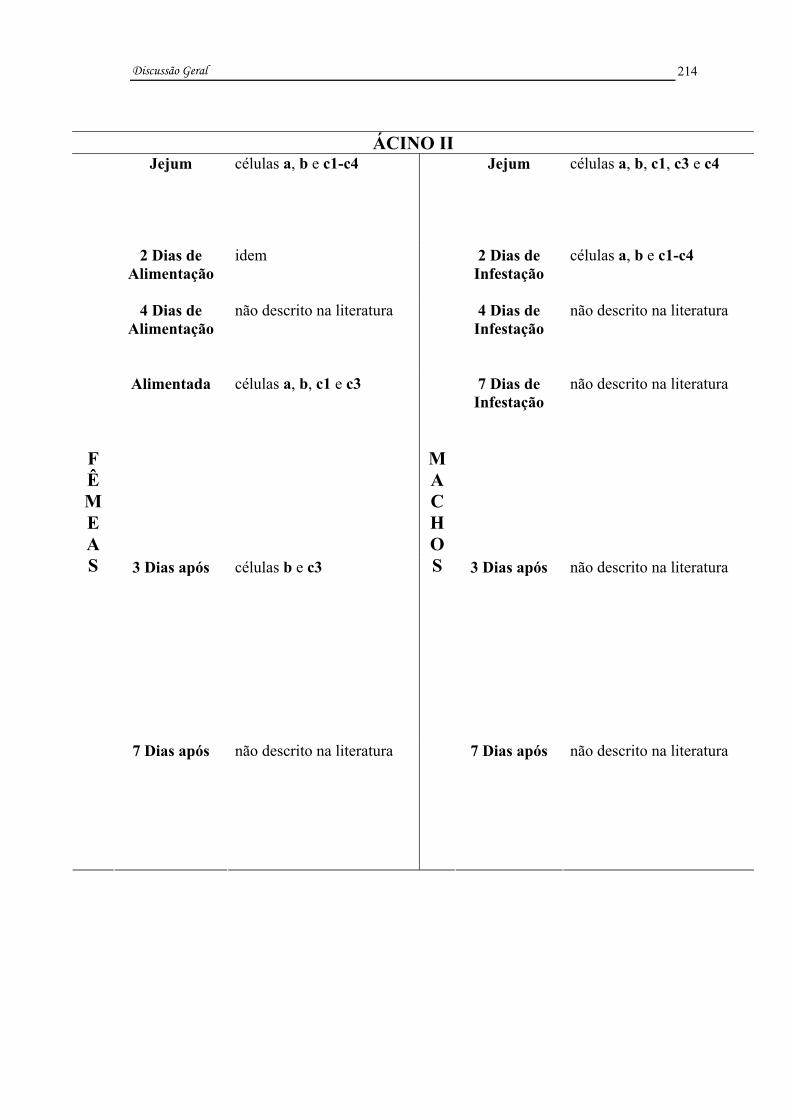
Discussão Geral
214
ÁCINO II Jejum células a, b e c1-c4
Jejum células a, b, c1, c3 e c4
2 Dias de Alimentação
idem
2 Dias de Infestação
células a, b e c1-c4
4 Dias de Alimentação
não descrito na literatura
4 Dias de Infestação
não descrito na literatura
Alimentada células a, b, c1 e c3
7 Dias de Infestação
não descrito na literatura
3 Dias após células b e c3
3 Dias após não descrito na literatura
F Ê M E A S
7 Dias após não descrito na literatura
MA C H O S
7 Dias após não descrito na literatura

Discussão Geral
215
ÁCINO III Jejum
idem
Jejum idem
2 Dias de Alimentação
idem
2 Dias de Infestação
idem
4 Dias de Alimentação
d e e pavimentosas e granulares f pavimentosas e agranulares
4 Dias de Infestação
idem
Alimentada d, e e f em degeneração, com alterações nucleares, pouca ou nenhuma atividade ATPásica e grande atividade fosfatásica
7 Dias de Infestação
d, e e f em degeneração, com alterações nucleares, pouca ou nenhuma atividade ATPásica e grande atividade fosfatásica
3 Dias após degenerado
3 Dias após f ausentes d e e persistiram grande atividade fosfatásica e corpos apoptóticos
F Ê M E A S
7 Dias após degenerado
MA C H O S
7 Dias após d e e em degeneração, com pouca atividade fosfatásica

Discussão Geral
216
ÁCINO III Jejum d e e cúbicas e granulares
f cúbicas e agranulares
Jejum d e e cúbicas e granulares f cúbicas e agranulares
2 Dias de Alimentação
d, e e f cúbicas e granulares
2 Dias de Infestação
idem
4 Dias de Alimentação
não descrito na literatura
4 Dias de Infestação
idem
Alimentada d e e pavimentosas e granulares f pavimentosas e agranulares
7 Dias de Infestação
não descrito na literatura
3 Dias após não descrito na literatura
3 Dias após não descrito na literatura
F Ê M E A S
7 Dias após não descrito na literatura
MA C H O S
7 Dias após não descrito na literatura

Discussão Geral
217
ÁCINO IV Jejum
idem
2 Dias de Infestação
idem
4 Dias de Infestação
idem
7 Dias de Infestação
células g em degeneração, com alterações nucleares, pouca ou nenhuma atividade ATPásica e grande atividade fosfatásica
3 Dias após células g em degeneração, com grande atividade fosfatásica e corpos apoptóticos
M A C H O S
7 Dias após células g em degeneração, sem atividade ATPásica e com pouca atividade fosfatásica
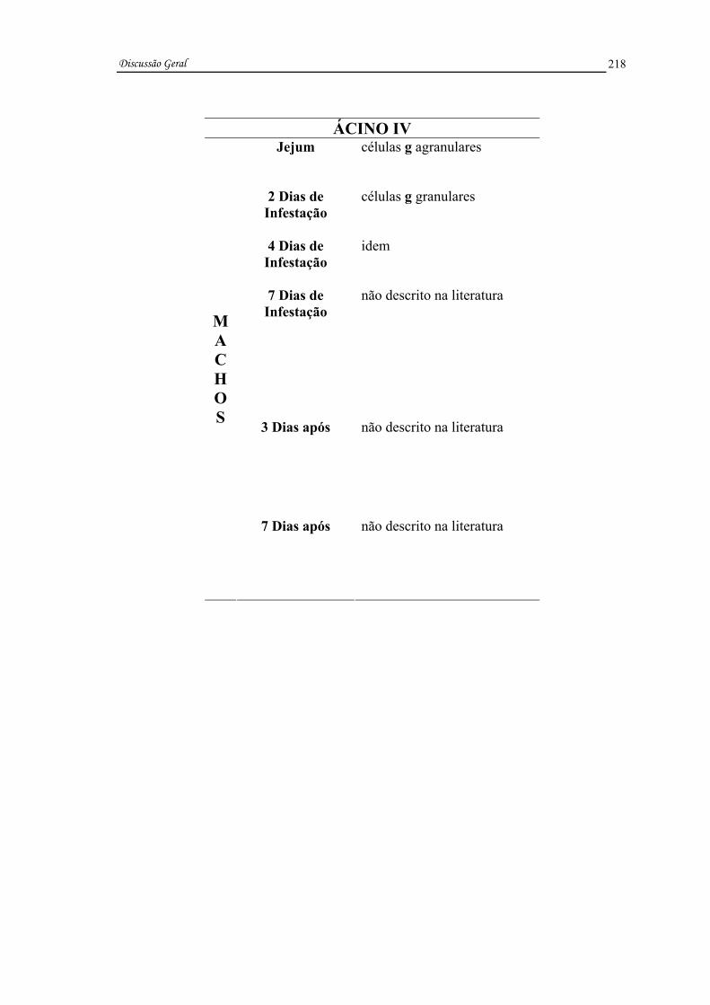
Discussão Geral
218
ÁCINO IV Jejum células g agranulares
2 Dias de Infestação
células g granulares
4 Dias de Infestação
idem
7 Dias de Infestação
não descrito na literatura
3 Dias após não descrito na literatura
M A C H O S
7 Dias após não descrito na literatura

Conclusões

Conclusões
220
VI. CONCLUSÕES
1) Os ácinos I das glândulas salivares de carrapatos Rhipicephalus sanguineus
não sofreram alterações morfológicas nem histológicas no decorrer da alimentação, e
adquiriram características degenerativas somente após o término da alimentação
(desprendimento do hospedeiro);
2) Este trabalho descreveu pela primeira vez nos ácinos II quatro novos tipos
celulares, que foram aqui denominados de “indiferenciadas”, indefinidas 1 e 2, c5, c6,
c7 e c8, sendo estes dois últimos observados somente nos machos;
3) As células secretoras dos ácinos II e III das fêmeas e II, III e IV dos machos
permaneceram inativas até que os carrapatos iniciassem a alimentação;
4) Nos ácinos do tipo II de fêmeas e de machos e nos III de fêmeas os diferentes
tipos celulares secretaram assincronicamente ao longo do ciclo glandular e
conseqüentemente atuaram em momentos diferentes do processo de alimentação;
5) Nos machos as células das glândulas salivares passaram por vários ciclos
secretores diferentemente das fêmeas, sofrendo no final de cada ciclo uma
reprogramação na sua expressão e voltando a ter a mesma morfologia observada
naqueles em jejum;
6) Nas glândulas salivares dos machos somente as células a, c1 e c3 nos ácinos
II e as d e e nos III permaneceram continuamente ativas;
7) No decorrer do ciclo secretor das glândulas de fêmeas e machos as células
secretoras (fêmeas c5 e machos c6, c7 e c8) deixaram de ser observadas por regredirem
e provavelmente perderem sua função;
Conclusões
221
8) O surgimento de características degenerativas no tecido glandular das fêmeas
provocou redução da sua capacidade secretora, o que fez com que estes indivíduos
finalizassem seu processo alimentar;
9) A degeneração das glândulas salivares de fêmeas e machos de R. sanguineus
foi detectada no final da alimentação e se intensificou à medida que ficaram mais tempo
fora do hospedeiro;
10) As alterações degenerativas das glândulas salivares foram observadas com
mais intensidade nas fêmeas do que nos machos;
11) A degeneração glandular, tanto de fêmeas quanto de machos de R.
sanguineus, foi assincrônica entre os diferentes ácinos e entre as diferentes células de
um mesmo ácino, afetando primeiramente aqueles da região posterior da glândula;
12) Durante a degeneração das glândulas salivares detectou-se ausência da
atividade ATPásica e perda da integridade da membrana plasmática em momento
diferente (precocemente) daquele relatado na literatura;
13) A fosfatase ácida que atuou apenas na remoção dos restos citoplasmáticos,
nas glândulas de R. sanguineus, provavelmente teve origem endógena (produção pela
própria célula), contrariando registros da literatura que informam origem exógena (na
hemolinfa);
14) A degeneração nas glândulas salivares em R. sanguineus apresentou
características de mais de um tipo de morte celular (apoptose e morte autofágica),
caracterizando assim uma apoptose atípica;

Referências
Referências
223
VII. REFERÊNCIAS
ABREU, R.M.M.; SILVA DE MORAES, R.L.M.; TABOGA, S.R.; CAMARGO-
MATHIAS, M.I. Cytochemical studies of the nuclei of the venom glands’ cells of Apis
mellifera (Hymenoptera, Apidae). Acta Histochemica et Cytochemica, Kyoto, v. 37,
n. 5, p. 195-299, 2004.
ARMBRUSTER, L.; LEVY, M.; MATHIEU, M. N.; BAUTZ, A. M. Acid phosphatase
activity in the hemolymph, hemocytes, fat body and salivary glands during larval and
prepupal development in Calliphora erythrocephala (Diptera: Calliphoridae).
Comparative Biochemistry and Physiology, Tarrytown, v. 84B, n.3, p. 349-354,
1986.
AZEREDO-OLIVEIRA, M. T. V.; MELLO, M. L. Mg2+-dependent ATPase activity
investigated on heterochromatin, euchromatin and nucleoli of Triatoma infestans.
Revista Brasileira de Genética, Ribeirão Preto, v. IX, n. 2, p. 223-229, 1986.
BALASHOV, Yu. S. Bloodsucking ticks (Ixodoidea)- vectors of diseases of man and
animals. Miscellaneous Publications of the Entomological Society of America,
Lanham, v.8, p. 161-376, 1972.
BALASHOV, Yu. S. An Atlas of Ixodid Tick Ultrastructure. In: RAIKHEL, A. S.;
HOOGSTRAAL, I. I. (ed.). Entomological Society of America (Special Publication),
Lanham, 1983. 289p.

Referências
224
BARKER, D. M.; OWNBY, C. L.; KROLAK, J. M.; CLAYPOOL, P.L.; SAUER, J. R.
The effects of attachment, feeding and mating on the morphology of type I alveolus of
salivary glands of the lone star tick, Amblyomma americanum. International Journal
for Parasitology, Oxford, v. 70, p. 99-113, 1984.
BECHARA, G.H.; SZABÓ, M.P.J.; FERREIRA, B.R.; GARCIA, M.V. Rhipicephalus
sanguineus tick in Brazil: feeding and reproductive aspects under laboratorial
conditions. Brazilian Journal of Veterinary Parasitology, São Paulo, v. 4, n. 2, p. 61-
66, 1995.
BINNINGTON, K. C. Sequential changes in salivary gland structure during attachment
and feeding of the cattle tick Boophilus microplus. International Journal for
Parasitology, Oxford, v. 8, p. 97-115, 1978.
BIOR, A.D.; ESSEMBERG, R.C.; SAUER, J.R. Comparison of differentially expressed
genes in the salivary glands of male ticks, Amblyomma americanum and Dermacentor
andersoni. Insect Biochemistry and Molecular Biology, Oxford, v. 32, p. 645-655,
2002.
BISHOP, R.; LAMBSON, B.; WELLS, C.; PANDIT, P.; OSASO, J.; NKONGE, C.;
MORZARIA, S.; MUSOKE, A.; NENE, V. A cement protein of the tick Rhipicephalus
appendiculatus, located in the secretory e cell granules of the type III salivary gland
acini, induces strong antibody responses in cattle. International Journal for
Parasitology, Oxford, v. 32, p. 833-842, 2002.
BOWEN, I. D. Apoptosis or programmed cell death? Cell Biology International,
London, v. 17, n. 4, p. 365-380, 1993.
BOWEN, I. D.; BOWEN, S. M. Programmed cell death in tumours and tissues.
London: Chapman and Hall, 1990. 289p.

Referências
225
BOWEN, I. D.; MORGAN, S. M.; MULLARKEY, K. Cell death in the salivary glands
of metamorphosing Calliphora vomitoria. Cell Biology International Reports,
London, v. 17, p. 13-34, 1993.
BOWEN, I. D.; MULLARKEY, K.; MORGAN, S. M. Programmed cell death in the
salivary gland of the blow fly Calliphora vomitoria. Microscopy Research and
Technique, New York, 34: 202-217. 1996.
BOWEN, I. D.; WORRILL, N.A.; WINTERS, C. A.; MULLARKEY, K. The use of
backscattered electron imaging, X-ray microscopy in demonstrating physiological cell
death. Scanning Microscopy, Chicago, v. 2, p. 1453-1462, 1988.
BOWMAN, A. S.; SAUER, J. R. Tick salivary glands: function, physiology and future.
Parasitology, Cambridge, v. 129, p. S67-S81, 2004.
BRITTO, F. B.; CAETANO, F. H. Morphological features and occurrence of
degenerative characteristics in the hypopharyngeal glands of the paper wasp Polistes
versicolor (Olivier) (Hymenoptera: Vespedae). Micron, New York, v. 37, p. 742-747,
2006.
CLARKE, P. G. H. Developmental cell death: morphological diversity and multiple
mechanisms. Anatomy and Embriology, Berlin, v. 181, p. 195-213, 1990.
COONS, L. B.; L’AMOREAUX, W. J. Developmental changes in the salivary glands
of male and female Dermacentor variabilis (Say) during feeding. In: BOROVSKY, D.;
SPIELMAN, A. (eds.). Host Regulated Developmental Mechanisms in Vector
Arthropods. v. 2, University of Florida-IFAS, Vero Beach, 1986. p. 86-92.
COSTA, R. A. C.; CRUZ-LANDIM, C. Distribution of acid phosphatases in the
hypopharyngeal glands from workers, queens and males of a Brazilian stingless bee
Scaptotrigona postica Latreille: an ultrastructural cytochemical study. Histochemical
Journal, London, v. 33, p. 653-662, 2002.
Referências
226
CUMMINGS, L. A.; BOWEN, I. D. The localization of acid phosphatase as a prelude
to cell death in the blow-fly Calliphora erithrocephala. Micron and Microscopica
Acta, New York, v. 23, n. 3, p. 357-358, 1992.
DAI, J. D., GILBERT, L. I. Programmed cell death of the prothoracic glands of
Manduca sexta during pupal-adult metamorphosis. Insect Biochemistry and
Molecular Biology, Oxford, v. 27, n. 1, p. 69-78, 1997.
FAWCETT, D. W.; BINNINGTON, K. C.; VOIGHT, W. R. The cell biology of the
ixodid tick salivary gland. In: SAUER, J. R.; HAIR, J. A. (ed.). Morphology,
Physiology and Behavioral Biology of Ticks. Chichester: Ellis Horwood, 1986. p. 22-
45.
FELDMAN-MUHSAM, B.; BORUT, S.; SALITERNIK-GIVANT, S.. Salivary
secretion of the male tick during copulation. Journal of Insect Physiology, Oxford, v.
16, p. 1945-1949, 1970.
FLECHTMANN, C. H. W. Ácaros de importância médico-veterinária. Editora
Livraria Nobel S.A., São Paulo, 1973. 192 p.
FEULGEN, R.; ROSSENBECK, H. Mikroskopisch – Chemischer Nachweis einer
Nucleinsaure vom Typus der Thymonucleinsaure und Die Darauf Beruhend Elective
Farbung von Zillkernen in Mikroskopischen Praparaten Hoppe-Seylers. Zeitschrift fur
Physiologische Chemie, Strassburg, v.135, p.203-248, 1924.
FURQUIM, K. C. S. 2004. Processos degenerativos em glândulas salivares de
carrapatos Rhipicephalus sanguineus (LATREILLE, 1806) (ACARI, IXODIDAE)
em jejum, em alimentação e alimentados. 2004. 37 pp. 1º Relatório Científico
CAPES, Instituto de Biociências, UNESP, Rio Claro, 2004.
Referências
227
FURQUIM, K. C. S. 2005. Processos degenerativos em glândulas salivares de
carrapatos Rhipicephalus sanguineus (LATREILLE, 1806) (ACARI, IXODIDAE)
em jejum, em alimentação e alimentados. 2005. 40 pp. 2º Relatório Científico
CAPES, Instituto de Biociências, UNESP, Rio Claro, 2005.
FURQUIM, K. C. S. 2006. Processos degenerativos em glândulas salivares de
carrapatos Rhipicephalus sanguineus (LATREILLE, 1806) (ACARI, IXODIDAE)
em jejum, em alimentação e alimentados. 2006. 27 pp. 3º Relatório Científico
CAPES, Instituto de Biociências, UNESP, Rio Claro, 2006.
FURQUIM, K. C. S.; CAMARGO-MATHIAS, M. I.; SILVA DE MORAES, R. L. M.
Morphological modifications induced by an artificial diet on the hypopharyngeal glands
of Apis mellifera (Hymenoptera, Apidae) during their degenerative process.
Sociobiology, Chico, v. 43, n. 2, p. 279-291, 2004.
GAEDE, H.; KNULLE, W. On the mechanism of water vapour sorption from
unsaturated atmospheres by ticks. The Journal of Experimental Biology, Cambridge,
v. 200, p. 1491-1498, 1997.
GILL, H. S.; WALKER, A. R. The salivary glands of Hyalomma anatolicum
anatolicum: nature of salivary gland components and their role in tick attachment and
feeding. International Journal for Parasitology, Oxford, v. 18, n. 1, p. 83-93, 1987.
GREGORC, A.; BOWEN, I. D. Programmed cell death in the honeybee (Apis mellifera)
larvae midgut. Cell Biology International, London, v. 21, n. 3, p. 151-158, 1997.
GREGORC, A.; BOWEN, I.D.; POGACNIK, A. Acid phosphatase activity in the
midgut of honeybee (Apis melliphera L.) larvae. Apidologie, Versailles, v. 29, p. 579-
584, 1998.
HÄCKER, G. The morphology of apoptosis. Cell and Tissue Research, New York, v.
301. p. 5-17, 2000.
Referências
228
HARRIS, R. A.; KAUFMAN, W. R. Neural involvement in the control of salivary
gland degeneration in the ixodid tick Amblyomma hebraeum. Journal of Experimental
Biology, Cambridge, v. 109, p. 281-290, 1984.
HARRIS, R. A.; KAUFMAN, W. R. Ecdysteroids: possible candidates for the hormone
which triggers salivary gland degeneration in the tick, Amblyomma hebraeum.
Experientia, Basel, v. 41, p. 740-741, 1985.
HURLE, J.; HINCHLIFFE, J. R. Cell death in the posterior necrotic zone (PNZ) of the
chick wing-bud: a setereoscan and ultrastructural survey of autolysis and cell
fragmentation. Journal of Embryology and Experimental Morphology, London, v.
43, p. 123-136, 1978.
HUSSEIN, M. A.; BOWEN, I. D.; LEWIS, G. H. The histochemical localization of
ATPase, cholinesterase and acid phosphatase activity in Culex pipiens (Diptera,
Culicidae) larvae using a methacrylate embedding technique. Cell Biology
International Reports, London, v. 14, n. 9, p. 775-781, 1990.
JIANG, C.; BAEHRECKE, E. B.; THUMMEL, C. S. Steroid regulated programmed
cell death during Drosophila metamorphosis. Development, Cambridge, v. 124, p.
4673-4683, 1997.
JOCHOVÁ, J.; ZAKERI, Z.; LOCKSHI, R. A. Protein synthesis, DNA degradation,
and morphological changes during programmed cell death in labial glands of Manduca
sexta. Developmental Genetics, New York, v. 21, p. 249-257, 1997.
JONES, H. E.; BOWEN, I. D. Acid phosphatase activity in the salivary glands of
developing Drosophila melanogaster. Cell Biology International, London, v. 17, p.
305-315, 1993.
JUNQUEIRA, L. C. U.; JUNQUEIRA, L. M. M. S. Técnicas básicas de citologia e
histologia. São Paulo: Editora Santos, 1983. p. 48-81.
Referências
229
JUNQUEIRA, L. C.; CARNEIRO, J. Histologia básica- Texto/Atlas, 10 ed. Rio de
Janeiro: Guanabara Koogan, 2004, 488 p.
KAUFMAN, W.E. Effect of 20-hydroxiecdysone on the salivary glands of the male
tick, Amblyomma hebraeum. Experimental and Applied Acarology, Amsterdam, v. 9,
p. 87-95, 1990.
KAUFMAN, W. R.; LOMAS, L. O. Male factors in ticks: Their role in feeding and egg
development. Invertebrate Reproduction and Development, Rehovot, v. 30, p. 191-
198, 1996.
KAUFMAN, W. R.; SAUER, J. R. Ion and water balance in feeding ticks: mechanism
of tick excretion. In: OBENCHAIN, F. D., GALUN, R. (eds.), Physiology of Ticks.
Pergamon Press, Oxford, 1982. p. 213-243.
KERR, J. F. R.; GOBÉ, G. C.; WINTERFORD, C. M.; HARMONT, B. V. Anatomical
Methods in Cell Death. In: SCHWARTZ, L. M.; OSBORNE, B. A. (ed.). Cell Death.
San Diego: Academic Press, 1995. p. 1-27.
KRSTIC, R.; PEXIEDER, T. Ultrastructure of cell death in bulbar cushions of chick
embryo heart. Zeitschrift für Anatomie und Entwicklungsgeschichte. Berlin, v. 140,
p. 337-350, 1973.
L'AMOREAUX, W. J.; JUNAID, L.; TREVIDI, S. Morphological evidence that
salivary gland degeneration in the American dog tick, Dermacentor variabilis (Say),
involves programmed cell death. Tissue and Cell, Edinburgh, v. 35, p. 95-99, 2003.
LEVY, M.; BAUTZ, A. M. Degeneration of larval salivary glands during
metamorphosis of the blowfly Calliphora erythrocephala Meigen (Diptera:
Calliphoridae). International Journal of Insect Morphology and Embriology,
Oxford, v. 14, p. 281-290, 1985.
Referências
230
LOCKSHIN, A. R.; ZAKERI, Z. The Biology of Cell Death and Its Relationship to
Aging. In: HOLBROOK, N.; MARTIN, G. R., LOCKSHIN, R. A. (eds.). Cellular
Aging and Cell Death. Wiley-Liss, New York, 1996, p. 167-180.
LOMAS, L.O. The influence of a male-derived protein factor on salivary gland
degeneration in the ixodid tick, Amblyomma hebraeum Koch. 1993. 197 p. Tese
(PhD), University of Alberta, Edmond, 1993.
LOMAS, L. O.; GELMAN, D.; KAUFMAN, W. R. Ecdysteroid regulation of salivary
gland degeneration in the ixodid tick Amblyomma hebraeum: a reconciliation of in vivo
and in vitro observations. General and Comparative Endocrinology, Orlando, v. 109,
p. 200-211, 1998.
LOMAS, L. O.; KAUFMAN, W. R. An indirect mechanism by which a protein from
the male gonad hastens salivary gland degeneration in the female ixodid tick
Amblyomma hebraeum. Archives of Insect Biochemistry and Physiology, Bonn, v.
21, p.169-178, 1992.
MACGAHON, A. J.; MARTIN, S. J.; BISSONNETTE, R. P.; MAHBOUBI, A.; SHI,
Y.; MOGIL, R. J. The End of the (Cell) Line: Methods for the Study of Apoptosis in
vitro. In: SCHWARTZ, L. M.; OSBORNE, B. A. (eds.). Cell Death. San Diego:
Academic Press, 1995. p. 153-185.
MAO, H.; KAUFMAN, W. R. Profile of the ecdysteroid hormone and its receptor in the
salivary gland of the adult female tick, Amblyomma hebraum. Insect Biochemistry and
Molecular Biology, Oxford, v. 29, p. 33-42, 1999.
MAO, H.; MACBLAIN, W. A.; KAUFMAN, W. R. Some properties of the ecdysteroid
receptor in the salivary gland of the ixodid tick, Amblyomma hebraeum. General and
Comparative Endocrinology, San Diego, v. 99, p. 340-348, 1995.

Referências
231
MARZOUK, A. S.; DARWISH, Z. E. A. Changes in the salivary glands of female
Hyalomma (Hyalomma) dromedarii during and after feeding. Journal of the Egyptian
Society of Parasitology, Cairo, v. 24, n. 1, p. 39-57, 1994.
MCMANUS, J. F. A. Histological demonstration of mucin after periodic acid. Nature,
London, v. 158, p. 202, 1946.
MCMULLEN, H. L.; SAUER, J. R.; BURTON, R. L. Possible role in uptake of water
vapour by ixodid ticks salivary glands. Journal of Insect Physiology, Oxford, v. 22, p.
1281-1285, 1976.
MELLO, M. L. S. Cytochemical properties of euchromatin and heterochromatin.
Histochemical Journal, London, v. 15, p. 739-751, 1983.
MELLO, M. L. S.; BARBISAN, L. F.; LAREEF, M. H.; RUSSO, J.; VIDAL, B. C.
Cell death evaluation in benzo[α]pyrene-transformed human breast epithelial cells after
microcell-mediated transfer of chromosomes 11 and 17. Mutation Research,
Amsterdam, v. 546, n. 1-2, p. 39-43, 2004.
MELLO, M. L. S.; CONTENTE, S.; VIDAL, B. C.; PLANDING, W.; SCHENCK, U.
Modulation of ras transformation affecting chromatin supraorganization as assessed by
image analysis. Experimental Cell Research, New York, v. 220, p. 374-382, 1995.
MELLO, M. L. S.; VIDAL, B. C. V. Chromatin organization in normal and cancer
cells. Journal of Women’s Cancer, Rydal, v. 2, n. 2, p. 73-78, 2000.
MELLO, M. L. S.; VIDAL, B. C.; DANTAS, M. M.; MONTEIRO, A. L. P.
Discrimination of the nucleolus by a critical eletrolyte concentration method. Acta
Histochemica et Cytochemica, Kyoto, v. 26, n. 1, p. 1-3, 1993.
Referências
232
MORAES, A. S.; VIDAL, B. C.; GUARALDO, A. M. A.; MELLO, M. L. S.
Chromatin supraorganization and extensibility in mouse hepatocytes following
starvation and refeeding. Cytometry, Hoboken, part A, v. 63A, p. 94-107, 2005.
MULLARKEY, K. Gene expression in the salivary gland of the blowfly (Calliphora
vomitoria) larva. 1987. 156 p. Tese (PhD), University of Exerter, Exerter, 1987.
NEEDHAM, G. R.; GREENWALD, L.; FREDA, T. J.; COONS, L. B. The
ultrastructure of the tick (Amblyomma americanum) salivary gland acini responsible for
secretion of the hygroscopic fluid and localization of Na+/K+ = ATPase. American
Zoologist, Lawrence, v. 23, p.1013, 1983.
NUNES, E. T.; CAMARGO-MATHIAS, M. I.; BECHARA, G. H. Rhipicephalus
(Boophilus) microplus (Canestrine, 1887) (Acari: Ixodidae): acid phosphatase and
ATPase activities localization in salivary glands of females during the feeding period.
Experimental Parasitology, San Diego, v. 114, p. 109-117, 2006a.
NUNES, E. T.; CAMARGO-MATHIAS, M. I.; BECHARA, G. H. Structural and
cytochemical changes in the salivary glands of the Rhipicephalus (Boophilus) microplus
(Canestrini, 1887) (Acari: Ixodidae) tick female during feeding. Veterinary
Parasitology, Amsterdam, v. 140, p. 114-123, 2006b.
OLIVEIRA, P. R.; BECHARA, G. H.; DENARDI, S. E.; SAITO, K. C.; NUNES, E. T.;
SZABÓ, M. P. J.; CAMARGO-MATHIAS, M. I. Comparison of the external
morphology of Rhipicephalus sanguineus (Latreille, 1806) (Acari: Ixodidae) ticks from
Brazil and Argentina. Veterinary Parasitology, Amsterdam, v. 129, p.139-147, 2005.
OLIVER, J. H. JR. Relationship among feeding, gametogenesis, mating and syngamy in
ticks. In: BOROVSKY, D.; SPIELMAN, A. (eds.). Host Regulated Developmental
Mechanism in Vector Arthropods. Vero Beach: University of Florida-IFAS, 1986. pp.
93-99.

Referências
233
OLIVIERI, J. A.; SERRA-FREIRE, N. M.. Structure of the salivary glands of the unfed
female tick Amblyomma cajennense (Fabricius) (Acarina: Ixodidae). Memórias do
Instituto Oswaldo Cruz, Rio de Janeiro,v. 87, suppl. I, p. 167-174, 1992.
PAUTOU, M.; KIENY, M. Sur les mecanismes histologiques et cytologiques de la
necrose morphogene interdigitale chez I’embrion de poulet. Comptes Rendus
Hebdomadaires des Seances de l' Academie des Sciences, Sciences Naturelles, Série
D, Paris, v. 272, p. 2025-2028, 1971.
PELT-VERKUIL, E. V. Hormone mediated induction of acid phosphatase activity in
the fat body of Calliphora erythrocephala prior to metamorphosis. Journal of Insect
Physiology, Oxford, v. 25, p. 965-973, 1979.
PIPAN, N.; RAKOVEC, V. Cell death in the midgut epithelium of the worker honey
bee (Apis mellifera carnica) during metamorphosis. Zoomorphologie, Berlin, v. 97, p.
217-224, 1980.
REY, L. Parasitologia. Rio de Janeiro: Guanabara Koogan, 1973. p. 633-641.
RIBEIRO, A. L.; FACCINI, J. L. H.; DAEMON, E. Estudo das variações morfológicas
de Rhipicephalus sanguineus (Latreille, 1806) (Acari: Ixodidae) no Brasil. Revista da
Universidade Federal Rural do Rio de Janeiro, Série Ciências da Vida, Itaguai,
v.18, n. 1-2, p. 25-33, 1996.
RIBEIRO, J. M. C.; MAKOUL, G.T.; LEVINE, J; ROBINSON, D. K.; SPILMAN, A.
Antihaemostatic, antiinflammatory and immunosupressive properties of the saliva of a
tick, Ixodes dammini. Journal of Experimental Medicine, New York, v. 161, p. 332-
344, 1985.
ROSENAU, W.; GOLDBERG, M. L.; BURKE, G. C. Early biochemical alterations
induced by lymphotoxin in target cells. The Journal of Immunology, Baltimore, v.
111, p. 1128-1135, 1973.

Referências
234
RUDOLPH, D. KUNLLE, W. Site of the mechanism of water vapour uptake from the
atmosphere in ixodid ticks. Nature, London, v. 249, p. 84-85, 1974.
RUDOLPH, D.; KNÜLLE, W. Uptake of water vapour from air: process, site, and
mechanism in ticks. In: SCHMIDT-MIELSEN, K.; BOLIO, L.; MADDRELL, S. H. P.
(eds.). Comparative Physiology: Water, Ions and Fluid Mechanics. Cambridge:
Cambridge University Press, 1978. p. 84-85.
SANDERS, M. L.; SCOTT, A. L.; GLASS, G. E.; SCHWARTZ, B. S. Salivary gland
changes and host antibody responses associated with feeding of male lone star ticks
(Acari: Ixodidae). Journal of Medical Entomology, Lanham, v. 33, n. 4, p. 628-634,
1996.
SAUER, J. R; ESSENBERG, R. C.; BOWMAN, A. S. Salivary glands in ixodid ticks:
control and mechanism of secretion. Journal of Insect Physiology, Oxford, v. 46, p.
1069-1078, 2000.
SCHUMAKER, T. T.; SERRA-FREIRE, N. M. Histologia das glândulas salivares de
adultos de Argas (Persicargas) miniatus (Koch, 1844) (Ixodoidea: Argasidae) em
jejum, em alimentação e alimentados. Revista Brasileira de Entomologia, São Paulo,
v. 35, p. 49-72, 1991.
SERRA-FREIRE, N. M.; OLIVIERI, J. A. Structure of the salivary glands of the unfed
male tick Amblyomma cajennense (Fabricius) (Acari: Ixodidae). Memórias do
Instituto Oswaldo Cruz, Rio de Janeiro, v. 88, n. 2, p.207-213, 1993.
SILVA DE MORAES, R. L. M. Morte celular nas glândulas hipofaríngeas de Apis
mellifera (Hymenoptera, Apidae). 1998. 97p. Tese (Livre Docência), Instituto de
Biociências, UNESP, Rio Claro, Brasil, 1998.

Referências
235
SILVA DE MORAES, R. L. M.; BOWEN, I. D. Modes of cell death in the
hypopharyngeal gland of the honey bee (Apis mellifera). Cell Biology International,
London, v. 24, n. 10, p. 737-743, 2000.
SONENSHINE, D. E. Biology of ticks. New York: Oxford University Press, 1991. p.
141-158.
TATA, J. R. Requirement for RNA and protein synthesis for induced regression of the
tadpole tail in organ culture. Developmental Biology, San Diego, v. 13, p. 77-94, 1966.
TILL, W. M. A contribution to the anatomy and histology of the brown ear tick
Rhipicephalus appendiculatus Neumann. Memoirs of the Entomological Society of
Southern Africa, Pretoria, v. 6, p. 1-124, 1961.
VIDAL, B. C.; RUSSO, J.; MELLO, M. L. S. DNA content and chromatin texture of
benzo[α]pyrene-transformed human breast epithelial cells as assessed by image
analysis. Experimental Cell Research, New York, v. 244, p. 77-82, 1998.
WALKER, A. Arthropods of domestic animals. A guide to preliminary identification.
London: Chapman and Hall, 1994. p. 25-29.
WALKER, A.; FLETCHER, J. D.; GILL, H. S. Structural and histochemical changes in
the salivary glands of Rhipicephalus appendiculatus during feeding. International
Journal for Parasitology, Oxford, v. 15, n. 1, p. 81-100, 1985.
WALKER, S. M.; LUCAS, Z. J. Cytochemical activity of lymphocytes: II. Studies on
mechanism of lymphotoxin-mediated cytotoxicity. The Journal of Immunology,
Baltimore, v. 109, p. 1233-1244, 1972.
WANG, H.; HENBEST, P. J.; NUTTALL, P. A. Successful interrupted feeding of adult
Rhipicephalus appendiculatus (Ixodidae) is accompanied by re-programming of
salivary gland protein expression. Parasitology, Cambridge, v. 119, p. 143-149, 1999.

Referências
236
WEBER, R. Tissue involution and lysosomal enzymes during anuran metamorphosis.
In: DINGLE, J. T.; FELL, H. B. (eds.). Lysosomes in Biology and Pathology,
Amsterdam: North Holland Publishing Co., vol.2., 1969. p. 437-461.
WEISS, B. L.; KAUFMAN, W. R. The relationship between ‘critical weight’ and 20-
hydroxyecdysone in the female ixodid tick Amblyomma hebraeum. Journal of Insect
Physiology, Oxford, v. 47, p. 1261-1267, 2001.
WEISS, B. L.; KAUFMAN, W. R. Two feeding-induced proteins from the male gonad
trigger engorgement of the female tick Amblyomma hebraeum. Proceedings of the
National Academy of Sciences, Allahabad, v. 101, n. 16, p. 5874-5879, 2004.
WIKEL, S. K. The induction of host resistance to tick infestation with a salivary gland
antigen. American Journal of Tropical Medicine Hygiene, Northbrook, v. 30, p. 284-
288, 1981.
YAMAMOTO, S.; SAWADA, K.; SHIMOMURA, H.; KAWAMURA, K.; JAMES, T.
N.. On the nature of cell death during remodeling of hypertrophied human myocardium.
Journal of Molecular and Cellular Cardiology, London, v. 32, p. 161-175, 2000.
ZAKERI, Z. F.; AHUJA, H. S. Cell death/apoptosis: normal, chemically induced, and
teratogenic effect. Mutation Research, Amsterdam, v. 396, p. 149-161, 1997.
ZAKERI, Z.; BURSCH, W.; TENNISWOOD, M.; LOCKSHIN, R. A. Cell death:
programmed, apoptosis, necrosis, or other? Cell Death and Differentiation, [S. I.], v.
2, p. 87-96, 1995.

Tese
237
Orientada: ___________________________________
Karim Christina Scopinho Furquim
Orientadora: ___________________________________
Profa. Dra. Maria Izabel Camargo-Mathias
Co-Orientador: ___________________________________
Prof. Dr. Gervásio Henrique Bechara