ESTUDO DE BIOPROSPECÇÃO DE AVICENNIA SCHAUERIANA · CCB/UFPE, o Departamento de Histologia e...
79
UNIVERSIDADE FEDERAL DE PERNAMBUCO CENTRO DE CIÊNCIAS DA SAÚDE PROGRAMA DE PÓS-GRADUAÇÃO EM ODONTOLOGIA MESTRADO EM ODONTOLOGIA ÁREA DE CONCENTRAÇÃO EM CLÍNICA INTEGRADA ESTUDO DE BIOPROSPECÇÃO DE AVICENNIA SCHAUERIANA: DESENVOLVIMENTO DE UM CREME CICATRIZANTE CAROLINE MARIA IGREJAS LOPES RECIFE 2015
Transcript of ESTUDO DE BIOPROSPECÇÃO DE AVICENNIA SCHAUERIANA · CCB/UFPE, o Departamento de Histologia e...
UNIVERSIDADE FEDERAL DE PERNAMBUCO
CENTRO DE CIÊNCIAS DA SAÚDE
PROGRAMA DE PÓS-GRADUAÇÃO EM ODONTOLOGIA
MESTRADO EM ODONTOLOGIA
ÁREA DE CONCENTRAÇÃO EM CLÍNICA INTEGRADA
ESTUDO DE BIOPROSPECÇÃO DE AVICENNIA SCHAUERIANA:
DESENVOLVIMENTO DE UM CREME CICATRIZANTE
CAROLINE MARIA IGREJAS LOPES
RECIFE
2015
CAROLINE MARIA IGREJAS LOPES
ESTUDO DE BIOPROSPECÇÃO DE AVICENNIA SCHAUERIANA:
DESENVOLVIMENTO DE UM CREME CICATRIZANTE
Dissertação apresentada ao Colegiado do
Programa de Pós-Graduação em Odontologia
do Centro de Ciências da Saúde da
Universidade Federal de Pernambuco, como
requisito parcial para obtenção do grau de
mestre em Odontologia, área de concentração
em Clínica Odontológica Integrada.
Orientadora: Profª. Drª. Liriane Baratella
Evêncio
Co-orientador: Prof. Dr. Jeymesson Raphael
Cardoso Vieira
RECIFE
2015
TÍTULO DO TRABALHO: ESTUDO DE BIOPROSPECÇÃO DE AVICENNIA
SCHAUERIANA: DESENVOLVIMENTO DE UM CREME CICATRIZANTE
NOME DO ALUNO: CAROLINE MARIA IGREJAS LOPES
DISSERTAÇÃO APROVADA EM 10 DE AGOSTO DE 2015
MEMBROS DA BANCA EXAMINADORA:
Profa. Dra. Maria Luiza dos Anjos Pontual: ______________________________________
Profa. Dra. Ivone Antônia de Souza: ____________________________________________
Profa. Dra. Andrea dos Anjos Pontual: __________________________________________

UNIVERSIDADE FEDERAL DE PERNAMBUCO
REITOR
Prof. Dr. Anísio Brasileiro de Freitas Dourado
VICE-REITOR
Prof. Dr. Silvio Romero de Barros Marques
PRÓ-REITOR DA PÓS-GRADUAÇÃO
Prof. Dr. Francisco de Souza Ramos
CENTRO DE CIÊNCIAS DA SAÚDE
DIRETOR
Prof. Dr. Nicodemos Teles de Pontes Filho
COORDENADOR DA PÓS-GRADUAÇÃO EM ODONTOLOGIA
Profa. Dra. Alessandra Carvalho
PROGRAMA DE PÓS-GRADUAÇÃO EM ODONTOLOGIA
MESTRADO EM CLÍNICA INTEGRADA
COLEGIADO
MEMBROS PERMANENTES
Profa. Dra. Alessandra Albuquerque T. Carvalho
Prof. Dr. Anderson Stevens Leônidas Gomes
Prof.Dr. Arnaldo de França Caldas Junior
Prof. Dr. Carlos Menezes Aguiar
Prof.Dr. Danyel Elias da Cruz Perez
Profa. Dra. Flavia Maria de Moraes Ramos Perez
Prof. Dr. Jair Carneiro Leão
Profa. Dra. Jurema Freire Lisboa de Castro
Profa. Dra. Liriane Baratella Evêncio
Prof. Dr. Luiz Alcino Monteiro Gueiros
Prof. Dra. Maria Luiza dos Anjos Pontual
Prof. Dr. Paulo Sávio Angeiras Goes
Profa. Dra. Renata Cimões Jovino Silveira
Profa. Dra. Silvia Regina Jamelli
Prof. Dra. Simone Guimaraes Farias Gomes
Prof. Dr. Tibério César Uchoa Matheus
MEMBRO COLABORADOR
Prof. Dr. Cláudio Heliomar Vicente da Silva
Profa. Dra. Lúcia Carneiro de Souza Beatrice
SECRETARIA : Oziclere Sena de Araújo
Dedico este trabalho a Deus, por todas as graças
alcançadas, e aos meus familiares, por me ajudarem a
tornar esse sonho realidade.
AGRADECIMENTOS
A Deus, pelo dom da vida, pela sabedoria e força para concretização deste sonho. Agradeço
imensamente pelas graças alcançadas ao longo desta caminhada.
Aos meus pais, Aurea e Leonardo, por sempre acreditarem em mim e me incentivarem para
seguir firme e determinada nos meus ideais, por terem contribuído para a construção do meu
caráter e pelo amor e dedicação dados durante todo esse tempo. Não existem palavras para
expressar a minha gratidão.
Aos meus irmãos, Danielle e Leonardo, pelo apoio, carinho, compreensão e amizade.
À minha avó, Mariza, por sempre estar rezando para a minha felicidade, por me apoiar e
acreditar no meu potencial.
Ao meu noivo, Bernardo, por todo amor, paciência, apoio, compreensão e que, mesmo
distante, me ajudou e me aconselhou bastante durante os momentos de estresse. Você foi
essencial nessa conquista, muito obrigada.
À minha querida Tia Lou, que sempre me chamava para sair e quase nunca eu podia ir,
porque tinha que dar continuidade a minha pesquisa. Muito obrigada pela compreensão,
carinho, incentivo e amor a mim dedicados.
À minha futura sogra, Márcia e minha tia do coração, tia Rosa, agradeço bastante por todos os
momentos especiais que vocês me proporcionaram, pela compreensão, incentivo e apoio.
A todos os meus amigos, principalmente, Ary, Aninha, Lila, Nathy, Babi, Gabi, Henrique,
Rafa, Ju e meus familiares que sempre estiveram ao meu lado, me incentivando a perseverar e
a seguir firme nessa caminhada.
Às meninas do pilates, Dani, Gabi, Priscila e Suzana por me escutarem e pelos conselhos
dados, sempre me incentivando a seguir em frente.
Aos meus amigos de mestrado, especialmente, Fernandinha, Dani, Rafa e Anderson que se
tornaram verdadeiros amigos. Obrigada por dividirem comigo momentos de angústias e
alegrias. Foi muito bom contar com vocês!
À Universidade Federal de Pernambuco, o Biotério do Departamento de Antibióticos
CCB/UFPE, o Departamento de Histologia e Embriologia do CCB/UFPE e o Laboratório de
Ecologia Aplicada e Fitoquímica/CCB/UFPE, por permitirem que eu realizasse esse sonho de
conclusão do mestrado, pois possuíam pessoas bem preparadas, equipamentos e materiais que
viabilizaram o desenvolvimento do projeto.
A todos os professores do programa de pós-graduação em odontologia, pela atenção dada e
pelas aulas ministradas, que foram muito úteis para a concretização deste projeto.
À CAPES, pelo apoio financeiro durante esses 2 anos de mestrado.
Às professoras Sara Grinfield e Viviane Colares, que me acolheram carinhosamente no
estágio docência e me fizeram aprender mais durante as aulas e na clínica de adolescentes.
Ao professor Danyel Perez, por toda disponibilidade e ajuda para o escaneamento das lâminas
histológicas, que foi de extrema importância para a finalização desta pesquisa.
À professora Ivone e a todas as pessoas que me ajudaram na realização dessa pesquisa,
especialmente, Erwelly, Jeferson, Marllon, Pedro, Layse, Isaú, Alessandra, Jéssica e Mariana.
Agradeço carinhosamente a todos vocês, cada um foi extremamente importante para a
concretização deste sonho, pois sem vocês não seria possível à realização deste trabalho.
Meus sinceros agradecimentos à minha orientadora Liriane Baratella e meu co-orientador
Jeymesson Vieira, pela exemplar orientação, paciência, pela sinceridade, atenção e por todo o
esforço e seriedade durante a orientação e correção deste trabalho, ajudando-me a conquistar
mais essa etapa da minha vida. Vocês foram peças fundamentais para a realização desta
pesquisa.

RESUMO
Introdução: Avicennia schaueriana é uma espécie endêmica da vegetação de manguezal
pertencente à família Verbenaceae. As espécies do gênero Avicennia são muito utilizadas
pelas comunidades tradicionais para cura de várias doenças. Objetivo: Investigar a presença
de compostos químicos e avaliar a citotoxicidade in vitro do extrato aquoso de folhas de
Avicennia schaueriana e a ação cicatrizante do creme desse extrato nas feridas cutâneas em
ratos. Materiais e Métodos: O extrato aquoso de folhas de A. schaueriana foi analisado por
cromatografia em camada delgada para realização do perfil fitoquímico de cumarinas,
flavonoides, triterpenos e taninos. Para a análise de saponinas foi utilizado o teste de
formação de espuma e para os alcaloides o de precipitação. A avaliação citotóxica foi
realizada através do método colorimétrico de brometo em células Vero. A ação cicatrizante
foi avaliada utilizando 45 ratos divididos em três grupos iguais tratados durante 5, 10 e 15
dias com creme de extrato aquoso de folhas de A. schaueriana, solução de cloreto de sódio a
0,9% e creme de dexpantenol, aplicados sobre a região dorsal previamente tricotomizada e
lesionada. Foram realizadas mensurações iniciais e finais de cada ferida para calcular o índice
de cicatrização das úlceras, a análise histomorfométrica e a contagem dos fibroblastos nas
secções histológicas das feridas cirúrgicas nos diferentes grupos e intervalos de tempo.
Resultados: O estudo fitoquímico mostrou a presença de flavonoides, taninos, triterpenos e
saponinas, porém não foi evidenciada a presença de cumarinas e alcaloides. Em relação à
citotoxicidade in vitro, observou que o extrato aquoso de folhas de A. schaueriana não foi
considerado citotóxico, pois apresentou capacidade de proliferação de células Vero na maior
concentração testada (100µg/mL). Na análise morfométrica verificou-se que o percentual
médio de contração das feridas, após 10 dias de tratamento, foi mais elevado no grupo
medicado com dexpantenol (93,41%). No tempo de 15 dias, o menor percentual médio de
contração ocorreu no grupo do dexpantenol (94,41%) e o maior no de A. schaueriana
(98,50%). Na histomorfometria, após 10 dias da cirurgia, o grupo do dexpantenol apresentou
o menor comprimento médio não reepitelizado, não demonstrando diferença significativa com
o de A.schaueriana, mas apresentando com o soro fisiológico. No período de 15 dias, a média
foi nula no grupo da planta estudada, indicando 100% de reepitelização das feridas.
Evidenciou-se também que, após 10 dias, o número médio de fibroblastos encontrado no
grupo A. schaueriana foi mais elevado do que o soro fisiológico. No período de 15 dias, o
grupo A. schaueriana manteve uma maior quantidade de fibroblastos quando comparado aos
demais grupos. Conclusão: O extrato aquoso de folhas de A. schaueriana contém importantes
metabólitos secundários, associados a relevantes propriedades farmacológicas. Além disso, a
espécie A. schaueriana não apresenta atividade citotóxica e a aplicação tópica do creme desse
extrato diminui a área da ferida, estimula a reepitelização e aumenta o número de fibroblastos,
exibindo ação cicatrizante mais eficiente nas feridas cutâneas em ratos do que o creme de
dexpantenol. Portanto, esta planta poderá tornar-se um tratamento de uso tópico no processo
de reparação tecidual.
Palavras-chaves: Avicennia. Cromatografia em Camada Delgada. Células Vero. Cicatrização.
Reepitelização.

ABSTRACT
Introduction: Avicennia schaueriana is an endemic species of mangrove vegetation
belonging to the Verbenaceae family. The species of the Avicennia gender are extensively
used by traditional communities for curing various diseases. Aim: To investigate the presence
of chemical compounds and to evaluate the cytotoxicity in vitro of aqueous extract of
Avicennia schaueriana leaves and the healing action of the cream of this extract in skin
wounds in mice. Materials and Methods: The aqueous extract of A. schaueriana leaves was
analyzed by thin-layer chromatography for performing the phytochemical profile of
coumarins, flavonoids, triterpenes and tannins. For the analyses of saponnins the formation of
foaming test was used and alkaloids the precipitation. The cytotoxic evaluation was
performed using the colorimetric method of bromide in Vero cells. The cicatrizing activity
was evaluated using 45 mice, divided into three equal groups treated for 5, 10 and 15 days
with the cream of the aqueous extract of A. schaueriana leaves, solution of sodium chloride at
0.9% and cream of dexpanthenol, applied to the dorsal region previously shaved and injured.
Thus, the initial and final measurements of each wound were taken to calculate the rate of
healing of the ulcers, the histomorphometric analysis and the count of fibroblasts of
histological sections of surgical wounds in the different groups and timeslots. Results: The
phytochemical study showed the presence of flavonoids, tannins, triterpenes and saponins, but
the presence of coumarins and alkaloids was not detected. Regarding the cytotoxicity in vitro,
it was observed the aqueous extract of A. schaueriana leaves was not considered cytotoxic, as
it presented Vero cell proliferation capacity at the highest concentration tested (100μg/ml). In
the morphometric analysis, it was found that the average percentage of contraction of the
wounds, after 10 days of treatment, was higher in the group medicated with dexpanthenol
(93.41%). In the 15 days analysis, the lowest average percentage of contraction was observed
in the dexpanthenol group (94.41%) and the highest in the A. schaueriana (98.50%). In the
histomorphometry, after 10 days of the surgery, the dexpanthenol group had the lowest
average length not re-epithelialized, showing no significant statistical difference from the
A.schaueriana, but showing with saline solution. In the 15-days period, the average was void
in the group of the studied plant, indicating 100% of re-epithelialization of the wounds. It was
also noticed that after 10 days, the average number of fibroblasts found in the A. schaueriana
group was higher than the saline solution. In the 15-days period, the A. schaueriana group
maintained a higher amount of fibroblasts when compared to the others groups. Conclusion:
The aqueous extract of A. schaueriana leaves contains important secondary metabolites,

which have relevant pharmacological properties. Furthermore, the species A. schaueriana
present no cytotoxic activity and the topical application of the cream of this extract decreases
the wound area, stimulates the re-epithelialization, and increases the number of fibroblasts,
presenting healing action more efficient, on mice skin wounds, than dexpanthenol cream.
Therefore, this plant could become a topical treatment in the tissue repair process.
Key-words: Avicennia. Thin-Layer Chromatography. Vero Cells. Wound Healing. Re-
Epithelialization.
LISTA DE ILUSTRAÇÕES
ARTIGO CIENTÍFICO 1: ESTUDO FITOQUÍMICO PRELIMINAR DO EXTRATO
AQUOSO DE AVICENNIA SCHAUERIANA
Figura 1. Resultados da cromatografia em camada delgada com Extrato Aquoso (EA) de
folhas de A. schaueriana e Padrão (P) ...................................................................................... 20
Quadro 1 – Metabólitos, padrões, reveladores, fase móvel e a proporção da fase móvel
utilizados para o Screening Fitoquímico do extrato aquoso de A. schaueriana. ...................... 19
Quadro 2 – Prospecção dos constituintes da planta .................................................................. 19
ARTIGO CIENTÍFICO 2: ESTUDO DE BIOPROSPEÇÃO DE AVICENNIA
SCHAUERIANA: DESENVOLVIMENTO DE UM CREME CICATRIZANTE
Figura 1. Processo de cicatrização das feridas em diferentes dias e tratamentos ..................... 38
Figura 2. Secções histológicas de pele dorsal de ratos mostrando grupo controle (A), tratado
com dexpantenol (B) e medicado com A. schaueriana (C). Todas as imagens mostram o grau
de reepitelização após 15 dias de tratamento: [1] distância entre os epitélios e [2]
reepitelização total. Coloração HE. Aumento 2x. .................................................................... 42
Quadro 1 – Descrição do aspecto macroscópico das feridas induzidas na região dorsal dos
ratos no intervalo de 5, 10 e 15 dias. ........................................................................................ 39
LISTA DE TABELAS
ARTIGO CIENTÍFICO 2: ESTUDO DE BIOPROSPEÇÃO DE AVICENNIA
SCHAUERIANA: DESENVOLVIMENTO DE UM CREME CICATRIZANTE
Tabela 1 – Medidas do número de células Vero em concentrações decrescentes de Avicennia
schaueriana e em DMEM e PBS. ............................................................................................ 37
Tabela 2 – Medidas da área da ferida (mm2) e o percentual de contração da ferida de acordo
com o grupo e o tempo de avaliação após o procedimento cirúrgico in vivo...........................40
Tabela 3 – Distância entre os epitélios da ferida cirúrgica dos grupos em relação ao tempo de
avaliação após o procedimento cirúrgico in vivo...................................................................... 41
Tabela 4 – Medidas da contagem de fibroblastos dos grupos em relação ao tempo de avaliação
após o procedimento cirúrgico in vivo. ..................................................................................... 43

LISTA DE ABREVIATURAS
µg Micrograma
µl Microlitro
µm Micrômetro
A. Avicennia
CCD Cromatografia em Camada Delgada
cm Centímetro
CO2 dióxido de carbono
DMEM Meio Eagle Modificado por Dulbeco
DMSO Dimetilsulfóxido
ETOH Etanol
g Grama
h Hora
M Mol
mg Miligrama
min Minutos
ml Mililitro
mm Milímetro
MTT 3-(4,5 dimetiltiazol-2il)-2,5-difenil-tetrazólio
nm Nanômetro
nº Número
oC grau centígrado
PBS solução de tampão fosfato salino
rpm rotações por minuto

SUMÁRIO
1.APRESENTAÇÃO .............................................................................................................. 12
2.OBJETIVOS ........................................................................................................................ 13
2.1. OBJETIVO GERAL ............................................................................................................... 13
2.2. OBJETIVOS ESPECÍFICOS ..................................................................................................... 13
3. ARTIGO CIENTÍFICO 1: ESTUDO FITOQUÍMICO PRELIMINAR DO
EXTRATO AQUOSO DE AVICENNIA SCHAUERIANA ................................................. 14
1.RESUMO............................................................................................................................... 14
2.ABSTRACT .......................................................................................................................... 15
3.INTRODUÇÃO ..................................................................................................................... 15
4.MATERIAS E MÉTODOS ................................................................................................... 17
4.1.Material vegetal .................................................................................................................. 17
4.2.Obtenção do extrato aquoso de Avicennia schaueriana ..................................................... 18
4.3.Estudo Fitoquímico............................................................................................................. 18
5.RESULTADOS E DISCUSSÃO ........................................................................................... 20
6.CONCLUSÃO ....................................................................................................................... 22
7. DECLARAÇÃO DE CONFLITO DE INTERESSE ............................................................ 22
8.REFERÊNCIAS .................................................................................................................... 23
4. ARTIGO CIENTÍFICO 2: ESTUDO DE BIOPROSPEÇÃO DE AVICENNIA
SCHAUERIANA: DESENVOLVIMENTO DE UM CREME CICATRIZANTE ........... 27
1.RESUMO............................................................................................................................... 27
2.ABSTRACT .......................................................................................................................... 28
3. INTRODUÇÃO ......................................................................................................................... 30
4. MATERIAS E MÉTODOS ........................................................................................................... 32
4.1. Material vegetal ................................................................................................................. 32
4.2. Material biológico ............................................................................................................. 32
4.2.1. Obtenção do extrato aquoso de folhas de Avicennia schaueriana ................................. 32
4.2.2. Cultura celular ............................................................................................................... 33
4.2.3. Confecção do creme de Avicennia schaueriana ............................................................. 33
4.2.4. Animais experimentais .................................................................................................... 33
4.3. Teste de citotoxicidade ...................................................................................................... 34
4.4. Ação cicatrizante .............................................................................................................. 34
4.4.1. Divisão dos grupos ......................................................................................................... 34
4.4.2. Procedimentos cirúrgicos em tecidos lesados in vivo .................................................... 35
4.4.3. Pós-operatório das feridas cutâneas ............................................................................. 35
4.4.4. Análise morfométrica da ferida ...................................................................................... 36
4.4.5. Estudo histomorfométrico............................................................................................... 36
4.5. Análise estatística .............................................................................................................. 36
5. RESULTADOS ......................................................................................................................... 37
5.1. Citotoxicidade .................................................................................................................... 37
5.2. Descrição macroscópica das feridas ................................................................................. 38
5.3. Análise morfométrica das feridas ...................................................................................... 39
5.4. Análise Histomorfométrica e contagem dos fibroblastos .................................................. 41
6. DISCUSSÃO ............................................................................................................................ 43
7. CONCLUSÃO .......................................................................................................................... 46
8. REFERÊNCIAS......................................................................................................................... 46
ANEXO 1 ................................................................................................................................. 51
ANEXO 2 ................................................................................................................................. 56
ANEXO 3 ................................................................................................................................. 57
12
1. APRESENTAÇÃO
A presente dissertação do Programa de Pós-Graduação em Odontologia da
Universidade Federal de Pernambuco (UFPE) faz parte de um projeto intitulado: Atividades
Biológicas de Plantas do Mangue Brasileiro iniciado no Departamento de Histologia e
Embriologia do Centro de Ciências Biológicas da UFPE.
Após a realização de um estudo piloto sobre o efeito tóxico de extratos de plantas do
mangue em células e larvas, verificou-se, de forma contrária, o estímulo à proliferação
celular. Diante de tal fato, este estudo se propôs a investigar a presença de compostos
químicos e avaliar a citotoxicidade in vitro do extrato aquoso de folhas de Avicennia
schaueriana e a ação cicatrizante nas feridas cutâneas em ratos através da formulação de um
creme desse extrato. Este trabalho incentivou a busca pela concessão de uma patente para fins
de bioprospecção.
Os dados obtidos com este estudo resultaram em dois artigos originais intitulados:
“Estudo fitoquímico preliminar do extrato aquoso de Avicennia schaueriana”, o qual será
enviado para o International Journal of Pharmacy and Pharmaceutical Sciences; e “Estudo de
bioprospecção de Avicennia schaueriana: desenvolvimento de um creme cicatrizante”, que
será enviado para o Journal of Ethnopharmacology.
Atendendo as normas vigentes do Programa de Pós-Graduação em Odontologia para
elaboração da dissertação, os dados obtidos nesta pesquisa estão apresentados no formato de
artigos originais.
13
2. OBJETIVOS
2.1. Objetivo Geral
Identificar os compostos químicos e realizar a bioprospecção de um creme
cicatrizante a partir do extrato aquoso de folhas de Avicennia schaueriana.
2.2. Objetivos Específicos
Investigar a presença de alcaloides, cumarinas, flavonoides, triterpenos, saponinas e
taninos em extrato aquoso de folhas de A. schaueriana;
Avaliar a citotoxicidade do extrato aquoso de folhas de A. schaueriana sobre células
Vero;
Avaliar a evolução clínica do reparo das feridas cutâneas e realizar a análise
morfométrica dessas lesões na região dorsal de ratos wistar;
Analisar histomorfometricamente a reepitelização e a contagem dos fibroblastos nas
secções histológicas das feridas cutâneas da região dorsal de ratos wistar.

14
3. ARTIGO CIENTÍFICO 1
ESTUDO FITOQUÍMICO PRELIMINAR DO EXTRATO AQUOSO DE AVICENNIA
SCHAUERIANA
CAROLINE MARIA IGREJAS LOPES1, JEYMESSON RAPHAEL CARDOSO VIEIRA
2,
JÉSSICA GUIDO DE ARAÚJO2, MARLLON ALEX NASCIMENTO SANTANA
2, PEDRO
PAULO MARCELINO NETO2,
MARIANA OLIVEIRA BARBOSA3, LIRIANE
BARATELLA-EVÊNCIO2.
1Programa de Pós-Graduação em Odontologia, Centro de Ciências da Saúde, Universidade
Federal de Pernambuco, UFPE, Recife, Pernambuco, Brasil, 2Departamento de Histologia e
Embriologia, Centro de Ciências Biológicas, UFPE, 3Departamento de Botânica, Centro de
Ciências Biológicas, UFPE.
Endereço: Av. Professor Moraes Rego, s/n, Cidade Universitária, 50670-901, Recife-PE,
Telefone: +5581 21268535, Fax: +5581 21268560
Email: [email protected]
1. RESUMO
Avicennia schaueriana é uma espécie endêmica da vegetação de manguezal pertencente à
família Verbenaceae. As espécies do gênero Avicennia são muito utilizadas pelas
comunidades tradicionais para cura de várias doenças. O objetivo deste estudo foi investigar a
presença de compostos químicos em extrato aquoso de folhas de Avicennia schaueriana. Esse
extrato foi submetido à filtração simples e, posteriormente, analisado por cromatografia em
camada delgada para realização do perfil fitoquímico de cumarinas, flavonoides, triterpenos e
taninos. Para a análise de saponinas foi utilizado o teste de formação de espuma e para os
alcaloides o teste de precipitação. O estudo fitoquímico mostrou a presença de flavonoides,
taninos, triterpenos e saponinas, porém não foi evidenciada a presença de cumarinas e
alcaloides. Assim, foi possível concluir que o extrato aquoso de folhas de Avicennia
schaueriana contém importantes metabólitos secundários, associados a relevantes
propriedades farmacológicas.
Palavras-chaves: Avicennia, Cromatografia em Camada Delgada, Plantas Medicinais.

15
2. ABSTRACT
Avicennia schaueriana is an endemic species of mangrove vegetation belonging to the
Verbenaceae family. The species of the Avicennia gender are extensively used by traditional
communities for curing various diseases. The aim of the study was to investigate the presence
of chemical compounds in aqueous extract of Avicennia schaueriana leaves. The aqueous
extract of A. schaueriana leaves was subjected to simple filtration and then analyzed by thin-
layer chromatography for performing the phytochemical profile of coumarins, flavonoids,
triterpenes and tannins. For the analyses of saponnins the formation of foaming test was used
and alkaloids the precipitation test. The phytochemical study showed the presence of
flavonoids, tannins, triterpenes and saponins, but the presence of coumarins and alkaloids was
not detected. Thus, it was concluded the aqueous extract of A. schaueriana leaves contains
important secondary metabolites, which have relevant pharmacological properties.
Key-words: Avicennia, Thin-Layer Chromatography, Medicinal plants.
3. INTRODUÇÃO
Atualmente, a maior parte da população do mundo usa plantas na forma de
medicamentos [1], mas a utilização de recursos naturais no tratamento e cura de doenças
existe desde os primórdios da civilização [2]. As plantas mostram-se importantes no
descobrimento de novos fármacos, pois fornecem princípios ativos para a síntese de novos
medicamentos, os quais são baseados em modelos químicos oriundos de compostos
secundários das plantas [1].
Em relação à magnitude da biodiversidade brasileira, não é conhecida com precisão
em decorrência da sua complexidade, estimando-se mais de dois milhões de espécies distintas
de plantas, animais e micro-organismos. Isso coloca o Brasil como detentor da maior
diversidade biológica do mundo
[3], apresentando um grande potencial para o
desenvolvimento da fitoterapia. Contudo, apesar de toda a diversidade de espécies existentes,
o potencial de uso de plantas como fonte de novos medicamentos é ainda pouco explorado.
Entre as 250 mil e 500 mil espécies de plantas estimadas no mundo, apenas pequena
percentagem tem sido investigada fitoquimicamente, fato que ocorre também em relação às
propriedades farmacológicas, nas quais, em muitos casos, existem apenas estudos
preliminares [4]. Em relação ao uso médico, estima-se que apenas 5 mil espécies foram
16
estudadas [5]. No Brasil, com cerca de 55 mil espécies de plantas, há relatos de investigação
de apenas 0,4% da flora [6].
Cumpre ressaltar que cerca de 82% da população brasileira utiliza produtos à base de
plantas medicinais para tratar certas doenças, visto que elas são conhecidas como uma
importante fonte de substâncias químicas com potenciais efeitos terapêuticos [4]. Entretanto,
várias comunidades têm utilizado plantas cujos efeitos não foram analisados, o que pode
representar sérios riscos a saúde [7]. Isso acontece porque os produtos fitoterápicos são,
algumas vezes, erroneamente considerados como seguros porque são de origem natural [8].
Porém, esses produtos contêm princípios bioativos capazes de causar efeitos adversos [9]. Por
isso, estudos acerca das propriedades farmacológicas de diversas plantas têm se intensificado
a fim de minimizar esses riscos, bem como buscar novas drogas eficazes para o combate de
doenças [7].
Os manguezais podem apresentar plantas medicinais e extratos de diferentes partes
delas são amplamente utilizados ao redor do mundo [10]. Esse tipo de vegetação apresenta
uma rica fonte de triterpenos, saponinas, taninos, alcaloides e flavonoides [11,12,13]. Extratos
dessas plantas demonstraram, em estudos anteriores, atividades antiviral, antibacteriana e
antifúngica [10,12].
Foram descritas diversas atividades para as classes de substâncias contendo
flavonoides como ações hipoglicemiante, antioxidante, antiviral, antibacteriana, anti-
inflamatória e antitumoral [14,15,16,17]. As cumarinas apresentam atividades antifúngica,
inseticida, vasodilatadora coronariana e anticoagulante [18]. Além disso, possuem efeito anti-
inflamatório que pode ser devido à inibição da produção de citocinas pró-inflamatórias no
local de inflamação [19]. Os alcaloides são antitumorais, antitussígenos e antivirais [20]. Os
triterpenoides também apresentam diversas atividades biológicas, entre as quais se destacam:
cardioprotetora [21], gastroprotetora [22], anti-inflamatória [23], antitumoral [24] e anti-
hiperglicêmica [25]. Já os taninos ajudam no tratamento da hipertensão arterial, além de
apresentarem propriedades bactericidas e fungicidas [20] como também possuem efeito
cicatrizante de feridas e úlceras [26,27]. Por último, as saponinas têm atividade antiviral,
atuam sobre as membranas celulares [20], como também possuem ações antiulcerogênica e
sedativa [28]. Flavonoides [29], taninos [30] e saponinas [31] são conhecidos por
promoverem o processo de cicatrização de feridas, principalmente devido as suas
propriedades adstringente e antimicrobiana, que parecem ser responsáveis pela contração da
ferida e aumento da taxa de epitelização [32].
17
As florestas de mangue do litoral brasileiro são compostas basicamente por três
gêneros: Avicennia, Laguncularia e Rhizophora, podendo existir ainda representantes do
gênero Conocarpus [33]. O gênero Avicennia, com a ocorrência de duas espécies no Brasil,
Avicennia schaueriana e Avicennia germinans [34], é bem adaptado para sobreviver em
substrato periodicamente inundado por águas salobras [35]. A Avicennia schaueriana
conhecida popularmente como mangue-preto ou siriúba, é uma espécie endêmica da
vegetação de manguezal pertencente à família Verbenaceae. É uma planta arbórea de médio
porte com casca lisa castanho-claro, possui folhas esbranquiçadas na face abaxial devido à
presença de minúsculas escamas [36].
É crescente a gama de estudos que investigam o potencial terapêutico de plantas
medicinais, normalmente, empregadas pelas comunidades tradicionais, mas que não tiveram
ainda sua eficácia cientificamente comprovada. Os compostos destas plantas têm sido objeto
de interesse para o tratamento de vários tipos de infecções humanas. Ressalta-se que é
necessária a realização de tais investigações, pois a seleção de plantas a partir de informações
da medicina tradicional ou popular pode conduzir a descoberta de moléculas promissoras
[37,38].
Dessa forma, para a realização do screening fitoquímico de determinada espécie
vegetal, a Cromatografia em Camada Delgada (CCD) é uma das técnicas mais empregadas
devido à simplicidade, rapidez, praticidade e baixo custo [39]. Consiste em um método físico-
químico de separação e está fundamentada na migração diferencial dos componentes de uma
mistura, que ocorre devido a diferentes interações entre duas fases imiscíveis, a fase móvel e a
fase estacionária [40]. Portanto, o objetivo desse trabalho foi investigar a presença de
compostos químicos, especialmente alcaloides, cumarinas, flavonoides, triterpenos, saponinas
e taninos, em extrato aquoso de folhas de Avicennia schaueriana, com a finalidade de
aprofundar os conhecimentos relativos às propriedades farmacológicas desta planta.
4. MATERIAS E MÉTODOS
4.1. Material vegetal
As folhas da espécie Avicennia schaueriana foram coletadas em outubro de 2013, no
mangue da Ilha de Itamaracá, situado em Vila Velha, no litoral norte do Estado de
Pernambuco, Brasil, entre as coordenadas 07°48.716’ latitude sul e 34°51.347’ longitude
oeste. Uma exsicata do material botânico encontra-se depositada no acervo do Herbário
18
Geraldo Mariz da Universidade Federal de Pernambuco (UFPE) sob o número de registro
UFP 75.458.
4.2. Obtenção do extrato aquoso de Avicennia schaueriana
O extrato aquoso foi preparado por infusão a partir de 226g de folhas frescas de
Avicennia schaueriana. O material foi pesado, triturado e extraído com água destilada a 40oC
por 20 minutos; o resíduo sólido foi removido por filtração e a água por liofilização. Por sua
vez, o material seco foi estocado à -20ºC . O rendimento do extrato aquoso foi de 4%, sendo
utilizado para a realização do estudo fitoquímico.
4.3. Estudo Fitoquímico
Para a realização do estudo fitoquímico de cumarinas, flavonoides, triterpenos e
taninos, foi aplicada uma amostra do extrato aquoso de folhas de A. schaueriana com o
auxílio de um tubo capilar [20] no lado esquerdo de uma placa cromatográfica constituída de
gel de sílica com dimensões 20 cm x 20 cm e 0,25 mm de espessura (fase estacionária da
CCD) (Merck - Darmstadt, Alemanha). O padrão correspondente a cada classe de metabólito
foi depositado no lado direito da placa cromatográfica. Ambos, extrato e padrão, foram
aplicados a um centímetro da base da placa e o líquido foi transferido por capilaridade para a
superfície da fase estacionária, penetrando-a [20].
A cromatografia foi obtida por uma técnica de ascensão, na qual a placa
cromatográfica foi colocada imersa no solvente de desenvolvimento adequado (fase móvel),
até uma profundidade aproximada de 0,5 cm da base da placa, em uma cuba cromatográfica.
Dessa forma, a cromatografia se desenvolveu com a fase móvel migrando através da fase
estacionária por ação da capilaridade [20].
Após esse processo, as placas foram retiradas da cuba cromatográfica e levadas para
secagem em temperatura ambiente. Posteriormente, foram empregados os reveladores
adequados para obtenção dos resultados quanto à presença ou ausência dos constituintes
químicos investigados. O quadro 1 mostra os padrões cromatográficos, os diversos sistemas
de desenvolvimento e reveladores adequados para cada classe de metabólito.
19
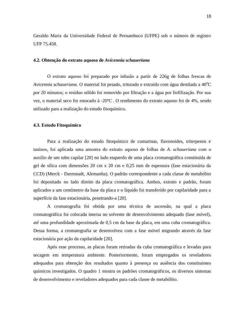
Quadro 1 – Metabólitos, padrões, reveladores, fase móvel e a proporção da fase móvel
utilizados para o screening Fitoquímico do extrato aquoso de A. schaueriana [20,41].
METABÓLITOS PADRÕES REVELADORES FASE MÓVEL PROPORÇÃO
Cumarinas Ácido
cumárico
KOH 10 % –
ETOH Tolueno + Éter 50:50
Flavonóides Quercetina NEU
Acetato de Etila
+ Ácido Fórmico
+ Ácido Acético
glacial + Água
100:11:11:26
Triterpenos Lupeol Liberman –
Burchard
Tolueno +
Clorofórmio +
Etanol
40:40:10
Taninos Ácido
Tânico Cloreto férrico 1%
Clorofórmio +
Metanol + Água 65:30:5
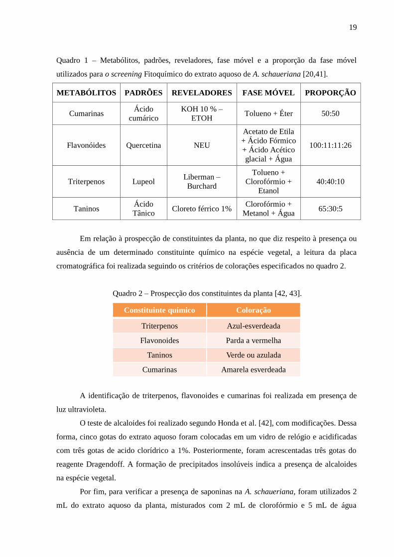
Em relação à prospecção de constituintes da planta, no que diz respeito à presença ou
ausência de um determinado constituinte químico na espécie vegetal, a leitura da placa
cromatográfica foi realizada seguindo os critérios de colorações especificados no quadro 2.
Quadro 2 – Prospecção dos constituintes da planta [42, 43].
Constituinte químico Coloração
Triterpenos Azul-esverdeada
Flavonoides Parda a vermelha
Taninos Verde ou azulada
Cumarinas Amarela esverdeada
A identificação de triterpenos, flavonoides e cumarinas foi realizada em presença de
luz ultravioleta.
O teste de alcaloides foi realizado segundo Honda et al. [42], com modificações. Dessa
forma, cinco gotas do extrato aquoso foram colocadas em um vidro de relógio e acidificadas
com três gotas de acido clorídrico a 1%. Posteriormente, foram acrescentadas três gotas do
reagente Dragendoff. A formação de precipitados insolúveis indica a presença de alcaloides
na espécie vegetal.
Por fim, para verificar a presença de saponinas na A. schaueriana, foram utilizados 2
mL do extrato aquoso da planta, misturados com 2 mL de clorofórmio e 5 mL de água
20
destilada. A solução foi filtrada para um tubo de ensaio e agitada por 3 minutos de forma
ininterrupta. Após a agitação, caso as saponinas estiverem presentes na espécie vegetal,
existirá espuma persistente e abundante, formando uma espécie de colarinho na solução
contida no tubo de ensaio [20,42].
5. RESULTADOS E DISCUSSÃO
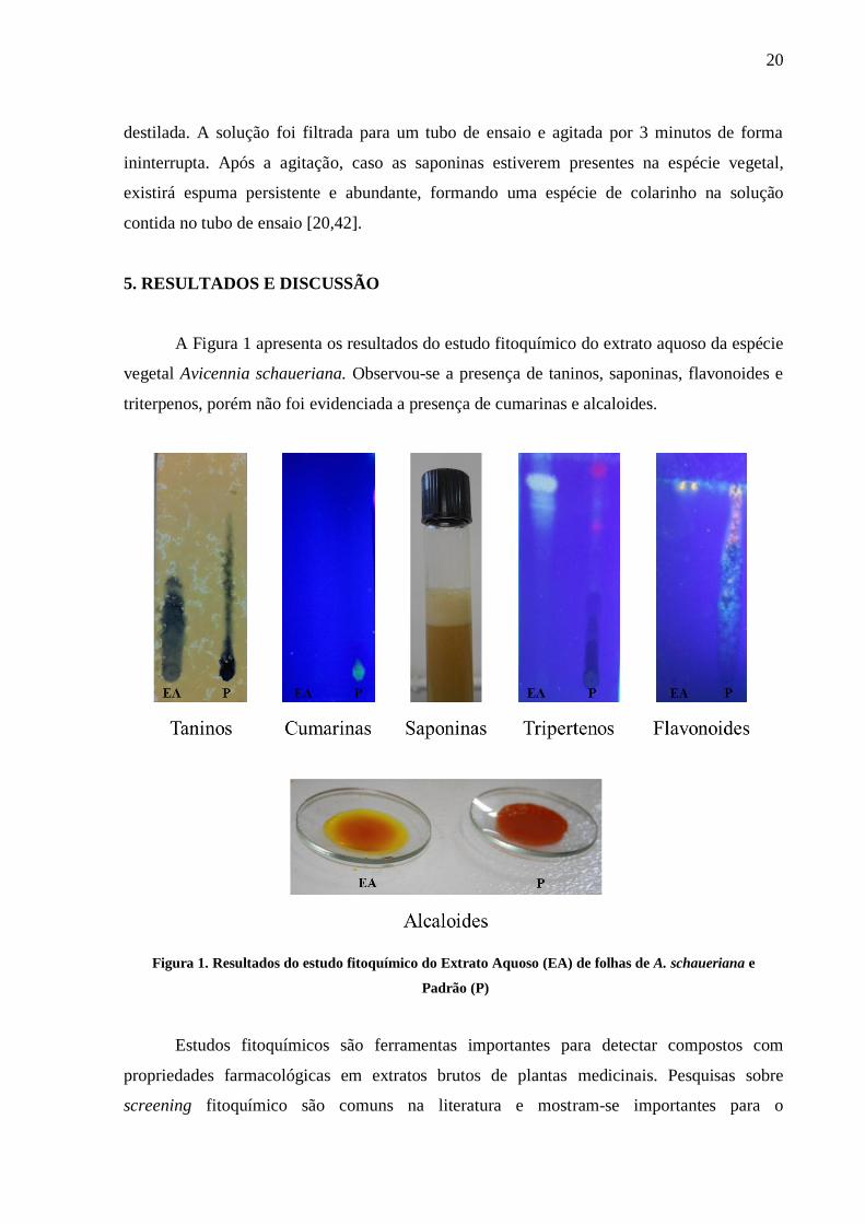
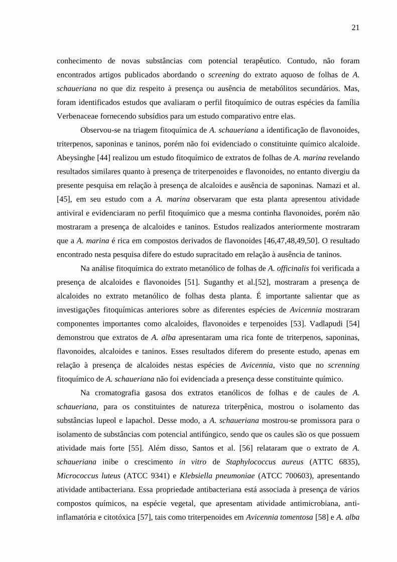
A Figura 1 apresenta os resultados do estudo fitoquímico do extrato aquoso da espécie
vegetal Avicennia schaueriana. Observou-se a presença de taninos, saponinas, flavonoides e
triterpenos, porém não foi evidenciada a presença de cumarinas e alcaloides.
Figura 1. Resultados do estudo fitoquímico do Extrato Aquoso (EA) de folhas de A. schaueriana e
Padrão (P)
Estudos fitoquímicos são ferramentas importantes para detectar compostos com
propriedades farmacológicas em extratos brutos de plantas medicinais. Pesquisas sobre
screening fitoquímico são comuns na literatura e mostram-se importantes para o
21
conhecimento de novas substâncias com potencial terapêutico. Contudo, não foram
encontrados artigos publicados abordando o screening do extrato aquoso de folhas de A.
schaueriana no que diz respeito à presença ou ausência de metabólitos secundários. Mas,
foram identificados estudos que avaliaram o perfil fitoquímico de outras espécies da família
Verbenaceae fornecendo subsídios para um estudo comparativo entre elas.
Observou-se na triagem fitoquímica de A. schaueriana a identificação de flavonoides,
triterpenos, saponinas e taninos, porém não foi evidenciado o constituinte químico alcaloide.
Abeysinghe [44] realizou um estudo fitoquímico de extratos de folhas de A. marina revelando
resultados similares quanto à presença de triterpenoides e flavonoides, no entanto divergiu da
presente pesquisa em relação à presença de alcaloides e ausência de saponinas. Namazi et al.
[45], em seu estudo com a A. marina observaram que esta planta apresentou atividade
antiviral e evidenciaram no perfil fitoquímico que a mesma continha flavonoides, porém não
mostraram a presença de alcaloides e taninos. Estudos realizados anteriormente mostraram
que a A. marina é rica em compostos derivados de flavonoides [46,47,48,49,50]. O resultado
encontrado nesta pesquisa difere do estudo supracitado em relação à ausência de taninos.
Na análise fitoquímica do extrato metanólico de folhas de A. officinalis foi verificada a
presença de alcaloides e flavonoides [51]. Suganthy et al.[52], mostraram a presença de
alcaloides no extrato metanólico de folhas desta planta. É importante salientar que as
investigações fitoquímicas anteriores sobre as diferentes espécies de Avicennia mostraram
componentes importantes como alcaloides, flavonoides e terpenoides [53]. Vadlapudi [54]
demonstrou que extratos de A. alba apresentaram uma rica fonte de triterpenos, saponinas,
flavonoides, alcaloides e taninos. Esses resultados diferem do presente estudo, apenas em
relação à presença de alcaloides nestas espécies de Avicennia, visto que no screnning
fitoquímico de A. schaueriana não foi evidenciada a presença desse constituinte químico.
Na cromatografia gasosa dos extratos etanólicos de folhas e de caules de A.
schaueriana, para os constituintes de natureza triterpênica, mostrou o isolamento das
substâncias lupeol e lapachol. Desse modo, a A. schaueriana mostrou-se promissora para o
isolamento de substâncias com potencial antifúngico, sendo que os caules são os que possuem
atividade mais forte [55]. Além disso, Santos et al. [56] relataram que o extrato de A.
schaueriana inibe o crescimento in vitro de Staphylococcus aureus (ATTC 6835),
Micrococcus luteus (ATCC 9341) e Klebsiella pneumoniae (ATCC 700603), apresentando
atividade antibacteriana. Essa propriedade antibacteriana está associada à presença de vários
compostos químicos, na espécie vegetal, que apresentam atividade antimicrobiana, anti-
inflamatória e citotóxica [57], tais como triterpenoides em Avicennia tomentosa [58] e A. alba

22
[12]; taninos e triterpenos em A.marina [59]; flavonóides [60] e saponinas em A. officinalis
[61]. Extratos de A. alba também apresentaram atividade antimicrobiana contra espécies
patogênicas testadas, incluindo cepas resistentes a antibióticos [54]. Entre os princípios ativos
de plantas com potencial antimicrobiano, os flavonoides são um dos constituintes químicos
encontrados na literatura científica para o gênero Avicennia [56]. Esses trabalhos corroboram
com a presente pesquisa que evidenciaram a identificação desses metabólitos secundários na
espécie estudada.
Então, sugere-se que novas investigações sejam realizadas com a Avicennia
schaueriana, especialmente análises mais refinadas, como cromatografia gasosa e líquida, a
fim de identificar quais substâncias pertencem a cada classe metabólica avaliada no presente
estudo. Dessa forma, resultará na comprovação dos seus constituintes químicos, os quais
possuem propriedades farmacológicas, dando subsídios para que essa planta apresente
atividade medicamentosa, assim como outras espécies de Avicennia.
6. CONCLUSÃO
A planta do mangue A.schaueriana contém importantes metabólitos secundários, que
sugerem possíveis atividades anti-inflamatória, antibacteriana e antifúngica. Porém, é
recomendado que estudos adicionais sejam realizados para uma melhor compreensão do
potencial farmacológico desta espécie.
7. DECLARAÇÃO DE CONFLITO DE INTERESSE
Nós declaramos que não há nenhum conflito de interesse.
AGRADECIMENTOS
À Universidade Federal de Pernambuco, especialmente, o Laboratório de Ecologia
Aplicada e Fitoquímica, que viabilizou o desenvolvimento da pesquisa; e a CAPES pelo apoio
financeiro.
23
8. REFERÊNCIAS
1- Garcia ES. Biodiversidade, Biotecnologia e Saúde. Cad Saude Publica 1995;11 Suppl
3:495-500.
2- Di Stasi LC. Arte, ciência e magia. In: Di Stasi LC. Plantas medicinais: arte e ciência.
Um guia de estudo interdisciplinar. São Paulo: Fundação Unesp; 1996. p. 1-175.
3- Wilson EO. A situação atual da biodiversidade biológica. In: Wilson EO. Biodiversidade.
Rio de Janeiro: Nova Fronteira; 1997. p. 3-24.
4- Ministério da Saúde (Brasil), Secretaria de Atenção à Saúde, Departamento de Atenção
Básica, Práticas integrativas e complementares: plantas medicinais e fitoterapia na
Atenção Básica. Brasília: Ministério da Saúde; 2012. p.156.
5- Rates SMK. Plants as source of drougs. Toxicon 2001; 39:603-13.
6- Gurib-Fakim A. Medicinal plants: traditions of yesterday and drugs of tomorrow. Mol
Aspects Med 2006; 27 Suppl 1:1-93.
7- Santos PO, Barbosa Junior AM, Melo DLFM, Trindade RC. Investigação da atividade
antimicrobiana do látex da mangabeira (Hancornia speciosa GOMES). Revista Brasileira
de Plantas Medicinais 2007; 9 Suppl 2:108-11.
8- Gesler WM. Therapeutic landscapes: medical issue in light of the new cultural
geography. Soc Sci Med 1992; 34 Suppl 7:735-46.
9- Bent S, Ko R. Commonly used herbal medicines in the United States: a review. Am J
Med 2004; 116 Suppl 7: 478-85.
10- Bandaranayake WM. Traditional and medicinal uses of mangroves. Mangroves and Salt
Marshes 1998; 2:133–48.
11- Bandaranayake WM. Survey of mangrove plants from Northern Australia for
phytochemical constituents and uv-absorping compounds. Current Topics in
Phytochemistry 1995; 14:60–72.
12- Bandaranayake WM. Bioactivities, bioactive compounds and chemical constituents of
mangrove plants. Wetlands Ecology and Management 2002; 10:421–52.
13- Agoramoorthy G, Chandrasekaran M, Venkatesalu V, Hsu MJ. Antibacterial and
antifungal activities of fatty acid methyl esters of the blind-your eye mangrove from
India. Braz J Microbiol 2007; 38:739–42.
14- Harbone JB, Williams CA. Advances in flavonoid research since 1992. Phytochemistry
2000; 55: 481-504.
15- Ahmed M, Akthar MS, Malik T, Gilani AH. Hypoglycaemic action of the flavonoids
fraction of Cuminum nigrum seeds. Phytother Res 2000; 14:103-06.

24
16- Dornas WC, Oliveira TT, Rodrigues-das-Dores RG, Santos AF, Nagem TJ. Flavonóides:
potencial terapêutico no estresse oxidativo. Revista de Ciências Farmacêuticas Básica e
Aplicada 2007; 28 Suppl 3: 241-49.
17- Arun LB, Arunachalam AM, Arunachalam KD, Annamalai SK, Amit Kumar K. In vivo
anti-ulcer, anti-stress, anti-allergic, and functional properties of Gymnemic acid isolated
from Gymnema sylvestre R. Br. BMC Complement Altern Med 2014;14:70.
18- Scio E. Cumarinas encontradas no gênero Kielmeyera – Família Clusiaceae. Revista
Brasileira de Farmácia 2004; 85 Suppl 1: 27-31.
19- Alves CF, Alves VBF, Assis IP, Clemente-Napimoga JT, Uber-Bucek E, Dal-Secco D,
Cunha FQ, Rehder VLG, Napimoga MH. Anti-inflammatory activity and possible
mechanism of extract from Mikania laevigata in carrageenan-induced peritonitis. J Pharm
Pharmacol 2009; 61 Suppl 8: 1097-104.
20- Da Silva NLA, Miranda FAA, Da Conceição GM. Triagem Fitoquímica de Plantas de
Cerrado, da Área de Proteção Ambiental Municipal do Inhamum, Caxias, Maranhão.
Scientia Plena 2010; 6 Suppl 2: 1-17.
21- Sudhahar V, Kumar SA, Sudharsan PT, Varalakshmi P. Protective effect of lupeol and
its ester on cardiac abnormalities in experimental hypercholesterolemia. Vascul
Pharmacol 2007; 46 Suppl 6: 412-18.
22- Pertino M, Schmeda-Hirschmann G, Rodriguez JA, Theoduloz C. Gastroprotective effect
and cytotoxicity of terpenes from the Paraguayan crude drug “yagua rova” (Jatropha
isabelli). J Ethnopharmacol 2007; 111 Suppl 3: 553-59.
23- Medeiros R, Otuki MF, Avellar MCW, Calixto JB. Mechanisms underlying the inhibitory
actions of the pentacyclic triterpene [alpha]-amyrin in the mouse skin inflammation
induced by phorbol ester 12-O-tetradecanoylphorbol-13-acetate. Eur J of Pharmacol
2007; 559 Suppl 2-3: 227-35.
24- Braga F, Ayres-Saraiva D, Gattas CR, Capella MAM. Oleanolic acid inhibits the activity
of the multidrug resistance protein ABCC1 (MRP1) but not of the ABCB1 (P-
glycoprotein): Possible use in cancer chemotherapy. Cancer Lett 2007; 248 Suppl 1: 147-
52.
25- Sato H, Genet C, Strehle A, Thomas C, Lobstein A, Wagner A, Mioskowski C, Auwerx
J, Saladin R. Antihyperglycemic activity of a TGR5 agonist isolated from Olea
europaea. Biochem and Biophys Res Commun 2007; 362 Suppl 4: 793-98.
26- Corrêa MP. Dicionário das plantas úteis do Brasil e das exóticas cultivadas. Rio de
Janeiro: Imprensa Nacional; 1984. p. 267-69.
27- Costa AF. Farmacognosia. 3 ed. Lisboa: Fundação Calouste Gulbekian; 1986.
28- Caballero-George C, Vanderheyden PML, Okamoto Y, Masaki T, Mbwambo Z, Apers S,
Gupta MP, Pieters L, Vanquelin G, Vlitinek A. Evolution of bioactive saponins and
25
triterpenoidal aglycons for their binding properties on human endothelin ETA and
Angiotensin AT1 receptors. Phytother Res 2004; 18: 729-36.
29- Vinothapooshan G, Sundhar K. Wound healing effect of various extracts of Adhatoda
vasica. Int J Pharm Bio Sci 2010; Suppl 1: 530-536.
30- Chaudhari M, Mengi S. Evaluation of phytoconstituents of Terminalia arjuna for wound
healing activity in rats. Phytother Res 2006; Suppl 20: 799-805.
31- Murti K, Lambole V, Panchal M, Shah M, Gajera V 2011. Evaluation of wound healing
activity of polyherbal formulation in rats. J Pharmacogn Phytochem 2011; Suppl 3: 112-
115.
32- Deshmukh PT, Fernandes J, Akarte A, Emmanuel T. Wound healing activity of
Calotropis gigantea root bark in rats. J Ethnopharmacol 2009; Suppl 125: 178-181.
33- Barros HM, Macedo SJ, Leça EE. Gerenciamento Participativo de Estuários e
Manguezais. Recife: Universidade Federal de Pernambuco, 2000.
34- Profice SR, Kameyama C, Côrtes ALA, Braz DM, Indriunas A, Vilar T et. al.
Acanthaceae in Lista de Espécies da Flora do Brasil. Jardim Botânico do Rio de Janeiro;
2010. p.871.
35- Barroso GM. Sistemática das Angiospermas do Brasil. 2ed. Viçosa (MG): UFV Imprensa
Universitária;1991.
36- Schaeffer-Novelli Y. Manguezal: ecossistema entre a terra e o mar. São Paulo:
Universidade Federal da Bahia; 1995.
37- Hostettmann K, Queiroz EF, Vieira PC. Princípios ativos de plantas superiores. São
Paulo: EDUFSCAR; 2003.
38- Verdi LG, Brighente IMC, Pizzolatti MG. Gênero Baccharis (Asteraceae): aspectos
químicos, econômicos e biológicos. Quim Nova 2005; 28: 85-94.
39- Gil ES. Controle físico-químico de qualidade de medicamentos. Campo Grande:
UNIDERP; 2005.
40- Degani ALG, Cass QB, Vieira PC. Cromatografia: um breve ensaio. Atualidades em
Química 1998; 7: 21-5.
41- Wagner H, Bladt S. Plant Drugs Analysis – a thin layer chromatography atlas. 2 nd ed.
Berlin: Springer-Verlag; 1996.
42- Honda NK, Garcez WS, Garcez FR, Conceição CA. Estudo químico de plantas de
Mato Grosso do Sul I: triagem fitoquímica. Campo Grande (MS): EUFMS; 1990.
43- Amaral MPH, Vieira FP, Leite MN, Amaral LH, Pinheiro LC, Fonseca BG et al.
Determinação do teor de cumarina no xarope de guaco armazenado em diferentes
temperaturas. Rev Bras Farmacogn 2009; 19 Suppl 2B: 607-11.

26
44- Abeysinghe PD. Antibacterial Activity of some Medicinal Mangroves against Antibiotic
Resistant Pathogenic Bacteria. Indian J Pharm Sci 2010; 72 Suppl 2:167-72.
45- Namazi R, Zabihollahi R, Behbahani M, Rezaei A. Inhibitory Activity of Avicennia
marina, a Medicinal Plant in Persian Folk Medicine, against HIV and HSV. Iran J Pharm
Res 2013; 12 Suppl 2: 435-43.
46- Rezaii Y. Study on pharmacognosy effect of Avicennia marina. Tehran: Tehran
University; 1993.
47- Rui J, YueWei G and HuiXin H. Studies on the chemical constituents from leaves of
Avicennia marina. Chin J Nat Med 2004; 2: 16-9.
48- Jia R, Guo YW, Hou HX. Studies on the chemical constituents from leaves of Avicennia
marina. Chin J Nat Med 2004; 2: 16-9.
49- Shraf M, El-Ansari MA, Saleh NAM. New flavonoids from Avicennia marina.
Fitoterapia 2000; 71: 274-7.
50- Yang CM, Cheng HY, Lin TC, Chiang LC, Lin CC. Acetone, ethanol and methanol
extracts of Phyllanthus urinaria inhibit HSV-2 infection in-vitro. Antiviral Res 2005; 67:
24-30.
51- Hossain H, Howlader SI, Dey SK, Hira A, Ahmed A. Evaluation of Diuretic and
Neuropharmacological Properties of the Methanolic Extract of Avicennia officinalis L.
leaves from Bangladesh. International journal of pharmaceutical and
phytopharmacological research 2012; 2 Suppl 1: 2-6.
52- Suganthy N, Pandian SK, Devi KP.Cholinesterase inhibitory effects of Rhizophora
lamarckii, Avicennia officinalis, Sesuvium portulacastrum and Suaeda monica:
Mangroves inhabiting an Indian coastal area (Vellar Estuary). J Enzyme Inhib Med
Chem 2009; 24 Suppl 3: 702-7.
53- Ghani A. Medicinal Plants of Bangladesh: Chemical constituents and uses. Asiatic
Society of Bangladesh; 1998. p. 212-16.
54- Vadlapudi V. In vitro antimicrobial activity of plant extracts of Avicennia alba against
some important pathogens. Asian Pac J Trop Dis 2012; 2 Suppl 1: 408-11.
55- Fardin KM, Young CM. Estudo químico e avaliação de atividade antifúngica em
Avicennia schaueriana Stapf & Leech. Núcleo de Pesquisa em Fisiologia e
Bioquímica, 18° Reunião Anual do Instituto de Botânica, São Paulo, 2011.
56- Santos SC, Ferreira FS, Damião AO, Damião AO, Barros TF, Rossi-Alva JC et. al.
Avaliação da atividade antibacteriana dos extratos de Avicennia schaueriana Stapf &
Leechm. ex Moldenke, Verbenaceae. Ver Bras Farmacogn 2010; 20 Suppl 1: 124-9.
57- Bloor SJ. A survey of extracts of New Zealand indigenous plants for selected
biological activities. New Zealand Journal of Botany 1995; 33:523-40.

27
58- Majumdar SG, Patra G. Chemical investigation of some mangrove species: part I genus
Avicennia. Journal of the Indian Chemical Society 1979; 56: 111-13.
59- Padmakumar K, Ayyakkannu K. Antiviral activity of marine plants. Indian J Vir 1997;
13: 33-6.
60- Basak UC, Das AB, Das P. Chlorophyll, carotenoids, proteins and secondary metabolites
in leaves of 14 species of mangroves. Bulletin of marine Science 1996; 58: 654-59.
61- Champagne DE, Koul O, Isman MB, Scudder GGE, Towers GHN. Biological activity of
limonoids from the rutales. Phytochemistry 1992; 31: 377-94.
27
4. ARTIGO CIENTÍFICO 2
ESTUDO DE BIOPROSPECÇÃO DE AVICENNIA SCHAUERIANA:
DESENVOLVIMENTO DE UM CREME CICATRIZANTE
Caroline Maria Igrejas Lopesa*
, Jeymesson Raphael Cardoso Vieirab, Ivone Antônia de
Souzac, Erwelly Barros de Oliveira
b, Jéssica Guido de Araújo
b, Marllon Alex Nascimento
Santanab, Pedro Paulo Marcelino Neto
b, Liriane Baratella-Evêncio
b.
aPrograma de Pós-Graduação em Odontologia, Centro de Ciências da Saúde, Universidade
Federal de Pernambuco, UFPE, Av. Professor Moraes Rego, s/n, Cidade Universitária,
50670-901, Recife, Pernambuco, Brasil.
bDepartamento de Histologia e Embriologia, Centro de Ciências Biológicas, UFPE, Av.
Professor Moraes Rego, s/n, Cidade Universitária, 50670-901, Recife, Pernambuco, Brasil.
cDepartamento de Antibióticos, Centro de Ciências Biológicas, UFPE, Av. Professor Moraes
Rego, s/n, Cidade Universitária, 50670-901, Recife, Pernambuco, Brasil.
E-mails: [email protected], [email protected], [email protected],
1. RESUMO
Relevância etnofarmacológica: A Avicennia schaueriana é uma espécie vegetal endêmica
pertencente à família Verbenaceae, encontrada nas florestas de mangue do litoral brasileiro.
As espécies do gênero Avicennia são muito utilizadas pelas comunidades tradicionais para
cura de várias doenças.
Objetivo do estudo: Avaliar a citotoxicidade in vitro do extrato aquoso de folhas de A.
schaueriana e a ação cicatrizante do creme desse extrato nas feridas cutâneas em ratos.
Materiais e métodos: A avaliação citotóxica foi realizada através do método colorimétrico de
brometo (MTT) em células Vero. A ação cicatrizante foi avaliada utilizando 45 ratos
divididos em três grupos iguais (n=15) tratados durante 5, 10 e 15 dias com creme de extrato
aquoso de folhas de A. schaueriana, solução de cloreto de sódio a 0,9% e creme de
dexpantenol, aplicados sobre a região dorsal previamente tricotomizada e lesionada. Foram
realizadas mensurações iniciais e finais de cada ferida para calcular o índice de cicatrização
das úlceras, assim como a análise histomorfométrica e a contagem dos fibroblastos nas
secções histológicas das feridas cirúrgicas nos diferentes grupos e intervalos de tempo.
28
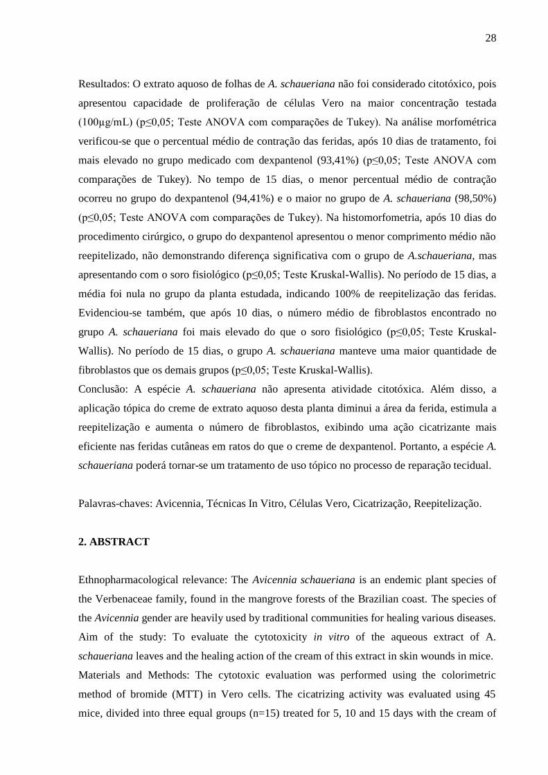
Resultados: O extrato aquoso de folhas de A. schaueriana não foi considerado citotóxico, pois
apresentou capacidade de proliferação de células Vero na maior concentração testada
(100µg/mL) (p≤0,05; Teste ANOVA com comparações de Tukey). Na análise morfométrica
verificou-se que o percentual médio de contração das feridas, após 10 dias de tratamento, foi
mais elevado no grupo medicado com dexpantenol (93,41%) (p≤0,05; Teste ANOVA com
comparações de Tukey). No tempo de 15 dias, o menor percentual médio de contração
ocorreu no grupo do dexpantenol (94,41%) e o maior no grupo de A. schaueriana (98,50%)
(p≤0,05; Teste ANOVA com comparações de Tukey). Na histomorfometria, após 10 dias do
procedimento cirúrgico, o grupo do dexpantenol apresentou o menor comprimento médio não
reepitelizado, não demonstrando diferença significativa com o grupo de A.schaueriana, mas
apresentando com o soro fisiológico (p≤0,05; Teste Kruskal-Wallis). No período de 15 dias, a
média foi nula no grupo da planta estudada, indicando 100% de reepitelização das feridas.
Evidenciou-se também, que após 10 dias, o número médio de fibroblastos encontrado no
grupo A. schaueriana foi mais elevado do que o soro fisiológico (p≤0,05; Teste Kruskal-
Wallis). No período de 15 dias, o grupo A. schaueriana manteve uma maior quantidade de
fibroblastos que os demais grupos (p≤0,05; Teste Kruskal-Wallis).
Conclusão: A espécie A. schaueriana não apresenta atividade citotóxica. Além disso, a
aplicação tópica do creme de extrato aquoso desta planta diminui a área da ferida, estimula a
reepitelização e aumenta o número de fibroblastos, exibindo uma ação cicatrizante mais
eficiente nas feridas cutâneas em ratos do que o creme de dexpantenol. Portanto, a espécie A.
schaueriana poderá tornar-se um tratamento de uso tópico no processo de reparação tecidual.
Palavras-chaves: Avicennia, Técnicas In Vitro, Células Vero, Cicatrização, Reepitelização.
2. ABSTRACT
Ethnopharmacological relevance: The Avicennia schaueriana is an endemic plant species of
the Verbenaceae family, found in the mangrove forests of the Brazilian coast. The species of
the Avicennia gender are heavily used by traditional communities for healing various diseases.
Aim of the study: To evaluate the cytotoxicity in vitro of the aqueous extract of A.
schaueriana leaves and the healing action of the cream of this extract in skin wounds in mice.
Materials and Methods: The cytotoxic evaluation was performed using the colorimetric
method of bromide (MTT) in Vero cells. The cicatrizing activity was evaluated using 45
mice, divided into three equal groups (n=15) treated for 5, 10 and 15 days with the cream of
29
the aqueous extract of A. schaueriana leaves, solution of sodium chloride at 0.9% and cream
of dexpanthenol, applied to the dorsal region previously shaved and injured. Thus, the initial
and final measurements of each wound were taken to calculate the rate of healing of the
ulcers, as well as the histomorphometric analysis and the count of fibroblasts of histological
sections of surgical wounds in the different groups and timeslots.
Results: The aqueous extract of A. schaueriana leaves was not considered cytotoxic, as it
presented Vero cell proliferation capacity at the highest concentration tested (100μg/ml)
(p≤0,05; ANOVA test with Tukey comparisons). In the morphometric analysis, it was found
that the average percentage of contraction of the wounds, after 10 days of treatment, was
higher in the group medicated with dexpanthenol (93.41%) (p≤0,05; ANOVA test with Tukey
comparisons). In the 15 days analysis, the lowest average percentage of contraction was
observed in the dexpanthenol group (94.41%) and the highest in the A. schaueriana group
(98.50%) (p≤0,05; ANOVA test with Tukey comparisons). In the histomorphometry, after 10
days of the surgery, the dexpanthenol group had the lowest average length not re-
epithelialized, showing no significant statistical difference from the A.schaueriana group, but
showing with saline solution (p≤0,05; Kruskal-Wallis test). In the 15-days period, the average
was void in the group of the studied plant, indicating 100% of re-epithelialization of the
wounds. It was also noticed that after 10 days, the average number of fibroblasts found in the
A. schaueriana group was higher than the saline solution (p≤0,05; Kruskal-Wallis test). In the
15-days period, the A. schaueriana group maintained a higher amount of fibroblasts when
compared to the others (p≤0,05; Kruskal-Wallis test).
Conclusion: The species A. schaueriana present no cytotoxic activity. Furthermore, the
topical application of the cream of aqueous extract of this plant decreases the wound area,
stimulates the re-epithelialization and increases the number of fibroblasts, presenting a
healing action more efficient, on mice skin wounds, than dexpanthenol cream. Therefore, the
species A. schaueriana could become a topical treatment in the tissue repair process.
Key-words: Avicennia, In Vitro Techniques, Vero Cells, Wound Healing, Re-
Epithelialization.

30
3. Introdução
A ferida é definida como a separação dos tecidos do corpo ou qualquer lesão tecidual,
seja epitelial, mucosa ou em órgão, com prejuízo de suas funções básicas. Ela pode ser
produzida por fatores extrínsecos – como incisão cirúrgica, lesões acidentais, corte e trauma –
ou por fatores intrínsecos, como aquelas produzidas por infecção, alterações vasculares,
defeitos metabólicos ou neoplasias (Centre for Medical Education, 1994).
Recentemente, o estudo de cicatrização de feridas tem se tornado um tema relevante,
uma vez que inúmeras pessoas sofrem com feridas cirúrgicas ou traumáticas a cada ano,
apresentando a necessidade iminente de tratamento das mesmas (Li et al., 2015). A perda do
epitélio e a exposição do tecido conjuntivo que caracterizam as úlceras causam dor e
desconforto, afetando a qualidade de vida dos pacientes (Duarte et al., 2011).
A cicatrização de feridas cutâneas é um processo intrínseco de reconstrução de
estruturas celulares e camadas de tecidos perdidos (Li et al., 2015), constituindo uma cascata
perfeitamente coordenada de acontecimentos celulares e moleculares que interagem de modo
a promover a reparação dos tecidos (Duarte et al., 2011). Esse processo é comum a todas as
feridas, independentemente do agente que a causou, é sistêmico e dinâmico e está diretamente
relacionado às condições gerais do organismo (Broughton et al., 2006). É um processo
fisiológico altamente orquestrado, envolvendo esforços colaborativos de vários tipos de
células da pele localizadas na epiderme e derme (Martin, 1997).
As populações de células predominantes para a cicatrização adequada de toda a
espessura da ferida são os queratinócitos, fibroblastos e células endoteliais (Barrientos et al.,
2008). Os fibroblastos são células do tecido conjuntivo responsáveis pela deposição de
colágeno que é necessária para reparar o dano do tecido (Ross, 1969). Caso não haja algum
fator modificador do reparo nas feridas, o processo de cicatrização ocorre em sequência
ordenada e eficiente de eventos (Campos et al., 2007; Diegelmann e Evans, 2004), que em
geral é dividida em três fases principais: fase inflamatória, fase de proliferação ou granulação
e fase de remodelação ou maturação (Clark, 2005; Gurtner et al., 2008). Entretanto, alguns
autores, como Diegelmann e Evans (2004), afirmam que a cicatrização é caracterizada por
quatro fases distintas e sobrepostas: hemostasia, inflamação, proliferação e remodelação.
Vários autores têm investigado medicamentos que possam acelerar a cicatrização de
feridas, reduzir os sintomas dolorosos e apresentar uma ótima relação custo-benefício (Duarte
et al, 2011). Existem várias opções de tratamento tópico para as úlceras – como medicações
anti-inflamatórias, agentes antimicrobianos, anestésicos, películas de proteção e laser – que

31
podem ser usadas isoladamente ou em combinação (Duarte et al, 2011). Ademais, estudos têm
sugerido que os antioxidantes podem desempenhar um importante papel no processo
cicatricial das lesões (Kim et al., 2008).
É importante salientar que, a partir do ponto de vista clínico, a aplicação tópica da
medicação por toda a espessura da lesão é interessante, devido à redução dos efeitos adversos
sobre outros órgãos (Li et al., 2015). O tratamento tópico de úlceras consiste em restaurar o
ambiente fisiológico no leito da lesão, visando manter uma umidade adequada, controle de
temperatura, regulação de pH, controle da carga bacteriana, remoção do tecido não-viável
(desbridamento), controle do odor, minimização da dor e proteção da pele na região afetada.
Estas condições, umas vez ajustadas, irão contribuir para o reparo e restauração da função
tecidual. Nenhum tratamento tópico será eficaz se a condição patológica do paciente não for
corrigida (Rolstad et al., 2012).
Dessa forma, historicamente, as plantas têm sido uma fonte de inspiração para
compostos farmacêuticos novos, os quais têm feito grandes contribuições para a saúde
humana (Vadlapudi, 2012), pois contêm componentes de valor terapêutico (Panda et al.,
2009). De acordo com a Organização Mundial de Saúde, as plantas são uma fonte de
compostos que têm a capacidade para combater doenças, apresentando atividades
antimicrobiana, antiviral e antifúngica (Gazim et al., 2008; Nascimento et al., 2000).
Entretanto, os produtos fitoterápicos só podem ser introduzidos na sociedade desde que
estudos laboratoriais e clínicos específicos comprovem sua eficácia e segurança (Agra et al.,
2007), visto que alguns deles podem apresentar efeitos colaterais e/ou serem tóxicos. Portanto,
a utilização adequada de plantas medicinais representa um passo importante e mais uma
opção medicamentosa a ser destinada à população na tentativa de melhorar sua saúde e
qualidade de vida (Silva et al., 2006).
Os extratos de diferentes plantas de mangue são relatados por possuírem diversas
propriedades medicinais (Agoramoorthy et al., 2007; Bandaranayake, 1998). Em relação às
propriedades farmacológicas da família Verbenaceae, segundo Bandaranayake (1998), as
plantas do mangue A. alba, A. africana, A. germinans e A. marina apresentam componentes
terapêuticos que podem ser utilizados para o tratamento de várias enfermidades, entre elas, as
úlceras. Sumithra et al. (2011), abordaram que o extrato metanólico de folhas de A. officinalis
tem mostrado atividade anti-inflamatória, sendo usada principalmente para o tratamento de
reumatismo, paralisia, asma, doenças de pele e úlceras (Kathiresan e Ramanathan, 1997;
Ramanathan, 2000).

32
O gênero Avicennia apresenta duas espécies no Brasil, a Avicennia schaueriana e
Avicennia germinans (Profice et al., 2010). A Avicennia schaueriana conhecida popularmente
como mangue-preto ou siriúba, é uma espécie endêmica da vegetação de manguezal
pertencente à família Verbenaceae (Schaeffer-Novelli, 1995). O extrato desta planta foi
aplicado para inibição do crescimento in vitro das bactérias Staphylococcus aureus (ATTC
6835), Micrococcus luteus (ATCC 9341) e Klebsiella pneumoniae (ATCC 700603),
apresentando atividade antibacteriana (Santos et al., 2010). Além disso, a A. schaueriana
mostrou-se promissora para o isolamento de substâncias com potencial antifúngico, sendo os
extratos etanólicos de caules os que possuem atividade antifúngica mais forte (Fardin e
Young, 2011).
É importante ressaltar que as espécies do gênero Avicennia são muito utilizadas pelas
comunidades tradicionais para cura de várias doenças (Santos et al., 2010), porém ainda não
existem relatos científicos sobre o potencial cicatrizante de A. schaueriana. Portanto, o
objetivo deste artigo é avaliar a citotoxicidade in vitro e a ação cicatrizante do creme de
extrato aquoso de folhas de Avicennia schaueriana nas lesões cutâneas em ratos.
4. Materias e métodos
4.1. Material vegetal
As folhas da espécie Avicennia schaueriana foram coletadas em outubro de 2013, no
mangue da Ilha de Itamaracá, situado em Vila Velha, no litoral norte do Estado de
Pernambuco, Brasil, entre as coordenadas 07°48.716’ latitude sul e 34°51.347’ longitude
oeste. Uma exsicata do material botânico encontra-se depositada no acervo do Herbário
Geraldo Mariz da Universidade Federal de Pernambuco (UFPE) sob o número de registro
UFP 75.458.
4.2. Material biológico
4.2.1. Obtenção do extrato aquoso de folhas de Avicennia schaueriana
O extrato aquoso foi obtido por infusão a partir de 500g de folhas frescas de Avicennia
schaueriana. O material foi pesado, triturado e extraído com água à 40oC por 20 minutos. O
resíduo sólido foi removido por filtração e a água por liofilização. Por sua vez, o material seco
33
foi estocado à -20ºC. O rendimento do extrato aquoso foi de 4%, sendo utilizado para a
atividade citotóxica e cicatrizante.
4.2.2. Cultura celular
Foram utilizadas células Vero que são fibroblastos, originárias do rim de macaco
verde africano ou macaco do velho mundo (Cercopithecus aethiops). Essa linhagem celular
(CCL-81, Rio de Janeiro, Brasil) foi obtida do Laboratório de Cultura de Células do
Departamento de Histologia e Embriologia (UFPE). As células Vero foram cultivadas no
meio de cultura Eagle Modificado por Dulbeco (DMEM - Sigma Chemical Co., St. Louis,
MO, EUA) complementado com 10% de Soro Fetal Bovino e 1% de solução antibiótica-
antimicótica (10.000 unidades de penicillina, 10 mg de estreptomicina em 0.9% de cloreto de
sódio; Sigma), sendo mantidas na estufa a 37°C em atmosfera umidificada com 5% de CO2.
4.2.3. Confecção do creme de Avicennia schaueriana
O extrato aquoso de folhas de A. schaueriana foi pesado em balança analítica digital
(Shimadzu ATY 224) com a utilização de papel manteiga até atingir 3g e vertido em gral de
porcelana. Em seguida foi solubilizado com água destilada e homogeneizado. Em um vidro de
relógio, pesou-se a emulsão aniônica até atingir 60g e verteu-se no gral contendo o extrato de
A. schaueriana até total solubilização. Foi aferido o pH e mantido entre 5,5 e 6,5. Por último,
foi envasado em pote plástico compondo o extrato aquoso de folhas de A. schaueriana a 5%
em creme.
4.2.4. Animais experimentais
Foram utilizados 45 ratos fêmeas da linhagem Wistar com idade entre 8 e 12 semanas,
pesando 230 ± 20 g, obtidos do Biotério do Departamento de Antibióticos do CCB/UFPE. Os
experimentos com os animais foram realizados com a aprovação do Comitê de Ética em
Experimentação Animal da Universidade Federal de Pernambuco (UFPE) sob o número
23076025194/2012-10 (Anexo 2).
34
4.3. Teste de citotoxicidade
A avaliação da atividade citotóxica foi realizada através do método colorimétrico de
brometo (3-[4,5-dimetiltiazol-2-il]-2,5-difenil tetrazólio) (MTT) (Geran et al., 1972;
Mosmann, 1983). A metodologia utilizada para a realização desse teste seguiu as normas da
International Standard Organization (ISO) 10993-5 (2009).
Dessa forma, as células na concentração de 2x10⁵/mL de DMEM por poço foram
distribuídas em placas de 96 poços (TPP, Darmstadt, Alemanha) e incubadas por 24h a 37°C
em estufa, com atmosfera enriquecida com 5% de CO2 e 95% de ar para estabilização. Após
esse período, foi adicionado nos poços com as células Vero o extrato aquoso de folhas de A.
schaueriana, previamente dissolvido em tampão fosfato-salino (PBS) e filtrado (Filtro para
seringa 0.22µm – TPP, Darmstadt, Alemanha), nas concentrações de 100 µg/mL, 50 µg/mL,
25 µg/mL, 12,5 µg/mL e 6,25 µg/mL. O PBS e o meio de cultura DMEM foram utilizados
como controle. Após a incubação por 24h de contato das células com o extrato, 25µl
(5mg/mL) de solução de MTT foi adicionada a cada poço e a placa foi incubada na estufa por
3 horas. O MTT e o meio de cultura foram então removidos e 25µl de dimetilsulfóxido
(DMSO) foi adicionado a cada poço para dissolver os cristais de formazan. Posteriormente,
foi realizada a leitura no espectrofotômetro (Bio-Rad, São Paulo, Brasil) com comprimento de
onda de 570nm. O ensaio foi realizado em duplicata.
4.4. Ação cicatrizante
4.4.1. Divisão dos grupos
Para avaliação da ação cicatrizante, os ratos foram divididos ao acaso em 3 grupos,
conforme o tratamento proposto para as úlceras induzidas em cada animal:
Grupo controle: 15 animais em cujas feridas foi aplicada gaze embebida com solução
de cloreto de sódio a 0,9%;
Grupo padrão: 15 animais cujas úlceras foram tratadas com creme de dexpantenol
(Bepantol® creme, 5% dexpantenol, Bayer, Alemanha), aplicada com uma paleta de
forma a cobrir toda a lesão; e
Grupo experimental: 15 ratos em cujas feridas foi aplicado o creme de extrato aquoso
de folhas de A. schaueriana a 5%.
35
Cada grupo foi dividido em 3 subgrupos de 5 animais, acompanhados durante o
intervalo de tempo de 5, 10 e 15 dias após a indução da úlcera dorsal.
4.4.2. Procedimentos cirúrgicos em tecidos lesados in vivo
Os animais foram previamente pesados e anestesiados com cloridrato de quetamina
(10 mg/kg, Ketamin®), cloridrato de xilazina (0,5 mg/kg, Anasedan®) e soro fisiológico a
0,9%, associados na mesma seringa e administrados por via intramuscular. Em seguida, cada
rato foi submetido à demarcação da área para indução da ferida com posterior tricotomia na
região dorsal e posicionado na mesa operatória em decúbito ventral. Após a assepsia da região
dorsal com álcool 70%, foi realizada a indução de uma ferida retangular com cerca de 2,3 cm
de comprimento e 2,0 cm de largura na pele do rato. Para isso, foi retirado um fragmento
cutâneo no centro da área tricotomizada até a exposição da fáscia muscular dorsal, com a
utilização de um bisturi nº 15.
4.4.3. Pós-operatório das feridas cutâneas
Após o procedimento cirúrgico, os ratos foram submetidos ao tratamento
correspondente e acondicionados em gaiolas; as úlceras não receberam curativos oclusivos. A
aplicação da medicação referente a cada grupo foi realizada, diariamente, uma vez por dia,
por volta das 11h00min, até o final do experimento. Além disso, o aspecto da ferida foi
descrito ao longo da pesquisa nos diferentes grupos.
No 5° dia, foi realizada à medição da área da ferida (largura e comprimento) com o
auxílio de um paquímetro, porém sem a retirada do fragmento cutâneo. Por sua vez, após 10 e
15 dias, foram realizadas a medição da área da lesão e retirada do fragmento cutâneo
contendo a ferida. Para a realização desse procedimento, os animais foram anestesiados,
conforme descrito anteriormente, e com o auxílio de um bisturi nº 15, as peças cirúrgicas
foram excisionadas, sendo constituídas da cicatriz ou lesão cutânea, com margem de 1 cm de
pele em torno da lesão e profundidade até a musculatura dorsal do animal. O tecido
excisionado foi colocado em formol a 10% durante 24 horas a temperatura ambiente e
processado para microscopia de luz. Após o procedimento de coleta dos fragmentos, os
animais foram eutanasiados, através do deslocamento cervical e descartados segundo as
normas preconizadas pelo Colégio Brasileiro de Experimentação Animal (COBEA).
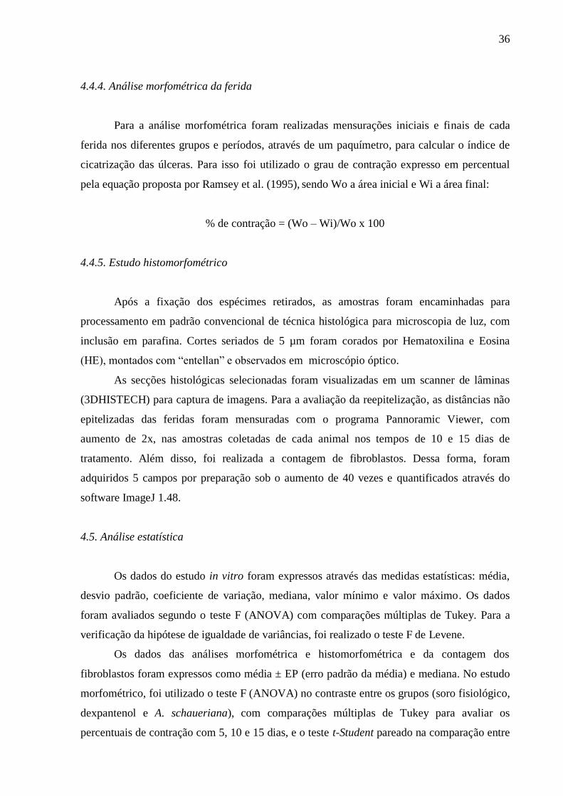
36
4.4.4. Análise morfométrica da ferida
Para a análise morfométrica foram realizadas mensurações iniciais e finais de cada
ferida nos diferentes grupos e períodos, através de um paquímetro, para calcular o índice de
cicatrização das úlceras. Para isso foi utilizado o grau de contração expresso em percentual
pela equação proposta por Ramsey et al. (1995), sendo Wo a área inicial e Wi a área final:
% de contração = (Wo – Wi)/Wo x 100
4.4.5. Estudo histomorfométrico
Após a fixação dos espécimes retirados, as amostras foram encaminhadas para
processamento em padrão convencional de técnica histológica para microscopia de luz, com
inclusão em parafina. Cortes seriados de 5 µm foram corados por Hematoxilina e Eosina
(HE), montados com “entellan” e observados em microscópio óptico.
As secções histológicas selecionadas foram visualizadas em um scanner de lâminas
(3DHISTECH) para captura de imagens. Para a avaliação da reepitelização, as distâncias não
epitelizadas das feridas foram mensuradas com o programa Pannoramic Viewer, com
aumento de 2x, nas amostras coletadas de cada animal nos tempos de 10 e 15 dias de
tratamento. Além disso, foi realizada a contagem de fibroblastos. Dessa forma, foram
adquiridos 5 campos por preparação sob o aumento de 40 vezes e quantificados através do
software ImageJ 1.48.
4.5. Análise estatística
Os dados do estudo in vitro foram expressos através das medidas estatísticas: média,
desvio padrão, coeficiente de variação, mediana, valor mínimo e valor máximo. Os dados
foram avaliados segundo o teste F (ANOVA) com comparações múltiplas de Tukey. Para a
verificação da hipótese de igualdade de variâncias, foi realizado o teste F de Levene.
Os dados das análises morfométrica e histomorfométrica e da contagem dos
fibroblastos foram expressos como média ± EP (erro padrão da média) e mediana. No estudo
morfométrico, foi utilizado o teste F (ANOVA) no contraste entre os grupos (soro fisiológico,
dexpantenol e A. schaueriana), com comparações múltiplas de Tukey para avaliar os
percentuais de contração com 5, 10 e 15 dias, e o teste t-Student pareado na comparação entre
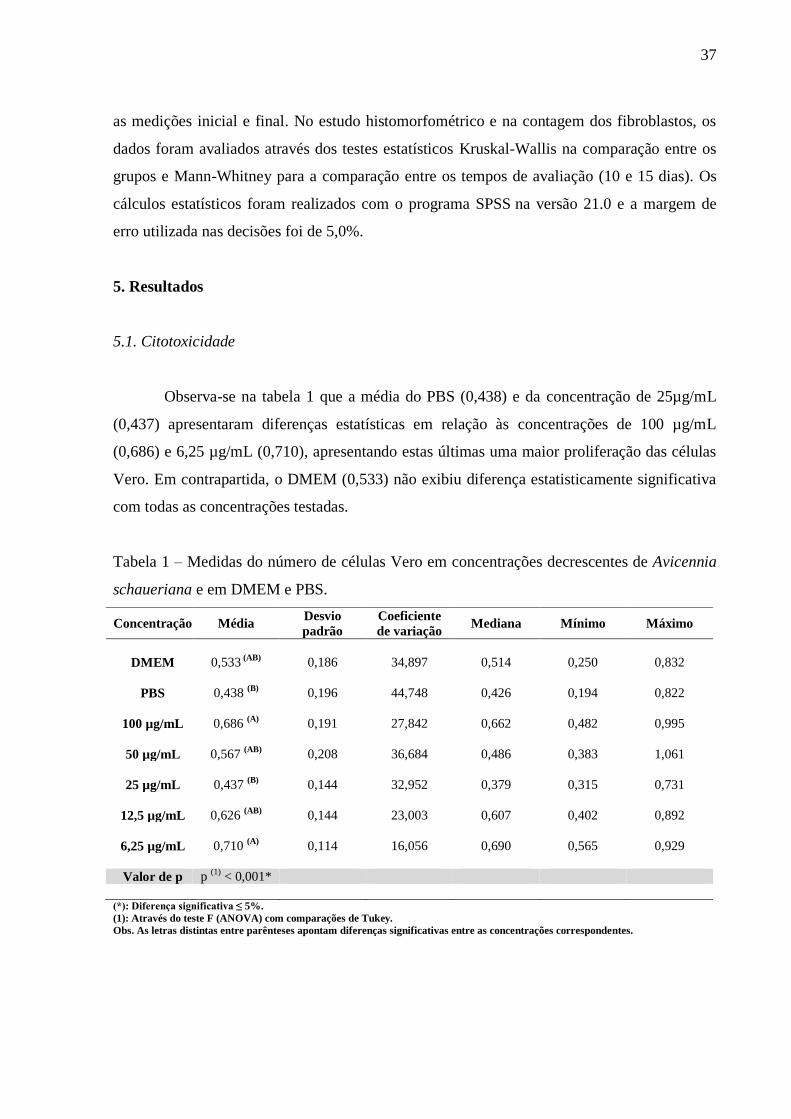
37
as medições inicial e final. No estudo histomorfométrico e na contagem dos fibroblastos, os
dados foram avaliados através dos testes estatísticos Kruskal-Wallis na comparação entre os
grupos e Mann-Whitney para a comparação entre os tempos de avaliação (10 e 15 dias). Os
cálculos estatísticos foram realizados com o programa SPSS na versão 21.0 e a margem de
erro utilizada nas decisões foi de 5,0%.
5. Resultados
5.1. Citotoxicidade
Observa-se na tabela 1 que a média do PBS (0,438) e da concentração de 25µg/mL
(0,437) apresentaram diferenças estatísticas em relação às concentrações de 100 µg/mL
(0,686) e 6,25 µg/mL (0,710), apresentando estas últimas uma maior proliferação das células
Vero. Em contrapartida, o DMEM (0,533) não exibiu diferença estatisticamente significativa
com todas as concentrações testadas.
Tabela 1 – Medidas do número de células Vero em concentrações decrescentes de Avicennia
schaueriana e em DMEM e PBS.
Concentração Média Desvio
padrão
Coeficiente
de variação Mediana Mínimo Máximo
DMEM 0,533 (AB)
0,186 34,897 0,514 0,250 0,832
PBS 0,438 (B)
0,196 44,748 0,426 0,194 0,822
100 µg/mL 0,686 (A)
0,191 27,842 0,662 0,482 0,995
50 µg/mL 0,567 (AB)
0,208 36,684 0,486 0,383 1,061
25 µg/mL 0,437 (B)
0,144 32,952 0,379 0,315 0,731
12,5 µg/mL 0,626 (AB)
0,144 23,003 0,607 0,402 0,892
6,25 µg/mL 0,710 (A)
0,114 16,056 0,690 0,565 0,929
Valor de p p (1)
< 0,001*
(*): Diferença significativa ≤ 5%.
(1): Através do teste F (ANOVA) com comparações de Tukey.
Obs. As letras distintas entre parênteses apontam diferenças significativas entre as concentrações correspondentes.

38
5.2. Descrição macroscópica das feridas
A Figura 1 mostra o processo de cicatrização das feridas induzidas nos ratos. As
imagens digitais permitiram o acompanhamento da evolução da área da ferida nos diferentes
tempos experimentais, de acordo com o tratamento.
Figura 1. Processo de cicatrização das feridas em diferentes dias e tratamentos

39
O quadro 1 resume o aspecto macroscópico das feridas induzidas na região dorsal dos
ratos no intervalo de 5, 10 e 15 dias.
Quadro 1 – Descrição do aspecto macroscópico das feridas induzidas na região dorsal dos
ratos no intervalo de 5, 10 e 15 dias.
Período de avaliação
5 dias 10 dias 15 dias
Gru
pos
Soro
fis
ioló
gic
o Quatro ratos
apresentaram feridas
infectadas com
presença de
sangramento. Um
rato apresentou a
ferida úmida.
Quatro animais
apresentaram feridas
com tecido de
granulação e um
animal com formação
de crosta.
Dois ratos havia a presença
de tecido de granulação e em
três animais a formação de
crosta
Dex
pan
tenol
Dois ratos
apresentaram
infecção nas feridas
e os demais
apresentaram feridas
úmidas, sem
infecção.
Todos os ratos
apresentaram tecido
de granulação e
formação de crosta.
Todos exibiram presença de
crosta.
A. sc
hauer
iana
Todos os ratos
exibiram feridas
secas, com formação
de crosta, sem
presença de
infecção.
Verificou-se presença
de crosta em todos os
ratos.
Dois ratos apresentaram
reepitelização das feridas
(presença de cicatriz) e os
outros 3 exibiram presença de
pequena crosta.
5.3. Análise morfométrica das feridas
Verifica-se na tabela 2 que a média das áreas da ferida diminuiu com a progressão do
tempo de avaliação e que houve um aumento do percentual médio de contração da ferida em
cada um dos grupos estudados. Na avaliação após 10 dias, o grupo tratado com dexpantenol
apresentou a média da área menos elevada (30,30 mm2) com o percentual médio de contração
de 93,41% (p ≤ 0,05). Porém, na análise após 15 dias, a média da área mais elevada ocorreu
no grupo do dexpantenol (25,70 mm2) com o menor percentual médio de contração (94,41%)
e a área menos elevada ocorreu no grupo tratado com A. schaueriana (6,90 mm2) com o maior
percentual médio de contração (98,50%), sendo verificada diferença estatisticamente
significativa entre esses dois grupos (p ≤ 0,05).

40
Tabela 2 – Medidas da área da ferida (mm2) e o percentual de contração da ferida de acordo
com o grupo e o tempo de avaliação após o procedimento cirúrgico in vivo.
Tempo de avaliação
após a indução da
ferida
A. schaueriana
(n=15)
Soro fisiológico
(n=15)
Dexpantenol
(n=15) Valor de p
Média + EPM Média + EPM Média + EPM
(Mediana) (Mediana) (Mediana)
Inicial 460,00 ± 0,00
460,00 ± 0,00
460,00 ± 0,00
(460,00) (460,00) (460,00)
5 dias 262,30 ± 22,98 227,60 ± 22,55 238,04 ± 39,05 p (1)
= 0,698
(256,50) (230,00) (260,00)
10 dias 93,31 ± 10,41 (A)
93,60 ± 17,22 (A)
30,30 ± 4,29 (B)
p (1)
= 0,003*
(97,50) (88,00) (34,00)
15 dias 6,90 ± 2,98 (A)
17,71 ± 3,81 (AB)
25,70 ± 3,47 (B)
p (1)
= 0,008*
(8,00) (16,00) (24,00)
Valor de p p (2)
= 0,001* p (2)
= 0,001* p (2)
= 0,005*
p (3)
< 0,001* p (3)
< 0,001* p (3)
< 0,001*
p (4)
< 0,001* p (4)
< 0,001* p (4)
< 0,001*
% contração com 5
dias
42,98 ± 4,99 50,52 ± 4,90 48,25 ± 8,49 p (1)
= 0,698
(44,24) (50,00) (43,48)
% contração com 10
dias
79,72 ± 2,26 (A)
79,65 ± 4,74 (A)
93,41 ± 0,93 (B)
p (1)
= 0,003*
(78,80) (80,87) (92,60)
% contração com 15
dias
98,50 ± 0,65 (A)
96,15 ± 0,83 (AB)
94,41 ± 0,75 (B)
p (1)
= 0,008*
(98,26) (96,52) (94,78)
(*): Diferença significativa ≤ 5%.
(1): Através do teste F (ANOVA) para comparação entre os grupos em cada avaliação e para os percentuais de contração com 5 dias,
10 dias e 15 dias com comparações de Tukey.
(2): Através do teste de t-Student pareado para comparações entre as avaliações do tempo inicial e 5 dias em cada grupo.
(3): Através do teste de t-Student pareado para comparações entre as avaliações do tempo inicial e 10 dias em cada grupo.
(4): Através do teste de t-Student pareado para comparações entre as avaliações do tempo inicial e 15 dias em cada grupo.
Obs. As letras distintas entre parênteses apontam diferenças significativas entre os grupos correspondentes.

41
5.4. Análise Histomorfométrica e contagem dos fibroblastos
A tabela 3 destaca que, no período de 10 dias, a média da distância entre os epitélios
da ferida cirúrgica foi mais elevada no grupo tratado com soro fisiológico (3498,66 µm)
sendo o p ≤ 0,05, a menor média foi observada no grupo do dexpantenol (706,28 µm) sem
diferença estatisticamente significativa com o grupo de A. schaueriana (833,04 µm). Após 15
dias, em todos os grupos estudados, verificou-se que as distâncias médias entre os epitélios
foram correspondentemente menores do que se comparado com o período de 10 dias,
indicando um aumento da reepitelização, apresentando o grupo do soro fisiológico a distância
mais elevada. É importante salientar que a média foi nula no grupo de A. schaueriana, no
intervalo de 15 dias, indicando reepitelização completa das feridas em todos os ratos (Figura
2).
Tabela 3 – Distância entre os epitélios da ferida cirúrgica dos grupos em relação ao tempo de
avaliação após o procedimento cirúrgico in vivo.
Tempo de avaliação
após a indução da
ferida
A. schaueriana
(n=10)
Soro fisiológico
(n=10)
Dexpantenol
(n=10) Valor de p
Média + EPM Média + EPM Média + EPM
(Mediana) (Mediana) (Mediana)
10 dias 833,04 ± 515,65 (A)
3498,66 ± 232,37 (B)
706,28 ± 233,73 (A)
p (1)
= 0,002*
(0,00) (3572,98) (704,84)
15 dias 0,00 ± 0,00 968,89 ± 303,49 582,21 ± 370,34 p (1)
= 0,078
(0,00) (905,04) (0,00)
Valor de p p (2)
= 0,444 p (2)
= 0,008* p (2)
= 0,683
(*): Diferença significativa ≤ 5%.
(1): Através do teste de Kruskal-Wallis para comparações entre os grupos em cada tempo de avaliação com comparações do referido
teste.
(2): Através do teste de Mann-Whitney para comparações entre os tempos de avaliação em cada grupo.
Obs. As letras distintas entre parênteses apontam diferenças significativas entre os grupos correspondentes.

42
Figura 2. Secções histológicas de pele dorsal de ratos mostrando grupo controle (A), tratado
com dexpantenol (B) e medicado com A. schaueriana (C). Todas as imagens mostram o grau
de reepitelização após 15 dias de tratamento: [1] distância entre os epitélios e [2]
reepitelização total. Coloração HE. Aumento 2x.
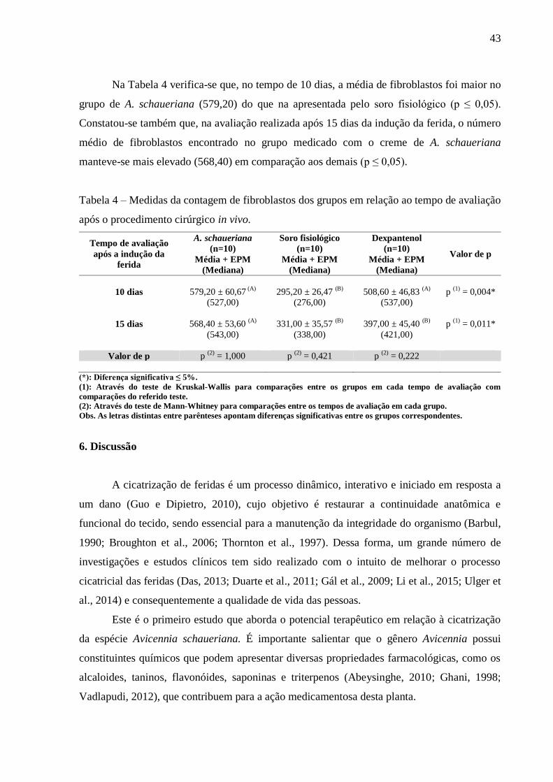
43
Na Tabela 4 verifica-se que, no tempo de 10 dias, a média de fibroblastos foi maior no
grupo de A. schaueriana (579,20) do que na apresentada pelo soro fisiológico (p ≤ 0,05).
Constatou-se também que, na avaliação realizada após 15 dias da indução da ferida, o número
médio de fibroblastos encontrado no grupo medicado com o creme de A. schaueriana
manteve-se mais elevado (568,40) em comparação aos demais (p ≤ 0,05).
Tabela 4 – Medidas da contagem de fibroblastos dos grupos em relação ao tempo de avaliação
após o procedimento cirúrgico in vivo.
Tempo de avaliação
após a indução da
ferida
A. schaueriana
(n=10)
Soro fisiológico
(n=10)
Dexpantenol
(n=10) Valor de p
Média + EPM Média + EPM Média + EPM
(Mediana) (Mediana) (Mediana)
10 dias 579,20 ± 60,67 (A)
295,20 ± 26,47 (B)
508,60 ± 46,83 (A)
p (1)
= 0,004*
(527,00) (276,00) (537,00)
15 dias 568,40 ± 53,60 (A)
331,00 ± 35,57 (B)
397,00 ± 45,40 (B)
p (1)
= 0,011*
(543,00) (338,00) (421,00)
Valor de p p (2)
= 1,000 p (2)
= 0,421 p (2)
= 0,222
(*): Diferença significativa ≤ 5%.
(1): Através do teste de Kruskal-Wallis para comparações entre os grupos em cada tempo de avaliação com
comparações do referido teste.
(2): Através do teste de Mann-Whitney para comparações entre os tempos de avaliação em cada grupo.
Obs. As letras distintas entre parênteses apontam diferenças significativas entre os grupos correspondentes.
6. Discussão
A cicatrização de feridas é um processo dinâmico, interativo e iniciado em resposta a
um dano (Guo e Dipietro, 2010), cujo objetivo é restaurar a continuidade anatômica e
funcional do tecido, sendo essencial para a manutenção da integridade do organismo (Barbul,
1990; Broughton et al., 2006; Thornton et al., 1997). Dessa forma, um grande número de
investigações e estudos clínicos tem sido realizado com o intuito de melhorar o processo
cicatricial das feridas (Das, 2013; Duarte et al., 2011; Gál et al., 2009; Li et al., 2015; Ulger et
al., 2014) e consequentemente a qualidade de vida das pessoas.
Este é o primeiro estudo que aborda o potencial terapêutico em relação à cicatrização
da espécie Avicennia schaueriana. É importante salientar que o gênero Avicennia possui
constituintes químicos que podem apresentar diversas propriedades farmacológicas, como os
alcaloides, taninos, flavonóides, saponinas e triterpenos (Abeysinghe, 2010; Ghani, 1998;
Vadlapudi, 2012), que contribuem para a ação medicamentosa desta planta.

44
Segundo a ISO 10993-1 (2009), o ensaio de citotoxicidade in vitro é um dos testes que
serve para avaliação de biocompatibilidade de um determinado material ou extrato, sendo um
passo importante para os ensaios em animais e ensaios clínicos. Dessa maneira, os resultados
desta pesquisa mostraram que o extrato aquoso de folhas de A. schaueriana não foi
considerado citotóxico, uma vez que em todas as concentrações testadas houve capacidade de
proliferação de células Vero. Estudos com algumas espécies da família Verbenaceae
apresentaram resultados semelhantes.
Akter et al. (2014), utilizando o extrato metanólico de folhas de A. alba, mostraram
que essa planta apresentou potencial antitumoral pela citotoxicidade contra células
cancerígenas, porém não demonstrou atividade citotóxica em células Vero. Behbahani et al.
(2013) concluíram que o extrato metanólico de A. marina não afetou a viabilidade das células
Vero, não demostrando efeito citotóxico em concentrações iguais ou menores que 32 µg/mL.
De acordo com Bueno et. al (2014), os taninos influenciam na fisiologia das células da pele
através das suas propriedades farmacológicas, aumentando a proliferação celular. Sendo
assim, o resultado do presente trabalho sugere uma ação mitogênica do extrato de A.
schaueriana por possivelmente possuir este composto.
Em relação à inspeção das feridas operatórias foi observado que os grupos do soro
fisiológico e do dexpantenol apresentaram crostas e tecido de granulação com processos
infecciosos das feridas durante o período do estudo. Entretanto, o grupo de A. schaueriana
apresentou crostas e cicatriz sem infecção da ferida cirúrgica. Estes resultados podem ser
explicados por diferentes mecanismos de ação dos compostos secundários presentes na planta,
como flavonoides e taninos, que são conhecidos por possuírem propriedades antimicrobianas
e antioxidantes (Ofori‐Kwakye et al., 2011). Nesse sentido, Santos et.al (2010) mostraram que
os extratos hidro-alcoólicos da casca, folha e raiz de A. schaueriana apresentaram atividade
antibacteriana in vitro, a qual pode contribuir para a ação cicatrizante desta planta, diminuindo
o risco de infeção da lesão, que é a causa mais provável de retardo na cicatrização de feridas
(Leaper, et al., 2015).
A análise morfométrica revelou que a média das áreas da ferida diminuiu com a
progressão do tempo de avaliação em todos os grupos estudados. Observou-se que o creme de
dexpantenol teve uma atuação excelente na cicatrização das lesões cutâneas dos ratos nos
primeiros 10 dias em relação aos demais grupos. O creme de dexpantenol é amplamente
utilizado, pois indica melhorias na cicatrização de feridas (Heise et al., 2012; Oguz et al.,
2015; Ulger et al., 2014), visto que penetra facilmente na pele em concentrações locais
elevadas. Os efeitos mais significativos de formulações contendo dexpantenol incluem a
45
estimulação da epitelização, granulação e o alívio de prurido (Ebner et al., 2002). Entretanto,
após 15 dias de tratamento o grupo de A.schaueriana foi o que apresentou o maior percentual
de cicatrização da ferida. Comprovou-se dessa maneira que a planta estudada estimulou
significativamente a contração da ferida, acelerando o processo de cicatrização. Essa
propriedade cicatrizante deve-se provavelmente aos teores elevados de flavonoides
(Vinothapooshan e Sundhar, 2010), saponinas (Jiang et al., 1991) e taninos presentes na
planta, pois esses compostos secundários possuem características adstringente e
antimicrobiana, que parecem ser responsáveis pela contração da ferida e aumento da taxa de
epitelização (Deshmukh et al., 2009).
Na análise histomorfométrica, a média da distância entre os epitélios da ferida
cirúrgica foi significativamente mais elevada no grupo do soro fisiológico após 10 dias, sendo
semelhante entre os grupos que utilizaram creme de dexpantenol e de A. schaueriana. Na
avaliação após 15 dias de tratamento, todos os animais medicados com o creme de
A.schaueriana indicaram reepitelização completa das feridas. Isso pode ser atribuído à
presença dos taninos nesta planta, pois eles contribuem para formação de uma camada
protetora sobre a pele e as mucosas, atuando em processos inflamatórios, causando uma
reestruturação do epitélio e a neovascularização (Simões et al., 2010). Esta película pode
exercer uma ação protetora isolando a ferida do ambiente, acelerando significativamente a
reparação tecidual no grupo tratado com o creme de A. schaueriana.
Adicionalmente, no presente estudo, houve um aumento significativo na quantidade de
fibroblastos no grupo de A. schaueriana, em comparação aos demais grupos, no intervalo de
15 dias. Ulger et al. (2014) acreditam que a melhora na taxa de cicatrização de feridas é
devido ao aumento da proliferação de fibroblastos, bem como de uma epitelização acelerada.
Além disso, outros estudos mostram a importância dos fibroblastos no processo cicatricial
(Das, 2013; Diegelmann e Evans, 2004; Ebner et al., 2002; Khoshneviszadeh et al, 2014;
Sonmez et al, 2015). Sugere-se que esta atividade estimuladora de fibroblastos da derme no
grupo da planta estudada é devido à presença de taninos hidrolisáveis (Bueno et al., 2014).
Portanto, ressalta-se a importância da histomorfometria nos estudos, visto que somente
a partir da análise microscópica foi possível avaliar o grau de reepitelização da ferida,
mostrando que os grupos que utilizaram creme de dexpantenol e de A. schaueriana foram
superiores ao do soro fisiológico, pois obtiveram um processo cicatricial mais rápido e
eficiente. De todos os grupos analisados, o que obteve os melhores resultados de cicatrização
foi o de A. schaueriana. Contudo, são necessários mais estudos que comprovem o potencial
46
cicatrizante desta planta, além de pesquisar outras atividades terapêuticas que esta espécie
possa apresentar.
7. Conclusão
Este estudo mostrou que a espécie A. schaueriana não apresenta atividade citotóxica.
Além disso, a aplicação tópica do creme de extrato aquoso da planta A. schaueriana diminui a
área da ferida, estimula a reepitelização e aumenta o número de fibroblastos, exibindo uma
ação cicatrizante nas lesões cutâneas em ratos mais eficiente do que o creme de dexpantenol.
Portanto, com a realização de novas pesquisas, esta planta poderá se tornar um tratamento de
uso tópico no processo de reparação tecidual, trazendo vários benefícios à população.
Agradecimentos
À Universidade Federal de Pernambuco, especialmente, ao Departamento de
Histologia e Embriologia do CCB/UFPE e ao Biotério do Departamento de Antibióticos do
CCB/UFPE, que viabilizaram o desenvolvimento da pesquisa; e a CAPES pelo apoio
financeiro.
8. Referências
1. Abeysinghe, P.D., 2010. Antibacterial Activity of some Medicinal Mangroves against
Antibiotic Resistant Pathogenic Bacteria. Indian J. Pharm. Sci. 72, 167-172.
2. Agoramoorthy, G., Chandrasekaran, M., Venkatesalu, V., Hsu, M.J., 2007.
Antibacterial and antifungal activities of fatty acid methyl esters of the blind-your eye
mangrove from India. Braz. J. Microbiol. 38, 739–742.
3. Agra, M.F., Freitas, P.F., Barbosa-Filho, J.M., 2007. Synopsis of the plants known as
medicinal and poisonous in Northeast of Brazil. Rev. Bras. Farmacogn. 17, 114-140.
4. Akter, R., Uddin, S.J., Grice, I.D., Tiralongo, E., 2014. Cytotoxic activity screening of
Bangladeshi medicinal plant extracts. J. Nat. Med. 68, 246-252.
5. Bandaranayake, W.M., 1998. Traditional and medicinal uses of
mangroves. Mangroves and Salt Marshes. 2, 133–148.
6. Barbul, A., 1990. Immune aspects of wound repair. Clin. Plast. Surg. 17, 433-442.
47
7. Barrientos, S., Stojadinovic, O., Golinko, M.S., Brem, H., Tomic-Canic, M.S., 2008.
Growth factors and cytokines in wound healing. Wound Repair Regen. 16, 585-601.
8. Behbahani, M., Zadeh, M.S., Mohabatkar, H., 2013. Evaluation of antiherpetic activity
of crude extract and fractions of Avicenna marina, in vitro. Antiviral Res. 97, 376-380.
9. Broughton, G., Janis, J.E., Attinger, C.E., 2006. Wound healing: an overview. Plast.
Reconstr. Surg. 117, 1-32.
10. Bueno, F.G., Panizzon, G.P., Mello, E.V., Lechtenberg, M., Petereit, F., de Mello,
J.C., Hensel, A., 2014. Hydrolyzable tannins from hydroalcoholic extract from
poincianella pluviosa stem bark and its wound-healing properties: phytochemical
investigations and influence on in vitro cell physiology of human keratinocytes and
dermal fibroblasts. Fitoterapia. 99,252-260.
11. Campos, A.C.L., Borges-Branco, A., Groth, A.K., 2007. Cicatrização de feridas. Arq.
Bras. Cir. Dig. 20, 51-58.
12. Clark, R.A.F., 2005. Wound repair, in: Kumar, Robbins, Cotran (7.ed.), Pathologic
Basis of Disease. Saunders, pp.112;
13. Centre for Medical Education. El programa de las heridas. Scotland: The University of
Dundee, 1994. 188 p;
14. Das, K., 2013. Wound healing potential of aqueous crude extract of Stevia rebaudiana
in mice. Brazilian Journal of Pharmacognosy. 23, 351-357
15. Deshmukh, P.T., Fernandes, J., Akarte, A., Emmanuel, T., 2009. Wound healing
activity of Calotropis gigantea root bark in rats. J. Ethnopharmacol. 125, 178-181.
16. Diegelmann, R.F., Evans, M.C., 2004. Wound healing: an overview of acute, fibrotic
and delayed healing. Front. Biosci. 9, 283-289.
17. Duarte, C.M.E., Quirino, M.R.S., Patrocínio, M.C., Anbinder, A.L., 2011. Effects of
Chamomilla recutita (L.) on oral wound healing in rats. Med. Oral. Patol. Oral. Cir.
Bucal. 16, e716-721
18. Ebner, F., Heller, A., Rippke, F., Tausch, I., 2002. Topical use of dexpanthenol in skin
disorders. Am. J. Clin. Dermatol. 3, 427- 433.
19. Fardin, K.M., Young, C.M., 2011. Estudo químico e avaliação de atividade
antifúngica em Avicennia schaueriana Stapf & Leech. Núcleo de Pesquisa em
Fisiologia e Bioquímica, 18° Reunião Anual do Instituto de Botânica, São Paulo.
20. Gál, P., Toporcer, T., Grendel, T., Vidová, Z., Smetana, K., Dvoránková, B., Gál, T.,
Mozes, S., Lenhardt, L., Longauer, F., Sabol, M., Sabo, J., Backor, M., 2009. Effect of
Atropa belladonna L. on skin wound healing: biomechanical and histological study in
rats and in vitro study in keratinocytes, 3T3 fibroblasts, and human umbilical vein
endothelial cells. Wound Repair Regen. 17, 378–386.
48
21. Gazim, Z.C., Rezende, C.M., Fraga, S.R., Svidzinski, T.I., Cortez, D.A., 2008.
Antibacterial activity of the essential oil from Calendula officinalis L.(Asteracea)
growing in Brazil. Braz. J. Microbiol. 39, 61–63.
22. Geran, R.I., Greenberg, N.H., Mac donald, M.M., 1972. Protocols for screening
chemical agents and natural products against animal tumors and other biological
systems. Cancer Chemotherapy Reports. 3, 208-209.
23. Ghani, A., 1998. Medicinal Plants of Bangladesh: Chemical constituents and uses.
Asiatic Society of Bangladesh, p. 212-216.
24. Guo, S., Dipietro, L.A., 2010. Factors affecting wound healing. J. Dent. Res. 89, 219–
229.
25. Gurtner, G.C., Werner, S., Barrandon, Y., Longaker, M.T., 2008. Wound repair and
regeneration. Nature. 453, 314-321.
26. Heise, R., Skazik, C., Marquardt, Y., Czaja, K., Sebastian, K., Kurschat, P., Gan,
L., Denecke, B., Ekanayake-Bohlig, S., Wilhelm, K.P., Merk, H.F., Baron, J.M., 2012.
Dexpanthenol modulates gene expression in skin wound healing in vivo. Skin
Pharmacol. Physiol. 25, 241-248.
27. ISO 10993-1, 2009. International standard: Biological evaluation of medical devices
Part 1: Evaluation and testing in the risk management process.
28. ISO 10993-5, 2009. International standard: Biological Evaluation of Medical Devices
Part 5: Tests for Cytotoxicity: in vitro methods.
29. Jiang, Y., Haag-Berrurrier, M., Antıon, R., 1991. Structure of a new saponin from the
bark of Mimosa tenuiflora. J. Nat. Prod. 54, 1247–1253
30. Kathiresan, K., Ramanathan, T., 1997. Medicinal Plants of Parangipettai Coast.
Monograph, Annamalai University, India.
31. Khoshneviszadeh, M., Ashkani-Esfahani, S., Namazi, M.R., Noorafshan,
A., Geramizadeh, B., Miri, R., 2014. Topical Simvastatin Enhances Tissue
Regeneration in Full-thickness Skin wounds in rat models. Iran. J. Pharm. Res. 13,
263-269.
32. Kim, H., Kawazoe, T., Han, D.W., Matsumara, K., Suzuki, S., Tsutsumi, S., Hyon,
S.H., 2008. Enhanced wound healing by an epigallocatechin gallate-incorporated
collagen sponge in diabetic mice. Wound Repair Regen. 16, 714-720.
33. Leaper, D., Assadian, O., Edmiston, C.E., 2015. Approach to chronic wound
infections. Br. J. Dermatol. 1-8
34. Li, X., Wang, H., Rong, H., Li, W., Luo, Y., Tian, K., Quan, D., Wang, Y., Jiang, L.,
2015. Effect of composite SiO2@AuNPs on wound healing: In vitro and vivo studies.
J. Colloid Interface Sci. 445, 312-319.
49
35. Martin, P., 1997. Wound healing-aiming for perfect skin regeneration. Science. 276,
75-81.
36. Mosmann, T., 1983. Rapid colorimetric assay for cellular growth and survival:
application to proliferation and cytotoxicity assays. J. Immunol. Methods. 65, 55-63.
37. Nascimento, G.G.F., Locatelli, J., Freitas, P.C., Silva, G.L., 2000. Antibacterial
activity of plant extracts and phytochemicals on antibiotic resistant bacteria. Braz. J.
Microbiol. 31, 247–256.
38. Ofori-Kwakye, K., Kwapong, A.A., Bayor, M.T., 2011. Wound healing potential of
methanol extract of Spathodea campanulata stem bark formulated into a topical
preparation. Afr. J. Tradit. Complement. Altern. Med.8, 218-223.
39. Oguz, A., Uslukaya, O., Alabalık, U., Turkoglu, A., Kapan, M., Bozdag, Z., 2015.
Topical N-acetylcysteine improves wound healing comparable to dexpanthenol: an
experimental study. Int. Surg. 100, 656-661.
40. Panda, S.K., Thatoi, H.N., Dutta, S.K., 2009. Antibacterial activity and phytochemical
screening of leaf and bark extracts of Vitex negundo I.from Similipal biosphere
reserve, Orissa. J. Med. Plant. Res. 3, 294–300.
41. Profice, S.R.., Kameyama, C., Côrtes, A.L.A., Braz, D.M., Indriunas, A., Vilar, T.,
2010. Acanthaceae in Lista de Espécies da Flora do Brasil. Jardim Botânico do Rio de
Janeiro, p.871.
42. Ramanathan T., 2000. Studies of Medicinal Plants of Parangipettai Coast (South East
Coast of India). Thesis, Annamalai University, India.
43. Ramsey, D.T., Pope, E.R., Wagner-mann, C., Berg, J.N., Swain, S.F., 1995. Effects of
three occlusive dressing materials on healing of full thickness skin wounds in dogs.
Am. J. Vet. Res. 56, 941-949.
44. Rolstad, B.S., Bryant, R.A., Nix, D.P., 2012. Topical management, in: Bryant, R.A.,
Nix, D.P. (Eds.), Acute and chronic wounds: current management concepts. Fourth ed.
Elsevier Mosby, Missouri, pp. 289-306.
45. Ross, R., 1969. Wound healing. Sci. Am. 220, 40-50.
46. Santos, S.C., Ferreira, F.S., Damião, A.O., Damião, A.O., Barros, T.F., Rossi-Alva,
J.C., Fernandez, L.G., 2010. Avaliação da atividade antibacteriana dos extratos de
Avicennia schaueriana Stapf & Leechm. ex Moldenke, Verbenaceae. Rev. Bras.
Farmacogn. 20, 124-129.
47. Schaeffer-Novelli, Y., 1995. Manguezal: ecossistema entre a terra e o mar. 1.ed.
Universidade Federal da Bahia, São Paulo.
48. Silva, M.I.G., Gondim, A.P.S., Nunes, I.F.S., Sousa, F.C.F., 2006. Utilização de
fitoterápicos nas unidades básicas de atenção à Saúde da Família no município de
Maracanaú (CE). Rev. Bras. Farmacogn. 16, 455-462.
50
49. Simões, C.M.O., Schenkel, E.P., Gosmann, G., Mello, J.C.P., Auler, l., Petrovick,
P.R., 2010. Farmacognosia: da Planta ao Medicamento, sexta ed. Porto
Alegre/Florianópolis: Editora da Universidade UFRGS / Editora da UFSC.
50. Sonmez E, Turkdogan, K.A., Civelek, C., Dur, A., Gulen, B., Karayel, E., Gucin,
Z., Sogut, O., 2015. The efficacy of absorbable polysaccharide haemostats in wound
healing. Blood Coagul. Fibrinolysis. 26, 50-53
51. Sumithra, M., Janjanam, V.K., Kancharana, V.S., 2011. Influence of methanolic
extract of Avicennia officinalis leaves on acute, subacute and chronic inflammatory
models. Int. J. Pharmtech Res. 3, 763–768.
52. Thornton, F.J., Schäffer, M.R., Barbul, A., 1997. Wound healing in sepsis and trauma.
Shock. 8, 391-401.
53. Ulger, B.V., Kapan, M., Uslukaya, O., Bozdag, Z., Turkoglu, A., Alabalık, U., Onder,
A., 2014. Comparing the effects of nebivolol and dexpanthenol on wound healing: an
experimental study. Int. Wound. J. 1-5.
54. Vadlapudi, V., 2012. In vitro antimicrobial activity of plant extracts of Avicennia alba
against some important pathogens. Asian Pac. J. Trop. Dis. 2, 408-411.
55. Vinothapooshan, G., Sundhar, K., 2010. Wound healing effect of various extracts of
Adhatoda vasica. Int. J. Pharm. Bio. Sci.1, 530-536.

51
ANEXO 1
INTERNATIONAL JOURNAL OF PHARMACY AND PHARMACEUTICAL SCIENCES
Instruction to Authors
International Journal of Pharmacy and Pharmaceutical Sciences (Int J Pharm Pharm Sci) is
peer reviewed, Monthly (Onward April 2014) open access Journal. IJPPS publishes original
research work that contributes significantly to further the scientific knowledge in pharmacy
and pharmaceutical sciences (Pharmaceutical Technology, Pharmacognosy, Natural Product
Research, Pharmaceutics, Novel Drug Delivery, Biopharmaceutics, Pharmacokinetics,
Pharmaceutical/Medicinal Chemistry, Computational Chemistry and Molecular Drug Design,
Pharmacology, Pharmaceutical Analysis, Pharmacy Practice, Clinical and Hospital Pharmacy,
Cell Biology, Genomics and Proteomics, Pharmacogenomics, Bioinformatics,
Pharmacoeconomics). Research outcomes from medical sciences/case study and
biotechnology of pharmaceutical interest are also considered. IJPPS publishes original
research work either as an Original Article or as a Short Communication. Review articles on
current topic under mentioned scopes are also considered for publication.
*Type-written manuscripts prepared using MS Word should be submitted by online
submission system
(http://innovareacademics.in/journals/index.php/ijpps/about/submissions#onlineSubmissions)
or email to [email protected]
*(Article should be submitted through any one of the mode- email or online submission
system).
Online submission system must be preferred
Manuscripts will be subjected to peer review process to determine their suitability for
publication provided they fulfill the requirements of the journal as laid out in the instructions
to authors. After the review, manuscripts will be returned for revision along with reviewer’s
and/or editor’s comments. Prepare the manuscript in Times New Roman font using a font size
of 12. There shall not be any decorative borders anywhere in the text including the title
page. All the tables and figures should be in the text at suitable place.
Submission of a manuscript to International Journal of Pharmacy and Pharmaceutical
Sciences for publication implies that the same work has not been either published or under
consideration for publication in another Journal. Authors, in their cover note to the Editor,
have to clearly mention whether the manuscript shall be considered as a Research Paper,
Short Communication or Review Article and also confirm that the manuscript has not been
submitted to any other Journal for publication. Authors publishing results from in vivo
experiments involving animals or humans should state whether due permission for conduction
of these experiments was obtained, from the relevant ethics committees, in the Materials and Methods section. In addition, authors wishing to publish research work involving human
studies should also send a notary verified letter of approval from the Ethics Committee or the
Institutional Review Board.
Manuscript should be divided into Title page, Abstract, Introduction, Materials and Methods,
Results, Discussion or Results and Discussion, conclusions, Acknowledgements, References.
52
Title page- In separate lines, title page should contain the title of the manuscript, the name(s)
and affiliation(s) of the author(s), and the mailing address, telephone and fax numbers, and e-
mail address of the corresponding author. The title must be specific, concise and informative.
Title - HEPTOPROTECTIVE ACTIVITY OF -----------
(12 FONT SIZE, TIMES NEW ROMAN, NORMAL, BOLD)
Authors - Avijeet Jain, Amit K Jain -----------
(12 FONT SIZE, TIMES NEW ROMAN, NORMAL, BOLD)
Keywords and abstract - Please include 3-10 keywords for indexing purposes; Full-length
manuscript submission should contain an abstract of up to 250 words in a structured form,
consisting of: Objective, Methods, Results, and Conclusion.
Headings – INTRODUCTION, MATERIALS AND METHODS, RESULTS,
DISCUSSION, CONCLUSIONS. Subheadings- Preparation of extracts
Introduction- It should summarize the rationale, provides a concise research background (not
an exhaustive review) and states in single sentence the objective of the study. Please do not
include any results or the conclusions of the study.
Materials and methods- All the ethical permission associated in the research work must be
specified. It should provide technical information about the study. Published methodological
details are not needed to describe that have been published previously. Specifications
(including the manufacturer, city, and the country) should be given for the main drugs,
chemicals, and instruments. Indicate the statistical methods used and identify statistical
significance using superscripts (* and **) following the data (*P < 0.05, **P < 0.01).
Results- It should reveal the findings of works.
Discussion- It should be with the interpretation of the results and their comparison with those
of other studies. No need to repeat the results, review literature, textbook knowledge or cite
references that do not have a close relationship with the present result.
Conclusions – conclude the study linking back to the aim of the study.
Abbreviations- At the first appearance in the abstract and the text, abbreviations should be
preceded by words for which they stand.
Tables- Tables must be concise and cited consecutively using Arabic numerals in the text
(table 1, table 2...etc.). The title of the table should clearly indicate the nature of the contents
and sufficient detail should be included in the footnote to facilitate interpretation without
reference to the text. Use horizontal rules only.
Table Format – It should be designed using table tools of MS Word and exactly same as
below
53
Table 1: It shows ---(12 font size, times new roman, normal, bold)
Groups Change in body weight (gm)
Control
Negative control
Protective
Curative
Figures -Figures (photographs, drawings, diagrams and charts) should be clear, easily legible
and cited consecutively using Arabic numerals in the text(fig. 1, fig. 2...etc.). Please supply
figures 1.5 to 2 times the size at which they will be finally reproduced. For line work, submit
black-ink drawings of professional quality. Micrographs or other glossy photographs must be
of the highest quality. Use standard symbols: ○, ●, ×, □, ■, △, ▲. Freehand or typewritten
lettering is unacceptable. If a figure comprises more than one glossy photograph, these should
be marked A, B, C...etc. Figure legends should be marked clearly with their correspond
letters. Legends should contain sufficient detail to permit figure interpretation without
reference to the text. Scale markers should be indicated in the photographs. Color plates are also welcome. The choice of cover art illustration will be made by the Editor.
Figure format –
Fig. 1: It shows ---(12 Font size, Times New Roman, Normal, Bold)
Manuscripts that fail to conform to the requirements of the Journal, as specified under
'Instructions to Authors', will be rejected outright.
Tables and figures should be cited in the text in numerical order. Table 2 should not be first
cited before Table 1.
Units and Symbols - The use of the International System of Units (SI) is recommended.
Physical quantity Base unit SI Symbol
Length Meter m
Mass Gram g
Kilogram kg
Microgram µg
Time Second s
Minute min
Hour h
Day d
Week w
Month mo
Year y
Amount of substance Mole mol
Area square meter m2
Volume cubic meter m3
Liter l
Milliliter ml
Microliter µl
Temperature ˚C
54
Specification Example Correct style
Use lowercase for symbols or abbreviations, Kilogram kg
Symbols are not followed by a period,
exception end of sentence
Meter m
Do not pluralize symbols kilograms kg
When numbers are printed symbols are
preferred
100 meters 100 m
Space between number and symbol 2mol
10mg
60˚C
2 mol
10 mg
60 ˚C
Place a zero before a decimal 0.01
Decimal numbers are preferable to fractions 0.75
Space used to separate long number exception
four-digit numbers
1 500 000
1000
References References should be numbered consecutively in the order in which they are first mentioned
in the text (not in alphabetic order). Identify references in text, tables, and legends by Arabic
numerals in parenthesis/bracket like- [1]. References cited only in tables or figure legends
should be numbered in accordance with the sequence established by the first identification in
the text of the particular table or figure. Use the style of the examples below, which are based
on the formats used by the NLM in Index Medicus. The titles of journals should be
abbreviated according to the style used in Index Medicus. Use complete name of the journal
for non-indexed journals. Avoid using abstracts as references. Information from manuscripts
submitted but not accepted should be cited in the text as “unpublished observations” with
written permission from the source. Avoid citing a “personal communication” unless it
provides essential information not available from a public source, in which case the name of
the person and date of communication should be cited in parentheses in the text. For scientific
articles, contributors should obtain written permission and confirmation of accuracy from the
source of a personal communication.
The commonly cited types of references are shown here, for other types of references such as
electronic media, newspaper items, etc. please refer to ICMJE Guidelines
(http://www.icmje.org).
Articles in Journals 1. Devi KV, Pai RS. Antiretrovirals: Need for an Effective Drug Delivery. Indian J
Pharm Sci 2006;68:1-6. List the first six contributors followed by et al.
2. Volume with supplement: Shen HM, Zhang QF. Risk assessment of nickel
carcinogenicity and occupational lung cancer. Environ Health Perspect 1994;102
Suppl 1:275-82.
3. Issue with supplement: Payne DK, Sullivan MD, Massie MJ. Women’s psychological
reactions to breast cancer. Semin Oncol 1996;23(1, Suppl 2):89-97.
Books and Other Monographs 1. Personal author(s): Ringsven MK, Bond D. Gerontology and leadership skills for
nurses. 2nd ed. Albany (NY): Delmar Publishers;1996.
55
2. Editor(s), compiler(s) as author: Norman IJ, Redfern SJ, editors. Mental health care
for elderly people. New York: Churchill Livingstone;1996.
3. Chapter in a book: Phillips SJ, Whisnant JP. Hypertension and stroke. In: Laragh JH,
Brenner BM, editors. Hypertension: pathophysiology, diagnosis, and management.
2nd ed. New York: Raven Press; 1995. p. 465-78.
Short Communication The journal publishes exciting findings, preliminary data or studies that did not yield enough
information to make a full paper as short communications. These have the same format
requirements as full papers but are only up to 5 pages in length. Short Communications
should not have subtitles such as Introduction, Materials and Methods, Results and Discussion
- all these have to be merged into the running text. Short Communications preferably should
have only 3-4 illustrations.
Review Articles Should be about 18 pages long, contain up-to-date information, comprehensively cover
relevant literature and preferably be written by scientists who have in-depth knowledge on the
topic. All format requirements are same as those applicable to full papers. Review articles
need not be divided into sections such as Materials and Methods and Results and Discussion,
but should definitely have an Abstract and Introduction, if necessary.
Conflict of Interest statement It must be declared by authors.

56
ANEXO 2
57
ANEXO 3
JOURNAL OF ETHNOPHARMACOLOGY
An Interdisciplinary Journal Devoted to Indigenous Drugs
DESCRIPTION
The Journal of Ethnopharmacology is dedicated to the exchange of information and
understandings about people's use of plants, fungi, animals, microorganisms and minerals and
their biological and pharmacological effects based on the principles established through
international conventions. Early people confronted with illness and disease, discovered a
wealth of useful therapeutic agents in the plant and animal kingdoms. The empirical
knowledge of these medicinal substances and their toxic potential was passed on by oral
tradition and sometimes recorded in herbals and other texts on materia medica. Many valuable
drugs of today (e.g., atropine, ephedrine, tubocurarine, digoxin, reserpine) came into use
through the study of indigenous remedies. Chemists continue to use plant-derived drugs (e.g.,
morphine, taxol, physostigmine, quinidine, emetine) as prototypes in their attempts to develop
more effective and less toxic medicinals.
In recent years the preservation of local knowledge, the promotion of indigenous medical
systems in primary health care, and the conservation of biodiversity have become even more
of a concern to all scientists working at the interface of social and natural sciences but
especially to ethnopharmacologists. Recognizing the sovereign rights of States over their
natural resources, ethnopharmacologists are particularly concerned with local people's rights
to further use and develop their autochthonous resources.
Accordingly, today's ethnopharmacological research embraces the multidisciplinary effort in
the:
• documentation of indigenous medical knowledge,
• scientific study of indigenous medicines in order to contribute in the long-run to improved
health care in the regions of study, as well as
• search for pharmacologically unique principles from existing indigenous remedies.
The Journal of Ethnopharmacology publishes original articles concerned with the observation
and experimental investigation of the biological activities of plant and animal substances used
in the traditional medicine of past and present cultures. The journal will particularly welcome
interdisciplinary papers with an ethnopharmacological, an ethnobotanical or an ethnochemical
approach to the study of indigenous drugs. Reports of anthropological and ethnobotanical
field studies fall within the journal's scope. Studies involving pharmacological and
toxicological mechanisms of action are especially welcome. Clinical studies on efficacy will
be considered if contributing to the understanding of specific ethnopharmacological problems.
The journal welcomes review articles in the above mentioned fields especially those highlighting the multi-disciplinary nature of ethnopharmacology. Commentaries are by
invitation only.
AUDIENCE
Ethnopharmacologists, Medicinal Chemists, Pharmacologists, Toxicologists,
Anthropologists, Pharmacognosists, Ethnobotanists, Economic Botanists, Ethnobiologists
58
IMPACT FACTOR
2013: 2.939 © Thomson Reuters Journal Citation Reports 2014
ABSTRACTING AND INDEXING
AGRICOLA, BIOSIS, Cambridge Scientific Abstracts, Chemical Abstracts, Current
Contents/Life Sciences, MEDLINE®, International Pharmaceutical Abstracts, EMBASE,
NAPRALERT (Natural Products Alert), Science Citation Index, CAB Abstracts, Scopus,
EMBiology
EDITORIAL BOARD
Editor-in-Chief: R. Verpoorte, Gorlaeus Lab., HB024, Universiteit Leiden,
Einsteinweg 55, 2333 CC, Leiden, Netherlands Deputy Editor-in-Chief A.M. Viljoen,
Tshwane University of Technology, Pretoria, South Africa Associate Editor: D. Guo, Chinese
Academy of Sciences (CAS), Shanghai, China A.K. Jäger, University of Copenhagen,
Copenhagen O, Denmark G. Lin, Chinese University of Hong Kong, Hong Kong, Hong Kong
P.K. Mukherjee, Jadavpur University, Kolkata, India G. Schmeda Hirschmann, Universidad
de Talca, Talca, Chile A. Shikov, Saint Petersburg Institute of Pharmacy, Kuzmolovo P 245,
Russian Federation E. Yesilada, Yeditepe University, Erenkoy-Istanbul, Turkey Reviews
Editor (including Commentaries and Book Reviews): M. Heinrich, The School of Pharmacy,
University of London, 29-39 Brunswick Square, London, WC1N 1AX, UK If you want to
suggest a review, please provide a structured abstract and include an annotated table of
contents and a short CV of the lead author(s). Managing Editor: B. Pomahacova, Leiden
University, Leiden, Netherlands I. Vermaak, Tshwane University of Technology, Pretoria,
South Africa M. Sandasi, Tshwane University of Technology, Pretoria, South Africa Editorial
Board: S. Alban, Kiel, Germany M.J. Balick, Bronx, New York, USA R. Bauer G. Bourdy,
Cayenne, French Guiana J.B. Calixto, Florianópolis, Brazil C-T. Che, Hong Kong, Hong
Kong G.A. Cordell, Evanston, Illinois, USA V.S. da Silva Bolzani, Araraquara, Brazil
J. Ding, Shanghai, China V.M. Dirsch, Vienna, Austria T. Efferth, Heidelberg, Germany E.
Elisabetsky, Porto Alegre, Brazil J. Fleurentin, Metz, France B.L. Furman, Glasgow, UK M.P.
Germano, Messina, Italy J. Gertsch, Bern, Switzerland A.H. Gilani, Karachi, Pakistan M.P.
Gupta, Panama City, Panama A. Hensel, Münster, Germany P.J. Houghton, London, UK Z.
Ismail, Penang, Malaysia W. Jia, Kannapolis, North Carolina, USA T. Johns, Ste. Anne de
Bellevue, Quebec, Canada A.K. Jäger, Copenhagen O, Denmark G. Kavalali, Istanbul,
Turkey H-S. Kim, Cheongju, South Korea J. Kim, Seoul, South Korea Y. Kimura, Ehime,
Japan M.A. Lacaille-Dubois, Dijon, France M. Leonti, Cagliari, Italy E. Matteucci, Pisa, Italy
I. Merfort, Freiburg, Germany J.J.M. Meyer, Pretoria, South Africa D.E. Moerman D.A.
Mulholland, Guildford, England, UK A. Panthong, Chiang Mai, Thailand X. Peigen, Beijing,
China A. Pieroni, Pollenzo/Bra, Italy D.D. Soejarto, Chicago, Illinois, USA E. Speroni,
Bologna, Italy A.J. Vlietinck, Antwerpen, Belgium H. Wagner, München, Germany C.S.
Weckerle, Zurich, Switzerland C.W. Wright, Bradford, UK S. Zacchino, Rosario, Argentina
Founding Editors: J.G. Bruhn L. Rivier, Lausanne, Switzerland

59
GUIDE FOR AUTHORS
INTRODUCTION
The Journal of Ethnopharmacology is dedicated to the exchange of information and
understandings about people's use of plants, fungi, animals, microorganisms and minerals and
their biological and pharmacological effects based on the principles established through
international conventions. Early people, confronted with illness and disease, discovered a
wealth of useful therapeutic agents in the plant and animal kingdoms. The empirical
knowledge of these medicinal substances and their toxic potential was passed on by oral
tradition and sometimes recorded in herbals and other texts on materia medica. Many valuable
drugs of today (e.g., atropine, ephedrine, tubocurarine, digoxin, reserpine) came into use
through the study of indigenous remedies. Chemists continue to use plant-derived drugs (e.g.,
morphine, taxol, physostigmine, quinidine, emetine) as prototypes in their attempts to develop
more effective and less toxic medicinals.
Please note that figures and tables should be embedded in the text as close as possible to
where they are initially cited. It is also mandatory to upload separate graphic and table files as
these will be required if your manuscript is accepted for publication.
Classification of your paper
Please note that upon submitting your article you will have to select at least one classification
and at least three of the given keywords. You can preview the list of classifications and
keywords (here). This information is needed by the Editors to more quickly process your
article. In addition to this, you can submit free keywords as described below under
"Keywords".
The "rules of 5"
The Editors and Editorial Board have developed the "Rules of 5" for publishing in JEP. We
have produced five clear criteria that each author needs to think about before submitting a
manuscript and setting the whole process of editing and reviewing at work. Click here.
For more details on how to write a world class paper, please visit our Pharmacology Author
Resources page.
Authors are encouraged to submit video material or animation sequences to support and
enhance your scientific research. For more information please see the paragraph on video data
below.
Types of paper
The Journal of Ethnopharmacology will accept the following contributions:
1. Original research articles - whose length is not limited and should include Title, Abstract,
Methods and Materials, Results, Discussion, Conclusions, Acknowledgements and
References. As a guideline, a full length paper normally occupies no more than 10 printed
pages of the journal, including tables and illustrations.
2. Ethnopharmacological communications (formerly Short Communications) - whose average
length is not more than 4 pages in print (approx. 2000-2300 words, including abstract and
60
references). A maximum of 2 illustrations (figures or tables) is allowed. See paragraph below
for description and format.
3. Letters to the Editors.
4. Reviews - Authors intending to write review articles should consult and send an outline to
the Reviews Editor (see inside front cover for contact information) before preparing their
manuscripts. The organization and subdivision of review articles can be arranged at the
author's discretion. Authors should keep in mind that a good review sets the trend and
direction of future research on the subject matter being reviewed. Tables, figures and
references are to be arranged in the same way as research articles in the journal. Reviews on
topics that address cutting-edge problems are particularly welcome. Outlines for potential
reviews need to include: A detailed abstract using the structure provided in the guidelines An
annotated table of contents A short CV of the lead author
5. Book reviews - Books for review should be sent to the Reviews Editor.
6. Commentaries - invited, peer-reviewed, critical discussion about crucial aspects of the field
but most importantly methodological and conceptual-theoretical developments in the field and
should also provide a standard, for example, for pharmacological methods to be used in
papers in the Journal of Ethnopharmacology. The scientific dialogue differs greatly in the
social / cultural and natural sciences, the discussions about the common foundations of the
field are ongoing and the papers published should contribute to a transdisciplinary and
multidisciplinary discussion. The length should be a maximum of 2-3 printed pages or 2500
words. Please contact the Reviews Editor [email protected] with an outline.
7. Conference announcements and news.
BEFORE YOU BEGIN
Ethics in publishing
For information on Ethics in publishing and Ethical guidelines for journal publication see
http://www.elsevier.com/publishingethics and http://www.elsevier.com/journal-authors/ethics.
Policy and ethics
In the covering letter, the author must also declare that the study was performed according to
the international, national and institutional rules considering animal experiments, clinical
studies and biodiversity rights. See below for further information. The ethnopharmacological
importance of the study must also be explained in the cover letter.
Animal and clinical studies - Investigations using experimental animals must state in the
Methods section that the research was conducted in accordance with the internationally
accepted principles for laboratory animal use and care as found in for example the European
Community guidelines (EEC Directive of 1986; 86/609/EEC) or the US guidelines (NIH
publication #85-23, revised in 1985). Investigations with human subjects must state in the
Methods section that the research followed guidelines of the Declaration of Helsinki and
Tokyo for humans, and was approved by the institutional human experimentation committee
or equivalent, and that informed consent was obtained. The Editors will reject papers if there
is any doubt about the suitability of the animal or human procedures used.
Biodiversity rights - Each country has its own rights on its biodiversity. Consequently for
studying plants one needs to follow the international, national and institutional rules
concerning the biodiversity rights.
61
Author contributions
For each author the contribution to the publication should be mentioned.
Conflict of interest
All authors are requested to disclose any actual or potential conflict of interest including any
financial, personal or other relationships with other people or organizations within three years
of beginning the submitted work that could inappropriately influence, or be perceived to
influence, their work. See also http://www.elsevier.com/conflictsofinterest. Further
information and an example of a Conflict of Interest form can be found at:
http://help.elsevier.com/app/answers/detail/a_id/286/p/7923.
Submission declaration and verification
Submission of an article implies that the work described has not been published previously
(except in the form of an abstract or as part of a published lecture or academic thesis or as an
electronic preprint, see http://www.elsevier.com/sharingpolicy), that it is not under
consideration for publication elsewhere, that its publication is approved by all authors and
tacitly or explicitly by the responsible authorities where the work was carried out, and that, if
accepted, it will not be published elsewhere in the same form, in English or in any other
language, including electronically without the written consent of the copyright-holder. To
verify originality, your article may be checked by the originality detection service CrossCheck
http://www.elsevier.com/editors/plagdetect.
Changes to authorship
This policy concerns the addition, deletion, or rearrangement of author names in the
authorship of accepted manuscripts: Before the accepted manuscript is published in an online
issue: Requests to add or remove an author, or to rearrange the author names, must be sent to
the Journal Manager from the corresponding author of the accepted manuscript and must
include: (a) the reason the name should be added or removed, or the author names rearranged
and (b) written confirmation (e-mail, fax, letter) from all authors that they agree with the
addition, removal or rearrangement. In the case of addition or removal of authors, this
includes confirmation from the author being added or removed. Requests that are not sent by
the corresponding author will be forwarded by the Journal Manager to the corresponding
author, who must follow the procedure as described above. Note that: (1) Journal Managers
will inform the Journal Editors of any such requests and (2) publication of the accepted
manuscript in an online issue is suspended until authorship has been agreed. After the
accepted manuscript is published in an online issue: Any requests to add, delete, or rearrange
author names in an article published in an online issue will follow the same policies as noted
above and result in a corrigendum.
Article transfer service
This journal is part of our Article Transfer Service. This means that if the Editor feels your
article is more suitable in one of our other participating journals, then you may be asked to
consider transferring the article to one of those. If you agree, your article will be transferred
automatically on your behalf with no need to reformat. Please note that your article will be
reviewed again by the new journal. More information about this can be found here:
http://www.elsevier.com/authors/article-transfer-service.
Copyright
Upon acceptance of an article, authors will be asked to complete a 'Journal Publishing
Agreement' (for more information on this and copyright, see
62
http://www.elsevier.com/copyright). An e-mail will be sent to the corresponding author
confirming receipt of the manuscript together with a 'Journal Publishing Agreement' form or a
link to the online version of this agreement.
Subscribers may reproduce tables of contents or prepare lists of articles including abstracts for
internal circulation within their institutions. Permission of the Publisher is required for resale
or distribution outside the institution and for all other derivative works, including
compilations and translations (please consult http://www.elsevier.com/permissions). If
excerpts from other copyrighted works are included, the author(s) must obtain written
permission from the copyright owners and credit the source(s) in the article. Elsevier has
preprinted forms for use by authors in these cases: please consult
http://www.elsevier.com/permissions.
For open access articles: Upon acceptance of an article, authors will be asked to complete an
'Exclusive License Agreement' (for more information see
http://www.elsevier.com/OAauthoragreement). Permitted third party reuse of open access
articles is determined by the author's choice of user license (see
http://www.elsevier.com/openaccesslicenses).
Author rights
As an author you (or your employer or institution) have certain rights to reuse your work. For
more information see http://www.elsevier.com/copyright.
Role of the funding source
You are requested to identify who provided financial support for the conduct of the research
and/or preparation of the article and to briefly describe the role of the sponsor(s), if any, in
study design; in the collection, analysis and interpretation of data; in the writing of the report;
and in the decision to submit the article for publication. If the funding source(s) had no such
involvement then this should be stated.
Funding body agreements and policies
Elsevier has established a number of agreements with funding bodies which allow authors to
comply with their funder's open access policies. Some authors may also be reimbursed for
associated publication fees. To learn more about existing agreements please visit
http://www.elsevier.com/fundingbodies.
Open access
This journal offers authors a choice in publishing their research:
Open access
• Articles are freely available to both subscribers and the wider public with permitted reuse
• An open access publication fee is payable by authors or on their behalf e.g. by their research
funder or institution
Subscription
• Articles are made available to subscribers as well as developing countries and patient groups
through our universal access programs (http://www.elsevier.com/access).
• No open access publication fee payable by authors.
63
Regardless of how you choose to publish your article, the journal will apply the same peer
review criteria and acceptance standards.
For open access articles, permitted third party (re)use is defined by the following Creative
Commons user licenses:
Creative Commons Attribution (CC BY)
Lets others distribute and copy the article, create extracts, abstracts, and other revised
versions, adaptations or derivative works of or from an article (such as a translation), include
in a collective work (such as an anthology), text or data mine the article, even for commercial
purposes, as long as they credit the author(s), do not represent the author as endorsing their
adaptation of the article, and do not modify the article in such a way as to damage the author's
honor or reputation.
Creative Commons Attribution-NonCommercial-NoDerivs (CC BY-NC-ND)
For non-commercial purposes, lets others distribute and copy the article, and to include in a
collective work (such as an anthology), as long as they credit the author(s) and provided they
do not alter or modify the article.
The open access publication fee for this journal is USD 3000, excluding taxes. Learn more
about Elsevier's pricing policy: http://www.elsevier.com/openaccesspricing.
Language (usage and editing services)
Please write your text in good English (American or British usage is accepted, but not a
mixture of these). Authors who feel their English language manuscript may require editing to
eliminate possible grammatical or spelling errors and to conform to correct scientific English
may wish to use the English Language Editing service available from Elsevier's WebShop
(http://webshop.elsevier.com/languageediting/) or visit our customer support site
(http://support.elsevier.com) for more information.
Submission
Our online submission system guides you stepwise through the process of entering your
article details and uploading your files. The system converts your article files to a single PDF
file used in the peer-review process. Editable files (e.g., Word, LaTeX) are required to typeset
your article for final publication. All correspondence, including notification of the Editor's
decision and requests for revision, is sent by e-mail.
Additional information
Authors who want to submit a manuscript should consult and peruse carefully recent issues of
the journal for format and style. Authors must include the following contact details on the title
page of their submitted manuscript: full postal address; fax; e-mail. All manuscripts submitted
are subject to peer review. The minimum requirements for a manuscript to qualify for peer
review are that it has been prepared by strictly following the format and style of the journal as
mentioned, that it is written in good English, and that it is complete. Manuscripts that have not
fulfilled these requirements will be returned to the author(s).
In addition, you are recommended to adhere to the research standards described in the
following articles:
64
Cos P., Vlietinck A.J., Berghe D.V., et al. (2006) Anti-infective potential of natural products:
how to develop a stronger in vitro 'proof-of-concept'. Journal of Ethnopharmacology, 106:
290-302.
Matteucci, E., Giampietro, O. (2008) Proposal open for discussion: defining agreed diagnostic
procedures in experimental diabetes research. Journal of Ethnopharmacology,115: 163-172.
Froede, T.SA. and Y.S. Medeiros, Y.S. (2008) Animal models to test drugs with potential
antidiabetic activity. Journal of Ethnopharmacology 115: 173-183. Gertsch J. (2009) How
scientific is the science in ethnopharmacology? Historical perspectives and epistemological
problems. Journal of Ethnopharmacology, 122: 177-183.
Chan K., et al. (2012) Good practice in reviewing and publishing studies on herbal medicine,
with special emphasis on traditional Chinese medicine and Chinese Materia Medica. Journal
of Ethnopharmacology 140: 469-475.
Heinrich, M., Edwards. S., Moerman. D.E.. and Leonti. M. (2009), Ethnopharmacological
field studies: a critical assessment of their conceptual basis and methods. J. Ethnopharmacol,
124: 1-17.
PREPARATION
Use of word processing software
It is important that the file be saved in the native format of the word processor used. The text
should be in single-column format. Keep the layout of the text as simple as possible. Most
formatting codes will be removed and replaced on processing the article. In particular, do not
use the word processor's options to justify text or to hyphenate words. However, do use bold
face, italics, subscripts, superscripts etc. When preparing tables, if you are using a table grid,
use only one grid for each individual table and not a grid for each row. If no grid is used, use
tabs, not spaces, to align columns. The electronic text should be prepared in a way very
similar to that of conventional manuscripts (see also the Guide to Publishing with Elsevier:
http://www.elsevier.com/guidepublication). Note that source files of figures, tables and text
graphics will be required whether or not you embed your figures in the text. See also the
section on Electronic artwork. To avoid unnecessary errors you are strongly advised to use the
'spell-check' and 'grammar-check' functions of your word processor.
Article structure
Subdivision - numbered sections
Divide your article into clearly defined and numbered sections. Subsections should be
numbered 1.1 (then 1.1.1, 1.1.2, ...), 1.2, etc. (the abstract is not included in section
numbering). Use this numbering also for internal cross-referencing: do not just refer to 'the
text'. Any subsection may be given a brief heading. Each heading should appear on its own
separate line.
Introduction
State the objectives of the work and provide an adequate background, avoiding a detailed
literature survey or a summary of the results.
Material and methods

65
Provide sufficient detail to allow the work to be reproduced. Methods already published
should be indicated by a reference: only relevant modifications should be described.
Theory/calculation
A Theory section should extend, not repeat, the background to the article already dealt with in
the Introduction and lay the foundation for further work. In contrast, a Calculation section
represents a practical development from a theoretical basis.
Results
Results should be clear and concise.
Discussion
This should explore the significance of the results of the work, not repeat them. A combined
Results and Discussion section is often appropriate. Avoid extensive citations and discussion
of published literature.
Conclusions
The main conclusions of the study may be presented in a short Conclusions section, which
may stand alone or form a subsection of a Discussion or Results and Discussion section.
Glossary
Please supply, as a separate list, the definitions of field-specific terms used in your article.
Appendices
If there is more than one appendix, they should be identified as A, B, etc. Formulae and
equations in appendices should be given separate numbering: Eq. (A.1), Eq. (A.2), etc.; in a
subsequent appendix, Eq. (B.1) and so on. Similarly for tables and figures: Table A.1; Fig.
A.1, etc.
Essential title page information
• Title. Concise and informative. Titles are often used in information-retrieval systems. Avoid
abbreviations and formulae where possible.
• Author names and affiliations. Please clearly indicate the given name(s) and family name(s)
of each author and check that all names are accurately spelled. Present the authors' affiliation
addresses (where the actual work was done) below the names. Indicate all affiliations with a
lower-case superscript letter immediately after the author's name and in front of the
appropriate address. Provide the full postal address of each affiliation, including the country
name and, if available, the e-mail address of each author.
• Corresponding author. Clearly indicate who will handle correspondence at all stages of
refereeing and publication, also post-publication. Ensure that the e-mail address is given and
that contact details are kept up to date by the corresponding author.
• Present/permanent address. If an author has moved since the work described in the article
was done, or was visiting at the time, a 'Present address' (or 'Permanent address') may be
indicated as a footnote to that author's name. The address at which the author actually did the
work must be retained as the main, affiliation address. Superscript Arabic numerals are used
for such footnotes.
Abstract
A concise and factual abstract is required. The abstract should state briefly the purpose of the
research, the principal results and major conclusions. An abstract is often presented separately
66
from the article, so it must be able to stand alone. For this reason, References should be
avoided, but if essential, then cite the author(s) and year(s). Also, non-standard or uncommon
abbreviations should be avoided, but if essential they must be defined at their first mention in
the abstract itself.
The author should divide the abstract with the headings Ethnopharmacological relevance,
Aim of the study, Materials and Methods, Results, and Conclusions.
Click here to see an example.
Graphical abstract
A Graphical abstract is mandatory for this journal. It should summarize the contents of the
article in a concise, pictorial form designed to capture the attention of a wide readership
online. Authors must provide images that clearly represent the work described in the article.
Graphical abstracts should be submitted as a separate file in the online submission system.
Image size: please provide an image with a minimum of 531 × 1328 pixels (h × w) or
proportionally more. The image should be readable at a size of 5 × 13 cm using a regular
screen resolution of 96 dpi. Preferred file types: TIFF, EPS, PDF or MS Office files. See
http://www.elsevier.com/graphicalabstracts for examples. Authors can make use of Elsevier's
Illustration and Enhancement service to ensure the best presentation of their images also in
accordance with all technical requirements: Illustration Service.
Keywords
After having selected a classification in the submission system, authors must in the same step
select 5 keywords. These keywords will help the Editors to categorize your article accurately
and process it more quickly. A list of the classifications and set keywords can be found here.
In addition, you can provide a maximum of 6 specific keywords, using American spelling and
avoiding general and plural terms and multiple concepts (avoid, for example, "and", "of"). Be
sparing with abbreviations: only abbreviations firmly established in the field may be eligible.
These keywords will be used for indexing purposes.
Chemical compounds
You can enrich your article by providing a list of chemical compounds studied in the article.
The list of compounds will be used to extract relevant information from the NCBI PubChem
Compound database and display it next to the online version of the article on ScienceDirect.
You can include up to 10 names of chemical compounds in the article. For each compound,
please provide the PubChem CID of the most relevant record as in the following example:
Glutamic acid (PubChem CID:611). The PubChem CIDs can be found via
http://www.ncbi.nlm.nih.gov/pccompound. Please position the list of compounds immediately
below the 'Keywords' section. It is strongly recommended to follow the exact text formatting
as in the example below:
Chemical compounds studied in this article
Ethylene glycol (PubChem CID: 174); Plitidepsin (PubChem CID: 44152164); Benzalkonium
chloride (PubChem CID: 15865)
More information is available at: http://www.elsevier.com/PubChem.
Plant names In the Materials and Methods section there must be a separate heading for
describing the material used. That includes official name, local name, English name (if
known), GPS position in case of collection in the wild or cultivation, a voucher specimen
must be deposited in an official herbarium for possible future comparison. In the text it should
67
be stated that the plant name has been checked with www.theplantlist.org mentioning the data
of accessing that website.
In case of commercially procured material should mention the source, batch number, quality
control data. Data on chemical characterization (metabolomics, chromatographic methods)
should also be presented, in case of known active compounds their quantitative analysis
should be presented.
Acknowledgements
Collate acknowledgements in a separate section at the end of the article before the references
and do not, therefore, include them on the title page, as a footnote to the title or otherwise.
List here those individuals who provided help during the research (e.g., providing language
help, writing assistance or proof reading the article, etc.).
Math formulae
Please submit math equations as editable text and not as images. Present simple formulae in
line with normal text where possible and use the solidus (/) instead of a horizontal line for
small fractional terms, e.g., X/Y. In principle, variables are to be presented in italics. Powers
of e are often more conveniently denoted by exp. Number consecutively any equations that
have to be displayed separately from the text (if referred to explicitly in the text).
Footnotes
Footnotes should be used sparingly. Number them consecutively throughout the article. Many
word processors can build footnotes into the text, and this feature may be used. Otherwise,
please indicate the position of footnotes in the text and list the footnotes themselves separately
at the end of the article. Do not include footnotes in the Reference list.
Artwork
Electronic artwork
General points
• Make sure you use uniform lettering and sizing of your original artwork.
• Embed the used fonts if the application provides that option.
• Aim to use the following fonts in your illustrations: Arial, Courier, Times New Roman,
Symbol, or use fonts that look similar.
• Number the illustrations according to their sequence in the text.
• Use a logical naming convention for your artwork files.
• Provide captions to illustrations separately.
• Size the illustrations close to the desired dimensions of the published version.
• Submit each illustration as a separate file.
A detailed guide on electronic artwork is available on our website:
http://www.elsevier.com/artworkinstructions.
You are urged to visit this site; some excerpts from the detailed information are given here.
Formats
If your electronic artwork is created in a Microsoft Office application (Word, PowerPoint,
Excel) then please supply 'as is' in the native document format.
Regardless of the application used other than Microsoft Office, when your electronic artwork
is finalized, please 'Save as' or convert the images to one of the following formats (note the
resolution requirements for line drawings, halftones, and line/halftone combinations given
below):
EPS (or PDF): Vector drawings, embed all used fonts.

68
TIFF (or JPEG): Color or grayscale photographs (halftones), keep to a minimum of 300 dpi.
TIFF (or JPEG): Bitmapped (pure black & white pixels) line drawings, keep to a minimum of
1000 dpi.
TIFF (or JPEG): Combinations bitmapped line/half-tone (color or grayscale), keep to a
minimum of 500 dpi.
Please do not:
• Supply files that are optimized for screen use (e.g., GIF, BMP, PICT, WPG); these typically
have a low number of pixels and limited set of colors;
• Supply files that are too low in resolution;
• Submit graphics that are disproportionately large for the content.
Please note that figures and tables should be embedded in the text as close as possible to
where they are initially cited. It is also mandatory to upload separate graphic and table files as
these will be required if your manuscript is accepted for publication.
Color artwork
Please make sure that artwork files are in an acceptable format (TIFF (or JPEG), EPS (or
PDF), or MS Office files) and with the correct resolution. If, together with your accepted
article, you submit usable color figures then Elsevier will ensure, at no additional charge, that
these figures will appear in color online (e.g., ScienceDirect and other sites) regardless of
whether or not these illustrations are reproduced in color in the printed version. For color
reproduction in print, you will receive information regarding the costs from Elsevier after
receipt of your accepted article. Please indicate your preference for color: in print or online
only. For further information on the preparation of electronic artwork, please see
http://www.elsevier.com/artworkinstructions.
Please note: Because of technical complications that can arise by converting color figures to
'gray scale' (for the printed version should you not opt for color in print) please submit in
addition usable black and white versions of all the color illustrations.
Figure captions
Ensure that each illustration has a caption. Supply captions separately, not attached to the
figure. A caption should comprise a brief title (not on the figure itself) and a description of the
illustration. Keep text in the illustrations themselves to a minimum but explain all symbols
and abbreviations used.
Tables
Please submit tables as editable text and not as images. Tables can be placed either next to the
relevant text in the article, or on separate page(s) at the end. Number tables consecutively in
accordance with their appearance in the text and place any table notes below the table body.
Be sparing in the use of tables and ensure that the data presented in them do not duplicate
results described elsewhere in the article. Please avoid using vertical rules.
References
Citation in text
Please ensure that every reference cited in the text is also present in the reference list (and
vice versa). Any references cited in the abstract must be given in full. Unpublished results and
personal communications are not recommended in the reference list, but may be mentioned in
the text. If these references are included in the reference list they should follow the standard
reference style of the journal and should include a substitution of the publication date with
69
"Unpublished results". "Personal communication" will not be accepted as a reference. Citation
of a reference as "in press" implies that the item has been accepted for publication.
Reference links
Increased discoverability of research and high quality peer review are ensured by online links
to the sources cited. In order to allow us to create links to abstracting and indexing services,
such as Scopus, CrossRef and PubMed, please ensure that data provided in the references are
correct. Please note that incorrect surnames, journal/book titles, publication year and
pagination may prevent link creation. When copying references, please be careful as they may
already contain errors. Use of the DOI is encouraged.
Reference management software
Most Elsevier journals have a standard template available in key reference management
packages. This covers packages using the Citation Style Language, such as Mendeley
(http://www.mendeley.com/features/reference-manager) and also others like EndNote
(http://www.endnote.com/support/enstyles.asp) and Reference Manager
(http://refman.com/support/rmstyles.asp). Using plug-ins to word processing packages which
are available from the above sites, authors only need to select the appropriate journal template
when preparing their article and the list of references and citations to these will be formatted
according to the journal style as described in this Guide. The process of including templates in
these packages is constantly ongoing. If the journal you are looking for does not have a
template available yet, please see the list of sample references and citations provided in this
Guide to help you format these according to the journal style.
If you manage your research with Mendeley Desktop, you can easily install the reference style
for this journal by clicking the link below:
http://open.mendeley.com/use-citation-style/journal-of-ethnopharmacology
When preparing your manuscript, you will then be able to select this style using the Mendeley
plugins for Microsoft Word or LibreOffice. For more information about the Citation Style
Language, visit http://citationstyles.org.
Reference style
Text: All citations in the text should refer to:
1. Single author: the author's name (without initials, unless there is ambiguity) and the year of
publication;
2. Two authors: both authors' names and the year of publication;
3. Three or more authors: first author's name followed by 'et al.' and the year of publication.
Citations may be made directly (or parenthetically). Groups of references should be listed first
alphabetically, then chronologically.
Examples: 'as demonstrated (Allan, 2000a, 2000b, 1999; Allan and Jones, 1999). Kramer et
al. (2010) have recently shown ....'
List: References should be arranged first alphabetically and then further sorted
chronologically if necessary. More than one reference from the same author(s) in the same
year must be identified by the letters 'a', 'b', 'c', etc., placed after the year of publication.
Examples:
Reference to a journal publication:
Van der Geer, J., Hanraads, J.A.J., Lupton, R.A., 2010. The art of writing a scientific article.
J. Sci. Commun. 163, 51–59.
Reference to a book:
Strunk Jr., W., White, E.B., 2000. The Elements of Style, fourth ed. Longman, New York.
70
Reference to a chapter in an edited book:
Mettam, G.R., Adams, L.B., 2009. How to prepare an electronic version of your article, in:
Jones, B.S., Smith , R.Z. (Eds.), Introduction to the Electronic Age. E-Publishing Inc., New
York, pp. 281–304.
Video data
Elsevier accepts video material and animation sequences to support and enhance your
scientific research. Authors who have video or animation files that they wish to submit with
their article are strongly encouraged to include links to these within the body of the article.
This can be done in the same way as a figure or table by referring to the video or animation
content and noting in the body text where it should be placed. All submitted files should be
properly labeled so that they directly relate to the video file's content. In order to ensure that
your video or animation material is directly usable, please provide the files in one of our
recommended file formats with a preferred maximum size of 150 MB. Video and animation
files supplied will be published online in the electronic version of your article in Elsevier Web
products, including ScienceDirect: http://www.sciencedirect.com. Please supply 'stills' with
your files: you can choose any frame from the video or animation or make a separate image.
These will be used instead of standard icons and will personalize the link to your video data.
For more detailed instructions please visit our video instruction pages at
http://www.elsevier.com/artworkinstructions. Note: since video and animation cannot be
embedded in the print version of the journal, please provide text for both the electronic and
the print version for the portions of the article that refer to this content.
AudioSlides
The journal encourages authors to create an AudioSlides presentation with their published
article. AudioSlides are brief, webinar-style presentations that are shown next to the online
article on ScienceDirect. This gives authors the opportunity to summarize their research in
their own words and to help readers understand what the paper is about. More information
and examples are available at http://www.elsevier.com/audioslides. Authors of this journal
will automatically receive an invitation e-mail to create an AudioSlides presentation after
acceptance of their paper.
Supplementary material
Elsevier accepts electronic supplementary material to support and enhance your scientific
research. Supplementary files offer the author additional possibilities to publish supporting
applications, highresolution images, background datasets, sound clips and more.
Supplementary files supplied will be published online alongside the electronic version of your
article in Elsevier Web products, including ScienceDirect: http://www.sciencedirect.com. In
order to ensure that your submitted material is directly usable, please provide the data in one
of our recommended file formats. Authors should submit the material in electronic format
together with the article and supply a concise and descriptive caption for each file. For more
detailed instructions please visit our artwork instruction pages at
http://www.elsevier.com/artworkinstructions.
Database linking
Elsevier encourages authors to connect articles with external databases, giving readers access
to relevant databases that help to build a better understanding of the described research. Please
refer to relevant database identifiers using the following format in your article: Database:
xxxx (e.g., TAIR: AT1G01020; CCDC: 734053; PDB: 1XFN). See

71
http://www.elsevier.com/databaselinking for more information and a full list of supported
databases.
Submission checklist
The following list will be useful during the final checking of an article prior to sending it to
the journal for review. Please consult this Guide for Authors for further details of any item.
Ensure that the following items are present:
One author has been designated as the corresponding author with contact details:
• E-mail address
• Full postal address All necessary files have been uploaded, and contain:
• Keywords
• All figure captions
• All tables (including title, description, footnotes) Further considerations
• Manuscript has been 'spell-checked' and 'grammar-checked'
• References are in the correct format for this journal
• All references mentioned in the Reference list are cited in the text, and vice versa
• Permission has been obtained for use of copyrighted material from other sources (including
the Internet) Printed version of figures (if applicable) in color or black-and-white
• Indicate clearly whether or not color or black-and-white in print is required.
• For reproduction in black-and-white, please supply black-and-white versions of the figures
for printing purposes.
For any further information please visit our customer support site at
http://support.elsevier.com.
AFTER ACCEPTANCE
Use of the Digital Object Identifier
The Digital Object Identifier (DOI) may be used to cite and link to electronic documents. The
DOI consists of a unique alpha-numeric character string which is assigned to a document by
the publisher upon the initial electronic publication. The assigned DOI never changes.
Therefore, it is an ideal medium for citing a document, particularly 'Articles in press' because
they have not yet received their full bibliographic information. Example of a correctly given
DOI (in URL format; here an article in the journal Physics Letters B):
http://dx.doi.org/10.1016/j.physletb.2010.09.059
When you use a DOI to create links to documents on the web, the DOIs are guaranteed never
to change.
Online proof correction
Corresponding authors will receive an e-mail with a link to our online proofing system,
allowing annotation and correction of proofs online. The environment is similar to MS Word:
in addition to editing text, you can also comment on figures/tables and answer questions from
the Copy Editor. Web-based proofing provides a faster and less error-prone process by
allowing you to directly type your corrections, eliminating the potential introduction of errors.
If preferred, you can still choose to annotate and upload your edits on the PDF version. All
instructions for proofing will be given in the e-mail we send to authors, including alternative
methods to the online version and PDF. We will do everything possible to get your article
published quickly and accurately. Please use this proof only for checking the typesetting,
editing, completeness and correctness of the text, tables and figures. Significant changes to
the article as accepted for publication will only be considered at this stage with permission
from the Editor. It is important to ensure that all corrections are sent back to us in one

72
communication. Please check carefully before replying, as inclusion of any subsequent
corrections cannot be guaranteed. Proofreading is solely your responsibility.
Offprints
The corresponding author, at no cost, will be provided with a personalized link providing 50
days free access to the final published version of the article on ScienceDirect. This link can
also be used for sharing via email and social networks. For an extra charge, paper offprints
can be ordered via the offprint order form which is sent once the article is accepted for
publication. Both corresponding and co-authors may order offprints at any time via Elsevier's
WebShop (http://webshop.elsevier.com/myarticleservices/offprints).
Authors requiring printed copies of multiple articles may use Elsevier WebShop's 'Create
Your Own Book' service to collate multiple articles within a single cover
(http://webshop.elsevier.com/myarticleservices/booklets).
AUTHOR INQUIRIES
You can track your submitted article at http://www.elsevier.com/track-submission.
You can track your accepted article at http://www.elsevier.com/trackarticle.
You are also welcome to contact Customer Support via http://support.elsevier.com.