Juliana Gonçalves Pereira - repositorium.sdum.uminho.pt · Juliana Gonçalves Pereira Janeiro de...
84
Juliana Gonçalves Pereira Janeiro de 2013 Anticancer Potential of the Triterpenic Fraction of Eucalyptus Bark Extracts in Colorectal Cancer Cells UMinho|2013 Juliana Gonçalves Pereira Anticancer Potential of the TriterpenicFraction of Eucalyptus Bark Extracts in Colorectal Cancer Cells Universidade do Minho Escola de Ciências
Transcript of Juliana Gonçalves Pereira - repositorium.sdum.uminho.pt · Juliana Gonçalves Pereira Janeiro de...

Juliana Gonçalves Pereira
Janeiro de 2013
Anticancer Potential of the TriterpenicFraction of Eucalyptus Bark Extracts in Colorectal Cancer Cells
UM
inho
|201
3Ju
liana
Gon
çalv
es P
erei
raA
nti
can
cer
Po
ten
tia
l of
the
Tri
terp
en
icFr
act
ion
of
Eu
caly
ptu
s B
ark
Ext
ract
s in
Co
lore
cta
l Ca
nce
r C
ells
Universidade do Minho
Escola de Ciências

Juliana Gonçalves Pereira
Janeiro de 2013
Dissertação de MestradoMestrado em Biotecnologia e Bioempreendedorismo em Plantas Aromáticas e Medicinais
Anticancer Potential of the TriterpenicFraction of Eucalyptus Bark Extracts in Colorectal Cancer Cells
Universidade do Minho
Escola de Ciências
Trabalho realizado sob a orientação doDoutor Cristóvão Lima
e co-orientação daDoutora Cristina Pereira-Wilson

É AUTORIZADA A REPRODUÇÃO PARCIAL DESTA DISSERTAÇÃO APENAS PARA EFEITOS DE INVESTIGAÇÃO, MEDIANTE DECLARAÇÃO ESCRITA DO INTERESSADO, QUE A TAL SECOMPROMETE;
Universidade do Minho, ___/___/______
Assinatura: ________________________________________________

iii
ACKNOWLEDGMENTS
“Learn from yesterday, live for today, hope for tomorrow. The important thing is to not stop
questioning.”
Albert Einstein, Relativity.
Firstly, I would like to thank Doctor Cristóvão Lima, my main supervisor, for the opportunity of
working in this project, as well as for all his support and time and the privilege to share his
scientific knowledge with me.
Also a special thanks to Doctor Cristina Pereira-Wilson, my co-supervisor, for this opportunity and
for her support throughout this work.
I would also like to thank Doctor Armando Silvestre and Doctor Fátima Duarte for providing
extracts and compounds used in this work.
To the support of the project AFORE: Forest biorefineries: Added-value from chemicals and
polymers by new integrated separation, fractionation and upgrading technologies”, from
the Seventh Framework Programme FP7/2007-2013, with the referece CP-IP 228589-2 AFORE.
To my laboratory colleagues, Dalila, Cristina, Cecília, Carla and Alice for all their help and
conversations.
To Cláudia Silva and Joana Silvestre as well as Artur Oliveira and Filipe Pinto, for all their support,
friendship and invaluable help.
To all my friends for just being there, for the good moments.
To my parents, sister, aunt and grandparents a very special thanks for always believing in me
and for all the care, support and opportunities.


v
Anticancer Potential of the Triterpenic Fraction of Eucalyptus Bark Extracts in
Colorectal Cancer Cells
ABSTRACT
Cancer is one of the leading causes of death worldwide, being the colorectal cancer the
third most occurring cancer in developed countries. The search for promising natural anticancer
compounds is exponentially increasing, with the exploitation of new sources that were
disregarded. Eucalyptus nitens crops are used in Portugal mainly by the pulp and paper
industries, which produce substantial bark residues with no add-value use. They can, however,
be an interesting source of triterpenic compounds. In this study, the potential anticancer effect of
a lipophilic crude extract (CE) of E. nitens, a fraction of this more enriched in triterpenoids (F2),
as well as their main isolated compounds, betulinic acid (BiA) and betulonic acid (BoA), was
studied in the colorectal cancer cells. All test extracts/compounds showed potent anticancer
effects based on cell viability, colony forming and migration assays. The F2 extract was shown to
be two times more potent than the CE (IC50s of 1.3 µg/ml and 2.2 µg/ml, respectively), whereas
BoA was about four times more potent than BiA (IC50s of 0.8 µM and 3.9 µM, respectively). The
anticancer effects of the extracts/compounds were shown to be dependent both on inhibition of
cell proliferation, as shown by the induction of cell cycle arrest assessed by flow cytometry, and
on induction of cell death, as measures by the PI staining. At the higher concentrations tested,
apoptosis was a contributor to the cell death. Interestingly, contrarily to their IC50s, BiA was a
more potent inducer of apoptosis than BoA. Apoptosis was triggered by the intrinsic mitochondria
pathway, probably through JNK activation but not through p53, since its levels were remarkably
decreased by all the extracts/compounds. At lower doses of E. nitens extracts and tested
triterpenoids, a non-apoptotic cell death was present, which could be mediated through a
metabolic crisis, due to the significant activation of the AMPK energy-sensing regulator. This work
shows the potential use of the wasted bark of E. nitens as an interesting source of potent natural
anticancer triterpenoids against colorectal cancer cells.


vii
Potencial Anticancerígeno da Fração Triterpénica de Extratos da Casca de
Eucalipto em Células do Carcinoma Colorectal
RESUMO
O cancro é uma das maiores causas de morte mundial, sendo o cancro colorectal o terceiro
tipo de cancro com maior ocorrência nos países desenvolvidos. A procura de compostos
anticancerígenos naturais promissores está a aumentar exponencialmente, com a exploração de
novas fontes que eram desconsideradas. Em Portugal as plantações de eucalipto são sobretudo
utilizadas pelas indústrias de polpa e papel, e uma vez que a casca desta árvore não é utilizada no
processo, produzem-se quantidades substanciais de resíduos que não são utilizados para fins com
alto valor económico. Estes resíduos podem, no entanto, ser uma fonte interessante de compostos
triterpénicos. Neste estudo, o potencial efeito anticancerígeno de extratos lipofílicos de Eucalyptus
nitens, um bruto (CE) e um fraccionado (F2) mais enriquecido em triterpenóides, bem como os seus
compostos principais ácido betulínico (BiA) e ácido betulónico (BoA), foram estudados nas células
HCT116 do carcinoma colorectal. Todos os extratos/compostos testados demonstraram possuir
efeitos anticancerígenos potentes, tal como observado nos ensaios de viabilidade celular, de
formação de colónias e de migração celular. Foi demonstrado que o extrato F2 é duas vezes mais
potente que o CE (IC50s de 1.3 µg/ml e 2.2 µg/ml, respectivamente), enquanto que o BoA foi cerca
de quatro vezes mais potente que o BiA (IC50s de 0.8 µM e 3.9 µM, respectivamente). Também se
verificou que os efeitos anticancerígenos dos extratos/compostos são dependentes quer da inibição
da proliferação celular, como demonstrado pela indução de interrupção no ciclo celular avaliado por
citometria de fluxo, quer da indução da morte celular, medido pela marcação por iodeto de propídio.
Nas concentrações mais altas testadas, houve uma contribuição da apoptose para a morte celular
encontrada. Contrariamente aos valores de IC50s, o BiA foi um indutor de apoptose mais potente que
o BoA. A apoptose foi desencadeada pela via de sinalização intrínseca mitocondrial, provavelmente
através da ativação da via JNK mas não através do p53, visto que os seus níveis foram
marcadamente diminuídos pelos extratos/compostos. A doses mais baixas dos extratos de E. nitens
e dos triterpenóides testados, também ocorreu morte celular mas de uma forma independente de
apoptose, a qual pode ter sido mediada por uma crise metabólica, em virtude da ativação
significativa observada da via AMPK. Os resultados deste trabalho sugerem uma potencial utilização
valorizada da casca de E. nitens como uma fonte de potentes triterpenóides naturais com atividade
anticancerígena contra células do carcinoma colorectal.


ix
INDEX
Acknowledgments ……………………………………………………………………………………………………..iii
Abstract ……………………………………………………………………………………………………………………v
Resumo …………………………………………………………………………………………………………………..vii
Abbreviations ……………………………………………………………………………………………………………xi
List of Figures and Tabels….………………………………………………………………………………………..xv
Introduction ……………………………………………………………………………………………………………1
1. Cancer ………………………………………………………………………………………………………….3
1.1. Colorectal Cancer …………………………………………………………………………………….4
2. Cancer Cell Signaling ………………………………………………………………………………………6
2.1. MAP Kinase Pathways ……………………………………………………………………………….7
2.1.1. ERK Pathway …………………………………………………………………………………7
2.1.2. JNK and p38 Pathways …………………………………………………………………..8
2.2. Apoptosis ………………………………………………………………………………………………..9
2.2.1. The Intrinsic Mitochondrial Pathway …………………………………………………..9
2.2.2. The Extrinsic Death Receptors Pathway …………………………………………….10
2.2.3. Role of p53 in Apoptosis ………………………………………………………………11
3. Eucalyptus spp. …………………………………………………………………………………………….12
3.1. Eucalyptus nitens ……………………………………………………………………………………12
3.2. Eucalyptus Pulp Residues Exploitation ………………………………………………………..13
4. Natural Compounds ………………………………………………………………………………………14
4.1. Terpenic Compounds ………………………………………………………………………………15
4.1.1. Triterpenic Compounds …………………………………………………………………15
4.1.1.1. Betulinic Acid ………………………………………………………………………..16
4.1.1.2. Betulonic Acid ……………………………………………………………………….19
4.1.1.3. Ursolic Acid …………………………………………………………………………..20
4.1.1.4. Oleanolic Acid ……………………………………………………………………….20
x
Objectives of the Work …………………………………………………………………………………………22
Materials and Methods ………………………………………………………………………………………..23
1. Chemicals ……………………………………………………………………………………………………25
2. Eucalyptus nitens Extracts and Triterpenoids ……………………………………………………..25
3. Antibodies ……………………………………………………………………………………………………25
4. Cell Culture ………………………………………………………………………………………………….26
5. MTT Reduction Assay …………………………………………………………………………………….26
6. Anchorage-Dependent Colony Forming ……………………………………………………………..27
7. Cell Death Analysis by Propidium Iodide (PI) Staining …………………………………………..27
8. Nuclear Condensation ……………………………………………………………………………………28
9. Cell Cycle Analysis ………………………………………………………………………………………..29
10. Migration Assay (Wound Healing) …………………………………………………………………….29
11. Western Blotting ……………………………………………………………………………………………30
12. Statistics ……………………………………………………………………………………………………..30
Results and Discussion …………………………………………………………………………………………31
1. Triterpenic Acids-Enriched Extracts from E. nitens Possess Anticancer Activity Against
HCT116 Cells ………………………………………………………………………………………………34
2. Apoptosis Contributes to the Cell Death Induced by E. nitens Extracts and their Main
Lupane Acids in HCT116 Cells ………………………………………………………………………..42
3. Eucalyptus nitens Extracts and their Main Lupane Acids Induce JNK and AMPK Siganlling
in HCT116 Cells …………………………………………………………………………………………..47
4. Eucalyptus nitens Extracts and their Main Lupane Acids Induce Cell Cycle Arrest in
HCT116 Cells ……………………………………………………………………………………………...49
Conclusions and Future Perspectives …………………………………………………………………..51
1. Conclusions …………………………………………………………………………………………………53
2. Future Perspectives ……………………………………………………………………………………….55
References ……………………………………………………………………………………………………….….57
xi
ABBREVIATIONS
5-FU 5-Fluorouracil
AIF Apoptosis-inducing factor
Akt Protein kinase B
AMPK 5’ adenosine monophosphate-activated protein kinase
ANOVA Analysis of variance
Apaf-1 Apoptotic protease activating factor 1
APC Antigen-presenting cell
β-TrcP Beta-transducin repeat containing E3 ubiquitin protein ligase BAD Bcl-2-associated death promoter
BAK Bcl-2 homologous antagonist killer
BAX Bcl-2-associated protein
Bcl-2 B-cell lymphoma 2
Bcl-Xl B-cell lymphoma-extra large
BH3 Bcl-2 homology 3
BiA Betulinic acid
Bid BH3 interacting-domain death agonist
Bik Bcl-2-interacting killer
BoA Betulonic acid
BRAF V-raf murine sarcoma viral oncogene homolog B1
BSA Bovine serum albumin
CD95 Cluster of differentiation 95
CE Crude extract
COX2 Cyclooxygenase
CRC Colorectal cancer
DCC Deleted in colorectal cancer
DMSO Dimethyl sulfoxide
DNA Deoxyribonucleic acid
DR Death receptor
ERK Extracellular-signal-regulated kinase
F2 Fraction 2

xii
FADD Fas-associated protein with death domain
FAP Familial adenomatous polyposis
FBS Fetal bovine serum
HDM2 Human double minute 2 homolog
HIV Human immunodeficiency virus
HNPCC Hereditary nonpolyposis colorectal cancer
IAP Inhibitor of apoptosis
JNK c-Jun N-terminal kinase
KRAS V-Ki-ras2 Kirsten rat sarcoma viral oncogene homolog
LT Lupane triterpenoids
MAPK Mitogen-activated protein kinase
MAPKK MAPK kinase
MAPKKK MAPKK kinase
MCC Mutated in colorectal cancer
MMP Mitochondrial membrane permeabilization
NF-kB Nuclear factor kappa-light-chain-enhancer of activated B cells
Noxa Phorbol-12-myristate-13-acetate-induced protein 1
OA Oleanolic acid
OMM Outer mitochondrial membrane
p53 Protein 53
PARP1 Poly (ADP-ribose) polymerase 1
PBS Phosphate-buffered saline
PI Propidium iodide
PI3K Phosphoinositide 3-kinase
PMSF Phenylmethylsulfonyl fluoride
PTEN Phosphatase and tensin homolog
Puma p53 upregulated modulator of apoptosis
RIPA Radioimmunoprecipitation assay
RPMI Roswell park memorial institute medium
RTK Receptor tyrosine kinase
SAPK Stress activated protein kinases
SDS Sodium dodecyl sulfate

xiii
SEM Standard error of the mean
SMAC IAP-binding mitochondrial protein
SMAD4 SMAD family member 4
TGFβ-RII Transforming growth factor beta receptor II
TRAIL TNF-related apoptosis-inducing ligand
TREN Triterpenic-enriched E. nitens extracts
UA Ursolic acid


xv
LIST OF FIGURES AND TABLES
Figure 1. APC/β-catenin signalling in colorectal cancer. ..........................................................5
Figure 2. Schematic representation of MAPK/ERK and PI3K/Akt pathways in relation with cell
proliferation and apoptosis. ……………………………………………………………………….………6
Figure 3. Schematic representation of the ERK pathway. ……………………………………………….7
Figure 4. Scheme of the intrinsic mitochondrial pathway of apoptosis, with release of
cytochrome c ……………………………………………………………………………………………….10
Figure 5. Scheme of the extrinsic death receptor pathway of apoptosis …………………………….11
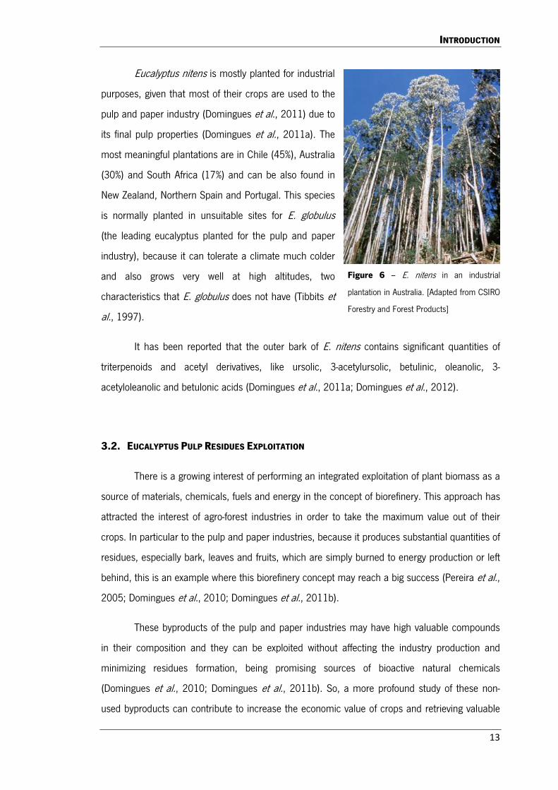
Figure 6. E. nitens in an industrial plantation in Australia. …………………………………………..…13
Figure 7. Chemical structure of betulinic acid. …………………………………………………………….16
Figure 8. Chemical structure of betulonic acid. …………………………………………………………...19
Figure 9. Chemical structure of ursolic acid. ……………………………………………………………….20
Figure 10. Chemical structure of oleanolic acid. ………………………………………………………….21
Figure 11. Representative images of the flow cytometry for cell death measurement ………….28
Figure 12. Effect of tested extracts and compounds in HCT116 cells viability ……………………35
Figure 13. Representative images of the effect of different concentrations of tested extracts and
compounds on the morphology of HCT116 cells …………………………………………………36
Figure 14. Effect of tested extracts and compounds in HCT116 cell viability and cell death
along the time of incubation ………………………………………………………………………..….38
Figure 15. Effect of tested extracts and compounds in the ability to inhibit colony forming in
HCT116 cells ……………………………………………………………………………………………….40
Figure 16. Effect of tested extracts and compounds in the ability of HCT116 cells to migrate..41
xvi
Figure 17. Effect of tested extracts and compounds in the presence of nuclear condensation in
HCT116 cells ……………………………………………………………………………………………….43
Figure 18. Effect of tested extracts and compounds on the levels of apoptosis and kinases
markers using western blot ……………………………………………………………………………..46
Figure 19. Effect of tested extracts and compounds in the cell cycle of HCT116 cells …………50
Figure 20. Scheme representing the possible mechanisms of action of the anticancer effect of
tested extracts and compounds in HCT116 cells …………………………………………………54
Table 1. Chemical composition of the E. nitens crude extract and of the triterpenic enriched
fraction 2 ………………………………………………………………………………………………….…26

INTRODUCTION


INTRODUCTION
3
1. CANCER
Cancer is one of the leading causes of death in the world. In 2005, the global cancer
incidence was of 11 million with over than 7.6 million of deaths, and it is prospected that by
2030 the incidence grows to 15.5 million with 11.5 million of deaths (Amin et al., 2009).
Although being a very common disease, cancer is mainly preventable (Amin et al.,
2009). Only 5 to 10% of all cancers are completely hereditary, while several external and
environmental factors are known to increase significantly the risk of this disease, such as
smoking, poor diet (fatty foods and alcohol), exposure to radiation, obesity, excessive sunlight
exposure, viruses, environmental pollutants and certain infections (Anand et al., 2008).
Cancer is a complex disease that can develop in human beings over a number of years
(Dzubak et al., 2005). In the development of cancer a normal cell is transformed in a cancer cell
through a process called carcinogenesis. During this process it is necessary that the genes
regulating cell differentiation and growth undergo failure or alteration (Dzubak et al., 2005).
These genes are divided into oncogenes, which are responsible for the promotion of cell growth
and proliferation; and into tumour suppressor genes, like p53, that inhibit cell division and
survival. The carcinogenic process is characterized by three phases of development, which
ultimately leads to cancer formation and growth. These steps are initiation/mutation, promotion
and progression/metastasis. The normal cell suffers damage, leading to mutation of DNA which
no longer undergoes DNA repair or undergoes defective DNA repair resulting in unrestrained
proliferation (initiation) (Dzubak et al., 2005). The continuous proliferation facilitates the
occurrence of even more mutations originating a mass of cells that no longer have their normal
abilities and only proliferate (promotion). The growth of the tumour is aided by angiogenesis,
which not only provides nutritients to the tumour, but also allows cancerous cells to migrate to
other tissues via the circulatory system, metastasizing the tumour (progression), which normally
is lethal. The most noticeable change in these cells is their ability to avoid programmed cell death
(apoptosis), leading to the tumour growth (Dzubak et al., 2005). Besides the resistance to
apoptosis, sustainable cell proliferation without stimulating signals, promotion of angiogenesis as
well as the ability to tissue invasion and establishment of metastasis, other characteristics
(“hallmarks”) of cancer include evasion to growth suppressors, limitless proliferative potential,
local promotion of inflammation, genome instability and mutation, evasion of immune system

INTRODUCTION
4
and deregulation of metabolic pathways (Hanahan and Weinberg, 2000; Hanahan and Weinberg,
2011).
Chemotherapy is a standard treatment to cancer, especially after surgical tumour
removal (Saxena et al., 2006; Drag et al., 2009). Considering that chemotherapy’s main aim is
to eliminate the remaining cancer cells present in the organism, most of the times the success of
this treatment determines the final success of the recovery (Drag et al., 2009). The efficacy of
this treatment has improved greatly in the last decade, but the treatment for this disease still
faces a very high mortality rate (Santos et al., 2011). Also, the toxic adverse effects of the used
drugs lead to severe life quality threats and 80% of the patients undergoing this treatment die due
to the resistance of cancer cells to drugs. Therefore, the development of new potent, non-toxic
and non-resistant anticancer agents is crucial to more effective therapies (Fulda and Debatin,
2000; Jung et al., 2007; Drag et al., 2009; Santos et al., 2011).
1.1. COLORECTAL CANCER (CRC)
Colorectal cancer (CRC) is the third most occurring cancer (8.5% of all cancers (Jung et
al., 2007)) in the world and one of the leading causes of death in the developed countries (with
65.500 deaths worldwide (Rajendran et al., 2008)), being uncommon in most non-developed
countries (Jung et al., 2007; Rajendran et al.; 2008; Xavier et al., 2009a; Chintharlapalli et al.,
2011). Therefore, the incidence rates of CRC are highly variable in different regions of the world
and the differences of the occurrence of this disease in migrants suggests that environmental
factors and diet play a major role in the development of this cancer (Chintharlapalli et al., 2011).
CRC can be divided into sporadic, familial and inherited. The sporadic represents 50 to
60% of CRC and there is no evidence of the disease in family history, being more common in
individuals with age over 50. The familial account for 30 to 40% of all the cases, and normally the
patient has a history of CRC in the family (Souglakos, 2007). The inherited CRC represents 4 to
6% and can be further divided depending on whether there are or not colonic polyps: familial
adenomatous polyposis (FAP) on the presence of those and hereditary nonpolyposis colorectal
cancer (HNPCC) or Lynch Syndrome on their absence (Rustki, 2007).

INTRODUCTION
5
The initial event of
carcinogenesis in the colon epithelium
is mostly the accumulation of a genetic
alteration in the antigen-presenting cell
(APC) gene, which is part of the Wnt
signalling pathway (Fig.1). This
mutation disables the production of the
APC protein leading to accumulation of
the β-catenin protein, which translocate
to the nucleus, leading to the activation
of transcription of genes that, at high
levels, cause cancer (Souglakos, 2007).
However, mutation in this pathway are
not the only ones necessary for the cell
to become cancerous; other mutations
must take place, such as in the TP53
gene that is responsible for killing the
cell if there is any defect in the Wnt pathway. Genetic alteration in the transforming growth factor
β receptor II (TGFβ-RII), phosphatase and tensin homologue (PTEN), cyclooxygenase 2 (COX2),
deleted in colorectal cancer (DCC), mutated in colorectal cancer (MCC), SMAD4,
phosphoinositide 3-kinase (PI3K), v-raf murine sarcoma viral oncogene homolog 1 (BRAF), v-Ki-
ras2 Kirsten rat sarcoma viral oncogene homolog (KRAS) and Bcl-2-associated x protein (BAX)
genes have also been found to play a role in CRC (Takami et al., 1995; Soreide et al., 2006;
Souglakos, 2007).
Complete remission of CRC by sirurgical removal is possible. Nevertheless,
approximately 50% of the cases eventually develops incurable metastesis, normally in the liver,
even when coupled with adjuvant treatment (Jung et al., 2007; Rajendran et al., 2008).
Therapies for gastrointestinal cancers have many defficiencies since the general response to
treatment is low to moderate, the life extension is, on most cases, only of two to three months,
there is a high toxicity to normal cells, high percentages of relapse cases and multiple drug
resistances observed (Rajendran et al., 2008). Chemotherapy based on 5-fluorouracil (5-FU) is
the standard treatment over the last forty years to extend patients survival when the CRC is
Figure 1 – APC/β-catenin signalling in CRC. (OFF) The APC
protein is synthesised, β-catenin is phosphorylated and occurs
ubiquitin-mediated degradation of β-catenin by the beta-
transducin repeat containing E3 ubiquitin protein ligase (β-
Trc)P. (ON) The APC protein is not produced, occurring
accumulation of β-catenin which enters the nucleus, enabling
transcription. [http://www.umcutrecht.nl]
INTRODUCTION
6
metastasized. However, the response rate of CRC metastasis to 5-FU is only of approximately
20% mainly due to cancer cell resistance to this drug (Jung et al., 2007; Rajendran et al., 2008;
Xavier et al., 2011). To overcome this resistance two new drugs were developed, Irinotecan and
Oxalipatin, that, combined with 5-FU, are able to overcome some of the drug resistance,
increasing the survival of the patients (Rajendran et al., 2008; Xavier et al., 2011). Nevertheless,
this improvement of efficiency is not observed in all patients (Xavier et al., 2011).
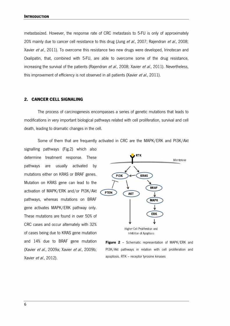
2. CANCER CELL SIGNALING
The process of carcinogenesis encompasses a series of genetic mutations that leads to
modifications in very important biological pathways related with cell proliferation, survival and cell
death, leading to dramatic changes in the cell.
Some of them that are frequently activated in CRC are the MAPK/ERK and PI3K/Akt
signalling pathways (Fig.2) which also
determine treatment response. These
pathways are usually activated by
mutations either on KRAS or BRAF genes.
Mutation on KRAS gene can lead to the
activation of MAPK/ERK and/or PI3K/Akt
pathways, whereas mutations on BRAF
gene activates MAPK/ERK pathway only.
These mutations are found in over 50% of
CRC cases and occur alternately with 32%
of cases being due to KRAS gene mutation
and 14% due to BRAF gene mutation
(Xavier et al., 2009a; Xavier et al., 2009b;
Xavier et al., 2012).
Figure 2 – Schematic representation of MAPK/ERK and
PI3K/Akt pathways in relation with cell proliferation and
apoptosis. RTK – receptor tyrosine kinases

INTRODUCTION
7
2.1 MAP KINASE PATHWAYS
Mitogen-activated protein kinases (MAPK) are a family of ubiquitous proline-directed,
protein-serine/threonine kinases, which participate in signal transduction pathways that control
several intracellular events (Pearson et al., 2001), including embryogenesis, inflammatory
response, cell differentiation, cell proliferation and cell death (Chen et al., 2001). They help
mediate diverse processes from transcription of proto-oncogenes to programmed cell death
(Cobb, 1999). The three major and best-known subfamilies of MAPKs are the extracellular-signal-
regulated kinase (ERK) 1/2, which conveys growth signals from the RAS/RAF pathway and
receptor kinases, and the c-Jun N-terminal kinase (JNK) 1/2/3 and the p38 MAP kinase (p38)
α/β/γ/δ, which relay various stress signals.
The MAPK are catalytically inactive in their base form, and require, in order to become
active, phosphorylation events in their activation loops. The activity is regulated by a cascade of
activations where MAPKs are phosphorylated by the MAPK kinase (MAPKK), which is, in turn,
phosphorylated by MAPKK kinase (MAPKKK) (Cobb, 1999; Chen et al., 2001; Pearson et al.,
2001; Cuevas et al., 2007). The MAPKKK are not specific to one MAPKK, being able to regulate
multiples MAPKKs, and leading to the activation of different subfamilies of MAPK, like the
mentioned ERK, JNK and p38 (Cuevas et al., 2007).
2.1.1. ERK PATHWAY
The MAPK/RAF/ERK pathway is one of the
most important signalling networks that control
proliferation, differentiation and cell survival (Kolch,
2000), and this cascade is activated by a variety of
receptors involved in growth and differentiation
(McCubrey et al., 2006). When ERK is improperly
activated it contributes to malignant transformation.
Raf is a serine/threonine protein that can be
found in three forms: a-Raf, b-Raf and c-Raf, that are
recruited to the membrane, bind to Ras, being Figure 3 – Schematic representation of the
ERK pathway. [Image from Kolch, 2000]
INTRODUCTION
8
subjected to phosphorylations and dephosphorylations promoting the activation of Raf. After Ras
activation, a MAPKKK phosphorylates two MAPKK proteins, MAPK 1 and 2, which activates the
MAPK ERK 1 and 2 (Fig.3). The activated ERK acts on cytosol by activating NF-kB, and on the
nucleus by promoting the phosphorylation of several transcription factors.
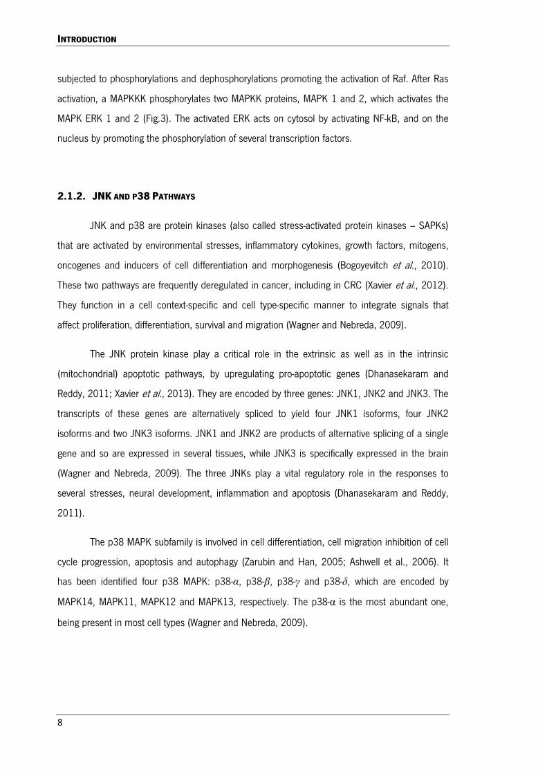
2.1.2. JNK AND P38 PATHWAYS
JNK and p38 are protein kinases (also called stress-activated protein kinases – SAPKs)
that are activated by environmental stresses, inflammatory cytokines, growth factors, mitogens,
oncogenes and inducers of cell differentiation and morphogenesis (Bogoyevitch et al., 2010).
These two pathways are frequently deregulated in cancer, including in CRC (Xavier et al., 2012).
They function in a cell context-specific and cell type-specific manner to integrate signals that
affect proliferation, differentiation, survival and migration (Wagner and Nebreda, 2009).
The JNK protein kinase play a critical role in the extrinsic as well as in the intrinsic
(mitochondrial) apoptotic pathways, by upregulating pro-apoptotic genes (Dhanasekaram and
Reddy, 2011; Xavier et al., 2013). They are encoded by three genes: JNK1, JNK2 and JNK3. The
transcripts of these genes are alternatively spliced to yield four JNK1 isoforms, four JNK2
isoforms and two JNK3 isoforms. JNK1 and JNK2 are products of alternative splicing of a single
gene and so are expressed in several tissues, while JNK3 is specifically expressed in the brain
(Wagner and Nebreda, 2009). The three JNKs play a vital regulatory role in the responses to
several stresses, neural development, inflammation and apoptosis (Dhanasekaram and Reddy,
2011).
The p38 MAPK subfamily is involved in cell differentiation, cell migration inhibition of cell
cycle progression, apoptosis and autophagy (Zarubin and Han, 2005; Ashwell et al., 2006). It
has been identified four p38 MAPK: p38-α, p38-β, p38-γ and p38-δ, which are encoded by
MAPK14, MAPK11, MAPK12 and MAPK13, respectively. The p38-α is the most abundant one,
being present in most cell types (Wagner and Nebreda, 2009).

INTRODUCTION
9
2.2. APOPTOSIS
Apoptosis, which in Greek literally means “falling away”, is an intrinsic programmed cell
death process highly conserved in different species, that occurs in multicellular organisms as a
natural and organized process, which plays an important role in the embryonic development and
in the balance of human tissues by adjusting the involved physiologic processes (Fulda, 2008;
Fulda and Kroemer, 2009; Wu et al., 2010; Yadav et al., 2010). Through apoptosis, the
organism is able to maintain homeostasis, by the elimination of damaged or unnecessary cells
without local inflammation due to leakage of cells’ contents. Therefore, any cell that has
abnormalities such as DNA damage, oncogene activation, nutrition deficiency or hypoxia, can be
eliminated without damage to the surrounding cells (Yadav et al., 2010). Apoptosis is
characterized by distinct morphological characteristics such as cellular shrinkage, chromatin
condensation, plasmatic membrane blebbing, oligonucleosomal DNA fragmentation and collapse
of the cell in smaller units (formation of apoptotic bodies) (Liu et al., 2004; Santos et al., 2011).
Since apoptosis is involved in the regulation of many physiological processes, a deficient
apoptosis signalling may contribute to a variety of different pathological conditions (Fulda and
Kroemer, 2009). Cancer is one of such pathologies, since cancer cells are able to escape
apoptosis, allowing tumours to grow rapid and uncontrollably (Yadav et al., 2010).
There are two different pathways through which apoptosis can be initiated: the intrinsic
pathway that involves the mitochondria and the extrinsic pathway that involves the plasmatic
membrane via death receptors (DR), both of them inducing activation of proteolytic enzymes
called cysteine aspartic acid specific proteases (caspases) (Fulda, 2008; Fulda and Kroemer,
2009).
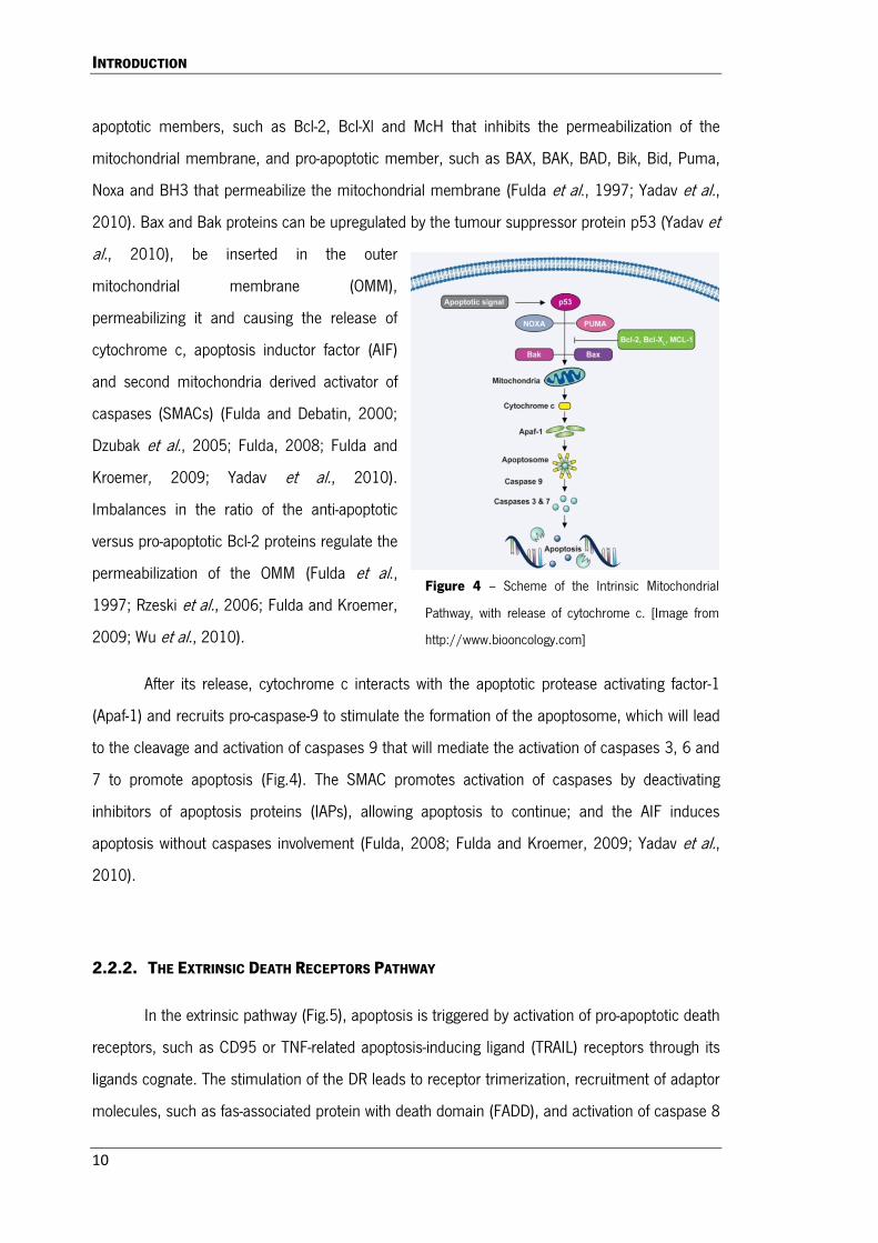
2.2.1. THE INTRINSIC MITOCHONDRIAL PATHWAY
The intrinsic pathway (Fig.4) can be activated by different stress stimuli, such as heat
shock, ultraviolet radiation or DNA damage. The permeabilization of the mitochondrial
membranes is frequently the decisive event that delimits the boundary between survival and
death (Fulda et al., 1997; Fulda and Debatin, 2000; Liu et al., 2004; Fulda and Kroemer, 2009).
The B-cell lymphoma 2 (Bcl-2) family proteins are regulators of apoptosis and comprise both anti-
INTRODUCTION
10
apoptotic members, such as Bcl-2, Bcl-Xl and McH that inhibits the permeabilization of the
mitochondrial membrane, and pro-apoptotic member, such as BAX, BAK, BAD, Bik, Bid, Puma,
Noxa and BH3 that permeabilize the mitochondrial membrane (Fulda et al., 1997; Yadav et al.,
2010). Bax and Bak proteins can be upregulated by the tumour suppressor protein p53 (Yadav et
al., 2010), be inserted in the outer
mitochondrial membrane (OMM),
permeabilizing it and causing the release of
cytochrome c, apoptosis inductor factor (AIF)
and second mitochondria derived activator of
caspases (SMACs) (Fulda and Debatin, 2000;
Dzubak et al., 2005; Fulda, 2008; Fulda and
Kroemer, 2009; Yadav et al., 2010).
Imbalances in the ratio of the anti-apoptotic
versus pro-apoptotic Bcl-2 proteins regulate the
permeabilization of the OMM (Fulda et al.,
1997; Rzeski et al., 2006; Fulda and Kroemer,
2009; Wu et al., 2010).
After its release, cytochrome c interacts with the apoptotic protease activating factor-1
(Apaf-1) and recruits pro-caspase-9 to stimulate the formation of the apoptosome, which will lead
to the cleavage and activation of caspases 9 that will mediate the activation of caspases 3, 6 and
7 to promote apoptosis (Fig.4). The SMAC promotes activation of caspases by deactivating
inhibitors of apoptosis proteins (IAPs), allowing apoptosis to continue; and the AIF induces
apoptosis without caspases involvement (Fulda, 2008; Fulda and Kroemer, 2009; Yadav et al.,
2010).
2.2.2. THE EXTRINSIC DEATH RECEPTORS PATHWAY
In the extrinsic pathway (Fig.5), apoptosis is triggered by activation of pro-apoptotic death
receptors, such as CD95 or TNF-related apoptosis-inducing ligand (TRAIL) receptors through its
ligands cognate. The stimulation of the DR leads to receptor trimerization, recruitment of adaptor
molecules, such as fas-associated protein with death domain (FADD), and activation of caspase 8
Figure 4 – Scheme of the Intrinsic Mitochondrial
Pathway, with release of cytochrome c. [Image from
http://www.biooncology.com]

INTRODUCTION
11
initiator, which spreads the death signal to effector
caspases like caspases 3, 6 and 7 (Fulda, 2008; Yadav
et al., 2010).
At this point, both intrinsic and extrinsic
pathways converge with the activation of the same
caspases. Also, caspase 8, through its mediated
cleavage, activates the BH3 interacting-domain death
agonist (Bid) which is translocated to the mitochondria
to promote cytochrome c release (Fulda, 2008).
2.2.3. ROLE OF P53 IN APOPTOSIS
The protein 53 (p53) is a tumour suppressor protein whose activity stops the formation
of tumours, being considered the “guardian of the genome”. It mediates critical functions in
cells, such as inhibition of proliferation by induction of cell cycle arrest and induction of apoptotic
cell death. In normal cells, p53 is usually present at very low levels and it can be activated in
response to stress, like hypoxia, heat shock and DNA damage agents. Its stability is regulated by
the E3 ubiquitin ligase and human double minute 2 homolog (HDM2), which mediates the
ubiquitination of p53 and allows its degradation by the proteosome, maintaining the p53 levels
low (Chari et al., 2009).
When activated, p53 translocates to the nucleus where it binds to specific DNA
sequences elements within the regulatory regions of target gene promoters regulating the
transcription of several genes related with apoptosis and/or cell cycle (Chari et al., 2009).
Furthermore, this protein can also activate the apoptotic mechanism through interaction with the
members of the anti- and pro-apoptotic Bcl-2 family proteins to induce the intrinsic mitochondrial
pathway (Vaseva and Moll, 2009).
Figure 5 – Scheme of the Extrinsic Death
Receptor Pathway. [Image from
http://www.biooncology.com]

INTRODUCTION
12
3. EUCALYPTUS SPP.
Eucalyptus spp., from the family Myrtaceae, are evergreen flowering trees (Russell et al.,
1997). This genus consists of approximately 600 species and has a physiological plasticity (it
supports tropical and temperate climes) that allows some species to propagate in different
regions of the world (Moura et al., 2012). In spite of this, only fifteen species occur outside
Australia, and only nine do not occur there, thus eucalyptus is mostly native to Australia. The
eucalyptus tree has high value mainly because it is a major source of cellulose for paper
manufacture, being the most common short fibre source for pulpwood in Portugal, Spain, Brazil,
Chile, South Africa, Japan, among other countries (Pereira et al., 2005; Domingues et al., 2010;
Domingues et al., 2011b; Moura et al., 2012). It is also very sought for its essential oil, which,
despite being highly toxic if ingested pure, is a good antiseptic, anti-malarial, industrial solvent
and insect repellent as well as used as additive for food industry (Russell et al., 1997; Pereira et
al., 2005; Moura et al., 2012). Eucalyptuses are also valuable for apiculture and the resulting
honey being studied for their antibacterial activity (Irish et al., 2011). Furthermore, the lipophilic
extracts from the bark of some eucalyptus (namely Eucalyptus globulus, Eucalyptus
camaldulensis var. obtusa, Eucalyptus grandis, Eucalyptus maidenii, Eucalyptus urograndis and
Eucalyptus nitens) are profoundly rich in triterpenic compounds, specially triterpenic acids like
oleanolic, 3-acetyloleanolic, ursolic, 3-acetyl-ursolic, betulinic, 3-acetylbetulinic and betulonic
acids, that are valuable bioactive compounds (Siddiqui et al., 2000; Kibblewhite et al., 2001;
Pereira et al., 2005; Domingues et al., 2010; Domingues et al., 2011a;Domingues et al., 2011b;
Santos et al., 2011b; Domingues et al., 2012).
3.1. EUCALYPTUS NITENS
Eucalyptus nitens (Fig. 6) is a species of fast growth and great development, with a
natural distribution in small and normally isolated populations. It is a tall to very tall forest tree,
normally with heights between 40 to 70 meters being able to reach 90 meters and diameter of 1
to 2 meters. Its juvenile leaves are smooth, orbicular and very fragrant; adult leaves are alternate,
long, curved, flexible and they set up a high silvery crown when grown in plantations (Tibbits et
al., 1997). Eucalyptus nitens is also known as Shining Gum, since nitens in Latin means
shining/bright and this refers to their leaves, buds, bark and fruits that have a distinct glossy.
INTRODUCTION
13
Eucalyptus nitens is mostly planted for industrial
purposes, given that most of their crops are used to the
pulp and paper industry (Domingues et al., 2011) due to
its final pulp properties (Domingues et al., 2011a). The
most meaningful plantations are in Chile (45%), Australia
(30%) and South Africa (17%) and can be also found in
New Zealand, Northern Spain and Portugal. This species
is normally planted in unsuitable sites for E. globulus
(the leading eucalyptus planted for the pulp and paper
industry), because it can tolerate a climate much colder
and also grows very well at high altitudes, two
characteristics that E. globulus does not have (Tibbits et
al., 1997).
It has been reported that the outer bark of E. nitens contains significant quantities of
triterpenoids and acetyl derivatives, like ursolic, 3-acetylursolic, betulinic, oleanolic, 3-
acetyloleanolic and betulonic acids (Domingues et al., 2011a; Domingues et al., 2012).
3.2. EUCALYPTUS PULP RESIDUES EXPLOITATION
There is a growing interest of performing an integrated exploitation of plant biomass as a
source of materials, chemicals, fuels and energy in the concept of biorefinery. This approach has
attracted the interest of agro-forest industries in order to take the maximum value out of their
crops. In particular to the pulp and paper industries, because it produces substantial quantities of
residues, especially bark, leaves and fruits, which are simply burned to energy production or left
behind, this is an example where this biorefinery concept may reach a big success (Pereira et al.,
2005; Domingues et al., 2010; Domingues et al., 2011b).
These byproducts of the pulp and paper industries may have high valuable compounds
in their composition and they can be exploited without affecting the industry production and
minimizing residues formation, being promising sources of bioactive natural chemicals
(Domingues et al., 2010; Domingues et al., 2011b). So, a more profound study of these non-
used byproducts can contribute to increase the economic value of crops and retrieving valuable
Figure 6 – E. nitens in an industrial
plantation in Australia. [Adapted from CSIRO
Forestry and Forest Products]

INTRODUCTION
14
compounds that would be otherwise discarded (Pereira et al., 2005; Domingues et al., 2011b;
Santos et al., 2011b). The exploitation of high value low molecular mass compounds, such as
phytosterols (namely β-sitosterol), lignans and triterpenoids, from industrial byproducts is a
strategy already implemented in some pulp industries (Domingues et al., 2010), as some of
these compounds, specially triterpenoids, can be of great interest as anticancer agents.
The bark of eucalyptus species is among the residues with most interest (Domingues et
al., 2011b). The bark of different eucalyptus residues left behind by pulp and paper industries
contains very valuable compounds, namely triterpenoids with great biological activities. The
compounds extracted from these different species are basically the same, although with different
proportions. So, E. camaldulensis var. obtusa, E. nitens, E. globulus, E. maidenii, E. urograndis
and E. grandis are highly rich in triterpenic acids with lupane, ursane and oleane skeletons
namely ursolic, 3-acetylursolic, oleanolic, 3-acetyloleanolic, betulinic and betulonic acids
(Kibblewhite et al., 2000; Siddiqui et al., 2000; Pereira et al., 2005; Domingues et al., 2010;
Domingues et al., 2011a; Domingues et al., 2011b; Santos et al., 2011). Besides triterpenic
compounds, it can also be found small quantities of fatty acids, fatty alcohols and aromatic
compounds (Domingues et al., 2011b).
4. NATURAL COMPOUNDS
Traditional medicine and diet have been serving humanity throughout the centuries as a
means of preventing and treating most of chronic diseases (Fulda, 2008; Fulda and Kroemer,
2009; Yadav et al., 2010; Yi et al., 2010). Natural compounds have been extensively used in the
treatment of multiple diseases and are of great value for the scientific community, either in its
natural form or as models to synthetic modification (Rajendran et al., 2008; Yadav et al., 2010).
These compounds used nowadays in modern medicine exhibit a great chemical diversity and,
along with its analogues and many others natural products, show the great importance of these
compounds in the efforts of discovering new drugs (Fulda, 2008; Fulda and Kroemer, 2009;
Yadav et al., 2010; Yi et al., 2010). The interest in natural compounds has increased over the
last few years, due to concerns about drug costs and, mainly, due to their security, as natural
compounds have usually low toxicity. Furthermore, they have high yield, easy obtainment,
favourable physiological functions, antioxidant activity and are generally accepted as dietetic
INTRODUCTION
15
supplements (Amin et al., 2009; Yadav et al., 2010; Yi et al., 2010), being considered a
fascinating strategy as therapeutic agents (Bishayee et al., 2011). Over the last decade, many
bioactive compounds have been identified in plants and human diet and are being developed as
chemopreventive agents to inhibit, slow down or even reverse the progression of several cancers
(Amin et al., 2009; Yadav et al., 2010; Bishayee et al., 2011). In fact, the great majority of
anticancer agents are derived from natural compounds or their analogues (Fulda, 2008; Fulda
and Kroemer, 2009; Santos et al., 2011). The anticancer activity of natural compounds has been
partially explained by its ability in triggering cellular death pathways, including apoptosis (Fulda
and Kroemer, 2009).
4.1. TERPENIC COMPOUNDS
One of the groups of natural compounds are terpenes (or terpenoids or isoprenoides)
that can be divided, according with the number of structural molecules of isoprenes, into mono-,
sesqui-, di-, sester-, tri-, tetra- and poly-terpenes which create extensive isoprenoides groups. With
almost 40.000 different terpenes isolated from plants, animals and microbial species they are
widely found in Nature. They are the biggest natural compound group found in plants (Dzubak et
al., 2005; Bishayee et al., 2011). Terpenes and its metabolites play a very important role in
plants defence mechanisms; they protect plants from the constitutive and induced defensive
responses against insects and environmental stress (Yadav et al., 2010). Among terpenes, the
triterpenes have recently emerged as an unique group of phytochemicals with multi-functional
activities (Bishayee et al., 2011).
4.1.1. TRITERPENIC COMPOUNDS
Triterpenes, or triterpenoids, are isopentenyl pyrophosphate oligomers metabolites that
are chemically related to squalene, which is a big group of compounds that have thirty carbon
atoms arranged in five rings with several oxygen atoms connected. Triterpenes are part of the
biggest plants compounds group, the Saponins (Yadav et al., 2010; Bishayee et al., 2011). It is
estimated that there are over 20.000 triterpenes in Nature, and they are predominantly found in
plants and fruits, being considered one of the most important natural compounds’ class (Dzubak

INTRODUCTION
16
et al., 2005; Kommera et al., 2011; Bishayee et al., 2011; Csuk et al., 2011). Thousands of new
structures have been described with hundreds of new derivatives being found each year
(Cichewicz and Kouzi, 2004). The variability of triterpenes in Nature is the result of the evolution
of a big family of terpene synthase (Yadav et al., 2010). The triterpenes can be subclassified in
diverse groups including the cucurbitanes, cycloartanes, dammaranes, euphanes, friedelanes,
holostanes, hopanes, isomalabaricanes, lanostanes, limonoids, lupanes, oleananes, protostanes,
squalenes, tirucallanes, ursanes and other compounds (Bishayee et al., 2011). In a biological
perspective the most important triterpenoids structures are the oleananes, ursanes, lupanes,
dammaranes and euphanes (Dzubak et al., 2005), emphasizing the lupane group that are
recognized as promising compounds for the development of new bioactive agents (Cichewicz and
Kouzi, 2004; Kommera et al., 2011; Domingues et al., 2011b).
Even though for a long time the triterpenes were considered biologically inactive, the
accumulating evidences of its wide pharmacological activities spectrum along with its low toxicity
aroused a new interest about its potential (Bishayee et al., 2011). The triterpenes, widely used in
medicine in Asian countries, have a range of unique biological effects and have been studied for
its anti-inflammatory, hepatoprotector, analgesic, antimicrobial, antimicotic, virostatic,
immunomodulatory, tonic, anticancer, antiangiogenesis, analgesic, antipyretic, antioxidant,
antiallergic, spasmolytic, anti-HIV and antimalarial effects (Dzubak et al., 2005; Kommera et al.,
2011; Yadav et al., 2010; Bishayee et al., 2011). Therefore, they can be considered a promising
and expanding platform to biological active natural compounds whose potential is only partially
explored by the pharmaceutical industry.
4.1.1.1. BETULINIC ACID
Betulinic acid (BiA), 3β,hydroxy-lup-20(29)-en-
28-oic acid (Fig.7) is part of the lupane group, a
pentacyclic triterpene, and it can be found in several
plants throughout the world like in Ziziphus spp.,
Amenone spp., Lycopodium spp., Syzygium spp., Betula
spp., Tryphyllum spp., Ancistrocladus spp., Eucalyptus
spp., Diopsyros spp., Paeonia spp. and Tetracera spp., Figure 7 – Chemical structure of betulinic acid.

INTRODUCTION
17
being highly available in the white-barked birch tree (from the genus Betula) (Cjowdhury et al.,
2002; Amin et al., 2009; Drag et al., 2009; Kommera et al., 2010a; Kim et al., 2011; Soica et
al., 2011). Betulinic acid is the oxidized derivative of its precursor betulin, which was one of the
first natural compounds to be isolated from plants more than two centuries ago (Cichewicz and
Kouzi, 2004; Fulda and Kroemer, 2009). Depending on the extraction method, betulin
comprehends over than 90% of the total isolated products and BiA around 1 to 5% (Drag et al.,
2009).
Betulinic acid, a white crystalline solid, is a bioactive compound that possess a wide
range of pharmacological effects like anti-inflammatory, antimicrobial, antiplasmodial, anti-HIV,
anti-diabetic, cardiovascular, anti-atherosclerosis, anti-obesity, anti-proliferative, anti-angiogenic,
cytotoxic, anticancer, antiparasitic, antimalarial, anti-viral, antioxidant, hepatoprotector, anti-
allergic and anti-tuberculosis effects as well as spasmogenic, anthelmintic and antinociceptive
activities (Ryu et al., 1994; Cichewicz and Kouzi, 2004; Dzubak et al., 2005; Fulda and Kroemer,
2009; Wu et al., 2010; Bishayee et al., 2011). It has been postulated that many of these effects
may be due to its ability to modulate immune functions, being an important immunomodulator
(Yi et al., 2010).
The Native Americans used the bark of white birch tree (rich in BiA) as a potent folk
medicine to treat skin diseases and as an anti-inflammatory (Fulda and Kroemer, 2009). The
cytotoxic activity of BiA was first described in 1976 by Trumbull and collaborators, but it was only
in 1995 that Pisha et al. published an important paper reporting the cytotoxic effects of BiA in a
human melanoma cell line, causing an increasing interest in this compound. It was first thought
that this compound was cytotoxic specifically to melanoma cell lines (Fulda, 2008; Fulda and
Kroemer, 2009), but since then its anticancer activity has been reported against several other
human cancers, including neuroblastoma, glioblastoma, colon, breast, liver, lungs and prostate
carcinomas, among others (Cichewicz and Kouzi, 2004; Ehrardt et al., 2004; Fulda, 2008; Amin
et al., 2009; Eichenmuller et al., 2009; Fulda and Kroemer, 2009; Chintharlapalli et al., 2011).
Many mechanisms of action have been published for explaining its anticancer effect. One
of them is the ability of BiA to induce cell death by apoptosis (Drag et al., 2009; Eichenmuller et
al., 2009; Santos et al., 2011), which is suggested to be mediated by the increase of
mitochondrial membrane permeability, meaning through the intrinsic mitochondrial apoptosis
pathway (Fulda et al., 1997; Dzubak et al., 2005; Fulda and Debatin, 2000; Zuco et al., 2001;

INTRODUCTION
18
Ehrhardt et al., 2004; Liu et al., 2004; Jung et al., 2007; Fulda, 2008; Eichenmuller et al., 2009;
Fulda and Kroemer, 2009; Wu et al., 2010; Bishayee et al., 2011; Kommera et al., 2011;
Santos et al., 2011). Betulinic acid has also been proven to be a topoisomerase I and II inhibitor
(Chowdhury et al., 2002; Wada and Tanaka, 2005) as well as being responsible by the
downregulation of the transcription factors specificity protein 1 (Sp1), 3 (Sp3) and 4 (Sp4) (Drag
et al., 2009; Chintharlapalli et al., 2011), which regulates the expression of a vast number of
genes involved in many cellular functions from differentiation, proliferation and apoptosis
(Deniaud et al., 2009). An interesting and clinically important fact is that BiA has a higher effect
in an environment with pH lower than 6.8, which is the pH at which almost every tumours
develops (Cichewicz and Kouzi, 2004; Dzubak et al., 2005).
A very important characteristic that has been reported for this compound is its ability to
trigger apoptosis in cancer cells resistant to drugs, suggesting that BiA may circumvent some
forms of resistance in cancer patients that show resistance to chemotherapy (Fulda and Debatin,
2000; Jung et al., 2007; Drag et al., 2009). Furthermore, despite its cytotoxicity against a variety
of cancers, normal cells and tissues appear relatively resistant to BiA, taking up to 100 mg/kg in
animal studies without showing great toxicity, which points to a very good therapeutic window
(Zuco et al., 2002; Dzubak et al., 2005; Rzeski et al., 2006; Csuk et al., 2011). So, due to its low
toxicity against normal tissues and cells, its remarkable anticancer activity and the fact that it can
bypass drug resistance cancers, it can be expected that BiA will eventually be accepted as an
adjuvant therapeutic in the treatment of cancer cells. However, BiA is a highly lipophilic molecule
with limited water solubility (being limitedly soluble in organic alcohols and highly soluble in
pyridine and acetic acid) which can lower its in vivo uptake. So, developing specialized
formulations/carriers, such as liposomes, may help to augment its in vivo efficiency as an
anticancer agent (Cichewicz and Kouzi, 2004;).
Tough BiA is widely available in Nature, their sources may not be enough to a possible
increase in the demand of this compounds. But, fortunately, the BiA’s precursor betulin is highly
available in several species of plants with significant yields over 20%, and BiA is easily prepared
from betulin in a simple two-step process (Chintharlapalli et al., 2011; Soica et al., 2011). Also,
due to its outstanding characteristics, a number of new BiA derivatives are being synthetized
(Kim et al., 2001; Liu et al., 2004; Rajendran, 2008; Kommera et al., 2010a; Nakagawa-Goto et
al., 2010; Kommera et al., 2011; Santos et al., 2011).

INTRODUCTION
19
4.1.1.2. BETULONIC ACID
Betulonic acid (BoA), 3-oxolup-20(29)-en-28-oic-acid (Fig.8), is also part of the lupane
group and a pentacyclic triterpene, which can be found in Syzigium spp, Eucalyptus spp.,
Prunella spp., Ziziphus spp. and Betula spp. (Ryu et al., 1994; Symon et al., 2005; Domingues et
al., 2010; Kommera et al., 2010a). Betulonic acid can also be obtained from betulinol or from
betulin. The yield obtained from the extraction of BoA from betulinol is aroun 90%, whereas from
betulin is only about 60% (Saxena et al., 2 006).
Betulonic acid is a white crystalline solid, which,
until the year 2000, was only focus of interest as a
precursor of BiA (Melinokova et al., 2012). However, it
was discovered that it was by itself a very valuable
bioactive compound with great pharmacological
activities such as anti-viral, anticancer, anti-
inflammatory, anti-malarial, anti-angiogenic, anti-HIV,
cytotoxic, hepatoprotector, antioxidant, antimicrobial,
and immunomodulatory (Pavlova et al., 2003; Sorokina
et al., 2004; Sorokina et al., 2006; Vabilevsky et al., 2009; Melinokova et al., 2012; Semenov et
al., 2012). In fact, it has been reported that in many of its effects, BoA is much more active and
potent inhibitor than BiA, being immensely potent against several tumour cell lines (Symon et al.,
2005; Wada and Tanaka, 2005). Betulonic acid is also known for being a powerful
topoisomerase II inhibitor (Ryu et al., 1994).
As mentioned, BoA has a remarkable cytotoxic activity against cancers such as
melanoma and prostate, neck and head, ovary/cervix, lung, colon, breast, thyroid and liver
carcinomas (Saxena et al., 2006; Shintyapina et al., 2007; Kommera et al., 2010a).
Due to its very low solubility in aqueous medium (being only soluble in organic solvents),
it has not been yet possible to determine in all its extent BoA’s powerful activity (Saxena et al.,
2006). Also because of this, there is not yet literature that clarifies exactly the mechanisms of
action of the anticancer activity of BoA, but it has been reported that it may up-regulate p53
activity (Zhang et al., 2008). Also, it has been reported that BoA may be a powerful agent that
Figure 8 – Chemical structure of betulonic acid.

INTRODUCTION
20
improves cytostatic effects of drugs (as a bioregulator to decrease the organism resistance to
toxic cytostatic effects) (Sorokina et al., 2004).
As with BiA, BoA seems to be little toxic to normal fibroblast cells (Saxena et al., 2006).
There has been also a number of BoA derivatives that are being synthesized (Vabilevsky et al.,
2009; Semenov et al., 2012).
4.1.1.3. URSOLIC ACID
Ursolic acid (UA), 3β-hydroxy-urs-12-en-28-oic
acid, (Fig. 9) is a natural pentacyclic triterpene
carboxylic acid. It is widely present in several
medicinal plants like sage, olive and rosemary as well
as in some fruits, such as some berries and apples,
and in eucalyptus (Liu, 1995; Domingues et al.,
2010).
UA is also a bioactive compound and has numerous effects such as anti-inflammatory,
anticancer and hepatoprotective effects (Liu, 1995; Xavier et al., 2009b; Xavier et al., 2013), as
well as it is able to provide protection against oxidative damage in DNA (Ramos et al., 2008;
Ramos et al., 2010). It also presents low toxicity to normal cells (Xavier et al., 2009b). It has
been reported that UA’s anticancer effect is due to its capacity of modulating important signalling
pathways, such as PI3K, inhibiting cell proliferation and inducing apoptosis (Liu, 1995 Xavier et
al., 2009b; Xavier et al., 2012).
4.1.1.4. OLEANOLIC ACID
Oleanolic acid (OA), 3β-hydroxy-olea-12-en-28-oic acid, (Fig.10) is a natural occurring
triterpenoids that is found in Phytolacca spp., Syzygium spp., Eucalyptus spp., in garlic and other
medicinal plants (Liu, 1995; Domingues et al., 2010).
Figure 9– Chemical structure of ursolic acid.

INTRODUCTION
21
OA is a bioactive compound and possesses
interesting effects such as hepatoprotector,
anticancer, anti-HIV and antiviral activities, being little
toxic to normal cells and tissues (Liu, 1995). It has
been reported that, besides being less potent in
inducing cell death than UA, it may be used in cancer
treatment in specific physiological conditions, such as
under metabolic stress (Duarte, 2012).
Figure 10 – Chemical structure of oleanolic. id
INTRODUCTION
22
OBJECTIVES OF THE WORK
Portugal is one of the leading countries in the production of eucalyptus for the pulp and
paper industry. However, this sector is responsible for the production of tons of bark residues
that does not have a valuable use. The present work intends to give a contribution to the
exploitation of this byproduct of the pulp and paper industry as possible source of high-added-
value bioactive compounds, a study that is under the scope of the European project AFORE (FP7:
CP-IP 228589-2 AFORE) that intends to develop novel applications for forest residues.
Therefore, the objective of this work was to study the potential anticancer effect of the
triterpenic fraction of extracts obtained from the bark of E. nitens in colorectal cancer cells. An
enriched triterpenic fraction (F2) in BiA and BoA was also tested and, in addition, these two
isolated compounds.
For that, the anticancer potential of the triterpenic extracts/compounds was studied by
the MTT assay, the anchorage-dependent colony forming assay and by the cell migration assay
(wound healing). Induction of cell death and apoptosis was studied by the PI staining and the
presence of nuclear condensation, respectively. In addition, effects on cell cycle were studied by
flow cytometry. Finally, the involvement of several signalling pathways, related with proliferation
and death, in the effects of the triterpenic extracts/compounds were analysed by western blot.
With this work we intend not only to establish the importance of triterpenoids compounds
as anticancer agents, but also to test an important source of these compounds that are being
disregarded as residues from crops used in the pulp and paper industry.

MATERIAL AND METHODS


MATERIAL AND METHODS
25
1. CHEMICALS
Propidium iodide (PI), quercetin, RNAse A, 3-(4,5-Dimethylthiazolyl-2)-2,5-
diphenyltetrazolium bromide (MTT), RPMI 1640, antibiotic/antimicotic solution, bovine serum
albumin (BSA), N-(2-Hydroxyethyl)piperazine-N'-(2-ethanesulfonic acid) (HEPES), were purchased
from Sigma-Aldrich (St. Louis, MO, USA). Fetal bovine serum (FBS) was bought from Biochrom
AG (Berlin, Germany). All the other compounds were of analytical grade.
2. EUCALYPTUS NITENS EXTRACTS AND TRITERPENOIDS
Betulinic acid and betulonic acid were purchased from Molekula Ltd (Gillingham, Dorset,
United Kingdom) and Chemos GmbH (Regenstauf, Germany), respectively. Eucalyptus nitens
crude extract (E. nitens CE) and the triterpenic enriched fraction 2 (E. nitens F2) were kindly
provided by Professor Armando Silvestre from the Department of Chemistry, University of Aveiro.
The extracts were prepared by Domingues et al. through extraction of E. nitens bark with
dichlorometane as previously described (Domingues et al., 2011). The CE and F2 extracts of E.
nitens were then analysed by GC-MS and the main chemical composition present in Table 1
(Domingues et al., 2012, unpublished data). Extracts and isolated compounds were dissolved in
dimethyl sulfoxide (DMSO) in stock solutions, in order that DMSO concentration in cell culture
was no higher than 0.5% (v/v). Controls received vehicle only.
3. ANTIBODIES
The primary antibodies were purchased from the following sources: anti-phospho-ERK,
anti-PARP-1, anti-phospho-JNK, anti-Bcl-2 and anti-p53 from Santa Cruz Biotechnology, Inc.
(Santa Cruz, CA, USA); anti-caspase 9, anti-phospho-p38 MAPK (Thr180/Tyr182), anti-phospho-
AMPKα (Thr172) and anti-phospho-Akt (Ser473) from Cell Signalling (Danvers, MA, USA); anti-
caspase 3 from Calbiochem (San Diego, CA, USA); and anti-β-actin from Sigma-Aldrich.
Secondary antibodies anti-rabbit and anti-mouse were purchased from Santa Cruz Biotechnology.

MATERIAL AND METHODS
26
Table 1 – Chemical composition (based on % of weight from the total extract weight) of the E. nitens crude extract
and of the triterpenic enriched fraction 2 (Domingues et al., 2012, unpublished data).
Compounds Crude extract (%) Fraction 2 (%)
Fatty acids 1.2 4.2
Long chain aliphatic alcohols 0.9 0.2
Sterols 1.2 0.0
Triterpenoids 70.7 93.3
β-amirin 0.3 0.0
Lupeol 0.3 0.0
Betulonic acid 7.0 24.2
Oleanolic acid 20.8 15.5
Betulinic acid 19.0 32.5
Ursolic acid 10.2 15.5
3-acetyloleanolic acid 3.2 0.0
3-acetylursolic acid 1.8 0.6
Unidentified triterpenoids 8.1 4.9
Other compounds 1.9 0.0
Unidentified compounds 24.1 2.3
4. CELL CULTURE
HCT116 cells (human colorectal carcinoma cell line) were kindly provided by Professor
Raquel Seruca from IPATIMUP, Porto. The cell line was maintained in culture in 25 cm2
polystyrene flasks (TPP, Switzerland) with RPMI 1640 medium, containing 6% FBS, 1% antibiotic–
antimicotic solution, 0.1 mM sodium pyruvate, 10 mM HEPES and 2 g/L sodium bicarbonate
under an atmosphere of 5% CO2 and 95% air at 37°C.
5. MTT REDUCTION ASSAY
MTT reduction assay was performed to evaluate the potential of the tested
compounds/extracts to decrease the number of viable cells as previously described (Lima et al.,
2011). Briefly, HCT116 cells were plated in 24-multiwell culture plates at 8x10⁴ cells per ml and
allowed to grow for two day. Cells were incubated with test compounds/extracts at different
concentrations and for different time periods. One hour before the end of incubation, 50µL of

MATERIAL AND METHODS
27
MTT (final concentration of 0.5mg/ml) was added to each well. When the incubation time ended,
the medium was removed and discarded, and the formazan crystals (formed by the cell’s
capacity to reduce MTT) were dissolved with a 50:50 (v/v) DMSO:ethanol solution. The
absorbance was measured at 570nm, with background subtraction at 690nm. The results were
expressed as percentage relative to the control. The concentration of each test compound/extract
that decreases the number of viable cells to half (IC50) was calculated using mathematical
modelling with the program GraphPad Prism 5 (GraphPad Software, Inc., La Jolla, CA, USA).
6. ANCHORAGE-DEPENDENT COLONY FORMING
The anticancer potential of tested compounds/extracts was also tested by their ability to
inhibit the formation of cell colonies. HCT116 cells were plated in 6-multiwell culture plates at
500 cells per ml for two days for cell attachment. Cells were then incubated with different
concentration of test compounds/extracts dissolved in new culture medium for six days (without
changing the medium). After this incubation time, medium was removed from the wells, washed
with PBS and cells fixed with ice cold absolute ethanol for 15 minutes. Then, cells were stained
with Giemsa working solution (0.4% w/v) for 5 minutes and the wells washed with tap water. The
plate wells were photographed and colony forming ability estimated by quantifying colony area
intensity using appropriate image analysis system (Adobe Photoshop CS3 Adobe Systems
Incorporated, San Jose, CA, USA).
7. CELL DEATH ANALYSIS BY PROPIDIUM IODIDE (PI) STAINING
PI staining was performed to evaluate cell death induced by the tested
compounds/extracts. This DNA fluorophore is impermeable to live cells, but will stain strongly the
DNA of dying and death cells due to their compromised or permeabilized plasma membranes
(Rieger et al., 2011) The HCT116 cells were plated in 6-multiwell culture plates at 8x10⁴ cells per
ml, for two days for cell attachment and growth, before incubating them with test
compounds/extracts dissolved in new culture medium. After treatment with different
concentrations and incubation times, cells were collected (both floating and attached cells)
washed with cold PBS containing 6% (v/v) FBS, and incubated with PI added to a final
MATERIAL AND METHODS
28
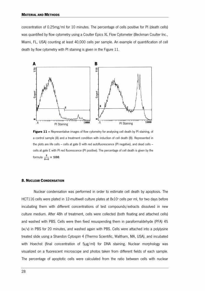
concentration of 0.25mg/ml for 10 minutes. The percentage of cells positive for PI (death cells)
was quantified by flow cytometry using a Coulter Epics XL Flow Cytometer (Beckman Coulter Inc.,
Miami, FL, USA) counting at least 40,000 cells per sample. An example of quantification of cell
death by flow cytometry with PI staining is given in the Figure 11.
8. NUCLEAR CONDENSATION
Nuclear condensation was performed in order to estimate cell death by apoptosis. The
HCT116 cells were plated in 12-multiwell culture plates at 8x10⁴ cells per ml, for two days before
incubating them with different concentrations of test compounds/extracts dissolved in new
culture medium. After 48h of treatment, cells were collected (both floating and attached cells)
and washed with PBS. Cells were then fixed resuspending them in paraformaldehyde (PFA) 4%
(w/v) in PBS for 20 minutes, and washed again with PBS. Cells were attached into a polylysine
treated slide using a Shandon Cytospin 4 (Thermo Scientific, Waltham, MA, USA), and incubated
with Hoechst (final concentration of 5µg/ml) for DNA staining. Nuclear morphology was
visualized on a fluorescent microscope and photos taken from different fields of each sample.
The percentage of apoptotic cells were calculated from the ratio between cells with nuclear
Figure 11 – Representative images of flow cytometry for analysing cell death by PI staining, of
a control sample (A) and a treatment condition with induction of cell death (B). Represented in
the plots are life cells – cells at gate D with red autofluorescence (PI negative), and dead cells –
cells at gate E with PI red fluorescence (PI positive). The percentage of cell death is given by the
formula 𝐄 𝐃+𝐄
× 𝟏𝟏𝟏.
PI Staining PI Staining
A B
MATERIAL AND METHODS
29
condensation typical of apoptosis and the total number of cells, from a count higher than 500
cells per sample.
9. CELL CYCLE ANALYSIS
The analysis of cell cycle was done by flow cytometry using PI for staining DNA of
permeabilized cells. For that, HCT116 cells were plated in 6-multiwell culture plates at 8x10⁴
cells per ml for two days for cell attachment and growth, before incubating them with the test
compounds/extracts dissolved in new culture medium. After treatment for 48h with different
concentrations of compounds/extracts, cells were collected (both floating and attached cells) and
washed with cold PBS. Then, cells were fixed and permeabilized in ice cold ethanol 70% (v/v) for
15 minutes and washed again with PBS. Finally, cells were incubated with staining solution (50
µg/ml PI and 20 µg/ml RNase A in PBS) at 37ºC for 15 minutes. Analysis of cell cycle
progression by flow cytometry was done using a Coulter Epics XL Flow Cytometer (Beckman
Coulter Inc.) counting at least 40,000 single cells per sample. Phases of cell cycle were fitted
using the mathematical Watson Pragmatic model with the FlowJo Analysis Software (Tree Star,
Inc., Ashland, OR, USA).
10. MIGRATION ASSAY (WOUND HEALING)
Migration assay was performed to evaluate the effect of tested compounds/extracts to
affect the extent of wound closure. For that, HCT116 cells were plated in 12-multiwell culture
plates at 2.5x10⁵ cells per ml, and allowed to grow for two days until cell confluence was
reached. Cell layer was then wounded in an x shape with an 100 µl tip, and a photo of the
wound site was taken. Cells were incubated with the test compounds/extracts for 24h and a new
photo in the central wound site taken. The extend of wound closure was given as the percentage
of covered area after 24h in relation to the initial uncovered area, using the following formula:
Extent of wound closure = �Initial uncovered area-Final uncovered area�
Initial uncovered area ×100
and used to estimate the cells’ migration ability.

MATERIAL AND METHODS
30
11. WESTERN BLOTTING
To measure the expression of different proteins involved in cell signalling and apoptosis
western blot was used using total cell homogenates. HCT116 cells were plated in 6-multiwell
culture plates at 8x10⁴ cells per ml for two days for cell attachment and growth. Cells were
treated with different concentrations of tested compounds/extracts for 24h or 48h and a total
cell homogenate obtained incubating cells for 15 minutes at 4ºC with ice cold RIPA buffer (1%
NP-40 in 150mM NaCl, 50mM Tris-HCl (pH 8), 2mM EDTA) containing 1mM PMSF,
phosphatase inhibitors (20mM NaF, 20mM Na2V3O4) and protease inhibitor cocktail (Roche,
Mannheim, Germany). Protein concentration was quantified by Bio-Rad DC protein assay (Bio-
Rad Laboratories, Hercules, CA, USA) and BSA used as protein standard. For Western Blot, 20
µg of protein was resolved in SDS-polyacrilamide gel and then electroblotted to a polyvinylidene
difluoride membrane (Millipore, Billerica, MA, USA). Membranes were blocked in TPBS (PBS with
0.05% of Tween-20) containing 5% (w/v) of non-fat dry milk, washed in TPBS and incubated with
primary antibody overnight. After washing, membranes were incubated with the secondary
antibody for 1h, and membranes washed again. The immunoreactive bands were detected using
the Immobilon solutions (Millipore) under a chemiluminescence detection system, the Chemidoc
XRS (Bio-Rad Laboratories). Band area intensity was quantified using the Quantity One software
from Bio-Rad. β-actin was used as loading control.
12. STATISTICS
Data expressed as the mean ± SEM of at least 3 independent experiments. Statistical
significances among data groups were analysed by one-way ANOVA followed by the Dunnett’s
multiple comparison test, or by the Student’s t-test, as appropriate, using GraphPad Prism 5.0
software (San Diego, CA, USA). Differences between groups were considered statistically
significant when P≤0.05.

RESULTS AND DISCUSSION

RESULTS AND DISCUSSION
33
The pulp and paper industries are one of the most important industries in Portugal
contributing positively to the country’s export (Pereira et al., 2005; Domingues et al., 2010;
Domingues et al., 2011b; Moura et al., 2012). Eucalyptus wood is the main source of fibbers for
the production of paper. However, it generates large amounts of residues, mainly bark, which
can be a source of valuable compounds of otherwise burned or discarded material (Doningues et
al., 2011). Therefore, the exploitation of this byproduct for the production of interesting added-
value new products is being viewed by the pulp and paper industry as a way to increase their
revenue and to implement the biorefinery concept in their production chain (Domingues et al.,
2011a; Mota et al., 2012). Previously, it was reported that lipophilic extracts from the bark of
Eucalyptus species can be an important source of high-value triterpenic compounds in view of
their high content and attributed bioactive properties (Domingues et al., 2011a).
In the present work, the potential anticancer effect of lipophilic extracts enriched in
triterpenic acids obtained from the bark of E. nitens was studied in the colorectal HCT116 cancer
cells. Although E. nitens is not the main species of eucalyptus in Portugal, it possess high content
of triterpenic acids like the most abundant species E. globulus, but contrarily to this one, where
ursane acids dominates, it is the richest in oleanane and lupane acids (Domingues et al.,
2011a).
For this work, two extracts of E. nitens were used (see Table 1, in Materials section): a
lipophilic crude extract (CE) with about 70% (w/w) of triterpenoids, where the main ones are
oleanolic acid (20.8%) followed by betulinic acid (19%), ursolic acid (10.2%) and betulonic acid
(7%); and, a fraction (F2) of the CE more enriched in triterpenoids (about 93% of total weight),
specially in lupane acids, since the main ones are betulinic acid (32.5%) followed by betulonic
acid (24.2%), ursolic acid (15.5%) and oleanolic acid (15.5%). In addition, the above lupane acids
were also tested alone for comparison.

RESULTS AND DISCUSSION
34
1. TRITERPENIC ACIDS-ENRICHED EXTRACTS FROM E. NITENS POSSESS ANTICANCER ACTIVITY
AGAINST HCT116 CELLS
In order to study the anticancer potential of the two E. nitens extracts, HCT116 cells
were incubated for 48h with different concentrations of CE or F2 extracts, and cell viability
evaluated by the MTT assay (Fig. 12). As shown in Fig. 12A & 12C, both extracts presented high
anticancer activity, decreasing significantly the number of viable cells in a concentration
dependent manner. Based on these results, their IC50 (concentration of extract necessary to
decrease by half the number of viable cells as compared with negative control) were calculated
and presented as an example for the E. nitens CE in Fig. 13A. The results showed that the E.
nitens F2 extract was more active than the E. nitens CE extract, with IC50s of 1.3 µg/ml and 2.2
µg/ml, respectively.
Considering the high content of these extracts in triterpenoids, especially in betulinic,
betulonic, oleanolic and ursolic acids, the main compounds were also tested alone. In a previous
work of our group (Duarte, 2012), the anticancer potential of both UA and OA were evaluated in
HCT116 cells using the same culture conditions than here. Whereas for UA a good anticancer
activity was obtained with an IC50 of 8 µM, for OA no significant decrease of cell viability was
observed until the highest concentration tested (50 µM), meaning that the IC50 is much over this
value. In the present work, the remaining two main triterpenoids of the E. nitens extracts, BiA and
BoA, were tested. Both compounds exhibit a high capability to decrease the number of viable
cells, also in a concentration-dependent manner (Fig. 12D and E). Interestingly, BoA presents a 4
times lower IC50 concentration than BiA, with 0.8 µM and 3.9 µM, respectively, and a 10 times
lower than UA.
Converting the IC50 of the isolated compounds from µM to µg/ml, and taking into
account the percentage of each compound in both extracts, the concentration range of the
isolated compounds are in the same order of magnitude than that of the extracts. However, a
compound alone does not explain the total extract activity, which suggests that all the three main
active triterpenoids BoA, BiA and UA contribute to the extracts’ activity. Interestingly, taking into
account only these three triterpenoids, E. nitens F2 extract has about two times more of their
quantity than E. nitens CE (see Table 1, in Materials section), and the IC50 of the former is about
half of the latter. This observation corroborates that the activity of the E. nitens extracts dependes
mainly from the three active triterpenoids (OA is non-active until 50 µM).

RESULTS AND DISCUSSION
35
Fig. 12 – Effects of E. nitens CE (B) and F2 extracts (C), as well as the isolated compounds betulinic acid (D) and
betulonic acid (E) in cell viability of HCT116 cells after 48h of incubation, as measured by the MTT assay. In (A) is
presented, as an example, data of (B) with a sigmoid mathematical modelling with the program GraphPad Prism for
calculation of the IC50 value. The same metod was used to calculate the IC50 given in C, D and E. Values are mean ±
SEM of at least three independent experiments. * P≤0.05, ** P≤0.01, *** P≤0.001, when compared with respective
control, analysed by one-away ANOVA with Dunnett’s multiple comparison test.
A
E D
C B
IC50 = 1.3 µg/ml IC50 = 2.2 / l
IC50 = 3.9 µM IC50 = 0.8 µM

RESULTS AND DISCUSSION
36
The morphologic changes observed in HCT116 cells after being incubated for 48h with
eucalyptus extracts (Fig. 13A) or isolated lupane acids (Fig. 13B), also agrees to their potent
anticancer activity. Along with the increasing concentration of the extract/compound, the number
of adherent (live) cells decreased and the floating (dead) increased. The anticancer activity of
eucalyptus extracts has been poorly investigated. Recently, polyphenolic-rich extracts from E.
globulus bark demonstrated some capacity to reduce human breast cancer cell proliferation
(Mota et al., 2012). In addition, Islam et al. (2012) showed the anti-tumour activity of a lipophilic
extract of E. camaldulensis against Ehrlich ascites carcinoma celsl in mice. In the present work, it
was demonstrated a high anticancer potential of triterpenic acids-rich extracts from E. nitens bark
in colorectal cancer cell, with IC50 of about 2 µg/ml.
Fig. 13 – Representative images of the effect of different concentrations of E. nitens crude extract (CE) and E.
nitens fraction 2 (F2) extract (A), and test compounds betulinic acid (BiA) and betulonic acid (BoA) (B) on the
morphology of HCT116 cells after 48h incubation. Photos were taken with an objective of 10 times magnification
in an inverted microscope (Olympus IX71 – Lisboa, Portugal) with phase contrast.

RESULTS AND DISCUSSION
37
The anticancer potential of their main triterpenoids are, however, described in the
literature. Many studies (Cichewicz and Kouzi, 2004; Ehrardt et al., 2004; Symon et al., 2005;
Wada and Tanaka, 2005; Fulda, 2008; Amin et al., 2009; Eichenmuller et al., 2009; Fulda and
Kroemer, 2009; Chintharlapalli et al., 2011), including our owns (Ramos et al., 2008; Xavier et
al., 2009b; Ramos et al., 2010; Xavier et al., 2012), demonstrated the anticancer activity of
these triterpenoids against several types of tumours cells. Many mechanisms of action, mainly
for UA (Liu, 1995 Xavier et al., 2009b; Xavier et al., 2012) and BiA (Deniaud et al., 2009), were
also depicted. Considering that OA is non-active in HCT116 cells and that the lupane acids were
more active than UA, along with the E. nitens extracts, the work was proceeded with BiA and
BoA.
The observed decrease in the number of viable cells induced either by the extracts or by
the isolated compounds could have been a result of inhibition of cell proliferation and/or
induction of cell death. As to better clarify that, some concentrations of extracts and compounds
were chosen and their effect on cell viability along with different times of incubation (6, 24, 48
and 72h) was measured by MTT reduction assay (Fig. 14A, C, E and G). Furthermore, cell death
was followed by flow cytometry by measuring the ability of live cells to exclude PI after different
times of incubation (24, 48 and 72h) (Fig. 14B, D, F and H).
As shown in Fig. 14A, cells incubated with 1 or 2.5 µg/ml of E. nitens CE accompanied
the growth of control cells until 24h of incubation, but decreased significantly after 48h. On other
hand, cells incubated with 10 µg/ml of CE did not grow in the first 24h but did not induce cell
death (Fig.13B). With the PI staining assay, cell death was only observed after 48h of incubation
and in a concentration-dependent manner (Fig. 14B). At 72h, the same results were obtained,
but with a higher magnitude of cell death. The pattern of results of E. nitens F2 (Fig. 14C and D)
was similar to the ones of CE. However, the magnitude of effects was higher at lower
concentrations, which, once more, shows that the F2 has higher anticancer activity when
compared to the CE. Regarding the isolated compounds, cell death was already visible at 24h of
incubation with the higher tested concentration of each compound, 15 µM for BiA and 50 µM for
BoA, being of notice that even tough BiA’s concentration is significantly lower than BoA’s, it
shows more cell death (Fig. 14F and H). This result seems contradictory to the IC50 value
calculated for BiA when compared with BoA. However, at 48h of incubation, BoA at lower doses
induces higher cell death as compared with BiA, which agrees with the MTT assays (Fig. 14E, F,
RESULTS AND DISCUSSION
38
A
H G
F E
D C
B
Fig. 14 – Effect of E. nitens crude extract (CE), E. nitens fraction 2 (F2), betulinic acid and betulonic acid at different
concentrations in cell viability of HCT116 cells along the time. (A, C, E and G) Number of viable cells measured by the
MTT assay. (B, D, F and H) Cell death evaluated by the PI staining as measured by flow cytometry. Values are mean ±
SEM of at least three independent experiments. * P≤0.05, ** P≤0.01, *** P≤0.001, when compared with respective
control, analysed by the Student’s t-test.
RESULTS AND DISCUSSION
39
G and H). All of these results seem to indicate a contribution of both inhibition of cell
proliferation and induction of cell death in the observed reduction of viable cells by the extracts
and the compounds.
In order to confirm the anticancer potential of the triterpenic acid-enriched E. nitens
extracts and its main compounds, an anchorage-dependent colony formation assay and cell
migration assay was performed. As shown in Figure 15A, the two tested extracts and compounds
exhibited a marked inhibition of colony formation and growth in a concentration-dependent
manner. The observed effects resulted mainly from inhibition of cancer cell colonies growth and
colony size – Fig. 15B – since concentrations of extracts/compounds that induced cell death, in
the conditions used in this assay (concentrations higher than that present in Fig. 15), were
discarded. In accordance with the results of the cell viability assay, E. nitens F2 extract was
shown to be more potent than the CE, having the concentrations of the first (2.5 and 5 µg/ml)
achieved the same results of the latter at 5 and 10 µg/ml (Fig.15). However, for the triterpenoids
BiA and BoA a similar inhibition of colonies growth were obtained for the same concentrations.
As expected, the positive control – starved cell (grown in medium with 0.6% (v/v) FBS) – resulted
in marked colonies growth inhibition.
For the migration assay, the capacity of cells to wound healing/closure were evaluated in
the presence of different concentrations of extracts/compounds for 24 hours. As a positive
control, cells were incubated in starvation medium (medium with 0.6% (v/v) FBS), and
representative images of this and control cells are present in Figure 15B. In the migration assay,
the wounds were made in a confluent cell layer, in order that the wound closure would be
dependent on cell migration and not on cell proliferation. When cells reach confluence, they
acquire a quiescent state, where cell cycle pauses at G0 state (Coller et al., 2006).
Figure 16A shows that the lower concentrations of either the extracts or the compounds
do not have any effect on the cell migration ability, being their values very similar to those of the
control. However, with a higher concentration we did obtain migration inhibition in cells treated
with the extracts and the compounds. Rzeski and collaborators (2006) had previously reported
that cultures treated with BiA had a significant lower number of cell migrating to the wounded
area. Taking into account the two E. nitens extracts, it was observed again that half the
concentration used for the CE obtain the same result for F2. The same also happens with the
isolated compounds, where 5 µM of BoA has the overall same result of 10 µM of BiA (Fig. 16A),

RESULTS AND DISCUSSION
40
Fig. 15 – Effect of E. nitens crude extract (CE), E. nitens fraction 2 (F2), betulinic acid and betulonic acid in the
ability to inhibit colony forming in HCT116 cells. Cells were incubated with test compounds/extracts (controls
received vehicle only – DMSO; starved cells received 0.6% (v/v) FBS-containing medium) for six days before
being fixed and stained with Giemsa. A) Values are expressed as the mean area intensity of colonies and are
mean ± SEM of the mean area intensity of colonies of at least three independent experiments. * P≤0.05, **
P≤0.01, *** P≤0.001, when compared with respective control, analysed by the Student’s t-test. B)
Representative images of the anchorage-dependent colonies formation in HCT116 cells under the effect of tested
compounds/extracts for six days.
Control Control +
(Starved cells) CE E. nitens
(5 µg/ml) E. nitens F2 (5 µg/ml)
Control Control +
(Starved cells) Betulinic acid
(5 µM) Betulonic acid
(5 µM)
A
B

RESULTS AND DISCUSSION
41
demonstrating, once more, that BoA is a more potent compound than BiA. The effect of higher
concentrations of extracts/compounds than that shown in Figure 16 was not shown, since
inhibition of wound closure did not result from inhibition of cell migration, but noticeably from
induction of cell death, in the conditions used in this assay.
A
Control
Control + (Starved cells)
24 h 0h
Control
Control + (Starved cells)
B
Fig. 16 – Effect of E. nitens crude extract (CE), E. nitens fraction 2 (F2), betulinic acid and betulonic acid in the ability of
HCT116 cells to migrate. Cells were incubated with test compounds/extracts (controls received vehicle only – DMSO;
starved cells received 0.6% (v/v) FBS medium) for 24 hours. A) Values are given as mean ± SEM of the percentage of
covered area after 24h in relation to the initial uncovered area, of at least three independent experiments. * P≤0.05, ***
P≤0.001, when compared with respective control, analysed by the Student’s t-test. B) Representative images of the
wound before (0h) and after 24 hours of incubation for the negative control (complete medium with vehicle) and the
positive control (starved cells) (bar – 500 µm).

RESULTS AND DISCUSSION
42
All the present results show that BoA is the most active anticancer compound for
HCT116 cells between the tested triterpenoids. Eucalyptus nitens bark can be an interesting
source of extracts rich in bioactive triterpenoids, namely BoA, BiA and UA, with potent anticancer
activity against HCT116 cells.
2. APOPTOSIS CONTRIBUTES TO THE CELL DEATH INDUCED BY E. NITENS EXTRACTS AND THEIR
MAIN LUPANE ACIDS IN HCT116 CELLS
Considering the cell death induced by the tested extracts and compounds, we wondered
whether it was apoptosis dependent. During apoptosis, DNA is fragmented and condensed in
order to be included in the apoptotic bodies (Elmore, 2007). Therefore the ability of the E. nitens
extracts and tested isolated compounds to induce nuclear condensation in HCT116 cells after
48h of incubation, as a marker of cell death by apoptosis, were studied. As shown in Figure 16,
apoptosis occurs in cells treated with either the extracts or the compounds, but only at the higher
tested concentrations. Among the E. nitens extracts, F2 seems to be more potent inducing
apoptosis than CE, since the first has about five times more apoptosis at 10 µg/ml than control
cells, whereas the latter has no effect. Interestingly, BiA, which has a higher IC50 than BoA at
48h, has a higher ability to induce cell death by apoptosis at lower doses (Fig.17A). Some
intermediate concentrations of the tested extracts or compounds that already induced cell death
as assessed by the PI staining (Fig.14), did not, however, induce apoptosis. So, nuclear
condensation assay was also performed after 72h of incubation with these concentrations of
extracts/compounds that induce cell death as observed with PI staining, but no induction of
apoptosis was observed (data not shown).
These results indicate that our tested extracts and compounds are able to induce cell
death by apoptosis only at high concentrations. At lower or intermediate concentrations cell death
occurs, but is not as a result of apoptosis but through some kind of non-apoptotic type of cell
death.
Whilst there is no scientific data as how E. nitens extracts or BoA induces apoptosis in
cancer cells, it has been reported that BiA induces apoptosis in cancer cells through activation of
the intrinsic mitochondrial pathway (Fulda and Debatin, 2000). Therefore, the levels of several
proteins related with apoptosis were evaluated by western blot after treating HCT116 cells with
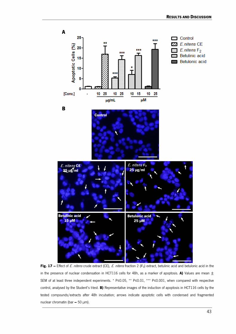
RESULTS AND DISCUSSION
43
Control
E. nitens F2 25 µg/ml
Betulinic acid 15 µM
E. nitens CE 25 µg/ml
Betulonic acid 25 µM
A
B
Fig. 17 – Effect of E. nitens crude extract (CE), E. nitens fraction 2 (F2) extract, betulinic acid and betulonic acid in the
in the presence of nuclear condensation in HCT116 cells for 48h, as a marker of apoptosis. A) Values are mean ±
SEM of at least three independent experiments. * P≤0.05, ** P≤0.01, *** P≤0.001, when compared with respective
control, analysed by the Student’s t-test. B) Representative images of the induction of apoptosis in HCT116 cells by the
tested compounds/extracts after 48h incubation; arrows indicate apoptotic cells with condensed and fragmented
nuclear chromatin (bar – 50 µm).

RESULTS AND DISCUSSION
44
either an eucalyptus extract or a tested triterpenoids compound. HCT116 cells possess a wild-
type p53 (Kim et al., 2012), and therefore are prone to p53-dependent apoptosis. Thus, the
natural flavonoid quercetin was used in this work as a positive control, since it was previously
shown to induce p53 and caspases-dependent apoptosis in colorectal cancer cells (Xavier et al.,
2011).
During apoptosis, caspases are the execution proteins responsible for the cell death
program. Their sequential cleavage results in their activation that in turn will end in the cleavage
of critical cellular substrates, including poly(ADP.ribose) polymerase-1 (PARP1) (Chaitanya et al.,
2010). In the intrinsic mitochondrial pathway of apoptosis, a suppression in the anti-apoptotic
protein Bcl-2 and increase of the pro-apoptotic Bax protein result in the imbalance of the Bcl-
2/Bax ratio, with the consequent permeabilization of the mitochondrial membrane, the release of
cytochrome c and apoptosome assembly that leads to caspase 9-mediated caspase 3 activation
(Fulda et al., 1997; Fulda, 2008). The cleavage of caspases and PARP1 (inactivating it) are
considered good markers of apoptosis (Chaitanya et al., 2010).
As observed in Figure 18B, all the extracts and compounds at the higher concentrations
tested induced the cleavage of caspase 9, the decrease of procaspase 3 levels (an indirect
indication of cleavage of caspase 3) and the cleavage of PARP1 after 48h of incubation. These
results corroborate that apoptosis are triggered by both eucalyptus extracts and by their main
lupane triterpenoids acids, but that did not occur at intermediate concentrations where cell death
already take place.
Among the triterpenoids, the western blot results also showed that BiA is more active
than BoA, since cleavage of PARP1 were present at 10 µM and 15 µM in BiA but not in BoA
(Figure 18), regardless the lower IC50 for the later. Interestingly, BiA at 15 µM already induced
PARP1 cleavage in HCT116 cells after 24h of incubation. It is important to note that, as seen on
Figure 14F, this was also the concentration that, after 24h of incubation, has a significant
percentage of cell death assessed by PI staining, unlike BoA and the two extracts that did not
present significant death at time. These results confirm the superior ability of BiA to induce cell
death by apoptosis, as compared to BoA. Previous results in literature also reported the ability of
BiA to induce apoptosis in leukaemia and neuroblastoma cancer cells accompanied by cleavage
of caspase 3 and PARP1 (Fulda and Debatin, 2000; Wu et al., 2010). The levels of Bcl-2

RESULTS AND DISCUSSION
45
decrease in a concentration-dependent manner, especially for E. nitens CE, BiA and BoA, even at
concentrations that apoptosis was not present (Figure 18B).
Different types of stresses, including DNA damage, can trigger apoptosis, and the p53
tumour suppressor is an important regulator in the process (Chari et al., 2009). Therefore, the
levels of p53 were also measured by western blot and shown in Figure 18. As expected, the
positive control quercetin (50 µM) induced the levels of p53 accompanied by cell death by
apoptosis, as shown by the cleavage of caspase 9 and PARP1 as well as the decrease in the
levels of procaspase 3 and Bcl-2 (Figure 18), and the induction of nuclear condensation (data not
shown).
As shown in Figure 18A, contrarily to quercetin, both eucalyptus extracts, BiA and BoA,
decrease the expression of p53 protein in a concentration-dependent manner after 24h of
incubation. After 48h of incubation, the decrease of p53 levels was even stronger, especially for
eucalyptus extracts and BoA at concentrations that cell death occurs independent of apoptosis
(Figure 18B). Although p53 can interact with members of the Bcl-2 family to induce the
mitochondrial pathway of apoptosis (Vaseva and Moll, 2009), these results may indicate that
apoptosis induced by these extracts/compounds is not dependent in p53. However, that needs
to be elucidated in further experiments. Interestingly, in a previous result in our laboratory, UA
was also shown to decrease the levels of p53 in colorectal cancer cells (Xavier et al., 2013),
which may indicate that triterpenoids in general may be involved in reduction of p53 levels. The
p53 suppression may be explained either by the interruption in its synthesis or by a fastest
HDM2 activation that leads to the proteosome-related degradation of p53 (Zhang et al., 2010).
Although this needs to be clarified in following experiments, it was reported that, in fact, BiA is an
inducer of proteosome to increase protein degradation (Huang et al., 2007). Since these extracts
and triterpenoids induce apoptosis apparently independent of p53, it would also be interesting to
test in further experiments whether these extracts/compounds would be able to induce this type
of cell death in apoptotic-resistant cancer cell lines with p53 mutations.
Overall, these results indicate that apoptosis mediated by the mitochondrial-caspase 9-
caspase 3 axis contributes to the cell death induced by the eucalyptus extracts and their isolated
compounds BiA and BoA in HCT116 cells, mainly at higher concentrations. At intermediate
concentrations, a non-apoptotic type of cell death occurs together with loss of p53.

RESULTS AND DISCUSSION
46
Fig. 18 – Effect of E. nitens crude extract (CE; at 10 and 25 µg/ml), E. nitens fraction 2 (F2) extract (at 5 and 25
µg/ml), betulinic acid (BiA; at 5, 10 and 15 µM)) and betulonic acid (BoA; at 2.5, 15 and 25 µM)) on the levels of:
A) phospho-JNK, phospho-AMPK, p53 and PARP1 after 24h of incubation; B) p53, Bcl-2, caspase 9, caspase 3
and PARP1 after 48h of incubation, using western blot. β-Actin was used as loading control. Images are
representative of at least three independent experiments.
A
B

RESULTS AND DISCUSSION
47
3. EUCALYPTUS NITENS EXTRACTS AND THEIR MAIN LUPANE ACIDS INDUCE JNK AND AMPK
SIGNALLING IN HCT116 CELLS
Different signalling pathways play a fundamental role in cancer development. In
pathways related with promotion of cell proliferation and inhibition of apoptosis, usually a kinase
(proto-oncogene) is mutated resulting in the constitutive activation of the signalling pathway
without external mitogenic signals (Orton et al., 2005). The HCT116 cell line has an activation
mutation in KRAS, which usually results in the activation of both MAPK/ERK and PI3K/Akt
pathways.
In this work, the activity of these pathways was assessed by measuring the
phosphorylation status of their end kinases – ERK and Akt, respectively – as previously described
(Xavier et al., 2009b). As expected for a KRAS-mutated cell line, phosphor-ERK levels were high
even after 24h of incubation (time after medium change). However, both the extracts and
isolated compounds BiA and BoA did not change (either induction or inhibition) significantly the
phosphorylation levels (data not shown). In a previous study, Xavier et al. (2009b) also showed
that the MAPK/ERK pathway did not mediate the anticancer effect of the triterpenoids UA.
Unexpectedly, in view of the KRAS mutation, HCT116 cells did not show detectable levels of
phospho-Akt after 24h of incubation. Probably, a functional PTEN is inhibiting the constitutive
activation of the PI3K/Akt pathway due to KRAS mutation. Therefore, p-Akt levels were measured
after 2h of incubation with the extracts/compounds and, although phosphorylation of Akt was
observed, there were no significant differences when compared with control cells (data not
shown). These results demonstrated that both PI3K/Akt and MAPK/ERK signalling pathways
were not involved in the anticancer effects of E. nitens extracts and their main lupane acids.
The effect of test extracts/compounds on the stress-activated protein kinases p38 and
JNK was also studied, in view of their involvement in the control of proliferation and induction of
apoptosis (Ashwell et al., 2006; Dhanasekaram and Reddy, 2011). Although there was no
significant effects on the phosphorylation levels of p38 (data not shown), both the E. nitens
extracts and the lupane acids induced p-JNK levels in a concentration-dependent manner (Figure
18A). Differently from here, a previous work reported that both these kinases were induced in
melanoma cells treated with BiA (Csuk et al., 2011). However, it was recently shown that the
triterpenoid UA induced apoptosis through JNK pathway in colorectal cancer cells (Xavier et al.,
2013). Since the tested triterpenoid-enriched extracts and their isolated compounds BiA and BoA

RESULTS AND DISCUSSION
48
induce JNK signalling more strongly at the higher concentrations where apoptosis occur, it will
also be interesting in future experiments to check whether apoptosis are dependent on JNK
activation. These extracts/compounds at higher concentrations may be inducing stress in cells
that leads to JNK activation that will result in apoptosis through the mitochondrial-caspase 3
pathway (Weston and Davies, 2007).
As discussed above, test extracts and compounds at low/intermediate concentrations
were able to induce non-apoptotic cell death only after 24h of incubation. In fact, at this
concentrations cells even grow as good as control cells during 24h and then the number of viable
cells begin to decrease (see Fig. 14 A, C, E & G). Therefore, we asked whether these E. nitens
extracts, BiA and BoA were inducing some kind of metabolic stress that precipitate cell to die. For
that, the phosphorylation levels of AMPK were measured by western blot, since this kinase is a
sensor of cell energy status (AMP/ATP ratio) and plays a fundamental role in the cellular energy
homeostasis (Inoki et al., 2012). As can be observed in Figure 18A, p-AMPK is markedly
activated by both extracts/compounds after 24h of incubation, but, interestingly, mostly at
intermediate concentrations where non-apoptotic cell death occur at later time (Fig. 14 and
Fig.16). Another observation was that, at these concentrations where non-apoptotic cell death
occur after 48h and 72h of incubation, cells morphologically lack their shining feature (contrarily
to control cells) when observed under phase contrast in an inverted microscope. In addition, cells
easily lose their adherence to the wells with a minimal physical impact, being left floating in the
medium. With these results, we hypothesise that cells in the presence of low concentrations of
triterpenoids are able to grow in the first 24h of incubation. Then, after the lacking of some
important factor in the medium for energy production, cells undergo a reprogramming through
the metabolic master regulator AMPK, such as by inducing autophagy and inhibiting protein
synthesis through the mTOR pathway (Inoki et al., 2012), in order to have energy for survival
under starvations conditions. However, due to some interruption of this survival pathway, the
AMPK is even more activated but, eventually with the energy loss, the cells end up in a sudden
death due to a metabolic crisis. Interestingly, it was previously shown that the triterpenoids UA is
able to inhibit autophagy in colorectal and breast cancer cells (Xavier, 2010; Xavier et al., 2013),
which is in accordance to the hypothesis outlined above. Furthermore, Duarte (2012)
demonstrated that the triterpenoids OA induced non-apoptotic cell death in HepG2 cells grown
under starvation condition, but no significant cell death occurs when grown in complete medium.
In accordance to the above hypothesis, it was also shown that the transcription factors Sp1, Sp3

RESULTS AND DISCUSSION
49
and Sp4 are downregulated in cells treated with BiA (Chintharlapalli et al., 2011), and, as shown
here, that the tested extracts/compounds induced a marked loss of p53 levels, which indicates
that protein synthesis is maybe being interrupted. So, taking all this information into account, we
can postulate that these intermediate concentrations of extracts/compounds induce mechanisms
for cell survival, but due to some important defect cells does not even have the machinery to
programme their death, dying of a metabolic failure.
4. EUCALYPTUS NITENS EXTRACTS AND THEIR MAIN LUPANE ACIDS INDUCE CELL CYCLE ARREST
IN HCT116 CELLS
In order to evaluate if the decrease in the number of viable cells induced by the two E.
nitens extracts and the two triterpenoids compounds was due to inhibition of cell growth, the cell
cycle progression was studied by flow cytometry, after 48h of incubation with extracts/
compounds. Figure 19A shows that, in general, the extracts and the compounds induced a
significant G2/M arrest, which indicates their ability to induce cell cycle arrest. However, in most
of the cases, this effect does not seem to be concentration dependent, probably due to the high
cell death already present in the higher doses tested. In the particular case of BiA at a
concentration of 15 µM, besides G2 arrest it was also observed S phase arrest and a significant
decrease in the percentage of cells at G1/G0 (Fig. 19 A e B). This effect of BiA is in accordance
with the results reported by Wu et al. (2010) in leukaemia cells. Drag and collaborators (2009)
reported that cell cycle arrest induced by BiA is dependent of the cell line. Overall, our data in the
cell cycle arrest induced by the extracts/compounds confirm the results obtained in the anchora-
dependent colony forming (Fig. 14), where we observed an inhibition of colony growth, there is
colonies contained less cell number as compared with control condition.
In addition, with the cell cycle analysis, a concentration-dependent increase in the
percentage of cells at sub-G1 was observed after treating cells for 48h with either one of the E.
nitens extracts or lupane acids (Figure 19A). This indicates the ability of this extracts/compounds
to induce DNA fragmentation, which is typical to what happens during apoptosis. These results
confirmed the higher ability of E. nitens F2 extracts and of BiA to induce cell death by apoptosis
as compared with E. nitens CE and BoA, respectively, and mainly at higher concentrations
(Figure 19).

RESULTS AND DISCUSSION
50
Fig.19 – Effect of E. nitens crude extracts (CE), E. nitens fraction 2 (F2), betulinic acid (BiA) and betulonic acid
(BoA) in the cell cycle of HCT116 cells after 48h of incubation. A) Values are mean ± SEM of at least three
independent experiments. * P≤0.05, ** P≤0.1, *** P≤0.001, when compared with respective control, analysed by
the Student’s t-test. B) Representative images of cell cycle analysis in HCT116 cells in a control condition and
treated for 48h with BiA 15 µM. Shown are cells at sub-G1 (gate F), and divided by the phases of cell cycle: G0/G1
(gate C), S (gate D) and G2/M (gate E). Clear in the images are the S and G2 arrest induced by BiA as compared
with control, and a fraction of cell at sub-G1 typical of apoptosis.
Cou
n Cou
n
DNA Content (PI
DNA Content (PI
A
B

CONCLUSIONS AND FUTURE PERSPECTIVES


CONCLUSIONS AND FUTURE PERSPECTIVES
53
1. CONCLUSIONS
The ability to inhibit cell proliferation and to induce apoptosis are two of the major
characteristics searched in new bioactive drugs for the treatment of cancer. In this study the
effects of a crude lipophilic extract of E. nitens rich in triterpenic acids, and a fraction of this
extract (F2) even more enriched in this compound, was studied in HCT116 colorectal cancer
cells. In addition, two of the major triterpenic constituents of the extracts, the betulinic and
betulonic acids, were also evaluated for comparison.
Our findings suggest that all the extracts/compounds tested are biologically active, with
potent anticancer effects. The E. nitens F2 extract is more potent than the CE, with an IC50
calculated based on cell viability of 1.3 µg/ml and 2.2 µg/ml, respectively, which correlates,
therefore, with the extract content in triterpenoids, specially of BiA, BoA and UA. In respect to the
tested isolated triterpenoids, BoA was shown to be 4 times more active than BoA, with an IC50 of
0.8 µM and 3.9 µM, respectively. The anticancer potential of the extracts/compounds were
confirmed by their ability to decrease significantly anchorage-dependent colonies formation and
to inhibit migration of cells. This latter effect is a very important characteristic to be taken into
account in cancer, since in later phases of disease development, cancer cells have the tendency
to migrate from the tumour to metastasize other tissues in the body.
The anticancer effects of tested extracts/compounds are related in part to their inhibition
of cell proliferation, since cell cycle arrest was observed. In agreement with that, inhibition of
colony forming was correlated with the presence of colonies with less number of cells, as
compared with control. It was also observed that induction of cell death contributes to the
anticancer effects of the extracts/compounds, since PI-positive cells increase in a concentration
dependent manner after 24h of incubation.
Our results also demonstrate that cell death induced by the extracts/compounds are in
part apoptosis-related, especially at the higher concentrations tested, in view of the presence of
nuclear condensation and a fraction of cells at sub-G1 phase. Interestingly, comparing the results
between the lupane acids, although BoA is more potent decreasing cell viability, BiA is more
potent inducing apoptosis. The apoptosis induced by E. nitens extracts and lupane acids seems
to be mitochondrial and caspases-dependent, but may not involve p53 (Figure 20), since
cleavage of caspase 9, caspase 3 and PARP1 was observed accompanied by a decrease of Bcl2
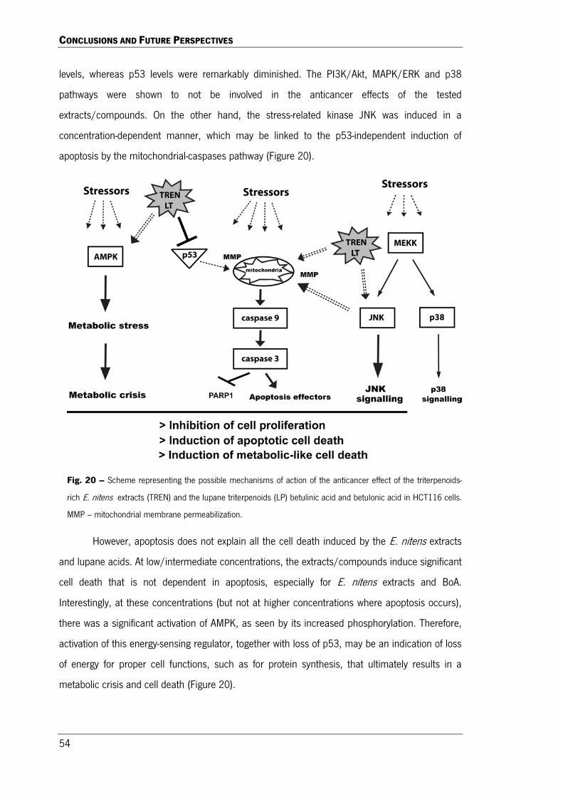
CONCLUSIONS AND FUTURE PERSPECTIVES
54
levels, whereas p53 levels were remarkably diminished. The PI3K/Akt, MAPK/ERK and p38
pathways were shown to not be involved in the anticancer effects of the tested
extracts/compounds. On the other hand, the stress-related kinase JNK was induced in a
concentration-dependent manner, which may be linked to the p53-independent induction of
apoptosis by the mitochondrial-caspases pathway (Figure 20).
However, apoptosis does not explain all the cell death induced by the E. nitens extracts
and lupane acids. At low/intermediate concentrations, the extracts/compounds induce significant
cell death that is not dependent in apoptosis, especially for E. nitens extracts and BoA.
Interestingly, at these concentrations (but not at higher concentrations where apoptosis occurs),
there was a significant activation of AMPK, as seen by its increased phosphorylation. Therefore,
activation of this energy-sensing regulator, together with loss of p53, may be an indication of loss
of energy for proper cell functions, such as for protein synthesis, that ultimately results in a
metabolic crisis and cell death (Figure 20).
Fig. 20 – Scheme representing the possible mechanisms of action of the anticancer effect of the triterpenoids-
rich E. nitens extracts (TREN) and the lupane triterpenoids (LP) betulinic acid and betulonic acid in HCT116 cells.
MMP – mitochondrial membrane permeabilization.

CONCLUSIONS AND FUTURE PERSPECTIVES
55
Summing up, the present results describes the potent anticancer effects of lipophilic
extracts from E. nitens bark in HCT116 colorectal cancer cell, which can be an interesting source
of bioactive natural triterpenoids used as potent anticancer drugs and/or for the semi-synthesis
of new improved and more hydrophilic triterpenoids. Being the bark of Eucalyptus one of the
main byproducts in Portugal from the pulp and paper industry, this work encourages the use of
these residues to extract added-value compounds and to implement the biorefinery concept in
their production chain.
2. FUTURE PERSPECTIVES
With this study it was demonstrated the potential anticancer effects of triterpenoids
isolated from the bark of Eucalyptus nitens. They were able to trigger apoptosis accompanied by
activation of the intrinsic mitochondrial-caspase 9 pathway, a decrease of Bcl-2 levels and the
induction of JNK pathway. In addition, at lower doses, especially BoA was able to induce cell
death independent of apoptosis and accompanied with the induction of AMPK activity and loss of
p53. These novel results arise, however, many questions that remain unanswered that should be
approached in future experiments.
Although the extracts/compounds induced apoptosis and activation of caspases, the
present results do not prove that caspases are directly involved in apoptosis. By using
pharmacological caspases inhibitors, or RNAi against caspases, it will clarify this issue. Also, it
was hypothesised that apoptosis was triggered by JNK through induction of MMP, which could be
depicted by using a pharmacological inhibitor against JNK and by measuring the mitochondrial
membrane potential after treating cells with the E. nitens extracts or triterpenoids.
Because apoptosis was triggered by the triterpenoids and extracts accompanied with
reduction of p53 levels, it may indicate that apoptosis was independent of p53. Making the same
type of experiments in the isogenic HCT116 cells with p53 knockout, or by using RNAi against
p53, would clarify if this tumour suppressor is really necessary for apoptosis induction by tested
extracts/compounds. If this hypothesis is correct it is expected that these kind of compounds will
induce cell death by apoptosis even in resistant cancer cells with p53 mutations, which would be
an interesting source of anticancer drugs for decreasing cancer chemoresistance.

CONCLUSIONS AND FUTURE PERSPECTIVES
56
There are some studies that report the ability of some triterpenoids to induce proteasome
activity, which may explain the decrease in p53 levels in this work and probably other proteins
(like Sp transcription factors), which can lead to a non-apoptotic type of cell death. So, it would
be interesting to measure the proteasome activity in the presence of these extracts/compounds
and to test whether proteasome inhibition would prevent cell death induced by intermediate
doses of BoA. That would explain the kind of “metabolic death” observed in this study due to a
chronic degradation of vital proteins. However, the inhibition of their synthesis could be the cause
of the reduction of these important transcription factors. In fact, AMPK activation, as observed
here, is known to inhibit mTOR and, therefore, inhibit protein synthesis and to induce autophagy.
However, it was recently reported that the triterpenoids UA inhibits autophagy at later steps
(Xavier et al., 2010; Xavier et al., 2012b). If the same happens with BoA, cells would miss the
acquisition of energy block from recycling cellular molecules, which would lead to a metabolic
crisis and, eventually, death. This hypothesis should be explored in further experiments.
To have a broader perspective of its anticancer action, it would also be interesting to
study the effects of the extracts/compounds in the inhibition of angiogenesis.
Also, due to the fact that betulinic and betulonic acids are structurally similar to
cholesterol, those compounds may affect its cellular homeostasis and function, which will affect
vital cellular processes, some already addressed above. So, investigating this relation could be
very valuable.
All these possibilities would help clarify the full potential of the E. nitens bark as source of
potent anticancer compounds, such as betulinic and betulonic acids.

REFERENCES


REFERENCES
59
Amin, A.R.M.R.; Kucuk, O.; Khuri, F.R.; Shin D.M.,
Perspectives for Cancer Prevention with Natural
Compounds. Journal of Clinical Oncology, 2009;
27(6): 2712- 2725.
Anand, P.; Kunnumakkara, A.B.; Sundaram, C.;
Harikumar, K.B.; Tharakan, S.T.; Lai, O.S.; Sung,
B.; Aggarwal, B.B., Cancer is a preventable
disease that requires major lifestyle changes.
Pharmaceutical Research, 2008; 25(9): 2097-
2116.
Ashwell, J.D., The many paths to p38 mitogen-
activated protein kinase activation in the immune
system. Nature Reviews Immunology, 2006; 6(7):
532-540.
Bishayee, A.; Ahmed, S.; Brankov, N.; Perloff, M.,
Triterpenoids as potential agents for the
chemoprevention and therapy of breast cancer.
Frontiers in Bioscience, 2011; 16: 980-996.
Bogoyevitch, M.A.; Ngoei, K.R.; Zhao, T.T.; Yeap, Y.Y;
Ng, D.C., c-Jun N-terminal kinase (JNK)
signalling: recent advances and challenges.
Biochimica et Biophysica Acta, 2010; 1804(3):
463-475.
Chaitanya, G.V; Alexander, J.S.; Babu, P.P., PARP-1
cleavage fragments: signatures of cell-death
proteases in neurodegeneration. Cell
Communication and Signaling, 2010; 8: 31-42.
Chari, N.S.; Pinaire, N.L.; Thorpe, L.; Medeiros, L.J.;
Routbort, M.J.; McDonnell, T.J., The p53 tumor
suppressor network in cancer and the therapeutic
modulation of cell death. Apoptosis, 2009; 14:
336-47.
Chen, Z.; Gibson, T.B.; Robinson, F.; Silvestro, L.;
Pearson G.; Xu, B.; Wright, A.; Vanderbilt, C.;
Cobb, M.H., MAP kinases. Chemical Reviews,
2001; 101: 2449-2476.
Chintharlapalli, S.; Papineni, S.; Lei, P.; Pathi, S.;
Safe, S., Betulinic acid inhibits colon cancer cell
and tumor growth and induces proteasome-
dependent and –independent downregulation of
specificity proteins (Sp) transcription factors. BMC
Cancer, 2011; 11: 371.
Chowdhury, A.R.; Mandal, S.; Mittra, B.; Sharma, S.;
Mukhopadhyay, S.; Majumder, H.K., Betulinic
acid, a potent inhibitor of eukaryotic
topoisomerase I: identification of the inhibitory
step, the major functional group responsible and
development of more potent derivatives. Medical
Science Monitor, 2002; 8 (7): 254-260.
Cichewicz, R.H.; Kouzi, S.A., Chemistry, biological
activity, and chemotherapeutic potential of
betulinic acid for the prevention and treatment of
cancer and HIV infection. Medicinal Research
Reviews, 2004; 1: 90-114.
Cobb, M.H., MAP kinases pathways. Progress in
Biophysics and Molecular Biology, 1999; 71(3-4):
479-500.
Coller, H.A.; Sang, L.; Roberts, J.M., A new
description of cellular quiescence. PLOS Biology,
2006; 4 (3): 329-349.
Csuk, R.; Barthel, A.; Sczepek, R.; Siewert. B.;
Schwarz, S., Synthesis, encapsulation and
antitumor activity of new betulin derivatives. Archiv
der Pharmazie – Chemistry in Life Sciences,
2011; 1: 37-49.
Cuevas, B.D.; Abell, A.N.; Johnson, G.L., Role of
mitogen-activated protein kinase kinases in signal
integration. Oncogene, 2007; 26(22): 3159-3171.
Deniaud, E.; Baguet, J.; Chalard, R.; Blanquier, B.;
Brinza, L.; Meunier, J.; Michallet, M.C.; Laugraud,
A.; Ah-Soon, C.; Wierinckx, A.; Castellazzi, M.;
Lachuer, J.; Gautier, C.; Marvel, J.; Leverrier, Y.,
Overexpression of transcription factor Sp1 leads to
gene expression perturbations and cell cycle
inhibition. PLOS One, 2009; 4(9): 1-13.
Dhanasekaran, D.N.; Reddy, E.P., JNK signalling
apoptosis. Oncogene, 2011; 27(48): 6245-6251.

REFERENCES
60
Drag, M.; Surowiak, P.; Drag-Zalesinska, M.; Dietel,
M.; Lage, H.; Oleksyszyn, J., Comparison os the
cytotoxic effects of birch bark extract, betulin and
betulinic acid towards human gastric carcinoma
and pancreatic carcinoma drug-sensitive and drug-
resistant cell lines. Molecules, 2009; 14: 1639-
1651.
Domingues, R.M.A.; Oliveira, E.L.G.; Freire, C.S.R.;
Couto, R.M.; Simões, P.C.; Pascoal Neto, C.;
Silvestre, A.J.D.; Silva, C.S., Supercritical fluid
extraction of Eucalyptus globulus bark- a
promising approach for triterpenoids production.
International Journey of Molecular Sciences,
2012; 13(6): 7648-7662.
Domingues, R.M.A.; Patinha, D.J.S., Sousa, G.D.A.;
Villaverde, J.J.; Silva, C.M.; Freire, C.S.R.;
Silvestre, A.J.D., Pascoal Neto, C., Eucalyptus
biomass residues from agro-forest and pulping
industris as sources of high-value triterpenic
compounds. Cellulose Chemistry and Technology,
2011a.
Domingues, R.M.A.; Sousa G.D.A.; Freire, C.S.R.;
Silvestre, A.J.D.; Pascoal Neto, C., Eucalyptys
globulus biomass residues from pulping industry
as a source of high value triterpenic compounds.
Industrial Crops and Products, 2010; 31: 65-70.
Domingues, R.M.A.; Sousa, G.D.A.; Silva, C.M.;
Freire, C.S.R.; Silvestre, A.J.D.; Pascoal Neto, C.,
High value triterpenic compounds from the outer
bark of several Eucalyptus species cultivated in
Brazil and in Portugal. Industrial Crops and
Products, 2011b; 33: 158-164.
Duarte, C.C.L.; Oleanolic acid but not ursolic acid
induces cell death in HepG2 cells under
starvation-induced autophagy. Master Thesis,
2012.
Dzubak, P.; Hajduch, M.; Vydra, D.; Hustova, A.;
Kvasnica, M.; Biedermann, D.; Markova, L.;
Urban, M.; Sarek, J., Pharmacological activities of
natural triterpenoids and their therapeutic
implications. Natural Products Reports, 2006; 23:
394-411.
Ehrhardt, H.; Fulda, S.; Fuhrer M.; Debatin, K.M.;
Jeremias, I., Betulinic acid-induced apoptosis in
leukemia cells. Nature Publishing Group, 2004;
18: 1406-1412.
Eichenmuller, M.; Hemmerlein, B.; Schweinitz, D.;
Kappler, R., Betulinic acid induces apoptosis and
inhibits hedgehog signalling in
rhabdomyasarcoma. British Journal of Cancer,
2010; 103: 43-51.
Eichenmuller, M.; Schweinitz, D.; Kappler, R.,
Betulinic acid treatment promotes apoptosis in
hepatoblastoma cells. International Journal of
Oncology, 2009; 35: 873-879.
Elmore, S., Apoptosis: a review of programmed cell
death. Toxicologic Pathology, 2007; 35(4): 495-
516.
Fulda, S., Betulinic acid for cancer treatment and
prevention. International Journal of Molecular
Sciences, 2008; 9: 1096-1107.
Fulda, S.; Debatin, K.M., Betulinic acid induces
apoptosis through a direct effect on mitochondria
in neuroectodermal tumors. Medical and Pediatric
Oncology, 2000; 35: 616-618.
Fulda, S.; Friesen, C.; Los, M.; Scaffidi, C.; Mier, W.;
Benedict, M.; Nuñez, G.; Krammer, P.H.; Peter,
M.E.; Debatin, K.M., Betulinic acid triggers CD95
(Apo-1/Fas) and p53-independent apoptosis via
activation of caspases in neuroectodermal tumors.
Cancer Research, 1997; 57: 4956-4964.
Fulda, S.; Kroemer, G., Targeting mitochondrial
apoptosis by betulinic acid in human cancers.
Drug Discovery Today, 2009; 14: 17-18.
Hanahan, D.; Weinberg, R.A., The Hallmarks of
Cancer. Cell Press, 2000; 100: 57-70.
Hanahan, D.; Weinberg, R.A., Hallmarks of cancer:
the next generation. Cell, 2011; 144: 646-674.

REFERENCES
61
Huang, L.; Ho, P.; Chen, C., Activation and inhibition
of proteasomes by betulinic acid and its
derivatives. FEBS Letters, 2007; 581(25): 4955-
4959.
Inoki, K.; Kim, J.; Guan, K., AMPK and mTOR in
cellular energy homeostasis and drug targets.
Annual Review of Pharmacology and Toxicology,
2012. 52: 381-400.
Irish, J.;Blair, S.; Carter, D.A., The antibacterial
activity of honey derived from Australian flora.
PLoS One, 2011; 6(3): 18229.
Islam, F.; Khatun, H.; Ghosh, S.; Ali, M.M.; Khanam,
J.A., Bioassay of Eucalyptus extracts for anticancer
activity against Ehrlich ascites carcinoma (eac)
cells in Swiss albino mice. Asian Pacific Journal of
Tropical Biomedicine, 2012; 394-398.
Jung, G.R.; Kim, K.J.; Choi, C.H.; Lee, T.B; Han, S.I.;
Han, H.K.; Lim, S.C., Effect of betulinic acid on
anticancer drug-resistant colon cancer cells. Basic
and Clinical Pharmacology & Toxicology, 2007;
101: 277-285.
Kibblehite, P.; Johnson, B.I.; Shelbourne, C.J.A.,
Kraft pulp qualities of Eucalyptus nitens, E.
globulus, and E.maidenii, at ages 8 and 11 years.
New Zealand Journal of Forestry Science, 2000;
30(3): 447-457.
Kim, J.Y.; Koo, H.M.; Kim, D.S.H.L., Development of
C-20 modified betulinic acid derivatives as
antitumor agents. Bioorganic & Medicinal
Chemistry Letters , 2001; 2405-2408.
Kim, J.; Lee, Y.S.; Kim, C.S.; Kim, J.S., Betulinic acid
has an inhibitory effect on pancreatic lipase and
induces adipocyte lipolysis. Phytotherapy
Research, 2011; 3672.
Kim, T.D.; Oh, S.; Shin, S.; Janknecht, R., Regulation
of tumor suppressor p53 and HCT116 cell
physiology by histone demethylase
JMJD2D/KDM4D. PLOS One, 2012; 7(4): 1-8.
Kolch, W., Meaningful relationships: the regulation of
the Ras/Raf/MEK/ERK pathway by protein
interactions. Biochemical Journal, 2000; 351:
289-305.
Kommera, H.; Kaluderovic, G.N.; Bette, M.; Kalbitz,
J.; Fuchs, P.; Fulda, S.; Mier, W.; Paschke, R., In
vitro anticancer studies of α- and β-D-
glucopyranose betulin anomers. Chemico-
Biological Interactions, 2010a; 185: 128-136.
Kommera, H.; Kaluderovic, G.N.; Kalbitz, J.; Drager,
J.; Paschke, R., Small structural changes of
pentacyclic lupane type triterpenoids derivatives
lead to significant differences in their anticancer
properties. European Journal of Medicinal
Chemistry, 2010b; 45: 3346-3353.
Kommera, H.; Kaluderovic, G.N.; Kalbitz J.; Paschke,
R., Lupane triterpenoids – betulin and betulinic
acid derivatives induce apoptosis in tumor cells.
Invest New Drugs, 2011; 29: 266-272.
Lima, C.F.; Pereira-Wilson, C.; Rattan, S.I., Curcumin
induces heme oxygenase-1 in normal human skin
fibroblasts through redox signalling: relevance for
anti-aging interventiom. Molecular Nutrition &
Food Research, 2011; 55: 430-442.
Liu, J., Pharmacology of oleanolic acid and ursolic
acid. Journal of Ethnopharmacology, 1995; 40(2):
57-68.
Liu, W.K.; Ho, J.C.K.; Cheung F.W.K.; Liu, B.P.L.; Ye,
W.C.; Che, C.T., Apoptotic activity of betulinic acid
derivatives on murine melanoma B16 cell line.
European Journal of Pharmacology, 2004; 498:
71-78.
Ma, C.M.; Cai, S.Q.; Cui, J.R.; Wang, R.Q.; Tu, P.F.;
Hattori, M.; Daneshtalab, M., The cytotoxic activity
of ursolic acid derivatives. European Journal of
Medicine Chemistry, 2005; 40(6): 582-589.
McCubrey, J.A.; Steelman, L.S.; Abrams, S.L.; Lee,
T.J.; Chang, F.; Bertrand, F.E.; Navolanic, P.M.;
Terrian, D.M.; Franklin, R.A.; D’Assoro, A.B.;

REFERENCES
62
Salisbury, J.L.; Mazzarino, M.C.; Stivala, F.; Libra,
M., Roles of the RAF/MEK/ERK and
PI3K/PTEN/AKT pathways in malignant
transformation and drug resistance. Advances in
Enzyme Regulation, 2006; 46: 249-279.
Melnikova, N; Burlova, I.; Kiseleva, T.; Klabukova, I.;
Gulenova, M.; Kislitsin, A.; Vasin, V.; Tanaseichuk,
B., A practical synthesis of betulonic acid using
selective oxidation of betulin on aluminium solid
support. Molecules, 2012; 17: 11849-11863.
Mota, I.; Pinto, P.C.R.; Novo, C.; Sousa, G.;
Guerreiro, O.; Guerra, A.R.; Duarte, M.F.;
Rodrigues, A.E., Extraction of polyphenolic
compounds from Eucalyptus globulus bark:
process optimization and screening for biological
activity. Industrial & Engineering Chemistry
Research, 2012; 51: 6991-7000.
Moura, J.C.M.S.; Araújo, P.; Brito, M.S.; Souza, U.R.;
Viana, J.O.F.; Mazzafera, P., Validation of
reference genes from Eucalyptus spp. under
different stress conditions. Research Notes, 2012;
5: 634.
Nakagawa-Goto, K.; Yamada, K.; Taniguchi, M.;
Tokuda, H.; Lee, K., Cancer preventive agentes 9.
Betulinic acid derivatives as potent cancer
chemopreventive agents. Biooroganic & Medicinal
Chemistry Letters, 2009; 19 (13): 3378-3381.
Orton, R.J.; Sturm, O.E.; Vyshemirsky, V.; Calder, M.;
Gilbert, D.R.; Kolch, W., Computational modelling
of the receptor.tyrosine-kinase-activated MAPK
pathway. Biochemical Journal, 2005; 392(2): 249-
261.
Pavlova, N.I.; Savinova, O.V.; Nikolaeva, S.N.;
Boreko, E.I.; Flekhter, O.B., Antiviral activity of
betulin, betulinic and betulonic acids against some
enveloped and non-enveloped viruses. Fitoterapia,
2003; 74: 489-492.
Pearson, G.; Robinson, F.; Gibson, T.B.; Exu, B.;
Karandikar, M.; Berman K.; Cobb, M.H., Mitogen-
activated protein (MAP) kinase pathways:
regulation and physiological functions. Endocrine
Reviews, 2001; 22(2): 153-183.
Pereira, S.I.; Freire, C.S.R.; Pascoal Neto, C.;
Silvestre, A.J.D.; Silva, A.M.S., Chemical
composition of the epicular wax from the fruits of
Eucalyptus globulus. Phytochemical Analysis,
2005; 16: 364-369.
Pisha, E.; Chai, H.; Lee, I.S.; Chagwedera, T.E.;
Farnsworth, N.R.; Cordell, G.A.; Beecher, C.W.;
Fong, H.H.; Kinghorn, A.D.; Brown, D.M.,
Discovery of betulinic acid as a selective inhibitor
of human melanoma that functions by induction of
apoptosis. Natural Medicine, 1995; 1: 1046.
Rajendram, P.; Jaggi, M.; Singh, M.K.; Mukherjee, R.;
Burman, A.C., Pharmacological evaluation of C-3
modified betulinic acid derivatives with potent
anticancer activity. Invest New Drugs, 2008; 26:
25-34.
Ramos, A.A.; Lima, C.F.; Pereira, M.L.; Fernandes-
Pereira, M.; Pereira-Wilson, C., Antigenotoxic
effects of quercetin, rutin and ursolic acid on
HepG2 cells: evaluation by the comet assay.
Toxicology Letters, 2008; 177: 66-73.
Ramos, A.A.; Pereira-Wilson, C.; Collins, A.R.,
Protective effects of ursolic acid and luteolin
against oxidative DNA damage include
enhancement of DNA repair in Caco-2 cells.
Mutation Research, 2010; 692: 6-11.
Rieger, A.M.; Nelson, K.L.; Konowalchuk, J.D.;
Barreda, D.R., Modified annexin v/propidium
iodide apoptosis assay for accurate assessment of
cell death. Journal of Visualized Experiments,
2011; 50: 2597.
Russell, A.B., Hardin, J.W.; Grand, L.; Fraser, A.,
Poisonous plants of North Carolina. 1997.
Rustgi, A.K., The genetics of hereditary colon cancer.
Genes & Development, 2007; 21: 2525-2538.
REFERENCES
63
Rzeski, W.; Stepulak, A.; Szymanski, M.; Sifringer,
M.; Kaczor, J.; Wejksza, K.; Zdzisinska, B.;
Kandefer-Szerszen, M., Betulinic acid decreases
expression of bcl-2 and cyclin D1, inhibits
proliferation, migration and induces apoptosis in
cancer cells. Naunyn-Schmiedeberg’s Archives of
Pharmacology, 2006; 374: 11-20.
Ryu, S.Y.; Choi, S.U.; Lee, S.H.; Lee, C.O.; No, Z.;
Ahn, J.W., Antitumor triterpenes from medicinal
plants. Archives of Pharmacals Research, 1994;
17(5): 375-377.
Santos, R.C.; Salvador, J.A.R.; Cortés, R.; Pachón,
G.; Marín, S.; Cascante, M., New betulinic acid
derivatives induce potent and selective
antiproliferative activity through cell cycle arrest at
the S phase and caspase dependente apoptosis in
human cancer cell. Biochemie, 2011; 93: 1065-
1075.
Santos, S.A.O.; Freire, C.S.R.; Domingues, M.R.M.;
Silvestre, A.J.D.; Pascoal Neto, C.,
Characterization of phenolic components in polar
extracts of Eucalyptus globulus Labill. bark by high
performance liquid chromatography mass
spectrometry. Journal of Agricultural and Food
Chemistry, 2011b.
Saxena, B.B.; Zhu, L.; Hao, M.; Kisilis, E.; Katdare,
M.; Oktem, O.; Bomshteyn, A.; Rathnam, P., Boc-
lysinated-betulonic acid: a potent, anti-prostate
cancer agent. Bioorganic & Medicinal Chemistry,
2006; 14: 6349-6358.
Semenove, D.E.; Zhukova, N.A.; Bessergeneva, E.P.;
Sorokina, I.V.; Baev, D.S.; Glukhov, B.M.,
Nepomnyaschikh G.I.; Tolstikova , T.G., Effect of
triterpene derivatives on the total hepatocyte count
in the liver of rats with toxic hepatitis. Bulletin of
Experimental Biology and Medicine, 2012; 153(6):
858-861.
Shintyapina, A.B.; Shults, E.E.; Petrenko, N.I.;
Uzenkova, N.V.; Tolstikov, G.A.; Pronkina, N.V.;
Kozhevnikov, V.S.; Pokrovsky, A.G., Effect of
nitrogen-containing derivatives of the plant
triterpenes betulin and glycyrrhetic acid on the
growth of MT-4, MOLT-4, CEM and HepG2 tumor
cells. Russian Journal of Bioorganic Chemistry,
2007; 33(6): 579-583.
Siddiqui, B.S.; Sultana, I.; Begum, S., Triterpenoidal
constituents from Eucalyptus camaldulensis var.
obtusa leaves. Phytochemistry, 2000; 54: 861-
865.
Soica, C.M.; Dehelean, C.A.; Peev, C.; Aluas, M.;
Zupko, I.; Kása Jr, P.; Alexa, E., Physico-chemical
comparison of betulinic acid, betulin and birch
bark extract and in vitro investigation of their
cytotoxic effects towards skin epidermoid
carcinoma (A431), breast carcinoma (MCF7) and
cervix adenocarcinoma (HeLa) cell lines. Natural
Product Research, 2011; 1-7.
Soreide, K.; Janssen, E.A.; Soiland, H.; Korner, H.;
Baak, J.P., Microsatellite instability in colorectal
cancer. British Journal of Surgery, 2006; 93: 395-
406.
Sorokina, I.V.; Tolstikova, T.G.; Zhukova, N.A.;
Petrenko, N.I.; Schults, E.E.; Uzenkova, N.V.;
Grek, O.R.; Pozdenyakova, S.V.; Tolstikov, G.A.,
Betulonic acid and derivatives, a new group of
gents reducing side effects of cytostatics. Doklady
Biological Sciences, 2004; 399: 434-437.
Sorokina, I.V.; Tolstikova, T.G.; Zhukova, N.A.;
Petrenko, N.I.; Uzenkova, N.V.; Schults, E.E.;
Popova, N.A., Antitumor and antimetastatic effects
of BoA amides in mice with transplanted lewis
carcinoma. Bulletin of Experimental Biology and
Medicine, 2006; 142 (1): 69-72.
Souglakos, J., Genetic alterations in sporadic and
hereditary colorectal cancer: implementations for
screening and follow-up. Digestive Diseases,
2007; 25: 9-19.
REFERENCES
64
Symon, A.V.; Veselova, N.N.; Kaplun, A.P.;
Vlasenkova, N.K.; Fedorova, G.A.; Lyutik, A.I.;
Gerasimova, G.K.; Shvets, V.I., Synthesis and
antitumor activity of cyclopropane derivatives of
betulinic and betulonic acids. Russian Journal of
Bioorganic Chemistry, 2005; 31 (3): 286-291.
Takami, K.; Yana, I.; Kurahashi, H.; Nishisho, I,
Multistep carcinogenesis in colorectal cancers.
Southeast Asian Journal of Tropical Medicine and
Public Health, 1995; 1: 190-196.
Tibbits, W.N.; Boomsma, D.B.; Jarvis, S.,
Distribution, biology, genetics and improvement
programs for Eucalyptus globulus and E.nitens
around the world. 1997.
Trumbull, E.R.; Bianchi, E.; Eckert, D.J.; Wiedhopf, R.M.;
Cole, J.R., Tumor inhibitory agents from Vauquelinia
corymbosa (Rosaceae). Journal of Pharmaceutical
Science, 1976; 65: 1407.
Vabilevsky, S.F.; Govdi, A.I.; Shults, E.E.; Shakinov
M.M.; Sorokina I.V.; Tolstikova, T.G.; Baev, D.S:;
Tolstikov, G.A., Alabugin, I.V., Efficient synthesis of
the first betulonic acid-acetylene hybrids and their
hepatoprotective and anti-inflammatory activity.
Bioorganic & Medicinal Chemistry, 2009; 17(14):
5164-5169.
Vaseva, A.V.; Moll, U.M., The mitochondrial p53
pathway. Biochima Biophysa Acta, 2009; 1787:
414-420.
Wada, S.; Tanaka, R., Betulinic acid and its
derivatives, potent DNA topoisomerase II
inhibitors, from the bark of Bischofia javanca.
Chemistry and Biodiversity, 2005; 2: 689-693.
Wagner, E.F,; Nebreda, A.R., Signal integration by
JNK and p38 MAPK pathways in cancer
development. Nature Reviews Cancer, 2009; 9(8):
537-549.
Weston, C.R.; Davis, R.J., The JNK signal
transduction pathway. Current Opinion in Cell
Biology, 2007; 19: 142-149.
Wu, Q.; He, J; Fang, J; Hong, M., Antitumor effect of
betulinic acid on human acute leukemia K562
cells in vitro. Medical Sciences, 2010; 30(4): 453-
457.
Xavier, C.P.R., The anticarcinogenic potential of
dietary natural compounds on colorectal
carcinoma: effects on signalling pathways related
to proliferation and death. PhD Thesis, 2010.
Xavier, C.P.R.; Lima, C.F.; Fernandes-Ferreira, M.;
Pereira-Wilson, C., Salvia fruticosa, Salvia
officinalis and rosmarinic acid induce apoptosis
and inhibit proliferation of human colorectal cell
lines: the role in MAPK/ERK pathway. Nutrition
and Cancer, 2009a; 61(4): 564-571.
Xavier, C.P.R.; Lima, C.F.; Fernandes-Ferreira, M.;
Pereira-Wilson, C., Hypericum androsaemum
water extract inhibits proliferation in human
colorectal cancer cell through effects on MAP
kinases and PI3K/Akt pathway. Food & Function,
2012; 3: 844-852.
Xavier, C.P.R.; Lima, C.F.; Pedro, D.F.N.; Wilson,
J.M.; Kristiansen, K.; Pereira-Wilson, C., Ursolic
acid induces cell death and modulates autophagy
through JNK pathway in apoptosis-resistant
colorectal cancer cells. Journal of Nutritional
Biochemistry, 2013.
Xavier, C.P.R.; Lima, C.F.; Preto, A.; Seruca, R.;
Fernandes-Ferreira, M.; Pereira-Wilson, C.,
Luteolin, quercetin and ursolic acids are potent
inhibitors of proliferation and inducers of apoptosis
in both KRAS and BRAF mutated human colorectal
cancer cells. Cancer Letters, 2009b; 281: 162-
170.
Xavier, C.P.R.; Lima, C.F.; Rohde, M.; Pereira-Wilson,
C., Quercetin enhances 5-fluorouracil-induced
apoptosis in MSI colorectal cancer cells through
p53 modulation. Cancer Chemotherapy
Pharmacology, 2011; 1641-1649.

REFERENCES
65
Yadav, V.R.; Prasad, S.; Sung, B.; Kannappan, R.;
Aggarwal, B.B., Targeting inflammatory pathways
by triterpenoids for prevention and treatment of
cancer. Toxins, 2010; 2: 2428-2466.
Yi, J.; Obminska-Mrukowics, B.; Yuan, L.; Yuan, H.
Immunomodulatory effects of betulinic acid
from84 the bark of white birch on mice. Journal of
Veterinary Science, 2010; 11(4): 305-313.
Zarubin, T.; Han, J., Activation and signalling of the
p38 MAP kinase pathway. Cell Research, 2005;
15(1): 11-18.
Zhang, X.J.; Han, L.; Ji, Y.B.; Fang, G.Z., Studies of
betulonic acid on cell cycle and related proteins
expressions on mice of bearing H22 tumor cells.
Zhongguo Zhong Ya Za Zhi, 2008; 33(14): 1739-
1743.
Zhang, Y.; Wang, J.; Yuan, Y.; Zhang, W.; Guan, W.;
Wu, Z.; Jin, C.; Chen, H.; Zhang, L.; Yang, X.; He,
F., Negative regulation of HDM2 to attenuate p53
degradation by ribosomal protein L26. Nucleic
Acids Research, 2010; 38(19): 6544-6554.
Zuco, V.; Supino, R.; Righetti, S.C.;Cleris, L.;
Marchesi, E.; Gambacorti-Passerine, C.; Formelli,
F., Selective cytotoxicity of betulinic acid on tumor
cell lines, but not on normal cells. Cancer Letters,
2002; 175: 17-25.