Klebsiella virus UPM2146 lyses multiple drug-resistant ...
19
RESEARCH ARTICLE Klebsiella virus UPM2146 lyses multiple drug- resistant Klebsiella pneumoniae in vitro and in vivo Omar Assafiri ID 1 , Adelene Ai-Lian Song 1 , Geok Hun Tan 2,3 , Irwan Hanish 1 , Amalia Mohd Hashim 1,4 , Khatijah Yusoff 1,3 * 1 Faculty of Biotechnology and Biomolecular Sciences, Department of Microbiology, Universiti Putra Malaysia, Serdang, Selangor, Malaysia, 2 Faculty of Agriculture, Department of Agriculture Technology, Universiti Putra Malaysia, Serdang, Selangor, Malaysia, 3 Institute of Biosciences, Universiti Putra Malaysia, Serdang, Selangor, Malaysia, 4 Halal Products Research Institute, Universiti Putra Malaysia, Serdang, Selangor, Malaysia * [email protected] Abstract Klebsiella pneumoniae are opportunistic bacteria found in the gut. In recent years they have been associated with nosocomial infections. The increased incidence of multiple drug-resis- tant K. pneumoniae makes it necessary to find new alternatives to treat the disease. In this study, phage UPM2146 was isolated from a polluted lake which can lyse its host K. pneumo- niae ATCC BAA-2146. Observation from TEM shows that UPM2146 belongs to Caudovir- iales (Order) based on morphological appearance. Whole genome analysis of UPM2146 showed that its genome comprises 160,795 bp encoding for 214 putative open reading frames (ORFs). Phylogenetic analysis revealed that the phage belongs to Ackermannviri- dae (Family) under the Caudoviriales. UPM2146 produces clear plaques with high titers of 10 10 PFU/ml. The phage has an adsorption period of 4 min, latent period of 20 min, rise period of 5 min, and releases approximately 20 PFU/ bacteria at Multiplicity of Infection (MOI) of 0.001. UPM2146 has a narrow host-range and can lyse 5 out of 22 K. pneumoniae isolates (22.72%) based on spot test and efficiency of plating (EOP). The zebrafish larvae model was used to test the efficacy of UPM2146 in lysing its host. Based on colony forming unit counts, UPM2146 was able to completely lyse its host at 10 hours onwards. Moreover, we show that the phage is safe to be used in the treatment against K. pneumoniae infections in the zebrafish model. Introduction Klebsiella pneumoniae is a Gram-negative bacterium which can colonize the gastrointestinal tract, lungs, and leads to liver abscesses, pneumoniae, and bacteremia in human [1]. It belongs to the ESKAPE group (Enterococcus faecium, Staphylococcus aureus, K. pneumoniae, Acineto- bacter baumannii, Pseudomonas aeruginosa, and Enterobacter species) which is responsible for many nosocomial infections in hospitals [2]. The number of these drug-resistant bacteria is on the rise due to the spread of plasmids expressing the β-lactamase enzyme which renders PLOS ONE PLOS ONE | https://doi.org/10.1371/journal.pone.0245354 January 8, 2021 1 / 19 a1111111111 a1111111111 a1111111111 a1111111111 a1111111111 OPEN ACCESS Citation: Assafiri O, Song AA-L, Tan GH, Hanish I, Hashim AM, Yusoff K (2021) Klebsiella virus UPM2146 lyses multiple drug-resistant Klebsiella pneumoniae in vitro and in vivo. PLoS ONE 16(1): e0245354. https://doi.org/10.1371/journal. pone.0245354 Editor: Yung-Fu Chang, Cornell University, UNITED STATES Received: October 1, 2020 Accepted: December 28, 2020 Published: January 8, 2021 Copyright: © 2021 Assafiri et al. This is an open access article distributed under the terms of the Creative Commons Attribution License, which permits unrestricted use, distribution, and reproduction in any medium, provided the original author and source are credited. Data Availability Statement: All relevant data are within the paper and its Supporting information files. Funding: Universiti Putra Malaysia grant number GP/IPS/2018/9602400. Competing interests: The authors have declared that no competing interests exist.
Transcript of Klebsiella virus UPM2146 lyses multiple drug-resistant ...

RESEARCH ARTICLE
Klebsiella virus UPM2146 lyses multiple drug-
resistant Klebsiella pneumoniae in vitro and in
vivo
Omar AssafiriID1, Adelene Ai-Lian Song1, Geok Hun Tan2,3, Irwan Hanish1, Amalia
Mohd Hashim1,4, Khatijah Yusoff1,3*
1 Faculty of Biotechnology and Biomolecular Sciences, Department of Microbiology, Universiti Putra
Malaysia, Serdang, Selangor, Malaysia, 2 Faculty of Agriculture, Department of Agriculture Technology,
Universiti Putra Malaysia, Serdang, Selangor, Malaysia, 3 Institute of Biosciences, Universiti Putra Malaysia,
Serdang, Selangor, Malaysia, 4 Halal Products Research Institute, Universiti Putra Malaysia, Serdang,
Selangor, Malaysia
Abstract
Klebsiella pneumoniae are opportunistic bacteria found in the gut. In recent years they have
been associated with nosocomial infections. The increased incidence of multiple drug-resis-
tant K. pneumoniae makes it necessary to find new alternatives to treat the disease. In this
study, phage UPM2146 was isolated from a polluted lake which can lyse its host K. pneumo-
niae ATCC BAA-2146. Observation from TEM shows that UPM2146 belongs to Caudovir-
iales (Order) based on morphological appearance. Whole genome analysis of UPM2146
showed that its genome comprises 160,795 bp encoding for 214 putative open reading
frames (ORFs). Phylogenetic analysis revealed that the phage belongs to Ackermannviri-
dae (Family) under the Caudoviriales. UPM2146 produces clear plaques with high titers of
1010 PFU/ml. The phage has an adsorption period of 4 min, latent period of 20 min, rise
period of 5 min, and releases approximately 20 PFU/ bacteria at Multiplicity of Infection
(MOI) of 0.001. UPM2146 has a narrow host-range and can lyse 5 out of 22 K. pneumoniae
isolates (22.72%) based on spot test and efficiency of plating (EOP). The zebrafish larvae
model was used to test the efficacy of UPM2146 in lysing its host. Based on colony forming
unit counts, UPM2146 was able to completely lyse its host at 10 hours onwards. Moreover,
we show that the phage is safe to be used in the treatment against K. pneumoniae infections
in the zebrafish model.
Introduction
Klebsiella pneumoniae is a Gram-negative bacterium which can colonize the gastrointestinal
tract, lungs, and leads to liver abscesses, pneumoniae, and bacteremia in human [1]. It belongs
to the ESKAPE group (Enterococcus faecium, Staphylococcus aureus, K. pneumoniae, Acineto-bacter baumannii, Pseudomonas aeruginosa, and Enterobacter species) which is responsible for
many nosocomial infections in hospitals [2]. The number of these drug-resistant bacteria is
on the rise due to the spread of plasmids expressing the β-lactamase enzyme which renders
PLOS ONE
PLOS ONE | https://doi.org/10.1371/journal.pone.0245354 January 8, 2021 1 / 19
a1111111111
a1111111111
a1111111111
a1111111111
a1111111111
OPEN ACCESS
Citation: Assafiri O, Song AA-L, Tan GH, Hanish I,
Hashim AM, Yusoff K (2021) Klebsiella virus
UPM2146 lyses multiple drug-resistant Klebsiella
pneumoniae in vitro and in vivo. PLoS ONE 16(1):
e0245354. https://doi.org/10.1371/journal.
pone.0245354
Editor: Yung-Fu Chang, Cornell University, UNITED
STATES
Received: October 1, 2020
Accepted: December 28, 2020
Published: January 8, 2021
Copyright: © 2021 Assafiri et al. This is an open
access article distributed under the terms of the
Creative Commons Attribution License, which
permits unrestricted use, distribution, and
reproduction in any medium, provided the original
author and source are credited.
Data Availability Statement: All relevant data are
within the paper and its Supporting information
files.
Funding: Universiti Putra Malaysia grant number
GP/IPS/2018/9602400.
Competing interests: The authors have declared
that no competing interests exist.

antibiotics that has lactam enzyme as a functional group useless (NDM-1) [3]. Furthermore,
the number of new antibiotic groups being discovered has dwindled down drastically com-
pared to the rates in which bacteria gain resistance towards the antibiotics [4]. Thus, it is neces-
sary to search for an alternative to treatments using antibiotics such as phage therapy. For
instance, phage cocktail (Kp152, Kp154, Kp155, Kp164, Kp6377, and HD001) with an inacti-
vated sulfamethoxazole-trimethoprim was able to protect a patient from K. pneumoniae Uri-
nary Tract Infection (UTI) [5].
Bacteriophages are highly selective in infecting and lysing their bacterial hosts [6]. In
addition, they exhibit far fewer side effects compared to the antibiotics [7]. At least 70 K. pneu-moniae phages have been isolated and characterized to date [8]. However, very few K. pneumo-niae phages have been studied in vivo using mouse, rat and pig models [9–13]. Apart from
these animals, the virulence of K. pneumoniae has also been evaluated in other non-conven-
tional models such as amoeba and zebrafish to mitigate limitations in the mouse, rat and pig
models such as high cost, specialized facilities and ethical concerns [8]. It has been shown
that only certain K. pneumoniae strains were able to infect zebrafish and cause mortality. In
another study, it was shown that a bioactive molecule from Streptomyces sp. was able to reduce
mortality of zebrafish infected with K. pneumoniae [14]. However, there are no publications
on phage therapy against K. pneumoniae using the zebrafish larvae model although this has
been studied for other pathogens such as Vibrio anguillarum [15] and Enterococcus faecalis[16]. In the present study, UPM2146 was isolated, characterized, and tested for its efficacy
against K. pneumoniae 2146 in vivo using a zebrafish larvae model.
Material and methods
Bacterial strains and experimental conditions
Multiple drug-resistant Klebsiella pneumoniae ATCC BAA-2146 was used as the host for
UPM2146. K. pneumoniae IMR-1 (non-drug resistant strain), multiple drug-resistant ATCC
BAA-1705 and clinical isolates A01 until A18, and A20 [17], multi-drug resistant Staphylococ-cus aureus 10, and multi-drug resistant Escherichia coli ATCC 25922 and E. coli Top 10 (non-
drug resistant) were also used in this study. Dr Erin Lim of Perdana University generously
gifted K. pneumoniae IMR-1, ATCC BAA-2146, ATCC BAA-1705 and Escherichia coli ATCC
25922. Clinical isolates K. pneumoniae A01-A018 and A20 were gifted by Dr Siti Norbaya of
Faculty of Medicine and Health Sciences, Universiti Putra Malaysia. The following experimen-
tal condition has been applied for all bacteria throughout the experiments unless stated other-
wise. A purified single colony was taken from LB agar plate and re-grown overnight in LB
broth with shaking at 37˚C. On the next day, the bacteria were grown to log phase [5 × 10⁸ col-
ony-forming unit (CFU)/ml] at an OD600nm = 0.6.
Isolation, enrichment and purification of phage
UPM2146 was isolated from a polluted lake using K. pneumoniae 2146 as host. Approximately
50 ml of the lake water was centrifuged for 10 min at 6000 × g, 4˚C. The supernatant was fil-
tered using a 0.45 μm membrane filter. In order to enrich the phages, 100 μl of K. pneumoniae2146 (OD600nm = 0.6) in 0.1 M CaCl2 were added to 10 ml of the filtered supernatant before
being made up to 30 ml with 5 × LB broth (final concentration of 3.33× LB) (Merck, Germany)
and shaken overnight at 37˚C. The mixture was centrifuged for 10 min at 8000 × g at 4˚C. The
supernatant was filtered again. The presence of phage was confirmed through spotting onto a
soft overlay agar containing the bacteria. Phages resulting in a positive spot test were then sub-
jected to dilution and plated using the double-layer agar method [18]. Single plaques formed
on plates were cut from the double layer agar and placed back into SM buffer [100 mM NaCl,
PLOS ONE Effectiveness of bacteriophage UPM2146 in lysing Klebsiella pneumoniae in vivo
PLOS ONE | https://doi.org/10.1371/journal.pone.0245354 January 8, 2021 2 / 19
8 mM MgSO4� 7 H2O, 50 mM Tris-Cl (1 M, pH 7.5), and 0.01 g of gelatin)] overnight at 4˚C,
filtered and subjected to double-layer agar again the next day. The same process was repeated
three times to ensure purified phages were obtained which showed uniformed distribution of
plaques per plate. High-titered phage was prepared from plates flooded with SM buffer, which
was centrifuged and filtered. The filtrate was precipitated using 1.5 mM of NaCl and 12% of
PEG-8000 to phage lysate and left at 4˚C overnight. On the next day, the lysate was centrifuged
and the pellet was resuspended in 1 ml of sterile SM buffer with 1 ml of 1 M KCl. After being
left on ice for 1 h, the mixture was re-centrifuged at the same speed and temperature. The
supernatant which consisted of purified concentrated phage was stored at 4˚C.
Adsorption rate of isolated phages
Determination of the adsorption rate of phage was performed according to Hyman & Abedon
[19]. Mid log phase host bacteria (OD600nm = 0.6) were infected with a (1 × 10¹⁰ PFU/ml) of
UPM2146 at MOI of 0.01 at 37˚C. After infection, the phage titers at every 2 min for 10 min
were determined. The samples (30 ml) were centrifuged at 5 000 × g for 3 min at 4˚C to
remove the bacteria and adsorbed phage. The concentration of free phage was determined by
double-layer agar method.
Adsorption rate of phage was calculated as:
Initial titer of phage-titer of free phageInitial titer of phage
� 100%
Host range and efficiency of plating (EOP)
The host range of UPM2146 was determined according to D’Andrea et al. [20]. The phage was
tested against multiple sub-species of K. pneumoniae (IMR-1 non-drug resistant strain, ATCC
BAA-2146, ATCC BAA-1705, A01 until A18, and A20), multi-drug resistant Staphylococcusaureus 10, and Escherichia coli (ATCC 25922 and Top 10). Ten μl of the phage were spotted
onto an overlay agar containing the bacterial host. The agar plates were incubated overnight at
37˚C. EOP of bacteria strains which were positive for the spot test was determined against K.
pneumoniae 2146. One hundred μl of UPM2146 were mixed with 100 μl of bacteria, followed
by double-layer assay for plaque count at MOI of 1. The EOP of UPM2146 was calculated by
dividing the phage titers on the target bacterium (K. pneumoniae 2146, A5, A7, A13, and IMR-
1) by the phage titers on their original isolated host (K. pneumoniae 2146). EOP was classified
as ‘High’ when the ratio of target to original host was at least 50% (EOP�0.5), ‘Medium’ when
the EOP was in between 10% to 50% (0.1�EOP<0.5), ‘Low’ when less than 10%, and ‘Ineffi-
cient” when EOP was less than 0.1% (EOP�0.001) [21].
One-step growth curve
One-step growth experiment was performed according to Kutter & Sulakvelidze [22] to deter-
mine the latency periods and burst sizes of UPM2146. Briefly, K. pneumoniae 2146 was grown
to a concentration of 1 × 10¹⁰ CFU/ml and mixed with phage lysate UPM2146 at MOI = 0.001
before incubation in a shaker at 37˚C and 180 rpm. Aliquots of the mixture were then assayed
using the double-layer agar method at every 5 min for 30 min.
The stability of the phage
The stability of the phage at different pH and temperatures was determined according to Taj
et al. [23] with some modifications. For thermal stability, UPM2146 (1010 PFU/ml) was
PLOS ONE Effectiveness of bacteriophage UPM2146 in lysing Klebsiella pneumoniae in vivo
PLOS ONE | https://doi.org/10.1371/journal.pone.0245354 January 8, 2021 3 / 19

incubated at 4˚C, 14˚C, 24˚C, 37˚C, 45˚C, 60˚C, and 70˚C for 1 h in SM buffer and then the
phage titer was measured. For pH stability, UPM2146 (1010 PFU/ml) was incubated at a pH
of 2–14 for 1 h at 37˚C in LB broth before the titer was measured using the double-layer agar
overlay method. Statistical analysis was performed using SPSS v.23 Chicago: SPSS Inc. One-
Way ANOVA was performed followed by post-hoc multiple comparison using Tukey HSD
test to determine differences in the phage titer between different treatments. Statistical signifi-
cance was assumed when p<0.05.
Turbidity assay
The effectiveness of the phage in clearing its host was tested through turbidity assay. Fifty μl of
the phage was mixed with K. pneumoniae 2146 at log phase at different MOI of 0.02, 0.2, and 2
and incubated by shaking at 37˚C, 180 rpm. The OD600 nm was measured at every 30 min. At
the peak of the growth curve of K. pneumoniae which was at OD600nm 0.8 for this analysis, a
plate count was also conducted to determine the total reduction of K. pneumoniae in CFU
[24].
Transmission electron microscopy
Sample preparation was done via 38% sucrose gradient for cushioning in Beckman Coulter
Class S ultracentrifuge using SW 40 Ti rotor at a speed of 288 000 × g for 4 h at 4˚C [25]. Then,
thin carbon support films were prepared by sublimation of a carbon thread onto a freshly
cleaved mica surface. Phage was adsorbed onto the carbon film and negatively stained with 2%
(w/v) aqueous uranyl acetate, pH 5.0. The sample was visualized using JEM-2100F field emis-
sion electron microscope, 200 kV FE (Field Emission). Morphological appearance of the phage
was determined based on Goodridge et al. [26] with some modifications. Capsid diameter,
tail length, and width were analyzed from electron micrographs with ImageJ software Ver1.48
(Rasband, Ver 1.48).
DNA sequencing and bioinformatics analysis of the phage genome
The DNA was isolated using modified conventional method [27]. The purity and concentra-
tion of the phage DNA were determined using Nanophotometer (Implen GmbH, Germany)
[28]. Next-generation sequencing was performed on UPM2146 using Illumina Hiseq sequenc-
ing by Shanghai Biozeron Biotechnology (PE150 mode), which provided 100× sequence cover-
age. Then, raw sequencing data were generated by Illumina base-calling software CASAVA
v1.8.2 (http://support.illumina.com/sequencing/sequencing_software/casava.ilmn). Adaptor
or primer sequences were trimmed using Trimmomatic (http://www.usadellab.org/cms/
uploads/supplementary/Trimmomatic) with default parameters. Low quality reads (Q<20)
were filtered using the same software. The Sequence’s quality was checked using FastQC. The
clean reads were assembled with multiple-Kmer parameters using ABySS (http://www.bcgsc.
ca/platform/bioinfo/software/abyss) [29]. GapCloser software (https://sourceforge.net/
projects/soapdenovo2/files/GapCloser/) was used to close the gaps between contigs and cor-
rect the single base polymorphism for the final assembly results. Gene structural and func-
tional annotation was performed through the RAST (Rapid Annotation using Subsystem
Technology) [30]. Determination of lytic or lysogenic lifecycle was predicted using PHACTS
[31]. Phylogenetic tree of UPM2146 was constructed to determine the evolutionary relation-
ship of UPM2146 with other existing phages using FASTME. The evolutionary intergenomic
distance was estimated using Genome BLAST Distance Phylogeny (GBDP) method [32] and
used to infer a balanced minimum evolution tree with branch support via FASTME, including
SPR postprocessing [33] for formula D6 respectively. Branch support was inferred from 100
PLOS ONE Effectiveness of bacteriophage UPM2146 in lysing Klebsiella pneumoniae in vivo
PLOS ONE | https://doi.org/10.1371/journal.pone.0245354 January 8, 2021 4 / 19

pseudo-bootstrap replicates each. Trees were rooted at the midpoint [34] and visualized with
FigTree [35]. A phylogenetic tree based on the amino acid sequence of UPM2146 putative pep-
tidoglycan-binding protein was also constructed using Molecular Evolutionary Genetics Anal-
ysis Version X (MEGA X) [36]. Multiple sequence alignment was conducted using ClustalW
using default parameters, followed by manual correction of the sequence alignment where nec-
essary. Initial tree(s) for the heuristic search were obtained automatically by applying Maxi-
mum Likelihood and BioNJ algorithms to a matrix of pairwise distances estimated using a JTT
model, and then selecting the topology with superior log likelihood value. Pairwise distance
was estimated using Poisson correction model. All nucleotide sequences of the phage genome
and peptidoglycan-binding protein used as reference sequences to create the dendrograms
were obtained from Genbank (https://www.ncbi.nlm.nih.gov/nucleotide). GenBank accession
number for each of the reference genomes and genes were denoted in the trees.
Comparison between UPM2146 with Klebsiella virus 0507KN21 was visualized using Easy-
fig. All pairwise comparisons of the nucleotide sequences were conducted using the Genome-
BLAST Distance Phylogeny (GBDP) method [37] under settings recommended for prokary-
otic viruses [38].
Taxon boundaries at the species, genus and family level were estimated with the OPTSIL
program [39], the recommended clustering thresholds [38] and an F value (fraction of links
required for cluster fusion) of 0.5 [40].
Effect of UPM2146 on K. pneumoniae using zebrafish larvae
Immersion assays for K. pneumoniae were adapted from similar methods performed previ-
ously to study S. typhimurium virulence [41]. Bacterial cultures were grown until exponential
phase (OD600nm = 0.4–0.6) and the cells were serially diluted until 106 CFU/ml. Zebrafish lar-
vae (Danio Assay Laboratories Sdn. Bhd., Malaysia) (3 days-post-fertilization) were washed
with DANIO media and groups of 10 larvae were placed per well in sterile 6-well plates for the
following treatments: (1) Larvae infected with K. pneumoniae 2146 (3.3 × 10⁶ CFU/ml) (2) Lar-
vae infected with K. pneumoniae 2146 (3.3 × 10⁶ CFU/ml) and treated with UPM2146 (1 × 10⁶
PFU/ml) (MOI� 0.3), (3) Untreated larvae (DANIO buffer with LB broth) as the negative
control, (4) Untreated larvae (DANIO buffer with LB broth and SM buffer) as the negative
control. The zebrafish larvae were incubated into two sets at the following times, 30 min and
90 min respectively of each of the group followed by two times washing before adding them
into new 6-well plates filled with DANIO buffer, then phage (1 × 10⁶ PFU/ml) was added
directly into the new 6-well plate for Group 2. Each group were performed in triplicates. To
evaluate the CFU count of K. pneumoniae 2146 in the infected larvae, 10 larvae from each
group were sampled at an interval of 2 h in each of the groups, euthanized via 0.015 M tricaine
(pH 7); thus immobilizing the zebrafish larvae. The bacterium on the surface of the larvae were
washed away by 2 × washings in DANIO buffer. The larvae were homogenized with 100 μl
using Triton X100, followed by adding 12.5 μl of Kanamycin (30 mg/ml) and then the homog-
enate was spread plated onto Simmons Citrate agar (Merck) before being incubated overnight
at 37˚C.
Results
Morphological characterization and nomenclature of the phage
Phage UPM2146 was isolated from a polluted lake in Serdang, Malaysia. It formed clear uni-
form plaques after overnight incubation at 37˚C (Fig 1a), and the maximum size of plaques
ranged between 0.7–1 cm in diameter on K. pneumoniae 2146 with a halo zone (0.5 cm)
forming around the plaque after 36 h of incubation at 37˚C (Fig 1b). Transmission electron
PLOS ONE Effectiveness of bacteriophage UPM2146 in lysing Klebsiella pneumoniae in vivo
PLOS ONE | https://doi.org/10.1371/journal.pone.0245354 January 8, 2021 5 / 19

microscopy showed that UPM2146 had an icosahedral head of 51.3 ± 1.8 nm, with a long non-
contractile flexible tail of 173.4 ± 1.9 nm in length and a 10.8 ± 7.7 nm in width. Moreover,
the presence of the neck, but no collar was observed (Fig 2). Based on these observations,
UPM2146 belongs to Caudovirales order, according to ICTV [42] (Fig 2).
Host range and efficiency of plating (EOP) for UPM2146
UPM2146 was tested against a panel of K. pneumoniae that includes ATCC BAA-2146, ATCC
BAA-1705, clinical isolates A01 until A18, and A20. Besides K. pneumoniae, it was also tested
Fig 1. (a) Plaque morphology of UPM2146 was uniformly distributed after overnight incubation at 37˚C. (b) The maximum size of
plaques ranged from 0.7–1 cm with halo zone of 0.5 cm forming around it, at 36 hours incubation, 37˚C.
https://doi.org/10.1371/journal.pone.0245354.g001
Fig 2. TEM micrograph of UPM2146, indicating that it belongs to Caudovirales order. 100 nm scaled bar is shown.
https://doi.org/10.1371/journal.pone.0245354.g002
PLOS ONE Effectiveness of bacteriophage UPM2146 in lysing Klebsiella pneumoniae in vivo
PLOS ONE | https://doi.org/10.1371/journal.pone.0245354 January 8, 2021 6 / 19

on multi-drug resistant Staphylococcus aureus 10, and Escherichia coli strains ATCC 25922
and Top 10 using spot test assay. The EOP was used to distinguish the discrepancies that arise
between spot test assay and double-layer agar method [21]. Using the spot test, UPM2146 was
able to lyse 5 out of 22 K. pneumoniae tested (22.7%) and did not lyse any of the other bacteria
genera. However, based on the results as summarized in Table 1, only the EOP of UPM2146
against its original host which was K. pneumoniae 2146 showed High EOP while the other four
strains which were positive on spot test showed either Low or Inefficient EOP.
Phage characterization: One-step growth curve, the biological activity of
the phage, and turbidity assay
Adsorption is the first step in a phage life cycle that includes attachment of phage to the surface
of K. pneumoniae 2146. In the present study, 100% of UPM2146 was able to adhere onto the
surface of its host bacteria between 2 to 10 minutes. An MOI of 0.001 was used for the one-
step growth curve experiment. UPM2146 had a burst size of 20 PFU/bacteria cell, adsorption
period of 5 minutes, a latent period of 20 min, and a rise period of 5 min (Fig 3). UPM2146
was stable at all temperatures tested except that it lost its lytic activity at 65˚C.and within all
pHs tested except the extreme pH of 2, 13, and 14 (Fig 4a & 4b). This also indicates that
UPM2146 has the capability to survive at temperature of 37˚C and pH 7 making it suitable for
therapy purposes. The efficacy of the phage in clearing the bacteria was measured using turbid-
ity assay at OD600nm. After adding UPM2146 to K. pneumoniae 2146 host, the optical density
at 600 nm was measured for different MOIs (0.02, 0.2 and 2). At the peak of the K. pneumoniaegrowth after 240 min and without phage treatment, the OD600nm was at 0.8 which was equiva-
lent to the bacterial count of 1.5±0.104 × 10⁹ CFU/ml. With phage treatment at all tested
MOIs; the bacterial count was reduced to zero. UPM2146 was able to completely lyse the host
at 60 min for all different MOIs (Fig 5).
Genomic analysis and annotation
UPM2146 was found to have a linear double-stranded DNA with a genome size of 160,795 bp
and with a GC content of 46.31% (NCBI accession number MN478483). The genome contains
214 open reading frames (ORF) that can be transcribed to 81 proteins of known putative func-
tions, 133 hypothetical proteins and 5 tRNAs as summarized in Fig 6. In addition, UPM2146
appear to have 89% similarity with Klebsiella virus 0507KN21 (Fig 7), suggesting that it may
belong to Caudovirales, sub-family Ackermannviridae based on phylogenic tree analysis and
using OPTSIL clustering yielded 39 (D6) species clusters, respectively. At the genus level, there
Table 1. Phage lytic activity against bacterial strains used in this study.
Bacteria Spot Test (%) High
(EOP�0.5)
Medium
(EOP>0.1<0.5)
Low (EOP�0.1) No Activity
(EOP<0.001)
Klebsiella pneumoniae Clinical isolate (n = 19) 3 (15.59%) 0 0 1 2
Klebsiella pneumoniae ATTC strain (n = 2) 1 (50%) 1 0 0 0
Klebsiella pneumoniae non-drug resistant strain (n = 1) 1 (100%) 0 0 1 0
Multi-drug resistance Staphylococcus aureus clinical isolate
(n = 1)
0 (0%) 0 0 0 0
Escherichia coli ATTC strain (n = 1) 0 (0%) 0 0 0 0
Escherichia coli non-drug resistant strain (n = 1) 0 (0%) 0 0 0 0
EOP was calculated using double-layer method. EOP is classified as ‘High’ only when the ratio of compared bacterium to their host is at minimum or higher than 50%
(EOP�0.5), ‘Medium’ when the EOP is in between 10% and 50% (0.1�EOP<0.5), ‘Low’ when less than 10%, and ‘Inefficient’ when EOP is less than 0.1% (EOP�0.001).
https://doi.org/10.1371/journal.pone.0245354.t001
PLOS ONE Effectiveness of bacteriophage UPM2146 in lysing Klebsiella pneumoniae in vivo
PLOS ONE | https://doi.org/10.1371/journal.pone.0245354 January 8, 2021 7 / 19

were 12 (D6) clusters determined at the family level (Fig 8a). In addition, the presence of GP5
in the tail of the phage was predicted.
Furthermore, a phylogenic tree drawn by using Mega X software [36] based on UPM2146
putative phage-encoded peptidoglycan binding protein places UPM2146 within the newly
proposed family of Ackermannviridae [43] in Fig 8b. UPM2146 was shown to be a lytic phage
since it lacks integrase and toxin gene (PHASTER and PHACTS).
Effectiveness of UPM2146 to kill K. pneumoniae 2146-infected zebrafish
larvae
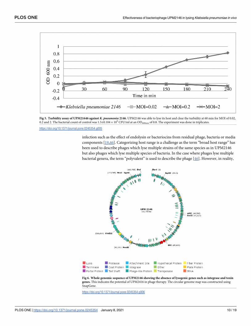
The efficiency and safety of using UPM2146 in the treatment of K. pneumoniae infection invivo were tested in a zebrafish larvae model. Based on morphological appearance, that is deter-
mined on whether the larvae exhibit hook shape tail which indicates that the larvae had died
[44], the mortality rate was less, and the zebrafish larvae were able to survive up to 24 hours
when infected for 30 min. On the other hand, the zebrafish larvae reached a mortality rate
100% at 10 hours when infected for 1 hour and thirty-minute; therefore, 30 min of infection
was used for the CFU count experiment in zebrafish larvae (Fig 9a and 9b). The CFU count
was conducted at an interval of 2 hours post-treatment with UPM2146 showing the presence
of 1.17 x 10⁵ of colonies within zebrafish larvae in the group containing K. pneumoniae 2146
alone at the beginning and continuously increasing until reaching 2.53 × 10⁷ colonies com-
pared to starting with 6.3 x 10⁴ colonies but reaching zero at 10 hours and remaining zero
until 20 hours in the treated group (Fig 10).
Discussion
In this study, UPM2146 was isolated from a polluted lake and was able to form halo-zones
when plated on a K. pneumoniae lawn. This phenomenon was probably due to the release of
Fig 3. Growth curve of phage UPM2146. One step growth curve of UPM2146 taking 20 min of latent period, 5 min of
rise period, and exponential phase of 10 min. Total time required to lyse its host was 25 min that is characteristic of
lytic phages. The experiment was done in triplicates.
https://doi.org/10.1371/journal.pone.0245354.g003
PLOS ONE Effectiveness of bacteriophage UPM2146 in lysing Klebsiella pneumoniae in vivo
PLOS ONE | https://doi.org/10.1371/journal.pone.0245354 January 8, 2021 8 / 19

soluble polysaccharide-degrading enzymes such as capsule depolymerase that was able to
degrade the capsules of K. pneumoniae [45]. Interestingly, UPM2146 had shown to be a slight
broad range but still limited to Klebsiella species as shown by host range and EOP. In addition,
spot tests (for host range determination) may sometimes result in an overestimation of the
capability of the phage to lyse the host bacteria due to other lysis effects not due to phage
Fig 4. (a) UPM 2146 optimum temperature was at 37˚C but was stable at all temperatures tested except at 65 ˚C and (b)
UPM 2146 was stable from pH 3 to 12; thus indicating the potential for it to be used in phage therapy. Bars with
different letters indicate significant difference at p<0.05 (Tukey HSD post-hoc test). Error bars are standard deviations
(n = 3).
https://doi.org/10.1371/journal.pone.0245354.g004
PLOS ONE Effectiveness of bacteriophage UPM2146 in lysing Klebsiella pneumoniae in vivo
PLOS ONE | https://doi.org/10.1371/journal.pone.0245354 January 8, 2021 9 / 19
infection such as the effect of endolysin or bacteriocins from residual phage, bacteria or media
components [19,46]. Categorizing host range is a challenge as the term “broad host range” has
been used to describe phages which lyse multiple strains of the same species as in UPM2146
but also phages which lyse multiple species of bacteria. In the case where phages lyse multiple
bacterial genera, the term “polyvalent” is used to describe the phage [46]. However, in reality,
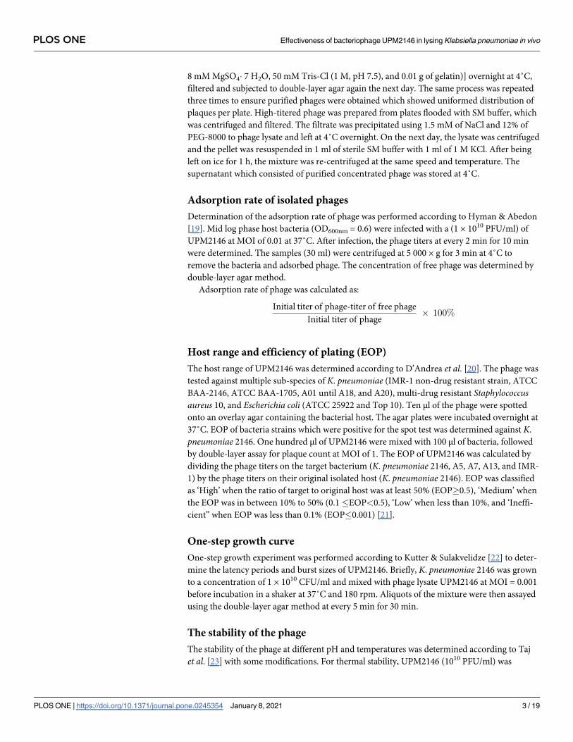
Fig 5. Turbidity assay of UPM21646 against K. pneumonia 2146. UPM2146 was able to lyse its host and clear the turbidity at 60 min for MOI of 0.02,
0.2 and 2. The bacterial count of control was 1.5±0.104 × 10⁹ CFU/ml at an OD600nm of 0.8. The experiment was done in triplicates.
https://doi.org/10.1371/journal.pone.0245354.g005
Fig 6. Whole genomic sequence of UPM2146 showing the absence of lysogenic genes such as integrase and toxin
genes. This indicates the potential of UPM2416 in phage therapy. The circular genome map was constructed using
SnapGene.
https://doi.org/10.1371/journal.pone.0245354.g006
PLOS ONE Effectiveness of bacteriophage UPM2146 in lysing Klebsiella pneumoniae in vivo
PLOS ONE | https://doi.org/10.1371/journal.pone.0245354 January 8, 2021 10 / 19

as shown with UPM2146, the phage while showing its ability to lyse several strains of Klebsi-ella, may only be efficient on one specific bacterial strain which is usually the host used to iso-
late the phage. This is due to the standard isolation procedure which involved enrichment with
only one bacterial host. However, there are recent indications where using multiple hosts dur-
ing isolation increases chances of isolating phages with broader host ranges [19]. In addition,
the host range activity of bacteriophages can evolve and change with time [46–48].
UPM2146 has the requirements for therapeutic purposes since its optimum temperature
and pH are 37˚C and pH of 7 and it has the capability to lyse its host at a fast rate as shown by
the one-step growth curve in Fig 3 [19]. Furthermore, UPM2146 was proven to be lytic based
on turbidity assay due to its fast activity in lysing its host. This further confirms that K. pneu-moniae 2146 has a low chance to develop resistance against UPM2146 [49] as well as any genes
encoding integrase based on PHASTER [50]. The absence of integrase which is vital for the
lysogenic life cycle further confirms that UPM2146 is a lytic phage. Moreover, this is supported
by the prediction of being lytic based on PHACTS database (S1 and S2 Tables, S1 and S2 Figs)
[31]. Furthermore, the presence of a putative phage-encoded peptidoglycan binding protein at
ORF 19 shared 99.62% similarity with that belonging to Klebsiella virus 0507KN21 DNA [51].
Interestingly, UPM2146 belongs to the newly established Ackermannviridae family within the
Caudovirales order based on the phylogenic tree analysis (Fig 8). UPM2146 is similar to Klebsi-ella virus 0507KN2 in which the pairwise distance between them was 0.0038 estimated using
Poisson correction model (S2 Table). The genome size of UPM2146 which is over 155 kbp fur-
ther supports that the phage belongs to Ackermannviridae [52] which is different from the
other families (Myoviridae, Siphoviridae, and Podoviridae) within the Caudovirales order [53].
Moreover, UPM2146 is capable of encoding the muramidase endolysin which catalyzes the
hydrolysis of the peptidoglycan in the K. pneumoniae cell wall, rupturing it at the end of the
virulence cycle resulting in lysis from within [54]. In addition, the presence of the GP5 protein
at the tail of the phage probably enables the mechanism of lysis from without leading to the
breakdown of the bacterial cell wall [55]. Moreover, the genome seemed to encode for 133
hypothetical proteins with unknown functionality but they may have the potential to be used
as new biologics after bioinformatics and proteomic analysis of hypothetical protein [56].
Turbidity assay (A600nm) of K. pneumoniae infected with UPM2146 showed that the phage
was able to lyse the host bacteria and clear the turbidity within 1 hour at MOI of 0.02, 0.2 and
Fig 7. Genomic comparison between UPM2146 and Klebsiella virus 0507KN21 analysed using Easyfig. Genomes of each phage is
represented by linear visualization of their coding sequences (CDS) represented by arrows. The direction of the arrows indicates the
direction of transcription of each CDS. The black vertical lines between the two genomic sequences indicate regions of shared
similarities (89%) according to BLASTn. The intensity of these lines indicates the degree of similarity. This confirms that UPM2146
belongs to Ackermannviridae.
https://doi.org/10.1371/journal.pone.0245354.g007
PLOS ONE Effectiveness of bacteriophage UPM2146 in lysing Klebsiella pneumoniae in vivo
PLOS ONE | https://doi.org/10.1371/journal.pone.0245354 January 8, 2021 11 / 19

Fig 8. a. Phylogenic tree of UPM2146 show that the phylogenomic GBDP trees inferred using D6 formula and yielding average support of 58%, The
numbers above branches are GBDP pseudo-bootstrap support values from 100 replications. The branch lengths of the resulting VICTOR tree are scaled
in terms of D6 formula used. b. The evolution of UPM2146 of putative phage-encoded peptidoglycan binding protein had been drawn using
phylogenic tree analysis using 1000 bootstrap, using the maximum likelihood (ML) method based on the Jones-Taylor-Thornton (JTT) model, the tree
with the highest log likelihood (-1805.44) is shown. The percentage of trees in which the associated taxa clustered together is shown next to the
branches. Initial tree(s) for the heuristic search were obtained by applying the Neighbor-Joining method to a matrix of pairwise distances estimated
using a JTT model. The tree is drawn to scale, with branch lengths measured in the number of substitutions per site. This analysis involved 22 amino
acid sequences. All positions containing gaps and missing data were eliminated (complete deletion option). There were a total of 261 positions in the
final dataset. Evolutionary analyses were conducted in MEGA X [36].
https://doi.org/10.1371/journal.pone.0245354.g008
PLOS ONE Effectiveness of bacteriophage UPM2146 in lysing Klebsiella pneumoniae in vivo
PLOS ONE | https://doi.org/10.1371/journal.pone.0245354 January 8, 2021 12 / 19
Fig 9. Survivability of zebrafish larvae based on morphological appearance. (a.) zebrafish larvae were able to survive up to 24 h when infected with
K. pneumoniae 2146 for 30 min. while (b.) upon increasing the infection to 90 min the zebrafish larvae was able to survive up to 9 h. The experiment
was done in triplicates.
https://doi.org/10.1371/journal.pone.0245354.g009
PLOS ONE Effectiveness of bacteriophage UPM2146 in lysing Klebsiella pneumoniae in vivo
PLOS ONE | https://doi.org/10.1371/journal.pone.0245354 January 8, 2021 13 / 19

Fig 10. CFU count in zebrafish larvae. The phage UPM2146 managed to completely lyse K. pneumoniae 2146 at 10 h and remains zero until 20
h while the zebrafish larvae infected with bacteria (K. pneumoniae 2146) continue to increase till reaching 25.3 x 10⁶ CFU/ml. The experiment
was done in triplicates.
https://doi.org/10.1371/journal.pone.0245354.g010
PLOS ONE Effectiveness of bacteriophage UPM2146 in lysing Klebsiella pneumoniae in vivo
PLOS ONE | https://doi.org/10.1371/journal.pone.0245354 January 8, 2021 14 / 19

2. Thus, UPM2146 appears to have all of the requirements to be used for phage therapy [19,57]
except for the ability to lyse a broad range of K. pneumoniae which can be overcome by using a
phage cocktail [58]. In earlier studies, infected mice were used to prove the efficiency of phage
in treating against K. pneumoniae [10–12]. However, in the present study, the zebrafish larvae
model was used instead. It is important to note that as far as we know; testing the efficiency of
phage using zebrafish larvae against K. pneumoniae has never been reported before. In a study
by Marcoleta et al. [9], three types of K. pneumoniae were used to infect zebrafish larvae, but
only one (Kp RYC492) was found to be highly virulent and caused 75% mortality after 72
hours post-infection (h.p.i.). In another study by Cheepurupalli et al. [14], clinical MDR K.
pneumoniae at 107 CFU/ml was used to infect the zebrafish resulting in 100% mortality at 24
h.p.i. In comparison with the current study, K. pneumoniae 2146 was extremely virulent to the
zebrafish larvae; causing 100% mortality at 10 h.p.i. after 1 hour and 30 minutes infection at
103 CFU/ml. However, 100% mortality of the zebrafish larvae was observed at 24 h.p.i. when
the larvae was exposed to the bacteria for only 30 minutes. Furthermore, the c.f.u. count
dropped to 0 at 10 hours onwards when UPM2146 was used to treat the infected zebrafish lar-
vae at an MOI 0.3. This indicates that UPM2146 has the potential to be used for therapeutic
purposes. On a side note, the zebrafish infection model is commonly used to test cytotoxicity
[16] which in this study has proven UPM2146 is safe to be used.
Conclusion
The continuous increase in drug-resistant bacteria had led scientists to search for alternative
treatments to antibiotics such as phage therapy. In this study, UPM2146 had the ability to lyse
multiple drug-resistant K. pneumoniae 2146 efficiently in vitro as well as four other K. pneumo-niae strains with lesser efficacy. It had a short latent period, and its genome appears to be free
from toxin and integrase genes. In addition, UPM2146 belongs to novel family (Ackermann-viridae) within the Caudovirales order along with the presence of phage-encoded peptidogly-
can binding protein (endolysin) that has the potential to be used for therapeutic purposes.
UPM2146 was highly efficient in clearing the K. pneumoniae infection and can be considered
as safe with the potential to be used as phage therapy against the bacteria although it is likely to
be practically effective only when used in a phage cocktail due to its narrow host range.
Supporting information
S1 Fig.
(PNG)
S2 Fig.
(PNG)
S1 Table.
(XLS)
S2 Table.
(XLSX)
Acknowledgments
We would like to thank the Institute of Bioscience, UPM for providing the TEM facilities and
Dr. Syahida Ahmad from the Department of Biochemistry, Faculty of Biotechnology and Bio-
molecular Sciences, UPM for providing advice on the use of zebrafish larvae for the in vivoexperiments.
PLOS ONE Effectiveness of bacteriophage UPM2146 in lysing Klebsiella pneumoniae in vivo
PLOS ONE | https://doi.org/10.1371/journal.pone.0245354 January 8, 2021 15 / 19

Author Contributions
Conceptualization: Irwan Hanish, Khatijah Yusoff.
Data curation: Amalia Mohd Hashim.
Investigation: Khatijah Yusoff.
Methodology: Omar Assafiri, Adelene Ai-Lian Song, Geok Hun Tan, Amalia Mohd Hashim,
Khatijah Yusoff.
Supervision: Adelene Ai-Lian Song, Geok Hun Tan, Irwan Hanish, Khatijah Yusoff.
Validation: Amalia Mohd Hashim.
Writing – original draft: Omar Assafiri, Adelene Ai-Lian Song, Geok Hun Tan, Irwan Han-
ish, Khatijah Yusoff.
Writing – review & editing: Omar Assafiri, Adelene Ai-Lian Song, Geok Hun Tan, Irwan
Hanish, Amalia Mohd Hashim, Khatijah Yusoff.
References1. Paczosa MK, Mecsas J. Klebsiella pneumoniae: going on the offense with a strong defense. Microbiol-
ogy and Molecular Biology Reviews. 2016 Sep 1; 80: 629–61. https://doi.org/10.1128/MMBR.00078-15
PMID: 27307579
2. Pendleton JN, Gorman SP, Gilmore BF. Clinical relevance of the ESKAPE pathogens. Expert Review
of Anti-Infective Therapy. 2013 Mar 1; 11:297–308. https://doi.org/10.1586/eri.13.12 PMID: 23458769
3. Hudson CM, Bent ZW, Meagher RJ, Williams KP. Resistance determinants and mobile genetic ele-
ments of an NDM-1-encoding Klebsiella pneumoniae strain. PloS One. 2014 Jun 6; 9: e99209. https://
doi.org/10.1371/journal.pone.0099209 PMID: 24905728
4. Ribeiro da Cunha B, Fonseca LP, Calado CR. Antibiotic discovery: where have we come from, where
do we go?. Antibiotics. 2019 Jun; 8: 45. https://doi.org/10.3390/antibiotics8020045 PMID: 31022923
5. Bao J, Wu N, Zeng Y, Chen L, Li L, Yang L, et al. Non-active antibiotic and bacteriophage synergism to
successfully treat recurrent urinary tract infection caused by extensively drug-resistant Klebsiella pneu-
moniae. Emerging Microbes & Infections. 2020 Jan 1; 9:771–4. https://doi.org/10.1080/22221751.
2020.1747950. PMID: 32212918
6. Haq IU, Chaudhry WN, Akhtar MN, Andleeb S, Qadri I. Bacteriophages and their implications on future
biotechnology: a review. Virology Journal. 2012 Dec; 9: 1–8.
7. Wittebole X, De Roock S, Opal SM. A historical overview of bacteriophage therapy as an alternative to
antibiotics for the treatment of bacterial pathogens. Virulence. 2014 Jan 1; 5: 226–35. https://doi.org/10.
4161/viru.25991 PMID: 23973944
8. Herridge WP, Shibu P, O’Shea J, Brook TC, Hoyles L. Bacteriophages of Klebsiella spp., their diversity
and potential therapeutic uses. Journal of Medical Microbiology. 2020 Feb 1; 69: 176–94. https://doi.
org/10.1099/jmm.0.001141 PMID: 31976857
9. Marcoleta AE, Varas MA, Ortiz-Severın J, Vasquez L, Berrıos-Pasten C, Sabag AV, et al. Evaluating
different virulence traits of Klebsiella pneumoniae using Dictyostelium discoideum and zebrafish larvae
as host models. Frontiers in Cellular and Infection Microbiology. 2018 Feb 9; 8: 30. https://doi.org/10.
3389/fcimb.2018.00030 PMID: 29479519
10. Cao F, Wang X, Wang L, Li Z, Che J, Wang L, et al. Evaluation of the efficacy of a bacteriophage in the
treatment of pneumonia induced by multidrug resistance Klebsiella pneumoniae in mice. BioMed
Research International. 2015 Oct; 2015. https://doi.org/10.1155/2015/752930 PMID: 25879036
11. Hung CH, Kuo CF, Wang CH, Wu CM, Tsao N. Experimental phage therapy in treating Klebsiella pneu-
moniae-mediated liver abscesses and bacteremia in mice. Antimicrobial Agents and Chemotherapy.
2011 Apr 1; 55: 1358–65. https://doi.org/10.1128/AAC.01123-10 PMID: 21245450
12. Chibber S, Kaur S, Kumari S. Therapeutic potential of bacteriophage in treating Klebsiella pneumoniae
B5055-mediated lobar pneumonia in mice. Journal of Medical Microbiology. 2008 Dec 1; 57: 1508–13.
https://doi.org/10.1099/jmm.0.2008/002873-0 PMID: 19018021
13. Vinodkumar CS, Neelagund YF, Kalsurmath S. Bacteriophage in the treatment of experimental septice-
mic mice from a clinical isolate of multidrug resistant Klebsiella pneumoniae. The Journal of Communi-
cable Diseases. 2005 Mar 1; 37: 18–29. PMID: 16637396.
PLOS ONE Effectiveness of bacteriophage UPM2146 in lysing Klebsiella pneumoniae in vivo
PLOS ONE | https://doi.org/10.1371/journal.pone.0245354 January 8, 2021 16 / 19

14. Cheepurupalli L, Raman T, Rathore SS, Ramakrishnan J. Bioactive molecule from Streptomyces sp.
mitigates MDR Klebsiella pneumoniae in Zebrafish infection model. Frontiers in Microbiology. 2017 Apr
12; 8: 614. https://doi.org/10.3389/fmicb.2017.00614 PMID: 28446900
15. Silva YJ, Costa L, Pereira C, Mateus C, Cunha A, Calado R, et al. Phage therapy as an approach to pre-
vent Vibrio anguillarum infections in fish larvae production. PloS One. 2014 Dec 2; 9: e114197. https://
doi.org/10.1371/journal.pone.0114197 PMID: 25464504
16. Al-Zubidi M, Widziolek M, Gains AF, Smith RE, Ansbro K, Alrafaie A, et al. Identification of novel bacteri-
ophages with therapeutic potential that target Enterococcus faecalis. Infection and Immunity. 2019 Nov
1; 87: e00512–19. https://doi.org/10.1128/IAI.00512-19 PMID: 31451618
17. Mohd H, Mohd D, Taib NM, Tengku J, Masri SN. Multiple ambler class A ESBL genes among Klebsiella
pneumoniae isolates in a Malaysian district hospital. Tropical Biomedicine. 2016; 33: 109–19.
18. Kropinski AM, Mazzocco A, Waddell TE, Lingohr E, Johnson RP. Enumeration of bacteriophages by
double agar overlay plaque assay. In Bacteriophages 2009; 1: 69–76. Humana Press. https://doi.org/
10.1007/978-1-60327-164-6_7.
19. Hyman P, Abedon ST. Practical methods for determining phage growth parameters. In Bacteriophages
2009 1:175–202. Humana Press.
20. D’Andrea MM, Marmo P, De Angelis LH, Palmieri M, Ciacci N, Di Lallo G, et al. φBO1E, a newly discov-
ered lytic bacteriophage targeting carbapenemase-producing Klebsiella pneumoniae of the pandemic
Clonal Group 258 clade II lineage. Scientific Reports. 2017 Jun 1; 7: 1–8. https://doi.org/10.1038/
s41598-017-02788-9 PMID: 28572684
21. Mirzaei MK, Nilsson AS. Isolation of phages for phage therapy: a comparison of spot tests and effi-
ciency of plating analyses for determination of host range and efficacy. PLoS One. 2015 Mar 11; 10:
e0118557. https://doi.org/10.1371/journal.pone.0118557 PMID: 25761060
22. Kutter E., & Sulakvelidze A. (2004). Bacteriophages: biology and applications. CRC Press, London.
ISBN 9780849313363.
23. Taj MK, Ling JX, Bing LL, Qi Z, Taj I, Hassani TM, et al. Effect of dilution, temperature and pH on the
lysis activity of T4 phage against E. coli bl21. Journal of Animal and Plant Sciences. 2014 Aug 1; 24:
1252–5.
24. Rajnovic D, Muñoz-Berbel X, Mas J. Fast phage detection and quantification: An optical density-based
approach. PloS one. 2019 May 9; 14: e0216292. https://doi.org/10.1371/journal.pone.0216292. PMID:
31071103
25. Hurwitz BL, Deng L, Poulos BT, Sullivan MB. Evaluation of methods to concentrate and purify ocean
virus communities through comparative, replicated metagenomics. Environmental Microbiology. 2013
May; 15: 1428–40. https://doi.org/10.1111/j.1462-2920.2012.02836.x. PMID: 22845467
26. Goodridge L, Gallaccio A, Griffiths MW. Morphological, host range, and genetic characterization of two
coliphages. Applied and Environmental Microbiology. 2003 Sep 1; 69: 5364–71. https://doi.org/10.
1128/aem.69.9.5364-5371.2003 PMID: 12957924
27. Sambrook J, Russell DW. Purification of nucleic acids by extraction with phenol: chloroform. Cold
Spring Harbor Protocols. 2006 Jun 1; 2006(1):pdb-rot4455. https://doi.org/10.1101/pdb.prot4455.
PMID: 22485786
28. Zeman M, Maslaňova I, Indrakova A, Siborova M, Mikulasek K, Bendıčkova K, et al. Staphylococcus
sciuri bacteriophages double-convert for staphylokinase and phospholipase, mediate interspecies plas-
mid transduction, and package mecA gene. Scientific Reports. 2017 Apr 13; 7: 46319. https://doi.org/
10.1038/srep46319. PMID: 28406168
29. Jackman SD, Vandervalk BP, Mohamadi H, Chu J, Yeo S, Hammond SA, et al. ABySS 2.0: resource-
efficient assembly of large genomes using a Bloom filter. Genome Research. 2017 May 1; 27: 768–77.
https://doi.org/10.1101/gr.214346.116. PMID: 28232478
30. Brettin T, Davis JJ, Disz T, Edwards RA, Gerdes S, Olsen GJ, et al. RASTtk: a modular and extensible
implementation of the RAST algorithm for building custom annotation pipelines and annotating batches of
genomes. Scientific Reports. 2015 Feb 10; 5: 8365. https://doi.org/10.1038/srep08365. PMID: 25666585
31. McNair K, Bailey BA, Edwards RA. PHACTS, a computational approach to classifying the lifestyle of
phages. Bioinformatics. 2012 Mar 1; 28: 614–8. https://doi.org/10.1093/bioinformatics/bts014. PMID:
22238260
32. Auch AF, von Jan M, Klenk HP, Goker M. Digital DNA-DNA hybridization for microbial species delinea-
tion by means of genome-to-genome sequence comparison. Standards in Genomic Sciences. 2010
Jan 1; 2: 117–34. https://doi.org/10.4056/sigs.531120. PMID: 21304684
33. Lefort V, Desper R, Gascuel O. FastME 2.0: a comprehensive, accurate, and fast distance-based phy-
logeny inference program. Molecular Biology and Evolution. 2015 Oct 1; 32: 2798–800. https://doi.org/
10.1093/molbev/msv150. PMID: 26130081
PLOS ONE Effectiveness of bacteriophage UPM2146 in lysing Klebsiella pneumoniae in vivo
PLOS ONE | https://doi.org/10.1371/journal.pone.0245354 January 8, 2021 17 / 19

34. Farris JS. Estimating phylogenetic trees from distance matrices. The American Naturalist. 1972 Sep 1;
106: 645–68.
35. Rambaut A, Drummond AJ. FigTree version 1.4. 0. URL: http://tree.bio.ed.ac.uk/software/figtree/.
36. Kumar S, Stecher G, Li M, Knyaz C, Tamura K. MEGA X: molecular evolutionary genetics analysis
across computing platforms. Molecular Biology and Evolution. 2018 Jun 1; 35: 1547–9. https://doi.org/
10.1093/molbev/msy096. PMID: 29722887
37. Meier-Kolthoff JP, Auch AF, Klenk HP, Goker M. Genome sequence-based species delimitation with
confidence intervals and improved distance functions. BMC Bioinformatics. 2013 Dec 1; 14: 60. https://
doi.org/10.1186/1471-2105-14-60. PMID: 23432962
38. Meier-Kolthoff JP, Goker M. VICTOR: genome-based phylogeny and classification of prokaryotic
viruses. Bioinformatics. 2017 Nov 1; 33: 3396–404. https://doi.org/10.1093/bioinformatics/btx440.
PMID: 29036289
39. Goker M, Garcıa-Blazquez G, Voglmayr H, Tellerıa MT, Martın MP. Molecular taxonomy of phytopatho-
genic fungi: a case study in Peronospora. PloS One. 2009 Jul 29; 4: e6319. https://doi.org/10.1371/
journal.pone.0006319. PMID: 19641601
40. Meier-Kolthoff JP, Hahnke RL, Petersen J, Scheuner C, Michael V, Fiebig A, et al. Complete genome
sequence of DSM 30083 T, the type strain (U5/41 T) of Escherichia coli, and a proposal for delineating
subspecies in microbial taxonomy. Standards in Genomic Sciences. 2014 Dec 1; 9: 2. https://doi.org/
10.1186/1944-3277-9-2. PMID: 25780495
41. Varas M, Fariña A, Dıaz-Pascual F, Ortız-Severın J, Marcoleta AE, Allende ML, et al. Live-cell imaging
of Salmonella Typhimurium interaction with zebrafish larvae after injection and immersion delivery
methods. Journal of Microbiological Methods. 2017 Apr 1; 135: 20–5. https://doi.org/10.1016/j.mimet.
2017.01.020 PMID: 28161588
42. Lefkowitz EJ, Dempsey DM, Hendrickson RC, Orton RJ, Siddell SG, Smith DB. Virus taxonomy: the
database of the International Committee on Taxonomy of Viruses (ICTV). Nucleic Acids Research.
2018 Jan 4; 46: D708–17. https://doi.org/10.1093/nar/gkx932 PMID: 29040670
43. Walker PJ, Siddell SG, Lefkowitz EJ, Mushegian AR, Dempsey DM, Dutilh BE, et al. Changes to virus
taxonomy and the International Code of Virus Classification and Nomenclature ratified by the Interna-
tional Committee on Taxonomy of Viruses (2019). Archives of Virology. 2019 Sep 1; 164: 2417–29.
https://doi.org/10.1007/s00705-019-04306-w PMID: 31187277
44. Foo R, Ahmad S, Lai K, Idrus Z, Yusoff K, Liang J. Palm kernel cake oligosaccharides acute toxicity and
effects on nitric oxide levels using a zebrafish larvae model. Frontiers in Physiology. 2020 Sep 24; 11:
1–9. https://doi.org/10.3389/fphys.2020.555122. PMID: 32038307
45. Pan YJ, Lin TL, Chen YY, Lai PH, Tsai YT, Hsu CR, et al. Identification of three podoviruses infecting
Klebsiella encoding capsule depolymerases that digest specific capsular types. Microbial Biotechnol-
ogy. 2019 May; 12: 472–86. https://doi.org/10.1111/1751-7915.13370 PMID: 30706654
46. Ross A, Ward S, Hyman P. More is better: selecting for broad host range bacteriophages. Frontiers in
Microbiology. 2016 Sep 8; 7: 1352. https://doi.org/10.3389/fmicb.2016.01352 PMID: 27660623
47. Li E, Zhao J, Ma Y, Wei X, Li H, Lin W, et al. Characterization of a novel Achromobacter xylosoxidans
specific siphoviruse: phiAxp-1. Scientific Reports. 2016 Feb 24; 6: 1–1.
48. Hamdi S, Rousseau GM, Labrie SJ, Tremblay DM, Kourda RS, Slama KB, et al. Characterization of two
polyvalent phages infecting Enterobacteriaceae. Scientific Reports. 2017 Jan 16; 7: 1–2.
49. Hyman P. Phages for phage therapy: isolation, characterization, and host range breadth. Pharmaceuti-
cals. 2019 Mar; 12: 35. https://doi.org/10.3390/ph12010035 PMID: 30862020
50. Arndt D, Grant JR, Marcu A, Sajed T, Pon A, Liang Y, et al. PHASTER: a better, faster version of the
PHAST phage search tool. Nucleic Acids Research. 2016 May 3; 44: W16–21. https://doi.org/10.1093/
nar/gkw387. PMID: 27141966
51. Adriaenssens EM, Wittmann J, Kuhn JH, Turner D, Sullivan MB, Dutilh BE, et al. Taxonomy of prokary-
otic viruses: 2017 update from the ICTV Bacterial and Archaeal Viruses Subcommittee. Archives of
Virology. 2018 Apr 1; 163: 1125–9. https://doi.org/10.1007/s00705-018-3723-z. PMID: 29356990
52. Born Y, Knecht LE, Eigenmann M, Bolliger M, Klumpp J, Fieseler L. A major-capsid-protein-based mul-
tiplex PCR assay for rapid identification of selected virulent bacteriophage types. Archives of Virology.
2019 Mar 1; 164: 819–30. https://doi.org/10.1007/s00705-019-04148-6. PMID: 30673846
53. Taxonomy V, Murphy FA. Ninth report of the international committee on taxonomy of viruses. Virus Tax-
onomy. San Diego, CA: Elsevier. 2012:1281–325. ISBN: 978-0-12-384684-6.
54. Rodrıguez-Rubio L, Gerstmans H, Thorpe S, Mesnage S, Lavigne R, Briers Y. DUF3380 domain
from a Salmonella phage endolysin shows potent N-acetylmuramidase activity. Applied and Environ-
mental Microbiology. 2016 Aug 15; 82: 4975–81. https://doi.org/10.1128/AEM.00446-16. PMID:
27287318
PLOS ONE Effectiveness of bacteriophage UPM2146 in lysing Klebsiella pneumoniae in vivo
PLOS ONE | https://doi.org/10.1371/journal.pone.0245354 January 8, 2021 18 / 19

55. Abedon ST. Lysis from without. Bacteriophage. 2011 Jan 1; 1: 46–9. https://doi.org/10.4161/bact.1.1.
13980. PMID: 21687534
56. Shahbaaz M, Imtaiyaz Hassan M, Ahmad F. Functional annotation of conserved hypothetical proteins
from Haemophilus influenzae Rd KW20. PloS One. 2013 Dec 31; 8: e84263. https://doi.org/10.1371/
journal.pone.0084263. PMID: 24391926
57. Ciacci N, D’Andrea MM, Marmo P, Demattè E, Amisano F, Pilato VD, et al. Characterization of
vB_Kpn_F48, a newly discovered lytic bacteriophage for Klebsiella pneumoniae of sequence type 101.
Viruses. 2018 Sep; 10: 482. https://doi.org/10.3390/v10090482 PMID: 30205588
58. Lin DM, Koskella B, Lin HC. Phage therapy: An alternative to antibiotics in the age of multi-drug resis-
tance. World Journal of Gastrointestinal Pharmacology and Therapeutics. 2017 Aug 6; 8: 162. https://
doi.org/10.4292/wjgpt.v8.i3.162 PMID: 28828194
PLOS ONE Effectiveness of bacteriophage UPM2146 in lysing Klebsiella pneumoniae in vivo
PLOS ONE | https://doi.org/10.1371/journal.pone.0245354 January 8, 2021 19 / 19