Luciana Aparecida Corá - ibb.unesp.br · Luís Gustavo Rubi de Souza, Marcelo Agostinho, Marjorie...
95
Luciana Aparecida Corá A Biosusceptometria AC aplicada à tecnologia farmacêutica Botucatu 2008
Transcript of Luciana Aparecida Corá - ibb.unesp.br · Luís Gustavo Rubi de Souza, Marcelo Agostinho, Marjorie...
Luciana Aparecida Corá
A Biosusceptometria AC aplicada
à tecnologia farmacêutica
Botucatu 2008
Luciana Aparecida Corá
A Biosusceptometria AC aplicada à tecnologia farmacêutica
Tese apresentada ao Instituto de Biociências deBotucatu, Universidade Estadual Paulista “Júlio deMesquita Filho”, como exigência parcial para obtençãodo Título de Doutor em Ciências Biológicas (Área deConcentração: Farmacologia).
Orientador: Prof. Adj. José Ricardo de Arruda Miranda
Botucatu 2008
FICHA CATALOGRÁFICA ELABORADA PELA SEÇÃO TÉCNICA DE AQUISIÇÃO E TRATAMENTO
DA INFORMAÇÃO
DIVISÃO TÉCNICA DE BIBLIOTECA E DOCUMENTAÇÃO - CAMPUS DE BOTUCATU - UNESP
BIBLIOTECÁRIA RESPONSÁVEL: SELMA MARIA DE JESUS
Corá, Luciana Aparecida.
A biosusceptometria AC aplicada à tecnologia farmacêutica / Luciana
Aparecida Corá. – Botucatu : [s.n.], 2008.
Tese (doutorado) – Universidade Estadual Paulista, Instituto de Biociências
de Botucatu 2008
Orientador: José Ricardo de Arruda Miranda
Assunto CAPES: 21000000
1. Biosusceptometria 2. Tecnologia farmacêutica
CDD 615
Palavras-chave: Biomagnetismo; Desintegração; Formas farmacêuticas
sólidas;Trânsito gastrintestinal
Agradecimentos
"A gratidão desbloqueia a abundância da vida. Ela torna o que temos em suficiente,
e mais: a gratidão dá sentido ao nosso passado, traz paz para o hoje, e cria uma
visão para o amanhã.” Melody Beattie
Agradecimentos A Deus, por me guiar por caminhos onde encontrei mestres dedicados, pessoas extraordinárias e a vocação pela Ciência.
"O criador não dá a você o desejo de fazer o que você não tem capacidade para fazer." Orison Marden
Ao Prof. Dr. José Ricardo de Arruda Miranda, pela oportunidade e orientação que permitiram fazer da Ciência a minha profissão. Pelo criticismo, sempre focado no aperfeiçoamento do trabalho e amadurecimento das discussões e, fundamentalmente, por acreditar que seria possível chegar tão longe quando muitos duvidaram.
"Nenhum de nós chegou onde está exclusivamente através do impulso de nossos próprios pés. Chegamos aqui porque alguém se inclinou e nos alavancou." Thurgood Marshal
Ao meu esposo, Agostinho Mazza, por todos os anos de incentivo e apoio incondicionais. Por me acompanhar nas mais diversas etapas dessa vida acadêmica, sempre com ombros fortes onde constantemente me amparei. "A vida nos ensinou que o amor não consiste em olhar um para o outro, mas sim olhar juntos para fora, na mesma direção.”
Saint-Exupéry Aos meus pais, Marisa Frasson Corá e Arzenido Corá, pelos contínuos exemplos de superação e por mostrarem que as grandes realizações dependem, inicialmente, de pequenas atitudes.
“Os nossos pais amam-nos porque somos seus filhos, é um fato inalterável. Nos momentos de sucesso, isso pode parecer irrelevante, mas nas ocasiões de fracasso, oferecem um consolo e uma segurança que não se encontram em qualquer outro
lugar.” Bertrand Russel Ao meu irmão Emerson Corá e minha cunhada Heidi Macedo, pelo respeito e admiração e, sobretudo pelo pequeno João Pedro, um anjinho que chegou para abençoar nossas vidas e cativar nossos corações.
"A alegria e o amor são as duas grandes asas para os grandes feitos." Goethe
À minha segunda família: Delazir S. Mazza, Dalva Mazza, Adriano Ruolla, Marco Antônio Mazza, Josi Galindo, pela compreensão e encorajamento, sempre presentes.
"A alegria compartilhada é uma alegria dobrada." John Ray
À minha querida amiga e jovem Dra. Madileine F. Américo, por sua amizade verdadeira, pelo apoio e dedicação. Seu amor pela Ciência me ensinou que as adversidades também são caminhos necessários e, muitas vezes, inevitáveis para alcançar os objetivos.
"Para realizar grandes conquistas, devemos não apenas agir, mas também sonhar; não apenas planejar, mas também acreditar." Anatole France
Aos demais colegas do Laboratório de Biomagnetismo: Camila Souza Melo, Ednaldo Alexandre Zandoná, Fabiano Carlos Paixão, Giovana Evangelista, Leandro Bolognesi, Luís Gustavo Rubi de Souza, Marcelo Agostinho, Marjorie do Val Ietsugo, Murilo Stelzer, Paulo Roberto da Fonseca Filho, Rozemeire Garcia Marques e Uilian de Andreis, pela convivência, desafios e experiências compartilhadas.
“Não caminhe atrás de mim; eu posso não liderar. Não caminhe na minha frente; eu posso não seguir. Simplesmente caminhe a meu lado e seja meu amigo." Albert Camus
Ao médico e amigo Fernando Gomes Romeiro, pela amizade, discussões proveitosas e pelos momentos de descontração.
"Fácil é ser colega, fazer companhia a alguém, dizer o que ele deseja ouvir. Difícil é ser amigo para todas as horas e dizer sempre a verdade quando for preciso. E com confiança no que diz." Carlos Drummond de Andrade
Aos voluntários, sempre dispostos, cuja participação foi imprescindível para a realização desses trabalhos.
"É rara a verdadeira gratidão, porque são raros os genuínos benfeitores.” Marquês de Maricá
Aos Professores Dr. Oswaldo Baffa (FFCLRP‐USP) e Dr. Ricardo Brandt de Oliveira F RP‐USP) pelos auxílios e revisões dos artigos publicados. ( M "Todo homem que encontro é superior a mim em alguma coisa. Por isso, dele sempre aprendo alguma coisa.” Emerson
Às pessoas que não foram mencionadas, mas sempre acreditaram que seria possível.
"Não há no mundo exagero mais belo que a gratidão." Jean de La Bruyère
os Programas de pós‐graduação em Ciências Biológicas – Biologia Geral e Aplicada e armacologia, pela oportunidade concedida. AF À Coordenação de Aperfeiçoamento de Pessoal de Nível Superior (CAPES), pela bolsa e doutoramento e à Fundação de Amparo à Pesquisa do Estado de São Paulo pelos uxílios financeiros. da

Resumo e Abstract
"Considero feliz aquele que quando se fala de êxito busca a resposta em seu
trabalho." Emerson
Resumo
Corá, L.A. A Biosusceptometria AC aplicada à tecnologia farmacêutica. 2008.
84 p. Tese Doutorado – Instituto de Biociências de Botucatu, Universidade Estadual
aulista “Júlio de Mesquita Filho”. P
A administração oral de drogas é uma prática comum na terapia e as formas farmacêuticas sólidas são amplamente utilizadas. A variação no perfil de absorção ao longo do trato gastrintestinal (TGI) humano e a possibilidade de liberar drogas em diferentes regiões são os maiores desafios para o desenvolvimento de novos produtos. Desse modo, avaliar formas farmacêuticas sólidas in vivo fornece um entendimento mais profundo quando um efeito sistêmico ou local é desejado. Geralmente, estes estudos são realizados por meio da cintilografia e técnicas biomagnéticas. A Biosusceptometria de Corrente Alternada (BAC) é uma técnica que merece destaque por suas características, acurácia dos resultados obtidos e versatilidade. A BAC propiciou imagens do processo de desintegração de comprimidos tanto in vitro quanto no estômago humano, introduzindo outra perspectiva na análise desse processo. Os resultados também foram correlacionados com sucesso com aqueles obtidos por metodologias específicas, garantindo uma análise mais acurada dos parâmetros físicos envolvidos com a desintegração de comprimidos. A utilização da BAC permitiu avaliar a motilidade gastrintestinal e o processo de desintegração de cápsulas de hidroxipropilmetilcelulose (HPMC) revestidas no cólon humano. Além disso, também foi possível investigar a influência do estado prandial no esvaziamento gástrico e no trânsito gastrintestinal de um sistema multiparticulado magnético. Todos esses trabalhos fortaleceram a BAC como um método alternativo na pesquisa farmacêutica demonstrando seu potencial para avaliar diferentes processos, apesar das suas limitações. Sintetizando, a BAC é uma ferramenta valiosa, com a vantagem de ser livre de radiação e inócua aos voluntários, e vasta aplicabili a d de na pesquisa farmacêutica, farmacológica e fisiológica. Palavraschave: Biomagnetismo, trânsito gastrintestinal, formas farmacêuticas sólidas, desintegração.
Abstract
Corá, L.A. AC Biosusceptometry applied to pharmaceutical technology. 2008.
84 p. Tese Doutorado – Instituto de Biociências de Botucatu, Universidade Estadual
aulista “Júlio de Mesquita Filho”. P
Oral administration is widely accepted route for drug delivery and solid dosage forms are commonly administered. The variation of absorption profiles along the human gastrointestinal tract (GIT) and the ability to target drugs by adequate dosage forms to distinct sites is the challenge in the pharmaceutical development of solid dosage forms. An understanding of the factors involved in drug absorption and how the gastrointestinal variables can interfere with this process is important to develop more reliable drug delivery systems. The performance of pharmaceutical dosage forms must be fully investigated in vivo to provide more reliable information when a local or systemic effect is desirable. Generally, in vivo investigation on the behavior of dosage forms has been made by using gamma‐scintigraphy and biomagnetic techniques. AC Biosusceptometry (ACB) deserves consideration due to its features, accuracy and versatility. By using ACB technique, it was possible to monitor the disintegration process through acquisition of magnetic images in vitro and in human stomach. The results also were successfully correlated with those obtained with standard methods which provided a more reliable analysis on the physical parameters involved in the disintegration process of tablets. ACB allowed evaluating the gastrointestinal motility and the disintegration of hydroxipropylmethylcellulose (HPMC) coated capsules in human colon. Moreover, it was possible to investigate the gastric emptying and gastrointestinal transit of a magnetic multiparticulate system under influence of prandial state. All these studies have contributed to establish the ACB as an alternative method for pharmaceutical research and, despite some limitations, it was feasible to evaluate different pharmaceutical processes. In summary, ACB is a radiation free and non‐invasive technique with wide applicability in pharmaceutical, physiological and pharmacological researches.
Key words: Biomagnetism, gastrointestinal transit, solid dosage forms, disintegration.
Índice de Figuras e Tabelas
Introdução
Figura 1 Motilidade gastrintestinal no período interdigestivo – Complexo Motor Migratório .................................................................................................................................. 16
Figura 2 Etapas envolvidas na biodisponibilidade de uma droga a partir da
administração oral de uma forma farmacêutica sólida. A desintegração promove a fragmentação em partículas que serão dissolvidas no meio e absorvidas pela mucosa do TGI ..................................................................................... 21
Figura 3 Sensor magnético constituído por dois pares de bobinas de indução. (a)
Bobina de excitação externa e (b) bobina de detecção interna ............................ 25 Figura 4 Esquema de funcionamento do sensor magnético. A bobina de excitação induz
fluxo magnético na bobina de detecção que ao se aproximar do material magnético promove um desbalanceamento no fluxo magnético entre as bobinas, permitindo seu monitoramento ..................................................................... 26
Figura 5 Sistema de Biosusceptometria AC com multisensores mostrando o par de
bobinas de excitação (1) e os sete pares de bobinas de detecção (2) ................ 27
Capítulo 1: Magnetic images of the disintegration process of tablets in the human stomach by AC Biosusceptometry
Figure 1 (a) Functional diagram of the single sensor AC Biosusceptometer. The proximity of a magnetic material to the measurement system causes an unbalancing in the magnetic flux and the response is monitored. (b) Schematic diagram of the single sensor showing the pairs excitation/ detection coils in a coaxial arrangement ............................................................................................................... 40
Figure 2 Multisensor AC Biosusceptometer system. (1) a pair of excitation coil and (2)
seven pairs of detection coils on a hexagonal symmetry ........................................ 41 Figure 3 Positioning of the multisensor AC Biosusceptometer in the gastric projection
according to the external anatomical references ...................................................... 42 Figure 4 Transversal sensitivity profile of the multisensor AC Biosusceptometer
system. Variation rate of intensity (mV) in the magnetic signal was obtained for the central sensor for distinct distances between the sensor and a magnetic tablet ......................................................................................................................... 43
Figure 5 (a) Photographs of a tablet in the acidic solution to illustrate the disintegration process. The onset of the disintegration (t50) occurred in the instant t3. (b) Spreading of the magnetic material and the time variation in the number of pixels in the segmented area of the photographs ................................ 44
Figure 6 (a) Magnetic images of the disintegration process of a tablet in vitro. The onset of the disintegration (t50) occurred in the instant t3. The gradual increase of the imaging area has characterized the spreading of the magnetic material. (b) In vitro spreading of the magnetic material and the time variation of the number of pixels contained inside a delineated area showing the “velocity of the disintegration” ................................................................................................................... 45
Figure 7 (a) Magnetic images of the disintegration process of a tablet in human stomach at t1 to t6. 50% disintegration (t50) is located between ti and ti+1. (b) In vivo spreading of the magnetic material as number of pixels in the segmented area and its rate of change ................................................................................................... 46
Capítulo 2: Influence of compression forces on tablets disintegration by AC Biosusceptometry
Figure 1 Water uptake and disintegration force apparatus. When water was taken up by the uncoated tablet, the force developed was measured by the transducer placed on the upper side of the cylindrical frame ...................................................... 52
Figure 2 Magnetic apparatus mounted for disintegration measurement. A coated tablet was inserted by a guide in the glass container placed in front of the multisensor AC Biosusceptometry system. The disintegration process was monitored in real time by seven magnetic sensors .................................................. 52
Figure 3 Time-dependent profiles of water uptake (a), disintegration force (b) and magnetic area variation (c) for uncoated tablets at different compression forces ............................................................................................................................................. 54
Figure 4 Plots of physical parameters of the disintegration process for tablets compressed at 30 kN. (a) Temporal magnetic area variation and disintegration force developed during water uptake. (b) Correlation between magnetic area and force developed (R=0.987). (c) Relationship between magnetic area, water uptake and disintegration force ............................................ 54
Figure 5 Relationship between compression force and t63.2 parameter. The time
interval needed for the maximum amount of water to be able to promote the development of disintegration force and magnetic area was higher for a same level of compression force ................................................................................................... 55
Figure 6 Magnetic data profiles for coated tablets. (a) Temporal effect of compression force on the magnetic area variation. (b) First derivative of magnetic area variation curves for compression force in the time. ................................................... 56
Table 1 Fit parameters of water uptake, disintegration force and magnetic area
profiles for uncoated tablets according to Weibull model, expressed as mean ± SD .................................................................................................................................................... 55
Table 2 Fit parameters of magnetic area profile for coated tablets according to Weibull
model, expressed as mean ± SD. ......................................................................................... 56
Capítulo 3: Enteric coated magnetic HPMC capsules evaluated in human gastrointestinal tract by AC Biosusceptometry Figure 1 Single sensor AC Biosusceptometer. (a) Excitation coil and (b) detection coil in
the first-order gradiometric configuration .................................................................. 59 Figure 2 Multisensor AC Biousceptometer system. (a) Pair of excitation coils and (b)
seven pairs of detection coils coaxially arranged with a hexagonal symmetry .......................................................................................................................................................... 60
Figure 3 (a) Positioning of the multisensor AC Biosusceptometer system on the
abdominal surface. (b) Square matrix (9x9) drawn on the ileocolonic region. The xiphoid process and the McBurney’s point were the external anatomical references, respectively ........................................................................................................ 60
Figure 4 In vitro characterization of an enteric coated magnetic HPMC capsule. (a)
Photographs and corresponding magnetic images (c) of the disintegration process of a capsule in the phosphate buffer. Mean disintegration time (t50) occurred in the instant t2. (b) and (d) represents the spreading of the magnetic material and the time variation of the number of pixels contained inside a delineated area showing the velocity of the disintegration .................................... 61
Figure 5 Magnetic signals recorded concomitantly to the ingestion of the enteric coated
HPMC capsule. (a) Intense contractile waves of the gastric activity recorded by the sensors located distally. (b) Fast Fourier Transformed sowing the frequency peak of 0.05 Hz (3 cycles/ minute) .............................................................. 62
Figure 6 (a) Magnetic images of the disintegration process of an enteric coated HPMC
capsule in ileocolonic region. The instant t1 shows the arrival of the capsule; from t2 occurred a gradual increase in the image area which characterized the spreading of the magnetic material. (b) Spreading of the magnetic material in number of pixels in the segmented area showing the velocity of the disintegration process ........................................................................................................... 63
Table 1 Gastrointestinal transit time and mean disintegration time (t50) for magnetic enteric coated magnetic HPMC capsules ........................................................................ 62
Capítulo 4: AC Biosusceptometry to evaluate the gastrointestinal transit of pellets under influence of prandial state
Figure 1 Single-sensor AC Biosusceptometry system with the pair of excitation (1) and detection coils (2) coaxially arranged in a first-order gradiometric configuration. .............................................................................................................................. 70
Figure 2 Schematic representation of square point matrix (gray circles) drawn around the gastric and colonic regions .................................................................... 72
Figure 3 Sequence of magnetic images of the multiparticulate system showing the pellets dispersion from the HPMC capsule in the stomach and its spreading in the colonic region for a fasted volunteer. White outlines are the ribs on top and the iliac crests on the bottom ................ 77
Figure 4 Example of gastric emptying (GE) and colon arrival (CA) profiles on fasted and fed conditions for subject number 1. The arrows indicate the parameters measured ............................................................................. 78
Table 1 Gastric emptying, colon arrival and small intestinal transit time for multiparticulate formulation administered to fasted and fed volunteers characterized using Statistical Moments ................................................................. 75
Sumário
Índice de Figuras e Tabelas
Resumo
Abstract
Introdução
1. A farmacotécnica e o trato gastrintestinal humano ................................................ 13
1.1. Estômago, intestino delgado e cólon .................................................................. 14
1.2. Variáveis fisiológicas e farmacêuticas ............................................................... 15
2. Formas farmacêuticas sólidas ......................................................................................... 19
3. Métodos para avaliar formas farmacêuticas in vivo .............................................. 22
Referências Bibliográficas ............................................................................................................ 29
Objetivos ................................................................................................................................................. 37
Capítulo 1
Magnetic images of the disintegration process of tablets in the human
stomach by AC Biosusceptometry ..................................................................................... 38
Capítulo 2
Influence of compression forces on tablets disintegration by
AC Biosusceptometry .............................................................................................................. 50
Capítulo 3
Enteric coated magnetic HPMC capsules evaluated in human gastrointestinal
tract by AC Biosusceptometry ............................................................................................. 58
Capítulo 4
AC Biosusceptometry to evaluate the gastrointestinal transit of pellets under
influence of prandial state ..................................................................................................... 66
Considerações Finais ....................................................................................................................... 82
Introdução
“A mente que se abre a uma nova idéia jamais voltará ao seu tamanho original." Albert Einstein

12
Introdução
Considerando que as variáveis fisiológicas referentes ao trato gastrintestinal
(TGI) humano influenciam significativamente a absorção e a biodisponibilidade de
fármacos administrados por via oral, tornou-se imperativo implementar métodos de
análise capazes de caracterizar o comportamento de formas farmacêuticas in vivo.
Nesse contexto, os métodos fundamentados no Biomagnetismo, destacando a
Biosusceptometria AC (BAC), tornaram-se alternativas viáveis para a pesquisa
farmacêutica. A inserção da BAC nesse campo de estudo veio de encontro à
necessidade de utilizar metodologia de baixo custo, livre de radiação ionizante, não
invasiva e capaz de avaliar processos farmacêuticos não apenas in vitro, como
também no TGI humano.
O contínuo aperfeiçoamento da BAC, culminando no desenvolvimento do
sistema com multisensores, foi fundamental para aplicá-la na pesquisa farmacêutica.
A análise do processo de desintegração de comprimidos fortaleceu-se com a
possibilidade de obter imagens magnéticas, introduzindo um novo conceito em
imagens biológicas (Capítulo 1). Associando-se a BAC com metodologias específicas
foi possível realizar uma análise mais acurada dos parâmetros físicos envolvidos no
processo de desintegração de comprimidos (Capítulo 2). A BAC permitiu, também,
avaliar a motilidade gastrintestinal e a desintegração de cápsulas de
hidroxipropilmetilcelulose (HPMC) no cólon humano (Capítulo 3). Além disso,
possibilitou a investigação da influência do estado prandial no esvaziamento gástrico
e no trânsito gastrintestinal de um sistema multiparticulado magnético enfocando a
liberação colônica (Capítulo 4).
Esses trabalhos mostraram que a BAC é um método capaz de prover os
requisitos necessários para monitorar diferentes processos farmacêuticos visando
uma análise mais detalhada dos complexos parâmetros fisiológicos e farmacêuticos
conhecidos por influenciarem a liberação e a absorção de drogas.
13
1. A farmacotécnica e o trato gastrintestinal humano
Ao administrar um fármaco devem ser considerados fatores como a via de
administração que será mais efetiva e melhor aceita pelo paciente, além da forma
farmacêutica apropriada (Ansel et al., 2000). A administração de drogas por via oral é
amplamente utilizada, sendo o trato gastrintestinal (TGI) o principal acesso à
circulação sistêmica. Apesar de apresentar algumas desvantagens, essa via é a
preferida por oferecer maior comodidade e permitir o estabelecimento de esquemas
terapêuticos fáceis de serem cumpridos (Jivraj et al., 2000; Sastry et al., 2000).
Em se tratando da administração por via oral, as formas farmacêuticas
sólidas são muito utilizadas devido à relativa facilidade de obtenção, ao custo
reduzido e à estabilidade (Jivraj et al., 2000). No entanto, os parâmetros relacionados
ao TGI interagem com a forma farmacêutica e, conseqüentemente, influenciam os
processos de liberação, absorção e biodisponibilidade do fármaco (Rouge et al., 1996;
Martinez & Amidon, 2002). Assim, compreender como esses parâmetros e variáveis
fisiológicas podem alterar a performance de uma forma farmacêutica in vivo é
fundamental para o desenvolvimento de produtos com maior eficácia terapêutica e
menor incidência de efeitos colaterais (Zahirul & Khan, 1996).
Para a absorção de um fármaco administrado por via oral são considerados
processos que incluem desde a liberação da droga, por meio da desintegração da
forma farmacêutica, até sua dissolução no meio, de com propriedades físico-químicas
como solubilidade e coeficiente de partição (Lipka & Amidon, 1999). Além disso, as
características fisiológicas do TGI, como tamanho da superfície de absorção, perfil do
pH nas diferentes regiões, as taxas de esvaziamento gástrico e trânsito intestinal e a
motilidade gastrintestinal são os principais fatores que influenciam diretamente a
biodisponibilidade do fármaco e podem, ainda, limitar a fração da dose que será
absorvida (Rouge et al., 1996).
O TGI humano é um meio complexo, que apresenta diferenças regionais
bastante acentuadas as quais devem ser completamente investigadas para o
desenvolvimento de um produto farmacêutico, seja para exercer um efeito local ou
sistêmico.
14
1.1 Estômago, intestino delgado e cólon
O estômago humano é um órgão subdivido em dois compartimentos
funcionais: enquanto a região proximal atua como reservatório para acomodar o
conteúdo ingerido, a região distal é responsável pela trituração desse conteúdo e sua
mistura com as secreções gástricas (Camilleri, 2006). No entanto, o processo de
absorção no estômago é limitado, pois apresenta uma camada mucosa bastante
espessa e uma superfície reduzida (Hörter & Dressman, 2001). Assim, a ação
coordenada das regiões gástricas contribui para a otimização do aproveitamento dos
alimentos reduzindo os alimentos sólidos a pequenas partículas e regulando
precisamente a velocidade de transferência para o intestino delgado (Hasler, 1999).
Por outro lado, o intestino delgado possui uma superfície extremamente ampla cujas
propriedades fisiológicas facilitam a absorção de nutrientes e, conseqüentemente, de
muitos fármacos (Hörter & Dressman, 2001; Masaoka et al., 2006).
No cólon proximal, e em menor grau nos demais segmentos, os padrões
motores estão amplamente vinculados à sua função de propulsão, absorção de água e
eletrólitos. Esse segmento apresenta movimentos lentos e coordenados que facilitam
a absorção, além de propiciar o crescimento, no lúmen colônico, de microorganismos
capazes de facilitar a absorção de certos nutrientes para os próprios colonócitos
(Christensen, 1987; Camilleri & Ford, 1998). Até há pouco tempo, essas eram as
únicas funções atribuídas a esse segmento. Atualmente, esse órgão vem ganhando
destaque como um local específico para liberação de fármacos, proteínas e peptídios
com potencial terapêutico (Chourasia & Jain, 2003; Shareef et al., 2003; Freire et al.,
2006a). Comparando-se com o intestino delgado, o cólon tem uma superfície de
absorção menor, fato que é compensado pelo trânsito mais lento, o que proporciona
uma excelente oportunidade para a absorção de drogas e outros materiais.
15
1.2 Variáveis fisiológicas e farmacêuticas
A taxa e a extensão da absorção de fármacos administrados por via oral são
determinadas por parâmetros relacionados ao TGI e à forma farmacêutica. Dentre os
parâmetros fisiológicos, destacam-se a motilidade gastrintestinal, o estado prandial, o
esvaziamento gástrico, o trânsito intestinal e a variação do pH ao longo do TGI (Singh,
1999; Kimura & Higaki, 2002; Martinez & Amidon, 2002). Além dos fatores
fisiológicos, a solubilidade do fármaco e o tamanho das partículas, bem como as
características da forma farmacêutica, como friabilidade, dureza, tipo de
revestimento e densidade também podem interferir no processo de liberação e
absorção de drogas (Jenquin et al., 1990; Hancock et al., 1997; Jain, 1999).
A motilidade do trato gastrintestinal humano
Dentre as propriedades funcionais do TGI destaca-se a motilidade, que é a
capacidade de contrair e relaxar a sua musculatura para misturar e propelir o
material ao longo do seu comprimento (Nguyen et al., 2007). A motilidade
gastrintestinal é organizada, basicamente, de acordo com o estado prandial (Quigley,
1996; Camilleri, 2006). Com o término do processo digestivo tem início uma atividade
motora cíclica denominada Complexo Motor Migratório (CMM), que alterna ciclos de
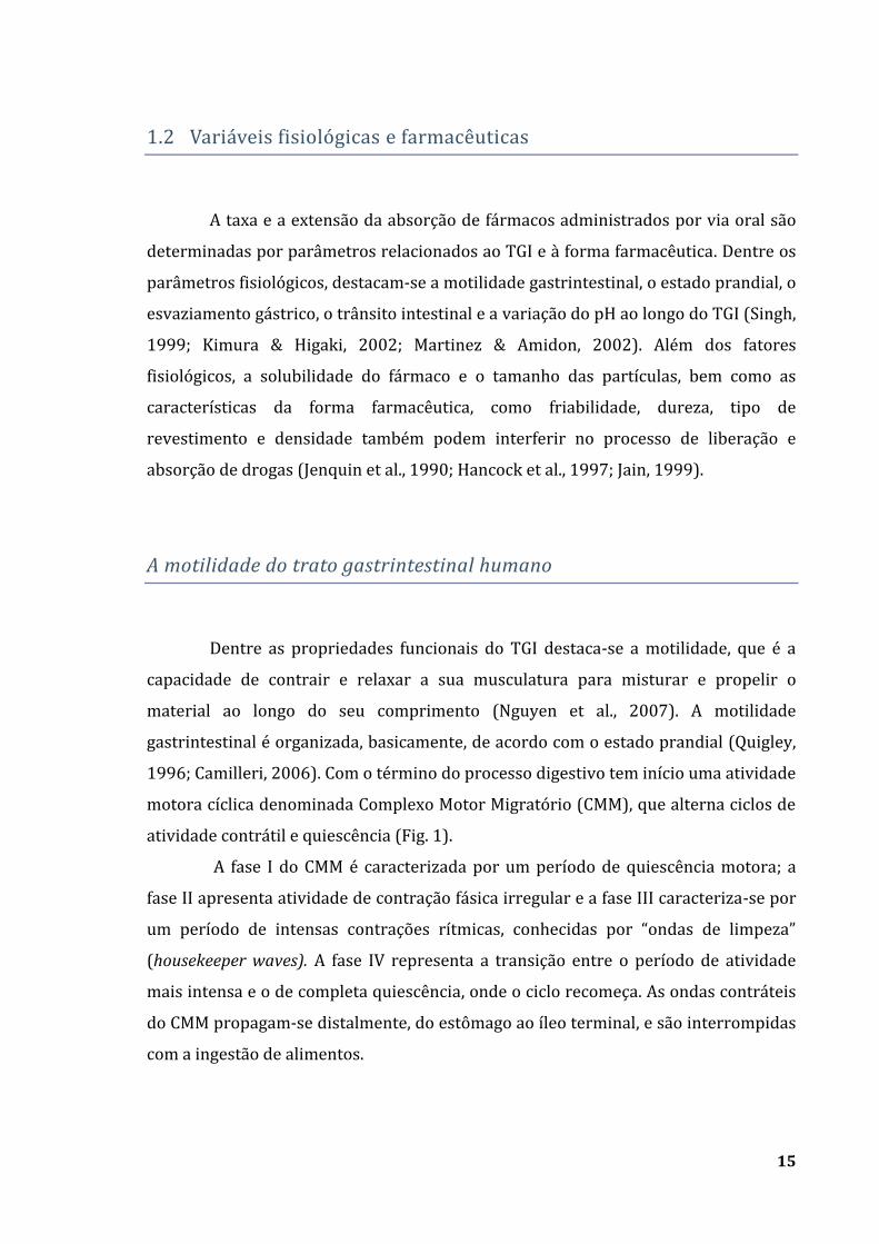
atividade contrátil e quiescência (Fig. 1).
A fase I do CMM é caracterizada por um período de quiescência motora; a
fase II apresenta atividade de contração fásica irregular e a fase III caracteriza-se por
um período de intensas contrações rítmicas, conhecidas por “ondas de limpeza”
(housekeeper waves). A fase IV representa a transição entre o período de atividade
mais intensa e o de completa quiescência, onde o ciclo recomeça. As ondas contráteis
do CMM propagam-se distalmente, do estômago ao íleo terminal, e são interrompidas
com a ingestão de alimentos.
16
Com a ingestão de alimentos inicia-se o período pós-prandial, em que a
freqüência e a amplitude da atividade de contração variam de acordo com o segmento
do TGI, sendo que este período persiste até que o estômago esvazie completamente
seu conteúdo (Hasler, 1999). Considerando que apenas durante a fase III ocorre o
esvaziamento gástrico de partículas sólidas indigeridas, é a motilidade do TGI
humano que determina o tempo de trânsito e de retenção de formas farmacêuticas
sólidas em diferentes segmentos, interferindo desse modo, no processo de liberação e
absorção do fármaco.
Fig. 1- Motilidade gastrintestinal no período interdigestivo – Complexo Motor Migratório (CMM)
(Modificado de Chawla et al., 2003).
17
Esvaziamento gástrico
O esvaziamento gástrico é controlado por mecanismos coordenados que
promovem alterações no tônus e na peristalse, sendo influenciado pela viscosidade,
conteúdo calórico e volume da refeição ingerida (Simonian et al., 2004; Burton et al.,
2005; Hellström et al., 2006).
Quando um líquido pouco calórico é ingerido, ocorre sua distribuição nas
duas regiões gástricas e o esvaziamento inicia-se imediatamente, sendo descrito por
uma exponencial de primeira-ordem, que é diretamente proporcional ao volume. Por
outro lado, o esvaziamento de partículas sólidas é caracterizado por uma fase de
atraso (lag phase), na qual essas partículas ingeridas são redistribuídas para serem
trituradas em partículas menores e propelidas em direção ao duodeno durante uma
fase linear. Essa natureza bifásica caracteriza o esvaziamento gástrico de sólidos
(Holt et al., 1982; Siegel et al., 1988; Ziessman et al., 1996).
Neste contexto, o esvaziamento gástrico tem um papel fundamental na
determinação do tempo de retenção de uma forma farmacêutica sólida no estômago.
As características físicas da forma farmacêutica, como a densidade e o tamanho, a
ingestão de alimentos ou a administração concomitante de drogas que afetam a
motilidade, além de fatores biológicos como idade, postura, índice de massa corporal,
atividade física e algumas doenças constituem os principais parâmetros que alteram o
esvaziamento gástrico (Davis et al., 1986; Dressman et al., 1993; Kuo et al., 2008).
Um comprimido com revestimento gastro-resistente pode permanecer retido
no estômago durante todo o período digestivo, sendo que seu esvaziamento ocorrerá
apenas durante a fase III do CMM. Por outro lado, sistemas multiparticulados podem
ser esvaziados do estômago gradualmente, independente do CMM. Prolongar o tempo
de retenção gástrica de uma forma farmacêutica constitui uma excelente abordagem
para a absorção de drogas, especialmente para aquelas que são melhor absorvidas no
TGI superior (Chawla et al., 2003; Talukder & Fassihi, 2004; Davis, 2005; Streubel et
al., 2006).
18
Trânsito intestinal e côlonico
O trânsito em diferentes segmentos do TGI determina quanto tempo a forma
farmacêutica permanece em contato com a superfície absortiva. A propulsão do
conteúdo do intestino delgado depende do tônus da parede intestinal e da amplitude
das contrações (Quigley, 1996). Similar ao esvaziamento gástrico, o trânsito intestinal
também é influenciado pela atividade motora do CMM e pode ser dependente,
também, do tipo de forma farmacêutica administrada e do estado prandial (Davis et
al., 1986; Coupe et al., 1991).
O trânsito colônico, por sua vez, mostra-se significativamente variável sendo
influenciado por fatores como a dieta e determinadas doenças (Frexinos & Delvaux,
1993; Price et al., 1993), além do tipo de forma farmacêutica. Em relação à forma
farmacêutica, há uma vantagem em formular sistemas multiparticulados em
detrimento dos monolíticos quando o alvo para liberação de drogas for o cólon, pois o
trânsito de péletes é mais lento, assegurando que toda a droga será liberada e
absorvida (Wilding et al., 2000; Asghar & Chandran, 2006; Freire et al., 2006b).
pH e fluidos gastrintestinais
A absorção da droga bem como a resposta clínica, depende da sua
solubilidade nos fluidos gastrintestinais e, também, da superfície de absorção. A
solubilidade, o pKa do fármaco, o pH do meio, a concentração do fármaco e a área da
superfície de absorção são os principais fatores que influem na absorção de fármacos
(Dressman et al., 1998). A taxa de dissolução de uma droga é uma função da
superfície, do coeficiente de difusão e dos componentes do meio de dissolução
(Corrigan et al., 2003; Azarmi et al., 2007). A variabilidade observada nos
constituintes do fluido gastrintestinal tais como eletrólitos, enzimas e ácidos biliares,
podem afetar a dissolução e, conseqüentemente, a absorção e biodisponibilidade de
um fármaco (Lindahl et al. 1997).
19
Como a variação do pH ao longo do TGI humano também interfere com a
solubilidade das drogas, pode ser explorada como uma alternativa à liberação de
drogas de uma maneira controlada (Dittgen et al., 1997; Badawy & Hussain, 2007). A
dieta, algumas doenças, ácidos graxos e outros produtos da fermentação colônica são
responsáveis por uma expressiva variação de pH inter e intra- indivíduos (Evans et
al., 1988; Dressman et al., 1990).
2. Formas Farmacêuticas Sólidas
Do ponto de vista tecnológico, as formas farmacêuticas sólidas como
cápsulas, comprimidos e péletes, revestidos ou como matrizes hidrofílicas, são
comumente utilizadas em detrimento de outras vias de administração (Pezzini et al.,
2007).
Comprimidos são formas farmacêuticas sólidas convencionais que podem ser
obtidos por granulação ou compressão direta, sendo a escolha do método dependente
das características do princípio ativo que será utilizado (Ansel et al., 2000). A
compressão direta consiste na mistura e compactação dos pós que, por sua vez,
implica na redução do volume e no aumento da força mecânica, devido às interações
entre as partículas (Ansel et al., 2000). Caracteriza-se por ser um método simples e
econômico, pois requer menos tempo para o preparo da formulação, visto que
envolve um menor número de etapas e unidades operacionais (Jivraj et al., 2000).
Embora os princípios que governam a compressão direta sejam conhecidos há anos,
apenas recentemente a técnica tornou-se mais estabelecida. Isso ocorreu devido à
introdução de excipientes especificamente desenvolvidos, os quais apresentam,
essencialmente, fluidez e compressibilidade, características exigidas para a obtenção
de comprimidos por este método (Jivraj et al., 2000, Pifferi & Restani, 2003).
Cápsulas são formas farmacêuticas sólidas onde uma ou mais substâncias
medicinais ou inertes são acondicionadas em um invólucro à base de gelatina ou
derivados da celulose, como a hidroxipropilmetilcelulose (Ogura et al., 1998; Ansel et
al., 2000). As cápsulas são bastante versáteis, possuem diversos tamanhos e podem
ser preenchidas por uma grande variedade de produtos como grânulos, pós e péletes.

20
São formas farmacêuticas comuns na administração oral de medicamentos e
apresentam como vantagens, em relação aos comprimidos, o processo de fabricação
mais simples e com um menor número de etapas envolvidas. Assim como ocorre na
produção de comprimidos, os excipientes são constituintes essenciais na obtenção do
produto encapsulado, pois o princípio ativo e os excipientes devem constituir uma
mistura homogênea e compatível (Ansel et al., 2000).
Péletes apresentam diversas formas e tamanhos e podem ser produzidos por
diferentes processos que incluem a granulação, extrusão/esferonização ou
revestimento de núcleos inertes, sendo que a seleção do método depende de fatores
como custo, perfil de liberação desejado e propriedades do fármaco (Gandhi et al.,
1999; Asghar and Chandran, 2006; Pezzini et al., 2007). Apesar da complexa
produção e do alto custo, sistemas multiparticulados apresentam vantagens
tecnológicas e biofarmacotécnicas que merecem consideração. Dentre essas
vantagens, permitem veicular substâncias incompatíveis e dosagens diferentes para
um mesmo produto. Além disso, observa-se menor variabilidade intra e inter-
indivíduos, com risco reduzido de irritação da mucosa do TGI e menor flutuação na
concentração plasmática (Asghar and Chandran, 2006). Associados às cápsulas
gelatinosas duras ou de hidroxipropilmetilcelulose, esses péletes oferecem uma
solução altamente flexível para tratamento específico, pois são passíveis de
revestimento não apenas para a modulação da liberação, mas também para a
proteção de fármacos instáveis (Pezzini et al., 2007).
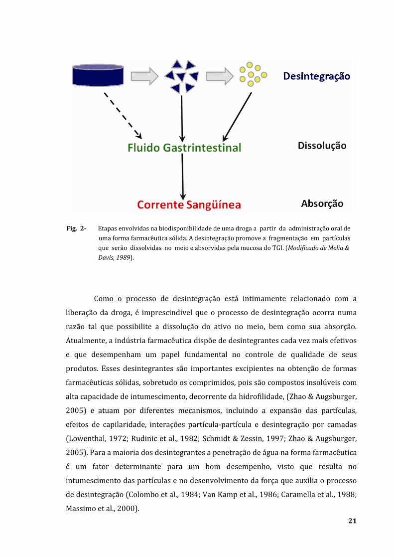
A liberação do princípio ativo contido em uma forma farmacêutica sólida
ocorre por meio do processo de desintegração (Fig. 2). A desintegração é
caracterizada como um processo tempo-dependente que ocorre sob ação de um
desintegrante e promove a fragmentação da forma farmacêutica em partículas
passíveis de serem dissolvidas e absorvidas (Lowenthal, 1972; Melia & Davis, 1989).
Se este processo for lento ou incompleto, a biodisponibilidade da droga será
comprometida, portanto, a escolha dos excipientes apropriados é fundamental
durante o desenvolvimento da formulação (Lipka & Amidon, 1999).
21
Como o processo de desintegração está intimamente relacionado com a
liberação da droga, é imprescindível que o processo de desintegração ocorra numa
razão tal que possibilite a dissolução do ativo no meio, bem como sua absorção.
Atualmente, a indústria farmacêutica dispõe de desintegrantes cada vez mais efetivos
e que desempenham um papel fundamental no controle de qualidade de seus
produtos. Esses desintegrantes são importantes excipientes na obtenção de formas
farmacêuticas sólidas, sobretudo os comprimidos, pois são compostos insolúveis com
alta capacidade de intumescimento, decorrente da hidrofilidade, (Zhao & Augsburger,
2005) e atuam por diferentes mecanismos, incluindo a expansão das partículas,
efeitos de capilaridade, interações partícula-partícula e desintegração por camadas
(Lowenthal, 1972; Rudinic et al., 1982; Schmidt & Zessin, 1997; Zhao & Augsburger,
2005). Para a maioria dos desintegrantes a penetração de água na forma farmacêutica
é um fator determinante para um bom desempenho, visto que resulta no
intumescimento das partículas e no desenvolvimento da força que auxilia o processo
de desintegração (Colombo et al., 1984; Van Kamp et al., 1986; Caramella et al., 1988;
Massimo et al., 2000).
Fig. 2- Etapas envolvidas na biodisponibilidade de uma droga a partir da administração oral de
uma forma farmacêutica sólida. A desintegração promove a fragmentação em partículas
que serão dissolvidas no meio e absorvidas pela mucosa do TGI. (Modificado de Melia &
Davis, 1989).
22
Ainda em relação ao desenvolvimento das formas farmacêuticas, um dos
maiores progressos alcançados foi a possibilidade de controlar ou modificar a
liberação de drogas no TGI humano (Ranade, 1991; Urquhart, 2000). As formas
farmacêuticas convencionais são desenvolvidas para liberar o fármaco rapidamente
após a administração. Por outro lado, formas farmacêuticas de liberação modificada
são produzidas para modularem a liberação do fármaco, prolongando ou retardando
sua dissolução (Pezzini et al., 2007). De um modo geral, essas formas farmacêuticas
promovem a liberação do fármaco gradualmente e, assim, possibilitam a manutenção
da mesma concentração terapêutica no plasma reduzindo flutuações, ou seja, evitam
níveis sub-terapêuticos ou tóxicos, além de propiciarem uma redução na freqüência
de administração, facilitando a adesão ao tratamento (Li et al., 1987; Kannan et al.,
2003).
A qualidade de um produto será assegurada se houver um equilíbrio entre a
escolha dos excipientes, do método de produção e dos perfis de liberação e dissolução
do fármaco. O maior desafio é desenvolver uma forma farmacêutica cuja liberação e
resposta clínica do fármaco possa ser monitorada por meio do estabelecimento de
uma correlação in vitro-in vivo (Emami, 2006).
3- Métodos para avaliar formas farmacêuticas in vivo
Apesar dos diferentes métodos de análise consolidados pelas principais
farmacopéias, nenhum deles é capaz de simular in vitro um meio tão complexo
quanto o TGI humano (Zahirul & Khan, 1996; Lipka & Amidon, 1999). Assim, houve a
necessidade de implementar métodos de análise capazes de caracterizar o
comportamento dessas formas farmacêuticas in vivo por meio do desenvolvimento de
técnicas não invasivas.
A Cintilografia sempre foi considerada como a técnica padrão para monitorar
formas farmacêuticas sólidas no TGI humano (Wilding et al., 2001). Essa técnica
fornece informações sobre o trânsito gastrintestinal de comprimidos, cápsulas,
sistemas de liberação controlada de drogas sendo, também, associada à
farmacocinética para avaliar o perfil de liberação de drogas (Kenyon et al., 1997;
23
Wilding et al., 2000; Brunner et al., 2003). Basicamente, essa técnica envolve a
marcação da forma farmacêutica com um radionuclídeo que emite radiação gama,
sendo seu acompanhamento realizado pela gama-câmara (Wilding et al., 2001). Além
da radiação ionizante, outras desvantagens da Cintilografia incluem a complicada
preparação e o custo dos radiofármacos utilizados nessas formulações e a
impossibilidade de fornecer a localização anatômica precisa da forma farmacêutica.
A implementação de métodos fundamentados no Biomagnetismo para
monitorar de maneira não invasiva formas farmacêuticas sólidas constitui,
atualmente, uma alternativa à medicina nuclear para a pesquisa farmacêutica (Corá et
al., 2008a). Há sensores magnéticos altamente sensíveis capazes de medir os campos
magnéticos resultantes da atividade elétrica associada aos movimentos dos íons ou
dos materiais magnéticos em resposta a um campo magnético aplicado externamente
(Williamson & Kaufman, 1981). Freqüentemente, os materiais magnéticos, quando
empregados em medidas biomédicas, são agrupados em traçadores ou marcadores
magnéticos, de acordo com a sua forma de apresentação. Os traçadores magnéticos
são definidos como partículas do material magnético dispersas em um meio,
enquanto nos marcadores as partículas estão contidas em uma forma farmacêutica
sólida (Américo, 2008). Geralmente, as ferritas e magnetitas são materiais magnéticos
muito utilizados por serem inertes e inócuos ao indivíduo (Bahadur & Giri, 2003).
Dentre as técnicas que utilizam esses princípios, destacam-se: os Dispositivos
Supercondutores de Interferência Quântica (SQUID), os Sensores Anisotrópicos
Magneto-resistivos (AMR), a Ressonância Magnética (MRI) e a Biosusceptometria de
Corrente Alternada (BAC).
Empregando-se o SQUID é possível determinar a localização, a orientação e a
evolução temporal do marcador magnético, com informações sobre o tempo de
trânsito gastrintestinal da forma farmacêutica (Weitschies et al., 2005a). Por ser um
método altamente sensível, o SQUID tem como principal desvantagem o alto custo de
manutenção, além de ser pouco viável para estudos de desintegração.
O efeito magneto-resistivo baseia-se na alteração da resistividade elétrica de
um material provocada pela aplicação de um campo magnético (Kwiatkowski &
Tumanski, 1986). Os sensores AMR foram utilizados para monitorar o trânsito
gastrintestinal de marcadores magnetizados, bem como o processo de desintegração
de comprimidos (Weitschies et al., 2005b). No entanto, como a desintegração
24
promove a perda do momento magnético do marcador, nenhuma informação
adicional pode ser adquirida pelos sensores AMR após o processo.
Recentemente, a MRI foi introduzida na pesquisa farmacêutica em alguns
poucos estudos envolvendo a caracterização de novas formulações (Richardson et al.,
2005) e monitoramento de um sistema de liberação de drogas baseado na
propriedade de gastro-retenção em humanos (Steingöetter et al., 2003). Apesar das
imagens de altíssima resolução, a MRI apresenta alguns inconvenientes como a
dificuldade de posicionamento dos voluntários e a alta incidência de artefatos de
movimento. Além disso, o alto custo de aquisição e manutenção do equipamento
restringe sua utilização na pesquisa básica.
Biosusceptometria AC
Nos últimos anos, a Biosusceptometria de Corrente Alternada (BAC)
despontou como uma técnica alternativa para estudos enfocando a motilidade
gastrintestinal (Baffa et al., 1995; Miranda et al., 1997; Romeiro et al., 2006; Américo
et al., 2007) bem como a pesquisa farmacêutica (Corá et al., 2005a). Essa técnica
utiliza bobinas de indução para registrar a variação temporal do fluxo magnético
obtida como resposta de um material ferromagnético. Esse material tem como
principal característica uma alta susceptibilidade magnética () e, por isso, produz
uma resposta intensa quando um campo magnético é aplicado ao meio biológico.
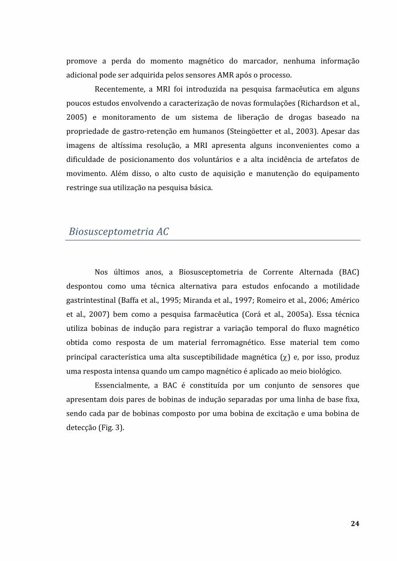
Essencialmente, a BAC é constituída por um conjunto de sensores que
apresentam dois pares de bobinas de indução separadas por uma linha de base fixa,
sendo cada par de bobinas composto por uma bobina de excitação e uma bobina de
detecção (Fig. 3).
25
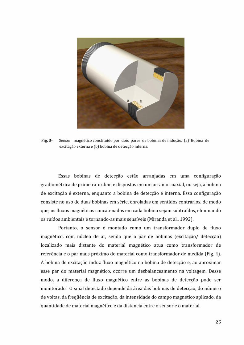
Essas bobinas de detecção estão arranjadas em uma configuração
gradiométrica de primeira-ordem e dispostas em um arranjo coaxial, ou seja, a bobina
de excitação é externa, enquanto a bobina de detecção é interna. Essa configuração
consiste no uso de duas bobinas em série, enroladas em sentidos contrários, de modo
que, os fluxos magnéticos concatenados em cada bobina sejam subtraídos, eliminando
os ruídos ambientais e tornando-as mais sensíveis (Miranda et al., 1992).
Portanto, o sensor é montado como um transformador duplo de fluxo
magnético, com núcleo de ar, sendo que o par de bobinas (excitação/ detecção)
localizado mais distante do material magnético atua como transformador de
referência e o par mais próximo do material como transformador de medida (Fig. 4).
A bobina de excitação induz fluxo magnético na bobina de detecção e, ao aproximar
esse par do material magnético, ocorre um desbalanceamento na voltagem. Desse
modo, a diferença de fluxo magnético entre as bobinas de detecção pode ser
monitorado. O sinal detectado depende da área das bobinas de detecção, do número
de voltas, da freqüência de excitação, da intensidade do campo magnético aplicado, da
quantidade de material magnético e da distância entre o sensor e o material.
Fig. 3- Sensor magnético constituído por dois pares de bobinas de indução. (a) Bobina de
excitação externa e (b) bobina de detecção interna.
26
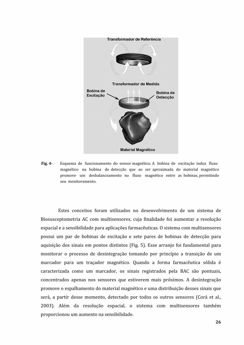
Estes conceitos foram utilizados no desenvolvimento de um sistema de
Biosusceptometria AC com multisensores, cuja finalidade foi aumentar a resolução
espacial e a sensibilidade para aplicações farmacêuticas. O sistema com multisensores
possui um par de bobinas de excitação e sete pares de bobinas de detecção para
aquisição dos sinais em pontos distintos (Fig. 5). Esse arranjo foi fundamental para
monitorar o processo de desintegração tomando por princípio a transição de um
marcador para um traçador magnético. Quando a forma farmacêutica sólida é
caracterizada como um marcador, os sinais registrados pela BAC são pontuais,
concentrados apenas nos sensores que estiverem mais próximos. A desintegração
promove o espalhamento do material magnético e uma distribuição desses sinais que
será, a partir desse momento, detectado por todos os outros sensores (Corá et al.,
2003). Além da resolução espacial, o sistema com multisensores também
proporcionou um aumento na sensibilidade.
Fig. 4- Esquema de funcionamento do sensor magnético. A bobina de excitação induz fluxo
magnético na bobina de detecção que ao ser aproximada do material magnético
promove um desbalanceamento no fluxo magnético entre as bobinas, permitindo
seu monitoramento.
27
A BAC aplicada à pesquisa farmacêutica proporcionou outra abordagem
referente ao processo de desintegração de comprimidos e cápsulas in vitro e no TGI
humano (Corá et al., 2003; 2006a,b). Refletindo seu constante aperfeiçoamento, a BAC
também demonstrou um grande potencial para obtenção de imagens magnéticas
destas formas farmacêuticas, introduzindo um novo conceito em imagens dos
sistemas biológicos (Corá et al., 2005b). Os sinais registrados pela BAC são
representados por matrizes temporais e utilizando-se ferramentas matemáticas, é
possível obter imagens seqüenciais provenientes de intervalos de tempo pré-
definidos (3 segundos cada um). Uma vez calculadas, essas seqüências de imagens são
submetidas ao processamento digital para subtração de background, ajustes de brilho
e contraste e segmentação. Por meio da segmentação das imagens é possível
quantificar, em número de pixels, a variação temporal da área da imagem magnética
no decorrer do processo, possibilitando a análise de processos como a desintegração
e o espalhamento de material magnético em uma determinada região do TGI.
Diante dos bons resultados, novas perspectivas ao estudo de formas
farmacêuticas sólidas permitiram iniciar um trabalho cujo foco principal foi o
controle de qualidade na indústria farmacêutica. De maneira inédita, a BAC foi
associada a dois métodos de análise convencionais para investigar a influência da
força de compressão na penetração de água e desenvolvimento de força durante a
Fig. 5- Sistema de Biosusceptometria AC com multisensores mostrando o par de bobinas
de excitação (a) e os sete pares de bobinas de detecção (b).
28
desintegração de comprimidos efervescentes (Corá et al., 2008b). Além disso, essa
técnica também foi utilizada para monitorar o esvaziamento gástrico e o trânsito
intestinal de um sistema multiparticulado alterando-se o estado prandial do
voluntário (Miranda et al., submetido para publicação). Além de ser um método não
invasivo e livre de radiação ionizante, a BAC não requer ambiente magneticamente
blindado e possui um custo de implementação relativamente baixo, comparando-se
com as outras técnicas biomagnéticas.
Considerando o desenvolvimento de um novo produto ou o refinamento de
uma forma farmacêutica já existente, torna-se imperativo dispor de uma técnica como
a BAC com versatilidade para avaliar sua performance. A análise dos resultados
obtidos nos trabalhos que serão apresentados permitiu demonstrar que a BAC é um
método capaz de monitorar diferentes processos farmacêuticos in vivo e in vitro,
corroborando seu potencial inovador para aplicações farmacêuticas.
29
Referências Bibliográficas1
AMÉRICO, M.F., OLIVEIRA, R.B., ROMEIRO, F.G., BAFFA, O., CORÁ, L.A., MIRANDA, J.R.A. Scintigraphic validation of AC Biosusceptometry to study the gastric motor activity and the intragastric distribution of food in humans. Neurogastroenterol. Motil., v.19, p.804-811, 2007. AMÉRICO, M.F. Desenvolvimento de método para o estudo da motilidade gastrintestinal no cão empregando a Biosusceptometria de Corrente Alternada (BAC). 2008. 111p. Tese (Doutorado) - Faculdade de Medicina, Universidade de São Paulo, Ribeirão Preto. ANSEL, H.C., POPOVICH, N.G., ALLEN, L.V. Formas farmacêuticas: considerações biofarmacêuticas. Farmacotécnica: formas farmacêuticas e sistemas de liberação de fármacos. São Paulo: Editorial Premier, 2000. 568 p. ASGHAR, L.F.A., CHANDRAN, S. Multiparticulate formulation approach to colon specific drug delivery: current perspectives. J. Pharm. Pharmaceut. Sci., v.9, p.327-338, 2006. AZARMI, S., ROA, W., LÖBENBERG, R. Current perspectives in dissolution testing of conventional and novel dosage forms. Int. J. Pharm., v.328, p.12–21, 2007. BADAWY, S.I.F., HUSSAIN, M.A. Microenvironmental pH modulation in solid dosage forms. J. Pharm. Sci., v.96, p.948-959, 2007 BAFFA, O., OLIVEIRA, R.B., MIRANDA, J.R.A., TRONCON, L.E.A. Analysis and development of an AC Biosusceptometer for orocaecal transit time measurements. Med. Biol. Eng. Comput., v.33, p.353-357, 1995. BAHADUR, D., GIRI, J. Biomaterials and magnetism. Sadhana, v.28, p.639–656, 2003. BRUNNER, M., GREINWALD, R., KLETTER, K., KVATERNIK, H., CORRADO, M.E., EICHLER, H.G., MÜLLER, M. Gastrointestinal transit and release of 5-aminosalicylic acid from 153Sm-labelled mesalazine pellets vs. tablets in male healthy volunteers. Aliment. Pharmacol. Ther., v.17, p.1163–1169, 2003. BURTON, D.D., KIM, J., CAMILLERI, M., STEPHENS, D.A., MULLAN, B.P., O’CONNOR, M.K., TALLEY, N.J. Relationship of gastric emptying and volume changes after a solid meal in humans. Am. J. Physiol. Gastrointest. Liver Physiol., v.289, p.G261–G266, 2005.
1 Referências citadas de acordo com VOLPATO, E.S.N., SILVA, R.C., PIZZANI, L. Manual de apresentação de trabalho científico: tese, dissertação e monografia. Botucatu: Divisão Técnica de Biblioteca e Documentação. 2003. 28p.
30
CAMILLERI, M.; FORD, M.J. Review article: colonic sensorimotor physiology in health, and its alteration in constipation and diarrhoeal disorders. Aliment. Pharmacol. Ther., v. 12, p. 287-302, 1998. CAMILLERI, M. Integrated upper gastrointestinal response to food intake. Gastroenterology, v.131, p.640-658, 2006. CARAMELLA, C., COLOMBO, P., CONTE, U., FERRARI, F., GAZZANIGA, A., LA MANNA, A., PEPPAS, N.A. A physical analysis of the phenomenon of tablet disintegration. Int. J. Pharm., v. 44, p.177–186, 1988. CHAWLA, G., GUPTA, P., KORADIA, V., BANSAL, A.K. A means to address regional variability in intestinal drug absorption. Pharm. Technol., v.27, p.50-68, 2003. CHOURASIA, MK; JAIN, SK. Pharmaceutical approaches to colon target drug delivery systems. J. Pharm. Pharmaceut. Sci., v.6, p.33-66, 2003. CHRISTENSEN, J. Motility of the colon. In: Physiology of the Gastrointestinal Tract. 2nd New York: Raven Press, 1987, p. 665-693. COLOMBO, P., CONTE, U., CARAMELLA, C., GEDDO, M., LA MANNA, A. Disintegration force as a new formulation parameter. J. Pharm. Sci., v. 73, p.701–705, 1884. CORÁ, L.A., AMÉRICO, M.F., OLIVEIRA, R.B., BAFFA, O., MORAES, R., ROMEIRO, F.G., MIRANDA, J.R.A. Disintegration of magnetic tablets in human stomach evaluated by alternate current Biosusceptometry. Eur. J. Pharm. Biopharm., v.56, p.413–420, 2003. CORÁ, L.A., ROMEIRO, F.G., STELZER, M., AMÉRICO, M.F., OLIVEIRA, R.B., BAFFA, O., MIRANDA, J.R.A. AC biosusceptometry in the study of drug delivery. Adv. Drug Deliv. Rev., v.57, p.1223-1241, 2005a. CORÁ, L.A., ANDREIS, U., ROMEIRO, F.G., AMÉRICO, M.F., OLIVEIRA, R.B., BAFFA, O., MIRANDA, J.R.A. Magnetic images of the disintegration process of tablets in the human stomach by AC Biosusceptometry. Phys. Med. Biol., v.50, p.5523–5534, 2005b. CORÁ, L.A., ROMEIRO, F.G., STELZER, M., AMÉRICO, M.F., OLIVEIRA, R.B., BAFFA, O., STELZER, M., MIRANDA, J.R.A. Gastrointestinal transit and disintegration of enteric coated magnetic tablets assessed by AC Biosusceptometry. Eur. J. Pharm. Sci., v.27, p.1-8, 2006a. CORÁ, L.A., ROMEIRO, F.G., PAIXÃO, F.C., AMÉRICO, M.F., OLIVEIRA, R.B., BAFFA, O., MIRANDA, J.R.A. Enteric coated magnetic HPMC capsules evaluated in human gastrointestinal tract by AC Biosusceptometry. Pharm. Res., v.23, p.1809-1816, 2006b. CORÁ, L.A., MIRANDA, J.R.A., AMÉRICO, M.F., OLIVEIRA, R.B., BAFFA, O. Biomagnetic approaches applied to drug delivery studies. In: Hartmann, A.O., Neumann, L.K. Drugs: approval and evaluation, delivery and control. New York: Nova Science Publishers, Inc., 2008 (no prelo).
31
CORÁ, L.A., FONSECA, P.R., AMÉRICO, M.F., OLIVEIRA, R.B., BAFFA, O., MIRANDA, J.R.A. Influence of compression forces on tablets disintegration by AC Biosusceptometry. Eur. J. Pharm. Biopharm., v.69, p.372-379, 2008b. CORRIGAN, O.I., DEVLIN, Y., BUTLER, J. Influence of dissolution buffer composition on ketoprofen release from ER products and in vitro-in vivo correlation. Int. J. Pharm., v.254, p.147-154, 2003. COUPE, A.J., DAVIS, S.S., WILDING, I.R. Variation in gastrointestinal transit of pharmaceutical dosage forms in healthy subjects. Pharm. Res., v.8, p.360-364, 1991. DAVIS, S.S., HARDY, J.G., FARA, J.W. Transit of pharmaceutical dosage forms through the small intestine. Gut, v.27, p.886-892, 1986. DAVIS, S.S., STOCKWELL, A.F., TAYLOR, M.J., HARDY, J.G., WHALLEY, D.R., WILSON, C.G., BECHGAARD, H., CHRISTENSEN, F.N. The effect of density on the gastric emptying of single- and multiple-unit dosage forms. Pharm. Res., v.3, p.208-213, 1986. DAVIS, S.S. Formulation strategies for absorption windows. Drug Disc. Today, v.10, p.249-257, 2005. DITTGEN, M., DURRANI, M., LEHMANN, K. Acrylic polymers: a review of pharmaceutical applications. STP Pharm. Sci., v.7, p.406-437, 1997. DRESSMAN, J.B., BERARDI, R.R., DERMENTZOGLOU, L.C., RUSSEL, T.L., SCHMALTZ, S.P., BARNETT, J.L., JARVENPAA, K.M. Upper gastrointestinal (GI) pH in young, healthy men and women. Pharm. Res., v.7, p.756–761, 1990. DRESSMAN, J.B., BASS, P., RITSCHEL, W.A., FRIEND, D.R., RUBINSTEIN, A., ZIV, E. Gastrointestinal parameters that influence oral medications. J. Pharm. Sci., v.82, p.857-872, 1993. DRESSMAN, J.B., AMIDON, G.L., REPPAS, C., SHAH, V.P. Dissolution testing as a prognostic tool for oral drug absorption: immediate release dosage forms. Pharm. Res., v.15, p.11-22, 1998. EMAMI, J. In vitro-in vivo correlation: From theory to applications. J. Pharma. Pharm. Sci., v.9, p.31-51, 2006. EVANS, D.F., PYE, G., BRAMLEY, R., CLARK, A.G., DYSON, T.J., HARDCASTLE, J.D. Measurement of gastrointestinal pH profiles in normal ambulant human subjects. Gut, v.29, p.1035-1041, 1998. FREIRE, A.C., PODCZECK, F., SOUSA, J., VEIGA, F. Liberação específica de fármacos para administração no cólon por via oral. I- O cólon humano como local de liberação de fármacos. Rev. Bras. Cienc. Farm., v.42, p.319-335, 2006a.
32
FREIRE, A.C., PODCZECK, F., SOUSA, J., VEIGA, F. Liberação específica de fármacos para administração no cólon por via oral. II- Tipos de sistemas utilizados. Rev. Bras. Cienc. Farm., v.42, p.337-355, 2006b. FREXINOS, F., DELVAUX, M. Colonic motility. In: KUMAR, D., WINGATE, D. (Eds.). An Illustrated Guide to Gastrointestinal Motility. London: Churchill Livingstone, 1993, 427– 448. GANDHI, R., KAUL, C.L., PANCHAGNULA, R. Extrusion and spheronization in the development of oral controlled-release dosage forms. Pharm. Sci. Technol. Today, v.2, p.160-170, 1999. HANCOCK, B.C., YORK, P., ROWE, R.C. The use of solubility parameters in pharmaceutical dosage form design. Int. J. Pharm., v. 148, p.1-21, 1997. HASLER, W.L. Physiology of gastric motility and gastric emptying. In: YAMADA T., ALPERS, D.H., LAINE, L., OWYANG, C., POWELL, D.W. Textbook of Gastroenterology. 3rd ed. Philadelphia: Lippincott Williams & Wilkins, 1999. Disponível em: http://www.portaldapesquisa.com.br/databases/login?cust=unesp&area=clear&action=homepage Acesso em: 20/05/2003. HELLSTRÖM, P.M., GRYBÄCK, P., JACOBSSON, H. The physiology of gastric emptying. Best Pract. Res. Clin. Anaesth., v.20, p.397-407, 2006. HOLT, S., REID, J., TAYLOR, T.V., TOTHILL, P., HEADING, R.C. Gastric emptying of solids in man. Gut, v.23, p.292-296, 1982.
HÖRTER, D., DRESSMAN, J.B. Influence of physicochemical properties on dissolution of drugs in the gastrointestinal tract. Adv. Drug Deliv. Rev., v.46, p.75–87, 2001. JAIN, S. Mechanical properties of powder for compaction and tableting: an overview. Pharm. Sci. Technol. Today, v.2, p.20-31, 1999. JENQUIN, M.R., LIEBOWITZ, S.M., SARABIA, R.E., McGINITY, J.W. Physical and chemical factors influencing the release of drugs from acrylic resin films. J. Pharm. Sci., v.79, p.811-816, 1990. JIVRAJ, M., MARTINI, L.G., THOMSON, C.M. An overview of the different excipients useful for the direct compression of tablets. Pharm. Sci. Tech. Today, v.3, p.58-63, 2000. KANNAN, V., KANDARAPU, V., GARG, S. Optimization techniques for the design and development of novel drug delivery systems. Part II. Pharm. Technol., v.27, p.102-118, 2003. KENYON, C.J., NARDI, R.V., WONG, D., HOOPER, G., WILDING, I.R., FRIEND, D.R. Colonic delivery of dexamethasone: a pharmacoscintigraphic evaluation. Aliment. Pharmacol. Ther., v.11, p.205-213, 1997.
33
KIMURA, T., HIGAKI, K. Gastrointestinal transit and drug absorption. Biol. Pharm. Bull., v.25, p.149—164, 2002. KUO, B., McCALLUM, R.W., KOCH, K.L., SITRIN, M.D., WO–, J.M., CHEY, W.D., HASLER, W.L., LACKNER, J.M., KATZ, L.A., SEMLER, J.R., WILDING, G.E., PARKMAN, H.P. Comparison of gastric emptying of a nondigestible capsule to a radio-labelled meal in healthy and gastroparetic subjects. Aliment. Pharmacol. Ther., v.27, p.186-196, 2008. KWIATKOWSKI, W., TUMANSKI, S. The permalloy magnetoresistive sensors - properties and applications. J. Phys. E-Sci. Instrum., v.19, p.502-515, 1986. LI, V.H.K., ROBINSON, J.R., LEE, V.H.L. Influence of drug properties and routes of drug administration on the design of sustained and controlled release systems. In: ROBINSON, J.R., LEE, V.H.L. (Eds.). Controlled drug delivery: fundamentals and applications. 2nd Ed. New York: Marcel Dekker, 1987, 4–61. LINDAHL, A., UNGELL, A.-L., KNUTSON, L., LENNERNÄS, H. Characterization of fluids from the stomach and proximal jejunum in men and women. Pharm. Res., v.14, p.497-502, 1997. LIPKA, E., AMIDON, G.L. Setting bioequivalence requirements for drug development based on preclinical data: optimizing oral drug delivery systems. J. Control. Release, v.62, p.41-49, 1999. LOWENTHAL, W. Disintegration of tablets. J. Pharm. Sci., v.61, p.1695–1711, 1972. MARTINEZ, M.N., AMIDON, G.L. A mechanistic approach to understanding the factors affecting drug absorption: a review of fundamentals. J. Clin. Pharmacol., v.42, p.620-643, 2002. MASAOKA, Y., TANAKA, Y., KATAOKA, M., SAKUMA, S., YAMASHITA, S. Site of drug absorption after oral administration: assessment of membrane permeability and luminal concentration of drugs in each segment of gastrointestinal tract. Eur. J. Pharm. Sci., v. 29, p. 240–250, 2006. MASSIMO, G., CATELLANI, P.L., SANTI, P., BETTINI, R., VAONA, G., BONFANTI, A., MAGGI, L., COLOMBO, P. Disintegration propensity of tablets evaluated by means of disintegrating force kinetics. Pharm. Develop. Technol., v.5, p.163–169, 2000. MELIA, C.D., DAVIS, S.S. Review article: mechanisms of drug release from tablets and capsules. I: Disintegration. Aliment. Pharmacol. Ther., v.3, p.223–232, 1989. MIRANDA, J.R.A., BAFFA, O., OLIVEIRA, R.B., MATSUDA, N.M. An AC Biosusceptometer to study gastric emptying. Med. Phys., v.19, p.445-448, 1992. MIRANDA, J.R.A, OLIVEIRA, R.B., SOUSA, P.L., BRAGA, F.J.H., BAFFA, O. A novel biomagnetic method to study antral contractions. Phys. Med. Biol., v.42, p.1791-1799, 1997.
34
MIRANDA, J.R.A., CORÁ, L.A., AMÉRICO, M.F., ROMEIRO, F.G. AC Biosusceptometry to evaluate the gastrointestinal transit of pellets under influence of prandial state. International Journal of Pharmaceutics, 2008 (artigo submetido para publicação). NGUYEN, N.Q., FRASER, R.J., BRYANT, L.K., HOLLOWAY, R.H. Functional association between proximal and distal gastric motility during fasting and duodenal nutrient stimulation in humans. Neurogastroenterol. Motil., v.19, p.638-645, 2007. OGURA, T., FURUYA, Y., MATSUURA, S. HPMC capsules: an alternative to gelatin. Pharm. Technol. Europe, v.10, p.32–42, 1998. PEZZINI, B.R., SILVA, M.A.S., FERRAZ, H.G. Formas farmacêuticas sólidas orais de liberação prolongada: sistemas monolíticos e multiparticulados. Rev. Bras. Cienc. Farm., v. 43, p.491-502, 2007. PIFFERI, G., RESTANI, P. The safety of pharmaceutical excipients. Il Farmaco, v.58, p.541-550, 2003.
PRICE, J.M.C., DAVIS, S.S., WILDING, I.R. Characterization of colonic transit of nondisintegrating tablets in healthy subjects. Dig. Dis. Sci., v.38, p.1015-1021, 1993. QUIGLEY, EMM. Gastric and small intestinal motility in health and disease. Gastroenterol. Clin. North Am., v.25, 113– 145, 1996. RANADE, V.V. Drug delivery systems 5A. Oral drug delivery. J. Clin. Pharmacol., v.31, p.2-16, 1991. RICHARDSON, J.C., BOWTEL, R.W., MÄDER, K., MELIA, C.D. Pharmaceutical applications of magnetic resonance imaging (MRI). Adv. Drug Deliv. Rev., v.57, p.1191-1209, 2005. ROMEIRO, F.G., CORÁ, L.A., ANDREIS, U., AMÉRICO, M.F., OLIVEIRA, R.B., BAFFA, O., MIRANDA, J.R.A. A novel biomagnetic approach to study caecocolonic motility in humans. Neurogastroenterol. Motil., v.18, p.1078-1083, 2006. ROUGE, N., BURI, P., DOELKER, E. Drug absorption sites in the gastrointestinal tract and dosage forms for site-specific delivery. Int. J. Pharm., v.136, p.117-139, 1996. RUDNIC, E.M. Rhodes, C.T., Welch, S., Bernardo, P. Evaluations of the mechanism of disintegrant action. Drug Dev. Ind. Pharm., v.8, p.87-109, 1982. SASTRY, S.V., NYSHADHAM, J.R., FIX, J.A. Recent technological advances in oral drug delivery. Pharm. Sci. Tech. Today, v.3, p.138-145, 2000. SCHMIDT, J., ZESSIN, G. Investigation of different vegetable cell wall as disintegrant in direct compressing of tablets. Drug Dev. Ind. Pharm., v.6, p.527-532, 1997.
35
SIEGEL, J.A., URBAIN, J.-L., ADLER, L.P., CHARKES, N.D., MAURER, A.H., KREVSKY, B., KNIGHT, L.C., FISHER, R.S., MALMUD, L.S. Biphasic nature of gastric emptying. Gut, v.29, p.85-89, 1988. SIMONIAN, H.P., MAURER, A.H., KNIGHT, L.C., KANTOR, S., KONTOS, D., MEGALOOIKONOMOU, V., FISHER, R.S., PARKMAN, H.P. Simultaneous assessment of gastric accommodation and emptying: studies with liquid and solid meals. J. Nucl. Med., v.45, p.1155–1160, 2004. SINGH, B.N. Effects of food on clinical pharmacokinetics. Clin. Pharmacokinet., v.37, p.213-255, 1999. SHAREEF, M.A., KHAR, R.K., AHUJA, A., AHMAD, F.J., RAGHAVA, S. Colonic Drug Delivery: an updated review. AAPS Pharm. Sci., v. 5, p.1-25, 2003. STEINGÖTTER, A., WEISHAUPT, D., KUNZ, P., MÄDER, K., LENGSFELD, H., THUMSHIRN, M., BOESIGER, P., FRIED, M., SCHWIZER, W. Magnetic resonance imaging for the in vivo evaluation of gastric-retentive tablets. Pharm. Res., v.20, p.2001-2007, 2003. STREUBEL, A., SIEPMANN, J., BODMEIER, R. Drug delivery to the upper small intestine window using gastroretentive technologies. Curr. Opin. Pharmacol., v.6, p.501-508, 2006. TALUKDER, R., FASSIHI, R. Gastroretentive Delivery Systems: a mini review. Drug Develop Ind. Pharm., v.30, p.1019–1028, 2004. URQUHART, J. Controlled drug delivery: therapeutic and pharmacological aspects. J. Int. Med., v.248, p.357-376, 2000. VAN KAMP, H.V., BOLHUIS, G.K., DE BOER, A.H., LERK, C.F., LIE- A-HUEN, L. The role of water uptake on tablet disintegration. Pharm. Acta Helv., v.61, p.22–29, 1986. WEITSCHIES, W., KOSCH, O., MÖNNIKES, H., TRAHMS, L. Magnetic Marker Monitoring: An application of biomagnetic measurement instrumentation and principles for the determination of the gastrointestinal behavior of magnetically marked solid dosage forms. Adv. Drug Deliv. Rev., v.57, p.1210– 1222, 2005a. WEITSCHIES, W., WEDEMEYER, R.S., KOSCH, O., FACH, K., NAGEL, S., SÖDERLIND, E., TRAHMS, L., ABRAHAMSSON, B., MÖNNIKES, H. Impact of the intragastric location of extended release tablets on food interactions. J. Control. Release, v.108, p.375–385, 2005b. WILDING, I.R., KENYON, C.J., HOOPER, G. Gastrointestinal spread of oral prolonged-release mesalazine microgranules (Pentasa) dosed as either tablets or sachet. Aliment. Pharmacol. Ther., v.14, p.163-169, 2000. WILDING, I.R., COUPE, A.J., DAVIS, S.S. The role of -scintigraphy in oral drug delivery. Adv. Drug Deliv. Rev., v.46, p.103-124, 2001.
36
WILLIAMSON, S.J., KAUFMAN, L. Biomagnetism. J. Magn. Magn. Mater., v.22, p.129-201, 1981. ZAHIRUL, M., KHAN, I. Dissolution testing for sustained or controlled release oral dosage forms and correlation with in vivo data: challenges and opportunities. Int. J. Pharm., v.140, p.131-143, 1996. ZHAO, N., AUGSBURGER, L.L. Functionality comparison of 3 classes of superdisintegrants in promoting aspirin tablet disintegration and dissolution. AAPS Pharm. Sci. Tech., v.6, p.634-640, 2005a. ZIESSMAN, H.A., ATKINS, F.B., VEMULAKONDA, U.S., TALL, J., HARKNESS, B., FAHEY, F.H. Lag phase quantification for solid gastric emptying studies. J. Nucl. Med., v.37, p.1639-1643, 1996.
Objetivos
“Quando você tem uma meta, o que era um obstáculo passa a ser uma etapa de um
dos planos.” Gerhard Erich Boehme
37
Objetivos
Essa tese incorporou quatro artigos que utilizaram a BAC para avaliar
diferentes parâmetros farmacêuticos em comprimidos, cápsulas e sistemas
multiparticulados. Todos esses trabalhos empregaram a BAC tendo como objetivo
principal:
Obter imagens da desintegração de comprimidos in vitro e no estômago
humano;
Avaliar a influência da força de compressão em comprimidos para validar a
técnica biomagnética para o controle de qualidade em processos
farmacêuticos;
Determinar o tempo de trânsito gastrintestinal em humanos de cápsulas de
hidroxipropilmetilcelulose (HPMC) revestidas e quantificar o processo de
desintegração na região ileocolônica;
Avaliar um sistema multiparticulado sob influência do estado prandial no
esvaziamento gástrico e trânsito intestinal.
Capítulo 1
Magnetic images of the disintegration process of tablets in the human stomach
by AC Biosusceptometry
Physics in Medicine and Biology, v.50, p. 5523–5534, 2005
38
S IN MEDICINE AND BIOLOGY
doi:10.1088/0031-9155/50/23/007
iosusceptometry
5523
INSTITUTE OF PHYSICS PUBLISHING PHYSIC
Phys. Med. Biol. 50 (2005) 5523–5534
Magnetic images of the disintegration process of tablets in the human stomach by ac b
L A Corá1, U Andreis1, F G Romeiro2, M F Américo3, R B Oliveira3 , 1 O Baffa4 and J R A Miranda
1 Departamento de Física e Biofísica, IBB, UNESP, Botucatu, SP, Brazil
2 Departamento de Clínica Médica, FMB, UNESP, Botucatu, SP, Brazil
3 Departamento de Clínica Médica, FMRP, USP, Ribeirão Preto, SP, Brazil
4 Departamento de Física e Matemática, FFCLRP, USP, Ribeirão Preto, SP, Brazil
E-mail: [email protected]
Received 5 April 2005, in final form 16 September 2005 Published 9 November 2005 Online at stacks.iop.org/PMB/50/5523
Abstract
Oral administration of solid dosage forms is usually preferred in drugtherapy. Conventional imaging methods are essential tools to investigate the in vivo performance of these formulations. The non-invasive technique of ac biosusceptometry has been introduced as an alternative in studiesfocusing on gastrointestinal motility and, more recently, to evaluate thebehaviour of magnetic tablets in vivo. The aim of this work was to employ a multisensor ac biosusceptometer system to obtain magnetic images ofdisintegration of tablets in vitro and in the human stomach. The results showed that the transition between the magnetic marker and the magnetic tracer characterized the onset of disintegration (t50) and occurred in a short time interval (1.1 ± 0.4 min). The multisensor ac biosusceptometer wasreliable to monitor and analyse the in vivo performance of magnetic tablets showing accuracy to quantify disintegration through the magnetic imagesand to characterize the profile of this process.
(Some figures in this article are in colour only in the electronic version)
1. Introduction Medical imaging methods are essential to evaluate anatomically and functionally internal structuresof the human body. Imaging techniques also play an important role in pharmaceutical research sincethey provide valuable information on the in vivo performance for any dosage forms (Wilson et al 1997, Singh and Waluch 2000, Newman et al 2003).
Oral administration is the most popular method for drug therapy and the active substances areconveniently administered in a solid form (Sastry et al 2000). Drug delivery occurs by the disintegration of the solid dosage form and promotes drug release to be absorbed in thegastrointestinal tract (Melia and Davis 1989). Physiological factors and the formulation design
0031-9155/05/235523+12$30.00 © 2005 IOP Publishing Ltd Printed in the UK
39
have significant influences on the disintegration and drug absorption and therefore in the safety and efficacy of the drug product (Dressman et al 1993, Lipka and Amidon 1999).
For these reasons, imaging modalities introduced a new perspective for the in vivo investigation of drug delivery. γ -scintigraphy is the standard method widely used to assess complexinteractions between the drug, the dosage form and the gastrointestinal physiology (Wilding et al 2001). The main drawbacks of this method are the exposure of patients to the ionizingradiation, precluding repetitive assays with a single subject and the complicated and expensive preparation of the radiopharmaceuticals.
The development of radiation-free techniques provides a non-invasive approach to acquire information about the in vivo performance of oral dosage forms within the gastrointestinal tract.Magnetic resonance imaging (MRI) was recently employed to monitor the intragastric course of a labelled and gastric-retentive tablet (Steingöetter et al 2003a, 2003b). Although there are favourable advantages for this purpose, including high temporal and spatial resolution, the use of MRI in studying oral delivery systems is restricted due to the cost of equipment and limitations in positioning of the patients, since up to now most MRI units do not operate with the subject in an orthostatic position.
al 5524 L A Corá et
Biomagnetic methods represent a feasible and promising alternative in clinical,physiological and pharmaceutical research. Magnetic fields associated with the flow of electrical activity or as a result of ingestion of a magnetically labelled dosage form are detectable by multichannel SQUID (superconducting quantum interference device) systems(Weitschies et al 1997, 2001,Hu et al 2000).
de scale.
Whereas the SQUID has been developed to detect extremely weak magnetic fields, the need for a magnetically shielded environment and an expensive operational cost limit its use on a wi
In the past few years, alternating current biosusceptometry (ACB) has been becoming an interesting and valuable tool in gastroenterology research. ACB uses induction coils for recording the magnetic flux variation obtained from the response of a magnetic materialingested (Miranda et al 1992). This material has a high magnetic susceptibility that produces a strong response when an alternating magnetic field is applied in a biological system containing small amounts of ferrite. ACB showed accuracy to evaluate, physiologically, gastric emptying(Baffa et al 1995, Oliveira et al 1996) and gastric motility in humans (Miranda et al 1997) and dogs (Moraes et al 2003) as well as colonic motility (Ferreira et al 2004). ACB was also employed to obtain magnetic imaging of ferromagnetic tracers in vitro, introducing a novel concept in imaging of biological systems (Moreira et al 2000).
Recently, a new instrumental arrangement comprising a multisensor system (multisensorACB) was implemented and proposed for the first time to characterize the disintegration process of tablets in vitro and in the human stomach, through the acquisition of magnetic signals (Corá et al 2003). The results obtained were very satisfactory and emphasized theimportance of studying how pharmaceutical forms behave in the human gastrointestinal tract(Corá et al 2005).
The aim of this investigation was to employ multisensor ACB to image the disintegration of tablets in vitro and in the human stomach.
2. Materials and methods 2.1. Fundamentals
The ac biosusceptometer consists of two pairs of coils separated by a fixed distance (baseline), where each pair of coils is composed of an excitation coil (outer) and a detection coil (inner), in a first-order gradiometric configuration (figure 1).
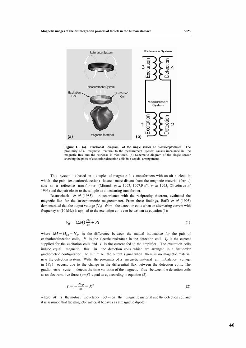
40
5525 5525 Magnetic images of the disintegration process of tablets in the human stomach
Figure 1. (a) Functional diagram of the single sensor ac biosusceptometer. The Figure 1. (a) Functional diagram of the single sensor ac biosusceptometer. The proximity of a magnetic material to the measurement system causes imbalance in the magnetic flux and the response is monitored. (b) Schematic diagram of the single sensorshowing the pairs of excitation/detection coils in a coaxial arrangement.
ring transformer.
∆
This system is based on a couple of magnetic flux transformers with an air nucleus in which the pair (excitation/detection) located more distant from the magnetic material (ferrite) acts as a reference transformer (Miranda et al 1992, 1997,Baffa et al 1995, Oliveira et al 1996) and the pair closer to the sample as a measu
Bastuscheck et al (1985), in accordance with the reciprocity theorem, evaluated themagnetic flux for the susceptometric magnetometer. From these findings, Baffa et al (1995) demonstrated that the output voltage (Vd) from the detection coils when an alternating current with frequency ω (10 kHz) is applied to the excitation coils can be written as equation (1):
(1)
∆
where is the difference between the mutual inductance for the pair ofexcitation/detection coils, R is the electric resistance in the detection coil, is the current supplied for the excitation coils and I is the current fed to the amplifier. The excitation coils induce equal magnetic flux in the detection coils which are arranged in a first-order gradiometric configuration, to minimize the output signal when there is no magnetic material near the detection system. With the proximity of a magnetic material an imbalance voltage in ( ) occurs, due to the change in the differential flux between the detection coils. The gradiometric system detects the time variation of the magnetic flux between the detectioas an electromotive force equa
n coils l to , according to equation (2).
∆ (2)
where
is the mutual inductance between the magnetic material and the detection coil and it is assumed that the magnetic material behaves as a magnetic dipole.
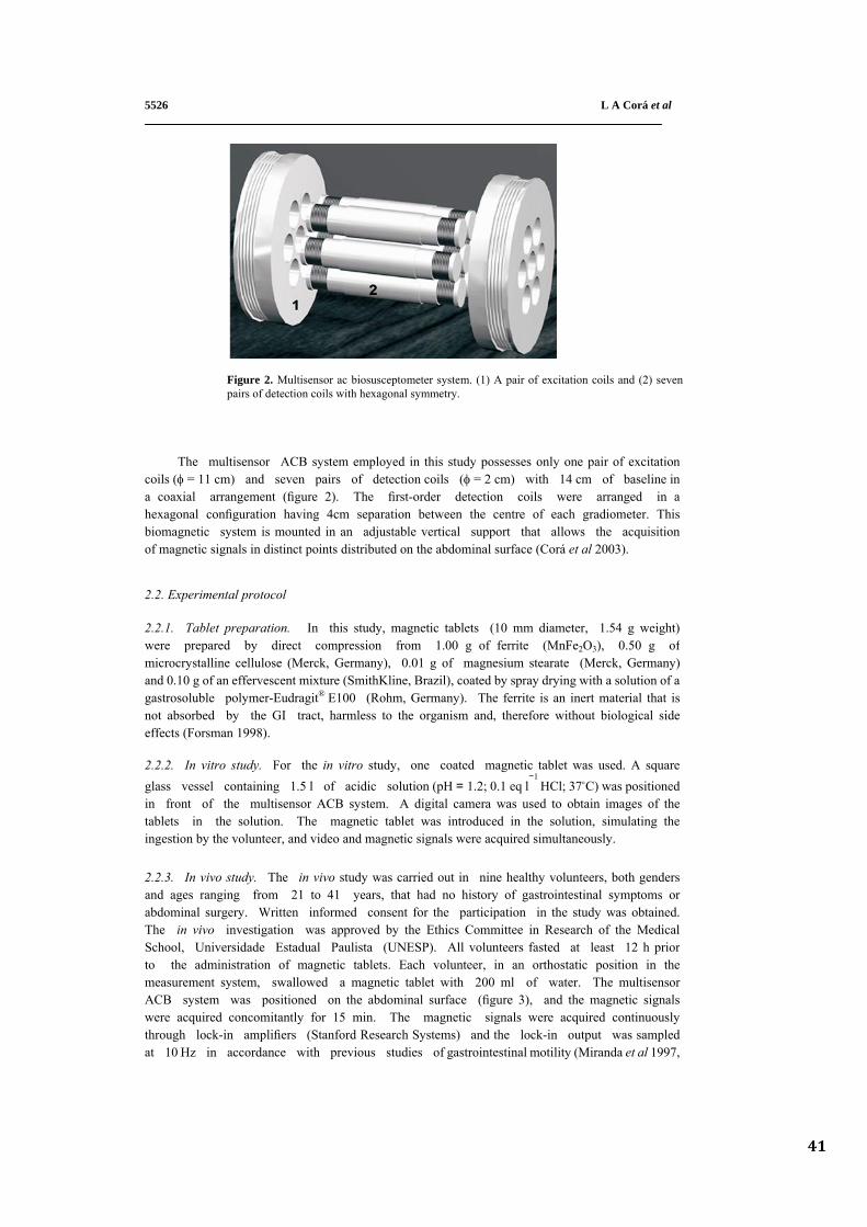
41
L A Corá et al5526
Figure 2. Multisensor ac biosusceptometer system. (1) A pair of excitation coils and (2) sevenpairs of detection coils with hexagonal symmetry.
The multisensor ACB system employed in this study possesses only one pair of excitationcoils (φ = 11 cm) and seven pairs of detection coils (φ = 2 cm) with 14 cm of baseline in a coaxial arrangement (figure 2). The first-order detection coils were arranged in a hexagonal configuration having 4cm separation between the centre of each gradiometer. Thisbiomagnetic system is mounted in an adjustable vertical support that allows the acquisition of magnetic signals in distinct points distributed on the abdominal surface (Corá et al 2003).
2.2. Experimental protocol
2.2.1. Tablet preparation. In this study, magnetic tablets (10 mm diameter, 1.54 g weight) were prepared by direct compression from 1.00 g of ferrite (MnFe2O3), 0.50 g of microcrystalline cellulose (Merck, Germany), 0.01 g of magnesium stearate (Merck, Germany) and 0.10 g of an effervescent mixture (SmithKline, Brazil), coated by spray drying with a solution of agastrosoluble polymer-Eudragit® E100 (Rohm, Germany). The ferrite is an inert material that is not absorbed by the GI tract, harmless to the organism and, therefore without biological sideeffects (Forsman 1998).
;2.2.2. In vitro study. For the in vitro study, one coated magnetic tablet was used. A square glass vessel containing 1.5 l of acidic solution (pH = 1.2 0.1 eq l
−1 HCl; 37°C) was positioned
in front of the multisensor ACB system. A digital camera was used to obtain images of the tablets in the solution. The magnetic tablet was introduced in the solution, simulating theingestion by the volunteer, and video and magnetic signals were acquired simultaneously.
2.2.3. In vivo study. The in vivo study was carried out in nine healthy volunteers, both genders and ages ranging from 21 to 41 years, that had no history of gastrointestinal symptoms orabdominal surgery. Written informed consent for the participation in the study was obtained. The in vivo investigation was approved by the Ethics Committee in Research of the MedicalSchool, Universidade Estadual Paulista (UNESP). All volunteers fasted at least 12 h prior to the administration of magnetic tablets. Each volunteer, in an orthostatic position in the measurement system, swallowed a magnetic tablet with 200 ml of water. The multisensor ACB system was positioned on the abdominal surface (figure 3), and the magnetic signals were acquired concomitantly for 15 min. The magnetic signals were acquired continuously through lock-in amplifiers (Stanford Research Systems) and the lock-in output was sampled at 10 Hz in accordance with previous studies of gastrointestinal motility (Miranda et al 1997,
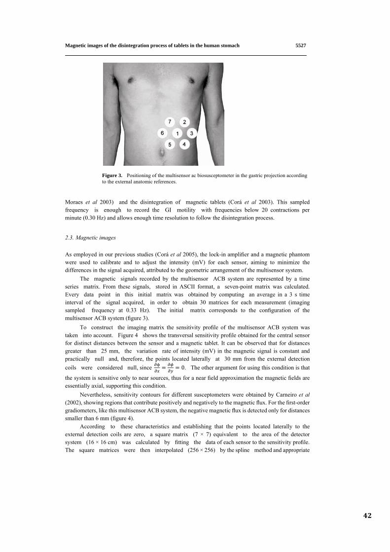
42
5527
Magnetic images of the disintegration process of tablets in the human stomach
Figure 3. Positioning of the multisensor ac biosusceptometer in the gastric projection accordingto the external anatomic references.
Moraes et al 2003) and the disintegration of magnetic tablets (Corá et al 2003). This sampled frequency is enough to record the GI motility with frequencies below 20 contractions per minute (0.30 Hz) and allows enough time resolution to follow the disintegration process.
2.3. Magnetic images
As employed in our previous studies (Corá et al 2005), the lock-in amplifier and a magnetic phantom were used to calibrate and to adjust the intensity (mV) for each sensor, aiming to minimize thedifferences in the signal acquired, attributed to the geometric arrangement of the multisensor system.
The magnetic signals recorded by the multisensor ACB system are represented by a time series matrix. From these signals, stored in ASCII format, a seven-point matrix was calculated. Every data point in this initial matrix was obtained by computing an average in a 3 s time interval of the signal acquired, in order to obtain 30 matrices for each measurement (imaging sampled frequency at 0.33 Hz). The initial matrix corresponds to the configuration of the multisensor ACB system (figure 3).
To construct the imaging matrix the sensitivity profile of the multisensor ACB system was taken into account. Figure 4 shows the transversal sensitivity profile obtained for the central sensor for distinct distances between the sensor and a magnetic tablet. It can be observed that for distances greater than 25 mm, the variation rate of intensity (mV) in the magnetic signal is constant and practically null and, therefore, the points located laterally at 30 mm from the external detection coils were considered null, since 0. The other argument for using this condition is that
the system is sensitive only to near sources, thus for a near field approximation the magnetic fields are essentially axial, supporting this condition.
Nevertheless, sensitivity contours for different susceptometers were obtained by Carneiro et al (2002), showing regions that contribute positively and negatively to the magnetic flux. For the first-order gradiometers, like this multisensor ACB system, the negative magnetic flux is detected only for distances smaller than 6 mm (figure 4).
According to these characteristics and establishing that the points located laterally to theexternal detection coils are zero, a square matrix (7 × 7) equivalent to the area of the detector system (16 × 16 cm) was calculated by fitting the data of each sensor to the sensitivity profile. The square matrices were then interpolated (256 × 256) by the spline method and appropriate
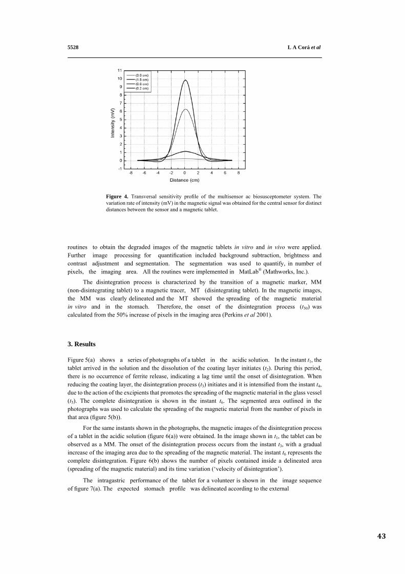
43
L A Corá et al 5528
Figure 4. Transversal sensitivity profile of the multisensor ac biosusceptometer system. The variation rate of intensity (mV) in the magnetic signal was obtained for the central sensor for distinctdistances between the sensor and a magnetic tablet.
routines to obtain the degraded images of the magnetic tablets in vitro and in vivo were applied. Further image processing for quantification included background subtraction, brightness and contrast adjustment and segmentation. The segmentation was used to quantify, in number of pixels, the imaging area. All the routines were implemented in MatLab® (Mathworks, Inc.).
ns et al 2001).
The disintegration process is characterized by the transition of a magnetic marker, MM (non-disintegrating tablet) to a magnetic tracer, MT (disintegrating tablet). In the magnetic images, the MM was clearly delineated and the MT showed the spreading of the magnetic material in vitro and in the stomach. Therefore, the onset of the disintegration process (t50) was calculated from the 50% increase of pixels in the imaging area (Perki
3. Results
Figure 5(a) shows a series of photographs of a tablet in the acidic solution. In the instant t1, the tablet arrived in the solution and the dissolution of the coating layer initiates (t2). During this period, there is no occurrence of ferrite release, indicating a lag time until the onset of disintegration. When reducing the coating layer, the disintegration process (t3) initiates and it is intensified from the instant t4, due to the action of the excipients that promotes the spreading of the magnetic material in the glass vessel (t5). The complete disintegration is shown in the instant t6. The segmented area outlined in the photographs was used to calculate the spreading of the magnetic material from the number of pixels in that area (figure 5(b)).
For the same instants shown in the photographs, the magnetic images of the disintegration processof a tablet in the acidic solution (figure 6(a)) were obtained. In the image shown in t1, the tablet can be observed as a MM. The onset of the disintegration process occurs from the instant t3, with a gradual increase of the imaging area due to the spreading of the magnetic material. The instant t6 represents the complete disintegration. Figure 6(b) shows the number of pixels contained inside a delineated area(spreading of the magnetic material) and its time variation (‘velocity of disintegration’).
The intragastric performance of the tablet for a volunteer is shown in the image sequence of figure 7(a). The expected stomach profile was delineated according to the external
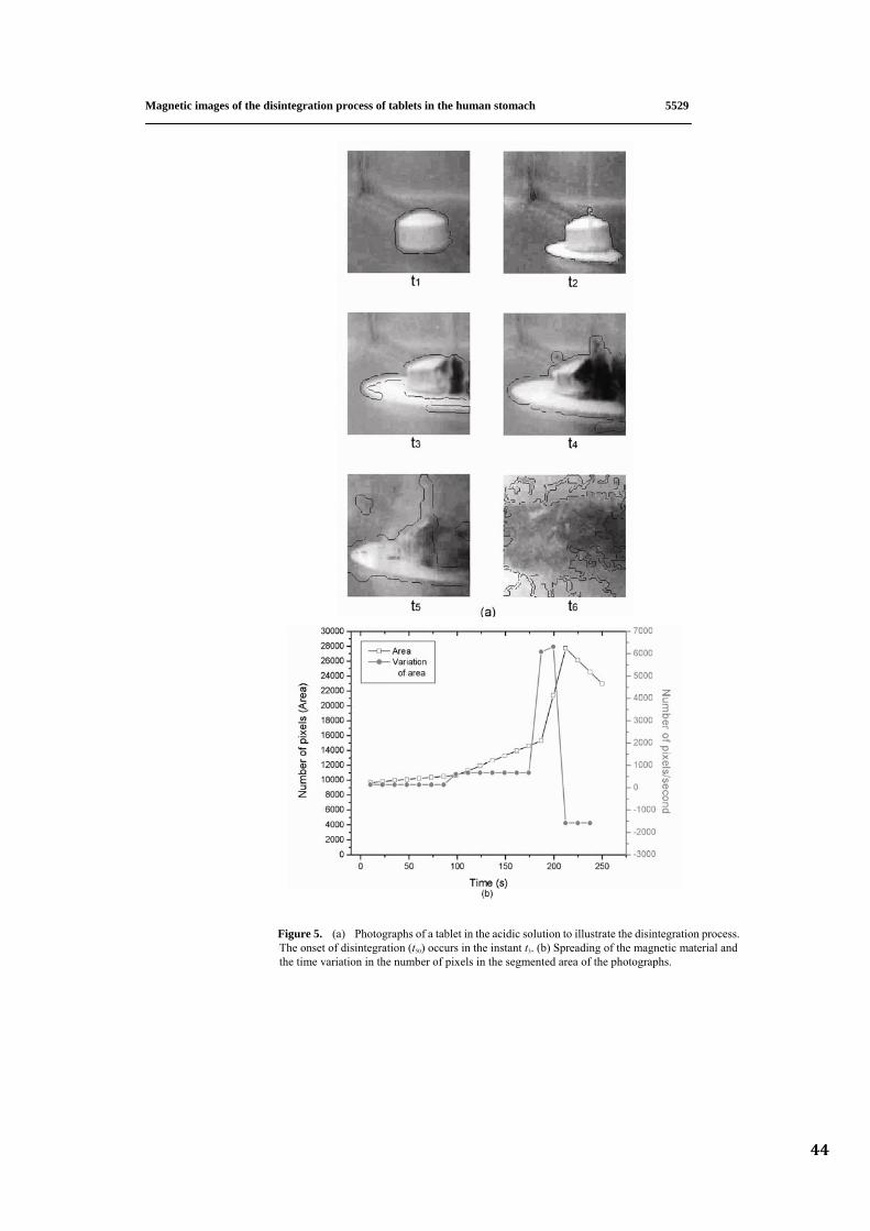
44
5529
s.
s.
Magnetic images of the disintegration process of tablets in the human stomach
Figure 5. (a) Photographs of a tablet in the acidic solution to illustrate the disintegration proces The onset of disintegration (t50) occurs in the instant t3. (b) Spreading of the magnetic material and the time variation in the number of pixels in the segmented area of the photograph
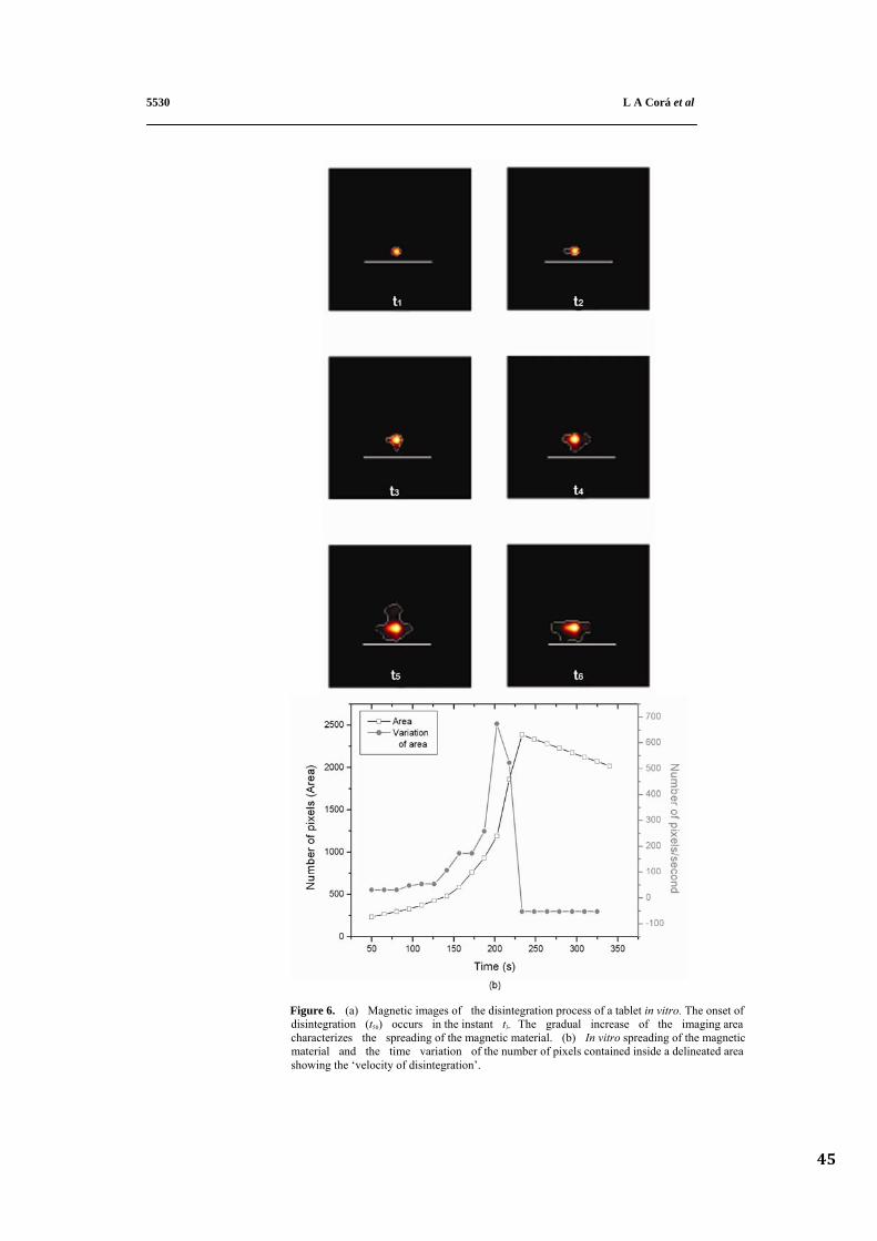
45
L A Corá et al
et of
aging areaetic
d area n’.
5530
Figure 6. (a) Magnetic images of the disintegration process of a tablet in vitro. The ons disintegration (t50) occurs in the instant t3. The gradual increase of the im characterizes the spreading of the magnetic material. (b) In vitro spreading of the magn material and the time variation of the number of pixels contained inside a delineate showing the ‘velocity of disintegratio
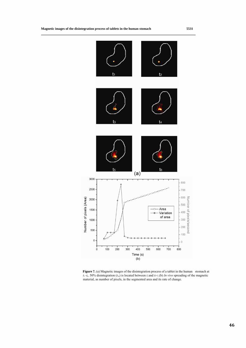
46
5531
h at tic
ge.
Magnetic images of the disintegration process of tablets in the human stomach
Figure 7. (a) Magnetic images of the disintegration process of a tablet in the human stomac t1–t6. 50% disintegration (t50) is located between ti and ti+1.(b) In vivo spreading of the magne material, as number of pixels, in the segmented area and its rate of chan
5532 L A Corá et al
anatomic references and the positioning of the sensors in the abdominal surface (figure 3). In these images, the arrival of the tablet in the distal stomach can be observed (instant t1). The onset of disintegration occurred in the instant t2. After t3, a gradual increase in the imaging area can be verified, characterizing the spreading of the magnetic material within the organ. The complete disintegration is represented in the instant t6.
Figure 7(b) shows the number of pixels present inside a delineated area and its time variation(‘velocity of disintegration’). The onset of disintegration (t50) of the tablets in the stomach ranged from 0.5 to 2.1 min (mean 1.1 ± 0.4).
4. Discussion Oral administration of solid pharmaceutical forms is a common practice in drug therapy and the imaging methods represent important tools to provide more reliable information about their performance in the human gastrointestinal tract.
In a recent study, single-sensor ACB was successfully used to generate images of ferromagnetic phantoms (Moreira et al 2000). Nevertheless, the development of a multisensor ACB system improved the spatial resolution allowing us to monitor the disintegration process of tablets and simultaneous gastrointestinal motility under physiologic conditions (Corá et al 2003, 2005).
In addition, multisensor ACB showed sensitivity and temporal resolution to obtain magneticimages. This system remains over the area of interest during all the recording time, not requiring repositioning and, consequently, there is no noise from vibrations. These are the important features of themultisensor ACB system that allows evaluation of the dynamic process that occurs in brief periods oftime.
In order to obtain a profile of the disintegration process of tablets an in vitro study was carried out aiming to qualitatively compare the photographs and the corresponding magnetic images. Analysing the image sequences shown in the figures 5 and 6, it was possible to verify the MM in the initial instances, while the tablet remains intact during the dissolution of the coating layer by the action of theacidic solution. When the coating layer was reduced the onset of disintegration (from instant t3) could be observed, continuously, until the spreading of the magnetic material in the glass vessel (instant t6).
This study was focused on the investigation of the disintegration process of tablets in the human stomach through magnetic imaging. The disintegration (figure 7(a)) was visualized solely by the magnetic method. By segmentation of the imaging area, it was possible to quantify the spreading of the magnetic material to characterize the transition between the MM and MT due to the disintegration process. Our data demonstrated that multisensor ACB was capable of identifying differences in the profile of the disintegration process.
From the results presented, it was observed that the onset of disintegration occurred in a short time interval (1.1 ± 0.4 min), indicating that this process once initiated promotes the dispersion of the ferrite continuously.
Pharmacoscintigraphy is an important method to investigate the gastrointestinal performance of pharmaceutical dosage forms and to provide information about the release and subsequent drug absorption (Wilding et al 2001). As an alternative to scintigraphy, biomagnetic methods have become feasible to monitor the dosage forms in the human gastrointestinal tract (Weistchies et al 2001). Although this multisensor ACB does not have sensitivity, the spatial resolution of the SQUID systems with a larger number of detectors was able to characterize efficiently the disintegration of tablets through magnetic images (Corá et al 2005). More reliable data will be obtained in combination with pharmacokinetics studies, since the magnetic material is devoid of harmful effects.
47
Magnetic images of the disintegration process of tablets in the human stomach 5533
Despite the difficulties, the multisensor ACB system was able to obtain images with reasonable quality. However, the blurring in the magnetic images due to the differences in the sensitivity profile could be corrected by applying the point-spread function for each sensor. Moreover, the application of restoration techniques could improve image quality and suppress noise simultaneously (Kondo et al 1977, Gravel et al 2004).
More extensive studies are required to obtain a comprehensive knowledge of the behaviour ofpharmaceutical forms in the human gastrointestinal tract. Moreover, it is essential that the developmentof sophisticated and specified delivery systems can improve and control the bioavailability andeffectiveness of administered drugs. In the future, ACB with a larger number of channels as anon-invasive and radiation-free imaging tool might achieve the same importance as other techniques in pharmaceutical and physiological research. In summary, our study showed that multisensor ACB, acompletely safe and harmless device, demonstrated enough sensitivity and spatial resolution to evaluatepharmaceutical dosage forms in the human gastrointestinal tract.
Acknowledgments The authors would like to thank FAPESP (01/11539-0), PRONEX (03/10107-5) and CAPESfor financial support. Eudragit® samples used in this study were a gift from Almapal–Tecnologia Validada, Brazil.
3
References
Baffa O, Oliveira R B, Miranda J R A and Troncon L E A 1995 Analysis and development of AC biosusceptometer for orocaecal transit time measurements Med. Biol. Eng. Comput. 33 353–7 Bastuscheck C M and Williamson S J 1985 Technique for measuring the ac susceptibility of portions of the human body or other large objects J. Appl. Phys. 58 3896–906 Carneiro AA O, Baffa O, Fernandes JP, Zago MA 2002 Theoretical evaluation of the susceptometric measurement of iron in human liver by four different susceptometers Physiol. Meas. 23 683–9Corá LA, Américo M F, Oliveira R B, Baffa O, Moraes R, Romeiro F G and Miranda J R A 2003 Disintegration of magnetic tablets in human stomach evaluated by alternate current biosusceptometry Eur. J. Pharm. Biopharm. 56 413–20
susceptometry of drug delivery Adv. Drug Deliv. Rev. 57 1223–41
Ziv E 1993 Gastrointestinal parameters that
Baffa O 2004 Study of the magnetic content movement
lds Magnetism in Medicine 1st ed W Andrä and
A method for modeling noise in medical images
Corá LA, Romeiro F G, Stelzer M, Américo MF, Oliveira R B, Baffa O and Miranda J RA 2005 AC bio in the studyDressman J B, Bass P, Ritschel W A, Friend D R, Rubinstein A and influence oral medications J. Pharm. Sci. 82 857–72 Ferreira A, Carneiro A A O, Moraes E R, Oliveira R B and present in the large intestine J. Magn. Magn. Mater. 283 16–21 Forsman M 1998 Magnetic substances and externally applied fie H Nowak (Berlin: Wiley-VCH) pp 430–45 Gravel P, Beaudoin G and De Guise J A 2004 IEEE Trans. Med.
N, Takada K, Yoshikawa H, Arakawa A and Yosida Y 2000 Application of a biomagnetic
capsules (PCDCs) in human subjects Pharm. Res. 17 160–7
Imaging 23 1221–32 Hu Z, Mawatari S, Shibata measurement system (BMS) to the evaluation of gastrointestinal transit of intestinal pressure-controlled colon deliveryKondo K, Ichioka Y and Suzuki T 1977 Image restoration by Wiener filtering in the presence of signal-dependent
systems J. Control. Release 62 41–9
Pharmacol. Ther. 3 223–32 meter to study gastric emptying
noise Appl. Optics 16 2554–8 Lipka E and Amidon G L 1999 Setting bioequivalence requirements for drug development based on preclinical data: optimizing oral drug delivery Melia C D and Davis S S 1989 Review article: mechanisms of drug release from tablets and capsules. I: disintegration Aliment.Miranda J R A, Baffa O and Oliveira R B 1992 An AC biosuscepto Med. Phys. 19 445–8
48
49
L A Corá et al
stric ntral contractions Phys. Med. Biol. 42 1791–9
a O and Miranda J R A 2003 Measurement of gastric contraction
Murta L O and Baffa O 2000 Imaging ferromagnetic tracers with an AC biosusceptometer
5534
Miranda J R A, Oliveira R B, Sousa P L, Braga F J H and Baffa O 1997 A novel biomagnetic method to study ga aMoraes R, Corá, LA, Américo MF, Oliveira RB, Baff activity in dogs by means of AC biosusceptometry Physiol. Meas. 24 337–45 Moreira M, Rev. Sci. Instrum. 71 2532–8 Newman S P, Hirst P H and Wilding I R 2003 New developments in radionuclide imaging for assessing drug delivery
rm. Sci. 18 19–22 nique
measurement of orocaecal transit time Eur. J. Gastroenterol. Hepatol. 8 491–5 ckshaw P E, Juan D, Dansereaus R J, Hathaways S, Li Z, Long P and
t in gastro-oesophageal reflux disease and normal control subjects Aliment. Pharmacol. Ther. 15 115–21 delivery Pharm. Sci. Tech.
in man Eur. J. PhaOliveira R B, Baffa O, Troncon L E A, Miranda J R A and Cambrea C R 1996 Evaluation of a biomagnetic tech for Perkins A C, Wilson C G, Frier M, Bla Spiller R C 2001 Oesophageal transit, disintegration and gastric emptying of a film-coated risedronate placebo tableSastry S V, Nyshadham J R and Fix J A 2000 Recent technological advances in oral drug Today 3 138–45 Singh M and Waluch V 2000 Physics and instrumentation for imaging in-vivo drug distribution Adv. Drug Deliv. Rev. 4 7–20 Steingoetter A, Weishaupt D, Kunz P, Mäder K, Lengsfeld H, Thumshirn M, Boesiger P, Fried M and Schwizer W 2003a Magnetic Resonance Imaging for the in vivo evaluation of gastric-retentive tablets Pharm. Res.
etic resonance imaging Aliment. Pharmacol. Ther. 18 713–20
20 2001–7 Steingoetter A, Kunz P, Weishaupt D, Mäder K, Lengsfeld H, Thumshirn M, Boesiger P, Fried M and Schwizer W 2003b Analysis of the meal-dependent intragastric performance of a gastric-retentive tablet assessed by magnWeitschies W, Karaus M, Cordini D, Trahms L, Breitkreutz J and Semmler W 2001 Magnetic marker monitoring of disintegrating capsules Eur. J. Pharm. Sci. 13 411–6 Weitschies W, K¨otitz R, Cordini D and Trahms L 1997 High resolution monitoring of the gastrointestinal transit of a magnetically marked capsule J. Pharm. Sci. 86 1218–22 Wilding I R, Coupe A J and Davis S S 2001 The role of γ -scintigraphy in oral drug delivery Adv. Drug Deliv. Rev.
46 103–24 Wilson C G, McJury M, O’Mahony B, Frier M and Perkins A C 1997 Imaging of oily formulations in the gastrointestinal tract Adv. Drug Deliv. Rev. 25 91–101
Capítulo 2
Influence of compression forces on tablets disintegration by
AC Biosusceptometry
European Journal of Pharmaceutics and Biopharmaceutics, v.69, 372-379, 2008
50
Research Paper
Available online at www.sciencedirect.com
European Journal of Pharmaceutics and Biopharmaceutics 69 (2008) 372–379
Influence of compression forces on tablets disintegration by
AC Biosusceptometry
Luciana A. Corá a
, Paulo R. Fonseca a
, Madileine F. Américo b
, Ricardo B. Oliveira c
,
Oswaldo Baffa d
, José Ricardo A. Miranda a,
*
a
Dep. de Física e Biofísica, São Paulo State University, Botucatu, Brazil
b
Dep. de Fisiologia, São Paulo University, Ribeirão Preto, Brazil
c
Dep. de Clínica Médica, São Paulo University, Ribeirão Preto, Brazil
d
Dep. de Física e Matemática, São Paulo University, Ribeirão Preto, Brazil
Received 4 September 2007; accepted in revised form 12 November 2007
Available online 19 November 2007
Abstract
Analysis of physical phenomena that occurs during tablet disintegration has been studied by several experimental approaches; how-
ever none of them satisfactorily describe this process. The aim of this study was to investigate the influence of compression force on the tablets
by associating the AC Biosusceptometry with consolidated methods in order to validate the biomagnetic technique as a tool for quality control in
pharmaceutical processes.
Keywords: AC Biosusceptometry; Compression force; Disintegration force; Water uptake; Magnetic tablets; Disintegration
® 2007 Elsevier B.V. All rights reserved.
Tablets obtained at five compression levels were submitted to mechanical properties tests. For uncoated tablets, water uptake and
disintegration force measurements were performed in order to compare with magnetic data. For coated tablets, magnetic measurements were
carried out to establish a relationship between physical parameters of the disintegration process. According to the results, differences between the
compression levels were found for water uptake, force development and magnetic area variation measurements. ACB method was able to estimate
the disintegration properties as well as the kinetics of disintegration process for uncoated and coated tablets. This study provided a new approach
for in vitro investigation and validated this biomagnetic technique as a tool for quality control for pharmaceutical industry. Moreover, using ACB
will also be possible to test these parameters in humans allowing to establish an in vitro/in vivo correlation (IVIVC).
www.elsevier.com/locate/ejpb
1. Introduction
Despite increasing interest in modified release systems,
conventional tablets are still the most popular solid dosage
forms due to ease of manufacture, convenience of dosing
and stability [1,2].
Drug release from tablets occurs by disintegration pro-
cess promoting a fast fragmentation of the dosage form
*Corresponding author. Departamento de Física e Biofísica, Instituto
de Biociências – IBB, Laboratório de Biomagnetismo, Universidade
Estadual Paulista – UNESP, CXP 510, CEP 18618-000 Botucatu, São
Paulo, Brazil. Tel./fax: +55 14 3811 6254.
E-mail address: [email protected] (J. R. A. Miranda).
0939-6411/$ -see front matter ® 2007 Elsevier B.V. All rights reserved.
doi:10.1016/j.ejpb.2007.11.005
under the action of the disintegrant [3]. If this process is
slow or incomplete the bioavailability of a drug will be
inadequate. Appropriate choice of a disintegrant and its
consistency of performance have critical importance to the
formulation development [4].
Disintegration of compressed tablets is an important
quality parameter and it is strongly influenced by the prop-
erties of the excipients, such as particle size distributions
and the compression force [5,6]. It is well established that
the compression force is essential for the tablet manufac-
turing process since an increase in the compression force
causes a reduction of tablet porosity and, as a consequence,
a linear increase of the disintegration time [7,8].
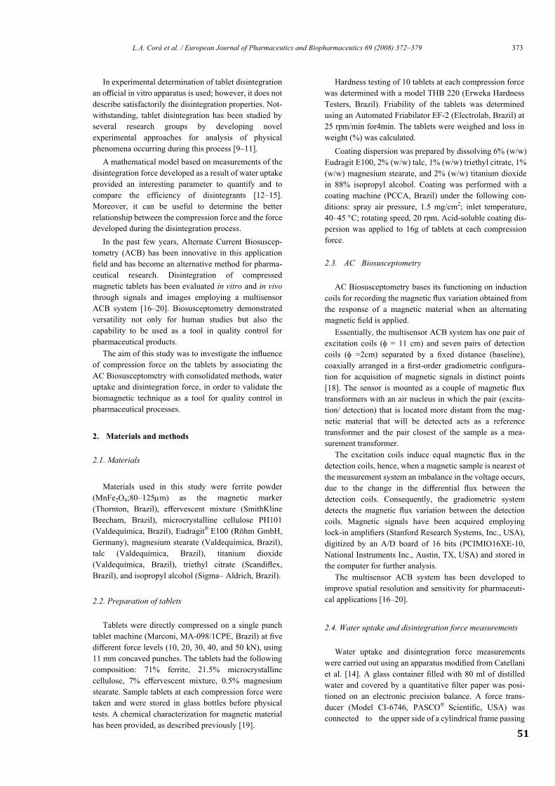
51
L.A. Corá et al. / European Journal of Pharmaceutics and Biopharmaceutics 69 (2008) 372–379 373
In experimental determination of tablet disintegration
an official in vitro apparatus is used; however, it does not
describe satisfactorily the disintegration properties. Not-
withstanding, tablet disintegration has been studied by
several research groups by developing novel
experimental approaches for analysis of physical
phenomena occurring during this process [9–11].
A mathematical model based on measurements of the
disintegration force developed as a result of water uptake
provided an interesting parameter to quantify and to
compare the efficiency of disintegrants [12–15].
Moreover, it can be useful to determine the better
relationship between the compression force and the force
developed during the disintegration process.
In the past few years, Alternate Current Biosuscep-
tometry (ACB) has been innovative in this application
field and has become an alternative method for pharma-
ceutical research. Disintegration of compressed
magnetic tablets has been evaluated in vitro and in vivo
through signals and images employing a multisensor
ACB system [16–20]. Biosusceptometry demonstrated
versatility not only for human studies but also the
capability to be used as a tool in quality control for
pharmaceutical products.
The aim of this study was to investigate the influence
of compression force on the tablets by associating the
AC Biosusceptometry with consolidated methods, water
uptake and disintegration force, in order to validate the
biomagnetic technique as a tool for quality control in
pharmaceutical processes.
2. Materials and methods
2.1. Materials
Materials used in this study were ferrite powder
(MnFe2O4;80–125m) as the magnetic marker
(Thornton, Brazil), effervescent mixture (SmithKline
Beecham, Brazil), microcrystalline cellulose PH101
(Valdequímica, Brazil), Eudragit®
E100 (Röhm GmbH,
Germany), magnesium stearate (Valdequímica, Brazil),
talc (Valdequímica, Brazil), titanium dioxide
(Valdequímica, Brazil), triethyl citrate (Scandiflex,
Brazil), and isopropyl alcohol (Sigma– Aldrich, Brazil).
2.2. Preparation of tablets
Tablets were directly compressed on a single punch
tablet machine (Marconi, MA-098/1CPE, Brazil) at five
different force levels (10, 20, 30, 40, and 50 kN), using
11 mm concaved punches. The tablets had the following
composition: 71% ferrite, 21.5% microcrystalline
cellulose, 7% effervescent mixture, 0.5% magnesium
stearate. Sample tablets at each compression force were
taken and were stored in glass bottles before physical
tests. A chemical characterization for magnetic material
has been provided, as described previously [19].
Hardness testing of 10 tablets at each compression force
was determined with a model THB 220 (Erweka Hardness
Testers, Brazil). Friability of the tablets was determined
using an Automated Friabilator EF-2 (Electrolab, Brazil) at
25 rpm/min for4min. The tablets were weighed and loss in
weight (%) was calculated.
Coating dispersion was prepared by dissolving 6% (w/w)
Eudragit E100, 2% (w/w) talc, 1% (w/w) triethyl citrate, 1%
(w/w) magnesium stearate, and 2% (w/w) titanium dioxide
in 88% isopropyl alcohol. Coating was performed with a
coating machine (PCCA, Brazil) under the following con-
ditions: spray air pressure, 1.5 mg/cm2; inlet temperature,
40–45 C; rotating speed, 20 rpm. Acid-soluble coating dis-
persion was applied to 16g of tablets at each compression
force.
2.3. AC Biosusceptometry
AC Biosusceptometry bases its functioning on induction
coils for recording the magnetic flux variation obtained from
the response of a magnetic material when an alternating
magnetic field is applied.
Essentially, the multisensor ACB system has one pair of
excitation coils ( = 11 cm) and seven pairs of detection
coils ( =2cm) separated by a fixed distance (baseline),
coaxially arranged in a first-order gradiometric configura-
tion for acquisition of magnetic signals in distinct points
[18]. The sensor is mounted as a couple of magnetic flux
transformers with an air nucleus in which the pair (excita-
tion/ detection) that is located more distant from the mag-
netic material that will be detected acts as a reference
transformer and the pair closest of the sample as a mea-
surement transformer.
The excitation coils induce equal magnetic flux in the
detection coils, hence, when a magnetic sample is nearest of
the measurement system an imbalance in the voltage occurs,
due to the change in the differential flux between the
detection coils. Consequently, the gradiometric system
detects the magnetic flux variation between the detection
coils. Magnetic signals have been acquired employing
lock-in amplifiers (Stanford Research Systems, Inc., USA),
digitized by an A/D board of 16 bits (PCIMIO16XE-10,
National Instruments Inc., Austin, TX, USA) and stored in
the computer for further analysis.
The multisensor ACB system has been developed to
improve spatial resolution and sensitivity for pharmaceuti-
cal applications [16–20].
2.4. Water uptake and disintegration force measurements
Water uptake and disintegration force measurements
were carried out using an apparatus modified from Catellani
et al. [14]. A glass container filled with 80 ml of distilled
water and covered by a quantitative filter paper was posi-
tioned on an electronic precision balance. A force trans-
ducer (Model CI-6746, PASCO®
Scientific, USA) was
connected to the upper side of a cylindrical frame passing
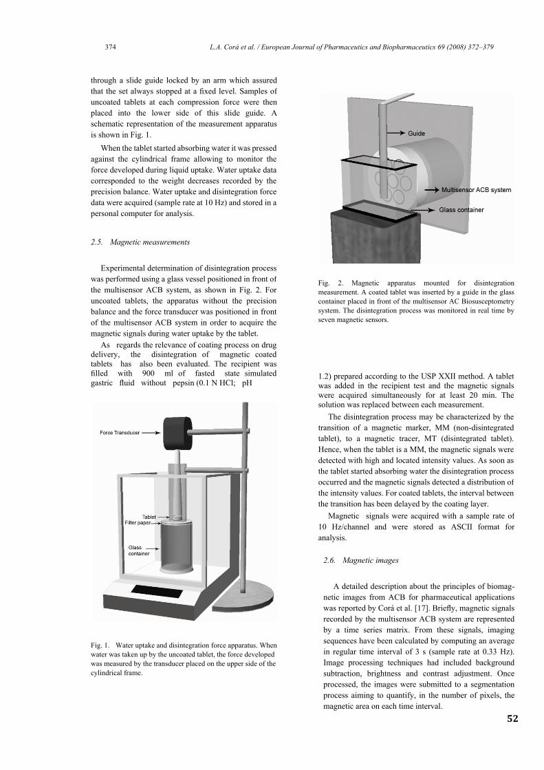
52
374 L.A. Corá et al. / European Journal of Pharmaceutics and Biopharmaceutics 69 (2008) 372–379
through a slide guide locked by an arm which assured
that the set always stopped at a fixed level. Samples of
uncoated tablets at each compression force were then
placed into the lower side of this slide guide. A
schematic representation of the measurement apparatus
is shown in Fig. 1.
When the tablet started absorbing water it was pressed
against the cylindrical frame allowing to monitor the
force developed during liquid uptake. Water uptake data
corresponded to the weight decreases recorded by the
precision balance. Water uptake and disintegration force
data were acquired (sample rate at 10 Hz) and stored in a
personal computer for analysis.
2.5. Magnetic measurements
Experimental determination of disintegration process
was performed using a glass vessel positioned in front of
the multisensor ACB system, as shown in Fig. 2. For
uncoated tablets, the apparatus without the precision
balance and the force transducer was positioned in front
of the multisensor ACB system in order to acquire the
magnetic signals during water uptake by the tablet.
As regards the relevance of coating process on drug
delivery, the disintegration of magnetic coated
tablets has also been evaluated. The recipient was
filled with 900 ml of fasted state simulated
gastric fluid without pepsin (0.1 N HCl; pH 1.2) prepared according to the USP XXII method. A tablet
was added in the recipient test and the magnetic signals
were acquired simultaneously for at least 20 min. The
solution was replaced between each measurement.
Fig. 1. Water uptake and disintegration force apparatus. When
water was taken up by the uncoated tablet, the force developed
was measured by the transducer placed on the upper side of the
cylindrical frame.
Fig. 2. Magnetic apparatus mounted for disintegration
measurement. A coated tablet was inserted by a guide in the glass
container placed in front of the multisensor AC Biosusceptometry
system. The disintegration process was monitored in real time by
seven magnetic sensors.
The disintegration process may be characterized by the
transition of a magnetic marker, MM (non-disintegrated
tablet), to a magnetic tracer, MT (disintegrated tablet).
Hence, when the tablet is a MM, the magnetic signals were
detected with high and located intensity values. As soon as
the tablet started absorbing water the disintegration process
occurred and the magnetic signals detected a distribution of
the intensity values. For coated tablets, the interval between
the transition has been delayed by the coating layer.
Magnetic signals were acquired with a sample rate of
10 Hz/channel and were stored as ASCII format for
analysis.
2.6. Magnetic images
A detailed description about the principles of biomag-
netic images from ACB for pharmaceutical applications
was reported by Corá et al. [17]. Briefly, magnetic signals
recorded by the multisensor ACB system are represented
by a time series matrix. From these signals, imaging
sequences have been calculated by computing an average
in regular time interval of 3 s (sample rate at 0.33 Hz).
Image processing techniques had included background
subtraction, brightness and contrast adjustment. Once
processed, the images were submitted to a segmentation
process aiming to quantify, in the number of pixels, the
magnetic area on each time interval.
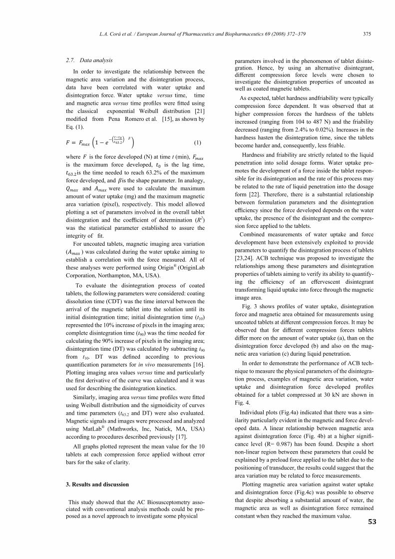
53
L.A. Corá et al. / European Journal of Pharmaceutics and Biopharmaceutics 69 (2008) 372–379 375
2.7. Data analysis
In order to investigate the relationship between the
magnetic area variation and the disintegration process,
data have been correlated with water uptake and
disintegration force. Water uptake versus time, time
and magnetic area versus time profiles were fitted using
the classical exponential Weibull distribution [21]
modified from Pena Romero et al. [15], as shown by
Eq. (1).
𝐹 = 𝐹𝑚𝑎𝑥 1 − 𝑒−
𝑡−𝑡0𝑡63.2
𝛽
(1)
where 𝐹 is the force developed (N) at time t (min), 𝐹𝑚𝑎𝑥
is the maximum force developed, 𝑡0 is the lag time,
𝑡63.2is the time needed to reach 63.2% of the maximum
force developed, and 𝛽is the shape parameter. In analogy,
𝑄𝑚𝑎𝑥 and 𝐴𝑚𝑎𝑥 were used to calculate the maximum
amount of water uptake (mg) and the maximum magnetic
area variation (pixel), respectively. This model allowed
plotting a set of parameters involved in the overall tablet
disintegration and the coefficient of determination (R2)
was the statistical parameter established to assure the
integrity of fit.
For uncoated tablets, magnetic imaging area variation
(𝐴𝑚𝑎𝑥 ) was calculated during the water uptake aiming to
establish a correlation with the force measured. All of
these analyses were performed using Origin®
(OriginLab
Corporation, Northampton, MA, USA).
To evaluate the disintegration process of coated
tablets, the following parameters were considered: coating
dissolution time (CDT) was the time interval between the
arrival of the magnetic tablet into the solution until its
initial disintegration time; initial disintegration time (t10)
represented the 10% increase of pixels in the imaging area;
complete disintegration time (t90) was the time needed for
calculating the 90% increase of pixels in the imaging area;
disintegration time (DT) was calculated by subtracting t90
from t10. DT was defined according to previous
quantification parameters for in vivo measurements [16].
Plotting imaging area values versus time and particularly
the first derivative of the curve was calculated and it was
used for describing the disintegration kinetics.
Similarly, imaging area versus time profiles were fitted
using Weibull distribution and the sigmoidicity of curves
and time parameters (t63.2 and DT) were also evaluated.
Magnetic signals and images were processed and analyzed
using MatLab®
(Mathworks, Inc, Natick, MA, USA)
according to procedures described previously [17].
All graphs plotted represent the mean value for the 10
tablets at each compression force applied without error
bars for the sake of clarity.
3. Results and discussion
This study showed that the AC Biosusceptometry asso-
ciated with conventional analysis methods could be pro-
posed as a novel approach to investigate some physical
parameters involved in the phenomenon of tablet disinte-
gration. Hence, by using an alternative disintegrant,
different compression force levels were chosen to
investigate the disintegration properties of uncoated as
well as coated magnetic tablets.
As expected, tablet hardness andfriability were typically
compression force dependent. It was observed that at
higher compression forces the hardness of the tablets
increased (ranging from 104 to 487 N) and the friability
decreased (ranging from 2.4% to 0.02%). Increases in the
hardness hasten the disintegration time, since the tablets
become harder and, consequently, less friable.
Hardness and friability are strictly related to the liquid
penetration into solid dosage forms. Water uptake pro-
motes the development of a force inside the tablet respon-
sible for its disintegration and the rate of this process may
be related to the rate of liquid penetration into the dosage
form [22]. Therefore, there is a substantial relationship
between formulation parameters and the disintegration
efficiency since the force developed depends on the water
uptake, the presence of the disintegrant and the compres-
sion force applied to the tablets.
Combined measurements of water uptake and force
development have been extensively exploited to provide
parameters to quantify the disintegration process of tablets
[23,24]. ACB technique was proposed to investigate the
relationships among these parameters and disintegration
properties of tablets aiming to verify its ability to quantify-
ing the efficiency of an effervescent disintegrant
transforming liquid uptake into force through the magnetic
image area.
Fig. 3 shows profiles of water uptake, disintegration
force and magnetic area obtained for measurements using
uncoated tablets at different compression forces. It may be
observed that for different compression forces tablets
differ more on the amount of water uptake (a), than on the
disintegration force developed (b) and also on the mag-
netic area variation (c) during liquid penetration.
In order to demonstrate the performance of ACB tech-
nique to measure the physical parameters of the disintegra-
tion process, examples of magnetic area variation, water
uptake and disintegration force developed profiles
obtained for a tablet compressed at 30 kN are shown in
Fig. 4.
Individual plots (Fig.4a) indicated that there was a sim-
ilarity particularly evident in the magnetic and force devel-
oped data. A linear relationship between magnetic area
against disintegration force (Fig. 4b) at a higher signifi-
cance level (R= 0.987) has been found. Despite a short
non-linear region between these parameters that could be
explained by a preload force applied to the tablet due to the
positioning of transducer, the results could suggest that the
area variation may be related to force measurements.
Plotting magnetic area variation against water uptake
and disintegration force (Fig.4c) was possible to observe
that despite absorbing a substantial amount of water, the
magnetic area as well as disintegration force remained
constant when they reached the maximum value.
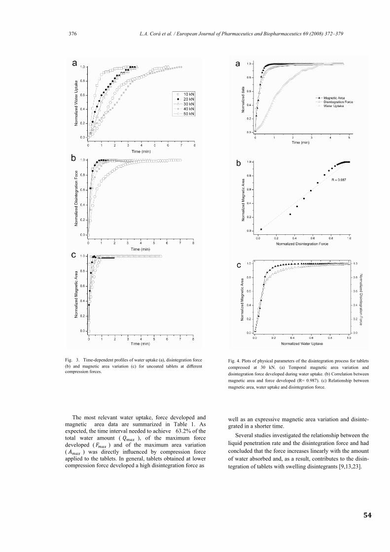
54
376 L.A. Corá et al. / European Journal of Pharmaceutics and Biopharmaceutics 69 (2008) 372–379
Fig. 3. Time-dependent profiles of water uptake (a), disintegration force
(b) and magnetic area variation (c) for uncoated tablets at different
compression forces.
Fig. 4. Plots of physical parameters of the disintegration process for tablets
compressed at 30 kN. (a) Temporal magnetic area variation and
disintegration force developed during water uptake. (b) Correlation between
magnetic area and force developed (R= 0.987). (c) Relationship between
magnetic area, water uptake and disintegration force.
The most relevant water uptake, force developed and
magnetic area data are summarized in Table 1. As
expected, the time interval needed to achieve 63.2% of the
total water amount ( 𝑄𝑚𝑎𝑥 ), of the maximum force
developed (𝐹𝑚𝑎𝑥 ) and of the maximum area variation
( 𝐴𝑚𝑎𝑥 ) was directly influenced by compression force
applied to the tablets. In general, tablets obtained at lower
compression force developed a high disintegration force as
well as an expressive magnetic area variation and disinte-
grated in a shorter time.
Several studies investigated the relationship between the
liquid penetration rate and the disintegration force and had
concluded that the force increases linearly with the amount
of water absorbed and, as a result, contributes to the disin-
tegration of tablets with swelling disintegrants [9,13,23].
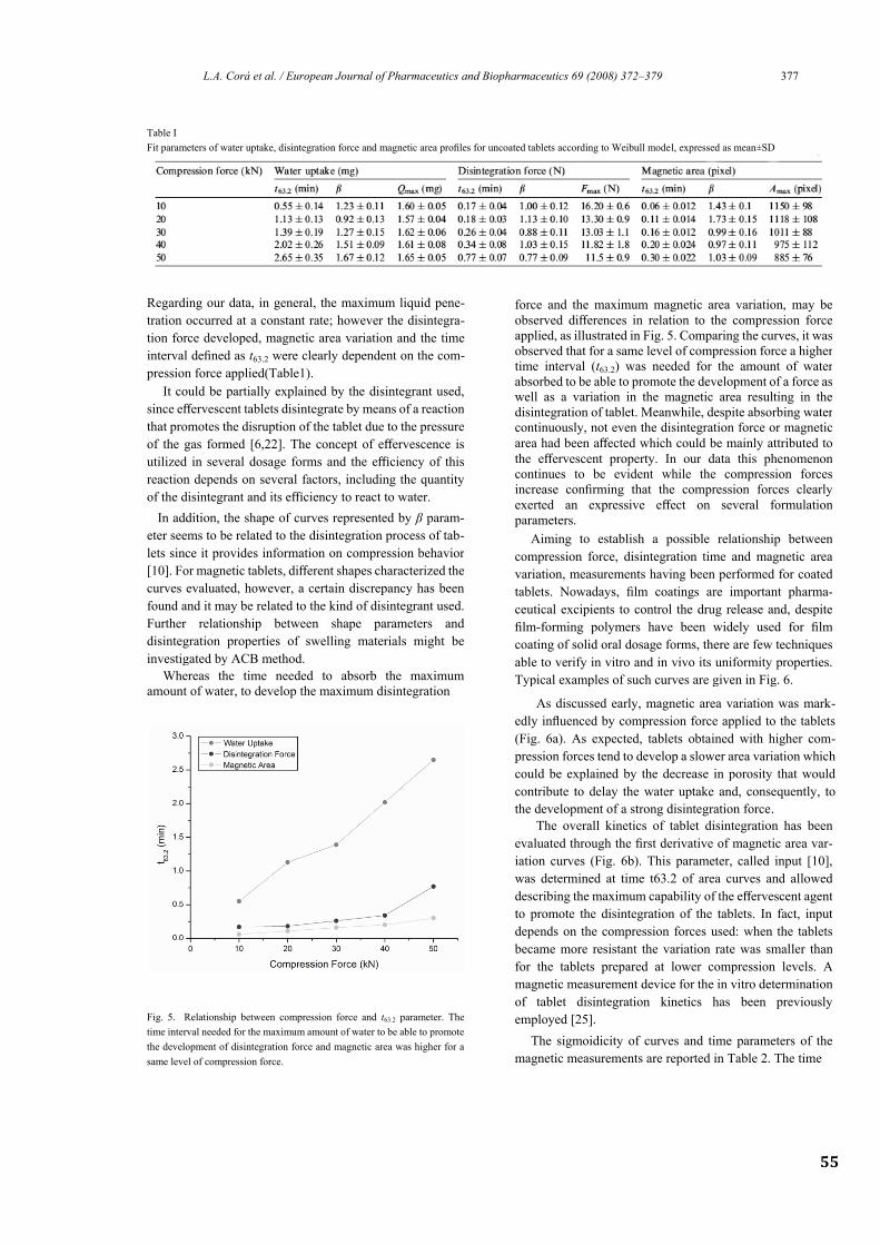
55
L.A. Corá et al. / European Journal of Pharmaceutics and Biopharmaceutics 69 (2008) 372–379 377
Table I
Fit parameters of water uptake, disintegration force and magnetic area profiles for uncoated tablets according to Weibull model, expressed as mean±SD
Regarding our data, in general, the maximum liquid pene-
tration occurred at a constant rate; however the disintegra-
tion force developed, magnetic area variation and the time
interval defined as t63.2 were clearly dependent on the com-
pression force applied(Table1).
force and the maximum magnetic area variation, may be
observed differences in relation to the compression force
applied, as illustrated in Fig. 5. Comparing the curves, it was
observed that for a same level of compression force a higher
time interval (t63.2) was needed for the amount of water
absorbed to be able to promote the development of a force as
well as a variation in the magnetic area resulting in the
disintegration of tablet. Meanwhile, despite absorbing water
continuously, not even the disintegration force or magnetic
area had been affected which could be mainly attributed to
the effervescent property. In our data this phenomenon
continues to be evident while the compression forces
increase confirming that the compression forces clearly
exerted an expressive effect on several formulation
parameters.
Aiming to establish a possible relationship between
compression force, disintegration time and magnetic area
variation, measurements having been performed for coated
tablets. Nowadays, film coatings are important pharma-
ceutical excipients to control the drug release and, despite
film-forming polymers have been widely used for film
coating of solid oral dosage forms, there are few techniques
able to verify in vitro and in vivo its uniformity properties.
Typical examples of such curves are given in Fig. 6.
As discussed early, magnetic area variation was mark-
edly influenced by compression force applied to the tablets
(Fig. 6a). As expected, tablets obtained with higher com-
pression forces tend to develop a slower area variation which
could be explained by the decrease in porosity that would
contribute to delay the water uptake and, consequently, to
the development of a strong disintegration force.
It could be partially explained by the disintegrant used,
since effervescent tablets disintegrate by means of a reaction
that promotes the disruption of the tablet due to the pressure
of the gas formed [6,22]. The concept of effervescence is
utilized in several dosage forms and the efficiency of this
reaction depends on several factors, including the quantity
of the disintegrant and its efficiency to react to water.
In addition, the shape of curves represented by β param-
eter seems to be related to the disintegration process of tab-
lets since it provides information on compression behavior
[10]. For magnetic tablets, different shapes characterized the
curves evaluated, however, a certain discrepancy has been
found and it may be related to the kind of disintegrant used.
Further relationship between shape parameters and
disintegration properties of swelling materials might be
investigated by ACB method.
Whereas the time needed to absorb the maximum
amount of water, to develop the maximum disintegration
Fig. 5. Relationship between compression force and t63.2 parameter. The
time interval needed for the maximum amount of water to be able to promote
the development of disintegration force and magnetic area was higher for a
same level of compression force.
The overall kinetics of tablet disintegration has been
evaluated through the first derivative of magnetic area var-
iation curves (Fig. 6b). This parameter, called input [10],
was determined at time t63.2 of area curves and allowed
describing the maximum capability of the effervescent agent
to promote the disintegration of the tablets. In fact, input
depends on the compression forces used: when the tablets
became more resistant the variation rate was smaller than
for the tablets prepared at lower compression levels. A
magnetic measurement device for the in vitro determination
of tablet disintegration kinetics has been previously
employed [25].
The sigmoidicity of curves and time parameters of the
magnetic measurements are reported in Table 2. The time
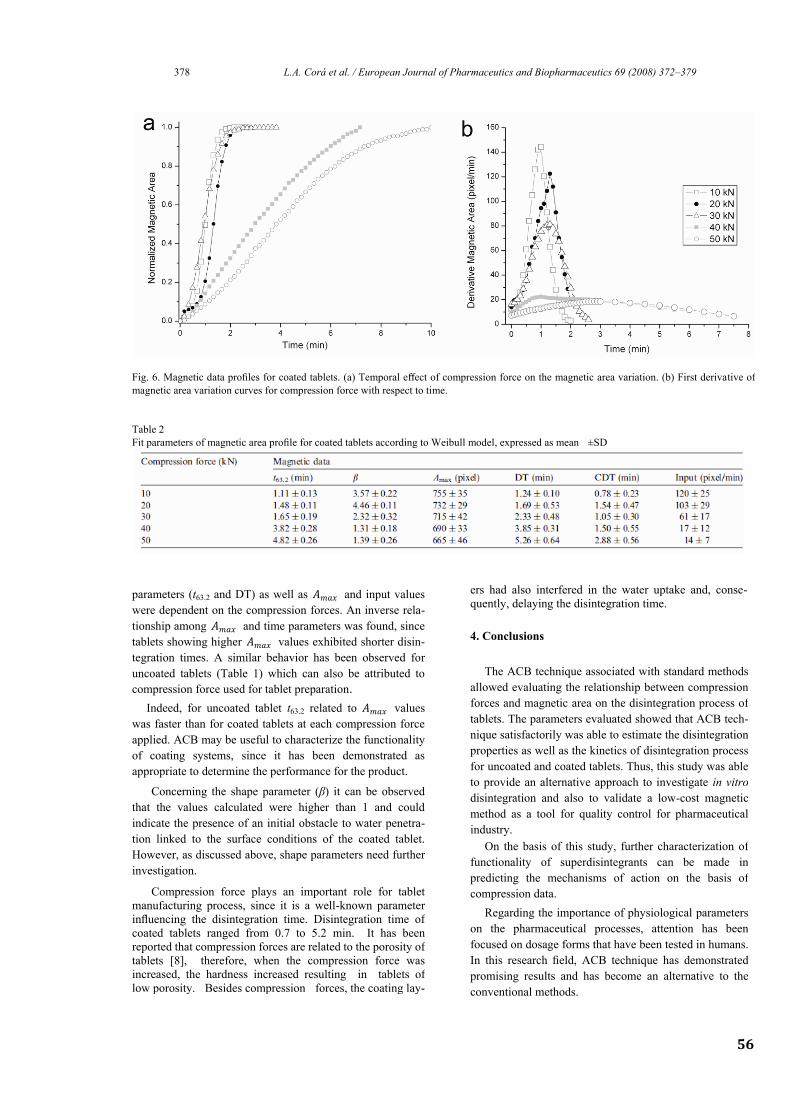
56
378 L.A. Corá et al. / European Journal of Pharmaceutics and Biopharmaceutics 69 (2008) 372–379
Fig. 6. Magnetic data profiles for coated tablets. (a) Temporal effect of compression force on the magnetic area variation. (b) First derivative of
magnetic area variation curves for compression force with respect to time.
Table 2
Fit parameters of magnetic area profile for coated tablets according to Weibull model, expressed as mean ±SD
parameters (t63.2 and DT) as well as 𝐴𝑚𝑎𝑥 and input values
were dependent on the compression forces. An inverse rela-
tionship among 𝐴𝑚𝑎𝑥 and time parameters was found, since
tablets showing higher 𝐴𝑚𝑎𝑥 values exhibited shorter disin-
tegration times. A similar behavior has been observed for
uncoated tablets (Table 1) which can also be attributed to
compression force used for tablet preparation.
4. Conclusions
Indeed, for uncoated tablet t63.2 related to 𝐴𝑚𝑎𝑥 values
was faster than for coated tablets at each compression force
applied. ACB may be useful to characterize the functionality
of coating systems, since it has been demonstrated as
appropriate to determine the performance for the product.
Concerning the shape parameter (β) it can be observed
that the values calculated were higher than 1 and could
indicate the presence of an initial obstacle to water penetra-
tion linked to the surface conditions of the coated tablet.
However, as discussed above, shape parameters need further
investigation.
Compression force plays an important role for tablet
manufacturing process, since it is a well-known parameter
influencing the disintegration time. Disintegration time of
coated tablets ranged from 0.7 to 5.2 min. It has been
reported that compression forces are related to the porosity of
tablets [8], therefore, when the compression force was
increased, the hardness increased resulting in tablets of
low porosity. Besides compression forces, the coating lay-
ers had also interfered in the water uptake and, conse-
quently, delaying the disintegration time.
The ACB technique associated with standard methods
allowed evaluating the relationship between compression
forces and magnetic area on the disintegration process of
tablets. The parameters evaluated showed that ACB tech-
nique satisfactorily was able to estimate the disintegration
properties as well as the kinetics of disintegration process
for uncoated and coated tablets. Thus, this study was able
to provide an alternative approach to investigate in vitro
disintegration and also to validate a low-cost magnetic
method as a tool for quality control for pharmaceutical
industry.
On the basis of this study, further characterization of
functionality of superdisintegrants can be made in
predicting the mechanisms of action on the basis of
compression data.
Regarding the importance of physiological parameters
on the pharmaceutical processes, attention has been
focused on dosage forms that have been tested in humans.
In this research field, ACB technique has demonstrated
promising results and has become an alternative to the
conventional methods.
57
L.A. Corá et al. / European Journal of Pharmaceutics and Biopharmaceutics 69 (2008) 372–379 379
In summary, AC Biosusceptometry might be able to
characterize a number of parameters related to drug deliv-
ery, deserving the same importance as conventional tech-
niques for pharmaceutical research. Furthermore, ACB
method will be especially powerful when combined with
classical pharmacokinetic data (‘‘magnetopharmacokinet-
ic’’) allowing establishing an in vitro/in vivo correlation
(IVIVC).
Acknowledgements
Partial financial support was received from PRONEX-
FAPESP, CAPES, CNPq. Eudragit®
samples were a gift
from Almapal S/A – Tecnologia Validada, Brazil.
References
[1] S.V. Sastry, J.R. Nyshadham, J.A. Fix, Recent technological
advances in oral drug delivery, Pharm. Sci. Technol. Today 3
(2000) 138–145.
[2] M. Jivraj, L.G. Martini, C.M. Thomson, An overview of the different
excipients useful for the direct compression of tablets, Pharm. Sci.
Technol. Today 3 (2000) 58–63.
[3] C.D. Melia, S.S. Davis, Review article: mechanisms of drug release
from tablets and capsules. I: Disintegration, Aliment. Pharmacol.
Ther. 3 (1989) 223–232.
[4] N. Zhao, L.L. Augsburger, Functionality comparison of 3
classes of superdisintegrants in promoting aspirin tablet disinte-
gration and dissolution, AAPS Pharm. Sci. Tech. 6 (2005) 634–
640. [5] K.-H. Lin, S.-Y. Lin, M.-J. Li, Compression forces and amount of
outer coating layer affecting the time-controlled disintegration
of the compression-coated tablets prepared by direct compression
with micronized ethylcellulose, J. Pharm. Sci. 90 (2001) 2005– 2009.
[6] W. Lowenthal, Disintegration of tablets, J. Pharm. Sci. 61 (1972)
1695–1711. [7] H.G. Ibrahim, Observations on the dissolution behavior of a tablet
formulation: effect of compression forces, J. Pharm. Sci. 74 (1985)
575–577.
[8] M. Riippi, O. Antikainen, T. Niskanen, J. Yliruusi, The effect of
compression force on surface structure, crushing strength, friability
and disintegration time of erythromycin acistrate tablets, Eur. J.
Pharm. Biopharm. 46 (1998) 339–345.
[9] P. Colombo, C. Caramella, U. Conte, A. La Manna, A.M. Guyot-
Hermann, J. Ringard, Disintegrating force and tablet properties,
Drug Dev. Ind. Pharm. 7 (1981) 135–153.
[10] P. Colombo, U. Conte, C. Caramella, M. Geddo, A. La Manna,
Disintegration force as a new formulation parameter, J. Pharm. Sci.
73 (1984) 701–705.
[11] C. Caramella, P. Colombo, U. Conte, F. Ferrari, A. Gazzaniga, A. La
Manna, N.A. Peppas, A physical analysis of the phenomenon of
tablet disintegration, Int. J. Pharm. 44 (1988) 177–186.
[12] H.V. van Kamp, G.K. Bolhuis, A.H. de Boer, C.F. Lerk, L. Lie- A-
Huen, The role of water uptake on tablet disintegration, Pharm. Acta
Helv. 61 (1986) 22–29.
[13] N.A. Peppas, P. Colombo, Development of disintegration forces
during water penetration in porous pharmaceutical systems, J.
Control. Release 10 (1989) 245–250.
[14] P.L. Catellani, P. Predella, A. Bellotti, P. Colombo, Tablet water
uptake and disintegration force measurements, Int. J. Pharm. 51
(1989) 63–66.
[15] A. Pena Romero, C. Caramella, M. Ronchi, F. Ferrari, D. Chulia,
Water uptake and force development in an optimized prolonged
release formulation, Int. J. Pharm. 73 (1991) 239–248.
[16] L.A. Corá, M.F. Américo, R.B. Oliveira, O. Baffa, R. Moraes, F.G.
Romeiro, J.R.A. Miranda, Disintegration of magnetic tablets in
human stomach evaluated by alternate current Biosusceptometry,
Eur. J. Pharm. Biopharm. 56 (2003) 413–420.
[17] L.A. Corá, U. Andreis, F.G. Romeiro, M.F. Américo, R.B. Oliveira,
O. Baffa, J.R.A. Miranda, Magnetic images of the disintegration
Process of tablets in the human stomach by AC Biosusceptometry,
Phys. Med. Biol. 50 (2005) 5523–5534.
[18] L.A. Corá, F.G. Romeiro, M. Stelzer, M.F. Américo, R.B. Oliveira,
O. Baffa, J.R.A. Miranda, AC Biosusceptometry in the study of drug
delivery, Adv. Drug Deliv. Rev. 57 (2005) 1223–1241.
[19] L.A. Corá, F.G. Romeiro, F.C. Paixão, M.F. Américo, R.B. Oliveira,
O. Baffa, J.R.A. Miranda, Enteric coated magnetic HPMC capsules
evaluated in the human gastrointestinal tract by AC Biosusceptometry,
Pharm. Res. 23 (2006) 1809–1816.
[20] L.A. Corá, F.G. Romeiro, M.F. Américo, R.B. Oliveira, O. Baffa, M.
Stelzer, J.R.A. Miranda, Gastrointestinal transit and disintegration of
enteric coated magnetic tablets assessed by AC Biosusceptometry,
Eur. J. Pharm. Sci. 27 (2006) 1–8.
[21] F. Langenbucher, Linearization of dissolution rate curves by the
Weibull distribution, J. Pharm. Pharmacol. 24 (1972) 979–981.
[22] W. Lowenthal, Mechanism of action of tablet disintegrants, Pharm.
Acta Helv. 48 (1973) 589–609.
[23] C. Caramella, F. Ferrari, M.C. Bonferoni, M. Ronchi, Disintegrants in
solid dosage forms, Drug Dev. Ind. Pharm. 16 (1990) 2561–2577.
[24] G. Massimo, P. Santi, G. Colombo, S. Nicoli, F. Zani, P. Colombo,
R. Bettini, The suitability of disintegrating force kinetics for studying
the effect of manufacturing parameters on spironolactone tablet
properties, AAPS Pharm. Sci. Tech. 4 (2003) 1–7.
[25] W. Weitschies, V. Hartmann, R. Grü tzmann, J. Breitkreutz, Deter-
mination of the disintegration behavior of magnetically marked
tablets, Eur. J. Pharm. Biopharm. 52 (2001) 221–226.
Capítulo 3
Enteric coated magnetic HPMC capsules evaluated in human gastrointestinal tract by AC
Biosusceptometry
Pharmaceutical Research, v.23, p. 1809-1815, 2006
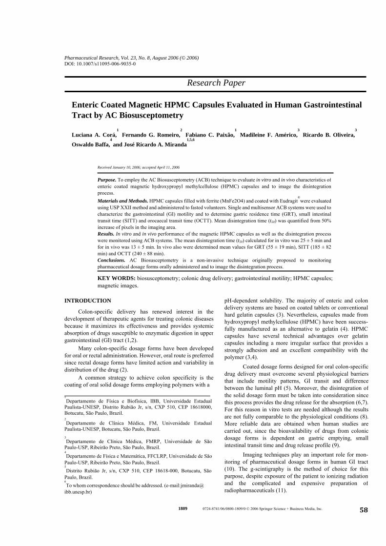
Pharmaceutical Research, Vol. 23, No. 8, August 2006 (© 2006) DOI: 10.1007/s11095-006-9035-0
Research Paper
Enteric Coated Magnetic HPMC Capsules Evaluated in Human Gastrointestinal Tract by AC Biosusceptometry
Luciana A. Corá,1
Fernando G. Romeiro,2
Fabiano C. Paixão,1
Madileine F. Américo,3
Ricardo B. Oliveira,3
Oswaldo Baffa,4 and José Ricardo A. Miranda
1,5,6
Received January 10, 2006; accepted April 11, 2006
Purpose. To employ the AC Biosusceptometry (ACB) technique to evaluate in vitro and in vivo characteristics of enteric coated magnetic hydroxypropyl methylcellulose (HPMC) capsules and to image the disintegrationprocess. Materials and Methods. HPMC capsules filled with ferrite (MnFe2O4) and coated with Eudragit
®were evaluated
using USP XXII method and administered to fasted volunteers. Single and multisensor ACB systems were used tocharacterize the gastrointestinal (GI) motility and to determine gastric residence time (GRT), small intestinal transit time (SITT) and orocaecal transit time (OCTT). Mean disintegration time (t50) was quantified from 50% increase of pixels in the imaging area. Results. In vitro and in vivo performance of the magnetic HPMC capsules as well as the disintegration process were monitored using ACB systems. The mean disintegration time (t50) calculated for in vitro was 25 ± 5 min and for in vivo was 13 ± 5 min. In vivo also were determined mean values for GRT (55 ± 19 min), SITT (185 ± 82 min) and OCTT (240 ± 88 min). Conclusions. AC Biosusceptometry is a non-invasive technique originally proposed to monitoring pharmaceutical dosage forms orally administered and to image the disintegration process.
KEY WORDS: biosusceptometry; colonic drug delivery; gastrointestinal motility; HPMC capsules; magnetic images.
pH-dependent solubility. The majority of enteric and colondelivery systems are based on coated tablets or conventionalhard gelatin capsules (3). Nevertheless, capsules made fromhydroxypropyl methylcellulose (HPMC) have been success-fully manufactured as an alternative to gelatin (4). HPMCcapsules have several technical advantages over gelatincapsules including a more irregular surface that provides astrongly adhesion and an excellent compatibility with thepolymer (3,4).
INTRODUCTION
Colon-specific delivery has renewed interest in thedevelopment of therapeutic agents for treating colonic diseasesbecause it maximizes its effectiveness and provides systemicabsorption of drugs susceptible to enzymatic digestion in uppergastrointestinal (GI) tract (1,2).
Many colon-specific dosage forms have been developedfor oral or rectal administration. However, oral route is preferredsince rectal dosage forms have limited action and variability indistribution of the drug (2). Coated dosage forms designed for oral colon-specific
drug delivery must overcome several physiological barriers that include motility patterns, GI transit and difference between the luminal pH (5). Moreover, the disintegration of the solid dosage form must be taken into consideration since this process provides the drug release for the absorption (6,7). For this reason in vitro tests are needed although the results are not fully comparable to the physiological conditions (8). More reliable data are obtained when human studies are carried out, since the bioavailability of drugs from colonic dosage forms is dependent on gastric emptying, small intestinal transit time and drug release profile (9).
A common strategy to achieve colon specificity is the coating of oral solid dosage forms employing polymers with a
1Departamento de Física e Biofísica, IBB, Universidade Estadual
Paulista-UNESP, Distrito Rubião Jr, s/n, CXP 510, CEP 18618000, Botucatu, São Paulo, Brazil. 2Departamento de Clínica Médica, FM, Universidade Estadual
Paulista-UNESP, Botucatu, São Paulo, Brazil.
58
6To whom correspondence should be addressed. (e-mail:jmiranda@
ibb.unesp.br)
5Distrito Rubião Jr, s/n, CXP 510, CEP 18618-000, Botucatu, São
Paulo, Brazil.
4Departamento de Física e Matemática, FFCLRP, Universidade de São
Paulo-USP, Ribeirão Preto, São Paulo, Brazil.
3Departamento de Clínica Médica, FMRP, Universidade de São
Paulo-USP, Ribeirão Preto, São Paulo, Brazil. Imaging techniques play an important role for mon-
itoring of pharmaceutical dosage forms in human GI tract(10). The g-scintigraphy is the method of choice for thispurpose, despite exposure of the patient to ionizing radiationand the complicated and expensive preparation ofradiopharmaceuticals (11).
c. 1809 0724-8741/06/0800-1809/0 © 2006 Springer Science + Business Media, In
1810 Corá et al.
On the other hand, radiation-free modalities introduceda new perspective for the in vivo investigation of drug delivery.Magnetic Resonance Imaging (MRI) has been employed tomonitor solid pharmaceutical forms in animals and healthysubjects (12-14). Regardless of widespread use in clinical andinherent advantages, MRI has limited application inpharmaceutical research due to the high cost, the positioning ofthe subject during the exposure and the contrast agents do notrepresent an ideal drug model.
Biomagnetic methods are feasible alternative inclinical, physiological and pharmaceutical research and themultichannel SQUID (Superconducting Quantum InterferenceDevice) devices are employed for the measurement of themagnetic field, after ingestion of a magnetically marked dosageform (15). This system is able to detect the extremely weakbiomagnetic fields generally in a magnetically shieldedenvironment. However, SQUID has an expensive operational cost, which limits its use in a wide scale. Fig. 1. Single sensor AC Biosusceptometer. (a) Excitation coil and
(b) detection coil in the first-order gradiometric configuration. Alternating Current Biosusceptometry (ACB) has been
introduced as a valuable tool in gastroenterology (16) andpharmaceutical research (17). ACB uses induction coils forrecording the magnetic flux variation obtained from theresponse of a magnetic material ingested (18). The ACB showed accuracy to evaluate physiologically different param-eters of GI tract (16,19-22) as well as to obtain the magneticimages in vitro (23).
The magnetic signals are acquired through “lock-in” amplifiers (Stanford Research Systems) digitalized by A/D board of 16 bits (PCI-MIO-16XE-10, National Instruments Inc.) and stored in a microcomputer.
Capsule Preparation A multisensor ACB system was implemented tocharacterize the disintegration process of tablets in vitro and in the human stomach, through the acquisition of magnetic signals(17). In addition, this system was also employed to monitormagnetic tablets in GI tract and to image the disintegrationprocess, introducing a novel technique in imaging of thebiological systems (18,24,25).
Size 00 capsules made from hydroxypropyl methylcellu-lose (Vcaps
® , Capsugel Division of Pfizer Inc.) without
colouring agent were filled with 1.20 g of ferrite (MnFe2O4-Ferroxcube, USA) into which had been mixed 0.30 g of sodium starch glycolate-Explotab
® (Penwest, USA).
Following these initial proposals, the aim of this workwas to employ the single and multisensor ACB systems todetermine the gastrointestinal transit time of enteric coatedmagnetic HPMC capsules and to image the disintegrationprocess of these formulations in human ileocolonic region.
Ferrite is a ferromagnetic material with general compo-sition MeFe2O4, where Me represents a divalent transition metal such as manganese (Mn). Ferrite was described as a contrast material or magnetic medicinal preparations (26,27). This material presents good mixing with the GI secretions, absence of toxicity, and lack effects on the digestive tract.
To provide a more objective chemical characterization for this material, the concentrations of iron ions in the dissolution medium were prepared according to USP XXII method (pH 1.2, pH 6.0 and pH 7.4) and were determined by FAAS (Flame Absorption Atomic Spectrometry) using Spec-trophotometer SHIMADZU AA-6800. Standard solutions contained 0.50, 1.00, 2.00, 4.00 and 5.00 mg l
-1 of Fe(III) ions
in HCl 0.01 mol l-1
medium and were used in the calibration of spectrometer (according to the manufactures standard guidelines). No measured iron ion was detected in the samples collected at 0(control), 6, 12, 24 and 48 h, suggesting that the ferrite (MnFe2O4) is a stable molecule and is not absorbed by GI mucosa.
Commercially available system for colon specific drug delivery was used in this study. Eudragit
® S 100 (Röhm,
Pharma Polymers) is a methacrylic acid methylmethacrylate co-polymer, soluble above pH 7, making it particularly suitable for delivery into the colon (28).
METHODS AND MATERIALS
Fundamentals
The single sensor ACB has two pairs of coils (φ = 3.0 cm) separated by a fixed distance (baseline), where each pair of coils are composed of an excitation coil (external) and adetection coil (internal), in the first-order gradiometric con-figuration (Fig. 1). This system working as a double magneticflux transformer with an air nucleus, in which the pair (ex-citation/detection), located more distant from magnetic mate-rial (ferrite), acts as reference. Due to this configuration, whenno magnetic material is near to the measurement system, theoutput signal is minimized. When there is an approximation ofa magnetic mass, an unbalancing in the magnetic flux of thegradiometric system occurs, and the magnetic material ismonitored (18,22,25).
59
Excipients used for the coating dispersions were triethyl citrate (Citroflex
® 2 -Morflex Inc., USA) as a plasticizer,
magnesium stearate as a lubricant, titanium dioxide employed as a pigment, Polysorbate 80 as an emulsifier and glycerol monostearate (Imwitor
® 900 K Sasol, German) as a
The multisensor ACB has only a pair of excitation coil (φ = 11 cm) and seven pairs of detection coils (φ = 2.9 cm), coaxially arranged (Fig. 2). This system is fixed in a vertical support to be positioned on the abdominal surface and to acquire the magnetic signals at different points (17,18,24,25).
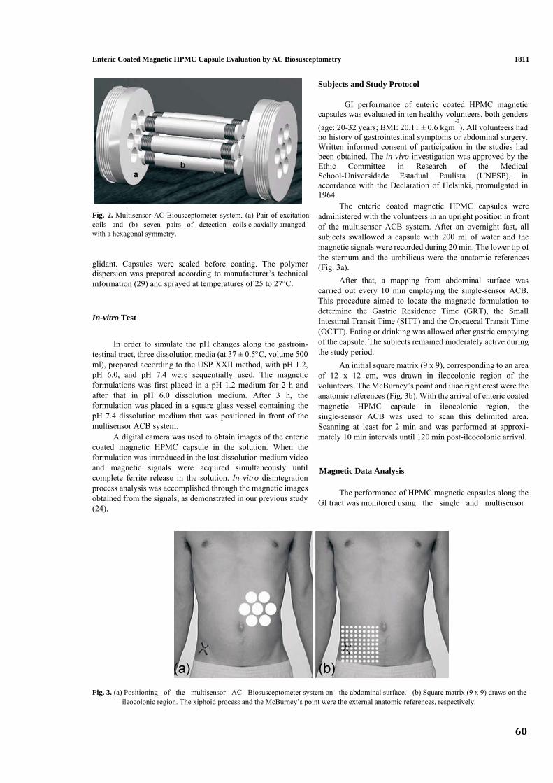
60
glidant. Capsules were sealed before coating. The polymerdispersion was prepared according to manufacturer’s technical information (29) and sprayed at temperatures of 25 to 27°C.
Fig. 2. Multisensor AC Biousceptometer system. (a) Pair of excitationcoils and (b) seven pairs of detection coils c oaxially arranged
1811
with a hexagonal symmetry.
Enteric Coated Magnetic HPMC Capsule Evaluation by AC Biosusceptometry
Subjects and Study Protocol
GI performance of enteric coated HPMC magnetic capsules was evaluated in ten healthy volunteers, both genders (age: 20-32 years; BMI: 20.11 ± 0.6 kgm
-2). All volunteers had
no history of gastrointestinal symptoms or abdominal surgery. Written informed consent of participation in the studies had been obtained. The in vivo investigation was approved by the Ethic Committee in Research of the Medical School-Universidade Estadual Paulista (UNESP), in accordance with the Declaration of Helsinki, promulgated in 1964.
The enteric coated magnetic HPMC capsules were administered with the volunteers in an upright position in frontof the multisensor ACB system. After an overnight fast, all subjects swallowed a capsule with 200 ml of water and the magnetic signals were recorded during 20 min. The lower tip of the sternum and the umbilicus were the anatomic references (Fig. 3a).
After that, a mapping from abdominal surface was carried out every 10 min employing the single-sensor ACB. This procedure aimed to locate the magnetic formulation to determine the Gastric Residence Time (GRT), the Small Intestinal Transit Time (SITT) and the Orocaecal Transit Time (OCTT). Eating or drinking was allowed after gastric emptying of the capsule. The subjects remained moderately active during the study period.
In-vitro Test
In order to simulate the pH changes along the gastroin-testinal tract, three dissolution media (at 37 ± 0.5°C, volume 500 ml), prepared according to the USP XXII method, with pH 1.2,pH 6.0, and pH 7.4 were sequentially used. The magneticformulations was first placed in a pH 1.2 medium for 2 h andafter that in pH 6.0 dissolution medium. After 3 h, theformulation was placed in a square glass vessel containing thepH 7.4 dissolution medium that was positioned in front of themultisensor ACB system.
An initial square matrix (9 x 9), corresponding to an area of 12 x 12 cm, was drawn in ileocolonic region of the volunteers. The McBurney’s point and iliac right crest were the anatomic references (Fig. 3b). With the arrival of enteric coated magnetic HPMC capsule in ileocolonic region, the single-sensor ACB was used to scan this delimited area. Scanning at least for 2 min and was performed at approxi-mately 10 min intervals until 120 min post-ileocolonic arrival. A digital camera was used to obtain images of the enteric
coated magnetic HPMC capsule in the solution. When theformulation was introduced in the last dissolution medium videoand magnetic signals were acquired simultaneously until complete ferrite release in the solution. In vitro disintegration process analysis was accomplished through the magnetic imagesobtained from the signals, as demonstrated in our previous study(24).
Magnetic Data Analysis
The performance of HPMC magnetic capsules along the GI tract was monitored using the single and multisensor
y.
Fig. 3. (a) Positioning of the multisensor AC Biosusceptometer system on the abdominal surface. (b) Square matrix (9 x 9) draws on the ileocolonic region. The xiphoid process and the McBurney’s point were the external anatomic references, respectivel

1812 Corá et al.
was obtained from the 50% increase of pixels in the imaging area (24,25).
ACB systems. Initially, the magnetic signals were recorded frommultisensor ACB with acquisition frequency of 10 Hz/ channeland stored in ASCII format. The signal processing includedbi-directional Butterworth low-pass filter with cutoff frequencyof 0.2 Hz and Fast Fourier Transform (FFT) and allowed tocharacterize the gastric activity contraction (GAC) in theinterdigestive period.
RESULTS
Figure 4(a) illustrates a series of photographs of an enteric coated magnetic HPMC capsule in the phosphate buffer dissolution medium (pH 7.4). Instant t1 represents 10 min of measurement and there was no occurrence of ferrite release. When the coating layer is reduced, the disintegration process (instant t2) initiates and it is intensified due to the action of the excipients that promotes the spreading of the magnetic material in the glass vessel (instant t3). The segmented area outlined in the photographs was used to calculate the mean time disintegration (t50) from the 50% increase of pixels in the imaging area (Fig. 4b).
The Gastric Residence Time (GRT) was defined as thetime interval between the arrival of the enteric coated magneticHPMC capsule in the stomach and its gastric emptying. TheOrocaecal Transit Time (OCTT) was calculated by determining the time between the intake of capsule and the location in theileocolonic region. The Small Intestinal Transit Time (SITT) wasobtained by subtracting the GRT from the OCTT.
The square matrices (9 x 9) were interpolated (256 x 256) by the spline method and appropriate routines to obtain thedegraded images of the enteric coated magnetic HPMC capsulesin vivo were applied (25). Further image processing forquantification included: background subtraction, brightness andcontrast adjustment and segmentation. The segmentation wasused to quantify the spreading of the magnetic material and thevelocity of disintegration (24). All the routines wereimplemented in MatLab
® (Mathworks, Inc.).
For the same instants shown in the photographs, the magnetic images were obtained from a capsule in dissolution medium and showed the disintegration process (Fig. 4c). In the instant t1 the capsule can be observed as a MM. The onset of the disintegration process occurred in the instant t2, with a gradual increase of the imaging area due to the spreading of the magnetic material (instant t3). Fig. 4(d) shows the number of pixels contained inside a delineated area (spreading of the magnetic material) and its time variation (‘velocity of disintegration’). The mean disintegration time (t50) for in vitromeasurements was 25 ± 5 (mean ± SD) min.
The disintegration process for in vitro and in vitro mea-surements was characterized by the transition of a magneticmarker ̶ - MM (non-disintegrated capsule) to a magnetic tracer - MT (disintegrated capsule). In the magnetic images, the MM was clearly delineated and the MT showed thespreading of the magnetic material in the ileocolonic region.Disintegration process was calculated as the mean time
ntegration (t50) after reaching the ileo
Gastric activity contraction (GAC) was recorded in real time by the multisensor ACB, concomitantly to the ingestion of the HPMC capsule is showed in Fig. 5a. The variation of intensity and the basal level of the magnetic signals acquired by the sensors located more distally showed that the enteric disi colonic region, and
Fig. 4. In vitro characterization of an enteric coated magnetic HPMC capsule. (a) Photographs and corresponding magnetic images of thedisintegration process of a capsule in the phosphate buffer. Mean disintegration time (t50) occurred in the instant t2. (b) Spreading of the magnetic material and the time variation of the number of pixels contained inside a delineated area showing the velocity of the disintegration.
61
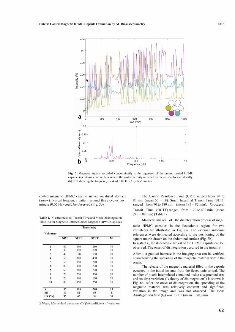
62
1813
ocaecal
Enteric Coated Magnetic HPMC Capsule Evaluation by AC Biosusceptometry
Fig. 5. Magnetic signals recorded concomitantly to the ingestion of the enteric coated HPMCcapsule. (a) Intense contractile waves of the gastric activity recorded by the sensors located distally. (b) FFT showing the frequency peak of 0.05 Hz (3 cycles/minute).
coated magnetic HPMC capsule arrived on distal stomach The Gastric Residence Time (GRT) ranged from 20 to (arrow).Typical frequency pattern around three cycles per 80 min (mean 55 ± 19). Small Intestinal Transit Time (SITT)minute (0.05 Hz) could be observed (Fig. 5b). ranged from 90 to 380 min (mean 185 ± 82 min). Or
Transit Time (OCTT) ranged from 120 to 430 min (mean 240 ± 88 min) (Table I).
Table I. Gastrointestinal Transit Time and Mean Disintegration Magnetic images of the disintegration process of mag-Time (t50) for Magnetic Enteric Coated Magnetic HPMC Capsules
X Mean, SD standard deviation, CV (%) coefficient of variation.
netic HPMC capsules in the ileocolonic region for two volunteers are illustrated in Fig. 6a. The external anatomic references were delineated according to the positioning of the square matrix drawn on the abdominal surface (Fig. 3b). In instant t1, the ileocolonic arrival of the HPMC capsule can beobserved. The onset of disintegration occurred in the instant t2.
After t3, a gradual increase in the imaging area can be verified, characterizing the spreading of the magnetic material within the organ.
The release of the magnetic material filled in the capsule occurred in the initial instants from the ileocolonic arrival. The number of pixels interpolated contained inside a segmented area and its time variation (“velocity of disintegration”) is shown inFig. 6b. After the onset of disintegration, the spreading of the magnetic material was relatively constant and significant variation in the image area was not observed. The mean disintegration time (t50) was 13 ± 5 (mean ± SD) min.

1814 Corá et al.
Fig. 6. (a) Magnetic images of the disintegration process of an enteric coated HPMC capsule in theileocolonic region. The instant t1 shows the arrival of the capsule; from t2 occurred a gradual increase in the image area that characterized the spreading of the magnetic material. (b) Spreading of the magneticmaterial in number of pixels in the segmented area showing the velocity of the disintegration process.
DISCUSSION AND CONCLUSION
lower pH values of the stomach and of the proximal part of the small intestine and also be able to disintegrate at the neutral to slightly alkaline pH of the terminal ileum or at the ileocecal junction (5,28).
An ideal technique to provide more reliable dataabout pharmaceutical drug product performance in humansshould be harmless, noninvasive and have low cost. Thus,AC Biosusceptometry has gained importance in thepharmaceutical research for evaluating successful magneticsolid dosage forms in human GI tract (17,18,25).
In order to characterize the disintegration profiles from the magnetic HPMC capsules coated with Eudragit
® S 100 an in vitro
study was performed. None of the capsules released ferrite in acid medium or at pH 6.0, showing integrity of the coating layer under simulated gastric and small intestine medium.
A variety of coated forms have not been developedwith significant therapeutic advantages for the localtreatment of colonic diseases (2,31). The use of coateddosage forms for oral colon specific drug delivery, allowingto develop enteric coated of HPMC capsules that appears asan industrially viable process, resulting from improvedcoating technologies and flexibility in their design (3,38).
Aiming to compare quantitatively the disintegration profiles, photographs and the corresponding magnetic images were analyzed (Fig. 4). The capsule was suitable to release rapidly the magnetic material at pH 7.4 as shown in the instant t2. Once initiated, the disintegration promotes the dispersion of the ferrite continuously (instant t3).
The polymers used for colon targeting exploit thegenerally accepted fact that pH of the human GI tractincreases progressively from the stomach at the distal ileum(30). Therefore, should be able to withstand the
Magnetic images are different from the photographs since the field-of-views are not the same because they were obtained at distinct angles and distances. Despite this, employing ACB
63
64
it was possible to characterize the disintegration process bycomparing the similar profiles with those obtained by photo-graphs (Fig. 4). In vitro disintegration of the capsulessuggests good performance of ACB and ferrite release only inthe distal ileum or the proximal colon.
Although disintegration process can be studied in vitro, the interaction between physiological parameters and thesolid dosage forms affect drug delivery profile and thereproducibility of drug release (8). If the intended site of drugrelease is the colon, it must be taken into consideration theprandial state and gastric emptying of dosage forms.
ACB system was able to record in real-time the gastric motility during the interdigestive period (Fig. 5), character-ized by a cyclical motor pattern so-called myoelectric migrating complex (MMC) (32). Phase III contractions promote the emptying of indigestible materials, includingsolid pharmaceutical forms (33,34). In the presented study, allvolunteers fasted prior to the administration of the magnetic formulation, allowing inferring that the capsules had beenemptied from stomach during this period of activity.
Gastric residence time (GRT) for enteric coated mag-netic HPMC capsules was obtained from the arrival in thestomach until its emptying (Table I). The mean GRT was 55min showing an important intersubject variation despite of experimental protocol had been designed to minimize theinfluence of any external factor in the gastric emptying of themagnetic formulation.
The results from our investigation (Table I) showed thatthe SITT presented a significant intersubjects variation(mean 185 ± 82 min), however are within the normal rangesobtained in previous studies (33,37). As reported by otherstudies, it is generally accepted that small intestinal transittime (SITT) is not affected by the digestive state or by the nature of the pharmaceutical form (33,35,36).
Orocaecal transit time (OCTT) occurred on average at240 ± 88 min (Table I). The variation observed can beattributed to the GRT and SITT, since these parametersshowed significant intersubject differences as discussedearlier. Not surprisingly, GI transit for enteric coatedmagnetic tablets (25) compared with magnetic HPMC capsules was not significantly different for the GRT andSITT.
Mean disintegration time for magnetic capsules occurredin a short time interval (mean 13 ± 5 min), when comparedwith the disintegration of the magnetic tablets (mean 90 ± 40 min). Indeed the observed difference could be attributed tothe kind of pharmaceutical form, since the powder filled intothe capsule was not compressed. It is well known that thecompression force is a very important parameter for the tablet manufacturing process, particularly for the development ofthe time-controlled disintegration (39).
Our findings about disintegration time of magneticcapsules contrasts sharply with those obtained by recentreports (3,4,38). The discrepancy might be due to thepharmaceutical strategies to achieve drug release in the colon(pH-sensitive or enzyme-controlled release), coatingthickness and excipients used. Moreover, the criteria adoptedfor the analysis considered the mean time disintegrationstarted after reaching the ileocolonic region, instead of thecomplete process.
Magnetic images constitute an innovative approach tocharacterize the disintegration of pharmaceutical dosage
forms in human GI tract (24). The segmentation of the imaging area allowed quantifying the spreading of the magnetic material to characterize the transition between the MM to MT provided by the disintegration process. Although these images presentedreasonable quality, the application of restoration techniques could improve image quality and suppress noise simultaneously.
Unfortunately, based on the characteristics of the magnetic formulation, it remains impractical to compare directly ourfindings with the results obtained from standard imaging techniques. However, great effort has been made to improve the biomagnetic systems and to reduce the amount of ferrite in the magnetic formulation similar to a conventional dosage form. Thereby, this magnetic method could be associated with pharmacokinetic parameters (“magnetopharmacokinetics”)to predicting the drug absorption in a specific site of GI tract for optimized pharmacotherapy (40).
In summary, AC Biosusceptometry systems are com-pletely safe and harmless devices, able to evaluate accurately solid dosage forms in human GI tract. Additionally, these systems represent a novel imaging tool to characterize diverse parameters related to drug delivery, thus deserving the same importance as conventional techniques in pharmaceutical research.
ACKNOWLEDGMENTS
The authors would like to thank the Brazilian agencies: FAPESP/PRONEX (03/10107-5) and CAPES for financial support. Eudragit
® samples used in this study were supplied from
Almapal S/A-Tecnologia Validada, Brazil. HPMC capsules (V-Caps®) were a gift from Capsugel-Divison of Pfizer, Brazil.
REFERENCES
1. L. Yang, J. S. Chu, and J. A. Fix. Colon-specific drug delivery: new approaches and in vitro/in vivo evaluation. Int. J. Pharm.235:1-15 (2002).
2. M. A. Shareef, R. K. Khar, A. Ahuja, F. J. Ahmad, and S.Raghava. Colonic drug delivery: an updated review. AAPS Pharm. Sci. 5:1-26 (2003).
3. E. T. Cole, R. A. Scott, A. L. Connor, I. R. Wilding, H-U. Petereit, C. Schminke, T. Beckert, and D. Cadé. Enteric coated HPMC capsules designed to achieve intestinal targeting. Int. J.
M. ma
ug
.
e-
or
3
5
Pharm. 231:83-95 (2002). 4. O. Honkanen, J. Marvola, H. Kanerva, K. Lindevall, Lipponen, T. Kekki, A. Ahonen, and M. Marvola. Gam scintigraphic evaluation of the fate of hydroxypropyl methylcel- lulose capsules in the human gastrointestinal tract. Eur. J. Pharm. Sci. 21:671-678 (2004). 5. C. S. Leopold. Coated dosage forms for colon-specific dr delivery. Pharm. Sci. Technol. Today 2:197-252 (1999). 6. C. D. Melia and S. S. Davis. Review article: mechanisms of drug release from tablets and capsules. I: disintegration. Aliment Pharmacol. Ther. 3:223-232 (1989). 7. E. Lipka and G. L. Amidon. Setting bioequivalence requir ments for drug development based on preclinical data: optimizing oral drug delivery systems. J. Control. Release 62:41-49 (1999). 8. M. Zahirul and I. Khan. Dissolution testing for sustained controlled release oral dosage forms and correlation with in vivo data: challenges and opportunities. Int. J. Pharm. 140:131-14 (1996). 9. N. Rouge, P. Buri, and E. Doelker. Drug absorption sites in the gastrointestinal tract and dosage forms for site-specific delivery. Int. J. Pharm. 136:117-139 (1996).
Enteric Coated Magnetic HPMC Capsule Evaluation by AC Biosusceptometry 181
1816 Corá et al.
65
or
. -.
.
g .
er. o
10. M. Singh and V. Waluch. Physics and instrumentation f imaging in-vivo drug distribution. Adv. Drug Deliv. Rev. 4:7-20 (2000)11. I. R. Wilding, A. J. Coupe, and S. S. Davis. The role of γ scintigraphy in oral drug delivery. Adv. Drug Deliv. Rev 46:103-124 (2001)12. J. C. Richardson, R. W. Bowtell, K. Mäder, and C. D. Melia. Pharmaceutical applications of magnetic resonance imagin (MRI). Adv. Drug Deliv. Rev. 57:1191-1209 (2005)13. A. Steingoetter, D. Weishaup, P. Kunz, K. Mäder, H.Lengsfeld, M. Thumshirn, P. Boesiger, M. Fried, and W. Schwiz Magnetic resonance imaging for the in vivo evaluation
f gastric-retentive tablets. Pharm. Res. 20:2001-2007 (2003).
ic-g.
. ms.ic
AChys.
.
of ate
.
Rev.
14. A. Steingoetter, D. Weishaup, P. Kunz, K. Mäder, H.Lengsfeld, M.Thumshirn, P.Boesiger, M.Fried, and W.Schwizer. Analysis of the meal-dependent intragastric performance of a gastr retentive tablet assessed by magnetic resonance imagin Aliment. Pharmacol. Ther. 18:713-720 (2003)15. W. Weitschies, O. Kosch, H. Mönnikes, and L. Trah Magnetic Marker Monitoring: an application of biomagnet measurement instrumentation and principles for the determina- tion of the gastrointestinal behavior of magnetically marked solid dosage forms. Adv. Drug Deliv. Rev. 57:1210-1222 (2005).16. J. R. A. Miranda, O. Baffa, and R. B. Oliveira. An biosusceptometer to study gastric emptying. Med. P 19:445-448 (1992)17. L. A. Corá, M. F. Américo, R. B. Oliveira, O. Baffa, R. Moraes, F. G. Romeiro, and J. R. A. Miranda. Disintegration magnetic tablets in human stomach evaluated by altern current biosusceptometry. Eur.J. Pharm.Biopharm. 56:413-420 (2003)18. L. A. Corá, F. G. Romeiro, M. Stelzer, M. F. Américo, R. B. Oliveira, O. Baffa, and J. R. A. Miranda. AC biosusceptometry in the study of drug delivery. Adv. Drug Deliv. 57:1223-1241 (2005). 19. O. Baffa, R. B. Oliveira, J. R. A. Miranda, and L E. A. Troncon. Analysis and development of AC biosusceptometer for orocaecal transit time measurements. Med. Biol. Eng. Comput.
r l.
.
O. Baffa. A novel biomagnetic method to study gastric antral
, O. Baffa,
iol.
nd O. Baffa. Imaging ferromagnetic
s, F. G. Romeiro, M. F. Américo, R. B.
ac.
.ity
. J.
X-ray.
o-.
. lic
Sci..
dt,.
n.
i..
nd.
n ins in
. M.
coneol.
. -
2.
thy
. ,
ne: ar.
. .
s..
ndlledby
..
al7
.
33:353-357 (1995). 20. R. B. Oliveira, O. Baffa, L. E. A. Troncon, J. R. A. Miranda, and C. R. Cambrea. Evaluation of a biomagnetic technique fo measurement of orocaecal transit time. Eur. J. Gastroentero Hepatol. 8:491-495 (1996)21. J. R. A. Miranda, R. B. Oliveira, P. L. Sousa, F. J. H. Braga, and contractions. Phys. Med. Biol. 42:1791-1799 (1997). 22. R. Moraes, L. A. Corá, M. F. Américo, R. B. Oliveira and J. R. A. Miranda. Measurement of gastric contraction activity in dogs by means of AC biosusceptometry. Phys Meas. 24:337-345 (2003). 23. M. Moreira, L. Murta, a tracers with an AC biosusceptometer. Rev. Sci Instrum. 71:2532-2538 (2000). 24. L. A. Corá , U. Andrei Oliveira, O. Baffa, and J. R. A. Miranda. Magnetic images of the
disintegration process of tablets in the human stomach by biosusceptometry. Phys. Med. Biol. 50:5523-5534 (2005)25. L. A. Corá , F. G. Romeiro, M. F. Américo, R. B. Oliveira, O Baffa, M. Stelzer, and J. R. A. Miranda. Gastrointestinal trans and disintegration of enteric coated magnetic tablets assessed b ac biosusceptometry. Eur. J. Pharm. Sci. 27:1-8 (2006)26. E. H. Frei, E. Gunders, M. Pajewsky, W. J. Alkan, and Eshcher. Ferrites as contrast material for medical diagnosis. J. Appl. Phys. 39:99-101 (1968)27. V. G. Belikov and A. G. Kuregyan. Generation and medic biological application of magnetic fields and carriers (review) Pharm. Chem. J. 35:88-95 (2001)28. A. David, B. Yagen, A. Sintov, and A. Rubinstein. Acry polymers for colon-specific drug delivery. STP Pharma 7:546-554 (1997)29. K. Lehman. Practical Course in Film Coating of Pharmaceutical Dosage Forms with Eudragit®, Pharma Polymers, Darmsta 200130. D. F. Evans, G. Pye, R. Bramley, A. G. Clark, T. J. Dyson, and J. D. Hardcastle. Measurement of gastrointestinal pH profiles i normal ambulant human subjects. Gut 29:1035-1041 (1988)31. M. K. Chourasia and S. K. Jain. Pharmaceutical approaches to colon target drug delivery systems. J. Pharm. Pharmaceut. Sc 6:33-66 (2003)32. E. M. M. Quigley. Gastric and small motility in health a disease. Gastroenterol. Clin. North Am. 25:113-145 (1996)33. A. J. Coupe, S. S. Davis, and I. R. Wilding. Variatio gastrointestinal transit of pharmaceutical dosage form healthy subjects. Pharm. Res. 8:360-364 (1991)34. F. Kedzierewicz, P. Thouvenot, J. Lemut, A. Etienne, Hoffman, and P. Maincent. Evaluation of peroral sili dosage forms in humans by gamma-scintigraphy. J. Contr Release 58:195-205 (1999)35. S. S. Davis, J. G. Hardy, and J. W. Fara. Transit of pharmaceu Tical dosage forms through the small intestine. Gut 27:886-89 (1986)36. D. Harris, J. T. Fell, H. L. Sharma, and D. C. Taylor. GI transi of potential bioadhesive formulations in man: a scintigrap study. J. Control. Release 12:45-53 (1990)37. C. J. Kenyon, R. V. Nardi, D. Wong, G. Hooper, I. R. Wilding and D. R. Friend. Colonic delivery of dexamethaso pharmacoscintigraphic evaluation. Aliment. Pharmacol. The 11:205-213 (1997)38. C. Tuleu, A. W. Basit, W. A. Waddington, P. J. Ell, and J. M Newton. Colonic delivery of 4-aminosalicylic acid using amylase- ethylcellulose-coated hydroxypropylmethylcellulose capsule Aliment. Pharmacol.Ther. 16:1771-1779 (2002)39. K.-H. Lin, S.-Y. Lin, and M.-J. Li. Compression forces a amount of outer coating layer affecting the time-contro disintegration of the compression-coated tablets prepared direct compression with micronized ethylcellulose. J. Pharm. Sci 90:2005-2009 (2001)40. H. Zhou. Pharmacokinetic strategies in deciphering atypic drug absorption profiles. J. Clin. Pharmacol. 43:211-22 (2003)
Capítulo 4
AC Biosusceptometry to evaluate the gastrointestinal transit of pellets under influence of prandial state
International Journal of Pharmaceutics (2008)
66
AC Biosusceptometry to evaluate the gastrointestinal transit
of pellets under influence of prandial state
José Ricardo A. Mirandaa, Luciana A. Coráa,*, Madileine F. Américoa, Fernando Romeirob
aDep. Física e Biofísica, IBB, São Paulo State University – UNESP, Botucatu, Brazil.
bDep. Clínica Médica, FMB, São Paulo State University – UNESP, Botucatu, Brazil.
Abstract
Multiparticulate dosage forms have been proposed when distal regions of
gastrointestinal tract are desirable as target of drugs. Nevertheless, as physiological
parameters might interfere with the processes related to the drug delivery and
absorption, it is essential to evaluate the behavior of these systems in vivo. The aim of
this study was to propose the AC Biosusceptometry (ACB) as a noninvasive and
radiation free technique to evaluate the gastrointestinal transit of a magnetic
multiparticulate dosage form in healthy volunteers under fasting and fed conditions.
Magnetic pellets were prepared by the powder layering method of ferrite on
nonpareils sugar beads which have been coated by using Eudragit®. Our data showed
that ACB was able to monitoring the gastrointestinal transit of pellets and resulted in
similar profiles as demonstrated by standard techniques. Food intake has markedly
influenced the gastric emptying as well as the colon arrival and the small intestine
transit of magnetic pellets. This biomagnetic method showed a number of advantages
over existing methodologies and deserves the same importance for this kind of
analysis.
Keywords: Gastrointestinal transit, pellet, prandial state, magnetic techniques,
Biosusceptometry
*Corresponding Author. Departamento de Física e Biofísica, Instituto de Biociências – IBB, Laboratório de Biomagnetismo, Universidade Estadual Paulista – UNESP, CXP 510, CEP 18618-000, Botucatu, São Paulo, Brazil. Tel./fax: +55-14-3811-6254. E-mail address: [email protected] (L.A. Corá)
67
1. Introduction
Controlled drug delivery systems are designed to achieve more predictable
bioavailability of drugs either to increase efficacy or minimize adverse effects. Several
approaches have been proposed, and multiparticulate dosage forms seem to have
better performance when distal regions of gastrointestinal tract are desirable as
target of drugs (Ranade, 1991; Gandhi et al., 1999).
Human colon has been proposed as the specific targeting of drugs for the
topical treatment of intestinal diseases as well as for the delivery of therapeutic
peptides and proteins (Yang et al., 2002; Chourasia and Jain, 2003; Shareef et al.,
2003). For this purpose, the development of multiparticulate systems has been
emphasized due to its advantages over single-unit dosage forms that include the
release of drugs at optimal rate, reduced dosing frequency and constant blood levels
(Asghar and Chandran, 2006).
Experimental determination of drug release may be assessed by well-
established in vitro methodologies. However, these tests are often not predictive of
the more complex in vivo behavior of drug products (Zahirul and Khan, 1996).
Physiological parameters as motility, gastric emptying time, intestinal transit time,
and pH are involved in the rate and extent of drug absorption (Dressman et al., 1993).
Therefore, an understanding of the factors involved in the drug absorption and how
these parameters can interfere with this process is crucial to develop more reliable
drug delivery systems.
Scintigraphy has been extensively used as an imaging tool to monitoring the in
vivo performance of drug delivery systems (Wilding et al., 2001); however the
ionizing radiation exposure is its major drawback.
Biomagnetic as well as radiotelemetric methods have advantages over
scintigraphy due to the non-invasivity and radiation-free features. SQUIDs
(Superconducting Quantum Interference Devices) have been employed to investigate
the behavior of solid dosage forms in the human gastrointestinal tract (Weitschies et
al., 2005). Heidelberg Radiotelemetry Capsule is a device which has been used to
monitor gastrointestinal pH and the gastric residence time in both humans and
animals (Mojaverian, 1996).
68
Alternate Current Biosusceptometry (ACB) has been becoming a promising
method for pharmaceutical research. AC Biosusceptometry was originally proposed
as a method to investigate the gastrointestinal motility in humans (Miranda et al.,
1997; Romeiro et al., 2006; Américo et al., 2007) and animals (Moraes et al., 2003;
Andreis et al., 2007), regarding several aspects on physiology and clinical researches.
Continuous improvements allowed enhancing the spatial resolution and the
sensitivity of the device for pharmaceutical applications. It was initially proposed to
monitor the disintegration process of magnetic tablets in vitro and in human stomach
(Corá et al., 2003). The potential demonstrated allowed applying this method to
investigate the behavior of enteric-coated dosage forms and to quantify the
gastrointestinal transit time as well as the disintegration time in human colon (Corá
et al., 2006a,b). Moreover, ACB method has been proposed as a new tool for imaging
of pharmaceutical processes (Corá et al., 2005) and, more recently, as a tool for
quality control for pharmaceutical products (Corá et al., 2008).
The aim of this study was to propose the AC Biosusceptometry as an
alternative method to investigate the magnetic multiparticulate delivery system
under influence of prandial state on the gastric emptying and intestinal transit.
2. Materials and methods
2.1. Materials
Ferrite powder (MnFe2O4; 80-125m) was purchased from Thornton, Brazil
and was used as the magnetic marker. The enteric polymer used was the
methacrylate copolymer (Eudragit® S100) as a gift from Röhm GmbH, Germany.
Other excipients used for coating were of standard pharmaceutical grade: magnesium
stearate (Valdequímica, Brazil), talc (Valdequímica, Brazil), titanium dioxide
(Valdequímica, Brazil), triethyl citrate (Scandiflex, Brazil), and isopropyl alcohol
(Sigma-Aldrich, Brazil). The excipients used to prepare pellets and for coating were of
standard pharmaceutical grade and all chemical reagents used were of analytical
grade.
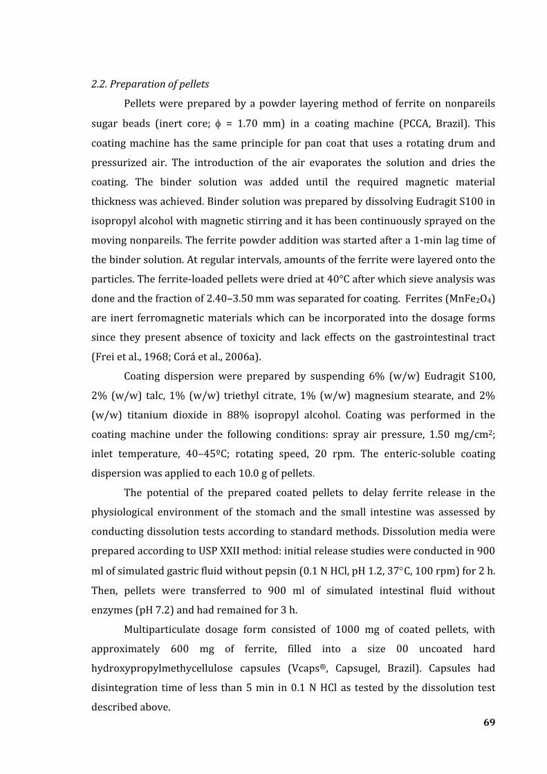
69
2.2. Preparation of pellets
Pellets were prepared by a powder layering method of ferrite on nonpareils
sugar beads (inert core; = 1.70 mm) in a coating machine (PCCA, Brazil). This
coating machine has the same principle for pan coat that uses a rotating drum and
pressurized air. The introduction of the air evaporates the solution and dries the
coating. The binder solution was added until the required magnetic material
thickness was achieved. Binder solution was prepared by dissolving Eudragit S100 in
isopropyl alcohol with magnetic stirring and it has been continuously sprayed on the
moving nonpareils. The ferrite powder addition was started after a 1-min lag time of
the binder solution. At regular intervals, amounts of the ferrite were layered onto the
particles. The ferrite-loaded pellets were dried at 40°C after which sieve analysis was
done and the fraction of 2.40–3.50 mm was separated for coating. Ferrites (MnFe2O4)
are inert ferromagnetic materials which can be incorporated into the dosage forms
since they present absence of toxicity and lack effects on the gastrointestinal tract
(Frei et al., 1968; Corá et al., 2006a).
Coating dispersion were prepared by suspending 6% (w/w) Eudragit S100,
2% (w/w) talc, 1% (w/w) triethyl citrate, 1% (w/w) magnesium stearate, and 2%
(w/w) titanium dioxide in 88% isopropyl alcohol. Coating was performed in the
coating machine under the following conditions: spray air pressure, 1.50 mg/cm2;
inlet temperature, 40–45ºC; rotating speed, 20 rpm. The enteric-soluble coating
dispersion was applied to each 10.0 g of pellets.
The potential of the prepared coated pellets to delay ferrite release in the
physiological environment of the stomach and the small intestine was assessed by
conducting dissolution tests according to standard methods. Dissolution media were
prepared according to USP XXII method: initial release studies were conducted in 900
ml of simulated gastric fluid without pepsin (0.1 N HCl, pH 1.2, 37C, 100 rpm) for 2 h.
Then, pellets were transferred to 900 ml of simulated intestinal fluid without
enzymes (pH 7.2) and had remained for 3 h.
Multiparticulate dosage form consisted of 1000 mg of coated pellets, with
approximately 600 mg of ferrite, filled into a size 00 uncoated hard
hydroxypropylmethycellulose capsules (Vcaps®, Capsugel, Brazil). Capsules had
disintegration time of less than 5 min in 0.1 N HCl as tested by the dissolution test
described above.
70
2.3. AC Biosusceptometry
A detailed description about the principles of AC Biosusceptometry (ACB)
technique was reported by Corá et al. (2005a). Briefly, AC Biosusceptometry bases its
functioning on induction coils for recording the magnetic flux variation obtained from
the response of a magnetic material when an alternating magnetic field is applied.
This study was carried out using a single-sensor ACB system which has a pair of
excitation and detection coils ( = 3.0 cm) coaxially arranged in a first-order
gradiometric configuration (Fig. 1). The pair (excitation/detection) that is located
more distant from the magnetic material that will be detected acts as a reference coil
and the pair closest of the sample as a measurement coil.
The excitation coils induce equal magnetic flux in the detection coils, hence,
when a magnetic sample is nearest of the measurement system an imbalance in the
voltage occurs, due to the change in the differential flux between the detection coils.
Magnetic signals from single-sensor ACB has been acquired employing a lock-in
amplifier (Stanford Research Systems, Inc., USA).
Fig. 1- Single-sensor AC Biosusceptometry system with the pair of excitation (1) and detection
coils (2) coaxially arranged in a first-order gradiometric configuration.
71
2.4. Subjects and study protocol
Nine healthy volunteers (5 male, 4 female; age, 22-30 years; weight, 50-75 kg)
participated in the study. The study was approved by the Ethical Committee of
Medical School - Sao Paulo State University and the trial was conducted in accordance
with the Declaration of Helsinki (1964) and its revisions. Each subject provided
written informed consent to participate in the study.
The study consisted of two phases: on one occasion, the multiparticulate
dosage form was administered after an overnight fasted (Fasted phase), and on
another occasion following the standard breakfast (Fed phase) described above. The
two phases of the study were carried out at least 1 week apart.
The Fasted phase consisted of the administration of the multiparticulate
dosage form with 200 ml of water to volunteers who fasted for at least 12 h before
dosing. The Fed phase consisted of the administration of the multiparticulate dosage
form with 200 ml of water 10 min after the standard breakfast (comprising two slices
of bread, two slices of ham, two slices of cheese and 180 g of yogurt) with energy
content of 502 Kcal. Further drinks or meals were not allowed until the magnetic
pellets had left the stomach.
2.5. Magnetic measurements
A square point matrix (5x5) was drawn around the gastric (lower tip of
sternum and the umbilicus as anatomical references) and colonic (McBurney’s point
and iliac right crest as anatomical references) regions (Fig. 2). The ACB sensor was
attached to a computer controlled x-y scanning stage and the measurements were
performed. From each scanner, it was generated the magnetic field maps which
indicated the intensity values distributed on gastric or colonic region. Those values
were corrected for basal measurements prior the administration of the dosage form.
Each magnetic monitoring had 120 s duration and was recorded at 10 min intervals
over 7 h. This monitoring was performed with the volunteers in orthostatic position.
Acquisition of the magnetic signals was carried out from the lock-in amplifier
(Stanford Research Systems, Inc., USA) and the signal processing was done using
MatLab® (Mathworks, Inc., Natick, MA, USA).
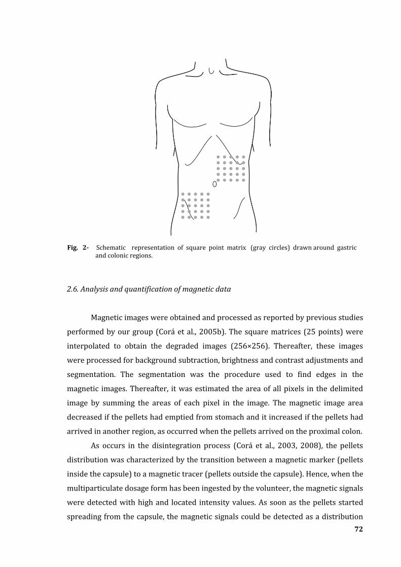
72
2.6. Analysis and quantification of magnetic data
Magnetic images were obtained and processed as reported by previous studies
performed by our group (Corá et al., 2005b). The square matrices (25 points) were
interpolated to obtain the degraded images (256×256). Thereafter, these images
were processed for background subtraction, brightness and contrast adjustments and
segmentation. The segmentation was the procedure used to find edges in the
magnetic images. Thereafter, it was estimated the area of all pixels in the delimited
image by summing the areas of each pixel in the image. The magnetic image area
decreased if the pellets had emptied from stomach and it increased if the pellets had
arrived in another region, as occurred when the pellets arrived on the proximal colon.
As occurs in the disintegration process (Corá et al., 2003, 2008), the pellets
distribution was characterized by the transition between a magnetic marker (pellets
inside the capsule) to a magnetic tracer (pellets outside the capsule). Hence, when the
multiparticulate dosage form has been ingested by the volunteer, the magnetic signals
were detected with high and located intensity values. As soon as the pellets started
spreading from the capsule, the magnetic signals could be detected as a distribution
Fig. 2- Schematic representation of square point matrix (gray circles) drawn around gastric and colonic regions.
73
on the intensity values. The gastric emptying as well as the colonic arrival has been
characterized by measuring the magnetic image area at different time intervals. For
gastric emptying, the decrease in the area was calculated as the % in relation to the
initial area, i.e., by measuring the number of pixels inside the delimited magnetic
image area. For colon arrival, it was calculated the increase of the number of pixels
inside the delimited magnetic image area. To obtain more accurate evaluation, those
curves were interpolated for 60 points by spline method which has improved the
temporal resolution.
As reported by a previous study (Podczeck et al., 1995), the parameter t50 is
not reliable to quantify the gastric emptying mainly due to the irregular shaped
profiles which may hinder the correct interpretation of the whole process. Hence, the
gastric emptying and colonic arrival of magnetic pellets also were evaluated by
applying statistical moments. The Mean Gastric Emptying Time (MGET) was
described as amount of pellets emptied at time t; the Mean Colon Arrival Time
(MCAT) was represented as amount of pellets arrived at time t; Mean Small Intestinal
Transit Time (MSITT) also was quantified as being the difference between MCAT and
MGET. MGET and MCAT, as well as their variances (VGET and VCAT, respectively)
have been calculated using the simple trapezoidal rules, as proposed by Podczeck et
al. (1995).
All the results are expressed as mean ± standard deviation (SD). In order to
investigate the relationship between both analyses, the values of, MGET, MCAT and
MSITT obtained under fasted and fed conditions were correlated. By, using paired t-
test statistically significant difference was considered at p < 0.01.
74
3. Results and discussion
Concerning the retention time in the stomach and the transit through the small
intestine, it is interesting that the start of drug release could be controlled by pH-
dependent polymer dissolution (Leopold, 1999). An essential prerequisite for a
delivery system for colon targeting it is to prevent the drug release until the dosage
form reaches the colon. Hence, the polymer used in this study has been sufficiently
able into assure the magnetic material release solely after arrival in the colon since
none of the magnetic coated pellets showed ferrite released during the dissolution
test performed at simulated gastric fluid.
The gastrointestinal transit times of the coated pellets for fasted and fed
subjects are summarized in Table 1. As expected, gastric emptying time was markedly
different under fasted and fed conditions. MGET values quantified for both fasted and
fed conditions were 34 ± 14 and 125 ± 46, respectively. Statistically significant
differences were obtained between the fed and fasted values of MGET (p < 0.01).
Although a different method was employed, the observed delayed emptying of
magnetic pellets is consistent with previously reported results that showed the
gastric emptying time of solid dosage forms increases under fed conditions (O’Reilly
et al., 1987; Wilding et al., 1991; Choe et al., 2001).
Gastric emptying plays an important role in determining the retention of oral
dosage forms; then, delayed gastric emptying might be exploited as an approach to
enhance the absorption of drugs with an absorption window in the upper
gastrointestinal tract (Marathe et al., 2000).
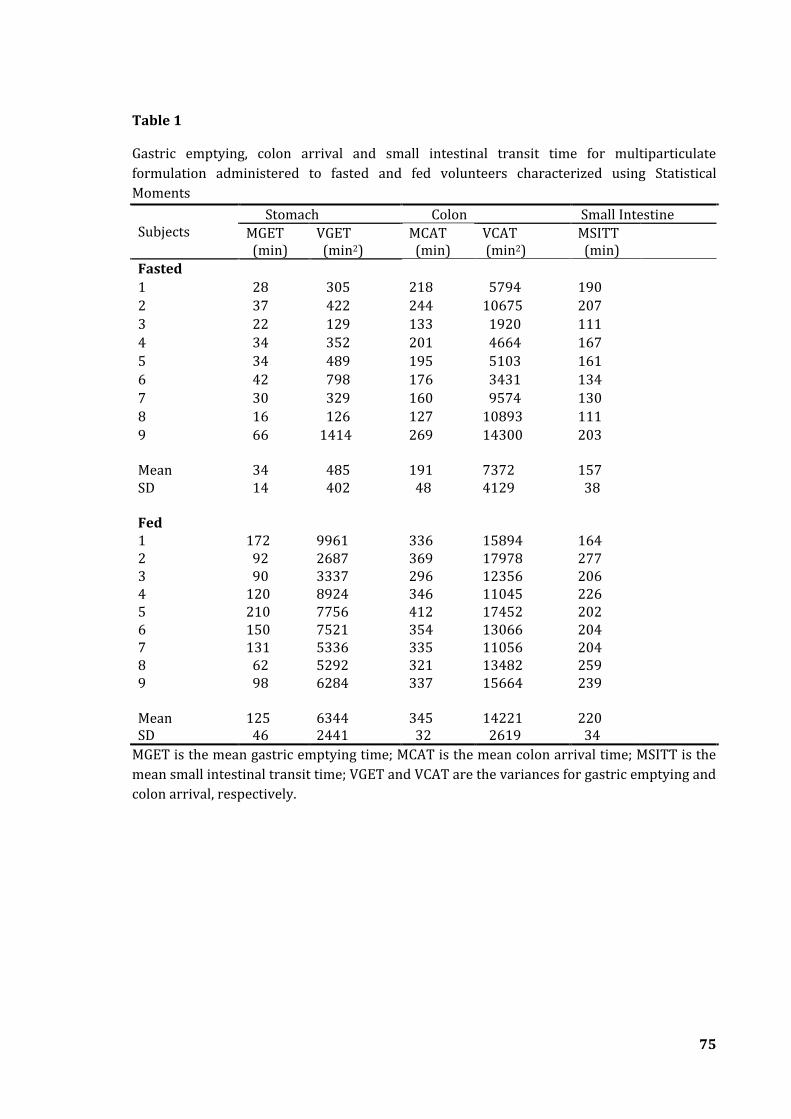
75
Table 1
Gastric emptying, colon arrival and small intestinal transit time for multiparticulate
formulation administered to fasted and fed volunteers characterized using Statistical
Moments
Subjects
Stomach Colon Small Intestine
MGET (min)
VGET (min2)
MCAT (min)
VCAT (min2)
MSITT (min)
Fasted
1 28 305 218 5794 190
2 37 422 244 10675 207
3 22 129 133 1920 111
4 34 352 201 4664 167
5 34 489 195 5103 161
6 42 798 176 3431 134
7 30 329 160 9574 130
8 16 126 127 10893 111
9 66 1414 269 14300 203 Mean
34
485
191
7372
157
SD 14 402 48 4129 38 Fed
1 172 9961 336 15894 164 2 92 2687 369 17978 277 3 90 3337 296 12356 206 4 120 8924 346 11045 226 5 210 7756 412 17452 202 6 150 7521 354 13066 204 7 131 5336 335 11056 204 8 62 5292 321 13482 259 9 98 6284 337 15664 239 Mean SD
125 46
6344 2441
345 32
14221 2619
220 34
MGET is the mean gastric emptying time; MCAT is the mean colon arrival time; MSITT is the
mean small intestinal transit time; VGET and VCAT are the variances for gastric emptying and
colon arrival, respectively.
76
For pharmaceutical purposes, the transit of a dosage form through the
gastrointestinal tract determines how long a compound remains in contact with its
absorptive site. The bioavailability of a drug can be affected by factors that change
gastrointestinal transit. Several publications have related that the pellets which had
spread in small intestine tend to regroup in the ileocaecal junction for an
undetermined period before reaching the colon and the passage across the junction
can occurs quickly or for many hours (Coupe et al., 1991 ; Wilding et al., 1991; Clark
et al., 1995; Wilding et al., 2000). In the present study, arrival of pellets in the colonic
region seemed to occur in bolus, after a stagnation period which might be
characterized by a signal with a punctual intensity, i.e., it would be detected none or a
minimal spreading of the material. However, further investigation would be needed
to elucidate this behavior.
MCAT values for fasted and fed condition were 191 ± 48 and 345 ± 32,
respectively, also presented a significant increase (p < 0.01). The mean values of
MSITT on both fasted and fed state were 157 ± 38 and 220 ± 34, respectively.
Statistically significant differences were obtained between the fed and fasted values
of MSITT (p < 0.01). As reported by others, although small intestinal transit time of
pharmaceutical dosage forms in humans seems to be relatively constant and appears
to be independent of both the type of dosage form and prandial state, it can be
observed a considerable inter-subjects variability (Hardy et al., 1985; Davis et al.,
1986; Coupe et al., 1991); moreover, colon arrival may be notably influenced by the
gastric emptying. The spreading of pellets in the colon after emptying from the
stomach was evaluated by compute the variance of the distributions for stomach
(VGET) and colon (VCAT).
Figure 3 are the representative magnetic images of the multiparticulate
system were taken from different time intervals, illustrating key stages of the
gastrointestinal transit for a fasted subject. On all occasions, pellets had fully
dispersed from the HPMC capsule within 5 min. In the subsequent post-dose
magnetic images, spreading of the pellets could be observed in the stomach. Gastric
emptying was characterized as a decrease in the segmented image area; meanwhile,
the colon arrival was evidenced by the gradual increase in the image area.
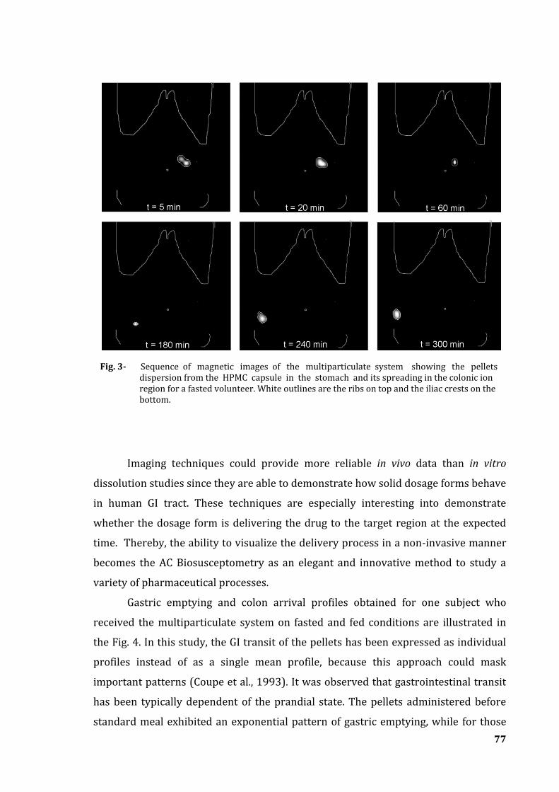
77
Imaging techniques could provide more reliable in vivo data than in vitro
dissolution studies since they are able to demonstrate how solid dosage forms behave
in human GI tract. These techniques are especially interesting into demonstrate
whether the dosage form is delivering the drug to the target region at the expected
time. Thereby, the ability to visualize the delivery process in a non-invasive manner
becomes the AC Biosusceptometry as an elegant and innovative method to study a
variety of pharmaceutical processes.
Gastric emptying and colon arrival profiles obtained for one subject who
received the multiparticulate system on fasted and fed conditions are illustrated in
the Fig. 4. In this study, the GI transit of the pellets has been expressed as individual
profiles instead of as a single mean profile, because this approach could mask
important patterns (Coupe et al., 1993). It was observed that gastrointestinal transit
has been typically dependent of the prandial state. The pellets administered before
standard meal exhibited an exponential pattern of gastric emptying, while for those
Fig. 3- Sequence of magnetic images of the multiparticulate system showing the pellets dispersion from the HPMC capsule in the stomach and its spreading in the colonic ion region for a fasted volunteer. White outlines are the ribs on top and the iliac crests on the bottom.
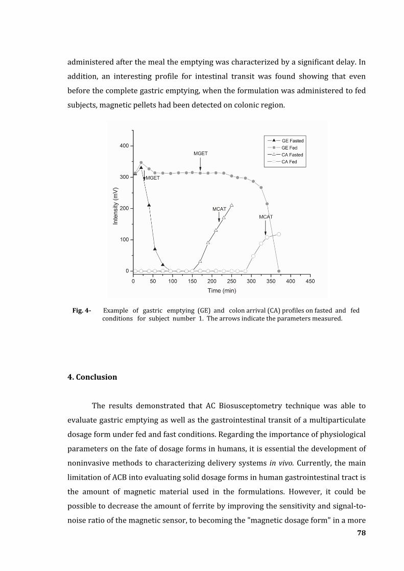
78
administered after the meal the emptying was characterized by a significant delay. In
addition, an interesting profile for intestinal transit was found showing that even
before the complete gastric emptying, when the formulation was administered to fed
subjects, magnetic pellets had been detected on colonic region.
4. Conclusion
The results demonstrated that AC Biosusceptometry technique was able to
evaluate gastric emptying as well as the gastrointestinal transit of a multiparticulate
dosage form under fed and fast conditions. Regarding the importance of physiological
parameters on the fate of dosage forms in humans, it is essential the development of
noninvasive methods to characterizing delivery systems in vivo. Currently, the main
limitation of ACB into evaluating solid dosage forms in human gastrointestinal tract is
the amount of magnetic material used in the formulations. However, it could be
possible to decrease the amount of ferrite by improving the sensitivity and signal-to-
noise ratio of the magnetic sensor, to becoming the "magnetic dosage form" in a more
Fig. 4- Example of gastric emptying (GE) and colon arrival (CA) profiles on fasted and fed conditions for subject number 1. The arrows indicate the parameters measured.
79
conventional dosage form. Nevertheless, the development of more sensitive sensors
will allow overcoming depth effects providing more accurate location of the dosage
forms.
In recent years, continuous studies employing the ACB allowed characterizing
a number of parameters related to solid dosage forms. It is reasonable to assume the
potential of ACB as an alternative tool for pharmaceutical purposes. Furthermore, this
technique will be especially valuable when associated with clinical pharmacokinetics
to provide more predictable data on bioavailability of drugs correlated to
physiological properties.
Acknowledgements
This study was supported by FAPESP, CAPES and PRONEX. The authors are grateful
to Almapal S/A, Scandiflex and Capsugel® for donation of materials which has been
used in the manufacture of the multiparticulate dosage form.
References
Andreis, U., Corá, L.A., Américo, M.F., Oliveira, R.B., Baffa, O., Miranda, J.R.A. 2007. Effect of
erythromycin on motility and gastric emptying in dogs, by AC Biosusceptometry.
International Congress Series 1300, 307–310.
Asghar, L.F.A., Chandran, S. 2006. Multiparticulate formulation approach to colon specific
drug delivery: current perspectives. J. Pharm. Pharmaceut. Sci. 9, 327-338.
Choe, S.Y., Neudeck, B.L., Welage, L.S., Amidon, G.E., Barnett, J.L., Amidon, G.L. 2001. Novel
method to assess gastric emptying in humans: the pellet gastric emptying test. Eur. J. Pharm.
Sci. 14, 347-353.
Chourasia, M.K., Jain, S.K. 2003. Pharmaceutical approaches to colon target drug delivery
systems. J. Pharm. Pharmaceut. Sci. 6, 33-66.
Corá, L.A., Américo, M.F., Oliveira, R.B., Baffa, O., Moraes, R., Romeiro, F.G., Miranda, J.R.A.
2003. Disintegration of magnetic tablets in human stomach evaluated by alternate current
Biosusceptometry. Eur. J. Pharm. Biopharm. 56, 413-420.
Corá, L.A., Romeiro; F.G., Stelzer, M., Américo, M.F., Oliveira, R.B., Baffa, O., Miranda, J.R.A.
2005a. AC Biosusceptometry in the study of drug delivery. Adv. Drug Deliv. Rev. 57, 1223-
1241.
80
Corá, L.A., Andreis, U., Romeiro, F.G., Américo, M.F., Oliveira, R.B., Baffa, O., Miranda, J.R.A.
2005b. Magnetic images of the disintegration process of tablets in the human stomach by AC
Biosusceptometry. Phys. Med. Biol. 50, 5523-5534.
Corá, L.A., Romeiro, F.G., Paixão, F.C., Américo, M.F., Oliveira, R.B., Baffa, O., Miranda, J.R.A.
2006a. Enteric coated magnetic HPMC capsules evaluated in the human gastrointestinal tract
by AC Biosusceptometry. Pharm. Res. 23, 1809-1816.
Corá, L.A., Romeiro, F.G., Américo, M.F., Oliveira, R.B., Baffa, O., Stelzer, M., Miranda, J.R.A.
2006b. Gastrointestinal transit and disintegration of enteric coated magnetic tablets assessed
by AC Biosusceptometry. Eur. J. Pharm. Sci. 27, 1-8.
Corá, L.A., Fonseca, P.R., Américo, M.A., Oliveira, R.B., Baffa, O., Miranda, J.R.A. 2008. Influence
of compression forces on tablets disintegration by AC Biosusceptometry. Eur. J. Pharm.
Biopharm. 69, 372-379.
Coupe, A.J., Davis, S.S., Wilding, I.R. 1991. Variation in gastrointestinal transit of
pharmaceutical dosage forms in healthy subjects. Pharm. Res. 8, 360-364.
Coupe, A.J., Davis, S.S., Evans, D.F., Wilding, I.R. 1993. Do pellet formulations empty from the
stomach with food? Int. J. Pharm., 92, 167-175.
Davis, S.S., Stockwell, A.F., Taylor, M.J., Hardy, J.G., Whalley, D.R., Wilson, C.G., Bechgaard, H.,
Christensen, F.N. 1986. The effect of density on the gastric emptying of single- and multiple-
unit dosage forms. Pharm. Res. 3, 208-213.
Davis, S.S., Hardy, J.G., Fara, J.W. 1986. Transit of pharmaceutical dosage forms through the
small intestine. Gut. 27, 886-892.
Dressman, J.B., Bass, P., Ritschel, W.A., Friend, D.R., Rubinstein, A., Ziv, E. 1993.
Gastrointestinal parameters that influence oral medications. J. Pharm. Sci. 82, 857-872.
Frei, E.H., Gunders, E., Pajewsky, M., Alkan, W.J., Eshcher, J. 1968. Ferrites as contrast material
for medical X-ray diagnosis. J. Appl. Phys. 39, 99-101.
Gandhi, R., Kaul, C.L., Panchagnula, R. 1999. Extrusion and spheronization in the development
of oral controlled-release dosage forms. Pharm. Sci. Technol. Today. 2, 160-170.
Hardy, J.G., Wilson, C.G., Wood, E. 1985. Drug delivery to the proximal colon. J. Pharm.
Pharmacol. 37, 874-877.
Leopold, C.S. 1999. Coated dosage forms for colon-specific drug delivery. Pharm. Sci. Technol.
Today. 2, 197-204.
Marathe, P.H., Wen, Y., Norton, J., Greene, D.S., Barbhaiya, R.H., Wilding, I.R. 2000. Effect of
altered gastric emptying and gastrointestinal motility on metformin absorption. Br. J. Clin.
Pharmacol. 50, 325-332.
81
Miranda, J.R.A., Oliveira, R.B., Sousa, P.L., Braga, F.J.H., Baffa, O. 1997. A novel biomagnetic
method to study gastric antral contractions. Phys. Med. Biol. 42, 1791-1799.
Mojaverian, P. 1996. Evaluation of Gastrointestinal pH and Gastric Residence Time via the
Heidelberg Radiotelemetry Capsule: Pharmaceutical Application. Drug Dev. Res. 38:73-85.
Moraes, R., Corá, L.A., Américo, M.F., Oliveira, R.B., Baffa, O., Miranda, J.R.A. 2003.
Measurement of gastric contraction activity in dogs by means of AC Biosusceptometry.
Physiol. Meas. 24, 337–345.
O’Reilly, S., Wilson, C.G., Hardy, J.G. 1987. The influence of food on the gastric emptying of
multiparticulate dosage forms. Int. J. Pharm. 34, 213-216.
Podczeck, F., Newton, J.M., Yuen, K-.H. 1995. The description of the gastrointestinal transit of
pellets assessed by gama scintigraphy using statistical moments. Pharm. Res. 12, 376-379.
Ranade, V.V. 1991. Drug delivery systems: oral drug delivery. J. Clin. Pharmacol. 31, 2-16.
Romeiro, F.G., Corá, L.A., Andreis, U., Américo, M.F., Oliveira, R.B., Baffa, O., Miranda, J.R.A.
2006. A novel biomagnetic approach to study caecocolonic motility in humans.
Neurogastroenterol. Motil. 18, 1078–1083.
Shareef, M.A., Khar, R.K., Ahuja, A., Ahmad, F.J., Raghava, S. 2003. Colonic drug delivery: an
updated review. AAPS Pharm. Sci. 5, 1-26.
Weitschies, W., Kosch, O., Mönnikes, H., Trahms, L. 2005. Magnetic Marker Monitoring: An
application of biomagnetic measurement instrumentation and principles for the
determination of the gastrointestinal behavior of magnetically marked solid dosage forms.
Adv. Drug Deliv. Rev. 57, 1210– 1222.
Wilding, I.R., Hardy, J.G., Maccari, M., Ravelli, V., Davis, S.S. 1991. Scintigraphic and
pharmacokinetic assessment of a multiparticulate sustained release formulation of diltiazem.
Int. J. Pharm. 76, 133-143.
Wilding, I.R., Kenyon, C.J., Hooper, G. 2000. Gastrointestinal spread of oral prolonged-release
mesalazine microgranules (Pentasa) dosed as either tablets or sachet. Aliment. Pharmacol.
Ther. 14, 163-169.
Wilding, I.R., Coupe, A.J., Davis; S.S. 2001. The role of -scintigraphy in oral drug delivery. Adv.
Drug Deliv. Rev. 46, 103-124.
Yang, L., Chu, J.S., Fix, J.A. 2002. Colon-specific drug delivery: new approaches and in vitro/ in
vivo evaluation. Int. J. Pharm. 235, 1-15.
Zahirul, M., Khan, I. Dissolution testing for sustained or controlled release oral dosage forms
and correlation with in vivo data: challenges and opportunities. Int. J. Pharm. 140, 131-143.

Considerações Finais
“Vá tão longe quanto possa ver. Quando chegar lá, você poderá ver ainda mais longe.” Thomas Carlyle
82
Considerações Finais
A indústria farmacêutica investe continuamente na pesquisa e
desenvolvimento de produtos não apenas de liberação alvo-específica, como também
de maior eficácia terapêutica. Nesse sentido, o advento de excipientes e sistemas de
revestimento mais efetivos, possibilitou a obtenção de formas farmacêuticas capazes
de controlar a liberação de um ativo no local de ação, visando um efeito mais
prolongado ou ainda, a ação imediata e menos errática.
A qualidade de um produto será assegurada pelo equilíbrio entre a escolha
dos excipientes, do método de produção e dos perfis de liberação e dissolução do
fármaco. Portanto, a liberação do ativo constitui um fator limitante para sua absorção
e, desse modo, qualquer fator que interfira nesse processo poderá alterar
cineticamente a biodisponibilidade do fármaco administrado.
Sendo conhecido que o trato gastrintestinal humano influencia
significativamente os parâmetros relacionados com a liberação e absorção de drogas,
houve o interesse no desenvolvimento ou aprimoramento de métodos capazes de
avaliar a performance de um produto in vivo. Nesse sentido, merece destaque a
Biosusceptometria AC, uma técnica cujas características e versatilidade permitiram
inseri-la na pesquisa farmacêutica como uma alternativa aos métodos tradicionais.
Diante dos resultados apresentados, a BAC demonstrou potencial como um
método capaz de prover os requisitos necessários para monitorar diferentes
processos farmacêuticos não apenas in vitro, como também no TGI humano. Permitiu,
em um primeiro momento, a obtenção de imagens do processo de desintegração de
comprimidos tanto in vitro quanto no estômago humano. Esse trabalhou inseriu a
BAC como um novo método de imagem e introduziu outra perspectiva na análise do
processo de desintegração, visto que até esse momento, era caracterizado apenas
como alterações no nível de intensidade do sinal magnético detectado.
Ainda em relação ao processo de desintegração, a penetração de água na
forma farmacêutica é um fator determinante para um bom desempenho, visto que
resulta no intumescimento das partículas e no desenvolvimento de uma força que
auxilia a desintegração. Então, o nível de compressão aplicado também é um
83
parâmetro fundamental no que concerne à obtenção de um comprimido, visto que na
medida em que a compressão aumenta, ocorre uma diminuição na porosidade da
forma farmacêutica que, por sua vez, interfere com a capacidade de penetração de
líquido e, conseqüentemente, com a força desenvolvida durante a desintegração.
Esses parâmetros puderam ser avaliados pela BAC em um estudo cujo foco principal
foi demonstrar que os resultados fornecidos pela técnica magnética podem ser
correlacionados com aqueles obtidos por metodologias específicas, garantindo uma
análise mais acurada dos parâmetros físicos envolvidos com a desintegração de
comprimidos.
Além de comprimidos, cápsulas gelatinosas duras ou aquelas constituídas
por hidroxipropilmetilcelulose (HPMC) são formas farmacêuticas sólidas muito
utilizadas quando se objetiva a administração oral de drogas. A possibilidade de
utilizar cápsulas de HPMC revestidas visando a liberação colônica foi demonstrada
em diversos estudos, visto que o cólon humano é um órgão-alvo para a liberação de
drogas de ação local ou sistêmica. Nesse sentido, utilizando-se a BAC também foi
possível avaliar o trânsito gastrintestinal e o processo de desintegração de cápsulas
revestidas no cólon humano. Além da desintegração, também foi caracterizado o
perfil da motilidade, o tempo de retenção e o trânsito intestinal da forma
farmacêutica.
Considerando o desenvolvimento de novas formulações, um dos maiores
progressos alcançados foi a possibilidade de controlar ou modificar a liberação de
drogas no trato gastrintestinal humano e, desse modo, os sistemas multiparticulados
foram propostos devido às vantagens biofarmacotécnicas e terapêuticas que
apresentam. A BAC foi empregada com o intuito de monitorar um sistema
multiparticulado magnético e avaliar a influência do estado prandial em parâmetros
fisiológicos como esvaziamento gástrico e trânsito intestinal.
Este trabalho demonstrou que, apesar das suas limitações, a BAC tem
potencial para avaliar diferentes formas farmacêuticas, inserindo-se como um
método alternativo na pesquisa farmacêutica. Desse modo, seu constante
aperfeiçoamento, aliado à necessidade de estabelecer uma análise mais acurada no
que concerne à liberação não apenas do material magnético, como também de um
princípio ativo, a BAC terá como foco principal avaliar a qualidade do produto
farmacêutico proposto e comparar os perfis de dissolução de um medicamento
84
referência in vitro, seguindo as análises preconizadas pela Farmacopéia Brasileira,
com os resultados obtidos por meio das medidas empregando-se esse método
magnético. Desse modo, serão utilizados sistemas de liberação modificada de drogas
baseados em matrizes hidrofílicas e sistemas multiparticulados em comprimidos.
Além disso, a BAC será associada com análises farmacocinéticas
(“magnetofarmacocinética”), no intuito de estabelecer a correlação in vitro-in vivo
(IVIVC) para o fármaco utilizado.