Modelos de co-culturas de células in vitro para desenvolvimento … Costa... · 1 UNIVERSIDADE DA...
104
1 UNIVERSIDADE DA BEIRA INTERIOR Ciências da Saúde Modelos de co-culturas de células in vitro para desenvolvimento de novos sistemas de entrega de fármacos Elisabete Cristina da Rocha Costa Dissertação para obtenção do Grau de Mestre em Ciências Biomédicas (2 o ciclo de estudos) Orientador: Professor Doutor Ilídio Joaquim Sobreira Correia Coorientador: Mestre Vítor Manuel Abreu Gaspar Covilhã, junho 2013
Transcript of Modelos de co-culturas de células in vitro para desenvolvimento … Costa... · 1 UNIVERSIDADE DA...
1
UNIVERSIDADE DA BEIRA INTERIOR
Ciências da Saúde
Modelos de co-culturas de células in vitro para desenvolvimento de novos sistemas de entrega de
fármacos
Elisabete Cristina da Rocha Costa
Dissertação para obtenção do Grau de Mestre em
Ciências Biomédicas (2o ciclo de estudos)
Orientador: Professor Doutor Ilídio Joaquim Sobreira Correia
Coorientador: Mestre Vítor Manuel Abreu Gaspar
Covilhã, junho 2013

In vitro cell co-culture models for development of new drug delivery systems
ii

In vitro cell co-culture models for development of new drug delivery systems
iii
List of publications
Articles in peer reviewed international journals:
Costa, C. E., Gaspar. V. M., Marques, J.G., Coutinho, P., Correia, I. J. (2013). “Evaluation of
Nanoparticle Uptake in Co-culture Cancer Models.”PloS one. Article in press.
Poster communications:
Costa, C. E., Gaspar. V. M., Marques, J.G., Coutinho, P., Correia, I. J., Mimicking Breast
Cancer Microenvironment with In Vitro Co-culture Models, Instituto Politécnico da Guarda
(IPG), 3rd of May 2013, Guarda, Portugal.
Best poster award:
Costa, C. E., Gaspar. V. M., Marques, J.G., Coutinho, P., Correia, I. J., Mimicking Breast
Cancer Microenvironment with In Vitro Co-culture Models, Instituto Politécnico da Guarda
(IPG), 3rd of May 2013, Guarda, Portugal.

In vitro cell co-culture models for development of new drug delivery systems
iv
In vitro cell co-culture models for development of new drug delivery systems
v
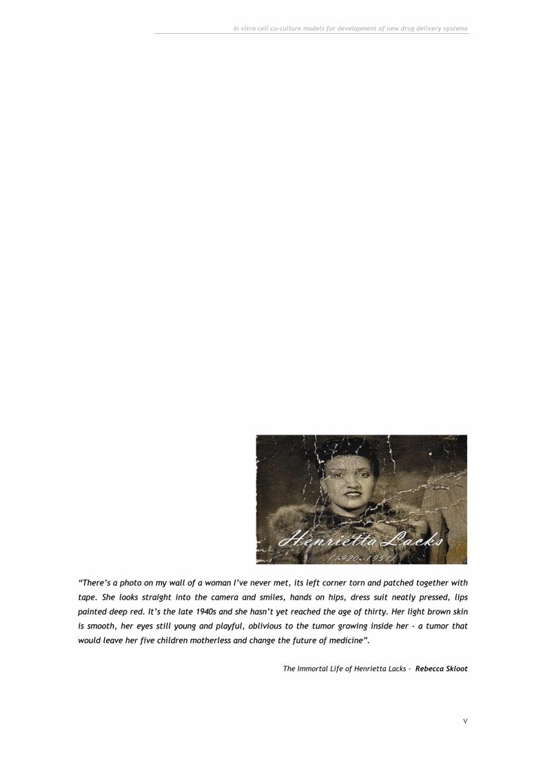
““TThheerree’’ss aa pphhoottoo oonn mmyy wwaallll ooff aa wwoommaann II’’vvee nneevveerr mmeett,, iittss lleefftt ccoorrnneerr ttoorrnn aanndd ppaattcchheedd ttooggeetthheerr wwiitthh
ttaappee.. SShhee llooookkss ssttrraaiigghhtt iinnttoo tthhee ccaammeerraa aanndd ssmmiilleess,, hhaannddss oonn hhiippss,, ddrreessss ssuuiitt nneeaattllyy pprreesssseedd,, lliippss
ppaaiinntteedd ddeeeepp rreedd.. IItt’’ss tthhee llaattee 11994400ss aanndd sshhee hhaassnn’’tt yyeett rreeaacchheedd tthhee aaggee ooff tthhiirrttyy.. HHeerr lliigghhtt bbrroowwnn sskkiinn
iiss ssmmooootthh,, hheerr eeyyeess ssttiillll yyoouunngg aanndd ppllaayyffuull,, oobblliivviioouuss ttoo tthhee ttuummoorr ggrroowwiinngg iinnssiiddee hheerr -- aa ttuummoorr tthhaatt
wwoouulldd lleeaavvee hheerr ffiivvee cchhiillddrreenn mmootthheerrlleessss aanndd cchhaannggee tthhee ffuuttuurree ooff mmeeddiicciinnee””..
The Immortal Life of Henrietta Lacks - Rebecca Skloot

In vitro cell co-culture models for development of new drug delivery systems
vi
Dedication
The completion of this work has not possible without the familiar support. Thus, I would like
to dedicate this last year of my academic path to parents and to my little sister which always
understood my distance.
Moreover, I would like to dedicate this work to all persons with cancer around the world. I
wish that the investigation in tumor biology continues to open new windows for new
anti-cancer therapies for better quality of life of cancer patients.

In vitro cell co-culture models for development of new drug delivery systems
vii
In vitro cell co-culture models for development of new drug delivery systems
viii
Acknowledgments
Firstly, I would like to thank to my supervisor Professor Ilídio Correia for the opportunity of
work in his group, and for all the time and orientation provided during the aquision of my
master degree.
Specially, I would like to thanks my co-supervisor, Vítor Gaspar (Ph.D. student), for
supporting me in my instabilities and doubts. Furthermore, for spend great part of his time in
helping me, putting in second place his own work. In fact, without his orientation I would not
be the person academically and personally that I am today.
To João Marques, my co-worker and friend, I would like to thanks him for all accomplices and
honesty. To him, I would like to wish a wonderful future like he deserves.
To another great influence and orientation to me, Ph.D. students and M.Sc. of the group, I
thank for good working environment, entrepreneurship, good humour and in last for the
friendship. We are a family.
I acknowledge to my friends and housemates which always listen my confidences.
In last, I thank to me for surpass myself.

In vitro cell co-culture models for development of new drug delivery systems
ix
In vitro cell co-culture models for development of new drug delivery systems
x
Abstract
The demanding applications of nanocarriers in cancer biology require the existence of testing
platforms that mimic the in vivo tumor microenvironment and its unique biological features.
For this, highly informative methodologies such as animal experimentation are the current
gold-standard. However, very recent reports issued by regulatory agencies appeal for the
reduction of the used animal research models due to economical and ethical issues, thus
evidencing the urgent necessity for novel alternatives. Co-culture cell models have the
potential to bridge the gap between the required reduction of animal use and the existence
of suitable models that closely reproduce in vivo tumors. This is a novel type of in vitro cell
culture that is mainly characterized by the culture of cancer cells in contact with stromal
cells, mimicking the tumor microenvironment in vitro trough the establishment of cancer-
stroma synergic interactions. However, this evaluation was until now limited to co-culture
systems established with precise cell ratios, not addressing the natural heterogeneity
commonly found in tumors of different patients. The research work presented in this thesis
describes the development and optimization of novel 2D co-culture models of breast and
cervical cancers with various cell-to-cell ratios, in order to unravel the influence of
heterogeneous conditions on the evaluation of nanocarrier biological performance and
ultimately in the therapeutic outcome. As a proof of concept these novel platforms were used
to evaluate a multifunctional gene delivery system designed for cancer therapy and revealed
that in fact different co-culture ratios may influence the overall assessment of nanocarrier
targeting specificity. In addition, since recent reports demonstrate the high influence of the
3D architecture of tumor masses in the response to anti-cancer drugs or delivery systems, the
engineering and optimization of suitable substrates for generation of organotypic 3D co-
culture models with various cancer-fibroblast cell ratios was also investigated. The 3D
multicelular spheroid models of breast and cervix cancer produced at various time points,
possess all the major characteristics of in vivo tumors including the structural rearrangement,
the diffusional limit of oxygen or nutrients and most importantly, the distinctive necrotic core
of solid tumors. Overall, these newly developed co-culture and 3D models assume crucial
importance for the future design and optimization of new drug delivery providing a new level
of in vitro reproducibility of in vivo tumors.
Keywords
2D co-cultures; 3D multicellular spheroids; Nanosized delivery systems; Tumor
microenvironment.

In vitro cell co-culture models for development of new drug delivery systems
xi
In vitro cell co-culture models for development of new drug delivery systems
xii
Resumo alargado
Os recentes avanços na área da Nanotecnologia abriram novas oportunidades para o
desenvolvimento de novos nano-sistemas como as nanopartículas para entrega de fármacos ou
de informação genética com potencial para serem usadas futuramente na terapia do cancro.
Todavia, para que as suas aplicações terapêuticas sejam significativas num contexto clínico,
estes sistemas devem ser testados em modelos que representem o mais aproximadamente
possível as propriedades únicas que o microambiente tumoral tem in vivo. Por forma a atingir
este objetivo, vários protocolos experimentais usam modelos animais para avaliar a atividade
biológica de nano transportadores. No entanto, recentemente, as diferentes agências
regulatórias têm apelado pela aplicação da regra dos 3Rs (Reduzir, Reutilizar, Reciclar) em
relação ao uso de animais como modelos para estudos experimentais em fases pré-clinicas,
não só devido aos problemas económicos e legais associados ao seu uso, mas também devido a
inconvenientes éticos, e à variabilidade dos resultados obtidos quando comparados com
aqueles adquiridos em estudos clínicos com humanos. Neste contexto, tem-se procurado
desenvolver novas alternativas que permitam reproduzir o que ocorre in vivo.
A cultura in vitro de células tumorais em co-cultura com outras células presentes no
microambiente tumoral surge como uma abordagem muito promissora no que diz respeito a
mimetizar as caraterísticas dos variados tipos tumores. Esta metodologia permite estudar de
uma forma abrangente a biologia dos tumores, sob variadas condições e até mesmo testar, de
uma forma rápida, novos fármacos ou sistemas de entrega direcionada. O contacto direto
entre as células cancerígenas e as células do estroma, reproduzem as interações sinergéticas
que ocorrem no microambiente tumoral. Contudo, estes sistemas de culturas celulares são
usualmente desenvolvidos tendo por base um número fixo de células tumorais em relação às
células do estroma. De facto, até à data apenas foram descritos testes de novos agentes
anti-tumorais ou de novos sistemas de entrega em co-culturas com apenas um rácio de
células, sendo que esta abordagem não permite assim analisar a heterogeneidade natural dos
tumores.
Desta forma, o trabalho de investigação desenvolvido nesta tese descreve desenvolvimento e
otimização de novos modelos 2D de co-culturas do cancro da mama e do colo do útero. Para
tal, foram usados vários rácios de células cancerígenas células normais do estroma, com o
intuito de representar a distribuição celular em diferentes tumores. Adicionalmente,
pretende-se também verificar de que forma estas diferentes condições influenciam a
atividade dos novos sistemas de entrega de drogas e consequentemente a sua eficácia
terapêutica.
Os resultados obtidos demonstraram uma influência evidente dos fibroblastos, sobre o
comportamento das células cancerígenas. Inicialmente, foi possível verificar uma alteração
na organização estrutural das co-culturas, quando comparadas com as monoculturas de

In vitro cell co-culture models for development of new drug delivery systems
xiii
controlo. Além disto, foi também observado um aumento na viabilidade celular na presença
de fibroblastos, tendo sido obtida uma correlação entre o tempo de co-cultura e um aumento
de proliferação celular. Após a demonstração do sucesso da otimização destas plataformas,
foram testadas nanopartículas funcionalizadas nas várias co-culturas desenvolvidas. Os
resultados obtidos na microscopia confocal e citometria de fluxo demonstraram que o uso de
diferentes rácios celulares pode de facto influenciar a avaliação da especificidade de nano
transportadores. Estes resultados evidenciam assim que, os mesmos sistemas de entrega
podem atuar de forma diferente, de paciente para paciente.
Para além do desenvolvimento destes sistemas de co-culturas, foi também otimizada a
produção de novos modelos de culturas tridimensionais. Estes modelos 3D, mais comumente
chamados de esferoides, conseguem mimetizar os tumores sólidos, pois são constituídos por
vários tipos de células e com o decorrer do tempo adquirirem propriedades únicas,
nomeadamente uma superfície constituída por células com elevada proliferação, que
mimetizam as zonas do tumor irrigadas por vasos sanguíneos e um núcleo necrótico, que
corresponde às zinas do tumor com baixa densidade de vasos. Para promover a formação
destes modelos foi utilizada a técnica de cultura celular com sobreposição líquida em
conjugação com agitação horizontal. Esta nova abordagem permitiu evitar a adesão das
células e promoveu a formação de esferoides 3D com morfologias bem definidas e
reprodutíveis.
Para a formação destes modelos fibroblastos revelaram um papel fundamental visto que as
interações que se estabelecem entre os dois tipos celulares são essenciais para formar
esferoides coesos e com um gradiente de densidades celulares da periferia para o núcleo.
Em geral, os novos modelos de co-culturas desenvolvidos assumem um papel crucial no futuro
desenvolvimento e investigação de novos nano transportadores, já que estes serão na prática
direcionados para os tumores. Por outro lado, os modelos 3D permitem reproduzir com
exatidão o que acontece in vivo, criando um conjunto de ferramentas que poderão contribuir
para aperfeiçoar as terapias anti-tumorais.
Palavras-chave
Co-culturas 2D; Esferoides multicelulares; Microambiente tumoral; Nano sistemas de entrega
direcionada.

In vitro cell co-culture models for development of new drug delivery systems
xiv

In vitro cell co-culture models for development of new drug delivery systems
xv
Table of Contents
1. Introduction 1
1.1. Tumor microenvironment: The driving force for cancer evolution 2
1.1.1. ECM in the tumor microenvironment 5
1.1.2. Stromal cells 5
1.1.2.1. Vascular and lymphatic endothelial cells 5
1.1.2.2. Endothelial cells 5
1.1.2.3. Perycites 5
1.1.2.4. Adipocytes 6
1.1.2.5. Immune System cell 6
1.1.2.6. Fibroblasts 7
1.2. Nanosized delivery systems as novel therapeutic approaches for cancer
therapy 11
1.3. Experimental models to evaluate nanoparticulated delivery systems for
application in cancer therapy 14
1.3.1. In vivo models 14
1.3.2. In vitro models 16
1.3.2.1. Co-cultures 16
1.3.2.2. 3D cell cultures: Spheroids 17
1.4. Objectives 21
2. Methods 22
2.1. Materials 23
2.2. Breast cancer and cervical cancer 2D and 3D in vitro co-culture models
optimization 23
2.2.1. Cell lines maintenance 23
2.2.2. Optimization of 2D in vitro cell co-culture models of breast cancer
(MCF-7:hFIB) and cervical cancer (HeLa:hFIB) 23
In vitro cell co-culture models for development of new drug delivery systems
xvi
2.2.2.1. Optical microscopy analysis of the distribution and morphology of 2D
in vitro cell co-culture models of breast cancer (MCF-7:hFIB) and
cervical cancer (HeLa:hFIB)
24
2.2.2.2. Resazurin assay for analysis of cell viability of 2D in vitro cell co-
culture models of breast cancer (MCF-7:hFIB) 24
2.2.3. Optimization of 3D in vitro cell co-culture models of breast cancer
(MCF-7:hFIB) and cervical cancer (HeLa:hFIB) 25
2.2.3.1. Optical microscopy analysis of the distribution and morphology of the
3D in vitro cell co-culture models of breast cancer (MCF-7:hFIB) and
cervical cancer (HeLa:hFIB)
25
2.2.3.2. Scanning electron microscopy (SEM) analysis of the 3D in vitro cell co-
culture models of breast cancer (MCF-7:hFIB) 26
2.2.3.3. Confocal laser scanning microscopy analysis of 3D in vitro cell co-
culture models of cervical cancer (HeLa:hFIB) 26
2.3. Evaluation of Chitosan-Histidine-Arginine/pDNA nanoparticles in 2D
breast cancer co-culture models (MCF-7:hFIB) 26
2.3.1. CLSM of CH-H-R/pDNA nanoparticles cell uptake analysis in 2D
breast cancer co-culture models 26
2.3.2. Flow cytometry of CH-H-R/pDNA nanoparticles cell uptake analysis
in 2D breast cancer co-culture models 27
3. Results and Discussion 29
3.1. Breast cancer and cervical cancer 2D and 3D in vitro co-culture models 30
3.1.1. Development and optimization of 2D in vitro cell co-culture models
of breast cancer (MCF-7:hFIB) 30
3.1.2. Development and optimization of 2D in vitro cell co-culture models
of cervical cancer (HeLa:hFIB) 37
3.1.3. Evaluation of CH-H-R/pDNA nanoparticles cellular uptake in 2D
breast cancer co-culture models (MCF-7:hFIB) 40
3.1.3.1. CLSM analysis of CH-H-A/pDNA nanoparticles cellular uptake analysis
in 2D cell co-culture models of breast cancer with different MCF-7 to
hFIB ratios
44
3.1.3.2. Flow cytometry analysis of CH-H-A/pDNA nanoparticles cellular
uptake analysis in 2D cell co-culture models of breast cancer with
different MCF-7 to hFIB ratios
44
3.2. 3D in vitro cell co-culture models of breast cancer and cervical cancer 48
3.2.1. 3D in vitro cell co-culture models of breast cancer (MCF-7:hFIB) 50
In vitro cell co-culture models for development of new drug delivery systems
xvii
3.2.2. 3D in vitro cell co-culture models of cervical cancer (HeLa: hFIB)
3.2.2.1. SEM analysis of the 3D in vitro cell co-culture models of breast cancer
(MCF-7:hFIB) 53
3.2.3. 3D in vitro cell co-culture models of cervical cancer (HeLa: hFIB) 53
3.2.3.1. CLSM analysis of 3D in vitro 3D in vitro cell co-culture models of
cervical cancer (HeLa:hFIB) 55
4. Conclusions and Future Perspectives 59
5. Bibliography 62

In vitro cell co-culture models for development of new drug delivery systems
xviii
In vitro cell co-culture models for development of new drug delivery systems
xix
List of Figures
Chapter I - Introduction
Figure 1 – Representation of carcinogenesis. 2
Figure 2 – Representation of tumor microenvironment. 4
Figure 3 – General interactions established between tumour cells and stromal
cells in human tumor tissue. 4
Figure 4 – Immune system behaviour in tumor microenvironment. 6
Figure 5 – Comparison of normal and cancer associated fibroblasts (CAF). 7
Figure 6 – Influence of fibroblasts in tumor microenvironment. 8
Figure 7 – Representation of the mechanism of epithelial-to-mesenchymal
transition (EMT). 10
Figure 8 – Relation between drug anti-tumoral effect and toxic effect. 11
Figure 9 – Representation of the different types of nanoparticles available. 12
Figure 10 – Steps needed for nanoparticle development. 14
Figure 11 - Absolute bioavailability of various drugs in dogs, primates and
rodents versus the absolute bioavailability reported for humans. 15
Figure 12 – Representation of the vascularisation in normal and in cancer tissue. 18
Figure 13 – Representation of the process of vascularisation in solid tumors. 19
Figure 14 – Characterization of the similarities between the original tumor and
the respective spheroids. 20
Chapter III – Results and Discussion
Figure 14 - Light Microscope images of 2D MCF-7 and hFIB co-cultures during 10
days of culture. 32
Figure 15 - Light Microscope images of 2D MCF-7 and hFIB monocultures during
10 days of culture (controls). 33
Figure 16 – Cell viability of 2D MCF-7 and hFIB co-culture models with different
ratios at 24, 48, 72 and 96 h after their seeding.
34
In vitro cell co-culture models for development of new drug delivery systems
xx
Figure 17 – Optical contrast microscopy images of 1MCF-7:3hFIB 2D co-cultures
after 9 and 10 days of culture. 35
Figure 18 – Optical contrast microscopy images of 1MCF-7:5hFIB 2D co-cultures
after 9 and 10 days of culture. 35
Figure 19 – Microscope images of H&E histological sections of human breast
healthy and carcinoma tissue in different stages of tumor evolution. 36
Figure 20 - Light Microscope images of 2D HeLa and hFIB co-cultures during 10
days of culture. 38
Figure 21 - Light Microscope images of 2D HeLa and hFIB monocultures during 10
days of culture (controls). 39
Figure 22 – Inverted Light Microscope images of fillopodium structures (arrows)
of HeLa and Fibroblasts cells after 10 days in co-culture. 39
Figure 23 - Confocal Laser Scanning Microscopy images of nanoparticles cellular
uptake, after 4 h of incubation in MCF-7:hFIB 2D co-cultures models. 41
Figure 24 - CLSMimages of nanoparticles cellular uptake, after 4h of incubation
in MCF-7 and hFIB 2D monocultures (controls). 42
Figure 25 - CLSM images co-cultures at 1MCF-7:1hFIB ratio for nanoparticles
cellular localization analysis. 43
Figure 26 – Representative dot plots of nanoparticles cellular uptake analysis by
flow cytometry after 4 h of incubation with RITC-labelled pDNA/CH-H-R
nanoparticles in MCF-7:hFIB 2D co-cultures models.
45
Figure 27 – Representative histograms of nanoparticle uptake in MCF-7 (A) and
hFIB (B) monocultures after 4h of incubation with RITC-labelled pDNA/CH-H-R
nanoparticles.
46
Figure 28 - Flow cytometry analysis of mono and co-cultures non- stained with
GFP and non- incubated with nanoparticles. 46
Figure 29 - Representative histograms of flow cytometry analysis of
nanoparticles cellular uptake in MCF-7 and hFIB cell populations after 4h of
incubation with RITC-labelled pDNA/CH-H-R nanoparticles.
47
Figure 30 – Light Microscope images of HeLa agglomerates formed using a flat
hydrophobic bottom culture plate. 49
Figure 31 – Scheme of spheroids formation protocol. 49
Figure 32 – Contrast microscopy images of 3D MCTS of MCF-7 and hFIB in mono
and co-cultures grown during 12 days at various initial cell numbers and ratios. 51
Figure 33 – Representative high resolution micrograph of a 3D MCTS produced
with the modified liquid overlaid method. 52
In vitro cell co-culture models for development of new drug delivery systems
xxi
Figure 34 – Scanning Electron Microscope (SEM) images representation of 3D
MCF-7:hFIB MCTS. 54
Figure 35 - Light Microscope images of 3D MCTS of HeLa and hFIB mono and
co-cultures grown during 12 days at various initial cell numbers and ratios. 54
Figure 36 – CLSM images of 3D reconstruction and dept coding of 2HeLa:1hFIB
MTCS. 56
Figure 37 - CLSM of 2HeLa:1hFIB MCTS Z-stack slice representation orthogonal
sectioning in xy axis and high resolution images of the spheroid surface. 57

In vitro cell co-culture models for development of new drug delivery systems
xxii
In vitro cell co-culture models for development of new drug delivery systems
xxiii
List of Tables
Chapter I – Introduction
Table 1 – Interactions between cancer cells and tumor stromal fibroblasts. 9
Table 2 - The influence of 3D cell organization in cell behaviour and signalling. 18
Chapter II – Methods
Table 3 - MCF-7 to hFIB cell ratios used in vitro to mimic the breast cancer microenvironment.
24
Table 4 - HeLa to hFIB cell ratios used in vitro to mimic the cervical cancer microenvironment.
24

In vitro cell co-culture models for development of new drug delivery systems
xxiv
In vitro cell co-culture models for development of new drug delivery systems
xxv
List of Acronyms
-SMA -Smooth muscle actin
2D Two-dimensional
3D Three-dimensional
ADH Atypical ductal hyperplasia
ALH Atypical lobular hyperplasia
bFGF Basic fibroblast growth factor
CAF Cancer associated fibroblasts
CAM Cell adhesion molecule
CH-H-R/pDNA Chitosan-Histidine-Arginine/plasmid deoxyribonucleic acid
CLSM Confocal laser scanning microscopy
CO2 Carbon dioxide
CXCL12 (SDF-1) Chemokine stromal derived factor-1
DC Dendritic cell
DCIS Ductal carcinoma in situ
DEMEM-HG Dulbecco’s Modified Eagle’s Medium High Glucose
DMEM-F12 Dulbecco’s Modified Eagle’s Medium F-12
DMSO Dimethyl sulfoxide
DNA Deoxyribonucleic acid
EC European Commission
ECM Extracellular matrix
EDTA Ethylenediamine tetraacetic acid
EGF Endothelial growth factor
EMEA European medicines agency
EMT Epithelial-to-mesenchymal transition
EtOH Ethanol
FBS Fetal bovine serum
FDA Food and Drug Administration
FL Fluorescence
GFP Green fluorescent protein
H2O Water
In vitro cell co-culture models for development of new drug delivery systems
xxvi
HeLa Human cervix adenocarcinoma
hFIB Primary normal human dermal fibroblasts
HGF Hepatocyte growth factor
HIF-1 Tumor hypoxia-inducible factor-1
IBC Invasive breast cancer
IGF Insulin-like growth factor
IL Interleukin
ISO Organization for Standardization
KGF Keratinocyte growth factor
LOX Lysyl oxidase
MCF-7 Oestrogen-dependent human breast adenocarcinoma
MCP Monocyte chemotatic protein
MCTS Multicellular tumor spheroids
MDR Acquired multidrug resistance
MDSC Myeloid-derived suppressive cell
MMP Metalloproteinase
NK Natural killer cells
NO Nitrogen species
O2 Oxygen
PBS Phosphate-buffered saline
PDGF Platelet-derived growth factor
pDNA Plasmid deoxyribonucleic acid
PFA Paraformaldehyde
PPFS Patient progression free survival
RB Retinoblastoma protein
RITC Rhodamine B isothiocianate
ROI Region of interest
ROS Reactive oxygen species
RT Room temperature
SEM Scanning electron microscopy
T reg Regulatory T cells
TGF-β Transforming growth factor-β
TNF- Tumour necrosis factor-
In vitro cell co-culture models for development of new drug delivery systems
xxvii
TP53 Tumor protein p53
VEGF Vascular endothelial growth factor
WGA-Alexa 594 Germ agglutinin conjugated Alexa 594

xxviii

Chapter I
Introduction
Chapter I - Introduction
2
1. Introduction
1.1. Tumor microenvironment: The driving force for cancer
evolution
Cancer is nowadays a major public health problem, being the second main cause of death in
developing countries (Siegel et al., 2012). It is estimated that in 2020 the world population
will reach a total of 7.5 billion people and a total of 15 million new cancer cases will arise
(Bray and Møller, 2005). These impairing statistics contribute for the tremendous efforts put
forward from behalf of the medical and scientific community to develop more effective
therapeutic approaches and also to understand the mechanisms responsible for cancer
development. The complex transformation of a normal cell into a malignant phenotype is
generally dependent on the accumulation of multiple changes in gene expression patterns in
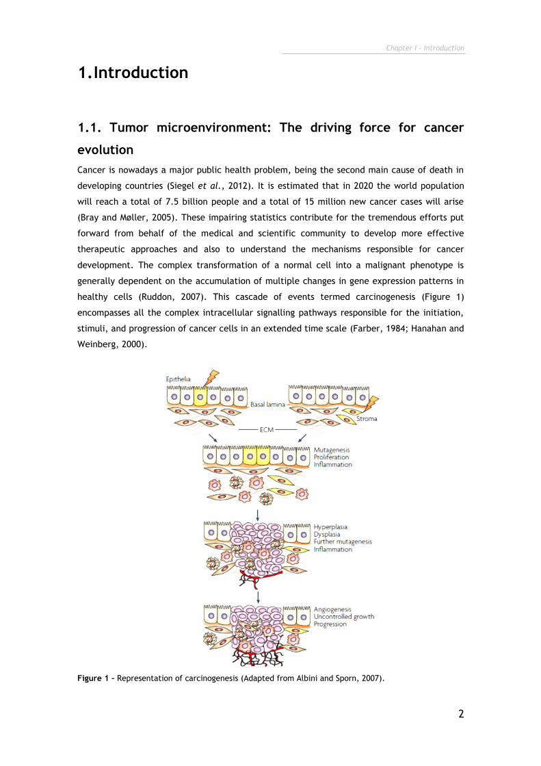
healthy cells (Ruddon, 2007). This cascade of events termed carcinogenesis (Figure 1)
encompasses all the complex intracellular signalling pathways responsible for the initiation,
stimuli, and progression of cancer cells in an extended time scale (Farber, 1984; Hanahan and
Weinberg, 2000).
Figure 1 – Representation of carcinogenesis (Adapted from Albini and Sporn, 2007).
Chapter I - Introduction
3
Cancer development, starts with changes in structure and function of deoxyribonucleic acid
(DNA), usually known as mutations (Mbeunkui and Johann, 2009). These mutations usually
lead to the loss of tumor-suppressor genes (tumor protein p53 (TP53), retinoblastoma protein
(RB)), or activation of oncogenes (myc, RAS, AKT) (Gaspar et al., 2011; Janssen and Medema,
2012), which propels malignant cells to escape from senescence signals and accept the
proliferative stimuli, respectively (Levine and Puzio-Kuter, 2010). Thus, cancer cells acquire
unlimited replicative potential, losing their characteristic non-dividing state (Hanahan and
Weinberg, 2011). In addition, the deregulations of growth-promoting signals lead to an
uncontrolled proliferation of cancer cells (Witsch et al., 2010; Zhang et al., 2010). In the last
stages of cancer development, in order to sustain this dynamic growth and high metabolic
activity, extensive neovascularisation is promoted by malignant cells in order to assure the
necessary uptake of nutrients and oxygen (Annibaldi and Widmann, 2010). The angiogenesis in
association with mechanisms that underlie the extravasion of cancer cells through the
extracellular matrix (ECM) facilitating tumor spread into healthy tissues (Poste and Fidler,
1980; Zetter, 1998; Mbeunkui and Johann, 2009).
Recent reports demonstrate that these abilities acquired by cancer cells, i.e., sustained
proliferation, cell death resistance, immortality, angiogenesis, invasion and metastization are
a consequence of the unique conditions of the tumor niche. In fact, the tumor
microenvironment is of critical importance for the success of cancer progression (Straussman
et al., 2012). Already in 1984, Dolberg and Bissel described an impaired tumor development
in chicken embryos infected with an oncogene expressing Rous sarcoma virus, and
hypothesized that these results were probably correlated with the absence of some additional
cellular components (Dolberg and Bissell, 1984). Recent studies demonstrated that
phenotypic and genotypic abnormalities of cancer cells are insufficient to induce the
malignant phenotype (Ma et al., 2003; Weigelt et al., 2003). The influence of the tumor
microenvironment in cancer development was then postulated. So far different therapies
specifically directed against the tumor microenvironment have been produced (McMillin et
al., 2013; Sounni and Noel, 2013).
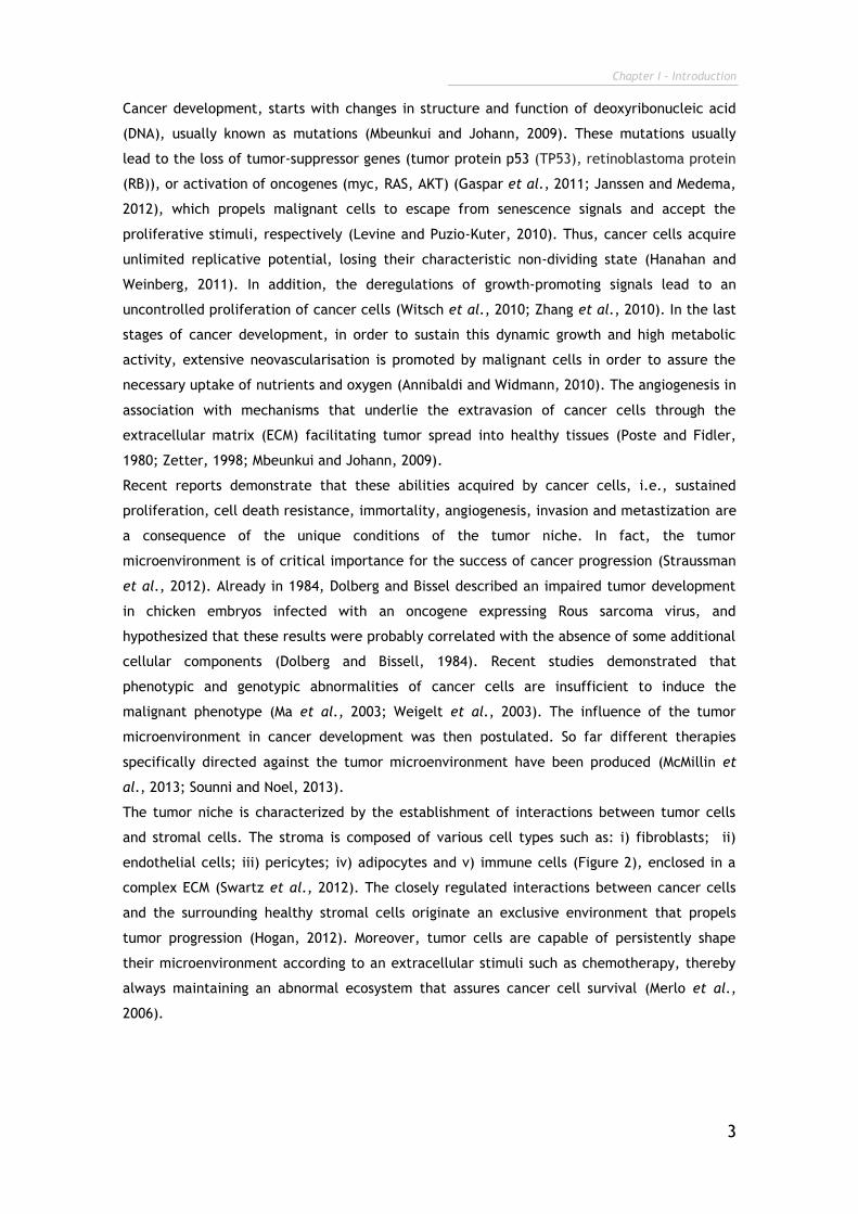
The tumor niche is characterized by the establishment of interactions between tumor cells
and stromal cells. The stroma is composed of various cell types such as: i) fibroblasts; ii)
endothelial cells; iii) pericytes; iv) adipocytes and v) immune cells (Figure 2), enclosed in a
complex ECM (Swartz et al., 2012). The closely regulated interactions between cancer cells
and the surrounding healthy stromal cells originate an exclusive environment that propels
tumor progression (Hogan, 2012). Moreover, tumor cells are capable of persistently shape
their microenvironment according to an extracellular stimuli such as chemotherapy, thereby
always maintaining an abnormal ecosystem that assures cancer cell survival (Merlo et al.,
2006).
Chapter I - Introduction
4
Figure 2 – Representation of tumor microenvironment (Adapted from Coussens and Werb, 2002).
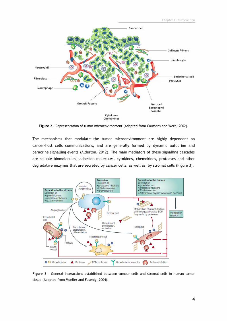
The mechanisms that modulate the tumor microenvironment are highly dependent on
cancer-host cells communications, and are generally formed by dynamic autocrine and
paracrine signalling events (Alderton, 2012). The main mediators of these signalling cascades
are soluble biomolecules, adhesion molecules, cytokines, chemokines, proteases and other
degradative enzymes that are secreted by cancer cells, as well as, by stromal cells (Figure 3).
Figure 3 – General interactions established between tumour cells and stromal cells in human tumor
tissue (Adapted from Mueller and Fusenig, 2004).
Chapter I - Introduction
5
1.1.1. ECM in the tumor microenvironment
ECM is a complex three-dimensional (3D) network formed by macromolecular fibers (collagen,
elastin, etc) as well as nonfibrous proteins (like proteoglycans and glicoproteins). This 3D
matrix is present in the stroma of all tissues, either healthy or malignant (Noguera et al.,
2012). The ECM regulates almost all cellular behaviour (Brizzi et al., 2012), and provides
structural support and strength, thus allowing cellular communication, adhesion and
migration (Noguera et al., 2012). In tumor, the deregulation of ECM is essential for the
angiogenesis and invasion of cancer cells into normal tissues (Shuman Moss et al., 2012).
Abnormal ECM can promote an increase in collagen deposition or stiffness. As a consequence,
the integrin signalization is increased and stimulates cytoskeletal remodeling to regulate cell
behaviour, thus inducing cell survival and proliferation (Lu et al., 2012). The ECM also
regulates the activity of immune system cells. ECM components function as chemoattractans
playing an essential role in infiltration, differentiation, and functional activation of immune
systems cells. In turn, these immune cells produce degradative enzymes such as
metalloproteinases (MMPs), which are responsible for the degradation of ECM anti-apoptotic
activities (Wang et al., 2012).
1.1.2. Stromal cells
1.1.2.1. Vascular and lymphatic endothelial cells
Tumor proliferation and metastasis are dependent on the growth of blood and lymphatic
vessels into the tumor mass (Weis and Cheresh, 2011; Balkwill et al., 2012). The major
constituent of these vessels are the endothelial cells. Particularly, in the case of blood
vessels, endothelial cells are disorganized, loosely connected, branched and form a defective
cellular lining on the vessel wall (Hashizume et al., 2000). These cells are able to recruit
immune cells due to the expression of cell adhesion molecules (CAMs) on their surface
(Kobayashi et al., 2007; Hanahan and Coussens, 2012). In turn, these immune cells in
association with other stromal cells and cancer cells, will promote the formation of new
vessels due to the activation of endothelial cells by the secretion of the main growth factor
involved in angiogenesis, the vascular endothelial growth factor (VEGF) (Weis and Cheresh,
2011). Once endothelial cells become activated, they secrete their own VEGF and hepatocyte
growth factor (HGF) (Li et al., 2007).
The lymphatic endothelial cells are also responsible for secretion of angiogenic factors (VEGF)
and for modulating the host immune cells in the tumor microenvironment (Skobe et al., 2001;
Balkwill et al., 2012).
1.1.2.2. Perycites
Another important component of vascular vessels is the pericytes cells, which are specialized
smooth-muscle cells that are present outside the vessel wall (Pietras and Östman, 2010).
These cells have a potential influence on endothelial cells due to the direct contact and
paracrine signalling with them, through platelet-derived growth factor subunit-B (PDGF-B),
Chapter I - Introduction
6
which is a potential mitogenic factor of fibroblasts (Minami et al., 2013). In addition,
pericytes stabilize endothelial cells, mediating their survival, i.e., indirectly, pericytes
control the formation, maturation, remodeling, stabilization and function of vascular vessels
(Minami et al., 2013).
1.1.2.3. Adipocytes
The influence of adipocytes in the tumor microenvironment is characterized by their capacity
to secret of adipokines that promote the growth of malignant cells by providing them fatty
acids (Nieman et al., 2011).
1.1.2.4. Immune System cells
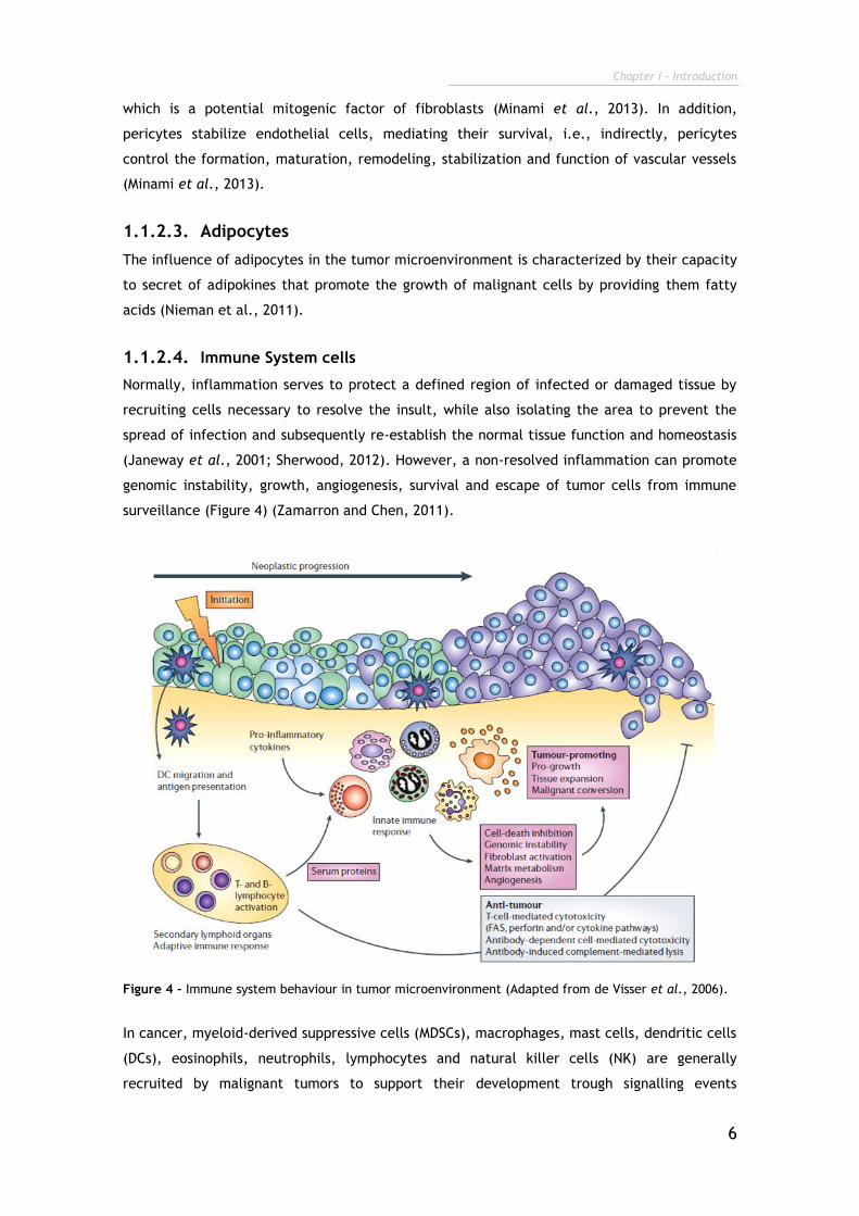
Normally, inflammation serves to protect a defined region of infected or damaged tissue by
recruiting cells necessary to resolve the insult, while also isolating the area to prevent the
spread of infection and subsequently re-establish the normal tissue function and homeostasis
(Janeway et al., 2001; Sherwood, 2012). However, a non-resolved inflammation can promote
genomic instability, growth, angiogenesis, survival and escape of tumor cells from immune
surveillance (Figure 4) (Zamarron and Chen, 2011).
Figure 4 – Immune system behaviour in tumor microenvironment (Adapted from de Visser et al., 2006).
In cancer, myeloid-derived suppressive cells (MDSCs), macrophages, mast cells, dendritic cells
(DCs), eosinophils, neutrophils, lymphocytes and natural killer cells (NK) are generally
recruited by malignant tumors to support their development trough signalling events
Chapter I - Introduction
7
mediated by chemokines, cytokines, cytotoxic mediators and soluble mediators of cell death
and proliferation (Tlsty and Coussens, 2006; Kerkar and Restifo, 2012).
The cells from the immune system induce DNA damage by production of reactive oxygen
species (ROS) and nitrogen species (NO) (Alexander and Friedl, 2012). In addition, immune
cells promote angiogenesis and tissue remodelling by producing growth factors such as HGF,
transforming growth factor-β (TGF-β), VEGF, cytokines (tumour necrosis factor- (TNF-),
interleukins (IL-6, IL-10) and MMPs (MMP-7, MMP-9) (Stockmann et al., 2008; See et al., 2012),
Moreover, tumor growth can be promoted by regulatory T cells (T reg) that suppress cytotoxic
T cell responses (Kerkar and Restifo, 2012), and also by humoral immune responses that
increase chronic inflammation in the tumor microenvironment (Grivennikov et al., 2010). All
these processes are summarized in Figure 4.
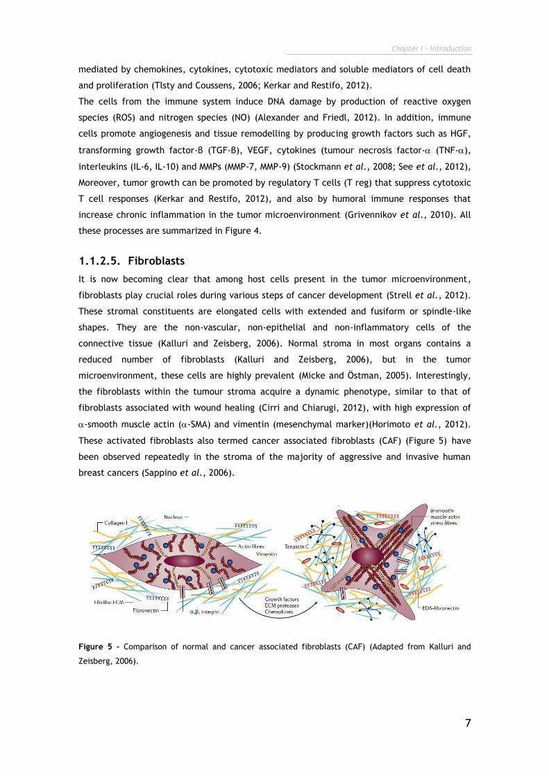
1.1.2.5. Fibroblasts
It is now becoming clear that among host cells present in the tumor microenvironment,
fibroblasts play crucial roles during various steps of cancer development (Strell et al., 2012).
These stromal constituents are elongated cells with extended and fusiform or spindle-like
shapes. They are the non-vascular, non-epithelial and non-inflammatory cells of the
connective tissue (Kalluri and Zeisberg, 2006). Normal stroma in most organs contains a
reduced number of fibroblasts (Kalluri and Zeisberg, 2006), but in the tumor
microenvironment, these cells are highly prevalent (Micke and Östman, 2005). Interestingly,
the fibroblasts within the tumour stroma acquire a dynamic phenotype, similar to that of
fibroblasts associated with wound healing (Cirri and Chiarugi, 2012), with high expression of
-smooth muscle actin (-SMA) and vimentin (mesenchymal marker)(Horimoto et al., 2012).
These activated fibroblasts also termed cancer associated fibroblasts (CAF) (Figure 5) have
been observed repeatedly in the stroma of the majority of aggressive and invasive human
breast cancers (Sappino et al., 2006).
Figure 5 – Comparison of normal and cancer associated fibroblasts (CAF) (Adapted from Kalluri and
Zeisberg, 2006).
Chapter I - Introduction
8
The great influence of fibroblasts in cancer evolution is proved by their capability to induce
the growth and activation of immortalized prostate epithelial cells in vitro and in vivo (Olumi
et al., 1999). Parrott and co-workers, showed in 2001, that ovarian cancer cells recruit
adjacent fibroblasts from normal tissue in order to from the stroma of the tumor,
demonstrating the need of other cell types to form tissue primary cancer. Furthermore, it has
been also shown that, therapies against stromal fibroblasts obtained high anti-tumoral effect
(Parrott et al., 2001; Strell et al., 2012; Mertens et al., 2013).
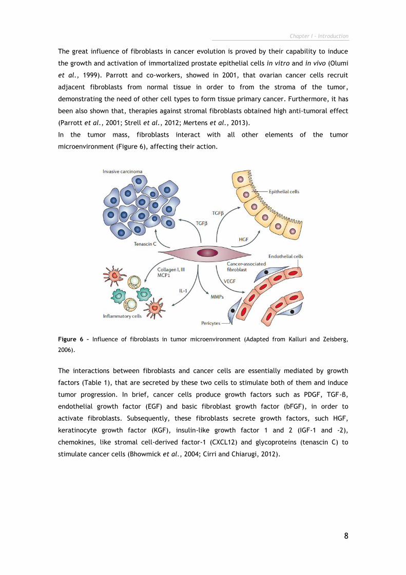
In the tumor mass, fibroblasts interact with all other elements of the tumor
microenvironment (Figure 6), affecting their action.
Figure 6 – Influence of fibroblasts in tumor microenvironment (Adapted from Kalluri and Zeisberg,
2006).
The interactions between fibroblasts and cancer cells are essentially mediated by growth
factors (Table 1), that are secreted by these two cells to stimulate both of them and induce
tumor progression. In brief, cancer cells produce growth factors such as PDGF, TGF-β,
endothelial growth factor (EGF) and basic fibroblast growth factor (bFGF), in order to
activate fibroblasts. Subsequently, these fibroblasts secrete growth factors, such HGF,
keratinocyte growth factor (KGF), insulin-like growth factor 1 and 2 (IGF-1 and -2),
chemokines, like stromal cell-derived factor-1 (CXCL12) and glycoproteins (tenascin C) to
stimulate cancer cells (Bhowmick et al., 2004; Cirri and Chiarugi, 2012).
Chapter I - Introduction
9
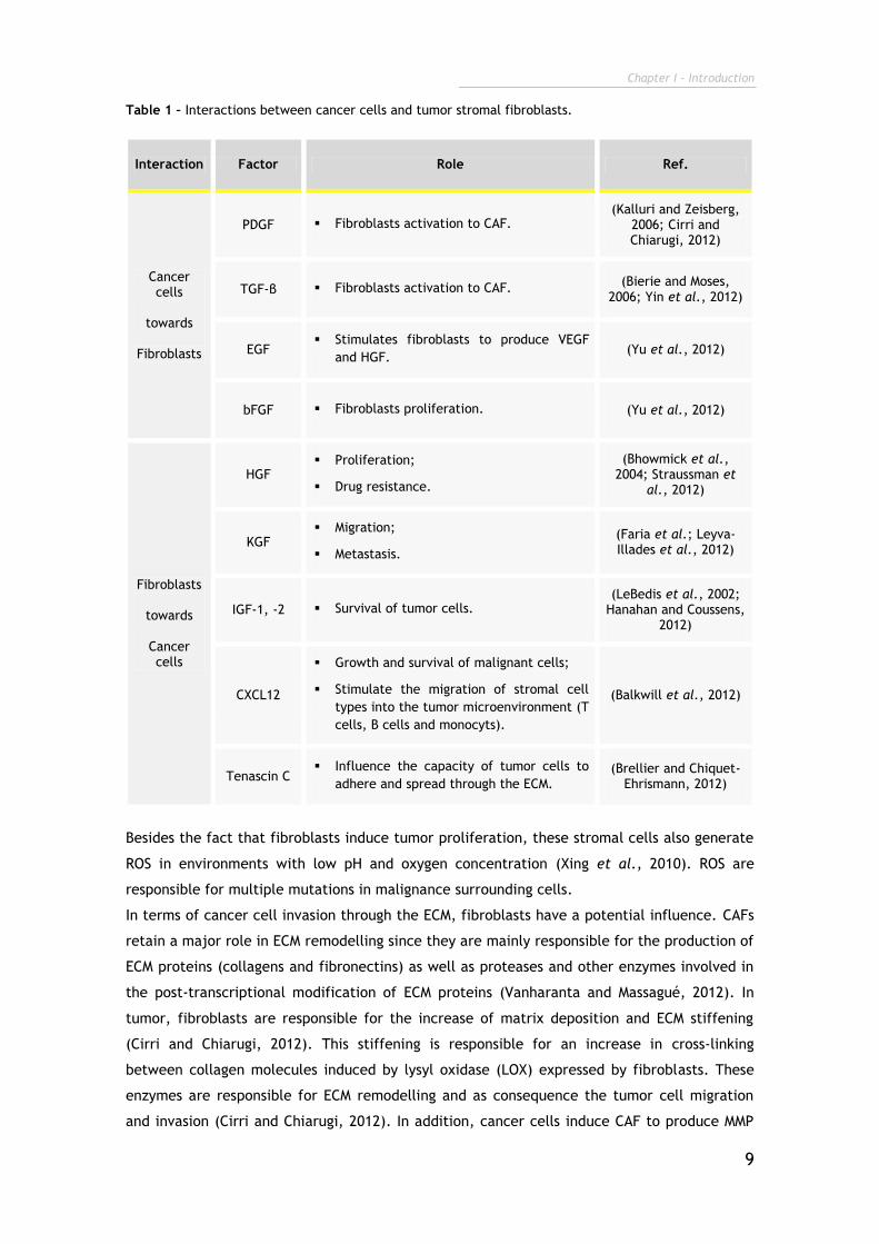
Table 1 – Interactions between cancer cells and tumor stromal fibroblasts.
Interaction Factor Role Ref.
Cancer cells
towards
Fibroblasts
PDGF Fibroblasts activation to CAF. (Kalluri and Zeisberg,
2006; Cirri and Chiarugi, 2012)
TGF-β Fibroblasts activation to CAF. (Bierie and Moses,
2006; Yin et al., 2012)
EGF Stimulates fibroblasts to produce VEGF
and HGF. (Yu et al., 2012)
bFGF Fibroblasts proliferation. (Yu et al., 2012)
Fibroblasts
towards
Cancer cells
HGF Proliferation;
Drug resistance.
(Bhowmick et al., 2004; Straussman et
al., 2012)
KGF Migration;
Metastasis.
(Faria et al.; Leyva-Illades et al., 2012)
IGF-1, -2 Survival of tumor cells. (LeBedis et al., 2002;
Hanahan and Coussens, 2012)
CXCL12
Growth and survival of malignant cells;
Stimulate the migration of stromal cell
types into the tumor microenvironment (T
cells, B cells and monocyts).
(Balkwill et al., 2012)
Tenascin C Influence the capacity of tumor cells to
adhere and spread through the ECM. (Brellier and Chiquet-
Ehrismann, 2012)
Besides the fact that fibroblasts induce tumor proliferation, these stromal cells also generate
ROS in environments with low pH and oxygen concentration (Xing et al., 2010). ROS are
responsible for multiple mutations in malignance surrounding cells.
In terms of cancer cell invasion through the ECM, fibroblasts have a potential influence. CAFs
retain a major role in ECM remodelling since they are mainly responsible for the production of
ECM proteins (collagens and fibronectins) as well as proteases and other enzymes involved in
the post-transcriptional modification of ECM proteins (Vanharanta and Massagué, 2012). In
tumor, fibroblasts are responsible for the increase of matrix deposition and ECM stiffening
(Cirri and Chiarugi, 2012). This stiffening is responsible for an increase in cross-linking
between collagen molecules induced by lysyl oxidase (LOX) expressed by fibroblasts. These
enzymes are responsible for ECM remodelling and as consequence the tumor cell migration
and invasion (Cirri and Chiarugi, 2012). In addition, cancer cells induce CAF to produce MMP

Chapter I - Introduction
10
(Talmadge and Fidler, 2010; Xing et al., 2010), which binds to the cancer cells and it is used
for degradation and invasion of malignance cells through the ECM (Pavlaki and Zucker 2003).
Two other ECM components produced by fibroblasts are fibronectin and hyaluronan.
Fibronectin is associated with integrin receptors and MMP secretion, thus affecting cell
adhesion, migration. Within tumors, fibroblasts, secrete high concentrations of hyaluronan
that are responsible for macrophage recruitment, which have also an essential role in tumor
progression, as previously mentioned (Lu et al., 2012; Raz and Erez, 2013). In addition to
attract macrophages, fibroblasts are also responsible for mediating the inflammatory
response, by secreting chemokines (monocyte chemotactic protein-1 (MCP-1)), interleukins
(IL-1) and inducing immune suppression by expression of TGF-β (McClellan et al., 2012; Raz
and Erez, 2013). This transforming growth factor is also responsible for the orchestration of
the epithelial-to-mesenchymal transition (EMT) (Figure 7) (Chaffer and Weinberg, 2011).
Different studies showed the crucial role of EMT in tumor progression, since it promotes the
invasion and metastasis of cancer cells (Alexander and Friedl, 2012).
Figure 7 – Representation of the mechanism of epithelial-to-mesenchymal transition (EMT) (Adapted
from (Peinado et al., 2007).
EMT is the mechanism responsible for the acquisition of mesenchymal like properties of
epithelial cells, in result of disruption of intercellular adhesion (adherens junctions) and the
enhancement of cell motility (Alexander and Friedl, 2012).
In last, fibroblasts stromal cells also interfere in tumor vascularization. These stromal cells
express VEGF, which have a potential angiogenic effect in tumor mass (Strell et al., 2012).

Chapter I - Introduction
11
1.2. Nanosized delivery systems as novel therapeutic
approaches for cancer therapy
Advanced oncologic diseases have been for long associated with limited patient progression
free survival (PPFS) (Chabner and Roberts, 2005). Over the last two decades, physicians have
been focused in increasing PPFS rate through the application of multimodal treatment
approaches that involve surgery, chemo- and radiotherapy, in an attempt to tackle the above
mentioned adaptive and multi-resistant profile of cancer cells (Riehemann et al., 2009).
The anti-cancer chemical drugs are commonly administered intravenously, being partitioned
in both the tumor and major organs (e.g. liver, lungs, kidneys) due to their inability to
differentiate healthy from malignant cells (Sinha et al., 2006).
Figure 8 – Relation between drug anti-tumoral effect and toxic effect. A) Hypothetical dose-response
curves for conventional chemotherapeutic drugs. In conventional therapies the curves of antitumor
effect and toxic effect are parallel, revealing that higher doses induce higher adverse effects.
B) Hypothetical dose-response curves for new drug delivery systems. The drug delivery systems increase
the specificity for cancer cells, reducing the toxic adverse effects. (Adapted from Fox et al., 2002).
This pharmacokinetic profile accounts for the adverse cytotoxicity effect attained with the
majority of chemotherapeutic agents (Fox et al., 2002). In addition, due to an inherent
limited aqueous solubility, anti-cancer drugs present a short circulation time and sometimes
their concentration in blood remains below the therapeutic window concentrations, rendering
this therapy rather ineffective (Danhier et al., 2012). To overcome these issues, physicians
administer chemotherapy through longer time periods (usually every 21 days, with a total of 5
to 8 sessions) (Hamilton and Hortobagyi, 2005). Although, this approach has a limited success,
since tumor drug accumulation remains low and cancer cells develop resistance against the
effect of chemotherapeutics (Krishna and Mayer, 2000; Raguz and Yagüe, 2008). This acquired
multidrug resistance (MDR) is nowadays one of the most serious problems associated with
chemotherapy, having a negative impact in PPFS.
Similarly, radiotherapy-based treatments are also impaired by the lack of cell specificity
(Sorensen et al., 2012; Allen et al., 2013). In fact, ionizing radiation penetrates within tissues
indiscriminately and although it triggers cancer cell death, it also damages the surrounding
Chapter I - Introduction
12
tissues and organs in such a way that new cancer cells may arise from radiation-induced
mutations (Allen et al., 2013; Zimmermann et al., 2013).
Radiation as an anti-cancer therapy in young woman can affect reproductive organs, and as a
consequence induce fertility problems (Metzger et al., 2013). Another major disadvantage of
these conventional therapies is their inability to eradicate circulating cancer cells that may
lay dormant for a refractory period and form new metastatic cancers in other organs
(Chabner and Roberts, 2005; Alexander and Friedl, 2012). Metastatic cancer cells are
markedly more aggressive than their parent cells and these conventional therapies are still
ineffective (Alexander and Friedl, 2012).
Thus, despite having increased expectations for cancer treatment, these traditional therapies
still lack the necessary specificity and effectives to eliminate cancer cells. Therefore,
recently the advent of Nanotechnology was unlocked a whole new range of opportunities to
develop safe and highly effective anti-cancer treatments. In reality, gathering the unique
capacity of nanoscale materials and directed it to the manufacture of novel medical devices
brings forth the potential to change the effectiveness of cancer therapies (Zhang et al.,
2013). Moreover, nano-based platforms also make an important contribution in cancer
prevention, detection, diagnosis and imaging (Yang et al., 2013).
In contrast to conventional therapies, nanomedicine attempts to use sophisticated approaches
to either kill specific cells or repair them, one cell at a time (Zhang et al., 2013), like
nanoparticles (Figure 9).
Figure 9 – Representation of the different types of nanoparticles available (Adapted from Chou et al.,
2011).
Chapter I - Introduction
13
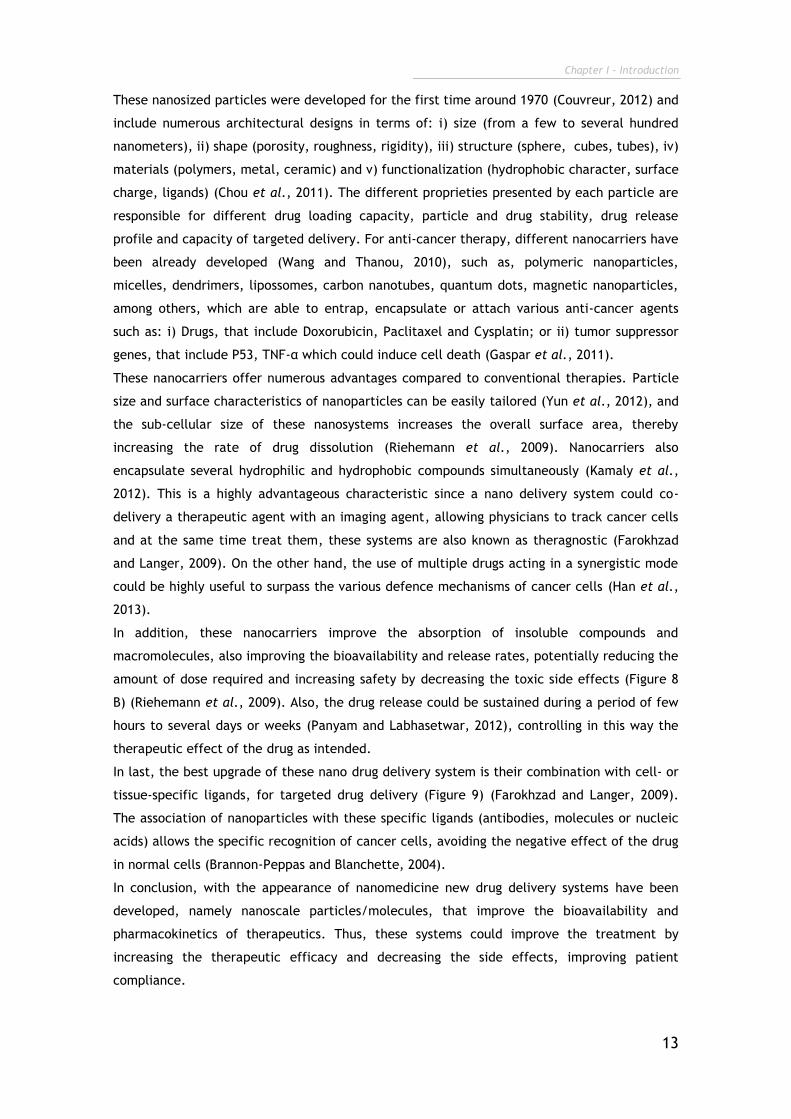
These nanosized particles were developed for the first time around 1970 (Couvreur, 2012) and
include numerous architectural designs in terms of: i) size (from a few to several hundred
nanometers), ii) shape (porosity, roughness, rigidity), iii) structure (sphere, cubes, tubes), iv)
materials (polymers, metal, ceramic) and v) functionalization (hydrophobic character, surface
charge, ligands) (Chou et al., 2011). The different proprieties presented by each particle are
responsible for different drug loading capacity, particle and drug stability, drug release
profile and capacity of targeted delivery. For anti-cancer therapy, different nanocarriers have
been already developed (Wang and Thanou, 2010), such as, polymeric nanoparticles,
micelles, dendrimers, lipossomes, carbon nanotubes, quantum dots, magnetic nanoparticles,
among others, which are able to entrap, encapsulate or attach various anti-cancer agents
such as: i) Drugs, that include Doxorubicin, Paclitaxel and Cysplatin; or ii) tumor suppressor
genes, that include P53, TNF-α which could induce cell death (Gaspar et al., 2011).
These nanocarriers offer numerous advantages compared to conventional therapies. Particle
size and surface characteristics of nanoparticles can be easily tailored (Yun et al., 2012), and
the sub-cellular size of these nanosystems increases the overall surface area, thereby
increasing the rate of drug dissolution (Riehemann et al., 2009). Nanocarriers also
encapsulate several hydrophilic and hydrophobic compounds simultaneously (Kamaly et al.,
2012). This is a highly advantageous characteristic since a nano delivery system could co-
delivery a therapeutic agent with an imaging agent, allowing physicians to track cancer cells
and at the same time treat them, these systems are also known as theragnostic (Farokhzad
and Langer, 2009). On the other hand, the use of multiple drugs acting in a synergistic mode
could be highly useful to surpass the various defence mechanisms of cancer cells (Han et al.,
2013).
In addition, these nanocarriers improve the absorption of insoluble compounds and
macromolecules, also improving the bioavailability and release rates, potentially reducing the
amount of dose required and increasing safety by decreasing the toxic side effects (Figure 8
B) (Riehemann et al., 2009). Also, the drug release could be sustained during a period of few
hours to several days or weeks (Panyam and Labhasetwar, 2012), controlling in this way the
therapeutic effect of the drug as intended.
In last, the best upgrade of these nano drug delivery system is their combination with cell- or
tissue-specific ligands, for targeted drug delivery (Figure 9) (Farokhzad and Langer, 2009).
The association of nanoparticles with these specific ligands (antibodies, molecules or nucleic
acids) allows the specific recognition of cancer cells, avoiding the negative effect of the drug
in normal cells (Brannon-Peppas and Blanchette, 2004).
In conclusion, with the appearance of nanomedicine new drug delivery systems have been
developed, namely nanoscale particles/molecules, that improve the bioavailability and
pharmacokinetics of therapeutics. Thus, these systems could improve the treatment by
increasing the therapeutic efficacy and decreasing the side effects, improving patient
compliance.
Chapter I - Introduction
14
1.3. Experimental models to evaluate nanoparticulated
delivery systems for application in cancer therapy
Nowadays, tremendous resources about the application of nanoparticles in prevention,
diagnosis, and treatment of cancer are being investigated (Zhang et al., 2007). The discovery
and development of anti-cancer agents are the main objectives of important pharmaceutical
companies (Narang and Desai, 2009). However, the introduction of new nanotechnology based
pharmaceuticals creates new challenges for regulatory institutions (Lövestam et al., 2010). In
Europe, the European Commission (EC) and European Medicines Agency (EMEA) already define
the need that all products based on nanomaterials used in humans must comply with the high
level of public health and safety regulations (Hellsten, 2005). In addition, internationally, the
new nanodevices need to be in agreement with the basic rules from regulatory agencies such
as Food and Drug Administration (FDA) and Organization for Standardization (ISO) (Grieger et
al., 2009).
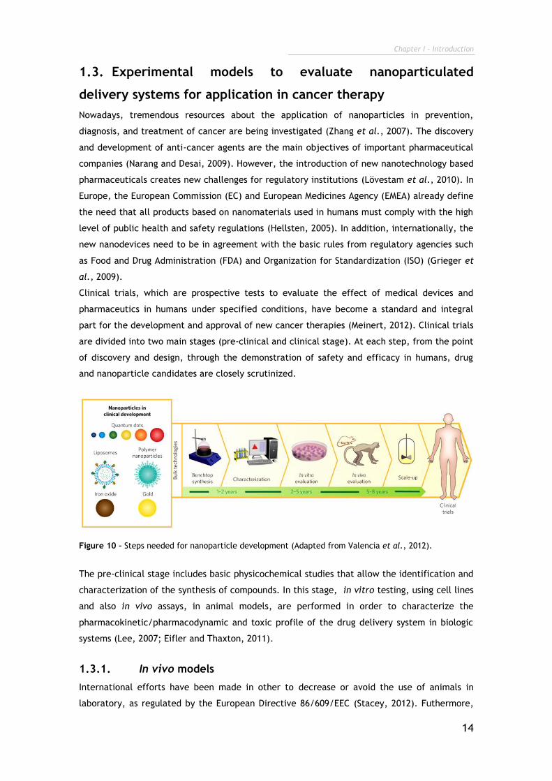
Clinical trials, which are prospective tests to evaluate the effect of medical devices and
pharmaceutics in humans under specified conditions, have become a standard and integral
part for the development and approval of new cancer therapies (Meinert, 2012). Clinical trials
are divided into two main stages (pre-clinical and clinical stage). At each step, from the point
of discovery and design, through the demonstration of safety and efficacy in humans, drug
and nanoparticle candidates are closely scrutinized.
Figure 10 – Steps needed for nanoparticle development (Adapted from Valencia et al., 2012).
The pre-clinical stage includes basic physicochemical studies that allow the identification and
characterization of the synthesis of compounds. In this stage, in vitro testing, using cell lines
and also in vivo assays, in animal models, are performed in order to characterize the
pharmacokinetic/pharmacodynamic and toxic profile of the drug delivery system in biologic
systems (Lee, 2007; Eifler and Thaxton, 2011).
1.3.1. In vivo models
International efforts have been made in other to decrease or avoid the use of animals in
laboratory, as regulated by the European Directive 86/609/EEC (Stacey, 2012). Futhermore,
Chapter I - Introduction
15
there are different countries where animal experimentation is not allowed (Milstein et al.,
1996; Stacey, 2012). Thus, nowadays the scientists are using the 3 Rs rule (Replacement,
Reduction, and Refinement) in what concerns research animal experimentations (Ranganatha
and Kuppast, 2012; Wolfensohn et al., 2013). As a consequence, the experiments are
performed only with the essential animals needed to validate the results, with fewer
repetitions and during limited periods of a time (Madden et al., 2012). In addition, in order to
reduce maintenance costs, the studies tend to use young animals, whereas, the most difficult
patients to treat are the elderly ones (Hartung, 2008).
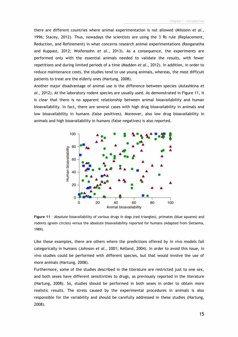
Another major disadvantage of animal use is the difference between species (Astashkina et
al., 2012). At the laboratory rodent species are usually used. As demonstrated in Figure 11, it
is clear that there is no apparent relationship between animal bioavailability and human
bioavailability. In fact, there are several cases with high drug bioavailability in animals and
low bioavailability in humans (false positives). Moreover, also low drug bioavailability in
animals and high bioavailability in humans (false negatives) is also reported.
Figure 11 - Absolute bioavailability of various drugs in dogs (red triangles), primates (blue squares) and
rodents (green circles) versus the absolute bioavailability reported for humans (Adapted from Sietsema,
1989).
Like these examples, there are others where the predictions offered by in vivo models fail
categorically in humans (Johnson et al., 2001; Kelland, 2004). In order to avoid this issue, in
vivo studies could be performed with different species, but that would involve the use of
more animals (Hartung, 2008).
Furthermore, some of the studies described in the literature are restricted just to one sex,
and both sexes have different sensitivities to drugs, as previously reported in the literature
(Hartung, 2008). So, studies should be performed in both sexes in order to obtain more
realistic results. The stress caused by the experimental procedures in animals is also
responsible for the variability and should be carefully addressed in these studies (Hartung,
2008).
Chapter I - Introduction
16
1.3.2. In vitro models
Cell cultures emerged in 19th century as a valuable solution for challenging issues of in vivo
models. The first culture was performed by Wilhem Roux in 1885, by maintaining in vitro
chicken embryo tissue during several days (Langdon, 2004). However, only in 1922 the
culture of epithelial cells was performed and in 1951 the first continuous human cancer cell
line (HeLa) was obtained and cultured by George Gey (Langdon, 2004).
These in vitro techniques allow the growth of cells outside of their natural environment,
under controlled conditions (Lindl and Steubing, 2013). These types of assays become the
main force of biopharmaceutical investigation due to their few limitations, namely, bacterial
and fungal contaminations (Stacey, 2011). Cell culture offers a unique testing platform to
investigate the effects of different drug formulations and nanoparticle designs during pre-
clinical development, under highly controlled and reproducible conditions (HogenEsch and
Nikitin, 2012), such as temperature, pH, osmotic pressure, oxygen (O2) and carbon dioxide
(CO2) tension. Moreover, cell cultures provide an easy way to manipulate numerous
experimental variables in order to mimic some in vivo conditions, whilst avoiding ethical and
legal issues associated with animal handling and experimentation (Duell et al., 2011).
However, up till now, the cell-cell interactions were commonly disregarded in the majority of
the studies reported so far (Duell et al., 2011).
1.3.2.1. Co-cultures
One of the main goals of the in vitro systems has been the reproduction of original tissue
characteristics and its cell–cell interactions, to simulate, as close as possible, the in vivo
environment. To overcome such limitations a new category of cell cultures, termed co-
cultures, is currently being developed (Miki et al., 2012). This cell culture system arises as a
quite interesting alternative that allows a better method to mimic the tumor
microenvironment (Duell et al., 2011).
A co-culture consists of at least two different types of cells (i.e., cancer/fibroblast, epithelial
cell/lymphocyte, etc.) (Miki et al., 2012) and they provide a more sophisticated and sensitive
system that allows to reproduce in vitro the in vivo tumor niche. By using co-cultures it is
possible to recreate some of the cell inter-relationship (Tumarkin et al., 2011), since
heterotypic cell-cell interactions are established in close contact. These direct physical
connections between cells play a pivotal role in the mechanisms of cancer invasion through
actions of adhesion molecules, such as Cadherins (De Wever and Mareel, 2003; Miki et al.,
2012). In addition, different cells in co-culture could release cytokines, interleukins,
chemokines and growth factors that are essential for the establishment of cell morphology,
phenotype, metabolism and proliferation, features that are always present in vivo (Streuli et
al., 1991; Krause et al., 2010; Purpura et al., 2011; Miki et al., 2012). In addition, co-cultures
can also influence the drug resistance presented by cancer cells. In fact, Martinez-Outschoorn
and his team, 2011, (Martinez-Outschoorn et al., 2011) analyzed de apoptotic effect of
Tamoxifen in monocultures of breast cancer cells and co-cultures of breast cancer cells and

Chapter I - Introduction
17
fibroblasts. As a conclusion, they verified a markedly reduction in apoptosis of breast cancer
cells when they were in co-culture, highlighting the importance of these models.
1.3.2.2. 3D cell cultures: Spheroids
In addition to the presence of various cell types encountered in the tumor microenvironment,
the inclusion of ECM, which is 3D, complex and dynamic network is crucial for mimic the in
vitro tumor (Cukierman et al., 2002). So, in 1972, researchers began to explore the
differences between cells grown on a flat surface versus 3D supports (Elsdale and Bard, 1972).
They concluded that, ECM is not just a random mix of secreted components, but it has a
specific composition of biomolecules entrapped in a very well defined geometrical structure,
that stimulates specific cell responses, such as differentiation. Thus, in addition to co-
localization of different cell types with cell–cell interactions and the exchange of bioactive
molecules, a 3D architecture that mimics cell organization in tissues and organs is demanded
to mimic tissue native proprieties using in vitro conditions (Kim et al., 2004). Such, is the
main disadvantage of two-dimensional (2D) in vitro models. Such models rely on the
migration of cells on flat substrates, like glass or polystyrene surfaces (Freshney et al., 2006),
in which cells grow in two dimensions (Burdett et al., 2010), which leads to the loss of spatial
organization of cells and a deregulation of cell metabolism and functionality as consequence
(Lee et al., 2008).
To overcome such disadvantage, the 3D cell cultures have emerged as a viable option. One of
the most widely used 3D in vitro models are the tumor spheroids (Burdett et al., 2010). This
unique culture is based in a small, tightly bound cellular aggregate that tends to form when
cancer cells are cultured in non adherent surfaces (Trédan et al., 2007; Burdett et al., 2010;
Fennema et al., 2013). Spheroids have been used in cancer research, because the 3D
architecture and extensive cell–cell interactions provided by spheroid growth closely mimic
the in vivo cellular environment. Recently, 3D culture gene expression profiles, have been
shown to reflect the clinical expression profiles, in comparison with those observed for 2D
cultures (Hirschhaeuser et al., 2010). In fact, the 3D cell organization can influence cell
shape, gene expression, growth, morphogenesis, motility and differentiation (Table 2)
(Yamada and Cukierman, 2007). In addition, cells cultured in 2D monolayers typically exhibit
a lower resistance to therapy than those of in vivo tumors, a critical factor if novel
therapeutic approaches are under evaluation (Phung et al., 2011). Spheroids have also been
considered an essential cell culture technique to study solid tumors. Some cancer types
(sarcomas, carcinomas and lymphomas) tend to form solid tumors (Gavhane et al., 2011).
These tumors have a unique 3D architecture characterized by hypoxia, low pH and low levels
of glucose (Box et al., 2010; Yeom et al., 2012).
Chapter I - Introduction
18
Table 2 - The influence of 3D cell organization in cell behaviour and signalling.
Biologic Function Observed in 3D References
Cell Shape Loss of epithelial cell polarity;
Altered epithelial and fibroblast shape.
(Yamada and Cukierman, 2007; Dhimolea et al.,
2010)
Gene
Expression
Variation in gene expression when cells are culture in 2D or 3D.
(Birgersdotter et al., 2005)
Growth Cell growth is influenced by 3D
structure. (Lu et al., 2012)
Morphogenesis 3D cultures are associated with
formation of new vessels. (Shaw et al., 2004)
Motility Cell motility in 3D matrices is restricted. (Khatau et al., 2012)
Differentiation 3D matrix-induced cell differentiation. (Liu and Roy, 2005;
Willerth et al., 2007)
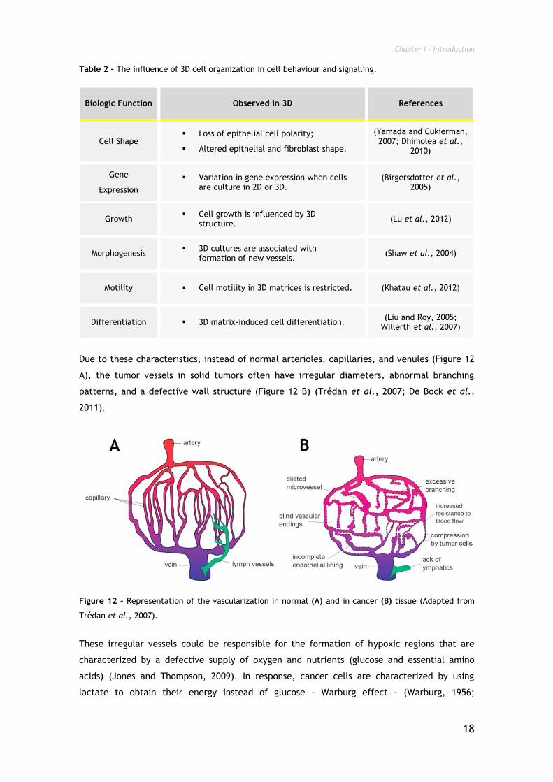
Due to these characteristics, instead of normal arterioles, capillaries, and venules (Figure 12
A), the tumor vessels in solid tumors often have irregular diameters, abnormal branching
patterns, and a defective wall structure (Figure 12 B) (Trédan et al., 2007; De Bock et al.,
2011).
Figure 12 – Representation of the vascularization in normal (A) and in cancer (B) tissue (Adapted from
Trédan et al., 2007).
These irregular vessels could be responsible for the formation of hypoxic regions that are
characterized by a defective supply of oxygen and nutrients (glucose and essential amino
acids) (Jones and Thompson, 2009). In response, cancer cells are characterized by using
lactate to obtain their energy instead of glucose - Warburg effect - (Warburg, 1956;
Chapter I - Introduction
19
Hockenbery et al., 2013). Such process involves CO2 production and results in a
microenvironment with a low pH (Harris, 2002).
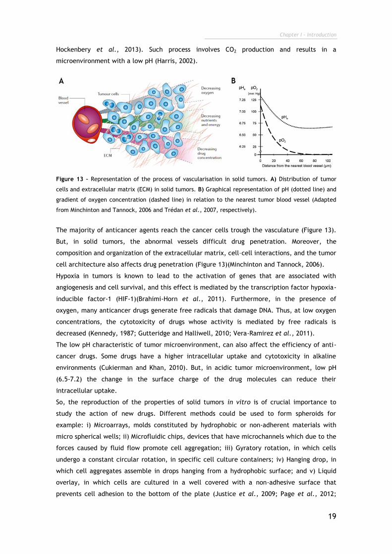
Figure 13 – Representation of the process of vascularisation in solid tumors. A) Distribution of tumor
cells and extracellular matrix (ECM) in solid tumors. B) Graphical representation of pH (dotted line) and
gradient of oxygen concentration (dashed line) in relation to the nearest tumor blood vessel (Adapted
from Minchinton and Tannock, 2006 and Trédan et al., 2007, respectively).
The majority of anticancer agents reach the cancer cells trough the vasculature (Figure 13).
But, in solid tumors, the abnormal vessels difficult drug penetration. Moreover, the
composition and organization of the extracellular matrix, cell–cell interactions, and the tumor
cell architecture also affects drug penetration (Figure 13)(Minchinton and Tannock, 2006).
Hypoxia in tumors is known to lead to the activation of genes that are associated with
angiogenesis and cell survival, and this effect is mediated by the transcription factor hypoxia-
inducible factor-1 (HIF-1)(Brahimi-Horn et al., 2011). Furthermore, in the presence of
oxygen, many anticancer drugs generate free radicals that damage DNA. Thus, at low oxygen
concentrations, the cytotoxicity of drugs whose activity is mediated by free radicals is
decreased (Kennedy, 1987; Gutteridge and Halliwell, 2010; Vera-Ramirez et al., 2011).
The low pH characteristic of tumor microenvironment, can also affect the efficiency of anti-
cancer drugs. Some drugs have a higher intracellular uptake and cytotoxicity in alkaline
environments (Cukierman and Khan, 2010). But, in acidic tumor microenvironment, low pH
(6.5-7.2) the change in the surface charge of the drug molecules can reduce their
intracellular uptake.
So, the reproduction of the properties of solid tumors in vitro is of crucial importance to
study the action of new drugs. Different methods could be used to form spheroids for
example: i) Microarrays, molds constituted by hydrophobic or non-adherent materials with
micro spherical wells; ii) Microfluidic chips, devices that have microchannels which due to the
forces caused by fluid flow promote cell aggregation; iii) Gyratory rotation, in which cells
undergo a constant circular rotation, in specific cell culture containers; iv) Hanging drop, in
which cell aggregates assemble in drops hanging from a hydrophobic surface; and v) Liquid
overlay, in which cells are cultured in a well covered with a non-adhesive surface that
prevents cell adhesion to the bottom of the plate (Justice et al., 2009; Page et al., 2012;
Chapter I - Introduction
20
Fennema et al., 2013). This last technique is the simplest, reproducible and less expensive
method for spheroid production (Perche and Torchilin, 2012). In addition, this model forms
single spherical spheroids without the limitation of size or shape associated for instance with
the micro fluidic and microarray-based approaches.
With 3D cell cultures it is possible to mimic this tumor microenvironment formed in in vivo
conditions. In 1970, Sutherland and their team treated 3D multicellular tumor spheroids with
radiation and observed that the therapeutic results obtained were very similar to those
reported for solid tumors in vivo (Sutherland et al., 1970).
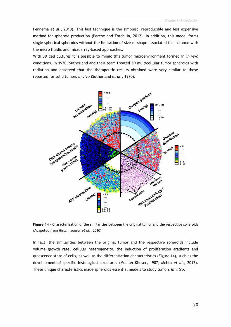
Figure 14 – Characterization of the similarities between the original tumor and the respective spheroids
(Adapeted from Hirschhaeuser et al., 2010).
In fact, the similarities between the original tumor and the respective spheroids include
volume growth rate, cellular heterogeneity, the induction of proliferation gradients and
quiescence state of cells, as well as the differentiation characteristics (Figure 14), such as the
development of specific histological structures (Mueller-Klieser, 1987; Mehta et al., 2012).
These unique characteristics made spheroids essential models to study tumors in vitro.
Chapter I - Introduction
21
1.4. Objectives
During the elaboration of this master dissertation, different objectives are taken into account
in order to achieve the main goals proposed. The aims are summarized in the following
points:
The main aim of this thesis workplan was is the optimization of heterogenic breast and
cervical 2D and 3D cell co-culture models for future development and investigation of new
drugs and delivery systems for drug and gene delivery:
□ Develop co-culture models that mimic in vitro the complex tumor
microenvironment found in vivo, by using malignant cells and stromal fibroblasts;
□ Evaluate the influence of co-culture conditions in cell proliferation and spatial
organization;
□ Analyse the establishment of direct interactions between cancer and normal cells
throughout extended time periods when co-culture in vitro;
□ Develop in vitro co-culture models of breast and cervical cancers;
□ Investigate the tumor targeting specificity of a multifunctional gene delivery
system towards –cancer cells in 2D mono- and co-culture;
□ Optimize the production of 3D multicellar tumor spheroids in vitro by using
different methodologies;
□ Develop 3D co-culture models of breast and cervical cancers during various time
points;
□ Analyse the 3D tumor spheroids for the acquisition of in vivo mimicking
characteristics of solid tumors.
Chapter II
Methods
Chapter II - Methods
23
2. Methods
2.1. Materials
Human cervix adenocarcinoma (HeLa) and oestrogen-dependent human breast
adenocarcinoma (MCF-7) cells were obtained from ATCC (Middlesex, UK) and primary normal
human dermal fibroblasts (hFIB) from Promocell (Heidelberg, Germany). The cell culture
plates and T-flasks were obtained from Orange Scientific (Braine-l'Alleud, Belgium). Ibidi cell
imaging chambers plates were acquired from Ibidi GmbH (Munich, Germany). Cacodylate,
collagen Type I, Dulbecco’s Modified Eagle’s Medium F-12 (DMEM-F12), Dulbecco’s Modified
Eagle’s Medium High Glucose (DEMEM-HG), ethanol (EtOH), glutharaldehyde,
paraformaldehyde (PFA), phosphate-buffered saline (PBS), resazurin, rhodamine B
isothiocianate (RITC) and trypsin were purchased from Sigma–Aldrich (Sintra, Portugal).
Dimethyl sulfoxide (DMSO) was obtained from VWR BDH Prolabo (Madrid, Spain) and agarose
was purchase from Grisp (Porto, Portugal). CellLight® Actin-Green fluorescent protein (GFP)
BacMam 2.0, Germ agglutinin conjugated Alexa 594 (WGA-Alexa 594) and Hoechst 33342®
were obtained from Invitrogen (Carlsbad, CA, USA). Fetal bovine serum (FBS) was purchased
from Biochrom AG (Berlin, Germany). All reagents were of analytical grade and used as
received.
2.2. Breast cancer and cervical cancer 2D and 3D in vitro co-
culture models optimization
2.2.1. Cell lines maintenance
All cell lines, MCF-7, HeLa and hFIB, were grown in 75 cm2 T-flasks with a humidified
atmosphere of 5% CO2, at 37 °C. MCF-7 and hFIB were maintained in DMEM-F12 medium
supplemented with 10% FBS, and 1% streptomycin and gentamycin. HeLa cell line was growth
in DMEM-HG, with 10% (v/v) FBS and 1% streptomycin and gentamycin. When cells attained
confluence, they were harvested using 0.18% trypsin (1:250) and 5 mM EDTA (Ethylenediamine
tetraacetic acid).
2.2.2. Optimization of 2D in vitro cell co-culture models of breast cancer
(MCF-7:hFIB) and cervical cancer (HeLa:hFIB)
Upon attaining confluence, cancer cells and normal fibroblasts were harvested using 0.18%
trypsin (1:250) and 5 mM EDTA. Cells were counted by using a haemocytometer and trypan
blue 4 % (w/v), in PBS.
Subsequently, co-cultures were performed. For breast cancer co-cultures, MCF-7 and hFIB
were seeded onto 6-well plates, with a total number of 2x10⁴ of cells per well and with MCF-7
to hFIB ratio as summarized in Table 3. As controls, homotypic cultures of MCF-7 and hFIB
Chapter II - Methods
24
were seeded using the same total number of cells per well. All these cultures were
maintained in DMEM-F12 medium supplemented with 10% (v/v) FBS, and 1% streptomycin and
gentamycin.
HeLa and hFIB were seeded onto 6-well plates, with a total number of 2x 10⁴ of cells per well
and with HeLa to hFIB ratio as summarized in Table 4. As controls, homotypic cultures of
HeLa and hFIB were seeded using the same total number of cells per well. All these cultures
were maintained in DMEM-HG medium supplemented with 10% (v/v) FBS and 1% streptomycin
and gentamycin.
Table 3 - MCF-7 to hFIB cell ratios used in vitro to mimic the breast cancer microenvironment.
Ratio MCF-7 hFIB References
i) 1:1 50.00% 50.00% (Heneweer et al., 2005)
ii) 1:3 25.00% 75.00% (Ko et al., 2012)
iii) 1:5 16.67% 83.33% (Whitaker-Menezes et al., 2011)
iv) 3:1 75.00% 25.00% (Martinez-Outschoorn et al., 2010)
Table 4 - HeLa to hFIB cell ratios used in vitro to mimic the cervical cancer microenvironment.
Ratio HeLa hFIB References
i) 1:1 50.00% 50.00% (Delinassios and Kottaridis, 1984)
ii) 1:2 33.33% 66.67% (Delinassios and Kottaridis, 1984)
iii) 1:10 9.09% 90.91% (Delinassios, 1987)
iv) 2:1 66.67% 33.33% (Delinassios and Kottaridis, 1984)
2.2.2.1. Optical microscopy analysis of the distribution and morphology
of 2D in vitro cell co-culture models of breast cancer (MCF-7:hFIB) and
cervical cancer (HeLa:hFIB)
The evolution of 2D MCF-7:hFIB and HeLa:hFIB co-cultures and their controls in terms of cell
distribution and morphology was analysed by using an Olympus CX41 inverted optical
microscope equipped with an Olympus SP-500 UZ digital camera, at various magnifications.
2.2.2.2. Resazurin assay for analysis of cell viability of 2D in vitro cell
co-culture models of breast cancer (MCF-7:hFIB)
The evolution of 2D MCF-7:hFIB co-cultures and their controls in terms of cell distribution and
morphology were analysed using an Olympus CX41 inverted optical microscope equipped with
Chapter II - Methods
25
an Olympus SP-500 UZ digital camera. Co-cultures were seeded onto 96 well plates with MCF-
7 to fibroblasts ratios in accordance to what is described in Table 3. For each MCF-7:hFIB
ratio, 5 wells were seeded with 10x103 cells per well (n=5). Cells were maintained in DMEM-
F12 supplemented with 10% FBS, without antibiotics. After 24h of co-culture, the medium was
replaced and 10µL of resazurin 0.1% (w/v) was incubated in each well. After an overnight
incubation, the fluorescence of metabolized Resazurin was measured with a
spectrofluorimeter (Molecular Devices, Spectramax Gemini XS) at an excitation/emission
wavelength of λ=560/590nm, respectively. As controls, the cell viability of MCF-7 and hFIB
monocultures was also determined. Cell viability was determined at 48, 72 and 96 hours (h) of
MCF-7:hFIB co-culture.
2.2.3. Optimization of 3D in vitro cell co-culture models of breast cancer
(MCF-7:hFIB) and cervical cancer (HeLa:hFIB)
Prior to all the co-culture experiments, the wells of the culture plates were coated with
300µL of 1% (w/v) agarose dissolved in double deonized and filtered water (Milli-Q water) by
heating until 80ºC. Upon attaining confluence, cancer cells and normal fibroblasts were
harvested using 0.18% trypsin (1:250) and 5 mM EDTA. Cells were stained with trypan blue 4%
(w/v), and counted using a haemocytometer. Subsequently, co-cultures were seeded onto 24-
well plates, with a total number of 5x10³, 10x103 and 15x103 cells per well, with cancer cells
to fibroblasts ratios in accordance to what is described in Table 3 and Table 4. As controls,
MCF-7 spheroids of cancer cells and fibroblasts alone were seeded, using the same total
number of cells per well.
The MCF-7:hFIB spheroids and respective monoculture 3D model were maintained in DMEM-
F12 complete medium (10% (v/v) FBS, and 1% streptomycin and gentamycin). HeLa:hFIB
spheroids and their monoculture counterparts were maintained in DMEM-HG complete
medium. All 3D cell cultures were maintained under horizontal agitation in a Grant-bio PMS-
1000 microplate shaker (Fisher Scientific, Leicestershire, UK), in a humidified atmosphere at
37 °C with an atmosphere of 5% CO2, during 6 days.
2.2.3.1. Optical microscopy analysis of the distribution and morphology
of the 3D in vitro cell co-culture models of breast cancer (MCF-7:hFIB) and
cervical cancer (HeLa:hFIB)
The evolution of 3D MCF-7:hFIB and HeLa:hFIB co-cultures and their controls in terms of cell
distribution and morphology was analysed using an Olympus CX41 inverted optical microscope
equipped with an Olympus SP-500 UZ digital camera, at various magnifications.
Chapter II - Methods
26
2.2.3.2. Scanning electron microscopy (SEM) analysis of the 3D in vitro
cell co-culture models of breast cancer (MCF-7:hFIB)
For scanning electron microscopy (SEM) analysis of the 3D spheroid models the medium in the
wells was removed and spheroids were washed with cacodylate buffer 0.1 M, in PBS 1% (w/v),
and then incubated for 1h at room temperature (RT). Subsequently, the 3D spheroids were
washed with PBS 1% (w/v) solution to remove traces of cacodylate. The cells were then
submitted to a second fixation stage (2.5% glutharaldehyde, in PBS 1% (w/v), at RT). After 2h
of fixation, the samples were washed 3 times with PBS. Then, spheroids were dehydrated
with growing ethanol (50%, 60%, 70%, 80%, 90% and absolute ethanol) during 15 min. The
spheroids were then subjected to critical point drying prior to visualization in the Scanning
Electron Microscope. Subsequently, samples were coated with gold and observed in a Hitachi
S-2700 (Tokyo, Japan) electron microscope at different magnifications.
2.2.3.3. Confocal laser scanning microscopy analysis of 3D in vitro cell
co-culture models of cervical cancer (HeLa:hFIB)
The cell distribution and morphology of 3D HeLa:hFIB co-cultures and their controls was
analysed through confocal laser scanning microscopy (CLSM) (Zeiss LSM 710 laser scanning
confocal microscope (Carl Zeiss SMT Inc., USA). For this purpose, the culture medium in wells
was removed and then spheroids were subjected to fixation with PFA 4% (w/v) in filtered
water (H2O). After 1h of fixation, spheroids were transferred carefully to µ-Slide Ibidi plates.
All the spheroids were labelled with WGA-Alexa 594 during 30 min prior to image acquisition.
3D reconstruction and application of depth coding algorithms was performed in the Zeiss Zen
software (2010) as previously mentioned.
2.3. Evaluation of Chitosan-Histidine-Arginine/pDNA
nanoparticles cellular uptake in 2D breast cancer co-culture
models (MCF-7:hFIB)
2.3.1. CLSM for CH-H-R/pDNA nanoparticles cell uptake analysis in 2D
breast cancer co-culture models
In order to differentiate both cell types (hFIB and MCF-7), the cancer cell line was labelled
with the BacMan Cell Light 2.0® Actin-GFP probe, in accordance with the manufacturer’s
protocol. For better cell attachment to well plates, previously to cell seeding, plates were
coated with collagen type IV, for 30 min, at 37ºC. After the onset of GFP expression, various
MCF-7(GFP) to hFIB ratios were sub-cultured on 8 well µ-Slide Ibidi plates, at a density of
2 x 10⁴ cells per well. The cells were cultured in DMEM-F12 medium supplemented with 10%
(v/v) FBS, in a humidified atmosphere (5% CO2), at 37 ºC. After the 24 h of co-culture, the
cells were incubated with chitosan polymeric nanoparticles produced accordingly to the
procedure previously developed by our team (Gaspar et al., 2013). Cell cultures were then
Chapter II - Methods
27
incubated with Chitosan-Histidine-Arginine/plasmid deoxyribonucleic acid nanoparticles (CH-
H-R/pDNA) with RITC-labelled pDNA (1 μg/cm2). As control, nanoparticles were also incubated
in monocultures of hFIB and MCF-7.
After a 4 h, the cells were fixed with PFA 4% (w/v) in H2O, for 15 min. Following the chemical
fixation, cells were washed with PBS and stained with Hoechst 33342® nuclear probe, at room
temperature. Cell imaging was performed using Zeiss LSM 710 laser scanning confocal
microscope (Carl Zeiss SMT Inc., USA) using a Plan-Apochromat 40x/1.4 Oil DIC objective. The
samples were acquired in z-stack mode with a slice thickness of 0.23 µm. Orthogonal
sectioning and 3D reconstruction of the various Z-stacks was performed in the Zeiss LSM Zen
software (2010).
2.3.2. Flow cytometry for CH-H-R/pDNA nanoparticles cell uptake analysis
in 2D breast cancer co-culture models
The cellular uptake of polymeric nanoparticles was analyzed through flow cytometry by using
a BD FACSCalibur flow cytometer (Becton Dickinson Inc., USA). In summary, MCF-7:hFIB co-
cultures with different cancer cells to fibroblasts ratios and monocultures of both types of
cells were used as controls were seeded in 6 well culture plates with a total of 2 x 10⁵ cells
per well. Cells were grown during 24 h in DMEM-F12 with 10% FBS. For better signal acquision,
co-cultures and monocultures of hFIB and MCF-7 were used as controls to establish the
correct gating and acquisition parameters in the fluorescence 1 (FL-1) (530/30nm (GFP)) and
fluorescence 2 (FL-2) (585/42 nm (RITC)) channels. 24 h after seeding, different co-cultures
and monocultures were incubated with nanoparticles during 4 h. For these, CH-H-R
nanoparticles were prepared with freshly labeled RITC-pDNA (1 µg/cm2) as previously
described by (Gaspar et al., 2013). Then, cells were extensively washed with ice cold PBS,
harvested with 0.18 % trypsin/5 mM EDTA, and recovered by centrifugation. Subsequently,
cells collection, cells were resuspendend in 500 µL of fresh PBS. Data acquisition was
performed in the CellQuestTM Pro software, where 8 x 10³ events were recorded in the gated
regions of interest, assigned to hFIB and MCF-7 cells. The results obtained were analyzed with
a trial version of FCS express v. 4 research edition software (De Novo Software, Ontario,
Canada).

Chapter II - Methods
28

Chapter III
Results and Discussion
Chapter III- Results and Discussion
30
3. Results and Discussion
3.1. Breast cancer and cervical cancer 2D and 3D in vitro co-
culture models
As previously mentioned, co-cultures unlock the possibility to mimic in vitro the in vivo tumor
niche, using simple tools and cost-effective methodologies. These co-cultures are usually
performed with two types of cells: cancer cells and stromal cells, namely fibroblasts which
are those that mostly influence the behaviour of tumor cells. Among all types of cancer,
breast cancer and cervical cancer are the most common malignancies amongst women, being
responsible for a high rate of mortality at worldwide (Youlden et al., 2012). From these
standpoints, the aim of this thesis was the development and optimization of breast cancer
and cervical cancer 2D and 3D co-cultures models, for future development of drugs and
delivery systems to be applied in cancer therapy.
3.1.1. Development and optimization of 2D in vitro cell co-culture models
of breast cancer (MCF-7:hFIB)
In order to mimic the tumor microenvironment of in vivo breast cancers in a more realistic
mode by using cell cultures, MCF-7 adenocarcinoma cells were seeded in culture together
with non-malignant human skin fibroblasts. The use of fibroblasts representative cells of the
tumor stromal cells is attributed to their potential biological interaction with breast cancer
cells in vivo, as recently reported by Straussman and co-workers, 2013 (Straussman et al.,
2012). Especially in breast tumor tissue, fibroblasts act as a key factor for the spatiotemporal
evolution of malignant cells (Aboussekhra, 2011). Furthermore, skin fibroblasts may also be
biologically altered and exhibit characteristics associated with a transformed phenotype
characterized by the expression of growth factors, such as HGF, KGF, IGF, which have a
potential effect in cancer cells, namely, in proliferation, metastization and drug resistance
(Aboussekhra, 2011; Hanahan and Coussens, 2012). Therefore, these cells are usually used by
different authors for the development of breast co-cultures models (Martinez-Outschoorn et
al., 2010; Olsen et al., 2010; Chiavarina et al., 2011). However, it is important to emphasize
that in in vivo tumors the fraction of normal cell in relation to cancer cells is highly dynamic
and changes from patient to patient. In fact, breast tumor is commonly described to be a
heterogeneous mass of cancer and stromal cells in different concentrations (Weigelt et al.,
2010). Besides, in the literature, there are several conflicting reports concerning the actual
ratio of MCF-7 to hFIB used to establish this particular in vitro model of breast cancer. In fact,
several authors suggest the establishment of co-culture systems with equal number of normal
and cancer cells, but others perform co-cultures with more cancer cells than fibroblasts, or
vice-versa (Brouty-Boyé et al., 1994; Sadlonova et al., 2005; Krause et al., 2010; Xu et al.,
Chapter III- Results and Discussion
31
2013). Thus, in general the described MCF-7 to hFIB cell ratios are described as follows: i)
1:1; ii) 1:3; iii) 1:5 and iv) 3:1 (Heneweer et al., 2005; Martinez-Outschoorn et al., 2010;
Whitaker-Menezes et al., 2011; Ko et al., 2012; Le Droumaguet et al., 2012).
Taking these discrepancies into account, co-cultures with different MCF-7:hFIB ratios were
developed in an attempt to overcome the urgent necessity to mimic tumor heterogeneity.
However, until presently the organization of cells in co-culture with time has never been
explored (Duell et al., 2011; Tumarkin et al., 2011). Actually, the latter assumes further
significance since an extended co-culture time is important to maximize the interactions
between cell types and will surely influence the aggressiveness and resistance of cancer cells
to anti-tumoral therapeutics (Kalluri and Zeisberg, 2006; Hanahan and Coussens, 2012; Strell
et al., 2012). This communication may occur directly by tight junctions or indirectly by
paracrine inter-cellular signalling (Wang et al., 2006; Pietras and Östman, 2010; Lu et al.,
2012).
Thus, in this study co-cultures were maintained during 10 days, in order to analyze the
influence of signalling in cell organization and biological behaviour. During the establishment
of the co-culture systems, the medium was not replaced by fresh medium in order to preserve
the exchanged soluble mediators between cancer and stromal cells. This is a critical
parameter that needs to be addressed during the production of co-culture models since
various biomolecules such as growth factors, cytokines, chemokines and other soluble factors
are secreted by these cells and can thus have a potential effect in cell behaviour (Hanahan
and Coussens, 2012). By using this approach the tumor microenvironment is more precisely
reproduced since extensive exchange of these mediators also occurs in vivo (Miki et al.,
2012).
The spatiotemporal evolution of these 2D co-cultures systems over time is presented in Figure
14.
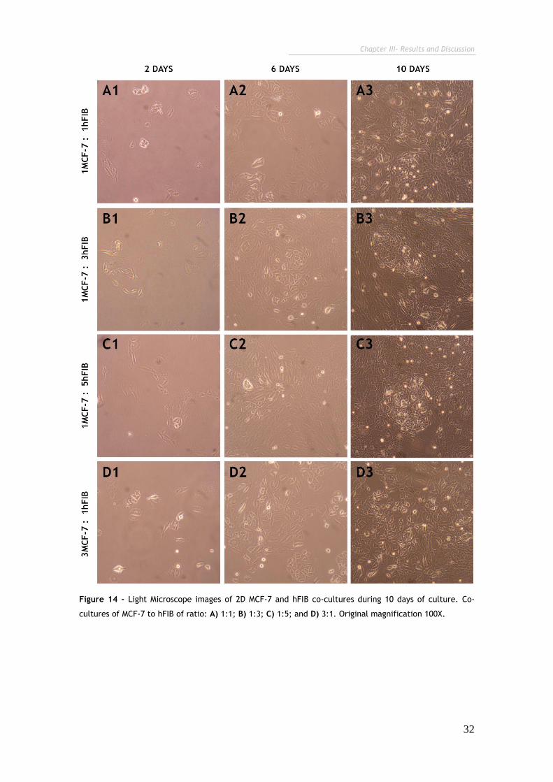
Chapter III- Results and Discussion
32
Figure 14 - Light Microscope images of 2D MCF-7 and hFIB co-cultures during 10 days of culture. Co-
cultures of MCF-7 to hFIB of ratio: A) 1:1; B) 1:3; C) 1:5; and D) 3:1. Original magnification 100X.
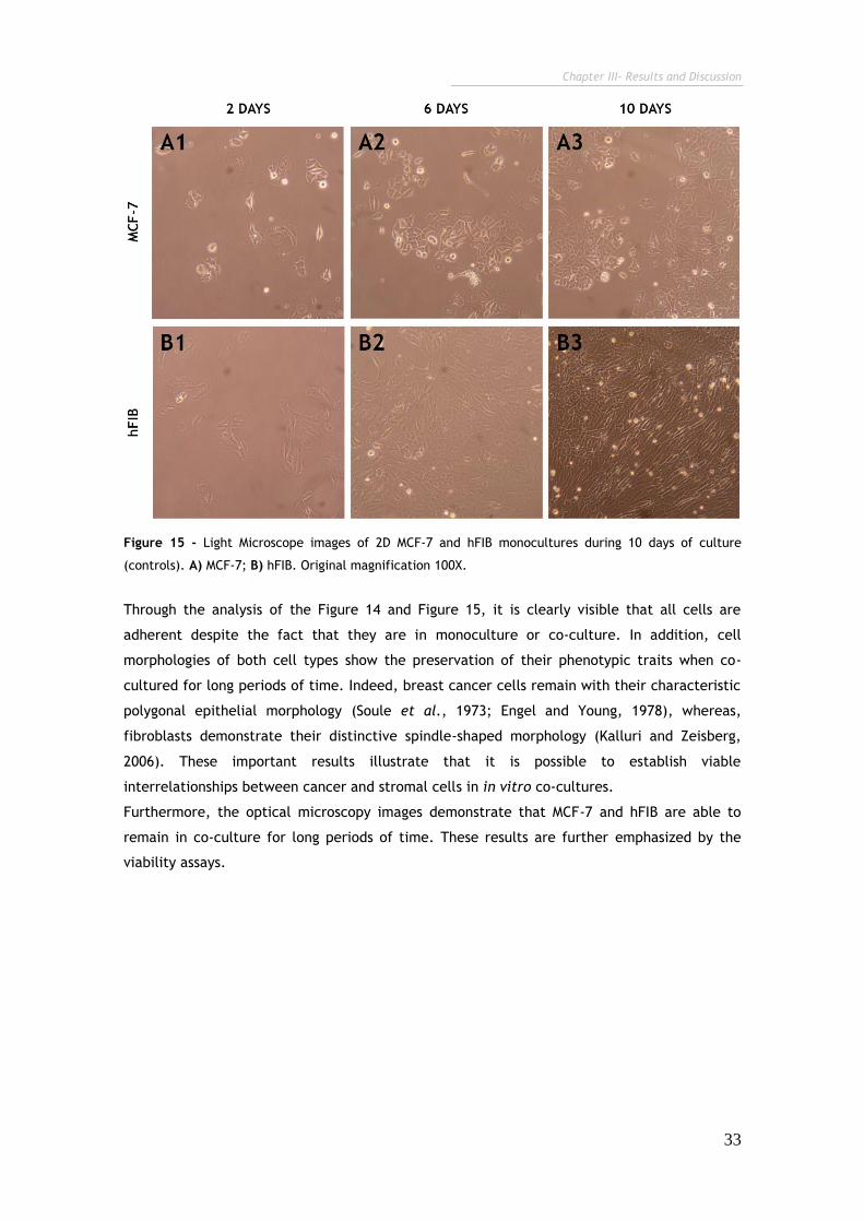
Chapter III- Results and Discussion
33
Figure 15 - Light Microscope images of 2D MCF-7 and hFIB monocultures during 10 days of culture
(controls). A) MCF-7; B) hFIB. Original magnification 100X.
Through the analysis of the Figure 14 and Figure 15, it is clearly visible that all cells are
adherent despite the fact that they are in monoculture or co-culture. In addition, cell
morphologies of both cell types show the preservation of their phenotypic traits when co-
cultured for long periods of time. Indeed, breast cancer cells remain with their characteristic
polygonal epithelial morphology (Soule et al., 1973; Engel and Young, 1978), whereas,
fibroblasts demonstrate their distinctive spindle-shaped morphology (Kalluri and Zeisberg,
2006). These important results illustrate that it is possible to establish viable
interrelationships between cancer and stromal cells in in vitro co-cultures.
Furthermore, the optical microscopy images demonstrate that MCF-7 and hFIB are able to
remain in co-culture for long periods of time. These results are further emphasized by the
viability assays.
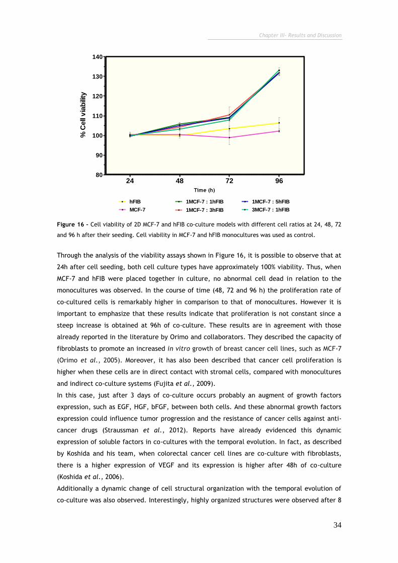
Chapter III- Results and Discussion
34
24 48 72 9680
90
100
110
120
130
140
hFIB
MCF-7
1MCF-7 : 1hFIB
1MCF-7 : 3hFIB
1MCF-7 : 5hFIB
3MCF-7 : 1hFIB
Time (h)
% C
ell
via
bili
ty
Figure 16 – Cell viability of 2D MCF-7 and hFIB co-culture models with different cell ratios at 24, 48, 72
and 96 h after their seeding. Cell viability in MCF-7 and hFIB monocultures was used as control.
Through the analysis of the viability assays shown in Figure 16, it is possible to observe that at
24h after cell seeding, both cell culture types have approximately 100% viability. Thus, when
MCF-7 and hFIB were placed together in culture, no abnormal cell dead in relation to the
monocultures was observed. In the course of time (48, 72 and 96 h) the proliferation rate of
co-cultured cells is remarkably higher in comparison to that of monocultures. However it is
important to emphasize that these results indicate that proliferation is not constant since a
steep increase is obtained at 96h of co-culture. These results are in agreement with those
already reported in the literature by Orimo and collaborators. They described the capacity of
fibroblasts to promote an increased in vitro growth of breast cancer cell lines, such as MCF-7
(Orimo et al., 2005). Moreover, it has also been described that cancer cell proliferation is
higher when these cells are in direct contact with stromal cells, compared with monocultures
and indirect co-culture systems (Fujita et al., 2009).
In this case, just after 3 days of co-culture occurs probably an augment of growth factors
expression, such as EGF, HGF, bFGF, between both cells. And these abnormal growth factors
expression could influence tumor progression and the resistance of cancer cells against anti-
cancer drugs (Straussman et al., 2012). Reports have already evidenced this dynamic
expression of soluble factors in co-cultures with the temporal evolution. In fact, as described
by Koshida and his team, when colorectal cancer cell lines are co-culture with fibroblasts,
there is a higher expression of VEGF and its expression is higher after 48h of co-culture
(Koshida et al., 2006).
Additionally a dynamic change of cell structural organization with the temporal evolution of
co-culture was also observed. Interestingly, highly organized structures were observed after 8
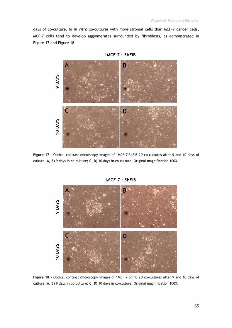
Chapter III- Results and Discussion
35
days of co-culture. In in vitro co-cultures with more stromal cells than MCF-7 cancer cells,
MCF-7 cells tend to develop agglomerates surrounded by fibroblasts, as demonstrated in
Figure 17 and Figure 18.
Figure 17 – Optical contrast microscopy images of 1MCF-7:3hFIB 2D co-cultures after 9 and 10 days of
culture. A, B) 9 days in co-culture; C, D) 10 days in co-culture. Original magnification 100X.
Figure 18 – Optical contrast microscopy images of 1MCF-7:5hFIB 2D co-cultures after 9 and 10 days of
culture. A, B) 9 days in co-culture; C, D) 10 days in co-culture. Original magnification 100X.
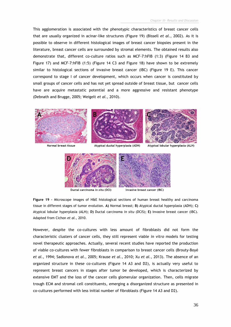
Chapter III- Results and Discussion
36
This agglomeration is associated with the phenotypic characteristics of breast cancer cells
that are usually organized in acinar-like structures (Figure 19) (Bissell et al., 2002). As it is
possible to observe in different histological images of breast cancer biopsies present in the
literature, breast cancer cells are surrounded by stromal elements. The obtained results also
demonstrate that, different co-culture ratios such as MCF-7:hFIB (1:3) (Figure 14 B3 and
Figure 17) and MCF-7:hFIB (1:5) (Figure 14 C3 and Figure 18) have shown to be extremely
similar to histological sections of invasive breast cancer (IBC) (Figure 19 E). This cancer
correspond to stage I of cancer development, which occurs when cancer is constituted by
small groups of cancer cells and has not yet spread outside of breast tissue, but cancer cells
have are acquire metastatic potential and a more aggressive and resistant phenotype
(Debnath and Brugge, 2005; Weigelt et al., 2010).
Figure 19 – Microscope images of H&E histological sections of human breast healthy and carcinoma
tissue in different stages of tumor evolution. A) Normal breast; B) Atypical ductal hyperplasia (ADH); C)
Atypical lobular hyperplasia (ALH); D) Ductal carcinoma in situ (DCIS); E) Invasive breast cancer (IBC).
Adapted from Cichon et al., 2010.
However, despite the co-cultures with less amount of fibroblasts did not form the
characteristic clusters of cancer cells, they still represent viable in vitro models for testing
novel therapeutic approaches. Actually, several recent studies have reported the production
of viable co-cultures with fewer fibroblasts in comparison to breast cancer cells (Brouty-Boyé
et al., 1994; Sadlonova et al., 2005; Krause et al., 2010; Xu et al., 2013). The absence of an
organized structure in these co-cultures (Figure 14 A3 and D2), is actually very useful to
represent breast cancers in stages after tumor be developed, which is characterized by
extensive EMT and the loss of the cancer cells glomerular organization. Then, cells migrate
trough ECM and stromal cell constituents, emerging a disorganized structure as presented in
co-cultures performed with less initial number of fibroblasts (Figure 14 A3 and D2).
Chapter III- Results and Discussion
37
Therefore, all the co-cultures systems developed represent viable systems that mimic the
heterogeneity found in breast cancers, a parameter that could not be reproduced in MCF-7 or
hFIB monocultures (Figure 15). Moreover, the obtained results also demonstrate that stromal
fibroblasts cells have an effect in the spatiotemporal organization of the cultures, and also,
influence malignant cell proliferation throughout time. Such was not observed mono-cultured
cells, mostly due to the absence of inter-cellular signalling events (Miki et al., 2012).
Therefore, these interesting observations render the co-culture models produced during
relatively long periods of time a very attractive testing platform for screening novel drug
delivery systems or novel synthetic or natural compounds with possible anti-tumoral activity
in breast cancer cells.
3.1.2. Development and optimization of 2D in vitro cell co-culture models
of cervical cancer (HeLa:hFIB)
In addition, to the establishment of MCF-7 breast cancer in vitro 2D co-cultures a model of
co-cultures of cervix cancer was also developed. For this purpose HeLa cells were used as
model cancer cells and fibroblasts were used as stromal cells. The establishment of these
HeLa and hFIB co-cultures is very rare in the literature, with the studies concerning HeLa:hFIB
co-cultures dating back to the late 80s. Thus, there is a need to optimize the development of
HeLa co-cultures systems in order to evaluate new drugs and delivery systems that are
designed for targeted therapy of cervix cancer. Thus, similarly to breast cancer co-cultures at
various ratios of HeLa to hFIB that were previously reported in the literature were used: i)
1:1; ii) 1:2; iii) 1:10 and iv) 2:1 (Delinassios and Kottaridis, 1984; Delinassios, 1987). However,
it should emphasized that although fibroblasts are generally grown in DMEM-F12 complete
medium, for the production of these particular co-cultures cells were maintained in the same
medium used for HeLa in vitro cultures (DMEM HG). This is an important parameter in the
experimental design of different co-cultures and should be carefully addressed since it could
ultimately affect cell viability. However, as the results demonstrate, despite this fact, hFIB
cells self-adapted to the new environmental conditions imposed by high glucose, remaning
viable along the time of the experiments, as shown in Figure 15 and 16. These results are
corroborated by those also reported by Wang and co-workers, 2013, that also grew hFIB in
high glucose culture mediums in vitro without deleterious reduction of cell viability (Wang et
al., 2013a).
Similarly to MCF-7 co-cultures, HeLa:hFIB co-cultures were also performed in an extended
time scale. The resultant light microscopy images of these co-cultures models are shown in
Figure 20.
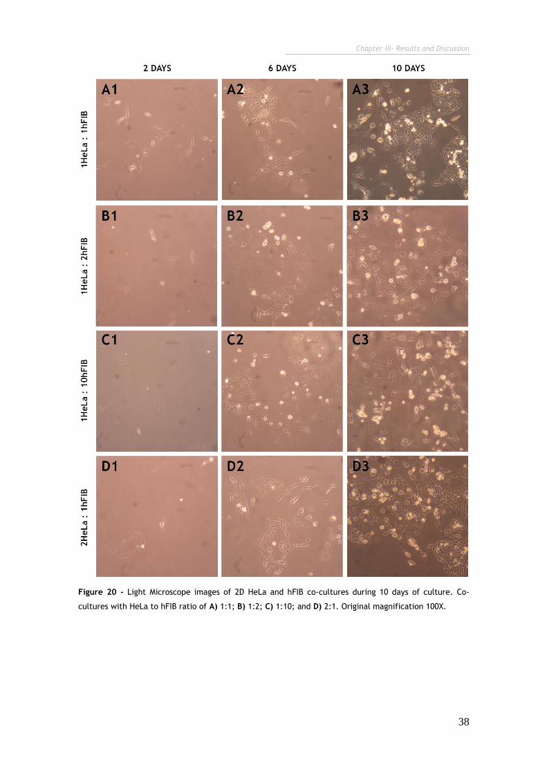
Chapter III- Results and Discussion
38
Figure 20 - Light Microscope images of 2D HeLa and hFIB co-cultures during 10 days of culture. Co-
cultures with HeLa to hFIB ratio of A) 1:1; B) 1:2; C) 1:10; and D) 2:1. Original magnification 100X.

Chapter III- Results and Discussion
39
Figure 21 - Light Microscope images of 2D HeLa and hFIB monocultures during 10 days of culture
(controls). A) HeLa; B) hFIB. Original magnification 100X.
Trough the analysis of Figure 21 it is possible to observe that the co-cultivation of human
fibroblasts and HeLa cells in vitro leads to the development of a unique cellular organization,
in comparison to their monocultured counterparts. In addition, in all HeLa:hFIB co-culture
models cytoplasmatic projections, such as fillopodium structures between cells are evidenced
in co-cultures with more fibroblasts than HeLa cervical cancer cells (Figure 22).
Figure 22 – Inverted Light Microscope images of fillopodium structures (arrows) of HeLa and Fibroblasts
cells after 10 days of co-culture. Co-cultures with HeLa to hFIB ratio of A) 1:1; B) 1:2; C) 1:10; and D)
2:1. Original magnification 100X.
Interestingly, these HeLa:hFIB co-cultures with more fibroblasts than cancer cells
demonstrate a slightly cell dead when compared with homotypic cell cultures, has
demonstrated by the spherical shape cells present in Figure 20 B and C. In contrast, in co-
cultures with higher concentrations of HeLa cells (Hela:hFIB co-cultures with 1:1 (Figure 20 A)
Chapter III- Results and Discussion
40
and 2:1 (Figure 20 D)), cells present a more agglomerated distribution, similarity to that
observed for monocultures (Figure 21).
3.1.3. Evaluation of CH-H-R/pDNA nanoparticles cellular uptake in 2D
breast cancer co-culture models (MCF-7:hFIB)
In order to take advantage of the co-culture models that were previously established, they
were used to test the biological performance and cancer cell targeting capacity of a gene
delivery system comprised of CH-H-R/pDNA that has been previously synthesized in our
research group (Gaspar et al., 2013). For this purpose breast cancer co-cultures were chosen
as in vitro cell models since this particular gene delivery system is envisioned to be applied
on breast cancer therapy in a near future.
Initially, the in vitro cellular uptake of the nanoparticles was analyzed by CLSM. These
polymeric nanoparticles are formed by chitosan, a biocompatible polymer with positive
charge that has the ability to encapsulate therapeutic nucleic acids (Borchard, 2001).
Moreover, to increase the biological activity and selectivity of the system to cancer cells the
polymeric backbone of chitosan was also conjugated with two functional and bioinspired
ligands, namely, arginine and histidine (Gaspar et al., 2013). As described in the literature,
Arginine allows the establishment of electrostatic interactions with the characteristic
negatively charged cancer cell membranes (Nakase et al., 2012), thus, the use of Ariginine
ligands in nanoparticles leads to an increased penetration of nanoparticles trough cellular
membrane. Apart from grafting arginine into the chitosan polymeric backbone, since histidine
pKa is in the range of the endosomal/lysosomal pH endows chitosan with improved buffering
capacity and subsequently, with an improved endosomal release capacity mediated by the
proton sponge effect.
This escape of nanoparticles is essential for the improvement of their therapeutic efficiency,
since a large number of nanoparticles is destroyed in these compartments (Wu et al., 2012).
These important features could be responsible for the tendency of these nanodevices to
target cancer cells, improving in this way, the cellular uptake of this delivery system in
different co-culture models.
This specific targeting capacity of nanoparticles could be tested in co-cultures, in fact these
platforms are considered the perfect tool to analyse the targeting specificity of drug and
delivery systems designed for tumor therapy (Le Droumaguet et al., 2012).
Therefore, these nanoparticles were tested the different breast cancer co-culture ratios to
verify if these nanoparticles are specific to cancer cells and if this possible targeting
specificity is maintained or is influenced by the heterogeneous models developed.
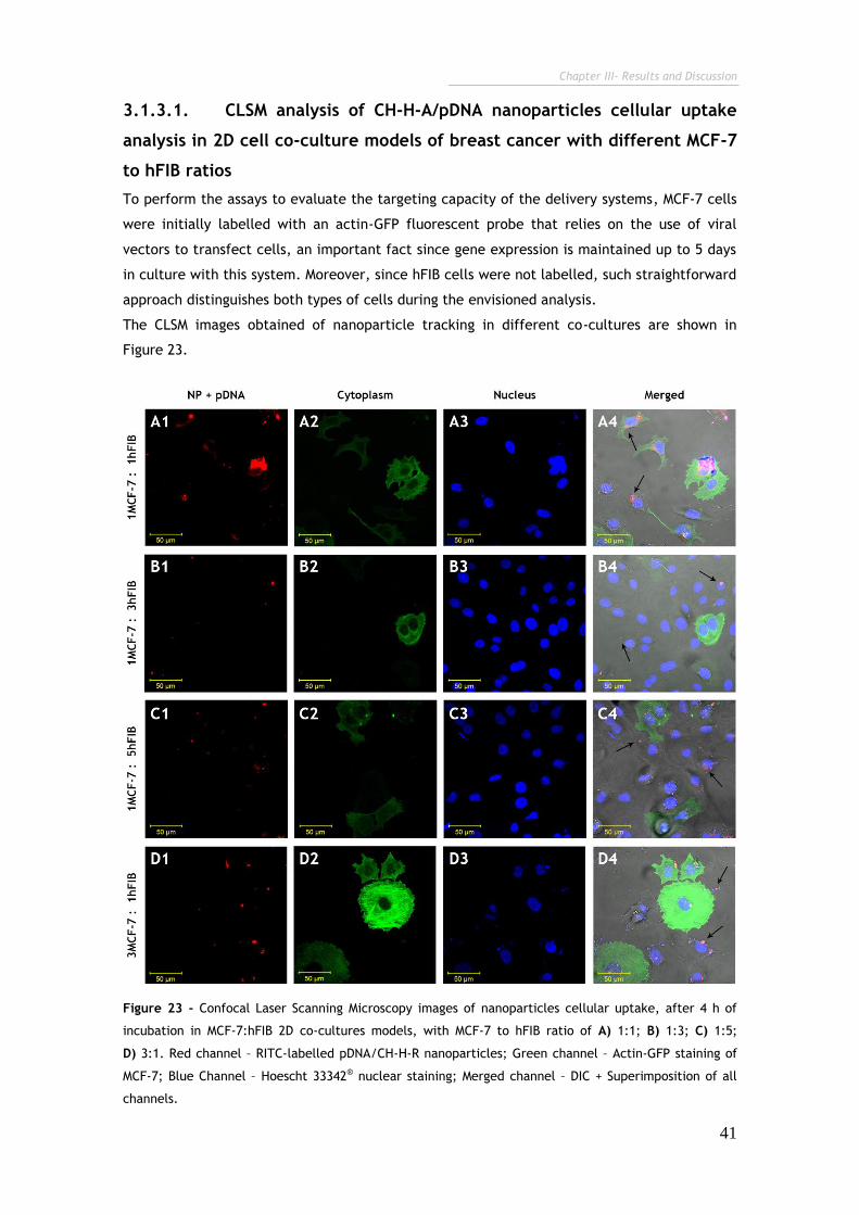
Chapter III- Results and Discussion
41
3.1.3.1. CLSM analysis of CH-H-A/pDNA nanoparticles cellular uptake
analysis in 2D cell co-culture models of breast cancer with different MCF-7
to hFIB ratios
To perform the assays to evaluate the targeting capacity of the delivery systems, MCF-7 cells
were initially labelled with an actin-GFP fluorescent probe that relies on the use of viral
vectors to transfect cells, an important fact since gene expression is maintained up to 5 days
in culture with this system. Moreover, since hFIB cells were not labelled, such straightforward
approach distinguishes both types of cells during the envisioned analysis.
The CLSM images obtained of nanoparticle tracking in different co-cultures are shown in
Figure 23.
Figure 23 - Confocal Laser Scanning Microscopy images of nanoparticles cellular uptake, after 4 h of
incubation in MCF-7:hFIB 2D co-cultures models, with MCF-7 to hFIB ratio of A) 1:1; B) 1:3; C) 1:5;
D) 3:1. Red channel – RITC-labelled pDNA/CH-H-R nanoparticles; Green channel – Actin-GFP staining of
MCF-7; Blue Channel – Hoescht 33342® nuclear staining; Merged channel – DIC + Superimposition of all
channels.
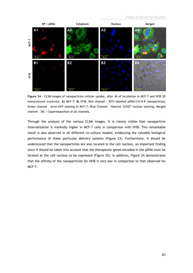
Chapter III- Results and Discussion
42
Figure 24 – CLSM images of nanoparticles cellular uptake, after 4h of incubation in MCF-7 and hFIB 2D
monocultures (controls). A) MCF-7; B) hFIB. Red channel – RITC-labelled pDNA/CH-H-R nanoparticles;
Green channel – Actin-GFP staining of MCF-7; Blue Channel – Hoechst 33342® nuclear staining; Merged
channel – DIC + Superimposition of all channels.
Through the analysis of the various CLSM images, it is clearly visible that nanoparticle
internalization is markedly higher in MCF-7 cells in comparison with hFIB. This remarkable
result is also observed in all different co-culture models, evidencing the valuable biological
performance of these particular delivery systems (Figure 23). Furthermore, it should be
underscored that the nanoparticles are also located in the cell nucleus, an important finding
since it should be taken into account that the therapeutic genes encoded in the pDNA must be
located at the cell nucleus to be expressed (Figure 25). In addition, Figure 24 demonstrates
that the affinity of the nanoparticles for hFIB is very low in comparison to that observed for
MCF-7.

Chapter III- Results and Discussion
43
Figure 25 - CLSM images co-cultures at 1MCF-7:1hFIB ratio for nanoparticles cellular localization
analysis. CLSM images of MCF-7 breast cancer cells after 4 h of incubation with nanocarriers (A, B),
orthogonal sectioning in xy axis (B), 3D reconstruction of the cell nucleus (C). Colocalization of the red
and green channels (D). Colocalization of the red and blue channels (E). Red channel – RITC labelled
pDNA/CH-H-R; Green channel – Actin-GFP staining of MCF-7 and hFIB; Blue Channel – Hoechst 33342®
nuclear staining. Grey channels: colocalization analysis.
Chapter III- Results and Discussion
44
In general, these results suggest that this particular nanodevice possesses tumor selectivity, a
valuable characteristic that is most likely correlated with chitosan surface amino acid
functionalization as previously mentioned. Nevertheless, in order to further confirm this
targeting specificity a population based approach was also performed through the use of flow
cytometry. This analysis is crucial since the established co-culture models are highly diverse
and CLSM imaging only provide information about of the cellular events that occur in the field
of view, rather than in the all population of co-cultured cells, in the various ratios.
3.1.3.2. Flow cytometry analysis of CH-H-A/pDNA nanoparticles cellular
uptake analysis in 2D cell co-culture models of breast cancer with different
MCF-7 to hFIB ratios
Flow cytometry is a very sensible technique that measures cell parameters under dynamic
flow conditions mainly through the analysis of fluorescence (Ormerod and Imrie, 1990). This
fact allows a careful and precise analysis of nanoparticles uptake in the various co-cultures
with different MCF-7:hFIB ratios. Actually, flow cytometry has been widely used for the
evaluation of nanoparticle uptake in in vitro cell models, however its use in co-culture
analysis has only recently been described (Kievit et al., 2009; Wang et al., 2013b),
The initial transfection of cancer cells with GFP is an important parameter since it allows the
separation of MCF-7 and hFIB populations, by gating the region of interest (ROI) in the GFP
channel as shown in the R2 quadrant of (Figure 26). Thus, since the nanocarriers are labelled
with red colour, the cells that are simultaneously GFP positive (i.e. green) and Rhodamine
positive (i.e. red) are identified as MCF-7cells, with internalized nanoparticles. On the
contrary green fluorescent cells represent cancer cells without internalized nanoparticles. In
respect to hFIB, only those cells that present red fluorescence have the nanoparticulated
systems in the intracellular space.
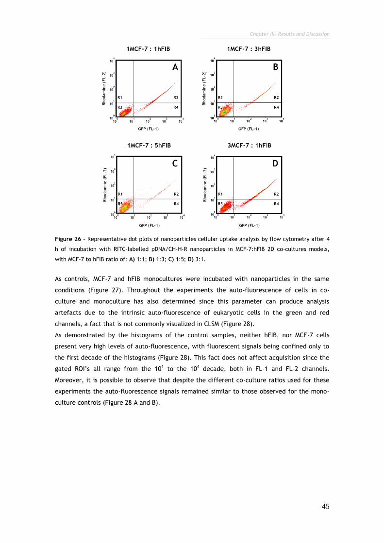
Chapter III- Results and Discussion
45
Figure 26 – Representative dot plots of nanoparticles cellular uptake analysis by flow cytometry after 4
h of incubation with RITC-labelled pDNA/CH-H-R nanoparticles in MCF-7:hFIB 2D co-cultures models,
with MCF-7 to hFIB ratio of: A) 1:1; B) 1:3; C) 1:5; D) 3:1.
As controls, MCF-7 and hFIB monocultures were incubated with nanoparticles in the same
conditions (Figure 27). Throughout the experiments the auto-fluorescence of cells in co-
culture and monoculture has also determined since this parameter can produce analysis
artefacts due to the intrinsic auto-fluorescence of eukaryotic cells in the green and red
channels, a fact that is not commonly visualized in CLSM (Figure 28).
As demonstrated by the histograms of the control samples, neither hFIB, nor MCF-7 cells
present very high levels of auto-fluorescence, with fluorescent signals being confined only to
the first decade of the histograms (Figure 28). This fact does not affect acquisition since the
gated ROI’s all range from the 101 to the 104 decade, both in FL-1 and FL-2 channels.
Moreover, it is possible to observe that despite the different co-culture ratios used for these
experiments the auto-fluorescence signals remained similar to those observed for the mono-
culture controls (Figure 28 A and B).
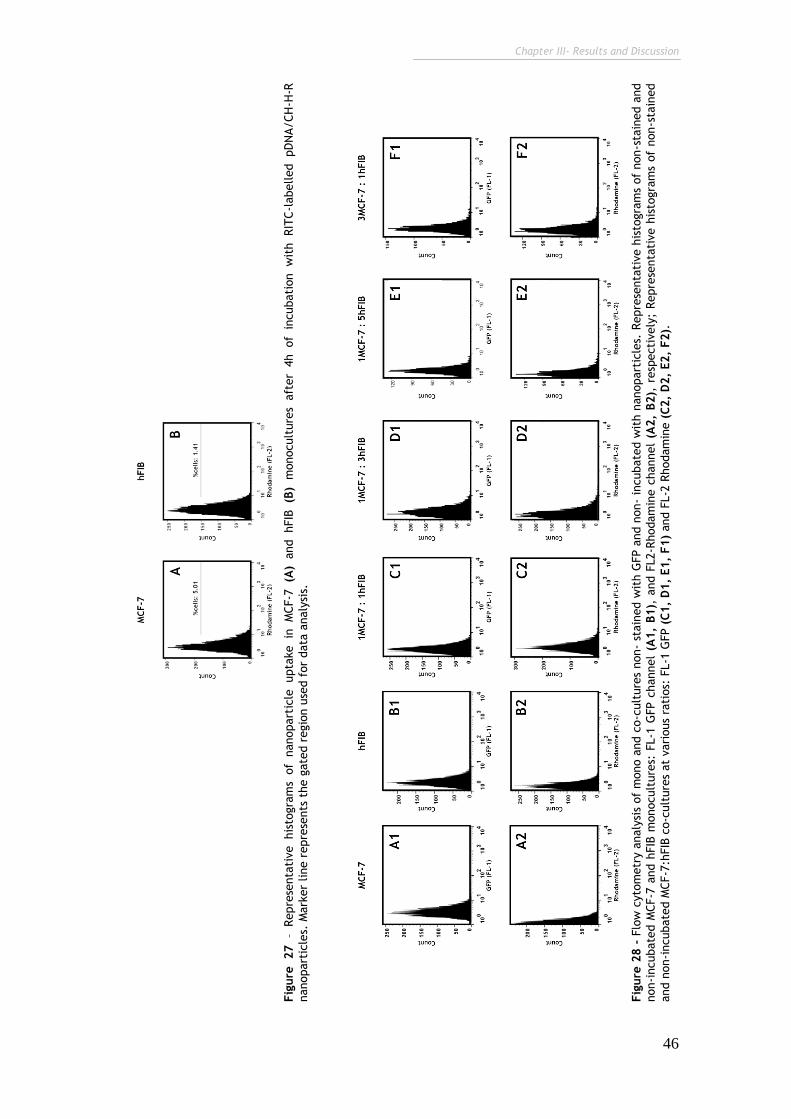
Chapter III- Results and Discussion
46
Fig
ure
28 -
Flo
w c
yto
metr
y a
naly
sis
of
mono a
nd c
o-c
ult
ure
s non-
stain
ed w
ith G
FP a
nd n
on-
incubate
d w
ith n
anopart
icle
s. R
epre
senta
tive h
isto
gra
ms
of
non-s
tain
ed a
nd
non-i
ncubate
d M
CF-7
and h
FIB
monocult
ure
s: F
L-1
GFP c
hannel
(A1,
B1),
and F
L2-R
hodam
ine c
hannel
(A2,
B2),
resp
ecti
vely
; Repre
senta
tive h
isto
gra
ms
of
non-s
tain
ed
and n
on-i
ncubate
d M
CF-7
:hFIB
co-c
ult
ure
s at
vari
ous
rati
os:
FL-1
GFP (
C1,
D1,
E1,
F1)
and F
L-2
Rhodam
ine (
C2,
D2,
E2,
F2).
Fig
ure
27
– Repre
senta
tive his
togra
ms
of
nanopart
icle
upta
ke in
M
CF-7
(A
) and hFIB
(B
) m
onocult
ure
s aft
er
4h of
incubati
on w
ith RIT
C-l
abelled pD
NA/CH
-H-R
nanopart
icle
s. M
ark
er
line r
epre
sents
the g
ate
d r
egio
n u
sed f
or
data
analy
sis.
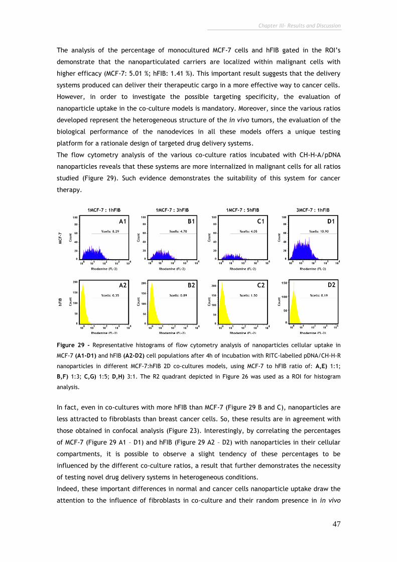
Chapter III- Results and Discussion
47
The analysis of the percentage of monocultured MCF-7 cells and hFIB gated in the ROI’s
demonstrate that the nanoparticulated carriers are localized within malignant cells with
higher efficacy (MCF-7: 5.01 %; hFIB: 1.41 %). This important result suggests that the delivery
systems produced can deliver their therapeutic cargo in a more effective way to cancer cells.
However, in order to investigate the possible targeting specificity, the evaluation of
nanoparticle uptake in the co-culture models is mandatory. Moreover, since the various ratios
developed represent the heterogeneous structure of the in vivo tumors, the evaluation of the
biological performance of the nanodevices in all these models offers a unique testing
platform for a rationale design of targeted drug delivery systems.
The flow cytometry analysis of the various co-culture ratios incubated with CH-H-A/pDNA
nanoparticles reveals that these systems are more internalized in malignant cells for all ratios
studied (Figure 29). Such evidence demonstrates the suitability of this system for cancer
therapy.
Figure 29 - Representative histograms of flow cytometry analysis of nanoparticles cellular uptake in
MCF-7 (A1-D1) and hFIB (A2-D2) cell populations after 4h of incubation with RITC-labelled pDNA/CH-H-R
nanoparticles in different MCF-7:hFIB 2D co-cultures models, using MCF-7 to hFIB ratio of: A,E) 1:1;
B,F) 1:3; C,G) 1:5; D,H) 3:1. The R2 quadrant depicted in Figure 26 was used as a ROI for histogram
analysis.
In fact, even in co-cultures with more hFIB than MCF-7 (Figure 29 B and C), nanoparticles are
less attracted to fibroblasts than breast cancer cells. So, these results are in agreement with
those obtained in confocal analysis (Figure 23). Interestingly, by correlating the percentages
of MCF-7 (Figure 29 A1 – D1) and hFIB (Figure 29 A2 – D2) with nanoparticles in their cellular
compartments, it is possible to observe a slight tendency of these percentages to be
influenced by the different co-culture ratios, a result that further demonstrates the necessity
of testing novel drug delivery systems in heterogeneous conditions.
Indeed, these important differences in normal and cancer cells nanoparticle uptake draw the
attention to the influence of fibroblasts in co-culture and their random presence in in vivo
Chapter III- Results and Discussion
48
tumors. Further analysis also shows that the same delivery system may behave in a different
mode according to the tumor heterogeneity that is found, i.e., depends on the MCF-7 to hFIB
ratios.
Therefore, addressing different cancer to stromal cell ratios is a critical requirement to
properly mimic heterogeneity and investigate the biological efficiency and specificity of a
nanocarrier system under variable tumor environments. It should be underscored that the
recent reports that employ co-culture models to evaluate the biological efficiency of
nanodevices, disregard the possible uncertainty of the cell populations present in a tumor
microenvironment (Le Droumaguet et al., 2012), and thus do not utterly explore the full
potential of co-cultures, as demonstrated in this thesis.
Nevertheless, despite the major improvements obtained with the various co-culture testing
platforms they are still rather limited by their 2D architecture which does not entirely mimic
the complexity of in vivo solid tumors. Therefore, in addition to the 2D systems also 3D
models of multi-cellular tumors were developed.
3.2. 3D in vitro cell co-culture models of breast cancer and
cervical cancer
Solid tumors are an abnormal mass of tissue that usually does not contain cysts or liquid areas
(Gavhane et al., 2011). Particularly, breast and cervix solid cancers originate from epithelial
cells (Elenbaas et al., 2001), more specifically, from the epithelial cells of glandular tissue,
forming the so termed adenocarcinomas (Weigelt et al., 2010; Ibeanu, 2011). Since the cell
lines used in this research were obtained from tumor sections of human breast and cervix
cancers they could have the capacity to form 3D multicellular tumor spheroids (MCTS) in
vitro, as they do in vivo (Harris, 2002; Jögi et al., 2012).
Similarly to co-cultures, in vitro 3D cell models which mimic solid tumors are essential in the
pre-clinical development of new drugs and novel delivery systems for cancer therapy.
However, unlike 2D cultures, 3D MCTS provide a whole new level of complexity since they
represent a suitable testing platform that reproduces the complex multicellular architecture,
the barriers to mass transport and the extracellular matrix deposition found in malignant
tissues (Minchinton and Tannock, 2006).
Hence, after the development of 2D co-cultures, MCF-7 and hFIB cells and fibroblasts were
also used in the optimization and establishment of 3D MCTS.
For the development of 3D models, it is required that adherent cells are seeded in various
substrates that maintain the cells in a non adherent state, in order to form spheroids with a
3D architecture. Examples of such substrates are: i) Hidrogels, ii) Scaffolds, iii) Microspheres,
or iv) Fibers that mimic the physical structure and chemical proprieties of the ECM, in a 3D
volume (Yamada and Cukierman, 2007; Justice et al., 2009; Rimann and Graf-Hausner, 2012).
An essential mechanism for spheroid assembly is the induction of cell-cell attachment and
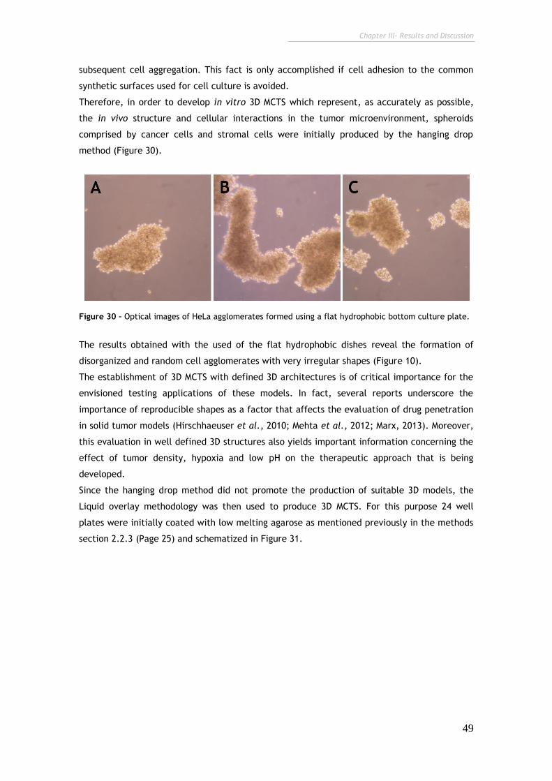
Chapter III- Results and Discussion
49
subsequent cell aggregation. This fact is only accomplished if cell adhesion to the common
synthetic surfaces used for cell culture is avoided.
Therefore, in order to develop in vitro 3D MCTS which represent, as accurately as possible,
the in vivo structure and cellular interactions in the tumor microenvironment, spheroids
comprised by cancer cells and stromal cells were initially produced by the hanging drop
method (Figure 30).
Figure 30 – Optical images of HeLa agglomerates formed using a flat hydrophobic bottom culture plate.
The results obtained with the used of the flat hydrophobic dishes reveal the formation of
disorganized and random cell agglomerates with very irregular shapes (Figure 10).
The establishment of 3D MCTS with defined 3D architectures is of critical importance for the
envisioned testing applications of these models. In fact, several reports underscore the
importance of reproducible shapes as a factor that affects the evaluation of drug penetration
in solid tumor models (Hirschhaeuser et al., 2010; Mehta et al., 2012; Marx, 2013). Moreover,
this evaluation in well defined 3D structures also yields important information concerning the
effect of tumor density, hypoxia and low pH on the therapeutic approach that is being
developed.
Since the hanging drop method did not promote the production of suitable 3D models, the
Liquid overlay methodology was then used to produce 3D MCTS. For this purpose 24 well
plates were initially coated with low melting agarose as mentioned previously in the methods
section 2.2.3 (Page 25) and schematized in Figure 31.
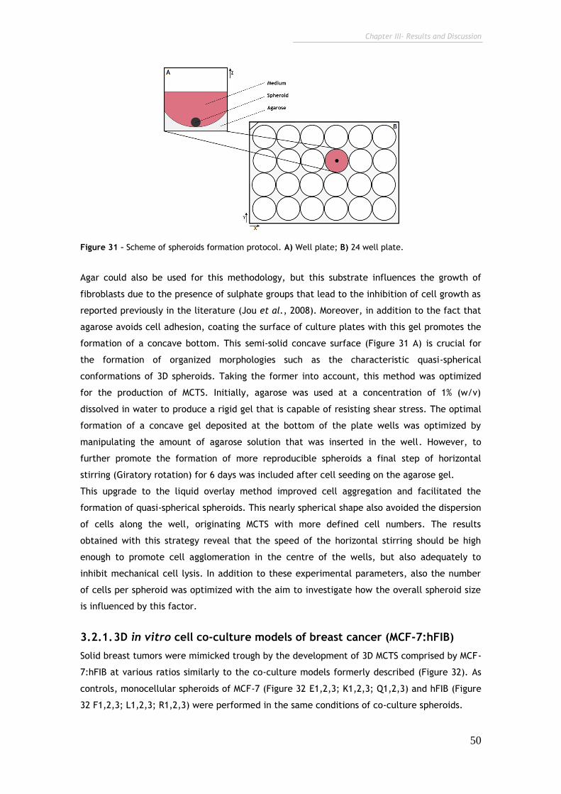
Chapter III- Results and Discussion
50
Figure 31 – Scheme of spheroids formation protocol. A) Well plate; B) 24 well plate.
Agar could also be used for this methodology, but this substrate influences the growth of
fibroblasts due to the presence of sulphate groups that lead to the inhibition of cell growth as
reported previously in the literature (Jou et al., 2008). Moreover, in addition to the fact that
agarose avoids cell adhesion, coating the surface of culture plates with this gel promotes the
formation of a concave bottom. This semi-solid concave surface (Figure 31 A) is crucial for
the formation of organized morphologies such as the characteristic quasi-spherical
conformations of 3D spheroids. Taking the former into account, this method was optimized
for the production of MCTS. Initially, agarose was used at a concentration of 1% (w/v)
dissolved in water to produce a rigid gel that is capable of resisting shear stress. The optimal
formation of a concave gel deposited at the bottom of the plate wells was optimized by
manipulating the amount of agarose solution that was inserted in the well. However, to
further promote the formation of more reproducible spheroids a final step of horizontal
stirring (Giratory rotation) for 6 days was included after cell seeding on the agarose gel.
This upgrade to the liquid overlay method improved cell aggregation and facilitated the
formation of quasi-spherical spheroids. This nearly spherical shape also avoided the dispersion
of cells along the well, originating MCTS with more defined cell numbers. The results
obtained with this strategy reveal that the speed of the horizontal stirring should be high
enough to promote cell agglomeration in the centre of the wells, but also adequately to
inhibit mechanical cell lysis. In addition to these experimental parameters, also the number
of cells per spheroid was optimized with the aim to investigate how the overall spheroid size
is influenced by this factor.
3.2.1. 3D in vitro cell co-culture models of breast cancer (MCF-7:hFIB)
Solid breast tumors were mimicked trough by the development of 3D MCTS comprised by MCF-
7:hFIB at various ratios similarly to the co-culture models formerly described (Figure 32). As
controls, monocellular spheroids of MCF-7 (Figure 32 E1,2,3; K1,2,3; Q1,2,3) and hFIB (Figure
32 F1,2,3; L1,2,3; R1,2,3) were performed in the same conditions of co-culture spheroids.
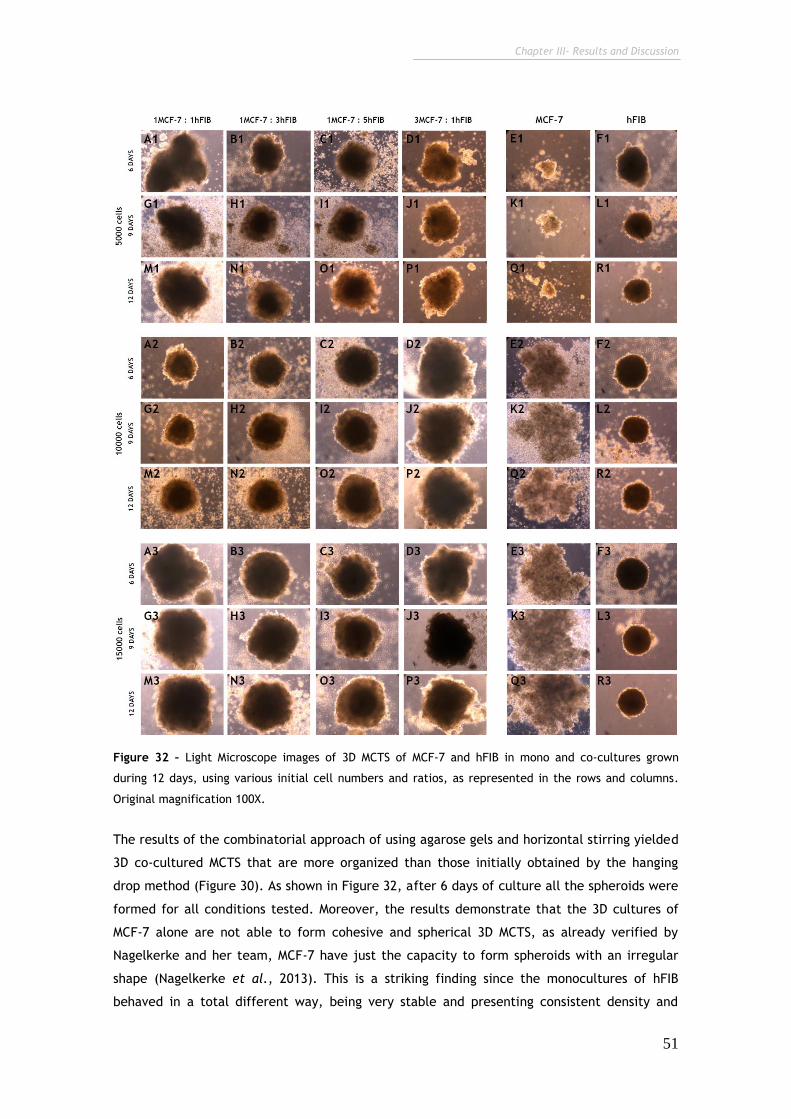
Chapter III- Results and Discussion
51
Figure 32 – Light Microscope images of 3D MCTS of MCF-7 and hFIB in mono and co-cultures grown
during 12 days, using various initial cell numbers and ratios, as represented in the rows and columns.
Original magnification 100X.
The results of the combinatorial approach of using agarose gels and horizontal stirring yielded
3D co-cultured MCTS that are more organized than those initially obtained by the hanging
drop method (Figure 30). As shown in Figure 32, after 6 days of culture all the spheroids were
formed for all conditions tested. Moreover, the results demonstrate that the 3D cultures of
MCF-7 alone are not able to form cohesive and spherical 3D MCTS, as already verified by
Nagelkerke and her team, MCF-7 have just the capacity to form spheroids with an irregular
shape (Nagelkerke et al., 2013). This is a striking finding since the monocultures of hFIB
behaved in a total different way, being very stable and presenting consistent density and
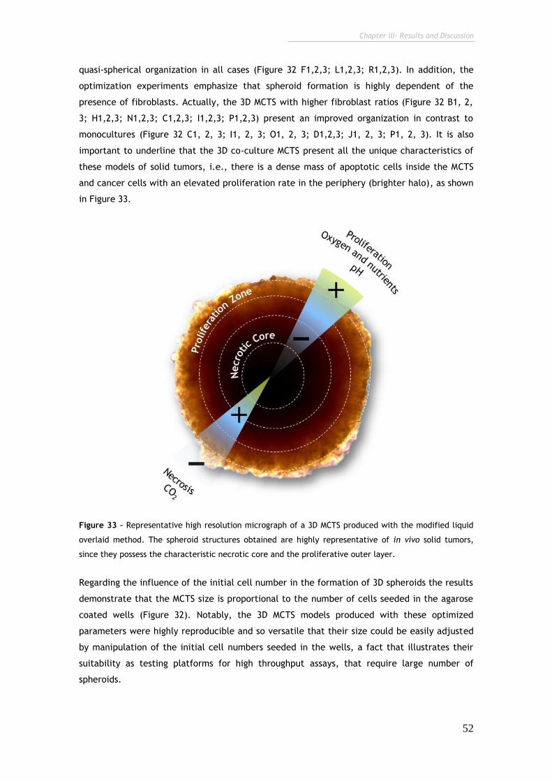
Chapter III- Results and Discussion
52
quasi-spherical organization in all cases (Figure 32 F1,2,3; L1,2,3; R1,2,3). In addition, the
optimization experiments emphasize that spheroid formation is highly dependent of the
presence of fibroblasts. Actually, the 3D MCTS with higher fibroblast ratios (Figure 32 B1, 2,
3; H1,2,3; N1,2,3; C1,2,3; I1,2,3; P1,2,3) present an improved organization in contrast to
monocultures (Figure 32 C1, 2, 3; I1, 2, 3; O1, 2, 3; D1,2,3; J1, 2, 3; P1, 2, 3). It is also
important to underline that the 3D co-culture MCTS present all the unique characteristics of
these models of solid tumors, i.e., there is a dense mass of apoptotic cells inside the MCTS
and cancer cells with an elevated proliferation rate in the periphery (brighter halo), as shown
in Figure 33.
Figure 33 – Representative high resolution micrograph of a 3D MCTS produced with the modified liquid
overlaid method. The spheroid structures obtained are highly representative of in vivo solid tumors,
since they possess the characteristic necrotic core and the proliferative outer layer.
Regarding the influence of the initial cell number in the formation of 3D spheroids the results
demonstrate that the MCTS size is proportional to the number of cells seeded in the agarose
coated wells (Figure 32). Notably, the 3D MCTS models produced with these optimized
parameters were highly reproducible and so versatile that their size could be easily adjusted
by manipulation of the initial cell numbers seeded in the wells, a fact that illustrates their
suitability as testing platforms for high throughput assays, that require large number of
spheroids.
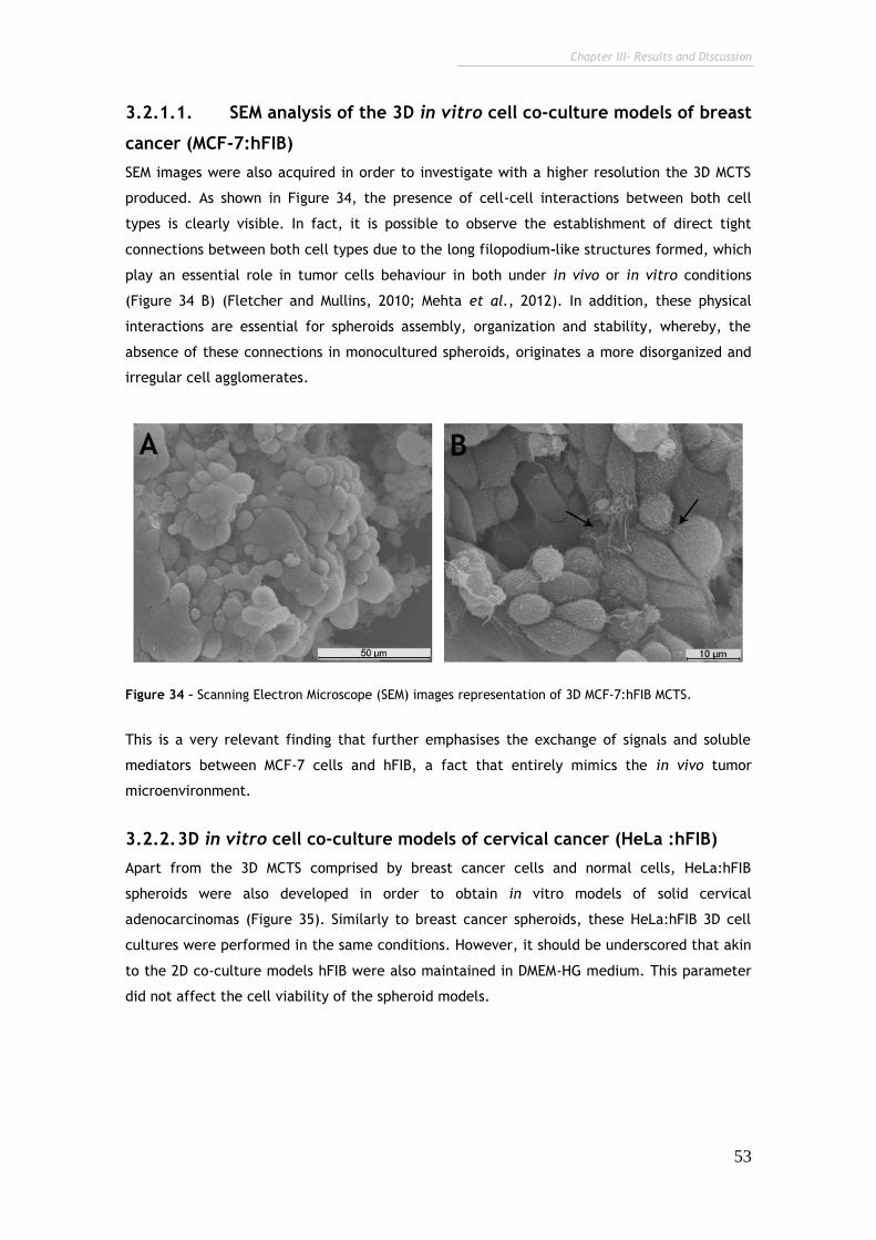
Chapter III- Results and Discussion
53
3.2.1.1. SEM analysis of the 3D in vitro cell co-culture models of breast
cancer (MCF-7:hFIB)
SEM images were also acquired in order to investigate with a higher resolution the 3D MCTS
produced. As shown in Figure 34, the presence of cell-cell interactions between both cell
types is clearly visible. In fact, it is possible to observe the establishment of direct tight
connections between both cell types due to the long filopodium-like structures formed, which
play an essential role in tumor cells behaviour in both under in vivo or in vitro conditions
(Figure 34 B) (Fletcher and Mullins, 2010; Mehta et al., 2012). In addition, these physical
interactions are essential for spheroids assembly, organization and stability, whereby, the
absence of these connections in monocultured spheroids, originates a more disorganized and
irregular cell agglomerates.
Figure 34 – Scanning Electron Microscope (SEM) images representation of 3D MCF-7:hFIB MCTS.
This is a very relevant finding that further emphasises the exchange of signals and soluble
mediators between MCF-7 cells and hFIB, a fact that entirely mimics the in vivo tumor
microenvironment.
3.2.2. 3D in vitro cell co-culture models of cervical cancer (HeLa :hFIB)
Apart from the 3D MCTS comprised by breast cancer cells and normal cells, HeLa:hFIB
spheroids were also developed in order to obtain in vitro models of solid cervical
adenocarcinomas (Figure 35). Similarly to breast cancer spheroids, these HeLa:hFIB 3D cell
cultures were performed in the same conditions. However, it should be underscored that akin
to the 2D co-culture models hFIB were also maintained in DMEM-HG medium. This parameter
did not affect the cell viability of the spheroid models.
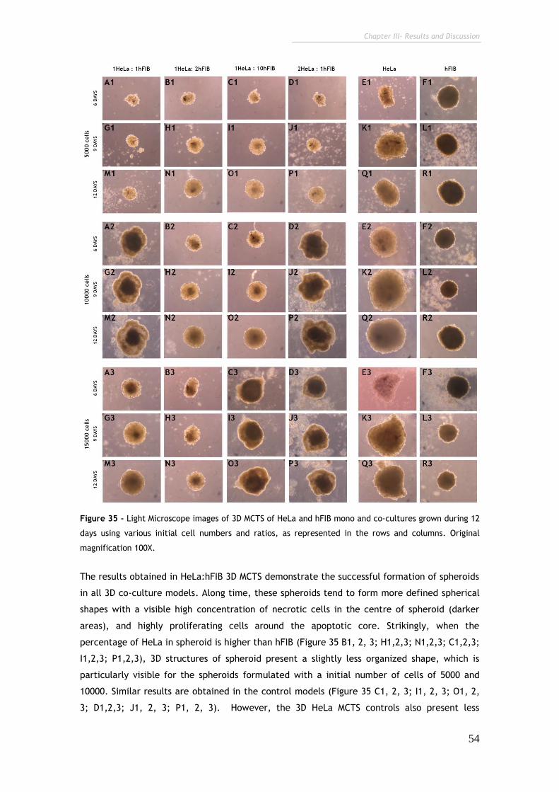
Chapter III- Results and Discussion
54
Figure 35 - Light Microscope images of 3D MCTS of HeLa and hFIB mono and co-cultures grown during 12
days using various initial cell numbers and ratios, as represented in the rows and columns. Original
magnification 100X.
The results obtained in HeLa:hFIB 3D MCTS demonstrate the successful formation of spheroids
in all 3D co-culture models. Along time, these spheroids tend to form more defined spherical
shapes with a visible high concentration of necrotic cells in the centre of spheroid (darker
areas), and highly proliferating cells around the apoptotic core. Strikingly, when the
percentage of HeLa in spheroid is higher than hFIB (Figure 35 B1, 2, 3; H1,2,3; N1,2,3; C1,2,3;
I1,2,3; P1,2,3), 3D structures of spheroid present a slightly less organized shape, which is
particularly visible for the spheroids formulated with a initial number of cells of 5000 and
10000. Similar results are obtained in the control models (Figure 35 C1, 2, 3; I1, 2, 3; O1, 2,
3; D1,2,3; J1, 2, 3; P1, 2, 3). However, the 3D HeLa MCTS controls also present less
Chapter III- Results and Discussion
55
organized shapes even for a lower initial cell density. These results are in agreement with
those obtained for MCF-7:hFIB spheroids and also emphasize the important role of fibroblasts
in the formation of these unique structures. Regarding, the hFIB control spheroids, they
present a highly consistent quasi-spherical morphology. HeLa 3D spheroids are particularly
interesting to analyze since the characteristic dense necrotic core is entirely absent on these
monoculture spheroids. This is a remarkable finding since it suggests that without the
presence of fibroblasts the 3D MCTS do not entirely mimic the in vivo solid tumors that are
characterized by their extensive necrotic cores with low oxygen and nutrient intake.
It is also important to emphasize that mono-culture spheroids are formed by the same initial
number of cells than the HeLa:hFIB MCTS, thus, the bigger size of HeLa spheroids could be a
result of a remarkably high proliferation rate.
3.2.2.1. CLSM analysis of 3D in vitro cell co-culture models of cervical
cancer (HeLa:hFIB)
In order to support the previous morphological analysis of the 3D MCTS developed and provide
further insights into the spheroids spatial architecture, 3D CLSM images with various slices
along the MCTS structures were acquired (Figure 36 and 37). For fluorescence imaging the
spheroids were labelled with WGA. This fluorescent probe is a carbohydrate-binding protein
that selectively recognizes sialic acid and N-acetylglucosaminyl sugar residues that are
predominantly found on the plasma membrane of cells (Favi et al., 2013).
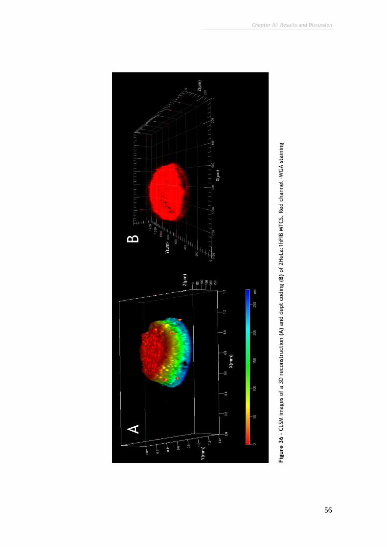
Chapter III- Results and Discussion
56
Fig
ure
36 –
CLSM
im
ages
of
a 3
D r
econst
ructi
on (
A)
and d
ept
codin
g (
B)
of
2H
eLa:1
hFIB
MTCS.
Red c
hannel –W
GA s
tain
ing
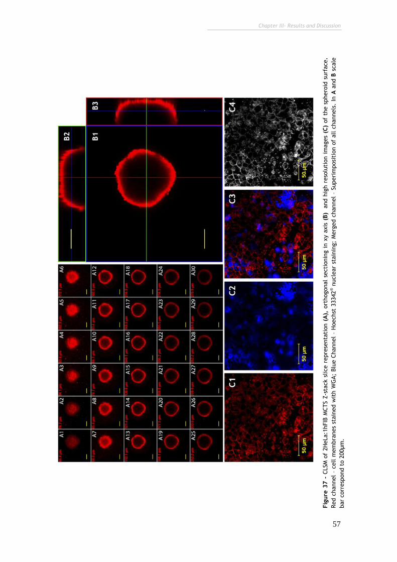
Chapter III- Results and Discussion
57
Fig
ure
37 -
CLSM
of
2H
eLa:1
hFIB
MCTS Z
-sta
ck s
lice r
epre
senta
tion (
A),
ort
hogonal
secti
onin
g i
n x
y a
xis
(B)
and h
igh r
eso
luti
on i
mages
(C)
of
the s
phero
id s
urf
ace.
Red c
hannel
– cell m
em
bra
nes
stain
ed w
ith W
GA;
Blu
e C
hannel
– H
oechst
33342
® n
ucle
ar
stain
ing;
Merg
ed c
hannel
– Superi
mposi
tion o
f all c
hannels
. In
A a
nd B
scale
bar
corr
esp
ond t
o 2
00µm
.
Chapter III- Results and Discussion
58
As shown by CLSM images the produced spheroids have diameters higher than 500 µm.
Regarding this parameter it has been previously described that spheroids with sizes bigger
than 500 µm display all the major characteristics of in vivo solid tumors, becoming more
compact and compartmentalized, and possessing pH, nutrient, and waste removal gradients
throughout the extension of their solid structure (LaBarbera et al., 2012). Moreover, as
demonstrated in Figure 33, 36 and 37, spheroids have the former observed spherical shape,
with a dense nucleus inside, illustrated by the fact that the fluorochrome could not penetrate
deep enough into the tumor spheroid to provide a fluorescent signal. This is a notable finding
since it clearly demonstrates that the penetration of anti-tumoral compounds and also drug
delivery systems into the core of solid tumors is highly limited due to the diffusion barriers
imposed by the solid tumor.
In brief, these spheroids possess all the unique features of solid tumors: i) uniform size and
shape; ii) different zones with variable oxygen and nutrients intake, with different
proliferation and necrotic rates and iii) multicellular constitution. Thus, these 3D cell cultures
represent cell culture platform with promising potential for standard biological assays and
screening of novel therapeutic approaches wither based on new pharmaceutics or delivery
systems avoiding the need of animal models for this propose.
Chapter IV
Conclusion and Future Perspectives
Chapter IV – Conclusions and Future Perspectives
60
4. Conclusions and Future Perspectives
New anti-tumoral drugs and delivery systems have been described for cancer therapies. They
can be more effective without the risky side effects of traditional anti-cancer therapies.
However, before attaining a viable clinical application, these therapeutics need to be tested
both in vitro and in vivo, under tight regulations imposed by different regulatory agencies.
For this, in vitro models have been proposed for replacing the use of laboratorial animals,
due to the absence of legal authorizations, ethical constrictions and expensive costs.
However, classical cell monocultures are associated with the outdated idea that the tumor
tissue is comprised solely by malignant cells with a high proliferative rate, failing in this way,
in the representation of the complex tumor microenvironment which plays an essential role in
disease progression.
After the development of different MCF-7:hFIB and HeLa:hFIB co-culture models it was
discovered that cancer cells are extremely affected by normal stromal cells, either by the
physical contact or by the establishment of essential paracrine signalizing between cancer
cells and fibroblasts in co-culture. In addition, the production of models with different cancer
to normal cell ratios has unlocked the opportunity to evaluate the effects of tumor
heterogeneity overtime. As a consequence, cells demonstrate a higher proliferate rate when
in co-culture and also a different cellular organization, in accordance to fibroblasts
concentration. Namely in breast 2D co-cultures systems with more fibroblasts than cancer
cells, acinar structures emerged, a highly distinctive feature of breast cancer cells that was
reproduced in vitro. After the optimization of these co-culture models, the real applicability
of the developed systems was tested for the evaluation of a novel gene delivery system. The
results demonstrate that in general, chitosan-H-R/pDNA nanoparticles have a tendency to
target cancer cells, but the cellular uptake varies from co-culture to co-culture, evidencing
the necessity of accounting for tumor heterogeneity during pre-clinical testing of therapeutic
candidates.
Moreover, extending the concept of 2D in vitro cell cultures to 3D allowed to further mimic
the physiological characteristics of a tumor in vitro. In fact, the results obtained demonstrate
that in 3D cell cultures, cell behaviour is extremely similar to that of tumor cells formed in
living organisms. Actually, beyond the cellular connections between cancer cells and tumor
cells, 3D structure of tumor play a fundamental role in the establishment of cancer hallmarks,
mainly in the ability of cancer cells to resist to bioactive molecules, with anti-tumoral
capacity. The multicellular spheroids of MCF-7:hFIB and HeLa:hFIB demonstrated a spherical
morphology constituted by a dense necrotic core and cells with high proliferation rate in the
periphery, similarly to in vivo solid cervical tumors. In addition, it should be underscored that
structural conformation of 3D MCTS is highly dependent of the number of fibroblasts.
In conclusion, it is possible to mimic breast and cervical cancer microenvironments with these
established different cell co-culture models. Moreover, the results proved the essential role
of tumor stroma, namely fibroblasts, in both culture systems developed here. This point
Chapter IV – Conclusions and Future Perspectives
61
emphasize the high influence of stromal cells in cancer cells behaviour and their important
role in co-cultures, suggesting that mimicking the tumor microenvironment with stroma cells
actually offers a more precise evidence of what happens in vivo. Moreover, the temporal
evolution of the co-cultures and the ratio of cancer cell to stromal cells could potentially
affect the action of nanodevices, hence, these variables should be considered in future
screening of delivery systems.
With the incessant creation of new anti-cancer therapies, these culture models represent
descriptive, simple, and inexpensive systems to evaluate the biological performance of
nanoparticles in microenvironments that closely mimic the tumor niche, being in the future a
potential tool for the research and development of new pharmacologic drugs and delivery
systems in large scale, with more realistic results, that can offer a perfect correlation with
the clinical trials.
Chapter V
Bibliography
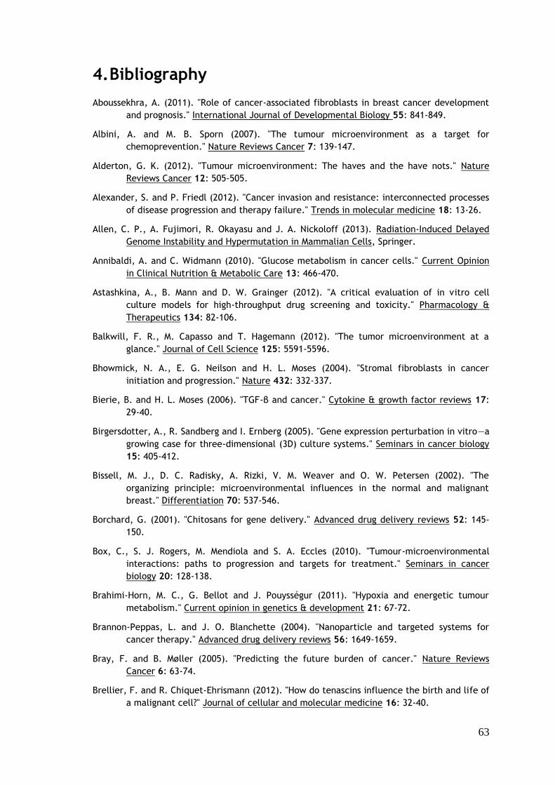
63
4. Bibliography
Aboussekhra, A. (2011). "Role of cancer-associated fibroblasts in breast cancer development
and prognosis." International Journal of Developmental Biology 55: 841-849.
Albini, A. and M. B. Sporn (2007). "The tumour microenvironment as a target for
chemoprevention." Nature Reviews Cancer 7: 139-147.
Alderton, G. K. (2012). "Tumour microenvironment: The haves and the have nots." Nature
Reviews Cancer 12: 505-505.
Alexander, S. and P. Friedl (2012). "Cancer invasion and resistance: interconnected processes
of disease progression and therapy failure." Trends in molecular medicine 18: 13-26.
Allen, C. P., A. Fujimori, R. Okayasu and J. A. Nickoloff (2013). Radiation-Induced Delayed
Genome Instability and Hypermutation in Mammalian Cells, Springer.
Annibaldi, A. and C. Widmann (2010). "Glucose metabolism in cancer cells." Current Opinion
in Clinical Nutrition & Metabolic Care 13: 466-470.
Astashkina, A., B. Mann and D. W. Grainger (2012). "A critical evaluation of in vitro cell
culture models for high-throughput drug screening and toxicity." Pharmacology &
Therapeutics 134: 82-106.
Balkwill, F. R., M. Capasso and T. Hagemann (2012). "The tumor microenvironment at a
glance." Journal of Cell Science 125: 5591-5596.
Bhowmick, N. A., E. G. Neilson and H. L. Moses (2004). "Stromal fibroblasts in cancer
initiation and progression." Nature 432: 332-337.
Bierie, B. and H. L. Moses (2006). "TGF-β and cancer." Cytokine & growth factor reviews 17:
29-40.
Birgersdotter, A., R. Sandberg and I. Ernberg (2005). "Gene expression perturbation in vitro—a
growing case for three-dimensional (3D) culture systems." Seminars in cancer biology
15: 405-412.
Bissell, M. J., D. C. Radisky, A. Rizki, V. M. Weaver and O. W. Petersen (2002). "The
organizing principle: microenvironmental influences in the normal and malignant
breast." Differentiation 70: 537-546.
Borchard, G. (2001). "Chitosans for gene delivery." Advanced drug delivery reviews 52: 145-
150.
Box, C., S. J. Rogers, M. Mendiola and S. A. Eccles (2010). "Tumour-microenvironmental
interactions: paths to progression and targets for treatment." Seminars in cancer
biology 20: 128-138.
Brahimi-Horn, M. C., G. Bellot and J. Pouysségur (2011). "Hypoxia and energetic tumour
metabolism." Current opinion in genetics & development 21: 67-72.
Brannon-Peppas, L. and J. O. Blanchette (2004). "Nanoparticle and targeted systems for
cancer therapy." Advanced drug delivery reviews 56: 1649-1659.
Bray, F. and B. Møller (2005). "Predicting the future burden of cancer." Nature Reviews
Cancer 6: 63-74.
Brellier, F. and R. Chiquet-Ehrismann (2012). "How do tenascins influence the birth and life of
a malignant cell?" Journal of cellular and molecular medicine 16: 32-40.
64
Brizzi, M. F., G. Tarone and P. Defilippi (2012). "Extracellular matrix, integrins, and growth
factors as tailors of the stem cell niche." Current opinion in cell biology 24: 645-651.
Brouty-Boyé, D., C. Mainguené, V. Magnien, L. Israel and R. Beaupain (1994). "Fibroblast-
mediated differentiation in human breast carcinoma cells (MCF-7) grown as nodules IN
VITRO." International journal of cancer 56: 731-735.
Burdett, E., F. K. Kasper, A. G. Mikos and J. A. Ludwig (2010). "Engineering tumors: a tissue
engineering perspective in cancer biology." Tissue Engineering Part B: Reviews 16:
351-359.
Chabner, B. A. and T. G. Roberts (2005). "Chemotherapy and the war on cancer." Nature
Reviews Cancer 5: 65-72.
Chiavarina, B., D. Whitaker-Menezes, U. E. Martinez-Outschoorn, A. K. Witkiewicz, R. Birbe,
A. Howell, R. G. Pestell, J. Smith, R. Daniel and F. Sotgia (2011). "Pyruvate kinase
expression (PKM1 and PKM2) in cancer-associated fibroblasts drives stromal nutrient
production and tumor growth." Cancer biology & therapy 12: 1101-1113.
Chou, L. Y. T., K. Ming and W. C. W. Chan (2011). "Strategies for the intracellular delivery of
nanoparticles." Chemical Society Reviews 40: 233-245.
Cichon, M. A., A. C. Degnim, D. W. Visscher and D. C. Radisky (2010). "Microenvironmental
influences that drive progression from benign breast disease to invasive breast
cancer." Journal of mammary gland biology and neoplasia 15: 389-397.
Cirri, P. and P. Chiarugi (2012). "Cancer-associated-fibroblasts and tumour cells: a diabolic
liaison driving cancer progression." Cancer and Metastasis Reviews 31: 195-208.
Coussens, L. M. and Z. Werb (2002). "Inflammation and cancer." Nature 420: 860-867.
Couvreur, P. (2012). "Nanoparticles in drug delivery: Past, present and future." Advanced drug
delivery reviews 65: 21-23.
Cukierman, E. and D. R. Khan (2010). "The benefits and challenges associated with the use of
drug delivery systems in cancer therapy." Biochemical pharmacology 80: 762-770.
Cukierman, E., R. Pankov and K. M. Yamada (2002). "Cell interactions with three-dimensional
matrices." Current opinion in cell biology 14: 633-640.
Danhier, F., E. Ansorena, J. M. Silva, R. Coco, A. Le Breton and V. Préat (2012). "PLGA-based
nanoparticles: an overview of biomedical applications." Journal of Controlled Release
161: 505-522.
De Bock, K., S. Cauwenberghs and P. Carmeliet (2011). "Vessel abnormalization: another
hallmark of cancer?: molecular mechanisms and therapeutic implications." Current
opinion in genetics & development 21: 73-79.
de Visser, K. E., A. Eichten and L. M. Coussens (2006). "Paradoxical roles of the immune
system during cancer development." Nature Reviews Cancer 6: 24-37.
De Wever, O. and M. Mareel (2003). "Role of tissue stroma in cancer cell invasion." The
Journal of pathology 200: 429-447.
Debnath, J. and J. S. Brugge (2005). "Modelling glandular epithelial cancers in three-
dimensional cultures." Nature Reviews Cancer 5: 675-688.
Delinassios, J. and S. Kottaridis (1984). "Interactions between human fibroblasts and HeLa
cells in vitro." Biology of the Cell 50: 9-16.
65
Delinassios, J. G. (1987). "Fibroblasts against cancer cells in vitro." Anticancer Research 7:
1005-1010.
Dhimolea, E., M. V. Maffini, A. M. Soto and C. Sonnenschein (2010). "The role of collagen
reorganization on mammary epithelial morphogenesis in a 3D culture model."
Biomaterials 31: 3622-3630.
Dolberg, D. S. and M. J. Bissell (1984). "Inability of Rous sarcoma virus to cause sarcomas in
the avian embryo." Nature 309: 552-556.
Duell, B. L., A. W. Cripps, M. A. Schembri and G. C. Ulett (2011). "Epithelial cell coculture
models for studying infectious diseases: benefits and limitations." Journal of
Biomedicine and Biotechnology 2011: 1-9.
Eifler, A. C. and C. S. Thaxton (2011). "Nanoparticle therapeutics: FDA approval, clinical
trials, regulatory pathways, and case study." Methods Molecular Biology 726: 325-338.
Elenbaas, B., L. Spirio, F. Koerner, M. D. Fleming, D. B. Zimonjic, J. L. Donaher, N. C.
Popescu, W. C. Hahn and R. A. Weinberg (2001). "Human breast cancer cells
generated by oncogenic transformation of primary mammary epithelial cells." Genes
& development 15: 50-65.
Elsdale, T. and J. Bard (1972). "Collagen substrata for studies on cell behavior." The Journal
of Cell Biology 54: 626-637.
Engel, L. W. and N. A. Young (1978). "Human breast carcinoma cells in continuous culture: a
review." Cancer research 38: 4327-4339.
Farber, E. (1984). "The multistep nature of cancer development." Cancer research 44: 4217-
4223.
Faria, C., C. Smith and J. Rutka "The Role of HGF/c-Met Pathway Signaling in Human
Medulloblastoma."
Farokhzad, O. C. and R. Langer (2009). "Impact of nanotechnology on drug delivery." ACS
nano 3: 16-20.
Favi, P. M., R. S. Benson, N. R. Neilsen, R. L. Hammonds, C. C. Bates, C. P. Stephens and M.
S. Dhar (2013). "Cell proliferation, viability, and in vitro differentiation of equine
mesenchymal stem cells seeded on bacterial cellulose hydrogel scaffolds." Materials
Science and Engineering C: Materials for Biological Applications 33: 1935-1944.
Fennema, E., N. Rivron, J. Rouwkema, C. van Blitterswijk and J. de Boer (2013). "Spheroid
culture as a tool for creating 3D complex tissues." Trends in biotechnology 31: 108-
115.
Fletcher, D. A. and R. D. Mullins (2010). "Cell mechanics and the cytoskeleton." Nature 463:
485-492.
Fox, E., G. A. Curt and F. M. Balis (2002). "Clinical trial design for target-based therapy." The
oncologist 7: 401-409.
Freshney, R. I., G. Vunjak-Novakovic and R. Freshney (2006). Basic principles of cell culture,
John Wiley & Sons.
Fujita, H., K. Ohuchida, K. Mizumoto, T. Egami, K. Miyoshi, T. Moriyama, L. Cui, J. Yu, M.
Zhao and T. Manabe (2009). "Tumor–stromal interactions with direct cell contacts
enhance proliferation of human pancreatic carcinoma cells." Cancer science 100:
2309-2317.
66
Gaspar, V. M., I. J. Correia, Â. Sousa, F. Silva, C. M. Paquete, J. A. Queiroz and F. Sousa
(2011). "Nanoparticle mediated delivery of pure P53 supercoiled plasmid DNA for gene
therapy." Journal of Controlled Release 156: 212-222.
Gaspar, V. M., J. G. Marques, F. Sousa, R. O. Louro, J. A. Queiroz and I. J. Correia (2013).
"Biofunctionalized nanoparticles with pH-responsive and cell penetrating blocks for
gene delivery." Nanotechnology 24: doi: 10.1088/0957-4484/1024/1027/275101.
Gavhane, Y., A. Shete, A. Bhagat, V. Shinde, K. Bhong, G. Khairnar and A. Yadav (2011).
"Solid Tumors: Facts, Challenges and Solutions." International Journal 2.
Grieger, K. D., S. F. Hansen and A. Baun (2009). "The known unknowns of nanomaterials:
Describing and characterizing uncertainty within environmental, health and safety
risks." Nanotoxicology 3: 222-233.
Grivennikov, S. I., F. R. Greten and M. Karin (2010). "Immunity, inflammation, and cancer."
Cell 140: 883-899.
Gutteridge, J. and B. Halliwell (2010). "Antioxidants: molecules, medicines, and myths."
Biochemical and biophysical research communications 393: 561-564.
Hamilton, A. and G. Hortobagyi (2005). "Chemotherapy: what progress in the last 5 years?"
Journal of clinical oncology 23: 1760-1775.
Han, K., S. Chen, W.-H. Chen, Q. Lei, Y. Liu, R.-X. Zhuo and X.-Z. Zhang (2013). "Synergistic
gene and drug tumor therapy using a chimeric peptide." Biomaterials 34: 4680-4689.
Hanahan, D. and L. M. Coussens (2012). "Accessories to the crime: functions of cells recruited
to the tumor microenvironment." Cancer Cell 21: 309-322.
Hanahan, D. and R. A. Weinberg (2000). "The hallmarks of cancer." Cell 100: 57-70.
Hanahan, D. and R. A. Weinberg (2011). "Hallmarks of cancer: the next generation." Cell 144:
646-674.
Harris, A. L. (2002). "Hypoxia—a key regulatory factor in tumour growth." Nature Reviews
Cancer 2: 38-47.
Hartung, T. (2008). "Thoughts on limitations of animal models." Parkinsonism & Related
Disorders 14: S81-S83.
Hashizume, H., P. Baluk, S. Morikawa, J. W. McLean, G. Thurston, S. Roberge, R. K. Jain and
D. M. McDonald (2000). "Openings between defective endothelial cells explain tumor
vessel leakiness." The American journal of pathology 156: 1363-1380.
Hellsten, E. (2005). "Nanosciences and nanotechnologies: An Action Plan for Europe 2005-
2009." Dialog zur Bewertung von synthetischen Nanopartikeln in Arbeits-und
Umweltbereichen. Bonn.
Heneweer, M., M. Muusse, M. Dingemans, P. C. De Jong, M. Van Den Berg and J. T. Sanderson
(2005). "Co-culture of primary human mammary fibroblasts and MCF-7 cells as an in
vitro breast cancer model." Toxicological Sciences 83: 257-263.
Hirschhaeuser, F., H. Menne, C. Dittfeld, J. West, W. Mueller-Klieser and L. A. Kunz-
Schughart (2010). "Multicellular tumor spheroids: an underestimated tool is catching
up again." Journal of biotechnology 148: 3-15.
Hockenbery, D. M., M. Tom, C. Abikoff and D. Margineantu (2013). The Warburg Effect and
Beyond: Metabolic Dependencies for Cancer Cells, Springer.
67
Hogan, C. (2012). "Impact of interactions between normal and transformed epithelial cells
and the relevance to cancer." Cellular and Molecular Life Sciences 69: 203-213.
Hogenesch, H. and A. Y. Nikitin (2012). "Challenges in pre-clinical testing of anti-cancer drugs
in cell culture and in animal models." Journal of Controlled Release 164: 183-186.
Horimoto, Y., Y. Takahashi, U. Polanska and A. Orimo (2012). "Emerging roles of the tumor-
associated stroma in promoting tumor metastasis." Cell Adhesion & Migration 6: 203-
206.
Ibeanu, O. A. (2011). "Molecular pathogenesis of cervical cancer." Cancer biology & therapy
11: 295-306.
Janeway, C. A., P. Travers, M. Walport and M. J. Shlomchik (2001). Immunobiology, Garland
Science.
Janssen, A. and R. Medema (2012). "Genetic instability: tipping the balance." Oncogene:
doi:10.1038/onc.2012.1576.
Jögi, A., M. Vaapil, M. Johansson and S. Påhlman (2012). "Cancer cell differentiation
heterogeneity and aggressive behavior in solid tumors." Upsala journal of medical
sciences 117: 217-224.
Johnson, J., S. Decker, D. Zaharevitz, L. Rubinstein, J. Venditti, S. Schepartz, S. Kalyandrug,
M. Christian, S. Arbuck and M. Hollingshead (2001). "Relationships between drug
activity in NCI preclinical in vitro and in vivo models and early clinical trials." British
journal of cancer 84: 1424.
Jones, R. G. and C. B. Thompson (2009). "Tumor suppressors and cell metabolism: a recipe for
cancer growth." Genes & development 23: 537-548.
Jou, C.-H., W.-C. Chen, M.-C. Yang, M.-C. Hwang, W.-L. Chou, S.-M. Lin and C. Y. Hsu (2008).
"In vitro biocompatibility of three-dimensional chitosan scaffolds immobilized with
chondroitin-6-sulfate." Polymers for Advanced Technologies 19: 377-384.
Justice, B. A., N. A. Badr and R. A. Felder (2009). "3D cell culture opens new dimensions in
cell-based assays." Drug Discovery Today 14: 102-107.
Kalluri, R. and M. Zeisberg (2006). "Fibroblasts in cancer." Nature Reviews Cancer 6: 392-401.
Kamaly, N., Z. Xiao, P. M. Valencia, A. F. Radovic-Moreno and O. C. Farokhzad (2012).
"Targeted polymeric therapeutic nanoparticles: design, development and clinical
translation." Chemical Society Reviews 41: 2971-3010.
Kelland, L. (2004). "Of mice and men: values and liabilities of the athymic nude mouse model
in anticancer drug development." European journal of cancer 40: 827-836.
Kennedy, K. (1987). "Hypoxic cells as specific drug targets for chemotherapy." Anti-cancer
drug design 2: 181-194.
Kerkar, S. P. and N. P. Restifo (2012). "Cellular constituents of immune escape within the
tumor microenvironment." Cancer research 72: 3125-3130.
Khatau, S. B., R. J. Bloom, S. Bajpai, D. Razafsky, S. Zang, A. Giri, P.-H. Wu, J. Marchand, A.
Celedon and C. M. Hale (2012). "The distinct roles of the nucleus and nucleus-
cytoskeleton connections in three-dimensional cell migration." Scientific reports 2.
Kievit, F. M., O. Veiseh, N. Bhattarai, C. Fang, J. W. Gunn, D. Lee, R. G. Ellenbogen, J. M.
Olson and M. Zhang (2009). "PEI–PEG–Chitosan-Copolymer-Coated Iron Oxide
68
Nanoparticles for Safe Gene Delivery: Synthesis, Complexation, and Transfection."
Advanced functional materials 19: 2244-2251.
Kim, J. B., R. Stein and M. J. O'Hare (2004). "Three-dimensional in vitro tissue culture models
of breast cancer—a review." Breast cancer research and treatment 85: 281-291.
Ko, Y. H., Z. Lin, N. Flomenberg, R. G. Pestell, A. Howell, F. Sotgia, M. P. Lisanti and U. E.
Martinez-Outschoorn (2012). "Glutamine fuels a vicious cycle of autophagy in the
tumor stroma and oxidative mitochondrial metabolism in epithelial cancer cells."
Cancer Biology & Therapy 12: 1085-1097.
Kobayashi, H., K. C. Boelte and P. C. Lin (2007). "Endothelial cell adhesion molecules and
cancer progression." Current medicinal chemistry 14: 377-386.
Koshida, Y., M. Kuranami and M. Watanabe (2006). "Interaction between stromal fibroblasts
and colorectal cancer cells in the expression of vascular endothelial growth factor."
Journal of Surgical Research 134: 270-277.
Krause, S., M. V. Maffini, A. M. Soto and C. Sonnenschein (2010). "The microenvironment
determines the breast cancer cells' phenotype: organization of MCF7 cells in 3D
cultures." BMC cancer 10: 263.
Krishna, R. and L. D. Mayer (2000). "Multidrug resistance (MDR) in cancer: mechanisms,
reversal using modulators of MDR and the role of MDR modulators in influencing the
pharmacokinetics of anticancer drugs." European journal of pharmaceutical sciences
11: 265-283.
LaBarbera, D. V., B. G. Reid and B. H. Yoo (2012). "The multicellular tumor spheroid model
for high-throughput cancer drug discovery." Expert Opinion on Drug Discovery 7: 819-
830.
Langdon, S. P. (2004). Cancer cell culture: methods and protocols, Springer.
Le Droumaguet, B., J. Nicolas, D. Brambilla, S. Mura, A. Maksimenko, L. De Kimpe, E. Salvati,
C. Zona, C. Airoldi and M. Canovi (2012). "Versatile and Efficient Targeting Using a
Single Nanoparticulate Platform: Application to Cancer and Alzheimer’s Disease." ACS
nano 6: 5866.
LeBedis, C., K. Chen, L. Fallavollita, T. Boutros and P. Brodt (2002). "Peripheral lymph node
stromal cells can promote growth and tumorigenicity of breast carcinoma cells
through the release of IGF‐I and EGF." International journal of cancer 100: 2-8.
Lee, J., M. J. Cuddihy and N. A. Kotov (2008). "Three-dimensional cell culture matrices: state
of the art." Tissue Engineering Part B: Reviews 14: 61-86.
Lee, J. J. (2007). "Clinical trial design for anticancer therapies." The Cancer Handbook.
Levine, A. J. and A. M. Puzio-Kuter (2010). "The control of the metabolic switch in cancers by
oncogenes and tumor suppressor genes." science 330: 1340-1344.
Leyva-Illades, D., M. McMillin, M. Quinn and S. DeMorrow (2012). "Cholangiocarcinoma
pathogenesis: Role of the tumor microenvironment." Translational Gastrointestinal
Cancer 1: 71-80.
Li, H., X. Fan and J. Houghton (2007). "Tumor microenvironment: the role of the tumor
stroma in cancer." Journal of cellular biochemistry 101: 805-815.
Lindl, T. and R. Steubing (2013). "Basic Cell Culture Techniques." Atlas of Living Cell Cultures:
5-9.
69
Liu, H. and K. Roy (2005). "Biomimetic three-dimensional cultures significantly increase
hematopoietic differentiation efficacy of embryonic stem cells." Tissue engineering
11: 319-330.
Lövestam, G., H. Rauscher, G. Roebben, B. S. Klüttgen, N. Gibson, J. P. Putaud and H. Stamm
(2010). Considerations on a definition of nanomaterial for regulatory purposes. Joint
Research Centre Reference Reports, European Commission: 80004-80001.
Lu, P., V. M. Weaver and Z. Werb (2012). "The extracellular matrix: a dynamic niche in
cancer progression." The Journal of Cell Biology 196: 395-406.
Ma, X. J., R. Salunga, J. T. Tuggle, J. Gaudet, E. Enright, P. McQuary, T. Payette, M. Pistone,
K. Stecker and B. M. Zhang (2003). "Gene expression profiles of human breast cancer
progression." Proceedings of the National Academy of Sciences 100: 5974-5979.
Madden, J. C., M. Hewitt, K. Przybylak, R. J. Vandebriel, A. H. Piersma and M. T. Cronin
(2012). "Strategies for the Optimisation of In Vivo Experiments in Accordance with
the 3Rs Philosophy." Regulatory Toxicology and Pharmacology 63: 140-154.
Martinez-Outschoorn, U. E., Z. Lin, Y. H. Ko, A. Goldberg, N. Flomenberg, C. Wang, S.
Pavlides, R. G. Pestell, A. Howell and F. Sotgia (2011). "Understanding the metabolic
basis of drug resistance: Therapeutic induction of the Warburg effect kills cancer
cells." Cell Cycle 10: 2521-2528.
Martinez-Outschoorn, U. E., S. Pavlides, D. Whitaker-Menezes, K. M. Daumer, J. N. Milliman,
B. Chiavarina, G. Migneco, A. K. Witkiewicz, M. P. Martinez-Cantarin and N.
Flomenberg (2010). "Tumor cells induce the cancer associated fibroblast phenotype
via caveolin-1 degradation: Implications for breast cancer and DCIS therapy with
autophagy inhibitors." Cell Cycle 9: 2423-2433.
Marx, V. (2013). "Cell culture: A better brew." Nature 496: 253-258.
Mbeunkui, F. and D. J. Johann (2009). "Cancer and the tumor microenvironment: a review of
an essential relationship." Cancer chemotherapy and pharmacology 63: 571-582.
McClellan, J. L., J. M. Davis, J. L. Steiner, R. T. Enos, S. H. Jung, J. A. Carson, M. M. Pena, K.
A. Carnevale, F. G. Berger and E. A. Murphy (2012). "Linking tumor-associated
macrophages, inflammation, and intestinal tumorigenesis: role of MCP-1." American
Journal of Physiology-Gastrointestinal and Liver Physiology 303: 1087-1095.
McMillin, D. W., J. M. Negri and C. S. Mitsiades (2013). "The role of tumour–stromal
interactions in modifying drug response: challenges and opportunities." Nature
Reviews Drug Discovery 12: 217-228.
Mehta, G., A. Y. Hsiao, M. Ingram, G. D. Luker and S. Takayama (2012). "Opportunities and
challenges for use of tumor spheroids as models to test drug delivery and efficacy."
Journal of Controlled Release 164: 192-204.
Meinert, C. L. (2012). ClinicalTrials: Design, Conduct and Analysis, OUP USA.
Merlo, L. M. F., J. W. Pepper, B. J. Reid and C. C. Maley (2006). "Cancer as an evolutionary
and ecological process." Nature Reviews Cancer 6: 924-935.
Mertens, J. C., C. D. Fingas, J. D. Christensen, R. L. Smoot, S. F. Bronk, N. W. Werneburg, M.
P. Gustafson, A. B. Dietz, L. R. Roberts and A. E. Sirica (2013). "Therapeutic effects
of deleting cancer-associated fibroblasts in cholangiocarcinoma." Cancer research 73:
897-907.
70
Metzger, M. L., L. R. Meacham, B. Patterson, J. S. Casillas, L. S. Constine, N. Hijiya, L. B.
Kenney, M. Leonard, B. A. Lockart and W. Likes (2013). "Female Reproductive Health
After Childhood, Adolescent, and Young Adult Cancers: Guidelines for the Assessment
and Management of Female Reproductive Complications." Journal of clinical oncology
31: 1239-1247.
Micke, P. and A. Östman (2005). "Exploring the tumour environment: cancer-associated
fibroblasts as targets in cancer therapy." 9: 1217-1233.
Miki, Y., K. Ono, S. Hata, T. Suzuki, H. Kumamoto and H. Sasano (2012). "The advantages of
co-culture over mono cell culture in simulating in vivo environment." The Journal of
Steroid Biochemistry and Molecular Biology 131: 68-75.
Milstein, J., V. Grachev, A. Padilla and E. Griffiths (1996). "WHO activities towards the three
Rs in the development and control of biological medicines." Developmental Biology
stands 86: 31-39.
Minami, Y., T. Sasaki, J.-i. Kawabe and Y. Ohsaki (2013). Accessory Cells in Tumor
Angiogenesis—Tumor-Associated Pericytes, InTech.
Minchinton, A. I. and I. F. Tannock (2006). "Drug penetration in solid tumours." Nature
Reviews Cancer 6: 583-592.
Mueller-Klieser, W. (1987). "Multicellular spheroids." Journal of cancer research and clinical
oncology 113: 101-122.
Mueller, M. M. and N. E. Fusenig (2004). "Friends or foes—bipolar effects of the tumour
stroma in cancer." Nature Reviews Cancer 4: 839-849.
Nagelkerke, A., J. Bussink, F. C. Sweep and P. N. Span (2013). "Generation of multicellular
tumor spheroids of breast cancer cells: how to go three-dimensional." Analytical
Biochemistry 437: 17-19.
Nakase, I., Y. Konishi, M. Ueda, H. Saji and S. Futaki (2012). "Accumulation of arginine-rich
cell-penetrating peptides in tumors and the potential for anticancer drug delivery in
vivo." J Control Release 159: 181-188.
Narang, A. S. and D. S. Desai (2009). "Anticancer Drug Development." Pharmaceutical
Perspectives of Cancer Therapeutics, Springer.
Noguera, R., O. A. Nieto, I. Tadeo, F. Fariñas and T. Álvaro (2012). "Extracellular matrix,
biotensegrity and tumor microenvironment. An update and overview." Histology and
histopathology 27: 693-705.
Olsen, C. J., J. Moreira, E. M. Lukanidin and N. S. Ambartsumian (2010). "Human mammary
fibroblasts stimulate invasion of breast cancer cells in a three-dimensional culture
and increase stroma development in mouse xenografts." BMC cancer 10: 1-16.
Olumi, A. F., G. D. Grossfeld, S. W. Hayward, P. R. Carroll, T. D. Tlsty and G. R. Cunha
(1999). "Carcinoma-associated fibroblasts direct tumor progression of initiated human
prostatic epithelium." Cancer research 59: 5002-5011.
Orimo, A., P. B. Gupta, D. C. Sgroi, F. Arenzana-Seisdedos, T. Delaunay, R. Naeem, V. J.
Carey, A. L. Richardson and R. A. Weinberg (2005). "Stromal fibroblasts present in
invasive human breast carcinomas promote tumor growth and angiogenesis through
elevated SDF-1/CXCL12 secretion." Cell 121: 335-348.
Ormerod, M. G. and P. R. Imrie (1990). Flow cytometry, Springer.
71
Page, H., P. Flood and E. G. Reynaud (2012). "Three-dimensional tissue cultures: current
trends and beyond." Cell and Tissue Research: 1-9.
Panyam, J. and V. Labhasetwar (2012). "Biodegradable nanoparticles for drug and gene
delivery to cells and tissue." Advanced drug delivery reviews 55: 329-347.
Parrott, J. A., E. Nilsson, R. Mosher, G. Magrane, D. Albertson, D. Pinkel, J. W. Gray and M.
K. Skinner (2001). "Stromal-epithelial interactions in the progression of ovarian
cancer: influence and source of tumor stromal cells." Molecular and cellular
endocrinology 175: 29-39.
Peinado, H., D. Olmeda and A. Cano (2007). "Snail, Zeb and bHLH factors in tumour
progression: an alliance against the epithelial phenotype?" Nature Reviews Cancer 7:
415-428.
Perche, F. and V. P. Torchilin (2012). "Accumulation and toxicity of antibody-targeted
doxorubicin-loaded PEG-PE micelles in ovarian cancer cell spheroid model." Journal of
Controlled Release 164: 95-102.
Phung, Y. T., D. Barbone, V. C. Broaddus and M. Ho (2011). "Rapid generation of in vitro
multicellular spheroids for the study of monoclonal antibody therapy." Journal of
Cancer 2: 507.
Pietras, K. and A. Östman (2010). "Hallmarks of cancer: interactions with the tumor stroma."
Experimental cell research 316: 1324-1331.
Poste, G. and I. J. Fidler (1980). "The pathogenesis of cancer metastasis." Nature 283: 139-
146.
Purpura, K. A., A. M. Bratt-Leal, K. A. Hammersmith, T. C. McDevitt and P. W. Zandstra
(2011). "Systematic engineering of 3D pluripotent stem cell niches to guide blood
development." Biomaterials 33: 1271-1280.
Raguz, S. and E. Yagüe (2008). "Resistance to chemotherapy: new treatments and novel
insights into an old problem." British journal of cancer 99: 387-391.
Ranganatha, N. and I. Kuppast (2012). "A review on alternatives to animal testing methods in
drug development." International Journal of Pharmacy and Pharmaceutical Sciences
4: 28-32.
Raz, Y. and N. Erez (2013). "An inflammatory vicious cycle: Fibroblasts and immune cell
recruitment in cancer." Experimental cell research: doi:
10.1016/j.yexcr.2013.1003.1022.
Riehemann, K., S. W. Schneider, T. A. Luger, B. Godin, M. Ferrari and H. Fuchs (2009).
"Nanomedicine—challenge and perspectives." Angewandte Chemie International
Edition 48: 872-897.
Rimann, M. and U. Graf-Hausner (2012). "Synthetic 3D multicellular systems for drug
development." Current Opinion in Biotechnology 23: 803-809.
Ruddon, R. W. (2007). Cancer biology, Oxford University Press, USA.
Sadlonova, A., Z. Novak, M. R. Johnson, D. B. Bowe, S. R. Gault, G. P. Page, J. V.
Thottassery, D. R. Welch and A. R. Frost (2005). "Breast fibroblasts modulate
epithelial cell proliferation in three-dimensional in vitro co-culture." Breast Cancer
Res 7: 46-49.
72
Sappino, A. P., O. Skalli, B. Jackson, W. Schürch and G. Gabbiani (2006). "Smooth-muscle
differentiation in stromal cells of malignant and non-malignant breast tissues."
International journal of cancer 41: 707-712.
See, A. P., J. E. Han, J. Phallen, Z. Binder, G. Gallia, F. Pan, D. Jinasena, C. Jackson, Z.
Belcaid and S. J. Jeong (2012). "The role of STAT3 activation in modulating the
immune microenvironment of GBM." Journal of neuro-oncology 110: 359-368.
Shaw, K. R. M., C. N. Wrobel and J. S. Brugge (2004). "Use of three-dimensional basement
membrane cultures to model oncogene-induced changes in mammary epithelial
morphogenesis." Journal of mammary gland biology and neoplasia 9: 297-310.
Sherwood, L. (2012). Human physiology: from cells to systems, Thomson Brooks/Cole.
Shuman Moss, L. A., S. Jensen-Taubman and W. G. Stetler-Stevenson (2012). "Matrix
metalloproteinases: changing roles in tumor progression and metastasis." The
American journal of pathology 181: 1895-1899.
Siegel, R., D. Naishadham and A. Jemal (2012). "Cancer statistics, 2012." CA: a cancer journal
for clinicians 62: 10-29.
Sietsema, W. (1989). "The absolute oral bioavailability of selected drugs." International
journal of clinical pharmacology, therapy, and toxicology 27: 179-211.
Sinha, R., G. J. Kim, S. Nie and D. M. Shin (2006). "Nanotechnology in cancer therapeutics:
bioconjugated nanoparticles for drug delivery." Molecular cancer therapeutics 5:
1909-1917.
Skobe, M., T. Hawighorst, D. G. Jackson, R. Prevo, L. Janes, P. Velasco, L. Riccardi, K.
Alitalo, K. Claffey and M. Detmar (2001). "Induction of tumor lymphangiogenesis by
VEGF-C promotes breast cancer metastasis." Nature Medicine 7: 192-198.
Sorensen, A., R. J. Mairs, L. Braidwood, C. Joyce, J. Conner, S. Pimlott, M. Brown and M.
Boyd (2012). "In vivo evaluation of a cancer therapy strategy combining HSV1716-
mediated oncolysis with gene transfer and targeted radiotherapy." Journal of Nuclear
Medicine 53: 647-654.
Soule, H., J. Vazquez, A. Long, S. Albert and M. Brennan (1973). "A human cell line from a
pleural effusion derived from a breast carcinoma." Journal of the National Cancer
Institute 51: 1409-1416.
Sounni, N. E. and A. Noel (2013). "Targeting the Tumor Microenvironment for Cancer
Therapy." Clinical chemistry 59: 85-93.
Stacey, G. (2012). Current developments in cell culture technology, Springer.
Stacey, G. (2011). Cell culture contamination, Springer.
Stockmann, C., A. Doedens, A. Weidemann, N. Zhang, N. Takeda, J. I. Greenberg, D. A.
Cheresh and R. S. Johnson (2008). "Deletion of vascular endothelial growth factor in
myeloid cells accelerates tumorigenesis." Nature 456: 814-818.
Straussman, R., T. Morikawa, K. Shee, M. Barzily-Rokni, Z. R. Qian, J. Du, A. Davis, M. M.
Mongare, J. Gould and D. T. Frederick (2012). "Tumour micro-environment elicits
innate resistance to RAF inhibitors through HGF secretion." Nature 487: 500-504.
Strell, C., H. Rundqvist and A. Östman (2012). "Fibroblasts-a key host cell type in tumor
initiation, progression, and metastasis." Upsala journal of medical sciences 117: 187-
195.
73
Streuli, C. H., N. Bailey and M. J. Bissell (1991). "Control of mammary epithelial
differentiation: basement membrane induces tissue-specific gene expression in the
absence of cell-cell interaction and morphological polarity." The Journal of Cell
Biology 115: 1383-1395.
Sutherland, R. M., W. R. Inch, J. A. McCredie and J. Kruuv (1970). "A multi-component
radiation survival curve using an in vitro tumour model." International Journal of
Radiation Biology 18: 491-495.
Swartz, M. A., N. Iida, E. W. Roberts, S. Sangaletti, M. H. Wong, F. E. Yull, L. M. Coussens and
Y. A. DeClerck (2012). "Tumor microenvironment complexity: emerging roles in
cancer therapy." Cancer research 72: 2473-2480.
Talmadge, J. E. and I. J. Fidler (2010). "AACR centennial series: the biology of cancer
metastasis: historical perspective." Cancer research 70: 5649-5669.
Tlsty, T. D. and L. M. Coussens (2006). "Tumor stroma and regulation of cancer development."
Annual Review of Pathology-mechanisms of Disease 1: 119-150.
Trédan, O., C. M. Galmarini, K. Patel and I. F. Tannock (2007). "Drug resistance and the solid
tumor microenvironment." Journal of the National Cancer Institute 99: 1441-1454.
Tumarkin, E., L. Tzadu, E. Csaszar, M. Seo, H. Zhang, A. Lee, R. Peerani, K. Purpura, P. W.
Zandstra and E. Kumacheva (2011). "High-throughput combinatorial cell co-culture
using microfluidics." Integrative Biology 3: 653-662.
Valencia, P. M., O. C. Farokhzad, R. Karnik and R. Langer (2012). "Microfluidic technologies
for accelerating the clinical translation of nanoparticles." Nature Nanotechnology 7:
623-629.
Vanharanta, S. and J. Massagué (2012). "Field Cancerization: Something New Under the Sun."
Cell 149: 1179-1181.
Vera-Ramirez, L., P. Sanchez-Rovira, M. C. Ramirez-Tortosa, C. L. Ramirez-Tortosa, S.
Granados-Principal, J. A. Lorente and J. L. Quiles (2011). "Free radicals in breast
carcinogenesis, breast cancer progression and cancer stem cells. Biological bases to
develop oxidative-based therapies." Critical reviews in oncology/hematology 80: 347-
368.
Wang, J., A. S. Levenson and R. L. Satcher Jr (2006). "Identification of a unique set of genes
altered during cell-cell contact in an in vitro model of prostate cancer bone
metastasis." International journal of molecular medicine 17: 849-856.
Wang, M. and M. Thanou (2010). "Targeting nanoparticles to cancer." Pharmacological
Research 62: 90-99.
Wang, S., X. Hao, Y. Su, C. Yi, B. Li, X. Fan, J. Pei, Y. Song, W. Xia and B. Liu (2013a). "The
Utilization of Perforated Bioinert Chambers to Generate an In Vivo Isolated Space for
Tissue Engineering Involving Chondrocytes, Mesenchymal Stem Cells, and Fibroblasts."
Tissue Engineering Part C: Methods 19: doi:10.1089/ten.tec.2012.0269.
Wang, T., F. M. Kievit, O. Veiseh, H. Arami, Z. R. Stephen, C. Fang, Y. Liu, R. G. Ellenbogen
and M. Zhang (2013b). "Targeted cell uptake of a non-internalizing antibody through
conjugation to iron oxide nanoparticles in primary central nervous system lymphoma."
World neurosurgery: doi:10.1016/j.wneu.2013.1001.1011.
Wang, Z., R. K. Kundu, M. T. Longaker, T. Quertermous and G. P. Yang (2012). "The
angiogenic factor Del1 prevents apoptosis of endothelial cells through integrin
binding." Surgery 151: 296-305.
74
Warburg, O. (1956). "On the origin of cancer cells." Science 123: 309-314.
Weigelt, B., F. C. Geyer and J. S. Reis-Filho (2010). "Histological types of breast cancer: how
special are they?" Molecular oncology 4: 192.
Weigelt, B., A. M. Glas, L. F. A. Wessels, A. T. Witteveen, J. L. Peterse and L. J. Van't Veer
(2003). "Gene expression profiles of primary breast tumors maintained in distant
metastases." Proceedings of the National Academy of Sciences 100: 15901-15905.
Weis, S. M. and D. A. Cheresh (2011). "Tumor angiogenesis: molecular pathways and
therapeutic targets." Nature Medicine 17: 1359-1370.
Whitaker-Menezes, D., U. E. Martinez-Outschoorn, Z. Lin, A. Ertel, N. Flomenberg, A. K.
Witkiewicz, R. C. Birbe, A. Howell, S. Pavlides and R. Gandara (2011). "Evidence for a
stromal-epithelial" lactate shuttle" in human tumors: MCT4 is a marker of oxidative
stress in cancer-associated fibroblasts." Cell cycle 10: 1772-1783.
Willerth, S. M., T. E. Faxel, D. I. Gottlieb and S. E. Sakiyama-Elbert (2007). "The effects of
soluble growth factors on embryonic stem cell differentiation inside of fibrin
scaffolds." Stem Cells 25: 2235-2244.
Witsch, E., M. Sela and Y. Yarden (2010). "Roles for growth factors in cancer progression."
Physiology 25: 85-101.
Wolfensohn, S., P. Hawkins, E. Lilley, D. Anthony, C. Chambers, S. Lane, M. Lawton, H.-M.
Voipio and G. Woodhall (2013). "Reducing suffering in experimental autoimmune
encephalomyelitis (EAE)." Journal of pharmacological and toxicological methods 67:
169-176.
Wu, H., L. Zhu and V. P. Torchilin (2012). "pH-sensitive poly (histidine)-PEG/DSPE-PEG co-
polymer micelles for cytosolic drug delivery." Biomaterials.
Xing, F., J. Saidou and K. Watabe (2010). "Cancer associated fibroblasts (CAFs) in tumor
microenvironment." Frontiers in bioscience: a journal and virtual library 15: 166-179.
Xu, J., Y. Lu, S. Qiu, Z.-N. Chen and Z. Fan (2013). "A novel role of EMMPRIN/CD147 in
transformation of quiescent fibroblasts to cancer-associated fibroblasts by breast
cancer cells." Cancer Letters 335: 380-386.
Yamada, K. M. and E. Cukierman (2007). "Modeling tissue morphogenesis and cancer in 3D."
Cell 130: 601-610.
Yang, M., X. Jiang, L. Liang and J.-L. Yang (2013). "Application of Nanobiotechnology in
Cancer: Creation of Nanooncology and Revolution in Cancer Research and Practice."
World Journal of Cancer Research 1: 24-36.
Yeom, C. J., Y. Goto, Y. Zhu, M. Hiraoka and H. Harada (2012). "Microenvironments and
Cellular Characteristics in the Micro Tumor Cords of Malignant Solid Tumors."
International journal of molecular sciences 13: 13949-13965.
Yin, M., J. Soikkeli, T. Jahkola, S. Virolainen, O. Saksela and E. Hölttä (2012). "TGF-β
Signaling, Activated Stromal Fibroblasts, and Cysteine Cathepsins B and L Drive the
Invasive Growth of Human Melanoma Cells." The American journal of pathology 181:
2202-2216.
Youlden, D. R., S. M. Cramb, N. A. Dunn, J. M. Muller, C. M. Pyke and P. D. Baade (2012).
"The descriptive epidemiology of female breast cancer: an international comparison
of screening, incidence, survival and mortality." Cancer Epidemiology 36: 237-248.
75
Yu, A., Y. Matsuda, A. Takeda, E. Uchinuma and Y. Kuroyanagi (2012). "Effect of EGF and
bFGF on fibroblast proliferation and angiogenic cytokine production from cultured
dermal substitutes." Journal of Biomaterials Science, Polymer Edition 23: 1315-1324.
Yun, Y., Y. W. Cho and K. Park (2012). "Nanoparticles for oral delivery: Targeted
nanoparticles with peptidic ligands for oral protein delivery." Advanced drug delivery
reviews 65: 822-832.
Zamarron, B. F. and W. Chen (2011). "Dual roles of immune cells and their factors in cancer
development and progression." International journal of biological sciences 7: 651-658.
Zetter, P., B.R. (1998). "Angiogenesis and tumor metastasis." Annual review of medicine 49:
407-424.
Zhang, G., X. Zeng and P. Li (2013). "Nanomaterials in Cancer-Therapy Drug Delivery System."
Journal of Biomedical Nanotechnology 9: 741-750.
Zhang, L., F. Gu, J. Chan, A. Wang, R. Langer and O. Farokhzad (2007). "Nanoparticles in
medicine: therapeutic applications and developments." Clinical Pharmacology &
Therapeutics 83: 761-769.
Zhang, X., D. Nie and S. Chakrabarty (2010). "Growth factors in tumor microenvironment."
Frontiers in bioscience: a journal and virtual library 15: 151-165.
Zimmermann, M., C. Box and S. A. Eccles (2013). Two-Dimensional vs. Three-Dimensional In
Vitro Tumor Migration and Invasion Assays, Springer.