Multifunctional biomedical applications of magnetic ...
10
Multifunctional biomedical applications of magnetic nanodiamond Elena Perevedentseva Artashes Karmenyan Yu-Chung Lin Chang-You Song Zhe-Rui Lin Ashek-I-Ahmed Chia-Chi Chang Svetlana Norina Valentina Bessalova Nikolai Perov Olga Levinson Boris Zousman Chia-Liang Cheng Elena Perevedentseva, Artashes Karmenyan, Yu-Chung Lin, Chang-You Song, Zhe-Rui Lin, Ashek-I-Ahmed, Chia-Chi Chang, Svetlana Norina, Valentina Bessalova, Nikolai Perov, Olga Levinson, Boris Zousman, Chia-Liang Cheng, “Multifunctional biomedical applications of magnetic nanodiamond, ” J. Biomed. Opt. 23(9), 091404 (2018), doi: 10.1117/1.JBO.23.9.091404. Downloaded From: https://www.spiedigitallibrary.org/journals/Journal-of-Biomedical-Optics on 13 Mar 2022 Terms of Use: https://www.spiedigitallibrary.org/terms-of-use
Transcript of Multifunctional biomedical applications of magnetic ...
Multifunctional biomedicalapplications of magneticnanodiamond
Elena PerevedentsevaArtashes KarmenyanYu-Chung LinChang-You SongZhe-Rui LinAshek-I-AhmedChia-Chi ChangSvetlana NorinaValentina BessalovaNikolai PerovOlga LevinsonBoris ZousmanChia-Liang Cheng
Elena Perevedentseva, Artashes Karmenyan, Yu-Chung Lin, Chang-You Song, Zhe-Rui Lin, Ashek-I-Ahmed,Chia-Chi Chang, Svetlana Norina, Valentina Bessalova, Nikolai Perov, Olga Levinson, Boris Zousman,Chia-Liang Cheng, “Multifunctional biomedical applications of magnetic nanodiamond,” J. Biomed. Opt.23(9), 091404 (2018), doi: 10.1117/1.JBO.23.9.091404.
Downloaded From: https://www.spiedigitallibrary.org/journals/Journal-of-Biomedical-Optics on 13 Mar 2022Terms of Use: https://www.spiedigitallibrary.org/terms-of-use
Multifunctional biomedical applications of magneticnanodiamond
Elena Perevedentseva,a,b Artashes Karmenyan,a Yu-Chung Lin,a,c Chang-You Song,a Zhe-Rui Lin,aAshek-I-Ahmed,a Chia-Chi Chang,a Svetlana Norina,d Valentina Bessalova,d Nikolai Perov,dOlga Levinson,e Boris Zousman,e and Chia-Liang Chenga,*aNational Dong Hwa University, Department of Physics, Hualien, TaiwanbP. N. Lebedev Physics Institute of Russian Academy of Sciences, Moscow, RussiacInstitute of Physics, Academia Sinica, Taipei, TaiwandMoscow State University, Department of Physics, Moscow, RussiaeRay Techniques, Jerusalem, Israel
Abstract. The research and development for biomedical applications are recently focused on multifunctionalnanoparticles. To integrate various functionalities, different methods of modifying the particle’s physical proper-ties are developed. Among the considered, nanodiamond (ND) is a promising candidate for the development ofmultifunctional complex due to its variable features in size, structure, surface chemistry, physical properties, andbiocompatibility. In addition to its well-studied structural, surface, electrochemical and photonic properties,strong magnetism of ND can be observed. In the present work, magnetically modified ND is introduced interms of its bioapplications. Along with the soft ferromagnetism of ND, the increased fluorescence at one-and two-photon excitation is realized. Utilizing the combined magnetic and fluorescence properties of themagnetically modified ND, fluorescence imaging, fluorescence lifetime imaging and manipulation of cells bymagnetic field are demonstrated. The perspectives to use the magnetic ND for drug delivery, cells magneticseparation and filtration, in bioengineering to control the cell distribution combined with imaging and treatmentare discussed. © 2018 Society of Photo-Optical Instrumentation Engineers (SPIE) [DOI: 10.1117/1.JBO.23.9.091404]
Keywords: nanodiamond; magnetic nanoparticles; magnetic nanodiamond; theranostics; bioimaging; controllable drug delivery.
Paper 170786SSR received Dec. 9, 2017; accepted for publication Feb. 22, 2018; published online Mar. 21, 2018; corrected Apr. 16,2018.
1 IntroductionThe recent intense developed nanoparticle (NP) applicationsin biology and medicine are essentially determined by theirmultifunctional uses. Particularly, for example, theranosticapplication1–3 usually includes combining functionalities ofdrug delivery or other medical treatments with multimodalimaging. Therefore, the particles that are able to combine thedesired physical/chemical properties attract close attention.Among all NPs, nanodiamond (ND) appears to be a promisingcandidate for the multifunctional applications2 due to its surface,electrochemical, and optical properties; and the availability invarious size and surface structure, and, most importantly, thedemonstrated biocompatibility.3 In spite of some limitations,a number of facilities for ND theranostic applications havebeen demonstrated. These include stimulus-responsible NDdrug delivery and release;2 multimodal imaging utilizing lumi-nescence properties of ND, allowing fluorescence imaging atone- and two-photon excitation,2,3 fluorescence lifetime imaging(FLIM);4 Raman spectroscopic analysis and mapping;3 and opti-cal sensing on the base of energy transfer5 and magnetic sensingutilizing spin properties of structural defects6. One possibility toget multifunctional nanoparticles is to modify the nanoparticleto enhance the desired properties for certain application, forexample, methods to enhance ND fluorescence in differentspectral ranges.2,5,7
In this work, we discuss magnetically modified ND and itsbiomedical applications. Magnetic ND (MND) can combineproperties and advantages of the ND and magnetic nanoparticle.The most developed and clear way to prepare magnetic ND isdoping with magnetic atoms.8 However, in some cases, such NDcan show some level of cytotoxicity and be not biocompatible.
Here we report, the magnetic NDwas synthesized and treatedwithout the use of metals and metal oxides using the methodproposed by ray techniques.9 The origin of its magnetism isunclear. As for magnetic properties of diamond in general,pure diamond structure combines diamagnetic and paramag-netic components,10 but in an intermediate graphite–diamondstructure the conditions for spin ordering and magnetic inter-actions can be created.10 Such structure can be realized inND. Ferromagnetic properties of carbon nanostructures arediscussed for theoretically predicted structures and are foundexperimentally; the methods to obtain such properties withoutembedding magnetic atoms are developed using mechanical,electromagnetic treatments,11 hydrogenation,12,13 creationdefects and doping (with nonmagnetic atoms),13 and surfacestate modification.14 Additionally, spin properties of structuraldefects of ND are studied15 and the applications of ND arediscussed, such as magnetic resonance imaging (MRI) agentfor MRI,16 and for quantum sensing.6,15
Bioapplications of magnetic nanoparticles have also devel-oped into a topical issue. The magnetic nanoparticles of variouscomposition, size, shape, and surface properties are developed
*Address all correspondence to: Chia-Liang Cheng, E-mail: [email protected] 1083-3668/2018/$25.00 © 2018 SPIE
Journal of Biomedical Optics 091404-1 September 2018 • Vol. 23(9)
Journal of Biomedical Optics 23(9), 091404 (September 2018)
Downloaded From: https://www.spiedigitallibrary.org/journals/Journal-of-Biomedical-Optics on 13 Mar 2022Terms of Use: https://www.spiedigitallibrary.org/terms-of-use
for different purposes. For instance, they can serve as probes inliving tissue and be controlled noninvasively from the outside byan external magnetic field, wherein the magnetic field action onthe diamagnetic biotissues is negligible.17 The most developedapplications of magnetic nanoparticles are used as a MRI markerat local magnetic hyperthermia and its applications; controlleddrug delivery, guided by magnetic field; cell separation andfiltration.18 With the aforementioned possibilities, in the presentwork, the magnetic-modified ND is characterized, analyzed, anddemonstrated from the point of view of bioapplications.
2 Methods and Materials
2.1 Magnetic Fluorescent Nanodiamond
The ND, called further RayND, was synthesized by laser treat-ment of specially prepared hydrocarbon targets in liquidmedium without the use of metals or metal oxides.19 TheseNDs produced by controlled technology of light hydrodynamicpulse (LHDP)9 have average size of 4.3 nm and are rather sim-ilar to ND obtained by detonation synthesis (DND), just purerand more uniform due to the use of metal-free raw materials andthe controlled method of the synthesis. After isolation fromhydrocarbon blend, synthesized RayND particles underwent atwo-step chemical–thermal modification using metal-free chem-icals. First, RayND was treated thermally for ordering carbonradicals on the ND surface resulting in the unusual ferromagnet-ism of unknown nature. Then to provide the colloidal stability,to enhance the ND dispersion in aqueous solutions and increasein the sedimentation stability, of diamond nanofluids, theobtained magnetic ND powder was further chemically modifiedin metal-free solvents, nitrogenized, which for unclear reasonchanged its optical properties (e.g., PL). Note, that the processof RayND surface modification is not disclosed in this paper.Magnetically and optically modified ND (RayND-M) were ana-lyzed and compared with DND received with nominal averagesize of crystallites 3 to 10 nm (Microdiamant AG, Switzerland).
2.2 RayND-M Characterization
The diamond structure of ND (DND, RayND, and RayND-M)was confirmed by x-ray diffraction analysis (XRD) using D8Advance diffractometer (Bruker AXS, Karlsruhe, Germany),performed as described in Ref. 19. The obtained XRD patternsare identical and indicate that all samples have almost the samecrystalline structure close to 100% diamond with the averagesize of primary particles automatically defined by Scherrer’sformula of 4.3 to 4.5 nm.
Magnetic properties of the RayND-M, and the comparisonwith DND, were measured using LakeShore 7407 vibratingsample magnetometer (VSM, Lake Shore Cryotronics) at ambi-ent conditions. The ND samples (about 15 mg) were packed inthe plastic diamagnetic bag with a size of 5 × 5 mm and a thick-ness of 0.5 mm. The free bag was also investigated for magneticmoment field dependence. The sample magnetic moment wasdetermined as a difference of the sample and free bag signalsat the same magnetic field.
The composition of the investigated samples was estimatedvia energy-dispersive x-ray analysis (EDX), with scanning elec-tron microscopy imaging using FE-SEM JEOL JSM 7000F(JEOL, Japan) to check the presence of magnetic atoms inthe RayND-M.
For EDX-SEM analysis as well as for measurements ofRaman, FTIR, fluorescence spectra, and fluorescence lifetimemeasurements, ND water suspension was dropped on the Sisubstrate (1 cm × 1 cm) and dried in a desiccator at roomtemperature.
The particles (and their aggregates) size distribution as wellas ζ-potential in water were measured using the dynamic lightscattering (DLS) method in a Zetasizer nano ZS (MalvernInstruments, United Kingdom), with a 4-mW, 633-nm wave-length He–Ne laser, detection angle 173 deg at pH ∼ 6.8.
To estimate predominant structure of crystallites, Ramanspectra were measured using a Renishaw 1000B (Renishaw,United Kingdom) spectrometer equipped with a CW laser(DPGL-2100F; Photop Suwtech, China) with 532-nm wave-length; and a confocal Raman α-300S (Witec, Germany) witha 488-nm wavelength argon laser (Melles Griot). Surfacefunctional groups were analyzed using a Fourier transforminfrared spectroscopy (Bomem MB154 FTIR spectrometer,ABB Bomem, Switzerland) in situ using a mercury cadmiumtelluride liquid nitrogen cooled detector under vacuum withchamber base pressure ∼3 × 10⁻⁶ Torr.
To study photonic properties of the RayND-M, absorptionspectra were measured for ND water suspensions using aUV–visible JASCO V-550 spectrometer (JASCO, Japan) atroom temperature. For the fluorescence spectra, the Renishaw1000B and α-300S confocal Raman spectrometers were used.Analysis of the RayND-M fluorescence lifetime at two-photonexcitation and FLIM at their interaction with target biologicalsystems were performed using a system on the base ofa femtosecond tunable Ti-Sapphire laser Chameleon Ultra-II(Coherent), the used excitation wavelength was in the 760- to800-nm range, repetition rate 80 MHz, pulse duration 140 fs,and laser power at about 3 mW. For FLIM, a 2-D scanner(EINST Technology, Singapore) was used. The registrationwas recorded in the 450- to 650-nm spectral range with thesingle photon counting system PicoHarp 300 (PicoQuant,Germany) and cooled PMT connected with an Olympus IX71 optical microscope, 40× objective.
2.3 RayND-M Interaction with Cells
The biointeraction of RayND-M was studied using babyhamster kidney cells (BHK) and human lung alveolar carcinomacells (A549) cell cultures obtained from BioresourceCollection and Research Center, Taiwan (BCRC, Taiwan).The A549 cells were cultured in RPMI1640 medium(Gibco, Invitrogen, United Kingdom), BHK cells were culturedin Dulbecco’s modified eagle medium (DMEM) (Gibco,Invitrogen, United Kingdom), all are supplemented with 2-mML-glutamine (Invitrogen), 1.5 g∕L sodium bicarbonate (Sigma),and 10% fetal bovine serum (FBS) (Gibco/Life Technologies).Cells were maintained under standard cell culture conditions inan incubator (Galaxy 170S, Eppendorf) at 95% air and 5% CO2
and 37°C. Culture medium was replaced with fresh mediumevery 48 or 72 h. Cells were detached by treatment with0.5% trypsin and 2.6 mM ethyl-enediaminetetraacetic acid(Gibco/Life Technologies); cultures were subcultured routinelyat ∼80% confluence.
Human serum albumin (HSA, Sigma, Ronkonkoma) wasphysically adsorbed on RayND-M. The albumin solution witha concentration of 40 mg∕ml and an aqueous suspension ofthe ND with concentration of 4 mg∕ml were mixed in a 1∶1ratio. After thorough mixing for 2 h, the mixture was centrifuged
Journal of Biomedical Optics 091404-2 September 2018 • Vol. 23(9)
Perevedentseva et al.: Multifunctional biomedical applications of magnetic nanodiamond
Downloaded From: https://www.spiedigitallibrary.org/journals/Journal-of-Biomedical-Optics on 13 Mar 2022Terms of Use: https://www.spiedigitallibrary.org/terms-of-use
to separate ND from the protein adsorbed on the surface.The separated sediment was washed twice in distilled water.This method provides the formation of a stable ND-proteincomplex.20 To confirm the adsorption, the albumin content in thesolutions was determined using a UV–Visible spectrometerJASCO V-550 before its mixing with ND and after the isolationof ND–protein complexes.
To study the interaction with the BHK cells, the cells weretreated with 20 μg∕ml of RayND-M added to the cell growthmedium for 4 h, whereas nonreacted ND has been washedout. Then the cells were fixed with 3.7% formaldehyde (FA)on the coverslips. The cells with ND were investigated witha confocal microscope and FLIM, to visualize the ND penetra-tion into the cell. For investigations with the confocal micro-scope, cell cytosol was dyed with Alexa Fluor 674 (Sigma);the nuclei were stained with Hoechst33342 (Sigma). A TCSSP5 Laser Scanning Fluorescence Confocal Microscope (Leica,Germany) was used. To define the ND location in the cell struc-tures, the scan along vertical direction (Z-scan) was performed.For ND intercellular distribution visualization, the RayND-Mfluorescence was excited by 488 nm and collected in therange from 560 to 600 nm; Hoechst fluorescence was excitedwith 405 nm and detected at 450 to 490 nm; cytoplasm stainedwith Alexa Fluor 647 was excited with 633 nm with detection at640 to 690 nm.
For FLIM investigation, the cells were not dyed. The FLIMinvestigation at two-photon excitation was performed asdescribed above.
The cytotoxicity was estimated by MTT (3-(4,5-dimethylth-iazol-2-yl)-2,5-diphenyltetrazolium bromide) assay21 using ND-HSA complex and BHK cell line. BHK cells were seeded inquantity of 4 × 10⁵ cells∕well in a 96-well microtiter plate inDMEM supplemented with 2-mM L-glutamine (Invitrogen)and 10% of FBS and incubated at 37°C and 5% CO2 for24 h. After incubation, the medium was removed. Then thecells were treated with a fresh medium containing ND-HSAof different RayND-M concentrations (5 to 200 μg∕ml) for24 and 48 h. After this procedure, the medium was removedand the cells were treated for 4 h with MTT reagent (withthe concentration of 2.5 mg∕ml). The surviving cells convertedMTT-agent to formazan, a blue-purple color when dissolved indimethyl sulfoxide (DMSO). The solution was removed and200 μl of DMSO was added to dissolve the formazan. Theoptical absorption of the treated cells and control (nontreated)cells was measured at 570 nm (OD 570) with ELISA reader(MRX revelation Microplate Reader, DYNEX). Relative per-centages of surviving cells were calculated by dividing theabsorbance of the treated cells with that of the control measuredin each experiment.
For the experiment with magnetic guidance of the cells con-taining RayND-M by a magnetic field, the cells were detachedfrom the bottom of the culture dish, suspended in the cellularmedium, and investigated with microscopy and FLIM.
2.4 Utilizing RayND-M Magnetic Properties
2.4.1 Magnetic field guidance of RayND-M/cell complexes
The possibility of magnetic guidance of ND and ND-containingcells was demonstrated for particles in water and for RayND-M-containing living cells. To create a nonuniform magnetic field,a thin steel needle (with diameter of 0.18 mm) was magnetizedwith neodymium magnet (NdFeB, disk of diameter of 5 mm;
National Imports). For a preliminary testing, RayND-M wassuspended in the distilled water with a concentration of20 μg∕ml. To demonstrate a magnetic guidance and controlby the magnetic field of the spatial distribution of the ND-containing cells, the cells were detached from the bottom ofthe culture dish and suspended in the cellular medium. Theguidance of particles and their aggregates and the cells withembedded RayND-M by the magnetic field were observedwith a conventional microscope (Olympus IX 71, objective withmagnification 40×) and with FLIM.
2.4.2 Magnetic field-controlled RayND-M/cell complexdistribution and positioning
To demonstrate the possibility to control the spatial distributionof the cells during culturing by the magnetic field, suspension ofRayND-M-containing cells was mixed with the cells withoutthe ND and recultured together for 12 to 36 h in the presenceof the magnetic field. The magnetic field was applied locallyto the selected area of the cultural dish, using the permanentmagnets. After removing the magnets, the areas treated and non-treated with the magnetic field were analyzed with a confocalmicroscope and FLIM to see the cells distribution.
3 Results and Discussion
3.1 Characterization of the Nanodiamond
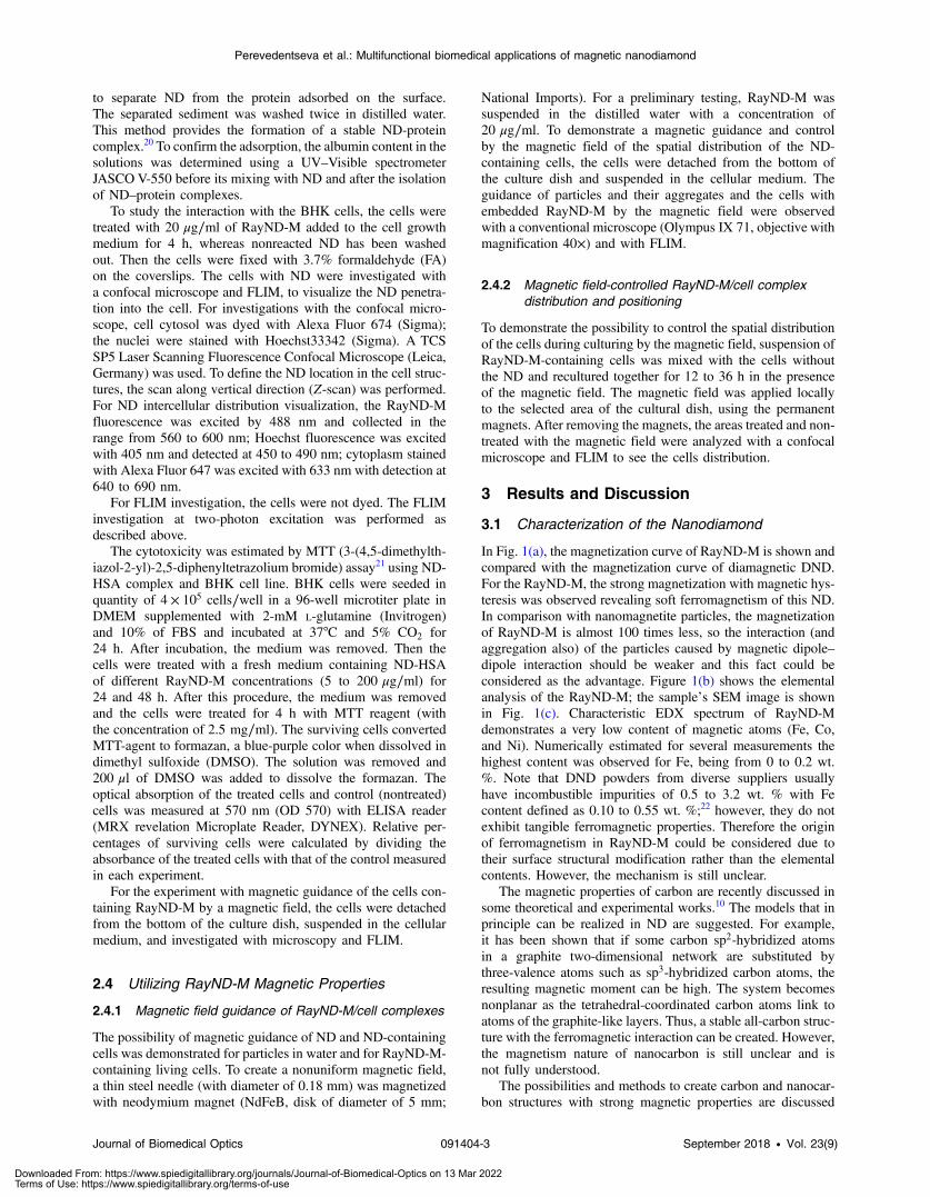
In Fig. 1(a), the magnetization curve of RayND-M is shown andcompared with the magnetization curve of diamagnetic DND.For the RayND-M, the strong magnetization with magnetic hys-teresis was observed revealing soft ferromagnetism of this ND.In comparison with nanomagnetite particles, the magnetizationof RayND-M is almost 100 times less, so the interaction (andaggregation also) of the particles caused by magnetic dipole–dipole interaction should be weaker and this fact could beconsidered as the advantage. Figure 1(b) shows the elementalanalysis of the RayND-M; the sample’s SEM image is shownin Fig. 1(c). Characteristic EDX spectrum of RayND-Mdemonstrates a very low content of magnetic atoms (Fe, Co,and Ni). Numerically estimated for several measurements thehighest content was observed for Fe, being from 0 to 0.2 wt.%. Note that DND powders from diverse suppliers usuallyhave incombustible impurities of 0.5 to 3.2 wt. % with Fecontent defined as 0.10 to 0.55 wt. %;22 however, they do notexhibit tangible ferromagnetic properties. Therefore the originof ferromagnetism in RayND-M could be considered due totheir surface structural modification rather than the elementalcontents. However, the mechanism is still unclear.
The magnetic properties of carbon are recently discussed insome theoretical and experimental works.10 The models that inprinciple can be realized in ND are suggested. For example,it has been shown that if some carbon sp2-hybridized atomsin a graphite two-dimensional network are substituted bythree-valence atoms such as sp3-hybridized carbon atoms, theresulting magnetic moment can be high. The system becomesnonplanar as the tetrahedral-coordinated carbon atoms link toatoms of the graphite-like layers. Thus, a stable all-carbon struc-ture with the ferromagnetic interaction can be created. However,the magnetism nature of nanocarbon is still unclear and isnot fully understood.
The possibilities and methods to create carbon and nanocar-bon structures with strong magnetic properties are discussed
Journal of Biomedical Optics 091404-3 September 2018 • Vol. 23(9)
Perevedentseva et al.: Multifunctional biomedical applications of magnetic nanodiamond
Downloaded From: https://www.spiedigitallibrary.org/journals/Journal-of-Biomedical-Optics on 13 Mar 2022Terms of Use: https://www.spiedigitallibrary.org/terms-of-use
recently, especially imparting paramagnetism and ferromagnet-ism to ND significantly increases the ND attractiveness for itsuse in biomedical researches and applications.10,11
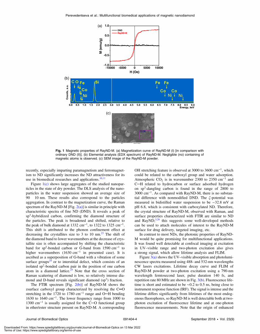
Figure 1(c) shows large aggregates of the studied nanopar-ticles in the state of dry powder. The DLS analysis of the nano-particles in the water suspension showed an average size of90� 10 nm. These results also correspond to the particlesaggregation. In contrast to the magnetization curve, the Ramanspectrum of the RayND-M [Fig. 2(a)] is similar in principle withcharacteristic spectra of fine ND (DND). It reveals a peak ofsp3-hybridized carbon, confirming the diamond structure ofthe particles. The peak is broadened and shifted, relative tothe peak of bulk diamond at 1332 cm−1, to 1322 to 1325 cm−1.This shift is attributed to the phonon confinement effect atdecreasing the crystallites size to 3 to 10 nm.23 The shift ofthe diamond band to lower wavenumbers at the decrease of crys-tallite size is often accompanied by shifting the characteristicband for sp2-bonded carbon or G-band from 1590 cm−1 tohigher wavenumbers (1630 cm−1 in presented case). It isascribed as a superposition of G-band with a vibration of somesurface groups24 or to interstitial defect, which consists of anisolated sp2-bonded carbon pair in the position of one carbonatom in a diamond lattice.25 Note that the cross section ofRaman scattering of diamond is low, so relatively intense dia-mond and D-band reveals significant diamond (sp3) fraction.
The FTIR spectrum [Fig. 2(b)] of RayND-M shows thezsurface carboxyl group characterized by resolving the C═O
stretching in the 1720 to 1780 cm−1 range and O─H bending1630 to 1640 cm−1. The lower frequency range from 1000 to1300 cm−1 is usually assigned for the C─O functional groupin ether/ester structure present on RayND-M. A corresponding
OH stretching feature is observed at 3000 to 3600 cm−1, whichcould be related to the carboxyl group and water adsorption.Atmospheric CO2 is in wavenumber 2300 to 2350 cm−1 andC─H related to hydrocarbon or surface adsorbed hydrogenon sp3-dangling carbon is found in the range of 2800 to3000 cm−1. As compared with RayND-M, there is no substan-tial difference with nonmodified DND. The ζ-potential wasmeasured in bidistilled water suspension to be −32.8 mV atpH 6.8, which is consistent with carboxylated ND. Therefore,the crystal structure of RayND-M, observed with Raman, andsurface properties characterized with FTIR are similar to NDand RayND;3,26 this suggests some well-developed methodscan be used to attach molecules of interest to the RayND-Msurface for drug delivery, targeted imaging, etc.
In contrast to most NDs, the photonic properties of RayND-M would be quite promising for multifunctional applications.It was found well detectable at confocal imaging at excitationin UV–visible range and two-photon excitation also givesa strong signal, which allow lifetime analysis and FLIM.
Figure 3(a) shows the UV–visible absorption and photolumi-nescence spectra measured using 488- and 532-nm wavelengthsCW lasers excitations. Lifetime decay curve and FLIM ofRayND-M powder at two-photon excitation using a 790-nmwavelength femtosecond laser, pulse duration 140 fs, andrepetition rate 80 MHz are shown in Fig. 3(b). Fluorescence life-time is short and estimated to be ∼0.2 to 0.5 ns, being close toinstrument response function (IRF). The signal is intense and thelifetime differs significantly from lifetimes of the most endog-enous fluorophores, so RayND-M is well detectable both at two-photon excitation of fluorescence lifetime and at one-photonfluorescence measurements. Note that the origin of enhanced
-10000 -5000 0 5000 10000-1.0
-0.5
0.0
0.5
1.0
M (
emu
/g)
H (Oe)
DND RayND-M
(a)
Fig. 1 Magnetic properties of RayND-M. (a) Magnetization curve of RayND-M (I) [in comparison withordinary DND (II)]. (b) Elemental analysis (EDX spectrum) of RayND-M. Negligible (no) containing ofmagnetic atoms is observed. (c) SEM image of the RayND-M powder.
Journal of Biomedical Optics 091404-4 September 2018 • Vol. 23(9)
Perevedentseva et al.: Multifunctional biomedical applications of magnetic nanodiamond
Downloaded From: https://www.spiedigitallibrary.org/journals/Journal-of-Biomedical-Optics on 13 Mar 2022Terms of Use: https://www.spiedigitallibrary.org/terms-of-use
fluorescence emission from magnetic-modified ND is also notclear yet and can be discussed together with the origin ofmagnetic properties, but it is subject to other consideration.
3.2 Interaction with Cell
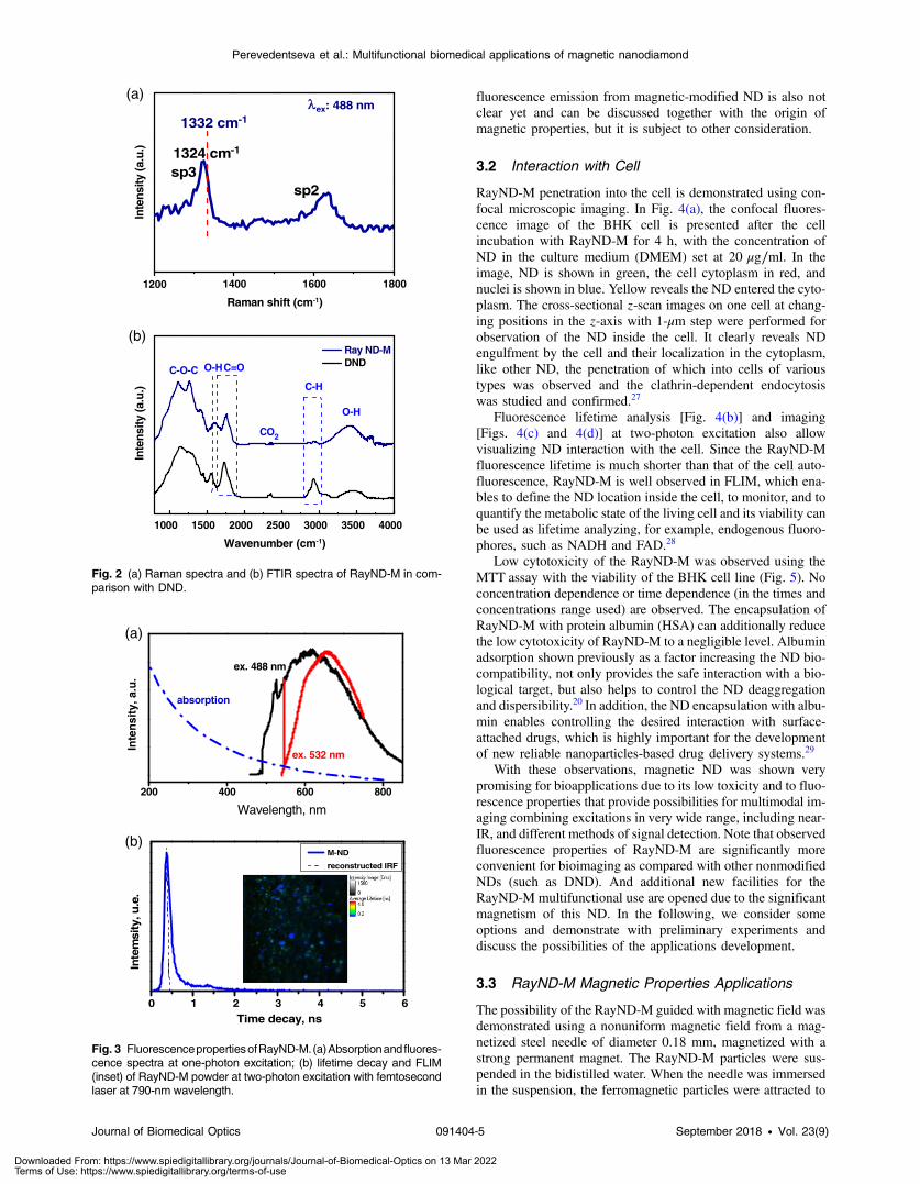
RayND-M penetration into the cell is demonstrated using con-focal microscopic imaging. In Fig. 4(a), the confocal fluores-cence image of the BHK cell is presented after the cellincubation with RayND-M for 4 h, with the concentration ofND in the culture medium (DMEM) set at 20 μg∕ml. In theimage, ND is shown in green, the cell cytoplasm in red, andnuclei is shown in blue. Yellow reveals the ND entered the cyto-plasm. The cross-sectional z-scan images on one cell at chang-ing positions in the z-axis with 1-μm step were performed forobservation of the ND inside the cell. It clearly reveals NDengulfment by the cell and their localization in the cytoplasm,like other ND, the penetration of which into cells of varioustypes was observed and the clathrin-dependent endocytosiswas studied and confirmed.27
Fluorescence lifetime analysis [Fig. 4(b)] and imaging[Figs. 4(c) and 4(d)] at two-photon excitation also allowvisualizing ND interaction with the cell. Since the RayND-Mfluorescence lifetime is much shorter than that of the cell auto-fluorescence, RayND-M is well observed in FLIM, which ena-bles to define the ND location inside the cell, to monitor, and toquantify the metabolic state of the living cell and its viability canbe used as lifetime analyzing, for example, endogenous fluoro-phores, such as NADH and FAD.28
Low cytotoxicity of the RayND-M was observed using theMTT assay with the viability of the BHK cell line (Fig. 5). Noconcentration dependence or time dependence (in the times andconcentrations range used) are observed. The encapsulation ofRayND-M with protein albumin (HSA) can additionally reducethe low cytotoxicity of RayND-M to a negligible level. Albuminadsorption shown previously as a factor increasing the ND bio-compatibility, not only provides the safe interaction with a bio-logical target, but also helps to control the ND deaggregationand dispersibility.20 In addition, the ND encapsulation with albu-min enables controlling the desired interaction with surface-attached drugs, which is highly important for the developmentof new reliable nanoparticles-based drug delivery systems.29
With these observations, magnetic ND was shown verypromising for bioapplications due to its low toxicity and to fluo-rescence properties that provide possibilities for multimodal im-aging combining excitations in very wide range, including near-IR, and different methods of signal detection. Note that observedfluorescence properties of RayND-M are significantly moreconvenient for bioimaging as compared with other nonmodifiedNDs (such as DND). And additional new facilities for theRayND-M multifunctional use are opened due to the significantmagnetism of this ND. In the following, we consider someoptions and demonstrate with preliminary experiments anddiscuss the possibilities of the applications development.
3.3 RayND-M Magnetic Properties Applications
The possibility of the RayND-M guided with magnetic field wasdemonstrated using a nonuniform magnetic field from a mag-netized steel needle of diameter 0.18 mm, magnetized with astrong permanent magnet. The RayND-M particles were sus-pended in the bidistilled water. When the needle was immersedin the suspension, the ferromagnetic particles were attracted to
200 400 600 800
Wavelength, nm
Inte
nsi
ty, a
.u.
absorption
ex. 532 nm
ex. 488 nm
0 1 2 3 4 5 6
Inte
msi
ty, u
.e.
Time decay, ns
M-ND
reconstructed IRF
(a)
(b)
Fig. 3 FluorescencepropertiesofRayND-M. (a)Absorptionand fluores-cence spectra at one-photon excitation; (b) lifetime decay and FLIM(inset) of RayND-M powder at two-photon excitation with femtosecondlaser at 790-nm wavelength.
1200 1400 1600 1800
ex: 488 nm
sp2
Inte
nsity
(a.u
.)
Raman shift (cm-1)
sp31324 cm-1
1332 cm-1
1000 1500 2000 2500 3000 3500 4000
CO2
O-H
C-O-C
C-H
O-H
Inte
nsity
(a.u
.)
Wavenumber (cm-1)
Ray ND-M DNDC=O
(b)
(a)
Fig. 2 (a) Raman spectra and (b) FTIR spectra of RayND-M in com-parison with DND.
Journal of Biomedical Optics 091404-5 September 2018 • Vol. 23(9)
Perevedentseva et al.: Multifunctional biomedical applications of magnetic nanodiamond
Downloaded From: https://www.spiedigitallibrary.org/journals/Journal-of-Biomedical-Optics on 13 Mar 2022Terms of Use: https://www.spiedigitallibrary.org/terms-of-use

the region of the magnetic field gradient as a result of magneto-phoresis.30,31 Figure 6(a) shows the scheme of this simpleexperiment. The moving of particles and their aggregates inthe needle’s magnetic field were observed with an optical micro-scope and with lifetime imaging, respectively. The optical imagein Fig. 6(b-1) was obtained at the beginning of the experiment;Fig. 6(b-2) is the FLIM of the particles attracted to the needle;and Fig. 6(b-3) shows the microscopic image of the needle withattracted particles just after FLIM measurement. This series ofimages show that particles in water or in a medium can be
moved by magnetic field and in principle can be used for devel-oping methods of drug delivery.
The motion of the water- or medium-suspended particlesdriven by a magnetic field can be used in controlled dispersionof therapeutic agents within liquid- and gel-filled cavities of anorganism. Nonspecific delivery and distribution in such a sys-tem are driven by diffusion and are limited by the molecule (orNP carrier) concentration and size, surrounding medium proper-ties, such as viscosity. Application of magnetic NP controlled bythe external magnetic field provides the possibility to optimize
(b) (c) (d)
0 2 4 6 8 100
1000
2000
3000
4000
5000
6000
7000
Inte
nsi
ty, a
.u.
Time delay, ns
cell autofluorescence RayND-M
cell autofluorescence
RayND-M
(a)
Fig. 4 Imaging of RayND-M interaction with cell. Observation RayND-M in BHK cell, concentration ofRayND-M in the medium is 20 μg∕ml, cocultured time 4 h. (a) Laser confocal fluorescence image:(1) RayND-M fluorescence is excited with 488 nm and detected in the 530- to 690-nm range,shown in green; (2) the cell cytoplasm was dyed with Fluor 674; (3) cell nuclei with Hoechst33342;and (4) merging of 1 to 3 with RayND-M shown in yellow. (b) Lifetime decays of cell autofluorescenceand fluorescence of RayND-M in BHK cell; (c) FLIM of the cell with RayND-M; and (d) FLIM of controlcell without RayND-M.
Fig. 5 RayND-M cytotoxicity on the BHK cells estimated via MTT test at various concentrations ofRayND-M in the complex with HSA after (a) 24 h and (b) 48 h of the ND treatment.
Journal of Biomedical Optics 091404-6 September 2018 • Vol. 23(9)
Perevedentseva et al.: Multifunctional biomedical applications of magnetic nanodiamond
Downloaded From: https://www.spiedigitallibrary.org/journals/Journal-of-Biomedical-Optics on 13 Mar 2022Terms of Use: https://www.spiedigitallibrary.org/terms-of-use
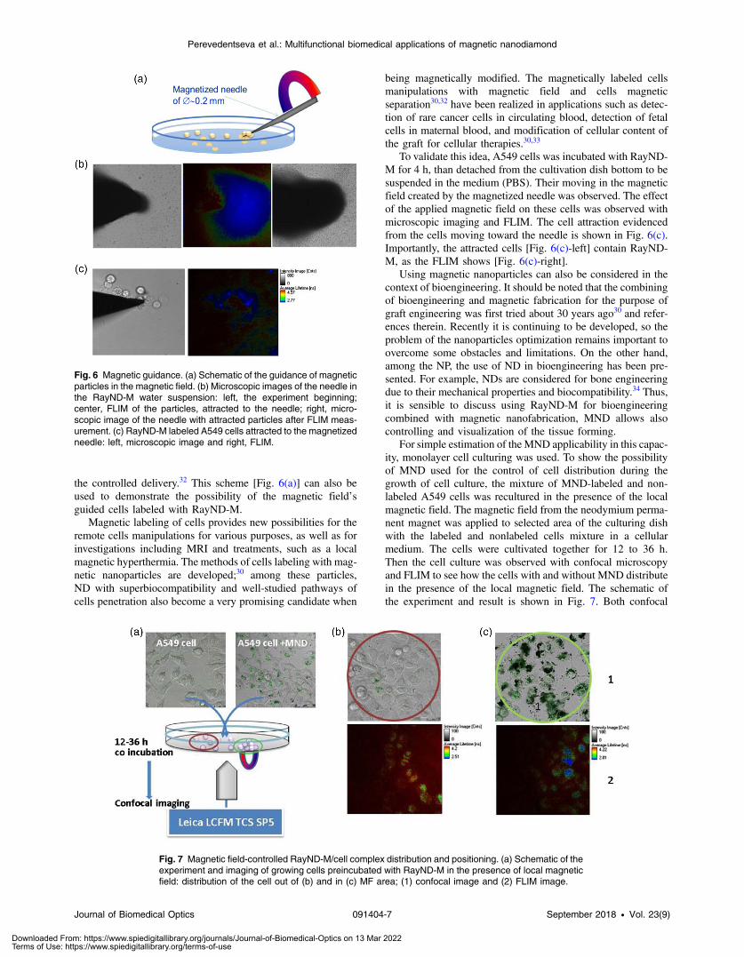
the controlled delivery.32 This scheme [Fig. 6(a)] can also beused to demonstrate the possibility of the magnetic field’sguided cells labeled with RayND-M.
Magnetic labeling of cells provides new possibilities for theremote cells manipulations for various purposes, as well as forinvestigations including MRI and treatments, such as a localmagnetic hyperthermia. The methods of cells labeling with mag-netic nanoparticles are developed;30 among these particles,ND with superbiocompatibility and well-studied pathways ofcells penetration also become a very promising candidate when
being magnetically modified. The magnetically labeled cellsmanipulations with magnetic field and cells magneticseparation30,32 have been realized in applications such as detec-tion of rare cancer cells in circulating blood, detection of fetalcells in maternal blood, and modification of cellular content ofthe graft for cellular therapies.30,33
To validate this idea, A549 cells was incubated with RayND-M for 4 h, than detached from the cultivation dish bottom to besuspended in the medium (PBS). Their moving in the magneticfield created by the magnetized needle was observed. The effectof the applied magnetic field on these cells was observed withmicroscopic imaging and FLIM. The cell attraction evidencedfrom the cells moving toward the needle is shown in Fig. 6(c).Importantly, the attracted cells [Fig. 6(c)-left] contain RayND-M, as the FLIM shows [Fig. 6(c)-right].
Using magnetic nanoparticles can also be considered in thecontext of bioengineering. It should be noted that the combiningof bioengineering and magnetic fabrication for the purpose ofgraft engineering was first tried about 30 years ago30 and refer-ences therein. Recently it is continuing to be developed, so theproblem of the nanoparticles optimization remains important toovercome some obstacles and limitations. On the other hand,among the NP, the use of ND in bioengineering has been pre-sented. For example, NDs are considered for bone engineeringdue to their mechanical properties and biocompatibility.34 Thus,it is sensible to discuss using RayND-M for bioengineeringcombined with magnetic nanofabrication, MND allows alsocontrolling and visualization of the tissue forming.
For simple estimation of the MND applicability in this capac-ity, monolayer cell culturing was used. To show the possibilityof MND used for the control of cell distribution during thegrowth of cell culture, the mixture of MND-labeled and non-labeled A549 cells was recultured in the presence of the localmagnetic field. The magnetic field from the neodymium perma-nent magnet was applied to selected area of the culturing dishwith the labeled and nonlabeled cells mixture in a cellularmedium. The cells were cultivated together for 12 to 36 h.Then the cell culture was observed with confocal microscopyand FLIM to see how the cells with and without MND distributein the presence of the local magnetic field. The schematic ofthe experiment and result is shown in Fig. 7. Both confocal
Fig. 6 Magnetic guidance. (a) Schematic of the guidance of magneticparticles in the magnetic field. (b) Microscopic images of the needle inthe RayND-M water suspension: left, the experiment beginning;center, FLIM of the particles, attracted to the needle; right, micro-scopic image of the needle with attracted particles after FLIM meas-urement. (c) RayND-M labeled A549 cells attracted to the magnetizedneedle: left, microscopic image and right, FLIM.
Fig. 7 Magnetic field-controlled RayND-M/cell complex distribution and positioning. (a) Schematic of theexperiment and imaging of growing cells preincubated with RayND-M in the presence of local magneticfield: distribution of the cell out of (b) and in (c) MF area; (1) confocal image and (2) FLIM image.
Journal of Biomedical Optics 091404-7 September 2018 • Vol. 23(9)
Perevedentseva et al.: Multifunctional biomedical applications of magnetic nanodiamond
Downloaded From: https://www.spiedigitallibrary.org/journals/Journal-of-Biomedical-Optics on 13 Mar 2022Terms of Use: https://www.spiedigitallibrary.org/terms-of-use
fluorescence imaging and FLIM show cells with MND localizedclose to the magnet. Note also that the distribution of RayND-M-labeled cells in the magnetic field could be controlled by thefield geometric configuration and parameters of cell cultivation(ND concentration, cocultivation time). This demonstrates theprinciple to use the magnetic ND to control the cell distributioncombined with multimodal imaging; and opens new prospectsfor its applications in bioengineering allowing simultaneouscontrolled growth of the tissue, tracking of particles duringthe process and visualization of obtained structures.
4 ConclusionIn the present work, magnetically modified ND is introduced interms of its multifunctional bioapplications. Fluorescent ferro-magnetic metal-free ND powder (RayND-M) has been obtainedby surface modification of ND produced by the LHDP method(the process of the surface modification is not disclosed in thispaper). The diamond structure and purity of ND were defined byEDX, Raman, SEM, and the surface was characterized withFTIR; soft ferromagnetism of the ND was detected by a vibrat-ing sample magnetometer. Together with the ferromagnetism ofthe nanoparticles, the increased fluorescence at one- and two-photon excitation is demonstrated. Note, however, that theorigins of the ferromagnetism as well as of enhancement offluorescence emission in the result of magnetic modificationof the ND are still unclear and can be considered together.
High biocompatibility of RayND-M was confirmed by MTTanalysis using the BHK cell line. RayND-M uptakes and itslocalization in the cytoplasm similar to other NDs have beenobserved. The possibility of magnetic guidance of ND incells and magnetic manipulation of ND containing cells in tissuehas been demonstrated by the example of A549 cells coculturedwith RayND-M. Strong magnetism, fluorescence, and non-cytotoxicity of the modified magnetic ND enables significantbreakthroughs in controlled bioengineering, tissues regenera-tion, early diagnostics, and targeted drug delivery.
DisclosuresThe authors have no relevant financial interests in this article andno potential conflicts of interest to disclose.
AcknowledgmentsThe authors appreciate the financial support of this research bythe Ministry of Science and Technology (MoST) of Taiwan,Grant No. MOST 106-2112-M-259-009-MY3 and by theEuropean Commission REA Grant No. 690945 CARTHER.
References1. D. E. Lee et al., “Multifunctional nanoparticles for multimodal imaging
and theragnosis,” Chem. Soc. Rev. 41(7), 2656–2672 (2012).2. V. Vaijayanthimala et al., “Nanodiamond-mediated drug delivery and
imaging: challenges and opportunities,” Expert Opin. Drug Delivery12(5), 735–749 (2015).
3. E. Perevedentseva et al., “Biomedical applications of nanodiamond inimaging and therapy,” Future Med. Nanomed. 8(12), 2041–2060(2013).
4. W. W.-W. Hsiao et al., “Fluorescent nanodiamond: a versatile toolfor long-term cell tracking, super-resolution imaging, and nanoscaletemperature sensing,” Acc. Chem. Res. 49(3), 400–407 (2016).
5. R. Fudala et al., “FRET enhanced fluorescent nanodiamonds,” Curr.Pharm. Biotechnol. 14(13), 1127–1133 (2014).
6. Y. Wu et al., “Diamond quantum devices in biology,” Angew. Chem. Int.Ed. 55(23), 6586–6598 (2016).
7. I. I. Vlasov et al., “Molecular-sized fluorescent nanodiamonds,” Nat.Nanotechnol. 9, 54–58 (2014).
8. C. H. Chen et al., “Fe doped magnetic nanodiamonds made by ionimplantation,” Sci. Rep. 7, 41938 (2017).
9. B. Zousman and O. Levinson, “Pure nanodiamonds produced by laser-assisted technique,” Chapter 5 in Nanodiamond, O. Williams, Ed.,pp. 221–252, RSC Nanoscience and Nanotechnology, London (2014).
10. T. Makarova, “Magnetic properties of carbon structures,”Semiconductors 38, 615–638 (2004).
11. V. E. Orel et al., “Magnetic characteristics and anticancer activity of ananocomplex consisting of detonation nanodiamond and doxorubicin,”J. Superhard Mater. 34, 179–185 (2012).
12. G. Zhang et al., “Superconducting ferromagnetic nanodiamond,” ACSNano 11, 5358–5366 (2017).
13. K. Kenmochi et al., “Materials design of ferromagnetic diamond,”Jpn. J. Appl. Phys. 44(2), L51 (2005).
14. N. I. Kiselev et al., “Electrical and magnetic properties of nanodiamondand pyrocarbon composites,” Russ. J. Gen. Chem. 83(11), 2173–2181(2013).
15. M. Geiselmann et al., “Three-dimensional optical manipulations ofa single electron spin,” Nat. Nanotechnol. 8, 175–179 (2013).
16. D. E. J. Waddington et al., “Nanodiamond-enhanced MRI via in situhyperpolarization,” Nat. Commun. 8, 15118 (2017).
17. S. Odenbach, Colloidal Magnetic Fluids: Basics, Development andApplication of Ferrofluids, Springer, Berlin-Heidelberg (2009).
18. L. Mohammed et al., “Magnetic nanoparticles for environmental andbiomedical applications: a review,” Particuology 30, 1–14 (2017).
19. E. Perevedentseva et al., “Nanodiamonds of laser synthesis for biomedi-cal applications,” J. Nanosci. Nanotechnol. 15(2), 1045–1052 (2015).
20. L.-W. Tsai et al., “Nanodiamonds for medical applications: interactionwith blood in vitro and in vivo,” Int. J. Mol. Sci. 17(7), 1111 (2016).
21. T. L. Riss et al., “Cell viability assays,” in Assay Guidance Manual,G. S. Sittampalam et al., Eds., Eli Lilly & Company and the NationalCenter for Advancing Translational Sciences, Bethesda, Maryland(2004).https://www.ncbi.nlm.nih.gov/books/NBK144065/
22. R. Yu et al., “Determination of impurities in detonation nanodiamondsby gamma activation analysis method,” Diamond Relat. Mater. 55, 77–86 (2015).
23. S. Osswald et al., “Phonon confinement effects in the Raman spectrumof nanodiamond,” Phys. Rev. B 80, 075419 (2009).
24. V. Mochalin, S. Osswald, and Y. Gogotsi, “Contribution of functionalgroups to the Raman spectrum of nanodiamond powders,” Chem.Mater. 21, 273–279 (2009).
25. J. O. Orwa et al., “Raman investigation of damage caused by deep ionimplantation in diamond,” Phys. Rev. B 62(9), 5461–5472 (2000).
26. M. V. Baidakova et al., “Structure of nanodiamonds prepared by lasersynthesis,” Phys. Solid State 55(8), 1747–1753 (2013).
27. K.-J. Huang et al., “Phagocytosis and immune response studies of mac-rophage-nanodiamond interactions in vitro and in vivo,” J. Biophotonics10, 1315–1326 (2017).
28. T. S. Blacker et al., “Separating NADH and NADPH fluorescence inlive cells and tissues using FLIM,” Nat. Commun. 5, 3936 (2014).
29. J. Mariam, S. Sivakami, and P. M. Dongre, “Albumin corona on nano-particles—a strategic approach in drug delivery,” Drug Delivery 23(8),2668–2676 (2016).
30. J.-K. Lim et al., “Magnetophoresis of nanoparticles,” ACS Nano 5(1),217–226 (2011).
31. Y. Jing et al., “Quantitative intracellular magnetic nanoparticle uptakemeasured by live cell magnetophoresis,” FASEB J. 22(12), 4239–4247(2008).
32. M. Zborowski and J. J. Chalmers, Magnetic Cell Separation, Vol. 32,Elsevier Science, Laboratory Techniques in Biochemistry andMolecular Biology, Amsterdam, The Netherlands (2008).
33. S. J. Kuhn, D. E. Hallahan, and T. D. Giorgio, “Characterization ofsuperparamagnetic nanoparticle interactions with extracellular matrixin an in vitro system,” Ann. Biomed. Eng. 34(1), 51–58 (2006).
34. X. Wu et al., “Functionalization of bone implants with nanodiamondparticles and angiopoietin-1 to improve vascularization and boneregeneration,” J. Mater. Chem. B 5, 6629–6636 (2017).
Elena Perevedentseva received her PhD in physics from MoscowState University, Moscow, Russia. She is working at the National
Journal of Biomedical Optics 091404-8 September 2018 • Vol. 23(9)
Perevedentseva et al.: Multifunctional biomedical applications of magnetic nanodiamond
Downloaded From: https://www.spiedigitallibrary.org/journals/Journal-of-Biomedical-Optics on 13 Mar 2022Terms of Use: https://www.spiedigitallibrary.org/terms-of-use
Dong Hwa University, Taiwan. Her research is focused on micro-scopic and spectroscopic studies of nanoparticle interaction with bio-logical objects. She also collaborates as a senior researcher with theP. N. Lebedev Physics Institute of the Russian Academy of Sciences,Moscow, Russia, working in the field of optics and spectroscopy.
Artashes Karmenyan received his PhD in physics from YerevanState University (YSU), Armenia. He was head of theSpectroscopy Laboratory in “Laserayin Technika” Institute, YSU,then joined the Laboratory of Laser Methods for Diagnosis andTherapy, Cancer Research Center, Moscow, Russia. Since 2001he is working in National Yang-Ming University and National Dong-Hwa University, Taiwan, in the fields of laser spectroscopy (Raman,fluorescence, time-resolved), biomedical applications, and noninva-sive optical techniques for mammalian embryos research.
Yu-Chung Lin received his BSc degree from the Department ofBiomechatronic Engineering, National Chiayi University, Taiwan, in2009. He graduated from the National Dong Hwa University witha master’s degree in physics in 2011 and received his PhD in June2016 in physics. His research is in the field of Raman spectroscopyand fluorescence spectroscopy applications in life science.
Chang-You Song received his BSc degree from the Department ofPhysics, Notional Dong Hwa University, Taiwan, in 2016. While hewas an undergraduate, he did his undergraduate research focusedon the cellular models of the magnetic nanoparticle interaction withvarious cell lines of interested.
Zhe-Rui Lin received his master’s degree from NDHU in Hualien,Taiwan. He studied at the nanocarrier for the therapy of cancercell which was via modulation the nanoparticle to enhance the drugefficiency. Furthermore, he is the most interested in the fluorescencespectroscopy applications in biosystems.
Ashek-I-Ahmed has achieved his BSc and MSc degrees in physicsfrom the Department of Physics, Rajshai University, Bangladesh.Currently, he is pursing PhD on applied physics under supervisionof Professor Chia-Liang Cheng (Charlie) at the Department of
Physics, National Dong Hwa University, Taiwan. His research topicis “Surface reaction dynamics of nanodiamond and its bioapplica-tions”. His research interest is on the field of optical spectroscopy,biomechanics, and nanophysics.
Chia-Chi Chang is an undergraduate student at Department ofPhysics of National Don Hwa University, Hualien, Taiwan. He is inter-ested in using Raman and fluorescence spectroscopy to analyzenanodiamond as a drug carrier for biomedical applications.
Svetlana Norina achieved her PhD in physics from Moscow StateUniversity in 1983. She is doing her research in Magnetism Chairof Physics Department of Moscow State University focusing onproblems of biomagnetism.
Valentina Bessalova has graduated from Lomonosov MSU in 2014and works as the junior researcher of the Magnetism Department ofLomonosov MSU, research interests are in investigation of nanopar-ticles and nanocomposites.
Nikolai Perov is a professor in Lomonosov Moscow State University.Does his research in the Magnetism Chair of Physics Departmentsince 1977, has visiting experience—Tohoku University, ToyohashiUniversity of Technology, National University of Singapore, Duisburg-Essen University. Nowadays he is the head of the MagnetismDepartment of Lomonosov MSU, research interests are in investiga-tion of magnetic properties of smart and intelligent materials.
Chia-Liang Cheng is a specialist in Raman and infrared spectros-copy. He received his PhD from Department of Physics, University ofOregon, USA, in 1993, and did postdoctoral research at the Universityof California, Berkeley, and at the Institute of Atomic and MolecularSciences, Academia Sinica, Taiwan. Currently, he is a professor ofphysics at the Physics Department, National Dong Hwa University,Taiwan. His interest is in carbon nanostructural materials, especiallynanodiamond, and their biomedical applications.
Biographies for the other authors are not available.
Journal of Biomedical Optics 091404-9 September 2018 • Vol. 23(9)
Perevedentseva et al.: Multifunctional biomedical applications of magnetic nanodiamond
Downloaded From: https://www.spiedigitallibrary.org/journals/Journal-of-Biomedical-Optics on 13 Mar 2022Terms of Use: https://www.spiedigitallibrary.org/terms-of-use


![index []...Esmeriladoras / Grinders P. 36 Bases magnéticas / Magnetic bases P. 37 Bases magnéticas / Magnetic bases P. 38 Bases magnéticas / Magnetic bases P. 39 Puntos giratorios](https://static.fdocumentos.com/doc/165x107/6070256c7ce579012138bd7e/index-esmeriladoras-grinders-p-36-bases-magnticas-magnetic-bases.jpg)