Original Article - Amazon S3...in a Neubauer chamber. Cytocentrifuges were prepared with 25,000...
9
J Bras Pneumol. 2007;33(5):510-518 Original Article * Study carried out at the Universidade do Estado do Rio de Janeiro – UERJ, Rio de Janeiro State University – and at the Multidisciplinary Research Laboratory of the Universidade Federal do Rio de Janeiro – UFRJ, Federal University of Rio de Janeiro – Rio de Janeiro (RJ) Brazil. 1. Adjunct Professor of Pulmonology and Phthisiology. Universidade do Estado do Rio de Janeiro – UERJ, Rio de Janeiro State University School of Medical Sciences – Rio de Janeiro (RJ) Brazil. 2. Adjunct Professor of Clinical Medicine. Universidade Federal do Rio de Janeiro – UFRJ – Federal University of Rio de Janeiro School of Medicine, Rio de Janeiro (RJ) Brazil. 3. Full Professor in the Department of Pathology. Universidade Federal do Rio de Janeiro – UFRJ, Federal University of Rio de Janeiro School of Medicine – Rio de Janeiro (RJ) Brazil. 4. Full Professor of Pulmonology and Phthisiology. Universidade Federal do Rio de Janeiro – UFRJ, Federal University of Rio de Janeiro School of Medicine – Rio de Janeiro (RJ) Brazil. Correspondence to: José Roberto Lapa e Silva. Laboratório Multidisciplinar, Hospital Universitário Clementino Fraga Filho, Universidade Federal do Rio de Janeiro, Av. Brigadeiro Tromposki s/n, Ilha do Fundão, 21941-590, Rio de Janeiro, RJ, Brasil. Tel 55 21 2562-2669. Fax 55 21 2290-3520. E-mail: [email protected] Submitted: 27 September 2007. Accepted, after review: 25 February 2007. Induced sputum and peripheral blood cell profile in chronic obstructive pulmonary disease* Rogerio Rufino 1 , Cláudia Henrique da Costa 1 , Heitor Siffert Pereira de Souza 2 , Kalil Madi 3 , José Roberto Lapa e Silva 4 Abstract Objective: To determine cell profiles, as well as to identify CD4 + and CD8 + lymphocyte subgroups, in induced sputum (IS) and peripheral venous blood (PVB) of patients with chronic obstructive pulmonary disease (COPD). Methods: Total cell counts and counts of individual cell types, including CD4 + and CD8 + T lymphocytes, were determined in the IS and PVB of 85 subjects (38 with COPD without exacerbation, 29 smokers without obstruction and 18 nonsmokers). Mann-Whitney and Spearman nonparametric tests were used in the statistical analysis, and values of p < 0.05 were considered statistically significant. Results: Comparing the IS of subjects with COPD to that of nonsmokers, neutrophil, eosinophil and CD8 + T lymphocyte counts were higher (respectively p = 0.005, p < 0.05 and p < 0.05), whereas the percentage of macrophages was lower (p = 0.003). There were weak linear correlations (r 2 < 0.1) between each cell type in IS and forced expiratory volume in one second (FEV 1 ), forced vital capacity (FVC) and FEV1/FVC ratio. Eosinophil and CD8 + T lymphocyte counts were also higher in PVB (p = 0.04 and p = 0.02). Conclusions: In patients with stable COPD, CD8 + T lymphocyte counts were higher in PVB, whereas total leukocyte counts were similar to those of the other two groups analyzed, suggesting systemic inflammatory involvement. The CD8 + T lymphocyte count in blood can be a useful marker of systemic inflammation and can help identify smokers who already present a COPD inflammatory pattern. Keywords: Sputum; T-lymphocytes; Neutrophils; Macrophages; Eosinophils; Pulmonary disease, chronic obstructive.
Transcript of Original Article - Amazon S3...in a Neubauer chamber. Cytocentrifuges were prepared with 25,000...

J Bras Pneumol. 2007;33(5):510-518
Original Article
* Study carried out at the Universidade do Estado do Rio de Janeiro – UERJ, Rio de Janeiro State University – and at the Multidisciplinary Research Laboratory of the Universidade Federal do Rio de Janeiro – UFRJ, Federal University of Rio de Janeiro – Rio de Janeiro (RJ) Brazil.1. Adjunct Professor of Pulmonology and Phthisiology. Universidade do Estado do Rio de Janeiro – UERJ, Rio de Janeiro State University School of Medical Sciences – Rio de Janeiro (RJ) Brazil.2. Adjunct Professor of Clinical Medicine. Universidade Federal do Rio de Janeiro – UFRJ – Federal University of Rio de Janeiro School of Medicine, Rio de Janeiro (RJ) Brazil.3. Full Professor in the Department of Pathology. Universidade Federal do Rio de Janeiro – UFRJ, Federal University of Rio de Janeiro School of Medicine – Rio de Janeiro (RJ) Brazil.4. Full Professor of Pulmonology and Phthisiology. Universidade Federal do Rio de Janeiro – UFRJ, Federal University of Rio de Janeiro School of Medicine – Rio de Janeiro (RJ) Brazil.Correspondence to: José Roberto Lapa e Silva. Laboratório Multidisciplinar, Hospital Universitário Clementino Fraga Filho, Universidade Federal do Rio de Janeiro, Av. Brigadeiro Tromposki s/n, Ilha do Fundão, 21941-590, Rio de Janeiro, RJ, Brasil. Tel 55 21 2562-2669. Fax 55 21 2290-3520. E-mail: [email protected]: 27 September 2007. Accepted, after review: 25 February 2007.
Induced sputum and peripheral blood cell profile in chronic obstructive pulmonary disease*
Rogerio Rufino1, Cláudia Henrique da Costa1, Heitor Siffert Pereira de Souza2, Kalil Madi3, José Roberto Lapa e Silva4
AbstractObjective: To determine cell profiles, as well as to identify CD4+ and CD8+ lymphocyte subgroups, in induced sputum (IS) and peripheral venous blood (PVB) of patients with chronic obstructive pulmonary disease (COPD). Methods: Total cell counts and counts of individual cell types, including CD4+ and CD8+ T lymphocytes, were determined in the IS and PVB of 85 subjects (38 with COPD without exacerbation, 29 smokers without obstruction and 18 nonsmokers). Mann-Whitney and Spearman nonparametric tests were used in the statistical analysis, and values of p < 0.05 were considered statistically significant. Results: Comparing the IS of subjects with COPD to that of nonsmokers, neutrophil, eosinophil and CD8+ T lymphocyte counts were higher (respectively p = 0.005, p < 0.05 and p < 0.05), whereas the percentage of macrophages was lower (p = 0.003). There were weak linear correlations (r2 < 0.1) between each cell type in IS and forced expiratory volume in one second (FEV1), forced vital capacity (FVC) and FEV1/FVC ratio. Eosinophil and CD8+ T lymphocyte counts were also higher in PVB (p = 0.04 and p = 0.02). Conclusions: In patients with stable COPD, CD8+ T lymphocyte counts were higher in PVB, whereas total leukocyte counts were similar to those of the other two groups analyzed, suggesting systemic inflammatory involvement. The CD8+ T lymphocyte count in blood can be a useful marker of systemic inflammation and can help identify smokers who already present a COPD inflammatory pattern.
Keywords: Sputum; T-lymphocytes; Neutrophils; Macrophages; Eosinophils; Pulmonary disease, chronic obstructive.

Induced sputum and peripheral blood cell profile in chronic obstructive pulmonary disease
J Bras Pneumol. 2007;33(5):510-518
511
neutrophils and macrophages, presented a new cellular actor: CD8+ T lymphocytes.(9,10)
In 1995, one group of authors(11) postulated that the people who are likely to develop COPD are those who already presented phenotypical decrease of CD4+ cells or phenotypical increase of CD8+ cells in the blood. Therefore, alterations in blood lymphocytes could be involved in the pathophysi-ological mechanism of COPD, which has not been confirmed by the literature yet.(6)
More recently, some authors demonstrated the presence of T lymphocytes and macrophages in the airway walls of smokers, whereas neutrophils are preferably found in the bronchial lumen.(7,10) Those authors observed that even the smokers who do not present respiratory symptoms develop chronic pulmonary inflammatory response as a result of the aggression caused by smoking. However, only a small number of smokers develop COPD, suggesting that the inflammatory process can evolve in different ways among these patients. The study of the cellular profile of the two groups is fundamental in order to better understand the pathogenesis of the disease.
Induced sputum (IS), initially used in the diagnosis of lung cancer and, subsequently, of infectious diseases, was methodologically restudied and accepted as an instrument in the investiga-tion of the pathogenesis of asthma.(12) In COPD, IS is still being aggregated as a tool to improve the knowledge of the inflammatory process, due to the difficulty of reproducing cellular and biochemical findings obtained with this method.(13)
The objective of this study was to analyze the cellular profile of IS and peripheral blood of patients with COPD, smokers without bronchial obstruction, and nonsmoking controls, as well as to attempt to establish the cellular profile of patients with obstructive disease.
Methods
This project was approved by the Ethics in Research Committee of the Clementino Fraga Filho University Hospital of the Federal University of Rio de Janeiro, and all participants in the study gave written informed consent. A total of 85 people were evaluated: 38 patients with COPD, 29 smokers, and 18 nonsmokers without airflow obstruction. The diagnosis of COPD was made based on the criteria of the Global Initiative for Chronic Obstructive Lung
Introduction
Chronic obstructive pulmonary disease (COPD) has been characterized, since 1998, as an inflam-matory disease of the airways, parenchyma, and pulmonary vessels, evolving to slow, progressive, irreversible airway obstruction.(1) The World Health Organization predicts that, in the next 15 years, it will become the fifth most prevalent disease world-wide and the third leading cause of death.(1)
Under certain circumstances, such as infection and smoking, lymphocytes and monocytes migrate toward the lung in large numbers.(2) There, they are activated and generate an inflammatory response that, over the years, results in changes in the pulmonary structure and function. This inflamma-tory process takes place in the airways of smaller caliber (<2 mm). It is typically tenuous and uninter-rupted. It has been postulated that the persistent inflammation of the distal airways in patients with COPD is responsible for the rupture of epithelial alveolar support structures in patients with COPD. Therefore, alveoli and alveolar ducts coalesce in an irregular and definitive manner.(3)
Inflammatory cells recruited in COPD release elastases, collagenases, and oxidant products that, overlapping inhaled oxidants of cigarette smoke, change the components of the extracellular matrix.(4) Therefore, the lung becomes irreversibly deformed, with stretching and disappearance of alveolar septa, forming larger air spaces, accompanied by areas of bronchial compression, and developing heteroge-neous hyperinflation regions.(4,5)
In the 1990s, study groups investigating the pathogenesis of COPD described the imbalance of substances released by pulmonary neutrophils and macrophages, proposing the concept of a single process of pathophysiological development of the disease. Therefore, theories involving elastase/antielastase and oxidant/anti-oxidant began to be interpreted in conjunction. This information was compiled into a proposal for an inflammatory model of COPD, especially regarding the participation of macrophages and neutrophils.(6-8)
In that same decade, other studies involving lung biopsy samples demonstrated that CD8+ T lymphocyte counts were significantly increased in the lower airways of patients with COPD. Therefore, the inflammatory model, until then composed of

512 Rufino R, Costa CH, Souza HSP, Madi K, Lapa e Silva JR
J Bras Pneumol. 2007;33(5):510-518
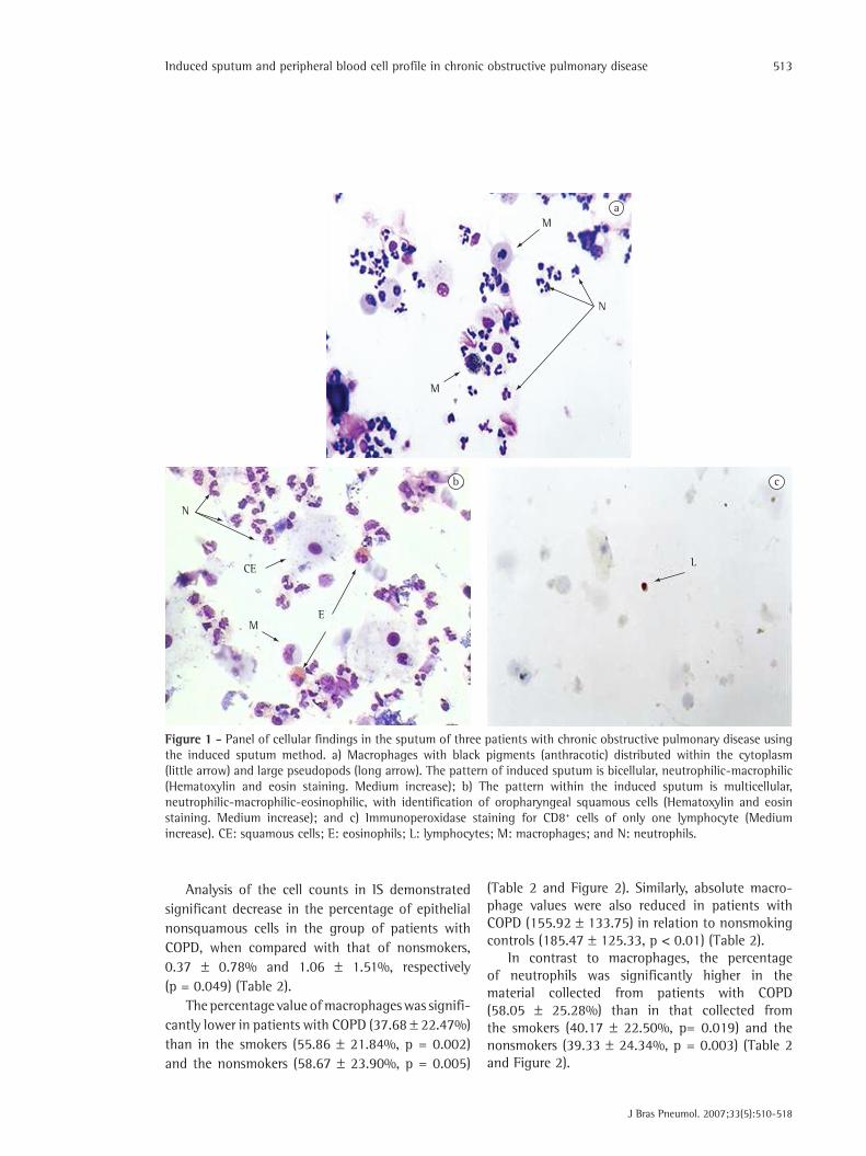
Eppendorf for determination of sol phase levels. Subsequently, the remainder was treated with dithiotreitol (Sigma Chemical, St. Louis, MO, USA), immersed in a double boiler at 37 °C for 10 min and centrifuged (1500 rpm) at 4 °C for 10 min. In this phase, a new supernatant sample was collected and stored in an Eppendorf pipette. The precipitate was resuspended in 1000 µl of phosphate buff-ered saline solution. One aliquot was separated in order to determine cellular viability by trypan blue exclusion, and total cellularity was determined in a Neubauer chamber. Cytocentrifuges were prepared with 25,000 cells/well (cytospin), fixed in chloroform:acetone. A slide stained with Diff-Quick was used in order to determine differential cellular counts, using a Nikon 155196 microscope, with a ×10 ocular and a ×40 objective, at ×400 (Figure 1).
Collected material that presented a percentage of squamous cells by hematoxylin and eosin staining of > 70% of total cells found, a number of nonsq-uamous cells < 200, or cell viability < 50%, was excluded from the analysis.(12)
Stainings were performed in lymphocyte subpop-ulations of CD4+ and CD8+ T cells, with respective CD4 monoclonal human antibodies (clone QA4120, code C-1805, lot 116H4875; Sigma Chemical) and CD8 (clone UCHT-4, code C-7423, lot 40K4830, Sigma Chemical). Immunohistochemical testing was performed using the avidin-biotin immunoperoxi-dase complex technique (Figure 1).
Peripheral blood cell counts was performed using flow cytometry by FACScount (BD Biosciences Imunocytometry Systems, San Jose, CA, USA), which provided the absolute number and percentage of leukocytes and their subtypes, as well as subpopu-lations of CD4+ and CD8+ T lymphocytes. Results are expressed as median and range or as mean and standard deviation. Initially, the sample distribu-tion was analyzed using Kolmogorov-Smirnov tests. The nonparametric Mann-Whitney two-tailed test was used for the analysis among groups, and the Spearman test was used for linear correlations. The level of statistical significance for the tests was set at p < 0.05.
Results
Demographic data of all individuals in the study and mean functional analyses are shown in Table 1.
Disease (GOLD); (1) according to which 3 cases were defined as mild, 18 as moderate, and 17 as severe.
In order to participate in the study, individuals were required to meet all of the following criteria: a) presenting α-1 antitrypsin value within the range of normality; b) having no history of respiratory infection for at least 4 weeks; c) presenting respi-ratory function test with negative bronchodilator test within one week prior; (14) d) having no history of atopic skin inflammation or upper/lower airway atopy.
Exclusion criteria were as follows: a) use of inhaled or systemic corticosteroids within one month prior; b) use of antibiotics within one month prior; c) history of tuberculosis; d) infection with acquired human immunodeficiency virus.
Three groups were formed:1) COPD: smoking patients (active smokers) with
nicotine load of > 20 pack-years, with no history of atopy, forced expiratory volume in one second (FEV1)/forced vital capacity (FVC) ≤ 70%, and negative bronchodilator test, according to the American Thoracic Society (ATS) criteria;(14)
2) Smokers: smoking patients (active smokers) with nicotine load of > 20 pack-years, with no atopy history, FEV1/FVC > 70%, FEV1 > 80% and negative bronchodilator test, according to the ATS criteria;(14)
3) Nonsmokers: individuals with no history of smoking or atopy, FEV1/FVC > 70%, FEV1 > 80%, and negative bronchodilator test, according to the ATS criteria.(14)
Venous blood sample collection was performed immediately before sputum collection. Sputum was induced according to the technique devised by Pin et al.,(12) with modifications, after pretreatment with 400 µg of inhaled albuterol to prevent bronchial obstruction symptoms. The modification of the method consisted of using increasing concentrations of 2, 3, and 4% hypertonic saline solution (10 mL per solution). Each nebulization phase lasted 7 min, totaling 21 min. The equipment used in the ultra-sonic nebulization was Ultra-NEB’99 (DeVilbiss, Somerset, PA, USA). The collected material was immediately put on ice and sent for laboratory analysis.
Grumes and the thickest parts of the sputum were selected and centrifuged (1500 rpm) at 4 °C for 20 min. The supernatant was stored in sterile
Induced sputum and peripheral blood cell profile in chronic obstructive pulmonary disease
J Bras Pneumol. 2007;33(5):510-518
513
(Table 2 and Figure 2). Similarly, absolute macro-phage values were also reduced in patients with COPD (155.92 ± 133.75) in relation to nonsmoking controls (185.47 ± 125.33, p < 0.01) (Table 2).
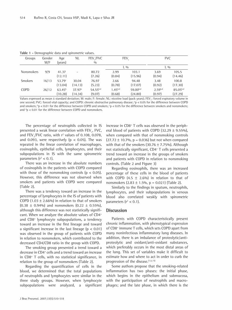
In contrast to macrophages, the percentage of neutrophils was significantly higher in the material collected from patients with COPD (58.05 ± 25.28%) than in that collected from the smokers (40.17 ± 22.50%, p= 0.019) and the nonsmokers (39.33 ± 24.34%, p = 0.003) (Table 2 and Figure 2).
Analysis of the cell counts in IS demonstrated significant decrease in the percentage of epithelial nonsquamous cells in the group of patients with COPD, when compared with that of nonsmokers, 0.37 ± 0.78% and 1.06 ± 1.51%, respectively (p = 0.049) (Table 2).
The percentage value of macrophages was signifi-cantly lower in patients with COPD (37.68 ± 22.47%) than in the smokers (55.86 ± 21.84%, p = 0.002) and the nonsmokers (58.67 ± 23.90%, p = 0.005)
N
M
M
a
b
ME
CE
N
c
L
Figure 1 - Panel of cellular findings in the sputum of three patients with chronic obstructive pulmonary disease using the induced sputum method. a) Macrophages with black pigments (anthracotic) distributed within the cytoplasm (little arrow) and large pseudopods (long arrow). The pattern of induced sputum is bicellular, neutrophilic-macrophilic (Hematoxylin and eosin staining. Medium increase); b) The pattern within the induced sputum is multicellular, neutrophilic-macrophilic-eosinophilic, with identification of oropharyngeal squamous cells (Hematoxylin and eosin staining. Medium increase); and c) Immunoperoxidase staining for CD8+ cells of only one lymphocyte (Medium increase). CE: squamous cells; E: eosinophils; L: lymphocytes; M: macrophages; and N: neutrophils.
514 Rufino R, Costa CH, Souza HSP, Madi K, Lapa e Silva JR
J Bras Pneumol. 2007;33(5):510-518
increase in CD8+ T cells was observed in the periph-eral blood of patients with COPD (32.29 ± 9.55%), when compared with that of nonsmoking controls (27.72 ± 10.7%, p = 0.036) but not when compared with that of the smokers (30.76 ± 7.75%). Although not statistically significant, CD4+ T cells presented a trend toward an increase in the groups of smokers and patients with COPD in relation to nonsmoking controls. (Table 2 and Figure 3)
Regarding eosinophils, there was an increased percentage of these cells in the blood of patients with COPD (4.5 ± 2.6%) in relation to that of nonsmokers (2.83 ± 1.5%, p = 0.021) (Table 2).
Similarly to the findings in sputum, neutrophils, lymphocytes, and their subpopulations in venous blood also correlated weakly with spirometric parameters (r2 < 0.1).
Discussion
Patients with COPD characteristically present chronic inflammation, with phenotypical expression of CD8+ immune T cells, which sets COPD apart from many noninfectious inflammatory lung diseases. In addition, there is an imbalance of proteolytic/anti-proteolytic and oxidant/anti-oxidant substances, which preferably occurs in the most distal areas of the lung. This set of variables make it difficult to estimate how and where to act in order to curb the progression of the disease.(1,6,15)
Some authors propose that the smoking-related inflammation has two phases: the initial phase, which begins in the epithelium and submucosa, with the participation of neutrophils and macro-phages; and the late phase, in which there is the
The percentage of neutrophils collected in IS presented a weak linear correlation with FEV1, FVC, and FEV1/FVC ratio, with r2 values of 0.108, 0.078, and 0.093, were respectively (p < 0.05). The was repeated in the linear correlation of macrophages, eosinophils, epithelial cells, lymphocytes, and their subpopulations in IS with the same spirometric parameters (r2 < 0.1).
There was an increase in the absolute numbers of eosinophils in the patients with COPD compared with those of the nonsmoking controls (p < 0.05). However, this difference was not observed when smokers and patients with COPD were compared (Table 2).
There was a tendency toward an increase in the percentage of lymphocytes in the IS of patients with COPD (1.03 ± 2.66%) in relation to that of smokers (0.38 ± 0.94%) and nonsmokers (0.22 ± 0.55%), although this difference was not statistically signifi-cant. When we analyze the absolute values of CD4+ and CD8+ lymphocyte subpopulations, a tendency toward an increase in the first lineage and toward a significant increase in the last lineage (p < 0.01) was observed in the group of patients with COPD in relation to nonsmokers, which contributed to the decreased CD4/CD8 ratio in the group with COPD.
The smoking group presented a trend toward a decrease in CD4+ cells and a trend toward an increase in CD8+ T cells, with no statistical significance, in relation to the group of nonsmokers (Table 2).
Regarding the quantification of cells in the blood, we determined that the total populations of neutrophils and lymphocytes were similar in the three study groups. However, when lymphocyte subpopulations were analyzed, a significant
Table 1 - Demographic data and spirometric values.
Groups GenderM/F
Age(years)
NL FEV1/FVC %
FEV1 FVC
L % L %Nonsmokers 9/9 41.37
(12.11)- 80.72
(7.26) 2.99(0.84)
103.1(15.96)
3.69(0.94)
105.5(14.46)
Smokers 16/13 53.79c
(13.04)30.04(14.13)
76.97(5.23)
2.66(0.78)
94.48(17.07)
3.48(0.92)
100.8(17.30)
COPD 26/12 63.45b (10.28)
37.97a
(14.34) 54.55b.d
(9.07)1.45b,d
(0.68)59.00b,d
(24.80) 2.59b,d
(0.97) 85.05b,d
(27.29)Values expressed as mean ± standard deviation; M: male; F: female; NL: nicotine load (pack-years); FEV1: forced expiratory volume in one second; FVC: forced vital capacity; and COPD: chronic obstructive pulmonary disease; ap < 0.05 for the difference between COPD and smokers; bp < 0.01 for the difference between COPD and smokers; cp < 0.05 for the difference between smokers and nonsmokers; and dp < 0.01 for the difference between COPD and nonsmokers.

Induced sputum and peripheral blood cell profile in chronic obstructive pulmonary disease
J Bras Pneumol. 2007;33(5):510-518
515
The IS technique has not been standardized for the study of inflammation in patients with COPD, which accounts for some differences in the method used by the studies that have been carried out, both in the processing of the material collected and in the quantification of the cells. In addition, the hypertonic saline solution might increase the number of neutrophils. The most appropriate mate-rial for cellular analysis might be the first morning sputum, which represents the accumulation of cells, cellular debris and cytokines from the large and small airways.(19,20)
The correlation between the percentage of neutrophils collected in IS with FEV1 was inversely proportional and weak (r2 = −0.108 and p = 0.002). This fact and other cellular correlations of IS and venous blood with the principal spirometric param-eters (FEV1, FVC, and FEV1/FVC) might not properly reflect the inflammatory model of COPD, which is predominant in the small airways. As we know,
additional participation of lymphocytes .(16,17) Our findings demonstrate that the numbers of CD8+ T lymphocytes and neutrophils are significantly elevated in the sputum of patients with COPD, whereas smokers present increased neutrophils in relation to nonsmokers, as has also been reported in the literature.(18) Therefore, the higher percentages of CD8+ T lymphocytes and neutrophils in IS could help to identify smokers who already present an inflammatory model similar to that observed in patients with COPD. In the literature, there are reports on increased neutrophils in bronchoalveolar lavage and in IS of patients with COPD; however, these findings have not been reproduced in studies utilizing material from bronchial biopsy. The prin-cipal explanation for this fact refers to the rapid migration of neutrophils from the vessel toward the airways, making it more common to find them within the airways than in the bronchial wall or in the pulmonary parenchyma.
Table 2 - Absolute values of cell counts in induced sputum and peripheral blood.
Induced sputum Nonsmokers Smokers COPD
Total cells (× 105/mL) 586.95 (268.29) 603.90 (232.94) 637.13 (392.68)
Macrophages (× 105/mL) 185.47 (125.33) 208.43 (111.19)c 155.92 (133.75)e
Macrophages (%) 58.67 (23.90) 55.86 (21.84) 37.68 (22.40)b,e
Eosinophils (× 105/mL) 2.47 (5.10) 7.37 (17.05) 20.66 (44.08) d
Eosinophils (%) 0.22 (0.55) 0.38 (0.94) 1.03 (2.66)Neutrophils (× 105/mL) 141.00 (112.39) 199.40 (159.12)c 312.37 (396.30)e
Neutrophils (%) 39.33 (24.34) 40.17 (22.50) 58.05 (25.28)a,e
Epithelial cells (× 105/mL) 2.50 (3.79) 3.90 (8.90) 1.50 (2.29)e
Lymphocytes (× 105/mL) 0.89 (1.53) 1.37 (2.68) 2.58 (5.37)Lymphocytes (%) 0.22 (0.55) 0.38 (0.94) 1.03 (2.66)CD4+ (× 105/mL) 0.63 (1.01) 0.47(0.90) 0.71(2.05)
CD8+ (× 105/mL) 0.26 (0.45) 1.00 (2.24) 1.89 (3.54)e
CD4/CD8 0.42 (1.02) 1.89 (3.54) 0.10 (0.25) Peripheral blood (mm3)
Nonsmokers Smokers COPDTotal cells 6593.68 (1939.08) 7011.07 (1381.13) 6701.66 (1290.57)Neutrophils 4167.95 (1752.96) 4559.75 (1368.14) 4386.57 (1188.30)Eosinophils 186.53 (114.31) 233.89 (181.19) 306.58 (189.40)e
Lymphocytes 1857.95 (705.37) 1768.21 (564.54) 1611.58 (576.95)CD4+ 583.42 (450.13) 985.14 (262.11) 908.53 (347.64)CD8+ 471.16 (137.57) 536.07 (204.70) 502.26 (175.11)e
CD4/CD8 2.22 (1.01) 2.02 (0.70) 1.99 (0.87)e
Values expressed as mean ± standard deviation; COPD: chronic obstructive pulmonary disease; ap < 0.05 for the difference between COPD and smokers; bp < 0.01 for the difference between COPD and smokers; cp < 0.05 for the difference between smokers and nonsmokers; dp < 0.05 for the difference between COPD and nonsmokers; ep < 0.01 for the difference between COPD and nonsmokers.

516 Rufino R, Costa CH, Souza HSP, Madi K, Lapa e Silva JR
J Bras Pneumol. 2007;33(5):510-518
not all smokers develop COPD, neither is the disease directly related to nicotine load.
Our finding of elevated CD8+ T cell counts in IS and peripheral blood supports the theory of the systemic involvement of COPD, since the migra-tion of these cells occurs from the vessel toward the pulmonary tissue. In addition, their differentiation and proliferation occur principally at other sites, especially in the bone marrow.(6,7,24) However, the reduced numbers of lymphocytes typically found in IS make it difficult to value it.
The CD8+ T lymphocytes have various func-tions. In COPD, the most prevalent theory refers to their participation as defense cells (cytotoxic) in frequent viral infections.(6) However, the reason why they remain in the lung is still unknown. Activated CD8+ T lymphocytes increase the rate of apoptosis of CD4+ T cells, decreasing the CD4/CD8 ratio. This mechanism depends on Fas expression and is not antigen-specific.(25) The permanence of CD8+ T lymphocytes in patients with COPD might be due to the immune mechanism known as antigen toler-
these spirometric parameters do not express only small airway disturbances.(11) In addition, IS predom-inantly represents the upper airways. Therefore, the correlation of the cellular study using the IS tech-nique and spirometry might not be the ideal form of studying inflammation in COPD, since the initial and preferential site of the inflammatory process occurs in the small airways.
In this study, absolute counts and percentages of macrophages were lower among the patients with COPD. Since the population of macrophages collected in the IS of individuals without respiratory disease represents approximately the total of cells, it is understandable that, in patients with COPD, this percentage is decreased, due to the increase of other cellular populations.(13,21)
The alveolar macrophages of patients with COPD are pleomorphic and, in many cases, contain anthra-cotic pigments within their cytoplasm (see Figure 1 section A). This finding constitutes the principal factor for the macrophage antigen theory.(16,22,23) Macrophages would activate and recruit other cells for lung protection. However, since these anthracotic particles are not effectively cleaned, the inflamma-tory process would be perpetuated. However, there is as yet no consensus regarding this theory, since
0.0190.0030.001
0.002
Nonsmokers Smokers COPDM MMN NN
100
75
50
25
0
Cells
(%)
Lym
phoc
ytes
in
per
iphe
ral b
lood
(%)
90
80
70
60
50
40
30
20
10CD4 CD4 CD4CD8 CD8 CD8
0.04
0.08
Nonsmokers Smokers COPD
Figure 2 - Percentage data of macrophages and neutrophils observed in induced sputum. The horizontal line between hollow or full dots represents mean values. The statistical analysis was carried out using the two-tailed Mann-Whitney test. M: macrophages; N: neutrophils; and COPD: chronic obstructive pulmonary disease.
Figure 3 - Percentage data of lymphocyte subtypes in peripheral blood – CD4+ T lymphocytes () and CD8+ T lymphocytes () – together with mean values (). The statistical analysis was carried out using the two-tailed Mann-Whitney test. Significant increase of CD8+ T lymphocytes was found in patients with chronic obstructive pulmonary disease (COPD), compared with nonsmoking controls.

Induced sputum and peripheral blood cell profile in chronic obstructive pulmonary disease
J Bras Pneumol. 2007;33(5):510-518
517
UFRJ, as well as the staff of the Multidisciplinary Research Laboratory of the UFRJ: Patrícia Martins Lago, Vera Cristina da Silva Flores Batista, Lorena de Oliveira Rego Peçanha, Cesônia de Assis Martinusso, and Alessandra Ferreira Coelho.
References
1. GOLD - Global strategy for the diagnosis, management, and prevention of chronic obstructive pulmonary disease [Homepage on the Internet]. Executive Summary, Global Strategy for the Diagnosis, Management, and Prevention of COPD Updated 2005 [cited 2006 Sep 9]. Available from: http://www.goldcopd.org/Guidelineitem.asp?l1=2&l2=1&intId=1662
2. Walker RI, Willemze R. Neutrophil kinetics and the regulation of granulopoiesis. Rev Infect Dis. 1980;2(2):282-92.
3. Cosio MG, Guerassimov A. Chronic obstructive pulmonary disease. Inflammation of small airways and lung parenchyma. Am J Respir Crit Care Med. 1999;160(5 Pt 2):S21-5.
4. Cosio M, Ghezzo H, Hogg JC, Corbin R, Loveland M, Dosman J, et al. The relations between structural changes in small airways and pulmonary-function tests. N Engl J Med. 1978;298(23):1277-81.
5. Janoff A. Elastases and emphysema. Current assessment of the protease-antiprotease hypothesis. Am Rev Respir Dis. 1985;132(2):417-33.
6. Barnes PJ, Shapiro SD, Pauwels RA. Chronic obstructive pulmonary disease: molecular and cellular mechanisms. Eur Respir J. 2003;22(4):672-88.
7. Hogg JC. Pathophysiology of airflow limitation in chronic obstructive pulmonary disease. Lancet. 2004;364(9435):709-21.
8. Shapiro SD. Evolving concepts in the pathogenesis of chronic obstructive pulmonary disease. Clin Chest Med. 2000;21(4):621-32.
9. Di Stefano A, Capelli A, Lusuardi M, Balbo P, Vecchio C, Maestrelli P, et al. Severity of airflow limitation is associated with severity of airway inflammation in smokers. Am J Respir Crit Care Med. 1998;158(4):1277-85.
10. Turato G, Zuin R, Miniati M, Baraldo S, Rea F, Beghe B, et al. Airway inflammation in severe chronic obstructive pulmonary disease: relationship with lung function and radiologic emphysema. Am J Respir Crit Care Med. 2002;166(1):105-10.
11. Amadori A, Zamarchi R, De Silvestro G, Forza G, Cavatton G, Danieli GA, et al. Genetic control of the CD4/CD8 T-cell ratio in humans. Nat Med. 1995;1(12):1279-83.
12. Pin I, Gibson PG, Kolendowicz R, Girgis-Gabardo A, Denburg JA, Hargreave FE, et al. Use of induced sputum cell counts to investigate airway inflammation in asthma. Thorax. 1992;47(1):25-9.
13. O’Donnell R, Breen D, Wilson S, Djukanovic R. Inflammatory cells in the airways in COPD. Thorax. 2006;61(5):448-54.
14. Lung function testing: selection of reference values and interpretative strategies. American Thoracic Society. Am Rev Respir Dis. 1991;144(5):1202-18.
15. Hogg JC, Chu F, Utokaparch S, Woods R, Elliott WM, Buzatu L, et al. The nature of small-airway obstruction in chronic obstructive pulmonary disease. N Engl J Med. 2004;350(26):2645-53.
ability, that is, if the antigen stimulus is persistent and occasionally very intense, CD8+ T cells would regulate the intensity of the immune response.
In patients with stable disease, some studies have demonstrated increased eosinophils in sputum, in bronchoalveolar lavage, and in the airway walls.(13) It has been observed that smokers with no history of asthma can be more sensitized to certain occupa-tional allergens and present greater immunoglobulin E and blood eosinophil values than do nonsmokers and individuals without a history of allergy.(26,27) However, other authors have been unable to repro-duce these data.(13,28) Other authors(29) reported increased cell counts in some patients, suggesting that eosinophils in sputum could be related to a better response to treatment with corticoster-oids. In the present study, we observed a tendency toward an increase in the numbers of these cells in the material collected from the airways of patients with bronchial obstruction (3.97 ± 8.35%), which cannot be attributed to pulmonary infection, since the patients had been free of exacerbations for moe than one month. Therefore, eosinophils can partici-pate in the inflammatory process in COPD as well as in periods of clinical stability.(29,30) The analysis of peripheral blood demonstrated significant increase of eosinophils (p = 0.021); in addition, the leuko-cyte percentages and total values were within the range of normality, a fact that also strengthens the characterization of the participation of eosinophils in COPD.
In conclusion, neutrophil, eosinophil, macro-phage, and CD8+ T lymphocyte counts were elevated in IS, which indicates an inflammatory model of multicellular involvement in the pathogenesis of COPD. In addition, CD8+ T cell counts were also elevated in peripheral blood during the stable phase of the disease. This fact suggests that this cell can serve as a marker of systemic inflammation and facilitate the early identification of smokers who already present an inflammatory pattern of COPD. To that end, new studies are required to better char-acterize the role of lymphocytes in the pathogenesis of COPD.
Acknowledgments
We would like to thank Drs. Alberto José de Araújo and Arnaldo José Noronha Filho, and the depart-ment of Respiratory Function Test of the HUCFF/

518 Rufino R, Costa CH, Souza HSP, Madi K, Lapa e Silva JR
J Bras Pneumol. 2007;33(5):510-518
23. Hogg JC, Senior RM. Chronic obstructive pulmonary disease - part 2: pathology and biochemistry of emphysema. Thorax. 2002;57(9):830-4.
24. Shaw RJ, Djukanovic R, Tashkin DP, Millar AB, du Bois RM, Orr PA. The role of small airways in lung disease. Respir Med. 2002 Feb;96(2):67-80.
25. Liu CC, Young LH, Young JD. Lymphocyte-mediated cytolysis and disease. N Engl J Med. 1996;335(22):1651-9.
26. O’Connor GT, Sparrow D, Weiss ST. The role of allergy and nonspecific airway hyperresponsiveness in the pathogenesis of chronic obstructive pulmonary disease. Am Rev Respir Dis. 1989;140(1):225-52.
27. Sluiter HJ, Koëter GH, de Monchy JG, Postma DS, de Vries K, Orie NG. The Dutch hypothesis (chronic non-specific lung disease) revisited. Eur Respir J. 1991;4(4):479-89.
28. Saetta M. Airway inflammation in chronic obstructive pulmonary disease. Am J Respir Crit Care Med. 1999;160(5 Pt 2):S17-20.
29. Pizzichini E, Pizzichini MM, Gibson P, Parameswaran K, Gleich GJ, Berman L, et al. Sputum eosinophilia predicts benefit from prednisone in smokers with chronic obstructive bronchitis. Am J Respir Crit Care Med. 1998;158(5 Pt 1):1511-7.
30. Saetta M, Di Stefano A, Maestrelli P, Turato G, Ruggieri MP, Roggeri A, et al. Airway eosinophilia in chronic bronchitis during exacerbations. Am J Respir Crit Care Med. 1994;150(6 Pt 1):1646-52.
16. O’Shaughnessy TC, Ansari TW, Barnes NC, Jeffery PK. Inflammation in bronchial biopsies of subjects with chronic bronchitis: inverse relationship of CD8+ T lymphocytes with FEV1. Am J Respir Crit Care Med. 1997;155(3):852-7.
17. Saetta M, Di Stefano A, Turato G, Facchini FM, Corbino L, Mapp CE, et al. CD8+ T-lymphocytes in peripheral airways of smokers with chronic obstructive pulmonary disease. Am J Respir Crit Care Med. 1998;157(3 Pt 1):822-6.
18. Bosken CH, Hards J, Gatter K, Hogg JC. Characterization of the inflammatory reaction in the peripheral airways of cigarette smokers using immunocytochemistry. Am Rev Respir Dis. 1992;145(4 Pt 1):911-7.
19. Kips JC, Peleman RA, Pauwels RA. Methods of examining induced sputum: do differences matter? Eur Respir J. 1998;11(3):529-33.
20. Parameswaran K, Pizzichini MM, Li D, Pizzichini E, Jeffery PK, Hargreave FE. Serial sputum cell counts in the management of chronic airflow limitation. Eur Respir J. 1998;11(6):1405-8.
21. Barnes PJ. Mediators of chronic obstructive pulmonary disease. Pharmacol Rev. 2004;56(4):515-48.
22. Cosio MG, Majo J, Cosio MG. Inflammation of the airways and lung parenchyma in COPD: role of T cells. Chest. 2002;121(5 Suppl):160S-165S.