Produção, purificação parcial por Fermentação Extrativa em ... · A todos os funcionários...
144
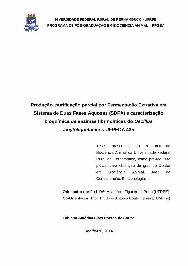
UNIVERSIDADE FEDERAL RURAL DE PERNAMBUCO - UFRPE PROGRAMA DE PÓS-GRADUAÇÃO EM BIOCIÊNCIA ANIMAL – PPGBA Produção, purificação parcial por Fermentação Extrativa em Sistema de Duas Fases Aquosas (SDFA) e caracterização bioquímica de enzimas fibrinolíticas do Bacillus amyloliquefaciens UFPEDA 485 Fabiana América Silva Dantas de Souza Recife-PE, 2014
-
Upload
phungtuyen -
Category
Documents
-
view
217 -
download
0
Transcript of Produção, purificação parcial por Fermentação Extrativa em ... · A todos os funcionários...

UNIVERSIDADE FEDERAL RURAL DE PERNAMBUCO - UFRPE
PROGRAMA DE PÓS-GRADUAÇÃO EM BIOCIÊNCIA ANIMAL – PPGBA
Produção, purificação parcial por Fermentação Extrativa em
Sistema de Duas Fases Aquosas (SDFA) e caracterização
bioquímica de enzimas fibrinolíticas do Bacillus
amyloliquefaciens UFPEDA 485
Fabiana América Silva Dantas de Souza
Recife-PE, 2014
NIVERSIDADE FEDERAL RURAL DE PERNAMBUCO - UFRPE
PROGRAMA DE PÓS-GRADUAÇÃO EM BIOCIÊNCIA ANIMAL – PPGBA
Produção, purificação parcial por Fermentação Extrativa em
Sistema de Duas Fases Aquosas (SDFA) e caracterização
bioquímica de enzimas fibrinolíticas do Bacillus
amyloliquefaciens UFPEDA 485
Tese apresentada ao Programa de
Biociência Animal da Universidade Federal
Rural de Pernambuco, como pré-requisito
parcial para obtenção do grau de Doutor
em Biociência Animal. Área de
Concentração: Biotecnologia.
Orientador (a): Prof. Drª. Ana Lúcia Figueiredo Porto (UFRPE)
Co-Orientador: Prof. Dr. José António Couto Teixeira (UMinho)
Fabiana América Silva Dantas de Souza
Recife-PE, 2014

Dedico mais esta vitória a Deus que sempre
me deu força e fé para relizar tudo que
parecia ser impossível.
Aos meus pais Livanete Dantas Souza e
José Manoel de Souza pelas lições de
esperança e amor incondicional.
À minha irmã Flaviana América Souza pelo
apoio, companheirismo, amor fraternal e
paciência pelas minhas ausências.
Às minhas sobrinhas Luana Clara Alves e
Diana Dantas que tornaram esta jornada
mais leve e feliz.

AGRADECIMENTOS
À minha orientadora Prof.ª Dr.ª Ana Lúcia Figueiredo Porto pela dedicação, estímulo,
confiança, respeito, amizade e por proporcionar desafios que me fizeram crescer e aprender
ainda mais.
Ao meu co-orientador Prof. Dr. José António Couto Teixeira pela paciência,
dedicação, respeito e confiança.
À Universidade Federal Rural de Pernambuco, à Universidade Federal do Ceará e à
Universidade do Minho. À Pós-graduação em Biociência Animal.
À CAPES pela bolsa de pesquisa que proporcionou a minha estadia de um ano na
Europa, resultando na minha total dedicação ao desenvolvimento desta pesquisa.
A todos os funcionários que fazem parte do Departamento de Morfologia e Fisiologia
Animal (DMFA), em especial Márcia Domingos e a Secretária Edna Cherias pela paciência e
dedicação.
Às Professoras do DMFA, Prof.ª Dr.ª Tatiana Porto e Prof.ª Dr.ª Taciana Cavalcanti,
pelo auxílio quando necessário. Ao Prof. Dr. Valdemiro Júnior por intermediar a minha ida
para Universidade Federal do Ceará - UFC e pela consideração e amizade. Ao Prof. Dr.
Romildo Nogueira pela concideração, atenção, respeito e amizade. Ao Prof. Dr. Fabrício
Bezerra de Sá pela paciência na coleta de sangue de cavalo para os experimentos e, em
especial, à Prof.ª Dr.ª Raquel Pedrosa Bezerra pela dedicação, atenção, confiança, respeito
e amizade.
Aos Professores da Universidade Federal do Ceará, Prof. Dr. Edson Holanda
Teixeira e Prof. Dr. Benildo Sousa Cavada, por permitirem minha estadia no Laboratório de
Moléculas Biologicamente Ativas e pelo incentivo e confiança.
Aos Pós-doutorandos Cyntia Nascimento, André Mota, Polyana Herculano, Camila
Porto, Romero Costa e, em especial, a Daniela de Araújo Viana Marques pela contribuição,
consideração, respeito e amizade.
Aos companheiros de turma, em especial ao meu grande amigo Mauro José
Gonçalves Bezerra pelo companheirismo e pelos ensinamentos de vida que vou levar para
sempre comigo.
A todos do LABTECBIO e CENAPESQ, em especial aos amigos Amanda Sales
(Mandinha), Júlio Cézar dos Santos Nascimento e Páblo Eugênio Costa e Silva pelo
companheirismo, cumplicidade, parceria e pelas valiosas contribuições e inesquecíveis
momentos juntos.
A todos do Laboratório de Moléculas Biologicamente Ativa, em especial ao amigo
Mayron Vasconcelos pelos valiosos ensinamentos
.

A todos do Laboratório de Fermentação (LF), em especial a Mariana Pimenta
Machado Braga dos Anjos e Maycon Kelbert pela contribuição, respeito, cumplicidade e
amizade. Ao grande amigo Luís Flávio do Rosário Machado que não está mais entre nós,
mas quando tinha o privilégio de sua companhia, tornava o dia no laboratório mais leve com
sua doçura e gentileza desinteressada que fazia toda diferença.
Ao pessoal do Laboratório de Indústria e Processos (LIP) que me receberam tão
calorosamente em Portugal, em especial aos meus amigos Eduardo José Valença
Cordeiro Pires, Tânia Ferreira, Miguel Cerqueira, Michelly Menezes e Cláudia
Araújo.
Aos meus grandes amigos colombianos Lina Ballesteros, Sebas Ballesteros e
minhas amigas mexicanas Luz Ramos Ponce, Marlet Martinez e ao meu amigo Prof. Dr.
Adilson Silva, Alessandra Accioly e Jocemar Accioly pela força e companheirismo nos
momentos mais importantes da minha estadia em Portugal.
À Drª Germana Medeiros e Silva pelos primeiros ensinamentos sobre proteases
fibrinolíticas, pelo respeito, consideração e amizade.
À minha amiga e companheira Fabiana Félix, que me acompanhou na jornada pela
Universidade Federal do Ceará.
Aos meus alunos da turma de Biologia, na disciplina de Bioquímica Molecular pelo
respeito e confiança.
Aos alunos de Iniciação Ciêntífica Kildare Rossano dos Santos Silva, Erika Raysa
Pinto Bezerra, Débora Rafaela Lourenço de Paula e Anielly Karoline da Silva Freire pela
dedicação, respeito e confiança.
Aos amigos Cleide Bunzen, Salomão de Andrade Ferreira, Patrícia Campos Querioz,
Marcos Antônio Pereira da Costa Júnior e a minha amiga argentina Drª. Mariana Cabrera
pelo apoio nos momentos em que mais precisei.
Ao André Filipe Pessoa, que acompanhou toda a minha trajetória acadêmica desde
quando tudo começou, pelo apoio, companheirismo, cumplicidade, paciência pelas minhas
ausências e por fazer a diferença na minha história de vida, será sempre um amigo muito
especial.
À Profª Drª Alexandra Amorim Salgueiro pela consideração, amizade, apoio
acadêmico e valiosos ensinamentos de vida.
Aos membros externos da banca Prof. Dr. Daniel Pereira da Silva, e em especial à
Profª. Drª. Leonie Asfora Sarubbo pela consideração e valiosas contribuições durante minha
trajetória acadêmica.
,

O que sabemos é uma gota e o que ignoramos é um oceano.
(Isaac Newton)
SUMÁRIO
LISTA DE FIGURAS ............................................................................................... XIII
LISTA DE TABELAS ............................................................................................ XVIII
LISTA DE ABREVIATURAS E SÍMBOLOS ........................................................... XXI
Resumo ................................................................................................................ XXIII
Abstract ................................................................................................................ XXIV
INTRODUÇÃO ....................................................................................................... XXV
OBJETIVOS ......................................................................................................... XXVII
Objetivo geral ....................................................................................................... XXVII
Objetivos específicos ........................................................................................... XXVII
CAPÍTULO I .............................................................................................................. 30
1. REVISÃO BIBLIOGRÁFICA ................................................................................. 30
1.1. O gênero Bacillus e sua importância na produção de enzimas de interesse
industrial ................................................................................................................. 30
1.2. Proteases: potencial terapêutico e aplicações atuais ...................................... 33
1.3. Proteases com propriedades anticoagulantes ................................................ 35
1.4. Proteases com propriedades fibrinolíticas....................................................... 39
1.4.1. Obtenção e comercialização de enzimas fibrinolíticas ............................. 43
1.4.2. Propriedades bioquímicas das enzimas fibrinolíticas produzidas pelo
gênero Bacillus ................................................................................................... 49
1.4.3. Otimização e escalonamento da produção de enzimas fibrinolíticas ........ 51

1.5. Fermentação extrativa utilizando sistema de duas fases aquosas (SDFA).... 54
2. REFERÊNCIAS BIBLIOGRÁFICAS ..................................................................... 58
CAPÍTULO II ............................................................................................................. 77
Optimization of production, biochemical characterization and evaluation of the
therapeutic potential in vitro of a new fibrinolytic enzyme from Bacillus
amyloliquefaciens UFPEDA 485 ............................................................................ 78
Abstract .................................................................................................................... 79
1. INTRODUCTION ................................................................................................... 80
2. MATERIALS AND METHODS .............................................................................. 81
2.1. Reagents ......................................................................................................... 81
2.2. Screening and culture conditions .................................................................... 81
2.3. Microbial identification ..................................................................................... 82
2.4. Selection of the best condition for production of fibrinolytic enzyme by
Response Surface Methodology (RSM) using a Central Composite Design (CCD)82
2.5. Determination of total protein .......................................................................... 83
2.6. Assay of fibrinolytic activity by degradation of blood clots ............................... 83
2.7. Determination of fibrinolytic activity at the fibrin plate ...................................... 84
2.8. Fibrinolytic activity determination by spectrometry .......................................... 85
2.9. Determination of optimum pH and temperature of the enzyme ....................... 85
2.10. Effect of pH and temperature on the stability of the enzyme ......................... 86
2.11. Effects of inhibitors and metal ions at the fibrinolytic activity ........................ 86

3. RESULTS AND DISCUSSION .............................................................................. 86
3.1. Screening fibrinolytic enzyme producer........................................................... 86
3.2. Bacteria identification using 16S and rpoB genes ........................................... 88
3.3. Optimization of the conditions for production of the fibrinolytic enzyme from
Bacillus amyloliquefaciens UFPEDA 485 ............................................................... 89
3.4.Evaluation of fibrinolytic activity by degradation of blood clots ......................... 93
3.5.Effects of pH and temperature on the activity and stability of the enzyme ...... 94
3.6.Effects of inhibitors and metal ions on the fibrinolytic activity ........................... 96
4. CONCLUSION ..................................................................................................... 98
Acknowledgments .................................................................................................. 99
5. LITERATURE CITED ........................................................................................... 99
CAPÍTULO III .......................................................................................................... 103
Process scale-up and biochemical characterization of an enzyme with
anticoagulant and fibrinolytic potential from Bacillus amyloliquefaciens
UFPEDA 485 ......................................................................................................... 104
Abstract .................................................................................................................. 104
1. INTRODUCTION ................................................................................................. 105
2. MATERIALS AND METHODS ............................................................................ 106
2.1. Reagents ....................................................................................................... 106
2.2. Microoganism ................................................................................................ 106
2.3. Culture condition ........................................................................................... 107
2.4. Statistical analysis and experimental design ................................................. 107
2.5. Protein concentration .................................................................................... 108
2.6. Fibrinolytic activity ......................................................................................... 108
2.7. Evaluation anticoagulant effect ..................................................................... 108
2.8. Amidolytic activity determination and substrate specificity ............................ 109
2.9. Determination of kinetic parameters .............................................................. 109
2.10. Determination of optimum pH and temperature for the enzyme activity ...... 109
2.11. Effects of pH and temperature on the stability of the enzyme ..................... 110
2.12. Effects of inhibitors and metal ions on the amidolytic activity of the fibrinolytic
enzyme ................................................................................................................ 110
2.13. Storage stability ........................................................................................... 110
3. RESULTS AND DISCUSSION ............................................................................ 111
3.1. Conditions for production of enzyme by Bacillus amyloliquefaciens UFPEDA
485 in bioreactor .................................................................................................. 111
3.2.Evaluation of the anticoagulant effect of the enzyme ..................................... 112
3.3.Substrate specificity and amidolytic activity of the fibrinolytic protease .......... 112
3.4.Kinetic parameters of the enzyme with amidolytic activity .............................. 113
3.5.Effects of inhibitors and metal ions on the amidolytic activity of the fibrinolytic
enzyme ................................................................................................................ 114
3.6.Effects of pH and temperature on the activity and stability of the enzyme ..... 114
3.7.Storage stability .............................................................................................. 115
4. CONCLUSION ................................................................................................... 116
Acknowledgments ................................................................................................ 116

5. REFERENCES ................................................................................................... 117
CAPÍTULO IV .......................................................................................................... 131
Integrated process Production and Extraction of the Fibrinolytic Protease from
Bacillus sp. UFPEDA 485 ...................................................................................... 132
Abstract .................................................................................................................. 132
1. INTRODUCTION ................................................................................................. 133
2. MATERIALS AND METHODS ............................................................................ 133
2.1. Microorganism ............................................................................................... 133
2.2. Culture conditions ......................................................................................... 134
2.3. Medium for extractive fermentation using ATPS ........................................... 134
2.4. Factorial design 23......................................................................................... 134
2.5. Determination of total protein ........................................................................ 135
2.6. Determination of fibrinolytic activity on fibrin plates ....................................... 135
2.7. Determination of fibrinolytic activity ............................................................... 135
2.8. Biochemical characterization of fibrinolytic protease ..................................... 135
2.9. Effect and stability to pH and temperature on fibrinolytic activity ................... 136
2.10. Effect of metal ions on fibrinolytic activity .................................................... 136
2.11. Effect of inhibitors in fibrinolytic activity ....................................................... 136
2.12. Methodology for the analysis of the results ................................................. 136
3. RESULTS AND DISCUSSION ............................................................................ 137
3.1. Extractive fermentation of the fibrinolytic protease ........................................ 137

3.2. Partition coefficient of the fibrinolytic protease .............................................. 138
3.3. Protease Inhibitors on fibrinolytic ativity ........................................................ 140
3.4. Metal ions on fibrinolytic activity .................................................................... 141
3.5.Optimum temperature and stability of the fibrinolytic protease ....................... 141
3.6.Optimum pH and stability of the fibrinolytic protease ..................................... 141
4. CONCLUSION ................................................................................................... 143
Acknowledgments ................................................................................................ 143
5. REFERENCES ................................................................................................... 143
CONSIDERAÇÕES FINAIS .................................................................................... 145

XIII
LISTA DE FIGURAS
CAPÍTULO I
Figura 1. Proteases aplicadas com sucesso para terapias procoagulante: (FBN -
fibronectina), (FII - protrombina); anticoagulante: (APC - proteína C ativada);
fibrinolítica: (PMGN – plasminogênio), (PMN – plasmina), (TNKase ou TNK-t-PA -
tenecteplase); anti-hipertensiva: (KAL – calicreína) e citoprotetora: (APC -
proteína C ativada). Algumas proteases (mostradas em círculos verdes escuros)
foram aprovadas para uso clínico. Cofatores de proteínas estão representados por
retângulos alaranjados com arestas arredondadas ................................................... 34
Figura 2. Estrutura cristalizada da proteína C humana ativada (PCa) complexada
com D-Phe-Pro-Arg-Chloromethylketone (PPACK) ................................................. 37
Figura 3. Sistema anticoagulante mostrando a ação da proteína C ativada sobre os
fatores de coagulação Va (Ac-globulina ativada) e VIIIa (globulina anti-hemofílica
ativada) .................................................................................................................... 38
Figura 4. Estrutura do fibrinogênio contendo o domínio central E, os domínios
globulares externos D com suas respectivas cadeias polipeptídicas (α, β e ɣ) e a
clivagem dos fibrinopeptídeos A (FpA) e B (FpB) por ação da trombina, expondo os
locais de polimerização para interações com as moléculas vizinhas e formação de
fibrina insolúvel .......................................................................................................... 40

XIV
Figura 5. (A) Complexo do domínio catalítico da plasmina humana com
estreptoquinase e (B) Estrutura cristalizada da Nattokinase (enzima fibrinolítica) do
Bacillus subtilis natto ................................................................................................. 42
Figura 6. Mecanismo de ação do sistema fibrinolítico .............................................. 43
Figura 7. Representação simplificada do processo integrado da Fermentação
Extrativa em Sistema de Duas Fases Aquosas (SDFA) para enzimas fibrinolíticas
extracelulares ............................................................................................................ 56
CAPÍTULO II
Figure 1. Response surface plot showing the effect of independent variables:
soybean flour (%) and glucose (%) with agitation at 200 rpm, after 48 hours of
cultivation on the response variable Fibrinolytic Activity (FA) of Bacillus
amyloliquefaciens UFPEDA 485 ............................................................................... 92
Figura 2. Degradation of blood clots by dispersion of red blood cells. (a) Blood clots
after 1 hour in the enzyme extract from Bacillus amyloliquefaciens UFPEDA 485 (in
triplicate), (b) Blood clot after 1 hour in physiological solution ................................... 94
Figure 3. (A) Effect of pH on the fibrinolytic activity relative of the enzyme from
Bacillus amyloliquefaciens UFPEDA 485 after 1 h of incubation. (B) Effect of pH on
the stability of enzyme measured at intervals of 2 h per 12 h of incubation of the
enzyme and expressed as percentage of residual activity. Buffers used: Glycine-HCl
(pH 3.0), Sodium acetate (pH 4.0-5.0), Citrate phosphate (pH 6.0), Tris-HCl (pH 7.0-
XV
8.0), Glycine-NaOH (pH 9.0-10.0). All buffer concentrations were 20 mM. Each value
is the average of the results of three experiments, and the error bars show the
standard deviations ................................................................................................... 95
Figure 4. (A) Effect of temperature on the fibrinolytic activity relative of the enzyme
from Bacillus amyloliquefaciens UFPEDA 485 after 1 h of incubation; (B) Effect of
temperature on the stability of enzyme measured at intervals of 2 h per 12 h of
incubation of the enzyme and expressed as percentage of residual activity. Each
value is the average of the results of three experiments, and the error bars show the
standard deviations ................................................................................................... 96
CAPÍTULO III
Figure 1. Scatterplot of the interaction between the independent variables agitation
(rpm) and the aeration (vvm) for the response variables fibrinolytic activity of the
protease from Bacillus amyloliquefaciens UFPEDA 485 ......................................... 126
Figure 2. Scatterplot of the interaction between the independent variables agitation
(rpm) and the aeration (vvm) for the response variables amidolytic activity of the
protease from Bacillus amyloliquefaciens UFPEDA 485 ......................................... 127
Figure 3. Cell growth (■), Fibrinolytic activity - FA (▲) and Amidolytic activity - AA (○)
over time of culture of Bacillus amyloliquefaciens UFPEDA 485 in bioreactor under
optimum conditions studied (800 rpm and 1.5 vvm) ............................................... 128

XVI
Figure 4. Fibrinolytic activity (▲) and Amidolytic activity (○) in (A) Effect of pH on the
activity of enzyme from Bacillus amyloliquefaciens UFPEDA 485 and in (B) Effect of
pH on the stability of enzyme measured after 1 h incubation and expressed as
percentage of residual activity. Buffers used: Glycine-HCl (pH 3.0), Sodium acetate
(pH 4.0-5.0), Citrate phosphate (pH 6,0), Tris-HCl (pH 7.0-8.0), Glycine-NaOH (pH
9.0-10.0). All buffer concentrations were 20 mM. To determine the amidolytic and
fibrinolytic activity was used as substrate N-succinyl-Ala-Ala-Pro-Phe-pNA and fibrin,
respectively. The data are presented as means ± SD (n = 3) from three independent
experiments ............................................................................................................. 129
Figure 5. Fibrinolytic activity (▲) and Amidolytic activity (○) in (A) Effect of
temperature on the activity enzyme from Bacillus amyloliquefaciens UFPEDA 485
and in (B) Effect of temperature on the stability of enzyme measured after 1 h
incubation and expressed as percentage of residual activity. Effects of temperature
were determined at (4, 25, 37, 40, 50, 60, 70 and 80 ⁰C). To determine the amidolytic
and fibrinolytic activity was used as substrate N-succinyl-Ala-Ala-Pro-Phe-pNA and
fibrin, respectively. The data are presented as means ± SD (n = 3) from three
independent experiments ........................................................................................ 130
CAPÍTULO IV
Figure 1. Cubic plot of the interaction between the variables salt concentration
(Csalt), PEG concentration (CPEG), and PEG molar mass (MPEG) for fibrinolytic
activity response in the bottom phase of extractive Fermentation ........................... 139
XVII
Figure 2. Simultaneous effects of salt concentration (Csalt) and PEG molar mass
(MPEG) for fibrinolytic activity in the bottom phase of extractive fermentation ........ 140
Figure 3. Optimum temperature and stability of the fibrinolytic protease produced by
Bacillus sp. UFPEDA 485 in the bottom phase of extractive fermentation .............. 142
Figure 4. Optimum pH and stability of the fibrinolytic protease produced by Bacillus
sp. UFPEDA 485 in the bottom phase of extractive fermentation ............................ 142
XVIII
LISTA DE TABELAS
CAPÍTULO I
Tabela 1. Enzimas de interesse industrial produzidas pelo gênero Bacillus ............. 32
Tabela 2. Fontes de enzimas fibrinolíticas ................................................................ 45
Tabela 3. Empresas envolvidas no desenvolvimento e comercialização de enzimas
fibrinolíticas ............................................................................................................... 47
Tabela 4. Preço comercial de alguns agentes fibrinolíticos ...................................... 48
Tabela 5. Propriedades bioquímicas das proteases fibrinolíticas do gênero Bacillus..50
Tabela 6. Comparação do perfil amino terminal das cadeias peptídicas das enzimas
fibrinolíticas produzidas por espécies de Bacillus ..................................................... 51
CAPÍTULO II
Table 1. Microorganisms of the genus Bacillus and their fibrinolytic activity after 48h
of cultivation in soybean medium (MS-2), pH 7.2, 150 rpm and 37°C ....................... 87
Table 2. Matrix of the Central Composite Design (CCD) star for the optimization of
the conditions for production of the fibrinolytic enzyme from Bacillus
amyloliquefaciens UFPEDA 485 ............................................................................... 90

XIX
Table 3. Analysis of variance (ANOVA) for the response fibrinolytic activity, over the
independent variables soybean flour (%), glucose (%) and agitaton (rpm), according
to Central Composite Design (CCD) star, with 95% confidence level ....................... 91
Table 4. Effects of inhibitors on the fibrinolytic activity of the enzyme ....................... 97
Table 5. Effects of metal ions on the fibrinolytic activity of the enzyme ..................... 98
CAPÍTULO III
Table 1. Levels and values of independent variables of the full 22 factorial design .120
Table 2. Analysis of variance for the responses amidolytic activity and fibrinolytic
activity, over the independent variables agitation and aeration, according to 22 full
statistical design ...................................................................................................... 121
Table 3. Substrate specificity for amidolytic activity of the fibrinolytic enzyme ........ 122
Table 4. Effects of inhibitors on the amidolytic activity of the fibrinolytic enzyme .... 123
Table 5. Effects of metal ions on the amidolytic activity of the fibrinolytic enzyme .. 124
CAPÍTULO IV
Table 1. Levels of the independent variables of the full factorial design 23 for the
extractive fermentation process of the fibrinolytic protease from Bacillus sp. UFPEDA
485 .......................................................................................................................... 134
Table 2. Matrix decoded and results of the full factorial design 23 for the responses:
the partition coefficient (K) and activity in the ATPS top and bottom phases .......... 137

XX
Table 3. Calculated effect of the responses in the factorial design 23 for the
integrated production and purification of the fibrinolytic protease by Bacillus
sp.UFPEDA 485 ...................................................................................................... 139
Table 4. Effect of inhibitors on the fibrinolytic activity .............................................. 140
Table 5. Effect of the metal ions on the fibrinolytic activity ...................................... 141

XXI
LISTA DE ABREVIATURAS E SÍMBOLOS
A - Adenina
AFi - Atividade Fibrinolítica na fase inferior
AFs - Atividade Fibrinolítica na fase superior
APC ou PCa - Proteína C
Arg – Arginina
BSA – Bovine Serum Albumin
C4b – Proteína de ligação da proteína S
D-Di - Dímero D
EC - Comissão de Enzimas
EDTA - Ácido etilenodiaminotetracético - C10H16N2O8
Fator Va - Ac-globulina ativado na cascata de coagulação
Fator VIIIa - Globulina anti-hemofílica ativada na cascata de coagulação
Fator Xa - Fator Stuart ativo na cascata de coagulação
Fator XII - Fator Hageman
FBN - Fibronectina
FDA - Food and Drug Administration
FE- Fermentação extrativa
FII - Protrombina
FOB - Free on board
FpA - Fibrinopeptídeos A, peptídeos N-terminais
FpB - Fibrinopeptídeos B, peptídeos N-terminais
G – Guanina
Glu - Glutamina
K - Coeficiente de Partição
KAL - Calicreína
kDa - Kilodaltons (peso molecular)
Km - Constante de Michaelis-Menten
MSR - Metodologia de Superfície de Resposta
OMS - Organiszação Mundial de Saúde
PAI - 1 - Inibidor do ativador de plasminogênio t-PA
PAI - 2 - Inibidor do ativador de plasminogênio u-PA
PCa-PS - Complexo de proteínas C e S ativo

XXII
PCC - Planejamento Central Composto
PDF - Produtos de degradação da fibrina
PEG - Polietileno Glicol
pH - Potencial Hidrogeniônico
pI - ponto isoelétrico
pKAL - pré-calicreína tecidual
PM - Peso Molecular
PMGN - Plasminogênio
PMN - Plasmina
PMSF - fluoreto de fenilmetilsulfonil - C7H7FO2S
pNA - p-nitroanilide
rpm - Rotação por Minuto
rpoB - Subunidade β da RNA polimerase
SDFA - Sistema de Duas Fases Aquosas
TNKase - tenecteplase
t-PA - Fator tecidual ativador de plasminogênio
u-PA - uroquinase ativador de plasminogênior
Vmax - Velocidade Máxima da Reação
vvm - Volume de ar por Volume de meio por Minuto

XXIII
RESUMO
Os distúrbios cardiovasculares é um problema frequente na medicina humana e veterinária,
implicando muitas vezes em consequências fatais. As enzimas fibrinolíticas são uma alternativa
promissora frente à indústria farmacêutica. Este trabalho objetivou selecionar uma bactéria do
gênero Bacillus produtora de enzima fibrinolítica, otimizar a produção enzimática por
fermentação convencional em frascos contendo meio de soja, à 37⁰C e pH 7,2, com farinha de
soja (1% a 3%), glicose (0,5% a 1,5%) e agitação (100 a 200 rpm) utilizando planejamento
central composto, aumentar a produção enzimática em biorreator de 7L estudando a influência
da aeração (0,5 a 1,5 vvm) e agitação (200 a 800 rpm), avaliar a produção e purificação
integradas de protease fibrinolítica por fermentação extrativa (FE) em sistema de duas fases
aquosas (SDFA) utilizando meio de soja com adição de polietileno glicol (PEG) e sulfato de
sódio (Na2SO4), além de avaliar o potencial anticoagulante do extrato enzimático e determinar
as características bioquímicas das enzimas fibrinolíticas produzidas nas fermentações
convencional e extrativa. O Bacillus amyloliquefaciens UFPEDA 485 foi o melhor produtor de
protease fibrinolítica. A condição ótima em frascos foi com 2% de farinha de soja e 1% de
glicose em 48h, obtendo uma atividade fibrinolítica (AF) de 813 U.mL-1 e um percentual de
degradação do coágulo sanguíneo in vitro de 62%. AF do extrato enzimático foi 91,52% inibida
por PMSF (fluoreto de fenilmetilsulfonil) e 89,64% inibida por EDTA (ácido
etilenodiaminotetracético), constatando a presença de serino-metalo proteases. Em biorreator, a
melhor AF de 2.169 UmL-1 e atividade amidolítica (AA) de 1.587 µmol min-1 mL-1 foi a 800 rpm e
1,5 vvm em 12h. O substrato sintético de maior especificidade foi o N-succinil-Ala-Ala-Pro-Phe-
pNA e os valores obtidos para Km e Vmax foram de 0,68 mM e 357,14 mmol min-1 mL-1. A
temperatura ótima foi de 37⁰C e pH ótimo entre 7.0-8.0 para AA e 37ºC e pH 7.0 para AF. Após
9 meses à - 20⁰C foram mantidas AF de 92,2% e AA de 95,8%. A melhor condição da FE foi
com PEG 8000 (18% p/v) e Na2SO4 (13% p/v), obtendo AF de 835 U.mL-1 na fase sal. Nessas
condições, AF aumentou na presença de CaCl2 (440%), MgCl2 (440%), FeSO4 (268%) e KCl
(268%) e foi 96,87%, inibida por EDTA. Fibrinogênio e trombina incubados com o extrato
enzimático por 1h não formou coágulo de fibrina, evidenciando seu potencial anticoagulante.
Logo, o Bacillus amyloliquefaciens UFPEDA 485 pode produzir enzimas com potencial para
aplicação na terapia trombolítica e a FE permitiu uma purificação parcial da enzima, podendo
ser uma alternativa para reduzir o custo de obtenção do produto.
Palavras-chave: Otimização; enzima fibrinolítica; anticoagulante; Bacillus amyloliquefaciens;
fermentação extrativa; sistema de duas fases aquosas.

XXIV
ABSTRACT
Cardiovascular disorders are a frequent problem in human and veterinary medicine,
oftentimes causing fatal consequences. The fibrinolytic enzymes are a promising alternative
for the pharmaceutical industry. The aim this work was to select a bacterium of the genus
Bacillus producer fibrinolytic enzyme, optimize the enzyme production by fermentation
conventional in flasks containing soyean medium at 37⁰C and pH 7.2, with soybean flour
(1% at 3%), glucose (0.5% at 1.5%) and agitation (100 at 200 rpm) using a central composite
design, increase the enzyme production in 7L bioreactor studying the influence of the
aeration (0.5 at 1,5 vvm) and agitation (200 at 800 rpm), evaluate the production process and
purification integrated of fibrinolytic protease by extractive fermentation (EF) in aqueous two-
phase system (ATPS) using soybean medium with added polyethylene glycol (PEG) and
sodium sulphate (Na2SO4), as well as evaluate the anticoagulant potential of enzyme extract
and determine the biochemical characteristics of the fibrinolytic enzymes produced at the
conventional and extractive fermentations. Bacillus amyloliquefaciens UFPEDA 485 was the
best producer of fibrinolytic protease. The optimum condition in flasks was with 2% soybean
flour and 1% glucose in 48h, obtained a fibrinolytic activity (FA) of 813 U.mL-1 and a
percentage degradation of blood clots in vitro of 62%. FA of the enzyme extract was 91.52%
inhibited by PMSF (fluoride methylphenylsulfonyl) and 89.64% inhibited by EDTA
(ethylenediaminetetraacetic acid), confirming the presence of serine-metallo proteases. In
bioreactor, the best FA of 2.169 U.mL-1 and amidolytic activity (AA) of 1.587 µmol min-1 mL-1
was at 800 rpm and 1.5 vvm in 12 h. The synthetic substrate of larger specificity was N-
succinyl-Ala-Ala-Pro-Phe-pNA and the values obtained for Km and Vmax were of 0.68 mM and
357.14 mmol min-1 mL-1. The optimum temperature was of 37⁰C and pH optimum between
7.0-8.0 for AA and 37⁰C and pH 7.0 for FA. After 9 months at - 20⁰C were maintained FA of
92.2% and AA of 95.8%. The best conditions of the EF was with PEG 8000 (18% w/v) and
Na2SO4 (13% w/v), getting FA of 835 U.mL-1 at the salt phase. In these conditions, FA
increased in the presence of CaCl2 (440%), MgCl2 (440%), FeSO4 (268%), KCl (268%) and
was 96.87% inhibited by EDTA. Fibrinogen and thrombin incubated with enzyme extract by
1h not formed fibrin clot, demonstrating its potential anticoagulant. Therefore, the Bacillus
amyloliquefaciens UFPEDA 485 can produce enzymes with potential for application at the
thrombolytic therapy and EF allowed a partial purification of the enzyme and can be an
alternative to reduce the cost of production of the product.
Keywords: Optimization; fibrinolytic enzyme; anticoagulant; Bacillus amyloliquefaciens;
extractive fermentation; aqueous two-phase system.

XXV
INTRODUÇÃO
De acordo com a Organização Mundial da Saúde (OMS), as doenças
cardiovasculares são responsáveis por 30% das mortes a cada ano. Estima-se que
cerca de 23,6 milhões de pessoas morrerão por complicações causadas por
distúrbios cardiovasculares até o ano de 2030 (DENG et al., 2010).
A trombose é desencadeada quando há um desequilíbrio no sistema
hemostático e a formação destes coágulos sanguíneos é proveniente da interação
entre fatores genéticos e/ou ambientais (CHRISTO et al., 2010; LILLICRAP et al.,
2013). O tratamento da trombose visa prevenir a formação de coágulos utilizando
anticoagulantes e, quando formados, dissolvê-los utilizando agentes fibrinolíticos.
Contudo, os anticoagulantes como a heparina e varfarina e fibrinolíticos como
ativadores de plasminogênio tecidual (t-PA) e uroquinase, dentre outros utilizados
atualmente, causam diversos efeitos colaterais, além de requererem monitoramento
constante devido ao alto risco de hemorragias. Desta forma, pesquisas vêm sendo
direcionadas em busca de novos fármacos mais seguros e eficazes para
manutenção do equilíbrio hemostático (AL-JUAMILY et al., 2013; BAJAJ et al., 2013;
HEO et al., 2013).
As enzimas fibrinolíticas são proteínas que degradam coágulos de sangue e
são consideradas uma alternativa promissora para auxiliar na terapia trombolítica.
Estas enzimas podem ser provenientes de diversas fontes, inclusive microbianas
(SHIRASAKA et al., 2012; CHOI et al., 2013; MUKHERJEE et al., 2013; PARK et al.,
2013; BILHEIRO et al., 2013; PRIHANTO et al., 2013). Contudo, as enzimas
fibrinolíticas extracelulares microbianas e principalmente as produzidas pelo gênero
Bacillus têm se destacado pela sua facilidade de obtenção, alta atividade,
propriedades fisiológicas adequadas ao sistema sanguíneo (pH 7,0 e temperatura
37°C) e elevada estabilidade (CHANG et al., 2012; HUANG et al., 2013).
A capacidade das espécies de Bacillus de produzir grandes quantidades de
enzimas extracelulares e apresentar alta taxa de crescimento, que leva a ciclos de
fermentação curtos, coloca este gênero entre os mais importantes produtores de
enzimas industriais, além dos bioprodutos de algumas espécies de Bacillus serem
considerados seguros na alimentação e na administração de drogas (SCHALLMEY
et al., 2004; RATHAKRISHNAN et al., 2011).
Um fator relevante para a produção de proteases é a seleção de um agente
indutor. Os meios de cultura à base de farinha de soja (substrato indutor na

XXVI
produção de enzimas) são adequados em nível industrial por serem mais baratos e
porque, em certos casos, se obtêm melhores rendimentos e aumento na
produtividade. Isto se deve ao seu elevado nível protéico e uma variedade de
moléculas orgânicas, em sua constituição, que evitam que a célula necessite
sintetizá-las a partir de glicose e compostos inorgânicos (LONSANE, 1994). A
farinha de soja, resíduo industrial proveniente do óleo de soja, rica em nitrogênio,
tem demonstrado ser um excelente substrato para produção de enzimas fibrinolíticas
(SALES et al., 2013; MEDEIROS e SILVA et al., 2013).
O custo do processo de produção e purificação de proteases é o principal
obstáculo para a aplicação dessas enzimas nas indústrias. Para melhorar o
rendimento da produção enzimática é necessário otimizar os processos e aumentar
o escalonamento de produção dessas enzimas fibrinolíticas, consideradas de grande
valor agregado para o mercado mundial (RAJ et al., 2012).
Contudo, para que um fármaco seja comercializado, é necessário que ele
esteja puro. Diante desta necessidade, diversos métodos de purificação vêm sendo
investigados e a bioconversão ou fermentação extrativa utilizando sistema de duas
fases aquosas (SDFA) vem sendo considerada uma alternativa promissora na
redução de etapas dos processos de purificação, reduzindo assim o custo para
obtenção do produto final (DELOISA et al., 2009; NALINANON et al., 2009).
A fermentação extrativa é um processo de produção e recuperação
simultânea. O SDFA é formado pela mistura de duas soluções aquosas de
(polímeros-polímero ou polímero-sal) acima de certa concentração crítica, indicada
por um diagrama de fases em que a formação de duas fases aquosas imiscíveis é
observada. Em um comportamento ideal, espera-se que as células e os
componentes do meio devam se concentrar em uma das fases do sistema, enquanto
que a biomolécula de interesse deva preferir a fase oposta. Esse comportamento
facilita a extração do produto, levando-o a uma purificação parcial e eliminando a
influência de inibidores presentes no processo (SINHA et al., 2000; NG et al., 2013).
Considerando as desvantagens e o alto custo para obtenção dos fármacos
utilizados atualmente para o tratamento de distúrbios vasculares, este trabalho
busca produzir uma nova enzima com potencial para manutenção do controle
hemostático, disponiblizando novas tecnologias à indústria farmacêutica.

XXVII
OBJETIVOS
Objetivo geral
Selecionar uma espécie de Bacillus produtora de protease fibrinolítica, avaliar
a produção enzimática em frascos agitados e biorreator por fermentação
convencional, analizar o processo de produção por fermentação extrativa em
sistema de duas fases aquosas (SDFA), bem como avaliar o potencial
anticoagulante e determinar as características bioquímicas das enzimas fibrinolíticas
produzidas pelo micro-organismo selecionado.
Objetivos específicos
Selecionar uma espécie do gênero Bacillus com potencial em produzir
enzimas fibrinolíticas.
Otimizar as condições de produção da enzima fibrinolítica em frascos
agitados por fermentação convencional, utilizando planejamento central
composto estrela (PCCE) 23, avaliando a influência isolada e a interação das
variáveis independentes concentração de farinha de soja (%), concentração
de glicose (%) e agitação (rpm)
Caracterizar bioquimicamente o extrato enzimático das condições otimizadas
em frascos agitados, determinando pH e temperatura ótimos, estabilidade ao
pH e à temperatura, efeito de inibidores e íons metálicos, além do efeito de
degradação de coágulos de sangue in vitro.
Avaliar a melhor condição de produção da enzima em biorreator de 7L,
analisando a influência das variáveis independentes aeração (vvm) e agitação
(rpm) com relação às variáveis respostas atividade amidolítica e fibrinolítica,
utilizando planejamento fatorial completo 22.
Caracterizar bioquimicamente o extrato enzimático com propriedade
amidolítica, após fermentação convencional em biorreator, determinando
efeito anticoagulante, especificidade do substrato, Km e Vmax, efeito de
inibidores e íons metálicos, além da comparação entre as propriedades

XXVIII
amidolítica e fibrinolítica quanto à estabilidade por tempo de estocagem, pH e
temperatura ótimos e estabilidade ao pH e à temperatura.
Avaliar a melhor condição de produção e purificação integradas em frascos
agitados por fermentação extrativa em sistema de duas fases aquosas
(SDFA), analisando a influência das variáveis respostas massa molar do PEG
(g/mol), concentração do PEG (%) e concrentação de sulfato de sódio (%)
com relação às variáveis respostas coeficiente de partição (K), atividade
fibrinolítica na fase superior (AFs) e atividade fibrinolítica na fase inferior (AFi)
do sistema, utilizando planejamento fatorial completo 23.
Caracterizar bioquimicamente o extrato enzimático com propriedade
fibrinolítica, após fermentação extrativa em frascos agitados, determinando
pH e temperatura ótimos, estabilidade ao pH e à temperatura, efeito de
inibidores e íons metálicos.

30
CAPÍTULO I
1. REVISÃO BIBLIOGRÁFICA
1.1. O gênero Bacillus e sua importância na produção de enzimas
de interesse industrial
O gênero Bacillus pertence à família Bacillaceae, é extremamente
heterogêneo, tanto geneticamente quanto fenotipicamente. Estudos das regiões
16S, 23S e rpoB confirmam essa heterogeneidade e mostram que o gênero Bacillus
pode ser dividido em muitos outros gêneros correlatos: Alicyclobacillus,
Amphibacillus, Aneurinibacillus, Brevibacillus, Filobacillus, Geobacillus,
Gracilibacillus, Halobacillus, Jeotgalibacillus, Lysinibacillus, Marinibacillus,
Paenibacillus, Salibacillus, Ureibacillus e Virgibacillus, dos quais muitos destes
constam nos registros da Fundação Oswaldo Cruz (SLEPECKY e HEMPHILL,
2006).
De acordo com a List of Prokaryotic names with Standing in Nomenclature do
pesquisador J.P. Euzéby, há cerca de 265 espécies e 7 subespécies do gênero
Bacillus spp. citadas, mas apenas 172 espécies aceitas. A partir do ano 2000, mais
de 118 espécies foram incluídas no gênero e, nos últimos anos, foram incluídas
novas espécies, tais como: Bacillus beringensis (YU et al., 2011); Bacillus deserti
(ZHANG et al., 2011); Bacillus zhanjiangensis (CHEN et al., 2011); B. berkeleyi
(NEDASHKOVSKAYA et al., 2012); Bacillus daliensis (ZHAI et al., 2012); Bacillus
eiseniae (HONG et al., 2012); Bacillus endoradicis (ZHANG et al., 2012); Bacillus
iranensis (BAGHERI et al., 2012); Bacillus kochii (SEILER et al., 2012); Bacillus
purgationiresistens (VAZ-MOREIRA et al., 2012); Bacillus cytotoxicus
(GUINEBRETIÈRE et al., 2013);
Os Bacillus spp. são extremamente atraentes industrialmente, devido a alta
taxa de crescimento que leva a ciclos de fermentação curtos, capacidade para
secretar proteínas extracelulares e pelo fato de os bioativos de algumas espécies
geralmente serem considerados seguros na alimentação e na administração de

31
drogas intravenosas. Estima-se que o gênero Bacillus produza cerca de 50% do
mercado total de enzimas, das quais, a maior parte são proteases. Além disso, já se
conhece sobre a bioquímica, fisiologia e genética de algumas espécies do gênero, o
que facilita ainda mais o desenvolvimento e uma maior exploração destes micro-
organismos em processos industriais. Desta forma, com a caracterização do genoma
do B. subtilis 168 e de algumas espécies relacionadas, o gênero Bacillus tem sido
cada vez mais utilizado em tecnologias de bioprocessos à medida que avançamos
na era genômica e proteômica (SCHALLMEY et al., 2004).
A maioria das enzimas utilizadas na produção industrial tem origem de
bactérias do gênero Bacillus (RATHAKRISHNAN et al., 2011). A cada ano, novas
enzimas de interesse industrial são produzidas por espécies deste gênero (Tabela 1)
e esta diversidade enzimática desperta interesse por apresentar alta especificidade,
expressiva atividade e estabilidade.
As proteases frequentemente produzidas por espécies de Bacillus possuem
larga aplicação nas indústrias têxteis, de produtos de couro, de alimentos, produtos
farmacêuticos e cosméticos, fabricação de cerveja, diagnóstico médico e na
formulação de detergentes. Para uma enzima ser utilizada como um aditivo de
detergente deve ser estável e ativa na presença de ingredientes típicos presentes
nos detergentes, tais como surfactantes, agentes de branqueamento, amaciadores
de tecidos, dentre outros (SATHYAVRATHAN e KRITHIKA, 2014).
A indústria farmacêutica tem grande interesse em diversas espécies do
gênero Bacillus devido ao seu potencial em secretar enzimas para uso terapêutico,
tais como, enzimas como a esfericase utilizadas no tratamento para bronquite
crônica e pneumonia aguda e produzidas pelo Bacillus sphaericus; a ß-lactamase
com fins para o tratamento de alergias agudas após administração de penicilina,
sendo produzidas pelo Bacillus cereus (ZIMMER et al., 2009); a colagenase, enzima
de grande importância na constituição da matriz extracelular do tecido conjuntivo,
produzida pelo Bacillus pumilus (WU et al., 2010); e a peroxidase, produzida
também por espécies do gênero Bacilos, que tem um papel importante na
desintoxicação celular ao eliminar o peróxido de hidrogênio (RAJKUMAR et al.,
2013).
Pesquisas recentes mostram que as proteases com potencial terapêutico,
principalmente as proteases fibrinolíticas produzidas por Bacillus spp., apresentam
uma importância bastante significativa na medicina e na indústria farmacêutica (AL-
JUAMILY et al., 2013; BAJAJ et al., 2013).

32
Tabela 1. Enzimas de interesse industrial produzidas pelo gênero Bacillus
Espécie de Bacilos Enzima Aplicação Industrial Referência
Bacillus circulans Levansucrases Alimentos; cosméticos; farmacêutica OSEGUERA et al., 1996
Bacillus firmus CGTase Alimentos GAWANDE et al., 1999
Bacillus circulans Esterases Alimentos; farmacêutica; papel e celulose; têxtil; bebidas: detergentes e agricultura KADEMI et ai., 2000
Bacillus licheniformis Tanase Alimentos; cervejaria; farmacêutica; tratamento de efluentes MONDAL et al., 2000
Bacillus thuringiensis Quitinase Agricultura; farmacêutica REYES-RAMIREZ et al., 2004
Bacillus pumilus Liase Alimentos KLUG-SANTNER et al., 2006
Bacillus subtilis CotA lacase Têxtil DURÃO et al., 2008
Bacillus thuringiensis Celulase e Chitinase Hidrólise de resíduos agroindustriais; alimentos; bebidas; sacarificação DUMAS et al., 2009
Bacillus cereus Xilanase Papel e cellulose; Alimentos ROY e ROWSHANUL, 2009
Bacillus circulans Endoglucanase Hidrólise da fração amorfa da celulose
NIRMALA e SINDHU, 2011
Bacillus subtilis Fitase Alimentos; agricultura; ração animal SHAMNA et al., 2012
Bacillus tequilensis Lipase Tratamento de efluentes; laticínios; alimentos; farmacêutico BONALA e MANGAMOORI, 2012
Bacillus licheniformis Colagenase Couro; farmacêutica; medicina BAEHAKI et al., 2012
Bacillus thuringiensis Lacase Efluente têxtil; papel OLUKANNI et al., 2013
Bacillus amyloliquefaciens Amilase Alimentos; têxteis; detergentes; celulose DEB. et al., 2013
Bacillus subtilis Protease Detergentes; alimentos; couro; medicina; farmacêutica; láctea: têxtil; bebidas ZHU et al., 2013
Bacillus amyloliquefaciens Protease fibrinolítica Farmacêutica: medicina HEO et al., 2013
33
1.2. Proteases: potencial terapêutico e aplicações atuais
As proteases (proteinases, peptidases ou enzimas proteolíticas) EC 3.4
crescem na indústria farmacêutica como uma promessa medicamentosa para
vários tratamentos. A Food and Drug Administration (FDA) nos United States of
America (U.S.A.) aprovou 12 terapias utilizando proteases, além de uma série
de proteases completamente novas estarem em desenvolvimento clínico.
Embora a utilização predominante dessas enzimas tenha se concentrado no
tratamento de doenças cardiovasculares, estas também estão emergindo como
agentes úteis no tratamento de fibrose cística, psoríase, sepse (infecção
generalizada), hemofilia, inflamações recorrentes, desordens digestivas,
hemorragia traumática, espasmos musculares, distúrbios na retina e outras
doenças (CRAIK et al., 2011).
A primeira droga aprovada pelo FDA foi a u-PA (uroquinase tipo
plasminogênio ativador) que inaugurou a era da terapia trombolítica à base de
enzimas. A uroquinase derivada a partir da cultura de células de rim neonatal
primário foi aprovada para a aplicação clínica em 1978 e permanece em uso
pela sua capacidade de dissolver coágulos de sangue nos vasos sanguíneos e
cateteres intravenosos (CRAIK et al., 2011), além de ter sido associada com a
degradação de proteínas da matriz extracelular na invasão das células
tumorais em metástase. Desse modo, esta protease também é alvo tanto para
o tratamento como para diagnósticos do câncer (DASS et al., 2008).
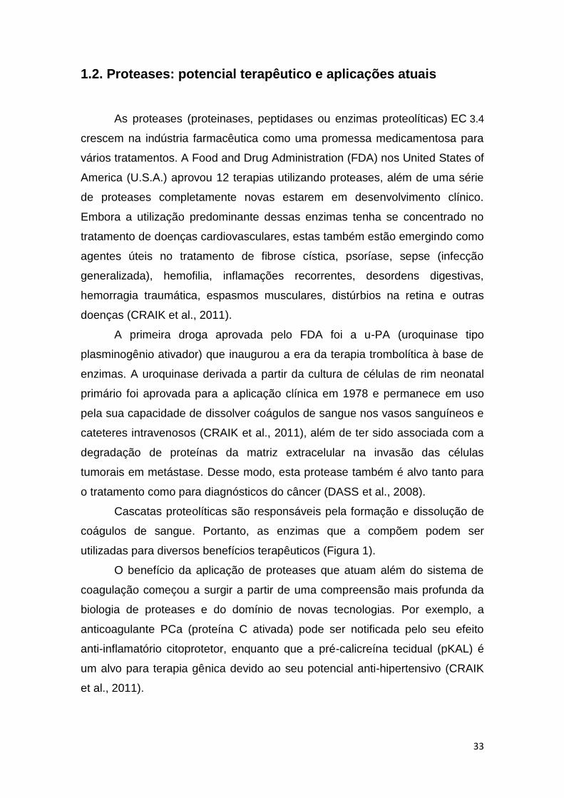
Cascatas proteolíticas são responsáveis pela formação e dissolução de
coágulos de sangue. Portanto, as enzimas que a compõem podem ser
utilizadas para diversos benefícios terapêuticos (Figura 1).
O benefício da aplicação de proteases que atuam além do sistema de
coagulação começou a surgir a partir de uma compreensão mais profunda da
biologia de proteases e do domínio de novas tecnologias. Por exemplo, a
anticoagulante PCa (proteína C ativada) pode ser notificada pelo seu efeito
anti-inflamatório citoprotetor, enquanto que a pré-calicreína tecidual (pKAL) é
um alvo para terapia gênica devido ao seu potencial anti-hipertensivo (CRAIK
et al., 2011).

34
Figura 1. Proteases aplicadas com sucesso para terapias pró-coagulante: (FBN -
fibronectina), (FII - protrombina); anticoagulante: (APC - proteína C ativada);
fibrinolítica: (PMGN – plasminogênio), (PMN – plasmina), (TNKase ou TNK-t-PA -
tenecteplase); anti-hipertensiva: (KAL – calicreína) e citoprotetora: (APC - proteína C
ativada). Algumas proteases (mostradas em círculos verdes escuros) foram aprovadas
para uso clínico. Cofatores de proteínas estão representados por retângulos
alaranjados com arestas arredondadas (Fonte: Adaptado de CRAIK et al., 2011).
Atualmente várias proteases são utilizadas em diversos tipos de terapia,
como por exemplo, a terapia genética para substituição de proteases
constitutivas (KURSCHUS et al., 2010), aplicações médicas dermatológicas e
cosméticas (CRAIK et al., 2011), tratamentos com toxinas botulínicas tipo B
para paralisia e distonia cervical (TRUONG et al., 2009), proteases que
auxiliam no processo digestivo (WOOLDRIDGE et al., 2009), dentre outras.
Como as proteases desempenham papéis fundamentais na fisiologia e
fisiopatologia, estão disponíveis muitas opções para explorar a utilização
dessas enzimas como agentes terapêuticos. A literatura relata 53 doenças
hereditárias que são causadas por mutações nos genes de proteases que
levam à perda de determinadas funções ou baixos níveis de expressão
proteásica. Pesquisas futuras podem apresentar novas oportunidades para
terapias de reposição de proteases para algumas dessas doenças (PUENTE et
al., 2005).

35
O reconhecimento de que as proteases constituem uma classe de
medicamentos seguros e eficazes estimula investigações para produção e
aplicação de novos fármacos com a finalidade de melhorar as terapias
atualmente aprovadas com essas enzimas. A engenharia de proteases é e
continuará sendo utilizada com sucesso para modificar suas propriedades.
Portanto, durante as últimas décadas as proteases proporcionaram resultados
clínicos que sugerem um futuro promissor como uma classe terapêutica
distinta, com diversas aplicações clínicas, destacando seu potencial em
terapias vasculares (CRAIK et al., 2011).
O tratamento da trombose visa prevenir a formação de coágulos
utizando anticoagulantes e, quando formandos, dissolvê-los utilizando agentes
fibrinolíticos ou trombolíticos. Os medicamentos anticoagulantes que vêm
sendo utilizados são a heparina, enoxaparina, varfarina e rivaroxabano e, para
dissolver os trombos já formados na corrente sanguínea, são utilizados
fibrinolíticos como uroquinase, pamiteplase, saruplase, estreptoquinase,
anistreplase, monteplase, reteplase, duteplase, lanoteplase, alteplase,
desmoteplase, estafiloquinase, tenecteplase e o ativador de plasminogênio
tecidual (t-PA). Contudo, os anticoagulantes e fibrinolíticos utilizados causam
diversos efeitos colaterais e requerem monitoramento constante devido ao alto
risco de hemorragias, promovendo um desequilíbrio do sistema hemostático
(KUMAR et al., 2011).
1.3. Proteases com propriedades anticoagulantes
A história da descoberta dos anticoagulantes é marcada pelo acaso. O
efeito anticoagulante da heparina (administração intravenosa) foi descoberto
por McLean em 1916, enquanto ele estava à procura de um pró-coagulante no
fígado de um cão. Em 1941, foi registrada a primeira patente para um
medicamento anticoagulante oral, o dicumarol, descoberto por cientistas da
Universidade de Wisconsin. Esse medicamento recebeu melhorias em sua
composição e, em 1946, foi desenvolvida a varfarina, anticoagulante oral da
classe das cumarinas. A heparina, até a presente data, é o principal fármaco
anticoagulante de ação rápida para o tratamento inicial de trombose venosa,

36
enquanto que os anticoagulantes orais só têm efeito depois de vários dias
(GÓMEZ-OUTES et al., 2012).
Nos últimos anos, foram descobertos anticoagulantes capazes de inibir
um único fator da cascata de coagulação, além de serem administrados em
doses fixas indicadas para cada caso, não necessitando de monitoramento,
oferecendo, assim, vantagens sobre anticoagulantes como a varfarina. Entre
essas novas drogas, estão em uso a dabigatrana (ação direta sobre a
trombina), a rivaroxabana e a apixabana (ação direta sobre o Fator Xa). A ação
inibidora direta pelos novos fármacos anticoagulantes fornece uma resposta
farmacocinética e farmacodinâmica mais previsível e consistente. No entanto,
apesar dessas conquistas, os novos fármacos ainda oferecem desvantagens,
tais como, dificuldade de eliminação da droga em pacientes com problemas
renais, risco hemorrágico e tempo de meia vida curto do fármaco, podendo
ocasionar trombose em caso de esquecimento da administração do
medicamento (GÓMEZ-OUTES et al., 2012).
Diante do exposto, novas pesquisas vêm sendo realizadas na procura
de anticoagulantes semelhantes aos anticoagulantes naturais fisiológicos, pois
se acredita que, por esta via, os efeitos colaterais indesejados podem ser
reduzidos (BERG et al., 2003; CHOI et al., 2013.
A Proteína C ativada é uma serino-protease natural do plasma
sanguíneo que apresenta uma expressiva propriedade anticoagulante (Figura
2). Esta glicoproteína é sintetizada pelos hepatócitos e liberada na corrente
sanguínea sob a forma de um zimogênio inativo. O zimogênio da proteína C é
composto de duas cadeias peptídicas unidas por uma ligação dissulfeto
(CRAIK et al., 2011).
A inibição da coagulação acontece pela ativação inapropriada da
cascata de coagulação, ativando a via anticoagulante. As proteínas C e S
inativadas se encontram livres no plasma e são dependentes da vitamina K.
Para ativar a proteína C, a trombina produzida na cascata de coagulação se
liga a trombomodulina (receptor de membrana), perdendo suas propriedades
pró-coagulantes, transformando-se num potente ativador da proteína C (Figura
3). A proteína S se encontra inativada quando está complexada à proteína de
ligação C4b, porém, quando livre, liga-se à proteína C, funcionando como um
cofator e formando o complexo ativado PCa-PS (proteína C ativada e proteína

37
S) que tem a função de degradar os fatores Va (Ac-globulina ativado) e VIIIa
(globulina anti-hemofílica ativada), limitando a produção de fibrina e impedindo
a formação do coágulo sanguíneo (DAHLBACK e VILLOUTREIX, 2005).
Figura 2. Estrutura cristalizada da proteína C humana ativada
(PCa) complexada com D-Phe-Pro-Arg-Chloromethylketone
(PPACK). (Fonte: Protein Data Bank: http://www.pdb.org/).
Além das propriedades anticoagulantes, outras propriedades são
conferidas pela proteína C ativada, como função antiapoptótica e o auxílio no
tratamento de diversas doenças inflamatórias, tais como a esclerose múltipla e
artrite reumatóide e, em 2001, foi aprovada para o tratamento da sepse (CRAIK
et al., 2011).
Alguns casos de doenças trombolíticas recorrentes estão relacionados
com a resistência à proteína C ativada. Essa resistência ocorre devido a uma
mutação do Fator V de Leiden, na qual acontece a substituição de uma
guanina (G) por uma adenina (A) na posição 1691. Essa substituição resulta na
troca da arginina 506 (Arg) do Fator V por uma glutamina (R506Q) (Glu). Com
a substituição desse aminoácido, fica bloqueado o sítio onde a proteína C faz a
clivagem natural do fator V, o que diminui a sua ação, aumentando a produção
de trombina e consequentemente a formação de trombos (DUQUE e MELLO,
2003).

38
Figura 3. Sistema anticoagulante mostrando a ação da proteína C ativada (PCa) sobre os
fatores de coagulação Va (Ac-globulina ativada) e VIIIa (globulina anti-hemofílica ativada).
(Fonte: Adaptado de http://www.pathologyoutlines.com/topic/coagulationproteinCS.html)
Metodologias utilizando substratos cromogênicos são bastante utilizadas
para avaliar a atividade amidolítica de proteínas com propriedades
semelhantes à proteína C ativada (BERG et al., 2003) e de outras proteínas
relacionadas com o sistema hemostático (LU et al., 2010; CHOI et al., 2011;
CHANG et al., 2012; CHOI et al., 2013; KIM et al., 2013). A afinidade do sítio
ativo dessas proteínas por determinados aminoácidos presentes nos peptídeos
cromogênicos testados é indicada pela intensidade da cor após a reação, ou
seja, quanto maior a afinidade da enzima pelo aminoácido ligado ao pNA (p-
nitroanilide) e pelos outros aminoácidos que compõem o peptídeo
cromogênico, maior é a liberação de pNA que gera uma intensidade da cor
amarela no final da reação.
Pesquisas mostram a produção de enzimas com propriedade bifuncional
(anticoagulante e fibrinolítica) (LU et al., 2010; HASSANEIN et al., 2011; CHOI
et al., 2013). Essa característica conferida por tais enzimas gera expectativa e
sugere que este potencial simultâneo atribua um maior controle hemostático ao
ser administrado como fármaco.
39
1.4. Proteases com propriedades Fibrinolíticas
As enzimas fibrinolíticas ou trombolíticas são proteínas que degradam
coágulos de sangue e normalmente são geradas nas células endoteliais dos
vasos sanguíneos. As células endoteliais existem em todo o corpo: nas
artérias, veias e no sistema linfático. Dessa forma, a baixa produção de
enzimas fibrinolíticas pode levar ao desenvolvimento de coágulos sanguíneos
em praticamente qualquer parte do corpo. Quando o corpo envelhece, a
produção destas enzimas começa a diminuir, tornando o sangue mais
propenso à coagulação. Esse mecanismo pode conduzir a diversas
complicações envolvendo o sistema cardiovascular que podem levar à
invalidez ou até mesmo à morte (VERSTEEG et al., 2013).
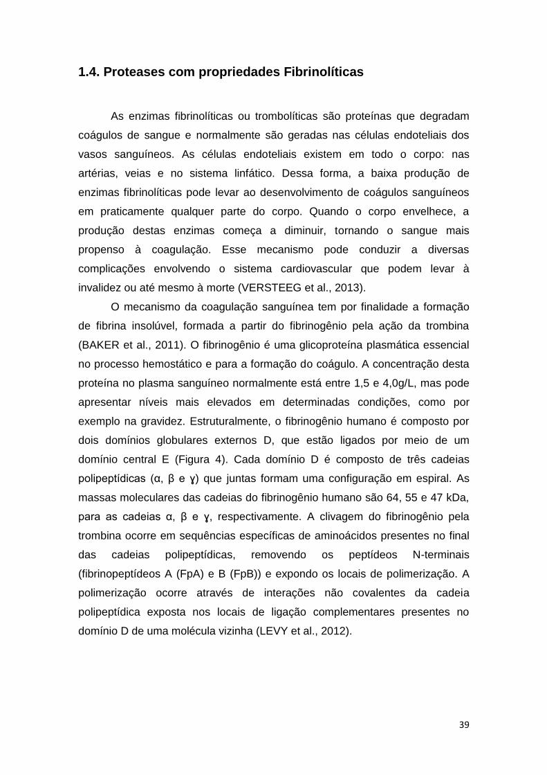
O mecanismo da coagulação sanguínea tem por finalidade a formação
de fibrina insolúvel, formada a partir do fibrinogênio pela ação da trombina
(BAKER et al., 2011). O fibrinogênio é uma glicoproteína plasmática essencial
no processo hemostático e para a formação do coágulo. A concentração desta
proteína no plasma sanguíneo normalmente está entre 1,5 e 4,0g/L, mas pode
apresentar níveis mais elevados em determinadas condições, como por
exemplo na gravidez. Estruturalmente, o fibrinogênio humano é composto por
dois domínios globulares externos D, que estão ligados por meio de um
domínio central E (Figura 4). Cada domínio D é composto de três cadeias
polipeptídicas (α, β e ɣ) que juntas formam uma configuração em espiral. As
massas moleculares das cadeias do fibrinogênio humano são 64, 55 e 47 kDa,
para as cadeias α, β e ɣ, respectivamente. A clivagem do fibrinogênio pela
trombina ocorre em sequências específicas de aminoácidos presentes no final
das cadeias polipeptídicas, removendo os peptídeos N-terminais
(fibrinopeptídeos A (FpA) e B (FpB)) e expondo os locais de polimerização. A
polimerização ocorre através de interações não covalentes da cadeia
polipeptídica exposta nos locais de ligação complementares presentes no
domínio D de uma molécula vizinha (LEVY et al., 2012).
40
Figura 4. Estrutura do fibrinogênio contendo o domínio central E, os
domínios globulares externos D com suas respectivas cadeias
polipeptídicas (α, β e ɣ) e a clivagem dos fibrinopeptídeos A (FpA) e B
(FpB) por ação da trombina, expondo os locais de polimerização para
interações com as moléculas vizinhas e formação de fibrina insolúvel
(Fonte: Adaptada de LEVY et al., 2012).
Os coágulos de sangue são compostos principalmente pela fibrina, uma
proteína fibrosa que diminui o fluxo sanguíneo, aumentando a viscosidade do
sangue. Essa proteína, quando reticulada, obstrui os vasos, interrompe o
fornecimento de oxigênio e eleva a pressão arterial. Assim, as proteases
fibrinolíticas são importantes porque mantêm o fluxo sanguíneo saudável,
auxiliando no sistema de compensação circulatória do corpo (HARRIS et al.,
2013).
No organismo estão presentes vinte enzimas que auxiliam no processo
de coagulação do sangue, mas apenas uma enzima do corpo pode quebrar o
coágulo, a plasmina (Figura 5A). A descoberta da primeira enzima fibrinolítica,
a plasmina, aconteceu de maneira gradual com a colaboração de vários
pesquisadores: Tillet e Garner (1933) descobriram a fibrinolisina
estreptocócica; Milstone (1941) verificou que a ação da fibrinolisina
estreptocócica dependia de uma globulina humana que ele chamou de fator

41
lítico e Kaplan (1944) constatou que esse fator lítico plasmático era uma
protease ativada pelo fator estreptocócico; Christensen (1945) denominou o
fator estreptocócico de estreptoquinase, o precursor plasmático do
plasminogênio, sendo a protease ativa chamada de plasmina (DUQUE e
MELLO, 2003).
Outra enzima fibrinolítica de importância é a Nattokinase (Figura 5B),
que foi descoberta em 1980 pelo Dr. Hiroyuki Sumi, enquanto trabalhava como
pesquisador durante sua graduação em química fisiológica da Faculdade de
Medicina da Universidade de Chicago. O Dr. Sumi pesquisou por muito tempo
enzimas trombolíticas, testou 173 alimentos naturais e encontrou o que
procurava quando o Natto (alimento tradicional japonês feito de soja e
fermentado pelo Bacillus subtilis natto) foi deixado em contato com um trombo
artificial de fibrina em uma placa de Petri incubada a 37⁰C (temperatura
próxima à corporal). O trombo em torno do natto foi degradado completamente
ao final de 18 horas. A enzima foi chamada de nattokinase, que significa
"enzima em natto" (SUMI et al., 1987; MERUVU e VANGALAPATI, 2011).
O sistema fibrinolítico pode ser ativado pelas vias intrínseca e
extrínseca. Quando ativado pelo mecanismo intrínseco, há formação de
calicreína pelo endotélio lesado. A calicreína juntamente com o cininogênio de
alto peso molecular e o Fator XII (Fator Hageman) ativado agem sobre o
plasminogênio ativando-o em plasmina. Quando ativado pela via extrínseca, o
endotélio vascular sintetiza ativadores de plasminogênio tipo tecidual (t-PA) e
tipo uroquinase (u-PA), que nesta via também tem a função de converter o
plasminogênio inativo em plasmina ativa, que degrada o coágulo gerando os
produtos de degradação da fibrina (PDF): fragmento X, o primeiro a ser
formado, é o maior deles, podendo ainda ser lentamente coagulado pela
trombina; os fragmentos Y, E, e o dímero D (D-Di) têm pesos moleculares
menores e não podem sofrer ação da trombina (LIMA et al., 2006).
Os mecanismos de ação e inibição do sistema fibrinolítico são desencadeados
de acordo com a necessidade harmônica do sistema hemostático. Nesse
processo, o endotélio vascular controla a síntese dos ativadores de
plasminogênio ou pode ocorrer também a ação dos inibidores do sistema
fibrinolítico (PAI-1 e PAI-2) que atuam sobre os ativadores de plasminogênio
tipo tecidual (t-PA) e tipo uroquinase (u-PA) ou ainda pela ação de inibidores α

42
2 - antiplasmina, α 2 - macroglobulina e α 1 - antitripsina, que agem inibindo
diretamente a plasmina formada, como mostra na Figura 6 (LIMA et al., 2006;
MACEDO, 2009).
(A)
(B)
Figura 5. (A) Complexo do domínio catalítico da plasmina humana com estreptoquinase e
(B) Estrutura cristalizada da Nattokinase (enzima fibrinolítica) do Bacillus subtilis natto
(Fonte: Protein Data Bank: http://www.pdb.org/).
Todos esses mecanismos envolvendo a hemostasia são cada vez mais
estudados e novos fatos vêm sendo elucidados, gerando novas teorias e
apresentando novos conceitos para ativação das proteases que compõem o
sistema hemostático. Na nova teoria, após a lesão vascular, as plaquetas
aderem ao local danificado e através de interações entre receptores
plaquetários com ligantes extracelulares e proteínas solúveis, o dano vascular
é induzido por exposição do fator tecidual subendotelial, gerando vestígios de
trombina transitória com vários efeitos sobre outros fatores de coagulação e
plaquetas. Os íons de cálcio e os fosfolipídios são cofatores necessários para
todas as reações que controlam a hemostasia. Esse novo modelo hemostático
elucida o fato que as propriedades da trombina vão além da formação do
coágulo de fibrina, relatando que a trombina tem efeito direto sobre os outros
constituintes da coagulação, plaquetas e células endoteliais, além de participar

43
da sua própria regulação negativa influenciando diretamente os mecanismos
da via anticoagulante e o sistema fibrinolítico (VERSTEEG et al., 2013)
Figura 6. Mecanismo de ação do sistema fibrinolítico (Fonte: MACEDO, 2009).
Atualmente, embora a terapia trombolítica seja amplamente adotada
como uma estratégia de primeira linha no tratamento de distúrbios
cardiovasculares, ainda existe necessidade de trombolíticos com melhor
farmacocinética e farmacodinâmica. Esforços estão concentrados no
desenvolvimento de formulações de drogas com propriedades semelhantes à
plasmina e à nattokinase, que degradam diretamente o coágulo sanguíneo.
Diante desse fato, pesquisadores descobriram enzimas fibrinolíticas
provenientes de diversas fontes, inclusive microbianas.
1.4.1. Obtenção e comercialização de enzimas fibrinolíticas
Ao longo dos anos, diversas fontes de enzimas fibrinolíticas foram
descobertas. Dentre essas fontes, potentes proteases fibrinolíticas são
produzidas em processos fermentativos envolvendo micro-organismos,
principalmente por várias espécies do gênero Bacillus, dentre estas: Bacillus
firmus (SEO e LEE, 2004); Bacillus polymaxa (MAHMOUD et al., 2011);
Bacillus subtilis (NGUYEN et al., 2013); Bacillus licheniformis (AL-JUAMILY et
al., 2013); Bacillus cereus (BAJAJ et al., 2013); Bacillus amyloliquefaciens
44
(HEO et al., 2013). Contudo, outras pesquisas mostram que enzimas
fibrinolíticas podem ser provenientes de diversas fontes (Tabela 2).
Enzimas microbianas têm se destacado como um dos principais
produtos biotecnológicos. As enzimas terapêuticas começaram a ser utilizadas
na década de 80 e vêm ganhando cada vez mais importância na indústria
biotecnológica. Uma ampla gama de enzimas, de diferentes fontes e para
diversos usos terapêuticos pode ser encontrada no mercado. O Brasil tem
grande potencial para obtenção de novos fármacos enzimáticos, por ter uma
enorme quantidade e variedade de produtos naturais e uma notável
biodiversidade microbiana disponível para transformação dos mesmos em
produtos de maior valor agregado. Enzimas fibrinolíticas são exemplos de
valiosos bioprodutos, devido sua eficiência, alta especificidade e grande
potencial para uso terapêutico. Dessa forma, a relevância do uso de enzimas
como medicamento se dá pelo fato de que pequenas quantidades desses
catalisadores biológicos podem produzir efeitos bastante significativos em
condições fisiológicas (ZIMMER et al., 2009).
O mercado de enzimas está divido de duas formas: enzimas industriais
(para indústria de alimentos e ração animal) e enzimas especiais (terapêuticas,
para diagnóstico, para pesquisa e para química quiral) (MONTEIRO e SILVA,
2009).
O uso da engenharia enzimática aliada à tecnologia do DNA
recombinante e expressão heteróloga de enzimas vão ter um avanço
significativo na próxima década, sendo importante no desenvolvimento de
novos produtos industriais produzidos por via enzimática. Contudo, as enzimas
não podem ser consideradas a única ferramenta para o avanço nos processos
industriais, é necessário também, o conhecimento da bioquímica, fisiologia e a
genética dos micro-organismos. Logo, a contribuição de novas áreas da
biologia, como a proteômica, genômica e metabolômica, será de fundamental
importância para a aplicação de micro-organismos em escala industrial e o
desenvolvimento de tecnologias mais eficazes (MONTEIRO e SILVA, 2009).

45
Tabela 2. Fontes de enzimas fibrinolíticas
FONTE REFERÊNCIA
Algas (Costaria costata)
(Codium fragile)
KIM et al., 2013
CHOI et al., 2013
Fungos (Bionectria sp.)
(Candida Guilliermondii)
(Aspergillus oryzae)
ROVATI et al., 2010
RASHAD et al., 2012
SHIRASAKA et al., 2012
Bactérias (Streptomyces sp.)
(Pseudomonas aeruginosa)
(Halophilic Lactic Acid Bacteria)
(Escherichia coli)
(Yersinia pestis)
(Streptomyces sp.)
(Paenibacillus sp)
BAJAJ e SHARMA, 2011
RAJ et al., 2012
PRIHANTO et al.,2013
KOTRA et al., 2013
KORHONEN et al., 2013
MEDEIROS e SILVA et al., 2013
VIJAYARAGHAVAN e VINCENT, 2014
Cobras (Daboia russelii russelii)
(Trimeresurus malabaricus)
MUKHERJEE et al., 2013
KUMAR et al., 2013
Poliquetas (Neanthes japonica)
(Cirriformia tentaculata)
DENG et al, 2010
PARK et al., 2013
Insetos (Yellow Mealworm)
(Eupolyphaga sinensis)
HUANG et al., 2012
WANG et al., 2012
Cogumelos (Schizophyllum commune)
(Ganoderma lucidum)
PARK et al, 2010
KUMARAN et al., 2011
Vegetais (Artocarpus heterophyllus latex)
(Euphorbia hirta latex)
(Carica candamarcensis fruta)
SIRITAPETAWEE et al., 2012
PATEL et al., 2012
BILHEIRO et al., 2013
Arraias marinhas (Dasyatis sephen)
(Aetobatis narinari)
KUMAR et al., 2011
Minhocas (Lumbricus rubullus)
(Lumbricus bimastus)
YONG-GANG et al., 2010

46
O uso endovenoso de enzimas microbianas requer purificação elevada e
preservação da atividade enzimática, evitando a desnaturação protéica e a
proteólise. Várias questões devem ser consideradas para o uso de enzimas
terapêuticas, dentre elas, alta atividade e estabilidade em pH fisiológico, baixa
taxa de eliminação do organismo, baixa resposta imunológica e independência
de cofatores exógenos. Além do mais, quando micro-organismos são utilizados
como fonte destas biomoléculas, é importante utilizar cepas não patogênicas
para evitar a presença de toxinas (ZIMMER, 2009).
A busca por substâncias que dissolvem coágulos de sangue é uma das
principais prioridades para as indústrias farmacêuticas. Genentech em
colaboração com investigadores da Universidade de Lueven (Bélgica)
conseguiram clonar o gene que produz t-PA (RIJKEN e COLLEN, 1981) e
diversas empresas estão trabalhando para produzir t-PA a partir de rDNA com
mostra a Tabela 3.
A eficácia bioquímica e a viabilidade comercial de t-PAs produzidos a
partir de tecnologias envolvendo rDNA continuam a ser demonstradas em
diversas pesquisas realizadas por Universidades do mundo inteiro, além do
sucesso proveniente de sua comercialização. O investimento diferenciado nas
instituições de pesquisas em países diversos reflete na qualidade das enzimas
fibrinolíticas produzidas. (Fonte: https://www.princeton.edu). Diante desse fato,
é notória a necessidade de patrocínio para um maior avanço nas pesquisas
envolvendo enzimas fibrinolíticas brasileiras.
É preocupante o atraso na produção de enzimas no Brasil, apesar da
nossa vocação para a biotecnologia e potencial para produção e uso de
biocatalisadores. No contexto mundial, a situação do Brasil é incomparável em
relação à quantidade e à variedade de recursos naturais disponíveis para
serem transformados, por tecnologia enzimática, em produtos úteis e de maior
valor agregado. O Brasil possui uma notável biodiversidade microbiana a ser
estudada, por uso direto ou após melhoramento genético, visando à produção
de enzimas por processos fermentativos (Fonte: http://www.redetec.org.br).

47
Table 3. Empresas e Universidade envolvidas no desenvolvimento e comercialização de enzimas fibrinolíticas
Enzima Empresas/Universidades Descrição do Projeto
Estreptoquinase Hoechst-Roussel (F. R. G.)
KabiVitrum (Sweden)
Produção por bactéria
Produção por bactéria
Uroquinase Abbott Laboratories (U. S.)
Genex (U. S.)lMitsui Toatsu Chemicals, Inc. (Japão)
Genentech (U. S.)/Grunenthal (F. R.G.)
Extração por cultura de células renais
Produção por rDNA
Produção por rDNA
Ativador de Plasminogênio Humano Genentech/Universidade de Leuven (Bélgica)
Mitsubishi Chemical Industries, Inc. (Japão)/Kyowa
Hakko Kogyo (Japão)
Biogen S.A. (Switz.)/Fujtsawa (Japão) Integrated
Genetics (U.S,)
Toyobo Pharmaceutical (Japão) Chiron (U. S.)
Collaborative Resarch (U.S.)/Green Cross (Japão)
Produção por rDNA
Produção por rDNA
Produção por rDNA
Produção por rDNA
Extração por cultura de células renais
Agentes Fibrinolíticos e Anticoagulantes Genentech/Yamanouchi Ltd. (Japão)
Genex/Yamanouchi Ltd.
Desenvolvimento de estirpes de
micro-organismos que produzem
agentes fibrinolíticos
(Fonte: https://www.princeton.edu)

48
Tabela 4. Preço comercial de alguns agentes fibrinolíticos
(Fonte: http://portuguese.alibaba.com/manufacturers/fibrinolytics.html)
Fonte de obtenção da
enzima
Enzima Preço/Kg Pedido
mínimo
Prazo de
entrega
Capacidade de
fornecimento
País Empresa/fornecedores
Extrato da soja (Natto)
fermentado pelo Bacillus
subtilis natto
Nattokinase FOB: US $ 180-220 /Kg 5 Kg 20 dias 20 toneladas/
semana
Taiwan Contek Life Science
Co., Ltd.
Extrato da soja Nattokinase FOB: US $ 99-429/Kg 20 Kg 20 dias 20 toneladas/mês Taiwan Contek Life Science
Co., Ltd.
Extrato da soja Nattokinase
FOB: US $ 1-2000/Kg 25 Kg 5 dias 10 tonaladas/mês China Shaanxi Kingsci
Biotechnology Co., Ltd.
Extrato de minhoca Lumbrokinase FOB: US $ 1550-3200/Kg 5 Kg 10 dias 1000 kiloampere/
quarto
China Beijing Herbal Health
Biotech Limited Liability
Company
Extrato de minhoca Lumbrokinase FOB: US $ 1000-2000/Kg 1 Kg 3 dias 50 Kg / mês China Shaanxi Sciphar Hi-
Tech Industry Co., Ltd.
Pó liofillizado de Streptococcus beta-hemolítico
Estreptoquinase FOB: US $ 1-10/Kg 1 Kg 7 dias 1000 Kg/ano India Swapnroop Drugs and
Pharmaceuticals
49
Apesar das diversas fontes encontradas, as enzimas fibrinolíticas
extracelulares microbianas e principalmente as produzidas pelo gênero Bacillus
têm se destacado pela sua facilidade de obtenção/extração, expressiva
atividade, propriedades fisicoquímicas compatíveis ao sistema sanguíneo
(temperatura 37°C e pH 7,0) e elevada estabilidade (MUKHERJEE et al.,
2012).
1.4.2. Propriedades bioquímicas das enzimas fibrinolíticas
produzidas pelo gênero Bacillus
A diversidade de características bioquímicas das enzimas fibrinolíticas
produzidas pelos Bacillus (Tabela 5) tem atraído cada vez mais o interesse de
pesquisadores do mundo inteiro. De acordo com o mecanismo catalítico, a
maioria das enzimas produzidas por espécies de Bacillus são caracterizadas
como serinoproteases; contudo, podemos encontrar metalo proteases e até
mesmo serino-metalo proteases. Na maioria dos casos o pH ótimo destas
enzimas é encontrato entre a faixa neutra e alcalina e temperatura ótima a
partir de 37⁰C, podendo chegar até a 60⁰C. Quanto ao peso molecular
podemos encontrar uma ampla faixa de 18 kDa até 33 kDa, e alguns trabalhos
relatam pesos moleculares de 46kDa (DEEPAK et al., 2010) e 65 kDa (CHOI et
al., 2010). O ponto isoelétrico se encontra em torno de 5.8 a 9.2.
Outra importante propriedade que tem sido bastante estudada é a sequência
de aminoácidos que constitui os peptídeos das enzimas fibrinolíticas. Como já
se sabe, as principais características dos aminoácidos são em decorrência do
tipo de cadeia lateral. Dessa maneira, fica claro que a atividade biológica de um
dado peptídeo, bem como suas características físico-químicas, estão
intimamente ligadas à sequência e aos tipos de aminoácidos que o constituem.
O perfil sequencial dos aminoácidos das extremidades N-terminal ou amino
terminal das cadeias peptídicas da maioria das enzimas fibrinolíticas
produzidas por espécies de Bacillus, quando comparado, possuem em boa
parte dos casos analisados, sequenciamentos idênticos (Tabela 6)

50
Tabela 5. Propriedades bioquímicas das proteases fibrinolíticas do gênero Bacillus
Micro-organismo Mecanismo catalítico Temperatura e
pH ótimos
*PM **pI Referências
Bacillus subtilis IMR-NK1 Serinoprotease 55ºC e 7.8 31.5 kDa 8.3 CHANG et al., 2000
Bacillus amyloliquefaciens CD-4 Serinoprotease 48ºC e 9.0 28 kDa 8.0 PENG et al., 2003
Bacillus subtilis DC33 Serinoprotease 55ºC e 8.0 30 kDa 8.7 WANG et al., 2006
Bacillus Subtilis Serinoprotease 60°C e 9.0 29.93 kDa 6.35 CHANG et al., 2012
Bacillus sp. AS-S20-I Serinoprotease 37ºC e 7.4 32.3 kDa 5.8 MUKHERJEE et al., 2012
Bacillus subtilis HQS-3 Serino-metalo protease 45-50ºC e 8.0 26 kDa 9.0 - 9.2 HUANG et al., 2013
Bacillus subtilis K42 Metalo protease 40ºC e 9.4 20.5 kDa - HASSANEIN et al., 2011
Bacillus polymaxa NRC-A Serinoprotease 40ºC e 9.5 18 kDa - MAHMOUD et al., 2011
Bacillus subtilis ICTF-1 Serinoprotease 50ºC e 9.0 28 kDa - MAHAJAN et al., 2012
* Peso molecular (PM); * * ponto isoelétrico (pI)

51
Tabela 6. Comparação do perfil amino terminal das cadeias peptídicas das enzimas
fibrinolíticas produzidas por espécies de Bacillus
Apesar de todas as pesquisas e potenciais mostrados por estas novas
enzimas que vêm sendo descobertas, a principal problemática ainda é
minimizar o custo de produção e aumentar a escala em nível industrial. Para
isso, estudos de otimização de bioprocessos e escalonamento da produção se
fazem cada vez mais necessários.
1.4.3. Otimização e escalonamento da produção de enzimas
fibrinolíticas
O custo do processo de produção de proteases é um dos principais
obstáculos para a aplicação bem sucedida dessas enzimas nas indústrias.
Para as enzimas fibrinolíticas, muitas tentativas em laboratórios têm sido
realizadas para melhoria das mesmas, incluindo a seleção de um meio de
Bacilos Sequência do aminoácido N-terminal
da enzima fibrinolítica
Referência
B. subtilis IMR-NK1 AQPVPNGRTAIKA CHANG et al., 2000
B. amyloliquefaciens DC-4 AQSVPYGVSQIKAPALHS PENG et al., 2003
B. firmus NA-1 IVGGYEQZAHSQPHQ SEO et al., 2004
Bacillus subtilis QK02 AQSVPYGISQIKAPALHSQG KO et al., 2004
B. pumilus TYO-67 AQTVPYGIPQIKAD TAKAHASHI et al., 2004
Bacillus subtilis CD33 AQSVPYGIPQIKAPA WANG et al., 2006
Bacillus subtilis A26 AQSVPYGISQI AGREB et al., 2009
B. amyloliquefaciens DJ-5 AQSVPYGISQIKAPA CHOI et al., 2009ª
Bacillus subtilis A1 QTGGSIIDPINGYN YEO et al., 2011
Bacillus subtilis AQSVPYGISQIK posição 1–12
VAVIDSGIDSSHPDLNVR posição 28–45
YPSTIAVGAVNSSNQR posição 171-186
NGUYEN et al., 2013
52
cultura ideal, a otimização das condições ambientais e nutricionais e, até
mesmo, modificações genéticas de linhagens (JO et al., 2011).
Para as condições de fermentação, a seleção dos componentes do meio
de cultivo é geralmente um fator crítico na produção de enzimas fibrinolíticas.
Uma vez que os micro-organismos possuem diversas características
fisiológicas, é necessário encontrar as condições favoráveis para a produção
de acordo com cada espécie microbiana envolvida no processo. Para melhorar
o rendimento da produção enzimática é necessário otimizar o processo e
aumentar o escalonamento de produção dessas enzimas fibrinolíticas (AVHAD
e RATHOD, 2014).
Embora as estratégias tradicionais para otimização dos processos de
produção sejam relativamente simples, é frequente não encontrar a condição
ótima para a resposta desejada. Em busca do melhor resultado, é comum a
utilização de ferramentas estatísticas que auxiliem na análise de interações
mútuas entre os fatores e as estimativas do valor exato da variável resposta em
tempo mínimo com um número reduzido de ensaios. Entre outros, o
planejamento central composto (PCC) é uma das ferramentas estatísticas
utilizadas para auxiliar na otimização dos processos fermentativos, e a Análise
de Metodologia de Superfície de Respostas (MSR) é utilizada para modelar,
analisar e, finalmente, encontrar a condição ótima para a obtenção da resposta
de interesse. Assim, cada vez mais pesquisas mostram a otimização da
produção de enzimas fibrinolíticas utilizando PCC e MSR (LIU et al., 2005;
DEEPAK et al., 2010; MAHAJAN et al, 2010; KANAGASABAI et al., 2013).
Um importante fator que aumenta em muitas vezes o rendimento e a
atividade enzimática é a produção de enzimas em larga escala, realizada
através de fermentações em biorreatores de tanque agitado aumentando o
escalonamento do produto de interesse. Os reatores biológicos proporcionam
em geral um aumento no rendimento do bioativo produzido e melhora a relação
entre o desenvolvimento microbiano e o ambiente ao qual ele é submetido,
gerando melhores resultados de obtenção do produto. Na produção em
biorreatores, alguns fatores são essenciais para a obtenção dos resultados
desejados, como por exemplo, a agitação e a aeração, parâmetros essenciais
nos processos fermentativos. A agitação influencia a mistura e transferência de
calor e massa, sendo um parâmetro importante para o crescimento e
53
desempenho de células microbianas. A aeração também é um fator chave nos
processos aeróbios, devido ao seu impacto sobre a oferta de oxigênio. Em
geral, a transferência de oxigênio depende da taxa de fluxo do gás, do tipo de
agitador, da velocidade de agitação e das propriedades dos líquidos envolvidos
no sistema (GANGADHARAN et al., 2011).
O fornecimento de oxigênio está ligado ao metabolimo celular, auxiliando
na obtenção do produto desejado. Em células com metabolismo aeróbio, o
oxigênio é utilizado como aceptor final de elétrons no término da cadeia
respiratória e na reoxidação de coenzimas. Isso resulta na produção de
adenosina trifosfato (ATP), fonte energética celular. Contudo, o oxigênio é
pouco solúvel em água, precisando ajustar sua concentração de saturação em
mg/L. Dessa forma, não é possível fornecer de uma só vez todo o oxigênio
necessário em um bioprocesso. Esse deve ser continuamente liberado no
bioreator durante o cultivo. O controle do suprimento e a demanda de oxigênio
determinam a taxa ótima de transferência de massa ou taxa de absorção de
oxigênio da fase gasosa para a líquida. A taxa de oxigênio dissolvido é dada
pela diferença entre o oxigênio fornecido e o utilizado pelas células. O
suprimento do oxigênio está relacionado com a velocidade de agitação, o grau
de mistura, a vazão de ar, a temperatura e a geometria do biorreator. Esses
fatores são relacionados com a transferência de massa gás-líquido. E a
demanda de oxigênio está relacionada à fisiologia celular, que determina a
quantidade necessária de oxigênio durante o bioprocesso (ZULKEFLEE e
AZIZ, 2007).
Outro fator importante para produção bem sucedida, é a utilização de
agentes indutores, que é de fundamental importância para a síntese
enzimática, uma vez que a presença desses contribui para obtenção de
atividades enzimáticas mais elevadas. A farinha de soja, resíduo da indústria
de produção de óleo de soja, demonstra ser um excelente substrato indutor,
estimulando o crescimento de uma variedade de micro-organismos e
aumentando a atividade de diversas enzimas produzidas (LIMA et al., 2011),
incluindo enzimas fibrinolíticas (SALES et al., 2013, MEDEIROS e SILVA et al.,
2013).
Porém, após todas essas fases que incluem, encontrar as condições
adequadas para o bioprocesso da produção enzimática, selecionar um

54
substrato indutor para produção da enzima fibrinolítica, otimizar a produção e
aumentar a escala do produto almejado, para que este produto possa ser
industrializado e posteriormente comercializado, é necessária a obtenção de
um produto puro, principalmente em se tratando de um fármaco, como é o caso
das enzimas fibrinolíticas. Diante dessa necessidade, diversos métodos de
purificação vêm sendo investigados e, dentre esses, a fermentação extrativa é
considerada uma alternativa promissora na redução de etapas dos processos
de purificação.
1.5. Fermentação extrativa utilizando sistema de duas fases
aquosas (SDFA)
Existe uma busca constante das indústrias por eficiência e baixo custo
nos processos de obtenção de biomoléculas de alto valor agregado. A
fermentação ou bioconversão extrativa tem sido apresentada como solução
para superar a baixa produtividade dos processos biotecnológicos (DELOISA et
al, 2009; NALINANON et al, 2009).
A bioconversão extrativa em sistema de duas fases aquosas (SDFA) é
integrada pelas seguintes etapas: upstream, que antecede a transformação,
seguida da etapa de transformação propriamente dita e a etapa de à jusante
(downstream), momento que acontece o processo de recuperação do
bioproduto desejado (RATANAPONGLEKA, 2010).
A fermentação extrativa é um processo que integra o cultivo microbiano
à produção e recuperação do bioprotudo in situ de maneira simultânea (Figura
7), sendo utilizado para aumentar o rendimento dos processos fermentativos
convencionais. O sistema de duas fases aquosas (SDFA) utilizado para uma
fermentação extrativa é uma alternativa considerada bastante promissora
quando comparado com os processos de fermentações convencionais, desde
que seja um meio propício para o crescimento e estabilização das células
microbianas e adequado para o favorecimento de produção da biomolécula,
sem que esta sofra desnaturação ou algum tipo de desestabilização,
aumentando assim a produtividade do processo. O SDFA é formado por dois
polímeros ou um polímero e um sal em solução aquosa. Esta técnica é

55
constituída pela mistura de duas soluções acima de certa concentração crítica,
indicada por um diagrama de fases em que a formação de duas fases aquosas
imiscíveis é observada (SINHA et al., 2000; NG et al., 2013).
Em um comportamento ideal para um processo de fermentação
extrativa, espera-se que as células e os componentes do meio se concentrem,
preferencialmente, em uma das fases do sistema, enquanto que o produto
(biomolécula de interesse) prefira a fase oposta. Esse comportamento facilita a
extração do produto do meio de produção assim que este é formado, levando a
uma purificação parcial do produto alvo, além de apresentar outras vantagens
como eliminar a influência de inibidores que possam surgir eventualmente no
processo de produção (SINHA et al., 2000; NG et al., 2013).
Os resultados do particionamento em SDFA dependem das propriedades da
biomolécula produzida, tais como, peso molecular, ponto isoelétrico e
hidrofobicidade, mas também das características físico-químicas de ambas as
fases do sistema, que por sua vez são influenciados por fatores que compõem
o sistema, tais como, massa molecular do polímero utilizado, concentração e o
tipo de polímero e sal utilizados, além da temperatura e do pH estabelecidos
para o sistema (PERICIN et al, 2009).
Uma estratégica maneira de manipular os resultados de um bioprocesso
extrativo é por modificação química de um dos polímeros, anexando um ligante
adequado para os receptores existentes no bioproduto ou fármaco de interesse
que está sendo produzido (XU et al., 2001).
O impacto do custo total no processo de produção depende
expressivamente do tipo de produto que se quer obter e da concentração de
produto obtida ao final do processo. Dessa forma, o custo da venda de um
determinado produto alvo tem correlação inversamente proprocional a sua
concentração alcançada no fim do processo de produção. A recuperação in situ
de biomoléculas tem sido proposta como solução para reduzir o número de
etapas de purificação dos produtos biotecnológicos, sendo de fundamental
importância para a viabilidade do processo, uma vez que reduz o custo para
obtenção do produto final (NG et al., 2013).

56
Figura 7. Representação simplificada do processo integrado da fermentação extrativa
em sistema de duas fases aquosas (SDFA) para enzimas fibrinolíticas extracelulares
O processo de fermentação extrativa utilizando SDFA vem sendo
reportado na literatura para extração de biomoléculas provenientes de espécies
de Bacillus para recuperação de proteases alcalinas por Bacillus licheniformis
(LEE e CHANG, 1990); α-amilase por Bacillus amyloliquefaciens (KIM e YOO,
1991); surfactina de Bacillus subtilis (DROUIN e COOPER, 1992); subtilina por
Bacillus subtilis (KUBOI et al., 1994); enzimas que hidrolisam polissacarídeos
produzidos por Bacillus thuringiensis H14 (BANIK e SANTHIAGU, 2002); e
enzimas fibrinolíticas de Bacillus subtilis e Bacillus sp. UFPEDA 485 que são
consideradas potentes fármacos promissores na medicina vascular (ASHIPALA
et al., 2008; SALES et al., 2013).
A bioconversão extrativa em sistemas de duas fases aquosas está
sendo cada vez mais cogitada, devido sua eficiência, especialmente para a
separação de pequenas moléculas farmacêuticas, tais como, anticorpos,
antibióticos, antígenos, proteínas recombinantes, aminoácidos, oligopeptídeos,
ácido lático e principalmente enzimas para fins farmacêuticos (XU et al., 2001),
incluindo enzimas fibrinolíticas (SALES et al., 2013).
Com iniciativas simples, de poucos esforços e baixo custo, a purificação
de muitos produtos biofarmacêuticos pode ser conseguida em processos de
bioconversão extrativa suave não desnaturante. Para aumentar a seletividade,
vários bioligantes ou ligantes químicos com afinidade pelo polímero utilizado e
alguns aditivos de superfície ativos podem ser inseridos nos sistemas de
separação, ocasionando em resultados bem sucedidos (XU, 2001).
PROCESSO INTEGRADO (Fermentação Extrativa - FE)
Meio de cultivo + Polímero + Sal
Produção e extração simultânea da enzima fibrinolítica por SDFA
Micro-organismo

57
A aplicação de fármacos endovenosos, tais como, as enzimas
fibrinolíticas microbianas, exige cepas não patogênicas ausentes de toxinas,
além de uma alta purificação e estabilidade enzimática nas condições
fisiológicas do sistema sanguíneo (pH 7,2 e temperatura 37°C), sendo também
de extrema importância a obtenção de um fármaco que ocasione o mínimo de
efeitos colaterais durante o período terapêutico.

58
2. REFERÊNCIAS
AGREBI, R.; HADDAR, H.; Hmidet, N.; JELLOULI, K.; MANNI, L. and NASRI,
M. BSF1 fibrinolytic enzyme from a marine bacterium Bacillus subtilis A26:
Purification, biochemical and molecular characterization. Process
Biochemistry, v. 44, p. 1252-1259, 2009.
AL-JUAMILY, E. F. and AL-ZAIDY, B. H. Purification and Characterization of
Fibrinolytic Enzyme Produced from Bacillus licheniformis B4. Chemical
Science Review and Letters, v. 2, n. 5, p. 256-266, 2013
ASHIPALA, O. K. e HE, Q. Optimization of fibrinolytic enzyme production by
Bacillus subtilis DC-2 in aqueous two-phase system (poly-ethylene glycol 4000
and sodium sulfate). Bioresource Technology, v. 99, p. 4112-4119, 2008.
AVHAD, D. N. and RATHOD, V. K. Ultrasound stimulated production of a
fibrinolytic enzyme. Ultrasonics Sonochemistry. v. 21, n. 1, p. 182-188, 2014.
BAEHAKI, A.; SUHARTONO, M. T.; SUKARNO; SYAH, D.; SITANGGANG, A.
B.; SETYAHADI, S. and MEINHARDT, F. Purification and characterization of
collagenase from Bacillus licheniformis F11.4. African Journal of
Microbiology Research, v. 6, n.10, p. 2373-2379, 2012.
BAGHERI, M.; DIDARI, M.; AMOOZEGAR, M. A.; SCHUMANN, P.; SÁNCHEZ-
PORRO, C.; MEHRSHAD, M. and VENTOSA, A. Bacillus iranensis sp. nov., a
moderate halophile from a hypersaline lake. International Journal of
Systematic and Evolutionary Microbiology, v. 62, p. 811-816, 2012
BAJAJ, B. K. and SHARMA, P. An alkali-thermotolerant extracellular protease
from a newly isolated Streptomyces sp. DP2. New Biotechnology, v. 28, n. 6,
p. 725-732, 2011.

59
BAJAJ, B. K.; SHARMA, N. and SINGH, S. Enhanced production of fibrinolytic
protease from Bacillus cereus NS-2 using cotton seed cake as nitrogen source.
Biocatalysis and Agricultural Biotechnology. v. 2, p. 204-209, 2013.
BAKER, D. C. and BRASSARD, J. Review of Continuing Education Course on
Hemostasis. Toxicologic Pathology, v. 39, p. 281-288, 2011.
BANIK, R. M. and SANTHIAGU, A. Extractive fermentation for enhanced gellan
hydrolyzing enzyme production. Word Journal and Microbiology and
Biotechnology, v. 18, p. 715-720, 2002.
BERG, D. T.; GERLITZ, B.; SHANG, J.; SMITH, T.; SANTA, P.; RICHARDSON,
M. A.; KURZ, K. D.; GRINNELL, B. W.; MACE, K and JONES, B. E.
Engineering the proteolytic specificity of activated protein C improves its
pharmacological properties. Applied Biological Sciences, v. 100, n. 8, p.
4423-4428, 2003.
BILHEIRO, R. P.; BRAGA, A. D.; FILHO, M. L.; CARVALHO-TAVARES, J.;
AGERO, U.; CARVALHO, M. G.; SANCHEZ, E. F.; SALAS, C. E. and LOPES,
M. T. P. The thrombolytic action of a proteolytic fraction (P1G10) from Carica
candamarcensis. Thrombosis Research, v. 131, p. e175-e182, 2013.
BONALA, K. C. and MANGAMOORI, L. N. Production and optimization of lipase
from Bacillus tequilensis NRRL B-41771. International Journal of
Biotechnology Applications, v. 4, n. 1, p.134-136, 2012.
CHANG, C. T.; FAN, M. H.; KUO, F. C.; SUNG, H. Y. Potent fibrinolytic enzyme
from a mutant of Bacillus subtilis IMR-NK1. Journal of Agricultural and Food
Chemistry, v. 48, p. 3210-3216, 2000.
CHANG, C-T.; WANG, P-M.; HUNG, Y-F. and CHUNG, Y-C. Purification and
biochemical properties of a fibrinolytic enzyme from Bacillus subtilis-fermented
red bean. Food Chemistry. v. 133, n. 4, p. 1611-1617, 2012.

60
CHEN, Y. G.; HU, S. P.; TANG, S. K.; HE, J. W.; XIAO, J. Q.; ZHU, H. Y. and
LI, W. J. Bacillus zhanjiangensis sp. nov., isolated from an oyster in South
China Sea. Antonie Van Leeuwenhoek, v. 99, p. 473-480, 2011.
CHOI, D.; CHA, W. S.; PARK, N.; KIM, H. W.; LEE, J. H.; PARK, J. S. and
PARK, S. S. Purification and characterization of a novel fibrinolytic enzyme from
fruiting bodies of Korean Cordyceps militaris. Bioresource Technology, v. 102,
p. 3279-3285, 2011.
CHOI, J-H.; SAPKOTA, K.; PARK, S-E.; KIM, S. and KIM, S-J. Thrombolytic,
anticoagulant and antiplatelet activities of codiase, a bi-functional fibrinolytic
enzyme from Codium fragile. Biochimie, v. 95, p. 1266-1277, 2013.
CHOI, N. S.; CHUNG, D. M.; HAN, Y. J.; KIM, S. H. and SONG, J. J.
Purification and characterization of a subtilisin D5, a fibrinolytic enzyme of
Bacillus amyloliquefaciens DJ-5 isolated from Doenjang. Food Science and
Biotechnology, v. 18, p. 500-505, 2009a.
CHOI, N-S.; CHUNG, D-M.; PARK, C-S.; AHN,, K-H.; KIM,, J. S.; SONG, J. J.;
KIM,, S-H.; YOON, B-D. and KIM, M-S. Expression and identification of a minor
extracellular fibrinolytic enzyme (Vpr) from Bacillus subtilis KCTC 3014.
Biotechnology and Bioprocess Engineering. v. 15, n. 3, p 446-452, 2010.
CHRISTENSEN, L. R. Streptococcal fibrinolysis: proteolytic reaction due to
serum enzyme activated by streptococcal fibrinolysin. Journal of General
Physiology, v. 28, n. 4, p. 363-383, 1945.
CHRISTO, P. P.; CARVALHO, G. M. and NETO, A. P. G. Trombose de seios
venosos cerebrais: estudo de 15 casos e revisão de literatura. Revista da
Associação Médica Brasileira, v. 56, n. 3, p. 288-292, 2010.
CRAIK, C. S.; PAGE, M. J. and MADISON, E. L. Proteases as therapeutics.
Biochemical Journal, v. 435, p. 1-16, 2011.

61
DAHLBACK, B and VILLOUTREIX, B. O. The anticoagulant protein C pathway.
FEBS Letters, v. 579, p. 3310-3316, 2005.
DASS, K.; AHMAD, A.; AZMI, A. S.; SARKAR, S. H. and SARKAR, F. H.
Evolving role of uPA/uPAR system in human cancers. Cancer Treatment
Reviews, v. 34, 122-136, 2008.
DEB, P.; TALUKDAR, S. A.; MOHSINA, K; SARKER, P. K. and ABU SAYEM,
S. M. Production and partial characterization of extracellular amylase enzyme
from Bacillus amyloliquefaciens P-00. SpringerPlus, v. 2, n.154, p. 1-12, 2013.
DEEPAK, V.; ILANGOVAN, S.; SAMPATHKUMAR, M. V.; VICTORIA, M. J.;
PASHA, S. P. B. S.; PANDIAN, S. B. R. K. and GURUNATHAN, S. Medium
optimization and immobilization of purified fibrinolytic URAK from Bacillus
cereus NK1 on PHB nanoparticles. Enzyme and Microbial Technology, v. 47,
p. 297-304, 2010.
DELOISA, K. M.; HERNANDEZ, M. R. T.; PALOMARES, M. R. Recovery of
laccase from the residual compost of Agaricus bisporus in aqueous two-phase
systems, Process Biochemistry, v. 44, p. 435-439, 2009.
DENG, Z.; WANG, S.; LI, Q.; JI, X.; ZHANG, L. and HONG, M. Purification and
characterization of a novel fibrinolytic enzyme from the polychaete, Neanthes
japonica (Iznka). Bioresource Technology, v. 101, 1954-1960, 2010.
DROUIN, C. M. and COOPER, D. G. Biosurfactants and aquous two phase
fermentation. Biotechnology and Bioengineering, v. 40, p. 86, 1992.
DUMAS, V. F.; GOLDENBERG, C. S.; MARTINS, E. S.; PRAÇA, L. B.;
QUEIROZ, P. R.; MONNERAT, R. G. and MELO, F. R. Chitinase and cellulase
activity from Bacillus thuringiensis strains. Universitas: Ciências da Saúde,
Brasília, v. 7, n. 1, p. 1-12, 2009.
62
DUQUE, F. L. V. and MELLO, N. A. Trombogênese - Trombofilia. Jornal
Vascular Brasileiro, v. 2, n. 2, p. 105-118, 2003.
DURÃO, P.; CHEN, Z.; FERNANDES, A. T.; HILDEBRANDT, P.; MURGIDA, D.
H.; TODOROVIC, S.; PEREIRA, M. M.; MELO, E. P. and MARTINS, L. O.
Copper incorporation into recombinant CotA laccase from Bacillus subtilis:
characterization of fully copper loaded enzymes. Journal of Biological
Inorganic Chemistry, v. 13, p. 183-193, 2008.
GANGADHARAN, D.; NAMPOOTHIRI, K. M. and PANDEY, A. α-Amylase
Production by Bacillus amyloliquefaciens Using Agro Wastes as Feed Stock.
Food Technology and Biotechnology, v. 49, p. 336-340, 2011.
GAWANDE, B. N.; GOEL, A.; PARKAR, A. Y. and NENE, S. N. Purification and
properties of a novel starch degrading cyclomaltodextrin glucanotransferase
from Bacillus firmus. Applied Microbiology and Biotechnology, v. 51, p. 504-
509, 1999.
GÓMEZ-OUTES, A.; SUÁREZ-GEA, M. L.; CALVO-ROJAS, G.; LECUMBERRI,
R.; ROCHA, E.; POZO-HERNÁNDEZ, C.; TERLEIRA-FERNÁNDEZ, A. I. and
VARGAS-CASTRILLÓN, E. Discovery of anticoagulant drugs: a historical
perspective. Current Drug Discovery Technologies, v. 9, n. 2, p. 83-104,
2012.
GUINEBRETIÈRE, M. H.; AUGER, S.; GALLERON, N.; CONTZEN, M.; DE
SARRAU, B.; DE BUYSER, M. L.; LAMBERET, G.; FAGERLUND, A.;
GRANUM, P. E.; LERECLUS, D.; DE VOS, P.; NGUYEN-THE, C. and
SOROKIN, A. Bacillus cytotoxicus sp. nov. is a novel thermotolerant species of
the Bacillus cereus Group occasionally associated with food poisoning.
International Journal of Systematic and Evolutionary Microbiology, v. 63,
31-40, 2013.
http://www.ncbi.nlm.nih.gov/pubmed?term=Lecumberri%20R%5BAuthor%5D&cauthor=true&cauthor_uid=21838662

63
HARRIS, L. F.; CASTRO-LÓPEZ, V. and KILLARD, A. J. Coagulation
monitoring devices: Past, present, and future at the point of care. Trends in
Analytical chemistry, v. 50, p. 85-95, 2013.
HASSANEIN, W. A.; KOTB, E.; AWNY, N. M. and EL-ZAWAHRY, Y. A.
Fibrinolysis and anticoagulant potential of a metallo protease produced by
Bacillus subtilis K42. Journal of Biosciences, v. 36, n. 5, p. 773-779, 2011.
HEO, K.; CHO, K. M.; LEE, C. K.; KIM, G. M.; SHIN, J-H.; KIM, J. S. and KIM,
J. H. Characterization of a Fibrinolytic Enzyme Secreted by Bacillus
amyloliquefaciens CB1 and Its Gene Cloning. Journal of Microbiology and
Biotechnology, v. 23, n. 7, p. 974-983, 2013.
HONG, S. W.: PARK, J. M.; KIM, S. J. and CHUNG, K. S. Bacillus eiseniae sp.
nov., a swarming, moderately halotolerant bacterium isolated from the intestinal
tract of an earthworm (Eisenia fetida L.). International Journal of Systematic
and Evolutionary Microbiology, v. 62, p. 2077-2083, 2012.
HUANG, M. X.; YE, Y.; CHEN, Y. X and HAN, Y. L. Partial Purification and
Characterization of Fibrinolytic Enzymes from Yellow Mealworm. International.
Journal of Peptide Research and Therapeutics, v. 18, p. 153-161, 2012.
HUANG, S.; PAN, S.; CHEN, G.; HUANG, S.; ZHANG, Z.; LI, Y. and LIANG, Z.
Biochemical characteristics of a fibrinolytic enzyme purified from a marine
bacterium, Bacillus subtilis HQS-3. International Journal of Biological.
Macromolecules. v. 62, p. 124-130, 2013.
KADEMI, A.; AIT-ABDELKADER, N.; FAKHREDDINE, L. and BARATTI, J.
Purification and characterization of a thermostable esterase from the moderate
thermophile Bacillus circulans. Applied Microbiology and Biotechnology, v.
54, p. 173-179, 2000.
KANAGASABAI, V. and THANGAVELU, V. Response surface methodological
optimization of the medium components for production of xylanase under ssf by
64
aspergillus fumigatus. Journal of Advanced Scientific Research, v. 4, n. 2, p.
13-20, 2013.
KAPLAN, M.H. Nature and role of the lytic factor in hemolytic streptococcal
fibrinolysis. Proceedings of the Society for Experimental Biology and
Medicine, v. 57, n. 1, p. 40-43, 1944.
KIM, D-W.; SAPKOTA, K.; CHOI, J-H.; KIM, Y-S.; KIM, S. and KIM, S-J. Direct
acting anti-thrombotic serine protease from brown seaweed Costaria costata.
Process Biochemistry, v. 48, n. 2, 340-350, 2013.
KIM, S. H. and YOO, Y.J. Extractive purification of enzyme from animal tissue
using aquous two phase. Journal of Fermentation and Bioengineering, v. 71,
p. 373, 1991.
KLUG-SANTNER, B. G.; SCHNITZHOFER, W.; VRSANSKÁ, M.; WEBER, J.;
AGRAWAL, P.B.; NIERSTRASZ, V. A. and GUEBITZ, G. M. Purification and
characterization of a new bioscouring pectate lyase from Bacillus pumilus BK2.
Journal of Biotechnology, v. 121, n. 3, p. 390-401, 2006.
KO, J. H.; YAN, J. P.; ZHU, L. and QI, Y. P. Identification of two novel
fibrinolytic enzymes from Bacillus Subtilis QK02. Comparative Biochemistry
and Physiology Part C: Toxicology & Pharmacology, v. 137, n. 1, p. 65-74,
2004.
KORHONEN, T. K.; HAIKO, J.; LAAKKONEN, L.; JÄRVINEN, H. M. e
WESTERLUND-WIKSTRÖM, B. Fibrinolytic and coagulative activities of
Yersinia pestis. Frontiers in Cellular and Infection Microbiology. v. 3, p. 1-9,
2013.
KOTRA, S. R.; PRUDVI, N.; ERAVALI, J. B.;KUMAR, A.; SAMBASIVA RAO, K.
R. S. e PULICHERLA, K. K. Optimization of Media Components for the Over
Production and Enhanced Fibrinolytic Activity of Recombinant Msak – RGD –

65
Hirulog from E.Coli GJ1158. International Journal of Bio-Science and Bio-
Technology. v. 5, n. 3, p. 181-198, 2013.
KUBOI, R.; MARUKI, T.; TANAKA, H. and KOMASAWA, I. Fermentation of
Bacillus subtilis ATCC 6633 and production of subtilin in polyethylene
glycol/phosphate aquous two phase systems. Journal of Fermentation and
Bioengineering, v. 78, p. 431, 1994.
KUMAR, A.; PULICHERLA, K. K.; RAM, K. S. and SAMBASIVA RAO, K. R. S.
Evolutionary Trend of Thrombolytics. International Journal of Bio-Science
and Bio-Technology. v. 2, n. 4, p. 51-68, 2010.
KUMAR, K. R.; VENNILA, R.; KANCHANA, S.; ARUMUGAM, M. e
BALASUBRAMANIAM, T. Fibrinogenolytic and anticoagulant activities in the
tissue covering the stingers of marine stingrays Dasyatis sephen and Aetobatis
narinari. Journal of Thrombosis and Thrombolysis, v. 31, n. 4, p 464-471,
2011.
KUMAR, R. V.; YARISWAMY, M.; JOSHI, V.; DHARMAPPA, K. K.;
VENKATESHA, S. H.; SHARATH, B. K. and VISHWANATH, B. S. Malabarase,
a serine protease with anticoagulant activity from Trimeresurus malabaricus
venom. Comparative Biochemistry and Physiology, Part B, v. 164, p. 111-
116, 2013.
KUMARAN, S.; PALANI, P.; CHELLARAM, C.; ANAND, T. P. and
KAVIYARASAN, V. Screening of fibrinolytic protease from south indian isolates
of Ganoderma lucidum. International Journal of Pharma and Bio Sciences.
v. 2, n. 1, p. 419-431, 2011
KURSCHUS, F. C. and JENNE, D. E. Delivery and therapeutic potential of
human granzyme B. Immunological Reviews, v. 235, p. 159-171, 2010
66
LEE, Y. H. and CHANG, H. N. Production of alkaline protease by Bacillus
licheniformis in a aquous two phase system. Journal of fermentation and
Bioengineering. v. 69, p. 89, 1990.
LEVY, J. H.; SZLAM, F.; TANAKA, K. A and SNIECIENSKI, R. M. Fibrinogen
and Hemostasis: A Primary Hemostatic Target for the Management of Acquired
Bleeding. International Anesthesia Research Society, v. 114, n. 2, p. 261-
274, 2012.
LILLICRAP, D. Introduction to a series of reviews on cancer-associated
thrombotic disease. Blood, v. 122 p. 1687-1688, 2013.
LIMA, C. A.; LIMA FILHO, J. L; NETO, B. B.; CONVERTI, A.; CARNEIRO DA
CUNHA, M. G. and PORTO, A. L. F. Production and Characterization of a
Collagenolytic Serine Proteinase by Penicillium aurantiogriseum URM 4622: A
Factorial Study. Biotechnology and Bioprocess Engineering. v. 16, p. 549-
560, 2011.
LIMA, L. M.; CARVALHO, M. G.; SABINO, A. P. and SOUSA, M. O.
Lipoproteína (a) e inibição da fibrinólise na doença arterial coronariana. Revista
Brasileira de Hematologia e Hemoterapia, v. 28, n. 1, p. 53-59, 2006.
LIU, J.; XING, J.; CHANG, T.; MA, Z. and LIU, H. Optimization of nutritional
conditions for nattokinase production by Bacillus natto NLSSE using statistical
experimental methods. Process Biochemistry, v. 40, p. 2757-2762, 2005.
LOSANE, B. K. Use of Plackett-Burman design for rapid screening of several
nitrogen sources, growth/product promoters, minerals and enzyme inducers for
the production of alpha-galactosidase by Aspergillus niger MRSS 234 in solid
state fermentation system. Bioprocess Engineering, v. 10, p. 139-144, 1994.
LU, F.; LU, Z.; BIE, X.; YAO, Z.; WANG, Y.; LU, Y. and GUO, Y. Purification
and characterization of a novel anticoagulant and fibrinolytic enzyme produced

67
by endophytic bacterium Paenibacillus polymyxa EJS-3. Thrombosis
Research, v. 126, p. e349–e355, 2010.
MACEDO, A. A. Ação da Vitamina C no processo fibrinolítico in vitro. 113f.
(Dissertação de Mestrado) Universidade Federal Fluminense. NIterói, Rio de
Janeiro, 2009.
MAHAJAN, P. M.; GOKHALE, S. V. and LELE, S. S. Production of Nattokinase
Using Bacillus natto NRRL 3666: Media Optimization, Scale Up, and Kinetic
Modeling. Food Science and Biotechnology, v. 19, n. 6, p. 1593-1603, 2010.
MAHAJAN, P. M.; NAYAK, S. and LELE, S. S.. Fibrinolytic enzyme from newly
isolated marine bacterium Bacillus subtilis ICTF-1: Media optimization,
purification and characterization. Journal of Bioscience and Bioengineering,
v. 113, n. 3, p. 307-314, 2012.
MAHMOUD, M-G.; GHAZY, I. A.; IBRAHIM, G. S.; FAHMY, A. S.; EL-BADRY,
M. O. and ABDEL-ATY, A-M. Purification and characterization of a new
fibrinolytic enzyme of Bacillus polymaxa nrc-a. International Journal of
Academic Research. v. 3. n. 4, p. 52-547, 2011.
McLEAN, J. The thromboplastic action of cephalin. American Journal of
Physiology, v. 41, p. 250-257, 2916.
MEDEIROS E SILVA, G. M. M.; MARQUES, D. A. V.; LIMA-FILHO, J. L.;
TEIXEIRA, J. A.; PESSOA JUNIOR, A. and PORTO, A. L. F. Extraction of
fibrinolytic proteases from Streptomyces sp. DPUA1576 using PEG-phosphate
aqueous two-phase systems. Fluid Phase Equilibria, v. 339, p. 52-57, 2013.
MERUVU, H. and VANGALAPATI, M. Nattokinase: A Review on fibrinolytic
Enzyme. International Journal of Chemical, Environmental and
Pharmaceutical Research, v. 2, n.1, p. 61-66, 2011

68
MILSTONE, H. H. A factor in normal human blood with participates in
streptococcal fibrinolysis. The Journal of Immunology, v. 42, p. 109-116,
1941.
MONDAL, K. C.; BANERJEE, R. and PATI, B. R. Tannase production by
Bacillus licheniformis. Biotechnology Letters, v. 22, p. 767-769, 2000.
MONTEIRO, V. N: e SILVA, R. N. Aplicações Industriais da Biotecnologia
Enzimática. Revista Processos Químicos. v. 1, p. 9-23, 2009.
MUKHERJEE, A. K. and MACKESSY, S. P. Biochemical and pharmacological
properties of a new thrombin-like serine protease (Russelobin) from the venom
of Russell's Viper (Daboia russelii russelii) and assessment of its therapeutic
potential. Biochimica et Biophysica Acta, v. 1830, p. 3476-3488, 2013.
MUKHERJEE, A. K.; RAI, S. K.; THAKUR, R.; CHATTOPADHYAY, P. and
KAR, S. K. Bafibrinase: A non-toxic, non-hemorrhagic, direct-acting fibrinolytic
serine protease from Bacillus sp. strain AS-S20-I exhibits in vivo anticoagulant
activity and thrombolytic potency. Biochimie, v. 94, p. 1300-1308, 2012.
NALINANON, S.; BENJAKUL, S.; VISESSANGUAN, W.; KISHIMURA, H.
Partitioning of protease from stomach of albacore tuna (Thunnus alalunga) by
aqueous two-phase systems. Process Biochemistry, v. 44, p. 471-476, 2009.
NEDASHKOVSKAYA, O. I.; VAN TRAPPEN, S.; FROLOVA, G. M. and DE
VOS, P. Bacillus berkeleyi sp. nov., isolated from the sea urchin
Strongylocentrotus intermedius. Archives of Microbiology, v. 194 n. 3, 215-
221, 2012.
NG, H. S.; OOI, C. W.; MOKHTAR, M. N.; SHOW, P. L.; ARIFF, A.; TAN, J. S.;
NG, E-P. and LING, T. C. Extractive bioconversion of cyclodextrins by Bacillus
cereus cyclodextrin glycosyltransferase in aqueous two-phase system.
Bioresource Technology, v. 142, p. 723-726, 2013.
69
NGUYEN, T. T.; QUYEN, T. D. and LE, H. T. Cloning and enhancing production
of a detergent- and organic-solvent-resistant nattokinase from Bacillus subtilis
VTCC-DVN-12-01 by using an eight-protease-gene-deficient Bacillus subtilis
WB800. Microbial Cell Factories, v. 12, n. 79, p. 1-11, 2013.
NIRMALA, P. and SINDHU, A. Production of endoglucanase by optimizing the
environmental conditions of Bacillus circulans on submerged fermentation.
International Journal of Applied Engineering Research, Dindigul., v. 2, n. 2,
p. 472-481, 2011.
OLUKANNI, O: D.; ADENOPO, A.; AWOTULA, A. O. and OSUNTOKI, A. A.
Biodegradation of Malachite Green by Extracellular Laccase Producing Bacillus
thuringiensis RUN1. Journal of Basic & Applied Sciences, v. 9, p. 543-549,
2013.
OSEGUERA, M. A. P.; GUERECA, L. and LOPEZ-MUNIGUIA, A. Properties of
levansucrase from Bacillus circulans. Applied Microbiology and
Biotechnology, v. 45, p. 465-471, 1996.
PARK, I. S.; PARK, J. U.; SEO, M. J.; KIM, M. J.; LEE, H. H.; KIM, S. R.;
KANG, B. W.; CHOI, Y. H.; JOO, W. H. and JEONG, Y. K. Purification and
Biochemical Characterization of a 17 kDa Fibrinolytic Enzyme from
Schizophyllum commune. The Journal of Microbiology, v. 48, n. 6, p. 836-
841, 2010.
PARK, J. W.; PARK, J. E.; CHOI, H. K.; JUNG, T. W.; YOON, S. M. and LEE, J.
S. Purification and characterization of three thermostable alkaline fibrinolytic
serine proteases from the polychaete Cirriformia tentaculata. Process
Biochemistry, v. 48, n. 5-6, p. 979-987, 2013.
PATEL, G. K.; KAWALE, A. A. and SHARMA, A. K. Purification and
physicochemical characterization of a serine protease with fibrinolytic activity
from latex of a medicinal herb Euphorbia hirta. Plant Physiology and
Biochemistry, v. 52, p. 104-111, 2012.

70
PENG, Y.; HUANG, Q.; ZHANG, R-H and ZHANG, Y-Z. Purification and
characterization of a fibrinolytic enzyme produced by Bacillus amyloliquefaciens
DC-4 screened from douchi, a traditional Chinese soybean food. Comparative
Biochemistry and Physiology - Part B: Biochemistry & Molecular Biology,
v. 134, n. 1, p. 45-52, 2003.
PERICIN, D. M.; MADAREV-POPOVIC, S. V. and RADULOVIC-POPOVIC, L.
M. Optimization of conditions for acid protease partitioning and purification in
aqueous two-phase systems using response surface methodology.
Biotechnology Letters, v. 31, p. 43-47, 2009.
PRIHANTO, A. A.; DARIUS and FIRDAUS, M. Proteolytic and fibrinolytic
activities of halophilic lactic acid bacteria from two indonesian fermented foods,
Journal of Microbiology, Biotechnology and Food Sciences, v. 2, n. 5, p.
2291-2293, 2013.
PUENTE, X. S.; SÁNCHEZ, L. M,.; GUTIÉRREZ-FERNÁNDEZ, A.; VELASCO,
G. and LÓPEZ-OTÍN C. A genomic view of the complexity of mammalian
proteolytic systems. Biochemical Society Transactions, v. 33, p. 331-334,
2005.
RAJ, A.; KHESS, N.; PUJARI, N.; BHATTACHARYA, S.; DAS, A. and RAJAN,
S. S. Enhancement of protease production by Pseudomonas aeruginosa
isolated from dairy effluent sludge and determination of its fibrinolytic potential.
Asian Pacific Journal of Tropical Biomedicine. p. S1845-S1851, 2012.
RAJKUMAR, R.; YAAKOB, Z.; TAKRIFF, M. S. and KAMARUDIN, K. F.
Optimization of medium composition for the production of peroxidase by
Bacillus sp. Der Pharma Chemica, v. 5, n. 2, p.167-174, 2013.
RASHAD, M. M.; MAHMOUD, A. E.; AL-KASHEF, A. S. and NOOMAN, M. U.
Purification and Characterization of a Novel Fibrinolytic Enzyme by Candida

71
guilliermondii Grown on Sunflower Oil Cake. Journal of Applied Sciences
Research, v. 8, n. 2, p. 635-645, 2012.
RATANAPONGLEKA, K. Recovery of Biological Products in Aqueous Two
Phase Systems. International Journal of Chemical Engineering and
Applications, v. 1, n. 2, p. 191-198, 2010.
RATHAKRISHNAN, P. and NAGARAJAN, P. Red gran husk: A potent substrate
for production of protease by Bacillus cereus in Solid-State Fermentation.
International Journal of ChemTech Research., v. 3 n. 3, p. 1526-1533, 2011.
REYES-RAMÍREZ, A.; ESCUDERO-ABARCA, B. I.; AGUILAR-USCANGA, G.;
HAYWARD-JONES, P. M. and Barboza-CORONA, J. E. Antifungal Activity of
Bacillus thuringiensis Chitinase and Its Potential for the Biocontrol of
Phytopathogenic Fungi in Soybean Seeds. Journal of Food Science, v. 69, n.
5, p. 131-134, 2004.
RIJKEN, D. C. and COLLEN, D. Purification and characterizationof the
plasminogen activator secreted by human melanoma cellsin culture. Journal of
Biological Chemistry, v. 256, p. 7035-7041, 1981.
ROVATI, J. I.; DELGADO, O. D.; FIGUEROA, L. I. C. e FARIÑA, J. I. A novel
source of fibrinolytic activity: Bionectria sp., an unconventional enzyme-
producing fungus isolated from Las Yungas rainforest (Tucuma´n, Argentina).
World Journal of Microbiology and Biotechnology, v. 26, p. 55-62, 2010.
ROY, N. and ROWSHANUL HABIB, M. Isolation and characterization of
xylanase producing strain of Bacillus cereus from soil. Iranian Journal of
Microbiology, v. 1, n. 2, p. 49-53, 2009.
SALES, A. E.; SOUZA, F. A. S. D.; TEIXEIRA, J. A.; PORTO, T. S. and Porto,
A. L. F. Integrated Process Production and Extraction of the Fibrinolytic
Protease from Bacillus sp. UFPEDA 485. Applied Biochemistry and
Biotechnology, v. 170, p. 1676-1688, 2013.

72
SATHYAVRATHAN, P. and KRITHIKA, S. Production and Optimization of
Protease from Bacillus licheniformis NRRL-NRS-1264 using cheap source
substrates by submerged (SmF) and solid-state fermentation (SSF).
International Journal of ChemTech Research, v.6, n.1, p 286-292, 2014.
SCHALLMEY M.; SINGH A. and WARD, O. P. Developments in the use of
Bacillus species for industrial production. Canadian Journal of Microbiology,
v. 50, p. 1-17, 2004.
SEILER, H.; SCHMIDT, V.; WENNING, M. and SCHERER, S. Bacillus kochii
sp. nov., isolated from foods and a pharmaceuticals manufacturing site.
International Journal of Systematic and Evolutionary Microbiology, v. 62,
p. 1092-1097, 2012.
SEO, J. H. and LEE, S. P. Production of fibrinolytic enzyme (KK) from soybean
grits fermented by Bacillus firmus NA-1. Journal of Medicinal Food, v. 7, p.
442-449, 2004.
SHAMNA, K. S.; RAJAMANIKANDAN, K. C. P.; MUKESH KUMAR, D. J.;
BALAKUMARAN, M. D. and KALAICHELVAN, P. T. Extracellular production of
Phytases by a Native Bacillus subtilis Strain. Annals of Biological Research,
v. 3 n. 2, p. 979-987, 2012.
SHIRASAKA, N.; NAITOU, M.; OKAMURA, K.; KUSUDA, M. FUKUTA, Y. and
TERASHITA, T. Purification and characterization of a fibrinolytic protease from
Aspergillus oryzae KSK-3. Mycoscience, v. 53, p. 354-364, 2012.
SINHA, J.; DEY, P. K.; PANDA, T. Extractive fermentation for improved
production of endoglucanase by intergeneric fusant of Trichoderma
reesei/Saccaharomyces cerevisiae using aquous two phase system.
Biochemical Engineering Journal, v. 6, p. 163-175, 2000.

73
SIRITAPETAWEE, J.; THUMANU, K.; SOJIKUL, P. and THAMMASIRIRAK, S.
A novel serine protease with human fibrino(geno)lytic activities from Artocarpus
heterophyllus latex. Biochimica et Biophysica Acta, v. 1824, p. 907-912,
2012.
SLEPECKY, R. A. and HEMPHILL, H. E. The Genus Bacillus-Nonmedical.
Prokaryotes, v. 4, p. 530-562, 2006.
SUMI, H.; HAMADA, H.; TSUSHIMA, H.; MIHARA, H. and MURAKI, H. "A novel
fibrinolytic enzyme (nattokinase) in the vegetable cheese Natto; a typical and
popular soybean food in the Japanese diet". Experientia, v. 43, n. 10, p. 1110-
1111, 1987.
TAKAHASHI, M.; SEKINE, T.; KUBA, N.; NAKAMORI, S. and YASUDA, M. The
production of recombinant APRP, an alkaline protease derived from Bacillus
pumilus TYO-67, by in vitro refolding of pro-enzyme fixed on a solid surface.
The Journal of Biochemistry, v. 136, p. 549-556, 2004.
TILLET, W. S. and GARNER, R. L. The fibrinolytic activity of hemolytic
streptococci. The Journal of Experimental Medicine, v. 58, n. 4, p. 485-502,
1933.
TRUONG, D. D.; STENNER, A. and REICHEL, G. Current clinical applications
of botulinum toxin. Current Pharmaceutical Design, v. 15, 3671-3680, 2009.
VERSTEEG, H. H.; HEEMSKERK, J. W.M.; LEVI, M. AND REITSMA, P. H.
New fundamentals in hemostasis. Physiological Reviews, v. 93, p. 327–358,
2013.
VIJAYARAGHAVAN, P. and VINCENT, S. G.P. Medium Optimization for the
Production of Fibrinolytic Enzyme by Paenibacillus sp. IND8 Using Response
Surface Methodology. The ScientificWorld Journal. v. 2014, p. 1-9 , 2014.

74
WANG , Y.; YAN, H.; WANG, Y.; YANG, H.; WEI, L.; XIAO, Y.; YE, H.; LAI, R.
and LIU, R. Proteomics and transcriptome analysis coupled with
pharmacological test reveals the diversity of anti-thrombosis proteins from the
medicinal insect, Eupolyphaga sinensis. Insect Biochemistry and Molecular
Biology, v. 42, p. 537-544, 2012.
WANG, C. T.; JI, B. P.; LI B.; NOUT, R.; LI, P. L.; JI, H. and CHEN, L. F.
Purification and characterization of a fibrinolytic enzyme of Bacillus subtilis
DC33, isolated from Chinese traditional Douchi. Journal of Industrial
Microbiology & Biotechnology, v. 33, n. 9, p. 750-758, 2006.
WOOLDRIDGE, J. L.; HEUBI, J. E.; AMARO-GALVEZ, R.; BOAS, S. R.;
BLAKE, K. V.; NASR, S. Z.; CHATFIELD, B.; MCCOLLEY, S. A.; WOO, M.S.;
HARDY, K. A.; KRAVITZ, R.M.; STRAFORINI, C.; ANELLI, M. and LEE, C.
EUR-1008 pancreatic enzyme replacement is safe and effective in patients with
cystic fibrosis and pancreatic insufficiency. Journal of Cystic Fibrosis, v. 8, n.
6, p. 405-417, 2009.
WU, Q.; LI, C.; LI, C.; CHEN, H. and SHULIANG, L. Purification and
characterization of a novel collagenase from Bacillus pumilus Col-J. Applied
Biochemistry and Biotechnology, v. 160, p. 129-139, 2010.
XU, Y.; SOUZA, M. A.; RIBEIRO-PONTES, M. Z.; VITOLO, M. and PESSOA-
JR, A. Liquid-liquid Extraction of Pharmaceuticals by AqueousTwo-phase
Systems. Brazilian Journal of Pharmaceutical Sciences, v. 37, n. 3, p. 305-
320, 2001.
YEO, W. S.; SEO, M. J.; KIM, M. J.; LEE, H. H.; KANG, B.W.; PARK, J.U.;
CHOI, Y. H. and JEORG, Y. K. Biochemical Analysis of a Fibrinolytic Enzyme
Purified from Bacillus subtilis Strain A1. The Journal of Microbiology, v. 49, n.
3, p. 376-380, 2011.
75
YONG-GANG, Z.; HUA, L.; WILLIAM, X.; JIA, L.; RUI-AN, X. An overview of the
fibrinolytic enzyme from earthworm. Chinese Journal of Natural Medicines. v.
8, n. 4, p. 301-308, 2010.
YU, Y.; LI, H. R.; ZENG, Y. X. and CHEN, B. Bacillus beringensis sp. nov., a
psychrotolerant bacterium isolated from the Bering Sea. Antonie Van
Leeuwenhoek, v. 99, n. 3, p 551-557, 2011.
ZHAI, L.; LIAO, T.; XUE, Y. and MA, Y. Bacillus daliensis sp. nov., an
alkaliphilic, Gram-positive bacterium isolated from a soda lake. International
Journal of Systematic and Evolutionary Microbiology, v. 62, p. 949-953,
2012.
ZHANG, L.; WU, G. L.; WANG, Y.; DAI, J. and FANG, C. X. Bacillus deserti sp.
nov., a novel bacterium isolated from the desert of Xinjiang, China. Antonie
Van Leeuwenhoek, v. 99, p. 221-229, 2011.
ZHANG, Y. Z.; CHEN, W. F.; LI, M.; SUI, X. H.; LIU, H. C. and ZHANG, X. X.
and CHEN, W. X. Bacillus endoradicis sp. nov., an endophytic bacterium
isolated from soybean root. International Journal of Systematic and
Evolutionary Microbiology, v. 62, p. 359-363, 2012.
ZHU, M. J.; CHENG, J. R.; CHEN, H. T.; DENG, M. C. and XIE, W. H.
Optimization of neutral protease production from Bacillus subtilis: using
agroindustrial residues as substrates and response surface methodology.
Biotechnology and Applied Biochemistry, v. 60, n. 3, p. 336-42, 2013.
ZIMMER, K. R.; BORRÉ, G. L.; TRENTIN, D. S.; JÚNIOR, C. W.; FRASSON,
A. P.; GRAEFF, A. A.; GOMES, P. E MACEDO, A. J. Enzimas microbianas de
uso terapêutico e diagnóstico clínico. Revista Liberato, Novo Hamburgo, v.
10, n. 14, p. 123-137, 2009.

76
ZULKEFLEE, S. A. and AZIZ, N. Control Implementation in Bioprocess System:
A Review. International Conference on Control, Instrumentation and
Mechatronics Engineering, v. 29, p. 798-804, 2007.
.

77
CAPÍTULO II
Optimization of production, biochemical characterization and evaluation
of the therapeutic potential in vitro of a new fibrinolytic enzyme from
Bacillus amyloliquefaciens UFPEDA 485
Artigo submetido à revista Biotechnology Progress
Impact Factor: 1.853
78
Optimization of production, biochemical characterization and evaluation
of the therapeutic potential in vitro of a new fibrinolytic enzyme from
Bacillus amyloliquefaciens UFPEDA 485
1Fabiana América Silva Dantas de Souza;
1Amanda Emmanuelle Sales;
5Páblo Eugênio Costa e Silva;
1Raquel
Pedrosa Bezerra; 7Germana Michelle de Medeiros e Silva;
6Janete Magali de Araújo;
4Galba Maria de Campos Takaki;
3Tatiana Souza Porto;
2José António Couto Teixeira;
1Ana Lúcia Figueiredo Porto
a
1Department of Morphology and Animal Physiology, Federal Rural University of Pernambuco, 52171-900, Recife, PE, Brazil.
2Institute for Biotechnology and Bioengineering, Centre of Biological Engineering, University of Minho, Campus Gualtar, 4710-
057, Braga, Portugal.
3Academic Unit of Garanhuns, Federal Rural University of Pernambuco, 55296-901, Garanhuns, PE, Brazil.
4Center for Research in Environmental Sciences, Catholic University of Pernambuco, 50050-900, Recife, PE, Brazil
5Laboratory of Immunopathology Keizo Asami, Federal University of Pernambuco, 50670-901, Recife, PE, Brazil
6Department of antibiotics, Federal University of Pernambuco, 50670-901, Recife, PE, Brazil
7Department of Nanotechnology, Center of Strategic Technologies of the Northeast, 50740-540, Recife, PE, Brazil
* Corresponding author: [email protected] Phone and Fax: + 55 (81) 3320.6345

79
Abstract
Fibrinolytic enzymes have importance in the medicine and pharmaceutical industry
because of their efficacy to degrade blood clots. In this work, forty-three
microorganisms of the genus Bacillus were evaluated for its potential to produce
fibrinolytic proteases. Thirty bacteria were confirmed as producers of fibrinolytic
enzymes, being the best results obtained for the strain Bacillus amyloliquefaciens
UFPEDA 485. The highest fibrinolytic activity of 813 U.mL-1 and a degradation of
blood clot in vitro of 62% were obtained in an optimized medium with 2 % of
soybean flour and 1% glucose at 200 rpm after 48 h of cultivation, at pH 7.2 and
37°C. Optimal conditions for soybean flour and glucose concentration and
agitation rate were obtained by applying a central composite design (CCD) star 23
and response surface methodology (RSM). Characterization of the enzyme extract
was made from the best experimental condition with bigger fibrinolytic activity.
Results concerning the effect of PMSF confirmed the enzyme as a serine-metallo
protease once the fibrinolytic activity was inhibited by PMSF (fluoride
methylphenylsulfonyl) 91.52% and EDTA (ethylenediaminetetraacetic acid). The
optimum pH and temperature were 7.0 and 37⁰C, respectively. Enzyme activity
was inhibited by FeSO4 (93.18%), and it was increased by CaCl2 (110.22%), The
obtained results confirm that the enzyme produced by Bacillus amyloliquefaciens
UFPEDA 485, in particular its fibrinolytic activity at physiological conditions and
long term stability, has suitable characteristics for human and veterinary
applications, and promises to be a powerful drug for the treatment of vascular
diseases.
Keywords: Screening; Bacillus amyloliquefaciens; Fibrinolytic enzyme;
Optimization; Characterization.

80
1. Introduction
Proteases from microbial sources are hydrolytic enzymes that play an
important role in cell metabolism and have attracted great interest from the
pharmaceutical industry. Although several proteolytic enzymes are being
produced, its production is not sufficient to meet the growing demand in the world
market.24
Among the several proteases, enzymes with fibrinolytic activity have gained
importance in the medical and pharmaceutical industry due to their effectiveness
to degrade blood clots in vitro and in vivo.13,9
Fibrin, a protein component of blood clots is responsible for the appearing
of thrombolytic disorders. The human body produces various kinds of enzymes
that assist the formation of thrombus, but only one enzyme, plasmin (EC 3.4.21.7),
may break and remove the clot, because it acts directly by dissolving the blood
clot and maintaining blood flow at sites of vascular injury.3
Fibrinolytic agents that are commercially available for clinical use and are
plasminogen activators such as urokinase and streptokinase (bacterial origin),
have disadvantages as short half-life, high cost, need for high doses, collateral
effects such as allergic reactions and hemorrhagic complications.25 On the other
hand, plasmin, and enzymes such as nattokinase, lumbrokinase (EC 3.4.17.13)
and fibrolase are fibrinolytic enzymes which directly degrade fibrin thrombus
dissolving blood clots quickly and completely. Thus, enzymes with such properties
are required as alternatives for a superior therapy in the treatment of
cardiovascular diseases and research for new fibrinolytic agents continues.5
Fibrinolytic enzymes were discovered in insects,5 snake venom,11 fruits
such as Campomanesia xanthocarpa, popularly known as “guavirova”17 brown
algae,16 fermented foods Bacillus subtilis natto22 and are produced by various
microorganisms, mainly bacteria of the genus Bacillus, widely studied due to their
potential to produce potent fibrinolytic proteases.1,21,6
The obtention within a reasonable period of time of a microbial product of
interest demands the use of methodologies than can accelerate the knowledge of
the production process and the effect of the main process variables. Among

81
others, the central composite design (CCD) and response surface methodology
(RSM) are statistical tools widely used in the optimization of fermentation
processes involving enzyme production.14
Thus, considering the biotechnological potential of bioactive compounds
production by microorganisms of the genus Bacillus, this work presents resulting
on the screening of an efficient producer of fibrinolytic protease as well as a
preliminary characterization of the obtained fibrinolytic protease, including the
evaluation in vitro of its therapeutic potential.
2. Materials and Methods
2.1. Reagents
The chemicals reagents used were analytical grade and obtained from
(Merck - Darmstadt, Germany) and (Sigma - St. Louis, MO).
2.2. Screening and culture conditions
Forty-three microorganisms were obtained from Cultures Collections of
Department of Antibiotics, at the Federal University of Pernambuco and Catholic
University of Pernambuco, Brazil. The stock culture was maintained in nutrient
broth (peptone 1 % (w/v), meat extract 0.3 % (w/v) and 0.5 % NaCl (w/v)) in
cryotubes (10 % v/v glycerol) at - 80 ◦C. For the inoculum, the colonies from the
stock culture were reactivated and maintained in nutrient broth at 37 ⁰C and 150
rpm. After 24 h growth in nutrient broth, the inocula was standardized by optical
density (600 nm) and diluted when necessary.
For the screening experiments, the soybean medium (MS-2)23 was used for
the production of fibrinolytic proteases. The medium composition was: filtered
soybean flour (2 % w/v), K2HPO4 (0.435 % w/v), NH4Cl (0.1 % w/v), MgSO4.7H2O
82
(0.06 % w/v), glucose (1 % w/v) and 1 % of mineral solution (v/v) containing:
FeSO4.7H2O (100 mg); MnCl2.4H2O (100 mg), and ZnSO4.H2O (100 mg) in 100
mL of distilled water. Medium pH was 7.2. The production was done in shake
flasks of 250 mL with a 100 mL working volumefor 48 hours at 150 rpm and 37 °C.
At the end of the production process, the culture medium was centrifuged at
10.000 xg for 20 min to obtain the enzyme extract and after to make analytical
determinations.
2.3. Microbial identification
The microorganism was identified using 16S and rpoB genes and the
sequencing was performed by STAB VIDA in Lisbon, Portugal. A sequence
similarity search was performed in the National Center for Biotechnology (NCBI)
database using Basic Local Alignment Search Tool (BLAST)
(http://www.ncbi.nih.gov/BLAST/). For 16S rRNA sequencing, total genomic DNA
was extracted from the nutrient broth. The partial sequence of the 16S rRNA gene
was amplified using a polymerase chain reaction (PCR) and bacterial universal
primers specific to 16S rRNA gene. For further characterization of the
microorganism the rpoB gene fragment which encodes the β subunit of RNA
polymerase was investigated. The rpoB gene fragment was amplified from the
genome DNA of B. amyloliquefaciens and B. subtilis by PCR using the following
primers 5’- ATC GAA ACG CCT GAA GGT CCA AAC AT – 3’ and 5’ – ACA CCC
TTG TTA CCG TGA CGA CC – 3’. The data base used was the Blast NCBI, and
sequence comparison program was used Chromas Lite.
2.4. Selection of the best condition for production of fibrinolytic enzyme by
Response Surface Methodology (RSM) using a Central Composite Design
(CCD)
Central Composite Design (CCD) using two levels and three factors was
composed of 17 runs with 3 repetitions at the central point, needed to calculate the
pure error. The goodness of fit was evaluated by the adjusted coefficient of
83
determination (R2 adj) and the analysis of variance (ANOVA), as well as
comparative analysis by the plot of the predicted values with the observed
experimental values. The independent variables were concentration of soybean
flour (1.0; 2.0 and 3.0 % w/v); concentration of glucose (0.5; 1.0 and 1.5 % w/v)
and agitation (100; 150 and 200 rpm) and the response variable was fibrinolytic
activity (U.mL -1). Statistical significance of the variables was determined at 5 %
probability level (p < 0.05). The analysis of the results was carried with the
program Statistic version 8.0. Response surface methodology (RSM) using CCD
was employed to optimize the selected three variables by the second order
polynomial equation (1):
Eq. (1),
where Y is the measured response; Xi and Xj represent the independent variables;
βo, βi, βii, βij, represent the independent term and (o, i, ii, ij) are the intercept
coefficient, linear coefficient, quadratic coefficient, interaction coefficient,
respectively.
2.5. Determination of total protein
Total protein concentration was determined by the Bradford method using
as standard bovine serum albumin (BSA).7
2.6. Assay of fibrinolytic activity by degradation of blood clots
The used blood was extracted from the jugular of healthy young horses.
The required volume of blood for clot formation was standardized and added
without anticoagulant in test tubes having its walls previously wetted with saline
84
solution. After blood clotting, the tubes were placed in a water bath at 37°C for 1
hour to separate the serum from the clot of fibrin. The retracted clots were washed
with saline solution up to the obtention of a translucent solution. Then the clots
were transferred to test tubes with enzyme extract and kept at rest for 1 hour at
37°C. The sizes of the clots were standardized to 50% (w/v) in relation to the
volume of enzyme extract. A saline solution was used as a negative control. The
Percentage of Degradation of the Clot (PDC) was calculated according to equation
(2), where weight0 (zero) is the weight of the clot formed spontaneously before the
treatment with the enzyme extract. The weightr is the weight of the residue
remaining after treatment with the enzyme extract.27
Eq. (2)
2.7. Determination of fibrinolytic activity at the fibrin plate
The fibrin plate method4 was used to evaluate the fibrinolytic activity in the
screening process. The fibrin plate was composed by 2 mg/mL of fibrinogen
solution (Fibrinogen from bovine plasma in Tris-HCl 150 mM containing NaCl 150
mM pH 7.75), 200 µL solution of Thrombin from bovine plasma (20 U/mL diluted in
saline) was added to 4 ml of fibrinogen solution; 4 mL of agarose solution (2%)
and 100 mL of CaCl2 solution (1M). The reaction mixture was placed in plastic
Petri dishes. After polymerization of the fibrin were made wells of 5 mm diameter,
where 20 µL of enzymatic extract were placed. Then the plates were incubated at
37°C for 18 hours and the diameter of the halos measured. Assays for the
standard curve were performed in triplicate and the values of the diameters of
halos were obtained by average of the repetitions. One unit (U) of fibrinolytic
activity corresponds to the correlation between the diameter of the degradation
PDC =
Weight 0 – Weight
r
Weight 0

85
halos in fibrin plate (mm) and the standard curve made using plasmin from human
plasma. The activity was expressed in U.mL-1.
2.8. Fibrinolytic activity determination by spectrometry
The fibrinolytic activity26 of the enzyme extract obtained after the
optimization experiments was was realized by reaction mixture containing 0.1 mL
of 245 mM phosphate buffer (pH 7.0) and 0.4 mL of 0.72% fibrinogen solution was
incubated at 37°C for 5 min; after that, 0.1 mL of a 20 U.mL-1 thrombin solution
was added and the reaction mixture was again incubated at 37°C for 10 min. After
clot formation, 0.1 mL of diluted enzyme solution was added, and incubation
continued at 37°C for 60 min, the reaction mixture being shaken every 20 min. The
reaction was stopped by adding 0.7 mL of 0.2 M trichloroacetic acid (TCA). The
reaction mixture was centrifuged at 15000 xg for 10 min, and the absorbance of
the supernatant was measured at 275 nm by spectrophotometry. In this assay,
one unit of fibrinolytic protease activity was defined as the amount of enzyme
required to produce an increase in absorbance equal to 0.01 per minute,
equivalent to liberation of tyrosine. All experiments were performed in triplicate.
2.9. Determination of optimum pH and temperature of the enzyme
The determination of the optimum pH and temperature of the fibrinolytic
activity of the enzyme was done using fibrin as substrate as described in 2.7. For
determination of the optimum temperature, the enzyme activity was analyzed at
several temperatures between 4-80⁰C under the standard assays conditions. For
determination of the optimum pH, the reaction occurred at 37⁰C and the enzyme
activity was measured in the pH range (3.0-10.0). The buffers used were Glycine-
HCl (pH 3.0), Sodium acetate (pH 4.0-5.0), Citrate phosphate (pH 6.0), Tris-HCl
(pH 7.0-8.0), Glycine-NaOH (pH 9.0-10.0). The concentration of the buffers was
standardized at 20 mM.
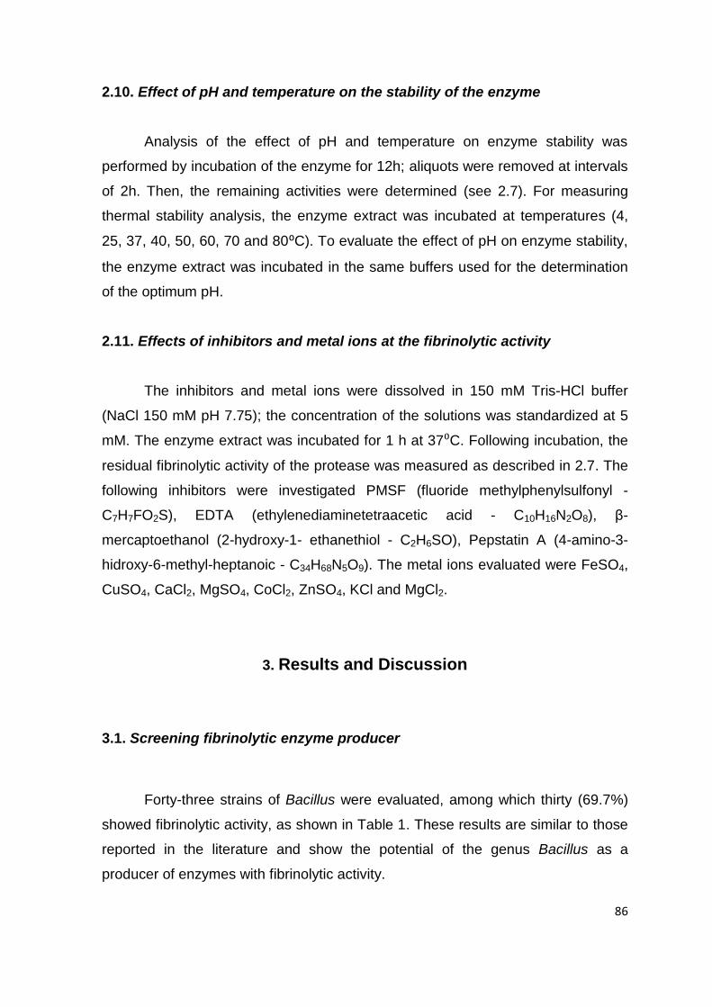
86
2.10. Effect of pH and temperature on the stability of the enzyme
Analysis of the effect of pH and temperature on enzyme stability was
performed by incubation of the enzyme for 12h; aliquots were removed at intervals
of 2h. Then, the remaining activities were determined (see 2.7). For measuring
thermal stability analysis, the enzyme extract was incubated at temperatures (4,
25, 37, 40, 50, 60, 70 and 80⁰C). To evaluate the effect of pH on enzyme stability,
the enzyme extract was incubated in the same buffers used for the determination
of the optimum pH.
2.11. Effects of inhibitors and metal ions at the fibrinolytic activity
The inhibitors and metal ions were dissolved in 150 mM Tris-HCl buffer
(NaCl 150 mM pH 7.75); the concentration of the solutions was standardized at 5
mM. The enzyme extract was incubated for 1 h at 37⁰C. Following incubation, the
residual fibrinolytic activity of the protease was measured as described in 2.7. The
following inhibitors were investigated PMSF (fluoride methylphenylsulfonyl -
C7H7FO2S), EDTA (ethylenediaminetetraacetic acid - C10H16N2O8), β-
mercaptoethanol (2-hydroxy-1- ethanethiol - C2H6SO), Pepstatin A (4-amino-3-
hidroxy-6-methyl-heptanoic - C34H68N5O9). The metal ions evaluated were FeSO4,
CuSO4, CaCl2, MgSO4, CoCl2, ZnSO4, KCl and MgCl2.
3. Results and Discussion
3.1. Screening fibrinolytic enzyme producer
Forty-three strains of Bacillus were evaluated, among which thirty (69.7%)
showed fibrinolytic activity, as shown in Table 1. These results are similar to those
reported in the literature and show the potential of the genus Bacillus as a
producer of enzymes with fibrinolytic activity.

87
Table 1. Microorganisms of the genus Bacillus and their fibrinolytic activity after 48 h of cultivation in soybean
medium (MS-2), pH 7.2, 150 rpm and 37°C
Microorganisms *FA (U.mL-1
) Microorganisms *FA (U.mL-1
)
B. subtilis UCP 999 - B. circulans UFPEDA 436 26.7 ± 0.83
B. licheniformis UCP 1008 34.2 ± 1.01 Bacillus sp. UFPEDA 437 2.25 ± 0.93
B. licheniformis UCP 1009 0.11 ± 0.02 Bacillus sp. UFPEDA 449 34.2 ± 1.61
B. licheniformis UCP 1010 - Bacillus sp. UFPEDA 450 9.93 ± 0.54
B. licheniformis UCP 1013 6.06 ± 1.07 Bacillus sp. UFPEDA 451 -
B. licheniformis UCP 1016 16.3 ± 0.92 B. cereus UFPEDA 452 -
B. licheniformis UCP 1033 16.3 ± 0.82 B. alvei UFPEDA 461 9.93 ± 1.33
B. licheniformis UCP 1477 16.3 ± 0.82 Bacillus sp.UFPEDA 464 16.3 ± 0.73
B. licheniformis UCP 1482 9.93 ± 0.89 Bacillus sp. UFPEDA 465 3.69 ± 0.10
B. cereus UFPEDA 11 - Bacillus sp. UFPEDA 466 -
Bacillus. sp UFPEDA 12 - Bacillus sp. UFPEDA 469 26.7 ± 0.00
Bacillus. sp.UFPEDA 13 1.37 ± 0.74 B. licheniformis UFPEDA 470 -
B. mycoides UFPEDA 14 0.84 ± 0.08 B. firmus UFPEDA 471 -
B. subtilis UFPEDA 15 - B. pumilus UFPEDA 472 20.9 ± 1.49
B. subtilis UFPEDA 16 - B. pumilus UFPEDA 474 6.06 ± 1.46
B. subtilis UFPEDA 86 0.84 ± 0.09 Bacillus sp. UFPEDA 483 26.7 ± 0.08
B. megaterium UFPEDA 108 0.31 ± 0.09 Bacillus sp. UFPEDA 484 -
B. subtilis v. aterrimus UFPEDA 170 0.51 ± 0.08 Bacillus amyloliquefaciens UFPEDA 485 71.8 ± 0.08
Bacillus. sp UFPEDA 189 26.7 ± 1.51 Bacillus sp. UFPEDA 486 0.40 ± 0.08
Bacillus. sp UFPEDA 194 4.73 ± 0.74 Bacillus sp.UFPEDA 487 -
B. subtilis UFPEDA 260 0.51 ± 0.10 Bacillus sp.UFPEDA 488 0.15 ± 0.03
B. subtilis UFPEDA 404 20.9 ± 1.03
*FA - Fibrinolytic activity by fibrin plate method and correlated with a standard curve of plasmin.
Absence of fibrinolytic activity (-)

88
Other researchers8 isolated microorganisms from the Chinese soybean
cheese doufuru as well as Japanese natto and Chinese douche. Among the
isolates, 16 microorganisms showed fibrinolytic activity being the highest value
obtained for a Bacillus subtilis strain with a crude enzyme activity of 1833 IU/mL.
In other research,20 evaluated fibrinolytic proteases production by various bacteria,
and, after 48 hours of culture, the Bacillus sp. strain AS-S20-I obtained the
maximum fibrinolytic activity of 145.8 U.mL-1 a value similar to the one obtained in
this work by the strain Bacillus amyloliquefaciens UFPEDA 485 (71.78 U.mL-1).
3.2. Bacteria identification using 16S and rpoB genes
Initially, the study for the identification was based on 16S rRNA gene
sequence analysis. The results showed 99% similarity homologies with the
species Bacillus amyloliquefaciens and Bacillus subtilis, thus, the analysis using
the 16S rRNA gene sequence alone did not allow differentiating the species
Bacillus subtilis and Bacillus amyloliquefaciens. Therefore, analysis was
performed on the rpoB region which showed greater identity to the species
Bacillus amyloliquefaciens than to the species Bacillus subtilis. The Blast from the
consensus sequence obtained revealed about 98% homology with the region
DNA-directed RNA polymerase β-subunit of the species Bacillus
amyloliquefaciens and 89% homology to the same region of the Bacillus subtilis
species.
For this study identification made by the analysis of the rpoB region
sequencing offered advantages over the 16S because the rpoB gene has a higher
degree of polymorphism compared to 16S, making it more accurate for species
identification. The heterogeneity of the 16S rRNA gene hampers the quantification
of bacterial species by PCR based assays. In contrast, the rpoB gene is common
to all bacteria and occurs as a single copy in the genome.
Researchers obtained similar results in the process of identification of
microorganism of the genus Bacillus by comparison of 16S and rpoB genes of the
13 Bacillus species. Found that, of the 13 species, the levels of similarity ranged
from 90.3% (B. hwajinpoensis and B. sporothermodurans) to 99.8% (B. anthracis
and B. cereus).15

89
In this work the results in conjunction of the 16S and rpoB region confirmed
that the sample tested has a higher homology to the species Bacillus
amyloliquefaciens and demonstrate that the polymorphism of the Bacillus rpoB
gene can be used to identify Bacillus species, providing improvement over
conventional methods of identification Bacillus species. The result was submitted
to the GenBank database.
3.3. Optimization of the conditions for production of the fibrinolytic enzyme
from Bacillus amyloliquefaciens UFPEDA 485
To achieve the optimization of the fibrinolytic activity in shake flasks by this
central composite design (CCD) star 23, was previously realized other full factorial
design 23, that studied other concentrations of soybean flour and glucose near
optimal found in this work and by which was found the optimum temperature
(37°C) and optimum pH 7.2 of production (data not shown), therefore, these
factors were not investigated in the experiments realized in this work.
The optimization of the conditions for the production of the fibrinolytic
enzyme from Bacillus amyloliquefaciens UFPEDA 485 was conducted according
to a Central Composite Design (CCD) star 23 with two levels and three factors and
the response variable Fibrinolytic Activity (FA) after 48 hours of cultivation of the
Bacillus amyloliquefaciens UFPEDA 485 (Table 2).
The Increase of the concentration of the independent variables, soybean
flour, glucose and the interaction between these two factors, had a linear effect
positive and statistically significant for increased of the fibrinolytic activity. For the
quadratic effect, the increase of the concentration of the independent variables,
soybean flour, glucose and the interaction among the factors, was statistically
significant and had a negative effect on the increased fibrinolytic activity. The
results from the analysis of variances and effect estimates for the main factors are
shown in Table 3. The variation around the mean of the residuals is fully explained
by pure error, because there is no evidence of lack of fit, and a good model fit is
obtained. The p value for lack of fit was < 0.00. The validity of the model is
confirmed by the ANOVA analysis.

90
Table 2. Matrix of the Central Composite Design (CCD) star for the optimization of the conditions for
production of the fibrinolytic enzyme from Bacillus amyloliquefaciens UFPEDA 485.
Runs Soybean flour (%) Glucose (%) Agitation (rpm) Fibrinolytic activity (U.mL-1
)
1 1.0 ( - ) 0.5 ( - ) 100 ( - ) 462 ± 0.014
2 1.0 ( - ) 1.5 ( + ) 200 ( + ) 682 ± 0.003
3 3.0 ( + ) 0.5 ( - ) 200 ( + ) 666 ± 0.013
4 3.0 ( + ) 1.5 ( + ) 100 ( - ) 492 ± 0.004
5 (C) 2.0 ( 0 ) 1.0 ( 0 ) 150 ( 0 ) 702 ± 0.006
6 1.0 ( - ) 0.5 ( - ) 200 ( + ) 710 ± 0.008
7 1.0 ( - ) 1.5 ( + ) 100 ( - ) 419 ± 0.005
8 3.0 ( + ) 0.5 ( - ) 100 ( - ) 413 ± 0.009
9 3.0 ( + ) 1.5 ( + ) 200 ( + ) 696 ± 0.011
10 (C) 2.0 ( 0 ) 1.0 ( 0 ) 150 ( 0 ) 701 ± 0.003
11 1.0 ( - ) 1.0 ( 0 ) 150 ( 0 ) 636 ± 0.004
12 3.0 ( + ) 1.0 ( 0 ) 150 ( 0 ) 613 ± 0.004
13 2.0 ( 0 ) 0.5 ( - ) 150 ( 0 ) 607 ± 0.026
14 2.0 ( 0 ) 1.5 ( + ) 150 ( 0 ) 604 ± 0.007
15 2.0 ( 0 ) 1.0 ( 0 ) 100 ( - ) 538 ± 0.006
16 2.0 ( 0 ) 1.0 ( 0 ) 200 ( + ) 813 ± 0.014
17 (C) 2.0 ( 0 ) 1.0 ( 0 ) 150 ( 0 ) 701 ± 0.004

91
Table 3. Analysis of variance (ANOVA) for the response fibrinolytic
activity, over the independent variables soybean flour (%), glucose (%)
and agitaton (rpm), according to Central Composite Design (CCD)
star, with 95% confidence level.
Factor
Fibrinolytic Activity
Estimates effect p value
(1) Soybean flour (%) (L) 178 0.00
Soybean flour (%) (Q) - 51.3 0.00
(2) Glucose (%) (L) 506 0.00
Glucose (%) (Q) - 282 0.00
(3) Agitation (rpm) (L) 2.93 0.00
1L by 2L 45.0 0.00
1L by 3L - 0.14 0.00
2L by 3L - 0.17 0.00
Regression coefficient ANOVA
Pure Error = 0.18 R2 = 0.99 R
2adjusted = 0,97
Using the obtained values in the table 2, the corresponding multiple
regression equation was calculated. The quadratic effect of the variable (X3) was
ignored in Equation (3) as its effect was not statistically significative.
From the regression analysis, the Equation (3) was obtained:
Y = - 151 + 178 (X1) – 51.3 (X1)2 + 506 (X2) – 282 (X2)
2 + 2.93
(X3) + 45 (X1.X2) – 0.14 (X1.X3) – 0.17 (X2.X3)
The optimum conditions found for the fibrinolytic activity (FA) were 1.88%
for soybean flour and 0.988% for glucose, at 200 rpm. Under these conditions the
value calculated was 808 U.mL-1, very close to the value 813 U. mL-1, observed by
Eq. (3)
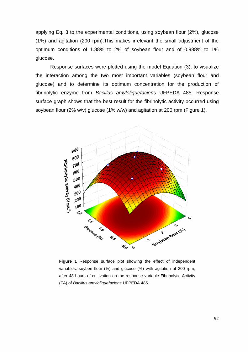
92
applying Eq. 3 to the experimental conditions, using soybean flour (2%), glucose
(1%) and agitation (200 rpm).This makes irrelevant the small adjustment of the
optimum conditions of 1.88% to 2% of soybean flour and of 0.988% to 1%
glucose.
Response surfaces were plotted using the model Equation (3), to visualize
the interaction among the two most important variables (soybean flour and
glucose) and to determine its optimum concentration for the production of
fibrinolytic enzyme from Bacillus amyloliquefaciens UFPEDA 485. Response
surface graph shows that the best result for the fibrinolytic activity occurred using
soybean flour (2% w/v) glucose (1% w/w) and agitation at 200 rpm (Figure 1).
Figure 1 Response surface plot showing the effect of independent
variables: soyben flour (%) and glucose (%) with agitation at 200 rpm,
after 48 hours of cultivation on the response variable Fibrinolytic Activity
(FA) of Bacillus amyloliquefaciens UFPEDA 485.

93
Previous researches have proved that glucose is a preferred carbon source
for growth of Bacillus spp.. Other carbon sources such as fructose, maltose,
lactose, sucrose and glycerol have shown minimum solubility, poor cellular uptake
and unsuitable metabolic pathway reducing the production of fibrinolytic enzymes.5
Researchers5 reported an optimum concentration of glucose (1.25% w/v) for
maximum fibrinolytic activity from Bacillus sphaericus MTCC 3672, result similar to
this work, that obtained optimum concentration of glucose at (1% w/v).
Others researchers optimized the production of fibrinolytic enzyme by a
central composite rotary design (CCRD), using soybean as a nitrogen source and
glucose as carbon source. In this case the maximum activity, obtained in a
bioreactor, reached in 12 hours, using glucose 0.5% and soybean meal 0.5%.
Unlike this study, using a central composite design (star) 23 in shake flasks, and
achieved the best result for fibrinolytic activity in 48 h, using soybean flour (2%
w/v) and glucose (1% w/v).10
3.4. Evaluation of fibrinolytic activity by degradation of blood clots
The enzyme extract produced by Bacillus amyloliquefaciens UFPEDA 485
showed a high effectiveness in degrading blood clots in vitro. After 1 hour, the
results showed a percentage degradation of the clot (PDC) of 62%. Negative
control containing saline solution kept the clot intact (Figure 2). Other researchers
studied the potential of the enzyme from Bacillus sp. SFN in degrading blood clots
and showed that 20 U/mL of fibrinolytic enzyme could degrade completely the
blood clot after 18 hours of incubation. The results obtained in this work suggest
the high efficiency of the obtained enzyme in the degradation of blood clots as, in
just one hour, a PDC of 62% was achieved.19
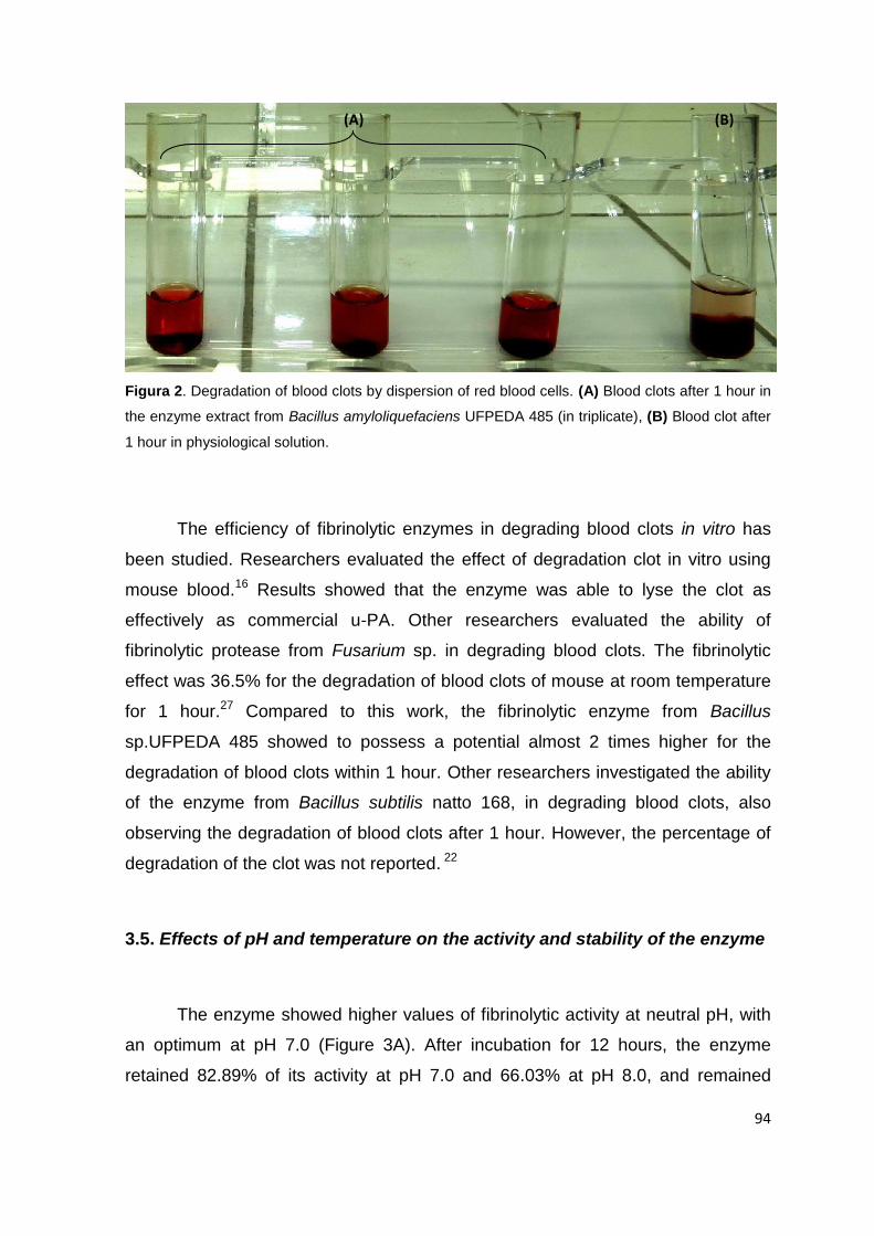
94
Figura 2. Degradation of blood clots by dispersion of red blood cells. (A) Blood clots after 1 hour in
the enzyme extract from Bacillus amyloliquefaciens UFPEDA 485 (in triplicate), (B) Blood clot after
1 hour in physiological solution.
The efficiency of fibrinolytic enzymes in degrading blood clots in vitro has
been studied. Researchers evaluated the effect of degradation clot in vitro using
mouse blood.16 Results showed that the enzyme was able to lyse the clot as
effectively as commercial u-PA. Other researchers evaluated the ability of
fibrinolytic protease from Fusarium sp. in degrading blood clots. The fibrinolytic
effect was 36.5% for the degradation of blood clots of mouse at room temperature
for 1 hour.27 Compared to this work, the fibrinolytic enzyme from Bacillus
sp.UFPEDA 485 showed to possess a potential almost 2 times higher for the
degradation of blood clots within 1 hour. Other researchers investigated the ability
of the enzyme from Bacillus subtilis natto 168, in degrading blood clots, also
observing the degradation of blood clots after 1 hour. However, the percentage of
degradation of the clot was not reported. 22
3.5. Effects of pH and temperature on the activity and stability of the enzyme
The enzyme showed higher values of fibrinolytic activity at neutral pH, with
an optimum at pH 7.0 (Figure 3A). After incubation for 12 hours, the enzyme
retained 82.89% of its activity at pH 7.0 and 66.03% at pH 8.0, and remained
(A) (B)
95
around 50% of its activity at pH 6.0 and 9.0 after 8 h of incubation (Figure 3B).
Results corroborate with other researchers,3 that studied a fibrinolytic enzyme
produced by Bacillus licheniformis B4 - an optimum pH of 7.5 was observed, and
the enzyme retained 100% of its activity at pH 7.0 after 30 min of incubation. Other
researchers,6 evaluated the fibrinolytic protease from Bacillus cereus NS-2 and,
unlike this work, an optimum pH 9.0 was found and the fibrinolytic protease lost its
activity in just 30 minutes. Thus, the fibrinolytic enzyme produced by Bacillus
amyloliquefaciens UFPEDA 485 is very stable in comparison with other fibrinolytic
enzymes produced by other species of the genus Bacillus. Moreover, it has the
best activity at physiological conditions.
(A)
(B)
Figure 3. (A) Effect of pH on the fibrinolytic activity relative of the enzyme from
Bacillus amyloliquefaciens UFPEDA 485 after 1h of incubation. (B) Effect of pH on the
stability of enzyme measured at intervals of 2h per 12h of incubation of the enzyme
and expressed as percentage of residual activity. Buffers used: Glycine-HCl (pH 3.0),
Sodium acetate (pH 4.0-5.0), Citrate phosphate (pH 6.0), Tris-HCl (pH 7.0-8.0),
Glycine-NaOH (pH 9.0-10.0). All buffer concentrations were 20 mM. Each value is the
average of the results of three experiments, and the error bars show the standard
deviations.
The optimum temperature was 37⁰C (Figure 4A). Thermostability assays
showed that, after 12h of incubation, the enzyme remained stable at 4, 25 and
37⁰C, maintaining activities of 96.04, 95.88 and 98.26 %, respectively, and still

96
maintained 58.84% of its activity at 40⁰C (Figure 4B). In other study,3 was also
found a fibrinolytic enzyme with an optimum temperature of 37⁰C that retained its
total activity after 1 hour of incubation when preserved at 20-40⁰C; then the activity
decreased with the increase of temperature. Differently, in other study,6 was found
an optimum temperature of 40⁰C, and during the thermostability assays the
enzyme lost 40-60% of its activity at each temperature of incubation. These results
confirm the excellent thermostability of the fibrinolytic enzyme produced by
Bacillus amyloliquefaciens UFPEDA 485 when compared with the enzymes
produced by Bacillus cited at the literature.
(A)
(B)
Figure 4 (A) Effect of temperature on the fibrinolytic activity relative of the enzyme from Bacillus
amyloliquefaciens UFPEDA 485 after 1h of incubation; (B) Effect of temperature on the stability
of enzyme measured at intervals of 2h per 12h of incubation of the enzyme and expressed as
percentage of residual activity. Each value is the average of the results of three experiments,
and the error bars show the standard deviations.
3.6. Effects of inhibitors and metal ions on the fibrinolytic activity
The fibrinolytic activity was almost completely inhibited after 60 min of
incubation with PMSF (91.52%) and EDTA (89.64 %) in Table 4. The results show
that the enzymatic extract from Bacillus amyloliquefaciens UFPEDA 485 contains

97
serine-metallo proteases, indicating that the hydroxyl (serine) group is located at
or near the active site of one of the enzymes contained in the extract, hence being
inhibited by PMSF (serine protease inhibitor); moreover, divalent metals seem to
be required for the enzyme to maintain its activity, as an inhibition by chelating
agents such as EDTA (metallo protease inhibitor) occurs. Enzyme activity was
less inhibited by Pepstatin A (51.78%) an aspartic protease inhibitor, and β-
mercaptoethanol (63.82%) a cysteine protease inhibitor.
Table 4. Effects of inhibitors on the fibrinolytic activity of the enzyme
Inhibitors and metal ions Residual activity (%)
Control 100
PMSF 8.48 ± 0.01
EDTA 10.4 ± 0.02
Pepstatin A 48.2 ± 0.04
β-mercaptoethanol 36.2 ± 0.02
The values are presented as means ± SD (standard deviation).
Enzyme activity was significantly inhibited by FeSO4 (93.18%) and the
CaCl2 (10%) slightly increased the fibrinolytic activity of the enzyme (Table 5). Both
the activation as the inhibition the activity of an enzyme occurs due to allosterics
effects. The connection of a chemical substance with the allosteric site of an
enzyme may induce conformational changes in the spatial structure of the
enzyme, changing the affinity by its substrate.
In another study12 evaluated the influence of protease inhibitors and metal
ions in fibrinolytic enzyme activity also reported an inhibition of 100% and 96.65%,
in the presence of PMSF and EDTA, respectively. The enzyme was also classified
as a serine-metallo protease. As wel as in this work, the enzyme when incubated
with CaCl2, increased its fibrinolytic activity to 118.15%

98
Table 5. Effects of metal ions on the fibrinolytic activity of the enzyme
Inhibitors and metal ions Residual activity (%)
Control 100
FeSO4 6.82 ± 0.04
CuSO4 50.0 ± 0.05
MgSO4 94.5 ± 0.03
ZnSO4 52.6 ± 0.04
CoCl2 48.3 ± 0,06
MgCl2 96.8 ± 0,06
CaCl2 110 ± 0,02
KCl 98.6 ± 0,01
The values are presented as means ± SD (standard deviation).
The influence of protease inhibitors was also evaluated in other study18
unlike this work, when the enzyme was incubated with PMSF and EDTA, an
inhibition of 99.8% and 8% was observed, respectively, making this enzyme to be
considered only as a serine potease. However, for study of the effect of metal
ions, as in this work, the CaCl2 increased fibrinolytic activity to 111% and when
incubated with CoCl4 and FeSO4 caused inhibition of 59% and 100%, respectively.
4. Conclusion
From the 43 strains evaluated, Bacillus amyloliquefaciens UFPEDA 485
was selected as the one producing the highest fibrinolytic activity.
Optimum conditions for production of the fibrinolytic enzyme from Bacillus
amyloliquefaciens UFPEDA 485 were found.
The fibrinolytic protease from Bacillus amyloliquefaciens UFPEDA 485 has
biochemical characteristics suitable for human and veterinary applications, namely
the best activity at physiological conditions, besides having high thermal stability.

99
The results demonstrate the biotechnological potential of the Bacillus
amyloliquefaciens UFPEDA 485 and its proteases to the pharmaceutical industry
and the possible application on the treatment of vascular disorders.
Acknowledgments: We express our thanks to Coordination for the Improvement
of Higher Level Education Personnel (CAPES) - Doctoral Sandwich Program
(PDSE) Nº 0259/12-8 and National Council for Scientific and Technological
Development (CNPq) - Nº 202026/2011-6 for the financial support.
5. Literature Cited
1. Agrebi R, Hmidet N, Hajji M, Ktari N, Haddar A, Fakhfakh-zouari N, Nasri M.
Fibrinolytic Serine Protease Isolation from Bacillus amyloliquefaciens An6 Grown
on Mirabilis jalapa Tuber Powders. Appl Biochem Biotech. 2010; 162:75-88.
2. Ahn MY, Hahn BS, Ryu K.S, Kim JW, Kim I, Kim YS. Purification and
characterization of a serine protease with fibrinolytic activity from dung beetles,
Catharsius molossus. Thromb Res. 2003;112:339-347.
3. Al-Juamily EF, Al-Zaidy BH. Purification and Characterization of Fibrinolytic
Enzyme Produced from Bacillus lichniformis B4. Chem Sci Rev Lett. 2013;2:256-
266.
4. Astrup T, Mullertz S. The fibrin plate method for estimating fibrinolytic activity. Arch
Biochem Biophys. 1952;40:346–531.
5. Avhad DN, Vanjari SS, Rathod VK. A Novel Fibrinolytic Enzyme from Bacillus
Sphaericus MTCC 3672: Optimization and Purification Studies. AJCMicrob.
2013;1:1-13.

100
6. Bajaj BK, Sharma N, Singh S. Enhanced production of fibrinolytic protease from
Bacillus cereus NS-2 using cotton seed cake as nitrogen source. Biocatal Agric
Biotechnol. 2013;2:204-209.
7. Bradford M.M. A rapid and sensitive method for the quantification of protein
utilizing the principle of protein-dye binding. Anal Bioch. 1976;72:248-254.
8. Chen B, Huo J, He Z, He Q, Hao Y, Chen Z. Isolation and identification of an
effective fibrinolytic strain Bacillus subtilis FR-33 from the Chinese doufuru and
primary analysis of its fibrinolytic enzyme. Afr J Microbiol Res. 2013;7:2001-2009.
9. Choi, J-H, Sapkota K, Park S-E, Kim S, Kim S-J. Thrombolytic, anticoagulant and
antiplatelet activities of codiase, a bi-functional fibrinolytic enzyme from Codium
fragile. Biochimie. 2013;95:1266-1277.
10. Deepak V, Ilangovan S, Sampathkumar MV, Victoria MJ, Pasha SPBS, Pandian S
BRK, Gurunathan S. Medium optimization and immobilization of purified fibrinolytic
URAK from Bacillus cereus NK1 on PHB nanoparticles. Enz Microb Technol.
2010;47:297-304.
11. He J, Chen S, Gu J. Identification and characterization of Harobin, a novel
fibrino(geno)lytic serine protease from a sea snake (Lapemishardwickii). FEBS
Lett. 2007;581:2965-2973.
12. Heo K, Cho KM, Lee CK, Kim GM, Shin J-H, Kim JS, Kim JH. Characterization of
a Fibrinolytic Enzyme Secreted by Bacillus amyloliquefaciens CB1 and Its Gene
Cloning. J Microbiol Biotechnol. 2013;23:974-983.
13. Jin M, Chen W, Huang W, Rong L, Gao Z. Preparation of pegylated lumbrokinase
and an evaluation of its thrombolytic activity both in vitro and in vivo. APSB.
2013;3:123-129.

101
14. Kanagasabai V, Thangavelu V. Response surface methodological optimization of
the medium components for production of xylanase under ssf by aspergillus
fumigatus. J Adv Sci Res. 2013;4:13-20.
15. Ki JS, Zhang W, Qian PY. Discovery of marine Bacillus species by 16S rRNA and
rpoB comparisons and their usefulness for species identification. J Microbiol Meth.
2009;77:48-57.
16. Kim D-W, Sapkota K, Choi J-H, Kim Y-S, Kim S, Kim S-J. Direct acting anti-
thrombotic serine protease from brown seaweed Costaria costata. Process
Biochem. 2013;48:340-350.
17. Klafke JZ, Silva MA, Rossato MF, Trevisan G, Walker CIB, Leal CAM, Borges DO,
Schetinger MRC, Moresco RN, Duarte MMMF, Santos ARS, Viecili PRN, Ferreira
J. Antiplatelet, Antithrombotic, and Fibrinolytic Activities of Campomanesia
xanthocarpa. Evid-Based Compl Alt. 2012;2012:1-8.
18. Mahajan PM, Nayak S, Lele SS. Fibrinolytic enzyme from newly isolated marine
bacterium Bacillus subtilis ICTF-1: Media optimization, purification and
characterization. J Biosci Bioeng. 2012;113:307-314.
19. Mohanasrinivasan V, Devi CS, Biswas R, Paul F, Mitra M, Selvarajan E, Suganthi
V. Enhanced production of nattokinase from UV mutated Bacillus sp. Bangladesh
J Pharmacol. 2013;8:110-115.
20. Mukherjee AK, RAI SK. A statistical approach for the enhanced production of
alkaline protease showing fibrinolytic activity from a newly isolated Gram-negative
Bacillus sp. strain AS-S20-I. New Biotech. 2011;28:182-189.
21. Mukherjee AK, Rai SK, Thakur R, Chattopadhyay P, Kar S K. Bafibrinase: A non-
toxic, non-hemorrhagic, direct-acting fibrinolytic serine protease from Bacillus sp.
strain AS-S20-I exhibits in vivo anticoagulant activity and thrombolytic potency.
Biochimie. 2012;94:1300-1308.

102
22. Omura K, Hitosugi M, Zhu X, Ikeda M, Maeda H, Tokudome S. A Newly Derived
Protein From Bacillus subtilis natto With Both Antithrombotic and Fibrinolytic
Effects. J Pharmacol Sci. 2005;99:247-251.
23. Porto ALF, Campos-Takaki GM, Lima Filho JL. Effects of culture conditions on
protease production by Streptomyces clavuligerus growing soy bean flour medium.
App Biochem Biotech. 1996;60:115-122.
24. Sundararajan S, Kannan CN, Chittibabu S. Alkaline protease from Bacillus cereus
VITSN04: Potential application as a dehairing agent. J Biosc Bioeng.
2011;111:128-133.
25. Uesugi Y, Usuki H, Iwabuchi M, Hatanaka T. Highly potent fibrinolytic serine
protease from Streptomyces. Enz Microb Tech. 2011;48:7-12.
26. Wang S, Wu Y, Liang T. Purification and biochemical characterization of a
nattokinase by conversion of shrimp shell with Bacillus subtilis TKU007. New
Biotechnol. 2011;28:196-202.
27. Wu B, Wu L, Ruan L, Ge M, Chen D. Screening of Endophytic Fungi with
Antithrombotic Activity and Identification of a Bioactive Metabolite from the
Endophytic Fungal Strain CPCC 480097. Current Microb. 2009;58:522-527.

103
CAPÍTULO III
Process scale-up and biochemical characterization of an enzyme with
anticoagulant and fibrinolytic potential from Bacillus amyloliquefaciens
UFPEDA 485
Artigo submetido à revista Biochemical and Engineering Journal
Impact Factor: 2.578
104
Process scale-up and biochemical characterization of an enzyme with anticoagulant and fibrinolytic 1
potential from Bacillus amyloliquefaciens UFPEDA 485 2
3
1 Fabiana América Silva Dantas de Souza; 1 Júlio Cézar dos Santos Nascimento; 1 André Manuel de Oliveira Mota; 3 Maikon Kelbert; 1Raquel 4
Pedrosa Bezerra; 1Daniela de Araújo Viana Marques; 2 José Antônio Couto Teixeira; 1Ana Lúcia Figueiredo Porto* 5
6
7
1Department of Morphology and Animal Physiology, Federal Rural University of Pernambuco, Rua Dom Manoel de Medeiros, s/n., Dois Irmãos, 52171-900, 8
Recife, PE, Brazil. 9 2Institute for Biotechnology and Bioengineering, Centre of Biological Engineering, University of Minho, Largo do Paço, Campus Gualtar, 4710-057, Braga, 10
3 University of Joinville Region, Street Paul Malschitzki, n.10, Bom Retiro, 89219-710, Joinville, Santa Catarina, Brazil. 11
12
* Corresponding author: [email protected] Phone and Fax: + 55 (81) 3320.6345 13
14
15
Abstract 16
17
Fibrinolytic proteases produced by Bacillus sp. have attracted an interest in the pharmaceutical industry due to its 18
effectiveness in degrading and preventing the formation of blood clots. Fibrinolytic protease production by a 19
Bacillus amyloliquefaciens strain was conducted in a 7L bioreactor. A full 22 factorial design was conducted to 20
investigate the effects and interactions of independent variables aeration and agitation on the fibrinolytic and 21
amidolytic activities of protease. The best conditions for protease production occurred at 12h, 800 rpm and 1.5 22
vvm, pH 7.2, at 37⁰C. Under these conditions the enzyme achieved a fibrinolytic activity of 2169.11 U.mL-1
and 23
amidolytic activity of 1586.89 µmol. min-1.
mL-1
. Further characterization of the protease properties showed that 24
the amidolytic activity was inhibited by PMSF and among the synthetic substrates evaluated the one with the 25
highest affinity was N-succinyl-Ala-Ala-Pro-Phe-pNA, indicating that the enzyme is a serine protease. The 26
corresponding, Km and Vmax values were 0.68 mM and 357.14 mmol pNA min-1
mL-1
, respectively. The 27
optimum pH for the hydrolysis of N-succinyl-Ala-Ala-Pro-Phe-pNA was between 7 and 8 and for hydrolysis of 28
fibrin pH 7.0. The optimum temperature for both fibrinolytic activity and amidolytic activity was 37 ⁰C. The 29
fibrinolytic and amidolytic properties of the enzyme were preserved at 92.2% and 95.8% respectively after 9 30

105
months of storage. The results show the potential of the enzyme to be marketed and used as a new and potent 31
drug in thrombolytic therapy. 32
33
Keywords: Bacillus amyloliquefaciens, fibrinolytic enzyme, anticoagulant, production scale-up, bioreactor 34
35
36
1. Introduction 37
38
Fibrinolytic proteases are hydrolytic enzymes that dissolve blood clots. The formation of blood clots is 39
a natural phenomenon of protection of the human body to prevent excessive bleeding from injuries and wounds, 40
but may sometimes block blood flow causing cardiovascular disorders such as stroke, myocardial infarction and 41
others [19]. Thrombolytic therapy using fibrinolytic proteases has been shown as a potential solution to many 42
vascular disorders. 43
The blood clots are composed of fibrin, a protein formed by the activation of fibrinogen by proteolytic action of 44
thrombin (EC 3.4.21.5). Fibrin clot formation and fibrinolysis are normally well balanced in blood system. 45
Generally the fibrin formed is hydrolyzed by plasmin (EC 3.4.21.7), but when fibrin is not dissolved because of 46
some imbalance, thrombosis occurs [2]. 47
Antithrombotic drugs, including anticoagulants and antiplatelets, are used to prevent and treat thrombosis. Drugs 48
such as heparin and vitamin K antagonists (warfarin) are commonly used in anticoagulant therapy, but 49
unfortunately these drugs have important limitations that drive continuous and intense efforts to develop new, 50
efficient and safe anticoagulants, especially those targeting specific coagulation factors. Direct thrombin 51
inhibitors such as argatroban, dabigatran, bivalirudin, and hirudin, as well as the factor (F) Xa inhibitor 52
rivaroxaban, anticoagulants are currently marketed, but the nature of thrombosis involving complex clinical 53
situations (patients with renal failure, cancer and thrombosis in pregnant women) continues to ask for new, safer 54
and more effective anticoagulants with different pharmacological and pharmacokinetic properties [1] [12] 55
In recent years, various fibrinolytic enzymes derived from different sources have been discovered, Daboia 56
russelli [2] [16], Neanthes japonica [26], Cordyceps militaris [4], Campomanesia xanthocarpa [11], Yellow 57
Mealworm [7], Euphorbia hirta [17], Artocarpus heterophyllus latex [22], Candida guilliermondii [20]. The 58
fibrinolytic protease from Bacillus sp. has attracted the interest of researchers and the pharmaceutical industry 59
106
because of its efficiency as thrombolytic agents in the process of fibrinolysis and plasmin activation [19]. 60
Researchs have shown that Bacillus sp. produce a variety of fibrinolytic enzymes [6], [24], [23], [15]. 61
The species of the genus Bacillus are attractive to the industry because they have high growth rates resulting in 62
short cycles of cultivation and high production, and are able to secrete various proteins in the extracellular 63
medium [21]. These features put the microorganisms of the genus Bacillus among the most important producers 64
of industrial enzymes. Around 60% of commercially available enzymes are produced by Bacillus species [5]. 65
Enzyme production on a large scale is mainly carried out by batch fermentation in stirred tank bioreactors and 66
increase production of enzymes in the scale-up process is achieved by optimizing the relation between the 67
microorganism and its environment. Agitation and aeration are impact factors in the fermentation process. The 68
agitation influences mixing and heat and mass transfer being an important parameter for growth and performance 69
of microbial cells. Aeration is also a key factor in aerobic processes due to its impact on oxygen supply. Overall, 70
oxygen transfer depends on the gas flow rate, type of agitator, agitation speed and the properties of liquids [5]. 71
The purpose of this study was to evaluate the best conditions of aeration and agitation for the production of the 72
fibrinolytic enzyme from Bacillus sp. UFPEDA 485 in bioreactor, in order to maximize the enzyme activity 73
produced and to minimize the production time, as well as to characterize the enzyme with amidolytic and 74
fibrinolytic properties. 75
76
77
2. Materials and methods 78
79
2.1. Reagents 80
81
The chemicals reagents used were analytical grade and obtained from Merck (Darmstadt, Germany) and 82
Sigma (St. Louis, MO). 83
84
2.2. Microorganism 85
86
The Bacillus amyloliquefaciens UFPEDA 485 strain was obtained from Culture Collection of 87
Department of Antibiotics, at the Federal University of Pernambuco, Brazil. The microorganism was isolated 88

107
from the fermentation of sugar cane mills from Zona da Mata of Pernambuco, Brazil. The stock culture was 89
maintained in nutrient broth (1 % peptone, meat extract 0.3 % and 0.5 % NaCl) in cryotubes (10 % v/v glycerol) 90
at -80°C. 91
92
2.3. Culture condition 93
94
The soybean flour medium (MS-2) [18] was used for the production of fibrinolytic proteases. The 95
medium composition was: filtered soybean flour (2% w/v), K2HPO4 (0.435 % w/v), NH4Cl (0.1% w/v), 96
MgSO4.7H2O (0.06% w/v), glucose (1% w/v) and mineral solution (1% v/v) containing: FeSO4.7H2O (100 mg); 97
MnCl2.4H2O (100 mg), and ZnSO4.H2O (100 mg) in 100 mL of distilled water. For the selection of the best 98
condition of production of the fibrinolytic enzyme was used 22 full factorial design. The experiments were 99
performed in a 7L bioreactor (B. Braun Biotech International, Model Biostat®A, type A5, Melsungen, Germany) 100
under controlled temperature (37⁰C) and pH (7.2) by a micro DCU-twin controller. The liquid working volume 101
was 5L and the culture pH was controlled by the addition of 6 N HCl or 6N NaOH. 102
For the inoculum, the colonies from the stock culture were reactivated in nutrient broth at 37⁰C and 200 103
rpm up to reach the end of the exponential growth phase; then, the cells were maintained on nutrient agar for 24 104
h and transferred to flasks with 250 mL and 100 mL working volume. In these flasks, cells were grown in 105
soybean medium (MS-2) at 37⁰C and 200 rpm up to reach the end of the exponential growth phase, and the 106
obtained cell culture was standardized by optical density and transferred into the bioreactor. 107
At the end of the production process, the culture medium was centrifuged at 10.000 xg for 20 min to 108
obtain the enzyme extract and make analytical determinations. 109
110
2.4. Statistical analysis and experimental design 111
112
For statistical analysis, the variables were coded according to Eq. (1): 113
i
oii
ΔX
XX=x
Eq. (1) 114
where each independent variable is represented by xi (coded value), Xi (real value), X0 (real value at the center 115
point), and ΔXi (step change value). 116

108
A 22 full factorial design was used to analyze the influence of the two independent variables agitation (rpm) and 117
aeration (vvm) on enzyme production. The effects of these two independent variables were determined having as 118
response variable the fibrinolytic protease production (expressed as fibrinolytic and amidolytic activity). The 119
experimental design was composed of 7 runs and 3 repetitions at the central point, needed to calculate the pure 120
error (Table 1). Statistical significance of the variables was determined at 5 % probability level (p < 0.05). 121
The goodness of fit was evaluated by the coefficient of determination (R2) and the analysis of variance 122
(ANOVA); the first-order equation was determined by Fischer’s test. The analysis of the results was carried with 123
the program Statistic version 8.0. 124
125
2.5. Protein concentration 126
127
Total protein concentration was determined by Bradford method using as standard bovine serum albumin (BSA) 128
[1]. 129
130
2.6. Fibrinolytic activity 131
132
Fibrinolytic activity was determined according to Wang et al [23]. The reaction mixture containing 0.1 mL of 133
245 mM phosphate buffer (pH 7.0) and 0.4 mL of 0.72% fibrinogen solution was incubated at 37 °C for 5 min; 134
after that, 0.1 mL of a 20 U.mL-1
thrombin solution was added and the reaction mixture was again incubated at 135
37°C for 10 min. After clot formation, 0.1 mL of diluted enzyme extract was added, and incubation continued at 136
37°C for 60 min, the reaction mixture being shaken every 20 min. The reaction was stopped by adding 0.7 mL of 137
0.2 M trichloroacetic acid (TCA). The reaction mixture was centrifuged at 15000 xg for 10 min, and the 138
absorbance of the supernatant was measured at 275 nm by spectrophotometry. In this assay, one unit of 139
fibrinolytic protease activity was defined as the amount of enzyme required to produce an increase in absorbance 140
equal to 0.01 per minute, equivalent to liberation of tyrosine. All experiments were performed in triplicate. 141
142
2.7. Evaluation anticoagulant effect 143
144

109
The anticoagulant effect of the enzyme extract was observed by monitoring of the no formation of a 145
fibrin clot artificial. For the test was added in polypropylene tubes 0.6 mL of enzyme extract without dilution 146
and diluted (1:2), (1:4) , (1:8) in 0.6 mL of a solution containing ; 0, 1 ml of phosphate buffer (245 mM, pH 7.0), 147
0.4 mL of fibrinogen (0.72%), 0.1 ml of thrombin solution (20 U.mL -1
). As a control, the enzyme extract was 148
replaced by saline solution (0.15 M, pH 7.2). The assays were incubated at 37°C for 1 h. (Lu et al., 2010, 149
Modified). 150
151
2.8. Amidolytic activity determination and substrate specificity 152
153
Amidolytic activity of the enzyme was performed according to Chang et al [2]. The following synthetic 154
chromogenic substrates were analyzed: N-succinyl-Ala-Ala-Pro-Phe-pNA, N-benzoyl-Val-Gly-Arg-pNA, N-155
Succinyl-Ala-Ala-Val-pNA, Gly-Pro-pNA, Gly-Phe-pNA, D-Phe-Val-pNA, and D-Val-Leu-Lys-pNA. The 156
reaction mixture, containing 63 µL of 1.7 mM synthetic substrate solution, 63 µL of 25 mM Tris-HCl buffer (10 157
mM CaCl2, pH 7.8), and 25 µL of the enzyme extract, was incubated at 37 ⁰C for 10 min, and then 63 µL of 0.2 158
M acetic acid was added to stop the reaction. The amount of liberated p-nitroanilide was measured using a 159
microplate spectrophotometer (Biotek, model Synergy HT, Winooski – USA) at an absorbance of 405 nm and 160
calculated using an extinction coefficient (ε) of 9950 cm-1
M-1
. One unit of amidolytic activity was expressed as 161
µmol of p-nitroanilide released due to substrate hydrolysis per min per mL by the enzyme. Each value is the 162
mean of 3 determinations. 163
164
2.9. Determination of kinetic parameters 165
166
The kinetic constants, Km and Vmax, of the enzyme were determined by the method of Lineweaver and Burk [14] 167
with different concentrations (0.2-2.8 mM) of N-succinyl-Ala-Ala-Pro-Phe-pNA as a substrate. Reactions were 168
performed in 25 mM Tris-HCl buffer (10 mM CaCl2, pH 7.8) at 37⁰C. 169
170
2.10. Determination of optimum pH and temperature for the enzyme activity 171
172

110
The determination of the optimum pH and temperature on the amidolytic activity and fibrinolytic activity of the 173
enzyme were measured using N-succinyl-Ala-Ala-Pro-Phe-pNA and fibrin as substrates, respectively. For 174
determination of the optimum temperature, the enzyme activity was analyzed at several temperatures between 4-175
80 ⁰C under the standard assays conditions. For determination of the optimum pH, the reaction occurred at 37 ⁰C 176
and the enzyme activity was measured in the pH range (3.0-10.0). The buffers used were Glycine-HCl (pH 3.0), 177
Sodium acetate (pH 4.0-5.0), Citrate phosphate (pH 6.0), Tris-HCl (pH 7.0-8.0), Glycine-NaOH (pH 9.0-10.0). 178
The concentrations of the buffers were standardized 20 mM. 179
180
2.11. Effect of pH and temperature on the stability of the enzyme 181
182
Analysis of the effect of pH and temperature on enzyme stability was performed by incubation of the enzyme for 183
1h. Then, the activities remaining were determined. For measuring thermal stability analysis, the enzyme extract 184
was incubated at temperatures (4, 25, 37, 40, 50, 60, 70 and 80⁰C). To evaluate the effect of pH on enzyme 185
stability, the reaction occurred at 37⁰C and the enzyme activity was measured in the pH range (3.0-10.0). The 186
buffers and their concentrations used were according to the item 2.10. 187
188
2.12. Effects of inhibitors and metal ions on the amidolytic activity of the fibrinolytic enzyme 189
190
The inhibitors and metal ions were dissolved in 25 mM Tris-HCl buffer (10 mM CaCl2, pH 7.8); the 191
concentration of the solutions was standardized at 5 mM. The enzyme extract was incubated for 1 h at 37⁰C. 192
Following incubation, the residual amidolytic activity of the protease was measured. The following inhibitors 193
were investigated PMSF (fluoride methylphenylsulfonyl - C7H7FO2S), EDTA (ethylenediaminetetraacetic acid - 194
C10H16N2O8), β-mercaptoethanol (2-hydroxy-1- ethanethiol - C2H6SO), Pepstatin A (4-amino-3-hidroxy-6-195
methyl-heptanoic - C34H68N5O9). The metal ions evaluated were FeSO4, CuSO4, CaCl2, MgSO4, CoCl2, ZnSO4, 196
KCl and MgCl2. 197
198
2.13. Storage stability 199
200

111
The storage stability was analyzed at -20, 4 and 25⁰C for a total of 9 months for the fibrinolytic activity and 201
amidolytic activity. Aliquots were withdrawn every 15 days for determination of the residual amidolytic activity 202
and fibrinolytic activity. 203
204
205
3. Results and discussion 206
207
3.1. Conditions for production of enzyme by Bacillus amyloliquefaciens UFPEDA 485 in bioreactor 208
209
The enzyme production in the fermentations process depends critically on maintaining an oxygen transfer 210
rate to satisfy the optimum oxygen demand of the microorganism for product formation. The most relevant 211
factors concerning oxygen supply in a fermentor are the agitation and aeration rates. The (Fig. 1) shows the 212
interaction between these variables and its effect on enzyme production. The results prove that both agitation and 213
aeration are important factors for the production of fibrinolytic enzyme. The best value for fibrinolytic activity 214
found was 2.169.11 U.mL-1
obtained at the highest agitation and aeration rates (800 rpm and 1.5 vvm) in 12 h, at 215
pH 7.2 and 37⁰C. 216
From the experiments in bioreactor, it was possible to find the best conditions for agitation and aeration. When 217
compared to previous experiments conducted in flasks (data not shown), the highest fibrinolytic activity was 2.5 218
times higher and was obtained after 12 hours of fermentation (a fermentation time of 48 hours was required in 219
the flasks experiments - data not shown). These results correspond to 10 fold increase enzyme productivity. 220
The same result was obtained from the effect of these two operating variables on amidolytic activity (Fig. 2). 221
The best result for amidolytic activity was 1586.89 µmol min-1
mL-1
also found at 800 rpm and 1.5 vvm in 12h, 222
which again proves the importance of agitation and aeration for the increase production of this enzyme. 223
The influence of the independent variables, agitation and aeration rate, on the amidolytic and fibrinolytic 224
activities are described in Table 2. All independents variables and their interactions were statistically significant 225
at 95%, and showed p values < 0.05. The analysis of variance revealed, R2adjusted = 0.99 for amidolytic activity, 226
and R2adjusted = 0.97 for fibrinolytic activity. 227
Similar result was observed by Lee et al [13], when studying the production of a fibrinolytic enzyme from 228
Bacillus subtilis BK-17 in bioreactor. The best result for fibrinolytic activity was 1,100 U.mL-1
, and was 229

112
achieved after 12h, at 800 rpm, aeration 1.3 vvm, initial pH 7.5 and temperature 37°C. Thus, the results reports 230
in this work clearly demonstrate the biotechnological potential of Bacillus amyloliquefaciens UFPEDA 485 on 231
the production of fibrinolytic enzyme, as a two fold increase in enzyme productivy is obtained when compared 232
with the research of Lee et al [13] that used similar conditions of agitation and aeration. 233
In this work, the growth of the microorganism in the bioreactor under conditions established, showed not lag 234
phase and the exponential phase lasted 12h (Fig. 3). The fibrinolytic and amidolytic activity increased 235
simultaneously and accompanied cell growth. The maximum values for both fibrinolytic and amidolytic 236
activities were achieved at the end of the exponential growth phase and there was no loss of enzyme activity 237
when the fermentation process was extended. 238
Cho et al. [3] studied the optimum conditions for nattokinase production by submerged cultivation from Bacillus 239
subtilis. Temperature and pH were controlled at 37°C and pH 7.0. Dissolved oxygen (DO) level was 240
automatically controlled at 20% by changing agitation speed (500-900 rpm). In batch culture of B. subtilis in 241
fermentor, the highest nattokinase activity was obtained at 10 h with 50 g/L of peptone supplementation. As in 242
this work, the production of fibrinolytic enzyme obtained by Cho et al. [3] was also associated with the cell 243
growth and the higher activity was also observed at the end of exponential growth phase. However, the enzyme 244
did not remain stable during the cultivation process and a reduction in activity upon entering the stationary phase 245
of cell growth was observed differently from what is observed in the process here presented. 246
247
3.2. Evaluation of the anticoagulant effect of the enzyme 248
249
For the test performed in triplicate, after 1h of incubation with enzyme extract it was not observed the 250
formation of fibrin clots. In the control using saline solution, there was formation of the insoluble fibrin clot. The 251
result indicated that the enzyme exhibited an efficient anticoagulant effect. As in this work, other researchers, 252
also tested the anticoagulant effect of fibrinolytic enzyme from endophytic bacterium Paenibacillus polymyxa 253
EJS-3, and there was not clot formation in the presence of the enzyme [3]. 254
255
3.3. Substrate specificity and amidolytic activity of the fibrinolytic protease 256
257
113
The amidolytic activity of the fibrinolytic enzyme was investigated with several synthetic substrates (Table 3). 258
The enzyme showed amidolytic activity and was able to hydrolyse all synthetic substrates tested, being N-259
succinyl-Ala-Ala-Pro-Phe-pNA the substrate hydrolysed at the highest rate. N-succinyl-Ala-Ala-Pro-Phe-pNA is 260
a well-known substrate for subtilisin, chymotrypsin and cathepsin G (serine protease). The enzyme was less 261
effective on the other synthetic substrates N-Benzoyl-Val-Gly-Arg-pNA; D-Val-Leu-Lys-pNA (for Plasmin and 262
Streptokinase activated plasminogen); Gly-Phe-p-nitronilide; D-Phe-Val-pNA; N-succinyl-Ala-Ala-Val-pNA 263
(for Elastase) and Gly-Pro-pNA. 264
The results presented corroborate with Chang et al [2] which used the same synthetic substrates and also showed 265
higher affinity for N-succinyl-Ala-Ala-Pro-Phe-pNA, showing amidolytic activity of 38.3 nmol.min-1
.ml-1
. The 266
fibrinolytic enzyme from Bacillus subtilis fermented red bean, showed little activity for N-benzoyl-Val-Gly-Arg-267
pNA, D-Val-Leu-Lys-pNA and N-succinyl-Ala-Ala-Val-pNA and showed not amidolytic activity to other 268
synthetic substrates tested. However, the enzyme produced in this work stands out of the enzymes currently 269
found in the literature because it showed affinity for all synthetic substrates tested and achieved an amidolytic 270
activity of 1.300.00 µmol.min-1
.mL-1
for the substrate with higher affinity (N-succinyl-Ala-Ala-Pro-Phe-pNA). 271
Choi et al. [4], investigated the amidolytic activity of a fibrinolytic enzyme from fruiting bodies of Korean 272
Cordyceps militaris and, among various substrates, the enzyme also exhibited the highest specificity (34.9 µmol. 273
min-1
) for N-succinyl-Ala-Ala-Pro-Phe-pNA, a value that is much lower than the one obtained in this work. 274
Kim et al. [10] measured the amidolytic activity of a fibrinolytic enzyme from brown seaweed Costaria costata 275
on four substrates. All tested substrates were hydrolyzed, but different of the results of this work, the enzyme 276
exhibited a larger activity when the synthetic substrate H-D-Val-Leu-Lys-pNA was used. 277
278
3.4. Kinetic parameters of the enzyme with amidolytic activity 279
280
A Michaelis–Menten-type curve was obtained from the plot of the amidolytic activity of the enzyme using N-281
succinyl-Ala-Ala-Pro-Phe-pNA as substrate. The Km and Vmax values obtained using a Lineweaver-Burk double-282
reciprocal plot were 0.68 mM and 357.14 mmole pNA min-1
.mL-1
, respectively. Investigating the fibrinolytic 283
enzyme from a Bacillus subtilis and using the same substrate, Chang et al. [2] obtained a Km 0.59 mM, a value 284
similar to the obtained in this work and a different value for Vmax (79.4 µmole pNA min-1
.mg-1
). Zambare et al. 285
[25] evaluating the fibrinolytic enzyme from Pseudomonas aeruginosa MCM B-327, also used N-succinyl-Ala-286

114
Ala-Pro-Phe-pNA and different this work reported a value for Vmax of 36.90 U.min-1
and a Km of 8.81 mM, a 287
value much higher than the one obtained in this work. These results point out the high specificity of the substrate 288
for the enzyme produced by Bacillus amyloliquefaciens UFPEDA 485 here presented. 289
290
3.5. Effects of inhibitors and metal ions on the amidolytic activity of the fibrinolytic enzyme 291
292
The enzyme activity was almost completely inhibited (72.91%) after 60 min of incubation with PMSF (Table 4). 293
These results indicate that hydroxyl (serine) group is located at or near the active site of the enzyme, and prove 294
the results found in the study substrate specificity of the enzyme (affinity for N-succinyl-Ala-Ala-Pro-Phe-pNA 295
that is a well-known substrate for various serine proteases). The performance of the enzyme to hydrolyze N-296
succinyl-Ala-Ala-Pro-Phe-pNA was slightly inhibited by EDTA (29.57%), β-mercaptoethanol (26.01%) and 297
Pepstatin A (25.23%). 298
Enzyme activity was inhibited by the following ions: FeSO4 (18.89 %), CuSO4 (28.79 %), MgSO4 (34.52 %), 299
ZnSO4 (39.01 %), CoCl2 (35.14 %) and MgCl2 (40.87 %); CaCl2 and KCl slightly increased the amidolytic 300
activity of the enzyme (Table 5). Both the activation as the inhibition the activity of an enzyme occurs due to 301
allosterics effects. The connection of a chemical substance with the allosteric site of an enzyme may induce 302
conformational changes in the spatial structure of the enzyme, changing the affinity by its substrate. 303
Kim et al. [10] investigated the effect of inhibitors and metal ions on the activity of a protease named CCP with 304
antithrombotic action and in agreement with this work, the enzyme activity was significantly inhibited by PMSF 305
and a slight increase in enzyme activity was observed when incubated with Ca++
. Another study that also agrees 306
with this work was conducted by Chang et al. [2], using N-succinyl-Ala-Ala-Pro-Phe-pNA as substrate for a 307
enzyme from Bacillus subtilis The enzyme activity was almost completely inhibited after 30 min incubation with 308
PMSF and, when in contact with calcium ion and potassium ion, the amidolytic activity appeared to increase 309
slightly. 310
311
3.6. Effects of pH and temperature on the activity and stability of the enzyme 312
313
115
The enzyme from Bacillus amyloliquefaciens UFPEDA 485 had an optimum pH at 7 and 8 for the hydrolysis of 314
N-succinyl-Ala-Ala-Pro-Phe-pNA, while the highest activity occurred at pH 7 when fibrin was used as the 315
substrate (Fig. 4a). 316
In what concerns amidolytic activity, the enzyme was very stable in the range of pH 5.0–9.0. The enzyme 317
maintained 100 % activity after incubation at pH 6.0, 7.0 and 8.0 at 37°C for 60 min. The pH stability tests for 318
fibrinolytic activity showed the enzyme was stable in the range of pH 6.0-8.0 and retained 100% activity at pH 319
7.0 at 37°C for 60 min (Fig. 4b). 320
The influence of temperature on the enzyme activity showed that the optimum temperature for both fibrinolytic 321
activity and for amidolytic activity was 37°C (Fig. 5a). For the thermal stability tests, the enzyme was stable and 322
retained 100 % activity at temperatures of up to 40°C, after 60 min of incubation. When incubated at 50°C for 60 323
min, the enzyme retained 64% and 52% of amidolytic activity and fibrinolytic activity, respectively (Fig. 5b). 324
Chang et al. [2], also analyzed the influence of pH and temperature on amidolytic and fibrinolytic activity, and 325
unlike this work, the enzyme had an optimum pH at 9 for the hydrolysis of N-succinyl-Ala-Ala-Pro-Phe-pNA, 326
while the highest activity occurred at pH 11 when fibrin was used as substrate. For hydrolysis of N-succinyl-Ala-327
Ala-Pro-Phe-pNA, the optimum temperature was 60°C. The enzyme produced in this work offers advantages 328
over others enzymes because showed optimum pH and temperature at physiological conditions (pH 7.0 and 329
temperature 37°C) for both hydrolysis of N-succinyl-Ala-Ala-Pro-Phe-pNA and fibrin. 330
In another study, concerning pH and temperature stability, Mahajan et al. [15], using N-succinyl-Ala-Ala-Pro-331
Phe-pNA as substrate, observed that the enzyme also was stable in a wide range of pH values (5.0-11.0), but 332
unstable at temperatures above 37°C , as after 1h of incubation at 40°C and 50°C, the enzyme retained only 50% 333
and 18% of its initial activity, respectively, and activity was completely lost at 60°C when incubated by 10 min. 334
The enzyme produced in this work has a higher stability because retained 100% activity after 1h of incubation in 335
the range pH 6.0-8.0, and also retained 100% activity at temperatures of up to 40°C, after 1h of incubation. 336
Overall, the enzyme produced by Bacillus amyloliquefaciens UFPEDA 485 presents relevant advantages, 337
namely higher activity and stability at physiological pH and temperature, in relation to the other enzymes 338
reported in the literature. These features are essential for its successful application. 339
340
3.7. Storage stability 341
342

116
The enzyme showed high stability when stored at -20⁰C temperature. The fibrinolytic and amidolytic activity of 343
the enzyme are preserved to 92.2% and 95.8%, respectively, after 9 months of storage. When kept at a 344
temperature of 4⁰C, the enzyme retained 86.2% of the amidolytic activity and 62.4% of fibrinolytic activity after 345
2 months of storage. After 30 days stored at 25⁰C, the enzyme retained 61.0% of the amidolytic activity and 346
52.5% of the fibrinolytic activity. 347
The stability of the fibrinolytic enzyme studied by Chang et al. [2], maintained 48.5%, 59.3% and 61.1% residual 348
activity after being stored at 25, 4, and -20⁰C, respectively, for 28 days. 349
In comparison with the results presented in the literature, the enzyme from Bacillus amyloliquefaciens UFPEDA 350
485 proved to be highly stable when stored for long periods, demonstrating the potential of the enzyme to 351
commercialization in the pharmaceutical industry. 352
353
4. Conclusion 354
355
The best condition of agitation and aeration for enzyme production in a bioreactor were defined allowing for a 356
10 fold increase in productivity when compared to previous experiments conducted in flasks (data not shown). 357
The presented results also demonstrate the advantages of the enzyme produced by Bacillus amyloliquefaciens 358
UFPEDA 485 as compared to other enzymes with amidolytic activity. The enzyme obtained was able to 359
hydrolyse all synthetic substrates tested, showed an amidolytic activity higher than any other enzyme reported in 360
the literature and was active at physiological conditions. Also, a higher storage stability than similar enzymes 361
was observed. 362
The biotechnological potential of this strain of Bacillus amyloliquefaciens to produce a new enzyme of interest 363
to the pharmaceutical industry because of its potential to be used as anticoagulant and fibrinolytic in the 364
thrombolytic therapy and other cardiovascular disorders is demonstrated. 365
366
367
Acknowledgments We express our thanks to Coordination for the Improvement of Higher Level Education 368
Personnel (CAPES) - Doctoral Sandwich Program (PDSE) Nº 0259/12-8 and National Council for Scientific 369
and Technological Development (CNPq) - Nº 202026/2011-6 for the financial support. 370
371

117
5. References 372
373
1. Bradford MMA (1976) Rapid and sensitive method for the quantification of protein utilizing the principle of 374
protein-dye binding. Anal Bioch 72: 248-254. 375
2. Chang CT, Wang PM, Hung YF, Chung YC (2012) Purification and biochemical properties of a fibrinolytic 376
enzyme from Bacillus subtilis-fermented red bean. Food Chem 133:1611-1617. 377
doi:10.1016/j.foodchem.2012.02.061 378
3. Cho YH, Song JY, Kim KM, Kim MK, Lee IY, Kim SB, Kim HS, Han NS, Lee BH, Kim BS (2010) 379
Production of nattokinase by batch and fed-batch culture of Bacillus subtilis. New Biotechnol 27:342-346. 380
doi:10.1016/j.nbt.2010.06.003 381
4. Choi D, Cha WS, Park N, Kim HW, Lee JH, Park JS, PARK SS (2011) Purification and characterization of a 382
novel fibrinolytic enzyme from fruiting bodies of Korean Cordyceps militaris. Bioresource Technol 383
102:3279-3285. doi:10.1016/j.biortech.2010.10.002. 384
5. Gangadharan D, Nampoothiri KM, Pandey A (2011) α-Amylase Production by Bacillus amyloliquefaciens 385
Using Agro Wastes as Feed Stock. Food Technol. Biotechnol 49:336-340. 386
6. Hassanein WA, Kotb E, Awny NM, El-Zawahry YA (2011) Fibrinolysis and anticoagulant potential of a 387
metallo protease produced by Bacillus subtilis K42. J Biosci 6:773-779. doi 10.1007/s12038-011-9151-9. 388
7. Huang MX, Ye Y, Chen YX, Han YL (2012) Partial Purification and Characterization of Fibrinolytic 389
Enzymes from Yellow Mealworm. Int J Pept Res Ther 18:153-161. doi 10.1007/s10989-012-9288-x 390
10. Kim DW, Sapkota K, Choi JH, Kim YS, Kim S, Kim SJ (2013) Direct acting anti-thrombotic serine protease 391
from brown seaweed Costaria costata. Process Biochem 48:340-350. doi.org/10.1016/j.procbio.2012.12.012. 392
11. Klafke JZ, Silva MA, Rossato MF, Trevisan G, Walker CIB, Leal CAM, Borges DO, Schetinger MRC, 393
Moresco RN, Duarte MMMF, Santos ARS, Viecili PRN, Ferreira J (2012) Antiplatelet, Antithrombotic, and 394
Fibrinolytic Activities of Campomanesia xanthocarpa. Evid Based Compl Alternat Med 2012:1-8. 395
doi:10.1155/2012/954748. 396
12. Koh CY, Kini RM (2009) Molecular diversity of anticoagulants from haematophagous animals. Thromb 397
Haemost 102:437-453. doi:10.1160/TH09-04-0221. 398
13. Lee J, Park S, Choi WA, Lee KH, Jeong YK, Kong IS, Park S (1999) Production of a fibrinolytic in 399
bioreactor culture by Bacillus subtilis BK-17. J Microbiol Biotechnol 9:443-449. 400

118
14. Lineweaver H, Burk D (1934) The determination of enzyme dissociation constants. J. Am. Chem 56:658-401
666 402
15. Lu F, Lu Z, Bie X, Yao Z, Wang Y, Lu Y and Guo Y (2010) Purification and characterization of a novel 403
anticoagulant and fibrinolytic enzyme produced by endophytic bacterium Paenibacillus polymyxa EJS-3. 404
Thrombosis Research 126: e349–e355. doi:10.1016/j.thromres.2010.08.003 405
16. Mahajan PM, Nayak S, Lele SS (2012) Fibrinolytic enzyme from newly isolated marine bacterium Bacillus 406
subtilis ICTF-1: Media optimization, purification and characterization. J Biosci Bioeng 113:307-314. 407
doi:10.1016/j.jbiosc.2011.10.023. 408
17. Maity G, Mandal S, Bhattacharjee P, Bhattacharyya D (2011) Thermal detoxification of the venom from 409
Daboia russelli russelli of Eastern India with restoration of fibrinolytic activity. Toxicon 57:747-754. 410
doi:10.1016/j.toxicon.2011.02.008. 411
18. Patel GK, Kawale AA, Sharma AK (2012) Purification and physicochemical characterization of a serine 412
protease with fibrinolytic activity from latex of a medicinal herb Euphorbia hirta. Plant Physiol Bioch 413
52:104-111. doi:10.1016/j.plaphy.2011.12.004. 414
19. Porto ALF, Campos-Takaki GM, Lima Filho JL (1996) Effects of culture conditions on protease production 415
by Streptomyces clavuligerus growing soybean bean flour medium. Appl Biochem Biotech 60:115-122. 416
doi:10.1007/BF02788066. 417
20. Raafat AI, Araby E, Lotfy S (2012) Enhancement of fibrinolytic enzyme production from Bacillus subtilis 418
via immobilization process onto radiation synthesized starch/dimethylaminoethyl methacrylate hydrogel. 419
Carbohyd Polym 87:1369-1374. doi.org/10.1016/j.carbpol.2011.09.029. 420
21. Rashad MM, Mahmoud AE, Al-Kashef AS, Nooman MU (2012) Purification and Characterization of a 421
Novel Fibrinolytic Enzyme by Candida guilliermondii Grown on Sunflower Oil Cake. J Appl Sci Res 8:635-422
645. 423
22. Schallmey M, Singh A, Ward OP (2004) Developments in the use of Bacillus species for industrial 424
production. Can J Microbiol 50:1-17. doi: 10.1139/W03-076. 425
23. Siritapetawee J, Thumanu K, Sojikul P, Thammasirirak S (2012) A novel serine protease with human 426
fibrino(geno)lytic activities from Artocarpus heterophyllus latex. Biochim Biophys Acta 1824:907-912. 427
doi:10.1016/j.bbapap.2012.05.002. 428

119
24. Wang SL, Wu YY, Liang TW (2011) Purification and Biochemical characterization of a nattokinase by 429
conversion of shrimp shell with Bacillus subtilis TKU007. New Biotechnol 28:196-202. 430
doi:10.1016/j.nbt.2010.09.003. 431
25. Wei X, Luo M, Xu L, Zhang Y, Lin X, Kong P, Liu H (2011) Production of Fibrinolytic Enzyme from 432
Bacillus amyloliquefaciens by Fermentation of Chickpeas, with the Evaluation of the Anticoagulant and 433
Antioxidant Properties of Chickpeas. J Agric Food Chem 59:3957-3963. doi.org/10.1021/jf1049535. 434
26. Zambare V, Nilegaonkar S, Kanekar P (2011) A novel extracellular protease from Pseudomonas aeruginosa 435
MCM B-327: enzyme production and its partial characterization. New Biotechnol 28:173-181. 436
doi:10.1016/j.nbt.2010.10.002. 437
27. Wang S, Deng Z, Li Q, Ge X, Bo Q, Liu J, Cui J, Jiang X, Liu J, ZhangL and Hong M (2011) A novel 438
alkaline serine protease with fibrinolytic activity from the polychaete, Neanthes japonica. Comparative 439
Biochemistry and Physiology. Part B 159; 18-25. doi:10.1016/j.cbpb.2011.01.004. 440
441

120
Tables
Table 1 Levels and values of independent variables of the full 22 factorial design
Independent variables
Levels
Low (-1) Central (0) High (+1)
Agitation (rpm) 200 500 800
Aeration (vvm) 0.5 1.0 1.5
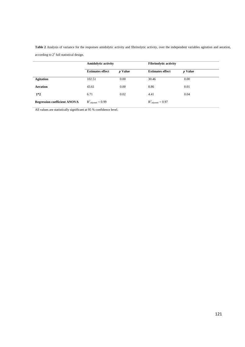
121
Table 2 Analysis of variance for the responses amidolytic activity and fibrinolytic activity, over the independent variables agitation and aeration,
according to 22 full statistical design.
All values are statistically significant at 95 % confidence level.
Amidolytic activity Fibrinolytic activity
Estimates effect p Value Estimates effect p Value
(1) Agitation 102.51 0.00 30.46 0.00
(2) Aeration 43.61 0.00 8.86 0.01
1*2 6.71 0.02 4.41 0.04
Regression coefficient ANOVA R2adjusted = 0.99 R2
adjusted = 0.97

122
Table 3 Substrate specificity for amidolytic activity of the fibrinolytic enzyme
Synthetic substrate (1.7 mM) Substrate hydrolysis rate (µmol.min-1.mL-1)
N-succinyl-Ala-Ala-Pro-Phe-p-nitroanilide 1.300 ± 0.80
N-Benzoyl-Val-Gly-Arg-p-nitroanilide 344.4 ± 1.04
D-Val-Leu-Lys-p-nitroanilide 55.00 ± 2.20
Gly-Phe-p-nitronilide 20.08 ± 0.70
D-Phe-Val-p-nitroanilide 16.88 ± 1.82
N-succinyl-Ala-Ala-Val-p-nitroanilide 14.08 ± 2.22
Gly-Pro-p-nitroanilide 13.68 ± 3.06
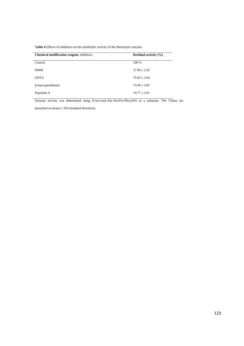
123
Table 4 Effects of inhibitors on the amidolytic activity of the fibrinolytic enzyme
Chemical modification reagent, inhibitors Residual activity (%)
Control 100 %
PMSF 27.09 ± 1.02
EDTA 70.43 ± 2.04
β-mercaptoethanol 73.99 ± 3.05
Pepstatin A 74.77 ± 2.01
Enzyme activity was determined using N-succinyl-Ala-Ala-Pro-Phe-pNA as a substrate. The Values are
presented as means ± SD (standard deviation).

124
Table 5 Effects of metal ions on the amidolytic activity of the fibrinolytic enzyme
Chemical modification reagent, metal ions Residual activity (%)
Control 100 %
FeSO4 81.11 ± 1.08
CuSO4 71.21 ± 1.06
MgSO4 65.48 ± 3.03
ZnSO4 60.99 ± 1.07
CoCl2 64.86 ± 2.03
MgCl2 59.13 ± 1.01
CaCl2 112.1 ± 0.85
KCl 108.5 ± 1.02
Enzyme activity was determined using N-succinyl-Ala-Ala-Pro-Phe-pNA as a substrate. The Values are
presented as means ± SD (standard deviation).

125
Figure Caption
Fig. 1 Scatterplot of the interaction between the independent variables agitation (rpm) and the aeration (vvm) for the response variables
fibrinolytic activity of the protease from Bacillus amyloliquefaciens UFPEDA 485.
Fig. 2 Scatterplot of the interaction between the independent variables agitation (rpm) and the aeration (vvm) for the response variables
amidolytic activity of the protease from Bacillus amyloliquefaciens UFPEDA 485.
Fig. 3 Cell growth (■), Fibrinolytic activity - FA (▲) and Amidolytic activity - AA (○) over time of culture of Bacillus amyloliquefaciens
UFPEDA 485 in bioreactor under optimum conditions studied (800 rpm and 1.5 vvm).
Fig. 4 Fibrinolytic activity (▲) and Amidolytic activity (○) in (A) Effect of pH on the activity of enzyme from Bacillus amyloliquefaciens
UFPEDA 485 and in (B) Effect of pH on the stability of enzyme measured after 1 h incubation and expressed as percentage of residual
activity. Buffers used: Glycine-HCl (pH 3.0), Sodium acetate (pH 4.0-5.0), Citrate phosphate (pH 6,0), Tris-HCl (pH 7.0-8.0), Glycine-
NaOH (pH 9.0-10.0). All buffer concentrations were 20 mM.. To determine the amidolytic and fibrinolytic activity was used as substrate N-
succinyl-Ala-Ala-Pro-Phe-pNA and fibrin, respectively. The data are presented as means ± SD (n = 3) from three independent experiments.
Fig. 5 Fibrinolytic activity (▲) and Amidolytic activity (○) in (A) Effect of temperature on the activity enzyme from Bacillus
amyloliquefaciens UFPEDA 485 and in (B) Effect of temperature on the stability of enzyme measured after 1 h incubation and expressed as
percentage of residual activity. Effects of temperature were determined at (4, 25, 37, 40, 50, 60, 70 and 80 ⁰C). To determine the amidolytic
and fibrinolytic activity was used as substrate N-succinyl-Ala-Ala-Pro-Phe-pNA and fibrin, respectively. The data are presented as means ±
SD (n = 3) from three independent experiments.

126
Figures
Fig. 1
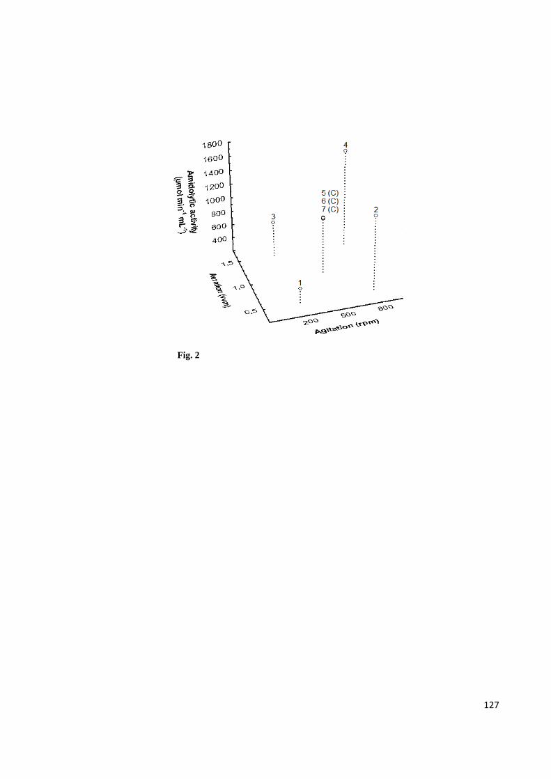
127
Fig. 2

128
Fig. 3
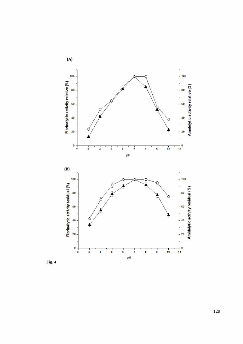
129
Fig. 4
(B)
(A)

130
Fig. 5
(A)
(B)
131
CAPÍTULO IV
Integrated Process Production and Extraction of the Fibrinolytic Protease from
Bacillus sp. UFPEDA 485
Artigo publicado pela revista Applied Biochemistry and Biotechnology
Impact Factor: 1.93
132

133

134

135
136

137

138

139

140
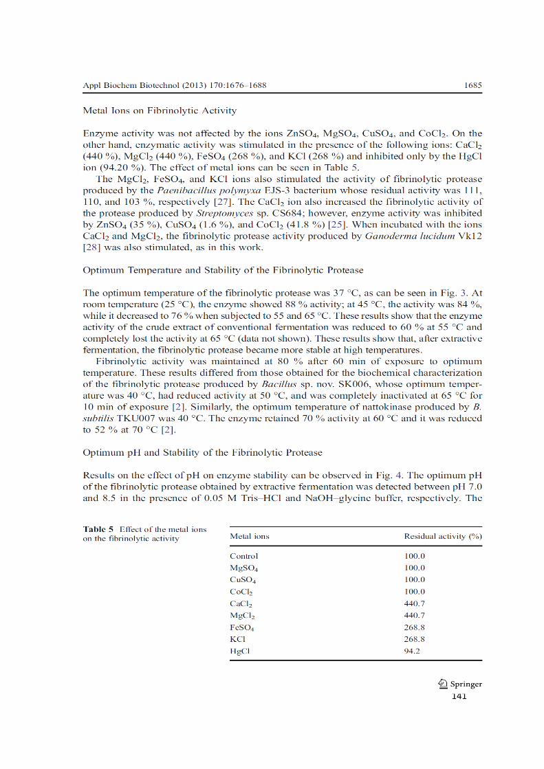
141
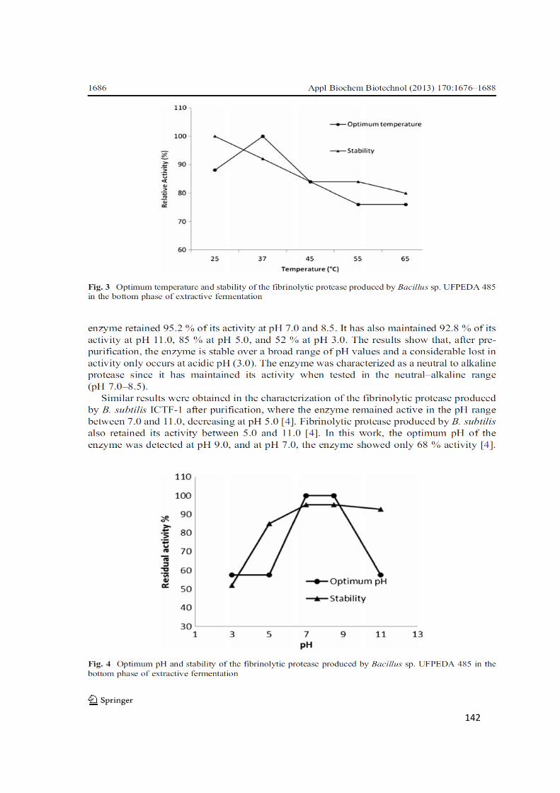
142

143

144

145
CONSIDERAÇÕES FINAIS
• Das 43 bactérias do gênero Bacillus, 30 bacérias apresentaram atividade
fibrinolítica, e o Bacillus amilolyquefaciens UFPEDA 485 foi considerado o
melhor produtor por apresentar a maior atividade fibrinolítica.
• Foi possível otimizar em frascos agitados as condições de produção da
enzima fibrinolítica produzida pelo Bacillus amyloliquefaciens UFPEDA 485,
com o auxílio de ferramentas estatísticas.
• Os resultados demonstram o potencial biotecnológico do Bacillus
amyloliquefaciens UFPEDA 485 de poduzir uma nova enzima com
propriedades fibrinolítica e anticoagulante de interesse para indústria
farmacêutica para aplicação no tratamento de distúrbios vasculares.
• A protease fibrinolítica do Bacillus amyloliquefaciens UFPEDA 485 se
destaca por apresentar propriedades fisico-químicas adequadas ao sistema
sanguíneo, além de possuir alta estabilidade de estocagem quando
comparada com outras enzimas citadas na literatura.
• Foi selecionada a melhor condição de agitação e aeração para a produção
da enzima em biorreator pela constatação do aumento de 10 vezes mais na
produtividade, quando comparado com os experimentos desenvolvidos em
frascos agitados.
• O processo de produção e purificação integrado por fermentação extrativa
em sistema de duas fases aquosas permitiu a purificação parcial da enzima
fibrinolítica do Bacillus amyloliquefaciens UFPEDA 485, podendo substituir
as fases iniciais dos processos de separação convencionais, reduzindo
assim o custo para obtenção final da enzima.