Relationship between Protein kinase C and derepression of different enzymes · 2018-01-19 ·...
33
Relationship between Protein kinase C and derepression of different enzymes. Salgado, A.P.C. 1 ; Schuller, D. 2 ; Casal, M. 2 ; Leão, C. 2 ; Leiper, F.C. 3 ; Carling; D. 3 ; Fietto, L.G. 1 , Trópia, M.J. 1 ; Castro, I.M. 1 and Brandão, R.L 1* . 1 Laboratório de Biologia Celular e Molecular, Núcleo de Pesquisas em Ciências Biológicas, Escola de Farmácia, Universidade Federal de Ouro Preto, Campus do Morro do Cruzeiro - 35.400-000 Ouro Preto, MG - Brazil. 2 Centro de Ciências do Ambiente, Departamento de Biologia, Universidade do Minho, 4710-057 Braga, Portugal. 3 Cellular Stress Group, MRC Clinical Sciences Center, Imperial College School of Medicine, Hammersmith Hospital, Du Cane Road, London W12 0NN, UK Running title: Pkc1 in the control of glucose-repressed enzymes Keywords: protein kinase C, Saccharomyces cerevisiae, signal transduction * Author for correspondence: Dr. Rogelio Lopes Brandão, Laboratório de Bioquímica Celular e Molecular, Núcleo de Pesquisas em Ciências Biológicas, Escola de Farmácia, Universidade Federal de Ouro Preto, Campus do Morro do Cruzeiro - 35.400-000 Ouro Preto, MG - Brazil.
Transcript of Relationship between Protein kinase C and derepression of different enzymes · 2018-01-19 ·...
Relationship between Protein kinase C and derepression of different
enzymes.
Salgado, A.P.C.1; Schuller, D. 2; Casal, M.2; Leão, C. 2; Leiper, F.C.3; Carling;
D.3; Fietto, L.G.1, Trópia, M.J.1; Castro, I.M.1 and Brandão, R.L1*.
1 Laboratório de Biologia Celular e Molecular, Núcleo de Pesquisas em Ciências
Biológicas, Escola de Farmácia, Universidade Federal de Ouro Preto, Campus do
Morro do Cruzeiro - 35.400-000 Ouro Preto, MG - Brazil.
2 Centro de Ciências do Ambiente, Departamento de Biologia, Universidade do Minho,
4710-057 Braga, Portugal.
3 Cellular Stress Group, MRC Clinical Sciences Center, Imperial College School of
Medicine, Hammersmith Hospital, Du Cane Road, London W12 0NN, UK
Running title: Pkc1 in the control of glucose-repressed enzymes
Keywords: protein kinase C, Saccharomyces cerevisiae, signal transduction
* Author for correspondence:
Dr. Rogelio Lopes Brandão, Laboratório de Bioquímica Celular e Molecular, Núcleo de
Pesquisas em Ciências Biológicas, Escola de Farmácia, Universidade Federal de
Ouro Preto, Campus do Morro do Cruzeiro - 35.400-000 Ouro Preto, MG - Brazil.
2
Fax number +55 31 3559 1680 E-mail: [email protected]
Abstract
The PKC1 gene in the yeast Saccharomyces cerevisiae encodes for protein
kinase C which is known to control a MAP kinase cascade consisting of different
kinases: Bck1, Mkk1 and Mkk2, and Mpk1. This cascade affects the cell wall integrity
but the phenotype of pkc1∆ mutants suggests additional targets that have not yet been
identified [1]. The pkc1∆ mutant, as opposed to other mutants in the MAP kinase
cascade, displays defects in the control of carbon metabolism. One of them occurs in
the derepression of SUC2 gene after exhaustion of glucose from the medium
suggesting an involvement of Pkc1p in the derepression process that is not shared by
the downstream MAP kinase cascade. In this work, we demonstrate that Pkc1p is
required for the increase of the activity of enzymatic systems during derepression
process. We observed that Pkc1p is involved in the derepression of invertase and
alcohol dehydrogenase activities. On the other hand, it seems not to be necessary for
the derepression of the enzymes of the GAL system. Our results suggest that Pkc1p
is acting through the main glucose repression pathway since introduction of an
additional mutation in the PKC1 gene in yeast strains already presenting mutations in
the HXKII or MIG1 genes does not interfere with the typical derepressed phenotype
observed in these single mutants. Moreover, our data indicate that Pkc1p participates
in this process through the control of the cellular localization of the Mig1 transcriptional
factor.
3
1. Introduction
The negative effect exerted by glucose on the transcription of different genes in
Saccharomyces cerevisiae cells is one of the most studied signaling transduction
pathways. Catabolic or glucose repression is a term that has been used to define such
phenomenon. The principal components of this regulatory system are the
transcriptional factor Mig1, the protein kinase Snf1, the protein phosphatase Glc7 and
the sugar kinase HxkII [2-4]. Briefly, a complex formed by Mig1p that recruits for this
proposal the co-repressor factors Ssn6 and Tup1 exerts the glucose repression. This
complex is negatively controlled by Snf1 protein kinase since phosphorylation of
Mig1p promotes its translocation from the nucleus to the cytoplasm relieving the
repression when glucose concentration is low [5-6]. For this process is also important
the participation of the exportin Msn5p that recognizes the Mig1p phosphorylated form
and facilitates its translocation to the cytoplasm [7]. On the other hand, when glucose
concentration is high, the Snf1 activity is inhibited through the action of two main
upstream regulators: the Glc7 phosphatase and the HxkII sugar kinase. Even though
many details of this mechanism are already known, there are many aspects for which
scarce information is available. The identity of the kinase that controls the Snf1 activity
and the exact way through which the HxkII p is involved in the control of Snf1 activity
are examples of details still unclear.
There are different classes of systems repressed by the presence of high
glucose concentrations, but in spite of the existence of a general glucose-repression
mechanism not all of systems are repressed in the same way. The following class can
be cited: proteins involved in the utilization of alternative carbon sources (invertase;
enzymes from the so-called galactose and maltose systems); essential proteins for
4
respiration; high-affinity glucose carriers; and proteins involved in different types of
stress [4]. These differences are related in many situations to the manner by which the
repressor complex interacts with the target genes. Thus, in the case of the SUC2 gene
(that encodes for the invertase) the repressor complex binds to the upstream
repressive sequence (URS) at the promoter region. On the other hand, in the case of
the GAL and MAL genes, Mig1 p also controls the expression of specific inducers
(GAL 4 and MAL R). The same situation is observed in the control of respiratory genes
where the transcription of Hap4 p, an important effector, is also repressed. By its turn,
for gluconeogenic genes, such as FBP1 and PCK1, there is also the action of the Snf
1 complex directly on a specific transcriptional activator (Sip 4p) [2].
By working with mutants of PKC MAP kinase signalling pathway, we
demonstrated that Pkc1p is specifically involved in the glucose depression pathway
and enzymes after glucose depletion [8]. Indeed, many other functions have been
attributed to Pkc1p that are not shared by the downstream components of the PKC
MAP kinase pathway [9-10].
Our data indicates that Pkc1p is necessary for the derepression of enzymatic
systems related to the carbon metabolism in yeast. Moreover, we also found evidence
for a connection between Pkc1p and the main repression pathway, since the
introduction of pkc1∆ mutation in strains carrying deletions in HXKII or MIG1 genes
does not change the derepressed status exhibited in strains with single mutations in
these two genes. Furthermore, the results suggest that Pkc1p would participate in this
process controlling the cellular localization of the Mig1 transcription factor.
5
2. Material and Methods
2.1. Strains and growth conditions
The Saccharomyces cerevisiae strains used in this study are shown in Table 1.
Yeast cells were grown in medium containing peptone (2%, w/v) and yeast extract
(1%, w/v) (YP medium) supplemented with glucose (4%, w/v) and 1M sorbitol as
indicated (25 ml, final volume). In all experiments the cells were grown in rotatory
incubator New Brunswick Model G25 (200 rpm) at 28º C until the middle of
exponential phase. At this moment, the cellular suspension was divided in two
fractions: 4 and 21 ml samples. Both were harvested and washed by centrifugation
(approximately 2000g) with 200 mM imidazol buffer (containing 40 mM MgCl2; 400
mM KCl; 1M sorbitol; pH 7.0). The pellet from the small fraction was used to prepare a
cellular extract (control time – repressed cells). The rest of the cells was resuspended
in a 25 ml YP medium supplemented with raffinose (2%, w/v) or galactose (2%, w/v) or
ethanol (2% w/v) plus glucose (0.05% w/v) and 1M sorbitol (for all situations). Again,
samples of 4 ml were taken at given time intervals, immediately harvested and washed
by centrifugation (approximately 2000g) with the same buffer and used to prepare
cellular extracts.
To test the cellular growth in solid media on different carbon sources, yeast
cells were grown overnight on YPD plus sorbitol 1M. 3µl of cellular suspension with a
OD at 600 nm of around 1.5 were inoculated on plates containing YP media
supplemented with glucose, galactose, glycerol or ethanol with or without sorbitol 1M.
The plates were incubated at 28º C for 3-4 days.

6
2.2. Determination of enzyme activities
For determination of invertase and alcohol dehydrogenase activities cell
extracts were obtained by adding 500 mg of glass beads (0.5 mm) and 500 µl of 50
mM imidazol buffer without sorbitol to the pellets. The cells were disrupted by
vortexing with glass beads for 30 seconds three times with intervals in ice. Then, 1 ml
of the buffer was added plus 2.5 µl 100 mM PMSF and the suspension was
centrifuged for 3 min at 13000 rpm in a microcentrifuge and the supernatant used to
measure the enzyme activities. Measurement of specific activity of invertase was
performed as described [11] with the modifications [12] except that the assay was
carried out at pH 5.1 and 37ºC. Alcohol dehydrogenase was measured by using the
protocol described in [13].
In the experiments performed to verify the expression of elements of the
system GAL, strains containing a GAL1-lacZ fusion were grown as described before
being transferred to the appropriate derepression medium (see legends of the figures).
However, the cell samples were harvested and washed by centrifugation
(approximately 2000 g) in a 0,5 M sodium phosphate buffer (containing 10 mM KCl; 1
mM MgCl2; sorbitol 1M; pH 7.0). Cell disruption and further sample treatment obeyed
the same scheme of the other enzymes. ß-galactosidase activity was measured by
incubating the extracts with 30 mM ONPG (orto-nitrophenyl-ß-D-galactopyranoside).
After 30 minutes the reaction was interrupted by addition of 1M Na2CO3 and the
absorbance read at 420nm. The enzyme activity was expressed in nmol of orto-
nitrophenol/min/mg protein. Protein was determined using classical procedures [14].
2.3. RNA isolation and Northern-blot analysis
For a shift from growth on glucose to growth on glycerol, yeast cells were grown
in 20 ml YP glucose (4%, w/v) plus 1M sorbitol at 30ºC up to early exponential phase
(DO600nm = 0.8-1.2). Then the sample was split in two. 10 ml were washed quickly by
7
centrifugation with 1M sorbitol and the cellular pellet used to RNA extraction
(repressed state). The rest of the suspension was washed quickly by centrifugation
with 1M sorbitol and the pellet resuspended in YP glycerol (3%, w/v) and glucose
(0.05%) plus 1M sorbitol, rapidly mixed and incubated as before. After two hours, the
sample was washed in the same way and the cellular pellet used for RNA extraction
(derepressed state).
Isolation of total yeast RNA was performed by using the classical hot acid
phenol method. 15 µg of total RNA was separated on 1% (w/v) agarose in 50 mM
boric acid, 1mM sodium citrate, 5 mM NaOH, pH 7.5, containing 1% (w/v)
formaldehyde. Subsequently RNA was blotted onto Hybond-N membranes in 10X
SSC (1.5M NaCl, 0.15 M sodium citrate, pH 7.0) and hybridised with gene-specific
probes. Probe fragments were radioactively labelled using the rediprimeTM II labelling
kit (Pharmacia). In all cases equal amounts of RNA were loaded. Gene specific probes
were obtained by PCR. The following primer pairs were used to synthesize FKS2 and
ACT1 (constitutive endogenous control) fragments, respectively: 5’-
ATGTCCTACAACGATCC-3’ (sense) and 5’-GAACCATCTTGATCAGG-3’ (antisense)
and 5’-GCTGCTTTGGTTATTGATAAC-3’ (sense) and 5’-
GATAGTGGACCACTTTCGTCG-3’ (antisense). The RNA levels were visualized by
exposing the membrane to CL-X PosureTM Film from Pierce.
2.4. Subcellular localization of Mig1 p by Western-blot and by analysis of GFP
fluorescence
To study the subcellular localization of Mig1 p we used yeast cells transformed
with a plasmid containing the MIG1 gene labeled with a FLAG epitope tag
(DYKDDDDK) placed immediately following the initiating methionine. The cells were
grown on YPglucose (2%) plus 1M sorbitol at 30º C up to early exponential phase
(repressive state) and then transferred to YPglucose (0.05%) plus 1M sorbitol for 30
8
minutes (derepressive state). The detection of the FLAG tagged Mig1 p was
performed as described previously [15].
On the other hand, strains containing the transport domain of Mig1 fused to
GFP gene (GFP states for green fluorescent protein) were grown on YP medium
supplemented with 4% glucose until mid-exponential phase. Then, 1ml samples were
harvested and washed by centrifugation (13000 rpm in a microcentrifuge) with a
sorbitol (1M) plus 4% glusose (4% , w/v) cold solution. The remaining cell suspension
was transferred to YP raffinose (2%, w/v) medium for 1 hour. The cells were harvested
and washed by centrifugation with a cold solution containing sorbitol (1M), plus
glucose(2%, W/v) (repressed cells) or glycerol (3%, w/v) (derepressed cells). Finally
10-15 µl of these cell suspensions were mixed with 10-15 µl of agarose (1%, w/v), at
37ºC for microscopy observations. Images were registered by using a Zeitz Laborlux S
Microscopic with accessory apparatus for fluorescence (Ploemopak Filter 12)
connected to a Sony Progressive 3 CCD. The images were processed with an Axio
Vision 3.0 software.
2.5. Reproducibility of results
All experiments of enzyme activities were performed at least three times and the
results showed are mean of independent experiments.
9
3. Results
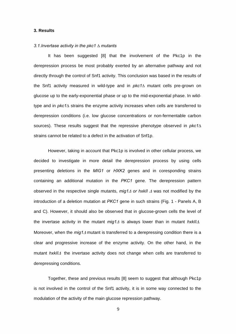
3.1.Invertase activity in the pkc1 ∆ mutants
It has been suggested [8] that the involvement of the Pkc1p in the
derepression process be most probably exerted by an alternative pathway and not
directly through the control of Snf1 activity. This conclusion was based in the results of
the Snf1 activity measured in wild-type and in pkc1∆ mutant cells pre-grown on
glucose up to the early-exponential phase or up to the mid-exponential phase. In wild-
type and in pkc1∆ strains the enzyme activity increases when cells are transferred to
derepression conditions (i.e. low glucose concentrations or non-fermentable carbon
sources). These results suggest that the repressive phenotype observed in pkc1∆
strains cannot be related to a defect in the activation of Snf1p.
However, taking in account that Pkc1p is involved in other cellular process, we
decided to investigate in more detail the derepression process by using cells
presenting deletions in the MIG1 or HXK2 genes and in coresponding strains
containing an additional mutation in the PKC1 gene. The derepression pattern
observed in the respective single mutants, mig1∆ or hxkII ∆ was not modified by the
introduction of a deletion mutation at PKC1 gene in such strains (Fig. 1 - Panels A, B
and C). However, it should also be observed that in glucose-grown cells the level of
the invertase activity in the mutant mig1∆ is always lower than in mutant hxkII∆.
Moreover, when the mig1∆ mutant is transferred to a derepressing condition there is a
clear and progressive increase of the enzyme activity. On the other hand, in the
mutant hxkII∆ the invertase activity does not change when cells are transferred to
derepressing conditions.
Together, these and previous results [8] seem to suggest that although Pkc1p
is not involved in the control of the Snf1 activity, it is in some way connected to the
modulation of the activity of the main glucose repression pathway.
10
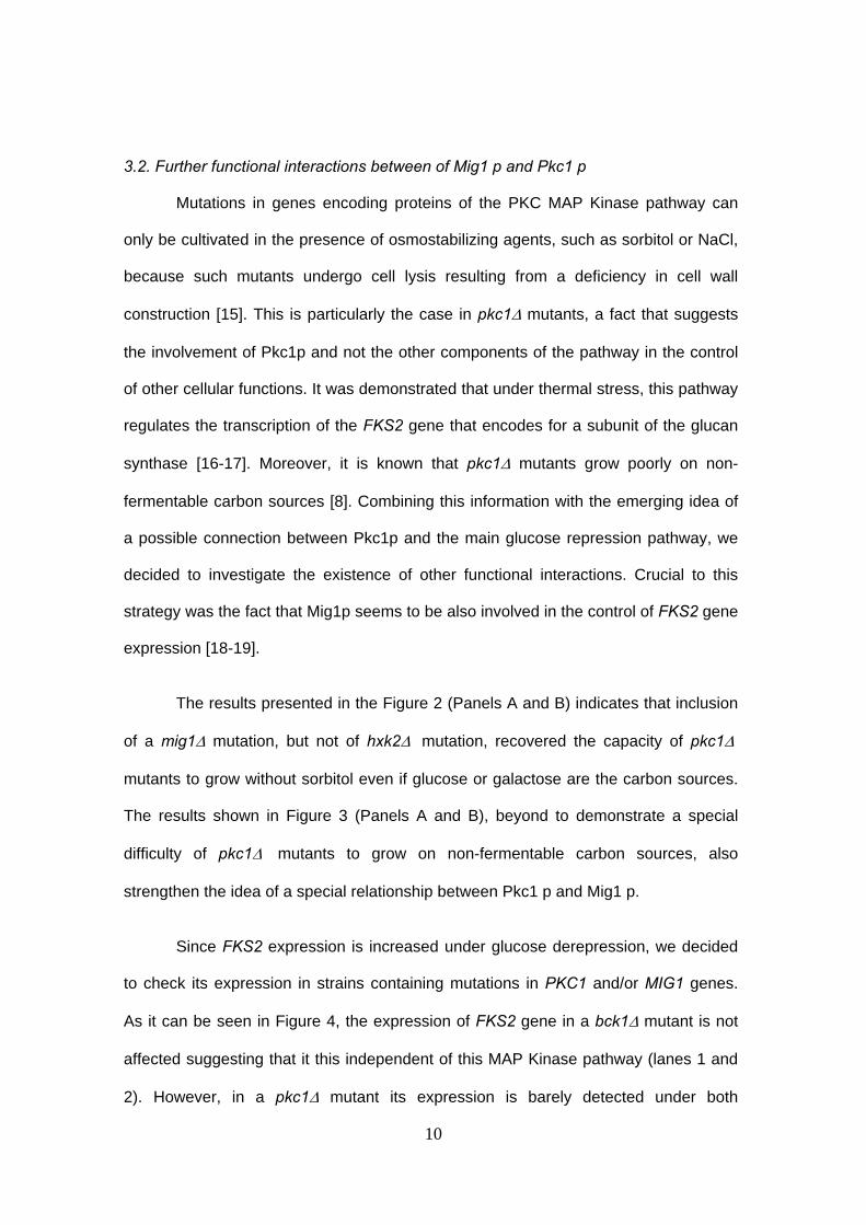
3.2. Further functional interactions between of Mig1 p and Pkc1 p
Mutations in genes encoding proteins of the PKC MAP Kinase pathway can
only be cultivated in the presence of osmostabilizing agents, such as sorbitol or NaCl,
because such mutants undergo cell lysis resulting from a deficiency in cell wall
construction [15]. This is particularly the case in pkc1∆ mutants, a fact that suggests
the involvement of Pkc1p and not the other components of the pathway in the control
of other cellular functions. It was demonstrated that under thermal stress, this pathway
regulates the transcription of the FKS2 gene that encodes for a subunit of the glucan
synthase [16-17]. Moreover, it is known that pkc1∆ mutants grow poorly on non-
fermentable carbon sources [8]. Combining this information with the emerging idea of
a possible connection between Pkc1p and the main glucose repression pathway, we
decided to investigate the existence of other functional interactions. Crucial to this
strategy was the fact that Mig1p seems to be also involved in the control of FKS2 gene
expression [18-19].
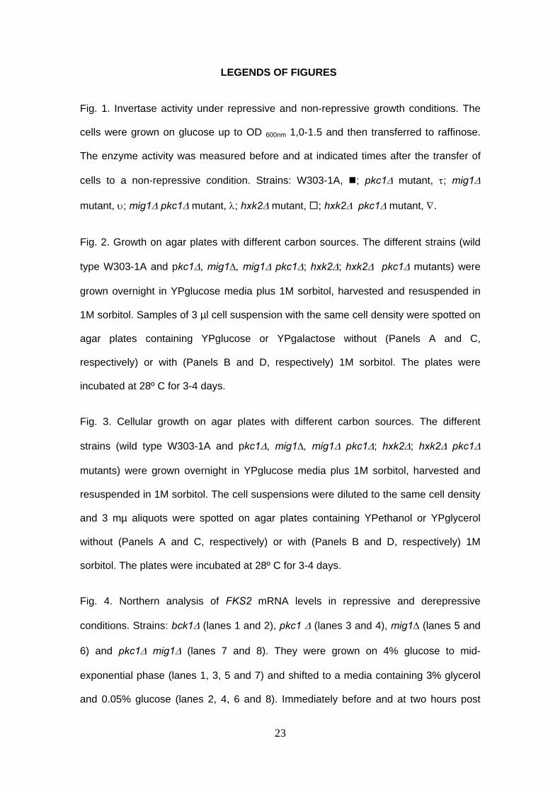
The results presented in the Figure 2 (Panels A and B) indicates that inclusion
of a mig1∆ mutation, but not of hxk2∆ mutation, recovered the capacity of pkc1∆
mutants to grow without sorbitol even if glucose or galactose are the carbon sources.
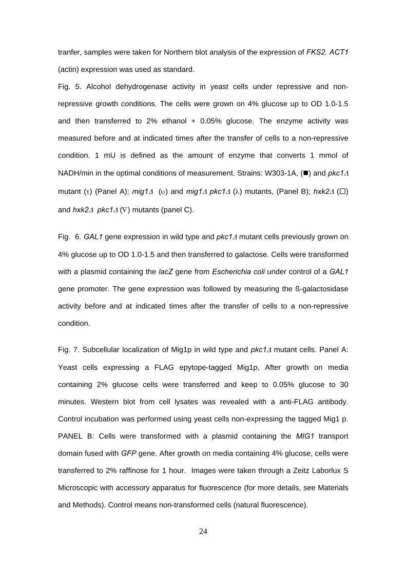
The results shown in Figure 3 (Panels A and B), beyond to demonstrate a special
difficulty of pkc1∆ mutants to grow on non-fermentable carbon sources, also
strengthen the idea of a special relationship between Pkc1 p and Mig1 p.
Since FKS2 expression is increased under glucose derepression, we decided
to check its expression in strains containing mutations in PKC1 and/or MIG1 genes.
As it can be seen in Figure 4, the expression of FKS2 gene in a bck1∆ mutant is not
affected suggesting that it this independent of this MAP Kinase pathway (lanes 1 and
2). However, in a pkc1∆ mutant its expression is barely detected under both
11
repressive (lane 3) and derepressive (lane 4) conditions. In the mig1∆ mutant, the
expression is clearly present in both cases (lanes 5 and 6) confirming that its
expression is under the control of Mig1p as described previously [18]. Beyond that, in
the pkc1∆ mig1∆ mutant the level of expression of FKS2 gene (lanes 7 and 8) is
completely different from that observed in the pkc1∆ being more similar to those
observed in mig1∆ mutant. Together, these results indicate the existence of a
functional relationship between Pkc1p and Mig1p.
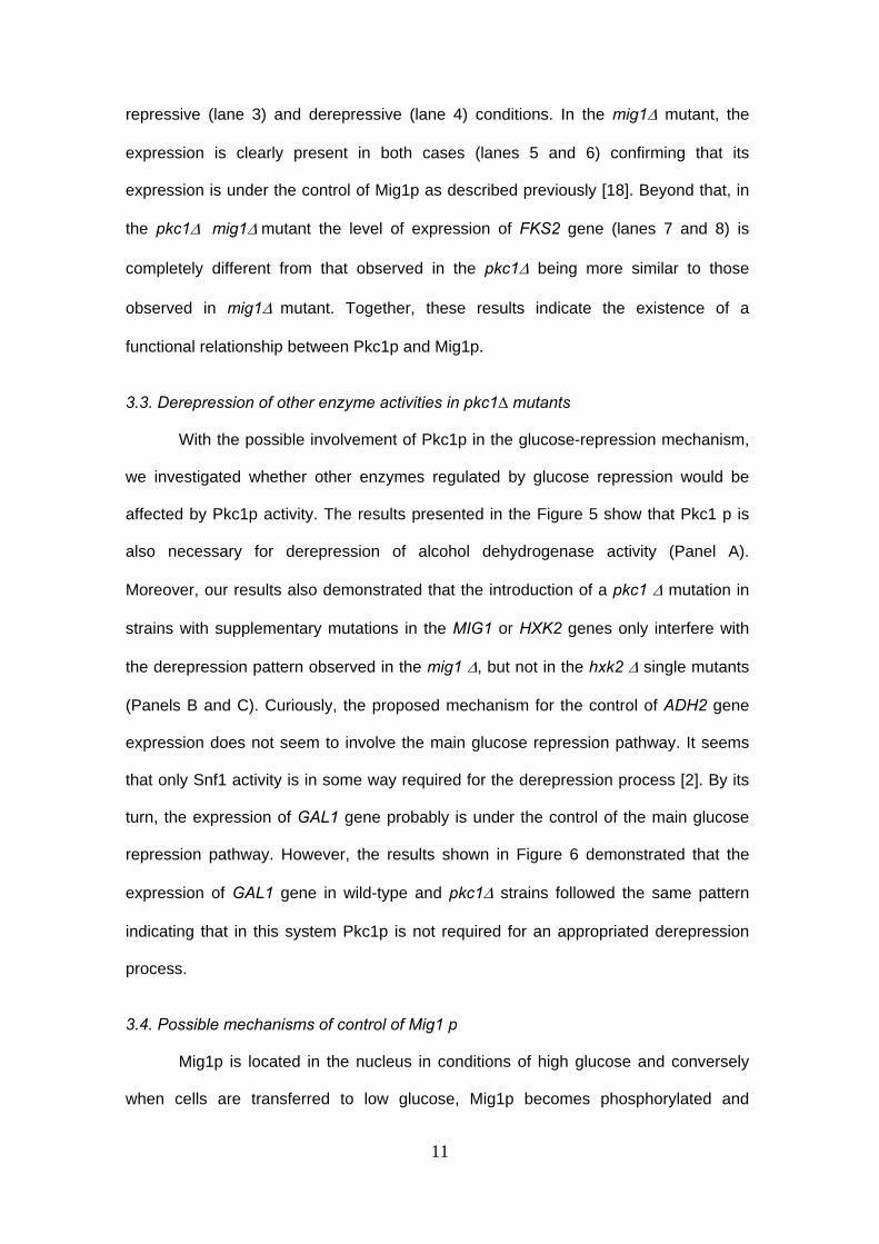
3.3. Derepression of other enzyme activities in pkc1∆ mutants
With the possible involvement of Pkc1p in the glucose-repression mechanism,
we investigated whether other enzymes regulated by glucose repression would be
affected by Pkc1p activity. The results presented in the Figure 5 show that Pkc1 p is
also necessary for derepression of alcohol dehydrogenase activity (Panel A).
Moreover, our results also demonstrated that the introduction of a pkc1 ∆ mutation in
strains with supplementary mutations in the MIG1 or HXK2 genes only interfere with
the derepression pattern observed in the mig1 ∆, but not in the hxk2 ∆ single mutants
(Panels B and C). Curiously, the proposed mechanism for the control of ADH2 gene
expression does not seem to involve the main glucose repression pathway. It seems
that only Snf1 activity is in some way required for the derepression process [2]. By its
turn, the expression of GAL1 gene probably is under the control of the main glucose
repression pathway. However, the results shown in Figure 6 demonstrated that the
expression of GAL1 gene in wild-type and pkc1∆ strains followed the same pattern
indicating that in this system Pkc1p is not required for an appropriated derepression
process.
3.4. Possible mechanisms of control of Mig1 p
Mig1p is located in the nucleus in conditions of high glucose and conversely
when cells are transferred to low glucose, Mig1p becomes phosphorylated and
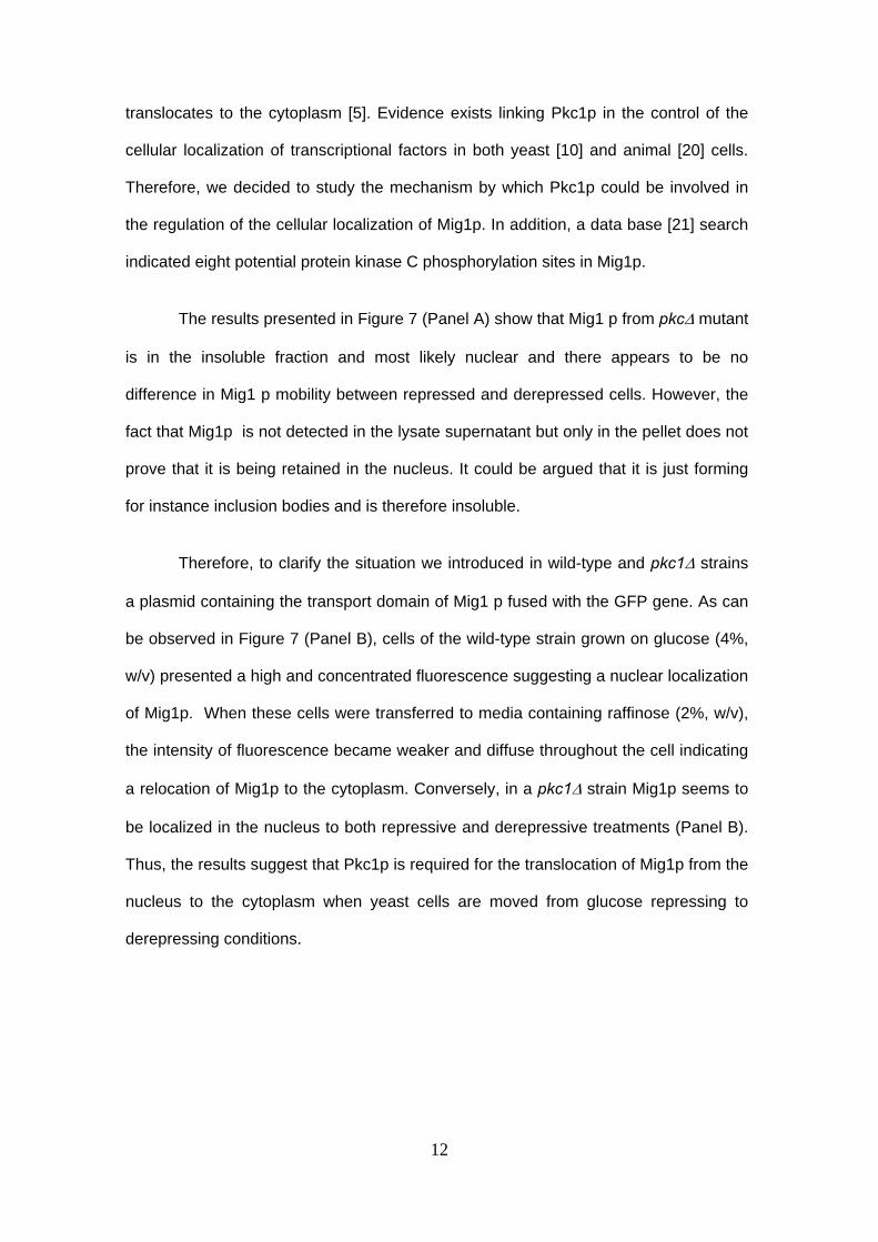
12
translocates to the cytoplasm [5]. Evidence exists linking Pkc1p in the control of the
cellular localization of transcriptional factors in both yeast [10] and animal [20] cells.
Therefore, we decided to study the mechanism by which Pkc1p could be involved in
the regulation of the cellular localization of Mig1p. In addition, a data base [21] search
indicated eight potential protein kinase C phosphorylation sites in Mig1p.
The results presented in Figure 7 (Panel A) show that Mig1 p from pkc∆ mutant
is in the insoluble fraction and most likely nuclear and there appears to be no
difference in Mig1 p mobility between repressed and derepressed cells. However, the
fact that Mig1p is not detected in the lysate supernatant but only in the pellet does not
prove that it is being retained in the nucleus. It could be argued that it is just forming
for instance inclusion bodies and is therefore insoluble.
Therefore, to clarify the situation we introduced in wild-type and pkc1∆ strains
a plasmid containing the transport domain of Mig1 p fused with the GFP gene. As can
be observed in Figure 7 (Panel B), cells of the wild-type strain grown on glucose (4%,
w/v) presented a high and concentrated fluorescence suggesting a nuclear localization
of Mig1p. When these cells were transferred to media containing raffinose (2%, w/v),
the intensity of fluorescence became weaker and diffuse throughout the cell indicating
a relocation of Mig1p to the cytoplasm. Conversely, in a pkc1∆ strain Mig1p seems to
be localized in the nucleus to both repressive and derepressive treatments (Panel B).
Thus, the results suggest that Pkc1p is required for the translocation of Mig1p from the
nucleus to the cytoplasm when yeast cells are moved from glucose repressing to
derepressing conditions.
13
4. Discussion
Pkc1p seems to have cellular functions other than the control of cell wall
construction through the PKC MAP Kinase pathway. Pkc1p is involved in the control of
yeast cell division through the activation of Cdc28 at START [22] and it has been
demonstrated that Pkc1p is required for the appropriate localization of some
transcription factors to specific stimuli [10]. In addition, previous results from our
laboratory suggest the involvement of Pkc1p in the glucose induced-activation of the
plasma membrane ATPase and in the derepression mechanism of the glucose-
repressible genes [8-9].
Therefore, taking in account the available information on other potential roles
played by Pkc1 p, we first investigated the invertase activity in cells presenting
deletions in MIG1 or HXKII genes combined with pkc1 ∆ mutation. This mutation does
not interfere with the normal pattern exhibited by the mig1 ∆ or hxkII ∆ single mutants
i.e. increasing when cells are transferred to derepressive condition and/or higher
activity even in the presence of high glucose concentrations (Figure 1). These results
suggest that the probable participation of Pkc1 p in the control of SUC2 gene
expression occur in connection with the classical glucose repression pathway.
It is known that Mig1p participates in the control of FKS2 gene expression in
derepressing conditions [18-19]. As shown here, only strains presenting combined
mutations in the PKC1 and MIG1 genes reversed the phenotype exhibited by pkc1∆
mutants, indicating that there is a specific and functional interaction between Pkc1p
and Mig1p (Figures 2 and 3). Since a mig1∆ mutant displayed a nine-fold increase in
FKS2 gene expression compared with the congenic wild type strain [18], a plausible
hypothesis would be that Mig1p function is controlled (in part) by Pkc1p.
A contradiction emerges on comparison of invertase activity (Figure 1) and
growth in different carbon sources (Figures 2 and 3). Care is required in the
14
interpretation of such data. Firstly, there is already strong evidence pointing to the
direct involvement of HxkIIp in the regulation of glucose repression of SUC2, as,
HxkIIp can be found in the nucleus [23], where it participates in regulatory DNA-protein
complexes necessary for glucose repression [24]. Secondly, it was shown that Mig1p
but not HxkIIp is involved in the control of FKS2 expression [18], suggesting a specific
interaction of Pkc1p in the main glucose repression pathway that is likely to be
downstream of Snf1p.
Thus, in terms of glucose repression, it is expected that the phenotypes of both
pkc1∆ hxkII∆ or pkc1∆ mig1∆ mutants would be the same as the one exhibited by the
single mutants (hxkII∆ or mig1∆). HxkIIp exerts a more prominent role, most probably
because it interacts with and inhibits Snf1p [25] and also through its participation in
DNA-protein complexes controlling SUC2 expression [23, 24]. This indicates why
invertase activity is higher in hxkII∆ cells than in mig1∆ cells when grown on glucose.
However, in the control of the FKS2 gene expression (Figure 4), Pkc1p would exert its
effects by regulating the action of the Mig1 transcriptional factor. In this sense, our
results offer an explanation for the fact that even in presence of sorbitol pkc1∆ is
harder to grow than other strains containing mutations in the genes encoding
downstream kinases of the PKC MAP Kinase pathway. Indeed, they also corroborate
the findings that multiple mechanisms are involved in the control of FKS2 gene
expression, whilst until now none indicated a role for HxkIIp [18].
Moreover, it should be added that Pkc1p also seems to be involved in the
control of other genes which encode for key proteins (17) of all the components of the
cell wall biosynthesis, i.e. (1-3)-ß-glucan (FKS1 and GAS1), (1-6)-ß-glucan (KRE6),
chitin (CSD2) and N- and O-linked mannoproteins (MNN1) This is probably the reason
why the pkc1∆ mutant presents more difficulty to grow in glucose containing media.
15
It is known that Pkc1p is required for the translocation of nuclear factors from
the nucleus to the cytoplasm [10]. Interestingly, a protein kinase C from animal cells
has also been described as contributing to a mechanism involved in the localisation of
transcription factors [20]. Thus, considering the data [9, this work] we imagine that
Pkc1p could contribute to both, the glucose derepression pathway and FKS2
expression, by controlling the cellular localization of Mig1p. This seems to be the case,
as in the pkc1∆ mutant Mig1p remains in the nucleus even after the cells are
transferred from glucose repressing to derepressing conditions (Figures 7 – Panels A
and B). Nevertheless, the mechanism by which Pkc1p controls the subcellular
localization of Mig1p is not clear, but in our opinion there are at least two possibilities.
Pkc1p could phosphorylate Mig1p since that there are potentially eight protein kinase
C phosphorylation sites in Mig1p [21]. This phosphorylation could trigger and/or
facilitate the translocation of Mig1p. On the other hand, Pkc1p could be involved in the
regulation of translocation factors that are required to export Mig1p from nucleus to the
cytoplasm. We are currently investigating these possibilities.
However, the involvement of Pkc1p in the derepression of glucose-controlled
systems seems to be still more complex. For instance, the genes that encode
enzymes involved in galactose metabolism are glucose repressed indicating that the
main glucose pathway has an important role [26]. According the current model, in the
presence of glucose, Mig1p blocks the expression of genes such as GAL4 and the
regulator Gal80p binds to Gal4p preventing the transcriptional activation of genes that
encode enzymes for galactose metabolism. On the other hand, in the presence of
galactose, Gal3p impairs the binding of the Gal80p inhibitor to Gal4p, Gal4p can then
be phosphorylated and activate GAL transcription. In the pkc1∆ mutant, and in the
presence of an effector molecule, Mig1p may play either a secondary role to Pkc1p
and/or a glucose repression-independent mechanism may operate to activate GAL
transcription.

16
Moreover, and to add more complexity to the involvement of Pkc1p in the
control of glucose-repressed enzymes, the increase in alcohol dehydrogenase activity
when yeast cells are transferred from glucose to ethanol seems to be dependent on
Pkc1p. Interestingly, Mig1p has not been described as being involved in the control of
ADH2 gene transcription. Indeed, the introduction of a pkc1∆ mutation in a mig1∆
strain blocked the increase in the alcohol dehydrogenase activity suggesting an
alternative mechanism by which Pkc1p participates in the derepression process. It is
known that Adr1p, a transcriptional factor that acts at an upstream activating sequence
(UAS) on the ADH2 and GUT1 (encoding glycerol kinase, an essential enzyme in
glycerol metabolism) promoters, has a positive effect when the glucose levels are low
[2-4, 27]. Thus, since the pkc1∆ mutant grows poorly on ethanol or glycerol carbon
sources, it is possible that Pkc1p could regulate the cellular localization of Adr1.
However, more experiments are required to clarify this complex mechanism.
17
Acknowledgements
This work was financed by grants from Fundação de Capacitação de Pessoal
Docente from Ministry of Education (fellowship to A.P.C.S.), from Fundação de
Amparo a Pesquisa do Estado de Minas Gerais – FAPEMIG (Brazil) Process CBS-
1875/95 to R.L.B and a research fellowship from Conselho Nacional de
Desenvolvimento Científico e Tecnológico – CNPq (Brazil) Process 300998/89-9.

18
References
[1] Heinisch, J. J.; Lorberg, A.; Schmitz, H. & Jacoby, J. J. (1999). The protein kinase
C-mediated MAP kinase pathway involved in the maintenance of cellular integrity
in Saccharomyces cerevisiae. Mol. Microbiol. 32: 671-680.
[2] Gancedo, J. M. (1998). Yeast carbon catabolite repression. Microbiology and
Molecular Biology Reviews 62: 334-361.
[3] Carlson, M. (1999). Glucose repression in yeast. Curr. Opin. Microbiol. 2: 202-207.
[4] Rolland, F.; Winderickx, J. & Thevelein, J. M., (2002). Glucose-sensing and –
signalling mechanisms in yeast. FEMS Yeast Research 2(2): 183-201.
[5] Smith, F.C.; Davies, S. P; Wilson, W.A; Carling, D. and Hardie, D.G. (1999). The
SNF1 kinase complex from Saccharomyces cerevisiae phosphorylates the
transcriptional repressor protein Mig 1 p in vitro at four sites within or near
regulatory domain 1. FEBS Letters 453: 219-223.
[6] Osting, J. and Ronne, H. (1998). Negative control of the Mig1 p repressor by Snf1-
dependent phosphorylation in the absence of Mig1 glucose repressor. Eur. J.
Biochem. 252, 162-168.
[7] DeVit, M. J., Johnston, M. (1999). The nuclear exportin Msn 5 is required for
nuclear export of the Mig 1 glucose repressor of Saccharomyces cerevisiae.
Current Biology 9, 1231-1241.
[8] Brandão, R. L., Etchebehere, L., Queiroz, C. C., Trópia, M. J., Ernandes, J. R.,
Gonçalves, T., Loureiro-Dias, M. C., Winderickx, J., Thevelein, J. M., Leiper, F.C.,
Carling, D., Castro, I.M. (2002). Evidence for involvement of Saccharomyces
cerevisiae protein kinase C in glucose induction of HXT genes and derepression of
SUC 2.FEMS Yeast Research 2(2): 93-102.
19
[9] Souza, M. A. A, Trópia, M. J., Brandão, R. L. (2001) . New aspects of the glucose
activation of the H+-ATPase in the yeast Saccharomyces cerevisiae. Microbiology
147, 2849-2855.
[10] Nanduri, J and Tartakoff, A M. (2001). Pertubation of the nucleus: a novel Hog1 p
independent, Pkc1 p-dependent consequence of hypertonic shock in yeast.
Molecular Biology of the Cell 12, 1835-1841.
[11] Goldstein, A. & Lampen, J. O. (1975). Beta-D-fructofuranoside fructohydrolase from
yeast. Methods Enzymol. 42: 504-511.
[12] Celenza, J. L. & Carlson, M. (1989). Mutational analysis of the Saccharomyces
cerevisiae SNF1 protein kinase and evidence for functional interaction with the
SNF4 protein. Mol. Cell. Biol. 9: 5045-5054.
[13] Denis, C. L., M. Ciriacy, and E. T. Young. (1981). A positive regulatory gene is
required for accumulation of the functional messenger RNA for the glucose-
repressible alcohol dehydrogenase from Saccharomyces cerevisiae. J. Mol. Biol.
148:355-368
[14] Lowry, O.H.; Rosenbrough, N.J.; Farr, A.L. & Randall, R.J. (1951). Protein
measurement with the Folin phenol reagent. J. Biol. Chem. 193: 265-275.
[15] Watanabe, M.; Chen, C.; & Levin, D. E. (1994). Saccharomyces cerevisiae PKC1
Encodes a Protein Kinase C (PKC) Homolog with a Substrate Specificity Similar to
That of Mammalian PKC. J. Biol. Chem., 269 (24): 16829-16836.
[16] Kamada, Y.; Jung, U.S.; Piotrowski, J. & Levin, D.E. (1995). The protein kinase C-
activated MAP kinase pathway of Saccharomyces cerevisiae mediates a novel
aspect of the heat shock response. Genes Dev. 9:1559-1571.
20
[17] Igual, J.C.; Johnson, A.L. & Johnston, L. H. (1996). Coordinated regulation of gene
expression by the cell cycle transcription factor SWI4 and the protein kinase C MAP
kinase pathway for yeast cell integrity. EMBO J. 15: 5001-5013.
[18] Zhao, C.; Jung, U.S.; Garret-Engele, P.; Roe, T.; Cyert, M.S. & Levin, D. E. (1998).
Temperature-induced expression of yeast FKS2 is under the dual control of protein
kinase C and calcineurin. Mol. Cell. Biol. 18:1013-1022.
[19] Mazur, P.; Morin, N.; Baginsky, W.; El-Sherbeini, M.; Clemas, J.A.; Nielsen, J.B. &
Foor, F. (1995). Differential expression and function of two homologous subunits of
yeast 1,3-ß-D-glucan synthase. Mol Cell. Biol. 15(10): 5671-5681.
[20] Harbers, M., Nomura, T., Ohno, S., Ishii, S. (2001). Intracellular localization of the
Ret finger protein depends on a functional nuclear export signal and protein kinase
C activation. The journal of Biological Chemistry 51, 48596-48607.
[21] Kreegipuu A, Blom N, Brunak S (1999). PhosphoBase, a database of
phosphorylation sites: release 2.0. Nucleic Acids Res 27(1):237-239.
[22] Marini, N. J.; Meldrum, E.; Buehrer, B.; Hubberstey, A. V.; Stone, D. E.; Traynor-
Kaplan, A. & Reed, S. I. (1996). A pathway in the yeast cell division cycle linking
protein kinase C (Pkc1) to activation of Cdc28 at START. Embo J. 15(12): 3040-
3052.
[23] Randez-Gil, F.; Herrero, P.; Sanz, P.; Prieto, J.A. & Moreno, F. (1998). Hexokinase
II has a double cytosolic-nuclear localisation in Saccharomyces cerevisiae. FEBS
Letters 425: 475-478.
[24] Herrero, P; Martinez-Campa; C & Moreno, F. (1998). The hexokinase II protein
participates in regulatory DNA-protein complexes necessary for glucose repression
of the SUC2 gene in Saccharomyces cerevisiae. FEBS Letters 434: 71-76.
21
[25] Jiang, R. & Carlson, M. (1996). Glucose regulates protein interactions within the
yeast SNF1 protein kinase complex. Genes Dev. 10: 3105-3115.
[26] Ostergaard, S., Walloe, K. O., Gomes, C. S. G., Olsson, L., Nielsen, J. (2001). The
impact of GAL6, GAL80, and MIG1 on glucose control of the GAL system in
Saccharomyces cerevisiae. FEMS Yeast Research 1, 47-55.
[27] Grauslund, M; Lopes, J. & Rønnow (1999). Expression of GUT1, which encodes
glycerol kinase in Saccharomyces cerevisiae, is controlled by the positive regulators
Adr1 p, Ino2 p and Ino4 p and the negative regulator Opi1 p in a carbon source-
dependent fashion. Nucleic Acid Research 27(22): 4391-4398.
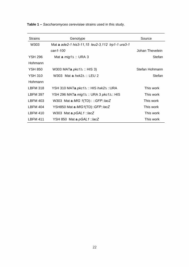
Table 1 – Saccharomyces cerevisiae strains used in this study.
Strains Genotype Source
W303 Mat a ade2-1 his3-11,15 leu2-3,112 trp1-1 ura3-1
can1-100 Johan Thevelein
YSH 296 Mat a mig1∆ :: URA 3 Stefan
Hohmann
YSH 850 W303 MATa pkc1∆ :: HIS 3) Stefan Hohmann
YSH 310 W303 Mat a hxk2∆ :: LEU 2 Stefan
Hohmann
LBFM 318 YSH 310 MATa pkc1∆ :: HIS hxk2∆ ::URA This work
LBFM 397 YSH 296 MATa mig1∆ :: URA 3 pkc1∆:: HIS This work
LBFM 403 W303 Mat a MIG 1(TD):: ::GFP::lacZ This work
LBFM 404 YSH850 Mat a MIG1(TD)::GFP::lacZ This work
LBFM 410 W303 Mat a pGAL1 ::lacZ This work
LBFM 411 YSH 850 Mat a pGAL1 ::lacZ This work
22
23
LEGENDS OF FIGURES
Fig. 1. Invertase activity under repressive and non-repressive growth conditions. The
cells were grown on glucose up to OD 600nm 1,0-1.5 and then transferred to raffinose.
The enzyme activity was measured before and at indicated times after the transfer of
cells to a non-repressive condition. Strains: W303-1A, ; pkc1∆ mutant, τ; mig1∆
mutant, υ; mig1∆ pkc1∆ mutant, λ; hxk2∆ mutant, ; hxk2∆ pkc1∆ mutant, ∇.
Fig. 2. Growth on agar plates with different carbon sources. The different strains (wild
type W303-1A and pkc1∆, mig1∆, mig1∆ pkc1∆; hxk2∆; hxk2∆ pkc1∆ mutants) were
grown overnight in YPglucose media plus 1M sorbitol, harvested and resuspended in
1M sorbitol. Samples of 3 µl cell suspension with the same cell density were spotted on
agar plates containing YPglucose or YPgalactose without (Panels A and C,
respectively) or with (Panels B and D, respectively) 1M sorbitol. The plates were
incubated at 28º C for 3-4 days.
Fig. 3. Cellular growth on agar plates with different carbon sources. The different
strains (wild type W303-1A and pkc1∆, mig1∆, mig1∆ pkc1∆; hxk2∆; hxk2∆ pkc1∆
mutants) were grown overnight in YPglucose media plus 1M sorbitol, harvested and
resuspended in 1M sorbitol. The cell suspensions were diluted to the same cell density
and 3 mµ aliquots were spotted on agar plates containing YPethanol or YPglycerol
without (Panels A and C, respectively) or with (Panels B and D, respectively) 1M
sorbitol. The plates were incubated at 28º C for 3-4 days.
Fig. 4. Northern analysis of FKS2 mRNA levels in repressive and derepressive
conditions. Strains: bck1∆ (lanes 1 and 2), pkc1 ∆ (lanes 3 and 4), mig1∆ (lanes 5 and
6) and pkc1∆ mig1∆ (lanes 7 and 8). They were grown on 4% glucose to mid-
exponential phase (lanes 1, 3, 5 and 7) and shifted to a media containing 3% glycerol
and 0.05% glucose (lanes 2, 4, 6 and 8). Immediately before and at two hours post
24
tranfer, samples were taken for Northern blot analysis of the expression of FKS2. ACT1
(actin) expression was used as standard.
Fig. 5. Alcohol dehydrogenase activity in yeast cells under repressive and non-
repressive growth conditions. The cells were grown on 4% glucose up to OD 1.0-1.5
and then transferred to 2% ethanol + 0.05% glucose. The enzyme activity was
measured before and at indicated times after the transfer of cells to a non-repressive
condition. 1 mU is defined as the amount of enzyme that converts 1 mmol of
NADH/min in the optimal conditions of measurement. Strains: W303-1A, ( ) and pkc1∆
mutant (τ) (Panel A); mig1∆ (υ) and mig1∆ pkc1∆ (λ) mutants, (Panel B); hxk2∆ ( )
and hxk2∆ pkc1∆ (∇) mutants (panel C).
Fig. 6. GAL1 gene expression in wild type and pkc1∆ mutant cells previously grown on
4% glucose up to OD 1.0-1.5 and then transferred to galactose. Cells were transformed
with a plasmid containing the lacZ gene from Escherichia coli under control of a GAL1
gene promoter. The gene expression was followed by measuring the ß-galactosidase
activity before and at indicated times after the transfer of cells to a non-repressive
condition.
Fig. 7. Subcellular localization of Mig1p in wild type and pkc1∆ mutant cells. Panel A:
Yeast cells expressing a FLAG epytope-tagged Mig1p, After growth on media
containing 2% glucose cells were transferred and keep to 0.05% glucose to 30
minutes. Western blot from cell lysates was revealed with a anti-FLAG antibody.
Control incubation was performed using yeast cells non-expressing the tagged Mig1 p.
PANEL B: Cells were transformed with a plasmid containing the MIG1 transport
domain fused with GFP gene. After growth on media containing 4% glucose, cells were
transferred to 2% raffinose for 1 hour. Images were taken through a Zeitz Laborlux S
Microscopic with accessory apparatus for fluorescence (for more details, see Materials
and Methods). Control means non-transformed cells (natural fluorescence).

25

FIGURE 1
hxkII ∆ pkc1 ∆
hxkII ∆ C
B0
250
500
750
1000pkc1 ∆
AW303 - Wild Type
0
500
1000
1500mig1 ∆ pkc1 ∆mig1 ∆
0 2 4 6 8 10 120
500
1000
1500
Inve
rtas
e ac
tivity
(nm
ol g
luco
se/m
in/m
g pr
otei
n)
Time (h)
FIGURE 2A B
w/o dil 10-2 10-3 10-4 10-5
w/o dil 10-2 10-3 10-4 10-5
W303
pkc1 ∆
mig1 ∆
mig1 ∆ pkc1 ∆
hxk2 ∆ pkc1 ∆
hxk2 ∆
YPD YPD + sorbitol
DCW/o
dil 10-2 10-3 10-4 10-5
YPGalactose YPGalactose +sorbitol
w/o dil 10-2 10-3 10-4 10-5
W303
pkc1 ∆
mig1 ∆
mig1 ∆ pkc1 ∆
hxk2 ∆
hxk2 ∆ pkc1 ∆
FIGURE 3 Bw/o
dil 10-2 10-3 10-4 10-5
Aw/o
dil 10-2 10-3 10-4 10-5
W303
pkc1 ∆
mig1 ∆
mig1 ∆ pkc1 ∆
hxk2 ∆ pkc1 ∆
hxk2 ∆
YPEthanol YPEthanol + sorbitol
DCw/o dil 10-2 10-3 10-4 10-5
w/o dil 10-2 10-3 10-4 10-5
W303
pkc1 ∆
mig1 ∆
mig1 ∆ pkc1 ∆
hxk2 ∆
hxk2 ∆ pkc1 ∆
YPGlycerol YPGlycerol + sorbitol
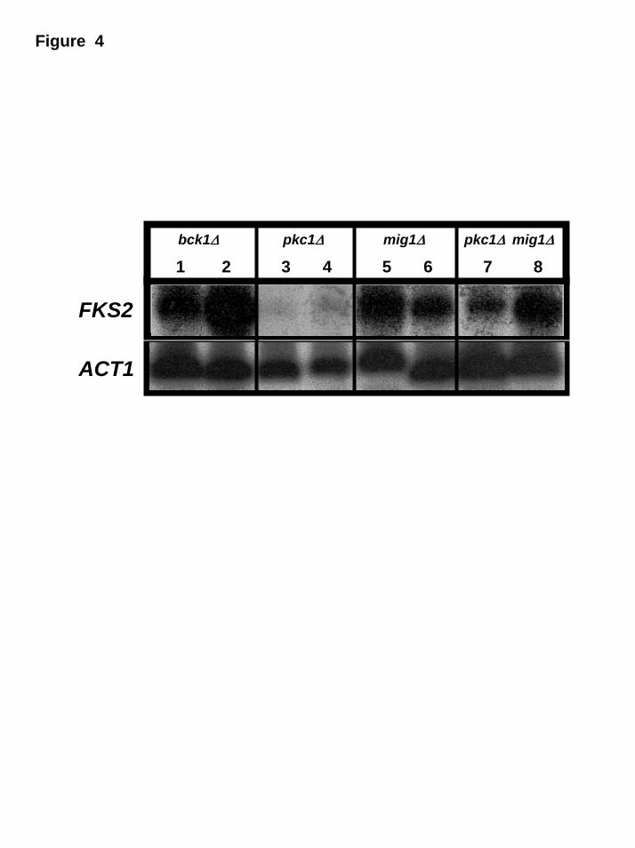
Figure 4
bck1∆ pkc1∆ mig1∆ pkc1∆ mig1∆
1 2 3 4 5 6 7 8
FKS2
ACT1

hxk2 ∆ pkc1 ∆
hxk2 ∆
0
50
100
150
200
250
300
pkc1 ∆
Wild type − W303
0
50
100
150
200
250 pkc1 ∆mig1 ∆ mig1 ∆
0 6 12 18 240
50
100
150
200
Time (h)
Alc
ohol
deh
ydro
gena
se (m
U/m
g pr
otei
n)
FIGURA 5
A
B
C
FIGURE 6
1
10
100
1000
10000
0 0.5 1 3 6 12Time (h

Wild type pkc1 ∆Cytoplasmic
fractionCytoplasmic
fractionInsolublefraction
InsolublefractionControl
incubation R DR R DR R DR R DRkDa
105
75
35
Mig1
50
160
FIGURE 7 - Panel A
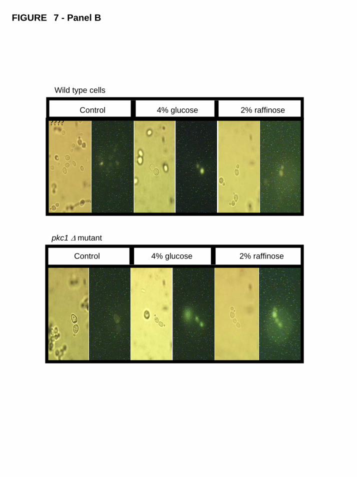
FIGURE 7 - Panel B
Control 4% glucose 2% raffinose
?????
Control 4% glucose 2% raffinose
Wild type cells
pkc1 ∆ mutant