
RESUMOS - LNLSlnls.cnpem.br/wp-content/uploads/2016/06/RAU23.pdf · 2020-01-10 · Feb 26th–6 p.m...
192
RESUMOS DE TRABALHOS CIENTÍFICOS RESUMOS DE TRABALHOS CIENTÍFICOS
Transcript of RESUMOS - LNLSlnls.cnpem.br/wp-content/uploads/2016/06/RAU23.pdf · 2020-01-10 · Feb 26th–6 p.m...
RESUMOSDE TRABALHOS CIENTÍFICOSRESUMOSDE TRABALHOS CIENTÍFICOS

Coordenação: Comitê dos Usuários do LNLS LNLS Users Committee
Victor Hugo V. Sarmento (UFS) – presidente Leandro R.S. Barbosa (USP) – vice-presidente Celso Santilli (Unesp) Flavio C. Cruz (BR-LABS e Unicamp) Marcia Regina Soares (UFRJ) Reinaldo Luiz Cavasso Filho (UFABC)
Comitê Científico Scientific Committee
Aldo F. Craievich (USP) Angelo M. de Souza (UFMG) Cassia C. Turci (UFRJ) Leandro R. S. Barbosa (USP) Luis Mauricio T. R. Lima (UFRJ) Manoel G. P. Homem (UFSC) Maria do Carmo M. Alves (UFRGS) Mario Ernesto G. Valerio (UFS) Nádya Pesce Silveira (UFRGS) Victor Hugo V. Sarmento (UFS) Watson Loh (UNICAMP)
Comitê Local CNPEM/LNLS Local Organizing Committee
Maria Claudia Izique Dora Marques Ilderia Santos William Barbosa Mirela Amaral Roberta Santarosa Gustavo Moreno

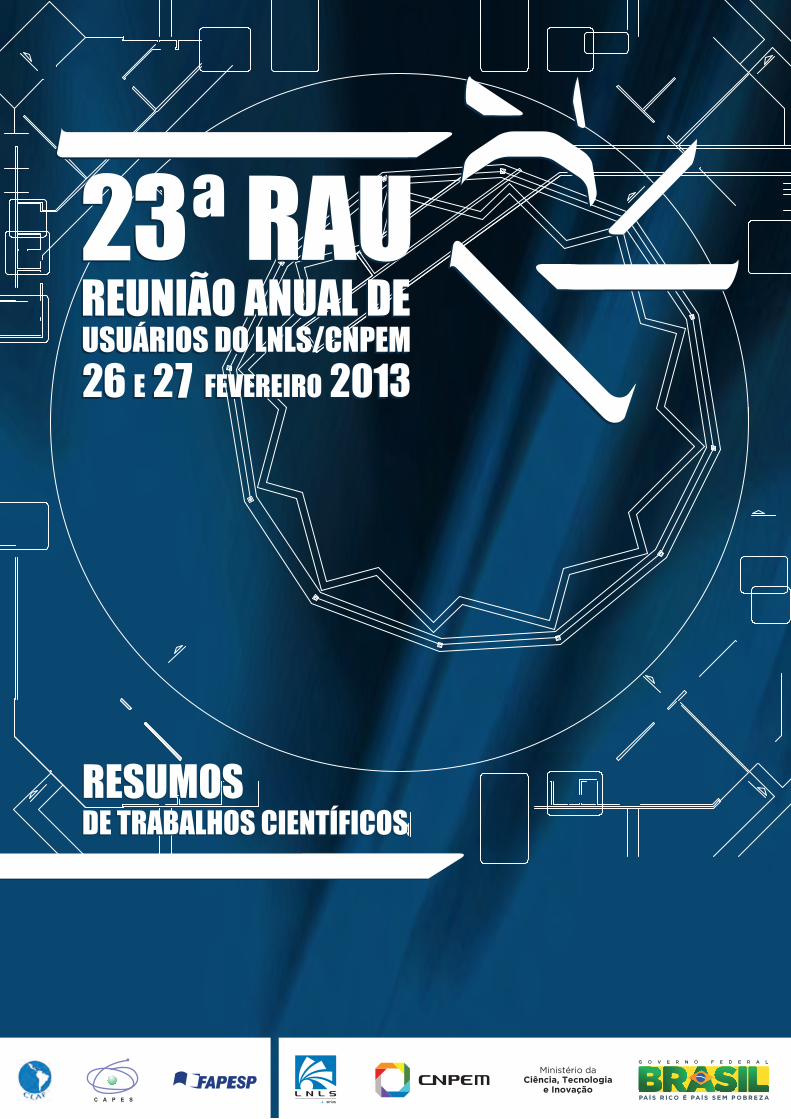
23ª Reunião Anual de Usuários do LNLS/CNPEM Laboratório Nacional de Luz Síncrotron
Campinas, 26 e 27 Fevereiro de 2013
Prefácio O Comitê de Usuários do LNLS saúda a todos os participantes da 23ª Reunião Anual
dos Usuários do Laboratório Nacional de Luz Síncrotron (LNLS).
Este encontro ocorre ao mesmo tempo em que o LNLS dá um importante passo para o
desenvolvimento da ciência brasileira: o início da construção de Sirius, a nova fonte de
luz Síncrotron, agendada para maio próximo. Sirius vai operar com energia de 3 GeV e
emitância de 0.28 nm.rad, das mais baixas em todo o mundo. Está programada,
inicialmente, a construção de 13 linhas experimentais, abrangendo grande parte das
pesquisas desenvolvidas pelos mais de 1.400 pesquisadores que já utilizam as atuais
instalações do LNLS. Sirius terá área de 30.000 m2 e capacidade para abrigar até 40
estações experimentais.
É fundamental que, nesse período, os usuários estreitem contato com a direção do
LNLS por meio do Comitê de Usuários, para que possam acompanhar o andamento do
projeto.
Agradecemos o esforço do Comitê Científico que muito auxiliou na seleção de diversos
cientistas convidados e dos resumos que serão apresentados nesta edição da RAU.
Foram selecionadas um total de 50 apresentações orais e 94 apresentações em forma de
pôster, além das quatro tradicionais seções plenárias. Também não podemos deixar de
agradecer a Diretoria do LNLS e a todos os seus funcionários que apoiaram a
organização deste evento.
Desejamos a todos uma proveitosa e frutífera Reunião dos Usuários do LNLS,
Comitê de Usuários da 23ª RAU
Orientações aos Participantes Prezado Participante, seja bem-vindo à 23ª Reunião Anual de Usuários. O Comitê Organizador pede sua atenção para as informações abaixo. 1. Credenciamento O credenciamento no evento (retirada de material, crachás de identificação e assinaturas dos recibos para agências de fomento - FAPESP/Capes) será no seguinte horário: 26/02, terça-feira, das 8:00 às 12:00 2. Crachá de identi f icação O crachá de identificação deve ser usado durante toda a reunião. 3. Sessões As Comunicações Orais ocorrerão em diferentes salas do hotel Premium Norte: Sala Plenária, Milão, Ibiza, Madrid e Roma do Centro de Convenções, conforme programação. 4. Sessão de Pôsteres A sessão de pôsteres ocorrerá nos dias 26 e 27 de fevereiro. Seguem os horários para fixação e retirada dos mesmos.
Data Horário montagem Horário de ret irada 26/fev – das18:00 às 19:10 Das 8:00 às 12:00 19:20 27/fev – das 10:10 às 11:40 Das 8:00 às 9:00 Até às13:00
O local de fixação do pôster estará sinalizado por etiqueta com a numeração correspondente à área de pesquisa do apresentador. Os organizadores da RAU não se responsabilizam por danos ou perda do pôster, e também não se comprometem a embalar, remover ou transportar o material. 5. Hotel O hotel oferece desktops no Lobby para uso de hóspedes e participantes da 23ª RAU. O estacionamento está disponível para uso durante a reunião – vagas limitadas.
6. Almoço Os almoços dos dias 26/02 e 27/02 serão servidos no restaurante do hotel, a partir do horário indicado na programação. 7. Segurança Pedimos a todos que fiquem atentos à segurança de seus objetos pessoais de valor. O CNPEM e o Hotel Premium Norte não poderão se responsabilizar pela segurança de tais objetos. 8. Certi f icado Os certificados serão enviados por email até o dia 30 de março de 2013. Para recebê-los, os participantes deverão assinar a lista de presença no balcão de credenciamento no dia 26 de fevereiro. O Comitê de Organização deseja a todos os part ic ipantes uma agradável e produtiva reunião.

Guidelines to Participants
Dear Participant, welcome to 23rd LNLS Users Annual Meeting. Please, read carefully the following instructions. 1. Registrat ion Registration at the event (identification badge, personal kit and signature on the receipt for the participants that have financial support) will be done following the schedule below: Tuesday, 8 a.m to 12 p.m. 2. Identi f ication Badge The identification badge must be used at all time during the meeting. 3. Sessions The Oral Communication Sessions will be held in the rooms: Milão, Ibiza, Madrid, Roma at the Convention Center and also in the plenary room in the Premium Norte Hotel. 4. Posters Session The poster session will be on February 26th and 27th, following the schedules for fixing and dismantle:
Date Fixing Dismantle Feb 26th – 6 p.m to 7:10 p.m 8 a.m to 12 p.m 7:20 p.m
Feb 27th – 10:10 a.m to 11:40 a.m 8 a.m to 9 a.m until 1 p.m The local where the poster must be fixed will be indicated with the corresponding research area of the author. The organizing committee and staffs are not responsible for packing, removing or shipping your poster neither responsible for any loss or damage of it. 5. Hotel The hotel offers desktop computer at the lobby area for the guest and participants’ use during the event. The parking lot is also available during the meeting.
6. Lunch Lunch will be served at the restaurant, following the schedule in the program. 7. Security Please, pay attention to your personal belongings. CNPEM and Premium Norte Hotel cannot be responsible for the safety of any objects. 8. Cert i f icate Certificates will be sending by email until March 30th, 2013. To receive this document, the participants should sign the presence list available on the registration desk on Feb 26th. The Organization wishes al l part ic ipants a pleasant and productive meeting.

Agenda, February 26th
08:00 - 09:00 Reception /Registrations
09:00 - 09:15 Plenary room
Opening Antônio José Roque da Silva Director of LNLS
09:15 - 10:15 Plenary room
Status do LNLS e Sirius Yves Petroff and Harry Westfahl Scientific Director of LNLS
10:15 - 10:20 Official Photo 10:20 - 10:40 Coffee Break 10:40 - 11:40 Plenary room
Plenary I Lucia Zuin (Canadian Light Source - CLS)
11:40 - 12:40 Plenary room
Plenary II Jan Ilavsky (Advanced Photon Source, Chicago, USA)
12:40 - 14:00 Lunch 14:00 - 16:00 Plenary room Sirius Beamlines
16:00 - 17:40 Oral Communication I Plenary room Electronic Structural Properties I Milão room Structural Biology Ibiza room Surfaces Interfaces and Nanosystems I
Madrid room Atomic Science Roma room Structural Materials and Industrial Applications
17:40 - 18:00 Coffee Break 18:00 - 19:10 Posters Session I
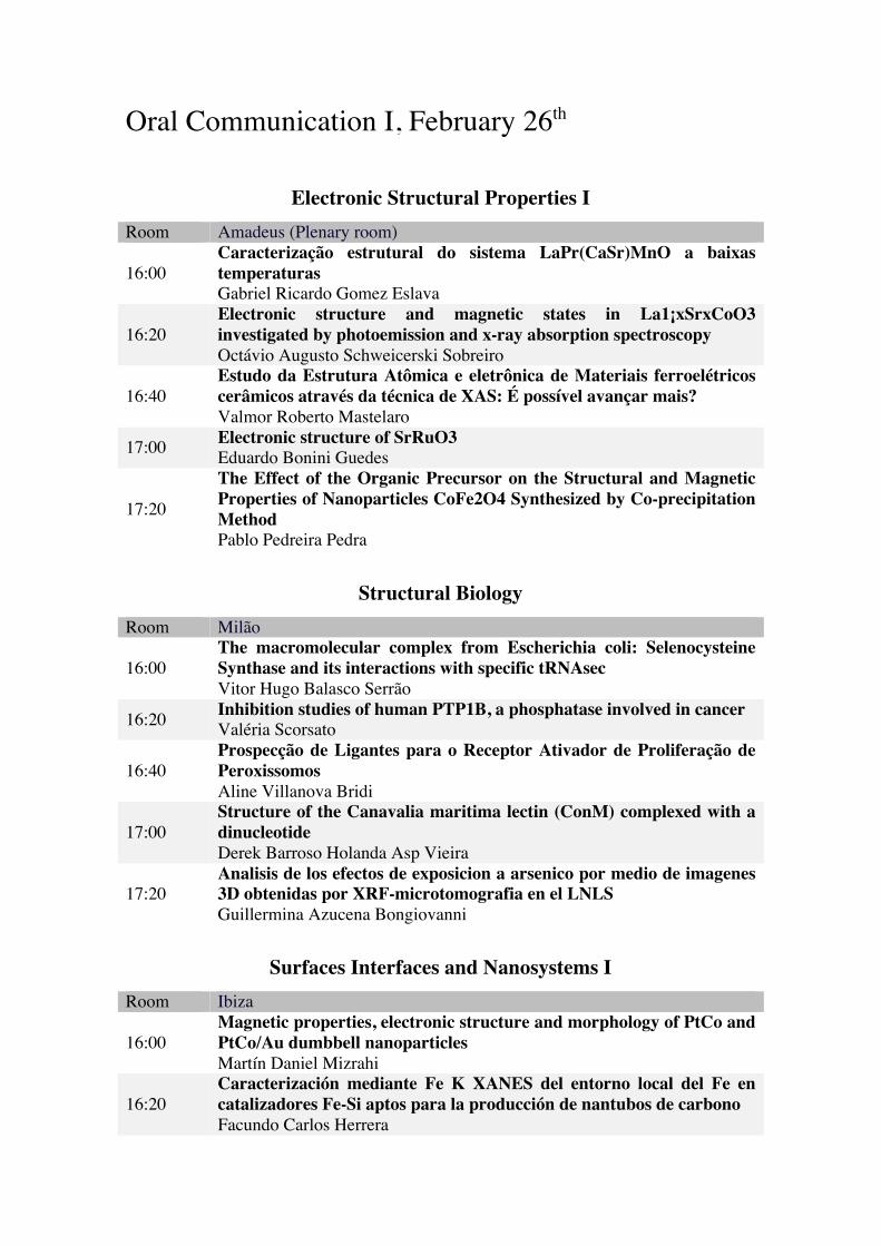
Oral Communication I, February 26th
Electronic Structural Properties I Room Amadeus (Plenary room)
16:00 Caracterização estrutural do sistema LaPr(CaSr)MnO a baixas temperaturas Gabriel Ricardo Gomez Eslava
16:20 Electronic structure and magnetic states in La1¡xSrxCoO3 investigated by photoemission and x-ray absorption spectroscopy Octávio Augusto Schweicerski Sobreiro
16:40 Estudo da Estrutura Atômica e eletrônica de Materiais ferroelétricos cerâmicos através da técnica de XAS: É possível avançar mais? Valmor Roberto Mastelaro
17:00 Electronic structure of SrRuO3 Eduardo Bonini Guedes
17:20
The Effect of the Organic Precursor on the Structural and Magnetic Properties of Nanoparticles CoFe2O4 Synthesized by Co-precipitation Method Pablo Pedreira Pedra
Structural Biology Room Milão
16:00 The macromolecular complex from Escherichia coli: Selenocysteine Synthase and its interactions with specific tRNAsec Vitor Hugo Balasco Serrão
16:20 Inhibition studies of human PTP1B, a phosphatase involved in cancer Valéria Scorsato
16:40 Prospecção de Ligantes para o Receptor Ativador de Proliferação de Peroxissomos Aline Villanova Bridi
17:00 Structure of the Canavalia maritima lectin (ConM) complexed with a dinucleotide Derek Barroso Holanda Asp Vieira
17:20 Analisis de los efectos de exposicion a arsenico por medio de imagenes 3D obtenidas por XRF-microtomografia en el LNLS Guillermina Azucena Bongiovanni
Surfaces Interfaces and Nanosystems I Room Ibiza
16:00 Magnetic properties, electronic structure and morphology of PtCo and PtCo/Au dumbbell nanoparticles Martín Daniel Mizrahi
16:20 Caracterización mediante Fe K XANES del entorno local del Fe en catalizadores Fe-Si aptos para la producción de nantubos de carbono Facundo Carlos Herrera
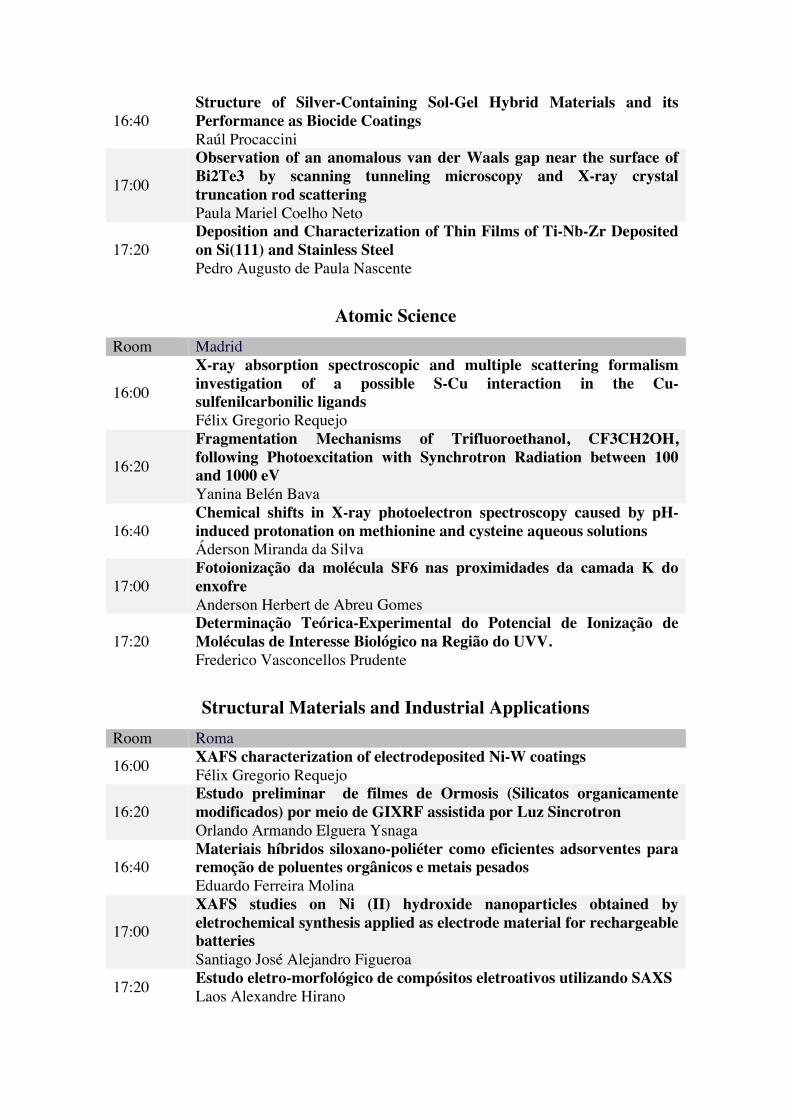
16:40 Structure of Silver-Containing Sol-Gel Hybrid Materials and its Performance as Biocide Coatings Raúl Procaccini
17:00
Observation of an anomalous van der Waals gap near the surface of Bi2Te3 by scanning tunneling microscopy and X-ray crystal truncation rod scattering Paula Mariel Coelho Neto
17:20 Deposition and Characterization of Thin Films of Ti-Nb-Zr Deposited on Si(111) and Stainless Steel Pedro Augusto de Paula Nascente
Atomic Science Room Madrid
16:00
X-ray absorption spectroscopic and multiple scattering formalism investigation of a possible S-Cu interaction in the Cu- sulfenilcarbonilic ligands Félix Gregorio Requejo
16:20
Fragmentation Mechanisms of Trifluoroethanol, CF3CH2OH, following Photoexcitation with Synchrotron Radiation between 100 and 1000 eV Yanina Belén Bava
16:40 Chemical shifts in X-ray photoelectron spectroscopy caused by pH-induced protonation on methionine and cysteine aqueous solutions Áderson Miranda da Silva
17:00 Fotoionização da molécula SF6 nas proximidades da camada K do enxofre Anderson Herbert de Abreu Gomes
17:20 Determinação Teórica-Experimental do Potencial de Ionização de Moléculas de Interesse Biológico na Região do UVV. Frederico Vasconcellos Prudente
Structural Materials and Industrial Applications Room Roma
16:00 XAFS characterization of electrodeposited Ni-W coatings Félix Gregorio Requejo
16:20 Estudo preliminar de filmes de Ormosis (Silicatos organicamente modificados) por meio de GIXRF assistida por Luz Sincrotron Orlando Armando Elguera Ysnaga
16:40 Materiais híbridos siloxano-poliéter como eficientes adsorventes para remoção de poluentes orgânicos e metais pesados Eduardo Ferreira Molina
17:00
XAFS studies on Ni (II) hydroxide nanoparticles obtained by eletrochemical synthesis applied as electrode material for rechargeable batteries Santiago José Alejandro Figueroa
17:20 Estudo eletro-morfológico de compósitos eletroativos utilizando SAXS Laos Alexandre Hirano

Agenda, February 27th
08:30 - 10:10 Oral Communication I Plenary room Electronic Structural Properties II Milão room Atomic Science II Ibiza room Surfaces Interfaces and Nanosystems II
Madrid room Soft Materials Roma room Methods and Instrumentation 10:10 - 11:40 Posters Session II and Coffee Break 11:40 - 12:40 Plenary room
Plenary III Kay Diederichs (Universitat Konstanz)
12:40 - 14:00 Lunch 14:00 - 15:00 Plenary room
Plenary IV Cinthia Piamonteze (Paul Scherrer Institute – PSI)
15:00 - 15:30 Plenary room
Articulation between the Laboratories of CNPEM Prof. Carlos Alberto Aragão de Carvalho Filho Director General CNPEM
15:30 - 16:30 Plenary room
Round table Evaluations and Discussions with the Users Committee of LNLS
16:30 - 18:00 Plenary room
User's round table: Improvements on the TGM UV beamline
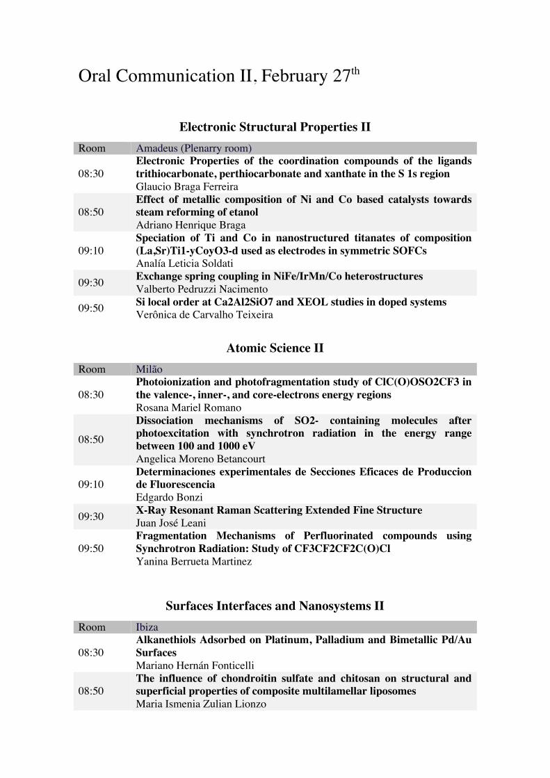
Oral Communication II, February 27th
Electronic Structural Properties II Room Amadeus (Plenarry room)
08:30 Electronic Properties of the coordination compounds of the ligands trithiocarbonate, perthiocarbonate and xanthate in the S 1s region Glaucio Braga Ferreira
08:50 Effect of metallic composition of Ni and Co based catalysts towards steam reforming of etanol Adriano Henrique Braga
09:10 Speciation of Ti and Co in nanostructured titanates of composition (La,Sr)Ti1-yCoyO3-d used as electrodes in symmetric SOFCs Analía Leticia Soldati
09:30 Exchange spring coupling in NiFe/IrMn/Co heterostructures Valberto Pedruzzi Nacimento
09:50 Si local order at Ca2Al2SiO7 and XEOL studies in doped systems Verônica de Carvalho Teixeira
Atomic Science II Room Milão
08:30 Photoionization and photofragmentation study of ClC(O)OSO2CF3 in the valence-, inner-, and core-electrons energy regions Rosana Mariel Romano
08:50
Dissociation mechanisms of SO2- containing molecules after photoexcitation with synchrotron radiation in the energy range between 100 and 1000 eV Angelica Moreno Betancourt
09:10 Determinaciones experimentales de Secciones Eficaces de Produccion de Fluorescencia Edgardo Bonzi
09:30 X-Ray Resonant Raman Scattering Extended Fine Structure Juan José Leani
09:50 Fragmentation Mechanisms of Perfluorinated compounds using Synchrotron Radiation: Study of CF3CF2CF2C(O)Cl Yanina Berrueta Martinez
Surfaces Interfaces and Nanosystems II Room Ibiza
08:30 Alkanethiols Adsorbed on Platinum, Palladium and Bimetallic Pd/Au Surfaces Mariano Hernán Fonticelli
08:50 The influence of chondroitin sulfate and chitosan on structural and superficial properties of composite multilamellar liposomes Maria Ismenia Zulian Lionzo
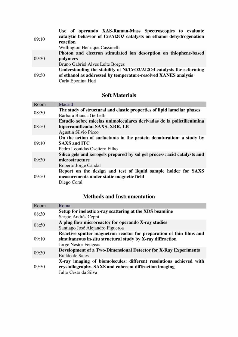
09:10
Use of operando XAS-Raman-Mass Spectroscopies to evaluate catalytic behavior of Cu/Al2O3 catalysts on ethanol dehydrogenation reaction Wellington Henrique Cassinelli
09:30 Photon and electron stimulated ion desorption on thiophene-based polymers Bruno Gabriel Alves Leite Borges
09:50 Understanding the stability of Ni/CeO2/Al2O3 catalysts for reforming of ethanol as addressed by temperature-resolved XANES analysis Carla Eponina Hori
Soft Materials Room Madrid
08:30 The study of structural and elastic properties of lipid lamellar phases Barbara Bianca Gerbelli
08:50 Estudio sobre micelas unimoleculares derivadas de la polietilienimina hiperramificada: SAXS, XRR, LB Agustin Silvio Picco
09:10 On the action of surfactants in the protein denaturation: a study by SAXS and ITC Pedro Leonidas Oseliero Filho
09:30 Silica gels and xerogels prepared by sol gel process: acid catalysts and microstructure Roberto Jorge Candal
09:50 Report on the design and test of liquid sample holder for SAXS measurements under static magnetic field Diego Coral
Methods and Instrumentation Room Roma
08:30 Setup for inelastic x-ray scattering at the XDS beamline Sergio Andrés Ceppi
08:50 A plug flow microreactor for operando X-ray studies Santiago José Alejandro Figueroa
09:10 Reactive sputter magnetron reactor for preparation of thin films and simultaneous in-situ structural study by X-ray diffraction Jorge Nestor Feugeas
09:30 Development of a Two-Dimensional Detector for X-Ray Experiments Eraldo de Sales
09:50 X-ray imaging of biomolecules: different resolutions achieved with crystallography, SAXS and coherent diffraction imaging Julio Cesar da Silva
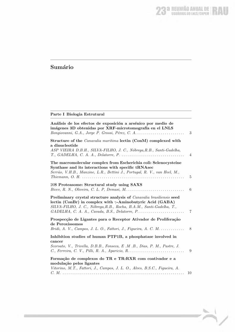
Sumario
Parte I Biologia Estrutural
Analisis de los efectos de exposicion a arsenico por medio deimagenes 3D obtenidas por XRF-microtomografıa en el LNLSBongiovanni, G.A., Jorge P. Grossi, Perez, C. A. . . . . . . . . . . . . . . . . . . . . . . . . 3
Structure of the Canavalia maritima lectin (ConM) complexed witha dinucleotideASP VIEIRA D.B.H., SILVA-FILHO, J. C., Nobrega,R.B., Santi-Gadelha,T., GADELHA, C. A. A., Delatorre, P. . . . . . . . . . . . . . . . . . . . . . . . . . . . . . . . . 4
The macromolecular complex from Escherichia coli: SelenocysteineSynthase and its interactions with specific tRNAsecSerrao, V.H.B., Manzine, L.R., Bettini J., Portugal, R. V., van Heel, M.,Thiemann, O. H. . . . . . . . . . . . . . . . . . . . . . . . . . . . . . . . . . . . . . . . . . . . . . . . . . . . . 5
20S Proteasome: Structural study using SAXSBicev, R. N., Oliveira, C. L. P, Demasi, M. . . . . . . . . . . . . . . . . . . . . . . . . . . . . . 6
Preliminary crystal structure analysis of Canavalia brasiliensis seedlectin (ConBr) in complex with γ-Aminobutyric Acid (GABA)SILVA-FILHO, J. C., Nobrega,R.B., Rocha, B.A.M., Santi-Gadelha, T.,GADELHA, C. A. A., Cavada, B.S., Delatorre, P. . . . . . . . . . . . . . . . . . . . . . . . 7
Prospeccao de Ligantes para o Receptor Ativador de Proliferacaode PeroxissomosBridi, A. V., Campos, J. L. O., Fattori, J., Figueira, A. C. M. . . . . . . . . . . . . 8
Inhibition studies of human PTP1B, a phosphatase involved incancerScorsato, V., Trivella, D.B.B., Fonseca, E .M .B., Dias, P. M., Pastre, J.C., Ferreira, C. V., Pilli, R. A., Aparicio, R. . . . . . . . . . . . . . . . . . . . . . . . . . . . . 9
Formacao de complexos do TR e TR:RXR com coativador e amodulacao pelos ligantesVitorino, M.T., Fattori, J., Campos, J. L. O., Alves, B.S.C., Figueira, A.C. M. . . . . . . . . . . . . . . . . . . . . . . . . . . . . . . . . . . . . . . . . . . . . . . . . . . . . . . . . . . . . . . 10
2 Sumario
Binding studies of substituted 2-oxo-2-(phenylamino)acetic acidsand phenylsulfamic acids as potential inhibitors of PTP1BMANDAPATI, K. R., Fonseca, E. M. B., Pilli, R. A., Aparicio, R. . . . . . . . 11
Using docking studies for the development of LMW-PTPinhibitors based on fragmentsFonseca, E. M. B., Trivella, D.B.B., Scorsato, V., Bazzo, N., Oliveira, F.L., Pilli, R. A., Miranda, P. C. M. L., Aparicio, R. . . . . . . . . . . . . . . . . . . . . . . 12
Parte II Biologia Molecular e Quımica de Proteınas
Characterization of the Sulfate binding protein Sbp in XanthomonascitriTAMBASCIA, C., Roesler, C., Balan, A. . . . . . . . . . . . . . . . . . . . . . . . . . . . . . . . 15
Proteomic analysis of Xanthomonas axonopodis pv. citri underdifferent culture conditionsFaria, J. N., TAMBASCIA, C., Paes Leme, A.F., Balan, A. . . . . . . . . . . . . . . 16
Cloning, expression and purification of enzymes from naturalproducts biosynthesis of biotechnological interest.Bury, P, de Sa, LA, Dias, M.V.B. . . . . . . . . . . . . . . . . . . . . . . . . . . . . . . . . . . . . . 17
Parte III Ciencia Atomica e Molecular
Espectroscopia de multicoincidencia da molecula de diclorometanopor impacto de fotons UVV e raios-x molesALCANTARA. K. F., Sigaud, L., Gomes, A. H. A., W. Wolff, ACF Santos 21
Fotoionizacao da molecula SF6 nas proximidades da camada K doenxofreGomes, A. H. A., Alcantara, K.F de, Luna, H, Sigaud, G. M., W. Wolff,ACF Santos . . . . . . . . . . . . . . . . . . . . . . . . . . . . . . . . . . . . . . . . . . . . . . . . . . . . . . . . . 22
Study of the conformational stability of proteins in solutionprobed by X-Ray Fluorescence NEXAFSCoutinho, L. H., Cardoso, S.C., Vicentin, F.C., MORAES-SILVA, G.,Salles, A. C. A., de Souza, G.G.B. . . . . . . . . . . . . . . . . . . . . . . . . . . . . . . . . . . . . . 23
Fragmentation Mechanisms of Perfluorinated compounds usingSynchrotron Radiation: Study of CF3CF2CF2C(O)ClBerrueta Martinez, Y., Cavasso Filho, R. L., Della Vedova, Carlos O.,Erben, Mauricio F., Gerones, Mariana, Moreno Betancourt, Angelica,Romano, Rosana M. . . . . . . . . . . . . . . . . . . . . . . . . . . . . . . . . . . . . . . . . . . . . . . . . . 24
Dissociation mechanisms of SO2- containing molecules afterphotoexcitation with synchrotron radiation in the energy rangebetween 100 and 1000 eV
Sumario 3
Moreno Betancourt, Angelica, Bava Yanina, Cavasso Filho, R. L., DellaVedova, Carlos O., Erben, Mauricio F., Gerones, Mariana, Romano,Rosana M. . . . . . . . . . . . . . . . . . . . . . . . . . . . . . . . . . . . . . . . . . . . . . . . . . . . . . . . . . . 25
Fragmentation Mechanisms of Trifluoroethanol, CF3CH2OH,following Photoexcitation with Synchrotron Radiation between100 and 1000 eVBava Yanina, Cavasso Filho, R. L., Della Vedova, Carlos O., Erben,Mauricio F., Gerones, Mariana, Angelica Moreno Betancourt, Romano,Rosana M. . . . . . . . . . . . . . . . . . . . . . . . . . . . . . . . . . . . . . . . . . . . . . . . . . . . . . . . . . . 26
Photoionization and photofragmentation study of ClC(O)OSO2CF3
in the valence-, inner-, and core-electrons energy regionsMoreno Betancourt, Angelica, Bava Yanina, Berrueta Martinez, Yanina,Cavasso Filho, R. L., Della Vedova, Carlos O., Erben, Mauricio F.,Gerones, Mariana, Romano, Rosana M. . . . . . . . . . . . . . . . . . . . . . . . . . . . . . . . . 27
STRUCTURE DETERMINATION OF LASSBIO-1515:A NEW LEAD-COMPOUND CANDIDATE OF THEN-ACYLHYDRAZONE CLASSCosta, F.N., Braz, D., Ferreira, F. F., Silva, T. F., Barreiro, E. J., L.Kuplich, Colaco, M.V., Barroso, R.C. . . . . . . . . . . . . . . . . . . . . . . . . . . . . . . . . . . 28
1s3dsatellites of the Kβ1,3 emission spectrum of ZnParedes Mellone, O. A., Ceppi, S., Bianco, L., Stutz, G. . . . . . . . . . . . . . . . . . . 29
OXIDE NANO-LAYERS STUDIED BY X-RAY RAMANSCATTERING IN TOTAL REFLECTION GEOMETRYLeani, J.J., H. J. Sanchez, Perez, C. A. . . . . . . . . . . . . . . . . . . . . . . . . . . . . . . . . 30
X-ray absorption spectroscopic and multiple scatteringformalism investigation of a possible S-Cu interaction in the Cu-sulfenilcarbonilic ligands.Andrini, L., Giovanetti, L. J., Erben, Mauricio F., Della Vedova, CarlosO., Requejo, F. G. . . . . . . . . . . . . . . . . . . . . . . . . . . . . . . . . . . . . . . . . . . . . . . . . . . . 31
Um estudo sobre a formacao de dımeros de acido acetico e acidoformico duplamente deuterado em fase gasosa na borda K doOxigenio e do CarbonoArruda, M. S., A. Medina, L.A.V. Mendes, Marinho, R. R. T., Prudente,F. V. . . . . . . . . . . . . . . . . . . . . . . . . . . . . . . . . . . . . . . . . . . . . . . . . . . . . . . . . . . . . . . . 32
Determinacao Teorica-Experimental do Potencial de Ionizacao deMoleculas de Interesse Biologico na Regiao do UVV.Prudente, F. V., Arruda, M. S., A. Medina, L.A.V. Mendes, SOUSA, J.N.,Marinho, R. R. T. . . . . . . . . . . . . . . . . . . . . . . . . . . . . . . . . . . . . . . . . . . . . . . . . . . . 33
Determinaciones experimentales de Secciones Eficaces deProduccion de FluorescenciaE. Bonzi, G. Grad . . . . . . . . . . . . . . . . . . . . . . . . . . . . . . . . . . . . . . . . . . . . . . . . . . . 34
4 Sumario
X-RAY RESONANT RAMAN SCATTERING EXTENDEDFINE STRUCTURELeani, J.J., H. J. Sanchez, R.D. Perez, Perez, C. A. . . . . . . . . . . . . . . . . . . . . . 35
X-ray absorption spectroscopic and multiple scatteringformalism investigation of a possible S-Cu interaction in the Cu-sulfenilcarbonilic ligands.Andrini, L., Giovanetti, L. J., Erben, Mauricio F., Della Vedova, CarlosO., Requejo, F. G. . . . . . . . . . . . . . . . . . . . . . . . . . . . . . . . . . . . . . . . . . . . . . . . . . . . 36
Production of Highly Charged Kr Ions By Synchrotron Radiation:New resultsAlmeida, D. P. . . . . . . . . . . . . . . . . . . . . . . . . . . . . . . . . . . . . . . . . . . . . . . . . . . . . . . 37
Chemical shifts in X-ray photoelectron spectroscopy caused bypH-induced protonation on methionine and cysteine aqueoussolutionsMiranda, A., Mocellin,A., A. Medina, de Oliveira, A. N., Marinho, R. R.T., Naves de Brito, A. . . . . . . . . . . . . . . . . . . . . . . . . . . . . . . . . . . . . . . . . . . . . . . . . 38
Parte IV Geociencia, Meio-ambiente e Aplicacoes em MateriaisBiologicos
Size selective elemental concentration of aerosol particles usingSR-XRFAchad, Mariana, Lopez, M.L., Ceppi, S., Palancar, G. G., Tirao G.,TOSELLI B. M. . . . . . . . . . . . . . . . . . . . . . . . . . . . . . . . . . . . . . . . . . . . . . . . . . . . . . 41
Avaliacao de substancias inorganicas no lodo de algumas estacoesde tratamento de esgoto da Regiao Metropolitana de Campinaspor SR-TXRFBroleze, S. T., Moreira, S. . . . . . . . . . . . . . . . . . . . . . . . . . . . . . . . . . . . . . . . . . . . . 42
Study of schizophrenia at embrionary and atomic levelsCardoso, S.C., Stelling, M.P., Paulsen, B.S., Rehen, S . . . . . . . . . . . . . . . . . . . 43
Titanium Diffusion in Shinbone of Mice with OsseointegratedImplantsM. S. Grenon, H. J. Sanchez, Robledo, J. . . . . . . . . . . . . . . . . . . . . . . . . . . . . . . . 44
Utilizacion de las tecnicas de SR-XRF para identificar los blancosmoleculares del arsenico ambientalNAVONI J.A., Perez, C. A., Bongiovanni, G.A. . . . . . . . . . . . . . . . . . . . . . . . . . 45
Parte V Materia Mole e Fluıdos Complexos
Physical Chemical Properties of Sunflower Oil Stearins: thermaland polymorphic behaviorRincon Cardona, J. A., Candal, R., M. L. Herrera . . . . . . . . . . . . . . . . . . . . . . . 49
Sumario 5
Silica gels and xerogels prepared by sol gel process: acid catalystsand microstructureNoe, J. Morales M., Huck-Iriart, C., M. L. Herrera, Goyanes, S.N., Candal,R. . . . . . . . . . . . . . . . . . . . . . . . . . . . . . . . . . . . . . . . . . . . . . . . . . . . . . . . . . . . . . . . . . 50
The study the properties structurals and elastics of phaseslamellar of lipidGerbelli, B. B., Rubim, R. L., da Silva, E.R., Nallet, F., L Navailles, L.,Oliveira, C. L. P, Oliveira, E. A. . . . . . . . . . . . . . . . . . . . . . . . . . . . . . . . . . . . . . . 51
On the action of surfactants in the protein denaturation: a studyby SAXS and ITCOseliero Filho, P. L., Oliveira, C. L. P, Pedersen, J.S., Otzen, D. E. . . . . . . . 52
Report on the design and test of liquid sample holder for SAXSmeasurements under static magnetic fieldCoral, D.F., Mendoza Zelis P., M. B. Fernandez van Raap . . . . . . . . . . . . . . . . 53
ESTUDIO SOBRE MICELAS UNIMOLECULARESDERIVADAS DE LA POLIETILIENIMINAHIPERRAMIFICADA: SAXS, XRR, LBPicco,A., Silbestri, G.F., Appel C., Didzoleit H., Stuehn B., O. Azzaroni,Ceolin M. . . . . . . . . . . . . . . . . . . . . . . . . . . . . . . . . . . . . . . . . . . . . . . . . . . . . . . . . . . . 54
COMPLEJOS SUPRAMOLECULARES AUTOENSAMBLADOSIONICAMENTE DERIVADOS DE LA POLIETILENIMINAHIPERRAMIFICADAA.Lorenzo, Picco,A., O. Azzaroni, Ceolin M. . . . . . . . . . . . . . . . . . . . . . . . . . . . . 55
Caracterizacion estructural de agregados de asfaltenos medianteel uso de tecnicas de dispersion de luz. Dinamic Light Scatteringy Small-angle X-ray Scattering.Poveda, J.C., D.R. Molina, Henao, J. A. . . . . . . . . . . . . . . . . . . . . . . . . . . . . . . . 56
Interactions and structure of lipid membranes by SAXSinvestigationsRubim, R. L., Gerbelli, B. B., Oliveira, C. L. P, L Navailles, L., Nallet, F.,Oliveira, E. A. . . . . . . . . . . . . . . . . . . . . . . . . . . . . . . . . . . . . . . . . . . . . . . . . . . . . . . . 57
Parte VI Materiais Estruturais e Aplicacoes na Industria
Effect of bambu on the morphology of low densitypolyethylene/bamboo flour compositesPereira, I. M., Ayres, E. . . . . . . . . . . . . . . . . . . . . . . . . . . . . . . . . . . . . . . . . . . . . . . 61
Luminescent Properties of Eu doped CaAl2O4 produced byProteic Sol Gel RouteAlmeida, G. M., Rezende, M. V. dos S., Andrade, A.B., Valerio, M.E.G. . . . 62
6 Sumario
Influencia das Fases Hexagonal e Monoclınica do SrAl2O4 nasPropriedades Opticas de Nanopos Dopados com Eu e DySantos, C., Rezende, M. V. dos S., Montes, PJR, Valerio, M.E.G. . . . . . . . . . 63
XAFS characterization of electrodeposited Ni-W coatings.Ramallo-Lopez, J. M., Quiroga Arganaraz, M. P., M. Mizrahi, Requejo, F.G., S. B. Ribotta . . . . . . . . . . . . . . . . . . . . . . . . . . . . . . . . . . . . . . . . . . . . . . . . . . . . . 64
Impurity Atoms in Electrodeposited Films And Milled Powdersof ZnMO (M=Co, Mn)Hoya, J., Laborde, Juan I., Meyer M., L.C.Damonte, L.Mendoza-Zelis . . . . . . 65
Estudo preliminar de filmes de Ormosis (Silicatos organicamentemodificados) por meio de GIXRF assistida por Luz SincrotronElguera Ysnaga Orlando . . . . . . . . . . . . . . . . . . . . . . . . . . . . . . . . . . . . . . . . . . . . . . 66
Materiais hıbridos siloxano-polieter como eficientes adsorventespara remocao de poluentes organicos e metais pesados.Molina, E. F., Giolo, J. V. B., Caetano, B.L, Carvalho, H.W.P, Nassar, E.J., Ciuffi, K J . . . . . . . . . . . . . . . . . . . . . . . . . . . . . . . . . . . . . . . . . . . . . . . . . . . . . . . 67
XAFS studies on Ni (II) hydroxide nanoparticles obtainedby eletrochemical synthesis applied as electrode material forrechargeable batteriesAndrini, L., F.J. Rodrıguez Nieto, Figueroa, S. J. A. . . . . . . . . . . . . . . . . . . . . . 68
Estudo eletro-morfologico de compositos eletroativos utilizandoSAXSHirano, L. A., Rey, J. F. Q., Mantovani, G. L., Scuracchio, C. H. . . . . . . . . . 69
Parte VII Metodos e Instrumentacao
The limit of perfect-like mosaic crystals in Back-diffractionM.G. Honnicke, C.Cusatis, C. Bernardi, Tasca, K. R., Kellermann, G.,Hartwig, J. . . . . . . . . . . . . . . . . . . . . . . . . . . . . . . . . . . . . . . . . . . . . . . . . . . . . . . . . . . 73
Development of a Two-Dimensional Detector for X-RayExperimentsSales, E., Oliveira, C. L. P, SUAIDE, A. A. P. . . . . . . . . . . . . . . . . . . . . . . . . . . 74
Calibration method for confocal x-ray microanalysis simplified byMontecarlo simulationSosa, C., R.D. Perez, H. J. Sanchez . . . . . . . . . . . . . . . . . . . . . . . . . . . . . . . . . . . . 75
Microscopic X-ray fluorescence analysis of Spheroid CultureModel of Human Prostate CellsLeitao,R.G., Santos, C. A. N., Palumbo, A.J, Souza, P.A.V.R, Pereira, G.R., Canellas,C.G.L, Anjos, M. J., Nasciutti, L.E, Lopes, R.T. . . . . . . . . . . . . . 76
Setup for inelastic x-ray scattering at the XDS beamlineCeppi, S., Bittar, E. M., Eleoterio, M., Granado, E., Stutz, G. . . . . . . . . . . . . 77
Sumario 7
A plug flow microreactor for operando X-ray studiesFigueroa, S. J. A., Zambello, F. R., Barros, A. T., Braga, A. H.,Caldas,P.C.P., Bueno, J.M.C. . . . . . . . . . . . . . . . . . . . . . . . . . . . . . . . . . . . . . . . . . 78
X-ray imaging of biomolecules: different resolutions achieved withcrystallography, SAXS and coherent diffraction imagingda Silva, J.C. . . . . . . . . . . . . . . . . . . . . . . . . . . . . . . . . . . . . . . . . . . . . . . . . . . . . . . . . 79
Reactive sputter magnetron reactor for preparation of thin filmsand simultaneous in-situ structural study by X-ray diffractionFeugeas J. N., J. Burgi, J Garcia Molleja, A. F. Craievich, Kellermann,G., Neueschwander, R. . . . . . . . . . . . . . . . . . . . . . . . . . . . . . . . . . . . . . . . . . . . . . . . 80
Parte VIII Propriedades Estruturais, Eletronicas e Magneticas deSolidos
In situ Ce L3-edge XANES studies on the effect of Smcontent and Pd incorporation on reduction behaviour in 2 wt%Pd/samaria-doped ceria catalysts.Baker, R.T., Munoz, F.F., Fuentes, R. O. . . . . . . . . . . . . . . . . . . . . . . . . . . . . . . 83
Phase stability and structural distortions of nanostructuredtitanium-cobaltiteNapolitano, F. R., Lamas, D. G., Baque, L., Soldati, A.L., Serquis, A . . . . . . 84
Electronic structure of SrRuO3
E. B. Guedes, Abbate, M., R. J. O. Mossanek . . . . . . . . . . . . . . . . . . . . . . . . . . . 85
Resonant photoemission of Sr2FeMoO6
Martins, H. P., R. J. O. Mossanek, Abbate, M. . . . . . . . . . . . . . . . . . . . . . . . . . . 86
Energy conversion evaluated from structural parameters ofBaHfZrO3: The convolution of theoretical and experimentalinsights.Moreira M. L., Ferrer, M. M. ou M. M. Ferrer, Longo, E., Varela, J.A. . . . . 87
Reduction behaviour of nanostructured rare earth-doped ceriasolid solutions: XRD and Ce L3-edge XANES in situ studies.Munoz, F.F., A.G.Leyva, Baker, R.T., Fuentes, R. O. . . . . . . . . . . . . . . . . . . . . 88
Exchange spring coupling in NiFe/IrMn/Co heterostructuresNascimento, V. P., Castro, I.L., Passamani, E. C., Miguel Tafur, F.Pelegrini, Magalhaes-Paniago, R. . . . . . . . . . . . . . . . . . . . . . . . . . . . . . . . . . . . . . . 89
Local structure of Pb(Fe1/2Nb1/2)O3 and Pb(Fe2/3W1/3)1−xTixO3
multiferroic materials probed by X-ray absorption spectroscopyat Pb LIII-edgeMesquita, A., Fraygola, B.M., Mastelaro, V.R., Eiras, J.A . . . . . . . . . . . . . . . . 90
8 Sumario
ELECTRONIC PROPERTIES OF THE COORDINATIONCOMPOUNDS OF THE LIGANDS TRITHIOCARBONATE,PERTHIOCARBONATE AND XANTHATE IN THE S 1sREGIONFerreira, G. B., Siqueira Jr, J.M., Guerra, A. C. O., Turci, C. C., ClaudiaV. T. Barros, Paes, P.C., Bavier, O.C. . . . . . . . . . . . . . . . . . . . . . . . . . . . . . . . . . 91
X-RAY PHOTOELECTRON SPECTRA OF LiMn2O4
OBTAINED BY SOL-GEL ASSISTED BY CORN STARCHMachado, C.T., Siqueira Jr, J.M., Ferreira, G. B., Claudia V. T. Barros,Guerra, A. C. O., Garrido, F.M.S. . . . . . . . . . . . . . . . . . . . . . . . . . . . . . . . . . . . . . 92
Element specific XMCD hysteresis loops of iron oxidesPasquevich, G. A., Mendoza Zelis P., Rodrıguez Torres, C. E. . . . . . . . . . . . . . 93
Influence of co-dopant in the Europium reduction in SrAl2O4 hostRezende, M. V. dos S., Montes, PJR, Santos, C., Soares, F. M. S., Valerio,M.E.G. . . . . . . . . . . . . . . . . . . . . . . . . . . . . . . . . . . . . . . . . . . . . . . . . . . . . . . . . . . . . . 94
Caracterizacion Fe/Co-K-XAFS de CoFe2O4 en funcion deltamano de partıcula.Lede, E. J., Moscoso-Londono, O., Martinez-Garcia, R, Socolovsky, L. M. . . 95
Estudo das propriedades estruturais e opticas do polifosfatoLiLaP4O12 dopado com Europio trivalenteAndrade, A.B., Valerio, M.E.G. . . . . . . . . . . . . . . . . . . . . . . . . . . . . . . . . . . . . . . . 96
CORRELACION DEL COMPORTAMIENTO ESTRUCTURALDE NANOPARTICULAS DE OXIDOS DE HIERRO DE 3 Y 9NM: EL ROL DE LA RELACION SUPERFICIE/VOLUMENMoscoso-Londono, O., Lede, E. J., Martinez-Garcia, R, Socolovsky, L. M. . . 97
XAFS characterization of electrodeposited Ni-W coatingsQuiroga Arganaraz, M. P., Ramallo-Lopez, J. M., M. Mizrahi, Requejo, F.G., M.E.Vela, Salvarezza R C . . . . . . . . . . . . . . . . . . . . . . . . . . . . . . . . . . . . . . . . . 98
Si local order at Ca2Al2SiO7 and XEOL studies in doped systemsTeixeira, V. C., Valerio, M.E.G. . . . . . . . . . . . . . . . . . . . . . . . . . . . . . . . . . . . . . . . 99
The Effect of the Organic Precursor on the Structural andMagnetic Properties of Nanoparticles CoFe2O4 Synthesized byCo-precipitation MethodPedra, P. P., Filho, J. L. S., Lima, R. J. S., Meneses, C. T. . . . . . . . . . . . . . . 100
Ca2Al2SiO7 synthesis, local order and optical properties studiedusing synchrotron radiationTeixeira, V. C., Valerio, M.E.G. . . . . . . . . . . . . . . . . . . . . . . . . . . . . . . . . . . . . . . . 101
Caracterizacao estrutural do sistema LaPr(CaSr)MnO a baixastemperaturasG. G. Eslava, Ghivelder, L., Bernardo, P. L., A.G.Leyva, M. Quintero, F.Parisi . . . . . . . . . . . . . . . . . . . . . . . . . . . . . . . . . . . . . . . . . . . . . . . . . . . . . . . . . . . . . . 102
Sumario 9
Room temperature A and B-site magnetic contributions inferrimagnetic ZnFe2O4 thin film and nanoparticles studied usingXMCDC.E. Rodrıguez Torres, Mendoza Zelis P., Pasquevich, G. A., S.J. Stewart . . 103
Relationship between structural and morphological properties ofLSC powders and electrochemical performance of porous LSCthick films for IT-SOFCsAcuna, L. M., Fuentes, R. O., Vigna, M. B., Munoz, F.F., Lamas, D. G. . . . 104
Analisis superficial de cintas de NdyFe(86−y−x)B14Mx (M=Ti, Nb,Mo) con la tecnica XPS usando radiacion sincrotronBilovol, V., Ferrari, S., Pampillo, L. G., Pagnola, M.P., Saccone, F. D. . . . . 105
Electronic structure and magnetic states in La1−xSrxCoO3
investigated by photoemission and x-ray absorption spectroscopyOctavio A. S. Sobreiro, Abbate, M. . . . . . . . . . . . . . . . . . . . . . . . . . . . . . . . . . . . . . 106
Speciation of Ti and Co in nanostructured titanates of composition(La,Sr)Ti1-yCoyO3-d used as electrodes in symmetric SOFCsSoldati, A.L., Napolitano, F. R., Geck, J., Lamas, D. G., Serquis, A . . . . . . . 107
ON THE CATION SUBSTITUTION SITE OF Ca-DOPEDLAYERED COBALTITES YBaCo2O5.5 STUDIED BYNEUTRON DIFFRACTION AND X-RAY ABSORPTIONSPECTROSCOPYAurelio, G., Prado, R. J., Saleta, M. E., Sanchez, R.D. . . . . . . . . . . . . . . . . . . . 108
Hydrothermal synthesis of ZnO: Correlation between structuralproperties and photocatalytic activity.Burger, T. S., Feltrin, C.W, E. P. Bissacot, Rodrigues, A, BOITA, Jocenir.,Bernardi, F, Dos Santos, J. H. Z., J. Morais, Alves, M.C.M. . . . . . . . . . . . . . . 109
Estudo da Estrutura Atomica e eletronica de Materiaisferroeletricos ceramicos atraves da tecnica de XAS: E possıvelavancar mais?Mastelaro, V.R. . . . . . . . . . . . . . . . . . . . . . . . . . . . . . . . . . . . . . . . . . . . . . . . . . . . . . 110
Structural, Optical and thermal properties of NSH crystals dopedwith manganese ions.C. M. R. Remedios, Morelhao, S.L., Cardoso, L.P. . . . . . . . . . . . . . . . . . . . . . . . 111
The formation studies of NiO and Cr2O3 by X-ray diffractionwith synchrotron light sourceLaıs Sardinha, C. M. R. Remedios . . . . . . . . . . . . . . . . . . . . . . . . . . . . . . . . . . . . . 112
Sol gel synthesis, structural and luminescent properties ofYVO4:Eu3+
MATOS, M. G., Rocha, L. A., Ciuffi, K J, Calefi, P. S., FARIA, E.H.,Nassar, E. J., Sarmento, V.H.V. . . . . . . . . . . . . . . . . . . . . . . . . . . . . . . . . . . . . . . 113
10 Sumario
Effect of metallic composition of Ni and Co based catalyststowards steam reforming of ethanolBraga, A. H., Santos, J.B.O., Bueno, J.M.C. . . . . . . . . . . . . . . . . . . . . . . . . . . . . 114
In-situ XANES experiments on Ni K-edge in mesoporousZrO2-CeO2:NiBacani, R., Fantini, M. C. A., Martins, T. S., Lamas, D. G., Larrondo, S.A. 115
Parte IX Superfıcies, Interfaces e Nanossistemas
Site-selective photofragmentation of chlorinated polymeric filmsobserved around the chlorine K-edgeArantes, C., L.A.V. Mendes, Pinho,R.R., Ferreira, M., de Souza, G.G.B.,A.B. Rocha, Rocco, M.L.M. . . . . . . . . . . . . . . . . . . . . . . . . . . . . . . . . . . . . . . . . . . . 119
SYNTHESIS AND CHARACTERIZATION OF CALCIUMPHOSPHATE MACRO AND MESOPOROUS FOR DRUGRELEASE SYSTEMSLima, T.A.R.M., Valerio, M. E. G., J.A.Peixoto, brito, N.S. . . . . . . . . . . . . . . 120
Estudo de propriedades elasticas e opticas de nanomembranas deGaAs:InAsL. A. B. Marcal, B. L. T. Rosa, Neto, P. M., Freitas, R. O., P. S. S.Guimaraes, Deneke, C., Malachias, A. . . . . . . . . . . . . . . . . . . . . . . . . . . . . . . . . . 121
On the chemical state of the PdXCu1−X nanoparticles, surfaceatomsCastegnaro, M. V., Burger, T. S., Alves, M.C.M., J. Morais . . . . . . . . . . . . . . 122
Characterization and catalytic activity of Palladium nanoparticlesduring NO decompositionCastegnaro, M. V., KILIAN, A. S., Alves, M.C.M., baibich, i.m., J. Morais . 123
The influence of oxidation state, size and electronic propertiesof Co-Cu crystallites on carbon deposition during reforming ofethanolAvila-Neto, C.N., Ribeiro, R.U., Hori, C. E., Zanchet, D., Bueno, J.M.C. . . 124
Structure of Silver-Containing Sol-Gel Hybrid Materials and itsPerformance as Biocide CoatingsProcaccini, R.A., Cere, S, Studdert, C., Pellice, S. A. . . . . . . . . . . . . . . . . . . . . 125
Photon and electron stimulated ion desorption on thiophene-basedpolymersBorges, B.G.A.L., Beck, B.P., Rocco, M.L.M., Santa Rita, J. R., Roman LS, Araujo,G.S, Micaroni, L. . . . . . . . . . . . . . . . . . . . . . . . . . . . . . . . . . . . . . . . . . . . 126
Estudo da formacao de NPs de Cu e Pt por DXAS in situBOITA, Jocenir., Castegnaro, M. V., Alves, M.C.M., J. Morais . . . . . . . . . . . 127
Sumario 11
Characterization of titanium and titanium nitride coatingsobtained with a PIII&D systemFazio, M., Laura S. Vaca, A. Marquez . . . . . . . . . . . . . . . . . . . . . . . . . . . . . . . . . . 128
Estudo de XANES in situ durante o crescimento de estruturas deZnORodrigues, A, BOITA, Jocenir., Alves, M.C.M., J. Morais . . . . . . . . . . . . . . . . 129
Observation of an anomalous van der Waals gap near the surfaceof Bi2Te3 by scanning tunneling microscopy and X-ray crystaltruncation rod scatteringCoelho, P. M., Malachias, A., Pimentel, V. L., Magalhaes-Paniago, R. . . . . . 130
Ultrafast charge transfer in poly(thiophene) probed by resonantAuger spectroscopyArantes, C., B.G.A.L.B., Araujo,G.S, Roman, L. S., Rocco, M.L.M. . . . . . . . 131
Deposition and Characterization of Thin Films of Ti-Nb-ZrDeposited on Si(111) and Stainless SteelTallarico, D.A., Gobbi, A. L., Paulin Filho, P.I., A. Galtayries, Nascente,P. A. P. . . . . . . . . . . . . . . . . . . . . . . . . . . . . . . . . . . . . . . . . . . . . . . . . . . . . . . . . . . . . 132
Fotodegradacao de gelos astrofısicos induzida por radiacaosıncrotronRibeiro, Fabio de Almeida, Almeida,G.C., Mendoza, E. F., Monfredini, T.,Andrade, D. P. P., Boechat-Roberty , H.M., Rocco, M.L.M. . . . . . . . . . . . . . . . 133
Understanding the stability of Ni/CeO2/Al2O3 catalysts forreforming of ethanol as addressed by temperature-resolvedXANES analysisDantas, S.C., Resende, K. A., Avila-Neto, C.N., Bueno, J.M.C., Hori, C. E. 134
Caracterizacion mediante Fe K XANES del entorno local del Feen catalizadores Fe-Si aptos para la produccion de nantubos decarbonoF.Herrera, Noe, J. Morales M., Huck Iriart Cristian, Candal, R.J., Andrini,L., Requejo, F. G. . . . . . . . . . . . . . . . . . . . . . . . . . . . . . . . . . . . . . . . . . . . . . . . . . . . 135
Magnetic properties, electronic structure and morphology of PtCoand PtCo/Au dumbbell nanoparticlesM. Mizrahi, Giovanetti, L. J., Ramallo-Lopez, J. M., Beron, F., Pirota, K.R., E. V. Shevchenko, Requejo, F. G. . . . . . . . . . . . . . . . . . . . . . . . . . . . . . . . . . . . 136
Alkanethiols Adsorbed on Platinum, Palladium and BimetallicPd/Au SurfacesCorthey, G., M. A. Floridia Addato, Azcarate, J. C., Rubert, A. A.,Salvarezza R C, Fonticelli, M. H. . . . . . . . . . . . . . . . . . . . . . . . . . . . . . . . . . . . . . . 138
Redox behaviour of Ce0.9Zr0.1O2 and NiO(60wt%)/Ce0.9Zr0.1O2
nanocatalystZimicz, M. G., Prado, R. J., Lamas, D. G., Larrondo, S.A. . . . . . . . . . . . . . . . 139
12 Sumario
Producao de gas de sıntese a partir da oxidacao parcial do metanoem presenca de catalisadores de nıquel derivados de perovskitasdo tipo La1−xCaxNiO3.Silva Junior, R. B., Brandao, S. T., Simplıcio, L. M. T. . . . . . . . . . . . . . . . . . . 140
Caracterizacao estrutural e magnetica de nanopartıculas deα− Fe2O3
Lima, R. J. S., Jesus, J. R., Duque J.G.S., Meneses, C. T. . . . . . . . . . . . . . . . 141
Structure of Hollow Iron Oxide Nanoparticles modified with Moaddition studied by XAFS and SAXSdos Santos Claro, P. C., Giovanetti, L. J., Requejo, F. G., Bonil Koo, E.V. Shevchenko . . . . . . . . . . . . . . . . . . . . . . . . . . . . . . . . . . . . . . . . . . . . . . . . . . . . . . . 142
Oxido-reduccion en nanopartıculas de oxido de Fe producida porirradiacion con rayos X blandosMendoza Zelis P., Pasquevich, G. A., E de Sousa, M. B. Fernandez vanRaap, F.H.Sanchez, C.E. Rodrıguez Torres . . . . . . . . . . . . . . . . . . . . . . . . . . . . . . 143
Size-dependent phase transitions in nanostructured zirconia-scandia solid solutionsAbdala, P. M., A. F. Craievich, Lamas, D. G. . . . . . . . . . . . . . . . . . . . . . . . . . . . 144
Nanohilos magneticos preparados por sıntesis quımicaMartinez-Garcia, R, Bilovol, V., Pirota, K. R., Micheli, S., Socolovsky, L. M.145
Chemical and structural characterization of Hf-based high-kdielectrics by X-ray spectroscopyD. R. Huanca, Santos, S. G. . . . . . . . . . . . . . . . . . . . . . . . . . . . . . . . . . . . . . . . . . . 146
Structural and chemical analysis of low-k dielectric films for MOSintegrated CircuitsD. R. Huanca, Kellermann, G., Santos, S. G., P. Verdonck . . . . . . . . . . . . . . . 147
Study of the structural properties of ultra-thin films of (Fe3O4) byphotoelectron diffraction (PED).D.E. PARREIRAS, Paniago, R., Soares, E.A., Gomes, G. F. M., de SiervoA., Landers R . . . . . . . . . . . . . . . . . . . . . . . . . . . . . . . . . . . . . . . . . . . . . . . . . . . . . . . 148
Structure of Hollow Iron Oxide Nanoparticles modified with Moaddition studied by XAFS and SAXS.dos Santos Claro, P. C., Giovanetti, L. J., Requejo, F. G., E. V.Shevchenko, Bonil Koo . . . . . . . . . . . . . . . . . . . . . . . . . . . . . . . . . . . . . . . . . . . . . . . 149
Structure of Hollow Iron Oxide Nanoparticles modified with Moaddition studied by XAFS and SAXS.Giovanetti, L. J., dos Santos Claro, P. C., Requejo, F. G., Bonil Koo, E.V. Shevchenko . . . . . . . . . . . . . . . . . . . . . . . . . . . . . . . . . . . . . . . . . . . . . . . . . . . . . . . 150
Magnetic properties, electronic structure and morphology of PtCoand PtCo-Au dumbbell nanoparticles.M. Mizrahi, Giovanetti, L. J., Ramallo-Lopez, J. M., Beron, F., Pirota, K.R., Requejo, F. G. . . . . . . . . . . . . . . . . . . . . . . . . . . . . . . . . . . . . . . . . . . . . . . . . . . . 151
Sumario 13
Study of expanded austenite formed in plasma nitrided AISI 316Lusing synchrotron radiation diffraction- IICampos, M., de Souza, S.D., Correa, H. P. S., Martinez, L. G., Olzon-Dionysio, M. . . . . . . . . . . . . . . . . . . . . . . . . . . . . . . . . . . . . . . . . . . . . . . . . . . . . . . . . 152
Caracterizacion mediante Fe K XANES del entorno local del Feen catalizadores Fe-Si aptos para la produccion de nantubos decarbono.F.Herrera, Noe, J. Morales M., Huck Iriart Cristian, Candal, R.J., Andrini,L., Requejo, F. G. . . . . . . . . . . . . . . . . . . . . . . . . . . . . . . . . . . . . . . . . . . . . . . . . . . . 153
X-ray absorption spectroscopy study on La0.6Sr0.4CoO3 andLa0.6Sr0.4Co1−yFeyO3 nanotubes and nanorods for IT-SOFCcathodesMejıa-Gomez, A., Sacanell, J., Soldati, A.L., Fantini, M. C. A., Lamas, D. G.154
In-situ DXAS study of NiO/CeO2-Gd2O3 nanocomposites forIT-SOFC anodesBellora, M. S., Abdala, P. M., Suarez Anzorena M., Bacani, R., Larrondo,S.A., Prado, R. J., Lamas, D. G. . . . . . . . . . . . . . . . . . . . . . . . . . . . . . . . . . . . . . . 155
Evaluation of Structural Features of Crystalline and AmorphousTa2O5 NTs by X-ray Spectroscopy: Effects of Structural Disorderon the Photocatalytic ActivityGoncalves, R. V., Migowski, P, Wender, H., Azevedo, G. M., Teixeira, S.R. 156
MATERIAIS HIBRIDOS CONTENDO NANOPARTICULASDE ZnO DOPADAS COM Mn OU Co PARA LIBERACAOCONTROLADA DE MEDICAMENTOSCaetano, B.L, Pulcinelli, S.H., Florian Meneau, Santilli, C.V. . . . . . . . . . . . . . 157
Avaliacao da sılica CTA-MCM-41 contendo polımero no interiordos seus canais utilizando SAXSCruz, F. T., Araujo, J. A., Cruz, I.H., Chaves, T. F., CAMPOS A.F.P,SILVA, L.L . . . . . . . . . . . . . . . . . . . . . . . . . . . . . . . . . . . . . . . . . . . . . . . . . . . . . . . . . 158
The influence of chondroitin sulfate and chitosan on structuraland superficial properties of composite multilamellar liposomes.Lionzo, M. I. Z., Silveira, N. P. . . . . . . . . . . . . . . . . . . . . . . . . . . . . . . . . . . . . . . . 159
Use of operando XAS-Raman-Mass Spectroscopies toevaluate catalytic behavior of Cu/Al2O3 catalysts on ethanoldehydrogenation reactionCassinelli, W. H., Martins, L., Pulcinelli, S.H., Briois, V., Santilli, C.V. . . . 160


Parte I
Biologia Estrutural

Analisis de los efectos de exposicion a arsenico por medio deimagenes 3D obtenidas por XRF-microtomografıa en elLNLS
Bongiovanni, G.A.1, Jorge P. Grossi2, and Perez, C. A.3
1 Unidad Ejecutora Neuquen-CONICET - Neuquen Neuqu Argentina2 Universidad Nacional de Cordoba - Cordoba Argentina3 Laboratorio Nacional de Luz Sıncrotron - Campinas SP Brazil
El arsenico (As) es uno de los metales toxicos mas abundantes de nuestro medioam-biente. El consumo cronico de agua con As o de alimentos contaminados, estaasociado al desarrollo de patologıas cancerosas y no cancerosas como el HACRE(hidroarsenicismo cronico regional endemico). Sin embargo, no son claros aun losmecanismos patogenicos de este contaminante y esto esta relacionado, en granmedida, a la falta de modelos experimentales. Este equipo interdisciplinario deinvestigacion cientıfica ha desarrollado modelos in vivo e in vitro para estudiarsu bioacumulacion y sus mecanismos de accion, a fin de determinar la relaciondosis-efecto en organos blanco. En este trabajo se muestran imagenes 3D de ladistribucion de elementos en tejidos sanos y tumorales de organismos expuestoscronicamente a As.
Acknowledgements: Agradecemos al LNLS por el apoyo a la propuesta D09B-XRF-13421/2012. en los aportes de ANPCyT, CONICET y a la Clınica Regional de Villadel Rosario
3
Structure of the Canavalia maritima lectin (ConM) complexedwith a dinucleotide
ASP VIEIRA D.B.H.1, SILVA-FILHO, J. C.1, Nobrega,R.B.2, Santi-Gadelha,T.1, GADELHA, C. A. A.1, and Delatorre, P.1
1 Universidade Federal da Paraıba - Joao Pessoa PB Brazil2 Universidade Federal do Ceara - Fortaleza CE Brazil
The DNA molecule is capable of interacting with several different proteins, non-specifically or in a sequence-specific manner, with a key role in all cellular regula-tion, being capable of forming lectin-DNA complexes. Lectins belong to a group ofproteins of non-immune origin which have at least one non-catalytic sugar bind-ing site reversible and can be found in vertebrates, invertebrates, microorganismsand plant kingdom. The aim of this work was to crystallize and solve the three-dimensional structure of the lectin from Canavalia maritima (ConM) complexedwith DNA fragments of Bauhinia variegata by the vapor diffusion method. Thepurified DNA was incubated in a water bath at 37 C with ConM, lyophilizedand the homogeneously sample was diluted in Tris-HCL 1mM pH 7,0 containing5mM CaCl2 and 5 mM MnCl2 at a concentration of 40mg/ml, and used to preparecrystallization plates. It was obtained a crystal in a reagent consisting of 0.2 Mmagnesium format and the crystal belongs to the orthorhombic space group I222with cell parameters a = 140.6, b = 140.6, c = 199.0. In the central cavity of ConMwas found an electron density of a dinucleotide between residues HIS 127 of thetetramers chains in which the dinucleotide interacts with three of the four aminoacids in this region, through hydrogen bonds and Van der Waals interactions. HIS127 residues are conserved in the tribe of Dioclea lectins, corroborating to the ideathat legume lectins are able to form complexes with DNA.
Acknowledgements: This Work was supported by CNPq and DBM-UFPB
4
The macromolecular complex from Escherichia coli:Selenocysteine Synthase and its interactions with specifictRNAsec
Serrao, V.H.B.1, Manzine, L.R.1, Bettini J.2, Portugal, R. V.3, van Heel, M.4,and Thiemann, O. H.1
1 Universidade de Sao Paulo - Sao Carlos - Sao Carlos SP Brazil2 Laboratorio Nacional de Nanotecnologia - Campinas SP Brazil3 Laboratorio Nacional de Luz Sıncrotron - Campinas SP Brazil4 Imperial College of Science, Technology and Medicine - South Kensington, London
United Kingdom
Selenocysteine (Sec U) is co-translationally incorporated into selenoproteins atin-frame UGA stop-codons and is synthesized from serine in the cognate tRNA,tRNAsec (SELC) by a series of enzymatic steps. In bacteria domain, SELC isaminoacylated with L-serine by the Seryl-tRNA Synthetase, then Seryl-tRNAsecis converted to Selenocysteyl-tRNAsec by the homodecameric complex Selenocys-teine Synthase (SELA). The binary complex SELA-SELC interaction has, since1992, always been described as occurring at a ratio of 5 SELC molecules per 10SELA monomers. Here we show by electron microscopical symmetry analysis thatthis stoichiometric ratio is rather of 10 SELC molecules per 10 SELA monomers andshow the comparison of cryo preparation using the Jeol 2100 (LNNano) and TitanKrios which were made at Netherlands Center of Electron Nanoscopy (NeCEN),Universiteit Leiden Netherlands. This data set will allow us obtaining the struc-tural model of the complex SELA and the binary interaction system, SELA-SELC,to determination of binding configuration. With the structural models will be pos-sible discuss the structure-function relationships of these assemblies and their reg-ulatory role in bacterial Selenocysteyl-tRNAsec synthesis.
Acknowledgements: This work was supported by FAPESP and CAPES
5
20S Proteasome: Structural study using SAXS
Bicev, R. N.1, Oliveira, C. L. P1, and Demasi, M.2
1 Universidade de Sao Paulo - Sao Paulo - Sao Paulo SP Brazil2 Instituto Butantan - Sao Paulo SP Brazil
The process of intracellular proteolysis (protein degradation) is a regulatory mech-anism of cellular homeostasis with the same level of importance as gene expression[1].The proteasome is a proteolytic complex responsible for protein degradationand consists of a catalytic core unit called the 20S(20SPT) where the hydrolysisoccurs, engaged in one or both ends by regulatory units, called 19S, responsiblefor the recognition of poly-ubiquitylated proteins, unfolding and translocation ofthem to the 20S catalytic chamber. However, the catalytic unit (20SPT) can alsodegrade not marked proteins with poly-ubiquitin tail, as in the case of oxidizedproteins. Oxidized proteins have a tendency to form aggregates (a phenomenonthat underlies human neurodegenerative diseases), and therefore they must be ef-fectively removed from the living cell. Interestingly, the cells have approximately1/3 of proteasome without regulatory units, i.e. only the 20S catalytic unit.The SAXS technique was used to investigate structural aspects of this system. It isknown that the proteasome in solution may have structural changes induced by lig-ands, the presence and absence of modulators, activators and inhibitors [2], includ-ing post-translational modifications. The aim of this investigation is to study thechanges on the quaternary structure by the addition of DTT on the sample buffer,which may remove the glutathione-modifier of cysteine residues of the 20SPT.Advanced data analysis and modeling methods can be used to obtain structuralinformation. It was also possible to use atomic resolution structures to modeling ofthe SAXS data. The models were also correlated with TEM results. As it will bepresented, the SAXS data has shown that the presence of DTT induces importantconformational changes on the proteasome structure which can be directly corre-lated with its function and catalytic mechanism [2].REFERENCES[1] WEISSMAN AM et al. Nat Rev Mol Cell Biol., 2011 12:605-620.[2] SILVA G.M et al. Antioxidants & Redox Signaling, 2012, vol 16, doi: 10.1089/ars.2011.4210.
Acknowledgements: FAPESP, USP, CAPES, CNPq, IBU
6
Preliminary crystal structure analysis of Canavalia brasiliensisseed lectin (ConBr) in complex with γ-Aminobutyric Acid(GABA)
SILVA-FILHO, J. C.1, Nobrega,R.B.2, Rocha, B.A.M.2, Santi-Gadelha, T.1,GADELHA, C. A. A.1, Cavada, B.S.2, and Delatorre, P.1
1 Universidade Federal da Paraıba - Joao Pessoa PB Brazil2 Universidade Federal do Ceara - Fortaleza CE Brazil
Lectins are defined by its capability in interact with specificity and reversibility withcarbohydrates through H-bonds and Van der Waals forces. In 2007, however, Dela-torre and coworkers demonstrated that Canavalia gladiata seed lectin can interactwith the non-protein amino acid α-aminobutyric acid (Abu), and more recently itwas observed that this molecule was co-purified with Canavalia brasiliensis seedlectin (ConBr). Since Abu is a homolog molecule of γ-Aminobutyric Acid (GABA),and in plant this amino acid is involved with plant defense, as well as seed lectins areproposed to function in this way, we aim verify through X-ray diffraction analysiswhether ConBr can interact with GABA. ConBr crystals were soaked with GABA5 mM. Crystals then were submitted to X-Ray diffraction experiment in MXI beamline of Laboratorio Nacional de Luz Sincrotron (LNLS), Campinas, Brazil. Crys-tals belong to C2 space group, with cell parameters a=120.4, b=72.1 and c=68.4.Matthews coefficient was 2.45 3 Da−1 indicating the presence of two molecules inthe asymmetric unit. Initial rigid body and restrained refinements lead to an Rfactor and R free values of 0.28 and 0.31, respectively. Preliminary model analysisshowed that ConBr molecule display a canonical dimmer assembly, where GABAencounters in the interface of the two monomers, being stabilized through H-bondsand Van der Waals forces by Ser113, Lys114, Leu115, Leu126, Val179 and His180from one monomer and by Phe130 and Asp139 from another monomer. Further-more, GABA interacts with two water molecules. As a dimmeric assembly, thereare two GABA-binding sites in the ConBr structure, but GABA encounters only inone site. The other site encounters filled with three water molecules. Model analysisalso demonstrated the presence of glycerol molecule in carbohydrate binding siteof each monomer and it was observed three electron density maps corresponding todiethylene glycol. After this analyses and molecular modeling, further restrainedrefinement cycles lead to R factor and R free values of 0.21 and 0.25, respectively.In the present moment, we are finishing structure analysis for improve R free valueand for understand some observations viewed in this preliminary study.
Acknowledgements: This work was supported by CNPq and CAPES.
7
Prospeccao de Ligantes para o Receptor Ativador deProliferacao de Peroxissomos
Bridi, A. V.1, Campos, J. L. O.1, Fattori, J.1, and Figueira, A. C. M.1
Laboratorio Nacional de Biociencias - Campinas SP Brazil
A pandemia da sındrome metabolica atinge pelo menos 25% da populacao mundial,sendo caracterizada como um conjunto de patologias de efeitos associados, como di-abetes, obesidade, inflamacao, aumento de colesterol e hipertensao, o que pode levara cardiopatias, entre outras doencas. Atualmente, a maioria dos esforcos no combatea esta sındrome se reflete em tentativas de controle de receptores nucleares, princi-palmente TR e PPAR. Porem, a maioria dos farmacos disponıveis no mercado, queutilizam esses receptores como alvos, apresentam efeitos indesejaveis. Portanto, acomunidade cientıfica e as industrias farmaceuticas estao em busca de novos com-postos que possam atuar como ligantes destes receptores, modulando-os de formaseletiva, na tentativa de minimizar efeitos deleterios. Os PPARs regulam a tran-scricao de genes relacionados ao metabolismo de lipıdeos, controle de inflamacaoe producao de insulina. Seus ligantes, acidos graxos, eicosanoides e prostaglandi-nas tem sido muito estudados no tratamento de diabetes e dislipidemias, sendo degrande importancia para a indestria farmaceutica. Neste contexto, este trabalhotem como objetivo buscar alguns compostos que possam atuar como agonistas doPPAR. Para tanto, inicialmente a proteına (PPAR) recombinante foi expressa epurificada por afinidade e alguns compostos, de uma lista de 80 moleculas sinteti-zadas, foram selecionados para testes, com base na estrutura geral dos ligantes jaconhecidos como Rosiglitazona, por exemplo. Foram realizados ensaios de cristal-izacao da proteına com diferentes ligantes e foram obtidos cristais em cerca de100 condicoes com 31 ligantes diferentes. Os dados de alguns cristais, referente aosligantes que apresentaram resultados promissores nos ensaios biofısicos foram co-letados. Paralelamente estao sendo realizados ensaios celulares e de diferenciacaode adipocitos na busca de ligantes que modulem seletivamente o PPAR e portanto,que possam ser aplicados no desenvolvimento de farmacos para o tratamento dediabetes.
Acknowledgements: Os autores agradecem o suporte financeiro da FAPESP e CNPq
8
Inhibition studies of human PTP1B, a phosphatase involvedin cancer
Scorsato, V.1, Trivella, D.B.B.1, Fonseca, E .M .B.1, Dias, P. M.1, Pastre, J. C.1,Ferreira, C. V.1, Pilli, R. A.1, and Aparicio, R.1
Universidade Estadual de Campinas - Campinas SP Brazil
Cancer is a leading cause of death today, and the second cause of non accidentaldeath in Brazil, where government spending with the disease exceeds US$ 1 billionper year. In many cases, anticancer therapies and treatments are not completelyeffective, and therefore it is extremely important to develop new therapies or toimprove the existing ones. The Protein Tyrosine Phosphatase PTP1B (UniProt:P18031) was identified as an important target for the development of anticancertherapies. This protein is involved in cellular signaling pathways through dephos-phorylation of protein substrates and is found overexpressed in many types ofmalignant tumors (Yip, S.C. et al., Trends Biochem. Sci., 2010, 35: 442–449). Thegene for this protein was commercially synthesized and cloned. We have establishedin our lab protocols for expression, purification, and crystallization of PTP1B, aswell protocols for large scale ligand screening. PTP1B cleaves the substrate para-nitrophenolphosphate (pNPP) to yield para-nitrophenol (pNP) and inorganic phos-phate. Screening of newly synthesized compounds is performed in vitro by quan-tifying the conversion product by colorimetric methods, recording the absorbanceat λ=405 nm. When the enzyme is inhibited, conversion of substrate to productdoes not occur or occur less efficiently. A series of novel compounds, referred to asRPJ, was synthesized at IQ/UNICAMP. One of the compounds exhibited a highdegree of inhibition (RPJ 355 – 99,5%) of enzyme activity. Co-crystallization andsoaking of apo crystals were adopted to obtain complexes protein:ligand. Crystalsdiffracted to ∼ 2.0 resolution and belong to the space group P3221, with unit cellparameters a = b = 88.7 , c = 104.7 , α = β = 90◦, γ = 120◦. Initial structureswere solved by Molecular Replacement using the PDB entry 1AAX (Puius, Y.A.et al., Proc. Natl. Acad. Sci. USA, 1997, 94: 13420–13425) as the search model. Inthis work, we present the protocols established in our laboratory, along with theresults of ligand screening. Although a true complex has not been obtained so far,different conformations were observed at the active site region of the enzyme. Rep-resentative structures determined by our group are also presented and discussedon the light of the available knowledge of PTP1B.
Acknowledgements: This work was supported by FAPESP and CNPq. The authors alsoacknowledge LNLS/CNPEM for beamline time. R. A. is recipient of a research grant fromCNPq.
9
Formacao de complexos do TR e TR:RXR com coativador ea modulacao pelos ligantes
Vitorino, M.T.1, Fattori, J.1, Campos, J. L. O.1, Alves, B.S.C.1, and Figueira, A.C. M.1
Laboratorio Nacional de Biociencias - Campinas SP Brazil
A regulacao da transcricao genica e controlada por receptores nucleares (RNs) ee responsavel por governar eventos como desenvolvimento, metabolismo e diferen-ciacao, por exemplo. O receptor de hormonio tiroideano (TR) e um destes RNs,que controla diversos aspectos do metabolismo basal. Tais receptores atuam comohomo ou heterodımero, sendo modulado pelo ligante cognato T3. Um dos parceirosdo TR na formacao do heterodımero e o RXR, um RN considerado promıscuo, porinteragir com varios receptores na formacao de heterodımeros. Neste trabalho foicaracterizado o homodımero de TR e o heterodımero com RXR, na ausencia epresenca de cada um dos agonistas, T3 e acido 9-cis retinoico (RXR), com o ob-jetivo de descobrir a base molecular da modulacao da transcricao pelos ligantes.Alem disso, tambem foram caracterizados complexos maiores, como homo e het-erodımeros, na presenca de uma proteına coativadora, GRIP 1. E importante men-cionar que o coativador utilizado nesta abordagem nao foi apenas um peptıdeo, talcomo na maioria dos estudos envolvendo RNs e coativadores, mas uma construcaoda proteına contendo 250 aminoacidos, abrangendo os tres motivos de interacaocom RNs (LXXLL). Utilizando anisotropia de fluorescencia, SAXS e ensaios celu-lares de transativacao, foi realizado um estudo sistematico sobre o TR, TR-RXR,TR-CoA e TR-RXR-CoA na presenca de T3 e acido 9-cis retinoico. A partir dosresultados obtidos foi possıvel discutir sobre a formacao de homo e heterodımeroe sobre a estequiometria no recrutamento do coativador. Mais ainda, as afinidadesmedidas por fluorescencia mostraram que a adicao do acido 9-cis retinoico naointerfere na ligaccao a elementos responsivos do DNA, resultado que vai contra aalguns trabalhos da literatura. Os resultados dos ensaios de transativacao celularforam condizentes com os resultados de anisotropia, indicando que o acido 9-cisretinoico nao interfere na transativacao. Pelos resultados de SAXS observou-se queo coativador GRIP 1 estabiliza estruturalmente o heterodımero TR-RXR, conformevisto nas curvas de Kratky. Este estudo traz informacao molecular, funcional e es-trutural sobre a formacao do complexo entre homo e heterodımero do TR, comcoativador e ligantes e como os ligantes podem modular este processo. Tais re-sultados podem contribuir para a melhor compreensao das bases moleculares daativacao do TR.
Acknowledgements: Os autores agradecem o suporte financeiro da FAPESP.
10
Binding studies of substituted 2-oxo-2-(phenylamino)aceticacids and phenylsulfamic acids as potential inhibitors ofPTP1B
MANDAPATI, K. R.1, Fonseca, E. M. B.1, Pilli, R. A.1, and Aparicio, R.1
Universidade Estadual de Campinas - Campinas SP Brazil
According to the World Health Organization, cancer is amongst the three leadingcauses of death for adults in developing countries, where both incidence and mor-tality are increasing at alarming rates. By 2020, 10.3 million deaths are estimatedworldwide, two-thirds of them in newly-industrialized and developing countries.Protein tyrosine phosphatases (PTPs) are a family of signaling enzymes whichplay essential roles in intracellular signal transduction by regulating tyrosine phos-phorylation thus helping to control cell growth, differentiation and other cellularprocesses. The protein tyrosine phosphatase 1B (PTP1B) plays an important rolein cellular signaling and is implied in many human diseases, including cancer, dia-betes, obesity and osteoporosis. In spite of previous studies, it is still a challengingproblem to discover specific inhibitors towards each PTP due to the structural ho-mogeneity of active and secondary-binding sites within this family. In this context,small molecule inhibitors of PTP1B can be promising drug candidates1. We aimto synthesize a small molecule series of substituted 2-oxo-2-(phenylamino)aceticacids2 and substituted phenylsulfamic acids3. The compounds were docked on thePDB entry 2F713 by using OpenEye software FRED4. The compounds selected pre-sented good chemical complementarity with the active site, which occurs throughhydrophobic interactions with residues F182 and Y46, in addition to hydrogenbonds established with residues R221 and S216 located at the bottom of the ac-tive site pocket. The most promising candidates were 2-(4-chlorophenylamino)-2-oxoacetic acid and para-methoxy phenylsulfamic acid. In this work, these resultsare described and discussed in detail, with a comparative study of the mode ofbinding of the successful candidates. Experimental studies are in course in orderto validate the inhibition properties of the compounds designed.
[1] Lee, S. and Wang, Q., Q. Med. Res. Rev., 2007, 27: 553–573.[2] Navarrete-Vazquez, G. et al., Eur. J. Med. Chem., 2012, 53: 346–355.[3] Klopfenstein et al., Bioorg. Med. Chem. Lett., 2006, 16: 1574–1578.[4] McGann, M., J. Chem. Inf. Model., 2011, 51: 578–596.
Acknowledgements: The authors acknowledge FAPESP and CNPq for financial support.K. R. Mandapati is supported by a bursary from TWAS/CNPq. R. A. is recipient of aresearch grant from CNPq.
11
Using docking studies for the development of LMW-PTPinhibitors based on fragments
Fonseca, E. M. B.1, Trivella, D.B.B.1, Scorsato, V.1, Bazzo, N.1, Oliveira, F. L.1,Pilli, R. A.1, Miranda, P. C. M. L.1, and Aparicio, R.1
Universidade Estadual de Campinas - Campinas SP Brazil
Cancer is currently a leading cause of death, accounting for 7.6 million deathsrecorded worldwide. It is believed that in 2020, cancer will kill more than HIV/AIDS,tuberculosis and malaria put together1. The search for enzyme inhibitors has be-come an important way in the design of new drugs against cancer. LMW-PTP isa protein phosphatase involved in critical cell signaling pathways and have beenidentified as an important target for new anticancer therapies2. The present workis aimed at the development of new inhibitors of LMW-PTP. Based on the crys-tallographic structure PDB entry 5PNT3, which contains the buffer molecule MES(2-(N-morpholino)-ethanesulfonic acid) in the active site, five molecular fragmentswere initially synthesized, with molecular weight ranging from 172 to 245 Da, andassayed in vitro against LMW-PTP to check possible inhibition. Three of thesecompounds inhibited the protein in a single concentration assay (compound con-centration equal to 5mM), presenting inhibition between 19% and 54% of the en-zyme activity. The fragment which inhibited 54% of the enzyme activity was chosenas a starting guide for the design of new compounds, in order to improve inhibitoryactivity. Molecular docking studies of these new compounds were then carried outusing FRED4 and a recent crystallographic structure of LMW-PTP obtained in ourgroup, which contains a phenylmethylsulfonyl fluoride (PMSF) molecule bound inthe active site. According to the docking results, the compounds presenting (i) goodshape complementarity with the active site, (ii) hydrophobic interactions with theresidue Y131 and (iii) hydrogen bond interactions with the main chain nitrogenatoms located at the bottom of the active site pocket, have been selected for organicsynthesis (in course). In this work, inhibitory assays will be presented, along withdocking results and the mode of binding of the new molecules proposed for synthe-sis. This study will contribute for a deeper understanding of molecular recognitionby the phosphatase and the identification of novel inhibitors.
[1] World Health Organization.[2] Berti, A., et al., FEBS Letters, 1994. 349(1): p. 7-12.[3] Zhang, M., et al., 1998. 273(34): p. 21714-21720.[4] McGann, M., J. Chem. Inf. Model., 2011, 51: 578–596.
Acknowledgements: This work was supported by FAPESP, CAPES and CNPq. The au-thors also acknowledge the academic license provided by OpenEye and LNLS/CNPEMfor beamline time. R. A. is recipient of a research grant from CNPq.
12
Parte II
Biologia Molecular e Quımica de Proteınas

Characterization of the Sulfate binding protein Sbp inXanthomonas citri
TAMBASCIA, C.1, Roesler, C.1, and Balan, A.1
Laboratorio Nacional de Biociencias - Campinas SP Brazil
Xanthomonas citri is an important phytopathogen that causes the citrus cankerdisease, which affects most of citrus plants and causes significant economic losses toBrazil, the second largest exporter of orange and juice of the World. Understandingthe mechanisms of infection and pathogenesis of this bacteria is an important stepfor the development of ways to control this disease. In Xanthomonas citri genomewe identified the sbpcysUWA operon, encoding the ABC transporter for sulfateassimilation. Sbp is the sulfate periplasmic-binding protein (Sbp) responsible forthe affinity and specificity of the system, which blockade of its activity can lead tothe lackness of sulfate inside the cell. In this work we show the first functional andstructural characterization of this protein as an attempt to use it as a target for thedevelopment of grow inhibitors. Sbp was expressed and purified as a soluble proteinfrom Escherichia coli BL21 (DE3) strain with 37.5 kDa. Spectroscopic analyses inpresence of sulfate revealed the protein shows secondary structure gain as well asan increasing of almost 10 degrees in the thermal stability. Proteomic analyses andGFP expression controlled by the it sbpcysWUA promotor region revealed theexpression of Sbp in different conditions. Crystallization trials produced crystals asneedles in different conditions, which need to be refined for difraction experiments.Based on the atomic coordinates from the it Salmonella typhimurium ortholog,which shares more that 60 percent of sequence identity, we built a structural modeland mapped the ligand-binding site.
Acknowledgements: Financial support: FAPESP, CNPEM.
15
Proteomic analysis of Xanthomonas axonopodis pv. citri underdifferent culture conditions
Faria, J. N.1, TAMBASCIA, C.1, Paes Leme, A.F.2, and Balan, A.1
1 Laboratorio Nacional de Biociencias - Campinas SP Brazil2 Laboratorio Nacional de Luz Sıncrotron - Campinas SP Brazil
Xanthomonas citri is an important phytopathogenic bacterium that infects citrusplants causing significant losses for the economy. In our group, we have focusedon the identification and characterization of ABC transporter proteins of this bac-terium, in order to determinate their function for growth in vitro and in vivo,infection and pathogenesis. ABC transporters represent one of the largest fami-lies of proteins, which transport since small molecules as ions up to oligopeptidesand sugars. In prokariotic cells many works have reported the importance of ABCtransporters for pathogenesis, resistance, biofilm formation, infectivity and DNArepair, but until our knowledge, there is no data related to these transporters inX. citri. In order to determinate which transporters are expressed in X. citri, westarted a proteomic analysis based on SDS PAGE gels associated to LC MS/MS.After growing X. citri in LB, XAM (a citrus mimetic culture medium), minimummedia (M9) and M9 with half sulfate, cellular extracts were obtained and used forpreparation SDS PAGE gel. Seven bands (14 kDa to 126 kDa) in SDS-PAGE werecut off the gel, treated with trypsin and submitted to the MS for protein identifi-cation. The data analysis started by the software Mascot Distiller against X. citridatabase and the results were analysed in Scaffold Q+.
Acknowledgements: Financial support: CNPEM
16
Cloning, expression and purification of enzymes fromnatural products biosynthesis of biotechnological interest.
Bury, P1, de Sa, LA1, and Dias, M.V.B.1
Laboratorio Nacional de Biociencias - Campinas SP Brazil
Natural products represent one of the principal sources of bioactive moleculeswhich can be used therapeutically on humans. Within the natural products, var-ious antibiotics exist which have been utilized throughout many decades againstvarious types of infections. The natural antibiotics are chemically classified by itsaction mechanism and they are generally complex molecules produced by micro-organisms, plants and marine organisms whose chemical manipulation is not simple.These organisms produce bioactive molecules through many enzymatic pathwaysthat may have biotechnological application in the production of known antibioticsderivatives. Meanwhile, for this, the determination of its crystallographic structuresis necessary. Among the enzymes related to biosynthesis, the glycosyltransferasesare of great importance, since almost every natural product has sugars bonded tothe aglycone. Here we will show partial results of cloning, expression and purifi-cation of several glycosyltransferases of the biosynthesis of important antibioticsderived from Streptomyces. We have cloned, expressed and crystallization trialis in progress for enzymes of the biosynthesis of macrolides and aminoglycosides.This enzymes may be modified to increase the promiscuity to be applied in theproduction of new antibiotics.
Acknowledgements: process FAPESP: 2010/15971-3 and 2012/16631; CNPq/PIBIC
17

Parte III
Ciencia Atomica e Molecular

Espectroscopia de multicoincidencia da molecula dediclorometano por impacto de fotons UVV e raios-x moles
ALCANTARA. K. F.1, Sigaud, L.1, Gomes, A. H. A.1, W. Wolff1, and ACFSantos1
Universidade Federal do Rio de Janeiro - Rio de Janeiro RJ Brazil
A espectroscopia por tempo de voo (TOF- Time of Flight) e uma tecnica am-plamente utilizada na foto-fragmentacao molecular. Dando sequencia ao estudoda molecula do diclorometano [1], queremos correlacionar os mecanismos de frag-mentacao ionica com os eletrons acessados seletivamente atraves da luz mono-cromatica. Para acessar diferentes estados eletronicos de diferentes valores de ener-gia e de forma contınua, utilizamos o feixe de luz sıncroton na linha TGM do LNLSselecionados atraves de monocromadores na faixa de Ultravioleta de vacuo (UVV)e raios-X moles (12 eV a 210 eV). Os estados excitados das moleculas possuemcurto tempo de vida e uma grande probabilidade de ionizacao. A complexidade deseu estudo torna-se maior no caso em que ocorrem ionizacoes multiplas. Os ıonsde cargas multiplas, na maior parte das vezes, tambem sao instaveis e se disso-ciam gerando especies de massa menor com carga simples. Para compreender essesmecanismos de dissociacao estamos empregando as tecnicas de espectroscopia demassa em coincidencia com eletrons e ıons, PEPICO (coincidencia entre o foto-eletron e o foto-ıon) e PEPIPICO (coincidencia entre o fotoeletron e dois foto-ıonsque chegam em sequencia). Tecnicas de multicoincidencia permitem uma analisemuito completa do processo de fragmentacao, dando informacoes sobre o caminhoe padrao da fragmentacao e da dinamica da fotoionizacao. Neste trabalho com-paramos os valores obtidos atraves das inclinacoes das ilhas de coincidencia noespectro PEPIPICO com valores obtidos para diferentes processos de separacao decarga atraves do modelo [2]. Notamos que para eventos em que o mesmo par defragmentos e liberado, podem ocorrer diferentes tipos de processos de separacaode carga. Observamos tambem atraves do rendimento parcial do PEPIPICO quepara a molecula de diclorometano, a coincidencia mais abundante que correspondea aproximadamente 25% dos eventos na maior parte das energias, e a chegada deum ıon de H+ seguido pelo ıon de Cl+. Referencias - [1] K. F. Alcantara,W.Wolff,A. H. A Gomes. L. Sigaud, S. Soriano, V. Oliveira, A. B. Rocha, A. C. F. Santos,J. Phys. B vol. 44 (2011). [2] M. Simon et al. J Phys. Chem.1993, 97, 5228-5237
Acknowledgements: Os autores gostariam de agradecer ao LNLS (Proposta D05A - TGM- 10651), ao CNPq, CAPES e FAPERJ.
21
Fotoionizacao da molecula SF6 nas proximidades da camadaK do enxofre
Gomes, A. H. A.1, Alcantara, K.F de1, Luna, H1, Sigaud, G. M.2, W. Wolff1, andACF Santos1
1 Universidade Federal do Rio de Janeiro - Rio de Janeiro RJ Brazil2 PUC - Rio de Janeiro - Rio de Janeiro RJ Brazil
O hexafluoreto de enxofre (SF6) e uma molecula de vasta aplicacao industrial.Entre suas propriedades, e um otimo dieletrico, quimicamente inerte e com altapressao de vapor em temperatura ambiente, o que o torna um otimo isolante eletricode alta voltagem em transformadores, geradores, capacitores e cabos [1]. Tambeme usado em tecnicas de gravacao por plasma [1, 2]. Na atmosfera, o SF6 contribuifortemente para o efeito estufa, e por ser muito inerte persiste por um longo tempona atmosfera [3]. Portanto, a fragmentacao dessa molecula por raios-x, que bom-bardeiam a atmosfera devido a radiacao cosmica e solar, e bastante interessante.Excitacao e ionizacao de moleculas em camada interna geralmente resultam nacriacao de ıons moleculares simples ou multiplamente carregados muito instaveisque se fragmentam em especies mais leves com ou sem separacao de carga. A fimde estudar a fragmentacao da molecula de SF6 apos excitacao em camada interna,foram realizadas medidas de fotoionizacao desta molecula na borda da camada K1sdo enxofre. Usando tecnicas de coincidencia multipla (PEPICO e PEPIPICO), asmedidas foram realizadas na linha SXS do Laboratorio Nacional de Luz Sıncrotron(LNLS). Foram medidos os rendimentos ionicos parciais em funcao da energia dofoton. Observou-se um alto grau de fragmentacao, com completa fragmentacao damolecula e a formacao de ıons ate triplamente carregados. Tambem foram deter-minadas as energias cineticas dos fragmentos produzidos.
Referencias:[1] W. M. Johnstone and W. R. Newell, J. Phys. B 24, 473 (1991)[2] M. B. Roque, R. B. Siegel, K. E. Martus, V. Tanovsky, and K. Becker, J.
Chem. Phys. 94, 341 (1991)[3] A. R. Ravishankara, S. Solomon, A. A. Turnipssed, R. F. Warren, Science
259, 194 (1993)
Acknowledgements: CNPq, CAPES e LNLS
22
Study of the conformational stability of proteins in solutionprobed by X-Ray Fluorescence NEXAFS
Coutinho, L. H.1, Cardoso, S.C.1, Vicentin, F.C.2, MORAES-SILVA, G.3, Salles,A. C. A.3, and de Souza, G.G.B.1
1 Universidade Federal do Rio de Janeiro - Rio de Janeiro RJ Brazil2 Laboratorio Nacional de Luz Sıncrotron - Campinas SP Brazil3 Instituto Federal de Educacao, Ciencia e Tecnologia do Rio d - Rio de Janeiro RJ
Brazil
Several proteins with essential roles in the human metabolism owe much of theirstructure stability to the presence of sulfur-containing amino acids and their ca-pability to form disulfide bridges (-S-S- bonding). These sulfur bridges often formbetween two cysteines that are distant from each other in the polypeptide back-bone or even in two separate polypeptides, and help to stabilize the intricate shapesof proteins, particularly those present outside the cells where they are subjectedto added physical and chemical stress. The shape of proteins composed of morethan one chain may be altered through the action of several agents such as organicsolvents, temperature, pH conditions, heat, radiation and so on. In this work wefocused on insulin, a small protein produced at the pancreas, responsible for con-trolling blood sugar levels. Insulin consists of two polypeptide chains and presentsthree disulfide bridges, two involving cysteines from different chains and the otherone involving cysteines from the same polypeptide. We measured NEXAFS spec-tra of insulin in solution for three different pH conditions, and compared withthe spectrum of solid insulin. Preliminary results point to a break in the disulfidebridges in the pH conditions measured. The measurements were taken at the SXSbeamline, using a new samples holder designed at the beamline to measure liquidsamples. The NEXAFS spectra were recorded around the S 1s edge, using a fluo-rescence detector. Since these are pioneering measurements of proteins in solutionusing this instrumentation, we also measured two smaller biomolecules, cystine andmethionine.
Acknowledgements: The authors would like to thank the LNLS staff for technical sup-port. This work is supported by LNLS, CNPq and FAPERJ.
23
Fragmentation Mechanisms of Perfluorinated compoundsusing Synchrotron Radiation: Study of CF3CF2CF2C(O)Cl
Berrueta Martinez, Y.1, Cavasso Filho, R. L.2, Della Vedova, Carlos O.1, Erben,Mauricio F.1, Gerones, Mariana1, Moreno Betancourt, Angelica1, and Romano,Rosana M.1
1 Universidad Nacional de La Plata - La PLata B.A. Argentina2 Universidade Federal do ABC - Santo Andre SP Brazil
Perfluorinated compounds have been extensively used in industrial applicationsduring the last years. Recently, some perfluorinated species were detected in tissuesof animals, in environmental waters, and in the atmosphere. Different processesmay be responsible for the presence of these compounds in the environment. Forexample, the thermolysis of fluoropolymers and the degradation of fluorotelomeralcohols result in the emission of perfluorocarboxylic species to the atmosphere.In this work, and as part of a general project aimed to the elucidation of thephotofragmentation mechanisms of compounds relevant for atmospheric chemistry,we present the study of perfluorobutanoyl chloride using synchrotron radiation withenergies between 7.3 and 300 eV. The photoexcitation and photofragmentationmechanisms of perfluorobutanoyl chloride were studied in the TGM beamline atLNLS using the chamber for gaseous samples and coincidence techniques. There isno ion in the PEPICO spectra at energies lower than 11 eV, denoting that theseenergies are below the first ionization potential of the molecule. At 12 eV the onlyion in the spectrum is the ClCO+ fragment, while the increment of the excitationenergies leads to the formation of (M-Cl)+ and (M-ClCO)+. The CF3
+ ion appearsfrom 16 eV, becoming the most important fragment at higher energies. The TIYspectrum obtained around the Cl 2p threshold, 211 eV, shows three resonancespeaks at 201, 204 and 206 eV, while the C 1s TIY spectrum presents only oneresonance at 287 eV below the threshold, occurring at 296 eV. The PEPICO spectrain this energy region were found to be almost independent of the ionization energy,being the CF+ (the only CF species found so far in the interstellar medium),CF2
+ and CF3+ fragments the most abundant of the spectra. The ions FCO+,
CF2Cl+, C2F4Cl+, C3F6Cl+ and C3F7O+, also detected in the spectra, denotethe importance of frustrated dissociations to radicals followed by intramolecularabstraction reactions. Dissociation mechanisms were elucidated from PEPIPICOspectra.
Acknowledgements: This work has been supported by LNLS under Proposal TGM-1276.We thank Arnaldo Naves de Brito and TGM beamline staff for their assistance throughoutthe experiments, and Fac. Cs. Ex., UNLP, CONICET and ANPCyT for financial support.
24
Dissociation mechanisms of SO2- containing molecules afterphotoexcitation with synchrotron radiation in the energyrange between 100 and 1000 eV
Moreno Betancourt, Angelica1, Bava Yanina1, Cavasso Filho, R. L.2, DellaVedova, Carlos O.1, Erben, Mauricio F.1, Gerones, Mariana1, and Romano,Rosana M.1
1 Universidad Nacional de La Plata - La PLata B.A. Argentina2 Universidade Federal do ABC - Santo Andre SP Brazil
Recent investigations in our research group have been dedicated to the elucida-tion of photochemical reaction mechanisms between small molecules that are rele-vant as atmospheric contaminants or components. For example the photochemicalgas-phase reaction between SO2, O2 and Cl2 was studied and the reaction mech-anisms were proposed on the basis of the isolation and identification of the prod-ucts. Molecules of the type ClSO2(OSO2)nCl, with n=1,2 and the novel peroxideClSO2OOSO2Cl were determined, among others, as the main photoproducts. Onthe frame of a general project aimed to the elucidation of the photochemical andelectronic properties of molecules containing the XSO2- group, with X=halogen, wepresent here the study of the of the ionic photofragmentation of three compoundsof this family: (ClSO2)2O, FSO2NCO and ClSO2NCO, after core-shell excitationof C 1s, N 1s, O 1s, and F 1s electrons. Photoionization and photofragmentationof the compounds in the energy region between 250 and 1000 eV were studiedin the SGM beamline at LNLS, using the experimental station for gaseous sam-ples and coincidence techniques. The detection of ionic fragments in coincidence,PEPIPICO techniques, allows the study of unimolecular photofragmentation mech-anisms. At the high excitation energies, the most important processes conduct usu-ally to atomic fragments produced through concerted mechanisms. Nevertheless,in the three molecules studied here several polyatomic ionic fragments were de-tected in coincidence. The NCO+/SO+ coincidence was observed for FSO2NCOand interpreted as a four-body secondary decay after differed charge separationmechanisms. It is worth to mention that this coincidence was not observed inthe case of ClSO2NCO, for which most of the fragmentation mechanisms involveatomic fragments. On the other hand, the SO2
+ ion was produced from (ClSO2)2Oby several different mechanisms.
Acknowledgements: This work has been supported by LNLS under Proposal SGM-11670.We thank Arnaldo Naves de Brito and SGM beamline staff, and Fac. Cs. Ex., UNLP,CONICET and ANPCyT for financial support.
25
Fragmentation Mechanisms of Trifluoroethanol,CF3CH2OH, following Photoexcitation with SynchrotronRadiation between 100 and 1000 eV
Bava Yanina1, Cavasso Filho, R. L.2, Della Vedova, Carlos O.1, Erben, MauricioF.1, Gerones, Mariana1, Angelica Moreno Betancourt1, and Romano, Rosana M.1
1 Universidad Nacional de La Plata - La PLata B.A. Argentina2 Universidade Federal do ABC - Santo Andre SP Brazil
Fluorinated alcohols are industrial alternative compounds for chlorofluorocar-bons and hydrochlorofluorocarbons, species responsible for ozone depletion in thestratosphere and with high global warming potentials. Although Trifluoroethanolpresents a wide variety of industrial applications, going from pharmaceutical usesas starting material for some inhalation anesthetics, in the preparation and treat-ment of nylon, as working fluid for heat engines, between others, studies regardingits potential environmental impact are still scarce. In this work, and as part of ageneral project aimed to the elucidation of the photofragmentation mechanismsusing synchrotron radiation of compounds relevant for atmospheric chemistry, wepresent here the study of Trifluoroethanol using synchrotron radiation between 100and 1000 eV. Photoionization and photofragmentation of Trifluoroethanol in theenergy region between 100 and 1000 eV were studied in the SGM beamline atLNLS, using the experimental station for gaseous samples and coincidence tech-niques. Photoelectron-Photoion-Coincidence (PEPICO) spectra were taken at eachresonance, and bellow and above every resonance. The most intense signals in thePEPICO spectra are the ones assigned to H+, COH+, and the peak at m/z = 31,that may corresponds to CH2OH+, CF+, or both. Features arising from C+, F+,CF2
+, CF3+ and OH+ fragments are also important in the spectra. It is worth
to mention the presence of several ions, with relative intensities between 1 and 5%, explained only by atomic rearrangements: CH3
+, H2O+, FH+, C2H3+, CFH+,
CH2F+, FO+ y F2+. The PEPIPICO spectra are dominated by islands correspond-
ing for the coincidence of H+ with almost all the possible charged fragments. Oneof the most important channels leads to the formation of H+ and CF3
+ in a con-certed mechanism. Other examples are the coincidences between CF+/CH2O+ andCF2
+/CHO+, that follow Differed Charge Separation mechanisms.
Acknowledgements: This work has been supported by LNLS under Proposal SGM-11670.We thank Arnaldo Naves de Brito and SGM beamline staff for their assistance throughoutthe experiments, and Facultad de Ciencias Exactas, Universidad Nacional de La Plata,CONICET and ANPCyT for financial support.
26
Photoionization and photofragmentation study ofClC(O)OSO2CF3 in the valence-, inner-, and core-electronsenergy regions
Moreno Betancourt, Angelica1, Bava Yanina1, Berrueta Martinez, Yanina1,Cavasso Filho, R. L.2, Della Vedova, Carlos O.1, Erben, Mauricio F.1, Gerones,Mariana1, and Romano, Rosana M.1
1 Universidad Nacional de La Plata - La PLata B.A. Argentina2 Universidade Federal do ABC - Santo Andre SP Brazil
As part of a research project dedicated to the study of the electronic properties ofmolecules containing the SO2- group and halogen atoms, we present here the frag-mentation studies following phoionization of valence, inner-shell and core-shell elec-trons of chlorocarbonyl trifluoromethanesulfonate, ClC(O)OSO2CF3. This com-pound has been prepared by our research group, and its conformational, structuraland spectroscopic properties were studied using different experimental and theoret-ical techniques. Gas electron diffraction and vibrational analysis were interpretedin terms of an equilibrium of two conformers in the gas phase. ClC(O)OSO2CF3
was prepared by the reaction of ClC(O)SCl with AgOSO2CF3, according to theliterature. The studies were performed using the experimental station for gaseoussamples and coincidence techniques, in the TGM and SGM beamlines at LNLS. Inthe valence electron region, the PEPICO spectra are dominated by signals at m/q= 63/65 and 69, attributable to ClCO+ (with its characteristic isotopic splitting)and CF3
+, respectively. If the gaseous sample is irradiated with synchrotron lightof higher energies, several new fragments appear in the spectra, with an increas-ing importance of atomic ions for ionization energies corresponding to core-shellelectrons. SO2
+, CF2+, SO+, CO2
+, CF+, CO+, among others, are the fragmentobserved for irradiation energies in the region of S 2p and Cl 2p electrons, togetherwith ClCO+ and CF3
+. In the C 1s, O 1s and F 1s energy regions, atomic fragmentsbecame important in the spectra, being observable also signals originated in dou-ble charged species (CO2
2+, Cl2+, F2+, O2+, and C2+). The PEPIPICO spectrahave revealed different photodissociation mechanisms. The most abundant islandsin the spectra correspond to the formation of the following pairs of fragments incoincidence: ClCO+/CF3
+, S+/Cl+, and O+/S+, the fragmentation processes canbe interpreted in terms of concerted mechanisms.
Acknowledgements: This work has been supported by LNLS under Proposals TGM-1276and SGM-1670. We thank Arnaldo Naves de Brito and the TGM and SGM beamlinesstaff for their assistance throughout the experiments, and Fac. Cs. Ex., UNLP, CONICETand ANPCyT for financial support.
27
STRUCTURE DETERMINATION OF LASSBIO-1515: ANEW LEAD-COMPOUND CANDIDATE OF THEN-ACYLHYDRAZONE CLASS
Costa, F.N.1, Braz, D.1, Ferreira, F. F.2, Silva, T. F.1, Barreiro, E. J.1, L.Kuplich3, Colaco, M.V.3, and Barroso, R.C.3
1 Universidade Federal do Rio de Janeiro - Rio de Janeiro RJ Brazil2 Universidade Federal do ABC - Santo Andre SP Brazil3 Universidade do Estado do Rio de Janeiro - Rio de Janeiro RJ Brazil
The use of X-rays for determination of three-dimensional atomic arrangements incrystal structures of organic and inorganic compounds is an important stage bothin the design of new substances and in the development of the new procedures forthe synthesis of the already known compounds. In this work, synchrotron X-raypowder diffraction (XRPD) data allowed for structure determination of LASSBio-1515 compound, a N-acylhydrazone (NAH) derivative lead-compound candidate,synthesized in Laboratory of Synthesis and Evaluation of Bioactive Substances(LASSBio r©) of the Federal University do Rio de Janeiro. The NAH moiety consti-tutes a pharmacophoric group due to its relation to several biological activities, suchas cardioinotropic, anti-inflammatory and analgesic ones. The preliminary resultshave shown that XRPD is an effective tool for the crystal structure determinationof N-acylhydrazone derivatives. This technique has been indispensable for unam-biguous characterization of the relative configuration of these organic molecules,allowing an accurate understanding of bioactivity profile. The measurement hasbeen performed in the XPD beamline at Brazilian Synchrotron Light Labora-tory (LNLS). X-ray powder diffraction data collected in transmission and high-throughput geometries were used to analyze LASSBio-1515. The X-ray wavelengthof the synchrotron radiation used in this study was determined to be λ = 1.55054. The structure of the LASSBio-1515 compound was solved and Rietveld methodwas used to refine the crystal structure. This compound crystallizes in a mono-clinic (P21/c) space group, with unit cell a = 11.26255(16) , b = 12.59785(16) ,c = 8.8540(1) , β = 90.5972(7)o and V = 1256.17(3) 3. The crystal structure ofLASSBio-1515 is comprised by four formula units per unit cell (Z = 4), accommo-dating one molecule in the asymmetric unit (Z = 1). The goodness-of-fit indicatorχ2 = 1.04 and Rwp = 5.006.
Acknowledgements: This work was performed at the LNLS Brazilian Synchrotron LightLaboratory with support from the LNLS User Program (Projects XPD 12474). We wouldlike to thank the Brazilian agencies CNPq, FAPESP, INCT-INOFAR and FAPERJ forfinancial support.
28
1s3d satellites of the Kβ1,3 emission spectrum of Zn
Paredes Mellone, O. A.1, Ceppi, S.2, Bianco, L.2, and Stutz, G.2
1 Universidad Nacional de Cordoba - Cordoba Cba Argentina2 Universidad Nacional de Cordoba - Cordoba Argentina
The intensity evolution of the 3d spectator hole satellite of the Kβ1,3 emissionspectrum of Zn was studied as a function of the excitation energy from thresholdto saturation. Emission spectra were measured at the XRD1 beamline using a highresolution X-ray spectrometer in Rowland geometry. Satellite structures associatedto 1s3d excitation channels were extracted from the measured spectra by fitting acalculated satellite lineshape, the whole satellite intensity being a fitting parameter.The 1s3d satellite structure was calculated using the GRASP code. Spectrometerresolution effects were included by convoluting with a gaussian response function.The double ionization threshold and the saturation intensity were determined byfitting the Thomas model to the experimental data. The assignment of the satellitestructures to 1s3d multielectronic effects was confirmed from the threshold energyof 15.2±0.4 eV, which is close to the prediction of the Z + 1 model of 17.4 eV. Asaturation intensity of 35±2%, relative to the main peak intensity, was observed.Emission spectra could be accurately fitted considering the Kβ1,3 diagram lines,1s3d satellites along with contributions of KM2,3M4,5 radiative Auger transitions.
Acknowledgements: This work was partially supported by the National SynchrotronLight Laboratory (LNLS), Brazil and the Secretarıa de Ciencia y Tecnologıa de la Uni-versidad Nacional de Cordoba.
29
OXIDE NANO-LAYERS STUDIED BY X-RAY RAMANSCATTERING IN TOTAL REFLECTION GEOMETRY
Leani, J.J.1, H. J. Sanchez1, and Perez, C. A.2
1 Universidad Nacional de Cordoba - Cordoba Cba Argentina2 Laboratorio Nacional de Luz Sıncrotron - Campinas SP Brazil
Different processes could take place from the interaction of X-ray with matter: pho-tons can be absorbed by the photoelectric effect and decay in florescence process,the photons could also experience coherent or incoherent scattering. Nevertheless,under resonant conditions, another low probability interaction can occur: the x-rayresonant Raman scattering (RRS) [1].
If the angle of the incident radiation is less than a critical angle derived from theSnells law, X-rays are totally reflected due to the refractive index is less than unity[2]. Making use of this fact, different depths of a sample can be studied scanningthe incident photon angle through this critical angle. At this respect, studies ofcharacteristic emissions, or reflected intensities, present a tool for the analysis ofsurface properties, such as variations of electron density with depth with resolutionsfrom ngstroms to microns [3].
This work shows experimental results of X-ray Raman scattering at grazingincidence conditions with the aim of recognize chemical environments in differ-ent layers of stratified materials using a low resolution energy dispersive system.Multilayer samples of Fe compounds were measured in the XRF beamline of theBrazilian synchrotron facility using monochromatic radiation and an EDS setup.The measurements were performed in total reflection regime with incident photonenergy lower and close to the K absorption edge of Fe.
After a simple data processing, the result allowed characterizing oxide nano-layers, not observable with the use of conventional geometries, reaching identifica-tion of the oxidation state present in a particular depth of a sample surface withnanometric, or even sub-nanometric, resolution using a low-resolution EDS system.
[1] A. G. Karydas and T. Paradellis, J. Phys. B: At., Mol. Opt. Phys. 30, 1893(1997).
[2] E.E. Koch, Handbook on Synchrotron Radiation 1A, North-Holland Pub-lishing Company, (1983).
[3] L.G. Parrat, Physical Review 95, 359 (1954).
Acknowledgements: This work has been partially supported by the Brazilian SynchrotronLight Laboratory (LNLS).
30
X-ray absorption spectroscopic and multiple scatteringformalism investigation of a possible S-Cu interaction in theCu- sulfenilcarbonilic ligands.
Andrini, L.1, Giovanetti, L. J.2, Erben, Mauricio F.2, Della Vedova, Carlos O.2,and Requejo, F. G.2
1 Instituto de Investigaciones Fisicoquimicas Teoricas y Aplic - La Plata BA Argentina2 Universidad Nacional de La Plata - La PLata B.A. Argentina
Chemical, biochemical, biogeochemical, and environmental studies of sulfur haveproliferated because of the recognition of its role in many important processes onthe earths various ecosystems. For example, the sulfur has an important role inelectron transfer systems (ETs), being the ETs an integral component in a diverserange of important biological functions including photosynthesis, respiration, andnitrogen fixation. Sulfur is also present in urea, which is the main end product ofprotein metabolism in humans and other mammalian animals.
In this work, we published a series of results obtained to investigate the sulfurand copper environments in new synthetic covalent sulfenilcarbonilic compoundswhich can be used as ligands in the coordination chemistry. Previous studies haveillustrated the utility of Cu and S K-edge XAS for establishing the electronic struc-ture of copper-sulfur complexes. The K XANES involving 1s-np transitions, morespecifically, the XANES pre-edge region has been identified as providing informa-tion on the amount of covalent mixing of sulfur and ligand atomic orbitals in variousS-compounds. The shape of XANES spectra will reflect the density of empty states,because in X-ray absorption the excited electron is not free and the dipole tran-sition poses strong selection rules to the final state (i.e., we will sense site andsymmetry for S-atom). The Cu K XANES involving 1s-np and 1s-3d transitions,which enables sensing states of hybridization, unoccupied states like symmetriesand charge transfers.
Show the differences between S-ligands and Cu-S-ligands in the S K XANESspectra. Using de FDMNES program in multiple scattering formalism, we could at-tribute these differences to the conformations of sulfur environment and correlatedwith those obtained by other techniques (XRD and NMR).
The Cu K EXAFS/XANES study allowed us to obtain the coordination num-bers and average distances of first neighbors sulfur (Feffit pack), and hybridizationsresponsible in the complex formation, respectively. Also, using de FDMNES pro-gram, we could investigate the electronic state responsible in the charge transferin the system.
Acknowledgements: This work was supported by PIP 3079 and PICT 2008 - 00038.
31
Um estudo sobre a formacao de dımeros de acido acetico eacido formico duplamente deuterado em fase gasosa naborda K do Oxigenio e do Carbono
Arruda, M. S.1, A. Medina2, L.A.V. Mendes2, Marinho, R. R. T.2, and Prudente,F. V.2
1 Universidade Federal do Reconcavo da Bahia - Amargosa/ BA Brazil2 Universidade Federal da Bahia - Salvador BA Brazil
O estudo de moleculas pre-bioticas simples e de fundamental importancia paraentender a formacao de moleculas organicas complexas, como os aminoacidos, asproteınas e as nucleobases presentes no DNA e no RNA. Uma hipotese para osurgimento das nucleobases e que elas teriam sido sintetizadas a partir de com-postos organicos mais simples contidos em meteorito e asteroides que caıram naTerra. Diversos experimentos demonstraram que e possıvel produzir moleculas pre-bioticas complexas a partir de moleculas mais simples em ambientes que simulamo meio interestelar. Neste contexto, temos realizado um estudo sobre processosde fotoionizacao e fotofragmentacao de moleculas pre-bioticas simples. Neste tra-balho, em particular, realizamos a fotoionizacao e fotofragmentacao do acido acetico(CH3COOH) e do acido formico duplamente deuterado (DCOOD) em fase gasosa.Os dados experimentais foram obtidos no Laboratorio Nacional de Luz Sıncroton,mas especificamente na linha de luz SGM, com o auxılio de um espectrometro demassa por tempo voo. Obtemos espectros da producao total de ıons (TIY) emfuncao da energia dos fotons e espectros de massa, utilizando a tecnica de coin-cidencia entre eletrons e ıons, PEPICO. Os diversos espectros TIY e de massado CH3COOH e do DCOOD, foram obtidos para diferentes valores de pressao dacamara experimental e para fotons com energia na borda K do oxigenio, 525 a550 eV e na borda K do carbono, 280 a 330 eV. A variacao da pressao e feita natentativa de verificar se a producao de alguns ıons e resultante da fragmentacaode dımeros, formados antes da regiao de ionizacao do espectrometro de massa portempo de voo.
Acknowledgements: LNLS, CNPQ, CAPES
32
Determinacao Teorica-Experimental do Potencial deIonizacao de Moleculas de Interesse Biologico na Regiao doUVV.
Prudente, F. V.1, Arruda, M. S.2, A. Medina1, L.A.V. Mendes1, SOUSA, J.N.1,and Marinho, R. R. T.1
1 Universidade Federal da Bahia - Salvador BA Brazil2 Universidade Federal do Reconcavo da Bahia - Amargosa/ BA Brazil
Estudos da interacao da radiacao com moleculas pre-bioticas incluem a formacao deaminoacidos a partir de seus precursores como os acidos carboxılicos e moleculas dogrupo amina, bem como a formacao de nucleotıdeos que sao partes constituintes doRNA e do DNA. As propriedades fotofısicas e fotoquımicas dessas moleculas pre-bioticas nas regioes UV e VUV sao, assim, de direto interesse para o entendimentoda fotoestabilidade, bem como para contribuicao com pesquisas de radioastrono-mia, ciencia dos cometas e para estudos de exobiologia. O estudo da fotoestabili-dade dessas moleculas mais simples tanto em fase gasosa como em fase condensada,bem como a possıvel fotodegradacao delas, quando expostas a radiacao com com-primentos de onda na regiao do ultravioleta de vacuo e raios-x moles, sao entaode grande interesse. Recentemente foram implementadas melhorias na linha de luzD05A-TGM, que passou a fornecer fotons com energia a partir de 7,3 eV. Isto pos-sibilitou estudarmos processos de fotoionizacao e fotofragmentacao de moleculasde acidos carboxılicos e dos grupos amina e amida em fase gasosa apos a interacaocom fotons com energia no intervalo de 7,3 a 12,0 eV. A estacao experimental foiequipada com um espectrometro de massa por tempo de voo, montado em umacamara rotativa. Utilizamos tecnicas de producao parcial de ıons, que permitiudeterminar, de forma precisa, a energia de aparecimento dos ıons pais (ou poten-cial de ionizacao adiabatico) do acido formico deuterado (DCOOD), acido acetico(CH3COOH), formamida (HCONH2), acetaldeıdo (CH3COH) e etanolamina (CH2
(NH2)CH2OH) e de outros canais de fragmentacao que nao puderam ser observa-dos em medidas anteriores devido a limitacao da linha de luz. Alem dos resultadosexperimentais, fazemos um tratamento teorico utilizando a Teoria do Funcional daDensidade (DFT) onde obtemos, por exemplo, a energia teorica de aparecimentodos ıons coletados no experimento.
Acknowledgements: LNLS, CNPq, CAPES, FAPESB
33
Determinaciones experimentales de Secciones Eficaces deProduccion de Fluorescencia
E. Bonzi1 and G. Grad2
1 Universidad Nacional de Cordoba - Cordoba Cba Argentina2 Facultad de Matematica, Astronomia y Fisica - Cordoba Argentina
Valores experimentales de Secciones Eficaces de Produccion de Fluorescencia Lson necesarios para muchos casos, por ejemplo para ser utilizados en investiga-ciones basicas y aplicadas, en el analisis elemental no-destructivos, calculo de do-sis, etc. Tambien son requeridos para probar los modelos atomicos utilizados en laprediccion de datos, como por ejemplo en la produccion de rayos X por fluorescen-cia.
En este trabajo se han determinado experimentalmente las Secciones Eficacesde Produccion de Rayos X por Fluorescencia L para elementos con 45 ≤ Z ≤ 50utilizando radiacion sincrotron a 9 keV.
Los datos obtenidos para las lıneas Ll, Lα, Lβ1, Lβ2, Lγ1 y Lγ2, fueron agru-padas considerando el esquema de transicion, las energıas de las lıneas de emison yla resolucion del detector. Los resultados obtenidos fueron comparados con datosteoricos, obteniendose un buen acuerdo en general.
Acknowledgements: Este trabajo fue soportado por la SeCyT-UNC, Argentina y parcial-mente soportado por el LNLS, Brasil.
34
X-RAY RESONANT RAMAN SCATTERINGEXTENDED FINE STRUCTURE
Leani, J.J.1, H. J. Sanchez1, R.D. Perez1, and Perez, C. A.2
1 Universidad Nacional de Cordoba - Cordoba Cba Argentina2 Laboratorio Nacional de Luz Sıncrotron - Campinas SP Brazil
When X-ray photons irradiate atoms under resonant conditions, a low probabilityinteraction can arise: the X-ray resonant Raman scattering (RRS). This inelasticprocess presents some relevant differences compared to other interactions betweenX-rays and matter; when the exciting photon energy approaches from below to anabsorption edge of a target element, the Raman process become dominant overother effects. In a X-ray Raman process for an incident photon energy a variety ofemitted photon energies are possible, contrasting, for example, florescent processwhere the emitted photon has a fixed energy.
In this work we study the oscillations present in the RRS fine structure of diversemetal compounds. In order to understand and explain these oscillation patterns,we also isolated the oscillations of different EXAFS spectra. After a simple datamanipulation, a direct comparison between the oscillation of both RRS and EXAFSexperiments was performed. A notable similitude between both patterns is presentin all the studied compounds.
The experiments were carried out at the XRF Beamline of the LaboratorioNacional de Luz Sincrotron (LNLS) of Campinas, Brazil.
For the first time interference information, i.e. EXAFS modulations, has beenobserved in X-ray scattering spectra using a low resolution EDS system.
These results suggest a new spectroscopy technique allowing chemical charac-terizations using X-ray Raman scattering. In this regard, first results had shownthe potentiality of these kinds of experiments. Additionally, due to the adaptabilityof emission techniques, several possibilities appear from the applications of RRSspectroscopy in different geometries, as in grazing incident conditions for surfacestudies and even three-dimensional analysis by RRS in confocal arrangements. Inthese cases, this new tool could obtain meticulous 3D spatially resolved informationabout constitutive elements and their oxidation states; information not viable toachieve using conventional absorption techniques.
This novel RRS chemical-environment technique will offer an excellent oppor-tunity to study diverse kind of samples in the micro- and nano-regime using a lowresolution system.
Acknowledgements: This work has been partially supported by the Brazilian SynchrotronLight Laboratory (LNLS)
35
X-ray absorption spectroscopic and multiple scatteringformalism investigation of a possible S-Cu interaction in theCu- sulfenilcarbonilic ligands.
Andrini, L.1, Giovanetti, L. J.2, Erben, Mauricio F.2, Della Vedova, Carlos O.2,and Requejo, F. G.2
1 Instituto de Investigaciones Fisicoquimicas Teoricas y Aplic - La Plata BA Argentina2 Universidad Nacional de La Plata - La PLata B.A. Argentina
Chemical, biochemical, biogeochemical, and environmental studies of sulfur haveproliferated because of the recognition of its role in many important processes onthe earths various ecosystems. For example, the sulfur has an important role inelectron transfer systems (ETs), being the ETs an integral component in a diverserange of important biological functions including photosynthesis, respiration, andnitrogen fixation. Sulfur is also present in urea, which is the main end productof protein metabolism in humans and other mammalian animals. In this work,we published a series of results obtained to investigate the sulfur and copper en-vironments in new synthetic covalent sulfenilcarbonilic compounds which can beused as ligands in the coordination chemistry. Previous studies have illustrated theutility of Cu and S K-edge XAS for establishing the electronic structure of copper-sulfur complexes. The K XANES involving 1s-np transitions, more specifically, theXANES pre-edge region has been identified as providing information on the amountof covalent mixing of sulfur and ligand atomic orbitals in various S-compounds. Theshape of XANES spectra will reflect the density of empty states, because in X-rayabsorption the excited electron is not free and the dipole transition poses strong se-lection rules to the final state (i.e., we will sense site and symmetry for S-atom). TheCu K XANES involving 1s-np and 1s-3d transitions, which enables sensing statesof hybridization, unoccupied states like symmetries and charge transfers. Show thedifferences between S-ligands and Cu-S-ligands in the S K XANES spectra. Usingde FDMNES program in multiple scattering formalism, we could attribute thesedifferences to the conformations of sulfur environment and correlated with thoseobtained by other techniques (XRD and NMR). The Cu K EXAFS/XANES studyallowed us to obtain the coordination numbers and average distances of rst neigh-bors sulfur (Feffit pack), and hybridizations responsible in the complex formation,respectively. Also, using de FDMNES program, we could investigate the electronicstate responsible in the charge transfer in the system.
Acknowledgements: This work was partially supported by LNLS and PIP 3079 and PICT2008 - 00038.
36
Production of Highly Charged Kr Ions By SynchrotronRadiation: New results
Almeida, D. P.1
Universidade Federal de Santa Catarina - Florianopolis SC Brazil
Charge state spectra of Kr ions generated by photoionization from 1900 to 2000eV X-ray with Ne atoms have been measured by the PEPICO technique. Relativeabundances of Kr(q+) ions in charge state up 5+ were obtained. A comparisonwith other experimental and theoretical data is presented.
Acknowledgements: LNLS, CNPq
37
Chemical shifts in X-ray photoelectron spectroscopy causedby pH-induced protonation on methionine and cysteineaqueous solutions
Miranda, A.1, Mocellin,A.1, A. Medina2, de Oliveira, A. N.2, Marinho, R. R. T.2,and Naves de Brito, A.3
1 Universidade de Brasılia - Brasılia DF Brazil2 Universidade Federal da Bahia - Salvador BA Brazil3 Universidade Estadual de Campinas - Campinas SP Brazil
In the early 70s, Siegbahn and Siegbanh published the first results of experimentalelectronic states for liquids [1], however, restricted to liquid samples with low vaporpressure (pv), as formamide. With the development of microjet technique proposedby B. M. Winter and Faubel [3] significantly has increased the possibility to analyzeother liquids with higher vapor pressure, such as liquid water (pv = 4.6 Torr).Liquid water is the medium where the main biological reactions occur, therefore,data of electronic states of biological molecules in vapor-liquid interface is of greatimportance for understanding the subject.
We present the first results of preliminary characterization of R4000 Scientaelectron analyser in the study of liquid-vapor interface of methionine and cys-teine aqueous solutions, using the microjet technique. We obtain by using x-rayphotoelectron spectroscopy the N1s, C1s and S2p chemical shifts of cysteine andmethionine in aqueous solutions at different pH values. These shifts are associatedwith the sequential protonation of the amino and carboxyl groups, which affectsboth charge state and hydrogen bonding to the surrounding water molecules.
[1] H. Siegbahn and K. Siegbahn, J. Electron Spectros. Relat. Phenom., 2 (1973)319-325.
[2] H. Siegbahn and K. Siegbahn, J. Electron Spectros. Relat. Phenom., 5 (1974)1059-1079.
[3] B. Winter and M. Faubel , Chem. Rev., 2006, 106 (4), 11761211.[4] D. Nolting, et al., J. Am. Chem. Soc. 2007, 129, 45.[5] N. Ottosson, et al., J. Am. Chem. Soc. 2011, 133, 31203130
Acknowledgements: This work was supported by CNPq, FAP-DF, DPP-UnB and LNLS.
38
Parte IV
Geociencia, Meio-ambiente e Aplicacoes emMateriais Biologicos

Size selective elemental concentration of aerosol particlesusing SR-XRF
Achad, Mariana1, Lopez, M.L.2, Ceppi, S.2, Palancar, G. G.1, Tirao G.2, andTOSELLI B. M.1
1 Universidad Nacional de Cordoba - Cordoba CO Argentina2 Universidad Nacional de Cordoba - Cordoba Argentina
Trace metals are ubiquitous in urban ambient air, with mass concentrations inthe range of a few µg m-3 down to less than a few ng m-3. To measure such lowconcentrations represents a challenge for chemical and physical analysis. In thisstudy, ambient aerosols were collected in Cordoba (Argentina) from April 2010-June 2012, in 3-h intervals and four size fractions, using a SIOUTAS impactor(SKC). In addition, 24 h samples in the PM2.5 fraction were collected with theDPS-SKC impactor. Aerosol samples were excited with a monochromatic excita-tion beam at two different energies both, with and without the presence of air toobtain size-segregated elemental aerosol mass concentrations for Mg, Al, Si, P, S,Cl, K, Ca, Ti, V, Cr, Mn, Fe, Ni, Cu and Zn under the selected experimental con-ditions. The high sensitivity of SR-XRF allowed for very low detection limits formost of the above elements. The data obtained with this technique aims to illus-trate that there is a considerable gain of relevant information when time resolutionfor measurements is increased from 1 day to 3 h. The individual size fractions ofa specific element seems to indicate different short-term patterns although furtheranalysis need to be performed. The aims of this work were: (1) to present detailedsource apportionment results for PM2.5 and the minor fractions (with time reso-lution) measured in the city, and (2) to compare with samples collected from July2009-April 2010 to verify possible seasonal and inter annual variability.
Acknowledgements: This research was partially supported by the National SynchrotronLight Laboratory (LNLS), Brazil, by the Agencia Nacional de Promocion Cientıfica yTecnologica (FONCyT), by the Consejo Nacional de Investigaciones Cientıficas y Tecnicas(CONICET), and SECyT (UNC). M. Achad thank FONCyT, and M.L. Lopez and S.Ceppi thank CONICET for graduate fellowships.
41
Avaliacao de substancias inorganicas no lodo de algumasestacoes de tratamento de esgoto da Regiao Metropolitanade Campinas por SR-TXRF
Broleze, S. T.1 and Moreira, S.1
Universidade Estadual de Campinas - Campinas SP Brazil
O aumento da producao de lodo decorrente do crescimento do volume de esgototratado e do numero de estacoes de tratamento de esgotos (ETE) no Brasil temexigido a busca de alternativas para a sua disposicao final. A compostagem e autilizacao agrıcola sao alternativas viaveis uma vez que o lodo e rico em materiaorganica, macronutrientes (N, P, S, Ca e Mg) e micronutrientes (Cu, Fe, Mn, Zn,B, Mo, Co, Se, Na e Cl) necessarios a fertilidade dos solos. Porem devido aoslancamentos clandestinos e despejos industriais nas redes publicas de coleta deesgoto, pode haver a presenca de elementos que causam danos a saude e ao am-biente como Pb, Hg, Cd, As, Cr e Se. Neste projeto foram avaliados os elemen-tos potencialmente toxicos no lodo previamente desaguado da ETE Camanducaia(Jaguariuna), ETE Vila Flora (Sumare), ETE Praia Azul e Carioba (Americana),ETE Samambaia, Anhumas, Santa Monica, Picarrao e Barao Geraldo (Campinas),atraves de Fluorescencia de Raios X por Reflexao Total com Radiacao Sıncrotron(SR-TXRF). Os resultados preliminares indicaram que o lodo de esgoto das ETEda Regiao Metropolitana de Campinas RMC, exceto o lodo da ETE Santa Monica,atende as concentracoes maximas permitidas de substancias inorganicas estabele-cidas pela Resolucao CONAMA n 375 de 2006, porem, nao atende o limite doelemento nıquel da Instrucao Normativa IN n 27/06 (70 mg.kg−1). Com relacaoao lodo da ETE Santa Monica, os elementos potencialmente toxicos (EPTs) Cu,Ni e Zn excedem a Resolucao CONAMA 375/06, e com relacao a IN 27/06 excedepara o elemento nıquel. Os dados analisados indicam que os lodos da RMC nao po-dem ser usados diretamente como fertilizantes. Para o uso agrıcola destes resıduos,os mesmos necessitam passar por um processo que reduzam as concentracoes dassubstancias inorganicas, adequando o produto as legislacoes brasileiras.
Acknowledgements: Os autores agradecem ao LNLS pelo suporte tecnico na realizacaoda medidas (Projeto XRF 10970) e ao CNPq pelos auxılios financeiros (Processos304211/2010-9 e 474007/2007-3)
42
Study of schizophrenia at embrionary and atomic levels
Cardoso, S.C.1, Stelling, M.P.1, Paulsen, B.S.1, and Rehen, S1
Universidade Federal do Rio de Janeiro - Rio de Janeiro RJ Brazil
Schizophrenia is a chronic, severe, and disabling brain disorder that has affectedpeople throughout history and its causes have not been completely elucidated yet.Oxidative stress, disturbed thinking and irrational behavior which are common toschizophrenic patients may be a result of changes in the levels of certain tracemetals. In this work we analyzed induced pluripotent stem cells derived fromschizophrenic patients in order to unveil schizophrenias impairments at the em-brionary and atomic levels. Multielemental analyses were performed and resultsrevealed high potassium and zinc levels on schizophrenia-derived cells comparedto healthy ones. These findings suggest that potassium and zinc are related to thedevelopment of the schizophrenia.
Acknowledgements: We thank Carlos Perez for the technical support during the experi-ment. This work was supported by CNPq, FAPERJ, INCTC and LNLS.
43
Titanium Diffusion in Shinbone of Mice withOsseointegrated Implants
M. S. Grenon1, H. J. Sanchez2, and Robledo, J.1
1 Universidad Nacional de Cordoba - Cordoba Argentina2 Universidad Nacional de Cordoba - Cordoba Cba Argentina
The use of Ti-based dental implants to replace lost dental elements is increasinglyfrequent. Dental implants are composed of Ti IV-type which is an alloy of tita-nium, aluminum and vanadium. When the implant is contacted with the tissue,a modification in its surface undergoes, releasing particles of titanium in differ-ent concentrations. The metabolism of free titanium is not well known. Titaniumup-taking mode or movement in the body is not known, nor its toxic dose.
The micro-XRF technique is a suitable method for measuring trace elements insmall samples whether liquid or solid. Six adult male Wistar mice of approx. 180g were studied. Surgical site was prepared in a shinbone of the animals. A verticalincision of 1.5 cm was performed and a single pure titanium foil of 1.5 mm by 0.3mm and 4 mm thick was introduced inside the bone, in the longitudinal direction.Euthanasia of animals was done after titanium implantation. Animals care andsurgery were performed under International Animal Protection Rules.
The experiments were carried out at the microfluorescence station of the XRFbeamline of the LNLS. The actual beam size on the sample was 10 x 10 microns.For each sample, XRF spectra were taken by linear scanning in area near the newbone formed around the Ti implant.
The scanning line show a clear effect of titanium diffusion while calcium inten-sity present a different behavior. Moreover, a clear correlation among the differentstructures of bones is observed in the Ti and Ca intensities. A more complex dataanalysis is required in order to find quantitative parameters of diffusion rates. Theseanalyses will require intensities normalization of titanium peaks. This process couldbe attained by considering the net intensity of calcium, which represents somehowthe compact degree of the octocalcium phosphate structure. After normalization,the diffusion rates will be quantified by derivatives procedures.
Acknowledgements: This work has been supported by the Brazilian Synchrotron LightLaboratory (LNLS).
44
Utilizacion de las tecnicas de SR-XRF para identificar losblancos moleculares del arsenico ambiental
NAVONI J.A.1, Perez, C. A.2, and Bongiovanni, G.A.3
1 Universidad de Buenos Aires - Buenos Aires DF Argentina2 Laboratorio Nacional de Luz Sıncrotron - Campinas SP Brazil3 Unidad Ejecutora Neuquen-CONICET - Neuquen Neuqu Argentina
Utilizando la tecnica SR-TXRF determinamos bioacumulacion de As en especies deecosistemas marinos, que son claves dentro de la cadena alimenticia, como el kril.Aunque no esta totalmente dilucidado su mecanismo de toxicidad, se ha descriptoque el As puede conjugarse a peptidos como el glutation y a proteınas interac-cionando con sus grupos tioles afectando sus actividades biologicas. Sin embargo,muy pocas han sido identificadas, a pesar que podrıan estar relacionadas tambiencon el proceso de retencion en tejidos y con su transferencia dentro de la cadenatrofica. Por ello, hemos iniciado estudios tendientes a identificar los blancos molec-ulares involucrados en estos procesos. Se encontro As en todas las muestras detejidos procedentes de animales de ambientes marinos, analizando la composicionelemental (16¡Z¡40) por SR-TXRF. En sub-fracciones analizadas por SR-XRF, ob-servamos que algunos elementos mayoritarios como el Cl y K son principalmentesolubles. Mientras que una alta proporcion del Fe y del Zn estarıa interaccionandofuertemente con proteınas. Estos resultados sugieren que la SR-XRF es una tecnicaapropiada para analizar interacciones metales-biomoleculas. Sin embargo, el As nopudo ser detectado en las bandas proteicas separadas por SDS-PAGE y analizadaspor SR-XRF por ser un elemento minoritario. Se estan obteniendo sub-fraccionesmas concentradas para continuar con la identificacion de los destinos celulares deeste contaminante.
Acknowledgements: Agradecimientos: Agradecemos al LNLS, ANPCyT, IAA y CON-ICET. Resultados obtenidos durante las pesquisas: D09B-XRF-11784/2011 y D09B-XRF-13495/2012.
45


Parte V
Materia Mole e Fluıdos Complexos

Physical Chemical Properties of Sunflower Oil Stearins:thermal and polymorphic behavior
Rincon Cardona, J. A.1, Candal, R.2, and M. L. Herrera2
1 Universidad Nacional de General San Martin - San Martin Bueno Argentina2 Universidad de Buenos Aires - Buenos Aires DF Argentina
The isothermal crystallization behavior of soft (SS) and hard (HS) stearins ob-tained from high stearic high oleic sunflower oil (HSHOSFO) was studied usingpolarized light microscopy, differential scanning calorimetry, texture analysis andwide (WAXS) and small (SAXS) angle X-ray scattering with synchrotron source. Samples were crystallized in isothermal conditions at different temperatures. SSsamples were crystallized at 5, 15, 16, 17, 18.5, and 19 C; while HS samples werecrystallized at 10, 21, 22, 23, 24, and 25 C. Results from this research show thatcrystallization temperature strongly affects the crystallization behavior of thesesystems. Both fractions are polymorphic in nature which translates into differentcrystal morphologies and thermal behavior. The first form obtained when sam-ples were isothermally crystallized to crystallization temperature (Tc) was alwaysthe α-form. Polymorphic behavior strongly changed with Tc for both fractions. SSfractions were characterized by β
′2 crystals at lower Tc (below and at 15 C) and
β′1 at higher Tc (16C and above). Interestingly, neither β2 nor β1 crystals were
observed for SS samples crystallized at 10C/min in a DSC pan after 90 min at Tc.Similarly, HS samples were mainly characterized by β
′2 crystals when crystallized
at lower Tc (below and at 23 C) and α and β′1 crystals when crystallized at higher
Tc (24 C and above). β2 and β1 crystals were only obtained after 48 h of storage at25C. These results showed that depending on crystallization conditions, sunfloweroil stearins had 5 polymorphic forms. These different polymorphic behaviors as afunction of Tc are translated in significant differences in morphology and thermalbehavior. This article will be submitted for publication sum.
Acknowledgements: This work was supported by the National Agency for the Promotionof Science and Technology (ANPCyT) of Argentina through Project PICT 0060. Theauthors wish to thank to the Synchrotron Light National Laboratory (LNLS, Campinas,Brazil) for the use of X-ray facilities through Project D11A-SAXS1-12411.
49
Silica gels and xerogels prepared by sol gel process: acidcatalysts and microstructure
Noe, J. Morales M.1, Huck-Iriart, C.2, M. L. Herrera3, Goyanes, S.N.3, andCandal, R.3
1 Universidad de Buenos Aires - Buenos Aires - Argentina2 Universidad de Buenos Aires - Buenos Aires Argentina3 Universidad de Buenos Aires - Buenos Aires DF Argentina
This work is about the effect of the type of acid used as catalyst on the microstruc-ture of silica hydrogels and xerogels prepared by sol-gel process. Small Angle Xray Scattering (SAXS), BET sorptometry and low resolution nuclear magnetic res-onance spectroscopy (H-RMN) were used to characterize the materials. Ethanol,water and tetraethoxysilane in a volumetric ratio 5:7.5:5 were used as precursors.HF, HCl or HNO3 acid were used as catalyst. Sols were turned to gels and xero-gels by drying and firing (450 C). SAXS measurements on gels were performed atDO2A-SAXS2, wavelength= 1.7968 , q range: 0.0055 - 0.10 A−1. Fired xerogelswere analyzed at DO2A-SAXS1, wavelength= 1.5400 , q range: 0.0177 - 0.4747A−1. T2 relaxation times were determined with a Minispec, mq60, Brucker. N2 ad-sorption isotherms were determined with a Micromeritics ASAP 2020. Gelificationwas 2 orders faster with HF. H-RMN analysis indicated that gels made with HClor HNO3 have a more open structure containing trapped water. SAXS analysis wasperformed by 2 approaches. The 1st was based in a fractal arrange of poly-dispersespherical pores, considering sub units poly-dispersity. The 2nd was an empiricalmodel that does not require previous knowledge of the system. Similar trends wereobtained with both models. Gels have a fractal structure that depends on the typeof acid. Rg2 is larger for HF; these structures may be associated with porous ag-gregates. HCl and HNO3 produced polymeric type gels with Rg1 and Rg2 smallerthan for HF. Gel structure collapsed after firing with an increment in Rg1. Porodslope was 4.0 in the HF-xerogels and 2.7-2.6 for the others, indicating smooth andrough separation surfaces respectively. BET analysis showed that HF-xerogels weremesoporous while the other microporous. The size of microporous determined byBET and SAXS was similar. These results agree with a polymeric structure withrough separation surfaces. Due to the high nucleophilicity of F, HF leads to fasthydrolysis and condensation with formation of particle like units, while HNO3 andHCl produce polymeric type units. The first lead to mesoporous and the 2nd tomicroporous xerogels.
Acknowledgements: This work was supported by UBA CyT 20020090100297 and LNLSresearch proposal D11A - SAXS1-11755
50

The study the properties structurals and elastics of phaseslamellar of lipid
Gerbelli, B. B.1, Rubim, R. L.1, da Silva, E.R.2, Nallet, F.3, L Navailles, L.4,Oliveira, C. L. P1, and Oliveira, E. A.1
1 Universidade de Sao Paulo - Sao Paulo - Sao Paulo SP Brazil2 Fundacao Universidade Federal do ABC - Santo Andre SP Brazil3 Centre national de la recherche scientifique - PESSAC France4 Centre National de la Recherche Scientifique - Paris cedex 16 France
Lamellar systems composed of lipid bilayers have been widely used as model systemfor investigating properties of biological membranes, interactions between mem-branes and with biomolecules. The composition of the membrane determines itsthree dimensional shape and its properties such as rigidity and compressibilitywhich play an important role on membrane fusion, protein adhesion, interactionsbetween proteins, etc. We present a systematic study of a lamellar system com-posed of lecithin and a commercial co-surfactant (Simusol), which is a mixture ofethoxylated fatty acids. Using X ray scattering and a new procedure to fit X-rayexperimental data, we determine relevant parameters characterizing the lamellarstructure, varying membrane composition from 100% of lecithin to 100% of Simul-sol. We present experimental data illustrating the swelling behavior for the mem-brane of different compositions and the respective behavior of the Caille parameter.From and GISAXS experiments on oriented films under controlled humidity we in-vestigate the compressibility of the lamellar phase and the effect of incorporatingco-surfactant. Combining the Caille parameter and compressibility studies we de-termine the bending rigidity of membranes. The results obtained with this experi-mental approach and new procedure to fit X-ray experimental allows us to identifystructural changes in the bilayer depending both on hydration and co-surfactantcontent, with implications on elastic properties of membranes.
Acknowledgements:
51
On the action of surfactants in the protein denaturation: astudy by SAXS and ITC
Oseliero Filho, P. L.1, Oliveira, C. L. P1, Pedersen, J.S.2, and Otzen, D. E.2
1 Universidade de Sao Paulo - Sao Paulo - Sao Paulo SP Brazil2 Aarhus University - Aarhus Denmark
Proteins are the major constituent of biological systems along with carbohydrates, lipidsand nucleic acids (DNA and RNA). According to their structure and composition, pro-teins perform several functions in the organism, since from the macroscopic level, withparticipation on the olfaction of animals, to the cellular level, allocated in the membraneand making the connection between extra and intracellular environment. The functionof a protein (which may be enzymatic, hormonal, structural, energy, transport etc) isrelated to several factors including its structure (primary, secondary, tertiary or quater-nary). Denaturation occurs when the secondary structure and / or tertiary is lost, which isalmost always followed of loss of the biological function. Temperature, pH and the actionof surfactants influence the process of the denaturation. This work investigates the latteraspect mentioned before. Therefore we are using an isolated protein, alpha-lactalbumin,that is found in the milk and whose function is related to the synthesis of galactose. Thepurpose is to characterize, in a thermodynamic-structural point of view, the denaturationof alpha-lactalbumin in the presence of surfactants anionic (sodium dodecyl sulfate SDS),cationic (tetradecyltrimethylammonim bromide TTAB), zwitterionic (2-diheptanoyl-sn-glycero-3-phosphocholine DHPC) and nonionic (decyl-β-D-Maltopyranoside DM). Thetechnique of isothermal titration calorimetry (ITC), which provides information of struc-tural changes from changes in energy, represents the starting point for the study, whilethe technique of small angle X-ray scattering (SAXS) provides information about thestructural characteristics of surfactant-protein complexes formed at each step of the de-naturation process. Data analysis is in the initial stage, but it was possible to obtaingeneral parameters related to the complex formed from the correlation of both calorimet-ric and scattering data. As will be shown, each class of surfactant denatures protein in aparticular way, as shown in the literature [1], providing complexes with different character-istics. The studies will provide the basis for a characterization of other protein-surfactantsystems (as lysozyme-surfactant), in addition to future correlation of data obtained withthose from other techniques such as circular dichroism (CD), nuclear magnetic resonance(NMR) and fluorescence spectroscopy, which provide additional information for these sys-tems.
[1] D. E. Otzen et al. α Lactalbumin is unfolded by all classes of surfactants but by
different mechanisms. Journal of Colloidand Interface Science 329 (2009) 273 283.
Acknowledgements: Acknowledgments: FAPESP, CNPq, USP
52
Report on the design and test of liquid sample holder forSAXS measurements under static magnetic field
Coral, D.F.1, Mendoza Zelis P.1, and M. B. Fernandez van Raap1
Universidad Nacional de La Plata - La Plata Bs.As Argentina
Here, we aim to present the design and test of a sample holder for liquids to per-form SAXS measurement under static magnetic field. This is a modification of thealready existing holder available in SAXS2 beam line. The design was discussedat the last RAU [1], the construction is under progress nowadays and the imple-mentation will be carried out under proposal D11A - SAXS1-13714 (Nov. 2012).This device can find applications in the study of induced anisotropy in colloidalmagnetic dispersion and rheological problems, among others. The static magneticfield are produced with commercially available NdFeB magnets of remanent fieldBr=1.12 T. We have designed two distinct configurations to achieve homogenousfield, parallel and perpendicular to the beam direction. Also, the arrangements canbe modify to vary the field intensity up to 0.7 T. We choose to keep the origi-nal material of the liquid sample holder (stainless steel 316L) because austeniticstainless steels are non-magnetic in the annealed, fully austenitic condition. Themagnetic permeability of this material in this condition is generally less than 1.02at 5 kA/m. To test the effectiveness of the field achieved with the projected mag-nets arrangements we choose the use of stable colloidal suspensions, consisting ofsingle-domain spherical magnetic nanoparticles (NP) displaying very low polydis-persity and dispersed in liquids. The mean primary particle sizes are in the rangefrom 2 to 7 nm and coated with ∼ 2 nm oleic acid shell. This ensemble exhibited anice SAXS pattern [2], which is the result of primary particle pattern superimposedto the power law behavior originated at the NP self organization.
[1]. Design of a sample holder for SAXS measurements under static magneticfield, Marcela B. Fernandez van Raap and P. Mendoza Zelis, Sessao SAXS1/SAXS2,22a RAU (22 ava Reunion Annual de Usuarios del LNLS, Campinas Brazil) 28 e29 de fevereiro de 2012.
[2] M. B. Fernandez van Raap, P. Mendoza Zelis, D. F. Coral, T. E. Torres,C. Marquina, G. F. Goya and F. H. Sanchez. Self organization in oleic acid-coatedCoFe2O4 colloids: a SAXS study. Journal of Nanoparticle Research. Vol 14(9),pag.1072. 2012.
Acknowledgements:
53
ESTUDIO SOBRE MICELAS UNIMOLECULARESDERIVADAS DE LA POLIETILIENIMINAHIPERRAMIFICADA: SAXS, XRR, LB
Picco,A.1, Silbestri, G.F.2, Appel C.3, Didzoleit H.3, Stuehn B.3, O. Azzaroni1,and Ceolin M.1
1 Instituto de Investigaciones Fisicoquimicas Teoricas y Aplic - La Plata BA Argentina2 Universidad Nacional del Sur - Bahıa Blanca Bueno Argentina3 Technical University Darmstadt - Darmstadt Germany
Las micelas unimoleculares1 son estructuras constituidas por un nucleo polimericodendrıtico rodeado por una coraza (generalmente moleculas pequenas en relacional nucleo), anclada covalentemente, de distinta caracterısticas fisicoquımicas. Deeste modo, la entidad resultante emula a las micelas tradicionales (multimericas),pudiendo presentar, en funcion de sus constituyentes, caracterısticas micelares di-rectas o reversas. El anclaje de acidos carboxılicos (C8-C18), a traves de enlaceamida, a nucleos hiperramificados de polietilenimina (HPEI, Mn= 10kda), da porresultado la formacion de micelas unimoleculares (HPEI-Cx) con comportamientoreverso, solubles en solventes organicos variados (cloroformo, tolueno, THF)2. Lasmismas han sido efectivas en una mirıada de campos de estudio tanto basicoscomo aplicados, como pueden ser la transferencia de nanopartıculas a fase organicay la interaccion con colorantes hidrofilos en medio apolar. Previamente, hemos de-mostrado que HPEI-C16 en tolueno, puede presentarse como micela unimoleculara alta temperatura (¿33C), o agregados supramoleculares estructurados a baja,sufriendo estos ultimos expansion termica negativa (NTE, Negative Thermal Ex-pansion)3. En el presente trabajo se mostraran resultados del estudio de dichossistemas, evaluando el impacto del largo de cadena presente en la coraza sobreel comportamiento en solucion/suspension utilizando Dispersion de Rayos X BajoAngulo (SAXS) y la estructura de monocapas en la interfase aire/agua obtenidas enBalanza de Langmuir y caracterizadas mediante Reflectividad de rayos X (XRR).
1)Satoh, T.; Soft Matter 2009, 5, 1972 2)Kramer M.; Kopaczynska, M.; Krause,S.; Haag, R.; Journal of Polymer Science: Part A: Polymer Chemistry 2007, 22873)Picco, A. S.; Yameen, B.; Azzaroni, O.; Ceolın, M.; Chemical communications2011, 47, 3802
Acknowledgements: LNLS, CONICET, MINCYT, ANPCYT.
54
COMPLEJOS SUPRAMOLECULARESAUTOENSAMBLADOS IONICAMENTE DERIVADOSDE LA POLIETILENIMINA HIPERRAMIFICADA
A.Lorenzo1, Picco,A.2, O. Azzaroni2, and Ceolin M.2
1 Universidad Nacional de La Plata - La PLata B.A. Argentina2 Instituto de Investigaciones Fisicoquimicas Teoricas y Aplic - La Plata BA Argentina
El autoensamblado ionico (ISA, Ionic self assembly) ha demostrado ser una versatilherramienta para la formacion de complejos supramoleculares, con variados com-portamientos y funcionalidades. Dentro de los ISA, las estructuras derivadas depolielectrolitos, se han convertido en un area de gran desarrollo dentro de la materiablanda [1]. Complejos de este estilo, pueden ser generados a partir de la interaccionentre acidos carboxılicos y la polietilenimina hiperramificada (HPEI) [2]. En estecaso, la reaccion acido base entre los grupos carboxılicos del acido y las aminasdel polımero, da origen a la interaccion ionica entre los carboxilatos y amoniosresultantes, que es muy estable en solventes de baja constante dielectrica (comocloroformo, tolueno o THF), generandose, por tanto, complejos supramolecularescon un nucleo hidrofilo de HPEI y una coraza constituida por las cadenas alifaticasde los acidos interactuantes. En el presente trabajo, mostraremos un estudio sis-tematico de estructuras derivadas de la interaccion ionica HPEI (Mn= 10Kda), conacidos grasos de distinto largo de cadena (C8-C18). Los diferentes complejos fueroncaracterizados en fase solida y en suspension/solucion (THF,Tolueno) utilizandoDispersion de Rayos X a Bajo Angulo (SAXS), evaluando el impacto del largo decadena y sus mezclas, el grado de recubrimiento (Relacion Carboxilato/Amonio) ,la mezcla de distintos largos de cadena y el importante efecto de la temperaturaen las diversas fases resultantes.
(1) Faul, C. F. J.; Antonietti, M.; Adv. Mater. 2003, 15, 673683. (2) Chen, Y.;Shen, Z.; Frey, H.; Perez-Prieto, J.; Stiriba, S.-E.; Chem. Commun. 2005, 7557.
Acknowledgements: LNLS, CONICET, ANPCyT, MINCyT, Max Planck Society
55
Caracterizacion estructural de agregados de asfaltenosmediante el uso de tecnicas de dispersion de luz. DinamicLight Scattering y Small-angle X-ray Scattering.
Poveda, J.C.1, D.R. Molina2, and Henao, J. A.2
1 Universidad Nacional Autonoma de Mexico - Mexico Mexico2 Universidad Industrial de Santander - Bucaramanga Santa Colombia
La caracterizacion estructural de asfaltenos es un aspecto importante para entenderlos procesos fısicos y quımicos que tienen lugar durante su floculacion en las difer-entes etapas de los procesos de produccion y refinacion. La estructura molecularde los asfaltenos puede ser analizada con base en parametros moleculares prome-dio obtenidos mediante informacion de analisis elemental, distribuciones de pesomolecular y resonancia magnetica nuclear. Durante su floculacion o precipitacion,la interaccion entre unidades moleculares es importante y la formacion de agre-gados tiene lugar. Estos agregados pueden ser caracterizados mediante tecnicasde dispersion de luz como DLS en el rango visible o SAXS en la region de losrayos X. En presente trabajo una muestra de asfaltenos obtenida a partir de uncrudo pesado, obtenidos mediante tecnica SARA, se caracterizo mediante resonan-cia magnetica, espectrometrıa de masas y espectroscopia de infrarrojo. Difraccionde rayos X fue utilizada para caracterizar las muestras en fase solida y obtenerinformacion relacionada con los parametros cristalinos del material como espaci-ado interlaminar, diametro promedio de las laminas aromaticas y diametro de losagregados de asfalteno. La caracterizacion de los agregados de asfalteno se estudiomediante DLS a 532 nm en benceno y tolueno como solventes, considerando dosvariables experimentales: la concentracion de la muestra y la temperatura. Los re-sultados experimentales mostraron una dependencia directa entre la concentracionde las muestras y diametro promedio de los agregados, mostrando una disminucionprogresiva a medida que la concentracion disminuye. Se observo que el incrementode la temperatura de la muestra disminuye en forma progresiva el diametro de losagregados en el rango de 30 a 90C. Se requieren experimentos adicionales utilizandola tecnica SAXS para establecer una relacion con los resultados de XRD y DLS.
Acknowledgements: Los autores agradecen a la Escuela de Quımica de la UniversidadIndustrial de Santander.
56
Interactions and structure of lipid membranes by SAXSinvestigations
Rubim, R. L.1, Gerbelli, B. B.1, Oliveira, C. L. P1, L Navailles, L.2, Nallet, F.3,and Oliveira, E. A.1
1 Universidade de Sao Paulo - Sao Paulo - Sao Paulo SP Brazil2 Centre National de la Recherche Scientifique - Paris cedex 16 France3 Centre national de la recherche scientifique - PESSAC France
Lipid membranes are present in many structures of living cells, such as the plasmamembrane, the Golgi complex and other organelles. Changes in membranes com-position can modify their mechanical properties and structure, like the rigidity,which plays a key role in their three dimensional spatial organization. In this work,we investigate interaction and the flexibility of lecithin membranes and the effectof incorporation of fatty acids. An osmotic pressure (P) applied to the membranesis controlled by preparing the lamellar phase in a polymer solution of PVP. SmallAngle X- ray Scattering (SAXS) experiments are carried out in lamellar phasesin equilibrium with the polymer solution, allowing us to obtain the curves P(D)for each membrane composition, where D is the lamellar period. For a given os-motic pressure, we observe that the lamellar period is shifted to higher values uponincorporation of fatty acids, indicating that repulsive forces are enhanced. An ex-ponential decreasing of P(D) was observed for membranes containing up to 50Withfittings of SAXS curves we may obtain structural parameter of the membranes. Wemay than observe the behavior of Caille parameter, which is related to the RigidityConstant and the Compression Modulus of the membrane. We observed that theseparameter are sensible to the incorporation of fatty acids, showing some changes inthe membrane flexibility. These findings reveal the great potential of this method-ology for studies of membranes of biological interest.
H. Vink, European Polymer Journal, 7, 1973, 1411-1419.F. Nallet; R. Laversanne; D. Roux. J. Phys. II France 3, 1993, 487-502.Horia I. Petrache, (1998). Structure and interactions of fluid phospholipid bilay-ers measured by high resolution x-ray scattering. Dissertation, Carnegie MellonUniversity, USA.
Acknowledgements: This work was supported by FAPESP and INCT-FCx.
57


Parte VI
Materiais Estruturais e Aplicacoes na Industria

Effect of bambu on the morphology of low densitypolyethylene/bamboo flour composites
Pereira, I. M.1 and Ayres, E.2
1 Centro Tecnologico do Exercito - Rio de Janeiro RJ Brazil2 Universidade Federal de Minas Gerais - Belo Horizonte MG Brazil
Bamboo belongs to the grass family. It is an alternative of sustainable material foruse in product design, being inserted in the concept of Eco-design as fiber reinforce-ment in polymer matrix composites. Although, previous reports have shown that itis difficult to extract bamboo fibers having its superior mechanical properties, theyalso indicated that bamboo cellulose crystals incorporated in the polymeric matrixhave the potential to increase the mechanical properties of composites. In this study,we investigate the morphology of low density polyethylene (LDPE)/bamboo flour(BF) composites that were modified with polyethylene-graft-maleic anhydride (PE-g-MA) and glycerol. BF was obtained by grinding samples of 3 years old bambooPhyllostachys Heterocycla. The samples were ground using sandpaper in carpentryand then the grains which passed through 100-mesh screen were selected. Due toits low processing temperature, LDPE (Braskem-Brazil) was used as matrix. Ini-tially, the raw materials were mixed all together in a torque rheometer at 110oCwith rotor speed of 40 rpm for 10 minutes. Then films were obtained using the con-ventional hot-press method at 110oC for 5 minutes with 2 MPa of pressure. Themorphological studies were performed using a monochromatic beam of wavelength1.55 (exposure time was 300 sec). The scattering intensity was registered usinga Pilatus detector (300K, 84 mm X 107 mm) for SAXS with a sample to detec-tor distance of 1976.5 mm. Porod and Guinier analysis were carried out to obtainstructural information on the inhomogeneities present in the matrix. MultiplyingI(q) by the square q, the Lorentz-corrected scattering curves were obtained and theinterdomain spacing L (L = 2π/q) were estimated from the peak position. SAXS2D patterns of the PE and composites are isotropic, indicating that the BF arehomogeneously distributed in the matrix. Nevertheless, BF introduced affects thestructure and morphology of the matrix, changing the initial structure. Although,L is similar for PE and composites, the inhomogeneities present in the matrix aredifferent in shape. Results, from Porod region, indicated a very smooth particlesurface (α = -4) for PE samples. On the other hand, composite particles presentedrough surface(α ∼ -3.2). Guinier analysis, at low angles, at a log(I(q)) versus log(q)plot, described planar shapes (β = 2) only for PE samples.
Acknowledgements: The authors acknowledge the financial support from: CNPq, FAPEMIG,and LNLS for the use of the SAXS beamline facilities.
61

Luminescent Properties of Eu doped CaAl2O4 produced byProteic Sol Gel Route
Almeida, G. M.1, Rezende, M. V. dos S.2, Andrade, A.B.1, and Valerio, M.E.G.1
1 Universidade Federal de Sergipe - Sao Cristovao SE Brazil2 Instituto Federal de Educacao Ciencia e Tec. de Sergipe - Pov. Carro quebrado SE
Brazil
In the last years the luminescent properties of aluminates have been investigatedfor use in a range of applications, varying from use in medicine up to signpostslight. The CaAl2O4 (calcium aluminate) when doped with rare earths ions hasshown long lasting phosphorescence due the presence of defects that provide thetrapping of the electrons, thus slowing, the emission. In this work, CaAl2O4 dopedwith trivalent ions are produced and the luminescent properties are studied. Thesamples were produced via Proteic Sol-Gel route that consist in using the coconutwater as a starting solvent. DTA (Differential Thermal Analyze) and TG (Termo-gravimetry) were done to seek for the best calcination conditions leading to theformation of the desired phase. XRD (X-Ray Diffraction) was used in the struc-tural characterizations of the nano-powders to know the crystalline phases presentin the samples and the results indicated that the samples produced at 1200C for2h exhibit single crystalline phase. X ray absorption spectroscopy (XAS) was per-formed around the Eu L3 edge of the doped samples in the XAFS-2 beam lineand around the Ca K edge in the SXS beam line both at LNLS. X ray stimulatedoptical luminescent (XEOL) was measured simultaneously with XAS in both casesand the luminescence mechanisms were followed. X-ray induced luminescence decaywas also measured at the XAFS2 beam line in the single bunch mode. The charac-teristics transitions of the Eu3+ were identified in the emission spectra indicatingthe presence of Eu3+ in the CaAl2O4 matrix. Preliminary results of photolumines-cence of the material will be presented, including the main excitation and emissionwavelengths, characteristic decay time, and behavior of the emission intensity as afunction of excitation energy.
Acknowledgements: CAPES, CNPq, LNLS, FAPITEC and FINEP
62
Influencia das Fases Hexagonal e Monoclınica do SrAl2O4
nas Propriedades Opticas de Nanopos Dopados com Eu e Dy
Santos, C.1, Rezende, M. V. dos S.2, Montes, PJR1, and Valerio, M.E.G.1
1 Universidade Federal de Sergipe - Sao Cristovao SE Brazil2 Instituto Federal de Educacao Ciencia e Tec. de Sergipe - Pov. Carro quebrado SE
Brazil
Os aluminatos sao uma classe importante de materiais que apresentam pro-priedades luminescentes quando dopados com terras raras. O SrAl2O4 (alumi-nato de estroncio) quando dopado com Eu e Dy tem atraıdo atencao por apre-sentar fosforescencia de longa duracao (LLP). O SrAl2O4 apresenta duas duasfases cristalinas, hexagonal e monoclınica, com transicao reversıvel reportada naliteratura em 650◦C. Neste trabalho investigamos a influencia das condicoes deproducao das amostras nas concentracoes de ambas as fases e nas propriedadesopticas das amostras dopadas. As amostras foram analisadas atraves das tecnicasde Difracao de Raios X (DRX), Microscopia eletronica de Varredura (MEV), Ra-dioluminescencia (RL), Luminescencia Optica Excitada com raios X (XEOL) eEspectroscopia de absorcao de raios X (XAS). As medidas de XAS e XEOL foramrealizadas na linha XAFS-2 (X-ray Absorption Fine Structure) no Laboratorio Na-cional de Luz Sıncrotron, Campinas, Brasil (LNLS). As analises de difracao de raiosX mostram que a taxa de resfriamento influencia na concentracao das fases mon-oclınica e hexagonal. As amostras puras e dopadas com Dy apresentaram maiorquantidade de fase monoclınica quando produzidas com taxa de resfriamento de10◦C/min e as amostras dopadas com Eu, com taxa de 2◦C/min induziu a formacaoda fase monoclınica. Este resultado pode ser interpretado em termos do custo en-ergetico envolvido na estabilizacao das duas fases cristalinas e do custo energeticodos defeitos gerados pelos dopantes. As propriedades luminescentes sao influenci-adas pela razao das fases monoclınica e hexagonal presentes na amostra. Foi possıveldeterminar que o comportamento da resposta radioluminescente em funcao da con-centracao das fases cristalinas e na verdade definida pelas propriedades especıficasdos defeitos induzidos pelos dopantes e pelo ambiente cristalino que estes se en-contram na estrutura do SrAl2O4. As medidas de RL, combinadas com a XEOL ecom medidas de tempo de vida de luminescencia excitada com raios X foi possıvelmostrar que o Dy, ao contrario de modelos propostos na literatura, nao e ummero coadjuvante, mas participa ativamente do processo de emissao luminescentedefinindo as propriedades da LLP para o material.
Acknowledgements: CNPq, CAPES, FINEP, LNLS e Ceramica Sergipe S/A.
63
XAFS characterization of electrodeposited Ni-W coatings.
Ramallo-Lopez, J. M.1, Quiroga Arganaraz, M. P.2, M. Mizrahi3, Requejo, F.G.1, and S. B. Ribotta2
1 Universidad Nacional de La Plata - La PLata B.A. Argentina2 Universidad Nacional de Tucuman -Facultad de Ciencias Exact - San Miguel de
Tucuman Tucum Argentina3 Universidad Nacional de La Plata - La Plata Bs.As Argentina
Alloys of tungsten and molybdenum with iron group metals have attracted consid-erable attention due to their specific magnetic, electrical, mechanical, thermal andcorrosion resistance properties. Among them, nickel and tungsten (Ni-W) alloys areparticularly interesting because they exhibit good corrosion resistance in differentaqueous media, high hardness and ductility, and anisotropic magnetic properties.Therefore, they have been studied as candidates to protect structural metals such assteels and copper, for magnetic recording systems, and for MEMS and NEMS appli-cations. In this work a detailed characterization by X-ray absorption spectroscopyof electrodeposited Ni-W coatings prepared by pulsating electrodeposition on steeland copper substrates is presented. X-ray Diffraction, Atomic Force Microscopy(AFM) and Scanning Electron Microscopy (SEM) results are also presented. Theseresults are discussed in terms of the good hardness and ductility that the coatingspresent. Different Ni-W coatings were prepared with different pulse programs inorder to determine the effect of the preparation conditions on the final structuresthat were formed. Both XANES and EXAFS experiments at both Ni-K and W-L3
edges were studied at the XAFS2 beamline of the Laboratorio Nacional de LuzSincrotron (LNLS), Campinas, Brazil. Experiments were performed in fluorescencemode at room temperature. The results indicate that two main phases are foundin the bulk structure: a W-rich amorphous phase and Ni-rich crystalline phases.The crystalline phases consist of crystalline domains of 7-8 nm in size of Ni(W)(fcc) solid solution (12% W content). The amorphous phase exhibits a less compactNi-W structure and a higher W content. We have found that in both phases theW is under a metallic state without evidence of significant amounts of tungstencarbide, tungstates or citrate-tungsten complexes. The good hardness and ductilityof the coatings can be related to the structure consisting of a W-rich Ni-W matrixcomposite reinforced by the 7-8 nm crystalline domains of the Ni(W) solid solution.
Acknowledgements: We acknowledge financial support from the CONICET (PIP 03079),ANPCyT, (PICT 2010-2554, PICT CNPQ 0019) and 0038), Argentina, Consejo de In-vestigaciones de la Universidad Nacional de Tucuman, Argentina and LNLS (ProjectD04B-XAFS1-12720), Brazil.
64
Impurity Atoms in Electrodeposited Films And MilledPowders of ZnMO (M=Co, Mn)
Hoya, J.1, Laborde, Juan I.2, Meyer M.2, L.C.Damonte2, and L.Mendoza-Zelis2
1 Universidad Nacional de La Plata - La PLata B.A. Argentina2 Universidad Nacional de La Plata - La Plata Bs.As Argentina
(Zn, Mn)O and (Zn, Co)O appears as new promising materials for spintronic.Some authors observed ferromagnetic behavior even at room temperature in theseso called diluted magnetic semiconductors (DMS) materials, but the origin of thisbehavior is still controversial. Although some researchers have claimed that theorigin of ferromagnetism is related to the formation of Co-rich clusters, the sourceof the observed ferromagnetism is far from being clear. In previous works, we haveanalyzed the magnetic properties of mechanically milled powders [1-3] and elec-trodeposited films [4] of ZnFeO or ZnCoO samples. In these cases, ZnCoO samplesare paramagnetic, what in principle is an indication of a homogeneous distributionof Co into the ZnO matrix. However ZnMnO reveals an antiferromagnetic behaviorfor high contents of Mn. Therefore taking advantage of synchrotron radiation ca-pabilities for the residual elemental detection inside Mn-rich or Co-rich clusters, weattempt an EXAFS characterization of these Zn1−xMxO (M: Mn, Fe and Co) sam-ples. The results are discussed taking into account previous studies made by X-raydiffraction, Mossbauer Spectroscopy and magnetic measurements in a QuantumDesign MPMS-5S superconducting quantum interference device (SQUID) magne-tometer.
[1]-Structural and Magnetic Properties in Mechanically Alloyed Zn1−xCoxOSemiconductor Powders, L.C.Damonte, M.A.Hernandez-Fenollosa, M.Meyer, L.Mendoza-Zelis and B.Marı, Physica B 398/2 (2007) 380.
[2]-Mossbauer Characterization Of Fe-Doped ZnO Prepared By MechanicalMilling L. Baum, M. Meyer, D. Richard, L.C. Damonte, and L.A. Mendoza-ZelisHyp.Inter. 176 (2007) 87.
[3]-L.Damonte, M.Meyer, L.Mendoza Zelis, Journal of Alloys and Compounds536 (2012) S495-S498.
[4]-B. Marı, A. Elmanouni, L. C. Damonte, M. Mollar, Phys. Status Solidi A207 (2010) 1623.
Acknowledgements: This work was supported by CONICET, Argentina.
65
Estudo preliminar de filmes de Ormosis (Silicatosorganicamente modificados) por meio de GIXRF assistidapor Luz Sincrotron
Elguera Ysnaga Orlando1
Universidade de Sao Paulo - Sao Carlos - Sao Carlos SP Brazil
O presente trabalho trata sobre medidas de fluorescencia de raios-x assistida por luzsıncrotron em angulo rasante de materiais hıbridos(ormosis).O alvo deste trabalhoe demonstrar o potencial desta tecnica na analise quantitativa destes materiais.Silicatos organicamente modificados (ormosis) tem aplicacao em diversas ramasda ciencia (optica, biotecnologia, catalise...). Existe muita documentacao sobre suacaracterizacao e avaliacao de suas propriedades, mas ate o momento nao existe umametodologia analıtica que possibilite quantificar sua composicao e correlacionar estacom suas propriedades e sua estrutura. A dependencia angular da Reflexao Total deRaios-X(TXRF) tambem conhecida como analise GIXRF (grazing incidence x-rayfluorescence) apresenta um perfil de fluorescencia em funcao do angulo rasante quee registrado para um determinado intervalo de angulos,por exemplo de 0 a 0.2 empassos de 0.005(graus sexagesimais).A analise de fluorescencia de raios-x em angulorasante (GIXRF) tem o potencial de contribuir eficazmente na caracterizacao denanopartıculas depositadas em superfıcies planas assim como fornecer informacoessobre a distribuicao de profundidade e concentracao de elementos nas camadas.Esta tecnica e muito sensıvel, mesmo a profundidades de alguns micrometros (µm)e os efeitos de absorcao sao minimizados. Para o desenvolvimento das medidasGIXRF as amostras foram preparadas depositando filmes finos de ormosis con-tendo Zn sobre substratos de silıcio monocristalino.Trabalhamos em uma faixade concentracao de Zn de 0.00005M ate 0.1M. Fizeram-se medidas a diferentesangulos (0.05, 0.1, 0.15, 0.2 graus sexagesimais), para obter um perfil dependentedo angulo que forneca informacao superficial dos filmes. O espectro GIXRF ap-resenta ruido de fundo mınimo devido que a luz sıncrotron e polarizada.Temosobtido linearidade trabalhando em faixas de concentracao de Zn de 0.00005M ate0.01M. Concluimos que esta tecnica possibilita a analise quantitativa dos ormo-sis. References 1) Reinhold Klockenkamper: Total Reflection X-Ray FluorescenceAnalysis Chemical Analysis A Series of Monographs on Analytical Chemistry andIts Applications Volume140 John Wiley & Sons New York 1997
2) D. Ingerle, G. Pepponi, C. Streli, and P. Wobrauschek: GIXRF and XRRcharacterization of near surface layers
Acknowledgements: Agradecemos a CNPQ e a FAPESP pelo apoio financeiro quanto aoLNLS pelas medidas desenvolvidas
66
Materiais hıbridos siloxano-polieter como eficientesadsorventes para remocao de poluentes organicos e metaispesados.
Molina, E. F.1, Giolo, J. V. B.1, Caetano, B.L2, Carvalho, H.W.P2, Nassar, E.J.1, and Ciuffi, K J1
1 Universidade de Franca - Franca SP Brazil2 Universidade Estadual Paulista - Araraquara - Araraquara SP Brazil
A presenca de poluentes organicos e metais pesados na agua, oriundos de umavariedade de fontes de efluentes, vem se tornando excessiva, e desta forma pre-ocupante no meio ambiente. Fontes poluentes de Cu, Ni e Cr incluem os efluentesde industrias do setor eletrico, de fungicidas, de polpa de papel, fertilizantes, cur-tumes e de alimentos.O uso de hıbridos da classe siloxano-polieter podem levar apreparacao de novos dispositivos multifuncionais para eficiente adsorcao de corantese ıons metalicos. Esses materiais apresentam propriedades unicas como boa re-sistencia mecanica, transparencia e flexibilidade, sao insoluveis em agua e podemse comportar como hidrogeis, absorvendo grande quantidade de agua. O objetivodeste trabalho e estudar o ambiente quımico dos ıons metalicos Cu2+, Ni2+ eCr6+ apos os ensaios de adsorcao na matriz. Uma vez que as propriedades dessesmateriais parecem estar relacionadas ao sinergismo entre o metal e a matriz, essemecanismo de interacao merece ser estudado. Para isso utilizamos a tecnica de XAS(Beamline XAFS1) que fornece informacoes as quais podem nos permitir sugerirmecanismos para remocao dos ıons metalicos estudados, ja que esta matriz apre-senta diferentes sıtios de interacoes entre metal-matriz. O hıbrido siloxano-polietera base de PEO com massa molar 500 g.mol-1, mostrou-se eficiente para remocaode especies ionicas. Apos algumas horas de contato com a solucao de acid red 114(anionico) ou azul de metileno (cationico) a matriz hıbrida adsorveu todo o corante,mudando sua caracterıstica fısica de transparente para colorida, neste caso, azulou vermelho dependendo da especie ionica utilizada. A matriz hıbrida mostrou-setambem eficiente na remocao dos cations Cu2+, Ni2+ e Cr6+ em solucao aqu-osa. O pH, assim como, o balanco hidrofılico/hidrofobico apresentaram importanteinfluencia na capacidade de remocao. Um estudo inicial por absorcao de raios-Xusando tres diferentes matrizes hıbridas a base de PEO e PPO , confirmam queo ambiente ao redor do metal (Ni, Cu e Cr) e formada em grande parte por ni-trogenio e oxigenio. No caso dos materiais adsorvidos com Ni a analise dos espectrosXANES revela que a estrutura da pre borda e caracterıstica de uma coordenacaooctaedrica.
Acknowledgements: FAPESP, CAPES, CNPq e LNLS
67
XAFS studies on Ni (II) hydroxide nanoparticles obtainedby eletrochemical synthesis applied as electrode material forrechargeable batteries
Andrini, L.1, F.J. Rodrıguez Nieto1, and Figueroa, S. J. A.2
1 Instituto de Investigaciones Fisicoquimicas Teoricas y Aplic - La Plata BA Argentina2 Laboratorio Nacional de Luz Sıncrotron - SP Brazil
Its widely known that the Nickel Hydroxide is an important and functional materialwith multiple applications, principally in battery technology as the positive elec-trode of so called Cd/Ni, Zn/Ni,H2/Ni, Fe/Ni and MH/Ni cells, also as a precursorof catalysts, and in electrochromic devices. Particular emphasis is placed on thestructural (and textural) characterization of the phases involved in nickel hydrox-ide electrodes as a prerequisite to a rational approach to their redox behavior andunderstand the relationship between the structural features and the charge storagecapacity. Nickel hydroxide has a hexagonal layered structure with two polymorphsforms, namely α and β. The α-form is isostructural with hydrotalcite (HT)- likecompounds and consists of a stacking of positively charged Ni(OH)x−2 layers, withintercalated anions (e.g., carbonate, nitrate, etc.) and water molecules in the in-terlayer space to restore charge neutrality; if the slabs are oriented randomly withrespect to one another, leading to turbostratic disorder. The β-form possesses abrucite like Mg(OH)2 structure and does not contain any intercalated species. Inthis structure, the OH− ions are hexagonally packed and the nickel ions occupy al-ternate rows of octahedral sites leading to layered structure which can be describedas an ordered stacking of charge neutral layers of Ni(OH)2. The HT and the brucitestructures are not very much different. In most cases, the active material of positiveelectrodes belongs to the β-form. It is generally accepted that α-nickel hydroxidewill exhibit superior electrochemical properties compared to the β-form. However,the α-nickel hydroxide is a metastable phase and is difficult to synthesize becauseit changes rapidly to the β-form during synthesis or on storage in a strong alkali.Actually its recognized that the overall performance of nickel cathodes depends onthe microstructure, textural characteristics, and the crystallite size of the activematerial. In this work we explore la possibility of obtain α and/or β nickel hydrox-ide at semi micro level using a high area platinum mesh as cathode and followingthe changes with Co hydroxide doping.
Acknowledgements: The authors acknowledge the invaluable help of LNLS staff and alsogratefully acknowledged LNLS for their financial support.
68
Estudo eletro-morfologico de compositos eletroativosutilizando SAXS
Hirano, L. A.1, Rey, J. F. Q.1, Mantovani, G. L.1, and Scuracchio, C. H.1
Universidade Federal do ABC - Santo Andre SP Brazil
Os compositos de polımero ionomerico e metal (IPMC) sao materiais funcionaisque deformam em resposta a um estımulo eletrico, amplamente estudado comoatuadores roboticos. O IPMC estudado e composto por uma membrana de Nafion r©
entre eletrodos de ouro. O Nafion r© e um copolımero em bloco que combina uma fasehidrofobica baseada de uma cadeia principal de PTFE e grupos laterais hidrofılicosde perfluor eter-sulfonados. Na forma hidratada, estes grupos laterais se organizamna forma de aglomerados ionicos conectados por canais os quais sao responsaveispela capacidade de transporte ionico deste material. Os aglomerados ionicos pos-suem uma forte correlacao com um pico em torno de q=2π/(4nm) que varia emfuncao do grau de hidratacao do polımero, e os cristalitos de PTFE, com um pontode inflexao em baixos angulos. Considerando estas caracterısticas estruturais doNafion r©, quando um campo eletrico e ativado sobre a fase polimerica do IPMC,cations solvatados migram em direcao do catodo atraves dos canais. A mudancade volume das faces causada pela diferenca de concentracao de agua resulta nodobramento do material. O objetivo deste trabalho foi estudar as variacoes mor-fologicas da fase de Nafion r© causadas pela influencia do campo eletrico e que estaorelacionadas com os mecanismos de deformacao do IPMC. Para evitar variacoesmorfologicas causadas pela variacao do grau de hidratacao da amostra duranteos experimentos, as medidas foram realizadas dentro de uma camara com tempe-ratura e umidade controlada, equipada com janelas transparentes ao feixe de raio-X(Myler r©). As membranas de Nafion r© N117 (forma de H+) foram mergulhadas emuma solucao de HCl por um perıodo longo o suficiente para garantir o H+ comounica especie cationica presente na fase polimerica. As membranas foram secadasem uma estufa (90o) por 12 horas e metalizadas com ouro em uma evaporadora, for-mando eletrodos com espessura de 150nm. O espalhamento causado pelos eletrodose a atmosfera de controle de umidade foram subtraıdas do sinal espalhado. Apos aaplicacao da tensao eletrica uma sequencia de padroes de espalhamento foi obtidaem intervalos de 6 segundos durante 180 segundos. Em uma analise preliminarfoi possıvel observar uma mudanca na posicao e na intensidade do pico atribuıdoaos aglomerados ionicos. Simulacoes utilizando o pacote IRENA do IgorPro estaosendo realizadas para uma analise mais detalhada dessas mudancas morfologicas.
Acknowledgements: Os autores agradecem ao apoio financeiro da Fundacao de Amparoa Pesquisa do Estado de Sao Paulo FAPESP (processo 2009/09928-0)
69


Parte VII
Metodos e Instrumentacao

The limit of perfect-like mosaic crystals in Back-diffraction
M.G. Honnicke1, C.Cusatis2, C. Bernardi2, Tasca, K. R.3, Kellermann, G.2, andHartwig, J.4
1 Universidade Federal de Goias - Jataı GO Brazil2 Universidade Federal do Parana - Curitiba PR Brazil3 Universidade Estadual de Campinas - Campinas SP Brazil4 European Syncrotron Radiation Facility - Grenoble Cedex France
X-ray diffraction at angles close and exactly equal to 90 degrees (back-diffraction)has been applied for a variety of experiments which requires high energy resolution.Among them, thermal expansion measurements and inelastic X-ray scattering aresome applications. Few years ago, has been demonstrated that mosaic crystals showlike perfect crystals in back-diffraction condition. There are some limits where thisis entirely true. Such limits are exploited here. A back-diffraction experiment wasmounted at XRD2 beamline at LNLS. A high-energy resolution 4-Crystal Si 551monochromator was used to have a divergence of 8 µrad and energy resolution of24 meV at 9.2 keV. This monochormator was employed in order to have a planeand monochromatic X-ray wave beam for a LiF 600 crystal in back-diffraction con-dition, avoiding any rocking curve broadening due to the beam divergence and/orchromaticity. The 90 µm thick LiF 600 crystal was prepared at the Crystal Labo-ratory at the ESRF. It was characterized by X-ray topography showing a mosaicityof 200 µrad in an area of 2 cm2. The back-diffraction measurements on this crys-tal were done by detecting the transmitted diffracted o-beam profile. The resultsshow diffraction profiles much broader than the ones predicted for a perfect crys-tal. By making some simulations we show that a mosaic-chromatic effect is themain contributor for this effect. A perfect-like condition is achieved only if theback-diffraction bandwidth (δλ/λ) is smaller than the crystal mosaicity (δd/d).Even with this limitation, we show that such mosaic crystals can be used for lowenergy resolution inelastic X-ray scattering analyzers and/or for a high angularacceptance X-ray back-diffraction monochromator with energy resolution similarto a conventional double crystal monochromator.
Acknowledgements: LNLS for the beamtime. M.G. Honnicke is greateful to CNPq/PQ(305034/2010-3) for his research fellow. C. Cusatis acknowledges CNPq/PQ (311570/2009-7) for his support.
73
Development of a Two-Dimensional Detector for X-RayExperiments
Sales, E.1, Oliveira, C. L. P1, and SUAIDE, A. A. P.2
1 Universidade de Sao Paulo - Sao Paulo - Sao Paulo SP Brazil2 Universidade de Sao Paulo - Sao Paulo - Sao Paulo SP Brazil
The detection system is one of the most important aspects of any experimentalapparatus. The data quality is a direct consequence of the efficiency and resolutionof the detector. One example are the proportional counters which, in its simplestconception, are composed of a wire, placed between two plates, with a kilovolt volt-age between them. When a photon passes, it ionizes the gas. The created ions areaccelerated by the difference of potential, which can be detected as charge pulses ineach end, permitting a precise determination the event position [1]. Several meth-ods have been developed to determine the position of the event in a proportionaldetector [2]. In this project we will work on the development, characterization andcontrol of a two-dimensional multiwire gas detector that will be used for X-Raysexperiments (5-8 KeV). This project is been developed in collaboration with theDepartment of Nuclear Physics of University of Sao Paulo, which has experiencewith two-dimensional multiwire gas detectors used for heavy ions research [2]. Oneof the first goals will be to perform modifications in currently available detectors forX-Ray experiment. The modification of the detectors carries several implications,which corresponds to the design of an appropriate window, choice of the internalpressured gas mixture and the setup of the operation and control system [3]. Forthe windows, some alternatives would be Kapton or Mylar since both materialshave low X-Ray absorption and also sufficient high mechanical strength to supportthe pressure difference (4 atm), even using thin windows. Since the detector willhave a reasonable large area (10x10cm or larger), the fixation and sealing of thewindow is an additional problem. As will be shown, several tests have been alreadyperformed both for the test of the electronics and control of the system and alsothe development of solutions for window fixation and sealing. [1] G. F. Knoll, Ra-diation Detection and Measurement, John Wiley and Sons, New York, Chicester,Brisbane, Toronto, Singapure (1979). [2] M.M de Moura et al, Nucl. Instr. andMeth. 433 (1999) 623-629A [3] A. Gabriel et al, Nucl. Instr. and Meth. 152 (1978)191-194
Acknowledgements: Acknowledgements: We thank FAPESP and CNpQ for the finantialsupport and the tecnitians Celso C. Perego (Pelletron) and Tarsis M. Germano (GFCx)for their valuable support to this work.
74
Calibration method for confocal x-ray microanalysissimplified by Montecarlo simulation
Sosa, C.1, R.D. Perez1, and H. J. Sanchez1
Universidad Nacional de Cordoba - Cordoba Cba Argentina
The confocal setup consists of x-ray lenses in the excitation as well as in the detec-tion channel. in this configuration, a probing volume defined by the overlap of thefoci of both x-ray lenses is analyzed. Scanning the sample through this micro vol-ume, 1-3 dimensional studies can be performed. An elemental analysis is obtainedby the detection of the x-ray fluorescence produced in the micro volume.
The fundamental parameters method (FP) applied at the confocal configurationrelies on a three- dimensional model for the probing volume which depends on thecharacteristics of both x-ray lenses. The model describes the sensitivity of thespectrometer which relates the theoretical and measured XRF intensities in everypixel of the sample. For the particular case of polychromatic excitation, the theoryshows that the focalization properties of the excitation lens for all incident energiesaffect the x-ray fluorescence intensity. Therefore the traditional calibration methodfor FP based on the measurement of standard samples becomes unstable sincethe number of required fitting parameters is too high. To overcome this problema previous characterization of the excitation lens is recommended. In this workMontecarlo radiation transport simulation was tested as a low cost solution for thecharacterization of the excitation lens. An own programming code was written inFORTRAN language considering the multiple Fresnel reflections and refractions ofthe incident beam in the walls of the excitation lens.
The proposed calibration method simplified by Montecarlo simulation was ap-plied for a confocal spectrometer implemented in the Brazilian Synchrotron Ra-diation Source (LNLS) with white beam. The experimental parameters of thesensitivity were obtained by means of depth profile analysis of several thin films(MicromattersTM ). The calibrated confocal setup was used to quantify referencestandards in order to validate the calibration procedure. Our results for elementalconcentrations had good match with the nominal values of light matrix referencestandards with relative error less than 15.
Acknowledgements: This work has been supported by the Brazilian Synchrotron LightLaboratory (LNLS) / Brazilian Biosciences National Laboratory (LNBio) under proposalXRF 11690 and CONICET.
75
Microscopic X-ray fluorescence analysis of Spheroid CultureModel of Human Prostate Cells
Leitao,R.G.1, Santos, C. A. N.2, Palumbo, A.J1, Souza, P.A.V.R1, Pereira, G.R.1, Canellas,C.G.L1, Anjos, M. J.3, Nasciutti, L.E1, and Lopes, R.T.1
1 Universidade Federal do Rio de Janeiro - Rio de Janeiro RJ Brazil2 Instituto Nacional de Metrologia, Normal e Qualidade Industr - Xerem RJ Brazil3 Universidade do Estado do Rio de Janeiro - Rio de Janeiro RJ Brazil
Diseases of the prostate gland such as Prostate cancer (PCa) and Benign prostatehyperplasia (BPH) are the most frequently health problems in men past middleage. Spheroid Culture Model are a useful culture technique for tissue engineeringor regenerative medicine research, pharmacological and toxicological studies, andfundamental studies in cell biology. Synchrotron Radiation micro X-Ray Fluores-cence (µSRXRF) was used to investigate two-dimensional spatial distributions ofFe, Cu and Zn in spheroids derived from prostate tissue with PCa and BPH. Themeasurements were carried out in the X-ray microfluorescence station of the XRFbeam line at the Synchrotron Light National Laboratory (LNLS). The distribu-tion of the elements Fe, Cu and Zn was determined. The results showed that theelements analyzed presented non-uniform distribution.
Acknowledgements: This work was partially developed at Brazilian National SynchrotronLight Laboratory (projects XRF 10894 and 11872) and had the financial support of CNPqand FAPERJ.
76
Setup for inelastic x-ray scattering at the XDS beamline
Ceppi, S.1, Bittar, E. M.2, Eleoterio, M.3, Granado, E.3, and Stutz, G.1
1 Universidad Nacional de Cordoba - Cordoba Argentina2 Laboratorio Nacional de Luz Sıncrotron - Campinas SP Brazil3 Universidade Estadual de Campinas - Campinas SP Brazil
Since a decade ago, inelastic x-ray scattering (IXS) experiments have been per-formed at the XRD1 beamline of the LNLS with high energy resolution but witha very limited photon flux. The construction of the new wiggler beamline at theLNLS (XDS) will open up the possibility of carrying out new IXS experiments witheven better resolution and much higher photon fluxes thanks to the superior x-raybeam quality provided by this source. In this work we present a new setup to bemounted at the XDS beamline to perform IXS experiments with sub-eV energy res-olution along with high counting rates. This beamline has a 6+2 circle goniometerwhich will allow measurements on horizontal, vertical and oblique scattering planesto be performed, while the momentum transfer will can be continuously modifiedby changing the scattering angle. In order to improve even more the energy reso-lution we propose to extend the spectrometer arm to 1 m and at the same timeto construct analyzer crystals with larger curvature radius. The implementation ofthe dispersion-compensation method, along with a linear detector, has been con-sidered. The setup design is aimed to two configurations, a high energy resolutionmode (∼ 0.5 eV, with an incident beam intensity of 2 × 10¹¹ photons/s/100mA)and a high flux mode (1× 10¹³ photons/s/100mA with an energy resolution of ∼ 1eV). This setup will provide access to the experimental study of valence and coreelectronic excitations in condensed matter by means of the inelastic x-ray scatteringspectroscopy both using resonant and non-resonant regimes.
Acknowledgements: This research was partially supported by the National SynchrotronLight Laboratory (LNLS), Brazil, by the Consejo Nacional de Investigaciones Cientıficasy Tecnicas (CONICET), and SeCyT (Universidad Nacional de Cordoba).
77
A plug flow microreactor for operando X-ray studies
Figueroa, S. J. A.1, Zambello, F. R.2, Barros, A. T.2, Braga, A. H.3,Caldas,P.C.P.3, and Bueno, J.M.C.3
1 Laboratorio Nacional de Luz Sıncrotron - SP Brazil2 Laboratorio Nacional de Luz Sıncrotron - Campinas SP Brazil3 Universidade Federal de Sao Carlos - Sao Carlos SP Brazil
There is an increasing importance of apply operando techniques to study heteroge-neous catalysis to explore the often complex structure-property relationships. Thiscannot be overemphasized, it is only by measuring the reactant conversion or therate of formation of products that one can be sure that the catalysts behaves andperforms as expected. Moreover as the surface and sometimes bulk structure ofcatalysts can be modified under reaction conditions, it is strongly necessary usetechniques, like the spectroscopic ones, that able us to investigate catalysts underthe actual conditions of operation. This kind of characterization enhancing ouroverall understanding of the catalytic process and allows an association of the cat-alyst properties with the activity and/or selectivity provides clues to determine theactive reaction site. The core of an X-ray operando study is the reaction cell thatholds the catalyst in the beam and make possible that the reaction medium flow tothe catalyst at a given reaction condition. The reactor cell designs for X-ray stud-ies is one of the most active areas of development as Clausen et al introduced thefirst XRD adapted plug flow reactor in 1991. Here the innovation for diminish thethermal gradients coming from the use of this circular aluminum shield around thecapillary. The excellent properties given by the aluminum for reflect the infraredradiation generated on the heaters are combined with a geometrical structure thatfocalize the radiation onto the catalyst, the hole effect is traduced in minimizethe temperature gradients along the bed reaching mean values of ∼1.7 K/mm upto temperatures of 973 K. These last modifications give us additional benefits aslower potency consumption and a fast temperature response. With these additionsthe microreactor can be used in XAFS beamlines in tramission or fluorescencegeometries and also is able to be used in XRD beamlines.
Acknowledgements: The authors acknowledge the invaluable help of Pablo Pereira. TheLNLS is gratefully acknowledged for their financial support.
78
X-ray imaging of biomolecules: different resolutions achievedwith crystallography, SAXS and coherent diffraction imaging
da Silva, J.C.1
Laboratorio Nacional de Biociencias - Campinas SP Brazil
In Biosciences, the knowledge about the biomolecule structures, in special the pro-teins, can provide very important insight about their functions which makes thisfield of research very attractive. The interests in those information are enormousthat many efforts have been done to obtain the best possible resolution, at least,considering the experimental conditions. And it is about those experimental con-ditions that this work concerns. The classical and robust method to get atomicresolution structure of biomolecules is the crystallography, but it requires crystalsthat diffract quite well, which it is not always the case. When crystals are notfeasible to get, Small-Angle X-ray Scattering (SAXS) may be an alternative. Al-though SAXS can only provided quite low-resolution, i.e., molecular envelopes, thistechnique can help the understanding of the protein conformation in solution. Ifwe combine SAXS with in silico calculations, we can obtain fairly detailed modelsof the domains assembling or quaternary structures of the proteins, consideringthat the proteins deposited in the Protein Data Bank are fully representative ofall other proteins that are not there yet. However, the resolution obtained withSAXS is too low that the Biologists claim for higher resolution without massiveprotein crystallization essays. So, this made us think about alternative methods forthis aim. The promising technique in this case is the Coherent Diffraction Imag-ing (CDI), in which the light coherence has yielded a great evolution of X-rayscattering techniques. In this work, we present the differences between those threetechniques (crystallography, SAXS and CDI) and what we can do with each one.We will discuss the data oversampling that is needed for phase retrieval and infor-mation content in the case of SAXS. Crystallography and SAXS data of standardsamples were obtained in the MX1 and SAXS2 beamlines from LNLS, respectively,and CDI data come from computational simulations. At the end, we would like topresent the new ideas that have come out with the advent of the 4th generationX-ray Sources (i.e., Free-electron lasers) for single molecule structural analysis.This comprise the ideas of non-crystalline X-ray diffraction technique proposed byDr. David Sayre several years ago. He was also responsible for the introductionof the Shannon sampling theorem to X-ray diffraction techniques to quantify theinformation content in the data and its resolution.
Acknowledgements: This work was supported by LNBio/CNPEM.
79
Reactive sputter magnetron reactor for preparation of thinfilms and simultaneous in-situ structural study by X-raydiffraction
Feugeas J. N.1, J. Burgi2, J Garcia Molleja2, A. F. Craievich3, Kellermann, G.4,and Neueschwander, R.5
1 Universidad Nacional de Rosario - Rosario Argentina2 Universidad Nacional de Rosario - Rosario Argentina3 Universidade de Sao Paulo - Sao Paulo - Sao Paulo SP Brazil4 Universidade Federal do Parana - Curitiba PR Brazil5 Laboratorio Nacional de Luz Sıncrotron - Campinas SP Brazil
Reactive Sputter Magnetron (RSM) is a widely used technique to thin films growingof compounds both, in research laboratories and in industrial processes. The natureof the deposited compound will depend then on the nature of the magnetron targetand the nature of the ions generated in the plasma. One important aspect of theproblem is the knowledge of the evolution of the film during the process of growingitself. In this work, we present the design, construction of a chamber to be installedin the Huber goniometer in the XRD2 line of LNLS in Campinas, which allows insitu growing kinetic studies of thin films.
The purpose of the designed reactor is (i) to obtain polycrystalline and/oramorphous thin films by controlled deposition induced by a reactive sputteringmagnetron and (ii) to perform a parallel in situ structural study of the depositedthin films by X-ray diffraction, in real time, during the whole growth process. Thedesigned reactor allows for the control and precise variation of the relevant pro-cessing parameters, namely magnetron target-to-sample distance, DC magnetronvoltage, nature of the gas mixture, gas pressure and temperature of the substrate.On the other hand, the chamber can be used in different X-ray diffraction scanningmodes, namely θ−2θ scanning, fixed α−2θ scanning and also low angle techniquessuch as grazing incidence small angle X-ray scattering and X-ray reflectivity. Thechamber was mounted on a standard four circle diffractometer located in a syn-chrotron beam line and firstly used for a preliminary X-ray diffraction analysis ofAlN thin films during their growth on the surface of a (100) silicon wafer.
Acknowledgements: Research work performed with Grant BID-PICT 2008-0374 fromthe ANPCyT, Argentina, and under the Scientific Project 8090 and the research proposal13427 of Laboratorio Nacional de Luz Sıncrotron, Campinas, Brazil.
80
Parte VIII
Propriedades Estruturais, Eletronicas eMagneticas de Solidos

In situ Ce L3-edge XANES studies on the effect of Smcontent and Pd incorporation on reduction behaviour in 2wt% Pd/samaria-doped ceria catalysts.
Baker, R.T.1, Munoz, F.F.2, and Fuentes, R. O.3
1 University of St Andrews - St Andrews UK United Kingdom2 Consejo Nacional de Investigaciones Cientıficas y Tecnicas - Buenos Aires Argentina3 Comision Nacional de Energia Atomica - Buenos Aires B.A. Argentina
In the present work, the Sm-doped ceria (SDC) system was studied. SDC is one ofthe best ionic conductors in this class of materials. SDC nanopowders with threeSm contents were prepared and these were studied as the bare supports (i.e. withno metal function) and as supported metal catalysts, that is, after addition of a 2wt% Pd metal function by incipient wetness impregnation. All samples were charac-terized by XRD, X-ray absorption near-edge spectroscopy (XANES) and scanningand high resolution transmission electron microscopy (SEM and HRTEM). In or-der to determine the effects of Sm content and of Pd incorporation on reductionbehaviour, the three support nanopowders and the three corresponding catalystswere studied using in situ XANES experiments at the Ce L3-edge carried out undera 5%H2/He atmosphere. A Sm content of x=0.2 resulted in the largest fractionalreduction among the supports and the catalyst samples. The incorporation of Pdconsiderably increased the reducibility of the Ce in the SDC mixed oxide nanopow-ders. This was demonstrated by analysis of in situ XANES spectra obtained underreducing conditions.
Acknowledgements: This work has been supported by the Brazilian Synchrotron LightLaboratory (LNLS) / Brazilian Biosciences National Laboratory (LNBio) under proposalXAFS1 11748 and SXS 11749, Agencia Nacional de Promocion Cientıfica y Tecnologica(Argentina, PICT No. 01152) and PIDDEF No. 0011/11/CITEDEF and by a UK EP-SRC/MEL Chemicals CASE Award. The TEM was performed at the Electron MicroscopyFacility, University of St Andrews.
83
Phase stability and structural distortions of nanostructuredtitanium-cobaltite
Napolitano, F. R.1, Lamas, D. G.2, Baque, L.2, Soldati, A.L.1, and Serquis, A1
1 Centro Atomico de Bariloche - S. C. de Bariloche Argentina2 Universidad Nacional del Comahue - Neuquen Argentina
In this work we report the synthesis (by a low-temperature chemical route) and thestructural characterization of lanthanum strontium titanates based oxides. Thesematerials with perovskite-type crystal structure are good candidates for symmet-rical solid-oxide fuel cell electrodes. The room temperature crystal structure ofLa0.4Sr0.6Ti1−yCoyO3−δ (0.0 ≤ y ≤ 0.5) oxides, not previously reported beforefor y 6= 0, was studied by XPD. Structural parameters obtained by Rietveld refine-ments of synchrotron X-ray powder diffraction data allowed to identify that A-sitevacancies creation is the dominant charge compensation mechanism at low cobaltcontent level but, as Co concentration is increased from y = 0.0 to 0.3 there is atransfer to another mechanism (e.g. Co oxidation state changes). For y ≥ 0.3 theSr content (and consequently A-site vacancies) stabilizes. A correlation was foundbetween A-site vacancy concentration and lattice parameter evolution as a functionof Co content. A rhombohedric to cubic (R3c at room temperature to Pm3m at750◦C) phase transition was established for La0.4Sr0.6Ti0.7Co0.3O3+δ compositionfrom high temperature measurements. Meanwhile, no further structural changeswas observed as the atmosphere was changed from synthetic air to 5%H2/95%Heat 750◦C, simulating cathode and anode working condition, respectively. Besides,no significant differences on lattice parameters were found comparing samples syn-thesized at 850 and 1100 ◦C. The same dependence in perovskite A-site vacancyconcentration and structural distortion level were observed for both sample se-ries, but the charge compensation mechanism shift was favored and the structuraldistortion level was increased in the series of higher synthesis temperature.
Acknowledgements: This work has been supported by: the Brazilian Synchrotron LightLaboratory (LNLS) / Brazilian Biosciences National Laboratory (LNBio) under proposalD10B - XPD 11737, the Argentinean project PICT-PAE 2288, the PRH074 Agencia-CONICET and the BMBF-MinCyT binational cooperation AL-10-10.
84
Electronic structure of SrRuO3
E. B. Guedes1, Abbate, M.1, and R. J. O. Mossanek1
Universidade Federal do Parana - Curitiba PR Brazil
The electronic structure of the SrRuO3 ruthenate was studied using several spec-troscopic techniques. The Ru 4p core-level and valence band were probed withhigh energy photoemission spectroscopy (PES), and the conduction band withx-ray absorption spectroscopy (XAS). The experimental results were interpretedwith an extended cluster model, which was solved using the configuration interac-tion method. The theoretical results are in good agreement with the experiments,and were able to reproduce all the spectra with the same set of parameters. Thecalculations yielded a metallic and ferromagnetic ground state dominated by the4d5L configuration, showing that the compound is in the negative charge transferregime. The core-level spectrum presents structures at low and high binding ener-gies related to well- and poorly-screened states, respectively. In the valence bandspectrum, the Ru 4d region, near the Fermi level, shows the coherent and incoher-ent features, associated with the well-screened 4d5LC and 4d5L2 states. On theother hand, the O 2p region, at higher energies, presents some ruthenium contri-bution associated with the poorly-screened 4d3 and 4d4L states. This shows thatthe screening mechanisms in the core-level and valence band regions are similar, aspreviously suggested. We were also able to extract the Ru 4d contribution for thevalence band spectrum by performing Ru L3 resonant photoemission spectroscopy(RPES). The O1s x-ray absorption spectrum showed the O 2p contribution to theconduction band. The structures in this spectrum are attributed to the unoccupiedparts of the t2g and eg sub-bands. The effects in this region are governed by therelatively large crystal field splitting (10 Dq) and intratomic exchange interaction(J). Therefore, the electronic structure of SrRuO3 is dominated by a strong Ru4d - O 2p covalent character, and the oxygen states play an important role in thephysical properties of this system.
Acknowledgements: This work was supported by CNPq
85
Resonant photoemission of Sr2FeMoO6
Martins, H. P.1, R. J. O. Mossanek1, and Abbate, M.1
Universidade Federal do Parana - Curitiba PR Brazil
The electronic structure of the double perovskite Sr2FeMoO6 was studied usingX-ray Photoemission Spectroscopy (XPS). The Sr2FeMoO6 material is a ferri-magnetic half-metal with µ ≈ 4.0µB . The experimental spectra were measuredat room temperature at the SXS beamline at the Laboratorio Nacional de LuzSıncrotron (LNLS) - Brazil, and were interpreted with band structure calculationsusing the LDA method with correlation in the Fe sites (LDA+U). The Fermi levelis dominated by Fe 3d and Mo 4d states, which are covalently mixed with O 2pstates. We used high energy Mo L3 resonant photoemission (RPES) to identifyand separate the Mo 4d contribution near the Fermi level. The partial density ofstates (DOS) of molybdenum agrees very well with the RPES spectrum. We alsostudied the spectra taken at different photon energies, which shows changes in therelative peak intensities. These results help to explain the magnetic ordering andthe magnetotransport properties of this material, and provide a more reliable andbulk sensitive technique than the Cooper minimum method.
Acknowledgements: This work was financially supported by CNPq.
86

Energy conversion evaluated from structural parameters ofBaHfZrO3: The convolution of theoretical and experimentalinsights.
Moreira M. L.1, Ferrer, M. M. ou M. M. Ferrer2, Longo, E.3, and Varela, J.A.3
1 Universidade Federal de Pelotas - Pelotas RS Brazil2 Universidade Federal de Sao Carlos - Sao Carlos SP Brazil3 Universidade Estadual Paulista - Araraquara - Araraquara SP Brazil
The input task of hafnium as a substitution element into barium zirconate cubiclattice was preformed through microwave-assisted hydrothermal method. Regard-less of hafnium concentration, all diffraction patterns remain reporting a singlecubic phase identified as barium zirconate compound, even that the induced firstorder Raman active modes were significantly changed. Scanning and transmis-sion electronic microscopy revealed the mesocrystalline nature of self-assembledBHZO nanoparticles under dacaoctahedral shape. The detection and quantifica-tion of hafnium amounts was possible via light energy conversion supported by an-alytical and physical analysis. Their luminescence emission was strong dependentof the temperature and improved as hafnium contends was introduced, producinga specific green-yellow light emission, improving its promissory applications. Fur-thermore, X ray absorption spectroscopy (XAS) over k and L-edge of zirconiumfound local symmetry breaks associated with specific ordering degree. The K andLIII -edges is very sensitive to overlapping of 4d 2p orbital regarding the Zr-Obond and/or 5d 2p, regarding Hf-O bound. Besides the EXAFS simulations ofHf LIII -edge indicate that the hafnium contends replaces preferentially zirconiumat octahedral sites. For instance, ab initio theoretical approach was used to avail-able the main electronic features of high doped BZO host lattice, via super-cellconduction under DFT methodology.
Acknowledgements: The authors render acknowledgements to LNLS by scientific sup-port and measurements, Capes, CNPq, Fapesp 2009/17752-0 and FAPERGS by financialsupport.
87

Reduction behaviour of nanostructured rare earth-dopedceria solid solutions: XRD and Ce L3-edge XANES in situstudies.
Munoz, F.F.1, A.G.Leyva2, Baker, R.T.3, and Fuentes, R. O.2
1 Consejo Nacional de Investigaciones Cientıficas y Tecnicas - Buenos Aires Argentina2 Comision Nacional de Energia Atomica - Buenos Aires B.A. Argentina3 University of St Andrews - St Andrews UK United Kingdom
Ceria (CeO2) and rare earth (RE)-doped ceria (Ce1−xRExO2−x/2) mixed oxides areimportant materials in significant environmental applications such as the three-waycatalysts (TWC) and the electrolyte and anode materials for solid oxide fuel cell(SOFC) technology. In the present work, the RE-doped ceria (RE: Gd, Sm, Tb andPr ) system was studied. All samples were characterized by XRD, X-ray absorptionnear-edge spectroscopy (XANES) and scanning and high resolution transmissionelectron microscopy (SEM and HRTEM). In order to determine the effects of REcontent on reduction behaviour, catalysts were studied by in situ XRD and XANES(Ce L3-edge) experiments carried out under a 5%H2/He atmosphere. The resultsobtained were fundamental to understand the influence of Ce4+/ Ce3+ redox couplein nanostructured RE-doped ceria for catalytic applications.
Acknowledgements: This work has been supported by the Brazilian Synchrotron LightLaboratory (LNLS) / Brazilian Biosciences National Laboratory (LNBio) under proposalXAFS1 13435 and XRD1 13437, Agencia Nacional de Promocion Cientıfica y Tecnologica(Argentina, PICT No. 01152) and by a UK EPSRC/MEL Chemicals CASE Award. TheTEM was performed at the Electron Microscopy Facility, University of St Andrews.
88
Exchange spring coupling in NiFe/IrMn/Coheterostructures
Nascimento, V. P.1, Castro, I.L.1, Passamani, E. C.1, Miguel Tafur2, F.Pelegrini3, and Magalhaes-Paniago, R.4
1 Universidade Federal do Espırito Santo - Vitoria ES Brazil2 Universidade Federal de Itajuba - Itajuba MG Brazil3 Universidade Federal de Goias - Goiania GO Brazil4 Universidade Federal de Minas Gerais - Belo Horizonte MG Brazil
Exchange bias (EB) [1] is one effect characterized by a shift in the hysteresis loopalong the magnetic field axis (Hex) and it is present in several devices for tech-nological applications. Here, it has been studied the spring spin structure formedinside the AF layer when it is coupled to two different FM materials and theEB exists [2,3]. Si(100)/Cu(10nm)/NiFe(20nm)/IrMn(t)/Co(10nm)/Cu(5nm) andSi(100)/Ta(10nm)/ /NiFe(20nm)/IrMn(t)/Co(10nm)/Ta(5nm) multilayers (t= 4,6, 8, 12, 15, 18, 20, 25 e 30 nm) were deposited by magnetron sputtering and theEB was set by a field cooling of only 10 Oe, since 400 K up to the room temperature(about 300 K). Being NiFe a soft magnet and Co a hard one, one domain wall canbe formed inside the AF when and external magnetic field is applied, resulting inthe so called exchange spring system [4]. The behavior of the spiral spin structuretorsion angle in function of the AF layer thickness was investigated by magne-tometry and ferromagnetic resonance. The torsion angle varies from 0o to 180o,gradually, as the AF layer thickness changes since 4 nm up to 15 nm, keeping 180o
for ticker spacers. The system with Ta buffer layer becomes more stable. In orderto refine the magnetic spin structure of the layers, asymmetry ratios in functionof both energy and angles, as well as, element specific hysteresis loops in differentincident angles were obtained by resonant magnetic X-ray diffraction in the SGMbeam line of LNLS. In our knowledge, it is the first time this technique is appliedto multilayers with success in Brazil.
References [1] W. H Meiklejohn and C. P. Bean, Phys. Rev.102 (1956) 1413;[2] F. Y. Yang and C. L. Chien, Phys. Rev. Lett. 85 (2000) 2597; [3] P. Steadmanet al., Phys. Rev. Lett. 89 (2002) 077201; [4] A. Scholl et al., Phys. Rev. Lett 92(2004) 247201.
Acknowledgements: This work was supported by the Brazilian agencies CNPq, FAPESand Brazilian Synchrotron Light Source (LNLS) under Proposals No. SGM-10979 andSGM-11779. We acknowledge the LNLS staff for their prompt assistance at the SGMbeamline.
89
Local structure of Pb(Fe1/2Nb1/2)O3 andPb(Fe2/3W1/3)1−xTixO3 multiferroic materials probed byX-ray absorption spectroscopy at Pb LIII-edge
Mesquita, A.1, Fraygola, B.M.2, Mastelaro, V.R.3, and Eiras, J.A2
1 Universidade Federal de Alfenas - Alfenas MG Brazil2 Universidade Federal de Sao Carlos - Sao Carlos SP Brazil3 Universidade de Sao Paulo - Sao Carlos - Sao Carlos SP Brazil
A material is generally considered functional if it possesses a physical propertythat is usable in applications. In recent years, much attention has been paid tomultiferroics materials which simultaneously exhibit magnetic and electric order-ing. The interaction of the electric and magnetic subsystems can manifest itselfas the magnetoelectric effect and the effect of mutual influence of the polarizationand magnetization. Among the multiferroic materials, a large number of Pb-basedperovskites present the formula Pb(B1B2)O3 with excellent dielectric, electrome-chanical and pyroelectric properties, make these and their solid solutions suitablefor high performance materials with potential applications as sensors and actua-tors. The Pb(Fe1/2Nb1/2)O3 (PFN) and Pb(Fe2/3W1/3)1−xTixO3 (PFW-PT) aremultiferroic compounds from this kind of materials showing ferroelectric to para-electric phase transitions and paramagnetic to G-type antiferromagnetic respec-tively. These materials have attracted the attention of many researchers because itcan be used in multilayer ceramic, capacitors and other electronic devices due to itshigh dielectric constant, by presenting a diffuse phase transition and low sinteringtemperature. Although the long distance structure and magnetic/electric proper-ties of these compounds have been characterized by numerous researchers in orderto better understand the processes of phase transition and electrical behavior ofthese compounds, few studies were conducted to verify the existence of a correla-tion between these properties and local order and electronic structure of materials.Thus, the aim of this study is probe the local structure of PFN and PFW-PTmaterials by X-ray absorption spectroscopy at Pb LIII -edge. In addition, it is notwell established in the literature whether PFN composition undergoes a transitionfrom a tetragonal ferroelectric phases to a rhombohedral or a monoclinic phaseas the temperature decreases. Our EXAFS measurements at Pb LIII -edge for thismaterial have shown that the local structure in lower temperatures is compatible toa monoclinic structure Moreover, EXAFS measurements at Pb LIII -edge for PFWcompound are relative to a rhombohedral structure in lower temperatures. Thisresult is in agreement to our ferroelectric hysteresis loops, which show a typicalbehavior of ferroelectric material, although the structure for PFW compound hasbeen described as a cubic phase.
Acknowledgements:
90
ELECTRONIC PROPERTIES OF THE COORDINATIONCOMPOUNDS OF THE LIGANDSTRITHIOCARBONATE, PERTHIOCARBONATE ANDXANTHATE IN THE S 1s REGION
Ferreira, G. B.1, Siqueira Jr, J.M.1, Guerra, A. C. O.2, Turci, C. C.2, Claudia V.T. Barros2, Paes, P.C.1, and Bavier, O.C.1
1 Universidade Federal Fluminense - Niteroi RJ Brazil2 Universidade Federal do Rio de Janeiro - Rio de Janeiro RJ Brazil
The synthesis and characterization of coordination compounds containing sulfurheterocyclic ligands have been object of intense studies in the last decades[1]. Thedithiolene is the most important representative of this class of compounds. However,other polisulfurs as perthiocarbonate (CS4), trithiocarbonate (CS3) and xanthate(S2COEthyl) are also detached as intermediaries of several chemical reactions.They also act as addictive, anticorrosive and antioxidant species for lubricatingoils and greases[2]. These properties were not elucidated and explored in the liter-ature yet. The studies are still limited to the structural characterization of somefew complexes[3]. In this work photoabsorption and photoelectron spectra in the S1s and long scan regions of CS4, CS3 and S2COEthyl ligands have been measuredfor [Ni(S2COEthyl)2][Net4], [Ni(CS3)2][Net4]2 and [Ni(CS3)(CS4)][Net4]2 com-pounds. All of them were synthesized following the literature[1-3]. TEY spectrahave been acquired at Soft X-ray Spectroscopy (SXS) beamline at LNLS-Campinas(research proposal SXS-13665). The compounds were introduced into the mainchamber as solid samples using a carbon sticky tape. The work pressure was keptat 5.0×10⁻⁸ mbar. The spectra analysis of these species indicates different chemicalenvironment for the S 1s region. Ab initio and DFT calculations, associated withimproved virtual orbital (IVO) method and half-core-hole (HCH) method, carriedout with the GSCF3 and StoBe-deMon programs, were used to help us to establishthe assignments. The geometric parameters were optimized using the GAMESSprogram.
References: 1- Olk, R.M., Olk, B., Dietzsch, W., Kirmse, R., Hoyer, E., Coord.Chem. Rev., 117, 99, 1999. 2- Vicent, J., Chicote, M.T., Herrero, P.G., Jones, P.G.,Inorg. Chem., 36, 5735, 1997. 3- Muller, A., Krickemeyer, E.; El-Katri, F., Rehder,D., Stannler, A., Bogge, H., Hellweg, F., Z.Anorg. Allg. Chem., 621,1160, 1995.
Acknowledgements: LNLS(D04A- SXS-13665), PROPii-UFF, FAPERJ.
91
X-RAY PHOTOELECTRON SPECTRA OF LiMn2O4
OBTAINED BY SOL-GEL ASSISTED BY CORN STARCH
Machado, C.T.1, Siqueira Jr, J.M.1, Ferreira, G. B.1, Claudia V. T. Barros2,Guerra, A. C. O.2, and Garrido, F.M.S.2
1 Universidade Federal Fluminense - Niteroi RJ Brazil2 Universidade Federal do Rio de Janeiro - Rio de Janeiro RJ Brazil
Lithium-ion batteries are attracting extensive attentions for the use in high-powerapplications such as electric vehicles, plug-in hybrid electric vehicles, and hybridelectric vehicles. LiMn2O4 has been considered a promising candidate cathode ma-terial due to its low cost, low toxicity and environment friendliness [1,2]. For thecharacterization of these materials, XPS (X-Ray photoelectron Spectroscopy) hasbeen used and the valence band electronic structure information has been obtained.In the present work we used a sol-gel process for the synthesis of the metal-starchprecursor, characterized by the formation of the gel. Then we combine this methodwith a subsequent combustion of the metal-starch precursor, using a SHS method(Self-Propagating High-Temperature Synthesis). Then we have obtained very darksolids in the form of very fine powder, and then these solids were calcined at 300,500 and 700 C for 16h. MnO2 (Aldrich) was used as a reference material for XPS.The XPS measurement with the synchrotron radiation was performed at beamlineSXS on the LNLS-Campinas-SP synchrotron facility. The samples were introducedinto the chamber as a solid using a carbon sticky tape. The work pressure was keptat 6.0 × 10⁻⁸ mBar. The samples heated treated at different temperatures wereanalyzed for photoelectron spectra in long scan regions of Li 1s, Mn 3p/2p and O1s.
References:1- A.S. Arico, P. Bruce, B. Scrosati, J.M. Tarascon, WVan Schalkwijk Nat
Mater, 4 (2005), pp. 366377.2- P.G. Bruce, B. Scrosati, J.M. Tarascon Angew Chem Int Ed, 47 (2008), pp.
29302946
Acknowledgements: This work was supported by LNLS (D04A- SXS-13665), PROPii-UFF, FAPERJ.
92
Element specific XMCD hysteresis loops of iron oxides
Pasquevich, G. A.1, Mendoza Zelis P.1, and Rodrıguez Torres, C. E.1
Universidad Nacional de La Plata - La Plata Bs.As Argentina
X-ray magnetic circular dichroism (XMCD) is one of the most useful types ofmeasurement to investigate magnetic properties using synchrotron radiation X-rays. XMCD is measured at the X-ray absorption edge of a specified element ina sample, which enables element-selective measurement. The intensity of XMCDis proportional to the mean magnetic moment projected onto the direction of theincident X-ray, and the sign reflects the direction of the probed moment relative tothat of the total magnetization. Due to contributions of Fe3+ occupying octahedralsites and Fe3+ occupying tetrahedral sites in iron oxides are clearly distinguishablein XMCD signal it is possible to obtain hysteresis loops for each iron site. In thiswork we present XMCD hysteresis loops taken at Fe L-edge on maghemite andZn ferrites nanoparticles and thin films in total electron yield at LNLS (PGMbeamline). The results will be compared with hysteresis loops taken by a vibratingsample magnetometer in the same conditions.
Acknowledgements:
93
Influence of co-dopant in the Europium reduction inSrAl2O4 host
Rezende, M. V. dos S.1, Montes, PJR2, Santos, C.2, Soares, F. M. S.2, andValerio, M.E.G.2
1 Instituto Federal de Educacao Ciencia e Tec. de Sergipe - Pov. Carro quebrado SEBrazil
2 Universidade Federal de Sergipe - Sao Cristovao SE Brazil
Rare-earth-doped aluminates serve as an important class of phosphor for commer-cial applications, especially in the areas of safety improvement and energy saving.The SrAl2O4:Eu2+, RE3+ (RE3+= Gd, Dy, Er and Y) is a materials that may ex-hibit long lasting phosphorescence and luminescence when doped with trivalent anddivalent rare-earth ions. Most aluminates are activated by Eu2+ ions, and to obtainthe rare earth with this valence, the trivalent rare earth oxide is usually used as thestarting material. To reduce the rare earth from Eu3+/Eu2+ it is necessary to useparticular schemes in the production of the rare earth containing sample. The du-ration of the long lasting phosphorescence emission is increased by co-doping withrare earth ions, in especial the Dy3+ ions. Many studies have been dedicated tounderstanding the relationship between Dy3+ ions and hole traps. However, this re-lationship is still not fully understood. Therefore, was investigated the effect of thetrivalent rare earth ions RE3+ (such Gd, Dy, Er and Y) as co-dopant in Eu-dopedSrAl2O4 material. In this work, the DXAS technique (Dispersive X-ray AbsorptionSpectroscopy) is used to follow the XANES spectrum of the Eu ions during thecalcinations program in different atmospheres. Analysis of the reduction dynamicscan give information of the synthesis process and DXAS technique can be used tofollow it. The pre-calcined samples of SrO:Al2O3:Eu, RE were prepared at 600 Cvia a sol-gel proteic route. DXAS spectra were obtained as a function of tempera-ture excited at the LIII edge of Eu ions during the synthesis. The results shows thatthe Gd codopant was found to be the most efficient on stabilization of Eu ion in thevalence 2+ after annealing cycles. The results suggest that all Eu ion are reducedat 1100C but that part of the Eu return to Eu3+. The Eu reduction processes areexplained by a proposed model based on the charge compensation. The reductionof Eu ion occurs due to two charge compensation schemes: one involves the creationof reversible defect and the other involves the reaction of H2 forming water vapor.The comparison of the IPs of the dopant Eu2+ and the codopants RE3+ can toexplain the stabilization of Eu2+ ion in the SrAl2O4:Eu2+ when codoped with theother rare earth.
Acknowledgements: This work was supported by CAPES, CNPq, FINEP and LNLS
94

Caracterizacion Fe/Co-K-XAFS de CoFe2O4 en funcion deltamano de partıcula.
Lede, E. J.1, Moscoso-Londono, O.1, Martinez-Garcia, R2, and Socolovsky, L. M.2
1 Universidad de Buenos Aires - Buenos Aires Argentina2 Universidad de Buenos Aires - Buenos Aires CapFe Argentina
En la ultima decada ha cobrado fundamental relevancia el desarrollo de nuevosmateriales nanoparticulados con propiedades especiales. El estudio de estos sis-temas reviste especial interes tanto por sus aplicaciones diversas, como desde unpunto de vista estrictamente cientıfico basico. Especialmente interesantes son lasnanopartıculas de oxidos de Fe por sus multiples aplicaciones en la industria, enmedicina, farmacologıa, etc.. Actualmente diferentes oxidos de hierro estan siendoinvestigados por sus propiedades interesantes como filtros naturales de spin. La car-acterizacion cuantitativa del entorno cercano del Fe en nanoestructuras, es esencialpara comprender sus propiedades magneto-termodinamicas especiales observablesa escala macroscopica. Aun no ha sido estudiado con suficiente amplitud el como serefleja el tamano de partıcula de oxidos de hierro en el perfil de un espectro XAFS.Unos pocos trabajos aislados en casos muy particulares han sido reportados en labibliografıa [1,2], y en ellos solo describen aspectos cualitativos. Un estudio ex-haustivo y sistematico se hace necesario para un analisis cuantitativo mediante latecnica XAFS. La tecnica XAFS ha mostrado ser altamente sensible a cambios enla estructura electronica y topologıa de vecinos proximos del Fe. Nuestro grupo seha propuesto investigar de manera sistematica el patron Fe-K-XAFS caracterısticoen nanoestructuras de oxidos de hierro en general en funcion del tamano. Presen-tamos aquı los resultados Fe/Co-K-XAFS obtenidos sobre ferritas de cobalto enfuncion del tamano de partıcula (2, 4, 21, 36, 49 nm).
[1] M. Wilke, F. Farges, P. E. Petit, G. E. Brown Jr and F Martin. AmericanMineralogist, Volume 86, pages 714730. (2001). [2] T. W. Capehart, J. F. Herbst,R. K. Mishra and F. E. Pinkerton. Physical Review B.
Acknowledgements: Los autores agradecen al LNLS y al INTECIN (UBA-CONICET).
95
Estudo das propriedades estruturais e opticas do polifosfatoLiLaP4O12 dopado com Europio trivalente
Andrade, A.B.1 and Valerio, M.E.G.1
Universidade Federal de Sergipe - Sao Cristovao SE Brazil
O LiLaP4O12 (LLP) pertencente a famılia dos MLn(PO3)4 (sendo M ıons de metaisalcalinos e Ln ıons lantanıdeos) tem despertado interesse devido as propriedadesopticas quando dopado com ıons terras raras. Compostos fosfatos apresentam gapoptico em comprimentos de onda muito pequenos, sendo transparentes ate a regiaodo ultravioleta. O sistema LLP foi produzido pela rota sol-gel proteico. Este metodode producao baseia-se na rota sol gel convencional usando como solvente para osreagentes a agua de coco, no qual os ıons metalicos sao ancorados nas cadeias demoleculas organicas. Apos processos de secagem e calcinacao sao obtidos nanoposdo composto LLP. As fases cristalinas presentes nas amostras foram identificadasatraves da difracao de raios X da amostra comparada com o padrao do bancode dados cristalograficos (ICSD). A distribuicao do tamanho medio das partıculasfoi analisada atraves da tecnica de microscopia eletronica de varredura. As pro-priedades opticas foram avaliadas atraves do espectro de emissao fotoestimulada(PL) e emissao estimulada por raios X (XEOL). O estado de oxidacao do dopantefoi avaliada atraves de medidas de XANES (X-ray Absorption Near Edge Struc-ture). As medidas de EXAFS, XANES e XEOL forma realizadas na linha XAFS2do LNLS. Os resultados mostram que a rota sol gel proteico e eficiente na producaode materiais luminescentes de alta eficiencia de cintilacao.
Acknowledgements: INCT-INAMI, CNPq, LNLS, CMNano-UFS.
96
CORRELACION DEL COMPORTAMIENTOESTRUCTURAL DE NANOPARTICULAS DE OXIDOSDE HIERRO DE 3 Y 9 NM: EL ROL DE LA RELACIONSUPERFICIE/VOLUMEN
Moscoso-Londono, O.1, Lede, E. J.1, Martinez-Garcia, R2, and Socolovsky, L. M.2
1 Universidad de Buenos Aires - Buenos Aires Argentina2 Universidad de Buenos Aires - Buenos Aires CapFe Argentina
XAFS se presenta como una tecnica de gran sensibilidad a cambios en la estructuraelectronica y topologıa del ambiente quımico del Fe. Especialmente en la region depre-picos situada entre 15-20 eV antes del umbral de ionizacion. La estructura dedicha region esta relacionada con transiciones electronicas 1s a 3d (cuadrupolar)y/o 1s A 4p (dipolar) en metales. El pre-borde se desplaza hacia mayores energıascuando el estado de oxidacion del Fe aumenta. Esto refleja la transferencia de es-tados disponibles en la estructura electronica de sub-bandas entre el Fe y el O.En los ultimos anos este efecto ha sido utilizado para determinar el estado redox ycoordinacion del Fe en diversos materiales naturales y sinteticos [1]. Numerosos tra-bajos han sido reportados empleando esta metodologıa para el estudio del entornodel Fe (a corto y mediano rango) en oxidos para cristales de gran tamano. Unospocos trabajos cualitativos han sido reportados en la bibliografıa empleando carac-terizacion XANES en oxidos de Fe a escala nanometrica. Cada tipo cristalograficode oxido de Fe tiene una estructura electronica caracterıstica, una nanoestructurapresenta propiedades particulares que es preciso investigar. En partıculas de dimen-siones nanometricas los efectos de borde son dominantes y gobiernan la geometrıade la estructura. En consecuencia la estructura electronica de los atomos consti-tuyendo nanoestructuras presenta caracterısticas propias bien diferentes a la deagregados de mayor tamano. Esto se ve reflejado mediante un patron Fe-K-XAFScaracterıstico. Con el fin de potenciar el alcance de la tecnica XAFS en el estudiocuantitativo de este tipo de sistemas, nuestro grupo ha comenzado a investigar demanera exhaustiva y sistematica un amplio conjunto de oxidos de Fe en funciondel tamano de partıcula. Dentro de este marco, presentamos en este trabajo losprimeros resultados obtenidos sobre maghemita de 3 y 9 nm obtenidos mediantenuevas estrategias en el tratamiento de datos XAFS.
[1]. M. Wilke, F. Farges, P. E. Petit, G. E. Brown Jr and F Martin. AmericanMineralogist, Volume 86, pages 714730. (2001).
Acknowledgements: Los autores agradecen al LNLS y al INTECIN (UBA-CONICET)
97
XAFS characterization of electrodeposited Ni-W coatings
Quiroga Arganaraz, M. P.1, Ramallo-Lopez, J. M.2, M. Mizrahi3, Requejo, F.G.2, M.E.Vela4, and Salvarezza R C4
1 Universidad Nacional de Tucuman -Facultad de Ciencias Exact - San Miguel deTucuman Tucum Argentina
2 Universidad Nacional de La Plata - La PLata B.A. Argentina3 Universidad Nacional de La Plata - La Plata Bs.As Argentina4 Instituto de Investigaciones Fisicoquimicas Teoricas y Aplic - La Plata BA Argentina
Alloys of tungsten and molybdenum with iron group metals have attracted consid-erable attention due to their specific magnetic, electrical, mechanical, thermal andcorrosion resistance properties. Among them, nickel and tungsten (Ni-W) alloys areparticularly interesting because they exhibit good corrosion resistance in differentaqueous media and high hardness and ductility. Therefore, they have been studiedas candidates to protect structural metals such as steels and copper. In this work adetailed characterization by X-ray absorption spectroscopy of electrodeposited Ni-W coatings prepared by pulsating electrodeposition on steel and copper substratesis presented. X-ray Diffraction, Atomic Force Microscopy (AFM) and ScanningElectron Microscopy (SEM) results are also presented. These results are discussedin terms of the good hardness and ductility that the coatings present. DifferentNi-W coatings were prepared with different pulse programs in order to determinethe effect of the preparation conditions on the final structures that were formed.Both XANES and EXAFS experiments at both Ni-K and W-L3 edges were studiedat the XAFS2 beamline of the Laboratorio Nacional de Luz Sincrotron (LNLS),Campinas, Brazil. Experiments were performed in fluorescence mode at room tem-perature. The results indicate that two main phases are found in the bulk structure:a W-rich amorphous phase and Ni-rich crystalline phases. The crystalline phasesconsist of crystalline domains of 7-8 nm in size of Ni(W) (fcc) solid solution (12%W content). The amorphous phase exhibits a less compact Ni-W structure and ahigher W content. We have found that in both phases the W is under a metal-lic state without evidence of significant amounts of tungsten carbide, tungstates orcitrate-tungsten complexes. The good hardness and ductility of the coatings can berelated to the structure consisting of a W-rich Ni-W matrix composite reinforcedby the 7-8 nm crystalline domains of the Ni(W) solid solution.
Acknowledgements: We acknowledge financial support from the CONICET (PIP 03079),ANPCyT, (PICT 2010-2554, PICT CNPQ 0019) and 0038), Argentina, Consejo de In-vestigaciones de la Universidad Nacional de Tucuman, Argentina and LNLS (ProjectD04B-XAFS1-12720), Brazil.
98
Si local order at Ca2Al2SiO7 and XEOL studies in dopedsystems
Teixeira, V. C.1 and Valerio, M.E.G.1
Universidade Federal de Sergipe - Sao Cristovao SE Brazil
Ca2Al2Si O7 (CAS) is a stable matrix that can be used to produce scintillatorsand phosphors. To synthesize this material a new methodology that combines solidstate and proteic sol-gel method was applied. In the new method SiO2 was thesilicon source and soluble precursor were added to the starting suspension. Ce, Eu,and Mn ions were used for dopping the samples. The aims of the work are: i- theeffect of the calcination temperature on the Si local order and ii- the effect of thedopants on the optical properties of the samples. Powder XRD, EXAFS and X-rayexcited optical luminescence (XEOL) measured around the Si K edge were used asthe main investigation techniques. Results obtained by XRD indicated that singlecrystalline CAS phase were obtained at 1300C for the pure, doped and co-dopedsamples. Si local order was studied to samples dried and calcined at 100, 700,950 and 1300C. EXAFS measurements were done by total electron yield and X-rayfluorescence at SXS beamline. The results showed that the Si local order is differentin each one of the calcined temperature. Optical properties were analyzed by totalXEOL yield around the Si K edge in the XANES region. All samples showed aninverted absorption edge behavior in the XEOL indicating that the increasing inthe absorption due to the Si ions are competing with the mechanisms generatingthe light yield in the samples. XEOL spectra presented the characteristic emissionspectra of the dopant ions, and it confirms the added impurities are the CASluminescent centers. XEOL lifetime decay measurements done in the single buchmode at LNLS in the SXS and XAFS2 beamlines, showed the same decay curvetendency when the samples were excited with different X-ray photon energies. TheCe-Mn and Ce doped samples showed shorter lifetime decay constant indicatingthat these samples can be used in fast scintillator devices.
Acknowledgements: Work supported by CNEN and CNPq. The authors are thankful tothe XAFS2 and SXS beamline staff members for the help in XAS and XEOL measure-ments and the LNLS/CNPEM for the use of the facilities.
99
The Effect of the Organic Precursor on the Structural andMagnetic Properties of Nanoparticles CoFe2O4 Synthesizedby Co-precipitation Method
Pedra, P. P.1, Filho, J. L. S.1, Lima, R. J. S.1, and Meneses, C. T.1
Universidade Federal de Sergipe - Sao Cristovao SE Brazil
In the last few decades, the study of metal-oxides with nanometer size has attractedconsiderable attention among the scientific community due to their scientific andtechnological importance. The spinel ferrite (AFe2O4, A = Mn, Co, Ni, Cu, Zn)nanoparticles have been subject of intensive theoretical and experimental investiga-tions due to their remarkable electrical and magnetic properties and wide practicalapplications to information storage system, ferrofluid technology, magnetic reso-nance imaging (MRI) enhancement, magnetically guided drug delivery etc. Amongthese compounds the cobalt ferrite (CoFe2O4) has been actively investigated be-cause this material to present high chemical stability, moderate magnetization andhigh coercivity [2], these attractive properties makes CoFe2O4 nanostructured isa good candidate for many applications [3]. In this work, we report the synthesisprocess of CoFe2O4 in nanostructured form using co-precipitation method withaddiction different organic precursor to investigate the influence of organic mate-rial in structural properties of the CoFe2O4 nanoparticles and to correlate withtheir magnetic properties. The analysis of X-ray diffraction (XRD) data confirmsthat all CoFe2O4 samples have a single phase identified in a cubic crystalline sys-tem belonging to the space group Fd-3m. The peaks increasingly broadened in ofXRD patterns with the addition of the organic precursor indicate a decrease in theaverage crystallite size. From point of view magnetic, we have verified that mag-netic properties of CoFe2O4 changes significantly with decreasing of crystallite size(FAPITEC).References:[1] Montana Sangmanee, Santi Maensiri Appl. Phys. A 97, 167-177, (2009).[2] Xing-Hua Li, Cai-Ling Xu, Xiang-Hua Han, Liang Qiao, Tao Wang, Fa-Shen LiNanoscale Res. Lett. 5,1039-1044, (2010).[3] B.G. Toksha, Sagar E. Shirsath, S.M. Patange, K. M. Jadhav Solid State Com-mun. 147, 479-483, (2008).
Acknowledgements: The authors would like to thank the LME/LNNano for technicalsupport during electron microscopy work. The authors also are grateful to the fundingagencies FAPITEC and CNPq for financial support.
100
Ca2Al2SiO7 synthesis, local order and optical propertiesstudied using synchrotron radiation
Teixeira, V. C.1 and Valerio, M.E.G.1
Universidade Federal de Sergipe - Sao Cristovao SE Brazil
Ca2Al2SiO7(CAS) is a very interesting oxide that can be used as host materialfor rare earth and transition metal dopant ions. When doped, CAS exhibit opti-cal properties such as scintillation and fluorescence allowing several applications.This oxide is generally synthesized by solid state reaction, sol-gel routes or crystalgrowth. The silicon source used to prepare CAS usually SiO2, for solid state reac-tion and crystal growth, or TEOS in sol-gel or other chemical routes. In previousworks new methodology which combines solid state reaction and proteic sol-gelmethod was proposed, where coconut (Cocos nucifera) water is used as the start-ing solvent and the polymerizer agent mixed with non soluble reactants as SiO2.This work aimed the full characterization of the CAS samples produced with thishybrid route starting with the in situ XRD measurements of the synthesis process.These kinetic studies were performed in the XRD2 beamline at the LNLS. PreviousDTA/TG results showed a crystallization onset around 927oC that may be relatedto the formation of CAS, and that confirmed by conventional powder XRD. XRD insitu showed that up to 700oC SiO2 in quartz structure is preserved. Above 800oC,intermediate phases are formed and diffuse by thermal effect and react amongthemselves producing in the end the CAS phase. The kinetic studies also showedat 1160oC all SiO2 diffused and react with other phases. Morphological and struc-tural studies were carried out in the synthesized samples by electron microscopy atLME/LNNano and CMNano-UFS. The results indicated that nanoparticles wereobtained by this synthesis process. EXAFS studies around the K edges of matrixelements, Ca and Si, showed the local order around them in samples calcined atdifferent temperatures. XANES results around the Mn K edge and Ce and Eu LIII
edges were used to check the dopant oxidation states. XEOL spectra showed typicalemission of the dopants and different behaviors for XEOL excitation spectra whendifferent absorption edges were measured. These results can be used to build upa model to explain the scintillation mechanism of the doped CAS samples. Decaytime constants of the XEOL could be measured using the LNLS single bunch modeat both SXS and XASF2 beamlines. The XEOL decay curves showed characteristicdecay constant around few tenths of nanosecond sputting these materials in thegroup of the fastest known scintillators.
Acknowledgements: Work supported by CNEN and CNPq. The authors are grateful tothe CMNano/UFS, LME/LNNano, XRD2, SXS and XAFS2/LNLS/CNPEM.
101
Caracterizacao estrutural do sistema LaPr(CaSr)MnO abaixas temperaturas
G. G. Eslava1, Ghivelder, L.1, Bernardo, P. L.1, A.G.Leyva2, M. Quintero3, andF. Parisi3
1 Universidade Federal do Rio de Janeiro - Rio de Janeiro RJ Brazil2 Comision Nacional de Energia Atomica - Buenos Aires B.A. Argentina3 Centro Atomico Constituyentes, Comision Nacional de - San Martın Provi Argentina
A manganita La0.225Pr0.4Ca0.375MnO3 e um sistema prototipo para o estudo daseparacao de fases, por apresentar diferentes tipos de ordenamento de spin e decarga coexistindo em equilıbrio [1]. A proporcao entre as fases ferromagnetica(FM) e antiferromagnetica (AFM) pode ser alterada se as caracterısticas estrutu-rais do sistema sao manipuladas. Neste trabalho fizemos uma dopagem do compostoLa0.225Pr0.4Ca0.375MnO3, substituindo o ıon Ca2+ por um de maior tamanho comoo Sr2+, mantendo assim fixas as concentracoes de ıons Mn3+ e Mn4+. Neste estudoserao apresentados resultados de medidas de DRX feitas na linha XPD do LNLS,para amostras policristalinas com formula geral La0.225Pr0.4(Ca1−xSrx)0.375MnO3,com x = 0.00 e 0.05, em diferentes temperaturas na faixa de 10 a 300 K. Ajustes dosdifratogramas a temperatura ambiente feitos pelo metodo de Rietveld mostrarama presenca de fases cristalograficas unicas nas duas amostras. Comprovou-se que asimetria e ortorrombica correspondendo ao grupo espacial Pnma. A partir dasposicoes atomicas obtidas foi possıvel comprovar o incremento do angulo dasligacoes Mn-O-Mn com a inclusao do dopante, aproximando-o do valor ideal de 180◦
[2]. Como resultado, o crescimento fase FM foi favorecido, tal como foi observadoem medidas de magnetizacao em funcao da temperatura feitas no sistema PPMS.Abaixo de 145 K, contrario ao que acontece na amostra x = 0.00, na amostrax = 0.05 foi observado nos difratogramas o desdobramento dos picos com ındices(202) e (242), dando lugar a novos picos fora das posicoes de Bragg observadasna mesma estrutura a temperatura ambiente. Esse fenomeno vem acompanhadoda formacao da fase FM de baixa temperatura [1], o que sugere uma correlacaodessa fase com o aparecimento de uma nova estrutura cristalografica. Adicional-mente uma analise previa dos parametros de rede mostra uma rapida contracao dacelula unitaria ao esfriar de 100 ate 30 K. Os resultados obtidos mostram uma claracorrelacao entre as propriedades estruturais de baixa temperatura e a formacao dafase FM. Analises quantitativas que vem sendo feitas pelo metodo Rietveld devemser concluıdas com o intuito de determinar a simetria e a proporcao da nova faseobservada, assim como a correlacao entre a simetria e as propriedades estruturais.[1]. L.Ghivelder e F.Parisi, Phys. Rev. B 71 (2005) 184425[2]. P.W. Anderson e H. Hasegawa, Phys. Rev. 100 (1955) 675
Acknowledgements:
102
Room temperature A and B-site magnetic contributions inferrimagnetic ZnFe2O4 thin film and nanoparticles studiedusing XMCD
C.E. Rodrıguez Torres1, Mendoza Zelis P.1, Pasquevich, G. A.1, and S.J. Stewart1
Universidad Nacional de La Plata - La Plata Bs.As Argentina
To investigate the source of magnetism in nanostructured zinc ferrite, we presenta study using x-ray absorption (XAS) and magnetic circular dichroism (XMCD)at the L2,3 edges of Fe on zinc ferrite samples of different morphology (films andnanoparticles), all showing a relatively large magnetization at room temperature.The samples consist of: a) a zinc ferrite thin film grown at low O22 pressure of10−5 mbar, the film thickness being 57 nm (ZFO1), b) nanostructured ZnFe2O4
particles obtained by mechanically milling hydrothermally prepared nanoparticles(sample 2ZF10H, average grain size D = 13 nm) and c) nanostructured ferriteparticles obtained by mechanical grinding bulk ZnFe2O4 (BZF10H, D = 14 nm).In all cases, the x-ray diffraction patterns indicate that all the samples have thespinel structure with cubic symmetry.
Our results show that the XMCD signals at room temperature display thetypical features observed for ferrites, i.e., a positive peak A1 assigned to magneticcontributions from Fe3+ at tetrahedral A-sites of the spinel structure, and twonegative peaks B1 and B2 related to Fe3+ ions at octahedral B-sites. The dichroicsignal of film ZFO1 is markedly higher than those of nanoparticles 2ZF10H andBZF10H. Particularly, XMCD of ZFO1 shows markedly intense B1 and B2 peaks.This fact results in a larger magnetic moment m per iron ion, which is about 40%higher than the m found for nanoparticles. The results are interpreted in terms ofdifferences observed on the distribution of cations, the absence of nonmagnetic ionsat B sites added to overpopulation of A sites that took place in ZFO1 film and therole of oxygen vacancies.
Acknowledgements: We appreciate financial support by LNLS, Campinas, SP, Brazil(proposals SGM - 12490); CONICET (PIP 0324); ANPCyT, Argentina (PICT 1399-2008).
103
Relationship between structural and morphologicalproperties of LSC powders and electrochemical performanceof porous LSC thick films for IT-SOFCs
Acuna, L. M.1, Fuentes, R. O.2, Vigna, M. B.3, Munoz, F.F.1, and Lamas, D. G.3
1 Consejo Nacional de Investigaciones Cientıficas y Tecnicas - Buenos Aires Argentina2 Comision Nacional de Energia Atomica - Buenos Aires B.A. Argentina3 Universidad Nacional del Comahue - Neuquen Argentina
Solid oxide fuel cells (SOFCs) represent a very interesting alternative technology forelectric power generation. However, the high operation temperature (900-1000oC)of this device entails expensive fabrication costs and accelerated aging, amongother negative aspects. So a great effort is dedicated to the development of newmaterials that show high performance in the intermediate temperature range of500-700oC (IT-SOFCs) [1,2]. Particularly, nanostructured mixed ionic-electronicconductors (MIECs) perovskites represent promising materials for IT-SOFCs ap-plications as cathodes [3]. This work deals with the structural and morphologicalproperties of nanostructured powders (15-30 nm) of La0.6Sr0.4CoO3−d (LSC) rel-ative to microstructured powders of LSC and relates these features to the electro-chemical performance exhibited by cathodes made of LSC powders in symmetriccell configuration on samaria-doped ceria electrolytes (SDC). Measurements of theX-ray diffraction pattern and XAS spectrum of the powders were performed in theD10B-XPD and D04B-XAFS1 beamlines of the LNLS, in static air and in functionof temperature, in the range of 20 - 500oC. Results show that the observed en-hancement of the electrochemical activity in nanostructured LSC cathodes is dueto the higher SSA (14 m2/g) and the retention at room temperature of the LSChigh temperature cubic phase. Also we found that while Co is in 3+ and 4+ oxida-tion states at room temperature in microstructured LSC powders, so low vacanciesconcentration is present, XANES analyses of nanostructured LSC powders showedthe presence of Co2+ in the same amount at 20 and 500oC, implying that at roomtemperature, nanostructured LSC powders exhibit the same characteristics as theydo at IT-SOFC working temperature.
1. A. Orera and P.R. Slater, Chem.. Mater., (2010), 22, (3), pgs. 675-690; 2. A.J.Jacobson, Chem. Mater., (2010), 22, (3), pgs. 660-674; 3. L.M. Acuna, Ph. D. ThesisNanostructured mixed conductors for electrodes of intermediate temperature solidoxide fuel cells, Universidad de Buenos Aires (2012).
Acknowledgements: LNLS CNPq
104
Analisis superficial de cintas de NdyFe(86−y−x)B14Mx
(M=Ti, Nb, Mo) con la tecnica XPS usando radiacionsincrotron
Bilovol, V.1, Ferrari, S.2, Pampillo, L. G.1, Pagnola, M.P.1, and Saccone, F. D.1
1 Universidad de Buenos Aires - Buenos Aires CapFe Argentina2 Consejo Nacional de Investigaciones Cientıficas y Tecnicas - Buenos Aires Argentina
Los ferromagnetos de Nd-Fe-B, con bajas concentraciones de Nd, han despertadoel interes tecnologico en los ultimos anos, debido a sus interesantes propiedadesmagneticas tales como alta remanencia y producto de energıa elevando ası su per-formance. La baja concentracion de Nd, y el uso de elementos refractarios, min-imizan el riesgo de oxidacion, controlan respectivamente, la obtencion de la na-noestructura en el proceso de solidificacion con tratamientos termicos posteriores.Se han explorado tres sistemas de NdyFe(86−y−x)B14Mx (x=2, 4; y=7,8; M=Ti,Nb, Mo) en forma de cinta con la tecnica XPS usando radiacion sincrotron. Lasmuestras fueron preparadas por la tecnica de solidificacion rapida con 2% o 4%del aditivo (M) as quenched (amorfas) y luego con tratamiento termico a 700C(cristalinas). En las dopadas con Ti y Nb se hallaron sobre la superficie, oxidos deestos elementos. Para estas muestras, se encontro Ti+4 y Nb+5, con poca presenciade Ti0 y Nb0. Mientras que para las cintas con molibdeno, el aditivo se encontronotoriamente en estado metalico.
Acknowledgements: Los autores agradecen al Laboratorio Nacional de Luz de Sincrotronpor el financiamiento parcial de proyecto.
105
Electronic structure and magnetic states in La1−xSrxCoO3
investigated by photoemission and x-ray absorptionspectroscopy
Octavio A. S. Sobreiro1 and Abbate, M.1
Universidade Federal do Parana - Curitiba PR Brazil
Studies concerning Co perovskites La1−xAxCoO3 (a = Ca,Sr, orBa) have recentlybeen of a great interest because these systems show a fairly large magnetoresistanceboth in bulk and thin films. The parental compound LaCoO3 is known to be anonmagnetic (low-spin) semiconductor at low temperatures but it undergoes agradual transition from a nonmagnetic ground state to a paramagnetic groundstate above 90K and then to a metal above 500K. It is still controversial whetherthe 90K transition is a low-spin to a high-spin one or not. In addition, it is knownthat by replacing La by Sr one introduces holes into LaCoO3, leading the system toa transition from a nonmagnetic semiconductor to a ferromagnetic metal. In orderto understand this behavior we study the electronic structure of La1−xSrxCoO3
by photoemission and x-ray spectroscopy. The experimental data where obtainedat SXS and SGM beamlines of Brazilian Synchrotron Light Laboratory (LNLS).It is shown that Co2p core-level and valence-band spectra exhibit charge-transfersatellites. By replacing Sr by La in LaCoO3, we observe that the valence-bandspectra do not exhibit rigid-band behavior but change systematically, reflecting asemiconductor to metal transition which occurs with hole doping. The combinationof band structure and cluster model calculations on LaCoO3 are compared with theexperimental data, leading us to suggest that an intermediate spin state is realizedin the ferromagnetic phase.
Acknowledgements: The authors acknowledge the help from staff of the LNLS. O.AS.S.acknowledges the financial support from CAPES.
106
Speciation of Ti and Co in nanostructured titanates ofcomposition (La,Sr)Ti1-yCoyO3-d used as electrodes insymmetric SOFCs
Soldati, A.L.1, Napolitano, F. R.1, Geck, J.2, Lamas, D. G.3, and Serquis, A1
1 Centro Atomico de Bariloche - S. C. de Bariloche Argentina2 IFW Dresden - Dresden Germany3 Universidad Nacional del Comahue - Neuquen Argentina
ABO3 perovskites with Ti in the B site are promising materials for new solid ox-ide fuel cells (SOFC) with symetric designs. These compounds, widely recognizedas good anodes, enhance their ionic conductivity by replacing small amounts of Tiwith other transition metals. Thus, enabling their use as SOFC cathodes. However,substitution of polyvalent atoms in the ABO3 structure might induce oxidation andreduction processes in the components or even creation of vacancies of oxygen orA atoms to compensate for charge and size changes. We investigated here the per-ovskite family La0.4Sr0.6Ti1−yCoyO3−δ with (0.1 ≤ y ≤ 0.5 (LSTC) prepared by achemical route of citrates and sintered at 750 ◦C and 1100 ◦C. XANES and EXAFSanalysis at the transition metals K-edges supported by Density Functional Theorycalculations reveled that Co dramaticaly changes its oxidation state, while Tita-nium stays as Ti4+, as the dopant fraction increases. This seems to be independentof synthesis parameters as the sinterization temperature or grain size. These resultsare of significant importance to understand the charge compensation mechanismsin this system and will allow to find the optimal LSTC composition for symetricfuel cell designs.
Acknowledgements: This work has been supported by: the Brazilian Synchrotron LightLaboratory (LNLS) / Brazilian Biosciences National Laboratory (LNBio) under proposalD04B - XAFS 11743, the Argentinean project PICT-PAE 2288, the PRH074 Agencia-CONICET and the BMBF-MinCyT binational cooperation AL-10-10. 1
107
ON THE CATION SUBSTITUTION SITE OF Ca-DOPEDLAYERED COBALTITES YBaCo2O5.5 STUDIED BYNEUTRON DIFFRACTION AND X-RAY ABSORPTIONSPECTROSCOPY
Aurelio, G.1, Prado, R. J.2, Saleta, M. E.1, and Sanchez, R.D.1
1 Centro Atomico de Bariloche - La Plata AC Argentina2 Universidade Federal de Mato Grosso - Cuiaba MT Brazil
The layered compounds RBaCo2O5+δ (R being a rare earth or yttrium) have beenintensively studied in the past few years, as they allow for a wide range of oxygennon-stoichiometry controlling the mixed valence state of Co ions, and thereforemany novel physical properties may be tuned. An insteresting variable to exploreregards the substitution with other non-magnetic cations. Recently, we reported aneutron diffraction study of the Ba-site substituted cobaltites [1], showing that evena small addition of Sr or Ca in the Ba-site (5 to 10%) produces a dramatic effect onthe magnetization of the samples, on the Seebeck coefficient and on the resistivity.However, recent reports on substituted cobaltites have opened up the possibilitythat the substitution site for Ca atoms might not be obvious. Motin Seikh et al. [2]presented a study of Ca substituting at the Y site, showing very similar magneticproperties as our nominally Ba-site substituted samples. The structure of layeredcobaltites has been shown to allow the accommodation of cations with a wide rangeof ionic radii at the lanthanide site, which in addition to the wide oxygen non-stoichiometry range and three possible oxidation states for Co, renders an extremeversatility to the formation of the compound. Therefore, it can easily accommodatea host ion even if the charge and/or radius of the dopant is quite different. Morerecently [3], the possibility of any of both substitution sites has been suggested.All these studies motivated the careful synthesis of Ca- and Sr- substituted layeredcobaltites with nominal substitution sites at Ba and at Y. We performed X-rayabsorption spectroscopy at the Ca and Sr K-edges to study the local environmentaround the dopant ions and arrived to a picture showing inequivocally that Ca isreplaced at the Y site, whereas the Sr substitution site seems to be influenced by thedopant concentration. [1] G. Aurelio et al. J. Phys.: Condens. Matter 23 315403(2011) and references therein [2] M. Motin Seikh, V. Caignaert, V. Pralong, C.Simon, and B. Raveau, J. of Phys.: Cond. Matter 20, 015212 (2008). [3] T. Sarkar,V. Pralong, and B. Raveau, Physical Review B 83, 214428 (2011).
Acknowledgements: This work has been partially supported by the Argentina-Italy col-laboration program MAE-MinCyT. Financial support was also received from LNLS tocarry the XAS experiments.
108
Hydrothermal synthesis of ZnO: Correlation betweenstructural properties and photocatalytic activity.
Burger, T. S.1, Feltrin, C.W1, E. P. Bissacot1, Rodrigues, A1, BOITA, Jocenir.1,Bernardi, F1, Dos Santos, J. H. Z.1, J. Morais1, and Alves, M.C.M.1
Universidade Federal do Rio Grande do Sul - Porto Alegre RS Brazil
Zinc oxide (ZnO) has been studied for decades and in recent years it had renewedinterest due to its potential applications in many fields. In the micro and nanoscales, ZnO displays unique properties, which have been successfully exploited inmany technological applications. The particle size and morphology control hasbeen reported as a major challenge in tuning ZnO devices performances. ZnO hasa wurtzite structure and a band gap of 3.37 eV. Its relatively open crystalline struc-ture offers the possibility of incorporation of impurities, contaminants or dopants.In addition, ZnO arises as an alternative to TiO2, a well know photocatalyst thathas similar band gap energy. Nowadays, its use in photocatalysis became one of themost investigated applications of ZnO. In this work we report on the structural andoptical properties of ZnO powders and ZnO films deposited on carbon. The sam-ples were characterized by XAS, UV-VIS and photoluminescence. The correlationbetween the structure and photocatalytic activity will be presented.
Acknowledgements: This work was supported by LNLS, CNPq and FAPERGS.
109
Estudo da Estrutura Atomica e eletronica de Materiaisferroeletricos ceramicos atraves da tecnica de XAS: Epossıvel avancar mais?
Mastelaro, V.R.1
Universidade de Sao Paulo - Sao Carlos - Sao Carlos SP Brazil
Os materiais ferroeletricos tem sido objeto de intensas investigacoes cientıficas nosultimos anos devido ao grande espectro de aplicacoes tecnologicas e na busca de ummelhor entendimento das interessantes propriedades apresentadas por esta classe demateriais. O estudo de suas propriedades ferroeletricas, piroeletricas, piezoeletricase opticas tem ampliado cada vez mais o potencial de aplicacao tecnologica dessesmateriais. Nos ultimos 10 anos, atraves da tecnica de Espectroscopia de Absorcao deraios-X (XAS), realizamos uma quantidade significativa de trabalhos sobre a estru-tura local e eletronica de uma serie de materiais ferroeletricos baseados nos compos-tos ferroeletricos PbTiO3, PbZrTiO3 e BaTiO3 [1-6]. Neste trabalho, mostraremosas avancos obtidos durante este periodo na tentativa de melhor compreender as pro-priedades eletricas destes materiais atraves da tecnica de XAS e se ainda e possıvelavancar nesta area de conhecimento utilizando esta tecnica de analise estrutural.
1. P. P. Neves; V.R. Mastelaro; A. Michalowicz; J. A. Eiras; L. P. Lopes; A. C.Doriguetto; Y. P. Mascarenhas - J. of Phys. Chemistry B 108, 14840-14849, 2004.
2. V.R. Mastelaro; P.P. Neves; S.R. de Lazaro; E. Longo; A. Michalowicz; J.A.Eiras. Journal of Applied Physics, vol. 99, issue 4, no. 044104, 2006.
3.V.R. Mastelaro; P.P. Neves; A. Michalowicz; J.A. Eiras. Journal of Physics-condensed Matter, v. 19, issue 22, no. 226212, 2007.
4. V.R. Mastelaro; A. Mesquita; P.P. Neves; A. Michalowicz; M. Bounif; P.S.Pizani; M.R. Joya; J.A. Eiras - Journal of Applied Physics, v. 105, issue 3, no.033508, 2009.
5. V.R. Mastelaro; Y.P. Mascarenhas; P.P. Neves; M. Mir; A.C. Doriguetto; A.Michalowicz; J. Moscovici; M.H. Lente; J.A. Eiras. Journal of Applied Physics, v.107, issue 11, no. 114103, 2010.
6. A. Mesquita; A. Michalowicz, V.R. Mastelaro, Journal of Applied Physics,v. 111, issue 1, no. 104110, 2012.
Acknowledgements: A FAPESP e ao CNPq pelo apoio financeiro e ao LNLS pela uti-lizacao das linhas XAFS1 e XAFS2.
110
Structural, Optical and thermal properties of NSH crystalsdoped with manganese ions.
C. M. R. Remedios1, Morelhao, S.L.2, and Cardoso, L.P.3
1 Universidade Federal do Para - Belem PA Brazil2 Universidade de Sao Paulo - Sao Paulo - Sao Paulo SP Brazil3 Universidade Estadual de Campinas - Campinas SP Brazil
Nickel sulfate hexahydrate NiSO4.6H2O (NHS) crystals are known to possess an op-tical transmission spectrum which has attracted much attention because it presentstwo regions with high transmission, the first region being between 200 and 350 nmand the second between 400 and 600 nm, and a high absorption efficiency in otherregions of the UV-VIS spectrum1,2. A light transmission spectrum with these char-acteristics is similar to an optical filter. Unfortunately, this material loses stabilityat a relatively low temperature, 73 C. This low thermal stability is associated withthe large amount of water in the composition of these materials which has an ioniccomplex formed by ions consisting of nickel bonded to six water molecules in a cova-lent manner. A single nickel sulfate hexahydrate crystal doped with manganese ionswas grown. The green block was grown by the slow evaporation method. Its struc-tural, optical and thermal properties were studied using a Raman spectroscopy,X-ray multiple diffraction using synchrotron radiation transmission spectrometry,and TGA. A careful investigation of the crystal structure showed that doped sam-ples of nickel sulfate hexahydrate have the same tetragonal structure as the purecrystal. The dehydration temperature of doped crystal was found to be about 82oC, higher than that of pure crystal. High-resolution Renninger Scans of NSHcrystals were carried out at XRD1 station of the Brazilian Synchrotron radiationfacility (LNLS). A three-axes Huber goniometer was used in order to provide Ren-ninger scans. The (008) primary reflection was chosen for these experiments. Theresults show that the crystals symmetries are practically the same regardless of theincorporation of manganese ions. Accurate calculation of the unit-cell parametersrevels that there is relative dilation of the lattice parameters following Mn dopant.The results have also demonstrated that reflection phases are sensitive to presenceof dopant ions in the lattice of NSH. Structural changes causing the phase shiftsare discussed.
Acknowledgements: We acknowledge the financial support from the Brazilian agencyCNPq.
111
The formation studies of NiO and Cr2O3 by X-raydiffraction with synchrotron light source
Laıs Sardinha1 and C. M. R. Remedios1
Universidade Federal do Para - Belem PA Brazil
The interest in the physical of nanoparticles has increased in the last decades be-cause of the different properties encountered in the nanocrystalline materials andtheir corresponding bulk. Chromium oxide nanoparticles have several applicationssuch as catalysis of CO [1] and H2S [2], green pigments [3], thermal protection [4],wear resistance [5,6] as in advanced digital recording systems [7] that depend on thecrystalline size and morphology. Reduced particle size is also needed for improvedsintering abilities, that means decreased sintering temperatures and increased den-sity of sintered powders [8]. Several methods have been implemented in order toobtain the nanocrystalline chromium oxide, including microwave plasma [9], sol-gel[10,11] and mechanochemical processing [12]. From the idea of using coconut water[13] as a precursor to obtain metal oxide, in this work we used commercial gelatinthinking that both precursors are constituted of the protein in its composition. Inthis work we use a simple method to obtain nanoparticles of oxides by means ofgelatin using salts of Ni and Cr in aqueous solutions. We present here the resultsof X-ray powder diffraction (XRPD) as a function of temperature to see the initialformation of NiO and Cr2O3 [14] nanoparticles even the experimental setup is notproperly mounted to study the kinetic of the system.
Acknowledgements: This work has been supported by the Brazilian Synchrotron LightSource (LNLS) CNPq, CAPES and FUNCAP agencies.
112
Sol gel synthesis, structural and luminescent properties ofYVO4:Eu3+
MATOS, M. G.1, Rocha, L. A.1, Ciuffi, K J1, Calefi, P. S.1, FARIA, E.H.1,Nassar, E. J.1, and Sarmento, V.H.V.2
1 Universidade de Franca - Franca SP Brazil2 Universidade Federal de Sergipe - Sao Cristovao SE Brazil
YVO4:Eu3+ is interesting material due to their luminescence properties, and theyhave potential applications as TV phosphors, plasma display panels (PDPs), lamps,lasers, cathode ray tubes (CRTs), field emission displays (FEDs), X-ray detectors,and scintillators in medical image detectors. In this work the non-hydrolytic sol-gel methodology was employed in the preparation of YVO4:Eu3+. The influenceof temperature with respect to the attainment of a material consisting of purephases and displaying excellent luminescence properties was investigated by ther-mal analyses (TG/DTG/DTA), X-ray diffraction (DRX), small angle X-Ray scat-tering (SAXS), transmission electron microscopy (TEM), photoluminescence of theEu3+ ion (FL), lifetime of the Eu3+ ion excited state, and quantum efficiency. TheTG/DTG curves displayed peaks relative to mass losses of solvent molecules, pyrol-ysis of organic matter remaining from the synthesis, and structural arrangement.The DTA curve presented an exothermic peak at 660 ◦C, indicating crystallizationof the oxide. The XRD data demonstrated that the YVO4 phase was formed at600 ◦C, and that at 800 ◦C only the pure matrix is present, with crystallite size ofabout 60 nm. The samples treated at 800 and 1000 ◦C, the peaks relative to impu-rities disappeared, suggesting that only the crystalline phase YVO4 was present inthe samples. The SAXS results evidenced a power law behavior extended for onedecade (0.01 to 1 nm−1). Moreover, these curves followed the q−4 scaling law, in-dicating that the samples treated a 600, 800, and 1000 ◦C for 4 h obeyed the Porodlaw and presented smooth and nonfractal interfaces, typical of ideal two-phase sys-tems with sharp boundaries. The excitation spectra of the samples displayed thecharge transfer band (CTB) and sharp lines (f-f transitions) of Eu3+ ions. Thephotoluminescence data evidenced the characteristic transition bands arising fromthe 5D0 - 7FJ (J = 0, 1, 2, 3, and 4) manifolds under maximum excitation at CTBand 5L6 level of the Eu3+ ion. The lifetime and quantum efficiency were about 0.70ms and 50 %, respectively, corroborating literature data. The process reported hereis promising for the preparation of phosphors, since it gives rise to a material withstrong luminescence in the red region of the electromagnetic spectrum.
Acknowledgements: The authors acknowledge FAPESP, CNPq, and CAPES (Brazilianresearch funding agencies) for support of this work and LNLS by SAXS measurements.
113
Effect of metallic composition of Ni and Co based catalyststowards steam reforming of ethanol
Braga, A. H.1, Santos, J.B.O.1, and Bueno, J.M.C.1
Universidade Federal de Sao Carlos - Sao Carlos SP Brazil
Ethanol appears to be interesting for production of hydrogen (to be used as fuelon fuel cells or in industrial process) because of its availability and because thereaction can occur on lower temperatures. The metallic nature of catalysts leadsto very different catalytic properties in materials mostly because of the electronicproperties of the metals. The major problem of any reforming reaction is carbonaccumulation. So, achieve catalysts resistant to this problems remains a challengein catalysis field. Co and Ni are good catalysts for this purpose, although theirsuffer carbon accumulation in large extent. The strategy to avoid this is to increasethe amount of oxide species available to oxidize the carbon, equilibrating the ratesof ethanol adsorption and carbon oxidation, which can be done by decreasing ofparticle size and the formation of bimetallic systems. Our results show that lowermetal loadings (from 15 to 8 wt % of metal) lead to Co smaller particles, observedby a decreasing on the coordination number (CN) of Co-Co scattering and bythe appearance of Co-O scattering. In the Ni case, no changes regarding the CNand no significant Ni-O were present. But in the bimetallic CoNi catalyst, EXAFSspectra showed that smaller CNs were found in the low metal loading, in boththe Ni and Co K-edges. Also, there is presence of oxidized species. Interestingly,at the monometallic catalysts the amount of carbon formed is quite similar inboth loadings; in the bimetallic catalyst at high loading, it follows the order Nimore than CoNi more than Co, this intermediate profile its believed to the changesin electronic properties of the alloy CoNi formed. Although, at low loading, veryinsignificant amounts of C were found, showing that the electronic changes areenriched by the combination of smaller particles and metallic composition, leadingto stable catalysts.
Acknowledgements: We acknowledge LNLS for the use of facilitys, Nanomaterials andCatalysis Group of IQ-UNICAMP, FAPESP-VALE and CAPES for financial support.
114
In-situ XANES experiments on Ni K-edge in mesoporousZrO2-CeO2:Ni
Bacani, R.1, Fantini, M. C. A.1, Martins, T. S.2, Lamas, D. G.3, and Larrondo,S.A.4
1 Universidade de Sao Paulo - Sao Paulo - Sao Paulo SP Brazil2 Universidade Federal de Sao Paulo - Diadema SP Brazil3 Universidad Nacional del Comahue - Neuquen Argentina4 Consejo Nacional de Investigaciones Cientıficas y Tecnicas - Buenos Aires Argentina
Ordered mesoporous ZrO2-CeO2 mixed oxides are potential candidates for cat-alytic applications. These systems, used as anodes in solid oxide fuel cells (SOFC),may lead to better performance of SOFCs, due to an enhancement on surface area,aiming to achieve a lower working temperature. The aim of this studies is to eval-uate the reduction capacity of Ni2+ to Ni in ZrO2-x(mol)%CeO2 (x=50 and 90)samples impregnated with 60(wt.)%NiO.
The synthesis was made with Zr and Ce chloride precursors, HCl aqueous solu-tion, Pluronic P123, NH4OH to adjust the pH (3-4) and a teflon autoclave to per-form a hydrothermal treatment (80oC/48h). The samples were dried and calcined,until 540oC in N2 and 4 hours in air. The NiO impregnation was made with anethanol dispersion of Ni(NO3)×6H2O. The powder was calcinated in air until 350oCfor 2 hours. Temperature-resolved XANES data at the Ni K-edge were collected atthe DXAS beam line of the LNLS in transmission mode, using a Si(111) monochro-mator and a CCD detector. Sample preparation consisted of mixing ∼6mg of thepowder samples with boron nitride and pressing into pellets. The data were ac-quired during an experiment of temperature programmed reduction (TPR) undera 5% H2/He until 600oC and mixtures of 20%CH4:5%O2/He, at temperatures from400 to 600oC. All the reactions were monitored with a mass spectrometer. The datawas analyzed with a linear combination fit of 2 standards for each valence numberusing Athena software.
The Ni K-edge experiments demonstrated that for both contents of CeO2, NiOembedded in the porous zirconia-ceria matrix reduces at lower temperatures thanpure NiO, revealing that the ZrO2-CeO2 support improves the reduction of impreg-nated NiO. Ni was oxidized to NiO after all reactions with methane and oxygen. Hy-drogenated carbonaceous species were detected, but under reducing conditions, thehydrocarbon compounds are removed. The reaction of total oxidation of methaneCH4:O2 (1:2 ratio) was observed at lower temperatures (around 400oC) for bothsamples.
Acknowledgements: This work was supported by CNPq.
115

Parte IX
Superfıcies, Interfaces e Nanossistemas

Site-selective photofragmentation of chlorinated polymericfilms observed around the chlorine K-edge
Arantes, C.1, L.A.V. Mendes2, Pinho,R.R.3, Ferreira, M.4, de Souza, G.G.B.5,A.B. Rocha5, and Rocco, M.L.M.5
1 Instituto Nacional de Metrologia, Normal e Qualidade Industr - Xerem RJ Brazil2 Universidade Federal da Bahia - Salvador BA Brazil3 Universidade Federal de Juiz de Fora - Juiz de Fora MG Brazil4 Universidade Federal do Rio de Janeiro - Macae RJ Brazil5 Universidade Federal do Rio de Janeiro - Rio de Janeiro RJ Brazil
Photon stimulated ion desorption (PSID) and Near-Edge X-ray Absorption FineStructure (NEXAFS) studies have been performed on poly(vinyl chloride) (PVC)and poly(vinyl dichloride) (PVDC) around the chlorine 1s-edge. The experimentswere performed using a synchrotron source operating in the single-bunch mode anda time-of- flight mass spectrometry for ion analysis. Desorption ion yield curves weredetermined as a function of the photon energy. Cl+ ion yields reproduce photoab-sorption spectra, showing significant increase at the 1s-resonance. Edge-jump ratiosfor Cl+ ion yield were much higher than the equivalent electron yields, indicatingsite-selectivity in C-Cl bond breaking for both polymers, as a result of efficientspectator Auger decay. 3:1 isotope ratio of chlorine was measured for the PVC.For other ionic species the indirect X-ray induced electron stimulated desorption(XESD) process seems to play an important role. The interpretation of NEXAFSspectra were assisted by quantum mechanical calculations at a multireference per-turbation theory level.
Acknowledgements: Research partially supported by LNLS National Synchrotron LightLaboratory, Brazil. The authors would like to thank CNPq, CAPES, FAPERJ andFAPEMIG for financial support.
119
SYNTHESIS AND CHARACTERIZATION OF CALCIUMPHOSPHATE MACRO AND MESOPOROUS FOR DRUGRELEASE SYSTEMS
Lima, T.A.R.M.1, Valerio, M. E. G.1, J.A.Peixoto2, and brito, N.S.1
1 Universidade Federal de Sergipe - Sao Cristovao SE Brazil2 Universidade Federal de Sergipe - Sao Cristovao SE Brazil
The aim of the present work is the characterization of calcium phosphates, espe-cially hydroxylapatite (HAP - Ca10 (PO4 ) 6 (OH) 2 ) and β-tricalcium phosphate(β-TCP - Ca3(PO4)2) with hierarchical arrangement of pores obtained via organictemplates formed by two compounds: cetyltrimethylammonium bromide (C16TAB)surfactant and gelatinized corn starch. The IUPAC-International Union of Pure andApplied Chemistry ranked the pores according to their diameters as: mesopores (2nm < φ < 0 nm) or macropores (φ > 50 nm). Structures containing hierarchi-cal pores arrangement are useful in many areas of biomaterial engineering suchas drug delivery, bone repayments and bone reconstruction. The incorporation ofa luminescence probe such as Eu3+ in porous HAP and β-TCP increases the ap-plicability of the ending product for drug delivery since the fluorescent probes isalready nucleation sites in the inorganic scaffold hosting the drug. The synthesisof pure HAP and β-TCP as well as doped HAP and β-TCP with 6 mol% Eu3+
was also investigate. The samples were structural and morphological characterizedvia powder X-ray diffraction (XRD), Extended X-ray absorption fine structure of(EXAFS), scanning electron microscopy (SEM) and Small angles X-ray scattering(SAXS). The XRD results indicated that the type of organic template employedpromotes variations in the dimensions of the nanoparticles of calcium phosphate incrystalline phases. EXAFS results showed that the local symmetry of the Eu3+ in-corporated in hydroxyapatiteis consistent with incorporation of Eu3+ in larger scaleat the Ca(1) sites of HAP. The SAXS and SEM results indicated that meso andmacropores were not obtained on addition of C16TAB alone, whereas the samplesproduced with the combination of starch and C16TAB have a pore size distribution.
Acknowledgements: This work was supported by LNLS, CNPQ, CAPES, UFS and InctI-NAMI ;LNLS Research Proposals: SXS-12811; TEM-HR-12300 ; SEM-FEG-12290;SAXS1-10971;SAXS1-9954 and SXS-13497
120
Estudo de propriedades elasticas e opticas denanomembranas de GaAs:InAs
L. A. B. Marcal1, B. L. T. Rosa1, Neto, P. M.1, Freitas, R. O.2, P. S. S.Guimaraes1, Deneke, C.3, and Malachias, A.1
1 Universidade Federal de Minas Gerais - Belo Horizonte MG Brazil2 Laboratorio Nacional de Luz Sıncrotron - Campinas SP Brazil3 Laboratorio Nacional de Nanotecnologia - Campinas SP Brazil
Nanomembranas de semicondutores com espessuras entre 5nm e 300nm tem atraıdocrescente interesse cientıfico [1-3]. Tal atratividade deve-se a excepcional com-binacao de maleabilidade mecanica com a alta qualidade cristalina de filmes semi-condutores crescidos por tecnicas epitaxiais e a possibilidade de transferi-los paraqualquer substrato, fornecendo novas rotas para desenvolvimento de dispositivos.
Neste trabalho investigamos deformacoes sofridas por camadas finas de GaAs/AlGaAscom pontos quanticos de InAs em sua estrutura. As estruturas sao crescidas emsubstratos de GaAs (001) cobertos por uma camada sacrificial de AlAs de 50nm.Sobre esta camada depositamos membranas com 30nm e 60nm de espessura e pon-tos quanticos de InAs com cobertura nominal de 1.8 monocamadas, centrados emuma estrutura de barreiras de AlGaAs. As membranas sao entao liberadas pormetodos de litografia otica e ataque quımico, que permitem uma definicao da es-trutura a ser removida, sua corrosao vertical e eliminacao da camada de AlAs porHF:H2O.
Apos serem extraıdas de seus substratos originais as membranas sao transferi-das para qualquer substrato por diferentes tecnicas. A primeira consiste em liberara membrana em solucao e deposita-la por elevacao [3]. Este metodo permite obtermembranas de areas grandes, mas com formato lateral aleatorio. Dois outros pro-cessos que permitem uma melhor definicao da forma da membrana utilizam fitadupla face ou um substrato coberto por PMMA para que as membranas possamser aderidas a novas superfıcies.
Medidas de difracao de raios-x sob incidencia rasante permitem avaliar modi-ficacoes do strain dos pontos quanticos de InAs e da camada de GaAs, comparandoa qualidade das membranas extraıdas por cada processo. Os estados de deformacaogerados tambem induzem alteracoes na estrutura de bandas das camadas extraıdas,que podem ser averiguadas por medidas de fotoluminescencia. Deste modo pode-mos comparar informacoes acerca da qualidade da rede cristalina e da estruturaeletronica. Os resultados obtidos revelam o potencial de cada metodo de trans-ferencia para uso no processamento e fabricacao de novos dispositivos.
[1] Rogers, J. A.; et. al. Nature 2011, 477 (7362), 45-53.[2] Roberts, M. M.; et. al., Nature Materials 2006, 5 (5), 388-393.[3] Malachias, A.; et. al., Acs Nano 2008, 2 (8), 1715-1721.
Acknowledgements:
121
On the chemical state of the PdXCu1−X nanoparticles,
surface atoms
Castegnaro, M. V.1, Burger, T. S.1, Alves, M.C.M.1, and J. Morais1
Universidade Federal do Rio Grande do Sul - Porto Alegre RS Brazil
Synthesis and characterization of bimetallic systems have attracted great interestin nanoscience, since they have revealed novel physical and chemical properties,different of those presented by monometallic counterparts. Thereby, much efforthas been dedicated to investigate the shape, size, crystal and electronic structure,as well surface properties of bimetallic nanoparticles (NPs). The main goal of ourstudy is to synthetize and characterize PdXCu1−X (X= 0.7, 0.5 and 0.3) NPsobtained via wet chemical reduction. The average size of NPs obtained by thismethod is about 4 nm (varying with the value of X), as observed by TEM. TheXRD patterns of the NPs present the peaks related to Pd-Cu alloy and none of thepeaks characteristics of pure Pd0 or Cu0, indicating the formation of crystallinePdCu alloy with average crystallite size of about 4.1 nm (varying with the valueof X). Surface investigation made by XPS measurements provided the relationshipbetween the Pd concentration and the oxidation state of the nanoparticles, surfaceatoms.
Acknowledgements: Authors would like to thank LNLS and CME-UFRGS staff for thesupport given. This work was funded by CNPq and LNLS (SXS-13601 proposal). M. V.Castegnaro thanks CAPES for his MA fellowship.
122
Characterization and catalytic activity of Palladiumnanoparticles during NO decomposition
Castegnaro, M. V.1, KILIAN, A. S.1, Alves, M.C.M.1, baibich, i.m.1, and J.Morais1
Universidade Federal do Rio Grande do Sul - Porto Alegre RS Brazil
The catalytic decomposition of NOX over noble metals supported catalysts hasbecome one of the most attractive ways of controlling the emissions of these noxiousgases. In this work, we obtained Pd nanoparticles (NPs) by wet chemical reductionand we studied the catalytic activity of Pd NPs supported on carbon in the directdecomposition of NO. The formation of NPs was monitored by in-situ UV-visibleabsorption. By HRTEM, the morphology of the NPs was revealed as a worm-likesystem, formed by linked crystallite clusters with an average short axis diameterof 5.42 nm. XPS and XRD analysis confirmed the NPs metallic state. During thedecomposition reactions, the effluent gases from the reactor were analyzed by in-situ IR absorption. The Pd/C catalysts proved to be active in the temperaturerange of 323 K to 673 K.
Acknowledgements: Authors would like to thank LNLS and CME-UFRGS staff for thesupport given. This work was funded by CNPq and LNLS (SXS-13601 proposal). M. V.Castegnaro thanks CAPES for his MA fellowship.
123
The influence of oxidation state, size and electronicproperties of Co-Cu crystallites on carbon deposition duringreforming of ethanol
Avila-Neto, C.N.1, Ribeiro, R.U.2, Hori, C. E.1, Zanchet, D.3, and Bueno, J.M.C.2
1 Universidade Federal de Uberlandia - Uberlandia MG Brazil2 Universidade Federal de Sao Carlos - Sao Carlos SP Brazil3 Universidade Estadual de Campinas - Campinas SP Brazil
Production of hydrogen gas from reforming of ethanol via Co-based catalysts isa matter of global concern. In spite of reportedly high activities and selectivities,these catalysts still undergo significant deactivation, which is generally attributedto sintering, carbon deposition and oxidation of Co metallic particles. It is knownthat Co0 and CoOx species coexist and that control of deposited carbon can beaccomplished by manipulating the Co0/CoOx ratio. However, the nature of ac-tive sites is still a challenge and requires in situ studies. In this work we examinethe phase changes of Co by means of XAFS, XPS and XRD studies during steamreforming of ethanol, highlighting its correlation with resistance to carbon deposi-tion. Co-Cu/MgAl2O4 catalysts containing 12 wt. % of Co and 0.5 to 3 wt. % ofCu were prepared by incipient wetness impregnation of the supports with aqueoussolutions of the respective salts. After reducing the catalysts, there is a migrationof Cu atoms to the surface of crystallites. These atoms partially replace the Coatoms in the Co face-centered cubic lattice, thus forming a Co-Cu alloy. The oxida-tion of carbon filaments by water is hindered in Cu-promoted catalysts due to thestrong interaction between the filaments and the surface alloy. The rate of carbondeposition is also dependent on the oxidation state of Co and decreases with thecoordination number of Co-Co scattering. Above 673 K, the fraction of reducedCo is greater than the fraction of CoO and the products of ethanol reforming areobserved. This fact reflects a predominant reduction of CoO in the presence ofethanol above 673 K when compared to oxidation of Co0 in the presence of water.The size of Co crystallites can be controlled by changes in the nature of support, Coloading or addition of Cu, which associated to the right composition of reactantsresults in catalysts with tunable Co2+/Co0 ratio. The presence of both Co0 andCo2+ sites on catalyst surface helps to equilibrate the steps of ethanol activationand carbon oxidation, resulting in stable catalysts free of carbon.
Acknowledgements: VALE S.A., FAPESP, FINEP and CNPq for the financial supportand the staff of LNLS for the use of its facilities.
124
Structure of Silver-Containing Sol-Gel Hybrid Materials andits Performance as Biocide Coatings
Procaccini, R.A.1, Cere, S1, Studdert, C.2, and Pellice, S. A.1
1 Instituto de investigacion en ciencia y tecnologıa de materi - Mar del Plata BAArgentina
2 - Orsay Cedex France
Development of thin biocide coatings is acquiring higher meaning since it bringsthe possibility to functionalize surfaces exposed to biologic contamination and,therefore, to inhibit the spreading of contagious infections provoked by differentmicroorganisms. In this work, hybrid organic-inorganic sol-gel materials were func-tionalized with silver ions in order to analyze the relationship between the hybridstructures, silver ion stability and biocide effect as a function of the organic compo-nent and densifying thermal treatments. Hybrid silica-methyl and silica-epoxy coat-ings were synthesized, through the sol-gel method, by hydrolytic condensation oftetraethoxysilane (TEOS), methyl-trimethoxysilane (MTES) and glycidoxypropyl-trimethoxysilane (GPTMS) under acidic conditions. Silver ions were added in thesol stage by a silver nitrate solution. Stable and colorless sols were obtained.Through dip-coating at a constant withdrawal rate, homogeneous and cracks-freecoatings were obtained and thermally treated at 50, 100, 150 and 200 ◦C in air andoxygen atmospheres. Hybrid structures and thermal evolution of silver nanoparti-cles were analyzed by Small Angle X-ray Scattering (SAXS) at the SAXS1 beamlineof LNLS. A spinodal-like phase separation was resolved in each one of the hybridmatrixes. Analysis of SAXS curves were compared with results from Fourier Trans-formed Infrared Spectroscopy (FTIR), UV-visible spectroscopy and TransmissionElectron Microscopy (TEM). Biocide effect was evaluated, against Escherichia coli,through the inhibition halo in agar diffusion tests.
Acknowledgements: This work has been supported by the Brazilian Synchrotron LightLaboratory (LNLS) under proposal D11A - SAXS1 - 11517. Also the Argentine Na-tional Council of Scientific and Technical Researches (CONICET), the Argentine NationalAgency of Scientific and Technological Promotion (ANPCyT PICT-2011-0938) and theBunge y Born Foundation are gratefully acknowledged.
125
Photon and electron stimulated ion desorption onthiophene-based polymers
Borges, B.G.A.L.1, Beck, B.P.1, Rocco, M.L.M.1, Santa Rita, J. R.1, Roman L S2,Araujo,G.S1, and Micaroni, L.2
1 Universidade Federal do Rio de Janeiro - Rio de Janeiro RJ Brazil2 Universidade Federal do Parana - Curitiba PR Brazil
Photon (PSID) and electron (ESID) stimulated ion desorption studies have beenperformed on thiophene-based polymers at LNLS and at the Surface Chem-istry Laboratory, UFRJ. For poly(thiophene) (PT) and poly(3-methylthiophene)(P3MT), it was found that sulfur K-shell photoexcitation produces S+ desorptionefficiently. On the other hand, S2+ desorption is enhanced at higher energy excita-tions. These results were interpreted in terms of the Auger-stimulated ion desorp-tion mechanism. For poly(3-hexylthiophene) (P3HT), the S+ desorption seems tobe suppressed, which may be due to the hexyl side-chains. PSID spectra for P3HTand P3HT/CNT show great similarity and exhibit desorption of the polymer frag-ments only. This is in accordance with previous morphological studies on thesematerials, which suggested that the nanotubes are highly dispersed and involvedby the polymer. PT was also studied by the electron stimulated ion desorption(ESID) technique with variable electron energy and the results explained in termsof the Auger stimulated ion desorption process and secondary electrons. Atomicsulfur ions could be clearly measured and isotopic contributions identified. PSIDspectra of PT at different thicknesses are also presented, and together with ESIDstudies it was possible to investigate the influence of secondary electrons in thedesorption process, which is highly desirable, since they involve lower energy levels(valence electrons), affecting the properties of interest of photovoltaic materials.
Acknowledgements: Research partially supported by LNLS National Synchrotron LightLaboratory, Brazil. The authors would like to thank CNPq, FAPERJ and CAPES forfinancial support. The authors would also like to strongly acknowledge the technical as-sistance of the Accelerator and the Soft X-ray Groups of LNLS. The authors acknowledgeDr. Rogerio Valaski and Natasha Yamamoto for the polymer film deposition.
126
Estudo da formacao de NPs de Cu e Pt por DXAS in situ
BOITA, Jocenir.1, Castegnaro, M. V.1, Alves, M.C.M.1, and J. Morais1
Universidade Federal do Rio Grande do Sul - Porto Alegre RS Brazil
As nanopartıculas (NPs) de cobre (Cu) e platina (Pt) desencadearam ao longo dosanos consideraveis investigacoes em funcao das suas propriedades eletronicas, estru-turais, mecanicas, opticas e catalıticas [1-2]. Tais NPs possuem aplicacoes impor-tantes como catalisadores (mono e bimetalicos), por exemplo, na reacao de oxidacaode CO e em celulas combustıveis [3]. Neste trabalho monitoramos a formacao deNPs de Cu e Pt atraves de Espectroscopia de Absorcao de Raios X Dispersivo insitu (DXAS). O monitoramento in situ foi possıvel atraves do uso de um reator es-pecıfico, o qual permite acompanhar a formacao de NPs em solucao com resolucaotemporal, e um dosador de reagentes com acionamento remoto. Apos a realizacaodos experimentos in situ, foram medidos os espectros de EXAFS ex situ das NPsformadas. As medidas de DXAS in situ e XAS ex situ foram realizadas nas lin-has de luz DXAS e XAFS-1 do LNLS, respectivamente. As NPs formadas tambemforam caracterizadas por Difracao de Raios X (XRD) e Microscopia Eletronica deTransmissao (TEM). Os resultados mostraram claramente a evolucao da estruturaeletronica dos ıons em solucao ate a formacao de NPs. Foram sintetizadas NPs deCu com diametro medio de 2,5 ± 0,70 nm, e NPs de Pt com 2,7 ± 0,55 nm.
Referencias
1. Bernardi, F., M.C.M. Alves, and J. Morais, Monitoring of Pt Nanoparticle Formationby H2 Reduction of PtO2: An in Situ Dispersive X-ray Absorption Spectroscopy Study.The Journal of Physical Chemistry C, 2010. 114(49): p. 21434-21438.
2. Xu, Z., et al., Compositional dependence of the stability of AuCu alloy nanoparticles.Chemical Communications, 2012. 48(45): p. 5626-5628.
3. Guo, X., et al., Ferrous Centers Confined on CoreShell Nanostructures for Low-Temperature CO Oxidation. Journal of the American Chemical Society, 2012. 134(30):p. 12350-12353.
Acknowledgements: Agradecemos o apoio dado pelo CNPq, CAPES, LNLS (propostasDXAS - 12569 e XAFS1 - 11898) e aos funcionarios do LNLS.
127
Characterization of titanium and titanium nitride coatingsobtained with a PIII&D system
Fazio, M.1, Laura S. Vaca2, and A. Marquez1
1 Universidad de Buenos Aires - Buenos Aires DF Argentina2 Universidad Tecnologica Nacional - Concepcion del Uruguay Argentina
Ti and TiN coatings on stainless steel are proposed to improve the hardness andtribological properties of the surface of this material. Cathodic arc deposition is anefficient technique to produce thin films due to its high deposition rate. In thesedevices, a high current (100 A) runs between the electrodes generating the emissionof a metallic plasma jet from the cathode. When a substrate is located interceptingthe plasma jet, a metallic coating is obtained. Applying a pulsed negative bias tothe sample, the ion impinging energy on the surface can be increased and if the biasvoltage is high ( kV) the ions achieve enough energy to be implanted. When thehigh negative potential is on ions are accelerated towards the surface and are im-planted, while the bias is off ions condense on the surface resulting in the depositionof a coating or a film. In this way plasma immersion ion implantation is combinedwith deposition (PIII&D). PIII&D has demonstrated to be useful for the fabrica-tion of nanostructure coatings. The use of high voltage pulses to bias the substrateallows to obtain denser films and to relax internal stress improving the adhesion.The aim of this work was the characterization of Ti and TiN thin films obtainedwith a PIII&D system. The system was constituted by a dc cathodic arc with a Ticathode of 5 cm in diameter and an annular copper anode of 8 cm in diameter. Thedischarge was run continuously at 100 A. The substrate was biased with a pulsedgenerator. Pulse amplitude was varied from 1 to 15 kV, the pulse rate was fixed at200 Hz with a pulse duration fo 35 µs. Films deposited on stainless steel and sili-con substrates with different ion implantation conditions were study at the XRD2beamline of the LNLS. A theta/2theta geometry similar to Bragg Bentano config-uration and grazing incidence were employed. By means of XRR, the film densitywas determined. The PIII&D technique has not introduced significant changes inthe morphology of the films compared with coatings deposited with a cathodicarc without pulsed bias. However, XRD diffractograms showed clear differences, asshifts in the peak position broadening and splittingof the peaks, indicating thatPIII&D has affected the film structure. The lattice parameters and crystallite sizewere compared among films obtained without and with implantation.
Acknowledgements: This work was supported by grants from Buenos Aires University(20020110100161), and CONICET (PIP 11220090100219)
128
Estudo de XANES in situ durante o crescimento deestruturas de ZnO
Rodrigues, A1, BOITA, Jocenir.1, Alves, M.C.M.1, and J. Morais1
Universidade Federal do Rio Grande do Sul - Porto Alegre RS Brazil
Oxido de zinco (ZnO) e um semicondutor que possui uma banda de energia proibidade 3,3 eV, e alta energia de ligacao excitonica (60 meV) que assegura a emissao ex-citonica eficiente a temperatura ambiente. Tambem possui uma estrutura cristalinado tipo Wurtzita (hexagonal) com espacamento de rede a = 0,325 nm e c = 0,521nm. Trata-se de um material com ampla aplicabilidade na area da optica, na mi-croeletronica e em biosensores devido as suas importantes propriedades quımicas,eletricas e opticas [1]. Considerando que a morfologia destes materiais influenciasuas principais propriedades fısicas tem-se produzido nanomateriais com dimensoese formas bem controladas. Por exemplo, sao obtidas estruturas de ZnO na formade nanofios, nanotubos, nanocolunas, nanoflores, nanoesferas, etc. De acordo coma literatura, ha pouco esclarecimento do mecanismo e fatores que governam o tipode morfologia formada. Na deposicao por CBD (Chemical Bath Deposition) a mor-fologia cristalina e fortemente influenciada pelas condicoes experimentais, como: atemperatura, o tempo de deposicao, pH, tipo do substrato e solvente. O controledo tamanho, a forma e a orientacao dos cristais de ZnO no substrato e um pre-requisito para criacao de materiais de alta area superficial para uso em muitos tiposde dispositivos, incluindo os dispositivos fotovoltaicos e optoeletronicos [2]. No pre-sente trabalho serao apresentados estudos da formacao e caracterizacao de filmesde ZnO. O estudo com a tecnica XAS in situ na borda K do Zn, foi empregadopara monitorar a formacao do filme de ZnO durante a deposicao sobre o substratode grafite. Na caracterizacao das amostras empregamos a DRX (Difracao de RaiosX) para obter a estrutura cristalina dos filmes, enquanto que MEV (MicroscopiaEletronica de Varredura) forneceu detalhes da morfologia das estruturas. Os resul-tados de XPS (Espectroscopia de Fotoeletrons induzidos por Raios X) permitiramsondar o ambiente quımico dos elementos presentes na superfıcie da amostra.
Referencias
1. Klingshirn, C. Phys. Stat. Sol, 2007, 244, 3027-3073.2. Kuveshni G, David S. B, Peter B. K, Paul OB. J . Mater. Chem. 2004, 14, 2575 2591.
Acknowledgements: Agradecemos ao apoio do CNPq, CAPES e LNLS.
129
Observation of an anomalous van der Waals gap near thesurface of Bi2Te3 by scanning tunneling microscopy andX-ray crystal truncation rod scattering
Coelho, P. M.1, Malachias, A.1, Pimentel, V. L.2, and Magalhaes-Paniago, R.1
1 Universidade Federal de Minas Gerais - Belo Horizonte MG Brazil2 Laboratorio Nacional de Luz Sıncrotron - Campinas SP Brazil
Bi2Te3 is part of a very specific group of materials known as topological insula-tors. The motivation of the study of these materials comes from their conductivity:they are insulators in the bulk and conductors at the surface, features that are veryimportant for electronic applications. They are simple to be obtained through tech-niques already established to semiconductor materials such as silicon. The atomicstructure of Bi2Te3 is similar to graphene, since van der Waals interactions keepthe layers of the material connected. However, its stable configuration includes fivemonolayers at the following order: Te-Bi-Te-Bi-Te. Bi2Te3 is a promising materialand its atomic and electronic configurations are not completely determined. In thisexperimental study we have observed different terraces at the atomic level of thesample surface using scanning tunneling microscopy (STM). Using STM we haveconcluded that after being cleaved, Bi2Te3 does not break only into five monolay-ers, but also in smaller atomic steps. A theoretical simulation will be carried out toexplain its electronic structure (i.e., why Bi2Te3 is cleaved also in steps where thecovalent bonds are at first stronger than van der Waals interactions). The samplewas also characterized using X-ray diffraction at beamline XRD2 (LNLS). Recip-rocal space maps perpendicular to the substrate surface were carried out, alongthe 00L, 01L and 10L lines. These data were analyzed and compared to a five-monolayer periodic model. The final fit reveals a significant aproximation betweenintensity peaks allowed and the experimental data. We conclude that the surfacevan der Waals gap is different from the bulk one.
Acknowledgements: This work was supported by LNLS (MTA and XRD2), INCTNanocarbono, CNPq and FAPEMIG
130
Ultrafast charge transfer in poly(thiophene) probed byresonant Auger spectroscopy
Arantes, C.1, B.G.A.L.B.2, Araujo,G.S2, Roman, L. S.3, and Rocco, M.L.M.2
1 Instituto Nacional de Metrologia, Normal e Qualidade Industr - Xerem RJ Brazil2 Universidade Federal do Rio de Janeiro - Rio de Janeiro RJ Brazil3 Universidade Federal do Parana - Curitiba PR Brazil
Charge transfer dynamics is a topic of highly importance in fundamental and inmany applied areas like femtochemistry, surface photochemistry, molecular elec-tronics, solar energy, and so on. Besides pump-probe optical experiments, Augerdecay spectra following core excitation emerges as an alternative with major advan-tages. Firstly, because the core hole lifetime probed by core level spectroscopy canbe used as a fast internal clock and in so far very low timescales can be achieved, theso-called core hole clock (CHC) method. Secondly, because of the inherent atomicspecificity of core levels. For some applications, the surface sensitivity of low en-ergy electrons adds up. Ultrafast electron transfer dynamics in the low-femtosecondregime was evaluated for poly(thiophene) by resonant Auger spectroscopy (RAS)using the core-hole clock method. Sulfur KLL Auger decay spectra were measuredas a function of the photon energy. Remarkable changes developed by tuning thephoton energy along the sulphur 1s edge, depending on the nature of the interme-diate core excited states. It was possible to disentangle transitions to pi*, sigma*and Rydberg states by RAS not resolved by XAS. Competing Raman and Augerchannels could be separated and charge transfer times derived.
Acknowledgements: This work was supported by CNPq, CAPES, LNLS.
131
Deposition and Characterization of Thin Films of Ti-Nb-ZrDeposited on Si(111) and Stainless Steel
Tallarico, D.A.1, Gobbi, A. L.2, Paulin Filho, P.I.1, A. Galtayries3, and Nascente,P. A. P.1
1 Universidade Federal de Sao Carlos - Sao Carlos SP Brazil2 Laboratorio Nacional de Luz Sıncrotron - Campinas SP Brazil3 Ecole Nationale Superieure de Chimie de Paris - Paris France
Ti-6Al-4V is one of the main biomaterial alloys employed as implants, but therelease of Al and V ions is associated to health problems and adverse tissue reac-tions. A new class of Ti alloys employs Zr for solid-solution hardening and Nb asβ phase stabilizer. Metals such as Ti, Nb, and Zr, known as valve metals, usuallyhave their surfaces covered by a thin oxide film spontaneously formed in air. Thisoxide film constitutes a barrier between the metal and the medium. The Ti-Nb-Zralloys have mechanical and corrosion resistance characteristics which make themsuitable for use as implants. It is relevant to investigate the surface properties whichare adequate for clinical applications, and an interesting option for tuning surfacefunctionality is thin film technology. The Ti-Nb-Zr films were grown by magnetronco-sputtering deposition onto cleaned (111) silicon and stainless steel substrates atroom temperature from zirconium, titanium, and niobium metal targets. The mor-phology and chemical composition of the films were analyzed by means of atomicforce microscopy (AFM) and X-ray photoelectron spectroscopy (XPS). The AFMimages showed the low roughness and the nanometric grain sizes of the films, whichcan be described by the parameters (Ra and grain size) extracted from the imageanalyses over a surface region of 500 nm x 500 nm. In the case of the silicon sub-strate, the film showed a lower grain size (53 nm) than the stainless steel substrate(70 nm), both films exhibit roughnesses with Ra = 3 nm. The Ti 2p, Nb 3d, and Zr3d core level spectra were obtained by XPS for both films and the results showedthe formation of mixed oxides on the surfaces of the Ti-Nb-Zr films deposited onSi(111) and stainless steel substrates.
Acknowledgements: This work received financial support from FAPESP (proposal 2009/17055-7), CAPES, CNPq, and LNLS (proposal LMF-12439).
132
Fotodegradacao de gelos astrofısicos induzida por radiacaosıncrotron
Ribeiro, Fabio de Almeida1, Almeida,G.C.1, Mendoza, E. F.1, Monfredini, T.1,Andrade, D. P. P.2, Boechat-Roberty , H.M.1, and Rocco, M.L.M.1
1 Universidade Federal do Rio de Janeiro - Rio de Janeiro RJ Brazil2 Universidade do Vale do Paraıba - Sao Jose dos Campos SP Brazil
Acetonitrila (CH3CN), Formamida (HCONH2) e Acetona (CH3COCH3) sao moleculasrelevantes do ponto de vista pre-biotico. Todas essas moleculas foram identificadaspor sua emissao em radiofrequencia na direcao da nuvem molecular Sagitario B2(Sgr B2), uma regiao quimicamente rica de gas e poeira nas proximidades da regiaocentral da Via Lactea. Acetonitrila foi tambem detectada no cometa Kohoutek.De maneira analoga, formamida foi identificada no cometa Hale-Bopp e, tentativa-mente, em objetos estelares jovens, tais como W33 A e NGC 7538 IRS9. Todos estesambientes estao sujeitos a acao de agentes ionizantes, tais como fotons, eletrons eraios cosmicos, provocando a fragmentacao e consequente dessorcao de especiesneutras e ionicas, podendo levar a formacao de moleculas mais complexas. Com oobjetivo de simular a realidade astrofısica em laboratorio, foram preparados gelosde acetonitrila, formamida e acetona condensados a uma temperatura de 10 K emuma camara de ultra-alto vacuo. Estes gelos foram expostos a radiacao sıncrotronem ordem zero (nao monocromatica) na linha SGM do LNLS. A degradacao dogelo foi acompanhada por espectroscopia de fotoabsorcao nas bordas 1s do carbono,nitrogenio e oxigenio.
Acknowledgements: LNLS, CNPq, CAPES e FAPERJ
133
Understanding the stability of Ni/CeO2/Al2O3 catalysts forreforming of ethanol as addressed by temperature-resolvedXANES analysis
Dantas, S.C.1, Resende, K. A.2, Avila-Neto, C.N.3, Bueno, J.M.C.4, and Hori, C.E.3
1 Universidade Federal Rural do Rio de Janeiro - Seropedica RJ Brazil2 Universidade Federal de Uberlandia - Ituiutaba MG Brazil3 Universidade Federal de Uberlandia - Uberlandia MG Brazil4 Universidade Federal de Sao Carlos - Sao Carlos SP Brazil
Hydrogen can be produced by steam reforming (SRE) and oxidative steam re-forming of ethanol (ORE) catalyzed by metal surfaces. In this study we tried toelucidate the nature and the stability of the Ni species during ORE. We analyzeda 15%Ni/Al2O3 sample using XANES-ORE temperature-resolved spectra. The re-sults demonstrated that metallic nickel particles were oxidized by the feed. Theincrease in temperature caused a decrease in the number of Ni metallic sites upto 673 K. After this temperature, when the increase in ORE activity, and conse-quent production of H2, starts, there is a steady increase on the Ni0/Ni2+ ratio.Spatial-resolved XANES-ORE of 15%Ni/Al2O3 showed that near the entrance ofthe catalytic bed, the Ni0/Ni2+ ratio reaches a minimum due to the oxidizing na-ture of the feed. This ratio increases continuously along the reactor length, reachinga value around 90% at the exit. Ni0/Ni2+ ratio is strongly sensitive to the degree ofNi reduction. The control of Ni0/Ni2+ ratio by manipulating Ni clusters sizes andcomposition of the feed can equilibrate the steps of ethanol activation and carbonoxidation, resulting in stable catalysts. Therefore, we decided to study catalystswith different nickel loadings (5, 10 and 15 wt.%) supported on CeO2/Al2O3 forORE. Temperature-resolved analyses of XANES indicated that the smallest nickelcontent presented the highest oxidation state in all temperature range during ORE.The oxidation state of nickel particles submitted to reaction conditions decreasedwith an increase of nickel content. The highest deactivation during ORE was ob-served for 5%Ni/CeO2/Al2O3. On the other hand, 10%Ni/CeO2/Al2O3 catalystpresented the largest stability for ORE, as well as, the best hydrogen yield. Theexcellent performance obtained for 10%Ni/CeO2/Al2O3 catalyst can be attributedto a good balance among active phases and redox capacity that the support CeO2
supplies to the catalyst.
Acknowledgements: Vale S.A., Fapemig and Fapesp for the financial support and LNLSfor the use of its facilities and technical support in DXAS beamline.
134
Caracterizacion mediante Fe K XANES del entorno local delFe en catalizadores Fe-Si aptos para la produccion denantubos de carbono
F.Herrera1, Noe, J. Morales M.2, Huck Iriart Cristian3, Candal, R.J.3, Andrini,L.1, and Requejo, F. G.4
1 Instituto de Investigaciones Fisicoquimicas Teoricas y Aplic - La Plata BA Argentina2 Universidad de Buenos Aires - Buenos Aires - Argentina3 Universidad de Buenos Aires - Buenos Aires DF Argentina4 Universidad Nacional de La Plata - La Plata Bs.As Argentina
En este trabajo se presentan los resultados del estudio de un conjunto de catal-izadores aptos para la produccion de nanotubos de carbono (NTC) y de sus precur-sores, con el objetivo de obtener informacion que pueda correlacionar la eficienciacatalıtica de cada catalizador con la estructura local del Fe presente en el mismo.En los ultimos anos el estudio de las noveles propiedades de los NTC ha llevadoa optimizar las rutas de sıntesis de estos materiales, tanto como a ensayar nuevasvıas de obtencion. Estudiar los constituyentes del proceso, a la vez que el pro-ceso, permite obtener informacion cualitativa y cuantitativa importante tanto parael diseno como para la optimizacion de este tipo de materiales y sus productos.Mediante el metodo sol-gel se sintetizaron nanopartıculas de oxido de hierro enmatrices de SiO2, para luego usarlos como catalizadores en la sıntesis de NTC porDeposicion Quımica en Fase Vapor (CVD). Mediante la tecnica XANES (X-rayAbsorption Near Edge Spectroscopy) en el borde de absorcion K del Fe (7112 eV)se caracterizo al entorno local del Fe estudiandose la simetrıa y la valencia (estadode oxidacion) de las nanopartıculas de oxido de hierro en matrices de SiO2. El es-tudio de los prepicos Fe-K XANES revelo que las nanopartıculas de oxido de hierroen todos los catalizadores, previo a su uso en el proceso de catalisis, se encuentranen la fase alfa-Fe2O3, con una simetrıa de la naturaleza de este oxido, en tanto quepara los precursores existe una tendencia a un orden mas centrosimetrico. Este he-cho descarta la presencia de silicatos de Fe, que afectan los rendimientos catalıticos,cuando los catalizadores ingresan al reactor. En estos momentos se estan realizandoexperiencias Fe K XANES con los materiales en condiciones de reaccion catalıticay posteriores a la reaccion para ver si existen posibles modificaciones del entornolocal del Fe y poder comprender las grandes diferencias de la eficiencia catalıticamostradas por este conjunto de catalizadores.
Acknowledgements: Este trabajo se realizo gracias al apoyo y soporte de la linea XAFS2del LNLS, Campinas, SP, Brasil y por los subsidios PIP 112-200801-03079 (CON-ICET,Argentina) y PICT-2008-00038 (ANPCYT, Argentina CNPq, Brazil).
135
Magnetic properties, electronic structure and morphology ofPtCo and PtCo/Au dumbbell nanoparticles
M. Mizrahi1, Giovanetti, L. J.2, Ramallo-Lopez, J. M.2, Beron, F.3, Pirota, K.R.4, E. V. Shevchenko5, and Requejo, F. G.2
1 Universidad Nacional de La Plata - La Plata Bs.As Argentina2 Universidad Nacional de La Plata - La PLata B.A. Argentina3 Universidade Estadual de Campinas - Campinas SP Brazil4 Consejo Superior de Investigaciones Cientıficas - Madrid Spain5 Lawrence Berkeley National Laboratory - Berkeley CA United States of America
Multicomponent nanocrystals (MNCs) became an important class of materials dueto their abilities to carry multiple functions that can be utilized simultaneously[1-3]. Recent progress in synthesis of MNCs allows obtaining different types ofnanoparticles such as core-shells [1], dumbbells, or other geometries [3]. Charac-terization of PtCo and PtCo/Au alloys was performed using electron microscopy(TEM), X-ray absorption (XAS), X-ray scattering at low angle (SAXS), globalmagnetic measurements (MM) and magnetic circular dichroism measurements(XMCD). Combination of TEM and SAXS results show spherical particles witha size dispersion of about 5% for Pt3Co system. SAXS reveals that the addition ofAu modifies the stability of MNCs in solution producing NPs agglomerates. XAFSmeasurements at the Pt L2,3 edges and Co K reveal changes in the oxidation stateof Co after Au deposition. XAFS previous results [3] have shown that the Au addi-tion removes the Co atoms from the surface of the PtCo NP. XANES at the Pt L2,3
edges show a charge transfer from Pt 5d level after Au deposition respect to thePt configuration in the MNCs used as seed. From MM is observed that all samplesexhibit superparamagnetic behavior at RT with a blocking temperature which de-creases in smaller Pt3Co MNCs. XMCD at the Pt-L2,3 edges measurements showthat there is a transfer of magnetic moment from Co atoms to Pt and also revealsthat addition of Au does not affect the magnetic behavior for the Pt atoms. Thevariation of the XMCD intensity signal with temperature is in accordance withchanges in the magnetization of the PtCo alloy. This shows that the Co atoms inthe alloy are main responsible for the appearance of the XMCD signal, but can notrule out a contribution to this effect from the size of NPs.
Referencias
1. For example see: M. Casavola, et. al., Nano Lett. 2007,7, 1386.; R. Buonsanti, et al., J.Am. Chem. Soc. 2006, 128, 16 953; J. Gao, et al., J. Am. Chem. Soc. 2007, 129, 1428.
2. J.-H. Lee, et al., J. Cheon, Nat. Med. 2006,13, 95.3. Krylova, G.; Giovanetti, L.; Requejo, F.; Dimitrijevic, N.; Prakapenka, A.; Shevchenko,
E. Journal of the American Chemical Society, Manuscript ID: ja-2011-11459p.R1
136

Title Suppressed Due to Excessive Length 137
Acknowledgements: This work was partially supported by LNLS (DXAS, SAXS andXAFS beam lines ), ANPCYT (PICT-2008-00038 y PICT-2010- 0423), CONICET(PIP314112-200801-03079) and CIAM collaborative project (CONICET/CNPq).
Alkanethiols Adsorbed on Platinum, Palladium andBimetallic Pd/Au Surfaces
Corthey, G.1, M. A. Floridia Addato1, Azcarate, J. C.1, Rubert, A. A.1, Salvarezza RC1, and Fonticelli, M. H.1
Instituto de Investigaciones Fisicoquimicas Teoricas y Aplic - La Plata BA Argentina
The study of thiol self-assembly on platinum group metals is difficult since differentreaction pathways, involving molecule decomposition and S adsorption, could takeplace along with SAM formation. We have analyzed Pd, Au-Pd and Pt surfaces bymeans of XPS, using both a conventional X-ray source (Mg K α) and synchrotronradiation (SGM beamline).In every case we have spectroscopic or electrochemical evidence of the presence ofsulphide or atomic sulphur as coadsorbates of thiolates. The origin of the sulfide inthe different surfaces has been rationalized considering the ability of the metals toinduce the C-S bond cleavage, due to the high population of the electronic states nearthe Fermi Level. On the contrary, Au surfaces are not able to induce the formationof sulfide upon adsorption of alkanethiols. Thus, it is expected that Pd-Au bimetallicsurfaces would show intermediate behavior, depending on the Pd content. The S2psignals showed both sulfide and alkanethiolate components. However, our query aboutthe existance of a minimun Pd coverage neccesary to induce the C-S bond scissionremains to be answered.The possibility of C-S bond scission was considered and it was proposed as an expla-nation for the electrochemical behaviour of hexanethiol-modified Pt surfaces. For thismetal, the atomic sulphur and thiolate signals are expected to lie at very similar bind-ing energies. Thus, although atomic sulphur might be responsible of the poor barrierproperties of the short-chained alkanethiols on Pt, high resolution XPS showed onlyone component located at 162 eV.These results show that SAMs on platinum group metals and bimetallic surfaces haveintriguing properties and should be considered from a new perspective.
Acknowledgements: This work has been supported by the Brazilian Synchrotron LightLaboratory (LNLS) / Brazilian Biosciences National Laboratory (LNBio) under pro-posal SGM 11769. We also acknowledge financial support from Agencia Nacional dePromocion Cientıfica y Tecnologica (PICT 2010-0423, PICT 2010-2554), CONICET(PIP 11220090100139), Universidad Nacional de La Plata, Argentina. G. Cortheygratefully acknowledges the Swiss National Science Foundation (SNSF) and Univer-sidad Nacional de La Plata for financial support. M. A. F. A is a doctoral fellow ofCONICET.
138
Redox behaviour of Ce0.9Zr0.1O2 andNiO(60wt%)/Ce0.9Zr0.1O2 nanocatalyst
Zimicz, M. G.1, Prado, R. J.2, Lamas, D. G.3, and Larrondo, S.A.4
1 Consejo Nacional de Investigaciones Cientıficas y Tecnicas - Bahıa Blanca Bs.AsArgentina
2 Universidade Federal de Mato Grosso - Cuiaba MT Brazil3 Universidad Nacional del Comahue - Neuquen Argentina4 Consejo Nacional de Investigaciones Cientıficas y Tecnicas - Buenos Aires
Argentina
In this work, the reduction of Ce(IV) to Ce(III) in Ce0.9Zr0.1O2 (GS) and NiO(60wt%)/Ce0.9Zr0.1O2 (ZDC6) nanocatalysts, and the subsequent reoxidation, were studiedby time-resolved in situ XANES technique. GS nanopowder was synthesized via theglycine-nitrate gel-combustion method. Nickel was incorporated via incipient impreg-nation with alcoholic solutions of Ni(NO3)2.6H2O with the appropriate concentrationto get the final nominal composition. The in-situ XANES experiments were carriedout at the LNLS, Campinas, Brasil, in the D06A-DXAS dispersive line equipped witha Si(111) monochromator and a CCD detector. The data were collected near the ab-sorption edges LIII of Ce and K of Ni, in the transmission mode. While collecting thetransmitted signal, samples were heated at 10◦C/min from 20◦C to 800◦C, with a 30min isothermal period at 800◦C, in a continuous flow of Hydrogen (H2) (5mol%-Hebalance) or Oxygen (O2)(5mol%-He balance) in reduction or reoxidation experiments,respectively. In the reduction process, no significant difference in the Ce(IV) in GSand ZDC6 samples was observed up to 700◦C, while complete reduction of NiO to Ni◦ was achieved. At 700◦C, Ce(IV) cations in ZDC6 sample started to reduce faster
and deeper than in GS sample. This effect could be associated to the dissociation ofH2 molecules over the Ni◦ formed, facilitating the reduction of Ce(IV) cations. Duringthe reoxidation process, GS sample increases the reoxidization percentage almost lin-early with temperature, while ZDC6 sample showed a two-step process: the first onefrom room temperature to 300◦C and the second one in the 600◦C-800◦C temperaturerange. From 300◦C up to 600◦C, the reduction percentage remained unchanged, whilethe main part of the reoxidation of Ni◦ to Ni(II) took place. Above 700◦C and up to800◦C, the remaining Ce(III) and Ni◦ centers were completely reoxidized.
Acknowledgements: To LNLS under proposal D06A-DXAS-9949, ANPCyT (PAE-PICT N2288, PICT N1152), CNPq (PROSUL Program) and CAPES-MinCyT andCNPq-CONICET cooperation agreements between Brazil and Argentina.
139
Producao de gas de sıntese a partir da oxidacao parcial dometano em presenca de catalisadores de nıquel derivadosde perovskitas do tipo La1−xCaxNiO3.
Silva Junior, R. B.1, Brandao, S. T.1, and Simplıcio, L. M. T.1
Universidade Federal da Bahia - Salvador BA Brazil
Precursores catalıticos do tipo perovskitas La1−xCaxNiO3 com x = 0; 0,15; 0,3 e 0,5foram sintetizados pelos metodo do citrato, sendo os precursores LaNiO3 tambempreparados por impregnacao por via umida e co-precipitacao utilizando NaOH ouNa2CO3 como agentes precipitantes. Os catalisadores foram avaliados na oxidacaoparcial do metano (OPM) em testes de 24 horas de duracao. As amostras foram car-acterizadas por analise termogravimetrica (ATG), infravermelho com transformada deFourier (FT-IR), TPR e difracao de raios X (DRX) a temperatura ambiente. Tambemforam realizados experimentos DRX in situ sob atmosfera redutora, com aquisicoesem temperaturas exatamente anteriores ao inıcio dos principais eventos de reducaoobservados por reducao a temperatura programada (TPR-H2), estudos estes conheci-dos por hot-stage diffraction (HSD). As amostras tambem foram caracterizadas porreacao superficial a temperatura programada (TPSR), Oxidacao a Temperatura Pro-gramada pos-teste catalıtico (TPO) e tambem por Espectroscopia fotoeletronica deraios X (XPS). Todas as amostras se apresentaram seletivas a gas de sıntese e a pre-senca de calcio favoreceu a formacao de carbono filamentoso em detrimento do carbonoamorfo em toda a faixa de estudo (x = 0 a x = 0,5).
Acknowledgements: Este trabalho teve o apoio do Laboratorio Brasileiro de LuzSıncrontron (LNLS) / Laboratorio Nacional de Biociencias (LNBio) atraves do projetoDXAS 8129. Os autores tambem agradecem o apoio tecnico de C. B. Rodella (linhaXPD) e o apoio financeiro do CNPq.
140
Caracterizacao estrutural e magnetica de nanopartıculasde α − Fe2O3
Lima, R. J. S.1, Jesus, J. R.1, Duque J.G.S.1, and Meneses, C. T.1
Universidade Federal de Sergipe - Sao Cristovao SE Brazil
Atualmente, o estudo de materiais com dimensoes nanometricas tem atraıdo con-sideravel atencao, principalmente devido ao seu forte potencial para aplicacoes tec-nologicas. Neste sentido, a sıntese de nanomateriais com um tamanho e morfologiaespecıfica tem sido o principal desafio para os pesquisadores. Dentre os varios metodosde sıntese existentes na literatura, o metodo da coprecipitacao vem se destacando porsua praticidade e o uso de reagentes nao nocivos ao meio ambiente[1,2]. Neste tra-balho, utilizamos a tecnica de coprecipitacao aliada a adicao de agentes quelantes paraproducao de nanopartıculas de α − Fe2O3. Os precursores foram tratamos termica-mente nas temperaturas de 200, 300 e 400oC para obtencao das nanopartıculas deα − Fe2O3. As caracterizacoes das amostras sintetizadas foram realizadas atraves dedifracao de raios-x (DRX), espalhamento de raios-x a baixos angulos (SAXS), espectro-scopia de absorcao de raios-x (XAS), microscopia eletronica de varredura (MEV), mi-croscopia eletronica de transmissao (MET) e medidas de magnetizacao. Os resultadosde DRX e refinamento Rietveld, confirmam a formacao das nanopartıculas para todasas temperaturas de tratamento termico. As nanopartıculas produzidas com adicao deagente quelante possuem tamanhos medios de domınios cristalinos variando entre 4 e27nm[1]. Adicionalmente, observamos que a concentracao do agente quelante pode in-duzir um crescimento preferencial das partıculas. Os resultados de MEV, MET, DRXe SAXS mostram claramente que houve um melhor controle da morfologia, tamanho elargura da distribuicao de tamanhos das nanopartıculas obtidas com adicao do agentequelante. Os resultados de MET e XAS dispersivo mostram que as nanopartıculassao formadas ja na etapa da secagem. Os resultados de magnetizacao no modo Zero-Field-Cooling e Field-Cooling (ZFC-FC) exibem claramente uma dependencia entre atemperatura media de bloqueio e o tamanho medio das nanopartıculas.Referencias[1] R.J.S. Lima et al, Appl. Phys. 109, 123905 (2011)[2] R.J.S. Lima et al, Phys. B., 403, 3141 (2008)
Acknowledgements: Este trabalho foi financiado pelo CNPEM, CAPES, CNPq eFAPITEC/SE.
141
Structure of Hollow Iron Oxide Nanoparticles modifiedwith Mo addition studied by XAFS and SAXS
dos Santos Claro, P. C.1, Giovanetti, L. J.1, Requejo, F. G.1, Bonil Koo2, and E. V.Shevchenko2
1 Universidad Nacional de La Plata - La PLata B.A. Argentina2 Argonne National Laboratory - Argonne IL United States of America
The large capacity resulting from reducing iron oxide to iron is promising for lithiumbattery anodes; however, this conversion during the electrochemical cycling is accom-panied by fading of the electrode performance due to material pulverization and subse-quent loss of electrical contacts between the active material and current collector. Onthe other hand, cycling of iron oxide-based electrodes in the high voltage range whereonly intercalation of Li ions occurs has been recently found to be promising to designcathodes.[1] The unique nature of hollow metal oxide nanoparticles (NPs)[2] such asa thin shell, large internal void, and doubled surface area[3] and cation vacancies hasraised a lot of interest for their applications in energy storage. Recently Shevchenkoet al.[4] reported the role of the high concentration of cation vacancies in hollow ironoxide NPs in lithium intercalation reactions. This report explains significantly highercapacities observed in the case of hollow NPs as compared with their bulk analoguesand emphasize the importance of morphology of NPs for targeted applications. Veryrecently it was found that Mo-substituted γ-Fe2O3 also enhanced lithium ion batteryperformance by generating additional cation vacancies[1]. These studies motivated usto analyze the structure and morphology of hollow iron oxide NPs similar to the onesreported by Shevchenko et al. modified by the addition of Mo for lithium ion storage.In this work we report the analysis of samples before and after annealing process andafter Li cycling by transmission electron microscopy (TEM), small angle X-ray scat-tering (SAXS) and X-ray absorption fine structure (XAFS). SAXS experiments wereperformed in order to follow changes in morphology and NPs arrange with the Moaddition and after Li intercalation process. XAFS experiments at the Fe K edge andMo L23 edges were performed in order to understand the origin of the change in Fevacancies with Mo addition.[1] Hahn, B. P., et. al R. Energy Environ. Sci. 2011, 4, 1495 1502.[2] Yin, Y. D., et. al. Science 2004, 304, 711 714.[3] Cabot, A., et. al.Phys. Rev. B 2009, 79, 094419.[4] Koo, B., et. al. Nano Lett. 2012, 12, 24292435.
Acknowledgements: This work was partially supported by LNLS (SAXS and XAFSbeam lines projects), ANPCYT, and CONICET
142
Oxido-reduccion en nanopartıculas de oxido de Feproducida por irradiacion con rayos X blandos
Mendoza Zelis P.1, Pasquevich, G. A.1, E de Sousa1, M. B. Fernandez van Raap1,F.H.Sanchez1, and C.E. Rodrıguez Torres1
Universidad Nacional de La Plata - La Plata Bs.As Argentina
En este trabajo presentamos un estudio mediante XANES y XMCD realizado sobrenanopartıculas de oxido de magnetita de 9-12 nm de diametro, recubiertas con acidocıtrico, preparadas por coprecipitacion quımica de sales. Las medidas en los bordes L2
y L3 del Fe se realizaron en la lınea PGM (SGM-12490 Proposal) del LNLS. Se observoque los espectros XANES cambiaban con el tiempo de irradiacion. El XANES de par-tida evidencio una alta proporcion de Fe2O3 en la capa superficial de las nanopartıculas,consistente con la oxidacion de la fase original (magnetita). A medida que aumenta eltiempo de irradiacion, los espectros evolucionan en el sentido del incremento de unafase tipo hierro metalico conjuntamente con la reduccion del remanente de oxido dehierro. Se observo que la transformacion se completo en 90 min de irradiacion.En las mismas condiciones se realizaron medidas de dicroısmo con polarizacion cir-cular de aproximadamente 80% y campos de 0.6 T. Las medidas se realizaron tantoinvirtiendo la polarizacion como la direccion del campo aplicado. Se observo que lasenal XMCD presentaba dos picos negativos (componentes paralelas al campo apli-cado) y uno positivo (antiparalelas al campo). Estos picos pueden asignarse a Fe endiferentes sitios cristalinos y estados de oxidacion: Octaedrico (B) 2+ (pico negativo)o Fe metalico, Tetraedrico (A) 3+ (pico positivo) y Octaedrico (B) 3+ (pico negativo).La cinetica de transformacion se registro realizando medidas de dicroısmo en funciondel tiempo a la energıas para las cuales la senal XMCD presentaba maximos y mınimos.Los resultados indican que la interaccion de la radiacion con las nanopartıculas rompelos enlaces de acido cıtrico lo cual deja la superficie de las nanopartıculas reducidas enoxıgeno, originandose la reduccion de parte de parte del Fe de las nanopartıculas deFe3+ a hierro metalico.
Acknowledgements: Agradecemos el soporte economico del LNLS, Campinas, SP,Brazil (proposals SGM-12490), del CONICET (PIP 0324) y del ANPCyT, Argentina(PICT 1399-2008).
143
Size-dependent phase transitions in nanostructuredzirconia-scandia solid solutions
Abdala, P. M.1, A. F. Craievich2, and Lamas, D. G.3
1 Consejo Nacional de Investigaciones Cientıficas y Tecnicas - Buenos AiresArgentina
2 Universidade de Sao Paulo - Sao Paulo - Sao Paulo SP Brazil3 Universidad Nacional del Comahue - Neuquen Argentina
Size effects on phase stability and phase transitions in technologically relevant materialshave received growing attention. Several works reported that metastable phases can beretained at room temperature in nanomaterials, these phases generally correspondingto the high-temperature polymorph of the same material in bulk state. Additionally,size-dependent shifts in solubility limits and/or in the transition temperatures for onheating or on cooling cycles have been observed.ZrO2-Sc2O3 (zirconia-scandia) solid solutions are known to exhibit very high oxy-gen ion conductivity provided their structure is composed of cubic and/or pseudocu-bic tetragonal phases. Unfortunately, for solid zirconia-scandia polycrystalline sampleswith typical micrometrical average crystal sizes, the high-conductivity cubic phase isonly stable above 600◦C. Depending on composition, three low-conductivity rhombo-hedral phases (β, γ and δ) are stable below 600◦C down to room temperature, withinthe compositional range of interest for SOFCs. In previous investigations, we showedthat the rhombohedral phases can be avoided in nanopowders with average crystallitesize lower than 35 nm.In this work, the dependences of phase stability and solid state phase transitions onthe crystallite size in ZrO2-10, 12 and 14 mol% Sc2O3 nanopowders were investigatedby X-ray powder diffraction (XPD) at the D10B-XPD beamline of the LNLS [1]. Theaverage crystallite sizes were within the range of 35 to 100 nm, approximately. Atroom temperature these solid solutions exhibit mixtures of a cubic phase and one ortwo rhombohedral phases, β and γ, with their fractions depending on composition andaverage crystallite size, while at high temperatures these solid solutions become cubicsingle-phased. The size-dependent temperatures of the transitions from the rhombo-hedral phases to the cubic phase at high temperature were determined through theanalyses of a number of XPD patterns. These transitions were studied on cooling andon heating, exhibiting hysteresis effects whose relevant features are size and composi-tion dependent.[1] P.M. Abdala, A.F. Craievich, D.G. Lamas, RSC Advances 2 (2012) 5205
Acknowledgements: This work was supported by LNLS (proposals D10BXPD-7296and 9195), CNPq (PROSUL program), ANPCyT and CLAF.
144
Nanohilos magneticos preparados por sıntesis quımica
Martinez-Garcia, R1, Bilovol, V.1, Pirota, K. R.2, Micheli, S.3, and Socolovsky, L. M.1
1 Universidad de Buenos Aires - Buenos Aires CapFe Argentina2 Consejo Superior de Investigaciones Cientıficas - Madrid Spain3 Facultad de Ingenierıa Universidad de Buenos Aires - Buenos Aires Argentina
Hemos sintetizado nanohilos magneticos en sustratos de alumina anodizada. Para ellohemos utilizado un metodo sol-gel modificado, que nos ha permitido la obtencion dehilos de maghemita (gamma Fe2O3). Como molde para los mismos fueron utilizadossustratos de alumina nanoporosa, fabricados por un proceso de doble anodizacion.Hemos medido las propiedades magneticas de estas estructuras a traves de curvas demagnetizacion vs temperatura y magnetizacion vs campo magnetico aplicado, dondese ve claramente la existencia de una textura magnetica, compatible con la existenciade nanohilos. Las imagenes de microscopıa SEM muestran estas estructuras de undiametro de 45 nm y de hasta 1,5 micrometros, alojadas en los poros de la alumina. Lamicroscopıa de transmisio de alta resolucion (HR-TEM) muestra que las estructurasestan formadas por nanopartıculas orientadas de tamanos del orden de los 10 nm.
Acknowledgements: Agradecemos el apoyo financiero del CONICET y la ANPCyT(Argentina), y a la CAPES (Brasil), ası como al LME-LNLS por el uso del microscopioHR-TEM Jeol 3010
145
Chemical and structural characterization of Hf-basedhigh-k dielectrics by X-ray spectroscopy
D. R. Huanca1 and Santos, S. G.1
Universidade de Sao Paulo - Sao Paulo - Sao Paulo SP Brazil
For years, silicon oxide (SiO2) had been used as gate dielectric for field effect tran-sistors; however, in sub-100nm devices, this material presents high leakage currents,due to loss of their dielectric properties. For this reason, different highk materials havebeen suggested as candidates to substitute the SiO2 and SiOxNy as gate dielectric.The most promising candidate to substitute the conventional gate dielectrics is theHfO2, although its low crystallization temperature (≈ 500oC) is an important restric-tion so that to improve its thermal stability, the incorporation of Al or Si in the phaseof the HfO2 has been proposed. Usually, when these ones are thermally treated, thenanoparticles formation takes place. In order to investigate the conditions that allowthe formation of nanoparticles, 50−mol%Hf mixed with Al and O were deposited onsilicon substrate by atomic layer deposition and annealed at 1000oC in two differentenvironments (N2 and N2 + 5%O2) during 60 s by two methods: rapid thermal pro-cessing (RTP) and laser annealing. Following, the films were chemical and structurallycharacterized by X-ray reflectance (XRR, λ = 1.749), grazing incidence small anglescattering (GISAXS, λ = 1.749), and x-ray diffraction (XRD, λ = 1.542). The resultsshow that the thickness of the deposited layers was about 28.8 nm and the surfaceroughness varied as function of the annealing environment. The roughness of the filmannealed in N2 + 5%O2 was larger (1.3 nm) than that for the films annealed in N2
(≈ 0.9nm). For the case of the critical angle (θc), it is larger for the film annealedby laser (θc = 0.430o) compared to those annealed by RTP (0.413 and 0.414o). Onthe other hand, the GISAXS analysis shows the formation of spheroidal nanoparticleswith different radius (R) and height (H) depending on both thermal method and en-vironment. The largest nanoparticles are formed when the films are annealed by laser(R=2.0 nm; H=4.4 nm) while the smallest ones are obtained when the film is treatedby RTP in N2 (R=1.41 nm; H=2.94 nm). The addition of 5% of O into the annealingenvironment yields slight variation of the size (R=1.45 nm; H=2.85 nm). XRD revealslarger formation of HfO2 phases for films annealed by RTP, whereas after annealingby laser the films remain predominantly amorphous with the lowest nanoparticles ofAl2.4O3.6. In summary, the most adequate thermal treatment to avoid the nanoparticleformation was shown to be the annealing by laser.
Acknowledgements: The authors thank to FAPESP for financial support of the projectof the process 2010/09509-5 and to the LNLS for equipment facilities.
146
Structural and chemical analysis of low-k dielectric filmsfor MOS integrated Circuits
D. R. Huanca1, Kellermann, G.2, Santos, S. G.1, and P. Verdonck3
1 Universidade de Sao Paulo - Sao Paulo - Sao Paulo SP Brazil2 Universidade Federal do Parana - Curitiba PR Brazil3 IMEC - Leuven Belgium
Low-k dielectric materials has been employed in the microelectronics industry for ad-vanced CMOS technologies, enabling the continuous miniaturization of the MOS de-vices. In recent years, Porous SiCOH low-k films have been introduced in modernintegrated circuit fabrication to reduce the parasitic capacitance between interconnectmetal lines. In this sense, advanced low-k SiCOH (ALK) films with k-value of about2.0, open porosity of 45 %, and mean pore diameter of 3 nm were prepared on 300mm silicon wafers. The thickness of them was around 180nm and some them weresubsequently treated in N2 plasma for 20 s or Ar−H2 plasma, also for 20 s. Accordingto ellipsometry, the refractive index of the as-deposited films is slightly lower (1.254)compared to the films treated in N2 plasma (1.263) or in Ar−H2 plasma (1.258) andtheir thickness c.a 170 nm. From the X-Ray Reflectometry (XRR) analysis, the criticalangle remains almost constant for as-deposited and plasma-treated films (0.138-0,146degrees). From the GISAXS spectra, it was determined the mean radius and the geo-metrical features of the pores of the ALK films. Firstly, the two-dimensional GISAXSpattern (2D-GISAXS) was transformed to one-dimensional ones (1D-GISAXS). The1D-GISAXS profiles were extracted along the qy direction for different qz points (from0.69 to 1.52 nm−1), then they were analyzed with the aid of the Kratky curves. Theform of the Kratky curves showed the coexistence of different types of pores, with acharacteristic polydispersive behavior of the typical scattering. The fitting procedure ofthese curves were performed by modeling the ALK film with one, two or three types ofnon-interacting pores. A fitting with an excellent agreement was only possible if threetypes of pores were included in the model: two spheres and an elongated like-cylinder.Each feature was associated to an Ornstein-Zernike relation. For all cases, the meanpore radius was close to 1.2nm. On the other hand, porosity slightly decreased afterplasma treatment in N2 or Ar−H2 (from 47% to ≈ 40%) as estimated by integratingthe Kratky curves along with the electron density of porous matrix. Therefore, thestructural and chemical properties of ALK films can be well characterized by x-rayspectroscopy together with ellipsometry method.
Acknowledgements: The authors are grateful to FAPESP for financial support of theprocess 2010/09509-5 and to LNLS for equipment facilities.
147

Study of the structural properties of ultra-thin films of(Fe3O4) by photoelectron diffraction (PED).
D.E. PARREIRAS1, Paniago, R.1, Soares, E.A.1, Gomes, G. F. M.2, de Siervo A.3,and Landers R3
1 Universidade Federal de Minas Gerais - Belo Horizonte MG Brazil2 UNIVERSIDADE FEDERAL DE MINAS GERAIS - MG Brazil3 Universidade Estadual de Campinas - Campinas SP Brazil
The structural knowledge of a material is of fundamental importance because from itwe can obtain the band structure, density of states, electrical, thermal and mechanical.Generally the atomic arrangement of the surface of the material differs from volumestructure. In this work we perform the in-situ structural study of an ultra thin film(20) of magnetite (Fe3O4) grown on Pd (111) in an environment of ultra-high-vacuum(UHV) in LNLS (Brazilian Synchrotron Light Laboratory - Campinas, Brazil). Interestin the study of this compound is due to its applications in spintronics and magneticrecording [1].The chemical characterization was performed using X-ray photoelectron spectroscopy(XPS) and structural study was performed using the technique of photoelectron diffrac-tion (XPD) using conventional x-ray source (AlKα) and photon energy hv = 300eVfrom the beamline SGM. We compare the diffraction data with models for the sur-face structure with the help of the program MSCD (Multiple Scattering DiffractionCalculation). The surface Fe3O4(111) presents alternating planes of Fe and O andhas six possible endings. Our results indicate that the ultra-thin film prepared is com-pleted in 1/4 tetrahedral layer of iron with high relaxations in the first atomic layerand the coexistence of two domains Fe34O(111) rotated 60 degrees. Our results are ingood agreement with studies of ultra-thin films of magnetite via technical LEED (LowEnergy Electron Diffraction) [2,3].[1] W. Eerenstein, L. Kalev, L. Niesen, T. T. M. Palstra, T. Hibma, Journal of Mag-netism and Magnetic Materials, 258-259, 73 (2003). [2] M. Ritter, W. Weiss, SurfaceScience, 432, 81 (1999). [3] G. J. P. de Abreu. In situ characterization and structuredetermination of ultra-thin films of FeO/Ag(111), Fe3O4/Pd(111), graphene/Ni(111)and Au/Pd(100). Doctoral Thesis, Universidade Federal de Minas Gerais, Belo Hori-zonte - MG, Brazil (2011).
Acknowledgements: The funding agencies for research: FAPEMIG, CAPES, CNPq.At LNLS for providing their facilities for conducting the experiments.
148
Structure of Hollow Iron Oxide Nanoparticles modifiedwith Mo addition studied by XAFS and SAXS.
dos Santos Claro, P. C.1, Giovanetti, L. J.1, Requejo, F. G.1, E. V. Shevchenko2, andBonil Koo2
1 Universidad Nacional de La Plata - La PLata B.A. Argentina2 Argonne National Laboratory - Argonne IL United States of America
The large capacity resulting from reducing iron oxide to iron is promising for lithiumbattery anodes; however, this conversion during the electrochemical cycling is accom-panied by fading of the electrode performance due to material pulverization and subse-quent loss of electrical contacts between the active material and current collector. Onthe other hand, cycling of iron oxide-based electrodes in the high voltage range whereonly intercalation of Li ions occurs has been recently found to be promising to designcathodes [1]. The unique nature of hollow metal oxide nanoparticles (NPs) [2] such asa thin shell, large internal void, and doubled surface area [3] and cation vacancies hasraised a lot of interest for their applications in energy storage. Recently Shevchenkoet al. [4] reported the role of the high concentration of cation vacancies in hollow ironoxide NPs in lithium intercalation reactions. This report explains significantly highercapacities observed in the case of hollow NPs as compared with their bulk analoguesand emphasize the importance of morphology of NPs for targeted applications. Veryrecently it was found that Mo-substituted γ -Fe2O3 also enhanced lithium ion batteryperformance by generating additional cation vacancies[1]. These studies motivated usto analyze the structure and morphology of hollow iron oxide NPs similar to the onesreported by Shevchenko et al. modified by the addition of Mo for lithium ion storage.In this work we report the analysis of samples before and after annealing process andafter Li cycling by transmission electron microscopy (TEM), small angle X-ray scat-tering (SAXS) and X-ray absorption fine structure (XAFS). SAXS experiments wereperformed in order to follow changes in morphology and NPs arrange with the Moaddition and after Li intercalation process. XAFS experiments at the Fe K edge andMo L23 edges were performed in order to understand the origin of the change in Fevacancies with Mo addition.[1] Hahn, B. P., et. al R. Energy Environ. Sci. 2011, 4, 1495 1502. [2] Yin, Y. D., et.al. Science 2004, 304, 711 714. [3] Cabot, A., et. al.Phys. Rev. B 2009, 79, 094419. [4]Koo, B., et. al. Nano Lett. 2012, 12, 24292435.
Acknowledgements: This work was partially supported by LNLS (SAXS and XAFSbeam lines ), ANPCYT (PICT-2008-00038 y PICT-2010- 0423) and CONICET(PIP314112-200801-03079).
149
Structure of Hollow Iron Oxide Nanoparticles modifiedwith Mo addition studied by XAFS and SAXS.
Giovanetti, L. J.1, dos Santos Claro, P. C.1, Requejo, F. G.1, Bonil Koo2, and E. V.Shevchenko2
1 Universidad Nacional de La Plata - La PLata B.A. Argentina2 Argonne National Laboratory - Argonne IL United States of America
The large capacity resulting from reducing iron oxide to iron is promising for lithiumbattery anodes; however, this conversion during the electrochemical cycling is accom-panied by fading of the electrode performance due to material pulverization and subse-quent loss of electrical contacts between the active material and current collector. Onthe other hand, cycling of iron oxide-based electrodes in the high voltage range whereonly intercalation of Li ions occurs has been recently found to be promising to designcathodes [1]. The unique nature of hollow metal oxide nanoparticles (NPs) [2] such asa thin shell, large internal void, and doubled surface area [3] and cation vacancies hasraised a lot of interest for their applications in energy storage. Recently Shevchenkoet al. [4] reported the role of the high concentration of cation vacancies in hollow ironoxide NPs in lithium intercalation reactions. This report explains significantly highercapacities observed in the case of hollow NPs as compared with their bulk analoguesand emphasize the importance of morphology of NPs for targeted applications. Veryrecently it was found that Mo-substituted γ -Fe2O3 also enhanced lithium ion batteryperformance by generating additional cation vacancies[1]. These studies motivated usto analyze the structure and morphology of hollow iron oxide NPs similar to the onesreported by Shevchenko et al. modified by the addition of Mo for lithium ion storage.In this work we report the analysis of samples before and after annealing process andafter Li cycling by transmission electron microscopy (TEM), small angle X-ray scat-tering (SAXS) and X-ray absorption fine structure (XAFS). SAXS experiments wereperformed in order to follow changes in morphology and NPs arrange with the Moaddition and after Li intercalation process. XAFS experiments at the Fe K edge andMo L23 edges were performed in order to understand the origin of the change in Fevacancies with Mo addition.[1] Hahn, B. P., et. al R. Energy Environ. Sci. 2011, 4, 1495 1502 [2] Yin, Y. D., et.al. Science 2004, 304, 711 714 [3] Cabot, A., et. al.Phys. Rev. B 2009, 79, 094419 [4]Koo, B., et. al. Nano Lett. 2012, 12, 24292435
Acknowledgements: This work was partially supported by LNLS (SAXS and XAFSbeam lines), ANPCYT (PICT-2008-00038 y PICT-2010- 0423) and CONICET(PIP314112-200801-03079).
150
Magnetic properties, electronic structure and morphologyof PtCo and PtCo-Au dumbbell nanoparticles.
M. Mizrahi1, Giovanetti, L. J.2, Ramallo-Lopez, J. M.2, Beron, F.3, Pirota, K. R.4,and Requejo, F. G.2
1 Universidad Nacional de La Plata - La Plata Bs.As Argentina2 Universidad Nacional de La Plata - La PLata B.A. Argentina3 Universidade Estadual de Campinas - Campinas SP Brazil4 Consejo Superior de Investigaciones Cientıficas - Madrid Spain
Multicomponent nanocrystals (MNCs) became an important class of materials dueto their abilities to carry multiple functions that can be utilized simultaneously [1-3].Recent progress in synthesis of MNCs allows obtaining different types of nanoparticlessuch as core-shells [1], dumbbells, or other geometries [3]. Characterization of PtCoand PtCo-Au alloys was performed using electron microscopy (TEM), X-ray absorption(XAS), X-ray scattering at low angle (SAXS), global magnetic measurements (MM)and magnetic circular dichroism measurements (XMCD). Combination of TEM andSAXS results show spherical particles with a size dispersion of about 5% for Pt3Cosystem. SAXS reveals that the addition of Au modifies the stability of MNCs in solutionproducing NPs agglomerates. XAFS measurements at the Pt L2,3 edges and Co Kreveal changes in the oxidation state of Co after Au deposition. XAFS previous results[3] have shown that the Au addition removes the Co atoms from the surface of thePtCo NP. XANES at the Pt L3,2 edges show a charge transfer from Pt 5d level afterAu deposition respect to the Pt configuration in the MNCs used as seed. From MM isobserved that all samples exhibit superparamagnetic behavior at RT with a blockingtemperature which decreases in smaller Pt3Co MNCs. XMCD at the Pt-L3,2 edgesmeasurements show that there is a transfer of magnetic moment from Co atoms to Ptand also reveals that addition of Au does not affect the magnetic behavior for the Ptatoms. The variation of the XMCD intensity signal with temperature is in accordancewith changes in the magnetization of the PtCo alloy. This shows that the Co atomsin the alloy are main responsible for the appearance of the XMCD signal, but can notrule out a contribution to this effect from the size of NPs.References 1. For example see: M. Casavola, et al., Nano Lett. 2007,7, 1386.; R. Buon-santi, et al., J. Am. Chem. Soc. 2006, 128, 16 953; J. Gao, et al., J. Am. Chem. Soc.2007, 129, 1428. 2. J.-H. Lee, et al., J. Cheon, Nat. Med. 2006,13, 95. 3. Krylova, G.;Giovanetti, L.; Requejo, F.; Dimitrijevic, N.; Prakapenka, A.; Shevchenko, E. Journalof the American Chemical Society, Manuscript ID: ja-2011-11459p.R1
Acknowledgements:
151
Study of expanded austenite formed in plasma nitridedAISI 316L using synchrotron radiation diffraction- II
Campos, M.1, de Souza, S.D.1, Correa, H. P. S.2, Martinez, L. G.3, and Olzon-Dionysio, M.1
1 Universidade Federal de Sao Carlos - Sao Carlos SP Brazil2 Universidade Federal do Mato Grosso do Sul - Campo Grande MS Brazil3 Instituto de Pesquisas Energeticas e Nucleares - Sao Paulo SP Brazil
Plasma nitriding of stainless steels improves some tribological properties, as a result ofthe nitrided layer, which is formed on the surface. When using temperature lower than450 ◦C, an expanded austenite phase is formed, whose diffraction peaks are broaderand shifted to lower angles, when compared to γ-phase reflections. The γN (200) peakposition is more deviated relatively to γ(200) than other planes, demonstrating a dis-tortion ε from the cubic fcc unit cell. In a recent study of AISI 316L nitrided samplesusing synchrotron diffraction, Fewell et al tested ten plausible candidate structures inorder to propose a structure for this phase(1). They concluded that the triclinic latticehas the fewest deformation-split components of the candidate structures before the(440) reflections. For the current study, synchrotron diffraction using photon energy of6.5 keV was used for measuring diffractograms up to the (222) reflection, from eightnitrided AISI 316L samples. They were nitrided at different conditions of time (3, 4and 5 h) and temperature (350, 400 and 450◦C), and consequently, showed differentproperties, such as nitrogen concentration CN and nitrided layer thickness. Fewellsmodel (1) was assumed on the diffractogram fittings, using the Le Bail method. More-over, the nitrided layer thickness was correlated to the radiation penetration for thedifferent reflections in two aspects: 1) The austenite reflections are observed for all thesamples because the penetration of the lowest A(111) reflection is greater than thelargest thickness of the nitrided layer. In spite of this, if the thickness increases, theA(111) reflection intensity decreases; 2) Two different expanded austenite phases wereused in fittings of the samples nitrided at 350 and 400 ◦C fittings, and only one forthose nitrided at 450 ◦C. Moreover, when the time used in the process is fixed, if thetemperature increases, the CN values also rise for all of the γN phases. Finally, thedistortion ε increases in both cases, when the time and the temperature increase. Ref:(1) M.P. Fewell et al, Surf. Coat.Technol. 202 (2008) 1802.
Acknowledgements: FAPESP, CAPES and CNPq
152
Caracterizacion mediante Fe K XANES del entorno localdel Fe en catalizadores Fe-Si aptos para la produccion denantubos de carbono.
F.Herrera1, Noe, J. Morales M.2, Huck Iriart Cristian3, Candal, R.J.3, Andrini, L.1,and Requejo, F. G.4
1 Instituto de Investigaciones Fisicoquimicas Teoricas y Aplic - La Plata BAArgentina
2 Universidad de Buenos Aires - Buenos Aires - Argentina3 Universidad de Buenos Aires - Buenos Aires DF Argentina4 Universidad Nacional de La Plata - La PLata B.A. Argentina
En este trabajo se presentan los resultados del estudio de un conjunto de catalizadoresaptos para la produccion de nanotubos de carbono (NTC) y de sus precursores, conel objetivo de obtener informacion que pueda correlacionar la eficiencia catalıtica decada catalizador con la estructura local del Fe presente en el mismo. En los ultimosanos el estudio de las noveles propiedades de los NTC ha llevado a optimizar las ru-tas de sıntesis de estos materiales, tanto como a ensayar nuevas vıas de obtencion.Estudiar los constituyentes del proceso, a la vez que el proceso, permite obtener in-formacion cualitativa y cuantitativa importante tanto para el diseno como para laoptimizacion de este tipo de materiales y sus productos. Mediante el metodo sol-gel sesintetizaron nanopartıculas de oxido de hierro en matrices de SiO2, para luego usar-los como catalizadores en la sıntesis de NTC por Deposicion Quımica en Fase Vapor(CVD). Mediante la tecnica XANES (X-ray Absorption Near Edge Spectroscopy) enel borde de absorcion K del Fe (7112 eV) se caracterizo al entorno local del Fe es-tudiandose la simetrıa y la valencia (estado de oxidacion) de las nanopartıculas deoxido de hierro en matrices de SiO2. El estudio de los prepicos Fe-K XANES reveloque las nanopartıculas de oxido de hierro en todos los catalizadores, previo a su usoen el proceso de catalisis, se encuentran en la fase alfa-Fe2O3, con una simetrıa de lanaturaleza de este oxido, en tanto que para los precursores existe una tendencia a unorden mas centrosimetrico. Este hecho descarta la presencia de silicatos de Fe, queafectan los rendimientos catalıticos, cuando los catalizadores ingresan al reactor. Enestos momentos se estan realizando experiencias Fe K XANES con los materiales encondiciones de reaccion catalıtica y posteriores a la reaccion para ver si existen posiblesmodificaciones del entorno local del Fe y poder comprender las grandes diferencias dela eficiencia catalıtica mostradas por este conjunto de catalizadores.
Acknowledgements: Este trabajo fue parcialmente financiado por el LNLS - lineaXAFS2, Campinas, SP, Brasil y los subsidios PIP 112-200801-03079 (CON-ICET,Argentina) y PICT-2008-00038 (ANPCYT, Argentina CNPq, Brazil).
153
X-ray absorption spectroscopy study on La0.6Sr0.4CoO3
and La0.6Sr0.4Co1−yFeyO3 nanotubes and nanorods forIT-SOFC cathodes
Mejıa-Gomez, A.1, Sacanell, J.2, Soldati, A.L.3, Fantini, M. C. A.4, and Lamas, D.G.5
1 Consejo Nacional de Investigaciones Cientıficas y Tecnicas - Buenos AiresArgentina
2 Comision Nacional de Energia Atomica - Buenos Aires B.A. Argentina3 Centro Atomico de Bariloche - S. C. de Bariloche Argentina4 Universidade de Sao Paulo - Sao Paulo - Sao Paulo SP Brazil5 Universidad Nacional del Comahue - Neuquen Argentina
In the last years, extensive research has been devoted to develop novel materialsand structures with high electrochemical performance for intermediate-temperaturessolid-oxide fuel cells (IT-SOFCs) electrodes. In recent works, we have investi-gated the structural and electrochemical properties of La0.6Sr0.4CoO3 (LSCO) andLa0.6Sr0.4Co1−yFeyO3 (LSCFO) nanostructured cathodes, finding that they exhibitexcellent electrocatalytic properties for the oxygen reduction reaction [1,2]. These ma-terials were prepared by a pore-wetting technique using polycarbonate porous mem-branes as templates. Two average pore sizes were used: 200 nm and 800 nm. Ourscanning electronic microscopy (SEM) study showed that the lower pore size yieldednanorods, while nanotubes were obtained with the bigger pore size. All the sampleswere calcined at 1000oC in order to produce materials with the desired perovskite-typecrystal structure.In this work, we analyze the oxidation states of Co and Fe and the local atomic orderof LSCO and LSCFO nanotubes and nanowires for various compositions. For this pur-pose we performed XANES and EXAFS studies on both Co and Fe K edges. Thesemeasurements were carried out at the D08B-XAFS2 beamline of the Brazilian Syn-chrotron Light Laboratory (LNLS). XANES spectroscopy showed that Co and Fe onlychange slightly their oxidation state upon Fe addition. Surprisingly, XANES resultsindicated that the content of oxygen vacancies is low, even though it is well-known thatthese materials are mixed ionic-electronic conductors. EXAFS results were consistentwith those expected according to the rhombohedral crystal structure determined inprevious X-ray powder diffraction investigations.[1] M.G. Bellino et al, J. Am. Chem. Soc. 129 (2007) 3066[2] J.G. Sacanell et al., J. Power Sources 195 (2010) 1786
Acknowledgements: This work was supported by the Brazilian Synchrotron Light Lab-oratory (LNLS, Brazil, proposal XAFS1 9886), CNPq (Brazil, PROSUL program), AN-PCyT (Argentina) and MinCyT-CAPES (Argentina-Brazil) cooperation agreement.
154
In-situ DXAS study of NiO/CeO2-Gd2O3 nanocompositesfor IT-SOFC anodes
Bellora, M. S.1, Abdala, P. M.2, Suarez Anzorena M.2, Bacani, R.3, Larrondo, S.A.2,Prado, R. J.4, and Lamas, D. G.1
1 Universidad Nacional del Comahue - Neuquen Argentina2 Consejo Nacional de Investigaciones Cientıficas y Tecnicas - Buenos Aires
Argentina3 Universidade de Sao Paulo - Sao Paulo - Sao Paulo SP Brazil4 Universidade Federal de Mato Grosso - Cuiaba MT Brazil
Fuel cells are promising devices for environmentally clean energy production by directlyconverting chemical energy into electricity. Among them, solid oxide fuel cells (SOFCs)have the unique capability to use different fuels such as hydrocarbons or H2. However,several issues have to be solved in order to improve their efficiency and reduce theircosts. The reduction of their working temperature, which is typically around 900-1000oC, is one of the most important issues. For this reason, extensive research hasbeen devoted to develop novel materials for intermediate-temperature SOFCs (IT-SOFCs).CeO2-based anodes have proven to exhibit excellent catalytic properties. Besides, thesematerials are mixed ionic/electronic conductors (MIECs) under reducing atmosphereand, therefore, fuel oxidation can take place on its entire surface, while it only oc-curs in the [anode/electrolyte/gas] interphase (triple-phase boundaries) for electronicconductors.In recent works, we investigated the performance of nanostructured CeO2-based anodesfor IT-SOFCs. Nanomaterials are not employed in conventional SOFCs since graingrowth is expected to occur at high temperature, but their use in IT-SOFCs is currentlyunder evaluation. Anodes based on nanostructured MIECs are very interesting becausethe number of active sites for fuel oxidation is expected to increase dramatically. Forexample, we have found that NiO/GDC (GDC: Gd2O3-doped CeO2) nanocompositesexhibit excellent performance under H2, with enhanced properties with decreasinggrain size.The aim of this work was to study the influence of the crystallite size on the reducibilityof NiO/GDC nanocomposites under diluted H2 by the DXAS technique. We found thatNiO/GDC nanocomposites of smaller crystallite size exhibit much higher reducibilityfor both Ni and Ce atoms, which is probably related to the excellent performance ofnanostructured NiO/GDC anodes.
Acknowledgements: This work was supported by the Brazilian Synchrotron Light Lab-oratory (LNLS, Brazil, proposal DXAS 10900), CNPq (Brazil, PROSUL program), AN-PCyT (Argentina) and MinCyT-CAPES (Argentina-Brazil) cooperation agreement.
155
Evaluation of Structural Features of Crystalline andAmorphous Ta2O5 NTs by X-ray Spectroscopy: Effects ofStructural Disorder on the Photocatalytic Activity
Goncalves, R. V.1, Migowski, P1, Wender, H.2, Azevedo, G. M.1, and Teixeira, S.R.1
1 Universidade Federal do Rio Grande do Sul - Porto Alegre RS Brazil2 Laboratorio Nacional de Luz Sıncrotron - Campinas SP Brazil
Recently the electrochemical anodization for the manufacturing of high ordered andself-organized semiconductors with nanotubular (NTs) structures has attracted greatscientific interest. The main advantages of the anodization encompass a simple syn-thetic procedure, a cost-effective scale-up process and an art to control the structuressizes as well as their charge transport properties. Because of its wide range of appli-cations and potential chemical, electrical, electronic, biocompatible and optical prop-erties the Ta2O5 nanostructures have been extensively studied both experimentallyand theoretically over the past three decades. The as-anodized Ta2O5 NTs are usuallyamorphous and heat treatment is often performed to convert the amorphous phase tocrystalline phase. However, it has been shown that the heat treatment parameters af-fect the crystalline structure and morphology of semiconductor NTs. Furthermore, formany technological applications such as photocatalysis (water splitting) and solar cell(Gratzel solar cell) the crystallinity of the semiconductor nanotubes has a vital effect.In this work, we choose the EXAFS and XANES techniques to evaluate the electronicstructure and structural disorder of the Ta2O5 NTs. The results will be correlated tothe photocatalytic activity for hydrogen production by the reaction of water splitting.
Acknowledgements: This work was supported by CAPES and Brazilian SynchrotronLight Laboratory (LNLS) under proposal XAFS1-12826. The authors also thank theLNLS technical and administrative staff.
156
MATERIAIS HIBRIDOS CONTENDONANOPARTICULAS DE ZnO DOPADAS COM Mn OUCo PARA LIBERACAO CONTROLADA DEMEDICAMENTOS
Caetano, B.L1, Pulcinelli, S.H.1, Florian Meneau2, and Santilli, C.V.1
1 Universidade Estadual Paulista - Araraquara - Araraquara SP Brazil2 Syncrotron SOLEIL - Orsay France
Matrizes hibridas siloxano polieter apresentam propriedades unicas como elevada re-sistencia mecanica, transparencia e flexibilidade sendo um material de grande potencialpara varias areas cientıficas e tecnologicas, incluıdo o uso em dispositivos de liberacaocontrolada de farmoquımicos [1]. Nanopartıculas semicondutoras de ZnO despertaramgrande interesse dos pesquisadores devido as suas propriedades eletronicas, lumines-centes, catalıticas, de conversao de energia e optoeletronicas [2]. Outro fato relevante ea possibilidade de dopagem deste semicondutor com elementos magneticos originandouma nova classe de materiais conhecidos como semicondutores magneticos diluıdos(SMD), um material multifuncional com propriedades magneticas e oticas [3]. O ob-jetivo deste trabalho e preparar um dispositivo de liberacao controlada de farmacosconjugando as propriedades individuais de cada componente num mesmo material, us-ando as propriedades magneticas dos SMD para guiar o dispositivo de liberacao dedroga ate o orgao doente, e as propriedades luminescentes do ZnO para monitorar aposicao do dispositivo dentro do corpo humano. A tecnica de espalhamento de raios-X abaixo angulo (SAXS) permitiu obter informacoes sobre a distancia de correlacao entreos grupos siloxanos e a influencia da incorporacao das nanopartıculas nesta distancia eo raio de giro (Rg) das partıculas siloxano. Os dados estao sendo compilados na formade artigo cientifico para publicacao em breveReferencias [1] Judeinstein, P.; Brik, M. E.; Bayle, J. P.; Courtieu, J.; Rault, J. Mo-bility range in hybrid materials. In: Better ceramics through chemistry, 6th, 1994, SanFrancisco. Proceedings.Warrendale: Materials Reserch Soceity, v. 346, p. 937, 1994. [2]Trindade, T.; Brien, P. O.; Pickett, N. L. Chem. Mater., v. 13, p. 3843-3858, 2001. [3]Brus, L. E. J. Chem. Phys. (1984) 80, 4403-4407.
Acknowledgements: IQ-UNESP-Araraquara/FAPESP/CAPES/LNLS
157
Avaliacao da sılica CTA-MCM-41 contendo polımero nointerior dos seus canais utilizando SAXS
Cruz, F. T.1, Araujo, J. A.1, Cruz, I.H.1, Chaves, T. F.1, CAMPOS A.F.P1, andSILVA, L.L1
Universidade Federal de Sao Carlos - Sao Carlos SP Brazil
A utilizacao de catalisadores heterogeneos basicos para producao de biodiesel, ao invesde homogeneos, desperta interesse pela reducao do numero de operacoes de separacaoe neutralizacao envolvidas, pela reducao do impacto ambiental decorrente dos rejeitoslıquidos da lavagem, e pelo fato deles nao serem corrosivos, dando assim, uma vidautil muito maior para a planta industrial [1]. A pesquisa industrial e academica temse dedicado na busca de catalisadores heterogeneos mais eficientes de modo a aumen-tar os rendimentos em biodiesel na reacao de transesterificacao. Assim, nosso grupotem estudado a sılica mesoporosa com cations CTA ocluıdos (CTA-MCM-41) paraa transesterificacao [2]. Segundo Fabiano e colaboradores [2] ha uma diminuicao daatividade catalıtica apos sucessivos usos na transesterificacao devido a lixiviacao doscations CTA (cetiltrimetilamonio) do interior dos canais do material. Desta forma,perdem parte da basicidade necessaria para este tipo de reacao; visto que o sıtio ativoprovem da interacao do cation CTA com o anion siloxi. A fim de promover a mel-horia da estabilidade catalıtica foram feitas sınteses [3] com modificacoes em que seadicionou monomero e fotoiniciador e depois realizou-se a polimerizacao. Espera-seque os polımeros formados irao interagir com o tensoativo localizado no interior doscanais da CTA-MCM-41 e dessa forma melhore a estabilidade catalıtica. A tecnica deespalhamento de raios X a angulos pequenos (SAXS) pode comprovar a presenca dopolımero no interior dos canais do material. Sendo assim, o espalhamento q aprox-imadamente 1,7 nm−1 mostrou que, em relacao ao material que foi sintetizado sempolımero (R=0), nos solidos que contem o polımero ha um deslocamento da curva(100) para q menor. Esse deslocamento aumenta com o tamanho da cadeia alquılicado monomero que foi utilizado para sıntese e comprova que, pelo menos parte deleencontra-se no interior dos canais da CTA-MCM-41 e consequentemente houve a ex-pansao desses. As condicoes utilizadas para analise foram: 1) Comprimento de ondaλ=0,15498 nm; distancia amostra-detector de 562,5359 mm (curta distancia). Para asamostras solidas a taxa de aquisicao de 30s (2 x 15s).Referencias: [1] Martins, L., et al.. Appl. Catal. A, (2006), 312, 77. [2] Fabiano et al..J. Catal. (2010), 276, 190-196. [3] Schumacher, et al.. Microp. Mesop. Mater., (1999),27, 201-206.
Acknowledgements: Ao CNPq, CAPES e ao PRH pelo suporte financeiro.
158
The influence of chondroitin sulfate and chitosan onstructural and superficial properties of compositemultilamellar liposomes.
Lionzo, M. I. Z.1 and Silveira, N. P.2
1 Universidade Federal de Ciencias da Saude de Porto Alegre - Porto Alegre RSBrazil
2 Universidade Federal do Rio Grande do Sul - Porto Alegre RS Brazil
A composite multilamellar liposome containing chitosan attached to the inside andoutside of the membrane as well as an opposite charged polyelectrolyte, chondroitin,adsorbed at the surface was developed. Not only the chitosan/chondroitin ratio butalso the concentration of them were varied. The structure and superficial properties ofthe liposomes were studied through a combination of light scattering, ζ-potential, andsmall-angle X-rays scattering techniques. While the chitosan/chondroitin ratio affectedthe superficial charge distributions, the concentration of polyelectrolytes affected thestructural properties of the liposomes, as the rigidity of the phospholipid layers. Thesuperficial charge of the resultant composite liposome was influenced by the type andconcentration of the polyelectrolyte. Information about the charge density could beobtained by the treatment of ζ-potential data, and it was used to estimate the amountof chondroitin adsorbed to the liposome surface. Applying the modified Caille theory tothe X-rays scattering curves, information about the internal structure of the liposomeswas accessed. The ability to control the properties of composite multilamellar liposomesis an important issue when they have to be applied as a biomaterial device component.
Acknowledgements: The authors thank LNLS for SAXS measurements and Rede deNanocosmeticos/MCT and CNPq for financial support.
159
Use of operando XAS-Raman-Mass Spectroscopies toevaluate catalytic behavior of Cu/Al2O3 catalysts onethanol dehydrogenation reaction
Cassinelli, W. H.1, Martins, L.1, Pulcinelli, S.H.1, Briois, V.2, and Santilli, C.V.1
1 Universidade Estadual Paulista - Araraquara - Araraquara SP Brazil2 Syncrotron SOLEIL - Orsay France
The targets for reducing greenhouse gas emissions have a significant increase in theconsumption of biofuels in coming years, especially ethanol. Ethanol is highlighted, be-cause, besides being used as a direct fuel source in automotive, may be used to producehydrogen, ethyl acetate, n-butanol and other chemical products by dehydrogenationreactions. In this investigation, catalytic behavior of copper supported on hierarchicalporous alumina was accompanied using operando XAS-Raman-Mass Spectroscopies inethanol dehydrogenation reaction. This reaction was investigated in Soleil SynchrotronLaboratory - France, on reduced and calcined 10wt.%Cu/Al2O3 catalysts between 200-400oC. XANES data during the reduction treatment at 250oC for 30 minutes showedthat copper starts to reduce around 210oC, with the formation of about 60% of in-termediate Cu+ species. At the end of the reduction, the copper is nearly completelyreduced. For this reduced catalyst, the formation of H2, acetaldehyde and ethyl acetateas the main products at 300oC in ethanol flow was observed. At higher temperature,an increase in ethylene and ethyl ether products was detected, related to acid sitesof alumina. For unreduced Cu catalyst, XANES data showed that the reduction onlystarts at a temperature of 250oC on ethanol stream. At this temperature, an inductionperiod with a high increase the ethanol conversion and formation of H2 and acetalde-hyde as main products was observed. Above 300oC, products selectivity is similar toreduced sample. For this reaction, unreduced copper catalysts showed more active andstable than reduced sample, probably related to agglomeration of copper nanoparticlesduring the reduction treatment.
Acknowledgements: CAPES, FAPESP
160

Indice Remissivo
Avila-Neto, C.N., 124, 134
MANDAPATI, K. R., 11
A. F. Craievich, 80, 144A. Galtayries, 132A. Marquez, 128A. Medina, 32, 33, 38A.B. Rocha, 119A.G.Leyva, 88, 102A.Lorenzo, 55Abbate, M., 85, 86, 106Abdala, P. M., 144, 155ACF Santos, 21, 22Achad, Mariana, 41Acuna, L. M., 104Alcantara, K.F de, 22ALCANTARA. K. F., 21Almeida, D. P., 37Almeida, G. M., 62Almeida,G.C., 133Alves, B.S.C., 10Alves, M.C.M., 109, 122, 123, 127, 129Andrade, A.B., 62, 96Andrade, D. P. P., 133Andrini, L., 31, 36, 68, 135, 153Angelica Moreno Betancourt, 26Anjos, M. J., 76Aparicio, R., 9, 11, 12Appel C., 54Arantes, C., 119, 131Araujo, J. A., 158Araujo,G.S, 126, 131Arruda, M. S., 32, 33ASP VIEIRA D.B.H., 4Aurelio, G., 108Ayres, E., 61Azcarate, J. C., 138Azevedo, G. M., 156
B. L. T. Rosa, 121
B.G.A.L.B., 131
Burger, T. S., 109, 122
Bacani, R., 115, 155
baibich, i.m., 123
Baker, R.T., 83, 88
Balan, A., 15, 16
Baque, L., 84
Barreiro, E. J., 28
Barros, A. T., 78
Barroso, R.C., 28
Bava Yanina, 25–27
Bavier, O.C., 91
Bazzo, N., 12
Beck, B.P., 126
Bellora, M. S., 155
Bernardi, F, 109
Bernardo, P. L., 102
Beron, F., 136, 151
Berrueta Martinez, Y., 24
Berrueta Martinez, Yanina, 27
Bettini J., 5
Bianco, L., 29
Bicev, R. N., 6
Bilovol, V., 105, 145
Bittar, E. M., 77
Boechat-Roberty , H.M., 133
BOITA, Jocenir., 109, 127, 129
Bongiovanni, G.A., 3, 45
Bonil Koo, 142, 149, 150
Borges, B.G.A.L., 126
Braga, A. H., 78, 114
Brandao, S. T., 140
Braz, D., 28
Bridi, A. V., 8
Briois, V., 160
brito, N.S., 120
Broleze, S. T., 42
162 Indice Remissivo
Bueno, J.M.C., 78, 114, 124, 134Bury, P, 17
C. Bernardi, 73C. M. R. Remedios, 111, 112C.Cusatis, 73C.E. Rodrıguez Torres, 103, 143Caetano, B.L, 67, 157Caldas,P.C.P., 78Calefi, P. S., 113CAMPOS A.F.P, 158Campos, J. L. O., 8, 10Campos, M., 152Candal, R., 49, 50Candal, R.J., 135, 153Canellas,C.G.L, 76Cardoso, L.P., 111Cardoso, S.C., 23, 43Carvalho, H.W.P, 67Cassinelli, W. H., 160Castegnaro, M. V., 122, 123, 127Castro, I.L., 89Cavada, B.S., 7Cavasso Filho, R. L., 24–27Ceolin M., 54, 55Ceppi, S., 29, 41, 77Cere, S, 125Chaves, T. F., 158Ciuffi, K J, 67, 113Claudia V. T. Barros, 91, 92Coelho, P. M., 130Colaco, M.V., 28Coral, D.F., 53Correa, H. P. S., 152Corthey, G., 138Costa, F.N., 28Coutinho, L. H., 23Cruz, F. T., 158Cruz, I.H., 158
D. R. Huanca, 146, 147D.E. PARREIRAS, 148D.R. Molina, 56da Silva, E.R., 51da Silva, J.C., 79Dantas, S.C., 134de Oliveira, A. N., 38de Sa, LA, 17de Siervo A., 148de Souza, G.G.B., 23, 119de Souza, S.D., 152Delatorre, P., 4, 7Della Vedova, Carlos O., 24–27, 31, 36Demasi, M., 6
Deneke, C., 121Dias, M.V.B., 17Dias, P. M., 9Didzoleit H., 54dos Santos Claro, P. C., 142, 149, 150Dos Santos, J. H. Z., 109Duque J.G.S., 141
E de Sousa, 143E. B. Guedes, 85E. Bonzi, 34E. P. Bissacot, 109E. V. Shevchenko, 136, 142, 149, 150Eiras, J.A, 90Eleoterio, M., 77Elguera Ysnaga Orlando, 66Erben, Mauricio F., 24–27, 31, 36
F. Parisi, 102F. Pelegrini, 89F.H.Sanchez, 143F.Herrera, 135, 153F.J. Rodrıguez Nieto, 68Fantini, M. C. A., 115, 154FARIA, E.H., 113Faria, J. N., 16Fattori, J., 8, 10Fazio, M., 128Feltrin, C.W, 109Ferrari, S., 105Ferreira, C. V., 9Ferreira, F. F., 28Ferreira, G. B., 91, 92Ferreira, M., 119Ferrer, M. M. ou M. M. Ferrer, 87Feugeas J. N., 80Figueira, A. C. M., 8, 10Figueroa, S. J. A., 68, 78Filho, J. L. S., 100Florian Meneau, 157Fonseca, E .M .B., 9Fonseca, E. M. B., 11, 12Fonticelli, M. H., 138Fraygola, B.M., 90Freitas, R. O., 121Fuentes, R. O., 83, 88, 104
G. G. Eslava, 102G. Grad, 34GADELHA, C. A. A., 4, 7Garrido, F.M.S., 92Geck, J., 107Gerbelli, B. B., 51, 57Gerones, Mariana, 24–27Ghivelder, L., 102

Indice Remissivo 163
Giolo, J. V. B., 67Giovanetti, L. J., 31, 36, 136, 142, 149–151Gobbi, A. L., 132Gomes, A. H. A., 21, 22Gomes, G. F. M., 148Goncalves, R. V., 156Goyanes, S.N., 50Granado, E., 77Guerra, A. C. O., 91, 92
H. J. Sanchez, 30, 35, 44, 75Hartwig, J., 73Henao, J. A., 56Hirano, L. A., 69Hori, C. E., 124, 134Hoya, J., 65Huck Iriart Cristian, 135, 153Huck-Iriart, C., 50
J Garcia Molleja, 80J. Burgi, 80J. Morais, 109, 122, 123, 127, 129J.A.Peixoto, 120Jesus, J. R., 141Jorge P. Grossi, 3
Kellermann, G., 73, 80, 147KILIAN, A. S., 123
L Navailles, L., 51, 57L. A. B. Marcal, 121L. Kuplich, 28L.A.V. Mendes, 32, 33, 119L.C.Damonte, 65L.Mendoza-Zelis, 65Lopez, M.L., 41Laıs Sardinha, 112Laborde, Juan I., 65Lamas, D. G., 84, 104, 107, 115, 139, 144,
154, 155Landers R, 148Larrondo, S.A., 115, 139, 155Laura S. Vaca, 128Leani, J.J., 30, 35Lede, E. J., 95, 97Leitao,R.G., 76Lima, R. J. S., 100, 141Lima, T.A.R.M., 120Lionzo, M. I. Z., 159Longo, E., 87Lopes, R.T., 76Luna, H, 22
M. A. Floridia Addato, 138M. B. Fernandez van Raap, 53, 143
M. L. Herrera, 49, 50M. Mizrahi, 64, 98, 136, 151M. Quintero, 102M. S. Grenon, 44M.E.Vela, 98M.G. Honnicke, 73Machado, C.T., 92Magalhaes-Paniago, R., 89, 130Malachias, A., 121, 130Mantovani, G. L., 69Manzine, L.R., 5Marinho, R. R. T., 32, 33, 38Martinez, L. G., 152Martinez-Garcia, R, 95, 97, 145Martins, H. P., 86Martins, L., 160Martins, T. S., 115Mastelaro, V.R., 90, 110MATOS, M. G., 113Mejıa-Gomez, A., 154Mendoza Zelis P., 53, 93, 103, 143Mendoza, E. F., 133Meneses, C. T., 100, 141Mesquita, A., 90Meyer M., 65Micaroni, L., 126Micheli, S., 145Migowski, P, 156Miguel Tafur, 89Miranda, A., 38Miranda, P. C. M. L., 12Mocellin,A., 38Molina, E. F., 67Monfredini, T., 133Montes, PJR, 63, 94MORAES-SILVA, G., 23Moreira M. L., 87Moreira, S., 42Morelhao, S.L., 111Moreno Betancourt, Angelica, 24, 25, 27Moscoso-Londono, O., 95, 97Munoz, F.F., 83, 88, 104
Nobrega,R.B., 4, 7Nallet, F., 51, 57Napolitano, F. R., 84, 107Nascente, P. A. P., 132Nascimento, V. P., 89Nasciutti, L.E, 76Nassar, E. J., 67, 113Naves de Brito, A., 38NAVONI J.A., 45Neto, P. M., 121Neueschwander, R., 80
164 Indice Remissivo
Noe, J. Morales M., 50, 135, 153
O. Azzaroni, 54, 55Octavio A. S. Sobreiro, 106Oliveira, C. L. P, 6, 51, 52, 57, 74Oliveira, E. A., 51, 57Oliveira, F. L., 12Olzon- Dionysio, M., 152Oseliero Filho, P. L., 52Otzen, D. E., 52
P. S. S. Guimaraes, 121P. Verdonck, 147Perez, C. A., 3, 30, 35, 45Paes Leme, A.F., 16Paes, P.C., 91Pagnola, M.P., 105Palancar, G. G., 41Palumbo, A.J, 76Pampillo, L. G., 105Paniago, R., 148Paredes Mellone, O. A., 29Pasquevich, G. A., 93, 103, 143Passamani, E. C., 89Pastre, J. C., 9Paulin Filho, P.I., 132Paulsen, B.S., 43Pedersen, J.S., 52Pedra, P. P., 100Pellice, S. A., 125Pereira, G. R., 76Pereira, I. M., 61Picco,A., 54, 55Pilli, R. A., 9, 11, 12Pimentel, V. L., 130Pinho,R.R., 119Pirota, K. R., 136, 145, 151Portugal, R. V., 5Poveda, J.C., 56Prado, R. J., 108, 139, 155Procaccini, R.A., 125Prudente, F. V., 32, 33Pulcinelli, S.H., 157, 160
Quiroga Arganaraz, M. P., 64, 98
R. J. O. Mossanek, 85, 86R.D. Perez, 35, 75Ramallo-Lopez, J. M., 64, 98, 136, 151Rehen, S, 43Requejo, F. G., 31, 36, 64, 98, 135, 136,
142, 149–151, 153Resende, K. A., 134Rey, J. F. Q., 69
Rezende, M. V. dos S., 62, 63, 94Ribeiro, Fabio de Almeida, 133Ribeiro, R.U., 124Rincon Cardona, J. A., 49Robledo, J., 44Rocco, M.L.M., 119, 126, 131, 133Rocha, B.A.M., 7Rocha, L. A., 113Rodrıguez Torres, C. E., 93Rodrigues, A, 109, 129Roesler, C., 15Roman L S, 126Roman, L. S., 131Romano, Rosana M., 24–27Rubert, A. A., 138Rubim, R. L., 51, 57
S. B. Ribotta, 64S.J. Stewart, 103Sanchez, R.D., 108Sacanell, J., 154Saccone, F. D., 105Sales, E., 74Saleta, M. E., 108Salles, A. C. A., 23Salvarezza R C, 98, 138Santa Rita, J. R., 126Santi-Gadelha, T., 4, 7Santilli, C.V., 157, 160Santos, C., 63, 94Santos, C. A. N., 76Santos, J.B.O., 114Santos, S. G., 146, 147Sarmento, V.H.V., 113Scorsato, V., 9, 12Scuracchio, C. H., 69Serquis, A, 84, 107Serrao, V.H.B., 5Sigaud, G. M., 22Sigaud, L., 21Silbestri, G.F., 54Silva Junior, R. B., 140SILVA, L.L, 158Silva, T. F., 28SILVA-FILHO, J. C., 4, 7Silveira, N. P., 159Simplıcio, L. M. T., 140Siqueira Jr, J.M., 91, 92Soares, E.A., 148Soares, F. M. S., 94Socolovsky, L. M., 95, 97, 145Soldati, A.L., 84, 107, 154Sosa, C., 75SOUSA, J.N., 33
Indice Remissivo 165
Souza, P.A.V.R, 76Stelling, M.P., 43Studdert, C., 125Stuehn B., 54Stutz, G., 29, 77SUAIDE, A. A. P., 74Suarez Anzorena M., 155
Tallarico, D.A., 132TAMBASCIA, C., 15, 16Tasca, K. R., 73Teixeira, S.R., 156Teixeira, V. C., 99, 101Thiemann, O. H., 5Tirao G., 41TOSELLI B. M., 41Trivella, D.B.B., 9, 12
Turci, C. C., 91
Valerio, M. E. G., 120Valerio, M.E.G., 62, 63, 94, 96, 99, 101van Heel, M., 5Varela, J.A., 87Vicentin, F.C., 23Vigna, M. B., 104Vitorino, M.T., 10
W. Wolff, 21, 22Wender, H., 156
Zambello, F. R., 78Zanchet, D., 124Zimicz, M. G., 139

www.lnls.cnpem.brfacebook.com/CNPEM
twitter.com/SincrotronLNLS
Rua Giuseppe Máximo Scolfaro, 10.000 - Polo II de Alta TecnologiaCaixa Postal 6192 - CEP 13083-970 - Campinas - SPTelefone: (19) 3512.1010