RHIPICEPHALUS SANGUINEUS E A EPIDEMIOLOGIA DA …livros01.livrosgratis.com.br/cp104404.pdf · No...
116
FILIPE DANTAS-TORRES RHIPICEPHALUS SANGUINEUS E A EPIDEMIOLOGIA DA LEISHMANIOSE VISCERAL NO ESTADO DE PERNAMBUCO Recife, 2009
-
Upload
truonghuong -
Category
Documents
-
view
216 -
download
0
Transcript of RHIPICEPHALUS SANGUINEUS E A EPIDEMIOLOGIA DA …livros01.livrosgratis.com.br/cp104404.pdf · No...
FILIPE DANTAS-TORRES
RHIPICEPHALUS SANGUINEUS E A EPIDEMIOLOGIA DA
LEISHMANIOSE VISCERAL NO ESTADO DE
PERNAMBUCO
Recife, 2009

Livros Grátis
http://www.livrosgratis.com.br
Milhares de livros grátis para download.
FILIPE DANTAS-TORRES
RHIPICEPHALUS SANGUINEUS E A EPIDEMIOLOGIA DA
LEISHMANIOSE VISCERAL NO ESTADO DE
PERNAMBUCO
Tese apresentada ao Programa de Pós-
Graduação em Saúde Pública do Centro de
Pesquisas Aggeu Magalhães, da Fundação
Oswaldo Cruz, como requisito parcial para
obtenção do título de Doutor em Saúde
Pública.
Orientador: Sinval Pinto Brandão-Filho
Recife, 2009

“Os grandes flagelos das parasitoses são a
miséria, a ignorância, a fome e o descaso das
autoridades.”
(Pedro Marcos Linardi, 2008, p. 17)

Dedico esta obra à minha esposa, por todo
carinho e amor a mim dedicados.

AGRADECIMENTOS
Agradeço primeiramente a Deus e à minha família, que constituem a essência da
minha vida.
Ao Dr. Sinval P. Brandão-Filho e ao seu grupo de pesquisa, por mais esses dois
anos de trocas de experiências.
A todos que contribuíram de alguma forma nessa longa jornada, em especial a Dra.
Milena Paiva Cavalcanti, Luciana A. Figueredo, Maria E. F. de Brito, Bruna S. Lima,
Dr. Fábio L. de Melo, Prof. Lêucio C. Alves, Prof. Valdir Q. Balbino, Prof. Marcelo B.
Labruna, Thiago F. Martins, Prof. Matias P. J. Szabó, Prof. Nicolau M. da Serra-
Freire, Dr. Paulo F. P. Pimenta, Prof. Domenico Otranto, Dr. Riccardo P. Lia, Andrey
J. de Andrade, Luiza de Campos Reis, Eduardo M. R. Sanchez e Andrea N. M.
Rangel da Silva.
A todos os amigos, alguns dos quais já citados acima, pelos momentos de lazer e
descontração, especialmente ao grupo Arabiando (Rafael Marques, João Paulo
Albertim, Rodrigo Samico, Tadeu Júnior e Ricardo Freitas), pois sem música e
poesia tudo seria mais difícil. Sou fã de vocês!
Aos colegas e amigos do Departamento de Imunologia, do Centro de Pesquisas
Aggeu Magalhães.
A turma de Doutorado em Saúde Pública 2007–2011, do Centro de Pesquisas
Aggeu Magalhães.
À Fundação de Amparo à Ciência e Tecnologia do Estado de Pernambuco
(FACEPE) e a Coordenação de Aperfeiçoamento de Pessoal de Nível Superior
(CAPES) pelo auxílio financeiro.

RESUMO
No presente trabalho, objetivou-se estudar o papel do carrapato vermelho do cão
(Rhipicephalus sanguineus) na epidemiologia da leishmaniose visceral canina no
Estado de Pernambuco, Nordeste do Brasil. Em março de 2007, coletaram-se
carrapatos de um cão naturalmente infectado por Leishmania (Leishmania) infantum
que residia no município de Vicência, onde a presença de Lutzomyia longipalpis
ainda não foi demonstrada. Todos os carrapatos coletados (n=21) foram
identificados como Rh. sanguineus e quando testados para presença de DNA de L.
(L.) infantum por PCR, quatro foram positivos. Adicionalmente, 73 carrapatos
coletados de cães soropositivos residentes nos municípios de Tamandaré e
Bezerros foram testados para presença de DNA de L. (L.) infantum por PCR em
tempo real e nove (12,3%) foram positivos, com uma carga parasitária variando de
0,001 a 0,035. Em junho de 2008, realizou-se um estudo epidemiológico no
município de São Vicente Férrer, onde a presença de Lu. longipalpis também é
incerta. Dentre os 41 cães testados por uma reação de imunofluorescência indireta
para presença de anticorpos anti-Leishmania spp., 12 (29,3%) foram positivos.
Apenas dois cães foram positivos no exame parasitológico (um em esfregaço corado
de medula óssea e outro de lesão cutânea). Similarmente, apenas dois cães foram
positivos para presença de DNA de L. (L.) infantum (um na PCR em tempo real em
amostra de sangue e outro na PCR convencional em medula óssea). A prevalência
geral, considerando todos os métodos diagnósticos, foi de 34,1%. Não houve
diferença significativa na soropositividade em relação à idade, sexo ou status clínico
dos cães. Em relação à presença de ectoparasitos, 29 (70,7%) cães estavam
infestados por algum tipo de artrópode. Não houve diferença significativa na
soropositividade em relação à presença (ou ausência) de piolhos ou pulgas. Porém,
a maioria dos cães soropositivos não apresentava infestação por carrapatos (p-valor
≤ 0,05). O presente não descarta a possível participação do Rh. sanguineus na
epidemiologia da leishmaniose visceral em alguns municípios de Pernambuco,
particularmente onde a presença de Lu. longipalpis ainda não foi comprovada.
Contudo, novos estudos experimentais serão necessários para comprovar o papel
desse carrapato como um vetor de L. (L.) infantum entre cães.
Descritores: leishmaniose visceral – epidemiologia, Ixodidae, cães.
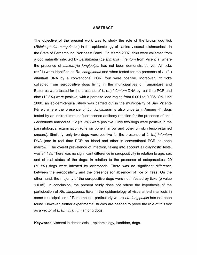
ABSTRACT
The objective of the present work was to study the role of the brown dog tick
(Rhipicephalus sanguineus) in the epidemiology of canine visceral leishmaniasis in
the State of Pernambuco, Northeast Brazil. On March 2007, ticks were collected from
a dog naturally infected by Leishmania (Leishmania) infantum from Vicência, where
the presence of Lutzomyia longipalpis has not been demonstrated yet. All ticks
(n=21) were identified as Rh. sanguineus and when tested for the presence of L. (L.)
infantum DNA by a conventional PCR, four were positive. Moreover, 73 ticks
collected from seropositive dogs living in the municipalities of Tamandaré and
Bezerros were tested for the presence of L. (L.) infantum DNA by real time PCR and
nine (12.3%) were positive, with a parasite load raging from 0.001 to 0.035. On June
2008, an epidemiological study was carried out in the municipality of São Vicente
Férrer, where the presence of Lu. longipalpis is also uncertain. Among 41 dogs
tested by an indirect immunofluorescence antibody reaction for the presence of anti-
Leishmania antibodies, 12 (29.3%) were positive. Only two dogs were positive in the
parasitological examination (one on bone marrow and other on skin lesion-stained
smears). Similarly, only two dogs were positive for the presence of L. (L.) infantum
DNA (one in real time PCR on blood and other in conventional PCR on bone
marrow). The overall prevalence of infection, taking into account all diagnostic tests,
was 34.1%. There was no significant difference in seropositivity in relation to age, sex
and clinical status of the dogs. In relation to the presence of ectoparasites, 29
(70.7%) dogs were infested by arthropods. There was no significant difference
between the seropositivity and the presence (or absence) of lice or fleas. On the
other hand, the majority of the seropositive dogs were not infested by ticks (p-value
≤ 0.05). In conclusion, the present study does not refuse the hypothesis of the
participation of Rh. sanguineus ticks in the epidemiology of visceral leishmaniosis in
some municipalities of Pernambuco, particularly where Lu. longipalpis has not been
found. However, further experimental studies are needed to prove the role of this tick
as a vector of L. (L.) infantum among dogs.
Keywords: visceral leishmaniasis – epidemiology, Ixodidae, dogs.
SUMÁRIO
1 INTRODUÇÃO ......................................................................................... 09
1.1 Sistemática e identificação ................................................................... 10
1.2 Biologia e ecologia ................................................................................. 12
1.3 Hospedeiros ............................................................................................ 15
1.4 Importância médica e veterinária ......................................................... 17
1.5 Rh. sanguineus e a transmissão de L. (L.) infantum .......................... 18
1.6 Rh. sanguineus e a leishmaniose canina em Pernambuco ............... 21
2 JUSTIFICATIVA ....................................................................................... 23
3 HIPÓTESE ............................................................................................. 25
4 OBJETIVOS ............................................................................................. 27
4.1 Geral ........................................................................................................ 28
4.2 Específicos ............................................................................................. 28
5 MATERIAL E MÉTODOS ........................................................................ 29
5.1 Epidemiologia da leishmaniose canina em São Vicente Férrer ....... 30
5.1.1 Área de estudo ....................................................................................... 30
5.1.2 Exame clínico e coleta de dados .......................................................... 31
5.1.3 Inquérito sorológico ............................................................................... 32
5.1.4 Inquérito parasitológico e molecular ................................................... 32
5.1.5 Coleta e identificação de ectoparasitos ............................................... 34
5.1.6 Análise estatística .................................................................................. 34
5.1.7 Aspectos éticos ...................................................................................... 34
5.2 Detecção de DNA de L. (L.) infantum em carrapatos .......................... 35
6 RESULTADOS ......................................................................................... 36
6.1 Epidemiologia da leishmaniose canina em São Vicente Férrer ........ 37
6.2 Ectoparasitos associados a cães de São Vicente Férrer ................... 39
6.3 Detecção de DNA de L. (L.) infantum em carrapatos .......................... 40
7 DISCUSSÃO ............................................................................................ 42
8 CONCLUSÕES ........................................................................................ 48
REFERÊNCIAS ........................................................................................ 50
ANEXOS .................................................................................................. 67

DANTAS-TORRES F. 9
1 INTRODUÇÃO

DANTAS-TORRES F. 10
Rhipicephalus sanguineus (Latreille, 1906) (Acari: Ixodida), também
conhecido como carrapato vermelho do cão ou carrapato do canil, é um ectoparasito
amplamente difundido em todo o mundo, sendo a espécie de carrapato de maior
distribuição mundial (DANTAS-TORRES, 2008c; WALKER; KEIRANS; HORAK,
2000). Sua ampla distribuição geográfica tem sido facilitada pelo transporte do seu
principal hospedeiro, o cão doméstico. Porém, embora esteja primariamente
associado ao cão, esse carrapato pode ser encontrado sobre uma grande
diversidade de animais silvestres e domésticos (SZABÓ et al., 2008; WALKER;
KEIRANS; HORAK, 2000), incluindo o homem (DANTAS-TORRES; FIGUEREDO;
BRANDÃO-FILHO, 2006; ESTRADA-PEÑA; JONGEJAN, 1999; LOULY et al., 2006).
O carrapato vermelho do cão está entre os principais vetores de
patógenos que acometem os cães (DANTAS-TORRES, 2008a, 2008c; WALKER;
KEIRANS; HORAK, 2000). Não obstante sua importância veterinária, esse carrapato
tem sido implicado na transmissão de patógenos aos seres humanos (DANTAS-
TORRES, 2007a; PAROLA et al., 2008). O número de relatos de parasitismo
humano pelo R. sanguineus tem crescido em anos recentes, inclusive no Brasil
(DANTAS-TORRES; FIGUEREDO; BRANDÃO-FILHO, 2006; LOULY et al., 2006).
Estudos indicam que o aquecimento global pode influenciar no comportamento
desse carrapato, favorecendo a ocorrência de casos de parasitismo humano e,
consequentemente, a transmissão de patógenos (PAROLA et al., 2008).
1.1 Sistemática e identificação
Rhipicephalus sanguineus foi descrito em 1806, como Ixodes sanguineus
e posteriormente transferido para o gênero Rhipicephalus (DANTAS-TORRES,
2008c). Rhipicephalus sanguineus pertence à subfamília Rhipicephalinae, dentro da
família Ixodidae. A identificação do carrapato vermelho do cão (Figura 1) pode ser
feita com base na combinação dos seguintes caracteres morfológicos: palpos curtos;
base do capítulo hexagonal; olhos presentes; festões presentes; corpo pequeno e
alongado; escudo não-ornamentado; coxa I bifurcada; e placa espiracular em forma
de vírgula nos machos (DANTAS-TORRES, 2008c).

DANTAS-TORRES F. 11
Figura 1. Rhipicephalus sanguineus, macho. A. Vista dorsal, capítulo e escudo. B. Vista ventral, capítulo, coxas e placas. C. Hipostômio. Adaptado de Cooley (1946).
A taxonomia dos carrapatos identificados como Rh. sanguineus ao redor
do mundo tem sido um assunto de debate. Diferentes abordagens (por exemplo,
análise morfológica versus análise filogenética) classificam Rh. sanguineus como um
grupo de aproximadamente 10 espécies bastante próximas. Entretanto, o status
biossistemático das espécies pertencentes ao grupo Rh. sanguineus é de difícil
determinação, principalmente com base apenas na morfologia dos exemplares
adultos (OLIVEIRA et al., 2005). Até o momento, métodos fenotípicos não são
suficientes para distinguir todas as espécies pertencentes a esse grupo. Sabe-se
que membros do grupo Rh. sanguineus podem apresentar maior ou menor

DANTAS-TORRES F. 12
susceptibilidade à infecção por certos patógenos (MATSUMOTO et al., 2005). Logo,
estudos sobre a sistemática do grupo Rh. sanguineus são de grande valia não só
sobre o ponto de vista taxonômico, mas também de transmissão de patógenos.
1.2 Biologia e ecologia
Rhipicephalus sanguineus é um carrapato de três hospedeiros. Isso
significa dizer que cada estágio ativo de desenvolvimento (larva, ninfa e adulto) se
alimenta apenas uma vez e a muda (ou ecdise) ocorre no ambiente. Seu ciclo de
vida é do tipo holometábolo (DANTAS-TORRES, 2008c).
Fêmeas adultas se alimentam por cinco a 21 dias (KOSHY; RAJAVELU;
LALITHA, 1983; PEGRAM et al., 1987; PETROVA-PIONTKOVSKAYA, 1947;
SRIVASTAVA; VARMA, 1964). Uma vez ingurgitadas, elas se desprendem do
hospedeiro para realizar a digestão sanguínea, maturação e postura dos ovos. A
postura é precedida por um período de pré-postura que varia de três a 14 dias
(JITTAPALAPONG et al., 2000; KOCH, 1982a; PEGRAM et al., 1987; SWEATMAN,
1967). A duração media da postura é de 16 a 18 dias (KOCH, 1982a; PETROVA-
PIONTKOVSKAYA, 1947). Fêmeas de Rh. sanguineus põem em media 4.000 ovos,
mas podem pôr tanto quanto 7.273 ovos (KOCH, 1982a).
A temperatura ótima para postura de Rh. sanguineus se situa entre 20 e
30°C (SWEATMAN, 1967). Após a postura dos ovos, a fêmea sucumbe. Ovos são
depositados em locais estratégicos, como frestas e buracos, normalmente acima do
nível do solo. O período de incubação dos ovos varia entre seis e 23 dias
(JITTAPALAPONG et al., 2000; KOCH, 1982a; PEGRAM et al., 1987; PETROVA-
PIONTKOVSKAYA, 1947). Após incubação, pequenas larvas eclodem (Figura 2) e,
após o enrijecimento da cutícula, passam imediatamente a procurar um hospedeiro
para realização do repasto sanguíneo. Larvas recém-eclodidas são pequenas
(comprimento, 0,54 mm; largura, 0,39 mm) e possuem apenas três pares de pernas.

DANTAS-TORRES F. 13
Figura 2. Larvas recém-eclodidas de Rh. sanguineus. Estruturas esbranquiçadas são produtos de excreção das larvas.
Larvas se alimentam por três a 10 dias, antes de se desprenderem do
hospedeiro e mudarem para ninfas (KOSHY; RAJAVELU; LALITHA, 1983; PEGRAM
et al., 1987; PETROVA-PIONTKOVSKAYA, 1947). O período de muda de larva para
ninfa varia de cinco a 15 dias (PEGRAM et al., 1987; PETROVA-PIONTKOVSKAYA,
1947). Diferentemente das larvas, as ninfas possuem quatro pares de pernas e se
assemelham aos adultos exceto por serem menores (comprimento, de 1,14 a 1,3
mm; largura, de 0,57 a 0,66 mm) e sexualmente imaturas, isto é, não apresentam
abertura genital. As ninfas se alimentam por três a 11 dias e então se desprendem
do hospedeiro (KOSHY; RAJAVELU; LALITHA, 1983; PEGRAM et al., 1987;
PETROVA-PIONTKOVSKAYA, 1947). O período de muda de ninfa para adulto varia
de nove a 47 dias (PEGRAM et al., 1987; PETROVA-PIONTKOVSKAYA, 1947).
Machos adultos são alongados (comprimento, 2,28–3,18 mm; largura 1,11–1,68
mm), marrom-avermelhados, com pequenas pontuações espalhadas ao longo
escudo dorsal. Antes do ingurgitamento, fêmeas adultas se assemelham aos
machos em tamanho (comprimento 2,4–2,7 mm; largura 1,44–1,68 mm), forma e
cor. Após o repasto sanguíneo, as fêmeas podem aumentar para 11,5 mm por 7,5

DANTAS-TORRES F. 14
mm e a porção mais larga do corpo se torna verde oliva (DANTAS-TORRES, 2008c).
Em condições favoráveis de temperatura e umidade o ciclo biológico do Rh.
sanguineus se completa em aproximadamente 63–91 dias (BECHARA et al., 1995;
GODDARD, 1987; LOULY et al., 2007).
É notória a capacidade de sobrevivência do Rh. sanguineus. Larvas de
Rh. sanguineus não alimentadas podem sobreviver por até oito meses sem se
alimentar, ao passo que ninfas e adultos podem sobreviver por seis e 19 meses,
respectivamente (GODDARD, 1987). Sob condições de laboratório, os parâmetros
biológicos (por exemplo, postura dos ovos e períodos de muda) do Rh. sanguineus
variam de acordo com a temperatura, umidade relativa e tipo de hospedeiro (cão,
hamster, coelho, etc.) (Figura 3) (BELLATO; DAEMON, 1997). A sobrevivência
máxima de ninfas ocorre sob 20°C e 85% de umidade relativa; a temperatura
mínima necessária para realização da muda está entre 10 e 15°C. Carrapatos
adultos não alimentados são mais resistentes que ninfas não alimentadas a
condições dissecantes, isto é, 35°C e 35% de umidade relativa (KOCH; TUCK,
1986). Foi recentemente demonstrado que Rh. sanguineus é menos dependente de
ambientes ricos em umidade (YODER et al., 2006), o que facilita seu
estabelecimento em regiões áridas.
Figura 3. Fêmeas e machos de Rh. sanguineus copulando durante o repasto sanguíneo em hamster em infestação experimental.
DANTAS-TORRES F. 15
Sob condições naturais, os períodos de ingurgitamento e muda podem
variar entre populações e são diretamente influenciados por fatores como
temperatura e disponibilidade de hospedeiro. Aparentemente existe uma forte
relação entre a temperatura e o tamanho da população de Rh. sanguineus
(MUMCUOGLU et al., 1993). Um estudo recente demonstrou que a temperatura
parece interferir na especificidade de hospedeiro do Rh. sanguineus, aumentando a
probabilidade desse carrapato se alimentar em seres humanos (PAROLA et al.,
2008). A duração do ciclo de vida do Rh. sanguineus pode varia de país para país e
de região para região. Estudos de campo demonstram que o carrapato vermelho do
cão pode completar duas (ou mais) gerações por ano (CRUZ-VAZQUEZ; GARCIA-
VAZQUEZ, 1990; KOCH, 1982b; USPENSKY; IOFFE-USPENSKY, 2002). No Brasil,
onde as condições ambientais são bastante favoráveis, Rh. sanguineus pode
completar até quatro gerações por ano (DANTAS-TORRES; FIGUEREDO, 2006;
LOULY et al., 2007).
Ixodídeos, particularmente carrapatos de três hospedeiros, passam 94–
97% de sua vida no ambiente (NEEDHAM; TEEL, 1991), onde estão sob a influência
de muitos fatores, como a estrutura do habitat e clima (RANDOLPH, 2004). A
maioria dos ixodídeos exibe um comportamento exofílico. Em contraste, Rh.
sanguineus é normalmente endofílico, permanecendo a maior parte do tempo no
ambiente intradomiciliar (DANTAS-TORRES, 2008c). Outra característica marcante
desse carrapato é o seu forte geotropismo negativo. Em casas onde habitam cães
infestados pelo Rh. sanguineus, é comum observar carrapatos caminhando sobre as
paredes e móveis (DANTAS-TORRES; FIGUEREDO; BRANDÃO-FILHO, 2006;
DEMMA et al., 2005; PAROLA et al., 2008).
1.3 Hospedeiros
Cães são os hospedeiros primários do Rh. sanguineus (Figura 4) e a
presença desses animais é provavelmente uma condição necessária para
manutenção de largas populações desse carrapato. Entretanto, em certas áreas, Rh.
sanguineus parecem agir de modo menos seletivo; estágios imaturos podem ser

DANTAS-TORRES F. 16
encontrados em roedores e outros pequenos mamíferos, ao passo que adultos
parasitam grandes animais, incluindo o homem (ESTRADA-PEÑA; JONGEJAN,
1999; HARRISON; ENGBER; APPERSON, 1997). No Brasil, existem registros de
infestação pelo Rh. sanguineus em cães, coelhos, gatos, roedores, aves, canídeos
silvestres e humanos (ARAGÃO, 1936; DANTAS-TORRES et al., 2004; DANTAS-
TORRES; FIGUEREDO; BRANDÃO-FILHO, 2006; DIOGO et al., 2003; LOULY et
al., 2006; SZABÓ et al., 2008; YOSHIZAWA et al., 1996).
Figura 4. Fêmea de Rh. sanguineus fixada no ouvido de um cão naturalmente infestado.
Embora seja considerado um evento esporádico, é crescente número de
relatos de casos de infestação pelo carrapato Rh. sanguineus em seres humanos na
literatura (BURGDORFER; ADKINS; PRIESTER, 1975; CARPENTER; MCMEANS;
MCHUGH, 1990; DANTAS-TORRES; FIGUEREDO; BRANDÃO-FILHO, 2006;
DEMMA et al., 2005; ESTRADA-PEÑA; JONGEJAN, 1999; FELZ; DURDEN;
OLIVER, 1996; GODDARD, 1989; GUGLIELMONE; MANGOLD; VINABAL, 1991;
HARRISON; ENGBER; APPERSON 1997; LOULY et al., 2006; MANFREDI et al.,
1990; PAROLA et al., 2008; SCHENONE, 1996; VENZAL et al., 2003). Isso parece
sugerir que esse tipo de associação carrapato-hospedeiro pode ser mais comum do

DANTAS-TORRES F. 17
que é atualmente reconhecido. Essa questão é bastante relevante quando se
considera a importância médica e veterinária do Rh. sanguineus.
1.4 Importância médica e veterinária
Rhipicephalus sanguineus é um vetor conhecido de vários patógenos aos
cães, tais como Babesia vogeli Reichenow, 1937, Ehrlichia canis (Donatien &
Lestoquard, 1935) e Hepatozoon canis (James, 1905) (DANTAS-TORRES, 2008a,
2008c; OTRANTO; DANTAS-TORRES; BREITSCHWERDT, 2009a, 2009b).
Suspeita-se que esse carrapato esteja envolvido na transmissão de outros
patógenos importantes como Anaplasma platys (French & Harvey, 1983) e
Leishmania (Leishmania) infantum Nicolle, 1908 (DANTAS-TORRES, 2008c).
O papel do Rh. sanguineus na transmissão de patógenos aos seres
humanos também está bem documentado, apesar da sua aparentemente baixa
atração por esses hospedeiros (PALMAS et al., 2001). Esse carrapato é vetor de
Rickettsia rickettsii (Wolbach, 1919), bactéria causadora da febre maculosa, no
México (BUSTAMANTE; VARELA, 1947; MARIOTTE; BUSTAMANTE; VARELA,
1944), Estados Unidos (DEMMA et al., 2005; WIKSWO et al., 2007) e possivelmente
no Brasil (MORAES-FILHO et al., 2008). Na região Mediterrânea, Rh. sanguineus é
vetor de Rickettsia conorii Brumpt, 1932, bactéria causadora da febre maculosa do
Mediterrâneo (MATSUMOTO et al., 2005).
O carrapato vermelho do cão pode também atuar como reservatório de
certos patógenos (por exemplo, R. conorii e E. canis). Isso significa que o carrapato
tem a habilidade de manter o patógeno na natureza, ao longo de várias gerações,
por passagem transovariana (da fêmea para a sua progênie) e transestadial (de um
estágio para outro) (BREMER et al., 2005; DANTAS-TORRES, 2007a). É importante
notar que carrapatos podem ser encontrados naturalmente infectados por micro-
organismos de patogenicidade desconhecida (DANTAS-TORRES, 2007a; MCGHEE;
COSGROVE, 1980; WALLACE, 1966). Por exemplo, Rh. sanguineus foi encontrado
infectado por tripanossomatídeos no Iraque (MACHATTIE; CHADWICK, 1930) e no
Brasil (SHERLOCK, 1964), os quais não puderam ser diferenciados
morfologicamente entre Leishmania e tripanossomatídeos monogenéticos (por
DANTAS-TORRES F. 18
exemplo, Leptomonas e Blastocrithidia). Logo, o uso de técnicas contemporâneas de
diagnóstico é importante para garantir uma identificação precisa do organismo
envolvido (DANTAS-TORRES, 2006d).
1.5 Rhipicephalus sanguineus e a transmissão de L. (L.) infantum
As leishmanioses são doenças parasitárias causadas por protozoários do
gênero Leishmania (Kinetoplastida: Trypanosomatidae), os quais são amplamente
distribuídos no mundo e infectam uma grande variedade de animais domésticos e
silvestres e, eventualmente, o homem (ASHFORD, 1996). As formas clínicas das
leishmanioses podem variar de uma lesão cutânea localizada, autolimitante, até
formas mais graves, sistêmicas, como a leishmaniose visceral que pode ser fatal se
não tratada adequadamente (CHAPPUIS et al., 2007; DESJEUX, 2004). A
leishmaniose visceral causada pela espécie L. (L.) infantum, atualmente considerada
como sinônimo de Leishmania (Leishmania) chagasi Cunha & Chagas, 1937
(BANETH et al., 2008; DANTAS-TORRES, 2008a; LUKES et al., 2007; SCHÖNIAN
et al., 2008), é uma zoonose prevalente em muitos países na América, Europa, Ásia
e África. Aproximadamente 90% dos casos humanos de leishmaniose visceral
registrados estão concentrados em áreas rurais e suburbanas de seis países:
Bangladesh, Brasil, Etiópia, Índia, Nepal e Sudão (CHAPPUIS et al., 2007;
DESJEUX, 2004). A leishmaniose visceral é uma doença de difícil controle que se
encontra em franca expansão geográfica, afetando principalmente crianças e
indivíduos infectados pelo vírus da imunodeficiência humana (HIV) (ALVAR;
YACTAYO; BERN, 2006; CHAPPUIS et al., 2007; DANTAS-TORRES; BRANDÃO-
FILHO, 2006b; DESJEUX, 2004).
A transmissão de L. (L.) infantum se dá primariamente por meio da picada
de fêmeas de flebotomíneos (Diptera: Psychodidae) dos gêneros Lutzomyia, no
Novo Mundo (Américas), e Phlebotomus, no Velho Mundo (Europa, Ásia e África)
(KILLICK-KENDRICK, 1990). No Brasil, Lutzomyia longipalpis (Lutz & Neiva, 1912) é
a principal espécie de flebotomíneo envolvida no ciclo de transmissão, porém, outras
espécies têm sido apontadas como possíveis vetores (CARVALHO et al., 2008;
PITA-PEREIRA et al., 2008; YOUNG; DUNCAN, 1994). Além da picada de
DANTAS-TORRES F. 19
flebotomíneos, outras formas de transmissão de L. (L.) infantum entre humanos têm
sido aventadas, incluindo congênita, por transfusão sangüínea, transplante de
órgãos e compartilhamento de seringas contaminadas entre usuários de drogas
(BASSET et al., 2005; BOEHME et al., 2006; ELTOUM et al., 1992; HERWALDT,
2001; MATHUR; SAMANTARAY, 2004; MEINECKE et al., 1999; MOLINA;
GRADONI; ALVAR, 2003; PAGLIANO et al., 2005; SYMMERS, 1960). De fato, o
ciclo de transmissão de L. (L.) infantum é bastante complexo e envolve um conjunto
de interações entre o protozoário e seus hospedeiros reservatórios e vetores, que
podem variar de região para região (ASHFORD, 1996; DANTAS-TORRES;
BRANDÃO-FILHO, 2006b; KILLICK-KENDRICK, 1990).
O cão doméstico tem sido apontado como o principal hospedeiro
reservatório de L. (L.) infantum, particularmente no ambiente doméstico e
peridoméstico (DANTAS-TORRES, 2007b), embora outros hospedeiros possam
estar envolvidos (DANTAS-TORRES; BRANDÃO-FILHO, 2006b), incluindo o homem
(COSTA et al., 2002; FAKHAR et al., 2008). Em alguns focos da doença, existe uma
forte relação entre a presença de casos de leishmaniose visceral em cães e
humanos (AGUILAR et al., 1998; CUNHA et al., 1995). Essa relação é de certa
forma esperada, uma vez que tanto os cães quanto os humanos são susceptíveis a
infecção por L. (L.) infantum e, em áreas endêmicas, estão sob constante exposição
aos vetores.
Existem algumas áreas, entretanto, onde a leishmaniose visceral canina é
endêmica ou tem sido esporadicamente relatada, porém poucos casos humanos são
notificados e/ou a presença do vetor primário não tem sido confirmada (CARVALHO
et al., 2007; DANTAS-TORRES et al., 2005). Isso tem sugerido a participação de
outras espécies de flebotomíneos ou até mesmo a existência de formas secundárias
de transmissão. Dentre essas formas, estariam incluídas a transmissão por meio de
transfusão sanguínea (FREITAS et al., 2006; OWENS et al., 2001), transmissão
transplacentária (ROSYPAL et al., 2005), transmissão venérea (SILVA et al., 2009),
pela mordedura, ingestão de vísceras contaminadas (SHERLOCK, 1964) e por
pulgas e carrapatos (COUTINHO et al., 2005; COUTINHO; LINARDI, 2007).
A hipótese sobre a participação do carrapato Rh. sanguineus na
transmissão de L. (L.) infantum entre cães não é recente (BLANC;
CAMINOPETROS, 1930; GIRAUD; RANQUE; CABASSU, 1954; MACHATTIE;
CHADWICK, 1930; MCKENZIE, 1984; SHERLOCK, 1964). No início da década de

DANTAS-TORRES F. 20
1930, na França, demonstrou-se a susceptibilidade do Rh. sanguineus à infecção
por L. (L.) infantum e a capacidade desse carrapato transmitir mecanicamente o
protozoário para roedores (BLANC; CAMINOPETROS, 1930). No Brasil, Sherlock
(1964) relatou o encontro de formas morfologicamente semelhantes a “leptomonas”
de Leishmania em carrapatos coletados de cães com leishmaniose, levando-o a
especular sobre a participação de carrapatos no ciclo zoonótico da leishmaniose
visceral (Figura 5).
Figura 5. Ciclo da leishmaniose visceral, segundo Sherlock (1964). Os carrapatos estariam envolvidos na transmissão entre cães e esporadicamente para o homem.
Em meados dos anos 1980, nos Estados Unidos, surgiram novas
evidências sobre a possibilidade da transmissão de L. (L.) infantum entre cães pelo
Rh. sanguineus (MCKENZIE, 1984). Na ocasião, comprovou-se a transmissão
transestadial (de larva para ninfa e de ninfa para adulto) de L. (L.) infantum em Rh.
sanguineus e que o protozoário era capaz de sobreviver no carrapato por um
período superior a 100 dias (MCKENZIE, 1984). Além disso, análise ultraestrutural
de L. (L.) infantum em Rh. sanguineus revelou formas promastigotas semelhantes
aquelas encontradas nos flebotomíneos vetores (MCKENZIE, 1984).

DANTAS-TORRES F. 21
No Brasil, há relatos do encontro de exemplares de Rh. sanguineus
infectados por formas semelhantes a promastigotas de L. (L.) infantum (SHERLOCK,
1964; SILVA et al., 2007). Contudo, é importante salientar que alguns
tripanossomatídeos monogenéticos que podem ser encontrados em Rh. sanguineus
(MACHATTIE; CHADWICK, 1930) podem ser facilmente confundidos com formas
promastigotas de Leishmania.
Em estudo recente realizado no Brasil, pesquisadores coletaram 39
carrapatos de cães soropositivos, apresentando sinais clínicos sugestivos de
leishmaniose visceral, e verificaram a presença de DNA de Leishmania em seis
desses carrapatos (COUTINHO et al., 2005). No mesmo estudo, carrapatos foram
macerados e inoculados em hamsters por via oral e intraperitoneal. Seis meses após
a inoculação, 14 animais apresentaram sinais de infecção, sendo que 12 haviam
sido inoculados por via intraperitoneal e dois por via oral (COUTINHO et al., 2005).
Assim, esse estudo levantou a possibilidade da transmissão oral de L. (L.) infantum
por meio da ingestão de carrapatos infectados, tal qual como acontece na
transmissão de H. canis (DANTAS-TORRES, 2008a).
Rhipicephalus sanguineus se encontra quase que invariavelmente
presente nas áreas onde a leishmaniose visceral canina é endêmica (DANTAS-
TORRES, 2006b, 2006d). De fato, as evidências disponíveis sugerem que Rh.
sanguineus pode estar envolvido na epidemiologia da leishmaniose visceral canina.
A real importância dessa possível forma secundária de transmissão é desconhecida.
Mais que isso, não se sabe se Rh. sanguineus é capaz de transmitir L. (L.) infantum
durante o repasto sanguíneo. Considerando que formas semelhantes promastigotas
de L. (L.) infantum têm sido encontradas nesse carrapato, a possibilidade de
transmissão biológica não pode ser totalmente descartada.
1.6 Rh. sanguineus e a leishmaniose visceral canina em Pernambuco
A leishmaniose visceral canina é uma doença endêmica em Pernambuco
(DANTAS-TORRES, 2006a, 2008b). Casos de leishmaniose visceral canina estão
comumente associados à presença de casos humanos, os quais encontram em
constante expansão geográfica em Pernambuco (DANTAS-TORRES; BRANDÃO-

DANTAS-TORRES F. 22
FILHO, 2006a). A prevalência da infecção por Leishmania spp. em cães em
Pernambuco, usualmente estimada por testes sorológicos, pode variar bastante de
região para região e de acordo com a técnica sorológica empregada
(ALEXANDRINO, 2001; BRANDÃO-FILHO et al., 1994; DANTAS-TORRES; BRITO;
BRANDÃO-FILHO, 2006; FRANÇA et al., 2003; LIMA-JÚNIOR et al., 2000). Em
alguns focos altamente endêmicos, a soroprevalência pode alcançar níveis
superiores a 50% (DANTAS-TORRES; BRITO; BRANDÃO-FILHO, 2006). Como
observado em outros estados brasileiros (RONDON et al., 2008) e na Europa
(BANETH et al., 2008), a maioria dos cães soropositivos em Pernambuco não
apresenta sinais clínicos sugestivos de leishmaniose visceral (DANTAS-TORRES;
BRITO; BRANDÃO-FILHO, 2006) o que representa um desafio extra para o
programa de controle da leishmaniose visceral nesse estado.
Pouco se sabe sobre os fatores de risco associados à infecção por
Leishmania spp. em cães em Pernambuco. No município de Paulista, a
soropositividade é maior entre os cães machos e menores de um ano (DANTAS-
TORRES; BRITO; BRANDÃO-FILHO, 2006). A presença de Lu. longipalpis é
comumente observada em áreas onde a leishmaniose visceral canina é endêmica
(DANTAS-TORRES; ALMEIDA; BRANDÃO-FILHO, 2006). Porém, em alguns
municípios onde casos de leishmaniose visceral canina têm sido diagnosticados (por
exemplo, Recife), a presença de Lu. longipalpis ainda não foi comprovada
(DANTAS-TORRES, 2006c; DANTAS-TORRES et al., 2005). Em São Vicente
Férrer, casos de leishmaniose visceral em cães e humanos têm sido registrados,
mas a presença do vetor primário ainda não foi comprovada (CARVALHO et al.,
2007). Isso sugere que outras espécies de Lutzomyia, como Lutzomyia migonei
(França, 1920), podem estar envolvidas na transmissão de L. (L.) infantum nesse
município. Contudo, formas semelhantes à promastigotas de Leishmania spp. foram
encontradas recentemente em carrapatos identificados como Rh. sanguineus
coletados de cães soropositivos no município de São Vicente Férrer (SILVA et al.,
2007). Esse achado sugere a necessidade de novas investigações sobre o papel do
Rh. sanguineus na epidemiologia da leishmaniose visceral canina nesse município.

DANTAS-TORRES F. 23
2 JUSTIFICATIVA

DANTAS-TORRES F. 24
Diante das evidências epidemiológicas e experimentais, novos estudos
são necessários para investigar o papel do Rh. sanguineus na transmissão de L. (L.)
infantum entre cães, uma vez que essa possibilidade apresenta implicações diretas
para o programa de controle da leishmaniose visceral no Brasil.

DANTAS-TORRES F. 25
3 HIPÓTESE

DANTAS-TORRES F. 26
O carrapato Rh. sanguineus desempenha um possível papel secundário
na transmissão de L. infantum entre os cães, principais reservatórios domésticos no
ciclo zoonótico da leishmaniose visceral em Pernambuco.

DANTAS-TORRES F. 27
4 OBJETIVOS
DANTAS-TORRES F. 28
4.1 Geral
• Investigar o papel do carrapato Rh. sanguineus na transmissão de L. (L.)
infantum em Pernambuco.
4.2 Específicos
• Estudar a epidemiologia da leishmaniose visceral canina no município de São
Vicente Férrer, onde a presença de Lu. longipalpis ainda não foi comprovada.
• Identificar os ectoparasitos (carrapatos, piolhos e pulgas) que infestam cães em
São Vicente Férrer.
• Verificar se existe uma associação entre a infestação por carrapatos e a
presença de anticorpos anti-Leishmania spp. em cães em São Vicente Férrer.
• Detectar a presença de DNA de L. (L.) infantum em carrapatos coletados de
cães em alguns municípios de Pernambuco.

DANTAS-TORRES F. 29
5 MATERIAL E MÉTODOS

DANTAS-TORRES F. 30
5.1 Epidemiologia da leishmaniose visceral canina em São Vicente Férrer
5.1.1 Área de estudo
O município de São Vicente Férrer (07º35’28’’ S, 35º29’29’’ W) (Figura 6)
está localizado na mesorregião Agreste do Estado de Pernambuco, Nordeste do
Brasil. Esse município possui uma população de pouco mais de 25.000 habitantes e
um território de aproximadamente 120 km2.
Figura 6. Localização do município de São Vicente Férrer.
Apesar de estar geopoliticamente situado na região Agreste, o município
de São Vicente Férrer possui características climáticas e de vegetação típicas da
Zona da Mata Atlântica. O clima é tropical úmido, com a estação seca de setembro a
fevereiro e a estação chuvosa de março a agosto. Embora a maior parte da
vegetação primária tenha sido substituída por plantações de banana (principal
atividade agrícola de São Vicente Férrer), ainda existem alguns remanescentes de
mata atlântica (Figura 7).

DANTAS-TORRES F. 31
Figura 7. Remanescente de mata atlântica (alto), em meio a um proeminente bananal.
5.1.2 Exame clínico e coleta de dados
Em junho 2008, examinaram-se 41 cães semidomiciliados (ambos os
sexos, sem raça definida, com idade variada). Essa amostragem incluiu todos os
cães de uma localidade de São Vicente Férrer, chamada Mundo Novo, exceto dois
animais que não foram contidos adequadamente pelos proprietários para realização
da coleta. Avaliou-se o status clínico de cada um dos cães, considerando
sintomáticos aqueles que apresentavam um ou mais sinais clínicos sugestivos de
leishmaniose visceral, tais como lesões cutâneas, aumento de linfonodos,
onicogrifose (crescimento anormal das unhas), lesões oculares e perda de peso.
Utilizou-se uma ficha individual para coleta de informações clínico-epidemiológicas
dos animais.

DANTAS-TORRES F. 32
5.1.3 Inquérito sorológico
Alíquotas de aproximadamente 3 ml de sangue total foram coletadas de
cada um dos 41 cães, por punção venosa da veia cefálica, femural ou jugular. As
alíquotas foram transferidas para tubos com anticoagulante (VACUETTE® EDTA K3,
Greiner Bio-One, Kremsmuenster, Austria) e mantidas sob refrigeração. As amostras
foram centrifugadas a 1.500 x g, durante cinco minutos, e os plasmas obtidos foram
congelados a –20ºC até serem testados para presença de anticorpos (IgG) contra
Leishmania spp. Para esse fim, utilizou-se uma reação de imunofluorescência
indireta (RIFI) (IFI-Leishmaniose-Visceral-Canina, Bio-Manguinhos, Rio de Janeiro,
Brasil) e um teste rápido de imunocromatografia (Kalazar Detect, InBios
International, Seattle, USA), seguindo as instruções dos fabricantes.
5.1.4 Inquérito molecular e parasitológico
Alíquotas de 3 ml de sangue total foram coletadas de cada um dos cães
(como descrito no item 4.1.2), as quais foram usadas para extração de DNA
utilizando um kit comercial (Illustra blood genomicPrep Mini Spin Kit, GE Healthcare,
New York, USA), seguindo as instruções do fabricante. As amostras de DNA de
sangue total foram testadas para presença de DNA de L. (L.) infantum, por meio de
uma reação em cadeia da polimerase convencional (PCR convencional) (DANTAS-
TORRES, 2006) e outra em tempo real (PCR em tempo real) (PAIVA CAVALCANTI
et al., 2008). Na PCR convencional, utilizou-se um par de oligonucleotídeos
iniciadores (senso, 5′-CTTTTCTGGTCCCGCGGGTAGG-3′; antissenso, 5′-
CCACCTGGCCTATTTTACACCA-3′) que amplificam uma sequência alvo de 145
pares de bases do DNA do cinetoplasto (kDNA) de L. (L.) infantum (LE FICHOUX et
al., 1999; RAVEL et al., 1995). A reação foi realizada em um volume final de 25 µl
contendo 14 µl de água livre de DNA, 2,5 µl de 10x PCR buffer (Invitrogen,
California, USA), 1,5 µl de 1,5 mM MgCl2 (Invitrogen, California, USA), 2,5 µl de 2
mM dNTP mix (Invitrogen, California, USA), 0,5 µl de cada oligonucleotídeo iniciador

DANTAS-TORRES F. 33
(25 pmol/µl), 0,5 µl de Platinum Taq DNA polymerase (5 U/µl) (Invitrogen, California,
USA) e 2 µl da amostra de DNA. As condições de amplificação incluíram um período
inicial de desnaturação (94ºC/5 min.), seguido por 35 ciclos de desnaturação
(94ºC/30 s), anelamento (67ºC/1 min.) e extensão (72ºC/30 s), e uma síntese
terminal (72ºC/5 min.). Controles negativo (água livre de DNA) e positivo (5 ng/µl de
DNA genômico de L. (L.) chagasi, cepa MHOM/BR/74/PP75) foram incluído em cada
reação. As reações foram realizadas no termociclador Mastercycler PCR (Eppendorf,
Hamburg, Germany). Os produtos da PCR foram separados por eletroforese em gel
de agarose a 1,5%, utilizando-se fragmentos λ DNA/Hind III (Invitrogen, California,
USA) como padrão de peso molecular. Após a eletroforese em gel de agarose, as
bandas resultantes foram visualizadas sob luz ultravioleta após coloração com
brometo de etídio (10 mg/ml).
Na PCR em tempo real, utilizou-se um par de oligonucleotídeos
iniciadores (senso, 5’-TCCCAAACTTTTCTGGTCCT-3’; antissenso, 5’-
TTACACCAACCCCCAGTTTC-3’) que amplificam uma sequência alvo de 132 pares
de bases do kDNA de L. (L.) infantum (PAIVA CAVALCANTI et al., 2008). A reação
foi realizada em um volume final de 50 µl contendo 21 µl de água livre de DNA, 25 µl
de SYBR Green Master Mix (Applied Biosystems, California, USA), 1 µl (3 pmol/µl)
de cada oligonucleotídeo iniciador e 2 µl da amostra de DNA. As condições de
amplificação incluíram um período inicial de 95ºC por 10 minutos, seguido por 40
ciclos de 95ºC por 15 segundos e 60ºC por 1 minuto. As amostras foram testadas
em duplicata. Controles negativo (água livre de DNA) e positivos (DNA genônico de
L. chagasi, cepa MHOM/BR/1974/PP75, nas concentrações de 0,01 fg a 1000 ng)
foram incluídos em cada reação. As reações foram realizadas num aparelho ABI
Prism 7500 (Applied Biosystems, California, USA)
Coletaram-se ainda amostras de medula óssea (de 26 cães) e de lesão
cutânea (de seis cães), por punção do manúbrio do osso esterno e raspado da
borda das lesões, respectivamente. Elaboraram-se esfregaços em lâminas de vidro
para microscopia, os quais foram corados com o kit Panótico rápido (Laborclin,
Paraná, Brasil), seguindo as instruções do fabricante. Os esfregaços foram
examinados para presença de formas amastigotas de Leishmania spp., em
microscópio óptico sob objetiva de imersão. Realizou-se a extração de DNA de 12
amostras de medula óssea utilizando o mesmo kit comercial usado para extração de
DNA de amostras de sangue total. Após a extração, as amostras de DNA purificado
DANTAS-TORRES F. 34
de medula óssea foram testadas pelos mesmos protocolos de PCR convenciona e
em tempo real, descritos anteriormente.
5.1.5 Coleta e identificação de ectoparasitos
Durante o exame clínico, ectoparasitos foram coletados e acondicionados
em frascos contendo álcool a 70%, individualizados por animal. A identificação dos
ectoparasitos foi realizada sob microscópio estereoscópico de acordo com as chaves
taxonômicas tradicionais (ARAGÃO; FONSECA, 1961; LINARDI; GUIMARÃES,
2000; WERNECK, 1936). Gêneros de carrapatos são abreviados conforme sugerido
por Dantas-Torres (2008d). Para fins de registro, alguns espécimes de carrapatos
foram depositados na Coleção Nacional de Carrapatos (números de acesso, 1317 e
1318) da Faculdade de Medicina Veterinária da Universidade de São Paulo e na
Coleção de Acari (números de acesso, 9980 e 9981) do Instituto Butantan.
5.1.6 Análise estatística
Utilizou-se o teste qui-quadrado (χ2) para comparar as taxas de
soroprevalência em relação ao sexo, idade, status clínico e presença de
ectoparasitos. Consideraram-se as diferenças como significativas quando p ≤ 0,05.
Calcularam-se os intervalos de confiança de 95% (IC 95%) para cada uma das taxas
de soroprevalência. Realizou-se a análise estatística com auxílio do programa Epi
Info, versão 6.04d (Centers for Disease Control and Prevention, Atlanta, USA).
5.1.7 Aspectos éticos

DANTAS-TORRES F. 35
Esse estudo faz parte de um projeto (P.0174-03), previamente licenciado
pela Comissão de Ética no Uso de Animais (CEUA), da Fundação Oswaldo Cruz
(FIOCRUZ).
5.2 Detecção de DNA de L. (L.) infantum em carrapatos
Em março de 2007, coletaram-se carrapatos de um cão naturalmente
infectado por L. (L.) infantum residente do município de Vicência (Zona da Mata). Os
carrapatos foram coletados manualmente e acondicionados em frascos contendo
álcool a 70%. Após a identificação, esses carrapatos foram processados
individualmente para extração de DNA utilizando um kit comercial (Illustra tissue &
cells genomicPrep Mini Spin Kit, GE Healthcare, New York, USA), seguindo as
instruções do fabricante. Após a extração, as amostras de DNA dos carrapatos
foram testadas pela PCR convencional descrita no item 4.1.4.
Adicionalmente, coletaram-se carrapatos de cães soropositivos residentes
nos municípios de Tamandaré (localizado na região Zona da Mata) e Bezerros
(região Agreste). Os carrapatos foram coletados manualmente e acondicionados em
frascos contendo álcool a 70%, individualizados por animal. Após a identificação da
espécie, os carrapatos foram processados individualmente para extração de DNA,
conforme descrito anteriormente. Posteriormente, as amostras de DNA foram
testadas utilizando a PCR em tempo real descrita no item 4.1.4.

DANTAS-TORRES F. 36
6 RESULTADOS
DANTAS-TORRES F. 37
6.1 Epidemiologia da leishmaniose visceral canina em São Vicente Férrer
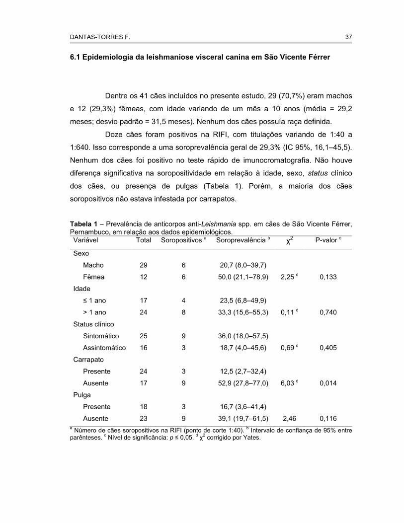
Dentre os 41 cães incluídos no presente estudo, 29 (70,7%) eram machos
e 12 (29,3%) fêmeas, com idade variando de um mês a 10 anos (média = 29,2
meses; desvio padrão = 31,5 meses). Nenhum dos cães possuía raça definida.
Doze cães foram positivos na RIFI, com titulações variando de 1:40 a
1:640. Isso corresponde a uma soroprevalência geral de 29,3% (IC 95%, 16,1–45,5).
Nenhum dos cães foi positivo no teste rápido de imunocromatografia. Não houve
diferença significativa na soropositividade em relação à idade, sexo, status clínico
dos cães, ou presença de pulgas (Tabela 1). Porém, a maioria dos cães
soropositivos não estava infestada por carrapatos.
Tabela 1 – Prevalência de anticorpos anti-Leishmania spp. em cães de São Vicente Férrer, Pernambuco, em relação aos dados epidemiológicos.
Variável Total Soropositivos a Soroprevalência b χ2 P-valor c
Sexo
Macho 29 6 20,7 (8,0–39,7)
Fêmea 12 6 50,0 (21,1–78,9) 2,25 d 0,133
Idade
≤ 1 ano 17 4 23,5 (6,8–49,9)
> 1 ano 24 8 33,3 (15,6–55,3) 0,11 d 0,740
Status clínico
Sintomático 25 9 36,0 (18,0–57,5)
Assintomático 16 3 18,7 (4,0–45,6) 0,69 d 0,405
Carrapato
Presente 24 3 12,5 (2,7–32,4)
Ausente 17 9 52,9 (27,8–77,0) 6,03 d 0,014
Pulga
Presente 18 3 16,7 (3,6–41,4)
Ausente 23 9 39,1 (19,7–61,5) 2,46 0,116 a Número de cães soropositivos na RIFI (ponto de corte 1:40). b Intervalo de confiança de 95% entre parênteses. c Nível de significância: p ≤ 0,05. d χ2 corrigido por Yates.

DANTAS-TORRES F. 38
Nove (75%) dos 12 cães soropositivos apresentavam pelo menos um
sinal clínico sugestivo de leishmaniose, incluindo úlcera cutânea, emagrecimento e
onicogrifose (Figuras 8 e 9).
Figuras 8 e 9. Caquexia (8) e onicogrifose (9).
Apenas um cão foi positivo para presença de amastigotas de Leishmania
sp. (Figura 10) em esfregaços de medula óssea e outro em raspado de lesão
cutânea (Figura 11).
Figuras 10 e 11. Amastigota de Leishmania spp. em esfregaço de medula óssea (10) e raspado de lesão cutânea (11). Aumento de 1000x.
Apenas um cão foi positivo na PCR convencional em amostra de medula
óssea (Figura 12).

DANTAS-TORRES F. 39
PM CN CP 1 2 3 4 5 6 7 8 9
145 pb
Figura 12. Eletroforese em gel de agarose 1,5% dos produtos da PCR convencional em amostras de medula óssea de cães de São Vicente Férrer. PM, peso molecular; CN, controle negativo; CP, controle positivo; 1, amostra positiva; 2–9, amostras negativas.
Em amostras de sangue total, apenas um cão (o mesmo cão que foi
positivo na citologia de medula óssea) foi positivo na PCR em tempo real,
apresentando uma carga parasitária muito baixa (0,02 parasitos/µl). Nenhum cão foi
positivo na PCR convencional em amostras de sangue total.
6.2 Ectoparasitos associados a cães de São Vicente Férrer
Vinte e nove dos 41 cães examinados estavam infestados por
ectoparasitos, correspondendo a uma prevalência 70,7% (IC 95%: 54,5–83,9). A
prevalência de infestação foi maior entre cães machos (χ2 = 15,52, p = 0,000).
Entretanto, não houve diferença significativa quando o percentual de machos no
grupo de cães infestado foi comparado com o grupo não-infestado (χ2 corrigido por
Yates = 0,56, p = 0,456) ou com todos os cães incluídos no estudo (Tabela 2). Da
mesma forma, não houve diferença significativa em relação à idade no grupo de
cães infestados (χ2 = 0,07, p = 0,792) e quando esse foi comparado ao grupo de
cães não infestados (χ2 corrigido por Yates = 1,06, p = 0,303) ou com todos os cães
incluídos no estudo (Tabela 2).
DANTAS-TORRES F. 40
Tabela 2 – Infestação por ectoparasitos em relação ao sexo e grupo etário dos cães.
Variável n Cães
infestados Prevalência (IC 95%) χ2 p a
Sexo
Macho 29 22 75,9 (56,5–89,7)
Fêmea 12 7 58,3 (27,7–84,8) 0,56 b 0,456
Idade
≤ 1 ano 17 14 82,4 (56,6–96,2)
> 1 ano 24 15 62,5 (40,6–81,2) 1,06 b 0,303
Total 41 29 70,7 (54,5–83,9) a Nível de significância: p<0,05. b χ2 corrigido por Yates.
A prevalência de infestação por carrapatos foi 58,5% (IC 95%: 42,1–73,7).
Dos 24 cães parasitados por carrapatos, 62,5% (IC 95%: 40,6–81,2) estavam
infestados exclusivamente por Rh. sanguineus, 16,7% (IC 95%: 4,7–37,4) por
Amblyomma ovale Koch, 1844 e 20,8% (IC 95%: 7,1–42,1) estavam co-infestados
por ambas as espécies. Ctenocephalides felis felis (Bouché, 1835) e Heterodoxus
spiniger (Enderlein, 1909) foram as únicas espécies de pulga e piolho,
respectivamente, identificadas. Dos 29 cães infestados, 20 (68,9%; IC 95%: 49,2–
84,7) estavam infestados por mais de uma espécie de ectoparasito, o que
corresponde a uma prevalência de co-infestação de 48,8% (IC 95%: 32,9–64,9). As
seguintes associações de ectoparasitos foram encontradas (número de observações
entre parênteses): Rh. sanguineus + C. felis felis (n=7); Rh. sanguineus + Am. ovale
(n=3); Rh. sanguineus + C. felis felis + H. spiniger (n=3); C. felis felis + H. spiniger
(n=2); C. felis felis + Am. ovale (n=1); Rh. sanguineus + H. spiniger (n=1); C. felis
felis + H. spiniger + Am. ovale (n=1); Rh. sanguineus + C. felis felis + Am. ovale
(n=1); Rh. sanguineus + C. felis felis + H. spiniger + Am. ovale (n=1).
6.3 Detecção de DNA de L. (L.) infantum em carrapatos
Dos 21 carrapatos coletados do cão residente no município de Vicência,
quatro fêmeas ingurgitadas foram positivas no PCR convencional. De um total de 73
carrapatos coletados sobre cães soropositivos testados pela PCR em tempo real,

DANTAS-TORRES F. 41
nove (12,3%) foram positivos, com uma carga parasitária variando de 0,001 a 0,035
parasitos/µl.

DANTAS-TORRES F. 42
7 DISCUSSÃO

DANTAS-TORRES F. 43
No presente estudo, observou-se uma alta prevalência de anticorpos anti-
Leishmania spp. entre cães de São Vicente Férrer. Em Pernambuco, a
soroprevalência pode variar bastante (ALEXANDRINO, 2001; DANTAS-TORRES;
BRITO; BRANDÃO-FILHO, 2006; FRANÇA et al., 2003; LIMA-JÚNIOR et al., 2000).
A taxa de soroprevalência média é de aproximadamente 2,5% (ALEXANDRINO,
2001), variando de 0,32% em Recife (LIMA-JÚNIOR et al., 2000) a 40,3% em
Paulista (DANTAS-TORRES; BRITO; BRANDÃO-FILHO, 2006). A situação no Brasil
é semelhante (COUTINHO et al., 1985; PARANHOS-SILVA et al., 1996; FRANÇA-
SILVA et al., 2003; IVERSON et al., 1983; RONDON et al., 2008), mas a
soroprevalência pode ser tão alta quanto 75% em algumas áreas altamente
endêmicas (CORTADA et al., 2004).
Nesse estudo, não houve diferença significativa na soroprevalência em
relação ao sexo ou idade dos cães. De fato, o sexo não parece ser um fator
importante (ABRANCHES et al., 1991; ALENCAR; CUNHA, 1963; AMELA et al.,
1995; AMUSATEGUI et al., 2003; FISA et al., 1999; FRANÇA-SILVA et al., 2003;
POZIO et al., 1981; RONDON et al., 2008; SIDERIS et al., 1996). Contudo, em
alguns focos de leishmaniose visceral canina no Brasil (DANTAS-TORRES; BRITO;
BRANDÃO-FILHO, 2006), na França (LANOTTE et al., 1975) e na Espanha
(MIRANDA et al., 2005), os machos parecem estar mais expostos ao risco de
infecção. Em relação à idade, a leishmaniose visceral canina apresenta uma
distribuição bi-modal, com um pico no número de casos em cães com menos de três
anos de idade e outro entre as idades de oito e 10 anos (ACEDO-SANCHEZ et al.,
1996; AMELA et al., 1995; MIRANDA et al., 2005). Estudos no Brasil têm mostrado
resultados diversos em relação à soroprevalência versus grupo etário (DANTAS-
TORRES; BRITO; BRANDÃO-FILHO, 2006; RONDON et al., 2008). Isso
provavelmente se deve a diferenças nos hábitos das populações de cães de
diferentes áreas endêmicas, que podem interferir na probabilidade de exposição aos
vetores.
Em áreas endêmicas, seja no Brasil ou na Europa, uma alta proporção de
cães soropositivos não apresenta sinais clínicos de leishmaniose visceral
(ABRANCHES et al., 1991; BRANDONISIO et al., 1992; DANTAS-TORRES; BRITO;
BRANDÃO-FILHO, 2006; MANCIANTI; PEDONESE; POLI, 1996; PORTÚS et al.,
1987; RONDON et al., 2008). Em contraste, no presente estudo a maioria (=75%)
dos cães soropositivos apresentava um ou mais sinais clínicos de leishmaniose.
DANTAS-TORRES F. 44
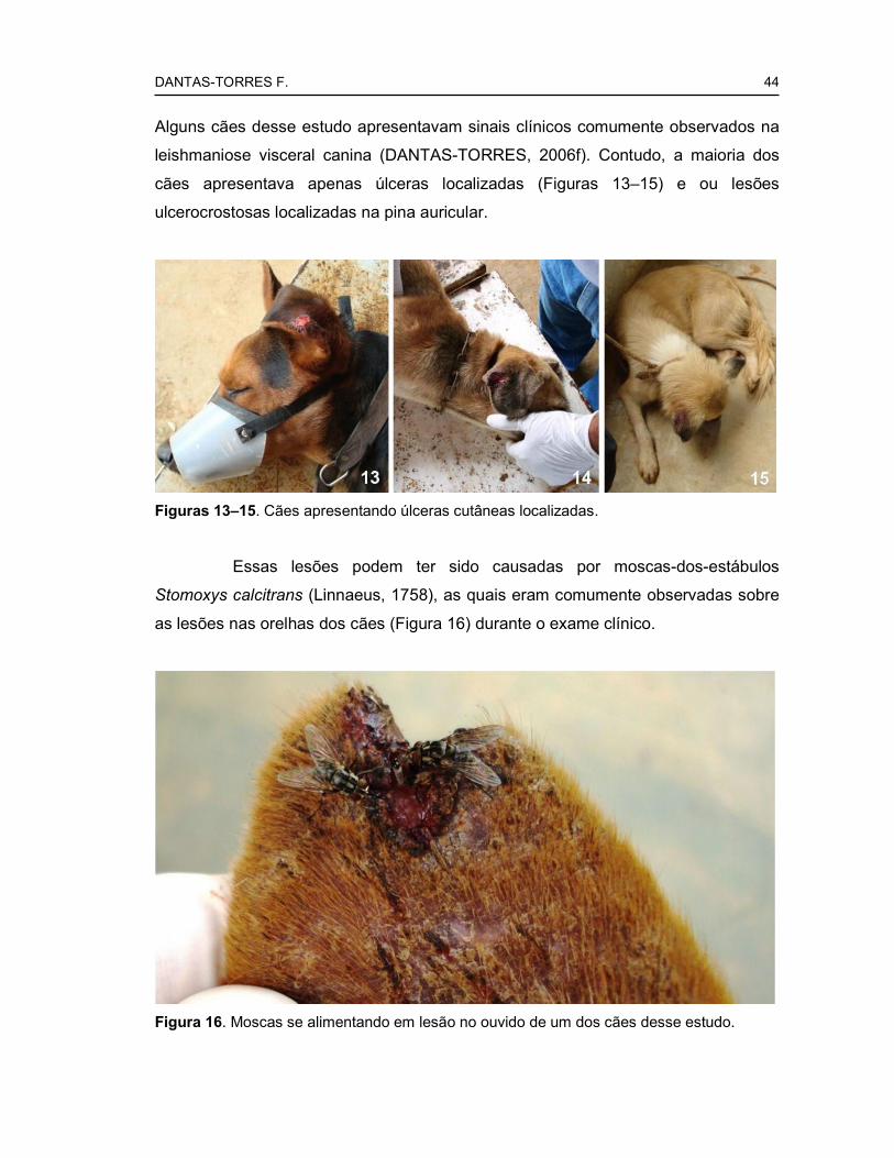
Alguns cães desse estudo apresentavam sinais clínicos comumente observados na
leishmaniose visceral canina (DANTAS-TORRES, 2006f). Contudo, a maioria dos
cães apresentava apenas úlceras localizadas (Figuras 13–15) e ou lesões
ulcerocrostosas localizadas na pina auricular.
Figuras 13–15. Cães apresentando úlceras cutâneas localizadas.
Essas lesões podem ter sido causadas por moscas-dos-estábulos
Stomoxys calcitrans (Linnaeus, 1758), as quais eram comumente observadas sobre
as lesões nas orelhas dos cães (Figura 16) durante o exame clínico.
Figura 16. Moscas se alimentando em lesão no ouvido de um dos cães desse estudo.

DANTAS-TORRES F. 45
Além disso, é importante destacar que a leishmaniose tegumentar
causada por Leishmania (Viannia) braziliensis (Vianna, 1911) é endêmica em São
Vicente Férrer (CARVALHO, 2005; CARVALHO et al., 2007). Considerando que a
RIFI utilizada no presente estudo apresenta uma baixa especificidade (LIRA et al.,
2006), a possibilidade de que alguns dos cães que apresentavam apenas lesões
cutâneas estivessem infectados por L. (V.) braziliensis não pode ser descartada.
Essa hipótese é reforçada pela baixa positividade observada nos exames
parasitológico e molecular. Por outro lado, recentes estudos longitudinais revelaram
que cães infectados por L. (L.) infantum podem se apresentar temporariamente
negativos, devido a uma diminuição da carga parasitária a níveis indetectáveis, até
mesmo por técnicas altamente sensíveis como a PCR (OLIVA et al., 2006).
No presente estudo, observou-se uma alta prevalência de infestação por
ectoparasitos (carrapatos, piolhos e pulgas). A prevalência de infestação por
ectoparasitos pode variar bastante de região para região e também de acordo com
as características da população de cães (CASTRO; RAFAEL, 2006; DANTAS-
TORRES; FIGUEREDO, FAUSTINO, 2004; LINARD; NAGEM, 1973; RODRIGUES
et al., 2001; SOARES et al., 2006). O número de machos infestados foi maior que o
de fêmeas, porém, isso pode ser atribuído às características da população de estudo
que era predominantemente composta por machos. Embora não tenha sido
encontrada diferença significativa em relação à idade, apenas três dos 13 cães
jovens incluídos no presente estudo estavam livres de ectoparasitos. Isso sugere
que cães jovens que vivem em São Vicente Férrer são mais susceptíveis e/ou
expostos à infestação por ectoparasitos.
Como observado em um estudo conduzindo na Região Metropolitana de
Recife (DANTAS-TORRES; FIGUEREDO, FAUSTINO, 2004), Rh. sanguineus foi o
ectoparasito mais comumente associado a cães em São Vicente Férrer. Porém, em
contraste com o que foi encontrado na área urbana, os cães da área rural também
estavam infestados por Am. ovale. Mais que isso, esse estudo revelou que Am.
ovale é um ectoparasito comum em São Vicente Férrer. Esse carrapato é comum
em canídeos silvestres e cães de área rural (LABRUNA et al., 2000) e tem sido
implicado na transmissão de H. canis no Brasil (FORLANO et al., 2005). Não menos
importante, Am. ovale pode parasitar seres humanos (LABRUNA et al., 2005),
embora o seu papel na transmissão de patógenos ao homem não esteja
estabelecido.
DANTAS-TORRES F. 46
A prevalência da infestação pelo piolho H. spiniger observada no presente
estudo foi mais alta que aquela encontrada em cães de área urbana de Pernambuco
(DANTAS-TORRES; FIGUEREDO, 2007; DANTAS-TORRES; FIGUEREDO,
FAUSTINO, 2004). Interessantemente, Trichodectes canis (Degeer, 1778), um piolho
comum de cães, não foi identificado nesse estudo. A razão para ausência de T.
canis na população estudada é desconhecida, principalmente porque esse piolho
ocorre no município de Vicência, vizinho a São Vicente Férrer (DANTAS-TORRES,
dados não publicados). Esse estudo também revelou uma alta prevalência de
infestação por C. felis felis em cães de São Vicente Férrer o que difere bastante da
realidade encontrada na Região Metropolitana de Recife (DANTAS-TORRES;
FIGUEREDO, FAUSTINO, 2004). Essa pulga tem sido aventada como um possível
vetor mecânico de L. (L.) infantum (COUTINHO; LINARDI, 2007). Em parte, a alta
prevalência de infestação por ectoparasitos em São Vicente Férrer é esperada, pois
a maioria dos proprietários de cães que vivem nesse município não pode arcar com
os custos de um controle sistemático de ectoparasitos.
Nesse estudo, observou-se a presença de DNA de L. (L.) infantum em
nove (12,3%) carrapatos Rh. sanguineus coletados de cães soropositivos e em
quatro fêmeas ingurgitadas coletadas de um cão residente em Vicência, onde a
presença de Lu. longipalpis ainda não foi confirmada. Em Minas Gerais, Coutinho et
al. (2005) encontraram DNA de Leishmania spp. em seis (15,4%) carrapatos
coletados de cães soropositivos. Esses resultados sugerem que carrapatos que se
alimentam de cães infectados podem se infectar por L. (L.) infantum, como
esperado, devido ao hábito alimentar hematófago desses artrópodes. Contudo,
novos estudos são necessários para verificar se Rh. sanguineus é capaz de
transmitir L. (L.) infantum durante o repasto sanguíneo. A possibilidade da
transmissão de L. (L.) infantum por meio da ingestão de carrapatos e pulgas
infectados tem sido aventada. Estudos experimentais com hamsters sugerem a
possibilidade dessa forma de transmissão (COUTINHO et al., 2005; COUTINHO;
LINARDI, 2007).
A leishmaniose visceral é uma doença negligenciada de difícil controle
(DANTAS-TORRES; BRANDÃO-FILHO, 2006b; DANTAS-TORRES; MARCONDES,
2008). No Brasil, país responsável pela maioria dos casos registrados na América
Latina, a leishmaniose visceral apresenta um perfil de doença emergente com
crescente taxa de letalidade (DANTAS-TORRES, 2005; DANTAS-TORRES;
DANTAS-TORRES F. 47
BRANDÃO-FILHO, 2006b). O recente registro de uma vacina comercial contra a
leishmaniose visceral canina (DANTAS-TORRES, 2006e) abriu novas perspectivas
para o controle da doença no Brasil. Contudo, a possibilidade da transmissão de L.
(L.) infantum pelo Rh. sanguineus impõe um novo desafio para o controle dessa
doença, haja vista que esse carrapato é um ectoparasito de difícil controle e que se
encontra amplamente difundido no Brasil (DANTAS-TORRES; FIGUEREDO;
FAUSTINO, 2004).
Novos estudos experimentais serão necessários para comprovar
definitivamente o papel do carrapato Rh. sanguineus na transmissão de L. (L.)
infantum entre cães. Em parte, a comprovação dessa possibilidade explicará porque
em algumas áreas (como, por exemplo, São Vicente Férrer e Vicência) existe uma
considerável proporção de cães soropositivos, apesar da ausência de Lu.
longipalpis.

DANTAS-TORRES F. 48
8 CONCLUSÕES

DANTAS-TORRES F. 49
Os resultados do presente estudo não descartam a possibilidade da
participação do carrapato Rh. sanguineus na transmissão de L. (L.) infantum entre
cães em São Vicente Férrer ou em outros municípios de Pernambuco, onde a
presença de Lu. longipalpis ainda não foi comprovada. Porém, novos estudos são
necessários para comprovar tal hipótese.

DANTAS-TORRES F.
50
REFERÊNCIAS

DANTAS-TORRES F.
51
ABRANCHES, P. et al. Canine leishmaniasis: pathological and ecological factors influencing transmission of infection. Journal of Parasitology, Lawrence, v. 77, n. 4, p. 557-561, Aug. 1991.
ACEDO-SANCHEZ, C. et al. Leishmaniasis eco-epidemiology in the Alpujarra region (Granada Province, southern Spain). International Journal for Parasitology, New York, v. 26, n. 3, p. 303-310, Mar. 1996.
AGUILAR, C. M. et al. Urban visceral leishmaniasis in Venezuela. Memórias do Instituto Oswaldo Cruz, Rio de Janeiro, v. 93, n. 1, p. 15-16, Jan./Feb. 1998.
ALENCAR, J. E.; CUNHA, R. V. Inquéritos sobre calazar canino no Ceará - novos resultados. Revista Brasileira de Malariologia de Doenças Tropicais, Rio de Janeiro, v. 15, n. 3, p. 391-403, jul./set. 1963.
ALEXANDRINO, A. C. Diagnóstico e controle da leishmaniose visceral: considerações sobre Pernambuco. 2001. 191 f. Tese (Doutorado)– Universidade Federal de Pernambuco, Recife, 2001.
ALVAR, J.; YACTAYO, S.; BERN, C. Leishmaniasis and poverty. Trends in Parasitology, Oxford, v. 22, n. 12, p. 552-557, Dec. 2006.
AMELA, C. et al. Epidemiology of canine leishmaniasis in the Madrid region, Spain. European Journal of Epidemiology, Rome, v. 11, n. 2, p. 157-161, Apr. 1995.
AMUSATEGUI, I. et al. Distribution and relationships between clinical and biopathological parameters in canine leishmaniasis. European Journal of Epidemiology, Rome, v. 18, n. 2, p. 147-156, Feb. 2003.
ARAGÃO, H. Ixodidas brasileiros e de alguns paizes limitrophes. Memórias do Instituto Oswaldo Cruz, Rio de Janeiro, v. 31, n. 4, p. 759-843, out. 1936.

DANTAS-TORRES F.
52
ARAGÃO, H. B.; FONSECA, F. Notas de Ixodologia.VIII. Lista e chave para os representantes da fauna ixodológica brasileira. Memórias do Instituto Oswaldo Cruz, Rio de Janeiro, v. 59, n. 2, p.115-129, jul. 1961.
ASHFORD, R. W. Leishmaniasis reservoirs and their significance in control. Clinics in Dermatology, Philadelphia, v. 14, n. 5, p. 523-532, Sept./Oct. 1996.
BANETH, G.; KOUTINAS, A.F.; SOLANO-GALLEGO, L.; BOURDEAU, P.; FERRER, L. Canine leishmaniosis - new concepts and insights on an expanding zoonosis: part one. Trends Parasitology, Oxford, v. 24, n. 7, p. 324-330, Jul. 2008.
BASSET, D. et al. Visceral leishmaniasis in organ transplant recipients: 11 new cases and a review of the literature. Microbes and Infection, Paris, v. 7, n. 13, p. 1370-1375, Oct. 2005.
BECHARA, G. H.; SZABÓ, M. P. J.; FERREIRA, B. R.; GARCIA, M. V. Rhipicephalus sanguineus in Brazil: feeding and reproductive aspects under laboratorial conditions. Revista Brasileira de Parasitologia Veterinária, Rio de Janeiro, v. 4, n. 2, p. 61-66, Aug. 1995.
BELLATO, V.; DAEMON, E. Efeitos de três temperaturas sobre a fase não parasitária de Rhipicephalus sanguineus (Latreille, 1806) (Acari: Ixodidae). Revista Brasileira de Parasitologia Veterinária, Rio de Janeiro, v. 6, n. 1, p. 21-27, fev. 1997.
BLANC, G.; CAMINOPETROS, J. La transmission du kala-azar mediterraneen pae une tique: Rhipicephalus sanguineus. Comptes Rendus Hebdomadaires des Séances de l'Académie des Sciences, Paris, v.191, p.1162-1164, 1930.
BOEHME, C. C.; HAIN, U.; NOVOSEL, A.; EICHENLAUB, S.; FLEISCHMANN, E.; LÖSCHER, T. Congenital visceral leishmaniasis. Emerging Infectious Diseases, Atlanta, v. 12, n. 2, p. 359-360, Feb. 2006.

DANTAS-TORRES F.
53
BRANDÃO-FILHO, S. P.; CARVALHO, F. G.; BRITO, M. E. F.; ALMEIDA, F.; NASCIMENTO, L. A. American cutaneous leishmaniasis in Pernambuco, Brazil: eco-epidemiological aspects in ‘Zona da Mata’ region. Memórias do Instituto Oswaldo Cruz, Rio de Janeiro, v. 89, n. 3, p. 445-449, Jul./Sept. 1994.
BRANDONISIO, O. et al. Canine leishmaniasis in the Gargano Promontory (Apulia, South Italy). European Journal of Epidemiology, Roma, v. 8, n. 2, p. 273-276, Mar. 1992.
BREMER, W. G. et al. Transstadial and intrastadial experimental transmission of Ehrlichia canis by male Rhipicephalus sanguineus. Veterinary Parasitology, Amsterdam, v. 131, n. 1/2, p. 95-105, Jul. 2005.
BURGDORFER, W.; ADKINS JR., T. R.; PRIESTER, L. E. Rocky Mountain spotted fever (tick-borne typhus) in South Carolina: an educational program and tick/rickettsial survey in 1973 and 1974. American Journal of Tropical Medicine and Hygiene, Cleveland, v. 24, n. 5, p. 866-872, Sept. 1975.
BUSTAMANTE, M. E.; VARELA, G. Papel del Rhipicephalus sanguineus en la transmisión de la fiebre manchada en la República Mexicana. Revista del Instituto de Salubridade y Enfermedades Tropicales, México, v. 8, n. 2, p. 139-141, Jun. 1947.
CARPENTER, T. L.; MCMEANS, M. C.; MCHUGH, C. P. Additional instances of human parasitism by the brown dog tick (Acari: Ixodidae). Journal of Medical Entomology, Lanham, v. 27, n. 6, p. 1065-1066, Nov. 1990.
CARVALHO, M. R. Eco-epidemiologia da leishmaniose visceral americana na zona da mata do norte de Pernambuco. 2005. 120 f. Dissertação (Mestrado)– Centro de Pesquisas Aggeu Magalhães, Recife, 2005.
CARVALHO, M. R. et al. Phlebotomine sandfly species from an American visceral leishmaniasis area in the Northern Rainforest region of Pernambuco State, Brazil. Cadernos de Saúde Pública, Rio de Janeiro, v. 23, n. 5, p. 1227-1232, May 2007.

DANTAS-TORRES F.
54
CARVALHO, G. M.; ANDRADE-FILHO, J. D.; FALCÃO, A. L.; ROCHA LIMA, A. C.; GONTIJO, C. M. Naturally infected Lutzomyia sand flies in a Leishmania-endemic area of Brazil. Vector Borne and Zoonotic Diseases, New York, v. 8, n. 3, p. 407-414, June 2008.
CASTRO, M. C. M.; RAFAEL, J. A. Ectoparasitos de cães e gatos da cidade de Manaus, Amazonas, Brasil. Acta Amazonica, Manaus, v. 36, n. 4, p. 535-538, out./dez. 2006.
CHAPPUIS, F. et al. Visceral leishmaniasis: what are the needs for diagnosis, treatment and control? Nature Reviews Microbiology, London, v. 5, n. 11, p. 873-882, Nov. 2007.
COOLEY, R. A. The genera Boophilus, Rhipicephalus, and Haemaphysalis (Ixodoidea) of the New World. National Institute of Health Bulletin, Washington DC, v. 187, p. 1-54, 1946.
CORTADA, V. M. et al. Canine visceral leishmaniosis in Anastácio, Mato Grosso do Sul state, Brazil. Veterinary Research Communications, Amsterdam, v. 28, n. 5, p. 365-374, July 2004.
COSTA, C. H. et al. Asymptomatic human carriers of Leishmania chagasi. American Journal of Tropical Medicine and Hygiene, Cleveland, v. 66, n. 4, p. 334-347, Apr. 2002.
COUTINHO, S. G. et al. A survey for American cutaneous and visceral leishmaniasis among 1,342 dogs from areas in Rio de Janeiro (Brazil) where the human diseases occur. Memórias do Instituto Oswaldo Cruz, Rio de Janeiro, v. 80, n. 1, p. 17-22, Jan./Mar. 1985.
COUTINHO, M. T. et al. Participation of Rhipicephalus sanguineus (Acari: Ixodidae) in the epidemiology of canine visceral leishmaniasis. Veterinary Parasitology, Amsterdam, v. 128, n. 1/2, p.149-155, Mar. 2005.
DANTAS-TORRES F.
55
COUTINHO, M. T.; LINARDI, P. M. Can fleas from dogs infected with canine visceral leishmaniasis transfer the infection to other mammals? Veterinary Parasitology, Amsterdam, v. 147, n. 3/4, p. 320-325, Jul. 2007.
CRUZ-VAZQUEZ, C.; GARCIA-VAZQUEZ, Z. Seasonal distribution of Rhipicephalus sanguineus ticks (Acari: Ixodidae) on dogs in an urban area of Morelos, Mexico. Experimental and Applied Acarology, Netherlands, v. 23, n. 3, p. 277-280, Mar. 1990.
CUNHA, S. et al. Visceral leishmaniasis in a new ecological niche near a major metropolitan area of Brazil. Transactions of the Royal Society of Tropical Medicine and Hygiene, London, v. 89, n. 2, p. 155-158, Mar./Apr. 1995.
DANTAS-TORRES, F. Canine vector-borne diseases in Brazil. Parasites and Vectors, London, v. 1, n. 1, p. 25, Aug. 2008a.
DANTAS-TORRES, F. Current epidemiological status of visceral leishmaniasis in Northeastern Brazil. Revista de Saúde Pública, São Paulo, v. 40, n. 3, p. 537-541, June 2006a.
DANTAS-TORRES, F. Do any insects other than phlebotomine sandflies (Diptera: Psychodidae) transmit Leishmania infantum (Kinetoplastida: Trypanosomatidae) from dog to dog? Veterinary Parasitology, Amsterdam, v. 136, n. 3/4, p. 379-380, Mar. 2006b.
DANTAS-TORRES, F. Epidemiologia da leishmaniose visceral no município de Paulista, Estado de Pernambuco, Nordeste do Brasil. 2006b. 94 f. Dissertação (Mestrado)– Centro de Pesquisas Aggeu Magalhães, Recife, 2006c.
DANTAS-TORRES, F. Epidemiologia e controle da leishmaniose visceral no estado de Pernambuco, Brasil: Situação atual e perspectivas. Salud(i)Ciencia, Buenos Aires, v. 16, n. 2, p. 156-159, Abr. 2008b.

DANTAS-TORRES F.
56
DANTAS-TORRES, F. Increasing case-fatality rate of visceral leishmaniasis in Brazil. Revista Brasileira de Vigilância Sanitária, São Paulo, v. 1, n. 4, p. 260-263, Oct./Dec. 2005.
DANTAS-TORRES, F. Leishmania chagasi: participação do Rhipicephalus sanguineus na transmissão. In: CONGRESSO BRASILEIRO DE PARASITOLOGIA VETERINÁRIA, 14., SIMPÓSIO LATINO-AMERICANO DE RICKETTSIOSES, 2., 2006, Ribeirão Preto. Anais... Ribeirão Preto: Colégio Brasileiro de Parasitologia Veterinária, 2006d. p. 116-117.
DANTAS-TORRES, F. Leishmune vaccine: the newest tool for prevention and control of canine visceral leishmaniosis and its potential as a transmission-blocking vaccine. Veterinary Parasitology, Amsterdam, v. 141, n. 1/2, p. 1-8, Oct. 2006e.
DANTAS-TORRES, F. Presence of Leishmania amastigotes in peritoneal fluid of a dog with leishmaniasis from Alagoas, Northeast Brazil. Revista do Instituto de Medicina Tropical de São Paulo, São Paulo, v. 48, n. 4, p. 219-221, Jul./Aug. 2006f.
DANTAS-TORRES, F. Rocky Mountain spotted fever. Lancet Infectious Diseases, London, v. 7, n. 11, p. 724-732, Nov. 2007a.
DANTAS-TORRES, F. The brown dog tick, Rhipicephalus sanguineus (Latreille, 1806) (Acari: Ixodidae): from taxonomy to control. Veterinary Parasitology, Amsterdam, v. 152, n. 3/4, p. 173-185, Apr. 2008c.
DANTAS-TORRES, F. The role of dogs as reservoirs of Leishmania parasites, with emphasis on Leishmania (Leishmania) infantum and Leishmania (Viannia) braziliensis. Veterinary Parasitology, Amsterdam, v. 149, n. 3/4, p. 139-146, Nov. 2007b.
DANTAS-TORRES, F. Towards the standardization of the abbreviations of genus names of ticks (Acari: Parasitiformes: Ixodida). Veterinary Parasitology, Amsterdam, v. 154, n. 1/2, p. 94-97, June 2008d.
DANTAS-TORRES F.
57
DANTAS-TORRES, F.; ALMEIDA, F. A.; BRANDÃO-FILHO, S. P. Phlebotomine sand flies of an urban focus of visceral leishmaniosis, Pernambuco State. Revista de Patologia Tropical, Goiânia, v. 35, n. 2, p. 157-160, May/Aug. 2006.
DANTAS-TORRES, F.; BRANDÃO-FILHO, S. P. Expansão geográfica da leishmaniose visceral no Estado de Pernambuco. Revista da Sociedade Brasileira de Medicina Tropical, Minas Gerais, v. 39, n. 4, p. 352-356, jul./ago. 2006a.
DANTAS-TORRES, F.; BRANDÃO-FILHO, S. P. Visceral leishmaniasis in Brazil: revisiting the paradigms of epidemiology and control. Revista do Instituto de Medicina Tropical de São Paulo, São Paulo, v. 48, n. 3, p. 151-156, May/June 2006b.
DANTAS-TORRES, F.; BRITO, M. E. F.; BRANDÃO-FILHO, S. P. Seroepidemiological survey on canine leishmaniasis among dogs from an urban area of Brazil. Veterinary Parasitology, Amsterdam, v. 140, n. 1/2, p. 54-60, Aug. 2006.
DANTAS-TORRES, F.; FAUSTINO, M. A. G.; LIMA, O. C. C.; ACIOLI, R. V. Epidemiologic surveillance of canine visceral leishmaniasis in the municipality of Recife, Pernambuco. Revista da Sociedade Brasileira de Medicina Tropical, Minas Gerais, v. 38, n. 5, p. 444-445, Set./Oct. 2005.
DANTASTORRES, F.; FIGUEREDO, L. A. Canine babesiosis: a Brazilian perspective. Veterinary Parasitology, Amsterdam, v. 141, n. 3/4, p. 197-203, Nov. 2006.
DANTAS-TORRES, F.; FIGUEREDO, L. A. Heterodoxus spiniger (Enderlein, 1909) on domestic dogs (Canis familiaris, L. 1758) from the city of Recife, Pernambuco state, Brazil. Brazilian Journal of Veterinary Research and Animal Science, São Paulo, v. 44, n. 2, p. 77-80, Mar./Apr. 2007.
DANTAS-TORRES, F.; FIGUEREDO, L. A.; BRANDÃO-FILHO, S. P. Rhipicephalus sanguineus (Acari: Ixodidae), the brown dog tick, parasitizing humans in Brazil. Revista da Sociedade Brasileira de Medicina Tropical, Minas Gerais, v. 39. n. 1, p. 64-67, Jan./Feb. 2006.

DANTAS-TORRES F.
58
DANTAS-TORRES, F.; FIGUEREDO, L.A.; FAUSTINO, M.A.G. Ectoparasitos de cães provenientes de alguns municípios da região metropolitana do Recife, Pernambuco, Brasil. Revista Brasileira de Parasitologia Veterinária, Rio de Janeiro, v. 13, n. 4, p. 151-154, out./dez. 2004.
DANTAS-TORRES, F.; MARCONDES, C. B. Fighting neglected tropical diseases in the postgenomic era. Trends in Parasitology, Oxford, v. 24, n. 4, p. 156-157, Apr. 2008.
DEMMA, L. J. et al. Rocky Mountain spotted fever from an unexpected tick vector in Arizona. New England Journal of Medicine, Boston, v. 353, n. 6, p. 587-594, Aug. 2005.
DESJEUX, P. Leishmaniasis: current situation and new perspectives. Comparative Immunology, Microbiology and Infectious Diseases, Oxford, v. 27, n. 5, p. 305-318, Sept. 2004.
DIOGO, A. A. R.; GUERIM, L.; PIRES, J. R.; COUTO, A. L. G.; SERRA-FREIRE, N. M. Parasitismo por Rhipicephalus sanguineus Latreille, 1806 em Columbia livia Linnaeus na Cidade do Rio de Janeiro, Brasil. Entomologia y Vectores, Rio de Janeiro, v. 10, n. 2, p. 277-280, abr./maio/jun. 2003.
ELTOUM, I. A. et al. Congenital kala-azar and leishmaniasis in the placenta. American Journal of Tropical Medicine and Hygiene, Cleveland, v. 46, n. 1, p. 57-62, Jan. 1992.
ESTRADA-PEÑA, A.; JONGEJAN, F. Ticks feeding on humans: a review of records on human-biting Ixodoidea with special reference to pathogen transmission. Experimental and Applied Acarology, Netherlands, v. 23, n. 9, p. 685-715, Sept. 1999.
FAKHAR, M.; MOTAZEDIAN, M. H.; HATAM, G. R.; ASGARI, Q.; KALANTARI, M.; MOHEBALI, M. Asymptomatic human carriers of Leishmania infantum: possible reservoirs for Mediterranean visceral leishmaniasis in southern Iran. Annals of Tropical Medicine and Parasitology, Liverpool, v. 102, n. 7, p. 577-583, Oct. 2008.
DANTAS-TORRES F.
59
FELZ, M. W.; DURDEN, L. A.; OLIVER JR., J. H. Ticks parasitizing humans in Georgia and South Carolina. Journal of Parasitology, Lawrence, v. 82, n. 3, p. 505-508, June 1996.
FISA, R. et al. Epidemiology of canine leishmaniosis in Catalonia (Spain): the example of the Priorat focus. Veterinary Parasitology, Amsterdam, v. 83, n. 2, p. 87-97, June 1999.
FORLANO, M.; SCOFIELD, A.; ELISEI, C.; FERNANDES, K. R.; EWING, S. A.; MASSARD, C. L. Diagnosis of Hepatozoon spp. in Amblyomma ovale and its experimental transmission in domestic dogs in Brazil. Veterinary Parasitology, Amsterdam v. 134, n. 1/2, p. 1-7, Nov. 2005.
FRANÇA, L. J. O. et al. Freqüência da leishmaniose visceral canina no município de Bezerros, Estado de Pernambuco, Brasil. In: CONGRESSO PERNAMBUCANO DE MEDICINA VETERINÁRIA, 5., 2003, Recife. Anais... Recife: Sociedade Pernambucana de Medicina Veterinária, 2003. p. 363-364.
FRANÇA-SILVA, J. C. et al. Epidemiology of canine visceral leishmaniosis in the endemic area of Montes Claros municipality, Minas Gerais State, Brazil. Veterinary Parasitology, Amsterdam, v. 111, n. 2/3, p. 161-173, Feb. 2003.
FREITAS, E.; MELO, M. N.; COSTA-VAL, A. P.; MICHALICK, M. S. Transmission of Leishmania infantum via blood transfusion in dogs: potential for infection and importance of clinical factors. Veterinary Parasitology, Amsterdam, v. 137, n. 1/2, p. 159-167, Apr. 2006.
GIRAUD, P.; RANQUE, J.; CABASSU, H. Epidemiologie de la leishmaniose viscerale humaine Mediterranéenne, en particulier dans ses rapports avec la leishmaniose canine. Archives Françaises de Pédiatrie, Paris, v. 11, n. 4, p. 337-353, 1954.
DANTAS-TORRES F.
60
GODDARD, J. Ticks of medical importance occurring in the western hemisphere. Texas: USAF School of Aerospace Medicine, 1987. 69p.
GODDARD, J. Focus of human parasitism by the brown dog tick, Rhipicephalus sanguineus (Acari: Ixodidae). Journal of Medical Entomology, Lanham, v. 26, n. 2, p. 628-629, Nov. 1989.
GUGLIELMONE, A. A.; MANGOLD, A. J.; VINABAL, A. E. Ticks (Ixodidae) parasitizing humans in four provinces of north-western Argentina. Annals of Tropical Medicine and Parasitology, Liverpool, v. 85, n. 5, p. 539-542, Oct. 1991.
HARRISON, B. A.; ENGBER, B. R.; APPERSON, C. S. Ticks (Acari: Ixodida) uncommonly found biting humans in North Carolina. Journal of Vector Ecology, city, v. 22, n. 1, p. 6-12, June 1997.
HERWALDT, B. L. Laboratory-acquired parasitic infections from accidental exposures. Clinical Microbiology Reviews, Washington DC, v. 14, n. 4, p. 659-688, Oct. 2001.
IVERSSON, L. B. et al. Inquérito sorológico para pesquisa de leishmaniose visceral em população canina urbana no município de São Paulo, Brasil (1979–1982). Revista do Instituto Medicina Tropical de São Paulo, São Paulo, v. 25, n. 6, p. 310-317, Nov./Dec. 1983.
JITTAPALAPONG, S.; STICH, R. W.; GORDON, J. C.; WITTUM, T. E.; BARRIGA, O. O. Performance of female Rhipicephalus sanguineus (Acari: Ixodidae) fed on dogs exposed to multiple infestations or immunization with tick salivary gland or midgut tissues. Medical and Veterinary Entomology, London, v. 37, n. 4, p. 601-611, Jul. 2000.
KILLICK-KENDRICK, R. Phlebotomine vectors of the leishmaniases: a review. Medical and Veterinary Entomology, London, v. 4, n. 1, p. 1-24, Jan. 1990.

DANTAS-TORRES F.
61
KOCH, H. G. Oviposition of the brown dog tick (Acari: Ixodidae) in the laboratory. Annals of the Entomological Society of America, Lanham, v. 75, n. 5, p. 583-586, Sept. 1982a.
KOCH, H. G. Seasonal incidence and attachment sites of ticks (Acari: Ixodidae) on domestic dogs in southeastern Oklahoma and northwestern Arkansas, USA. Medical and Veterinary Entomology, London, v. 19, n. 3, p. 293-298, May 1982b.
KOCH, H. G.; TUCK, M. D. Molting and survival of the brown dog tick (Acari: Ixodidae) under different temperatures and humidities. Annals of the Entomological Society of America, Lanham, v. 79, n. 1, p. 11-14, Jan. 1986.
KOSHY, T. J.; RAJAVELU, G.; LALITHA, C. M. On the life cycle of Rhipicephalus sanguineus (Latreille, 1806). Cheiron, v. 12, n. 6, p. 337-338, 1983.
LABRUNA, M. B.; HOMEM, V. S.; HEINEMANN, M. B.; FERREIRA NETO, J. S. Ticks (Acari: Ixodidae) associated with rural dogs in Uruará, eastern Amazon, Brazil. Journal of Medical Entomology, Lanham, v. 37, n. 5, p. 774-776, Sept. 2000.
LABRUNA, M. B.; CAMARGO, L. M. A.; TERRASSINI, F. A.; FERREIRA, F.; SCHUMAKER, T. T. S.; CAMARGO, E. P. Ticks (Acari: Ixodidae) from the State of Rondônia, western Amazon, Brazil. Systematic and Applied Acarology, London, v. 10, n. 1, p. 17-32, 2005.
LANOTTE, G. et al. Écologie des leishmanioses dans le Sud de la France. VIII Complément à l’application epidémiologique de la technique d’immunofluorescence: les titres geometrices et arithmetiques moyens dans la leishmaniose canine. Annales de Parasitologie Humaine et Comparée, Paris, v. 50, p. 1-5, 1975.
LE FICHOUX, Y. et al. Occurrence of Leishmania infantum parasitemia in asymptomatic blood donors living in an area of endemicity in southern France. Journal of Clinical Microbiology, Washington DC, v. 37, n. 6, p. 1953-1957, Jun. 1999.
DANTAS-TORRES F.
62
LIMA-JÚNIOR, A. D. et al. A survey of canine visceral leishmaniasis in the city of Recife, Northeastern Brazil. In: AMERICAN ASSOCIATION OF VETERINARY PARASITOLOGISTS ANNUAL MEETING, 45., 2000, Salt Lake City. Proceedings… Salt Lake City: American Association of Veterinary Parasitologists, 2000. p. 32.
LINARDI, P. M.; GUIMARÃES, L. R. Sifonápteros do Brasil. São Paulo: Editora do Museu de Zoologia da Universidade de São Paulo, 2000. 291p.
LINARDI, P. M.; NAGEM, R. L. Pulicídeos e outros ectoparasitos de cães de Belo Horizonte e municípios vizinhos. Revista Brasileira de Biologia, Rio de Janeiro, v. 33, n. 4, p. 529-538, nov. 1973.
LIRA, R. A. et al. Canine visceral leishmaniosis: A comparative analysis of the EIE-leishmaniose-visceral-canina-Bio-Manguinhos and the IFI-leishmaniose-visceral-canina-Bio-Manguinhos kits. Veterinary Parasitology, Amsterdam, v. 137, n. 1/2, p. 11-16, Jan. 2006.
LOULY, C. C. B.; FONSECA, I. N.; OLIVEIRA, V. F.; BORGES, L. M. F. Ocorrência de Rhipicephalus sanguineus em trabalhadores de clínicas veterinárias e canis, no município de Goiânia, GO. Ciência Animal Brasileira, Goiânia, v. 7, n. 1, p. 103-106, jan./mar. 2006.
LOULY, C. C. B.; FONSECA, I. N.; OLIVEIRA, V. F.; LINHARES, G. F. C.; MENEZES, L. B.; BORGES, L. M. F. Seasonal dynamics of Rhipicephalus sanguineus (Acari: Ixodidae) in dogs from a police unit in Goiânia, Goiás, Brazil. Ciência Rural, Santa Maria, v. 37, n. 2, p. 464-469, Mar./Apr. 2007.
LUKEŠ, J. et al. Evolutionary and geographical history of the Leishmania donovani complex with a revision of current taxonomy. Proceedings of the National Academy of Sciences of the USA, Washington DC, v. 104, n. 22, p. 9375-9380, May 2007.
MACHATTIE, C.; CHADWICK, C. R. Notes on a trypanosome infection of the dog tick Rhipicephalus sanguineus in Iraq. Transactions of the Royal Society of Tropical Medicine and Hygiene, London, v. 23, n. 4, p. 417-420, Jan. 1930.

DANTAS-TORRES F.
63
MANCIANTI, F.; PEDONESE, F.; POLI, A. Evaluation of dot enzyme-linked immunosorbent assay (dot-ELISA) for the serodiagnosis of canine leishmaniosis as compared with indirect immunofluorescence assay. Veterinary Parasitology, Amsterdam, v. 65, n. 1/2, p. 1-9, Oct. 1996.
MANFREDI, M. T.; DINI, V.; PIACENZA, S.; GENCHI, C. Tick species parasitizing people in an area endemic for tick-borne diseases in north-western Italy. Parassitologia, Roma, v. 41, n. 4, p. 555-560, Dec. 1990.
MARIOTTE, C. O.; BUSTAMANTE, M. E.; VARELA, G. Hallazago del Rhipicephalus sanguineus Latreille infectado naturalmente com fiebre manchada de las Montanas Rocosas, en Sonora (Mexico) 1944. Revista del Instituto de Salubridad y Enfermedades Tropicales, México, v. 5, p. 297-300, 1944.
MATHUR, P.; SAMANTARAY, J. C. The first probable case of platelet transfusion-transmitted visceral leishmaniasis. Transfusion Medicine, Oxford, v. 14, n. 4, p. 319-321, Aug. 2004.
MATSUMOTO, K.; BROUQUI, P.; RAOULT, D.; PAROLA, P. Experimental infection models of ticks of the Rhipicephalus sanguineus group with Rickettsia conorii. Vector Borne and Zoonotic Diseases, Larchmont, v. 5, n. 4, p. 363-372, Winter 2005.
MCGHEE, R. B.; COSGROVE, W. B. Biology and physiology of the lower Trypanosomatidae. Microbiological Reviews, Washington DC, v. 44, n. 1, p. 140-173, Mar. 1980.
MCKENZIE, K. K. A study of the transmission of canine leishmaniasis by the tick, Rhipicephalus sanguineus (Latreille), and an ultrastructural comparison of the promastigotes (PhD Thesis). Oklahoma State University, 1984. 165p.
MEINECKE, C. K.; SCHOTTELIUS, J.; OSKAM, L.; FLEISCHER, B. Congenital transmission of visceral leishmaniasis (Kala Azar) from an asymptomatic mother to her child. Pediatrics, Elk Grove Village, v. 104, n. 5, p. e65, Nov. 1999.

DANTAS-TORRES F.
64
MIRANDA, S. et al. Clinically patent canine leishmaniasis shows age, breed and sex predilection. In: WORLD CONGRESS ON LEISHMANIASIS, 3., 2005, Palermo. Anais… Palermo: Istituto Zooprofillatico Sperimentale della Sicilia, Istituto Superiore Di Sanita and Turkish Society for Parasitology, 2005. p. 171.
MOLINA, R.; GRADONI, L.; ALVAR, J. HIV and the transmission of Leishmania. Annals of Tropical Medicine and Parasitology, Liverpool, v. 97, Suppl. 1, p. 29-45, Oct. 2003.
MORAES-FILHO, J. et al. New epidemiological data on Brazilian spotted fever in an endemic area of the state of São Paulo, Brazil. Vector Borne and Zoonotic Diseases, Larchmont, 2008. No prelo.
MUMCUOGLU, K. Y. et al. Ecological studies on the brown dog tick Rhipicephalus sanguineus (Acari: Ixodidae) in southern Israel and its relationship to spotted fever group rickettsiae. Journal of Medical Entomology, Lanham, v. 30, n. 1, p. 114-121, Jan. 1993.
NEEDHAM, G. R.; TEEL, P. D. Off-host physiological ecology of ixodid ticks. Annual Review of Entomology, Palo Alto, v. 36, p. 659-681, Jan. 1991.
OLIVA, G. et al. Incidence and time course of Leishmania infantum infections examined by parasitological, serologic, and nested-PCR techniques in a cohort of naive dogs exposed to three consecutive transmission seasons. Journal of Clinical Microbiology, Washington DC, v. 44, n. 4, p. 1318-1322, Apr. 2006.
OLIVEIRA, P. R. et al. Comparison of the external morphology of Rhipicephalus sanguineus (Latreille, 1806) (Acari: Ixodidae) ticks from Brazil and Argentina. Veterinary Parasitology, Amsterdam, v. 129, n. 1/2, p. 139-147, Apr. 2005.
OTRANTO, D.; DANTAS-TORRES, F.; BREITSCHWERDT, E. B. Managing canine vector-borne diseases of zoonotic concern: part one. Trends in Parasitology, Oxford, 2009a. No prelo.

DANTAS-TORRES F.
65
OTRANTO, D.; DANTAS-TORRES, F.; BREITSCHWERDT, E. B. Managing canine vector-borne diseases of zoonotic concern: part two. Trends in Parasitology, Oxford, 2009b. No prelo.
OWENS, S. D. et al. Transmission of visceral leishmaniasis through blood transfusions from infected English foxhounds to anemic dogs. Journal of the American Veterinary Medical Association, Schaumburg, v. 219, n. 8, p. 1076-1083, Oct. 2001.
PAGLIANO, P. et al. Visceral leishmaniasis in pregnancy: a case series and a systematic review of the literature. Journal of Antimicrobial Chemotherapy, Oxford, v. 55, n. 2, p. 229-233, Feb. 2005.
PAIVA CAVALCANTI, M. P.; BRITO, M.E.F.; SOUZA, W. V.; GOMES, Y. M.; ABATH, F. G. C. The development of a real-time PCR assay for the quantification of Leishmania infantum DNA in canine blood. Veterinary Journal, London, 2008. No prelo.
PALMAS, C.; BORTOLETTI, G.; CONCHEDDA, M.; CONTINI, C.; GABRIELE, F.; ECCA, A. R. Study on immunobiology in ectoparasites of public health interest: Rhipicephalus sanguineus. Parassitologia, Roma, v. 43, Suppl. 1, p. 29-35, Dec. 2001.
PARANHOS-SILVA, M. et al. A cross-sectional serodiagnostic survey of canine leishmaniasis due to Leishmania chagasi. American Journal of Tropical Medicine and Hygiene, Cleveland, v. 55, n. 1, p. 39-44, July 1996.
PAROLA, P. et al. Warmer weather linked to tick attack and emergence of severe rickettsioses. PLoS Neglected Tropical Diseases, San Francisco, v. 2, n. 11, e338, Nov. 2008.
PEGRAM, R. G.; KEIRANS, J. E.; CLIFFORD, C. M.; WALKER, J. B. Clarification of the Rhipicephalus sanguineus group (Acari, Ixodoidea, Ixodidae). II. R. sanguineus (Latreille, 1806) and related species. Systematic Parasitology, Netherlands, v. 10, n. 1, p. 27-44, Aug. 1987.

DANTAS-TORRES F.
66
PETROVA-PIONTKOVSKAYA, S. P. Comparative data on the biology of Rhipicephalus sanguineus Latr. and Rhipicephalus turanicus Pom. under laboratory conditions. Zoologicheskii Zhurnal, Moscow, v. 25, n. 2, p. 173-176, 1947.
PITA-PEREIRA, D.; CARDOSO, M. A.; ALVES, C. R.; BRAZIL, R. P.; BRITTO, C. Detection of natural infection in Lutzomyia cruzi and Lutzomyia forattinii (Diptera: Psychodidae: Phlebotominae) by Leishmania infantum chagasi in an endemic area of visceral leishmaniasis in Brazil using a PCR multiplex assay. Acta Tropica, Basel, v. 107, n. 1, p. 66-69, Jul. 2008.
PORTÚS, M. et al. Estudios seroepidemiológicos sobre la leishmaniosis canina en Cataluña. Medicina Veterinaria, [S.l.], v. 4, p. 44-48, 1987.
POZIO, E. et al. Leishmaniasis in Tuscany (Italy). VI. Canine leishmaniasis in the focus of Monte Argentario (Grosseto). Acta Tropica, Basel, v. 38, n. 4, p. 383-393, Dec. 1981.
RANDOLPH, S. E. Tick ecology: processes and patterns behind the epidemiological risk posed by ixodid ticks as vectors. Parasitology, Cambridge, v. 129, n. 7, p. S37-S65, Oct. 2004.
RAVEL, S. et al. A highly sensitive and rapid procedure for direct PCR detection of Leishmania infantum within human peripheral blood mononuclear cells. Acta Tropica, Basel, v. 59, n. 3, p. 187-196, June 1995.
RODRIGUES, A. F. S. F., DAEMON, E.; D’AGOSTO, M. Investigação sobre alguns ectoparasitos em cães de rua no município de Juiz de Fora, Minas Gerais. Revista Brasileira de Parasitologia Veterinária, Rio de Janeiro, v. 10, n. 1, p. 13-19, fev. 2001.
RONDON, F. C. M. et al. Cross-sectional serological study of canine Leishmania infection in Fortaleza, Ceará state, Brazil. Veterinary Parasitology, Amsterdam, v. 155, n. 1/2, p. 24-31, Aug. 2008.
DANTAS-TORRES F.
67
ROSYPAL, A. C.; TROY, G. C.; ZAJAC, A. M.; FRANK, G.; LINDSAY, D. S. Transplacental transmission of a North American isolate of Leishmania infantum in an experimentally infected beagle. Journal of Parasitology, Lawrence, v. 91, n. 4, p. 970-972, Aug. 2005.
SCHENONE, H. Diagnósticos hechos a 1.384 pacientes que consultaron por probable mordedura de arañas o picaduras de insectos. Boletin Chileno de Parasitologia, Santiago, v. 51, n. 1, p. 20-27, Ene. 1996.
SCHÖNIAN, G.; MAURICIO, I.; GRAMICCIA, M.; CAÑAVATE, C.; BOELAERT, M.; DUJARDIN, J. C. Leishmaniases in the Mediterranean in the era of molecular epidemiology. Trends in Parasitology, Oxford, v. 24, n. 3, p. 135-142, Mar. 2008.
SHERLOCK, I. Notas sôbre a transmissão da leishmaniose visceral no Brasil. Revista Brasileira de Malariologia e Doenças Tropicais, Rio de Janeiro, v. 16, n. 1, p. 19-26, 1964.
SIDERIS, V. L. et al. Canine visceral leishmaniasis in the great Athens area, Greece. Parasite, Issy-les-Moulineaux, v. 3, n. 2, p. 125-130, June 1996.
SILVA, O. A. et al. La leishmaniose viscérale canine dans le Nord-Est du Brésil: aspects épidémiologiques. Bulletin de la Société de Pathologie Exotique, Paris, v. 100, n. 1, p. 49-50, Feb. 2007.
SILVA, F. L.; OLIVEIRA, R. G.; SILVA, T. M.; XAVIER, M. N.; NASCIMENTO, E. F.; SANTOS, R. L. Venereal transmission of canine visceral leishmaniasis. Veterinary Parasitology, Amsterdam, v. 160, n. 1/2, p. 55-59, Mar. 2009.
SOARES, A. O.; SOUZA, A. D.; FELICIANO, E. A.; RODRIGUES, A. F. S. F.; D’AGOSTO, M.; DAEMON, E. Avaliação ectoparasitológica e hemoparasitológica em cães criados em apartamentos e casas com quintal na cidade de Juiz de Fora, MG. Revista Brasileira de Parasitologia Veterinária, Rio de Janeiro, v. 15, n. 1, p. 13-16, fev. 2006.

DANTAS-TORRES F.
68
SRIVASTAVA, S. C.; VARMA, M. G. R. The culture of the tick Rhipicephalus sanguineus (Latreille) (Ixodidae) in the laboratory. Journal of Medical Entomology, Lanham, v. 1, n. 2, p. 154-157, Jul. 1964.
SWEATMAN, G. K. Physical and biological factors affecting the longevity and oviposition of engorged Rhipicephalus sanguineus female ticks. The Journal of Parasitology, Lawrence, v. 53, n. 2, p. 432-445, Apr. 1967.
SYMMERS, W. S. Leishmaniasis acquired by contagion: a case of marital infection in Britain. Lancet, London, v. 1, p. 127-132, Jan. 1960.
SZABÓ, M. P. J.; PASCOLI, G. V. T.; MARÇAL JÚNIOR, O.; FRANCHIN, A. G.; TORGA, K. Brown dog tick Rhipicephalus sanguineus parasitizing the bird Coereba flaveola in the Brazilian cerrado. Ciência Rural, Santa Maria, v. 38, n. 2, p. 543-545, Mar./Apr. 2008.
USPENSKY, I.; IOFFE-USPENSKY, I. The dog factor in brown dog tick Rhipicephalus sanguineus (Acari: Ixodidae) infestations in and near human dwellings. International Journal of Medical Microbiology, Jena, v. 291, Suppl. 33, p. 156-163, June 2002.
VENZAL, J. M.; GUGLIELMONE, A. A.; ESTRADA-PEÑA, A.; CABRERA, P. A.; CASTRO, O. Ticks (Ixodida: Ixodidae) parasitising humans in Uruguay. Annals of Tropical Medicine and Parasitology, Liverpool, v. 97, n. 7, p. 769-772, Oct. 2003.
WALKER, J. B.; KEIRANS, J. E.; HORAK, I. G. The genus Rhipicephalus (Acari, Ixodidae): A guide to the brown ticks of the world. Cambridge: Cambridge University Press, 2000. 655p.
WALLACE, F. G. The trypanosomatid parasites of insects and arachnids. Experimental Parasitology, New York, v. 18, n. 1, p. 124-193, Feb. 1966.
DANTAS-TORRES F.
69
WERNECK, F. L. Contribuição ao conhecimento dos Mallophagos encontrados nos mammiferos sulamericanos. Memórias do Instituto Oswaldo Cruz, Rio de Janeiro, v. 31, n. 3, p. 391-589, ago. 1936.
WIKSWO, M. E.; HU, R.; METZGER, M. E.; EREMEEVA, M. E. Detection of Rickettsia rickettsii and Bartonella henselae in Rhipicephalus sanguineus ticks from California. Journal of Medical Entomology, Lanham, v. 44, n. 1, p. 158-162, Jan. 2007.
YODER, J. A.; BENOIT, J. B.; RELLINGER, E. J.; TANK, J. L. Developmental profiles in tick water balance with a focus on the new Rocky Mountain spotted fever vector, Rhipicephalus sanguineus. Medical and Veterinary Entomology, London, v. 20, n. 4, p. 365-372, Dec. 2006.
YOSHIZAWA, M. A. C.; SOUZA, J. L.; BREDT, A.; BAGGIO, D. Ectoparasitos de Rattus norvergicus no Distrito Federal, Brasil. Revista Brasileira de Parasitologia Veterinária, Rio de Janeiro, v. 5, n. 1, p. 39-42, fev. 1996.
YOUNG, D. G.; DUNCAN, M. A. Guide to the identification and geographic distribution of Lutzomyia sand flies in Mexico, the West Indies, Central and South America (Diptera: Psychodidae). Memoirs of the American Entomological Institute, Gainesville, n. 54, p. 1-881, 1994.

DANTAS-TORRES F.
70
ANEXOS
DANTAS-TORRES F.
71
ANEXO 1
Artigo publicado em Veterinary Parasitology (ISSN 0304-4017)
DANTAS-TORRES F.
72

DANTAS-TORRES F.
73

DANTAS-TORRES F.
74

DANTAS-TORRES F.
75

DANTAS-TORRES F.
76
DANTAS-TORRES F.
77
DANTAS-TORRES F.
78

DANTAS-TORRES F.
79

DANTAS-TORRES F.
80

DANTAS-TORRES F.
81
DANTAS-TORRES F.
82

DANTAS-TORRES F.
83

DANTAS-TORRES F.
84
ANEXO 2
Trabalho apresentado no VI International Conference on Ticks and
Tick-borne Pathogens (Buenos Aires, 21-26 de setembro de 2008)
DANTAS-TORRES F.
85
ANEXO 3
Artigo submetido à Veterinary Parasitology (ISSN 0304-4017)
Infection of Rhipicephalus sanguineus by Leishmania infantum sensu lato in a
Brazilian area where Lutzomyia longipalpis has not been found
Filipe Dantas-Torres1,*, Fábio Lopes de Melo2, and Sinval Pinto Brandão-Filho1
1 Departamento de Imunologia, Instituto de Pesquisas Aggeu Magalhães, Fundação
Oswaldo Cruz, Recife, PE, Brasil 2 Departamento de Parasitologia, Instituto de Pesquisas Aggeu Magalhães,
Fundação Oswaldo Cruz, Recife, PE, Brasil
* Corresponding author. Tel.: +55 81 21012562; fax: +55 81 21012640.
E-mail address: [email protected] (F. Dantas-Torres).
Abstract
During a field work carried out in Vicência (State of Pernambuco, Northeast
Brazil), Rhipicephalus sanguineus ticks found on a dog with clinical signs suggestive
of visceral leishmaniasis were collected for testing by a polymerase chain reaction
(PCR) targeting the Leishmania infantum kinetoplast DNA. The dog was born and
raised in Vicência where Lutzomyia longipalpis, the primary vector of L. infantum
sensu lato in Brazil, has not previously been found. Out of the 14 ticks collected, four
adult females tested positive in PCR. This study supports the hypothesis of the
participation of R. sanguineus ticks in the epidemiology of canine visceral
leishmaniasis in Brazil. Further experimental studies to assess the competence of R.
sanguineus ticks as vectors of L. infantum s.l. are needed.
Keywords: Ticks; Leishmania; dogs; Brazil.
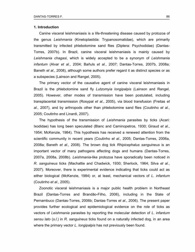
DANTAS-TORRES F.
86
1. Introduction
Canine visceral leishmaniasis is a life-threatening disease caused by protozoa of
the genus Leishmania (Kinetoplastida: Trypanosomatidae), which are primarily
transmitted by infected phlebotomine sand flies (Diptera: Psychodidae) (Dantas-
Torres, 2007b). In Brazil, canine visceral leishmaniasis is mainly caused by
Leishmania chagasi, which is widely accepted to be a synonym of Leishmania
infantum (Alvar et al., 2004; Bañuls et al., 2007; Dantas-Torres, 2007b, 2008a;
Baneth et al., 2008), although some authors prefer regard it as distinct species or as
a subspecies (Lainson and Rangel, 2005).
The primary vector of the causative agent of canine visceral leishmaniasis in
Brazil is the phlebotomine sand fly Lutzomyia longipalpis (Lainson and Rangel,
2005). However, other modes of transmission have been postulated, including
transplacental transmission (Rosypal et al., 2005), via blood transfusion (Freitas et
al., 2007), and by arthropods other than phlebotomine sand flies (Coutinho et al.,
2005; Coutinho and Linardi, 2007).
The hypothesis of the transmission of Leishmania parasites by ticks (Acari:
Ixodidae) has long been speculated (Blanc and Caminopetros, 1930; Giraud et al.,
1954; McKenzie, 1984). This hypothesis has received a renewed attention from the
scientific community in recent years (Coutinho et al., 2005; Dantas-Torres, 2006a,
2008a; Baneth et al., 2008). The brown dog tick Rhipicephalus sanguineus is an
important vector of many pathogens affecting dogs and humans (Dantas-Torres,
2007a, 2008a, 2008b). Leishmania-like protozoa have sporadically been noticed in
R. sanguineus ticks (Machattie and Chadwick, 1930; Sherlock, 1964; Silva et al.,
2007). Moreover, there is experimental evidence indicating that ticks could act as
either biological (McKenzie, 1984) or, at least, mechanical vectors of L. infantum
(Coutinho et al., 2005).
Zoonotic visceral leishmaniasis is a major public health problem in Northeast
Brazil (Dantas-Torres and Brandão-Filho, 2006), including in the State of
Pernambuco (Dantas-Torres, 2006b; Dantas-Torres et al., 2006). The present paper
provides further ecological and epidemiological evidence on the role of ticks as
vectors of Leishmania parasites by reporting the molecular detection of L. infantum
sensu lato (s.l.) in R. sanguineus ticks found on a naturally infected dog, in an area
where the primary vector L. longipalpis has not previously been found.

DANTAS-TORRES F.
87
2. Material and methods
The present study was carried out in the municipality of Vicência (07°39’25’’ S,
35°19’36’’ W), located in the Atlantic Rainforest of Pernambuco, Northeast Brazil,
where both cutaneous and visceral leishmaniases are endemic. Vicência is situated
87 km far from Recife, the capital of Pernambuco. This municipality has total land
area of about 250 km2 and an estimated population of about 27,360 inhabitants.
In March 2007, during a field work carried out in Vicência, an adult male mongrel
dog was found with clinical signs (i.e., apathy, severe loss of weight, ulcerated
cutaneous lesions, lymph node enlargement, and onychogryphosis) suggestive of
visceral leishmaniasis. On physical examination, ticks were noticed and promptly
collected. Ticks were kept in labeled vials containing 70% alcohol, until identification
using proper taxonomic keys (Aragão and Fonseca, 1961). After identification to
species level, ticks were either dissected or stored at –20°C for subsequent DNA
extraction and polymerase chain reaction (PCR) amplification.
DNA from ticks was extracted using either phenol-chloroform extraction or a
commercial kit that involves use of spin-column technology (Illustra tissue & cells
genomicPrep Mini Spin Kit, GE Healthcare, New York, USA). Extracted DNA was
stored at –20°C, until PCR amplification.
Extracted DNA was then assayed by a conventional PCR. We used a set of
primers (sense primer, 5′-CTTTTCTGGTCCCGCGGGTAGG-3′; antisense primer, 5′-
CCACCTGGCCTATTTTACACCA-3′) that amplifies a 145-base-pair sequence from
L. infantum kinetoplast DNA (Ravel et al., 1995). PCR was carried out in a 25 µl final
volume containing 14 µl of DNA-free water, 2.5 µl of 10x PCR buffer (Invitrogen), 1.5
µl of 1.5 mM MgCl2 (Invitrogen), 2.5 µl of 2 mM dNTP mix (Invitrogen), 0.5 µl of each
primer (25 pmol/µl), 0.5 µl of Platinum Taq DNA polymerase (5 U/µl) (Invitrogen), and
2 µl of extracted DNA. The amplification conditions were 94ºC/5-min initial
denaturation, followed by 35 cycles of 94ºC/30-sec denaturation, 67ºC/1-min
annealing, 72ºC/30-sec extension, and 72ºC/5-min final extension. A negative (DNA-
free water) and a positive control (5 ng/µl of DNA purified from promastigotes of L.
chagasi, strain MHOM/BR/74/PP75) were included in the PCR run. λ DNA/Hind III
fragments (Invitrogen) were used as a molecular weight marker. PCR products were
separated on a 1.5% agarose gel and the resulting bands were visualized under UV
illumination after ethidium bromide staining.

DANTAS-TORRES F.
88
3. Results and discussion
All ticks (8 males and 6 females) were identified as R. sanguineus. Two engorged
females were dissected, but no Leishmania-like forms were noticed. When tested by
PCR, four engorged females were positive.
Cases of natural infection of R. sanguineus ticks by Leishmania-like protozoa
have sporadically been reported in the literature (Machattie and Chadwick, 1930;
Sherlock, 1964; Silva et al., 2007). However, the finding of Leishmania-like forms in
dissected ticks should be evaluated carefully. Monogenetic trypanosomatids (e.g.,
Leptomonas, Crithidia, and Blastocrithidia) are known to infect ticks of several
species, including R. sanguineus (Machattie and Chadwick, 1930). These
nonpathogenic trypanosomatids can be easily confounded with and misidentified as
Leishmania parasites. For this reason, the use of contemporary molecular biology
techniques to accurately determine the true identify of protozoa infecting ticks is
imperative.
The infection of R. sanguineus ticks by Leishmania parasites is expected due to
their feeding habits. On the other hand, the finding of ticks infected by Leishmania
parasites does not ensure their vector competence, that is, their ability of
subsequently transmitting the infection to a naïve dog during blood feeding. For
instance, it has been experimentally demonstrated that R. sanguineus ticks can
become infected by Trypanosoma cruzi, but there is no evidence of subsequent
parasite development and transmission to naïve dogs (Dias et al., 2005).
There are few studies concerning the experimental transmission of Leishmania
parasites by ticks. The transstadial passage of L. infantum in R. sanguineus ticks has
been documented in the laboratory (Blanc and Caminopetros, 1930). It has also been
shown that L. infantum could survive for over 160 days in R. sanguineus ticks, as
confirmed by isolation in culture (McKenzie, 1984). More recently, it was
demonstrated that hamsters inoculated (either orally or intraperitonealy) with
macerated ticks developed signs of infection (Coutinho et al., 2005), suggesting that
Leishmania parasites could be transmitted to dogs via ingestion of infected tick.
The significance of the transmission of Leishmania parasites via ingestion of
infected ticks, in the epidemiology of canine visceral leishmaniasis in Brazil and
elsewhere, is uncertain. However, it is interesting to note that ingestion of infected
ticks is the primary mode of transmission of Hepatozoon canis, which is a highly
prevalent vector-borne pathogen infecting dogs in some rural areas in Brazil

DANTAS-TORRES F.
89
(O'Dwyer et al., 2001). It means that the importance of the transmission of L.
infantum to dogs through the ingestion of infected ticks should be further
investigated.
A strain originated from a bone marrow sample of the dog from Vicência was
successfully isolated and typed as Leishmania chagasi zymodeme IOC-1, which is
isoenzymatically indistinguishable from Leishmania infantum zymodeme MON-1
(data not shown). This is the commonest zymodeme involved in visceral
leishmaniasis in the Old World (Pratlong et al., 2004) and possibly in the New World
(Zerpa et al., 2001).
The present study reports for the first time the molecular detection of natural
infection of R. sanguineus ticks by L. infantum s.l. in a Brazilian area where the
primary vector L. longipalpis has not been found. Since 2004, we have conducted
systematic phlebotomine sand fly collections in Vicência and, at present, none of the
over 56,000 phlebotomine sand flies collected were identified as L. longipalpis (data
not shown). The epidemiological situation found in Vicência is similar to that found in
São Vicente Férrer, a neighboring municipality, where Leishmania-like forms have
been detected in R. sanguineus ticks (Silva et al., 2007) and the presence of L.
longipalpis has not been confirmed yet (Carvalho et al., 2007). These findings
reinforce the hypothesis of the participation of R. sanguineus ticks in the
epidemiology of canine visceral leishmaniasis, particularly in areas where the
disease has been diagnosed but the presence of the primary vector has not been
confirmed, as it is also the case of Recife (Dantas-Torres et al., 2005). Further
experimental studies to assess the competence of R. sanguineus ticks as vectors of
L. infantum s.l. are needed.
Acknowledgments
Thanks to Professor Domenico Otranto and to Luciana A. Figueredo for their
suggestions. The first author is supported by a PhD scholarship from the
Coordenação de Aperfeiçoamento de Pessoal de Nível Superior (CAPES). This
study was supported by the Conselho Nacional de Desenvolvimento Científico e
Tecnológico (CNPq) (grant 410481/2006-8) and the Fundação de Amparo à Ciência
e Tecnologia do Estado de Pernambuco (FACEPE) (grant APq-00522.02/07).

DANTAS-TORRES F.
90
References
Alvar, J., Canavate, C., Molina, R., Moreno, J., Nieto, J., 2004. Canine leishmaniasis.
Adv. Parasitol. 57, 1–88.
Aragão, H.B., Fonseca, F., 1961. Notas de Ixodologia. VIII. Lista e chave para
representantes da fauna Ixodológica Brasileira. Mem. Inst. Oswaldo Cruz 59, 115–
129.
Baneth, G., Koutinas, A.F., Solano-Gallego, L., Bourdeau, P., Ferrer, L., 2008.
Canine leishmaniosis - new concepts and insights on an expanding zoonosis: part
one. Trends Parasitol. 24, 324–330.
Bañuls, A.L., Hide, M., Prugnolle, F., 2007. Leishmania and the leishmaniases: a
parasite genetic update and advances in taxonomy, epidemiology and
pathogenicity in humans. Adv. Parasitol. 64, 1–109.
Blanc, G., Caminopetros, J., 1930. La transmission du Kala – Azar méditerranéen
par une tique: Rhipicephalus sanguineus. C. R. Acad. Sci. 191, 1162–1164.
Carvalho, M.R., Lima, B.S., Marinho-Júnior, J.F., Silva, F.J., Valença, H.F., Almeida,
F.A., Silva, A.L., Brandão-Filho, S.P., 2007. Phlebotomine sandfly species from an
American visceral leishmaniasis area in the Northern Rainforest region of
Pernambuco State, Brazil. Cad. Saude Publica 23, 1227–1232.
Coutinho, M.T., Bueno, L.L., Sterzik, A., Fujiwara, R.T., Botelho, J.R., Maria, M.,
Genaro, O., Linardi, P.M., 2005. Participation of Rhipicephalus sanguineus (Acari:
Ixodidae) in the epidemiology of canine visceral leishmaniasis. Vet. Parasitol. 128,
149–155.
Coutinho, M.T., Linardi, P.M., 2007. Can fleas from dogs infected with canine visceral
leishmaniasis transfer the infection to other mammals? Vet. Parasitol. 147, 320–
325.
Dantas-Torres, F., 2006a. Do any insects other than phlebotomine sandflies (Diptera:
Psychodidae) transmit Leishmania infantum (Kinetoplastida: Trypanosomatidae)
from dog to dog? Vet. Parasitol. 136, 379–380.
Dantas-Torres, F., 2006b. Situação atual da epidemiologia da leishmaniose visceral
em Pernambuco. Rev. Saude Publica 40, 537–541.
Dantas-Torres, F., 2007a. Rocky Mountain spotted fever. Lancet Infect. Dis. 7, 724–
732.

DANTAS-TORRES F.
91
Dantas-Torres, F., 2007b. The role of dogs as reservoirs of Leishmania parasites,
with emphasis on Leishmania (Leishmania) infantum and Leishmania (Viannia)
braziliensis. Vet. Parasitol. 149, 139–146.
Dantas-Torres, F., 2008a. Canine vector-borne diseases in Brazil. Parasit. Vectors 1,
25.
Dantas-Torres, F., 2008b. The brown dog tick, Rhipicephalus sanguineus (Latreille,
1806) (Acari: Ixodidae): from taxonomy to control. Vet. Parasitol. 152, 173–185.
Dantas-Torres, F., Brandão-Filho, S.P., 2006. Visceral leishmaniasis in Brazil:
revisiting paradigms of epidemiology and control. Rev. Inst. Med. Trop. Sao Paulo
48, 151–156.
Dantas-Torres, F., Brito, M.E.F., Brandão-Filho, S.P., 2006. Seroepidemiological
survey on canine leishmaniasis among dogs from an urban area of Brazil. Vet.
Parasitol. 140, 54–60.
Dantas-Torres, F., Faustino, M.A.G., Lima, O.C., Acioli, R.V., 2005. Epidemiologic
surveillance of canine visceral leishmaniasis in the municipality of Recife,
Pernambuco. Rev. Soc. Bras. Med. Trop. 38, 444–445.
Dias, J.C., Schofield, C.J., Machado, E.M., Fernandes, A.J., 2005. Ticks, ivermectin,
and experimental Chagas disease. Mem. Inst. Oswaldo Cruz 100, 829–832.
Freitas, E., Melo, M.N., Costa-Val, A.P., Michalick, M.S., 2006. Transmission of
Leishmania infantum via blood transfusion in dogs: potential for infection and
importance of clinical factors. Vet. Parasitol. 137, 159–167.
Giraud, P., Ranque, J., Cabassu, H., 1954. Epidemiologie de la leishmaniose
viscérale humaine mediterranéenne, en particulier dans sés rapports avec la
leishmaniose canine. Arch. Fr. Ped. 11, 337–353.
Lainson, R., Rangel, E.F., 2005. Lutzomyia longipalpis and the eco-epidemiology of
American visceral leishmaniasis, with particular reference to Brazil: a review.
Mem. Inst. Oswaldo Cruz. 100, 811–827.
Machattie, C., Chadwick, C.R., 1930. Notes on a trypanosome infection of the dog
tick Rhipicephalus sanguineus in Iraq. Trans. R. Soc. Trop. Med. Hyg. 23, 417-
420.
McKenzie, K.K., 1984. A study of the transmission of canine leishmaniasis by the
tick, Rhipicephalus sanguineus, and an ultrastructural comparison of the
promastigote. PhD Dissertation, Oklahoma State University, Oklahoma, United
States.

DANTAS-TORRES F.
92
O'Dwyer, L.H., Massard, C.L., Pereira de Souza, J.C., 2001. Hepatozoon canis
infection associated with dog ticks of rural areas of Rio de Janeiro State, Brazil.
Vet. Parasitol. 94, 143–150.
Pratlong, F., Rioux, J.A., Marty, P., Faraut-Gambarelli, F., Dereure, J., Lanotte, G.,
Dedet, J.P., 2004. Isoenzymatic analysis of 712 strains of Leishmania infantum in
the south of France and relationship of enzymatic polymorphism to clinical and
epidemiological features. J. Clin. Microbiol. 42, 4077–4082.
Ravel, S., Cuny, G., Reynes, J., Veas, F., 1995. A highly sensitive and rapid
procedure for direct PCR detection of Leishmania infantum within human
peripheral blood mononuclear cells. Acta Trop. 59, 187–196.
Rosypal, A.C., Troy, G.C., Zajac, A.M., Frank, G., Lindsay, D.S., 2005.
Transplacental transmission of a North American isolate of Leishmania infantum in
an experimentally infected beagle. J. Parasitol. 91, 970–972.
Sherlock, I., 1964. Notas sôbre a transmissão da leishmaniose visceral no Brasil.
Rev. Bras. Malariol. D. Trop. 16, 19–26.
Silva, O.A., Silva, P.B., Silva, O.V., Braga, G.M., Albuquerque Júnior, A., Queiros
Neto, V., Rocha, M.E., Silva, E.F., 2007. La leishmaniose viscérale canine dans le
Nord-Est du Brésil: aspects épidémiologiques. Bull. Soc. Pathol. Exot. 100, 49–50.
Zerpa, O., Pratlong, F., Ulrich, M., Convit, J., 2001. Isolation of Leishmania infantum,
zymodeme MON-1 from canine and human visceral leishmaniasis on Margarita
Island, Venezuela. Mem. Inst. Oswaldo Cruz. 96, 901–902.
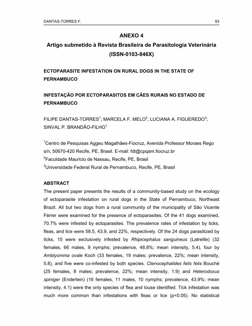
DANTAS-TORRES F.
93
ANEXO 4
Artigo submetido à Revista Brasileira de Parasitologia Veterinária
(ISSN-0103-846X)
ECTOPARASITE INFESTATION ON RURAL DOGS IN THE STATE OF
PERNAMBUCO
INFESTAÇÃO POR ECTOPARASITOS EM CÃES RURAIS NO ESTADO DE
PERNAMBUCO
FILIPE DANTAS-TORRES1; MARCELA F. MELO2, LUCIANA A. FIGUEREDO3;
SINVAL P. BRANDÃO-FILHO1
1Centro de Pesquisas Aggeu Magalhães-Fiocruz, Avenida Professor Moraes Rego
s/n, 50670-420 Recife, PE, Brasil. E-mail: [email protected] 2Faculdade Mauricio de Nassau, Recife, PE, Brasil 3Universidade Federal Rural de Pernambuco, Recife, PE, Brasil
ABSTRACT
The present paper presents the results of a community-based study on the ecology
of ectoparasite infestation on rural dogs in the State of Pernambuco, Northeast
Brazil. All but two dogs from a rural community of the municipality of São Vicente
Férrer were examined for the presence of ectoparasites. Of the 41 dogs examined,
70.7% were infested by ectoparasites. The prevalence rates of infestation by ticks,
fleas, and lice were 58.5, 43.9, and 22%, respectively. Of the 24 dogs parasitized by
ticks, 15 were exclusively infested by Rhipicephalus sanguineus (Latreille) (32
females, 66 males, 9 nymphs; prevalence, 48.8%; mean intensity, 5.4), four by
Amblyomma ovale Koch (33 females, 19 males; prevalence, 22%; mean intensity,
5.8), and five were co-infested by both species. Ctenocephalides felis felis Bouché
(25 females, 8 males; prevalence, 22%; mean intensity, 1.9) and Heterodoxus
spiniger (Enderlein) (16 females, 11 males, 10 nymphs; prevalence, 43.9%; mean
intensity, 4.1) were the only species of flea and louse identified. Tick infestation was
much more common than infestations with fleas or lice (p<0.05). No statistical

DANTAS-TORRES F.
94
difference was found in relation to sex and age group of the dogs and the prevalence
of ectoparasite infestation (p>0.05). Overall, 48.8% (20/41) of the dogs were infested
by more than one ectoparasite species.
KEY WORDS: Ixodida, Siphonaptera, Phthiraptera, dog, Brazil.
RESUMO
O presente artigo apresenta os resultados de um estudo de base comunitária sobre
a infestação por ectoparasitos em cães rurais no Estado de Pernambuco, Nordeste
do Brasil. Com exceção de dois, todos os cães de uma comunidade rural do
município de São Vicente Férrer foram examinados para presença de ectoparasitos.
Dos 41 cães examinados, 70,7% estavam infestados por ectoparasitos. As taxas de
prevalência de infestação por carrapatos, pulgas e piolhos foram 58,5, 43,9 e 22%,
respectivamente. Dos 24 cães parasitados por carrapatos, 15 estavam
exclusivamente infestados por Rhipicephalus sanguineus (Latreille) (32 fêmeas, 66
machos, 9 ninfas; prevalência, 48,8%; intensidade média, 5,4), quatro por
Amblyomma ovale Koch (33 fêmeas, 19 machos; prevalência, 22%; intensidade
média, 5,8), e cinco estavam co-infestados por ambas as espécies. Ctenocephalides
felis felis Bouché (25 fêmeas, 8 machos; prevalência, 22%; intensidade média, 1,9) e
Heterodoxus spiniger (Enderlein) (16 fêmeas, 11 machos, 10 ninfas; prevalência,
43,9%; intensidade média, 4,1) foram a única espécie de pulga e piolho
identificadas. A infestação por carrapatos foi muito mais freqüente que as
infestações por pulgas ou piolhos (p<0.05). Não houve diferença estatística em
relação ao sexo e grupo etário dos cães e a prevalência de infestação por
ectoparasitos (p>0.05). No geral, 48,8% (20/41) dos cães estavam infestados por
mais de uma espécie de ectoparasito.
PALAVRAS-CHAVE: Ixodida, Siphonaptera, Phthiraptera, cão, Brasil.
INTRODUCTION
Ectoparasites are known to infest dogs, wherever they are in the world. The main
types or groups of ectoparasites infesting dogs are ticks, fleas, lice, mites, and flies.
Among these, ticks are the most important both in terms of disease transmission and
exsanguination. Ticks are also notable, because they are well adapted to live in a

DANTAS-TORRES F.
95
great variety of environmental conditions, being also capable of survive for long
periods of time without feeding (DANTAS-TORRES, 2008a).
Some ectoparasites associated with dogs are important vectors of disease agents
of both veterinary and medical significance. For instance, the brown dog tick
Rhipicephalus sanguineus (Latreille) is known to transmit several pathogens,
including Rickettsia rickettsii (Wolbach), Rickettsia conorii Brumpt, Ehrlichia canis
(Donatien & Lestoquard), Babesia vogeli Reichenow (DANTAS-TORRES;
FIGUEREDO 2006; DANTAS-TORRES, 2007, 2008a). Cases of human parasitism
by Rh. sanguineus have been reported in South American countries, including in
Brazil (DANTAS-TORRES et al., 2006; GUGLIELMONE et al., 2006), and its role in
the transmission of pathogens to humans in this country needs to be further
investigated.
Due to their veterinary and medical significance, ectoparasites infesting dogs
have been subject of a number of studies in Brazil (LINARDI; NAGEM, 1973;
RIBEIRO et al., 1997; RODRIGUES et al., 2001; BELLATO et al., 2003; DANTAS-
TORRES et al., 2004; CASTRO; RAFAEL, 2006). Despite of this, the knowledge
about the ecology of ectoparasite infestation on dogs from rural areas in some
Brazilian regions is quite limited. The present paper presents the results of a
community-based study on the ecology of ectoparasite infestation on rural dogs in
the State of Pernambuco, Northeast Brazil.
MATERIAL AND METHODS
The present study was carried on 2–6 June 2008, in a rural community called
Mundo Novo (which in Portuguese means “New World”), which is located in the
municipality of São Vicente Férrer (07º35’28’’ S, 35º29’29’’ W, altitude 419 m). This
municipality belongs to the Agreste region of Pernambuco, but its environmental
features are similar to that found in the Atlantic rainforest region.
All but two dogs living in the aforementioned rural community were physically
examined for the presence of ectoparasites. The two dogs that were not enrolled in
this study run away before immobilization and examination. No control measures
against canine ectoparasites were being used in the community around the time of
the survey.
After manual collection, the ectoparasites were maintained in vials containing
70% alcohol. The identification of ticks was carried out under stereomicroscope,
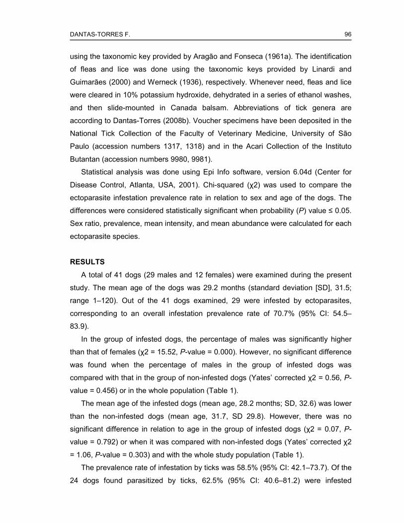
DANTAS-TORRES F.
96
using the taxonomic key provided by Aragão and Fonseca (1961a). The identification
of fleas and lice was done using the taxonomic keys provided by Linardi and
Guimarães (2000) and Werneck (1936), respectively. Whenever need, fleas and lice
were cleared in 10% potassium hydroxide, dehydrated in a series of ethanol washes,
and then slide-mounted in Canada balsam. Abbreviations of tick genera are
according to Dantas-Torres (2008b). Voucher specimens have been deposited in the
National Tick Collection of the Faculty of Veterinary Medicine, University of São
Paulo (accession numbers 1317, 1318) and in the Acari Collection of the Instituto
Butantan (accession numbers 9980, 9981).
Statistical analysis was done using Epi Info software, version 6.04d (Center for
Disease Control, Atlanta, USA, 2001). Chi-squared (χ2) was used to compare the
ectoparasite infestation prevalence rate in relation to sex and age of the dogs. The
differences were considered statistically significant when probability (P) value ≤ 0.05.
Sex ratio, prevalence, mean intensity, and mean abundance were calculated for each
ectoparasite species.
RESULTS
A total of 41 dogs (29 males and 12 females) were examined during the present
study. The mean age of the dogs was 29.2 months (standard deviation [SD], 31.5;
range 1–120). Out of the 41 dogs examined, 29 were infested by ectoparasites,
corresponding to an overall infestation prevalence rate of 70.7% (95% CI: 54.5–
83.9).
In the group of infested dogs, the percentage of males was significantly higher
than that of females (χ2 = 15.52, P-value = 0.000). However, no significant difference
was found when the percentage of males in the group of infested dogs was
compared with that in the group of non-infested dogs (Yates’ corrected χ2 = 0.56, P-
value = 0.456) or in the whole population (Table 1).
The mean age of the infested dogs (mean age, 28.2 months; SD, 32.6) was lower
than the non-infested dogs (mean age, 31.7, SD 29.8). However, there was no
significant difference in relation to age in the group of infested dogs (χ2 = 0.07, P-
value = 0.792) or when it was compared with non-infested dogs (Yates’ corrected χ2
= 1.06, P-value = 0.303) and with the whole study population (Table 1).
The prevalence rate of infestation by ticks was 58.5% (95% CI: 42.1–73.7). Of the
24 dogs found parasitized by ticks, 62.5% (95% CI: 40.6–81.2) were infested

DANTAS-TORRES F.
97
exclusively by Rh. sanguineus, 16.7% (95% CI: 4.7–37.4) by Amblyomma ovale
Koch, and 20.8% (95% CI: 7.1–42.1) were co-infested by both species.
Ctenocephalides felis felis (Bouché) and Heterodoxus spiniger (Enderlein) were the
only species of flea and louse identified. Number of examined specimens, sex ratio,
prevalence, mean intensity, and mean abundance for each ectoparasite species is
shown in Table 2.
Out of the 29 dogs infested by ectoparasites, 20 (68.9%; 95% CI: 49.2–84.7)
were infested by more than one ectoparasite species, which corresponds to an
overall co-infestation prevalence rate of 48.8% (95% CI: 32.9–64.9). The following
ectoparasite associations (number of observations are in brackets) were found: Rh.
sanguineus + C. felis felis (n=7); Rh. sanguineus + Am. ovale (n=3); Rh. sanguineus
+ C. felis felis + H. spiniger (n=3); C. felis felis + H. spiniger (n=2); C. felis felis + Am.
ovale (n=1); Rh. sanguineus + H. spiniger (n=1); C. felis felis + H. spiniger + Am.
ovale (n=1); Rh. sanguineus + C. felis felis + Am. ovale (n=1); Rh. sanguineus + C.
felis felis + H. spiniger + Am. ovale (n=1). Prevalence of concurrent infestations
involving two or more types of ectoparasites is shown in Figure 1.
DISCUSSION
This study provides new, interesting data about the ecology of ectoparasite
infestation on rural dogs in Northeast Brazil. The prevalence rate, sex ratio, mean
intensity, and mean abundance for each species of ectoparasite identified during the
present study may vary widely from region to region and also according to the
characteristics of the study population (e.g., rural versus urban dogs) (LINARD;
NAGEM, 1973; RODRIGUES et al., 2001; CASTRO; RAFAEL, 2006; SOARES et al.,
2006). For instance, in a study conducted in rural farms in North Brazil, Rh.
sanguineus were not found and the mean intensity of infestation by Am. ovale was
about 1.3 (i.e., eight ticks on six dogs) (LABRUNA et al., 2000). In the present study,
Rh. sanguineus was not only identified but was also the most common ectoparasite
found on rural dogs. Moreover, the mean intensity of infestation by Am. ovale was
5.8 (i.e., 52 ticks on nine dogs), which is over four-fold higher than that of North
Brazil. These differences were really surprising. Amblyomma ovale is typical of the
Atlantic Rainforest region and it would be expected that it should be more common in
Eastern Brazilian Amazon rather than in Agreste of Pernambuco. Further studies to
DANTAS-TORRES F.
98
assess the seasonality of Am. ovale in both regions would be welcome to clarify this
finding.
The percentage of males was much higher than that of females in the group of
infested dogs, this difference was not statistically significant and can be attributed to
characteristics of the studied population, which was predominately composed by
males. Only three out of 13 young dogs examined were not infested by
ectoparasites, possibly suggesting that young dogs living in the studied rural
community are more susceptible to and/or more exposed to ectoparasites. However,
we cannot conclude this because no significant difference was found in relation to
age.
Similar to what was found in a previous study conducted in an urban area in
Pernambuco (DANTAS-TORRES et al., 2004), Rh. sanguineus was the most
common ectoparasite associated with rural dogs. But in contrast to what was found in
the urban area, rural dogs were infested by Am. ovale in addition to Rh. sanguineus
ticks. This study revealed that Am. ovale is a common ectoparasite on dogs from the
studied area. This tick is a common parasite of wild carnivores and rural dogs
(ARAGÃO; FONSECA 1961b; LABRUNA et al., 2000) and has been implicated in the
transmission of Hepatozoon canis (James) in Brazil (FORLANO et al., 2005). Not
less important, Am. ovale is known to infest humans (LABRUNA et al., 2005).
The present study showed that the prevalence of infestation by H. spiniger
appears to be much higher on rural dogs when compared to urban ones (DANTAS-
TORRES et al., 2004; DANTAS-TORRES; FIGUEREDO, 2007). Worthy of note,
Trichodectes canis (Degeer), a common dog louse, was not identified in this study.
The reason for this is unknown. One of us (F.D-T) has collected some specimens of
T. canis on a dog from a neighboring rural community, located in the municipality of
Vicência. Further collections are advocated to access whether T. canis is present in
São Vicente Férrer and in what extent.
This study also revealed a high prevalence (i.e., 43.9%) of infestation by C. felis
felis on rural dogs from Pernambuco. In a previous study carried out in the
metropolitan region of Recife, the prevalence of infestation by C. felis felis was
around 5% (DANTAS-TORRES et al., 2004). In part, the high prevalence of
ectoparasite infestation on dogs from rural areas of Pernambuco is expected,
because most people living in these remote areas cannot afford the costs of
ectoparasite control.
DANTAS-TORRES F.
99
The high prevalence of ectoparasite infestation in the studied rural community
indicates that the dogs are potentially exposed to several vector-borne pathogens,
including B. vogeli, E. canis, and H. canis. It is worthy of note that canine visceral
leishmaniasis is endemic in the municipality of São Vicente Férrer, but the primary
vector Lutzomyia longipalpis is absent (CARVALHO et al., 2007). Interestingly,
Leishmania-like forms have been noticed in Rh. sanguineus ticks collected in this
municipality (SILVA et al., 2007), which suggests the possible participation of ticks in
the epidemiology of canine visceral leishmaniasis in this area. Further studies to
access the prevalence of vector-borne pathogens of zoonotic concern in dogs from
São Vicente Férrer are needed.
Acknowledgments:- We thank Fernando J. da Silva and Amilton Lopes for their
help during tick collection. The first author is supported by a PhD scholarship from
the Coordenação de Aperfeiçoamento de Pessoal de Nível Superior (CAPES).
REFERENCES
ARAGÃO, H.; FONSECA, F. Notas de Ixodologia VIII. Lista e chave para
representantes da fauna Ixodológica Brasileira. Memórias do Instituto Oswaldo
Cruz, v. 59, n. 2, p. 115-129, 1961a.
ARAGÃO, H.; FONSECA, F. Notas de Ixodologia. IX. O complexo Ovale do gênero
Amblyomma. Memórias do Instituto Oswaldo Cruz, v. 59, n. 2, p. 131–148,
1961b.
BELLATO, V.; SARTOR, A.A.; SOUZA, A.P.; RAMOS, B.C. Ectoparasitos em
caninos do município de Lages, Santa Catarina, Brasil. Revista Brasileira de
Parasitologia Veterinária, v. 12, n. 3, p. 95–98, 2003.
CARVALHO, M.R.; LIMA, B.S.; MARINHO-JÚNIOR, J.F.; SILVA, F.J.; VALENÇA,
H.F.; ALMEIDA, F.A.; SILVA, A.L.; BRANDÃO-FILHO, S.P. Phlebotomine sandfly
species from an American visceral leishmaniasis area in the Northern Rainforest
region of Pernambuco State, Brazil. Cadernos de Saúde Pública, v. 23, p. 1227–
1232, 2007.
CASTRO, M.C.M.; RAFAEL, J.A. Ectoparasitos de cães e gatos da cidade de
Manaus, Amazonas, Brasil. Acta Amazonica, v. 36, n. 4, p. 535–538, 2006.
DANTAS-TORRES, F. Rocky Mountain spotted fever. Lancet Infectious Diseases, v.
7, n. 11, p. 724-732, 2007.

DANTAS-TORRES F.
100
DANTAS-TORRES, F. The brown dog tick, Rhipicephalus sanguineus (Latreille,
1806) (Acari: Ixodidae): from taxonomy to control. Veterinary Parasitology, v. 152,
n. 3/4, p. 173-185, 2008a.
DANTAS-TORRES, F. Towards the standardization of the abbreviations of genus
names of ticks (Acari: Parasitiformes: Ixodida). Veterinary Parasitology, v. 154, n.
1/2, p. 94-97, 2008b.
DANTAS-TORRES, F.; FIGUEREDO, L.A. Canine babesiosis: A Brazilian
perspective. Veterinary Parasitology, v. 141, n. 3-4, p. 197-203, 2006.
DANTAS-TORRES, F.; FIGUEREDO, L.A. Heterodoxus spiniger (Enderlein, 1909)
on domestic dogs (Canis familiaris, L. 1758) from the city of Recife, Pernambuco
State, Brazil. Brazilian Journal of Veterinary Research and Animal Science, v. 44,
n. 2, p. 77–80, 2007.
DANTAS-TORRES, F.; FIGUEREDO, L.A.; BRANDÃO-FILHO, S.P. Rhipicephalus
sanguineus (Acari: Ixodidae), the brown dog tick, parasitizing humans in Brazil.
Revista da Sociedade Brasileira de Medicina Tropical, v. 39, n. 1, p. 64-67, 2006.
DANTAS-TORRES, F.; FIGUEREDO, L.A.; FAUSTINO, M.A.G. Ectoparasitos de
cães provenientes de alguns municípios da região metropolitana do Recife,
Pernambuco, Brasil. Revista Brasileira de Parasitologia Veterinária, v. 13, n. 4, p.
151-154, 2004.
FORLANO, M.; SCOFIELD, A.; ELISEI, C.; FERNANDES, K.R.; EWING, S.A.;
MASSARD, C.L. Diagnosis of Hepatozoon spp. in Amblyomma ovale and its
experimental transmission in domestic dogs in Brazil. Veterinary Parasitology, v.
134, n. 1-2, p. 1–7, 2005.
GUGLIELMONE, A.A.; BEATI, L.; BARROS-BATTESTI, D.M.; LABRUNA, M.B.;
NAVA, S.; VENZAL, J.M.; MANGOLD, A.J.; SZABÓ, M.P.; MARTINS, J.R.;
GONZÁLEZ-ACUÑA, D.; ESTRADA-PEÑA, A. Ticks (Ixodidae) on humans in
South America. Experimental and Applied Acarology, v. 40, n. 2, p. 83-100, 2006.
GUIMARÃES, J.H.; TUCCI, E.C.; BARROS-BATTESTI, D.M. Ectoparasitos de
importância veterinária. São Paulo: Editora Plêiade/FAPESP, 2001. 218p.
LABRUNA, M.B.; HOMEM, V.S.; HEINEMANN, M.B.; FERREIRA NETO, J.S. Ticks
(Acari: Ixodidae) associated with rural dogs in Uruará, eastern Amazon, Brazil.
Journal of Medical Entomology, v. 37, n. 5, p. 774-776, 2000.
LABRUNA, M.B.; CAMARGO, L.M.A.; TERRASSINI, F.A.; FERREIRA, F.;
SCHUMAKER, T.T.S.; CAMARGO, E.P. Ticks (Acari: Ixodidae) from the State of

DANTAS-TORRES F.
101
Rondônia, western Amazon, Brazil. Systematic and Applied Acarology, v. 10, n.
1, p. 17-32, 2005.
LINARDI, P.M.; GUIMARÃES, L.R. Sifonápteros do Brasil. Editora do Museu de
Zoologia da Universidade de São Paulo, São Paulo, 2000. 291p.
LINARDI, P.M.; NAGEM, R.L. Pulicídeos e outros ectoparasitos de cães de Belo
Horizonte e municípios vizinhos. Revista Brasileira de Biologia, v. 33, n. 4, p.
529–538, 1973.
RIBEIRO, V.L.S.; WEBER, M.A.; FETZER, L.O.; VARGAS, C.R.B. Espécies e
prevalência das infestações por carrapatos em cães de rua de Porto Alegre, RS,
Brasil. Ciência Rural, v. 27, n. 2, p. 285–289, 1997.
RODRIGUES, A.F.S.F., DAEMON, E.; D’AGOSTO, M. Investigação sobre alguns
ectoparasitos em cães de rua no município de Juiz de Fora, Minas Gerais.
Revista Brasileira de Parasitologia Veterinária, v. 10, n. 1, p. 13–19, 2001.
SILVA, O.A.; SILVA, P.B.; SILVA, O.V.; BRAGA, G.M.; ALBUQUERQUE JÚNIOR,
A.; QUEIROS NETO, V.; ROCHA, M.E.; SILVA, E.F. La leishmaniose viscérale
canine dans le Nord-Est du Brésil: aspects épidémiologiques. Bulletin de la
Société de Pathologie Exotique, v. 100, n. 1, p. 49–50, 2007.
SOARES, A.O.; SOUZA, A.D.; FELICIANO, E.A.; RODRIGUES, A.F.S.F.;
D’AGOSTO, M.; DAEMON, E. Avaliação ectoparasitológica e hemoparasitológica
em cães criados em apartamentos e casas com quintal na cidade de Juiz de
Fora, MG. Revista Brasileira de Parasitologia Veterinária, v. 15, n. 1, p. 13–16,
2006.
WERNECK, F.L. Contribuição ao conhecimento dos Mallophagos encontrados nos
mammiferos sulamericanos. Memórias do Instituto Oswaldo Cruz, v. 31, n. 3, p.
391–589, 1936.

DANTAS-TORRES F.
102
0
5
10
15
20
25
30
Ticks+fleas Ticks+lice Fleas+lice Ticks+fleas+lice
Prevalence
(%) ofectoparasite associations
Figure 1. Prevalence of associations by two or three types of ectoparasites on dogs
from a rural community located in Northeast Brazil.
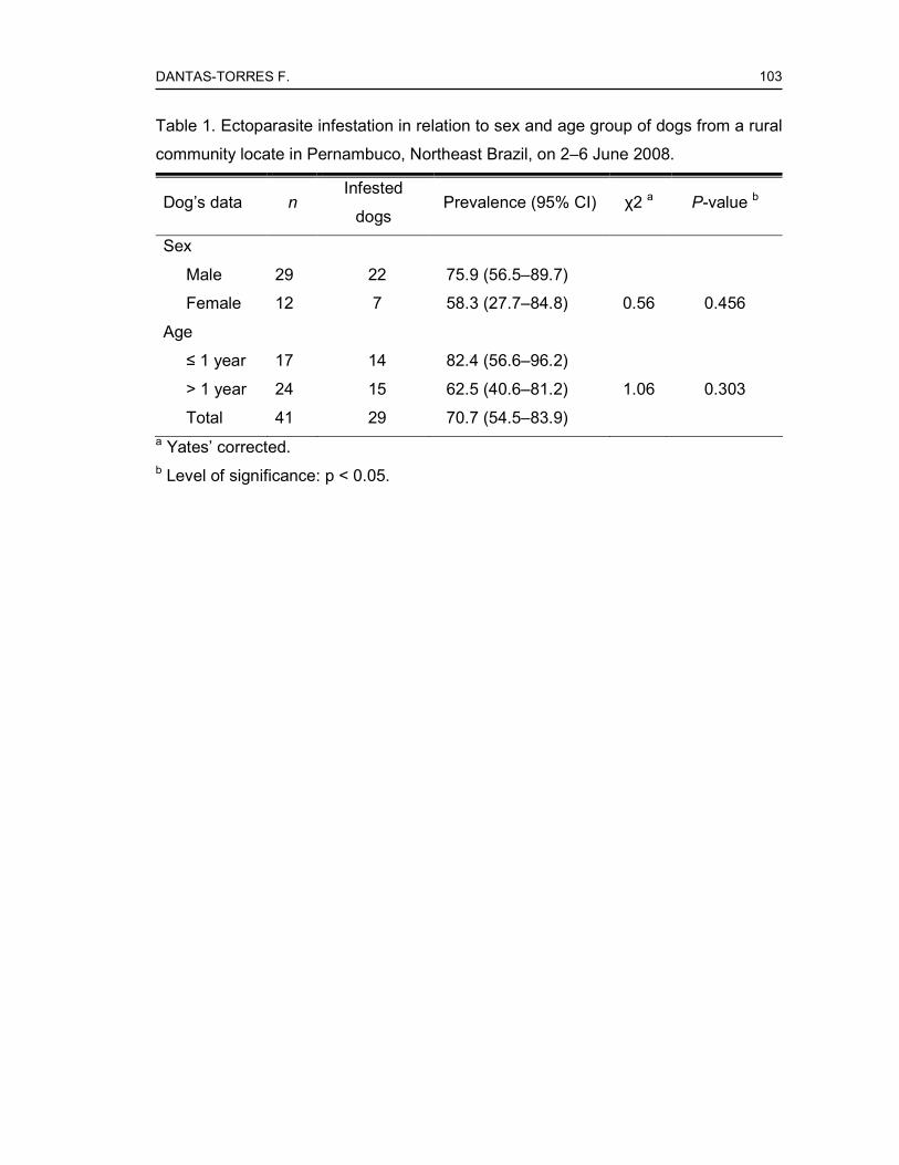
DANTAS-TORRES F.
103
Table 1. Ectoparasite infestation in relation to sex and age group of dogs from a rural
community locate in Pernambuco, Northeast Brazil, on 2–6 June 2008.
Dog’s data n Infested
dogs Prevalence (95% CI) χ2 a P-value b
Sex
Male 29 22 75.9 (56.5–89.7)
Female 12 7 58.3 (27.7–84.8) 0.56 0.456
Age
≤ 1 year 17 14 82.4 (56.6–96.2)
> 1 year 24 15 62.5 (40.6–81.2) 1.06 0.303
Total 41 29 70.7 (54.5–83.9) a Yates’ corrected. b Level of significance: p < 0.05.

DA
NT
AS
-TO
RR
ES
F.
104
Tab
le 2
. Ect
opar
asite
s (n
=2
29)
foun
d o
n do
gs (
n=2
9) f
rom
a r
ural
com
mun
ity lo
cate
d in
Per
nam
buc
o, N
ort
he
ast
Bra
zil,
on 2
–6
Jun
e
2008
.
Ect
opar
asite
sp
ecie
s ♀
♂
N
ymph
s S
ex r
atio
(♀:♂
)
Infe
sted
dogs
Pre
vale
nce
(95%
CI)
Mea
n
inte
nsity
a ±
SD
Me
an
abun
dan
ce b
±
SD
Rhipicephalus sanguineus
32
66
9 1:
2.1
20
48.8
(3
2.9–
64.9
) 5.
4 ±
4.5
2.6
± 4.
1
Heterodoxus spiniger
16
11
10
1.5:
1 9
22.0
(1
0.6–
37.6
) 4.
1 ±
3.4
0.9
± 2.
3
Amblyomma ovale
33
19
0 1.
7:1
9 22
.0 (
10.
6–37
.6)
5.8
± 5.
7 1.
3 ±
3.5
Ctenocephalides felis felis
25
8 0
3.1:
1 18
43
.9 (
28.
5–60
.3)
1.9
± 1.
3 0.
8 ±
1.3
a Num
ber
of e
ctop
aras
ites
per
num
ber
of in
fest
ed d
ogs.
b
Num
ber
of e
ctop
aras
ites
per
num
ber
of e
xam
ined
do
gs.

DANTAS-TORRES F.
105
ANEXO 5
Artigo em fase de elaboração
EPIDEMIOLOGY OF CANINE VISCERAL LEISHMANIOSIS IN A BRAZILIAN
AREA WHERE LUTZOMYIA LONGIPALPIS HAS NOT BEEN FOUND
Filipe Dantas-Torres1,2, Milena Paiva Cavalcanti1, Luciana A. Figueredo2, Marcela F.
Melo1, Maria Edileuza F. de Brito1, Sinval Pinto Brandão-Filho1
1 Department of Immunology, Centro de Pesquisas Aggeu Magalhães, Fundação
Oswaldo Cruz, 50670-420, Recife (Pernambuco), Brazil 2 Department of Veterinary Public Health, Faculty of Veterinary Medicine, University
of Bari, 70010, Valenzano (Bari), Italy
* Corresponding author. Tel.: +55 81 21012562; fax: +55 81 21012640.
E-mail address: [email protected] (F. Dantas-Torres).
Abstract
The present paper reports the results of an epidemiological study carried out in a
rural area in Northeast Brazil, where the presence of Lutzomyia longipalpis is
uncertain. All but two dogs from a rural community (namely Mundo Novo) were
screened by an indirect immunofluorescence antibody test for the presence of anti-
Leishmania antibodies and 12 (29.3%) of them were positive. In contrast, only one
dog was positive for L. (L.) infantum kDNA by real time PCR in blood. Only one dog
was positive for Leishmania amastigotes in bone marrow-stained smears, while only
one of them was positive for L. (L.) infantum kDNA by PCR in bone marrow. There
was no significant difference in seropositivity in relation to age, sex and clinical status
of the dogs. While there was no significant difference between the seropositivity and
the presence of lice or fleas, the majority of the seropositive dogs were not infested
by ticks (p-value ≤ 0.05). Additionally, we tested 73 Rhipicephalus sanguineus ticks
collected from seropositive dogs living in the municipalities of Tamandaré and

DANTAS-TORRES F.
106
Bezerros and nine (12.3%) of them were positive by real time PCR, with a parasite
load raging from 0.001 to 0.035. The present results provide additional evidence
supporting the hypothesis of the participation of Rh. sanguineus ticks in the
epidemiology of canine visceral leishmaniosis in Brazil, particularly where Lu.
longipalpis has not been found.
Keywords: Dogs; Leishmania; Ticks; Brazil.
1. Introduction
Canine leishmaniosis is caused by a range of Leishmania parasites, which are
primarily transmitted by phlebotomine sand flies (Dantas-Torres, 2007). The most
widely spread etiological agent of canine leishmaniosis is Leishmania infantum (syn.
Leishmania chagasi), which can cause a severe, life-threatening disease in dogs and
also in humans (Chappuis et al., 2007; Baneth et al., 2008). In Brazil, canine visceral
leishmaniasis is widely spread (Dantas-Torres, 2008) and dogs are implicated as the
main reservoir hosts in the peridomestic cycle of transmission of L. infantum (Dantas-
Torres and Brandão-Filho, 2006b; Dantas-Torres, 2007). The primary vector of L.
infantum in South America is Lutzomyia longipalpis, but other phlebotomine sand fly
species have also been suspected to act as secondary vectors (Lainson and Rangel,
2005; Carvalho et al., 2008; Pita-Pereira et al., 2008).
In the State of Pernambuco, Northeast Brazil, visceral leishmaniosis is a highly
prevalent disease (Dantas-Torres, 2006; Dantas-Torres and Brandão-Filho, 2006a).
The prevalence of infection by Leishmania spp. in dogs, as estimated by serological
tests, might reach over 51% in highly endemic areas, where the majority of the
animals are asymptomatic (Dantas-Torres et al., 2006). Although Lu. longipalpis is
usually present in areas where canine visceral leishmaniosis is endemic, there are
some foci where its presence has not been demonstrated (Dantas-Torres et al.,
2005; Carvalho et al., 2007). In some of these areas, ticks have been suspected to
be involved in transmission (Silva et al., 2007; Dantas-Torres et al., 2008). The
objective of the present study was to assess the epidemiology of canine visceral
leishmaniosis in a rural community of Pernambuco, where the presence of Lu.
longipalpis is uncertain.

DANTAS-TORRES F.
107
2. Material and methods
2.1 Study area
São Vicente Férrer (07º35’28’’ S, 35º29’29’’ W) is a municipality located in the
Agreste region of the State of Pernambuco, Northeast Brazil, where human and
canine leishmaniases are endemic. It has a population of about 25,000 inhabitants
and a territory of approximately 120 km2. Although located in the Agreste region, São
Vicente Férrer has vegetation coverage and climatic features typical of the Atlantic
rainforest region. The climate is tropical, with a dry season occurring from September
to February and a raining season from March to August. The primary vegetation has
been substituted by banana plantations, but there are some remnants of Atlantic
rainforest, where phlebotomine sand flies are abundant (Carvalho et al., 2007).
2.2 Clinical examination and sample collection
On June 2008, all (n=41) but two dogs from a rural community (namely Mundo
Novo) were evaluated for the presence of clinical signs (e.g. weight loss, dermatitis,
hair loss, mouth and skin ulcers, enlarged lymph nodes, onychogryphosis, and
conjunctivitis) suggestive of leishmaniosis. An individual questionnaire was used to
compile all clinical and epidemiological data from each dog. Then, 3–5 ml EDTA-
treated blood samples were collected by cephalic vein puncture. In the laboratory, an
aliquot of 1 ml was separated for subsequent DNA extraction and the remnant
material was centrifuged at 1500 x g for five minutes to separate the plasma. Then,
the obtained plasma was frozen at –20°C until tested for anti-Leishmania IgG
antibodies. Additionally, bone marrow aspirates were collected from 30 dogs for
preparation of stained smears and DNA extraction. The present study (P.0174-03)
was approved by the Ethical Committee of Animal Use of the Oswaldo Cruz
Foundation.
2.3 Serological diagnosis
Plasma samples were assayed by two commercial serological tests, an
immunofluorescence antibody test (IFAT) (IFI-Leishmaniose-Visceral-Canina, Bio-
Manguinhos, Rio de Janeiro, Brazil) and a dipstick test (Kalazar Detect, InBios
International, Seattle, USA). All assays were performed according to manufactures’
instructions.

DANTAS-TORRES F.
108
2.4 Parasitological and molecular diagnosis
Stained smears were prepared from bone marrow aspirates and examined for the
presence of Leishmania amastigotes under optic microscopy. Aliquots of EDTA-
treated blood samples and bone marrow aspirates were used for DNA extraction
using a commercial kit (Illustra blood genomicPrep Mini Spin Kit, GE Healthcare,
New York, USA), following the manufacturer’s instructions. After DNA extraction, the
samples were tested by a conventional PCR and by a real time PCR, both targeting
L. infantum kDNA, as described elsewhere (Le Fichoux et al., 1999; Paiva Cavalcanti
et al., 2008).
2.4 Detection of L. (L.) infantum kDNA in ticks
Ticks found on dogs during clinical examination were collected manually and
placed into vials containing 70% alcohol. After identification to species level, ticks
were used for DNA extraction using a commercial kit (Illustra tissue & cells
genomicPrep Mini Spin Kit, GE Healthcare, New York, USA), following the
manufacturer’s instructions. After DNA extraction, samples were tested by real time
PCR, as described elsewhere (Paiva Cavacanti et al., 2008). We also tested ticks
found on Leishmania-seropositive dogs from two other municipalities (Tamandaré
and Bezerros) of Pernambuco, using the same DNA extraction and real time PCR
protocols.
2.5 Statistical analysis
Chi-squared (χ2) was used to compare the seropositivity rates in relation to sex,
age, clinical status (symptomatic or asymptomatic) and presence of ectoparasites
(ticks or fleas). The differences were considered statistically significant when p-value
≤ 0.05. The 95% confidence interval (95% CI) was calculated for each
seroprevalence rate. Statistical analysis was performed using Epi Info software,
version 6.04d (Center for Disease Control, Atlanta, USA, 2001).
3. Results
Among the 41 dogs included in the present study, 29 (70.7%) were males and 12
(29.3%) females, with age varying from one month to 10 years (mean = 29.2 months;
standard deviation = 31.5 months). None of the dogs had a defined breed. Eleven
dogs were positive in IFAT, with titers ranging from 1:40 to 1:640. This corresponds

DANTAS-TORRES F.
109
to an overall seroprevalence rate of 29.3% (95% IC, 16.1–45.5). Nine (75%) dogs
presented at least one clinical signs suggestive of leishmaniosis. There was no
statistic difference between IFAT-positivity and sex, age or clinical status of the dogs
(Table 1). None of the dogs was positive in the dipstick test.
Only one dog was positive in the microscopic examination of stained bone marrow
smears. Similarly, only one dog was positive in the conventional PCR in bone
marrow; none of them were positive in real time PCR in bone marrow. Only one dog
(the same that was positive in the cytology of bone marrow smears) was positive in
real time PCR in blood, with a low parasite load (0.02 parasites/µl). None of the dogs
was positive in the conventional PCR in blood.
Twenty-nine (70.7%) dogs were infested by ectoparasites (fleas, lice and/or ticks).
Although the majority of the IFAT-positive dogs were not infested by ticks, there was
no association between the IFAT-positivity and presence of ectoparasites.
4. Discussion
In the present study, we detected a high prevalence of anti-Leishmania antibodies
in dogs from São Vicente Férrer. In Pernambuco, the seroprevalence can vary
widely, but might be as high as 51.2% in highly endemic foci (Dantas-Torres et al.,
2006). We found no difference in IFAT-positivity in relation to sex or age of the dogs.
Sex has not been found to be an important risk factor for canine leishmaniosis.
However, in some foci, there is a higher prevalence of infection among males as
compared to females (Dantas-Torres et al., 2006). This seems to be a matter of
exposition rather than sex-related susceptibility. In relation to age, studies carried out
in Brazil have shown divergent results (França-Silva et al., 2003; Dantas-Torres et
al., 2006; Rondon et al., 2008). It seems that the risk of infection by Leishmania does
not depend on sex, age and breed. However, the lifestyle of the dog might increase
the risk of exposure to phlebotomine sand flies, which ultimately pose them at a
higher risk of Leishmania infection. For example, the prevalence of infection is higher
among guard dogs that are usually kept outside at night (Rondon et al., 2008). In
fact, dogs that spent most of their time outside the houses and that, for instance,
have free-access to forested areas, as it is the case of dogs from São Vicente Férrer,
are at a higher risk of exposure to phlebotomine sand flies.
It is well known that in areas where canine leishmaniosis is endemic a high
proportion of infected dogs display no clinical sign of disease (Dantas-Torres et al.,
DANTAS-TORRES F.
110
2006; Rondon et al., 2008). In contrast, in the present study the majority of the IFAT-
positive dogs showed one or more clinical signs of leishmaniosis. However, it is
important to say that most of the symptomatic dogs presented only skin lesions
localized on the tip of hears. These lesions might be caused by stable-flies
(Stomoxys calcitrans), which were often observed on the lesions during the clinical
examination (data not shown). Moreover, cutaneous leishmaniosis caused by
Leishmania braziliensis is also endemic in São Vicente Férrer (Carvalho et al., 2007).
Considering that the IFAT used in the present study presents a low specificity
(Dantas-Torres et al., 2006), the possibility that some of the positivity dogs were
infected by L. braziliensis cannot be ruled out. This hypothesis is reinforced by the
low positivity for L. infantum kDNA obtained by conventional and real time PCR (in
blood and bone marrow).
In the present study, we detected the presence of L. infantum kDNA in nine Rh.
sanguineus ticks collected from IFAT-positive dogs. In Minas Gerais, authors
detected Leishmania DNA in six ticks collected from IFAT-positive dogs (Coutinho et
al., 2005). These findings indicates only that ticks feed on Leishmania-infected dogs
can probably become infected, which would be expected for any blood-feeding
arthropod.
It has been demonstrated that hamsters can become infected by ingesting fleas
and ticks collected from dogs with visceral leishmaniasis (Coutinho et al., 2005;
Coutinho and Linardi, 2007). Whether this route of transmission is possible in dogs
and whether it is epidemiologically relevant is uncertain. Further experimental studies
are necessary to assess the competence of Rh. sanguineus as a mechanical and/or
biological vector of L. infantum. The confirmation of this hypothesis should explain
the prevalence of L. infantum infection in dogs from some areas, where Lu.
longipalpis has not been found.
Acknowledgments
The first author is supported by a PhD scholarship from the Coordenação de
Aperfeiçoamento de Pessoal de Nível Superior (CAPES). This study was supported
by the Conselho Nacional de Desenvolvimento Científico e Tecnológico (CNPq)
(grant 410481/2006-8) and the Fundação de Amparo à Ciência e Tecnologia do
Estado de Pernambuco (FACEPE) (grant APq-00522.02/07).
DANTAS-TORRES F.
111
References
Aragão, H.B., Fonseca, F., 1961. Notas de Ixodologia. VIII. Lista e chave para
representantes da fauna Ixodológica Brasileira. Mem. Inst. Oswaldo Cruz 59, 115–
129.
Bañuls, A.L., Hide, M., Prugnolle, F., 2007. Leishmania and the leishmaniases: a
parasite genetic update and advances in taxonomy, epidemiology and
pathogenicity in humans. Adv. Parasitol. 64, 1–109.
Blanc, G., Caminopetros, J., 1930. La transmission du Kala – Azar méditerranéen
par une tique: Rhipicephalus sanguineus. C. R. Acad. Sci. 191, 1162–1164.
Carvalho, G.M., Andrade-Filho, J.D., Falcão, A.L., Rocha Lima, A.C., Gontijo, C.M.
Naturally infected Lutzomyia sand flies in a Leishmania-endemic area of Brazil.
Vector B Zoonotic Dis. 8, 407–414.
Carvalho, M.R., Lima, B.S., Marinho-Júnior, J.F., Silva, F.J., Valença, H.F., Almeida,
F.A., Silva, A.L., Brandão-Filho, S.P., 2007. Phlebotomine sandfly species from an
American visceral leishmaniasis area in the Northern Rainforest region of
Pernambuco State, Brazil. Cad. Saude Publica 23, 1227–1232.
Coutinho, M.T., Bueno, L.L., Sterzik, A., Fujiwara, R.T., Botelho, J.R., Maria, M.,
Genaro, O., Linardi, P.M., 2005. Participation of Rhipicephalus sanguineus (Acari:
Ixodidae) in the epidemiology of canine visceral leishmaniasis. Vet. Parasitol. 128,
149–155.
Coutinho, M.T., Linardi, P.M., 2007. Can fleas from dogs infected with canine visceral
leishmaniasis transfer the infection to other mammals? Vet. Parasitol. 147, 320–
325.
Dantas-Torres, F., 2006. Situação atual da epidemiologia da leishmaniose visceral
em Pernambuco. Rev. Saude Publica 40, 537–541.
Dantas-Torres, F., 2007. The role of dogs as reservoirs of Leishmania parasites, with
emphasis on Leishmania (Leishmania) infantum and Leishmania (Viannia)
braziliensis. Vet. Parasitol. 149, 139–146.
Dantas-Torres, F., 2008a. Canine vector-borne diseases in Brazil. Parasit. Vectors 1,
25.
Dantas-Torres, F., 2008b. The brown dog tick, Rhipicephalus sanguineus (Latreille,
1806) (Acari: Ixodidae): from taxonomy to control. Vet. Parasitol. 152, 173–185.

DANTAS-TORRES F.
112
Dantas-Torres, F., Brandão-Filho, S.P., 2006a. Expansão geográfica da
leishmaniose visceral no Estado de Pernambuco. Rev. Soc. Bras. Med. Trop. 39,
352–356.
Dantas-Torres, F., Brandão-Filho, S.P., 2006b. Visceral leishmaniasis in Brazil:
revisiting paradigms of epidemiology and control. Rev. Inst. Med. Trop. Sao Paulo
48, 151–156.
Dantas-Torres, F., Brito, M.E.F., Brandão-Filho, S.P., 2006. Seroepidemiological
survey on canine leishmaniasis among dogs from an urban area of Brazil. Vet.
Parasitol. 140, 54–60.
Dantas-Torres, F., Faustino, M.A.G., Lima, O.C., Acioli, R.V., 2005. Epidemiologic
surveillance of canine visceral leishmaniasis in the municipality of Recife,
Pernambuco. Rev. Soc. Bras. Med. Trop. 38, 444–445.
França-Silva, J.C., da Costa, R.T., Siqueira, A.M., Machado-Coelho, G.L., da Costa,
C.A., Mayrink, W., Vieira, E.P., Costa, J.S., Genaro, O., Nascimento, E., 2003.
Epidemiology of canine visceral leishmaniosis in the endemic area of Montes
Claros municipality, Minas Gerais State, Brazil. Vet. Parasitol. 111, 161–173.
Giraud, P., Ranque, J., Cabassu, H., 1954. Epidemiologie de la leishmaniose
viscérale humaine mediterranéenne, en particulier dans sés rapports avec la
leishmaniose canine. Arch. Fr. Ped. 11, 337–353.
Le Fichoux, Y., Quaranta, J.F., Aufeuvre, J.P., Lelievre, A., Marty, P., Suffia, I.,
Rousseau, D., Kubar, J., 1999. Occurrence of Leishmania infantum parasitemia in
asymptomatic blood donors living in an area of endemicity in southern France. J.
Clin. Microbiol. 37, 1953–1957.
Machattie, C., Chadwick, C.R., 1930. Notes on a trypanosome infection of the dog
tick Rhipicephalus sanguineus in Iraq. Trans. R. Soc. Trop. Med. Hyg. 23, 417-
420.
McKenzie, K.K., 1984. A study of the transmission of canine leishmaniasis by the
tick, Rhipicephalus sanguineus, and an ultrastructural comparison of the
promastigote. PhD Dissertation, Oklahoma State University, Oklahoma, United
States.
Pita-Pereira, D., Cardoso, M.A., Alves, C.R., Brazil, R.P., Britto, C., 2008. Detection
of natural infection in Lutzomyia cruzi and Lutzomyia forattinii (Diptera:
Psychodidae: Phlebotominae) by Leishmania infantum chagasi in an endemic
DANTAS-TORRES F.
113
area of visceral leishmaniasis in Brazil using a PCR multiplex assay. Acta Trop.
107, 66–69.
Sherlock, I., 1964. Notas sôbre a transmissão da leishmaniose visceral no Brasil.
Rev. Bras. Malariol. D. Trop. 16, 19–26.
Silva, O.A., Silva, P.B., Silva, O.V., Braga, G.M., Albuquerque Júnior, A., Queiros
Neto, V., Rocha, M.E., Silva, E.F., 2007. La leishmaniose viscérale canine dans le
Nord-Est du Brésil: aspects épidémiologiques. Bull. Soc. Pathol. Exot. 100, 49–50.

Livros Grátis( http://www.livrosgratis.com.br )
Milhares de Livros para Download: Baixar livros de AdministraçãoBaixar livros de AgronomiaBaixar livros de ArquiteturaBaixar livros de ArtesBaixar livros de AstronomiaBaixar livros de Biologia GeralBaixar livros de Ciência da ComputaçãoBaixar livros de Ciência da InformaçãoBaixar livros de Ciência PolíticaBaixar livros de Ciências da SaúdeBaixar livros de ComunicaçãoBaixar livros do Conselho Nacional de Educação - CNEBaixar livros de Defesa civilBaixar livros de DireitoBaixar livros de Direitos humanosBaixar livros de EconomiaBaixar livros de Economia DomésticaBaixar livros de EducaçãoBaixar livros de Educação - TrânsitoBaixar livros de Educação FísicaBaixar livros de Engenharia AeroespacialBaixar livros de FarmáciaBaixar livros de FilosofiaBaixar livros de FísicaBaixar livros de GeociênciasBaixar livros de GeografiaBaixar livros de HistóriaBaixar livros de Línguas

Baixar livros de LiteraturaBaixar livros de Literatura de CordelBaixar livros de Literatura InfantilBaixar livros de MatemáticaBaixar livros de MedicinaBaixar livros de Medicina VeterináriaBaixar livros de Meio AmbienteBaixar livros de MeteorologiaBaixar Monografias e TCCBaixar livros MultidisciplinarBaixar livros de MúsicaBaixar livros de PsicologiaBaixar livros de QuímicaBaixar livros de Saúde ColetivaBaixar livros de Serviço SocialBaixar livros de SociologiaBaixar livros de TeologiaBaixar livros de TrabalhoBaixar livros de Turismo