SEDE ADMINISTRATIVA DOUTORAMENTO PATOLOGIA E … · HRS – Hodgkin Reed-Steinberg-like cells HSC...
158
INSTITUTO DE CIÊNCIAS BIOMÉDICAS ABEL SALAZAR FACULDADE DE CIÊNCIAS FACULDADE DE MEDICINA Joana Marinho Dias. Epstein-Barr virus and development of PTLD in hematopoietic stem cell transplant recipients: Viral activity and host susceptibility. Epstein-Barr virus and development of PTLD in hematopoietic stem cell transplant recipients: Viral activity and host susceptibility. Joana Marinho Dias 2018 D .ICBAS 2018 SEDE ADMINISTRATIVA DOUTORAMENTO PATOLOGIA E GENÉTICA MOLECULAR Epstein-Barr virus and development of PTLD in hematopoietic stem cell transplant recipients: Viral activity and host susceptibility. Joana Marinho Dias D
Transcript of SEDE ADMINISTRATIVA DOUTORAMENTO PATOLOGIA E … · HRS – Hodgkin Reed-Steinberg-like cells HSC...

INST
ITU
TO
DE C
IÊNC
IAS B
IOM
ÉDIC
AS A
BEL SA
LAZ
AR
FAC
ULD
AD
E DE C
IÊNC
IAS
FAC
ULD
AD
E DE M
EDIC
INA
Joana Marinho D
ias. Epstein-Barr virus and development of PTLD
in hematopoietic
stem cell transplant recipients: V
iral activity and host susceptibility.
Epstein-Barr virus and development of PT
LD in hem
atopoietic stem cell transplant
recipients: Viral activity and host susceptibility.
Joana Marinho D
ias
2018
D.IC
BA
S 2018
SEDE A
DM
INIST
RA
TIVA
DOUTORAMENTO
PATOLOGIA E GENÉTICA MOLECULAR
Epstein-Barr virus and development of PTLD in hematopoietic stem cell transplant recipients: Viral activity and host susceptibility.Joana Marinho Dias
D

i
JOANA SOUSA GONÇALVES DE MARINHO DIAS
Tese de Candidatura ao grau de Doutor em
Patologia e Genética Molecular, submetida
ao Instituto de Ciências Biomédicas Abel
Salazar da Universidade do Porto.
Orientador – Prof. Doutor Hugo Manuel Lopes
Sousa
Categoria – Técnico Superior de Saúde
Afiliação – Instituto Português de Oncologia do
Porto FG, E.P.E.
Coorientador – Prof. Doutor Rui Manuel de
Medeiros Melo Silva
Categoria – Professor Afiliado
Afiliação – Faculdade de Medicina da
Universidade do Porto

ii

iii
To my Parents,
Sisters and Brother,
Marco, Diogo and Pedro.

iv
v
PREFACE
The present study was developed at the Molecular Oncology and Viral Pathology Group,
as well at the Virology Service, of the Portuguese Institute of Oncology of Porto FG, E.P.E.
This PhD thesis was written in the form of journal articles. Therefore, we present three
articles, with one published and one accepted for publication, included in the attachments,
and the third submitted for publication.
The results regarding the retrospective part of this study resulted in the following article:
Marinho Dias, J., Lobo, J., Henrique, R., Baldaque, I., Pinho-Vaz., Regadas, L., Branca, R.,
Campilho, F., Campos Jr, A., Medeiros, R., Sousa, H. Post-transplant lymphoproliferative
dirsorder in hematopoietic stem cell transplant patients: A single center retrospective study
between 2005 and 2012. Molecular Medicine Reports: 2018 Nov;18:4650-4656.
The results regarding the prospective part of this study resulted in the following article,
accepted for publication: Marinho Dias, J., Baldaque, I., Pinho-Vaz., Leite, L., Branca, R.,
Campilho, F., Campos Jr, A., Medeiros, R., Sousa, H. Epstein-Barr virus infection
association with allogeneic hematopoietic stem cell transplanted patients in Portugal.
Molecular Medicine Reports (in press).
The results from the analysis of susceptibility are presented in the following article,
submitted for publication: Marinho-Dias, J., Baldaque I., Pinho-Vaz, C., Regadas, L., Leite,
L., Branca, R., Campilho, F., Campos Jr, A., Medeiros, R. and Sousa, H. Single nucleotide
polymorphisms as genetic susceptibility markers for Epstein-Barr virus infection and post-
transplant lymphoproliferative disorder in hematopoietic stem cell recipients.

vi

vii
ACKNOWLEDGEMENTS
I would like to thank my supervisor Prof. Dr. Hugo Sousa for the leadership in the
development of this PhD thesis. He constantly allowed this paper to be my own work but
guided me in the right direction whenever he thought I needed it.
Besides my supervisor, I would like to thank to Prof. Dr. Rui Medeiros, my co-supervisor,
for its encouragement, astute comments and questions.
I would like to acknowledge the importance of the Bone Marrow Transplant Service in the
development of this project, specially to Dr. Pinho Vaz for its availability and valuable insight,
Rute Silva for providing us all the information we needed, and finally to Dr. António Campos
Jr., director of Service, for allowing me to do this study.
I have to thank the Anatomical Pathology Service, mainly to Prof. Dr. Rui Henrique for its
availability and help provided in the acquisition of biopsy samples, as well as Dr. João Lobo,
for its help in sample searching and classification.
To my boss, Dra. Inês Baldaque, a huge thank you for every opportunity, for trusting me
and mostly for being such a good person.
To my “second” boss, Dra. Maria Augusta Guimarães, who is a force of nature, with high
quality skills in leadership and laboratory management.
Fortunately, I have a good “lab family” and Sandra and Margarida, provided me a good
environment, made me laugh even when I did not want to and assured me that I was skilled
to pursue this until the end.
To my aunt Maria Luísa, who ensured that my thesis had to be done even with all the life
changes I made throughout these years. I hope I made you proud!
I would like to thank my family: my parents, António and Idalina, for pushing me to achieve
my goals. There were a few bumps in the road and without them I would not be able of
finishing this project. To my sisters and brother, Sara, Inês e António, for being such good
siblings by giving me support in very different ways. We are apart, but we are here for each
other every step of the way. I am proud of us!
To my dear husband, Marco, for making me believe in myself, for making me laugh, for all
the comfort and quietness, for meeting every need I had throughout these years. Mostly,
for giving me the best gift anyone could give, our little snowflake Diogo, I love you dearly.
Your support was crucial.

viii

ix
RESUMO
Introdução: Pacientes que recebem transplantes de células progenitoras hematopoiéticas,
para tratamento de malignidades, possuem um elevado risco de desenvolver doença
linfoproliferativa pós-transplante associada à infeção por EBV. Vários fatores de risco têm
sido descritos e a infeção por EBV é uma das maiores complicações após o transplante.
Métodos: O objetivo deste estudo é caraterizar infeções após transplante alogénico de
células progenitoras hematopoiéticas e identificar fatores de risco para o desenvolvimento
de doenças linfoproliferativas, tendo em consideração caraterísticas clínicas e a
suscetibilidade genética do hospedeiro. Desenvolvemos: 1) estudo retrospetivo com 15
pacientes com doença linfoproliferativa pós-transplante diagnosticados entre 2005 e 2012,
para caraterização dos fatores de risco de desenvolvimento de doença linfoproliferativa; 2)
estudo prospetivo de acompanhamento de 40 pacientes, selecionados aleatoriamente,
submetidos a transplante alogénico de células progenitoras hematopoiéticas entre Janeiro
e Dezembro de 2015, para avaliação da infeção por EBV em seis períodos diferentes após
transplante; e 3) estudo genotípico de polimorfismos em genes associados com a resposta
imunológica.
Resultados: O nosso estudo retrospetivo revelou que as doenças linfoproliferativas foram
observadas em pacientes que receberam células de dadores não relacionados e que não
estavam associadas com o regime de condicionamento. Nestes pacientes, o tempo médio
para deteção da infeção por EBV foi de 68 dias e a carga viral média foi de 4.9 log10
cópias/mL. O estudo prospetivo revelou que a infeção por EBV é frequente (70.0%) e
ocorre em média 65.6 ± 39.6 dias; A análise revelou que a infeção por EBV foi mais
frequente em pacientes com dadores não relacionados (D+60 e D+150 pós-transplante),
regime mieloablativo (D+60), ATG (D+150) e GVHD (D+90). A análise de sobrevivência
cumulativa demonstrou que pacientes com dadores não relacionado e infeção por EBV
levam a taxas de sobrevivência mais baixas (HR=8.8, p=0.03). Mortes associadas com
transplante compreendem 46.7% dos pacientes e 40.0%, no estudo retrospetivo e
prospetivo, respetivamente. No estudo de análise de suscetibilidade genética do
hospedeiro, a análise de genótipo confirmou uma associação entre a infeção por EBV e
portadores do alelo A de Interleucina-18 rs1143627 (p=0.024, OR=14.0); enquanto que o
desenvolvimento de doença linfoproliferativa foi associado com o genótipo AA de
Interleucina-1 (p=0.049, OR=1.28), genótipo GG de Interleucina-10 rs302446 e portadors
alelo A de Mitochondrial Antiviral Signaling (MAVS) rs6052130 (p=0.009, OR=1.50).
Conclusão: A monitorização de EBV é essencial no seguimento do transplante alogénico
e o nosso estudo demonstrou que a monitorização deve ser realizada frequentemente e

x
realizada até ao dia 150 pós-transplante. O estudo também demonstrou que polimorfismos
genéticos podem ser úteis como biomarcadores na previsão da infeção por EBV e
desenvolvimento de doenças linfoproliferativas pós-transplante nestes pacientes no futuro.

xi
ABSTRACT
Background: Patients receiving hematopoietic stem cell transplants (HSCT), for treatment
of hematological malignancies show a considerable high risk of developing EBV-associated
posttransplant lymphoproliferative disorder (PTLD). Several risk factors have been
described and EBV infection is one of the major complications post-transplantation.
Methods: The aim of this study is to characterize post-allo-HSCT EBV infections and identify
risk factors for the development of PTLD, by considering clinical characteristics and host
genetic susceptibility. We have developed: 1) retrospective study with 15 patients with
PTLD diagnosed between 2005 and 2012 for the characterization of risk factors for PTLD
development; 2) a prospective follow-up study with 40 randomly selected patients who
underwent allo-HSCT between January and December 2015 that were evaluated for EBV
infection at 6 different times after transplant; and 3) a genotyping study of polymorphisms
in genes associated with host immune response.
Results: Our retrospective study reveals that PTLD was observed in patients receiving cells
from unrelated donors but was not associated with the type of conditioning regimen. In these
patients, the mean time for the EBV infection detection was of 68 days and the mean viral
load of 4.9 log10 copies/mL. The prospective study revealed that EBV infection is frequent
(70.0%) and occurs in mean time of 65.6 ± 39.6 days; the analysis revealed that EBV
infection was more frequently found in patients with unrelated donors (D+60 and D+150
post-transplant), myeloablation (D+60), ATG (D+150), GVHD (D+90). Analysis of
cumulative survival showed that unrelated donor and EBV infection lead to poor survival
rates (HR=8.8, p=0.03). Transplant related rates of death are from 46.7% and 40.0%, in the
retrospective and prospective study, respectively. In the study regarding host genetic
susceptibility, the genotype analysis confirmed an association between EBV infection and
Interleukine-18 (IL-18) rs1143627 Acarriers (p=0.024, OR=14.0); while PTLD development
was associated with Interleukine-1 (IL-1A) rs2856838 AA genotype (p=0.049, OR=1.28),
Interleukine-10 (IL-10) rs302446 GG genotype (p=0.037, OR=7.20) and Mitochondrial
Antiviral Signaling (MAVS) rs6052130 Acarriers (p=0.009, OR=1.50).
Conclusion: EBV monitoring is essential in the subset of allogeneic transplant and our study
demonstrated that monitoring should performed frequently and extended until day 150. The
study also shows that host genetic polymorphisms could be useful as biomarkers for the
prediction of EBV infection and PTLD development in these patients in the future.

xii

xiii
ABREVIATIONS
AA – Aplastic Anemia
AdV – Adenovirus
aGVHD – Acute Graft Versus Host Disease
aHSCT – Allogeneic Hematopoietic Stem Cell Transplant
ALL – Acute Lymphoid Leukemia
AML – Acute Myelogenous Leukemia
ATG – Anti-Thymocyte Globulin
ATL – Adult T-cell Lymphoma
BCL – B-Cell Lymphoma
BKV – BK Virus
BL – Burkitt Lymphoma
BM – Bone Marrow
Bu – Busulfan
cGVHD – Chronic Graft Versus Host Disease
CLL – Chronic Lymphoid Leukemia
CML – Chronic Myelogenous Leukemia
CMV – Cytomegalovirus
CNS – Central Nervous System
CRP – C-Reactive Protein
CTL – Cytotoxic T-Cell
Cy – Cyclophosphamide
DLBCL – Diffuse Large B-Cell Lymphoma
DNA – Deoxyribonucleic acid
EA – Early Antigen
EBER – Epstein-Barr virus-encoded small RNA
EBNA – Epstein-Barr virus Nuclear Antigen
EBV – Epstein-Barr Virus
EDTA - Ethylenediamine Tetra-acetic Acid
FFPE – Formalin-Fixed Paraffin-Embedded
Flu – Fludarabine
GVHD – Graft Versus Host Disease

xiv
HBV – Hepatitis B Virus
HHV – Human Herpesvirus
HIV – Human Immunodeficiency Virus
HL – Hodgkin Lymphoma
HLA – Human Leukocyte Antigen
HPV – Human Papilloma Virus
HRS – Hodgkin Reed-Steinberg-like cells
HSC – Hematopoietic Stem Cells
HSV – Herpes Simplex Virus
HTLV – Human T-cell Leukemia Virus
IFN – Interferon
IFNG – Interferon Gamma
IFNGR – Interferon Gamma Receptor
Ig - Immunoglobulin
IL - Interleukin
IM - Infectious Mononucleosis
IR – Immune Reconstitution
ISH – In situ Hybridization
LD – Lineage Disequilibrium
LDH – Lactate Dehydrogenase
LMP – Latent Membrane Protein
LP – Leader Protein
MA – Myeloablative
MAVS – Mitochondrial Antiviral Signaling
MDS – Myelodysplastic Syndrome
Mel - Melphalan
MM – Multiple Myeloma
NCI – National Cancer Institute
NHL – Non-Hodgkin Lymphoma
NIH – National Institute of Health
NK – Natural Killer
NMA – Non-Myeloablative

xv
PBMC – Peripheral Blood Mononuclear Cell
PBSC – Peripheral Blood Stem Cell
PCR – Polymerase Chain Reaction
PTLD – Post-Transplant Lymphoproliferative Disorder
REAL – Revision European-American Lymphoma classification
RFLP – Restriction Fragment Length Polymorphism
RI – Reduction of Immunosuppression
RIC – Reduce Intensity Conditioning
RNA – Ribonucleic Acid
RT-PCR – Real-Time Polymerase Chain Reaction
SEER – Surveillance, Epidemiology and End Results Program
SNP – Single Nucleotide Polymorphism
SOT – Solid Organ Transplant
TBI – Total Body Irradiation
TGF – Tumor Growth Factor
TNF – Tumor Necrosis Factor
UCB – Umbilical Cord Blood
VZV – Varicella Zoster Virus
WHO – World Health Organization

xvi

xvii
FIGURES
Figure 1: Age standardized incidence rates per 100,000 population compared to the world
average.
Figure 2: Worldwide incidence of leukemia.
Figure 3: Worldwide incidence of Hodgkin lymphoma.
Figure 4: Worldwide incidence of Non-Hodgkin lymphoma.
Figure 5: Blood cell development.
Figure 6: Schematic diagram of the myeloid lineage illustrating the different types of acute
myeloid leukemia.
Figure 7: Infections following allogeneic hematopoietic stem cell transplantation.
Figure 8: Model of EBV infection in humans.
Figure 9: EBV-associated malignancies.
Study I
Figure 1A: Viral load distribution from all patients involved in the retrospective study.
Figure 2A: H&E staining for post-transplant lymphoproliferative disorder diagnosis and
classification (magnification, x400). H&E, hematoxylin and eosin; EBER ISH, Epstein-Barr
virus-encoded RNA in situ hybridization.
Figure 3A: Kaplan-Meier plot for survival analysis of patients with post-transplant
lymphoproliferative disorder. Cum, cumulative.
Study II
Figure 1B: Frequency of age groups.
Figure 2B: Percentage of EBV infection at different stages post-transplant.
Figure 3B: Kaplan-Meier plot for survival analysis.
Figure 4B: Survival analysis.

xviii

xix
TABLES
Study I
Table 1A: Clinical characteristics of patients.
Table 2A: Transplant-associated patient information.
Table 3A: Characteristics of PTLD and EBV in patients.
Study II
Table 1B: Clinical characteristics of patients.
Study III
Table 1C: Clinical-pathological data and transplant prophylaxis/regimen.
Table 2C: SNP genotyping pre and post-transplant.
Table 3C: SNP genotyping post-transplant and EBV infection.
Table 4C: SNP genotyping post-transplant and PTLD development.
Supplementary Table 1C: Description of single nucleotide polymorphisms (SNP) selected
for the study.
Supplementary Table 2C: Hardy-Weinberg Equilibrium
Supplementary Table 3C: SNP genotype change between pre and post-transplant and
EBV infection.
Supplementary Table 4C: SNP genotype change between pre and post-transplant and
PTLD.

xx
xxi
INDEX
PREFACE .........................................................................................................................................v
ACKNOWLEDGEMENTS ............................................................................................................... vii
RESUMO ......................................................................................................................................... ix
ABSTRACT ...................................................................................................................................... xi
ABREVIATIONS ............................................................................................................................ xiii
FIGURES ...................................................................................................................................... xvii
TABLES ......................................................................................................................................... xix
INDEX ............................................................................................................................................ xxi
INTRODUCTION ............................................................................................................................. 1
1. Cancer ........................................................................................................................................ 1
1.1 Epidemiology ........................................................................................................................ 1
2. Lymphoma and Leukemia .......................................................................................................... 3
2.1 Incidence .............................................................................................................................. 3
2.2 Pathology .............................................................................................................................. 5
2.3 Subtypes of Lymphomas ...................................................................................................... 6
2.4 Subtypes of Leukemias ........................................................................................................ 7
3. Hematopoietic Stem Cell Transplantation ............................................................................... 10
3.1 Transplantation ................................................................................................................... 10
3.2 Post-transplant complications ............................................................................................ 11
4. Post-Transplant Lymphoproliferative Disorder ......................................................................... 13
4.1 History ................................................................................................................................ 13
4.2 Incidence ............................................................................................................................ 14
4.3 Development ...................................................................................................................... 14
4.4 Clinical presentation ........................................................................................................... 15
4.5 Histologic interpretation and Classification ........................................................................ 16
4.6 Treatment and Prognosis ................................................................................................... 17
5. Epstein-Barr Virus .................................................................................................................... 19
5.1 Structure and Genome ....................................................................................................... 19
5.2 Epidemiology and Disease ................................................................................................. 19
5.3 EBV Infection and Latency ................................................................................................. 20
5.4. EBV and Disease .............................................................................................................. 23
5.5 Epstein-Barr Virus and PTLD ............................................................................................. 24
REFERENCES .............................................................................................................................. 27
OBJECTIVES ................................................................................................................................. 41
STUDY I ......................................................................................................................................... 43
INTRODUCTION .......................................................................................................................... 46
MATERIALS AND METHODS ..................................................................................................... 47

xxii
RESULTS ..................................................................................................................................... 48
DISCUSSION ............................................................................................................................... 54
STUDY II ........................................................................................................................................ 59
INTRODUCTION .......................................................................................................................... 62
MATERIALS AND METHODS ..................................................................................................... 63
RESULTS ..................................................................................................................................... 64
DISCUSSION ............................................................................................................................... 70
STUDY III ....................................................................................................................................... 77
INTRODUCTION .......................................................................................................................... 80
MATERIAL AND METHODS ........................................................................................................ 81
RESULTS ..................................................................................................................................... 83
DISCUSSION ............................................................................................................................... 89
GENERAL DISCUSSION ............................................................................................................ 103
REFERENCES ............................................................................................................................ 109
CONCLUSIONS ........................................................................................................................... 117
ATTACHMENT I – Journal Article (Study I) ................................................................................. 119
ATTACHMENT II – Journal Article (Study II) ............................................................................... 127

1
INTRODUCTION
1. Cancer
1.1 Epidemiology
Cancer is the second most common cause of death, following heart disease, and is an
emerging public health issue in developed countries. According to the World Health
Organization (Spolverato, Kim et al.) cancer figures amongst the leading causes of
morbidity and mortality with approximately 14 million new cases and 8.2 million deaths in
2012 (Figure 1) (data from WHO fact sheet 2014). In Portugal, in the same year, an overall
of 49174 new cases and 24112 deaths were reported.
Figure 1: Age standardized incidence rates per 100,000 population compared to the world average
(IARC 2014).
Despite its incidence, mortality rates have been decreasing with the introduction of new
screening methods, anti-cancer drugs and better treatment options. Currently, there are
over 30 million cancer survivors worldwide and rates of survival are continuing to increase,
including in Hodgkin’s disease or children’s leukemia with survival rates reaching 90%
(Moser and Meunier 2014).
Considering that cancer occurs in approximately one of three individuals and that DNA
mutations arise at a frequency of one in 20 million per gene per cell division, it would be
expected that human populations anywhere in the world should show parallel incidence
frequencies (Hejmadi 2010). However, cancer incidence rates vary across countries, which
indicates the influence of different factors in cancer incidences of different populations. The
major risk factors for cancer development include tobacco, alcohol consumption, unhealthy

2
diet, obesity and sedentarism (Singh and Dorak 2017). In addition, chronic infections have
been described as having an increasing role in cancer development, with approximately
15% of all cancers to be associated with Helicobacter pylori, Human Papilloma Virus (HPV),
Hepatitis B and C, as well as Epstein-Barr Virus (EBV) (Khan, Afaq et al. 2010, Plummer,
de Martel et al. 2016). Furthermore, some populations carry cancer-susceptibility genes or
that the environment where they live contributes to the cancer incidence rates (Wilson,
Jones et al. 2002, Hejmadi 2010).
1.2 Carcinogenesis
Cancer is a disease in which a group of abnormal cells grow uncontrollably. In normal cells,
the cell cycle regulation controls the proliferation of cell and is dependent on signals that
either trigger or inhibit cell division, differentiation or death (Hejmadi 2010, Peterson and
Kovyrshina 2017). Cancer cells are able to be partially independent of this regulation, and
therefore cells are maintained in a continuous growth which allows the invasion of
surrounding tissues, dissemination of the tumor cells (metastasis) and ultimately to death
(Wilson, Jones et al. 2002, Hejmadi 2010, Pandya, Orgaz et al. 2017).
The initiation and progression of cancer is dependent on external (tobacco, chemicals,
radiation and infectious microorganisms) and internal factors (inherited mutations,
hormones, immune conditions and mutations associated with metabolism) which combined
result in the unrestrained cell proliferation. Multiple genetic changes are necessary for the
development of most cancers, and indeed the majority of cancers take months to years to
result in a detectable cancer (Alberts, Johnson et al. 2002). These data are supported by
the evidence that cancer incidences are well correlated with the exponential increase of age
(Malaguarnera, Cristaldi et al. 2010). Cancer is a genetic disease associated to the
accumulation of genetic modifications in cells, therefore the longer the lifetime, the higher
the risk of developing cancer (Hejmadi 2010, Zou, Wang et al. 2017).

3
2. Lymphoma and Leukemia
2.1 Incidence
According to the National Cancer Institute (NIH) there are estimated over 60 thousand new
Leukemia cases in 2017 and approximately 24,500 deaths worldwide (Figure 2). Currently,
leukemia occupies the 9th position of most common types of cancers and is most frequently
diagnosed in people aged from 65 to 74 years-old (22.4%) (Howlader, Noone et al. 2017).
The distribution of leukemias by types varies with age: children are generally affected by
acute lymphocytic leukemia (ALL), which is also observed in adolescents and young adults;
and the elderly populations are more often affected by chronic lymphoid leukemias (Bhayat,
Das-Gupta et al. 2009). Despite its severity, ALL is mostly curable and 5-year survival in
children has reached over 90% in optimum conditions, with improvements in adolescents
and adults. This has been improved with the use of allogenic hematopoietic stem cell
transplants (aHSCT) and immunotherapy, as well as using pediatric inspired regimens in
young adults (Pui, Pei et al. 2011, Ibrahim, Ali et al. 2014, Malvezzi, Carioli et al. 2016).
Figure 2: Worldwide incidence of leukemia (Ferlay, Soerjomataram et al. 2013).
Lymphomas are often divided in Hodgkin (HL) and non-Hodgkin lymphoma (NHL). HL has
an estimated 8,260 new cases in 2017 with approximately one thousand deaths (Figure 3)
and the 5-year relative survival is estimated in 86.4%, depending on stage of the disease.
HL is more common in young adults, and more frequent in men. The median age of
diagnosis is 39 years-old, with 31.3% of these cases being diagnosed between 20 and 34
years-old. Non-Hodgkin lymphoma is more frequent than HL, with 72,240 new cases
expected for 2017 and approximately 20,000 deaths (Figure 4). The 5-year survival rates
can reach up to 71% depending on stage at time of diagnosis, mainly due to the fact that
50% of cases are diagnosed with disease at distance. Median age at diagnosis is 67 years-

4
old, with diagnosis being more frequent between 65 and 74 years-old (Howlader, Noone et
al. 2017).
Figure 3: Worldwide incidence of Hodgkin lymphoma (Ferlay, Soerjomataram et al. 2013).
Figure 4: Worldwide incidence of non-Hodgkin lymphoma (Ferlay, Soerjomataram et al. 2013).
5
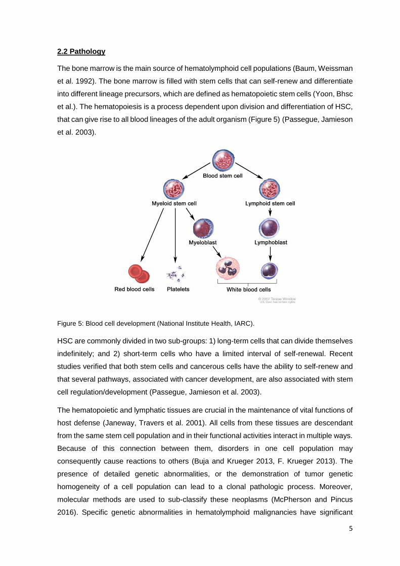
2.2 Pathology
The bone marrow is the main source of hematolymphoid cell populations (Baum, Weissman
et al. 1992). The bone marrow is filled with stem cells that can self-renew and differentiate
into different lineage precursors, which are defined as hematopoietic stem cells (Yoon, Bhsc
et al.). The hematopoiesis is a process dependent upon division and differentiation of HSC,
that can give rise to all blood lineages of the adult organism (Figure 5) (Passegue, Jamieson
et al. 2003).
Figure 5: Blood cell development (National Institute Health, IARC).
HSC are commonly divided in two sub-groups: 1) long-term cells that can divide themselves
indefinitely; and 2) short-term cells who have a limited interval of self-renewal. Recent
studies verified that both stem cells and cancerous cells have the ability to self-renew and
that several pathways, associated with cancer development, are also associated with stem
cell regulation/development (Passegue, Jamieson et al. 2003).
The hematopoietic and lymphatic tissues are crucial in the maintenance of vital functions of
host defense (Janeway, Travers et al. 2001). All cells from these tissues are descendant
from the same stem cell population and in their functional activities interact in multiple ways.
Because of this connection between them, disorders in one cell population may
consequently cause reactions to others (Buja and Krueger 2013, F. Krueger 2013). The
presence of detailed genetic abnormalities, or the demonstration of tumor genetic
homogeneity of a cell population can lead to a clonal pathologic process. Moreover,
molecular methods are used to sub-classify these neoplasms (McPherson and Pincus
2016). Specific genetic abnormalities in hematolymphoid malignancies have significant
6
prognostic value, which in turn can influence the type of treatment and the clinical outcome
(Jeon, Yoon et al. 2017). Malignant neoplasia of the lymphatic tissues are often
acknowledged as malignant lymphomas, or when malignant cells circulate through blood,
lymphocytic leukemias (Janeway, Travers et al. 2001, McPherson and Pincus 2016).
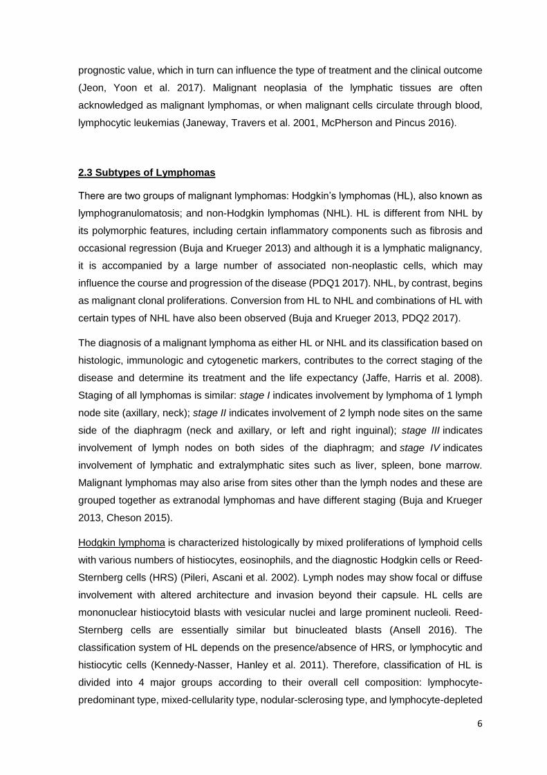
2.3 Subtypes of Lymphomas
There are two groups of malignant lymphomas: Hodgkin’s lymphomas (HL), also known as
lymphogranulomatosis; and non-Hodgkin lymphomas (NHL). HL is different from NHL by
its polymorphic features, including certain inflammatory components such as fibrosis and
occasional regression (Buja and Krueger 2013) and although it is a lymphatic malignancy,
it is accompanied by a large number of associated non-neoplastic cells, which may
influence the course and progression of the disease (PDQ1 2017). NHL, by contrast, begins
as malignant clonal proliferations. Conversion from HL to NHL and combinations of HL with
certain types of NHL have also been observed (Buja and Krueger 2013, PDQ2 2017).
The diagnosis of a malignant lymphoma as either HL or NHL and its classification based on
histologic, immunologic and cytogenetic markers, contributes to the correct staging of the
disease and determine its treatment and the life expectancy (Jaffe, Harris et al. 2008).
Staging of all lymphomas is similar: stage I indicates involvement by lymphoma of 1 lymph
node site (axillary, neck); stage II indicates involvement of 2 lymph node sites on the same
side of the diaphragm (neck and axillary, or left and right inguinal); stage III indicates
involvement of lymph nodes on both sides of the diaphragm; and stage IV indicates
involvement of lymphatic and extralymphatic sites such as liver, spleen, bone marrow.
Malignant lymphomas may also arise from sites other than the lymph nodes and these are
grouped together as extranodal lymphomas and have different staging (Buja and Krueger
2013, Cheson 2015).
Hodgkin lymphoma is characterized histologically by mixed proliferations of lymphoid cells
with various numbers of histiocytes, eosinophils, and the diagnostic Hodgkin cells or Reed-
Sternberg cells (HRS) (Pileri, Ascani et al. 2002). Lymph nodes may show focal or diffuse
involvement with altered architecture and invasion beyond their capsule. HL cells are
mononuclear histiocytoid blasts with vesicular nuclei and large prominent nucleoli. Reed-
Sternberg cells are essentially similar but binucleated blasts (Ansell 2016). The
classification system of HL depends on the presence/absence of HRS, or lymphocytic and
histiocytic cells (Kennedy-Nasser, Hanley et al. 2011). Therefore, classification of HL is
divided into 4 major groups according to their overall cell composition: lymphocyte-
predominant type, mixed-cellularity type, nodular-sclerosing type, and lymphocyte-depleted

7
type. The most frequently affected lymph nodes are in the mediastinum (59%), the neck
(55-58%), the axillae (13-14%), and the lung hilus (11-12%) (Buja and Krueger 2013).
Non-Hodgkin lymphomas are a diverse group of B, T and natural killer (NK) cell lymphomas
(Ansell 2015). These lymphomas arise from mature and precursor cells and typically,
infiltration of both lymphoid and hematopoietic tissues is observed, but extension to other
organs is also possible. The etiology of most NHLs is unknown, nevertheless some have
been associated with viral infections: Epstein-Barr Virus (EBV) and Burkitt’s and Burkitt’s-
type lymphoma; and Human Herpesvirus 8 (HHV-8), Human T-cell Leukemia Virus Type 1
(HTLV-1) and adult T-cell leukemia (ATL) (Buja and Krueger 2013, Linch and McNamara
2016, PDQ2 2017).
2.4 Subtypes of Leukemias
Leukemia is a common malignancy, affecting all age groups, that begins in blood-forming
tissues (Davis, Viera et al. 2014). Leukemias are divided into four major sub-groups
according to the French-American-British classification (Lilleyman, Hann et al. 1986, Abdul-
Hamid 2011): acute lymphoid leukemia (ALL), acute myelogenous leukemia (AML), chronic
myelogenous leukemia (CML) and chronic lymphoid leukemia (CLL). Chronic leukemia has
a slow progression and abnormal cells are not able to function, while acute leukemia has a
more aggressive form and cells are functional at the stage they are arrested (Torkaman,
Charkari et al. 2011).
Suspicion of leukemia should arise when white cell count is elevated, signs of bleeding
tendency (petechia, purpura or bruising), lymphadenopathy, hepatosplenomegaly,
expiratory wheeze (due to mediastinal mass), anemia, thrombocytopenia and
thrombocytosis. Infection, inflammation and stress must also be considered, since these
factors alter blood count (Raab and Gartner 2009, Davis, Viera et al. 2014).
Chronic myelogenous leukemia (CML) is associated with a characteristic chromosomal
t(9,22)(q34;q11) translocation, also known as the Philadelphia chromosome and is defined
by myeloid hyperplasia, leukocytosis, basophilia, and splenomegaly. Clinical characteristics
are fatigue, weight loss, sweats, bone pain, anemia, hepatosplenomegaly, and petechial
hemorrhages (Jabbour and Kantarjian 2016). The life expectancy of patients with CML
depends on disease progression and type of treatment; 45% to 65% of patients survive 5
years (Buja and Krueger 2013, Jabbour and Kantarjian 2014).
Acute Myelogenous Leukemia (AML) is a myeloproliferative disease representing
approximately 90% of all acute leukemias and is frequently observed in patients older than

8
65 years-old (NCI 2012). Patients typically present with malaise and fatigue, may have
resistant skin infections, unusual pallor, and bleeding from the gums and the nose; blood
smears show leukopenia, or excessive leukocytosis, with increase in immature cells; liver
and the spleen are enlarged and infiltrated by atypical blasts; and additional symptoms
result from metabolic and electrolyte derangements (hypokalemia, hypercalcemia),
agranulocytosis (necrotizing enterocolitis), or rapid lysis of leukemic blasts (Buja and
Krueger 2013). Survival rates for all AML subtypes combined are 40% at 15 months and
approximately 20% at 50 months (Buja and Krueger 2013). AML can be classified in eight
subtypes, according to the French-American-British (FAB) classification: AML-0 to AML-8
(Figure 6) (Rasaiyaah, Yong et al. 2007, Plesa, Ciuperca et al. 2008, NCI 2012).
Figure 6: Schematic diagram of the myeloid lineage illustrating the different types of acute myeloid
leukemia (Rasaiyaah, Yong et al. 2007).
Lymphocytic leukemias are characterized by blood circulating malignant cells and
lymphomatous infiltration of bone marrow, lymphatic organs, or extralymphatic sites.
Chronic Lymphocytic Leukemia (CLL) is observed in approximately 90% of B-cell
malignancies. This disease has a slow progression and predisposes patients to infections
due to immunodeficiency and autoimmune reactions (Hallek 2013). Acute lymphocytic
9
leukemia (ALL; cytological subtypes L1-L3 according to cell size) is frequently a childhood
leukemia, with over 80% of cases consisting of monoclonal B-precursor cells, and
approximately 15% of cells from the T-cell lineage (Cooper and Brown 2015). Clinical
characteristics are anemia (pallor, fatigue), thrombocytopenia (hemorrhage), and mature
leukocytopenia. The combination of chemotherapy and radiation therapy led into a long-
term disease-free survival of 70% to 80% of children (Buja and Krueger 2013, Chiaretti, Zini
et al. 2014).
Aplastic anemia (AA) is an anemia of deficient hematopoiesis and is characterized by
anemia, neutropenia, and thrombocytopenia, and may progress to leukemia (Scheinberg
2012). Patients often appear pale, with petechial hemorrhages, and increased susceptibility
to infection. Resistant cases necessitate multiple transfusions and thus may be complicated
by iron overload syndrome with secondary hemochromatosis, cardiac failure, and diabetes
mellitus (Scheinberg and Young 2012, Buja and Krueger 2013, Savona, Malcovati et al.
2015).
Myelodysplastic syndrome is a type of hematopoietic hyperplasia and dysplasia with
peripheral cytopenia. MDS originates from hematopoietic stem cell defects with multiple
genetic abnormalities and clonal proliferation of hematopoietic cells, T lymphocytes, and
clonal or polyclonal B lymphocytes. Several stages are identified by analyzing blasts
population and maturation (Germing, Kobbe et al. 2013). Anemia and fatigue are early
symptoms, followed by neutropenia, infections, thrombocytopenia, and bleeding. Bone
marrow aspirates show a megaloblastic erythropoiesis with ring sideroblasts, increased
myeloblasts, and hypolobulated megakaryocytes. Transition to acute myelogenous
leukemia (AML) occurs in 40% to 50% of advanced cases (Buja and Krueger 2013, Garcia-
Manero 2015).
Multiple myeloma (MM) also recognized as plasmacytoma is a neoplastic clonal
proliferation of plasmocytic cells usually at multiple sites in the bone marrow. It is frequently
accompanied by the production of unusual immunoglobulin components (gammopathy,
monoclonal M protein in serum, and Bence Jones protein in urine). About 90% of patients
will develop osteolytic bone lesions, due to high osteoclastic and low osteoblastic activity,
which is a major cause of morbidity and mortality of these patients (Miceli, Colson et al.
2011). Clinical features include bone pain, anemia, bleeding, hypercalcemia,
hyperglobulinemia, and susceptibility to infection. Anemia occurs in approximately 75% of
these patients and is the major cause for fatigue (Buja and Krueger 2013, Rajkumar and
Kumar 2016).

10
3. Hematopoietic Stem Cell Transplantation
3.1 Transplantation
Transplant of hematopoietic stem cells is currently the standard treatment for patients with
congenital or acquired hemoglobinopathies or with malignancies sensitive to
chemotherapy, radiotherapy or immunological treatments (Gratwohl, Baldomero et al.
2010). Allogeneic stem cells are infused to correct the basic genetic defect by replacing
genes required for a normal hematopoiesis. For a successful treatment, two main objectives
are required: 1) elimination of the deficient marrow; and 2) providing a tolerant environment
for the transplanted marrow survival (Lucarelli, Isgro et al. 2012).
Conditioning for immunological system suppression is required and therefore there are two
types of approaches: myeloablative conditioning (MA) and non-myeloablative/reduced-
intensity conditioning (RIC). MA involves induction of long-lasting aplasia and is achieved
with total body irradiation and/or alkylating agents. Some examples of MA conditioning are
the use of cyclophosphamide (CY)/total body irradiation (TBI) or busulfan (BU)/CY; NMA
conditioning consists in applying fludarabine (FLU)/TBI or low dose of TBI; finally, RI is often
applied using FLU and melphalan (MEL), FLU/BU and FLU/CY. Variations in these
regimens may occur depending on which center they are applied (Juric, Ghimire et al.
2016).
The success of HSCT is often associated with the HLA-match of hematopoietic stem cells.
Indeed, HLA-related sibling would be the ideal donor with 25% chance to be HLA-identical
(Petersdorf 2007). Patients without a related match must search for unrelated donors,
always considering variations in HLA-specific alleles and ethnicity. Nevertheless, for
patients lacking any HLA-match unrelated donor, three other options are available: HLA-
mismatched unrelated donors, umbilical cord blood (UCB) and HLA-haploidentical family
members (Gyurkocza, Rezvani et al. 2010).
The source of stem cells is also an important issue in HSCT, since it depends on the age
of the donor and recipient, clinical comorbidities, stage of disease and is extremely variable
between centers according to their experience (Juric, Ghimire et al. 2016). Three options
are available for harvesting HSC: Bone marrow (BM), Peripheral blood stem cells (PBSC)
and UCB: BM gives a lower risk of GVHD occurrence but has a more invasive harvesting
process; PBSC does not require general anesthesia for collection and has minimal
secondary effects, is related to faster engraftment and immune reconstitution (IR) but
carries a higher risk for GVHD; and UCB, is a non-invasive procedure, has lower risks of
GVHD and relapse, although a lower number of HSC are available and a slower IR is
observed (Smith and Wagner 2009, Juric, Ghimire et al. 2016).

11
3.2 Post-transplant complications
Post-transplant infections and graft-versus-host disease (GVHD) are the most common
problems in HSCT, especially in those patients receiving an allogeneic transplant (aHSCT)
(Choi, Levine et al. 2010).
Among the infections that affect patients undergoing aHSCT, viruses are the most frequent,
since they require a fast and effective immune response (Figure 7). The list of viruses that
infect these patients and cause severe morbidity and mortality gets longer each day,
nevertheless Cytomegalovirus (CMV) is still the most important virus (Atalay,
Gokahmetoglu et al. 2014).
Figure 7: Infections following allogeneic hematopoietic stem cell transplantation (Tomblyn, M. et al.,
2009).
GVHD is characterized by the rejection of the graft by the host due to an immune rejection
of the host tissues led by the donor lymphocytes (Barriga, Ramirez et al. 2012). Several
studies have shown that over 50% of patients who undergo aHSCT may develop GVHD
(Funke, Moreira et al. 2016). Transfused immunocompetent T lymphocytes recognize and
destroy such allogeneic host cells (epidermal, hepatocytes, intestinal, and hemolymphatic
tissues). Microscopically, a typical acute GVHD shows a T-cell immune reaction in the skin,
the liver, and the upper intestines combined with growth inhibition and atrophy of

12
hemolymphatic tissues. Severe acute GVHD has a high mortality secondary to severe
ulcerating enteritis with superinfection, diarrhea, and fluid loss; severe hepatitis with
hepatocellular necrosis; or systemic viral disease and bacterial septicemia (Buja and
Krueger 2013, Socie and Ritz 2014).
Another severe complication of HSCT is the development of post-transplant
lymphoproliferative disorder (PTLD). PTLD occurs after either solid or HSCT and is
responsible for high rates of morbidity and mortality among these patients (Zimmermann
and Trappe 2013). In HSCT recipients the incidence of PTLD is 1.0%, in non-complicated
HLA-matched transplants, and 25% after T-cell depleted highly immunosuppressed
transplants (Capello and Gaidano 2009). PTLDs are characterized by several factors:
usually derive from B cells, with preferential presentation as non-Hodgkin’s lymphoma;
usually originate in extranodal sites; rarely affect skin; aggressive behavior; and frequently
are Epstein-Barr virus related (Bar-Natan and Nagler 2006).
In aHSCT a regimen of immunosuppression must be applied to avoid graft rejection.
Considering a reduced intensity conditioning (RIC) and myeloablative regimen (MA),
several studies suggest that patients subjected to RIC carry more high-risk features and
comorbidities, although overall survival rates were found to be similar (Chevallier, Szydlo et
al. 2012, Atilla, Atilla et al. 2017). Myeloablative condition comprises the solo/combined
utilization of the following compounds: cyclophosphamide, busulfan, anti-thymocyte
globulin, fludarabine and/or melphalan. Most of RIC include fludarabine and intermediate
doses of busulfan and melphalan. RIC regimens are associated with mild
myelosuppression, low-treatment related toxicity and wider antitumor responses (Atilla,
Atilla et al. 2017). Almost all patients receiving myeloablative conditioning regimens develop
fever during neutropenia, and most of these febrile episodes are due to infections (Satwani,
Baldinger et al. 2009, Therriault, Wilson et al. 2010, Safdar 2011). Infections in neutropenia
after aHSCT may be life-threatening. Bacterial pathogens account for about 90% of
infections during this phase (Figure 7) (Safdar 2011, Balletto and Mikulska 2015).
13
4. Post-Transplant Lymphoproliferative Disorder
4.1 History
Although post-transplant lymphoproliferative disorder (PTLD) is relatively rare, it is the most
frequent malignant disease early after transplantation (Glotz, Chapman et al. 2012). PTLD
refers to a heterogeneous group of lymphoproliferative diseases, with potentially life-
threatening conditions, exhibiting a spectrum of histopathologies (Gulley and Tang 2010,
Mucha, Foroncewicz et al. 2010, Glotz, Chapman et al. 2012). These lymphoproliferative
disorders, may be nodal and/or extranodal, restricted to the allograft or widely disseminated
(Allen, Alfieri et al. 2002).
PTLD incidence seems to increase in patients receiving either intense immunosuppression
to protect against GVHD and/or increased immunosuppression following identification of de
novo human leukocyte antigen (HLA) antibodies in long-term transplant recipients. The
clinical, morphologic, and biologic heterogeneity of PTLD has made difficult the
understanding of its development and the treatment of these complex disorders.
The lymphoid proliferations that occur after organ transplantation have been recognized for
more than a quarter of century (Castellano-Sanchez, Li et al. 2004). In 1981 Frizzera et al.
studied tumors from a group of renal transplant recipients and observed the occurrence of
lymphoproliferations that had not been described before (Frizzera, Hanto et al. 1981). Given
the heterogeneity in tumor cell size and shape he called them “polymorphic” and additional
investigation demonstrated that tumors were composed of B-lymphocytes. Frizzera et al.
then created a classification system which differentiated nonspecific reactive hyperplasia
from polymorphic diffuse B cell hyperplasia and polymorphic diffuse B cell lymphoma and
from immunoblastic sarcoma (Frizzera, Hanto et al. 1981, Kalinova, Indrakova et al. 2009).
Later, in 1988, Nalesnik et al. investigated a transplant population at the University of
Pittsburg and rearranged the old classification system: by not distinguishing the clinical
presentation of the two types of polymorphic lesions, both were included under the term
polymorphic PTLD; and separating the group of lesions which resemble typical non-
Hodgkin’s lymphomas in occurrence and aggressive behavior designated as monomorphic
PTLD. In 1995, Knowles et al. established a new classification with three categories: 1)
reactive hyperplasia of plasma cells; 2) polymorphic hyperplasia and polymorphic
lymphoma, both of which were monoclonal and lacked oncogene and tumor suppressor
gene alterations; and 3) true lymphomas and hematopoietic neoplasms which were
monoclonal and contained proto-oncogenes and/or tumor suppressor gene alterations
(Knowles, Cesarman et al. 1995, Kalinova, Indrakova et al. 2009). Then, in 1997 the Society
for Hematopathology Workshop Classification categorized PTLDs into: early lesions,
14
polymorphic PTLDs, monomorphic PTLDs (B and T cell lymphomas), plasmacytoma-like
lesions, and T cell-rich large B cell lymphoma/Hodgkin's disease-like lesions. In 2001,
Harris, Swerdlow, Frizzera and Knowles reviewed the classification system for the 2001
World Health Organization Classifications of Tumors, although, doubts about the extent to
which specific genetic or molecular alterations, remained. Therefore, a last update was
performed, in 2008, where WHO expanded the definition of disease by considering patients’
age at diagnosis, tumor location, molecular characteristics, association with viral infection
and inflammation, as a criteria (Kalinova, Indrakova et al. 2009, Turner, Morton et al. 2010,
Campo, Swerdlow et al. 2011).
4.2 Incidence
The cumulative incidence of PTLD in allogeneic hematopoietic stem cell transplantation
(HSCT) recipients is 1.0% (range 0.5-1.8%), with slightly higher rates in the pediatric
population (Castellano-Sanchez, Li et al. 2004, Grywalska, Markowicz et al. 2013). Overall
incidence of PTLD varies from 1 to 22% depending on what type of organ was transplanted,
patient age, EBV serostatus from recipient and donor, aggressiveness of
immunosuppression and combination of risk factors (Bar-Natan and Nagler 2006, Ibrahim
and Naresh 2012) .
Rate of survival depends mainly on patient age and extent of disease, with pediatric patients
and patients with localized disease having a better prognosis. The more aggressive are
monomorphic lesions (Kalinova, Indrakova et al. 2009, Kim, Kim et al. 2010, Luo, Zhang et
al. 2014). .
4.3 Development
PTLD is usually classified as early onset lesions, which develop within one year, and late
onset lesions, which occur later than 1-year post-transplant. PTLD pathogenesis is
multifactorial and EBV plays a major role in the development, by driving the proliferation of
infected B cells (Ibrahim and Naresh 2012). Knowing the anatomic distribution of PTLD is
important for diagnosis (Tai, Tirumani et al. 2015).
The anatomic distribution of PTLDs varies with patient age and the type of
immunosuppressive therapy. PTLD localizes specially in the area of the transplanted organ
or in the allograft itself. For HSCT patients, PTLD tends to be disseminated and affects
mainly lymph nodes (Tai, Tirumani et al. 2015, Metser and Lo 2016). Childhood PTLDs
often involve lymphoid tissues including lymph nodes and adenoids and arise in the
15
abdomen, thoracic cavity, and head and neck; while PTLDs in adults tend to localize to the
liver, lung, lymph nodes, and gastrointestinal tract (Castellano-Sanchez, Li et al. 2004).
Risk factors include young age and age over 50 years at transplantation, white race,
unrelated or HLA-mismatched graft, Epstein-Barr virus negative serostatus prior to
transplant, primary EBV infection, type of organ transplant, intensity of immunosuppression
and presence of cytomegalovirus disease (Kim, Kim et al. 2010, Glotz, Chapman et al.
2012). EBV infection is thought to play the most important role in the pathogenesis of PTLD,
although, not all PTLD cases are EBV related (Kim, Kim et al. 2010, Luo, Zhang et al. 2014).
To this date, there have been no large studies performed to explore the connection of EBV
infection, CMV infection, and acute rejection. It is therefore difficult to define the relative
contribution of these events as separate risk factors for PTLD (Glotz, Chapman et al. 2012).
An elevated EBV-DNA load has recently become a sensitive aid for predicting individual
patients at risk for PTLD development (Luo, Zhang et al. 2014).
Current data for several studies suggests that PTLD is likely to be associated with a high
level of immunosuppression rather than the individual use of immunosuppressive agents.
Moreover, results from separate studies demonstrate that transplant recipients treated with
triple or quadruple combinations of immunosuppressive agents are at higher risk of
developing PTLD than patients receiving less agents (Glotz, Chapman et al. 2012). These
findings imply that a reduction in immunosuppressive load may lead to a decline in the risk
of PTLD (Loren, Porter et al. 2003). The results of some studies suggest that lengthy
immunosuppression might also increase the risk of PTLD (Issa, Amer et al. 2009). It has
also been suggested that the balance of T- and B-cell depletion may have an impact on the
risk of PTLD, although the optimal balance has not been determined (Herzig, Juffs et al.
2003, Opelz and Dohler 2004, Kremers, Devarbhavi et al. 2006).
4.4 Clinical presentation
PTLD presents itself with fever, malaise, an infectious mononucleosis-like syndrome,
lymphadenopathy and symptoms regarding organ dysfunction (Kalinova, Indrakova et al.
2009). Symptoms are often nonspecific, and some patients are asymptomatic. PTLD
frequently presents as a rapidly enlarging mass in the grafted organ, in lymph nodes, filling
the marrow space, or in extranodal sites such as upper airway or intestine. In young
children, primary EBV infection often occurs after iatrogenic immunosuppression
commences, either when an infected graft is introduced or later in the posttransplant period.
Despite PTLD can present with symptoms reminiscent of infectious mononucleosis, but
PTLD is a much more serious illness (Gulley and Tang 2010).

16
The risk of infection among allogeneic hematopoietic stem cell transplant recipients is
determined by patient age, underlying disease, the complication that occurred during
preceding treatment regimens, the selected transplantation modality, and the severity of
graft-versus-host disease (GVHD) (Balletto and Mikulska 2015). In comparison with patients
undergoing high-dose chemotherapy and autologous stem cell transplantation, aHSCT
recipients are at a much higher risk of infection, due to delayed recovery of T-cell and B-
cell functions (Ritter, Seitz et al. 2015). Immunological reconstitution after hematopoietic
retrieval has an impact on the type of posttransplant infectious complications, and infection-
related mortality is significantly higher post-engraftment than during the short posttransplant
neutropenia. RIC regimens have a lower risk of severe and deadly infections in the early
posttransplant period (Safdar 2011). Indeed, the most critical risk factor is the drug regimen
used to prepare the patient for transplant as well as the ongoing immunosuppressive drugs
used to prevent graft rejection, for example, anti-thymocyte globulin (ATG) depletes T cells
and therefore protects from graft rejection, but its increases the probability of PTLD
occurrence (Landgren, Gilbert et al. 2009, Atilla, Atilla et al. 2017). Fludarabine,
azathioprine, and other agents responsible for T cell suppression or mutagenicity are also
associated with PTLD pathogenesis (Martinez and de Gruijl 2008, Landgren, Gilbert et al.
2009). In addition, patients having multiple rejections and consequently more interventions
to amplify immunosuppression may be at higher risk of PTLD. Some HLA types influence
identification of cells expressing external viral proteins, which influences the pathogenesis
of EBV-driven lymphoproliferation (Gulley and Tang 2010).
4.5 Histologic interpretation and Classification
Due to its heterogenous profile, PTLD diagnosis and histological classification requires a
tissue biopsy (Kalinova, Indrakova et al. 2009). This histological evaluation has implications
on treatment decisions according to whether the target antigen for rituximab (CD20) is
expressed (Parker, Bowles et al. 2010, Glotz, Chapman et al. 2012).
PTLD is divided into four major histopathologic subtypes with corresponding clinical and
biologic features, as described in the World Health Organization sub-classification scheme:
early lesions, polymorphic PTLD, monomorphic PTLD and classical Hodgkin lymphoma-
type PTLD (Spasojevic-Dimitrijeva, Peco-Antic et al. 2014). There is no staging system for
PTLD, since it is almost impossible to elaborate a scheme for a disease with such broad
histological spectrum (Bowden, Ljungman et al. 2010).
Early lesions, occur within one year after transplant, are sub-divided into two groups:
plasmocytic hyperplasia (PP) and infectious mononucleosis-like lesions. These lesions are
17
B cell derived polyclonal lesions and are characterized by preserving the affected tissue
architecture (Bowden, Ljungman et al. 2010, Ibrahim and Naresh 2012). These type of
lesions frequently involve tonsils, adenoids or lymph nodes (Ibrahim and Naresh 2012).
Polymorphic PTLD is a mixture of small to large lymphocytes and immunoblasts, with a
presentation very similar to HL, with Reed-Sternberg-like cells (Ibrahim and Naresh 2012).
This subtype includes EBV-infected neoplastic B cells as well as reactive CD4+ and CD8+
T cells and clonality assays reveal monoclonal B cells (Gulley and Tang 2008). Reducing
the level of immunosuppression is frequently effective in reversing cell growth (Bakker, van
Imhoff et al. 2007).
Monomorphic PTLD is the most common form of PTLD and can be divided in two types: B-
cell and T-cell/NK-cell (Cai, Chen et al. 2015). Most cases of B-cell monomorphic PTLD are
of a non-germinal center type, especially those who are EBV positive (~50% of all cases)
(Choquet 2016). Occasional EBV-negative cases are more likely to occur later after
transplantation (beyond 1 year) (Swerdlow 2007). Monomorphic T-cell/NK-cell PTLDs are
rare, accounting for ~10% of all PTLDs, and are associated with peripheral T-cell lymphoma
(Choquet 2016). Conventional lymphoma therapy is not necessarily needed, since
monomorphic PTLD can be treated by reducing immunosuppression (Green 2001, Knight,
Tsodikov et al. 2009).
Classical Hodgkin Lymphoma-Type PTLD is very similar to HL, although it has the Hodgkin-
like form of polymorphic PTLD, and is always EBV positive (Pitman, Huang et al. 2006). As
for monomorphic and polymorphic lesions, classic Hodgkin lymphoma-like PTLD is
characterized by destroyed tissue architecture (Choquet 2016). This type of PTLD occurs
late after transplant (after the first year) and response to therapy is generally favorable
(Gulley and Tang 2010).
4.6 Treatment and Prognosis
The first line of treatment comprises the reduction or withdrawal of immunosuppression,
while chemotherapy and radiation are applied as a second line treatment (Reshef,
Vardhanabhuti et al. 2011). Other approaches include, surgery, antiviral therapy, anti-B-cell
monoclonal antibody (rituximab) and cytotoxic T cells are currently being investigated
(Kalinova, Indrakova et al. 2009, Kim, Kim et al. 2010, Parker, Bowles et al. 2010, Glotz,
Chapman et al. 2012).
Immunosuppression after transplantation, in a patient who is a carrier of Epstein-Barr virus,
seems to reduce the activity of the patient’s EBV-specific cytotoxic T-cell surveillance, which

18
increases the chances of uncontrolled proliferation of EBV-infected B-cells and subsequent
progression to PTLD (Allen, Preiksaitis et al. 2009). Moreover, transplant recipients
experiencing primary EBV infection, during the early post-transplant period, seem to be
particularly susceptible to developing EBV-specific PTLD of B-cell origin, mainly by lacking
preexisting EBV-specific T-cell immunity (Allen, Preiksaitis et al. 2009, Glotz, Chapman et
al. 2012).
PTLD almost always develops rapidly to a fatal outcome unless it is diagnosed and treated
(Opelz and Dohler 2004). The ability to reduce or eliminate immunosuppressive drugs is a
helpful strategy for restoring natural antiviral and antineoplastic immunity. Almost all types
are primarily of B-cell origin. Over 90% of PTLD cases are associated with EBV infection.
Immunosuppressive treatment leads to T-lymphocyte dysfunction which allows uncontrolled
proliferation of EBV.
Histology has a strong prognostic significance in PTLD (prognosis is much worse in NK-, T-
cell, and plasmablastic B-cell PTLD compared with polymorphic, DLBCL-type and
Burkitt/Burkitt-like B-cell PTLD, and is more satisfactory in early lesion and plasmacytoma-
like PTLD (Allen, Preiksaitis et al. 2009). Thus, the WHO histologic classification provides
important information on the probable progression and outcome of the disease and thereby
influence choices of treatment (Glotz, Chapman et al. 2012). EBV association may also
impact on prognosis, as EBV-associated PTLD may have a better prognosis than EBV-
negative PTLD and may need less chemotherapy. EBV antigen expression (EBNA-1, -2, -
3, LMP etc.) may also help understanding PTLD response to a reduction in
immunosuppression and thus help to select treatment (Shimoyama, Asano et al. 2009).
PTLD is associated with mortality rates reaching up to 70-90% in HSCT patients, and 5-
year survival rates of 59%, although recent data suggest that outcomes have improve
(Opelz and Dohler 2004, Al-Mansour, Nelson et al. 2013, Caillard, Porcher et al. 2013).
.

19
5. Epstein-Barr Virus
5.1 Structure and Genome
The Epstein-Barr virus (EBV), also known as Human Herpesvirus 4 (HHV-4) belongs to the
Herpesviridae family, gamma subfamily, and is the only known human Lymphocryptovirus
(Kwok 2007, Grywalska, Markowicz et al. 2013). Like other members of the Herpesviridae,
EBV virions have double-stranded, linear DNA genome, with approximately 172 kb pairs in
length, encoding approximately 100 genes surrounded by a protein capsid (Young, Arrand
et al. 2007). EBV has a series of 0.5 kb terminal direct repeats and internal repeat
sequences, that divide the genome into short and long, large unique sequence domains
(Arvin, Campadelli-Fiume et al. 2007). The EBV genome is linear in virus particles and
circularized in infected cells (Kwok 2007, Odumade, Hogquist et al. 2011). EBV has a toroid-
shaped protein core that is wrapped with DNA, a nucleocapsid with 162 capsomeres, a
protein tegument between the nucleocapsid and the envelope, and an outer envelope with
external glycoprotein spikes. These glycoproteins are essential in cell tropism, host range
and receptor recognition. Mature virions have approximately 120-180 nm in diameter.
5.2 Epidemiology and Disease
EBV infections are most prevalent in developing countries, in populations of low
socioeconomic status. In countries with proper hygiene practices, EBV seroprevalence
tends to increase gradually with age, showing two seroconversions peaks: at 2 to 4 years
and at 14 to 18 years. The mean seroprevalence in children is approximately 50% and
increases steadily to a value of 90% to 99% in adults (Walling, Ray et al. 2007, Grywalska,
Markowicz et al. 2013). EBV by itself, accounts for 0.5%-2% of all cancers, varying by
geographic locations (Jha, Pei et al. 2016).
Geographical distribution of EBV is variable depending on genotypes. EBV strains are
characterized as type 1 (B95.8-like) or type 2 (Jijoye and AG876-like) (originally referred as
A and B, respectively) differing in organization of the genes that encode the EBV nuclear
antigen (O'Mahony, Debnath et al.) The two major types, 1 and 2, which are distinguished
by genomic difference in a subset of latent genes, who encode nuclear proteins in latently
infected cells (EBNA-LP, EBNA2, EBNA3A, EBNA3B and EBNA3C) (Dolan, Addison et al.
2006, Janani, Malathi et al. 2015). Both types are detected all over the world, with type 1
being more frequent in the Western hemisphere and Southeast Asia (Sample, Young et al.
1990, Odumade, Hogquist et al. 2011). Although, in central Africa, Papua New Guinea and
Alaska, type 2 is more prevalent (Hjalgrim, Friborg et al. 2007). EBV type 1 is frequently
20
found in healthy individuals, while type 2 is associated with immunocompromised patients
(Janani, Malathi et al. 2015).
There is no consistent data according to EBV seroprevalence by sex in children, although,
in developed countries, where infection occurs in the adolescence, a higher seroprevalence
and earlier occurrence of infectious mononucleosis is observed in women (Crawford,
Swerdlow et al. 2002). Recent studies showed that an individual can harbor multiple viral
strains, and these strains might be acquired during primary infection (Kwok 2007, Atalay,
Gokahmetoglu et al. 2014) Arvin, Campadelli-Fiume et al. 2007).
5.3 EBV Infection and Latency
Transmission
The virus is transmitted through saliva, spread through close human oral contact, or
transmitted by transfusion, and is highly immunogenic (Grywalska, Markowicz et al. 2013).
Infection by EBV has several clinical displays since children are often asymptomatic, and
have self-limited brief viral illness, whereas in adults it appears as infectious mononucleosis,
in 30 to 50% of cases (Rea, Russo et al. 2001, Pittaluga 2013, Atalay, Gokahmetoglu et al.
2014).
Primary Infection
The receptor necessary for viral entry on epithelial cells and B lymphocytes is the CD21
molecule, which was previously known as CR2. When in vitro, the infection of B
lymphocytes leads to continuous cell proliferation resulting in immortalization. Moreover,
when infecting epithelial cells, occurs an active replication, leading to virions production and
host cell apoptosis (Figure 8). Viral replication happens in the cell of the oropharynx and
shedding of the virus is maintained throughout the host life. Primary infection induces both
humoral and cellular immune response, with production of antibodies against lytic and latent
genes, as well for the glycoprotein 350 preventing the binding of CD21 on B cells (Eligio,
Delia et al. 2010).
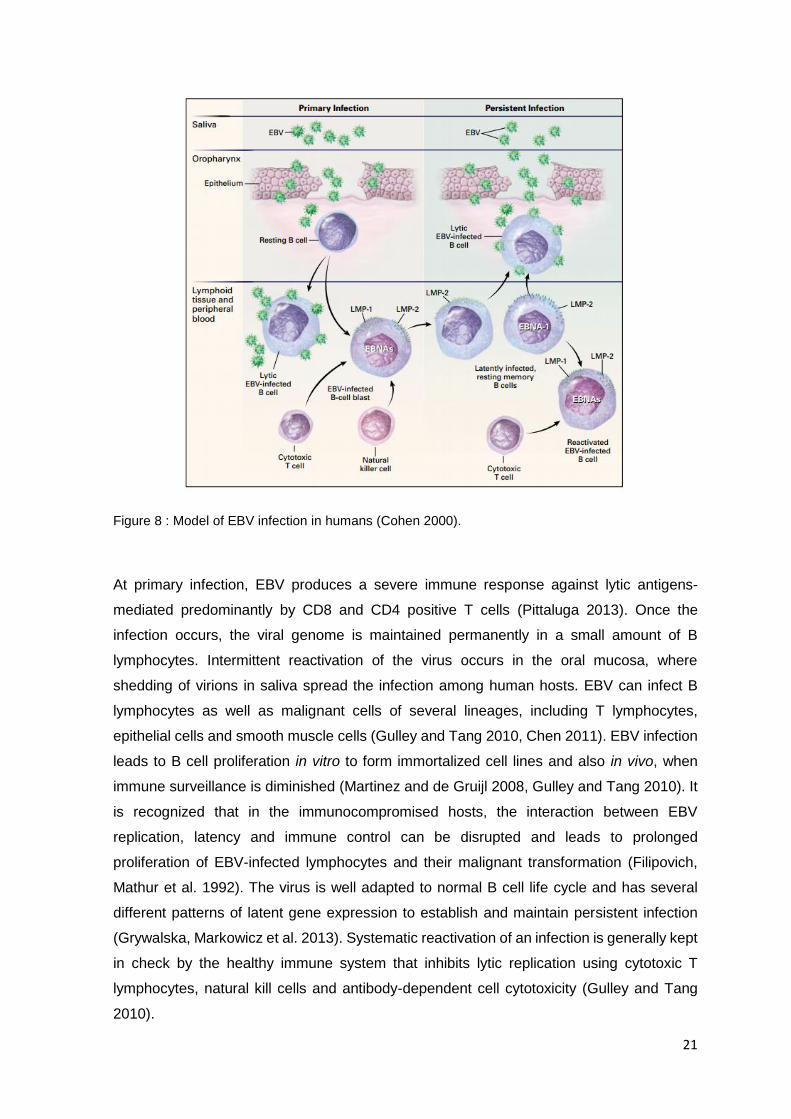
21
Figure 8 : Model of EBV infection in humans (Cohen 2000).
At primary infection, EBV produces a severe immune response against lytic antigens-
mediated predominantly by CD8 and CD4 positive T cells (Pittaluga 2013). Once the
infection occurs, the viral genome is maintained permanently in a small amount of B
lymphocytes. Intermittent reactivation of the virus occurs in the oral mucosa, where
shedding of virions in saliva spread the infection among human hosts. EBV can infect B
lymphocytes as well as malignant cells of several lineages, including T lymphocytes,
epithelial cells and smooth muscle cells (Gulley and Tang 2010, Chen 2011). EBV infection
leads to B cell proliferation in vitro to form immortalized cell lines and also in vivo, when
immune surveillance is diminished (Martinez and de Gruijl 2008, Gulley and Tang 2010). It
is recognized that in the immunocompromised hosts, the interaction between EBV
replication, latency and immune control can be disrupted and leads to prolonged
proliferation of EBV-infected lymphocytes and their malignant transformation (Filipovich,
Mathur et al. 1992). The virus is well adapted to normal B cell life cycle and has several
different patterns of latent gene expression to establish and maintain persistent infection
(Grywalska, Markowicz et al. 2013). Systematic reactivation of an infection is generally kept
in check by the healthy immune system that inhibits lytic replication using cytotoxic T
lymphocytes, natural kill cells and antibody-dependent cell cytotoxicity (Gulley and Tang
2010).

22
Latency
EBV latency comprises five EBV-encoded nuclear antigens (EBNAs), two latent membrane
proteins (LMPs), EBV-encoded small RNA (EBER) and non-transcribed BART RNAs.
EBERs are present in high amounts of copies in latently infected cells, therefore, these
transcripts are targeted by in situ hybridization (ISH) on tissue sections.
EBV infection establishes four different latency patterns depending on which viral proteins
are expressed. Latency 0 is defined by non-expression of viral proteins, where viral gene
expression is limited two small non-coding, non-polyadenylated RNAs (EBER 1/2) and a
set of transcripts from BamA rightward transcript (BART). This type of latency allows EBV
to be persistence and become immunologically undetectable (Toczyski, Matera et al. 1994,
Pittaluga 2013). Burkitt’s lymphoma is associated with type I latency and expresses
EBER1/2 RNA, EBNA-1, LMP-2A/B and BART RNA. Type II latency is associated with
nasopharyngeal carcinoma and Hodgkin lymphoma, where EBV expresses EBER1/2 RNA,
EBNA-1, LMP-1, LMP-2A/B (Carbone, Gloghini et al. 2008). Type III latency is when
unlimited expression of all viral proteins are expressed: EBER 1/2 RNA, EBNA-LP, EBNA-
1, EBNA-2, EBNA-3A/B/C, LMP-1, LMP-2A/B and BART RNA. This latency pattern occurs
mainly in immunocompromised patients with post-transplant lymphoproliferative disorders,
infectious mononucleosis, HIV-associated lymphoproliferative disorders and in
lymphoblastoid cell lines (Grywalska, Markowicz et al. 2013, Kang and Kieff 2015).
Considering latently infected cells, a limited pattern of viral gene expression is observed:
EBNA1 is required for maintenance of the episomal form of the virus, while EBNA2, the
main transactivator protein of EBV, transactivates viral and cellular genes promoters.
EBNA3 modulates the transactivator activity of EBNA2. The transmembrane proteins LMP1
and LMP2 affect several signal transduction pathways. The EBERs and BARTs, and the
BARF1 protein are also associated with oncogenesis. The microRNAs derived from
transcripts of the BHRF1 gene and the BART transcripts target both viral and cellular RNAs
affecting the quantity (BALF5) and latent (LMP1) EBV transcripts as well as certain cellular
RNAs. When EBNA2 is absent (the main viral transactivator protein in latency type III), the
LMP promoters are triggered by cellular proteins in nasopharyngeal carcinomas and in
Sternberg-Reed cells of Hodgkin’s disease (latency type II), but not in type I BL cell lines or
BL biopsies (type I latency) or memory B cells (type 0 latency) (Takacs, Segesdi et al. 2009).
Lytic Infection
EBV encoded proteins are necessary in productive infection and include transactivators,
which are enzymes necessary for viral DNA amplification and assembly of structural
compounds of the virions (Takacs, Segesdi et al. 2009). In the lytic cycle a progressive
23
cascade of gene activation is initiated: immediate early genes (BZLF1 and BRLF1) are
expressed first and activate the early genes. The shift between latent and lytic infection is
mediated by immediate early proteins (EB1 and Rta). These proteins are transcriptions
factors that activate EBV early genes, which are necessary for viral DNA replication. Late
proteins, who are only expressed after viral DNA synthesis, encode proteins necessary for
assembly, maturation and release of infectious particles (Aubry, Mure et al. 2014).
5.4. EBV and Disease
EBV was the first human virus to be directly implicated in carcinogenesis (Grywalska,
Markowicz et al. 2013). EBV was first discovered in 1964 by Epstein, Achong and Barr,
from culture tumor cells, as the agent responsible for Burkitt’s lymphoma in East African
biopsy samples (Epstein 2001, Kwok 2007, Stanfield and Luftig 2017). Several studies have
also demonstrated the ability of the virus to transform human B cells into lymphoblastoid
cell line, suggesting its oncogenic potential (Stanfield and Luftig 2017).
Since its discovery, EBV has been found in a variety of other tumor types. The evidence for
an association with EBV is strongest for Burkitt’s lymphoma, NK/T cell lymphoma,
nasopharyngeal carcinoma, Hodgkin’s lymphoma and for malignant lymphomas in immune
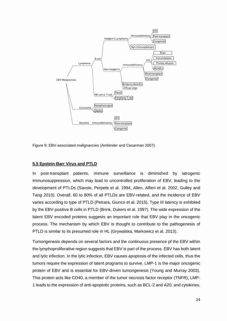
incompetent patients (Figure 9). Additionally, certain epithelial cell tumors, such as
nasopharyngeal carcinoma and more recently to gastric carcinoma have been found to be
EBV related (Gulley and Tang 2010, Grywalska, Markowicz et al. 2013, Sousa, Breda et al.
2013, Sousa, Mesquita et al. 2016).
24
Figure 9: EBV-associated malignancies (Ambinder and Cesarman 2007).
5.5 Epstein-Barr Virus and PTLD
In post-transplant patients, immune surveillance is diminished by iatrogenic
immunosuppression, which may lead to uncontrolled proliferation of EBV, leading to the
development of PTLDs (Savoie, Perpete et al. 1994, Allen, Alfieri et al. 2002, Gulley and
Tang 2010). Overall, 60 to 80% of all PTLDs are EBV-related, and the incidence of EBV
varies according to type of PTLD (Petrara, Giunco et al. 2015). Type III latency is exhibited
by the EBV-positive B cells in PTLD (Brink, Dukers et al. 1997). The wide expression of the
latent EBV encoded proteins suggests an important role that EBV play in the oncogenic
process. The mechanism by which EBV is thought to contribute to the pathogenesis of
PTLD is similar to its presumed role in HL (Grywalska, Markowicz et al. 2013).
Tumorigenesis depends on several factors and the continuous presence pf the EBV within
the lymphoproliferative region suggests that EBV is part of the process. EBV has both latent
and lytic infection. In the lytic infection, EBV causes apoptosis of the infected cells, thus the
tumors require the expression of latent programs to survive. LMP-1 is the major oncogenic
protein of EBV and is essential for EBV-driven tumorigenesis (Young and Murray 2003).
This protein acts like CD40, a member of the tumor necrosis factor receptor (TNFR), LMP-
1 leads to the expression of anti-apoptotic proteins, such as BCL-2 and A20, and cytokines,

25
such as IL-1 and CD40L. EBV malignancies require the induction of telomerase activity,
which is a ribonucleoprotein complex, that extends the telomeres at the ends, preventing
cell senescence and apoptosis (Petrara, Giunco et al. 2015).
Analogous EBV-driven lymphoproliferations are observed in other immunosuppressive
pathologies, such as chemotherapy-related immunosuppression for autoimmune disease
or age-related decline in immunity, suggesting that reduced immunity is another major
tumorigenic cofactor (Martinez and de Gruijl 2008, Gulley and Tang 2010). Diminished T
cell immunity leads to uncontrolled proliferation, which can develop rapidly and systemically
if not rapidly identified and treated (Gulley and Tang 2010). Although immunosuppression
is a major factor in PTLD development, also persistent immune activation and chronic
inflammation are important factors. Chronic inflammation leads to overproduction of B-cell
stimulatory cytokines, such as IL-6, IL-10, IFN-α and TNF, thus enabling EBV proliferation
(Petrara, Giunco et al. 2015).
Considering that most cases of PTLD are from donor or recipient derived B-cells, one
approach to avoid the development of PTLD is to eliminate EBV infected B-cells in the early
phase after aHSCT. Elevated levels of EBV DNA are present in blood samples of infected
patients, including intracellular EBV within circulating B lymphocytes and extracellular EBV
DNA measurable in plasma. EBV viral load, as measured by quantitative molecular analysis
of the viral genome, serves as biomarker for predicting and monitoring the course of PTLD
(Gulley and Tang 2008, Kalinova, Indrakova et al. 2009, Gulley and Tang 2010). Indeed,
monitoring the EBV viral load, in peripheral blood, in addition to administering preemptive
rituximab therapy has recently been reported to be a successful approach to preventing the
development of B cell PTLD (Alexander, Jarrett et al. 2000, Adhikary, Behrends et al. 2006,
Tanaka, Takizawa et al. 2014). Furthermore, reduced anti-EBV nuclear antigen antibody
levels have also been linked with an increased risk for PTLD, suggesting that monitoring
these antibodies may also be a biomarker of PTLD development (Riddler, Breinig et al.
1994).

26
27
REFERENCES
• Abdul-Hamid, G. (2011). Classification of Acute Leukemia Acute Leukemia - The
Scientist's Perspective and Challenge. M. Antica. Croatia, Intech: 3-18.
• Adhikary, D., U. Behrends, A. Moosmann, K. Witter, G. W. Bornkamm and J. Mautner
(2006). "Control of Epstein-Barr virus infection in vitro by T helper cells specific for virion
glycoproteins." J Exp Med 203(4): 995-1006.
• Al-Mansour, Z., B. P. Nelson and A. M. Evens (2013). "Post-transplant
lymphoproliferative disease (PTLD): risk factors, diagnosis, and current treatment
strategies." Curr Hematol Malig Rep 8(3): 173-183.
• Alberts, B., A. Johnson, J. Lewis, M. Raff, K. Roberts and P. Walter (2002). Molecular
Biology of the Cell. New York, Garland Science.
• Alexander, F. E., R. F. Jarrett, D. Lawrence, A. A. Armstrong, J. Freeland, D. A. Gokhale,
E. Kane, G. M. Taylor, D. H. Wright and R. A. Cartwright (2000). "Risk factors for Hodgkin's
disease by Epstein-Barr virus (EBV) status: prior infection by EBV and other agents." Br J
Cancer 82(5): 1117-1121.
• Allen, U., C. Alfieri, J. Preiksaitis, A. Humar, D. Moore, B. Tapiero, R. Tellier, M. Green,
D. Davies, D. Hebert, S. Weitzman, M. Petric, K. Jacobson and P. W. G. Canadian (2002).
"Epstein-Barr virus infection in transplant recipients: Summary of a workshop on
surveillance, prevention and treatment." Can J Infect Dis 13(2): 89-99.
• Allen, U., J. Preiksaitis.Practice (2009). "Epstein-barr virus and posttransplant
lymphoproliferative disorder in solid organ transplantion." Am J Transplant 9 Suppl 4: 107-
20.
• Ambinder, R. F. and E. Cesarman (2007). Clinical and pathological aspects of EBV and
KSHV infection. Human Herpesviruses: Biology, Therapy, and Immunoprophylaxis. A.
Arvin, G. Campadelli-Fiume, E. Mocarski et al. Cambridge.
• Ansell, S. M. (2016). "Hodgkin lymphoma: 2016 update on diagnosis, risk-stratification,
and management." Am J Hematol 91(4): 434-442.
• Arvin, A., G. Campadelli-Fiume, E. Mocarski, P. S. Moore, B. Roizman, R. Whitley and
K. Yamanishi (2007). Human Herpesviruses: Biology, therapy and Immunoprophylaxis.
Cambridge, Cambridge University Press.
• Atalay, A., S. Gokahmetoglu, S. Durmaz, I. Kandemir, D. Saglam, L. Kaynar, B. Eser, M.
Cetin and H. Kilic (2014). "Investigation of epstein-barr virus and parvovirus b19 DNA in
allogeneic stem cell transplant patients." Turk J Haematol 31(2): 155-160.
• Atilla, E., P. Atilla and T. Demirer (2017). "A Review of Myeloablative vs Reduced
Intensity/Non-Myeloablative Regimens in Allogeneic Hematopoietic Stem Cell
Transplantations." Balkan Med J 34(1): 1-9.
28
• Aubry, V., F. Mure, B. Mariame, T. Deschamps, L. S. Wyrwicz, E. Manet and H. Gruffat
(2014). "Epstein-Barr virus late gene transcription depends on the assembly of a virus-
specific preinitiation complex." J Virol 88(21): 12825-12838.
• Bakker, N. A., G. W. van Imhoff, E. A. Verschuuren and W. J. van Son (2007).
"Presentation and early detection of post-transplant lymphoproliferative disorder after solid
organ transplantation." Transpl Int 20(3): 207-218.
• Balletto, E. and M. Mikulska (2015). "Bacterial Infections in Hematopoietic Stem Cell
Transplant Recipients." Mediterr J Hematol Infect Dis 7(1): e2015045.
• Bar-Natan, M. and A. Nagler (2006). "Epstein-Barr virus-associated post-transplant
lymphoproliferative disorder." Isr Med Assoc J 8(3): 205-207.
• Barriga, F., P. Ramirez, A. Wietstruck and N. Rojas (2012). "Hematopoietic stem cell
transplantation: clinical use and perspectives." Biol Res 45(3): 307-316.
• Baum, C. M., I. L. Weissman, A. S. Tsukamoto, A. M. Buckle and B. Peault (1992).
"Isolation of a candidate human hematopoietic stem-cell population." Proc Natl Acad Sci U
S A 89(7): 2804-2808.
• Bhayat, F., E. Das-Gupta, C. Smith, T. McKeever and R. Hubbard (2009). "The incidence
of and mortality from leukaemias in the UK: a general population-based study." BMC Cancer
9: 252.
• Bowden, R., P. Ljungman and D. R. Snydman (2010). Epstein-Barr Virus Infection and
Lymphoproliferative Disorders. Transplant Infections. Philadelphia, PA., Wolters Kluwer:
371-382.
• Brink, A. A., D. F. Dukers, A. J. van den Brule, J. J. Oudejans, J. M. Middeldorp, C. J.
Meijer and M. Jiwa (1997). "Presence of Epstein-Barr virus latency type III at the single cell
level in post-transplantation lymphoproliferative disorders and AIDS related lymphomas." J
Clin Pathol 50(11): 911-918.
• Buja, L. M. and G. R. F. Krueger (2013). Netter's Illustrated Human Pathology. Texas,
USA, Elsevier.
• Cai, Q., K. Chen and K. H. Young (2015). "Epstein-Barr virus-positive T/NK-cell
lymphoproliferative disorders." Exp Mol Med 47: e133.
• Caillard, S., R. Porcher, F. Provot, J. Dantal, S. Choquet, A. Durrbach, E. Morelon, V.
Moal, B. Janbon, E. Alamartine, C. Pouteil Noble, D. Morel, N. Kamar, M. Buchler, M. F.
Mamzer, M. N. Peraldi, C. Hiesse, E. Renoult, O. Toupance, J. P. Rerolle, S. Delmas, P.
Lang, Y. Lebranchu, A. E. Heng, J. M. Rebibou, C. Mousson, D. Glotz, J. Rivalan, A. Thierry,
I. Etienne, M. C. Moal, L. Albano, J. F. Subra, N. Ouali, P. F. Westeel, M. Delahousse, R.
Genin, B. Hurault de Ligny and B. Moulin (2013). "Post-transplantation lymphoproliferative

29
disorder after kidney transplantation: report of a nationwide French registry and the
development of a new prognostic score." J Clin Oncol 31(10): 1302-1309.
• Campo, E., S. H. Swerdlow, N. L. Harris, S. Pileri, H. Stein and E. S. Jaffe (2011). "The
2008 WHO classification of lymphoid neoplasms and beyond: evolving concepts and
practical applications." Blood 117(19): 5019-5032.
• Capello, D. and G. Gaidano (2009). "Post-transplant lymphoproliferative disorders: role
of viral infection, genetic lesions and antigen stimulation in the pathogenesis of the disease."
Mediterr J Hematol Infect Dis 1(2): e2009018.
• Carbone, A., A. Gloghini and G. Dotti (2008). "EBV-associated lymphoproliferative
disorders: classification and treatment." Oncologist 13(5): 577-585.
• Castellano-Sanchez, A. A., S. Li, J. Qian, A. Lagoo, E. Weir and D. J. Brat (2004).
"Primary central nervous system posttransplant lymphoproliferative disorders." Am J Clin
Pathol 121(2): 246-253.
• Chen, J., Z. Liang, F. Lu, X. Fang, S. Liu, Y. Zeng, F. Zhu, X. Chen, T. Shen, J. Li and
H. Zhuang (2011). "Toll-like receptors and cytokines/cytokine receptors polymorphisms
associate with non-response to hepatitis B vaccine." Vaccine 29(4): 706-711.
• Chen, M. R. (2011). "Epstein-barr virus, the immune system, and associated diseases."
Front Microbiol 2: 5.
• Cheson, B. D. (2015). "Staging and response assessment in lymphomas: the new
Lugano classification." Chin Clin Oncol 4(1): 5.
• Chevallier, P., R. M. Szydlo, D. Blaise, R. Tabrizi, M. Michallet, M. Uzunov, N. Fegueux,
F. Guilhot, S. Lapusan, N. Gratecos, J. Y. Cahn, G. Socie, I. Yakoub-Agha, A. Huynh, S.
Francois, J. O. Bay, S. Maury, A. Buzyn, N. Contentin and M. Mohty (2012). "Reduced-
intensity conditioning before allogeneic hematopoietic stem cell transplantation in patients
over 60 years: a report from the SFGM-TC." Biol Blood Marrow Transplant 18(2): 289-294.
• Chiaretti, S., G. Zini and R. Bassan (2014). "Diagnosis and subclassification of acute
lymphoblastic leukemia." Mediterr J Hematol Infect Dis 6(1): e2014073.
• Choi, S. W., J. E. Levine and J. L. Ferrara (2010). "Pathogenesis and management of
graft-versus-host disease." Immunol Allergy Clin North Am 30(1): 75-101.
• Choquet, S. (2016). "Classification and Treatment of Posttransplant Lymphoproliferative
Disorders." Lymphoma and Chronic Lymphocytic Leukemias 6: 13-19.
• Cohen, J. I. (2000). "Epstein-Barr virus infection." N Engl J Med 343(7): 481-492.
• Cooper, S. L. and P. A. Brown (2015). "Treatment of pediatric acute lymphoblastic
leukemia." Pediatr Clin North Am 62(1): 61-73.

30
• Crawford, D. H., A. J. Swerdlow, C. Higgins, K. McAulay, N. Harrison, H. Williams, K.
Britton and K. F. Macsween (2002). "Sexual history and Epstein-Barr virus infection." J
Infect Dis 186(6): 731-736.
• Davis, A. S., A. J. Viera and M. D. Mead (2014). "Leukemia: an overview for primary
care." Am Fam Physician 89(9): 731-738.
• Dolan, A., C. Addison, D. Gatherer, A. J. Davison and D. J. McGeoch (2006). "The
genome of Epstein-Barr virus type 2 strain AG876." Virology 350(1): 164-170.
• Eligio, P., R. Delia and G. Valeria (2010). "EBV Chronic Infections." Mediterr J Hematol
Infect Dis 2(1): e2010022.
• Epstein, M. A. (2001). "Historical background." Philos Trans R Soc Lond B Biol Sci
356(1408): 413-420.
• F. Krueger, G. R. (2013). Atlas of anatomic pathology with imaging : a correlative
diagnostic companion. New York, Springer.
• Ferlay, J., I. Soerjomataram, M. Ervik, R. Dikshit, S. Eser, C. Mathers, M. Rebelo, D. M.
Parkin, D. Forman and F. Bray. (2013). "Cancer Incidence and Mortality Worldwide: IARC
CancerBase No. 11." Retrieved 26 May, 2017, from
http://globocan.iarc.fr/Pages/Map.aspx.
• Filipovich, A. H., A. Mathur, D. Kamat and R. S. Shapiro (1992). "Primary
immunodeficiencies: genetic risk factors for lymphoma." Cancer Res 52(19 Suppl): 5465s-
5467s.
• Frizzera, G., D. W. Hanto, K. J. Gajl-Peczalska, J. Rosai, R. W. McKenna, R. K. Sibley,
K. P. Holahan and L. L. Lindquist (1981). "Polymorphic diffuse B-cell hyperplasias and
lymphomas in renal transplant recipients." Cancer Res 41(11 Pt 1): 4262-4279.
• Funke, V. A., M. C. Moreira and A. C. Vigorito (2016). "Acute and chronic Graft-versus-
host disease after hematopoietic stem cell transplantation." Rev Assoc Med Bras (1992) 62
Suppl 1: 44-50.
• Garcia-Manero, G. (2015). "Myelodysplastic syndromes: 2015 Update on diagnosis, risk-
stratification and management." Am J Hematol 90(9): 831-841.
• Germing, U., G. Kobbe, R. Haas and N. Gattermann (2013). "Myelodysplastic
syndromes: diagnosis, prognosis, and treatment." Dtsch Arztebl Int 110(46): 783-790.
• Glotz, D., J. R. Chapman, V. R. Dharnidharka, D. W. Hanto, M. C. Castro, H. H. Hirsch,
V. Leblond, A. K. Mehta, B. Moulin, A. Pagliuca, J. Pascual, A. B. Rickinson, F. P. Russo,
R. U. Trappe, A. C. Webster, A. O. Zuckermann and T. G. Gross (2012). "The Seville expert
workshop for progress in posttransplant lymphoproliferative disorders." Transplantation
94(8): 784-793.
31
• Gratwohl, A., H. Baldomero, M. Aljurf, M. C. Pasquini, L. F. Bouzas, A. Yoshimi, J. Szer,
J. Lipton, A. Schwendener, M. Gratwohl, K. Frauendorfer, D. Niederwieser, M. Horowitz, Y.
Kodera, B. Worldwide Network of and T. Marrow (2010). "Hematopoietic stem cell
transplantation: a global perspective." JAMA 303(16): 1617-1624.
• Green, M. (2001). "Management of Epstein-Barr virus-induced post-transplant
lymphoproliferative disease in recipients of solid organ transplantation." Am J Transplant
1(2): 103-108.
• Grywalska, E., J. Markowicz, P. Grabarczyk, M. Pasiarski and J. Rolinski (2013).
"Epstein-Barr virus-associated lymphoproliferative disorders." Postepy Hig Med Dosw
(Online) 67: 481-490.
• Gulley, M. L. and W. Tang (2008). "Laboratory assays for Epstein-Barr virus-related
disease." J Mol Diagn 10(4): 279-292.
• Gulley, M. L. and W. Tang (2010). "Using Epstein-Barr viral load assays to diagnose,
monitor, and prevent posttransplant lymphoproliferative disorder." Clin Microbiol Rev 23(2):
350-366.
• Gyurkocza, B., A. Rezvani and R. F. Storb (2010). "Allogeneic hematopoietic cell
transplantation: the state of the art." Expert Rev Hematol 3(3): 285-299.
• Hallek, M. (2013). "Chronic lymphocytic leukemia: 2013 update on diagnosis, risk
stratification and treatment." Am J Hematol 88(9): 803-816.
• Hejmadi, M. (2010). Introduction to Cancer Biology, Ventus Publishing.
• Herzig, K. A., H. G. Juffs, D. Norris, A. M. Brown, D. Gill, C. M. Hawley, R. Cobcroft, J.
B. Petrie, P. Marlton, D. Thomson, S. B. Campbell, D. L. Nicol and D. W. Johnson (2003).
"A single-centre experience of post-renal transplant lymphoproliferative disorder." Transpl
Int 16(7): 529-536.
• Hjalgrim, H., J. Friborg and M. Melbye (2007). The epidemiology of EBV and its
association with malignant disease. Human Herpesviruses: Biology, Therapy, and
Immunoprophylaxis. A. Arvin, G. Campadelli-Fiume, E. Mocarski et al. Cambridge.
• Howlader, N., A. M. Noone, M. Krapcho, D. Miller, K. Bishop, C. L. Kosary, M. Yu, J.
Ruhl, Z. Tatalovich, A. Mariotto, D. R. Lewis, H. S. Chen, E. J. Feuer and K. A. Cronin.
(2017). "SEER Cancer Statistics Review, 1975-2014." Retrieved 26 May, 2017, from
http://seer.cancer.gov/csr/1975_2014/.
• IARC. (2014). "World Cancer Factsheet." Retrieved 26 May, 2017, from
http://www.cancerresearchuk.org/sites/default/files/cs_report_world.pdf.
• Ibrahim, A., A. Ali and M. M. Mohammed (2014). "Outcome of Adolescents with Acute
Lymphoblastic Leukemia Treated by Pediatrics versus Adults Protocols." Adv Hematol
2014: 697675.

32
• Ibrahim, H. A. and K. N. Naresh (2012). "Posttransplant lymphoproliferative disorders."
Adv Hematol 2012: 230173.
• Issa, N., H. Amer, P. G. Dean, W. K. Kremers, Y. C. Kudva, N. Rostambeigi, F. G. Cosio,
T. S. Larson, T. M. Habermann, M. D. Stegall and M. D. Griffin (2009). "Posttransplant
lymphoproliferative disorder following pancreas transplantation." Am J Transplant 9(8):
1894-1902.
• Jabbour, E. and H. Kantarjian (2014). "Chronic myeloid leukemia: 2014 update on
diagnosis, monitoring, and management." Am J Hematol 89(5): 547-556.
• Jabbour, E. and H. Kantarjian (2016). "Chronic myeloid leukemia: 2016 update on
diagnosis, therapy, and monitoring." Am J Hematol 91(2): 252-265.
• Jaffe, E. S., N. L. Harris, H. Stein and P. G. Isaacson (2008). "Classification of lymphoid
neoplasms: the microscope as a tool for disease discovery." Blood 112(12): 4384-4399.
• Janani, M. K., J. Malathi, M. Rela, M. Farouk, J. Padmapriya and H. N. Madhavan (2015).
"Genotypic Detection of Epstein Barr Virus in Pediatric Transplant Recipients From India."
Indian Pediatr 52(11): 946-950.
• Janeway, C. A., P. Travers and M. Walport (2001). Immunobiology: The Immune System
in Health and Disease. New York, Garland Science.
• Jeon, Y. K., S. O. Yoon, J. H. Paik, Y. A. Kim, B. K. Shin, H. J. Kim, H. J. Cha, J. E. Kim,
J. Huh, Y. H. Ko, P. Hematopathology Study Group of the Korean Society of and P.
Molecular Pathology Study Group of Korean Society of (2017). "Molecular Testing of
Lymphoproliferative Disorders: Current Status and Perspectives." J Pathol Transl Med
51(3): 224-241.
• Jha, H. C., Y. Pei and E. S. Robertson (2016). "Epstein-Barr Virus: Diseases Linked to
Infection and Transformation." Front Microbiol 7: 1602.
• Juric, M. K., S. Ghimire, J. Ogonek, E. M. Weissinger, E. Holler, J. J. van Rood, M.
Oudshoorn, A. Dickinson and H. T. Greinix (2016). "Milestones of Hematopoietic Stem Cell
Transplantation - From First Human Studies to Current Developments." Front Immunol 7:
470.
• Kalinova, L., J. Indrakova and P. Bachleda (2009). "Post-transplant lymphoproliferative
disorder." Biomed Pap Med Fac Univ Palacky Olomouc Czech Repub 153(4): 251-257.
• Kang, M. S. and E. Kieff (2015). "Epstein-Barr virus latent genes." Exp Mol Med 47: e131.
• Kennedy-Nasser, A. A., P. Hanley and C. M. Bollard (2011). "Hodgkin disease and the
role of the immune system." Pediatr Hematol Oncol 28(3): 176-186.
• Khan, N., F. Afaq and H. Mukhtar (2010). "Lifestyle as risk factor for cancer: Evidence
from human studies." Cancer Lett 293(2): 133-143.

33
• Kim, M. J., I. Kim, H. M. Bae, K. Seo, N. Park, S. S. Yoon, S. Park and B. K. Kim (2010).
"Hematopoietic stem cell transplantation after posttransplant lymphoproliferative disorder."
J Korean Med Sci 25(5): 781-784.
• Knight, J. S., A. Tsodikov, D. M. Cibrik, C. W. Ross, M. S. Kaminski and D. W. Blayney
(2009). "Lymphoma after solid organ transplantation: risk, response to therapy, and survival
at a transplantation center." J Clin Oncol 27(20): 3354-3362.
• Knowles, D. M., E. Cesarman, A. Chadburn, G. Frizzera, J. Chen, E. A. Rose and R. E.
Michler (1995). "Correlative morphologic and molecular genetic analysis demonstrates
three distinct categories of posttransplantation lymphoproliferative disorders." Blood 85(2):
552-565.
• Kremers, W. K., H. C. Devarbhavi, R. H. Wiesner, R. A. Krom, W. R. Macon and T. M.
Habermann (2006). "Post-transplant lymphoproliferative disorders following liver
transplantation: incidence, risk factors and survival." Am J Transplant 6(5 Pt 1): 1017-1024.
• Kwok, H. (2007). Characterization of Epstein-Barr Virus (EBV) strains in praimary EBV
infection. Master of Philosophy, University of Hong Kong.
• Landgren, O., E. S. Gilbert, J. D. Rizzo, G. Socie, P. M. Banks, K. A. Sobocinski, M. M.
Horowitz, E. S. Jaffe, D. W. Kingma, L. B. Travis, M. E. Flowers, P. J. Martin, H. J. Deeg
and R. E. Curtis (2009). "Risk factors for lymphoproliferative disorders after allogeneic
hematopoietic cell transplantation." Blood 113(20): 4992-5001.
• Lilleyman, J. S., I. M. Hann, R. F. Stevens, O. B. Eden and S. M. Richards (1986).
"French American British (FAB) morphological classification of childhood lymphoblastic
leukaemia and its clinical importance." J Clin Pathol 39(9): 998-1002.
• Linch, D. and C. McNamara (2016). Non-Hodgkin's Lymphoma: Diagnosis and
Management. London.
• Loren, A. W., D. L. Porter, E. A. Stadtmauer and D. E. Tsai (2003). "Post-transplant
lymphoproliferative disorder: a review." Bone Marrow Transplant 31(3): 145-155.
• Lucarelli, G., A. Isgro, P. Sodani and J. Gaziev (2012). "Hematopoietic stem cell
transplantation in thalassemia and sickle cell anemia." Cold Spring Harb Perspect Med 2(5):
a011825.
• Luo, L., L. Zhang, B. Cai, H. Li, W. Huang, Y. Jing, H. Zhu, Y. Zhao, J. Bo, Q. Wang, X.
Han, L. Yu and C. Gao (2014). "Post-transplant lymphoproliferative disease after allogeneic
hematopoietic stem cell transplantation: a single-center experience." Ann Transplant 19: 6-
12.
• Malaguarnera, L., E. Cristaldi and M. Malaguarnera (2010). "The role of immunity in
elderly cancer." Crit Rev Oncol Hematol 74(1): 40-60.

34
• Malvezzi, M., G. Carioli, P. Bertuccio, T. Rosso, P. Boffetta, F. Levi, C. La Vecchia and
E. Negri (2016). "European cancer mortality predictions for the year 2016 with focus on
leukemias." Ann Oncol; 27: 926-933.
• Martinez, O. M. and F. R. de Gruijl (2008). "Molecular and immunologic mechanisms of
cancer pathogenesis in solid organ transplant recipients." Am J Transplant 8(11): 2205-
2211.
• McPherson, R. A. and M. R. Pincus (2016). Henry's Clinical Diagnosis and Management
by Laboratory Methods, 23rd Edition. New York, Elsevier.
• Metser, U. and G. Lo (2016). "FDG-PET/CT in abdominal post-transplant
lymphoproliferative disease." Br J Radiol 89(1057): 20150844.
• Miceli, T. S., K. Colson, B. M. Faiman, K. Miller, J. D. Tariman and B. International
Myeloma Foundation Nurse Leadership (2011). "Maintaining bone health in patients with
multiple myeloma: survivorship care plan of the International Myeloma Foundation Nurse
Leadership Board." Clin J Oncol Nurs 15 Suppl: 9-23.
• Moser, E. C. and F. Meunier (2014). "Cancer survivorship: A positive side-effect of more
successful cancer treatment." EJC Suppl 12(1): 1-4.
• Mucha, K., B. Foroncewicz, B. Ziarkiewicz-Wroblewska, M. Krawczyk, J. Lerut and L.
Paczek (2010). "Post-transplant lymphoproliferative disorder in view of the new WHO
classification: a more rational approach to a protean disease?" Nephrol Dial Transplant
25(7): 2089-2098.
• NCI. (2012). "SEER Stat Fact Sheets: Acute Myeloid Leukemia." Retrieved June, 13,
2017, from http://seer.cancer.gov/statfacts/html/amyl.html.
• O'Mahony, D., I. Debnath, J. Janik, D. Aisner, E. Jaffe, T. Waldmann and J. Morris
(2008). "Cardiac involvement with human T-cell lymphotrophic virus type-1-associated adult
T-cell leukemia/lymphoma: The NIH experience." Leuk Lymphoma 49(3): 439-446.
• Ocheni, S., N. Kroeger, T. Zabelina, I. Sobottka, F. Ayuk, C. Wolschke, A. Muth, H.
Lellek, L. Petersen, R. Erttmann, H. Kabisch, A. R. Zander and U. Bacher (2008). "EBV
reactivation and post transplant lymphoproliferative disorders following allogeneic SCT."
Bone Marrow Transplant 42(3): 181-186.
• Odumade, O. A., K. A. Hogquist and H. H. Balfour, Jr. (2011). "Progress and problems
in understanding and managing primary Epstein-Barr virus infections." Clin Microbiol Rev
24(1): 193-209.
• Opelz, G. and B. Dohler (2004). "Lymphomas after solid organ transplantation: a
collaborative transplant study report." Am J Transplant 4(2): 222-230.
• Pandya, P., J. L. Orgaz and V. Sanz-Moreno (2017). "Modes of invasion during tumour
dissemination." Mol Oncol 11(1): 5-27.
35
• Parker, A., K. Bowles, J. A. Bradley, V. Emery, C. Featherstone, G. Gupte, R. Marcus,
J. Parameshwar, A. Ramsay, C. Newstead, H. Haemato-oncology Task Force of the British
Committee for Standards in and S. British Transplantation (2010). "Management of post-
transplant lymphoproliferative disorder in adult solid organ transplant recipients - BCSH and
BTS Guidelines." Br J Haematol 149(5): 693-705.
• Passegue, E., C. H. Jamieson, L. E. Ailles and I. L. Weissman (2003). "Normal and
leukemic hematopoiesis: are leukemias a stem cell disorder or a reacquisition of stem cell
characteristics?" Proc Natl Acad Sci U S A 100 Suppl 1: 11842-11849.
• PDQ1 (2017). Adult Hodgkin Lymphoma Treatment (PDQ(R)): Patient Version. PDQ
Cancer Information Summaries. Bethesda (MD).
• PDQ2 (2017). Adult Non-Hodgkin Lymphoma Treatment (PDQ(R)): Patient Version.
PDQ Cancer Information Summaries. Bethesda (MD).
• Petersdorf, E. W. (2007). "Risk assessment in haematopoietic stem cell transplantation:
histocompatibility." Best Pract Res Clin Haematol 20(2): 155-170.
• Peterson, L. E. and T. Kovyrshina (2017). "Progression inference for somatic mutations
in cancer." Heliyon 3(4): e00277.
• Petrara, M. R., S. Giunco, D. Serraino, R. Dolcetti and A. De Rossi (2015). "Post-
transplant lymphoproliferative disorders: from epidemiology to pathogenesis-driven
treatment." Cancer Lett 369(1): 37-44.
• Pileri, S. A., S. Ascani, L. Leoncini, E. Sabattini, P. L. Zinzani, P. P. Piccaluga, A. Pileri,
Jr., M. Giunti, B. Falini, G. B. Bolis and H. Stein (2002). "Hodgkin's lymphoma: the
pathologist's viewpoint." J Clin Pathol 55(3): 162-176.
• Pitman, S. D., Q. Huang, C. W. Zuppan, E. H. Rowsell, J. D. Cao, J. G. Berdeja, L. M.
Weiss and J. Wang (2006). "Hodgkin lymphoma-like posttransplant lymphoproliferative
disorder (HL-like PTLD) simulates monomorphic B-cell PTLD both clinically and
pathologically." Am J Surg Pathol 30(4): 470-476.
• Pittaluga, S. (2013). "Viral-associated lymphoid proliferations." Semin Diagn Pathol
30(2): 130-136.
• Plesa, A., G. Ciuperca, V. Louvet, L. Pujo-Menjouet, S. Genieys, C. Dumontet, X.
Thomas and V. Volpert (2008). "Diagnostics of the AML with immunophenotypical data."
Mathematic Modelling of Natural Phenomena 2(1): 104-123.
• Plummer, M., C. de Martel, J. Vignat, J. Ferlay, F. Bray and S. Franceschi (2016). "Global
burden of cancers attributable to infections in 2012: a synthetic analysis." Lancet Glob
Health 4(9): e609-616.
• Pui, C. H., D. Pei, D. Campana, W. P. Bowman, J. T. Sandlund, S. C. Kaste, R. C.
Ribeiro, J. E. Rubnitz, E. Coustan-Smith, S. Jeha, C. Cheng, M. L. Metzger, D. Bhojwani,
36
H. Inaba, S. C. Raimondi, M. Onciu, S. C. Howard, W. Leung, J. R. Downing, W. E. Evans
and M. V. Relling (2011). "Improved prognosis for older adolescents with acute
lymphoblastic leukemia." J Clin Oncol 29(4): 386-391.
• Raab, C. P. and J. C. Gartner, Jr. (2009). "Diagnosis of childhood cancer." Prim Care
36(4): 671-684.
• Rajkumar, S. V. and S. Kumar (2016). "Multiple Myeloma: Diagnosis and Treatment."
Mayo Clin Proc 91(1): 101-119.
• Rasaiyaah, J., K. Yong, D. R. Katz, P. Kellam and B. M. Chain (2007). "Dendritic cells
and myeloid leukaemias: plasticity and commitment in cell differentiation." Br J Haematol
138(3): 281-290.
• Rea, T. D., J. E. Russo, W. Katon, R. L. Ashley and D. S. Buchwald (2001). "Prospective
study of the natural history of infectious mononucleosis caused by Epstein-Barr virus." J Am
Board Fam Pract 14(4): 234-242.
• Reshef, R., M. R. Luskin, M. Kamoun, S. Vardhanabhuti, J. E. Tomaszewski, E. A.
Stadtmauer, D. L. Porter, D. F. Heitjan and E. Tsai de (2011). "Association of HLA
polymorphisms with post-transplant lymphoproliferative disorder in solid-organ transplant
recipients." Am J Transplant 11(4): 817-825.
• Reshef, R., S. Vardhanabhuti, M. R. Luskin, D. F. Heitjan, D. Hadjiliadis, S. Goral, K. L.
Krok, L. R. Goldberg, D. L. Porter, E. A. Stadtmauer and D. E. Tsai (2011). "Reduction of
immunosuppression as initial therapy for posttransplantation lymphoproliferative disorder(
bigstar)." Am J Transplant 11(2): 336-347.
• Riddler, S. A., M. C. Breinig and J. L. McKnight (1994). "Increased levels of circulating
Epstein-Barr virus (EBV)-infected lymphocytes and decreased EBV nuclear antigen
antibody responses are associated with the development of posttransplant
lymphoproliferative disease in solid-organ transplant recipients." Blood 84(3): 972-984.
• Ritter, J., V. Seitz, H. Balzer, R. Gary, D. Lenze, S. Moi, S. Pasemann, A. Seegebarth,
M. Wurdack, S. Hennig, A. Gerbitz and M. Hummel (2015). "Donor CD4 T Cell Diversity
Determines Virus Reactivation in Patients After HLA-Matched Allogeneic Stem Cell
Transplantation." Am J Transplant 15(8): 2170-2179.
• Safdar, A. (2011). Principles and Practice of Cancer Infectious Diseases. New York,
Springer.
• Sample, J., L. Young, B. Martin, T. Chatman, E. Kieff, A. Rickinson and E. Kieff (1990).
"Epstein-Barr virus types 1 and 2 differ in their EBNA-3A, EBNA-3B, and EBNA-3C genes."
J Virol 64(9): 4084-4092.
• Satwani, P., L. Baldinger, J. Freedman, J. S. Jacobson, J. Guerra, C. van de Ven, E.
Morris, J. Garvin, D. George, M. B. Bradley, M. Bhatia, B. Tallamy, J. Schwartz, Z. Jin and

37
M. S. Cairo (2009). "Incidence of Viral and fungal infections following busulfan-based
reduced-intensity versus myeloablative conditioning in pediatric allogeneic stem cell
transplantation recipients." Biol Blood Marrow Transplant 15(12): 1587-1595.
• Savoie, A., C. Perpete, L. Carpentier, J. Joncas and C. Alfieri (1994). "Direct correlation
between the load of Epstein-Barr virus-infected lymphocytes in the peripheral blood of
pediatric transplant patients and risk of lymphoproliferative disease." Blood 83(9): 2715-
2722.
• Scheinberg, P. (2012). "Aplastic anemia: therapeutic updates in immunosuppression and
transplantation." Hematology Am Soc Hematol Educ Program 2012: 292-300.
• Scheinberg, P. and N. S. Young (2012). "How I treat acquired aplastic anemia." Blood
120(6): 1185-1196.
• Shimoyama, Y., N. Asano, M. Kojima, S. Morishima, K. Yamamoto, T. Oyama, T.
Kinoshita and S. Nakamura (2009). "Age-related EBV-associated B-cell lymphoproliferative
disorders: diagnostic approach to a newly recognized clinicopathological entity." Pathol Int
59(12): 835-843.
• Singh, S. K. and M. T. Dorak (2017). "Cancer Immunoprevention and Public Health."
Front Public Health 5: 101.
• Smith, A. R. and J. E. Wagner (2009). "Alternative haematopoietic stem cell sources for
transplantation: place of umbilical cord blood." Br J Haematol 147(2): 246-261.
• Socie, G. and J. Ritz (2014). "Current issues in chronic graft-versus-host disease." Blood
124(3): 374-384.
• Sousa, H., E. Breda, A. M. Santos, R. Catarino, D. Pinto, P. Canedo, J. C. Machado and
R. Medeiros (2013). "IL-1RN VNTR polymorphism as a susceptibility marker for
nasopharyngeal carcinoma in Portugal." Arch Oral Biol 58(8): 1040-1046.
• Sousa, H., L. Mesquita, J. Ribeiro, R. Catarino, E. Breda and R. Medeiros (2016).
"Polymorphisms in host immune response associated genes and risk of nasopharyngeal
carcinoma development in Portugal." Immunobiology 221(2): 145-152.
• Spasojevic-Dimitrijeva, B., A. Peco-Antic, D. Paripovic, D. Kruscic, Z. Krstic, M. Cupic,
M. Cvetkovic, G. Milosevski-Lomic and M. Kostic (2014). "Post-transplant
lymphoproliferative disorder--case reports of three children with kidney transplant." Srp Arh
Celok Lek 142(1-2): 83-88.
• Spolverato, G., Y. Kim, S. Alexandrescu, I. Popescu, H. P. Marques, L. Aldrighetti, T.
Clark Gamblin, J. Miura, S. K. Maithel, M. H. Squires, C. Pulitano, C. Sandroussi, G.
Mentha, T. W. Bauer, T. Newhook, F. Shen, G. A. Poultsides, J. Wallis Marsh and T. M.
Pawlik (2015). "Is Hepatic Resection for Large or Multifocal Intrahepatic
38
Cholangiocarcinoma Justified? Results from a Multi-Institutional Collaboration." Ann Surg
Oncol 22(7): 2218-2225.
• Stanfield, B. A. and M. A. Luftig (2017). "Recent advances in understanding Epstein-Barr
virus." F1000Res 6: 386.
• Swerdlow, S. H. (2007). "T-cell and NK-cell posttransplantation lymphoproliferative
disorders." Am J Clin Pathol 127(6): 887-895.
• Tai, R., S. H. Tirumani, H. Tirumani, A. B. Shinagare, J. L. Hornick and N. H. Ramaiya
(2015). "Is there a difference in post-transplant lymphoproliferative disorder in adults after
solid organ and haematologic stem cell transplantation? Experience in 41 patients." Br J
Radiol 88(1052): 20140861.
• Takacs, M., J. Segesdi, F. Banati, A. Koroknai, H. Wolf, H. H. Niller and J. Minarovits
(2009). "The importance of epigenetic alterations in the development of epstein-barr virus-
related lymphomas." Mediterr J Hematol Infect Dis 1(2): e2009012.
• Tanaka, T., J. Takizawa, S. Miyakoshi, T. Kozakai, K. Fuse, Y. Shibasaki, M. Moriyama,
K. Ohshima, K. Toba, T. Furukawa, H. Sone and M. Masuko (2014). "Manifestations of
fulminant CD8 T-cell post-transplant lymphoproliferative disorder following the
administration of rituximab for lymphadenopathy with a high level of Epstein-Barr Virus
(EBV) replication after allogeneic hematopoietic stem cell transplantation." Intern Med
53(18): 2115-2119.
• Therriault, B. L., J. W. Wilson, J. N. Barreto and L. L. Estes (2010). "Characterization of
bacterial infections in allogeneic hematopoietic stem cell transplant recipients who received
prophylactic levofloxacin with either penicillin or doxycycline." Mayo Clin Proc 85(8): 711-
718.
• Toczyski, D. P., Matera, A. G., D. C. Ward and Steitz, J. A. (1994). "The Epstein-Barr
virus (EBV) small RNA EBER1 binds and relocalizes ribosomal protein L22 in EBV-infected
human B lymphocytes." Proc Natl Acad Sci U S A 91(8): 3463-3467.
• Tomblyn, M., Chiller, T., Einsele, H., Gress, R., Sepkowitz, K., Storek, J., Wingard, J. R.,
Young, J-A. H., Boeckh, M. A. (2009). "Guidelines for preventing infectious complications
among hematopoietic cell transplantation recipients: a global prespective." Biology Blood
Marrow Transplant 15:1143-1238.
• Torkaman, A., N. M. Charkari and M. Aghaeipour (2011). "An approach for leukemia
classification based on cooperative game theory." Anal Cell Pathol (Amst) 34(5): 235-246.
• Turner, J. J., L. M. Morton, M. S. Linet, C. A. Clarke, M. E. Kadin, C. M. Vajdic, A.
Monnereau, M. Maynadie, B. C. Chiu, R. Marcos-Gragera, A. S. Costantini, J. R. Cerhan
and D. D. Weisenburger (2010). "InterLymph hierarchical classification of lymphoid

39
neoplasms for epidemiologic research based on the WHO classification (2008): update and
future directions." Blood 116(20): e90-98.
• Walling, D. M., A. J. Ray, J. E. Nichols, C. M. Flaitz and C. M. Nichols (2007). "Epstein-
Barr virus infection of Langerhans cell precursors as a mechanism of oral epithelial entry,
persistence, and reactivation." J Virol 81(13): 7249-7268.
• Wang, J., T. Jin, F. Mo, H. Dong, Y. Wang, L. Kang and Q. Hu (2018). "Association
between IL-1 gene polymorphisms and tuberculosis susceptibility in the Chinese Tibetan
population." Int J Clin Exp Pathol 11(11): 5441-5449.
• Wang, M. H., K. J. Helzlsouer, M. W. Smith, J. A. Hoffman-Bolton, S. L. Clipp, V.
Grinberg, A. M. De Marzo, W. B. Isaacs, C. G. Drake, Y. Y. Shugart and E. A. Platz (2009).
"Association of IL10 and other immune response- and obesity-related genes with prostate
cancer in CLUE II." Prostate 69(8): 874-885.
• Wilson, S., L. Jones, C. Coussens and K. Hanna (2002). Cancer and the Environment -
Gene-environment interaction. Washington, D.C., National Academy of Sciences.
• Yoon, J. O., Bhsc, M. H. Kang, J. S. Kim, Bhsc and J. S. Oh (2015). "The effects of gait
with use of smartphone on repositioning error and curvature of the lumbar spine." J Phys
Ther Sci 27(8): 2507-2508.
• Young, L. S., J. R. Arrand and P. G. Murray (2007). EBV gene expression and regulation.
Human Herpesviruses: Biology, Therapy, and Immunoprophylaxis. A. Arvin, G. Campadelli-
Fiume, E. Mocarski et al. Cambridge.
• Young, L. S. and P. G. Murray (2003). "Epstein-Barr virus and oncogenesis: from latent
genes to tumours." Oncogene 22(33): 5108-5121.
• Zimmermann, H. and R. U. Trappe (2013). "EBV and posttransplantation
lymphoproliferative disease: what to do?" Hematology Am Soc Hematol Educ Program
2013: 95-102.
• Zou, J., C. Wang, X. Ma, E. Wang and G. Peng (2017). "APOBEC3B, a molecular driver
of mutagenesis in human cancers." Cell Biosci 7: 29.

40
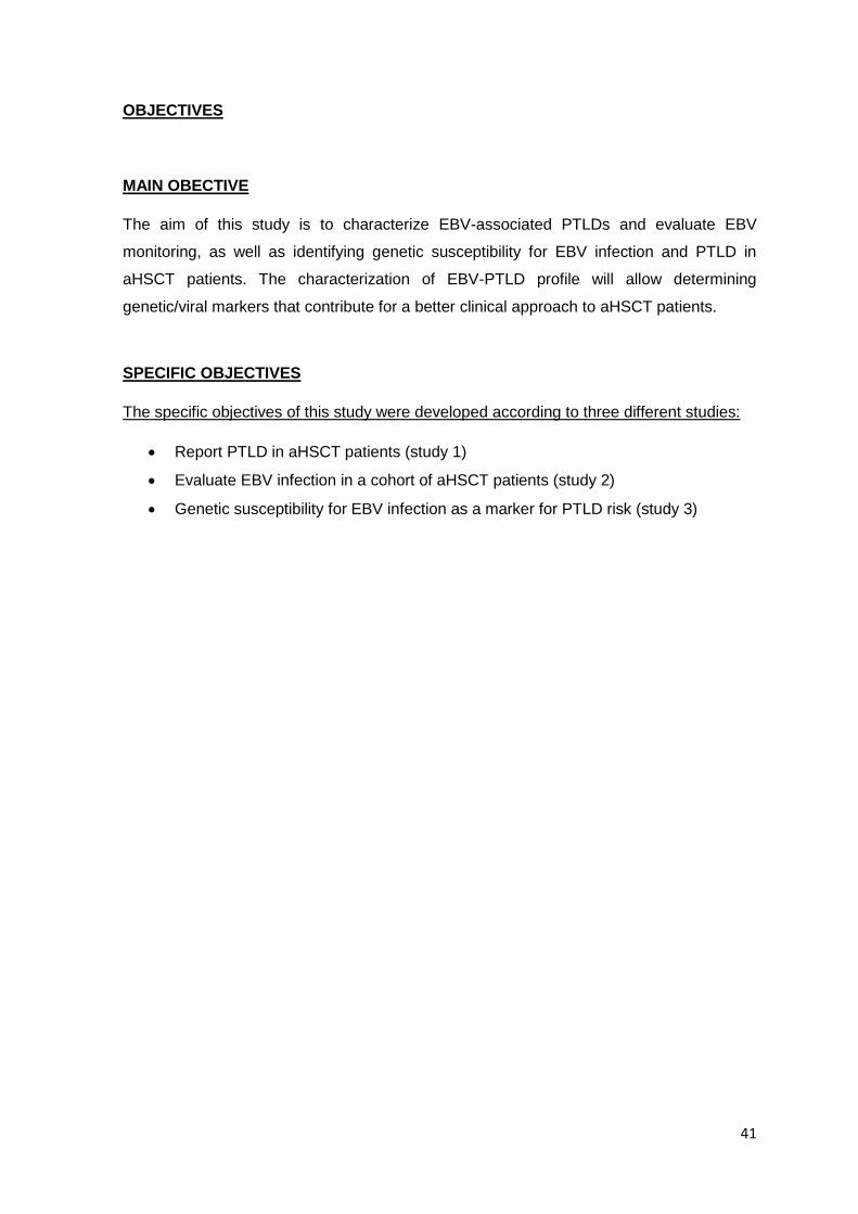
41
OBJECTIVES
MAIN OBECTIVE
The aim of this study is to characterize EBV-associated PTLDs and evaluate EBV
monitoring, as well as identifying genetic susceptibility for EBV infection and PTLD in
aHSCT patients. The characterization of EBV-PTLD profile will allow determining
genetic/viral markers that contribute for a better clinical approach to aHSCT patients.
SPECIFIC OBJECTIVES
The specific objectives of this study were developed according to three different studies:
• Report PTLD in aHSCT patients (study 1)
• Evaluate EBV infection in a cohort of aHSCT patients (study 2)
• Genetic susceptibility for EBV infection as a marker for PTLD risk (study 3)

42

43
STUDY I
POST-TRANSPLANT LYMPHOPROLIFERATIVE DISORDER IN HEMATOPOIETIC
STEM CELL TRANSPLANT PATIENTS: A SINGLE CENTER RETROSPECTIVE STUDY
BETWEEN 2005 AND 2012
Article published on Molecular Medicine Reports (Attachment I)
44
TITLE
POST-TRANSPLANT LYMPHOPROLIFERATIVE DISORDER IN HEMATOPOIETIC
STEM CELL TRANSPLANT PATIENTS: A SINGLE CENTER RETROSPECTIVE STUDY
BETWEEN 2005 AND 2012
AUTHORS:
Joana Marinho-Dias 1-3 , João Lobo 3-5, Rui Henrique 3-5, Inês Baldaque 1, Carlos Pinho-Vaz
6, Luísa Regadas 6, Rosa Branca 6, Fernando Campilho 6, António Campos Jr 6, Rui
Medeiros 1-3,7, Hugo Sousa1,2
Affiliations
1 Virology Service, Portuguese Oncology Institute of Porto; 2 Molecular Oncology and Viral
Pathology Group, IPO-Porto Research Center (CI-IPOP), Portuguese Oncology Institute of
Porto, 4200-072 Porto; 3 Abel Salazar Institute for The Biomedical Sciences (ICBAS),
University of Porto, 4050-313 Porto; 4 Department of Pathology, Portuguese Oncology
Institute of Porto; 5 Cancer Biology and Epigenetics Group, IPO-Porto Research Center (CI-
IPOP); 6 Bone Marrow Transplantation Unit, Portuguese Oncology Institute of Porto, 4200-
072 Porto; 7 Research Department, Portuguese League Against Cancer (LPCC-NRNorte),
4200-172 Porto, Portugal
Correspondence to:
Joana Marinho Dias / Hugo Sousa, MD PhD
Serviço de Virologia, Laboratórios 4º Piso
Instituto Português de Oncologia do Porto FG EPE
Rua Dr. António Bernardino Almeida
4200-072 Porto, Portugal.
Phone: +351 22 508 4000 (ext 5410)
Fax: +351 22 508 4001
E-mail: [email protected] or [email protected]
45
ABSTRACT
Background: Post-transplant lymphoproliferative disorder (PTLD), despite its rarity, is an
important mortality/morbidity event in transplant patients. The purpose of the present study
was to retrospectively examine the clinical and pathologic characteristics, and outcomes of
PTLD at the Portuguese Oncology Institute of Porto.
Material and Methods: A retrospective review of patient information was performed for
patients that developed PTLD following allogeneic hematopoietic stem cell transplant
(aHSCT) and were diagnosed between 2005 and 2012. The present study included a total
of 15 patients, 8 females (53.3%) and 7 males (46.7%), with different clinicopathological
characteristics.
Results: The most frequent clinical condition inducing aHSCT was acute lymphocytic
leukemia (40.0%). Conditioning regimens consisted primarily in busulfan and
cyclophosphamide, with anti-thymocyte globulin, and myeloablation was the preferential
treatment. Epstein-Barr virus (EBV) was present in all patients with a median time of
diagnosis following transplant of 75 days (range, 25-485 days) and a median viral load of
4.75 log10 copies/ml (range, 3.30-6.26 log10 copies/ml). PTLD diagnosis was mainly
assessed by clinical findings, and histological confirmation was available for 5 patients: 3
monomorphic, 1 polymorphic and 1 with early lesions of PTLD.
Discussion/Conclusion: To the best of our knowledge, this is the first study to describe
PTLD cases in HSCT patients in Portugal. The data reinforces the importance of performing
EBV monitoring in high-risk patients, particularly those receiving a transplant from
mismatch/unrelated donors, and those with myeloablative conditioning regimen including
antithymocyte globulin. The results also suggested that EBV viral load may be significant
for the prediction of PTLD development.
KEYWORDS: Post-Transplant Lymphoproliferative Disorder; Post-Transplant
Lymphoproliferative Disease, Epstein-Barr Virus.
46
INTRODUCTION
The development of lymphoid proliferations after transplantation has been recognized for
more than a quarter of century as an important morbidity factor (Castellano-Sanchez, Li et
al. 2004). The post-transplant lymphoproliferative disorder (PTLD) refers to a
heterogeneous group of lymphoproliferative diseases, which vary from uncomplicated, self-
limiting infectious mononucleosis, to malignant lymphoma. The histological characterization
varies from reactive-appearing, polyclonal lymphoid infiltrates or undifferentiated cells that
are morphologically indistinguishable from malignant lymphoma or plasma cell myeloma
(Gulley and Tang 2010, Mucha, Foroncewicz et al. 2010, Glotz, Chapman et al. 2012).
PTLD is relatively rare; nevertheless, it is the most frequent malignant disease early after
transplantation, with the majority of cases being reported in the first year after
transplantation (Kalinova, Indrakova et al. 2009, Glotz, Chapman et al. 2012, Atalay,
Gokahmetoglu et al. 2014). Risk factors for PTLD development include young age and age
over 50 years at transplantation, white race, unrelated or HLA-mismatched graft, Epstein-
Barr virus (EBV)-seronegative status prior to transplant, primary EBV infection, type of
organ transplant, intensity of immunosuppression and the occurrence of concomitant
cytomegalovirus disease (Kim, Kim et al. 2010, Glotz, Chapman et al. 2012).
Not all PTLD cases are EBV-related, but consistent data recognize primary EBV infection
as the most important risk factor for PTLD development (Funch, Walker et al. 2005, Caillard,
Lelong et al. 2006). Indeed, the immunosuppression after transplantation in an EBV-
seropositive patient reduces the activity of the patients’ EBV-specific cytotoxic T-cell
surveillance, which increases the probability of uncontrolled proliferation of EBV-infected B-
cells and subsequent progression to PTLD (Allen, Preiksaitis et al. 2009). Moreover,
transplant recipients experiencing primary EBV infection during the early posttransplant
period seem to be particularly susceptible to developing EBV-specific PTLD of B-cell origin,
reflecting their lack of any preexisting EBV-specific T-cell immunity (Allen, Preiksaitis et al.
2009, Glotz, Chapman et al. 2012).
The overall incidence of PTLD varies from 1 to 22% depending on the presence of risk
factors, namely the transplanted organ, patient age, EBV serostatus from recipient and
donor, aggressiveness of immunosuppression and combination of risk factors (Bar-Natan
and Nagler 2006). The cumulative incidence of PTLD in allogeneic hematopoietic stem cell
transplantation (HSCT) recipients is 1.0% (range 0.5-1.8%), with slightly higher rates in the
pediatric population (Castellano-Sanchez, Li et al. 2004, Grywalska, Markowicz et al. 2013).
Survival rates depend mainly on the type of PTLD, extent of disease and patient age: While
pediatric patients and those with localized disease seem to have a better prognosis,

47
monomorphic PTLDs are the most aggressive forms (Kalinova, Indrakova et al. 2009, Kim,
Kim et al. 2010, Luo, Zhang et al. 2014).
The purpose of this study was to examine the clinical and pathologic characteristics, as well
as the outcome of PTLD after allogeneic hematopoietic stem cell transplant (aHSCT), in
patients diagnosed at the Portuguese Oncology Institute (Porto, Portugal) between 2005
and 2012.
MATERIALS AND METHODS
Type of study and study participant
We retrospectively reviewed the information of patients that developed PTLD after aHSCT
at the Portuguese Oncology Institute of Porto (IPO Porto) between 2005 and 2012. The
study included a total of 15 patients, 8 females (53.3%) and 7 males (46.7%), with different
lymphoid malignancies who attended at the Bone Marrow Transplant Service of IPO Porto.
All cases included were histologically confirmed. PTLD diagnosis was performed by a
Pathologist from the institution using to the World health organization (Spolverato, Kim et
al.) Classification of Tumours of Haematopoietic and Lymphoid Tissues (4th Edition).
Sample processing and EBV detection
Samples were collected in EDTA-containing tubes (Vacutainer®; BD Biosciences, Franklin
Lakes, NJ, USA) and stored in freezing temperature prior to processing. Blood samples
were collected retrospectively from the institution archives. DNA was extracted by MagNA
Pure Compact Nucleic Acid Isolation kit I (Roche Diagnostics GmbH, Mannheim, Germany).
DNA/RNA quality was assessed by measuring the absorbance at 260/280 nm using the
NanoDrop 1000 Spectrophotometer v3.7 (Thermo Fisher Scientific, Inc., Wilmington, DE,
USA).
All patients submitted to aHSCT were monitored for EBV infection upon request from
clinicians after clinical suspicion. EBV detection was performed at the Virology Service of
IPO Porto using the commercial Real-Time PCR kit EBV Q-PCR Alert (Nanogen Advanced
Diagnostics S.p.A., Trezzano sul Naviglio, Italy) which targets a region from EBV nuclear
antigen 1 gene (EBNA1). Amplification was performed with the ABI PRISM 7300 Sequencer
Detection System (Applied Biosystems; Thermo Fisher Scientific, Inc., Waltham, MA, USA)
and results were obtained by measuring the geometric increase of probe fluorescence
during amplification and samples were considered positive when the exponential curve
exceeded the cycle threshold line. Regarding amplification quality, positive and negative
controls were used: as negative control we used double distilled water in replacement of

48
template DNA; and as positive control we have used samples from the external quality
control panel used at the Virology Service for EBV diagnosis.
Data collection
Clinic-pathological data was extracted from institutional databases including pre-transplant
recipient age, gender, underlying disease, HLA-donor-recipient status, EBV serological
status of the recipient, source of stem cells, conditioning regimen and use of ATG; post-
transplant information (clinical findings, date of PTLD suspicion, date of PTLD confirmation,
PTLD type, GVHD prophylaxis, GVHD type and outcome) and viral data (date of EBV
suspicion, EBV viral load).
Statistical analysis
Statistical analysis was performed using the SPSS version 20.0 software (IBM Corp.,
Armonk, NY, USA). Overall survival was defined as the time between the date of transplant
and the date of last follow-up or mortality. The differences in survival were calculated using
the log-rank test and the Kaplan-Meier method.
RESULTS
The study included a total of 15 patients, 8 females (53.3%) and 7 males (46.7%), with
median age of 10 years-old (range 3-38) - Table IA. Patients had a median follow-up time
of 14 months (range: 2-72). Primary diagnoses of patients included in this study included
paroxysmal nocturnal hemoglobinuria (n=1), primary immunodeficiency (n=1), acute
lymphocytic leukemia (n=6), acute myelogenous leukemia (n=4), chronic myelogenous
leukemia (n=1), myelodysplastic/myeloproliferative syndrome (n=1) and congenital
amegakaryocytic thrombocytopenia (n=1). Most of patients had mismatched/unrelated
donors (73.3%) and the collection of cells was mainly performed by peripheral blood stem
cells (80.0%). Myeloablative conditioning was used in 14 patients and ATG in 12 patients.
Transplant-related information for each patient is described in Table IIA.
Regarding the clinical presentation of patients, 2 presented with fever, 12 had increased
liver enzymes, adenomegalies were observed in 2 patients and 12 patients had also
increased lactate dehydrogenase. EBV serological status prior to transplantation were
evaluated according to presence of IgM and IgG titers in plasma samples. Serological status
was divided in three groups: susceptible (absence of IgM and IgG), active infection
(presence of IgM and/or IgG) and finally, past infection (absence of IgM and presence of
IgG).

49
The development of EBV infection was present in all of 15 patients, with a median time of
diagnosis after transplant of 68 days (range 29-464 days), with 80% (n=12) of them detected
<180 days after transplant, and with a median viral load of 4.75 log10 copies/ml (range 3.30-
6.26 log10 copies/ml; Fig. 1A). PTLD diagnosis occurred approximately in the same period
where EBV infection occurred (mean 135, median 75 days and range 25-485 days vs. mean
130 days, median 68 days and range 29-464 days, respectively). PTLD classification was
available for only 5 patients and included monomorphic-type PTLD (n=3), polymorphic
PTLD (n=1) and reactive plasmocytic hyperplasia (early lesions) (n=1) (Table IIIA; Fig. 2A).
The remaining cases were not histological confirmed, and diagnosis was established by
considering all clinical findings.
We observed graft-vs.-host disease (GVHD) in 13 patients (93.3%): 3 with acute GVHD
(20.0%), 2 with chronic GVHD (13.3%) and 8 with both (53.3%). Considering the grade of
acute GVHD, all patients with clinical information had a grade of II or higher. Regarding
chronic GVHD, 3 patients had an evolution of acute-to-chronic, while 7 had a de novo
chronic GVHD; two patients experienced extensive disease and 5 had only limited disease
(Table IIA).
As for the overall outcome, 8 patients are still alive (53.3%), 5 with no signs of disease
(33.3%) and 3 with evidence of disease (20.0%); and 7 patients have died (46.7%), 4 from
complications associated with the transplant (26.7%) and 3 from progression of disease
(20.0%) (Table IIA). A Kaplan-Meier plot was obtained by evaluating the cumulative survival
of these patients, in months (Fig. 3A).

50
Table 1A: Clinical characteristics of patients.
N (%)
Age, median (range) years old 10 (3-38)
Sex, n (%)
Male 7 (46.7%)
Female 8 (53.3%)
Underlying disease, n (%)
Acute leucemia 10 (66.6%)
Chronic leucemia 1 (6.7%)
Myelodysplastic/Myeloproliferative syndrome 1 (6.7%)
Others 3 (20.0%)
HLA donor, n (%)
Match/Related 4 (26.7%)
Mismatched/Unrelated 11 (73.3%)
Source of cells, n (%)
PBSC 12 (80.0%)
BM 2 (13.3%)
UCB 1 (6.7%)
Conditioning regímen, n (%)
MAC 14 (93.3%)
RIC 1 (6.7%)
ATG, n (%)
Yes 12 (85.7%)
No 2 (14.5%)
Legend: ATG: anti-thymocyte globulin; PBSC: peripheral blood stem cells; BM: bone marrow;
UCB: umbilical cord blood; RIC: reduced-Intensity conditioning; MAC: myeloablative conditioning;
HLA: Human leukocyte antigen.
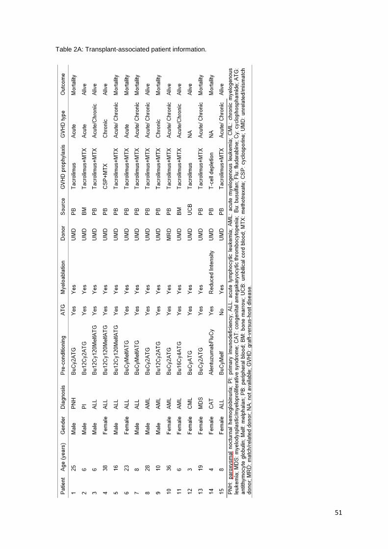
51
Table 2A: Transplant-associated patient information.
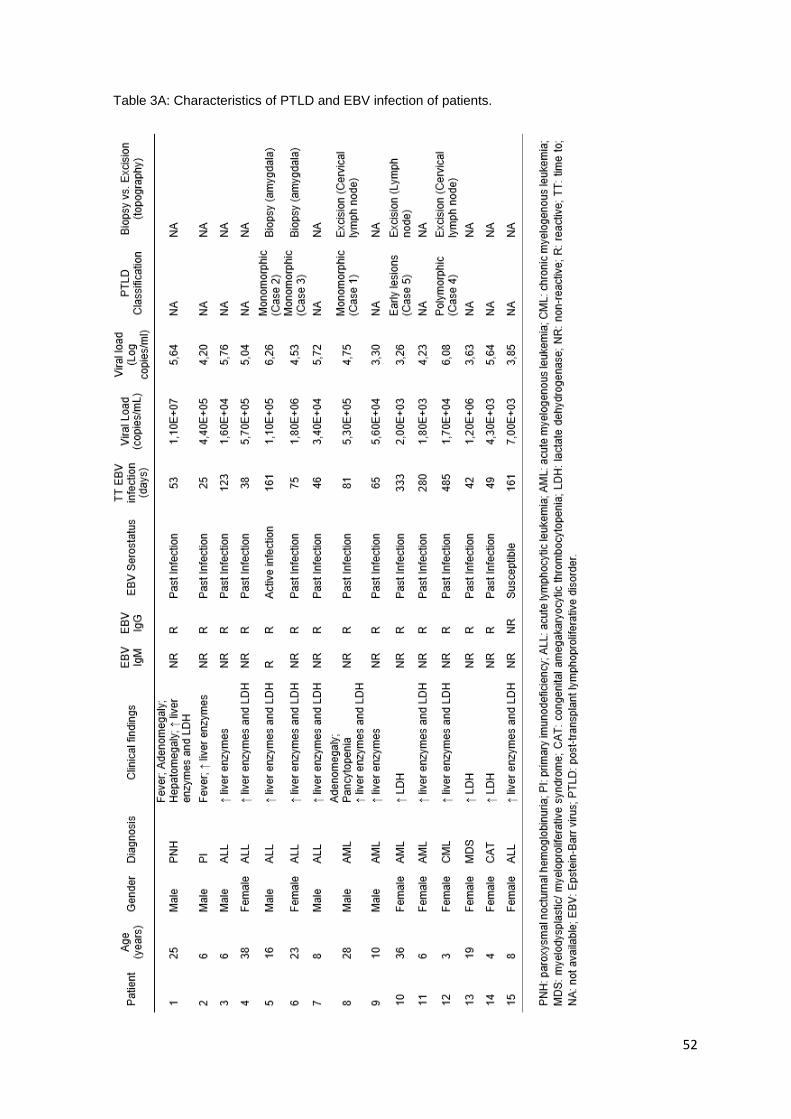
52
Table 3A: Characteristics of PTLD and EBV infection of patients.
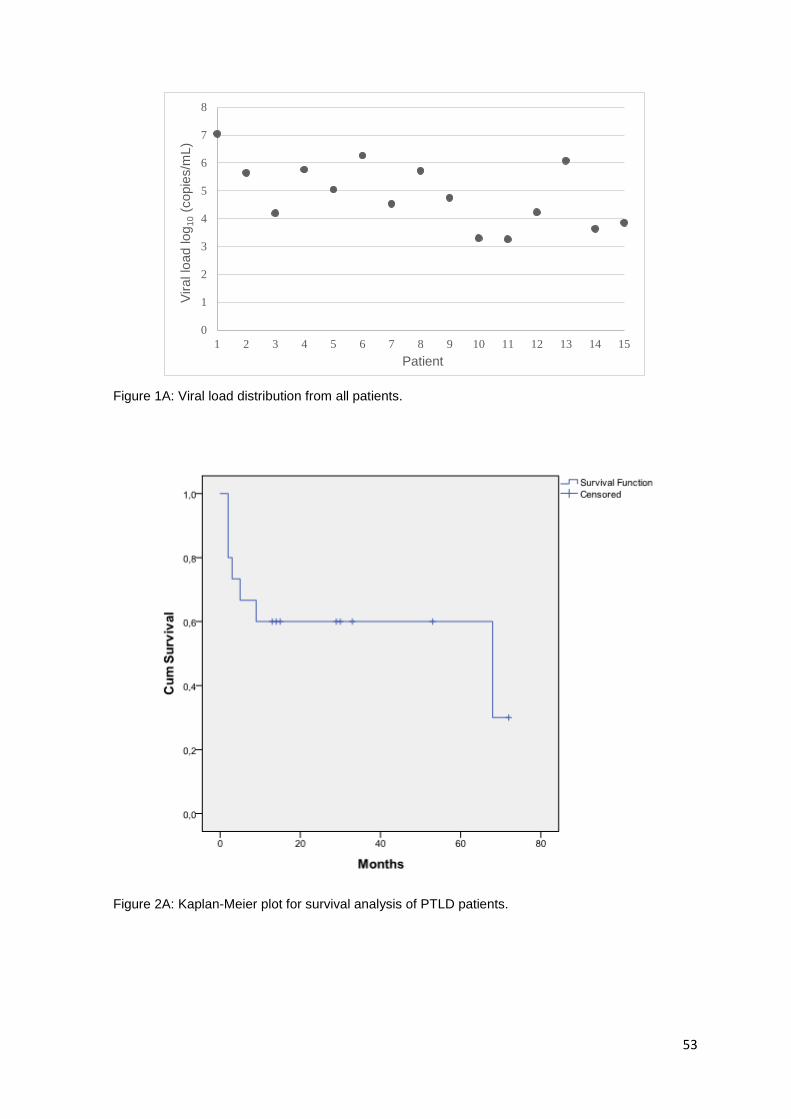
53
Figure 1A: Viral load distribution from all patients.
Figure 2A: Kaplan-Meier plot for survival analysis of PTLD patients.
0
1
2
3
4
5
6
7
8
1 2 3 4 5 6 7 8 9 10 11 12 13 14 15
Viral lo
ad log
10
(copie
s/m
L)
Patient

54
DISCUSSION
PTLD is one of the most serious complications of immunosuppression in patients who
undergo hematopoietic stem cell transplantation with high impact on morbidity and mortality
in patients (Loren, Porter et al. 2003). EBV infection has been strongly associated with the
development of PTLD, although, EBV-negative PTLD cases have been reported especially
late after transplantation (>1 year) (Choi, Park et al. 2010, Ibrahim and Naresh 2012).
In this retrospective analysis, we verify that PTLD affects individuals of all age groups and
several types of hematological malignancies and the majority have had unrelated donors.
Our patients had different types of pre-conditioning regimens (myeloablative in 14 patients),
with predominance in busulfan and cyclophosphamide. Since the types of regimen are
varied, they appear not to have a direct correlation with the development of PTLD. ATG was
used in almost all patients, except for one, and without absolute prevalence date it is difficult
to confirm if its use is directly correlated with PTLD development. GVHD prophylaxis was
performed mainly with tacrolimus, and concomitant with MTX, and still patients have
developed some type of GVHD which indicates that altering prophylaxis regimen should be
taken in consideration.
EBV infection is associated with the intermediate period after allogeneic stem cell
transplantation, which means, it occurs mainly after 3 weeks to 3 months posttransplant
Burns, Rana et al. (2016). In our case series, EBV infection was diagnosed at a median of
68 days after transplant. Viral infection during this period is correlated to delayed or
incomplete reconstitution of specific immunity, or patients experiencing GVHD (Safdar
2011).
Frequently, the median onset of PTLD development is 3 months, with a range of 2-5 months
after transplantation (Luo, Zhang et al. 2014), which is consistent with our data. Symptoms
are quite nonspecific, with patients presenting with fever, malaise, enlarged lymph nodes
and high levels of LDH, which were the factors for clinical PTLD suspicion in our patients
(Gulley and Tang 2010). All patients that developed PTLD had an EBV infection at some
point prior to transplantation. EBV positivity is directly related to PTLD development since
its infection, or increase in viral load up to 2000 copies/mL, occurs mainly, at the same time
PTLD is diagnosed. PTLD is more frequent in EBV-seronegative patients receiving
allografts from EBV-seropositive donors and in patients with delayed immune reconstitution
due to T-cell-depletion or HLA-mismatched donor. In a study conducted by Brunstein,
Weisdorf et al. (2006), 15 of 335 patients developed a EBV-related complication, at a
median of 133 days (range 52-407 days) which is consistent with our results (Brunstein,
Weisdorf et al. 2006).

55
As previously described by Al-Mansour, Nelson et al. (2013), PTLD has mortality rates
reaching up to 70-90%, which is higher than our results (46.7%) (Bhatia, Ramsay et al.
1996). Survival rates depend on age and stage of disease at the time of diagnosis, with
pediatric and patients with localized disease with best prognosis (Kalinova, Indrakova et al.
2009). In our study, overall patient survival was not affected by the development of PTLD.
This study demonstrates that frequent monitorization of EBV viral load in patients receiving
aHSCT is extremely important. The infection occurs mainly between 2 and 4 months after
transplant and precedes the development of PTLD, and especially the viral load may be of
significant importance for the monitorization and early diagnosis of PTLD.
AKNOWLEDGMENTS
The authors would like to acknowledge the support of Rute Silva from the Bone Marrow
Transplant Service at IPO Porto for the help in the access to some clinical data. The first
author received a grant for the development of project from the Portuguese League Against
Cancer (Liga Portuguesa Contra o Cancro – Núcleo Regional do Norte) between April and
September 2016.
FINANCIAL DISCLOSURE
All authors declare no competing financial interests.
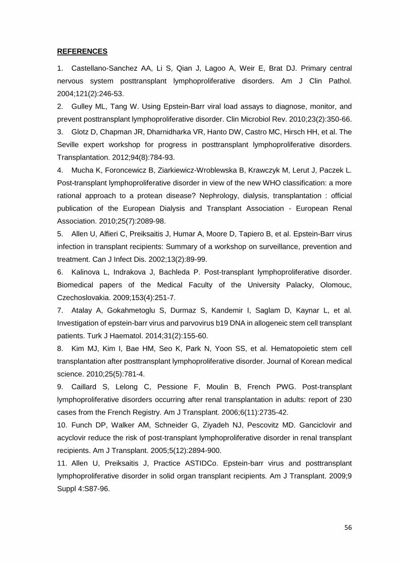
56
REFERENCES
1. Castellano-Sanchez AA, Li S, Qian J, Lagoo A, Weir E, Brat DJ. Primary central
nervous system posttransplant lymphoproliferative disorders. Am J Clin Pathol.
2004;121(2):246-53.
2. Gulley ML, Tang W. Using Epstein-Barr viral load assays to diagnose, monitor, and
prevent posttransplant lymphoproliferative disorder. Clin Microbiol Rev. 2010;23(2):350-66.
3. Glotz D, Chapman JR, Dharnidharka VR, Hanto DW, Castro MC, Hirsch HH, et al. The
Seville expert workshop for progress in posttransplant lymphoproliferative disorders.
Transplantation. 2012;94(8):784-93.
4. Mucha K, Foroncewicz B, Ziarkiewicz-Wroblewska B, Krawczyk M, Lerut J, Paczek L.
Post-transplant lymphoproliferative disorder in view of the new WHO classification: a more
rational approach to a protean disease? Nephrology, dialysis, transplantation : official
publication of the European Dialysis and Transplant Association - European Renal
Association. 2010;25(7):2089-98.
5. Allen U, Alfieri C, Preiksaitis J, Humar A, Moore D, Tapiero B, et al. Epstein-Barr virus
infection in transplant recipients: Summary of a workshop on surveillance, prevention and
treatment. Can J Infect Dis. 2002;13(2):89-99.
6. Kalinova L, Indrakova J, Bachleda P. Post-transplant lymphoproliferative disorder.
Biomedical papers of the Medical Faculty of the University Palacky, Olomouc,
Czechoslovakia. 2009;153(4):251-7.
7. Atalay A, Gokahmetoglu S, Durmaz S, Kandemir I, Saglam D, Kaynar L, et al.
Investigation of epstein-barr virus and parvovirus b19 DNA in allogeneic stem cell transplant
patients. Turk J Haematol. 2014;31(2):155-60.
8. Kim MJ, Kim I, Bae HM, Seo K, Park N, Yoon SS, et al. Hematopoietic stem cell
transplantation after posttransplant lymphoproliferative disorder. Journal of Korean medical
science. 2010;25(5):781-4.
9. Caillard S, Lelong C, Pessione F, Moulin B, French PWG. Post-transplant
lymphoproliferative disorders occurring after renal transplantation in adults: report of 230
cases from the French Registry. Am J Transplant. 2006;6(11):2735-42.
10. Funch DP, Walker AM, Schneider G, Ziyadeh NJ, Pescovitz MD. Ganciclovir and
acyclovir reduce the risk of post-transplant lymphoproliferative disorder in renal transplant
recipients. Am J Transplant. 2005;5(12):2894-900.
11. Allen U, Preiksaitis J, Practice ASTIDCo. Epstein-barr virus and posttransplant
lymphoproliferative disorder in solid organ transplant recipients. Am J Transplant. 2009;9
Suppl 4:S87-96.

57
12. Bar-Natan M, Nagler A. Epstein-Barr virus-associated post-transplant
lymphoproliferative disorder. Isr Med Assoc J. 2006;8(3):205-7.
13. Grywalska E, Markowicz J, Grabarczyk P, Pasiarski M, Rolinski J. Epstein-Barr virus-
associated lymphoproliferative disorders. Postepy Hig Med Dosw (Online). 2013;67:481-
90.
14. Luo L, Zhang L, Cai B, Li H, Huang W, Jing Y, et al. Post-transplant lymphoproliferative
disease after allogeneic hematopoietic stem cell transplantation: a single-center
experience. Annals of transplantation. 2014;19:6-12.
15. Ibrahim HA, Naresh KN. Posttransplant lymphoproliferative disorders. Advances in
hematology. 2012;2012:230173.
16. Choi JH, Park BB, Suh C, Won JH, Lee WS, Shin HJ. Clinical characteristics of
monomorphic post-transplant lymphoproliferative disorders. Journal of Korean medical
science. 2010;25(4):523-6.
17. Burns DM, Rana S, Martin E, Nagra S, Ward J, Osman H, et al. Greatly reduced risk
of EBV reactivation in rituximab-experienced recipients of alemtuzumab-conditioned
allogeneic HSCT. Bone marrow transplantation. 2016;51(6):825-32.
18. Safdar A. Principles and Practice of Cancer Infectious Diseases. New York: Springer;
2011.
19. Brunstein CG, Weisdorf DJ, DeFor T, Barker JN, Tolar J, van Burik JA, et al. Marked
increased risk of Epstein-Barr virus-related complications with the addition of antithymocyte
globulin to a nonmyeloablative conditioning prior to unrelated umbilical cord blood
transplantation. Blood. 2006;108(8):2874-80.
20. Al-Mansour Z, Nelson BP, Evens AM. Post-transplant lymphoproliferative disease
(PTLD): risk factors, diagnosis, and current treatment strategies. Curr Hematol Malig Rep.
2013;8(3):173-83.
21. Bhatia S, Ramsay NK, Steinbuch M, Dusenbery KE, Shapiro RS, Weisdorf DJ, et al.
Malignant neoplasms following bone marrow transplantation. Blood. 1996;87(9):3633-9.

58

59
STUDY II
ASSOCIATION OF EPSTEIN‑BARR VIRUS INFECTION WITH ALLOGENEIC
HEMATOPOIETIC STEM CELL TRANSPLANTATION IN PATIENTS IN PORTUGAL
Article published on Molecular Medicine Reports (Attachment II)

60
TITLE
ASSOCIATION OF EPSTEIN‑BARR VIRUS INFECTION WITH ALLOGENEIC
HEMATOPOIETIC STEM CELL TRANSPLANTATION IN PATIENTS IN PORTUGAL.
AUTHORS:
Joana Marinho-Dias 1,2,4, Inês Baldaque 1, Carlos Pinho-Vaz 3, Luís Leite 3, Rosa Branca 3,
Fernando Campilho 3, António Campos Jr 3, Rui Medeiros 1,2,4,5, Hugo Sousa1,2
Affiliations
1 Virology Service, 2 Molecular Oncology and Viral Pathology Group and 3 Bone Marrow
Transplantation Unit, Portuguese Institute of Oncology of Porto, Rua Dr. António Bernardino
de Almeida, 4200-072 Porto, PORTUGAL
4 Abel Salazar Institute for the Biomedical Sciences (ICBAS), University of Porto, Rua de
Jorge Viterbo Ferreira n.º 228, 4050-313 Porto, PORTUGAL.
5 Research Department, Portuguese League Against Cancer (LPCC-NRNorte), Estrada
Interior da Circunvalação 6657, 4200 Porto, PORTUGAL
Correspondence to:
Joana Marinho Dias, MSc / Hugo Sousa, MD PhD
Serviço de Virologia, Laboratórios 4º Piso
Instituto Português de Oncologia do Porto FG EPE
Rua Dr. António Bernardino Almeida
4200-072 Porto, Portugal.
Phone: +351 22 508 4000 (ext 5410)
Fax: +351 22 508 4001
E-mail: [email protected] or [email protected]

61
ABSTRACT
Background: Epstein-Barr virus (EBV) infection in immunocompromised patients, such as
hematopoietic stem cell transplanted patients can play a major role on the clinical outcome.
The identification of patients at higher risk of developing EBV infection may be useful for
the prevention of EBV-associated diseases.
Material and Methods: This prospective study was developed with 40 patients (27 male and
13 females, with mean age of 32.2±1.5 years old) randomly selected from the cohort of
patients undergoing aHSCT at the Bone Marrow Transplant Service of IPO Porto. Between
1st January to 31st December 2015. EBV was tested in blood samples collected during
routine procedures at Day 30, 60, 90, 120, 150 and 180 post-transplant.
Results: In our study we verified that 70.0% of our patients were EBV positive at least once
during the follow-up period. Regarding the correlation of EBV infection and clinical features
of patients, we verified that patients with unrelated donors had increased association with
EBV infection at 60- and 150-days post-transplant (OR=3.9, p=0.058; OR=8.0, p=0.043;
respectively). Moreover, myeloablative conditioning (OR=4.3, p=0.052), ATG use
(OR=12.0, p=0.030) and GVHD (OR=6.7, p=0.032) were associated with EBV infection at
at day 60, day 150 and day 90, respectively. Despite not statistically significant Overall
Survival (OS) was associated with EBV infection at day 90 and 180 after transplant (p=0.095
and p=0.097, respectively). In our series, none patient developed PTLD disease.
Discussion/Conclusion: This is the first study to report the prevalence of EBV infection at
different points in patients undergoing aHSCT from Portugal. Our study reveal that a high
percentage of patients will develop an EBV infection during the post-transplant period and
that some cofactors may influence its development. In our series EBV infection seems to
be correlated with transplant from unrelated donors, the use of myeloablative conditioning,
ATG and the development of GVHD. This study reinforces the importance of performing
EBV monitoring for a better management of post-transplanted complications.
KEYWORDS: EBV, Epstein-Barr Virus; HSCT, Hematopoietic stem cell transplant; GVHD,
Graft-versus-host disease.

62
INTRODUCTION
Epstein-Barr virus (EBV) is a ubiquitous Human Herpesvirus and infects 50% to 89% of
children and remains latent, in memory B cells, of approximately 90% of adults (Styczynski,
Tridello et al. 2016). Viral infections are known to be a major cause of morbidity and mortality
in patients undergoing hematopoietic stem cell transplants (HSCT) and Herpesvirus are
known to be among the most common viral infections in these patients (Fan, Jing et al.
2016). Moreover, monitoring of EBV DNA in peripheral blood is routinely performed in
several transplant centers, since these patients have a higher-risk of developing EBV-
related diseases (Janani, Malathi et al. 2015).
HSCT is an effective therapy in the treatment of hematological malignancies. Leukemias
and lymphomas are a major part of worldwide cancers (Howlader, Noone et al. 2017). The
regimens required for transplant produce profound immune deficiency in the early period
after transplantation (Curtis, Travis et al. 1999). This iatrogenic suppression of T cell
function in transplant recipients, leads to EBV proliferation in B cells (Janani, Malathi et al.
2015). Conditioning regimens include: myeloablative (MAC) and reduced intensity
conditioning (RIC). MACs include concomitant or single use of alkylating agents, such as
cyclophosphamide (Cy) and busulfan (Bu), while RICs are mainly performed with
fludarabine (Flu) or low doses of total body irradiation (TBI) (Xuan, Huang et al. 2012, Juric,
Ghimire et al. 2016). Choice of conditioning depends on patient’s age, underlying disease,
relevant comorbidities and type of donor. These regimens are related to several risks such
as infections, graft-versus-host disease (GVHD), post-transplant lymphoproliferative
disorder (PTLD) (Fan, Jing et al. 2016). GVHD occurs in approximately 40% to 90% of
transplanted patients (Funke, Moreira et al. 2016). ATG seems to be effective in GVHD
prophylaxis, and is related to reduced rates of relapses and infections in adults who undergo
bone marrow (BM) or peripheral blood stem cells (PBSC) transplant (Storek, Mohty et al.
2015). In allogeneic HSCT selection of grafts donor consists in matched-related sibling,
mismatched-related, matched-unrelated and mismatched-unrelated (Gratwohl, Baldomero
et al. 2010, Barriga, Ramirez et al. 2012). The priority is to find a related donor, and for
patients who lack such, they must find an unrelated donor. The probability of finding a
matched unrelated donor is around 30% to 70%, depending on the frequency of the HLA
genotype in the donor registries and patient’s ethnicity (Juric, Ghimire et al. 2016).
Rates of mortality after HSCT often reach up to 50.0% (Fan, Jing et al. 2016). Some studies
describe that intensive conditioning regimens are associated with reduction of tumor
relapses, although it might simultaneously increase the transplant-related mortality rates,
including the mortality of infections (Xuan, Huang et al. 2012).
63
This study aims to analyze patients’ clinical characteristics at time of transplantation and
evaluating risk factors related to poor outcomes.
MATERIALS AND METHODS
Type of study and study participant
A prospective follow-up study was performed with patients who underwent aHSCT at the
Bone Marrow Transplant Service of Portuguese Oncology Institute of Porto (IPO Porto)
between 1st January to 31st December of 2015 that were selected randomly. In this study,
40 patients were included, 13 females (32.5%) and 27 males (67.5%), within a 6 months
follow-up. The study used the samples that were collected during routine procedures for
viral monitoring at the Virology Service of IPO Porto. The study was approved by the local
Ethical Committee and did not interfere with the routine procedures decided by clinicians
Sample processing:
Blood samples were selected at 6 different times: 30, 60, 90, 120, 150 and 180 days after
transplant. Samples were collected in EDTA-containing tubes (BD Vacutainer®, NJ) and
stored prior to processing and DNA was extracted by MagNA Pure Compact Nucleic Acid
Isolation kit I (Roche, Germany). DNA/RNA quality was assessed by measuring the
absorbance at 260/280 nm using the NanoDrop 1000 Spectrophotometer v3.7 (Thermo
Scientific, Wilmington, DE, USA).
EBV detection
EBV detection was performed by a real-time PCR protocol targeting EBV polymerase gene
(EBV POL) with forward and reverse as previously reported (Marinho-Dias and Sousa
2013). Amplification was performed with the ABI PRISM 7300 Sequencer Detection System
(Applied Biosystems) and results were obtained by measuring the geometric increase of
probe fluorescence during amplification and samples were considered positive when the
exponential curve exceeded the cycle threshold line.
Quality Control
Regarding quality control, all real-time PCR amplifications used positive and negative
controls: as negative control, we used double distilled water in replacement of template
DNA; and as positive control we have used samples from the External Quality Control panel
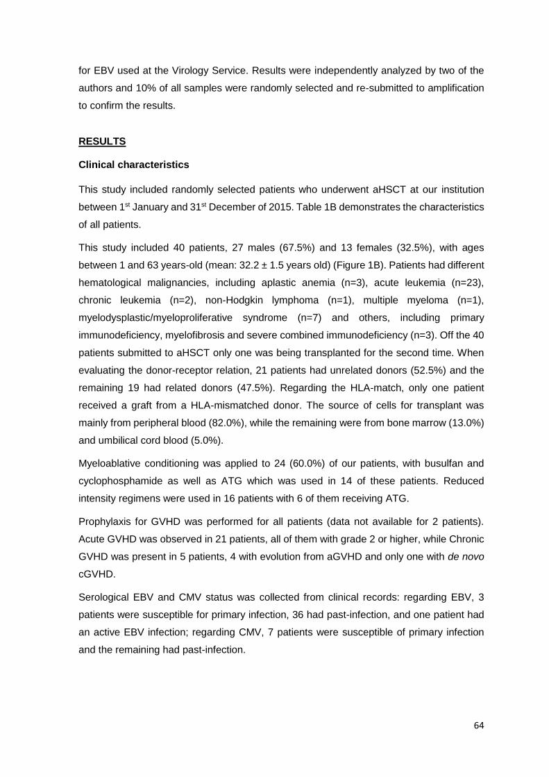
64
for EBV used at the Virology Service. Results were independently analyzed by two of the
authors and 10% of all samples were randomly selected and re-submitted to amplification
to confirm the results.
RESULTS
Clinical characteristics
This study included randomly selected patients who underwent aHSCT at our institution
between 1st January and 31st December of 2015. Table 1B demonstrates the characteristics
of all patients.
This study included 40 patients, 27 males (67.5%) and 13 females (32.5%), with ages
between 1 and 63 years-old (mean: 32.2 ± 1.5 years old) (Figure 1B). Patients had different
hematological malignancies, including aplastic anemia (n=3), acute leukemia (n=23),
chronic leukemia (n=2), non-Hodgkin lymphoma (n=1), multiple myeloma (n=1),
myelodysplastic/myeloproliferative syndrome (n=7) and others, including primary
immunodeficiency, myelofibrosis and severe combined immunodeficiency (n=3). Off the 40
patients submitted to aHSCT only one was being transplanted for the second time. When
evaluating the donor-receptor relation, 21 patients had unrelated donors (52.5%) and the
remaining 19 had related donors (47.5%). Regarding the HLA-match, only one patient
received a graft from a HLA-mismatched donor. The source of cells for transplant was
mainly from peripheral blood (82.0%), while the remaining were from bone marrow (13.0%)
and umbilical cord blood (5.0%).
Myeloablative conditioning was applied to 24 (60.0%) of our patients, with busulfan and
cyclophosphamide as well as ATG which was used in 14 of these patients. Reduced
intensity regimens were used in 16 patients with 6 of them receiving ATG.
Prophylaxis for GVHD was performed for all patients (data not available for 2 patients).
Acute GVHD was observed in 21 patients, all of them with grade 2 or higher, while Chronic
GVHD was present in 5 patients, 4 with evolution from aGVHD and only one with de novo
cGVHD.
Serological EBV and CMV status was collected from clinical records: regarding EBV, 3
patients were susceptible for primary infection, 36 had past-infection, and one patient had
an active EBV infection; regarding CMV, 7 patients were susceptible of primary infection
and the remaining had past-infection.
65
Table 1B: Clinical characteristics of patients.
Legend: Bu: Busulfan; Cy: Cyclophosphamide; Flu: Fludarabine; Melf: Melphalan; ATG: Anti-thymocyte
globulin; PBSC: Peripheral blood stem cells; BM: Bone marrow; UCB: Umbilical cord blood.
Characteristics of patients Value
Age, years, median (range) 32.2 (1-63)
Sex (n, %)
Male 27 (67.5%)
Female 13 (32.5%)
Underlying disease
Aplastic anemia 3 (7.5%)
Acute leukemia 23 (57.5%)
Chronic leukemia 2 (5.0%)
Non-Hodgkin lymphoma 1 (2.5%)
Multiple myeloma 1 (2.5%)
Myelodysplastic/Myeloproliferative syndrome 7 (17.5%)
Others 3 (7.5%)
Conditioning regimen
BuCy 4 (10.0%)
BuCy2 20 (50.0%)
Cy 1 (2.5%)
FluBu 10 (25.0%)
FluCy 3 (7.5%)
FluMelf 1 (2.5%)
ATG
Yes 20 (50.0%)
No 20 (50.0%)
Type of donor
Related 19 (47.5%)
Mismatched/Unrelated 21 (52.5%)
Source of cells
PBSC 33 (82.5%)
BM 5 (12.5%)
UCB 2 (5.0%)

66
Figure 1B: Frequency of age groups (Group1 - <=11 years-old; Group 2 – 12-18 years-old; Group 3
– 19-36 years-old; Group 4 – 37-50 years-old; Group 5 – 51-63 years-old).
EBV infection analysis
Samples from all patients were collected at 6 periods post-transplant. Results regarding the
detection of EBV demonstrated a prevalence of 38.3% positive samples and results
according to periods are shown in figure 2B. Considering number of EBV positive samples
we had: Day 30 - 10 samples (25.0%); Day 60 - 18 samples (47.4%); Day 90 – 13 samples
(38.2%); Day 120 – 9 samples (31.0%); Day 150 – 10 samples (45.5%); Day 180 – 7
samples (58.3%).
Figure 2B: Percentage of EBV infection at different stages post-transplant.
0
10
20
30
40
50
60
70
80
90
100
30 60 90 120 150 180
Per
cen
tage
Days
Negative
Positive
n
=
n
=
n
=
67
EBV infection and clinical characteristics
The analysis according to gender seem to show that post-transplant EBV infection is more
frequent in females (OR=8.3, p=0.06), although with borderline statistical significance.
Despite not statistically significant, we have found that EBV infection is more frequent in
patients with unrelated donors (OR=2.5, p=0.170), engrafted with PBSC (OR=2.0,
p=0.410), patients submitted to myeloablative conditioning regimen (OR=2.4, p=0.166),
using ATG in the conditioning regimen (OR=2.2, p=0.176), and that developed aGVHD
(OR=2.5, p=0.170). EBV serostatus prior to transplant does not seem to be related with the
development of infection during the post-transplant period. Patients with chronic GVHD
were all EBV positive at least once, suggesting that cGVHD may also related with infection.
Additionally, we proceeded with the analysis of EBV infection at the different times during
the follow-up period. At 60 days post-transplant, we verified that EBV infection was
associated with transplants from unrelated donors OR=3.9, p=0.058), myeloablative
conditioning (OR=4.3, p=0.052), and ATG use (OR=3.6, p=0.099). At 90 days only GVHD
was related a higher risk of infection (OR=6.7, p=0.032). Finally, at 150 days, risk of infection
was related with unrelated donors (OR=8.0, p=0.043), ATG (OR=12.0, p=0.03) and GVHD
(OR=5.6, p=0.099).
By the analysis of cox regressions on cumulative survival considering patient’s sex, donor,
myeloablative/reduced intensity regimen, with/without ATG and development of aGVHD,
we observed that there is an association of unrelated donor and EBV infection at 150 days
post-transplant (HR=8.8, p=0.03).
Follow-up
Of the 40 patients here analyzed, we observed that 16 patients are currently deceased, 6
are alive with evidence of disease and 18 are alive and without evidence of disease. The
cumulative survival was evaluated by performing a Kaplan-Meier plot (Figure 3B), and
estimated survival time was approximately 476 days. Survival analysis was correlated with
EBV DNA positivity and the following Kaplan-Meier plots were obtained (Figure 4a, b, c, d,
e and f). Results showed that EBV DNA positivity at 90 days post-transplant, is associated
with increased mortality (p=0.095), with EBV positive patients having approximately less
290 days than EBV negative patients (303.3 versus 593.2 days, respectively).
During this period, none of the patients developed PTLD, neither clinical signs suggesting
the development of PTLD.
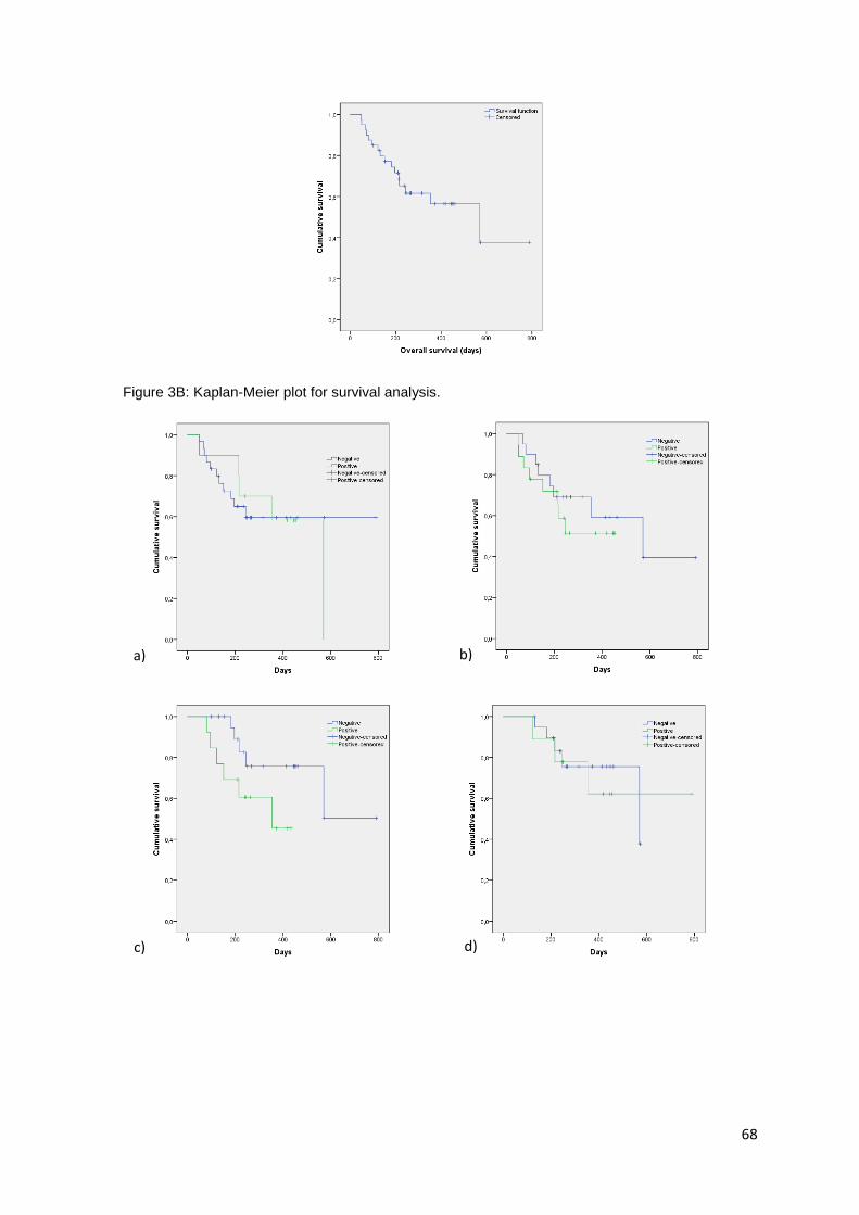
68
Figure 3B: Kaplan-Meier plot for survival analysis.
a) b)
c) d)
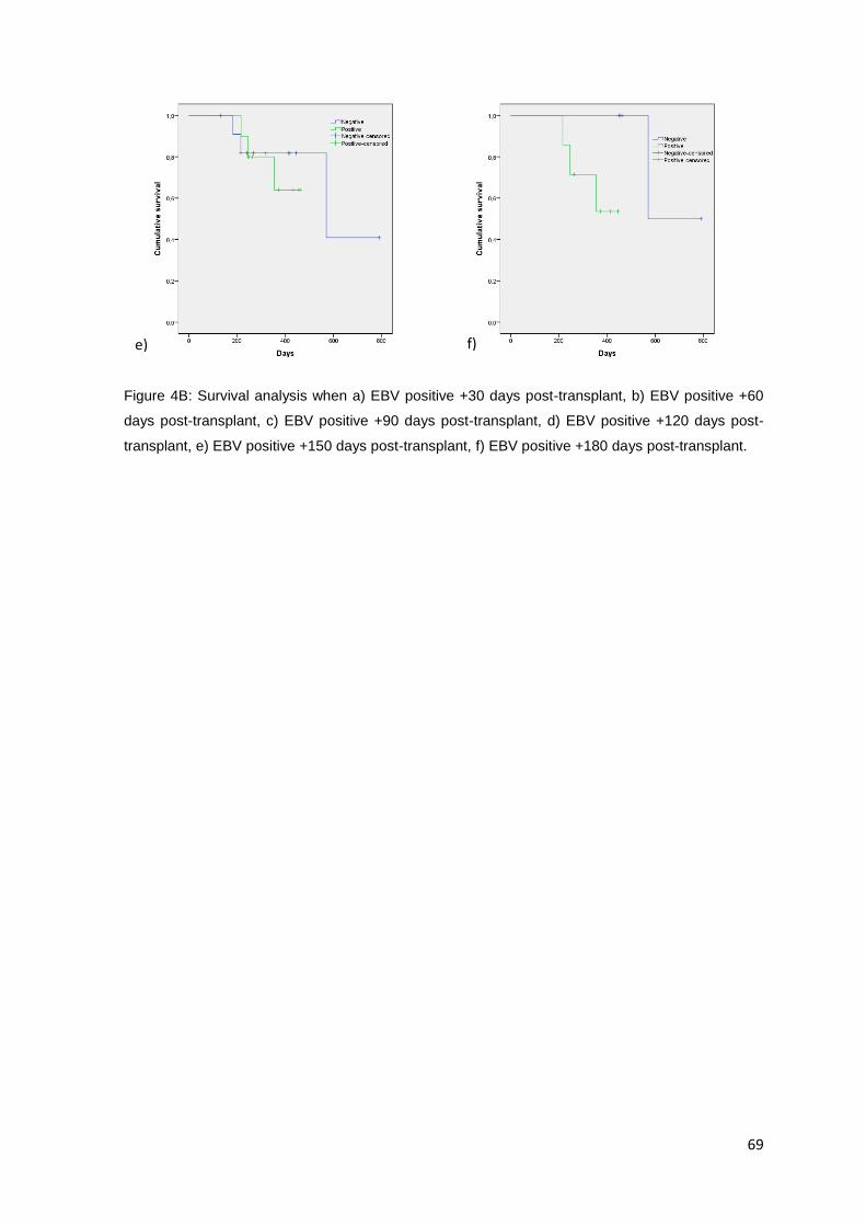
69
Figure 4B: Survival analysis when a) EBV positive +30 days post-transplant, b) EBV positive +60
days post-transplant, c) EBV positive +90 days post-transplant, d) EBV positive +120 days post-
transplant, e) EBV positive +150 days post-transplant, f) EBV positive +180 days post-transplant.
e) f)

70
DISCUSSION
Allogeneic hematological stem cell transplant (aHSCT) is an option for the treatment of
hematological malignancies and these patients are submitted to pre-transplant treatments
that reduce significantly the immune system to avoid rejection of the graft (Henig and
Zuckerman 2014). This immunosuppression is associated with the occurrence of different
events in the post-transplant period, such as development of graft-versus-host disease,
infections and post-transplant lymphoproliferative disorders (Al-Mansour, Nelson et al.
2013).
Viral infections are a major concern in the subset of aHSCT, and while CMV infection has
been consistently associated with a significant morbidity/mortality increase, EBV infection
has been underestimated in these patients since only a minority will suffer from EBV-
associated complications, such as PTLD (Preiksaitis 2004). Gulley et al., affirm that routine
monitoring of EBV infection is viable in PTLD prevention, although further studies must be
done to correlate specific viral loads to the identification of high-risk patients. Viral load is
also informative, when there is a withdrawal of immunosuppression of rituximab
administration, to verify if the treatment is successful (Gulley and Tang 2010).
In our study, we verified that 70.0% of our patients were EBV positive at least once during
the follow-up period. Blaes et al., described that EBV viremia was present in 23.0% of their
patients, which is very different from what we have found (Blaes, Cao et al. 2010). Esser et
al., observed rates of EBV infection in 49.0% in patients submitted to SCT, once again lower
than what we obtained (van Esser, van der Holt et al. 2001). Furthermore, EBV infection
varied throughout the follow-up period, with a mean of 65.6±39.6 days (range 27-183).
Duman et al., monitored EBV viral load at least once a week for 3 months and verified that
EBV reactivation occurred in approximately 14.0% of patients, which is also much lower
than what we describe (Dumas, Ruggeri et al. 2013). Styczynski et al., refer that screening
of EBV DNAemia should start within the first month after aHSCT, and that monitoring should
continue for at least 4 months after transplant, with a frequency of once a week (Styczynski,
van der Velden et al. 2016). Despite this suggestion, many authors discuss the cost-
effectiveness of EBV monitoring once-a-week and therefore many studies are required to
show what would be the best time and interval of monitoring. Moreover, EBV viral load has
been having different input data since the viral loads for treatment/follow-up cutoff are
variable. Indeed, the development of international standards for EBV viral load management
is yet to be defined (Marques, Shikanai-Yasuda et al. 2014).
Our data showed that patients with unrelated and/or mismatched donors were more prone
to develop an EBV infection post-transplant, and that using peripheral blood as source of

71
stem cells adds a higher risk for EBV infection. Studies report EBV infections of 8.8% in
myeloablative conditionings and 35.0% in reduced intensity conditionings (Cohen, Gandhi
et al. 2005), 54.0% in T-cell depletion (van Esser, Niesters et al. 2002) and 65.0% in T-cell
depletion concomitant with ATG use (van Esser, van der Holt et al. 2001). Despite UCB
has been related with higher incidences of EBV viremia, many others have failed to show
this (Dumas, Ruggeri et al. 2013). Nevertheless, studies with larger populations and
preferably with equal proportions of PBSC, BM and UCB graft receptors, should be
performed to clarify these evidences.
The intensity of conditioning regimen has been shown to directly affect the relapse and
survival of patients who undergo aHSCT (Liu, Fan et al. 2009). In our study, myeloablation
conditioning and use of ATG demonstrated a 3-times higher risk of developing EBV. These
findings are according those described by Xuan et al. that observed that intensified
conditioning increase the incidence of EBV viremia and disease. Furthermore, use of ATG,
HLA-mismatched, unrelated donor and acute aGVHD were also identified as risk factors for
EBV infection (Xuan, Huang et al. 2012).
When addressing the development of GVHD, we observed that patients who developed
aGVHD, had 3-times increased association with EBV infection. Furthermore, we noticed
that all patients with cGVHD, were positive for EBV at least once in the post-transplant
follow-up. As described previously by Janeczko et al., GVHD is related to delayed immune
reconstitution, favoring infections in the early period post-transplant. Moreover, viral
infections are also associated with delayed IR and appear to be linked to the degree of
immunosuppression. Therefore, monitoring of infections is critical in this phase (Janeczko,
Mielcarek et al. 2016).
Analysis of EBV infection on different periods post-transplant revealed that unrelated donor
(OR=3.9, p=0.058), myeloablation (OR=4.3, p=0.052) and the use of ATG (OR=3.6,
p=0.099) seem to be risk factors for EBV infection occurrence at 60 days post-transplant;
At 90+ days GVHD is connected to EBV infection (OR=6.7, p=0.032).; use of ATG
(OR=12.0, p=0.030) , unrelated donor (OR=4.8, p=0.043) and GVHD (OR=3.3, p=0.099)
are related to EBV infection at day 150 post-transplant.
Our mortality rates are from 40.0%, with a median follow-up period of 8.2±5.3 months (range
1-25 months). Similar findings have been reported by other authors, such as Styczynski et
al., that reports a mortality rate of 45.2% in a follow-up period of 4.9 years (Styczynski,
Tridello et al. 2016). EBV infection and its association with mortality were analyzed and data
revealed that EBV DNA positivity at 90 days and 180 days post-transplant, are associated
72
with increased mortality (p=0.095, 303.3 versus 593.2 days and p=0.097, 367.0 versus
679.5).
At our institution, EBV detection is not performed routinely, and with these results, we
demonstrate the necessity in EBV monitoring after transplantation, since infection seems to
have a major role in complications after transplant and mortality. It is critical to develop a
risk profile to determine which patients are at higher risk of developing EBV infection and
further complications. This profile must be obtained by information on which risks factor are
more related to EBV infection.
CONCLUSION
EBV infection is still a major concern in the subset of HSCT. Therefore, and with these
results, monitoring and identification of which variables define high risk patients are
necessary for preventing EBV-related complications and mortality.
AKNOWLEDGEMENTS
The authors would like to acknowledge the support of Rute Silva from the Bone Marrow
Transplant Service at IPO Porto for the help in the access to some clinical data. The first
author received a grant for the development of project from the Portuguese League Against
Cancer (Liga Portuguesa Contra o Cancro – Núcleo Regional do Norte) between April and
September 2016.
FINANCIAL DISCLOSURE
All authors declare no competing financial interests.

73
REFERENCES
1. Howlader N, Noone AM, Krapcho M, et al.: SEER Cancer Statistics Review, 1975-2014.
National Cancer Institute, Bethesda, MD2017.
2. Curtis RE, Travis LB, Rowlings PA, et al.: Risk of lymphoproliferative disorders after bone
marrow transplantation: a multi-institutional study. Blood 94: 2208-2216, 1999.
3. Juric MK, Ghimire S, Ogonek J, et al.: Milestones of Hematopoietic Stem Cell
Transplantation - From First Human Studies to Current Developments. Front Immunol 7:
470, 2016.
4. Xuan L, Huang F, Fan Z, et al.: Effects of intensified conditioning on Epstein-Barr virus
and cytomegalovirus infections in allogeneic hematopoietic stem cell transplantation for
hematological malignancies. Journal of hematology & oncology 5: 46, 2012.
5. Fan J, Jing M, Yang M, et al.: Herpesvirus infections in hematopoietic stem cell transplant
recipients seropositive for human cytomegalovirus before transplantation. Int J Infect Dis
46: 89-93, 2016.
6. Funke VA, Moreira MC and Vigorito AC: Acute and chronic Graft-versus-host disease
after hematopoietic stem cell transplantation. Rev Assoc Med Bras (1992) 62 Suppl 1: 44-
50, 2016.
7. Storek J, Mohty M and Boelens JJ: Rabbit anti-T cell globulin in allogeneic hematopoietic
cell transplantation. Biology of blood and marrow transplantation: journal of the American
Society for Blood and Marrow Transplantation 21: 959-970, 2015.
8. Styczynski J, Tridello G, Gil L, et al.: Impact of Donor Epstein-Barr Virus Serostatus on
the Incidence of Graft-Versus-Host Disease in Patients With Acute Leukemia After
Hematopoietic Stem-Cell Transplantation: A Study From the Acute Leukemia and Infectious
Diseases Working Parties of the European Society for Blood and Marrow Transplantation.
Journal of clinical oncology: official journal of the American Society of Clinical Oncology 34:
2212-2220, 2016.
9. Janani MK, Malathi J, Rela M, Farouk M, Padmapriya J and Madhavan HN: Genotypic
Detection of Epstein Barr Virus in Pediatric Transplant Recipients From India. Indian Pediatr
52: 946-950, 2015.
10. Styczynski J, Einsele H, Gil L and Ljungman P: Outcome of treatment of Epstein-
Barr virus-related post-transplant lymphoproliferative disorder in hematopoietic stem cell
recipients: a comprehensive review of reported cases. Transplant infectious disease: an
official journal of the Transplantation Society 11: 383-392, 2009.
11. Styczynski J, van der Velden W, Fox CP, et al.: Management of Epstein-Barr Virus
infections and post-transplant lymphoproliferative disorders in patients after allogeneic

74
hematopoietic stem cell transplantation: Sixth European Conference on Infections in
Leukemia (ECIL-6) guidelines. Haematologica 101: 803-811, 2016.
12. Solano C, Mateo EM, Perez A, et al.: Epstein-Barr virus DNA load kinetics analysis in
allogeneic hematopoietic stem cell transplant recipients: Is it of any clinical usefulness? J
Clin Virol 97: 26-32, 2017.
13. Marinho-Dias J, Lobo J, Henrique H, et al.: Post-Transplant Lymphoproliferative
Disease in Hematopoietic Stem Cell Transplant Patients: A Single Center Retrospective
Study between 2005 and 2012. Molecular medicine reports in press2018.
14. Marinho-Dias J and Sousa H: Cytomegalovirus infection and cervical cancer: from past
doubts to present questions. Acta Med Port 26: 154-160, 2013.
15. Henig I and Zuckerman T: Hematopoietic stem cell transplantation-50 years of
evolution and future perspectives. Rambam Maimonides Med J 5: e0028, 2014.
16. Al-Mansour Z, Nelson BP and Evens AM: Post-transplant lymphoproliferative disease
(PTLD): risk factors, diagnosis, and current treatment strategies. Curr Hematol Malig Rep
8: 173-183, 2013.
17. Sousa H, Boutolleau D, Ribeiro J, et al.: Cytomegalovirus infection in patients who
underwent allogeneic hematopoietic stem cell transplantation in Portugal: a five-year
retrospective review. Biology of blood and marrow transplantation: journal of the American
Society for Blood and Marrow Transplantation 20: 1958-1967, 2014.
18. Campos AB, Ribeiro J, Boutolleau D and Sousa H: Human cytomegalovirus antiviral
drug resistance in hematopoietic stem cell transplantation: current state of the art. Rev Med
Virol 26: 161-182, 2016.
19. Campos AB, Ribeiro J, Pinho Vaz C, et al.: Genotypic resistance of cytomegalovirus to
antivirals in hematopoietic stem cell transplant recipients from Portugal: A retrospective
study. Antiviral research 138: 86-92, 2017.
20. Preiksaitis JK: New developments in the diagnosis and management of
posttransplantation lymphoproliferative disorders in solid organ transplant recipients.
Clinical infectious diseases: an official publication of the Infectious Diseases Society of
America 39: 1016-1023, 2004.
21. Blaes AH, Cao Q, Wagner JE, Young JA, Weisdorf DJ and Brunstein CG: Monitoring
and preemptive rituximab therapy for Epstein-Barr virus reactivation after antithymocyte
globulin containing nonmyeloablative conditioning for umbilical cord blood transplantation.
Biology of blood and marrow transplantation: journal of the American Society for Blood and
Marrow Transplantation 16: 287-291, 2010.
22. van Esser JW, van der Holt B, Meijer E, et al.: Epstein-Barr virus (EBV) reactivation is
a frequent event after allogeneic stem cell transplantation (SCT) and quantitatively predicts
EBV-lymphoproliferative disease following T-cell--depleted SCT. Blood 98: 972-978, 2001.

75
23. Dumas PY, Ruggeri A, Robin M, et al.: Incidence and risk factors of EBV reactivation
after unrelated cord blood transplantation: a Eurocord and Societe Francaise de Greffe de
Moelle-Therapie Cellulaire collaborative study. Bone Marrow Transplant 48: 253-256, 2013.
24. Reddy N, Rezvani K, Barrett AJ and Savani BN: Strategies to prevent EBV reactivation
and posttransplant lymphoproliferative disorders (PTLD) after allogeneic stem cell
transplantation in high-risk patients. Biology of blood and marrow transplantation: journal of
the American Society for Blood and Marrow Transplantation 17: 591-597, 2011.
25. Uhlin M, Wikell H, Sundin M, et al.: Risk factors for Epstein-Barr virus-related post-
transplant lymphoproliferative disease after allogeneic hematopoietic stem cell
transplantation. Haematologica 99: 346-352, 2014.
26. Marques HH, Shikanai-Yasuda MA, dAzevedo LS, et al.: Management of post-
transplant Epstein-Barr virus-related lymphoproliferative disease in solid organ and
hematopoietic stem cell recipients. Rev Soc Bras Med Trop 47: 543-546, 2014.
27. Gulley ML and Tang W: Using Epstein-Barr viral load assays to diagnose, monitor, and
prevent posttransplant lymphoproliferative disorder. Clin Microbiol Rev 23: 350-366, 2010.
28. Bar-Natan M and Nagler A: Epstein-Barr virus-associated post-transplant
lymphoproliferative disorder. Isr Med Assoc J 8: 205-207, 2006.
29. Cohen J, Gandhi M, Naik P, et al.: Increased incidence of EBV-related disease
following paediatric stem cell transplantation with reduced-intensity conditioning. Br J
Haematol 129: 229-239, 2005.
30. van Esser JW, Niesters HG, van der Holt B, et al.: Prevention of Epstein-Barr virus-
lymphoproliferative disease by molecular monitoring and preemptive rituximab in high-risk
patients after allogeneic stem cell transplantation. Blood 99: 4364-4369, 2002.
31. Dabas R, Lee R, Servito MT, et al.: Antithymocyte Globulin at Clinically Relevant
Concentrations Kills Leukemic Blasts. Biology of blood and marrow transplantation: journal
of the American Society for Blood and Marrow Transplantation 22: 815-824, 2016.
32. Crocchiolo R, Esterni B, Castagna L, et al.: Two days of antithymocyte globulin are
associated with a reduced incidence of acute and chronic graft-versus-host disease in
reduced-intensity conditioning transplantation for hematologic diseases. Cancer 119: 986-
992, 2013.
33. Bacigalupo A: ATG in allogeneic stem cell transplantation: standard of care in 2017?
Blood Advances 1: 569-572, 2017.
34. Pidala J, Tomblyn M, Nishihori T, et al.: ATG prevents severe acute graft-versus-host
disease in mismatched unrelated donor hematopoietic cell transplantation. Biology of blood
and marrow transplantation: journal of the American Society for Blood and Marrow
Transplantation 17: 1237-1244, 2011.

76
35. Podgorny PJ, Ugarte-Torres A, Liu Y, Williamson TS, Russell JA and Storek J: High
rabbit-antihuman thymocyte globulin levels are associated with low likelihood of graft-vs-
host disease and high likelihood of posttransplant lymphoproliferative disorder. Biology of
blood and marrow transplantation: journal of the American Society for Blood and Marrow
Transplantation 16: 915-926, 2010.
36. Janeczko M, Mielcarek M, Rybka B, Ryczan-Krawczyk R, Noworolska-Sauren D and
Kalwak K: Immune recovery and the risk of CMV/ EBV reactivation in children post
allogeneic haematopoietic stem cell transplantation. Cent Eur J Immunol 41: 287-296, 2016.

77
STUDY III
SINGLE NUCLEOTIDE POLYMORPHISMS AS GENETIC SUSCEPTIBILITY
MARKERS FOR EPSTEIN-BARR VIRUS INFECTION AND POS-TRANSPLANT
LYMPHOPROLIFERATIVE DISORDER IN HEMATOPOIETIC STEM CELL RECIPIENTS
Article submitted for publication
78
TITLE
SINGLE NUCLEOTIDE POLYMORPHISMS AS GENETIC SUSCEPTIBILITY MARKERS
FOR EPSTEIN-BARR VIRUS INFECTION AND POS-TRANSPLANT
LYMPHOPROLIFERATIVE DISORDER IN HEMATOPOIETIC STEM CELL RECIPIENTS
AUTHORS:
Joana Marinho-Dias1,2,4, Inês Baldaque1, Carlos Pinho-Vaz 3, Luísa Regadas3, Luís Leite3,
Rosa Branca3, Fernando Campilho3, António Campos Jr3, Rui Medeiros1,2,4,5, Hugo
Sousa1,2,6
Affiliations
1 Virology Service, 2 Molecular Oncology and Viral Pathology Group, 3 Bone Marrow
Transplantation Unit, Portuguese Oncology Institute of Porto, Rua Dr. António Bernardino
de Almeida, 4200-072 Porto, PORTUGAL
4 Abel Salazar Institute for the Biomedical Sciences (ICBAS), University of Porto, Rua de
Jorge Viterbo Ferreira n.º 228, 4050-313 Porto, PORTUGAL.
5 Research Department, Portuguese League Against Cancer (LPCC-NRNorte), Estrada
Interior da Circunvalação 6657, 4200 Porto, PORTUGAL
6 Microbiology and Infection Research Domain, Life and Health Sciences Research Institute
(ICVS), Universidade do Minho, Campus de Gualtar, 4710-057 Braga, PORTUGAL
Correspondence to:
Joana Marinho Dias / Hugo Sousa, MD PhD
Serviço de Virologia, Laboratórios 4º Piso
Instituto Português de Oncologia do Porto FG EPE
Rua Dr. António Bernardino Almeida, 4200-072 Porto, Portugal.
Phone: +351 22 508 4000 (ext 5410)
Fax: +351 22 508 4001
E-mail: [email protected] or [email protected]
79
ABSTRACT
Introduction: Epstein-Barr Virus (EBV) is one of the major risk factors for morbidity and
mortality after allogeneic-Hematopoietic Stem Cell Transplantation (allo-HSCT). The
identification of genetic polymorphisms in genes that regulate the immune response to viral
infections may be useful in the definition of risk biomarkers for EBV-infection and/or the
development of Post-transplant lymphoproliferative disorders (PTLD) in allo-HSCT
recipients.
Material and Methods: A total of 39 patients who underwent allo-HSCT were studied. Single
nucleotide polymorphism (SNPs) from HLA polymorphic sites and non-HLA regions
including host immune response genes and antiviral effectors were selected and analyzed
using MassARRAY iPLEX Gold technology. The analysis was performed in samples
collected pre-and post-transplant and analyzed the risk of EBV infection and PTLD
development.
Results: Overall, 31 patients (79.5%) were positive for EBV at least once in the follow-up
period, and 9 patients were diagnosed with PTLD (2 with histological confirmation and 7
based on clinical findings. Pre- and post-transplant genotype analysis revealed that in the
majority of SNPs more than 20% of patients had changed their genotype after transplant.
Furthermore, we observed that IL-1B rs1143627 Acarriers (p=0.024; OR=14.0) are
associated with EBV infection; and IL-1A rs2856838 AA genotype (p=0.049; OR=1.28), IL-
10 rs3024496 GG genotype (p=0.037; OR=7.20) and MAVS rs6052130 Acarriers (p=0.009;
OR 1.50) are associated with PTLD development.
Discussion: This is the first study analyzing the role of these SNPS in EBV infection and
PTLD development in allo-HSCT recipients. The study identifies IL-1B rs1143627
(Acarriers) as a significant risk factor for EBV infection and IL-1A rs2856838 (AA genotype),
IL-10 rs3024496 (GG genotype) and MAVS rs6052130 (Acarriers) as risk markers for PTLD
development.
80
INTRODUCTION
Allogeneic hematopoietic stem cell transplant (allo-HSCT) is a potentially curative therapy
for many haematological malignant disorders (Majhail et al., 2015). This procedure is often
associated with a significant reduction of immune response and therefore after
transplantation the development of opportunistic infections is considered a determinant
factor for success. Indeed, viral infections are amongst the most frequent events after allo-
HSCT, and are the leading causes of morbidity and mortality after transplant (Bollard and
Heslop, 2016; Campos et al., 2017; Sousa et al., 2014).
Epstein-Barr virus (EBV) is one of the most important viruses in allo-HSCT recipients.
Despite EBV is a ubiquitous virus, infecting around 90% of the population, which is
associated with the development of several malignancies (Tan et al., 2018) including B-cell
lymphoproliferative disorders, such as Burkitt and Hodgkin lymphoma, as well as post-
transplant lymphoproliferative disorders (PTLD) (Mui et al., 2017). Patients undergoing allo-
HSCT are at increased risk of EBV infection and consequently have an increased risk of
developing post-transplant lymphoproliferative disorders (PTLDs) (Marinho-Dias et al.,
2018a; Marinho-Dias et al., 2018b).
In the majority of population, the infection by EBV is often asymptomatic, and the virus
transforms and immortalizes B lymphocytes (Hatton et al., 2014). In immunocompromised
patients, such as those submitted to immunosuppressive therapy to avoid rejection or graft-
versus-host disease (GVHD), T-cell function is inhibited which leads to uncontrolled
proliferation of EBV-infected B-cells (Weikert and Blumberg, 2008). Genetic polymorphisms
in genes that regulate the host immune response are of extreme importance for the
identification of a genetic profile of viral infection susceptibility, associated with the initiation
of an immune response to EBV infection, disease development, and prognosis (Akay et al.,
2014; Johnson et al., 2015; Medina-Acosta et al., 2014; Song et al., 2017). Recent
advances have been made to characterize the role of variations in the Major
Histocompatibility Complex (MHC) (such as HLA-A, HLA-B and HLA-G from Class I; and
HLA-DR and DQ from Class II) and genes that regulate the host immune response to viral
infections (such as TNFA, IL-1a, IL-1b, IL-1RN, IL10, IFNG, IFNGRI and IFNGRII) and also
genes involved in antiviral response (such as RIG-I, DDX58, MDA-5, IFIH1, MAVS, VISA,
IPS1, CARDIF and ISG15 ubiquitin-like pathway) (Diepstra, Niens et al. 2005, McAulay,
Higgins et al. 2007, Brennan and Burrows 2008, Hjalgrim, Rostgaard et al. 2010, Long,
Taylor et al. 2011). Previous studies have shown that HLA-A*0101, A*0201, A*0207,
A*1101, DRB1*07, DQA1*0103, DQA1*0201 are associated with susceptibility to EBV
infection and development of certain associated diseases (Brennan and Burrows, 2008;
Diepstra et al., 2005; Long et al., 2011; McAulay et al., 2007).

81
Although several studies have been made with the purpose of finding predictive factors for
the development of EBV infection and PTLD development, there is no clear result that could
be used in clinical management. The aim of this study is to characterize host genetic
polymorphisms that could be used to predict the risk of EBV infection PTLD development
in allo-HSCT.
MATERIAL AND METHODS
Type of study
We developed a hospital-based retrospective study with patients from the cohort of patients
who underwent allogeneic hematopoietic stem cell transplant (aHSCT) at the Bone Marrow
Transplant Service of Portuguese Oncology Institute of Porto (IPO Porto). Cases were
selected according to sample availability and quality. The study (CI-IPOP 13/2014) was
approved by the local Ethical Committee (CES 197/2014) and did not interfere with the
routine procedures.
Population
Patients with different hematological malignancies who underwent aHSCT between
February of 2011 and November of 2015. Inclusion criteria: pre-transplant serologic EBV
characterization and follow-up after transplant according to the protocol for EBV monitoring.
Clinicopathological data (age, gender, hematological diseases, EBV serological status,
stem cell source, date of aHSCT, status of disease at the time of HSCT, GVHD, date of
GVHD, complications, type of conditioning regimen and therapeutic approach/prophylaxis)
was collected from individual clinical records and inserted on a database with unique
codification.
Sample collection and processing
For each patient, two samples were selected: one at the time of transplant and one 4 months
post-transplant. After transplantation, the reconstitution of different immune cells occurs at
different time points (Ogonek, Kralj Juric et al. 2016) and Chimerism analysis is used for
peri-transplant surveillance of engraftment (Bader, Niethammer et al. 2005). In our
population this was used in all cases to confirm engraftment no later than 4 months after
transplantation.
82
Peripheral blood samples collected following standard venipuncture techniques in EDTA-
containing tubes were used for total nucleic acid (NA) extraction with High Pure Viral Nucleic
Acid kit (Roche, Germany). Total NA were preserved at -80ºC and DNA/RNA integrity was
analyzed using the NanoDrop® 1000 Spectrophotometer v3.7 (Thermo Scientific,
Wilmington DE, USA).
Host genetic susceptibility profile characterization
Polymorphisms were selected from HLA polymorphic sites (class I, II and III) and non-HLA
regions including host immune response genes (such as TNFA, IL-1a, IL-1b, IL-1RN, IL10,
IFNG, IFNGRI and IFNGRII); antiviral effectors (such as RIG-I, DDX58, MDA-5, IFIH1,
MAVS, VISA, IPS1, CARDIF and ISG15 ubiquitin-like pathway); and cellular glycoprotein
(FUT2/3). SNPs were selected for study following biomedical literature search on PubMed
(http://www.ncbi.nlm.nih.gov/pubmed/) for SNPs associated with host immune response
and antiviral effectors. SNPs from the selected genes were then elected according to the
following criteria: 1) plausible functional mechanisms; 2) a minor allele frequency (MAF) of
10% or more in a Caucasian population according to the dbSNP database
(http://ncbi.nlm.nih.gov/projects/SNP/); and 3) must represents a region of each selected
gene, showing tight linkage disequilibrium (LD, r2>0.8) with other SNPs throughout the
selected gene, according to Hapmap database comparing data for our population
(Caucasians residents of European ancestry from Utah - CEU).
SNP genotyping was performed using MassARRAY iPLEX Gold technology (Sequenom,
SanDiego, CA), based on multi-plexed amplification followed by mass-spectrometric
product separation. This technique was carried-out by the Genomic Unit/Genotyping
Service of the Gulbenkian Institute of Science. A total of 40 SNPs were selected, of which
7 were excluded from analysis due to failure to design suitable primers. Thus, a plex of 33
SNPs was generated having in consideration the following genes: IL1α/β, IL-6, IL-10, IL-18,
TGFβ1, IFNGRI, TNFα, DDX8, MAVS and FUT2.
Statistical analysis
All observed genotype frequencies of patients prior to transplantation were compared with
expected genotype frequencies to test for deviations from Hardy-Weinberg equilibrium.
Statistical analysis was performed with IBM SPSS Statistics for Mac, Version 24.0 (Armonk,
NY: IBM Corp). The Chi-square (χ2) analysis was used to compare the categorical variables
with a 5% significance level and Fisher Exact test (2-sided) was used when expected
frequencies was < 5. The odds ratio (OR) and the corresponding 95% confidence intervals
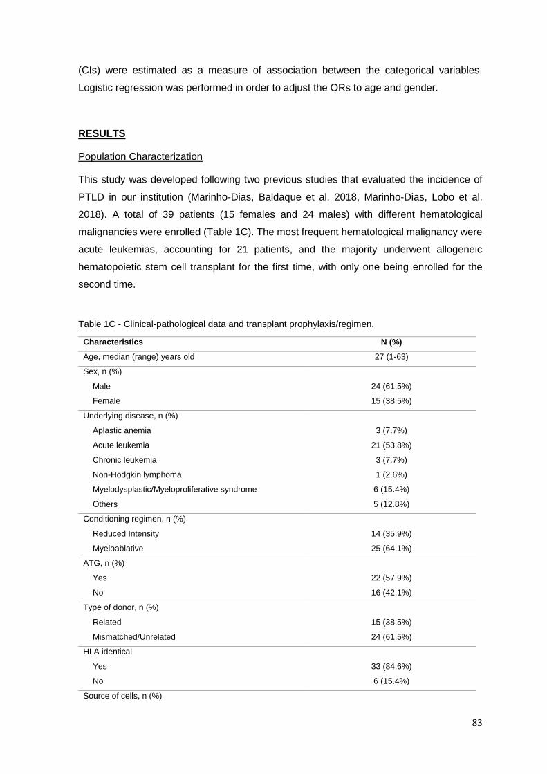
83
(CIs) were estimated as a measure of association between the categorical variables.
Logistic regression was performed in order to adjust the ORs to age and gender.
RESULTS
Population Characterization
This study was developed following two previous studies that evaluated the incidence of
PTLD in our institution (Marinho-Dias, Baldaque et al. 2018, Marinho-Dias, Lobo et al.
2018). A total of 39 patients (15 females and 24 males) with different hematological
malignancies were enrolled (Table 1C). The most frequent hematological malignancy were
acute leukemias, accounting for 21 patients, and the majority underwent allogeneic
hematopoietic stem cell transplant for the first time, with only one being enrolled for the
second time.
Table 1C - Clinical-pathological data and transplant prophylaxis/regimen.
Characteristics N (%)
Age, median (range) years old 27 (1-63)
Sex, n (%)
Male 24 (61.5%)
Female 15 (38.5%)
Underlying disease, n (%)
Aplastic anemia 3 (7.7%)
Acute leukemia 21 (53.8%)
Chronic leukemia 3 (7.7%)
Non-Hodgkin lymphoma 1 (2.6%)
Myelodysplastic/Myeloproliferative syndrome 6 (15.4%)
Others 5 (12.8%)
Conditioning regimen, n (%)
Reduced Intensity 14 (35.9%)
Myeloablative 25 (64.1%)
ATG, n (%)
Yes 22 (57.9%)
No 16 (42.1%)
Type of donor, n (%)
Related 15 (38.5%)
Mismatched/Unrelated 24 (61.5%)
HLA identical
Yes 33 (84.6%)
No 6 (15.4%)
Source of cells, n (%)

84
Peripheral blood stem cells 32 (82.1%)
Bone marrow 3 (7.7%)
Umbilical cord blood 4 (10.3%)
EBV Infection and PTLD
Frequency of EBV infection was accessed in these patients, by monthly monitorization up
to 6 months in peripheral blood samples. Analysis of EBV DNAemia (data not shown)
revealed that 9 (23.1%) patients were first positive for EBV by the first month after
transplant, 14 (35.9%) by 3 months and 6 (15.4%) by 6 months after transplant. Overall, 31
patients (79.5%) were positive for EBV at least once in the follow-up period.
In this group of patients, a total of 9 patients were diagnosed with PTLD. Nevertheless, the
histological confirmation was only available for 2 cases. All other cases were based on
clinical findings (e.g. fever, elevated LDH or hepatic enzymes and lymphadenopathies).
SNP Genotype Analysis
The evaluation of SNP genotypes was accessed by excluding SNPs that 1) did not provide
results for >10% of the samples; and 2) samples with low quality DNA that were not suitable
for genotyping, and therefore not presenting results for over 5% of SNP. Of the 33 SNPs
only 22 SNPs matched these criteria and were used for analysis (Supplementary Table 1C).
The Hardy-Weinberg equilibrium was accessed to identify SNPs with genotypic deviations
from the expected in overall population. The analysis shows that with exception of
rs1800469, rs2856838, rs3024505 and rs4937113, all other SNPs were in accordance with
Hardy-Weinberg equilibrium (p>0.05) (Supplementary Table 2C). This test was performed
in the genotypes detected in the samples collected prior transplantation.
The description of the different SNP genotypes in samples obtained pre- and post-
transplant is shown in Table 2. We also evaluated the genotype change after transplant by
comparing the individual genotypes pre and post-transplant (Table 2C). Results show that
in the majority of SNPs there was >20% of cases that changed the genotype, particularly
for rs281381 (30.6%), rs4937113 (31.4%), rs1878321 (32.4%) and rs2069727 (38.2%).
SNPs and EBV infection
We analyzed the association of post-transplant SNP genotypes and the development of
EBV infection and results showed significant genotypic differences in genotype distribution
of rs1143627 (p=0.019), rs1800629 (p=0.061) and rs2735097 (p=0.087) (Table 3C). The

85
statistical analysis revealed that rs1143627 A carrier genotypes are associated with a 14-
fold increased risk of EBV infection (p=0.024; OR=14.0; 95%CI 1.63-120), which was
confirmed by logistic regression adjusted for age and gender (p=0.016; OR=14.0; 95%CI
1.63-120) (data not shown).
We also analyzed the association of genotype change with EBV infection and no statistically
significant result was found (Supplementary Table 3C).
SNPs and PTLD
Similarly, we analyzed the association of post-transplant SNP genotypes and the
development of PTLD (Table 4C). Results showed significant differences in genotype
distribution for rs2856838 (p=0.029), rs3024496 (p=0.056), rs6052130 (0.009) and
rs281381 (0.098). The statistical analysis revealed increased risk for PTLD development
associated with rs2856838 AA genotype (p=0.049; OR=1.28; 95%CI 0.91-1.82), rs3024496
GG genotype (p=0.037; OR=7.20; 95%CI 1.22-42.5) and rs6052130 A carriers (p=0.009;
OR 1.50; 95%CI 0.95-2.38.) These data were not confirmed by logistic regression with
adjustment for age and gender (data not shown).
The analysis of genotype change after transplantation and PTLD development revealed
that genotype modification in the MAVS rs6052130 was associated with higher risk of PTLD
(OR=1.50, p=0.010). Furthermore, results showed two SNPs (rs281381 and rs602662) with
borderline significance showing that genotype change might be associated with a tendency
for protection of PTLD development (Supplementary Table 4C).

86
Table 2C - SNP genotyping pre and post-transplant.
SNP (npre/npost) Genotype Pre-transplant
n (%) Post-transplant
n (%) Cases with
genotype change, n (%)
rs1799964 (37/36) CC CT TT
5 (13.5) 15 (40.5) 17 (45.9)
4 (11.1) 14 (38.9) 18 (50.0)
4 (11.8)
rs1143627 (38/36) AA AG GG
16 (42.1) 18 (47.4) 4 (10.5)
18 (50.0) 13 (36.1) 5 (13.9)
9 (25.0)
rs1143633 (38/39) CC CT TT
15 (39.5) 20 (52.6) 3 (7.9)
16 (41.0) 20 (51.3)
3 (7.7) 10 (26.3)
rs11436341 (38/38) AA AG GG
2 (5.3) 12 (31.6) 24 (63.2)
3 (7.9) 13 (34.2) 22 (57.9)
10 (27.0)
rs1327474 (37/39) CC CT TT
8 (21.6) 15 (40.5) 14 (37.8)
8 (20.5) 15 (38.5) 16 (41.0)
10 (27.0)
rs1800469 (38/39) AA AG GG
4 (10.5) 10 (26.3) 24 (63.2)
2 (5.1) 17 (43.6) 20 (51.3)
9 (23.7)
rs1800629 (39/39) AA AG GG
1 (2.6) 10 (25.6) 28 (71.8)
1 (2.6) 9 (23.1)
29 (74.4) 3 (7.7)
rs1878321 (35/38) AA AG GG
17 (48.6) 15 (42.9) 3 (8.6)
15 (39.5) 18 (47.4) 5 (13.2)
11 (32.4)
rs2069727 (37/36) CC CT TT
12 (32.4) 18 (48.6) 7 (18.9)
12 (33.3) 13 (36.1) 11 (30.6)
13 (38.2)
rs2069840 (38/38) CC CG GG
12 (31.6) 21 (55.3) 5 (13.2)
16 (42.1) 19 (50.0)
3 (7.9) 9 (24.3)
rs2735097 (37/36) AA AC CC
3 (8.1) 16 (43.2) 18 (48.6)
3 (8.3) 15 (41.7) 18 (50.0)
1 (2.9)
rs281381 (37/37) CC CT TT
5 (13.5) 21 (56.8) 11 (29.7)
6 (16.2) 16 (43.2) 15 (40.5)
11 (30.6)
rs2856838 (39/39) AA AG GG
2 (5.1) 20 (51.3) 17 (43.6)
2 (5.1) 23 (59.0) 14 (35.9)
10 (25.6)
rs3024496 (37/39) AA AG GG
17 (45.9) 15 (40.5) 5 (13.5)
16 (41.0) 16 (41.0) 7 (17.9)
10 (27.0)
rs3024498 (37/37) CC CT TT
--- 9 (24.3)
28 (75.7)
1 (2.6) 7 (24.3)
29 (75.7) 7 (20.0)
rs3024505 (39/39) AA AG GG
--- 14 (35.9) 25 (64.1)
1 (2.6) 12 (30.8) 26 (66.7)
9 (23.1)
rs3783521 (38/37) AA AG GG
5 (13.2) 18 (47.4) 15 (39.5)
4 (10.8) 12 (32.4) 21 (56.8)
7 (19.4)
rs4633144 (37/39) CC CT TT
23 (62.2) 11 (29.7) 3 (8.1)
17 (43.6) 18 (46.2) 4 (10.3)
11 (29.7)
rs4937113 (36/38) AA AT TT
15 (41.7) 11 (30.6) 10 (27.8)
9 (23.7) 14 (36.8) 15 (39.5)
11 (31.4)
rs602662 (38/39) AA AG GG
9 (23.7) 18 (47.4) 11 (28.9)
11 (28.2) 16 (41.0) 12 (30.8)
11 (28.9)
rs6052130 (38/39) CC CA AA
36 (94.7) 2 (5.3)
---
36 (92.3) 3 (7.7)
--- 3 (7.9)
rs9376267 (36/37) CC CT TT
22 (61.1) 11 (30.6) 3 (8.3)
23 (62.2) 8 (21.6) 6 (16.2)
7 (20.6)

87
Table 3C - SNP genotyping post-transplant and EBV infection.
SNP (n) Genotype EBV negative
n (%) EBV positive
n (%) p
rs1799964 (36) CC CT TT
1 (14.3) 2 (28.6) 4 (57.1)
3 (10.3) 12 (41.4) 14 (48.3)
0.817
rs1143627 (36) AA AG GG
2 (33.3) 1 (16.7) 3 (50.0)
16 (53.3) 12 (40.0)
2 (6.7) 0.019
rs1143633 (39) CC CT TT
4 (50.0) 3 (37.5) 1 (12.5)
12 (38.7) 17 (54.8)
2 (6.5) 0.646
rs1143634 (38) AA AG GG
---- 2 (25.0) 6 (75.0)
3 (10.0) 11 (36.7) 16 (53.3)
0.458
rs1327474 (39) CC CT TT
1 (12.5) 4 (50.0) 3 (37.5)
7 (22.6) 11 (35.5) 13 (41.9)
0.707
rs1800469 (39) AA AG GG
1 (12.5) 5 (62.5) 2 (25.0)
1 (3.2) 12 (38.7) 18 (58.1)
0.197
rs1800629 (39) AA AG GG
1 (12.5) 3 (37.5) 4 (50.0)
---- 6 (29.4)
25 (80.6) 0.061
rs1878321 (38) AA AG GG
2 (25.0) 4 (50.0) 2 (25.0)
13 (43.3) 14 (46.7) 3(10.0)
0.442
rs2069727 (36) CC CT TT
3 (37.5) 3 (37.5) 2 (25.0)
9 (32.1) 13 (35.7) 9 (32.1)
0.922
rs2069840 (38) CC CG GG
2 (28.6) 4 (57.1) 1 (14.3)
14 (45.2) 15 (48.4)
2 (6.5) 0.636
rs2735097 (36) AA AC CC
---- 6 (75.0) 2 (25.0)
3 (10.7) 9 (32.1)
16 (57.1) 0.087
rs281381 (37) CC CT TT
1 (12.5) 4 (50.0) 3 (37.5)
5 (17.2) 12 (41.4) 12 (41.4)
0.897
rs2856838 (39) AA AG GG
---- 4 (50.0) 4 (50.0)
2 (6.5) 19 (61.3) 10 (32.3)
0.546
rs3024496 (39) AA AG GG
3 (37.5) 4 (50.0) 1 (12.5)
13 (41.9) 12 (38.7) 6 (19.4)
0.821
rs3024498 (37) CC CT TT
---- 2 (25.0) 6 (75.0)
1 (3.4) 5 (17.2)
23 (79.3) 0.783
rs3024505 (39) AA AG GG
---- 4 (50.0) 4 (50.0)
1 (3.2) 8 (25.8)
22 (71.0) 0.389
rs3783521 (37) AA AG GG
1 (12.5) 2 (25.0) 5 (72.5)
3 (10.3) 10 (34.5) 16 (55.2)
0.878
rs4633144 (39) CC CT TT
3 (37.5) 4 (50.0) 1 (12.5)
14 (45.2) 14 (35.2)
3 (9.7) 0.920
rs4937113 (38) AA AT TT
1 (12.5) 2 (25.0) 5 (72.5)
8 (26.7) 12 (40.0) 10 (33.3)
0.319
rs602662 (39) AA AG GG
---- 4 (50.0) 4 (50.0)
11 (35.5) 12 (38.7) 8 (25.8)
0.120
rs6052130 (39) CC CA AA
8 (100) ---- ---
28 (90.3) 3 (9.7)
--- 1.000
rs9376267 (37) CC CT TT
4 (57.1) 1 (14.3) 2 (28.6)
19 (63.3) 7 (23.3) 4 (13.3)
0.588

88
Table 4C - SNP genotyping post-transplant and PTLD infection.
SNP (n) Genotype PTLD
negative n (%)
PTLD positive
n (%) p Risk model p OR (95%CI)
rs1799964 (36) CC CT TT
4 (13.8) 11 (37.9) 14 (48.3)
---- 3 (42.9) 4 (57.1)
0.580 Ccarrier vs TT 1.00 1.42 (0.27 – 7.58)
rs1143627 (36) AA AG GG
13 (48.1) 10 (37.0) 4 (14.8)
5 (55.6) 2 (33.3) 1 (11.1)
0.920 Gcarrier vs AA 1.00 1.35 (0.30 – 6.13)
rs1143633 (39) CC CT TT
12 (40.0) 16 (53.3) 2 (6.7)
4 (44.4) 4 (44.4) 1 (11.1)
0.853 CC vs Tcarrier 1.00 0.83 (0.18 – 3.75)
rs1143634 (38) AA AG GG
2 (6.7) 10 (33.3) 18 (60.0)
1 (12.5) 3 (37.5) 4 (50.0)
0.813 GG vs Acarrier 0.698 1.50 (0.31 – 7.18)
rs1327474 (39) CC CT TT
6 (20.0) 11 (36.7) 13 (43.3)
2 (22.2) 4 (44.4) 3 (33.3)
0.863 TT vs Ccarrier 0.711 1.58 (0.32 – 7.30)
rs1800469 (39) AA AG GG
2 (6.7) 14 (46.7) 14 (46.7)
--- 6 (66.6) 3 (33.3)
0.491 Acarrier vs GG 0.451 2.28 (0.48 – 10.9)
rs1800629 (39) AA AG GG
1 (3.3) 8 (70.0) 21 (26.7)
---- 1 (11.1) 8 (88.9)
0.507 Acarrier vs GG 0.400 3.42 (0.37 – 31.6)
rs1878321 (39) AA AG GG
11 (37.9) 14 (48.3) 4 (13.8)
4 (44.4) 4 (44.4) 1(11.1)
0.936 GG vs Acarrier 1.00 1.28 (0.12 – 13.2)
rs2069727 (36) CC CT TT
10 (37.0) 11 (40.7) 6 (22.2)
2 (22.2) 2 (22.2) 5 (55.6)
0.170 Ccarrier vs TT 0.096 4.38 (0.89 – 21.6)
rs2069840 (38) CC CG GG
12 (41.4) 16 (55.2) 1 (3.4)
4 (44.4) 3 (33.3) 2 (22.2)
0.154 Ccarrier vs GG 0.134 8.00 (0.63 – 101)
rs2735097 (36) AA AC CC
2 (6.9) 12 (41.4) 15 (51.7)
1 (14.3) 3 (42.9) 3 (42.9)
0.794 Ccarrier vs AA 0.488 2.25 (0.42 – 14.2)
rs281381 (37) CC CT TT
6 (20.7) 10 (34.5) 13 (44.8)
---- 6 (75.0) 2 (25.0)
0.098 TT vs Ccarrier 0.431 2.44 (0.42 – 14.2)
rs2856838 (39) AA AG GG
---- 19 (63.3) 11 (36.7)
2 (22.2) 4 (44.4) 3 (33.3)
0.029 Gcarrier vs AA 0.049 1.28 (0.91 – 1.82)
rs3024496 (39) AA AG GG
13 (43.3) 14 (46.7) 3 (10.0)
3 (33.3) 2 (22.2) 4 (44.4)
0.056 Acarrier vs GG 0.037 7.20 (1.22 – 42.5)
rs3024498 (37) CC CT TT
---- 6 (20.7) 23 (79.3)
1 (12.5) 1 (12.5) 6 (75.0)
0.145 TT vs Ccarrier 1.00 1.28 (0.20 – 8.00)
rs3024505 (39) AA AG GG
1 (3.3) 9 (30.0) 20 (66.7)
---- 3 (33.3) 6 (66.7)
0.850 Acarrier vs GG 1.00 1.00 (0.21 – 4.86)
rs3783521 (37) AA AG GG
4 (13.3) 10 (33.3) 16 (53.3)
---- 2 (28.6) 5 (71.4)
0.522 A carrier vs GG 0.674 2.19 (0.36 – 13.2)
rs4633144 (39) CC CT TT
14 (46.7) 13 (43.3) 3 (10.0)
3 (33.3) 5 (55.6) 1 (11.1)
0.733 CC vs Tcarrier 0.704 1.75 (0.36 – 8.39)
rs4937113 (38) AA AT TT
6 (20.0) 13 (43.3) 11 (36.7)
3 (37.5) 1 (12.5) 4 (50.0)
0.255 Tcarrier vs AA 0.363 2.40 (0.44 – 13.0)
rs602662 (39) AA AG GG
8 (26.7) 11 (36.7) 11 (36.7)
3 (33.3) 5 (55.6) 1 (11.1)
0.336 GG vs Acarrier 0.228 4.63 (0.51 – 42.1)
rs6052130 (39) CC CA AA
30 (100) ---- ---
6 (66.7) 3 (33.3)
--- 0.009 CC vs Acarrier 0.009 1.50 (0.95 – 2.38)
rs9376267 (37) CC CT TT
15 (53.6) 7 (25.0) 6 (21.6)
8 (88.9) 1 (11.1)
---- 0.142 Tcarrier vs CC 0.112 6.90 (0.76 – 63.0)
89
DISCUSSION
EBV infection, in the period subsequent to allogeneic stem cell transplantation, is frequent
and is responsible for uncontrolled B cell proliferation in an environment with diminished or
absence of immunologic surveillance (Janani, Malathi et al. 2015, Fan, Jing et al. 2016,
Marinho-Dias, Baldaque et al. 2018). PTLDs are rare events and rather deadly in these
patients, which makes it important to identify correctly patients at risk (Marinho-Dias, Lobo
et al. 2018). In patients submitted to allo-HSCT, there have been identified several risk
factors, such as HLA-mismatched, unrelated donor, mismatched seropositivity (D+/R-),
active EBV infection, use of anti-thymocyte globulin and use of myeloablative regimens
(Marinho-Dias, Lobo et al. 2018). These clinical data are not enough to determine which
patients are at higher risk, and therefore it is important to identify potential biomarkers that
could help in the prediction of EBV infection or PTLD development.
In our study, we verified that EBV infection was significantly frequent in our population, since
79.5% of all patients were positive at least once in the period post-transplant, with a peak
at 2 months (76.9%). This is consistent with our previous study and other reports, where
EBV DNAemia reached 70.0%, confirming that we have a significant rate of infection in our
allo-HSCT recipients (Schonberger, Meisel et al. 2010, Liu, Xuan et al. 2013, Marinho-Dias,
Baldaque et al. 2018).
The main aim of this study was to evaluate the role of genetic polymorphism in the definition
of a susceptibility profile to EBV infection and consequently PTLD development in allo-
HSCT. We have developed a strategy to identify potential SNPs that represent haplotypes,
and which could represent a significative part of host genome immune response. In the
study we included a total of 33 SNPs from HLA polymorphic sites (class I, II and III) and
non-HLA regions including host immune response genes (such as TNFA, IL-1a, IL-1b, IL-
1RN, IL10, IFNG, IFNGRI and IFNGRII); antiviral effectors (such as RIG-I, DDX58, MDA-5,
IFIH1, MAVS, VISA, IPS1, CARDIF and ISG15 ubiquitin-like pathway); and cellular
glycoprotein (FUT2/3). After the study we were only able to retrieve reliable data from 22
SNPS in different genes associated with immune response (IL1α/β, IL-6, IL-10, IL-18,
TGFβ1, IFNGRI, TNFα, DDX8, MAVS and FUT2). Since all these SNPs were selected with
a tight linkage disequilibrium, it is important to take into account that each SNP represents
a locus of several SNPs.
Several SNPs, associated with EBV infection and PTLD development, have been studied,
but mainly regarding solid transplantation. Namely IL-28B (rs12979860), where the CC
genotype might influence the anti-viral response from IFNG (Akay, Patel et al. 2014); HLA-
A26, B38 haplotype, both in the donor and recipient, predisposes the patient to PTLD

90
development in the subset of solid organ transplant (Reshef, Luskin et al. 2011); and TNF
promoter -1031C and -863A variant alleles have been linked to a higher risk of PTLD
development in heart transplant patients (McAulay, Haque et al. 2009, Morscio, Dierickx et
al. 2013). Furthermore, HLA-A2, A11, B5 and B35, concomitant with azathioprine, are
associated with higher risk of PTLD and (Pourfarziani, Einollahi et al. 2007). Finally, when
studying PTLD development in mice, IFNG +874 T>A polymorphism, the AA genotype was
linked to early onset EBV-driven PTLD (Dierksheide, Baiocchi et al. 2005).
One of the important things in this study is that we have analyzed the genotypes pre- and
post-transplant. This is particularly important since, despite HLA-match, after transplant the
recipient will have an immune system based on the donor, and therefore the genetic
susceptibility should be tested at the time of engraftment confirmation. Our data analysis
revealed that variation in genotype after transplantation was observed in the majority of
SNPs particularly for FUT 2 rs281381 (30.6%), IL-18 rs4937113 (31.4%), IL-1A rs1878321
(32.4%) and IFNG rs2069727 (38.2%). FUT2, also known as fucosyltransferase 2 gene, is
responsible for encoding Lewis antigens related to the ABO blood group. FUTs are involved
in many biological processes such as cell adhesion and tumor progression (Chen, Liao et
al. 2017). FUT2 has been recently associated with an increased risk of GVHD after HSCT
by genetically modifying the gut microbiome (Rayes, Morrow et al. 2016). IL-18, also
designated as interleukin-18 gene, encodes a proinflammatory cytokine that enhances
natural killer cell activity in spleen cells and stimulates interferon gamma production in T-
helper type I cell (Carroll, Paunovic et al. 2008). IL-18 is known to have synergistic effect,
along with IL-12, which reduces T-cell mediated GVHD in mice (Leung, Iyengar et al. 2004).
IL-1A has been known to play a major role in immune response, inflammatory processes
and hematopoiesis, and its deregulated signaling leads to severe acute or chronic
inflammation, such as GVHD (Park, Lee et al. 2015, Di Paolo and Shayakhmetov 2016, de
Mooij, Netea et al. 2017). IFNG is a proinflammatory cytokine and plays an important role
in innate and adaptive immunity against infections and tumor development, by modulating
the function of hematopoietic stem cells, mainly by inhibiting their production (de Bruin,
Voermans et al. 2014). IFNG has been used as a therapy for PTLD, where IFNG secreting
T-cells of the donor are isolated and infused in the patient, and it has shown to be effective
in early stages of PTLD (Bollard and Heslop 2016). IFNG has also been associated with
EBV-driven tumors, where viral lytic proteins favor immune evasion by inhibiting IFNG
production and therefore contribute to tumorigenesis (Petrara, Giunco et al. 2015). Despite
these evidences, when evaluating genotypic changes associated with EBV infection, no
statistically significant results were attained; while for PTLD development we have only
verified an association of genotype change in MAVS rs6052130 (OR=1.50, p=0.010).
91
Additionally, FUT2 rs281381 and rs602662, presented borderline statistically significant
results for a protective effect of PTLD development after genotype alterations. These results
support the need for further studies to characterize the role of these proteins in EBV
infection and PTLD development and to evaluate its utility in allo-HSCT patients
management.
The independent analysis of SNPs revealed that IL-1B rs1143627 Acarriers have a
significant higher risk of EBV infection (OR=14.0, p=0.024). IL-1B polymorphisms have
been strongly associated with the development of cancer, namely lung and prostate cancer,
because of its pro-inflammatory effect (Andersen, Holst et al. 2013). In lung cancer, a study
from Zienolddiny et al., revealed an association between Acarrier subjects and a mutation
in the p53 gene, with the development of lung cancer and the risk was higher when
homozygosity was present (AA genotype) (Zienolddiny, Ryberg et al. 2004). As for the
association with viral infections, literature shows that a relation Acarriers have increased
susceptibility to pandemic H1N1 influenza A virus (Liu, Li et al. 2013). Furthermore, there
are other studies that show that same SNP is associated with hepatitis B vaccine immune
response (heterozygotes associated with non-response to vaccination) and Chronic
Hepatitis B virus infection (GG genotype) (Chen, Liang et al. 2011, Wu, Song et al. 2018).
This is the first study to show an association of this SNP with EBV infection and results
show a clear association that should be further confirmed in larger series since the
predictive risk (14-fold increase) may be of significant importance for clinical management
and the genotype of donors regarding the SNP may be useful to select the best follow-up
and prophylaxis for allo-HSCT recipients.
Regarding the development of PTLD we found that 3 SNPs are associated with increased
risk: IL-1A rs2856838 AA genotype (OR=1.28, p=0.049); IL-10 rs3024496 GG genotype
(OR=7.20, p=0.037): and MAVS rs6052130 A carriers (OR=1.50, p=0.009). IL-1A
rs2856838 has not been studied extensively, although one study reports an association
between the AA genotype and a protective effect on chronic rhinosinusitis (Mfuna Endam,
Cormier et al. 2010); other reveals association of heterozygotic genotype and increased
susceptibility for tuberculosis (Wang, Jin et al. 2018); and another associates heterozygotic
and AA genotype with lower risk of contracting malaria (Legason, Pfeiffer et al. 2017). As
for IL-10 rs3024496 GG genotype has been linked with prostate and colorectal cancer
(Tsilidis, Helzlsouer et al. 2009, Wang, Helzlsouer et al. 2009), but also to HCMV infection
(Loeffler, Steffens et al. 2006). Nevertheless, there are controversial studies regarding this
SNP and the development of tuberculosis (Shin, Park et al. 2005, Moller, Nebel et al. 2010).
Finally, MAVS rs6052130 CA genotype has been reported as a risk factor for development

92
of cervical precancerous lesions as well as increased vulnerability to high-risk HPV-induced
cervical precancerous lesions (Xiao, Liu et al. 2018).
To the best of our knowledge, this is the first study concerning genotype alterations of these
SNPs and its association with EBV infection and PTLD development in allo-HSCT
recipients. Despite having a small population, the study clearly identifies IL-1B rs1143627
(Acarriers) as a significant risk factor for EBV infection and IL-1A rs2856838 (AA genotype),
IL-10 rs3024496 (GG genotype) and MAVS rs6052130 (Acarriers) as risk markers for PTLD
development. To clarify these data, more studies should be performed in larger series and
testing other SNPs from same genes.

93
SUPPLEMENTARY TABLES
Supplementary Table 1C - Description of single nucleotide polymorphisms (SNP) selected for the
study.

94
Supplementary Table 2C – Hardy-Weinberg Equilibrium.
SNP Pre-transplant genotypes Hardy-Weinberg
rs1799964 CC (13.5%), TC (40.5%), TT (45.9%) p=0.348
rs1143627 AA (42.1%), AG, 18 (47.4%), GG, 4 (10.5%) p=0.594
rs1143633 CC, 15 (39.5%), TC, 20 (52.6%), TT, 3 (7.9%) p=0.092
rs1143634 AA, 2 (5.3%), AG, 12 (31.6%), GG, 24 (63.2%) p=0.608
rs1327474 CC, 8 (21.6%), CT, 15 (40.5%), TT, 14 (37.8%) p=0.094
rs1800469 AA, 4 (10.5%), GA, 10 (26.3%), GG, 24 (63.2%) p=0.006
rs1800629 AA, 1 (2.6%), GA, 10 (25.6%), GG, 28 (71.8%) p=0.861
rs1878321 AA, 17 (48.6%), AG, 15 (42.9%), GG, 3 (8.6%) p=0.841
rs2069727 CC, 12 (32.4%), TC, 18 (48.6%), TT, 7 (18.9%) p=0.929
rs2069840 CC, 12 (31.6%), GC, 21 (55.3%), GG, 5 (13.2%) p=0.151
rs2735097 AA, 3 (8.1%), CA, 16 (43.2%), CC, 18 (48.6%) p=0.727
rs281381 CC, 5 (13.5%), CT, 21 (56.8%), TT, 11 (29.7%) p=0.096
rs2856838 AA, 2 (5.1%), AG, 20 (51.3%), GG, 17 (43.6%) p=0.041
rs3024496 AA, 17 (45.9%), GA, 15 (40.5%), GG, 5 (13.5%) p=0.348
rs3024498 CC ---, CT, 9 (24.3%), TT, 28 (75.7%) p=0.166
rs3024505 AA ---, GA, 14 (35.9%), GG, 25 (64.1%) p=0.028
rs3783521 AA, 5 (13.2%), AG, 18 (47.4%), GG, 15 (39.5%) p=0.863
rs4633144 CC, 23 (62.2%), CT, 11 (29.7%), TT, 3 (8.1%) p=0.109
rs4937113 AA, 15 (41.7%), TA, 11 (30.6%), TT, 10 (27.8%) p<0.001
rs602662 AA, 9 (23.7%), AG, 18 (47.4%), GG, 11 (28.9%) p=0.621
rs6052130 CC, 36 (94.7%), CA, 2 (5.3%), AA --- p=0.785
rs9376267a CC, 22 (61.1%), CT, 11 (30.6%), TT, 3 (8.3%) p=0.130
p, pearson coefficient by Chi-square analysis; G, guanine; C, cytosine; T, thymidine; A, adenine.
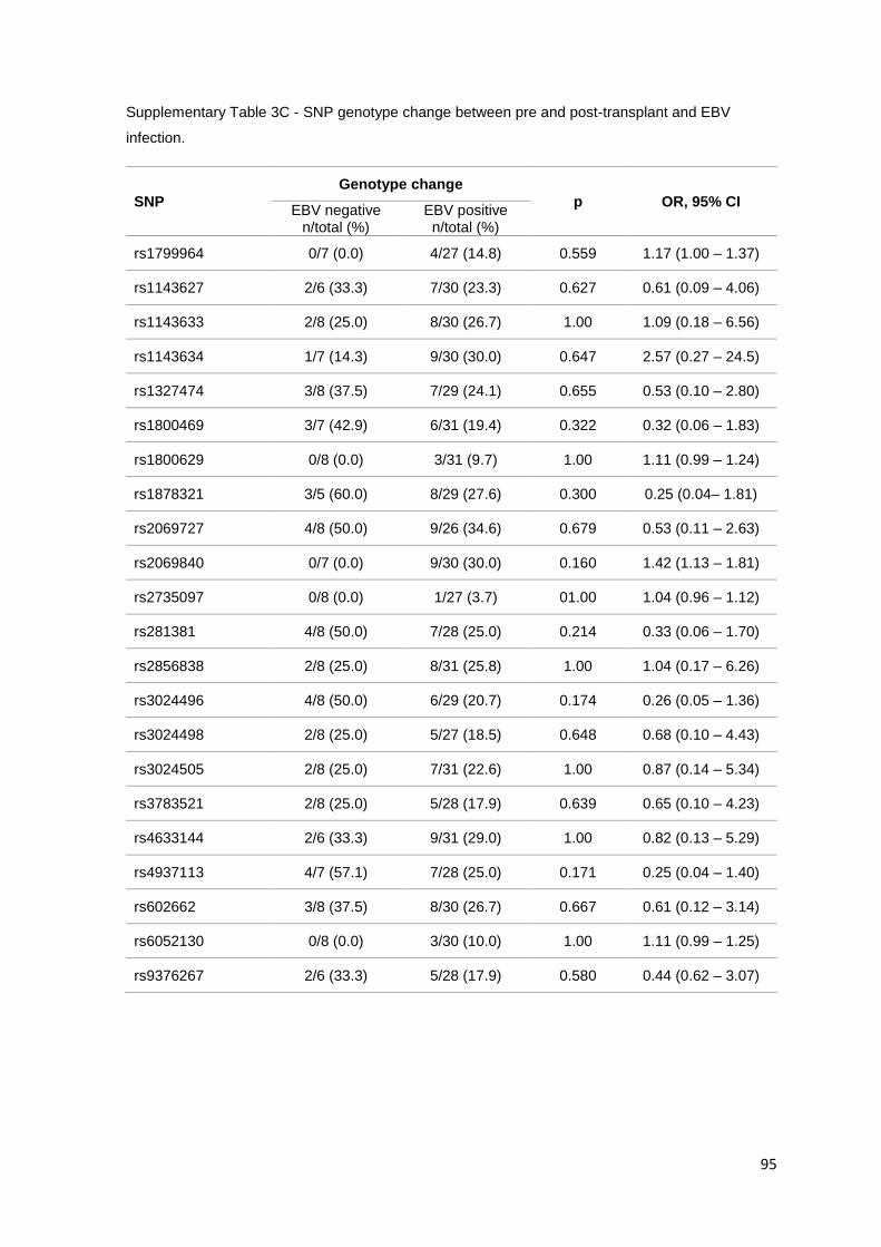
95
Supplementary Table 3C - SNP genotype change between pre and post-transplant and EBV
infection.
SNP
Genotype change
p OR, 95% CI EBV negative
n/total (%) EBV positive n/total (%)
rs1799964 0/7 (0.0) 4/27 (14.8) 0.559 1.17 (1.00 – 1.37)
rs1143627 2/6 (33.3) 7/30 (23.3) 0.627 0.61 (0.09 – 4.06)
rs1143633 2/8 (25.0) 8/30 (26.7) 1.00 1.09 (0.18 – 6.56)
rs1143634 1/7 (14.3) 9/30 (30.0) 0.647 2.57 (0.27 – 24.5)
rs1327474 3/8 (37.5) 7/29 (24.1) 0.655 0.53 (0.10 – 2.80)
rs1800469 3/7 (42.9) 6/31 (19.4) 0.322 0.32 (0.06 – 1.83)
rs1800629 0/8 (0.0) 3/31 (9.7) 1.00 1.11 (0.99 – 1.24)
rs1878321 3/5 (60.0) 8/29 (27.6) 0.300 0.25 (0.04– 1.81)
rs2069727 4/8 (50.0) 9/26 (34.6) 0.679 0.53 (0.11 – 2.63)
rs2069840 0/7 (0.0) 9/30 (30.0) 0.160 1.42 (1.13 – 1.81)
rs2735097 0/8 (0.0) 1/27 (3.7) 01.00 1.04 (0.96 – 1.12)
rs281381 4/8 (50.0) 7/28 (25.0) 0.214 0.33 (0.06 – 1.70)
rs2856838 2/8 (25.0) 8/31 (25.8) 1.00 1.04 (0.17 – 6.26)
rs3024496 4/8 (50.0) 6/29 (20.7) 0.174 0.26 (0.05 – 1.36)
rs3024498 2/8 (25.0) 5/27 (18.5) 0.648 0.68 (0.10 – 4.43)
rs3024505 2/8 (25.0) 7/31 (22.6) 1.00 0.87 (0.14 – 5.34)
rs3783521 2/8 (25.0) 5/28 (17.9) 0.639 0.65 (0.10 – 4.23)
rs4633144 2/6 (33.3) 9/31 (29.0) 1.00 0.82 (0.13 – 5.29)
rs4937113 4/7 (57.1) 7/28 (25.0) 0.171 0.25 (0.04 – 1.40)
rs602662 3/8 (37.5) 8/30 (26.7) 0.667 0.61 (0.12 – 3.14)
rs6052130 0/8 (0.0) 3/30 (10.0) 1.00 1.11 (0.99 – 1.25)
rs9376267 2/6 (33.3) 5/28 (17.9) 0.580 0.44 (0.62 – 3.07)

96
Supplementary Table 4C - SNP genotype change between pre and post-transplant and PTLD
SNP
Genotype change
p OR (95% CI) PTLD negative
n/total (%)
PTLD positive
n/total (%)
rs1799964 4/28 (14,3) 0/6 (0.0) 1.00 0.86 (0.74 – 0.99)
rs1143627 6/27 (22.2) 3/9 (33.3) 0.660 1.75 (0.33 – 9.17)
rs1143633 6/30 (20.0) 4/8 (50.0) 0.170 4.00 (0.77 – 20.8)
rs1143634 6/29 (20.7) 4/8 (50.0) 0.174 3.83 (0.74 – 19.9)
rs1327474 8/28 (28.6) 2/9 (22.2) 1.00 0.71 (0.12 – 4.20)
rs1800469 8/29 (27.6) 1/9 (11.1) 0.411 0.33 (0.04 – 3.06)
rs1800629 1/30 (3.3) 2/9 (22.2) 0.127 8.29 (0.66 – 104.9)
rs1878321 8/26 (30.8) 3/8 (37.5) 1.00 1.35 (0.26 – 7.07)
rs2069727 9/25 (36.0) 4/9 (44.4) 0.704 1.42 (0.30 – 6.69)
rs2069840 6/29 (20.7) 3/8 (37.5) 0.373 2.30 (0.42 – 12.4)
rs2735097 1/28 (3.6) 0/7 (0.0) 1.00 0.96 (0.90 - 1.04)
rs281381 11/29 (37.9) 0/7 (0.0) 0.076 0.62 (0.47 – 0.83)
rs2856838 6/30 (20.0) 4/9 (44.4) 0.197 3.20 (0.65 – 15.7)
rs3024496 8/29 (27.6) 2/8 (25.0) 1.00 0.88 (0.15 – 5.27)
rs3024498 4/28 (14.3) 3/7 (42.9) 0.125 4.50 (0.72 – 28.1)
rs3024505 7/30 (23.3) 2/9 (22.2) 1.00 0.94 (0.16 – 5.59)
rs3783521 6/29 (20.7) 1/7 (14.3) 1.00 0.64 (0.06– 6.37)
rs4633144 8/28 (28.6) 3/9 (33.3) 1.00 1.25 (0.25 – 6.26)
rs4937113 9/27 (33.3) 2/8 (25.0) 1.00 0.67 (0.11 – 3.99)
rs602662 11/30 (36.7) 0/8 (0.0) 0.077 0.63 (0.48 – 0.83)
rs6052130 0/29 (0.0) 3/9 (3.4) 0.010 1.50 (0.95 – 2.38)
rs9376267 5/25 (20.0) 2/9 (22.2) 1.00 1.14 (0.18 – 7.28)

97
REFERENCES
• Akay, E., Patel, M., Conibear, T., Chaggar, T., Haque, T., 2014. Interleukin 28B gene
polymorphisms and Epstein-Barr virus-associated lymphoproliferative diseases.
Intervirology 57, 112-115.
• Andersen, V., Holst, R., Kopp, T.I., Tjonneland, A., Vogel, U., 2013. Interactions between
diet, lifestyle and IL10, IL1B, and PTGS2/COX-2 gene polymorphisms in relation to risk of
colorectal cancer in a prospective Danish case-cohort study. PloS one 8, e78366.
• Bader, P., Niethammer, D., Willasch, A., Kreyenberg, H., Klingebiel, T., 2005. How and
when should we monitor chimerism after allogeneic stem cell transplantation? Bone marrow
transplantation 35, 107-119.
• Bollard, C.M., Heslop, H.E., 2016. T cells for viral infections after allogeneic
hematopoietic stem cell transplant. Blood 127, 3331-3340.
• Brennan, R.M., Burrows, S.R., 2008. A mechanism for the HLA-A*01-associated risk for
EBV+ Hodgkin lymphoma and infectious mononucleosis. Blood 112, 2589-2590.
• Campos, A.B., Ribeiro, J., Pinho Vaz, C., Campilho, F., Branca, R., Campos, A., Jr.,
Baldaque, I., Medeiros, R., Boutolleau, D., Sousa, H., 2017. Genotypic resistance of
cytomegalovirus to antivirals in hematopoietic stem cell transplant recipients from Portugal:
A retrospective study. Antiviral Res 138, 86-92.
• Carroll, H.P., Paunovic, V., Gadina, M., 2008. Signalling, inflammation and arthritis:
Crossed signals: the role of interleukin-15 and -18 in autoimmunity. Rheumatology (Oxford)
47, 1269-1277.
• Chen, C.T., Liao, W.Y., Hsu, C.C., Hsueh, K.C., Yang, S.F., Teng, Y.H., Yu, Y.L., 2017.
FUT2 genetic variants as predictors of tumor development with hepatocellular carcinoma.
Int J Med Sci 14, 885-890.
• Chen, J., Liang, Z., Lu, F., Fang, X., Liu, S., Zeng, Y., Zhu, F., Chen, X., Shen, T., Li, J.,
Zhuang, H., 2011. Toll-like receptors and cytokines/cytokine receptors polymorphisms
associate with non-response to hepatitis B vaccine. Vaccine 29, 706-711.
• de Bruin, A.M., Voermans, C., Nolte, M.A., 2014. Impact of interferon-gamma on
hematopoiesis. Blood 124, 2479-2486.
• de Mooij, C.E.M., Netea, M.G., van der Velden, W., Blijlevens, N.M.A., 2017. Targeting
the interleukin-1 pathway in patients with hematological disorders. Blood 129, 3155-3164.
• Di Paolo, N.C., Shayakhmetov, D.M., 2016. Interleukin 1alpha and the inflammatory
process. Nat Immunol 17, 906-913.
• Diepstra, A., Niens, M., Vellenga, E., van Imhoff, G.W., Nolte, I.M., Schaapveld, M., van
der Steege, G., van den Berg, A., Kibbelaar, R.E., te Meerman, G.J., Poppema, S., 2005.
98
Association with HLA class I in Epstein-Barr-virus-positive and with HLA class III in Epstein-
Barr-virus-negative Hodgkin's lymphoma. Lancet 365, 2216-2224.
• Dierksheide, J.E., Baiocchi, R.A., Ferketich, A.K., Roychowdhury, S., Pelletier, R.P.,
Eisenbeis, C.F., Caligiuri, M.A., VanBuskirk, A.M., 2005. IFN-gamma gene polymorphisms
associate with development of EBV+ lymphoproliferative disease in hu PBL-SCID mice.
Blood 105, 1558-1565.
• Fan, J., Jing, M., Yang, M., Xu, L., Liang, H., Huang, Y., Yang, R., Gui, G., Wang, H.,
Gong, S., Wang, J., Zhang, X., Zhao, H., Gao, H., Dong, H., Ma, W., Hu, J., 2016.
Herpesvirus infections in hematopoietic stem cell transplant recipients seropositive for
human cytomegalovirus before transplantation. Int J Infect Dis 46, 89-93.
• Hatton, O.L., Harris-Arnold, A., Schaffert, S., Krams, S.M., Martinez, O.M., 2014. The
interplay between Epstein-Barr virus and B lymphocytes: implications for infection,
immunity, and disease. Immunol Res 58, 268-276.
• Hjalgrim, H., Rostgaard, K., Johnson, P.C., Lake, A., Shield, L., Little, A.M., Ekstrom-
Smedby, K., Adami, H.O., Glimelius, B., Hamilton-Dutoit, S., Kane, E., Taylor, G.M.,
McConnachie, A., Ryder, L.P., Sundstrom, C., Andersen, P.S., Chang, E.T., Alexander,
F.E., Melbye, M., Jarrett, R.F., 2010. HLA-A alleles and infectious mononucleosis suggest
a critical role for cytotoxic T-cell response in EBV-related Hodgkin lymphoma. Proceedings
of the National Academy of Sciences of the United States of America 107, 6400-6405.
• Janani, M.K., Malathi, J., Rela, M., Farouk, M., Padmapriya, J., Madhavan, H.N., 2015.
Genotypic Detection of Epstein Barr Virus in Pediatric Transplant Recipients From India.
Indian Pediatr 52, 946-950.
• Johnson, P.C., McAulay, K.A., Montgomery, D., Lake, A., Shield, L., Gallagher, A., Little,
A.M., Shah, A., Marsh, S.G., Taylor, G.M., Jarrett, R.F., 2015. Modeling HLA associations
with EBV-positive and -negative Hodgkin lymphoma suggests distinct mechanisms in
disease pathogenesis. International journal of cancer. Journal international du cancer 137,
1066-1075.
• Legason, I.D., Pfeiffer, R.M., Udquim, K.I., Bergen, A.W., Gouveia, M.H., Kirimunda, S.,
Otim, I., Karlins, E., Kerchan, P., Nabalende, H., Bayanjargal, A., Emmanuel, B., Kagwa,
P., Talisuna, A.O., Bhatia, K., Yeager, M., Biggar, R.J., Ayers, L.W., Reynolds, S.J.,
Goedert, J.J., Ogwang, M.D., Fraumeni, J.F., Jr., Prokunina-Olsson, L., Mbulaiteye, S.M.,
2017. Evaluating the Causal Link Between Malaria Infection and Endemic Burkitt
Lymphoma in Northern Uganda: A Mendelian Randomization Study. EBioMedicine 25, 58-
65.

99
• Leung, W., Iyengar, R., Turner, V., Lang, P., Bader, P., Conn, P., Niethammer, D.,
Handgretinger, R., 2004. Determinants of antileukemia effects of allogeneic NK cells.
Journal of immunology 172, 644-650.
• Liu, Q., Xuan, L., Liu, H., Huang, F., Zhou, H., Fan, Z., Zhao, K., Wu, M., Xu, L., Zhai,
X., Zhang, F., Liu, C., Sun, J., Huang, X., 2013a. Molecular monitoring and stepwise
preemptive therapy for Epstein-Barr virus viremia after allogeneic stem cell transplantation.
American journal of hematology 88, 550-555.
• Liu, Y., Li, S., Zhang, G., Nie, G., Meng, Z., Mao, D., Chen, C., Chen, X., Zhou, B., Zeng,
G., 2013b. Genetic variants in IL1A and IL1B contribute to the susceptibility to 2009
pandemic H1N1 influenza A virus. BMC Immunol 14, 37.
• Loeffler, J., Steffens, M., Arlt, E.M., Toliat, M.R., Mezger, M., Suk, A., Wienker, T.F.,
Hebart, H., Nurnberg, P., Boeckh, M., Ljungman, P., Trenschel, R., Einsele, H., 2006.
Polymorphisms in the genes encoding chemokine receptor 5, interleukin-10, and monocyte
chemoattractant protein 1 contribute to cytomegalovirus reactivation and disease after
allogeneic stem cell transplantation. Journal of clinical microbiology 44, 1847-1850.
• Long, H.M., Taylor, G.S., Rickinson, A.B., 2011. Immune defence against EBV and EBV-
associated disease. Curr Opin Immunol 23, 258-264.
• Majhail, N.S., Farnia, S.H., Carpenter, P.A., Champlin, R.E., Crawford, S., Marks, D.I.,
Omel, J.L., Orchard, P.J., Palmer, J., Saber, W., Savani, B.N., Veys, P.A., Bredeson, C.N.,
Giralt, S.A., LeMaistre, C.F., 2015. Indications for Autologous and Allogeneic Hematopoietic
Cell Transplantation: Guidelines from the American Society for Blood and Marrow
Transplantation. Biology of blood and marrow transplantation : journal of the American
Society for Blood and Marrow Transplantation 21, 1863-1869.
• Marinho-Dias, J., Baldaque, I., Pinho-Vaz, C., Leite, L., Branca, R., Campilho, F.,
Campos Jr, A., Medeiros, R., Sousa, H., 2018a. Association of Epstein-Barr virus infection
with allogeneic hematopoietic stem cell transplantation in patients in Portugal. Molecular
Medicine Reports (in press).
• Marinho-Dias, J., Lobo, J., Henrique, R., Baldaque, I., Pinho-Vaz, C., Regadas, L.,
Branca, R., Campilho, F., Campos, A., Jr., Medeiros, R., Sousa, H., 2018b. Posttransplant
lymphoproliferative disorder in hematopoietic stem cell transplant patients: A single center
retrospective study between 2005 and 2012. Mol Med Rep.
• McAulay, K.A., Haque, T., Crawford, D.H., 2009. Tumour necrosis factor gene
polymorphism: a predictive factor for the development of post-transplant lymphoproliferative
disease. British journal of cancer 101, 1019-1027.
• McAulay, K.A., Higgins, C.D., Macsween, K.F., Lake, A., Jarrett, R.F., Robertson, F.L.,
Williams, H., Crawford, D.H., 2007. HLA class I polymorphisms are associated with

100
development of infectious mononucleosis upon primary EBV infection. J Clin Invest 117,
3042-3048.
• Medina-Acosta, E., Nakaya, H.T., Pontillo, A., de Souza Campos Fernandes, R.C., 2014.
Genetic control of immune response and susceptibility to infectious diseases. Biomed Res
Int 2014, 796073.
• Mfuna Endam, L., Cormier, C., Bosse, Y., Filali-Mouhim, A., Desrosiers, M., 2010.
Association of IL1A, IL1B, and TNF gene polymorphisms with chronic rhinosinusitis with
and without nasal polyposis: A replication study. Arch Otolaryngol Head Neck Surg 136,
187-192.
• Moller, M., Nebel, A., van Helden, P.D., Schreiber, S., Hoal, E.G., 2010. Analysis of eight
genes modulating interferon gamma and human genetic susceptibility to tuberculosis: a
case-control association study. BMC Infect Dis 10, 154.
• Morscio, J., Dierickx, D., Tousseyn, T., 2013. Molecular pathogenesis of B-cell
posttransplant lymphoproliferative disorder: what do we know so far? Clin Dev Immunol
2013, 150835.
• Mui, U.N., Haley, C.T., Tyring, S.K., 2017. Viral Oncology: Molecular Biology and
Pathogenesis. J Clin Med 6.
• Ogonek, J., Kralj Juric, M., Ghimire, S., Varanasi, P.R., Holler, E., Greinix, H.,
Weissinger, E., 2016. Immune Reconstitution after Allogeneic Hematopoietic Stem Cell
Transplantation. Front Immunol 7, 507.
• Park, M.J., Lee, S.H., Lee, S.H., Lee, E.J., Kim, E.K., Choi, J.Y., Cho, M.L., 2015. IL-1
Receptor Blockade Alleviates Graft-versus-Host Disease through Downregulation of an
Interleukin-1beta-Dependent Glycolytic Pathway in Th17 Cells. Mediators Inflamm 2015,
631384.
• Petrara, M.R., Giunco, S., Serraino, D., Dolcetti, R., De Rossi, A., 2015. Post-transplant
lymphoproliferative disorders: from epidemiology to pathogenesis-driven treatment. Cancer
letters 369, 37-44.
• Pourfarziani, V., Einollahi, B., Taheri, S., Nemati, E., Nafar, M., Kalantar, E., 2007.
Associations of Human Leukocyte Antigen (HLA) haplotypes with risk of developing
lymphoproliferative disorders after renal transplantation. Annals of transplantation 12, 16-
22.
• Rayes, A., Morrow, A.L., Payton, L.R., Lake, K.E., Lane, A., Davies, S.M., 2016. A
Genetic Modifier of the Gut Microbiome Influences the Risk of Graft-versus-Host Disease
and Bacteremia After Hematopoietic Stem Cell Transplantation. Biology of blood and
marrow transplantation : journal of the American Society for Blood and Marrow
Transplantation 22, 418-422.

101
• Reshef, R., Luskin, M.R., Kamoun, M., Vardhanabhuti, S., Tomaszewski, J.E.,
Stadtmauer, E.A., Porter, D.L., Heitjan, D.F., Tsai de, E., 2011. Association of HLA
polymorphisms with post-transplant lymphoproliferative disorder in solid-organ transplant
recipients. Am J Transplant 11, 817-825.
• Schonberger, S., Meisel, R., Adams, O., Pufal, Y., Laws, H.J., Enczmann, J., Dilloo, D.,
2010. Prospective, comprehensive, and effective viral monitoring in children undergoing
allogeneic hematopoietic stem cell transplantation. Biology of blood and marrow
transplantation : journal of the American Society for Blood and Marrow Transplantation 16,
1428-1435.
• Shin, H.D., Park, B.L., Kim, Y.H., Cheong, H.S., Lee, I.H., Park, S.K., 2005. Common
interleukin 10 polymorphism associated with decreased risk of tuberculosis. Exp Mol Med
37, 128-132.
• Song, Y., Liu, S., Zhao, Z., Zhang, Y., Yang, Y., Luo, B., 2017. Relationship between
PPP1R15A gene polymorphism (rs611251) and Epstein-Barr virus-associated tumors. Acta
Virol 61, 445-452.
• Sousa, H., Boutolleau, D., Ribeiro, J., Teixeira, A.L., Pinho Vaz, C., Campilho, F.,
Branca, R., Campos, A., Jr., Baldaque, I., Medeiros, R., 2014. Cytomegalovirus infection in
patients who underwent allogeneic hematopoietic stem cell transplantation in Portugal: a
five-year retrospective review. Biology of blood and marrow transplantation : journal of the
American Society for Blood and Marrow Transplantation 20, 1958-1967.
• Tan, G.W.L., Quek, L., Tan, B.P., Pua, U., 2018. Early Experience and Lessons Learnt
with Customized Fenestrated Thoracic Endovascular Aortic Reconstruction for Aortic Arch
Pathology in an Asian Population. Cardiovasc Intervent Radiol 41, 544-553.
• Tsilidis, K.K., Helzlsouer, K.J., Smith, M.W., Grinberg, V., Hoffman-Bolton, J., Clipp, S.L.,
Visvanathan, K., Platz, E.A., 2009. Association of common polymorphisms in IL10, and in
other genes related to inflammatory response and obesity with colorectal cancer. Cancer
Causes Control 20, 1739-1751.
• Wang, J., Jin, T., Mo, F., Dong, H., Wang, Y., Kang, L., Hu, Q., 2018. Association
between IL-1 gene polymorphisms and tuberculosis susceptibility in the Chinese Tibetan
population. Int J Clin Exp Pathol 11, 5441-5449.
• Wang, M.H., Helzlsouer, K.J., Smith, M.W., Hoffman-Bolton, J.A., Clipp, S.L., Grinberg,
V., De Marzo, A.M., Isaacs, W.B., Drake, C.G., Shugart, Y.Y., Platz, E.A., 2009. Association
of IL10 and other immune response- and obesity-related genes with prostate cancer in
CLUE II. Prostate 69, 874-885.
• Weikert, B.C., Blumberg, E.A., 2008. Viral infection after renal transplantation:
surveillance and management. Clin J Am Soc Nephrol 3 Suppl 2, S76-86.

102
• Wu, J.F., Song, S.H., Lee, C.S., Chen, H.L., Ni, Y.H., Hsu, H.Y., Wu, T.C., Chang, M.H.,
2018. Clinical Predictors of Liver Fibrosis in Patients With Chronic Hepatitis B Virus Infection
From Children to Adults. The Journal of infectious diseases 217, 1408-1416.
• Xiao, D., Liu, D., Wen, Z., Huang, X., Zeng, C., Zhou, Z., Han, Y., Ye, X., Wu, J., Wang,
Y., Guo, C., Ou, M., Huang, S., Huang, C., Wei, X., Yang, G., Jing, C., 2018. Interaction
between susceptibility loci in MAVS and TRAF3 genes, and high-risk HPV infection on the
risk of cervical precancerous lesions in Chinese population. Cancer Prev Res (Phila).
• Zienolddiny, S., Ryberg, D., Maggini, V., Skaug, V., Canzian, F., Haugen, A., 2004.
Polymorphisms of the interleukin-1 beta gene are associated with increased risk of non-
small cell lung cancer. International journal of cancer. Journal international du cancer 109,
353-356.

103
GENERAL DISCUSSION
Leukemia and Lymphomas are amongst the cancer types that have been increasing in the
past decades (Howlader, Noone et al. 2017) The use of hematopoietic stem cell
transplantation as an approach to the treatment for hematological malignancies represents
one of the greatest advances in medicine. Nevertheless, these patients are exposed to
significant morbidity and mortality due to the critical immunocompromised status during the
pre- and post-transplant period, especially those submitted to allogeneic HSCT (Safdar
2011).
Viral infections are one of the most common complications during the post-transplant period
and the identification of groups of individuals with increased risk of developing a viral
infection is crucial (Lin and Liu 2013, Srinivasan, Wang et al. 2013). EBV is one of the most
common viral infections during this period and is associated with the period of major
immunosuppression, which is from 1 month to 1 year (Jha 2010). The monitoring of EBV
status during pre- and post-transplant period is still not performed in all cases, probably due
to the low number of severe EBV-associated complications (Allen, Alfieri et al. 2002, Gulley
and Tang 2010). Indeed, EBV infection is associated with the development of PTLD in
HSCT patients, and this represents the major problem of EBV infection (Styczynski, van der
Velden et al. 2016).
This thesis intends to clarify EBV infection in HSCT patients and to evaluate associated risk
factors and the ability to predict the risk of EBV and PTLD development based on genetic
markers. The study was performed in three distinct parts including 1) a retrospective study
to characterize PTLD cases; 2) a prospective study to characterize EBV infection and its
association with clinicopathological data of patients; and 3) a prospective study to evaluate
the genetic susceptibility for EBV infection based on the genetic profile of HSCT patients.
PTLD and HSCT
The retrospective study of PTLD in aHSCT at our institution allowed us to identify a total of
15 patients that developed PTLD between 2008 and 2012. The analysis revealed that PTLD
development was not associated with age at transplantation neither with the myeloablative
or reduced intensity regimens. On the other hand, we observed that PTLD was more
frequent in patients who received grafts from unrelated donors. As described by Landgreen
et al., unrelated donors leads to a 4-fold increase in risk of PTLD development (Landgren,
Gilbert et al. 2009) Uhlin et al., in a retrospective study of risk factor for PTLD development,
verified than in 40 patients who developed PTLD, 22 (55.0%) were from aHSCT from
104
unrelated donors, although with no statistical significance (Uhlin, Wikell et al. 2014). PTLD
is often associated with increased risk of mortality (Bakker, van Imhoff et al. 2007). In our
series the mortality rate was of 46.7%, which is consistent by previous studies (Preiksaitis
2004, Park, Choi et al. 2006), although other studies demonstrate higher rates (Curtis,
Travis et al. 1999, Al-Mansour, Nelson et al. 2013). We observed that the most significant
factor associated with mortality was the use of unrelated donors.
EBV monitoring in HSCT
EBV infection is, most of the times, directly correlated with PTLD and therefore the detection
of EBV is predictive of PTLD occurrence (San-Juan, Comoli et al. 2014). The retrospective
study with PTLD patients revealed that, the mean time for the detection of viral load was 68
days (mean 130 days; range 29-464 days), and 60.0% of these patients had infection on
the early-period post-transplant, while the remaining had infection after 90 days. This data,
when comparing to a study from Fan et al., reveals that our infection period is later than
what is described (mean 45 days; range 14-88 days) (Fan, Jing et al. 2016). Janani et al
showed that PTLD was diagnosed approximately in the same period of EBV infection.
(Janani, Malathi et al. 2015). EBV viral loads at first detection were 4.9 log10 copies/mL
(median 4.7 log10 copies/mL; range 3.3-7.0 log10 copies/mL), which is not very different from
what has been reported by Janani et al., observed that PTLD patients had at the time of
diagnosis EBV viral loads of 5.20 log10 copies/mL (Janani, Malathi et al. 2015). Gärtner at
al., when studying EBV viral load to monitor PTLD development, observed that EBV positive
patients without PTLD revealed viral loads of 3.6 log10 copies/mL (range 2.5-6.8 log10
copies/mL) and patients with PTLD had mean viral loads of 6.1 log10 copies/mL (range 3.1-
8.0 log10 copies/mL) (Gartner, Fischinger et al. 2002).
The prospective study was developed to analyze the impact of EBV monitoring in HSCT
management. Considering that viral infections usually start arising at 30 days posttransplant
(Safdar 2011), we collected samples at day 30, 60, 90, 120, 150 and 180 posttransplant.
This is a critical period since immune reconstitution is not yet established and T cell
surveillance is impaired (Janeczko, Mielcarek et al. 2016). In our study, we had 38.3%
samples positive for EBV, and the period with higher frequency of infection was between
day +60 and +90. Our findings are consistent with data previously described, indicating EBV
reactivation is more frequent between day 60 and 90 post-transplant (Ocheni, Kroeger et
al. 2008). EBV infection was more frequently found in women and in adult population.
As PTLD is nearly always EBV-related, viral load monitoring can be applied as predictive of
PTLD development (Gulley and Tang 2008, Gulley and Tang 2010). The median onset of
105
PTLD is 2 months, which is consistent with the occurrence of viral infections (Gulley and
Tang 2010, Safdar 2011). Studies suggest that EBV viral load must be monitored weekly,
starting at day +15 of transplant until day +100 (Coppoletta, Tedone et al. 2011, Styczynski,
van der Velden et al. 2016). Other authors, recommend weekly monitoring until day +90
and then monthly until 1 year after transplantation and define a cut-off value of over 4 log10
copies/mL, in two subsequent samples, as a predictive factor for PTLD development
(Comoli, Basso et al. 2007, Uhlin, Wikell et al. 2014). Indeed there is no consensus on
which are the corrected cut-off values for predictive purposes (Faraci, Caviglia et al. 2010).
EBV infection and risk factors in allo-HSCT
In the prospective study we have analyzed EBV infection and the clinic-pathological
characteristics of patients involved in the study. We observed that patients with unrelated
and/or mismatched donors were more prone to develop an EBV infection, and similar
findings were found for the use of peripheral blood as source of stem cells. The intensity of
conditioning regimen has been shown to directly affect the relapse and survival of patients
who undergo aHSCT (Atilla, Atilla et al. 2017). In our series, we found that myeloablative
conditioning was associated with a 3-fold increased risk of developing EBV infection. These
findings are according with those described by Xuan et al. that observed that intensified
conditioning might be increase the incidence of EBV viremia and disease. Similar results
were found for the use of ATG in the conditioning regimen (Xuan, Huang et al. 2012).
When addressing the development of GVHD, we observed that patients who developed
aGVHD, were 3 times more prone for EBV infection. Furthermore, we noticed that all
patients with cGVHD, were positive for EBV at least once in the post-transplant follow-up.
As described previously by Janeczko et al., GVHD is related to delayed immune
reconstitution, favoring infections in the early period post-transplant. Moreover, viral
infections are also associated with delayed immune reconstitution and appear to be linked
to the degree of immunosuppression. Therefore, monitoring of infections is critical in this
phase (Janeczko, Mielcarek et al. 2016).
Other interesting data from this prospective study was that we found that some risk factors
were differentially associated with EBV infection over time. At 60 days post-transplant, we
verified that EBV infection was associated with transplants from unrelated donors OR=3.9,
p=0.058), myeloablative conditioning (OR=4.3, p=0.052), and ATG use (OR=3.6, p=0.099).
At 90 days only GVHD was related a higher risk of infection (OR=6.7, p=0.032). Finally, at
150 days, risk of infection was related with unrelated donors (OR=8.0, p=0.043), ATG
(OR=12.0, p=0.03) and GVHD (OR=5.6, p=0.099). Indeed, the cox regression analysis

106
considering gender, type of donor, conditioning regimen, use of ATG and development of
aGVHD, we observed that there is an association of unrelated donor and EBV infection at
150 days post-transplant (HR=8.8, p=0.03).
In this series, despite the existence high rates of EBV infection, none of the patients included
in the prospective study developed PTLD. One possible explanation for this is the use of
Rituximab, either before transplant, as a prophylactic and preemptive approach, and when
there is evidence of EBV infection. Rituximab is a monoclonal antibody targeted to the
protein CD20, which is present on the surface of B lymphocytes, therefore inhibits EBV
proliferation. Indeed, there are several authors that have been showing the utility of
Rituximab to control PTLD. Wakabayashi et al. reported an EBV-associated PTLD following
HSCT from an unrelated donor, with EBV DNAemia at 30 days after transplant. Weekly
monitoring of EBV viral load was not effective in preventing PTLD. Administration of
rituximab was effective in PTLD regression (Wakabayashi, Ohashi et al. 2010).. Therefore,
as previous studies suggested, Rituximab can be used safety and with efficacy in the
treatment for EBV-related PTLD. Another explanation for this absence of PTLD in our
series, is the use of rabbit ATG instead of equine ATG, which was administered to patients
from the retrospective study. According to Storek et al., rabbit ATG is responsible for
profound and long-lasting lymphocytopenia. When comparing to the equine, rabbit ATG
was effective in GVHD prophylaxis, while the equine version was not. Moreover, when
analyzing a randomized study from Champlin et al., incidence of cGVHD was higher when
using equine ATG when compared to patients without ATG. Storek et al., shows evidence
that rabbit ATG, at low doses, promotes overall survival, and decreases rates of GVHD
when patients are submitted to myeloablation to BM or PBSC transplant, suggesting rabbit
ATG might be used as GVHD prophylaxis. These findings are supported by Yuan et al., in
a meta-analysis of the actions of rabbit vs. equine ATG (Champlin, Perez et al. 2007,
Storek, Mohty et al. 2015, Yuan, Pei et al. 2017).
Finally, it is important to state that our mortality rates were 46.7% and 40.0%, in the
retrospective and prospective study, respectively. These are consistent with a study from
Styczynski et al., that have reported, in a much larger population, a death rate of 45.2% (in
a follow-up period of 4.9 years) (Styczynski, Tridello et al. 2016).
SNPs and EBV infection and PTLD development
The analysis of SNPs as genetic risk markers of different diseases/infections has been
widely studied, and these studies focus in identifying biomarkers that can help in prevention,
diagnosis and treatment of several diseases (Gu et al. 2018, Erichsen et al. 2004). In our

107
study we aimed to identify potential biomarkers for predicting EBV infection/reactivation and
PTLD development.
Association between genetic polymorphisms and immune response have been widely
studied. Therefore, we hypothesized an association between certain polymorphisms of the
immune response and EBV infection/reactivation as well as PTLD development. With this
we developed aa 33 SNP microarray plex, while studying patients genotypes pre- and post-
transplantation. We concluded that there is an association of higher risk between IL-1B
rs1143627 Acarriers and the development of EBV infection post-transplantation. When
reviewing the results regarding PTLD development, three specific genotypes were
associated with higher risk of PTLD development: IL-1A rs2856838 AA genotype, IL-10
rs3024496 GG genotype and MAVS rs6052130 Acarriers. This information leads us to
hypothesize that there might be more SNPs associated with this matter. Since our
population is small, further studies with larger populations and multicentric approaches are
required to complement/emphasize these findings.

108

109
REFERENCES
Akay, E., M. Patel, T. Conibear, T. Chaggar and T. Haque (2014). "Interleukin 28B gene
polymorphisms and Epstein-Barr virus-associated lymphoproliferative diseases."
Intervirology 57(2): 112-115.
Andersen, V., Holst, R., Kopp, T. I., Tjonneland, A. and Vogel, U. (2013). "Interactions
between diet, lifestyle and IL10, IL1B, and PTGS2/COX-2 gene polymorphisms in relation
to risk of colorectal cancer in a prospective Danish case-cohort study." PLoS One 8(10):
e78366.
Ansell, S. M. (2015). "Hodgkin Lymphoma: Diagnosis and Treatment." Mayo Clin Proc
90(11): 1574-1583.
Bader, P., D. Niethammer, A. Willasch, H. Kreyenberg and T. Klingebiel (2005). "How and
when should we monitor chimerism after allogeneic stem cell transplantation?" Bone
Marrow Transplant 35(2): 107-119.
Bhatia, S., N. K. Ramsay, M. Steinbuch, K. E. Dusenbery, R. S. Shapiro, D. J. Weisdorf, L.
L. Robison, J. S. Miller and J. P. Neglia (1996). "Malignant neoplasms following bone
marrow transplantation." Blood 87(9): 3633-3639.
Blaes, A. H., Q. Cao, J. E. Wagner, J. A. Young, D. J. Weisdorf and C. G. Brunstein (2010).
"Monitoring and preemptive rituximab therapy for Epstein-Barr virus reactivation after
antithymocyte globulin containing nonmyeloablative conditioning for umbilical cord blood
transplantation." Biol Blood Marrow Transplant 16(2): 287-291.
Bollard, C. M. and H. E. Heslop (2016). "T cells for viral infections after allogeneic
hematopoietic stem cell transplant." Blood 127(26): 3331-3340.
Brennan, R. M. and S. R. Burrows (2008). "A mechanism for the HLA-A*01-associated risk
for EBV+ Hodgkin lymphoma and infectious mononucleosis." Blood 112(6): 2589-2590.
Brunstein, C. G., D. J. Weisdorf, T. DeFor, J. N. Barker, J. Tolar, J. A. van Burik and J. E.
Wagner (2006). "Marked increased risk of Epstein-Barr virus-related complications with the
addition of antithymocyte globulin to a nonmyeloablative conditioning prior to unrelated
umbilical cord blood transplantation." Blood 108(8): 2874-2880.
Burns, D. M., S. Rana, E. Martin, S. Nagra, J. Ward, H. Osman, A. I. Bell, P. Moss, N. H.
Russell, C. F. Craddock, C. P. Fox and S. Chaganti (2016). "Greatly reduced risk of EBV
reactivation in rituximab-experienced recipients of alemtuzumab-conditioned allogeneic
HSCT." Bone Marrow Transplant 51(6): 825-832.
Caillard, S., C. Lelong, F. Pessione, B. Moulin and P. W. G. French (2006). "Post-transplant
lymphoproliferative disorders occurring after renal transplantation in adults: report of 230
cases from the French Registry." Am J Transplant 6(11): 2735-2742.

110
Carroll, H. P., V. Paunovic and M. Gadina (2008). "Signalling, inflammation and arthritis:
Crossed signals: the role of interleukin-15 and -18 in autoimmunity." Rheumatology (Oxford)
47(9): 1269-1277.
Champlin, R. E., W. S. Perez, J. R. Passweg, J. P. Klein, B. M. Camitta, E. Gluckman, C.
N. Bredeson, M. Eapen and M. M. Horowitz (2007). "Bone marrow transplantation for
severe aplastic anemia: a randomized controlled study of conditioning regimens." Blood
109(10): 4582-4585.
Chen, C. T., W. Y. Liao, C. C. Hsu, K. C. Hsueh, S. F. Yang, Y. H. Teng and Y. L. Yu (2017).
"FUT2 genetic variants as predictors of tumor development with hepatocellular carcinoma."
Int J Med Sci 14(9): 885-890.
Choi, J. H., B. B. Park, C. Suh, J. H. Won, W. S. Lee and H. J. Shin (2010). "Clinical
characteristics of monomorphic post-transplant lymphoproliferative disorders." J Korean
Med Sci 25(4): 523-526.
Cohen, J., M. Gandhi, P. Naik, D. Cubitt, K. Rao, U. Thaker, E. G. Davies, H. B. Gaspar, P.
J. Amrolia and P. Veys (2005). "Increased incidence of EBV-related disease following
paediatric stem cell transplantation with reduced-intensity conditioning." Br J Haematol
129(2): 229-239.
Comoli, P., S. Basso, M. Zecca, D. Pagliara, F. Baldanti, M. E. Bernardo, W. Barberi, A.
Moretta, M. Labirio, M. Paulli, M. Furione, R. Maccario and F. Locatelli (2007). "Preemptive
therapy of EBV-related lymphoproliferative disease after pediatric haploidentical stem cell
transplantation." Am J Transplant 7(6): 1648-1655.
Coppoletta, S., E. Tedone, B. Galano, M. Soracco, A. M. Raiola, T. Lamparelli, F. Gualandi,
S. Bregante, A. Ibatici, C. di Grazia, A. Dominietto, R. Varaldo, B. Bruno, F. Frassoni, M. T.
Van Lint and A. Bacigalupo (2011). "Rituximab treatment for Epstein-Barr virus DNAemia
after alternative-donor hematopoietic stem cell transplantation." Biol Blood Marrow
Transplant 17(6): 901-907.
Curtis, R. E., L. B. Travis, P. A. Rowlings, G. Socie, D. W. Kingma, P. M. Banks, E. S. Jaffe,
G. E. Sale, M. M. Horowitz, R. P. Witherspoon, D. A. Shriner, D. J. Weisdorf, H. J. Kolb, K.
M. Sullivan, K. A. Sobocinski, R. P. Gale, R. N. Hoover, J. F. Fraumeni, Jr. and H. J. Deeg
(1999). "Risk of lymphoproliferative disorders after bone marrow transplantation: a multi-
institutional study." Blood 94(7): 2208-2216.
de Bruin, A. M., C. Voermans and M. A. Nolte (2014). "Impact of interferon-gamma on
hematopoiesis." Blood 124(16): 2479-2486.
de Mooij, C. E. M., M. G. Netea, W. van der Velden and N. M. A. Blijlevens (2017).
"Targeting the interleukin-1 pathway in patients with hematological disorders." Blood
129(24): 3155-3164.

111
Di Paolo, N. C. and D. M. Shayakhmetov (2016). "Interleukin 1alpha and the inflammatory
process." Nat Immunol 17(8): 906-913.
Diepstra, A., M. Niens, E. Vellenga, G. W. van Imhoff, I. M. Nolte, M. Schaapveld, G. van
der Steege, A. van den Berg, R. E. Kibbelaar, G. J. te Meerman and S. Poppema (2005).
"Association with HLA class I in Epstein-Barr-virus-positive and with HLA class III in Epstein-
Barr-virus-negative Hodgkin's lymphoma." Lancet 365(9478): 2216-2224.
Dierksheide, J. E., R. A. Baiocchi, A. K. Ferketich, S. Roychowdhury, R. P. Pelletier, C. F.
Eisenbeis, M. A. Caligiuri and A. M. VanBuskirk (2005). "IFN-gamma gene polymorphisms
associate with development of EBV+ lymphoproliferative disease in hu PBL-SCID mice."
Blood 105(4): 1558-1565.
Dumas, P. Y., A. Ruggeri, M. Robin, A. Crotta, J. Abraham, E. Forcade, J. O. Bay, M.
Michallet, Y. Bertrand, G. Socie, I. Ionescu, E. Gluckman, N. Milpied and V. Rocha (2013).
"Incidence and risk factors of EBV reactivation after unrelated cord blood transplantation: a
Eurocord and Societe Francaise de Greffe de Moelle-Therapie Cellulaire collaborative
study." Bone Marrow Transplant 48(2): 253-256.
Fan, J., M. Jing, M. Yang, L. Xu, H. Liang, Y. Huang, R. Yang, G. Gui, H. Wang, S. Gong,
J. Wang, X. Zhang, H. Zhao, H. Gao, H. Dong, W. Ma and J. Hu (2016). "Herpesvirus
infections in hematopoietic stem cell transplant recipients seropositive for human
cytomegalovirus before transplantation." Int J Infect Dis 46: 89-93.
Faraci, M., I. Caviglia, G. Morreale, E. Lanino, D. Cuzzubbo, S. Giardino, E. Di Marco, C.
Cirillo, F. Scuderi, S. Dallorso, P. Terranova, C. Moroni and E. Castagnola (2010).
"Viral-load and B-lymphocyte monitoring of EBV reactivation after allogeneic hemopoietic
SCT in children." Bone Marrow Transplant 45(6): 1052-1055.
Funch, D. P., A. M. Walker, G. Schneider, N. J. Ziyadeh and M. D. Pescovitz (2005).
"Ganciclovir and acyclovir reduce the risk of post-transplant lymphoproliferative disorder in
renal transplant recipients." Am J Transplant 5(12): 2894-2900.
Gartner, B. C., J. Fischinger, H. Schafer, H. Einsele, K. Roemer and N. Muller-Lantzsch
(2002). "Epstein-Barr viral load as a tool to diagnose and monitor post-transplant
lymphoproliferative disease." Recent Results Cancer Res 159: 49-54.
Hjalgrim, H., K. Rostgaard, P. C. Johnson, A. Lake, L. Shield, A. M. Little, K. Ekstrom-
Smedby, H. O. Adami, B. Glimelius, S. Hamilton-Dutoit, E. Kane, G. M. Taylor, A.
McConnachie, L. P. Ryder, C. Sundstrom, P. S. Andersen, E. T. Chang, F. E. Alexander,
M. Melbye and R. F. Jarrett (2010). "HLA-A alleles and infectious mononucleosis suggest
a critical role for cytotoxic T-cell response in EBV-related Hodgkin lymphoma." Proc Natl
Acad Sci U S A 107(14): 6400-6405.
Henig, I. and T. Zuckerman (2014). "Hematopoietic stem cell transplantation-50 years of
evolution and future perspectives." Rambam Maimonides Med J 5(4): e0028.

112
Jacobsohn, D. A. and G. B. Vogelsang (2007). "Acute graft versus host disease." Orphanet
J Rare Dis 2: 35.
Janeczko, M., M. Mielcarek, B. Rybka, R. Ryczan-Krawczyk, D. Noworolska-Sauren and K.
Kalwak (2016). "Immune recovery and the risk of CMV/ EBV reactivation in children post
allogeneic haematopoietic stem cell transplantation." Cent Eur J Immunol 41(3): 287-296.
Jha, V. (2010). "Post-transplant infections: An ounce of prevention." Indian J Nephrol 20(4):
171-178.
Legason, I. D., R. M. Pfeiffer, K. I. Udquim, A. W. Bergen, M. H. Gouveia, S. Kirimunda, I.
Otim, E. Karlins, P. Kerchan, H. Nabalende, A. Bayanjargal, B. Emmanuel, P. Kagwa, A. O.
Talisuna, K. Bhatia, M. Yeager, R. J. Biggar, L. W. Ayers, S. J. Reynolds, J. J. Goedert, M.
D. Ogwang, J. F. Fraumeni, Jr., L. Prokunina-Olsson and S. M. Mbulaiteye (2017).
"Evaluating the Causal Link Between Malaria Infection and Endemic Burkitt Lymphoma in
Northern Uganda: A Mendelian Randomization Study." EBioMedicine 25: 58-65.
Leung, W., R. Iyengar, V. Turner, P. Lang, P. Bader, P. Conn, D. Niethammer and R.
Handgretinger (2004). "Determinants of antileukemia effects of allogeneic NK cells." J
Immunol 172(1): 644-650.
Lin, R. and Q. Liu (2013). "Diagnosis and treatment of viral diseases in recipients of
allogeneic hematopoietic stem cell transplantation." J Hematol Oncol 6: 94.
Liu, Q., L. Xuan, H. Liu, F. Huang, H. Zhou, Z. Fan, K. Zhao, M. Wu, L. Xu, X. Zhai, F.
Zhang, C. Liu, J. Sun and X. Huang (2013). "Molecular monitoring and stepwise preemptive
therapy for Epstein-Barr virus viremia after allogeneic stem cell transplantation." Am J
Hematol 88(7): 550-555.
Liu, Q. F., Z. P. Fan, Y. Zhang, Z. J. Jiang, C. Y. Wang, D. Xu, J. Sun, Y. Xiao and H. Tan
(2009). "Sequential intensified conditioning and tapering of prophylactic
immunosuppressants for graft-versus-host disease in allogeneic hematopoietic stem cell
transplantation for refractory leukemia." Biol Blood Marrow Transplant 15(11): 1376-1385.
Liu, Y., S. Li, G. Zhang, G. Nie, Z. Meng, D. Mao, C. Chen, X. Chen, B. Zhou and G. Zeng
(2013). "Genetic variants in IL1A and IL1B contribute to the susceptibility to 2009 pandemic
H1N1 influenza A virus." BMC Immunol 14: 37.
Loeffler, J., M. Steffens, E. M. Arlt, M. R. Toliat, M. Mezger, A. Suk, T. F. Wienker, H. Hebart,
P. Nurnberg, M. Boeckh, P. Ljungman, R. Trenschel and H. Einsele (2006). "Polymorphisms
in the genes encoding chemokine receptor 5, interleukin-10, and monocyte chemoattractant
protein 1 contribute to cytomegalovirus reactivation and disease after allogeneic stem cell
transplantation." J Clin Microbiol 44(5): 1847-1850.
Long, H. M., G. S. Taylor and A. B. Rickinson (2011). "Immune defence against EBV and
EBV-associated disease." Curr Opin Immunol 23(2): 258-264.

113
Marinho-Dias, J., I. Baldaque, C. Pinho-Vaz, L. Leite, R. Branca, F. Campilho, A. Campos
Jr, R. Medeiros and H. Sousa (2018). "Association of Epstein-Barr virus infection with
allogeneic hematopoietic stem cell transplantation in patients in Portugal." Molecular
Medicine Reports. DOI: 10.3892/mmr.2018.9794.
Marinho-Dias, J., J. Lobo, R. Henrique, I. Baldaque, C. Pinho-Vaz, L. Regadas, R. Branca,
F. Campilho, A. Campos, Jr., R. Medeiros and H. Sousa (2018). "Posttransplant
lymphoproliferative disorder in hematopoietic stem cell transplant patients: A single center
retrospective study between 2005 and 2012." Mol Med Rep. 4650-4656.
Marinho-Dias, J. and H. Sousa (2013). "Cytomegalovirus infection and cervical cancer: from
past doubts to present questions." Acta Med Port 26(2): 154-160.
Marques, H. H., M. A. Shikanai-Yasuda, L. S. dAzevedo, H. H. Caiaffa-Filho, L. C. Pierrotti,
M. Z. Aquino, M. H. Lopes, N. Z. Maluf, S. V. Campos and S. F. Costa (2014). "Management
of post-transplant Epstein-Barr virus-related lymphoproliferative disease in solid organ and
hematopoietic stem cell recipients." Rev Soc Bras Med Trop 47(5): 543-546.
McAulay, K. A., T. Haque and D. H. Crawford (2009). "Tumour necrosis factor gene
polymorphism: a predictive factor for the development of post-transplant lymphoproliferative
disease." Br J Cancer 101(6): 1019-1027.
McAulay, K. A., C. D. Higgins, K. F. Macsween, A. Lake, R. F. Jarrett, F. L. Robertson, H.
Williams and D. H. Crawford (2007). "HLA class I polymorphisms are associated with
development of infectious mononucleosis upon primary EBV infection." J Clin Invest
117(10): 3042-3048.
Mfuna Endam, L., C. Cormier, Y. Bosse, A. Filali-Mouhim and M. Desrosiers (2010).
"Association of IL1A, IL1B, and TNF gene polymorphisms with chronic rhinosinusitis with
and without nasal polyposis: A replication study." Arch Otolaryngol Head Neck Surg 136(2):
187-192.
Moller, M., A. Nebel, P. D. van Helden, S. Schreiber and E. G. Hoal (2010). "Analysis of
eight genes modulating interferon gamma and human genetic susceptibility to tuberculosis:
a case-control association study." BMC Infect Dis 10: 154.
Morscio, J., D. Dierickx and T. Tousseyn (2013). "Molecular pathogenesis of B-cell
posttransplant lymphoproliferative disorder: what do we know so far?" Clin Dev Immunol
2013: 150835.
Ogonek, J., M. Kralj Juric, S. Ghimire, P. R. Varanasi, E. Holler, H. Greinix and E.
Weissinger (2016). "Immune Reconstitution after Allogeneic Hematopoietic Stem Cell
Transplantation." Front Immunol 7: 507.
Park, M. J., S. H. Lee, S. H. Lee, E. J. Lee, E. K. Kim, J. Y. Choi and M. L. Cho (2015). "IL-
1 Receptor Blockade Alleviates Graft-versus-Host Disease through Downregulation of an
114
Interleukin-1beta-Dependent Glycolytic Pathway in Th17 Cells." Mediators Inflamm 2015:
631384.
Park, S. H., S. M. Choi, D. G. Lee, J. H. Choi, J. H. Yoo, H. J. Kim, D. W. Kim, J. W. Lee,
W. S. Min, W. S. Shin and C. C. Kim (2006). "Clinical characteristics and outcomes of
posttransplant lymphoproliferative disorders following allogeneic hematopoietic stem cell
transplantation in Korea." J Korean Med Sci 21(2): 259-264.
Pourfarziani, V., B. Einollahi, S. Taheri, E. Nemati, M. Nafar and E. Kalantar (2007).
"Associations of Human Leukocyte Antigen (HLA) haplotypes with risk of developing
lymphoproliferative disorders after renal transplantation." Ann Transplant 12(4): 16-22.
Preiksaitis, J. K. (2004). "New developments in the diagnosis and management of
posttransplantation lymphoproliferative disorders in solid organ transplant recipients." Clin
Infect Dis 39(7): 1016-1023.
Rayes, A., A. L. Morrow, L. R. Payton, K. E. Lake, A. Lane and S. M. Davies (2016). "A
Genetic Modifier of the Gut Microbiome Influences the Risk of Graft-versus-Host Disease
and Bacteremia After Hematopoietic Stem Cell Transplantation." Biol Blood Marrow
Transplant 22(3): 418-422.
San-Juan, R., P. Comoli, S. Caillard, B. Moulin, H. H. Hirsch, P. Meylan. ESCMID Study
Group of Infection in Compromised Hosts (2014). "Epstein-Barr virus-related post-
transplant lymphoproliferative disorder in solid organ transplant recipients." Clin Microbiol
Infect 20 Suppl 7: 109-118.
Savona, M. R., L. Malcovati, R. Komrokji, R. V. Tiu, T. I. Mughal, A. Orazi, J. J. Kiladjian, E.
Padron, E. Solary, R. Tibes, R. Itzykson, M. Cazzola, R. Mesa, J. Maciejewski, P. Fenaux,
G. Garcia-Manero, A. Gerds, G. Sanz, C. M. Niemeyer, F. Cervantes, U. Germing, N. C.
Cross, A. F. List and M. M. I. W. Group (2015). "An international consortium proposal of
uniform response criteria for myelodysplastic/myeloproliferative neoplasms (MDS/MPN) in
adults." Blood 125(12): 1857-1865.
Schonberger, S., R. Meisel, O. Adams, Y. Pufal, H. J. Laws, J. Enczmann and D. Dilloo
(2010). "Prospective, comprehensive, and effective viral monitoring in children undergoing
allogeneic hematopoietic stem cell transplantation." Biol Blood Marrow Transplant 16(10):
1428-1435.
Shin, H. D., B. L. Park, Y. H. Kim, H. S. Cheong, I. H. Lee and S. K. Park (2005). "Common
interleukin 10 polymorphism associated with decreased risk of tuberculosis." Exp Mol Med
37(2): 128-132.
Srinivasan, A., C. Wang, D. K. Srivastava, K. Burnette, J. L. Shenep, W. Leung and R. T.
Hayden (2013). "Timeline, epidemiology, and risk factors for bacterial, fungal, and viral
infections in children and adolescents after allogeneic hematopoietic stem cell
transplantation." Biol Blood Marrow Transplant 19(1): 94-101.

115
Storek, J., M. Mohty and J. J. Boelens (2015). "Rabbit anti-T cell globulin in allogeneic
hematopoietic cell transplantation." Biol Blood Marrow Transplant 21(6): 959-970.
Styczynski, J., G. Tridello, L. Gil, P. Ljungman, J. Hoek, S. Iacobelli, K. N. Ward, C.
Cordonnier, H. Einsele, G. Socie, N. Milpied, H. Veelken, P. Chevallier, I. Yakoub-Agha, J.
Maertens, D. Blaise, J. Cornelissen, M. Michallet, E. Daguindau, E. Petersen, J. Passweg,
H. Greinix, R. F. Duarte, N. Kroger, P. Dreger, M. Mohty, A. Nagler and S. Cesaro (2016).
"Impact of Donor Epstein-Barr Virus Serostatus on the Incidence of Graft-Versus-Host
Disease in Patients With Acute Leukemia After Hematopoietic Stem-Cell Transplantation:
A Study From the Acute Leukemia and Infectious Diseases Working Parties of the
European Society for Blood and Marrow Transplantation." J Clin Oncol 34(19): 2212-2220.
Styczynski, J., W. van der Velden, C. P. Fox, D. Engelhard, R. de la Camara, C. Cordonnier,
P. Ljungman. Sixth European Conference on Infections in Leukemia.Bone Marrow
Transplantation, (2016). "Management of Epstein-Barr Virus infections and post-transplant
lymphoproliferative disorders in patients after allogeneic hematopoietic stem cell
transplantation: Sixth European Conference on Infections in Leukemia (ECIL-6) guidelines."
Haematologica 101(7): 803-811.
Tsilidis, K. K., K. J. Helzlsouer, M. W. Smith, V. Grinberg, J. Hoffman-Bolton, S. L. Clipp, K.
Visvanathan and E. A. Platz (2009). "Association of common polymorphisms in IL10, and
in other genes related to inflammatory response and obesity with colorectal cancer." Cancer
Causes Control 20(9): 1739-1751.
Uhlin, M., H. Wikell, M. Sundin, O. Blennow, M. Maeurer, O. Ringden, J. Winiarski, P.
Ljungman, M. Remberger and J. Mattsson (2014). "Risk factors for Epstein-Barr virus-
related post-transplant lymphoproliferative disease after allogeneic hematopoietic stem cell
transplantation." Haematologica 99(2): 346-352.
United States. President's Cancer Panel. Meeting (2005 : Washington D.C.) (2005).
Assessing progress, advancing change : challenges in cancer survivorship. Washington,
D.C., Dept. of Health & Human Services, National Institutes of Health, National Cancer
Institute.
van Esser, J. W., H. G. Niesters, B. van der Holt, E. Meijer, A. D. Osterhaus, J. W. Gratama,
L. F. Verdonck, B. Lowenberg and J. J. Cornelissen (2002). "Prevention of Epstein-Barr
virus-lymphoproliferative disease by molecular monitoring and preemptive rituximab in high-
risk patients after allogeneic stem cell transplantation." Blood 99(12): 4364-4369.
van Esser, J. W., B. van der Holt, E. Meijer, H. G. Niesters, R. Trenschel, S. F. Thijsen, A.
M. van Loon, F. Frassoni, A. Bacigalupo, U. W. Schaefer, A. D. Osterhaus, J. W. Gratama,
B. Lowenberg, L. F. Verdonck and J. J. Cornelissen (2001). "Epstein-Barr virus (EBV)
reactivation is a frequent event after allogeneic stem cell transplantation (SCT) and

116
quantitatively predicts EBV-lymphoproliferative disease following T-cell--depleted SCT."
Blood 98(4): 972-978.
Villarreal, C. D., J. C. Alanis, J. C. Perez and J. O. Candiani (2016). "Cutaneous graft-
versus-host disease after hematopoietic stem cell transplant - a review." An Bras Dermatol
91(3): 336-343.
Wakabayashi, S., K. Ohashi, R. Hanajiri, T. Kobayashi, T. Yamashita, H. Akiyama and H.
Sakamaki (2010). "Rapidly progressive Epstein-Barr virus-associated lymphoproliferative
disorder unpredictable by weekly viral load monitoring." Intern Med 49(10): 931-935.
Wu, J. F., S. H. Song, C. S. Lee, H. L. Chen, Y. H. Ni, H. Y. Hsu, T. C. Wu and M. H. Chang
(2018). "Clinical Predictors of Liver Fibrosis in Patients With Chronic Hepatitis B Virus
Infection From Children to Adults." J Infect Dis 217(9): 1408-1416.
Xiao, D., D. Liu, Z. Wen, X. Huang, C. Zeng, Z. Zhou, Y. Han, X. Ye, J. Wu, Y. Wang, C.
Guo, M. Ou, S. Huang, C. Huang, X. Wei, G. Yang and C. Jing (2018). "Interaction between
susceptibility loci in MAVS and TRAF3 genes, and high-risk HPV infection on the risk of
cervical precancerous lesions in Chinese population." Cancer Prev Res (Phila).
Xuan, L., F. Huang, Z. Fan, H. Zhou, X. Zhang, G. Yu, Y. Zhang, C. Liu, J. Sun and Q. Liu
(2012). "Effects of intensified conditioning on Epstein-Barr virus and cytomegalovirus
infections in allogeneic hematopoietic stem cell transplantation for hematological
malignancies." J Hematol Oncol 5: 46.
Yuan, J., R. Pei, W. Su, J. Cao and Y. Lu (2017). "Meta-analysis of the actions of
antithymocyte globulin in patients undergoing allogeneic hematopoietic cell
transplantation." Oncotarget 8(7): 10871-10882.
Zienolddiny, S., D. Ryberg, V. Maggini, V. Skaug, F. Canzian and A. Haugen (2004).
"Polymorphisms of the interleukin-1 beta gene are associated with increased risk of non-
small cell lung cancer." Int J Cancer 109(3): 353-356.

117
CONCLUSIONS
As conclusions of our study, I would like to emphasize the importance in EBV monitoring in
patients submitted to transplant. EBV infection, when associated with other transplant-
related complications, may lead to poor outcomes with high rates of morbidity and mortality.
Patients with risk factors such as unrelated donor, mismatched donor, EBV seronegative
prior to transplant, development of GVHD, use of ATG, and subsequent PTLD, should be
carefully monitored for viral infections. Furthermore, polymorphism analysis is important in
the subset of transplantation, mainly by identifying patients with higher risk of EBV infection
and PTLD development. IL-1B rs1143627 (Acarriers) increases EBV infection risk and IL-
1A rs2856838 (AA genotype), IL-10 rs3024496 (GG genotype) and MAVS rs6052130
(Acarriers) are risk markers for PTLD development and should be further studied in larger
populations. PTLD remains a major issue in the subset of transplant, and fortunately, at our
institution, progresses in this matter have been made since none of our prospective study
patients developed PTLD.

118

119
ATTACHMENT I – Journal Article (Study I)

120

121

122

123

124

125

126

127
ATTACHMENT II – Journal Article (Study II)

128
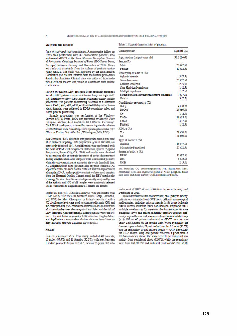
129
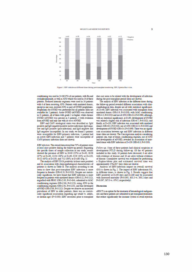
130
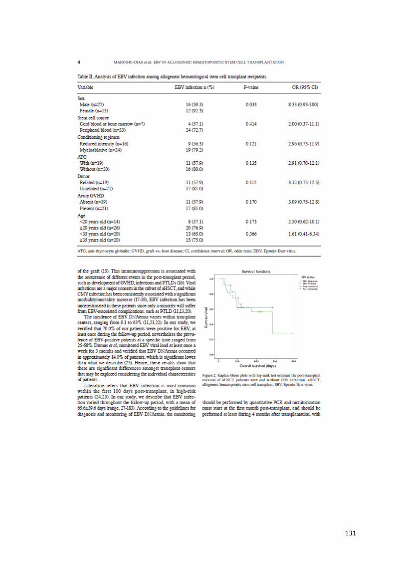
131
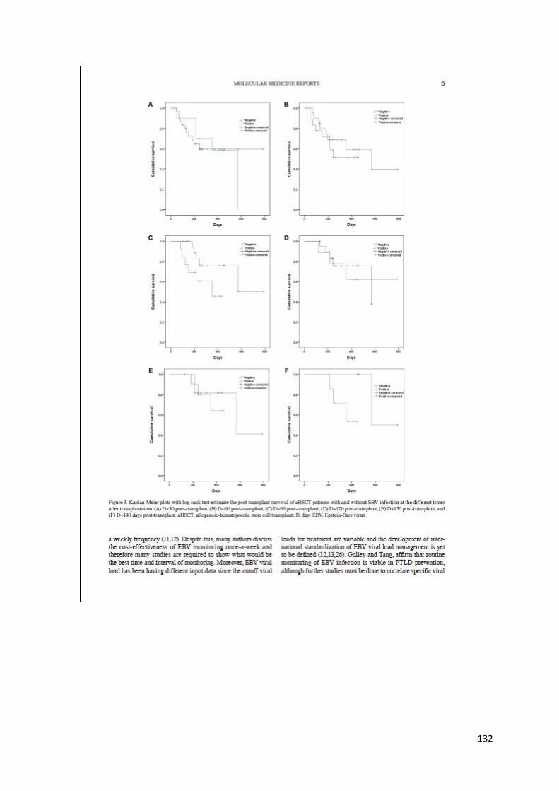
132
133

134

135