
Self-supervised learning improves dMMR/MSI detection from ...
15
Proceedings of Machine Learning Research 156, 2021 MICCAI Computational Pathlogy (COMPAY) Workshop Self-supervised learning improves dMMR/MSI detection from histology slides across multiple cancers Charlie Saillard [email protected] Owkin, Inc. Olivier Dehaene [email protected] Owkin, Inc. Tanguy Marchand [email protected] Owkin, Inc. Olivier Moindrot [email protected] Owkin, Inc. Aur´ elie Kamoun [email protected] Owkin, Inc. Benoit Schmauch [email protected] Owkin, Inc. Simon Jegou [email protected] Owkin, Inc. Abstract Microsatellite instability (MSI) is a tumor phenotype whose diagnosis largely impacts pa- tient care in colorectal cancers (CRC), and is associated with response to immunotherapy in all solid tumors. Deep learning models detecting MSI tumors directly from H&E stained slides have shown promise in improving diagnosis of MSI patients. Prior deep learning models for MSI detection have relied on neural networks pretrained on ImageNet dataset, which does not contain any medical image. In this study, we leverage recent advances in self-supervised learning by training neural networks on histology images from the TCGA dataset using MoCo V2. We show that these networks consistently outperform their coun- terparts pretrained using ImageNet and obtain state-of-the-art results for MSI detection with AUCs of 0.92 and 0.83 for CRC and gastric tumors, respectively. These models gen- eralize well on an external CRC cohort (0.97 AUC on PAIP) and improve transfer from one organ to another. Finally we show that predictive image regions exhibit meaningful histological patterns, and that the use of MoCo features highlighted more relevant patterns according to an expert pathologist. Keywords: Self-supervised learning, Microsatellite Instability (MSI) © PMLR, 2021. Editors: M. Atzori, N. Burlutskiy, F. Ciompi, Z. Li, F. Minhas, H. Müller, T. Peng, N. Rajpoot, B. TorbenNielsen, J. van der Laak, M. Veta, Y. Yuan, and I. Zlobec.
Transcript of Self-supervised learning improves dMMR/MSI detection from ...
Proceedings of Machine Learning Research 156, 2021 MICCAI Computational Pathlogy (COMPAY) Workshop
Self-supervised learning improves dMMR/MSI detectionfrom histology slides across multiple cancers
Charlie Saillard [email protected], Inc.
Olivier Dehaene [email protected], Inc.
Tanguy Marchand [email protected], Inc.
Olivier Moindrot [email protected], Inc.
Aurelie Kamoun [email protected], Inc.
Benoit Schmauch [email protected], Inc.
Simon Jegou [email protected]
Owkin, Inc.
Abstract
Microsatellite instability (MSI) is a tumor phenotype whose diagnosis largely impacts pa-tient care in colorectal cancers (CRC), and is associated with response to immunotherapyin all solid tumors. Deep learning models detecting MSI tumors directly from H&E stainedslides have shown promise in improving diagnosis of MSI patients. Prior deep learningmodels for MSI detection have relied on neural networks pretrained on ImageNet dataset,which does not contain any medical image. In this study, we leverage recent advances inself-supervised learning by training neural networks on histology images from the TCGAdataset using MoCo V2. We show that these networks consistently outperform their coun-terparts pretrained using ImageNet and obtain state-of-the-art results for MSI detectionwith AUCs of 0.92 and 0.83 for CRC and gastric tumors, respectively. These models gen-eralize well on an external CRC cohort (0.97 AUC on PAIP) and improve transfer fromone organ to another. Finally we show that predictive image regions exhibit meaningfulhistological patterns, and that the use of MoCo features highlighted more relevant patternsaccording to an expert pathologist.
Keywords: Self-supervised learning, Microsatellite Instability (MSI)
©PMLR, 2021.
Editors: M. Atzori, N. Burlutskiy, F. Ciompi, Z. Li, F. Minhas, H. Müller, T. Peng, N. Rajpoot, B. TorbenNielsen, J. van der Laak, M. Veta, Y. Yuan, and I. Zlobec.
MICCAI COMPAY 2021
1. Introduction
Microsatellite Instability (MSI) is a frequent tumor phenotype characterized by an abnor-mal repetition of short DNA motifs caused by a deficiency of the DNA mismatch repairsystem (MMR). MMR deficient tumors (dMMR) result from defects in the major MMRgenes, namely MLH1, MSH2, MSH6, PMS2. These defects arise either sporadically or as ahereditary condition named Lynch syndrome (LS), predisposing patients to develop cancersin several organs.
Recent studies have shown that immune checkpoint blockade therapy has a promisingresponse in dMMR/MSI cancers regardless of the tissue of origin [Le et al. (2017)]. In 2017,this genomic instability phenotype became the first pan-cancer biomarker approved by theUS FDA, allowing the use of pembrolizumab (Keytruda) for patients with dMMR/MSIsolid tumors at the metastatic stage [Prasad et al. (2018)].
As of today, systematic MSI screening is only recommended for colorectal cancer (CRC)and endometrial cancer [Svrcek et al. (2019)] where the prevalence is relatively high (10%to 20%), principally to detect LS patients and provide them with adequate follow-up. Inearly stages of CRC, MSI tumors are associated with good prognosis and resistance tochemotherapy [Sargent et al. (2010)], making the diagnosis of this phenotype all the moreessential for patient care and therapeutic decision. dMMR/MSI diagnosis is traditionallydone using immunohistochemistry (IHC), polymerase chain reaction (PCR) assays, or nextgeneration sequencing. Those methods can be time-consuming, expensive, and rely onspecific expertise which may not be available in every center.
Deep learning based MSI classifiers using H&E stained digital images offer a new alter-native for a broader and more efficient screening [Echle et al. (2020)]. In CRC, previouswork suggests that the use of such models as pre-screening tools could eventually replaceIHC and PCR for a subset of tumors classified as microsatellite stable (MSS) or unstable(MSI) with a high probability [Kacew et al. (2021)]. In other locations where MSI preva-lence is lower and screening not done as routine practice, predictive models of MSI statusfrom WSI could be used as efficient pre-screening tools.
In this work, we leverage recent advances in self-supervised learning (SSL) on images.We show that SSL permits to reach state-of-the-art results on colorectal and gastric cancercohorts from The Cancer Genome Atlas (TCGA), generalizes well on an unseen colorectalcohort (PAIP), and could pave the way for classifiers on locations with low MSI prevalence.
2. Related Work
Expert models Several histology patterns on H&E images have been reported to cor-relate with MSI, such as tumor-infiltrating lymphocytes, lack of dirty necrosis or poordifferentiation [(Greenson et al. (2009)]. A series of models based on clinico-pathologicalfeatures have been developed [Greenson et al. (2009); Jenkins et al. (2007); Hyde et al.(2010); Fujiyoshi et al. (2017); Roman et al. (2010)] and reported ROC-AUC performancesranging from 0.85 to 0.92 in various cohorts of patients with CRC. These methods howeverrequire time-consuming annotations from expert pathologists and are prone to inter-ratervariability.
2
MICCAI COMPAY 2021
Deep learning models In a seminal publication, Kather et al. (2019) proved the fea-sibility to determine the dMMR/MSI status from H&E stained whole slide images (WSI)using deep learning. They trained a first ResNet [He et al. (2016)] to segment tumor regionson WSI, and a second one to predict MSI/MSS status in each tumor tile. Each ResNet waspretrained on ImageNet and the last 10 layers were fine-tuned. Models were trained andvalidated on different TCGA cohorts and obtained respectively AUCs of 0.77, 0.81 and 0.75on Colorectal, Gastric and Endometrial formalin-fixed paraffin-embedded (FFPE) datasets.
In a larger scale study focusing on CRC only [Echle et al. (2020)], the same team latertrained a model on n = 6406 patients, reaching 0.96 AUC (95% CI 0.93–0.98) on an externaldataset of n = 771 patients. Tumor tissues were manually outlined by pathologists.
Since then, different works based on deep learning methods have been published [Zhanget al. (2018); Cao et al. (2020); Hong et al. (2020); Yamashita et al. (2021); Bilal et al.(2021); Lee et al. (2021)] and are reviewed in Hildebrand et al. (2021). The vast majorityrely on networks pretrained on the ImageNet dataset [Deng et al. (2009)] and only the lastlayers are re-trained or fine-tuned. Most of these papers also rely on tumor segmentationas a first step in their models (either by a pathologist or by a deep learning model).
Self-Supervised learning Over the past few years, rapid progress has been made in thefield of SSL using contrastive learning or self-distillation strategies: simCLR [Chen et al.(2020a,b)] , MoCo [He et al. (2020); Chen et al. (2020c, 2021)], BYOL [Grill et al. (2020)],achieving impressive performances on ImageNet without using any labels. Such modelshave also been shown to outperform supervised models in transfer learning tasks [Chenet al. (2020a); Caron et al. (2021); Li et al. (2021b)].
These advances are of particular interest in medical imaging applications where labeleddatasets are hard to collect, and especially in histology where each WSI contains thousandsof unlabeled images. There is growing evidence that SSL is a powerful method to obtainrelevant features for various prediction tasks from histology images [Dehaene et al. (2020);Lu et al. (2019); Li et al. (2021a); Koohbanani et al. (2021); Gildenblat and Klaiman (2019);Abbet et al. (2021); Srinidhi et al. (2021)]. In this work, we show that self-supervision canbe efficiently used to detect dMMR/MSI tumors from histology slides, and outperformImageNet pretrained models across and between several tumors.
3. Methods
3.1 Proposed pipeline
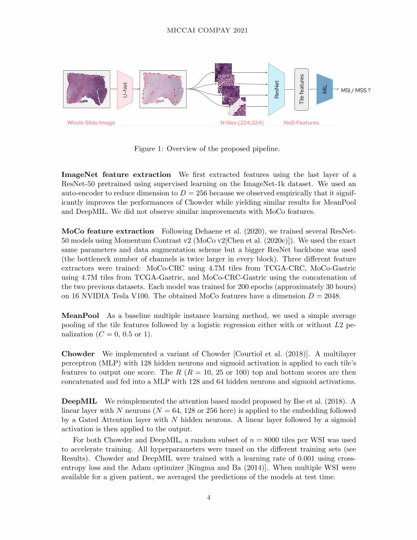
First, a U-Net neural network [Ronneberger et al. (2015)] is used to segment tissue on theinput WSI and discard the background, as well as artifacts. Second, segmented tissue isdivided into N (typically between 10,000 and 30,000) smaller images called tiles. Each tilehas a fixed shape of 224 × 224 pixels (resolution of 0.5 micron per pixel). Third, the Ntiles are embedded into feature vectors of shape D using a pretrained convolutional neuralnetwork. Fourth, the N × D features are aggregated using a multiple instance learningmodel. This final model is the only one trained using MSI/MSS labels.
In this study, we benchmarked 2 different feature extractors (ResNet-50 pretrained withsupervised learning on ImageNet [Deng et al. (2009)] or with SSL on TCGA) and 3 multipleinstance learning models (MeanPool, Chowder and DeepMIL).
3
MICCAI COMPAY 2021
I �Ĵ���
������
� �
�������������
�� �Ī�����Ğ
����������������������������������ę�����������������������������Ĵ����������ĸ�������Ĺ��������������������������������ŔŔŖ�ŔŔŖė�����������������������������������������������ĸ�������������ĹĘ��������������������������������������������������������������������������������������ĸ���������Ĺ�������������� ������������ė
��������ĸŔŔŖĘŔŔŖĹ������������ ���� �����������
Figure 1: Overview of the proposed pipeline.
ImageNet feature extraction We first extracted features using the last layer of aResNet-50 pretrained using supervised learning on the ImageNet-1k dataset. We used anauto-encoder to reduce dimension to D = 256 because we observed empirically that it signif-icantly improves the performances of Chowder while yielding similar results for MeanPooland DeepMIL. We did not observe similar improvements with MoCo features.
MoCo feature extraction Following Dehaene et al. (2020), we trained several ResNet-50 models using Momentum Contrast v2 (MoCo v2[Chen et al. (2020c)]). We used the exactsame parameters and data augmentation scheme but a bigger ResNet backbone was used(the bottleneck number of channels is twice larger in every block). Three different featureextractors were trained: MoCo-CRC using 4.7M tiles from TCGA-CRC, MoCo-Gastricusing 4.7M tiles from TCGA-Gastric, and MoCo-CRC-Gastric using the concatenation ofthe two previous datasets. Each model was trained for 200 epochs (approximately 30 hours)on 16 NVIDIA Tesla V100. The obtained MoCo features have a dimension D = 2048.
MeanPool As a baseline multiple instance learning method, we used a simple averagepooling of the tile features followed by a logistic regression either with or without L2 pe-nalization (C = 0, 0.5 or 1).
Chowder We implemented a variant of Chowder [Courtiol et al. (2018)]. A multilayerperceptron (MLP) with 128 hidden neurons and sigmoid activation is applied to each tile’sfeatures to output one score. The R (R = 10, 25 or 100) top and bottom scores are thenconcatenated and fed into a MLP with 128 and 64 hidden neurons and sigmoid activations.
DeepMIL We reimplemented the attention based model proposed by Ilse et al. (2018). Alinear layer with N neurons (N = 64, 128 or 256 here) is applied to the embedding followedby a Gated Attention layer with N hidden neurons. A linear layer followed by a sigmoidactivation is then applied to the output.
For both Chowder and DeepMIL, a random subset of n = 8000 tiles per WSI was usedto accelerate training. All hyperparameters were tuned on the different training sets (seeResults). Chowder and DeepMIL were trained with a learning rate of 0.001 using cross-entropy loss and the Adam optimizer [Kingma and Ba (2014)]. When multiple WSI wereavailable for a given patient, we averaged the predictions of the models at test time.
4
MICCAI COMPAY 2021
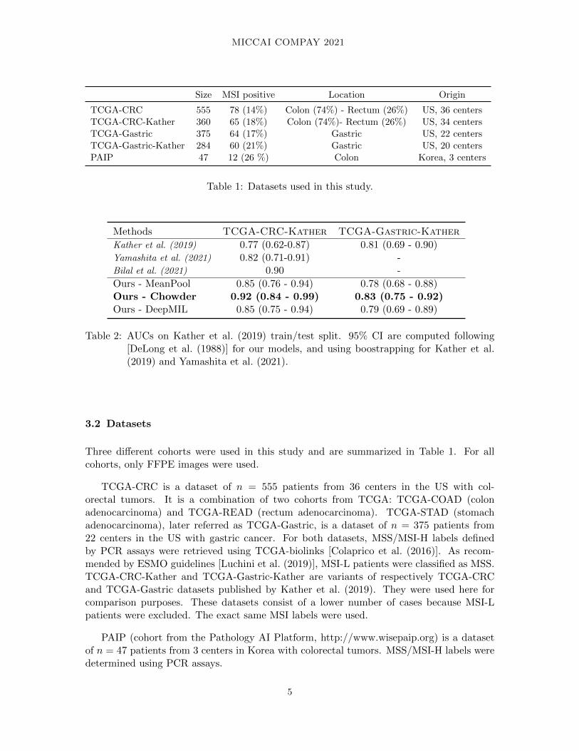
Size MSI positive Location Origin
TCGA-CRC 555 78 (14%) Colon (74%) - Rectum (26%) US, 36 centersTCGA-CRC-Kather 360 65 (18%) Colon (74%)- Rectum (26%) US, 34 centersTCGA-Gastric 375 64 (17%) Gastric US, 22 centersTCGA-Gastric-Kather 284 60 (21%) Gastric US, 20 centersPAIP 47 12 (26 %) Colon Korea, 3 centers
Table 1: Datasets used in this study.
Methods TCGA-CRC-Kather TCGA-Gastric-Kather
Kather et al. (2019) 0.77 (0.62-0.87) 0.81 (0.69 - 0.90)Yamashita et al. (2021) 0.82 (0.71-0.91) -Bilal et al. (2021) 0.90 -
Ours - MeanPool 0.85 (0.76 - 0.94) 0.78 (0.68 - 0.88)Ours - Chowder 0.92 (0.84 - 0.99) 0.83 (0.75 - 0.92)Ours - DeepMIL 0.85 (0.75 - 0.94) 0.79 (0.69 - 0.89)
Table 2: AUCs on Kather et al. (2019) train/test split. 95% CI are computed following[DeLong et al. (1988)] for our models, and using boostrapping for Kather et al.(2019) and Yamashita et al. (2021).
3.2 Datasets
Three different cohorts were used in this study and are summarized in Table 1. For allcohorts, only FFPE images were used.
TCGA-CRC is a dataset of n = 555 patients from 36 centers in the US with col-orectal tumors. It is a combination of two cohorts from TCGA: TCGA-COAD (colonadenocarcinoma) and TCGA-READ (rectum adenocarcinoma). TCGA-STAD (stomachadenocarcinoma), later referred as TCGA-Gastric, is a dataset of n = 375 patients from22 centers in the US with gastric cancer. For both datasets, MSS/MSI-H labels definedby PCR assays were retrieved using TCGA-biolinks [Colaprico et al. (2016)]. As recom-mended by ESMO guidelines [Luchini et al. (2019)], MSI-L patients were classified as MSS.TCGA-CRC-Kather and TCGA-Gastric-Kather are variants of respectively TCGA-CRCand TCGA-Gastric datasets published by Kather et al. (2019). They were used here forcomparison purposes. These datasets consist of a lower number of cases because MSI-Lpatients were excluded. The exact same MSI labels were used.
PAIP (cohort from the Pathology AI Platform, http://www.wisepaip.org) is a datasetof n = 47 patients from 3 centers in Korea with colorectal tumors. MSS/MSI-H labels weredetermined using PCR assays.
5
MICCAI COMPAY 2021
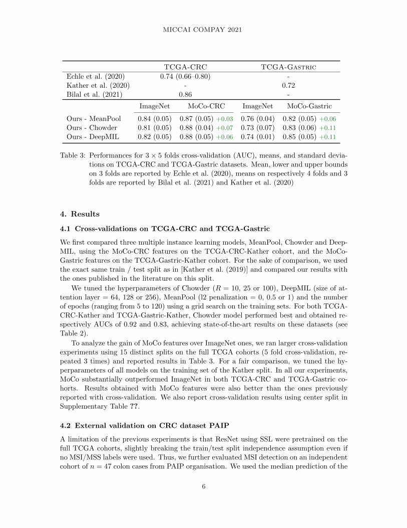
TCGA-CRC TCGA-Gastric
Echle et al. (2020) 0.74 (0.66–0.80) -Kather et al. (2020) - 0.72Bilal et al. (2021) 0.86 -
ImageNet MoCo-CRC ImageNet MoCo-Gastric
Ours - MeanPool 0.84 (0.05) 0.87 (0.05) +0.03 0.76 (0.04) 0.82 (0.05) +0.06
Ours - Chowder 0.81 (0.05) 0.88 (0.04) +0.07 0.73 (0.07) 0.83 (0.06) +0.11
Ours - DeepMIL 0.82 (0.05) 0.88 (0.05) +0.06 0.74 (0.01) 0.85 (0.05) +0.11
Table 3: Performances for 3 × 5 folds cross-validation (AUC), means, and standard devia-tions on TCGA-CRC and TCGA-Gastric datasets. Mean, lower and upper boundson 3 folds are reported by Echle et al. (2020), means on respectively 4 folds and 3folds are reported by Bilal et al. (2021) and Kather et al. (2020)
4. Results
4.1 Cross-validations on TCGA-CRC and TCGA-Gastric
We first compared three multiple instance learning models, MeanPool, Chowder and Deep-MIL, using the MoCo-CRC features on the TCGA-CRC-Kather cohort, and the MoCo-Gastric features on the TCGA-Gastric-Kather cohort. For the sake of comparison, we usedthe exact same train / test split as in [Kather et al. (2019)] and compared our results withthe ones published in the literature on this split.
We tuned the hyperparameters of Chowder (R = 10, 25 or 100), DeepMIL (size of at-tention layer = 64, 128 or 256), MeanPool (l2 penalization = 0, 0.5 or 1) and the numberof epochs (ranging from 5 to 120) using a grid search on the training sets. For both TCGA-CRC-Kather and TCGA-Gastric-Kather, Chowder model performed best and obtained re-spectively AUCs of 0.92 and 0.83, achieving state-of-the-art results on these datasets (seeTable 2).
To analyze the gain of MoCo features over ImageNet ones, we ran larger cross-validationexperiments using 15 distinct splits on the full TCGA cohorts (5 fold cross-validation, re-peated 3 times) and reported results in Table 3. For a fair comparison, we tuned the hy-perparameters of all models on the training set of the Kather split. In all our experiments,MoCo substantially outperformed ImageNet in both TCGA-CRC and TCGA-Gastric co-horts. Results obtained with MoCo features were also better than the ones previouslyreported with cross-validation. We also report cross-validation results using center split inSupplementary Table ??.
4.2 External validation on CRC dataset PAIP
A limitation of the previous experiments is that ResNet using SSL were pretrained on thefull TCGA cohorts, slightly breaking the train/test split independence assumption even ifno MSI/MSS labels were used. Thus, we further evaluated MSI detection on an independentcohort of n = 47 colon cases from PAIP organisation. We used the median prediction of the
6
MICCAI COMPAY 2021
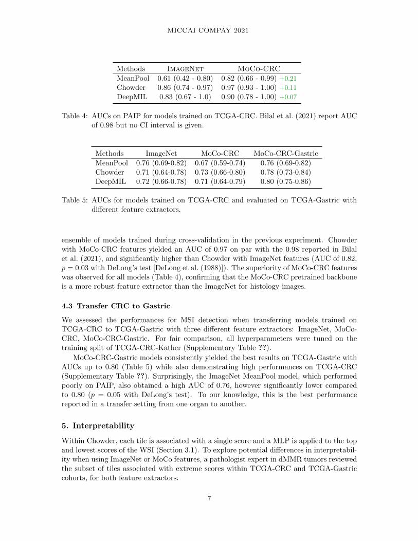
Methods ImageNet MoCo-CRC
MeanPool 0.61 (0.42 - 0.80) 0.82 (0.66 - 0.99) +0.21
Chowder 0.86 (0.74 - 0.97) 0.97 (0.93 - 1.00) +0.11
DeepMIL 0.83 (0.67 - 1.0) 0.90 (0.78 - 1.00) +0.07
Table 4: AUCs on PAIP for models trained on TCGA-CRC. Bilal et al. (2021) report AUCof 0.98 but no CI interval is given.
Methods ImageNet MoCo-CRC MoCo-CRC-Gastric
MeanPool 0.76 (0.69-0.82) 0.67 (0.59-0.74) 0.76 (0.69-0.82)Chowder 0.71 (0.64-0.78) 0.73 (0.66-0.80) 0.78 (0.73-0.84)DeepMIL 0.72 (0.66-0.78) 0.71 (0.64-0.79) 0.80 (0.75-0.86)
Table 5: AUCs for models trained on TCGA-CRC and evaluated on TCGA-Gastric withdifferent feature extractors.
ensemble of models trained during cross-validation in the previous experiment. Chowderwith MoCo-CRC features yielded an AUC of 0.97 on par with the 0.98 reported in Bilalet al. (2021), and significantly higher than Chowder with ImageNet features (AUC of 0.82,p = 0.03 with DeLong’s test [DeLong et al. (1988)]). The superiority of MoCo-CRC featureswas observed for all models (Table 4), confirming that the MoCo-CRC pretrained backboneis a more robust feature extractor than the ImageNet for histology images.
4.3 Transfer CRC to Gastric
We assessed the performances for MSI detection when transferring models trained onTCGA-CRC to TCGA-Gastric with three different feature extractors: ImageNet, MoCo-CRC, MoCo-CRC-Gastric. For fair comparison, all hyperparameters were tuned on thetraining split of TCGA-CRC-Kather (Supplementary Table ??).
MoCo-CRC-Gastric models consistently yielded the best results on TCGA-Gastric withAUCs up to 0.80 (Table 5) while also demonstrating high performances on TCGA-CRC(Supplementary Table ??). Surprisingly, the ImageNet MeanPool model, which performedpoorly on PAIP, also obtained a high AUC of 0.76, however significantly lower comparedto 0.80 (p = 0.05 with DeLong’s test). To our knowledge, this is the best performancereported in a transfer setting from one organ to another.
5. Interpretability
Within Chowder, each tile is associated with a single score and a MLP is applied to the topand lowest scores of the WSI (Section 3.1). To explore potential differences in interpretabil-ity when using ImageNet or MoCo features, a pathologist expert in dMMR tumors reviewedthe subset of tiles associated with extreme scores within TCGA-CRC and TCGA-Gastriccohorts, for both feature extractors.
7
MICCAI COMPAY 2021
ImageNet
MSI
MSS
MoCo CRC ImageNet
MSI
MSS
MoCo GastricA B
C
MoCo CRCImageNet
MSI
MSS
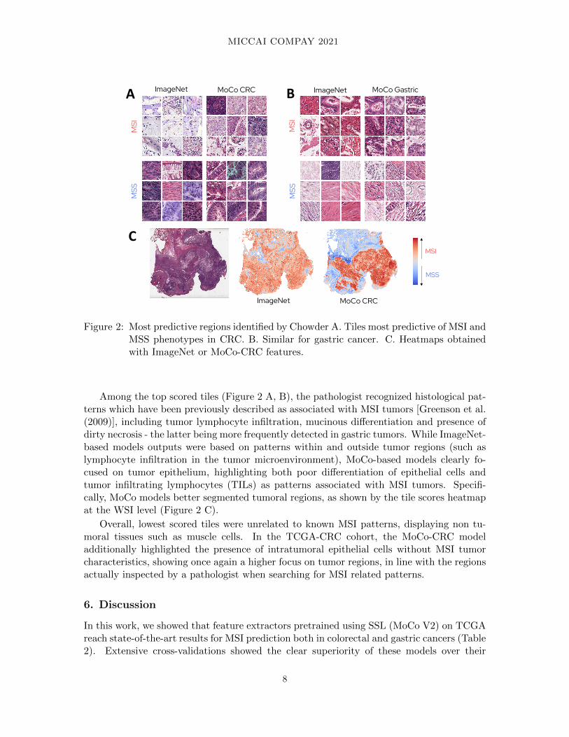
Figure 2: Most predictive regions identified by Chowder A. Tiles most predictive of MSI andMSS phenotypes in CRC. B. Similar for gastric cancer. C. Heatmaps obtainedwith ImageNet or MoCo-CRC features.
Among the top scored tiles (Figure 2 A, B), the pathologist recognized histological pat-terns which have been previously described as associated with MSI tumors [Greenson et al.(2009)], including tumor lymphocyte infiltration, mucinous differentiation and presence ofdirty necrosis - the latter being more frequently detected in gastric tumors. While ImageNet-based models outputs were based on patterns within and outside tumor regions (such aslymphocyte infiltration in the tumor microenvironment), MoCo-based models clearly fo-cused on tumor epithelium, highlighting both poor differentiation of epithelial cells andtumor infiltrating lymphocytes (TILs) as patterns associated with MSI tumors. Specifi-cally, MoCo models better segmented tumoral regions, as shown by the tile scores heatmapat the WSI level (Figure 2 C).
Overall, lowest scored tiles were unrelated to known MSI patterns, displaying non tu-moral tissues such as muscle cells. In the TCGA-CRC cohort, the MoCo-CRC modeladditionally highlighted the presence of intratumoral epithelial cells without MSI tumorcharacteristics, showing once again a higher focus on tumor regions, in line with the regionsactually inspected by a pathologist when searching for MSI related patterns.
6. Discussion
In this work, we showed that feature extractors pretrained using SSL (MoCo V2) on TCGAreach state-of-the-art results for MSI prediction both in colorectal and gastric cancers (Table2). Extensive cross-validations showed the clear superiority of these models over their
8
MICCAI COMPAY 2021
counterparts pretrained using ImageNet (Table 3). In addition, the former generalize betteron an external CRC cohort (Table 4). Finally, we observed that using a feature extractorpretrained on several organs using SSL (Table 5) opens the way to both state-of-the-artperformances in cross-validation and robust generalization from one organ to another.
There are several limitations to our current study. First, our models could benefit frombeing trained on more data as shown in the training curve from Echle et al. (2020) (Fig-ure 1.c). Second, our models should be validated on larger cohorts, encompassing differentpatient populations, treatments (neoadjuvant chemotherapy can impact cell morphology),scanner manufacturer and sample types (resections, biopsies). Third, SSL techniques areevolving rapidly using new architectures such as Vision Transformers [Dosovitskiy et al.(2020)] and more experiments are required to find how to apply them in histology, includ-ing taking into account the spatial arrangement of the tiles. However, such experimentsrequire access to extensive computational resources, which limits reproducibility. Finally,in contrast to several concurrent works [Echle et al. (2020); Bilal et al. (2021); Kather et al.(2019)] that fine-tuned the backbones, while our study kept them entirely frozen.
A recent study, Kacew et al. (2021) showed that performances of deep learning modelsmay impact the diagnosis of MSI for patients with CRC. Notably, MSI diagnosis is notroutinely done for patients with other solid tumors, missing the identification of candi-date patients for immunotherapy. Our results indicate that SSL is a promising solutionto develop accurate models for frequent tumor localization such as oesophagus, pancreas,small intestine or even brain, where images are available but MSI prevalence is too low forsystematic IHC or molecular testing.
9
MICCAI COMPAY 2021
Acknowledgments
We thank Florence Renaud, pathologist at CHU Lille, for her insightful analysis of themost predictive regions. We thank Patrick Sin-Chan for his corrections of the manuscript.
This work was granted access to the HPC resources of IDRIS under the allocationAD011012519 made by GENCI.
The results published here are part based upon data generated by the TCGA ResearchNetwork: https://www.cancer.gov/tcga.
Regarding the PAIP dataset: De-identified pathology images and annotations used inthis research were prepared and provided by the Seoul National University Hospital by agrant of the Korea Health Technology R&D Project through the Korea Health IndustryDevelopment Institute (KHIDI), funded by the Ministry of Health & Welfare, Republic ofKorea (grant number: HI18C0316).
10
MICCAI COMPAY 2021
References
Christian Abbet, Linda Studer, Andreas Fischer, Heather Dawson, Inti Zlobec, BehzadBozorgtabar, and Jean-Philippe Thiran. Self-rule to adapt: Learning generalized featuresfrom sparsely-labeled data using unsupervised domain adaptation for colorectal cancertissue phenotyping. In Medical Imaging with Deep Learning, 2021.
Mohsin Bilal, Shan E Ahmed Raza, Ayesha Azam, Simon Graham, Muhammad Ilyas, Ian ACree, David Snead, Fayyaz Minhas, and Nasir M Rajpoot. Novel deep learning algorithmpredicts the status of molecular pathways and key mutations in colorectal cancer fromroutine histology images. medRxiv, 2021.
Rui Cao, Fan Yang, Si-Cong Ma, Li Liu, Yu Zhao, Yan Li, De-Hua Wu, Tongxin Wang,Wei-Jia Lu, Wei-Jing Cai, et al. Development and interpretation of a pathomics-basedmodel for the prediction of microsatellite instability in colorectal cancer. Theranostics,10(24):11080, 2020.
Mathilde Caron, Hugo Touvron, Ishan Misra, Herve Jegou, Julien Mairal, Piotr Bojanowski,and Armand Joulin. Emerging properties in self-supervised vision transformers. arXivpreprint arXiv:2104.14294, 2021.
Ting Chen, Simon Kornblith, Mohammad Norouzi, and Geoffrey Hinton. A simple frame-work for contrastive learning of visual representations. In International conference onmachine learning, pages 1597–1607. PMLR, 2020a.
Ting Chen, Simon Kornblith, Kevin Swersky, Mohammad Norouzi, and Geoffrey Hin-ton. Big self-supervised models are strong semi-supervised learners. arXiv preprintarXiv:2006.10029, 2020b.
Xinlei Chen, Haoqi Fan, Ross Girshick, and Kaiming He. Improved baselines with momen-tum contrastive learning. arXiv preprint arXiv:2003.04297, 2020c.
Xinlei Chen, Saining Xie, and Kaiming He. An empirical study of training self-supervisedvision transformers. arXiv preprint arXiv:2104.02057, 2021.
Antonio Colaprico, Tiago C Silva, Catharina Olsen, Luciano Garofano, Claudia Cava, Da-vide Garolini, Thais S Sabedot, Tathiane M Malta, Stefano M Pagnotta, Isabella Cas-tiglioni, et al. Tcgabiolinks: an r/bioconductor package for integrative analysis of tcgadata. Nucleic acids research, 44(8):e71–e71, 2016.
Pierre Courtiol, Eric W Tramel, Marc Sanselme, and Gilles Wainrib. Classification anddisease localization in histopathology using only global labels: A weakly-supervised ap-proach. arXiv preprint arXiv:1802.02212, 2018.
Olivier Dehaene, Axel Camara, Olivier Moindrot, Axel de Lavergne, and Pierre Courtiol.Self-supervision closes the gap between weak and strong supervision in histology. arXivpreprint arXiv:2012.03583, 2020.
11
MICCAI COMPAY 2021
Elizabeth R DeLong, David M DeLong, and Daniel L Clarke-Pearson. Comparing the areasunder two or more correlated receiver operating characteristic curves: a nonparametricapproach. Biometrics, pages 837–845, 1988.
Jia Deng, Wei Dong, Richard Socher, Li-Jia Li, Kai Li, and Li Fei-Fei. Imagenet: A large-scale hierarchical image database. In 2009 IEEE conference on computer vision andpattern recognition, pages 248–255. Ieee, 2009.
Alexey Dosovitskiy, Lucas Beyer, Alexander Kolesnikov, Dirk Weissenborn, Xiaohua Zhai,Thomas Unterthiner, Mostafa Dehghani, Matthias Minderer, Georg Heigold, SylvainGelly, et al. An image is worth 16x16 words: Transformers for image recognition atscale. arXiv preprint arXiv:2010.11929, 2020.
Amelie Echle, Heike Irmgard Grabsch, Philip Quirke, Piet A van den Brandt, Nicholas PWest, Gordon GA Hutchins, Lara R Heij, Xiuxiang Tan, Susan D Richman, JeremiasKrause, et al. Clinical-grade detection of microsatellite instability in colorectal tumorsby deep learning. Gastroenterology, 159(4):1406–1416, 2020.
Kenji Fujiyoshi, Tatsuro Yamaguchi, Miho Kakuta, Akemi Takahashi, Yoshiko Arai, MinaYamada, Gou Yamamoto, Sachiko Ohde, Misato Takao, Shin-ichiro Horiguchi, et al.Predictive model for high-frequency microsatellite instability in colorectal cancer patientsover 50 years of age. Cancer medicine, 6(6):1255–1263, 2017.
Jacob Gildenblat and Eldad Klaiman. Self-supervised similarity learning for digital pathol-ogy. arXiv preprint arXiv:1905.08139, 2019.
Joel K Greenson, Shu-Chen Huang, Casey Herron, Victor Moreno, Joseph D Bonner,Lynn P Tomsho, Ofer Ben-Izhak, Hector I Cohen, Phillip Trougouboff, Jacob Bejhar,et al. Pathologic predictors of microsatellite instability in colorectal cancer. The Ameri-can journal of surgical pathology, 33(1):126, 2009.
Jean-Bastien Grill, Florian Strub, Florent Altche, Corentin Tallec, Pierre H Richemond,Elena Buchatskaya, Carl Doersch, Bernardo Avila Pires, Zhaohan Daniel Guo, Mo-hammad Gheshlaghi Azar, et al. Bootstrap your own latent: A new approach to self-supervised learning. arXiv preprint arXiv:2006.07733, 2020.
Kaiming He, Xiangyu Zhang, Shaoqing Ren, and Jian Sun. Deep residual learning forimage recognition. In Proceedings of the IEEE conference on computer vision and patternrecognition, pages 770–778, 2016.
Kaiming He, Haoqi Fan, Yuxin Wu, Saining Xie, and Ross Girshick. Momentum contrast forunsupervised visual representation learning. In Proceedings of the IEEE/CVF Conferenceon Computer Vision and Pattern Recognition, pages 9729–9738, 2020.
Lindsey A Hildebrand, Colin J Pierce, Michael Dennis, Munizay Paracha, and Asaf Maoz.Artificial intelligence for histology-based detection of microsatellite instability and pre-diction of response to immunotherapy in colorectal cancer. Cancers, 13(3):391, 2021.
12
MICCAI COMPAY 2021
Runyu Hong, Wenke Liu, Deborah DeLair, Narges Razavian, and David Fenyo. Predictingendometrial cancer subtypes and molecular features from histopathology images usingmulti-resolution deep learning models. bioRxiv, 2020.
Angela Hyde, Daniel Fontaine, Susan Stuckless, Roger Green, Aaron Pollett, MichelleSimms, Payal Sipahimalani, Patrick Parfrey, and Banfield Younghusband. A histology-based model for predicting microsatellite instability in colorectal cancers. The Americanjournal of surgical pathology, 34(12):1820–1829, 2010.
Maximilian Ilse, Jakub Tomczak, and Max Welling. Attention-based deep multiple instancelearning. In International conference on machine learning, pages 2127–2136. PMLR, 2018.
Mark A Jenkins, Shinichi Hayashi, Anne-Marie O’shea, Lawrence J Burgart, Tom C Smyrk,David Shimizu, Paul M Waring, Andrew R Ruszkiewicz, Aaron F Pollett, Mark Redston,et al. Pathology features in bethesda guidelines predict colorectal cancer microsatelliteinstability: a population-based study. Gastroenterology, 133(1):48–56, 2007.
Alec J Kacew, Garth W Strohbehn, Loren Saulsberry, Neda Laiteerapong, Nicole A Cipri-ani, Jakob N Kather, and Alexander T Pearson. Artificial intelligence can cut costs whilemaintaining accuracy in colorectal cancer genotyping. Frontiers in Oncology, 11, 2021.
Jakob Nikolas Kather, Alexander T Pearson, Niels Halama, Dirk Jager, Jeremias Krause,Sven H Loosen, Alexander Marx, Peter Boor, Frank Tacke, Ulf Peter Neumann, et al.Deep learning can predict microsatellite instability directly from histology in gastroin-testinal cancer. Nature medicine, 25(7):1054–1056, 2019.
Jakob Nikolas Kather, Lara R Heij, Heike I Grabsch, Chiara Loeffler, Amelie Echle, Han-nah Sophie Muti, Jeremias Krause, Jan M Niehues, Kai AJ Sommer, Peter Bankhead,et al. Pan-cancer image-based detection of clinically actionable genetic alterations. NatureCancer, 1(8):789–799, 2020.
Diederik P Kingma and Jimmy Ba. Adam: A method for stochastic optimization. arXivpreprint arXiv:1412.6980, 2014.
Navid Alemi Koohbanani, Balagopal Unnikrishnan, Syed Ali Khurram, Pavitra Krish-naswamy, and Nasir Rajpoot. Self-path: Self-supervision for classification of pathologyimages with limited annotations. IEEE Transactions on Medical Imaging, 2021.
Dung T Le, Jennifer N Durham, Kellie N Smith, Hao Wang, Bjarne R Bartlett, Laveet KAulakh, Steve Lu, Holly Kemberling, Cara Wilt, Brandon S Luber, et al. Mismatchrepair deficiency predicts response of solid tumors to pd-1 blockade. Science, 357(6349):409–413, 2017.
Sung Hak Lee, In Hye Song, and Hyun-Jong Jang. Feasibility of deep learning-based fullyautomated classification of microsatellite instability in tissue slides of colorectal cancer.International Journal of Cancer, 2021.
Bin Li, Yin Li, and Kevin W Eliceiri. Dual-stream multiple instance learning network forwhole slide image classification with self-supervised contrastive learning. In Proceedings of
13
MICCAI COMPAY 2021
the IEEE/CVF Conference on Computer Vision and Pattern Recognition, pages 14318–14328, 2021a.
Chunyuan Li, Jianwei Yang, Pengchuan Zhang, Mei Gao, Bin Xiao, Xiyang Dai, Lu Yuan,and Jianfeng Gao. Efficient self-supervised vision transformers for representation learning.arXiv preprint arXiv:2106.09785, 2021b.
Ming Y Lu, Richard J Chen, Jingwen Wang, Debora Dillon, and Faisal Mahmood. Semi-supervised histology classification using deep multiple instance learning and contrastivepredictive coding. arXiv preprint arXiv:1910.10825, 2019.
C Luchini, F Bibeau, MJL Ligtenberg, N Singh, A Nottegar, T Bosse, R Miller, N Riaz, J-YDouillard, F Andre, et al. Esmo recommendations on microsatellite instability testing forimmunotherapy in cancer, and its relationship with pd-1/pd-l1 expression and tumourmutational burden: a systematic review-based approach. Annals of Oncology, 30(8):1232–1243, 2019.
Vinay Prasad, Victoria Kaestner, and Sham Mailankody. Cancer drugs approved based onbiomarkers and not tumor type—fda approval of pembrolizumab for mismatch repair-deficient solid cancers. JAMA oncology, 4(2):157–158, 2018.
Ruth Roman, Montse Verdu, Miquel Calvo, August Vidal, Xavier Sanjuan, Mireya Jimeno,Antonio Salas, Josefina Autonell, Isabel Trias, Marta Gonzalez, et al. Microsatelliteinstability of the colorectal carcinoma can be predicted in the conventional pathologicexamination. a prospective multicentric study and the statistical analysis of 615 casesconsolidate our previously proposed logistic regression model. Virchows Archiv, 456(5):533–541, 2010.
Olaf Ronneberger, Philipp Fischer, and Thomas Brox. U-net: Convolutional networks forbiomedical image segmentation. In International Conference on Medical image computingand computer-assisted intervention, pages 234–241. Springer, 2015.
Daniel J Sargent, Silvia Marsoni, Genevieve Monges, Stephen N Thibodeau, Roberto Labi-anca, Stanley R Hamilton, Amy J French, Brian Kabat, Nathan R Foster, Valter Torri,et al. Defective mismatch repair as a predictive marker for lack of efficacy of fluorouracil-based adjuvant therapy in colon cancer. Journal of Clinical Oncology, 28(20):3219, 2010.
Chetan L Srinidhi, Seung Wook Kim, Fu-Der Chen, and Anne L Martel. Self-superviseddriven consistency training for annotation efficient histopathology image analysis. arXivpreprint arXiv:2102.03897, 2021.
Magali Svrcek, Olivier Lascols, Romain Cohen, Ada Collura, Vincent Jonchere, Jean-Francois Flejou, Olivier Buhard, and Alex Duval. Msi/mmr-deficient tumor diagnosis:Which standard for screening and for diagnosis? diagnostic modalities for the colon andother sites: Differences between tumors. Bulletin du cancer, 106(2):119–128, 2019.
Rikiya Yamashita, Jin Long, Teri Longacre, Lan Peng, Gerald Berry, Brock Martin, JohnHiggins, Daniel L Rubin, and Jeanne Shen. Deep learning model for the prediction of
14

MICCAI COMPAY 2021
microsatellite instability in colorectal cancer: a diagnostic study. The Lancet Oncology,22(1):132–141, 2021.
Renyu Zhang, Boleslaw L Osinski, Timothy J Taxter, Jason Perera, Denise J Lau, andAly A Khan. Adversarial deep learning for microsatellite instability prediction fromhistopathology slides. In Proceedings of the 1st Conference on Medical Imaging withDeep Learning (MIDL 2018), Amsterdam, The Netherlands, pages 4–6, 2018.
15


![Original Article NLRC5 silencing improves cardiac fibrosis by ...cardiac hypertrophy, heart failure and severe arrhythmia [8]. Therefore, understanding the pathogenesis of cardiac](https://static.fdocumentos.com/doc/165x107/60dead27fd28d006400d3d98/original-article-nlrc5-silencing-improves-cardiac-fibrosis-by-cardiac-hypertrophy.jpg)