STUDY PROTOCOL Open Access Transluminal endoscopic step …
13
STUDY PROTOCOL Open Access Transluminal endoscopic step-up approach versus minimally invasive surgical step-up approach in patients with infected necrotising pancreatitis (TENSION trial): design and rationale of a randomised controlled multicenter trial [ISRCTN09186711] Sandra van Brunschot 1,2* , Janneke van Grinsven 1,2 , Rogier P Voermans 1 , Olaf J Bakker 3 , Marc GH Besselink 4 , Marja A Boermeester 4 , Thomas L Bollen 5 , Koop Bosscha 6 , Stefan A Bouwense 2,7 , Marco J Bruno 8 , Vincent C Cappendijk 9 , Esther C Consten 10 , Cornelis H Dejong 11 , Marcel GW Dijkgraaf 12 , Casper H van Eijck 13 , G Willemien Erkelens 14 , Harry van Goor 7 , Mohammed Hadithi 15 , Jan-Willem Haveman 16 , Sijbrand H Hofker 16 , Jeroen JM Jansen 17 , Johan S Laméris 18 , Krijn P van Lienden 18 , Eric R Manusama 19 , Maarten A Meijssen 20 , Chris J Mulder 21 , Vincent B Nieuwenhuis 22 , Jan-Werner Poley 8 , Rogier J de Ridder 23 , Camiel Rosman 24 , Alexander F Schaapherder 25 , Joris J Scheepers 26 , Erik J Schoon 27 , Tom Seerden 28 , BW Marcel Spanier 29 , Jan Willem A Straathof 30 , Robin Timmer 31 , Niels G Venneman 32 , Frank P Vleggaar 33 , Ben J Witteman 34 , Hein G Gooszen 2 , Hjalmar C van Santvoort 3 , and Paul Fockens 1 for the Dutch Pancreatitis Study Group Abstract Background: Infected necrotising pancreatitis is a potentially lethal disease that nearly always requires intervention. Traditionally, primary open necrosectomy has been the treatment of choice. In recent years, the surgical step-up approach, consisting of percutaneous catheter drainage followed, if necessary, by (minimally invasive) surgical necrosectomy has become the standard of care. A promising minimally invasive alternative is the endoscopic transluminal step-up approach. This approach consists of endoscopic transluminal drainage followed, if necessary, by endoscopic transluminal necrosectomy. We hypothesise that the less invasive endoscopic step-up approach is superior to the surgical step-up approach in terms of clinical and economic outcomes. (Continued on next page) * Correspondence: [email protected] 1 Department of Gastroenterology and Hepatology, University of Amsterdam, Amsterdam, The Netherlands 2 Department of OR/Evidence Based Surgery, Radboud University Nijmegen Medical Centre, Nijmegen, The Netherlands Full list of author information is available at the end of the article © 2013 van Brunschot et al.; licensee BioMed Central Ltd. This is an Open Access article distributed under the terms of the Creative Commons Attribution License (http://creativecommons.org/licenses/by/2.0), which permits unrestricted use, distribution, and reproduction in any medium, provided the original work is properly cited. The Creative Commons Public Domain Dedication waiver (http://creativecommons.org/publicdomain/zero/1.0/) applies to the data made available in this article, unless otherwise stated. van Brunschot et al. BMC Gastroenterology 2013, 13:161 http://www.biomedcentral.com/1471-230X/13/161
Transcript of STUDY PROTOCOL Open Access Transluminal endoscopic step …
van Brunschot et al. BMC Gastroenterology 2013, 13:161http://www.biomedcentral.com/1471-230X/13/161
STUDY PROTOCOL Open Access
Transluminal endoscopic step-up approach versusminimally invasive surgical step-up approach inpatients with infected necrotising pancreatitis(TENSION trial): design and rationale of arandomised controlled multicenter trial[ISRCTN09186711]Sandra van Brunschot1,2*, Janneke van Grinsven1,2, Rogier P Voermans1, Olaf J Bakker3, Marc GH Besselink4,Marja A Boermeester4, Thomas L Bollen5, Koop Bosscha6, Stefan A Bouwense2,7, Marco J Bruno8,Vincent C Cappendijk9, Esther C Consten10, Cornelis H Dejong11, Marcel GW Dijkgraaf12, Casper H van Eijck13,G Willemien Erkelens14, Harry van Goor7, Mohammed Hadithi15, Jan-Willem Haveman16, Sijbrand H Hofker16,Jeroen JM Jansen17, Johan S Laméris18, Krijn P van Lienden18, Eric R Manusama19, Maarten A Meijssen20,Chris J Mulder21, Vincent B Nieuwenhuis22, Jan-Werner Poley8, Rogier J de Ridder23, Camiel Rosman24,Alexander F Schaapherder25, Joris J Scheepers26, Erik J Schoon27, Tom Seerden28, BW Marcel Spanier29,Jan Willem A Straathof30, Robin Timmer31, Niels G Venneman32, Frank P Vleggaar33, Ben J Witteman34,Hein G Gooszen2, Hjalmar C van Santvoort3, and Paul Fockens1 for the Dutch Pancreatitis Study Group
Abstract
Background: Infected necrotising pancreatitis is a potentially lethal disease that nearly always requires intervention.Traditionally, primary open necrosectomy has been the treatment of choice. In recent years, the surgical step-upapproach, consisting of percutaneous catheter drainage followed, if necessary, by (minimally invasive) surgicalnecrosectomy has become the standard of care. A promising minimally invasive alternative is the endoscopictransluminal step-up approach. This approach consists of endoscopic transluminal drainage followed, if necessary,by endoscopic transluminal necrosectomy. We hypothesise that the less invasive endoscopic step-up approach issuperior to the surgical step-up approach in terms of clinical and economic outcomes.(Continued on next page)
* Correspondence: [email protected] of Gastroenterology and Hepatology, University of Amsterdam,Amsterdam, The Netherlands2Department of OR/Evidence Based Surgery, Radboud University NijmegenMedical Centre, Nijmegen, The NetherlandsFull list of author information is available at the end of the article
© 2013 van Brunschot et al.; licensee BioMed Central Ltd. This is an Open Access article distributed under the terms of theCreative Commons Attribution License (http://creativecommons.org/licenses/by/2.0), which permits unrestricted use,distribution, and reproduction in any medium, provided the original work is properly cited. The Creative Commons PublicDomain Dedication waiver (http://creativecommons.org/publicdomain/zero/1.0/) applies to the data made available in thisarticle, unless otherwise stated.
van Brunschot et al. BMC Gastroenterology 2013, 13:161 Page 2 of 13http://www.biomedcentral.com/1471-230X/13/161
(Continued from previous page)
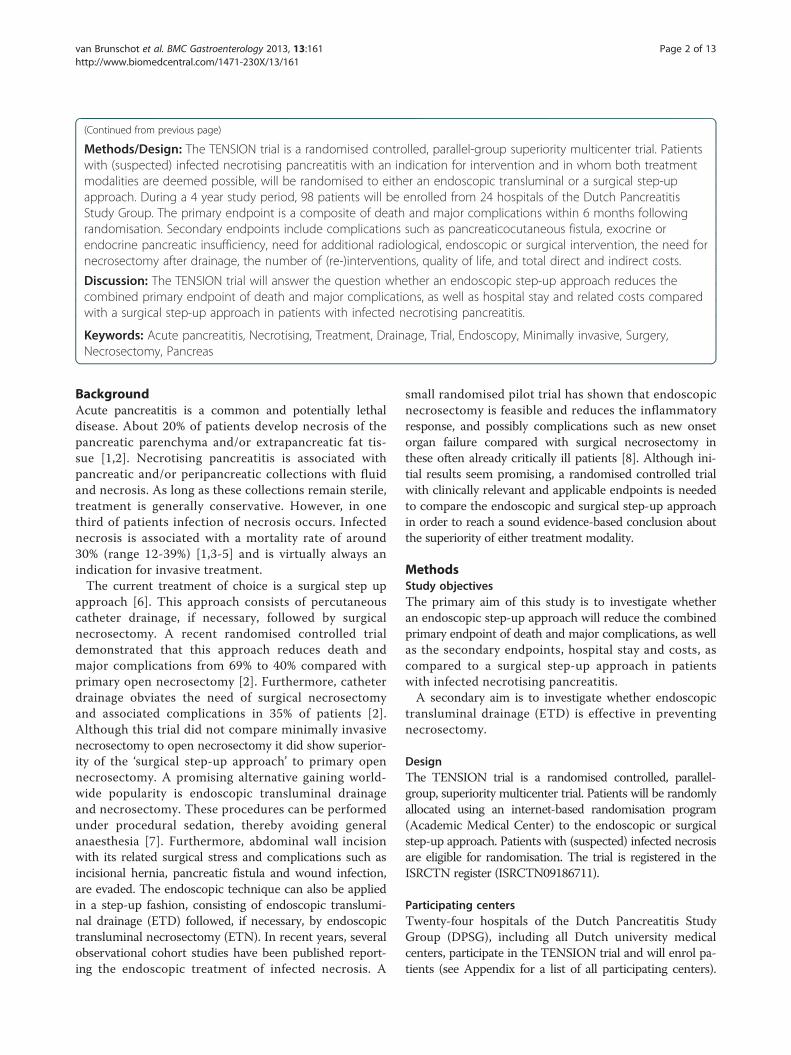
Methods/Design: The TENSION trial is a randomised controlled, parallel-group superiority multicenter trial. Patientswith (suspected) infected necrotising pancreatitis with an indication for intervention and in whom both treatmentmodalities are deemed possible, will be randomised to either an endoscopic transluminal or a surgical step-upapproach. During a 4 year study period, 98 patients will be enrolled from 24 hospitals of the Dutch PancreatitisStudy Group. The primary endpoint is a composite of death and major complications within 6 months followingrandomisation. Secondary endpoints include complications such as pancreaticocutaneous fistula, exocrine orendocrine pancreatic insufficiency, need for additional radiological, endoscopic or surgical intervention, the need fornecrosectomy after drainage, the number of (re-)interventions, quality of life, and total direct and indirect costs.
Discussion: The TENSION trial will answer the question whether an endoscopic step-up approach reduces thecombined primary endpoint of death and major complications, as well as hospital stay and related costs comparedwith a surgical step-up approach in patients with infected necrotising pancreatitis.
Keywords: Acute pancreatitis, Necrotising, Treatment, Drainage, Trial, Endoscopy, Minimally invasive, Surgery,Necrosectomy, Pancreas
BackgroundAcute pancreatitis is a common and potentially lethaldisease. About 20% of patients develop necrosis of thepancreatic parenchyma and/or extrapancreatic fat tis-sue [1,2]. Necrotising pancreatitis is associated withpancreatic and/or peripancreatic collections with fluidand necrosis. As long as these collections remain sterile,treatment is generally conservative. However, in onethird of patients infection of necrosis occurs. Infectednecrosis is associated with a mortality rate of around30% (range 12-39%) [1,3-5] and is virtually always anindication for invasive treatment.The current treatment of choice is a surgical step up
approach [6]. This approach consists of percutaneouscatheter drainage, if necessary, followed by surgicalnecrosectomy. A recent randomised controlled trialdemonstrated that this approach reduces death andmajor complications from 69% to 40% compared withprimary open necrosectomy [2]. Furthermore, catheterdrainage obviates the need of surgical necrosectomyand associated complications in 35% of patients [2].Although this trial did not compare minimally invasivenecrosectomy to open necrosectomy it did show superior-ity of the ‘surgical step-up approach’ to primary opennecrosectomy. A promising alternative gaining world-wide popularity is endoscopic transluminal drainageand necrosectomy. These procedures can be performedunder procedural sedation, thereby avoiding generalanaesthesia [7]. Furthermore, abdominal wall incisionwith its related surgical stress and complications such asincisional hernia, pancreatic fistula and wound infection,are evaded. The endoscopic technique can also be appliedin a step-up fashion, consisting of endoscopic translumi-nal drainage (ETD) followed, if necessary, by endoscopictransluminal necrosectomy (ETN). In recent years, severalobservational cohort studies have been published report-ing the endoscopic treatment of infected necrosis. A
small randomised pilot trial has shown that endoscopicnecrosectomy is feasible and reduces the inflammatoryresponse, and possibly complications such as new onsetorgan failure compared with surgical necrosectomy inthese often already critically ill patients [8]. Although ini-tial results seem promising, a randomised controlled trialwith clinically relevant and applicable endpoints is neededto compare the endoscopic and surgical step-up approachin order to reach a sound evidence-based conclusion aboutthe superiority of either treatment modality.
MethodsStudy objectivesThe primary aim of this study is to investigate whetheran endoscopic step-up approach will reduce the combinedprimary endpoint of death and major complications, as wellas the secondary endpoints, hospital stay and costs, ascompared to a surgical step-up approach in patientswith infected necrotising pancreatitis.A secondary aim is to investigate whether endoscopic
transluminal drainage (ETD) is effective in preventingnecrosectomy.
DesignThe TENSION trial is a randomised controlled, parallel-group, superiority multicenter trial. Patients will be randomlyallocated using an internet-based randomisation program(Academic Medical Center) to the endoscopic or surgicalstep-up approach. Patients with (suspected) infected necrosisare eligible for randomisation. The trial is registered in theISRCTN register (ISRCTN09186711).
Participating centersTwenty-four hospitals of the Dutch Pancreatitis StudyGroup (DPSG), including all Dutch university medicalcenters, participate in the TENSION trial and will enrol pa-tients (see Appendix for a list of all participating centers).
van Brunschot et al. BMC Gastroenterology 2013, 13:161 Page 3 of 13http://www.biomedcentral.com/1471-230X/13/161
Interventions will only take place in centers with sufficientexpertise and after the indication for intervention is sup-ported by an online expert panel.
Primary endpointThe primary endpoint is a composite of death or majorcomplications occurring within 6 months followingrandomisation.Major complications are defined as new onset (i.e. not
present 24 hours before randomisation) organ failure(cardiovascular, pulmonary or renal), bleeding requiringintervention, perforation of a visceral organ requiringintervention, enterocutaneous fistula requiring interventionand incisional hernia (including burst abdomen) (see Table 1for definitions).
Secondary endpoints
� the individual components of the primary endpoint� pancreaticocutaneous fistula (see Table 2 for
definitions)� exocrine or endocrine pancreatic insufficiency
(see Table 2 for definitions)� biliary strictures� wound infections (see Table 2 for definitions)� the need for necrosectomy (either endoscopically
or surgically)� the need for additional radiological, endoscopic or
surgical interventions� number of radiological, endoscopic and surgical (re-)
interventions� total length of intensive care and hospital stay� quality of life, quality adjusted life year’s (QALY’s,
with Short Form 36 and EQ5D)� costs per patient with poor outcome and costs
per QALY� total direct and indirect medical costs� total number of cross-over between groups
Table 1 Primary endpoint: definitions
Event Definition
Organ failure Organ failure is defined as:
● Cardiovascular: systolic blood pressure < 90 mm
● Pulmonary: PaO2 < 60 mmHg despite FiO2 30%
● Renal: serum creatinine > 177 mmol/L after reh
Definitions are adapted from the Atlanta classific
New onset organ failure Organ failure occurring after randomisation and
Multiple organ failure Failure of 2 or more organ systems on the same
Enterocutaneous fistula Enterocutaneous fistula is defined as secretion ofremoval of drains, or from a surgical wound, eith
Incisional hernia Incisional hernia is defined as a full-thickness discwith or without obstruction [2]
Study populationAll patients admitted or transferred to one of the 24 partici-pating hospitals of the DPSG with (suspected) infected ne-crosis and an indication for intervention will be assessedfor eligibility. Patients (or their legal representatives) thatmeet the in- and exclusion criteria will be asked for writteninformed consent.
Inclusion criteria
� (Suspected) infected pancreatic and/orextrapancreatic necrosis and an indication forintervention [2,10] (see Table 3 for definitions).
� Both the endoscopic step-up approach and thesurgical step-up approach are technically feasible
� Age ≥ 18 years
Exclusion criteria
� Previous intervention (e.g. surgical, endoscopic orpercutaneous) for pancreatic necrosis,extrapancreatic necrosis and/or peripancreaticcollections (see Table 3 for definitions)
� Acute flare up of chronic pancreatitis� Indication for emergency laparotomy because of
suspected abdominal compartment syndrome,bowel ischemia, bleeding or perforation of avisceral organ
RandomisationIf a patient with pancreatic and/or extrapancreaticnecrosis shows clinical deterioration and hasreached the stage to decide on invasive interventionfor (suspected) infected necrosis, the Dutch nation-wide expert panel is consulted. This panel, consist-ing of 17 experts (9 surgeons, 4 gastroenterologistsand 4 radiologists) is available 24 hours a day,7 days a week, to assess the indication for interven-tion, the feasibility of both treatment options and
Hg despite adequate fluid resuscitation or need for vasopressor support
, or the need for mechanical ventilation;
ydration or need for hemofiltration or hemodialysis;
ation and the same as previously used in the PANTER trial [2]
not present 24 hours before randomisation
day
fecal material from a percutaneous drain, drainage canal afterer from small or large bowel; confirmed by imaging or during surgery [2]
ontinuity of the abdominal wall and bulging of abdominal contents,
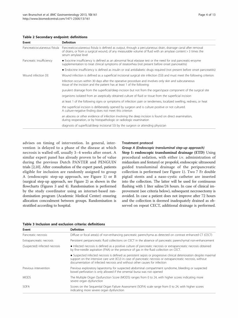
Table 2 Secondary endpoint: definitions
Event Definition
Pancreaticocutaneous fistula Pancreaticocutaneous fistula is defined as output, through a percutaneous drain, drainage canal after removalof drains, or from a surgical wound, of any measurable volume of fluid with an amylase content > 3 times theserum amylase level
Pancreatic insufficiency ● Exocrine insufficiency is defined as an abnormal fecal elastase test or the need for oral pancreatic-enzymesupplementation to treat clinical symptoms of steatorrhea (not present before onset pancreatitis)
● Endocrine insufficiency is defined as insulin or oral antidiabetic drugs required (not present before onset pancreatitis)
Wound infection [9] Wound infection is defined as a superficial incisional surgical site infection (SSI) and must meet the following criterion:
Infection occurs within 30 days after the operative procedure and involves only skin and subcutaneoustissue of the incision and the patient has at least 1 of the following:
purulent drainage from the superficial/deep incision but not from the organ/space component of the surgical site
organisms isolated from an aseptically obtained culture of fluid or tissue from the superficial incision
at least 1 of the following signs or symptoms of infection: pain or tenderness, localized swelling, redness, or heat
the superficial incision is deliberately opened by surgeon and is culture positive or not cultured.A culture-negative finding does not meet this criterion
an abscess or other evidence of infection involving the deep incision is found on direct examination,during reoperation, or by histopathologic or radiologic examination
diagnosis of superficial/deep incisional SSI by the surgeon or attending physician
van Brunschot et al. BMC Gastroenterology 2013, 13:161 Page 4 of 13http://www.biomedcentral.com/1471-230X/13/161
advises on timing of intervention. In general, inter-vention is delayed to a phase of the disease at whichnecrosis is walled-off, usually 3–4 weeks after onset. Asimilar expert panel has already proven to be of valueduring the previous Dutch PANTER and PENGUINtrials [2,10]. After consultation of the expert panel, patientseligible for inclusion are randomly assigned to groupA (endoscopic step-up approach, see Figure 1) or B(surgical step-up approach, see Figure 2) as shown in theflowcharts (Figures 3 and 4). Randomisation is performedby the study coordinator using an internet-based ran-domisation program (Academic Medical Center) ensuringallocation concealment between groups. Randomisation isstratified according to hospital.
Table 3 Inclusion and exclusion criteria: definitions
Event Definition
Pancreatic necrosis Diffuse or focal area(s) of non-enhancin
Extrapancreatic necrosis Persistent peripancreatic fluid collection
(Suspected) infected necrosis ● Infected necrosis is defined as a positby fine-needle aspiration (FNA) or the p
● Suspected infected necrosis is definedsupport on the intensive care unit (ICU)documentation of infected necrosis and
Previous intervention Previous exploratory laparotomy for susbowel perforation is only allowed if the
MODS The Multiple Organ Dysfunction Score (severe organ dysfunction
SOFA Scores on the Sequential Organ Failureindicating more severe organ dysfunctio
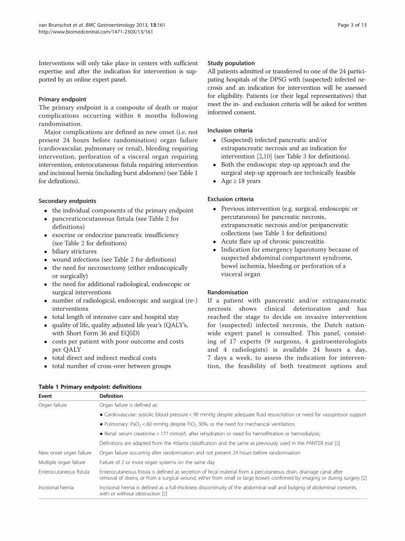
Treatment protocolGroup A (Endoscopic transluminal step-up approach)Step 1: endoscopic transluminal drainage (ETD) Usingprocedural sedation, with either i.v. administration ofmidazolam and fentanyl or propofol, endoscopic ultrasoundguided transluminal drainage of the peripancreaticcollection is performed (see Figure 1). Two 7 Fr doublepigtail stents and a naso-cystic catheter are insertedinto the collection. The latter will be used for continuousflushing with 1 liter saline/24 hours. In case of clinical im-provement (see criteria below), subsequent necrosectomy isavoided. In case a patient does not improve after 72 hoursand the collection is deemed inadequately drained as ob-served on repeat CECT, additional drainage is performed.
g pancreatic parenchyma as detected on contrast enhanced CT (CECT)
s on CECT in the absence of pancreatic parenchymal non-enhancement
ive culture of pancreatic necrosis or extrapancreatic necrosis obtainedresence of gas in the fluid collection on CECT.
as persistent sepsis or progressive clinical deterioration despite maximalin case of pancreatic necrosis or extrapancreatic necrosis, withoutwithout other causes for infection
pected abdominal compartment syndrome, bleeding or suspectedomental bursa was not opened
MODS) ranges from 0 to 24, with higher scores indicating more
Assessment (SOFA) scale range from 0 to 24, with higher scoresn
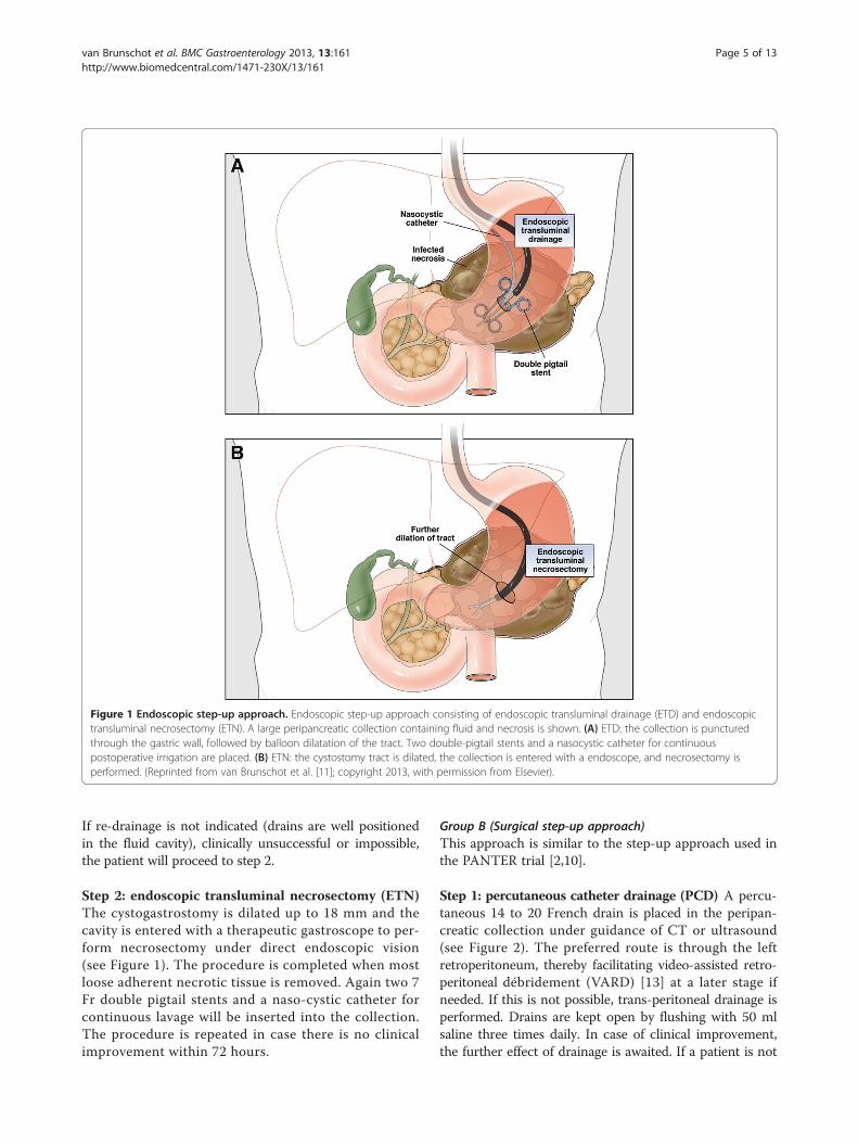
Figure 1 Endoscopic step-up approach. Endoscopic step-up approach consisting of endoscopic transluminal drainage (ETD) and endoscopictransluminal necrosectomy (ETN). A large peripancreatic collection containing fluid and necrosis is shown. (A) ETD: the collection is puncturedthrough the gastric wall, followed by balloon dilatation of the tract. Two double-pigtail stents and a nasocystic catheter for continuouspostoperative irrigation are placed. (B) ETN: the cystostomy tract is dilated, the collection is entered with a endoscope, and necrosectomy isperformed. (Reprinted from van Brunschot et al. [11]; copyright 2013, with permission from Elsevier).
van Brunschot et al. BMC Gastroenterology 2013, 13:161 Page 5 of 13http://www.biomedcentral.com/1471-230X/13/161
If re-drainage is not indicated (drains are well positionedin the fluid cavity), clinically unsuccessful or impossible,the patient will proceed to step 2.
Step 2: endoscopic transluminal necrosectomy (ETN)The cystogastrostomy is dilated up to 18 mm and thecavity is entered with a therapeutic gastroscope to per-form necrosectomy under direct endoscopic vision(see Figure 1). The procedure is completed when mostloose adherent necrotic tissue is removed. Again two 7Fr double pigtail stents and a naso-cystic catheter forcontinuous lavage will be inserted into the collection.The procedure is repeated in case there is no clinicalimprovement within 72 hours.
Group B (Surgical step-up approach)This approach is similar to the step-up approach used inthe PANTER trial [2,10].
Step 1: percutaneous catheter drainage (PCD) A percu-taneous 14 to 20 French drain is placed in the peripan-creatic collection under guidance of CT or ultrasound(see Figure 2). The preferred route is through the leftretroperitoneum, thereby facilitating video-assisted retro-peritoneal débridement (VARD) [13] at a later stage ifneeded. If this is not possible, trans-peritoneal drainage isperformed. Drains are kept open by flushing with 50 mlsaline three times daily. In case of clinical improvement,the further effect of drainage is awaited. If a patient is not

Figure 2 Surgical step-up approach. Surgical step-up approach consisting of percutaneous catheter drainage (PCD) and video-assistedretroperitoneal débridement (VARD). (A) Cross-sectional image and torso depicting a peripancreatic collection. The preferred route is through theleft retroperitoneal space between the kidney, spleen and descending colon. A percutaneous catheter drain is inserted in the collection tomitigate sepsis and postpone or even obviate necrosectomy. The area of detail is shown in (B). (C) A 5 cm subcostal incision is made and thepercutaneous drain is followed into the collection. The first necrosis is removed under direct vision with a long grasping forceps, followed byfurther debridement under videoscopic assistance (D). (Reprinted from van Brunschot et al. [11]; copyright 2013, with permission from Elsevier).
van Brunschot et al. BMC Gastroenterology 2013, 13:161 Page 6 of 13http://www.biomedcentral.com/1471-230X/13/161
improving and a collection is deemed inadequately drainedon repeat CECT after 72 hours, additional drainage isperformed. If this is not possible, or if a second drainage isclinically unsuccessful (see criteria for clinical improvementbelow) the patient will proceed to step 2.
Step 2: video-assisted retroperitoneal debridement(VARD, if not possible laparotomy) VARD is a drain-guided, minimally invasive retroperitoneal procedurerequiring a 5 cm flank incision according to the previ-ously published technique [13,14] (see Figure 2). Usingthe retroperitoneal drain for guidance, the collection isentered and only loosely adherent necrosis is removedunder video-assistance. At the end of the proceduretwo large bore surgical drains are inserted. A continuouspost-operative lavage system (building up to 10 litressaline per 24 hrs) is installed. In case of absence ofclinical improvement (see criteria below) and repeatedCECT shows foci of potentially inadequate drainage, VARDis repeated. If VARD is technically not feasible, debridementby laparotomy is performed.
If drainage technically fails in the endoscopic group aPCD is placed. In case of clinical or technical failure ofPCD, surgical necrosectomy is performed. Both approachesare performed, according to a strict protocol, only inparticipating centers with documented expertise and, ifnecessary, under supervision of an expert. Sufficientexpertise is defined as having performed at least tenindependent VARD procedures or ten independent endo-scopic transluminal drainage procedures and more thanfive endoscopic transluminal necrosectomies. In case ofinsufficient local experience, the patient is transferred to atertiary referral center with sufficient experience.
Criteria for clinical improvementCriteria similar to the PANTER trial are used to defineclinical improvement, failure and to decide to go to thenext step [2,10]. Each step is evaluated 72 hours afterintervention and considered successful in case of clinicalimprovement. Clinical improvement is defined as: improvedfunction of at least two organ systems (i.e. circulatory,pulmonary, or renal) or improvement of two out of three
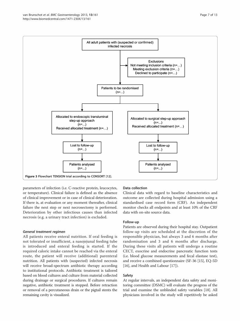
Figure 3 Flowchart TENSION trial according to CONSORT [12].
van Brunschot et al. BMC Gastroenterology 2013, 13:161 Page 7 of 13http://www.biomedcentral.com/1471-230X/13/161
parameters of infection (i.e. C-reactive protein, leucocytes,or temperature). Clinical failure is defined as the absenceof clinical improvement or in case of clinical deterioration.If there is, at evaluation or any moment thereafter, clinicalfailure the next step or next necrosectomy is performed.Deterioration by other infectious causes than infectednecrosis (e.g. a urinary tract infection) is excluded.
General treatment regimenAll patients receive enteral nutrition. If oral feeding isnot tolerated or insufficient, a nasojejunal feeding tubeis introduced and enteral feeding is started. If therequired caloric intake cannot be reached via the enteralroute, the patient will receive (additional) parenteralnutrition. All patients with (suspected) infected necrosiswill receive broad-spectrum antibiotic therapy accordingto institutional protocols. Antibiotic treatment is tailoredbased on blood cultures and culture from material collectedduring drainage or surgical procedures. If cultures remainnegative, antibiotic treatment is stopped. Before retractionor removal of a percutaneous drain or the pigtail stents theremaining cavity is visualized.
Data collectionClinical data with regard to baseline characteristics andoutcome are collected during hospital admission using astandardised case record form (CRF). An independentmonitor checks all endpoints and at least 10% of the CRFdata with on-site source data.
Follow-upPatients are observed during their hospital stay. Outpatientfollow-up visits are scheduled at the discretion of theresponsible physician, but always 3 and 6 months afterrandomisation and 3 and 6 months after discharge.During these visits all patients will undergo a routineCECT, exocrine and endocrine pancreatic function tests(i.e. blood glucose measurements and fecal elastase test),and receive a combined questionnaire (SF-36 [15], EQ-5D[16], and Health and Labour [17]).
SafetyAt regular intervals, an independent data safety and moni-toring committee (DSMC) will evaluate the progress of thetrial and examine the unblinded safety variables [18]. Allphysicians involved in the study will repetitively be asked
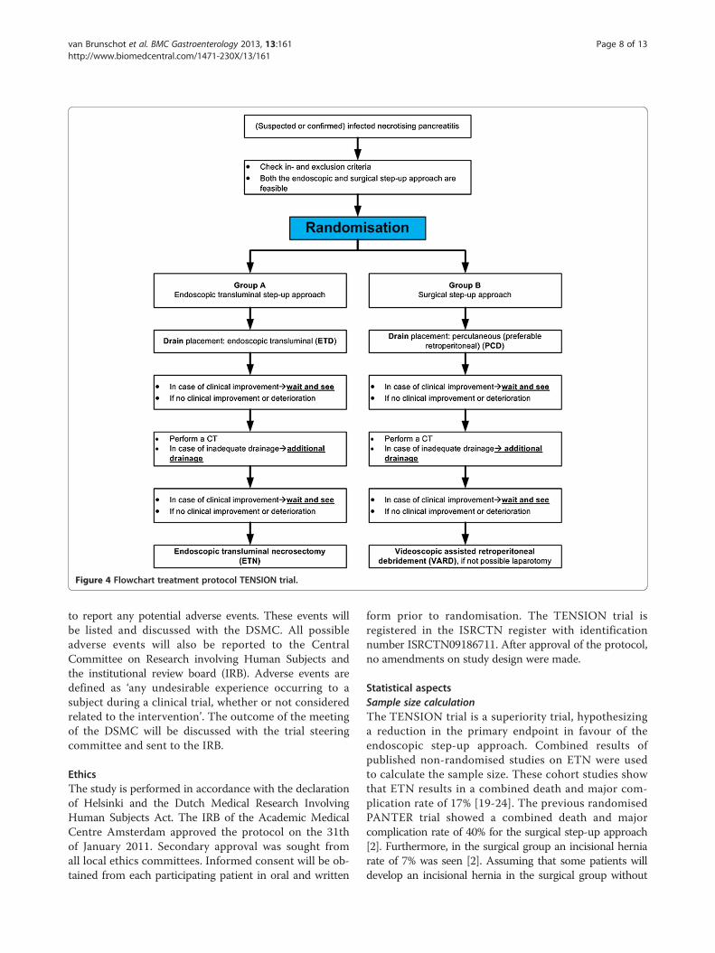
Figure 4 Flowchart treatment protocol TENSION trial.
van Brunschot et al. BMC Gastroenterology 2013, 13:161 Page 8 of 13http://www.biomedcentral.com/1471-230X/13/161
to report any potential adverse events. These events willbe listed and discussed with the DSMC. All possibleadverse events will also be reported to the CentralCommittee on Research involving Human Subjects andthe institutional review board (IRB). Adverse events aredefined as ‘any undesirable experience occurring to asubject during a clinical trial, whether or not consideredrelated to the intervention’. The outcome of the meetingof the DSMC will be discussed with the trial steeringcommittee and sent to the IRB.
EthicsThe study is performed in accordance with the declarationof Helsinki and the Dutch Medical Research InvolvingHuman Subjects Act. The IRB of the Academic MedicalCentre Amsterdam approved the protocol on the 31thof January 2011. Secondary approval was sought fromall local ethics committees. Informed consent will be ob-tained from each participating patient in oral and written
form prior to randomisation. The TENSION trial isregistered in the ISRCTN register with identificationnumber ISRCTN09186711. After approval of the protocol,no amendments on study design were made.
Statistical aspectsSample size calculationThe TENSION trial is a superiority trial, hypothesizinga reduction in the primary endpoint in favour of theendoscopic step-up approach. Combined results ofpublished non-randomised studies on ETN were usedto calculate the sample size. These cohort studies showthat ETN results in a combined death and major com-plication rate of 17% [19-24]. The previous randomisedPANTER trial showed a combined death and majorcomplication rate of 40% for the surgical step-up approach[2]. Furthermore, in the surgical group an incisional herniarate of 7% was seen [2]. Assuming that some patients willdevelop an incisional hernia in the surgical group without
van Brunschot et al. BMC Gastroenterology 2013, 13:161 Page 9 of 13http://www.biomedcentral.com/1471-230X/13/161
having another primary endpoint, the prevalence of deathand major complications in this group, including incisionalhernias is estimated to be 43%. Therefore, an absolutereduction in primary endpoint of 26% (from 43 to 17%)is anticipated. With a 2-sided 5% alpha, power of 80%, and2% loss to follow-up, the sample size was set at 98 patients.
Descriptive statisticsFor dichotomous data, frequencies will be presented.Continuous data will be presented as mean and standarddeviation or median and interquartile range. Baseline cri-teria are: age, sex, body mass index, aetiology of pancrea-titis, co-morbidity, American Society of Anaesthesiologist’s(ASA) classification, CT severity index, extent of pancreaticnecrosis, disease severity (SIRS, ICU admission, singleor multiple organ failure), Acute Physiology and ChronicHealth Evaluation (APACHE) ll score, Imrie score, MODS(Table 3), SOFA score (Table 3), C-reactive protein,time from onset of symptoms to randomisation, tertiaryreferral, and confirmed infected necrosis (bacterial cultureof first intervention).
AnalysesThere will be a blinded outcome assessment after the lastpatient completed follow-up for all primary and secondaryendpoints. Both intention-to-treat and per-protocol ana-lyses will be performed. In intention-to-treat analysis, allpatients are analysed according to their initially assignedstudy arm regardless of adherence to study protocol,which is the primary analysis of the study. Occurrencesof the primary and secondary endpoints are comparedbetween treatment groups. Comparison of the primaryendpoint will be expressed in terms of a relative riskand corresponding 95% confidence intervals. A two-tailed P < 0.05 is considered statistically significant.Subsequent analyses are directed at secondary end-points. Predefined subgroup analysis will be performedfor patients with and without (multiple) organ failure(see Table 1 for definitions) before randomisation, institutionand time between onset of symptoms and randomisation(<28 or >28 days). A formal test of interaction in a logistic-regression model is used to assess whether treatment effectsdiffer significantly between subgroups. In case of skewedrandomisation (i.e. statistically significant differences inbaseline variables), an adjusted analysis will be performedusing multivariable logistic regression.
Additional analysesDirect and indirect medical and non-medical costs of bothtreatment strategies for the follow-up period of 6 monthsafter randomisation will be compared. All costs will beestimated based on the actual input in terms of resourceuse (i.e. interventions, diagnostic procedures, hospitaland ICU stay), personnel, medication, visits to healthcare
providers, private household assistance, and indirectcosts from loss of productivity due to sick leave(assessed with the Health and Labour questionnaire).Total costs per patient are calculated by summing directmedical costs, direct nonmedical costs, and indirect costsand subsequently compared between groups. Furthermore,the impact of differences in complications on the quality oflife is measured by a generic quality of life questionnaire,the SF-36. In addition to this quality of life questionnaire,the EQ-5D is completed which screens for the presenceand severity of problems with mobility, self-care, dailyactivities, pain/complaints and mood.
Premature termination of the studyNo formal interim-analysis is planned. To guaranteepatient’s safety throughout the study, the DSMC willperform regular safety analyses. When harm (higherincidence of SAE’s in one group) occurs, the DSMCwill discuss potential stopping of the trial prematurelywith the trial steering committee. Since this is the firstrandomised trial comparing both approaches, and henceall data arising from this trial, regardless of its outcome,will influence treatment policy worldwide, the trial willnot be stopped for futility.
DiscussionThe TENSION trial is designed to answer the questionwhether an endoscopic step-up approach will lead to areduction of death and major complications compared toa surgical step-up approach in patients with (suspected)infected necrosis. The TENSION trial will also investigatewhether pancreatic fistula, exocrine or endocrine pancreaticinsufficiency, length of ICU and hospital stay, quality of lifeand costs are reduced by the endoscopic step-up approach.In recent years, minimally invasive approaches are grad-
ually replacing (primary) open necrosectomy. Minimallyinvasive approaches aim at minimizing surgical stress andhave proven to reduce complications. In the PANTERtrial, the surgical step-up approach reduced the combineddeath and major complication rate from 69% to 40% [2].Furthermore, the PANTER trial showed that 35% ofpatients with infected necrotising pancreatitis achievecomplete recovery after percutaneous drainage only,without the need for surgical debridement. Althoughthe combined death and major complication rate is stillhigh, the surgical step-up approach should at presentbe considered as the current standard of care worldwide[2]. Drainage is based on the hypothesis that alleviatingpressure of an infected collection may improve the patient’sclinical condition and thereby leaving the necrotic tissueto be dealt with by the patient’s own immune system.Endoscopic transluminal drainage can be applied accord-ing to the same rationale. Therefore, we chose to institutethe step-up approach in both study arms. Due to the large
van Brunschot et al. BMC Gastroenterology 2013, 13:161 Page 10 of 13http://www.biomedcentral.com/1471-230X/13/161
differences in treatments between both groups, blinding isnot feasible. To partially compensate for this, outcomeassessment is blinded.In the TENSION trial only patients with (suspected)
infected necrosis are included since the main indicationfor intervention in necrotising pancreatitis is nowadaysconsidered to be infected necrosis [25-28]. Patients withsterile necrosis can often be successfully managed conser-vatively (i.e. without any form of intervention) [28-30].A composite endpoint of death and major complications
was chosen because a study powered to demonstrate aclinically relevant difference in death alone would requirean unrealistic large sample size of over 2000 patients.In addition, previous studies have shown that majorcomplications have high impact in terms of quality of lifein patients with necrotising pancreatitis [2,8].A potential limitation of the endoscopic approach is that
periprocedural complications (e.g. perforation or bleeding)may be more difficult to manage when compared to peri-procedural complications occurring during surgical necro-sectomy. A systematic review and randomised trial havesuggested that endoscopic treatment of infected necrosisis feasible and associated with lower or comparable com-plication rates than surgery [7,8]. Furthermore, endoscopicdrainage and necrosectomy are advanced interventionsthat not only require the expertise from an interventionalendoscopist, but also the dedicated involvement of inter-ventional radiologists and pancreatic surgeons to managepotential complications. For this reason the endoscopic in-terventions in the TENSION trial will only be performedin expert centers with multidisciplinary expertise.
ConclusionThe TENSION trial is a randomised controlled multicen-ter trial designed to show a reduction in the compositeprimary endpoint of death and major complications, aswell in hospital stay and costs following an endoscopictransluminal step up approach compared with a surgi-cal step up approach in patients with infected necrotis-ing pancreatitis.
AppendixTENSION committee membersSteering committeeS. van Brunschot, MD, Dept. of Gastroenterology andHepatology, Academic Medical Center, University ofAmsterdam and dept. of OR/Evidence Based Surgery,Radboud University Nijmegen Medical CenterH.C. van Santvoort, MD PhD, Dept. of Surgery, University
Medical Center UtrechtM.G.H. Besselink, MD PhD, Dept. of Surgery, Academic
Medical CenterO.J. Bakker, MD PhD, Dept. of Surgery, University
Medical Center Utrecht
P. Fockens, MD PhD, Dept. of Gastroenterology andHepatology, Academic Medical Center, University ofAmsterdam (chair)H.G. Gooszen, MD PhD, Dept. of OR/Evidence Based
Surgery, Radboud University Nijmegen Medical CenterM.A. Boermeester, MD PhD, Dept. of Surgery, Academic
Medical CenterM.J. Bruno, MD PhD, Dept. of Gastroenterology, Erasmus
MC, University Medical CenterC.H.C. Dejong, MD PhD, Dept. of Surgery and NUTRIM
School for Nutrition, Toxicology and Metabolism,Maastricht University Medical CenterR. Timmer, MD PhD, Dept. of Gastroenterology,
St Antonius HospitalB.J.M. Witteman, MD PhD, Dept. of Gastroenterology,
Gelderse Vallei Hospital
Expert panelM.A. Boermeester, MD PhD, Dept. of Surgery, AcademicMedical Center, AmsterdamT.L. Bollen, MD, Dept. of Radiology, St Antonius Hospital,
NieuwegeinM. Bruno, MD PhD, Dept. of Gastroenterology, Erasmus
MC, University Medical Center, RotterdamV.C. Cappendijk, MD, Dept. of Radiology, Jeroen Bosch
Hospital, 's-HertogenboschC.H.C. Dejong, MD PhD, Dept. of Surgery and NUTRIM
School for Nutrition, Toxicology and Metabolism,Maastricht University Medical Center, MaastrichtC. van Eijck, MD PhD, Dept. of Surgery, Erasmus MC,
University Medical Center, RotterdamP. Fockens, MD PhD, Dept. of Gastroenterology and
Hepatology, Academic Medical Center, University ofAmsterdam, AmsterdamH. van Goor, MD PhD, Dept. Of Surgery, Radboud
University Nijmegen Medical Center, NijmegenH.G. Gooszen, MD PhD, Dept. of OR/Evidence Based
Surgery, Radboud University Nijmegen Medical Center,NijmegenJ.W. Haveman, MD PhD, Dept. of Surgery, University
Medical Center Groningen, GroningenH.S. Hofker, MD PhD, Dept. of Surgery, University
Medical Center Groningen, GroningenJ.S. Laméris, MD PhD, Dept. of Radiology, Academic
Medical Center, AmsterdamK.P. van Lienden, MD PhD, Dept. of Radiology, Academic
Medical Center, AmsterdamV.B. Nieuwenhuijs, MD PhD, Dept. of Surgery, Isala
Clinics, ZwolleJ.W. Poley, MD PhD, Dept. of Gastroenterology, Erasmus
MC, University Medical Center, RotterdamA.F.M. Schaapherder, MD PhD, Dept. of Surgery, Leiden
University Medical Center, Leiden
van Brunschot et al. BMC Gastroenterology 2013, 13:161 Page 11 of 13http://www.biomedcentral.com/1471-230X/13/161
R. Timmer, MD PhD, Dept. of Gatroenterology, StAntonius Hospital, Nieuwegein
Data and Safety Monitoring CommitteeJ.G.P. Tijssen, MD PhD, Dept. of Clinical Epidemiology,Academic Medical Center, Amsterdam (chair)J.F. Lange, MD PhD, Dept. of Surgery, Erasmus MC,
University Medical Center, RotterdamH.J. Bonjer, MD PhD, Dept. of Surgery, VU Medical
Center, AmsterdamJ. Stoker, MD PhD, Dept. of Radiology, Academic
Medical Center, AmsterdamA.A.M. Masclee, MD PhD, Dept. of Gastroenterology,
Maastricht University Medical Center, Maastricht
Independent physicianK.M.A.J. Tytgat, MD PhD, Dept. of Gastroenterology,Academic Medical Center, Amsterdam
Clinical centers and principal investigators (all in theNetherlands, alphabetical order):
1. Academic Medical Center, PO 22660, 1100 DDAmsterdam; P Fockens, MD PhD, Dept.of Gastroenterology and Hepatology;
2. Amphia Hospital Breda, PO 90158, 4800 RK Breda;T Seerden, MD PhD, Dept. of Gastroenterology;
3. Canisius-Wilhelmina Hospital, PO 9015, 6500 GSNijmegen; C Rosman, MD PhD, Dept. of Surgery;
4. Catharina Hospital, PO 1350, 5623 EJ Eindhoven; EJSchoon, MD PhD, Dept. of Gastroenterology;
5. Erasmus Medical Center, PO 2040, 3000 CARotterdam; MJ Bruno, MD PhD, Dept.of Gastroenterology;
6. Gelre Hospital, PO 9014, 7300 DS Apeldoorn;GW Erkelens, MD, Dept. of Gastroenterology;
7. Hospital Gelderse Vallei, PO 9025, 6710 HN Ede;B Witteman, MD PhD, Dept. of Gastroenterology;
8. Isala Clinics Zwolle, PO 10400, 8000 GK Zwolle;MAC Meijssen, MD PhD, Dept. Of Gastroenterology;
9. Jeroen Bosch Hospital, PO 90153, 5200 ME‘s-Hertogenbosch; K Bosscha, MD PhD, Dept.of Surgery;
10. Leiden University Medical Center, PO 9600, 2300RC Leiden; AFM Schaapherder, MD PhD,Dept. of Surgery;
11. Maasstad Hospital Rotterdam, PO 9100, 3007 ACRotterdam; H Hadithi, MD, Dept. ofGastroenterology;
12. Maastricht University Medical Center, PO 5800,6202 AZ Maastricht; RJJ de Ridder, MD PhD, Dept.Of Gastroenterology;
13. Máxima Medical Center Veldhoven, PO 7777,5500 MB Veldhoven; JWA Straathof, MD PhD,Dept. of Gastroenterology;
14. Meander Medical Center, PO 1502, 3800 BM,Amersfoort; EC Consten, MD PhD, Dept. of Surgery;
15. Medical Center Leeuwarden, PO 888, 8901 BRLeeuwarden; ER Manusama, MD PhD, Dept.of Surgery;
16. Medical Spectre Twente, PO 50000, 7500 KAEnschede; NG Venneman, MD PhD, Dept. ofGastroenterology;
17. OLVG Amsterdam, PO 95500, 1090 HM Amsterdam;JM Jansen, MD PhD, Dept. of Gastroenterology;
18. Radboud University Nijmegen Medical Center, PO9101, 6500 HB Nijmegen; H van Goor, MD PhD,Dept. of Surgery;
19. RdGG Delft, PO 5011, 2600 GA Delft; JJGScheepers, MD, dept. of Surgery;
20. Rijnstate Hospital, PO 9555, 6800 TA Arnhem;BWM Spanier, MD PhD, Dept. of Gastroenterology;
21. St Antonius Hospital, PO 2500, 3430 EM Nieuwegein;R Timmer, MD PhD, Dept. of Gastroenterology;
22. University Medical Center Groningen, PO 30001,9700 RB Groningen; HS Hofker, MD PhD, Dept.of Surgery;
23. University Medical Center Utrecht, PO 85500,3508 GA Utrecht; FP Vleggaar, MD PhD, Dept.of Gastroenterology and Hepatology;
24. VU Medical Center Amsterdam, PO 7057,1007 MB Amsterdam; CJ Mulder, MD PhD,Dept. of Gastroenterology.
Key staff at coordinating centersAcademic Medical Center Amsterdam: P Fockens(principal investigator), S van Brunschot (coordinator);Radboud University Nijmegen Medical Centre: S van
Brunschot (coordinator), J van Grinsven (coordinator),H van der Eng (research nurse) and HG Gooszen;University Medical Center Utrecht: HC van Santvoort
AbbreviationsCECT: Contrast enhanced computed tomography; CRF: Case record form;DPSG: Dutch pancreatitis study group; DSMC: Data safety monitoringcommittee; ETD: Endoscopic transluminal drainage; ETN: Endoscopictransluminal necrosectomy; ISRCTN: International standard randomisedcontrolled trial number; PCD: Percutaneous catheter drainage; QALY: Qualityadjusted life year; VARD: Video-assisted retroperitoneal debridement.
Competing interestsThe authors declare that they have no competing interests.
Authors’ contributionsSvB drafted the manuscript. HCvS, JvG, RV, OJB, MGB, HGG and PFco-authored the writing of the manuscript. SvB, HCvS, MGHB, OJB, RPV, MGWD,MAB, MJB, CHCD, RT, BJW, HGG, and PF participated in the design of the studyprior to and during several meetings of the Dutch Pancreatitis Study Group.SvB, HCvS, RPV and MGWD performed the sample size calculation. All authorscritically assessed the study design or included patients in the study, edited themanuscript and read and approved the final manuscript.
van Brunschot et al. BMC Gastroenterology 2013, 13:161 Page 12 of 13http://www.biomedcentral.com/1471-230X/13/161
FundingThe Dutch Digestive Disease Foundation (Maag Lever Darm Stichting, grantnumber WO 09–45), fonds NutsOhra (grant number 1101–108) and TheNetherlands Organization for Health Research and Development, Health CareEfficiency Research program (ZonMw, grant number 837004008) financiallysupported the TENSION trial. The TENSION trial is an investigator initiatedtrial and the sponsor had no influence on the design of the study. Neitherdo they have any influence on implementation, data collection,interpretation of results or decision to publish.
Author details1Department of Gastroenterology and Hepatology, University of Amsterdam,Amsterdam, The Netherlands. 2Department of OR/Evidence Based Surgery,Radboud University Nijmegen Medical Centre, Nijmegen, The Netherlands.3Department of Surgery, University Medical Center Utrecht, Utrecht, TheNetherlands. 4Department of Surgery, University of Amsterdam, Amsterdam,The Netherlands. 5Department of Radiology, St Antonius Hospital,Nieuwegein, The Netherlands. 6Department of Surgery, Jeroen BoschHospital, ‘s-Hertogenbosch, The Netherlands. 7Department of Surgery,Radboud University Nijmegen Medical Centre, Nijmegen, The Netherlands.8Department of Gastroenterology, Erasmus MC, University Medical Center,Rotterdam, The Netherlands. 9Department of Radiology, Jeroen BoschHospital, ‘s-Hertogenbosch, The Netherlands. 10Department of Surgery,Meander Medical Center, Amersfoort, The Netherlands. 11Department ofSurgery and NUTRIM School for Nutrition, Toxicology and Metabolism,Maastricht University Medical Center, Maastricht, The Netherlands. 12ClinicalResearch Unit, Academic Medical Center, University of Amsterdam,Amsterdam, The Netherlands. 13Department of Surgery, Erasmus MC,University Medical Center, Rotterdam, The Netherlands. 14Department ofGastroenterology, Gelre Hospital, Apeldoorn, The Netherlands. 15Departmentof Gastroenterology, Maasstad Hospital, Rotterdam, The Netherlands.16Department of Surgery, University Medical Center Groningen, Groningen,The Netherlands. 17Department of Gastroenterology, Onze Lieve VrouweGasthuis, Amsterdam, The Netherlands. 18Department of Radiology,University of Amsterdam, Amsterdam, The Netherlands. 19Department ofSurgery, Medical Center Leeuwarden, Leeuwarden, The Netherlands.20Department of Gastroenterology, Isala Clinics, Zwolle, The Netherlands.21Department of Gastroenterology, VU Medical Center, Amsterdam, TheNetherlands. 22Department of Surgery, Isala Clinics, Zwolle, The Netherlands.23Department of Gastroenterology, Maastricht University Medical Center,Maastricht, The Netherlands. 24Department of Surgery, Canisius-WilhelminaHospital, Nijmegen, The Netherlands. 25Department of Surgery, LeidenUniversity Medical Center, Leiden, The Netherlands. 26Department of Surgery,Reinier de Graaf Group, Delft, The Netherlands. 27Department ofGastroenterology, Catharina Hospital, Eindhoven, The Netherlands.28Department of Gastroenterology, Amphia Hospital, Breda, The Netherlands.29Department of Gastroenterology, Rijnstate Hospital, Arnhem, TheNetherlands. 30Department of Gastroenterology, Máxima Medical Center,Veldhoven, The Netherlands. 31Department of Gastroenterology, St AntoniusHospital, Nieuwegein, The Netherlands. 32Department of Surgery, MedischSpectrum Twente, Enschede, The Netherlands. 33Department ofGastroenterology and Hepatology, University Medical Center Utrecht,Utrecht, The Netherlands. 34Department of Gastroenterology, HospitalGelderse Vallei, Ede, The Netherlands.
Received: 8 October 2013 Accepted: 13 November 2013Published: 25 November 2013
References1. Rodriguez JR, Razo AO, Targarona J, Thayer SP, Rattner DW, Warshaw AL,
Fernandez-del Castillo C: Debridement and closed packing for sterile orinfected necrotizing pancreatitis: insights into indications and outcomesin 167 patients. Ann Surg 2008, 247(2):294–299.
2. van Santvoort HC, Besselink MG, Bakker OJ, Hofker HS, Boermeester MA,Dejong CH, van Goor H, Schaapherder AF, van Eijck CH, Bollen TL, vanRamshorst B, Nieuwenhuijs VB, Timmer R, Lameris JS, Kruyt PM,Manusama ER, van der Harst E, van der Schelling GP, Karsten T, HesselinkEJ, van Laarhoven CJ, Rosman C, Bosscha K, de Wit RJ, Houdijk AP, vanLeeuwen MS, Buskens E, Gooszen HG: A step-up approach or opennecrosectomy for necrotizing pancreatitis. N Engl J Med 2010,362(16):1491–1502.
3. Besselink MG, van Santvoort HC, Buskens E, Boermeester MA, van Goor H,Timmerman HM, Nieuwenhuijs VB, Bollen TL, van Ramshorst B, Witteman BJ,Rosman C, Ploeg RJ, Brink MA, Schaapherder AF, Dejong CH, Wahab PJ, vanLaarhoven CJ, van der Harst E, van Eijck CH, Cuesta MA, Akkermans LM,Gooszen HG: Probiotic prophylaxis in predicted severe acute pancreatitis:a randomised, double-blind, placebo-controlled trial. Lancet 2008,371(9613):651–659.
4. Raraty MG, Halloran CM, Dodd S, Ghaneh P, Connor S, Evans J, Sutton R,Neoptolemos JP: Minimal access retroperitoneal pancreaticnecrosectomy: improvement in morbidity and mortality with a lessinvasive approach. Ann Surg 2010, 251(5):787–793.
5. van Santvoort HC, Bakker OJ, Bollen TL, Besselink MG, Ahmed Ali U, SchrijverAM, Boermeester MA, van Goor H, Dejong CH, van Eijck CH, van RamshorstB, Schaapherder AF, van der Harst E, Hofker S, Nieuwenhuijs VB, Brink MA,Kruyt PM, Manusama ER, van der Schelling GP, Karsten T, Hesselink EJ, vanLaarhoven CJ, Rosman C, Bosscha K, de Wit RJ, Houdijk AP, Cuesta MA,Wahab PJ, Gooszen HG: A conservative and minimally invasive approachto necrotizing pancreatitis improves outcome. Gastroenterology 2011,141(4):1254–1263.
6. Working Group IAP/APA/APG: IAP/APA evidence-based guidelines for themanagement of acute pancreatitis. Pancreatology 2013, 13(4 Suppl 2):e1–e15.
7. Haghshenasskashani A, Laurence JM, Kwan V, Johnston E, Hollands MJ,Richardson AJ, Pleass HC, Lam VW: Endoscopic necrosectomy of pancreaticnecrosis: a systematic review. Surg Endosc 2011, 25(12):3724–3730.
8. Bakker OJ, van Santvoort HC, van Brunschot S, Geskus RB, Besselink MG,Bollen TL, van Eijck CH, Fockens P, Hazebroek EJ, Nijmeijer RM, Poley JW, vanRamshorst B, Vleggaar FP, Boermeester MA, Gooszen HG, Weusten BL, TimmerR: Endoscopic transgastric vs surgical necrosectomy for infectednecrotizing pancreatitis: a randomized trial. JAMA 2012, 307(10):1053–1061.
9. Horan TC, Andrus M, Dudeck MA: CDC/NHSN surveillance definition ofhealth care -associated infection and criteria for specific types of infectionsin the acute care setting. Am J Infect Control 2008, 36(5):309–332.
10. Besselink MG, van Santvoort HC, Nieuwenhuijs VB, Boermeester MA, BollenTL, Buskens E, Dejong CH, van Eijck CH, van Goor H, Hofker SS, Lameris JS,van Leeuwen MS, Ploeg RJ, van Ramshorst B, Schaapherder AF, Cuesta MA,Consten EC, Gouma DJ, van der Harst E, Hesselink EJ, Houdijk LP, KarstenTM, van Laarhoven CJ, Pierie JP, Rosman C, Bilgen EJ, Timmer R, van der TweelI, de Wit RJ, Witteman BJ, et al: Minimally invasive ‘step-up approach’ versusmaximal necrosectomy in patients with acute necrotising pancreatitis(PANTER trial): design and rationale of a randomised controlled multicentertrial [ISRCTN13975868]. BMC surgery 2006, 6:6.
11. van Brunschot S, Bakker OJ, Besselink MG, Bollen TL, Fockens P, GooszenHG, van Santvoort HC, Dutch Pancreatitis Study G: Treatment ofnecrotizing pancreatitis. Clin Gastroenterol Hepatol 2012, 10(11):1190–1201.
12. Schulz KF, Altman DG, Moher D, Group C: CONSORT 2010 statement: updatedguidelines for reporting parallel group randomised trials. BMJ 2010, 340:c332.
13. van Santvoort HC, Besselink MG, Horvath KD, Sinanan MN, Bollen TL, vanRamshorst B, Gooszen HG: Videoscopic assisted retroperitoneal debridementin infected necrotizing pancreatitis. HPB (Oxford) 2007, 9(2):156–159.
14. Horvath KD, Kao LS, Wherry KL, Pellegrini CA, Sinanan MN: A technique forlaparoscopic-assisted percutaneous drainage of infected pancreaticnecrosis and pancreatic abscess. Surg Endosc 2001, 15(10):1221–1225.
15. Ware JE Jr, Sherbourne CD: The MOS 36-item short-form health survey (SF-36).I. Conceptual framework and item selection. Med Care 1992, 30(6):473–483.
16. Brooks R: EuroQol: the current state of play. Health Pol 1996, 37(1):53–72.17. van Roijen L, Essink-Bot ML, Koopmanschap MA, Bonsel G, Rutten FF: Labor
and health status in economic evaluation of health care. The health andlabor questionnaire. Int J Technol Assess Health Care 1996, 12(3):405–415.
18. Chan AW, Tetzlaff JM, Gotzsche PC, Altman DG, Mann H, Berlin JA, DickersinK, Hrobjartsson A, Schulz KF, Parulekar WR, Krleza-Jeric K, Laupacis A, MoherD: SPIRIT 2013 explanation and elaboration: guidance for protocols ofclinical trials. BMJ 2013, 346:e7586.
19. Charnley RM, Lochan R, Gray H, O’Sullivan CB, Scott J, Oppong KE:Endoscopic necrosectomy as primary therapy in the management ofinfected pancreatic necrosis. Endoscopy 2006, 38(9):925–928.
20. Escourrou J, Shehab H, Buscail L, Bournet B, Andrau P, Moreau J, Fourtanier G:Peroral transgastric/transduodenal necrosectomy: success in the treatmentof infected pancreatic necrosis. Ann Surg 2008, 248(6):1074–1080.
21. Schrover IM, Weusten BL, Besselink MG, Bollen TL, van Ramshorst B, TimmerR: EUS-guided endoscopic transgastric necrosectomy in patients withinfected necrosis in acute pancreatitis. Pancreatology 2008, 8(3):271–276.
van Brunschot et al. BMC Gastroenterology 2013, 13:161 Page 13 of 13http://www.biomedcentral.com/1471-230X/13/161
22. Seewald S, Groth S, Omar S, Imazu H, Seitz U, de Weerth A, Soetikno R,Zhong Y, Sriram PV, Ponnudurai R, Sikka S, Thonke F, Soehendra N:Aggressive endoscopic therapy for pancreatic necrosis and pancreaticabscess: a new safe and effective treatment algorithm (videos).Gastrointest Endosc 2005, 62(1):92–100.
23. Seifert H, Biermer M, Schmitt W, Jurgensen C, Will U, Gerlach R, Kreitmair C,Meining A, Wehrmann T, Rosch T: Transluminal endoscopic necrosectomyafter acute pancreatitis: a multicentre study with long-term follow-up(the GEPARD Study). Gut 2009, 58(9):1260–1266.
24. Voermans RP, Veldkamp MC, Rauws EA, Bruno MJ, Fockens P: Endoscopictransmural debridement of symptomatic organized pancreatic necrosis(with videos). Gastrointest Endosc 2007, 66(5):909–916.
25. UK guidelines for the management of acute pancreatitis. Gut 2005,54(Suppl 3):iii1–iii9.
26. Buchler MW, Gloor B, Muller CA, Friess H, Seiler CA, Uhl W: Acute necrotizingpancreatitis: treatment strategy according to the status of infection.Ann Surg 2000, 232(5):619–626.
27. Forsmark CE, Baillie J: AGA Institute technical review on acutepancreatitis. Gastroenterology 2007, 132(5):2022–2044.
28. Nathens AB, Curtis JR, Beale RJ, Cook DJ, Moreno RP, Romand JA, Skerrett SJ,Stapleton RD, Ware LB, Waldmann CS: Management of the critically illpatient with severe acute pancreatitis. Crit Care Med 2004, 32(12):2524–2536.
29. AGA Institute medical position statement on acute pancreatitis.Gastroenterology 2007, 132(5):2019–2021.
30. Banks PA, Freeman ML: Practice guidelines in acute pancreatitis.Am J Gastroenterol 2006, 101(10):2379–2400.
doi:10.1186/1471-230X-13-161Cite this article as: van Brunschot et al.: Transluminal endoscopic step-upapproach versus minimally invasive surgical step-up approach in patientswith infected necrotising pancreatitis (TENSION trial): design and rationaleof a randomised controlled multicenter trial [ISRCTN09186711]. BMCGastroenterology 2013 13:161.
Submit your next manuscript to BioMed Centraland take full advantage of:
• Convenient online submission
• Thorough peer review
• No space constraints or color figure charges
• Immediate publication on acceptance
• Inclusion in PubMed, CAS, Scopus and Google Scholar
• Research which is freely available for redistribution
Submit your manuscript at www.biomedcentral.com/submit