TAHYNÁ DUDA DEPS ALMEIDA · com alguma má oclusão, anomalia dentária e/ou gengivite foi maior...
104
TAHYNÁ DUDA DEPS ALMEIDA SAÚDE BUCAL DE INDIVÍDUOS BRASILEIROS COM MUCOPOLISSACARIDOSE: UM ESTUDO TRANSVERSAL PAREADO Faculdade de Odontologia Universidade Federal de Minas Gerais Belo Horizonte 2015
Transcript of TAHYNÁ DUDA DEPS ALMEIDA · com alguma má oclusão, anomalia dentária e/ou gengivite foi maior...

TAHYNÁ DUDA DEPS ALMEIDA
SAÚDE BUCAL DE INDIVÍDUOS BRASILEIROS COM
MUCOPOLISSACARIDOSE: UM ESTUDO TRANSVERSAL PAREADO
Faculdade de Odontologia
Universidade Federal de Minas Gerais
Belo Horizonte
2015

1
TAHYNÁ DUDA DEPS ALMEIDA
SAÚDE BUCAL DE INDIVÍDUOS BRASILEIROS COM
MUCOPOLISSACARIDOSE: UM ESTUDO TRANSVERSAL PAREADO
Faculdade de Odontologia
Universidade Federal de Minas Gerais
Belo Horizonte
2015
Dissertação apresentada ao Programa de Pós-Graduação em
Odontologia da Faculdade de Odontologia da Universidade
Federal de Minas Gerais, como requisito parcial para obtenção
do título de Mestre em Odontologia.
Área de Concentração: Odontopediatria
Linha de pesquisa: Epidemiologia das doenças bucais
Orientadora: Profa. Dra. Ana Cristina Borges de Oliveira
Co-orientadora: Profa. Dra. Isabela Almeida Pordeus

2
DEDICATÓRIA
Dedico esse trabalho a todos os pacientes com Mucopolissacaridose. Se de alguma
forma puderem obter melhorias em sua qualidade de vida por meio dessa pesquisa,
sentirei realizada e com sensação de dever cumprido.

3
AGRADECIMENTOS
A Deus, porque e a luz, proteção e sabedoria que da sentido a minha vida.
A minha orientadora Profa. Ana Cristina Borges de Oliveira, os maiores e mais
sinceros agradecimentos. Sua confiança e orientação foram essenciais durante os meus
primeiros passos na pesquisa. Toda a minha admiração por seu brilhantismo na vida
acadêmica se iguala a minha admiração pelo seu lado humanista e sua vontade de fazer o
bem ao próximo. Sob sua orientação tive a oportunidade de enxergar mais longe. Muito
obrigada, Ana.
A minha co-orientadora, Profa. Isabela Almeida Pordeus, admiro sua competência,
maestria e generosidade. Sua sabedoria e otimismo em todas as situações compartilhadas
foram valiosos. Meus sinceros e eternos agradecimentos.
Agradeço a Dra. Eugênia Valadares, pelo apoio, conselhos e ensinamentos
imprescindíveis para a concretização deste trabalho.
Um agradecimento especial aos pais e aos pacientes, motivo maior desse estudo.
Vocês me fizeram crescer como acadêmica e mais ainda como ser humano. Foram seres
iluminados que cruzaram essa minha trajetória. O objetivo principal sempre foi tornar a
vida de vocês o melhor possível. Muito obrigada.
À Associação Mineira de MPS, sempre disposta à ajudar para a realização desse
trabalho.
Agradeço ao HC-UFMG e ao Hospital João Paulo II, por abrirem as portas para a
desenvolvimento desse estudo.
Ao Prof. Saul Martins de Paiva, por todo conhecimento compartilhado e momentos
de descontração. O senhor é uma grande referência para mim.

4
Aos Professores do Departamento de Odontopediatria e Ortodontia, principalmente
Carolina, Júnia, Sheyla, Patrícia, Miriam e Henrique pela dedicação e empenho no ensino.
Vocês são verdadeiros mestres.
Agradeço as amigas do mestrado, Rejane, Marcela, Walesca e Camila. Sinto que
nós percorremos esse caminho sempre juntas, nos completando e fortalecendo. Obrigada
pela rica troca e cumplicidade.
Agradeço as amigas Cacilda, Neuza, Isabela, e ao amigo Márcio, por tantos
ensinamentos, vocês foram fundamentais nessa jornada. Exemplos de dedicação e
humildade. Obrigada pela oportunidade de estar com vocês.
Agradeço imensamente a minha Vó Cecilia, minha flor mais linda, por todas as
orações e benção.
Um obrigada especial para as minhas cunhadas, Kátia e Cris, que sempre estiveram
do meu lado, me apoiando, ajudando e torcendo por mim. Vocês fazem a diferença e sou
grata a Deus por ter as colocado na minha vida.
Agradeço a Nathy, minha irmã de alma e amiga fiel, que esteve comigo em
momentos cruciais desse mestrado. Muito obrigada por tudo.
À amiga Suélen, agradeço pela amizade, pela atenção, por ter me ajudado em
diversos momentos, compartilhado conhecimento e diversão. Obrigada Sú!!
Agradeço aos amigos Luís, Nátila, Artur e Laís. Vocês me deram força e me
trouxeram sorrisos e alívio. Sou grata por tudo.
Agradeço aos amigos capixabas, em especial a Lu, Dani, Paty, Peralta e Gabriel,
por todo apoio e momentos de extrema felicidade. Obrigada pela amizade e
companheirismo. Vamos com tudo, rumo ao cume mais alto, sempre.
Ao amigo Esdras Campos por toda ajuda com a ortodontia.
A profa. Tânea e a radiologia da FO-UFMG por todo auxílio com as radiografias.

5
Agradeço a Paula Carolina Mendes dos Santos, que foi meu primeiro contato na
UFMG. Muito obrigada Paula, pelo grande incentivo e por me mostrar que o mestrado era
possível.
Agradeço a Natália Carneiro, companheira de projeto e com quem compartilhei
preocupações, aflições e muito trabalho.
As amigas da Odonto PUC-MG, Érica, Mayara, Priscilla, Juliana e Ana Luiza,
agradeço à amizade.
Aos funcionários da Faculdade de Odontologia da UFMG, especialmente aqueles
presents no Colegiado de Pos-graduação, pela disponibilidade, simpatia e gentileza.
Obrigada pela ajuda!
Agradeço ao CNPq pelo apoio financeiro.
Por fim, agradeço aos meus pais e meus irmãos. Deixei vocês por último, pois
sempre deixo o melhor para o final, e vocês são o melhor da minha vida. Meus pais,
Rosângela e Haroldo, minha segurança e fortaleza, meus melhores professores e
educadores. Vocês me inspiraram durante toda essa caminha, foi para vocês cada minuto
de esforço e dedicação. À vocês, meus pais, sou grata a tudo que sou, por tudo que
consegui conquistar e pela felicidade que tenho. Meus irmãos, Tedesko e Stanislaw, que
são minhas paixões, meus pilares, minha fonte de carinho, por serem tão fraternos. Me
sinto privilegiada por Deus de pertencer a uma família tão especial e me sinto envolvida
por um enorme sentimento: a gratidão. Muito obrigada, amo vocês.
6
Saúde bucal de indivíduos brasileiros com mucopolissacaridose: um estudo
transversal pareado
RESUMO
A mucopolissacaridose (MPS) é uma doença genética metabólica causada por erros inatos
do metabolismo. Acarreta diversas alterações físicas, motoras e intelectuais. Dentre as
alterações físicas, muitas estão presentes na face e na cavidade bucal. Na área odontológica
ainda são poucos os estudos dedicados a MPS, sendo insuficientes as informações sobre as
características dentárias presentes nas pessoas diagnosticadas com a doença. Este estudo
objetivou comparar as características bucais de indivíduos brasileiros com MPS e sem
MPS. O mesmo sera apresentado na forma de artigo científico. Foi realizado um estudo
transversal, pareado, com 29 pacientes com MPS e 29 sem MPS, na faixa etária de 2 a 25
anos. A coleta de dados foi realizada em uma das clínicas da Faculdade de Odontologia da
Universidade Federal de Minas Gerais (UFMG), na cidade de Belo Horizonte, região
sudeste do Brasil. Foram convidados a participar do estudo todos os pacientes
diagnosticados com MPS atendidos pelo Ambulatório de Erros Inatos do Metabolismo do
Hospital das Clínicas da UFMG e pelo Hospital João Paulo II, bem como os respectivos
pais/responsáveis. Os pacientes sem MPS foram selecionados nos Ambulatórios Bias
Fortes (setor de pediatria) e São Vicente de Paula (setor de adolescentes). Aqueles
pais/responsáveis que concordaram em participar do estudo responderam um questionário
abordando aspectos individuais, socioeconômicos e comportamentais relacionados aos
filhos. Foi realizado o exame da cavidade bucal dos pacientes com MPS e sem MPS.
Aconteceu sob luz artificial, na cadeira odontológica, maca ou cadeira de rodas. Foram
analisadas as seguintes condições: cárie dentária, gengivite, má oclusão (alterações de
overbite e/ou overjet), anomalias dentárias e defeitos de desenvolvimento de esmalte
7
(DDE). A examinadora foi previamente calibrada. Os valores de kappa e coeficiente de
correlação intraclasse variaram de 0,76 a 0,98. Após as fases de calibração e estudo piloto,
foi iniciado o estudo principal. Os dados foram analisados por meio do software Statistical
Package for Social Science - SPSS® (versão 22.0). Foi realizada a análise univariada e
bivariada. Por meio da análise bivariada (teste X2) verificou-se possíveis associações entre
os grupos e as variáveis independentes (p<0,05). Este estudo foi aprovado pelo Comitê de
Ética em Pesquisa da UFMG. Este estudo será apresentado em formato de artigo científico.
A média de idade dos pacientes foi de 13,9 anos (+7,2). A maioria deles foi de homens
(58,6%), pretos ou pardos (70,7%) e de classe econômica favorecida ou mais favorecida
(89,7%). As variáveis gengivite, má oclusão e anomalias dentárias foram estatisticamente
significativas quando associadas ao grupo dos pacientes. A chance de ser diagnosticado
com alguma má oclusão, anomalia dentária e/ou gengivite foi maior no grupo de pacientes
com MPS. As anomalias identificadas foram giroversão, agenesia e outras (dente conóide e
microdontia). Os resultados deste estudo pareado mostraram que existe uma prevalência
maior de má oclusão, anomalias dentárias e gengivite entre os indivíduos com MPS do que
entre aqueles sem MPS.
Palavras-chave: Mucopolissacaridose. Assistência odontológica para pessoas com
deficiência. Paciente com deficiencia. Crianças com deficiência. Criança. Adolescente.

8
Oral health of Brazilian individuals with Mucopolysaccharidosis: A paired cross-
sectional study
ABSTRACT
Mucopolysaccharidosis (MPS) is a metabolic genetic disease caused by inborn errors of
metabolism. It involves a number of physical, motor and intellectual disorders. Many of
the physical disorders are present on the face and in the mouth. There are few studies
devoted to MPS in the area of dentistry, and a lack of information on the dental
characteristics of individuals with the disease. This study aimed to compare the oral
characteristics of Brazilian individuals with MPS and without MPS. It will be presented
into one manuscript. A paired cross-sectional study of 29 patients with MPS and 29
patients without MPS, aged between two and 25 years, was performed. Data was collected
at a clinic of the School of Dentistry of the Universidade Federal de Minas Gerais
(UFMG), in the city of Belo Horizonte, in the southeast of Brazil. Patients diagnosed with
MPS at the Inborn Errors of Metabolism Clinic of the Clinical Hospital of UFMG and
Hospital João Paulo II, together with their respective parents/guardians, were invited to
participate in the study. The patients without MPS were selected from the Bias Fortes
outpatient clinic (pediatric sector) and São Vicente de Paula outpatient clinic (adolescent
sector). Parents/guardians who agreed to participate completed a questionnaire about the
individual, socioeconomic and behavioral aspects of their children. The oral cavity of
patients with and without MPS was examined under artificial light, in a dental chair,
stretcher or wheelchair. The following conditions were analyzed: dental caries, gingivitis,
malocclusion (overbite and/or overjet), dental anomalies and developmental enamel
defects (DED). The examiner had been previously calibrated. Kappa values and intraclass
correlation coefficients ranged from 0.76 to 0.98. Following calibration and a pilot study,

9
the main study was performed. The data obtained was analyzed using the Statistical
Package for Social Science - SPSS® (version 22.0) software. Univariate and bivariate
analysis was performed. Bivariate analysis (X2 test) identified possible associations
between the groups and the independent variables (p<0.05). The study was approved by
the Human Research Ethics Committee of the UFMG. The mean age of the patients was
13.9 years (+7.2). The majority of them was male (58.6%), black/brown skin color (70.7%)
and from favored or more favored economic classes (89.7%). There was a statistically
significant association between the group of MPS patients and the variables gingivitis,
malocclusion and dental anomalies. The chance of being diagnosed with a malocclusion,
dental anomaly and/or gingivitis was higher in patients with MPS. The anomalies
identified were giroversion, agenesis and others (conoid teeth and microdontia). The
results of this paired study revealed a greater prevalence of malocclusion, dental anomalies
and gingivitis among individuals with MPS than those without MPS.
Key words: Mucopolysaccharidosis. Dental care for people with disabilities. Patients with
disability. Disabled children. Child. Adolescent.

10
LISTA DE ABREVIATURAS E SIGLAS
ABEP Associação Brasileira de Empresas de Pesquisa
CI Confidence interval
COEP Comitê de Ética em Pesquisa
CNPq Conselho Nacional de Desenvolvimento Científico e Tecnologico
CNS Conselho Nacional de Saúde
DDE Defeito de Desenvolvimento de Esmalte/ Developmental Defects of Enamel
DEPE Diretório de Ensino, Pesquisa e Extensão
EPI Equipamento de Proteção Indivídual
FO-UFMG Faculdade de Odontologia da Universidade Federal de Minas Gerais
GAG Glicosaminoglicanos
IBGE Instituto Brasileiro de Geografia e Estatística
IC Intervalo de Confiança
IHOS Índice de Higiene Oral Simplificado
IPC Índice Periodontal Comunitário
HC-UFMG Hospital das Clínicas da Universidade Federal de Minais Gerais
HCPA Hospital de Clinicas de Porto Alegre
MPS Mucopolissacaridose / Mucopolyssaccaridosis
OR Odds Ratio
OMS Organização Mundial de Saúde
SNC Sistema Nervoso Central
SPSS Statistical Package for Social Sciences Program
SUS Sistema Único de Saúde

11
TCTH Transplante de células-tronco hematopoiéticas
TMO Transplante de Medula Óssea
UFMG Universidade Federal de Minas Gerais
X2 Teste Qui-Quadrado /Chi-squared test
WHO World Health Organization

12
LISTA DE QUADROS
Quadro 1- Subgrupos da MPS I .................................................................................... 24

13
LISTA DE TABELAS
Table 1- Distribution of patients according to presence of MPS (N=58) ...................... 52
Table 2 - Absolute and relative frequency of dental characteristics of patients
according to presence of MPS (N=58) ...........................................................................
53

14
LISTA DE GRÁFICOS
Graphic 1- Distribution of patients with MPS according to the type of the disease
(N=29) ………………………………………………………………………………..
51

15
SUMÁRIO
1 CONSIDERAÇÕES INICIAIS ............................................................................. 18
2 REVISÃO DE LITERATURA ............................................................................. 20
2.1 Etiologia da MPS ................................................................................................ 20
2.2 Epidemiologia da MPS ....................................................................................... 20
2.3 Diagnóstico da MPS ........................................................................................... 22
2.4 Classificação da MPS ......................................................................................... 23
2.5 Tratamento da MPS ............................................................................................ 27
2.6 A odontologia e a MPS ...................................................................................... 30
2.6.1 Manifestações bucais da MPS ......................................................................... 31
3 METODOLOGIA ................................................................................................. 35
3.1 Campo da pesquisa ............................................................................................. 35
3.2 Considerações éticas ........................................................................................... 35
3.3 Desenho do estudo .............................................................................................. 35
3.4 População do estudo ........................................................................................... 36
3.5 Instrumentos de coleta de dados ......................................................................... 37
3.5.1 Questionário estruturado ................................................................................. 37
3.5.2 Exame clínico odontológico ............................................................................ 38
3.5.3 Diagnóstico facial ............................................................................................ 40

16
3.5.4 Exame radiográfico ......................................................................................... 40
3.6 Calibração da examinadora ................................................................................ 41
3.7 Estudo piloto ....................................................................................................... 41
3.8 Estudo principal .................................................................................................. 42
3.9 Processamento de dados ..................................................................................... 42
4 RESULTADOS E DISCUSSÃO .......................................................................... 43
ARTIGO “A paired comparison of dental characteristics of Brazilians with and
without Mucopolyssaccaridosis” …………………………………………………..
44
5 CONSIDERAÇÕES FINAIS ................................................................................ 63
6 REFERÊNCIAS GERAIS ..................................................................................... 64
APÊNDICES ............................................................................................................ 74
Apêndice A- Termo de Consentimento Livre e Esclarecido .................................... 75
Apêndice B- Questionário ........................................................................................ 76
Apêndice C- Ficha de avaliação clínica odontológica ............................................. 80
ANEXOS .................................................................................................................. 84
Anexo A- Parecer da Diretoria de Ensino, Pesquisa e Extensão do HC-UFMG ..... 85
Anexo B- Parecer do Comitê de Ética em Pesquisa da UFMG ............................... 86
Anexo C- Critério de Classificação Econômica Brasil ............................................ 87
Anexo D- Normas para publicação no periódico Orphanet Journal of Rare

17
Diseases .................................................................................................................... 90
Anexo E- Comprovante de submissão do artigo ao periódico Orphanet Journal of
Rare Diseases ...........................................................................................................
99
PRODUÇÃO INTELECTUAL DESENVOLVIDA DURANTE O CURSO DE
MESTRADO ............................................................................................................
100

18
1 CONSIDERAÇÕES INICIAIS
A mucopolissacaridose (MPS) representa um grupo de doenças raras causadas
provocadas por uma inadequação enzimática dos lisossomos no organismo. É considerada
uma das doenças de depósito lisossômico mais frequentes. As enzimas alteradas deixam de
digerir substâncias denominadas glicosaminoglicanos (GAG), antes conhecidas como
mucopolissacarídeos. Por isso, ainda hoje, a doença é chamada de MPS (Coelho et al.,
1997; Parkinson-Lawrence et al., 2006; Nussbaum et al., 2008; Oussoren et al., 2011;
Valayannopoulos e Wijburg, 2011; Lin et al., 2014; Savitha et al., 2015). A incidência da
doença varia de 1.25.000 a 1.52.000 nascidos vivos (Lowry et al., 1990; Coelho et al.
1997; Nelson 1997; Nelson et al., 2003; Baehner et al., 2005; Martins et al., 2009; Turra
and Schwartz, 2009; Giugliani et al., 2010; Valayannopoulos and Wijburg, 2011).
O acúmulo dos GAG compromete a função celular, acarretando diversas
manifestações clínicas progressivas que afetam diversos órgãos. O déficit de onze enzimas
são responsáveis por 7 doenças distintas (MPS I, II, III, IV, VI, VII e IX) (Neufeld e
Muenzer, 2001; Giugliani et al., 2010; Valayannopoulos e Wijburg, 2011). A MPS possui
uma herança genética autossômica recessiva, exceto para o tipo II da doença, que é uma
herança ligada ao cromossomo X (Neufeld e Muenzer, 2001; Clarke, 2008; Oussoren et al.,
2011; Valayannopoulos e Wijburg, 2011; Lin et al., 2014; Savitha et al., 2015). A maioria
dos indivíduos, ao nascer, não apresenta sinais e sintomas específicos. Eles tendem a
aparecer na infância e intesificar progressivamente. (Muenzer, 2004)
Os diferentes tipos de MPS caracterizam-se por alguns sintomas típicos e comuns
que podem estar presentes em cada tipo de doença: fácies típicas, aumento de pêlos, baixa
estatura, dificuldade visual, cardiopatias, apnéia do sono, alterações neurológicas, distoses
ósseas, opacificação da córnea, hérnias inguinais e abdominais, hepatoespantomegalia,
dificuldade auditiva, dificuldade respiratória, limitação progressiva das articulações,

19
aumento do volume da língua e alterações do número e da anatomia dos dentes (Neufeld e
Muenzer, 2001; Ballabio e Gieselmann, 2009; Gönüldas et al., 2014; Lin et al., 2014;
Savitha et al., 2015).
É importante e necessário que haja um acompanhamento regular dos indivíduos
com MPS por uma equipe de profissionais: geneticista, pediatra, pneumologista,
otorrinolaringologista, oftalmologista, ortopedista, neurologista, fisioterapeuta, dentista,
fonoaudiólogo e psicólogo. Esse acompanhamento busca atenuar os sintomas da MPS,
melhorando a qualidade de vida dessa parcela da população (Martins et al., 2009; Turra e
Schwartz, 2009; Valayannopoulos e Wiijburg, 2011; Lin et al., 2014).
Possíveis complicações associadas à doença podem ser identificadas, prevenidas e
tratadas precocemente (Kuratani et al., 2005; Alpöz et al., 2006; Onçag et al., 2006; Turra
e Schwartz, 2009; Antunes et al., 2012; James et al., 2012; Lin et al., 2014; Fonseca et al.,
2015). Os estudos sobre as características dentárias presentes no paciente com MPS ainda
são insuficientes na literatura (Turra e Schwartz, 2009; James et al., 2012; Fonseca et al.,
2014; Kantaputra et al., 2014), sendo a maioria deles relatos de casos clínicos (Fitzgerald e
Verveniotis; 1998; Kuratani et al., 2005; Alpöz et al., 2006; Onçag et al., 2006; Antunes et
al., 2012; Savitha et al., 2015).

20
2 REVISÃO DE LITERATURA
2.1 Etiologia da MPS
O conceito de erros inatos do metabolismo foi inicialmente usado para determinar
uma condição hereditária autossômica recessiva rara cuja mutação de perda de função de
qualquer gene codificador de enzimas provoca um bloqueio metabólico. O termo foi
proposto por Garrod em 1902, quando estudava pacientes que apresentavam alcaptonúria,
pentosúria, albinismo e cistinúria. Garrod acreditou que essas condições eram causadas por
um erro no metabolismo intermediário de aminoácidos e monossacarídeos. O bloqueio
metabólico gera um acúmulo de substrato antes do metabolismo das enzimas e ausência de
produto depois do metabolismo (Clarke, 1996; Read e Donnai, 2008).
Dentre as doenças de erros inatos do metabolismo existe um grupo denominado
doenças de depóstio lisossômico. Caracterizam-se por um defeito genético nas enzimas
hidrolíticas presentes nos lisossomos, que são organelas envolvidas na degradação de
várias macromoléculas biológicas, dentre elas os GAG, que são os principais componentes
na matriz extracelular. A não degradação dos GAG causa um acúmulo de substratos que
resulta em uma disfunção celular que provoca uma doença denominada MPS (Coelho et
al., 1997; Neufeld e Muenzer, 2001; Parkinson e Lawrence et al., 2006; Clarke, 2008;
Nussbaum et al., 2008; Valayannopoulos et al., 2010; Oussoren et al., 2011;
Valayannopoulos e Wijburg, 2011; Lin et al., 2014; Savitha et al., 2015).
2.2 Epidemiologia da MPS
A primeira descrição de MPS ocorreu em 1917, quando foi reconhecida a síndrome
de Hunter (MPS II). Dois anos depois foi identificada a síndrome de Hurler (MPS I)
(Neufeld e Muenzer, 2001).

21
A prevalência da MPS na população varia conforme o tipo da doença. De forma
geral, estudos envolvendo os vários tipos de MPS em diferentes grupos populacionais da
Irlanda do Norte, Canadá, Austrália e Alemanha mostraram uma incidência de 1.25.000 a
1.52.000 nascidos vivos (Lowry et al., 1990; Nelson 1997; Nelson et al., 2003; Baehner et
al., 2005). Acredita-se que no Brasil existem cerca de 982 casos registrados de MPS, mas a
incidência ainda é desconhecida (Martins et al., 2009; Giugliani et al., 2010).
Um estudo desenvolvido no Laboratório de Referência de Erros Inatos do
Metabolismo do Serviço de Genética Médica do Hospital das Clínicas, em Porto Alegre,
que é um centro de referência internacional para dignóstico de MPS, analisou 9.901
indivíduos que apresentavam sinais sugestivos de erros inatos do metabolismo. Eram
pessoas de diferentes idades oriundas de todo o Brasil e de outros países da America
Latina. Um total de 647 indivíduos teve o diagnóstico de erros inatos do metabolismo
confirmado (6,5%). Dentre eles, 249 foram diagnosticados com MPS, sendo 60 com MPS
I, 82 com MPS II, 31 com MPS III, 15 com MPS IV, 57 com MPS VI, 4 com MPS VII e
23 indivíduos foram identificados com MPS, mas não foram classificados quanto ao tipo
de MPS (Coelho et al., 1997).
Entre os anos de 2006 e 2007 foram identificados, no laboratório supracitado, 78
pacientes com MPS I, II, III-B, IV-A e VI. A média de idade variou de 2,8 a 12,6 anos.
Setenta e quatro eram brasileiros, sendo 39,2% da região Sul, 35,1% da região Sudeste,
18,9% da região Nordeste, 4,1% da região Norte e 2,7% da região Centro-Oeste (Turra e
Schwartz, 2009). No período de 2004 e 2011 foram investigados 2060 pacientes, sendo
diagnosticados 508 novos casos de MPS no Brasil (MPS I: 92, MPS II: 160, MPS IIIA: 24,
MPS IIIB: 34, MPS IIIC: 21, MPS IVA: 49, MPS IVB: 4, MPS VI: 118, MPS VII: 6. Esse
crescente número de diagnósticos foi possível devido a uma parceria entre os serviços
médicos no Brasil que tratam pacientes com MPS, fornecendo um ampla gama de

22
informações sobre a doença e também testes de diagnóstico de forma gratuita (Guigliani,
2012).
2.3 Diagnóstico da MPS
A partir de uma suspeita clínica devem ser realizados os primeiros exames que
servirão de direcionamento para uma investigação da doença. O diagnóstico precoce e
preciso da MPS é de fundamental importância para prestação dos primeiros cuidados e
suporte adequado da criança afetada. Em muitos casos, o diagnóstico precoce pode retardar
ou mesmo prevenir o desenvolvimento de sequelas irreversíveis. As patologias associadas
à progressão da doença relacionam-se aos danos causados pelo acúmulo de GAG no
organismo (Valayannopoulos et al., 2010; Lehman et al., 2011; Valayannopoulos e
Wijburg, 2011; Gönüldas et al., 2014). No entanto, por se tratar de uma doença rara e com
sintomas semelhantes a outras patologias, o diagnóstico da MPS muitas vezes é difícil de
ser realizado em virtude de vários pediatras não pensarem na possibilidade da doença
(Martins et al., 2009; Lehman et al., 2011).
Diante de uma suspeita clínica, o diagnóstico da doença acontece a partir dos
resultados laboratoriais de análises de GAG na urina (os GAG não degradados
permanecem na urina), que vão servir de direcionamento para investigação da doença. Os
GAG presentes na urina podem variar conforme a gravidade do fenótipo da doença, sendo
que testes de atividade enzimática também são utilizados para a confirmação do
diagnóstico de MPS (Muenzer, 2004).
23
2.4 Classificação das MPS
Tipo I (Síndrome de Hurler, Hurler-Schele e Schele)
Causada pela deficiência da enzima lisossomal alfa-L-iduronidase. Os primeiros
sinais da doença geralmente manifestam-se por meio de presença de hérnia umbilical e
inguinal nos primeiros meses de vida do bebê. A criança pode apresentar macrocefalia, que
é um aumento do perímetro do crânio (Neufeld e Muenzer, 2001; Batzios et al., 2013).
Dos 6 aos 18 meses de vida podem surgir outras alterações: retardo mental, baixo
crescimento, perdas visuais e auditivas, hirsutismo (crescimento excessivo de pêlos),
doenças cardiorrespiratórias, disfunções intestinais e ósseas, síndrome do carpo,
macroglossia e alterações dentárias (forma, posicionamento e outras). Também ocorre
hipertensão arterial secundária, hipoplasia odontóide (desenvolvimento reduzido da
segunda vértebra do pescoço) e infiltração dos tecidos das vias aéreas superiores (provoca
dificuldades respiratórias e infecções de repetição) e das meninges (pode causar
hidrocefalia comunicante) (Wraith, 1995; Vijay e Wraith, 2005; Pastores et al., 2007;
D’Aco et al., 2012).
Devido ao depósito constante de GAG na face, ocorre uma transformação contínua
da aparência facial do indivíduo acometido pela MPS. Os lábios se tornam espessos e há
um aumento e depressão da base do nariz (Neufeld e Muenzer, 2001).
A doença divide-se em 3 subgrupos (MPS I-H, MPS I-HS, MPS I-S), conforme os
diferentes fenótipos resultantes de mutações alélicas no gene alfa-L-iduronidase (Quadro
1).
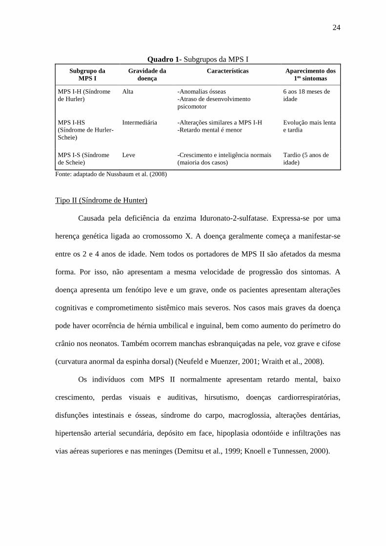
24
Quadro 1- Subgrupos da MPS I
Subgrupo da
MPS I
Gravidade da
doença
Características Aparecimento dos
1os sintomas
MPS I-H (Síndrome
de Hurler)
Alta -Anomalias ósseas
-Atraso de desenvolvimento
psicomotor
6 aos 18 meses de
idade
MPS I-HS
(Síndrome de Hurler-
Scheie)
Intermediária -Alterações similares a MPS I-H
-Retardo mental é menor
Evolução mais lenta
e tardia
MPS I-S (Síndrome
de Scheie)
Leve -Crescimento e inteligência normais
(maioria dos casos)
Tardio (5 anos de
idade)
Fonte: adaptado de Nussbaum et al. (2008)
Tipo II (Síndrome de Hunter)
Causada pela deficiência da enzima Iduronato-2-sulfatase. Expressa-se por uma
herença genética ligada ao cromossomo X. A doença geralmente começa a manifestar-se
entre os 2 e 4 anos de idade. Nem todos os portadores de MPS II são afetados da mesma
forma. Por isso, não apresentam a mesma velocidade de progressão dos sintomas. A
doença apresenta um fenótipo leve e um grave, onde os pacientes apresentam alterações
cognitivas e comprometimento sistêmico mais severos. Nos casos mais graves da doença
pode haver ocorrência de hérnia umbilical e inguinal, bem como aumento do perímetro do
crânio nos neonatos. Também ocorrem manchas esbranquiçadas na pele, voz grave e cifose
(curvatura anormal da espinha dorsal) (Neufeld e Muenzer, 2001; Wraith et al., 2008).
Os indivíduos com MPS II normalmente apresentam retardo mental, baixo
crescimento, perdas visuais e auditivas, hirsutismo, doenças cardiorrespiratórias,
disfunções intestinais e ósseas, síndrome do carpo, macroglossia, alterações dentárias,
hipertensão arterial secundária, depósito em face, hipoplasia odontóide e infiltrações nas
vias aéreas superiores e nas meninges (Demitsu et al., 1999; Knoell e Tunnessen, 2000).

25
Tipo III (Síndrome de Sanfilippo)
De acordo com a deficiência enzimática presente, a MPS III divide-se em 4
subgrupos, formando um grupo clinicamente semelhante e bioquimicamente diferente:
MPS III-A (heparan-N-sulaftase), MPS III-B (alfa-N-acetilglucosaminidase), MPS III-C
(acetil-CoA-alfa-glucosamina acetiltransferase) e MPS III-D (N-acetilglicosamina 6-
sulfatase) (Neufeld e Muenzer, 2001).
As alterações faciais, articulares e ósseas podem estar presentes, porém em grau
mais leve em relação aos outros tipos de MPS. As mudanças de comportamento são
percebidas entre os 2 e 6 anos de vida. Nesse período surgem os primeiros sinais da
doença, que se identificam pela perda da capacidade mental, distúrbio do sono, perda
gradual da marcha, do controle dos esfíncteres e a linguagem. São indivíduos hiperativos e
com estatura normal, o que caracteriza um envolvimento esquelético mínino da doença
(Wijburg et al., 2013; Cross et al., 2014; Mahon et al., 2014).
A degeneração neurológica pode provocar quadros de apnéia do sono, convulsões e
disfagia. A MPS III-A caracteriza-se por um comprometimento predominantemente
neurológico, enquanto nos demais tipos o envolvimento é predominantemente físico (MPS
III-B e MPS III-C) (Neufeld e Muenzer, 2001; Valstar et al., 2011; Delgadillo et al., 2013).
Tipo IV (Síndrome de Mórquio)
De acordo com a deficiência enzimática presente, a MPS IV divide-se em 2 grupos:
MPS IV-A (N-acetilgalactosamina 6- sulfatase) e MPS IV-B (Beta-galactosidade). Esses
dois grupos se difere porque o tipo B apresenta um acometimento mais leve da doença e
uma evolução mais lenta que o tipo A (Neufeld e Muenzer, 2001).
Os primeiros sintomas aparecem entre 1 e 3 anos de idade e o dignóstico
geralmente é confirmado até os 5 anos, sendo a gravidade bastante variável. Existe um

26
maior comprometimento ósseo. A inteligência geralmente é normal (Lachman et al., 2014;
Lin et al., 2014; Hendriksz et al., 2015).
Os indivíduos com MPS IV possuem marcha anormal e baixa estatura, que evolui
para hipercifose. Apresentam deformidades nos pés e nas mãos, hipoplasia odontóide,
perdas auditivas, disfunções intestinais, doenças cardiorrespiratórias e ósseas. Podem
desenvolver osteoporose. A face normalmente possui uma aparência “grosseira”, com a
presença de prognatismo, boca grande e dentes pequenos e com anormalias de esmalte
(Lachman et al., 2014; Lin et al., 2014; Hendriksz et al., 2015).
Tipo VI (Síndrome Maroteaux-Lamy)a
É causada pela deficiência da enzima N-acetilgalactosamina-4-sulfatase. O fenótipo
clínico é variável, podendo apresentar os primeiros sintomas antes dos 2 anos de idade.
Pode apresentar-se de forma leve, intermediária e severa, sendo que a forma severa
caracteriza-se por ser de rápida evolução. Geralmente o desenvolvimento cognitivo não é
afetado na MPS VI (Borges et al., 2003; Lachman et al., 2014).
Em alguns casos, nos primeiros meses de vida, a criança pode desenvolver hérnia
umbilical e inguinal, macrocefalia e deformidade do tórax. A partir dos 2 e 3 anos de idade
surgem sintomas clínicos semelhantes à MPS I (baixo crescimento, perdas visuais e
auditivas, opacificação da córnea, hirsutismo, doenças cardiorrespiratórias, disfunções
intestinais e ósseas, síndrome do carpo, macroglossia e alterações dentárias). Também
ocorre hipertensão arterial secundária, depósito em face contínua, infiltração das vias
aéreas superiores e das meninges e hipoplasia do processo odontóide. Não está associada
ao retardo mental (Azevedo et al., 2004; Valayannopoulos et al., 2010; Kantaputra et al.,
2014).
______ a Não há MPS tipo V

27
Tipo VII (Síndrome de Sly)
Causada pela deficiência da enzima beta-glucuronidase. Divide-se em 3 formas:
fetal-neonatal, grave e leve.
A forma fetal-neonatal pode causar óbito do feto e hidropsia fetal (edema fetal
generalizado habitualmente produzido por uma doença hemolítica: acumulam-se
quantidades anormais de líquido em duas ou mais áreas do corpo de um feto ou recém-
nascido).
O indivíduo com MPS VII normalmente apresenta fácies grosseira e disostose
múltipla (alteração dos ossos) e alterações de fígado e córneas (Nussbaum et al., 2008).
Na forma grave os sintomas surgem nos primeiros meses de vida, sendo que as
características clínicas tornam-se mais evidentes a partir dos 2 e 3 anos de idade. O
comprometimento sistêmico tem evolução mais lenta e menos grave. A forma grave da
doença é semelhante ao tipo I (retardo mental leve a moderado, baixo crescimento, perdas
visuais e auditivas, hirsutismo, doenças cardiorrespiratórias, disfunções intestinais e
ósseas, síndrome do carpo, macroglossia e alterações dentárias). Também ocorre
hipertensão arterial secundária, depósito em face contínua, infiltração das vias aéreas
superiores e das meninges e hipoplasia odontóide (Neufeld e Muenzer, 2001).
Na forma leve o comprometimento sistêmico é brando e a progressão da doença é
lenta (Nussbaum et al., 2008).
2.5 Tratamento da MPS
O tratamento do indivíduo com MPS baseia-se na substituição da enzima deficiente
pela enzima sadia. Mundialmente o protocolo de atendimento para a MPS baseia-se na
terapia de reposição enzimática e no transplante de medula óssea (TMO) / células-tronco
hematopoiéticas. Uma diferença significativa entre os dois tratamentos é que o transplante
28
pode tratar alguns distúrbios no Sistema Nervoso Central (SNC) presentes nos indivíduos
com MPS, principalmente quando realizado no inicio do curso da doença (Neufeld e
Muenzer, 2001; Clarke, 2008; Giugliani et al., 2010; Valayannopoulos e Wijburg, 2011;
Giugliani, 2012).
Reposição enzimática
A terapia de reposição enzimática consiste na infusão da forma recombinante da
enzima deficiente no organismo dos indivíduos com MPS. O tratamento é realizado a
longo prazo. Dependendo da enzima e dosagem, cada infusão dura em torno de 1 a 4 horas
por sessão. Devido à possibilidade de reações graves à perfusão, como por exemplo,
anafilaxia, a maioria das sessões de reposição enzimática é realizada em ambiente
hospitalar (Neufeld e Muenzer, 2001; Clarke, 2008; Giugliani et al., 2010;
Valayannopoulos e Wijburg, 2011; Giugliani, 2012).
Inicialmente o paciente pode apresentar efeitos colaterais como dor de cabeça,
rubor, febre e/ou erupção cutânea. Essas reações geralmente acontecem devido a uma
resposta imunitária à enzima. Geralmente são resolvidas por meio de um tratamento prévio
a base de antipiréticos e anti-histamínicos. Os efeitos colaterais tendem a diminuir com o
passar do tempo (Neufeld e Muenzer, 2001; Clarke, 2008; Giugliani et al., 2010;
Valayannopoulos e Wijburg, 2011; Giugliani, 2012).
A terapia de reposição enzimática não possibilita um retrocesso de manifestações
clínicas como a rigidez articular e o retardo mental. Porém, quando submetidos ao
tratamento, há uma manutenção da amplitude dos movimentos e da capacidade funcional
do indivíduo. Quanto mais precoce é realizado o diagnóstico e o início do tratamento,
maior é o impacto da medicação sobre a capacidade funcional e sobre a amplitude do

29
movimento articular (Neufeld e Muenzer, 2001; Clarke, 2008; Giugliani et al., 2010;
Valayannopoulos e Wijburg, 2011; Giugliani, 2012).
Somente as MPS I, II, IV e VI possuem medicação específica para terapia de
reposição enzimática. O medicamento, no entanto, não é facilmente acessível a todos que
precisam dele. Apresentam alto custo financeiro e são produzidos por uma única empresa
farmacêutica. Por não estar incluído na política de assistência farmacêutica do governo
federal brasileiro, são adquiridos para o paciente apenas por meio de ações judiciais
(Giugliani et al., 2010; Medeiros et al., 2013; Tomatsu et al., 2014).
Transplante de medula óssea (TMO)
No final da década de 80 o TMO passou a ser uma alternativa de tratamento para as
doenças oriundas de erros inatos do metabolismo. O tratamento consiste em corrigir ou
reduzir as anomalias viscerais dessas doenças (Nussbaum et al., 2008). Segundo os autores,
o transplante é indicado para crianças com a forma grave da MPS I que tenham sido
diagnosticadas antes dos 2 anos de idade. É que após essa idade a doença já compromete o
SNC de forma irreversível. Desse modo, o transplante não surtiria efeito.
Considerando-se a existência da terapia de reposição hormonal, o TMO não é
recomendado para as demais formas de MPS I e para os casos de MPS II e MPS VI devido
aos altos riscos e por não apresentar bons resultados (Giugliani et al., 2010;
Valayannopoulos e Wijburg, 2011).
O procedimento do TMO inicia-se a partir de uma quimioterapia para eliminar as
células deficientes do organismo. Essa fase pode ter uma duração de atá 12 meses. Quando
bem sucedido, o transplante tem a capacidade de prolongar significativamente a
sobrevivência da criança devido a uma normalização ou redução do tamanho do fígado,
baço e coração. Também é capaz de reduzir os episódios de apneia do sono e as doenças

30
das vias aéreas superiores, bem como preservar a audição e propiciar maior mobilidade dos
membros superiores. As anomalias esqueléticas, no entanto, não são corrigidas com o
TMO (Nussbaum et al., 2008).
Os principais desafios desse tratamento estão na dificuldade de se encontrar
doadores de medula óssea que sejam compatíveis e também da mortalidade associada ao
procedimento (Valayannopoulos e Wijburg, 2011).
Considerando-se o fato do tecido dentário ser extremamente sensível à
quimioterapia, Hingston et al. (2006) e Guven et al. (2008) ressaltaram a importância da
presença do cirurgião dentista na equipe de acompanhamento de pacientes em tratamento
de TMO. Segundo os autores, é essencial que o tratamento quimioterápico seja vinculado a
uma atenção cuidadosa à cavidade bucal que inclua cuidados com a higiene, escovação
supervisionada e acompanhamento odontológico periódico.
2.6 A odontologia e a MPS
As alterações bucais presentes na maior parte das pessoas com MPS destacam a
importância de um cirurgião-dentista na equipe de atendimento multidisciplinar que cuida
e acompanha esta parcela da população. A equipe de profissionais envolvida na assistência
dos indivíduos com MPS não pode ignorar o papel da odontologia na conquista de
melhores condições de vida para essa parcela da população (Hingston et al., 2006; Turra e
Schwartz, 2009; Antunes et al., 2012; Cavaleiro et al., 2013; Gönüldas et al., 2014). Muitas
vezes, as complicações odontológicas podem afetar a saúde geral das pessoas com MPS.
Complicações que promoveriam dor ou afetariam a saúde geral podem ser evitadas quando
o cuidado odontológico está presente (Fitzgerald e Verveniotis; 1998; Kuratani et al., 2005;
Alpöz et al., 2006; Onçag et al., 2006; Turra e Schwartz, 2009; Antunes et al., 2012; James
et al., 2012; Fonseca et al., 2014; Kantaputra et al., 2014; Savitha et al., 2015).

31
Os dentes decíduos das crianças com MPS devem receber cuidado especial. Em
alguns casos, a dentição decídua não é substituída pela permanente (Alpöz et al., 2006).
Por isso, a atenção odontológica para eles deve acontecer bem cedo.
A maioria dos indivíduos com MPS apresenta algum tipo de alteração na cavidade
bucal. Dentre elas destacam-se as alterações da arcada dentária e da língua, que podem
comprometer as funções de mastigação e deglutição (Turra e Schwartz, 2009). A
hipertrofia gengival também é frequente (Antunes et al., 2012; James et al., 2012). Além
de dificultar a higienização, em alguns casos o aumento de volume gengival ocasiona
transtornos de deglutição e mastigação. A qualidade da higiene bucal está relacionada ao
quadro clínico do paciente, conforme a sua capacidade de motricidade e de inteligência
(Oliveira et al., 2008b; Antunes et al., 2012; James et al., 2012).
2.6.1 Manifestações bucais da MPS
As principais alterações bucais descritas nos indivíduos com MPS são: alterações
de número e anatomia dos dentes decíduos e permanentes, atraso de erupção dos dentes
permanentes, má oclusão (mordida cruzada e mordida aberta anterior ou posterior),
protrusão da língua, macroglossia, palato estreito e profundo, limitações de abertura bucal,
defeitos de esmalte (dentes decíduos e permanentes), diastemas, bruxismo, lesões de cárie
e hipertrofia gengival (Fitzgerald e Verveniotis; 1998; Kuratani et al., 2005; Alpöz et al.,
2006; Onçag et al., 2006; Turra e Schwartz, 2009; Almeida-Barros et al., 2012; Antunes et
al., 2012; James et al., 2012; Fonseca et al., 2014; Kantaputra et al., 2014; Savitha et al.,
2015). Quando associadas a uma dieta cariogênica e a uma escovação deficiente, muitas
vezes a má oclusão atuará como fator predisponente da doença cárie (Onçag et al., 2006;
Turra e Schwartz, 2009; Fonseca et al., 2014).

32
Hingston et al. (2006) descreveram o caso clínico de um paciente com MPS de 11
anos de idade que, aos 18 meses de vida, foi submetido a um TMO com sucesso. O exame
bucal da criança mostrou a presença de má oclusão, de diastemas, macroglossia, higiene
bucal deficiente e gengivite marginal. Wadenya et al. (2010) relataram um caso clínico de
uma criança tratada com sucesso por meio de um TMO. O desenvolvimento dentário da
criança foi acompanhado desde a dentição decídua. Os autores observaram que, nas
diferentes fases da dentição, o efeito da terapia de medula óssea foi significativo e variável.
Uma amostra de 78 crianças/adolescentes com MPS I, II, III-B, IV-A e VI (média
de idade entre 2,8 a 12,6 anos) foi examinada em Porto Alegre. A grande maioria dos
pacientes apresentaram algum tipo de alteração na arcada dentária (98,4%) e na língua
(95,9%). Considerando-se outras estruturas da cavidade bucal, observou-se que 70,0% a
90,0% dos participantes foram identificados com alguma alteração nos lábios, bochechas,
mandíbula e palato duro (Turra e Schwartz, 2009).
Guimarães et al. (2010) descreveram o caso clínico de um indivíduo com MPS tipo
VI que foi submetido com sucesso ao TMO aos 22 meses de idade. Aos 15 anos o
adolescente foi examinado pelos autores, que verificaram que a MPS do paciente
apresentava-se de forma moderada, com as seguintes características bucais: hiperplasia
gengival, hipertrofia do rebordo alveolar maxilar, macroglossia, dentes não irrompidos, má
oclusão e cisto dentígero. Mcgovern et al. (2010) avaliaram a cavidade bucal de 25
pacientes com MPS tipo I (Hurler) que foram submetidos com sucesso ao TMO. Os
autores observaram que os pacientes apresentavam atraso de desenvolvimento dentário, má
oclusão e anomalias dentárias, sendo as mais comuns a hipodontia e a microdontia.
Um estudo desenvolvido em um Centro de Referência no Rio de Janeiro analisou
12 pacientes com MPS. Verificou-se um percentual alto de lesões de cárie, doença
periodontal, erupção dentária retardada, lábios grossos e processo alveolar mais espesso

33
entre os examinados (Antunes et al., 2012). Ribeiro et al. (2014) avaliaram 26 pacientes
com MPS de um Centro de Referência do Ceará e observaram um alto índice de
macroglossia, aumento gengival, diastemas generalizados e má oclusão. Os examinados
apresentavam o terço médio da face encurtado, o terço inferior aumentado e um perfil
convexo.
Fonseca et al., (2014), realizaram um estudo na Paraíba com 17 pacientes de MPS
com média de idade de 13,2 anos. Foi um estudo pareado: para cada paciente com MPS
dois pacientes controle foram selecionados. Os autores observaram diferenças entre os
grupos quanto às medidas angulares faciais. Os pacientes com MPS apresentaram maior
tendência de crescimento vertical da face, resultando em um padrão facial dolicocefálico.
Os autores relataram que a presença de mordida aberta nos indivíduos com MPS
possivelmente esta relacionada com a diminuição do espaço nasofaríngeo, um fator que
pode ser responsável pela respiração bucal desses pacientes.
O achado mais comum nos indivíduos com MPS tipo IV refere-se a uma aparência
facial comprometida esteticamente e funcionalmente. Nesses casos, o tratamento
ortodôntico e protético tem condições de promover ao indivíduo uma estética melhor e
uma oclusão satisfatória (Onçag et al., 2006).
Kuratani et al. (2005) descreveram o caso de uma paciente japonesa de 7 anos de
idade diagnosticada com MPS IV na qual foi realizado tratamento ortodôntico por meio
dos aparelhos fixo e móvel. Os autores observaram que após a intervenção ortodôntica a
paciente adquiriu uma boa oclusão e função mastigatória. Resultados semelhantes foram
descritos por Onçag et al. (2006), que relataram o caso de um paciente do sexo masculino
de 22 anos de idade com MPS IV em que foi feita intervenção ortodôntica do tipo
Edgewise.

34
Considerando-se as alterações de esmalte, normalmente o esmalte dos indivíduos
com MPS é frágil e se apresenta com maior porosidade (Rolling et al., 1999). Com relação
à morfologia dos dentes, os autores afirmaram que as cúspides geralmente são pontiagudas
e os dentes incisivos possuem um formato de espada.
Fitzgerald e Verveniotis (1998) relataram um caso clínico de um paciente com MPS
tipo IV (Mórquio) que apresentava dentes posteriores afilados e diastema na região dos
dentes anteriores superiores. Radiograficamente os autores observaram que o esmalte
dentário tinha uma espessura cerca de 25,0% menor, quando comparado ao esmalte de um
dente sem alteração. O paciente apresentava baixa experiência de cárie dentária.
Guven et al. (2008) desenvolveram um estudo onde foram avaliados, radiográfica e
histologicamente, o esmalte e a dentina dos dentes de um indivíduo portador de MPS tipo I
de Hurler. Foram extraídos 9 dentes decíduos. Por meio de microscopia eletrônica de
varredura verificou-se que os dentes apresentavam túbulos dentinários estreitos, espaços na
junção dentina-esmalte e arranjos irregulares dos prismas dos esmaltes. Segundo os
autores, o paciente apresentava abertura de boca limitada, diastemas, microdontia, aumento
do tecido gengival e hipoplasia de esmalte dos dentes decíduos.

35
3 METODOLOGIA
3.1 Campo da pesquisa
Os pacientes com MPS foram selecionados em dois centros de referência para
atendimento de pacientes com MPS em Belo Horizonte [Ambulatório de Erros Inatos do
Metabolismo do Hospital das Clínicas da Universidade Federal de Minas Gerais (HC-
UFMG) e Hospital João Paulo II]. As crianças/adolescentes sem MPS foram selecionadas
nos ambulatórios São Vicente de Paula (Setor de adolescentes) e Bias Fortes (Setor de
pediatria) do HC-UFMG. Os hospitais supra citados são públicos.
3.2 Considerações Éticas
Conforme Resolução do Conselho Nacional de Saúde (CNS), de 10 de outubro de
1996, este estudo foi aprovado pela Diretoria de Ensino, Pesquisa e Extensão (DEPE) do
HC-UFMG (ANEXO A) e pelo Comitê de Ética em Pesquisa (COEP) da UFMG, por se
tratar de um estudo envolvendo seres humanos (parecer 257.220, abril de 2013) (ANEXO
B).
3.3 Desenho do estudo
O estudo tem um caráter quantitativo do tipo observacional transversal. A coleta de
dados aconteceu por meio do exame clínico, radiográfico e análise facial dos pacientes
com MPS e sem MPS. Os pais/responsáveis responderam um questionário estruturado
abordando questões relacionadas ao tema abordado.
Os pais/responsáveis das crianças/adolescentes previamente selecionados foram
contatados pessoalmente, ou por telefone, e receberam uma explicação prévia sobre a
pesquisa, sendo convidados a irem na Faculdade de Odontologia da UFMG (FO-UFMG)
para a coleta de dados. Foram incluídos, portanto, aqueles pais/responsáveis que

36
concordaram em responder o questionário, que permitiram o exame clínico, radiográfico e
a análise facial do filho e que assinaram o termo de consentimento livre e esclarecido
(APÊNDICE A).
A coleta de dados foi realizada no sector de radiologia e em uma das clínicas da
FO-UFMG. Quando foi identificada alguma necessidade de tratamento odontológico no
participante, o responsável foi devidamente alertado e orientado sobre o fato, sendo
encaminhado para atendimento odontológico na FO-UFMG.
3.4 População do estudo
Amostra
Não se têm disponíveis registros de sistemas de informação caracterizando a
população acometida pela MPS, que faz parte de grupos de difícil localização e seleção
para amostras. Por isso, é difícil obter-se uma amostra representativa de indivíduos com a
doença na população geral. Desse modo, optou-se por uma amostra de conveniência
composta pelo universo de pacientes com MPS atendidos no Ambulatório de Erros Inatos
do Metabolismo do HC-UFMG e no Hospital João Paulo II. No ano 2013 foram
identificados 65 indivíduos com MPS nos arquivos de registros de pacientes com MPS dos
hospitais em questão. A faixa etária deles variou de 2 a 25 anos de idade.
A população do estudo foi selecionada na perspectiva de que os fenômenos em
observação, separada ou concomitantemente, ocorram dentro daquele contexto. Desse
modo o estudo contou com um grupo de comparação que viabilizou analisar a relação entre
a condição (ter MPS ou não ter MPS) e o desfecho (características dentárias). Garante-se,
assim, que ambos sejam variáveis aleatórias, ou seja, que variem entre as unidades de
observação. Por isso, a população do estudo foi constituída de duas subpopulações: uma de
crianças/adolescentes com MPS e uma de crianças/adolescentes sem MPS. Foram

37
selecionadas e originadas de populações de origens distintas, mas ambas atendidas pelo
Sistema Único de Saúde (SUS). Para cada paciente com MPS foi escolhido um participante
do grupo de comparação.
Fonte e Critérios de Elegibilidade
Foram considerados os seguintes critérios de inclusão para os participantes:
1- Crianças/adolescentes com MPS e seus pais/responsáveis.
2- Crianças/adolescentes sem MPS ou outra condição especial (outra deficiência ou
alguma doença crônica ou aguda) e seus pais/responsáveis.
3- Crianças/adolescentes atendidos nos Ambulatórios de Erros Inatos do Metabolismo e
São Vicente de Paula do HC-UFMG e Hospital João Paulo II e seus pais/responsáveis.
4- Crianças/adolescentes que não estavam realizando (ou já tivessem realizado) tratamento
ortodôntico e seus pais/responsáveis.
3.5 Instrumentos de coleta de dados
3.5.1 Questionário estruturado
O levantamento de dados com os pais/responsáveis foi realizado por meio de um
questionário estruturadoa. O instrumento foi aplicado individualmente a cada responsável
na forma de entrevista, caracterizando o contato face a face entre pesquisador e
pesquisado.
_______
a Alguns dados obtidos por meio do questionário serão analisados em um outro estudo.

38
Foi composto por questões relacionadas às características individuaisa e gerais, aos
hábitos comportamentais e à história médica e odontológica das crianças/adolescentes com
MPS e sem MPS (APÊNDICE B). Esse instrumento foi adaptado a partir do estudo de
Oliveira et al. (2008b; 2010).
3.5.2 Exame clínico odontológico
O exame da cavidade bucal do paciente foi realizado após a aplicação do
questionário junto aos pais/responsáveis. Aconteceu sob luz artificial, na cadeira
odontológica, maca ou cadeira de rodas. A equipe foi composta por uma examinadora e
por uma anotadora/organizadora.
O espelho bucal e a sonda do Índice Periodontal Comunitário (sonda IPC) foram os
instrumentos de medida utilizados para o exame clínico. Os resultados do exame clínico
foram registrados na ficha de exame do indivíduo (APÊNDICE C).
O tipo de respiração dos indivíduos examinados foi identificado pro meio do “teste
do espelho” (Oliveira et al., 2008a). Para isso foi utilizado um espelho dupla face sob o
nariz. Caso o examinado fosse respirador nasal, o espelho ficava embaçado na porção
superior. Caso fosse respirador bucal (sozinha ou associada à nasal), o espelho ficava
embaçado na parte inferior.
_______ a A categorização étnica foi determinada através de critérios estabelecidos pelo Instituto Brasileiro de
Geografia e Estatística (IBGE, 2011) para a cor da pele: branco, preto, marrom ou amarelo. A classe
econômica de cada nível familiar e educacional dos pais/responsáveis foram avaliados de acordo com o
critério de classificação econômica Brasil da Associação Brasileira de Empresas de Pesquisa (ABEP, 2012)
(ANEXO C). As classes econômicas foram agrupadas em mais favorecida (compreendendo as classes sociais
A e B), favorecida (C) e menos favorecida economicamente (classes D e E). A escolaridade dos
pais/responsáveis foi mensurada por meio dos anos de estudo, sendo fo classificada com base em um ponto
de corte de oito anos, o que corresponde ao ensino fundamental no Brasil.

39
Higiene bucal
A análise da higiene bucal foi realizada através do Índice de Higiene Oral
Simplificado (IHOS) (Greene e Vermillion, 1964).
Gengivite
A presença de gengivite foi identificada quando a cor e contorno da gengiva não
estavam de acordo com o pardão normal (Alaluusua and Malmivirta, 1994; Bonanato et al.,
2009b).
Prevalência de cárie
O número de dentes com lesões de cárie considerou a presença de lesão cavitada.
Os critérios de diagnóstico foram considerados com base na WHO (2013).
Defeitos de desenvolvimento de esmalte (DDE)
A presença de DDE foi registrada por meio do Índice DDE modificado (opacidade
difusa, opacidade demarcada e hypoplasia de esmalte). O índice permite estabelecer o tipo
(descoloração), o número (único ou múltiplo), a demarcação (demarcado ou difuso) e a
localização dos defeitos.
Anomalias dentárias
Foram registrados dados referentes à presence de anomalias dentárias como dente
conóide, agenesia, microdontia, giroversão e outras.

40
Exame oclusofacial
Os critérios de diagnóstico oclusal foram considerados com base nos estudos de
Oliveira et al. (2008a; 2010b) e WHO (2013). Foram registrados dados sobre a relação
oclusal anteroposterior e às alterações verticais e/ou transversais de oclusãoa
(sobremordida/overbite, sobressaliência/overjet e mordida cruzada posterior). O
diagnóstico oclusal foi realizado por meio do exame clínico e exame radiográfico.
Aqueles indivíduos diagnosticados com pelo menos um caso de protrusão, mordida
cruzada posterior/anterior, mordida profunda, mordida aberta anterior e/ou mordida em
topo foram classificados como portadores de má oclusão.
3.5.3 Diagnóstico facialb
Os critérios de diagnóstico facial foram considerados com base nos estudos de
Arnett (1993a; 1993b) e Proffit e Fields (2002). Essa técnica de diagnostico visa identificar
pacientes com desproporções severas (Arnett 1993a; 1993b; Proffit e Fields, 2002).
Embora com menos detalhes do que um exame radiográfico, uma análise facial cuidadosa
fornece informações das relações ósseas abaixo do tecidos (Proffit e Fields, 2002). Foi
realizada as a análise frontal e a análise lateral da face.
3.5.4 Exame Radiográficob
Foram realizados dois tipos de procedimentos radiográficos: telerradiografia e
radiografia panorâmica
__________ a Para a obtenção das medidas do overjet e overbite foi utilizada a sonda IPC. As medidas até 3mm foram
consideradas normais. b Os dados referentes à análise facial e ao exame radiográfico serão analisados em um outro estudo.
41
a) Telerradiografia
Foi avaliado o padrão esquelético, o padrão de crescimento e o padrão dentário dos
indivíduos com MPS e sem MPS (Proffit e Fields, 2002; Petrelli, 2011).
b) Radiografia Panorâmica
Possibilita uma visão geral das estruturas que compõem o completo maxilo-
mandibular: dentes, tecido ósseo, seios maxilares, articulação têmporo-mandibular,
cavidade nasal (Howerton e Iannucci, 2010).
3.6 Calibração da examinadora
Foi realizada a calibração teórica, por meio de figuras e slides. Foi conduzida para
verificação da variabilidade diagnóstica intra-examinadora, com um intervalo de 7 a 14
dias entre os dois momentos de calibração teórica.
O próximo passo foi a calibração prática da examinadora, seguindo um padrão ouro
de diagnóstico e conduzida com um intervalo de 7 dias entre os dois momentos da
calibração. Devido ao número limitado de crianças e adolescentes com MPS, foram
examinadas somente crianças/adolescentes sem MPS. A calibração foi feita em 20
crianças/adolescentes de uma escola pública no município de Confins, Minas Gerias. A
partir dos valores kappa obtidos (0,76 a 0,98) verificou-se que a examinadora se
encontrava treianda para realizar a coleta de dados.
3.7 Estudo piloto
O estudo piloto foi realizado após a fase de calibração da examinadora.
Participaram dessa faze 5 crianças/adolescentes com MPS e 5 sem MPS, bem como os
pais/responsáveis. A coleta de dados foi realizada nos hospitais previamente selecionados.

42
Esta etapa teve por finalidade avaliar a metodologia e os instrumentos da coleta de dados.
Após análise dos dados foi iniciado o estudo principal. Os participantes do estudo piloto
foram incluídos na amostra final do estudo.
3.8 Estudo principal
Após as fases de calibração e estudo piloto, foi iniciado o estudo principal.
3.9 Processamento dos dados
Os dados referentes aos questionários e exames clínico obtidos ao longo do estudo
foram devidamente armazenados e analisados por meio do software Statistical Package for
Social Science - SPSS® (versão 21.0). O processamento incluiu codificação, digitação,
edição dos dados e análise estatística.
Previamente à análise estatística dos dados foi realizada a análise descritiva.

43
4 RESULTADOS E DISCUSSÃO
Oss resultados, a discussão e a conclusão do estudo serão apresentados na forma de
artigo científico.

44
ARTIGO
A paired comparison of dental characteristics of Brazilians with and without
Mucopolyssaccaridosis
Tahyná Duda Deps1 ([email protected])
Natalia Cristina Ruy Carneiro1 ([email protected])
Esdras Castro França1 ([email protected])
Eugênia Ribeiro Valadares2 ([email protected])
Isabela Almeida Pordeus1 ([email protected])
Ana Cristina Borges-Oliveira3* ([email protected])
1Department of Pediatric Dentistry and Orthodontics, Faculty of Dentistry, Universidade
Federal de Minas Gerais, Belo Horizonte, Brazil
2Department of Propedeutica Complementar, Faculty of Medicine, Universidade Federal
de Minas Gerais, Belo Horizonte, Brazil
3Department of Social and Preventive Dentistry, Faculty of Dentistry, Universidade
Federal de Minas Gerais, Belo Horizonte, Brazil
Artigo submetido ao periódico Orphanet Journal of Rare Diseases
(Qualis - Medicina A1 / Fator de impacto 3,96) (Anexo D e Anexo E)

45
ABSTRACT
Background: There are several oral manifestations present in individuals with
Mucopolyssaccaridosis (MPS). Among them is the malocclusions, dental anomalies and
dental caries. Alterations in the oral cavity can result in infections and nutritional,
respiratory, chewing and speech problems in this population, which often has a severely
debilitated state of general health. The aim of the present study was to compare the dental
characteristics of individuals with and without Mucopolyssaccaridosis (MPS). Methods:
The paired cross-sectional study was composed on a sample of 29 parents/children with
MPS and 29 parents/normal children. The individuals were between three and 27 years old
and were attending in two hospitals in Belo Horizonte, southeastern Brazil. The parents
answered a questionnaire on sociodemographic and behavioral aspects of their children.
The dental characteristics of the patients were evaluated by clinical examination of dental
caries, gingivitis, malocclusion, dental anomalies and developmental defects of enamel
(DDE). The examiner was previously calibrated, and kappa values between 0.76 and 0.98
were obtained. Data were analyzed by means of univariate and bivariate analysis (X2 test),
with a level of confidence of 95.0%. The study was approved by the Research Ethics
Committee of the Universidade Federal de Minas Gerais. Results: The average age of the
patients was 13.9 years (± 7.2). The majority of them was male (58.6%), had black/brown
skin color (70.7%) and from favored or more favored economic classes (89.7%). When
groups were compared, there was a statistically significant association between them and
the variables gingivitis, dental malocclusion and anomalies (p<0.05). The chance of being
diagnosed with a malocclusion, dental anomaly and/or gingivitis was higher in the group of
patients with MPS. Conclusions: The results showed that there is a greater prevalence of
malocclusion, dental anomalies and gingivitis among individuals with MPS than in normal
individuals.

46
Keywords: Mucopolysaccharidosis, Dental care for disabled, Disabled children, Oral
health.
Background
Mucopolysaccharidosis (MPS) is a group of rare diseases caused by lysosomal
enzyme deficiencies that lead to glycosaminoglycans (GAG) acumulation. They are
characterized as severe, chronic and multisystemic and are associated with a high level of
mortality and morbidity. The diseases are classified into seven types, with classification
based on the deficiency of 11 enzymes (Type I, II, III, IV, VI, VII, IX). All, except type II,
have an autosomal recessive genetic inheritance pattern [1-3]. It is estimated that the global
incidence of MPS is between 1:25,000 to 1:52,000 live births [1,2,4,5]. In Brazil the
prevalence of the disease remains unknown [2].
Each type of MPS possesses significant clinical heterogeneity, but some
characteristics are common to all types: cardiac problems, respiratory insufficiency,
hepatosplenomegaly, short stature, delayed motor development, hearing loss, limited joint
mobility, macrocephaly, skeletal dysplasia, facial and dental abnormalities, macroglossia
and umbilical and inguinal hernias. The disease has high rates of morbidity and mortality
[3,4,6,7]. According to a previous study, corneal clouding is not frequent with MPS II, III
and IV, and mental retardation is not usual with MPS IV and VI [8].
The main oral alterations described among individuals with MPS are: alterations of
number and anatomy of deciduous and permanent teeth, enamel defects (primary and
permanent teeth), eruption delay of permanent teeth, diastema, malocclusion (mainly
anterior open bite and crossbite), tongue protrusion and largue, limited oral opening,
bruxism and dental caries [3,4,8-15]. Oral health has a fundamental role in the life of

47
individuals with MPS. Oral disease can result in infections and nutritional, respiratory,
chewing and speech problems in this population, which often has a severely debilitated
state of general health [14-17].
Although individuals with MPS suffer from various orofacial alterations, there is a
lack of studies about the characteristics of such alteration [4,12,14,15,17]. Most existing
studies are reports of clinical cases [8-10,13,16].
The aim of the present study was to compare the dental characteristics of
individuals with MPS and without MPS. The hypothesis is that individuals with MPS have
a greater prevalence of dental caries, gingivitis, malocclusion, dental anomalies and
developmental defects of enamel (DDE) than normal individuals.
Methods
Study design and sample characteristics
A matched cross-sectional study of individuals with and without MPS and their
respective parents/guardians was performed. The study universe included 34 individuals
with MPS treated at two referral centers on MPS in Belo Horizonte, state of Minas Gerais,
southeastern Brazil. The choice of location resulted in a convenience sample (non-random
sample), where individuals could be selected based on a value judgment rather than
statistical randomness. Such samples generally comprise more accessible individuals
[18,19]. Normal individuals and their parents/guardians were treated in the outpatient
pediatric clinic of a university’s hospital in Belo Horizonte. A comparison group was
drawn from this dataset, individually matched for age and gender (one normal person for
each person with MPS). Such pairing is a strategy used in research to make data more
reliable when comparing two groups that may be different for an given variable, but
similar in other ways [20].

48
The parents/guardians of the selected individuals were contacted in person or by
telephone and before being invited to participate in the present study, were given an
explanation of the purpose and nature of the research. Data collection was performed in a
dental clinic at the Faculty of Dentistry of UFMG in Belo Horizonte. Data acquisition
involved a clinical oral examination and interviews with parents/guardians. This study was
approved by the Research Ethics Committee of the Universidade Federal de Minas Gerais
(UFMG).
Non-clinical examination
The parents/guardians answered a questionnaire about the sociodemographic and
behavioral aspects of their children. Ethnic categorization was determined using criteria
established by the Brazilian Institute of Geography and Statistics for skin color: white,
black, brown or yellow [21]. The economic class of each family and educational level of
parents/guardians were evaluated using the Brazilian Economic Classification Criteria
[22], grouped into more economically favored (comprising social classes A and B),
favored (C) and less favored (classes D and E). Mother’s schooling (years of study) was
categorized based on a cut-off point of eight years, which corresponds to a primary school
education in Brazil.
Clinical oral examination
The dental characteristics of the patients were evaluated by clinical examination of
dental caries, gingivitis, malocclusion, dental anomalies and DDE. The type of MPS was
identified from the patient’s medical records.
The diagnostic criteria for carious lesions were based on the definition of the World
Health Organization (WHO) [23]: 1) healthy tooth (absence of a cavitated lesion); 2) tooth

49
decay (presence of cavitated lesion). Gingivitis was determined when the gingival contour
and color were abnormal. It was recorded as present or absent [24,25].
The presence of dental anomalies and malocclusion was based in WHO [23] and
Oliveira et al. [26]: alterations of overjet (protrusion, anterior crossbite, absent), overbite
(deep overbite, anterior open bite, absent, edge-to-edge) and posterior crossbite. When at
least one condition was diagnosed, the subject was classified as suffering from an occlusal
problem stemming from a variation in vertical or transversal occlusion. Overbite, overjet,
and posterior crossbite were identified through clinical examination. DDE was assessed in
accordance with the modified DDE index (diffuse opacity, demarcated opacity and enamel
hypoplasia [27].
Clinical examination was carried out by the researcher TDD. The clinical
examination was performed with the patient sitting in a conventional dental chair under
standard illumination. The examiner used appropriate individual protection equipment to
avoid cross infection. Mouth mirrors (PRISMA®, São Paulo, Brazil) and a Community
Periodontal Index probe (WHO-621; Trinity, Campo Mourão, PA, Brazil) were used for
the dental examination. Radiography was not used.
Calibration exercise
A calibration and training exercise was carried out prior to the study. It consisted of
two stages. The first stage included a theoretical discussion regarding the diagnosis of
dental caries, gingivitis, malocclusion, dental anomalies and DDE. The criteria for clinical
diagnosis were discussed and defined and oral clinical examination training with slides was
performed [18,19,28]. Specialists in paediatric dentistry and orthodontics were taken as the
gold standard for the theoretical framework and oral clinical examination. Training with
slides was performed on two different occasions with a one week interval between

50
sessions. Data analysis involved the calculation of Kappa coefficientes (0.76-0.98). The
second stage was the clinical examination. Intra-examiner reliability was determined on a
tooth-by-tooth basis for each clinical condition [28]. Five individuals with MPS and five
without MPS from one of the referral centers for patients with MPS and from the Pediatric
Outpatient Clinic were examined and re-examined after a two week interval to calculate
intra-examiner agreement. The Kappa test results were very good, with scores of 0.94 for
dental caries, 0.93 for malocclusion, 0.95 for dental anomalies and 0.89 for DDE. As a
result the examiner was considered able to carry out the main study.
Pilot study
A pilot study was conducted during the second calibration stage to analyze the
methodology and logistics of the research. The results of the pilot study indicated that no
changes to the methodology were required. The sample from this pilot study was included
in the main study.
Statistical analysis
Data was analyzed using the Statistical Package for Social Sciences software (SPSS
for Windows, version 20.0, SPSS Inc, Chicago, IL, USA). Following descriptive analysis,
bivariate analysis was used to evaluate associations between the groups and the
independent variables. This was performed using the chi-squared test (p<0.05).
Results
The present study evaluated 29 pairs of patients with MPS and their
parents/guardians, which represented 85.3% of the previously defined study universe.
There were 11 losses because refusal, death, other reason. A total of 29 normal individuals

51
and their respective parents/guardians also participated in the study.
The individuals with and without MPS were between three and 27 years old. The
average age was 13.9 years (+ 7.2) and the median was 14.0 years. The majority of
individuals were male (58.6%), had black/brown skin color (70.7%) and were from
favored or more favored economic classes (89.7%). None of the patients examined had a
history of orthodontic treatment. The age of parents/guardians varied from 23 to 59 years,
with a mean of 40.9 years (+9.0) and a median of 40.0 years. The majority of them had
eight years or more of schooling (67.2%) and declared to be the mother of the patient
(93.1%).
The distribution of patients according to type of MPS is described in graph 1. Two
individuals with MPS had not yet had their type of MPS diganosed during the period of
data collection and they were under investigation of MPS type.
Graphic 1- Distribution of patients with MPS according to the type of the disease (N=29).

52
Table 1 shows homogeneity between groups paired for gender and age (p>0.05).
The groups were also similar in terms of variables of skin color and economic class
(p>0.05).
Table 1- Distribution of patients according to presence of MPS (N=58)
a X2 Test (5% significance level)
The results of bivariate analysis to examine the relationships between genetic
condition and the dental clinical variables of individuals with or without MPS are shown in
table 2. Most independent variables demonstrated strong crude associations with genetic
condition. The variables gingivitis, malocclusion and dental anomalies were statistically
significant (p<0.05). The chance of being diagnosed with a malocclusion, dental anomaly
and/or gingivitis was higher in the group of patients with MPS.
INDIVIDUAL
VARIABLE
GROUP
With MPS
n (%)
Without MPS
n (%)
Total
n (100.0%) P valuea
Sex
Male 17 (50.0) 17 (50.0) 34 1.00 Female 12 (50.0) 12 (50.0) 24
Age (years)
3-12 13 (50.0) 13 (50.0) 26 1.00 13-27 16 (50.0) 16 (50.0) 32
Skin color
White 9 (52.9) 8 (47.1) 17 0.773 Black / Brown 20 (48.8) 21 (51.2) 41
Economic Class
Maost favored (A+B) 13 (56.5) 10 (43.5) 23
0.312 Favored (C) 14 (48.3) 15 (51.7) 29
Less favored (D+E) 2 (33.3) 4 (66.7) 6

53
Table 2- Absolute and relative frequency of dental characteristics of patients according to
presence of MPS (N=58)
aX2 Test (5% significance level) / b 95% CI: Confidence Interval
The types of malocclusions identified in 39 patients were alterations of overjet
(53.4%), overbite (55.2%) and posterior crossbite (12.1%). A total of 23 patients suffered
from some form of dental anomaly. The dental anomalies diagnosed were giroversion
(69.5% / n=16), agenesis (17.4% / n=4) and others [conoid teeth and microdontia (13.1% /
n=3)].
Discussion
The results of the present study support the hypothesis that individuals with MPS
have a greater chance of suffering from malocclusion, dental anomalies and gingivitis than
normal individuals. This difference most likely related to the occlusal and facial features
and developmental abnormalities present in the population with MPS. In addition to joint
and dental alterations, skeletal deformities are also found in the majority of individuals
with MPS [4,8,15,29]. The aesthetic and functional impairment of the orofacial region is
common in individuals with MPS, being similar in all types of MPS [8,10,29].
INDEPENDENT
VARIABLE
GROUP
With MPS
n (%)
Without MPS
n (%)
Total
n (100.0%) P valuea
Odds Rattio
( 95% CI)b
Dental caries
> 1 tooth 10 (58.8) 7 (41.2) 17 0.387
1.65 (0.52-5.19)
1 Absent 19 (46.3) 22 (53.7) 41
Gingivitis
Present 28 (56.0) 22 (44.0) 50 0.046
3.42 (0.08-13.43)
1 Absent 1 (12.5) 7 (87.5) 8
Malocclusion
Present 24 (61.5) 15 (38.5) 39 0.012
4.48 (1.33-14.99)
1 Absent 5 (26.3) 14 (73.7) 19
Dental anomalies
Present 17 (73.9) 6 (26.1) 23 0.003
5.43 (1.69-17.38)
1 Absent 12 (34.3) 23 (65.7) 35
DDE
Present 16 (55.2) 13 (44.8) 29 0.431
1.51 (0.53-4.26)
1 Absent 13 (44.8) 18 (55.2) 29

54
A high prevalence of malocclusion among patients with MPS (61.5%) was also
identified in a study by Kantaputra et al. [17]. Using clinical exam and panoramic
radiograph the authors evaluated 17 Thai, Turkish and Indian patients with MPS and found
that 87.0% suffered from some form of malocclusion. Other studies have also identified a
high prevalence of malocclusion in individuals with MPS [4,8,14,15,30].
Environmental and behavioral factors have a major influence on the development
of deformities in the lips, tongue, jaw, hard palate and dental arch in individuals with MPS.
It is therefore important that this population group receives pediatric, otolaryngology,
speech therapy and dental treatment as soon as possible [4,14]. A Brazilian study found
that the intake of only soft food until the age of two was common among 78 patients with
MPS. This habit contributes to a low chewing quality (lips apart, extremely slow speed),
alterations to the development of the stomatognathic system, and the appearance of open
bite and crossbite type malocclusions [4].
Orthodontic and prosthetic treatment can aesthetically improve the life of the
individual by establishing satisfactory occlusion [8]. A case study described by Kuratani et
al. [9] showed how early orthodontic treatment could be successful in improving the
chewing ability of a patient with MPS IV.
The majority of the population with MPS in the present study had some kind of
dental anomaly (73.9%), with giroversions and agenesis the most common. McGovern et
al. [30] evaluated the oral cavity of 25 patients with MPS and also found a high prevalence
of dental anomalies, the majority of which were hypodontia and microdontia. Other
authors have described how dental anomalies such as agenesis, microdontia, and conoid
teeth are very common among this population group [3,12].
The results showed that the prevalence of gingivitis was higher in patients with
MPS. This result was statistically significant and similar to other studies [30-32]. One of

55
the possible explanations for this is that due to their disability, responsibility of the daily
oral hygiene of individuals with MPS falls to their parents/guardians, who may not always
be able to perform this task, or who might not have received information about the
importance of brushing their child’s teeth, or the best way to go about this task. A number
of authors have stressed the importance of providing information to caregivers, along with
training about how to remove plaque in a disabled person [4,13,16,17,33,34].
The hypothesis that a person with MPS has a higher chance of being diagnosed
with dental caries and DDE was not supported. However, in terms of the association
between dental caries and patients with or without MPS, the number of teeth was not
calculated. In other words, the fact that agenesis, hypodontia and microdontia normally
occur in people with MPS was not considered in this analysis [3,12,30]. As a result, care
must be taken when affirming that individuals with MPS had the same chance of suffering
from dental caries as the control group, irrespective of the type of MPS. Some studies have
identified a significant prevalence of dental caries in the population with MPS [3,12].
However, the number of teeth present in the oral cavity was also not considered in these
studies.
One possible limitation of this study was that due to the difficulty in locating
individuals with MPS among the general population, a convenience sample was used.
While patients were selected at two referral centers for the treatment of MPS in Minas
Gerais, the sample is not representative of all individuals with MPS in this state, limiting
the usefulness of the present study for making inferences from the analytical results. The
small sample size reflected the rare nature of this disease.
Despite these limitations, one of the strengths of this study was the one-to-one
matching of individuals, in which the oral characteristics of a person with MPS and
without MPS could be compared, allowing the exclusion of factors that are normally

56
associated with tooth condition. Such comparisons enabled one to control for the age,
gender, ethnicity and economic status of the population studied, all of which are related to
the oral characteristics studied.
Regular monitoring of individuals with MPS by a team of professionals is vital.
This team should include a pediatrician, a clinical, a geneticist, a pulmonologist, an
otorhinolaryngologist, an ophthalmologist, an orthopedist, a neurologist, a physiotherapist,
a dentist, a speech therapist and a psychologist. Such monitoring can lessen the symptoms
of MPS, improving the quality of life of this population group [2,3,6,8-10,30,35].
According to these authors, possible complications associated with orofacial alterations
and dental diseases can be identified, prevented and treated at an early stage. Savitha et al.
[16] confirmed the importance of dental care in the management of MPS, as well as the
key role that the dentist, especially the pediatric dentist, has in the diagnostic and treatment
process of children suffering from this condition.
One of the main challenges posed by MPS today remains the rarity of the disease,
its signs and symptoms. This lack of knowledge leads to late, and often incorrect,
diagnosis. The awareness and dissemination of the disease is very important, as the sooner
the patient is diagnosed, the sooner his or her quality of life can be improved. Further
research is required to explore the dynamics involved in dental care for people with MPS.
An understanding of the oral characteristics of people with MPS may potentially facilitate
better care for this population group, with the aim of improving the oral health of MPS
patients.
Conclusion
The results of this paired study show that there is a greater prevalence of
malocclusion, dental anomalies and gingivitis among individuals with MPS than in normal

57
individuals.
Availability of supporting data
The data sets supporting the results of this article are included within the paper.
Abbreviations
MPS: Mucopolyssaccaridosis; GAG: glycosaminoglycans; DDE: Developmental defects
of enamel; UFMG: Universidade Federal de Minas Gerais; WHO: World Health
Organization; SPSS: Statistical Package for Social Sciences; CI: Confidence interval;
CNPq: National Council of Scientific and Technological Development
Competing interests
The authors declare that they have no competing interests.
Authors’ contributions
ACBO designed the project, supervised and coordinated the whole process and wrote the
manuscript. TDD, NCRC and ECF collected the data. TDD, ERV, IAP and ACBO
participated in writing the manuscript. ERV, IAP and ACBO were involved in statistical
analysis and data interpretation. All authors read and approved the final manuscript.
Acknowledgements
We acknowledge the financial support of National Council of Scientific and
Technological Development (CNPq).

58
References
1. Coelho JC, Wajner M, Burin MG, Vargas CR, Giugliani R. Selective screening
of 10.000 high-risk Brazilian patients for the detection of inborn errors of
metabolism. Eur J Pediatri. 1997;156:650-4.
2. Martins AM, Dualibi AP, Norato D, Takata ET, Santos ES, Valadares ER, Porta
G, de Luca G, Moreira G, Pimentel H, Coelho J, Brum JM, Semionato Filho J,
Kerstenetzky MS, Guimarães MR, Rojas MV, Aranda PC, Pires RF, Faria RG,
Mota RM, Matte U, Guedes ZC. Guidelines for the Management of
Mucopolysaccharidosis Type I. J Pediatr. 2009;155:S32-46.
3. Lin HY, Chuang CK, Chen MR, Chiu PC, Ke YY, Niu DM, Tsai FJ, Hwu WL,
Lin JL, Lin SP. Natural history and clinical assessment of Taiwanese patients
with mucopolysaccharidosis IVA. Orphanet J Rare Dis. 2014; 9:1-10.
4. Turra GS, Schwartz VD. Evaluation of orofacial motricity in patients with
mucopolysaccharidosis: a cross-sectional study. J Pediatr (Rio J). 2009;85:254-
60. [Portuguese]
5. Giugliani R, Federhen A, Rojas MV, Vieira TA, Artigalás O, Pinto LL et al.
Enzyme replacement therapy for mucopolysaccharidoses I, II and VI:
Recommendations from a group of Brazilian F experts. Rev Assoc Med Bras.
2010;56:271-7. [Portuguese]
6. Valayannopoulos V, Wijburg AF. Therapy for the mucopolysaccharidoses.
Rheumatology (Oxford). 2011;50:49-59.
7. Pastores GM, Arn P, Beck M, Clarke JT, Guffon N, Kaplan P, et al. The MPS I
registry: design, methodology, and early findings of a global disease registry for
monitoring patients with Mucopolysaccharidosis Type I. Mol Genet Metab.
http://www.ncbi.nlm.nih.gov/pubmed?term=Coelho%20J%255BAuthor%255D&cauthor=true&cauthor_uid=19765409
http://www.ncbi.nlm.nih.gov/pubmed?term=Rojas%20MV%255BAuthor%255D&cauthor=true&cauthor_uid=19765409
http://www.ncbi.nlm.nih.gov/pubmed?term=Pires%20RF%255BAuthor%255D&cauthor=true&cauthor_uid=19765409
59
2007;91:37-47.
8. Onçag G, Ertan Erdinc AM, Cal E. Multidisciplinary treatment approach of
Morquio syndrome (Mucopolysaccharidosis Type IVA). Angle Orthod.
2006;76:335-340.
9. Kuratani T, Miyawaki S, Murakami T, Takano-Yamamoto T. Early orthodontic
treatment and long-term observation in a patient with Morquio syndrome.
Angle Orthod. 2005;75:881-7.
10. Alpöz AR, Coker M, Celen E, Ersin NK, Gokcen D, van Diggelenc OP,
Huijmansc JG. The oral manifestations of Maroteaux-Lamy syndrome
(mucopolysaccharidosis VI): a case report. Oral Surg Oral Med Oral Pathol
Oral Radiol Endod. 2006;101:632-7.
11. Valayannopoulos V, Nicely H, Harmatz P, Turbeville S.
Mucopolysaccharidosis VI. Orphanet J Rare Dis. 2010;5:1-20.
12. James A, Hendriksz CJ, Addison O: The oral health needs of children,
adolescents and young adults affected by a mucopolysaccharide disorder. JIMD
Rep. 2012;2:51-8.
13. Antunes LA, Nogueira AP, Castro GF, Ribeiro MG, de Souza IP. Dental
findings and oral health status in patients with mucopolysaccharidosis: a case
series. Acta Odontol Scand. 2013;71:157-67.
14. Ribeiro EM, Fonteles CS, Freitas AB, da Silva Alves KS, Monteiro AJ, da Silva
CA. A clinical multicenter study of orofacial features in 26 Brazilian patients
with different types of Mucopolysaccharidosis. Cleft Palate Craniofac J.
2014;11:57-69.
15. Fonseca FR, de Santana Sarmento DJ, Vasconcelos Medeiros PF, Diniz DN,

60
dos Santos MT. Patients with mucopolysaccharidosis have tendencies towards
vertical facial growth. J Oral Maxillofac Surg. 2014;72:2539-46.
16. Savitha NS, Saurabh G, Krishnamoorthy SH, Nandan S, Ambili A. Hunter's
syndrome: A case report. J Indian Soc Pedod Prev Dent. 2015;33:66-8.
17. Kantaputra PN, Kayserili H, Güven Y, Kantaputra W, Balci MC, Tanpaiboon P,
Uttarilli A, Dalal A. Oral manifestations of 17 patients affected with
mucopolysaccharidosis type VI. J Inherit Metab Dis. 2014;37:263-8.
18. Freedman D, Pisani R, Purves RA. Statistics. 3 ed. W.W. New Yourk: Norton;
1998.
19. Kirkwood BR, Stern J. Essentials of Medical Statistics. London: Blackwell;
2003.
20. Fung K, Lawrence H, Allison P. A paired analysis of correlates of dental
restorative care in siblings with and without Down syndrome. Spec Care
Dentist. 2008;28:85-91.
21. Instituto Brasileiro de Geografia e Estatística - IBGE. Características étnico-
raciais da população. Rio de Janeiro. 2011.
<http://portal.mte.gov.br/data/files/8A7C816A316B688101318AB565C54F14/
PCERP2008.pdf>. Accessed 19 Sept 2014.
22. Associação Brasileira de Empresas de Pesquisa (ABEP). Critério de
Classificação Econômica Brasil. São Paulo. 2012.
http://www.abep.org/novo/Content.aspx?ContentID=301.
Accessed 11 Oct 2013.
23. World Health Organization (WHO). Oral Health Surveys. 5 ed. Geneva: World
Health Organization. 2013.

61
http://www.who.int/oral_health/publications/9789241548649/en/.
Accessed 17 Aug 2014.
24. Alaluusua S, Malmivirta R. 1994. Early plaque accumulation: a sign for caries
risk in young children. Community Dent Oral Epidemiol. 22:273-6.
25. Bonanato K, Paiva SM, Pordeus IA, Ramos-Jorge ML, Barbabela D, Allison
PJ. Relationship between mother’s sense of coherence and oral health status of
preschool children. Caries Res. 2009;43:103-9.
26. Oliveira AC, Paiva SM, Campos MR, Czeresnia D. 2008. Factors associated
with malocclusions in children and adolescents with Down syndrome. Am J
Orthod Dentofacial Orthop. 2008;133:489e1-8.
27. A review of the developmental defects of enamel index (DDE index).
Commission on Oral Health, Research & Epidemiology. Report of an FDI
Working Group. Int Dent J. 1992;42:411-26.
28. Rigby AS. Statistical methods in epidemiology. Towards an understanding of
the kappa coefficient. Disabil Rehabil. 2000;22:339-44.
29. Oussoren E, Brands MM, Ruijter GJ, der Ploeg AT, Reuser AJ. Bone, joint and
tooth development in mucopolysaccharidoses: relevance to therapeutic options.
Biochim Biophys Acta. 2011;1812:1542-56.
30. McGovern E, Owens L, Nunn J, Bolas A, Meara AO, Fleming P. Oral features
and dental health in Hurler Syndrome following hematopoietic stem cell
transplantation. Int J Paediatr Dent. 2010;20:322-9.
31. Rolling I, Clausen N, Nyvad B, Sindet-Pedersen S. Dental findings in three
siblings with Morquio’s syndrome. Int J Paediatr Dent. 1999;9:219-24.
32. Hingston EJ, Hunter ML, Hunter B, Drage N. Hurler's syndrome: dental

62
findings in a case treated with bone marrow transplantation in infancy. Int J
Paediatr Dent. 2006;16:207-12.
33. World Health Organization (WHO). World report on disability. Geneva: World
Health Organization. 2011.
http://whqlibdoc.who.int/publications/2011/9789240685215_eng.pdf. Accessed
13 Mar 2015.
34. Oliveira JS, Prado Jr RR, Lima KRS, Amaral HO, Moita Neto JM, Mendes RF.
Intellectual disability ans impact on oral health: a paired study. Spec Care
Dentist. 2013;33:262-8.
35. Guven G, Cehreli ZC, Altun C, Sencimen M, Ide S, Bayari SH, Karacay S.
Mucopolysaccharidosis type I (Hurler syndrome): oral and radiographic
findings and ultrastructural/chemical features of enamel and dentin. Oral Surg
Oral Med Oral Pathol Oral Radiol Endod. 2008;105:72-8.
---------
63
5 CONSIDERAÇÕES FINAIS
Por ser minoria dentro da população geral, o público acometido por necessidades
especiais muitas vezes acaba sendo desconsiderado ou ignorado dentro das medidas
implementadas por programas de saúde pública. Essa realidade é desanimadora e necessita
de mudanças. Mesmo que as doenças bucais possam causar quadros de dor, infecções,
complicações respiratórias e problemas mastigatórios e de fala, os cuidados com a saúde
bucal ainda são vistos como baixa prioridade, quando comparados aos cuidados médicos e
sociais direcionados aos indivíduos com a MPS.
Frente às várias características presentes na MPS, é evidente a relevância da
assistência aos indivíduos diagnosticados pela doença ser conduzida a partir de uma
abordagem integral. Qualquer aproximação, não só com a família, mas também com os
profissionais de áreas afins, só engrandece a odontologia e a população por ela assistida,
proporcionando inclusive melhoras nas políticas públicas de saúde direcionadas às pessoas
com deficiência.
A identificação das características dentárias abordadas neste estudo, de pacientes
com MPS, constitui uma ferramenta necessária para uma intervenção terapêutica mais
eficaz. O atendimento multidisciplinar e o acompanhamento precoce visando a prevenção
promove uma melhora na qualidade de vida desses indivíduos. Esse é o ponto de partida
para a promoção da saúde bucal deste grupo.
64
6 REFERÊNCIAS GERAIS
1. A review of the developmental defects of enamel index (DDE index). Commission
on Oral Health, Research & Epidemiology. Report of an FDI Working Group. Int
Dent J. 1992; 42:411-426.
2. Alaluusua S, Malmivirta R. Early plaque accumulation: a sign for caries risk in
young children. Community Dent Oral Epidemiol. 1994; 22:273-276.
3. Almeida-Barros RQ, Oka SC, Pordeus AC, de Medeiros PF, Bento PM, Godoy
GP. Oral and systemic manifestations of mucopolysaccharidosis type VI: a report
of seven cases. Quintessence Int. 2012; 43:32-38.
4. Alpöz AR, Coker M, Celen E, Ersin NK, Gokcen D, van Diggelenc OP,
Huijmansc JG. The oral manifestations of Maroteaux-Lamy syndrome
(mucopolysaccharidosis VI): a case report. Oral Surg Oral Med Oral Pathol Oral
Radiol Endod. 2006; 101:632-637.
5. Antunes LA, Nogueira AP, Castro GF, Ribeiro MG, de Souza IP. Dental findings
and oral health status in patients with mucopolysaccharidosis: a case series. Acta
Odontol Scand. 2012; 71:157-67.
6. Arnett, GW, BergmanR. Facial keys to orthodontic diagnosis and treatment
planning. Part I. Am J Orthod Dentofacial Orthop. 1993a; 103:299-312.
7. Arnett GW, Bergman R. Facial keys to orthodontic diagnosis and treatment
planning. Part II. Am J Orthod Dentofacial Orthop. 1993b; 103:395-411.
8. Associação Brasileira de Empresas de Pesquisa (ABEP). Critério de Classificação
Econômica Brasil. São Paulo, 2012. Disponível em:
<http://www.abep.org/novo/Content.aspx?ContentID=301> Acesso: 11 out. 2013.
9. Azevedo AC, Schwartz IV, Kalakun L, Brustolin S, Burin MG, Beheregaray AP,

65
Leistner S, Giugliani C, Rosa M, Barrios P, Marinho D, Esteves P, Valadares E,
Boy R, Horovitz D, Mabe P, da Silva LC, de Souza IC, Ribeiro M, Martins AM,
Palhares D, Kim CA, Giugliani R. Clinical and biochemical study of 28 patients
with mucopolysaccharidosis type VI. Clin Genet. 2004; 66:208-213.
10. Baehner F, Schmiedeskamp C, Krummenauer F, Miebach E, Bajbouj M, Whybra
C, Kohlschütter A, Kampmann C, Beck M. Cumulative incidence rates of the
mucopolysaccharidoses in Germany. J Inherit Metab Dis. 2005; 28:1011-1017.
11. Ballabio A, Gieselmann V. Lysosomal disorders: from storage to cellular damage.
Biochim biophys acta. 2009; 1793:684-696.
12. Batzios, DI. Brain and Spinal MR. Imaging Findings in Mucopolysaccharidoses: A
Review. Am J Neuroradiol. 2013; 34:5-13.
13. Bonanato K, Paiva SM, Pordeus IA, Ramos-Jorge ML, Barbabela D, Allison PJ.
Relationship between mother’s sense of coherence and oral health status of
preschool children. Caries Res. 2009; 43:103-109.
14. Borges FM, Tavares FS, Silva PC, Oliveira ZA, Ballarin MA, Gomes RA, Ferreira
BP. Mucopolissacaridose tipo IV (Síndrome de Maroteaux-Lamy): Avaliação
endócrina de três casos. Arq Bras Endocrinol Metab. 2003; 47:87-94.
15. Cavaleiro RM, Pinheiro Md, Pinheiro LR, Tuji FM, Feio Pdo S, de Souza IC, Feio
RH, de Almeida SC, Schwartz IV, Giugliani R, Pinheiro JJ, Santana-da-Silva LC.
Dentomaxillofacial manifestations of mucopolysaccharidosis VI: clinical and
imaging findings from two cases, with an emphasis on the temporomandibular
joint. Oral Surg Oral Med Oral Pathol Oral Radiol. 2013; 116:141-148.
16. Clarke LA. The mucopolysaccharidoses: a success of molecular medicine. Expert
Rev Mol Med. 2008; 10:10-18.

66
17. Clarke, JT. A clinical guide to inherited metabolic diseases. Cambridge:
Cambridge University Press; 1996.
18. Coelho JC, Wajner M, Burin MG, Vargas CR, Giugliani R. Selective screening of
10.000 high-risk Brazilian patients for the detection of inborn errors of
metabolism. Eur J Pediatri. 1997; 156:650-654.
19. Cross EM, Grant S, Jones S, Bigger BW, Wraith JE, Mahon LV, Lomax M, Hare
DJ. An investigation of the middle and late behavioural phenotypes of
Mucopolysaccharidosis Type-III. J Neurodev Disord. 2014; 6:1-9.
20. D'Aco K, Underhill L, Rangachari L, Arn P, Cox GF, Giugliani R, Okuyama T,
Wijburg F, Kaplan P. Diagnosis and treatment trends in mucopolysaccharidosis I:
findings from the MPS I Registry. Eur J Pediatr. 2012; 171:911-919.
21. Delgadillo V, O'Callaghan Mdel M, Gort L, Coll MJ, Pineda M. Natural history of
Sanfilippo syndrome in Spain. Orphanet J Rare Dis. 2013; 6:1-11.
22. Demitsu T1, Kakurai M, Okubo Y, Shibayama C, Kikuchi Y, Mori Y, Sukegawa
K, Mizuguchi M. Skin eruption as the presenting sign of Hunter syndrome IIB.
Clin Exp Dermatol. 1999; 24:179-82.
23. Fitzgerald J, Verveniotis SJ. Morquio's syndrome: a case report and review of
clinical findings. N Y State Dent J. 1998; 64:48-50.
24. Fonseca FR, de Santana Sarmento DJ, Vasconcelos Medeiros PF, Diniz DN, dos
Santos MT. Patients with mucopolysaccharidosis have tendencies towards vertical
facial growth. J Oral Maxillofac Surg. 2014; 72:2539-2546.
25. Freedman D, Pisani R, Purves RA. Statistics. 3 ed. W.W. Norton: NewYork; 1998.
26. Fung K, Lawrence H, Allison P. A paired analysis of correlates of dental
restorative care in siblings with and without Down syndrome. Spec Care Dentist.
67
2008; 28:85-91.
27. Giugliani R, Federhen A, Muñoz Rojas MV, Vieira TA, Artigalás O, Pinto LL,
Azevedo AC, Acosta AX, Bomfim C, Lourenço CM, Kim CA, Horovitz D, Souza
DB, Norato D, Marinho D, Palhares D, Santos ES, Ribeiro E, Valadares ER,
Guarany F, De Lucca GR, Pimentel H, Souza IN, Corrêa Neto J, Fraga JC, Góes
JE, Cabral JM, Simeonato J, Llerena JC Jr, Jardim LB, Giuliani Lde R, Silva LC,
Santos M, Moreira MA, Kerstenetzky M, Ribeiro M, Ruas N, Barrios P, Aranda P,
Honjo R, Boy R, Costa R, Souza CF, Alcântara FF, Avilla SG, Fagondes S,
Martins AM. Enzyme replacement therapy for mucopolysaccharidoses I, II and VI:
Recommendations from a group of Brazilian F experts. Rev Assoc Med Bras.
2010; 56:271-277.
28. Giugliani R. Mucopolysacccharidoses: From understanding to treatment, a century
of discoveries. Genet Mol Biol. 2012; 35:924-931.
29. Gönüldas B, Yılmaz T, Sivri HS, Güçer KŞ, Kılınç K, Genç GA, Kılıç M, Coşkun
T. Mucopolysaccharidosis: Otolaryngologic findings, obstructive sleep apnea and
accumulation of glucosaminoglycans in lymphatic tissue of the upper airway. Int J
Pediatr Otorhinolaryngol. 2014; 78:944-949.
30. Greene JC, Vermillion JR. The simplified oral hygiene index. J Am Dent Assoc.
1964; 68:7-13.
31. Guimarães MC, Farias SM, Costa AM, Amorim RF. Maroteaux-Lamy syndrome:
orofacial features after treatment by bone marrow transplant. Oral Health Prev
Dent. 2010; 8:139-142.
32. Guven G, Cehreli ZC, Altun C, Sencimen M, Ide S, Bayari SH, Karacay S.
Mucopolysaccharidosis type I (Hurler syndrome): oral and radiographic findings

68
and ultrastructural/chemical features of enamel and dentin. Oral Surg Oral Med
Oral Pathol Oral Radiol Endod. 2008; 105:72-78.
33. Hendriksz CJ, Berger KI, Giugliani R, Harmatz P, Kampmann C, Mackenzie WG,
Raiman J, Villarreal MS, Savarirayan R. International guidelines for the
management and treatment of Morquio A syndrome. Am J Med Genet A. 2015;
167A:11-25.
34. Hingston EJ, Hunter ML, Hunter B, Drage N. Hurler's syndrome: dental findings
in a case treated with bone marrow transplantation in infancy. Int J Paediatr Dent.
2006; 16:207-212.
35. Instituto Brasileiro de Geografia e Estatística - IBGE. Características étnico-raciais
da população. Rio de Janeiro, 2011. Disponível em:
<http://portal.mte.gov.br/data/files/8A7C816A316B688101318AB565C54F14/PC
ERP2008.pdf>. Acesso: 09 set. 2012.
36. James A, Hendriksz CJ, Addison O. The oral health needs of children, adolescents
and young adults affected by a mucopolysaccharide disorder. JIMD Rep. 2012;
2:51-58.
37. Kantaputra PN, Kayserili H, Güven Y, Kantaputra W, Balci MC, Tanpaiboon P,
Uttarilli A, Dalal A. Oral manifestations of 17 patients affected with
mucopolysaccharidosis type VI. J Inherit Metab Dis. 2014; 37:263-268.
38. Kirkwood BR, Stern J. Essentials of Medical Statistics. London: Blackwell; 2003.
39. Knoell KA, Tunnessen WW Jr. Picture of the month. Hunter syndrome
(mucopolysaccharidosis IIA). Arch Pediatr Adolesc Med. 2000; 154:85-86.
40. Kuratani T, Miyawaki S, Murakami T, Takano-Yamamoto T. Early orthodontic
treatment and long-term observation in a patient with Morquio syndrome. Angle

69
Orthod. 2005; 75:881-887.
41. Lachman RS, Burton BK, Clarke LA, Hoffinger S, Ikegawa S, Jin DK, Kano H,
Kim OH, Lampe C, Mendelsohn NJ, Shediac R, Tanpaiboon P, White KK.
Mucopolysaccharidosis IVA (Morquio A syndrome) and VI (Maroteaux-Lamy
syndrome): under-recognized and challenging to diagnose. Skeletal Radiol. 2014;
43:359-369.
42. Lehman AJT, Miller N, Norquist B, Underhill L, Keutzer J. Diagnosis of the
mucopolysaccharidoses. Rheumatology. 2011; 50:41-49.
43. Lin HY, Chuang CK, Chen MR, Chiu PC, Ke YY, Niu DM, Tsai FJ, Hwu WL,
Lin JL, Lin SP. Natural history and clinical assessment of Taiwanese patients with
mucopolysaccharidosis IVA. Orphanet J Rare Dis. 2014; 9:1-10.
44. Lowry RB, Applegarth DA, Toone JR, MacDonald E, Thunem NY. An update on
frequency of mucopolysaccharidoses syndrome in Bristish Columbia. Hum Genet.
1990; 85:389-390.
45. Mahon LV, Lomax M, Grant S, Cross E, Hare DJ, Wraith JE, Jones S, Bigger B,
Langford-Smith K, Canal M. Assessment of sleep in children with
mucopolysaccharidosis type III. PLoS One. 2014; 9:1-7.
46. Martins AM, Dualibi AP, Norato D, Takata ET, Santos ES, Valadares ER, Porta
G, de Luca G, Moreira G, Pimentel H, Coelho J, Brum JM, Semionato Filho J,
Kerstenetzky MS, Guimarães MR, Rojas MV, Aranda PC, Pires RF, Faria RG,
Mota RM, Matte U, Guedes ZC. Guidelines for the Management of
Mucopolysaccharidosis Type I. J Pediatr. 2009; 155:32-46.
47. McGovern E, Owens L, Nunn J, Bolas A, Meara AO, Fleming P. Oral features and
dental health in Hurler Syndrome following hematopoietic stem cell
http://www.ncbi.nlm.nih.gov/pubmed?term=Coelho%20J%255BAuthor%255D&cauthor=true&cauthor_uid=19765409
http://www.ncbi.nlm.nih.gov/pubmed?term=Rojas%20MV%255BAuthor%255D&cauthor=true&cauthor_uid=19765409
http://www.ncbi.nlm.nih.gov/pubmed?term=Pires%20RF%255BAuthor%255D&cauthor=true&cauthor_uid=19765409

70
transplantation. Int J Paediatr Dent. 2010; 20:322-329.
48. Medeiros M, Diniz D, Schwartz IV. A tese da judicialização da saúde pelas elites:
os medicamentos para mucopolissacaridose. Ciênc Saúde Coletiva. 2013; 18:1079-
1088.
49. Muenzer J. The Mucopolysaccharidoses: a heterogeneous group of disorders with
variable pediatric presentations. J Pediatri. 2004; 144:27-34.
50. Nelson J, Crowhurst J, Carey B, Greed L. Incidence of the mucopolysaccharidoses
in Western Australia. Am J Med Genet. 2003; 123:310-313.
51. Nelson J. Incidence of the mucopolysaccharidoses in Northem Ireland. Hum
Genet. 1997; 100:355-358.
52. Neufeld EF, Muenzer J. The mucopolisaccharidosis. In: Scriver CR, Beandet AL,
Sly S, Valle D, Childs B, Kinzler KW, editores. The metabolic and molecular basis
of inherited disease. 8 ed. New York: McGraw-Hill; 2001. p. 3421-52.
53. Nussbaum RL, McInnes RR, Willard HF. Thompson & Thompson Genética
Médica. 6 ed. Rio de Janeiro: Guanabara Koogan; 2008.
54. Oliveira AC, Czeresnia D, Paiva SM, Campos MR, Ferreira EF. Utilization of oral
health care for Down syndrome patients. Rev Saúde Pública. 2008b; 42:693-699.
55. Oliveira AC, Paiva SM, Campos MR, Czeresnia D. Factors associated with
malocclusions in children and adolescents with Down syndrome. Am J Orthod
Dentofacial Orthop. 2008a; 133:e489-e498.
56. Oliveira AC, Pordeus IA, Torres CS, Martins MT, Paiva SM. Feeding and
nonnutritive sucking habits and prevalence of open bite and crossbite in
children/adolescents with Down syndrome. Angle Orthod. 2010; 80:560-565.
57. Oliveira JS, Prado Jr RR, Lima KRS, Amaral HO, Moita Neto JM, Mendes RF.

71
Intellectual disability ans impact on oral health: a paired study. Spec Care Dentist.
2013; 33:262-268.
58. Onçag G, Ertan Erdinc AM, Cal E. Multidisciplinary treatment approach of
Morquio syndrome (Mucopolysaccharidosis Type IVA). Angle Orthod. 2006;
76:335-340.
59. Oussoren E, Brands MMMG, Ruijter GJG, Ploeg AT, Reuser AJJ. Bone, joint and
tooth development in mucopolysaccharidoses: Relevance to therapeutic options.
Biochim Biophys Acta. 2011; 1812:1542-1556.
60. Parkinson-Lawrence E, Fuller M, Hopwood JJ, Meikle PJ, Brooks DA.
Immunochemistry of lysosomal storage disorders. Clin Chem. 2006; 52:1660-
1668.
61. Pastores GM, Arn P, Beck M, Clarke JT, Guffon N, Kaplan P, Muenzer J, Norato
DY, Shapiro E, Thomas J, Viskochil D, Wraith JE. The MPS I registry: design,
methodology, and early findings of a global disease registry for monitoring
patients with Mucopolysaccharidosis Type I. Mol Genet Metab. 2007; 91:37-47.
62. Proffit WR, Fields HW. Ortodontia contemporânea. 3 ed. Rio de Janeiro:
Guanabara Koogan, 2002. 432p.
63. Read A, Donnai D. Genética clinica: uma nova abordagem. Porto Alegre: Artmed;
2008.
64. Ribeiro EM, Fonteles CS, Freitas AB, da Silva Alves KS, Monteiro AJ, da Silva
CA. A Clinical Multicenter Study of Orofacial Features in 26 Brazilian Patients
With Different Types of Mucopolysaccharidosis. Cleft Palate Craniofac J. 2014;
11:57-69.
65. Rigby AS. Statistical methods in epidemiology. Towards an understanding of the

72
kappa coefficient. Disabil Rehabil. 2000; 22:339-344.
66. Rolling I, Clausen N, Nyvad B, Sindet-Pedersen S. Dental findings in three
siblings with Morquio’s syndrome. Int J Paediatr Dent. 1999; 9:219-224.
67. Savitha NS, Saurabh G, Krishnamoorthy SH, Nandan S, Ambili A. Hunter's
syndrome: a case report. J Indian Soc Pedod Prev Dent. 2015; 33:66-68.
68. Tomatsu S, Yasuda E, Patel P, Ruhnke K, Shimada T, Mackenzie WG, Mason R,
Thacker MM, Theroux M, Montaño AM, Alméciga-Díaz CJ, Barrera LA, Chinen
Y, Sly WS, Rowan D, Suzuki Y, Orii T. Morquio A syndrome: diagnosis and
current and future therapies. Pediatr Endocrinol Rev. 2014; 1:141-151.
69. Turra GS, Schwartz IV. Avaliação da motricidade orofacial em pacientes com
mucopolissacaridose: um estudo transversal. J Pediatr. 2009; 85:254-260.
70. Valayannopoulos V, Nicely H, Harmatz P, Turbeville S. Mucopolysaccharidosis
VI. Orphanet J Rare Dis. 2010; 5:1-20.
71. Valayannopoulos V, Wijburg AF. Therapy for the mucopolysaccharidoses.
Rheumatology. 2011; 50:49-59.
72. Valstar MJ, Marchal JP, Grootenhuis M, Colland V, Wijburg FA. Cognitive
development in patients with Mucopolysaccharidosis type III (Sanfilippo
syndrome). Orphanet J Rare Dis. 2011; 6:1-6.
73. Vijay S, Wraith JE. Clinical presentation and follow-up of patients with the
attenuated phenotype of mucopolysaccharidosis type I. Acta Paediatr. 2005;
94:872-877.
74. Wadenya RO, Stout AM, Gupta A, Monge J. Hurler syndrome: a case report of a
5-year follow-up of dental findings after bone marrow transplantation. Spec Care
Dentist. 2010; 30:14-17.
73
75. Wijburg FA, Węgrzyn G, Burton BK, Tylki-Szymańska A.
Mucopolysaccharidosis type III (Sanfilippo syndrome) and misdiagnosis of
idiopathic developmental delay, attention deficit/hyperactivity disorder or autism
spectrum disorder. Acta Paediatr. 2013; 102:462-470.
76. World Health Organization (WHO). Oral Health Surveys. 5 ed. Geneva: World
Health Organization. 2013. 53p. Disponível em:
<http://www.who.int/oral_health/publications/9789241548649/en/>. Acesso: 17
ago. 2014.
77. World Health Organization (WHO). World report on disability. 2011. 326 p.
Disponível em:
<http://whqlibdoc.who.int/publications/2011/9789240685215_eng.pdf>.
Acesso: 13 mar 2015.
78. Wraith JE, Scarpa M, Beck M, Bodamer OA, Meirleir LD, Guffon N, Lund
AM, Malm G, Ploeg ATVd, Jiri Zeman J. Mucopolysaccharidosis type II (Hunter
syndrome): a clinical review and recommendations for treatment in the era of
enzyme replacement therapy. Eur J Pediatr. 2008; 167:267-277.
79. Wraith JE. The mucopolysaccharidosis: a clinical review and guide to
management. Arch Dis Child. 1995; 72:263-267.
-----------------

74
APÊNDICES

75
Apêndice A
TERMO DE CONSENTIMENTO LIVRE E ESCLARECIDO
Prezado responsável,
Estou realizando uma pesquisa sobre a saúde bucal de crianças/adolescentes com mucopolissacaridose e das
crianças/adolescentes sem mucopolissacaridose. O objetivo é aprimorar o conhecimento e as formas de prevenção e
tratamento odontológico para as pessoas com mucopolissacaridose. A pesquisa consiste em um exame clínico
odontológico e radiográfico da criança/adolescente e do preenchimento de um questionário por você.
Esclareço que:
1-As informações sobre você e a criança/adolescente só interessam a vocês. Nos resultados da pesquisa ninguém saberá o
nome de vocês.
2-Você pode se recusar a responder a qualquer pergunta caso lhe traga algum tipo de constrangimento.
3-Você pode, a qualquer momento, desistir de fazer parte dessa pesquisa.
4-Caso não queira participar da pesquisa, você ou a criança/adolescente não sofrerão nenhum tipo de prejuízo.
5-O exame dos dentes será realizado com um espelho clínico, sonda, gaze e algodão (todos esterilizados). A dentista
usará luvas descartáveis, avental, gorro e máscara descartável. Este é um simples exame que não oferece riscos de
nenhuma natureza para a criança/adolescente.
6-Caso seja constatada necessidade de tratamento odontológico, você será alertada e orientada sobre as providências a
serem tomadas.
7-As informações coletadas são específicas para esta pesquisa, sendo que os resultados serão divulgados através de
artigos científicos e apresentações orais e escritas em congressos de saúde.
8-Essa pesquisa está autorizada pelo Comitê de Ética em Pesquisa (COEP) da Universidade Federal de Minas Gerais.
Contando com sua participação, agradeço a colaboração.
Tahyná Duda Deps Almeida (Pesquisadora responsável)
Eu, ______________________________________________, após ler e entender o objetivo da pesquisa descrita,
concordo em responder o questionário e autorizo a realização do exame clínico e radiográfico dos dentes da
criança/adolescente.
______________________________________________________
Assinatura do responsável
Aluna de mestrado: Tahyná Duda Deps Almeida (31- 9195-9005)
Orientadores: Profa. Dra. Ana Cristina Borges de Oliveira (Tel: 31-3409-2442/2448)
Profa. Dra. Isabela Almeida Pordeus (Tel: 31-3409-2449)
Comitê de Ética em Pesquisa da Universidade Federal de Minas Gerais
End.: Av. Antônio Carlos, 6.627 - Unidade Administrativa II - 2º andar - Sala 2005, Campus Pampulha, CEP: 31270-901
- Belo Horizonte - MG, telefax: 31-3409-4592

76
Apêndice B
QUESTIONÁRIO
DATA:___________________
Nº ___________
1ª PARTE - INFORMAÇÕES SOBRE A CRIANÇA/ADOLESCENTE
A) IDENTIFICAÇÃO
Nome da criança/adolescente:______________________________________________________________
Cidade onde vive: ________________________________________________________________________
Tipo de MPS (prontuário):________________________________________________________________
Nome da mãe/responsável: __________________________________________ Tel:__________________
1) Sexo
0 ( ) masculino 1 ( ) feminino
2) Cor da pele
0 ( ) branca 1 ( ) preta 2 ( ) parda 3 ( ) amarela
3) Idade
__________________ anos
4) Quem é o responsável pela criança/adolescente a maior parte do tempo?
0 ( ) mãe 1 ( ) pai 2 ( ) avó 3 ( ) outra pessoa _________
B) HÁBITOS COMPORTAMENTAIS E HISTÓRIA MÉDICA
5) Seu filho costuma ficar com a boca aberta?
0 ( ) sim 1 ( ) não
6) Seu filho tem o hábito de projetar a língua entre os dentes anteriores?
0 ( ) sim 1 ( ) não
7) Seu filho costuma ranger os dentes?
0 ( ) sim 1 ( ) não
8) Seu filho ficou resfriado nos últimos seis meses?
0 ( ) sim 1 ( ) não
9) Seu filho teve dor de garganta nos últimos seis meses?
0 ( ) sim 1 ( ) não
10) Seu filho teve dor de ouvido nos últimos seis meses?
0 ( ) sim 1 ( ) não
Se sim, em que momento? 0 ( ) quando acordado 1 ( ) quando dorme 2 ( ) o tempo todo
Se sim, em que momento? 0 ( ) quando acordado 1 ( ) quando dorme 2 ( ) o tempo todo
Se sim, em que momento? 0 ( ) quando acordado 1 ( ) quando dorme 2 ( ) o tempo todo

77
11) Seu filho faz uso frequente de algum medicamento?
0 ( ) sim 1 ( ) não
12) Seu filho realiza (ou já realizou) infusão?
0 ( ) sim 1 ( ) não
13) Seu filho foi (ou ainda é) amamentado no peito?
0 ( ) sim 1 ( ) não
14) Seu filho usou (ou ainda usa) mamadeira?
0 ( ) sim 1 ( ) não
15) Seu filho usou (ou ainda usa) chupeta?
0 ( ) sim 1 ( ) não
16) Seu filho chupou (ou ainda chupa) algum dedo?
0 ( ) sim 1 ( ) não
17) Seu filho tem o hábito de “ficar mordendo” algum dedo / roer unha?
0 ( ) sim 1 ( ) não
18) Seu filho já foi submetido a alguma cirurgia?
0 ( ) sim 1 ( ) não
Se sim, até que idade?
0 ( ) até 3 meses 1 ( ) 4-5 meses 2 ( ) 6-18 meses 3 ( ) 19-24 meses 4 ( ) mais de 24 meses
Se sim, até que idade?
0 ( ) até 12 meses 1 ( ) 13-24 meses 2 ( ) 25-36 meses 3 ( ) mais de 36 meses 4 ( ) ainda usa
Se sim, até que idade?
0 ( ) até 12 meses 1( ) 13-24 meses 2( ) 25-36 meses 3( ) mais de 36 meses 4 ( ) ainda usa
Se sim, em que momento? 0( ) quando acordado 1( ) quando dorme 2( ) o tempo todo
Se sim, até que idade?
0 ( ) até 12 meses 1 ( ) 13-24 meses 2 ( ) 25-36 meses 3( ) mais de 36 meses 4 ( ) ainda usa
Se sim, em que momento? 0( ) quando acordado 1( ) quando dorme 2( ) o tempo todo
Se sim, qual? 0( ) coração 1( ) amígdala/adenóide 2( ) estômago/intestino 3( ) outra: _____________
Se sim, qual?
___________________________________________________________________________________
Se sim, com qual medicamento?
___________________________________________________________________________________
Se sim, faz quanto tempo que ele (a) faz infusão? ____________________________________
Se sim, com que frequência ele realiza a infusão? ____________________________________

78
C) INFORMAÇÕES GERAIS
19) Seu filho já foi ao dentista?
0 ( ) sim 1 ( ) não
20) Você já recebeu orientação de algum profissional que cuida (ou já cuidou) do seu filho, para levá-lo
ao dentista?
0 ( ) sim 1 ( ) não
21) Como você considera a aparência dos dentes do seu filho?
0 ( ) péssima 1 ( ) ruim 2 ( ) boa 3 ( ) excelente
D) CRITÉRIO DE CLASSIFICAÇÃO ECONÔMICA BRASIL
22) Até que série o chefe da casa da criança/adolescente estudou (ou estuda)?
0 ( ) analfabeto / primário incompleto
1 ( ) primário completo (4ª série) / ginasial incompleto
2 ( ) ginasial completo (8ª série) / colegial incompleto
3 ( ) colegial completo (2º grau) / superior incompleto
4 ( ) superior completo
23) Na casa da criança/adolescente tem.........? Quantos (as)?
0 ( ) TV em cores 5 ( ) aspirador de pó
1 ( ) rádio (excluindo o do carro) 6 ( ) máquina de lavar roupa
2 ( ) banheiro 7 ( ) videocassete/DVD
3 ( ) carro 8 ( ) geladeira
4 ( ) empregada doméstica mensalista 9 ( ) freezer (aparelho independente
ou parte da geladeira duplex)
Por quê? ____________________________________________________________________
a) Se sim, qual profissional? 0( ) médico 1( ) fonoaudiólogo 2( ) professora 3( ) outro: ____________
b) Se sim, qual o motivo?
0( ) prevenção 1( ) dor de dente /cárie 2( ) erupção dos dentes 3( ) avaliação ortodôntica 4( ) Outro _______
a) Se sim, qual o motivo da 1ª consulta?
0 ( ) prevenção 1 ( ) dor de dente/cárie 2( ) erupção dos dentes 3( ) extração 4( ) Outra: __________
b) Foi difícil encontrar um dentista para atendê-lo? 0 ( ) sim 1 ( ) não
Se sim, por quê? 0( ) difícil conseguir vaga 1( ) difícil encontrar um dentista que atenda paciente especial ( ) Outro ________
c) Você gostou do atendimento que seu filho recebeu? 0 ( ) Sim 1 ( ) Não
Por quê?: ___________________________________________________________________
c) Ele (a) tomou anestesia geral para algum atendimento com o dentista? 0 ( ) Sim 1 ( ) Não

79
2ª PARTE – INFORMAÇÕES SOBRE VOCÊ:
24) Qual a sua idade? __________________ anos.
25) Até que série você estudou (ou estuda)?
0 ( ) analfabeto / primário incompleto
1 ( ) primário completo (4ª série) / ginasial incompleto
2 ( ) ginasial completo (8ª série) / colegial incompleto
3 ( ) colegial completo (2º grau) / superior incompleto
4 ( ) superior completo
26) Qual o seu grau de parentesco com a criança/adolescente?
0 ( ) mãe 1 ( ) pai 2 ( ) avó 3 ( ) outro:_________________
-----------------------------------------------------------------------

80
Apêndice C
DATA:___________________ Nº ___________
NOME DA CRIANÇA/ADOLESCENTE ____________________________________________________
FICHA DE AVALIAÇÃO CLÍNICA ODONTOLÓGICA
1) Selamento labial 0 ( ) Presente 1 ( ) Ausente
2) Respiração 0 ( ) Nasal 1( ) Bucal
3) Defeitos de
desenvolvimento de
esmalte
0 ( ) Presente
0 ( ) decíduos 1 ( ) permanentes
Dente (s):______________________________________________
0 ( ) opacidade demarcada 1 ( ) opacidade difusa
2 ( ) hipoplasia 3 ( ) outros defeitos
4 ( ) opacidade demarcada e difusa
5 ( ) opacidade demarcada e hipoplasia
6 ( ) opacidade difusa e hipoplasia
4 ( ) todas as três alterações
1 ( ) Ausente
4) Anomalias dentárias 0 ( ) Presente
0 ( ) dente conóide 1 ( ) agenesia
2 ( ) giroversão 3 ( ) outra: _________________________
1 ( ) Ausente
EXAME OCLUSAL
5) Apinhamento dentário 0 ( ) Presente dentes:______________________________ 2 ( ) Ausente
6) Overjet /
Sobressaliência
0 ( ) Normal 1( ) Aumentado
2( ) Mordida
CRUZADA ant.
3( ) Ausente
7) Overbite /
Sobremordida
0 ( ) Normal 1( ) Aumentado
-mordida profunda-
2( ) Mordida
ABERTA ant.
3( ) Ausente 4( ) Topo
8) Mordida cruzada posterior 0( ) Presente __ Unilateral ___ Bilateral 1 ( ) Ausente
RESULTADO FINAL
Má oclusão
0 ( ) Presente 1 ( ) Ausente

81
MÁ OCLUSÃO
9- 0 ( ) Neutrooclusão
1 ( ) Classe I
2 ( ) Classe II divisão 1 -Subdivisão: 0 ( ) Direita 1 ( ) Esquerda
3 ( ) Classe II divisão 2 -Subdivisão : 0 ( ) Direita 1 ( ) Esquerda
4 ( ) Classe III
Relação de canino
10- Lado Direito: 0 ( ) Classe I 1 ( ) Classe II 2 ( ) Classe III
11- Lado Esquerdo: 0 ( ) Classe I 1 ( ) Classe II 2 ( ) Classe III
____________
ANÁLISE FACIAL
12) Tipo facial: 0 ( ) Dolicofacial 1 ( ) Mesofacial 2 ( ) Braquifacial
13) Simetria facial: 0 ( ) Simétrica 1 ( ) Assimétrica
14) Perfil: 0 ( ) Reto 1 ( ) Côncavo 2 ( ) Convexo
15) Projeção zigomática: 0 ( ) Presente 1 ( ) Ausente
16) Depressão infraorbitária: 0 ( ) Presente 1 ( ) Ausente
17) Linha de implantação do nariz: 0 ( ) Normal 1 ( ) Alterada
18) Proporção facial: 0 ( ) Normal 1 ( ) Alterada
Alteração: 0 ( ) Terço inferior 1 ( ) Terço superior
19) AFAI: 0 ( ) Normal 1 ( ) Diminuído 2 ( ) Aumentado
20) Ângulo Naso-labial: 0 ( ) Normal 1 ( ) Aberto 2 ( ) Fechado
21) Lábios: 0 ( ) Normal 1 ( ) Alterado
22) Selamento labial passivo: 0 ( ) Presente 1 ( ) Hipotônico 2 ( ) Hipertônico
23) Linha do sorriso: 0 ( ) Presente 1 ( ) Alta 2 ( ) Baixa
24) Corredor bucal: 0 ( ) Normal 1 ( ) Largo 2 ( ) Estreito
25) Sulco Mento-labial: 0 ( ) Normal 1 ( ) Profundo 2 ( ) Reto
26) Linha queixo-pescoço: 0 ( ) Normal 1 ( ) Curta 2 ( ) Longa
27) Edemas: 0 ( ) Sim 1 ( ) Não
28) Linha média: 0 ( ) Normal
0 ( ) Superior 1 ( ) Inferior 3 ( )Ambas
1 ( ) Alterada
0 ( ) Superior: 0 ( ) direita 1 ( ) esquerda
1 ( ) Inferior: 0 ( ) direita 1 ( ) esquerda
____________

82
ÍNDICE DE HIGIENE ORAL SIMPLIFICADO (IHOS)
Presença de placa / cálculo dental
Superfície
vestibular
Código
Superfície
lingual
Código
Placa Cálculo Placa Cálculo
Dente 11 / 51 Dente 36 / 75
Dente 31/ 71
Dente 16 / 55 Dente 46 / 85
Dente 26 / 65
TOTAL 1 (T1): __________ TOTAL 2 (T2): __________
RESULTADO FINAL (T1 + T2): __________________
(soma dos códigos de cada dente dividida pelo total de dentes examinados)
29-
0( ) 0 a 1 / Satisfatória 2 ( ) 2,1 a 3 / Deficiente
1( ) 1,1 a 2 / Regular 3 ( ) a partir de 3,1 / Muito ruim
Critérios diagnósticos e códigos usados no ÍHOS.
Critérios para placa Código Critérios para cálculo dental Código
Inexistência de placa 0 Inexistência de cálculo 0
Pouca placa (< de 1/3) 1 Pouco cálculo (< de 1/3) 1
Placa cobrindo + que 1/3 e - de 2/3 2 Cálculo cobrindo + de 1/3 e - de 2/3 2
Placa cobrindo + de 2/3 3 Cálculo cobrindo + de 2/3 3
Dente índice e substituto inexistentes X Dente índice e substituto inexistentes X
____________
Classificação dos dentes
55 54 53 52 51 61 62 63 64 65
17 16 15 14 13 12 11 21 22 23 24 25 26 27
47 46 45 44 43 42 41 31 32 33 34 35 36 37
85 84 83 82 81 71 72 73 74 75
HÍGIDO (H) ____ CÁRIE cavitada (C) ____
RESTAURADO (R) ____ AUSENTE (A) ____
EXTRAÍDO (E) ____ RESTO radicular (Rr) ____
RESTAURAÇÃO ausente (Ra) ___
30- Cárie dentária
( ) Presente ( ) Ausente

MUCOSA BUCAL
Mucosa labial 0 ( ) Normal 1 ( ) Com alteração
Comissuras labiais 0 ( ) Normal 1 ( ) Com alteração
Mucosa jugal (bochecha) 0 ( ) Normal 1 ( ) Com alteração
Mucosa vestibular 0 ( ) Normal 1 ( ) Com alteração
Palato mole e/ou duro 0 ( ) Normal 1 ( ) Com alteração
Língua (dorso / lateral ventre /) 0 ( ) Normal 1 ( ) Com alteração
Assoalho da boca 0 ( ) Normal 1 ( ) Com alteração
31- Alteração de mucosa bucal (Resultado Final)
0 ( ) Presente 1 ( ) Ausente
32-Gengivtie
0 ( ) Presente 1 ( ) Ausente
----------------------------------------------------------------------------------------------------

84
ANEXOS

85
Anexo A
Parecer da Diretoria de Ensino, Pesquisa e Extensão do HC-UFMG

86
Anexo B
Parecer do Comitê de Ética em Pesquisa da UFMG

87
Anexo C
CRITÉRIO DE CLASSIFICAÇÃO ECONÔMICA BRASIL
O Critério de Classificação Econômica Brasil, enfatiza sua função de estimar o poder de compra das
pessoas e famílias urbanas, abandonando a pretensão de classificar a população em termos de “classes
sociais”. A divisão de mercado definida abaixo é, exclusivamente de classes econômicas.
SISTEMA DE PONTOS
Posse de itens
QUANTIDADE DE ITENS
0 1 2 3 4 ou +
Televisão em cores 0 2 3 4 5
Rádio 0 1 2 3 4
Banheiro 0 2 3 4 4
Automóvel 0 2 4 5 5
Empregada mensalista 0 2 4 4 4
Aspirador de pó 0 1 1 1 1
Máquina de lavar 0 1 1 1 1
Videocassete e/ou DVD 0 2 2 2 2
Geladeira 0 2 2 2 2
Freezer (aparelho independente
ou parte da geladeira duplex) 0 1 1 1 1
Grau de Instrução do chefe de família
Analfabeto / Primário incompleto 0
Primário completo / Ginasial incompleto 1
Ginasial completo / Colegial incompleto 2
Colegial completo / Superior incompleto 3
Superior completo 5
Cortes do Critério Brasil
Classe PONTOS TOTAL BRASIL (%)
A1 30-34 1
A2 25-29 5
B1 21-24 9
B2 17-20 14
C 11-16 36
D 6-10 31
E 0-5 4

88
PROCEDIMENTO NA COLETA DOS ITENS
É importante e necessário que o critério seja aplicado de forma uniforme e precisa. Para tanto, é
fundamental atender integralmente as definições e procedimentos citados a seguir.
Para aparelhos domésticos em geral devemos considerar os seguintes casos:
• Bem alugado em caráter permanente
• Bem emprestado de outro domicílio há mais de 6 meses
• Bem quebrado há menos de 6 meses.
Não considerar os seguintes casos:
• Bem emprestado para outro domicílio há mais de 6 meses
• Bem quebrado há mais de 6 meses
• Bem alugado em caráter eventual
• Bem de propriedade de empregados ou pensionistas
Televisores: Considerar apenas os televisores em cores. Televisores de uso de empregados
domésticos (declaração espontânea) só devem ser considerados caso tenha(m) sido adquirido(s) pela família
empregadora.
Rádio: Considerar qualquer tipo de rádio no domicílio, mesmo que esteja incorporado a outro
equipamento de som ou televisor. Rádios tipo walkman, conjunto 3 em 1 ou microsystems devem ser
considerados, desde que possam sintonizar as emissoras de rádio convencionais.
Não pode ser considerado o rádio de automóvel.
Banheiro: O que define o banheiro é a existência de vaso sanitário. Considerar todos os banheiros e
lavabos com vaso sanitário, incluindo os de empregada, os localizados fora de casa e os da(s) suite(s). Para
ser considerado, o banheiro tem que ser privativo do domicílio. Banheiros coletivos (que servem a mais de
uma habitação) não devem ser considerados.
Automóvel: Não considerar táxis, vans ou pick-ups usados para fretes, ou qualquer veículo usado
para atividades profissionais. Veículos de uso misto (lazer e profissional) não devem ser considerados.
Empregada doméstica: Considerar apenas os empregados mensalistas, isto é, aqueles que trabalham
pelo menos 5 dias por semana, durmam ou não no emprego. Não esquecer de incluir babás, motoristas,
cozinheiras, copeiras, arrumadeiras, considerando sempre os mensalistas.
Aspirador de Pó: Considerar mesmo que seja portátil e também máquina de limpar a vapor
(“Vaporetto”).
Máquina de Lavar: Perguntar sobre máquina de lavar roupa mas, quando mencionado
espontaneamente, o tanquinho deve ser considerado.
Videocassete e/ou DVD: Verificar presença de qualquer tipo de vídeo cassete ou aparelho de DVD.
Geladeira e Freezer: No quadro de pontuação há duas linhas independentes para assinalar a posse de
geladeira e freezer respectivamente. A pontuação, entretanto, não é totalmente independente, pois uma
geladeira duplex (de duas portas) vale tantos pontos quanto uma geladeira simples (uma porta) mais um
freezer.
As possibilidades são:
Não possui geladeira nem freezer 0 pt
Possui geladeira simples (não duplex) e não possui freezer 2 pts
Possui geladeira de duas portas e não possui freezer 3 pts
Possui geladeira de duas portas e freezer 3 pts
Possui freezer mas não geladeira (caso raro mas aceitável) 1 pt
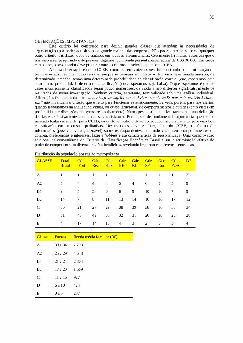
89
OBSERVAÇÕES IMPORTANTES
Este critério foi construído para definir grandes classes que atendam às necessidades de
segmentação (por poder aquisitivo) da grande maioria das empresas. Não pode, entretanto, como qualquer
outro critério, satisfazer todos os usuários em todas as circunstâncias. Certamente há muitos casos em que o
universo a ser pesquisado é de pessoas, digamos, com renda pessoal mensal acima de US$ 30.000. Em casos
como esse, o pesquisador deve procurar outros critérios de seleção que não o CCEB.
A outra observação é que o CCEB, como os seus antecessores, foi construído com a utilização de
técnicas estatísticas que, como se sabe, sempre se baseiam em coletivos. Em uma determinada amostra, de
determinado tamanho, temos uma determinada probabilidade de classificação correta, (que, esperamos, seja
alta) e uma probabilidade de erro de classificação (que, esperamos, seja baixa). O que esperamos é que os
casos incorretamente classificados sejam pouco numerosos, de modo a não distorcer significativamente os
resultados de nossa investigação. Nenhum critério, entretanto, tem validade sob uma análise individual.
Afirmações freqüentes do tipo “... conheço um sujeito que é obviamente classe D, mas pelo critério é classe
B...” não invalidam o critério que é feito para funcionar estatisticamente. Servem, porém, para nos alertar,
quando trabalhamos na análise individual, ou quase individual, de comportamentos e atitudes (entrevistas em
profundidade e discussões em grupo respectivamente). Numa pesquisa qualitativa, raramente uma definição
de classe exclusivamente econômica será satisfatória. Portanto, é de fundamental importância que todo o
mercado tenha ciência de que o CCEB, ou qualquer outro critério econômico, não é suficiente para uma boa
classificação em pesquisas qualitativas. Nesses casos deve-se obter, além do CCEB, o máximo de
informações (possível, viável, razoável) sobre os respondentes, incluindo então seus comportamentos de
compra, preferências e interesses, lazer e hobbies e até características de personalidade. Uma comprovação
adicional da conveniência do Critério de Classificação Econômica Brasil é sua discriminação efetiva do
poder de compra entre as diversas regiões brasileiras, revelando importantes diferenças entre elas.
Distribuição da população por região metropolitana
CLASSE Total
Brasil Gde
Fort Gde
Rec Gde
Salv Gde
BH Gde
RJ Gde
SP Gde
Cur Gde
POA DF
A1 1 1 1 1 1 1 1 1 1 3
A2 5 4 4 4 5 4 6 5 5 9
B1 9 5 5 6 8 9 10 10 7 9
B2 14 7 8 11 13 14 16 16 17 12
C 36 21 27 29 38 39 38 36 38 34
D 31 45 42 38 32 31 26 28 28 28
E 4 17 14 10 4 3 2 5 5 4
Classe Pontos Renda média familiar (R$)
A1 30 a 34 7.793
A2 25 a 29 4.648
B1 21 a 24 2.804
B2 17 a 20 1.669
C 11 a 16 927
D 6 a 10 424
E 0 a 5 207

90
Anexo D
Normas para publicação no periódico Orphanet Journal of Rare Diseases
Submission process
Manuscripts must be submitted by one of the authors of the manuscript, and should
not be submitted by anyone on their behalf. The submitting author takes responsibility
for the article during submission and peer review.
Please note that Orphanet Journal of Rare Diseases levies an article-processing charge
on all accepted Research; if the submitting author's institution is a BioMed Central
member the cost of the article-processing charge may be covered by the membership
(see About page for detail). Please note that the membership is only automatically
recognised on submission if the submitting author is based at the member institution.
To facilitate rapid publication and to minimize administrative costs, Orphanet Journal
of Rare Diseases prefers online submission.
Files can be submitted as a batch, or one by one. The submission process can be
interrupted at any time; when users return to the site, they can carry on where they
left off.
See below for examples of word processor and graphics file formats that can be
accepted for the main manuscript document by the online submission system.
Additional files of any type, such as movies, animations, or original data files, can also
be submitted as part of the manuscript.
During submission you will be asked to provide a cover letter. Use this to explain why
your manuscript should be published in the journal, to elaborate on any issues relating
to our editorial policies in the 'About Orphanet Journal of Rare Diseases' page, and to
declare any potential competing interests. You will be also asked to provide the
contact details (including email addresses) of potential peer reviewers for your
manuscript. These should be experts in their field, who will be able to provide an
objective assessment of the manuscript. Any suggested peer reviewers should not
have published with any of the authors of the manuscript within the past five years,
should not be current collaborators, and should not be members of the same research
institution. Suggested reviewers will be considered alongside potential reviewers
recommended by the Editor-in-Chief and/or Editorial Board members.
Assistance with the process of manuscript preparation and submission is available
from BioMed Central customer support team.
We also provide a collection of links to useful tools and resources for scientific authors
on our Useful Tools page.
File formats
The following word processor file formats are acceptable for the main manuscript
document:
. Microsoft word (DOC, DOCX)
. Rich text format (RTF)
. Portable document format (PDF)
. TeX/LaTeX (use BioMed Central's TeX template)
DeVice Independent format (DVI)
91
TeX/LaTeX users: Please use BioMed Central's TeX template and BibTeX stylefile if you
use TeX format. During the TeX submission process, please submit your TeX file as
the main manuscript file and your bib/bbl file as a dependent file. Please also convert
your TeX file into a PDF and submit this PDF as an additional file with the name
'Reference PDF'. This PDF will be used by internal staff as a reference point to check
the layout of the article as the author intended. Please also note that all figures must
be coded at the end of the TeX file and not inline.
If you have used another template for your manuscript, or if you do not wish to use
BibTeX, then please submit your manuscript as a DVI file. We do not recommend
converting to RTF.
For all TeX submissions, all relevant editable source must be submitted during the
submission process. Failing to submit these source files will cause unnecessary delays
in the publication procedures.
Publishing Datasets
Through a special arrangement with LabArchives, LLC, authors submitting manuscripts
to Orphanet Journal of Rare Diseases can obtain a complimentary subscription to
LabArchives with an allotment of 100MB of storage. LabArchives is an Electronic
Laboratory Notebook which will enable scientists to share and publish data files in situ;
you can then link your paper to these data. Data files linked to published articles are
assigned digital object identifiers (DOIs) and will remain available in perpetuity. Use of
LabArchives or similar data publishing services does not replace preexisting data
deposition requirements, such as for nucleic acid sequences, protein sequences and
atomic coordinates.
Instructions on assigning DOIs to datasets, so they can be permanently linked to
publications, can be found on the LabArchives website. Use of LabArchives’ software
has no influence on the editorial decision to accept or reject a manuscript.
Authors linking datasets to their publications should include an Availability of
supporting data section in their manuscript and cite the dataset in their reference list.
Preparing main manuscript text
General guidelines of the journal's style and language are given below.
Overview of manuscript sections for Research
Manuscripts for Research submitted to Orphanet Journal of Rare Diseases should be
divided into the following sections (in this order):
. Title page
. Abstract
. Additional non-English language abstract
. Keywords
. Background
. Methods
. Results and discussion
. Conclusions
. Availability of supporting data
. List of abbreviations used (if any)
. Competing interests
. Authors' contributions
. Authors' information
. Acknowledgements
. Endnotes
. References
. Illustrations and figures (if any)
. Tables and captions
. Preparing additional files
The Accession Numbers of any nucleic acid sequences, protein sequences or atomic
coordinates cited in the manuscript should be provided, in square brackets and include
the corresponding database name; for example, [EMBL:AB026295, EMBL:AC137000,
DDBJ:AE000812, GenBank:U49845, PDB:1BFM, Swiss-Prot:Q96KQ7, PIR:S66116].

92
The databases for which we can provide direct links are: EMBL Nucleotide Sequence
Database (EMBL), DNA Data Bank of Japan (DDBJ), GenBank at the NCBI (GenBank),
Protein Data Bank (PDB), Protein Information Resource (PIR) and the Swiss-Prot
Protein Database (Swiss-Prot).
For reporting standards please see the information in the About section.
Title page
The title page should:
provide the title of the article
list the full names, institutional addresses and email addresses for all
authors
indicate the corresponding author
Please note:
the title should include the study design, for example "A versus B in the
treatment of C: a randomized controlled trial X is a risk factor for Y: a case
control study"
abbreviations within the title should be avoided
Abstract
The Abstract of the manuscript should not exceed 350 words and must be structured
into separate sections: Background, the context and purpose of the study; Methods,
how the study was performed and statistical tests used; Results, the main findings;
Conclusions, brief summary and potential implications. Please minimize the use of
abbreviations and do not cite references in the abstract. Trial registration, if your
research reports the results of a controlled health care intervention, please list your
trial registry, along with the unique identifying number (e.g. Trial registration:
Current Controlled Trials ISRCTN73824458). Please note that there should be no
space between the letters and numbers of your trial registration number. We
recommend manuscripts that report randomized controlled trials follow the CONSORT
extension for abstracts.
Additional non-English language abstract
An additional non-English language abstract can be included within the article. The
additional abstract should be placed after the official English language abstract in the
submitted manuscript file and should not exceed 350 words. Please ensure you
indicate the language of your abstract. In addition to English, we can support German,
Spanish, French, Norwegian and Portuguese abstracts.
Keywords
Three to ten keywords representing the main content of the article.
Background
The Background section should be written in a way that is accessible to researchers
without specialist knowledge in that area and must clearly state - and, if helpful,
illustrate - the background to the research and its aims. Reports of clinical research
should, where appropriate, include a summary of a search of the literature to indicate
why this study was necessary and what it aimed to contribute to the field. The section
should end with a brief statement of what is being reported in the article.
Methods
The methods section should include the design of the study, the setting, the type of
participants or materials involved, a clear description of all interventions and
comparisons, and the type of analysis used, including a power calculation if
appropriate. Generic drug names should generally be used. When proprietary brands
are used in research, include the brand names in parentheses in the Methods section.
For studies involving human participants a statement detailing ethical approval and
consent should be included in the methods section. For further details of the journal's
editorial policies and ethical guidelines see 'About this journal'.
For further details of the journal's data-release policy, see the policy section in 'About
this journal'.
Results and discussion
The Results and discussion may be combined into a single section or presented

93
separately. Results of statistical analysis should include, where appropriate, relative
and absolute risks or risk reductions, and confidence intervals. The Results and
discussion sections may also be broken into subsections with short, informative
headings.
Conclusions
This should state clearly the main conclusions of the research and give a clear
explanation of their importance and relevance. Summary illustrations may be
included.
Availability of supporting data
Orphanet Journal of Rare Diseases requires authors to deposit the data set(s)
supporting the results reported in submitted manuscripts in a publicly-accessible data
repository. This section should only be included when supporting data are available
and must include the name of the repository and the permanent identifier or accession
number and persistent hyperlink(s) for the data set(s). The following format is
required:
"The data set(s) supporting the results of this article is(are) available in the
[repository name] repository, [unique persistent identifier and hyperlink to dataset(s)
in http:// format]."
We also recommend that the data set(s) be cited, where appropriate in the
manuscript, and included in the reference list.
A list of available scientific research data repositories can be found here. A list of all
BioMed Central journals that require or encourage this section to be included in
research articles can be found here.
List of abbreviations
If abbreviations are used in the text they should be defined in the text at first use, and
a list of abbreviations can be provided, which should precede the competing interests
and authors' contributions.
Competing interests
A competing interest exists when your interpretation of data or presentation of
information may be influenced by your personal or financial relationship with other
people or organizations. Authors must disclose any financial competing interests; they
should also reveal any non-financial competing interests that may cause them
embarrassment were they to become public after the publication of the manuscript.
Authors are required to complete a declaration of competing interests. All competing
interests that are declared will be listed at the end of published articles. Where an
author gives no competing interests, the listing will read 'The author(s) declare that
they have no competing interests'.
When completing your declaration, please consider the following questions:
Financial competing interests
In the past three years have you received reimbursements, fees,
funding, or salary from an organization that may in any way gain or lose
financially from the publication of this manuscript, either now or in the future?
Is such an organization financing this manuscript (including the article-
processing charge)? If so, please specify.
Do you hold any stocks or shares in an organization that may in any
way gain or lose financially from the publication of this manuscript, either now
or in the future? If so, please specify.
Do you hold or are you currently applying for any patents relating to the
content of the manuscript? Have you received reimbursements, fees, funding,
or salary from an organization that holds or has applied for patents relating to
the content of the manuscript? If so, please specify.
Do you have any other financial competing interests? If so, please
specify.
Non-financial competing interests
Are there any non-financial competing interests (political, personal, religious,
ideological, academic, intellectual, commercial or any other) to declare in relation to

94
this manuscript? If so, please specify.
If you are unsure as to whether you, or one your co-authors, has a competing interest
please discuss it with the editorial office.
Authors' contributions
In order to give appropriate credit to each author of a paper, the individual
contributions of authors to the manuscript should be specified in this section.
According to ICMJE guidelines, An 'author' is generally considered to be someone who
has made substantive intellectual contributions to a published study. To qualify as an
author one should 1) have made substantial contributions to conception and design, or
acquisition of data, or analysis and interpretation of data; 2) have been involved in
drafting the manuscript or revising it critically for important intellectual content; 3)
have given final approval of the version to be published; and 4) agree to be
accountable for all aspects of the work in ensuring that questions related to the
accuracy or integrity of any part of the work are appropriately investigated and
resolved. Each author should have participated sufficiently in the work to take public
responsibility for appropriate portions of the content. Acquisition of funding, collection
of data, or general supervision of the research group, alone, does not justify
authorship.
We suggest the following kind of format (please use initials to refer to each author's
contribution): AB carried out the molecular genetic studies, participated in the
sequence alignment and drafted the manuscript. JY carried out the immunoassays. MT
participated in the sequence alignment. ES participated in the design of the study and
performed the statistical analysis. FG conceived of the study, and participated in its
design and coordination and helped to draft the manuscript. All authors read and
approved the final manuscript.
All contributors who do not meet the criteria for authorship should be listed in an
acknowledgements section. Examples of those who might be acknowledged include a
person who provided purely technical help, writing assistance, or a department chair
who provided only general support.
Authors' information
You may choose to use this section to include any relevant information about the
author(s) that may aid the reader's interpretation of the article, and understand the
standpoint of the author(s). This may include details about the authors' qualifications,
current positions they hold at institutions or societies, or any other relevant
background information. Please refer to authors using their initials. Note this section
should not be used to describe any competing interests.
Acknowledgements
Please acknowledge anyone who contributed towards the article by making substantial
contributions to conception, design, acquisition of data, or analysis and interpretation
of data, or who was involved in drafting the manuscript or revising it critically for
important intellectual content, but who does not meet the criteria for authorship.
Please also include the source(s) of funding for each author, and for the manuscript
preparation. Authors must describe the role of the funding body, if any, in design, in
the collection, analysis, and interpretation of data; in the writing of the manuscript;
and in the decision to submit the manuscript for publication. Please also acknowledge
anyone who contributed materials essential for the study. If a language editor has
made significant revision of the manuscript, we recommend that you acknowledge the
editor by name, where possible.
The role of a scientific (medical) writer must be included in the acknowledgements
section, including their source(s) of funding. We suggest wording such as 'We thank
Jane Doe who provided medical writing services on behalf of XYZ Pharmaceuticals Ltd.'
Authors should obtain permission to acknowledge from all those mentioned in the
Acknowledgements section.
Endnotes
Endnotes should be designated within the text using a superscript lowercase letter and
all notes (along with their corresponding letter) should be included in the Endnotes

95
section. Please format this section in a paragraph rather than a list.
References
All references, including URLs, must be numbered consecutively, in square brackets, in
the order in which they are cited in the text, followed by any in tables or legends.
Each reference must have an individual reference number. Please avoid excessive
referencing. If automatic numbering systems are used, the reference numbers must
be finalized and the bibliography must be fully formatted before submission.
Only articles, clinical trial registration records and abstracts that have been published
or are in press, or are available through public e-print/preprint servers, may be cited;
unpublished abstracts, unpublished data and personal communications should not be
included in the reference list, but may be included in the text and referred to as
"unpublished observations" or "personal communications" giving the names of the
involved researchers. Obtaining permission to quote personal communications and
unpublished data from the cited colleagues is the responsibility of the author.
Footnotes are not allowed, but endnotes are permitted. Journal abbreviations follow
Index Medicus/MEDLINE. Citations in the reference list should include all named
authors, up to the first six before adding 'et al.'..
Any in press articles cited within the references and necessary for the reviewers'
assessment of the manuscript should be made available if requested by the editorial
office.
An Endnote style file is available.
Examples of the Orphanet Journal of Rare Diseases reference style are shown below.
Please ensure that the reference style is followed precisely; if the references are not in
the correct style they may have to be retyped and carefully proofread.
All web links and URLs, including links to the authors' own websites, should be given a
reference number and included in the reference list rather than within the text of the
manuscript. They should be provided in full, including both the title of the site and the
URL, as well as the date the site was accessed, in the following format: The Mouse
Tumor Biology Database. http://tumor.informatics.jax.org/mtbwi/index.do. Accessed
20 May 2013. If an author or group of authors can clearly be associated with a web
link, such as for weblogs, then they should be included in the reference.
Examples of the Orphanet Journal of Rare Diseases reference style
Article within a journal
Smith JJ. The world of science. Am J Sci. 1999;36:234-5.
Article within a journal (no page numbers)
Rohrmann S, Overvad K, Bueno-de-Mesquita HB, Jakobsen MU, Egeberg R, Tjønneland
A, et al. Meat consumption and mortality - results from the European Prospective
Investigation into Cancer and Nutrition. BMC Medicine. 2013;11:63.
Article within a journal by DOI
Slifka MK, Whitton JL. Clinical implications of dysregulated cytokine production. Dig J
Mol Med. 2000; doi:10.1007/s801090000086.
Article within a journal supplement
Frumin AM, Nussbaum J, Esposito M. Functional asplenia: demonstration of splenic
activity by bone marrow scan. Blood 1979;59 Suppl 1:26-32.
Book chapter, or an article within a book
Wyllie AH, Kerr JFR, Currie AR. Cell death: the significance of apoptosis. In: Bourne
GH, Danielli JF, Jeon KW, editors. International review of cytology. London: Academic;
1980. p. 251-306.
OnlineFirst chapter in a series (without a volume designation but with a DOI)
Saito Y, Hyuga H. Rate equation approaches to amplification of enantiomeric excess
and chiral symmetry breaking. Top Curr Chem. 2007. doi:10.1007/128_2006_108.
Complete book, authored
Blenkinsopp A, Paxton P. Symptoms in the pharmacy: a guide to the management of
common illness. 3rd ed. Oxford: Blackwell Science; 1998.
Online document
96
Doe J. Title of subordinate document. In: The dictionary of substances and their
effects. Royal Society of Chemistry. 1999. http://www.rsc.org/dose/title of
subordinate document. Accessed 15 Jan 1999.
Online database
Healthwise Knowledgebase. US Pharmacopeia, Rockville. 1998.
http://www.healthwise.org. Accessed 21 Sept 1998.
Supplementary material/private homepage
Doe J. Title of supplementary material. 2000. http://www.privatehomepage.com.
Accessed 22 Feb 2000.
University site
Doe, J: Title of preprint. http://www.uni-heidelberg.de/mydata.html (1999). Accessed
25 Dec 1999.
FTP site
Doe, J: Trivial HTTP, RFC2169. ftp://ftp.isi.edu/in-notes/rfc2169.txt (1999). Accessed
12 Nov 1999.
Organization site
ISSN International Centre: The ISSN register. http://www.issn.org (2006). Accessed
20 Feb 2007.
Dataset with persistent identifier
Zheng L-Y, Guo X-S, He B, Sun L-J, Peng Y, Dong S-S, et al. Genome data from sweet
and grain sorghum (Sorghum bicolor). GigaScience Database. 2011.
http://dx.doi.org/10.5524/100012.
Preparing illustrations and figures
Illustrations should be provided as separate files, not embedded in the text file. Each
figure should include a single illustration and should fit on a single page in portrait
format. If a figure consists of separate parts, it is important that a single composite
illustration file be submitted which contains all parts of the figure. There is no charge
for the use of color figures.
Please read our figure preparation guidelines for detailed instructions on maximising
the quality of your figures.
Formats
The following file formats can be accepted:
PDF (preferred format for diagrams)
DOCX/DOC (single page only)
PPTX/PPT (single slide only)
EPS
PNG (preferred format for photos or images)
TIFF
JPEG
BMP
Figure legends
The legends should be included in the main manuscript text file at the end of the
document, rather than being a part of the figure file. For each figure, the following
information should be provided: Figure number (in sequence, using Arabic numerals -
i.e. Figure 1, 2, 3 etc); short title of figure (maximum 15 words); detailed legend, up
to 300 words.
Please note that it is the responsibility of the author(s) to obtain permission
from the copyright holder to reproduce figures or tables that have previously
been published elsewhere.
Preparing tables
Each table should be numbered and cited in sequence using Arabic numerals (i.e.
Table 1, 2, 3 etc.). Tables should also have a title (above the table) that summarizes
the whole table; it should be no longer than 15 words. Detailed legends may then
follow, but they should be concise. Tables should always be cited in text in consecutive
numerical order.
Smaller tables considered to be integral to the manuscript can be pasted into the end

97
of the document text file, in A4 portrait or landscape format. These will be typeset and
displayed in the final published form of the article. Such tables should be formatted
using the 'Table object' in a word processing program to ensure that columns of data
are kept aligned when the file is sent electronically for review; this will not always be
the case if columns are generated by simply using tabs to separate text. Columns and
rows of data should be made visibly distinct by ensuring that the borders of each cell
display as black lines. Commas should not be used to indicate numerical values. Color
and shading may not be used; parts of the table can be highlighted using symbols or
bold text, the meaning of which should be explained in a table legend. Tables should
not be embedded as figures or spreadsheet files.
Larger datasets or tables too wide for a landscape page can be uploaded separately as
additional files. Additional files will not be displayed in the final, laid-out PDF of the
article, but a link will be provided to the files as supplied by the author.
Tabular data provided as additional files can be uploaded as an Excel spreadsheet (.xls
) or comma separated values (.csv). As with all files, please use the standard file
extensions.
Preparing additional files
Although Orphanet Journal of Rare Diseases does not restrict the length and quantity
of data included in an article, we encourage authors to provide datasets, tables,
movies, or other information as additional files.
Please note: All Additional files will be published along with the article. Do not
include files such as patient consent forms, certificates of language editing, or revised
versions of the main manuscript document with tracked changes. Such files should be
sent by email to [email protected], quoting the Manuscript ID number.
Results that would otherwise be indicated as "data not shown" can and should be
included as additional files. Since many weblinks and URLs rapidly become broken,
Orphanet Journal of Rare Diseases requires that supporting data are included as
additional files, or deposited in a recognized repository. Please do not link to data on a
personal/departmental website. The maximum file size for additional files is 20 MB
each, and files will be virus-scanned on submission.
Additional files can be in any format, and will be downloadable from the final published
article as supplied by the author. We recommend CSV rather than PDF for tabular
data.
Certain supported files formats are recognized and can be displayed to the user in the
browser. These include most movie formats (for users with the Quicktime plugin),
mini-websites prepared according to our guidelines, chemical structure files (MOL,
PDB), geographic data files (KML).
If additional material is provided, please list the following information in a separate
section of the manuscript text:
File name (e.g. Additional file 1)
File format including the correct file extension for example .pdf, .xls,
.txt, .pptx (including name and a URL of an appropriate viewer if format is
unusual)
Title of data
Description of data
Additional files should be named "Additional file 1" and so on and should be referenced
explicitly by file name within the body of the article, e.g. 'An additional movie file
shows this in more detail [see Additional file 1]'.
Additional file formats
Ideally, file formats for additional files should not be platform-specific, and should be
viewable using free or widely available tools. The following are examples of suitable
formats.
Additional documentation
PDF (Adode Acrobat)
Animations
SWF (Shockwave Flash)

98
Movies
MP4 (MPEG 4)
MOV (Quicktime)
Tabular data
XLS, XLSX (Excel Spreadsheet)
CSV (Comma separated values)
As with figure files, files should be given the standard file extensions.
Mini-websites
Small self-contained websites can be submitted as additional files, in such a way that
they will be browsable from within the full text HTML version of the article. In order to
do this, please follow these instructions:
Create a folder containing a starting file called index.html (or index.htm) in the root.
Put all files necessary for viewing the mini-website within the folder, or sub-folders.
Ensure that all links are relative (ie "images/picture.jpg" rather than
"/images/picture.jpg" or "http://yourdomain.net/images/picture.jpg" or
"C:\Documents and Settings\username\My Documents\mini-
website\images\picture.jpg") and no link is longer than 255 characters.
Access the index.html file and browse around the mini-website, to ensure that the
most commonly used browsers (Internet Explorer and Firefox) are able to view
all parts of the mini-website without problems, it is ideal to check this on a
different machine.
Compress the folder into a ZIP, check the file size is under 20 MB, ensure that
index.html is in the root of the ZIP, and that the file has .zip extension, then
submit as an additional file with your article.
Style and language
General
Currently, Orphanet Journal of Rare Diseases can only accept manuscripts written in
English. Spelling should be US English or British English, but not a mixture.
There is no explicit limit on the length of articles submitted, but authors are
encouraged to be concise.
Orphanet Journal of Rare Diseases will not edit submitted manuscripts for style or
language; reviewers may advise rejection of a manuscript if it is compromised by
grammatical errors. Authors are advised to write clearly and simply, and to have their
article checked by colleagues before submission. In-house copyediting will be minimal.
Non-native speakers of English may choose to make use of a copyediting service.
Help and advice on scientific writing
The abstract is one of the most important parts of a manuscript. For guidance, please
visit our page on Writing titles and abstracts for scientific articles.
Tim Albert has produced for BioMed Central a list of tips for writing a scientific
manuscript. American Scientist also provides a list of resources for science writing. For
more detailed guidance on preparing a manuscript and writing in English, please visit
the BioMed Central author academy.
Abbreviations
Abbreviations should be used as sparingly as possible. They should be defined when
first used and a list of abbreviations can be provided following the manuscript text.
Typography
Please use double line spacing.
Type the text unjustified, without hyphenating words at line breaks.
Use hard returns only to end headings and paragraphs, not to rearrange
lines.
Capitalize only the first word, and proper nouns, in the title.
Please do not format the text in multiple columns.
Greek and other special characters may be included. If you are unable
to reproduce a particular special character, please type out the name of the
symbol in full.

99
------------------- / / ------------------

100
Anexo E
Submissão do artigo ao periódico Orphanet Journal of Rare Diseases

101
PRODUÇÃO INTELECTUAL DESENVOLVIDA DURANTE O CURSO DE
MESTRADO
Artigos científicos
Deps TD, Angelo GL, Martins CC, Paiva SM, Pordeus IA, Borges-Oliveira AC.
Association between dental caries and down syndrome: a systematic review and meta-
analysis. Plos One. Fator de impacto: 3,53 / Qualis (Odontologia) A1. (no prelo)
Deps TD, Carneiro NCR, França EC, Valadares ER, Pordeus IA, Borges-Oliveira AC. A
paired comparison of dental characteristics of Brazilians with and without
Mucopolyssaccaridosis. Orphanet Journal of Rare Diseases. Fator de impacto: 3,96 / Qualis
(Medicina) A1. (submetido em abril de 2015)
Apresentação de trabalhos em eventos científicos
Apresentação oral na 31a Reunião Anual da Sociedade Brasileira de Pesquisa Odontológica
(SBPqO), Águas de Lindóia, 2014.
Deps TD, Angelo GL, Martins CC, Paiva SM, Pordeus IA, Oliveira AC. Associação entre
cárie dentária e síndrome de Down: uma revisão sistemática e meta-análise.
Apresentação de pôster no 13th International Symposium on Mucopolysaccharidodeses and
Related Diseases, Costa do Sauípe, 2014.
Deps TD, França EC, Valadares ER, Pordeus IA, Oliveira AC. Dentofacial characteristics
of children and adolescents with mucopolysaccharidosis: a pilot study.
Apresentação de pôster no XII Encontro Científico da Faculdade de Odontologia da
UFMG, Belo Horizonte, 2014.
Deps TD, Teixeira, SA, Angelo GL, Oliveira ACB. Questões relacionadas à saúde bucal
de crianças com paralisia cerebral: um estudo qualitativo.

102
Apresentação de pôster no 170 Congresso Latino-Americano de Odontopediatria, São
Paulo, 2014.
Deps TD, Siqueira MB, Gomes MC, Oliveira AC, Martins CC, Graville-Garcia AF Paiva
SM. A busca de atendimento odontológico após traumatismo dentário e seus fatores
predisponentes da dentição decídua.
Apresentação de pôster no IX Congresso Brasileiro de Epidemiologia: as fronteiras da
epidemiologia contemporânea: do conhecimento científico à ação, Vitória, 2014.
Almeida TDD, Cardoso NM, Castilho LS, Silva ME, Oliveira AC. Aspectos associados à
vulnerabilidade de pacientes em hemodiálise à doença cárie.
Apresentação de pôster na 300 Reunião Anual da SBPqO, Águas de Lindóia, 2013.
Almeida TDD, Angelo GL, Santos PM, Teixeira SA, Oliveira AC. Influência da atenção
multidisciplinar sobre a experiência odontológica das crianças com síndrome de Down.
Apresentação de pôster no 10 Congresso Internacional sobre Saúde da Pessoa com
Deficiência e Grupos Especiais, Brasília, 2013.
Deps TD, Cunha RG, Angelo GL, Santos PM, Teixeira SA, Oliveira AC. Atendimento
odontológico para crianças e adolescentes com deficiência.
Resumos de trabalhos publicados em anais de eventos científico
Deps TD, Angelo GL, Martins CC, Paiva SM, Pordeus IA, Oliveira AC. Associação entre
cárie dentária e síndrome de Down: uma revisão sistemática e meta-análise. In: 31a
Reunião Anual da SBPqO. Brazilian Oral Research. 2014; 28:56.
Deps TD, França EC, Valadares ER, Pordeus IA, Oliveira AC. Dentofacial characteristics
of children and adolescents with mucopolysaccharidosis: a pilot study. In: 13th
International Symposium on mucopolysaccharidosis and related diseases, 2014. Journal of
Inborn Errors of Metabolism and Screening 2014; 27.

103
Deps TD, Teixeira SA, Angelo GL, Oliveira AC. Questões relacionadas à saúde bucal de
crianças com paralisia cerebral: um estudo qualitativo. In: XII Encontro Científico da
Faculdade de Odontologia da UFMG / X Encontro Mineiro das Faculdade de Odontologia.
Arquivos em Odontologia. 2014; 50:25.
Almeida TDD, Cardoso NM, Castilho LS, Silva MES, Oliveira AC. Aspectos associados à
vulnerabilidade de pacientes em hemodiálise à doença cárie. Anais do IX Congresso
Brasileiro de Epidemiologia: as fronteiras da epidemiologia contemporânea: do
conhecimento científico à ação 2014; 1672.
Teixeira SA, Santos PM, Almeida TDD, Angelo GL, Oliveira AC. Significados de saúde
bucal relatados por mães de crianças/adolescentes com síndrome de Down: estudo
qualitativo exploratório. Anais do XXV Congresso Brasileiro de Genética Médica 2013.
Almeida TDD, Angelo GL, Santos PM, Teixeira SA, Oliveira AC. Influência da atenção
multidisciplinar sobre a experiência odontológica das crianças com síndrome de Down. In:
30a Reunião Anual da SBPqO. Braz Oral Res. 2013; 27:98.
Cunha RG, Angelo GL, Santos PM, Almeida TDD, Teixeira SA, Oliveira AC.
Atendimento odontológico para crianças e adolescentes com deficiência. In: 30a Reunião
Anual da SBPqO, 2013. Braz Oral Res. 2013; 27:51.
---------------------------- // ----------------------------