Theobroma cacao cystatins impair Moniliophthora perniciosa mycelial growth and are involved in
13
ORIGINAL ARTICLE Theobroma cacao cystatins impair Moniliophthora perniciosa mycelial growth and are involved in postponing cell death symptoms Carlos Priminho Pirovani • Andre ´ da Silva Santiago • Lı ´via Santana dos Santos • Fabienne Micheli • Roge ´rio Margis • Abelmon da Silva Gesteira • Fa ´tima Cerqueira Alvim • Gonc ¸alo Amarante Guimara ˜es Pereira • Ju ´lio Ce ´zar de Mattos Cascardo Received: 18 May 2010 / Accepted: 6 September 2010 / Published online: 22 September 2010 Ó Springer-Verlag 2010 Abstract Three cystatin open reading frames named TcCys1, TcCys2 and TcCys3 were identified in cDNA libraries from compatible interactions between Theobroma cacao (cacao) and Moniliophthora perniciosa. In addition, an ORF named TcCys4 was identified in the cDNA library of the incompatible interaction. The cDNAs encoded conceptual proteins with 209, 127, 124, and 205 amino acid residues, with a deduced molecular weight of 24.3, 14.1, 14.3 and 22.8 kDa, respectively. His-tagged recombinant proteins were purified from Escherichia coli expression, and showed inhibitory activities against M. perniciosa. The four recombinant cystatins exhibited K i values against papain in the range of 152–221 nM. Recombinant TcCYS3 and TcCYS4 immobilized in CNBr–Sepharose were effi- cient to capture M. perniciosa proteases from culture media. Polyclonal antibodies raised against the recombi- nant TcCYS4 detected that the endogenous protein was more abundant in young cacao tissues, when compared with mature tissues. A *85 kDa cacao multicystatin induced by M. perniciosa inoculation, MpNEP (necrosis and ethylene-inducing protein) and M. perniciosa culture supernatant infiltration were detected by anti-TcCYS4 antibodies in cacao young tissues. A direct role of the cacao cystatins in the defense against this phytopathogen was proposed, as well as its involvement in the develop- ment of symptoms of programmed cell death. Keywords Cystatin Cysteine protease inhibitor Moniliophthora Programmed cell death Theobroma Witches’ broom Abbreviations BSA Bovine serum albumin PLCPs Papain-like cysteine proteases PCD Programmed cell death NEP Necrosis and ethylene-inducing proteins ORF Open reading frame BApNA NL-alpha-benzoyl-DL-arginine-p-nitroanilide hydrochloride Introduction Diseases are a major problem for cacao (Theobroma cacao L.) production, causing annual crop losses of 30–40% (Wood and Lass 1987). Cacao is susceptible to a number of co-evolved pathogens, such as Moniliophthora (=Crinipel- lis) perniciosa, the causal agent of witches’ broom disease C. P. Pirovani A. da Silva Santiago L. S. dos Santos F. Micheli F. C. Alvim J. C. de Mattos Cascardo (&) UESC, DCB, Laborato ´rio de Proteo ˆmica, Centro de Biotecnologia e Gene ´tica, Rodovia Ilhe ´us-Itabuna, Km 16, Ilhe ´us, BA 45650-000, Brazil e-mail: [email protected] F. Micheli CIRAD, UMR DAP, Avenue Agropolis TA96/03, 34398 Montpellier Cedex 5, France R. Margis UFRGS, Centro de Biotecnologia, Laborato ´rio de Genomas e Populac ¸o ˜ es de Plantas, Porto Alegre, Rio Grande do Sul, Brazil A. da Silva Gesteira EMBRAPA/CNPMF, Cruz das Almas, Bahia, Brazil G. A. G. Pereira Departamento de Gene ´tica e Evoluc ¸a ˜o-UNICAMP, Instituto de Biologia, Universidade Estadual de Campinas, Campinas, Sa ˜o Paulo, Brazil 123 Planta (2010) 232:1485–1497 DOI 10.1007/s00425-010-1272-0
Transcript of Theobroma cacao cystatins impair Moniliophthora perniciosa mycelial growth and are involved in

ORIGINAL ARTICLE
Theobroma cacao cystatins impair Moniliophthora perniciosamycelial growth and are involved in postponing cell deathsymptoms
Carlos Priminho Pirovani • Andre da Silva Santiago • Lıvia Santana dos Santos •
Fabienne Micheli • Rogerio Margis • Abelmon da Silva Gesteira • Fatima Cerqueira Alvim •
Goncalo Amarante Guimaraes Pereira • Julio Cezar de Mattos Cascardo
Received: 18 May 2010 / Accepted: 6 September 2010 / Published online: 22 September 2010
� Springer-Verlag 2010
Abstract Three cystatin open reading frames named
TcCys1, TcCys2 and TcCys3 were identified in cDNA
libraries from compatible interactions between Theobroma
cacao (cacao) and Moniliophthora perniciosa. In addition,
an ORF named TcCys4 was identified in the cDNA library
of the incompatible interaction. The cDNAs encoded
conceptual proteins with 209, 127, 124, and 205 amino acid
residues, with a deduced molecular weight of 24.3, 14.1,
14.3 and 22.8 kDa, respectively. His-tagged recombinant
proteins were purified from Escherichia coli expression,
and showed inhibitory activities against M. perniciosa. The
four recombinant cystatins exhibited Ki values against
papain in the range of 152–221 nM. Recombinant TcCYS3
and TcCYS4 immobilized in CNBr–Sepharose were effi-
cient to capture M. perniciosa proteases from culture
media. Polyclonal antibodies raised against the recombi-
nant TcCYS4 detected that the endogenous protein was
more abundant in young cacao tissues, when compared
with mature tissues. A *85 kDa cacao multicystatin
induced by M. perniciosa inoculation, MpNEP (necrosis
and ethylene-inducing protein) and M. perniciosa culture
supernatant infiltration were detected by anti-TcCYS4
antibodies in cacao young tissues. A direct role of the
cacao cystatins in the defense against this phytopathogen
was proposed, as well as its involvement in the develop-
ment of symptoms of programmed cell death.
Keywords Cystatin � Cysteine protease inhibitor �Moniliophthora � Programmed cell death � Theobroma �Witches’ broom
Abbreviations
BSA Bovine serum albumin
PLCPs Papain-like cysteine proteases
PCD Programmed cell death
NEP Necrosis and ethylene-inducing proteins
ORF Open reading frame
BApNA NL-alpha-benzoyl-DL-arginine-p-nitroanilide
hydrochloride
Introduction
Diseases are a major problem for cacao (Theobroma cacao
L.) production, causing annual crop losses of 30–40%
(Wood and Lass 1987). Cacao is susceptible to a number of
co-evolved pathogens, such as Moniliophthora (=Crinipel-
lis) perniciosa, the causal agent of witches’ broom disease
C. P. Pirovani � A. da Silva Santiago � L. S. dos Santos �F. Micheli � F. C. Alvim � J. C. de Mattos Cascardo (&)
UESC, DCB, Laboratorio de Proteomica,
Centro de Biotecnologia e Genetica, Rodovia Ilheus-Itabuna,
Km 16, Ilheus, BA 45650-000, Brazil
e-mail: [email protected]
F. Micheli
CIRAD, UMR DAP, Avenue Agropolis TA96/03,
34398 Montpellier Cedex 5, France
R. Margis
UFRGS, Centro de Biotecnologia, Laboratorio de Genomas
e Populacoes de Plantas, Porto Alegre, Rio Grande do Sul, Brazil
A. da Silva Gesteira
EMBRAPA/CNPMF, Cruz das Almas, Bahia, Brazil
G. A. G. Pereira
Departamento de Genetica e Evolucao-UNICAMP,
Instituto de Biologia, Universidade Estadual de Campinas,
Campinas, Sao Paulo, Brazil
123
Planta (2010) 232:1485–1497
DOI 10.1007/s00425-010-1272-0
(Aime and Phillips-Mora 2005), which has spread
throughout Brazil, destroying plantations, leading to
important economical and social changes in affected areas
(Andebrhan et al. 1999). The M. perniciosa basidiospores
have the ability to infect any meristematic and young tissues
of cacao and are the only recognized infective propagules
(Evans 1980). The disease shows two distinct stages: a
biotrophic/parasitic and a necrotrophic/saprotrophic phase.
In the biotrophic phase, the fungus presents intercellular
monokaryotic mycelium, which causes hypertrophy and
hyperplasia of the tissues, loss of apical dominance, and
proliferation of axillary shoots, thus resulting in formation
of abnormal stems, called green brooms. In the second
stage, the fungus moves to the saprotrophic phase, with the
spread of intracellular dikaryotic mycelium, concomitant
with necrosis and death of infected tissues, distal to the
original infection site. This stage results in the formation of
the dry broom (Evans 1980; Ceita et al. 2007). Basidiomata
production and spore formation occur on infected necrotic
tissue (Silva et al. 2002).
In plant–pathogen interaction, proteases are thought to
be involved in a range of processes, including senescence
and defense responses (Van der Hoorn and Jones 2004), as
revealed by studies using protease inhibitors (Chichkova
et al. 2004). Proteolysis during plant–pathogen interaction
probably produces the selection of counteracting inhibitors,
non-cleavable substrates and other means to evade prote-
olysis. Therefore, the interaction of proteases with their
substrates and inhibitors can be seen as a molecular bat-
tlefield (Van der Hoorn and Jones 2004). Associations
between the induction of protease genes and defense have
also been found for genes that encode metallo-, aspartic-
and cysteine-proteases (Liu et al. 2001). Intriguingly, both
plants and their invaders use papain-like cysteine proteases
(PLCPs) or their inhibitors in these molecular confronta-
tions (Shindo and Hoorn 2008).
Phytocystatins are plant proteins that inhibit PLCPs.
Several members of this family have been characterized in
various plant species, and homology with animal cystatins
has been described (Margis et al. 1998). Most of phyto-
cystatins are small proteins, ranging in size from 12 to
16 kDa, with a distinct group having a higher molecular
weight (*23 kDa) due to a C-terminal extension (Shyu
et al. 2004; Margis-Pinheiro et al. 2008). Several multi-
cystatins with up to eight cystatin domains have been
reported, particularly in tomato (Wu and Haard 2004). In
plants, several roles have been attributed to cystatins,
ranging from regulation of various endogenous proteolytic
processes to the inhibition of exogenous cysteine proteases
secreted by predatory or pathogenic organisms during
herbivory or infection (Arai et al. 2002; Chan et al. 2010).
Empirical data suggested an active role for cystatins in
plant defense: (1) several cystatin-encoding genes are up
regulated by stress signals, such as mechanical wounding,
cold, insect predation, or metabolites involved in defense-
related pathways (Christova et al. 2006); (2) several plant
cystatins inhibit the digestive cysteine proteases of her-
bivorous insects and root parasitic nematodes (Arai et al.
2002); (3) some cystatins show detrimental effects against
plant pathogenic fungi (Soares-Costa et al. 2002; Martinez
et al. 2005); and (4) the expression of recombinant cysta-
tins in transgenic plants provides a protective effect against
several herbivorous predators and microbial pathogens
(Chan et al. 2010). Phytocystatins and cysteine proteases
were also required to regulate programmed cell death
(PCD) (Arai et al. 2002; Belenghi et al. 2003; Shindo and
Hoorn 2008).
Due to the environmental and economical importance of
the witches’ broom disease, recent studies have been
developed in order to understand the genomic program of
M. perniciosa, as well as the one of cacao during infection
by this pathogen (Gesteira et al. 2007; Ceita et al. 2007;
Garcia et al. 2007; Mondego et al. 2008). Theobroma
cacao–M. perniciosa interaction cDNA libraries contained
sequences of distinct classes of protease (cysteine, serine
and aspartic proteases) and protease inhibitors, such as
cystatins (Gesteira et al. 2007). The expression analysis by
RT-PCR of some of these protease genes indicated that
they were more expressed in cacao tissues infected by
M. perniciosa than in non-infected ones (Carvalho 2007).
Moreover, analysis of the M. perniciosa draft genome
led to the identification of three putative genes encoding
necrosis and ethylene-inducing proteins (MpNEPs). MpNEP1
and 2 have highly similar sequences and are able to induce
necrosis and ethylene emission in tobacco and cacao leaves
(Garcia et al. 2007). Recombinant MpNEP induced prote-
ase isoforms production and DNA degradation (laddering)
in tobacco suspension cells, thereby suggesting that
MpNEP may have a crucial role in PCD process (Cascardo,
unpublished results).
In this paper, we report the molecular and biochemical
characterization of four cystatins from cacao (TcCYS1,
TcCYS2, TcCYS3 and TcCYS4), identified from two
cDNA libraries, corresponding to incompatible and com-
patible interactions between T. cacao and M. perniciosa
(Gesteira et al. 2007). These ORFs were sub-cloned, and
His-Tag fused proteins were expressed in E. coli. Inhibitory
activity against M. perniciosa was demonstrated. TcCYS3-
and TcCYS4–Sepharose–CNBr immobilized recruited
distinct proteases isoforms from M. perniciosa supernatant
culture. The cysteine proteinase inhibitory activity of these
recombinant proteins against papain (EC 3.4.22.2) was
assessed using a colorimetric assay. Polyclonal antibodies
against the recombinant TcCYS4 were raised, hence
1486 Planta (2010) 232:1485–1497
123

allowing the immunodetection of the endogenous proteins
within different plant tissues.
Materials and methods
Plant material
Plant material (leaves, meristems, roots, seeds and stems)
was obtained from cacao (Theobroma cacao L.) infected or
not with Moniliophthora perniciosa, growing in the field of
the ‘Universidade Estadual de Santa Cruz’, UESC (Ilheus,
Bahia, Brazil). The development stages and symptoms of
the disease were evaluated as described by Ceita et al.
(2007) and Silva et al. (2002).
Screening for cystatin clones
The T. cacao–M. perniciosa interaction cDNA libraries
were screened using BLAST-X and tBLAST-X (Altschul
et al. 1997) in order to identify putative cystatin clones.
Clones containing ORFs were selected based on the fol-
lowing criteria: (1) presence of cystatin characteristic
motifs (a region containing a glycine residue at the
N-terminal region; a QxVxG motif; a region containing a
tryptophan residue near the C-terminal region); (2) pres-
ence of the consensus sequence LARFAVDEHN, a sig-
nature specific to phytocystatins (Margis et al. 1998).
Sequence analyses
Cystatin clones, named TcCys1, TcCys2, TcCys3 and
TcCys4, were fully sequenced using a MegaBace 1000
DNA sequencer (GE Healthcare, Chalfont, UK). Align-
ment of protein sequences using Clustal W at GenBank
(http://www.ncbi.nlm.nih.gov) in order to predict putative
signal, peptide was predicted using the Target P 1.1 Server
(http://www.cbs.dtu.dk/services/TargetP/) (Nielsen et al.
1997).
Phylogenetic tree reconstruction and gene organization
Analysis was performed using Molecular Evolutionary
Genetics Analysis (MEGA) software, version 2.0 (Kumar
et al. 2000) after a Clustal-X multialignment of the four
cacao cystatins with other 41 sequences from group II
phytocystatins. The evolutionary history was inferred using
the neighbor-joining method. All positions containing gaps
and missing data were eliminated from the dataset (com-
plete deletion option). There were a total of 76 positions of
amino acid residues in the final dataset. The percentage of
replicate trees in which the associated taxa clustered
together was estimated by bootstrap (2,000 replicates). The
tree was condensed with a cut off value of 50 grouping
sequences with low bootstraps. Cacao phytocystatin gene
organization was deduced after comparison of genomic and
cDNA sequences, associated with the deduced amino acid
sequences from their predicted coding sequences. The
alternative splicing model was inferred after comparison
and super imposition with the previous described mecha-
nisms occurring in Arabidopsis, and poplar (Margis-Pin-
heiro et al. 2008).
Expression and purification of recombinant cystatins
from Escherichia coli
The open reading frames encoding cysteine protease
inhibitor proteins were obtained by amplification using the
following primers: Cys33FNdeI: 50TTTGGGGGTTCATA
TGGAGGCGGAGG and Cys33RXhoI: 50TATACAAAG
TCTCGAGAAGCAGA for TcCYS1 and TcCYS3;
Cys46FNdeI: 50CTGCTCTGAACATATGGCCACCAC
and Cys46RXhoI: 50GGTTCAACCTCGAGCAATATAC
AGC for TcCYS2 and TcCYS4. Forward and reverse
primers contained restriction sites for NdeI and XhoI,
respectively, to clone into pET28a. Amplification products
were digested with NdeI and XhoI and inserted into
pET28a in frame with a His-Tag coding sequence
according to pET System Manual (Novagen, Darmstadt,
Germany). All clones identities and positions were con-
firmed by sequencing. E. coli Rosetta (DE3) containing the
recombinant plasmids were grown at 37�C to an OD600 of
0.7, and induced with 0.4 mM IPTG (isopropyl b-D-thio-
galactopyranoside) for 4 h, harvested and processed. The
lysate was centrifuged at 13,000g, 4�C for 15 min and
soluble and insoluble fractions were obtained. The fusion
proteins with a histidine tail were purified using a His-Trap
FF Crude column (GE Healthcare) following the manu-
facturer’s instructions. Insoluble recombinant cacao cyst-
atins were dissolved with buffer containing 6 M urea prior
to loading onto the column, and eluted with lyses buffer
containing 250 mM imidazole and 4 M urea. Protein
refolding was performed by gradual reduction of urea
concentration in dialysis buffer (10 mM Na2HPO4, pH 6.0;
5% glycerol; 1 mM dithiothreitol; 100 mM NaCl; and
0.01% Triton X-100). Bacterial extracts, soluble, insoluble
and purified proteins were analyzed using 15% SDS-
PAGE. Protein concentration was determined by the
Bradford’s method (Bradford 1976).
Growth inhibition assay of Moniliophthora perniciosa
In vitro growth inhibition assays were performed using
broken hyphae from M. perniciosa in 40 g/L PDA medium
(HiMedia, Bhaveshwar Plaza, Mumbai, India), as descri-
bed by Freitas-Filho et al. (2006). Recombinant cacao
Planta (2010) 232:1485–1497 1487
123

cystatins TcCYS1, TcCYS2, TcCYS3 and TcCYS4 (final
concentration of 5 lM) were mixed with broken hyphae in
0.3 mL volume, pre-incubated at 25�C for 30 min and
plated in PDA medium for 4 days at 25�C. Plates were
photographed and images were analyzed by Image Master
3D Platinum software (GE Healthcare). Inhibition of
pseudocolony regeneration (in %) compared to the control
was calculated based on the percentage of volume pro-
duced by the Image Master analysis. The Tukey–Kramer
multiple comparison tests were conducted based on three
experimental repetitions.
Protease trap
The recombinant cystatins were coupled to CNBr-activated
SepharoseTM 4 fast Flow (GE Healthcare) according to the
manufacturer instructions. The protease capture was per-
formed using resin incubation at 37�C for 30 min, under
agitation, with 10 volumes of 1 mg/mL of proteins from
M. perniciosa culture supernatant in the presence of cap-
ture buffer (100 mM phosphate, pH 6.0; 100 mM NaCl;
2 mM EDTA; 10 mM 2-mercaptoethanol; 0.01% Triton
X-100). After capture, the resin was washed 3 times with 5
volumes of capture buffer containing 0.5 M NaCl. The
bound proteases were eluted with 50 mM glycine pH 2.9,
for 5 min, and then equilibrated with equal volume of
10 mM Tris-base. Between each step, the resin was
recovered by centrifugation (8,000g, room temperature for
30 s). For qualitative analysis of captured proteases, 0.1%
gelatin/SDS polyacrylamide gel electrophoresis was used
(Michaud et al. 1996).
Quantitative analysis of the cysteine protease inhibitory
activity of recombinant cacao cystatins
For quantitative analysis of the cysteine protease inhibitory
activity by cystatins, the chromogenic substrate NL-alpha-
benzoyl-DL-arginine-p-nitroanilide hydrochloride (BAp-
NA; Fluka, Steinheim, Germany) was used. The papain
activity (Sigma, St. Louis, MO, USA) was assayed with
modifications, as described by Barrett (1976). The enzyme
was pre-incubated for 10 min in 50 lL of activation buffer
(0.1 M phosphate buffer, pH 6.0; containing 10 mM
2-mercaptoethanol and 2 mM EDTA) without cystatin or
with variable concentrations of the inhibitor. The reaction
was initiated by adding 200 lL of 1.2 mM solution of
BApNA prepared in activation buffer; the amount of
p-nitroanilide released at 37�C was monitored every
10 min up to 1 h using a microplate reader VERSAmax
(Tunable Molecular Devices, Silicon Valley, CA, USA) at
410 nm. The amount of enzymes used was previously
adjusted in order to obtain a final optical density between
0.5 and 0.7, in positive controls without inhibitors. The Ki
values were calculated from continuous rate assay experi-
ments as the slope of the plot of [I]/(1 - vi/vo) versus vo/vi
(Henderson 1972) and corrected for substrate (BApNA)
competition. The inhibitory activity of cacao cystatins were
recorded as an inhibition percentage (%), and the percentage
of papain inhibition (I%) by cacao cystatins was calculated
using the following equation: I% = [(T - T*)/T] 9 100%;
where T denotes the OD410 in the absence of cacao cyst-
atin, and T* in the presence of cacao cystatin.
Antibody production
Anti-TcCYS4 polyclonal antibody was raised in rabbits
against the purified recombinant His-tagged protein using
standard immunization protocols (Sambrook and Russell
1989). Briefly, TcCYS4 was mixed with an equal volume
of Freund’s complete adjuvant (GibcoBRL, Grand Island,
NY, USA) and injected subcutaneously into 3-month-old
rabbit. Injections of 500 lg of protein mixed with an equal
volume of Freund’s incomplete adjuvant were done every
20 days until 60 days. The antiserum was collected from
the animals 21 days after the last immunization. Specific
antibody was purified by affinity using the antigen (His-
tagged TcCYS4) immobilized in nitrocellulose support
according to Sambrook and Russell (1989).
Immunodetection of cystatin in cacao tissues
Approximately 200 mg of plant tissues were ground in
liquid nitrogen using mortar and pestle until reduction in
powder. Successive purification steps to obtain cacao pro-
teins under denaturing conditions were performed accord-
ing to Pirovani et al. (2008). Proteins were quantified using
the 2-D Quant Kit according to the manufacturer’s recom-
mendations (GE Healthcare). For Western blot analyses,
equal amounts (5 lg) of each protein samples were sepa-
rated by 15% SDS-PAGE and electroblotted onto hybond-C
extra NC support (Amersham Biosciences, Little Chalfont,
Buckinghamshire, UK). The protein blot was blocked with
5% casein in TBS-T buffer (20 mM Tris–HCl pH 7.6; 0.8%
NaCl; 0.1% Tween 20) and incubated with specific poly-
clonal anti-TcCYS4 antibody for 1 h at room temperature.
An alkaline phosphatase-labeled anti-rabbit antibody
(Sigma, dilution 1:5,000) was used as a secondary antibody.
The detection system was NBT/BCIP (Promega, Madison,
Wisconsin, USA).
Fungus inoculation and protein infiltration experiments
Spores of M. perniciosa were used to inoculate cacao
meristems as previously described by Ceita et al. (2007).
Recombinant His-tagged MpNEP1 (Garcia et al. 2007)
dissolved in 10 mM Tris–HCl pH 8; 100 mM NaCl buffer
1488 Planta (2010) 232:1485–1497
123

(TNB) was infiltrated into apical meristems of 30-day-old
seedlings of cacao, according to Garcia et al. (2007).
Protein (1 lM) was injected into cacao meristems using
1 ml plastic syringes. Similarly, proteins secreted by M.
perniciosa in culture medium were dialyzed against TNB
buffer and then injected into meristems. Apical infiltrated
and control meristems were harvested 3 days after treat-
ment, frozen in liquid nitrogen and stored at -80�C until
protein extraction.
Results
Phylogenetic characterization of cacao cystatins ORFs
Four distinct complete cDNA clones encoding putative
cystatins were identified in the two cDNA libraries from
the T. cacao–M. perniciosa interaction (susceptible and
resistant) sequenced by Gesteira et al. (2007). The clones
were completely sequenced and named TcCys1, TcCys2,
TcCys3 (present in susceptible interaction) and TcCys4
(present in resistant interaction). Their ORFs contained
627, 369, 372 and 615 bp, respectively, and the deduced
amino acid sequences matched the consensus criteria
(Fig. 1; boxes 1, 2, 3), described in the ‘‘Material and
methods’’. TcCys1 and TcCys3 clones contain a putative N-
terminal signal peptide, thereby suggesting that these two
clones encode secreted proteins similar to oryzacystatin-I
(Womack et al. 2000), while TcCys2 and TcCys4 clones
probably encode intracellular proteins.
Sequence alignment of the four cystatins showed
the presence of conserved cystatin motifs, as well as
phytocystatin-specific domain (Fig. 1). TcCYS1 and
TcCYS4 showed an extend carboxy terminal end, as pre-
viously described (Reis and Margis 2001; Shyu et al. 2004;
Martinez et al. 2007). TcCYS1 and TcCYS3 showed 96%
amino acid sequence identity (Fig. 1), forming a cluster in
the phylogenetic analysis with Arabidopsis thaliana
(At5g05110) and Populus trichocarpa (PtCys9) cystatins
(Fig. 2). TcCYS2 and TcCYS4 were 88% identical (Fig. 1),
and formed a group with Gossypium (Tc59933), Eucalyp-
tus grandis (19204304) and Quercus robur (DN950946)
cystatins (Fig. 2). TcCYS1 and TcCYS3 were less than
50% conserved in comparison with TcCYS2 and TcCYS4
(Fig. 1). TcCYS1 and TcCYS3 are basic proteins, with a
theoretical Ip of 9.52 and 9.75, respectively, while TcCYS2
and TcCYS4 are slightly acidic, with a theoretical Ip of
6.70 and 6.10, respectively. The theoretical molecular
weights of TcCYS1, TcCYS2, TcCYS3 and TcCYS4 were
21.5, 14.1, 11.5 and 22.8 kDa, respectively (Table 1). An
exon skipping splicing model was proposed for cacao
phytocystatins TcCYS1 and TcCYS3 in comparison to that
observed in poplar phytocystatin PtCys9 (Fig. 3). A third
exon shown in TcCYS1 could be skipped during mRNA
splicing, and then resulting in the shortened alternative
form, TcCYS3.
Heterologous expression and purification
of recombinant cacao cystatins
When analyzed by SDS-PAGE, protein extracts from IPTG
induced E. coli cultures containing the four cystatin genes
cloned in expression vectors have revealed the presence of
His-tagged proteins with their respective expected sizes
Fig. 1 Sequence alignment of
four cacao cystatins and their
respective amino acid sequence
identities. a Signal peptides are
underlined, and signal peptide
cleavage sites are shown in
gray. Box 1 shows a
phytocystatin conserved motif.
Boxes 2 and 3 show the
conserved amino acid residues
of the inhibitory site. The
legumain SNSL inhibitory site
is shown in Box 4. Gap
generated in alignment are
indicated by dashes; positions
with identical residues in the
four sequences are indicated by
asterisks. b Global amino acid
sequence identity is among the
distinct cacao cystatins
Planta (2010) 232:1485–1497 1489
123
(Fig. 4a). The four cystatins were expressed at high levels.
Analysis of soluble and insoluble fractions by SDS-PAGE
showed that most of the recombinant TcCYS1 and TcCYS2
proteins were in the insoluble form (Fig. 4b), and hence
both were purified from insoluble fractions with 6 M urea,
since it was not possible to recover using affinity chro-
matography from the soluble fraction (data not shown).
The recombinant proteins TcCYS3 and TcCYS4 were both
detected in soluble and insoluble forms (Fig. 4b), and
could be directly purified by affinity chromatography.
Soluble (TcCYS3 and TcCYS4) or dissolved urea (TcCYS1
and TcCYS2) fractions were loaded into purification col-
umns, and the His-tagged proteins were eluted in lyses
buffer containing 250 mM imidazole for TcCYS3 and
TcCYS4 (Fig. 4c; lanes 3, 4), or equal buffer containing
4 M urea for TcCYS1 and TcCYS2 (Fig. 4c; lanes 1, 2).
After purification, TcCYS1 and TcCYS2 were refolded by
progressive removing of urea in dialysis buffer. The His-
tagged recombinant proteins expressed in E. coli had an
observed molecular weight slightly superior to the theo-
retical ones (Table 1). The high amounts of pure concen-
trated recombinant proteins TcCYS3 and TcCYS4 (2 and
2.8 mg/mL, respectively) obtained after a single step of
purification, and those of soluble purified TcCYS1 and
TcCYS2 (0.45 and 0.54 mg/mL, respectively) obtained
after refolding were sufficient to perform the subsequent
activity tests.
Antifungal activity and protease interaction
Assays of inhibitory activity against M. perniciosa were in
vitro performed with 5 lM of recombinant cacao cystatin
(TcCYS1, TcCYS2, TcCYS3 and TcCYS4). Abundant
mycelia growth was observed in control culture without the
addition of recombinant cacao cystatins (Fig. 5a). When
recombinant His-tagged proteins TcCYS3 or TcCYS4 were
applied, the M. perniciosa mycelia growth was strongly
inhibited; it was also noted a strong shortening of hyphae
(data not shown), and a reduced number of mycelia units
(pseudo-colonies) were observed 4 days after treatment
(Fig. 5d, e), reaching around 94% of growth inhibition
(Fig. 5f). When recombinant His-tagged proteins TcCYS1
and TcCYS2 were used, fungus growth inhibition was
slightly inferior to the one observed for TcCYS3 and
TcCYS4 proteins (Fig. 5b, c); still, their inhibitory activi-
ties reached around 80.8 and 77.9%, respectively (Fig. 5f).
To further investigate the possible mechanism for cacao
cystatin inhibition of mycelium growth, an assay of pro-
tease capture using Sepharose–CNBr immobilized proteins
was performed (Fig. 5g). Immobilized bovine serum
albumin (BSA) sample analyzed in gel did not show any
band corresponding to protease activity (Fig. 5g, BSA).
Figure 5g (TcCYS3 and TcCYS4) showed that recombi-
nants TcCYS3 and TcCYS4 interacted with similar secre-
ted proteases from M. perniciosa culture medium. Two
bands of protease activity were observed after capture by
TcCYS3 and TcCYS4 immobilized on CNBr–Sepharose: a
major protease activity band with a smaller molecular
weight, and a slight protease activity band with a higher
molecular weight (Fig. 5g, arrows).
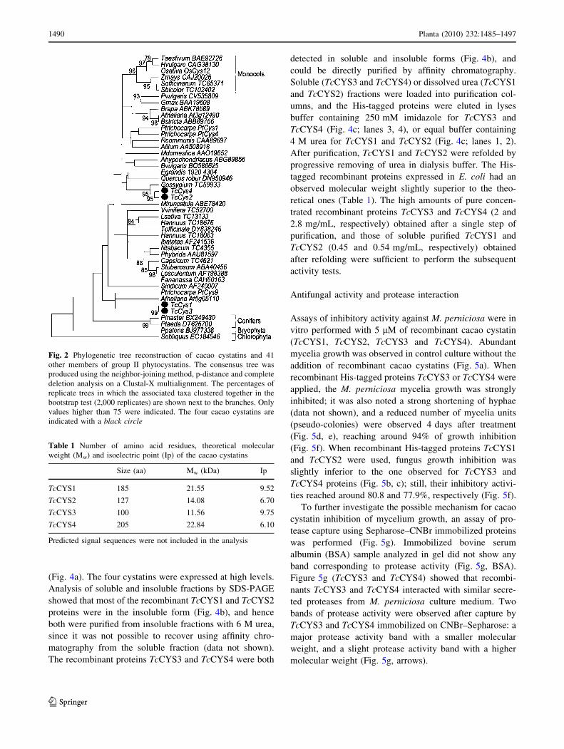
Fig. 2 Phylogenetic tree reconstruction of cacao cystatins and 41
other members of group II phytocystatins. The consensus tree was
produced using the neighbor-joining method, p-distance and complete
deletion analysis on a Clustal-X multialignment. The percentages of
replicate trees in which the associated taxa clustered together in the
bootstrap test (2,000 replicates) are shown next to the branches. Only
values higher than 75 were indicated. The four cacao cystatins are
indicated with a black circle
Table 1 Number of amino acid residues, theoretical molecular
weight (Mw) and isoelectric point (Ip) of the cacao cystatins
Size (aa) Mw (kDa) Ip
TcCYS1 185 21.55 9.52
TcCYS2 127 14.08 6.70
TcCYS3 100 11.56 9.75
TcCYS4 205 22.84 6.10
Predicted signal sequences were not included in the analysis
1490 Planta (2010) 232:1485–1497
123

Inhibitory activity of the recombinant
cacao cystatins against papain
The recombinant cacao cystatins were assayed against
papain. TcCYS1, TcCYS2, TcCYS3, TcCYS4 recombinant
proteins inhibited papain activity (Fig. 6). The estimated Ki
values of TcCYS1 and TcCYS3 for papain were 203.2 and
152.4 nM, respectively (Fig. 6a), whereas for TcCYS2 and
TcCYS4, Ki values for papain were 220.7 and 158.9
(Fig. 6b). The TcCYS1 and TcCYS2 purified under dena-
turing conditions (6 M urea) and obtained in soluble form
after refolding, showed smaller Ki values than those of
TcCYS3 and TcCYS4, which were obtained by direct
purification in native conditions.
Immunodetection of endogenous TcCYS4
Polyclonal antibodies were generated against the recom-
binant protein TcCYS4 to detect the expression of cystatins
in different healthy cacao tissues and organs, or infected by
M. perniciosa. By means of Western blot analysis, the anti-
TcCYS4 polyclonal antibody detected two bands with
higher intensity: one of about 23 kDa was observed in
protein extracts from meristem, young leaf (leaves up to
10 days), mature leaf (leaves with 30–40 days), stem,
young seed (fruits with 120–140 days after flower anthe-
sis), root and mature seed tissues (fruit with 6–7 months);
and one of 85 kDa observed in all the cacao tissues
(Fig. 7). The 23 kDa protein had an observed molecular
Fig. 3 TcCYS1 and TcCYS3 correspond to alternative spliced forms
of a single cacao group II phytocystatin gene. A splicing model,
corresponding to an exon skipping (E.S.) is proposed for cacao
phytocystatins TcCYS1 and TcCYS3 in comparison to that observed
to poplar phytocystatin PtCys9. Exon sequences are represented as
boxes linked by single or interrupted lines, corresponding to introns.
Black boxes represent the open reading frame (ORF) and white boxes
the 50 and 30 non-translated regions (NTR), respectively. Numbersplaced above exons and below introns indicate their nucleotide sizes.
Amino acids corresponding to codons flanking introns and intron
frames (0 or ?1) are indicated under boxes
Fig. 4 Analysis of the expression, solubility and purification of
recombinant cacao cystatins. a SDS-PAGE analysis of the expression
of recombinant cystatins in E. coli. Cell extracts before (lane 1) and
after (lanes 2–6) IPTG induction. b SDS-PAGE analysis of soluble
(lanes 1, 3, 5 and 7); and insoluble (lanes 2, 4, 6 and 8) fractions after
disruption of induced cells. c Purification of His-tagged T. cacao
cystatins recombinant TcCYS1 (lane 1), TcCYS2 (lane 2) from
insoluble fractions as well as, TcCYS3 (lane 3) and TcCYS4 (lane 4)
from soluble fractions by affinity chromatography in a nickel column.
M molecular weight marker. Arrows indicated the recombinant
proteins
Planta (2010) 232:1485–1497 1491
123

weight similar to that of recombinant TcCYS4 protein
(Fig. 4c, lane 4). The Western blot showed that endoge-
nous TcCYS4 or a similar protein was more expressed in
younger tissues such as young leaves, young seeds and
roots. The 23 kDa protein was present in healthy mature
leaf at a low level, and it was completely absent from
infected mature leaf. The 85 kDa protein detected in all
tissues presented a higher induction in the infected meri-
stem compared to the healthy one (Fig. 7a). A previous
control experiment showed no cross reaction between anti-
TcCYS4 and the TcCYS1 and TcCYS3 recombinant cacao
cystatins (data not shown), thus, based on other fully
sequenced plant genomes, the T. cacao should have 10–12
cystatin genes. This way, the band of 23 kDa may not have
been TcCYS4, but another similar cystatin.
Cacao meristems were submitted to the following
treatments: (1) infiltration with protein extract from
M. perniciosa culture medium; (2) infiltration with
recombinant MpNEP II; (3) infection by M. perniciosa
basidiospores. Three days after meristem treatments, the
total protein extracts were assayed by Western blot, using
TcCYS4 antibody (Fig. 7b). Quantitative results from three
independent immunoblots were shown in Fig. 7b. Inocu-
lation of cacao meristems by M. perniciosa basidiospores,
infiltration of the recombinant protein NEP, and the infil-
tration with protein extract from M. perniciosa culture
medium have induced the 85 kDa protein band (Fig. 7b).
Band intensity was about twice higher in all three treat-
ments, when compared to the untreated control (Fig. 7b, c).
Discussion
Four cDNA clones (TcCYS1, TcCYS2, TcCYS3 and
TcCYS4) encoding phytocystatins from cacao were iden-
tified in two cDNA libraries of cacao-M. perniciosa
Fig. 5 Growth inhibitory activities of the recombinant T. cacaocystatins. Growth of M. perniciosa colonies derived from broken
hyphae (saprotrophic mycelia) in presence of 10 mM phosphate
buffer (control, a), 5 lM of TcCYS1 (b), TcCYS2 (c), TcCYS3
(d) and TcCYS4 (e) recombinant proteins. f The fungus growth
inhibition was quantitatively analyzed using the Image Master 3D
Platinum software. Columns identified with different letters indicated
different means by Tukey–Kramer statistical test (P \ 0.01). g Pro-
teases from M. perniciosa supernatant culture was captured using
BSA–CNBr–Sepharose (control), recombinant TcCYS3–CNBr–
Sepharose and recombinant TcCYS4–CNBr–Sepharose. After cap-
ture, eluted sample was analyzed in semi-native gelatin/SDS-PAGE
Fig. 6 Papain inhibition by
recombinant cacao cystatins.
Purified recombinants TcCYS1,
2, 3 and 4 at different
concentrations (0–3.2 lM) were
incubated with papain (3 lM).
a Residual activity of BApNA
hydrolysis by TcCYS1 and
TcCYS3 and b by TcCYS2 and
TcCYS4. Variations in the
residual activity of papain are
shown as standard errors of the
means (n = 5)
1492 Planta (2010) 232:1485–1497
123

interaction. The open reading frames encoded predicted
polypeptides of 209, 127, 124 and 205 amino acids for
TcCYS1, TcCYS2, TcCYS3 and TcCYS4, respectively
(Fig. 1). TcCYS1 and TcCYS3 included a 26 amino acid
segment similar to a classical signal peptide, due to the
presence of charged amino acid residues very near the N-
terminal end, corresponding to signal sequence found in
oryzacystatin-I by Womack et al. (2000). This segment was
followed by a long stretch of hydrophobic amino acids,
with a small lateral chain at the C-terminal end for TcCYS1
and TcCYS3, followed by a C-terminal extended in
TcCYS1. Although the subcellular localization of cystatins
in cacao is still unknown, the presence of a putative signal
peptide suggested that TcCYS1 and TcCYS3 might be
synthesized as precursors which are exported to the apop-
last, as similarly described for the rice cystatin OC-I that
may be involved in the process of suspension-cultured rice
cells proliferation (Tian et al. 2009). The analysis of the
whole deduced cacao cystatin amino acid sequences
showed the presence of classical cystatin-like domains,
which contain the Q9V9V and the dipeptide PW motifs.
TcCYS1 and TcCYS4 differed from most common protein
or nucleotide plant cystatin sequences, which consist of a
single-domain cystatins, as found in sugarcane (Soares-
Costa et al. 2002; Gianotti et al. 2006) and sesame (Shyu
et al. 2004), or multicystatins, having 3 and 8 cystatin
domains, as reported in potato tubers (Waldron et al. 1993)
and tomato (Wu and Haard 2004). The extension sequence
found at the C-termini of TcCYS1 and TcCYS4 suggested
that these two proteins belong to group II phytocystatin,
previously identified by Margis-Pinheiro et al. (2008). A
specific role for this extended C-terminal sequence has
been determined by identifying a SNSL site known as
putative inhibitory site for legumin-like proteins (Martinez
et al. 2007). Both, TcCYS2 and TcCYS3 comprised cyst-
atins with low molecular weight (11–16 kDa). Indeed,
according to the phytocystatin classification proposed by
Margis-Pinheiro et al. (2008), TcCYS2 and TcCYS3 should
be placed in group II, as both corresponds to the shorter
spliced form of TcCYS1 (Fig. 3) and TcCYS4, respec-
tively. The internal clustering of TcCYS2 and TcCYS3
with other group II phytocystatin in the phylogenetic
reconstruction (Fig. 2) reinforces this proposition.
The purified His-tagged recombinant cystatins from
cacao was used (at 5 lM) in inhibition assays of M. per-
niciosa hyphae growth. This maximum concentration was
selected because TcCYS1 and TcCYS2 presented reduced
solubility after recombinant expression and purification
from E. coli. However, the cystatin concentrations used
were lower than those previously used in other inhibitory
assays [15 lM for canecystatin vs. Trichoderma reesei by
Soares-Costa et al. (2002); 8 lM for tarocystatin vs.
Fig. 7 a Immunodetection of
TcCYS4 in cacao protein
extracts. Approximately 10 lg
of proteins extracted from
healthy meristem, young leaf,
mature leaf, stem, young seed,
mature seed and root or infected
tissues by M. perniciosa were
analyzed by Western blot.
Molecular weights (MW) in kDa
are indicated on the left.b Immunodetection of 85 kDa
proteins of cacao.
Approximately 10 lg of
proteins extracted from cacao
meristem control (untreated),
infected by M. perniciosabasidiospores (?Mp), infiltrated
with NEP (?NEP), or infiltrated
with protein from M. perniciosaculture medium (?SNMp) were
analyzed by Western blot.
c Relative intensity of protein
band was normalized to control
band and quantified by the
Image Master 3D Platinum
software
Planta (2010) 232:1485–1497 1493
123
Alternaria brassicae by Yang and Yeh (2005)], but higher
than the probable physiological concentrations present in
cacao tissues. The four recombinant proteins strongly
inhibited M. perniciosa broken hyphae growth (Fig. 5b–e),
as similarly described for other plant cystatins used against
other phytopathogenic fungi (Soares-Costa et al. 2002;
Yang and Yeh 2005; Martinez et al. 2005; Christova et al.
2006).
It was investigated whether the growth inhibition
mechanism of TcCYS3 and TcCYS4 proteins against
M. perniciosa was due to protease interaction, by using
recombinant protein immobilized in CNBr-activated
Sepharose, followed by capturing proteases from M. per-
niciosa culture supernatant. Sample analysis in 0.1% gel-
atin/SDS-PAGE revealed two proteases bands (Fig. 5g),
suggesting that inhibition of protease activity by recombi-
nant cacao cystatin is a mechanism potentially affecting
mycelia growth. It shall predominantly derive from nutri-
tion depletion because lower protease activity in fungal cell
might cause less nutrition and/or digestion. Despite the fact
that we have tested the recombinant cystatins against
secreted fungus proteases, it is clear that cacao cystatins are
able to capture M. perniciosa proteases, thus providing
initial clues of the action mechanism. It remains unclear
whether cystatin penetrates the fungus cell wall and plas-
malemma, or not. The screening for cysteine proteases in
the genome data set from M. perniciosa (http://www.lge.
ibi.unicamp.br/vassoura) has not identified papain-like
cysteine proteases, as well as classical cysteine proteases
(Mondego et al. 2008), suggesting that the protease band
detected in the 0.1% gelatin/SDS/PAGE corresponded to a
novel cysteine protease from the fungus that interacted
with cacao cystatin.
TcCYS1, TcCYS2, TcCYS3 and TcCYS4 recombinant
proteins were tested for papain inhibition using BApNA as
the colorimetric substrate (Fig. 6). The Ki values of
TcCYS1 and TcCYS2 for papain activity inhibition were
203.2 and 220.7 nM, respectively; while the Ki values of
TcCYS2 and TcCYS4 were 220.7 and 158.9 nM, respec-
tively. This Ki values are very close to the Ki of tarocystatin
OC-I (252 nM) (Wang et al. 2008) and job’ tears cystatin
(190 nM) (Yoza et al. 2002). In addition to that, compar-
ison of inhibitory activity with other species showed that Ki
for cacaocystatins are lower than those for rice OC-II
(830 nM) (Kondo et al. 1990), wheat cystatin mTaMDC1
(580 nM) (Christova et al. 2006), soybean cystatin L1
(19 lM) (Zhao et al. 1996), but higher than those con-
cerning sesame (27 nM) (Shyu et al. 2004) and maize
CCI (23 nM) (Abe et al. 1994). The Ki values for TcCYS1
and TcCYS2 were slightly higher than the TcCYS3 and
TcCYS4 Ki values. That may be explained by structural
differences between TcCYS1 and TcCYS2, even if the
inhibitory domains were identical in the pairwise
combination TcCYS1/TcCYS3 and TcCYS2/TcCYS4. The
ability to inhibit papain enzymatic activity confirmed that
(1) the proteins purified in this study were PLCP inhibitors;
(2) they were in their active conformation and conse-
quently may be employed in further studies.
Polyclonal antibodies generated against the recombi-
nant protein TcCYS4 detected the presence of this cyst-
atin in cacao tissues. The anti-TcCYS4 polyclonal
antibodies reacted with a protein with molecular weight
of around 23 kDa in extracts from meristem, young leaf,
mature leaf, stem, young seed, root and mature seed tis-
sues (Fig. 7, lower arrow). Due to the observed size, this
protein band was likely the TcCYS4 protein with pre-
dicted mass of 22.8 kDa. When compared to the same
tissues in different developmental stages, Western blot
analysis detected that TcCYS4 or a similar protein
predominantly accumulated in young tissues. A higher
amount of 23 kDa cystatin was found in young seed than
in mature seed, which is consistent with the pattern of
cystatin accumulation in wheat (Kuroda et al. 2001), as
cystatin protein levels decreased during seed maturation.
A higher accumulation of this protein was also detected in
seedling roots (Fig. 7). Root accumulating cystatin may
play a role in the protection of root tissues against insects
and/or pathogens invading these organs (Valdes-Rodrıguez
et al. 2007).
Even at low level, 23 kDa cystatin was detected in
mature uninfected leaf, but it was completely absent in M.
perniciosa infected mature leaves. Major differences in
cystatin levels between uninfected and infected organs
have been observed during the transition from green
organ (e.g., green broom) to dry one (e.g., dry broom),
due to the establishment of a PCD program in susceptible
plants (Silva et al. 2002; Ceita et al. 2007). Our data
suggested that TcCYS4 or a similar protein could act
preventing the PCD in healthy leaves by inhibiting cys-
teine proteases engaged in this process, while in infected
leaves, in the absence of TcCYS4, cysteine proteases
may be active, participating in the cell death process.
According to this hypothesis, treatment of isolated
petal from iris flower with protease inhibitors prevented
the increase in endoprotease activity, and considerably
delayed or prevented the normal senescence symptoms
(Pak and van Doorn 2005). Cacao protease activity ana-
lyzed in gel revealed the presence of three protease iso-
forms in healthy tissues and the presence of an additional
protease isoform in infected but non-necrotic ones (Piro-
vani et al. 2008). Because proteases are well known to be
involved in PCD, in particular cysteine proteases or
caspases-like proteases (van der Hoorn and Jones 2004;
Shindo and Hoorn 2008), it has been hypothesized that
some specific protease isoforms may be involved in the
PCD process, helping the degradation of the cell content
1494 Planta (2010) 232:1485–1497
123

occurring in cacao organs, as described by Ceita et al.
(2007). Moreover, according to the acid buffer pH (4.0)
used by Pirovani et al. (2008), the detected protease
isoforms correspond to aspartic or cysteine proteases, but
not serine protease, which has a higher enzymatic activity
at alkaline pH (Barrett 1994). Yet, cystatin inhibits
hypersensitive response (HR) of viral infection in
Arabidopsis (Gholizadeh et al. 2005). The involvement of
cysteine proteases and protease inhibitor genes in the
regulation of programmed cell death in plants had been
previously demonstrated (Belenghi et al. 2003; Shindo
and Hoorn 2008). These authors suggested a crucial
counter-balancing role for endogenous protease inhibitors
in regulating protease activities.
The anti-TcCYS4 antibodies cross-reacted in all the
studied cacao tissues with an approximately 85 kDa pro-
tein, which may be correlated with multicystatin, as
observed in other species (potato, Weeda et al. 2009;
cowpea, Diop et al. 2004). This cross reaction was proba-
bly due to the detection of the multicystatin, which may
present conserved epitopes with TcCYS4. The Solanum
tuberosum multicystatin shows eight cystatin domains
similar to N-terminus domains present in 23 kDa cystatins
(Weeda et al. 2009). Because the cacao multicystatin was
induced in all tissues infected by M. perniciosa (Fig. 7a),
and also in protein extracts secreted by M. perniciosa and
in meristem infiltrated with NEP (Fig. 7b), the cacao
multicystatin may be considered as a pathogenesis related
protein. A multicystatin from cowpea leaves was shown to
be induced by drought stress (Diop et al. 2004), and an 88-
kDa multi-domain cystatin from tomato was induced by
methyl jasmonate (Wu and Haard 2004). Because MpNEP
is an ethylene-inducing protein that also induces cell death
in tobacco leaves and cacao meristems (Garcia et al. 2007),
several effects on cacao tissue might occur via MpNEP
action. MpNEP induced proteases isoforms in tobacco
suspension cells (Cascardo, unpublished results); hence,
cacao multicystatin induction by MpNEP may be a counter
response to the proteases activation, by down-regulating
their activity, therefore playing an important role in
defense.
Acknowledgments This research was supported by the ‘Financi-
adora de Estudos e Projetos’ (FINEP) and the ‘Fundacao de Amparo a
Pesquisa do Estado da Bahia’ (FAPESB) M. perniciosa proteomic
network. F C Alvim was the recipient of a PQI/CAPES graduate
fellowship, and C P Pirovani was the recipient of a FAPESB graduate
fellowship. We thank Robson Jose Costa Dias (Laboratorio de
Genomica, UESC) for technical assistance and Dra. Karina Perez
Gramacho (CEPEC, CEPLAC, Brasil) for the support in cacao
seedlings inoculations with M. perniciosa basidiospores. We are
thankful to Dr. Antonio Figueira (CENA/USP-SP, Brazil) for critical
reading of the manuscript. J.C.M. Cascardo is recipient of CNPq
research fellowship number 303987/2008-1. R. Margis is recipient of
CNPq research fellowship number 302684/2005-0.
References
Abe M, Abe K, Iwabuchi K, Domoto C, Arai (1994) Corn cystatin I
expressed in Escherichia coli: investigation of its inhibitory
profile and occurrence in corn kernels. J Biochem 116:488–492
Aime MC, Phillips-Mora W (2005) The causal agents of witches’
broom and frosty pod rot of cacao (chocolate, Theobroma cacao)
form a new lineage of Marasmiaceae. Mycologia 97:1012–1022
Altschul SF, Madden TL, Schaver AA, Zhang J, Zhang Z, Miller W,
Lipman DJ (1997) Gapped BLAST and PSI-BLAST: a new
generation of protein database search programs. Nucleic Acids
Res 25:3389–3402
Andebrhan T, Figueira A, Yamada MM, Cascardo J, Furtek DB
(1999) Molecular fingerprinting suggests two primary outbreaks
of witches’ broom disease (Crinipellis perniciosa) of Theobromacacao in Bahia, Brazil. Eur J Plant Pathol 105:167–175
Arai S, Matsumoto I, Emori Y, Abe K (2002) Plant seed cystatins and
their target enzymes of endogenous and exogenous origin.
J Agric Food Chem 50:6612–6617
Barrett AJ (1976) An improved color reagent for use in Barrett’s
assay of cathepsin B. Anal Biochem 76:374–376
Barrett AJ (1994) Classification of peptidases. Methods Enzymol
244:1–15
Belenghi B, Acconcia F, Trovato M, Perazzolli M, Bocedi A, Poticelli
F, Ascenzi P, Delledonne M (2003) AtCYS1, a cystatin from
Arabidopsis thaliana, suppresses hypersensitive cell death. Eur J
Biochem 270:2593–2604
Bradford MM (1976) A rapid and sensitive method for the
quantification of microgram quantities of protein utilizing the
principle of protein–dye binding. Anal Biochem 72:248–254
Carvalho HAS (2007) Biochemical and molecular analyses of
protease from the interaction Theobroma cacao–Moniliophthoraperniciosa. Masters dissertation, Universidade Estadual de Santa
Cruz, Brazil
Ceita GO, Macedo JNA, Santos TB, Alemanno L, Gesteira AS,
Micheli F, Mariano AC, Gramacho KP, Silva DC, Meinhardt L,
Mazzafera P, Pereira GAG, Cascardo JCM (2007) Involvement
of calcium oxalate degradation during programmed cell death in
Theobroma cacao tissues triggered by the hemibiotrophic fungus
Moniliophthora perniciosa. Plant Sci 173:106–117
Chan YL, Yang AH, Chen JT, Yeh KW, Chan MT (2010)
Heterologous expression of taro cystatin protects transgenic
tomato against Meloidogyne incognita infection by means of
interfering sex determination and suppressing gall formation.
Plant Cell Rep 29:231–238
Chichkova NV, Kim SH, Titova ES, Kalkum M, Morozov VS,
Rubtsov YP, Kalinina NO, Taliansky ME, Vartapetian AB
(2004) A plant caspase-like protease activated during the
hypersensitive response. Plant Cell 16:157–171
Christova PK, Christov NK, Imai R (2006) A cold inducible
multidomain cystatin from winter wheat inhibits growth of
the snow mold fungus, Microdochium nivale. Planta 223:1207–
1218
Diop NN, Kidric M, Repellin A, Gareil M, d’Arcy-Lameta A, Pham
Thi AT, Zuily-Fodil Y (2004) A multicystatin is induced by
drought-stress in cowpea (Vigna unguiculata (L.) Walp.) leaves.
FEBS Lett 577:545–550
Evans HC (1980) Pleomorphism in Crinipellis perniciosa, causal
agent of witches’ broom disease of cocoa. Trans Br Mycol Soc
74:515–523
Freitas-Filho D, Pungartnik C, Cascardo JMC, Brendel M (2006)
Broken hyphae of the basidiomycete Crinipellis perniciosa allow
quantitative assay of toxicity. Curr Microbiol 52:407–412
Garcia O, Macedo JA, Tiburcio R, Zaparoli G, Rincones J,
Bittencourt LM, Ceita GO, Micheli F, Gesteira A, Mariano
Planta (2010) 232:1485–1497 1495
123
AC, Schiavinato MA, Medrano FJ, Meinhardt LW, Pereira GA,
Cascardo JC (2007) Characterization of necrosis and ethylene-
inducing proteins (NEP) in the basidiomycete Moniliophthoraperniciosa, the causal agent of witches’ broom in Theobromacacao. Mycol Res 111:443–455
Gesteira AS, Micheli F, Carels N, Da Silva AC, Gramacho KP,
Schuster I, Macedo JN, Pereira GA, Cascardo JC (2007)
Comparative analysis of expressed genes from cacao meristems
infected by Moniliophthora perniciosa. Ann Bot 100:129–140
Gholizadeh A, Santha IN, Kohnehrouz BB, Lodha ML, Kapoor HC
(2005) Cystatins may confer viral resistance in plants by
inhibition of a virus-induced cell death phenomenon in which
cysteine proteinases are active: cloning and molecular charac-
terization of a cDNA encoding cysteine-proteinase inhibitor
(celostatin) from Celosia cristata (crested cocks comb). Bio-
technol Appl Biochem 42:197–204
Gianotti A, Rios WM, Soares-Costa A, Nogaroto V, Carmona AK,
Oliva ML, Andrade SS, Henrique-Silva F (2006) Recombinant
expression, purification, and functional analysis of two novel
cystatins from sugarcane (Saccharum officinarum). Protein Expr
Purif 47:483–489
Henderson PJF (1972) A linear equation that describes the steady-
state kinetics of enzymes and subcellular particles interacting
with tightly bound inhibitors. Biochem J 127:321–333
Kondo H, Abe K, Nishimura I, Watanabe H, Emori Y, Arai S (1990)
Two distinct cystatin species in rice seeds with different
specificities against cysteine proteinases. J Appl Biol Chem
265:15832–15837
Kumar S, Tamura K, Jacobsen I, Nei M (2000) MEGA2: molecular
evolutionary genetics analysis, version 2.0. Pennsylvania and
Arizona State Universities, University Park, Pennsylvania and
Tempe, Arizona
Kuroda M, Kiyosaki T, Matsumoto I, Misaka T, Arai S, Abe K (2001)
Molecular cloning, characterization and expression of wheat
cystatins. Biosci Biotechnol Biochem 65:22–28
Liu Y, Dammann C, Bhattacharyya MK (2001) The matrix metallo-
proteinase gene GmMMP2 is activated in response to pathogenic
infections in soybean. Plant Physiol 127:1788–1797
Margis R, Reis EM, Villeret V (1998) Structural and phylogenetic
relationships among plant and animal cystatins. Arch Biochem
Biophys 359:24–30
Margis-Pinheiro M, Zolet ACT, Loss G, Pasquali G, Margis R (2008)
Molecular evolution and diversification of plant cysteine
proteinase inhibitors: new insights after the poplar genome.
Mol Phylogenet Evol 49:349–355
Martinez M, Abraham Z, Gambardella M, Echaide M, Carbonero P,
Diaz I (2005) The strawberry gene Cyf1 encodes a phytocystatin
with antifungal properties. J Exp Bot 56:1821–1829
Martinez M, Diaz-Mendoza M, Carrillo L, Diaz I (2007) Carboxy
terminal extended phytocystatins are bifunctional inhibitors of
papain and legumain cysteine proteinases. FEBS Lett
581:2914–2918
Michaud D, Cantin L, Raworth DA, Vrain TC (1996) Assessing the
stability of cystatin/cysteine proteinase complexes using mildly-
denaturing gelatin-polyacrylamide gel electrophoresis. Electro-
phoresis 17:74–79
Mondego JM, Carazzolle MF, Costa GG, Formighieri EF, Parizzi LP,
Rincones J, Cotomacci C, Carraro DM, Cunha AF, Carrer H,
Vidal RO, Estrela RC, Garcıa O, Thomazella DP, de Oliveira
BV, Pires AB, Rio MC, Araujo MR, de Moraes MH, Castro LA,
Gramacho KP, Goncalves MS, Neto JP, Neto AG, Barbosa LV,
Guiltinan MJ, Bailey BA, Meinhardt LW, Cascardo JC, Pereira
GA (2008) A genome survey of Moniliophthora perniciosa gives
new insights into witches’ broom disease of cacao. BMC
Genomics 9:548. doi:10.1186/1471-2164-9-548
Nielsen H, Engelbrecht J, Brunak S, von Heijne G (1997) Identifi-
cation of prokaryotic and eukaryotic signal peptides and
prediction of their cleavage sites. Protein Eng 10:1–6
Pak C, van Doorn WG (2005) Delay of iris flower senescence by
protease inhibitors. New Phytol 165:473–480
Pirovani CP, Carvalho HA, Machado RC, Gomes DS, Alvim FC,
Pomella AW, Gramacho KP, Cascardo JC, Pereira GA, Micheli
F (2008) Protein extraction for proteome analysis from cacao
leaves and meristems, organs infected by Moniliophthoraperniciosa, the causal agent of the witches’ broom disease.
Electrophoresis 29:2391–2401
Reis EM, Margis R (2001) Sugarcane phytocystatins: identification,
classification, and expression pattern analysis. Genet Mol Biol
24:291–296
Sambrook J, Russell DW (1989) Molecular cloning: a laboratory
manual. Cold Spring Harbor Laboratory Press, New York
Shindo T, Hoorn R (2008) Papain-like cysteine proteases: key players
at molecular battlefields employed by both plants and their
invaders. Mol Plant Pathol 9:119–125
Shyu DJH, Chou WN, Yiu TJ, Lin CPC, Tzen JTC (2004) Cloning,
functional expression and characterization of cystatin in sesame
seeds. J Agric Food Chem 52:1350–1356
Silva SD, Luz EDMN, Almeida OC, Gramacho K, Bezerra JL (2002)
Redescricao da sintomatologia causada por Crinipellis pernic-iosa em cacaueiro. Agrotropica 1:1–23
Soares-Costa A, Beltramini LM, Thiemann OH, Henrique-Silva F
(2002) A sugarcane cystatin: recombinant expression, purifica-
tion, and antifungal activity. Biochem Biophys Res Commun
296:1194–1199
Tian L, Zhang L, Zhang J, Song Y, Guo Y (2009) Differential
proteomic analysis of soluble extracellular proteins reveals the
cysteine protease and cystatin involved in suspension-cultured
cell proliferation in rice. Biochim Biophys Acta 3:459–467
Valdes-Rodrıguez S, Guerrero-Rangel A, Melgoza-Villagomez C,
Chagolla-Lopez A, Delgado-Vargas F, Martınez-Gallardo N,
Sanchez-Hernandez C, Delano-Frier J (2007) Cloning of a
cDNA encoding a cystatin from grain amaranth (Amaranthushypochondriacus) showing a tissue-specific expression that is
modified by germination and abiotic stress. Plant Physiol
Biochem 45:790–798
Van der Hoorn RAL, Jones JDG (2004) The plant proteolytic
machinery and its role in defense. Curr Opin Plant Biol
7:400–407
Waldron C, Wegrich LM, Merlo PAO, Walsh TA (1993) Character-
ization of a genomic sequence coding for potato multicystatin,
an eight-domain cysteine proteinase inhibitor. Plant Mol Biol
23:801–812
Wang KM, Kumar S, Cheng YS, Venkatagiri S, Yang AH, Yeh KW
(2008) Characterization of inhibitory mechanism and antifungal
activity between group-1 and group-2 phytocystatins from taro
(Colocasia esculenta). FEBS J 275:4980–4989
Weeda SM, Mohan Kumar GN, Knowles RN (2009) Developmen-
tally linked changes in proteases and protease inhibitors suggest
a role for potato multicystatin in regulating protein content of
potato tubers. Planta 230:73–84
Womack JS, Randall J, Kemp JD (2000) Identification of a signal
peptide for oryzacystatin-I. Planta 210:844–847
Wood GAR, Lass RA (1987) Cocoa, 4th edn. London, Longman
Scientific and Technical, Tropical Agriculture Series
Wu J, Haard NF (2004) Purification and characterization of a cystatin
from the leaves of methyl jasmonate-treated tomato plants.
Comp Biochem Physiol 127:209–220
Yang AH, Yeh KW (2005) Molecular cloning, recombinant gene
expression, and antifungal activity of cystatin from taro (Col-ocasia esculenta cv. Kaosiung no. 1). Planta 221:493–501
1496 Planta (2010) 232:1485–1497
123
Yoza K, Nakamura S, Yaguchi M, Haraguchi K, Ohtsubo K (2002)
Molecular cloning and functional expression of cDNA encoding
a cysteine proteinase inhibitor, cystatin, from Job’s tears (Coixlacryma-jobi L. var Ma-yuen Stapf). Biosci Biotechnol Biochem
66:2287–2291
Zhao Y, Botella MA, Subramanian L, Niu X, Nielsen SS, Bressan
RA, Hasegawa PM (1996) Two wound inducible soybean
cysteine proteinases have greater insect digestive proteinase
inhibitory activities than a constitutive homolog. Plant Physiol
111:1299–1306
Planta (2010) 232:1485–1497 1497
123


![LAESCOBA DE BRUJA DEL CACAO [Crinipellis perniciosa ... · PDF fileLAESCOBA DE BRUJA DEL CACAO [Crinipellis perniciosa (Stahel Singer]: ... nifestaciones de la enfermedad en yemas](https://static.fdocumentos.com/doc/165x107/5a7a5d377f8b9a97398d832c/laescoba-de-bruja-del-cacao-crinipellis-perniciosa-de-bruja-del-cacao-crinipellis.jpg)

![Theobroma grandiflorum(Willd. Ex Spreng.) Shum.]](https://static.fdocumentos.com/doc/165x107/61929208a34c6b42ce5de00a/theobroma-grandiflorumwilld-ex-spreng-shum.jpg)