UNIVERSIDADE FEDERAL DE MINAS GERAIS …livros01.livrosgratis.com.br/cp112949.pdf · Para avaliar o...
135
1 UNIVERSIDADE FEDERAL DE MINAS GERAIS DEPARTAMENTO DE FISIOLOGIA E BIOFÍSICA INSTITUTO DE CIÊNCIAS BIOLÓGICAS CURSO DE PÓS-GRADUAÇÃO EM CIÊNCIAS BIOLÓGICAS FISIOLOGIA E FARMACOLOGIA BALANÇO TÉRMICO E METABÓLICO DURANTE O EXERCÍCIO FÍSICO: PARTICIPAÇÃO DOS RECEPTORES AT1 PARA ANGIOTENSINA II DOUTORANDA: Laura Hora Rios Leite ORIENTADOR: Prof. Dr. Cândido Celso Coimbra CO-ORIENTADOR: Prof. Dr. Kaushik P. Patel Belo Horizonte, Outubro de 2009
Transcript of UNIVERSIDADE FEDERAL DE MINAS GERAIS …livros01.livrosgratis.com.br/cp112949.pdf · Para avaliar o...

1
UNIVERSIDADE FEDERAL DE MINAS GERAIS
DEPARTAMENTO DE FISIOLOGIA E BIOFÍSICA
INSTITUTO DE CIÊNCIAS BIOLÓGICAS
CURSO DE PÓS-GRADUAÇÃO EM CIÊNCIAS BIOLÓGICAS
FISIOLOGIA E FARMACOLOGIA
BALANÇO TÉRMICO E METABÓLICO DURANTE O EXERCÍCIO
FÍSICO: PARTICIPAÇÃO DOS RECEPTORES AT1 PARA
ANGIOTENSINA II
DOUTORANDA: Laura Hora Rios Leite
ORIENTADOR: Prof. Dr. Cândido Celso Coimbra
CO-ORIENTADOR: Prof. Dr. Kaushik P. Patel
Belo Horizonte, Outubro de 2009

Livros Grátis
http://www.livrosgratis.com.br
Milhares de livros grátis para download.

2
Laura Hora Rios Leite
BALANÇO TÉRMICO E METABÓLICO DURANTE O EXERCÍCIO
FÍSICO: PARTICIPAÇÃO DOS RECEPTORES AT1 PARA
ANGIOTENSINA II
Tese apresentada ao Curso de Pós-
Graduação em Fisiologia e Farmacologia do
Instituto de Ciências Biológicas da
Universidade Federal de Minas Gerais, como
requisito parcial para a obtenção do título de
Doutor em Ciências Biológicas com área de
concentração em Fisiologia.
ORIENTADOR: Prof. Dr. Cândido Celso Coimbra
CO-ORIENTADOR: Prof. Dr. Kaushik P. Patel
Belo Horizonte, Outubro de 2009

3
O presente trabalho foi realizado no Laboratório de Endocrinologia e
Metabolismo do Departamento de Fisiologia e Biofísica do Instituto de Ciências
Biológicas da Universidade Federal de Minas Gerais, na vigência dos auxílios
concedidos pelo Conselho Nacional de Desenvolvimento Científico e
Tecnológico (CNPq), Coordenadoria de Apoio ao Pessoal de Nível Superior
(CAPES), Fundação de Amparo à Pesquisa do Estado de Minas Gerais
(FAPEMIG) e Pró-Reitoria de Pesquisa da UFMG (PRPq).

4
Aos meus pais, Cesar e Virginia
Aos meus irmãos, Daniel e Livia, e à Ivana e ao André
Ao meu querido Henrique

5
AGRADECIMENTOS
Ao Prof. Cândido pela orientação inteligente e primorosa, imprescindível em
minha formação científica.
Ao Prof. Kaushik Patel pela oportunidade de crescimento e confiança em meu
potencial ao me receber em seu laboratório.
À Profa. Umeko pela atenção, carinho e contribuição na concretização desse
estudo.
À Profa. Adelina pelo apoio sempre disponível.
À Profa. Danusa pela energia positiva e auxílio no desenvolvimento dos
experimentos.
Ao André, Janine e Patrícia por compartilharem o conhecimento técnico
fundamental para a realização deste trabalho.
A todos os amigos queridos do Laboratório de Endocrinologia e Metabolismo
pela convivência engrandecedora e divertida, em especial à Juliana, Samuel e
Daniel.

6
Aos amigos do Laboratório que já se foram mas que continuam muito presentes
em minha vida: Alex, Renato, Ana Cristina e Cláudio.
Aos saudosos amigos Hong, Lirong e Xuefei, não só pela ajuda técnica, mas
principalmente pela amabilidade.
À Cindy Norton pela eficiência e zelo.
À secretária do colegiado de pós-graduação Celinha.
As agências financiadoras, particularmente ao CNPq pela concessão da bolsa
de doutorado sanduíche.

7
ÍNDICE
I. RESUMO......................................................................................................................................9
II. ABSTRACT...............................................................................................................................12
III. INTRODUÇÃO..........................................................................................................................15
FADIGA E EXERCÍCIO FÍSICO....................................................................................................15
Hipertermia....................................................................................................................................16
Conteúdo de neurotransmissores centrais....................................................................................21
Disponibilidade de substratos energéticos....................................................................................25
O SISTEMA RENINA-ANGIOTENSINA........................................................................................27
Envolvimento da Ang II no equilíbrio térmico.................................................................................30
Envolvimento da ANG II nos ajustes metabólicos.........................................................................31
Interação entre o sistema renina-angiotensina e os sistemas serotonérgico e dopaminérgico....33
INTERAÇÃO ENTRE OS CENTROS REGULADORES DA TEMPERATURA CORPORAL E DO
METABOLISMO INTERMEDIÁRIO...............................................................................................34
HIPÓTESE.....................................................................................................................................38
IV. OBJETIVO...............................................................................................................................40
OBJETIVO GERAL........................................................................................................................40
Objetivos específicos.....................................................................................................................40
V. MÉTODOS................................................................................................................................42
ANIMAIS........................................................................................................................................42
PROCEDIMENTO CIRÚRGICO....................................................................................................42
EXERCÍCIO FÍSICO......................................................................................................................43
PROCEDIMENTO EXPERIMENTAL GERAL................................................................................44
Experimento 1: efeito do bloqueio angiotensinérgico central sobre o consumo de oxigênio
durante o exercício contínuo até a fadiga......................................................................................44
Experimento 2: efeito do bloqueio angiotensinérgico central sobre o consumo de oxigênio

8
durante o exercício progressivo até a fadiga.................................................................................45
Experimento 3: efeito do bloqueio angiotensinérgico central sobre a concentração plasmática de
substratos energéticos durante o exercício progressivo até a fadiga............................................45
Experimento 4: efeito do bloqueio angiotensinérgico central sobre a concentração de
monoaminas centrais no momento da fadiga após o exercício contínuo......................................46
Experimento 5: efeito do bloqueio e inibição óxido nitrérgica do PVN sobre parâmetros
cardiovasculares e térmicos durante a exposição ao calor. .........................................................47
ANÁLISE ESTASTÍSTICA.............................................................................................................48
VI. RESULTADOS ALCANÇADOS..............................................................................................50
Bloqueio dos receptores centrais AT1 eleva o custo metabólico durante o exercício, reduzindo a
eficiência mecânica e o desempenho físico em ratos...................................................................50
Receptores centrais AT1 estão envolvidos com ajustes metabólicos em resposta ao exercício
progressivo em ratos.....................................................................................................................58
Fadiga central induzida por Losartan envolve o conteúdo cerebral de serotonina e dopamina....65
Envolvimento do núcleo paraventricular nos ajustes cardiovasculares induzidos pelo estresse
térmico...........................................................................................................................................85
VII. CONSIDERAÇÕES GERAIS................................................................................................106
VIII. CONCLUSÕES....................................................................................................................111
VIV. REFERÊNCIAS BIBLIOGRÁFICAS...................................................................................113

9
I. RESUMO
A angiotensina II pode interferir no desempenho físico ao induzir ajustes
termorregulatórios e metabólicos caracterizados por hipotermia, facilitação da
dissipação cutânea de calor, diminuição da taxa metabólica e hiperglicemia.
Para avaliar o papel do sistema angiotensinérgico central sobre o equilíbrio
térmico, mobilização de substratos energéticos e fadiga central durante o
exercício aeróbio em ratos, salina (Sal) ou losartan (Los) foram injetados no
ventrículo lateral direito antes do exercício contínuo (velocidade de 18 m/min, 5
% de inclinação; ~66% VO2) ou progressivo (velocidade inicial de 10 m/min com
aumento de 1 m/mim a cada 3 minutos, 10% de inclinação) até a fadiga em
esteira. Foram analisados os seguintes parâmetros: (1) consumo de oxigênio
durante o exercício contínuo e progressivo; (2) dosagem plasmática de glicose,
lactato e ácidos graxos livres durante o exercício progressivo; e (3) temperatura
corporal interna e dosagem de serotonina (5-HT), ácido 5-hidroxindoleacético (5-
HIAA), dopamina (DA) e ácido 3,4 diidroxifenilacético (DOPAC) na área pré-
óptica, hipotálamo, hipocampo e córtex frontal no momento da fadiga após
exercício contínuo. Com base nas medidas foram calculados: trabalho realizado,
eficiência mecânica e taxa de aquecimento corporal. Independente do tipo de
exercício aeróbio, o tratamento com Los reduziu o trabalho realizado pelos
animais (p<0,02). Esses animais apresentaram maior consumo de oxigênio para
ambos os protocolos de exercício (p<0,05), além de menor eficiência mecânica
(p<0,05), a qual se correlacionou inversamente com o trabalho realizado

10
(p<0,01). Os animais tratados com Los também apresentaram hiperglicemia,
elevação de lactato e ácidos graxos livres até a fadiga, já verificada em baixa
intensidade de exercício como 20% do trabalho máximo realizado (p<0,05). A
elevada taxa de aquecimento corporal verificada nos animais injetados com Los
correlacionou-se diretamente com o aumento da concentração de 5-HT na área
pré-óptica (p<0,01) e hipotálamo (p<0,01). A concentração de 5-HT nessas
áreas mostrou-se inversamente relacionada com o reduzido tempo de exercício
dos ratos Los. Apesar dos níveis de DA não apresentarem alteração em
nenhuma das áreas estudadas, o hipotálamo dos animais Los mostrou elevada
razão 5-HT:DA (p<0,01) que correlacionou-se diretamente com a taxa de
aquecimento corporal (p<0,01) e inversamente com o tempo de exercício
(p<0,05). Os dados demonstram que o sistema angiotensinérgico central está
envolvido com modulação da produção de calor e melhora da eficiência
mecânica durante o exercício físico, exercendo importante efeito sobre o
conteúdo central de 5-HT, cuja interação com a DA parece afetar a fadiga central
através da modulação da temperatura corporal. O sistema angiotensinérgico
central também está envolvido em ajustes metabólicos durante o exercício,
alterando a mobilização de substratos energéticos semelhante a situações de
ativação simpática intensa e prematura.
Com o intuito de avaliar o envolvimento do núcleo paraventricular do
hipotálamo (PVN) nos ajustes cardiovasculares induzidos pelo estresse térmico,
os quais são fundamentais para a redistribuição sanguínea para a periferia e
dissipação de calor, foram registradas a atividade simpática do nervo renal

11
(RSNA), pressão arterial média (MAP), frequência cardíaca (HR), temperaturas
corporal interna e da cauda em ratos anestesiados durante a exposição ao calor.
Antes do aquecimento, CSF, lidocaína ou L-NMMA foram injetados
bilateralmente no PVN. O estímulo térmico resultou em bloqueio do aumento da
RSNA e da MAP e atenuação da HR após bloqueio do PVN com lidocaína,
sugerindo redução da vasoconstrição renal (p<0,05). No entanto, o limiar térmico
para vasodilatação da cauda não foi afetado pelo tratamento com lidocaína. A
RSNA, HR e MAP dos animais injetados com L-NMMA aumentaram
proporcionalmente ao estresse térmico. Porém, o limiar térmico de vasodilatação
da cauda foi maior nos animais L-NMMA (p<0,05), indicando prejuízo na
dissipação cutânea de calor. Os dados sugerem que, durante o estresse térmico,
o PVN tem ação importante na regulação da atividade simpática que contribui
para os ajustes cardiovasculares responsáveis pela redistribuição de sangue
interno para a periferia e perda de calor. Além disso, um possível mecanismo
através do qual o aquecimento eleva a dissipação cutânea de calor ocorre via
disponibilidade de óxido nítrico no PVN.

12
II. ABSTRACT
Angiotensin II may interfere on physical performance by inducing
thermoregulatory and metabolic adjustments characterized by hypothermia,
improvement of coetaneous heat loss, decrease of metabolic rate and
hyperglycemia. To asses the effect of central angiotensinergic system on heat
balance, energetic substrates mobilization and central fatigue during aerobic
exercise in rats, saline (Sal) or losartan (Los) were intracerebroventricularly
injected before continuous (18 m.min-1, 5% inclination; ~66% VO2) or graded
(starting at 10 m/min, increments of 1 m/min every 3 minutes, 10% inclination)
running until fatigue. The following parameters were analyzed: (1) oxygen
consumption during continuous and graded exercise (2) measurement of
glucose, lactate and free fatty acids concentrations during graded exercise; and
(3) body temperature and measurement of serotonin (5-HT), 5-
hydroxyindoleacetic acid (5-HIAA), dopamine (DA) and 3,4-
Dihydroxyphenylacetic acid (DOPAC) in preoptic area, hypothalamus,
hippocampus and frontal cortex at the moment of fatigue after continuous
exercise. Based on the results workload, mechanical efficiency and body heating
rate were calculated. Regardless of exercise type, workload of Los-treated rats
was lower than control rats (p<0.02). These animals had higher oxygen
consumption, both during continuous and graded exercise (p<0.05), as well as
lower mechanical efficiency (p<0.05) that correlated inversely with workload
(p<0.01). Los-animals also showed a faster hyperglycemic response, higher

13
levels of lactate and free fatty acids until fatigue, already seen at low exercise
intensity as 20% of maximal work (p<0.05). Los-rats exhibited increased body
heating rate that was related to the higher 5-HT concentration in preoptic area
(p<0.01) and hypothalamus (p<0.01). The content of 5-HT in these areas was
inversely related with the reduced time to fatigue of Los-rats (p<0.05). Although
the levels of DA were not altered in any of the studied brain areas, hypothalamus
of Los-treated animal showed higher 5-HT:DA ratio (p<0.01) that correlated
directly with body heating rate (p<0.01) and indirectly with time to fatigue
(p<0.05). The data demonstrate that central angiotensinergic transmission is
involved with modulation of heat production and improvement of mechanical
efficiency during exercise, performing important effects on brain 5-HT content,
whose interaction with DA seems to affect central fatigue through modulation of
body temperature. The central angiotensinergic system is also related with
metabolic adjustments during exercise, shifting energy balance similarly to
situations of enhanced and premature sympathetic activation.
With the purpose of evaluating the involvement of the paraventricular
nucleus of the hypothalamus (PVN) on cardiovascular adjustments induced by
heat stress, which are critical for the redistribution of blood flow to the periphery
and heat loss, renal sympathetic nerve activity (RSNA), mean arterial pressure
(MAP), heart rate (HR), body temperature and tail temperature were measured in
anesthetized rats during heat stress. Before heating, CSF, lidocaine or L-NMMA
were bilaterally injected into the PVN. This heat stimulus resulted in blunted
RSNA and MAP and attenuation of HR increase after blockade of the PVN with
14
lidocaine, suggesting a decreased renal vasoconstriction (p<0.05). However,
body temperature threshold for tail vasodilation was not affected by lidocaine
treatment. RSNA, HR and MAP in L-NMMA injected rats increased according to
the heating stress. Still, a higher ∆body temperature until tail vasodilation was
shown by L-NMMA injected, which is an indicative of impaired coetaneous heat
loss (p<0.05). The data suggest that the PVN is critical for enhancing
sympathetic activity to heating, contributing to the cardiovascular adjustments
elicited by heat stress that influence core blood redistribution to the periphery and
heat loss. Furthermore, one possible mechanism by which heating increases
heat loss through coetaneous vasodilation is via nitric oxide within the PVN.

15
III. INTRODUÇÃO
FADIGA E EXERCÍCIO FÍSICO
A fadiga induzida pelo exercício físico é um fenômeno multifatorial que
envolve interação complexa entre fatores fisiológicos e psicológicos. Ela pode
ser definida como inabilidade em manter força ou potência requerida, ou ainda
como dificuldade em manter a taxa de trabalho (Fernstrom & Fernstrom, 2006;
Foley & Fleshner, 2008; Meeusen et al., 2007; Nielsen & Nybo, 2003). É
importante destacar que a fadiga é considerada mecanismo de defesa por
prevenir ameaças à homeostase através de redução forçada da intensidade da
atividade física ou a cessação da mesma (Gandevia, 2001; Kay & Marino, 2000;
Noakes, 1998). Os fatores que desencadeiam a fadiga têm origem periférica
e/ou central, sendo que a última é consequência de falha do sistema nervoso
central em proporcionar motivação adequada para manutenção do exercício
(Gandevia, 2001; Kay & Marino, 2000; Noakes, 1998). Os mecanismos
fisiológicos propostos como precipitadores da fadiga incluem perturbações
metabólicas, cardiovasculares e do sistema nervoso central, muitas vezes
associados à temperatura corporal interna elevada (Fernstrom & Fernstrom,
2006; Foley & Fleshner, 2008; Meeusen et al., 2007; Nielsen & Nybo, 2003).

16
Hipertermia
O desbalanço térmico é descrito como fator importante para o
estabelecimento da fadiga (Nybo, 2008). O aumento da temperatura corporal
interna em função do exercício aeróbio é consequência do descompasso entre a
inerente elevação do calor metabolicamente produzido e a dissipação do mesmo
(Gleeson, 1998; Webb, 1995). Essas alterações ocorrem em decorrência do
metabolismo corporal aumentado dos músculos em atividade (Galbo, 1985;
Romijn et al., 1993). O cérebro é especialmente vulnerável a hipertermia,
indutora de apoptose neuronal, e a fadiga central constitui mecanismo protetor
da integridade do sistema nervoso central (Nielsen & Nybo, 2003). O rápido
aumento da temperatura corporal interna e da taxa de acúmulo de calor são
fatores considerados limitantes do exercício físico prolongado (González-Alonso
et al., 1999; Rodrigues et al., 2003), uma vez que reduzem o impulso do sistema
nervoso central para o desempenho físico. Sendo assim, o cérebro e a
homeostasia são protegidos pela instalação da fadiga (Nielsen & Nybo, 2003).
A atividade física promove o aumento das taxas de produção e dissipação
de calor proporcional à intensidade e duração do exercício (Harri et al., 1982). O
consumo de oxigênio durante o exercício aeróbio (i.e., taxa metabólica) é um
parâmetro importante que reflete tanto a produção de calor quanto a eficiência
mecânica e o desempenho físico (Brooks & White, 1978; Sonne & Galbo, 1980).
A eficiência energética do corpo varia durante o exercício físico, sendo
aproximadamente 20-27% da energia consumida utilizada para trabalho externo,

17
enquanto o ATP restante é utilizado para homeostase ou dissipado sob a forma
de calor (Brooks et al., 1984). Deve-se destacar que a produção de calor é
considerada a variável primária desencadeadora da resposta de dissipação de
calor durante a atividade física (Dawson & Keber, 1979; O`Leary et al., 1985,
Webb, 1995).
Durante os primeiros minutos do exercício aeróbio, denominada fase
dinâmica de balanço térmico, o aumento exagerado da produção de calor, não
compensado por sua dissipação, acarreta elevação abrupta da temperatura
corporal interna concomitante com o exercício físico (Briese, 1998). A
vasoconstrição cutânea mediada pelo sistema nervoso simpático (Hartley et al.,
1972; McAllister et al., 1995) dificulta a perda de calor durante este estágio do
exercício. A fase estável de balanço térmico do exercício aeróbio inicia-se a
partir do momento no qual o tônus simpático periférico cutâneo é superado, isto
é, quando o limiar térmico para a vasodilatação cutânea é atingido.
Consequentemente, a perda de calor por dissipação é facilitada pela indução da
vasodilatação da pele, aproximando-se da taxa de produção de calor e
provocando aumento menos acentuado da temperatura corporal interna até a
interrupção da atividade física.
A variação do consumo de oxigênio durante a atividade física aeróbia
acompanha o mesmo padrão da temperatura corporal interna, isto é, eleva-se
consideravelmente na fase dinâmica do exercício até alcançar um platô
(Barstow, 1994, Lacerda et al., 2006 a). A partir daí, permanece relativamente
inalterada durante a fase estável do exercício devido ao equilíbrio entre a

18
energia necessária para contração muscular e a produção de ATP pelo
metabolismo aeróbico (Barstow, 1994). Entretanto, o consumo de oxigênio na
fase estável do exercício apresenta um segundo aumento, menos abrupto,
sugerindo uma possível redução na eficiência mecânica nesta fase do esforço
(Schrauwen & Hesselink, 2003).
O modelo de balanço térmico durante o exercício aeróbio proposto para
humanos se ajusta também para ratos. A figura 1 ilustra dados obtidos durante
atividade física aeróbia contínua em esteira em ratos (Lacerda et al., 2005, 2006
a). A interação entre as variáveis de ajuste da temperatura corporal
representados pelo consumo de oxigênio (produção de calor) e temperatura da
cauda (dissipação de calor), cuja dinâmica afeta a variação da temperatura
corporal interna de acordo com o padrão citado anteriormente, pode ser
facilmente constatada. A rápida elevação da taxa metabólica (variável primária),
observada na fase dinâmica do exercício, é acompanhada por breve queda da
temperatura da cauda, indicativa de vasoconstrição cutânea (variável
secundária). Essa relação culmina em aumento exacerbado da temperatura
corporal interna nessa fase devido ao desequilíbrio entre produção e dissipação
de calor. Quando o limiar térmico para vasodilatação cutânea é atingido, a troca
de calor ocorre em intensidade suficiente para dissipar grande parte do calor
produzido. Dessa forma, a temperatura corporal eleva-se lentamente e
gradualmente durante a fase estável do exercício até atingir o valor crítico que
promove a interrupção da atividade física.

19
FIGURA 1. Gráfico ilustrativo da dinâmica entre a produção e a dissipação de calor durante as
diferentes fases do exercício físico. Adaptado de Lacerda et al., 2005, 2006 a.
Há uma relação inversamente proporcional entre a temperatura corporal
interna antes do início da atividade física e o tempo total de exercício, assim
como forte correlação negativa entre a taxa de aquecimento e o tempo para a
fadiga (González-Alonso et al., 1999; Lacerda et al., 2005; Leite et al., 2006;
Walters et al., 2000). Além disso, embora alguns autores proponham a
existência de um valor absoluto de temperatura corporal interna crítico que
levaria a interrupção da atividade física (Fuller et al., 1998; Walters et al., 2000),
a taxa de acúmulo de calor parece ser importante predisponente da antecipação

20
da fadiga, apresentando relação inversa com o desempenho físico (González-
Alonso et al., 1999; Rodrigues et al., 2003). Esses achados indicam que tanto o
valor da taxa de aquecimento corporal quanto a quantidade de calor acumulado
parecem ser parte dos fatores limitantes da continuidade da atividade física,
levando à fadiga (González-Alonso et al., 1999; Lacerda et al., 2005; Leite et al.,
2006; Rodrigues et al., 2003).
Estudos do laboratório têm contribuído para estabelecer essa hipótese ao
demonstrar a influência de diversos sistemas centrais sobre o equilíbrio térmico
e o desempenho físico (Balthazar et al., 2009; Lacerda et al., 2005, 2006 a; Leite
et al., 2006; Pires et al., 2007; Prímola-Gomes et al., 2007; Rodrigues et al.,
2004). Dentre esses trabalhos, alguns indicaram que o bloqueio central dos
sistemas óxido nitrérgico e angiotensinérgico induz redução do desempenho
físico em função do aumento do limiar térmico para a vasodilatação cutânea, o
qual eleva a taxa de aquecimento corporal e rapidamente produz hipertermia
durante o exercício físico (Lacerda et al., 2005; Leite et al., 2006). A elevada
taxa metabólica e menor eficiência mecânica, verificadas nos animais tratados
com L-NAME (metil Nω-nitro-L-arginina; bloqueador da óxido nítrico sintase),
também elevam a taxa de aquecimento e contribuem para a fadiga precoce
(Lacerda et al., 2006 a). Por outro lado, essa relação inversa entre taxa de
aquecimento e desempenho físico deixa de existir durante a ativação colinérgica
e dopaminérgica central (Balthazar et al., 2009; Pires et al., 2007; Prímola-
Gomes et al., 2007; Rodrigues et al., 2004). O sistema colinérgico também
exerce efeito termorregulatório durante o exercício por facilitar a perda de calor

21
através da vasodilatação cutânea, atenuando o aumento da temperatura
corporal interna, porém, sem afetar o desempenho físico, prevalecendo o
esforço cardiovascular como principal fator indutor da fadiga (Pires et al., 2007;
Prímola-Gomes et al., 2007; Rodrigues et al., 2004). Já o sistema dopaminérgico
central apresenta efeito ergogênico, apesar de induzir elevação da taxa
metabólica, acompanhada por hipertermia e aumento do acúmulo de calor
(Balthazar et al., 2009). Esses achados sugerem que a dopamina central
melhora a tolerância ao calor através da atenuação da percepção de esforço,
aumentando a capacidade física (Balthazar et al., 2009).
Conteúdo de neurotransmissores centrais
Entre os processos que levam à interrupção da atividade física se incluem
alterações da atividade de centros localizados no sistema nervoso. A fadiga
central durante o exercício físico prolongado pode envolver o acúmulo ou a
depleção de neurotransmissores centralmente (Nielsen & Nybo, 2003). A síntese
e metabolismo de monoaminas, particularmente da serotonina (5-HT), são
influenciados durante o exercício físico (Blomstrand, 2006; Meeusen et al.,
2007). O aumento da atividade serotonérgica está associada à letargia e perda
de motivação, resultando em alteração do desempenho físico (Blomstrand,
2006; Meeusen et al., 2007). Evidências indicam que o exercício físico
prolongado induz o aumento da atividade serotonérgica no cérebro, abreviando
o tempo para o estabelecimento da fadiga (Davis & Bailey, 1997; Rodrigues et

22
al., 2009; Soares et al., 2007). Tal fato é apoiado por indícios de que a
administração de agonistas serotonérgicos prejudica o desempenho físico de
maneira dose dependente (Bailey et al., 1993; Blomstrand, 2006). Em
contrapartida, a capacidade física é beneficiada pelo tratamento com
antagonistas da 5-HT (Bailey et al., 1993; Blomstrand, 2006).
O aumento do conteúdo de 5-HT nas principais regiões responsáveis pela
termorregulação, como a área pré-óptica e o hipotálamo, também está
relacionado com produção de calor e precipitação da fadiga (Blomstrand, 2006;
Caperuto et al., 2009; Rodrigues et al., 2009; Soares et al., 2007). Como citado
anteriormente, a temperatura interna elevada, assim como o acúmulo de calor,
estão entre os fatores considerados limitantes para a manutenção da atividade
física por reduzir o estímulo a partir do sistema nervoso central, dessa forma
protegendo o cérebro contra a hipertermia (Fuller et al., 1998; Rodrigues et al.,
2003; Walters et al., 2000). Recentemente foi demonstrado que a fadiga central
devido a hipertermia e acúmulo de calor elevado em ratos em exercício estão
relacionadas ao aumento de 5-HT na área pré-óptica (Caperuto et al., 2009;
Rodrigues et al., 2009; Soares et al., 2007). Além disso, verificou-se que a
estimulação central colinérgica e o aumento da disponibilidade central de
triptofano em ratos em exercício, uma vez que induzem ações termorregulatórias
antagônicas, também apresentam efeitos opostos em relação ao conteúdo de 5-
HT na área pré-óptica, hipotálamo e hipocampo (Rodrigues et al., 2009; Soares
et al., 2007). Em ambos os casos, a concentração de 5-HT na área pré-óptica
mostrou-se estar inversamente relacionada com o aumento da temperatura

23
corporal interna e acúmulo de calor durante o exercício (Rodrigues et al., 2009;
Soares et al., 2007). Em relação ao hipocampo, concentrações diminuídas de 5-
HT foram observadas após tratamento com triptofano enquanto o contrário
ocorreu em função da ativação colinérgica (Rodrigues et al., 2009; Soares et al.,
2007). O hipocampo está diretamente envolvido com o controle da atividade
motora (Takahashi et al., 2000), sendo possível que sua ação na determinação
da fadiga durante o exercício ocorra através de outro mecanismo além da
termorregulação.
Embora o envolvimento da 5-HT na fadiga central seja melhor
documentado, é provável que outros neurotransmissores sejam capazes de
influenciar a fadiga, tais como a dopamina (DA) (Blomstrand, 2006; Fernstrom &
Fernstrom, 2006; Foley & Fleshner, 2008; Meeusen et al., 2007). Há indicações
de que a 5-HT interage com a DA durante o exercício (Foley & Fleshner, 2008;
Meeusen et al., 2007). Isso se justifica pelo fato da ativação serotonérgica em
função do exercício contribuir para a fadiga através da inibição do sistema
dopaminérgico (Foley & Fleshner, 2008; Meeusen et al., 2007). A
neurotrasmissão dopaminérgica está associada com várias funções fisiológicas
como o estado de alerta, recompensa e motivação, as quais poderiam modificar
a capacidade física (Foley & Fleshner, 2008; Hasegawa et al., 2008; Meeusen et
al., 2007). O metabolismo central da DA aumenta durante o exercício em
animais, inclusive na área pré-óptica, hipotálamo e hipocampo, (Balthazar et al.,
2009; Hasegawa et al., 2008; Foley & Fleshner, 2008), sendo que sua ação está
associada com melhora do desempenho físico apesar da elevação da

24
temperatura corporal no ponto de fadiga e do acúmulo de calor durante o
exercício (Balthazar et al., 2009; Foley & Fleshner, 2008; Hasegawa et al.,
2008). Em função disso, o efeito ergogênico da DA parece implicar seu
envolvimento no controle do movimento e recompensa, ao invés da
termorregulação (Balthazar et al., 2009; Foley & Fleshner, 2008; Hasegawa et
al., 2008). Atuando no sistema mesolímbico de recompensa, considera-se que a
DA facilita a ultrapassagem dos limites seguros de temperatura (Balthazar et al.,
2009; Foley & Fleshner, 2008; Hasegawa et al., 2008). Sinais oriundos do
sistema límbico sobreporiam os sinais térmicos, atenuando a intensidade de
percepção de esforço e aumentando o desempenho físico (Balthazar et al.,
2009; Foley & Fleshner, 2008; Hasegawa et al., 2008).
Levando essas evidências em consideração, sugere-se que o
desenvolvimento da fadiga central dependeria da interação entre os sistemas
serotonérgico e dopaminérgico, dentre outros fatores (Foley & Fleshner, 2008;
Hasegawa et al., 2008). Sendo assim, a fadiga central estaria sujeita ao controle
termorregulatório, motivacional e motor a partir desses sistemas, sendo a
proporção [5-HT]:[DA], em áreas do sistema nervoso central relacionadas com a
termorregulação e motricidade, fundamental para seu estabelecimento
(Blomstrand, 2006; Fernstrom & Fernstrom, 2006; Foley & Fleshner, 2008;
Meeusen et al., 2007). Isto é, uma alta razão [5-HT]:[DA] estaria diretamente
relacionada com a redução do desempenho físico e vice versa (Foley &
Fleshner, 2008).
25
Disponibilidade de substratos energéticos
A oferta, distribuição e utilização adequada de substratos energéticos são
indispensáveis para manter o desempenho durante o exercício físico prolongado
(Braun & Brooks, 2008). O organismo dispõe de um sistema neuro-hormonal
bastante desenvolvido que garante suprimento adequado de substratos para os
músculos em atividade (Braun & Brooks, 2008; Coyle, 2000). Esses substratos
são carboidrato e gordura, responsáveis por viabilizar as reações químicas
geradoras da última fonte de energia para contração muscular, o ATP (Braun &
Brooks, 2008; Coyle, 2000). Os estoques intracelulares de ATP são pequenos,
portanto, durante o exercício, este deve ser continuamente e rapidamente
regenerado para garantir a manutenção da atividade física (Braun & Brooks,
2008; Coyle, 2000). Como citado anteriormente, a energia química liberada pela
hidrólise do ATP durante a contração muscular é convertida em força ou calor,
resultando em eficiência energética de aproximadamente 20-27% (Brooks et al.,
1984).
A regulação da produção hepática de glicose e mobilização de ácidos
graxos livres a partir do tecido adiposo, em situações de alta demanda
energética como o exercício, já foram descritas como controladas por inervação
simpática direta (Galbo et al., 1978; Greiwe et al., 1999; Kjaer, 1998). Esses
efeitos são mediados através do aumento da liberação de norepinefrina pelos
terminais nervosos e secreção de epinefrina pela medula das adrenais (Greiwe
et al., 1999; Kjaer, 1998). Independentemente da duração ou intensidade, o
26
trabalho físico induz um pico inicial de ativação simpática requerido para adaptar
o organismo à nova demanda metabólica (Febbraio et al., 1998; Greiwe et al.,
1999). Contudo, a intensidade do exercício é o fator responsável por selecionar
a contribuição dos substratos na produção de energia (Greiwe et al., 1999;
Zouhal et al., 2008). Exercícios de baixa-moderada intensidade são sustentados
primariamente por oxidação de ácidos graxos plasmático (Coyle, 2000). Para
intensidades mais elevadas, o substrato utilizado preferencialmente é a glicose
(Coker & Kjaer, 2005). O exercício físico, portanto, representa um estado
fisiológico no qual são necessárias adaptações metabólicas e hormonais para
manter o fornecimento de glicose e ácidos graxos para a musculatura ativa, bem
como manter fluxo de glicose adequado para o cérebro (Braun & Brooks, 2008;
Galbo et al., 1978).
Apesar da disponibilidade de substrato estar diretamente associada com
a manutenção do exercício, a precipitação da fadiga após bloqueio central dos
sistemas colinérgico e óxido nitrérgico não relacionou-se com menor oferta de
glicose e ácidos graxos livres durante o exercício até a fadiga (Lacerda et al.,
2006 b; Lima et al., 1998). Nessas situações, os níveis plasmáticos de glicose e
ácidos graxos livres elevaram-se consideravelmente, mesmo em baixa
intensidade de esforço, sugerindo que neurônios colinérgicos e óxido nitrérgicos
alteram os mecanismos autorreguladores da mobilização de substratos durante
o exercício (Lacerda et al., 2006 b; Lima et al., 1998).

27
O SISTEMA RENINA-ANGIOTENSINA
O sistema renina-angiotensina, além de funcionar como sistema
endócrino circulante, apresenta ação local em vários órgãos e tecidos, inclusive
o cérebro, caracterizando-o também como sistema parácrino e autócrino
(Fyhrquist & Saijonmaa, 2008). A angiotensina II (Ang II) é considerada um dos
principais mediadores e efetores do sistema renina-angiotensina, sendo formada
pela ação sequencial de duas enzimas, renina e enzima conversora de
angiotensina (ECA), atuando sobre o precursor angiotensinogênio. Este é
convertido em angiotensina I sob efeito da renina circulante. Por fim, nos
capilares pulmonares, a angiotensina I é convertida em Ang II sob ação da ECA
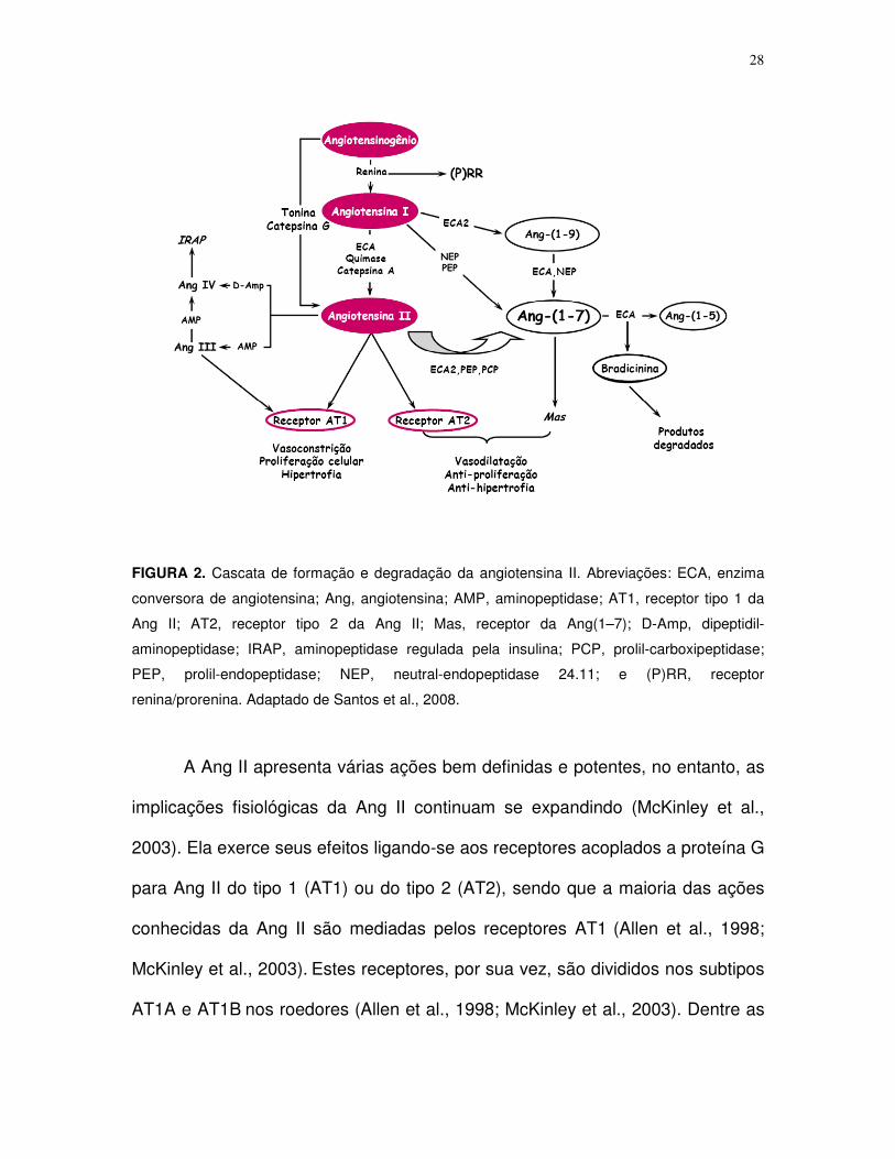
(Fyhrquist & Saijonmaa, 2008) (Figura 2).
28
FIGURA 2. Cascata de formação e degradação da angiotensina II. Abreviações: ECA, enzima
conversora de angiotensina; Ang, angiotensina; AMP, aminopeptidase; AT1, receptor tipo 1 da
Ang II; AT2, receptor tipo 2 da Ang II; Mas, receptor da Ang(1–7); D-Amp, dipeptidil-
aminopeptidase; IRAP, aminopeptidase regulada pela insulina; PCP, prolil-carboxipeptidase;
PEP, prolil-endopeptidase; NEP, neutral-endopeptidase 24.11; e (P)RR, receptor
renina/prorenina. Adaptado de Santos et al., 2008.
A Ang II apresenta várias ações bem definidas e potentes, no entanto, as
implicações fisiológicas da Ang II continuam se expandindo (McKinley et al.,
2003). Ela exerce seus efeitos ligando-se aos receptores acoplados a proteína G
para Ang II do tipo 1 (AT1) ou do tipo 2 (AT2), sendo que a maioria das ações
conhecidas da Ang II são mediadas pelos receptores AT1 (Allen et al., 1998;
McKinley et al., 2003). Estes receptores, por sua vez, são divididos nos subtipos
AT1A e AT1B nos roedores (Allen et al., 1998; McKinley et al., 2003). Dentre as

29
mais relevantes ações da Ang II estão vasoconstrição da musculatura lisa
vascular, retenção de sódio e água nos rins, tanto por efeito direto nesse órgão,
como indiretamente através da estimulação da biossíntese de aldosterona
adrenal. Ademais, a Ang II age no sistema nervoso central induzindo a sede, o
apetite ao sódio, liberação de hormônios pituitários, controle da temperatura
corporal e modulação do controle autonômico da função cardiovascular (Allen et
al., 1998; McKinley et al., 2003).
É interessante salientar que todos os componentes do sistema renina-
angiotensina estão presentes no cérebro, sugerindo haver um sistema renina-
angiotensina central (Von Bohlen Und Halbach & Albrecht, 2006). Em alguns
casos, as ações locais do sistema nervoso central interagem com aquelas da
Ang II sistêmica (Allen et al., 1998; McKinley et al., 2003). Uma vez que a Ang II
não atravessa a barreira hemato-encefálica, essa interação ocorre em sítios
específicos desprovidos dessa barreira, como os órgãos circumventriculares
área postrema e órgão subfornicial, os quais são ricos em receptores AT1 e
emitem projeções para outras regiões centrais que se situam atrás da barreira
(Allen et al., 1998; Von Bohlen Und Halbach & Albrecht, 2006; McKinley et al.,
2003).
A distribuição dos receptores AT1 no sistema nervoso central é difusa,
merecendo destaque a localização em outras regiões cerebrais envolvidas no
controle cardiovascular, da homeostase dos líquidos corporais e da temperatura
corporal (Allen et al., 1998; McKinley et al., 2003), como o núcleo paraventricular

30
do hipotálamo, núcleo do trato solitário e a área pré-óptica (Allen et al., 1998;
McKinley et al., 2003).
Envolvimento da Ang II no equilíbrio térmico
Há várias evidências mostrando que a hipotermia induzida pela Ang II
deve-se a facilitação da perda de calor (Fregly & Rowland, 1992, 1993, 1996;
Wilson & Fregly, 1985 a,b). Além disso, achados apontam que a ativação
angiotensinérgica também resulta em diminuição da produção de calor (Cassis et
al., 2002; Fregly & Rowland, 1992, 1993, 1996; Wilson & Fregly, 1985 a,b). A
administração tanto periférica quanto central de Ang II mostrou ser capaz de
provocar resposta hipotérmica dose-dependente, manifestada através do
aumento da temperatura da cauda e diminuição da taxa metabólica (Fregly &
Rowland, 1992, 1993, 1996; Mathai et al., 2000; Wilson & Fregly, 1985 a,b). Tais
respostas calóricas induzidas pela Ang II são abolidas pelo tratamento com
bloqueador do receptor AT1 (Fregly & Rowland, 1992; Horowitz et al., 1999;
Wilson & Fregly, 1985 a,b).
Mais recentemente, estudo do laboratório mostrou que a ação da Ang II no
balanço térmico não se limita a situações de repouso, mas também durante o
exercício físico (Leite et al., 2006). O bloqueio central do receptor AT1 para Ang II
utilizando o Los intracerebroventricularmente durante o exercício físico produz
aumento significativo das taxas de aquecimento corporal e de acúmulo de calor,
agravando a hipertermia do exercício e aumentando o limiar térmico para

31
vasodilatação cutânea. Além disso, o tratamento com Los reduz o desempenho
físico que mostrou-se intimamente associado com a taxa de aquecimento
corporal. Esses dados evidenciam que o sistema angiotensinérgico central tem
efeitos importantes sobre a termorregulação durante o exercício físico, facilitando
a dissipação de calor através da vasodilatação cutânea, atenuando o aumento da
temperatura corporal interna e, consequentemente, melhorando a capacidade
física (Leite et al., 2006).
Envolvimento da Ang II nos ajustes metabólicos
Além de suas ações sobre o sistema cardiovascular e na regulação do
equilíbrio hidroeletrolítico, a Ang II está envolvida com a regulação de funções
endócrinas e metabólicas (Machado et al., 2002), especialmente aquelas
envolvidas com a homeostase da glicose (Coimbra et al., 1999; Machado et al.,
1995 a,b, 1998). Sua ação sobre o metabolismo intermediário ocorre tanto de
maneira direta ou através de sua ação sobre a medula adrenal e controle da
atividade simpática (Coimbra et al., 1999, Machado et al., 1995 a,b, 1998;
Mihessen-Neto et al., 1996). A atuação desse peptídeo na regulação da glicemia
é significativa, induzindo hiperglicemia dose dependente atuando sobre
receptores AT1 (Machado et al., 1995 a,b). Por outro lado, a injeção intravenosa
de antagonista da Ang II resulta em inibição da hiperglicemia induzida por
hemorragia (Machado et al., 1995 a,b). A Ang II ainda produz hiperglicemia em
animais desmedulados, apesar dessa resposta ser atenuada em comparação
32
com animais normais (Mihessen-Neto et al., 1996). Portanto, a ação direta da
Ang II sobre a produção hepática de glicose, facilitando a gliconeogênese,
glicogenólise, e também sobre a atividade da glicogênio fosforilase hepática,
soma-se de maneira fundamental à regulação multifatorial da concentração de
glicose plasmática por esse peptídeo (Coimbra et al., 1999, Machado et al., 1995
a,b, 1998; Mihessen-Neto et al., 1996) (Figura 3). Não pode ser
desconsiderando o efeito inibitório da Ang II sobre a secreção de insulina em
resposta a elevação de glicose (Henriksen, 2007; Perkins & Davis, 2008). Uma
vez que bloqueadores do sistema renina-angiotensina são capazes de melhorar
a sensibilidade à insulina e o controle da glicemia, é possível que a Ang II eleve
a glicose sanguínea por interferir na ação desse hormônio (Coimbra et al., 1999,
Henriksen, 2007; Perkins & Davis, 2008).
Embora os resultados ainda sejam controversos, evidências apontam que
a Ang II também aumenta a lipólise nos tecidos subcutâneo e adiposo de
maneira dose dependente (Boschmann et al., 2006; Cabassi et al., 2005). Esse
resultado é acompanhado por elevação da concentração intersticial de
norepinefrina no tecido adiposo e também no plasma, sugerindo ser a lipólise
resultante da elevação da atividade simpática local e sistêmica (Cabassi et al.,
2005).

33
Figura 3. Esquema ilustrativo da regulação multifatorial da concentração de glicose plasmática
pela angiotensina II (Ang II).
Interação entre o sistema renina-angiotensina e os sistemas serotonérgico
e dopaminérgico
São poucas as evidências que abordam o efeito da Ang II sobre a
secreção central de 5-HT ou DA. No entanto, a ativação dos receptores AT1
parece afetar o metabolismo de 5-HT e DA no cérebro (Stadler et al., 1992;
Tanaka et al., 2003). Já foi demonstrado que a Ang II injetada dentro do orgão
subfornicial causa redução na concentração de 5-HT e ácido 5-
hidroxindoleacético (5-HIAA) neste mesmo sítio (Tanaka et al., 2003). Ademais,
a injeção intracerebroventricular de Ang II mostrou não modificar a concentração
de DA e ácido 3,4 diidroxifenilacético (DOPAC) no PVN ou hipotálamo anterior
Medula Adrenal
↑↑↑↑
↑↑↑↑ Fluxo simpático
↑↑↑↑ Atividade da glicogênio fosforilase
↑↑↑↑ Gliconeogênese
Fígado

34
(Qadri et al., 1991; Stadler et al., 1992). Portanto, assim como no orgão
subfornicial e no PVN, é possível que em outros centros termorregulatórios a
interação entre Ang II e 5-HT ou DA interfira no controle da temperatura interna
e, consequentemente, no desempenho físico.
INTERAÇÃO ENTRE OS CENTROS REGULADORES DA TEMPERATURA
CORPORAL E DO METABOLISMO INTERMEDIÁRIO
A regulação da temperatura corporal deve ser analisada sob o ponto de
vista autonômico, endócrino, metabólico e comportamental (Arancibia et al.,
1996). As áreas do sistema nervoso central que são ativadas por flutuações na
temperatura ambiente e que mediam as respostas fisiológicas a essas
flutuações estão localizadas principalmente no hipotálamo (Hasegawa et al.,
2000, 2005, 2008; Ishiwata et al., 2001, 2002, 2004; Nagashima et al., 2000;
Romanovsky, 2007). A área pré-óptica e o hipotálamo anterior têm sido
apontados como os sítios primários da integração de sinais térmicos originados
de diferentes partes do corpo e pela coordenação da regulação da temperatura
corporal (Hasegawa et al., 2000, 2005, 2008; Ishiwata et al., 2001, 2002, 2004;
Nagashima et al., 2000; Romanovsky, 2007). Esses centros contêm neurônios
sensíveis ao calor e ao frio que respondem a pequenas variações de
temperatura (Ishiwata et al., 2002; Zhang et al., 1997). Os neurônios sensíveis
ao calor são os principais efetores tanto da perda quanto da produção de calor.
Esses neurônios geram sinais excitatórios para a perda de calor e sinais

35
inibitórios para a produção de calor através do bloqueio dos neurônios sensíveis
ao frio (Nagashima et al., 2000; Romanovsky, 2007). Devido à projeção de vias
nervosas a partir da área pré-óptica para outras regiões do sistema nervoso
central, diversos sítios têm sido identificados como participantes do controle da
atividade termorreguladora (Kazuyuki et al., 1998; Nagashima et al., 2000;
Romanovsky, 2007). Propõe-se que as eferências que modulam as respostas
efetoras específicas, isto é salivação, tônus vasomotor, tremor, atividade
metabólica do tecido adiposo marrom e comportamento, são vias neurais
completamente distintas e independentes (Kazuyuki et al., 1998; Nagashima et
al., 2000; Romanovsky, 2007). Sendo assim, as respostas termorreguladoras
podem ser provocadas pela estimulação térmica, possivelmente de limiares
diferentes, de várias áreas do sistema nervoso central, incluindo alguns grupos
neuronais do tronco encefálico (formação reticular do mesencéfalo, ponte e
bulbo) e da medula espinhal (Kazuyuki et al., 1998; Nagashima et al., 2000;
Romanovsky, 2007) (Figura 4).

36
FIGURA 4. Esquema ilustrando as vias termorregulatórias efetoras da área pré-óptica para cada
resposta efetora termorregulatória. Linhas contínuas ou tracejadas indicam conexões
comprovadas e hipotéticas, respectivamente. A rede neuronal para a termorregulação
comportamental é pouco conhecida. Abreviações: (C) neurônios sensíveis ao calor, PVN (núcleo
paraventricular do hipotálamo), MFB (feixe prosencefálico medial), VTA (área tegmental ventral),
PAG (substância periaquedutal cinzenta), IML (coluna intermediolateral), HL (hipotálamo lateral),
RF (formação reticular), AH (hipotálamo anterior), PH (hipotálamo posterior), VMH (hipotálamo
ventromedial), DMH (hipotálamo dorsomedial). Adaptado de Nagashima et al., 2000;
Romanovsky, 2007.
O PVN é um sítio integrador da atividade nervosa simpática, sabidamente
fundamental para regulação cardiovascular (Li et al., 2006; Patel, 2000).
Recentemente, este núcleo foi descrito como participante do controle da
temperatura corporal interna (Nagashima et al., 2000; Romanovsky, 2007) por
PVN/DMH AH/P

37
conter neurônios termosensíveis que são ativados durante o estresse térmico
(Bratincsak & Palkovits, 2004; Cham & Badoer, 2008, Cham et al., 2006). Além
disso, achados apontam que projeções partem do PVN para outros centros
termorreguladores, influenciando a atividade simpática de orgãos termoefetores
como o tecido adiposo marrom, glândula salivar, vasculatura da cauda, assim
como rins e intestino (Cham & Badoer, 2008; Kazuyuki et al., 1998; Smith et al.,
1998). Foi descrito também que a inibição neuronal ou lesão do PVN durante
exposição ao calor previne a redução do fluxo sanguíneo renal (Cham & Badoer,
2008; Kenney et al., 2001). A redistribuição de sangue do meio interno para a
periferia é crítica em situações de hipertermia pois possibilita a perda de calor
(Cham & Badoer, 2008; Kenney et al., 2001; Kregel et al., 1994). Ela depende de
ajustes cardiovasculares controlados pelo sistema nervoso simpático que
induzem vasoconstrição visceral e vasodilatação da pele simultaneamente,
acompanhados por elevação da pressão arterial e da freqüência cardíaca
(Kanosue et al., 1994; Morrison, 2001; Smith et al., 1998). Considerando essas
evidências, o PVN parece estar envolvido na regulação da temperatura corporal
interna através do controle do fluxo simpático não uniforme responsável por
ajustar respostas cardiovasculares induzidas por estresse térmico determinantes
para a dissipação de calor.
Além de serem considerados centros termorreguladores, a área pré-
óptica e o PVN são importantes centros mantenedores da homeostase
metabólica (Coimbra & Migliorini, 1986; Ferreira et al., 1999; Foscolo et al.,
2003; Santos et al., 1991, Silveira et al., 2003). Já foi demonstrado que a

38
estimulação da área pré-óptica induz rápida elevação da concentração
plasmática de ácidos graxos livres, enquanto a estimulação noradrenérgica local
resulta em hiperglicemia (Coimbra & Migliorini, 1986; Foscolo et al., 2003). Além
disso, evidências comprovaram que as respostas de hiperglicemia e aumento da
mobilização de ácidos graxos livres induzidas pelo frio são impedidas após
tratamento com bloqueadores adrenérgicos na área pré-óptica (Coimbra &
Migliorini 1988; Ferreira et al., 1999). Esses indícios sugerem que o controle
termorregulatório e metabólico da área pré-óptica parecem ser integrados. Por
sua vez, a ativação noradrenérgica do PVN também induz hiperglicemia
(Ionescu et al., 1989). O PVN parece participar do controle metabólico em outras
situações de estresse como durante a hemorragia (Silveira et al., 2003).
Segundo este estudo, a inibição da atividade colinérgica do PVN, utilizando
metilatropina, reduz a resposta hiperglicêmica estimulada por hemorragia.
HIPÓTESE
O sistema angiotensinérgico central provavelmente está envolvido no
equilíbrio metabólico e térmico durante o exercício físico por modificar a
atividade do sistema monoaminérgico central.
As evidências experimentais apresentadas demonstram que o bloqueio
angiotensinérgico central durante o exercício dificulta a dissipação de calor,
promove elevação acentuada da temperatura corporal interna resultante do
aumento da taxa de aquecimento corporal e do acúmulo de calor. Em função
desses efeitos, foi verificada redução significativa do desempenho físico em

39
experimentos com ratos, caracterizando a Ang II como possível peptídeo
ergogênico e termorregulador. Uma vez que o sistema angiotensinérgico está
envolvido com a regulação tanto da perda de calor quanto da produção deste,
especula-se que o aumento acentuado da temperatura corporal interna
observado após bloqueio central da Ang II durante o exercício também se deve
a alterações na produção de calor e na eficiência mecânica, os quais afetariam o
desempenho físico. Além disso, o fato da elevação da concentração central de
Ang II possivelmente promover redução da concentração de 5-HT, cujo aumento
no cérebro está diretamente relacionado com elevação da temperatura corporal,
permite especular se a redução do tempo de exercício devido a hipertermia após
bloqueio angiotensinérgico central ocorreria em função do aumento do conteúdo
de 5-HT, interagindo com DA, em centros nervosos termorreguladores e de
controle motor. Considerando que a Ang II exerce efeitos metabólicos
importantes, principalmente em relação ao metabolismo da glicose e ácidos
graxos, é razoável sugerir que o efeito anti-ergogênico do Los central seria
desencadeado por alterações na disponibilidade de substratos energéticos. Por
fim, devido à ação termorregulatória e de controle da atividade simpática do
PVN, questiona-se caso esse sítio esteja envolvido no controle do fluxo
simpático não uniforme responsável por ajustar respostas cardiovasculares
induzidas por estresse térmico determinantes para a dissipação de calor.

40
IV. OBJETIVO
OBJETIVO GERAL
Avaliar a influência da transmissão angiotensinérgica central sobre o
equilíbrio térmico, mobilização de substratos energéticos e fadiga central durante
o exercício físico em ratos, e o envolvimento do PVN nos ajustes
cardiovasculares mediados pela atividade simpática, facilitadores da dissipação
de calor durante o estresse térmico.
Objetivos específicos
1. Estudar os efeitos do bloqueio central do receptor AT1 para Ang II sobre o
consumo de oxigênio, o gasto calórico e a eficiência mecânica em ratos não
treinados submetidos ao exercício submáximo na esteira;
2. Estudar os efeitos do bloqueio central do receptor AT1 para Ang II sobre o
consumo de oxigênio, o gasto calórico e a eficiência mecânica em ratos não
treinados submetidos ao exercício progressivo na esteira;
3. Estudar os efeitos do bloqueio central do receptor AT1 para Ang II sobre a
concentração plasmática de substratos energéticos (glicose, lactato e ácidos

41
graxos livres) em ratos não treinados submetidos ao exercício progressivo na
esteira;
4. Estudar os efeitos do bloqueio central do receptor AT1 para Ang II sobre as
concentrações centrais de 5-HT, 5-HIAA, DA e DOPAC na área pré-óptica,
hipotálamo, hipocampo e córtex frontal em ratos não treinados submetidos ao
exercício submáximo na esteira;
5. Estudar os efeitos da inibição bilateral do PVN e do bloqueio óxido nitrérgico
nesse mesmo núcleo durante a exposição ao calor sobre a atividade simpática
do nervo renal, pressão arterial média, frequência cardíaca, equilíbrio térmico e
limiar térmico para vasodilatação cutânea.

42
V. MÉTODOS
ANIMAIS
Foram utilizados ratos Wistar machos, com peso corporal entre 240-330 g,
provenientes do CEBIO-ICB. Os animais foram mantidos em ambiente com
temperatura entre 22+2 ºC e fotoperíodo de 14 h luz/10 h escuro, tendo livre
acesso à ração e água.
O trabalho foi realizado de acordo com as normas estabelecidas pelo
Comitê de Ética em Pesquisa com Animais da UFMG (CETEA/UFMG), segundo
o protocolo de número 145/2007.
PROCEDIMENTO CIRÚRGICO
Os animais foram submetidos à cirurgia para implante de cânula guia (16
mm de comprimento x 0,7 mm de diâmetro) no ventrículo cerebral lateral direito,
utilizada para microinjeções (2 µL) de solução salina 0,15 M (Sal) ou de solução
de Los (60 nmol). Os ratos utilizados foram anestesiados com mistura de 2,2,2-
tribromoetanol (300 mg/kg de peso corporal, via intraperitoneal) ou combinação
de ketamina (116 mg/kg de peso corporal) e xilazina (5,75 mg/kg de peso
corporal, via intraperitoneal) e fixados em estereotáxico. Foram obedecidas as
coordenadas estereotáxicas estabelecidas pelo Atlas de De Groot (1959) (A: -1,5
mm; L: -2,5mm; V: -3,0mm). O correto posicionamento da cânula no ventrículo
lateral foi verificado pelo deslocamento dos meniscos em um manômetro com
salina.

43
Os animais se recuperaram das cirurgias durante o período de pelo
menos uma semana.
EXERCÍCIO FÍSICO
A adaptação ao exercício físico consistiu de uma corrida diária em esteira
para roedores (Columbus Instruments, OH, USA, Modular treadmill, serie 96002-
2) a uma velocidade de 15 m/min, 5% de inclinação da esteira durante 5
minutos/4 dias consecutivos.
Durante os experimentos, os animais foram submetidos ao exercício
contínuo ou progressivo até a fadiga em esteira. O exercício contínuo consistiu
de corrida à velocidade constante de 18 m/min e 5% de inclinação. O modelo de
exercício progressivo consistiu de velocidade inicial ajustada em 10 m/min, com
inclinação de 10% da esteira. A velocidade sofreu acréscimo de 1m/min a cada 3
minutos até a fadiga do animal. O ponto de fadiga foi definido como o momento
no qual os animais não conseguiram manter o ritmo da esteira por mais de 10
segundos. A estimulação elétrica utilizada foi estabelecida de acordo com a
tolerância de cada animal, a ponto de causar um desconforto, sem causar dor, que
o fizesse escolher permanecer na esteira ao invés da grade de estimulação
elétrica. O tempo total de exercício e o trabalho realizado foram avaliados como
capacidade máxima de trabalho dos animais. O trabalho realizado foi
determinado de acordo com a equação:
Trabalho (kgm) = [(intensidade do exercício)x(tempo até a
fadiga)x(peso corporal)]. [seno θ (inclinação da esteira)]

44
PROCEDIMENTO EXPERIMENTAL GERAL
Os animais foram colocados no local do experimento 60 minutos antes do
início do exercício. Uma agulha (30G) foi introduzida na cânula guia e conectada
a uma seringa Hamilton para injeção das drogas. Imediatamente antes do início
da atividade física contínua ou progressiva foi administrado no ventrículo
cerebral lateral direito 2 µL de Sal ou de solução de Los (60 nmol),
aleatoriamente por método duplo-cego. A temperatura ambiente foi mantida dentro
de uma faixa constante, entre 22±2°C. Os experimentos foram realizados entre 10
e 14 horas.
Experimento 1: efeito do bloqueio angiotensinérgico central sobre o
consumo de oxigênio durante o exercício contínuo até a fadiga
Neste grupo experimental foi medido o consumo de oxigênio (VO2, mL.kg-
1.min-1) durante o exercício contínuo até a fadiga.
O VO2 foi medido por calorímetro indireto de fluxo aberto (Columbus
Instruments), o qual foi calibrado com mistura padrão de gases contendo 95% de
O2 e 5% de CO2 (White Martins) antes do experimento. O seu registro foi
realizado continuamente usando sistema computadorizado em linha com o
calorímetro (Oxymax Apparatus, Columbus Instruments). A eficiência mecânica
foi determinada de acordo com a equação: (Trabalho realizado/gasto energético)
x 100.

45
Experimento 2: efeito do bloqueio angiotensinérgico central sobre o
consumo de oxigênio durante o exercício progressivo até a fadiga
Neste grupo experimental foi medido o VO2 durante o exercício
progressivo até a fadiga de acordo com o protocolo citado anteriormente.
Experimento 3: efeito do bloqueio angiotensinérgico central sobre a
concentração plasmática de substratos energéticos durante o exercício
progressivo até a fadiga
Neste grupo experimental foi realizada a análise das concentrações
plasmáticas de glicose, lactato e ácidos graxos livres durante o exercício
progressivo até a fadiga de acordo com o protocolo citado anteriormente. Para
tal, além da cirurgia para implante da cânula no ventrículo lateral direito, os
animais receberam implante, através da veia jugular, de cateter de silastic no
átrio direito do coração, para colheitas seriadas de amostras de sangue (300 µL).
Durante o experimento as amostras de sangue foram retiradas pelo cateter atrial
imediatamente antes da atividade física, durante o exercício nos tempos de 03,
06, 09, 12, 15, 21 minutos e no momento da fadiga. Para evitar redução no
volume sanguíneo do animal, foram feitas reposições do mesmo volume de
sangue obtido de um rato doador normal. O sangue para determinação
plasmática de substratos foi colhido em seringas heparinizadas. As amostras
sanguíneas foram colocadas em gelo até a centrifugação e separação do
plasma. Após este procedimento, o sangue foi, então, congelado a -20ºC até a
análise bioquímica dos mesmos. As concentrações plasmáticas de glicose e

46
lactato foram determinadas por método oxidativo utilizando Glucose Analyser
(2300 STATPLUS, Yellow Springs Instruments, USA). A concentração dos
ácidos graxos livres foi determinada por método enzimático colorimétrico
utilizando kit NEFA 30T (Randox Laboratories, USA), adaptado para pequenos
volumes de plasma.
Experimento 4: efeito do bloqueio angiotensinérgico central sobre a
concentração de monoaminas centrais no momento da fadiga após o
exercício contínuo
Imediatamente após o término do exercício físico contínuo o animal foi
decapitado para retirada da área pré-óptica, hipotálamo, hipocampo e córtex
frontal. Os tecidos cerebrais foram armazenados em freezer -80 °C para análise
posterior, por cromatografia líquida de alta eficiência com detecção eletroquímica
(Smimadzu, Kyoto, Japan), das concentrações cerebrais de 5-HT, 5-HIAA, DA e
DOPAC. Para análise por CLAE, os tecidos foram previamente pesados e
homogeneizados com ácido perclórico 0,2 M e centrifugados a 15000 rpm por 20
minutos a 6 °C. Foram, então, injetados 20 µL do sobrenadante no cromatógrafo
e a quantificação das substâncias feita pela comparação da área do pico com
uma curva padrão utilizando o software CLASSVP em linha com o cromatógrafo.
Durante o exercício físico, a temperatura corporal interna dos animais
desse grupo foi registrada continuamente por telemetria utilizando sensor de
temperatura intraperitoneal previamente implantado e calibrado (TR3000 VM-
FH, Mini Mitter, Sun River, OR). A partir dos dados colhidos foram calculadas:
47
Taxa de aquecimento corporal (ºC.min-1) = (∆temperatura corporal
interna/tempo de exercício); e
Taxa de acúmulo de calor (cal.min-1)= [(∆temperatura corporal
interna).m.c]/(tempo de exercício), sendo m= massa corporal em gramas e c =
calor específico dos tecidos do animal (0.826 cal.g-1.ºc-1).
Experimento 5: efeito do bloqueio e inibição óxido nitrérgica do PVN sobre
parâmetros cardiovasculares e térmicos durante a exposição ao calor
Para realização desse experimento foram utilizados ratos Sprague-Dawley
(220–320 g) anestesiados com combinação de uretana (0,75 g/kg
intraperitonealmente) e α-cloralose (70 mg/kg intraperitonealmente). A artéria
femoral esquerda foi canulada e conectada a um sistema computadorizado de
registro e análise de dados (MacLab; AD Instruments, Mountainview, CA) via
transdutor de pressão (modelo P231D; Gould) para registro da pressão arterial e
frequência cardíaca. A traquéia foi entubada com o intuito de facilitar a
ventilação. Em seguida, o animal foi posicionado em estereotáxico (David Kopf
Instruments, Tujunga, CA) para posicionamento da cânula de microinjeção no
PVN seguindo as coordenadas de Paxinos e Watson (1986) (A: -1,5 mm; L: -0.4
mm; V: -7.8 mm). As microinjeções foram realizadas com uso de microseringa
(0,5 µL; modelo 7000.5; Hamilton).
O posicionamento em eletrodos de platina bipolares de um dos ramos do
nervo renal permitiu o registro da atividade simpática do nervo. O sinal foi
amplificado (10.000 vezes; Grass amplifier, modelo P55), retificado e integrado.

48
O sinal registrado após o fim do experimento, quando o animal já havia sido
sacrificado, foi considerado ruído. A descarga do nervo foi calculada pela
subtração entre o ruído e o valor basal ou o registrado durante o experimento.
Após os procedimentos cirúrgicos e estabilização dos parâmetros por no
mínimo 20 minutos, iniciou-se o estresse térmico. A atividade simpática do nervo
renal, pressão arterial, frequência cardíaca, assim como as temperaturas
corporal interna e da cauda foram registradas continuamente. A temperatura
colônica foi considerada como interna e medida usando probe inserido 4 cm
após o esfíncter anal (modelo 401, Yellow Springs Instruments, USA). Para
determinação da temperatura da cauda, um termistor de cauda (409-B, Yellow
Springs Instruments, USA) foi fixado à superfície dorsal da pele, cerca de 10 mm
da base da cauda. Essas temperaturas foram usadas para determinação da
variação da temperatura interna no momento no qual a temperatura da cauda
iniciou-se (vasodilatação). O estresse térmico foi induzido por aumento da
temperatura do cobertor térmico (Staco, Model 3PN 1010BV) entre 37 e 43°C,
em uma taxa de 1.2°C a cada 6 minutos, durante 30 minutos. Os animais foram
aleatoriamente separados nos grupos para receber microinjeção de solução
veículo (CSF; 100 nL/lado), lidocaina (1%; 200 nL/lado) ou L-NMMA (inibidor da
óxido nítrico sintase; 200 pmol, 100nL/lado) bilateralmente no PVN. Experimento
controle também foi realizado, durante o qual os mesmos parâmetros foram
registrados durante 30 minutos sem exposição ao calor.

49
Ao fim dos experimentos, os cérebros foram removidos para avaliação
histológica. Somente as injeções localizadas < 0,5 mm dos arredores do PVN
foram consideradas efetivas.
ANÁLISE ESTATÍSTICA
Para a análise estatística foi utilizada a análise de variância (ANOVA),
seguido do teste de Newman-Keuls. Os dados também foram comparados
utilizando teste t de Student pareado ou não pareado, de acordo com a sua
aplicabilidade. As correlações foram verificadas por meio do coeficiente de
correlação de Pearson. O nível de significância foi estabelecido em 5%.

50
VI. RESULTADOS ALCANÇADOS
Bloqueio dos receptores centrais AT1 eleva o custo metabólico durante o
exercício, reduzindo a eficiência mecânica e o desempenho físico em ratos
[Neuropeptides (41): 189-194; 2007].
Foi investigado o efeito do bloqueio central do receptor central AT1 para
Ang II sobre a taxa metabólica e desempenho físico em ratos durante o exercício
em esteira (18 m.min-1, 5 % inclinação). O consumo de oxigênio (VO2) foi
mensurado utilizando sistema calorimétrico indireto, após a injeção de 2 µL de
Sal (n=9) ou Los (60 nmol, n=9) no ventrículo cerebral lateral direito, antes dos
animais correram até a fadiga. A eficiência mecânica e o trabalho realizado
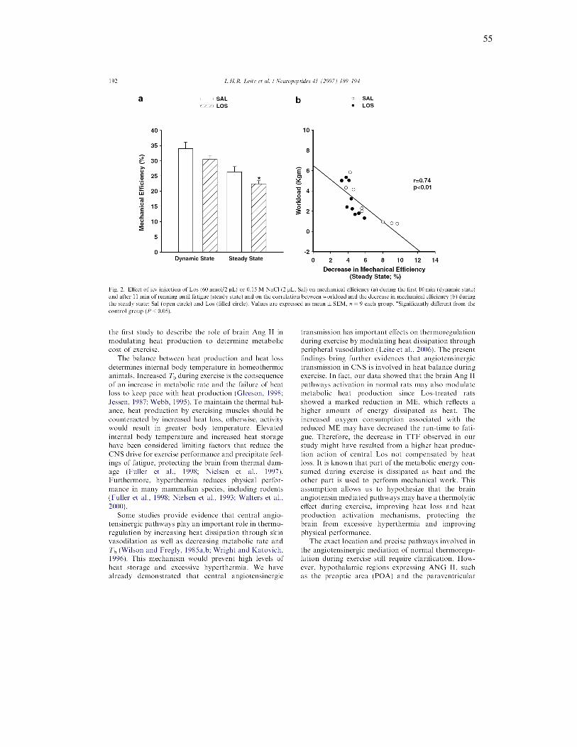
foram calculados. O trabalho realizado pelos animais tratados com Los foi 29%
inferior em relação aos animais tratados com Sal (p<0,02). Durante os primeiros
10 minutos de exercício (fase dinâmica do exercício), houve um aumento similar
do VO2, enquanto a eficiência mecânica permaneceu a mesma em ambos os
grupos. Durante a fase estável do exercício, o VO2 permaneceu estável no
grupo Sal, porém continuou a aumentar e estabilizou-se em um nível mais
elevado até a fadiga no grupo de animais tratados com Los. Durante a fase
estável do exercício houve uma redução mais acentuada da eficiência mecânica
nos ratos tratados com Los quando comparados com os animais tratados com
Sal (p<0,01), a qual se correlacionou inversamente com o trabalho realizado

51
(r=0,74; p<0,01). Nossos dados evidenciam que o bloqueio do receptor AT1
aumenta o custo metabólico durante o exercício, reduzindo a eficiência
mecânica e o desempenho físico. Os resultados indicam que o sistema
angiotensinérgico central modula a produção de calor, aumentando a eficiência
mecânica durante a fase estável do exercício.

52

53

54
55
56
57

58
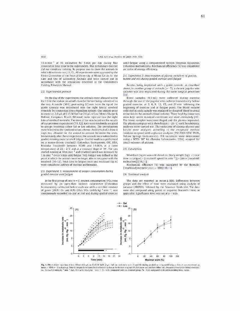
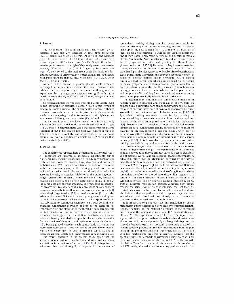
Receptores centrais AT1 estão envolvidos com ajustes metabólicos em
resposta ao exercício progressivo em ratos
[Peptides; (30): 1931-1935; 2009].
Com o intuito de investigar a influência dos receptores AT1 centrais para
Ang II sobre os ajustes metabólicos em ratos durante o exercício até a fadiga em
esteira, 2 µL de Sal ou Los (60 nmol) foram intracerebroventricularmente
injetados imediatamente antes do exercício progressivo (velocidade inicial de 10
m/min, com aumento de 1 m/mim a cada 3 minutos até a fadiga, 10% de
inclinação). O consumo de oxigênio (n=6) foi mensurado por calorimetria indireta
de fluxo aberto. O mesmo protocolo foi utilizado para coleta de amostras de
sangue através de cateter jugular (n=7). As concentrações plasmáticas de
glicose, lactato e ácidos graxos livres foram determinadas. A eficiência mecânica
e trabalho realizado foram calculados. Os animais tratados com Los
apresentaram hiperglicemia até a fadiga, já verificada em baixa intensidade de
exercício como 20% do trabalho máximo realizado. Altos níveis plasmáticos de
lactato e ácidos graxos livres acompanharam esta resposta hiperglicêmica.
Durante os seis primeiros minutos de exercício, aumento similar do VO2 e
eficiência mecânica semelhante foram observados em ambos os grupos. Em
seguida, o VO2 continuou a elevar-se, porém a uma taxa maior nos animais
tratados com Los, resultando em redução de 34% na eficiência mecânica
(p<0,01) associada com redução de 27% no trabalho realizado (p<0,01). Os
dados mostram que o bloqueio dos receptores AT1 centrais durante o exercício
59
progressivo induz hiperglicemia e maior mobilização de ácidos graxos livres em
baixos níveis de intensidade de exercício. Ademais, eleva o custo metabólico,
resultando em menor eficiência mecânica. Conclui-se que o sistema
angiotensinérgico central está envolvido em ajustes metabólicos durante o
exercício uma vez que o bloqueio dos receptores AT1 altera o equilíbrio
energético durante o exercício progressivo, semelhante a situações de ativação
simpática intensa e prematura.

60
61
62

63

64

65
Fadiga central induzida por Losartan envolve o conteúdo
cerebral de serotonina e dopamina
[Artigo submetido ao Medicine & Science in Sports & Exercise].
A fim de investigar a influência do bloqueio dos receptores AT1 centrais
para Ang II sobre a fadiga central devido ao metabolismo de 5-HT e DA durante
o exercício físico, 2 µL de Sal (n=6) ou Los (60 nmol, n=6) foram
intracerebroventricularmente injetados imediatamente antes de corrida até a
fadiga (18 m-min-1, 5 % inclinação). No momento da fadiga, o tecido cerebral foi
cuidadosamente removido para dosagem de 5-HT, 5-HIAA, DA e DOPAC por
cromatografia líquida de alta eficiência na área pré-óptica, hipotálamo,
hipocampo e córtex frontal. Os ratos tratados com Los exibiram aumento do
conteúdo de 5-HT na área pré-óptica e hipotálamo que correlacionou-se
diretamente com a elevada taxa de aquecimento corporal e indiretamente com o
reduzido tempo total de exercício. Ao contrário, o tempo para fadiga
correlacionou-se positivamente com o reduzido conteúdo de 5-HT no hipocampo
dos animais Los. Apesar dos níveis de DA não terem se alterado em nenhuma
das regiões estudadas, o hipotálamo dos animais Los apresentou elevada razão
5-HT:DA. Os resultados indicam que o sistema angiotensinérgico está envolvido
com a fadiga central devido a hipertermia uma vez que a elevada taxa de
aquecimento corporal resultante do bloqueio dos receptores AT1 centrais em
ratos em exercício está relacionada com aumento do conteúdo de 5-HT na área
pré-óptica e hipotálamo, assim como sua redução no hipocampo. Além disso, a

66
interação entre 5-HT e DA no hipotálamo provavelmente contribui
significativamente para a hipertermia e fadiga central prematura após inibição
angiotensinérgica.

67
Central fatigue induced by Losartan involves brain serotonin and dopamine content
Laura H. R. Leite1, Alex G. Rodrigues
3, Danusa D. Soares
2, Umeko Marubayashi,
1 and Cândido C.
Coimbra1
1Department of Physiology and Biophysics, Institute of Biological Sciences, and
2Department of Physical
Education, School of Physical Education, Physical Therapy and Occupational Therapy, Federal University
of Minas Gerais, 31270-901, Belo Horizonte, Minas Gerais, Brazil; 3Institute of Biological and Health
Sciences, Pontifícia Universidade Católica, 30535-610 Belo Horizonte, Minas Gerais, Brazil.
Corresponding Author: Cândido C. Coimbra, PhD
Departamento de Fisiologia e Biofísica, Instituto de Ciências Biológicas/UFMG,
Av. Antônio Carlos, 6627, 31270-901 Belo Horizonte, MG, Brazil.
Telephone: +55 (031) 3409-2936. Fax: +55 (031) 3409-2924
E-mail: [email protected]
Running title: Losartan-induced central fatigue
Disclosure statement of funding: This study was supported by grants from CNPq (Conselho Nacional de
Desenvolvimento Científico e Tecnológico), Capes (Coordenação de Aperfeiçoamento de Pessoal de Nível
Superior) and FAPEMIG (Fundação de Amparo à Pesquisa do Estado de Minas Gerais). The authors have
no conflicts to disclose.

68
ABSTRACT
Purpose: Investigate the influence of angiotensin II (Ang II) AT1 receptors blockade on central
fatigue induced by brain content of serotonin (5-HT) and dopamine (DA) during exercise. Methods:
Losartan (Los) was intracerebroventricularly injected in rats before running until fatigue (n=6/group). At
fatigue, brains were quickly removed for measurement of 5-HT, 5-hydroxyindoleacetic acid (5-HIAA), DA
and 3,4-Dihydroxyphenylacetic acid (DOPAC) by HPLC in preoptic area, hypothalamus, hippocampus and
frontal cortex. Results: Intracerebroventricular injection of Los increased 5-HT content in preoptic area and
hypothalamus that correlated positively with body heating rate and inversely with total time to fatigue. On
the contrary, time to fatigue was directly correlated with the diminished 5-HT concentration in
hippocampus of Los-rats. Although the levels of DA were not affected by Los treatment during exercise in
any of the studied brain areas, a higher 5-HT:DA ratio was seen in hypothalamus of Los-animals. This
higher hypothalamic 5-HT:DA ratio correlated positively with body heating rate and inversely with time to
fatigue. Conclusions: Our results show that central fatigue due to hyperthermia and increased body heating
rate induced by central Ang II AT1 receptors blockade in exercising rats is related with higher 5-HT
content in preoptic area and hypothalamus, as well as with decreased level of this neurotransmitter in
hippocampus. Furthermore, the interaction between 5-HT and DA within hypothalamus seems to contribute
markedly to hyperthermia and premature central fatigue following angiotensinergic inhibition.
Keywords: Preoptic area, hypothalamus, hippocampus, hyperthermia, exercise performance.

69
INTRODUCTION
Paragraph number 1 Central fatigue during prolonged exercise is considered to be affected by
accumulation or depletion of neurotransmitters, particularly serotonin (5-HT) (3, 9, 22, 23). High 5-HT
activity is associated with lethargy and loss of central drive/motivation (3, 22). In fact, evidences suggest
that a rise in 5-HT content in major brain areas responsible for thermoregulation, such as the preoptic
area/anterior hypothalamus, is related with heat production and precipitation of fatigue (3, 5, 26, 28). In
view of this last observation, substances that elevate body temperature, such as in the case of fever induced
by cytokines, may anticipate central fatigue through elevation of brain 5-HT content (7).
Paragraph number 2 Although the role of 5-HT on central fatigue has been well documented, it is
likely that other neurotransmitters are capable of influencing fatigue, such as dopamine (DA) (3, 9, 11, 22).
This monoamine has been suggested to interaction with 5-HT during exercise, affecting exercise
performance (11, 22). DA neurotransmission is associated with many physiological functions such as
arousal, reward and motivation (2, 11, 14, 22), that could modify running performance. Central DA
metabolism enhances during exercise in animals (2, 11, 14) and its central elevation has been linked with a
delay on fatigue despite higher body temperature at fatigue point and heat storage during exercise (2, 11,
14).
Paragraph number 3 High internal body temperature and increased heat storage have been
proposed as limiting factors that reduce the central nervous system drive for exercise performance, thus
protecting the brain from thermal damage (12, 27, 34). Therefore, considering that serotonergic and
dopaminergic interaction is involved with central fatigue, which is coincident with high body temperature
and/or heat storage, the activation of mechanisms that could modulate the activity of such systems would
improve exercise performance by inducing thermal adjustments.
` Paragraph number 4 We have recently shown that intracerebroventricular infusion of the widely
used antihypertensive drug Losartan (Los; Angiotensin II AT1 receptor antagonist) reduces running
performance in rats due to a heat imbalance characterized by higher metabolic cost and reduced peripheral
heat loss. These responses resulted in hyperthermia and increased body heating rate that was indirectly
related with time to fatigue (18, 20). Besides the effects of angiotensin II (Ang II) on thermoregulation and
metabolic cost during exercise, it is still not known whether this effect of angiotensinergic blockade on heat
storage may be linked to serotonergic and/or dopaminergic pathways that exhibit relevant thermoregulatory
and exercise performance effects (2, 11, 21, 28). Therefore, this study aimed to investigate the possible
interaction between the central angiotensinergic system and 5-HT and DA content in areas of the central
nervous system involved in thermoregulation and motor activity, including preoptic area, hypothalamus,
frontal cortex and hippocampus, during exercise.
METHODS
70
Animals
Paragraph number 5 Male Wistar rats, approximately three months old (290 ± 10 g), were housed
individually at a room temperature of 22 ± 2 ºC, under 14/10 h light-dark cycles and had free access to
water and rat chow. Following anesthesia achieved using a mixture of ketamine (115 mg/kg body weight
i.p.) and xylazine (6 mg/kg body weight i.p.), the rats were fixed to a stereotaxic apparatus (David Kopf
Instruments, M-900, Tujunga, CA, USA) and a guide cannula (22 G) was implanted into the right lateral
cerebral ventricle using a previously described technique (20). Also during this surgical procedure, TR3000
VM-FH temperature sensor (Mini Mitter, Sun River, OR) was implanted into the peritoneal cavity through
a small incision in the linea alba. All animals were allowed to recover for at least 1 week before being
submitted to the experiments. The animals were acclimatized to exercise on the motor-driven treadmill by
running at a speed of 15 m·min-1
at 5% inclination for 5 min per day during four consecutive days prior to
the experiments. This preliminary exercise did not constitute training. Its purpose was to show the animals
in which direction to run (18, 20). Electrical stimulation was determined according to each animal’s
tolerability. All experiments were approved by the Ethics Committee of the Federal University of Minas
Gerais for the Care and Use of Laboratory Animals and were carried out in accordance with the regulations
described in the Committee’s Guiding Principles Manual. Additionally, the experimental protocol followed
the American College of Sports Medicine (ACSM) animal care standards.
Experimental protocol
Paragraph number 6 On the day of the experiment, the animals were individually allowed to rest
for 1 h in the rodent treadmill chamber before being submitted to the test. A needle (30 G) protruding 0.3
mm from the tip of the guide cannula was introduced into the right lateral cerebral ventricle by connecting
it to a Hamilton syringe. One min prior to exercise, 2 µL of 0.15 M NaCl (Sal) or 2 µL of Los (Merck
Sharpe & Dohme, Campinas, Brazil; 60 nmol) were injected into the right lateral ventricle. The dose of Los
was based on the results of our previous experiments (18, 20). Rats were randomly assigned to groups
receiving either Sal or Los solution (n=6/group). The investigators were blinded to the randomization
scheme. Immediately after the intracerebroventricular injections, the animals were submitted to running
exercise until fatigue at constant speed and inclination (18 m/min and 5% inclination). Exercise was
performed on a motor-driven treadmill (Columbus Instruments, OH, USA, Modular Treadmill) between
10:00 and 14:00 h at a room temperature of 22 ± 2 ºC. The intensity of exercise corresponded to an oxygen
uptake of ~66% of maximal oxygen uptake, which represents a physical activity of moderate level (4, 15,
20). Body temperature was recorded continuously by telemetry. Fatigue was defined as the point at which
the animals were no longer able to keep pace with the treadmill (18, 20). Total time to fatigue (min) and
workload (kgm) were considered indexes of exercise performance.
71
Paragraph number 7 As soon as the fatigue point was reached, the animals were killed by
decapitation. The brain was quickly removed and washed with ice-cold saline. The preoptic area,
hypothalamus, hippocampus and frontal cortex were rapidly dissected on an ice-cold plate (26, 28), frozen
immediately in dry ice and stored at -80 °C until 5-HT, 5-hydroxyindoleacetic acid (5-HIAA), DA and 3,4-
Dihydroxyphenylacetic acid (DOPAC) were measured by high-pressure liquid chromatography (HPLC).
The HPLC system was equipped with a reverse-phase column (Shim Pack CLC-OSD; 25 cm, 5 m,
Shimadzu). The potential was set at 850 mV versus Ag/AgCl reference electrode. A mobile phase
containing 31.4 g citric acid, 584 mg NaCl, 800 mL miliQ water, 140 mg octylsodium sulfate, 48 mL
acetylnitrile and 28 mL tetrahydrofurane (pH 3.0) was filtered and pumped through the system at a flow
rate of 1.0 mL min-1
. The brain samples were weighed and preoptic area and hypothalamus homogenized in
200 µL of perchloric acid (0.1 M), while hippocampus and hypothalamus were homogenized in 300 µL of
the same solution. The homogenates were then centrifuged at 15000 × g for 20 min at 6 °C, and the
supernatants filtered through a Millipore membrane (0.22 pore size; 13 mm; Millex, SP, Brazil). Twenty
microliters of the supernatant were injected into the HPLC-EC system for analysis (Shimadzu, Kyoto,
Japan). Quantification of 5-HT, 5-HIAA, DA and DOPAC were made by comparing the peak area to a
standard curve.
Calculations
Paragraph number 8 Workload (kgm) was calculated as: [body weight (kg)]. [total time to fatigue
(min)]. [treadmill speed (m.min-1
)].[sine θ (treadmill inclination)] (18, 20).
Paragraph number 9 Body heating rate (ºC.min-1
), i.e. rate of increase in body temperature, was
calculated as: ∆body temperature/(running time interval), where ∆body temperature is the change in body
temperature (Tf-Ti), Tf = body temperature at fatigue point; and Ti = initial body temperature measured prior
to exercise (20).
Paragraph number 10 Heat storage rate (cal.min-1
) was calculated as: (∆body
temperature).m.c/(total time to fatigue), where "m" is the body weight in grams, and "c" is the specific heat
of the body tissues (0.826 cal.g-1
.ºc-1
) (20).
Statistical analysis
Paragraph number 11 The data are reported as mean ± S.E.M. A two-way analysis of variance
(ANOVA) was used for determining differences between time and treatment and also interactions between
them to evaluate the differences in changes of body temperature. Significant interactions observed by
ANOVA were further evaluated by Newman-Keuls post hoc analysis to locate significant differences
between means. Time to fatigue, workload, body heating rate, heat storage rate, as well as monoamines
concentrations were compared using unpaired Student’s t-test. The correlations were assessed using
Pearson’s correlation coefficient. Significance level was set at p<0.05.

72
RESULTS
Paragraph number 12 As previously seen and evidenced in table 1, intracerebroventricular
injection of Los reduced time to fatigue and workload by ~59 and 58%, respectively, compared to Sal-
treated group (p<0.01). Although exercise promoted a rapid increase in body temperature in both groups, at
fatigue point Los-treated rats had 0.33 °C higher body temperature than Sal-animals (p<0.01). To compare
the total thermal effects of exercise in both experimental groups, body heating rate and heat storage rate
were calculated. At fatigue, body heating rate and heat storage rate of Los-treated rats were increased by
~150 and 97% in comparison with Sal-treated rats (p<0.01).
Paragraph number 13 Fig 1 shows 5-HT and 5-HIAA content in the studied brain areas. At
fatigue, 5-HT content was significantly increased in preoptic area (Fig. 1A) and hypothalamus (Fig. 1B) of
Los-exercised rats compared to Sal-exercised animals. On the other hand, in fatigued Los-exercised rats, a
lower content of 5-HT was observed in hippocampus in comparison with control group (p<0.05) (Fig. 1C).
Within frontal cortex, 5-HT concentration was not affected by angiotensinergic blockade during exercise
(Fig. 1D). As also seen in Fig. 1, the levels of 5-HIAA were enhanced in preoptic area and hypothalamus
(p<0.05), but not within hippocampus and frontal cortex.
Paragraph number 14 Los treatment during exercise did not interfere with DA concentration in
any of the studied brain areas (Fig. 2). Again, the concentration of DOPAC increased in preoptic area and
hypothalamus (p<0.05), but not in hippocampus and frontal cortex (Fig. 2).
Paragraph number 15 The increased concentration of 5-HT in preoptic area (r = 0.85; p<0.01) and
hypothalamus (r = 0.75; p<0.01) correlated positively with body heating rate (Fig. 3A and C). Moreover, an
inverse relationship between time to fatigue and 5-HT levels in preoptic area (r = 0.82; p<0.01) and
hypothalamus (r = 0.70; p<0.01) (Fig. 3B and D) was verified. On the contrary, the correlation between 5-
HT level in the hippocampus and total time to fatigue (r = 0.56; p<0.05) was direct (Fig. 3F). However, no
correlation between body heating rate and 5-HT content in hippocampus was seen (Fig. 3E). Although, it
was not observed any correlation between DA concentration in any of the brain areas and body heating rate
or time to fatigue, the ratio between the content of 5-HT and DA (5-HT:DA) increased significantly within
the hypothalamus (p<0.01) (Fig. 4A). Additionally, the increased 5-HT:DA ratio of the hypothalamus
correlated positively with body heating rate (r = 0.73; p<0.01) and negatively with time to fatigue (r =
0.56; p<0.05) (Fig. 4B and C).
DISCUSSION
Paragraph number 16 The present study demonstrates that central fatigue due to hyperthermia and
increased body heating rate induced by central Ang II AT1 receptors blockade in exercising rats is related
with higher 5-HT content in preoptic area and hypothalamus, as well as with decreased level of this
neurotransmitter in hippocampus. Additionally, the higher hypothalamic 5-HT:DA ratio shown by Los rats,

73
which correlated directly with body heating rate and inversely with time to fatigue, indicates that 5-HT and
DA interaction in this region may contribute significantly to hyperthermia and premature central fatigue
following angiotensinergic inhibition. Taken together, the data indicate that angiotensinergic transmission
has important effects on brain 5-HT content during exercise, whose interaction with DA affects central
fatigue probably through modulation of body temperature.
Paragraph number 17 We have recently shown that intracerebroventricular infusion of Los
precipitates fatigue through an increase in metabolic cost and hyperthermia due to reduced peripheral heat
loss and increased heat production during exercise (18, 20). Elevated internal body temperature and
increased heat storage have been proposed as limiting factors to physical performance (12, 27, 34). It is
important to point out that fatigue is considered a defense mechanism that prevents the development of
homeostatic imbalances capable of endangering physical integrity, especially of the brain because of its
vulnerability to hyperthermia (17, 24). Mechanisms of fatigue include factors of peripheral and central
origin, the later being characterized by modifications within the central nervous system that impairs an
adequate drive to the muscles (9, 24).
Paragraph number 18 Increased brain serotonergic activity contributes to the development of
fatigue possibly by causing lethargy and loss of drive (9, 22, 24). It has been shown that the enhanced 5-HT
concentration in preoptic area and hypothalamus verified after exercise is related with exercise-induced
hyperthermia and decreased running performance (5, 26, 28). The present findings agree with such
observation and bring further evidence that inhibition of central angiotensinergic system during dynamic
exercise, which resulted in hyperthermia and higher body heating rate associated with increased 5-HT
content in preoptic area and hypothalamus, may have an anti-ergogenic effect, decreasing time to fatigue to
protect the organism against thermal damage. The indirect relation between time to fatigue and the
enhanced 5-HT concentration in preoptic area and hypothalamus of Los-rats also supports this observation.
Paragraph number 19 We can not disregard that serotonergic neurons have many other important functions
in the central nervous system, including the complex control of motor activity (3, 22, 30, 31, 32).
Hippocampus is also considered to be related with motor activity (30, 31, 32), as seen by findings that local
infusion of 5-HT in this site produces an increase in motor activity of rats (30, 32) and that lower
hippocampal serotonergic activity precipitates fatigue during exercise (26, 28). Our data showing decreased
5-HT concentration in hippocampus that correlated directly with time to fatigue, but not with body heating
rate, is in accordance with such idea. The decreased level of 5-HT in hippocampus may have contributed to
the lower exercise performance shown by Los-treated rats through modulation of motor activity. As
previously seen, it seems that the hippocampus involvement with central fatigue is of motor nature, rather
than thermal (26, 28). In addition, such opposite 5-HT turnover in areas of central nervous system involved
in thermoregulation and motor activity following inhibition of the brain angiotensinergic system agrees
with findings that the serotonergic system controls its effects through selective alteration of 5-HT
concentration according to specific brain regions (6, 26, 28).

74
Paragraph number 20 It is well defined that DA content and release is increased during exercise,
including within hypothalamus and hippocampus (2, 11, 14). Although increased brain DA results in
improvement of exercise performance (ergogenic effect), this response is also followed by hyperthermia
and enhanced heat storage (2, 14). DA acting on the mesolimbic reward system is considered to exceed the
limits of safe temperature by overruling inhibitory signals from the central nervous system that would alter
perceived effort and compromise exercise performance (2, 11, 14). In the present study, although
angiotensinergic blockade during exercise resulted in increased DOPAC levels in preoptic area and
hypothalamus, which is an indicative of elevated turnover, the treatment did not induce any modification in
the concentrations of DA in the studied brain areas. Such increased turnover of DA in the brain may have
prevented any marked elevation in the concentration of DA that could improve physical performance (3).
This finding corroborates with evidences that intracerebroventricular injection of Ang II does not alter the
concentrations of DA in paraventricular nucleus or anterior hypothalamus (25, 29). Nevertheless, there are
evidences that brain DA levels are similar to resting levels at fatigue (2, 11), thus, the possibility that the
concentration of DA increased differently among the groups in between exercise can not be excluded.
Paragraph number 21 The preoptic area/anterior hypothalamus is the major brain region involved
in thermoregulation that integrates thermal inputs with energy-linked metabolic processes (8, 10, 13, 16).
This is supported by the fact that this area contains both warm-sensitive and cold-sensitive neurons that
respond to small changes in temperature (13, 16) and that lesion or pharmacologic blockade of the preoptic
area/anterior hypothalamus produce a severe impairment in thermoregulation (13, 16). Besides preoptic
area, the hypothalamus also has influence on body temperature control. Lin et al. (21) demonstrated that
elevation of 5-HT levels in the hypothalamus through the infusion of 5-hydroxytryptophan resulted in
hyperthermic effects which were brought by increased metabolic heat production and decreased heat loss.
Similarly, it has also been shown that exercise increases DA concentration not only in hypothalamus, but
also in preoptic area, associated with elevated body temperature (2, 14). Therefore, it seems reasonable to
suggest that Los hypothetically perfused to these regions after its injection into the cerebral ventricle,
altering 5-HT and DA release and, consequently, thermal control. However, the mechanisms involved in
these hyperthermia-induced effects and how brain neurotransmission affects thermoregulation during
exercise still require clarification.
Paragraph number 22 The synthesis and turnover of 5-HT in the central nervous system depend
on changes in the availability of brain tryptophan, the enzyme responsible for catalyzing the first reaction in
the synthesis of 5-HT (3, 9). It is known that the exercise-induced increase in plasma free fatty acids
indirectly facilitate the entry of tryptophan into the brain because they compete for the same carrier
(albumin) (3, 9). In other words, an increase in free fatty acid concentration in plasma elevates the free
tryptophan level. We have recently shown that central angiotensinergic blockade during exercise also shifts
energy balance during graded exercise, resulting in higher and premature increase in plasma free fatty acids
at low absolute intensity of exercise (19). The association of this last result with the current one indicate
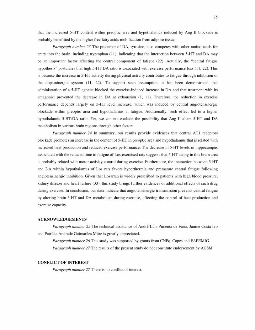
75
that the increased 5-HT content within preoptic area and hypothalamus induced by Ang II blockade is
probably benefitted by the higher free fatty acids mobilization from adipose tissue.
Paragraph number 23 The precursor of DA, tyrosine, also competes with other amino acids for
entry into the brain, including tryptophan (11), indicating that the interaction between 5-HT and DA may
be an important factor affecting the central component of fatigue (22). Actually, the “central fatigue
hypothesis” postulates that high 5-HT:DA ratio is associated with exercise performance loss (11, 22). This
is because the increase in 5-HT activity during physical activity contributes to fatigue through inhibition of
the dopaminergic system (11, 22). To support such assumption, it has been demonstrated that
administration of a 5-HT agonist blocked the exercise-induced increase in DA and that treatment with its
antagonist prevented the decrease in DA at exhaustion (1, 11). Therefore, the reduction in exercise
performance depends largely on 5-HT level increase, which was induced by central angiotensinergic
blockade within preoptic area and hypothalamus at fatigue. Additionally, such effect led to a higher
hypothalamic 5-HT:DA ratio. Yet, we can not exclude the possibility that Ang II alters 5-HT and DA
metabolism in various brain regions through other factors.
Paragraph number 24 In summary, our results provide evidences that central AT1 receptors
blockade promotes an increase in the content of 5-HT in preoptic area and hypothalamus that is related with
increased heat production and reduced exercise performance. The decrease in 5-HT levels in hippocampus
associated with the reduced time to fatigue of Los-exercised rats suggests that 5-HT acting in this brain area
is probably related with motor activity control during exercise. Furthermore, the interaction between 5-HT
and DA within hypothalamus of Los rats favors hyperthermia and premature central fatigue following
angiotensinergic inhibition. Given that Losartan is widely prescribed to patients with high blood pressure,
kidney disease and heart failure (33), this study brings further evidences of additional effects of such drug
during exercise. In conclusion, our data indicate that angiotensinergic transmission prevents central fatigue
by altering brain 5-HT and DA metabolism during exercise, affecting the control of heat production and
exercise capacity.
ACKNOWLEDGEMENTS
Paragraph number 25 The technical assistance of André Luis Pimenta de Faria, Janine Costa Ivo
and Patrícia Andrade Guimarães Mitre is greatly appreciated.
Paragraph number 26 This study was supported by grants from CNPq, Capes and FAPEMIG.
Paragraph number 27 The results of the present study do not constitute endorsement by ACSM.
CONFLICT OF INTEREST
Paragraph number 27 There is no conflict of interest.

76
REFERENCES
[1] Bailey SP, Davis JM, Ahlborn EN. Neuroendocrine and substrate responses to altered brain 5-HT
activity during prolonged exercise to fatigue. J Appl Physiol. 1993; 74(6): 3006-12.
[2] Balthazar CH, Leite LH, Rodrigues AG, Coimbra CC. Performance-enhancing and
thermoregulatory effects of intracerebroventricular dopamine in running rats. Pharmacol Biochem
Behav. 2009; 93(4): 465-9.
[3] Blomstrand E. A role for branched-chain amino acids in reducing central fatigue. J Nutr. 2006;
136(2): 544S-47S.
[4] Brooks GA, White TP. Determination of metabolic and heart rate responses of rats to treadmill
exercise. J Appl Physiol. 1978; 45(6): 1009-15.
[5] Caperuto EC, dos Santos RV, Mello MT, Costa Rosa LF. Effect of endurance training on
hypothalamic serotonin concentration and performance. Clin Exp Pharmacol Physiol. 2009; 36(2):
189-91.
[6] Chaouloff F, Laude D, Elghozi JL. PHysical exercise: evidence for differential consequences of
tryptophan on 5-HT synthesis and metabolism in central serotonergic cell bodies and terminals. J
Neural Transm. 1989; 78(2): 121-30.
[7] Chueh FS, Chang CP, Chio CC, Lin MT. Puerarin acts through brain serotonergic mechanisms to
induce thermal effects. J Pharmacol Sci. 2004; 96(4): 420-7.
[8] Coimbra CC, Migliorini RH. Cold-induced free fatty acid mobilization is impaired in rats with
lesions in the preoptic area. Neurosci Lett. 1988; 88(1): 1-5.
[9] Fernstrom JD, Fernstrom MH. Exercise, serum free tryptophan, and central fatigue. J Nutr. 2006;
136(2): 553S-59S.
[10] Ferreira ML, Marubayashi U, Coimbra CC. The medial preoptic area modulates the increase in
plasma glucose and free fatty acid mobilization induced by acute cold exposure. Brain Res Bull.
1999; 49(3): 189-93.
[11] Foley TE, Fleshner M. Neuroplasticity of dopamine circuits after exercise: implications for central
fatigue. Neuromolecular Med. 2008; 10(2): 67-80.

77
[12] Fuller A, Carter RN, Mitchell D. Brain and abdominal temperatures at fatigue in rats exercising in
the heat. J Appl Physiol. 1998; 84(3): 877-83.
[13] Hasegawa H, Ishiwata T, Saito T, Yazawa T, Aihara Y, Meeusen R. Inhibition of the preoptic area
and anterior hypothalamus by tetrodotoxin alters thermoregulatory functions in exercising rats. J
Appl Physiol. 2005; 98(4): 1458-62.
[14] Hasegawa H, Piacentini MF, Sarre S, Michotte Y, Ishiwata T, Meeusen R. Influence of brain
catecholamines on the development of fatigue in exercising rats in the heat. J Physiol. 2008;
586(1): 141-9.
[15] Hussain SO, Barbato JC, Koch LG, Metting PJ, Britton SL. Cardiac function in rats selectively
bred for low- and high-capacity running. Am J Physiol Regul Integr Comp Physiol. 2001; 281(6):
R1787-91.
[16] Ishiwata T, Hasegawa H, Yazawa T, Otokawa M, Aihara Y. Functional role of the preoptic area
and anterior hypothalamus in thermoregulation in freely moving rats. Neurosci Lett. 2002; 325(3):
167-70.
[17] Kay D, Marino FE. Fluid ingestion and exercise hyperthermia: implications for performance,
thermoregulation, metabolism and the development of fatigue. J Sports Sci. 2000; 18(2): 71-82.
[18] Leite LH, Lacerda AC, Balthazar CH, Marubayashi U, Coimbra CC. Central AT(1) receptor
blockade increases metabolic cost during exercise reducing mechanical efficiency and running
performance in rats. Neuropeptides. 2007; 41(3): 189-94.
[19] Leite LH, Lacerda AC, Balthazar CH, Marubayashi U, Coimbra CC. Central angiotensin AT1
receptors are involved in metabolic adjustments in response to graded exercise in rats. Peptides.
2009; 30(10): 1931-5.
[20] Leite LH, Lacerda AC, Marubayashi U, Coimbra CC. Central angiotensin AT1-receptor blockade
affects thermoregulation and running performance in rats. Am J Physiol Regul Integr Comp
Physiol. 2006; 291(3): R603-7.
[21] Lin MT, Tsay HJ, Su WH, Chueh FY. Changes in extracellular serotonin in rat hypothalamus
affect thermoregulatory function. Am J Physiol. 1998; 274(5 Pt 2): R1260-7.

78
[22] Meeusen R, Watson P, Hasegawa H, Roelands B, Piacentini MF. Brain neurotransmitters in
fatigue and overtraining. Appl Physiol Nutr Metab. 2007; 32(5): 857-64.
[23] Newsholme EA, Blomstrand E. Branched-chain amino acids and central fatigue. J Nutr. 2006;
136(1 Suppl): 274S-6S.
[24] Nielsen B, Nybo L. Cerebral changes during exercise in the heat. Sports Med. 2003; 33(1): 1-11.
[25] Qadri F, Badoer E, Stadler T, Unger T. Angiotensin II-induced noradrenaline release from anterior
hypothalamus in conscious rats: a brain microdialysis study. Brain Res. 1991; 563(1-2): 137-41.
[26] Rodrigues AG, Soares DD, Marubayashi U, Coimbra CC. Heat loss during exercise is related to
serotonin activity in the preoptic area. Neuroreport. 2009; 20(8): 804-8.
[27] Rodrigues LO, Oliveira A, Lima NR, Machado-Moreira CA. Heat storage rate and acute fatigue in
rats. Braz J Med Biol Res. 2003; 36(1): 131-5.
[28] Soares DD, Coimbra CC, Marubayashi U. Tryptophan-induced central fatigue in exercising rats is
related to serotonin content in preoptic area. Neurosci Lett. 2007; 415(3): 274-8.
[29] Stadler T, Veltmar A, Qadri F, Unger T. Angiotensin II evokes noradrenaline release from the
paraventricular nucleus in conscious rats. Brain Res. 1992; 569(1): 117-22.
[30] Takahashi H, Takada Y, Nagai N, Urano T, Takada A. Serotonergic neurons projecting to
hippocampus activate locomotion. Brain Res. 2000; 869(1-2): 194-202.
[31] Takahashi H, Takada Y, Urano T, Takada A. Dissociation of systemic and hippocampal
modulation of rat locomotor activity by 5-HT(2C) receptors. Neurosci Res. 2001; 40(1): 97-103.
[32] Takahashi H, Takada Y, Urano T, Takada A. 5-HT4 receptors in the hippocampus modulate rat
locomotor activity. Hippocampus. 2002; 12(3): 304-10.
[33] Terra SG. Cardiology patient page. Angiotensin receptor blockers. Circulation. 2003; 107(24):
e215-6.
[34] Walters TJ, Ryan KL, Tate LM, Mason PA. Exercise in the heat is limited by a critical internal
temperature. J Appl Physiol. 2000; 89(2): 799-806.

79
FIGURE CAPTIONS
Fig.1. Effect of intracerebroventricular injection of 2 µL of 0.15M NaCl (Sal) or Losartan (Los; 60 nmol)
on concentrations of serotonin (5-HT) and 5-hydroxyindoleacetic acid (5-HIAA) in preoptic area (A),
hypothalamus (B), hippocampus (C) and frontal cortex (D) after exercise until fatigue. Values are
expressed as mean ± SEM, n = 6 each group. * p<0.05 compared with Sal-treated group.
Fig.2. Effect of intracerebroventricular injection of 2 µL of 0.15M NaCl (Sal) or Losartan (Los; 60 nmol)
on concentrations of dopamine (DA) and 3,4-Dihydroxyphenylacetic acid (DOPAC) (ng.mg−1
) in preoptic
area (A), hypothalamus (B), hippocampus (C) and frontal cortex (D) after exercise until fatigue. Values are
expressed as mean ± SEM, n = 6 each group. * p<0.05 compared with Sal-treated group.
Fig. 3. Correlation between serotonin (5-HT) concentration in preoptic area and body heating rate (A) and
time to fatigue (B): correlation between 5-HT concentration in hypothalamus and body heating rate (C) and
time to fatigue (D); correlation between 5-HT concentration in hippocampus and body heating rate (E) and
time to fatigue (F) in rats treated with 2µL of 0.15M NaCl (open circles) or Losartan (Los; 60nmol, filled
circle).
Fig.4. (A) Effect of intracerebroventricular injection of 2 µL of 0.15M NaCl (Sal) or Losartan (Los; 60
nmol) on serotonin:dopamine (5-HT:DA) ratio in preoptic area, hypothalamus, hippocampus and frontal
cortex after exercise until fatigue. Values are expressed as mean ± SEM, n = 6 each group. * p<0.05
compared with Sal-treated group. Correlation between 5-HT:DA ratio in hypothalamus and body heating
rate (B) and time to fatigue (C) in rats treated with 2 µL of 0.15M NaCl (Sal) or Losartan (Los; 60 nmol).
80
TABLE AND FIGURES
Table 1: Effect of intracerebroventricular injection of 2 µL of 0.15 M NaCl (Sal) or Losartan (Los; 60
nmol) on exercise performance and thermoregulatory parameters during exercise until fatigue.
Values are expressed as mean ± SEM. n=6/group. * Significant diference between groups; p< 0.01.
Time to fatigue, min 70.67 ± 6.55 29.17 ± 4.75∗
Workload, Kgm 17.99 ± 2.01 7.56 ± 1.12∗
Body temperature at fatigue, ºC 38.71 ± 0.13 39.04 ± 0.15∗
Body heating rate, ºC.min-1
0.02 ± 0.00 0.05 ± 0.01∗
Heat storage rate, cal.min-1
5.69 ± 0.87 11.23 ± 1.72∗
Sal Los

81
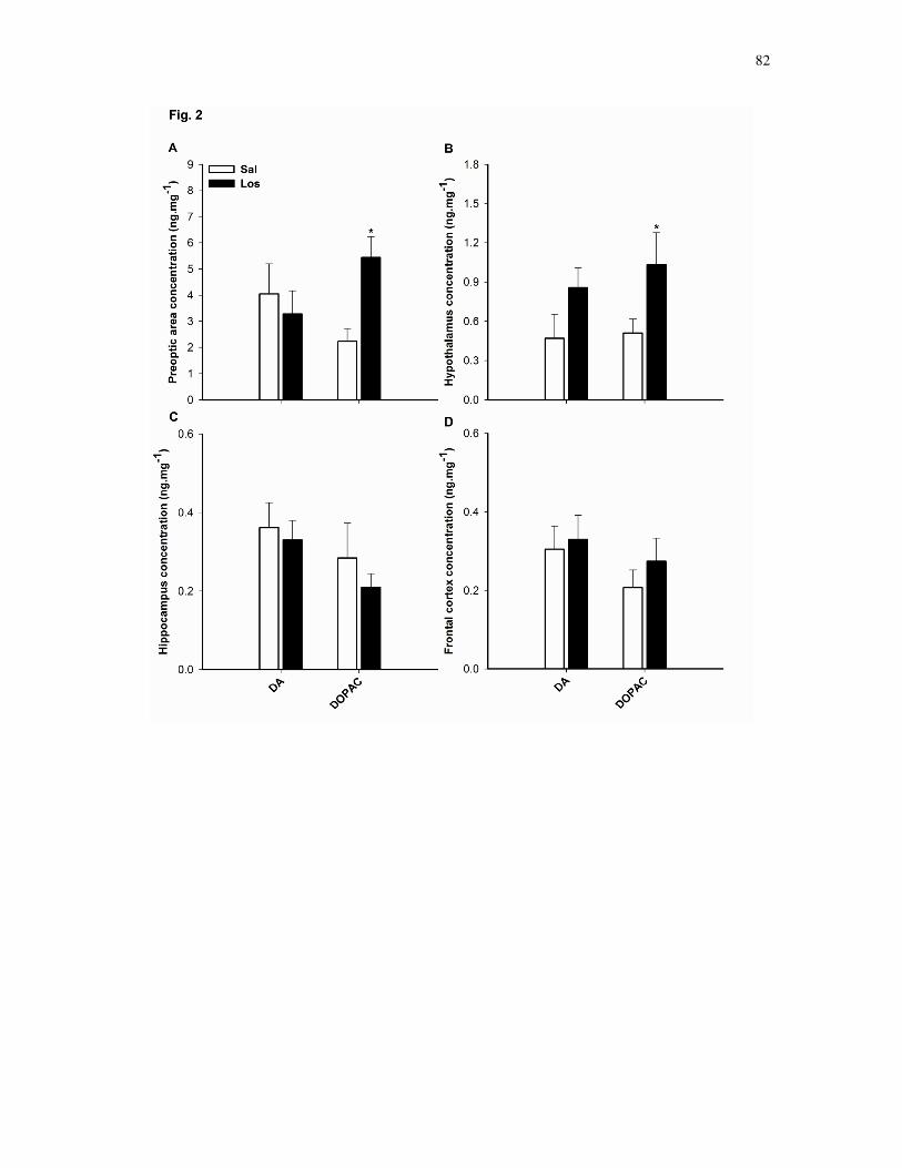
82
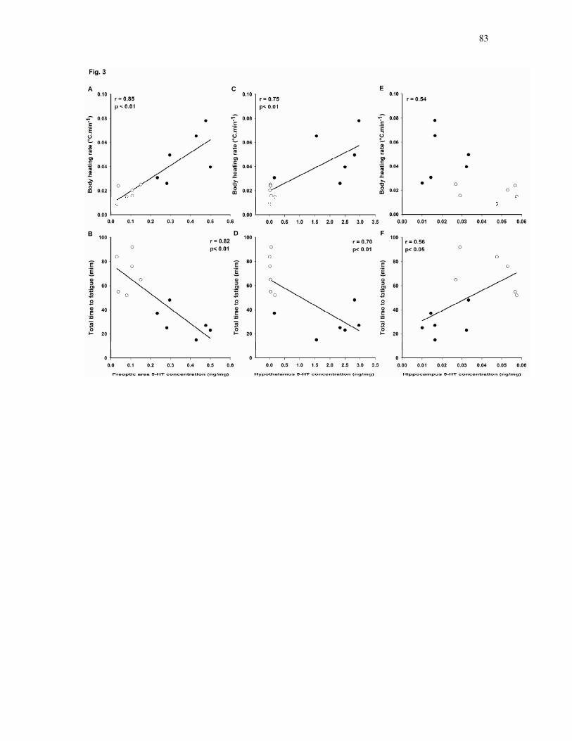
83

84

85
Envolvimento do núcleo paraventricular nos ajustes cardiovasculares
induzidos pelo estresse térmico (Em preparação)
O PVN está intimamente envolvido nos ajustes simpáticos
cardiovasculares induzidos pelo aquecimento, e conseqüente dissipação de
calor através da vasoconstrição visceral e vasodilatação da pele para
redistribuição do fluxo sanguíneo para a periferia. No presente estudo, RSNA,
MAP, HR, temperatura corporal (Tb) e temperatura da cauda (Ttail) foram
mensuradas em ratos anestesiados submetidos ao estresse térmico. Os ratos
foram separados aleatoriamente em grupos para receber microinjeção de CSF,
lidocaina ou L-NMMA bilateralmente no PVN. O estresse térmico foi induzido por
aumento da temperatura do cobertor térmico entre 37 e 43°C durante 30
minutos. Este estímulo térmico resultou em inibição do aumento da RSNA após
o bloqueio do PVN com lidocaína, sugerindo redução da vasoconstrição renal. O
aumento da MAP também foi bloqueado e o aumento da HR atenuado
significativamente, contudo o limiar térmico para vasodilatação da cauda não foi
afetado. RSNA, HR e MAP em ratos tratados com L-NMMA aumentaram
proporcionalmente ao estresse térmico. Além disso, ∆Tb até a vasodilatação da
cauda foi superior após injeção de L-NMMA, indicando prejuízo na dissipação de
calor. Os dados indicam que o PVN é fundamental nos ajustes da atividade
simpática que contribuem para a regulação cardiovascular durante o estresse
térmico passível de influenciar a redistribuição sanguínea para a periferia. Além
disso, um possível mecanismo pelo qual o aquecimento aumenta a perda de

86
calor através da vasodilatação cutânea ocorre via disponibilidade de óxido nítrico
no PVN.

87
Involvement of the paraventricular nucleus on heat stress-induced cardiovascular adjustments
Laura H. R. Leite,1;2
Hong Zheng,1 Cândido C. Coimbra,
2 and Kaushik P. Patel
1
1Department of Cellular and Integrative Physiology, University of Nebraska Medical Center, Omaha,
Nebraska 68198-5850; and 2Department of Physiology and Biophysics, Institute of Biological Sciences,
Federal University of Minas Gerais, Belo Horizonte, Minas Gerais, Brazil.
Running head: PVN mechanisms in thermoregulation
Corresponding author:
Kaushik P. Patel, PhD
Department of Cellular and Integrative Physiology
University of Nebraska Medical Center
985850 Nebraska Medical Center
Omaha, NE 68198-5850
Ph: (402) 559-8369 Fax: (402) 559-4438
Email: [email protected]

88
ABSTRACT
We hypothesized that the paraventricular nucleus (PVN) is intimately involved in the heating
mediated sympathetic cardiovascular adjustments and consequent heat dissipation through vasoconstriction
of the viscera and vasodilation of the skin to redistribute blood flow to the periphery. In the present study,
renal sympathetic nerve activity (RSNA), mean arterial blood pressure (MAP), heart rate (HR), body
temperature (Tb), and tail skin temperature (Ttail) were measured in α-chloralose-urethane-anesthetized
rats submitted to heat stress. The rats were randomly assigned to groups receiving microinjection of vehicle
(CSF), Lidocaine or NG-monomethyl-L-arginine (L-NMMA) bilaterally into the PVN. Heat stress was
induced by a heating pad with a graded increase in temperature from 37 to 43°C during 30 min. This heat
stimulus resulted in blunted RSNA response after blockade of the PVN with Lidocaine, suggesting a
decreased renal vasoconstriction. MAP was also blunted and HR increase significantly attenuated,
however, body temperature for peripheral tail vasodilation was not affected by Lidocaine treatment. RSNA,
HR and MAP in L-NMMA injected rats increased according to the heating stress. Nevertheless, a higher
∆Tb until tail vasodilation was shown by L-NMMA injected rats, which is an indicative of impaired
peripheral heat loss. In conclusion, the PVN is critical for enhancing sympathetic activity to heating,
contributing to cardiovascular adjustments elicited by heat stress that influence core blood redistribution to
the periphery. Furthermore, one possible mechanism by which heating increases heat loss through
peripheral vasodilation is via nitric oxide within the PVN.
Key words: Paraventricular nucleus, nitric oxide, thermoregulation, renal sympathetic nerve activity,
vasodilation threshold.

89
INTRODUCTION
In order to maintain body temperature (Tb) constant at ~37° C, the balance between heat
production and heat loss is accurately adjusted by coordinated thermoregulatory responses controlled by the
central nervous system (Webb, 1995). As a result of increases in Tb, heat loss mechanisms are stimulated
mainly through modulation of sympathetic activation (Kenney et al., 2001; Kregel et al., 1990). During
heat stress, the elevation of Tb is proportionally followed by increases in renal sympathetic nerve activity
(RSNA), heart rate (HR), mean arterial pressure (MAP), as well peripheral sympathetic inhibition (Gisolfi
et al., 1991; Kenney et al., 2001; Kregel and Gisolfi, 1994). These effects are critical for heat dissipation
through blood flow redistribution from the core to the skin surface, being a consequence of sympathetically
controlled vasoconstriction of the viscera and vasodilation of the skin (Kanosue et al., 1994; Morrison,
2001; Smith et al., 1998).
The paraventricular nucleus of the hypothalamus (PVN) is a central site for the integration of
sympathetic nerve activity (Li and Patel., 2003; Patel, 2000) and recently described to be involved in the
control of Tb (Nagashima et al., 2000; Romanovsky, 2007). The PVN contains thermosensitive neurons
that are activated during heat stress (Bratincsak and Palkovits, 2004; Cham and Badoer, 2006, 2008).
Additionally, evidences indicate that projections arise from the PVN to other thermoregulatory centers to
influence the sympathetic activity of thermoregulatory effectors organs like the brown adipose tissue,
vasculature of the rat’s tail, as well as the kidneys and gut (Kazuyuki et al., 1998; Kenney et al., 2003;
Smith et al., 1998). It has also been demonstrated that neuronal inhibition of the PVN prevents the
reduction in renal blood flow in response to increased Tb (Cham and Badoer, 2008), supporting that the
PVN may be involved in the control of cardiovascular adjustments that influence blood redistribution
induced by changes in Tb (Kazuyuki et al., 1998; Nagashima et al., 2000).
Nitric oxide (NO), which is diffusely found in the PVN (Cham et al., 2006, 2007), was proven to
exert thermoregulatory effects characterized by hypothermia (Almeida and Branco, 2001; Mathai et al.,
2004). Acting centrally, NO plays a tonic role in reducing Tb due to increased heat loss through peripheral
vasodilation (Lacerda et al., 2005). Within the PVN, the injection of the NO synthase inhibitor L-NMMA
induces vasoconstriction of the kidneys, while treatment with the NO donor sodium nitroprusside elicits the
opposite response (Zhang et al., 1997). These results are in agreement with the general idea that NO
decreases sympathetic tonus (Patel et al., 2001; Simon, 1998), and suggest that NO may be a potential
signaling molecule involved in the control of blood flow redistribution that enables heat dissipation during
heat stress.
We hypothesized that the PVN is intimately involved in heating mediated sympathetic
cardiovascular adjustments and consequent heat dissipation through blood flow redistribution to the
periphery. In the present study, we determined the effect of PVN blockade with Lidocaine during heat
stress on RSNA, which was considered an indicator of sympathetic outflow to the kidneys and a reliable
representation of renal vasomotor tonus and, consequently, blood flow. At the same moment, tail

90
temperature (Ttail) was measured as an index of peripheral vasculature tonus. Since NO is a
thermoregulatory heat loss effector, as well as sympathoinhibitory, we also investigated if the
cardiovascular actions of the PVN due to heat stress would be dependent on the availability of NO within
the nucleus. Thus, the purpose of the present study was to examine how the PVN affects RSNA, HR, MAP,
heat balance and Tb threshold for peripheral vasodilation during heat stress and if NO within the nucleus is
involved in such regulations.
METHODS
All rats were fed and housed according to approved protocol by the Institutional Animal Care and
Use Committee of the University of Nebraska Medical Center and conformed to the guidelines for the care
and use of laboratory animals of the National Institutes of Health and the American Physiological Society.
Male Sprague-Dawley rats (220–320 g body wt; Sasco Breeding Laboratories, Omaha, NE) were
allowed to acclimate to our animal care facility for 1 wk before use. On the day of the experiment, each rat
was anesthetized with urethane (0.75 g/kg ip) and α-chloralose (70 mg/kg ip), and the left femoral vein was
cannulated with polyethylene (PE-50) tubing for injection of supplemental anesthesia. The left femoral
artery was cannulated and connected to a computer-driven data-recording and -analyzing system (MacLab;
AD Instruments, Mountainview, CA) via a pressure transducer (model P231D; Gould) for recording of
arterial blood pressure and HR. The trachea was intubated to facilitate spontaneous ventilation.
Placement of Microinjection Cannula in the PVN
The anesthetized rat was placed in a stereotaxic apparatus (David Kopf Instruments, Tujunga,
CA). A longitudinal incision was made on the head and the bregma was exposed. The coordinates for the
PVN (1.5 mm posterior to bregma, 0.4 mm lateral to the midline, and 7.8 mm ventral to the dura) were
determined from the atlas of Paxinos and Watson (Paxinos and Watson, 1986). A small burr hole was made
in the skull. For the microinjections, a thin needle (0.5 mm OD, 0.1 mm ID) connected to a microsyringe
(0.5 µL; model 7000.5; Hamilton) was lowered into the PVN bilaterally.
Recording of Renal Sympathetic Nerve Activity
The left kidney was exposed through a retroperitoneal flank incision. A branch of the renal nerve
was isolated from the fat and connective tissue. The nerve was placed on a pair of thin bipolar platinum
electrodes. The nerve-electrode junction was insulated electrically from the surrounding tissue with silicone
gel (Sil-Gel 604 AB; Wacker). The electrical signal was amplified (10.000 times) with a Grass amplifier
(model P55) with high- and low-frequency cutoff of 1.000 and 100 Hz, respectively. The output signal
from the amplifier was rectified and integrated (20-ms time constant) and stored for later analysis using a
computer-run data acquisition system (MacLab). The signal recorded at the end of the experiment (after the
rat was dead) was considered background noise. Nerve discharge was calculated by subtraction of the
background noise from the actual recorded value. Basal nerve discharge was defined by subtraction of the
background noise from the actual nerve discharge before administration of drugs into the PVN. The peak

91
response of RSNA to administration of drugs into the PVN during the experiment (averaged over 20–30 s)
was subsequently expressed as percent change from baseline.
Recording of Colonic Temperature and Tail Skin Temperature
Colonic temperature was taken as Tb and was measured using a thermistor probe (model 401,
Yellow Springs Instruments, USA). The thermistor probe was inserted 4 cm past the anal sphincter after
fecal pellets had been removed from the colon by gentle external massage. Ttail was measured using a
probe (series 409-B,l Yellow Springs Instruments) taped to the dorsal surface of the skin, 10 mm from the
base of the tail. Tb and Ttail were used to determine ∆Tb until the moment at which Ttail clearly begins to
increase (vasodilation threshold).
Experimental Protocol
After the surgical procedures, the animals were allowed to stabilize for at least 20-30 min before
being submitted to the experimental protocol. RSNA, MAP, HR, Tb and Ttail of the rats were recorded
continuously during heat stress. Heat stress was produced in rats by increasing the temperature of a heating
pad (Staco, Model 3PN 1010BV) from 37 to 43°C at a rate of 1.5°C every 6 minutes in an interval of 30
minutes. The rats were randomly assigned to groups receiving bilateral microinjection into the PVN of
artificial cerebroespinal fluid (CSF; 100 nL/side), lidocaine (1%; 200 nL/side) or L-NMMA (200 pmol,
100nL/side) immediately before the beginning of heat stress. Throughout the experimental procedure, the
rat’s tail was maintained away from the surface of the heating pad.
Control experiments were carried out as well. The animals were submitted to similar experimental
procedures, but instead of being submitted to heat stress after injection of the drugs, their Tb was kept
stable during 30 min while RSNA, MAP, HR and Ttail were recorded.
Brain Histology
After the experiment, the rats were killed and the brains removed and fixed in 10% formalin for at
least 24 h. The brains were then frozen, and serial transverse sections (30 µm) were cut with a cryostat (-
18°C). The sections were mounted on microscope slides and stained using 1% neutral red. The location of
the injection within the PVN was verified under a microscope with X 40 magnification (Fig.1). The
microinjections that terminated in the boundaries of the PVN were considered effective. For the purposes
of analysis, only the animals in which dye was deposited within or < 0.5 mm from the boundaries of the
PVN were considered histologically targeted. The 100- to 200-nL injection volumes targeting the PVN
would be expected to distribute the drug in or within < 0.5 mm from the rostrocaudal and mediolateral
boundaries of the PVN.
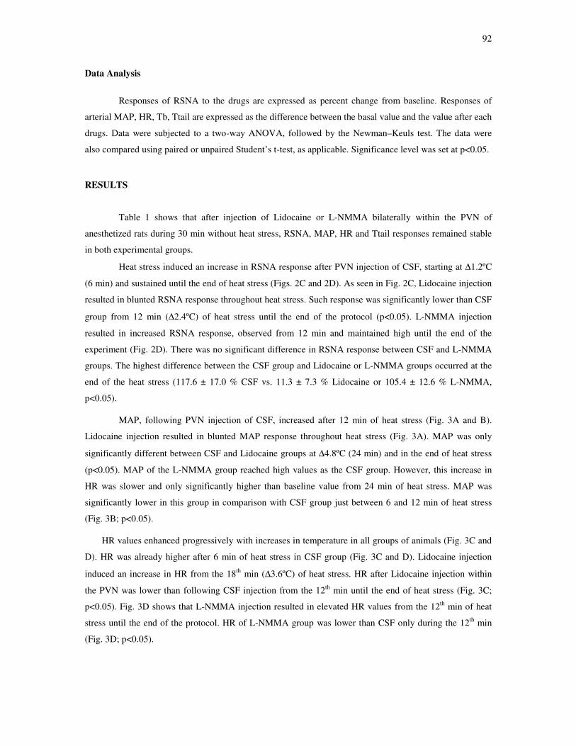
92
Data Analysis
Responses of RSNA to the drugs are expressed as percent change from baseline. Responses of
arterial MAP, HR, Tb, Ttail are expressed as the difference between the basal value and the value after each
drugs. Data were subjected to a two-way ANOVA, followed by the Newman–Keuls test. The data were
also compared using paired or unpaired Student’s t-test, as applicable. Significance level was set at p<0.05.
RESULTS
Table 1 shows that after injection of Lidocaine or L-NMMA bilaterally within the PVN of
anesthetized rats during 30 min without heat stress, RSNA, MAP, HR and Ttail responses remained stable
in both experimental groups.
Heat stress induced an increase in RSNA response after PVN injection of CSF, starting at ∆1.2ºC
(6 min) and sustained until the end of heat stress (Figs. 2C and 2D). As seen in Fig. 2C, Lidocaine injection
resulted in blunted RSNA response throughout heat stress. Such response was significantly lower than CSF
group from 12 min (∆2.4ºC) of heat stress until the end of the protocol (p<0.05). L-NMMA injection
resulted in increased RSNA response, observed from 12 min and maintained high until the end of the
experiment (Fig. 2D). There was no significant difference in RSNA response between CSF and L-NMMA
groups. The highest difference between the CSF group and Lidocaine or L-NMMA groups occurred at the
end of the heat stress (117.6 ± 17.0 % CSF vs. 11.3 ± 7.3 % Lidocaine or 105.4 ± 12.6 % L-NMMA,
p<0.05).
MAP, following PVN injection of CSF, increased after 12 min of heat stress (Fig. 3A and B).
Lidocaine injection resulted in blunted MAP response throughout heat stress (Fig. 3A). MAP was only
significantly different between CSF and Lidocaine groups at ∆4.8ºC (24 min) and in the end of heat stress
(p<0.05). MAP of the L-NMMA group reached high values as the CSF group. However, this increase in
HR was slower and only significantly higher than baseline value from 24 min of heat stress. MAP was
significantly lower in this group in comparison with CSF group just between 6 and 12 min of heat stress
(Fig. 3B; p<0.05).
HR values enhanced progressively with increases in temperature in all groups of animals (Fig. 3C and
D). HR was already higher after 6 min of heat stress in CSF group (Fig. 3C and D). Lidocaine injection
induced an increase in HR from the 18th
min (∆3.6ºC) of heat stress. HR after Lidocaine injection within
the PVN was lower than following CSF injection from the 12th
min until the end of heat stress (Fig. 3C;
p<0.05). Fig. 3D shows that L-NMMA injection resulted in elevated HR values from the 12th
min of heat
stress until the end of the protocol. HR of L-NMMA group was lower than CSF only during the 12th
min
(Fig. 3D; p<0.05).

93
The effect of heat stress on Tb is shown in Fig. 3. Heat stress induced a rapid increase in Tb in all
groups, which was already observed from the 9th
min after injection of CSF (Fig. 4A and B). From the 8th
min of heat stress, Tb was significantly higher than baseline value in the Lidocaine group (Fig. 4A).
Similarly to the response observed with CSF injection, L-NMMA induced an increase in Tb from the 9th
min until the end of heat stress (Fig. 4B). There were no significant differences between the CSF and
Lidocaine groups, as well as between the CSF and L-NMMA groups, throughout heat stress (Fig. 4A and
B).
As illustrated in Fig. 4C and D, Ttail increased within 15, 13 and 16 min of heat stress in CSF,
Lidocaine and L-NMMA groups, respectively, indicating that heat loss mechanisms had been activated.
The increases in Ttail remained similar between CSF and Lidocaine group (Fig. 4C). Although Ttail of L-
NMMA increased mostly in a similar way to CSF group, between the 5th
and 7th
min interval of heat stress
Ttail of these rats was significantly lower (Fig. 4C and D). To assess whether heat loss mechanism was
affected by PVN injection of the drugs, ∆Tb until tail vasodilation was calculated (Fig. 4E and F).
Vasodilation threshold was not different between rats that received CSF and Lidocaine injections within the
PVN (0.3 ± 0.1°C CSF; 0.4 ± 0.2°C Lidocaine). However, results showed that the value was 0.5°C higher
in L-NMMA injected rats compared with CSF injected animals (0.3 ± 0.1°C CSF vs 0.8 ± 0.1°C L-
NMMA; p<0.01).
DISCUSSION
In the present study, blockade of the PVN with Lidocaine resulted in blunted RSNA, suggesting a
decreased renal vasoconstriction, as well as blunted MAP response and attenuated increase in HR to heat
stress. Such treatment did not interference on body temperature threshold for peripheral vasodilation. On
the other hand, L-NMMA injection into the PVN led to higher ∆Tb until tail vasodilation but non-affected
RSNA, MAP and HR. Taken together, these data indicate that the PVN influences the responsiveness of
sympathetic pathways that mediate cardiovascular adaptations required for the control of Tb during heat
stress, particularly by modulating renal sympathetic nerve activity that enables diversion of core blood
volume during heat stress. Furthermore, one mechanism by which heating increases heat loss through
peripheral vasodilation is via NO within the PVN.
Body heating provides a potent stimulus to sympathetic activity in order to stimulate heat dissipation
responses and maintain Tb within safe limits (Kenney et al., 2001; Kregel et al., 1994). During heat stress,
the sympathetic nervous system is capable of altering blood flow of multiple organs nonuniformly by
selectively altering the pattern of sympathetic outflow according to specific vascular beds (Hirai et al.,
1995; Morrison et al., 2001). Heat dissipation through blood redistribution from the viscera to the periphery
is critical for the maintenance of Tb during heat stress. The shift of blood volume from the central to the
peripheral circulation is the result of cardiovascular responses controlled by the sympathetic nervous
system that induce vasoconstriction of the viscera and vasodilation of the skin simultaneously (Kanosue et
al., 1994; Morrison et al., 2001; Smith et al., 1998).

94
The recent involvement of the PVN in thermoregulation awoke the hypothesis that the nucleus may
contribute to the maintenance of Tb by influencing sympathetic outflow that regulates cardiovascular
responses elicited by heat stress (Li & Patel., 2003; Nagashima et al., 2000; Patel, 2000; Romanovsky,
2007). In fact, there is evidence that neuronal inhibition of the PVN with the GABAA agonist muscimol
prevents the normal reduction in renal blood flow in response to increased Tb (Cham and Badoer, 2008). In
the present study, Lidocaine was bilaterally injected to completely block all neuronal transmissions within
the PVN during heat stress. This treatment blunted RSNA response to heating, i.e., renal vasoconstriction,
which is an evidence of accumulation of blood volume in the kidneys that impairs its redistribution to the
skin surface to dissipate heat. During heat stress, up to 60% of cardiac output is redistributed to the surface
of the skin to improve heat loss, this effect being depended mostly on decreased peripheral resistance
(Rowel, 1983). The fact that rats treated with Lidocaine had their vasodilation threshold not altered is an
indicative that the PVN blockade by itself may not interfere with peripheral vasomotor tonus during heat
stress. In rodents, tail skin vasodilation is the primary route of heat loss from the body, being responsible
for the dissipation of an equivalent of 25% of resting heat production (Shellock and Rubin, 1984; Young
and Dawson, 1982). Therefore, although the blood volume of other viscera, such as the gut, are also
important for blood redistribution, the decreased renal sympathetic activation shown by Lidocaine rats
suggests a thermoregulatory deficit. Nevertheless, the apparent normal body temperature for tail
vasodilation possibly made heat loss possible, keeping the increase in Tb proportional to the heat stimulus.
In control rats, heat stress resulted in increased MAP and HR responses. This hemodynamic pattern is
the result of sympathetic activation, which increases cardiac output, due to enhanced HR, in combination
with a greater redistribution of blood flow from visceral vasculature beds for heat dissipation (Kenney,
2008; Rowell, 1983). In rats treated with Lidocaine, heat stress also resulted in blunted MAP. Although HR
increased in this group, such increase was significantly lowered. During heat stress, both the
vasoconstrictor and the vasodilator systems are critical in blood pressure regulation via the baroreflex
(Crandall et al., 1996). As mentioned before, the redistribution of blood flow to the periphery depends
mainly on the reduction of peripheral resistance (Charkoudian, 2003). On the other hand, the maintenance
of blood pressure depends particularly on vasoconstriction because a large percentage of the cardiac output
is redistributed to the periphery (Rowel, 1983). Since Lidocaine animals had decreased vasoconstriction of
the kidneys, it seems that the decreased MAP is the consequence of the lower sympathetic activation that
may have not only decreased artery resistance of the kidneys, but also of other vascular beds. Thus, it is
reasonable to suggest that the PVN blockade with Lidocaine results in attenuation of sympathetic nervous
system activation to heating and inability to elicit the appropriate cardiovascular adjustments.
Evidences showing the involvement of brain NO in thermoregulatory pathways demonstrated that this
neurotransmitter mediates autonomic heat loss mechanisms, as indicated by reduction in Tb associated with
a rise in skin temperature (Eriksson et al., 1997). Conversely, it has also been shown that central blockade
of NO production increases the threshold to activate tail skin vasodilation leading to higher Tb (Lacerda et
al., 2005; Mathai et al., 2004). This is in agreement with the current results that L-NMMA injection within
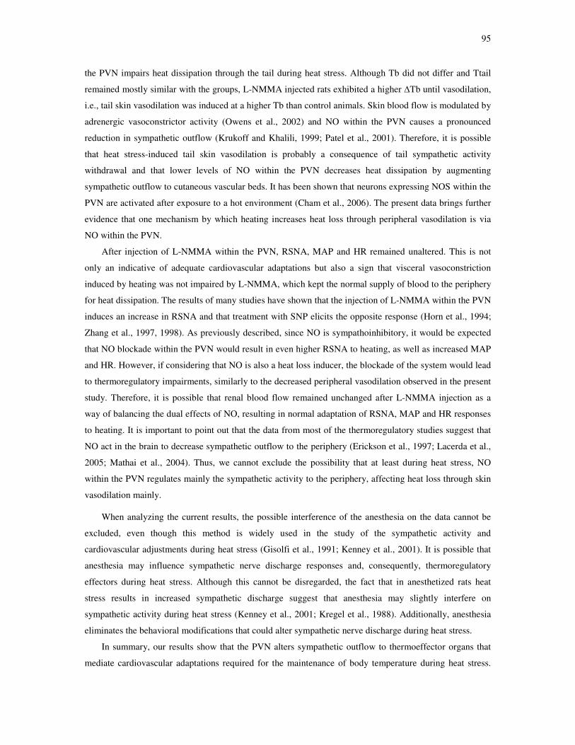
95
the PVN impairs heat dissipation through the tail during heat stress. Although Tb did not differ and Ttail
remained mostly similar with the groups, L-NMMA injected rats exhibited a higher ∆Tb until vasodilation,
i.e., tail skin vasodilation was induced at a higher Tb than control animals. Skin blood flow is modulated by
adrenergic vasoconstrictor activity (Owens et al., 2002) and NO within the PVN causes a pronounced
reduction in sympathetic outflow (Krukoff and Khalili, 1999; Patel et al., 2001). Therefore, it is possible
that heat stress-induced tail skin vasodilation is probably a consequence of tail sympathetic activity
withdrawal and that lower levels of NO within the PVN decreases heat dissipation by augmenting
sympathetic outflow to cutaneous vascular beds. It has been shown that neurons expressing NOS within the
PVN are activated after exposure to a hot environment (Cham et al., 2006). The present data brings further
evidence that one mechanism by which heating increases heat loss through peripheral vasodilation is via
NO within the PVN.
After injection of L-NMMA within the PVN, RSNA, MAP and HR remained unaltered. This is not
only an indicative of adequate cardiovascular adaptations but also a sign that visceral vasoconstriction
induced by heating was not impaired by L-NMMA, which kept the normal supply of blood to the periphery
for heat dissipation. The results of many studies have shown that the injection of L-NMMA within the PVN
induces an increase in RSNA and that treatment with SNP elicits the opposite response (Horn et al., 1994;
Zhang et al., 1997, 1998). As previously described, since NO is sympathoinhibitory, it would be expected
that NO blockade within the PVN would result in even higher RSNA to heating, as well as increased MAP
and HR. However, if considering that NO is also a heat loss inducer, the blockade of the system would lead
to thermoregulatory impairments, similarly to the decreased peripheral vasodilation observed in the present
study. Therefore, it is possible that renal blood flow remained unchanged after L-NMMA injection as a
way of balancing the dual effects of NO, resulting in normal adaptation of RSNA, MAP and HR responses
to heating. It is important to point out that the data from most of the thermoregulatory studies suggest that
NO act in the brain to decrease sympathetic outflow to the periphery (Erickson et al., 1997; Lacerda et al.,
2005; Mathai et al., 2004). Thus, we cannot exclude the possibility that at least during heat stress, NO
within the PVN regulates mainly the sympathetic activity to the periphery, affecting heat loss through skin
vasodilation mainly.
When analyzing the current results, the possible interference of the anesthesia on the data cannot be
excluded, even though this method is widely used in the study of the sympathetic activity and
cardiovascular adjustments during heat stress (Gisolfi et al., 1991; Kenney et al., 2001). It is possible that
anesthesia may influence sympathetic nerve discharge responses and, consequently, thermoregulatory
effectors during heat stress. Although this cannot be disregarded, the fact that in anesthetized rats heat
stress results in increased sympathetic discharge suggest that anesthesia may slightly interfere on
sympathetic activity during heat stress (Kenney et al., 2001; Kregel et al., 1988). Additionally, anesthesia
eliminates the behavioral modifications that could alter sympathetic nerve discharge during heat stress.
In summary, our results show that the PVN alters sympathetic outflow to thermoeffector organs that
mediate cardiovascular adaptations required for the maintenance of body temperature during heat stress.

96
Furthermore, the PVN is a site in the brain in which NO acts to facilitate heat loss by influencing the
sympathetic outflow to the periphery.
ACKNOLEGMENTS
The technical assistance of Lirong Xu and Xuefei Liu is greatly appreciated.
GRANTS
This work was supported by National Institutes of Health Grant HL6222 and Conselho Nacional
de Desenvolvimento Científico e Tecnológico (CNPq).

97
REFERENCES
1. Almeida MC and Branco LG. Role of nitric oxide in insulin-induced hypothermia in rats. Brain
Res Bull 54: 49-53, 2001.
2. Bratincsak A and Palkovits M. Activation of brain areas in rat following warm and cold ambient
exposure. Neuroscience 127: 385-397, 2004.
3. Cham JL and Badoer E. Exposure to a hot environment can activate spinally projecting and
nitrergic neurones in the lower brainstem in the rat. Exp Physiol 92: 529-540, 2007.
4. Cham JL and Badoer E. Hypothalamic paraventricular nucleus is critical for renal
vasoconstriction elicited by elevations in body temperature. Am J Physiol Renal Physiol 294: F309-315,
2008.
5. Cham JL, Klein R, Owens NC, Mathai M, McKinley M, and Badoer E. Activation of spinally
projecting and nitrergic neurons in the PVN following heat exposure. Am J Physiol Regul Integr Comp
Physiol 291: R91-101, 2006.
6. Charkoudian N. Skin blood flow in adult human thermoregulation: how it works, when it does
not, and why. Mayo Clin Proc 78: 603-612, 2003.
7. Crandall CG, Johnson JM, Kosiba WA, and Kellogg DL, Jr. Baroreceptor control of the
cutaneous active vasodilator system. J Appl Physiol 81: 2192-2198, 1996.
8. Eriksson S, Hjelmqvist H, Keil R, and Gerstberger R. Central application of a nitric oxide
donor activates heat defense in the rabbit. Brain Res 774: 269-273, 1997.
9. Gisolfi CV, Matthes RD, Kregel KC, and Oppliger R. Splanchnic sympathetic nerve activity
and circulating catecholamines in the hyperthermic rat. J Appl Physiol 70: 1821-1826, 1991.
10. Hirai T, Musch TI, Morgan DA, Kregel KC, Claassen DE, Pickar JG, Lewis SJ, and Kenney
MJ. Differential sympathetic nerve responses to nitric oxide synthase inhibition in anesthetized rats. Am J
Physiol 269: R807-813, 1995.
11. Horn T, Smith PM, McLaughlin BE, Bauce L, Marks GS, Pittman QJ, and Ferguson AV.
Nitric oxide actions in paraventricular nucleus: cardiovascular and neurochemical implications. Am J
Physiol 266: R306-313, 1994.
12. Kanosue K, Yanase-Fujiwara M, and Hosono T. Hypothalamic network for thermoregulatory
vasomotor control. Am J Physiol 267: R283-288, 1994.
13. Kazuyuki K, Hosono T, Zhang YH, and Chen XM. Neuronal networks controlling
thermoregulatory effectors. Prog Brain Res 115: 49-62, 1998.
14. Kenney MJ, Musch TI, and Weiss ML. Renal sympathetic nerve regulation to heating is altered
in rats with heart failure. Am J Physiol Heart Circ Physiol 280: H2868-2875, 2001.
15. Kenney MJ, Weiss ML, Mendes T, Wang Y, and Fels RJ. Role of paraventricular nucleus in
regulation of sympathetic nerve frequency components. Am J Physiol Heart Circ Physiol 284: H1710-1720,
2003.

98
16. Kenney WL. Human cardiovascular responses to passive heat stress. J Physiol 586: 3, 2008.
17. Kregel KC and Gisolfi CV. Circulatory responses to vasoconstrictor agents during passive
heating in the rat. J Appl Physiol 68: 1220-1227, 1990.
18. Kregel KC, Stauss H, and Unger T. Modulation of autonomic nervous system adjustments to
heat stress by central ANG II receptor antagonism. Am J Physiol 266: R1985-1991, 1994.
19. Kregel KC, Wall PT, and Gisolfi CV. Peripheral vascular responses to hyperthermia in the rat. J
Appl Physiol 64: 2582-2588, 1988.
20. Krukoff TL and Khalili P. Stress-induced activation of nitric oxide-producing neurons in the rat
brain. J Comp Neurol 377: 509-519, 1997.
21. Lacerda AC, Marubayashi U, and Coimbra CC. Nitric oxide pathway is an important
modulator of heat loss in rats during exercise. Brain Res Bull 67: 110-116, 2005.
22. Li YF and Patel KP. Paraventricular nucleus of the hypothalamus and elevated sympathetic
activity in heart failure: the altered inhibitory mechanisms. Acta Physiol Scand 177: 17-26, 2003.
23. Morrison SF. Differential regulation of sympathetic outflows to vasoconstrictor and
thermoregulatory effectors. Ann N Y Acad Sci 940: 286-298, 2001.
24. Nagashima K, Nakai S, Tanaka M, and Kanosue K. Neuronal circuitries involved in
thermoregulation. Auton Neurosci 85: 18-25, 2000.
25. Owens NC, Ootsuka Y, Kanosue K, and McAllen RM. Thermoregulatory control of
sympathetic fibres supplying the rat's tail. J Physiol 543: 849-858, 2002.
26. Patel KP. Role of paraventricular nucleus in mediating sympathetic outflow in heart failure. Heart
Fail Rev 5: 73-86, 2000.
27. Patel KP, Li YF, and Hirooka Y. Role of nitric oxide in central sympathetic outflow. Exp Biol
Med (Maywood) 226: 814-824, 2001.
28. Paxinos G and Watson C. The Rat Brain in Stereotaxic Coordinates. Orlando, FL: Academic,
1986.
29. Romanovsky AA. Thermoregulation: some concepts have changed. Functional architecture of the
thermoregulatory system. Am J Physiol Regul Integr Comp Physiol 292: R37-46, 2007.
30. Rowell LB. Cardiovascular aspects of human thermoregulation. Circ Res 52: 367-379, 1983.
31. Shellock FG and Rubin SA. Temperature regulation during treadmill exercise in the rat. J Appl
Physiol 57: 1872-1877, 1984.
32. Simon E. Nitric oxide as a peripheral and central mediator in temperature regulation. Amino
Acids 14: 87-93, 1998.
33. Smith JE, Jansen AS, Gilbey MP, and Loewy AD. CNS cell groups projecting to sympathetic
outflow of tail artery: neural circuits involved in heat loss in the rat. Brain Res 786: 153-164, 1998.
34. Webb P. The physiology of heat regulation. Am J Physiol 268: R838-850, 1995.
35. Young AA and Dawson NJ. Evidence for on-off control of heat dissipation from the tail of the
rat. Can J Physiol Pharmacol 60: 392-398, 1982.
99
36. Zhang K, Mayhan WG, and Patel KP. Nitric oxide within the paraventricular nucleus mediates
changes in renal sympathetic nerve activity. Am J Physiol 273: R864-872, 1997.
37. Zhang K and Patel KP. Effect of nitric oxide within the paraventricular nucleus on renal
sympathetic nerve discharge: role of GABA. Am J Physiol 275: R728-734, 1998.

100
FIGURE LEGENDS
Fig. 1. Schematic transverse sections of the rat hypothalamic PVN showing the center of the microinjection
sites of CSF, Lidocaine and L-NMMA .
Fig. 2: Effect of heat stress on renal sympathetic nerve activity (∆RSNA) after bilateral PVN injection of
CSF, Lidocaine or L-NMMA. A and B: segments of original recordings from individual rats; C and D:
graphical representation. Values expressed as mean ± SEM. n = 6/group. + Significantly different from
corresponding basal value. ∗ Significant difference between groups, p<0.05.
Fig. 3: Effect of heat stress over time on mean arterial pressure (∆MAP; A and B) and heart rate (∆HR; C
and D) after bilateral PVN injection of CSF, Lidocaine or L-NMMA. Values expressed as mean ± SEM. n
= 6/group. + Significantly different from corresponding basal value. ∗ Significant difference between
groups, p<0.05.
Fig. 4: Effect of heat stress over time on body temperature (∆Tb; A and B), skin tail temperature (∆Ttail; C
and D) and on ∆Tb until tail skin vasodilation (E and F) after bilateral PVN injection of CSF, Lidocaine or
L-NMMA. Values expressed as mean ± SEM. n = 6/group. + Significantly different from basal value until
30 min. ∗ Significant difference between groups, p<0.05.

101
FIGURES AND TABLES
Fig. 1
0.5m
�
�
¤ *
�
�
�
�
*
�
�
¤
*
*
*
�
* �
* * �
¤
�
* * ¤
�
* *
¤ CSF
∗∗∗∗ Lidocaine
••••

102
TABLE 1
Table 1. Effect of bilateral injection within the PVN of Lidocaine or L-NMMA on ∆RSNA, MAP, HR, and
Ttail over time, without changes in temperature in anesthetized rats.
Lidocaine (n=3) L-NMMA (n=4)
Baseline 18 min 30 min Baseline 18 min 30min
∆∆∆∆RSNA (%) - -4.6±2.1 -12.7±4.9 - 18.4±10.6 13.8±8.3
MAP (mmHg) 105±5 108±3 105±4 92±8 99±7 92±9
HR (bpm) 339±29 345±20 350±17 365±11 347±43 369±10
Ttail (°C) 28.2±0.7 27.3±0.5 27.8±0.5 28.4±0.6 28.3±0.6 28.2±0.6
Values are expressed as mean ± SEM.

103
Fig. 2
����Temperature (ºC)
0.0 1.2 2.4 3.6 4.8 6.0
�� ��R
SN
A (
%)
0
20
40
60
80
100
120
140
160 CSFLidocaine
+
+
+
+
***
C
+*
����Temperature (ºC)
0.0 1.2 2.4 3.6 4.8 6.0
�� ��R
SN
A (
%)
0
20
40
60
80
100
120
140
160 CSFL-NMMA
+
+
+
+
D
++
+
++
Lidocaine
RSNA (uV)
30
-30
CSF
RSNA (uV)
30
-30
A
L-NMMA
RSNA (uV)
5 sec
30
-30
CSF
RSNA (uV)
30
-30
B
37.0 38.2 41.8 39.4 40.6 43.0

104
Fig. 3
����Temperature (ºC)
0.0 1.2 2.4 3.6 4.8 6.0
�� ��H
R (
bea
ts/m
in)
0
20
40
60
80
100
120
140
+
++
+
+
+
+
+
+
*
����Temperature (ºC)
0.0 1.2 2.4 3.6 4.8 6.0
�� ��H
R (
bea
ts/m
in)
0
20
40
60
80
100
120
140
+
++
+
+
++
+
**
*
*
1.2 2.4 3.6 4.8 6.0
�� ��M
AP
(mm
Hg
)
-10
-5
0
5
10
15
20
25
30
35 CSFLidocaine
+ +
+
+
* *
A
1.2 2.4 3.6 4.8 6.0
�� ��M
AP
(mm
Hg
)
-10
-5
0
5
10
15
20
25
30
35 CSFL-NMMA
+ +
+
+
+
+
* *
B
C D

105
Fig. 4
6 12 18 24 30
�� ��T
b (
ºC)
-0.5
0.0
0.5
1.0
1.5
2.0
2.5
3.0
3.5 CSFLidocaine
++
+
6 12 18 24 30
�� ��T
b (
ºC)
-0.5
0.0
0.5
1.0
1.5
2.0
2.5
3.0
3.5 CSFL-NMMA
+
+
Time (min)
6 12 18 24 30
�� ��T
tail
(ºC
)
-1
0
1
2
3
4
5
+
+
Time (min)
6 12 18 24 30
�� ��T
tail
(ºC
)
-1
0
1
2
3
4
5
+
+
A B
C D
CSF Lidocaine
�� ��T
b u
nti
l vas
od
ilati
on
(ºC
)
0.0
0.2
0.4
0.6
0.8
CSF L-NMMA
�� ��T
b u
nti
l vas
od
ilati
on
(ºC
)
0.0
0.2
0.4
0.6
0.8
1.0 *E F
* **
106
VII. CONSIDERAÇÕES GERAIS
Os achados do presente estudo evidenciam elevação da taxa metabólica
e redução da eficiência mecânica associada à fadiga precoce após bloqueio
central angiotensinérgico durante o exercício físico. Esses dados demonstram
que a Ang II não está somente envolvida com a dissipação e calor (Leite et al.,
2006), mas também com a produção deste (Leite et al., 2007). O desbalanço
térmico induzido pelo bloqueio central dos receptores AT1 foi determinante para
a redução do desempenho físico uma vez que a hipertermia do exercício foi
mais intensa devido a maior produção de calor não compensado pela dissipação
do mesmo (Leite et al., 2006, 2007). Soma-se a esse efeito a menor eficiência
mecânica, indicativa de maior quantidade de energia metabolicamente produzida
dissipada sob a forma de calor. Esses dados estão de acordo com a descrição
da literatura que relata ser a Ang II, atuando tanto perifericamente quanto
centralmente, indutora de hipotermia manifestada através do aumento da
temperatura da cauda e diminuição da taxa metabólica (Fregly & Rowland, 1992,
1993, 1996; Mathai et al., 2000; Wilson & Fregly, 1985 a,b).
O efeito hipertérmico e redução do desempenho físico acarretados pelo
tratamento com Los central estão integrados ao aumento do conteúdo de 5-HT
em importantes centros termorregulatórios, como a área pré-óptica e o
hipotálamo. O aumento da atividade serotonérgica está associada à letargia e
perda de motivação, resultando em alteração do desempenho físico
(Blomstrand, 2006; Meeusen et al., 2007). Além disso, as evidências apontam

107
que a 5-HT induz efeitos termorregulatórios caracterizados por hipertermia
(Soares et al., 2004, 2007), estando a fadiga central devido ao acúmulo de calor
elevado em ratos em exercício relacionada ao aumento desse neurotransmissor
na área pré-óptica (Caperuto et al., 2009; Rodrigues et al., 2009; Soares et al.,
2007). Os dados aqui apresentados corroboram com essas evidências ao
mostrar que o sistema angiotensinérgico também regula a temperatura corporal
interna através da modulação do conteúdo de 5-HT em núcleos
termorregulatórios. O mesmo controle possivelmente ocorre em centros motores
como o hipocampo. Nessa região ocorreu diminuição do nível de 5-HT após
bloqueio dos receptores AT1 intimamente relacionada ao menor desempenho
físico, sugerindo haver uma resposta seletiva da 5-HT específica e dependente
do centro nervoso.
A DA é outro neurotransmissor associado à melhora do desempenho
físico, apesar de acarretar elevação da temperatura corporal no ponto de fadiga
e do acúmulo de calor durante o exercício (Balthazar et al., 2009; Foley &
Fleshner, 2008; Hasegawa et al., 2008). No entanto, após inibição
angiotensinérgica, não se verificou qualquer alteração do conteúdo de DA nas
regiões cerebrais estudadas. Essa resposta condiz com os poucos dados
demonstrando que a injeção intracerebroventricular de Ang II não modifica a
concentração de DA no PVN ou hipotálamo anterior (Qadri et al., 1991; Stadler
et al., 1992). De qualquer forma, essa monoamina interagindo com a 5-HT no
hipotálamo parece exercer ação importante no estabelecimento da hipertermia e
redução do tempo de fadiga após inibição dos receptores AT1. A alta razão

108
hipotalâmica 5-HT:DA, compatível com menor desempenho físico, mostrou-se
diretamente associada ao reduzido tempo de exercício e alta taxa de
aquecimento corporal dos animais Los.
Os efeitos metabólicos da Ang II, principalmente sobre a homeostase da
glicose (Coimbra et al., 1999, Machado et al., 1995 a,b, 1998; Mihessen-Neto et
al., 1996), também estão atuantes durante o exercício físico. No presente
estudo, o bloqueio central dos receptores AT1 resultou em hiperglicemia e maior
mobilização de ácidos graxos do tecido adiposo durante o exercício físico. Além
disso, o tratamento com Los em ratos em exercício elevou o fluxo glicolítico,
evidenciado pelo aumento plasmático de lactato. Em função dessa maior
disponibilidade de substratos energéticos, o reduzido desempenho físico desses
animais possivelmente não se deveu ao prejuízo na oferta, distribuição e
utilização de fontes de energia. Essa resposta metabólica do Los à atividade
física aparentemente foi semelhante a situações indutoras de aumento de fluxo
simpático periférico, como a neurocitoglicopenia (Coimbra & Migliorini, 1986;
Ribeiro-de-Oliveira et al., 1999), hipotensão hemorrágica (Silveira et al., 2003) e
exposição ao calor (Ferreira et al., 1999) que também exibiram hiperglicemia
aumento de lactato e ácidos graxos livres. É razoável sugerir que a mudança na
mobilização de substratos após bloqueio angiotensinérgico deve-se a maior
ativação do sistema simpático, como observado previamente através do
aumento da vasoconstrição da cauda e do limiar térmico para vasodilatação
cutânea em ratos em exercício tratados com Los (Leite et al., 2006). Essa
ativação simpática elevada foi verificada mesmo em baixa intensidade de
109
exercício, como 20% do trabalho máximo, a qual acarretou em maior
concentração plasmática de glicose, lactato e ácidos graxos livres. O fato de
ambos os grupos atingirem o mesmo nível de intensidade de exercício também
sugere que a atividade simpática foi intensificada e estimulada precocemente,
evidenciando maior esforço para execução de mesmo trabalho.
Contudo, ainda são desconhecidos os mecanismos através dos quais o
bloqueio angiotensinérgico resulta em efeitos semelhantes àqueles
consequentes da ativação simpática. Uma possível interação entre a Ang II e o
óxido nítrico, já verificada em outros centros nervosos como o PVN, pode ser um
dos mecanismos responsáveis por tal efeito (Li et al., 2006). Demonstrou-se que
dentro do PVN, a Ang II interage com o óxido nítrico, um inibidor simpático,
induzindo sua secreção, a qual modera o efeito excitatório da Ang II sobre o
fluxo simpático, possivelmente através de retroalimentação inibitória (Li et al.,
2006). Experimentos prévios do laboratório demonstraram que o bloqueio do
sistema óxido nitrérgico central resulta em efeitos sobre o balanço térmico e
metabólicos semelhantes aos observados após bloqueio angotensinérgico
(Lacerda et al., 2005, 2006 a,b; Leite et al., 2006, 2007, 2009). Portanto, é
possível que ocorra uma interação entre a Ang II e óxido nítrico em sítios
controladores da homeostase térmica e metabólica.
O PVN surge como um sítio que se encaixa nas características citadas
acima, particularmente no que concerne a termorregulação. O bloqueio do PVN
com lidocaína durante o estresse térmico resultou em inibição da RSNA, assim
como da MAP, e aumento menos acentuado da HR. No entanto, tal tratamento

110
não afetou o limiar térmico para vasodilatação cutânea. Resposta oposta foi
verificada após injeção bilateral no PVN de L-NMMA, isto é, RSNA, MAP e HR
inalterados, acompanhados de maior limiar térmico para vasodilatação. Os
dados sugerem que, durante o estresse térmico, o PVN tem ação importante na
regulação da atividade simpática que contribui para os ajustes cardiovasculares
responsáveis pela redistribuição de sangue interno para a periferia e perda de
calor. Além disso, um possível mecanismo através do qual o aquecimento eleva
a dissipação cutânea de calor ocorre via disponibilidade de óxido nítrico no PVN.

111
VIII. CONCLUSÕES
O sistema angiotensinérgico central está envolvido no equilíbrio
metabólico e térmico durante o exercício físico ao induzir modificações na
atividade do sistema serotonérgico central.
O bloqueio do receptor AT1 para Ang II durante o exercício físico induz:
1. Aumento do consumo de oxigênio, responsável por elevação de 19% do
custo metabólico em comparação com os animais controle, diminuição
significativa da eficiência mecânica diretamente relacionada com a
precipitação da fadiga. Tal elevação da eficiência mecânica foi verificada
já em baixos níveis de intensidade de exercício durante corrida de mesmo
alcance de intensidade, apesar do reduzido tempo total de exercício.
Esses dados indicam que o sistema angiotensinérgico central induz
ajustes metabólicos durante o exercício físico através da modulação da
produção de calor e melhora da eficiência mecânica;
2. Alteração da mobilização de substratos energéticos, evidenciada pelo
aumento da resposta hiperglicêmica e altos níveis de lactato e ácidos
graxos livres plasmáticos durante o exercício progressivo. Os achados
indicam que o sistema angiotensinérgico central está envolvido em
ajustes metabólicos durante o exercício, alterando a mobilização de

112
substratos energéticos através do ajuste da atividade simpática,
semelhante a situações de ativação simpática intensa e prematura;
3. Elevação do conteúdo de 5-HT no hipotálamo e área pré-óptica, assim
como redução no hipocampo, intimamente relacionadas com a taxa de
aquecimento corporal e redução do desempenho físico. Adicionalmente, a
interação entre 5-HT e DA no hipotálamo parece ser fator determinante
para a hipertermia e precipitação da fadiga. Os dados sugerem que o
sistema angiotensinérgico exerce importante efeito sobre o conteúdo
central de 5-HT durante o exercício, cuja interação com a DA afeta a
fadiga central através da modulação da temperatura corporal
A inibição bilateral do PVN induz bloqueio dos ajustes cardiovasculares
durante o estresse térmico fundamentais para dissipação de calor. Esses ajustes
são preservados após bloqueio óxido nitrérgico no PVN, apesar do elevado
limiar para vasodilatação cutânea. Os dados indicam que o controle simpático a
partir do PVN é fundamental nos ajustes cardiovasculares durante o estresse
térmico passíveis de influenciar a redistribuição sanguínea corporal para a
periferia. Além disso, um possível mecanismo pelo qual o aquecimento facilita a
perda de calor através da vasodilatação cutânea ocorre via disponibilidade de
óxido nítrico no PVN.

113
VIV. REFERÊNCIAS BIBLIOGRÁFICAS
ALLEN, A. M.; MOELLER, I.; JENKINS, T. A.; ZHUO, J.; ALDRED, G. P.; CHAI,
S. Y.; MENDELSOHN, F. A. Angiotensin receptors in the nervous system. Brain
Res Bull, v. 47, n. 1, p. 17-28. 1998.
ARANCIBIA, S.; RAGE, F.; ASTIER, H.; TAPIA-ARANCIBIA, L. Neuroendocrine
and autonomous mechanisms underlying thermoregulation in cold environment.
Neuroendocrinology, v. 64, n. 4, p. 257-67. 1996.
BAILEY, S. P.; DAVIS, J. M.; AHLBORN, E. N. Serotonergic agonists and
antagonists affect endurance performance in the rat. Int J Sports Med, v. 14, n. 6,
p. 330-3. 1993.
BALTHAZAR, C. H.; LEITE, L. H.; RODRIGUES, A. G.; COIMBRA, C. C.
Performance-enhancing and thermoregulatory effects of intracerebroventricular
dopamine in running rats. Pharmacol Biochem Behav, v. 93, n. 4, p. 465-9. 2009.
BARSTOW, T. J. Characterization of VO2 kinetics during heavy exercise. Med
Sci Sports Exerc, v. 26, n. 11, p. 1327-34. 1994.
BLOMSTRAND, E. A role for branched-chain amino acids in reducing central
fatigue. J Nutr, v. 136, n. 2, p. 544S-47S. 2006.

114
BOSCHMANN, M.; ENGELI, S.; ADAMS, F.; FRANKE, G.; LUFT, F. C.;
SHARMA, A. M.; JORDAN, J. Influences of AT1 receptor blockade on tissue
metabolism in obese men. Am J Physiol Regul Integr Comp Physiol, v. 290, n. 1,
p. R219-23. 2006.
BRATINCSAK, A.; PALKOVITS, M. Activation of brain areas in rat following
warm and cold ambient exposure. Neuroscience, v. 127, n. 2, p. 385-97. 2004.
BRAUN, B.; BROOKS, G. A. Critical importance of controlling energy status to
understand the effects of "exercise" on metabolism. Exerc Sport Sci Rev, v. 36,
n. 1, p. 2-4. 2008.
BRIESE, E. Normal body temperature of rats: the setpoint controversy. Neurosci
Biobehav Rev, v. 22, n. 3, p. 427-36. 1998.
BROOKS, G. A.; DONOVAN, C. M.; WHITE, T. P. Estimation of anaerobic
energy production and efficiency in rats during exercise. J Appl Physiol, v. 56, n.
2, p. 520-5. 1984.
BROOKS, G. A.; WHITE, T. P. Determination of metabolic and heart rate
responses of rats to treadmill exercise. J Appl Physiol, v. 45, n. 6, p. 1009-15.
1978.

115
CABASSI, A.; COGHI, P.; GOVONI, P.; BAROUHIEL, E.; SPERONI, E.;
CAVAZZINI, S.; CANTONI, A. M.; SCANDROGLIO, R.; FIACCADORI, E.
Sympathetic modulation by carvedilol and losartan reduces angiotensin II-
mediated lipolysis in subcutaneous and visceral fat. J Clin Endocrinol Metab, v.
90, n. 5, p. 2888-97. 2005.
CAPERUTO, E. C.; DOS SANTOS, R. V.; MELLO, M. T.; COSTA ROSA, L. F.
Effect of endurance training on hypothalamic serotonin concentration and
performance. Clin Exp Pharmacol Physiol, v. 36, n. 2, p. 189-91. 2009.
CASSIS, L.; HELTON, M.; ENGLISH, V.; BURKE, G. Angiotensin II regulates
oxygen consumption. Am J Physiol Regul Integr Comp Physiol, v. 282, n. 2, p.
R445-53. 2002.
CHAM, J. L.; BADOER, E. Hypothalamic paraventricular nucleus is critical for
renal vasoconstriction elicited by elevations in body temperature. Am J Physiol
Renal Physiol, v. 294, n. 2, p. F309-15. 2008.
CHAM, J. L.; KLEIN, R.; OWENS, N. C.; MATHAI, M.; MCKINLEY, M.; BADOER,
E. Activation of spinally projecting and nitrergic neurons in the PVN following
heat exposure. Am J Physiol Regul Integr Comp Physiol, v. 291, n. 1, p. R91-
101. 2006.

116
COIMBRA, C. C.; GAROFALO, M. A.; FOSCOLO, D. R.; XAVIER, A. R.;
MIGLIORINI, R. H. Gluconeogenesis activation after intravenous angiotensin II in
freely moving rats. Peptides, v. 20, n. 7, p. 823-7. 1999.
COIMBRA, C. C.; MIGLIORINI, R. H. Insulin-sensitive glucoreceptors in rat
preoptic area that regulate FFA mobilization. Am J Physiol, v. 251, n. 6 Pt 1, p.
E703-6. 1986.
______. Cold-induced free fatty acid mobilization is impaired in rats with lesions
in the preoptic area. Neurosci Lett, v. 88, n. 1, p. 1-5. 1988.
COKER, R. H.; KJAER, M. Glucoregulation during exercise : the role of the
neuroendocrine system. Sports Med, v. 35, n. 7, p. 575-83. 2005.
COYLE, E. F. Physical activity as a metabolic stressor. Am J Clin Nutr, v. 72, n. 2
Suppl, p. 512S-20S. 2000.
DAVIS, J. M.; BAILEY, S. P. Possible mechanisms of central nervous system
fatigue during exercise. Med Sci Sports Exerc, v. 29, n. 1, p. 45-57. 1997.
DAWSON, N. J.; KEBER, A. W. Physiology of heat loss from an extremity: the
tail of the rat. Clin Exp Pharmacol Physiol, v. 6, n. 1, p. 69-80. 1979.

117
DE GROOT, J. D. The rat forebrain in stereotaxic coordinates. Amsterdam,
Noord-Hollansche Uitg. Mij., 1959.
FEBBRAIO, M. A.; LAMBERT, D. L.; STARKIE, R. L.; PROIETTO, J.;
HARGREAVES, M. Effect of epinephrine on muscle glycogenolysis during
exercise in trained men. J Appl Physiol, v. 84, n. 2, p. 465-70. 1998.
FERNSTROM, J. D.; FERNSTROM, M. H. Exercise, serum free tryptophan, and
central fatigue. J Nutr, v. 136, n. 2, p. 553S-59S. 2006.
FERREIRA, A. J.; SANTOS, R. A. Cardiovascular actions of angiotensin-(1-7).
Braz J Med Biol Res, v. 38, n. 4, p. 499-507. 2005.
FERREIRA, M. L.; MARUBAYASHI, U.; COIMBRA, C. C. The medial preoptic
area modulates the increase in plasma glucose and free fatty acid mobilization
induced by acute cold exposure. Brain Res Bull, v. 49, n. 3, p. 189-93. 1999.
FOLEY, T. E.; FLESHNER, M. Neuroplasticity of dopamine circuits after
exercise: implications for central fatigue. Neuromolecular Med, v. 10, n. 2, p. 67-
80. 2008.
118
FOSCOLO, R. B.; DE CASTRO, M. G.; MARUBAYASHI, U.; DOS REIS, A. M.;
COIMBRA, C. C. Medial preoptic area adrenergic receptors modulate glycemia
and insulinemia in freely moving rats. Brain Res, v. 985, n. 1, p. 56-64. 2003.
FREGLY, M. J.; ROWLAND, N. E. Effect of losartan potassium and
deoxycorticosterone acetate on tail skin temperature response to acute
administration of angiotensin II. Pharmacol Biochem Behav, v. 43, n. 1, p. 229-
33. 1992.
______. Role of angiotensin II receptors in tail skin temperature response to
isoproterenol. Proc Soc Exp Biol Med, v. 203, n. 2, p. 157-62. 1993.
______. Centrally mediated vasodilation of the rat's tail by angiotensin II. Physiol
Behav, v. 60, n. 3, p. 861-5. 1996.
FULLER, A.; CARTER, R. N.; MITCHELL, D. Brain and abdominal temperatures
at fatigue in rats exercising in the heat. J Appl Physiol, v. 84, n. 3, p. 877-83.
1998.
FYHRQUIST, F.; SAIJONMAA, O. Renin-angiotensin system revisited. J Intern
Med, v. 264, n. 3, p. 224-36. 2008.
119
GALBO, H. The hormonal response to exercise. Proc Nutr Soc, v. 44, n. 2, p.
257-66. 1985.
GALBO, H.; RICHTER, E. A.; CHRISTENSEN, N. J.; HOLST, J. J. Sympathetic
control of metabolic and hormonal responses to exercise in rats. Acta Physiol
Scand, v. 102, n. 4, p. 441-9. 1978.
GANDEVIA, S. C. Spinal and supraspinal factors in human muscle fatigue.
Physiol Rev, v. 81, n. 4, p. 1725-89. 2001.
GLEESON, M. Temperature regulation during exercise. Int J Sports Med, v. 19
Suppl 2, p. S96-9. 1998.
GONZALEZ-ALONSO, J.; TELLER, C.; ANDERSEN, S. L.; JENSEN, F. B.;
HYLDIG, T.; NIELSEN, B. Influence of body temperature on the development of
fatigue during prolonged exercise in the heat. J Appl Physiol, v. 86, n. 3, p. 1032-
9. 1999.
GREIWE, J. S.; HICKNER, R. C.; SHAH, S. D.; CRYER, P. E.; HOLLOSZY, J. O.
Norepinephrine response to exercise at the same relative intensity before and
after endurance exercise training. J Appl Physiol, v. 86, n. 2, p. 531-5. 1999.

120
HARRI, M.; KUUSELA, P.; OKSANEN-ROSSI, R. Temperature responses of rats
to treadmill exercise, and the effect of thermoregulatory capacity. Acta Physiol
Scand, v. 115, n. 1, p. 79-84. 1982.
HARTLEY, L. H.; MASON, J. W.; HOGAN, R. P.; JONES, L. G.; KOTCHEN, T.
A.; MOUGEY, E. H.; WHERRY, F. E.; PENNINGTON, L. L.; RICKETTS, P. T.
Multiple hormonal responses to graded exercise in relation to physical training. J
Appl Physiol, v. 33, n. 5, p. 602-6. 1972.
HASEGAWA, H.; ISHIWATA, T.; SAITO, T.; YAZAWA, T.; AIHARA, Y.;
MEEUSEN, R. Inhibition of the preoptic area and anterior hypothalamus by
tetrodotoxin alters thermoregulatory functions in exercising rats. J Appl Physiol,
v. 98, n. 4, p. 1458-62. 2005.
HASEGAWA, H.; PIACENTINI, M. F.; SARRE, S.; MICHOTTE, Y.; ISHIWATA,
T.; MEEUSEN, R. Influence of brain catecholamines on the development of
fatigue in exercising rats in the heat. J Physiol, v. 586, n. 1, p. 141-9. 2008.
HASEGAWA, H.; YAZAWA, T.; YASUMATSU, M.; OTOKAWA, M.; AIHARA, Y.
Alteration in dopamine metabolism in the thermoregulatory center of exercising
rats. Neurosci Lett, v. 289, n. 3, p. 161-4. 2000.

121
HENRIKSEN, E. J. Improvement of insulin sensitivity by antagonism of the renin-
angiotensin system. Am J Physiol Regul Integr Comp Physiol, v. 293, n. 3, p.
R974-80. 2007.
HOROWITZ, M.; KASPLER, P.; SIMON, E.; GERSTBERGER, R. Heat
acclimation and hypohydration: involvement of central angiotensin II receptors in
thermoregulation. Am J Physiol, v. 277, n. 1 Pt 2, p. R47-55. 1999.
IONESCU, E.; COIMBRA, C. C.; WALKER, C. D.; JEANRENAUD, B.
Paraventricular nucleus modulation of glycemia and insulinemia in freely moving
lean rats. Am J Physiol, v. 257, n. 6 Pt 2, p. R1370-6. 1989.
ISHIWATA, T.; HASEGAWA, H.; YASUMATSU, M.; AKANO, F.; YAZAWA, T.;
OTOKAWA, M.; AIHARA, Y. The role of preoptic area and anterior hypothalamus
and median raphe nucleus on thermoregulatory system in freely moving rats.
Neurosci Lett, v. 306, n. 1-2, p. 126-8. 2001.
ISHIWATA, T.; HASEGAWA, H.; YAZAWA, T.; OTOKAWA, M.; AIHARA, Y.
Functional role of the preoptic area and anterior hypothalamus in
thermoregulation in freely moving rats. Neurosci Lett, v. 325, n. 3, p. 167-70.
2002.

122
ISHIWATA, T.; SAITO, T.; HASEGAWA, H.; YAZAWA, T.; OTOKAWA, M.;
AIHARA, Y. Changes of body temperature and extracellular serotonin level in the
preoptic area and anterior hypothalamus after thermal or serotonergic
pharmacological stimulation of freely moving rats. Life Sci, v. 75, n. 22, p. 2665-
75. 2004.
KANOSUE, K.; YANASE-FUJIWARA, M.; HOSONO, T. Hypothalamic network
for thermoregulatory vasomotor control. Am J Physiol, v. 267, n. 1 Pt 2, p. R283-
8. 1994.
KAY, D.; MARINO, F. E. Fluid ingestion and exercise hyperthermia: implications
for performance, thermoregulation, metabolism and the development of fatigue. J
Sports Sci, v. 18, n. 2, p. 71-82. 2000.
KAZUYUKI, K.; HOSONO, T.; ZHANG, Y. H.; CHEN, X. M. Neuronal networks
controlling thermoregulatory effectors. Prog Brain Res, v. 115, p. 49-62. 1998.
KENNEY, M. J.; MUSCH, T. I.; WEISS, M. L. Renal sympathetic nerve regulation
to heating is altered in rats with heart failure. Am J Physiol Heart Circ Physiol, v.
280, n. 6, p. H2868-75. 2001.
KJAER, M. Adrenal medulla and exercise training. Eur J Appl Physiol Occup
Physiol, v. 77, n. 3, p. 195-9. 1998.

123
KREGEL, K. C.; STAUSS, H.; UNGER, T. Modulation of autonomic nervous
system adjustments to heat stress by central ANG II receptor antagonism. Am J
Physiol, v. 266, n. 6 Pt 2, p. R1985-91. 1994.
LACERDA, A. C.; MARUBAYASHI, U.; BALTHAZAR, C. H.; COIMBRA, C. C.
Evidence that brain nitric oxide inhibition increases metabolic cost of exercise,
reducing running performance in rats. Neurosci Lett, v. 393, n. 2-3, p. 260-3.
2006a.
LACERDA, A. C.; MARUBAYASHI, U.; BALTHAZAR, C. H.; LEITE, L. H.;
COIMBRA, C. C. Central nitric oxide inhibition modifies metabolic adjustments
induced by exercise in rats. Neurosci Lett, v. 410, n. 2, p. 152-6. 2006b.
LACERDA, A. C.; MARUBAYASHI, U.; COIMBRA, C. C. Nitric oxide pathway is
an important modulator of heat loss in rats during exercise. Brain Res Bull, v. 67,
n. 1-2, p. 110-6. 2005.
LEITE, L. H.; LACERDA, A. C.; BALTHAZAR, C. H.; MARUBAYASHI, U.;
COIMBRA, C. C. Central AT(1) receptor blockade increases metabolic cost
during exercise reducing mechanical efficiency and running performance in rats.
Neuropeptides, v. 41, n. 3, p. 189-94. 2007.

124
______. Central angiotensin AT(1) receptors are involved in metabolic
adjustments in response to graded exercise in rats. Peptides. 2009.
LEITE, L. H.; LACERDA, A. C.; MARUBAYASHI, U.; COIMBRA, C. C. Central
angiotensin AT1-receptor blockade affects thermoregulation and running
performance in rats. Am J Physiol Regul Integr Comp Physiol, v. 291, n. 3, p.
R603-7. 2006.
LI, Y. F.; WANG, W.; MAYHAN, W. G.; PATEL, K. P. Angiotensin-mediated
increase in renal sympathetic nerve discharge within the PVN: role of nitric oxide.
Am J Physiol Regul Integr Comp Physiol, v. 290, n. 4, p. R1035-43. 2006.
LIMA, N. R.; COIMBRA, C. C.; MARUBAYASHI, U. Effect of
intracerebroventricular injection of atropine on metabolic responses during
exercise in untrained rats. Physiol Behav, v. 64, n. 1, p. 69-74. 1998.
MACHADO, L. J.; MARUBAYASHI, U.; REIS, A. M.; COIMBRA, C. C. Effect of
[1-Sar,8-Thr]-angiotensin II on the hyperglycemic response to hemorrhage in
adrenodemedullated and guanethidine-treated rats. Regul Pept, v. 60, n. 1, p.
69-77. 1995a.
______. The hyperglycemia induced by angiotensin II in rats is mediated by AT1
receptors. Braz J Med Biol Res, v. 31, n. 10, p. 1349-52. 1998.

125
MACHADO, L. J.; MIHESSEN-NETO, I.; MARUBAYASHI, U.; REIS, A. M.;
COIMBRA, C. C. Hyperglycemic action of angiotensin II in freely moving rats.
Peptides, v. 16, n. 3, p. 479-83. 1995b.
MACHADO, L. J.; REIS, A. M.; COIMBRA, C. C. Evidence of angiotensin II
involvement in prolactin secretion in response to hemorrhage in
adrenodemedullated and guanethidine-treated rats. Eur J Endocrinol, v. 146, n.
3, p. 439-46. 2002.
MATHAI, M. L.; HUBSCHLE, T.; MCKINLEY, M. J. Central angiotensin receptor
blockade impairs thermolytic and dipsogenic responses to heat exposure in rats.
Am J Physiol Regul Integr Comp Physiol, v. 279, n. 5, p. R1821-6. 2000.
MCALLISTER, R. M.; HIRAI, T.; MUSCH, T. I. Contribution of endothelium-
derived nitric oxide (EDNO) to the skeletal muscle blood flow response to
exercise. Med Sci Sports Exerc, v. 27, n. 8, p. 1145-51. 1995.
MCKINLEY, M. J.; ALBISTON, A. L.; ALLEN, A. M.; MATHAI, M. L.; MAY, C. N.;
MCALLEN, R. M.; OLDFIELD, B. J.; MENDELSOHN, F. A.; CHAI, S. Y. The
brain renin-angiotensin system: location and physiological roles. Int J Biochem
Cell Biol, v. 35, n. 6, p. 901-18. 2003.

126
MEEUSEN, R.; WATSON, P.; HASEGAWA, H.; ROELANDS, B.; PIACENTINI,
M. F. Brain neurotransmitters in fatigue and overtraining. Appl Physiol Nutr
Metab, v. 32, n. 5, p. 857-64. 2007.
MIHESSEN-NETO, I.; REIS, A. M.; MARUBAYASHI, U.; COIMBRA, C. C. Effect
of sympathoadrenal blockade on the hyperglycemic action of angiotensin II.
Neuropeptides, v. 30, n. 3, p. 303-8. 1996.
MORRISON, S. F. Differential regulation of sympathetic outflows to
vasoconstrictor and thermoregulatory effectors. Ann N Y Acad Sci, v. 940, p.
286-98. 2001.
NAGASHIMA, K.; NAKAI, S.; TANAKA, M.; KANOSUE, K. Neuronal circuitries
involved in thermoregulation. Auton Neurosci, v. 85, n. 1-3, p. 18-25. 2000.
NIELSEN, B.; NYBO, L. Cerebral changes during exercise in the heat. Sports
Med, v. 33, n. 1, p. 1-11. 2003.
NOAKES, T. D. Fluid and electrolyte disturbances in heat illness. Int J Sports
Med, v. 19 Suppl 2, p. S146-9. 1998.
NYBO, L. Hyperthermia and fatigue. J Appl Physiol, v. 104, n. 3, p. 871-8. 2008.

127
O'LEARY, D. S.; JOHNSON, J. M.; TAYLOR, W. F. Mode of neural control
mediating rat tail vasodilation during heating. J Appl Physiol, v. 59, n. 5, p. 1533-
8. 1985.
PATEL, K. P. Role of paraventricular nucleus in mediating sympathetic outflow in
heart failure. Heart Fail Rev, v. 5, n. 1, p. 73-86. 2000.
PAXINOS, G.; WATSON, C. The rat brain in stereotaxic coordinates. Orlando,
FL: Academic, 1986.
PERKINS, J. M.; DAVIS, S. N. The renin-angiotensin-aldosterone system: a
pivotal role in insulin sensitivity and glycemic control. Curr Opin Endocrinol
Diabetes Obes, v. 15, n. 2, p. 147-52. 2008.
PIRES, W.; WANNER, S. P.; LA GUARDIA, R. B.; RODRIGUES, L. O.;
SILVEIRA, S. A.; COIMBRA, C. C.; MARUBAYASHI, U.; LIMA, N. R.
Intracerebroventricular physostigmine enhances blood pressure and heat loss in
running rats. J Physiol Pharmacol, v. 58, n. 1, p. 3-17. 2007.
PRIMOLA-GOMES, T. N.; PIRES, W.; RODRIGUES, L. O.; COIMBRA, C. C.;
MARUBAYASHI, U.; LIMA, N. R. Activation of the central cholinergic pathway
increases post-exercise tail heat loss in rats. Neurosci Lett, v. 413, n. 1, p. 1-5.
2007.

128
QADRI, F.; BADOER, E.; STADLER, T.; UNGER, T. Angiotensin II-induced
noradrenaline release from anterior hypothalamus in conscious rats: a brain
microdialysis study. Brain Res, v. 563, n. 1-2, p. 137-41. 1991.
RIBEIRO-DE-OLIVEIRA, A., JR.; GUERRA, R. M.; FOSCOLO, R. B.;
MARUBAYASHI, U.; REIS, A. M.; COIMBRA, C. C. Effects of chronic
bromocriptine (CB-154) treatment on the plasma glucose and insulin secretion
response to neurocytoglucopenia in rats. J Endocrinol, v. 162, n. 2, p. 237-42.
1999.
RODRIGUES, A. G.; LIMA, N. R.; COIMBRA, C. C.; MARUBAYASHI, U.
Intracerebroventricular physostigmine facilitates heat loss mechanisms in running
rats. J Appl Physiol, v. 97, n. 1, p. 333-8. 2004.
RODRIGUES, A. G.; SOARES, D. D.; MARUBAYASHI, U.; COIMBRA, C. C.
Heat loss during exercise is related to serotonin activity in the preoptic area.
Neuroreport, v. 20, n. 8, p. 804-8. 2009.
RODRIGUES, L. O.; OLIVEIRA, A.; LIMA, N. R.; MACHADO-MOREIRA, C. A.
Heat storage rate and acute fatigue in rats. Braz J Med Biol Res, v. 36, n. 1, p.
131-5. 2003.

129
ROMANOVSKY, A. A. Thermoregulation: some concepts have changed.
Functional architecture of the thermoregulatory system. Am J Physiol Regul
Integr Comp Physiol, v. 292, n. 1, p. R37-46. 2007.
ROMIJN, J. A.; COYLE, E. F.; SIDOSSIS, L. S.; GASTALDELLI, A.; HOROWITZ,
J. F.; ENDERT, E.; WOLFE, R. R. Regulation of endogenous fat and
carbohydrate metabolism in relation to exercise intensity and duration. Am J
Physiol, v. 265, n. 3 Pt 1, p. E380-91. 1993.
SANTOS, G. L.; LEITE, J. V.; COIMBRA, C. C. Metabolic adjustments during
adaptation to high ambient temperature by preoptic-lesioned rats. Braz J Med
Biol Res, v. 24, n. 11, p. 1169-72. 1991.
SCHRAUWEN, P.; HESSELINK, M. Uncoupling protein 3 and physical activity:
the role of uncoupling protein 3 in energy metabolism revisited. Proc Nutr Soc, v.
62, n. 3, p. 635-43. 2003.
SILVEIRA, S. A.; VIANA LIMA, N. R.; HAIBARA, A. S.; COIMBRA, C. C. The
hypothalamic paraventricular nucleus and carotid receptors modulate
hyperglycemia induced by hemorrhage. Brain Res, v. 993, n. 1-2, p. 183-91.
2003.

130
SMITH, J. E.; JANSEN, A. S.; GILBEY, M. P.; LOEWY, A. D. CNS cell groups
projecting to sympathetic outflow of tail artery: neural circuits involved in heat
loss in the rat. Brain Res, v. 786, n. 1-2, p. 153-64. 1998.
SOARES, D. D.; COIMBRA, C. C.; MARUBAYASHI, U. Tryptophan-induced
central fatigue in exercising rats is related to serotonin content in preoptic area.
Neurosci Lett, v. 415, n. 3, p. 274-8. 2007.
SOARES, D. D.; LIMA, N. R.; COIMBRA, C. C.; MARUBAYASHI, U.
Intracerebroventricular tryptophan increases heating and heat storage rate in
exercising rats. Pharmacol Biochem Behav, v. 78, n. 2, p. 255-61. 2004.
SONNE, B.; GALBO, H. Simultaneous determinations of metabolic and hormonal
responses, heart rate, temperature and oxygen uptake in running rats. Acta
Physiol Scand, v. 109, n. 2, p. 201-9. 1980.
STADLER, T.; VELTMAR, A.; QADRI, F.; UNGER, T. Angiotensin II evokes
noradrenaline release from the paraventricular nucleus in conscious rats. Brain
Res, v. 569, n. 1, p. 117-22. 1992.
TAKAHASHI, H.; TAKADA, Y.; NAGAI, N.; URANO, T.; TAKADA, A.
Serotonergic neurons projecting to hippocampus activate locomotion. Brain Res,
v. 869, n. 1-2, p. 194-202. 2000.

131
TANAKA, J.; KARIYA, K.; NOMURA, M. Angiotensin II reduces serotonin release
in the rat subfornical organ area. Peptides, v. 24, n. 6, p. 881-7. 2003.
VON BOHLEN UND HALBACH, O.; ALBRECHT, D. The CNS renin-angiotensin
system. Cell Tissue Res, v. 326, n. 2, p. 599-616. 2006.
WALTERS, T. J.; RYAN, K. L.; TATE, L. M.; MASON, P. A. Exercise in the heat
is limited by a critical internal temperature. J Appl Physiol, v. 89, n. 2, p. 799-806.
2000.
WEBB, P. The physiology of heat regulation. Am J Physiol, v. 268, n. 4 Pt 2, p.
R838-50. 1995.
WILSON, K. M.; FREGLY, M. J. Angiotensin II-induced hypothermia in rats. J
Appl Physiol, v. 58, n. 2, p. 534-43. 1985a.
______. Factors affecting angiotensin II-induced hypothermia in rats. Peptides, v.
6, n. 4, p. 695-701. 1985b.
ZHANG, Y. H.; YAMADA, K.; HOSONO, T.; CHEN, X. M.; SHIOSAKA, S.;
KANOSUE, K. Efferent neuronal organization of thermoregulatory vasomotor
control. Ann N Y Acad Sci, v. 813, p. 117-22. 1997.
132
ZOUHAL, H.; JACOB, C.; DELAMARCHE, P.; GRATAS-DELAMARCHE, A.
Catecholamines and the effects of exercise, training and gender. Sports Med, v.
38, n. 5, p. 401-23. 2008.

Livros Grátis( http://www.livrosgratis.com.br )
Milhares de Livros para Download: Baixar livros de AdministraçãoBaixar livros de AgronomiaBaixar livros de ArquiteturaBaixar livros de ArtesBaixar livros de AstronomiaBaixar livros de Biologia GeralBaixar livros de Ciência da ComputaçãoBaixar livros de Ciência da InformaçãoBaixar livros de Ciência PolíticaBaixar livros de Ciências da SaúdeBaixar livros de ComunicaçãoBaixar livros do Conselho Nacional de Educação - CNEBaixar livros de Defesa civilBaixar livros de DireitoBaixar livros de Direitos humanosBaixar livros de EconomiaBaixar livros de Economia DomésticaBaixar livros de EducaçãoBaixar livros de Educação - TrânsitoBaixar livros de Educação FísicaBaixar livros de Engenharia AeroespacialBaixar livros de FarmáciaBaixar livros de FilosofiaBaixar livros de FísicaBaixar livros de GeociênciasBaixar livros de GeografiaBaixar livros de HistóriaBaixar livros de Línguas

Baixar livros de LiteraturaBaixar livros de Literatura de CordelBaixar livros de Literatura InfantilBaixar livros de MatemáticaBaixar livros de MedicinaBaixar livros de Medicina VeterináriaBaixar livros de Meio AmbienteBaixar livros de MeteorologiaBaixar Monografias e TCCBaixar livros MultidisciplinarBaixar livros de MúsicaBaixar livros de PsicologiaBaixar livros de QuímicaBaixar livros de Saúde ColetivaBaixar livros de Serviço SocialBaixar livros de SociologiaBaixar livros de TeologiaBaixar livros de TrabalhoBaixar livros de Turismo