UNIVERSIDADE FEDERAL DO CEARÁ FACULDADE DE...
52
UNIVERSIDADE FEDERAL DO CEARÁ FACULDADE DE FARMÁCIA, ODONTOLOGIA E ENFERMAGEM PROGRAMA DE PÓS-GRADUAÇÃO EM ODONTOLOGIA MESTRADO EM ODONTOLOGIA MARCELO VICTOR SIDOU LEMOS EFEITO DE DIFERENTES AGENTES NATURAIS BIOMODIFICADORES NA ADESÃO À DENTINA HÍGIDA E AFETADA POR CÁRIE FORTALEZA 2016
Transcript of UNIVERSIDADE FEDERAL DO CEARÁ FACULDADE DE...

UNIVERSIDADE FEDERAL DO CEARÁ
FACULDADE DE FARMÁCIA, ODONTOLOGIA E ENFERMAGEM
PROGRAMA DE PÓS-GRADUAÇÃO EM ODONTOLOGIA
MESTRADO EM ODONTOLOGIA
MARCELO VICTOR SIDOU LEMOS
EFEITO DE DIFERENTES AGENTES NATURAIS BIOMODIFICADORES NA
ADESÃO À DENTINA HÍGIDA E AFETADA POR CÁRIE
FORTALEZA
2016
MARCELO VICTOR SIDOU LEMOS
EFEITO DE DIFERENTES AGENTES NATURAIS BIOMODIFICADORES NA
ADESÃO À DENTINA HÍGIDA E AFETADA POR CÁRIE
Dissertação de Mestrado apresentada ao Programa de Pós-Graduação em Odontologia da Faculdade de Farmácia, Odontologia e Enfermagem da Universidade Federal do Ceará, como requisito parcial para a obtenção do Título de Mestre em Odontologia.
Área de Concentração: Clínica Odontológica.
Orientador: Prof. Dr. Sérgio Lima Santiago
Coorientador: Prof Dr. Victor Pinheiro Feitosa
FORTALEZA
2016
Dados Internacionais de Catalogação na Publicação
Universidade Federal do Ceará
Biblioteca de Ciências da Saúde
L578e Lemos, Marcelo Victor Sidou.
Efeito de diferentes agentes naturais biomodificadores na adesão à dentina hígida e afetada por
cárie/ Marcelo Victor Sidou Lemos. – Fortaleza, 2016.
49 f. : il.
Dissertação (Mestrado) – Universidade Federal do Ceará. Faculdade de Farmácia, Odontologia
e Enfermagem. Programa de Pós-Graduação em Odontologia, Fortaleza, 2016.
Área de concentração: Clínica Odontológica.
Orientação: Prof. Dr. Sérgio Lima Santiago.
Coorientação: Prof. Dr. Victor Pinheiro Feitosa.
1. Dentina. 2. Colágeno. 3. Cárie Dentária. 4. Proantocianidinas. I. Título.
CDD 617.67


Aos meus pais, Marcelo e Cláudia,
pelo incentivo, constante apoio,
aprendizado de vida e esforço
realizado para a minha educação.
Exemplos de garra, humildade e
perseverança, ensinando-me a
lutar pelos meus objetivos.

AGRADECIMENTOS
A Deus, a quem confio e nunca me decepcionou, sempre me proporcionando
momentos de felicidades e conquistas.
Aos meus avós maternos, Sidou e Juraci, por toda a confiança depositada e
apoio dado nos momentos mais difíceis. Sem vocês esse momento jamais seria
possível.
Aos meus avós paternos, Airton (in memoriam) e Rita (in memoriam), que
tenho certeza estão torcendo por mim de um lugar bem mais especial e que sempre
confiaram no meu potencial, sempre afirmando que seria um CAMPEÃO, mesmo
nos momentos mais árduos.
A minha família, em especial ao meu irmão, Vinícius, e meus tios, Cléber,
Carlos e Clayton, que sempre me desejaram o melhor estão pessoas em quem
posso confiar para compartilha minhas angustias e que sempre procuram me
mostrar os melhores caminhos.
Ao meu orientador, Prof. Dr. Sérgio Lima Santiago, uma pessoa que admiro
imensamente, tanto pela inteligência inigualável, como pela capacidade de buscar
sempre extrair todo o potencial das pessoas que com ele trabalham.
Ao meu Coorientador, Prof. Dr. Victor Pinheiro Feitosa, que além de
brilhante e dedicado pesquisador se mostrou como um amigo de imenso valor. Um
exemplo de dedicação e competência. Agradeço pelo acolhimento, pela amizade e
por todos os ensinamentos me permitiram crescer imensamente. Sem seus
conselhos e orientação esse momento não seria possível. Muitíssimo obrigado!
Á banca, Profa Dra Lívia Barros e Prof Dr Jiovanne Neri, pelas
contribuições que certamente contribuirão para o engrandecimento do trabalho
realizado.
Ao Programa de Pós-graduação em Odontologia da Universidade Federal
do Ceará, em especial aos funcionários e docentes.
Ao Team Feitosa, que trabalhou arduamente em conjunto para que tudo
corresse da melhor forma possível durante esse ano. Obrigado aos meus amigos
Diego Martins, Maria Elisa, Nara Sena, Vitaliano Neto e Caio Nobre.
A todos os pós-graduandos do laboratório de pesquisa do PPGO que tive a
oportunidade de trabalhar desde os anos de Iniciação Científica, em especial, aos
meus grandes amigos Jorgiana de Assis, Denise Moraes, Nadine Guimarães,

Vanara Passos, Jacqueline Santiago, Camila Ferraz, Cecília Atem, Mário Áureo
e tantos outros que contribuíram para que esse e tantos outros projetos se
tornassem possíveis. Além, é claro, do meu grande amigo David Queiroz, que
esteve sempre disposto á contribuir com seus conhecimentos e conselhos quando
precisei.
Aos meus grandes amigos de Mestrado, Felipe Ramirez, Flávia Jucá,
Samara Marinho, Joel Barreto e Felipe Marçal agradeço pelo companheirismo e
pelos momentos de descontração que marcaram esse ano de Mestrado.

RESUMO
A Odontologia restauradora minimamente invasiva tem priorizado cavidades
mais conservadoras. Por isso o uso de estratégias como a biomodificação da
dentina com o intuito de melhorar as propriedades mecânicas da camada híbrida e
de proteger as fibrilas colágenas da degradação, tem ganhado cada vez mais
destaque. O presente estudo tem o objetivo de avaliar a influência de diferentes
agentes naturais de biomodificação sobre a resistência de união (RU) resina-
dentina, nanoinfiltração e micropermeabilidade em dentina hígida e afetada por
cárie. Os fatores sob investigação são (1) pré-tratamento dentinário: álcool absoluto
(controle negativo), proantocianidinas a 2%; cardanol-metacrilato a 2%, cardol a 2%,
cardol-metacrilato a 2% e epigalocatequina-3-galato (EGCG) a 0,1%; (2) tipo de
substrato dentinário: dentina hígida e dentina afetada por cárie artificialmente.
Microtração (n=6), micropermeabilidade dentinária (n=3) e nanoinfiltração interfacial
(n=6), foram avaliados em uma máquina de ensaios universais, microscopia confocal
a laser e microscopia eletrônica de varredura, respectivamente. Os valores de RU
em dentina hígida foram superiores nos grupos tratados com cardol-metacrilato e
EGCG. Já em dentina afetada por cárie, o grupo pré-tratado com cardol-metacrilato
apresentou resultados superiores aos demais grupos. Todos os grupos de dentina
hígida apresentaram redução de nanoinfiltração em relação ao controle, exceto o
grupo pré-tratado com cardanol-metacrilato. Para selamento, em dentina hígida, os
grupos cardol-metacrilato e cardanol-metacrilato apresentaram os melhores
resultados. O cardol-metacrilato mostrou-se como um agente promissor para
biomodificação dentinária tanto em dentina hígida como em dentina afetada por
cárie.
Palavras-chave: dentina; colágeno; cárie dentária; proantocianidinas.

ABSTRACT
Minimally invasive dentistry has been prioritizing more conservative cavity
preparations. For this reason, the employment of strategies as dentin biomodification,
to ameliorate the mechanical properties of resin-dentin interface and to protect
collagen fibrils from biodegradation, is highlighted as a promising technique. The
present study aimed to evaluate the influence of different natural biomodification
agents on resin-dentin bond strength, nanoleakage and micropermeability at sound
and caries-affected dentin. The variable investigated were (1) dentin pre-treatment:
absolute ethanol (control), proanthocyanidins (PACs) 2%, cardol (from cashew nut
shell liquid) 2%, cardol-methacrylate 2%, cardanol-methacrylate 2% and
epigallocatechin-3-gallate (EGCG) 0.1%; and (2) dentin substrate: sound dentin and
artificial caries-affected dentin. Microtensile bond strength (n=6), dentin
micropermeability (n=3) and interfacial nanoleakage (n=6) were assessed on a
universal testing machine, confocal-laser scanning microscope and scanning
electron microscope respectively. The bond strength on sound dentin was higher by
using cardol-methacrylate and EGCG. On caries-affected dentin, pre-treatment using
cardol-methacrylate achieved the highest bond strength. All biomodification agents
reduced the nanoleakage in comparison with control, except cardanol-methacrylate.
On sound dentin, the micropermeability was decreased when using cardanol- and
cardol-methacrylate. Cardol-methacrylate has shown to be a promising
biomodification agent thereby improving dentin bonding at both sound and caries-
affected dentin.
Keywords: dentin; collagen; dental caries; proanthocyanidins.

SUMÁRIO
1 INTRODUÇÃO ........................................................................................ 10
2 PROPOSIÇÃO ........................................................................................ 14
2.1 Objetivo Geral.......................................................................................... 14
2.2 Objetivos Específicos .............................................................................. 14
3 CAPÍTULO .............................................................................................. 16
4 CONCLUSÃO ......................................................................................... 39
5 REFERÊNCIAS ...................................................................................... 41
ANEXO A – APROVAÇÃO DO COMITÊ DE ÉTICA EM PESQUISA ... 48
10
Introdução Geral

11
1 INTRODUÇÃO GERAL
A adesão entre materiais restauradores estéticos e o dente ocorre
predominantemente através de sistemas adesivos, que atuam em dentina por meio
de retenção micromecânica, ao promover a penetração de monômeros no
emaranhado de fibrilas colágenas expostas na dentina parcialmente ou
completamente desmineralizada (NIU et al., 2014). Essa interação entre adesivo e
substrato dentinário faz surgir na interface adesiva uma região com características
mistas chamada de zona de interdifusão (VAN MEERBEEK et al., 1993) ou camada
híbrida (NAKABAYASHI et al., 1991).
Porém essa camada híbrida formada após condicionamento ácido, no caso
de adesivos convencionais, parece ser o elo fraco da adesão em dentina. Sendo
esta susceptível a degradação através de diversas formas, dentre elas a hidrólise
dos monômeros ocasionada pelo excesso de água na interface adesiva e a ação de
enzimas colagenolíticas, como metaloproteinases da matriz (MMPs) e cisteíno
catepsinas (CTPs) (BRESCHI et al., 2008; PASHLEY et al., 2004; HEBLING et al.,
2005; TOLEDANO et al., 2013; MAZZONI et al., 2015; NASCIMENTO et al., 2011;
TJÄDERHANE et al., 2013; BEDRAN-RUSSO et al., 2014; VIDAL et al., 2014).
A adesão à dentina é mais crítica em regiões afetadas por cárie. Estudos
morfológicos preconizam que uma camada híbrida perfeita raramente é formada em
dentina afetada por cárie (HAJ-ALI et al, 2006). Devido à hipomineralização, esse
substrato torna-se muito poroso, permitindo uma desmineralização mais profunda
após o condicionamento ácido. Consequentemente uma zona mais profunda de
colágeno parcialmente exposto, torna-se mais difícil para os monômeros resinosos
infiltrarem completamente na trama de colágeno. Dessa forma, ocorre a presença de
maior número de fibrilas de colágeno desprotegidas e expostas à degradação
(NAKAJIMA et al, 2000; HAJ-ALI et al, 2006; WANG et al, 2007; LENZI et al, 2014).
A Odontologia restauradora minimamente invasiva tem alterado o conceito de
remoção do tecido cariado, sendo priorizadas cavidades mais conservadoras, nas
quais durante a intervenção clínica, a zonas de dentina afetada por cárie pode ser
preservada por serem passíveis de remineralização e apresentarem colágeno ainda
organizado (MOUNT; NGO, 2000; ÇEHRELI et al, 2003; NAIK et al, 2014). Por isso
o uso de estratégias como a biomodificação da dentina e aplicação de inibidores de

12
MMPs e CTPs, com o intuito de melhorar as propriedades mecânicas da camada
híbrida e de proteger as fibrilas colágenas da degradação, tem ganhado cada vez
mais destaque e são recursos importantes na tentativa de estender a longevidade da
adesão entre dentina e resina (TAY; PASHLEY, 2009; TJÄDERHANE et al., 2013;
BEDRAN-RUSSO et al., 2014).
Dentre os agentes sintéticos podemos destacar o glutaraldeído e a
carbodiimida, as quais são substâncias químicas que aumentam as propriedades
mecânicas da matriz colágena da dentina e com capacidade de redução da
degradação devido à formação de ligações covalentes e ligações cruzadas (cross-
link) entre fibrilas de colágeno. Entretanto, a citotoxicidade do primeiro e a limitada
ação cross-link da segunda apresentam-se como fatores limitantes a suas
aplicações clínicas (BEDRAN-RUSSO et al., 2010).
Dentro desse contexto, existe uma busca crescente por agentes naturais de
ligação cruzada (biomoficiadores) de colágeno, sendo as proantocianidinas (extraída
da semente de uva) destacadas por apresentarem influência positiva sobre dentina
em diversas características como resistência à tração (BEDRAN-RUSSO et al.,
2011), dureza (DOS SANTOS et al., 2011), módulo de elasticidade (CASTELLAN et
al., 2010; AGUIAR et al., 2014), resistência adesiva (MACEDO et al., 2009; AL-
AMMAR et al., 2009; BROYLES et al., 2013), resistência à biodegradação (LIU, et
al., 2013) e redução da desmineralização (PAVAN et al., 2011). Entretanto, há ainda
poucos estudos que revelem bons resultados utilizando-se períodos curtos e
clinicamente viáveis de aplicação de soluções contendo essas substâncias (LIU, et
al., 2013).
As catequinas compõem outro grupo de substâncias presentes em extratos
vegetais ricos em polifenóis com comprovado efeito biomodificador positivo sobre a
dentina (VIDAL et al., 2014). Nesse sentido, a Epigalocatequina-3-galato (EGCG),
principal polifenol do chá verde (Camelia sinensis), tem sido estudado devido ao seu
potencial inibidor de MMP-2, -9 e CTPs (GARBISA et al, 2001;DELL´AICA et al,
2000). Além de induzir ligações cruzadas de colágeno e impedir o livre acesso das
colagenases às cadeias dos sítios ativos do colágeno (JACKSON et al, 2010).
Mostrando-se efetivo em preservar a resistência de união dentina-resina por até 6
meses (SANTIAGO et al, 2013).
13
Na busca por novos agentes biomodificadores naturais tem sido propostas
diferentes moléculas extraídas de vários componentes naturais, dentre os quais
estão os componentes do líquido da casca da castanha do caju (LCC) que podem
apresentar efeitos benéficos sobre a dentina desmineralizada. Dentre essas
moléculas podemos destacar o cardol e o cardanol, potenciais agentes para esse
tipo de união química com o colágeno dentinário por suas longas cadeias carbônicas
(15 carbonos) e terminação em polifenol, similar ao EGCG e às proantocianidinas.
Baseado na forte interação e formação de ligações cruzadas entre o cardol/cardanol
e as fibrilas de colágeno da dentina recentemente descoberta em estudos pilotos do
nosso grupo, sintetizamos duas novas moléculas funcionais a partir do cardol e do
cardanol com funcionalização dos mesmos com um radical metacrilato (OGLIARI et
al., 2008), criando assim o cardol-metacrilato e o cardanol-metacrilato. Tais
moléculas funcionais podem ser capazes de promover cross-link de fibrilas de
colágeno dentinário, aumentando a resistência à biodegradação e melhorando a
adesão à dentina afetada por cárie.
Entretanto, não há estudos disponíveis que analisem o efeito do uso do
cardol, cardol-metacrilato e cardanol-metacrilato sobre propriedades mecânicas da
dentina e integridade da interface adesiva.

14
Proposição

15
2 PROPOSIÇÃO
O presente trabalho teve como objetivos:
2.1 Objetivo Geral
Avaliar o efeito de diferentes agentes naturais de biomodificação do colágeno
na resistência de união, selamento dentinário e integridade da interface adesiva com
dentina hígida e afetada por cárie.
2.2 Objetivos Específicos
- Analisar a influência de pré-tratamento com diferentes substâncias naturais
com potencial cross-link de colágeno sobre a resistência de união em dentina hígida
e em dentina afetada por cárie artificialmente.
- Avaliar o selamento dentinário promovido pelo sistema adesivo testado após
aplicação do pré-tratamento utilizando diferentes substâncias com potencial
biomodificador de colágeno por meio de microscopia confocal a laser.
- Avaliar a influência dos diferentes pré-tratamentos sobre nanoinfiltração com
nitrato de prata amoniacal na interface de união formada pelo adesivo resinoso e a
dentina hígida e afetada por cárie em microscopia eletrônica de varredura.
16
Capítulo
17
3 CAPÍTULO
Esta dissertação está baseada no Artigo 46 do Regimento Interno do
Programa de Pós-Graduação em Odontologia da Universidade Federal do Ceará
que regulamenta o formato alternativo para dissertações de Mestrado e teses de
Doutorado, e permite a inserção de artigos científicos de autoria ou coautoria do
candidato. Por se tratar de estudos envolvendo seres humanos, ou parte deles, o
projeto de pesquisa foi submetido à apreciação do Comitê de Ética em Pesquisa da
Universidade Federal do Ceará, tendo sido aprovado. Assim sendo, esta dissertação
é composta de um artigo científico que será submetido ao periódico The Journal of
Adhesive Dentistry, conforme descrito abaixo:
INFLUENCE OF PLANT-DERIVED BIOMODIFICATION AGENTS ON BONDING
TO SOUND AND CARIES-AFFECTED DENTIN
LEMOS MVS, DE-PAULA DM, MOURA MEM, LOMONACO D, MAZZETTO SE,
FEITOSA VP, SANTIAGO SL

18
INFLUENCE OF PLANT-DERIVED BIOMODIFICATION AGENTS ON BONDING
TO SOUND AND CARIES-AFFECTED DENTIN
ABSTRACT
Purpose: The present study aimed to evaluate the influence of different natural
biomodification agents on resin-dentin bond strength, nanoleakage and
micropermeability at sound and caries-affected dentin. The variable investigated
were (1) dentin pre-treatment: absolute ethanol (control), proanthocyanidins (PACs)
2%, cardol (from cashew nut shell liquid) 2%, cardol-methacrylate 2%, cardanol-
methacrylate 2% and epigallocatechin-3-gallate (EGCG) 0.1%; and (2) dentin
substrate: sound dentin and artificial caries-affected dentin. Materials and Methods:
Microtensile bond strength (n=6), dentin micropermeability (n=3) and interfacial
nanoleakage (n=6) were assessed on a universal testing machine, confocal-laser
scanning microscope and scanning electron microscope respectively. Results: The
bond strength on sound dentin was higher by using cardol-methacrylate and EGCG.
On caries-affected dentin, pre-treatment using cardol-methacrylate achieved the
highest bond strength. All biomodification agents reduced the nanoleakage in
comparison with control, except cardanol-methacrylate. On sound dentin, the
micropermeability was decreased when using cardanol- and cardol-methacrylate.
Conclusion: Cardol-methacrylate has shown to be a promising biomodification agent
thereby improving dentin bonding at both sound and caries-affected dentin.
KEYWORDS: dentin; collagen; dental caries; proanthocyanidins.
19
1. INTRODUCTION
The resin-dentin interface, in particular the hybrid layer formed after phosphoric
acid etching and use of etch-and-rinse adhesives, seems to be the vulnerable zone
of resin composite restoratives21. Dentin bonds are prone to degradation principally
by polymer hydrolysis and collagen breakdown accelerated by enzymes such as
matrix metalloproteinases (MMPs) and cysteine cathepsins (CTPs)5,14,18,24.
Particularly, the adhesion to caries-affected dentin is more critical, once it is more
challenging to create a uniform and homogeneous hybrid layer in this substrate10.
With the spread of minimally invasive dentistry, direct restorations are often bonded
on caries-affected dentin which is preserved due to its likelihood to
remineralization7,15,16. Nevertheless, caries-affected dentin is porous with areas of
partially demineralized collagen that allows deeper demineralization of the
phosphoric acid etching. Indeed, with a thicker layer of etched dentin, the infiltration
of monomers is compromised and more resin-sparse collagen fibers are exposed to
degradation10,11,17,26.
Recent strategies to improve dentin bonding and its durability are the use of
MMPs and CTPs inhibitors24, and dentin biomodification by using collagen cross-
linkers3 aiming to increase the mechanical properties of hybrid layer and unprotected
collagen fibrils, thereby preventing interface degradation and providing long-lasting
dentin bonds4,22,24. Furthermore, an increasing search for natural collagen cross-
linkers (biomodification agents) has occurred in the last years, with
proanthocyanidins (PACs, from grape seed extract Vitis vinifera) highlighted for

20
several improvements on dentin ultimate tensile strength3, hardness8, elastic
modulus6 and resistance against biodegradation12. Catechins from green tea
(Camelia sinensis) are other compounds rich in polyphenols able to induce
remarkable biomodification of dentin collagen25.
Major substances from cashew nut shell liquid (CNSL) are also potential dentin
biomodification agents extracted from plants (Anacardium Occidentale L.) due to the
long carbon chain (15 carbons) and the terminal polyphenols, similar to PACs.
However, there are no reports in the literature regarding the effects of CNSL
compounds on dentin bonding.
Therefore, the aim of this investigation was to evaluate the influence of pre-
treatment using proanthocyanidins, epigallocatechin-3-gallate (major catechin in
green tea), cardol (from CNSL), cardol-methacrylate monomer and cardanol-
methacrylate (monomer created from the major phenol in CNSL) on dentin bonding,
dentin micropermeability and resin-dentin interface nanoleakage. The two study
hypotheses were that (1) there are no differences among natural biomodification
agents tested in terms of bonding effectiveness (bond strength and nanoleakage) to
sound and caries affected dentin, and (2) the sealing ability promoted by the
adjunctive use of different biomodification agents is similar and better than the control
application of only adhesive.
2. Materials and methods
2.1. - Experimental Design

21
The factors investigated were (1) dentin pre-treatment (six levels): absolute
ethanol (negative control), 2wt% proanthocyanidins (PACs) from grape seed extract
(95% PACs, Vitis vinifera, Mega-Natural Gold, Polyphenolics, Madera, USA), 2wt%
cardol (separated and purified from CNSL), 2wt% cardol-methacrylate (synthesized
from cardol), 2wt% cardanol-methacrylate (synthesized from purified cardanol),
0.1wt% epigallocatechin-3-gallate (EGCG, Sigma Aldrich, St. Louis, USA), and (2)
type of dentin (two levels): sound dentin and artificially created caries-affected dentin.
All reagents were diluted in absolute ethanol (Sigma Aldrich). The experiments
undertaken were microtensile bond strength (µTBS) test, dentin micropermeability
and interfacial nanoleakage, the latter two qualitatively evaluated by confocal-laser
microscopy and scanning electron microscopy respectively. Six bonded teeth were
used in each group (n = 6) for µTBS and nanoleakage assessment whereas further
three teeth per group (n=3) were prepared for micropermeability evaluation.
2.2. Synthesis and purification of new monomers
Cardol and cardanol were obtained from industrial CNSL supplied by
Amendoas do Brasil LTDA (Fortaleza, Brazil) separated by column chromatography
(silica gel 60) and characterized by gas chromatography/mass spectroscopy13. The
synthesis and purification of cardol-methacrylate and cardanol-methacrylate was
undertaken according to the protocol of Ogliari et al. 2008 by means of esterification
of the phenolic compounds with methacrylic acid in order to attach the polymerizable
methacrylate functionality.
2.2. Preparation of artificial caries-affected dentin

22
Seventy two dentin specimens (n = 6) were prepared from extracted human
third molars obtained under approval of institutional Ethics Committee and stored in
0.1% timol solution at 4 °C.
Each tooth was sectioned to expose a flat middle dentine surface using a
slow-speed water-cooled diamond saw (Isomet 4000; Buehler, Lake Bluff, USA),
thereby removing occlusal enamel crown and the roots. Exposed dentin surfaces
were grounded using SiC 320-grit abrasive papers under constant water irrigation
during 30s to create a standardized smear-layer.
Half of the specimens were subjected to pH cycling in order to create artificial
caries-affected dentin. The occlusal dentin surface was polished with 1200-grit silicon
carbide papers to create a smooth surface. All further surfaces were protected with
acid-resistance nail varnish. A layer of partially demineralized dentin with
approximately 200μm thickness was created on the uncoated surface by pH cycling
using the demineralizing solution with 1.5 mM CaCl2, 0.9 mM KH2PO4, 50 mM acetic
acid and 5 mM NaN3 adjusted to pH 4.8. The remineralizing solution was consisted of
1.5 mM CaCl2, 0.9 mM NaH2PO4, 0.13 M KCl and 5 mM NaN3 buffered to pH 7.0 with
HEPES buffer. Each specimen was immersed in 10 ml demineralizing solution for 8 h
followed by immersion in 10 ml of remineralizing solution for 16 h, with fresh solutions
used for each cycle. This procedure was performed during 14 days at ambient
temperature20.
2.3. Microtensile bond strength testing
All specimens (with sound dentin and caries-affected dentin) were etched
using a 37% phosphoric acid gel (Condac 37%, FGM, Joinville, Brazil) for 15 s

23
followed by copious water rinse for 30s. The etched-dentin surfaces were gently air-
dried for 2s to remove the excess of water. Each pretreatment (negative control,
PACs, EGCG, cardol, cardol-methacrylate or cardanol-methacrylate) was actively
applied for 60s and following washed for 30s with distilled water. Treated dentin was
again gently air-dried for 2s to remove the excess of water leaving a moist reflective
surface. All specimens were bonded using the two-step etch-and-rinse adhesive
Optibond Solo Plus (Kerr, Orange, USA). The bonding agent was actively applied for
30 s, gently air-dried and light-cured for 20s using the LED light-curing unit DB-685
(1100 mW/cm2; Dabi Atlante, Ribeirao Preto, Brazil). The composite TPH Spectrum
(Dentsply Caulk, Milford, USA) was used for build-ups.
All bonded teeth (n = 6) were immersed in distilled water for 24h at 37 °C and
subsequently sectioned into 0.80 ± 0.04 mm2 sticks using the Isomet saw. The sticks
were fixed to jigs using cyanoacrylate glue and tested to failure under tension in a
universal testing machine (EMIC DL 2000, Sao Jose dos Pinhais, Brazil) with a
crosshead speed of 0.5 mm/min. Bond strengths of sticks from the same tooth were
averaged and the mean was used as statistical unit. Sticks that failed prematurely
were included as 0 MPa. The data were statistically analyzed using two-way ANOVA
(pre-treatment and type of dentin) and Tukey's test (p<0.05). The fractured sticks
were imaged to determine the failure mode by stereomicroscopy at 60×
magnification. The fractures were classified as ‘adhesive’, ‘cohesive’ or ‘mixed’.
2.4. Nanoleakage evaluation
Six resin-dentin bonded sticks from each group were analyzed for
nanoleakage, as previously described by Tay et al. 2002, using 50 wt% ammoniacal
silver nitrate solution [Ag(NH3)2NO3]. Specimens were immersed in the tracer solution

24
for 24h and then immersed in photo-developing solution for 8h under fluorescent light
to reduce silver ions into metallic silver grains. Afterwards, the specimens were
rinsed with distilled water, embedded in epoxy resin stubs and polished using
successive 600-, 1200- and 2000-grit wet SiC papers, and 1 μm diamond paste
(Buehler); they were cleaned for 5 min by means of ultrasonic bath after each
abrasive/polishing step. The specimens were dehydrated in silica gel for 24h, coated
with carbon and examined using field-emission SEM (Quanta FEG 450, FEI,
Amsterdam, Netherlands) in backscattered electron mode with 1000X and 2000X
standardized magnifications.
2.5. Micropermeability evaluation
Three teeth per group (n = 3) were bonded as previously described with the
adhesive doped with 0.1wt% rhodamine-B (Sigma Aldrich) and assessed by confocal
laser scanning microscopy (CLSM) according to a published protocol9. In brief, the
micropermeability of resin–dentin interfaces was evaluated using a 0.3 wt% aqueous
fluorescein (Sigma Aldrich) solution. This dye was perfused for 3h under 15 cm H2O
simulated pulpal pressure9 to test the sealing ability of the adhesive after different
pre-treatments. The specimens were subsequently cut into 1-mm thick slabs, slightly
polished with 2000-grit polishing paper and sonicated for 2 min.
The specimens evaluated using CLSM (LSM 710, Carl Zeiss, Munchen,
Germany) equipped with a 63×/1.4 NA oil immersion lens using 488-nm and 568-nm
laser illumination. CLSM fluorescence images were obtained with a 1μm z-step to
section optically the specimens up to 20μm below the surface. The z-stack scans
were compiled into single projections. Each resin–dentin interface was entirely
25
characterized and images were randomly captured along bonded interfaces
representing the micropermeability characteristic from each group.
3. RESULTS
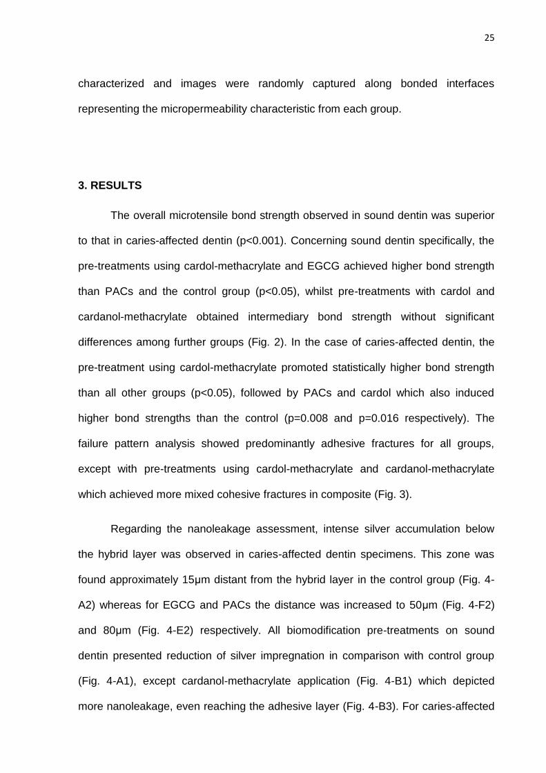
The overall microtensile bond strength observed in sound dentin was superior
to that in caries-affected dentin (p<0.001). Concerning sound dentin specifically, the
pre-treatments using cardol-methacrylate and EGCG achieved higher bond strength
than PACs and the control group (p<0.05), whilst pre-treatments with cardol and
cardanol-methacrylate obtained intermediary bond strength without significant
differences among further groups (Fig. 2). In the case of caries-affected dentin, the
pre-treatment using cardol-methacrylate promoted statistically higher bond strength
than all other groups (p<0.05), followed by PACs and cardol which also induced
higher bond strengths than the control (p=0.008 and p=0.016 respectively). The
failure pattern analysis showed predominantly adhesive fractures for all groups,
except with pre-treatments using cardol-methacrylate and cardanol-methacrylate
which achieved more mixed cohesive fractures in composite (Fig. 3).
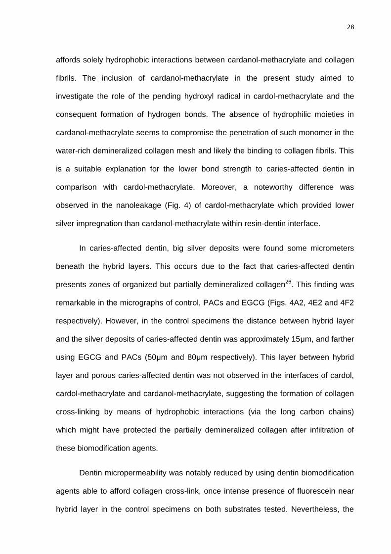
Regarding the nanoleakage assessment, intense silver accumulation below
the hybrid layer was observed in caries-affected dentin specimens. This zone was
found approximately 15μm distant from the hybrid layer in the control group (Fig. 4-
A2) whereas for EGCG and PACs the distance was increased to 50μm (Fig. 4-F2)
and 80μm (Fig. 4-E2) respectively. All biomodification pre-treatments on sound
dentin presented reduction of silver impregnation in comparison with control group
(Fig. 4-A1), except cardanol-methacrylate application (Fig. 4-B1) which depicted
more nanoleakage, even reaching the adhesive layer (Fig. 4-B3). For caries-affected

26
dentin, the presence of cracks (Fig. 4-A3 and 4-F3) and gaps (Fig. 4-B2 and 4-E3)
was often noted in control, cardanol-methacrylate, PACs and EGCG treatments.
Cardol-methacrylate treatment achieved less nanoleakage and better interface
integrity than further groups in caries-affected dentin.
On the micropermeability analysis, all treatments showed deficient sealing
ability when applied on caries-affected dentin, with presence of gaps at the interface
of control group (Fig. 5-A2). More fluorescein uptake was observed in the hybrid
layers created using EGCG (yellow pigmentation in Fig. 5-F2), cardanol-methacrylate
(Fig. 5-B2) and PACs (Fig. 5-E2). In sound dentin, the control group depicted intense
fluorescein uptake in the hybrid layer thereby indicating high micropermeability, whilst
the pre-treatments using EGCG (Fig. 5-F1) and cardol-methacrylate (Fig. 5-D1)
attained the improved dentin sealing ability.
4. DISCUSSION
Dentin pre-treatment using cardol-methacrylate has demonstrated the best
overall outcomes of microtensile bond strength and silver nanoleakage in both
substrates evaluated, thus, the first hypotheses needs to be rejected. Lower uptake
of fluorescein near the hybrid layer was observed by using cardanol-methacrylate
and cardol-methacrylate pre-treatments, thereby indicating improved sealing ability.
The second hypotheses should, then, be rejected.
The molecule epigallocatechin-3-gallate (EGCG) has presented high collagen
cross-linking ability due to the presence of galloyl moieties25. In fact, the high bond
strength of the two-step etch-and-rinse adhesive Optibond Solo Plus after pre-
treatment using EGCG might be explained by the optimal and rapid cross-linking of

27
collagen relying on the galloyl radicals. These polyphenolic functionalities are often
present in the proanthocyanidins whereas a similar structure is found in the molecule
of cardol. Indeed, one may ascertain the high presence of polyphenols in EGCG (Fig.
1) might represent improved collagen cross-linking capabilities due to the formation
of hydrogen bonds, which would be faster with EGCG than with more hydrophobic
proanthocyanidins and cardol. Contrariwise, cardol has a long 15-carbon chain
connected to the aromatic ring (Fig. 1) which may promote the formation of
hydrophobic interactions4 thereby improving the cross-linking of collagen fibrils
attained only via hydrogen bonds.
The aim at synthesizing a dental resin monomer from cardol molecule was to
produce a collagen binding monomer with cross-linking potential, observations
previously found in pilot studies. Cardol-methacrylate was created from cardol
encountered in the cashew nut shell liquid (CNSL), a natural (renewable) source
supplied by the cashew nut industry. Furthermore, CNSL has been widely
investigated in Chemistry Institutes to create several industrial products13. Cardol-
methacrylate may chemically bond the demineralized dentin collagen with the
adhesive resin by the collagen cross-linking reactions (hydrogen bond and
hydrophobic interactions) prior to the co-polymerization of methacrylate functionality
with further methacrylate monomers from the bonding agent. Indeed, such reaction
may increase the dentin bond strength as observed in the present outcomes (Fig. 2),
especially on caries-affected dentin.
Cardanol-methacrylate monomer embraces a chemical structure very similar
to that of cardol-methacrylate, but without the presence a free hydroxyl pending from
the aromatic ring (Fig. 1). Such hydroxyl functionality would allow the formation of
hydrogen bond during collagen cross-linking. Therefore, the lack of this hydroxyl
28
affords solely hydrophobic interactions between cardanol-methacrylate and collagen
fibrils. The inclusion of cardanol-methacrylate in the present study aimed to
investigate the role of the pending hydroxyl radical in cardol-methacrylate and the
consequent formation of hydrogen bonds. The absence of hydrophilic moieties in
cardanol-methacrylate seems to compromise the penetration of such monomer in the
water-rich demineralized collagen mesh and likely the binding to collagen fibrils. This
is a suitable explanation for the lower bond strength to caries-affected dentin in
comparison with cardol-methacrylate. Moreover, a noteworthy difference was
observed in the nanoleakage (Fig. 4) of cardol-methacrylate which provided lower
silver impregnation than cardanol-methacrylate within resin-dentin interface.
In caries-affected dentin, big silver deposits were found some micrometers
beneath the hybrid layers. This occurs due to the fact that caries-affected dentin
presents zones of organized but partially demineralized collagen26. This finding was
remarkable in the micrographs of control, PACs and EGCG (Figs. 4A2, 4E2 and 4F2
respectively). However, in the control specimens the distance between hybrid layer
and the silver deposits of caries-affected dentin was approximately 15μm, and farther
using EGCG and PACs (50μm and 80μm respectively). This layer between hybrid
layer and porous caries-affected dentin was not observed in the interfaces of cardol,
cardol-methacrylate and cardanol-methacrylate, suggesting the formation of collagen
cross-linking by means of hydrophobic interactions (via the long carbon chains)
which might have protected the partially demineralized collagen after infiltration of
these biomodification agents.
Dentin micropermeability was notably reduced by using dentin biomodification
agents able to afford collagen cross-link, once intense presence of fluorescein near
hybrid layer in the control specimens on both substrates tested. Nevertheless, the

29
micropermeability in sound dentin was lower (Fig. 5) after pre-treatments with all
biomodification agents evaluated. During the analysis of resin-dentin
micropermeability, the fluorescent dye is infiltrated through dentinal tubules under
simulated pulpal pressure which may not be properly correlated with resin-dentin
bond strength9.
The use of CNSL compounds to synthesize new dental monomers exhibits
two important advantages. (1) Easy obtainment, since several thousands of tons of
CNSL is produced yearly by cashew nut companies, which results in low production
costs. (2) Environmental issues, once CNSL cannot be discarded in the nature
because it is not biodegradable, which lead to the pursuit for different applications of
CNSL and its compounds. It is worth the large concentration of cardol and cardanol
containing in CNSL acquired after processing the cashew nut in high temperatures.
The resulting CNSL contains 60-65% cardanol and 15-20% cardol. Cardanol and its
derivates have been widely studied and employed to further chemistry purposes such
as polymer industry, mineral oil and ferrofluids2,13. Conversely, cardol has been
underused and little investigated.
Indeed, the optimal outcomes of bond strength attained by using cardol-
methacrylate on both sound and caries-affected dentin highlight a promising agent
for dentin biomodification. However, more studies are needed to evaluate the further
properties of this molecule, such as the effects on polymerization and
metalloproteinases/cathepsins inhibition.
CONCLUSION
(1) On sound dentin, cardol-methacrylate and EGCG provide the overall better
dentin bonding as well as dentin sealing ability.
30
(2) On caries-affected dentin, the pre-treatment with cardol-methacrylate
ethanol solution attains high bond strength and low nanoleakage.
REFERENCES
[1] Aguiar TR, Vidal CMP, Phansalkar RS, Todorova I, Napolitano JG, McAlpine JB,
Chen SN, Pauli GF, Bedran-Russo AK. Dentin Biomodification Potential Depends on
Polyphenol Source. J Dent Res 2014;93:417-22.
[2] Barreto ACH, Maia FJN, Santiago VR, Ribeiro VGP, Denardin JC, Mele G, Carbone L,
Lomonaco D, Mazzetto SE, Fechine PBA. Novel ferrofluids coated with a renewable
material obtained from cashew nut shell liquid. Microfluid Nanofluid 2012;12:677–686.
[3] Bedran-Russo AK, Castellan CS, Shinohara MS, Hassan L, Antunes A.
Characterization of biomodified dentin matrices for potential preventive and reparative
therapies. Acta Biomater 2011;7:1735–1741.
[4] Bedran-Russo AK, Pauli GF, Chen SN, McAlpine J, Castellan CS, Phansalkar RS,
Aguiar TR, Vidal CMP, Napotilano JG, Nam JW, Leme AA. Dentin biomodification:
strategies, renewable resources and clinical applications. Dent Mater 2014;30:62-76.
[5]. Breschi L, Mazzoni A, Ruggeri A, Cadenaro M, Di lenarda R, De stefano DE. Dental
adhesion review: aging and stability of the bonded interface. Dent Mater 2008;24:90-
101.
[6] Castellan CS, Pereira PNR, Grande RHM, Bedran-Russo AK. Mechanical
caracterization of proanthocyanidin-dentin matrix interaction. Dent Mater 2010;26:968-
973.
[7] Cehreli ZC, Yazici AR, Akca T, Ozgünaltay G. A morphological and microtensile bond
strength evaluation of a single bottle adhesive to caries affected human dentin after four
different caries removal technique. J Dent. 2003;31:429–35

31
[8] Dos Santos PH, Karol S, Bedran-Russo AK. Long-term nano-mechanical properties of
biomodified dentin-resin interface components. J Biomech 2011;44:1691-1694.
[9] Feitosa VP, Sauro S, Ogliari FA, Ogliari AO, Yoshihara K, Zanchi CH, Correr-
Sobrinho L, Sinhoreti MA, Correr AB, Watson TF, Van Meerbeek B. Impact of
hydrophilicity and length of spacer chains on the bonding of functional monomers. Dent
Mater. 2014; 30:317-23.
[10] Haj-Ali R, Walker M, Williams K, Wang Y, Spencer P. Histomorphologic
characterization of noncarious and caries-affected dentin/adhesive interfaces. J
Prosthodont. 2006,15: 82–88.
[11] Lenzi TL, Tedesco TK, Soares FZM, Loguercio AD, Rocha RO. Chlorhexidine
application for bond strength preservation in artificially-created caries-affected
primary dentin. Int J Adhes Adhes, 2014, 51–56.
[12] Liu Y, Chen M, Yao X, Xu C, Zhang Y, Wang Y. Enhancement in dentin collagen’s
biological stability after proanthocyanidins treatment in clinically relevant time periods.
Dent Mater 2013; 29:485-492.
[13] Maia FJN, Ribeiro VG, Clemente CS, Lomonaco D, Vasconcelos PHM, Mazzetto
SE. Thermo-oxidative evaluation of new cardol derivatives as antioxidants for mineral
oils. J Therm Anal Calorim. 2012;109:1013–8.
[14] Mazzoni A, Tjäderhane L, Checchi V, Di Lenarda R, Salo T, Tay FR, Pashley
DH, Breschi L. Role of dentin MMPs in caries progression and bond stability. J Dent
Res. 2015; 94:241-51.
[15] Mount GJ, Ngo H. Minimal intervention: a new concept for operative dentistry.
Quintessence Int. 2000; 3:527-33.
[16] Naik, SV, Shashikiran, ND, Chaitra, NL, & Syed, G. A microtensile bond strength
evaluation of a single-bottle adhesive to caries-affected dentin in conventional versus
minimal invasive caries removal techniques: An in-vitro study. Indian J Dent, 2014, 5:
127–131.
32
[17] Nakajima M., Sano H., Urabe I., Tagami J. and Pashley DH.: Bond strengths of
single-bottle dentin adhesives to caries-affected dentin. Oper. Dent., 25: 2-10, 2000.
[18] Nascimento FD, Minciotti CL, Geraldeli S, Carrilho MR, Pashley DH, Tay FR, et
al. Cysteine cathepsins in human carious dentin. J Dent Res. 2011. 90:506-511.
[19] Ogliari FA, da Silva Ede O, Lima Gda S, Madruga FC, Henn S, Bueno M, Ceschi
MA, Petzhold CL, Piva E. Synthesis of phosphate monomers and bonding to dentin:
esterification methods and use of phosphorus pentoxide. J Dent. 2008; 36:171-177.
[20] Qi YP, Li N, Niu LN, Primus CM, Ling JQ, Pashley DH, Tay FR. Remineralization
of artificial dentinal caries lesions by biomimetically modified mineral trioxide
aggregate. Acta Biomater. 2012; 8:836-42.
[21] Spencer P, Ye Q, Park J, Topp EM, Misra A, Marangos O, Wang Y, Bohaty BS,
Singh V, Sene F, Eslick J, Camarda K, Katz JL. Adhesive/Dentin interface: the weak
link in the composite restoration. Ann Biomed Eng. 2010; 38:1989-2003.
[22] Tay FR, Pashley DH. Biomimetic remineralization of resin-bonded acid-ecthed
dentin. J Dent Res 2009;88:719-724.
[23] Tay FR, Pashley DH, Yoshiyama M. Two modes of nanoleakage expression in
single-step adhesives. J Dent Res. 2002;81:472-6.
[24] Tjäderhane L, Nascimento FD, Breschi L, Mazzoni A, Tersariol ILS, Geraldeli S,
Tezvergil-Mutluay A, Carrilho MR, Carvalho RM, Tay FR, Pashley DH. Optimizing dentin
bond durability: Control of collagen degradation by matrix metalloproteinases and
cysteine cathepsins. Dent Mater 2013;29:116-135.
[25] Vidal CMP, Aguiar TR, Phansalkar R, McAlpine JB, Napolitano JG, Chen SN, Araújo
LSN, Pauli GF, Bedran-Russo AK. Galloyl moieties enhance the dentin biomodification
potential of plant-derived catechins. Acta Biomater 2014;10:3288–3294.
33
[26] Wang Y., Spencer P. and Walker MP.: Chemical profile of adhesive/caries-
affected dentin interfaces using Raman microspectroscopy. J. Biomed. Mater. Res.
A.,2007, 81: 279-286.
[27] Zavgorodniy AV, Rohanizadeh R, Bulcock S, Swain MV. Ultrastructural
observations and growth of occluding crystals in carious dentine. Acta Biomater
2008;4:1427-39.

34
Table 1. Resin composite and adhesive compositions and application protocols.
Composition Application procedure
TPH Spectrum (Dentsply
Caulk, Milford, USA)
Bis-GMA; Bis-EMA; TEGDMA;
Photo initiators; Stabilizers;
Bariumaluminiumborosilicate
glass (mean particle size < 1.5
µm); Highly dispersed silicon
dioxide (particle size 0.04 µm)
Incremental placement (in 3 mm levels or
less) and curing of composite
restorations is recommended to minimise
polymerisation shrinkage. Curing each
increment separately
Optibond Solo Plus
(Kerr, Orange, USA).
BIS-GMA, HEMA, GDMA-P,
ethanol, silica, barium glass,
camphoroquinone. Approximately
15% silico dioxide fillers (particle
size 0.04 µm)
The bonding agent must be actively applied
for 30 s, gently air-dried and light-cured for
20s using the LED light-curing unit
*HEMA – hydroxy ethyl methacrylate; Bis-GMA - bisphenol-A glycidyl methacrylate; Bis-EMA –
ethoxylated bisphenol-A glycidyl methacrylate; TEGDMA – triethylene glycol-imethacrylate; GDMA-P
– glycerol dimethacrylate phosphate.

35
Figure 1 - Chemical structures of the biomodification agents tested.

36
Figure 2. Graph depicting the microtensile bond strength results. Different capital letters indicate
statistically significant differences (p<0.05) on sound dentin. Different lowercase letters indicate
significant differences (p<0.05) among groups on caries-affected dentin. The bond strength on sound
dentin was significantly higher than on caries-affected dentin for all treatments (p<0.001).
Figure 3. Graph showing the fracture patterns. ”CDNMA” means cardanol-methacrylate. “CDL”
means cardol. “CDMA” means cardol-methacrylate. “CAD” means caries-affected dentin.

37
Figure 4. SEM micrographs of resin-dentin interfaces illustrating the most common nanoleakage
characteristics observed after silver uptake. Open arrows indicate silver deposits. White arrows
indicate the zone between hybrid layer and silver deposits at caries-affected dentin. The asterisk in
B2 indicates a gap.

38
Figure 5. Confocal micrographs showing the main features of fluorescein micropermeability. Open
arrows indicate fluorescein infiltration at and near hybrid layer. White arrow in A2 indicates a gap.

39
Conclusão

40
4 CONCLUSÃO
Em dentina hígida, o cardol-metacrilato e o EGCG apresentaram melhores
resultados de adesão e melhor selamento dentinário.
Em dentina afeta por cárie o pré-tratamento por 60s de cardol-metacrilato
demonstrou os melhores resultados de adesão com menor nanoinfiltração.

41
Referências

42
REFERÊNCIAS
AGUIAR TR, VIDAL CMP, PHANSALKAR RS, TODOROVA I, NAPOLITANO JG,
MCALPINE JB, CHEN SN, PAULI GF, BEDRAN-RUSSO AK. Dentin Biomodification
Potential Depends on Polyphenol Source. J Dent Res, Chicago, v. 93, n. 4, p. 417-422,
abr. 2014.
AL-AMMAR A, DRUMMOND JL, BEDRAN-RUSSO AK. The Use of Collagen Cross-
Linking Agents to Enhance Dentin Bond Strength. J Biomed Mater Res B Appl
Biomater, Hoboken, v. 91, n. 1, p. 419–424, out. 2009.
BARRETO ACH, MAIA FJN, SANTIAGO VR, RIBEIRO VGP, DENARDIN JC, MELE G,
CARBONE L, LOMONACO D, MAZZETTO SE, FECHINE PBA. Novel ferrofluids coated
with a renewable material obtained from cashew nut shell liquid. Microfluid Nanofluid,
Heidelberg, v.12, p. 677–686, nov. 2012.
BEDRAN-RUSSO AK, VIDAL CM, DOS SANTOS PH, CASTELLAN CS. Long-term
effect of carbodiimida on dentin matrix and resin-dentin bonds. J Biomed Mater Res
Part B: Applied Biomaterials, Hoboken, v. 94, n.1, p. 250-255, jul. 2010.
BEDRAN-RUSSO AK, CASTELLAN CS, SHINOHARA MS, HASSAN L, ANTUNES A.
Characterization of biomodified dentin matrices for potential preventive and reparative
therapies. Acta Biomater, Kidlington, v. 7, n. 4, p. 1735–1741, abr. 2011.
BEDRAN-RUSSO AK, PAULI GF, CHEN SN, MCALPINE J, CASTELLAN CS,
PHANSALKAR RS, AGUIAR TR, VIDAL CMP, NAPOTILANO JG, NAM JW, LEME AA.
Dentin biomodification: strategies, renewable resources and clinical applications. Dent
Mater, Copenhagen, v. 30, n. 1, p. 62-76, jan. 2014.
BRESCHI L, MAZZONI A, RUGGERI A, CADENARO M, DI LENARDA R, DE STEFANO
DE. Dental adhesion review: aging and stability of the bonded interface. Dent Mater,
Copenhagen, v. 24, n. 1, p. 90-101, jan. 2008.

43
BROYLES AC, PAVAN S, BEDRAN-RUSSO AK. Effect of dentin surface modification on
the microtensile bond strength of self-adhesive resin cements. J Prosthod, Philadelphia,
v. 22, n. 1, p. 59-62, jan. 2013.
CASTELLAN CS, PEREIRA PNR, GRANDE RHM, BEDRAN-RUSSO AK. Mechanical
caracterization of proanthocyanidin-dentin matrix interaction. Dent Mater, Copenhagen,
v. 26, n. 10, p. 968-973, out. 2010.
CEHRELI ZC, YAZICI AR, AKCA T, OZGÜNALTAY G. A morphological and microtensile
bond strength evaluation of a single bottle adhesive to caries affected human dentin after
four different caries removal technique. J Dent.. Bristol, v. 31, n. 6, p. 429–435, ago.
2003.
DELL'AICA I, CANIATO R, BIGGIN S, GARBISA S. Matrix proteases, green tea, and
St. John's wort: Biomedical research catches up with folk medicine. Clin Chem Acta,
Amsterdam, v. 38, n. 1, p. 69-77, mai. 2007.
DOS SANTOS PH, KAROL S, BEDRAN-RUSSO AK. Long-term nano-mechanical
properties of biomodified dentin-resin interface components. J Biomech, New York, v.
44, n. 9, p. 1691-1694, jun. 2011.
FEITOSA VP, SAURO S, OGLIARI FA, OGLIARI AO, YOSHIHARA K, ZANCHI CH,
CORRER-SOBRINHO L, SINHORETI MA, CORRER AB, WATSON TF, VAN
MEERBEEK B. Impact of hydrophilicity and length of spacer chains on the bonding of
functional monomers. Dent Mater, Copenhagen, v. 30, n. 12, p. 317-323, dez. 2014.
GARBISA S, SARTOR L, BIGGIN S, SALVATO B, BENELLI, R.; ALBINI, A. Tumor
gelatinases and invasion inhibited by the green tea flavanol epigallocatechin-3-gallate.
Cancer; New York, v. 91, n. 4, p. 822-832, fev. 2001.
HAJ-ALI R, WALKER M, WILLIAMS K, WANG Y, SPENCER P. Histomorphologic
characterization of noncarious and caries-affected dentin/adhesive interfaces. J
Prosthodont. Philadelphia, v. 6, n. 2, p. 82-88, mar./abr. 2006.

44
HEBLING J, PASHLEY DH, TJADERHANE L, TAY FR. Chlorhexidine arrests subclinical
degradation of dentin hybrid layers in vivo. J Dent Res, Chicago, v. 84, n. 8, p. 741–746,
ago. 2005.
JACKSON JK, ZHAO J, WONG W, BURT HM. The inhibition of collagenase induced
degradation of collagen by the galloyl-containing polyphenols tannic acid,
epigallocatechingallate and epicatechingallate. J Mater Sci Mater Med, London, v. 21, n.
5, p. 1435-1443, mai. 2010.
LENZI TL, TEDESCO TK, SOARES FZM, LOGUERCIO AD, ROCHA RO. Chlorhexidine
application for bond strength preservation in artificially-created caries-affected primary
dentin. Int J Adhes Adhes, v. 54, p. 51–56, out. 2014.
LIU Y, CHEN M, YAO X, XU C, ZHANG Y, WANG Y. Enhancement in dentin collagen’s
biological stability after proanthocyanidins treatment in clinically relevant time periods.
Dent Mater, Copenhagen, v. 29, n. 4, p. 485-492, abr. 2013.
MACEDO GV, YAMAUCHI M, BEDRAN-RUSSO AK. Effects of chemical cross-linkers on
caries-affected dentin bonding. J Dent Res, Chicago, v. 88, n. 12, p. 1096-1100, dez.
2009.
MAIA FJN, RIBEIRO VG, CLEMENTE CS, LOMONACO D, VASCONCELOS PHM,
MAZZETTO SE. Thermo-oxidative evaluation of new cardol derivatives as
antioxidants for mineral oils. J Therm Anal Calorim, Dordrecht, v. 109, n. 2, p.
1013–1018, ago. 2012.
MAZZONI A, TJÄDERHANE L, CHECCHI V, DI LENARDA R, SALO T, TAY FR,
PASHLEY DH, BRESCHI L. Role of dentin MMPs in caries progression and bond
stability. J Dent Res, Chicago, v. 94, n. 2, p. 241-51, fev. 2015.
MOUNT GJ, NGO H. Minimal intervention: a new concept for operative dentistry.
Quintessence Int, Berlin, v. 31, n. 8, p. 527-33, set. 2001.

45
NAIK, SV, SHASHIKIRAN, ND, CHAITRA, NL, & SYED, G. A microtensile bond
strength evaluation of a single-bottle adhesive to caries-affected dentin in
conventional versus minimal invasive caries removal techniques: An in-vitro study.
Indian J Dent, Haryana, v. 5, n. 3, p. 127–131, jul. 2014.
NAKABAYASHI N, NAKAMURA M, YASUDA N. Hibrid layer as a dentin-bonding
mechanism. J Esthet Dent, Berlin, v.3, n. 4, p. 133-138, jul./ago.1991.
NAKAJIMA M., SANO H., URABE I., TAGAMI J. AND PASHLEY DH. Bond strengths
of single-bottle dentin adhesives to caries-affected dentin. Oper Dent, Seattle, v. 25,
n. 1, p. 2-10, jan./fev. 2000.
NASCIMENTO FD, MINCIOTTI CL, GERALDELI S, CARRILHO MR, PASHLEY DH,
TAY FR. Cysteine cathepsins in human carious dentin. J Dent Res, Chicago, v. 90,
n. 4, p. 506-511, abr. 2011.
NIU LN, ZHANG W, PASHLEY DH, BRESCHI L, MAO J, CHEN JH, TAY FR. Biomimetic
remineralization of dentin. Dent Mater, Copenhagen, v. 30, n. 1, p. 77-96, jan. 2014.
OGLIARI FA, DA SILVA EDE O, LIMA GDA S, MADRUGA FC, HENN S, BUENO M,
CESCHI MA, PETZHOLD CL, PIVA E. Synthesis of phosphate monomers and bonding
to dentin: esterification methods and use of phosphorus pentoxide. J Dent, Bristol, v. 36,
n. 3, p. 171-77, mar. 2008.
OSORIO R, YAMAUTI M, RUIZ-REQUENA ME, TOLEDANO M. MMPs activity and bond
strength in deciduous dentine-resin bonded interfaces. J Dent, Bristol, v. 41, n. 6, p. 549-
555, jun. 2013.
PASHLEY DH, TAY FR, YIU C, HASHIMOTO M, BRESCHI L, CARVALHO RM.
Collagen degradation by host-derived enzymes during aging. J Dent Res, Chicago,
v. 83, n. 3, p. 216–221, mar. 2004.
PAVAN S, XIE Q, HARA AT, BEDRAN-RUSSO AK. Biomimetic approach for root caries
prevention using a proanthocyanidin-rich agent. Caries Res, Basel, v. 45, n. 5, p. 443–
447, ago. 2011.

46
QI YP, LI N, NIU LN, PRIMUS CM, LING JQ, PASHLEY DH, TAY FR.
Remineralization of artificial dentinal caries lesions by biomimetically modified
mineral trioxide aggregate. Acta Biomater, Kidlington; v. 8, n. 2, p. 836-842, fev.
2012.
SANTIAGO SL, OSORIO R, NERI JR, CARVALHO RM, TOLEDANO M. Effect of the
flavonoid epigallocatechin-3-gallate on resin-dentin bond strength. J Adhes Dent;
New Malden; v. 15, n. 6, p. 535-540, dez. 2013.
TAY FR, PASHLEY DH. Biomimetic remineralization of resin-bonded acid-ecthed dentin.
J Dent Res, Chicago; v. 88, n. 8, p. 719-724, ago. 2009.
TAY FR, PASHLEY DH, YOSHIYAMA M. Two modes of nanoleakage expression in
single-step adhesives. J Dent Res, Chicago, v. 81, n. 7, p. 472-476, jul. 2002.
TJÄDERHANE L, NASCIMENTO FD, BRESCHI L, MAZZONI A, TERSARIOL ILS,
GERALDELI S, TEZVERGIL-MUTLUAY A, CARRILHO MR, CARVALHO RM, TAY FR,
PASHLEY DH. Optimizing dentin bond durability: Control of collagen degradation by
matrix metalloproteinases and cysteine cathepsins. Dent Mater, Copenhagen, v. 29, n.
1, p. 116-135, jan. 2013.
TOLEDANO M, SAURO S, CABELLO I, WATSON T, OSORIO R. A Zn-doped etch-
and-rinse adhesive may improve the mechanical properties and the integrity at the
bonded-dentin interface. Dent Mater, Copenhagen, v. 29, n. 8, p. 142-52, ago. 2013.
VAN MEERBEEK B, DHEM A, GORET-NICAISE M, BRAEM M, LAMBRECHTS P,
VANHERLE G. Comparative SEM and TEM examination of ultrastructure of the resin-
dentin interdiffusion zone. J Dent Res, Chicago, v. 72, n. 2, p. 495-501, fev. 1993.
VIDAL CMP, AGUIAR TR, PHANSALKAR R, MCALPINE JB, NAPOLITANO JG, CHEN
SN, ARAÚJO LSN, PAULI GF, BEDRAN-RUSSO AK. Galloyl moieties enhance the
dentin biomodification potential of plant-derived catechins. Acta Biomater, Kidlington, v.
10, n. 7, p. 3288–3294, jul. 2014.

47
WANG Y., SPENCER P. AND WALKER MP. Chemical profile of adhesive/caries-
affected dentin interfaces using Raman microspectroscopy. J Biomed Mater Res,
Hoboken, v. 81, n. 2, p. 279-286, mai. 2007.
ZAVGORODNIY AV, ROHANIZADEH R, BULCOCK S, SWAIN MV. Ultrastructural
observations and growth of occluding crystals in carious dentine. Acta Biomater,
Kidlington, v. 4, n. 5, p. 1427-39, set. 2008.
48

49
50

51