UNIVERSIDADE FEDERAL DO PAMPA PROGRAMA DE...
77
UNIVERSIDADE FEDERAL DO PAMPA PROGRAMA DE PÓS-GRADUAÇÃO EM BIOQUÍMICA DETERMINAÇÃO DE PARÂMETROS OXIDATIVOS E BIOQUÍMICOS EM INDIVÍDUOS MULTITRANSFUNDIDOS DISSERTAÇÃO DE MESTRADO Marília Sabo Fernandes Uruguaiana, RS, Brasil 2012
Transcript of UNIVERSIDADE FEDERAL DO PAMPA PROGRAMA DE...
0
UNIVERSIDADE FEDERAL DO PAMPA
PROGRAMA DE PÓS-GRADUAÇÃO EM BIOQUÍMICA
DETERMINAÇÃO DE PARÂMETROS OXIDATIVOS E BIOQUÍMICOS EM
INDIVÍDUOS MULTITRANSFUNDIDOS
DISSERTAÇÃO DE MESTRADO
Marília Sabo Fernandes
Uruguaiana, RS, Brasil
2012
1
MARÍLIA SABO FERNANDES
DETERMINAÇÃO DE PARÂMETROS OXIDATIVOS E BIOQUÍMICOS EM
INDIVÍDUOS MULTITRANSFUNDIDOS
Dissertação apresentada ao Programa de Pós-
Graduação em Bioquímica, da Universidade
Federal do Pampa, UNIPAMPA, como
requisito parcial para obtenção do grau de
Mestre em Bioquímica.
Orientador: Prof° Dr° Robson Luiz Puntel
Co-orientador: ProfªDrª Vanusa Manfredini
Uruguaiana, RS, Brasil
2012

2

3

4
―A mente que se abre a uma nova idéia jamais
voltará ao seu tamanho original.‖
(Albert Einstein)
5
RESUMO
Dissertação de Mestrado
Programa de Pós-Graduação em Bioquímica
Universidade Federal do Pampa
DETERMINAÇÃO DE PARÂMETROS OXIDATIVOS E BIOQUÍMICOS EM
INDIVÍDUOS MULTITRANSFUNDIDOS
Autora: Marília Sabo Fernandes
Orientador: Prof. Dr. Robson Luiz Puntel
Co-orientador: Prof. Dr. Vanusa Manfredini
Local e Data da Defesa: Uruguaiana, 07 de dezembro de 2012.
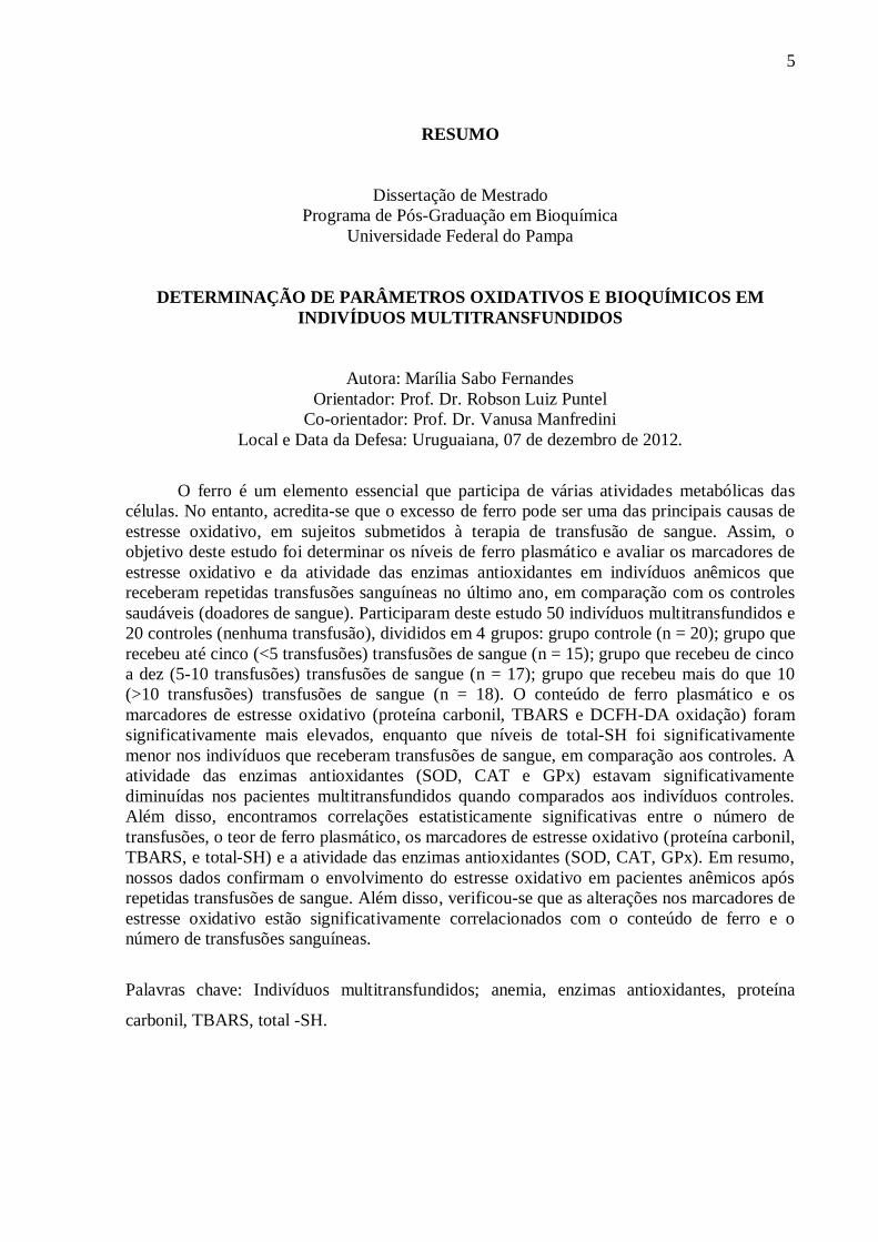
O ferro é um elemento essencial que participa de várias atividades metabólicas das
células. No entanto, acredita-se que o excesso de ferro pode ser uma das principais causas de
estresse oxidativo, em sujeitos submetidos à terapia de transfusão de sangue. Assim, o
objetivo deste estudo foi determinar os níveis de ferro plasmático e avaliar os marcadores de
estresse oxidativo e da atividade das enzimas antioxidantes em indivíduos anêmicos que
receberam repetidas transfusões sanguíneas no último ano, em comparação com os controles
saudáveis (doadores de sangue). Participaram deste estudo 50 indivíduos multitransfundidos e
20 controles (nenhuma transfusão), divididos em 4 grupos: grupo controle (n = 20); grupo que
recebeu até cinco (<5 transfusões) transfusões de sangue (n = 15); grupo que recebeu de cinco
a dez (5-10 transfusões) transfusões de sangue (n = 17); grupo que recebeu mais do que 10
(>10 transfusões) transfusões de sangue (n = 18). O conteúdo de ferro plasmático e os
marcadores de estresse oxidativo (proteína carbonil, TBARS e DCFH-DA oxidação) foram
significativamente mais elevados, enquanto que níveis de total-SH foi significativamente
menor nos indivíduos que receberam transfusões de sangue, em comparação aos controles. A
atividade das enzimas antioxidantes (SOD, CAT e GPx) estavam significativamente
diminuídas nos pacientes multitransfundidos quando comparados aos indivíduos controles.
Além disso, encontramos correlações estatisticamente significativas entre o número de
transfusões, o teor de ferro plasmático, os marcadores de estresse oxidativo (proteína carbonil,
TBARS, e total-SH) e a atividade das enzimas antioxidantes (SOD, CAT, GPx). Em resumo,
nossos dados confirmam o envolvimento do estresse oxidativo em pacientes anêmicos após
repetidas transfusões de sangue. Além disso, verificou-se que as alterações nos marcadores de
estresse oxidativo estão significativamente correlacionados com o conteúdo de ferro e o
número de transfusões sanguíneas.
Palavras chave: Indivíduos multitransfundidos; anemia, enzimas antioxidantes, proteína
carbonil, TBARS, total -SH.

6
ABSTRACT
Dissertation of Master
Graduate Course in Biochemistry
Federal University of Pampa
OXIDATIVE AND BIOCHEMISTRY PARAMETERS DETERMINATION IN
MULTITRANSFUSED SUBJECTS
Author: Marília Sabo Fernandes
Advisor: Robson Luiz Puntel
Co-Advisor: Vanusa Manfredini
Place and Date of Defense: Uruguaiana, December 07, 2012.
Iron is an essential element that participates in several metabolic activities of cells.
However, its excess is believed to be a major cause of iron-induced oxidative stress in
subjects undergoing blood transfusion therapy. Thus, the objective this study was to
determine the plasmatic iron content and evaluate the oxidative stress markers and the activity
of the antioxidant enzymes in anemic subjects receiving repeated blood transfusions in the
past year, comparing with healthy controls (blood donors). A total of 50 individuals
multitransfused and 20 controls (no transfusion), divided into 4 groups: control group (n=20);
group that received up to five (<5 transfusions) blood transfusions (n=15); group that received
from five up to ten (5 – 10 transfusions) blood transfusions (n=17); group that received over
than ten (>10 transfusions) blood transfusions (n=18). Plasmatic iron and oxidative stress
markers (protein carbonyl, TBARS and DCFH-DA oxidation) were significantly higher,
whereas total -SH levels was significantly lower in subjects receiving blood transfusion
compared to controls. Additionally, the activity of the antioxidant enzymes (CAT, SOD and
GPx) were significantly lower in the multitransfused subjects whem compared to controls
subjects. Moreover, we found statistically significant correlations between the number of
transfusions, the plasmatic iron content, the oxidative stress markers (protein carbonyl,
TBARS, and total –SH) and the activity of the antioxidant enzymes (CAT, SOD, and GPx). In
summary, our data confirm the involvement of oxidative stress in anemic patients after
repeated blood transfusions. Additionally, we found that the changes in the oxidative stress
markers are significantly correlated with both iron content and number of blood transfusions.
Keywords: Multitransfused subjects, anemia, antioxidant enzymes, protein carbonyl,
TBARS, total -SH.

7
LISTA DE FIGURAS
REVISÃO BIBLIOGRÁFICA
Figura 1. Hemocomponentes sanguíneos……………………..................................................15
Figura 2. Fisiopatologia da sobrecarga de ferro transfusional..................................................16
Figura 3. Homeostase do ferro ................................................……………………….............18
Figura 4. Redução do oxigênio molecular na mitocôndria.......................................................20
Figura 5. Sistema enzimático oxidante e antioxidante………………………………...…......22
ARTIGO CIENTÍFICO
Figure 1: Plasmatic labile iron levels in studied subjects (controls; group <5; group 5-10 and
group >10) ……..……………………………………………………………………………. 49
Figure 2: Pearson’s correlation among plasmatic iron content and number of blood
transfusions………………………………………………………………………………….. 50
Figure 3: Protein carbonyl levels in studied subjects (controls; group <5; group 5-10 and
group >10)…………………………………………………………………………………… 51
Figure 4: TBARS levels in different groups (controls; group<5; group 5-10 and group
>10)………………………………………………………………………………………..… 52
Figure 5: DCFH-DA oxidation in different groups (controls; group<5; group 5-10 and group
>10)………………………………………………………………………………………….. 53
Figure 6: Total SH levels in different groups (controls; group <5; group 5-10 and group
>10)……………………………………………………………………………………….…. 54
Figure 7: Pearson’s correlation among number of blood transfusions and protein carbonyl (A),
TBARS (B) and Total –SH content (C)……………………………………………...........… 55
Figure 8: Catalase activity in different groups (controls; group <5; group 5-10 and group
>10)……………………………………………………………………………………..…… 56
Figure 9: Superoxide dismutase activity in different groups (controls; group <5; group 5-10
and group >10)…………………………………………………………………………….… 57
Figure 10: Glutathione peroxidase activity in different groups (controls; group<5; group 5-10
and group >10)…………………………………………………………………….……….... 58
Figure 11: Pearson’s correlation among number of transfusions and CAT (A), SOD (B) and
GPx (C) activity……………………………………………………………………...……… 59
8
Figure 12: Pearson’s correlation among plasmatic iron content and CAT (A), SOD (B), GPx
(C), Total –SH (D), Protein carbonyl (E) and TBARS (F)………………………………. 60-61

9
LISTA DE TABELAS
ARTIGO CIENTÍFICO
Table I. Characteristics of the subjects................................................................................... 62
Table II. Pearson correlations in both control and multitransfused subjects……………….. 63
10
LISTA DE ESQUEMAS
REVISÃO BIBLIOGRÁFICA
Esquema 1. Reação de Fenton................................................................................................ 19
ARTIGO CIENTÍFICO
Scheme1. Fenton Reaction..................................................................................................... 48

11
LISTA DE ABREVIATURAS, SIGLAS E SIMBOLOS
ERO - espécies reativas de oxigênio
SUS - Sistema Único de Saúde
CHAD- concentrado de hemácias
HH - hemocromatose hereditária
OH- - radical hidroxila
O2- - radical superóxido
HO2 - hidroperoxila
H2O2 - peróxido de hidrogênio
GSH - glutationa
GPx - glutationa peroxidase
CAT - catalase
SOD - superóxido dismutase
DNA - Ácido desoxirribonucleico
LPO -peroxidação lipídica
MDA -malondialdeído
CO - grupamentos carbonílicos
Pro - prolina
Arg - arginina
Lis - lisina
Ter - treonina
TBARS - espécies reativas ao ácido tiobarbitúrico
LDL - lipoproteína de baixa densidade
DCFH-DA -diacetato dediclorofluoresceína
SH - grupamentos tiólicos
12
SUMÁRIO
1. INTRODUÇÃO ................................................................................................................. 13
2. REVISÃO BIBLIOGRÁFICA ......................................................................................... 14
2.1. Transfusão Sanguínea ................................................................................,................... 14
2.1.1. Hemocomponentes ................................................................................................. 14
2.1.2. Concentrado de Hemácias ...................................................................................... 14
2.2. Ferro ................................................................................................................................ 17
2.2.1. Homeostase do Ferro ............................................................................................. 17
2.2.2. Toxicidade ............................................................................................................. 18
2.3. Estresse Oxidativo ......................................................................................................... 19
2.4. Marcadores do Estresse Oxidativo .............................................................................. 20
2.4.1. Peroxidação Lipídica .............................................................................................. 21
2.4.2. Oxidação de proteínas ............................................................................................ 21
2.4.3. Total-SH ................................................................................................................. 22
2.4.4. Enzimas antioxidantes ............................................................................................ 22
3. OBJETIVOS ...................................................................................................................... 23
3.1. Objetivo Geral ................................................................................................................ 23
3.2. Objetivos Específicos ..................................................................................................... 23
4. ARTIGO CIENTÍFICO ................................................................................................... 24
5. DISCUSSÃO ...................................................................................................................... 64
6. CONCLUSÃO ................................................................................................................... 68
7. PERSPECTIVAS .............................................................................................................. 69
REFERÊNCIAS .................................................................................................................... 70
ANEXO I ................................................................................................................................ 76

13
1. INTRODUÇÃO
A transfusão sanguínea pode salvar vidas e existem várias condições hematológicas
para as quais a transfusão contínua de concentrado de hemácias é a única terapia disponível
(LAMBING et al, 2012). Nos EUA, aproximadamente de 14 a 15.000.000 unidades de sangue
são coletadas anualmente e transfundidas em cerca de cinco milhões pessoas (LANGLOIS et
al, 2004). Entretanto, os indivíduos que recebem regularmente transfusões de concentrados de
hemácias inevitavelmente desenvolvem sobrecarga cumulativa de ferro, e consequentemente,
risco de toxicidade devido a esse metal (SAZAMA et al, 2010; SHANDER et al, 2009;
LAMBING et al, 2012). O excesso de ferro transfusional pode ocasionar inúmeras
complicações no organismo (SAZAMA et al, 2010), tais como o estresse oxidativo (FIBACH
et al, 2010), peroxidação lipídica (CIGHETTI et al, 2002), osteoporose (TSAY et al, 2010),
cirrose, miocardiopatia e distúrbio do sistema endócrino (HERSHKO et al, 2010),
hemocromatose (ALLEN, 2010), necrose hepatocelular (TOYOKUNl, 2011), fibrose hepática
(RAMM et al, 2005), carcinoma hepatocelular (KEW, 2009), sendo a principal causa
secundária de óbito em pacientes com talassemias (SIMÕES et al, 2010). A sobrecarga de
ferro afeta a morbidade e a mortalidade em indivíduos dependentes de transfusões sanguíneas
(SAZAMA et al, 2010; GHOTI et al, 2010).
O ferro é um metal potencialmente tóxico por ser capaz de catalisar reações de geração
de espécies reativas de oxigênio (ERO), como a Reação de Fenton, podendo desencadear um
desequilíbrio no balanço redox celular. Quando a geração de ERO ultrapassa a capacidade de
reparo das defesas antioxidantes do organismo, cria-se um estado denominado de estresse
oxidativo (HALLIWELL, 2006; CHUNG et al, 2005). Conseqüentemente, as ERO interagem
com vários componentes celulares, ocasionando oxidação de biomoléculas e danos celulares
(GHOTI et al, 2010; GATTERMANN et al, 2011; HALLIWELL, 2006; SHANDER et al,
2009; FIBACH & RACHMILEWITZ, 2008).
Considerando que onúmero de transfusões sanguíneas é um preditor da sobrecarga de
ferro nos tecidos, consequentemente, as múltiplas transfusões sanguíneas geram risco de
toxicidade ao organismo. Entretanto pouco se sabe como as transfusões sanguíneas afetam o
sistema antioxidante em seres humanos e poucos estudos foram desenvolvidos até a presente
data. Neste contexto, objetivo deste estudo foi determinar os níveis de ferro plasmático e
avaliar os marcadores de estresse oxidativo e da atividade das enzimas antioxidantes em
indivíduos multitransfundidos, como evidência de estresse oxidativo nesses indivíduos.
14
2. REVISÃO BIBLIOGRÁFICA
2.1 Transfusão sanguínea
A transfusão sanguínea é uma pratica relevante na terapêutica moderna e consiste,
resumidamente, na técnica médica de transferência de sangue de um doador saudável a um
receptor (BRASIL I, 2008). Somente no Brasil, no ano de 2010, foram realizados o
quantitativo de 3.970.792 procedimentos transfusionais, referindo-se a rede pública, serviços
filantrópicos, privados conveniados aos SUS e somente privados (BRASIL II, 2011).
2.1.1 Hemocomponentes
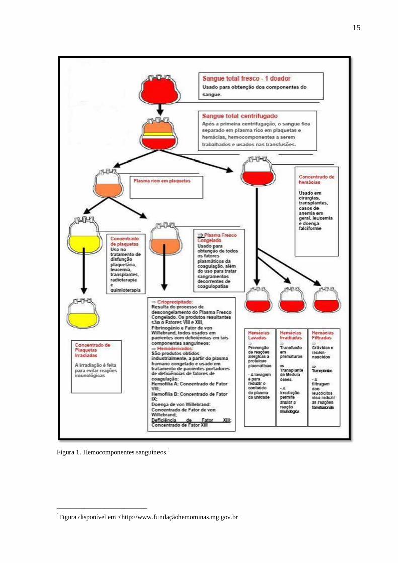
Os hemocomponentessão obtidos atravésdo processamento do sangue total oriundo
da doação de sangue de um doador saudável. No Brasil, este processo está regulamentado
pela Lei nº 10.205, de 21/3/2001, pela Resolução da Diretoria Colegiada n° 153, de 14 de
junho de 2004 e pela Portaria n° 1353, de 13 de junho de 2011. Toda doação de sangue deve
ser altruísta, voluntária e não-gratificada, assim como o anonimato do doador e o sigilo de
suas informações devem ser garantidos.O processamento é feito por meio de centrifugação
refrigerada, no qual se fraciona o sangue total em hemocomponentes eritrocitários,
plasmáticos e plaquetários (Figura 1).
2.1.2 Concentrado de Hemácias:
O concentrado de hemácias (CHAD) é o hemocomponente constituído pelos glóbulos
vermelhos ou eritrócitos, após a remoção do plasma. Os glóbulos vermelhos estão presentes
no sangue, em condições normais, por aproximadamente de 4,5 a 6,5x10⁶/mm³. São
constituídas basicamente por hemoglobina, uma metaloproteína composta de quatro
grupamentos heme que contém o ferro. Tem como função o transporte de oxigênio e gás
carbônico no organismo, com meia vida aproximada de 120 dias (VERRASTRO, 2005;
PIERGÉ et al, 2008).
15
Figura 1. Hemocomponentes sanguíneos.1
___________________________
1Figura disponível em <http://www.fundaçãohemominas.mg.gov.br
16
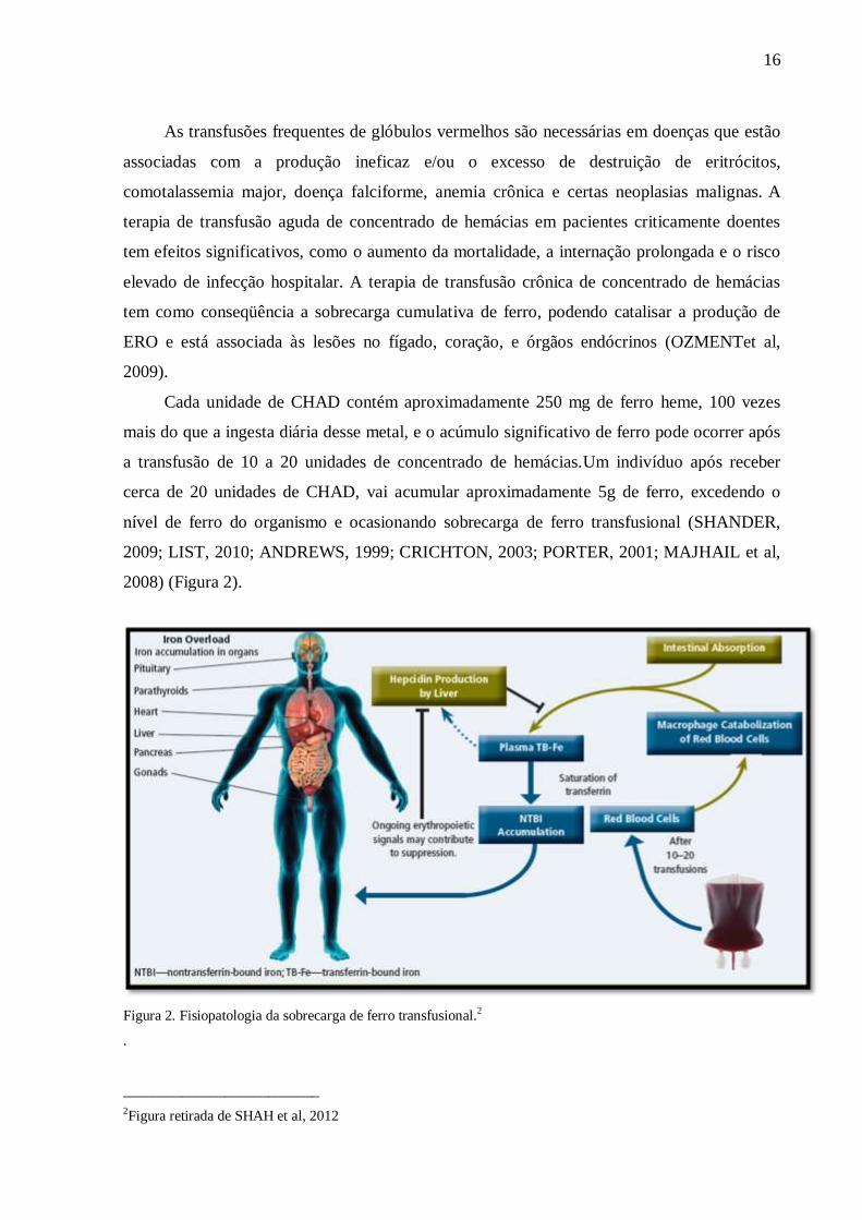
As transfusões frequentes de glóbulos vermelhos são necessárias em doenças que estão
associadas com a produção ineficaz e/ou o excesso de destruição de eritrócitos,
comotalassemia major, doença falciforme, anemia crônica e certas neoplasias malignas. A
terapia de transfusão aguda de concentrado de hemácias em pacientes criticamente doentes
tem efeitos significativos, como o aumento da mortalidade, a internação prolongada e o risco
elevado de infecção hospitalar. A terapia de transfusão crônica de concentrado de hemácias
tem como conseqüência a sobrecarga cumulativa de ferro, podendo catalisar a produção de
ERO e está associada às lesões no fígado, coração, e órgãos endócrinos (OZMENTet al,
2009).
Cada unidade de CHAD contém aproximadamente 250 mg de ferro heme, 100 vezes
mais do que a ingesta diária desse metal, e o acúmulo significativo de ferro pode ocorrer após
a transfusão de 10 a 20 unidades de concentrado de hemácias.Um indivíduo após receber
cerca de 20 unidades de CHAD, vai acumular aproximadamente 5g de ferro, excedendo o
nível de ferro do organismo e ocasionando sobrecarga de ferro transfusional (SHANDER,
2009; LIST, 2010; ANDREWS, 1999; CRICHTON, 2003; PORTER, 2001; MAJHAIL et al,
2008) (Figura 2).
Figura 2. Fisiopatologia da sobrecarga de ferro transfusional.2
.
___________________________
2Figura retirada de SHAH et al, 2012

17
2.2 Ferro
O ferro é um elemento essencial para a atividade celular e desempenha um papel
fundamental em várias reações bioquímicas que envolvem o transporte de oxigênio e a
transferência de elétrons no metabolismo energético celular (LINDER, 1991; COLPO, 2008;
MAJHAIL et al, 2008; SAZAMA et al, 2010). O ferro distribui-se amplamente nos tecidos,
totalizando cerca de 3,5 a 4,5g em um indivíduo adulto, onde 70 a 80% são considerados ferro
funcional. O ferro circula no plasma ligado a transferrina, e no estado de equilíbrio, não há
ferro livre circulante.A maior parte do ferro estáincorporado a hemoglobina e o resto é
armazenado em ferritina, mioglobina, enzimas ou como ferro livre (SHAH et al, 2012;
ANDREWS, 2005).
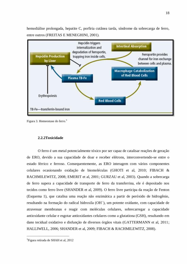
2.2.1Homeostase do ferro
A homeostase do ferro é bem regulada no organismo, pois tanto um aporte deficiente,
quanto um acúmulo excessivo de ferro conduzem a um desequilíbrio. A absorção do ferro é
controlada a nível intestinal pelo regulador hepcidina, que no excesso de ferro inibe sua
absorção intestinal e na deficiência de ferro, aumenta a absorção intestinal desse
metal(MAJHAIL et al, 2008; SAZAMA et al, 2010). Segundo GHOTI et al, 2010, a
hepcidina é omelhor regulador do metabolismo do ferro. A produção de hepcidina é regulada
por vários fatores, e está aumentada em resposta a sobrecarga de ferro e inflamação, e
diminuída após a hipóxia, intensa atividade eritropoiética e estresse oxidativo (LEE &
BEUTLER, 2009) (Figura 3). Pacientes multitransfundidos com mielodisplasia desenvolvem
sobrecarga de ferro e estresse oxidativo, suprimindo a produção de hepcidina (LEE &
BEUTLER, 2009). Aproximadamente 1-2mg de ferro é eliminado do organismo diariamente
pelas secreções corpóreas, descamação da pele e intestino ousangramento menstrual. O
organismo não possui um mecanismo específico para eliminar o excesso de ferro,e o aumento
do aporte de ferro, tanto por via gastrointestinal como por via parenteral, necessariamente leva
a condição patológica de sobrecarga de ferro (GATTERMANN et al, 2011; MAJHAIL et al,
2008; LAMBING et al, 2011; SAZAMA et al, 2010). Distúrbios no metabolismo do ferro
podem ser encontrados na hemocromatose hereditária (HH), alguns tipos de anemia
(talassemia major, anemia sideroblástica, hemolítica crônica), hepatopatia crônica,
18
hemodiálise prolongada, hepatite C, porfiria cutânea tarda, síndrome da sobrecarga de ferro,
entre outros (FREITAS E MENEGHINI, 2001).
Figura 3. Homeostase do ferro.3
2.2.2Toxicidade
O ferro é um metal potencialmente tóxico por ser capaz de catalisar reações de geração
de ERO, devido a sua capacidade de doar e receber elétrons, interconvertendo-se entre o
estado férrico e ferroso. Consequentemente, as ERO interagem com vários componentes
celulares ocasionando oxidação de biomoléculas (GHOTI et al, 2010; FIBACH &
RACHMILEWITZ, 2008; EMERIT et al, 2001; GURZAU et al, 2003). Quando a sobrecarga
de ferro supera a capacidade de transporte de ferro da transferrina, ele é depositado nos
tecidos como ferro livre (SHANDER et al, 2009). O ferro livre participa da reação de Fenton
(Esquema 1), que catalisa uma reação não enzimática a partir de peróxido de hidrogênio,
resultando na formação do radical hidroxila (OH-.), um potente oxidante, com capacidade de
atravessar membranas e reagir com moléculas celulares, sobrecarregar a capacidade
antioxidante celular e esgotar antioxidantes celulares como a glutationa (GSH), resultando em
dano tecidual oxidativo e disfunção de diversos órgãos vitais (GATTERMANN et al, 2011;
HALLIWELL, 2006; SHANDER et al, 2009; FIBACH & RACHMILEWITZ, 2008).
___________________________
3Figura retirada de SHAH et al, 2012

19
Vários estudos têm relatado o aumento do risco de câncer em condições de sobrecarga
de ferro, como observado na hemocromatose, onde oaumento das células contendo ferro pode
produzir eventos genotóxicos pela indução do estresse oxidativo (TOYOKUNI, 1996).
Esquema 1: Reação de Fenton
2.3. Estresse Oxidativo
As espécies reativas de oxigênio (ERO) são átomos ou moléculas que contém um
número ímpar de elétrons em sua última camada eletrônica, o que confere uma alta
reatividade a essas moléculas, e são encontradas em todos os sistemas biológicos. Em
condições fisiológicas do metabolismo celular aeróbio, o oxigênio sofre redução, resultando
na formação de água. Durante esse processo, cerca de 2 a 3% desse oxigênio molecular sofre
redução incompleta, originando intermediários reativos, tais como o radical superóxido (O2-),
hidroperoxil (HO2.), hidroxil (OH) e peróxido de hidrogênio (H2O2).Normalmente essa
redução ocorre na mitocôndria e, em condições normais, o organismo possui mecanismos
compensatórios para sequestramento das espécies reativasformadas nesse processo
(FERREIRA et al, 1997) (Figura 4).
As ERO no organismo são neutralizadas por substancias antioxidantes. Segundo
HALLIWELL (2000),―Antioxidante é qualquer substância que, quando presente em baixa
concentração comparada a do substrato oxidável, regenera o substrato ou previne
significativamente a oxidação do mesmo”. Os antioxidantes podem agir enzimaticamente
detoxificando o agente oxidante antes que ele cause lesão, como a glutationa peroxidase
(GPx), a catalase (CAT) e a superóxido dismutase(SOD); ou não enzimaticamente, reparando
a lesão ocorrida, como a glutationa (HALLIWELL, 2000; FERREIRA et al, 1997).
Quando ocorre um desequilíbrio no balanço redox, onde a geração de ERO ultrapassa
a capacidade de reparo das defesas antioxidantes do organismo,com a potencialidade de
Fe3+ + O2.- → Fe2+ + O2 (1)
Fe2+ + H2O2 → Fe3+ + OH- +.OH (2)
(1) Redução do Ferro da forma férrica (Fe ³+) para forma ferrosa (Fe ²+); (2) Reação de Fenton:
geração do radical hidroxil(.OH) através da reação entre Fe2+ e peróxido de hidrogênio (H2O2).

20
exercer efeitos deletérios, cria-se um estado denominado de estresse oxidativo
(HALLIWELL, 2006; VASCONCELOS et al, 2007). Os danos oxidativos causados pelos
altos níveis de ERO ocasionam modificações importantes a biomoléculas conduzindo a danos
celulares, nas membranas lipídicas, nas proteínas citosólicas e de membrana, e danos nas
bases nitrogenadas do ácido desoxirribonucléico(DNA) (BIANCHI et al, 1999), e têm sido
relacionados com a etiologia de várias doenças, tais como as cardiopatias e aterosclerose
(BATLOUNI, 1997), diabetes tipo II (MANFREDINI et al, 2010), problemas pulmonares
(FERREIRA et al, 1997), erros inatos do metabolismo (SITTA et al, 2009; RIBAS et al,
2010), envelhecimento cutâneo (HIRATA et al, 2004), carcinogênese (LOUREIRO et al,
2002), entre outros.
Figura 4. Redução tetravalente do oxigênio molecular na mitocôndria até a formação de água. Varias espécies de
ERO são formadas nesse processo. 4
2.4.Marcadores do estresse oxidativo
Usamos os biomarcadores sanguíneos do balanço redox para refletir os danos
causados pelas ERO e a eficiência das defesas antioxidantes do organismo. Esses marcadores
podem avaliar o dano oxidativo em componentes celulares como asestruturas lipídicas,
através da peroxidação lipídica (LPO) e avaliar o dano oxidativo a proteínas, através dos
compostos carbonílicos e sulfidrílicos. Também podem avaliar a defesa antioxidante
enzimática, como a SOD, a CAT e a GPx, queconstituem a primeira defesa endógena de
neutralização das ERO (HALLIWELL E GUTTERIDGE, 1999; AMES et al, 1993).
___________________________
4Figura retirada de FERREIRA et al, 1997

21
2.4.1. Peroxidação lipídica (LPO)
As ERO podem atacar as membranas celulares, as quais contêm uma grande
quantidade de ácidos graxos poliinsaturados. A peroxidação lipídica dá-se pela reação dos
radicais livres com os lipídeos insaturados das membranas, resultando na formação de hidro
ou lipoperóxidos, que são altamente reativos e podem dar início a uma cascata oxidativa, com
severos danos a integridade da membrana. Nessa reação ocorre à liberação de produtos de
degradação de ácidos graxos, como o malondialdeido (MDA), e a quantificação deste
composto têm sido utilizado para avaliar a extensão do dano oxidativo (MARNETT, 1999;
OHKAWA et al, 1979).
2.4.2. Oxidação de proteínas
As proteínas são alvos imediatos para a modificação oxidativa ocasionada por ERO,
alterando sua estrutura e provocando perda de função e fragmentação das estruturas
protéicas.A formação da proteína carbonil parece ser um fenômeno comum durante a
oxidação, e sua quantificação pode se usada para medir a extensão do dano oxidativo
(BERLETT & STADMAN, 1997; BEAL, 2003; DALLE-DONNEet al., 2003). Os
grupamentos carbonílicos (CO) são produzidos pela oxidação da cadeia lateral de
aminoácidos suscetíveis, como prolina (Pro), arginina (Arg), lisina (Lis) e treonina (Tre), ou
pela clivagem oxidativa das proteínas. Também, os CO podem ser introduzidos nas proteínas
por uma reação secundária das cadeias laterais com aldeídos produzidos durante a
peroxidação lipídica, como o malondialdeido. O conteúdo de proteína carbonil é atualmente o
marcador de oxidação protéica mais usado e observa-se seu aumento em várias doenças
humanas, tais como Doença de Alzheimer, Diabetes mellitus, processos inflamatórios e artrite
reumatóide. Vale ressaltar que níveis elevados de proteína carbonil também são indicadores
de doenças derivadas do metabolismo protéico, e não somente de estresse oxidativo (DALLE-
DONNE et al., 2003). As proteínas modificadas oxidativamente são produtos quimicamente
estáveis, sendo um fator importante para a sua detecção e armazenamento (RUTKOWSKA et
al., 2005).

22
2.4.3. Total-SH
Os compostos tiólicos são antioxidantes que contêm em sua estrutura o grupamento–
SH, entre estes compostos estão a glutationa, a cisteina e as proteínas tiólicas. Esses
compostos estão envolvidos no sequestramento de radicais livres,e são capazes
dequelarionsmetálicosdanosos,desempenhando assimum papel crucialna defesa
antioxidantedos eritrócitose no plasma (WLODEK et al, 2010).
2.4.4. Enzimas antioxidantes
As enzimas antioxidantes SOD, CAT e GPx, servem como linha primária de defesa na
destruição dos radicais livres. A SOD é uma enzima antioxidante que catalisa a dismutação do
radical superóxido em peróxido de oxigênio e oxigênio. A CAT é uma hemeproteina
citoplasmática que catalisa a redução do peróxido de hidrogênio a água e oxigênio. A GPx é
uma enzima que catalisa a redução do peróxido de hidrogênio e peróxidos orgânicos para seus
correspondentes alcoóis, as custas da glutationa (FERREIRA et al, 1997) (Figura 5).
Figura 5. Sistema enzimático oxidante e antioxidante. eNOS = óxido nítrico sintetase endotelial; O2- = ânion
superoxido; O2 = oxigênio. 5
___________________________
5Figura retirada de DEBLIN et al, 2009

23
3. OBJETIVOS
3.3. Objetivo Geral
Determinar os níveis de ferro plasmático e avaliar os marcadores de estresse oxidativo
e da atividade das enzimas antioxidantes em indivíduos multitransfundidos, como evidência
de estresse oxidativo nesses indivíduos.
3.4. Objetivos específicos
Quantificar os níveis de ferro sérico no plasma de indivíduos multitransfundidos e
controles;
Verificar a atividade das enzimas antioxidantes catalase (CAT), glutationa peroxidase
(GPx) e superóxido dismutase (SOD) nos eritrócitos de indivíduos multitransfundidos
e controles;
Quantificar o dano oxidativo em proteínas plasmáticas, pelo método do carbonil, em
plasma de indivíduos multitransfundidos e controles;
Quantificar o dano oxidativo em lipídios de membrana pelo método de TBA-RS em
plasma de indivíduos multitransfundidos e controles;
Determinar o conteúdo de sulfidrilas totais no plasma de indivíduos
multitransfundidos e controles;
Quantificar os níveis de Espécies Reativas de Oxigênio (ERO) no plasma de
indivíduos multitransfundidos e controles;
Correlacionar os parâmetros do estresse oxidativo (carbonil, TBARS, total-SH, ROS)
com o teor de ferro plasmático e o número de transfusões sanguíneas;
Correlacionar as enzimas antioxidantes (SOD, CAT, GPx) com o teor de ferro
plasmático e o número de transfusões sanguíneas.
24
4. ARTIGO CIENTÍFICO
Os resultados que fazem parte desta dissertação estão apresentados sob a forma de
artigo científico, o qual se encontra organizado neste item. Os itens Materiais e Métodos,
Resultados, Discussão dos Resultados e Referências Bibliográficas encontram-se no próprio
artigo e representam a íntegra deste estudo. O artigo está disposto na forma que foi submetido
para publicação na edição da revista científica AppliedBiochesmistryandBiotecnology.
4.1. Oxidative Stress Parameters In Anemic Subjects After Repeated Blood
Transfusions: A Positive Correlation With Plasmatic Labile Iron Content
Marília Sabo Fernandes1, Tatiana Tamborena Rissi
1, Luiza Zuravski
1, Juliana Mezzomo
1,
Carmen Regla Vargas3, Vanderlei Folmer
1, Félix Alexandre Antunes Soares
2, Vanusa
Manfredini1, Robson Luiz Puntel
1*
1Universidade Federal do Pampa - Campus Uruguaiana BR-472 Km 7, Uruguaiana, 97500-
970, RS, Brazil.
2Departamento de Química, Centro de Ciências Naturais e Exatas, Universidade Federal de
Santa Maria, Santa Maria, CEP 97105-900, RS, Brazil.
3Serviço de Genética Médica – Laboratório de Análise de Metabólitos, Hospital de Clínicas
de Porto Alegre, CEP 90035-003, RS, Brazil.

25
Abstract
Iron is an essential element that participates in several metabolic activities of cells; however,
it excess can be a major cause of oxidative stress (OS) in subjects undergoing blood
transfusion therapy. The objective of this study was to determine the plasmatic iron content,
evaluate the OS markers and the activity of the antioxidant enzymes in anemic subjects
receiving repeated blood transfusions. Here, 50 anemic subjects (divided into 3 subgroups: 1)
received up to five blood transfusions [n=15]; 2) received from five up to ten transfusions
[n=17]; and 3) received more than ten transfusions [n=18]) and 20 controls were analyzed.
Plasmatic labile iron, protein carbonyl, thiobarbithuric acid-reactive substances and
dichlorofluorescindiacetate oxidation were significantly higher, whereas Total -SH levels
were significantly lower in anemic subjects compared to controls. Additionally, the activity of
catalase, superoxide dismutase and glutathione peroxidase were significantly lower in the
transfused subjects. Moreover we found statistically significant correlations between the
number of transfusions, plasmatic iron content, the OS markers and the activity of the
antioxidant enzymes. Our data confirms the involvement of OS in patients following therapy
with repeated blood transfusions. Additionally we found that the changes in the OS markers
are tightly correlated with both iron content and the number of transfusions.
Keywords:Politransfused subjects, anemia, antioxidant enzymes, protein carbonyl, TBARS,
Total -SH.
Abbreviations: TBARS: Thiobarbithuric acid-reactive substances; DCFH-DA:
dichlorofluorescindiacetate; Total –SH: Total thiol groups; CAT: catalase; SOD: superoxide
dismutase; GPx: glutathione peroxidase.

26
1. Introduction
Iron is an essential trace element of cells and it participates in various redox processes
because its capacity to accept and donate electrons, interconverting between the Fe3+
and Fe2+
forms (1-3). Additionally, iron is a vital constituent of various enzymes, including iron–
sulphur and haem proteins of the respiratory chain, as well as ribonucleotidereductase, among
others (1, 4). Moreover, iron has the unique ability to alter its oxidation and redox states in
response to several ligants, which makes it indispensable for life processes (5). However, this
unique redox property renders iron potentially toxic in biological systems due to generation of
reactive oxygen species (ROS) during its redox cycling (6). In order to prevent ROS over
production, circulating and intracellular free iron are tightly regulated by binding to
transferrin, ferritin and other proteins (7).
However, in some situations the iron balance may be disturbed, either locally or
systemically, resulting in labile iron (“free iron” – not bound to ferritin or transferrin) which
could participate in Fenton chemistry (Scheme 1) and subsequently generating large amounts
of ROS (8). Indeed, iron overload could occur under some conditions, such as in several
chronic anemias, secondary to repeated blood transfusions, and following increased
gastrointestinal absorption (9-10). So, subjects undergoingrepeatedblood transfusions are
believed to be at risk oftoxicity associated withiron overload (11). Additionally, iron balance
could also be disrupted in patients in the end-stage of renal failure following therapy to treat
hemodialysis-associated anemia (12).
Accordingly, it was pointed that elevated tissue iron can overwhelm the protective
mechanisms and lead to an increase in iron complexes with small molecules such as
nucleotides and citrate in the serum of these patients and also within cytoplasm and organelles
(13-14). Indeed, it was previously reported that under repeated blood transfusions the levels of
iron increase, being available to generate catalytically active complexes, and consequently

27
free radicals and oxidative damage (14). Actually, such labile iron is catalytically active,
promoting free radical formation that culminates in the oxidation of biomolecules.
Accordingly, iron overload in humans and in experimental animals seems to be associated
with oxidative stress (15). In particular, the catalytically active iron promotes oxidative stress
in organs that accumulate it on excess (16-17). Indeed, it is known that an imbalance in the
oxidant/antioxidant status of the cell is associated with oxidative stress and this causes
important modifications in cellular macromolecules leading to cell damage (18). Thus,
oxidative stress is believed to be one of the most important factors determining cell injury in
patients with iron overload (19). Hence, the end-result of the oxidation reactions is the
formation of lipid peroxides and protein carbonyls, damaged deoxyribonucleic acid (DNA)
bases, and mitochondrial dysfunction (5, 20). Additionally, individuals with iron overload
also demonstrate impaired antioxidant defenses (10, 21). Accordingly, the long-term
consequence of chronic iron overload is organ injury, which could contribute to the initiation
and development of several chronic pathologies, such as endocrinopathies, diabetes mellitus,
cirrhosis, hypogonadism and heart failure (22-24).
In general, oxidative damage of biomolecules can be counteracted by enzymatic as
well as nonenzymatic defenses (25). Indeed, humans are well equipped with several
biological mechanisms to defend against intracellular oxidative stress. One of the most
important mechanisms involves the actions of antioxidant enzymes, such as superoxide
dismutase (SOD), catalase (CAT) and glutathione peroxidase (GPx) (26). However, despite
their well-developed antioxidant defense system, cells can be oxidatively damaged under
some pathological conditions (18, 27).
Considering the exposed, we hypothesize that oxidative stress can be correlated with
plasmatic iron content in anemic patients following therapy with repeated blood transfusions.
Moreover, to the best of our knowledge, data about labile iron accumulation in anemic

28
subjects receiving repeated blood transfusion and its correlation with the oxidative damage
markers are scarce in the literature. Therefore, in order to verify this hypothesis we evaluated
the oxidative stress markers and the activity of the enzymatic antioxidant defenses in the
blood of patients receiving repeated transfusions and in control subjects (no transfused).
Additionally, we determined the plasmatic labile iron content in these subjects and correlated
with each other evaluated markers.
2. Material and Methods
2.1. Chemicals
1,1,3,3-tetramethoxypropane, 2-thiobarbituric acid (TBA), sodium dodecyl sulfate
(SDS), 5,5- dithiobis(2-nitrobenzoic acid) (DTNB), trichloroacetic acid (TCA),2’,7’-
dichlorofluorescin diacetate (DCHF-DA), 2,4-Dinitrophenylhydrazine (DNPH), were
purchased from Sigma, St. Louis, MI, USA. The kit for iron determination was obtained from
BioSystems, kits for SOD and GPx from Randox® Laboratories, UK, and kit for protein
determination from BioClin. All the other chemicals were commercial products of the highest
purity grade available.
2.2. Subjects
This study was approved by the Ethics Committee in Research of Universidade Federal
do Pampa (UNIPAMPA), protocol number 001/2012. Altogether 50 anemic individuals
receiving blood transfusion and 20 healthy individuals (blood donors) from the Banco de
Sangue do Municipio de Uruguaiana were included in the study. Anemic subjects, selected for
the study, do not present other diagnosed disease, such as cancer, renal failure, hepatic
disease, blood loss or others. Indeed the inclusion criteria adopted here were: chronic anemic
patients that received the blood therapy during last year prior to collection (i.e. 12 months

29
from the first transfusion until sample collection to analysis). Additionally, it is important to
mention here that the sample collection was done before a new transfusion, namely clinical
screening. The anemic subjects were divided into 3 subgroups: 1) subgroup that received up
to five (<5 transfusions) blood transfusions (n=15); 2) subgroup that received from five up to
ten blood transfusions (n=17); and 3) subgroup that received more than ten blood transfusions
(n=18). Some characteristics of the subjects included in this study, including data concerning
the number of transfusions are pointed in Table I.
2.3. Sample collection
Blood from either controls or anemic subjects were collected by venous arm puncture
and stored into tubes containing heparin. The plasma and cells were separated by
centrifugation at 1500 rpmfor 10 min and were subsequently used for biochemical analyses.
All biochemical assays were done in duplicate or triplicate, depending on availability of
samples.
2.4. Analysis of hemoglobin
The electrophoretic analysis of hemoglobin was performed using the Minicap(Sebia,
Norcross, France) according to the manufacturer's instructions, running controls with every
test as described previously. The Minicap system uses the principle of capillary
electrophoresis in free solution. Charged molecules are separated by their electrophoretic
mobility in an alkaline buffer with a specific pH. Separation also occurs according to the
electrolyte pH and electro-osmotic flow. Electropherograms were expressed with divided
zones from Z1 to Z15 based on standardizing the location of HbAaccording to previous
described (28).

30
2.5. Measurement of plasmatic labile iron content
The plasmatic iron content was determinedby its reactivity with ferrozine(which does
not detect heme iron) in the presence of the denaturant sodium dodecyl sulfate and the
reducing agents ascorbate and sodium metabisulphite, according to previously described (29).
Accordingly, ferrous ions react with ferozine forming a coloured complex that can be
measured by spectrophotometry (30). We follow the previously validated convention of using
the term labile iron (“free iron”) to non heme iron that has reacted quickly in the ferrozine
assay (within few minutes), instead of the term “non heme iron” that include others (e.g.
ferritin and transferrin iron) (31).
2.6. Protein carbonyl determination
Content protein carbonyl was determined as described by (32). The carbonyl protein
presence is indicative of oxidation. Plasma samples were added 0.2 mL of trichloroacetic acid
(TCA), 10% and placed on ice for 5 minutes. After centrifugation (5 min, 8000), was added 1
mL of 2,4-dinitrophenylhydrazine (DNPH) in 2M HCl to 10mM and samples 1 mL of 2M
HCl in white tubes and incubated for 90 min at 37 ° C. The proteins were dissolved in 6M
guanidine and interference was removed after washing with ethanol-ethyl acetate 1:1 (v / v).
The extent of the damage will be done by reading absorbance at 370nm. The results were
expressed as nmolcabonyl/mg protein.
2.7. Determination of TBARS levels
Thiobarbituric acid reactive substances (TBARS) were determined in plasma by the
method of Ohkawa et al. (33). In brief, samples were incubated at 100 °C for 60 min in acid
medium containing 0.45% sodium dodecyl sulfate and 0.6% thiobarbituric acid. After

31
centrifugation, the reaction product was determined at 532 nm using 1,1,3,3-
tetramethoxypropane as standard and the results were expressed as nmol MDA/mg protein.
2.8. Total thiol (Total –SH) determination
Plasmatic total -SH were determined as described by Ellman (34). The colorimetric
assay was carried out in 1 M phosphate buffer, pH 7.4. A standard curve using glutathione
was constructed in order to calculate the total -SH content in samples. Total -SH content was
expressed as nmol total –SH /mg protein.
2.9. Determination of DCHF-DA oxidation
The determination of intracellular oxidant production was based on 2’,7’-
dichlorofluorescin diacetate (DCHF-DA) cleavage to 2’,7’-dichlorofluorescin (DCHF) that
can be oxidized to the fluorescent compound 2’,7’-dichlorofluorescein by ROS according to
previously described (35-36). The plasma sample was diluted (1: 10) in Tris/HCl 10 mM
buffer. Then, 50 µL of diluted plasma was incubated in 10 mMTris/HCl buffer and 10 M
DCHF-DA at 37 ° C for 30 min. The DCF fluorescence intensity emission was measured
using a Perkin-Elmer spectrofluorometer at an excitation wavelength of 488 nm and an
emission wavelength of 520 nm, 20 min after the addition of DCHF-DA to the medium. The
results were expressed as arbitrary fluorescence unit (AFU).
2.10. Catalase activity
Catalase (CAT) enzyme activity was measured by the method previously described
(37). Packed erythrocytes were hemolyzed by adding 100 volumes of distilled water, then, 20
μL of this hemolyzed sample was added to a cuvette and the reaction was started by the
addition of 100 μL of freshly prepared 300mM H2O2 in phosphate buffer (50 mM, pH 7.0) to
32
give a final volume of 1 mL. The rate of H2O2 decomposition was measured
spectrophotometer at 240nm during 120 seg. The CAT activity was expressed as UI/mg
protein.
2.11. Superoxide dismutase activity
Superoxide dismutase (SOD) activity was measured in erythrocytes using the Kit
RANSOD® (Randox Laboratories, UK). This method employs xanthine and xanthine oxidase
to produce superoxide radicals which react with 2-(4-iodophenyl)-3-(4-nitrophenyl)-5-
feniltetrazol chloride (INT) to form compound formazan red. The superoxide dismutase
activity was measured by the degree of inhibition of this reaction at 505 nm. The SOD
activity was expressed as UI/mg protein.
2.12. Glutathione peroxidase activity
Glutathione peroxidase (GPx) activity was determined in erythrocytes using the Kit
RANSEL® (Randox Laboratories, UK), according to the method previously described(38).
The GPx activity was expressed as UI/mg protein.
2.13. Protein determination
The protein content is determined by the biuret method, through the Total Protein Kit
Bioclin®, using bovine serum albumin as standard. The copper ions in an alkaline medium
(biuret reagent) react with peptide, producing a purple color, whose intensity is proportional
to the concentration of proteins in the samples being measured in a spectrophotometer at
545nm.

33
2.14. Statistical Analysis
Data are expressed as means ± SEM. Statistical analysis was performed using analysis
of variance (ANOVA), followed by post hoc Tukey multiple range test when appropriated. P
< 0.05 was considered significant. Pearson correlation between variables was also carried out.
3. Results
Some characteristics of the subjects included in this study are presented on Table I.
The eletrophoretic hemoglobin profile of the subjects included in this study indicates that
none individual presented hemoglobinopathy (100% had normal hemoglobin profile; data not
shown). Indeed, fetal hemoglobin variant was found in 8% of individuals, at normal
proportions (less than 10% of total current hemoglobin). As expected, the plasmatic labile
iron content was statistically (P<0.05) high in the transfused subjects when compared to
control group (Figure 1). Nonetheless, only subjects receiving >5 transfusions presented iron
levels statistically different from controls (Figure 1). Additionally, we found a statistically
significantly correlation between the number of transfusions and the iron content (Figure 2).
Moreover, we does not found significant correlation between the iron content and the age of
the subjects (r= -0.613; P=0.437).
The oxidative stress analyzed markers such as protein carbonyl (Figure 3), TBARS
(Figure 4) and DCFH-DA oxidation (a marker ROS, Figure 5) were significantly (P<0.05)
higher, whereas total -SH (Figure 6) levels were significantly (P<0.05) lower in transfused
subjects compared to controls. However, statistical analysis revealed that only subjects
receiving >10 transfusions presented statistically significant decrease in SH groups when
compared to controls (Figure 6). Indeed, to other evaluated oxidative stress markers, the
changes were seen to all groups of transfused subjects, independent from the number of
transfusions (see Figures 3-5). Likewise plasmatic iron content, we found statistically
34
significant correlations between the number of transfusions and protein carbonyl (Figure 7A),
TBARS (Figure 7B), and total -SH (Figure 7C).
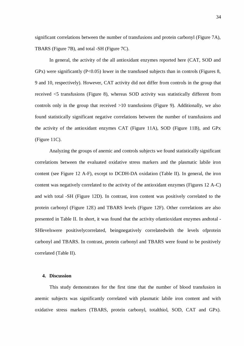
In general, the activity of the all antioxidant enzymes reported here (CAT, SOD and
GPx) were significantly (P<0.05) lower in the transfused subjects than in controls (Figures 8,
9 and 10, respectively). However, CAT activity did not differ from controls in the group that
received <5 transfusions (Figure 8), whereas SOD activity was statistically different from
controls only in the group that received >10 transfusions (Figure 9). Additionally, we also
found statistically significant negative correlations between the number of transfusions and
the activity of the antioxidant enzymes CAT (Figure 11A), SOD (Figure 11B), and GPx
(Figure 11C).
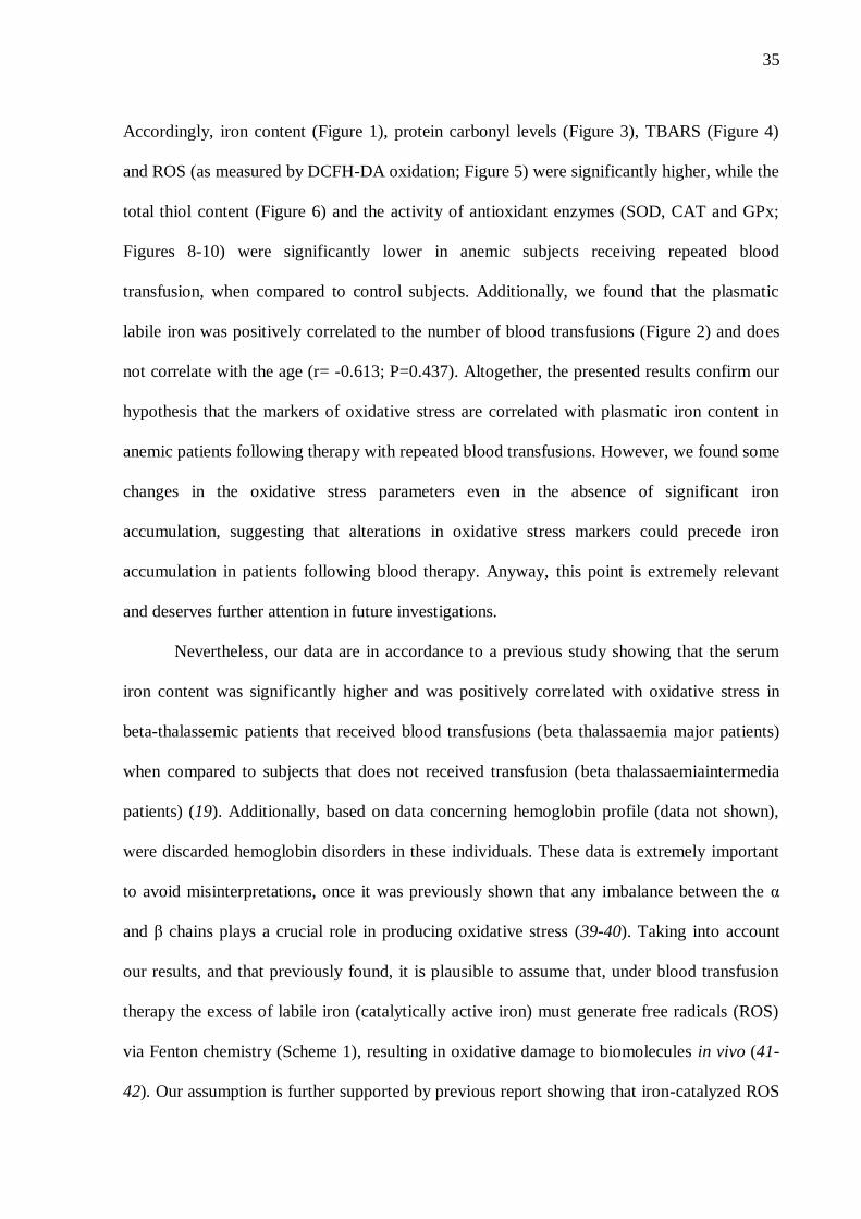
Analyzing the groups of anemic and controls subjects we found statistically significant
correlations between the evaluated oxidative stress markers and the plasmatic labile iron
content (see Figure 12 A-F), except to DCDH-DA oxidation (Table II). In general, the iron
content was negatively correlated to the activity of the antioxidant enzymes (Figures 12 A-C)
and with total -SH (Figure 12D). In contrast, iron content was positively correlated to the
protein carbonyl (Figure 12E) and TBARS levels (Figure 12F). Other correlations are also
presented in Table II. In short, it was found that the activity ofantioxidant enzymes andtotal -
SHlevelswere positivelycorrelated, beingnegatively correlatedwith the levels ofprotein
carbonyl and TBARS. In contrast, protein carbonyl and TBARS were found to be positively
correlated (Table II).
4. Discussion
This study demonstrates for the first time that the number of blood transfusion in
anemic subjects was significantly correlated with plasmatic labile iron content and with
oxidative stress markers (TBARS, protein carbonyl, totalthiol, SOD, CAT and GPx).
35
Accordingly, iron content (Figure 1), protein carbonyl levels (Figure 3), TBARS (Figure 4)
and ROS (as measured by DCFH-DA oxidation; Figure 5) were significantly higher, while the
total thiol content (Figure 6) and the activity of antioxidant enzymes (SOD, CAT and GPx;
Figures 8-10) were significantly lower in anemic subjects receiving repeated blood
transfusion, when compared to control subjects. Additionally, we found that the plasmatic
labile iron was positively correlated to the number of blood transfusions (Figure 2) and does
not correlate with the age (r= -0.613; P=0.437). Altogether, the presented results confirm our
hypothesis that the markers of oxidative stress are correlated with plasmatic iron content in
anemic patients following therapy with repeated blood transfusions. However, we found some
changes in the oxidative stress parameters even in the absence of significant iron
accumulation, suggesting that alterations in oxidative stress markers could precede iron
accumulation in patients following blood therapy. Anyway, this point is extremely relevant
and deserves further attention in future investigations.
Nevertheless, our data are in accordance to a previous study showing that the serum
iron content was significantly higher and was positively correlated with oxidative stress in
beta-thalassemic patients that received blood transfusions (beta thalassaemia major patients)
when compared to subjects that does not received transfusion (beta thalassaemiaintermedia
patients) (19). Additionally, based on data concerning hemoglobin profile (data not shown),
were discarded hemoglobin disorders in these individuals. These data is extremely important
to avoid misinterpretations, once it was previously shown that any imbalance between the α
and β chains plays a crucial role in producing oxidative stress (39-40). Taking into account
our results, and that previously found, it is plausible to assume that, under blood transfusion
therapy the excess of labile iron (catalytically active iron) must generate free radicals (ROS)
via Fenton chemistry (Scheme 1), resulting in oxidative damage to biomolecules in vivo (41-
42). Our assumption is further supported by previous report showing that iron-catalyzed ROS

36
generation leads to an increase in the genomic instability in hematopoietic progenitor cells
(43-45). Moreover, in animal models it was shown that iron overload causes liver damage via
both oxidative and nitrosative mechanisms (46).
Taking into account the presented results, we suggest that the repeated blood transfusion
increase the amount of labile iron available to participate in the Fenton chemistry (Scheme 1),
thus leading to a increase in the ROS generation, as measured by the increase in DCFH-DA
oxidation (Figure 5). Indeed, we assume that under repeated blood transfusions, the iron
content increase to values that overwhelm the protective mechanisms, leading to an increase
in the amount of iron available to form complexes with small molecules, the called
“catalytically active iron complexes”. Thus, we assume that ROS generated (via Fenton
chemistry) are responsible for the oxidation of DCFH-DA found in the transfused subjects,
which is supported by previous report showing that overload with iron (ferric nitrilotriacetate)
lead to an increase in oxidation of DCFH-DA in cultures rat hepatocytes (47).
We found the levels of TBARS significantly increased in subjects receiving blood
transfusion (Figure 4), which was positively correlated to the iron content (Figure 12F) and
the number of transfusions (Figure 7B). These findings are in accordance to previous reports
showing that the levels of lipid peroxidation products were increased in beta thalassaemic
patients receiving blood transfusions (19, 30, 48) and in subjects with hepatic iron overload
(49). Moreover, we found a significantly increase in the protein carbonyl in the subjects
receiving repeated blood transfusion (Figure 3), which was found to be significantly
correlated with iron content (Figure 12E). Likewise other oxidative markers, our data are in
accordance to a previous paper showing a significant increase in the protein carbonyl content
in conditions of iron overload (47).
Moreover, we found a significant reduction in total SH in the subjects receiving
repeated blood transfusion (Figure 6), which are in accordance to previous report showing a
37
decrease in thiol content in the liver of rats treated with iron (50).Albeit not completely
understood, we believed that thiols are oxidized (consumed/used) in these subjects due to
oxidative stress status following iron overload. However, other possibility is that the iron
could reacts nonenzymatically with thiols in plasma to generate ROS, which directly lead to
reduction of antioxidant capacity in plasma and the increased susceptibility of blood
components to oxidation (51). Thus, this thiol-dependent free radical generation by iron
overload might be a potential contributing factor for the changes in the oxidative markers
reported here. Our assumptions are supported by other previous studies showing that oxygen
radicals can be produced by iron-catalyzed autooxidation of cysteine or glutathione (GSH)
(52-53). So, the generated ROS (either by Fenton chemistry as well as via iron-catalyzed
autooxidation of thiols) are the putative responsible for the oxidation of other biomolecules
reported here, such as lipids and proteins.
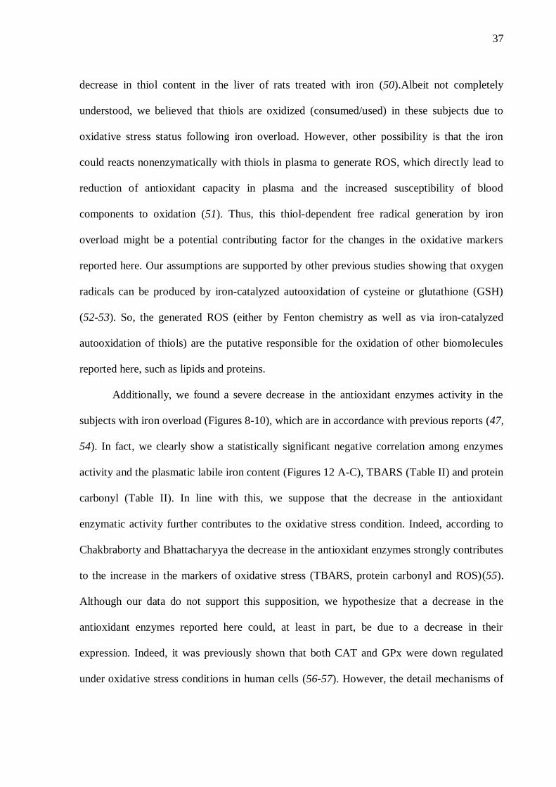
Additionally, we found a severe decrease in the antioxidant enzymes activity in the
subjects with iron overload (Figures 8-10), which are in accordance with previous reports (47,
54). In fact, we clearly show a statistically significant negative correlation among enzymes
activity and the plasmatic labile iron content (Figures 12 A-C), TBARS (Table II) and protein
carbonyl (Table II). In line with this, we suppose that the decrease in the antioxidant
enzymatic activity further contributes to the oxidative stress condition. Indeed, according to
Chakbraborty and Bhattacharyya the decrease in the antioxidant enzymes strongly contributes
to the increase in the markers of oxidative stress (TBARS, protein carbonyl and ROS)(55).
Although our data do not support this supposition, we hypothesize that a decrease in the
antioxidant enzymes reported here could, at least in part, be due to a decrease in their
expression. Indeed, it was previously shown that both CAT and GPx were down regulated
under oxidative stress conditions in human cells (56-57). However, the detail mechanisms of

38
regulation in the expression of antioxidant enzymes under iron overload remain to be better
explored.
Considering the exposed, is reasonable to suggest that antioxidants could be associated
with blood therapy. In fact, previous data reported that antioxidant supplementation is safe
and favorably affects the markers of oxidative stress, albeit it does not result in a significant
clinical benefit (58-59). Additionally, ironchelators that efficiently decrease the levels of
labile iron are putative candidate to counteract the iron-induced ROS generation (42, 60).
However, more studies are necessary to better understand the mechanism(s) associated to
iron-induced oxidative changes, tentatively to minimize the side effects associated to blood
transfusion therapy and uncertainly to improve some clinical benefits.
5. Conclusions
In summary, our data confirm the involvement of oxidative stress in and its correlation
with to plasmatic labile iron content in anemic patients following therapy with repeated blood
transfusions. However, we found some alterations on oxidative stress markers even in the
absence of significant iron accumulation, which encourages us to further explore the changes
in the oxidative stress parameters in subjects receiving blood therapy that occur previously to
iron overload. Moreover, to the best of our knowledge, it is important to emphasize that this is
the first report concerning the correlation between oxidative stress markers and the labile iron
content in anemic subjects following repeated blood therapy. Additionally, we found that the
changes in the oxidative stress markers are, at least in part, correlated to both iron content and
number of transfusions. However, more studies must be performed in order to establish the
temporal relationship among the oxidative changes reported here and consequently to better
understand the mechanism(s) involved with iron toxicity and/or blood transfusions,
tentatively to improve the quality of life of these subjects. Likewise, the mechanisms of
39
regulation in the expression of antioxidant enzymes under iron overload deserve further
attention.
Acknowledgements
Thisworkwassupportedbygrantsfrom UNIPAMPA (Universidade Federal do Pampa),
UFSM (Universidade Federal de Santa Maria), FAPERGS (Fundação de Amparo a Pesquisa
do Estado do Rio Grande do Sul), CAPES (Coordenação de Aperfeiçoamento de Pessoal de
Nível Superior), CNPq (Conselho Nacional de Desenvolvimento Científico e Tecnológico),
FINEP (Rede Instituto Brasileiro de Neurociência (IBN-Net) # 01.06.0842-00) andINCT-EN
(Instituto Nacional de Ciência e Tecnologia em Excitotoxicidade e Neuroproteção).
Additional support was given by CNPq/FAPERGS/DECIT/SCTIE-MS/PRONEM #11/2029-
1.
References
(1) Emerit, J., Beaumont, C., and Trivin, F. (2001) Iron metabolism, free radicals, and
oxidative injury. Biomed Pharmacother, 55, 333-339.
(2) Proteggente, A. R., England, T. G., Rice-Evans, C. A., and Halliwell, B. (2001) Iron
supplementation and oxidative damage to DNA in healthy individuals with high
plasma ascorbate. Biochem Biophys Res Commun, 288, 245-251.
(3) Premkumar, K., Min, K., Alkan, Z., Hawkes, W. C., Ebeler, S., and Bowlus, C. L.
(2007) The potentiating and protective effects of ascorbate on oxidative stress depend
upon the concentration of dietary iron fed C3H mice. J Nutr Biochem, 18, 272-278.
(4) Ponka, P. (1999) Cellular iron metabolism. Kidney Int Suppl, 69, S2-11.
(5) Kruszewski, M. (2003) Labile iron pool: the main determinant of cellular response to
oxidative stress. Mutat Res, 531, 81-92.
(6) Orino, K., Lehman, L., Tsuji, Y., Ayaki, H., Torti, S. V., and Torti, F. M. (2001)
Ferritin and the response to oxidative stress. Biochem J, 357, 241-247.
(7) Young, I. S., and Woodside, J. V. (2001) Antioxidants in health and disease. J Clin
Pathol, 54, 176-186.

40
(8) Nathan, D. G. (1995) An orally active iron chelator. N Engl J Med, 332, 953-954.
(9) Greenberg, P. L. (2006) Myelodysplastic syndromes: iron overload consequences and
current chelating therapies. J Natl Compr Canc Netw, 4, 91-96.
(10) Walter, P. B., Fung, E. B., Killilea, D. W., Jiang, Q., Hudes, M., Madden, J., Porter, J.,
Evans, P., Vichinsky, E., and Harmatz, P. (2006) Oxidative stress and inflammation in
iron-overloaded patients with beta-thalassaemia or sickle cell disease. Br J Haematol,
135, 254-263.
(11) Lambing, A., Kachalsky, E., and Mueller, M. L. (2012) The dangers of iron overload:
bring in the iron police. J Am Acad Nurse Pract, 24, 175-183.
(12) Kuo, K. L., Hung, S. C., Wei, Y. H., and Tarng, D. C. (2008) Intravenous iron
exacerbates oxidative DNA damage in peripheral blood lymphocytes in chronic
hemodialysis patients. J Am Soc Nephrol, 19, 1817-1826.
(13) Zanninelli, G., Loreal, O., Brissot, P., Konijn, A. M., Slotki, I. N., Hider, R. C., and
Ioav Cabantchik, Z. (2002) The labile iron pool of hepatocytes in chronic and acute
iron overload and chelator-induced iron deprivation. J Hepatol, 36, 39-46.
(14) Gutteridge, J. M., Rowley, D. A., Griffiths, E., and Halliwell, B. (1985) Low-
molecular-weight iron complexes and oxygen radical reactions in idiopathic
haemochromatosis. Clin Sci (Lond), 68, 463-467.
(15) Sinha, S., and Saxena, R. (2006) Effect of iron on lipid peroxidation, and enzymatic
and non-enzymatic antioxidants and bacoside-A content in medicinal plant Bacopa
monnieri L. Chemosphere, 62, 1340-1350.
(16) Hershko, C., Cappellini, M. D., Galanello, R., Piga, A., Tognoni, G., and Masera, G.
(2004) Purging iron from the heart. Br J Haematol, 125, 545-551.
(17) Halliwell, B. (1996) Vitamin C: antioxidant or pro-oxidant in vivo? Free Radic Res,
25, 439-454.
(18) Domanski, A. V., Lapshina, E. A., and Zavodnik, I. B. (2005) Oxidative processes
induced by tert-butyl hydroperoxide in human red blood cells: chemiluminescence
studies. Biochemistry (Mosc), 70, 761-769.
(19) Cighetti, G., Duca, L., Bortone, L., Sala, S., Nava, I., Fiorelli, G., and Cappellini, M.
D. (2002) Oxidative status and malondialdehyde in beta-thalassaemia patients. Eur J
Clin Invest, 32 Suppl 1, 55-60.
(20) Welch, K. D., Davis, T. Z., Van Eden, M. E., and Aust, S. D. (2002) Deleterious iron-
mediated oxidation of biomolecules. Free Radic Biol Med, 32, 577-583.
(21) Westerman, M. P., Zhang, Y., McConnell, J. P., Chezick, P. A., Neelam, R., Freels,
S., Feldman, L. S., Allen, S., Baridi, R., Feldman, L. E., and Fung, L. W. (2000)
Ascorbate levels in red blood cells and urine in patients with sickle cell anemia. Am J
Hematol, 65, 174-175.

41
(22) Zurlo, M. G., De Stefano, P., Borgna-Pignatti, C., Di Palma, A., Piga, A., Melevendi,
C., Di Gregorio, F., Burattini, M. G., and Terzoli, S. (1989) Survival and causes of
death in thalassaemia major. Lancet, 2, 27-30.
(23) Chern, J. P., Lin, K. H., Tsai, W. Y., Wang, S. C., Lu, M. Y., Lin, D. T., Lin, K. S.,
and Lo, S. H. (2003) Hypogonadotropic hypogonadism and hematologic phenotype in
patients with transfusion-dependent beta-thalassemia. J Pediatr Hematol Oncol, 25,
880-884.
(24) Cunningham, M. J., Macklin, E. A., Neufeld, E. J., and Cohen, A. R. (2004)
Complications of beta-thalassemia major in North America. Blood, 104, 34-39.
(25) De Freitas, J. M., and Meneghini, R. (2001) Iron and its sensitive balance in the cell.
Mutat Res, 475, 153-159.
(26) Scott, M. D. (2006) H2O2 injury in beta thalassemic erythrocytes: protective role of
catalase and the prooxidant effects of GSH. Free Radic Biol Med, 40, 1264-1272.
(27) Watanabe, S., Togashi, S., and Fukui, T. (2002) Contribution of nitric oxide to
potassium bromate-induced elevation of methaemoglobin concentration in mouse
blood. Biol Pharm Bull, 25, 1315-1319.
(28) Kim, J. E., Kim, B. R., Woo, K. S., Kim, J. M., Park, J. I., and Han, J. Y. (2011)
Comparison of capillary electrophoresis with cellulose acetate electrophoresis for the
screening of hemoglobinopathies. Korean J Lab Med, 31, 238-243.
(29) Repka, T., Shalev, O., Reddy, R., Yuan, J., Abrahamov, A., Rachmilewitz, E. A.,
Low, P. S., and Hebbel, R. P. (1993) Nonrandom association of free iron with
membranes of sickle and beta-thalassemic erythrocytes. Blood, 82, 3204-3210.
(30) Chakraborty, D., and Bhattacharyya, M. (2001) Antioxidant defense status of red
blood cells of patients with beta-thalassemia and Ebeta-thalassemia. Clin Chim Acta,
305, 123-129.
(31) Kuross, S. A., and Hebbel, R. P. (1988) Nonheme iron in sickle erythrocyte
membranes: association with phospholipids and potential role in lipid peroxidation.
Blood, 72, 1278-1285.
(32) Levine, R. L., Garland, D., Oliver, C. N., Amici, A., Climent, I., Lenz, A. G., Ahn, B.
W., Shaltiel, S., and Stadtman, E. R. (1990) Determination of carbonyl content in
oxidatively modified proteins. Methods Enzymol, 186, 464-478.
(33) Ohkawa, H., Ohishi, N., and Yagi, K. (1979) Assay for lipid peroxides in animal
tissues by thiobarbituric acid reaction. Anal Biochem, 95, 351-358.
(34) Ellman, G., and Lysko, H. (1979) A precise method for the determination of whole
blood and plasma sulfhydryl groups. Anal Biochem, 93, 98-102.

42
(35) Yang, H. W., Hwang, K. J., Kwon, H. C., Kim, H. S., Choi, K. W., and Oh, K. S.
(1998) Detection of reactive oxygen species (ROS) and apoptosis in human
fragmented embryos. Hum Reprod, 13, 998-1002.
(36) Karja, N. W., Kikuchi, K., Fahrudin, M., Ozawa, M., Somfai, T., Ohnuma, K.,
Noguchi, J., Kaneko, H., and Nagai, T. (2006) Development to the blastocyst stage,
the oxidative state, and the quality of early developmental stage of porcine embryos
cultured in alteration of glucose concentrations in vitro under different oxygen
tensions. Reprod Biol Endocrinol, 4, 54.
(37) Aebi, H. (1984) Catalase in vitro. Methods Enzymol, 105, 121-126.
(38) Paglia, D. E., and Valentine, W. N. (1967) Studies on the quantitative and qualitative
characterization of erythrocyte glutathione peroxidase. J Lab Clin Med, 70, 158-169.
(39) Scott, M. D., van den Berg, J. J., Repka, T., Rouyer-Fessard, P., Hebbel, R. P.,
Beuzard, Y., and Lubin, B. H. (1993) Effect of excess alpha-hemoglobin chains on
cellular and membrane oxidation in model beta-thalassemic erythrocytes. J Clin
Invest, 91, 1706-1712.
(40) Manca, L., and Masala, B. (2008) Disorders of the synthesis of human fetal
hemoglobin. IUBMB Life, 60, 94-111.
(41) Zhang, Y., Huang, Y., Deng, X., Xu, Y., Gao, Z., and Li, H. (2012) Iron overload-
induced rat liver injury: Involvement of protein tyrosine nitration and the effect of
baicalin. Eur J Pharmacol, 680, 95-101.
(42) Britton, R. S., Leicester, K. L., and Bacon, B. R. (2002) Iron toxicity and chelation
therapy. Int J Hematol, 76, 219-228.
(43) Koptyra, M., Falinski, R., Nowicki, M. O., Stoklosa, T., Majsterek, I., Nieborowska-
Skorska, M., Blasiak, J., and Skorski, T. (2006) BCR/ABL kinase induces self-
mutagenesis via reactive oxygen species to encode imatinib resistance. Blood, 108,
319-327.
(44) Naka, K., Muraguchi, T., Hoshii, T., and Hirao, A. (2008) Regulation of reactive
oxygen species and genomic stability in hematopoietic stem cells. Antioxid Redox
Signal, 10, 1883-1894.
(45) Rassool, F. V., Gaymes, T. J., Omidvar, N., Brady, N., Beurlet, S., Pla, M., Reboul,
M., Lea, N., Chomienne, C., Thomas, N. S., Mufti, G. J., and Padua, R. A. (2007)
Reactive oxygen species, DNA damage, and error-prone repair: a model for genomic
instability with progression in myeloid leukemia? Cancer Res, 67, 8762-8771.
(46) Toyokuni, S. (2011) Iron as a target of chemoprevention for longevity in humans.
Free Radic Res, 45, 906-917.
(47) Ye, S. F., Hou, Z. Q., and Zhang, Q. Q. (2007) Protective effects of Phellinus linteus
extract against iron overload-mediated oxidative stress in cultured rat hepatocytes.
Phytotherapy Research, 21, 948-953.

43
(48) Laksmitawati, D. R., Handayani, S., Udyaningsih-Freisleben, S. K., Kurniati, V.,
Adhiyanto, C., Hidayat, J., Kusnandar, S., Dillon, H. S., Munthe, B. G., Wirawan, R.,
Soegianto, R. R., Ramelan, W., and Freisleben, H. J. (2003) Iron status and oxidative
stress in beta-thalassemia patients in Jakarta. Biofactors, 19, 53-62.
(49) Walter, P. B., Fung, E. B., Killilea, D. W., Jiang, Q., Hudes, M., Madden, J., Porter, J.,
Evans, P., Vichinsky, E., and Harmatz, P. (2006) Oxidative stress and inflammation in
iron-overloaded patients with beta-thalassaemia or sickle cell disease. Brit J
Haematol, 135, 254-263.
(50) Devi, S. L., and Anuradha, C. V. (2010) Oxidative and nitrosative stress in
experimental rat liver fibrosis: Protective effect of taurine. Environ Toxicol Phar, 29,
104-110.
(51) Chung, K. Y., Lee, S. J., Chung, S. M., Lee, M. Y., Bae, O. N., and Chung, J. H.
(2005) Generation of free radical by interaction of iron with thiols in human plasma
and its possible significance. Thromb Res, 116, 157-164.
(52) Dabbagh, A. J., Mannion, T., Lynch, S. M., and Frei, B. (1994) The effect of iron
overload on rat plasma and liver oxidant status in vivo. Biochem J, 300 ( Pt 3), 799-
803.
(53) Albro, P. W., Corbett, J. T., and Schroeder, J. L. (1986) Generation of hydrogen
peroxide by incidental metal ion-catalyzed autooxidation of glutathione. J Inorg
Biochem, 27, 191-203.
(54) Zhang, Y., Huang, Y., Deng, X. R., Xu, Y., Gao, Z. H., and Li, H. L. (2012) Iron
overload-induced rat liver injury: Involvement of protein tyrosine nitration and the
effect of baicalin. European Journal of Pharmacology, 680, 95-101.
(55) Chakraborty, D., and Bhattacharyya, M. (2001) Antioxidant defense status of red
blood cells of patients with beta-thalassemia and E beta-thalassemia. Clinica Chimica
Acta, 305, 123-129.
(56) Yoshida, H., Sasaki, K., Hirowatari, Y., Kurosawa, H., Sato, N., Furutani, N., and
Tada, N. (2004) Increased serum iron may contribute to enhanced oxidation of low-
density lipoprotein in smokers in part through changes in lipoxygenase and catalase.
Clin Chim Acta, 345, 161-170.
(57) Lapenna, D., de Gioia, S., Ciofani, G., Mezzetti, A., Ucchino, S., Calafiore, A. M.,
Napolitano, A. M., Di Ilio, C., and Cuccurullo, F. (1998) Glutathione-related
antioxidant defenses in human atherosclerotic plaques. Circulation, 97, 1930-1934.
(58) Tesoriere, L., Allegra, M., Butera, D., Gentile, C., and Livrea, M. A. (2006)
Cytoprotective effects of the antioxidant phytochemical indicaxanthin in beta-
thalassemia red blood cells. Free Radic Res, 40, 753-761.
44
(59) Herrera, J., Nava, M., Romero, F., and Rodriguez-Iturbe, B. (2001) Melatonin
prevents oxidative stress resulting from iron and erythropoietin administration. Am J
Kidney Dis, 37, 750-757.
(60) Thephinlap, C., Ounjaijean, S., Khansuwan, U., Fucharoen, S., Porter, J. B., and
Srichairatanakool, S. (2007) Epigallocatechin-3-gallate and epicatechin-3-gallate from
green tea decrease plasma non-transferrin bound iron and erythrocyte oxidative stress.
Med Chem, 3, 289-296.

45
Figure Legends
Figure 1: Plasmatic labile iron levels in studied subjects (controls (n=20); up to five
transfusions (n=15); 5-10 transfusions (n=17); over 10 transfusions (n=18)). Data are
expressed as means ± SEM. (a) indicates significant differences (P< 0,05) from control group;
(b) significant difference (P< 0.05) from group up to five transfusion; (c) significant
difference (P< 0.05) from group 5-10 transfusion by one-way ANOVA followed by Tukey’s
multiple range test.
Figure 2: Pearson’s correlation among plasmatic iron content and number of blood
transfusions.
Figure 3: Protein carbonyl levels in studied subjects (controls (n=20); up to five transfusions
(n=15); 5-10 transfusions (n=17); over 10 transfusions (n=18)). Data are expressed as means
± SEM. (a) indicates significant differences (P< 0.05) from control group; (b) significant
difference (P< 0.05) from group up to five transfusion; (c) significant difference (P< 0.05)
from group 5-10 transfusion by one-way ANOVA followed by Tukey’s multiple range test.
Figure 4: TBARS levels in different groups (controls; group <5; group 5-10 and group >10).
Data are expressed as means ± SEM. (a) indicates significant differences (P< 0.05) from
control group by one-way ANOVA followed by Tukey’s multiple range test.
Figure 5: DCFH-DA oxidation in different groups (controls; group <5; group 5-10 and group
>10). Data are expressed as means ± SEM. (a) indicates significant differences (P< 0.05) from
control group by one-way ANOVA followed by Tukey’s multiple range test.

46
Figure 6: Total SH levels in different groups (controls; group <5; group 5-10 and group >10).
Data are expressed as means ± SEM. (a) indicates significant differences (P< 0.05) from
control group; (b) significant difference (P< 0.05) from group up to five transfusion; (c)
significant difference (P< 0.05) from group 5-10 transfusion by one-way ANOVA followed
by Tukey’s multiple range test.
Figure 7: Pearson’s correlation among number of blood transfusions and protein carbonyl
(A), TBARS (B) and Total –SH content (C).
Figure 8: Catalase activity in different groups (controls; group <5; group 5-10 and group
>10). Data are expressed as means ± SEM. (a) indicates significant differences (P< 0.05) from
control group; (b) significant difference (P< 0.05) from group up to five transfusion; (c)
significant difference (P< 0.05) from group 5-10 transfusion by one-way ANOVA followed
by Tukey’s multiple range test.
Figure 9: Superoxide dismutase activity in different groups (controls; group <5; group 5-10
and group >10). Data are expressed as means ± SEM. (a) indicates significant differences (P<
0.05) from control group; (b) significant difference (P< 0.05) from group up to five
transfusion; (c) significant difference (P< 0.05) from group 5-10 transfusion by one-way
ANOVA followed by Tukey’s multiple range test.
Figure 10:Glutathione peroxidase activity in different groups (controls; group <5; group 5-10
and group >10). Data are expressed as means ± SEM. (a) indicates significant differences (P<
0.05) from control group; (b) significant difference (P< 0.05) from group up to five

47
transfusion; (c) significant difference (P< 0.05) from group 5-10 transfusion by one-way
ANOVA followed by Tukey’s multiple range test.
Figure 11: Pearson’s correlation among number of transfusions and CAT (A), SOD (B) and
GPx (C) activity.
Figure 12: Pearson’s correlation among plasmatic iron content and CAT (A), SOD (B), GPx
(C), Total –SH (D), Protein carbonyl (E) and TBARS (F).
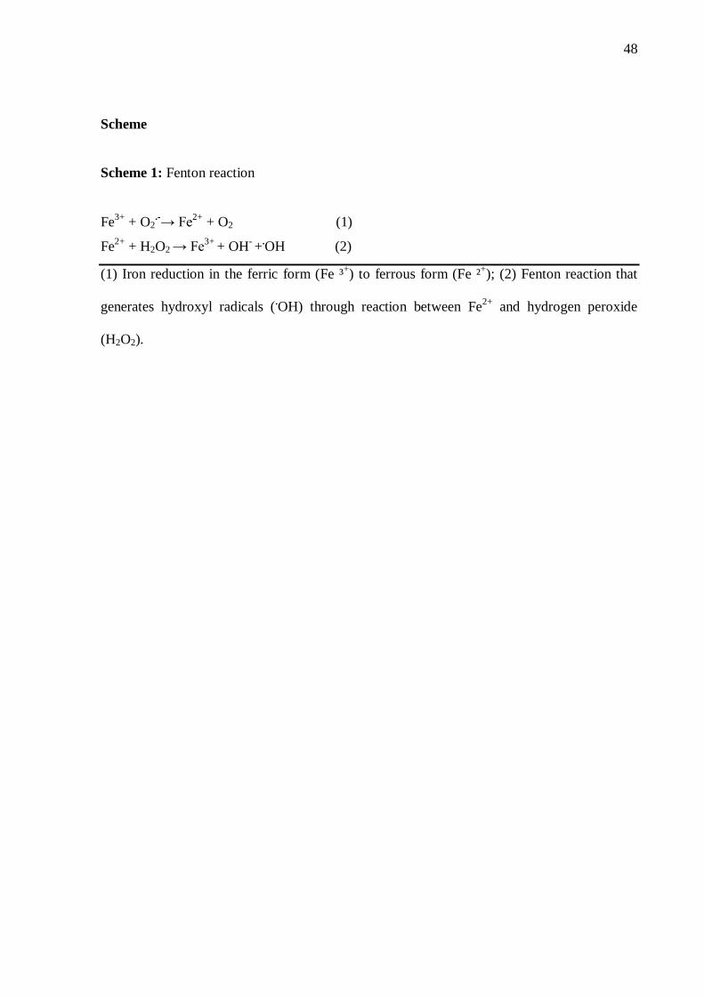
48
Scheme
Scheme 1: Fenton reaction
Fe3+
+ O2.-→ Fe
2+ + O2 (1)
Fe2+
+ H2O2 → Fe3+
+ OH- +
.OH (2)
(1) Iron reduction in the ferric form (Fe ³+) to ferrous form (Fe ²
+); (2) Fenton reaction that
generates hydroxyl radicals (.OH) through reaction between Fe
2+ and hydrogen peroxide
(H2O2).
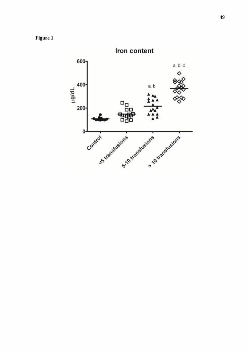
49
Figure 1

50
Figure 2
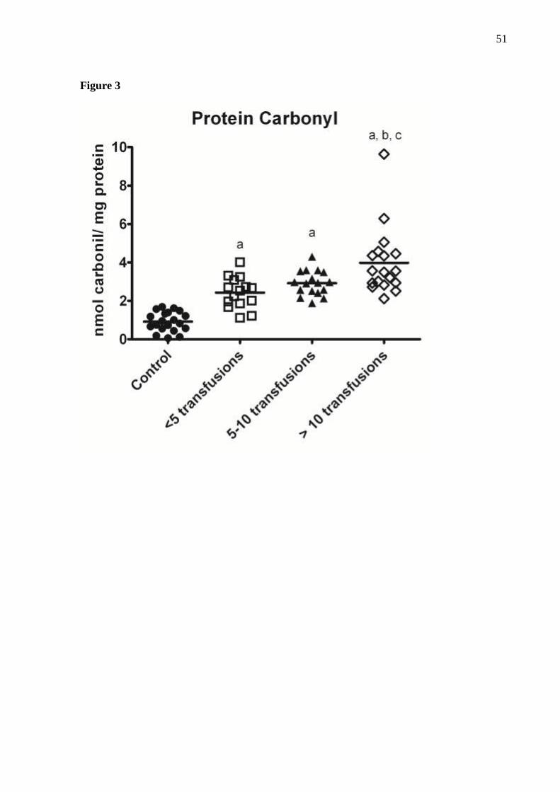
51
Figure 3

52
Figure 4

53
Figure 5
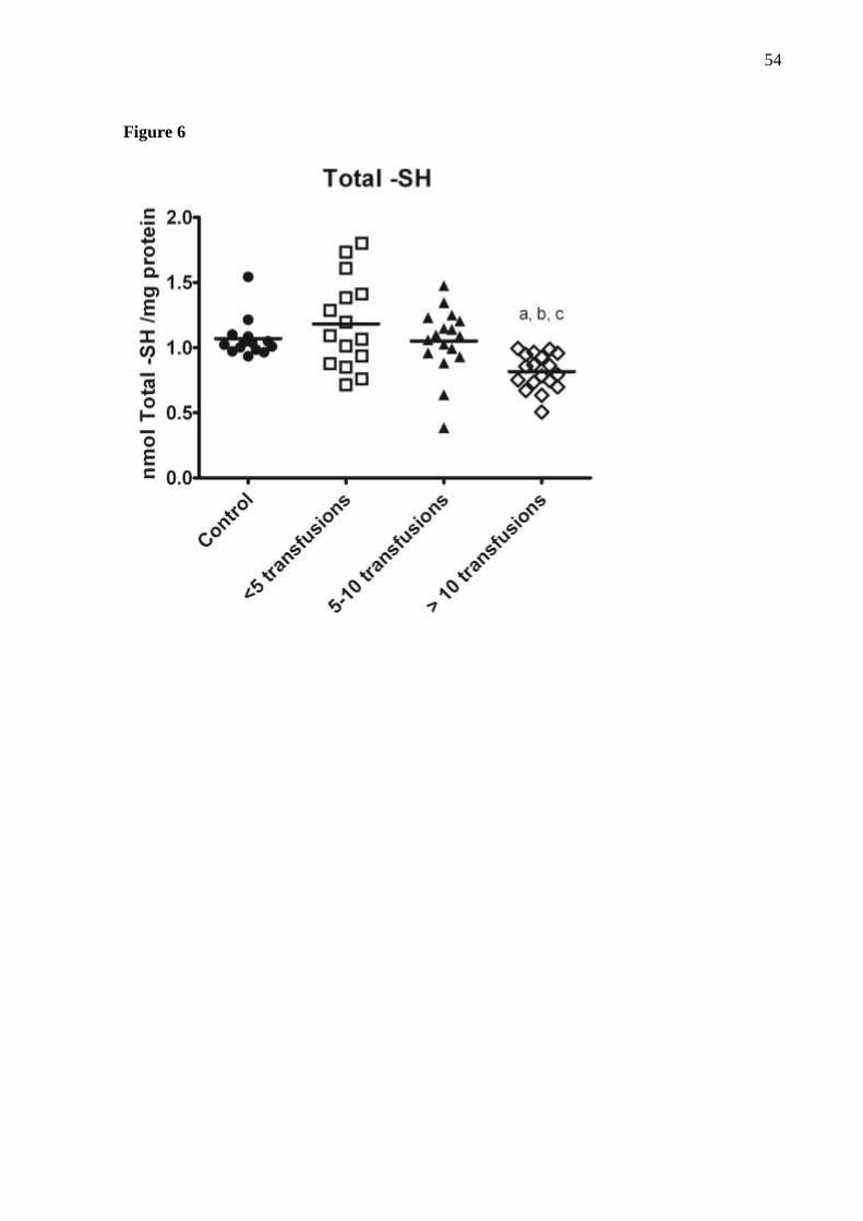
54
Figure 6
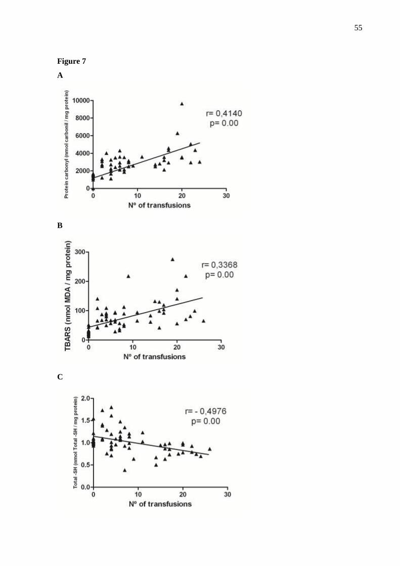
55
Figure 7
A
B
C
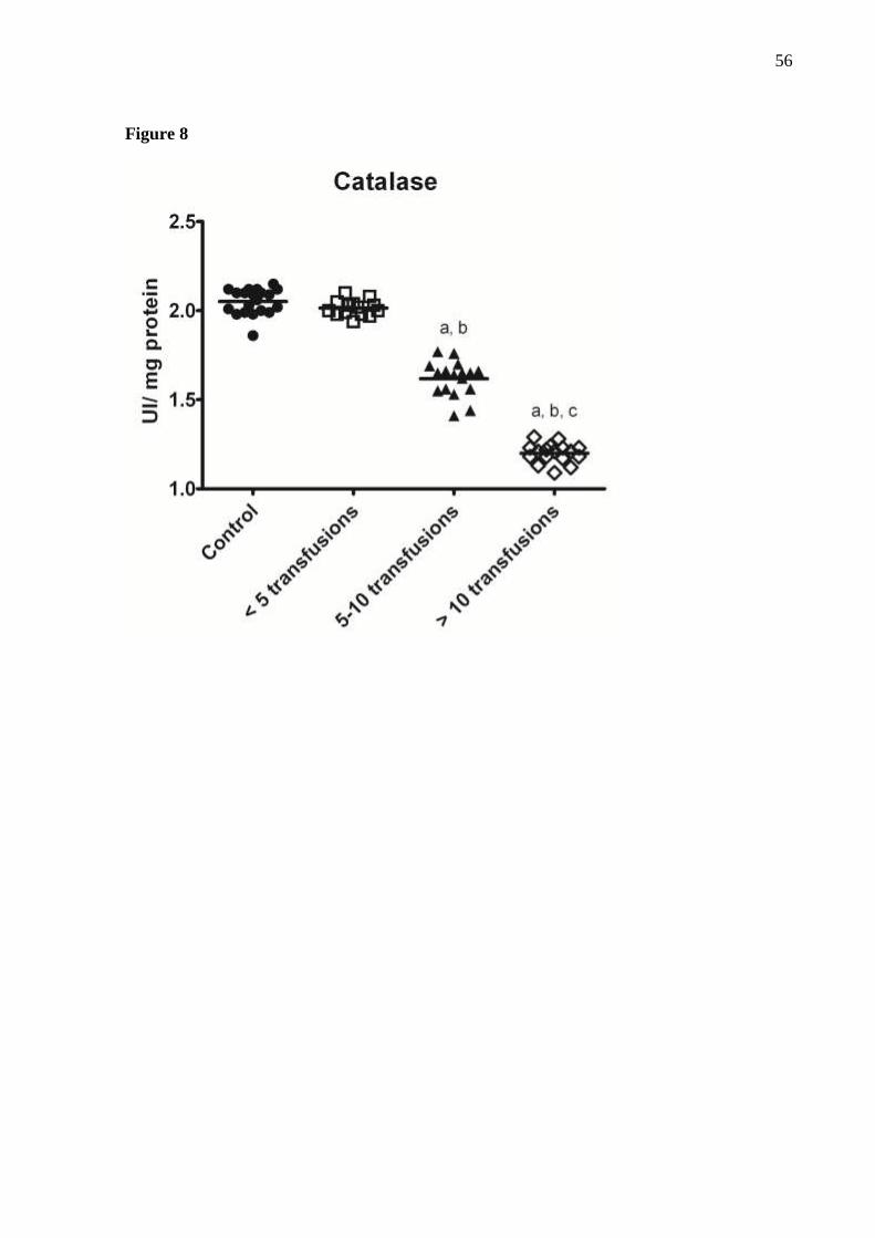
56
Figure 8
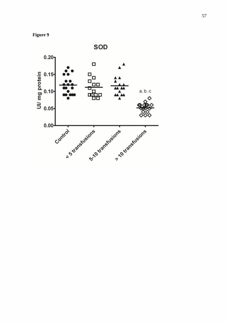
57
Figure 9

58
Figure 10

59
Figure 11
A
B
C
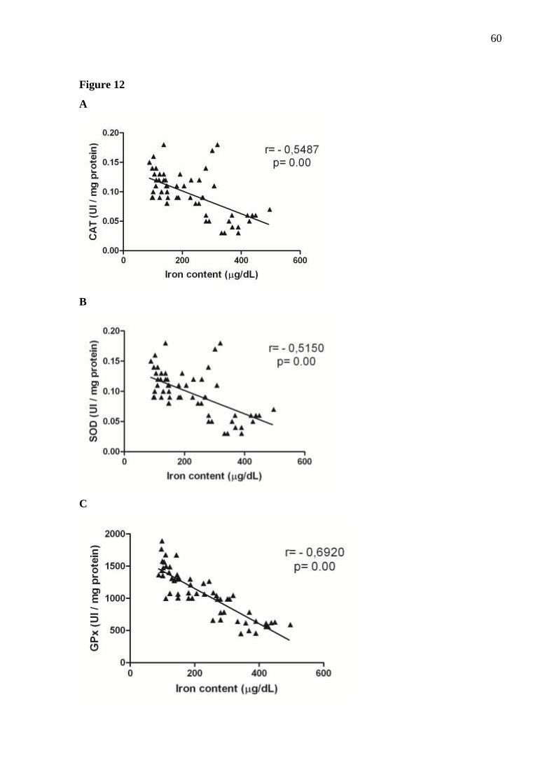
60
Figure 12
A
B
C

61
Figure 12
D
E
F
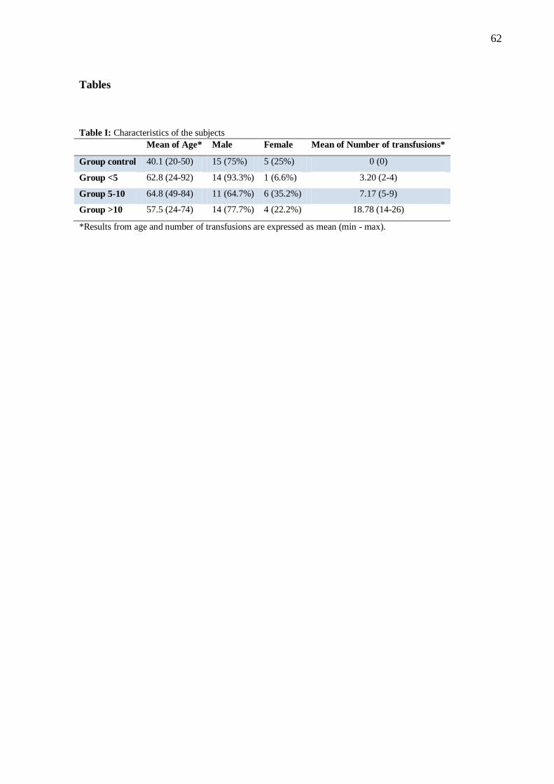
62
Tables
Table I: Characteristics of the subjects
Mean of Age* Male Female Mean of Number of transfusions*
Group control 40.1 (20-50) 15 (75%) 5 (25%) 0 (0)
Group <5 62.8 (24-92) 14 (93.3%) 1 (6.6%) 3.20 (2-4)
Group 5-10 64.8 (49-84) 11 (64.7%) 6 (35.2%) 7.17 (5-9)
Group >10 57.5 (24-74) 14 (77.7%) 4 (22.2%) 18.78 (14-26)
*Results from age and number of transfusions are expressed as mean (min - max).
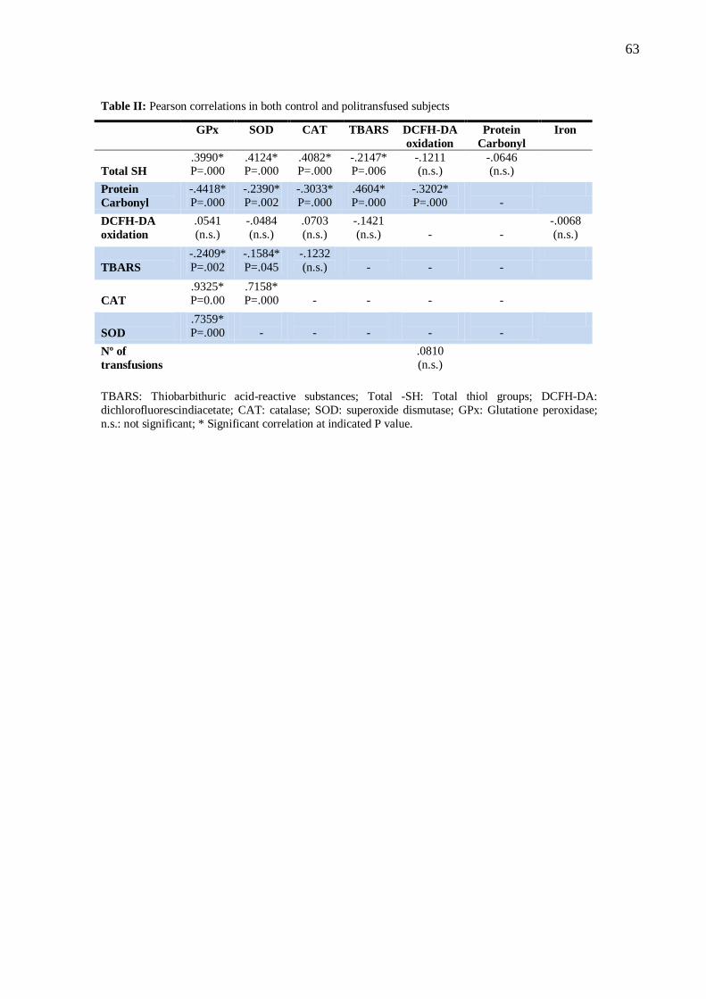
63
Table II: Pearson correlations in both control and politransfused subjects
GPx SOD CAT TBARS DCFH-DA
oxidation
Protein
Carbonyl
Iron
Total SH
.3990* P=.000
.4124* P=.000
.4082* P=.000
-.2147* P=.006
-.1211 (n.s.)
-.0646 (n.s.)
Protein
Carbonyl
-.4418*
P=.000
-.2390*
P=.002
-.3033*
P=.000
.4604*
P=.000
-.3202*
P=.000
-
DCFH-DA
oxidation
.0541
(n.s.)
-.0484
(n.s.)
.0703
(n.s.)
-.1421
(n.s.)
-
-
-.0068
(n.s.)
TBARS
-.2409*
P=.002
-.1584*
P=.045
-.1232
(n.s.)
-
-
-
CAT
.9325*
P=0.00
.7158*
P=.000
-
-
-
-
SOD
.7359*
P=.000
-
-
-
-
-
Nº of
transfusions
.0810
(n.s.)
TBARS: Thiobarbithuric acid-reactive substances; Total -SH: Total thiol groups; DCFH-DA:
dichlorofluorescindiacetate; CAT: catalase; SOD: superoxide dismutase; GPx: Glutatione peroxidase;
n.s.: not significant; * Significant correlation at indicated P value.

64
5. DISCUSSÃO
A análise dos dados obtidos nesse estudo demonstrouque os indivíduos
multitransfundidos apresentaram os parâmetros oxidativos alterados. Com o aumento do
número de transfusões sanguíneas houve um aumento significativo dos níveis de peroxidação
lipídica, uma diminuição significativa dos níveis de grupamentos tiólicos, um aumento
significativo dos níveis de carbonilação de proteína, um aumento significativo dos níveis de
espécies reativas de oxigênio, um aumento significativo do nível de ferro sérico, e uma
diminuição significativa da atividade das enzimas antioxidantes catalase, superóxido
dismutase e glutationa peroxidase. Essas alterações foram observadasprincipalmente em
indivíduos que receberam mais de dez transfusões sanguíneas, concordando com o estudo de
MAJHAIL et al, 2008, onde descreve que indivíduos que receberam de 10 a 20 unidades de
concentrado de hemácias já desenvolvem os efeitos oxidativos da sobrecarga cumulativa de
ferro.
Foi observado um aumento significativo do nível de ferro plasmático nos indivíduos
multitransfundidos com o aumento do número de transfusões sanguíneas, tendo uma
correlação significativa positiva do TBARS e carbonil com o conteúdo de ferro plasmático e
uma correlação significativa negativa do total-SH, CAT, SOD, GPx, com o conteúdo de ferro
plasmático. Esses dados estão de acordo com o estudo de CIGHETTI et al, 2002, que
observou que o ferro sérico esta significativamente mais elevado em pacientes talassemicos
dependentes de transfusões sanguíneas do que nos talassemicos não dependentes de
transfusão sanguíneas, e correlacionou positivamente peroxidação lipídica e ferro sérico.
Estes resultados confirmam que o ferro é um catalisador eficaz para a geração de ERO, o que
resulta em peroxidação lipídica e aumento do estresse oxidativo in vivo (ZHANG et al, 2012).
Uma análise retrospectiva multivariada de 902 pacientes demonstrou forte relação entre os
níveis de ferro sérico com a sobrevida global reduzida, e com sobrevivência livre de leucemia
(SANZ et al., 2008). Essa correlação entre a sobrecarga de ferro e o risco de progressão
leucêmica é apoiada por estudos que demonstram aumento da instabilidade genômica em
células progenitoras hematopoiéticas expostas a níveis aumentados de ROS (KOPTYRA ET
AL, 2006;. NAKA et al, 2008; RASSOOL ET AL, 2007). Em modelos animais, a sobrecarga
de ferro é demonstrada pelo mecanismo de dano hepatocelular mediado por ERO, onde a
sobrecarga de ferro facilita a formação de ERO, que perturba o equilíbrio redox das células e
causa estresse oxidativo e nitrosativo (TOYOKUNI, 2011).
65
O MDA plasmático é um biomarcador bem reconhecido de peroxidação lipídica. Nosso
estudo demonstrou aumento significativo dos níveis de MDA nos indivíduos
multitransfundidos, com correlação significativa positiva com o número de transfusões
sanguíneas recebidas. Nosso resultado esta de acordo com diversos estudos onde demonstram
que o aumento da peroxidação lipídica é amplamente observado em pacientes talassêmicos
que dependem de transfusões sanguíneas contínuas para sobreviver (CIGHETTI et al, 2002;
LAKSMITAWATI et al, 2003; CHAKRABORTY et al, 2001). WALTER e cols.
(2006)demonstraram que a análise da regressão comparando MDA com a concentração de
ferro no fígado de indivíduos talassemicos e indivíduos com anemia falciforme mostram que,
em ambas as doenças, concentrações mais elevadas de ferro no fígado estão associadas com
maiores níveis de MDA plasmático.
A dosagem dos grupamentos tiólicos totais são usados como parâmetro de medida de
dano oxidativo as proteínas plasmáticas. Os tiois estão presentes no plasma em substâncias de
baixo peso molecular, tais como cisteina e glutationa, e como resíduo de proteínas maiores,
como a albumina. Em geral, 80% dos tióis livres do plasma estão contidos na albumina
(RADI et al, 1991). Observamos que os níveis de grupamentos tiólicos foram
significativamente diminuídos com o aumento do número de transfusões sanguíneas, numa
correlação significativa negativa com o número de transfusões sanguíneas. Estes dados estão
de acordo com o estudo de DEVI et al, 2010, onde demonstrou que os níveis de proteínas
tiólicas na mitocôndria de fígado de ratos tratados com ferro e álcool eram significativamente
diminuídos em relação aos ratos tratados somente com álcool. CHUNG et al, 2005
demonstrou que o ferro reage não enzimaticamente com tióis no plasma, principalmente a
albumina, gerando espécies reativas de oxigênio, desencadeando redução da capacidade
antioxidante no plasma e aumento da susceptibilidade de oxidação de LDL colesterol.
Pesquisas recentes relataram que a sobrecarga de ferro e aumento dos níveis de tióis
plasmáticos estão associadas com o aumento da mortalidade em pacientes com síndrome da
angustia respiratória aguda secundaria a circulação extracorpórea (QUINLAN et al, 2000).
A formação da proteína carbonil parece ser um fenômeno comum durante a oxidação de
proteínas, e sua quantificação pode se usada para medir a extensão do dano oxidativo
(BERLETT & STADMAN, 1997; BEAL, 2003; DALLE-DONNE et al, 2003). Atualmente é
um marcador de oxidação protéica bastante usado e está aumentado em várias doenças
humanas, como Doença de Alzheimer, Diabetes mellitus, inflamação e artrite reumatóide
(DALLE-DONNE et al, 2003). São produzidos pela oxidação da cadeia lateral de
aminoácidos (prolina, arginina, lisina e treonina), ou pela clivagem oxidativa das proteínas.
66
Também podem ser introduzidos nas proteínas por uma reação dos aminoácidos cisteina,
histidina e lisina com aldeídos produzidos durante a peroxidação lipídica (DALLE-DONNE et
al, 2003). As proteínas modificadas oxidativamente são produtos quimicamente estáveis,
sendo um fator importante para a sua detecção e armazenamento (RUTKOWSKA et al,
2005). No nosso estudo encontramos um aumento significativo de grupamentos carbonilicos
com o aumento do numero de transfusões sanguíneas, com correlação significativa positiva.
Nosso estudo esta de acordo com o estudo de SHE-FANG et al, 2007, onde demonstrou
aumento de peroxidação lipídica e formação de proteína carbonil em hepatócitos de ratos
tratados com sobrecarga de ferro. Segundo VENKATRAMAN et al, 2004, as modificações de
proteínas durante o e estresse oxidativo pode causar aumento no teor de proteína carbonil e
perda de grupamentos tiólicos.
A fim de detectar as ERO geradas pela sobrecarga de ferro, foi medida a emissão
DCFH-DAno plasma. Observamos um aumento significativo de ERO nos indivíduos
multitransfundidos em relação ao grupo controle, com uma correlação significativa positiva
com o numero de transfusões sanguíneas. Nossos estudos estão de acordo com SHE-FANG et
al, 2007, onde demonstrou que o tratamento com ferro ao plasma humano aumentou a
emissão de DCFH-DAem uma concentração dose-dependente.
Este trabalho investigou a capacidade antioxidante dos indivíduos multitransfundidos
pela mensuração das enzimas antioxidantes CAT, SOD, GPx. Foi observado que a atividade
dessas enzimas foram significativamente diminuídas com o aumento do número de
transfusões sanguíneas, e ainda, observamos uma correlação significativa negativa das
enzimas antioxidantes com o número de transfusões sanguíneas. Provavelmente esta
diminuição da atividade das enzimas antioxidantes esta relacionada ao seu consumo como
sequestrador de oxidantes. Segundo CHAKRABORTY et al, 2011, estes níveis baixos de
enzimas antioxidantes provavelmente contribuem para níveis aumentados de MDA, proteína
carbonil e ERO. CHUNG et al, 2005, demonstrou que após o tratamento de plasma humano
com ferro, a capacidade antioxidante diminui em 50%, sugerindo que o plasma sob condição
de sobrecarga de ferro pode se tornar mais suscetível a ação de oxidantes. Nosso estudo
discorda em partes com o estudo de ZHANG et al, 2012, onde observou aumento de proteínas
carbonil, de MDA, da atividade da SOD e diminuição do conteúdo de sulfidrilas e da
atividade de GPx em de ratos com lesão hepática tratados com sobrecarga de ferro. Nosso
estudo concorda com o estudo de SHE-FANG et al, 2007, que demonstrou uma depleção de
47% na atividade da GPx em hepatócitos expostos a sobrecarga de ferro. No entanto, os

67
mecanismos de regulação da expressão das enzimas antioxidantes na sobrecarga de ferro
necessitam ser mais explorados.
Considerando o exposto, é razoável sugerir que os antioxidantes podem ser associados
com a terapia de sangue. De fato, dados anteriores relatam que a suplementação com
antioxidantes é segura e afeta favoravelmente os marcadores de estresse oxidativo, embora
isso não resulte em um beneficio clinico significativo. No entanto, são necessários mais
estudos para compreender melhor os mecanismos associados aos danos oxidativos
ocasionados por ferro, na tentativa de minimizar os efeitos secundários associados a terapia de
transfusão de sangue repetida e melhorar a qualidade de vida destes indivíduos.

68
6. CONCLUSÃO
Em resumo, este estudo confirma o envolvimento de estresse oxidativo em pacientes
anêmicos após a terapêutica com múltiplas transfusões sanguíneas, e sua correlação
com o teor plasmático de ferro. A concentração de ferro plasmático estava
significativamente aumentada nos indivíduos multitransfundidos;
As enzimas de defesas antioxidantes (CAT, SOD, GPx) encontraram-se
significativamente diminuídas nos indivíduos submetidos a múltiplas transfusões
sanguíneas;
O dano oxidativo a proteínas plasmáticas (proteína carbonil) e o dano oxidativo a
lipídeos (TBARS) encontraram-se significativamente maiores nos indivíduos
submetidos a múltiplas transfusões sanguíneas;
O conteúdo de grupamentos tiólicos totais (TOTAL-SH) foi significativamente
reduzido nos indivíduos submetidos a múltiplas transfusões sanguíneas;
O conteúdo de ERO foi significativamente aumentado nos indivíduos
multitransfundidos com o aumento do número de transfusões sanguíneas;
Além disso, verificou-se que as alterações nos marcadores de estresse oxidativo são
correlacionadas, pelo menos em parte, com o conteúdo de ferro plasmático e o número
de transfusões sanguíneas;

69
7. PERSPECTIVAS
Baseado nos resultados apresentados nessa dissertação faz-se necessário:
Quantificar os níveis séricos de ferritina e transferrina;
Quantificar os níveis de glutationa reduzida e glutationa oxidada;
Elucidar o(s) mecanismo(s) envolvido(s) com a toxicidade do ferro e/ou com as
múltiplas transfusões sanguíneas no desenvolvimento do estresse oxidativo;
Elucidar mecanismos de regulação da expressão das enzimas antioxidantes sob a
sobrecarga de ferro transfusional;
Instituir uma terapia antioxidante nos indivíduos em tratamento com múltiplas
transfusões sanguíneas, a fim de neutralizar e/ou reverter os danos oxidativos
observados neste estudo, como tentativa de melhorar a qualidade de vida desses
indivíduos.
70
REFERÊNCIAS
ALLEN, M.K., 2010. Hereditary hemochromatosis: a literature review and case report.
Physiother. Can. 62, 276–284.
AMES, B.N.; SHIGENAGA, M.K.; HEGEN, T.M.; 1993.Oxidants, antioxidants, and the
degenerative disease of aging. Pro NatlAcadSci USA. 90: 7915-7922.
ANDREWS NC.Disorders of iron metabolism. N Engl J Med 1999;341:1986-95.
ANDREWS, N. 2005. Iron metabolism. In N. Andrews, C. Breymann, C. Gasche, & ITO
Staff (Eds.), ITO textbook—Iron therapy in various settings. Retrieved from
http://www.irontherapy.org/a.php3?d=iron-metabolism--56&a=ITO-Textbook-Chapter-1:-
Iron-Metabolism--4233
ANDREWS, NC. 1999. Disorders of iron metabolism. N Engl J Med; 341:1986–1995.
BATLOUNI, M. 1997.Hipoteseoxidativa na ateroesclerose e empregados antioxidants na
doença arterial coronaria. Arquivo Brasileiro Cardiologia. 68(1).
BEAL, D. J., COHEN, R. R., BURKE, M. J., & MCLENDON, C. L. 2003. Cohesion and
performance in groups: A Meta-analytic clarification of construct relations.
JournalofAppliedPsychology , 88(6), 989-1004.
BERLETT, BS, E STADTMAN, ER 1997. Oxidação de proteínas no envelhecimento,
doenças e estresse oxidativo. J. Biol. Chem. 372 , 20313-20316.
BIANCHI, M.L.P.; ANTUNES, L.M.O. 1999.Free radicals and the main dietary
antioxidantes.Rev Nutrição. 12(2):123-130.
BRASIL I. 2008.Ministerio da Saude. Secretaria de atenção a Saude. Departamento de
atenção especializada. Guia para o uso de hemocomponentes. Brasilia.
BRASIL II. 2011.Ministerio da saúde. Sangue e Hemoderivados: Produção hemoterapica:
Sistema Único de Saude-SUS. Serie G. Estatistica e Informação em Saude. Brasilia.
CHAKRABORTY, D.; BHATTACHARYYA, M. 2001.Antioxidant defense status of red
blood cells of patients with b-thalassemia and Eb-thalassemia.ClinicaChimicaActa 305 (123–
129).
CHUNG, K. Y., LEE, S. J., CHUNG, S. M., LEE, M. Y., BAE, O. N., AND CHUNG, J. H.
2005. Generation of free radical by interaction of iron with thiols in human plasma and its
possible significance. Thromb Res, 116, 157-164.
CHUNG, K.Y. LEE,S.J. CHUNG,S.M. LEE,M.Y. BAE,O.N. CHUNG, J.H. 2005. Generation
of free radical by interaction of iron with thiols in human plasma and its possible significance.
Thrombosis Research (116, 157-164).
71
CIGHETTI, G. DUCA, L. BORTONE, L. SALA, S. NAVA, I. FIORELLI, G. CAPPELLINI,
M. D.2002. Oxidative status and malondialdehyde in b-thalassaemia patients.European
Journal of Clinical Investigation.32(1):55–60.
COLPO, E., BEM, A.F., PIENIZ, S., SCHETTERT, S.D., SANTOS, R.M., FARIAS, I.L.G.,
BERTONCELLO, I., MOREIRA, C.M., MORETTO, M.B., ROCHA, J.B.T. 2008. A single
high dose of ascorbic acid and iron not correlated with oxidative stress in healthy volunteers.
Annals of Nutrition and Metabolism 53, 79-85.
CRICHTON RR, WARD RJ. 2003. An overview of iron metabolism: molecular and cellular
criteria for the selection of iron chelators. Curr Med Chem; 10:997–1004.
DALLE-DONNE, I., D. GIUSTARINI, R. COLOMBO, R. ROSSI AND A. MILZANI,
2003. Protein carbonylation in human diseases.Trends Mol. Med., 9: 169-176.
DEVI, S. L.; ANURADHA, C.V. 2010.Oxidative and nitrosative stress in experimental rat
liver fibrosis: Protective effect of taurine. Environmental Toxicology and Pharmacology 29
104–110.
EMERIT, J., BEAUMONT, C., AND TRIVIN, F. 2001. Iron metabolism, free radicals, and
oxidative injury. Biomed Pharmacother, 55, 333-339.
FERREIRA, A.L.A. MATSUBARA, L.S. 1997. Radicais livres: conceitos, doenças
relacionadas, sistema de defesa e estresse oxidativo. RevAssistenciaMedica Brasil. 43(1):61-
68.
FIBACH, E. & RACHMILEWITZ, E. 2008.The role of oxidative stress in hemolytic anemia.
Current Molecular Medicine, 8, 609–619.
FIBACH, E. RACHMILEWITZ, E. A. VICHINSKY, E. P. NEUFELD, E. J. 2010. The role
of antioxidants and iron chelators in the treatment of oxidative stress in thalassemia.Annals of
the New York Academy of Science. 1202:10-16.
FREITAS JM, MENEGHINI R. MUTAT RES. 2001. 6. Iron and its sensitive balance in the
cell. Apr 18;475(1-2):153-9. Review. PMID:11295160
GATTERMANN, N., & RACHMILEWITZ, E.A. 2011.Iron overload in MDS-
pathophysiology, diagnosis, and complications. Annals of Hematology, 90, 1–10.
doi:10.1007/s00277-010-1091-1.
GHOTI, H. FIBACH, E. MERKEL, D. AVRAHAM, G. P. GRISARIU, S.
RACHMILEWITZ, E. A. 2010. Changes in parameters of oxidative stress and free iron
biomarkes during treatment with deferasirox in iron overloaded patients with myelodysplastic
syndromes.Letters to the Editor.Hematologica. 95(8):1433-1434.
GURZAU E.S., C. NEAGU, A. E. GURZAU, 2003:Essential metals - case study on iron,
Ecotoxicology and Environmental Safety, Special Issue on Methodologies for Assessing
Exposures to Metals: Speciation, Bioaccessibility and Bioavailability in the Environment,
Food and Feed, 56 (1), 190-200.

72
HALLIWELL, B.; GUTTERIDGE, J.C.M. 1999.Free Radicals in Biology and Medicine.3th
ed. Oxford University Press.
HALLIWELL, B. 2000.The antioxidant paradox.Lancet 355, 1179 – 1180.
HALLIWELL, B.; 2006.Proteasomal dysfunction; a common feature of all neurodegenerative
diseases?Implications for the environmental origins of neurodegeneration. Antioxidants &
Redox Signaling 8 (11-12): 2007-2019;
HERSHKO, C. VICHINSKY, E. P. NEUFELD, E. J. 2010. Pathogenesis and management of
iron toxicity in thalassemia.Annals of the New York Academy of Science. 1202:1-9.
HIRATA, L.L. SATO, M.E.O. SANTOS, C.A.M. 2004. Cutaneous ageling and free
radicals.ActaFarmaceuticaBonaerense. 23(3):418-424.
KEW, M.C. 2009. Hepatic iron overload and hepatocellular carcinoma.Cancer Lett. 286, 38–
43.
KOPTYRA, M., FALINSKI, R., NOWICKI, M.O., STOKLOSA, T., MAJSTEREK, I.,
NIEBOROWSKA SKORSKA, M., SKORSKI, T. 2006.BCR/ABL kinase induces self-
mutagenesis via reactive oxygen species to encode imatinib resistance. Blood, 108, 319–327.
LAKSMITAWATI, D.R., HANDAYANI, S., UDYANINGSIH-FREISLEBEN, S.K.,
KURNIATI, V., ADHIYANTO, C., HIDAYAT, J., KUSNANDAR, S., DILLON, H.S.,
MUNTHE, B.G., WIRAWAN, R., SOEGIANTO, R.R., RAMELAN, W. & FREISLEBEN,
H.J. 2003. Iron status and oxidative stress in beta-thalassaemia patients in Jakarta. Biofactors,
19, 53–62.
LAMBING, MSN, NP-C (NURSE PRACTITIONER), ELLEN KACHALSKY, LMSW
(SOCIAL WORKER), & MARY LINDA MUELLER. 2012.The dangers of iron overload:
Bring in the Iron Police. Journal of the American Academy of Nurse Practitioners 24 -175–
183.
LANGLOIS D’ESTAINTOT B, SANTAMBROGIO P, GRANIER T, GALLOIS B,
CHEVALIER JM, PRE´CIGOUX G, LEVI S, AROSIO P: 2004. Crystal structure and
biochemical properties of the human mitochondrial ferritin and its mutant Ser144. Ala. J
MolBiol; 340:277–293.
LEE, P.L. & BEUTLER, E. 2009.Regulation of hepcidina and iron-overload disease.Annual
Review of Pathology, 4, 489–515.
LEI Nº 10.205, de 21 de março de 2001. Disponível em:
www.saude.mg.gov.br/atos_normativos/...de.../Lei_10205.pdf
LINDER, M.C. 1991. Nutrition and metabolism of the trace elements. Elsevier Science
Publishing Company. New York. 215-276.
LIST, A.F. 2010. Iron overload in myelodysplastic syndromes: Diagnosis and management.
CancerControl, 17(Suppl.), 2–8.
73
LOUREIRO, A.P.M. MASCIO, P.D. MEDEIROS, M.H.G. 2002. Exocyclic DNA adducts:
implications in mutagenesis and carcinogenesis. Quimica Nova. 25(5):777-793.
MAJHAIL, N.S. LAZARUS, H.M. BURNS, L.J. 2008.Iron overload in hematopoietic cell
transplantation. Bone Marrow Transplantation 41, 997–1003.
MANFREDINI, V. BIANCINI, B.G. VANZIN, S. C.DAL VESCO, M. A. CIPRIANI,
F.BIASI, L. TREMÉA, R. DEON, M. PERALBA, M. C. WAJNER, M. VARGAS, R.C.
2010. Simvastatin Treatment Prevents Oxidative Damage to DNA in Whole Blood
Leukocytes of Dyslipidemic Type 2 Diabetic Patients. Cell Biochemistry and
Function.28:360-366.
NAKA, K., MURAGUCHI, T., HOSHII, T., & HIRAO, A. 2008.Regulation of reactive
oxygen species and genomic stability in hematopoietic stem cells.Antioxidants and Redox
Signaling, 10, 1883–1894.
OHKAWA, H., OHISHI, N., AND YAGI, K. 1979. Assay for lipid peroxides in animal
tissues by thiobarbituric acid reaction. Anal Biochem, 95, 351-358.
OZMENT, C.P., & TURI, J.L. 2009. Iron overload following red blood cell transfusion and
its impact on disease severity. BiochimicetBiophysica Acta, 1790, 694–701.
doi:10.1016/j.bbagen.2008.09.010
PAGLIA D E & VALENTINE WN. 2008. Studies on the quantitative and qualitative
characterization of erythrocyte glutathione peroxidase.J. Lab. Cm. Med. 70:158-69,
PIERIGÈ F, SERAFINI S, ROSSI L, MAGNANI M. (JANUARY 2008). "Cell-based drug
delivery". Advanced Drug Delivery Reviews 60 (2): 286–95.
PORTARIA Nº 1.353 DE 13 DE JUNHO DE 2011. Disponível em:
http://portal.saude.gov.br/portal/arquivos/pdf/resolucao_153_2004.pdf.
PORTER, J.B. 2001.Praticalmasnagement of iron overload. Br. J. Haematol. 115. 239–252.
QUINLAN GJ, MUMBY S, LAMB NJ, MORAN LK, EVANS TW, GUTTERIDGE JM.
2000. Acute respiratory distress syndrome secondary to cardiopulmonary bypass: do
compromised plasma iron-binding anti-oxidant protection and thiol levels influence outcome?
Crit Care Med;28:2271–6.
RADI R, BUSH KM, COSGROVE TP, FREEMAN BA. 1991. Reaction of xanthine oxidase-
derived oxidants with lipid and protein of human plasma. Arch BiochemBiophys; 286:117–
25.
RAMM, G.A., RUDDELL, R.G. 2005.Hepatotoxicity of iron overload: mechanisms of
ironinduced hepatic fibrogenesis. Semin. Liver Dis. 25, 433–449.
RASSOOL, F.V., GAYMES, T.J., OMIDVAR, N., BRADY, N., BEURLET, S., PLA, M.
PADUA, R.A.2007.Reactive oxygen species, DNA damage, and error-prone repair: A model
for genomic instability with progression in myeloid leukemia? CancerResearch, 67, 8762–
8771.

74
RESOLUÇÃO-RDC Nº 153, DE 14 DE JUNHO DE 2004. Disponível em:
http://portal.saude.gov.br/portal/arquivos/pdf/resolucao_153_2004.pdf
REPKA T, SHALEV O, REDDY R, YUAN J.2010.Nonrandom association of free iron with
membranes of sickle and b-thalassemic erythrocytes.Blood1993;82:3204–10.
RIBAS, G. S. MANFREDINI, V. DE MARI, J. WAYHS, Y. C. VANZIN, C. S. BIANCINI,
G. B. SITTA, A. DEON, M. WAJNER, M. VARGAS, C. R. 2010. Reduction of lipid and
protein damage in patients with disorders of propionate metabolism under treatment: a
possible protective role of L-carnitine supplementation. International Journal of
Developmental Neuroscience.28:127-132.
RUTKOWSKA, J., CICHON´, M., PUERTA, M. & GIL, D. 2005.Negative effects of
elevated testosterone on female fecundity in zebra finches. Hormones and Behavior, 47, 585–
591. doi:10.1016/jhbeh. 2004.12.006.
SANZ G, NOMDEDEU B, SUCH E, BERNAL T, BELKAID M, ARDANAZ,MT, MARCO
V, PEDRO C, RAMOS F, DEL CAN˜ IZO MC, LUN˜O E, COBO F, CARBONELL F,
GOMEZ V, MUN˜ OZ JA, AMIGO ML, BAILEN A, BONADAD S, TORMO M, ANDREU
R, ARRIZABALAGA B, ARILLA MJ, BUENO J, REQUENA MJ, BARGAY J, SANCHEZ
J, SENENT L, ARENILLAS L, DE PAZ R, XICOY B, DUARTE R, CERVERA J:
December 6–9, 2008. Independent impact of iron overload and transfusion dependency on
survival and leukemic evolution in patients with myelodysplastic syndrome. Presented at the
Annual Meeting of the American Society of Hematology.; San Francisco, California. Abstract
640
SAZAMA.SHANDER.2010. Clinical consequences of iron overload from chronic red blood
cell transfusions, its diagnosis, and its management by chelation therapy.
TRANSFUSION;50:1144-1155.
SHAH,J. SANDRA E. KURTIN, RN, LOUISE ARNOLD, PETRA LINDROOS-
KOLQVIST, SARA TINSLEY.2012. Management of Transfusion-Related Iron Overload in
Patients WithMyelodysplastic Syndromes. Clinical Journal of Oncology Nursing •
Supplement to Volume 16, Number 3.
SHANDER,A. CAPPELLINI, M.D. GOODNOUGH, L.T. 2009. Iron overload and toxicity:
the hidden risk of multiple blood transfusions. VoxSanguinis 97, 185–197.
SHE-FANG YE1, ZHEN-QING HOU1 AND QI-QING ZHANG. 2007. Protective Effects of
PhellinuslinteusExtract against Iron Overload-mediated Oxidative Stress in Cultured Rat
Hepatocytes. Phytother. Res. 21, 948–953.
SIMÕES, B.P. PIERONI, F. BANOS, G. M. N. MACHADO, C. L. CANÇADO, R. D.
SALVINO, N. A. ANGELO, I. VOLTARELLI, J. C. 2010. Brazilian consensus
meetingonstemcelltransplatation: hemoglobinopathiescomitte. Rev Brasileira de Hematologia
e Hemoterapia. 32(1).

75
SITTA, A. MANFREDINI, V. BIASI, L. TREMÉA, R. SCHWARTZ, I. WAJNER,
M. VARGAS, C. R.2009. Evidence that DNA damage is proportional to phenylalanine blood
levels in leukocytes from phenylketonuric patients.Mutation Research.Genetic Toxicology
and Environmental Mutagenesis. 679:13-16.
TOYOKUNI S. 1996. Iron-induced carcinogenesis: the role of redox regulation. Free Rad
Biol Med 20:553–566
TOYOKUNI, S. 2011. Iron as a target of chemoprevention for longevity in humans. Free.
Radic. Res. 45, 906–917.
TSAY, J. YANG, Z. W. ROSS, F. P. CUNNINGHAM-RUNDLES, S. LIN, H. COLEMAN,
R. MAYER-KUCHUK, P. DOTY, S. B. GRADY, R.W. GIARDINA, P. J. BOSKEY, A. L.
VOGIATZI, M. G. 2010. Bone low caused by iron overload in a murine model: importance of
oxidative stress. Blood. 116(14):2582-2589.
VASCONCELOS, S.M.L. GOULART, M.O.F. MOURA, J.B.F. MANFREDINI, V.
BENFATO, M.S. KUBOTA, L.T. 2007. Reactive oxygen and nitrogen species, antioxidants
and markers of oxidative damage in humanblood: main analytical methods for their
determination. Quimica Nova. 30(5):1323-1338.
VENKATRAMAN, A., LANDAR, A., DAVIS, A.J., ULASOVA, E., PAGE, G., MURPHY,
M.P., DARLEY-USMAR, V., BAILEY, S.M., 2004. Oxidative modification of hepatic
mitochondria protein thiols: effect of chronic alcohol consumption.Am.J. Physiol.
Gastrointest. Liver Physiol. 286, G521–G527.
VERRASTRO, Therezinha,2005. Atheneu, Hematologia e Hemoterapia, São Paulo.
WALTER, P.B. ELLEN B. FUNG,3 DAVID W. KILLILEA,1 QING JIANG,1 MARK
HUDES,4 JACQUELINE MADDEN,2 JOHN PORTER,5 PATRICIA EVANS,4 ELLIOTT
VICHINSKY3 AND PAUL HARMATZ. 2006. Oxidative stress and inflammation in iron
overloaded patients with b-thalassaemia or sickle cell disease. British Journal ofHaematology,
135, 254–263.
ZHANG A,B,Y. HUANG A.Y. DENG C.X. XU A. Y. GAO A.Z. HAILING LI. 2012. Iron
overload-induced rat liver injury: Involvement of protein tyrosine nitration and the effect of
baicalin. EuropeanJournalofPharmacology 680 95–101.

76
ANEXO I
Carta de aprovação do Comitê de Ética em Pesquisa da Unipampa.