UNIVERSIDADE FEDERAL DO RIO GRANDE DO NORTE … · E. vermicularis Enterobius vermicularis EDTA...
71
UNIVERSIDADE FEDERAL DO RIO GRANDE DO NORTE CENTRO DE CIÊNCIAS DA SAÚDE PROGRAMA DE PÓS-GRADUAÇÃO EM CIÊNCIAS DA SAÚDE IMUNOGLOBULINA E E EOSINÓFILOS EM CRIANÇAS DE ÁREA TROPICAL, INFECTADAS POR Ascaris lumbricoides EDNA MARQUES DE ARAÚJO SILVA NATAL/RN 2013
-
Upload
vuongkhanh -
Category
Documents
-
view
219 -
download
0
Transcript of UNIVERSIDADE FEDERAL DO RIO GRANDE DO NORTE … · E. vermicularis Enterobius vermicularis EDTA...
UNIVERSIDADE FEDERAL DO RIO GRANDE DO NORTE
CENTRO DE CIÊNCIAS DA SAÚDE
PROGRAMA DE PÓS-GRADUAÇÃO EM CIÊNCIAS DA SAÚDE
IMUNOGLOBULINA E E EOSINÓFILOS EM CRIANÇAS DE ÁREA TROPICAL,
INFECTADAS POR Ascaris lumbricoides
EDNA MARQUES DE ARAÚJO SILVA
NATAL/RN 2013
EDNA MARQUES DE ARAÚJO SILVA
IMUNOGLOBULINA E E EOSINÓFILOS EM CRIANÇAS DE ÁREA TROPICAL,
INFECTADAS POR Ascaris lumbricoides
Orientador: Profa. Dra. Valéria Soraya de Farias Sales
NATAL/RN 2013
Tese apresentada ao Programa de Pós-Graduação em Ciência da Saúde da Universidade Federal do Rio Grande do Norte como requisito para a obtenção do título de Doutor em Ciências da Saúde
CATALOGAÇÃO NA FONTE Universidade Federal do Rio Grande do Norte - UFRN
ii
UNIVERSIDADE FEDERAL DO RIO GRANDE DO NORTE
CENTRO DE CIÊNCIAS DA SAÚDE
S586i
Silva, Edna Marques de Araújo.
Imunoglobulinas e eosinófilos em crianças de área tropical
infectadas por Ascaris lumbricoides / Edna Marques de Araújo Silva.
- Natal, 2013.
65f. : il.
Orientadora: Profª Drª Valéria Soraya de Farias Sales.
Tese (Doutorado) apresentada ao Programa de Pós-Graduação em
Ciências da Saúde. Centro de Ciências da Saúde. Universidade
Federal do Rio Grande do Norte.
1. Ascaris lumbricoides - Tese. 2. Imunoglobulina E - Tese.
Resposta Th2 - Tese. 4. Imunidade adaptativa - Tese.
Eosinófilos - Tese. I. Sales, Valéria Soraya de Faria. III. Título.
RN-UF/BS-CCS CDU: 616-995.132(043.3)
RN-UF/BS-CCS CDU: 616-
995.132(043.3)
PROGRAMA DE PÓS-GRADUAÇÃO EM CIÊNCIAS DA SAÚDE
Coordenadora do Programa de pós-Graduação em Ciências da Saúde
Profa. Dra. Maria Ivonete de Araújo
iii
EDNA MARQUES DE ARAÚJO SILVA
IMUNOGLOBULINAS E EOSINÓFILOS EM CRIANÇAS DE ÁREA
TROPICAL NO BRASIL, INFECTADAS POR Ascaris lumbricoides
Aprovada em ____/___/___
Banca Examinadora:
Presidente da Banca: Prof.a. Dr.a Valéria Soraya de Farias Sales-UFRN
Membros da Banca: Prof.a Dr;a Paula Renata Lima Machado-UFRN
Prof.a Dr.a. Ana Claudia Galvão Freire Gouveia-UFRN
Prof.a Dr.a Francisca Inês de Sousa Freitas - UFPB
Prof.a Dr.a. Zulma Maria de Medeiros - FIOCRUZ-PE
iv

DEDICATÓRIA
A Deus, meus familiares e aos
meus amigos....
v
AGRADECIMENTOS
A DEUS por me dar forças, quando achei que não conseguiria concluir essa
etapa da minha vida e colocou as pessoas certas nas horas certas para que eu
prosseguisse na caminhada.
A minha família, em especial, meu marido e meus filhos, pela força,
incentivo e apoio.
A professora Valéria, pela força, paciência, ensinamentos e em especial
pela amizade que desabrochou nesses anos de convivência. MUITO OBRIGADA.
Aos colegas Tereza Neuma, Ana Claudia, Goretti, Luanda, Sara, Paula,
Antonia Claudia, Melina e Geraldo, pela ajuda, inventivo e compreensão.
A DaLuz, funcionária do Laboratório de Parasitologia, pela colaboração em
todos os momentos que precisei, pela sua disposição, seu carinho e amizade.
A Lucia e Erieneide do LIAC, pela colaboração nas coletas e realização de
exames sanguíneos.
Aos diretores das Creches que participaram da pesquisa, pela colaboração
e apoio na conscientização dos pais e alunos.
Ao laboratório de Alergia e Biologia Molecular do Departamento de
Pediatria e Biologia Molecular da Faculdade de Ribeirão Preto - USP, em especial
a Dra. Luiza Karla Arruda e Juliana Araújo pela colaboração e presteza
dispensadas.
Ao Departamento de Análises Clínicas, na pessoa do chefe do
Departamento Profa. Telma Maria A. M. Lemos pela colaboração e apoio.
Ao Programa de pós-Graduação do Centro de Ciências da Saúde pela
ajuda financeira na compra de substâncias essenciais para o desenvolvimento do
trabalho.
vi
vii
”Há um tempo em que é preciso abandonar
as roupas usadas, que já tem a forma do
nosso corpo, e esquecer os nossos
caminhos, que nos levam sempre aos
mesmos lugares. É o tempo da travessia: e,
se não ousarmos fazê-la, teremos ficado,
para sempre, à margem de nós mesmos."
(Fernando Pessoa)
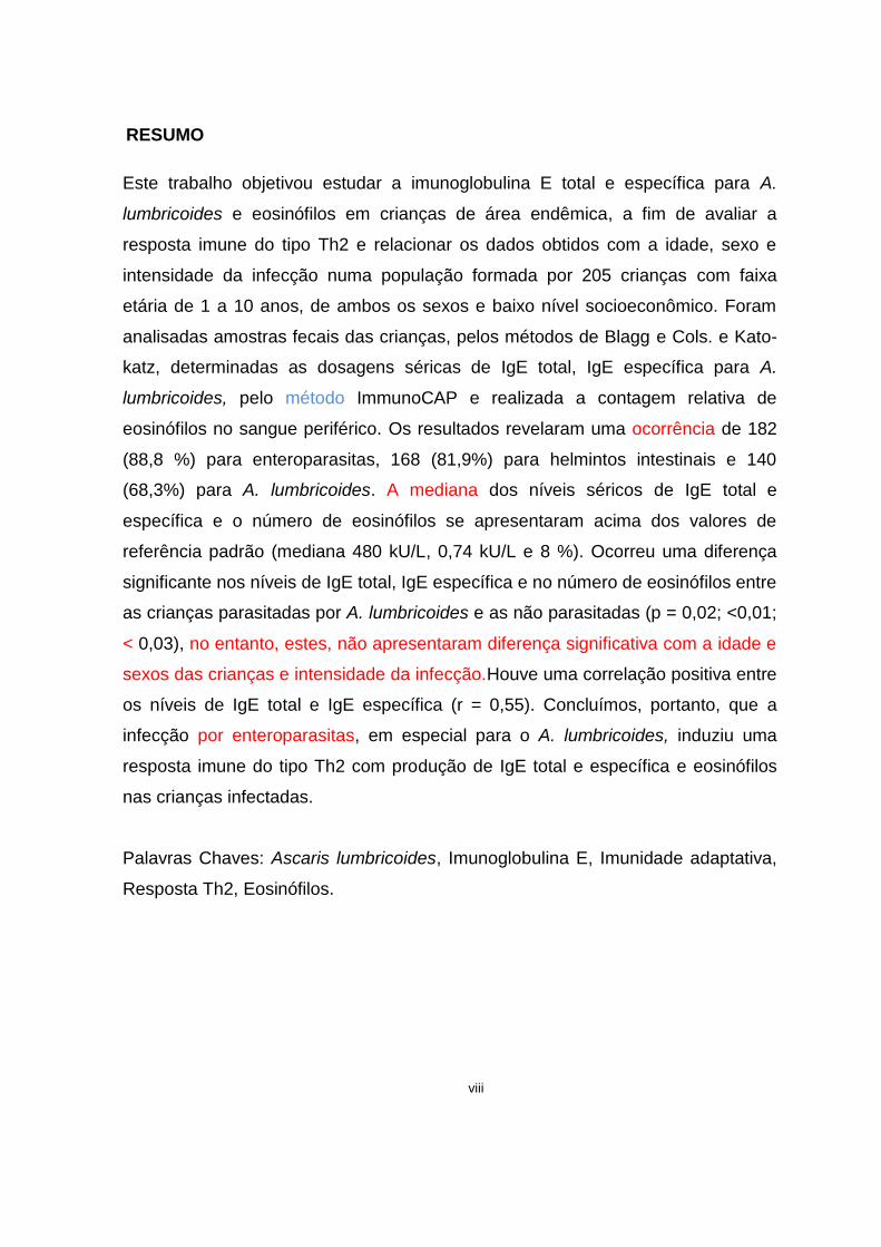
RESUMO
Este trabalho objetivou estudar a imunoglobulina E total e específica para A.
lumbricoides e eosinófilos em crianças de área endêmica, a fim de avaliar a
resposta imune do tipo Th2 e relacionar os dados obtidos com a idade, sexo e
intensidade da infecção numa população formada por 205 crianças com faixa
etária de 1 a 10 anos, de ambos os sexos e baixo nível socioeconômico. Foram
analisadas amostras fecais das crianças, pelos métodos de Blagg e Cols. e Kato-
katz, determinadas as dosagens séricas de IgE total, IgE específica para A.
lumbricoides, pelo método ImmunoCAP e realizada a contagem relativa de
eosinófilos no sangue periférico. Os resultados revelaram uma ocorrência de 182
(88,8 %) para enteroparasitas, 168 (81,9%) para helmintos intestinais e 140
(68,3%) para A. lumbricoides. A mediana dos níveis séricos de IgE total e
específica e o número de eosinófilos se apresentaram acima dos valores de
referência padrão (mediana 480 kU/L, 0,74 kU/L e 8 %). Ocorreu uma diferença
significante nos níveis de IgE total, IgE específica e no número de eosinófilos entre
as crianças parasitadas por A. lumbricoides e as não parasitadas (p = 0,02; <0,01;
< 0,03), no entanto, estes, não apresentaram diferença significativa com a idade e
sexos das crianças e intensidade da infecção.Houve uma correlação positiva entre
os níveis de IgE total e IgE específica (r = 0,55). Concluímos, portanto, que a
infecção por enteroparasitas, em especial para o A. lumbricoides, induziu uma
resposta imune do tipo Th2 com produção de IgE total e específica e eosinófilos
nas crianças infectadas.
Palavras Chaves: Ascaris lumbricoides, Imunoglobulina E, Imunidade adaptativa,
Resposta Th2, Eosinófilos.
viii
LISTA DE ABREVIATURAS E SIGLAS
A.lumbricoides Ascaris lumbricoides
A. hominis Blastocystis hominis
C.mesneli Chilomastix mesnili
DTN Doenças tropicais negligenciadas
E. coli Entamoeba coli
E. histolytica / dispar Entamoeba histolytica / dispar
E. nana Endolimax nana
E. vermicularis Enterobius vermicularis
EDTA Ácido etilenodiamino tetra-acético
ELISA “Enzime-Linked immunosorbent assay”
G. lamblia Giardia lamblia
H. nana Hymenolepis nana
HUOL Hospital Universitário Onofre Lopes
I. butschlli Iodamoeba butschlii
IgE Imunoglobulina E
IgG Imunoglobulina G
IgEAsc Imunoglobulina específica para A.
lumbricoides IL Interleucina
OMS Organização Mundial da Saúde
OPG Ovos por grama de fezes
RR Risco Relativo
S. strongyloides Strongyloides stercoralis
Th1 Resposta linfocitária do tipo 1 mediada
por linfócitos T helper 1 Th2 Resposta linfocitária do tipo 2 mediada
por linfócitos T helper 2 vx
T. trichiura Trichiuris trichiura
UFRN Universidade Federal do Rio Grande
do Norte HTS Helmintos solo transmissíveis
x
LISTA DE FIGURAS
Figura 1 – IgE total em crianças que moram em uma área endêmica para
parasitas intestinais.
Figura 2 - IgE específico para A. lumbricoides em crianças que moram em área
endêmica para parasitas intestinais.
Figura 3 – Correlação entre IgE específica para A. lumbricoides e IgE total em
crianças que moram em área endêmica para parasitas intestinais.
Figura 4 – Número de eosinófilos encontrado na população de crianças, que
moram em área endêmica, segundo parasitas intestinais encontrado e a presença
de IgE específica para A. lumbricoides.
xi
LISTA DE TABELAS
Tabela 1 - Características da população e o resultado das análises parasitológicas
de acordo com faixas etárias em crianças de área endêmica.
Tabela 2 - Parâmetros imunológicos estudados de acordo com as características
da população de 205 crianças de área endêmica.
xii
SUMÁRIO
DEDICATÓRIA
AGRADECIMENTOS
RESUMO
LISTA DE FIGURAS
LISTA DE TABELAS
1. INTRODUÇÃO 15
1.1. Parasitas intestinais 15
1.2. Respostas imunes nas infecções por helmintos 17
1.2.1. Eosinófilos 19
2. JUSTIFICATIVA 21
3. OBJETIVOS 22
3.1. Objetivo Geral 22
3.2. Objetivos específicos 22
4. MÉTODO 23
4.1. População de estudo 23
4.2. Considerações éticas 23
4.3. Coleta e análises das amostras 23
4.4. Coleta de sangue 24
4.5. Determinação das imunoglobulinas E total e específica para A. lumbricoides (ABA 1)
24
4.6. Eosinófilos 24
4.7. Análises estatísticas 25
5. ARTIGO PRODUZIDO 26
6. COMENTÁRIOS CRÍTICAS E CONCLUSÕES 59
7 REFERÊNCIAS 64
xiii
8 APÊNDICE 74
9 ANEXO 75
xiv
15
1. INTRODUÇÃO
1.1. Parasitas intestinais
A alta prevalência de parasitas intestinais encontrada em seres humanos
constitui agravo importante à saúde. A Organização Mundial da Saúde (OMS)
estima que mais de dois bilhões de pessoas no mundo estão infectadas com uma
ou mais espécie de parasito, a maioria dos quais residem em países em
desenvolvimento e em áreas com condições de vida socioeconomica e precárias
(1).
Os parasitas geohelmintos podem infectar os seres humanos em quase
todas as regiões geográficas e climáticas, sendo as prevalências mais elevadas
nos climas quente e úmido nas regiões tropicais e subtropicais (2). No Brasil as
enteroparasitoses ocorrem nas diversas regiões do país, seja em zona rural ou
urbana e em diferentes faixas etárias (3), estando fortemente correlacionadas
com níveis socioeconômicos mais baixos e condições precárias de saneamento
básico (4).
Entre os helmintos o Ascaris lumbricoides é o mais prevalente no ser
humano, acometendo cerca de 1,2 bilhões de pessoas em todo o mundo (5)
traduzindo em sérias consequências que são manifestadas em cerca de 122
milhões de caso/ano, sendo considerada como uma das doenças tropicais
negligenciadas (DTN) (6).
Apesar da importância das infecções por enteroparasitas para a Saúde
Pública, muito pouco se conhece sobre os detalhes de como estes interagem com
seus hospedeiros, incluindo a natureza e eficácia das respostas imunes que são
geradas (7).
As doenças infecciosas são adquiridas diretamente como resultado do
comportamento e estilo de vida e a luta do organismo contra essas doenças que
estão intimamente relacionadas ao nosso sistema imunológico. Acredita-se que o
sistema imunológico humano evoluiu em resposta ao desafio de patógenos
invasores (8).
16
Entre os agentes infecciosos, os parasitas helmintos são considerados os
maiores manipuladores da resposta imune do hospedeiro e estão associados as
infecções crônicas, geralmente assintomáticas. As infecções por helmintos
induzem respostas Th2 (inata e adaptativas), por células do sistema imunológico
(macrófagos, eosinófilos e linfóides), muito embora, vermes parasitas possam
sobreviver em seus hospedeiros mamíferos, inibindo respostas imunitárias
inflamatórias e induzindo, muitas vezes, tolerância a antígenos de parasitas (9-10).
Entretanto, o mecanismo exato de como os vermes conseguem este efeito ainda
não foi totalmente esclarecido (11).
A patologia na infecção por helmintos ocorre frequentemente em indivíduos
que tem uma imunidade reduzida e, portanto, altamente susceptíveis à infecção
com cargas muito elevadas de vermes. Mas também, pode ocorrer em indivíduos
que são imunologicamente muito reativos, apesar de ter cargas baixas de vermes,
neste caso, acredita-se que possa ter ocorrido um colapso entre o
estabelecimento da resposta imune regulatória pelo hospedeiro e o parasita (12-
13). A manutenção de um estado de tolerancia ou assintomático da doença requer
uma relação regulatória imunológica adequada para cada série de helminto, o que
é pouco provável de ocorrer em cada hospedeiro individualmente (14).
A infecção por helmintos pode produzir moduladores potentes da resposta
inflamatória do hospedeiro que protegem esses parasitas contra a morte ou
expulsão por parte do hospedeiro (15). Além da resposta Th2, células T
reguladoras, células B e macrófagos ativados foram identificados como
componentes-chave na regulação do funcionamento da rede imunológica durante
infecções por helmintos (16).
Nas doenças infecciosas e parasitárias, a monitorização da resposta imune
humoral ao agente etiológico constitui uma ferramenta valiosa para a
compreensão dos mecanismos de controle dos parasitas, sobre o sistema
imunológico do hospedeiro (17-20).
1.2. Respostas imunes nas infecções por helmintos
Os parasitas nematóides Ascaris, em particular, podem induzir a
Imunoglobulina E nas respostas tipo Th2, o que tem sido foco de pesquisa para a
17
compreensão das respostas imunes ao parasita e também por.estar diretamente
ligada a reações alérgicas, especialmente na maioria dos países
subdesenvolvidos localizados nos trópicos, onde as populações são naturalmente
expostas a parasitas e outros alérgenos (21).
Entretanto, a função desta resposta imune humoral nas infecções por A.
lumbricoides ainda é controversa e indefinida sendo discutido se a respostas a
antígenos específicos do parasita confere um grau de proteção ao hospedeiro
(22).
A resposta do tipo 2 provavelmente evoluiu ao proporcionar resistência ao
organismo, limitar o número de helmintos que podem viver em nosso trato
intestinal e para reparar os danos do tecido causados por helmintos que o
colonizaram (16).
As evidências da imunidade nas respostas tipo Th2 do hospedeiro, nas
infecções por helmintos intestinais, sugerem um papel crucial na redução da
gravidade da doença após infecção na fase aguda (23). Ela está correlacionada
com a produção das interleucinas IL-4, IL-5, IL-9, IL-10 e IL-13 e,
consequentemente, com a elevação da imunoglobulina (IgE) e da eosinofilia (18).
Entretanto, ainda não está claro como esta resposta é iniciada durante a infecção
por helmintos, pois uma ampla variedade de mecanismos efetores são ativados
por essas citocinas (11,13).
É oportuno destacar que a ação da IL-4 sobre a produção de IgE, bem
como a atuação da IL-13 sobre os mastócitos, resultam em aumento de
mediadores da inflamação, secreção de muco e aumento da contratilidade
muscular intestinal, que podem contribuir na eliminação de helmintos (24-26).
Todo esse processo induzido pela somatização de antígenos excretores e
secretores das larvas pode conferir resistência nas reinfecções (21). No entanto, o
mecanismo exato da expulsão dos parasitas ainda não está bem esclarecido,
embora, acredite-se que ele ocorra pela indução da resposta Th2 ou pela criação
de um ambiente inóspito (27).
Vários estudos ressaltam a importância dos anticorpos IgE na imunidade
contra infecções por helmintos mas, ainda é questionado se o aumento da
produção de IgE é resultado da imunidade humoral na defesa contra helmintos ou
se é apenas um mecanismo de escape dos parasitas, desencadeado pela
18
hiperprodução de IL-4, resultando na produção policlonal e portanto não funcional
da IgE (28-29).
Já está bem estabelecido que os mastócitos expressam o receptor FcƐRI e
que estes se ligam com alta afinidade com a porção Fc do anticorpo IgE. Os
anticorpos IgE se ligam aos receptores destas células, reconhecendo antígenos
multivalentes específicos, promovendo nos mastócitos um padrão característico
de mudanças bioquímicas e morfológicas, coletivamente denominadas
degranulação anafilática que resulta na liberação de um painel de mediadores
biologicamente ativos (30).
A existência de uma associação entre os níveis elevados de IgE, e a
elevada expressão de FcƐR de superfície, em soro de crianças, tem sido relatada
por alguns estudos como uma correlação positiva entre IgE ligada à célula e a
expressão de FcƐR em basófilos, células dendríticas e monócitos (23, 24).
Em alguns indivíduos, os anticorpos da classe IgE específica para certos
alérgenos não promovem a degranulação dos mastócitos e várias hipóteses tem
sido levantadas para explicar este fato como: i) A competição da IgE policlonal
induzida pelo helminto para receptores dos mastócitos. (ii) A elevação da IgG4 e
IgE nas infecções helmínticas; (iii) Indução de células T CD4+ CD25+ Foxp3+ que
imunomodulam negativamente a imunidade mediada por IgE (31). A IgG4 e a IgE
estão elevadas nas infecções helmínticas, no entanto, foi observado que a IgG4
pode atuar como um "anticorpo de bloqueio" na imunidade mediada por IgE
devido à sua capacidade de competir com os epítopos idênticos para IgE (31).
Trabalhos têm relatado que estas reações estão fortemente influenciadas
por fatores genéticos de ambos hospedeiros e parasitas e dessa forma, a
resistência natural a parasita em populações, tem sido pesquisada através da
genética, buscando o entendimento da evolução parasita-hospedeiro (32).
Estudos sobre a resposta ao A. lumbricoides encontraram em populações de
regiões tropicais numa área endêmica para este nematóide, que o polimorfismo de
genes, (LIG4 - ligase IV), TNFSF13B (B cell activation factor) e IRS2 (insulin
receptor substrate-2), localizados na região 13q33–34 que normalmente é relatada
para produção de anticorpo, está associado com a IgE total, IgE e IgG específica
para Ascaris (33-35).
19
Destacando a ação dos helmintos como moduladores da resposta imune
celular e humoral, foi demonstrado em alguns estudos que o extrato de Ascaris
suum obtido a partir de vermes adultos é capaz de suprimir a resposta imune
humoral e celular induzidas por antígenos heterólogos, como ovoalbumina (OVA)
ou micobactéria (36-37).
Os vermes nematóides tem a capacidade de produzir proteínas que
desencadeiam reações alérgicas, os NPAs (nematóides alérgenos poliproteicos)
que podem ser prejudiciais ou, ao mesmo tempo, proteger contra as infecções por
estes parasitas em humanos e outros animais (38). Duas dessas proteínas são
conhecidas no Ascaris: Asc l (ABA-1 fatty acid binding protein (FABP), que tem
aproximadamente 15 kDa e Tropomiosina (39). A proteína ABA-1 é encontrada
abundantemente no fluido pseudocelômico dos parasitas e também nos tecidos
das larvas e vermes adultos. (40). As tropomiosinas são proteínas envolvidas na
contração de células musculares, em conjunto com a actina e miosina e em
invertebrados, tem sido associadas com as respostas de anticorpos IgE (39)
Também foi encontrada como uma importante proteína que induz a resposta de
IgE em populações infectadas por A.lumbricoides (41).
1.2.1. Eosinófilos
Os eosinófilos são granulócitos que liberam mediadores pró-inflamatórios e
podem expressar uma gama de citocinas e tem como principal função a exocitose
da PBM (proteína básica principal), que é tóxica para parasitos causando a sua
morte (42-43).
Nas parasitoses, o aumento dos eosinófilos é geralmente detectável no
período pré-patente de parasitismo e os níveis séricos de IgE elevados ocorrem
em tecidos com migrações de larvas ou alojamento de parasitas (44). Isto é, a sua
detecção ocorre antes mesmo dos helmintos se transformarem em adultos e a
ocorrência de formas biológicas detectáveis. Inicialmente os eosinófilos estão
ligados aos linfócitos B, sob o comando de interleucinas Th-2 (IL-4 e IL-5) e a
produção de IgE em resposta a exposição inicial a um antígeno ou alérgeno, cuja
função é de destruir alguns helmintos através da citotoxicidade celular
dependentes do anticorpo (45).
20
Behm e Ovington estudando as funções do eosinófilo nas infecções
parasitárias relatam que sob a influência indireta das células Th2, os eosinófilos
respondem entre outros, a sinais químicos atrativos, e levados pelos vasos
sanguíneos ao local da inflamação ou sítios infectados por helmintos se tornam
ativados e secretam citocinas pró-inflamatórias, lipídios e outros mediadores,
degranulam para liberar produtos citotóxicos e fagocitam o material degradado.
Estas células têm como função primária a defesa contra os organismos que são
demasiado grandes para serem fagocitados, particularmente helmintos parasitas
(46).
A identificação de fatores qualitativos e quantitativos que influenciam as
respostas dos anticorpos anti-parasita em populações que moram em área
endêmica é uma importante informação, que tem sido estudada em áreas
diferentes, sugerindo que as respostas a esses anticorpos específicos podem
estar envolvidas com a imunidade da infecção para aquele parasita naquela
população (47).
A resposta imunológica provocada pela infecção por parasitas é complexa
e múltipla, devido a grande diversidade metabólica desses seres. Acredita-se que
os anticorpos IgE específicos atuam na exclusão dos parasitas do hospedeiro,
enquanto que a produção de anticorpos IgE não específicos, durante a invasão do
parasita esteja envolvida no sistema de evasão desses parasitas. Sendo, portanto
importante esclarecer em um processo infeccioso, a produção de anticorpos IgE
como resultados da infecção por parasitas e seu envolvimento no sistea de
evasão desses parasitas.
.
21
2. JUSTIFICATIVA
Globalmente são estimados que dois bilhões de pessoas esteja infectadas
com parasitas intestinais, especialmente em crianças vivendo em condições
vulneráveis. No Nordeste do Brasil, apesar de alguns avanços nas últimas
décadas, a elevada prevalência de parasitas intestinais continua a figurar entre as
principais causas de morbidade e mortalidade. Contudo, mais do que a
mortalidade resultante essas doenças, importam pela freqüência com que
produzem déficits orgânicos, comprometendo o desenvolvimento normal das
crianças e limitando a capacidade de trabalho dos adultos, gerando um exército
de enfermos que pesam no orçamento familiar e do Estado. Muitas dessas
parasitoses, geralmente benignas ou fáceis de curar, se tornam fatores
determinantes de óbitos, dependendo das circunstâncias. Embora a patologia e
morbidez, decorrente da infecção por enteroparasitas sejam bastantes
significantes, os sintomas atribuíveis e os efeitos patogênicos diretamente
envolvidos são, geralmente, moderados ou não detectados na maioria dos
indivíduos, o que pode confundir o diagnóstico e contribuir para uma maior
disseminação da infecção.
O helminto nematoide Ascaris lumbricoides é considerado pela OMS como
o helminto mais comum em toda população humana e foi o parasita que
apresentou maior prevalência entre a população de crianças estudada. A infecção
por Ascaris não induz somente anticorpos específicos, mas também anticorpos
IgE parasitas não específicos que, começam a se elevar na 1ª semana após a
infecção, alcançando um pico após 2 semanas após a elevação do anticorpos IgE
parasita-específico. Acredita-se que anticorpos IgE parasita-específico atuam na
exclusão do parasita no organismo do hospedeiro, enquanto a produção de
anticorpos IgE específico durante a invasão do parasita e está envolvido com o
sistema de evasão destes parasitas. Todavia é importante esclarecer se a
produção de anticorpos é resultado da infecção por parasitas e do sistema de
evasão destes parasitas.
Considerando o grande interesse que há no reconhecimento do
mecanismo imune contra os helmintos, em especial do Ascaris lumbricoides e, a
grande controvérsia nos estudos já realizados, Esta trabalho que se propôs
22
estudar a resposta imune do tipo Th2 (imunoglobulina E e eosinófilos) em uma
população infantil, para assim contribuir na compreensão, dessa inter-relação afim
de conter seus efeitos junto à população e favorecer para a mudança de uma
realidade adversa, em que há comprometimento significativo da saúde pública,
induzido por esta doença.
A resposta imunológica provocada pela infecção por parasitas é complexa
e múltipla, devido à grande diversidade metabólica desses seres. Acredita-se que
os anticorpos IgE específicos atuam na exclusão dos parasitas do hospedeiro,
enquanto que a produção de anticorpos IgE não específico, durante a invasão do
parasita e esteja envolvido no sistema de evasão desses parasitas. Sendo
portanto, importante esclarecer em um processo infeccioso, a produção de
anticorpos IgE como resultado da infecção por parasitas e seu envolvimento no
sistema de evasão desses parasitas.
Grande parte dessas complicações poderia ser evitada se as investigações
parasitológicas não fossem tão negligenciadas em nosso país. A escolha do tema
dessa pesquisa teve como justificativa dois aspectos importantes, a gravidade
que assumem as parasitoses intestinais na infância e o escasso estudo sobre a
situação atual das enteroparasitoses, principalmente na região Nordeste.
23
3. OBJETIVOS
3.1. Objetivo geral
Avaliar a resposta imune do tipo Th2, por meio da imunoglobulina E total e
específica para A. lumbricoides e eosinófilos em crianças de área endêmica e
relacionar os dados obtidos com a idade, sexo e intensidade da infecção.
3.2. Objetivos específicos
3.2.1. Identificar as infecções por parasitas intestinais na população do
estudo.
3.2.2. Analisar os níveis séricos da IgE total e IgE específica nas crianças
parasitadas com A. lumbricoides e outros enteroparasitas.
3.2.3. Relacionar da IgE total e IgE específica para A. lumbricoides
nas crianças do estudo.
3.2.4. Avaliar a existência da relação entre os níveis de IgE total e IgE
específica para A. lumbricoides e o sexo, idade e a carga parasitária
(OPG) do A. lumbricoides na população do estudo.
3.2.5. Avaliar o número de eosinófilos no sangue periférico das crianças
parasitadas por A. lumbricoides.
24
4. MÉTODO
4.1. População de estudo
O estudo foi conduzido na cidade de Natal, RN, situada na região tropical no
Nordeste do Brasil, em uma população constituída por crianças de ambos os sexos.
Estas crianças residiam na mesma área de região periférica e baixo nível
socioeconômico e frequentavam creches públicas locais. Todas as crianças
realizaram exame parasitologico de fezes (seriado de três amostras), exame de
sangue (hemograma e dosagem sérica de IgE total e IgE específica) e não haviam
recebido tratamento anti-helmíntico anteriormente, há mais de 6 meses.
4.2. Considerações éticas
O estudo foi aprovado pelo Comitê de Ética em Pesquisa sob protocolo nº
057/2006 CEP-HUOL e iniciado após assinatura do Termo de Consentimento Livre e
Esclarecido pelos pais ou responsável legal.
4.3. Coleta e análise das amostras fecais
As amostras fecais das 205 crianças foram coletadas em formaldeído a
10% em número de três amostras, por três dias alternados e analisadas pela
técnica de sedimentação de Blagg et al. (48). As amostras positivas para Ascaris
lumbricoides foram avaliadas também pela técnica de Kato-Katz (49) (Kit Helm
Teste - Fiocruz) em duplicata e determinado a média do número de ovos do
parasita por grama de fezes (OPG). De acordo com a classificação da OMS (50),
a intensidade da infecção por A. lumbricoides foi considerada leve (0 - 4,999
ovos/g fezes), moderada (5000 – 49,999 ovos/g fezes) e pesada (elevada) (=
>50,000 ovos/g fezes). Todas as crianças com resultados do parasitológico
positivo foram tratadas com mebendazol e/ou metronidazol, de acordo com a
idade e o peso apresentado.
4.4. Coleta de sangue
25
As amostras do sangue periférico foram coletadas por punção venosa
periférica em tubos Vacutainers (BD Ltd., UK), estando às crianças em jejum de 8
horas e sem uso de medicamento. Foram utilizados 2 tubos um com
anticoagulante para a realização do hemograma e outro sem anticoagulante para
obtenção do soro utilizado nas dosagens de IgE total e específica. As amostras
foram processadas no Laboratório Integrado de Análises Clínicas da Universidade
Federal do Rio Grande do Norte (LIAC) para realização do hemograma, em
analisador hematológico automatizado (ABX Micros 60) . O soro foi obtido por
centrifugação, aliquotado e estocado a -20°C para ser utilizado na determinação
das imunoglobulinas.
4.5. Determinação de Imunoglobulinas E total e específica para A.
lumbricoides (ABA-1)
Os níveis séricos de IgE total e IgE específica para A. lumbricoides (IgEAsc)
utilizando o antígeno recombinante ABA-1, foram determinados pelo método de
imunoenzimafluorimetria (ImmunoCAP-Phadia, Brasil)) de acordo com as
instruções do fabricante. A concentração de IgE total no soro (kU/L), foi
relacionada com a idade e os valores de referência de acordo com o fabricante
são: 1ano até 3,2 kU/L; 2 anos até 5,7 kU/L; 3 anos até 8,0 kU/L; 4 anos até 10
kU/L; 5 anos até 12 kU/L; 6 anos até 14 kU/L; 7 anos até 16 kU/L; 8 anos até 18
kU/L; 9 anos até 20 kU/L; ≥ 10 anos 22 kU/L quando atinge valores que se
mantêm durante toda a vida adulta. Os soros com níveis de IgE superiores a 100
kU/L foram diluídos e posteriormente testados para obter valores precisos em alta
titulação de anticorpos IgE.
Na dosagem de IgEAsc foi considerado o cut-off de 0,35 kU/L e classificado
os níveis de IgE específica como, baixo (0,35 - 0,7 KU/L); moderado (> 0,7 - 3,5
kU/L); elevado (> 3,5 -17,5 kU/L) e muito elevado (> 17,5 kU/L).
4.6 Eosinófilos
26
A contagem diferencial das células (eosinófilos) foi determinada utilizando-
se esfregaço sanguíneo, corado pelo Leishmann, após contagem de 100
leucócitos em microscópio óptico. Foi definido como critério para a eosinofilia em
crianças, valores acima de 4% (51).
4.7. Análises estatísticas
Para todos os testes realizados neste estudo utilizou-se um nível de
significância de 5%. O teste não paramétrico de Kruskal-Wallis foi aplicado na
comparação entre as medianas das variáveis clínicas de duas ou mais amostras
das categorias. O teste do Qui-quadrado foi aplicado para encontrar associação
entre grupos analisados, como faixa etária e prevalência do A. lumbricoides. A
relação entre as respostas de anticorpos IgE específicos e IgE total foi avaliada
pela correlação de Pearson. A contribuição dos níveis de anticorpos IgE
específicos para IgE total foi estimada em um modelo ajustado de regressão do
logaritmo. O banco de dados foi construído em formato xlsx, e para construção de
tabelas, gráficos e testes estatísticos utilizou-se Excel 2010 e os softwares
estatísticos SPSS, versão 20.0 e R.
Além disso, foi calculado o risco relativo (RR) para a eosinofilia entre as
crianças parasitadas (exposto) e não parasitadas (não exposto) e entre aquelas
com níveis de IgEAsc acima de 0,35 kU/L, relativos às infecções por protozoários,
helmintos e A. lumbricoides, por meio de uma tabela 2 X 2, seguindo os modelos
propostos por Pereira (52). Para RR menor que 1: a associação teria uma ação
protetora; RR igual a 1: ausência de associação e maior que 1: a associação
sugere fator de risco.
5. ARTIGO PRODUZIDO
27
O artigo intitulado, Total IgE, specific IgE and eosinophils in
children tropical area in Brazil, infected with Ascaris lumbricoides,
foi submetido em 26 de julho de 2013 no periódico Microbes and Infection ISSN:
1769-714X que possui fator de impacto: 3.1 Qualis A2 da CAPES para área
Medicina II.
From: Microbes and Infection <[email protected]>
Date: 2013/7/30 Subject: Editor handles MICINF-D-13-00228
Ms. Ref. No.: MICINF-D-13-00228 Title: Total IgE, specific IgE and eosinophils in children tropical
area in Brazil, infected with Ascaris lumbricoides Microbes and Infection
Dear Valéria Sales, Your submission "Total IgE, specific IgE and eosinophils in
children tropical area in Brazil, infected with Ascaris lumbricoides" will be handled by Editorial Coordinator Geraldine
Camus. You may check the progress of your paper by logging into the
Elsevier Editorial System as an author at http://ees.elsevier.com/micinf/.
Your username is: Valéria Sales If you need to retrieve password details, please go to:
http://ees.elsevier.com/micinf/automail_query.asp
Thank you for submitting your work to this journal.
Kind regards, Elsevier Editorial System Microbes and Infection
26
Total IgE, specific IgE and eosinophils in children tropical area in
Brazil, infected with Ascaris lumbricoides
SILVA, E.M.A.1; SILVA, V.M.A.1; SOUZA L. B. F. C.1; BRITO, T N S.1;
MEDEIROS, S. D. V.1; CAVALCANTE JÚNIOR, G. B.1; ARRUDA L. K. P.2;
SALES, V. S. F.1
1Clinic Analysis and Toxicology Department, Federal University of Rio Grande do
Norte, Natal, 59012-570 Brazil
2Department of Clinical Medicine, Faculty of Medicine of Ribeirão Preto, University
of SãoPaulo (FMRP-USP), Ribeirão Preto-SP, Brazil.
ABSTRACT
This study investigated the relationship between total IgE, specific IgE to A.
lumbricoides and eosinophils in children from parasite endemic area to assess the
Th2 immune response in this population and relate with the age, sex, and intensity
of infection. We analyzed fecal samples of the 205 children of both sexes, aged 1-
10 years, by the methods of Blagg and Kato-katz, serum levels of total IgE and
specific IgE by ImmunoCAP and the eosinophils count in peripheral blood. The
results showed a prevalence of 182 (88.8%) for intestinal parasites, and 140
(68.3%) A. lumbricoides. Serum total and specific IgE and eosinophils presented
values above those of standard reference (median 480 kU/L, 0,74 kU/L and 8%).
There was a significant difference between the levels of total IgE, specific IgE, and
eosinophils among children parasitized by A. lumbricoides and those non-
27
parasitized (p = 0.02, <0.01, 0.03). There was a positive correlation between the
levels of total IgE and specific IgE (r = .55). Total IgE and specific IgE did not bear
any significant relationships with the age and sex of children. We observed a rising
tendency in IgE levels by increasing the parasitic load of A. lumbricoides. We
therefore concluded that the infection to A. lumbricoides, induced a Th2 immune
response with production of the total and specific IgE and eosinophils in infected
children.
Key Words: Ascaris lumbricoides, IgE, Immune Response, Eosinophils Th2,
immunity.
1. Introduction
The high prevalence of intestinal parasites found in human beings is a major
health problem. The World Health Organization (WHO) estimates that more than
two billion people worldwide most of whom live in developing countries and areas
with poor hygiene are infected with at least one species of parasite. Among the
helminths, Ascaris lumbricoides is the most prevalent in humans, affecting
approximately 1.2 billion people worldwide leading to serious consequences
manifested in about 122 million cases/year such as ascariasis, considered a
Neglected Tropical Diseases (NTD) (1,2,3).
Th2 reponses have consistently been associated with natural resistance to
infection or re-infection in a number of different human helminthiases (4). This
response is correlated with the production of interleukin IL-4, IL-5, IL-9, IL-10, and
IL-13 and thus with the increase in immunoglobulin (IgE), eosinophils and mastcell
28
(5). However, it is not clear how this response is initiated during helminth infections
due to a variety of effectors mechanisms that are activated by these cytokines (6-
7).
The identification of qualitative and quantitative factors that influence the
responses of anti-parasites antibody in populations living in endemic areas is
important information, which has been studied in different areas, suggesting that
these specific antibodies may be involved in immunity for such parasite infection in
that population (8,9 ). Therefore, we chose to evaluate the Th2 immune response
(total IgE, specific IgE and eosinophils) in a population of children from an endemic
area for A. lumbricoides in Northeast Brazil.
2. Methods
2.1. Patients
The study was conducted in the city of Natal, RN, located in a tropical area
at the Northeast of Brazil, in a population consisting of children of both sexes.
These children were living in the same periferic, had low socioeconomic status, and
attended public kindergartens. The parasitological analysis (series of three
samples), as well as the blood collection (blood count and serum total IgE and
specific IgE), were performed with children who had not receive anthelmintic
treatment within the previous six months.
2.2. Collection and analysis of fecal samples

29
Fecal samples were collected in 10% formaldehyde for three alternate days
and analyzed by sedimentation of Blagg et al. (8). Samples positives for A.
lumbricoides were also evaluated by the Kato-Katz method (9) in duplicate and the
average number of parasite eggs per gram of feces (EPG) was determined. The
intensity of infection by A. lumbricoides was defined according to the WHO (10)
classification: light (0 to 4.999 eggs /g faeces), moderate (5.000 to 49.999 eggs / g
faeces) and heavy (=> 50.000 eggs /g faeces). All children with positive
parasitological results were treated with merbendazole and /or metronidazole
according to weight and age of individual subjects.
2.3. Blood collection
The peripheral blood samples were collected and processed in the Laboratory
of Integrated Clinical Analysis, Federal University of Rio Grande do Norte, to
determine the blood count in the automated hematology analyzer (ABX Micros
60). Serum was obtained by centrifugation, aliquoted, and stored at -20 ° C for use
in the determination of immunoglobulins.
2.4. Determination of total and specific IgE A. lumbricoides (ABA-1)
Total IgE and specific IgE to A. lumbricoides were quantitated by ImmunoCAP
(Phadia, Brazil).according to the manufacturer's instructions.The concentration of
total IgE (kU/L) serum was correlated with age. The reference value of total IgE by
age, according to the manufacturer, complies with following: 1 year up to 3.2 kU/L;
2 years up to 5.7 kU/L; 3 years up to 8.0 kU/L; 4 years up to 10 kU/L; 5 years up to
30
12 kU/L; 6 years up to 14 kU/L; 7 years up to 16 kU/L; 8 years up to 18 kU/L; 9
years up to 20 kU/L; and 10 years up to 22 kU/L, when it reaches values that
remain throughout adulthood. The serum IgE levels greater than 100 kU/L were
diluted and then tested to obtain accurate values for high titration of IgE
antibodies. In dosage IgEAsc, levels of specific IgE were classified as low (0.35 to
0.7 kU/L), moderate (0.7 to 3.5 kU/L); high (3.5 to 17.5 kU/L), and very high (>
17.5 kU/L).
2.5. Eosinophils
The differential cell count (eosinophils) was determined using blood smears
stained by Leishmann after counting 100 leukocytes in an optical microscope. An
eosinophil count above 4% was set as the criterion for eosinophilia in children (11).
2.6. Statistical analysis
All tests performed in this study used a significance level of .0.5. The
nonparametric Kruskal-Wallis test was used to compare the medians of clinical
samples from two or more of the categories.The chi-square test was applied to find
association between the analyzed groups, such as age and prevalence of A.
lumbricoides. The relationship between specific IgE and total IgE antibody
responses was assessed by Pearson correlation. The contribution of antibody
levels of IgE specific for total IgE was estimated in a regression model using an
adjusted logarithm.The database was built in xlsx format, and tables, graphs, and
31
statistical tests were constructed using Excel 2010 and statistical software SPSS,
version 20.0, and R.
Furthermore, we calculated the relative risk (RR) for eosinophilia in
parasitized children (exposed) and those non-parasitized (unexposed) and among
those with levels above IgEAsc 0.35 kU/L for infection by protozoa, helminths, and
A. lumbricoides, by means of a 2 x 2 table, following the models proposed by
Pereira (12). For RR less than 1, the association would have a protective action;
for RR equal to 1, there would be a lack of association; and for RR greater than 1,
the association would suggest a risk factor.
3. Results
3.1. Characteristics of the population
The basic characteristics of the study population, classified by age, sex, and
the results of coproparasite analysis are shown in Table 1. The study population
consisted of 205 children, 90 (43.9%) females and 115 (56.1%) males, aged 1-10
years (median 4), classified into 3 age groups 1-3, 4-6, and 7-10. Of all children,
182 (88,8%) were positive to one or more intestinal parasitic species, with an
average of 2.7 parasites per child. Among the positive, 92.3% (n = 168) were
infected with helminths, 75.8% (n = 138) with protozoa, 22.0% (n = 40) with a
parasite, and 78% (n = 142) with more than one species.
Among the intestinal parasites found, the helminth A. lumbricoides was the
most common with 140 (76.9%) infected children; 23 (16.4%) of whom were only
32
A. lumbricoides and 117 (83.6%) were co-infected patients, most frequently with
Trichiuris trichiura (31.1%) and Giardia lamblia (44.2%).
The results showed that the group 4-6 years and males were the most
infected, with prevalence of A. lumbricoides. According to the parasite load of A.
lumbricoides, the intensity of infection in the population showed a median EPG
4152, (range, 24 to 74740 EPG) consisting of 54.2% of the population with a light
load, 42.1% with a moderate load, and 3.5% with a heavy load.
3.2. Total IgE and specific IgE to A. lumbricoides
The total IgE levels were elevated in 100% (205) of the study population,
with a median of 480 kU/L (range, 10.9 kU/L to 22540 kU/L). In 103 (75%) children
infected with A. lumbricoides, total IgE levels were above the median, and seven of
these children had levels of more than 5000 kU/L.
A statistically significant difference was shown between total IgE levels of
children parasitized (median, 541 kU/L; range, 11 kU/L to 22540 kU/L;) and those
non-parasitized (median, 143 kU/L; range, 10 kU/L to 2204 kU/L), (p <0.01).
Positive children for A. lumbricoides (median, 565 kU/L; range,11 kU/L to
22540 kU/L) had a significantly higher total IgE compared with those who had other
parasites (median, 321 kU/L; range 20,1 kU/L to 7288 kU/L) and those not
parasitized (p = 0.02). However, when these were parasitized exclusively by A.
lumbricoides (median, 462 kU/L; range, 11, 8 kU/L to 2540 kU/L) no significant
difference was found between the group of children who were parasitized with
other intestinal parasites, but a difference was found between the group of children
who were not parasitized (p <0.01) (Fig. 1).
33
The group of children parasitized exclusively with A. lumbricoides showed
total IgE levels significantly different from those with only other helminths (median,
233 kU/L; range, 36.2 kU/L to 4032 kU/L) or only protozoa (median, 271 kU/L;
range, 20.1 kU/L to 7288 kU/L) (p <0.01). (Fig 1).There was also a significant
difference in total IgE among those with only protozoa and those with only other
helminths (p <0.01), with the group of non-parasitized (p <0.01).
Of the 166 with detectable levels of IgEAsc, 130 had levels above the cut off
(0,35 kU / L), and 7 (5.4%) of these children were not parasitized, 94 (72.3%) were
parasitized by A. lumbricoides, and 29 (22.3%) by other intestinal parasites.
Regarding the classification levels of IgEAsc, of the 130 children 27 (20.7%)
had low levels, 60 (46.3%) moderate, 30 (23%) high, and 13 (10%) very high.
IgEAsc levels were statistically higher among children parasitized (median,
1. 8 kU/L; range 0.2 kU/L to 101 kU/L) compared with non-parasitized (median, 0.5
kU/L; range, 0.13 kU/L to 5 kU/L) (p = 0.03). Among those who were parasitized
with A. lumbricoides (median, 1.8 kU/L; range, 0.12 kU/L to 101 kU/L), IgEAsc
levels were significantly higher compared with the levels of those parasitized by
other parasites (median, 1.4 kU/L; range, 0.13 kU/L to 46 kU/L) and the non-
parasitized (p = 0.01) (Fig. 2).
The group of children exclusively parasitized by A. lumbricoides (median,
2.6 KU/L; range, 0.14 kU/L to 66 kU/L) had higher levels of IgEAsc than children
with other intestinal parasites and not parasitized, but only a statistical difference
with the last group (p = 0.04). This group of children exclusively with A.
lumbricoides also showed no significant difference when compared with the group
that had only other helminths or protozoa (Fig. 2).
34
Among the 130 children in the study population who had IgEAsc over 0.35
kU/L, the total IgE was above 45 kU/L, and 124 (95.3%) of them were parasitized,
94 (72.3%) A. lumbricoides positive, 30(23%) other intestinal parasites
(predominantly T. trichiura and G. lamblia), and 6 (4.7%) were absent.
Of the 43 children with IgEAsc levels classified as high and very high, 41
(31.5%) had total IgE levels above 250 kU/L, and of these, 31 (23.8%) were
positive for A. lumbricoides.
A significant correlation was found between the levels of specific IgEAsc
and total IgE using the adjusted model regression of the log, showing that IgEAsc
bonds were significantly associated with total IgE, and that explains 30% of the
total IgE level in the population (Fig. 3).
According to the age groups of children, total IgE levels were higher with
increasing age, so that children aged 7 to 10 years had the highest levels (median
620 kU/L) compared with children aged 4 to 6 years (median 488 kU/L) and those
1 to 3 years of age (median, 368 kU/L). However, there were no statistically
significant differences between the 3 groups. There was also no difference in the
levels of total IgE between genders, but males had a median (546 kU/L) higher
than that of females (398 kU/L) (Table 2).
In all age groups, more than 50% of the children had high IgEAsc antibody
levels; in the ranges of 1 to 3 and 4 to 6 years of age, the median IgEAsc (median
for both,1.7 kU/L) was higher than the median for children aged 7-10 years (1.3
kU/L). However, no statistically significant difference was found between the 3
groups. Regarding gender, there was also no statistically significant difference for
IgEAsc, although males had a higher median (1.8 kU/L) compared with females
(1.6 kU/L) (Table 2).
35
Regarding the intensity of infection (median / EPG), it was observed that
total IgE levels were higher among children with heavy parasite burden compared
to those presenting a moderate load and those with a light load. However,
statistically, there was no significant difference between them.
For levels of IgEAsc, a remarkable difference was found between children
with a heavy load to those who had a light or moderate parasite load, however
there was no statistically significant difference between them. It was observed that
for over 50% of the children with a light parasite load, moderate and high antibody
levels were above 0.35 kU/L. For IgEAsc a positive correlation between was found
parasite intensity of Ascaris infection and IgEAsc levels (r = 0.203, p = 0,01).
3.3. Eosinophils
The number of peripheral eosinophils in the population had a median of 9%
(range, 0 to 40%) and eosinophilia (> 4%) was observed in 135 (65.9%) children.
Of these, 125 (92.6%) were parasitized with intestinal protozoa and helminths, and
97 (71.8%) were positive for A. lumbricoides.
According to the age groups of the population, the number of eosinophils
was higher among children 1-3 years old (median 8%) and 5-6 years old (median
7.5%) compared with the children 7 to 10 years of age (median 5 %). However, no
difference was statistically significant for eosinophils, among the groups. Similar
results were found between the genders (Table 2).
In accordance with the parasite load of the population, eosinophils showed
similar medians for children with light and moderate parasite load (median 9.0%)
36
and increased in those with high parasite load (median 11.0%), but were not
statistically significant (Table 2).
Among children who were parasitized and not parasitized, the number of
eosinophils showed a statistically significant difference (p = .0.02). The percentage
of eosinophils for those parasitized with A. lumbricoides was significantly higher
(median 9%) than that for those parasitized with other intestinal parasites (median
6%) and for the non-parasitized (p = 0.01).
Significant results were also observed for those who were not infected with
A. lumbricoides (median, 5.0%; range, 0 to 23 %) and the non-parasitized (p
<0.01) (Fig. 4).
Among children who had eosinophilia, 94 (69.6%) had levels above IgEAsc
0.35 kU/L, evidenced by the chi-square significant association (p = 0.02) between
the classification of eosinophils (>0.4% and <or = 4%) and IgEAsc levels (> 0.35
kU/L and < = 0.35 kU/L) (p = 0.02).
The values obtained in the calculation of the relative risk for children
eosinophilia were: with helminthíasis 2.3; with protozoa, 0.3 and with ascariasis
was 1.2. Based on the parameters, the elevation of IgE the relative risk to
eosinophilia for helminthiasis was RR=1.3; in protozoa RR= 0.5 and showed the
factor associated with eosinophilia, in ascariasis (RR = 1.1)
.
4. Discussion
Responses were analyzed for total IgE specific IgEAsc and for the presence
of eosinophilia in a children population that resides in an endemic area in Natal,
Northeast Brazil.
37
As indicated by most investigations in northeastern Brazil, the STH (soil
transmited helminths) parasites are more prevalent in these regions (13-17), and
the results of this study were not different in that respect. The most prevalent
parasite found was A. lumbricoides, although the prevalence of protozoa has also
been shown to be elevated, as seen in Table 1.
Based on the characteristics of the transmission cycle of this nematode, it is
possible that children who are economically underprivileged and more exposed to
contact with the contaminated soil are more susceptible to the consequences of A.
lumbricoides or other helminths. Despite living in an urban environment, many are
living in limited sanitary conditions where exposure to Ascaris will occur early in life
(18).
Helminths are unable to replicate within the host human (other than
Strongyloides stercoralis) and, therefore, endemic communities with prolonged
and repeated exposure to the worm allows the establishment of a substantial
parasitic load (19).
However, the mechanisms of the host-parasite relationship in infections with
A. lumbricoides and other helminths are not yet well understood in human
populations (20) and may vary according to the intensity of infection (21).
Immunologically, it is believed that chronic helminth infections are
characterized by maintaining regulatory mechanisms of Th1 and Th2 cells as a
strategy to modulate the host is immune responses against the parasite, there by
maintaining a beneficial environment that enables them to survive and avoid long-
term inflammatory harm to the host (22).
Although it has been described in the literature that positive serologic tests
are more frequent in identifying the intensity of infection than the detection of eggs
38
in feces, studies have shown that the identification of parasites eggs in fecal
samples seems to be more specific (23-24). In this context, the intensity of the
infection in this study was classified according to the parasitic load (EPG) using
the classification of WHO. It was found that the parasitic load for A. lumbricoides in
the population presented a very wide range, indicating that the intensity of the
infection in the population is very heterogeneous (24 to 74,000 EPG). This
variation may result from the endemic environment in which these children live and
the age group with children who are still infected early in life and individual
immunity.
Total IgE levels were very high at 100% of the population, while levels for
IgEAsc above the cut off were seen in 63.4% of the population. These data can be
attributed to the high degree of exposure to infection by intestinal parasites to
which children were exposed.
In accordance with Cooper et al., people living in endemic areas for
helminth infections tend to have very high levels of polyclonal IgE due to constant
exposure to those parasites. This can lead to chronic infections since these
parasites are potent inducers of polyclonal IgE and suggests that the elevated
levels of IgE polyclonal are a defense mechanism against the effects of helminth
parasite IgE antibodies (19).
Other studies agree that the stimulation of the Th2 response, it is likely that
better represents the total IgE infected by several species of helminths. As
especific IgE, a total IgE level is elevated in infected individuals and has been
observed an association between repeated anthelmintic treatments and significant
reductions in levels of IgE (25-26).
39
In agreement with this study, among this population there also existed a
strong correlation between the levels of total IgE and specific IgE and both showed
elevated levels in children who were parasitized.
Within the populations living in endemic areas, children are often more
intensely infected. Resistance to infections has been described as dependent on
factors such as age, gender, intensity of infection, and immune responses of the
population (27).
In this study, although the levels of total IgE (median) did not show a
statistically significant difference among age groups, there was an increase in the
median line with the increase of age, which shows the performance of the immune
system but not enough that one significant difference occurred.
Furthermore, no statistically significant difference was found between age
and median levels of IgEAsc. However, it should be noted that although we did not
find a statistically significant difference between infection intensity and IgEAsc
levels, results showed that for children with a heavy parasite load, the
immunoglobulin levels were higher, and we found a positive correlation between
IgEAsc and the parasite load. A study of children aged 5-11 years in Venezuela
found similar results for IgEAsc (26).
King et al. reported that immune responses and infection intensity are partly
dependent and may affect the correlation between the responses and the infection
intensity with increasing age in young children, since it would be expected that
these correlations were positive (28) .
Blackwell et al. studying the age-patterning of IgE levels in three
populations, found that in populations with higher transmission rates of parasites,
40
exposure triggers an elevation of IgE at earlier ages. However, this pattern may
vary with the intensity and prevalence of infection (29).
In endemic populations, the most prevalent period occurs at a younger age,
and it is believed that the peak intensity of infection tends to be earlier, which also
leads to a partial acquisition of previously acquired immunity, leading to a decrease
in prevalence after the peak (19, 29-30). In study, this difference was not so
evident, probably due to the age of the study poplation consisting of children whith
1-10 years.
Increasing intensity was observed according to the results of total IgE, as
described. The presence of high levels of IgEAsc antibodies in the population
reflects a partial immunity to infection. This could explain the correlation found in
this study between parasite burden (EPG) and the levels of specific IgE in children
parasitized by A. lumbricoides and corroborate other studies that found similar
results (31-32).
The levels of total IgE and IgEAsc were higher in children infected with A.
lumbricoides compared with those without the parasite. Regardless of the
extremely high levels of total IgE, most of these children in the study had the ability
to produce detectable levels of IgEAsc 166 (80,1%), but only 130(63.4%) were
able to form an effective response to this parasite, or above the cut-off value (0.35
kU/L)
The prevalence of intestinal parasites was similar between both groups of
children with high and low responses of IgEAsc, indicating that the risk of exposure
was similar for parasites. However, the intensity of the infection to A. lumbricoides
was significantly higher in children with higher levels of IgEAsc, suggesting that the
ability to establish an efficient response to IgEAsc may be an advantage that
41
provides a better mechanism against the helminth’s infections, especially A.
lumbricoides.
In Mitre et al., the humoral immune responses in helminth infections were
characterized by high antigen-specific IgE levels (33). In our study, subpopulations
of children parasitized with other helminths, when compared with the non-
parasitized, showed statistically significant IgEAsc levels, which can be attributed
to the fact that the specific IgE responses to helminth infections remain for some
time.
Several studies have reported that infection by helminths, such as A.
lumbricoides, are characterized by elevated levels of total IgE antibodies, which
begin to rise one week after infection and reach a peak two weeks after elevation
of parasite-specific antibodies. In contrast, the specific IgE response to helminth
infections is long lasting (33 - 34) and it acts in the expulsion of the parasite the
host (35). But regarding the present system of production of IgE antibodies in the
definitive host, the instances of natural infection remain a matter of investigation.
The infection with A. lumbricoides has been defined serologically by the
presence of specific IgE. In the present study, the levels of IgEAsc were
significantly higher in children with parasitized nematodes when compared to the
non-parasitized However, when this group was compared to a group with other
parasites, there was no difference, suggesting that other intestinal parasites also
have the ability to produce specific IgE antibodies, although he highest levels were
from those who were with A. lumbricoides.
Figueredo et al., studying children with 4-11 years of age, living in the less-
favored urban areas of Brazil, found especially high levels of total IgE, anti-A.
lumbricoides lgE and IgG4 in children with chronic infection. (36). Other authors,
42
however, have found specific IgE to be associated with lower rates of helminth
infection in humans (37-38).
Although this study has not collected information among the population, it is
consistent to say that the IgE responses of the population are attached to the
exposure to parasitic infections. The reason is that previous studies conducted in
Natal, in our laboratory by Sales et al. (39), showed that in a population of children
living in socioeconomic conditions similar to those of the population in this study,
the levels of IgE to A. lumbricoides (median12.4 kU/L) were significantly higher
than for other allergens (cockroach or mite).
In the group of 23 children not parasitized in this study, 14 (60.9%) had
detectable IgEAsc while 7 (30.4%) submitted high IgEAsc. According to studies by
Souza et al. (40), the presence of IgEAsc in uninfected patients suggests an
association with resistance to the parasite.
Fincham et al. also reported that many people who have no parasite eggs in
feces and who are living in an endemic area are immunologically activated as a
result of patent infection, patent or non-intermittent transient Ascaris, and / or other
worms (31).
When the levels of total IgE and IgEAsc, were analyzed, a strong
correlation was found, suggesting that the proteins of A. lumbricoides have the
property to stimulate a significant production of IgE during helminth infection. A
large amount of IgE produced against antigens of parasites along with a polyclonal
stimulation lead to an increase of total IgE levels.
Another important factor in immune responses in this population is the high
prevalence on polyparasitism found in this study to the extent that there is a
pathogenic synergism that further aggravates the parasitic disease. This result is
43
similar to the ones of other studies conducted with a population of children in
Brazil (14, 16, 41-43) and reveals that much of the population was co-infected with
helminths and protozoa.
Soboslay et al., studying the responses of chemokines among individuals
co-infected with Entamoeba histolytica/dispar, Necator americanus and
Mansonella with polyparasitism in patients after treatment, commented that
individuals living in endemic areas are usually co-infected with more than one
intestinal parasite and this can compromise the immune response against a
particular intestinal pathogen (44).
Most of the studies on immune responses in helminth infections do not take
into account the presence of protozoa in responses by IgE and eosinophils
although some authors have reported their involvement in immune responses (45-
47).The present study found that total IgE levels were higher in the group of
children parasitized only by protozoa than in those not parasitized.
Hangel et al. conducted a study on Th1 and Th2 immune responses in the
co-infection of Giardia duodenalis in children and found that those with a light load
of A. lumbricoides and co-infected with G.duodenalis showed growth stimulatory
cytokine IL13, IL-6, and INF-Ү as well as IgE and IgG antibodies in serum against
antigens of G. duodenalis (20). Other protozoa have also been reported to raise
levels of total IgE such as parasites of malaria (Plasmodium falciparum), Chagas
disease, amebiasis. (48).
The presence of eosinophilia is associated with a wide variety of conditions,
including asthma and atopic diseases, helminthic infections, hypersensitivity to
drugs, and neoplastic disorders (49). It has been described that the helminth
44
parasite antigens are able to stimulate a Th2 response with production of IL-4 and
IL-5, which induces IgE synthesis and promotes activation of eosinophils. (50)
In this study, in an area with high prevalence of intestinal parasites,
eosinophilia was found in the majority of the population, 137 (66.8%) children, of
which 125 (91.2%) were parasitized with helminths and intestinal protozoa,
showing a statistically significant difference between the parasitized and the non-
parasitized (p =0.02).
The number of eosinophils found in the population was not related to age
groups, gender, or parasite burden, no significant difference was found among
them.
The mean number of eosinophils observed in children parasitized only by
protozoa (8.1%) was slightly higher than those parasitized exclusively by
helminths (7.3%), but no significant difference was found between the two groups.
It is noteworthy that in our study population, Giardia lamblia (44.2%) was the most
prevalent protozoan, followed by Entameba histolytica / dispar (32.8%).
Fontenele et al., analyzing the number of eosinophils in children and
adolescents, found a prevalence of 52.6% of eosinophilia in patients who were
exclusive carriers of G. lamblia, and suggested that infection by this parasite could
develop a reaction in the intestinal mucosal hypersensitivity, in addition to
increasing the concentration of eosinophils (49).
Ustun et al. studied the IL-5 and eosinophils in patients with parasitic
infections and found that both were higher in the group with protozoa than in the
group with helminths (52).
The relationship between eosinophilia (common in Th2 responses) and
protozoa is rarely described in the literature. However, some authors regard it a

45
very important hematology parameter that must be considered in the approach
and evaluation of the patient that who presents eosinophilia, as may be suggested
not only in helminthiasis but also in cases of intestinal parasitic protozoa in
particular giardiasis (51). According to Machado et al., giardiasis is a parasitic
infection that causes severe eosinophilia as well as some helminthes (53).
The association between eosinophilia and IgE antibodies to A. lumbricoides
suggests that the parasite was able to induce eosinophilia among children, which
is in accordance with a study in children of the urban area in Brazil which reported
a higher susceptibility association between helminth infections, especially in
chronic infections, and the presence of total IgE and IgG4 anti-Ascaris antibodies
(46).
Analyzing RR, we observed remarkable eosinophilia as a factor associated
in with helminth infections and ascaridíase However, to infections, protozoa do not
constitute a risk factor. to eosinophilia. For helminthiasis and ascariasis the
elevation of IgE, showed risk factor to eosinophilia acording in part with the work of
Vieira Silva (46).
In summary, the obtained data may suggest the infection of intestinal
parasites, particularly A. lumbricoides induced a Th2-type immune response with
production of total and specific IgE, and the production of eosinophils in infected
children.
5. Acknowledgements
The authors thank the Laboratory of Allergy and Molecular Biology, Department
of Pediatrics and Molecular Biology, School of Medicine Ribeirão Preto - USP, for
the performance of the IgE examinations.
46
6. References
[1] WHO. The Millennium Development Goals and deworming. Report of the third
global meeting of the partners for parasite control: deworming for health and
development Geneva: World Health Organization (2005) 25-26.
[2] WHO. Preventative Chemotherapy in Human Helminthiases: Coordinated Use
of Anthelminthic Drugs in Control Interventions: a Manual for Health Professionals
and Programme Managers, World Health Organisation, Geneva, World Health
Organisation, Geneva 2006.
[3] Hotez P.J. Neglected infections of poverty in the United States of America.
PLoS Negl. Trop. Dis. 2 (2008) 2545 - 256.
[4] Maizels RM, Smith KA. Regulatory T cells in infection. Adv Immunol 112 (2011)
73-136.
[5] Negrao-Correa D. Importance of immunoglobulin E (IgE) in the protective
mechanism against gastrointestinal nematode infection: looking at the intestinal
mucosae. Rev Inst Med Trop Sao Paulo, 43 (2001) 291-299.
[6] White RR, Artavanis-Tsakonas K. How helminths use excretory secretory
fractions to modulate dendritic cells. Virulence 7 (2012) 668-677.
[7] McSorley HJ, Maizels RM. Helminth infections and host immune regulation. Clin
Microbiol Rev. 25 (2012) 585-608.
[8] Blagg W, Schloegel EL, Mansour NS, Khalaf GI. A new concentration technic
for the demonstration of protozoa and helminth eggs in feces. Am J Trop Med Hyg.
4 (1955) 23-28.
47
[9] Katz N, Chaves A, Pellegrino J. A simple device for quantitative stool thick-
smear technique in Schistosomiasis mansoni. Rev Inst Med Trop Sao Paulo 14
(1972) 397-400.
[10] Montresor A, Crompton. D.W.T., Bundy, D.A.P., Hall, A., Savioli, L. Guidelines
for the evaluation of soil transmitted helminthiasis and schistosomiasis at
community level. (1998).
[11] DACIE JVL, S.M. Practical Haematology 8ed. Livingstone C, editor.
Philadelphia (1995).
[12] Pereira M. Epidemiologia-Teoria e Prática. 11 ed. Rio de Janeiro Guanabara
Koogan ( 2007).
[13] Menezes AL, Lima VM, Freitas MT, Rocha MO, Silva EF, Dolabella SS.
Prevalence of intestinal parasites in children from public daycare centers in the city
of Belo Horizonte, Minas Gerais, Brazil. Rev Inst Med Trop Sao Paulo 50 (2008)
57-59.
[14] Silva JC, Furtado LF, Ferro TC, Bezerra Kde C, Borges EP, Melo AC.
Parasitism due to Ascaris lumbricoides and its epidemiological characteristics
among children in the State of Maranhão. Rev Soc Bras Med Trop. 4 (2011) 100-
102.
[15] Buschini MLTP, E.; Czervinski, T.;Mraes,I.F.. Moreira, M.M.; Sanches,H.F.;
Monteiro, M.C. Spatial distribution of enteroparasites among school children from
Guarapuava, State of Paraná, Brazil. Rev Bras Epidemiol 10 (2007) 568-578.
[16] Valverde JG, Gomes-Silva A, De Carvalho Moreira CJ, Leles De Souza D,
Jaeger LH, Martins PP, et al. Prevalence and epidemiology of intestinal parasitism,
as revealed by three distinct techniques in an endemic area in the Brazilian
Amazon. Ann Trop Med Parasitol. 105 (2011) 413-424.
48
[17] Lander RL, Lander AG, Houghton L, Williams SM, Costa-Ribeiro H, Barreto
DL, et al. Factors influencing growth and intestinal parasitic infections in
preschoolers attending philanthropic daycare centers in Salvador, Northeast
Region of Brazil. Cad Saude Publica. 28 (2012) 2177-2188.
[18] de Silva NR, Brooker S, Hotez PJ, Montresor A, Engels D, Savioli L. Soil-
transmitted helminth infections: updating the global picture. Trends Parasitol. 19
(2003) 547-551.
[19] Cooper PJ, Ayre G, Martin C, Rizzo JA, Ponte EV, Cruz AA. Geohelminth
infections: a review of the role of IgE and assessment of potential risks of anti-IgE
treatment. Allergy. 63 (2008) 409-17.
[20] Hagel I, Cabrera M, Puccio F, Santaella C, Buvat E, Infante B, et al. Co-
infection with Ascaris lumbricoides modulates protective immune responses
against Giardia duodenalis in school Venezuelan rural children. Acta Trop. 117
(2011) 189-195.
[21] Cooper PJ. Interactions between helminth parasites and allergy. Curr Opin
Allergy Clin Immunol 9 (2009) 29-37.
[22] van Riet E, Hartgers FC, Yazdanbakhsh M. Chronic helminth infections induce
immunomodulation: consequences and mechanisms. Immunobiology 212 (2007)
475-90.
[23] Karadag B, Ege M, Bradley JE, Braun-Fahrlander C, Riedler J, Nowak D, et al.
The role of parasitic infections in atopic diseases in rural schoolchildren. Allergy. 61
(2006) 996-1001.
[24] Johnston FH, Morris PS, Speare R, McCarthy J, Currie B, Ewald D, et al.
Strongyloidiasis: a review of the evidence for Australian practitioners. Aust J Rural
Health 13 (2005) 247-54.
49
[25] Cooper PJ, Alexander N, Moncayo AL, Benitez SM, Chico ME, Vaca MG, et
al. Environmental determinants of total IgE among school children living in the rural
Tropics: importance of geohelminth infections and effect of anthelmintic treatment.
BMC Immunol 9 (2008) 9:33.
[26] Hagel I, Cabrera M, Sanchez P, Rodriguez P, Lattouf JJ. Role of the low
affinity IgE receptor (CD23) on the IgE response against Ascaris lumbricoides in
Warao Amerindian children from Venezuela. Invest Clin. 47 (2006) 241-251.
[27] Naus CW, Booth M, Jones FM, Kemijumbi J, Vennervald BJ, Kariuki CH, et al.
The relationship between age, sex, egg-count and specific antibody responses
against Schistosoma mansoni antigens in a Ugandan fishing community. Trop Med
Int Health 8 (2003) 561-568.
[28] King EM, Kim HT, Dang NT, Michael E, Drake L, Needham C, et al. Immuno-
epidemiology of Ascaris lumbricoides infection in a high transmission community:
antibody responses and their impact on current and future infection intensity.
Parasite Immunol 27 (2005) 89-96.
[29] Blackwell AD, Gurven MD, Sugiyama LS, Madimenos FC, Liebert MA, Martin
MA, et al. Evidence for a peak shift in a humoral response to helminths: age
profiles of IgE in the Shuar of Ecuador, the Tsimane of Bolivia, and the U.S.
NHANES. PLoS Negl Trop Dis. 5 (2011) 1218.
[30] McSharry C, Xia Y, Holland CV, Kennedy MW. Natural immunity to Ascaris
lumbricoides associated with immunoglobulin E antibody to ABA-1 allergen and
inflammation indicators in children. Infect Immun. 67 (1999) 484-9.
[31] Fincham JE, Markus MB, van der Merwe L, Adams VJ, van Stuijvenberg ME,
Dhansay MA. Ascaris, co-infection and allergy: the importance of analysis based
50
on immunological variables rather than egg excretion. Trans R Soc Trop Med Hyg.
101 (2007) 680-682.
[32] Alcantara-Neves NM, Badaro SJ, dos Santos MC, Pontes-de-Carvalho L,
Barreto ML. The presence of serum anti-Ascaris lumbricoides IgE antibodies and
of Trichuris trichiura infection are risk factors for wheezing and/or atopy in
preschool-aged Brazilian children. Respir Res.11(2010) 111:114.
[33] Mitre E, Nutman TB. IgE memory: persistence of antigen-specific IgE
responses years after treatment of human filarial infections. J Allergy Clin Immunol.
117 (2006) 939-45.
[34] Muto R, Imai S, Tezuka H, Furuhashi Y, Fujita K. The biological activity of
ABA-1-like protein from Ascaris lumbricoides. J Med Dent Sci. 48 (2001) 95-104.
[35] Shaw RJ, Gatehouse TK, McNeill MM. Serum IgE responses during primary
and challenge infections of sheep with Trichostrongylus colubriformis. Int J
Parasitol. 28 (1998) 293-302.
[36] Figueiredo CA, Barreto ML, Rodrigues LC, Cooper PJ, Silva NB, Amorim LD,
et al. Chronic intestinal helminth infections are associated with immune
hyporesponsiveness and induction of a regulatory network. Infect Immun. 78
(2010) 3160-3167.
[37] Bethony J, Loukas A, Smout M, Brooker S, Mendez S, Plieskatt J, et al.
Antibodies against a secreted protein from hookworm larvae reduce the intensity of
hookworm infection in humans and vaccinated laboratory animals. FASEB J. 19
(2005) 1743-1745.
[38] Lobos E, Nutman TB, Hothersall JS, Moncada S. Elevated immunoglobulin E
against recombinant Brugia malayi gamma-glutamyl transpeptidase in patients with
51
bancroftian filariasis: association with tropical pulmonary eosinophilia or putative
immunity. Infect Immun.71 (2003) 747-753.
[39] Sales VSR, C. E.; Cavalcanti, G. B.; Trombone, A. P. F.; Lima,R. C.; Santos,
A. B.R., Olivera CF, V. P. L. ; Martin, D., Chapman MDMDA, L. K. Infection With
Ascaris lumbricoides in Pre-School Children: Role in Wheezing and IgE
Responses to Inhalant Allergens. J Allergy Clin Immunol 109 (2005)1.
[40] Souza V, Medeiros D, Sales I, Costa V, Silva A, Rizzo J, et al. Ascaris
lumbricoides infection in urban schoolchildren: Specific IgE and IL-10 production.
Allergol Immunopathol (Madr) (2013) 3
[41] Nobre LN, Silva RV, Macedo MS, Teixeira RA, Lamounier JA, Franceschini
SC. Risk factors for intestinal parasitic infections in preschoolers in a low socio-
economic area, Diamantina, Brazil. Pathog Glob Health 2013 Mar;107(2):103-106.
[42] Vasconcelos IABO, J.W.; Cabral,F.R.F. Coutinho,H.D.M. Menezes,I.R.A.
Prevalence of intestinal parasite infections among 4- to 12-year-old children in
Crato, Ceará State. Acta Scientiarum Health Sciences. 2011;33(1):35-41.
[43] Carvalho GL, Moreira LE, Pena JL, Marinho CC, Bahia MT, Machado-Coelho
GL. A comparative study of the TF-Test(R), Kato-Katz, Hoffman-Pons-Janer, Willis
and Baermann-Moraes coprologic methods for the detection of human parasitosis.
Mem Inst Oswaldo Cruz. 2012 Feb;107(1):80-84.
[44] Soboslay PT, Hamm DM, Pfafflin F, Fendt J, Banla M, Schulz-Key H. Cytokine
and chemokine responses in patients co-infected with Entamoeba
histolytica/dispar, Necator americanus and Mansonella perstans and changes after
anti-parasite treatment. Microbes Infect. 8 (2006) 238-247.
52
[45] Behm CA, Ovington KS. The role of eosinophils in parasitic helminth
infections: insights from genetically modified mice. Parasitol Today 16 (2000) 202-
209.
[46] Vieira Silva CC, Nogueira Ferraz CR, Fornari JV, Sena Barnabe CA.
Epidemiological analysis of eosinophilia and elevation of immunoglobulin E as a
predictable and relative risk of enteroparasitosis. Rev Cubana Med Trop. 64 (2012)
22-26.
[47] Weller PF. The immunobiology of eosinophils--it's a whole new world out
there: an interview with Dr. Peter F. Weller. J Leukoc Biol. 83 (2008) 822-823.
[48] Jimenez JC, Fontaine J, Grzych JM, Dei-Cas E, Capron M. Systemic and
mucosal responses to oral administration of excretory and secretory antigens from
Giardia intestinalis. Clin Diagn Lab Immunol 11 (2004) 152-160.
[49] Takatsu K, Nakajima H. IL-5 and eosinophilia. Curr Opin Immunol. 20 (2008)
288-94.
[50] Medeiros D SA, Rizzo JA, Motta ME, Oliveira FHB, Sarinho ESC. Total IgE
level in respiratory allergy: study of patients at high risk for helminthic infection. J
Pediatr 82 (2006) 255-259.
[51] Fontenele AC, PG; Ferreira, CHA; Girão, AB; Teixeira, MJ; Queiroz, JAN.
Evaluation of the dosage of Interleukin-5 and immunoglobulin E in patients with
giardiasis with or without eosinophilia. Rev Bras Anal Clin 38 (2006) 201-206.
[52] Ustun S, Turgay N, Delibas SB, Ertabaklar H. Interleukin (IL) 5 levels and
eosinophilia in patients with intestinal parasitic diseases. World J Gastroenterol. 15
(2004) 3643-3646.
[53] Machado PRLPRLA, Maria Ilma A. S.; Carvalho, Lucas; Carvalho, Edgar M.
Immune response mechanisms to infections. An bras Dermatol 79 (2004) 647-664.
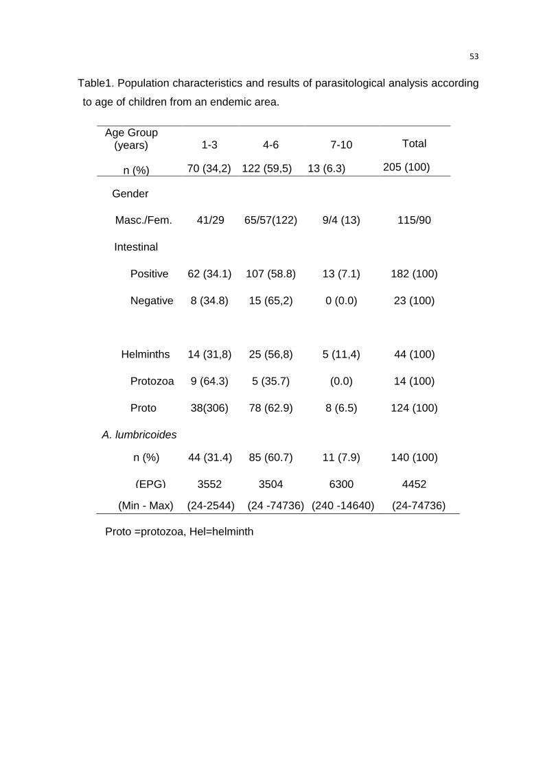
53
Table1. Population characteristics and results of parasitological analysis according
to age of children from an endemic area.
Proto =protozoa, Hel=helminth
Age Group (years) n (%)
1-3
70 (34,2)
4-6
122 (59,5)
7-10
13 (6.3)
Total
205 (100)
Gender
Masc./Fem.
n/n (%)
41/29
(70)
65/57(122) 9/4 (13) 115/90
Intestinal
parasite
Positive
n (%)
62 (34.1) 107 (58.8) 13 (7.1) 182 (100)
Negative
n (%)
8 (34.8) 15 (65,2) 0 (0.0) 23 (100)
Enteroparasites
Helminths
n (%)
14 (31,8) 25 (56,8) 5 (11,4) 44 (100)
Protozoa
n (%)
9 (64.3) 5 (35.7) (0.0) 14 (100)
Proto
+Hel n (%)
38(306) 78 (62.9) 8 (6.5) 124 (100)
A. lumbricoides
n (%) 44 (31.4) 85 (60.7) 11 (7.9) 140 (100)
(EPG)
Median
3552 3504 6300 4452
(Min - Max) (24-2544) (24 -74736) (240 -14640) (24-74736)
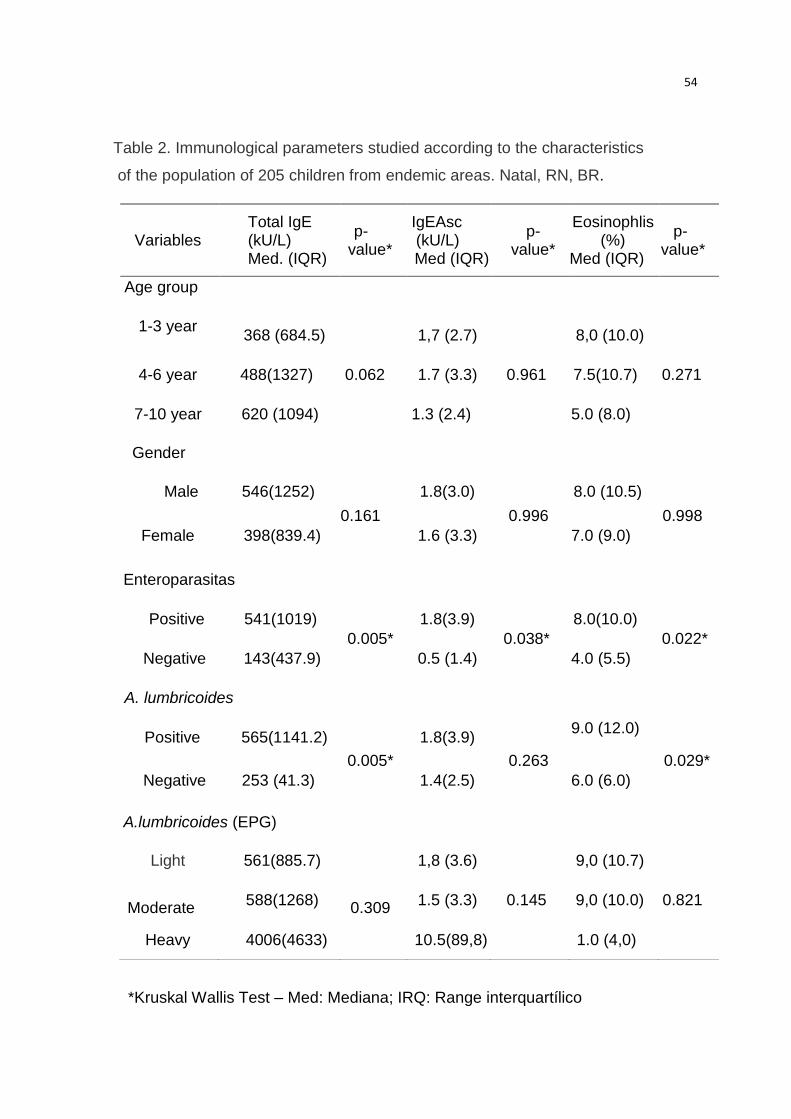
54
Table 2. Immunological parameters studied according to the characteristics
of the population of 205 children from endemic areas. Natal, RN, BR.
*Kruskal Wallis Test – Med: Mediana; IRQ: Range interquartílico
Variables
Total IgE (kU/L) Med. (IQR)
p- value*
IgEAsc (kU/L) Med (IQR)
p- value*
Eosinophlis (%) Med (IQR)
p- - value*
Age group
1-3 year
368 (684.5) 1,7 (2.7)
8,0 (10.0)
4-6 year 488(1327) 0.062 1.7 (3.3) 0.961 7.5(10.7) 0 0.271
7-10 year 620 (1094) 1.3 (2.4) 5.0 (8.0)
Gender
Male 546(1252)
0.161
1.8(3.0)
0.996
8.0 (10.5)
0.998
Female 398(839.4) 1.6 (3.3) 7.0 (9.0)
Enteroparasitas
Positive 541(1019)
0.005*
1.8(3.9)
0.038*
8.0(10.0)
0.022*
Negative 143(437.9) 0.5 (1.4) 4.0 (5.5)
A. lumbricoides
Positive 565(1141.2)
0.005*
1.8(3.9)
0.263
9.0 (12.0)
0.029*
Negative 253 (41.3) 1.4(2.5) 6.0 (6.0)
A.lumbricoides (EPG)
Light 561(885.7) 1,8 (3.6)
9,0 (10.7)
Moderate
588(1268)
0.309
1.5 (3.3) 0.145 9,0 (10.0) 0.821
Heavy 4006(4633) 10.5(89,8) 1.0 (4,0)
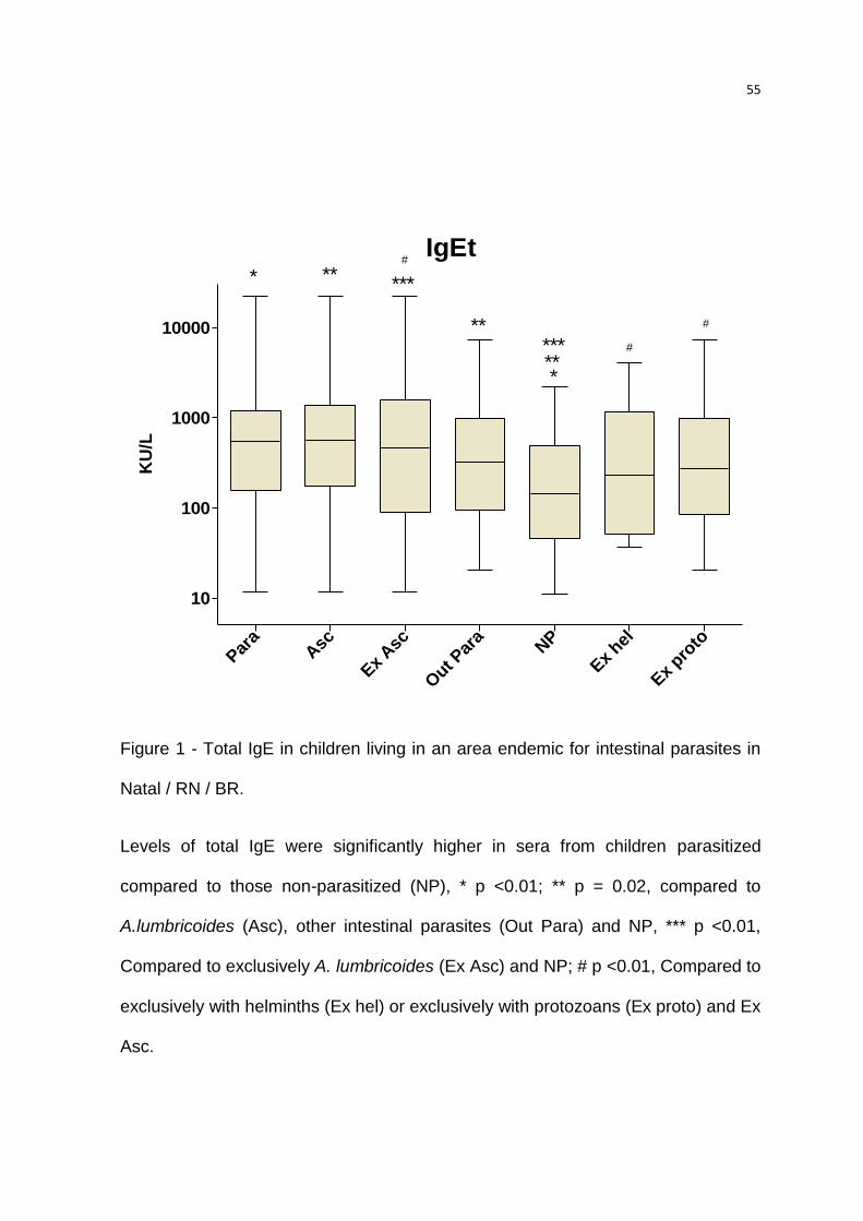
55
IgEt
Para
Asc
Ex Asc
Out P
ara
NP
Ex hel
Ex pro
to
10
100
1000
10000
*
*
**
**
**
***
***
#
#
#
KU
/L
Figure 1 - Total IgE in children living in an area endemic for intestinal parasites in
Natal / RN / BR.
Levels of total IgE were significantly higher in sera from children parasitized
compared to those non-parasitized (NP), * p <0.01; ** p = 0.02, compared to
A.lumbricoides (Asc), other intestinal parasites (Out Para) and NP, *** p <0.01,
Compared to exclusively A. lumbricoides (Ex Asc) and NP; # p <0.01, Compared to
exclusively with helminths (Ex hel) or exclusively with protozoans (Ex proto) and Ex
Asc.
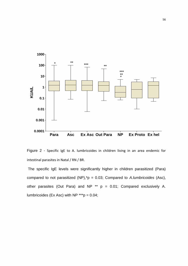
56
Para Asc Ex Asc Out Para NP Ex Proto Ex hel0.0001
0.001
0.01
0.1
1
10
100
1000
*
*
***** **
*****
KU
A/L
Figure 2 - Specific IgE to A. lumbricoides in children living in an area endemic for
intestinal parasites in Natal / RN / BR.
The specific IgE levels were significantly higher in children parasitized (Para)
compared to not parasitized (NP),*p = 0.03; Compared to A.lumbricoides (Asc),
other parasites (Out Para) and NP ** p = 0.01; Compared exclusively A.
lumbricoides (Ex Asc) with NP ***p = 0.04;
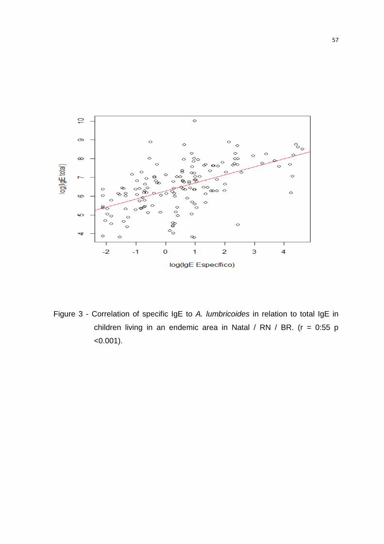
57
Figure 3 - Correlation of specific IgE to A. lumbricoides in relation to total IgE in
children living in an endemic area in Natal / RN / BR. (r = 0:55 p
<0.001).
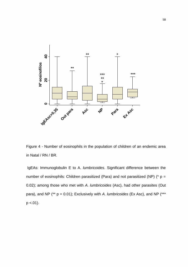
58
Figure 4 - Number of eosinophils in the population of children of an endemic area
in Natal / RN / BR.
IgEAs: Immunoglobulin E to A. lumbricoides. Significant difference between the
number of eosinophils: Children parasitized (Para) and not parasitized (NP) (* p =
0.02); among those who met with A. lumbricoides (Asc), had other parasites (Out
para), and NP (** p = 0.01); Exclusively with A. lumbricoides (Ex Asc), and NP (***
p <.01).
IgEAsc
>0,3
5
Out p
ara
Asc N
PPar
a
Ex
Asc
020
40 *
*
**
********
Nº
eo
sin
ofi
los
**
59
6. COMENTÁRIOS, CRÍTICAS E SUGESTÕES
A maioria das etapas propostas pelo anteprojeto foi cumprida: O estudo da
população, pesquisa da infecção parasitária através da coleta da amostra fecal
conservada, o hemograma para detecção do número de eosinófilos no sangue,
cultivo de larvas L3 infectantes, preparação dos extratos do verme adulto, do
estado larvário e ovo, dosagem de proteinas e eletroforese do extrato preparado,
análises por Enzimaimunoensaio (Elisa) por imunofluorimetria e dosagem de IgG
e IgE anti-A. lumbricoides com os extratos obtidos (Elisa-house). No entanto, o
trabalho enfrentou problemas, principalmente devido a ausência de recursos
financeiros. Assim, no decorrer da execução do projeto, foi necessária uma
mudança e conseqüentemente o título do projeto original.foi modificado. Mediante
as devidas alterações, o projeto realizado atingiu o objetivo proposto.
Não foi realizado o método de Graham, devido às dificuldades de coleta, o
que poderia ter evidenciado com maior precisão a presença de Enterobius
vermicularis nas crianças.
O total de 1453 crianças entre 1 a 12 anos que frequentavam 11 creches
públicas da região Oeste da cidade de Natal, RN matriculadas ou que
participavam como parentes, irmãos etc. Todas as crianças fizeram o exame
parasitológico de fezes, no entanto somente 205 fizeram hemograma e as
dosagem de IgEtotal e específica para A. lumbricoides. Todas as crianças
parasitadas foram medicadas com precrição realizada pelo médico participante
do projeto, mediante o peso, altura e idade das crianças com a colaboração da
Unidade de Saúde do bairro de Cidade Nova e da Secretária de Saúde da
Prefeitura de Natal, RN, que forneceu os medicamentos para o tratamento das
infecções parasitárias. Além do tratamento também foi prestada uma orientação
aos dos pais ou responsáveis sobre a profilaxia e epidemiologia das
enteroparasitoses.Todas as funcionarias das creches também fizeram o exame
parasitológico de fezes e, as que se encontravam parasitadas foram medicadas.
60
As dificuldades encontradas no decorrer do desenvolvimento do trabalho
serviram para obter maior conhecimento e aprendizagem. As pesquisas
realizadas antes e durante o trabalho foram de suma importância, pois foi a partir
delas que conseguimos desenvolver estudos que se concretizaram em TCCs e
apresentações em congressos e fórum.
As doenças infecciosas e parasitárias continuam a figurar entre as
principais causas de morte em todo o mundo. Contudo, mais do que a
mortalidade resultante essas doenças, elas importam pela freqüência com que
produzem déficits orgânicos, comprometendo o desenvolvimento normal das
crianças e limitando a capacidade de trabalho dos adultos, gerando um exército
de enfermos que pesam no orçamento familiar e do Estado. Muitas dessas
parasitoses, geralmente benignas ou fáceis de curar, se tornam fatores
determinantes de óbitos, dependendo das circunstâncias, como por exemplo, em
imunodeficientes. As parasitoses encontram-se entre os grandes problemas
médico-sanitário dos países em desenvolvimento, a exigir consideráveis recursos
financeiros, organização e pessoal habilitado para combatê-las. Portanto, a
importância em se buscar o conhecimento científico dos parasitas e as relações
parasito-hospedeiro é cada vez maior, para que possam resultar em informações
ou metodologia a ser utilizadas na luta contra as doenças produzidas e,
eventualmente, cooperar de forma ativa nos trabalhos de planejamento e controle
das endemias que tantos malefícios provocam na saúde dos seres humanos.
“ ...modificar as condições epidemiológicas até o ponto em
que o homem deixe de ser vítima de um processo natural, onde
muitos sucumbem e outros padecem num desgaste que compromete
a saúde e qualquer possibilidade de uma vida feliz” (Luís Rey ).
Diante dos dados obtidos neste, pretende-se continuar com o objetivo do
estudo, buscando novos dados que fortaleçam os resultados encontrados como
as análise do extratos do parasita por Western blot , e dosagens de citocinas,
cultura de ovos etc., não só com o parasita estudado mas com outros parasitas
que frequentemente infectam as crianças .
61
Um segundo artigo está sendo concluído e submetido: Infecção por
parasitas intestinais em crianças de zona urbana de região tropical, BR.
Produção gerada:
1. Apresentação de trabalhos em congressos científico (17)
1.1.Internacional
1.1.1. X CONGRESS OF THE LATIN-AMERICAN ASSOCIATION OF
IMMUNOLOGY –ALAI – 20 MAIO A 2 JUNHO 2012 /LIMA-
PERÚ: Prevalence of anti-Ascaris antibodies in chiildren with
parasitic infection.
1.1.2. INTERNATINAL CONGRESS OF CLINICAL CHEMISTRY AND
LABORATORY MEDICFINE DATA 2008.2 :Detection of the
degree of parasitic in children from kindergartens of the city of
Natal-RN.
1.2. Nacional (11)
1.2.1. XLVII CONGRESSO BRASILEIRO MEDICINA TROPICAL 2011.
a. A Prevalência de anticorpo anti-Ascaris em crianças com
Infecção parasitária.
b. Avaliando a presença da giardíase em crianças de creches
Públicas, na cidade de Natal/RN, através do exame
parasitológico de fezes.
c. Avaliação da Ascaridiase em crianças menores de 5 Anos de
Idade de creches na cidade de Natal,RN.
d. Presença de Larvas Strongyloides Stercoralis no exame de
Urina: Relato de caso
62
1.2.2. XXII CONGRESSO BRASILEIRO DE PARASITOLOGIA DATA
24ª 27 DE AGOSTO DE 2011 / SP
a. Relação entre Eosinofilia e Intensidade da Infecção por Ascaris
lumbricoides em Crianças Menores de 6 Anos na Cidade de
Natal-RN.
1.2.3. REUNIÃO ANUAL DA SBPS 25 A 30 DE JULHO DE 2010
a. Infecção por parasitoses intestinais em crianças que freqüentam
creches no município de Natal/RN.
b. Prevalência e intensidade da infecção por Ascaris lumbricoides
em crianças de creches da zona oeste de cidade de Natal-RN.
1.2.4. XXI CONGRESSO BRASILEIRO DE PARASITOLOGIA E II
ENCONTRO DE PARASITOLOGIA DO MERCOSUL 2009
a. Evolução dos ovos de Ascaris lumbricoides mediante a ação de
detergentes e desinfetantes
b. Ocorrência de enteroparasitas em hortaliças comercializadas nas
feiras livres da cidade de Natal – RN.
c. Prevalência de enteroparasitas em crianças do bairro de Cidade
Nova Natal/ RN.
d. Dados epidemiológicos da infecção por Schistosoma mansoni em
pacientes residentes na cidade deTouros/RN.
1.2.5.CONGRESSO DE INICIAÇÃO CIENTÍFICA :
63
a. Avaliação da parasitemia e das condições que predispõem à
infecção em crianças que frequentam o centro infantil cidade nova,
no município de Natal-RN.
b. Análise parasitológica em crianças da creche nossa senhora de
Fátima no bairro de Felipe Camarão, Natal, RN.
c. Correlação entre parasitas intestinais e anemia em crianças
menores de 10 ano, 2011.
d. Relação entre eosinofilia e intensidade da infecção por Ascaris
lumbricoides em crianças menores de 6 anos na cidade de Natal-
RN
2- Trabalhos de Conclusão de curso de Farmácia: (12)
3- Projetos de extensão: (3)
a. Avaliação Laboratorial e Nutricional em crianças de Creche Pública do
Município de Natal-RN.
b. Avaliação da Parasitemia e das condições que predispõem a Infecção
em crianças que freqüentam o Centro Infantil de Cidade Nova, no
Município de Natal-RN.
c. Fatores sócio-econômicos e ambientais na proliferação da ascaridíase e
enterobíase na Creche Recanto de Clara na cidade do Natal-RN.
4- Projeto de pesquisa: (1)
a. Avaliação da ascaridiase em crianças menores de 5 anos de idade na
cidade de Natal, RN.
64
7. REFERÊNCIAS
1. WHO. The Millennium Development Goals and deworming. Report of the third global meeting of the partners for parasite control: deworming for health and development Geneva: World Health Organization. 2005;25-26. 2. WHO. Preventative Chemotherapy in Human Helminthiases: Coordinated Use of Anthelminthic Drugs in Control Interventions: a Manual for Health Professionals and Programme Managers, World Health Organisation, Geneva, . World Health Organisation, Geneva, . 2006. 3. Fonseca EO, Teixeira MG, Barreto ML, Carmo EH, Costa Mda C. Prevalence and factors associated with geohelminth infections in children living in municipalities with low HDI in North and Northeast Brazil. Cad Saude Publica. 2010 Jan;26(1):143-52. 4. Hotez PJ, Brindley PJ, Bethony JM, King CH, Pearce EJ, Jacobson J. Helminth infections: the great neglected tropical diseases. J Clin Invest. 2008 Apr;118(4):1311-21. 5. de Silva NR, Brooker S, Hotez PJ, Montresor A, Engels D, Savioli L. Soil-transmitted helminth infections: updating the global picture. Trends Parasitol. 2003 Dec;19(12):547-51. 6. Hotez PJ. Neglected infections of poverty in the United States of America. PLoS Negl Trop Dis. 2008;2(6):e256. 7. Loukas A, Prociv P. Immune responses in hookworm infections. Clin Microbiol Rev. 2001 Oct;14(4):689-703, table of contents. 8. Fauci AS, Morens DM. The perpetual challenge of infectious diseases. N Engl J Med. 2012 Feb 2;366(5):454-61. 9. Zaccone P, Fehervari Z, Phillips JM, Dunne DW, Cooke A. Parasitic worms and inflammatory diseases. Parasite Immunol. 2006 Oct;28(10):515-23. 10. Figueiredo CA, Barreto ML, Rodrigues LC, Cooper PJ, Silva NB, Amorim LD, et al. Chronic intestinal helminth infections are associated with immune hyporesponsiveness and induction of a regulatory network. Infect Immun. 2010 Jul;78(7):3160-7. 11. White RR, Artavanis-Tsakonas K. How helminths use excretory secretory fractions to modulate dendritic cells. Virulence. 2012 Nov 15;3(7). 12. Wiria AE, Djuardi Y, Supali T, Sartono E, Yazdanbakhsh M. Helminth infection in populations undergoing epidemiological transition: a friend or foe? Semin Immunopathol. 2012 Nov;34(6):889-901. 13. McSorley HJ, Maizels RM. Helminth infections and host immune regulation. Clin Microbiol Rev. 2012 Oct;25(4):585-608. 14. Girgis NM, Gundra UM, Loke P. Immune regulation during helminth infections. PLoS Pathog. 2013 Apr;9(4):e1003250. 15. Cooper PJ. Interactions between helminth parasites and allergy. Curr Opin Allergy Clin Immunol. 2009 Feb;9(1):29-37. 16. Allen JE, Wynn TA. Evolution of Th2 immunity: a rapid repair response to tissue destructive pathogens. PLoS Pathog. 2011 May;7(5):e1002003. 17. Amiri P, Haak-Frendscho M, Robbins K, McKerrow JH, Stewart T, Jardieu P. Anti-immunoglobulin E treatment decreases worm burden and egg production in Schistosoma mansoni-infected normal and interferon gamma knockout mice. J Exp Med. 1994 Jul 1;180(1):43-51. 18. Negrao-Correa D. Importance of immunoglobulin E (IgE) in the protective mechanism against gastrointestinal nematode infection: looking at the intestinal mucosae. Rev Inst Med Trop Sao Paulo. 2001 Sep-Oct;43(5):291-9.
65
19. Maizels RM, Hewitson JP, Smith KA. Susceptibility and immunity to helminth parasites. Curr Opin Immunol. 2012 Jul 11. 20. Dold C, Holland CV. Investigating the underlying mechanism of resistance to Ascaris infection. Microbes Infect. 2011 Jul;13(7):624-31. 21. Acevedo N, Caraballo L. IgE cross-reactivity between Ascaris lumbricoides and mite allergens: possible influences on allergic sensitization and asthma. Parasite Immunol. 2011 Jun;33(6):309-21. 22. King EM, Kim HT, Dang NT, Michael E, Drake L, Needham C, et al. Immuno-epidemiology of Ascaris lumbricoides infection in a high transmission community: antibody responses and their impact on current and future infection intensity. Parasite Immunol. 2005 Mar;27(3):89-96. 23. Maizels RM, Smith KA. Regulatory T cells in infection. Adv Immunol. 2011;112:73-136. 24. McDonald V. Parasites in the gastrointestinal tract. Parasite Immunol. 2003 May;25(5):231-4. 25. Schneider B, Jariwala AR, Periago MV, Gazzinelli MF, Bose SN, Hotez PJ, et al. A history of hookworm vaccine development. Hum Vaccin. 2011 Nov;7(11):1234-44. 26. Hotez PJ, Brown AS. Neglected tropical disease vaccines. Biologicals. 2009 Jun;37(3):160-4. 27. Lawrence CE. Is there a common mechanism of gastrointestinal nematode expulsion? Parasite Immunol. 2003 May;25(5):271-81. 28. Yazdanbakhsh M, Kremsner PG, van Ree R. Allergy, parasites, and the hygiene hypothesis. Science. 2002 Apr 19;296(5567):490-4. 29. Cooper PJ. Intestinal worms and human allergy. Parasite Immunol. 2004 Nov-Dec;26(11-12):455-67. 30. Schwartz LB, Austen KF. Structure and function of the chemical mediators of mast cells. Prog Allergy. 1984;34:271-321. 31. Turner JD, Faulkner H, Kamgno J, Kennedy MW, Behnke J, Boussinesq M, et al. Allergen-specific IgE and IgG4 are markers of resistance and susceptibility in a human intestinal nematode infection. Microbes Infect. 2005 Jun;7(7-8):990-6. 32. Clough D, Kappeler PM, Walter L. Genetic regulation of parasite infection: empirical evidence of the functional significance of an IL4 gene SNP on nematode infections in wild primates. Front Zool. 2011;8(1):9. 33. Acevedo N, Mercado D, Vergara C, Sanchez J, Kennedy MW, Jimenez S, et al. Association between total immunoglobulin E and antibody responses to naturally acquired Ascaris lumbricoides infection and polymorphisms of immune system-related LIG4, TNFSF13B and IRS2 genes. Clin Exp Immunol. 2009 Aug;157(2):282-90. 34. Williams-Blangero S, VandeBerg JL, Subedi J, Aivaliotis MJ, Rai DR, Upadhayay RP, et al. Genes on chromosomes 1 and 13 have significant effects on Ascaris infection. Proc Natl Acad Sci U S A. 2002 Apr 16;99(8):5533-8. 35. Williams-Blangero S, Vandeberg JL, Subedi J, Jha B, Correa-Oliveira R, Blangero J. Localization of multiple quantitative trait loci influencing susceptibility to infection with Ascaris lumbricoides. J Infect Dis. 2008 Jan 1;197(1):66-71. 36. Macedo-Soares MF, Itami DM, Lima C, Perini A, Faquim-Mauro EL, Martins MA, et al. Lung eosinophilic inflammation and airway hyperreactivity are enhanced by murine anaphylactic, but not nonanaphylactic, IgG1 antibodies. J Allergy Clin Immunol. 2004 Jul;114(1):97-104. 37. Macedo MS, Barbuto JA. Murine delayed type hypersensitivity is suppressed by Ascaris suum extract. Braz J Med Biol Res. 1988;21(3):523-5. 38. Harhay MO, Horton J, Olliaro PL. Epidemiology and control of human gastrointestinal parasites in children. Expert Rev Anti Infect Ther. 2010 Feb;8(2):219-34. 39. Arruda LK, Santos AB. Immunologic responses to common antigens in helminthic infections and allergic disease. Curr Opin Allergy Clin Immunol. 2005 Oct;5(5):399-402.
66
40. Xia Y, Spence HJ, Moore J, Heaney N, McDermott L, Cooper A, et al. The ABA-1 allergen of Ascaris lumbricoides: sequence polymorphism, stage and tissue-specific expression, lipid binding function, and protein biophysical properties. Parasitology. 2000 Feb;120 ( Pt 2):211-24. 41. Santos AB, Rocha GM, Oliver C, Ferriani VP, Lima RC, Palma MS, et al. Cross-reactive IgE antibody responses to tropomyosins from Ascaris lumbricoides and cockroach. J Allergy Clin Immunol. 2008 Apr;121(4):1040-6 e1. 42. Melo-Reis PR, Diniz-Filho, J.A.F., Dias-Penna, K.G.B., Costa, S.H.N., Mesquita, M.M., Silva, J. B.,Castro, F.S., Chen, L.C. Correlation between eosinophilia and parasitic infections for Giardia lamblia in children. RBAC,. 2007;39 (3):237-9. 43. Machado RLDF, M.C.; Frade, A,F.; Kudo, M.D.; Sila, Filho. M.G.; Póvoa MM. Comparação de quatro métodos laboratoriais para diagnóstico da Giardia lamblia em fezes de crianças residentes em Belém, Pará. Rev Soc Bras Med Trop. 2001;34(1):91-3. 44. Vieira Silva CC, Nogueira Ferraz CR, Fornari JV, Sena Barnabe CA. Epidemiological analysis of eosinophilia and elevation of immunoglobulin E as a predictable and relative risk of enteroparasitosis. Rev Cubana Med Trop. 2012 Jan-Apr;64(1):22-6. 45. Fontenele ALA, Carvalho, P.G.C; Ferreira, C. H. A.;Girão, A.B.; Teixeira, M.J.; Queiroz, J. A.N., Almeida, Y. M. Evaluation of dosage of interleucina-5 and immunoglobulin e in patients with giardíase with or without eosinophilia. RBAC. 2006;38(3):201-6. 46. Behm CA, Ovington KS. The role of eosinophils in parasitic helminth infections: insights from genetically modified mice. Parasitol Today. 2000 May;16(5):202-9. 47. Naus CW, Booth M, Jones FM, Kemijumbi J, Vennervald BJ, Kariuki CH, et al. The relationship between age, sex, egg-count and specific antibody responses against Schistosoma mansoni antigens in a Ugandan fishing community. Trop Med Int Health. 2003 Jun;8(6):561-8. 48. Blagg W, Schloegel EL, Mansour NS, Khalaf GI. A new concentration technic for the demonstration of protozoa and helminth eggs in feces. Am J Trop Med Hyg. 1955 Jan;4(1):23-8. 49. Katz N, Chaves A, Pellegrino J. A simple device for quantitative stool thick-smear technique in Schistosomiasis mansoni. Rev Inst Med Trop Sao Paulo. 1972 Nov-Dec;14(6):397-400. 50. Montresor A, Crompton. D.W.T., Bundy, D.A.P., Hall, A., Savioli, L. Guidelines for the evaluation of soil transmitted helminthiasis and schistosomiasis at community level. [serial on the Internet]. 1998. 51. DACIE JVL, S.M. Practical Haematology 8ed. Livingstone C, editor. Philadelphia1995. 52. Pereira M. Epidemiologia-Teoria e Prática. 11 ed. Rio de Janeiro: Guanabara Koogan; 2007.
8. APÊNDICE
67
9.ANEXO

68
UNIVERSIDADE FEDERAL DO RIO GRANDE DO NORTE
CENTRO DE CIÊNCIAS DA SAÚDE DEPARTAMENTO DE ANÁLISES CLÍNICAS TOXICOLÓGICAS
TERMO DE CONSENTIMENTO LIVRE E ESCLARECIDO
Título do projeto: Identificação de antígenos de Ascaris lumbricoides indutores de resposta imune
Responsável pela pesquisa: Valeria Soraya de Farias Sales
O (a) Sr(a) ou o menor pelo qual é responsável está sendo convidado(a) a participar, voluntariamente,
de uma pesquisa que visa identificar antígenos de Ascaris lumbricoides e a resposta do seu organismo para se
defender deste parasita. Para avaliação desse estudo, será necessário e a coleta de amostras fecais para a
realização do exame parasitológico e 10 mL de sangue periférico para realização dos exames imunológicos.O
sangue será coletado por punção venosa na Creche “Recanto de Clara” situada na própria comunidade e as
amostras de fezes que deverão ser coletadas na residência, em recipiente descontaminado ou em um papel
limpo sob o chão, em seguida com o auxilio de uma palheta, devendo ser retirado parte do material e
transportado para o frasco coletor plástico próprio, que será cedido pelo projeto. Todo o procedimento da
coleta da amostra será fornecido, ao responsável e ou a criança maior, com antecedência e a amostra deverá
ser entregue na creche. As amostras serão analisadas no Laboratório de Imunologia Clínica e Parasitologia
Clínica da UFRN situadas em Natal-RN. O resultado do exame será entregue ao paciente e ao pai ou
responsável pela criança para então ser examinada e o médico definido a melhor forma de tratamento.
Os riscos da participação nesta pesquisa são mínimos e são referentes ao procedimento de coleta, tais
como: hematomas, sangramentos e desmaios referentes à coleta do material biológico, no entanto, não há
riscos previsíveis mas se vier a ocorrer qualquer problema com coleta do sangue na veia você terá toda a
assistência. Porém, em caso de dano pessoal diretamente causado pelo procedimento, o participante tem
direito a tratamento médico na instituição, bem como às indenizações legalmente estabelecidas e será
ressarcido por despesas comprovadamente relacionadas à participação na pesquisa. A aceitação não está
vinculada a nenhum tipo de remuneração e nem haverá despesas antecipadas devido à participação. O
pesquisador se compromete a utilizar os dados somente para essa pesquisa mantendo em sigilo as
informações obtidas.
Sua participação ou do seu filho neste estudo é totalmente voluntária, podendo recusar-se a fazer
parte do mesmo ou interromper se julgar conveniente, sem prejuízo de sua relação com a creche.
A pesquisa se encerra com o procedimento realizado e analisado o seu resultado.
Estas informações estão sendo fornecidas para que seja permitida a sua participação voluntária e ou de
seu filho neste estudo. Em caso de desistência ou de dúvidas sobre a pesquisa, entrar em contato com a
Profa.Valéria Soraya de Farias Sales do Departamento de Análises Clínicas, Av. General Cordeiro de Farias
s/n Petrópolis; fone: 3215-4229. Lembre-se que você pode retirar o consentimento de participação na
pesquisa em qualquer momento, sem qualquer prejuízo. Em caso de necessidade de esclarecimentos ou
queixas éticas relativas a esta pesquisa, você pode entrar em contato com o Comitê de Ética em Pesquisa,
Campus Universitário; fone 3215-3135.
Eu, __________________________________________________________________________fui
esclarecida (o) a respeito do estudo sobre gordura fecal. Ficou claro qual o propósito do estudo, o
procedimento a ser realizado, seus desconfortos e riscos, as garantias de confidencialidade e de
esclarecimentos permanentes, portanto, concordo com a minha participação e ou do meu/minha filho
(a)___________________________________________________________________neste estudo. Estou
ciente que poderei retirar o meu consentimento a qualquer momento, sem penalidades ou prejuízo ao seu
atendimento neste serviço. Desta forma, permito que seja realizado o exame aqui esclarecidos e concordo que
os dados obtidos sejam utilizados para os fins a que se prestam.
Assinatura dos pais ou responsável legal______________________________________________________
Declaro que obtive de forma apropriada e voluntária o consentimento livre e esclarecido deste paciente ou de
seu representante legal para a sua participação neste estudo.
Pesquisadora:____________________________________________________________
Natal, _____de ______________de 200___