UNNIIVVEERRSSIID DAADDEE RFFE ED DEERRAALL ......UNNIIVVEERRSSIID DAADDEE RFFE ED DEERRAALL DOO...
53
UNIVERSIDADE FEDERAL DO RIO GRANDE DO SUL INSTITUTO DE CIÊNCIAS BÁSICAS DA SAÚDE PROGRAMA DE PÓS-GRADUAÇÃO EM CIÊNCIAS BIOLÓGICAS - NEUROCIÊNCIAS Juliana Dalibor Neves EFEITOS DE DIFERENTES PROTOCOLOS DE TREINAMENTO FÍSICO SOBRE A FUNÇÃO E MORFOLOGIA DO NERVO MEDIANO DE RATOS APÓS PROTOCOLO DE LESÃO POR ESMAGAMENTO Porto Alegre 2011
Transcript of UNNIIVVEERRSSIID DAADDEE RFFE ED DEERRAALL ......UNNIIVVEERRSSIID DAADDEE RFFE ED DEERRAALL DOO...
UUNNIIVVEERRSSIIDDAADDEE FFEEDDEERRAALL DDOO RRIIOO GGRRAANNDDEE DDOO SSUULL
IINNSSTTIITTUUTTOO DDEE CCIIÊÊNNCCIIAASS BBÁÁSSIICCAASS DDAA SSAAÚÚDDEE
PPRROOGGRRAAMMAA DDEE PPÓÓSS--GGRRAADDUUAAÇÇÃÃOO EEMM CCIIÊÊNNCCIIAASS BBIIOOLLÓÓGGIICCAASS --
NNEEUURROOCCIIÊÊNNCCIIAASS
JJuulliiaannaa DDaalliibboorr NNeevveess
EEFFEEIITTOOSS DDEE DDIIFFEERREENNTTEESS PPRROOTTOOCCOOLLOOSS DDEE TTRREEIINNAAMMEENNTTOO FFÍÍSSIICCOO
SSOOBBRREE AA FFUUNNÇÇÃÃOO EE MMOORRFFOOLLOOGGIIAA DDOO NNEERRVVOO MMEEDDIIAANNOO DDEE RRAATTOOSS AAPPÓÓSS
PPRROOTTOOCCOOLLOO DDEE LLEESSÃÃOO PPOORR EESSMMAAGGAAMMEENNTTOO
Porto Alegre
2011
Juliana Dalibor Neves
EEFFEEIITTOOSS DDEE DDIIFFEERREENNTTEESS PPRROOTTOOCCOOLLOOSS DDEE TTRREEIINNAAMMEENNTTOO FFÍÍSSIICCOO
SSOOBBRREE AA FFUUNNÇÇÃÃOO EE MMOORRFFOOLLOOGGIIAA DDOO NNEERRVVOO MMEEDDIIAANNOO DDEE RRAATTOOSS AAPPÓÓSS
PPRROOTTOOCCOOLLOO DDEE LLEESSÃÃOO PPOORR EESSMMAAGGAAMMEENNTTOO
Dissertação de mestrado apresentada ao Programa de Pós-Graduação em Neurociências da Universidade Federal do Rio Grande do Sul como requisito parcial para obtenção do título de Mestre em Neurociências.
Orientadora: Profª. Dra. Maria Cristina Faccioni-Heuser
Porto Alegre
2011
AAGGRRAADDEECCIIMMEENNTTOOSS
Agradeço a profª Drª Maria Cristina Faccioni-Heuser por ter me aceito como sua
orientanda e por ter me possibilitado a realização de um sonho.
Ao profo Drº Léder Leal Xavier pela valiosa ajuda e paciência na elaboração deste
trabalho.
À profª Drª Matilde Achaval por ter permitido a ulitização das dependências do
Laboratório de Histofisiologia Comparada.
A todos os colegas do pós e do laboratório de Histofisiologia Comparada pelos bons
momentos de convívio. Em especial ao Fernando Camelier, Sandro Antunes da Silva e Sílvia
Barbosa.
Aos colegas de laboratório, Nígia Ramalho Arsego e Alexandre da Silva Costa, que
foram de grande importância para a realização dos experimentos.
À minha família por ser a base de tudo que se constrói e pelo amor incondicional,
carinho e paciência.
Aos meus queridos amigos pelos momentos de descontração e pelo companheirismo
nos períodos de angústia e pelo permanente incentivo, carinho e apoio enquanto buscava a
realização de meu sonho.
À UFRGS, ao Programa de Pós-Graduação em Neurociências e a CAPES pela
oportunidade e pela bolsa concedida durante o período.
RREESSUUMMOO
A maioria das lesões nervosas periféricas em humanos, afeta a extremidade superior e o maior
aspecto incapacitante dessa lesão é a perda dos movimentos da mão. As lesões do plexo
braquial apresentam um índice de morbidade elevado que é representado por graves sequelas
sensorio-motoras devido à fibrose que se desenvolve ao longo do tempo após a lesão.
Evidências indicam que o tipo e a intensidade da atividade física induzem o remodelamento
morfológico e eletrofisiológico da junção neuromuscular influenciando no reparo do nervo.
No presente trabalho, um programa de treinamento de equilíbrio e coordenação, de repetição
na esteira e uma associação desses treinamentos foram utilizados, por 4 semanas, após a lesão
por esmagamento do nervo mediano em ratos para verificar a influência dessas atividades
sobre os parâmetros morfométricos do nervo lesionado (área axonal, densidade axonal,
diâmetro das fibras mielinizadas, diâmetro axonal e espessura da bainha de mielina da porção
distal do nervo mediano), além de analisar a recuperação funcional dos membros anteriores
lesados. Análises histológicas e morfométricas do nervo mediano foram utilizadas para
avaliar a regeneração do nervo no final do tratamento. Os resultados do teste de motricidade
sobre grade revelaram que houve uma recuperação funcional acelerada em todos os grupos
lesionados após lesão do plexo braquial. No teste de suspensão no arame e no teste do
cilindro, entretanto, os grupos tratados não apresentaram diferença significativa comparada ao
grupo controle. O treinamento de equilíbrio e coordenação mostrou melhores resultados
comparado ao treinamento de repetição e a associação dos treinamentos para a densidade
axonal e o diâmetro axonal igualando-se estatisticamente aos resultados do grupo sham
sedentário. Esses dados fornecem evidências de que o treinamento de equilíbrio e
coordenação acelerou a regeneração do nervo mediano após lesão traumática experimental,
apesar dos testes funcionais não demonstrarem diferenças entre os tratamentos.
LLIISSTTAA DDEE IILLUUSSTTRRAAÇÇÕÕEESS
Figura 1 – Desenho esquemático do plexo braquial humano ..................................................... 9
Figura 2 – Desenho esquemático das fibras nervosas mielínicas do SNP................................ 11
Figura 3 – Corte transversal de um nervo periférico ................................................................ 12
Figura 4 – Figura esquemática mostrando resumidamente o processo de degeneração e
regeneração no SNP.................................................................................................................. 14
ARTIGO
Figure 1 – Time line demonstrating the experimental procedures ........................................... 40
Figure 2 – Performance on the footfault test (FT) .................................................................... 40
Figure 3 – Performance on the hanging wire (HW) ................................................................. 41
Figure 4 – Performance on the cylinder test (CT) .................................................................... 41
Figure 5 – Digitized images of transverse-semithin sections (1 µm) obtained from
regeneration median nerves after 6 weeks of specific exercise training .................................. 42
Figure 6 – Effects of specific physical exercise training on the morphometric parameters of
regenerating right median nerve fibers after 6 weeks of training ............................................. 43
Table 1 – Grupos experimentais e número de animais utilizados para cada análise ................ 44
LLIISSTTAA DDEE AABBRREEVVIIAATTUURRAASS
SNC – Sistema nervoso central
SNP – Sistema nervoso periférico
FRC – Flexor radial do carpo
ARTIGO
LBC – Lesio balance and coordination
LR – Lesion repetition
LBCR – Lesion repetition + balance and coordination
LSE – Lesion sedentary
Sh-s – Sham sedentary
CNS – Central nervous system
FT – Footfault test
HW – Hanging wire
CT – Cylinder test
SSUUMMÁÁRRIIOO
AGRADECIMENTOS .......................................................................................................... III
RESUMO ................................................................................................................................ IV
1 INTRODUÇÃO ..................................................................................................................... 7
1.1 PLEXO BRAQUIAL ......................................................................................................................... 8 1.2 MODELO DE LESÃO DO PLEXO BRAQUIAL .............................................................................. 10 1.3 ESTRUTURA NORMAL DOS NERVOS PERIFÉRICOS ...................................................................... 10 1.4 FISIOPATOLOGIA DO TRAUMA AXONAL & REGENERAÇÃO NERVOSA PERIFÉRICA ..................... 12 1.5 EXERCÍCIO FÍSICO E NEUROPATIAS PERIFÉRICAS ....................................................................... 15
2 OBJETIVOS ........................................................................................................................ 18
2.1 OBJETIVO GERAL ....................................................................................................................... 18 2.2 OBJETIVOS ESPECÍFICOS .............................................................................................................. 18
3 MÉTODOS E RESULTADOS ........................................................................................... 19
3.1 ARTIGO ...................................................................................................................................... 19 ABSTRACT ........................................................................................................................................ 21 INTRODUCTION ................................................................................................................................. 22 MATERIALS AND METHODS ............................................................................................................... 23
Experimental design and surgical procedures ............................................................................ 23 Rehabilitation protocols.............................................................................................................. 24 Repetition training ...................................................................................................................... 24 Balance and Coordination training ............................................................................................. 25 Repetition + Balance and coordination training ......................................................................... 25 Functional tests ........................................................................................................................... 25 Footfault test ............................................................................................................................... 25 Hanging Wire ............................................................................................................................. 26 Cylinder test................................................................................................................................ 26 Histological and morphometric studies ...................................................................................... 26 Statistical analysis ...................................................................................................................... 28 Footfault Test.............................................................................................................................. 28 Hanging Wire ............................................................................................................................. 29 Cylinder Test .............................................................................................................................. 29 Histological analysis ................................................................................................................... 29 Morphometric analysis of median nerve .................................................................................... 30
DISCUSSION...................................................................................................................................... 31 REFERENCES .................................................................................................................................... 34 LEGENDS .......................................................................................................................................... 38 FIGURES AND TABLE ........................................................................................................................ 40
4 CONCLUSÕES E PERSPECTIVAS................................................................................. 45
REFERÊNCIAS...................................................................................................................... 46
7
11 IINNTTRROODDUUÇÇÃÃOO
No Brasil, os dados epidemiológicos demonstram que as lesões traumáticas são as
causas mais comuns de lesão do plexo braquial (Flores, 2006). Na América do Norte e
Europa, 10 a 20% das lesões do SNP envolvem o plexo braquial. E dentre essas lesões, 80 a
90% são causados por traumatismos decorrentes de acidentes automobilísticos com vários
mecanismos de tração das raízes nervosas cervicais (Ferreira, 1999).
A incidência, no mundo, de lesões traumáticas é estimada em mais de 500.000 novos
pacientes a cada ano. Essas lesões levam a perda parcial ou total da função motora, sensorial e
autonômica no segmento corporal envolvido (Rodríguez et al., 2004; Valero-Cabré e Navarro,
2002). Periférica e centralmente, os alvos musculares e os neurônios motores,
respectivamente, perdem sua função (Johnson et al., 2005). No entanto, estas perdas podem
ser recompensadas graças à recuperação dos neurônios lesionados, fazendo com que os
axônios seccionados enviem novos prolongamentos ao coto distal e restabeleçam novas
conexões funcionais com os órgãos periféricos apropriados (Valero-Cabré e Navarro, 2002).
Lesões do plexo braquial ainda constituem um desafio clínico e um problema
cirúrgico, apesar do uso de técnicas microcirúrgicas especializadas. A maior parte das lesões
de nervo periférico, em humanos afeta a extremidade superior (Bertelli et al., 1995). Essas
lesões são responsáveis pela perda ou restrição da capacidade funcional de alcance, preensão
e manipulação de objetos (Duff, 2005). Em função da perda evidente na qualidade de
movimento após lesões nervosas periféricas, a potencialização da regeneração nervosa e a
recuperação da função têm sido alvo de diversos estudos (Van Meeteren et al., 1998; Bontioti
et al., 2003; Gordon et al., 2003; Bontioti et al., 2005; Ilha et al., 2008; Sebatier et al., 2008).
8
1.1 PLEXO BRAQUIAL
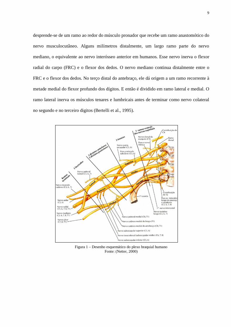
O plexo braquial em humanos é formado pela união de quatro raízes baixas cervicais
(C5, C6, C7 e C8) e a primeira raiz torácica (T1). Essas raízes são formadas pela união das
raízes dorsal sensorial e ventral motora. Cada raiz é formada pela união de 2 ou 3 raízes
dorsais e ventrais. A união da raiz de C5 e C6 forma o tronco superior. A raiz de C7 sozinha
forma o tronco médio, e as raízes de C7 e C8 formam o tronco inferior. As divisões anteriores
do tronco superior e médio unem-se para formar o fascículo lateral, a divisão anterior do
tronco inferior forma o fascículo medial e as divisões posteriores dos troncos se unem
formando o fascículo posterior (Gray, 1995). Os nervos peitoral lateral, musculocutâneo e a
raiz lateral do nervo mediano originam-se do fascículo lateral; os nervos axilar, radial,
toracodorsal e o nervo subescapular originam-se do fascículo posterior; os nervos peitoral
medial, raiz medial do nervo mediano, ulnar, cutâneo medial do braço e o nervo cutâneo
medial do antebraço originam-se do fascículo medial (Netter, 2008) (Fig. 1).
O nervo mediano em humanos origina-se das raízes de C5 a T1 e é formado pela
fusão dos ramos vindos dos fasciculos lateral e medial do plexo braquial. A raiz lateral do
nervo mediano, derivado dos ramos ventrais do quinto ao sétimo nervos cervicais (C5, C6 e
C7), inerva os músculos da região anterior do antebraço e curtos do polegar, assim como a
pele do lado lateral da mão. A raiz medial do nervo mediano, originada dos ramos ventrais do
oitavo nervo cervical e primeiro torácico (C8 e T1), inerva os músculos da região anterior do
antebraço e curtos do polegar, assim como a pele do lado medial da mão (Netter, 2008).
No rato, o nervo mediano é formado pela fusão de 3 ramos vindos dos fasciculos
lateral, posterior e medial do plexo braquial, respectivamente. Os ramos do fascículo posterior
e medial, entretanto, são mais desenvolvidos que o ramo do fascículo lateral (C5 e C6). No
pata anterior, o nervo mediano não se ramifica, mas perto da articulação do cotovelo ele
9
desprende-se de um ramo ao redor do músculo pronador que recebe um ramo anastomótico do
nervo musculocutâneo. Alguns milímetros distalmente, um largo ramo parte do nervo
mediano, o equivalente ao nervo interósseo anterior em humanos. Esse nervo inerva o flexor
radial do carpo (FRC) e o flexor dos dedos. O nervo mediano continua distalmente entre o
FRC e o flexor dos dedos. No terço distal do antebraço, ele dá origem a um ramo recorrente à
metade medial do flexor profundo dos dígitos. E então é dividido em ramo lateral e medial. O
ramo lateral inerva os músculos tenares e lumbricais antes de terminar como nervo colateral
no segundo e no terceiro digitos (Bertelli et al., 1995).
Figura 1 - Desenho representativo do plexo braquial humano Fonte: (Netter, 2000)
Figura 1 – Desenho esquemático do plexo braquial humano
Fonte: (Netter, 2000)
10
1.2 MODELO DE LESÃO DO PLEXO BRAQUIAL
São poucos os estudos usando o modelo experimental para o estudo da regeneração
nervosa nos membros anteriores do rato (Galtrey e Fawcett, 2007; Bontioti et al., 2003;
Bertelli et al., 1995). Entretanto, a maioria das lesões de nervo periférico em humanos afeta a
extremidade superior, e por essa razão, um modelo experimental de lesão nervosa na
extremidade superior é de grande importância (Bontioti et al., 2003). A distância para os
órgãos-alvo é pequena nos membros anteriores do rato (músculo e pele), a reinervação é
rápida, e o tempo requerido para a recuperação funcional é menor do que a dos membros
posteriores (Santos et al., 2007; Bertelli e Mira, 1993; Bontioti et al., 2003). Assim, supõe-se
que a regeneração nervosa e a recuperação funcional sejam obtidas mais rapidamente do que
os modelos de lesão do nervo ciático.
1.3 ESTRUTURA NORMAL DOS NERVOS PERIFÉRICOS
O axônio é uma extensão longa e delgada do corpo celular, que possui uma estrutura
arborescente em sua região distal - terminação axonal. É por meio dela que os axônios
realizam contatos sinápticos com os órgãos alvo. Em um nervo existem axônios mielinizados
e não mielinizados. No primeiro tipo, as células de Schwann se organizam ao redor do axônio
formando a bainha de mielina, que é interrompida em intervalos regulares, pelos nodos de
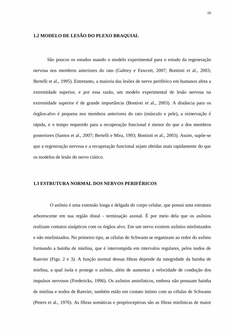
Ranvier (Figs. 2 e 3). A função normal dessas fibras depende da integridade da bainha de
mielina, a qual isola e protege o axônio, além de aumentar a velocidade de condução dos
impulsos nervosos (Fredericks, 1996). Os axônios amielínicos, embora não possuam bainha
de mielina e nodos de Ranvier, também estão em contato íntimo com as células de Schwann
(Peters et al., 1976). As fibras somáticas e proprioceptivas são as fibras mielínicas de maior
11
diâmetro, enquanto que as fibras sensoriais que medeiam a dor são as menores (Fredericks,
1996).
Figura 2 - Desenho esquemático das fibras nervosas mielínicas do SNP. (A) a, tecido conjuntivo; b, nodo de Ranvier; c, bainha de mielina; d, axônio; e, região internodal; e – f, núcleo da célula de Schwann. (B) a, nodo de Ranvier; b, região paranodal; c, tudo de tecido conjuntivo; d – e, citoplasma da célula de Schwann; f, núcleo da célula de Schwann; g, bainha de mielina; h, incisura de Lantermann Fonte: Modificado de (Rámon Y Cajal, 2003)
Os nervos periféricos englobam, de uma maneira geral, os axônios dos neurônios
motores e sensoriais, que constituem os nervos espinais e cranianos, os plexos e os troncos
nervosos do sistema nervoso vegetativo (Vallat e Magy, 2005).
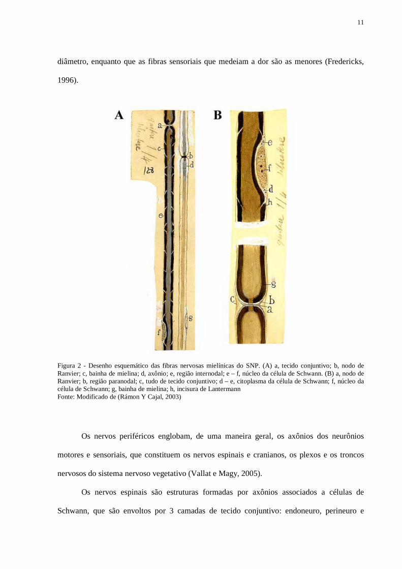
Os nervos espinais são estruturas formadas por axônios associados a células de
Schwann, que são envoltos por 3 camadas de tecido conjuntivo: endoneuro, perineuro e
12
epineuro. (Fig. 3). Essas sucessivas camadas de tecido conjuntivo servem para proteger e
sustentar as fibras nervosas, auxiliando-as durante o processo de regeneração. O perineuro
também fornece uma grande força mecânica e serve como uma barreira de difusão
perivascular. Ele isola quimicamente os feixes de fibras preservando um ambiente fluido no
interior dos fascículos, muito similar à proteção exercida pela barreira hemato-encefálica no
SNC. O perineuro atua como uma barreira para macromoléculas, podendo proteger as fibras
nervosas de várias substâncias danosas, como certas toxinas, antígenos e vírus (Fredericks,
1996).
Figura 3 - Corte transversal de um nervo periférico Fonte: Modificado de (Williams, 1995)
1.4 FISIOPATOLOGIA DO TRAUMA AXONAL & REGENERAÇÃO NERVOSA
PERIFÉRICA
Com base nas lesões do sistema nervoso periférico, diversos sistemas de graduação
foram desenvolvidos a fim de permitir a correlação entre as alterações microscópicas e a
13
sintomatologia clínica. Dentre as mais amplamente aceitas esta a que divide as lesões em três
categorias, de acordo com a severidade da lesão: neuropraxia, axonotmese e neurotmese
(Seddon, 1943). E dentre esses tipos de lesão, a axonotmese é o tipo de lesão mais
amplamente utilizado para o estudo da regeneração nervosa periférica.
A axonotmese ocorre quando há completa interrupção da continuidade de fibras
axonais e prejuízo da camada de mielina circundante, mas com manutenção da integridade do
tecido conjuntivo que envolve os feixes de fibras (perineuro) e o nervo (epineuro). Ocorre
degeneração axonal e mielínica distal ao foco da lesão, causando completa denervação. O
prognóstico de regeneração é favorável, uma vez que a preservação do tecido conjuntivo
provê orientação para o crescimento axonal e reinervação (Burnett e Zager, 2004).
Após a injúria nervosa periférica, entretanto, a axotomia das fibras nervosas determina
uma série de reações induzidas pela lesão axonal (reação axonal) que começam a ocorrer nos
neurônios sensoriais e motores, principalmente no soma celular, no local e distalmente à
lesão. O axônio desconectado do soma pela injúria tem seu segmento axonal distal
gradualmente degenerado, sendo chamado de degeneração Walleriana (Fig. 4). Os principais
alvos celulares da degeneração Walleriana são o axônio, as células de Schwann e a bainha de
mielina por ela formada (Schröder, 1975; Ide, 1996; Dahlin e Brandt, 2004).
Os macrófagos e as células de Schwann, todavia, mantém uma íntima interatividade
após a lesão nervosa periférica. Ao mesmo tempo em que as células de Schwann auxiliam os
macrófagos na remoção do axônio degenerado e dos resíduos de mielina, os macrófagos estão
envolvidos na produção de fatores que estimulam a mitose das células de Schwann (Baichwal
et al., 1988) e a regulação da síntese de fatores de crescimento por essas células (Lindholm et
al., 1987). A presença de moléculas tróficas no microambiente neural periférico, como o fator
de crescimento do nervo (NGF) e o fator de crescimento derivado do encéfalo (BDNF), são
alguns dos fatores responsáveis pela maior capacidade de regeneração em lesões do sistema
14
nervoso periférico quando comparado a lesões do sistema nervoso central (David e Aguayo,
1981; Yan et al., 1992; Yin et al., 1998; Burnett e Zager, 2004).
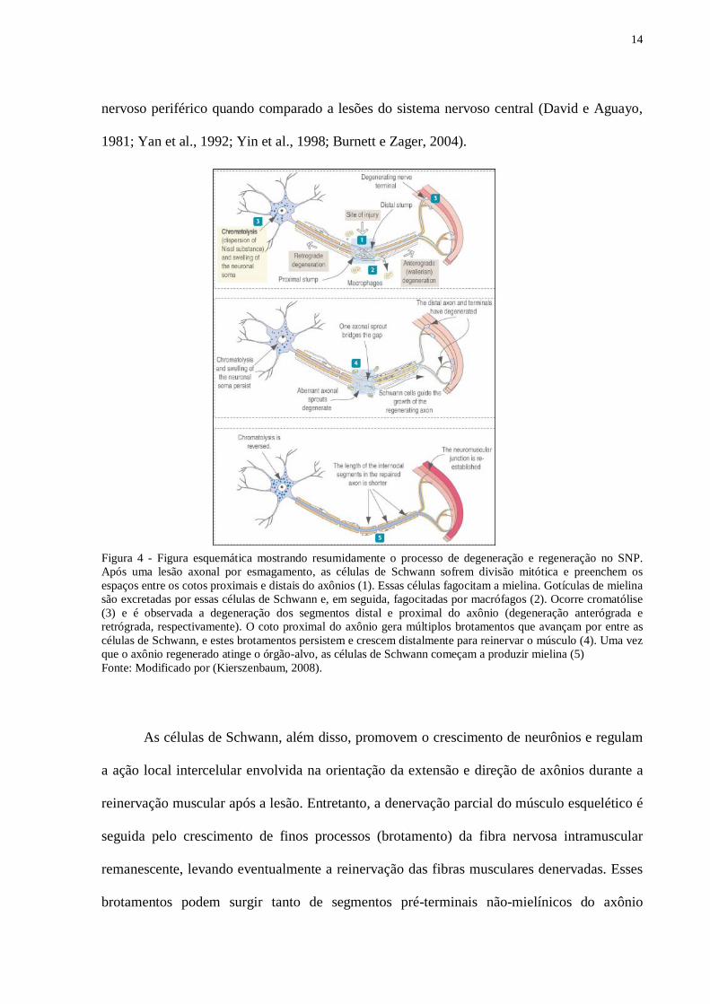
Figura 4 - Figura esquemática mostrando resumidamente o processo de degeneração e regeneração no SNP. Após uma lesão axonal por esmagamento, as células de Schwann sofrem divisão mitótica e preenchem os espaços entre os cotos proximais e distais do axônios (1). Essas células fagocitam a mielina. Gotículas de mielina são excretadas por essas células de Schwann e, em seguida, fagocitadas por macrófagos (2). Ocorre cromatólise (3) e é observada a degeneração dos segmentos distal e proximal do axônio (degeneração anterógrada e retrógrada, respectivamente). O coto proximal do axônio gera múltiplos brotamentos que avançam por entre as células de Schwann, e estes brotamentos persistem e crescem distalmente para reinervar o músculo (4). Uma vez que o axônio regenerado atinge o órgão-alvo, as células de Schwann começam a produzir mielina (5) Fonte: Modificado por (Kierszenbaum, 2008).
As células de Schwann, além disso, promovem o crescimento de neurônios e regulam
a ação local intercelular envolvida na orientação da extensão e direção de axônios durante a
reinervação muscular após a lesão. Entretanto, a denervação parcial do músculo esquelético é
seguida pelo crescimento de finos processos (brotamento) da fibra nervosa intramuscular
remanescente, levando eventualmente a reinervação das fibras musculares denervadas. Esses
brotamentos podem surgir tanto de segmentos pré-terminais não-mielínicos do axônio
15
(brotamento terminal) quanto de nódulos de Ranvier (brotamento nodal ou colateral) (Wang
et al., 2007).
As modificações no corpo neuronal e no segmento proximal das fibras dependem da
severidade da lesão, assim como da proximidade entre o segmento lesado e o corpo do
neurônio (Cullheim et al., 2002). As células de Schwann inevitavelmente degradam no
segmento próximo à lesão e os axônios e a mielina tornam-se visivelmente reduzidos em
diâmetro. Essa degradação proximal pode ser mínima (até o nodo de Ranvier mais próximo)
ou estender-se até o corpo do neurônio. Se o corpo do neurônio degenera, o que ocorre em
casos de trauma moderado a severo, todo o segmento proximal sofre degeneração e é
fagocitado (Lundborg, 2000).
Mesmo em lesões brandas, o corpo neuronal passa por modificações comparáveis após
a lesão. O núcleo migra para a periferia da célula e ocorre desmembramento dos corpúsculos
de Nissl e do retículo endoplasmático rugoso, processo denominado de cromatólise.
Simultaneamente, ocorre rápida resposta proliferativa das células gliais, de certa forma
sinalizada pela cromatólise. As células gliais, então, se estendem pelo neurônio afetado e
interrompem as conexões sinápticas, possivelmente para isolar o neurônio durante a fase de
recuperação (Burnett e Zager, 2004).
1.5 EXERCÍCIO FÍSICO E NEUROPATIAS PERIFÉRICAS
Terapias empregando programas de exercícios físicos são freqüentemente utilizados
na reabilitação de pacientes com neuropatias periféricas. Os exercícios físicos diminuem as
complicações comuns às patologias do SNP e promovem a recuperação funcional e o
aumento da capacidade aeróbica de pacientes com doenças neuromusculares (Herbison et al.,
1983; Linderman et al., 1995; Wright et al., 1996). Alguns resultados sustentam também que
16
a velocidade do crescimento axonal após lesão é aumentada por protocolos de treinamento
específicos (Van Meeteren et al., 1997).
Estudos experimentais têm empregado exercícios físicos na reabilitação de lesões
traumáticas do nervo ciático em modelos animais para estimular a regeneração nervosa e
melhorar a recuperação funcional, porém com resultados conflitantes. O treinamento de
endurance, por exemplo, promove a normalização da função motora dos membros posteriores
após uma semana de exercício de endurance (Ilha et al., 2008). Em um estudo realizado em
nosso laboratório, comparando-se exercícios acrobáticos e caminhada livre, observou-se uma
significativa melhora funcional e uma regeneração nervosa periférica satisfatória no grupo
tratado com o protocolo de equilíbrio e coordenação (Bonetti et al, 2011). Em outro estudo,
entretanto, ratos com esmagamento de nervo mediano e ulnar foram submetidos aos
protocolos de treinamento de habilidade, que consistia em alcançar alimento de dentro de uma
caixa de acrílico e um protocolo de treinamento de repetição, que consistia em caminhar em
uma esteira. Nesse estudo, evidenciou-se que ambos os protocolos de treinamento foram
suficientes para acelerar a recuperação funcional, porém o treinamento de repetição produziu
um maior grau de regeneração nervosa periférica do que o treinamento de habilidade
(Pagnussat et al., 2009). Por outro lado, outro estudo demonstrou que a natação não interfere
com a recuperação sensório-motora após lesão do nervo ciático e que um programa
intermediário de caminhada em esteira retarda a recuperação em ratos (Van Meeteren et al.,
1998). Também foi observado que a atividade motora intensa, realizada diariamente (natação)
em ratos com esmagamento do nervo ciático leva a deficiências na diferenciação das fibras
em regeneração (Gutmann e Jakoubek, 1963). Todos esses achados indicam que o tipo e a
intensidade do exercício físico podem exercer diferentes conseqüências na regeneração
nervosa periférica.
O treinamento de habilidade com tarefas acrobáticas induz à sinaptogênese,
17
potenciação sináptica, e reorganização da representação dos movimentos do córtex motor. O
treinamento de endurance, entretanto, induz á angiogênese no córtex motor, mas não altera a
organização do mapa motor cortical ou o número de sinapses. Além disso, o treinamento de
força altera a excitabilidade dos motoneurônios espinais e induz à sinaptogênese na medula
espinhal, mas não altera a organização do mapa motor cortical. Esses achados suportam a
idéia de que a natureza específica da reorganização é dependente da demanda comportamental
de cada treinamento (Adkins et al., 2006).
Há muito se sabe que o SNC depende de fontes de feedback sensorial para assegurar
um ótimo desempenho dos movimentos e a performance motora é comprometida quando esse
feedback é abolido ou quando ocorre algum distúrbio desta informação (Jones et al., 1999;
Kleim et al., 1996; VandenBerg et al., 2004). Entretanto, além das discrepâncias nos
resultados obtidos por esses prévios estudos, eles não especificam o tipo de treinamento
empregado, nem discutem se as suas respectivas ações no processo de regeneração poderiam
ser diferentes.
No sistema neuromuscular intacto de modelos de lesão em animais, diferentes
protocolos de exercícios têm demonstrado exercer distintas ações, o que remete a
possibilidade de que estes diversos efeitos dependentes do tipo de treinamento também
ocorram quando o exercício é aplicado após lesões do sistema nervoso. Por exemplo,
treinamentos aeróbicos não causam uma significante hipertrofia muscular, embora aumentem
a atividade colinesterásica (Ach) e resultem em significante expansão dos componentes pré e
pós sinápticos da junção neuromuscular em ratos (Crockett et al., 1976; Tomas et al., 1997).
Por outro lado, treinamentos de resistência muscular com altas cargas resultam em adaptações
neurais e hipertrofia muscular, as quais são responsáveis pelo aumento da força dos músculos
treinados (Deschenes et al., 2000; Lee et al., 2004). Desta forma, a especificidade do
treinamento é um fator importante e deve ser levado em consideração dentro do contexto da
18
regeneração nervosa quando se busca estudar os efeitos do exercício físico após lesões do
sistema nervoso. O tratamento de lesões de nervo periférico em humanos visa abordar a
combinação de diferentes estratégias de tratamento, por isso se faz necessário analisar o efeito
de diferentes protocolos de exercícios e suas associações.
22 OOBBJJEETTIIVVOOSS
2.1 OBJETIVO GERAL
Analisar o efeito de seis semanas de treinamento de repetição, treinamento de
equilíbrio e coordenação e treinamento de repetição associado ao treinamento de equilíbrio e
coordenação na regeneração nervosa periférica em ratos machos adultos após lesão por
esmagamento do nervo mediano.
2.2 OBJETIVOS ESPECÍFICOS
- Analisar, morfometricamente, o nervo mediano (área axonal, densidade axonal,
espessura da bainha de mielina, diâmetro axonal e diâmetro das fibras mielínicas) dos ratos
dos grupos sham sedentário e lesão por esmagamento do nervo mediano submetidos aos
treinamentos de equilíbrio e coordenação, repetição e a associação desses treinamentos;
- Avaliar melhoras funcionais dos ratos dos grupos sham sedentário e lesão por
esmagamento do nervo mediano submetidos aos treinamentos de equilíbrio e coordenação,
repetição e a associação desses treinamentos através do teste do cilindro, teste de motricidade
sobre grade (footfault test) e teste de suspensão no arame (hanging wire).
19
33 MMÉÉTTOODDOOSS EE RREESSUULLTTAADDOOSS
3.1 ARTIGO
Artigo – Juliana D. Neves, Fernando Soares Camelier, Sandro Antunes da Silva, Nígia
Ramalho Arsego, Jocemar Ilha, Simone Marcuzzo, Léder Leal Xavier, Maria Cristina
Faccioni-Heuser. Effects of different protocols of physical training on the median nerve
funtion and morphology of rats after crush injury.
Será submetido à Revista Muscle and Nerve.
20
Effects of different protocols of physical training on the median nerve funtion and
morphology of rats after crush injury
Juliana Dalibor Neves1,2, Fernando Soares Camelier1,2, Sandro Antunes da Silva1,2, Nígia
Ramalho Arsego2, Jocemar Ilha1,2, Simone Marcuzzo1,2, Léder Leal Xavier1,3, Maria Cristina
Faccioni-Heuser1,2
1Programa de Pós-Graduação em Neurociências, Instituto de Ciências Básicas da Saúde,
Universidade Federal do Rio Grande do Sul, RS, Brazil
2Laboratório de Histofisiologia Comparada, Departamento de Ciências Morfológicas,
Instituto de Ciências Básicas da Saúde, Universidade Federal do Rio Grande do Sul, RS,
Brazil
3Laboratório de Morfologia, Instituto de Biociências, Pontifícia Universidade Católica do Rio
Grande do Sul, RS, Brazil
Communicating author: Maria Cristina Faccioni-Heuser
Laboratório de Histofisiologia Comparada
Departamento de Ciências Morfológicas, ICBS
Universidade Federal do Rio Grande do Sul
Sarmento Leite 500, CEP: 90050-170, Porto Alegre, RS, Brazil
E-mail: [email protected]
Tel.: +55-51-33083789
21
ABSTRACT
Introduction: Numerous therapeutic interventions have been tested to enhance functional
recovery after crush lesion, but these studies did not examine the effects of specific types of
exercise. Methods: After median nerve crush injury we tested the effect of balance and
coordination, repetition training and the combination of both types of training program (for 6
weeks) on nerve regeneration in Wistar rats using functional tests and nerve morphometric
analysis. Results: Functional recovery was similarly accelerated in lesioned groups in the
footfault test, but in the hanging wire and in the cylinder test, the injured groups showed no
differences compared to control group. Balance and coordination training caused significantly
better values than repetition training alone, the association of the two protocols training, or
control for the axonal density and axonal diameter. Discussion: These data provide evidence
that balance and coordination training had greater potential for enhancing median nerve
histological regeneration after experimental traumatic injury, spite functional tests not show
differences between the treatments.
Keywords: Median nerve crush, physical training, peripheral nerve regeneration.
22
INTRODUCTION
Peripheral nerves are damaged by different factors such as acute trauma, chronic
repetitive insults, and inherit or acquired metabolic disorders1,2. The incidence of traumatic
injuries is estimated as ˃500.000 new patients annually 3. Peripheral nerve injuries reduce
muscular recruitment and sensation and can disrupt coordination through the changes that
occur in the peripheral and the central nervous system (CNS)4,5.
The majority of human peripheral nerve injuries affect the upper limbs and the most
disabling aspect of this injury is the loss of skilled hand movements6. Exercise training
improves motor function after experimental and clinical peripheral nervous lesion, and it can
be considered as an effective treatment of sensorial deficit7.
There are evidences that the type and intensity of activity induces morphological and
electrophysiological remodeling of neuromuscular junction and the motor nerve endings are
continuously changing and can suffer influence of modifications in functional demands8,9,10.
These adaptations of synapses can exert influence in nerve orientation and repair since one of
the mechanisms governing the regeneration is the secretion of growth factors by target
muscles during the period of denervation10,11.
A range of forms of exercise training has shown beneficial effects in various muscle
and nerve function related parameters in animal models of nerve injury12,13,14,15. Experimental
and clinical studies have employed physical exercise in the rehabilitation of traumatic injury
of the sciatic nerve with the purpose of stimulating nerve regeneration and improving
functional recovery, however conflicting results have been obtained. For example, the
endurance training promotes the normalization of hindlimb motor function after one week of
exercise15. In another study, the acrobatic exercise demonstrates significant functional
improvement and satisfactory nerve regeneration16. However, there is a few data about
23
forelimb exercise training after peripheral nerve injury.
Consequently, different training programs have diverse effects on peripheral nerve
regeneration. The results of specific treatments, as well as the combination of treatments are
necessary for the knowledge of mechanisms of recovery, and so for the application in humans
rehabilitation. Therefore, the present study was designed to compare the effects of balance
and coordination, repetition and the combination of these two types of training protocols on
functional recovery, and to carry out a morphometric analysis of the median nerve after crush
injury.
MATERIALS AND METHODS
Experimental design and surgical procedures
The experiment was performed on 46 three-month-old, male Wistar rats weighing
280-330 g (initial age and weight) from a local breeding colony (ICBS, Universidade Federal
do Rio Grande do Sul, Brazil). Rats were housed in standard Plexiglass boxes, under a 12:12
h light/dark cycle in a temperature-controlled environment (20 ± 1 °C) with food and water ad
libitum. All the procedures were approved by the Ethical Committee at the Federal University
of Rio Grande do Sul (nº 2008194) and all the animals were handled in accordance with the
Brazilian laws. Animals were randomly divided in five groups: (1) sham-operated rats,
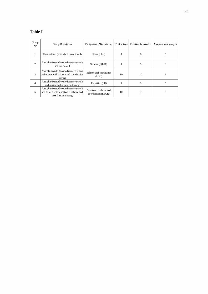
without median nerve crush and unexercised, sham sedentary (Sh-s, n = 8); (2) rats with
median nerve crush and unexercised, lesion sedentary (LSE, n = 9); (3) rats with median
nerve crush and repetition training (LR, n = 9); (4) rats with median nerve crush and balance
and coordination training (LBC, n = 10); and (5) rats with median nerve crush and repetition
+ balance and coordination training (LBCR, n = 10) (Table I). Before the surgical procedures,
animals were adapted for 5 days in each training program protocol. For the surgical
24
procedures, animals were anesthetized using ketamine and xylazine (90 and 15 mg/kg, i.p.,
respectively; Vetbrands, Brazil) and the right median nerve was exposed through a skin
incision in the axillopectoral region and the right pectoralis major muscle was removed with
preservation of the cephalic vein on the same side. The nerve crush injury was performed with
1 mm hemostatic forceps for 30 seconds (as previously described by Bridge and coworkers
(1994). The muscle and skin were then closed with 4-0 nylon sutures (Somerville, Brazil),
and the animals were put in their cages to rest. Four days after the surgery, the animals from
the LR, LBC and LBCR groups began specific training for 6 weeks, while Sh-s and LSE
animals were put in the same location as the training animals for a few minutes in order to
equal as much as possible the handling of all groups, but they did not perform any kind of
motor activity.
Rehabilitation protocols
After four days of brachial plexus crush animals received one of the following
treatments. Both protocols were performed along 6 weeks, 5 days per week (Fig. 1).
Repetition training
The repetition training program was performed in a treadmill for human (Runner,
Brazil) modified for use in rats15. This task consisted in walking to an adapted motorized
rodent treadmill during 20 min (0.03 m/s - 3 initial min; 0.05 m/s - 14 min; 0.03 m/s - last 3
min). The grade of the treadmill remained at 0% and no aversive stimulus was used. This
velocity was chosen in order to avoid possible effects of aerobic treadmill exercise. Since this
type of training used in this work is a simulation of a very low intensity exercise.
25
Balance and Coordination training
The balance and coordination training program was adapted from the acrobatic
training used by Black et al. (1990), Anderson, Alcantara and Greenough (1996) and Kleim et
al. (1996)17,18,19. Animals were required to traverse 5 different elevated obstacles per day,
such as suspension bridges, rope bridges, parallel bars, etc. (each 100 cm long) ending in
dark box. Each rat of this group crossed these obstacles 25 times, walking 2.500 cm each day
of training16. These obstacles require motor learning, balance and coordination from these
animals.
Repetition + Balance and coordination training
This group was daily subjected to the LR protocol followed to the protocol of LBC.
Functional tests
During training, the animals underwent the following behavioral assessments:
footfault test (FT), hanging wire (HW) and cylinder test (CT). For FT, the animals were
filmed 3 times from a lateral view and for CT, however, the animals were filmed during 4
minutes from an inferior view, and for HW they were recorded only once.
Footfault test
The footfault test was performed to assess whether training enhanced coordinated
placement of the forelimbs20. Rats were placed on an elevated grid platform (75X20 cm) for 3
trials. Rats moved across the platform by placing their paws on the rungs of the grid. Errors
were measured as slips with right forelimb through the grid openings21. In this study the
animals carry out the test on the 3rd postoperative day and after, weekly, and 24 hours after
completion of training.
26
Hanging Wire
This task was used to measure the ability to grasp and the strength of the forelimbs.
The animals used their forelimbs to suspend their body weight on a wire stretched between
two bars, 60 cm above the ground and the time (in seconds) before the animal fell was
recorded. A score of zero was used if the rat fell down immediately and 120 seconds was the
time limit of the test. The animals carry out the test on the ninth postoperative day and after,
weekly, and 24 hours after completion of training. A single trial was conducted for every rat
on each test day22.
Cylinder test
To examine the effect of brachial plexus crush and treatment on spontaneous forelimb
use during exploratory activity movements, animals were individually placed into a
transparent cylinder (20 cm diameter and 40 cm high) on a glass tabletop and video recorded
from below through an angled mirror for 4 min during each test session. The cylindrical shape
encouraged rearing and vertical exploration of the walls with the forelimbs. The number of
forepaws wall contacts used for postural support was counted and the percentage of
asymmetry of single-limb wall contacts [(contralateral/contralateral + ipsilateral) x 100] was
calculated. A single cylinder test session was performed 3 days after the surgery, 21 days after
the surgery and one day before perfusion23.
Histological and morphometric studies
Two days after the period of training the animals were anesthetized with sodium
thiopental (50 mg/kg, i.p.; Cristália, Brazil), injected with 1000 IU heparin (Cristália, Brazil)
and were transcardially perfused with 300 ml of saline solution, followed by 0.5%
glutaraldehyde (Sigma Chemicals Co., St Louis, MO) and 4% paraformaldehyde (Reagen,
27
Brazil) in a 0.1 M phosphate buffer (pH 7.4, PB) at room temperature. One short segment of
the right median nerve was rapidly excised, 5 mm after the crush injury site, in the distal
portion15. This region was chosen in order to show major alterations after injury, since the
changes are most pronounced in the distal portion of the nerve5. The specimens were
postfixed by immersion in a fixative solution of glutaraldehyde 2.5% and paraformaldehyde
2% at 4 °C until processed; after that, the samples were washed in 0.1 M PB and postfixed in
1% OsO4 (Sigma Chemicals Co., St Louis, MO) in 0.1 M PB for 30 min.; washed again in 0.1
M PB; dehydrated in a graded series of acetone, embedded in resin (Durcupan, ACM-Fluka,
Switzerlend) and polymerized at 60° C. Cross-semithin sections (1 µm) were obtained using
an ultramicrotome (MT 6000-XL, RMC, Tucson, USA) and stained with 1% toluidine blue
(Merck, Germany) in 1% sodium tetraborate (Ecibra, Brazil)15,24.
Afterwards, images of the distal portion of the right median nerve was captured and
digitalized (initially 1000x and further amplified 200% for analysis) using a Nikon Eclipse E-
600 microscope (Japan) coupled to a Pro-Series High Performance CCD camera and
processed with Image Pro Plus Software 6.0 (Media Cybernetics, USA)15,24.
For morphometric evaluation of the nerve, distal portion of the right median nerve was
analyzed; a set of 5 images was chosen using random sampling of one slice. Morphometric
measurements of the median nerve included the (1) myelinated fiber density (number of
fibers/mm2); (2) average myelinated fiber area (μm2); (3) average myelin sheath thickness
(μm); (4) average myelinated fiber diameter (µm); (5) average axon diameter (µm) of the
myelinated fiber). These morphological parameters were chosen to assess the differentiation
of regenerating median nerve15.
The average myelin sheath thickness was estimated using the measurement tools of
Image Pro Plus software. The measurements of areas were estimated with a point-counting
28
technique15,24 using grids with point density of 1 point per 1.30 µm2 and the following
equation:
Â=Σp.a/p, where  is area, Σp the sum of points, and a/p the area/point value. To
estimate the axon and neural fiber diameters, the area of each individual fiber was converted
to the diameter of a circle having an equivalent area.
Statistical analysis
Behavioral assessment were analyzed using one-way repeated measures analysis of
variance (ANOVA) and morphometric measurements of the median nerve were analyzed
using one way analysis of variance (ANOVA). All analyses were followed by post hoc
Duncan’s. Data were expressed as means ± standard error of the mean (SEM). The
significance level was p<0.05. Statistical analysis was performed using the Statistical
software package.
RESULTS
Behavioral Study
Footfault Test
Repeated measures analysis of variance of footfault test, showed time (F(6.198)=
34.16863, p<0.001), group (F(1.33) = 5.47632, P<0.01) and a significant interaction between
time and group (F(24.198) = 4.34956, p<0.001). As shown in Fig. 2, Duncan’s post hoc analysis
revealed that the number of right forelimb errors was significantly decreased in the Sh-s group
(0.58 ± 0.36) 3 days postlesion (pre-training), compared with all the lesioned groups: LSE
(2.59 ± 0.54, p<0.05), LBC (4.43 ± 0.80, p<0.05), LR (2.36 ± 0.35, p<0.05) and LBCR (2.43
29
± 0.28, p<0.05). After 9 days postlesion (one week of training), the LBCR (1.46 ± 0.22)
showed significantly more right forelimb errors than the Sh-s (0.33 ± 0.16; p<0.05).
Differently, the LBC (0.79 ± 0.25), LR (0.73 ± 0.23) and LSE (1.10 ± 0.23) groups showed no
signicant differences between the Sh-s group, after 9 days postlesion. Afterward, from day 15
poslesion, the injured groups revealed no signicant differences between the Sh-s group on
other days of testing.
Hanging Wire
Repeated measures analysis of variance of the hanging wire showed effect for time
factor (F(5.205) = 3.9437, P<0.01) and an significant interaction between time and group effects
(F(20.205) = 1.9323, P<0.05), but no group effect (F(1.41) = 2.2340, P˃0.05) (Fig. 3).
Cylinder Test
Repeated measures analysis of variance for the cylinder test showed effect for time
factor (F(2.82) = 4.8422, P<0.05), but no group effect (F(1.41) = 1.2943, p˃0.05) and no
significant interaction between time and group (F(8.82) = 0.7112, P˃0.05) (Fig. 4).
Histological analysis
The structural analysis of regenerating nerves (Fig. 5) showed important qualitative
differences between the Sh-s and injured groups (LSE, LBC, LR and LBCR). The histological
characteristics of distal portion of median nerve in the crush groups comprise enlargement of
endoneurial connective tissue between the nerve fibers and reduction of myelinated fiber
diameter. In LBC group these pathological features were apparently reduced and less
endoneurial connective tissue was observed.
30
Morphometric analysis of median nerve
For median nerve, one way ANOVA analyses evidenced effect of lesion for
Myelinated fiber area (F(1.23) = 4.3358, P<0.01); Myelin sheath thickness (F(1.23) = 5.5932,
P<0.01); Myelinated fiber diameter (F(1.23) = 6.0849, P<0.01); Axon diameter (F(1.23) = 5.4737,
P<0.01) and Axonal density (F(1.23) = 3.7803, P<0.05). Analysis of morphometric data with
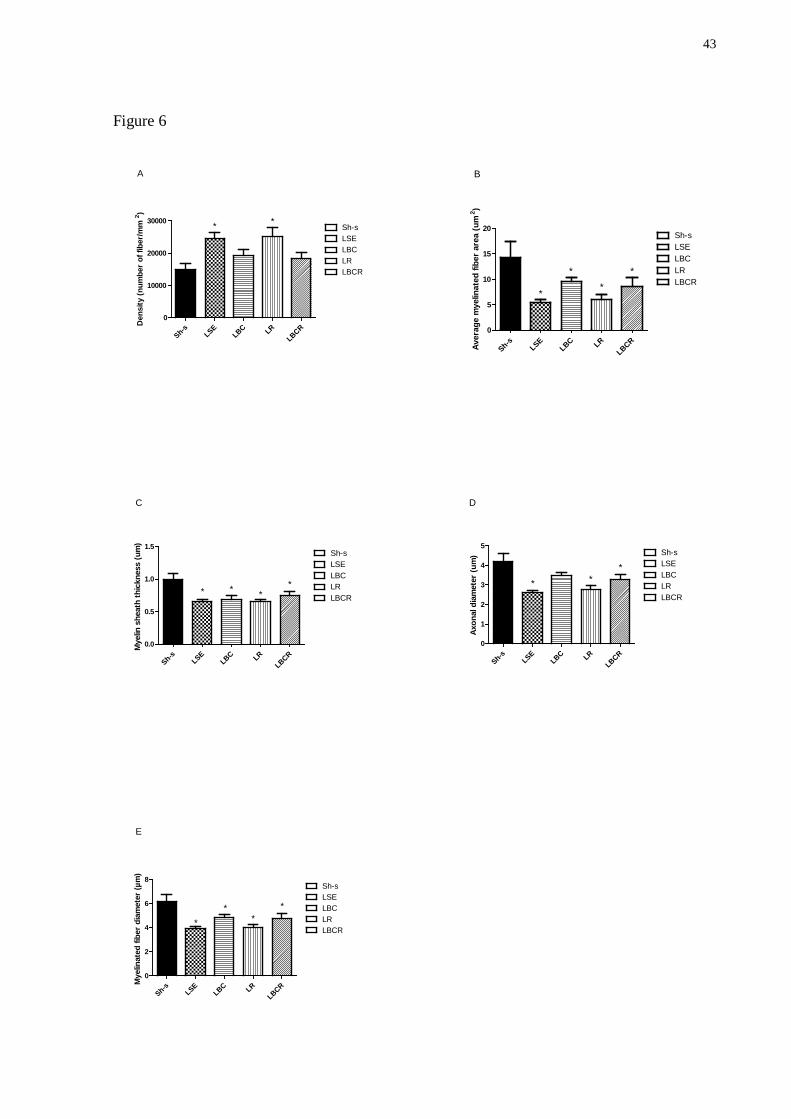
Duncan’s post-hoc test revealed that the LSE (24496 ± 2050.98 fibers/mm2; P=0.007) and LR
(25098 ± 3033.58 fibers/mm2; P=0.005) groups showed a greater density of the myelinated
fibers than the sham group (15004 ± 1831.82 fibers/mm2; Figure 6A). Furthermore, the
experimental groups (LSE: 5.5 ± 0.43 µm2; P=0.001; LBC: 9.51 ± 0.93 µm2; P=0.048; LR:
6.08 ± 0.99 µm2; P=0.002; LBCR: 8.66 ± 1.78 µm2; P=0.028) had smaller average myelinated
fiber areas than the sham group (14.32 ± 3.13 µm2; Figure 6B).
The average myelin sheath thickeness of the lesioned groups (LSE: 0.65 ± 0.02 µm;
P=0.0008; LBC: 0.69 ± 0.06 µm; P=0.0016; LR: 0.65 ± 0.02 µm; P=0.0007; LBCR: 0.74 ±
0.06 µm; P=0.0058) were thinner than that of the sham group (1.00 ± 0.08 µm; Figure 6C).
The average myelinated fiber diameters in the lesioned groups (LSE: 3.95 ± 0.13 µm;
P=0.0003; LBC: 4.84 ± 0.27 µm; P=0.012; LR: 4.04 ± 0.21 µm; P=0.0004; LBCR: 4.74 ±
0.43 µm; P=0.010) were different from the sham group (6.18 ± 0.56 µm; Figure 6E). In
addition, the average axon diameter in the LSE (2.63 ± 0.10 µm; P=0.0007), LR (2.74 ± 0.23
µm; P=0.0011) and LBCR (3.25 ± 0.30 µm; P=0.0230) groups was different from the sham
group (4.18 ± 0.41 µm; Figure 6D). However, the LBC group had the higher average
myelinated axon diameter (3.46 ± 0.16 µm), approximating of the values of the sham group.
31
DISCUSSION
Experimental studies showed that exercise training improves motor function after
clinical and experimental peripheral nervous lesion, and can be considered as an effective
treatment of sensorial deficits25,26,27,28. However, only one study have used proprioceptive and
repetition training as treatment for brachial plexus injury or tests of skilled forelimb function
to assess recovery after peripheral nerve injury and repair29.
In the present study we investigated the hypothesis that balance and coordination,
repetition training and the association of this two protocols, started four days after the crush
and performed for six weeks, could produce different effects on functional recovery and
morphological changes in the regenerating nerve. We chose to begin the exercise program as
early as four days after the crush in order to show that the training protocols (LBC and LR)
can be used in the early phase of rehabilitation, as long as performed carefully and
moderately.
Our results show that the injured groups (LSE, LBC, LR and LBCR) decreased
forelimb errors after one week, as evaluated by means of FT. What may prove a spontaneous
recovery by these groups or even suggest that the test was not sufficiently sensitive to
demonstrate the differences between them, since this task is more used in brain lesions than
peripheral ones21,30,31. Furthermore, 9 days postlesion the LBCR group showed more forelimb
error than Sh-s group. Perhaps, the high demand imposed by the association of training could
have a deleterious effect on functionality of the animals at earlier stages, and after, there is a
normalization of function. In the same way, the HW and CT show no significance difference
between the groups. The reason for this may be explained, as a hypothesis, since they
evidently demonstrated impaired right forelimb movements, until at least two weeks after the
injury. We think that the animals acquired compensatory strategies to perform the required
32
tasks and thus the deficits could not be quantified. More accurate analysis, which evaluates
the quality of movement, may be more effective to show the possible functional differences of
the treatments in the forelimbs. Moreover, in the HW, there was a learning curve in sham
group, shown by less time spent in performing the task every day analyzed. This demonstrates
that not only the motor skill training that is being evaluated in this test, but the ability to adapt
to it, dificulting the conclusions about this data.
In a crush lesion, the axon undergoes alterations both in the distal and proximal
portions to the injury, but such alterations occur mainly in the distal portion; thus, the
morphometric parameters distal to the lesion are more important for the analysis of the
influence of exercise in nerve regeneration5. In this portion of the nerve, the average
myelinated fiber area, myelin sheath thickness and myelinated fiber diameter showed a
significant difference between the lesioned groups and Sh-s. These results show that the
injury caused a pathological modification in nerve. But, there were some levels of recovery,
indicated by following morphological parameters: axonal density and axonal diameter. The
balance and coordination training associated or not to repetition showed a decreased number
of fibers/mm2, approximating of the sham values. This reduction indicates an increase in
myelinated fiber area caused by these training programs. Corroborating this data, the LBC
group presented an improvement in axonal diameter, statistically equaling to sham group. In a
previous study carried through our laboratory, the balance and coordination exercise training
had also demonstrated changes in morphological parameters (average myelinated fiber area,
average myelin sheath thickness, myelinated fiber diameter and axon diameter) of peripheral
nerves in young rats compared to free walking training16. In other study realized in our
laboratory, moderate endurance training showed a greater degree of the myelinated fiber
maturation than the sedentary, resistance-trained and concurrent training groups. Furthermore,
in this study, the endurance-trained group showed a smaller percentage area of endoneurial

33
connective tissue and a greater percentage area of myelinated fibers than the sedentary group
after sciatic crush injury, promoting the normalization of hindlimb motor function after one
week of exercise15. This study, thus provides evidence that resistance training or the
combination of two strategies may delay functional recovery and do not alter nerve
regeneration. All these findings indicate that the time and the type of exercise training can
exert different consequences in peripheral nerve regeneration.
In humans, the major aim of physiotherapy after nervous system injury is to restore the
patient’s autonomy in activities of daily living and to avoid permanent disability. In this
context, one of the most important tools in experimental studies is the functional assessment
as they provide the validation of more effective therapies with respect to self-sufficiency of
the patients. Unfortunately, in our study, the functional tests were not appropriate for this type
of injury. Since the crushed group, even showing some degree of protection of the forelimbs
visible until two weeks after lesion, demonstrated no deficit in the tests used. Perhaps this is
because of compensation used by these animals to perform the tasks, since the lesion is
unilateral. Also this may be due to the role of the forelimb, which is used more for skills tasks
than weight bearing and locomotion, variables involved in the tests performed in the present
work32. Tasks that assess the quality of movement can be more effective to show the
functional recovery in this type of injury/training (such as electromyography, staircase test,
pawprint analysis, kinematic analysis of movements).
In conclusion the present study showed that balance and coordination training can
accelerate the median nerve regeneration after an experimental traumatic injury compared to
sedentary and repetition groups and the association of treatments. The functional tests proved
no effective in demonstrate the real deficits of the forelimb and thus is necessary better skill
tests to show more subtle changes of movement. The need for efficiency in the recovery of
hand and forelimb movements requires further studies to verify the success of different
34
methods of treatment in promoting neuronal reorganization and functional improvement after
injury of the peripheral nervous system.
REFERENCES
1. Stone L, Keenan MA. Peripheral nerve injuries in the adult with traumatic brain injury.
Clin Orthop Relat Res 1988; 233: 136 – 144.
2. Kartje GL, Schwab ME. Axonal growth in the adult mammalian nervous system:
regeneration and compensatory plasticity. In: Siegel GJ, Alberts RW, Brady ST, Price DL
(eds). Basic Neurochemistry: molecular, cellular, and medical aspects. San Diego: Elsevier
Academic Press; 2006. p517 – 527.
3. Rodríguez FJ, Valero-Cabré A, Navarro X. Regeneration and functional recovery following
peripheral nerve injury. Drug Discov Today 2004; 1: 177 – 185.
4. Hansson T, Brismar T. Loss of sensory discrimination after median nerve injury and
activation in the primary somatosensory cortex on functional magnetic ressonance imaging. J
Neurosurg 2003; 99: 100 – 105.
5. Johnson EO, Zoubos AB, Soucacos PN. Regeneration and repair of peripheral nerves.
Injury Int J Care Injured 2005; 36S: S24 – S29.
6. Duff SV. Impact of peripheral nerve injury on sensorimotor control. J Hand Ther 2005; 18:
277 – 291.
7. Korb A, Bonetti LV, da Silva SA, Marcuzzo S, Ilha J, Bertagnolli M, Partata WA,
Faccioni-Heuser MC. Effect of treadmill exercise on serotonin immunoreactivity in medullary
raphe nuclei and spinal cord following sciatic nerve transection in rats. Neurochem Res 2010;
35: 380 – 389.
8. Deschenes MR, Maresh CM, Crivello JF, Armstrong LE, Kraemer WJ, Covault J. The
35
effects of exercise training of different intensities on neuromuscular junction morphology. J
Neurocytol 1993; 22: 603 – 615.
9. Tomas J, Santafé M, Lanuza MA, Fenoll-Brunet MR. Physiological activity-dependent
ultrastructural plasticity in normal adult rat neuromuscular junctions. Biol Cell 1997; 89: 19 –
28.
10. Nishizawa T, Yamashia S, McGrath KF, Tamaki H, Kasuga N, Takekura H. Plasticity of
neuromuscular junction architectures in rat slow and fast muscle fibers following temporaray
denervation and reinnervation processes. J Muscle Res Cell Motil 2006; 27: 607 – 615.
11. Tomas I, Ferre J, Fennol I, Brunet R, Santafe M, Mayano E. Changes in motor nerve
terminals during bupivacaine-induced postsynaptic deprivation. J Anat 1989; 162: 225 – 234.
12. Van Meeteren NLU, Brakkee JH, Hamers FPT, Helders PJM, Gispen WH. Exercise
training improve functional recovery and motor nerve conduction velocity after sciatic nerve
crush lesion in the rat. Arch Phys Med Rehabil 1997; 78: 70 – 77.
13. Meek MF, Koning MAJ, Nicolai JA, Gransbergen A. Rehabilitation strategy using
enhanced housing environment during neural regeneration. J Neurosci Methods 2004; 136:
179 – 185.
14. Molteni R, Zheng J, Gómez-Pinilla F, Twiss JL. Voluntary exercise increases axonal
regeneration from sensory neurons. PNAS 2004; 101: 8473 – 8478.
15. Ilha J, Araújo RT, Malysz T, Hermel EES, Rigon P, Xavier LL, Achaval M. Endurance
and resistance exercise training programs elicit specific effects on sciatic nerve regeneration
after experimental traumatic lesion in rats. Neurorehabil Neural Repair 2008; 22: 355 – 366.
16. Bonetti LV, Korb A, da Silva SA, Ilha J, Marcuzzo S, Achaval M, Faccioni-Heuser MC.
Balance and coordination training improves sensorimotor functions and sciatic nerve
regeneration after peripheral nerve injury model. Muscle Nerve 2011 (to be published).
17. Black JE, Issacs KR, Anderson BJ, Alcantara AA, Greenough WT. Learning causes
36
synaptogenesis, whereas motor activity causes angiogeneses in cerebellar cortex of adult rats.
Proc Natl Acad Sci 1990; 87: 5568 – 5572.
18. Anderson BJ, Alcantara AA, Greenough WT. Changes in synaptic organization of the rat
cerebellar cortex. Neurobiol Learn Mem 1996; 66: 221 – 229.
19. Kleim JA, Lussnig E, Schwarz ER, Comery TA, Greenough WT. Synaptogenesis and
FOS expression in the motor cortex of the adult rat after motor skill learning. J Neurol Sci
1996; 4529 – 4535.
20. Barth TM, Jones TA, Schallert T. Functional subdivisions of the rat somatic sensorimotor
cortex. Behav Brain Res 1990; 39: 73 – 75.
21. Li X, Blizzard KK, Zeng Z, Devries AC, Hurn PD, McCullough LD. Chronic behavioral
testing after focal ischemia in the mouse: functional recovery and the effects of gender. Exp
Neurol 2004; 187: 94 – 104.
22. Marcuzzo S, Dutra MF, Stigger F, Do Nascimento PS, Ilha J, Kalil-Gaspar PI, Achaval
M. Beneficial effects of treadmill training in a cerebral palsy-like rodent model: walking
pattern and soleus quantitative histology. Brain Res 2008; 1222: 129 – 140.
23. Schallert, T, Fleming, SM, Leasure JL, Tillerson JL, Bland ST. CNS plasticity and
assessment of forelimb sensorimotor outcome in unilateral rat models of stroke, cortical
ablation, parkinsonism and spinal cord injury. Neuropharmacology, 2000, 39: 777 – 787.
24. Galtrey CM, Fawcett JW. Characterization of tests of functional recovery after median
and ulnar nerve injury and repair in the rat forelimb. J Peripher Nerv Syst 2007; 12: 11-27.
25. Linderman E, Leffers P, Spaans F, Drukker J, Kerckhoffs M, Köke A. Strength training in
patients with myotomic dystrophy and hereditary motor and sensory neuropathy: a
randomized clinical trial. Arch Phys Med Rehabil 1995; 76: 612–620.
37
26. Wright NC, Kilmer DD, McCrory MA, Aitkens SG, Holcomb BJ, Bernauer EM. Aerobic
walking in slowly progressive neuromuscular disease: effect of a 12-week program. Arch
Phys Med Rehabil 1996; 77: 64–69.
27. Hutchinson KJ, Gómez-Pinilla F, Crowe MJ, Ying Z, Basso DM. Three exercise
paradigms differentially improve sensory recovery after spinal cord contusion in rats. Brain
2004; 127: 1403–1414.
28. Jones TA, Chu CJ, Grande LA, Gregory AD. Motor skills training enhances lesion-
induced structural plasticity in the motor cortex of adult rats. J Neurosc 1999; 19 (22): 10153
– 10163.
29. Bertelli JA, Taleb M, Saadi A, Mira JC, Pecot-Dechavassine M. The rat brachial plexus
and its terminal branches: an experimental model for the study of peripheral nerve
regeneration. Microsurgery 1995; 16: 77-85.
30. Lubics A, Reglödi D, Tamás A, Kiss P, Szalai, Szalontay L, Lengvári I. Neurological
reflexes and early motor behavior in rats subjected to neonatal hypoxic-ischemic injury.
Behavioral Brain Research 2005; 157: 157 – 165.
31. Modo M, Stroemer RP, Tang E, Veizovic T, Sowniski P, Hodges H. Neurological
sequelae and long-term behavioral assessment of rats with transient middle cerebral artery
occlusion. J Neurosci Methods 2000; 194: 99 – 109.
32. Imaniuk AN, Wishaw IQ. On the origin of skilled forelimb movements. Trends Neurosci
2000; 23: 372 – 376.
38
LEGENDS
Figure 1. Time line of experimental procedures: Treadmill: Electrical Treadmill habituation;
BC: habituation of apparatus of Balance and Coordination; FT: Footfault test; CT: Cylinder
test; HW: Hanging Wire.
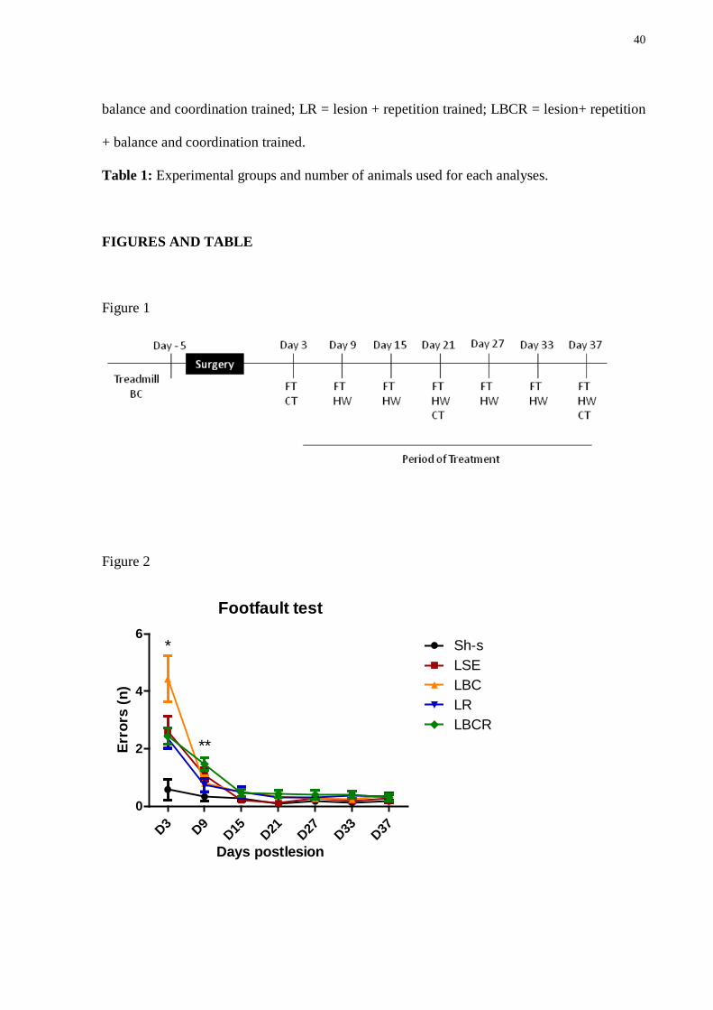
Figure 2. Footfault test: The footfault test was performed to assess whether acrobatic and
repetition training or the association of these two protocols enhanced coordinated placement
of the forelimbs. Each animal was recorded with a digital video camera crossing three times
and the errors were measured as slips with right forelimb through the grid openings. All the
damaged groups showed functional recovery over the weeks. At day 3 postlesion, all the
injured groups showed a statistically significant difference compared to Sh-s group.
Furthermore, in day 9 the LBCR group showed significant difference compared to Sh-s
group. Values presented are mean ± SEM (* indicates that injured groups are different to Sh-
s group and ** indicates that LBCR group are different to Sh-s group). Sh-s = sham; LSE =
lesion + sedentary; LBC = lesion + balance and coordination trained; LR = lesion + repetition
trained; LBCR = lesion+ repetition + balance and coordination trained.
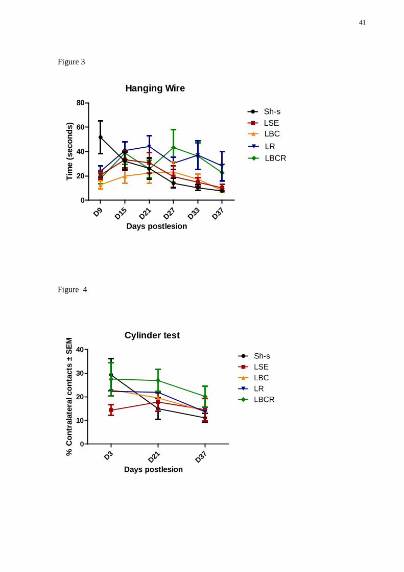
Figure 3. Hanging Wire: This task was used as a measure of the ability of grasp and of
strength of the forelimbs. Animals used their forelimbs to suspend their body weight on a wire
stretched between two bars. In day 9 postlesion, the LBC group showed that after one week of
training was unable to improve the strength of the forelimbs. Differently, the other injured
groups were superior in the ability to grasp and in the strength of the forelimbs. Values
presented are mean ± SEM. Sh-s = sham; LSE = lesion + sedentary; LBC = lesion + balance
and coordination trained; LR = lesion + repetition trained; LBCR = lesion+ repetition +
balance and coordination trained.
39
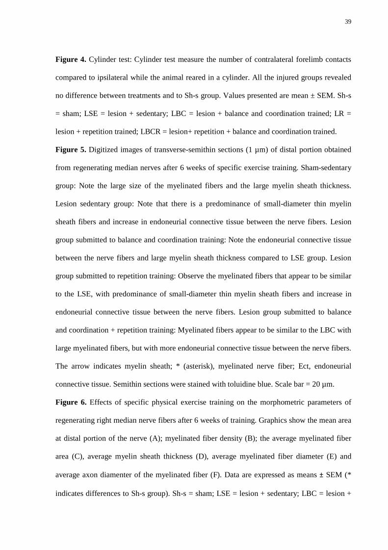
Figure 4. Cylinder test: Cylinder test measure the number of contralateral forelimb contacts
compared to ipsilateral while the animal reared in a cylinder. All the injured groups revealed
no difference between treatments and to Sh-s group. Values presented are mean ± SEM. Sh-s
= sham; LSE = lesion + sedentary; LBC = lesion + balance and coordination trained; LR =
lesion + repetition trained; LBCR = lesion+ repetition + balance and coordination trained.
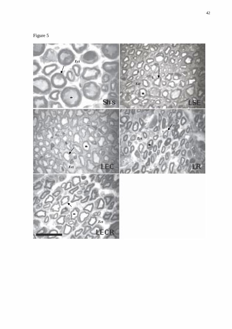
Figure 5. Digitized images of transverse-semithin sections (1 µm) of distal portion obtained
from regenerating median nerves after 6 weeks of specific exercise training. Sham-sedentary
group: Note the large size of the myelinated fibers and the large myelin sheath thickness.
Lesion sedentary group: Note that there is a predominance of small-diameter thin myelin
sheath fibers and increase in endoneurial connective tissue between the nerve fibers. Lesion
group submitted to balance and coordination training: Note the endoneurial connective tissue
between the nerve fibers and large myelin sheath thickness compared to LSE group. Lesion
group submitted to repetition training: Observe the myelinated fibers that appear to be similar
to the LSE, with predominance of small-diameter thin myelin sheath fibers and increase in
endoneurial connective tissue between the nerve fibers. Lesion group submitted to balance
and coordination + repetition training: Myelinated fibers appear to be similar to the LBC with
large myelinated fibers, but with more endoneurial connective tissue between the nerve fibers.
The arrow indicates myelin sheath; * (asterisk), myelinated nerve fiber; Ect, endoneurial
connective tissue. Semithin sections were stained with toluidine blue. Scale bar = 20 µm.
Figure 6. Effects of specific physical exercise training on the morphometric parameters of
regenerating right median nerve fibers after 6 weeks of training. Graphics show the mean area
at distal portion of the nerve (A); myelinated fiber density (B); the average myelinated fiber
area (C), average myelin sheath thickness (D), average myelinated fiber diameter (E) and
average axon diamenter of the myelinated fiber (F). Data are expressed as means ± SEM (*
indicates differences to Sh-s group). Sh-s = sham; LSE = lesion + sedentary; LBC = lesion +
40
balance and coordination trained; LR = lesion + repetition trained; LBCR = lesion+ repetition
+ balance and coordination trained.
Table 1: Experimental groups and number of animals used for each analyses.
FIGURES AND TABLE
Figure 1
Figure 2
Footfault test
D3 D9D15 D21 D27 D33 D37
0
2
4
6Sh-sLSELBCLRLBCR
*
**
Days postlesion
Erro
rs (n
)
41
Figure 3
Hanging Wire
D9D15 D21 D27 D33 D37
0
20
40
60
80Sh-sLSELBCLRLBCR
Days postlesion
Tim
e (s
econ
ds)
Figure 4
Cylinder test
D3D21 D37
0
10
20
30
40Sh-sLSELBCLRLBCR
Days postlesion
% C
ontr
alat
eral
con
tact
s ±
SEM
42
Figure 5
* *
* *
*
Ect
Ect
Ect
Ect
Ect
43
Figure 6
Sh-sLSE
LBC LRLBCR
0
10000
20000
30000Sh-sLSELBCLRLBCR
* *
A
Den
sity
(num
ber
of fi
ber/
mm
2 )
Sh-sLSE
LBC LRLBCR
0
5
10
15
20
*
**
*
Sh-sLSELBCLRLBCR
B
Aver
age
mye
linat
ed fi
ber
area
(um
2 )
Sh-sLSE
LBC LRLBCR
0.0
0.5
1.0
1.5Sh-sLSELBCLRLBCR
* * **
C
Mye
lin s
heat
h th
ickn
ess
(um
)
Sh-sLSE
LBC LRLBCR
0
1
2
3
4
5Sh-sLSELBCLRLBCR
* **
D
Axon
al d
iam
eter
(um
)
Sh-sLSE
LBC LRLBCR
0
2
4
6
8Sh-sLSELBCLRLBCR
**
**
E
Mye
linat
ed fi
ber
diam
eter
(µm
)
44
Table I
Group Nº
Group Description Designation (Abbreviation) Nº of animals Functional evaluation Morphometric analysis
1 Sham animals (untouched - unlesioned) Sham (Sh-s) 8 8 5
2 Animals submitted to median nerve crush and not treated
Sedentary (LSE) 9 9 6
3Animals submitted to median nerve crush and treated with balance and coordination
training
Balance and coodination (LBC) 10 10 6
4 Animals submitted to median nerve crush and treated with repetition training
Repetition (LR) 9 9 5
5Animals submitted to median nerve crush and treated with repetition + balance and
coor dination training
Reptition + balance and coordination (LBCR) 10 10 6
45
44 CCOONNCCLLUUSSÕÕEESS EE PPEERRSSPPEECCTTIIVVAASS
Os dados apresentados neste trabalho mostram evidências de que o treinamento de
equillíbrio e coordenação acelerou a regeneração nervosa após lesão traumática esperimental,
demonstrando que esse protocolo pode interferir no planejamento terapêutico dos
profissionais da área da saúde que trabalham com lesões do SNP. Os testes funcionais, no
entanto, não se mostraram efetivos em demonstrar os déficits reais dos membros anteriores e
por isso se faz necessário testes mais acurados para a obtenção de tarefas de habilidade,
enfocando as alterações mais sutis de movimento, como as causadas no presente trabalho.
Diante disso, os resultados obtidos nesse estudo servem como referência para futuras
pesquisas que busquem elucidar os efeitos da variação da intensidade e dos tipos de
protocolos utilizados e seus efeitos sobre a regeneração nervosa. Além disso, se faz necessário
analisar os efeitos da lesão no SNP e as tarefas utilizadas para reabilitação em neurônios da
medula espinhal e o papel de outras estruturas encefálicas relacionadas à atividade motora.
46
REFERÊNCIAS
Adkins DL, Boychuck J, Remple MS, Kleim JA. Motor training induces experience-specific
patterns of plasticity across motor cortex and spinal cord. J Appl Physiol 2006; 101: 1776 –
1782.
Baichwal RR, Bigbee JW, Devries GH. Macrophage-mediated myelin-related mitogenic
factor for cultured Schwann cells. Proc Natl Acad Sci 1988; 85: 1701 – 1705.
Bertelli JA, Taleb M, Saadi A, Mira J, Pecot-Dechavassine M. The rat brachial plexus and its
terminal branches: an experimental model for the study of peripheral nerve regeneration.
Microsurgery 1995; 16: 77 – 85.
Bertelli JA, Mira JC. Behavioral evaluating methods in the objective clinical assessment of
motor function after experimental brachial plexus reconstruction in the rat. J Neurosci
Methods 1993; 46: 203 – 208.
Bonetti LV, Korb A, da Silva SA, Ilha J, Marcuzzo S, Achaval M, Faccioni-Heuser MC.
Balance and coordination training improves sensorimotor functions and sciatic nerve
regeneration after peripheral nerve injury model. Muscle Nerve 2011 (to be published).
Bontioti EN, Kanje M, Dahlin LB. Regeneration and functional recovery in the upper
extremity of rats after various types of nerve injuries. J Peripher Nerv Syst 2003; 8: 159 –
168.
47
Bontioti EN, Kanje M, Lundborg G, Dahlin LB. End-to-side nerve repair in the upper
extremity of rat. J Peripher Nerv Syst 2005; 10: 58 – 68.
Burnett MG, Zager EL. Pathophysiology of peripheral nerve injury: a brief review. Neurosurg
Focus 2004; 16: E1.
Crockett JL, Edgerton VR, Max SR, Barnard RJ. The neuromuscular junctions in response to
endurance training. Exp Neurol 1976; 51: 207 – 215.
Cullheim S, Wallquist W, Hammarberg H, Linda H, Piehl F, Carlstedt T, Risling M.
Properties of motoneurons underlying their regenerative capacity after axon lesions in the
ventral funiculus or at the surface of the spinal cord. Brain Res Rev 2002; 40: 309 – 316.
Dahlin LB, Brandt J. Basic science of peripheral nerve repair: Wallerian degeneration/ growth
cones. Oper Tech Orthop 2004; 14: 138 – 145.
David S, Aguayo AJ. Axonal elongation into peripheral nervous system “bridges” after
central nervous system injury in adult rats. Science 1981; 214: 931 – 933.
Deschenes MR, Judelson DA, Kramer WJ, Meskaitis VJ, Volek JS, Nindl BC, Harman FS,
Deaver DR. Effects of resistance training on neuromuscular junction morphology. Muscle
Nerve 2000; 23: 1576 – 1581.
Duff SV. Impact of peripheral nerve injury on sensorimotor control. J Hand Ther 2005; 18:
48
277 – 291.
Ferreira AS. Lesões Nervosas Periféricas: Diagnóstico e Tratamento. Ed. Santos, 1999.
Flores PL. Epidemiological study of the traumatic brachial plexus injuries in adults. Arq
Neuro Psiquiatr 2006; 64(1).
Fredericks CM. Disorders of the peripheral nervous system: the peripheral neuropathies. In:
Fredericks CM, Saladin LK. (eds). Pathophysiology of the motor systems: principles and
clinical presentations. F.A. Davis Company, Philadelphia; 1996. p 346 – 372.
Galtrey CM, Fawcett JW. Characterization of tests of functional recovery after median and
ulnar nerve injury and repair in the rat forelimb. J Peripher Nerv Syst 2007; 12: 11-27.
Gordon T, Sulaiman O, Boyd JG. Experimental strategies to promote functional recovery
after peripheral nerve injuries. J Peripher Nerv Syst 2003; 8: 236 – 250.
Gray H. Anatomia. 37ed. Guanabara Koogan, 2 (7): 1066 – 1074, 1995.
Gutmann E, Jakoubek B. Effect of increased motor activity on regeneration of the peripheral
nerve in young rats. Physiol Bohemoslov 1963; 12: 463 – 468.
Herbison GJ, Jaweed MM, Diturnno JF. Exercise therapies in peripheral neuropathies. Arch
Phys Med Rehabil 1983; 64: 201 – 205.
49
Ide C. Peripheral nerve regeneration. Neurosci Res 1996; 25: 101 – 121.
Ilha J, Araújo RT, Malysz T, Hermel EES, Rigon P, Xavier LL, Achaval M. Endurance and
resistance exercise training programs elicit specific effects on sciatic nerve regeneration after
experimental traumatic lesion in rats. Neurorehabil and Neural Repair 2008; 22: 355 – 366.
Kierszenbaum AL. Histologia e biologia celular – uma introdução a patologia. 2ª ed. São
Paulo: Elsevier/ Medicinas Nacionais, 2008.
Kleim JA, Lussnig E, Schwarz ER, Comery TA, Greenough WT. Synaptogenesis and FOS
expression in the motor cortex of the adult rat after motor skill learning. The Journal of
Neuroscience 1996; 16: 4529 – 4535.
Johnson EO, Zoubos AB, Soucacos PN. Regeneration and repair of peripheral nerves. Injury,
Int J Care Injured 2005; 36S: S24 – S29.
Jones TA, Chu CJ, Grande LA, Gregory AD. Motor skills training enhances lesion-induced
structural plasticity in the motor cortex of adult rats. J Neurol Sci 1999; 19: 10153 – 10163.
Lee S, Barton ER, Sweeney HL, Farrar RP. Viral expression of insulin-like growth factor-I
enhances muscle hypertrophy in resistance-trained rats. Appl Physiol 2004; 96: 1097 – 1104.
Lindholm D, Heumann R, Hengerer B, Thoenen H. Interleukin-1 regulates nerve growth
factor sythesis in the non-neuronal cells of the rat sciatic nerve. Nature 1987; 330: 658 – 659.
50
Linderman E, Leffers P, Spaans F, Drukker J, Kerckhoffs M, Köke A. Strength training in
patients with myotomic dystrophy and hereditary motor and sensory neurophaty: a
randomized clinical trial. Arch Phys Med Rehabil 1995; 76: 612 – 620.
Lundborg G. A 25-year perspective of peripheral nerve surgery: evolving neuroscientific
concepts and clinical significance. J Hand Surg Am 2000; 25: 391 – 414.
Netter, FH. Anatomia Humana. Porto Alegre: Editora Guanabara, 2008.
Netter FH. Atlas de Anatomia Humana. 2ª ed. Porto Alegre: Artmed, 2000.
Pagnussat A. Aspectos motores e neurais em ratos submetidos à lesão do sistema nervoso
central e periférico e tratamento de reabilitação. 133 f. Tese (Doutorado em Ciências
Biológicas: Neurociências) – Universidade Federal do Rio Grande do Sul, Porto Alegre,
2009.
Peters A, Palay SL, Webster HF. In: The fine structure of the nervous system: the neurons and
sprouting cells. Phyladelphia: Saunders, 1976, p 406.
Ramón y Cajal S. Ciência y arte. E. Obra Social, Madrid, 2003.
Rodríguez FJ, Valero-Cabré A, Navarro X. Regeneration and functional recovery following
peripheral nerve injury. Drug Discov Today 2004; 1: 177 – 185.
Santos AP, Suaid CA, Fazan VP, Barreira AA. Microscopic anatomy of brachial plexus
51
branches in Wistar rats. Anat Rec 2007; 290: 477 – 485.
Sebatier MJ, Redmon N, Schwartz G, English AW. Treadmill training promotes axon
regeneration in injured peripheral nerves. Exp Neurol 2008; 211: 489 – 493.
Schröder JM. Degeneration and regeneration of myelinated nerve fibers in experimental
neuropathies. In: Dick PJ, Thomas PK, Lambert EH (Eds.), Peripheral Neuropathy; 1975. p
337 – 362.
Seddon HJ. Three types of nerve injury. Brain 1943; 66: 237 – 288.
Tomas J, Santafé M, Lanuza MA, Fenoll-Brunet MR. Physiological activity-dependent
ultrastructural plasticity in normal adult rat neuromuscular junctions. Biol Cell 1997; 89: 19 –
28.
Valero-Cabré A, Navarro X. Functional impact of axonal misdirection on muscle
reinnervation after peripheral nerve resection and graft or tube repair. J Neurotrauma 2002;
19: 1475 – 1485.
Vallat JM, Magy L. Neuropathies périphériques: généralités. EMC-Neurologie 2005; 2: 175 –
181.
Van Meeteren, NLU, Brakkee JH, Hamers FPT, Helders PJM, Gispen WH. Exercise training
improves functional recovery and motor nerve conduction velocity after nerve crush lesion in
the rat. Ach Phys Med Rehabil 1997; 78: 70 – 77.
52
Van Meeteren NLU, Brakkee JH, Helders PJM, Gispen WH. The effect of exercise training
on functinal recovery after sciatic nerve crush in the rat. J Peripher Nerv Syst 1998; 3: 277 –
282.
VandenBerg PM, Bruneau RM, Thomas N, Kleim JA. BDNF is required for maintaining
motor map integrity in adult cerebral cortex. Soc Neurosci Abstr 2004; 5: 681.
Wang Y, Zhou C, Shi Q, Smith N, Li T. Aging delays the regeneration process following
sciatic nerve injury in rats. J Neurotrauma 2007; 24: 885 – 894.
Williams PL. Gray’s anatomy: The anatomical basis of medicine and surgery. Licensing
Agency, London, 1995.
Wright NC, Kilmer DD, McCrory MA, Aitkens SG, Holcomb BJ, Bernauer EM. Aerobic
walking in slowly progressive neuromuscular disease: effect of a 12 – week program. Arch
Phys Med Rehabil 1996; 77: 64 – 69.
Yan Q, Elliot J, Snider WD. Brain-derived neurotrophic factor rescues spinal motor neurons
from axotomy-induced cell death. Nature 1992; 360: 753 – 755.
Yin Q, Kemp GJ, Frostick SP. Neutrophins, neurons and peripheral nerve regeneration. J
Hand Surg Br 1998; 23: 433 – 437.