


Línguas
Páginas
Legal

DECLARAÇÃO
Nome: Melyssa Fernanda Norman Negri Grassi
Endereço Electrônico: [email protected]
Título Dissertação: Insights into Candida tropicalis virulence factors
Orientadores: Professora Doutora Domingas do Rosário Oliveira
Professora Doutora Mariana Henriques
Ano de conclusão: 2011
Designação do Ramo de Conhecimento do Doutoramento: Engenharia Biomédica
AUTORIZADA A REPRODUÇÃO INTEGRAL DESTA TESE/TRABALHO APENAS PARA
EFEITOS DE INVESTIGAÇÃO, MEDIANTE DECLARAÇÃO ESCRITA DO INTERESSADO,
QUE A TAL SE COMPROMETE.
_________________________________ Melyssa Fernanda Norman Negri Grassi
Universidade do Minho, 27 de Maio de 2011

“O que vale na vida não é o ponto de partida e sim a caminhada. Caminhando e semeando, no fim terás o que colher.”
Cora Coralina

Acknowledgment
Numa folha qualquer eu desenho um sol amarelo E com cinco ou seis retas é fácil fazer um castelo.
Amado filho Henry, obrigada por simplesmente existir em nossas vidas, é a concretização do meu sonho
maior. Queridos Fabio, Dati, Fabinho, Mãe e Terezinha acreditem: nada disto seria possível sem vocês.
Corro o lápis em torno da mão e me dou uma luva, E se faço chover, com dois riscos tenho um guarda-‐chuva.
CAPES, Universidade do Minho, Dep. de Eng. Biológica e Laboratório de Microbiologia Aplicada obrigada pelo
apoio financeiro, estrutural e científico. Se um pinguinho de tinta cai num pedacinho azul do papel,
Num instante imagino uma linda gaivota a voar no céu.
Profª Rosário, obrigada pelo prazeroso convívio nestes 4 maravilhosos anos, por ter me dado “asas”
para voar e confiar no meu trabalho! Vai voando, contornando a imensa curva Norte e Sul,
Vou com ela, viajando, Havaí, Pequim ou Istambul.
Profª. e amiga Mariana, ainda não fomos juntas para estes lugares! Acredito que teremos outras
oportunidades. Muito obrigada por todo carinho, amizade e literalmente colorir a minha vida!!!
Pinto um barco a vela branco, navegando, é tanto céu e mar num beijo azul.
Queridos Rosy e Hector, gracias pela sincera amizade e por deixar as nossas vidas muito mais saborosas.
Entre as nuvens vem surgindo um lindo avião rosa e grená. Tudo em volta colorindo, com suas luzes a piscar.
Minha querida amiga Tayla tão longe, mas sempre muito pertinho quando eu mais precisei, obrigada!
Lu e Kelly as minhas eternas amigas. Basta imaginar e ele está partindo, sereno, lindo,
E se a gente quiser ele vai pousar.
Bartô e Fábia, não tenho palavras para expressar o enorme carinho que sentimos por vocês. Logo
estaremos pousando por estas Terras!
Numa folha qualquer eu desenho um navio de partida Com alguns bons amigos bebendo de bem com a vida.
Ah! “Galera do bem” (Júnia, Douglas, Carina, Priscila, Rita, Elisa, Isabel, Luís, António, Sílvio, Sanna, Diana, Sofia, Daniela, Cláudia, Pilar, Profª Olívia...) valeu por todos os momentos que passamos juntos.
De uma América a outra eu consigo passar num segundo, Giro um simples compasso e num círculo eu faço o mundo.
Grupo das “Candidas” e Profª Joana, obrigada por sempre partilharem experiências e conhecimento para o progresso das nossas pesquisas.
Um menino caminha e caminhando chega no muro E ali logo em frente, a esperar pela gente, o futuro está.
Vânia, Raquel, Luís e Diogo obrigada por terem feito parte deste trabalho.
E o futuro é uma astronave que tentamos pilotar, Não tem tempo nem piedade, nem tem hora de chegar.
Idalina e Margarida, obrigada pelos momentos compartilhados e pelas palavras de apoio e confiança.
Sem pedir licença muda nossa vida, depois convida a rir ou chorar.
Sónia, minha flor, rimos tanto e também choramos muito juntas. Ah! Também cantamos! Até dividimos a Dona Beatriz e o Seu José! Obrigada amiga!!! O futuro nos espera e vamos ver no que vai dar...
Nessa estrada não nos cabe conhecer ou ver o que virá. O fim dela ninguém sabe bem ao certo onde vai dar. Vamos todos numa linda passarela. De uma aquarela que um dia, enfim, descolorirá. Numa folha qualquer eu desenho um sol amarelo (que descolorirá). E com cinco ou seis retas é fácil fazer um castelo (que descolorirá). Giro um simples compasso e num círculo eu faço o mundo (que descolorirá). Aquarela: de Toquinho, Vinicius de Moraes, M. Fabrizio e G. Morra
Melyssa Negri
Abstract -‐ Insights into Candida tropicalis virulence factors
Candida tropicalis is a common species related to nosocomial infections, namely candidemia and candiduria. Several virulence factors seem to be responsible for C. tropicalis infections, which present high potential for dissemination and mortality. Adhesion to surfaces (medical devices and host cells) and biofilm formation, are considered important factors that contribute to the development of candidosis. Hence, the colonization of indwelling devices like urinary catheters by C. tropicalis poses a critical problem. Further, adhesion and invasion of host cells by C. tropicalis is considered the first step to initiate systemic infections. Once adhered to epithelium, C. tropicalis are able to secrete hydrolytic enzymes that cause damage in host cells membrane integrity, leading to dysfunction or disruption of host structures. Thus, the main aim of this work was to characterize the virulence factors of C. tropicalis as well as to evaluate adhesion to biotic and abiotic surfaces, biofilm formation, expression of hydrolytic enzymes and antifungal susceptibility of C. tropicalis clinical isolates from urine and blood cultures and from central venous catheters.
Accordingly, in order to enhance the knowledge in the process of C. tropicalis adhesion and consequent biofilm formation in urinary catheters, the first goal of this research was to develop an in vitro dynamic model, with silicone and latex urinary catheters, using artificial urine (AU). Moreover, Candida surface hydrophobicity was also evaluated, as well as the biofilm matrix content in terms of proteins and carbohydrates. So, this model using AU was shown to be suitable for studies mimicking the real body conditions. Additionally, C. tropicalis was, in fact, able to colonize both urinary catheters in the presence of AU and to detach from these catheters and move against the flow, demonstrating their ability to colonize distal sites.
In vitro studies for the assessment of yeast cells adhesion capability to host tissues are essential to characterise the virulence of Candida species. However, the assessment of the number of adhered yeast cells by traditional methods is time consuming. Therefore, a simple methodology, using crystal violet staining, was developed to quantify in vitro adhesion of different Candida species to epithelial cells. The method was validated for the different Candida reference strains of different species by comparison with traditional microscope observation and enumeration. The proposed technique is easy to perform and reproducible, enabling the determination of adhesion ability of Candida species to an epithelial cell line.
After standardizing the methodologies to evaluate Candida adhesion ability, the next step was the characterization of C. tropicalis virulence, by assessing antifungal susceptibility and comparing the expression of several virulence factors. Regarding adhesion, it can be highlighted that C. tropicalis strains adhered in significantly higher number to epithelium than to silicone. Furthermore, all C. tropicalis strains were able to form biofilms and to express total haemolytic activity. However, protease and phospholipase positive response were detected only in few isolates but from different sites of isolation. All isolates were susceptible to voriconazole, fluconazole and amphotericin B. Four strains were

susceptible-‐dose dependent to itraconazole and one clinical isolate was found to be resistant to this agent.
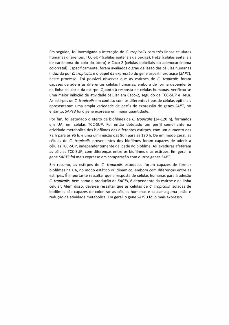
Then, it was investigated the interaction of C. tropicalis with three different human cell lines: TCC-‐SUP (epithelial cells from urinary bladder); HeLa (epithelial cells from cervical carcinoma) and Caco-‐2 (epithelial cells from colorectal adenocarcinoma). Specifically, the degree of human cells damage and activity reduction induced by C. tropicalis adhesion and the role of Candida tropicalis aspartyl proteinases (SAPT) genes expression in this process were assessed. It was possible to observed that C. tropicalis strains were able to adhere to the different human cells, although, in a strain and cell dependent manner. Concerning human cells response to C. tropicalis, the highest cell activity inhibition was obtained for Caco-‐2, followed by TCC-‐SUP and HeLa cells. C. tropicalis strains in contact with the different types of epithelial cells exhibited a wide range of expression profiles of SAPT genes, however, SAPT3 was the gene expressed in a higher level.
Finally, it was studied the behaviour of C. tropicalis in biofilms of different ages (24-‐120 h) formed in artificial urine (AU) and their effect in human urinary bladder cells (TCC-‐SUP). A similar profile in metabolic activity along biofilm age was found among strains, with an increase from 72 to 96 h and a decrease from 96 to 120 h. Candida tropicalis biofilm cells were able to adhere to TCC-‐SUP cells, in general, independently of biofilm age. Yeasts affected TCC-‐SUP cells, with difference among biofilms and strains. Generally, SAPT3 was highly expressed in comparison with other SAPT genes.
In summary, C. tropicalis strains were able to form biofilms in AU, in static or dynamic mode, although, with differences among strains. It is important to emphasize that human cells response to C. tropicalis adhesion, as well as SAPs production, is strain and epithelial cell line dependent. Additionally, it should be highlighted that C. tropicalis cells detached from biofilms are able to colonize human cells and cause some injury and reduction of metabolic activity. Generally, SAPT3 was highly expressed compared to other SAPT genes.
Resumo -‐ Fatores de virulência de Candida tropicalis
Candida tropicalis é uma espécie comummente relacionada com infecções nosocomiais, tais como, candidemia e candidúria. Vários fatores de virulência parecem ser responsáveis por infecções por C. tropicalis, que apresentam elevado potencial de disseminação e mortalidade. A adesão às superfícies (dispositivos médicos e células do hospedeiro) e a formação de biofilmes, são considerados factores importantes que contribuem para o desenvolvimento de candidose. Assim, a colonização do interior de cateteres urinários por C. tropicalis representa um problema crítico. Além disso, adesão e invasão das células hospedeiras por C. tropicalis é considerado o primeiro passo para iniciar infecções sistémicas. Uma vez aderidas ao epitélio, as células de C. tropicalis são capazes de excretar enzimas hidrolíticas que causam danos da membrana de células do hospedeiro. Assim, o objetivo principal deste trabalho foi caracterizar os factores de virulência de C. tropicalis, incluindo a avaliação da adesão às superfícies bióticas e abióticas, formação de biofilme, a expressão de enzimas hidrolíticas e suscetibilidade aos antifúngicos
Assim, a fim de aumentar o conhecimento no processo de adesão de C. tropicalis e consequente formação de biofilme em cateteres urinários, o primeiro objetivo deste trabalho foi desenvolver um modelo dinâmico in vitro, com cateteres urinários de silicone e látex, com urina artificial (UA). Além disso, hidrofobicidade superficial de Candida também foi avaliada, assim como o conteúdo da matriz do biofilme, em termos de proteínas e hidratos de carbono. Assim, este modelo mostrou-‐se adequado para estudos simulando as condições reais do corpo. Além disso, C. tropicalis foi, de facto, capaz de colonizar os cateteres urinários na presença de UA e destacar a partir desses cateteres e mover contra o fluxo imposto, demonstrando sua capacidade de colonizar locais mais distais.
Apesar de ser fundamental desenvolver estudos in vitro para a avaliação da capacidade de adesão de leveduras aos tecidos, a avaliação do número de células de leveduras aderidas por métodos tradicionais é demorada. Assim tornou-‐se necessário desenvolver uma metodologia simples, utilizando uma coloração com violeta cristal para quantificar a adesão in vitro de diferentes espécies de Candida a células epiteliais. O método foi validado para diferentes espécies de Candida e foi feita a comparação com a enumeração por observação ao microscópio. A técnica proposta é de fácil execução e reprodutível, permitindo a determinação da capacidade de adesão das espécies de Candida a uma linha de células epiteliais.
Um outro objetivo do presente trabalho foi a caracterização da virulência de C. tropicalis, através da avaliação da susceptibilidade aos antifúngicos e comparação com a expressão de factores de virulência. Verificou-‐se que as estirpes de C. tropicalis aderiram em número significativamente superior ao epitélio do que ao silicone, foram capazes de formar biofilmes e de manifestar atividade hemolítica total. No entanto, a protease e a fosfolipase foram detectadas apenas em alguns isolados. Todos os isolados foram susceptíveis ao voriconazol, fluconazol e anfotericina B. Quatro estirpes foram susceptíveis dose dependente ao itraconazol e um isolado clínico foi resistente a este agente.
Em seguida, foi investigada a interação de C. tropicalis com três linhas celulares humanas diferentes: TCC-‐SUP (células epiteliais da bexiga); HeLa (células epiteliais de carcinoma do colo do útero) e Caco-‐2 (células epiteliais do adenocarcinoma colorretal). Especificamente, foram avaliados o grau de lesão das células humanas induzida por C. tropicalis e o papel da expressão do gene aspartil protease (SAPT), neste processo. Foi possível observar que as estirpes de C. tropicalis foram capazes de aderir às diferentes células humanas, embora de forma dependente da linha celular e da estirpe. Quanto à resposta de células humanas, verificou-‐se uma maior inibição de atividade celular em Caco-‐2, seguido de TCC-‐SUP e HeLa. As estirpes de C. tropicalis em contato com os diferentes tipos de células epiteliais apresentaram uma ampla variedade de perfis de expressão de genes SAPT, no entanto, SAPT3 foi o gene expresso em maior quantidade.
Por fim, foi estudado o efeito de biofilmes de C. tropicalis (24-‐120 h), formados em UA, em células TCC-‐SUP. Foi então detetado um perfil semelhante na atividade metabólica dos biofilmes das diferentes estirpes, com um aumento das 72 h para as 96 h, e uma diminuição das 96h para as 120 h. De um modo geral, as células de C. tropicalis provenientes dos biofilmes foram capazes de aderir a células TCC-‐SUP, independentemente da idade do biofilme. As leveduras afetaram as células TCC-‐SUP, com diferenças entre os biofilmes e as estirpes. Em geral, o gene SAPT3 foi mais expresso em comparação com outros genes SAPT.
Em resumo, as estirpes de C. tropicalis estudadas foram capazes de formar biofilmes na UA, no modo estático ou dinâmico, embora com diferenças entre as estirpes. É importante ressaltar que a resposta de células humanas para à adesão C. tropicalis, bem como a produção de SAPTs, é dependente da estirpe e da linha celular. Além disso, deve-‐se ressaltar que as células de C. tropicalis isoladas de biofilmes são capazes de colonizar as células humanas e causar alguma lesão e redução da atividade metabólica. Em geral, o gene SAPT3 foi o mais expresso.
Scope and outline of thesis
Usually, Candida tropicalis is considered the first or the second non-‐Candida albicans Candida (NCAC) species most frequently isolated from bloodstream (candidemia) and from urinary tract (candiduria). Additionally, C. tropicalis is often found in patients admitted to Intensive Care Units (ICU), especially in patients with cancer, requiring prolonged catheterization, or/and receiving broad-‐spectrum antibiotics
Several virulence factors seem to be responsible for C. tropicalis infections, which present high potential for dissemination and mortality. Adhesion to abiotic surfaces (medical devices) or to host tissues, as well as biofilm formation, secretion of enzymes (proteases and phospholipases) and haemolytic activity are considered important factors in C. tropicalis mechanisms of infection. Therefore, the need to get more insights in C. tropicalis virulence was the driving force for the research performed.
The present thesis reports the works totally carried out in the Laboratory of Applied Microbiology – Biofilm Group, at IBB -‐ Institute for Biotechnology and Bioengineering, Centre of Biological Engineering, Universidade do Minho, Braga, Portugal.
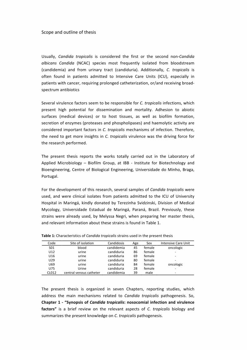
For the development of this research, several samples of Candida tropicalis were used, and were clinical isolates from patients admitted to the ICU of University Hospital in Maringá, kindly donated by Terezinha Svidzinski, Division of Medical Mycology, Universidade Estadual de Maringá, Paraná, Brazil. Previously, these strains were already used, by Melyssa Negri, when preparing her master thesis, and relevant information about these strains is found in Table 1.
Table 1: Characteristics of Candida tropicalis strains used in the present thesis
Code Site of isolation Candidosis Age Sex Intensive Care Unit S01 blood candidemia 45 female oncologic U12 urine candiduria 86 female -‐ U16 urine candiduria 69 female -‐ U29 urine candiduria 80 female -‐ U69 urine candiduria 84 female oncologic U75 Urine candiduria 28 female -‐ CL012 central venous catheter candidemia 39 male -‐
The present thesis is organized in seven Chapters, reporting studies, which address the main mechanisms related to Candida tropicalis pathogenesis. So, Chapter 1 -‐ “Synopsis of Candida tropicalis: nosocomial infection and virulence factors” is a brief review on the relevant aspects of C. tropicalis biology and summarizes the present knowledge on C. tropicalis pathogenesis.
To better understand the ability of C. tropicalis to infect through biofilm formation in urinary catheters, it was necessary to assess how C. tropicalis form biofilms in a system mimicking the real situation. Thus, in Chapter 2 -‐ “Candida tropicalis biofilms: artificial urine, urinary catheters and flow model”, is described a model to study the ability of C. tropicalis to form biofilm using artificial urine (AU) and urinary catheters, under flow conditions.
Since Candida species are able to detach from biofilms and colonize others sites such as host cells, another point addressed in this work was the development of a technique easy to perform and reproducible for the assessment of Candida species ability to adhere to an epithelial cell line, which is described in Chapter 3 -‐ “Crystal violet staining to quantify Candida adhesion to epithelial cells”.
After that optimization step and also contributing to deepen the knowledge on C. tropicalis virulence factors, next Chapter 4 -‐ “Examination of potential virulence factors of Candida tropicalis clinical isolates from hospitalized patients” is focused on the characterization of C. tropicalis virulence by assessing the susceptibility to the most common antifungal agents and comparing the expression of several virulence factors.
The last studies performed under the scope of this thesis were directed to investigate the interaction of C. tropicalis with human cells. In particular, Chapter 5 -‐ “An in vitro evaluation of Candida tropicalis infectivity using human cell monolayers” describes the degree of human cells damage and their activity reduction induced by C. tropicalis adhesion to different human epithelial cell lines and the role of SAPT gene expression in this process. Chapter 6 “Candida tropicalis biofilms: effect on urinary epithelial cells” reports the behaviour of C. tropicalis in biofilms of different ages (24 – 120 h) formed in AU and their effect on human urinary bladder cells (TCC-‐SUP).
Finally, Chapter 7 -‐ “Concluding remarks and future perspectives” highlights the main conclusions, obtained in this thesis, concerning Candida tropicalis and its virulence factors and proposes suggestions for future research that can contribute for enhanced understanding of C. tropicalis pathogenesis.
Publications within the thesis
PAPERS IN PEER REVIEWED JOURNALS
Published
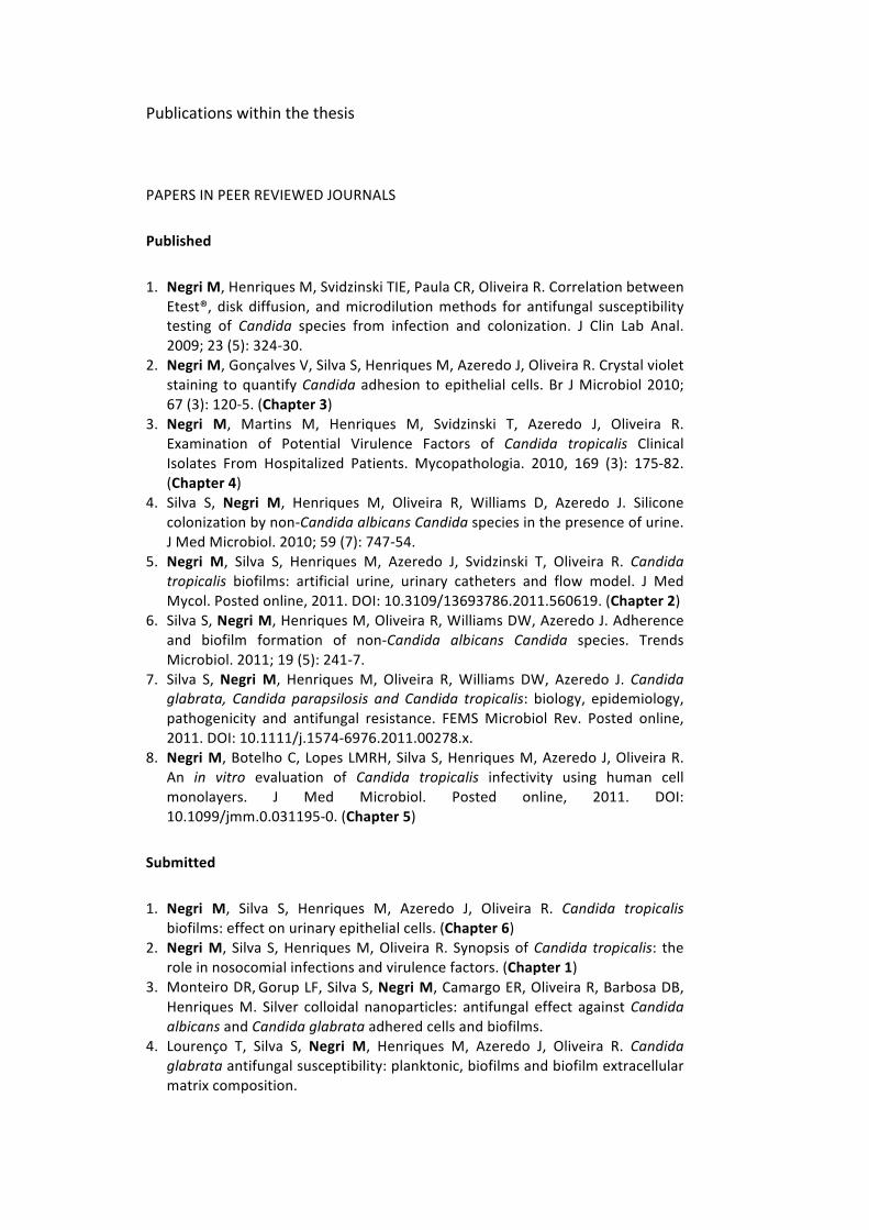
1. Negri M, Henriques M, Svidzinski TIE, Paula CR, Oliveira R. Correlation between Etest®, disk diffusion, and microdilution methods for antifungal susceptibility testing of Candida species from infection and colonization. J Clin Lab Anal. 2009; 23 (5): 324-‐30.
2. Negri M, Gonçalves V, Silva S, Henriques M, Azeredo J, Oliveira R. Crystal violet staining to quantify Candida adhesion to epithelial cells. Br J Microbiol 2010; 67 (3): 120-‐5. (Chapter 3)
3. Negri M, Martins M, Henriques M, Svidzinski T, Azeredo J, Oliveira R. Examination of Potential Virulence Factors of Candida tropicalis Clinical Isolates From Hospitalized Patients. Mycopathologia. 2010, 169 (3): 175-‐82. (Chapter 4)
4. Silva S, Negri M, Henriques M, Oliveira R, Williams D, Azeredo J. Silicone colonization by non-‐Candida albicans Candida species in the presence of urine. J Med Microbiol. 2010; 59 (7): 747-‐54.
5. Negri M, Silva S, Henriques M, Azeredo J, Svidzinski T, Oliveira R. Candida tropicalis biofilms: artificial urine, urinary catheters and flow model. J Med Mycol. Posted online, 2011. DOI: 10.3109/13693786.2011.560619. (Chapter 2)
6. Silva S, Negri M, Henriques M, Oliveira R, Williams DW, Azeredo J. Adherence and biofilm formation of non-‐Candida albicans Candida species. Trends Microbiol. 2011; 19 (5): 241-‐7.
7. Silva S, Negri M, Henriques M, Oliveira R, Williams DW, Azeredo J. Candida glabrata, Candida parapsilosis and Candida tropicalis: biology, epidemiology, pathogenicity and antifungal resistance. FEMS Microbiol Rev. Posted online, 2011. DOI: 10.1111/j.1574-‐6976.2011.00278.x.
8. Negri M, Botelho C, Lopes LMRH, Silva S, Henriques M, Azeredo J, Oliveira R. An in vitro evaluation of Candida tropicalis infectivity using human cell monolayers. J Med Microbiol. Posted online, 2011. DOI: 10.1099/jmm.0.031195-‐0. (Chapter 5)
Submitted
1. Negri M, Silva S, Henriques M, Azeredo J, Oliveira R. Candida tropicalis biofilms: effect on urinary epithelial cells. (Chapter 6)
2. Negri M, Silva S, Henriques M, Oliveira R. Synopsis of Candida tropicalis: the role in nosocomial infections and virulence factors. (Chapter 1)
3. Monteiro DR, Gorup LF, Silva S, Negri M, Camargo ER, Oliveira R, Barbosa DB, Henriques M. Silver colloidal nanoparticles: antifungal effect against Candida albicans and Candida glabrata adhered cells and biofilms.
4. Lourenço T, Silva S, Negri M, Henriques M, Azeredo J, Oliveira R. Candida glabrata antifungal susceptibility: planktonic, biofilms and biofilm extracellular matrix composition.

OTHERS SCIENTIFIC OUTPUT
Book chapter
1. Negri M, Lorenço T, Silva S, Henriques M, Azeredo J, Oliveira R. Effect of antifungal agents on Non-‐Candida albicans Candida species enzymes secretion. 2011, p. 313-‐317. Book title: "Science and Technology against Microbial Pathogens. Research, Development and Evaluation". ISBN-‐13: 978-‐981-‐4354-‐85-‐1
2. Henriques M, Negri M, Silva S. 2011. O impacto clínico de biofilmes de espécies de Candida.
Oral presentation
1. Negri M, Brêda D, Henriques M, Azeredo J, Oliveira R. The role of Candida tropicalis biofilms on human urinary bladder cells. Biofilms in Nosocomial Fungal Infections, Paris, 31 January-‐1 February, 2011. Book of Abstracts. O8, pag. 37.
Posters in conferences
2. Negri MF, Ribeiro A., Svidzinski TI, Henriques M., Oliveira R. Correlation between e-‐test, disk diffusion, and microdilution methods for antifungal susceptibility testing of Candida species. 9th Candida and Candidiasis, New York, March 24-‐28, 2008. Book of Abstracts. B 269. page 126.
3. Chassot F, Negri MF, Svidzinski AE, Donatti L, Peralta RM, Svidzinski TI, Consolaro ME. Can intrauterine contraceptive devices be a Candida albicans reservoir? Proceedings of Biofilms III: 3rd International Conference, Munich, October 5th – 8th, 2008. Book of Abstracts. P 113. page 77.
4. Negri MF, Henriques M., Svidzinski TI, Oliveira RO. Adhesion of Candida tropicalis clinical isolates to human epithelial cell and silicone. IX IFIC Congress VII Panamerican Congress and XIV Chilean Congress of Nosocomial Infections. Santiago, October 7th -‐ 10th, 2008. Book of Abstracts.
5. Negri M, Henriques M, Svidzinski TIE, Azeredo J, Oliveira R. Virulence factor of Candida tropicalis isolated from hospitalized patients. 3nd FEBS Advanced Lecture Course Human Fungal Pathogens: Molecular Mechanisms of Host Pathogen Interactions and Virulence, La Colle sur Loup, May 2-‐8, 2009. Book of Abstracts. P139A. page 189.
6. Negri M, Lopes LMRH, Henriques M, Svidzinski TI, Azeredo J, Oliveira R. Effect of Candida tropicalis in planktonic and biofilm form on urinary epithelial cells. 4th Trends in Medical Mycology, Athens, Greece, October 18-‐21, 2009. Mycoses 78: (Suppl.).
Publications within the thesis
7. Negri M, Henriques M, Lopes MRH, Svidzinski TIE, Azeredo J, Oliveira R. Effect of Candida tropicalis biofilm on urinary epithelial cells. 5th ASM Conference on Biofilms, Cancun, Mexico, November 15 – 19, 2009. Book of Abstracts. A 118. page 79.
8. Silva S, Negri M, Henriques M, Oliveira R, Williams D, Azeredo J. Adhesion and biofilm formation of non -‐Candida albicans Candida species on silicone in the presence of urine. 5th ASM Conference on Biofilms, Cancun, Mexico, November 15 – 19, 2009. Book of Abstracts. C 135. page 86.
9. Negri, M; Henriques, M; Svidzinski, T; Azeredo, J; Oliveira, R. Candida tropicalis biofilm on latex and silicone catheters. Eurobiofilms 2009: Book of Abstracts 2009, 105-‐106.
10. Negri M, Botelho C, Silva S, Henriques M, Azeredo J, Oliveira R. Candida tropicalis biofilms on catheters: formation and effect on urinary epithelial cells. 10th ASM Conference on Candida and Candidiasis, Miami, March 22nd – 26th, 2010. Book of Abstracts. 254B. page 155.
11. Botelho C, Negri M, Silva S, Henriques M, Azeredo J, Oliveira R. Adhesion of non-‐Candida albicans Candida spp to urinary epithelial cells. 10th ASM Conference on Candida and Candidiasis, Miami, March 22nd – 26th, 2010. Book of Abstracts. 174B. page 125.
12. Negri M, Botelho C, Silva S, Henriques M, Svidzinski T, Azeredo J, Oliveira R. Candida tropicalis biofilms: formation and virulence factors. Biofilms 4 International. Conference, Winchester, September 2 – 3, 2010. Book of Abstracts. 28. page 37.
13. Negri M, Lorenço T, Silva S, Henriques M, Azeredo J, Oliveira R. Effect of antifungal agents on Non-‐Candida albicans Candida species enzymes secretion. International Conference on Antimicrobial Research ICAR, Valladolid, November 3rd – 5th, 2010. Book of Abstracts. F 84. page 392.
14. Silva S, Lorenço T, Negri M, Henriques M, Azeredo J, Oliveira R. The role of antifungals agents on Candida glabrata biofilms matrix composition. International Conference on Antimicrobial Research ICAR, Valladolid, November 3rd – 5th, 2010. Book of Abstracts. W 68. page 292.
15. Freitas AR, Baeza LC, Dota KFD, Negri M, Svidzinski TIE. Yeast from urinary nosocomial infection: biofilm and susceptibility to antifungal profile. International Conference on Antimicrobial Research ICAR, Valladolid, November 3rd – 5th, 2010. Book of Abstracts. W 70.
16. Negri M, Brêda D, Henriques M, Azeredo J, Oliveira R. The role of Candida tropicalis biofilms on human urinary bladder cells. Biofilms in Nosocomial Fungal Infections, Paris, 31 January-‐1 February, 2011. Book of Abstracts. O8, pag. 37.
17. Monteiro DR, Silva SC, Negri M, Camargo ER, Oliveira R, Henriques M, Barbosa DB. Effect of silver nanoparticles against Candida albicans and Candida glabrata biofilms. Biofilms in Nosocomial Fungal Infections, Paris, 31 January-‐1 February, 2011. Book of Abstracts. page 91.


Table of contents
Acknowlegments . . . . . . . . . . . . . . . . . . . . . . . . . . . . . . . . . . . . . . . . . . . . . . . . . . . . . . . . . . . . . . . . . . . . . . . . . . . . . . . . . . . . . . . . . . . . . . . . . . . . . . iv Abstract . . . . . . . . . . . . . . . . . . . . . . . . . . . . . . . . . . . . . . . . . . . . . . . . . . . . . . . . . . . . . . . . . . . . . . . . . . . . . . . . . . . . . . . . . . . . . . . . . . . . . . . . . . . . . . . . . . . . vi Resumo . . . . . . . . . . . . . . . . . . . . . . . . . . . . . . . . . . . . . . . . . . . . . . . . . . . . . . . . . . . . . . . . . . . . . . . . . . . . . . . . . . . . . . . . . . . . . . . . . . . . . . . . . . . . . . . . . . viii Scope and outline of the thesis . . . . . . . . . . . . . . . . . . . . . . . . . . . . . . . . . . . . . . . . . . . . . . . . . . . . . . . . . . . . . . . . . . . . . . . . . . . . . . . . . x Publications within the thesis . . . . . . . . . . . . . . . . . . . . . . . . . . . . . . . . . . . . . . . . . . . . . . . . . . . . . . . . . . . . . . . . . . . . . . . . . . . . . . . . . xii Abbreviations and acronyms . . . . . . . . . . . . . . . . . . . . . . . . . . . . . . . . . . . . . . . . . . . . . . . . . . . . . . . . . . . . . . . . . . . . . . . . . . . . . . . . . . xx List of figures . . . . . . . . . . . . . . . . . . . . . . . . . . . . . . . . . . . . . . . . . . . . . . . . . . . . . . . . . . . . . . . . . . . . . . . . . . . . . . . . . . . . . . . . . . . . . . . . . . . . . . . . . . xxii List of tables . . . . . . . . . . . . . . . . . . . . . . . . . . . . . . . . . . . . . . . . . . . . . . . . . . . . . . . . . . . . . . . . . . . . . . . . . . . . . . . . . . . . . . . . . . . . . . . . . . . . . . . . . . xxiv Chapter 1 Synopsis of Candida tropicalis: the role in nosocomial infections and virulence factors . . . . . . . . . . . . . . . . . . . . . . . . . . . . . . . . . . . . . . . . . 27 Abstract . . . . . . . . . . . . . . . . . . . . . . . . . . . . . . . . . . . . . . . . . . . . . . . . . . . . . . . . . . . . . . . . . . . . . . . . . . . . . . . . . . . . . . . . . . . . . . . . . . . . . . . . . . . . . . . . . . . 29 Introduction . . . . . . . . . . . . . . . . . . . . . . . . . . . . . . . . . . . . . . . . . . . . . . . . . . . . . . . . . . . . . . . . . . . . . . . . . . . . . . . . . . . . . . . . . . . . . . . . . . . . . . . . . . . . 30 Microbiology . . . . . . . . . . . . . . . . . . . . . . . . . . . . . . . . . . . . . . . . . . . . . . . . . . . . . . . . . . . . . . . . . . . . . . . . . . . . . . . . . . . . . . . . . . . . . . . . . . . . . . . . . . . 31 Identification . . . . . . . . . . . . . . . . . . . . . . . . . . . . . . . . . . . . . . . . . . . . . . . . . . . . . . . . . . . . . . . . . . . . . . . . . . . . . . . . . . . . . . . . . . . . . . . . . . . . . . . . . . . 32 Risk factors . . . . . . . . . . . . . . . . . . . . . . . . . . . . . . . . . . . . . . . . . . . . . . . . . . . . . . . . . . . . . . . . . . . . . . . . . . . . . . . . . . . . . . . . . . . . . . . . . . . . . . . . . . . . . . 35 Epidemiology . . . . . . . . . . . . . . . . . . . . . . . . . . . . . . . . . . . . . . . . . . . . . . . . . . . . . . . . . . . . . . . . . . . . . . . . . . . . . . . . . . . . . . . . . . . . . . . . . . . . . . . . . . . 36 Virulence factors . . . . . . . . . . . . . . . . . . . . . . . . . . . . . . . . . . . . . . . . . . . . . . . . . . . . . . . . . . . . . . . . . . . . . . . . . . . . . . . . . . . . . . . . . . . . . . . . . . . . . 39
Adhesion and biofilm formation . . . . . . . . . . . . . . . . . . . . . . . . . . . . . . . . . . . . . . . . . . . . . . . . . . . . . . . . . . . . . . . . . . . . . . . 39 Infect and dissemination . . . . . . . . . . . . . . . . . . . . . . . . . . . . . . . . . . . . . . . . . . . . . . . . . . . . . . . . . . . . . . . . . . . . . . . . . . . . . . . . . . 43 Enzymes production . . . . . . . . . . . . . . . . . . . . . . . . . . . . . . . . . . . . . . . . . . . . . . . . . . . . . . . . . . . . . . . . . . . . . . . . . . . . . . . . . . . . . . . . . 45
Concluding remarks . . . . . . . . . . . . . . . . . . . . . . . . . . . . . . . . . . . . . . . . . . . . . . . . . . . . . . . . . . . . . . . . . . . . . . . . . . . . . . . . . . . . . . . . . . . . . . . . 47 References . . . . . . . . . . . . . . . . . . . . . . . . . . . . . . . . . . . . . . . . . . . . . . . . . . . . . . . . . . . . . . . . . . . . . . . . . . . . . . . . . . . . . . . . . . . . . . . . . . . . . . . . . . . . . . 48 Chapter 2 Candida tropicalis biofilms: artificial urine, urinary catheters and flow model . . . . . . . . . . . . . . . . . . . . . . . . . . . . . . . . . . . . . . . . . . . . . . . . . . 53 Abstract . . . . . . . . . . . . . . . . . . . . . . . . . . . . . . . . . . . . . . . . . . . . . . . . . . . . . . . . . . . . . . . . . . . . . . . . . . . . . . . . . . . . . . . . . . . . . . . . . . . . . . . . . . . . . . . . . . . 55 Introduction . . . . . . . . . . . . . . . . . . . . . . . . . . . . . . . . . . . . . . . . . . . . . . . . . . . . . . . . . . . . . . . . . . . . . . . . . . . . . . . . . . . . . . . . . . . . . . . . . . . . . . . . . . . . 56 Materials and methods . . . . . . . . . . . . . . . . . . . . . . . . . . . . . . . . . . . . . . . . . . . . . . . . . . . . . . . . . . . . . . . . . . . . . . . . . . . . . . . . . . . . . . . . . . . 57
Organisms and growth conditions . . . . . . . . . . . . . . . . . . . . . . . . . . . . . . . . . . . . . . . . . . . . . . . . . . . . . . . . . . . . . . . . . . . . 57 Candida tropicalis biofilm formation flow model . . . . . . . . . . . . . . . . . . . . . . . . . . . . . . . . . . . . . . . . . . . . . 58 Candida cells quantification . . . . . . . . . . . . . . . . . . . . . . . . . . . . . . . . . . . . . . . . . . . . . . . . . . . . . . . . . . . . . . . . . . . . . . . . . . . . . . 59
Adhesion or biofilm samples . . . . . . . . . . . . . . . . . . . . . . . . . . . . . . . . . . . . . . . . . . . . . . . . . . . . . . . . . . . . . . . . . . . . . . 59 Biofilm detached cells . . . . . . . . . . . . . . . . . . . . . . . . . . . . . . . . . . . . . . . . . . . . . . . . . . . . . . . . . . . . . . . . . . . . . . . . . . . . . . . . . 59 Biofilm biomass . . . . . . . . . . . . . . . . . . . . . . . . . . . . . . . . . . . . . . . . . . . . . . . . . . . . . . . . . . . . . . . . . . . . . . . . . . . . . . . . . . . . . . . . . . . 59
Scanning Electron Microscopy (SEM) . . . . . . . . . . . . . . . . . . . . . . . . . . . . . . . . . . . . . . . . . . . . . . . . . . . . . . . . . . . . . . . 60 Biofilm matrix composition . . . . . . . . . . . . . . . . . . . . . . . . . . . . . . . . . . . . . . . . . . . . . . . . . . . . . . . . . . . . . . . . . . . . . . . . . . . . . . . 60
Extraction method . . . . . . . . . . . . . . . . . . . . . . . . . . . . . . . . . . . . . . . . . . . . . . . . . . . . . . . . . . . . . . . . . . . . . . . . . . . . . . . . . . . . . . 60 Protein and carbohydrate quantification . . . . . . . . . . . . . . . . . . . . . . . . . . . . . . . . . . . . . . . . . . . . . . . . . . . 61
Candida surface properties . . . . . . . . . . . . . . . . . . . . . . . . . . . . . . . . . . . . . . . . . . . . . . . . . . . . . . . . . . . . . . . . . . . . . . . . . . . . . . . 61 Statistical analysis . . . . . . . . . . . . . . . . . . . . . . . . . . . . . . . . . . . . . . . . . . . . . . . . . . . . . . . . . . . . . . . . . . . . . . . . . . . . . . . . . . . . . . . . . . . . . 61

Table of contents
Results . . . . . . . . . . . . . . . . . . . . . . . . . . . . . . . . . . . . . . . . . . . . . . . . . . . . . . . . . . . . . . . . . . . . . . . . . . . . . . . . . . . . . . . . . . . . . . . . . . . . . . . . . . . . . . . . . . . . . 62 Candida tropicalis adhesion and surface properties . . . . . . . . . . . . . . . . . . . . . . . . . . . . . . . . . . . . . . . . 62 Biofilm development under flow conditions . . . . . . . . . . . . . . . . . . . . . . . . . . . . . . . . . . . . . . . . . . . . . . . . . . . . 64 Biofilm cells detachment . . . . . . . . . . . . . . . . . . . . . . . . . . . . . . . . . . . . . . . . . . . . . . . . . . . . . . . . . . . . . . . . . . . . . . . . . . . . . . . . . . 65
Discussion . . . . . . . . . . . . . . . . . . . . . . . . . . . . . . . . . . . . . . . . . . . . . . . . . . . . . . . . . . . . . . . . . . . . . . . . . . . . . . . . . . . . . . . . . . . . . . . . . . . . . . . . . . . . . . . 66 References . . . . . . . . . . . . . . . . . . . . . . . . . . . . . . . . . . . . . . . . . . . . . . . . . . . . . . . . . . . . . . . . . . . . . . . . . . . . . . . . . . . . . . . . . . . . . . . . . . . . . . . . . . . . . . 71 Chapter 3 Crystal violet staining to quantify Candida adhesion to epithelial cells . . . . . . . . . . . . . . . . . 73 Abstract . . . . . . . . . . . . . . . . . . . . . . . . . . . . . . . . . . . . . . . . . . . . . . . . . . . . . . . . . . . . . . . . . . . . . . . . . . . . . . . . . . . . . . . . . . . . . . . . . . . . . . . . . . . . . . . . . . . 75 Introduction . . . . . . . . . . . . . . . . . . . . . . . . . . . . . . . . . . . . . . . . . . . . . . . . . . . . . . . . . . . . . . . . . . . . . . . . . . . . . . . . . . . . . . . . . . . . . . . . . . . . . . . . . . . . 76 Materials and methods . . . . . . . . . . . . . . . . . . . . . . . . . . . . . . . . . . . . . . . . . . . . . . . . . . . . . . . . . . . . . . . . . . . . . . . . . . . . . . . . . . . . . . . . . . . 77
Yeasts and growth conditions . . . . . . . . . . . . . . . . . . . . . . . . . . . . . . . . . . . . . . . . . . . . . . . . . . . . . . . . . . . . . . . . . . . . . . . . . . . 77 Epithelial cells . . . . . . . . . . . . . . . . . . . . . . . . . . . . . . . . . . . . . . . . . . . . . . . . . . . . . . . . . . . . . . . . . . . . . . . . . . . . . . . . . . . . . . . . . . . . . . . . . . . 77 Silicone . . . . . . . . . . . . . . . . . . . . . . . . . . . . . . . . . . . . . . . . . . . . . . . . . . . . . . . . . . . . . . . . . . . . . . . . . . . . . . . . . . . . . . . . . . . . . . . . . . . . . . . . . . . . . . 78 Adhesion assay . . . . . . . . . . . . . . . . . . . . . . . . . . . . . . . . . . . . . . . . . . . . . . . . . . . . . . . . . . . . . . . . . . . . . . . . . . . . . . . . . . . . . . . . . . . . . . . . . 78 Crystal violet assay . . . . . . . . . . . . . . . . . . . . . . . . . . . . . . . . . . . . . . . . . . . . . . . . . . . . . . . . . . . . . . . . . . . . . . . . . . . . . . . . . . . . . . . . . . . 78 Microscopy observation . . . . . . . . . . . . . . . . . . . . . . . . . . . . . . . . . . . . . . . . . . . . . . . . . . . . . . . . . . . . . . . . . . . . . . . . . . . . . . . . . . . 79 Statistical analysis . . . . . . . . . . . . . . . . . . . . . . . . . . . . . . . . . . . . . . . . . . . . . . . . . . . . . . . . . . . . . . . . . . . . . . . . . . . . . . . . . . . . . . . . . . . . . 79
Results . . . . . . . . . . . . . . . . . . . . . . . . . . . . . . . . . . . . . . . . . . . . . . . . . . . . . . . . . . . . . . . . . . . . . . . . . . . . . . . . . . . . . . . . . . . . . . . . . . . . . . . . . . . . . . . . . . . . . 80 Discussion . . . . . . . . . . . . . . . . . . . . . . . . . . . . . . . . . . . . . . . . . . . . . . . . . . . . . . . . . . . . . . . . . . . . . . . . . . . . . . . . . . . . . . . . . . . . . . . . . . . . . . . . . . . . . . . 83 References . . . . . . . . . . . . . . . . . . . . . . . . . . . . . . . . . . . . . . . . . . . . . . . . . . . . . . . . . . . . . . . . . . . . . . . . . . . . . . . . . . . . . . . . . . . . . . . . . . . . . . . . . . . . . . 86 Chapter 4 Examination of potential virulence factors of Candida tropicalis clinical isolates from hospitalized patients . . . . . . . . . . . . . . . . . . . . . . . . . . . . . . . 89 Abstract . . . . . . . . . . . . . . . . . . . . . . . . . . . . . . . . . . . . . . . . . . . . . . . . . . . . . . . . . . . . . . . . . . . . . . . . . . . . . . . . . . . . . . . . . . . . . . . . . . . . . . . . . . . . . . . . . . . 91 Introduction . . . . . . . . . . . . . . . . . . . . . . . . . . . . . . . . . . . . . . . . . . . . . . . . . . . . . . . . . . . . . . . . . . . . . . . . . . . . . . . . . . . . . . . . . . . . . . . . . . . . . . . . . . . . 92 Materials and methods . . . . . . . . . . . . . . . . . . . . . . . . . . . . . . . . . . . . . . . . . . . . . . . . . . . . . . . . . . . . . . . . . . . . . . . . . . . . . . . . . . . . . . . . . . . 93
Isolates . . . . . . . . . . . . . . . . . . . . . . . . . . . . . . . . . . . . . . . . . . . . . . . . . . . . . . . . . . . . . . . . . . . . . . . . . . . . . . . . . . . . . . . . . . . . . . . . . . . . . . . . . . . . . . 93 Isolation and identification . . . . . . . . . . . . . . . . . . . . . . . . . . . . . . . . . . . . . . . . . . . . . . . . . . . . . . . . . . . . . . . . . . . . . . . . . . . . . . . 93 Adhesion and biofilm formation . . . . . . . . . . . . . . . . . . . . . . . . . . . . . . . . . . . . . . . . . . . . . . . . . . . . . . . . . . . . . . . . . . . . . . . 94 Quantification of Adhered Yeast Cells . . . . . . . . . . . . . . . . . . . . . . . . . . . . . . . . . . . . . . . . . . . . . . . . . . . . . . . . . . . . . . 94 Epithelial cells . . . . . . . . . . . . . . . . . . . . . . . . . . . . . . . . . . . . . . . . . . . . . . . . . . . . . . . . . . . . . . . . . . . . . . . . . . . . . . . . . . . . . . . . . . . . . . . . . . . 94 Silicone . . . . . . . . . . . . . . . . . . . . . . . . . . . . . . . . . . . . . . . . . . . . . . . . . . . . . . . . . . . . . . . . . . . . . . . . . . . . . . . . . . . . . . . . . . . . . . . . . . . . . . . . . . . . . . 95 Biofilm Biomass Quantification . . . . . . . . . . . . . . . . . . . . . . . . . . . . . . . . . . . . . . . . . . . . . . . . . . . . . . . . . . . . . . . . . . . . . . . . 95 Pseudohyphae Formation . . . . . . . . . . . . . . . . . . . . . . . . . . . . . . . . . . . . . . . . . . . . . . . . . . . . . . . . . . . . . . . . . . . . . . . . . . . . . . . . . 95 Proteinase and Phospholipase Secretion . . . . . . . . . . . . . . . . . . . . . . . . . . . . . . . . . . . . . . . . . . . . . . . . . . . . . . . . . 96 Haemolytic Activity . . . . . . . . . . . . . . . . . . . . . . . . . . . . . . . . . . . . . . . . . . . . . . . . . . . . . . . . . . . . . . . . . . . . . . . . . . . . . . . . . . . . . . . . . . . 96 Antifungal Susceptibility Test Methods . . . . . . . . . . . . . . . . . . . . . . . . . . . . . . . . . . . . . . . . . . . . . . . . . . . . . . . . . . . . 96 Statistical analysis . . . . . . . . . . . . . . . . . . . . . . . . . . . . . . . . . . . . . . . . . . . . . . . . . . . . . . . . . . . . . . . . . . . . . . . . . . . . . . . . . . . . . . . . . . . . 97
Results . . . . . . . . . . . . . . . . . . . . . . . . . . . . . . . . . . . . . . . . . . . . . . . . . . . . . . . . . . . . . . . . . . . . . . . . . . . . . . . . . . . . . . . . . . . . . . . . . . . . . . . . . . . . . . . . . . . . . 97 Discussion . . . . . . . . . . . . . . . . . . . . . . . . . . . . . . . . . . . . . . . . . . . . . . . . . . . . . . . . . . . . . . . . . . . . . . . . . . . . . . . . . . . . . . . . . . . . . . . . . . . . . . . . . . . . . 100 References . . . . . . . . . . . . . . . . . . . . . . . . . . . . . . . . . . . . . . . . . . . . . . . . . . . . . . . . . . . . . . . . . . . . . . . . . . . . . . . . . . . . . . . . . . . . . . . . . . . . . . . . . . . . 104

Table of contents
Chapter 5 An in vitro evaluation of Candida tropicalis infectivity using human cell monolayers . . . . . . . . . . . . . . . . . . . . . . . . . . . . . . . . . . . . . . . . . . . . . . . . . . . . . . . . . . . . . . . 107 Abstract . . . . . . . . . . . . . . . . . . . . . . . . . . . . . . . . . . . . . . . . . . . . . . . . . . . . . . . . . . . . . . . . . . . . . . . . . . . . . . . . . . . . . . . . . . . . . . . . . . . . . . . . . . . . . . . . . 109 Introduction . . . . . . . . . . . . . . . . . . . . . . . . . . . . . . . . . . . . . . . . . . . . . . . . . . . . . . . . . . . . . . . . . . . . . . . . . . . . . . . . . . . . . . . . . . . . . . . . . . . . . . . . . . 110 Materials and methods . . . . . . . . . . . . . . . . . . . . . . . . . . . . . . . . . . . . . . . . . . . . . . . . . . . . . . . . . . . . . . . . . . . . . . . . . . . . . . . . . . . . . . . . . 110
Yeast and growth conditions . . . . . . . . . . . . . . . . . . . . . . . . . . . . . . . . . . . . . . . . . . . . . . . . . . . . . . . . . . . . . . . . . . . . . . . . . . 110 Human epithelial cell lines . . . . . . . . . . . . . . . . . . . . . . . . . . . . . . . . . . . . . . . . . . . . . . . . . . . . . . . . . . . . . . . . . . . . . . . . . . . . . . 111 Adhesion assay . . . . . . . . . . . . . . . . . . . . . . . . . . . . . . . . . . . . . . . . . . . . . . . . . . . . . . . . . . . . . . . . . . . . . . . . . . . . . . . . . . . . . . . . . . . . . . . . 111
Candida tropicalis quantification . . . . . . . . . . . . . . . . . . . . . . . . . . . . . . . . . . . . . . . . . . . . . . . . . . . . . . . . . . . . . . 112 Determination of percentage of inhibition of cell activity . . . . . . . . . . . . . . . . . . . . . . . 112 Epithelial cells damage assay . . . . . . . . . . . . . . . . . . . . . . . . . . . . . . . . . . . . . . . . . . . . . . . . . . . . . . . . . . . . . . . . . . . . 113
Analysis of SAP gene expression . . . . . . . . . . . . . . . . . . . . . . . . . . . . . . . . . . . . . . . . . . . . . . . . . . . . . . . . . . . . . . . . . . . . . 113 RNA extraction . . . . . . . . . . . . . . . . . . . . . . . . . . . . . . . . . . . . . . . . . . . . . . . . . . . . . . . . . . . . . . . . . . . . . . . . . . . . . . . . . . . . . . . . . . 113 Primers . . . . . . . . . . . . . . . . . . . . . . . . . . . . . . . . . . . . . . . . . . . . . . . . . . . . . . . . . . . . . . . . . . . . . . . . . . . . . . . . . . . . . . . . . . . . . . . . . . . . . . 114 Synthesis of cDNA . . . . . . . . . . . . . . . . . . . . . . . . . . . . . . . . . . . . . . . . . . . . . . . . . . . . . . . . . . . . . . . . . . . . . . . . . . . . . . . . . . . . . 114 Real-‐time PCR . . . . . . . . . . . . . . . . . . . . . . . . . . . . . . . . . . . . . . . . . . . . . . . . . . . . . . . . . . . . . . . . . . . . . . . . . . . . . . . . . . . . . . . . . . . 114
Statistical analysis . . . . . . . . . . . . . . . . . . . . . . . . . . . . . . . . . . . . . . . . . . . . . . . . . . . . . . . . . . . . . . . . . . . . . . . . . . . . . . . . . . . . . . . . . . 115 Results . . . . . . . . . . . . . . . . . . . . . . . . . . . . . . . . . . . . . . . . . . . . . . . . . . . . . . . . . . . . . . . . . . . . . . . . . . . . . . . . . . . . . . . . . . . . . . . . . . . . . . . . . . . . . . . . . . . 115 Discussion . . . . . . . . . . . . . . . . . . . . . . . . . . . . . . . . . . . . . . . . . . . . . . . . . . . . . . . . . . . . . . . . . . . . . . . . . . . . . . . . . . . . . . . . . . . . . . . . . . . . . . . . . . . . . . 117 References . . . . . . . . . . . . . . . . . . . . . . . . . . . . . . . . . . . . . . . . . . . . . . . . . . . . . . . . . . . . . . . . . . . . . . . . . . . . . . . . . . . . . . . . . . . . . . . . . . . . . . . . . . . . 122 Chapter 6 Candida tropicalis biofilms: effect on urinary epithelial cells . . . . . . . . . . . . . . . . . . . . . . . . . . . . . . 125 Abstract . . . . . . . . . . . . . . . . . . . . . . . . . . . . . . . . . . . . . . . . . . . . . . . . . . . . . . . . . . . . . . . . . . . . . . . . . . . . . . . . . . . . . . . . . . . . . . . . . . . . . . . . . . . . . . . . . 127 Introduction . . . . . . . . . . . . . . . . . . . . . . . . . . . . . . . . . . . . . . . . . . . . . . . . . . . . . . . . . . . . . . . . . . . . . . . . . . . . . . . . . . . . . . . . . . . . . . . . . . . . . . . . . . 128 Materials and methods . . . . . . . . . . . . . . . . . . . . . . . . . . . . . . . . . . . . . . . . . . . . . . . . . . . . . . . . . . . . . . . . . . . . . . . . . . . . . . . . . . . . . . . . . 129
Candida tropicalis and Growth Conditions . . . . . . . . . . . . . . . . . . . . . . . . . . . . . . . . . . . . . . . . . . . . . . . . . . . . . 129 Candida tropicalis biofilms formation . . . . . . . . . . . . . . . . . . . . . . . . . . . . . . . . . . . . . . . . . . . . . . . . . . . . . . . . . . . . . 129 Candida tropicalis biofilm characterization . . . . . . . . . . . . . . . . . . . . . . . . . . . . . . . . . . . . . . . . . . . . . . . . . . . . 130
Number of cultivable yeasts . . . . . . . . . . . . . . . . . . . . . . . . . . . . . . . . . . . . . . . . . . . . . . . . . . . . . . . . . . . . . . . . . . . . . 130 Biofilm biomass quantification by crystal violet staining . . . . . . . . . . . . . . . . . . . . . . . . . 130 In situ biofilm metabolic activity . . . . . . . . . . . . . . . . . . . . . . . . . . . . . . . . . . . . . . . . . . . . . . . . . . . . . . . . . . . . . . . 131
Human urinary bladder epitelial cell line . . . . . . . . . . . . . . . . . . . . . . . . . . . . . . . . . . . . . . . . . . . . . . . . . . . . . . . . 131 Candida tropicalis biofilms in contact with TCC-‐SUP cells . . . . . . . . . . . . . . . . . . . . . . . . . . . . . . 131 Effect of Candida tropicalis on TCC-‐SUP cells . . . . . . . . . . . . . . . . . . . . . . . . . . . . . . . . . . . . . . . . . . . . . . . . . 132
Determination of epithelial cells damage and activity assay . . . . . . . . . . . . . . . . . . . 132 Analysis of SAP gene expression . . . . . . . . . . . . . . . . . . . . . . . . . . . . . . . . . . . . . . . . . . . . . . . . . . . . . . . . . . . . . . . . . . . . . 132
RNA extraction . . . . . . . . . . . . . . . . . . . . . . . . . . . . . . . . . . . . . . . . . . . . . . . . . . . . . . . . . . . . . . . . . . . . . . . . . . . . . . . . . . . . . . . . . . 133 Primer, synthesis of cDNA and real-‐time PCR . . . . . . . . . . . . . . . . . . . . . . . . . . . . . . . . . . . . . . . . . . 133
Statistical analysis . . . . . . . . . . . . . . . . . . . . . . . . . . . . . . . . . . . . . . . . . . . . . . . . . . . . . . . . . . . . . . . . . . . . . . . . . . . . . . . . . . . . . . . . . . . 133
Table of contents
Results . . . . . . . . . . . . . . . . . . . . . . . . . . . . . . . . . . . . . . . . . . . . . . . . . . . . . . . . . . . . . . . . . . . . . . . . . . . . . . . . . . . . . . . . . . . . . . . . . . . . . . . . . . . . . . . . . . . 133 Candida tropicalis biofilms characterization . . . . . . . . . . . . . . . . . . . . . . . . . . . . . . . . . . . . . . . . . . . . . . . . . . 133
Number of cultivable yeasts . . . . . . . . . . . . . . . . . . . . . . . . . . . . . . . . . . . . . . . . . . . . . . . . . . . . . . . . . . . . . . . . . . . . . 133 Biofilm biomass quantification . . . . . . . . . . . . . . . . . . . . . . . . . . . . . . . . . . . . . . . . . . . . . . . . . . . . . . . . . . . . . . . . . 134 In situ biofilm metabolic activity . . . . . . . . . . . . . . . . . . . . . . . . . . . . . . . . . . . . . . . . . . . . . . . . . . . . . . . . . . . . . . 134
Candida tropicalis biofilms in contact with TCC-‐SUP cells . . . . . . . . . . . . . . . . . . . . . . . . . . . . . . 136 Number of adhered yeasts to epithelial cells . . . . . . . . . . . . . . . . . . . . . . . . . . . . . . . . . . . . . . . . . . . 136
Effect of Candida tropicalis on TCC-‐SUP cells . . . . . . . . . . . . . . . . . . . . . . . . . . . . . . . . . . . . . . . . . . . . . . . . . 136 Determination of epithelial cells activity and damage . . . . . . . . . . . . . . . . . . . . . . . . . . . . . 136
Candida tropicalis SAP gene expression . . . . . . . . . . . . . . . . . . . . . . . . . . . . . . . . . . . . . . . . . . . . . . . . . . . . . . . . . 137 Discussion . . . . . . . . . . . . . . . . . . . . . . . . . . . . . . . . . . . . . . . . . . . . . . . . . . . . . . . . . . . . . . . . . . . . . . . . . . . . . . . . . . . . . . . . . . . . . . . . . . . . . . . . . . . . . 139 References . . . . . . . . . . . . . . . . . . . . . . . . . . . . . . . . . . . . . . . . . . . . . . . . . . . . . . . . . . . . . . . . . . . . . . . . . . . . . . . . . . . . . . . . . . . . . . . . . . . . . . . . . . . . 145 Chapter 7 Concluding remarks and future perspectives . . . . . . . . . . . . . . . . . . . . . . . . . . . . . . . . . . . . . . . . . . . . . . . . . . . . . . . 147

Abbreviation and acronyms
xx
P: Significance value
g: Gravity
Ɵ: Water contact angle (ᴏ)
Ɣ+: Electron acceptor surface tension parameter (mJ/m2)
Ɣ-‐: Electron donor surface tension parameter (mJ/m2)
ΔGsws: Total free energy variation between entities of a given surface(s) immersed in water (w) (mJ/m2)
%: Percent
ΔCT: Threshold cycle
°C: Degrees Celsius
h: Hour
l: Liter
ml: Milliliter
nm: nanometer
µg: Microgram
µm: Micrometer
µM: Micromolar
λ: wavelength
Abs: Absorbance
ALS: Agglutinin like sequence gene
Als: Agglutinin like sequence protein
ANOVA-‐Analysis of variance
ATCC: American Type Culture Collection
AU: Artificial urine
BSA: Bovine Serum Albumin
CAPES: Coordenação de Aperfeiçoamento de Pessoal de Nível Superior
cDNA: complementary Deoxyribonucleic Acid
CFU: Colony Forming Units
CHROMagar-‐Chromogenic media agar
CLSM: Confocal Laser Scanning Microscopy
CV: Crystal violet
DNA: Deoxyribonucleic Acid

xxi
FCT: Fundação para a Ciência e Tecnologia
LDH: Lactate Dehydrogenase
Log: Logarithm
min: Minute
mRNA: messenger Ribonucleic Acid
MTS: ([3-‐(4,5-‐dimethylthiazol-‐2-‐yl)-‐5-‐(3-‐carboxymethoxyphenyl)-‐2-‐(4-‐sulfophenyl)-‐2H–tetrazolium]
NCAC: non-‐Candida albicans Candida
ND-‐No detected
PBS: Phosphate Buffer Saline
PCR: Polymerase Chain Reaction
PLs: Phospholipases
RHOE: Reconstituted Human Oral Epithelium
RNA: Ribonucleic Acid
Rpm: rotation per minute
rRNA: ribosomal Ribonucleic Acid
SAP: Secreted aspartly proteinase gene
Sap: Secreted aspartly proteinase protein
SDA: Sabouraud dextrose agar
SDB: Sabouraud dextrose broth
SD: Standard deviation
SEM: Scanning Electron Microscopy
SPSS: Statistical package for the social sciences
UA: Urina artificial
UTIs: Urinary Tract Infections
U: Units
v: Volume
V: Voltage
w: Weight
XTT: 2, 3 bis(2-‐methoxy-‐4-‐nitro-‐5-‐sulfophenyl)-‐5-‐[(phenylamino)carbonyl]2-‐Htetrazolium hydroxide

List of figures
xxii
Figure 1.1 C. tropicalis morphology in routinely culture media: a) Colonies of C. tropicalis on Sabouraud dextrose agar; b) on CHROMagar™ Candida; c) on corn meal Tween 80 agar. . . . . . . . . . . . . . . . . . . . . . . . . . . . . . . . . . . . . . . . 32 Figure 1.2 Candida tropicalis adhered to different surfaces: A) optical micrograph of C. tropicalis on silicone coupons; B) Scanning electron micrograph of C. tropicalis adhered to a human epithelial urinary bladder cell line; C) Confocal laser scanning microscopy image of C. tropicalis adhered to a reconstituted human oral epithelium. . . . . . . . . . . . . . . . . . . . . . . . . . . . . . . . . . . . . . . . . . . . . . . . . . . . . . . . . . . . . . . . . . . . . 39 Figure 1.3 Confocal laser scanning micrograph of C. tropicalis infecting reconstituted human oral epithelium. . . . . . . . . . . . . . . . . . . . . . . . . . . . . . . . . . . . . . . . . . . . . . . . . . . . . . . . . . . . . . . . . . . . . 44 Figure 2.1 Schematic representation of the catheter flow model, including a peristaltic pump, waste flasks, inoculation flask, and common catheters. . . . . . . . . . . . . . . . . . . . . . . . . . . . . . . . . . . . . . . . . . . . . . . . . . . . . . . . . . . . . . . . . . . . . . . . . . . . . . . . . . . . . . . . . . . 58 Figure 2.2 SEM images of initial adhesion (a–d) and biofilm formation (e–h) on latex (I) and silicone (II) catheters by Candida tropicalis isolate U69. . . . . . . . . . . . . . . . . . . . . . . . . . . . . . . . . . . . . . . . . . . . . . . . . . . . . . . . . . . . . . . . . . . . . . . . . . . . . . . . . . . . . . . . . . . . . . . 63 Figure 2.3 Candida tropicalis reference strain (ATCC 750) biofilm (24 h) along the length of the silicone and latex urinary catheters, analysed by CV staining method. . . . . . . . . . . . . . . . . . . . . . . . . . . . . . . . . . . . . . . . . . . . . . . . . . . . . . . . . . . . . . . . . . . . . . . . . . . . . 64 Figure 2.4 Detachment of cells from Candida tropicalis biofilms grown in artificial urine. The number of dispersed cells recovered from C. tropicalis biofilms was quantified during 3, 6, 12 and 24 h. Results shown are expressed as mean and standard deviation from three independent experiments for each condition. . . . . . . . . . . . . . . . . . . . . . . . . . . . . . . . . . . . . . . . . . . . . . . . . . . . . . . . . . . . . . . . . . . . . . . . . . . . . . . . . . . . . . . . . . . . . . . . . . . . . . . . . . . . . . . . . 66 Figure 3.1 Phase contrast images of the steps of the proposed method: a) yeasts and TCC-‐SUP cells stained with CV only; b) TCC-‐SUP cells distained with ethanol and acetone; c) Yeast cells stained strongly with crystal violet and TCC-‐SUP cells distained with ethanol and acetone (original magnification x 200). . . . . . . . . . . . . . . . . . . . . . . . . . . . . . . . . . . . . . . . . . . . . . . . . . . . . . . . . . . . . . . . . . . . . . . . . . . . . . . . . . . . . . . . . . . . . . . . 80 Figure 3.2 Relationship between the number of a) Candida albicans, b) C. tropicalis, c) C. glabrata and d) C. parapsilosis adherent to TCC-‐SUP epithelial cells, and the corresponding CV absorbance (CV abs) at 570 nm. The adherent Candida spp. were expressed as yeast number or CV absorbance per area of each well. All procedures were performed in triplicate in three separate assays. . . . . . . . . . . . . . . . . . . . . . . . . . . . . . . . . . . . . . . . . . . . . . . . . . . . . . . . . . . . . . . . . . . . . . . . . . . . . . . . . . . . . . . . . . . . . 81
xxiii
Figure 3.3 Candida species cells adherent to a) TCC-‐SUP epithelial cells and b) silicone measured by crystal violet absorbance reported as cell/cm2. Data are the average of three measurements (+SD). The initial cell density: 106 (¢ ), 107 (¢ ) and 108 cell/ml (¢ ). * P < 0.05 between the different inoculums for the same species. . . . . . . . . . . . . . . . . . . . . . . . . . . . . . . . . . . . . . . . . . . . . . . . . . . . . . . . . . . . . . . . . . . . . . . . . . . . . . 82 Figure 4.1 Number of C. tropicalis cells per cm2 (cell/cm2) (mean ± standard deviation) adhered to TCC-‐SUP epithelial cells and to silicone measured by crystal violet staining. * Represents the statistical differences (P < 0.05) of adhesion extension to TCC-‐SUP between the strain L012 and strains 1, 16, 29, 69, ATCC 750; ** represents the statistical differences (P < 0.05) of adhesion extension to silicone of the strains 16 and 1 compared to the strains 29, 75, L012, ATCC 750. . . . . . . . . . . . . . . . . . . . . . . . . . . . . . . . . . . . . . . . . . . . . . . . . 98 Figure 4.2 Biofilm biomass on silicone measured by crystal violet staining, expressed as absorbance (570 nm)/cm2 [Abs (570 nm)/cm2]. Mean ± standard deviation. . . . . . . . . . . . . . . . . . . . . . . . . . . . . . . . . . . . . . . . . . . . . . . . . . . . . . . . . . . . . . . . . . . 99 Figure 5.1 Candida tropicalis adhesion to the different cells line (TCC-‐SUP, HeLa, and Caco-‐2 cell lines), analysed by CV staining method. *Isolates with statistically different extent of adhesion (P < 0.05). ‡C. tropicalis adhesion to cell line statistically different from other cells line (P < 0.05). Error bars represent standard deviation. . . . . . . . . . . . . . . . . . . . . . . . . . . . . . . . . . . . . . . . . . . . . . . . . . . . . . . . . . . . . . . . . . . . . . . 117 Figure 6.1 Candida tropicalis biofilms characterization. a) Number of cultivable yeasts by colony formation united; b) Biofilm biomass quantification by crystal violet; c) In situ biofilm metabolic activity by XTT. * Statistical difference among strains (P < 0.05); † Statistical differences among biofilms time point from the before biofilms time point (P < 0.05). . . . . . . . . . . . . . . . . . . . . . . . . . . . . . . . . . . 135 Figure 6.2 Number of Candida tropicalis from different biofilms time point attachment to human epithelial cells. * Statistical difference among strains (p < 0.05); ** Statistical differences from 24 to 48 h and 96 h to 120 h of U69 isolate biofilms time point (p < 0.05). . . . . . . . . . . . . . . . . . . . . . . . . . . . . . . . . . . . . . . . . . . . . . . . . . . . . . . . . . . . . . . . . . . . . . . . . . . . . . . 136
List of table
xxiv
Table 1 Characteristics of Candida tropicalis strains used in the present thesis . . . . . . . . . . . . . . . . . . . . . . . . . . . . . . . . . . . . . . . . . . . . . . . . . . . . . . . . . . . . . . . . . . . . . . . . . . . . . . . . . . . . . . . . . . . . . . . . . . . . . . . . . . . . x Table 1.1 Microbiological and biochemical characteristics of C. tropicalis compared with other important Candida species . . . . . . . . . . . . . . . . . . . . . . . . . . . . . . . . . . . 33 Table 1.2 Primers and probes used for polymerase chain reaction (PCR) and real-‐time PCR assay used for the identification of C. tropicalis from clinical samples and when this species is found in the presence of other fungi . . . . . . . . . . . . . . . . . . . . . . . . . . . . . . . . . . . . . . . . . . . . . . . . . . . . . . . . . . . . . . . . . . . . . . 34 Table 1.3 Summary of incidence and antifungal resistance attributed to Candida tropicalis candidosis (candidemia and candiduria) . . . . . . . . . . . . . . . . . . . . . . . . . . . . . . . . . . . . . . . . . . . . . . . . . . . . . . . . . . . . . . . . . . . . . . . . . . . . . . . . . . . . . . . . . . . . . . . . . . . . . . . . . . . . . . 38 Table 1.4 Candida tropicalis virulence factors analysed and major conclusions . . . . . . . . . . . . . . . . . . . . . . . . . . . . . . . . . . . . . . . . . . . . . . . . . . . . . . . . . . . . . . . . . . . . . . . . . . . . . . . . . . . . . . . . . . . . . . . . . . . . . . . . . . . . . . 40 Table 2.1 Number of C. tropicalis cells adhered (2 h) to both types of catheters, water contact angle (θ), surface tension parameters (γ+, γ-‐), and degree of hydrophobicity (∆Gsws) of planktonic cells. The values are means ± standard deviations from three independent experiments for each condition . . . . . . . . . . . . . . . . . . . . . . . . . . . . . . . . . . . . . . . . . . . . . . . . . . . . . . . . . . . . . . . . . . . . . . . . . . . . . . . . . . . . . . . . . . . . . . . . . . . . . . . . . . . . . . . . . 62 Table 2.2 Number of cultivable cells after 24 h, biofilm biomass expressed as CV absorbance and biofilm matrix composition (protein and carbohydrate) of both C. tropicalis isolates on silicone (SC) and latex (LC) catheters obtained from biofilms formed in artificial urine. The values are means ± standard deviations from three independent experiments for each condition . . . . . . . . . . . . . . . . . . . . . . . . . . . . . . . . . . . . . . . . . . . . . . . . . . . . . . . . . . . . . . . . . . . . . . . . . . . . . . . . . . . . . . . . . . . . . . . . . . . . . . . . . . . . . . . . . 65 Table 4.1 Comparison of putative virulence factors of C. tropicalis from clinical isolate, including proteolytic (Pro), phospholytic (Phos) and haemolysis (Hemo) activity and ability for pseudohyphae formation . . . . . . . . . . . . . . . . . . . . . . . . . . . . . . . . . . . . . . . . . . . . . . . . . . . . . . . . . . . . . . . . . . . . . . . 99 Table 4.2 In vitro susceptibility of C. tropicalis from clinical isolates for amphotericin B (AMB), voriconazole (VO), itraconazole (ITR) and fluconazole (FLU) . . . . . . . . . . . . . . . . . . . . . . . . . . . . . . . . . . . . . . . . . . . . . . . . . . . . . . . . . . . . . . . . . . . . . . . . . . . . . . . . . . . . . . . . . . . . 100 Table 5.1 Primers used for real time-‐PCR analysis of SAP and control gene expression . . . . . . . . . . . . . . . . . . . . . . . . . . . . . . . . . . . . . . . . . . . . . . . . . . . . . . . . . . . . . . . . . . . . . . . . . . . . . . . . . . . . . . . . . . . . . . . . . . . . 114
xxv
Table 5.2 Percentage of human cells activity inhibition and damage evaluated by MTS and LDH, respectively after Candida tropicalis adhesion to three different cells line. The P value obtained from the comparison between the two strains is also presented . . . . . . . . . . . . . . . . . . . . . . . . . . . . . . . . . . . . . . . . . . . . . . . . . . . . . . . . . . . . . . . . . . . . . . . . . . . . . . . . . . . . . . . . . . . . . . . . . . . . . . 115 Table 5.3 Detection of secreted aspartyl proteinase (SAPT1-‐4) gene expression associated with pathogenesis adhering Candida tropicalis cell line using quantitative real-‐time polymerase chain reaction . . . . . . . . . . . . . . . . . . . . . . . . . . . . . . . . . . . . . . . . . . . . . . . . . . . . . . . . . . . . . . . . . . . . . . . . . . . . . . . . . . . . . . . . . . . . . . . . . . . . . . . 116 Table 6.1 Percentage of cell activity inhibition and damage evaluated by MTS and LDH, respectively, after Candida tropicalis adhesion to human TCC-‐SUP cell line. P values obtained from the comparison among the three strains and among biofilms with different ages (24, 48, 72, 96 and 120 h) are also presented . . . . . . . . . . . . . . . . . . . . . . . . . . . . . . . . . . . . . . . . . . . . . . . . . . . . . . . . . . . . . . . . . . . . . . . . . . . . . . . . . . . . . . . . . . . . . . . . . . . . . . 137 Table 6.2 Detection by quantitative real-‐time polymerase chain reaction of secreted aspartyl proteinases (SAPT1-‐4) gene expression by planktonic and biofilm cells of Candida tropicalis and when adhered to the human epithelial TCC-‐SUP cell line . . . . . . . . . . . . . . . . . . . . . . . . . . . . . . . . . . . . . . . . . . . . . . . . . . . . . . . . . . . . . . . . . . . . . . . . . . . . . . . . . . . . . . . . . . . . . . . . . . . . . . . . . . . 138

CHAPTER 1
Synopsis of Candida tropicalis:the role in nosocomial infectionsand virulence factors


Chapter 1 -‐ Synopsis of Candida tropicalis
29
ABSTRACT
Candida tropicalis is considered the first or the second non-‐Candida albicans Candida (NCAC) species most frequently isolated from candidosis, mainly in patients admitted in intensive care units (ICUs), specially with cancer, requiring prolonged catheterization, or receiving broad-‐spectrum antibiotics. The proportion of candiduria and candidemia caused by C. tropicalis varies widely with geographical area and patient group. Actually, in certain countries, C. tropicalis is more prevalent, even compared with C. albicans or other NCAC species. Although prophylactic treatments with fluconazole cause a decrease in the frequency of candidosis caused by C. tropicalis, on other hand, C. tropicalis is increasingly showing a moderate level of fluconazole resistance. The propensity of C. tropicalis for dissemination and the high mortality associated to its infections might be strongly related to the potential of virulence factors exhibited by this species, such as adhesion to different host surfaces; biofilm formation; infection and dissemination; and enzymes secretion. Therefore, the aim of this review is to outline the present knowledge on all the above mention C. tropicalis virulence traits. Keywords: Candida tropicalis; epidemiology; risk factors; virulence factors; candiduria; candidemia. Negri M, Silva S, Henriques M, Oliveira R. Synopsis of Candida tropicalis: the role in nosocomial infections and virulence factors. Submitted.

Chapter 1
30
INTRODUCTION
Nosocomial infections (NIs), or in other words hospital acquired infections, are now a serious public health problem, since these infections are among the leading causes of morbidity and mortality, causing an increase in hospitalization time and, consequently, high costs associated to patient´s treatment [1, 2]. NIs have been particularly prominent in intensive care units (ICUs), where the incidence is two to five times higher than in the general population of hospitalized patients [3, 4]. The causes for the increased risk of NIs in ICUs have been associated with increased length of stay in ICU, invasive procedures, patients with compromised immune systems, and multiple exposure to antibiotics [5-‐7]. Beyond the hospital unit and the disease involving the patient, factors related to the infecting organism are of major importance to the progression of hospital acquired infections [8]. Most of the NIs is caused by microorganisms of the normal microbiota that attack the patient in special situations like under immunosupression. In these patients, considered at risk, invasive fungal infections are often severe, with a rapid progression and difficult to diagnose and/or treat [1, 7].
Fungal nosocomial infections (FNIs) incidence has increased significantly over the last decades. Candida species are the most frequently isolated fungi, corresponding to approximately 80% of FNIs, being the fourth responsible for blood stream infections and the overwhelming majority being responsible for urinary tract infections [7, 9, 10].
Until some years ago, Candida albicans was the Candida species that received major clinical attention. However, in parallel with the overall increase of fungal infections, it has been observed that infections caused by non-‐Candida albicans Candida (NCAC) species are emerging [7, 11, 12]. The reasons for this alteration in the pattern of Candida species distribution has not yet been completely understood, but could be attributed to the resistance of the NCAC species to antifungal agents, which are used for relatively long periods during hospitalisation [9, 12-‐14].

Synopsis of Candida tropicalis
31
Usually, Candida tropicalis is considered the first or the second NCAC species most frequently isolated from bloodstream (candidemia) [12, 13, 15, 16] and from urinary tract (candiduria) [17, 18] infections. Additionally, C. tropicalis is often found in patients admitted to ICUs, especially in patients with cancer, requiring prolonged catheterization, and/or receiving broad-‐spectrum antibiotics [8, 12]. This species appears to display higher potential for dissemination in the neutropenic host than C. albicans and other NCAC species. This propensity for dissemination in some way may explain the reported relatively high mortality associated with C. tropicalis [15, 19, 20].
Several virulence factors seem to be responsible for C. tropicalis infections, which present high potential for dissemination and mortality [21]. Adhesion to host surfaces (epithelial cells and medical devices), as well as biofilm formation [22, 23], secretion of enzymes (proteinases and phospholipases) and haemolytic activity are considered important factors in C. tropicalis infection [22, 24, 25]. Therefore, this article aims to review and discuss C. tropicalis general characteristics, focusing on its microbiology, epidemioogy, risk factors and mainly on its virulence factors.
MICROBIOLOGY
Candida tropicalis, firstly known as Oidium tropicale, was differentiated among several Candida species in 1910 by Aldo Castellani. Meanwhile other names have been attributed to this species, as Monilia tropicalis, Candida vulgaris, Mycotorula dimorpha, Candida paratropicalis and other 58 synonyms. Only in 1923, Berkhout introduced the present name [26, 27]. Candida tropicalis is a diploid ascomycete yeast and an opportunistic human pathogen, which colonizes several anatomically distinct sites, including the skin [28, 29], gastrointestinal [30] and genitourinary tracts [28], and may also be seen in the respiratory tract [29]. It can also be recovered from the environment, particularly from surfaces in medical setting [22, 29, 31]. Moreover, since 1960 C. tropicalis has been recognized as responsible for serious invasive candidosis [32, 33].
Chapter 1
32
Infections caused by C. tropicalis can be acquired endogenously, when the individual is already colonized by the microorganism as part of the normal flora, but under altered conditions yeasts may be translocated and spread through the gastrointestinal tract to different anatomic sites, causing infection [8, 12, 15]. The exogenous infection can occur through contact of the hands of health professionals with patients or through catheters, implantable prostheses, as well as parenteral solutions, which were previously contaminated [15, 22, 34, 35].
The mechanism used by the commensal C. tropicalis to become a human pathogen is not yet clear. Moreover C. tropicalis infections involve a broad spectrum of invasive diseases, affecting patients exposed to wide variety of risk factors [8, 36, 37]. Among the invasive infections caused by C. tropicalis, the most common are candiduria and candidemia [13, 15, 17, 18, 38].
IDENTIFICATION
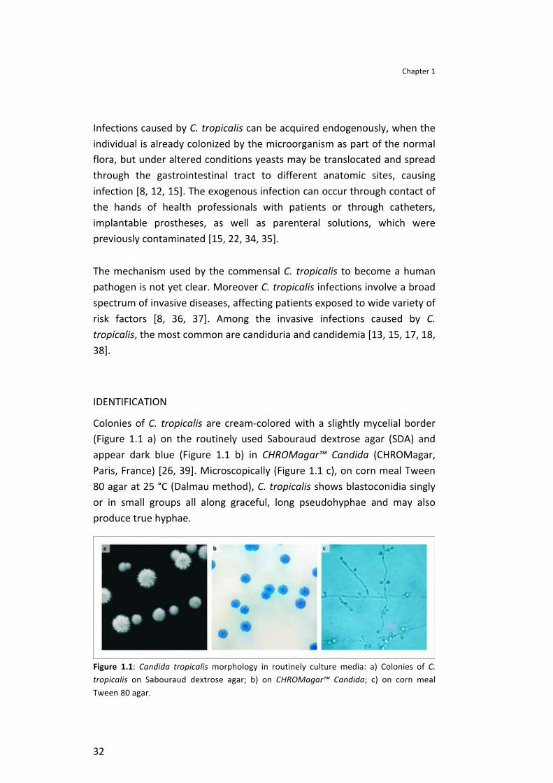
Colonies of C. tropicalis are cream-‐colored with a slightly mycelial border (Figure 1.1 a) on the routinely used Sabouraud dextrose agar (SDA) and appear dark blue (Figure 1.1 b) in CHROMagar™ Candida (CHROMagar, Paris, France) [26, 39]. Microscopically (Figure 1.1 c), on corn meal Tween 80 agar at 25 °C (Dalmau method), C. tropicalis shows blastoconidia singly or in small groups all along graceful, long pseudohyphae and may also produce true hyphae.
Figure 1.1: Candida tropicalis morphology in routinely culture media: a) Colonies of C. tropicalis on Sabouraud dextrose agar; b) on CHROMagar™ Candida; c) on corn meal Tween 80 agar.
Synopsis of Candida tropicalis
33
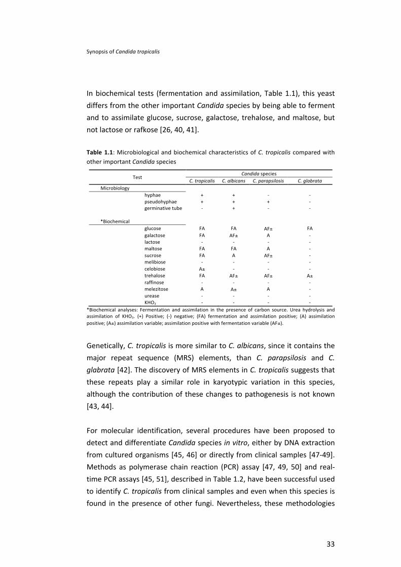
In biochemical tests (fermentation and assimilation, Table 1.1), this yeast differs from the other important Candida species by being able to ferment and to assimilate glucose, sucrose, galactose, trehalose, and maltose, but not lactose or rafkose [26, 40, 41].
Table 1.1: Microbiological and biochemical characteristics of C. tropicalis compared with other important Candida species
Test Candida species
C. tropicalis C. albicans C. parapsilosis C. glabrata Microbiology
hyphae + + -‐ -‐ pseudohyphae + + + -‐ germinative tube -‐ + -‐ -‐
*Biochemical glucose FA FA AF± FA galactose FA AF± A -‐ lactose -‐ -‐ -‐ -‐ maltose FA FA A -‐ sucrose FA A AF± -‐ melibiose -‐ -‐ -‐ -‐ celobiose A± -‐ -‐ -‐ trehalose FA AF± AF± A± raffinose -‐ -‐ -‐ -‐ melezitose A A± A -‐ urease -‐ -‐ -‐ -‐ KHO3 -‐ -‐ -‐ -‐
*Biochemical analyses: Fermentation and assimilation in the presence of carbon source. Urea hydrolysis and assimilation of KHO3. (+) Positive; (-‐) negative; (FA) fermentation and assimilation positive; (A) assimilation positive; (A±) assimilation variable; assimilation positive with fermentation variable (AF±).
Genetically, C. tropicalis is more similar to C. albicans, since it contains the major repeat sequence (MRS) elements, than C. parapsilosis and C. glabrata [42]. The discovery of MRS elements in C. tropicalis suggests that these repeats play a similar role in karyotypic variation in this species, although the contribution of these changes to pathogenesis is not known [43, 44].
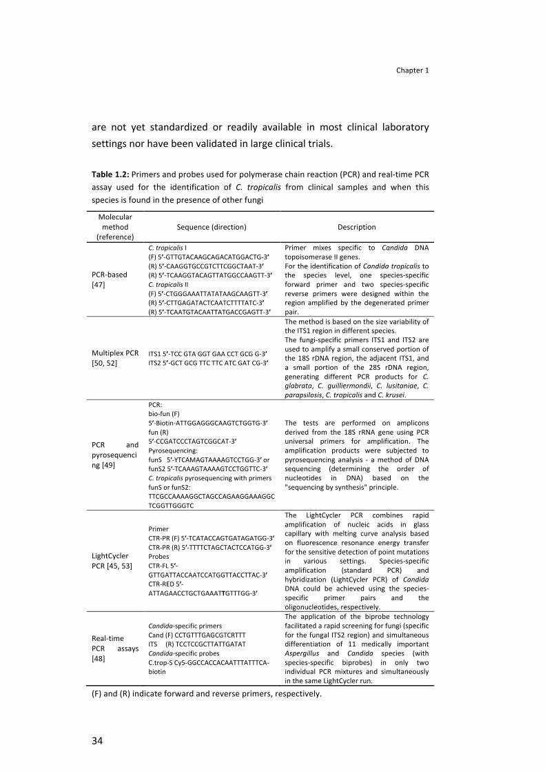
For molecular identification, several procedures have been proposed to detect and differentiate Candida species in vitro, either by DNA extraction from cultured organisms [45, 46] or directly from clinical samples [47-‐49]. Methods as polymerase chain reaction (PCR) assay [47, 49, 50] and real-‐time PCR assays [45, 51], described in Table 1.2, have been successful used to identify C. tropicalis from clinical samples and even when this species is found in the presence of other fungi. Nevertheless, these methodologies
Chapter 1
34
are not yet standardized or readily available in most clinical laboratory settings nor have been validated in large clinical trials.
Table 1.2: Primers and probes used for polymerase chain reaction (PCR) and real-‐time PCR assay used for the identification of C. tropicalis from clinical samples and when this species is found in the presence of other fungi
Molecular method
(reference) Sequence (direction) Description
PCR-‐based [47]
C. tropicalis I (F) 5’-‐GTTGTACAAGCAGACATGGACTG-‐3’ (R) 5’-‐CAAGGTGCCGTCTTCGGCTAAT-‐3’ (R) 5’-‐TCAAGGTACAGTTATGGCCAAGTT-‐3’ C. tropicalis II (F) 5’-‐CTGGGAAATTATATAAGCAAGTT-‐3’ (R) 5’-‐CTTGAGATACTCAATCTTTTATC-‐3’ (R) 5’-‐TCAATGTACAATTATGACCGAGTT-‐3’
Primer mixes specific to Candida DNA topoisomerase II genes. For the identification of Candida tropicalis to the species level, one species-‐specific forward primer and two species-‐specific reverse primers were designed within the region amplified by the degenerated primer pair.
Multiplex PCR [50, 52]
ITS1 5’-‐TCC GTA GGT GAA CCT GCG G-‐3’ ITS2 5’-‐GCT GCG TTC TTC ATC GAT CG-‐3’
The method is based on the size variability of the ITS1 region in different species. The fungi-‐specific primers ITS1 and ITS2 are used to amplify a small conserved portion of the 18S rDNA region, the adjacent ITS1, and a small portion of the 28S rDNA region, generating different PCR products for C. glabrata, C. guilliermondii, C. lusitaniae, C. parapsilosis, C. tropicalis and C. krusei.
PCR and pyrosequencing [49]
PCR: bio-‐fun (F) 5ʹ′-‐Biotin-‐ATTGGAGGGCAAGTCTGGTG-‐3ʹ′ fun (R) 5ʹ′-‐CCGATCCCTAGTCGGCAT-‐3ʹ′ Pyrosequencing: funS 5ʹ′-‐YTCAMAGTAAAAGTCCTGG-‐3ʹ′ or funS2 5ʹ′-‐TCAAAGTAAAAGTCCTGGTTC-‐3ʹ′ C. tropicalis pyrosequencing with primers funS or funS2: TTCGCCAAAAGGCTAGCCAGAAGGAAAGGCTCGGTTGGGTC
The tests are performed on amplicons derived from the 18S rRNA gene using PCR universal primers for amplification. The amplification products were subjected to pyrosequencing analysis -‐ a method of DNA sequencing (determining the order of nucleotides in DNA) based on the "sequencing by synthesis" principle.
LightCycler PCR [45, 53]
Primer CTR-‐PR (F) 5ʹ′-‐TCATACCAGTGATAGATGG-‐3ʹ′ CTR-‐PR (R) 5ʹ′-‐TTTTCTAGCTACTCCATGG-‐3ʹ′ Probes CTR-‐FL 5ʹ′-‐GTTGATTACCAATCCATGGTTACCTTAC-‐3ʹ′ CTR-‐RED 5ʹ′-‐ATTAGAACCTGCTGAAATTGTTTGG-‐3ʹ′
The LightCycler PCR combines rapid amplification of nucleic acids in glass capillary with melting curve analysis based on fluorescence resonance energy transfer for the sensitive detection of point mutations in various settings. Species-‐specific amplification (standard PCR) and hybridization (LightCycler PCR) of Candida DNA could be achieved using the species-‐specific primer pairs and the oligonucleotides, respectively.
Real-‐time PCR assays [48]
Candida-‐specific primers Cand (F) CCTGTTTGAGCGTCRTTT ITS (R) TCCTCCGCTTATTGATAT Candida-‐specific probes C.trop-‐S Cy5-‐GGCCACCACAATTTATTTCA-‐biotin
The application of the biprobe technology facilitated a rapid screening for fungi (specific for the fungal ITS2 region) and simultaneous differentiation of 11 medically important Aspergillus and Candida species (with species-‐specific biprobes) in only two individual PCR mixtures and simultaneously in the same LightCycler run.
(F) and (R) indicate forward and reverse primers, respectively.
Synopsis of Candida tropicalis
35
RISK FACTORS
In general, the risk factors involved in the development of hospital-‐acquired Candida infections are associated with: extended periods in intensive care unit, administration of broad-‐spectrum antibiotics, patients with immunosuppression, indwelling catheters, mechanical ventilation, candiduria, multiple sites of colonization, burns, and haemodialysis [5, 54-‐56]. However, the particularities of each Candida species may be influenced by specific risk factors. Studies have been shown, that in opposition to C. parapsilosis, C. tropicalis was less likely to occur among children of less than 1 year of age, but more likely to occur in patients with cancer or neutropenia [15, 57, 58], and is strongly associated with the presence of biofilms in urinary catheters. [18, 54, 59-‐61].
Candida colonization remains the most universally accepted predictive variable with regard to invasive candidosis, being particularly true for high density colonization. In fact, colonization by C. tropicalis, especially from a specific body site can be highly predictive of the development of invasive disease with this organism [62]. Nevertheless it has not yet been clarified whether colonization can be used alone to identify high-‐risk patients or if it should be combined with other variables indicating high risk [63]. According to Paul et al. [64] many risk factors traditionally linked to candiduria may be associated with urinary tract infections in general. Furthermore, Binelli et al. [65] found a significant association of candidemia with candiduria, although urine was not the main source of C. tropicalis bloodstream infection.
According to epidemiological data, when comparing patients with candidemia caused by C. tropicalis to those caused by other species of Candida, the former are, in average, older patients (67 years vs. 56 years, P = 0.01), present cancer (45.5% vs. 31.6%, P = 0.04), and that the portal of entry is the abdomen (32.2% vs. 11.9%, P = 0.001). Additionally, these patients also have a high hospital mortality rate (61% vs. 44%, P = 0.03) [66]. Further studies suggested that C. tropicalis is associated with higher dissemination potential and mortality in patients admitted in ICU,

Chapter 1
36
particularly in oncology patients, than C. albicans or any other NCAC species [13, 15, 67].
EPIDEMIOLOGY
The proportion of candidoses (candidemia and candiduria) caused by C. tropicalis varies widely with geographical area and patient group, with C. tropicalis being more prevalent, even compared with C. albicans, in certain countries [66, 68, 69]. Considering Table 1.3, it is possible to see that, among NCAC species, C. tropicalis has been considered the species most frequently isolated from candidosis in the Pacific-‐Asia region [13], Brazil [12, 15], and recently in Europe [7, 70]. Furthermore, important epidemiological studies revealed that 90% of invasive candidosis were due to NCAC species, with C. tropicalis accounting for about 4.6% in 1997-‐1998; 5.3% in 1999; and between 7.3% in 2000-‐2003 [71]. Additionally, in general, C. tropicalis appeared to be the first or second NCAC most frequently associated to candiduria. Nevertheless, regarding candidemia, C. tropicalis is less frequently isolated than C. glabrata [7, 17, 54, 67, 70].
Although the reasons for the increased detection of C. tropicalis in human infection are not completely clear the advent of molecular genetics, and the development of new methods of Candida identification and differentiation [45, 47, 72] may play an important role. Further, the changes of C. tropicalis incidence may also be attributed to the greater use of fluconazole nowadays [66, 69]. In the United States, due to fluconazole prophylaxis the frequency of candidosis caused for C. tropicalis has decreased. However, in other countries where it is not usual to use fluconazole prophylaxis, C. tropicalis appears more prevalent, e.g. second in Latin America, and even more common than C. glabrata in the Asia-‐Pacific region [71]. Nevertheless, the use of prophylactic fluconazole can become a risk factor since some cross-‐resistance between azoles [7, 11, 73] has been reported already. Several studies indicate that C. tropicalis has been showing a moderate level of fluconazole tolerance leading to the
Synopsis of Candida tropicalis
37
need of an increase in the drug concentrations and enabling a risk of azole resistance [67, 74, 75].
Curiously, according to Table 1.3, the epidemiological data related with antifungal resistance, have been indicating an increase of C. tropicalis resistance to 5-‐flucytosine [12, 15, 54, 76, 77]. Furthermore, it was observed 35% of resistance to 5-‐flucytosine by C. tropicalis isolates recovered from blood cultures in the active surveillance program on yeast-‐related fungemia implemented by the French National Reference Center for Mycoses and Antifungals (NRCMA) in the Paris area [76]. Additionally, Densos-‐Olliver et al. [76] studied the relationship between epidemiologic and genomic data of C. tropicalis 5-‐flucytosine resistance and, observed that a clone of 5-‐flucytosine-‐resistant isolate, associated with malignancies, had lower mortality than the other C. tropicalis isolates. This suggests that geographic and temporal distribution of C. topicalis may be related with 5-‐flucytosine-‐resistant isolates in the Paris area.
In fact, the major problem with the development of invasive candidosis by C. tropicalis is that it is associated with higher mortality than other NCAC species and C. albicans [8, 20, 68, 78]. This propensity of C. tropicalis for dissemination and associated high mortality may be related to the virulence factors exhibited by this species such as biofilm formation; proteinases secretion and dissemination [11, 22].
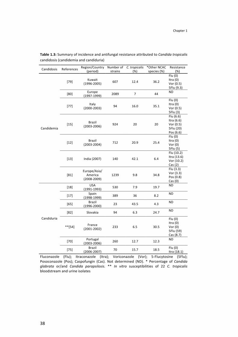
Chapter 1
38
Table 1.3: Summary of incidence and antifungal resistance attributed to Candida tropicalis candidosis (candidemia and candiduria)
Candidosis References Region/Country (period)
Number of strains
C. tropicalis (%)
*Other NCAC species (%)
Resistance (%)
Candidemia
[79] Kuwait (1996-‐2005) 607 12.4 36.2
Flu (0) Itra (0) Vor (0.5) 5Flu (9.3)
[80] Europe (1997-‐1999) 2089 7 44 ND
[77] Italy (2000-‐2003) 94 16.0 35.1
Flu (0) Itra (0) Vor (0.5) 5Flu (3)
[15] Brazil (2003-‐2006) 924 20 20
Flu (6.6) Itra (6.6) Vor (0.5) 5Flu (20) Pos (6.6)
[12] Brazil (2003-‐2004) 712 20.9 25.4
Flu (0) Itra (0) Vor (0) 5Flu (5)
[13] India (2007) 140 42.1 6.4
Flu (10.2) Itra (13.6) Vor (10.2) Cas (2)
[81] Europe/Asia/
America (2008-‐2009)
1239 9.8 34.8
Flu (3.3) Vor (3.3) Pos (0.8) Cas (0)
Candiduria
[18] USA (1991-‐1993) 530 7.9 19.7 ND
[17] Spain (1998-‐1999) 389 36 8.2 ND
[65] Brazil (1996-‐2000) 23 43.5 4.3 ND
[82] Slovakia 94 6.3 24.7 ND
**[54] France (2001-‐2002) 233 6.5 30.5
Flu (0) Itra (0) Vor (0) 5Flu (59) Cas (8.7)
[70] Portugal (2003-‐2006) 260 12.7 12.3 ND
[75] Brazil (2006-‐2007) 70 15.7 18.5 Flu (0)
Itra (18.1) Fluconazole (Flu); Itraconazole (Itra); Voriconazole (Vor); 5-‐Flucytosine (5Flu); Posoconazole (Pos); Caspofungin (Cas). Not determined (ND). * Percentage of Candida glabrata or/and Candida parapsilosis. ** In vitro susceptibilities of 22 C. tropicalis bloodstream and urine isolates
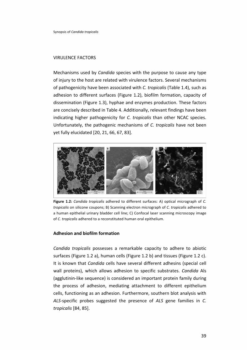
Synopsis of Candida tropicalis
39
VIRULENCE FACTORS
Mechanisms used by Candida species with the purpose to cause any type of injury to the host are related with virulence factors. Several mechanisms of pathogenicity have been associated with C. tropicalis (Table 1.4), such as adhesion to different surfaces (Figure 1.2), biofilm formation, capacity of dissemination (Figure 1.3), hyphae and enzymes production. These factors are concisely described in Table 4. Additionally, relevant findings have been indicating higher pathogenicity for C. tropicalis than other NCAC species. Unfortunately, the pathogenic mechanisms of C. tropicalis have not been yet fully elucidated [20, 21, 66, 67, 83].
Figure 1.2: Candida tropicalis adhered to different surfaces: A) optical micrograph of C. tropicalis on silicone coupons; B) Scanning electron micrograph of C. tropicalis adhered to a human epithelial urinary bladder cell line; C) Confocal laser scanning microscopy image of C. tropicalis adhered to a reconstituted human oral epithelium.
Adhesion and biofilm formation
Candida tropicalis possesses a remarkable capacity to adhere to abiotic surfaces (Figure 1.2 a), human cells (Figure 1.2 b) and tissues (Figure 1.2 c). It is known that Candida cells have several different adhesins (special cell wall proteins), which allows adhesion to specific substrates. Candida Als (agglutinin-‐like sequence) is considered an important protein family during the process of adhesion, mediating attachment to different epithelium cells, functioning as an adhesion. Furthermore, southern blot analysis with ALS-‐specific probes suggested the presence of ALS gene families in C. tropicalis [84, 85].

Chapter 1
40
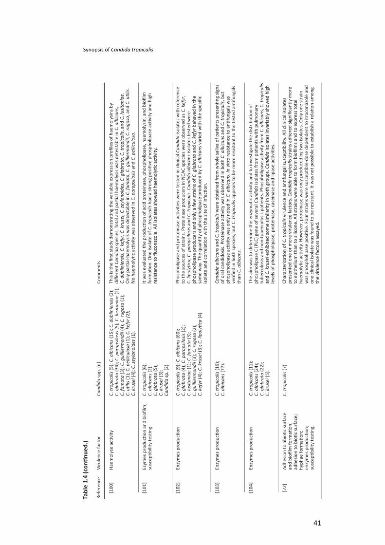
Synopsis of Candida tropicalis
41
Chapter 1
42
Furthermore, other factors, such as physicochemical interactions between yeast cells and materials surface, as well as environmental factors, can influence the initial adhesion of C. tropicalis [21, 86-‐88]. Several studies showed the ability of C. tropicalis to adhere, and consequently to form biofilms, in clinically relevant substrates like medical devices, and in different environmental situations, both in vitro and in vivo. Adherence of Candida cells to abiotic surfaces and to other cells is vital for biofilm formation [86, 88-‐91].
Candida biofilm formation is initiated when the yeast adheres to a surface, cells attach to each other and begin to proliferate, ultimately leading to the formation of a highly structured mature biofilm, comprised of complex intertwining layers of yeast, pseudohyphae and hyphae embedded in the extracellular matrix. [92, 93]. The matrix is one of the most distinctive features of a microbial biofilm. This complex extracellular material might function to defend against phagocytic cells, to serve as a scaffold for maintaining biofilm integrity, and to limit diffusion of toxic substances into the biofilm as antifungals [92, 94]. Further, studies indicate that C. tropicalis biofilms exhibit large amounts of matrix material completely resistant to antifungals [23, 67, 95-‐97]. Those data can explain why the major risk factor of C. tropicalis in candidosis development is related with the difficulty of treatment and, moreover, to the prolonged catheterization [53, 66, 73].
In a study done by Al-‐Fattani and Douglas [95], it was shown that the matrix of C. tropicalis contained carbohydrates, proteins, hexosamine, phosphorus and uronic acid. The major component in C. tropicalis matrix was hexosamine (27%), whereas in C. albicans matrix was glucose (32%). It is important to emphasize that hexosamine is present in S. epidermidis as a polysaccharide, which is sometimes referred to as the intercellular polysaccharide adhesin (PIA) and is known to mediate cell–cell interaction within the biofilm [98]. Further, in the same study, biofilms of C. albicans were more easily detached from plastic surfaces by treatment with the enzyme lyticase than were those of C. tropicalis.
Synopsis of Candida tropicalis
43
However, C. tropicalis biofilm and consequently matrix composition are extremely dependent on environmental conditions, such as medium composition, pH, oxygen and growth conditions (static or flow) [88, 90, 96]. According to a study done by Jain et al. [61], comparing Candida biofilms grown in RPMI medium and artificial urine, biofilm formation is highly dependent on the growth medium. In particular, C. albicans strains produced more biofilm in artificial urine than in RPMI. Furthermore, other researches indicated that biofilms grown under conditions of continuous flow produced more matrix than those grown statically, and were significantly more resistant to amphotericin B [95].
Other important factor about biofilm life-‐cycle is related with dispersion/detachment or dissolution of cells, which release from the biofilm and seed new surfaces with the consequent establishment of disseminated candidosis at distal organs. Additionally, there are indications that dispersed cells from biofilms are more virulent than planktonic cells [92]. Negri et al. [90] detected that C. tropicalis cells are able to detach from biofilms formed in catheters under a flow of artificial urine and move upflow. However, little is still known about C. tropicalis detachment cells from biofilm and more studies are necessary to better understand this process.
Infection and dissemination
Adherence of C. tropicalis to host cells, and consequently colonization, is seen as an essential early step in the establishment of disease, since, high density colonization is indicative of high risk factor to the host [63, 66]. It is known that C. tropicalis is able to adhere, colonize and infect host tissues (Figure 1.3) and further disseminate, both in vivo and in vitro [21, 22, 87, 89].
It is interesting to observe that, according to some researchers, C. tropicalis strains showed intermediate levels of adherence to buccal epithelial cells [30, 89] and to human epithelial cell monolayers [105], whereas C. albicans strains showed high in vitro adherence. However, in others studies, C.
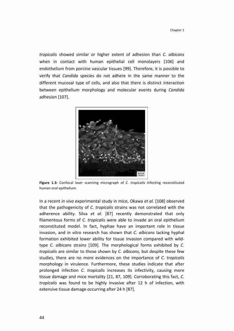
Chapter 1
44
tropicalis showed similar or higher extent of adhesion than C. albicans when in contact with human epithelial cell monolayers [106] and endothelium from porcine vascular tissues [99]. Therefore, it is possible to verify that Candida species do not adhere in the same manner to the different mucosal type of cells, and also that there is distinct interaction between epithelium morphology and molecular events during Candida adhesion [107].
Figure 1.3: Confocal laser scanning micrograph of C. tropicalis infecting reconstituted human oral epithelium.
In a recent in vivo experimental study in mice, Okawa et al. [108] observed that the pathogenicity of C. tropicalis strains was not correlated with the adherence ability. Silva et al. [87] recently demonstrated that only filamentous forms of C. tropicalis were able to invade an oral epithelium reconstituted model. In fact, hyphae have an important role in tissue invasion, and in vitro research has shown that C. albicans lacking hyphal formation exhibited lower ability for tissue invasion compared with wild-‐type C. albicans strains [109]. The morphological forms exhibited by C. tropicalis are similar to those shown by C. albicans, but despite these few studies, there are no more evidences on the importance of C. tropicalis morphology in virulence. Furthermore, these studies indicate that after prolonged infection C. tropicalis increases its infectivity, causing more tissue damage and mice mortality [21, 87, 109]. Corroborating this fact, C. tropicalis was found to be highly invasive after 12 h of infection, with extensive tissue damage occurring after 24 h [87].
Synopsis of Candida tropicalis
45
Thus, the pathogenic mechanisms of C. tropicalis seem to be different from those of C. albicans [21, 106]. A significant work [110] on pathogenicity of Candida species in an animal model, showed that the most pathogenic group was C. albicans and C. tropicalis, followed by an intermediate group with C. glabrata, C. lusitaniae and C. kyfyr and a least pathogenic group of C. parapsilosis, C. krusei and C. guillerimondii. It is important to emphasize the clinical relevance of those findings, since the major problems with the development of invasive C. tropicalis candidosis are associated with high dissemination and mortality.
Enzymes production
Once adhered to host cells, C. tropicalis requires other factors to penetrate into the deepest tissues, e.g., hyphal formation and production of enzymes. In fact, the pathogenic capabilities of this yeast have been related to the secretion of aspartyl proteinases, phospholipases and haemolytic activity [22, 24, 25, 67, 85, 111].
Candida produce a large variety of secreted hydrolases, and among the various potential virulence factors proposed, the secreted aspartyl proteinases (Sap) have been intensively investigated. It is now well established that the ability of C. albicans to adhere to mucosae, to invade in deep organs, and to resist to phagocytic cells, apparently requires the use of several different proteinases suitable to each particular condition during the infection. Like C. albicans, C. tropicalis presents in vitro Sap activity in a medium containing bovine serum albumin (BSA) as the sole source of nitrogen [22, 31, 85, 112].
Furthermore, Zaugg et al. [24] characterized a total of four SAPT genes family of C. tropicalis. According to this study, RT-‐PCR experiments revealed a strong SAPT1 signal with RNA extracted from cells grown in BSA medium. The SAPT2 and SAPT3 gene products, Sapt2p and Sapt3p, which have not yet been detected in C. tropicalis cultures in vitro, were produced as active recombinant enzymes with the methylotrophic yeast Pichia pastoris as an expression system. However, a weak signal was obtained with all other SAPT genes under in vitro conditions tested, suggesting that
Chapter 1
46
the gene products Sapt2p, Sapt3p, and Sapt4p could be produced during infection.
This idea is highlighted by recent studies [87, 109] that investigated epithelial infection by C. tropicalis using a reconstituted human oral epithelium (RHOE) and SAPT gene expression. The results obtained by real-‐time PCR, showed that C. tropicalis isolates were able to express SAPT1-‐4 during the infection process. Moreover, expression was strain-‐dependent, with SAPT2-‐4 transcripts being frequently detected and SAPT1 rarely detected. Furthermore, C. tropicalis can be considered as highly invasive with the ability to induce significant tissue damage [87]. These features, however, do not appear to be related to specific SAPT gene expression. Therefore it is necessary to perform more investigations focusing on SAPT genes family of C. tropicalis for better understanding the specific role of these genes.
In addition to Saps, enzymes categorized as lipases (LIPs), this means enzymes that hydrolyze phospholipids into fatty acids, are often considered to be involved in C. tropicalis pathogenicity and are suggested to contribute to host cell membrane damage which could also expose receptors to facilitate adherence [102, 111, 113]. In C. albicans, 10 genes encoding for LIPs (LIP1-‐10) have been identified and similar sequences were also detected in C. tropicalis [114]. Nevertheless, the most widely used diagnostic method for phospholipases (PLs) determination is based on yeast growth in an egg yolk agar medium [115]. According to recent studies, using this method, C. tropicalis, from different sources, appears to have a reduced ability to produce extracellular PLs in vitro when compared with C. albicans [29, 102, 103]. However, this production is highly species and strain dependent [22, 25, 101, 116].
Other important virulence factor recently described in literature is related with haemolytic activity which is tested on sheep blood agar supplemented with glucose [100]. It is known that enzymes as haemolysins are used by Candida species to degrade haemoglobin and facilitate recovery of the elemental iron from host cells, which is a contribute to pathogenicity in
Synopsis of Candida tropicalis
47
Candida species. Thus, haemolysins are considered key virulence factors enabling pathogen survival and persistence in the host [100, 117, 118]. The studies reported so far show that C. tropicalis are all able to produce haemolysins in vitro, inducing partial or total erythrocyte lyses, although the degree being strain dependent [100]. According to Luo et al. [100], total-‐haemolytic activities in C. albicans and C. tropicalis were significantly higher than in C. glabrata. However, Kumar et al. [104] observed the opposite, C. glabrata displayed the highest haemolytic activity when compared with C. albicans and C. tropicalis. Despite significant studies showed the ability of C. tropicalis to produce haemolytic activity on sheep blood agar supplemented with glucose, it is important to assess whether the haemolytic activity observed is true or is a product of extracellular PLs of Candida species. Moreover, it is still necessary to have more advances in molecular studies to clarify the role of haemolytic activity in C. tropicalis pathogenesis.
CONCLUDING REMARKS
In fact, the frequency of Candida tropicalis causing candidosis is increasing in the last decades, probably due to several situations, e.g., new and efficient molecular methods of identification; antifungal resistance mainly to fluconazole commonly used as prophylaxis agent; and factors related with host as well as invasiveness surgery, long time in ICU, antibiotic administration and catheterization. Additionally, invasive disease developed by C. tropicalis is associated with colonization, high potential of dissemination and pathogenicity by this organism. Mainly because C. tropicalis possesses a diversity of virulence factors that induces serious damage to patients and increases the mortality risk. However, it is necessary much more research to get deeper insights into the strategies used by C. tropicalis to change from a harmless commensal microorganism to become a human pathogen of high clinical concern.
Chapter 1
48
REFERENCES
1. Hota B. Contamination, Disinfection, and Cross Colonization: Are Hospital Surfaces Reservoirs for Nosocomial Infection? Clin Infect Dis 2004; 39 (8): 1182-‐1189. 2. Gudlaugsson O, Gillespie S, Lee K, et al. Attributable Mortality of Nosocomial Candidemia, Revisited. Clinical Infectious Diseases 2003; 37 (9): 1172-‐1177. 3. Bassetti M, Righi E, Costa A, et al. Epidemiological trends in nosocomial candidemia in intensive care. BMC Infect Dis 2006; 6 (1): 21. 4. Cheng M-‐F, Yang Y-‐L, Yao T-‐J, et al. Risk factors for fatal candidemia caused by Candida albicans and non-‐albicans Candida species. BMC Infect Dis 2005; 5 (1): 22. 5. Dimopoulos G, Ntziora F, Rachiotis G, Armaganidis A, Falagas ME. Candida albicans versus non-‐albicans intensive care unit-‐acquired bloodstream infections: differences in risk factors and outcome. Anesth Analg 2008; 106 (2): 523-‐529. 6. Meric M, Willke A, Caglayan C, Toker K. Intensive care unit-‐acquired infections: incidence, risk factors and associated mortality in a Turkish university hospital. Jpn J Infect Dis 2005; 58 (5): 297-‐302. 7. Lass-‐Flörl C. The changing face of epidemiology of invasive fungal disease in Europe. Mycoses 2009; 52 (3): 197-‐205. 8. Eggimann P, Garbino J, Pittet D. Epidemiology of Candida species infections in critically ill non-‐immunosuppressed patients. Lancet Infect Dis 2003; 3 (11): 685-‐702. 9. Snydman DR. Shifting Patterns in the Epidemiology of Nosocomial Candida Infections. Chest 2003; 123 (5 Suppl): 500S-‐3S. 10. Lundstrom T, Sobel J. Nosocomial Candiduria: A Review. Clin Infect Dis 2001; 32 (11): 1602-‐1607. 11. Krcmery V, Barnes AJ. Non-‐albicans Candida spp. causing fungaemia: pathogenicity and antifungal resistance. J Hosp Infect 2002; 50 (4): 243-‐260. 12. Colombo AL, Nucci M, Park BJ, et al. Epidemiology of Candidemia in Brazil: a Nationwide Sentinel Surveillance of Candidemia in Eleven Medical Centers. J Clin Microbiol 2006; 44 (8): 2816-‐2823. 13. Chakrabarti A, Chatterjee SS, Rao KLN, et al. Recent experience with fungaemia: change in species distribution and azole resistance. Scand J Infect Dis 2009; 41 (4): 275 -‐ 284. 14. Tortorano AM, Peman J, Bernhardt H, et al. Epidemiology of Candidaemia in Europe: Results of 28-‐Month European Confederation of Medical Mycology (ECMM) Hospital-‐Based Surveillance Study. Eur J Clin Microbiol Infect Dis 2004; 23 (4): 317-‐322. 15. Nucci M, Colombo AL. Candidemia due to Candida tropicalis: clinical, epidemiologic, and microbiologic characteristics of 188 episodes occurring in tertiary care hospitals. Diagn Microbiol Infect Dis 2007; 58 (1): 77-‐82. 16. Hsueh P-‐R, Graybill JR, Playford EG, et al. Consensus statement on the management of invasive candidiasis in Intensive Care Units in the Asia-‐Pacific Region. Int J Antimicro Ag 2009; 34 (3): 205-‐209. 17. Álvarez-‐Lerma F, Nolla-‐Salas J, León C, et al. Candiduria in critically ill patients admitted to intensive care medical units. Intensive Care Med. 2003; 29 (7): 1069-‐1076. 18. Kauffman CA, Vazquez JA, Sobel JD, et al. Prospective multicenter surveillance study of funguria in hospitalized patients. Clin Infect Dis 2000; 30 (1): 14-‐18. 19. Krcmery VJ. Candidemia in cancer patients: Risk factors and outcome in 140 episodes from a single cancer institution. Acta Chemoth 1999; 5: 133-‐145. 20. Kontoyiannis Dimitrios P, Vaziri I, Hanna Hend A, et al. Risk Factors for Candida tropicalis Fungemia in Patients with Cancer. Clin Infect Dis 2001; 33 (10): 1676-‐1681. 21. Okawa Y, Miyauchi M, Kobayashi H. Comparison of Pathogenicity of Various Candida tropicalis Strains. Biol Pharm Bull 2008; 31 (8): 1507-‐1510. 22. Negri M, Martins M, Henriques M, et al. Examination of potential virulence factors of Candida tropicalis clinical isolates from hospitalized patients. Mycopathologia 2010; 169 (3): 175-‐182. 23. Bizerra FC, Nakamura CV, de Poersch C, et al. Characteristics of biofilm formation by Candida tropicalis and antifungal resistance. FEMS Yeast Res 2008; 8 (3): 442-‐450. 24. Zaugg C, Borg-‐von Zepelin M, Reichard U, Sanglard D, Monod M. Secreted aspartic proteinase family of Candida tropicalis. Infect Immun 2001; 69 (1): 405-‐412.

Synopsis of Candida tropicalis
49
25. Galan-‐Ladero MA, Blanco MT, Sacristan B, et al. Enzymatic activities of Candida tropicalis isolated from hospitalized patients. Med Mycol 2010; 48 (1): 207-‐210 26. Larone D. Medically important fungi; a guide to identification, 4th edn. Washington: ASM Press; 2002. 27. Kurtzman CP, Fell JW. The Yeasts, A Taxonomic Study, fourth edition edn. New York: Elsevier Science Ltd; 1998. 28. Basu S, Gugnani H, Joshi S, Gupta N. Distribution of Candida species in different clinical sources in Delhi, India, and proteinase and phospholipase activity of Candida albicans isolates. Rev Iberoam Micol 2003; 20 (4): 137-‐140. 29. Oksuz S, Sahin I, Yildirim M, et al. Phospholipase and proteinase activities in different Candida species isolated from anatomically distinct sites of healthy adults. Jpn J Infect Dis. 2007; 60 (5): 280-‐283. 30. Biasoli MS, Tosello ME, Bottai H, Cuesta C, Magaró HM. Adherence of Candida strains isolated from the human gastrointestinal tract. Mycoses 2002; 45 (11-‐12): 465-‐469. 31. Tamura NK, Negri MFN, Bonassoli LA, Svidzinski TIE. Fatores de virulência de Candida spp isoladas de cateteres venosos e mãos de servidores hospitalares. Rev Soc Bras Med Trop 2007; 40 (1): 91-‐93. 32. Shurtleff DB, Peterson W, Sherris JC. Systemic Candida tropicalis infection treated with amphotericin. N Engl J Med 1963; 269: 1112-‐5. 33. D. HO. The Pathogenicity of Candida tropicalis. Can Med Assoc J 1960; 83 (27): 1439-‐40. 34. Nucci M, Silveira M, Spector N, et al. Fungemia in cancer patients in Brazil: Predominance of non-‐albicans species. Mycopathologia 1998; 141 (2): 65-‐68. 35. Rho J, Shin J, Song J, et al. Molecular investigation of two consecutive nosocomial clusters of Candida tropicalis candiduria using pulsed-‐field gel electrophoresis. J Microbiol 2004; 42 (2): 80-‐86. 36. Kliemann DA, Pasqualotto AC, Falavigna M, Giaretta T, Severo LC. Candida esophagitis: species distribution and risk factors for infection. Rev Inst Med Trop Sao Paulo. 2008; 50: 261-‐263. 37. Baley JE, Ellis FJ. Neonatal candidiasis: ophthalmologic infection. Semin Perinatol 2003; 27 (5): 401-‐405. 38. Silva EHd, Ruiz LdS, Matsumoto FE, et al. Candiduria in a public hospital of São Paulo (1999-‐2004): characteristics of the yeast isolates. Rev Inst Med Trop Sao Paulo 2007; 49 (6): 349-‐353. 39. Calderone RA. Introduction and historical perspectives. In: Calderone R, ed.^eds Candida and candidiasis. Washington D.C: ASM Press; 2002: 15-‐25. 40. Yoshio O, Kouji G. Antigenicity of Candida tropicalis strain cells cultured at 27 and 37ºC. FEMS Immunol Med Microbiol 2006; 46 (3): 438-‐443. 41. Martin MV. Germ-‐tube formation by oral strains of Candida tropicalis. J Med Microbiol 1979; 12 (2): 187-‐194. 42. Butler G, Rasmussen MD, Lin MF, et al. Evolution of pathogenicity and sexual reproduction in eight Candida genomes. Nature 2009; 459 (7247): 657-‐662. 43. Bennett RJ. A Candida-‐based view of fungal sex and pathogenesis. Genome Biol 2009; 10 (7): 230. 44. Zhang J, Hollis RJ, Pfaller MA. Variations in DNA subtype and antifungal susceptibility among clinical isolates of Candida tropicalis. Diagn Microbiol Infect Dis 1997; 27 (3): 63-‐7. 45. Fricke S, Fricke C, Schimmelpfennig C, et al. A real-‐time PCR assay for the differentiation of Candida species. J Appl Microbiol 2010; 9999 (9999). 46. Williams DW, Wilson MJ, Lewis MA, Potts AJ. Identification of Candida species by PCR and restriction fragment length polymorphism analysis of intergenic spacer regions of ribosomal DNA. J Clin Microbiol 1995; 33 (9): 2476-‐9. 47. Kanbe T, Horii T, Arishima T, Ozeki M, Kikuchi A. PCR-‐based identification of pathogenic Candida species using primer mixes specific to Candida DNA topoisomerase II genes. Yeast 2002; 19 (11): 973-‐989. 48. Schabereiter-‐Gurtner C, Selitsch B, Rotter ML, Hirschl AM, Willinger B. Development of novel real-‐time PCR assays for detection and differentiation of eleven medically important Aspergillus and Candida species in clinical specimens. J Clin Microbiol 2007; 45 (3): 906-‐14.
Chapter 1
50
49. Quiles-‐Melero I, Garcia-‐Rodriguez J, Romero-‐Gomez MP, Gomez-‐Sanchez P, Mingorance J. Rapid identification of yeasts from positive blood culture bottles by pyrosequencing. Eur J Clin Microbiol Infect Dis 2010. 50. Liguori G, Lucariello A, Colella G, De Luca A, Marinelli P. Rapid identification of Candida species in oral rinse solutions by PCR. J Clin Pathol 2007; 60 (9): 1035-‐9. 51. Bustamante CI. Treatment of Candida infection: a view from the trenches! Curr Opin Infect Dis 2005; 18 (6): 490-‐495. 52. Chang HC, Leaw SN, Huang AH, Wu TL, Chang TC. Rapid identification of yeasts in positive blood cultures by a multiplex PCR method. J Clin Microbiol 2001; 39 (10): 3466-‐71. 53. Loeffler J, Hagmeyer L, Hebart H, et al. Rapid detection of point mutations by fluorescence resonance energy transfer and probe melting curves in Candida species. Clin Chem 2000; 46 (5): 631-‐5. 54. Bougnoux ME, Kac G, Aegerter P, d'Enfert C, Fagon JY. Candidemia and candiduria in critically ill patients admitted to intensive care units in France: incidence, molecular diversity, management and outcome. Intensive Care Med 2008; 34 (2): 292-‐9. 55. Carey AJ, Saiman L, Polin RA. Hospital-‐Acquired Infections in the NICU: Epidemiology for the New Millennium. Clin Perinatol 2008; 35 (1): 223-‐249. 56. Shorr AF, Tabak YP, Johannes RS, et al. Candidemia on presentation to the hospital: development and validation of a risk score. Crit Care 2009; 13 (5): R156. 57. Cisterna R, Ezpeleta G, Telleria O, et al. Nationwide sentinel surveillance of bloodstream Candida infections in 40 tertiary care hospitals in Spain. J Clin Microbiol 2010; 48 (11): 4200-‐6. 58. Nucci M, Queiroz-‐Telles F, Tobon AM, Restrepo A, Colombo AL. Epidemiology of opportunistic fungal infections in Latin America. Clin Infect Dis 2010; 51 (5): 561-‐70. 59. Kojic EM, Darouiche RO. Candida Infections of Medical Devices. Clin Microbiol Rev 2004; 17 (2): 255-‐267. 60. Achkar JM, Fries BC. Candida infections of the genitourinary tract. Clin Microbiol Rev 2010; 23 (2): 253-‐73. 61. Jain N, Kohli R, Cook E, et al. Biofilm formation by and antifungal susceptibility of Candida isolates from urine. Appl Environ Microbiol 2007; 73 (6): 1697-‐1703. 62. Pfaller MA. Laboratory aids in the diagnosis of invasive candidiasis. Mycopathologia 1992; 120 (2): 65-‐72. 63. Viale P. Candida colonization and candiduria in critically ill patients in the intensive care unit. Drugs 2009; 69: 51-‐57 64. Paul N, Mathai E, Abraham OC, Michael JS, Mathai D. Factors associated with candiduria and related mortality. J Infect 2007; 55 (5): 450-‐455. 65. Binelli CA, Moretti ML, Assis RS, et al. Investigation of the possible association between nosocomial candiduria and candidaemia. Clin Microbiol Infect 2006; 12 (6): 538-‐43. 66. Munoz P, Giannella M, Fanciulli C, et al. Candida tropicalis fungaemia: incidence, risk factors and mortality in a general hospital. Clin Microbiol Infect 2010. 67. Kothavade RJ, Kura MM, Valand AG, Panthaki MH. Candida tropicalis: its prevalence, pathogenicity and increasing resistance to fluconazole. J Med Microbiol 2010; 59 (Pt 8): 873-‐80. 68. Colombo AL, Guimarães T, Silva LRBF, et al. Prospective Observational Study of Candidemia in Sãoo Paulo, Brazil: Incidence Rate, Epidemiology, and Predictors of Mortality. Infect Control Hosp Epidemiol 2007; 28 (5): 570-‐576. 69. Falagas ME, Roussos N, Vardakas KZ. Relative frequency of albicans and the various non-‐albicans Candida spp among candidemia isolates from inpatients in various parts of the world: a systematic review. Int J Infect Dis 2010; 14 (11): 954-‐66. 70. Paulo C, Mourão C, Veiga PM, et al. Retrospective analysis of clinical yeast isolates in a hospital in the centre of Portugal: spectrum and revision of the identification procedures. Med Mycol 2009; 19 (1): 1 -‐ 10. 71. Pfaller MA, Diekema DJ. Epidemiology of Invasive Candidiasis: a Persistent Public Health Problem. Clin Microbiol Rev 2007; 20 (1): 133-‐163. 72. Chen K, Chen Y, Lin Y, Chou H, Li S. The molecular epidemiology of serial Candida tropicalis isolates from ICU patients as revealed by multilocus sequence typing and pulsed-‐field gel electrophoresis Infect Genet Evol 2009; 9 (5): 912-‐920.
Synopsis of Candida tropicalis
51
73. Pappas Peter G, Kauffman Carol A, Andes D, et al. Clinical Practice Guidelines for the Management of Candidiasis: 2009 Update by the Infectious Diseases Society of America. Clin Infect Dis 2009; 48 (5): 503-‐535. 74. Pfaller MA, Boyken L, Hollis RJ, et al. Comparison of results of fluconazole and voriconazole disk diffusion testing for Candida spp. with results from a central reference laboratory in the ARTEMIS DISK Global Antifungal Surveillance Program. Diagn Microbiol Infect Dis 2009; 65 (1): 27-‐34. 75. Negri M, Henriques M, Svidzinski TIE, Paula CR, Oliveira R. Correlation between Etest®, disk diffusion, and microdilution methods for antifungal susceptibility testing of Candida species from infection and colonization. J Clin Lab Anal 2009; 23 (5): 324-‐330. 76. Desnos-‐Ollivier M, Bretagne S, Bernede C, et al. Clonal population of flucytosine-‐resistant Candida tropicalis from blood cultures, Paris, France. Emerg Infect Dis 2008; 14 (4): 557-‐65. 77. Bedini A, Venturelli C, Mussini C, et al. Epidemiology of candidaemia and antifungal susceptibility patterns in an Italian tertiary-‐care hospital. Clin Microbiol Infect 2006; 12 (1): 75-‐80. 78. Krcmery V. Torulopsis glabrata an emerging yeast pathogen in cancer patients. Int J Antimicrob Agents 1999; 11 (1): 1-‐6. 79. Mokaddas EM, Al-‐Sweih NA, Khan ZU. Species distribution and antifungal susceptibility of Candida bloodstream isolates in Kuwait: a 10-‐year study. J Med Microbiol 2007; 56 (2): 255-‐259. 80. Tortorano AM, Kibbler C, Peman J, et al. Candidaemia in Europe: epidemiology and resistance. Int J Antimicrob Agents 2006; 27 (5): 359-‐66. 81. Pfaller MA, Castanheira M, Messer SA, Moet GJ, Jones RN. Variation in Candida spp. distribution and antifungal resistance rates among bloodstream infection isolates by patient age: report from the SENTRY Antimicrobial Surveillance Program (2008-‐2009). Diagn Microbiol Infect Dis 2010; 68 (3): 278-‐83. 82. Dorko E, Pilipčinec E, Tkáčiková L. Candidal urinary tract infections caused by non-‐albicans Candida species. Folia Microbiol 2002; 47 (2): 182-‐184. 83. Kumar C, Kumar S, Menon T. Phospholipase and proteinase activities of clinical isolates of Candida from immunocompromised patients. Mycopathologia 2006; 161 (4): 213-‐218. 84. Hoyer LL, Fundyga R, Hecht JE, et al. Characterization of agglutinin-‐like sequence genes from non-‐albicans Candida and phylogenetic analysis of the ALS family. Genetics 2001; 157 (4): 1555-‐67. 85. Yang Y. Virulence factors of Candida species. J Microbiol Immunol Infect. 2003; 36 (4): 223-‐228. 86. Silva S, Negri M, Henriques M, et al. Silicone colonization by non-‐Candida albicans Candida species in the presence of urine. J Med Microbiol 2010; 59 (Pt 7): 747-‐54. 87. Silva S, Hooper SJ, Henriques M, et al. The role of secreted aspartyl proteinases in Candida tropicalis invasion and damage of oral mucosa. Clin Microbiol Infect 2011; 17 (2): 264-‐272. 88. Silva S, Negri M, Henriques M, et al. Adherence and biofilm formation of non-‐Candida albicans Candida species. Trends Microbiol 2011; 19 (5): 241-‐247. 89. Biasoli MS, Tosello ME, Luque AG, Magaró HM. Adherence, colonization and dissemination of Candida dubliniensis and other Candida species. Med Mycol 2010; 48 (2): 291-‐297. 90. Negri M, Silva S, Henriques M, et al. Candida tropicalis biofilms: artificial urine, urinary catheters and flow model. Med Mycol 2011; 0 (0): 1-‐9. 91. Hasan F, Xess I, Wang X, Jain N, Fries BC. Biofilm formation in clinical Candida isolates and its association with virulence. Microbes Infect 2009; 11 (8-‐9): 753-‐61. 92. Blankenship JR, Mitchell AP. How to build a biofilm: a fungal perspective. Curr Opin Microbiol 2006; 9 (6): 588-‐594. 93. Kumamoto CA. Candida biofilms. Curr Opin Microbiol 2002; 5 (6): 608-‐11. 94. Douglas LJ. Candida biofilms and their role in infection. Trends Microbiol 2003; 11 (1): 30-‐36. 95. Al-‐Fattani MA, Douglas LJ. Biofilm matrix of Candida albicans and Candida tropicalis: chemical composition and role in drug resistance. J Med Microbiol 2006; 55 (8): 999-‐1008.

Chapter 1
52
96. Silva S, Henriques M, Martins A, et al. Biofilms of non-‐Candida albicans Candida species: quantification, structure and matrix composition. Med Mycol 2009; 47 (7): 681-‐9. 97. Al-‐Dhaheri RS, Douglas LJ. Absence of amphotericin B-‐tolerant persister cells in biofilms of some Candida species. Antimicrob Agents Chemother 2008; 52 (5): 1884-‐1887. 98. Gotz F. Staphylococcus and biofilms. Mol Microbiol 2002; 43 (6): 1367-‐78. 99. Klotz SA, Drutz DJ, Harrison JL, Huppert M. Adherence and penetration of vascular endothelium by Candida yeasts. Infect Immun 1983; 42 (1): 374-‐384. 100. Luo G, Samaranayake LP, Yau JYY. Candida species exhibit differential in vitro hemolytic activities. J Clin Microbiol 2001; 39 (8): 2971-‐2974. 101. Furlaneto-‐Maia L, Specian A, Bizerra F, de Oliveira M, Furlaneto M. In vitro Evaluation of putative virulence attributes of oral isolates of Candida spp. obtained from elderly healthy individuals. Mycopathologia 2007; 166 (4): 209-‐217. 102. Kantarciǒlu AS, Yücel A. Phospholipase and protease activities in clinical Candida isolates with reference to the sources of strains. Mycoses 2002; 45 (5-‐6): 160-‐165. 103. da Costa K, Ferreira J, Komesu M, Candido R. Candida albicans and Candida tropicalis in oral candidosis: quantitative analysis, exoenzyme activity, and antifungal drug sensitivity. Mycopathologia 2009; 167 (2): 73-‐79. 104. Kumar VG, Latha R, Vedhagiri K, et al. Phospholipase C, proteinase and hemolytic activities of Candida spp. isolated from pulmonary tuberculosis patients. J Mycol Med 2009; 19 (1): 3-‐10. 105. Bendel CM. Colonization and epithelial adhesion in the pathogenesis of neonatal candidiasis. Semin Perinatol 2003; 27 (5): 357-‐364. 106. Bendel C, Hostetter M. Distinct mechanisms of epithelial adhesion for Candida albicans and Candida tropicalis. Identification of the participating ligands and development of inhibitory peptides. J Clin Invest. 1993; 92: 1840-‐1849. 107. Sohn K, Senyürek I, Fertey J, et al. An in vitro assay to study the transcriptional response during adherence of Candida albicans to different human epithelia. FEMS Yeast Res 2006; 6 (7): 1085-‐1093. 108. Miyauchi M, Giummelly P, Yazawa S, Okawa Y. Adhesion of Candida albicans to HeLa cells: studies using polystyrene beads. Biol Pharm Bull 2007; 30 (3): 588-‐590. 109. Jayatilake J, Samaranayake Y, Cheung L, Samaranayake L. Quantitative evaluation of tissue invasion by wild type, hyphal and SAP mutants of Candida albicans, and non-‐albicans Candida species in reconstituted human oral epithelium. J Oral Pathol Med 2006; 35 (8): 484-‐491. 110. Arendrup M, Horn T, Frimodt-‐Moller N. In vivo pathogenicity of eight medically relevant Candida species in an animal model. Infection 2002; 30 (5): 286-‐91. 111. Ghannoum MA. Potential role of phospholipases in virulence and fungal pathogenesis. Clin Microbiol Rev 2000; 13 (1): 122-‐143. 112. Naglik JR, Challacombe SJ, Hube B. Candida albicans Secreted Aspartyl Proteinases in Virulence and Pathogenesis. Microbiol Mol Biol Rev 2003; 67 (3): 400-‐428. 113. Prakobphol A, Leffler H, Fisher SJ. Specific adherence of Candida tropicalis to lysophospholipids. Biochemistry 1994; 33 (32): 9496-‐503. 114. Fu Y, Ibrahim AS, Fonzi W, et al. Cloning and characterization of a gene (LIP1) which encodes a lipase from the pathogenic yeast Candida albicans. Microbiology 1997; 143 ( Pt 2): 331-‐40. 115. Price M, Wilkinson I, Gentry L. Plate method for detection of phospholipase activity in Candida albicans. Sabouraudia 1982; 20: 7-‐14. 116. Cafarchia C, Romito D, Coccioli C, Camarda A, Otranto D. Phospholipase activity of yeasts from wild birds and possible implications for human disease. Med Mycol 2008; 46 (5): 429 -‐ 434. 117. Watanabe T, Takano M, Murakami M, et al. Characterization of a haemolytic factor from Candida albicans. Microbiology 1999; 145 (3): 689-‐694. 118. Manns JM, Mosser DM, Buckley HR. Production of a hemolytic factor by Candida albicans. Infect Immun 1994; 62 (11): 5154-‐5156.

CHAPTER 2
Candida tropicalis biofilms:artificial urine, urinary cathetersand flow model


Chapter 2 -‐ Model for Candida tropicalis biofilm study
55
ABSTRACT
Adhesion to medical devices and biofilm formation are considered important virulence factors of Candida tropicalis. This work aimed to use artificial urine (AU) and urinary catheters, under flow conditions, for studying C. tropicalis biofilms. Adhesion and biofilm formation on silicone and latex urinary catheters were quantified by crystal violet staining and determination of colony forming units. Candida surface hydrophobicity was also evaluated, as well as the biofilms’ matrix content in terms of proteins and carbohydrates. Candida tropicalis was able to adhere and to form biofilms along the entire length of the catheters under flow conditions. It was found that the isolate U69 adhered significantly more to both types of catheters than did the reference strain. However, U69 biofilms contained significantly less cultivable cells and higher biofilm biomass than those of the reference strain. Detachment of cells from biofilms on latex catheter was lower compared to silicone catheter. This model using AU appeared to be suitable for studies mimicking the real body conditions. Additionally, C. tropicalis was in fact able to colonize urinary catheters in the presence of AU and to detach from these catheters, demonstrating their capacity to colonize distal sites. Keywords: Candida tropicalis, urinary catheter, flow conditions, biofilm, cell detachment Negri M, Silva S, Henriques M, Azeredo J, Svidzinski T, Oliveira R. Candida tropicalis biofilms: artificial urine, urinary catheters and flow model. Journal of Medical Mycology. Posted online on March 3, 2011. Doi: 10.3109/13693786.2011.560619

Chapter 2
56
INTRODUCTION
Urinary tract infections (UTI) are usually the most common type found in hospitals in developed countries. Although medical devices are indispensable in the management of critically ill patients, about 20% of fungal UTI are associated with the use of urinary catheters [1-‐3]. Candida species are the most frequently isolated fungi, corresponding to approximately 80% of fungal associated nosocomial infections [1, 4, 5] and are the second most common species responsible for patient mortality [6, 7]. Moreover, candiduria has increased in recent years among patients admitted to intensive care units (ICUs), especially those requiring prolonged urinary catheterization or receiving broad-‐spectrum antibiotics [6, 8].
Candida tropicalis is usually considered to be the first or the second most common non-‐ Candida albicans Candida (NCAC) species isolated from the urinary tract [6, 8]. Additionally, C. tropicalis is often recovered from patients admitted in ICUs, particularly those with cancer, requiring prolonged catheterization, or receiving broad-‐spectrum antibiotics [9, 10]. Several virulence factors seem to be responsible for C. tropicalis infections, which present high potential for dissemination and mortality [11, 12]. Adhesion to medical devices as well as biofilm formation are considered important factors in these infection [13-‐15] and this yeast is able to form extensive biofilms in vitro on the surface of catheters [16-‐18].
Various model systems have been used to characterize the ability of Candida species to form biofilms but these procedures usually consider biofilm formation under static conditions [19-‐22]. However, biofilm formation in vivo is often subjected to a liquid flow and to both physical and environmental factors of the human host [19, 23]. Furthermore, shear force and replenishing nutrients play a key role in biofilm development and are known to alter biofilm growth and architecture [13, 19-‐21].
Moreover, different studies have reported that Candida biofilm formation under flow model conditions show better correlation with biofilm
Model for Candida tropicalis biofilm study
57
formation in vivo than do static models [18-‐20, 24]. Although there have been some studies using flow models [18-‐20, 24, 25], the information on C. tropicalis behaviour under medically relevant situations is still limited. Thus, the principal aim of this work was to test a model to evaluate the extent of the influence of dynamic conditions on C. tropicalis in vitro biofilm formation on urinary catheters (silicone and latex) using artificial urine.
MATERIAL AND METHODS
Organisms and growth conditions
In addition to the reference C. tropicalis strain (ATCC 750), this study was conducted with one isolate of C. tropicalis (U69) obtained from the collection of the University Hospital in Maringá, Paraná, Brazil. It had been initially recovered from a patient with candiduria who had been admitted to the intensive care unit of the hospital.
For each experiment, strains were subcultured on Sabouraud dextrose agar (SDA; Merck, Darmstadt, Germany) for 48 h at 37 °C. Cells were then inoculated into Sabouraud dextrose broth (SDB; Merck) and incubated for 18 h at 37 °C under agitation at 120 rpm. After incubation, cells were harvested by centrifugation at 8000 g for 5 min at 4 °C and washed twice with phosphate buffer solution (PBS). The remaining pellets were suspended in artificial urine (AU) and the cellular density adjusted to 1 x 10⁵ cells/ml using a Neubauer chamber. Artificial urine (pH 5.8) was prepared according to Silva et al. [13], with CaCl2 (0.65 g/l), MgCl2 (0.65 g/l), NaCl (4.6 g/l), Na2SO4 (2.3 g/l), Na3C3H5O(CO2)3 (0.65 g/l), Na2C2O4 (0.02 g/l), KH2PO4 (2.8 g/l), KCl (1.6 g/l), NH4Cl (1.0 g/l), urea (25.0 g/l), creatinine (1.1 g/l), and dextrose (0.3%).
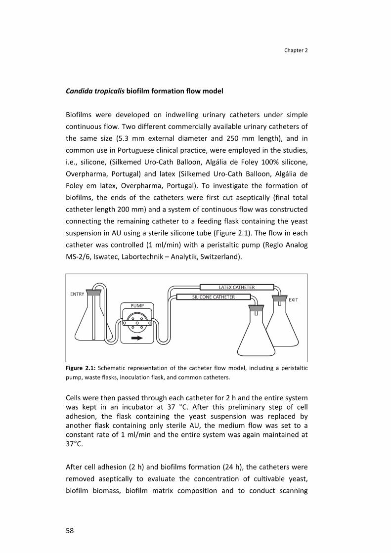
Chapter 2
58
Candida tropicalis biofilm formation flow model
Biofilms were developed on indwelling urinary catheters under simple continuous flow. Two different commercially available urinary catheters of the same size (5.3 mm external diameter and 250 mm length), and in common use in Portuguese clinical practice, were employed in the studies, i.e., silicone, (Silkemed Uro-‐Cath Balloon, Algália de Foley 100% silicone, Overpharma, Portugal) and latex (Silkemed Uro-‐Cath Balloon, Algália de Foley em latex, Overpharma, Portugal). To investigate the formation of biofilms, the ends of the catheters were first cut aseptically (final total catheter length 200 mm) and a system of continuous flow was constructed connecting the remaining catheter to a feeding flask containing the yeast suspension in AU using a sterile silicone tube (Figure 2.1). The flow in each catheter was controlled (1 ml/min) with a peristaltic pump (Reglo Analog MS-‐2/6, Iswatec, Labortechnik – Analytik, Switzerland).
Figure 2.1: Schematic representation of the catheter flow model, including a peristaltic pump, waste flasks, inoculation flask, and common catheters.
Cells were then passed through each catheter for 2 h and the entire system was kept in an incubator at 37 °C. After this preliminary step of cell adhesion, the flask containing the yeast suspension was replaced by another flask containing only sterile AU, the medium flow was set to a constant rate of 1 ml/min and the entire system was again maintained at 37°C.
After cell adhesion (2 h) and biofilms formation (24 h), the catheters were removed aseptically to evaluate the concentration of cultivable yeast, biofilm biomass, biofilm matrix composition and to conduct scanning
Model for Candida tropicalis biofilm study
59
electron microscope studies. All the experiments were performed in triplicate and in three independent assays.
Candida cells quantification
Adhesion or biofilm samples
The culture medium was removed from the catheter by passing air through the flow system and adhered or biofilm cells were scraped into PBS, after which each catheter was cut in small portions (10 mm). The time and potency of sonication had been previously established to optimize the complete removal of the adhered cells without causing any damage to them (optimization was followed with crystal violet staining and the determination of CFU). The catheter portions immersed in PBS were sonicated (Ultrasonic Processor, Cole-‐ Parmer, Illinois, USA) for 45 s at 30 W or 50 s at 30 W for the silicone and the latex catheters, respectively. Then the resultant suspension was vortexed for 2 min and serial dilutions in PBS were inoculated onto SDA plates and incubated for 24 h at 37 °C. The total colony forming units (CFUs) were counted per unit area (CFU/cm2) of catheter. These catheter portions were stained using the crystal violet (CV) method [14, 21] to verify the complete removal of the cells.
Biofilm detached cells
After the first 2 h of adhesion, the cells released from the catheters were collected (1 ml) at various time points during biofilm development at both the entry and exit of the flow system. The dispersed cells were determined by enumerating the CFUs.
Biofilm biomass
Biofilm formation was assessed by total biomass quantification using CV staining [14, 21]. After biofilm formation (24 h), the catheters were cut as described previously into sections. The catheter portions containing

Chapter 2
60
biofilms were transferred to 24-‐wells polystyrene microtiter plates (Orange Scientific c, Braine-‐l’ Alleud, Belgium) and fixed for 15 min with 1 ml of 100% (v/v) methanol. The catheter portions were then allowed to dry at room temperature, and 1 ml of CV (1% v/v) was added to each well and incubated for 5 min. The samples were gently washed with sterile water and 1 ml of acetic acid (33% v/v) was added to release the CV from the biofilm. The absorbance of the resultant solution was read in triplicate in a microtiter plate reader (Bio-‐Tek Synergy HT, Izasa, Lisbon, Portugal) at λ=570 nm. The final absorbance was standardized according to the volume of acetic acid and area of the catheter (absorbance/cm2). The Candida biofilms’ quantification was performed in triplicate and in three independent assays.
Scanning electron microscopy (SEM)
Catheter portions were prepared as described in biofilm biomass quantification and they were transferred to 24-‐well polystyrene microtiter plates. The samples were dehydrated with alcohol (using 70% ethanol for 10 min, 95% ethanol for 10 min and 100% ethanol for 20 min) and air dried for 20 min. Samples were kept in a desiccator until each catheter portion was removed for analysis. Prior to observation, the catheter portions were mounted onto aluminium stubs, sputter coated with gold and observed with an S-‐360 scanning electron microscope (Leo, Cambridge, USA).
Biofilm matrix composition
Extraction method
The biofilm matrix was extracted in accord with the procedures described by Silva et al. [26]. Briefly, biofilm samples were prepared as described before, sonicated and vortexed. Then the suspension was centrifuged at 3000 g for 10 min at 4 °C and the supernatant (containing the matrix) filtered through a 0.2 mm nitrocellulose filter and stored at -‐20 °C prior to analysis. The pellets were dried at 60 °C to obtain a constant dry biofilm weight.
Model for Candida tropicalis biofilm study
61
Protein and carbohydrate quantification
The protein content of the biofilm matrix was measured using the BCA Kit (Bicinchoninic Acid, Sigma-‐Aldrich, St Louis, MO, USA), with bovine serum albumin (BSA) as standard. Total carbohydrate content of the biofilm matrix was estimated according to the procedure of Dubois et al. [27], employing glucose as the standard. The biofilm matrix composition was determined in triplicate and in three independent assays.
Candida surface properties
Contact angle measurement
The degree of hydrophobicity (ΔGsws) was evaluated through contact angle measurements using the approach of van Oss et al. [28]. The measurements were made on Candida cell lawns on membrane filters, prepared according to Busscher et al. [29]. For this, suspensions of 5 ml of 1 x 10⁸ cells/ml in AU were filtered, inoculated over solidified agar plates (2% agar and 10% glycerol) and dried at 37 °C for 3 – 4 h to standardize the humidity level. Contact angles were measured by the sessile drop technique on the cell lawns, using a contact angle measurement apparatus (model OCA 15 Plus, Dataphysics). In order to determine the degree of hydrophobicity (ΔGsws), the measurements were made at room temperature, using three different liquids, i.e., water, formamide and 1-‐bromonaphtalene. Water contact angles were used as a qualitative indication of the cell surface hydrophobicity, with an angle lower than 65° indicating a more hydrophilic surface [30]. Each assay was performed in triplicate and at least 20 contact angles, per sample, were measured.
Statistical analysis
Results obtained were analysed using the SPSS 18 version (Statistical Package for the Social Sciences) program. Onaway ANOVA with the Bonferroni test was used to compare the number of adherent or biofilm cells and matrix composition of the strains assayed. All tests were performed with a confidence level of 95%. All the experiments were performed in triplicate and in three independent assays.
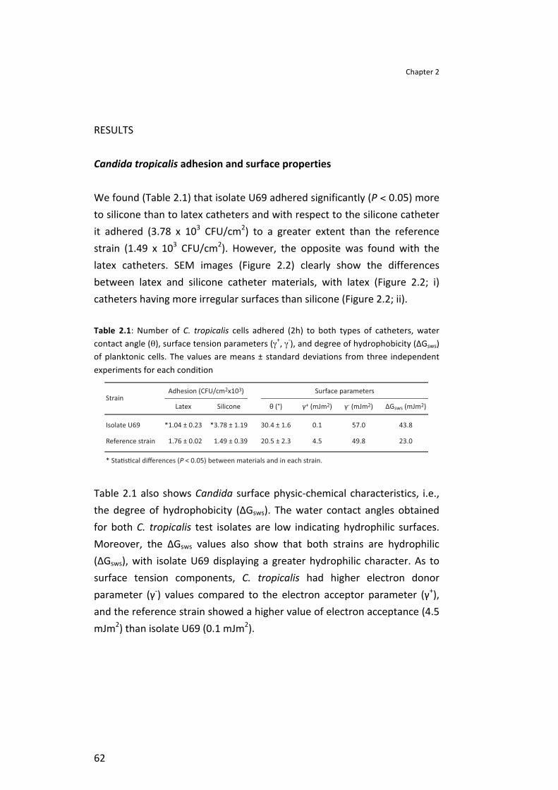
Chapter 2
62
RESULTS
Candida tropicalis adhesion and surface properties
We found (Table 2.1) that isolate U69 adhered significantly (P < 0.05) more to silicone than to latex catheters and with respect to the silicone catheter it adhered (3.78 x 10³ CFU/cm2) to a greater extent than the reference strain (1.49 x 10³ CFU/cm2). However, the opposite was found with the latex catheters. SEM images (Figure 2.2) clearly show the differences between latex and silicone catheter materials, with latex (Figure 2.2; i) catheters having more irregular surfaces than silicone (Figure 2.2; ii).
Table 2.1: Number of C. tropicalis cells adhered (2h) to both types of catheters, water contact angle (θ), surface tension parameters (γ+, γ-‐), and degree of hydrophobicity (ΔGsws) of planktonic cells. The values are means ± standard deviations from three independent experiments for each condition
Table 2.1 also shows Candida surface physic-‐chemical characteristics, i.e., the degree of hydrophobicity (ΔGsws). The water contact angles obtained for both C. tropicalis test isolates are low indicating hydrophilic surfaces. Moreover, the ΔGsws values also show that both strains are hydrophilic (ΔGsws), with isolate U69 displaying a greater hydrophilic character. As to surface tension components, C. tropicalis had higher electron donor parameter (γ-‐) values compared to the electron acceptor parameter (γ+), and the reference strain showed a higher value of electron acceptance (4.5 mJm2) than isolate U69 (0.1 mJm2).
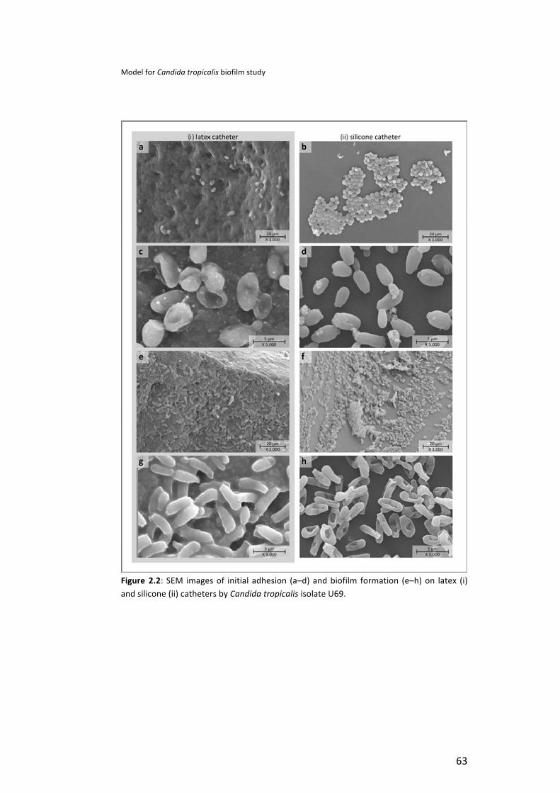
Model for Candida tropicalis biofilm study
63
Figure 2.2: SEM images of initial adhesion (a–d) and biofilm formation (e–h) on latex (i) and silicone (ii) catheters by Candida tropicalis isolate U69.
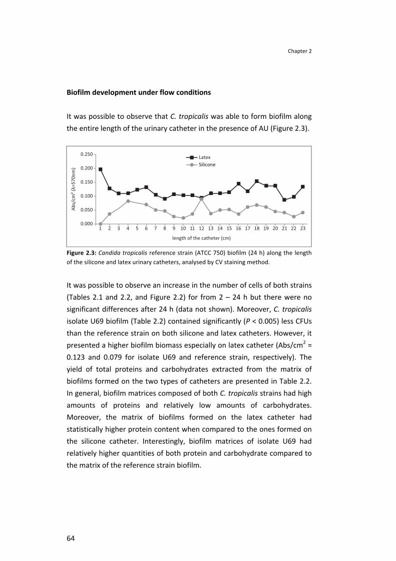
Chapter 2
64
Biofilm development under flow conditions
It was possible to observe that C. tropicalis was able to form biofilm along the entire length of the urinary catheter in the presence of AU (Figure 2.3).
Figure 2.3: Candida tropicalis reference strain (ATCC 750) biofilm (24 h) along the length of the silicone and latex urinary catheters, analysed by CV staining method.
It was possible to observe an increase in the number of cells of both strains (Tables 2.1 and 2.2, and Figure 2.2) for from 2 – 24 h but there were no significant differences after 24 h (data not shown). Moreover, C. tropicalis isolate U69 biofilm (Table 2.2) contained significantly (P < 0.005) less CFUs than the reference strain on both silicone and latex catheters. However, it presented a higher biofilm biomass especially on latex catheter (Abs/cm2 = 0.123 and 0.079 for isolate U69 and reference strain, respectively). The yield of total proteins and carbohydrates extracted from the matrix of biofilms formed on the two types of catheters are presented in Table 2.2. In general, biofilm matrices composed of both C. tropicalis strains had high amounts of proteins and relatively low amounts of carbohydrates. Moreover, the matrix of biofilms formed on the latex catheter had statistically higher protein content when compared to the ones formed on the silicone catheter. Interestingly, biofilm matrices of isolate U69 had relatively higher quantities of both protein and carbohydrate compared to the matrix of the reference strain biofilm.
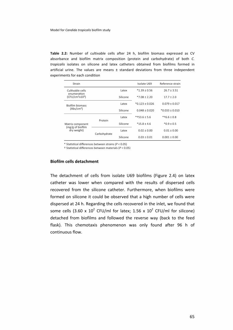
Model for Candida tropicalis biofilm study
65
Table 2.2: Number of cultivable cells after 24 h, biofilm biomass expressed as CV absorbance and biofilm matrix composition (protein and carbohydrate) of both C. tropicalis isolates on silicone and latex catheters obtained from biofilms formed in artificial urine. The values are means ± standard deviations from three independent experiments for each condition
Biofilm cells detachment
The detachment of cells from isolate U69 biofilms (Figure 2.4) on latex catheter was lower when compared with the results of dispersed cells recovered from the silicone catheter. Furthermore, when biofilms were formed on silicone it could be observed that a high number of cells were dispersed at 24 h. Regarding the cells recovered in the inlet, we found that some cells (3.60 x 10² CFU/ml for latex; 1.56 x 10² CFU/ml for silicone) detached from biofilms and followed the reverse way (back to the feed flask). This chemotaxis phenomenon was only found after 96 h of continuous flow.
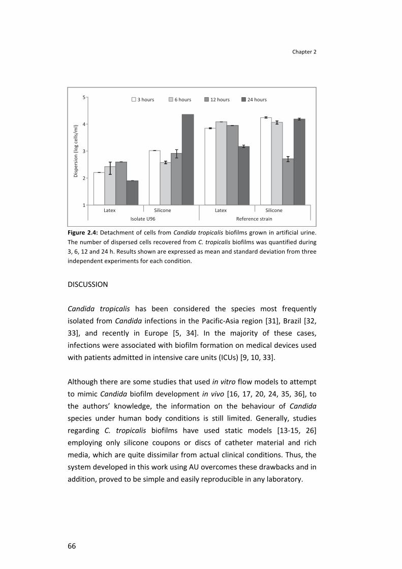
Chapter 2
66
Figure 2.4: Detachment of cells from Candida tropicalis biofilms grown in artificial urine. The number of dispersed cells recovered from C. tropicalis biofilms was quantified during 3, 6, 12 and 24 h. Results shown are expressed as mean and standard deviation from three independent experiments for each condition.
DISCUSSION
Candida tropicalis has been considered the species most frequently isolated from Candida infections in the Pacific-‐Asia region [31], Brazil [32, 33], and recently in Europe [5, 34]. In the majority of these cases, infections were associated with biofilm formation on medical devices used with patients admitted in intensive care units (ICUs) [9, 10, 33].
Although there are some studies that used in vitro flow models to attempt to mimic Candida biofilm development in vivo [16, 17, 20, 24, 35, 36], to the authors’ knowledge, the information on the behaviour of Candida species under human body conditions is still limited. Generally, studies regarding C. tropicalis biofilms have used static models [13-‐15, 26] employing only silicone coupons or discs of catheter material and rich media, which are quite dissimilar from actual clinical conditions. Thus, the system developed in this work using AU overcomes these drawbacks and in addition, proved to be simple and easily reproducible in any laboratory.
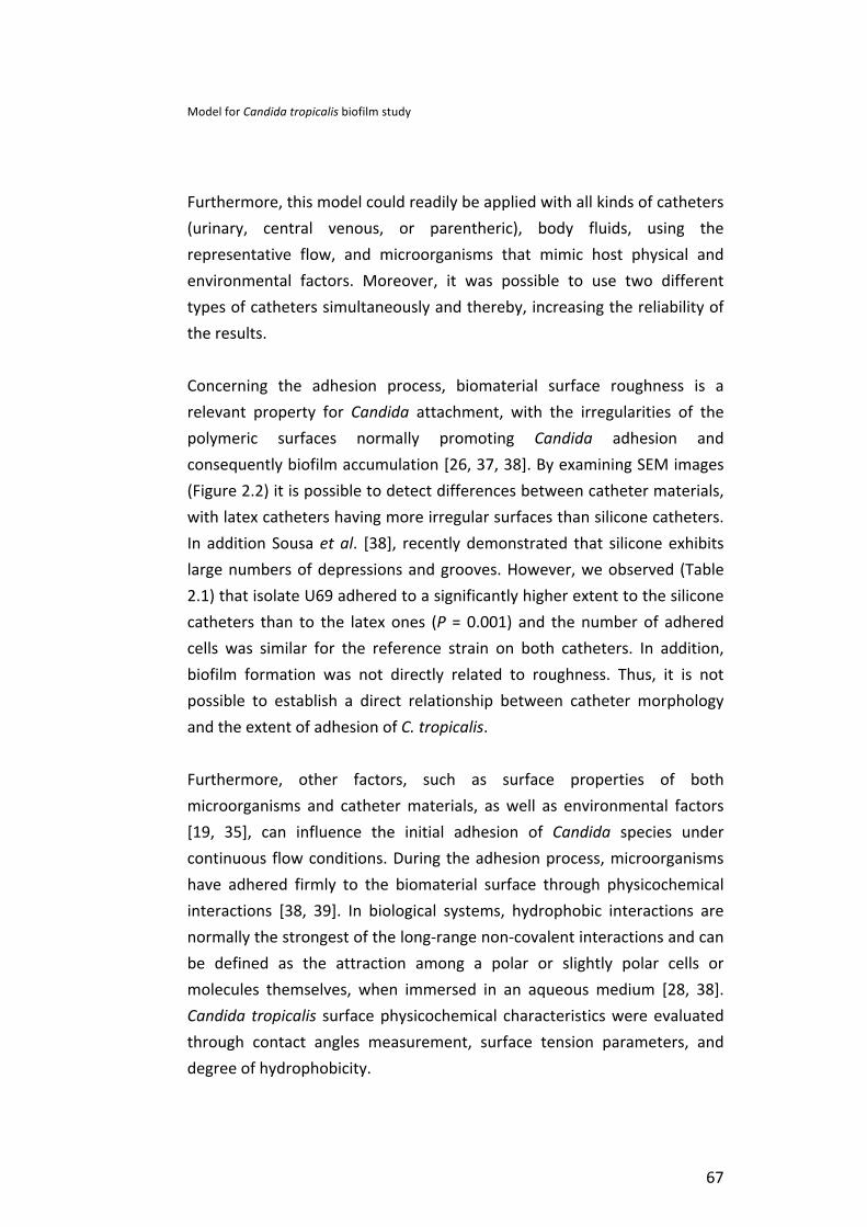
Model for Candida tropicalis biofilm study
67
Furthermore, this model could readily be applied with all kinds of catheters (urinary, central venous, or parentheric), body fluids, using the representative flow, and microorganisms that mimic host physical and environmental factors. Moreover, it was possible to use two different types of catheters simultaneously and thereby, increasing the reliability of the results.
Concerning the adhesion process, biomaterial surface roughness is a relevant property for Candida attachment, with the irregularities of the polymeric surfaces normally promoting Candida adhesion and consequently biofilm accumulation [26, 37, 38]. By examining SEM images (Figure 2.2) it is possible to detect differences between catheter materials, with latex catheters having more irregular surfaces than silicone catheters. In addition Sousa et al. [38], recently demonstrated that silicone exhibits large numbers of depressions and grooves. However, we observed (Table 2.1) that isolate U69 adhered to a significantly higher extent to the silicone catheters than to the latex ones (P = 0.001) and the number of adhered cells was similar for the reference strain on both catheters. In addition, biofilm formation was not directly related to roughness. Thus, it is not possible to establish a direct relationship between catheter morphology and the extent of adhesion of C. tropicalis.
Furthermore, other factors, such as surface properties of both microorganisms and catheter materials, as well as environmental factors [19, 35], can influence the initial adhesion of Candida species under continuous flow conditions. During the adhesion process, microorganisms have adhered firmly to the biomaterial surface through physicochemical interactions [38, 39]. In biological systems, hydrophobic interactions are normally the strongest of the long-‐range non-‐covalent interactions and can be defined as the attraction among a polar or slightly polar cells or molecules themselves, when immersed in an aqueous medium [28, 38]. Candida tropicalis surface physicochemical characteristics were evaluated through contact angles measurement, surface tension parameters, and degree of hydrophobicity.

Chapter 2
68
The water contact angles obtained for C. tropicalis are low and the ΔGsws was positive, both of which are in agreement and indicate surface hydrophilic character of Candida cells (Table 2.1). Moreover, according to Kazmierska et al. [40] silicone appears to be more hydrophobic than latex and according to Sousa et al. [38] the higher surface hydrophobicity of silicone is probably responsible for the highest levels of initial cell adhesion to this substrate. However, isolate U69, which is the most hydrophilic strain (higher ΔGsws), presented the highest extent of adhesion to silicone, the most hydrophobic material (Table 2.1). Therefore, based on these results (Table 2.1) no direct correlation was found between adhesion capability and the degree of cell hydrophobicity. This is in accord with other reports [13, 37], and demonstrates that cell hydrophobicity alone cannot be used for predicting the microbial adhesion. Another factor that is usually involved in determining the extent of adhesion is the surface tension components of the microorganism [38]. Cell surfaces of C. tropicalis strains were found (Table 2.1) to be predominantly electron donors (higher values of γ-‐). This polar character can be due to the presence of residual water of hydration or polar groups [28]. However, the low value of the electron acceptor parameter (γ+ = 0.1) of isolate U69 can also justify its highest adhesion to the more hydrophobic material such as silicone.
The extent of adhesion of C. tropicalis to these urinary catheters is in accordance to Silva et al. [13], who described the adherence of NCAC species, such as C. tropicalis, on silicone in the presence of artificial urine. After the initial process of attachment of individual cells to a substratum, what follows is the proliferation and formation of a highly structured mature biofilm comprised of complex intertwining layers of yeast, pseudohyphae and hyphae embedded in extracellular matrix [19, 24]. Candida tropicalis was able to form biofilms along the entire length of urinary catheters in the presence of AU (Figure 2.3). Furthermore SEM images revealed an increase in the number of cells attached to both types of catheters from 2 h (Figure 2.2 a – d) to 24 h (Figure 2.2 e – h). In fact, Candida species are able to adhere and develop biofilms in the presence of AU with the number of cells proliferating from the adhesion stage to
Model for Candida tropicalis biofilm study
69
biofilm formation [13, 41]. These data corroborate the in vivo situation when Candida biofilms grow on urinary catheters, which are intermittently bathed by patients’ urine as the only source of nutrients, and are undoubtedly responsible for patients’ infections. However, in this work, there was no direct correlation between the number of adhered cells and the number of biofilm cells. In fact the strain displaying the highest CFU number in initial adhesion (isolate U69) presented less CFUs in biofilms. Nevertheless, other authors [13, 14, 35, 37] have reported a correlation between the extent of adhesion and biofilm formation. However, Cerca et al. [42] have demonstrated that initial adhesion and biofilm formation are not always directly related and that this fact can be determined by cell-‐to-‐cell adhesion ability to make possible more than one cell layer.
Regarding biofilms, one of their most important characteristics is the presence and the composition of the extracellular matrix [17, 19], which can act as a barrier to diffusion of antimicrobial agents, thereby limiting access of antimicrobials to organisms at the base of the biofilm [17, 43]. Al-‐Fattani and Douglas [17], chemically analysed the matrix material from C. tropicalis biofilms and reported the synthesis of large concentrations of extrapolymeric material composed of hexosamine, carbohydrates, proteins, phosphorus, and uronic acid. In this study biofilm biomass and matrix composition (proteins and carbohydrates) was also evaluated and a higher amount of biofilm biomass and matrix proteins content were verified for biofilms formed on latex catheter than on silicone (Table 2.2).
Formation of mature biofilms and consequent production of matrix is strongly dependent on environmental conditions, such as medium composition, pH and oxygen [13, 26, 41]. Furthermore, some studies [35, 43-‐45] indicated that specific proteins are associated to adhesion, biofilm formation and dispersal of cells and the expressed quantities of these proteins depend not only on the microorganism but also on the surface where the biofilm is formed. In addition, biofilm matrices of isolate U69 had relatively higher quantities of both proteins and carbohydrates, and presented smaller CFU values compared to the reference strain.

Chapter 2
70
Formation of biofilms allows microbial pathogens to create a safe niche in which sessile cells remain in a protected environment. However, cells within a biofilm may be also confronted with adverse environmental conditions (i.e., reduced nutrient availability, accumulation of toxic waste products) so the dispersion of cells is also beneficial for their survival. Furthermore, this release of cells from the original biofilm community is required to generate new communities at other locations and the dispersal of cells from biofilms may be a critical step in biomaterial related cases of candidosis [35, 36, 43]. In this work, it was curious to note that cells detached from the biofilms started to follow backwards after 96 h (data not shown), confirming the possibility of colonization of distant host sites. Still it was possible to observe that cells were dispersed from biofilms from the first time points (Figure 2.4) and the number of dispersed cells recovered from latex catheter was lower than cells recovered from silicone catheter. The highest cell detachment from silicone catheter at 24 h was noted with U69. Accordingly, Uppuluri et al. [36] reported that dispersion occurs at all stages of the biofilm developmental cycle and is influenced by nutritional and other physiochemical conditions.
In conclusion, the model presented allows the easy study of Candida biofilm lifecycle and can better mimic the host physical and environmental conditions than the traditional systems. It was also possible to conclude that C. tropicalis were able to form biofilms in artificial urine on different urinary catheters under flow condition. In all situations there was a significant production of matrix components. As expected, cells were released constantly from biofilms, and were recovered at the inlet after 96 h, highlighting their great effect as human pathogens. However, there was no direct relation between cells and material surface properties (degree of hydrophobicity, surface tension and materials surface morphology) and adhesion capability and biofilm formation.
Model for Candida tropicalis biofilm study
71
REFERENCES
1. Lundstrom T, Sobel J. Nosocomial Candiduria: A Review. Clin Infect Dis 2001; 32 (11): 1602-‐1607. 2. Bouza E, Juan RS, Muñoz P, Voss A, Kluytmans J. A European perspective on nosocomial urinary tract infections II. Report on incidence, clinical characteristics and outcome (ESGNI-‐004 study). Clin Microbiol Infect 2001; 7 (10): 532-‐542. 3. Richards MJ, Edwards JR, Culver DH, Gaynes RP. Nosocomial infections in medical intensive care units in the United States. Crit Care Med 1999; 27 (5): 887-‐892. 4. Snydman DR. Shifting Patterns in the Epidemiology of Nosocomial Candida Infections. Chest 2003; 123 (5 Suppl): 500S-‐3S. 5. Lass-‐Flörl C. The changing face of epidemiology of invasive fungal disease in Europe. Mycoses 2009; 52 (3): 197-‐205. 6. Kauffman CA, Vazquez JA, Sobel JD, et al. Prospective Multicenter Surveillance Study of Funguria in Hospitalized Patients. Clin Infect Dis 2000; 30 (1): 14-‐18. 7. Nett J, Andes D. Review of techniques for diagnosis of catheter-‐related Candida biofilm infections. Curr Fungal Infect Rep 2008; 2 (4): 237-‐243. 8. Álvarez-‐Lerma F, Nolla-‐Salas J, León C, et al. Candiduria in critically ill patients admitted to intensive care medical units. Intensive Care Med. 2003; 29 (7): 1069-‐1076. 9. Eggimann P, Garbino J, Pittet D. Epidemiology of Candida species infections in critically ill non-‐immunosuppressed patients. Lancet Infect. Dis. 2003; 3 (11): 685-‐702. 10. Colombo AL, Nucci M, Park BJ, et al. Epidemiology of Candidemia in Brazil: a Nationwide Sentinel Surveillance of Candidemia in Eleven Medical Centers. J Clin Microbiol 2006; 44 (8): 2816-‐2823. 11. Krcmery V, Barnes AJ. Non-‐albicans Candida spp. causing fungaemia: pathogenicity and antifungal resistance. J Hosp Infect 2002; 50 (4): 243-‐260. 12. Okawa Y, Miyauchi M, Kobayashi H. Comparison of Pathogenicity of Various Candida tropicalis Strains. Biol Pharm Bull 2008; 31 (8): 1507-‐1510. 13. Silva S, Negri M, Henriques M, et al. Silicone colonization by non-‐Candida albicans Candida species in the presence of urine. J Med Microbiol 2010; 59 (Pt 7): 747-‐54. 14. Negri M, Martins M, Henriques M, et al. Examination of potential virulence factors of Candida tropicalis clinical isolates from hospitalized patients. Mycopathologia 2010; 169 (3): 175-‐182. 15. Bizerra FC, Nakamura CV, de Poersch C, et al. Characteristics of biofilm formation by Candida tropicalis and antifungal resistance. FEMS Yeast Res 2008; 8 (3): 442-‐450. 16. Hawser SP, Douglas LJ. Biofilm formation by Candida species on the surface of catheter materials in vitro. Infect Immun 1994; 62 (3): 915-‐921. 17. Al-‐Fattani MA, Douglas LJ. Biofilm matrix of Candida albicans and Candida tropicalis: chemical composition and role in drug resistance. J Med Microbiol 2006; 55 (8): 999-‐1008. 18. Nailis H, Vandenbroucke R, Tilleman K, et al. Monitoring ALS1 and ALS3 gene expression during in vitro Candida albicans biofilm formation under continuous flow conditions. Mycopathologia 2009; 167 (1): 9-‐17. 19. Douglas LJ. Candida biofilms and their role in infection. Trends Microbiol 2003; 11 (1): 30-‐36. 20. Ramage G, Wickes B, López-‐Ribot J. A seed and feed model for the formation of Candida albicans biofilms under flow conditions using an improved modified Robbins device. Rev Iberoam Micol. 2008; 25 (1): 37-‐40. 21. Stepanović S, Vuković D, Ježek P, Pavlović M, Švabic-‐Vlahović M. Influence of Dynamic Conditions on Biofilm Formation by Staphylococci. Eur J Clin Microbiol Infect Dis 2001; 20 (7): 502-‐504. 22. Donlan R. Biofilms and device-‐associated infections. Emerg Infect Dis 2001; 7 (2): 277-‐281. 23. Andes D, Nett J, Oschel P, et al. Development and characterization of an in vivo central venous catheter Candida albicans biofilm model. Infect Immun 2004; 72 (10): 6023-‐6031. 24. Uppuluri P, Chaturvedi A, Lopez-‐Ribot J. Design of a Simple Model of Candida albicans Biofilms Formed under Conditions of Flow: Development, Architecture, and Drug
Chapter 2
72
Resistance. Mycopathologia 2009; 168 (3): 101-‐109. 25. Seidler M, Salvenmoser S, Müller F-‐MC. Liposomal amphotericin B eradicates Candida albicans biofilm in a continuous catheter flow model. FEMS Yeast Res 2010; 10 (4): 492-‐495. 26. Silva S, Henriques M, Martins A, et al. Biofilms of non-‐Candida albicans Candida species: quantification, structure and matrix composition. Med Mycol 2009; 47 (7): 681-‐9. 27. DuBois M, Gilles KA, Hamilton JK, Rebers PA, Smith F. Colorimetric Method for Determination of Sugars and Related Substances. Anal Chem 1956; 28 (3): 350-‐356. 28. Van Oss CJ, Ju L, Chaudhury MK, Good RJ. Estimation of the polar parameters of the surface tension of liquids by contact angle measurements on gels. J Colloid Interf Sci 1989; 128 (2): 313-‐319. 29. Busscher HJ, Weerkamp AH, van der Mei HC, et al. Measurement of the surface free energy of bacterial cell surfaces and its relevance for adhesion. Appl Environ Microbiol 1984; 48 (5): 980-‐983. 30. Vogler EA. Structure and reactivity of water at biomaterial surfaces. Adv Colloid Interfac 1998; 74 (1-‐3): 69-‐117. 31. Chakrabarti A, Chatterjee SS, Rao KLN, et al. Recent experience with fungaemia: change in species distribution and azole resistance. Scand J Infect Dis 2009; 41 (4): 275 -‐ 284. 32. Colombo AL, Guimarães T, Silva LRBF, et al. Prospective Observational Study of Candidemia in Sãoo Paulo, Brazil: Incidence Rate, Epidemiology, and Predictors of Mortality. Infect Control Hosp Epidemiol 2007; 28 (5): 570-‐576. 33. Nucci M, Colombo AL. Candidemia due to Candida tropicalis: clinical, epidemiologic, and microbiologic characteristics of 188 episodes occurring in tertiary care hospitals. Diagn Microbiol Infect Dis 2007; 58 (1): 77-‐82. 34. Paulo C, Mourão C, Veiga PM, et al. Retrospective analysis of clinical yeast isolates in a hospital in the centre of Portugal: spectrum and revision of the identification procedures. Med Mycol 2009; 19 (1): 1 -‐ 10. 35. Sellam A, Al-‐Niemi T, McInnerney K, et al. A Candida albicans early stage biofilm detachment event in rich medium. BMC Microbiol 2009; 9 (1): 25. 36. Uppuluri P, Chaturvedi AK, Srinivasan A, et al. Dispersion as an Important Step in the Candida albicans Biofilm Developmental Cycle. PLoS Pathog 2010; 6 (3): e1000828. 37. Camacho D, Gasparetto A, Svidzinski T. The effect of chlorhexidine and gentian violet on the adherence of Candida spp. to urinary catheters. Mycopathologia 2007; 163 (5): 261-‐266. 38. Sousa C, Teixeira P, Oliveira R. Influence of Surface Properties on the Adhesion of Staphylococcus epidermidis to Acrylic and Silicone. Int J Biomater 2009; 2009, Article ID 718017 (Article ID 718017,): 9 pages. 39. Chatzinikolaou I, Raad I. Intravascular catheter-‐related infections: a preventable challenge in the critically ill. Semin Respir Infect. 2000; 15 (4): 264-‐271. 40. Kazmierska K, Szwast M, Ciach T. Determination of urethral catheter surface lubricity. J Mater Sci Mater Med 2008; 19 (6): 2301-‐2306. 41. Uppuluri P, Dinakaran H, Thomas DP, Chaturvedi AK, Lopez-‐Ribot JL. Characteristics of Candida albicans Biofilms Grown in a Synthetic Urine Medium. J Clin Microbiol 2009; 47 (12): 4078-‐4083. 42. Cerca N, Martins S, Sillankorva S, et al. Effects of Growth in the Presence of Subinhibitory Concentrations of Dicloxacillin on Staphylococcus epidermidis and Staphylococcus haemolyticus Biofilms. Appl Environ Microbiol 2005; 71 (12): 8677-‐8682. 43. Blankenship JR, Mitchell AP. How to build a biofilm: a fungal perspective. Curr Opin Microbiol 2006; 9 (6): 588-‐594. 44. Chandra J, Patel JD, Li J, et al. Modification of surface properties of biomaterials influences the ability of Candida albicans to form biofilms. Appl Environ Microbiol 2005; 71 (12): 8795-‐8801. 45. Chandra J, Kuhn DM, Mukherjee PK, et al. Biofilm Formation by the Fungal Pathogen Candida albicans: Development, Architecture, and Drug Resistance. J Bacteriol 2001; 183 (18): 5385-‐5394.

CHAPTER 3
Crystal violet staining to quantifyCandida adhesion to epithelial cells

Chapter 3-‐ Quantification method for Candida adhesion
75
ABSTRACT
In vitro studies of adhesion capability are essential to characterise the virulence of Candida species. However, the assessment of adhesion by traditional methods is time consuming. The aim of the present study is the development of a simple methodology using crystal violet staining to quantify in vitro adhesion of different Candida species to epithelial cells. The experiments are performed using Candida albicans (ATCC 90028), C. glabrata (ATCC 2001), C. parapsilosis (ATCC 22019) and C. tropicalis (ATCC 750). A human urinary bladder epithelial cell line (TCC-‐SUP) is used. Yeast and epithelial cells were stained with crystal violet, epithelial cells were then distained using intermediate washing, and the dye in the yeast cells was extracted with acetic acid. The method was validated for the different Candida reference species by comparison with traditional microscope observation and enumeration. The method was then used to assess Candida adhesion to epithelial cells and also to silicone. For all Candida species High correlation values (r2= 0.9724 – 0.9997) between the number of adherent yeasts (microscope enumeration) and absorbance values were obtained for an inoculum concentration > 106 cells/ml. The proposed technique was easy to perform and reproducible, enabling the determination of adhesion ability of Candida species to an epithelial cell line. Keywords: Adhesion; Candida; cell line; epithelial cells; gentian violet; silicone. Negri M, Gonçalves V, Silva S, Henriques M, Azeredo J, Oliveira R. Crystal violet expedite method for quantification adhesion to epithelial cells. British Journal of Biomedical Science, 2010; 67 (3): 120-‐125.
Chapter 3
76
INTRODUCTION
Candida species can adhere to a variety of different surfaces in the human body, thus facilitating the colonisation of many host niches. Remarkably, such niches provide very different environments for growth, and Candida has developed specific mechanisms to adapt to the respective conditions. Several studies have shown a correlation between adhesion of organisms and their potential virulence [1-‐4].
Adhesion to epithelial cells is well recognised as an essential step in the process of Candida colonisation and subsequent infection [5]. Candida adhesion to epithelial cells has been investigated to define parameters relevant to the pathogenesis of oral, gastrointestinal, vaginal and urinary candidiasis [6]. Furthermore, Candida can also grow on abiotic surfaces (e.g., plastic devices), for example, leading to biofilm formation in catheters, which represents a major problem especially in intensive care units [2, 7].
Over the past decades, a broad range of model systems have been described for the in vitro study of Candida adhesion to hard surfaces [8]. In most model systems, quantification of yeast cells is obtained by plating, which is labour-‐intensive and slow [9]. Moreover, yeast adhesion to epithelium can be determined by visual methods (e.g., light, fluorescence, scanning or transmission electron microscopy) or by counting radiolabelled yeast [6].
The visual method involves incubating standard yeast suspensions with confluent cell monolayers grown on a coverslip. Following removal of unattached yeasts, the number of adherent organisms per unit area of the monolayer is determined by direct microscopy after air drying, Gram staining and mounting on glass slides. Although visualisation allows monitoring of adhesion to individual epithelial cells, it is a very time-‐consuming technique [10, 11]. The radiolabelling method seems to offer an attractive alternative in some situations, although leaching of the isotope can produce misleading results [6].
Quantification method for Candida adhesion
77
Crystal violet (CV) staining, which is commonly used for the indirect quantification of adherent cells and amount of biofilm formed by Candida on abiotic surfaces, is a quick and cheap method. It involves a basic dye, which binds to negatively charged surface molecules and polysaccharides in the extracellular matrix, [8, 12] and dissolves easily in acetic acid.
The aim of this study is to develop a quick and simple technique to assess the number of Candida adherent to epithelial cells, based on the quantification of crystal violet absorbance.
MATERIAL AND METHODS
Yeasts and growth conditions
Candida species used in this study were Candida albicans (ATCC 90028), C. glabrata (ATCC 2001), C. parapsilosis (ATCC 22019) and C. tropicalis (ATCC 750), obtained from the American Type Culture Collection. Strains were kept frozen at – 80 °C in Sabouraud dextrose broth (SDB; Liofilchem, Italy) containing 5% (v/v) glycerol. Candida species were subcultured on Sabouraud dextrose agar (SDA; Liofilchem, Italy) for 24 h and then grown in SDB for 18 h at 37 °C at 120 rpm. After incubation, yeasts were harvested at 8000 rpm for 5 min. Cells resuspended in phosphate-‐buffered saline (PBS, 0.01 mol/l, pH 7.5) were enumerated using a haemocytometer (Boeco, Germany) and the final concentration (specific to each assay) was adjusted with PBS.
Epithelial cells
A human urinary bladder epithelial cell line (TCC-‐SUP; DSMZ – German Collection of Microorganisms and Cell Cultures) was used. Cells were cultured at 37 °C in 5% CO2 in Dulbecco’s modified Eagle’s medium (DMEM; Gibco, USA) containing 15% fetal bovine serum (FBS; Gibco, USA) and 1% penicillin/streptomycin (P/S; Gibco, USA) in cell culture flasks. After

Chapter 3
78
achieving 80% confluence, cells were detached using a 25% trypsin-‐EDTA solution (Gibco, USA) and cell concentration was adjusted to 1 x 10⁶ cells/ml with fresh DMEM without P/S and added to each well of a six well plate. Wells were washed (x 2) with PBS prior to assay.
Silicone
Coupons (2 x 2 cm) were cut from a 1 mm layer of silicone (Neves e Neves, Trofa, Portugal). All coupons were cleaned by immersion in ultrapure water for 2 h, followed by immersion in ethanol 50% (v/v) for 4 h. After rinsing with ultrapure water and air-‐drying, the coupons were autoclaved for 15 min at 121 °C.
Adhesion assay
Yeast cells were suspended in PBS to final concentrations of 104, 105, 106, 107 and 108 yeast/ml. Then, 3 ml each cell suspension was added to each well of the plate for tissue culture containing either a confluent layer of epithelial cells or the silicone coupon. After incubation (2 h, 120 rpm, 37 °C) the wells were washed (x 2) with PBS to remove unattached yeasts. Yeast cells were quantified using the CV staining method and also light microscope observation. All procedures were repeated in triplicate in three separate assays.
Crystal violet assay
Crystal violet (3 ml, 1% [w/v] in water) was added to each well containing the epithelial cells with adherent yeasts and allowed to stain for 5 min. The wells were then washed (x 3) with PBS. To remove CV from the epithelial cells, 3 ml ethanol: acetone (1: 1) was added to the wells and removed immediately. Acetic acid (33%, 3 ml) was added to each well and absorbance was read at 570 nm. Wells containing epithelial cells without yeasts were used as controls. Mean absorbance of yeasts was expressed as absorbance per area of each well.
Quantification method for Candida adhesion
79
Candida adherent to silicone were quantified according to the method of Henriques et al. [13]. Briefly, the coupons containing adherent yeasts were removed from each well and immersed for 5 min in a new well plate containing 3 ml methanol. After discarding the methanol, the coupons were allowed to dry at room temperature. Crystal violet (3 ml) was added to each well and allowed to stain for 5 min. Coupons were then removed to a new well, washed with ultrapure water and immersed in 3 ml acetic acid (33%) to dissolve the stain. Coupons without yeasts were used as controls. Absorbance of the resultant acetic acid solution was read at 570 nm. Mean absorbance of yeasts was expressed as absorbance per unit area of the coupon.
Microscope observation
Epithelial cells and coupons with adherent yeasts were treated as described above, but without acetic acid. A duplicate of each plate was performed. Candida attached to TCC-‐SUP were quantified using an inverted light microscope (Nikon Diaphot, x 400 magnification). Ten fields were randomly observed in each well. As the samples were set up in triplicate for each experiment, the mean number of yeasts per 30 fields was expressed as number of cells per unit area of the well. Candida cells with small daughter cells were regarded as one cell.
Statistical analysis
Results obtained were analysed using the SPSS (Statistical Package for the Social Sciences) program. One-‐way ANOVA with Bonferroni test was used to compare the number of adherent cells of the four strains. P < 0.05 was considered significant.

Chapter 3
80
RESULTS
The method proposed in this study involves CV staining of Candida adherent to an epithelial cell monolayer. As cells, yeasts and epithelium are all stained with CV (Figure 3.1 a) it was necessary to develop a technique (using a mixture of ethanol/ acetone) that permitted the removal of CV from epithelial cells (Figure 3.1 b), allowing it to remain in the Candida cells (Figure 3.1 c). It was then possible, using acetic acid, to remove CV from the Candida and read the absorbance of the solution obtained.
Figure 3.1: Phase contrast images of the steps of the proposed method: a) yeasts and TCC-‐SUP cells stained with CV only; b) TCC-‐SUP cells distained with ethanol and acetone; c) Yeast cells stained strongly with crystal violet and TCC-‐SUP cells distained with ethanol and acetone (original magnification x 200).
After the confirmation, by microscopy, of the applicability of this method, it was necessary to validate the technique by assessing and comparing adhesion using traditional enumeration of adherent yeast cell by microscope observation. The relationship between CV absorbance and the number of Candida attached to epithelial cells is presented in Figure 3.2. The values obtained were r2 = 0.9995 for C. albicans, r2 = 0.9997 for C. tropicalis, r2 = 0.9724 for C. glabrata and r2 = 0.9997 for C. parapsilosis.
The method proved adequate for the detection of Candida attachment at high yeast numbers, specifically above 1 x 10⁵ cell/cm2 for C. albicans (Figure 3.2 a), 2 x 10⁵ cell/cm2 for C. tropicalis (Figure 3.2 b), 3 x 10⁴ cell/cm2 for C. glabrata (Figure 3.2 c) and 1 x 10⁴ cell/cm2 for C. parapsilosis (Figure 3.2 d).

Quantification method for Candida adhesion
81
Figure 3.2: Relationship between the number of a) Candida albicans, b) C. tropicalis, c) C. glabrata and d) C. parapsilosis adherent to TCC-‐SUP epithelial cells, and the corresponding CV absorbance (CV abs) at 570 nm. The adherent Candida species were expressed as yeast number or CV absorbance per area of each well. All procedures were performed in triplicate in three separate assays.
Different Candida species have different sizes and absorb distinct amounts of dye, which does not allow comparison of the level of adhesion through direct CV absorbance readings. However, standardisation of the results is possible using respective equation curves for each species.
After the implementation of the methodology, the extent of adhesion of the different Candida species to TCC-‐SUP epithelial cells and also to silicone was determined (Figure 3.3). As shown in Figure 3.3 a, there were significant differences in the number of yeasts adherent to epithelial cells among the different concentrations tested for each Candida specie, except for C. parapsilosis. On silicone, the differences were significant for all C. albicans and C. parapsilosis inocula concentrations but only for the highest values in C. glabrata and C. tropicalis (Figure 3.3 b). All Candida species adhered to a greater extent to epithelial cells than to silicone.

Chapter 3
82
Figure 3.3: Candida species cells adherent to a) TCC-‐SUP epithelial cells and b) silicone measured by crystal violet absorbance reported as cell/cm2. Data are the average of three measurements (+SD). The initial cell density: 106 (¢ ), 107 (¢ ) and 108 cell/ml (¢ ). *P < 0.05 between the different inoculums for the same species.
Considering the difference between species, it is possible to observe (Figure 3.3) that C. glabrata followed by C. tropicalis adhered significantly (P < 0.05) more than other Candida species (initial cell density: 108 yeast/ml) to epithelial cells. C. albicans adhered less than other yeasts to both surfaces at 108 yeasts/ml (P < 0.05).
The methodology proposed proved efficient in demonstrating the in vitro adherence of C. albicans (ATCC 90028), C. glabrata (ATCC 2001), C. parapsilosis (ATCC 22019) and C. tropicalis (ATCC 750) to TCC-‐SUP.
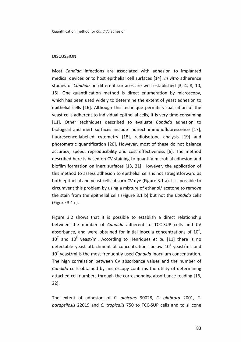
Quantification method for Candida adhesion
83
DISCUSSION
Most Candida infections are associated with adhesion to implanted medical devices or to host epithelial cell surfaces [14]. In vitro adherence studies of Candida on different surfaces are well established [3, 4, 8, 10, 15]. One quantification method is direct enumeration by microscopy, which has been used widely to determine the extent of yeast adhesion to epithelial cells [16]. Although this technique permits visualisation of the yeast cells adherent to individual epithelial cells, it is very time-‐consuming [11]. Other techniques described to evaluate Candida adhesion to biological and inert surfaces include indirect immunofluorescence [17], fluorescence-‐labelled cytometry [18], radioisotope analysis [19] and photometric quantification [20]. However, most of these do not balance accuracy, speed, reproducibility and cost effectiveness [6]. The method described here is based on CV staining to quantify microbial adhesion and biofilm formation on inert surfaces [13, 21]. However, the application of this method to assess adhesion to epithelial cells is not straightforward as both epithelial and yeast cells absorb CV dye (Figure 3.1 a). It is possible to circumvent this problem by using a mixture of ethanol/ acetone to remove the stain from the epithelial cells (Figure 3.1 b) but not the Candida cells (Figure 3.1 c).
Figure 3.2 shows that it is possible to establish a direct relationship between the number of Candida adherent to TCC-‐SUP cells and CV absorbance, and were obtained for initial inocula concentrations of 106, 107 and 108 yeast/ml. According to Henriques et al. [11] there is no detectable yeast attachment at concentrations below 104 yeast/ml, and 107 yeast/ml is the most frequently used Candida inoculum concentration. The high correlation between CV absorbance values and the number of Candida cells obtained by microscopy confirms the utility of determining attached cell numbers through the corresponding absorbance reading [16, 22].
The extent of adhesion of C. albicans 90028, C. glabrata 2001, C. parapsilosis 22019 and C. tropicalis 750 to TCC-‐SUP cells and to silicone

Chapter 3
84
was also determined using the proposed method (Figure 3.3). As Candida species differ on size, their absorbance value was standardised (according to each species curve) in order to permit comparison. It was noticed that different inoculum concentrations gave significantly different (P < 0.05) numbers of Candida adherent to the epithelial cells; an observation made by others [23-‐25] who report that the attachment of Candida species to epithelial cells gradually increases as the ratio of yeasts to epithelial cells, in incubation mixtures, is raised from 10: 1 to 10000: 1.
Differences were detected in the adherence of Candida species to TCC-‐SUP cells and silicone. All Candida were more prone to adhere to TCC-‐SUP than to silicone (Figure 3.3). Sohn et al. [3] described the comparison of adhesion of C. albicans to the human colorectal carcinoma cell line Caco-‐2 and to epidermoid vulvovaginal A431 cells and to polystyrene, reporting that C. albicans adherence is high to polystyrene and both epithelia.
C. albicans and C. parapsilosis adhered to a similar extent on both surfaces at an initial concentration of 108 cells/ml, but less than the other yeasts studied. Tamura et al. [26] investigated the adherence of C. albicans and C. parapsilosis to urinary catheters made of latex or silicone and found that adhesion of C. albicans was significantly greater than C. parapsilosis on latex, but similar on silicone. Furthermore, De Bernardis et al. [1] observed that from all the non-‐C. albicans species (NCAC) studied, C. parapsilosis seemed to be the least virulent, which might be consistent with lower adherence to epithelial cells.
Owing to the increasing prevalence of NCAC species, especially in immunosuppressed patients, more insight about virulence factors associated with these species is required. However, relatively little is known about the mechanisms of NCAC adhesion to epithelium or about factors affecting the adhesion process [11]. Candida tropicalis is a common species related to nosocomial candidemia and candiduria, and C. glabrata is now emerging as an important agent in both mucosal and bloodstream infections [14, 27]. Nevertheless, the adhesion mechanism of these species to different surfaces remains unclear [11, 14].
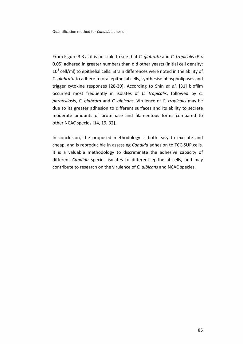
Quantification method for Candida adhesion
85
From Figure 3.3 a, it is possible to see that C. glabrata and C. tropicalis (P < 0.05) adhered in greater numbers than did other yeasts (initial cell density: 108 cell/ml) to epithelial cells. Strain differences were noted in the ability of C. glabrata to adhere to oral epithelial cells, synthesise phospholipases and trigger cytokine responses [28-‐30]. According to Shin et al. [31] biofilm occurred most frequently in isolates of C. tropicalis, followed by C. parapsilosis, C. glabrata and C. albicans. Virulence of C. tropicalis may be due to its greater adhesion to different surfaces and its ability to secrete moderate amounts of proteinase and filamentous forms compared to other NCAC species [14, 19, 32].
In conclusion, the proposed methodology is both easy to execute and cheap, and is reproducible in assessing Candida adhesion to TCC-‐SUP cells. It is a valuable methodology to discriminate the adhesive capacity of different Candida species isolates to different epithelial cells, and may contribute to research on the virulence of C. albicans and NCAC species.

Chapter 3
86
REFERENCES
1. De Bernardis F, Mondello F, San Millan R, Ponton J, Cassone A. Biotyping and Virulence Properties of Skin Isolates of Candida parapsilosis. J Clin Microbiol 1999; 37 (11): 3481-‐3486. 2. Douglas LJ. Candida biofilms and their role in infection. Trends Microbiol 2003; 11 (1): 30-‐36. 3. Sohn K, Senyürek I, Fertey J, et al. An in vitro assay to study the transcriptional response during adherence of Candida albicans to different human epithelia. FEMS Yeast Res 2006; 6 (7): 1085-‐1093. 4. Chassot F, Negri MFN, Svidzinski AE, et al. Can intrauterine contraceptive devices be a Candida albicans reservoir? Contraception 2008; 77 (5): 355-‐359. 5. Lyman C, Navarro E, Garrett K, et al. Adherence of Candida albicans to bladder mucosa: development and application of a tissue explant assay. Mycoses 1999; 42 (4): 255-‐259. 6. Ellepola ANB, Samaranayake LP. Investigative Methods for Studying the Adhesion and Cell Surface Hydrophobicity of Candida Species: An Overview. Microb Ecol Health Dis 2001; 13 (1): 46 -‐ 54. 7. Kumamoto CA. Candida biofilms. Curr Opin Microbiol 2002; 5 (6): 608-‐11. 8. Peeters E, Nelis H, Coenye T. Comparison of multiple methods for quantification of microbial biofilms grown in microtiter plates J Microbiol Methods 2008; 72 (2): 157-‐165. 9. Donlan RM, Costerton JW. Biofilms: survival mechanisms of clinically relevant microorganisms. Clin Microbiol Rev 2002; 15 (2): 167-‐93. 10. Samaranayake L, MacFarlane T. The adhesion of the yeast Candida albicans to epithelial cells of human origin in vitro. Arch Oral Biol 1981; 26 (10): 815-‐820. 11. Henriques M, Azeredo J, Oliveira R. Candida Species Adhesion to Oral Epithelium: Factors Involved and Experimental Methodology Used. Crit Rev Microbiol 2006; 32 (4): 217 -‐ 226. 12. Li F, Palecek SP. EAP1, a Candida albicans gene involved in binding human epithelial cells. Eukaryotic Cell 2003; 2: 1266 -‐ 1273. 13. Henriques M, Azeredo J, Oliveira R. Candida albicans and Candida dubliniensis: comparison of biofilm formation in terms of biomass and activity. Br J Biomed Sci 2006; 63 (1): 5-‐11. 14. Bizerra FC, Nakamura CV, de Poersch C, et al. Characteristics of biofilm formation by Candida tropicalis and antifungal resistance. FEMS Yeast Research 2008; 8 (3): 442-‐450. 15. Silva GMd, Silveira FRXd, Pires MdFC. Adherence to HeLa cells, typing by killer toxins and susceptibility to antifungal agents of Candida dubliniensis strains. Braz Oral Res 2007; 21: 87-‐91. 16. Irie MMT, Consolaro MEL, Guedes TA, et al. A simplified technique for evaluating the adherence of yeasts to human vaginal epithelial cells. J Clin Lab Anal 2006; 20 (5): 195-‐203. 17. Pacheco M, Pisa D, García-‐Gómez P, Carrasco L, Juarranz Á. Attachment and entry of Candida famata in monocytes and epithelial cells. Microsc Res Tech 2007; 70 (11): 975-‐986. 18. Polacheck I, Antman A, Barth I, Giloh ESH. Adherence of Candida albicans to epithelial cells: studies using fluorescently labelled yeasts and flow cytometry. Microbiology 1995; 141 (7): 1523-‐1533. 19. King RD, Lee JC, Morris AL. Adherence of Candida albicans and other Candida species to mucosal epithelial cells. Infect Immun 1980; 27 (2): 667-‐674. 20. Borg-‐von Zepelin M, Eucker J, Ruchel R. [Involvement of secretory Candida proteinases in the adherence of C. tropicalis blastoconidia in a cell culture model]. Mycoses 1997; 40 Suppl 1: 64-‐72. 21. Stepanović S, Vuković D, Ježek P, Pavlović M, Švabic-‐Vlahović M. Influence of Dynamic Conditions on Biofilm Formation by Staphylococci. Eur J Clin Microbiol Infect Dis 2001; 20 (7): 502-‐504. 22. Miyauchi M, Giummelly P, Yazawa S, Okawa Y. Adhesion of Candida albicans to HeLa cells: studies using polystyrene beads. Biol Pharm Bull 2007; 30 (3): 588-‐590.

Quantification method for Candida adhesion
87
23. Kimura LH, Pearsall NN. Adherence of Candida albicans to human buccal epithelial cells. Infect Immun 1978; 21 (1): 64-‐68. 24. Kennedy M, Sandin R. Influence of growth conditions on Candida albicans adhesion, hydrophobicity and cell wall ultrastructure. J Med Vet Mycol 1988; 26 (2): 79-‐92. 25. Lee K, Yoon M, Chun W. The effects of monoclonal antibodies against iC3b receptors in mice with experimentally induced disseminated candidiasis. Immunology 1997; 92 (1): 104-‐110. 26. Tamura NK, Gasparetto A, Svidzinski TIE. Evaluation of the adherence of Candida species to urinary catheters. Mycopathologia 2003; 156 (4): 269-‐272. 27. Li L, Redding S, Dongari-‐Bagtzoglou A. Candida glabrata, an Emerging Oral Opportunistic Pathogen. J Dent Res 2007; 86 (3): 204-‐215. 28. Ghannoum MA. Potential role of phospholipases in virulence and fungal pathogenesis. Clin Microbiol Rev 2000; 13 (1): 122-‐143. 29. Li L, Kashleva H, Dongari-‐Bagtzoglou A. Cytotoxic and cytokine-‐inducing properties of Candida glabrata in single and mixed oral infection models. Microbial Pathogenesis 2007; 42 (4): 138-‐147. 30. Biasoli MS, Tosello ME, Bottai H, Cuesta C, Magaró HM. Adherence of Candida strains isolated from the human gastrointestinal tract. Mycoses 2002; 45 (11-‐12): 465-‐469. 31. Shin JH, Kee SJ, Shin MG, et al. Biofilm production by isolates of Candida Species recovered from nonneutropenic patients: comparison of bloodstream isolates with isolates from other sources. J Clin Microbiol 2002; 40 (4): 1244-‐1248. 32. Zaugg C, Borg-‐von Zepelin M, Reichard U, Sanglard D, Monod M. Secreted aspartic proteinase family of Candida tropicalis. Infect Immun 2001; 69 (1): 405-‐412.

CHAPTER 4
Examination of potentialvirulence factors of Candidatropicalis clinical isolates fromhospitalized patients

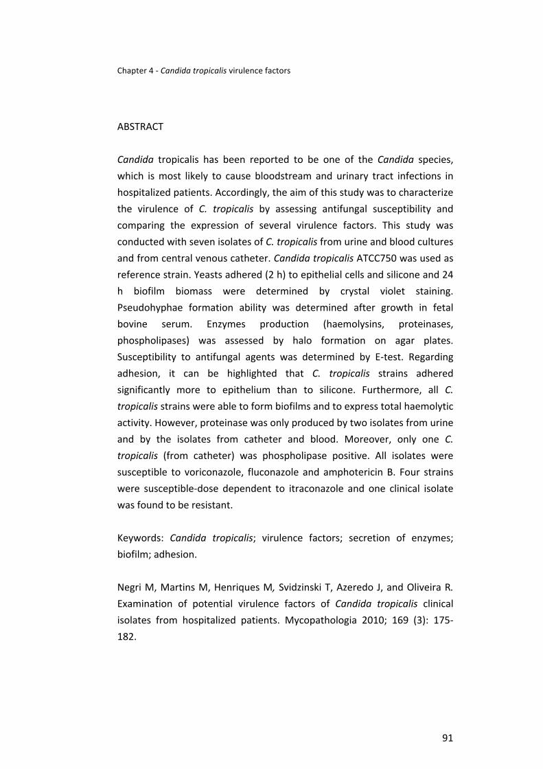
Chapter 4 -‐ Candida tropicalis virulence factors
91
ABSTRACT
Candida tropicalis has been reported to be one of the Candida species, which is most likely to cause bloodstream and urinary tract infections in hospitalized patients. Accordingly, the aim of this study was to characterize the virulence of C. tropicalis by assessing antifungal susceptibility and comparing the expression of several virulence factors. This study was conducted with seven isolates of C. tropicalis from urine and blood cultures and from central venous catheter. Candida tropicalis ATCC750 was used as reference strain. Yeasts adhered (2 h) to epithelial cells and silicone and 24 h biofilm biomass were determined by crystal violet staining. Pseudohyphae formation ability was determined after growth in fetal bovine serum. Enzymes production (haemolysins, proteinases, phospholipases) was assessed by halo formation on agar plates. Susceptibility to antifungal agents was determined by E-‐test. Regarding adhesion, it can be highlighted that C. tropicalis strains adhered significantly more to epithelium than to silicone. Furthermore, all C. tropicalis strains were able to form biofilms and to express total haemolytic activity. However, proteinase was only produced by two isolates from urine and by the isolates from catheter and blood. Moreover, only one C. tropicalis (from catheter) was phospholipase positive. All isolates were susceptible to voriconazole, fluconazole and amphotericin B. Four strains were susceptible-‐dose dependent to itraconazole and one clinical isolate was found to be resistant. Keywords: Candida tropicalis; virulence factors; secretion of enzymes; biofilm; adhesion. Negri M, Martins M, Henriques M, Svidzinski T, Azeredo J, and Oliveira R. Examination of potential virulence factors of Candida tropicalis clinical isolates from hospitalized patients. Mycopathologia 2010; 169 (3): 175-‐182.

Chapter 4
92
INTRODUCTION
Fungal hospital infections (FHI) incidence has increased significantly over the last decades. Candida species are the most frequently isolated fungi, corresponding to approximately 80% of FHI, being the fourth responsible for blood stream infection and the overwhelming majority responsible for urinary tract infections [1-‐3].
Usually, Candida tropicalis is considered the third Candida species most frequently isolated from urine cultures [4, 5]. Moreover, in a recent epidemiological study conducted in 12 Brazilian medical centres, C. tropicalis was the second most frequent Candida species, accounting for 20–24% of all candidemia [2, 6]. Additionally, C. tropicalis is often found in patients admitted in intensive care units (ICUs), especially in patients with cancer or/and requiring prolonged catheterization, or receiving broad-‐spectrum antibiotics [2, 4, 6-‐8].
Several virulence factors seem to be responsible for C. tropicalis infections, which present high potential for dissemination and mortality [8, 9]. Adhesion to host surfaces (epithelial cells and medical devices), as well as biofilm formation, are considered the first step to initiate Candida infection [10, 11]. Furthermore, secretion of enzymes (proteinases and phospholipases), as well as haemolytic activity, are recognized as important factors in tissue invasion [10-‐14].
Hence, the aim of this study was to assess and compare the expression of different virulence factors (enzymes secretion, adhesion and biofilm formation and pseudohyphae production) by several C. tropicalis clinical isolates. Moreover, antifungal susceptibility was also determined in order to deeply characterize the virulence of C. tropicalis.
Candida tropicalis virulence factors
93
MATERIALS AND METHODS
Isolates
This study was conducted with seven isolates of C. tropicalis: five obtained from candiduria, one from candidemia and one from a central venous catheter (CVC) and all were from patients admitted to ICUs at the University Hospital (UH) in Maringá, Paraná, Brazil. Candida tropicalis ATCC 750 was used as reference strain.
Isolation and identification
Yeasts were isolated according to hospital routine methods. To perform hemoculture, one volume of blood was inoculated in 10 volumes of Trypticase Soy Broth (Difco, Detroit, Michigan, EUA) and incubated in the automatic BACTEC (Becton–Dickinson Microbiology Systems, Sparks, MD) system. Urine was spread using a calibrated loop (10 µl) on CLED medium agar plates (Difco) and incubated at 37 °C for 48 h. CVC isolated yeasts underwent a well established technique [15]. Briefly, CVC was rolled on blood agar plates (Difco) and incubated at 35 °C for 72 h. After yeast growth, they were subcultured in CHROMagar Candida® (CHROMagar, BioMerieux, Paris, France) to assess the purity of the culture and the colour of the colonies. From this selective and differential medium, yeasts were identified by three methods: the MicroScan rapid yeast identification panel (Dade Behring Inc, CA, USA), the classical biochemical method [16] and molecular identification.
Candida DNA was extracted using the QIAamp® DNA Mini Kit (QIAGEN, IZASA, Lisbon, Portugal) according to the manufacturer’s instructions. DNA content was determined by spectrophotometry readings at 260 nm. Aliquots of 10 µl were analysed by electrophoresis in a 0.8% agarose (Bio-‐Rad, Lisbon, Portugal) gel in 1 x TBE buffer (Bio-‐Rad) and visualized with a UV transilluminator after ethidium bromide (Bio-‐Rad) staining (0.5 mg/ml). To assess Candida speciation, a polymerase chain reaction (PCR) method previously described [17] was used. Specific primers for the genomic
Chapter 4
94
sequences of DNA topoisomerase II of C. albicans, C. dubliniensis, C. tropicalis (genotypes I and II), C. parapsilosis (genotypes I and II), C. krusei, C. kefyr, C. guilliermondii and C. glabrata were used.
Adhesion and biofilm Formation
Yeast cells were grown at 37 °C, 120 rpm for 18 h on Sabouraud Dextrose Broth (SDB; Difco) and Phosphate saline buffer (PBS)—washed suspensions of each yeast culture were resuspended in RPMI 1640 (Sigma, Saint Louis, Missouri, USA) to a final concentration of 1.0 x 10⁷ cells/ml. Then, 3 ml of the suspension was added to each well of a 6-‐well plate containing either a confluent layer of TCC-‐SUP human urinary bladder epithelial cell line (DSMZ—German Collection of Microorganisms and Cell Cultures) or a silicone coupon (2 x 2 cm) (Neves e Neves, Trofa, Portugal). All procedures were performed in triplicate and repeated in three separate assays.
Quantification of adhered yeast cells
After 2 h of incubation (120 rpm, at 37 °C), the wells were washed twice with PBS to remove unattached yeasts. Yeast cells were quantified using the crystal violet (CV) staining method.
Epithelial cells
Three millilitres of CV stain (1%) was added to each well containing the epithelial cells with adherent yeasts and allowed to stain for 5 min. Then, the wells were washed three times with PBS. In order to remove the CV stain from the epithelial cells, 3 ml of ethanol: acetone (1: 1) was added to each well and removed immediately. Three millilitre of acetic acid (33%) was added to each well and the absorbance of the final solution was read at 570 nm in a microtiter plate reader (Bio-‐Tek® Synergy HT, IZASA). Wells containing epithelial cells without yeasts were used as controls. The mean absorbance of CV retained by yeasts was expressed as absorbance per unit of well area.
Candida tropicalis virulence factors
95
Silicone
Candida cells adhered to silicone were quantified according to Henriques et al. [18]. Briefly, the coupons containing adherent yeasts were removed from each well and immersed for 5 min in new well plates containing 3 ml of methanol. After withdrawing the methanol, the coupons were allowed to dry at room temperature. Then, 3 ml of CV (1%) was added to each well and allowed to stain for 5 min. Coupons were transferred to a new well, washed with ultrapure water and immersed in 3 ml acetic acid (33%) to dissolve the stain. Coupons without yeasts were used as controls. The absorbance of the obtained solution was read in triplicate in a microtiter plate reader at 570 nm. The final number of cells attached, per coupon unit area, was determined using the mean absorbance and the respective calibration curves, previously established.
Biofilm biomass quantification
After 24 h of incubation (120 rpm, at 37 °C) C. tropicalis biofilm forming ability was assessed on inert materials through quantification of total biomass by CV staining as described above. Experiments were repeated in 3–5 independent assays.
Pseudohyphae formation
Pseudohyphae formation was defined as a cell bearing a rounded outgrowth with a length greater than or equal to the diameter of the parent cell, with a constriction at the base. The percentage of cells in pseudohyphae form, against blastopores, was determined by microscopy counting after 2 h of cell growth in a liquid medium containing equal volumes of RPMI 1640 (Sigma) and fetal bovine serum (GIBCO, New York, USA). In these experiments, 100 cells per field were examined. Each experiment was conducted in triplicate.
Chapter 4
96
Proteinase and phospholipase secretion
Secretion of proteinases and phospholipases was detected by the formation of an opaque halo of degradation around the colonies grown in a specific agar plate, according to Ruchel [19] and Price et al. [20], respectively. An aliquot (5 µl) of a 1 x 10⁸ cells/ml suspension prepared in distilled water was inoculated on proteinase agar medium (2% agar, 1.17% yeast carbon base, 0.01% yeast extract and 0.2% bovine serum albumin) pH 5.0 and on phospholipase agar (2% agar, 1% peptone, 2% glucose, 1 M NaCl, 5 mM CaCl2.2H2O and 8% egg yolk) and the Petri dishes were incubated for 7 days at 37 °C. The enzymatic activity (Pz) was determined by the ratio between the colony diameter and the colony diameter plus the halo zone as described by Price et al. [20]. Each experiment was conducted in triplicate.
Haemolytic activity
Haemolysin production was evaluated using a modification of the plate assay described by Luo et al. [13]. Fresh cultured colonies of C. tropicalis were obtained after being spread on Sabouraud Dextrose Agar (SDA; Difco) (18–24 h). Then, a suspension was prepared in saline solution (0.9% NaCl) to reach 108 cells/ml, enumerated using a Neubauer chamber. Ten microliters of this suspension was spot-‐inoculated on sugar-‐enriched sheep blood (2% peptone, 1% agar, 7% fresh sheep blood, 3% glucose) and the plates were incubated at 37 °C. After 48 h they were classified as absent (no halo), partial, or total (completely transparent halo) haemolytic activity. The assay was conducted in quadruplicate on two separate occasions for each yeast isolate tested. A standard strain, C. albicans ATCC 90028, was used as control in each experiment.
Antifungal susceptibility test methods
The E-‐test method was used as recommended by the manufacturer with strips, provided by AB BIODISK (Solna, Sweden), having the following concentrations: from 0.002 to 256 µg/ml; for fluconazole (FLU) and from

Candida tropicalis virulence factors
97
0.002 to 32 µg/ml for itraconazole (ITR), voriconazole (VO) and amphotericin B (AMB). The minimum inhibitory concentrations (MIC) of drugs were determined on RPMI 1640 (Sigma) agar with 2% glucose. An inoculum suspension was adjusted to a turbidity of 0.5 McFarland standard (1 x 10⁶ to 5 x 10⁶ cells/ml) and was incubated at 37 °C for 48 h. MICs were read as the lowest concentration at which the border of the elliptical inhibition zone intercepted the scale on the strip. Quality control was performed in accordance with the Clinical and Laboratory Standards Institute (CLSI) document M27-‐A3 [21], using Candida krusei ATTC 6258 and C. parapsilosis ATCC 22019. MIC interpretative criteria was performed according to the CLSI M27-‐A3 [21]: (a) FLU: MIC ≤ 8 µg/ml—susceptible (S); 16 < MIC < 32 µg/ml—susceptible-‐dose dependent (S-‐DD) and MIC > 64 µg/ml—resistant (R); (b) ITR: MIC ≤ 0.125 µg/ml—S; 0.25 < MIC < 0.5 µg/ml—S-‐DD and MIC ≥ 1 µg/ml—R; (c) AMB MIC > 1 µg/ml—R; (d) VO MIC ≥ 1 µg/ml—S; ≤ 2 µg/ml—R.
Statistical analysis
Results obtained were analysed using the SPSS (Statistical Package for the Social Sciences) program. One-‐way ANOVA with the Bonferroni test was used to compare the number of adherent cells of the several strains assayed. All tests were performed with a confidence level of 95%.
RESULTS
A total of seven C. tropicalis isolates was used in this study: five obtained from urine samples, one from blood samples and one from CVC, all from patients admitted to ICUs at the UH of Maringá, Paraná, Brazil. Additionally, all the methods (MicroScan rapid yeast identification panel, classical biochemical and molecular identification) used had identified C. tropicalis with 100% concordance (data not shown).
Regarding adhesion ability (Figure 4.1), it can be highlighted that all C. tropicalis adhered in a significantly greater extent (P < 0.05) to epithelial

Chapter 4
98
cells than to silicone. Considering the differences among the isolates, it is possible to observe from Figure 4.1 that C. tropicalis L012, from CVC, adhered in a highest extent to epithelial cells (4.05 x 10⁶ cells/cm2) with P < 0.001 vs. strains 1, 16, 29, 69 and to silicone (9.37 x 10⁵ cells/cm2) with P < 0.001 vs. strains 1, 12, 69. Clinical isolates 29 and 69, from urine cultures, adhered to epithelial cells in lower number than other yeasts (P < 0.05 vs. L012) and the latter, as well as strain 12, adhered in lower extent than other yeasts to silicone (Figure 4.1).
Figure 4.1: Number of C. tropicalis cells per cm2 (cell/cm2) (mean ± standard deviation)
adhered to TCC-‐SUP epithelial cells and to silicone measured by crystal violet staining. *
represents the statistical differences (P < 0.05) of adhesion extension to TCC-‐SUP between
the strain L012 and strains 1, 16, 29, 69, ATCC 750; ** represents the statistical differences
(P < 0.05) of adhesion extension to silicone of the strains 16 and 1 compared to the strains
29, 75, L012, ATCC 750.
As it can be observed in Figure 4.2, all C. tropicalis were able to form biofilms and strains did not present significant statistical differences.
Concerning C. tropicalis pseudohyphae formation and enzymatic activity (Table 4.1), it was possible to verify that all isolates produced a low percentage of pseudohyphae formation. Among all, isolates 12 and L012 presented the highest production of pseudohyphae formation (10 and 7%, respectively). Nevertheless, all isolates were able to express total
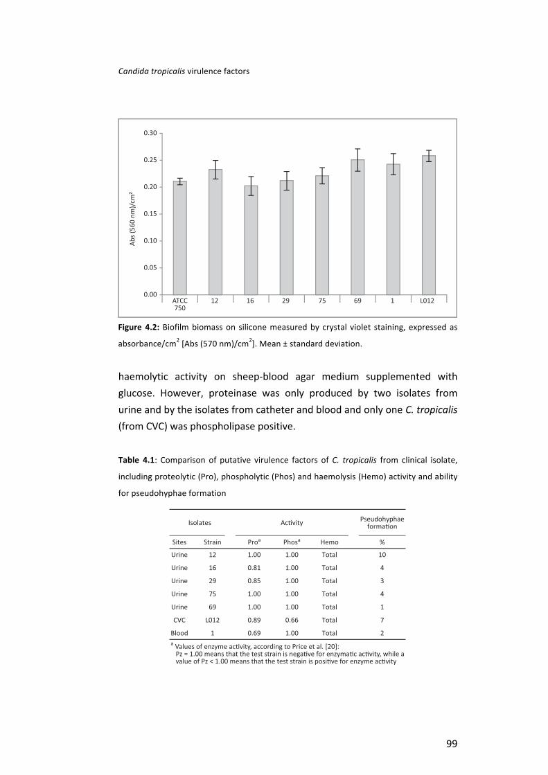
Candida tropicalis virulence factors
99
haemolytic activity on sheep-‐blood agar medium supplemented with glucose. However, proteinase was only produced by two isolates from urine and by the isolates from catheter and blood and only one C. tropicalis (from CVC) was phospholipase positive.
Table 4.1: Comparison of putative virulence factors of C. tropicalis from clinical isolate,
including proteolytic (Pro), phospholytic (Phos) and haemolysis (Hemo) activity and ability
for pseudohyphae formation
Figure 4.2: Biofilm biomass on silicone measured by crystal violet staining, expressed as
absorbance/cm2 [Abs (570 nm)/cm2]. Mean ± standard deviation.
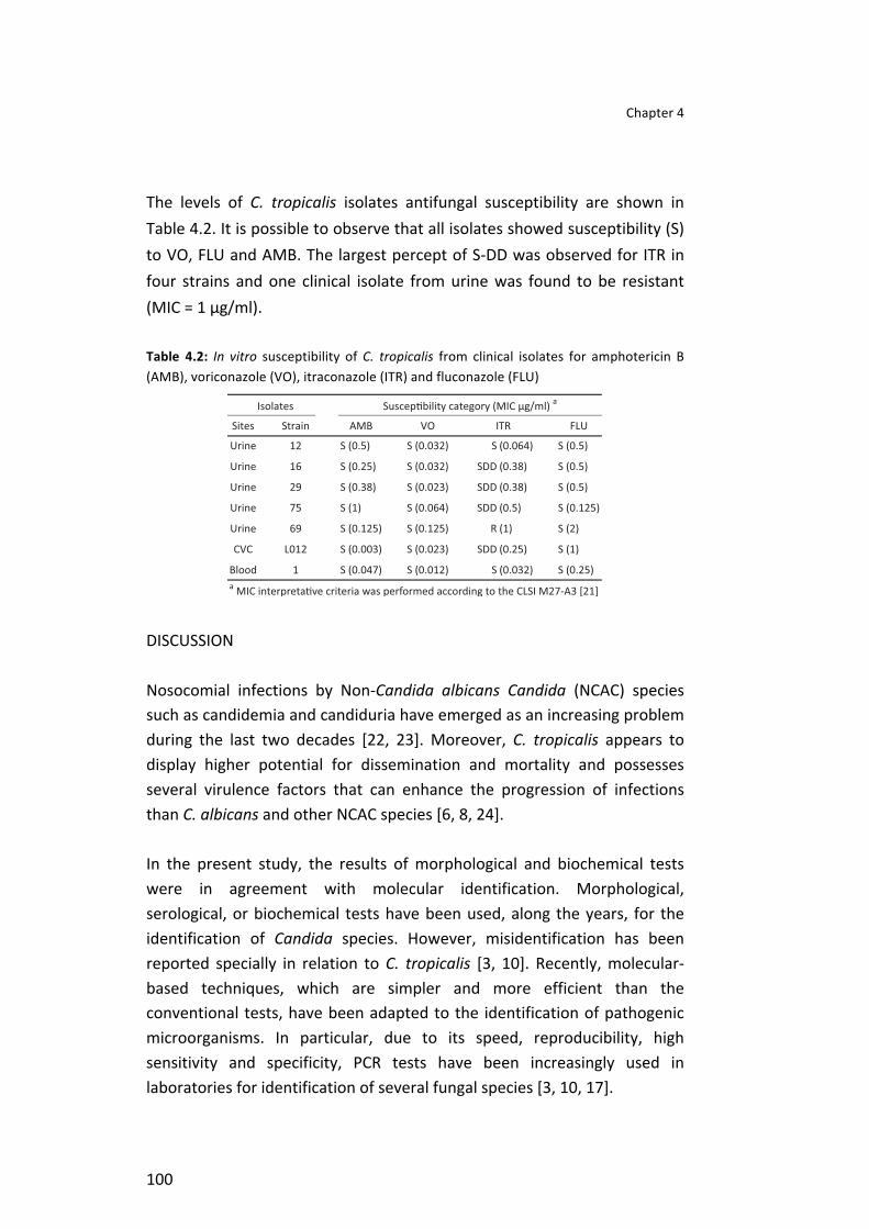
Chapter 4
100
The levels of C. tropicalis isolates antifungal susceptibility are shown in Table 4.2. It is possible to observe that all isolates showed susceptibility (S) to VO, FLU and AMB. The largest percept of S-‐DD was observed for ITR in four strains and one clinical isolate from urine was found to be resistant (MIC = 1 µg/ml).
Table 4.2: In vitro susceptibility of C. tropicalis from clinical isolates for amphotericin B (AMB), voriconazole (VO), itraconazole (ITR) and fluconazole (FLU)
DISCUSSION
Nosocomial infections by Non-‐Candida albicans Candida (NCAC) species such as candidemia and candiduria have emerged as an increasing problem during the last two decades [22, 23]. Moreover, C. tropicalis appears to display higher potential for dissemination and mortality and possesses several virulence factors that can enhance the progression of infections than C. albicans and other NCAC species [6, 8, 24].
In the present study, the results of morphological and biochemical tests were in agreement with molecular identification. Morphological, serological, or biochemical tests have been used, along the years, for the identification of Candida species. However, misidentification has been reported specially in relation to C. tropicalis [3, 10]. Recently, molecular-‐based techniques, which are simpler and more efficient than the conventional tests, have been adapted to the identification of pathogenic microorganisms. In particular, due to its speed, reproducibility, high sensitivity and specificity, PCR tests have been increasingly used in laboratories for identification of several fungal species [3, 10, 17].

Candida tropicalis virulence factors
101
Concerning adhesion ability, it was possible to verify that all C. tropicalis adhered in a significantly higher extent (P < 0.05) to epithelial cells than to silicone. Sohn et al. [25] compared the ability of C. albicans to adhere to the human colorectal carcinoma cell line Caco-‐2 and epidermoid vulvovaginal A431 cells and to polystyrene and reported that C. albicans adheres in high extent to polystyrene and both epithelia. According to Bendel and Hostetter [26], the extent of adhesion to the human epithelia cell line HeLa S3 did not differ between C. albicans and C. tropicalis despite both species having distinct mechanisms for this process. Nevertheless, the present results clearly show differences in the adhesion ability among the different isolates and hence adhesion of C. tropicalis to biotic and abiotic surfaces is strain dependant. Different intra-‐species adherence ability was also reported by other authors for other Candida species [9, 27-‐29].
We did not find a clear relation between pseudohyphae formation and adhesion capability. Although C. tropicalis L012, from CVC, and strain 12, from urine, adhered in higher extent to epithelial cells presenting also more pseudohyphae formation, the strain 75 exhibited an intermediate pseudohyphae formation and high adhesion ability. On the other hand, clinical isolates 29 and 69, from urine cultures, adhered to epithelial cells and displayed pseudohyphae formation in a lower extent than the other strains. This evidences the current lack of knowledge concerning the adhesion mechanisms of NCAC species to epithelium, as well as about the factors affecting the adhesion process [18].
Adhesion of Candida spp. to hard materials or host cells has been considered as an early step in biofilm formation [30, 31]. Nevertheless, it is important to highlight that, although strains 69 and 12 adhered in small number than other yeasts to silicone (Figure 4.1) they showed higher ability to form biofilms on this material (Figure 4.2). This result is consistent with other studies [14, 32], reporting differences between adhesion and biofilm formation abilities on polystyrene and poly (vinyl chloride) (PVC) surfaces under static conditions, which also suggest that adhesion and biofilm formation of C. tropicalis are two distinct phenomena.

Chapter 4
102
All C. tropicalis strains tested were able to form biofilms on silicone, which has important clinical impact once biofilm-‐associated infections are difficult to treat, representing a source of reinfections [23, 33]. Previous works also reported that C. tropicalis can form extensive in vitro biofilms in PVC catheters [34] and polystyrene surfaces [33].
The infective ability of yeasts depends on specific virulence mechanisms that confer the ability to colonize host surfaces, to invade deeper host tissue or to evade host defences [9, 35, 36]. During the pathogenic process many virulence attributes may be involved including production of extracellular proteinases and phospholipases, as well as haemolytic activity [10-‐14, 37, 38]. In this study, all isolates were able to express total haemolytic activity. Manns et al. [12] demonstrated that C. albicans produced haemolytic activity and Luo et al. [13] observed that NCAC species are capable of producing one or more types of haemolysins in vitro with differences among species. Moreover, they observed that C. tropicalis was able to produce complete haemolysis after 48 h, corroborating the results obtained herein.
In the present case, only few isolates were proteinase and phospholipase positive, corroborating the results of other authors [10, 39]. In opposition, Kumar et al. [38] detected 100 and 72.9% of proteinases and phospholipase producers, respectively, among Candida species isolated from pulmonary tuberculosis patients. According to these results, proteinase and phospholipase expression can vary according to Candida species, strain and the site of isolation. Furthermore, although the methods used to test the presence of these enzymes are simple and fast they are not excessively accurate, specially compared with molecular methods that can detect gene expression [24, 36].
Concerning antifungal susceptibility results, all isolates were susceptible to VO, FLU and AMB and for four strains were S-‐DD for ITR. However, one clinical isolate showed to be resistant to ITR. These results are comparable to those reported in the literature, with slight differences that were dependent on the underlying disease and the Candida species involved in the infection [10, 14, 40, 41].
Candida tropicalis virulence factors
103
Though some authors have already assessed some C. tropicalis virulence factors, this work gathers, for the first time, the most important ones: secretion of enzymes, pseudohyphae formation, adhesion (to epithelial cells and silicone), biofilm formation and antifungal susceptibility. Despite it was not possible to establish a relation among the virulence factors assayed, it is interesting to notice that the strain isolated from CVC (L012) presented higher levels of all these factors. Furthermore, all clinical isolates presented one or more virulence factors.

Chapter 4
104
REFERENCES
1. Lundstrom T, Sobel J. Nosocomial Candiduria: A Review. Clin Infect Dis 2001; 32 (11): 1602-‐1607. 2. Colombo AL, Nucci M, Park BJ, et al. Epidemiology of Candidemia in Brazil: a Nationwide Sentinel Surveillance of Candidemia in Eleven Medical Centers. J Clin Microbiol 2006; 44 (8): 2816-‐2823. 3. Asmundsdottir LR, Erlendsdottir H, Haraldsson G, et al. Molecular epidemiology of candidemia: evidence of clusters of smoldering nosocomial infections. Clin Infect Dis 2008; 47 (2): 17-‐24. 4. Kauffman CA, Vazquez JA, Sobel JD, et al. Prospective multicenter surveillance study of funguria in hospitalized patients. Clin Infect Dis 2000; 30 (1): 14-‐18. 5. Álvarez-‐Lerma F, Nolla-‐Salas J, León C, et al. Candiduria in critically ill patients admitted to intensive care medical units. Intensive Care Med. 2003; 29 (7): 1069-‐1076. 6. Nucci M, Colombo AL. Candidemia due to Candida tropicalis: clinical, epidemiologic, and microbiologic characteristics of 188 episodes occurring in tertiary care hospitals. Diagn Microbiol Infect Dis 2007; 58 (1): 77-‐82. 7. Rho J, Shin J, Song J, et al. Molecular investigation of two consecutive nosocomial clusters of Candida tropicalis candiduria using pulsed-‐field gel electrophoresis. J Microbiol 2004; 42 (2): 80-‐86. 8. Krcmery V, Barnes AJ. Non-‐albicans Candida spp. causing fungaemia: pathogenicity and antifungal resistance. J Hosp Infect 2002; 50 (4): 243-‐260. 9. Okawa Y, Miyauchi M, Kobayashi H. Comparison of Pathogenicity of Various Candida tropicalis Strains. Biol Pharm Bull 2008; 31 (8): 1507-‐1510. 10. Furlaneto-‐Maia L, Specian A, Bizerra F, de Oliveira M, Furlaneto M. In vitro Evaluation of putative virulence attributes of oral isolates of Candida spp. obtained from elderly healthy individuals. Mycopathologia 2007; 166 (4): 209-‐217. 11. Tamura NK, Negri MFN, Bonassoli LA, Svidzinski TIE. Fatores de virulência de Candida spp isoladas de cateteres venosos e mãos de servidores hospitalares. Rev Soc Bras Med Trop 2007; 40 (1): 91-‐93. 12. Manns JM, Mosser DM, Buckley HR. Production of a hemolytic factor by Candida albicans. Infect Immun 1994; 62 (11): 5154-‐5156. 13. Luo G, Samaranayake LP, Yau JYY. Candida species exhibit differential in vitro hemolytic activities. J Clin Microbiol 2001; 39 (8): 2971-‐2974. 14. Bizerra FC, Nakamura CV, de Poersch C, et al. Characteristics of biofilm formation by Candida tropicalis and antifungal resistance. FEMS Yeast Res 2008; 8 (3): 442-‐450. 15. Maki D, Weiss C, Sarafin H. A semiquantitative culture method for identifying intravenous-‐catheter-‐related infection. N Engl J Med. 1977; 296: 1305-‐9. 16. Kurtzman CP, Fell JW. The Yeasts, A Taxonomic Study, fourth edition edn. New York: Elsevier Science Ltd; 1998. 17. Kanbe T, Horii T, Arishima T, Ozeki M, Kikuchi A. PCR-‐based identification of pathogenic Candida species using primer mixes specific to Candida DNA topoisomerase II genes. Yeast 2002; 19 (11): 973-‐989. 18. Henriques M, Azeredo J, Oliveira R. Candida Species Adhesion to Oral Epithelium: Factors Involved and Experimental Methodology Used. Crit Rev Microbiol 2006; 32 (4): 217 -‐ 226. 19. Rüchel R. Proteinases of pathogenic fungi. Mycoses 1999; 42: 48-‐52. 20. Price M, Wilkinson I, Gentry L. Plate method for detection of phospholipase activity in Candida albicans. Sabouraudia 1982; 20: 7-‐14. 21. CLSI. Reference Method for broth dilution antifungal susceptibility testing of yeasts – Third Edition: Approved standard M27-‐A3. In. Wayne, PA, USA: Clinical and Laboratory Standards Institute; 2008. 22. Lai C-‐C, Tan C-‐K, Huang Y-‐T, Shao P-‐L, Hsueh P-‐R. Current challenges in the management of invasive fungal infections. J Infect Chemother 2008; 14 (2): 77-‐85. 23. Schelenz S. Management of candidiasis in the intensive care unit. J Antimicrob Chemother 2008; 61 (suppl_1): i31-‐34. 24. Zaugg C, Borg-‐von Zepelin M, Reichard U, Sanglard D, Monod M. Secreted aspartic proteinase family of Candida tropicalis. Infect Immun 2001; 69 (1): 405-‐412.

Candida tropicalis virulence factors
105
25. Sohn K, Senyürek I, Fertey J, et al. An in vitro assay to study the transcriptional response during adherence of Candida albicans to different human epithelia. FEMS Yeast Res 2006; 6 (7): 1085-‐1093. 26. Bendel C, Hostetter M. Distinct mechanisms of epithelial adhesion for Candida albicans and Candida tropicalis. Identification of the participating ligands and development of inhibitory peptides. J Clin Invest. 1993; 92: 1840-‐1849. 27. Panagoda GJ, Ellepola ANB, Samaranayake LP. Adhesion of Candida parapsilosis to epithelial and acrylic surfaces correlates with cell surface hydrophobicity. Mycoses 2001; 44 (1-‐2): 29-‐35. 28. Lyon JP, Resende MAd. Correlation between adhesion, enzyme production, and susceptibility to fluconazole in Candida albicans obtained from denture wearers. Oral Surg Oral Med Oral Pathol Oral Radiol Endod 2006; 102 (5): 632-‐638. 29. Henriques M, Azeredo J, Oliveira R. Candida albicans and Candida dubliniensis: comparison of biofilm formation in terms of biomass and activity. Br J Biomed Sci 2006; 63 (1): 5-‐11. 30. Chandra J, Mukherjee PK, Leidich SD, et al. Antifungal resistance of candidal biofilms formed on denture acrylic in vitro. J Dent Res 2001; 80 (3): 903-‐908. 31. Douglas LJ. Candida biofilms and their role in infection. Trends Microbiol 2003; 11 (1): 30-‐36. 32. Silva S, Henriques M, Martins A, et al. Biofilms of non-‐Candida albicans Candida species: quantification, structure and matrix composition. Med Mycol 2009; 47 (7): 681-‐9. 33. Shin JH, Kee SJ, Shin MG, et al. Biofilm production by isolates of Candida Species recovered from nonneutropenic patients: comparison of bloodstream isolates with isolates from other sources. J Clin Microbiol 2002; 40 (4): 1244-‐1248. 34. Hawser SP, Douglas LJ. Biofilm formation by Candida species on the surface of catheter materials in vitro. Infect Immun 1994; 62 (3): 915-‐921. 35. Yang Y. Virulence factors of Candida species. J Microbiol Immunol Infect. 2003; 36 (4): 223-‐228. 36. Fu Y, Luo G, Spellberg BJ, Edwards JE, Jr., Ibrahim AS. Gene overexpression/suppression analysis of candidate virulence factors of Candida albicans. Eukaryotic Cell 2008; 7 (3): 483-‐492. 37. da Costa K, Ferreira J, Komesu M, Candido R. Candida albicans and Candida tropicalis in oral candidosis: quantitative analysis, exoenzyme activity, and antifungal drug sensitivity. Mycopathologia 2009; 167 (2): 73-‐79. 38. Kumar VG, Latha R, Vedhagiri K, et al. Phospholipase C, proteinase and hemolytic activities of Candida spp. isolated from pulmonary tuberculosis patients. J Mycol Med 2009; 19 (1): 3-‐10. 39. Gokce G, Cerikcioglu N, Yagci A. Acid proteinase, phospholipase, and biofilm production of Candida species isolated from blood cultures. Mycopathologia 2007; 164 (6): 265-‐269. 40. Johnson E, Espinel-‐Ingroff A, Szekely A, Hockey H, Troke P. Activity of voriconazole, itraconazole, fluconazole and amphotericin B in vitro against 1763 yeasts from 472 patients in the voriconazole phase III clinical studies. Int J Antimicrob Agents 2008; 32 (6): 511-‐514. 41. Panizo MM, Reviákina V, Dolande M, Selgrad S. Candida spp. in vitro susceptibility profile to four antifungal agents. Resistance surveillance study in Venezuelan strains. Med Mycol 2009; 47 (2): 137 -‐ 143.

CHAPTER 5
An in vitro evaluation ofCandida tropicalis infectivityusing human cell monolayers

Chapter 5 -‐ Candida tropicalis infectivity of human cells
109
ABSTRACT
The aim of the present study was to investigate the interaction of Candida tropicalis with three different human cell lines: TCC-‐SUP (epithelial cells from urinary bladder); HeLa (epithelial cells from cervical carcinoma); Caco-‐2 (epithelial cells from colorectal adenocarcinoma). In particular to assess the degree of cell damage and activity reduction induced by C. tropicalis adhesion and the role of SAPT gene expression in this process. Two C. tropicalis strains were used in this study, the reference strain ATCC 750 and a clinical isolate from urine (U69). The ability of C. tropicalis to adhere to a confluent layer of human cells was determined using an adaptation of the crystal violet staining method; cell damage and cell activity inhibition induced by the adhesion of C. tropicalis were assessed by LDH and MTS reduction, respectively. Candida tropicalis aspartyl proteinase (SAPT) gene expression was determined by real-‐time PCR. Candida tropicalis strains were able to adhere to the different human cells, although, in a strain and cell dependent manner. Concerning cellular response to C. tropicalis, the highest cell activity inhibition was obtained for Caco-‐2, followed by TCC-‐SUP and HeLa cells. The highest percentage of cell damage (around 14%) was observed for TCC-‐SUP in contact with the U69 isolate and for Caco-‐2 in contact with the reference strain. Real time PCR analysis revealed a wide range of expression profiles of SAP genes for both C. tropicalis strains in contact with the different types of epithelial cells. SAPT3 was the gene expressed at the highest level for both C. tropicalis strains in contact with the three human epithelial cell lines. It is important to highlight that human cells response to C. tropicalis adhesion, as well as SAPs production, is strain and epithelial cell line dependent. Keywords: Adherence, Candida tropicalis; fungal infection, human pathogenic fungi, aspartyl proteinase Negri M, Botelho C, Silva S, Lopes L, Henriques M, Azeredo J, Oliveira R. An in vitro evaluation of Candida tropicalis infectivity using human cell monolayers. Journal of Medical Microbiology. Posted online on May 14, 2011. DOI: 10.1099/jmm.0.031195-‐0.

Chapter 5
110
INTRODUCTION
Adhesion to host surfaces, like human epithelial cells, and secretion of hydrolytic enzymes are considered important factors for Candida tropicalis virulence [1-‐4]. The capacity of C. tropicalis to adhere and to infect host cells becomes a serious problem when patients are in intensive care units (ICU), mainly because C. tropicalis is associated with higher dissemination potential and mortality, particularly in oncological patients [5-‐7]. In order to clarify the infectivity of Candida species, several in vitro studies have been performed using human cell monolayers from urinary, vaginal and intestinal epithelia [3, 8, 9].
In addition, hydrolytic enzymes such as aspartyl proteinases (Saps) are known to facilitate invasion and colonization of tissues by disrupting the host mucosal membranes and by degrading important immunological and structural defence proteins [1, 10]. It is also recognized that C. tropicalis possesses at least four genes encoding Saps, and these are designated SAPT1, SAPT2, SAPT3 and SAPT4 [1]
Since, C. tropicalis colonizes specific body sites such as the gastrointestinal and urinary tract and is associated with severe and invasive candidosis [11], it is important to understand the mechanisms of interaction between these epithelia and C. tropicalis. So, the aim of this work was to study the effect of C. tropicalis colonization of different human epithelial cells and the levels of SAPs genes expression by C. tropicalis when interacting with different human cells.
MATERIALS AND METHODS
Yeasts and growth conditions
Two strains of C. tropicalis were used in this study, one reference strain from the American Type Culture Collection (ATCC 750) and one isolate
Candida tropicalis infectivity of human cells
111
(U69) obtained from a urine sample of a 84 years old female patient with candiduria admitted to the intensive care of an oncologic unit and belonging to the archive collection of the University Hospital in Maringá, Paraná, Brazil. For each experiment, strains were inoculated in Sabouraud dextrose broth (SDB; Merck, Germany) and incubated for 18 h at 37 °C under agitation at 120 rpm. After incubation, cells were harvested by centrifugation at 8000 × g for 5 min at 4 °C and washed twice with Phosphate Buffer Solution (PBS; pH 7.5; 0.01 mol/l).
Human epithelial cells line
To assess the adhesion ability of C. tropicalis to human epithelial cells, the following cell lines were used as models: (i) TCC-‐SUP cells, derived from human urinary bladder epithelial cells (DSMZ -‐ German Collection of Microorganisms and Cell Cultures); (ii) HeLa cells, derived from a human cervical carcinoma, donated by “Instituto Gulbenkian de Ciência”, Lisbon, Portugal; (iii) Caco-‐2 cells derived from a human colorectal adenocarcinoma cell line (ATCC HTB-‐37), kindly donated by Carla Nunes, Department of Biochemistry, Faculdade de Farmácia da Universidade de Coimbra, Coimbra, Portugal. Cells were cultured at 37 °C under 5% CO2 in Dulbecco’s modified Eagle’s medium (D-‐MEM; Gibco, USA) containing 10% of fetal bovine serum (FBS; Gibco, USA) and 1% penicillin/streptomycin (P/S; Gibco, USA). After achieving 80% of confluence, cells were detached using a 25% trypsin-‐EDTA (Gibco, USA) solution and cell concentration was adjusted to 1x10⁶ cells/ml with fresh D-‐MEM without P/S and added to a 24-‐well plate. Prior to the adhesion assays, the wells were washed two times with PBS.
Adhesion assay
The yeast cells were suspended in D-‐MEM, without phenol, to a final concentration of 1 x 10⁷ yeast/ml using a Neubauer chamber (Boeco, Germany). Then, 1 ml of this suspension was added to each well of the 24-‐well plate covered with a confluent layer of a human epithelial cell line. After 2 h of incubation at 37 °C under 5% CO2, each well was washed once with PBS to remove unattached yeasts.
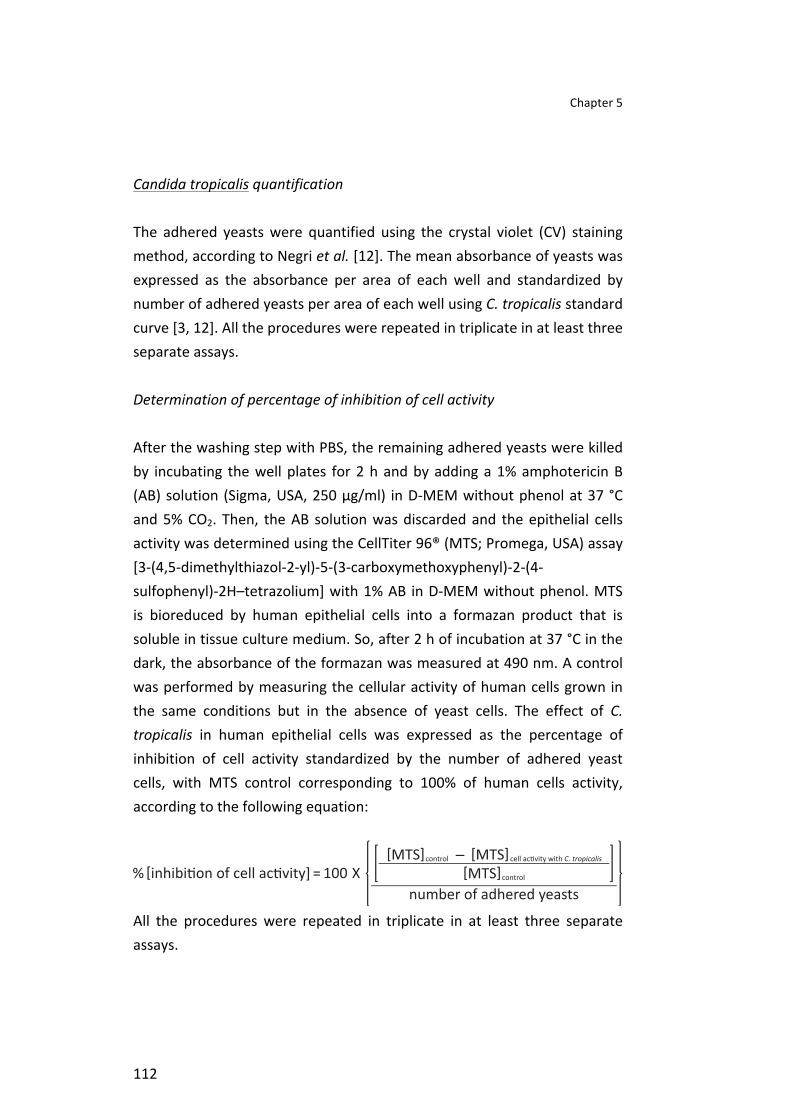
Chapter 5
112
Candida tropicalis quantification
The adhered yeasts were quantified using the crystal violet (CV) staining method, according to Negri et al. [12]. The mean absorbance of yeasts was expressed as the absorbance per area of each well and standardized by number of adhered yeasts per area of each well using C. tropicalis standard curve [3, 12]. All the procedures were repeated in triplicate in at least three separate assays.
Determination of percentage of inhibition of cell activity
After the washing step with PBS, the remaining adhered yeasts were killed by incubating the well plates for 2 h and by adding a 1% amphotericin B (AB) solution (Sigma, USA, 250 μg/ml) in D-‐MEM without phenol at 37 °C and 5% CO2. Then, the AB solution was discarded and the epithelial cells activity was determined using the CellTiter 96® (MTS; Promega, USA) assay [3-‐(4,5-‐dimethylthiazol-‐2-‐yl)-‐5-‐(3-‐carboxymethoxyphenyl)-‐2-‐(4-‐sulfophenyl)-‐2H–tetrazolium] with 1% AB in D-‐MEM without phenol. MTS is bioreduced by human epithelial cells into a formazan product that is soluble in tissue culture medium. So, after 2 h of incubation at 37 °C in the dark, the absorbance of the formazan was measured at 490 nm. A control was performed by measuring the cellular activity of human cells grown in the same conditions but in the absence of yeast cells. The effect of C. tropicalis in human epithelial cells was expressed as the percentage of inhibition of cell activity standardized by the number of adhered yeast cells, with MTS control corresponding to 100% of human cells activity, according to the following equation:
All the procedures were repeated in triplicate in at least three separate assays.
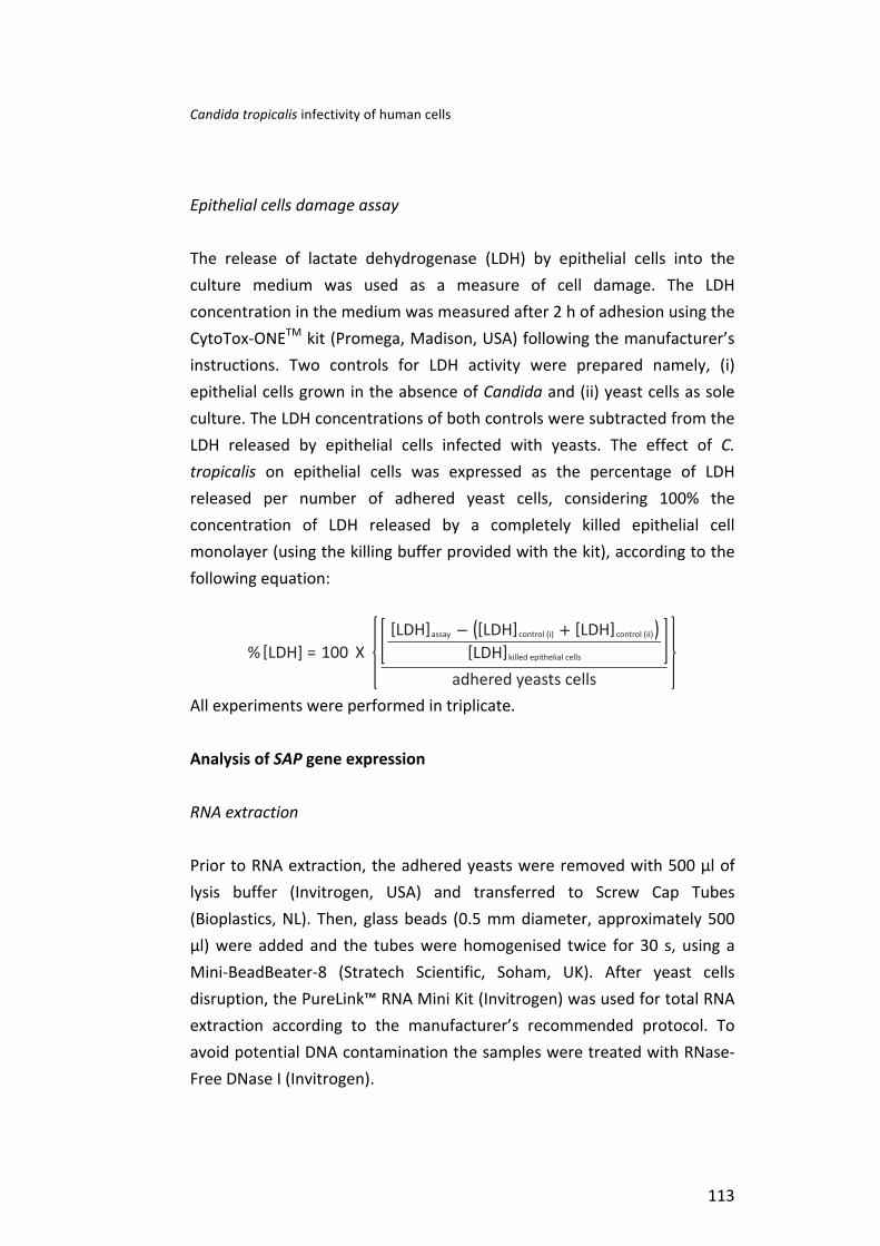
Candida tropicalis infectivity of human cells
113
Epithelial cells damage assay
The release of lactate dehydrogenase (LDH) by epithelial cells into the culture medium was used as a measure of cell damage. The LDH concentration in the medium was measured after 2 h of adhesion using the CytoTox-‐ONETM kit (Promega, Madison, USA) following the manufacturer’s instructions. Two controls for LDH activity were prepared namely, (i) epithelial cells grown in the absence of Candida and (ii) yeast cells as sole culture. The LDH concentrations of both controls were subtracted from the LDH released by epithelial cells infected with yeasts. The effect of C. tropicalis on epithelial cells was expressed as the percentage of LDH released per number of adhered yeast cells, considering 100% the concentration of LDH released by a completely killed epithelial cell monolayer (using the killing buffer provided with the kit), according to the following equation:
All experiments were performed in triplicate.
Analysis of SAP gene expression
RNA extraction
Prior to RNA extraction, the adhered yeasts were removed with 500 μl of lysis buffer (Invitrogen, USA) and transferred to Screw Cap Tubes (Bioplastics, NL). Then, glass beads (0.5 mm diameter, approximately 500 µl) were added and the tubes were homogenised twice for 30 s, using a Mini-‐BeadBeater-‐8 (Stratech Scientific, Soham, UK). After yeast cells disruption, the PureLink™ RNA Mini Kit (Invitrogen) was used for total RNA extraction according to the manufacturer’s recommended protocol. To avoid potential DNA contamination the samples were treated with RNase-‐Free DNase I (Invitrogen).
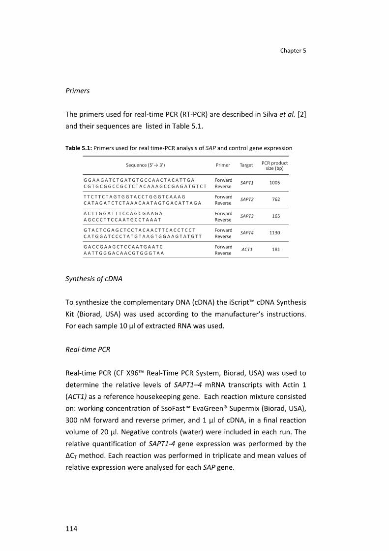
Chapter 5
114
Primers
The primers used for real-‐time PCR (RT-‐PCR) are described in Silva et al. [2] and their sequences are listed in Table 5.1.
Table 5.1: Primers used for real time-‐PCR analysis of SAP and control gene expression
Synthesis of cDNA
To synthesize the complementary DNA (cDNA) the iScript™ cDNA Synthesis Kit (Biorad, USA) was used according to the manufacturer’s instructions. For each sample 10 µl of extracted RNA was used.
Real-‐time PCR
Real-‐time PCR (CF X96™ Real-‐Time PCR System, Biorad, USA) was used to determine the relative levels of SAPT1–4 mRNA transcripts with Actin 1 (ACT1) as a reference housekeeping gene. Each reaction mixture consisted on: working concentration of SsoFast™ EvaGreen® Supermix (Biorad, USA), 300 nM forward and reverse primer, and 1 μl of cDNA, in a final reaction volume of 20 μl. Negative controls (water) were included in each run. The relative quantification of SAPT1-‐4 gene expression was performed by the ΔCT method. Each reaction was performed in triplicate and mean values of relative expression were analysed for each SAP gene.
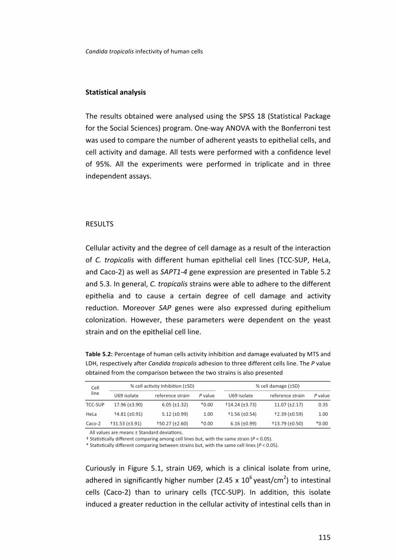
Candida tropicalis infectivity of human cells
115
Statistical analysis
The results obtained were analysed using the SPSS 18 (Statistical Package for the Social Sciences) program. One-‐way ANOVA with the Bonferroni test was used to compare the number of adherent yeasts to epithelial cells, and cell activity and damage. All tests were performed with a confidence level of 95%. All the experiments were performed in triplicate and in three independent assays.
RESULTS
Cellular activity and the degree of cell damage as a result of the interaction of C. tropicalis with different human epithelial cell lines (TCC-‐SUP, HeLa, and Caco-‐2) as well as SAPT1-‐4 gene expression are presented in Table 5.2 and 5.3. In general, C. tropicalis strains were able to adhere to the different epithelia and to cause a certain degree of cell damage and activity reduction. Moreover SAP genes were also expressed during epithelium colonization. However, these parameters were dependent on the yeast strain and on the epithelial cell line.
Table 5.2: Percentage of human cells activity inhibition and damage evaluated by MTS and LDH, respectively after Candida tropicalis adhesion to three different cells line. The P value obtained from the comparison between the two strains is also presented
Curiously in Figure 5.1, strain U69, which is a clinical isolate from urine, adhered in significantly higher number (2.45 x 10⁶ yeast/cm2) to intestinal cells (Caco-‐2) than to urinary cells (TCC-‐SUP). In addition, this isolate induced a greater reduction in the cellular activity of intestinal cells than in
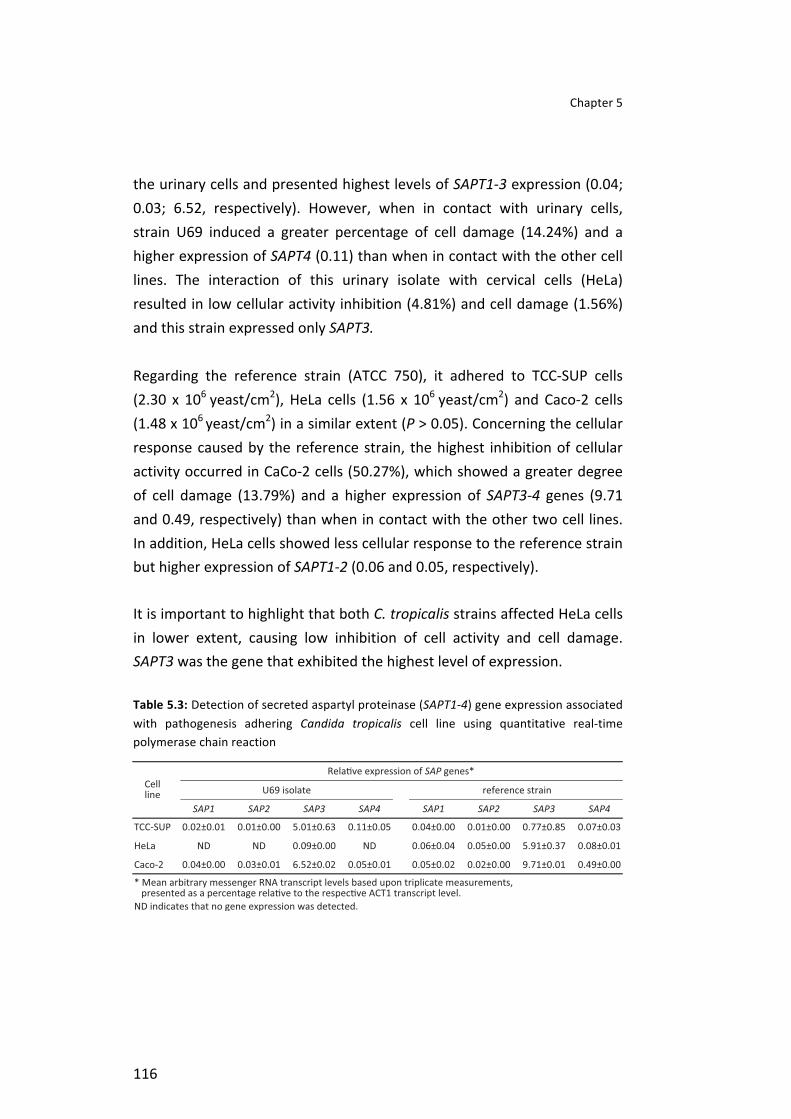
Chapter 5
116
the urinary cells and presented highest levels of SAPT1-‐3 expression (0.04; 0.03; 6.52, respectively). However, when in contact with urinary cells, strain U69 induced a greater percentage of cell damage (14.24%) and a higher expression of SAPT4 (0.11) than when in contact with the other cell lines. The interaction of this urinary isolate with cervical cells (HeLa) resulted in low cellular activity inhibition (4.81%) and cell damage (1.56%) and this strain expressed only SAPT3.
Regarding the reference strain (ATCC 750), it adhered to TCC-‐SUP cells (2.30 x 10⁶ yeast/cm2), HeLa cells (1.56 x 10⁶ yeast/cm2) and Caco-‐2 cells (1.48 x 10⁶ yeast/cm2) in a similar extent (P > 0.05). Concerning the cellular response caused by the reference strain, the highest inhibition of cellular activity occurred in CaCo-‐2 cells (50.27%), which showed a greater degree of cell damage (13.79%) and a higher expression of SAPT3-‐4 genes (9.71 and 0.49, respectively) than when in contact with the other two cell lines. In addition, HeLa cells showed less cellular response to the reference strain but higher expression of SAPT1-‐2 (0.06 and 0.05, respectively).
It is important to highlight that both C. tropicalis strains affected HeLa cells in lower extent, causing low inhibition of cell activity and cell damage. SAPT3 was the gene that exhibited the highest level of expression.
Table 5.3: Detection of secreted aspartyl proteinase (SAPT1-‐4) gene expression associated with pathogenesis adhering Candida tropicalis cell line using quantitative real-‐time polymerase chain reaction
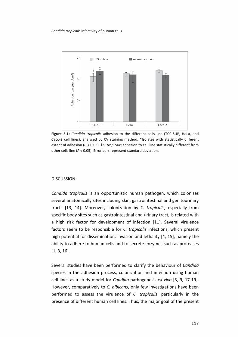
Candida tropicalis infectivity of human cells
117
Figure 5.1: Candida tropicalis adhesion to the different cells line (TCC-‐SUP, HeLa, and Caco-‐2 cell lines), analysed by CV staining method. *Isolates with statistically different extent of adhesion (P < 0.05). ‡C. tropicalis adhesion to cell line statistically different from other cells line (P < 0.05). Error bars represent standard deviation.
DISCUSSION
Candida tropicalis is an opportunistic human pathogen, which colonizes several anatomically sites including skin, gastrointestinal and genitourinary tracts [13, 14]. Moreover, colonization by C. tropicalis, especially from specific body sites such as gastrointestinal and urinary tract, is related with a high risk factor for development of infection [11]. Several virulence factors seem to be responsible for C. tropicalis infections, which present high potential for dissemination, invasion and lethality [4, 15], namely the ability to adhere to human cells and to secrete enzymes such as proteases [1, 3, 16].
Several studies have been performed to clarify the behaviour of Candida species in the adhesion process, colonization and infection using human cell lines as a study model for Candida pathogenesis ex vivo [3, 9, 17-‐19]. However, comparatively to C. albicans, only few investigations have been performed to assess the virulence of C. tropicalis, particularly in the presence of different human cell lines. Thus, the major goal of the present

Chapter 5
118
study was to investigate, ex vivo, the potential of C. tropicalis to colonize and damage urinary, vaginal and intestinal epithelium. Therefore, three different cell lines were used, namely: one from human bladder (TCC-‐SUP); other from human cervical carcinoma (HeLa); and the last from human colorectal adenocarcinoma (Caco-‐2). These cell lines are very often used to study, in vitro, mechanisms of interaction between Candida and gastrointestinal and genitourinary tracts [3, 8, 9, 12].
In the present study and corroborating other authors, C. tropicalis were able to adhere and to damage all the different epithelial cell lines used [8, 12, 18]. Furthermore C. tropicalis expressed a range of profiles of SAP genes [1, 2], although in a strain and cell dependent manner [3, 4]. In fact, C. tropicalis strains have shown different behaviours in their ability to invade human tissue with dramatic histopathological tissue alteration [2, 4, 18, 20].
Comparing with other studies [2, 3, 19], it is possible to verify that Candida species do not adhere in the same extent to the different mucosal type of cells, and also that there is not a strong correlation between the adherence ability to epithelial cells when the site of yeast isolation corresponds to a similar type of epithelium. It is important to highlight that both strains used in this study, U69 and the reference strain, are different clinical specimens and showed different infectivity. The strain U69 is a clinical isolate from a urine sample of a female patient from an oncology unit and the reference strain, ATCC 750, is a clinical isolate from a patient with bronchomycosis. Interestingly, the strain U69 adhered in higher extent to Caco-‐2 cells than to the other human cells and the reference strain adhered in similar extents to the different cell lines; curiously in higher number to TCC-‐SUP than U69 (Figure 5.1).
According to Sohn et al. [9] the interaction of C. albicans with Caco-‐2 cells is distinct from the interaction with epidermoid vulvo-‐vaginal cell line (A-‐431) and these authors suggested that this fact can be related with epithelial cells morphology and molecular events during adhesion. Furthermore, in a previous study with the same strains [3], it was
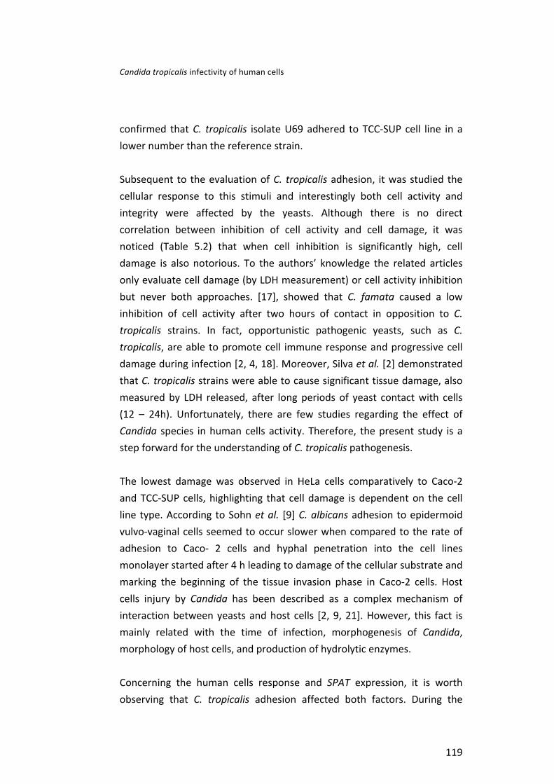
Candida tropicalis infectivity of human cells
119
confirmed that C. tropicalis isolate U69 adhered to TCC-‐SUP cell line in a lower number than the reference strain.
Subsequent to the evaluation of C. tropicalis adhesion, it was studied the cellular response to this stimuli and interestingly both cell activity and integrity were affected by the yeasts. Although there is no direct correlation between inhibition of cell activity and cell damage, it was noticed (Table 5.2) that when cell inhibition is significantly high, cell damage is also notorious. To the authors’ knowledge the related articles only evaluate cell damage (by LDH measurement) or cell activity inhibition but never both approaches. [17], showed that C. famata caused a low inhibition of cell activity after two hours of contact in opposition to C. tropicalis strains. In fact, opportunistic pathogenic yeasts, such as C. tropicalis, are able to promote cell immune response and progressive cell damage during infection [2, 4, 18]. Moreover, Silva et al. [2] demonstrated that C. tropicalis strains were able to cause significant tissue damage, also measured by LDH released, after long periods of yeast contact with cells (12 – 24h). Unfortunately, there are few studies regarding the effect of Candida species in human cells activity. Therefore, the present study is a step forward for the understanding of C. tropicalis pathogenesis.
The lowest damage was observed in HeLa cells comparatively to Caco-‐2 and TCC-‐SUP cells, highlighting that cell damage is dependent on the cell line type. According to Sohn et al. [9] C. albicans adhesion to epidermoid vulvo-‐vaginal cells seemed to occur slower when compared to the rate of adhesion to Caco-‐ 2 cells and hyphal penetration into the cell lines monolayer started after 4 h leading to damage of the cellular substrate and marking the beginning of the tissue invasion phase in Caco-‐2 cells. Host cells injury by Candida has been described as a complex mechanism of interaction between yeasts and host cells [2, 9, 21]. However, this fact is mainly related with the time of infection, morphogenesis of Candida, morphology of host cells, and production of hydrolytic enzymes.
Concerning the human cells response and SPAT expression, it is worth observing that C. tropicalis adhesion affected both factors. During the
Chapter 5
120
process of adhesion and invasion of host tissues, Candida species are known to secrete hydrolytic enzymes that cause damage on host cells membrane integrity, leading to dysfunction or disruption of host structures [1]. Furthermore, the expression of SAP genes by C. tropicalis has also been demonstrated during penetration of tissues and, evading macrophages after phagocytosis of yeast cells [1, 22, 23].
According to the results obtained in this study (Table 5.2), Caco-‐2 cells were the ones displaying the highest inhibition of activity when in contact with each of the tested strains and, in this situation, strain U69 and the reference strain expressed the highest levels of SAPT1-‐3 and SAPT3-‐4, respectively (Table 5.3). However, strain U69 in contact with TCC-‐SUP cells, promoted a greater percentage of cell damage and a higher expression of SAPT4 and the same happened with the reference strain when in contact with Caco-‐2 cells. Studies with C. albicans indicate that each SAP gene may be related to the yeast form and to a specific function [1, 2, 22, 24, 25].
Recent data for C. albicans indicated that SAP1 to SAP3 family genes are expressed by yeast cell only and contribute to the adhesion to human cells and tissue damage, whereas C. albicans SAP4 expression is confined to hyphae, and have been further implicated with systemic infection and in the evasion to phagocytosis [25].
Furthermore, SAPT3 transcript presented the highest level of gene expression for both strains. Silva et al. [2] studied the expression profiles of SAP genes for seven C. tropicalis strains in contact with reconstituted human oral epithelium and also determined the expression of the 4 genes but SAPT2 and SAPT4 transcripts were detected in a similar extent to SAPT3.
This fact can be related with the epithelium type and also with the different C. tropicalis strains studied. In the present case, the SAP gene expression was strain and human cell line dependent. It is important to highlight that there are only few studies [1, 2] reporting C. tropicalis SAPT gene expression during the adhesion to human cells and there is also
Candida tropicalis infectivity of human cells
121
limited knowledge about the role of these enzymes in C. tropicalis adhesion and tissue damage.
In summary, the present study shows that C. tropicalis is able to adhere to different human cells influencing their response in a way dependent on cell type and yeast strain. This study also stresses out the importance of using more than one methodology to assess cell injury caused by Candida species. Moreover, cell damage and activity caused by C. tropicalis seem to be related with the expression of different SAP genes. As a preliminary study, this work only explored a small period of contact between yeasts and epithelial cells, therefore, more studies, including higher contact times could be of major interest as well as the inclusion of different clinical isolates. The knowledge of human cells response to Candida stimuli could further help in the development of new therapeutic strategies.
Chapter 5
122
REFERENCES
1. Zaugg C, Borg-‐von Zepelin M, Reichard U, Sanglard D, Monod M. Secreted aspartic proteinase family of Candida tropicalis. Infect Immun 2001; 69 (1): 405-‐412. 2. Silva S, Hooper SJ, Henriques M, et al. The role of secreted aspartyl proteinases in Candida tropicalis invasion and damage of oral mucosa. Clin Microbiol Infect 2011; 17 (2): 264-‐272. 3. Negri M, Martins M, Henriques M, et al. Examination of potential virulence factors of Candida tropicalis clinical isolates from hospitalized patients. Mycopathologia 2010; 169 (3): 175-‐182. 4. Okawa Y, Miyauchi M, Kobayashi H. Comparison of Pathogenicity of Various Candida tropicalis Strains. Biol Pharm Bull 2008; 31 (8): 1507-‐1510. 5. Chakrabarti A, Chatterjee SS, Rao KLN, et al. Recent experience with fungaemia: change in species distribution and azole resistance. Scand J Infect Dis 2009; 41 (4): 275 -‐ 284. 6. Nucci M, Colombo AL. Candidemia due to Candida tropicalis: clinical, epidemiologic, and microbiologic characteristics of 188 episodes occurring in tertiary care hospitals. Diagn Microbiol Infect Dis 2007; 58 (1): 77-‐82. 7. Kothavade RJ, Kura MM, Valand AG, Panthaki MH. Candida tropicalis: its prevalence, pathogenicity and increasing resistance to fluconazole. J Med Microbiol 2010; 59 (Pt 8): 873-‐80. 8. Bendel C, Hostetter M. Distinct mechanisms of epithelial adhesion for Candida albicans and Candida tropicalis. Identification of the participating ligands and development of inhibitory peptides. J Clin Invest. 1993; 92: 1840-‐1849. 9. Sohn K, Senyürek I, Fertey J, et al. An in vitro assay to study the transcriptional response during adherence of Candida albicans to different human epithelia. FEMS Yeast Res 2006; 6 (7): 1085-‐1093. 10. Ruchel R, de Bernardis F, Ray TL, Sullivan PA, Cole GT. Candida acid proteinases. J Med Vet Mycol 1992; 30 Suppl 1: 123-‐32. 11. Pfaller MA. Laboratory aids in the diagnosis of invasive candidiasis. Mycopathologia 1992; 120 (2): 65-‐72. 12. Negri M, Gonçalves V, Silva S, et al. Crystal violet staining to quantify Candida adhesion to epithelial cells. Br J Microbiol 2010; 67 (3): 120-‐125. 13. Biasoli MS, Tosello ME, Bottai H, Cuesta C, Magaró HM. Adherence of Candida strains isolated from the human gastrointestinal tract. Mycoses 2002; 45 (11-‐12): 465-‐469. 14. Oksuz S, Sahin I, Yildirim M, et al. Phospholipase and proteinase activities in different Candida species isolated from anatomically distinct sites of healthy adults. Jpn J Infect Dis. 2007; 60 (5): 280-‐283. 15. Krcmery V, Barnes AJ. Non-‐albicans Candida spp. causing fungaemia: pathogenicity and antifungal resistance. J Hosp Infect 2002; 50 (4): 243-‐260. 16. Galan-‐Ladero MA, Blanco MT, Sacristan B, et al. Enzymatic activities of Candida tropicalis isolated from hospitalized patients. Med Mycol 2010; 48 (1): 207-‐210 17. Pacheco M, Pisa D, García-‐Gómez P, Carrasco L, Juarranz Á. Attachment and entry of Candida famata in monocytes and epithelial cells. Microsc Res Tech 2007; 70 (11): 975-‐986. 18. Saegusa S, Totsuka M, Kaminogawa S, Hosoi T. Cytokine responses of intestinal epithelial-‐like Caco-‐2 cells to non-‐pathogenic and opportunistic pathogenic yeasts in the presence of butyric acid. Biosci Biotechnol Biochem 2007; 71 (10): 2428-‐34.

Candida tropicalis infectivity of human cells
123
19. Bendel CM. Colonization and epithelial adhesion in the pathogenesis of neonatal candidiasis. Semin Perinatol 2003; 27 (5): 357-‐364. 20. Jayatilake J, Samaranayake Y, Cheung L, Samaranayake L. Quantitative evaluation of tissue invasion by wild type, hyphal and SAP mutants of Candida albicans, and non-‐albicans Candida species in reconstituted human oral epithelium. J Oral Pathol Med 2006; 35 (8): 484-‐491. 21. Filler SG, Sheppard DC. Fungal Invasion of Normally Non-‐Phagocytic Host Cells. PLoS Pathogens 2006; 2 (12): e129. 22. Monoda M, Capocciaa S, Léchennea B, et al. Secreted proteases from pathogenic fungi Int. J.Medical Microbiol. 2002; 292 (5-‐6): 405-‐419 23. Togni G, Sanglard D, Falchetto R, Monod M. Isolation and nucleotide sequence of the extracellular acid protease gene (ACP) from the yeast Candida tropicalis. FEBS Lett. 1991; 286 (1-‐2): 181-‐5. 24. Naglik JR, Challacombe SJ, Hube B. Candida albicans Secreted Aspartyl Proteinases in Virulence and Pathogenesis. Microbiol Mol Biol Rev 2003; 67 (3): 400-‐428. 25. Yang Y. Virulence factors of Candida species. J Microbiol Immunol Infect. 2003; 36 (4): 223-‐228.


CHAPTER 6
Candida tropicalis biofilms:effect on urinary epithelial cells


Chapter 6 -‐ Candida tropicalis biofilms: effect on epithelial cells
127
ABSTRACT
It was to study the behaviour of C. tropicalis in biofilms of different ages (24 -‐ 120 h) formed in artificial urine (AU) and their effect in human urinary bladder cells (TCC-‐SUP). The reference strain ATCC 750 and two isolates from urine (U69 and U75) were used in this study. The adhesion to TCC-‐SUP was evaluated using the crystal violet staining method and the TCC-‐SUP response was evaluated in terms of activity inhibition and cell damage. Candida tropicalis aspartyl proteinase (SAPT) gene expression was determined by real-‐time PCR. All strains of C. tropicalis were able to form biofilms in AU, although with differences among strains. Candida tropicalis biofilm cells were able to adhere to TCC-‐SUP, in general, independently of biofilm age. Yeasts affected TCC-‐SUP, with 120 h-‐biofilm cells of U69 and ATCC 750 strains causing the highest levels of cell injury. Generally, SAPT3 was highly expressed and SAPT4 was only detected in the reference strain. Overall, it is important to highlight that C. tropicalis cells detached from biofilms are able to colonize human cells and cause some injury and reduction of metabolic activity. Keywords: Candida tropicalis; biofilm; artificial urine; adhesion; epithelial cells; SAP gene Negri M, Silva S, Breda D, Henriques M, Azeredo J, Oliveira R. Candida tropicalis biofilms: effect on urinary epithelial cells. Submitted.

Chapter 6
128
INTRODUCTION
Candida tropicalis ranks between the second or third non-‐Candida albicans Candida species (NCAC) most frequently isolated from patients with Candida infections [1-‐4]. According to epidemiological data, C. tropicalis has been related with urinary tract infections and haematological malignancy [2, 4-‐6]. Furthermore, the most important causes of C. tropicalis candiduria are associated with antibiotic therapy and indwelling catheterization [7-‐9].
Several virulence factors seem to be responsible for C. tropicalis infections, which present higher potential for dissemination and mortality in patients admitted in intensive care units (ICUs) than C. albicans or any other NCAC species [9]. Included in these virulence factors are: their ability to adhere and to form biofilms onto different indwelling medical devices; their capacity to adhere, invade and damage host human tissues due to enzymes production such as proteinases [9-‐12].
It is known that Candida biofilms have strategies to be more resistant to antifungal agents and virulent [13, 14] than the corresponding planktonic cells. Moreover, recent studies showed the capacity of Candida cells or biofilm parts to detach from a biofilm and to colonize distal sites, being able to disperse into the host environment and adhere to endothelial or epithelial cells and initiating an infection [15]. However, there is little knowledge about the effect of C. tropicalis biofilms on epithelial cells. Thus, the aim of this work was to study the behaviour of C. tropicalis biofilms, formed in the presence of artificial urine, and their effect in bladder cells.
Candida tropicalis biofilms: effect on epithelial cells
129
MATERIAL AND METHODS
Candida tropicalis and growth conditions
Three strains of C. tropicalis were used in this study, one reference strain from the American Type Culture Collection (ATCC 750) and two clinical isolates (U69 and U75) obtained from patients with candiduria admitted to the intensive care unit and belonging to the archive collection of the University Hospital in Maringá, Paraná, Brazil. The strains were kept frozen at -‐80 °C in Sabouraud dextrose broth (SDB; Liofilchem, Italy) containing 5% (v/v) glycerol. For each experiment, strains were subcultured on Sabouraud dextrose agar (SDA; Merck, Darmstadt, Germany) for 48 h at 37 °C. Yeast cells were then inoculated in Sabouraud dextrose broth (SDB; Merck) and incubated for 18 h at 37 °C under agitation in an orbital shaker (120 rev/min). After incubation, yeast cells were harvested by centrifugation at 8000 × g for 5 min at 4 °C and washed twice with Phosphate Buffer Solution (PBS; pH 7.5; 0.01 mol/l). The remaining pellets were suspended in artificial urine (AU) and the cellular density adjusted to 1x10⁷ yeasts/ml, using a Neubauer chamber. Artificial urine (pH 5.8) was prepared according to Silva et al. [11].
Candida tropicalis biofilms formation
Biofilms, with different ages of maturation (24, 48, 72, 96 and 120 h), were formed on silicone coupons (1 x 1 cm2) according to Silva et al. [11]. The coupons were placed in 24 well microtiter plates (Orange Scientific, Braine-‐l`Alleud, Belgium) and 1 ml of standardized C. tropicalis suspension (1×10⁷ yeasts/ml in AU) was added to each well. The microtiter plates were incubated for 24-‐120 h at 37 °C in an orbital shaker (120 rev/min). Every 24 h, an aliquot of 500 µl of AU was removed and an equal volume of fresh AU added to each well. The silicone coupons used as controls were similarly treated but in the absence of C. tropicalis. After the defined times of incubation, the medium was aspirated and non-‐adherent C. tropicalis cells were removed by washing the silicone coupons with PBS.

Chapter 6
130
Candida tropicalis biofilm characterization
Biofilms, recovered at each time point, were evaluated in terms of: (i) number of cultivable yeasts by colony formation units (CFU) enumeration; (ii) total biofilm biomass using the crystal violet staining method (CV); (iii) metabolic activity by 2,3-‐bis(2-‐methoxy-‐4-‐nitro-‐5-‐sulfophenyl)-‐2H-‐tetrazolium-‐5-‐carboxanilide (XTT) reduction assay.
Number of cultivable yeasts
The number of cultivable yeasts was determined by CFU enumeration, according to Silva et al. [11] with some modifications. Briefly, after biofilm formation and washing (as described previously), 1 ml of PBS was added to the silicone coupons and the biofilms were removed with a cell scraper (Orange Scientific, Belgium). The coupons, immersed in PBS, were sonicated (Ultrasonic Processor; Cole-‐Parmer) for 45 s at 30 W (parameters optimized to avoid cell lysis). The suspensions obtained were vortexed vigorously for 5 min and then serial decimal dilutions (in PBS) were plated onto SDA. Agar plates were incubated for 24 h at 37 °C, followed by CFU enumeration, and the results were recorded as CFU per unit area of coupon (CFU/cm2). At the end, the coupons were stained using the CV assay to confirm the complete removal of biofilm [11]. Experiments were repeated on three occasions with individual samples evaluated in triplicate.
Biofilm biomass quantification by crystal violet staining
Biofilm formation was assessed by total biomass quantification using CV staining. Thus, at the defined time points of incubation and after the removal of non-‐adherent C. tropicalis by washing with PBS, the biofilms were stained in accordance to Silva et al. [11]. The final absorbance values were standardized according to the area of silicone coupons (Abs/cm2). Experiments were performed in triplicate and repeated in three to five independent assays.
Candida tropicalis biofilms: effect on epithelial cells
131
In situ biofilm metabolic activity
After biofilm formation (as described previously), the reduction assay of the tetrazolium salt 2,3-‐(2-‐methoxy-‐4-‐nitro-‐5-‐sulphophenyl)-‐5-‐[(phenylamino) carbonyl]-‐2H-‐tetrazolium hydroxide (XTT; Sigma–Aldrich, USA) [16] was used to determine the in situ biofilm mitochondrial activity of C. tropicalis cells in the biofilms. The absorbance values were standardized per unit area of well (absorbance/cm2).
Human urinary bladder epithelial cell line
The cell line TCC-‐SUP, derived from human urinary bladder epithelial cells (DSMZ -‐ German Collection of Microorganisms and Cell Cultures) was used as model for analysing the colonization by C. tropicalis cells when in contact with their biofilms. Cells were cultured at 37 °C under 5% CO2 in Dulbecco’s modified Eagle’s medium (D-‐MEM; Gibco, USA) containing 10% of fetal bovine serum (FBS; Gibco, USA) and 1% penicillin/streptomycin (P/S; Gibco, USA). After achieving 80% of confluence, cells were detached using a 25% trypsin-‐EDTA (Gibco, USA) solution and cell concentration was adjusted to 1x10⁶ cells/ml with fresh D-‐MEM without P/S and added to a 24-‐well plate. Prior to the adhesion assays, the wells containing human cells monolayers were washed two times with PBS.
Candida tropicalis biofilms in contact with TCC-‐SUP cells
The silicone coupons containing biofilms were removed from the wells, carefully inverted and immediately put in contact with the pre-‐formed monolayer of TCC-‐SUP cells and 1 ml of D-‐MEM without P/S was added to each well. After 2 h of contact at 37 °C under 5% CO2, the coupons containing the biofilms were removed and each well washed once with PBS. The remaining yeasts attached to the monolayer were quantified using the CV staining method, according to Negri et al. [17]. The mean absorbance of yeasts was expressed as the absorbance per area of each well and standardized by the number of adhered yeasts per area of each well using C. tropicalis standard curve [17, 18]. All the procedures were repeated in triplicate in at least three separate assays.

Chapter 6
132
Effect of Candida tropicalis on TCC-‐SUP cells
Determination of epithelial cells damage and activity assay
The effect of C. tropicalis on TCC-‐SUP cells was determined according to Negri et al. [19]. The release of lactate dehydrogenase (LDH) by epithelial cells into the culture medium was used as a measure of cell damage. The LDH concentration in the medium was measured after 2 h of C. tropicalis biofilm contact with human cells, using the CytoTox-‐ONETM kit (Promega, Madison, USA) following the manufacturer’s instructions.
After the removal of the coupons and the washing step with PBS the epithelial cells activity inhibition was determined using the CellTiter 96® assay ([3-‐(4,5-‐dimethylthiazol-‐2-‐yl)-‐5-‐(3-‐carboxymethoxyphenyl)-‐2-‐(4-‐sulfophenyl)-‐2H–tetrazolium], MTS; Promega, USA). All the procedures were repeated in triplicate in at least three separate assays.
Analysis of SAP gene expression
SAP gene expression was evaluated for planktonic and biofilm Candida cells and also for adhered yeast cells to the human epithelial TCC-‐SUP cells. For planktonic cells, a suspension of C. tropicalis was adjusted to 1 x 10⁷ yeasts/ml in AU was incubated for 24 h at 37 °C under agitation in an orbital shaker (120 rev/min). Finally, the yeast cells were harvested by centrifugation at 8000 × g for 5 min at 4 °C and the pelletized cells were suspended in 500 μl of lysis buffer (Invitrogen, USA). After biofilm formation on silicone, as described above, the coupons were rinsed with PBS once to remove the non-‐adherent cells and then the biofilms were scraped from the coupons into 500 μl of lyses buffer. Prior to RNA extraction, C. tropicalis cells attached to TCC-‐SUP cells were also scrapped from the 24-‐well plate into 500 μl of lysis buffer.

Candida tropicalis biofilms: effect on epithelial cells
133
RNA extraction
Candida tropicalis samples were transferred to screw cap tubes (Bioplastics, NL), then, glass beads (0.5 mm diameter, approximately 500 µl) were added and the tubes were homogenised twice for 30 s, using a Mini-‐BeadBeater-‐8 (Stratech Scientific, Soham, UK). After yeast cells disruption, the PureLink™ RNA Mini Kit (Invitrogen) was used for total RNA extraction according to the manufacturer’s recommended protocol. To avoid potential DNA contamination the samples were treated with RNase-‐Free DNase I (Invitrogen).
Primers, synthesis of cDNA and real-‐time PCR
The primers used for real-‐time PCR (RT-‐PCR), synthesis of cDNA and real-‐time PCR are described in Silva et al. [10].
Statistical Analysis
The results obtained were analysed using the SPSS 18 (Statistical Package for the Social Sciences) program. One-‐way ANOVA with the Bonferroni test was used in these tests. All tests were performed with a confidence level of 95%. All the experiments were performed in triplicate and in three independent assays.
RESULTS
Candida tropicalis biofilms characterization
Number of cultivable yeasts
Figure 6.1 (a) presents the number of cultivable cells retrieved from biofilms formed on silicone coupons and this confirms that all C. tropicalis strains were able to form biofilms in the presence of artificial urine (AU). Furthermore, the number of cultivable yeasts from U75 and ATCC 750

Chapter 6
134
biofilms were similar in all time points assayed. However, the clinical isolate U69 showed significantly less (P = 0.01) cultivable yeasts (1.60 x 10⁵ CFU/cm2) for the 24 h old biofilm and higher number of cultivable yeasts (1.08 x 10⁷ CFU/cm2) for the 48 h-‐biofilm, than the other two strains. In general, C. tropicalis biofilms showed a decrease in terms of the number of cultivable cells from 48 h to 72 h (significant differences for strains U69 and U75, P < 0.05).
Biofilm biomass quantification
Concerning, C. tropicalis biofilm biomass it is possible to observe (Figure 6.1 b) that there were some differences among the strains and among biofilms of different ages. The isolate U69 presented the highest biofilm biomass (P = 0.01) at 24 and 48 h, when compared with the other two strains. However, for 72 h biofilms the highest biomass (P = 0.01) was attained by strain U75. It is interesting to notice that in terms of total biofilm biomass the behaviour of the three different strains is completely different. While biofilm biomass of U75 and ATCC 750 varied along the time no variation was detected, for strain U69. In fact, strain U75 biofilm biomass presented a boost after 48 h but stabilizing after 72 h while the reference strain biomass increased significantly (P < 0.05) from 48 to 72 h and from 96 to 120 h, but decreased significantly (P < 0.05) from 72 to 96h.
In situ biofilm metabolic activity
The analysis of in situ biofilm metabolic activity (Figure 6.1 c) indicated that, although there were some differences in the first time points, there was a pattern of activity among the different strains after 72 h, namely, there was a significant increase (P < 0.05) from 72 to 96 h and a decrease from 96 to 120 h. Until 72 h, the different strains presented distinct behaviours, while C. tropicalis reference strain and U69 presented a decrease of activity from 24 until 72 h, strain U75 presented a slight increase from 24 to 48 h and a decrease from 48 to 72 h.
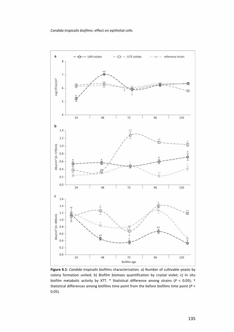
Candida tropicalis biofilms: effect on epithelial cells
135
Figure 6.1: Candida tropicalis biofilms characterization. a) Number of cultivable yeasts by colony formation united; b) Biofilm biomass quantification by crystal violet; c) In situ biofilm metabolic activity by XTT. * Statistical difference among strains (P < 0.05); † Statistical differences among biofilms time point from the before biofilms time point (P < 0.05).
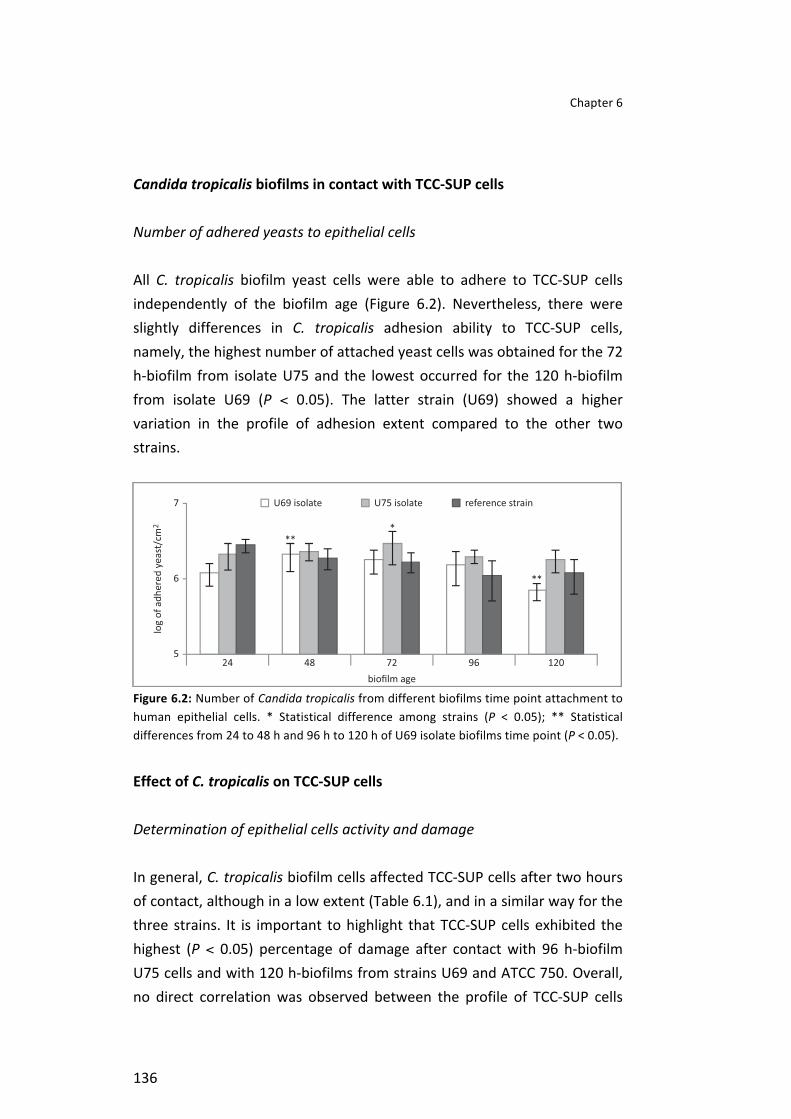
Chapter 6
136
Candida tropicalis biofilms in contact with TCC-‐SUP cells
Number of adhered yeasts to epithelial cells
All C. tropicalis biofilm yeast cells were able to adhere to TCC-‐SUP cells independently of the biofilm age (Figure 6.2). Nevertheless, there were slightly differences in C. tropicalis adhesion ability to TCC-‐SUP cells, namely, the highest number of attached yeast cells was obtained for the 72 h-‐biofilm from isolate U75 and the lowest occurred for the 120 h-‐biofilm from isolate U69 (P < 0.05). The latter strain (U69) showed a higher variation in the profile of adhesion extent compared to the other two strains.
Figure 6.2: Number of Candida tropicalis from different biofilms time point attachment to human epithelial cells. * Statistical difference among strains (P < 0.05); ** Statistical differences from 24 to 48 h and 96 h to 120 h of U69 isolate biofilms time point (P < 0.05).
Effect of C. tropicalis on TCC-‐SUP cells
Determination of epithelial cells activity and damage
In general, C. tropicalis biofilm cells affected TCC-‐SUP cells after two hours of contact, although in a low extent (Table 6.1), and in a similar way for the three strains. It is important to highlight that TCC-‐SUP cells exhibited the highest (P < 0.05) percentage of damage after contact with 96 h-‐biofilm U75 cells and with 120 h-‐biofilms from strains U69 and ATCC 750. Overall, no direct correlation was observed between the profile of TCC-‐SUP cells
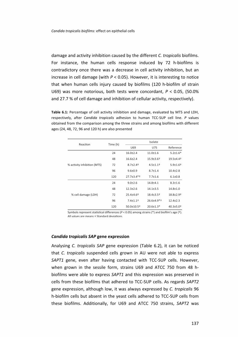
Candida tropicalis biofilms: effect on epithelial cells
137
damage and activity inhibition caused by the different C. tropicalis biofilms. For instance, the human cells response induced by 72 h-‐biofilms is contradictory once there was a decrease in cell activity inhibition, but an increase in cell damage (with P < 0.05). However, it is interesting to notice that when human cells injury caused by biofilms (120 h-‐biofilm of strain U69) was more notorious, both tests were concordant, P < 0.05, (50.0% and 27.7 % of cell damage and inhibition of cellular activity, respectively).
Table 6.1: Percentage of cell activity inhibition and damage, evaluated by MTS and LDH, respectively, after Candida tropicalis adhesion to human TCC-‐SUP cell line. P values obtained from the comparison among the three strains and among biofilms with different ages (24, 48, 72, 96 and 120 h) are also presented
Candida tropicalis SAP gene expression
Analysing C. tropicalis SAP gene expression (Table 6.2), it can be noticed that C. tropicalis suspended cells grown in AU were not able to express SAPT1 gene, even after having contacted with TCC-‐SUP cells. However, when grown in the sessile form, strains U69 and ATCC 750 from 48 h-‐biofilms were able to express SAPT1 and this expression was preserved in cells from these biofilms that adhered to TCC-‐SUP cells. As regards SAPT2 gene expression, although low, it was always expressed by C. tropicalis 96 h-‐biofilm cells but absent in the yeast cells adhered to TCC-‐SUP cells from these biofilms. Additionally, for U69 and ATCC 750 strains, SAPT2 was
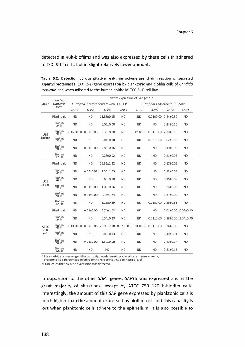
Chapter 6
138
detected in 48h-‐biofilms and was also expressed by these cells in adhered to TCC-‐SUP cells, but in slight relatively lower amount.
Table 6.2: Detection by quantitative real-‐time polymerase chain reaction of secreted aspartyl proteinases (SAPT1-‐4) gene expression by planktonic and biofilm cells of Candida tropicalis and when adhered to the human epithelial TCC-‐SUP cell line
In opposition to the other SAPT genes, SAPT3 was expressed and in the great majority of situations, except by ATCC 750 120 h-‐biofilm cells. Interestingly, the amount of this SAP gene expressed by planktonic cells is much higher than the amount expressed by biofilm cells but this capacity is lost when planktonic cells adhere to the epithelium. It is also possible to

Candida tropicalis biofilms: effect on epithelial cells
139
observe that, in general, the expression of SAPT3 was lower for C. tropicalis cells adhered to TCC-‐SUP cells issuing from biofilms. Interestingly, SAPT4 was only expressed by the reference strain, and in few situations (48h-‐biofilms and adhered yeasts grown in suspension and from 24 h-‐biofilms).
DISCUSSION
Candida species are the second most common cause of medical device infections, especially in patients from ICUs, which are associated with the highest pathogen-‐specific mortality [20, 21]. Furthermore, C. tropicalis and other Candida species are able to form biofilms, and their presence during infection has been related to higher mortality rates compared with isolates which are not capable of forming biofilms [21, 22]. Moreover, recent studies have demonstrated the ability of C. tropicalis to form biofilms in the presence of AU, under static and dynamic conditions [11, 12, 23].
In accordance with other studies [11, 12, 23], the strains of C. tropicalis assayed herein were able to form biofilms in the presence of AU (Figure 6.1), although in a strain and time dependent way. Similarly, a study by Jain et al. [23] with C. albicans, C. glabrata and C. tropicalis, using AU and RPMI 1640, showed that biofilm formation varied considerably among isolates under both growth conditions.
Observing the biofilm profile along time (Figure 6.1), no consistent pattern can be noticed among the different strains. The only similarity among strains is an increase in the number of cultivable cells and biofilm metabolic activity from 72 to 96 h biofilms. Variations among C. tropicalis strains concerning biofilm formation are expected due to physiological differences among strains [18, 22, 24]. Furthermore, as reported before [16, 18, 22] C. tropicalis species mature biofilms consist of a dense and heterogeneous network of yeast, pseudohyphae and hyphae and these forms are not always similar among C. tropicalis strains. These results corroborate other studies reporting that biofilm kinetics is strain dependent [11, 24]. For instance, in the present situation, U69 strain 24 h-‐

Chapter 6
140
biofilm presented the lowest number of cultivable yeasts (1.60 x 10⁵ CFU/cm2; P = 0.01), although, showing the highest biofilm biomass (0.54 Abs/cm2; P = 0.01). However, in a previous work [18], using RPMI 1640 as growth medium, U69 biofilm biomass was similar to the other C. tropicalis strains, which highlights that biofilms are dependent on growth medium, carbohydrate supplementation and the nature of the colonized surface [11, 22-‐24]. Furthermore, it has been reported that cultivable yeast cells and biofilm metabolic activity seems to be dependent of biofilm maturity with an increase in those parameters along biofilm development [16, 24, 25]. The present results underline strain differences in terms of biofilm metabolic activity and number of cultivable cells, which could have important implications in terms of C. tropicalis relative virulence.
Moreover, it should be stressed that the extracellular matrix of biofilms also accounts for total biofilm biomass and smaller amounts of cultivable cells do not mean a less amount of extracellular matrix because this is a trait strain specific and dependent on environmental conditions [12, 22]. Therefore, these differences on C. tropicalis biofilms can explain the increased difficulty on the treatment of infections related to this species. Moreover, these findings may have high significance concerning its pathogenic potential when extrapolated to in vivo situations.
It is known that during the development of Candida biofilms, some dispersion/detachment of cells or dissolution of biofilm pieces can occur [12, 15]. This is an important phenomenon, since the dispersed and detached cells could be responsible for the subsequent establishment of disseminated candidiasis at distal organs [15, 26]. Furthermore, in a recent study with C. albicans, Uppuluri et al. [15], have demonstrated that dispersed cells display a distinct phenotype, which is associated with an increased virulence. Additionally, according to epidemiological data, C. tropicalis infection is strongly connected with the presence of biofilms in urinary catheters [21, 23, 27-‐29]. Thus, it is extremely important to study the ability of C. tropicalis cells detached from pre-‐formed biofilms to colonize human epithelial cells and the consequent degree of damage.

Candida tropicalis biofilms: effect on epithelial cells
141
Consequently, biofilms with different degrees of maturation, were put in contact, for two hours, with human urinary bladder epithelial cells (TCC-‐SUP cells) in order to evaluate the effect of biofilm cells on this epithelium. Along with the determination of the number of adhered yeasts to TCC-‐SUP cells (by CFU quantification), human cells damage and activity inhibition were also assessed. The results are indicative that cells from all C. tropicalis biofilms (independently of their age) were able to detach and colonize TCC-‐SUP cells (Figure 6.2), although in a strain dependent way. Other studies have shown that C. tropicalis planktonic cells extent of adhesion to human cells is also strain specific [10, 18, 30].
It is interesting to highlight that biofilm yeast cells adhered to TCC-‐SUP cells in a similar extent of their planktonic counterparts [18]. Uppuluri et al. [15] reported a different behaviour for C. albicans adhesion to endothelial cells, since yeast cells dispersed from biofilms adhered in larger numbers than planktonic ones. Maybe, this fact can be related with differences between the two yeast species or the differences between the human cells used, which determine very specific interactions with yeast cells [31, 32]
Besides, the quantification of the number of yeasts adhered to TCC-‐SUP it is of major importance to understand Candida pathogenesis – their effect on human cells. Other studies are indicative that dispersed cells from yeast biofilms caused increased human cells damage when compared to planktonic yeast cells [15]. Moreover, human cells damage is reported to be dependent on the contact time and yeast infectivity rate [10, 15, 33]. Overall, the present results point out that older biofilms (96 h or 120 h) induced higher cell damage (Table 6.1) than younger ones, however in a strain dependent manner. Therefore, these data highlight the virulence potential of C. tropicalis cells dispersed from biofilms, since they seem to be a causative agent of an increased LDH release by TCC-‐SUP cells.
In fact, opportunistic yeast pathogens, such as C. tropicalis, are able to promote cell host immune response and progressive cell damage during infection [10, 30, 34]. However, in this study no direct correlation was observed between cell damage and inhibition of cellular activity, namely
Chapter 6
142
adhered yeast cells from 72 h-‐biofilm, induced high damage but a low extent of inhibition of cell activity. Nevertheless, it is very interesting to notice that when the inhibition of cellular activity was significantly higher (for U69 120 h-‐biofilm) cell damage was also the highest. This underlines that these two factors are only in direct relation when the degree of injury is very high. The lack of a direct relation between cell activity inhibition and cell damage at low levels of injury can be explained by the fact that, during the first stage of human cells colonization by Candida, the cellular defence mechanisms are initiated, which may cause an increase of metabolic activity before the onset of any damage [34-‐37]. Moreover, the present results point out that despite no significant differences observed in terms of C. tropicalis strains ability to colonize the TCC-‐SUP cells a direct correlation was not observed among the specific biofilm characteristics and the injury caused to human cells.
It has been widely reported that, during the adhesion and invasion processes of host tissues, Candida species are able to secrete hydrolytic enzymes that cause damage on host cells membrane integrity, leading to dysfunction or disruption of host structures [38, 39]. Candida secreted aspartyl proteinases (Saps) have been associated with tissue invasion and their secretion is also associated with the inflammatory host response [40]. Nowadays, several studies have been performed with the aim to elucidate the role of each Sap on the development of the host infection process. Recent data indicate that Sap1 to Sap3 family are probably the principal C. albicans proteinases involved on mucocutaneous infections in vivo and, also on adhesion to human cells and tissue damage. Furthermore, C. albicans Sap4 to Sap6 family proteinases have been implicated with systemic but not mucosal infections and in the evasion of phagocytosis [39, 41-‐43]. Additionally, the expression of SAP genes by C. tropicalis (SAPT1 to SAPT4) has also been demonstrated on the surface of fungal elements penetrating tissues during disseminated infection and evading macrophages after yeast cells phagocytosis [10, 38, 44, 45]. Although, little is known about the contribution of SAP genes on Candida biofilm formation, recent findings showed that sessile C. albicans cells adhered to abiotic surfaces secrete more Saps than their planktonic counterparts [46].

Candida tropicalis biofilms: effect on epithelial cells
143
Moreover, Nailis et al. [47] reported that SAP genes are upregulated in C. albicans biofilms grown in different model systems (biotic and abiotic) at different stages of maturity. Thus, with the aim to understand the role of Saps as causative agents of the injury caused to TCC-‐SUP cells, the levels of expression of C. tropicalis SAP genes were assessed for planktonic and biofilm cells before and after adhering to TCC-‐SUP human cells.
Real time PCR analysis (Table 6.2) revealed that SAPT gene expression by C. tropicalis grown in AU (in planktonic and biofilm form and colonizing TCC-‐SUP cells) showed, in general, a higher level for SAPT3 expression followed by lower levels of SAPT2, SAPT1 and SAPT4. These features were similar to those described by Silva et al. [10] who studied the expression profiles of SAP genes by seven C. tropicalis strains in contact with reconstituted human oral epithelium: all strains also expressed the 4 genes (SAPT1 – 4), moreover SAPT2 and SAPT4 transcripts were detected in a similar extent to SAPT3 and the majority of strains did not express SAPT1. Similarly, in the present study, only two strains (U69 and ATCC 750 strains) in 48 h-‐biofilms were able to express SAPT1, but in planktonic form this gene expression was not detected. Furthermore, the high expression of SAPT3 points out its importance in the pathogenesis of C. tropicalis strains under these conditions.
In order to confer maximum benefits for Candida pathogenicity SAP genes family are activated efficiently and in flexible way at specific time points during colonization and infection processes. Moreover, SAP genes expression is strongly correlated with environment where Candida is grown [39, 41, 44]. Nails et al. (2010) detected differences in C. albicans SAP genes expression between in vitro grown biofilms and in vivo model. Interestingly, SAP1, SAP2, SAP4 and SAP6 were highly upregulated in biofilms grown in vitro and the expression of SAP2, SAP4 and SAP6 was also high in the in vivo model. In the present study there is also a different gene expression among the different modes of growth. For instance, although C. tropicalis 96 h-‐biofilms exhibited SAPT2 expression, when these biofilm cells colonize TCC-‐SUP cells this gene is not expressed. Curiously, SAPT4 was only detected in ATCC 750 biofilms and at specific biofilm ages. Other
Chapter 6
144
studies indicate that there is an optimum pH for C. tropicalis-‐secreted aspartic proteinases activity, therefore making SAP gene expression strain and epithelium dependent [10, 38, 44]. It is important to highlight that there are only few studies reporting C. tropicalis SAPT genes expression during the adhesion to human cells and there is also limited knowledge about the role of these enzymes in C. tropicalis biofilms. This could suggest that Saps do not play a significant role in the reduction of epithelial cells activity and damage caused by C. tropicalis strains.
In summary, the present study shows that C. tropicalis detached from biofilms are able to colonize human cells and cause some injury: damage and reduction of metabolic activity. Moreover, it is important to highlight that C. tropicalis SAP genes are expressed during C. tropicalis biofilm formation. Furthermore, to our knowledge, this is the first report of C. tropicalis SAP genes expression during biofilm formation and when biofilm cells colonize TCC-‐SUP cells, and the results imply a limited role of these enzymes in human cells damage and metabolic activity reduction in the conditions assayed. Nevertheless, SAPT3 transcript presented the highest level of gene expression by C. tropicalis assayed, regardless of biofilm age. However, more studies have to be performed to clarify if these C. tropicalis SAPs genes are associated with biofilm development and C. tropicalis virulence potential.
Candida tropicalis biofilms: effect on epithelial cells
145
REFERENCES
1. Lass-‐Flörl C. The changing face of epidemiology of invasive fungal disease in Europe. Mycoses 2009; 52 (3): 197-‐205. 2. Nucci M, Queiroz-‐Telles F, Tobon AM, Restrepo A, Colombo AL. Epidemiology of opportunistic fungal infections in Latin America. Clin Infect Dis 2010; 51 (5): 561-‐70. 3. Horn DL, Neofytos D, Anaissie EJ, et al. Epidemiology and outcomes of candidemia in 2019 patients: data from the prospective antifungal therapy alliance registry. Clin Infect Dis 2009; 48 (12): 1695-‐703. 4. Falagas ME, Roussos N, Vardakas KZ. Relative frequency of albicans and the various non-‐albicans Candida spp among candidemia isolates from inpatients in various parts of the world: a systematic review. Int J Infect Dis 2010; 14 (11): 954-‐66. 5. Paulo C, Mourão C, Veiga PM, et al. Retrospective analysis of clinical yeast isolates in a hospital in the centre of Portugal: spectrum and revision of the identification procedures. Med Mycol 2009; 19 (1): 1 -‐ 10. 6. Paul N, Mathai E, Abraham OC, Michael JS, Mathai D. Factors associated with candiduria and related mortality. J Infect 2007; 55 (5): 450-‐455. 7. Goetz LL, Howard M, Cipher D, Revankar SG. Occurrence of candiduria in a population of chronically catheterized patients with spinal cord injury. Spinal Cord 2009; 48 (1): 51-‐54. 8. Munoz P, Giannella M, Fanciulli C, et al. Candida tropicalis fungaemia: incidence, risk factors and mortality in a general hospital. Clin Microbiol Infect 2010. 9. Kothavade RJ, Kura MM, Valand AG, Panthaki MH. Candida tropicalis: its prevalence, pathogenicity and increasing resistance to fluconazole. J Med Microbiol 2010; 59 (Pt 8): 873-‐80. 10. Silva S, Hooper SJ, Henriques M, et al. The role of secreted aspartyl proteinases in Candida tropicalis invasion and damage of oral mucosa. Clin Microbiol Infect 2011; 17 (2): 264-‐272. 11. Silva S, Negri M, Henriques M, et al. Silicone colonization by non-‐Candida albicans Candida species in the presence of urine. J Med Microbiol 2010; 59 (Pt 7): 747-‐54. 12. Negri M, Silva S, Henriques M, et al. Candida tropicalis biofilms: artificial urine, urinary catheters and flow model. Med Mycol 2011; 0 (0): 1-‐9. 13. Douglas LJ. Candida biofilms and their role in infection. Trends Microbiol 2003; 11 (1): 30-‐36. 14. Chandra J, Zhou G, Ghannoum M. Fungal biofilms and antimycotics. Curr Drug Targets. 2005; 6 (8): 887-‐94. 15. Uppuluri P, Chaturvedi AK, Srinivasan A, et al. Dispersion as an Important Step in the Candida albicans Biofilm Developmental Cycle. PLoS Pathog 2010; 6 (3): e1000828. 16. Silva S, Henriques M, Oliveira R, Williams D, Azeredo J. In Vitro Biofilm Activity of Non-‐Candida albicansCandida Species. Curr Microbiol 2010; 61 (6): 534-‐40. 17. Negri M, Gonçalves V, Silva S, et al. Crystal violet staining to quantify Candida adhesion to epithelial cells. Br J Microbiol 2010; 67 (3): 120-‐125. 18. Negri M, Martins M, Henriques M, et al. Examination of potential virulence factors of Candida tropicalis clinical isolates from hospitalized patients. Mycopathologia 2010; 169 (3): 175-‐182. 19. Negri M, Botelho C, Silva S, et al. An in vitro evaluation of Candida tropicalis infectivity using human cell monolayers. J Med Microbiol 2011. 20. Nett J, Andes D. Review of techniques for diagnosis of catheter-‐related Candida biofilm infections. Curr Fungal Infect Rep 2008; 2 (4): 237-‐243. 21. Kojic EM, Darouiche RO. Candida Infections of Medical Devices. Clin Microbiol Rev 2004; 17 (2): 255-‐267. 22. Silva S, Negri M, Henriques M, et al. Adherence and biofilm formation of non-‐Candida albicans Candida species. Trends Microbiol 2011; 19 (5): 241-‐247. 23. Jain N, Kohli R, Cook E, et al. Biofilm formation by and antifungal susceptibility of Candida isolates from urine. Appl Environ Microbiol 2007; 73 (6): 1697-‐1703. 24. Bizerra FC, Nakamura CV, de Poersch C, et al. Characteristics of biofilm formation by Candida tropicalis and antifungal resistance. FEMS Yeast Res 2008; 8 (3): 442-‐450.
Chapter 6
146
25. Uppuluri P, Dinakaran H, Thomas DP, Chaturvedi AK, Lopez-‐Ribot JL. Characteristics of Candida albicans Biofilms Grown in a Synthetic Urine Medium. J Clin Microbiol 2009; 47 (12): 4078-‐4083. 26. Sellam A, Al-‐Niemi T, McInnerney K, et al. A Candida albicans early stage biofilm detachment event in rich medium. BMC Microbiol 2009; 9 (1): 25. 27. Kauffman CA, Vazquez JA, Sobel JD, et al. Prospective multicenter surveillance study of funguria in hospitalized patients. Clin Infect Dis 2000; 30 (1): 14-‐18. 28. Achkar JM, Fries BC. Candida infections of the genitourinary tract. Clin Microbiol Rev 2010; 23 (2): 253-‐73. 29. Bougnoux ME, Kac G, Aegerter P, d'Enfert C, Fagon JY. Candidemia and candiduria in critically ill patients admitted to intensive care units in France: incidence, molecular diversity, management and outcome. Intensive Care Med 2008; 34 (2): 292-‐9. 30. Okawa Y, Miyauchi M, Kobayashi H. Comparison of Pathogenicity of Various Candida tropicalis Strains. Biol Pharm Bull 2008; 31 (8): 1507-‐1510. 31. Bendel CM. Colonization and epithelial adhesion in the pathogenesis of neonatal candidiasis. Semin Perinatol 2003; 27 (5): 357-‐364. 32. Sohn K, Senyürek I, Fertey J, et al. An in vitro assay to study the transcriptional response during adherence of Candida albicans to different human epithelia. FEMS Yeast Res 2006; 6 (7): 1085-‐1093. 33. Silva S, Henriques M, Oliveira R, et al. Characterization of Candida parapsilosis infection of an in vitro reconstituted human oral epithelium. Eur J Oral Sci 2009; 117 (6): 669-‐75. 34. Saegusa S, Totsuka M, Kaminogawa S, Hosoi T. Cytokine responses of intestinal epithelial-‐like Caco-‐2 cells to non-‐pathogenic and opportunistic pathogenic yeasts in the presence of butyric acid. Biosci Biotechnol Biochem 2007; 71 (10): 2428-‐34. 35. Wang L, Sun J, Horvat M, et al. Evaluation of MTS, XTT, MTT and3HTdR incorporation for assessing hepatocyte density, viability and proliferation. Methods Cell Sci 1996; 18 (3): 249-‐255. 36. Schaller M, Mailhammer R, Grassl G, et al. Infection of human oral epithelia with Candida species induces cytokine expression correlated to the degree of virulence. J Invest Dermatol 2002; 118 (4): 652-‐657. 37. van de Veerdonk FL, Kullberg BJ, Netea MG. Pathogenesis of invasive candidiasis. Curr Opin Crit Care 2010; 16 (5): 453-‐9. 38. Zaugg C, Borg-‐von Zepelin M, Reichard U, Sanglard D, Monod M. Secreted aspartic proteinase family of Candida tropicalis. Infect Immun 2001; 69 (1): 405-‐412. 39. Naglik JR, Albrecht A, Bader O, Hube B. Candida albicans proteinases and host/pathogen interactions. Cell Microbiol 2004; 6 (10): 915-‐926. 40. Yang Y. Virulence factors of Candida species. J Microbiol Immunol Infect. 2003; 36 (4): 223-‐228. 41. Naglik JR, Challacombe SJ, Hube B. Candida albicans Secreted Aspartyl Proteinases in Virulence and Pathogenesis. Microbiol Mol Biol Rev 2003; 67 (3): 400-‐428. 42. Gropp K, Schild L, Schindler S, et al. The yeast Candida albicans evades human complement attack by secretion of aspartic proteases. Mol Immunol 2009; 47 (2-‐3): 465-‐75. 43. Naglik JR, Moyes D, Makwana J, et al. Quantitative expression of the Candida albicans secreted aspartyl proteinase gene family in human oral and vaginal candidiasis. Microbiology 2008; 154 (Pt 11): 3266-‐80. 44. Togni G, Sanglard D, Falchetto R, Monod M. Isolation and nucleotide sequence of the extracellular acid protease gene (ACP) from the yeast Candida tropicalis. FEBS Lett. 1991; 286 (1-‐2): 181-‐5. 45. Monoda M, Capocciaa S, Léchennea B, et al. Secreted proteases from pathogenic fungi Int. J.Medical Microbiol. 2002; 292 (5-‐6): 405-‐419 46. Mendes A, Mores AU, Carvalho AP, et al. Candida albicans biofilms produce more secreted aspartyl protease than the planktonic cells. Biol Pharm Bull 2007; 30 (9): 1813-‐5. 47. Nailis H, Kucharikova S, Ricicova M, et al. Real-‐time PCR expression profiling of genes encoding potential virulence factors in Candida albicans biofilms: identification of model-‐dependent and -‐independent gene expression. BMC Microbiology 2010; 10: 114.
CHAPTER 7
Final conclusionsfuture perspectives


Chapter 7 -‐ Concluding remarks and future perspectives
149
CONCLUDING REMARKS AND FUTURE PERSPECTIVES
Candida tropicalis is a common yeast species related to nosocomial candidemia and candiduria. Several virulence factors seem to be responsible for C. tropicalis infections, which present high potential for dissemination and mortality. Adhesion to host surfaces (medical devices and host cells), as well as biofilm formation, are considered the first steps to initiate Candida infection. Hence, the colonization of indwelling devices like urinary catheters by C. tropicalis poses a critical problem.
In fact, several important studies have been describing the potential of C. tropicalis to form biofilms, although, most of the in vitro research do not always mimic what happens in reality. Thus, in Chapter 2, it is reported for the first time the formation of C. tropicalis biofilms in urinary catheters in a flow model, although using artificial urine (AU) to obtain more reproducible results. The model developed appears to be suitable for studies simulating the real conditions and furthermore it was possible to observe that C. tropicalis was able to adhere and to form biofilms along the entire length of both types of urinary catheters, latex and silicone.
Furthermore, all C. tropicalis strains assayed along this work were able to form biofilms independently of the culture medium used, RPMI 1640 (Chapter 4) or AU (Chapter 6). However, there were differences among strains when biofilms were formed in AU (Chapter 2 and 6). Curiously, although the isolate U69 adhered significantly in higher extent to both types of catheters than the reference strain (Chapter 2), U69 biofilms contained significantly less cultivable cells but higher biofilm biomass than those of the reference strain. A similar behaviour was observed for U69 24 h biofilms, formed in silicone coupons in AU (Chapter 6). In older biofilms, the clinical isolate U69 showed significantly higher number of cultivable yeasts (eg 48 h biofilm), than the other two strains (reference strain and U75 clinical isolate) assayed. Nevertheless, C. tropicalis biofilms grown in AU seem to stabilize in terms of number of cells over time (after 48 h) both in dynamic and static model.

Chapter 7
150
The formation of a biofilm inside or in the outer surface of medical devices causes a multiplicity of problems. One of the most frequent consequences is the infection of distal sites. Biofilm associated infections are very difficult to treat, especially due to the conjunction with an increased occurrence of multi-‐drug resistance by Candida species. It is important to emphasise that one clinical isolate (U69) was resistant and other four (U16, U29, U75 and CL012) were susceptible-‐dose dependent to itraconazole (Chapter 4). Since, those strains were effective biofilm formers, there is an increased risk factor associated to those strains in the development of candidosis, especially when these strains are able to detach from biofilm and to colonize other surfaces, as host cells. In this work, under flow conditions, C. tropicalis strains were able to detach from catheters (latex and silicone) and to move upflow against the stream, demonstrating undoubtedly their capacity to colonize distal sites (Chapter 2).
Adhesion to and invasion of host cells by Candida species is considered the first step in systemic infections. Although in vitro studies of adhesion capability are essential to characterise the virulence of those Candida species, the assessment of adhesion by traditional methods is very time consuming. Therefore, an easy to perform and reproducible technique was developed to assess the adhesion ability of Candida species to epithelial cell lines (Chapter 3). The method was validated for the different Candida reference strains of different species by comparison with traditional microscope observation and enumeration. High correlation values (r2= 0.9724–0.9997) between the number of adherent yeasts (microscope enumeration) and absorbance values were obtained for an inoculum concentration higher than 106 cells/ml. Additionally, it was also verified that all Candida species adhered in a greater extent to epithelial cells than to silicone. However, different Candida species have different sizes and absorb distinct amounts of dye, which does not allow comparison of the level of adhesion through direct CV absorbance readings. So, after standardization of the results using the corresponding equation for each species, C. glabrata (initial cell density: 108 yeast/ml), and C. tropicalis (initial cell density: 107 yeast/ml) adhered significantly more than other Candida species to epithelial cells. Although these results correspond to a
Concluding remarks and future perspectives
151
low number of strains, it is important to highlight these data since C. tropicalis is a common species related to nosocomial candidaemia and candiduria, and C. glabrata is now emerging as an important agent in both mucosal and bloodstream infections.
In this context, while investigating more about C. tropicalis virulence, with more samples (Chapter 4), it was possible to confirm that C. tropicalis strains adhered significantly in higher number to epithelium than to silicone when in RPMI 1640 culture medium. Interestingly, the same happened (Chapter 3) in a different culture medium (PBS), indicating that there is a greater predilection of C. tropicalis for urinary bladder cells (TCC-‐SUP) than silicone coupons, independently of culture medium.
Furthermore, regarding C. tropicalis infectivity (Chapter 5) it was shown that C. tropicalis strains were able to adhere to the different human cell lines, although, in a strain and cell line dependent manner. Strain U69, which is a clinical isolate from urine, adhered in significantly higher number to intestinal cells (Caco-‐2) than to urinary cells (TCC-‐SUP) and cervical cells (HeLa). Regarding the reference strain (ATCC 750), it adhered to TCC-‐SUP cells, HeLa cells and Caco-‐2 cells in a similar extent. Also, C. tropicalis biofilm cells (Chapter 6) were able to adhere to TCC-‐SUP cells, in general, independently of biofilm age, even so the highest extent of yeast attachment was obtained for the 72 h old U75 biofilm and the lowest occurred for the 120 h-‐biofilm from strain U69. It is important to note that there may be a specific interaction between each human cell type and each strain of C. tropicalis. Moreover, there is not a strong correlation between the adherence ability to epithelial cells when the site of yeast isolation corresponds to a similar type of epithelium.
Concerning the human cells response to C. tropicalis colonization, it was noticed that both planktonic (Chapter 5) and biofilms (Chapter 6) yeasts were able to affect the human cells. In general, the highest cell activity inhibition was observed for Caco-‐2, followed by TCC-‐SUP and HeLa cells (Chapter 5). The highest percentage of cell damage was observed for TCC-‐SUP in contact with the U69 isolate and for Caco-‐2 in contact with the
Chapter 7
152
reference strain. Yeasts from biofilms (Chapter 6) affected TCC-‐SUP cells, with 120 h-‐biofilm cells of U69 and ATCC 750 strains causing the highest levels of cell injury (damage and inhibition of cell activity). Comparing the effect of dispersed cells from C. tropicalis biofilms (Chapter 6) with planktonic cells (Chapter 5), it was observed that the first caused more cellular damage than the latter. Therefore, these data highlight the virulence potential of C. tropicalis cells dispersed from biofilms, since they seem to be a causative agent of an increased damage, specially on TCC-‐SUP cells.
Although no direct correlation between inhibition of cell activity and cell damage was found, it was noticed that when cell inhibition is significantly high, cell damage is also notorious. Unfortunately, there are few studies regarding the effect of Candida species in human cells activity. Therefore, the present study is a step forward for the understanding of C. tropicalis pathogenesis.
During the adhesion and invasion processes of host tissues, Candida species are able to secrete hydrolytic enzymes that cause damage on host cells membrane integrity, leading to dysfunction or disruption of host structures. Regarding expression of enzymes (proteinases, phospholipase and haemolysins) on specific agar medium (Chapter 4), all C. tropicalis strains assayed were able to express total haemolytic activity. However, proteinases were only produced by four isolates (U16, U29, CL012 and B1). Moreover, only one C. tropicalis strain was phospholipase positive (CL012). According to these results, proteinase and phospholipase expression can vary according to strain and the site of isolation. Although the methods used to test the production of these enzymes are simple and fast, they are not excessively accurate, specially compared with molecular methods that can detect gene expression.
Thus, real-‐time PCR analysis (Chapter 5) was also used and it revealed a wide range of expression profiles of Secreted Aspartyl Proteinases (SAP) genes for both C. tropicalis strains (U69 and reference strain) in contact with the different types of epithelial cells. According to data obtained in
Concluding remarks and future perspectives
153
Chapter 5 and 6, SAPT genes may be involved on pathogenesis of C. tropicalis, affecting the human cell lines. Moreover, C. tropicalis SAP genes were expressed during C. tropicalis biofilm formation (Chapter 6). Furthermore, in both studies (Chapter 5 and 6), SAPT3 produced by adhered and biofilm yeasts were highly expressed comparing with other SAPT genes. It is important to emphasize that human cells response to C. tropicalis adhesion, as well as SAPs production, is strain and epithelial cell line dependent. There is limited knowledge about the role of these enzymes in C. tropicalis adhesion and tissue damage. Hence, this is an important point that can contribute for the enhanced understand of C. tropicalis pathogenesis.
These findings lead to other questions, which would greater contribute to the understanding of the virulence factors used by C. tropicalis during the development of candidosis. Some of the suggestions that should be taken into consideration for future investigations are:
1. Assessment of mechanisms related with the phenomen of yeast cells detachment from C. tropicalis biofilms, investigating architecture, cells interaction and quorum sensing of C. tropicalis biofilms.
2. Characterization of the main virulence factors of C. tropicalis cells detached from biofilms and evaluation of their antifungal resistance.
3. Evaluation of the role of phospholipases and haemolysins in C. tropicalis pathogenesis by genomic approach, by analysing the levels of gene expression during the interaction with human cells.
4. Assessment of molecular interactions between human cells and C. tropicalis analysing cellular response and factors related with the process of adhesion and infectivity.
5. Summarising, these factors together with those previously exposed, may come together to help clarifying C. tropicalis behaviour during the process of infectivity and, furthermore, could contribute to develop new antifungal agents targeting these mechanism of Candida virulence.

Top Related