

![neuropatia periférica induzida pela quimioterapia (npiq) · 26 On 34 > [jan-jun 2017] neuropatia periférica induzida pela quimioterapia (npiq) resumo: a neuropatia Periférica Induzida](https://static.fdocumentos.com/doc/165x107/5fad5917dd23b224ef7f5622/neuropatia-perifrica-induzida-pela-quimioterapia-npiq-26-on-34-jan-jun.jpg)


Línguas
Páginas
Legal

A MELHORA DA MEMÓRIA INDUZIDA POR ESPERMIDINA ENVOLVE A FOSFORILAÇÃO DA PKC, PKA E CREB EM HIPOCAMPO DE RATOS
Gustavo Petri Guerra
UFSM
Tese de doutorado
A MELHORA DA MEMÓRIA INDUZIDA POR ESPERMIDINA ENVOLVE A FOSFORILAÇÃO DA PKC, PKA E CREB EM HIPOCAMPO DE RATOS
__________________________________________________
Gustavo Petri Guerra
PPGBT
SANTA MARIA – RS – BRASIL
2011

A MELHORA DA MEMÓRIA INDUZIDA POR
ESPERMIDINA ENVOLVE A FOSFORILAÇÃO DA PKC, PKA E CREB EM HIPOCAMPO DE RATOS
__________________________________________________
por
Gustavo Petri Guerra
Tese apresentada ao Programa de Pós-Graduação em
Ciências Biológicas: Bioquímica Toxicológica da Universidade
Federal de Santa Maria, como requisito parcial para a obtenção
do grau de
Doutor em Ciências Biológicas: Bioquímica Toxicológica.
PPGBT
SANTA MARIA – RS – BRASIL
2011
iii
Universidade Federal de Santa Maria Centro de Ciências Naturais e Exatas
Programa de Pós-Graduação em Ciências Biológicas: Bioquímica Toxicológica
A Comissão Examinadora, abaixo assinada,
aprova a Tese de Doutorado
A MELHORA DA MEMÓRIA INDUZIDA POR ESPERMIDINA ENVOLVE
A FOSFORILAÇÃO DA PKC, PKA E CREB EM HIPOCAMPO DE RATOS
elaborada por
Gustavo Petri Guerra
como requisito parcial para obtenção do grau de
Doutor em Ciências Biológicas: Bioquímica Toxicológica
COMISSÃO EXAMINADORA:
____________________
Maribel Antonello Rubin (Orientadora)
____________________ ____________________
Gustavo da Costa Ferreira Ana Flávia Furian
____________________ ____________________ Maria Rosa ChitolinaSchetinger Luiz Fernando Freire Royes
Santa Maria, 08 setembro de 2011.

iv
“Não se pode esperar resultados diferentes
fazendo as coisas da mesma forma”.
Albert Einstein

v
AGRADECIMENTOS
Agradeço a Deus, por estar sempre presente e nas horas mais difíceis
ajudar a escolher o caminho certo.
Agradeço aos meus familiares, em especial minha mãe Maria Elena, por
ser a base de meus princípios e de minha formação.
A minha namorada Vanessa pelo apoio, compreensão, carinho e amor.
Aos meus orientadores, Maribel, Juliano e Carlos, agradeço pela
orientação científica, pelos ensinamentos diários, pela amizade, e estejam
certos que terão sempre meu respeito.
Aos amigos do Lab., seja de iniciação científica, mestrado ou doutorado,
pela ajuda, amizade e companheirismo.
E a todos que participaram deste trabalho, sem medir esforços para que
desse certo.
Agradeço a Universidade Federal de Santa Maria pela oportunidade e a
CAPES, pelo apoio financeiro.

Sumário
vi
SUMÁRIO
LISTA DE ABREVIATURAS ....................................................................vii LISTA DE FIGURAS ................................................................................viii LISTA DE TABELAS .................................................................................xi RESUMO ..................................................................................................xii ABSTRACT ............................................................................................ xiv I. INTRODUÇÃO ........................................................................................1 II. OBJETIVOS ...........................................................................................6
II.1. OBJETIVO GERAL ........................................................................7 II.2. OBJETIVOS ESPECÍFICOS ..........................................................7
III. REVISÃO BIBLIOGRÁFICA .................................................................8 III.1 – MEMÓRIA ...................................................................................9
III.1.1 – A FORMAÇÃO DA MEMÓRIA .........................................10 III.1.2 – HIPOCAMPO ....................................................................12
III.2 – RECEPTOR N-METIL-D-ASPARTATO – NMDA .....................14 III.2.1 – RECEPTOR NMDA E MEMÓRIA .....................................16
III.3 – POLIAMINAS ............................................................................18 III.3.1 – ESTRUTURA DAS POLIAMINAS ....................................19 III.3.2 – METABOLISMO DAS POLIAMINAS ................................20
III.3.2.1 – SÍNTESE...................................................................21 III.3.2.2 – CATABOLISMO........................................................23
III.4 – RECEPTOR NMDA, POLIAMINAS E MEMÓRIA .....................23 III.5 – PROTEINA QUINASE ...............................................................26
III.5.1 – PROTEINA QUINASE A (PKA) ........................................27 III.5.1.1 – INIBIÇÃO DA PKA ...................................................30 III.5.1.2 –REGULAÇÃO DA PKA ..............................................30 III.5.1.3 – PROTEINA ANCORADOURA ..................................32
III.5.2 – PROTEINA QUINASE C (PKC) ........................................33 III.5.2.1 – ATIVAÇÃO DA PKC .................................................35
III.6 – CREB ........................................................................................36 III.7 – PROTEINA QUINASE, CREB E MEMÓRIA .............................37
IV. RESULTADOS ...................................................................................42 IV.1 – ARTIGO ....................................................................................43 IV.2 – MANUSCRITO ..........................................................................53
V. DISCUSSÃO ........................................................................................89 VI. CONCLUSÕES ...................................................................................97 VII. REFERÊNCIAS .................................................................................99
Lista de Figuras
vii
LISTA DE ABREVIATURAS L-NAME – N-nitro L-arginina metil éster
AC – Adenilato ciclase
AKAP – proteína âncora da quinase A
AMPA – Ácido α-amino-3-hidroxi-5-metil-4-isoxazol propiônico
AMPc – Adenosina monofosfato cíclica
AP-5 – Ácido D-2-amino-5-fosfonopentanóico
CaM – Calmodulina
CaMKII – Proteína quinase dependente de cálcio/calmodulina do tipo II
CPG – Ácido DL-beta-clorofenil glutâmico
CREB – Proteína ligante do elemento responsivo ao AMPc
DAG - Diacilglicerol
DAO – Diamina oxidase
GABA – Ácido δ-aminobutírico
IH – Intra-hipocampal
MgluR – Receptor glutamatérgico metabotrópico
MK-801 – (+)5-metil-10,11-dihidro-5H-dibenzo[a,b]-ciclohepteno-5-10-amino
MTA – Metiltioadenosina
NADPH – Nicotinamida adenina dinucleotídeo fosfato reduzida
NMDA – N-metil-D-aspartato
NO – Óxido Nítrico
NOS – Óxido nítrico sintase
ODC – L- ornitina descarboxilase
PAO – Poliamina oxidase
PDE - Fosfodiasterase
PKA – Proteína quinase A
PKC – Proteína quinase C
PKG – Proteína quinase dependente de GMPc
SNC – Sistema Nervoso Central
SPD – Espermidina

Lista de Figuras
viii
LISTA DE FIGURAS Revisão Bibliográfica Figura III.1 – Representação esquemática do hipocampo e seus
microcircuitos ...................................................................................14
Figura III.2 – Representação esquemática do receptor NMDA ...............16
Figura III.3 – Estrutura química das poliaminas.......................................20
Figura III.4 – Ativação da cascata AMPc/PKA/CREB..............................29
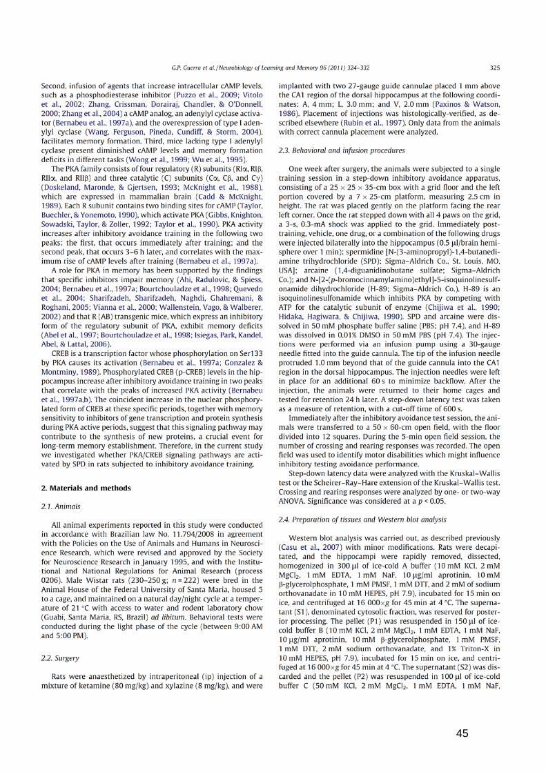
Resultados – Artigo Figura 1 – Co-administration of H-89 (0.5 ρmol intrahippocampus)
immediately after training prevents the improvement in memory
induced by spermidine (SPD, 0.2 nmol). ………………………..……46
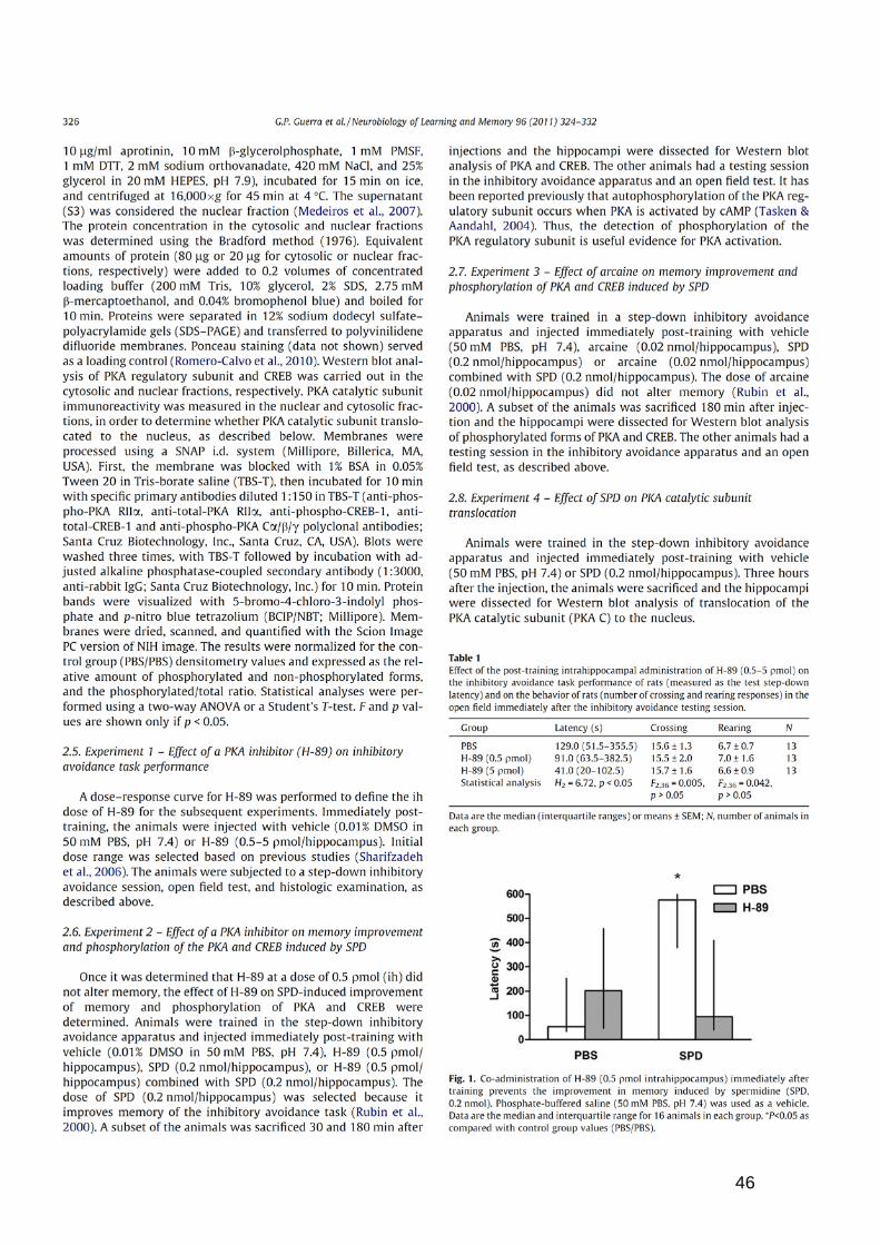
Figura 2 – Effect of the post-training intrahippocampal co-administration
of H-89 (0.5 ρmol) and spermidine (SPD, 0.2 nmol) on the
representative images of Western immunoblotting and densitometry
analyses of phospho-PKA, total-PKA, and the ratio of phospho-
PKA/total-PKA ………………….....…................................................47
Figura 3 – Effect of the post-training intrahippocampal co-administration
of H-89 (0.5 ρmol) and spermidine (SPD, 0.2 nmol) on the levels of
phospho-CREB, total-CREB, and the phospho-CREB/total-CREB
ratio………....………..........................................................................48

Lista de Figuras
ix
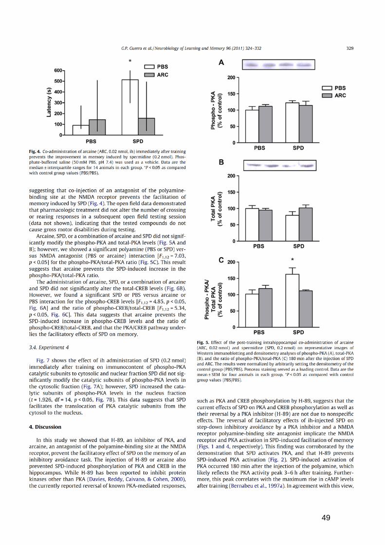
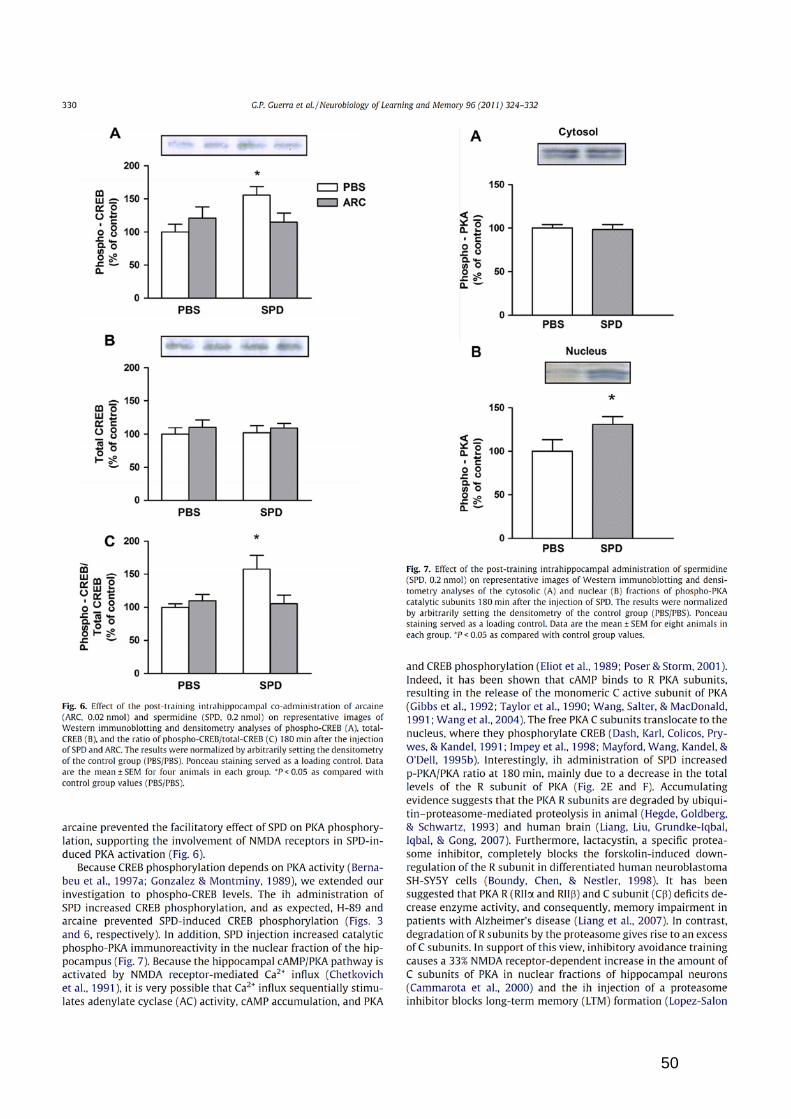
Figura 4 – Co-administration of arcaine (ARC, 0.02 nmol, ih) immediately
after training prevents the improvement in memory induced by
spermidine (0.2 nmol)…………….....................................................49
Figura 5 – Effect of the post-training intrahippocampal co-administration
of arcaine (ARC, 0.02 nmol) and spermidine (SPD, 0.2 nmol) on
representative images of Western immunoblotting and densitometry
analyses of phospho-PKA, total-PKA, and the ratio of phospho-
PKA/total-PKA.............…………………………………………….........49
Figura 6 – Effect of the post-training intrahippocampal co-administration
of arcaine (ARC, 0.02 nmol) and spermidine (SPD, 0.2 nmol) on
representative images of Western immunoblotting and densitometry
analyses of phospho-CREB, total-CREB, and the ratio of phospho-
CREB/total-CREB…………….........................................……...........50
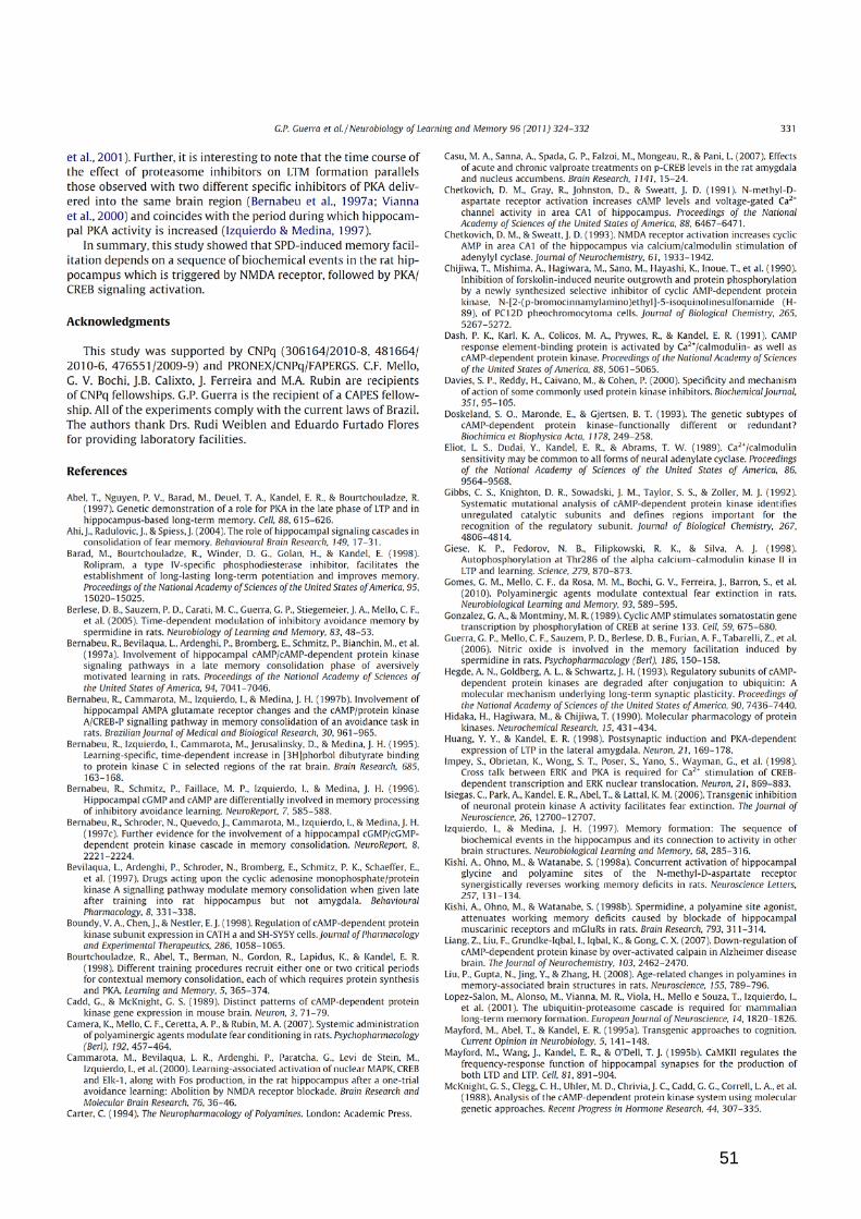
Figura 7 – Effect of the post-training intrahippocampal administration of
spermidine (SPD, 0.2 nmol) on representative images of Western
immunoblotting and densitometry analyses of the cytosolic (A) and
nuclear (B) fractions of phospho-PKA catalytic subunits.….......…...50
Resultados – Manuscrito
Figura 1 – Co-administration of GF 109203X (2.5 ρmol intrahippocampus)
immediately after training prevents the improvement in memory
induced by spermidine (SPD, 0.2 nmol). ………………………..……85

Lista de Figuras
x
Figura 2 – Effect of the post-training intrahippocampal co-administration
of GF 109203X (2.5 ρmol) and spermidine (SPD, 0.2 nmol) on the
representative images of Western immunoblotting and densitometry
analyses of phospho-PKC, total-PKC, and the ratio of phospho-
PKC/total-PKC ………………….....…................................................86
Figura 3 – Effect of the post-training intrahippocampal co-administration
of GF 109203X (2.5 ρmol) and spermidine (SPD, 0.2 nmol) on the
representative images of Western immunoblotting and densitometry
analyses of phospho-PKA, total-PKA, and the ratio of phospho-
PKA/total-PKA ………………….....…................................................87
Figura 4 – Effect of the post-training intrahippocampal co-administration
of GF 109203X (2.5 ρmol) and spermidine (SPD, 0.2 nmol) on
representative images of Western immunoblotting and densitometry
analyses of phospho-CREB, total-CREB, and the ratio of phospho-
CREB/total-CREB…………….........................................……...........88
Discussão Figura V.1 – Ativação da PKC, PKA e CREB na melhora da memória
induzida por SPD......................................................................................96

Lista de Tabelas
xi
LISTA DE TABELAS Resultados – Artigo Tabela 1 – Effect of the post-training intrahippocampal administration of
H-89 (0.5 - 5 ρmol) on the inhibitory avoidance task performance of
rats (measured as the test step-down latency) and on the behavior of
rats (number of crossing and rearing responses) in the open field
immediately after the inhibitory avoidance testing session…..…….…46
Resultados – Manuscrito Tabela 1 – Effect of the post-training intrahippocampal administration of
GF 109203X (1.25 – 2.5 ρmol) on the inhibitory avoidance task
performance of rats (measured as the test step-down latency) and on
the behavior of rats (number of crossing and rearing responses) in
the open field immediately after the inhibitory avoidance testing
session…..………………………………………………………………………..….…84

Resumo
xii
RESUMO
Tese de Doutorado Programa de Pós-Graduação em Ciências Biológicas: Bioquímica Toxicológica
Universidade Federal de Santa Maria, RS, Brasil.
A MELHORA DA MEMÓRIA INDUZIDA POR ESPERMIDINA ENVOLVE A FOSFORILAÇÃO DA PKC, PKA E CREB EM HIPOCAMPO DE RATOS
Autor: Gustavo Petri Guerra
Orientadora: Maribel Antonello Rubin Co-orientador: Juliano Ferreira
Local e Data da Defesa: Santa Maria, 08 de setembro de 2011.
As poliaminas endógenas, putrescina, espermidina e espermina,
são aminas alifáticas que estão presentes em altas concentrações no sistema nervoso central (SNC). As poliaminas modulam diversos canais iônicos, incluindo o subtipo de receptor glutamatérgico N-metil D-aspartato (NMDA). Os processos mediados pelo receptor NMDA incluem plasticidade sináptica e formação de circuitos neurais. Acredita-se que estas plasticidades ocorrendo em algumas regiões cerebrais específicas, como o hipocampo, são críticas para os processos de aprendizado e memória. Está descrito que a espermidina (SPD), assim como as proteínas quinase, esta diretamente envolvida com os processos de formação da memória. Assim, investigamos o envolvimento das proteínas quinases dependente de AMPc (PKA) e dependente de Ca2+ (PKC) sobre a melhora da memória induzida por SPD em ratos. Para isso, ratos Wistar machos foram canulados bilateralmente no hipocampo e após a recuperação cirúrgica treinados na tarefa de esquiva inibitória. Imediatamente após o treino os animais receberam através das cânulas (0,5 μl/sítio) a administração de N-[2-bromocinamilamino etil]-(5-isoquinolina sulfonamida) [H-89, 0,5 ρmol intrahipocampal (ih)], inibidor da PKA, 3-[1-(Dimetilaminopropil)indol-3-il]-4-(indol-3-il)maleimida hidrochloride [GF 109203X, 2,5 ρmol (ih)], inibidor da PKC, arcaína (0,02 nmol, ih), antagonista do sítio de ligação das poliaminas no receptor NMDA ou SPD (0,2 nmol, ih). Um grupo de animais foi eutanasiado 30 ou 180 minutos após a administração das drogas e a atividade da PKC, PKA e o elemento ligante responsivo ao AMPc (CREB), no hipocampo, foi determinada por Western blot. Os outros animais foram submetidos à sessão de teste, 24 horas depois do treino na esquiva inibitória. A administração de H-89, GF 109203X ou arcaína preveniu a melhora da memória induzida por SPD. A SPD (0,2 nmol) aumentou a fosforilação da PKC 30 min, da PKA e do CREB 180 min após a injeção e aumentou a translocação da subunidade catalítica da PKA do citosol para o núcleo. GF 109203X, (2,5 ρmol) preveniu o efeito estimulatório da SPD sobre a fosforilação da PKC, PKA e CREB. Além disso, arcaína (0,02 nmol) e H-

Resumo
xiii
89 (0,5 ρmol) preveniram o efeito estimulatório da SPD sobre a fosforilação da PKA e CREB 180 min depois da injeção. Nenhuma das drogas alterou a atividade motora dos animais. Estes resultados sugerem que o efeito facilitatório da memória induzido pela administração ih de SPD envolve um cruzamento entre PKC e PKA/CREB, com a ativação inicial da PKC, seguida da ativação da cascata PKA/CREB em ratos. Assim, poderemos determinar um possível mecanismo de ação da espermidina nos processos de formação da memória, e desta forma, fornecer subsídios para o desenvolvimento de fármacos.

Abstract
xiv
ABSTRACT
PhD thesis Post Graduation Program in Biological Sciences: Toxicological
Biochemistry Federal University of Santa Maria, RS, Brazil
THE IMPROVEMENT OF THE MEMORY INDUCED BY SPERMIDINE
INVOLVE THE PKC, PKA AND CREB PHOSPHORILATION IN HIPOCAMPPUS OF RAT
Author: Gustavo Petri Guerra
Advisors: Maribel Antonello Rubin Juliano Ferreira
Place and date of defense: Santa Maria, september 08, 2011.
The endogenous poliaminas, putrescine, spermidina and spermine are aliphatics amines that are present in high concentrations in the central nervous system (SNC). The action of the poliamines involves the modulation of several ionic channels, including the subtype of glutamatergic N-methyl-D-aspartate receptor (NMDA). The processes mediated by NMDA receptor include synaptic plasticity and formation of neural circuitry. It is believed that these plasticities happening in some cerebral areas specifies, as the hippocampus, are critical for the learning and memory processes. It is described that spermidine (SPD), as well as the protein kinase are directly involved with processes of formation of the memory. Therefore, we investigated the involvement of the Ca2+ dependent (PKC) and cAMP-dependent (PKA) protein kinase in the facilitatory effect induced by SPD on the memory of males Wistar rats. For that, the rats were bilaterally cannulae in the hippocampus, after the surgical recovery, the animals were trained in the inhibitory avoidance task and injected (0.5 µL) bilaterally in the hippocampus. A subset of the animals was euthanized 30 or 180 min after injections and activity of PKC, PKA and cAMP response element-binding protein (CREB), in the hippocampus, was determined for Western blot. The other animals had a testing session, 24 h pos-training in the inhibitory avoidance apparatus. The post-training administration of the 3-[1-(Dimethylaminopropyl)indol-3-yl]-4-(indol-3-yl)maleimide hydrochloride [GF 109203X, 2.5 ρmol intrahippocampal (ih)], inhibitor of PKC, N-[2-bromocinnamylamino ethyl]-(5-isoquinoline sulfonamide) [H-89, 0.5 ρmol intrahippocampal (ih)], PKA inhibitor or arcaine (0.02 nmol ih), the antagonist of the NMDA receptor polyamine-binding site prevented memory improvement induced by SPD (0.2 nmol ih). The SPD (0.2 nmol), in the hippocampus, facilitated PKC 30 min, PKA and CREB phosphorylation 180 min after administration, and increased translocation of the catalytic subunit of PKA into the nucleus. GF 109203X, (2.5 ρmol) prevented the stimulatory effect of SPD on PKC, PKA

Abstract
xv
and CREB phosphorylation. Furthermore, arcaine (0.02 nmol) and H-89 (0.5 ρmol) prevented the stimulatory effect of SPD on PKA and CREB phosphorylation 180 min after administration. None of the drugs studies altered the locomotor activity of the animals. These results suggest that the facilitatory effect of the memory induced by the ih administration SPD involves the cross talk between PKC and PKA/CREB, with PKC activation follow by PKA/CREB pathways activation in rats.

I. INTRODUÇÃO
Introdução
2
As poliaminas, putrescina, espermidina e espermina são aminas
alifáticas simples, que estão presentes em todas as células vivas e são
amplamente distribuídas no sistema nervoso central. Elas são solúveis em
água, possuem baixo peso molecular e caráter fortemente básico (Carter,
1994; Seiler et al., 1996; Teti et al., 2002; Gugliucci, 2004).
O principal foco de estudo das poliaminas baseia-se à interação
com o receptor NMDA (Coughenour and Barr, 2001). Em particular,
espermina e espermidina atuam de maneira bifásica sobre o receptor
NMDA, promovendo um efeito estimulatório ou inibitório sobre o receptor
(Williams, 1997). A resposta bifásica das poliaminas parece também
existir a nível comportamental. Neste contexto, tem sido mostrado que a
administração de baixas doses de poliaminas melhoram o desempenho
dos animais em tarefas cognitivas, enquanto altas doses prejudicam ou
não modificam (Conway, 1998; Rubin et al., 2000; 2001; 2004).
O efeito facilitatório da SPD sobre a memória envolve o receptor
NMDA uma vez que o MK-801, antagonista NMDA, e arcaína, antagonista
do sítio das poliaminas no receptor NMDA, revertem a melhora da
memória induzida pelas poliaminas (Rubin et al., 2000; 2001; 2004;
Camera et al., 2007; Gomes et al., 2010). Além disso, a melhora da
memória induzida por poliminas parece depender da atividade da enzima
óxido nítrico sintase hipocampal e da produção de óxido nítrico, uma vez
que a administração de L-NAME, um inibidor não específico da enzima
óxido nítrico sintase, impede a melhora da memória causada por SPD na
Introdução
3
tarefa de esquiva inibitória e o aumento dos níveis de nitrato e nitrito,
induzidos por SPD (Guerra et al., 2006).
Apesar de existirem vários estudos demonstrando o envolvimento
das poliaminas e do receptor NMDA sobre a memória, pouco ainda é
conhecido sobre o mecanismo de ação das poliaminas sobre a memória,
após a ativação do receptor NMDA.
Existem evidências demonstrando o envolvimento de proteínas
quinase nos processos de aprendizado e memória (Bernabeu et al.,
1997a; Izquierdo and Medina, 1997; Vianna et al., 2000a; b; Quevedo et
al., 2005). A proteína quinase C (PKC) é uma enzima monomérica, ou
seja, um único polipeptídio simples formado por um domínio regulatório e
um domínio catalítico (Newton, 2003; Amadio et al., 2006; Sun and Alkon,
2010). Foi visto que a atividade hipocampal da PKC aumenta
imediatamente depois do treino na tarefa de esquiva inibitória, alcança um
pico 30 min mais tarde e retorna aos níveis normais 120 min depois do
treino (Bernabeu et al., 1995; Cammarota et al., 1997). Além disso, alguns
estudos relatam um cruzamento entre PKC e proteína quinase A (PKA),
ou seja, é sugerido que PKC contribua para a ativação da cascata
AMPc/PKA/CREB (Sugita et al., 1997; Kubota et al., 2003; Yao et al.,
2008).
A PKA é uma holoenzima tetramérica formada por duas
subunidades R e duas subunidades C, sendo que a subunidade R possui
dois sítios de ligação para o AMPc. A PKA hipocampal pode ser ativada
Introdução
4
pelo influxo de Ca2+ que estimula a adenilil ciclase, sensível a
Ca2+/calmodulina, a qual sintetiza AMPc (Eliot et al., 1989; Chetkovich and
Sweatt, 1993). A PKA na forma ativa migra para o núcleo, fosforila a
proteína de elemento ligante responsivo ao AMPc (CREB) tornando-a
ativa e assim, aumenta a síntese protéica (Dash et al., 1991; Mayford et
al., 1995a; Impey et al., 1998).
A atividade da PKA e os níveis de CREB fosforilada aumentam
depois do treino na tarefa de esquiva inibitória em dois picos. O primeiro
imediatamente depois do treino, e o segundo, 3-6 horas depois (Bernabeu
et al., 1997a). O primeiro pico da atividade da PKA ocorre antes da
alteração nos níveis da AMPc, provavelmente devido a preexistência de
AMPc celular. O segundo pico coincide com o aumento máximo nos
níveis de AMPc depois do treino, e deve ser ativado por este (Bernabeu et
al., 1996).
A cascata AMPc/PKA/CREB possui um papel importante na fase
final da consolidação da memória, que necessita da síntese de proteínas
(Barad et al., 1998; Huang and Kandel, 1998; Pereira et al., 2001;
Quevedo et al., 2005;).
No presente estudo, foi investigado o envolvimento da PKC e da
cascata PKA/CREB sobre a melhora da memória induzida por SPD na
tarefa de esquiva inibitória. Para isto, foi avaliado o efeito do GF 109203X,
um inibidor da PKC, do H-89, um inibidor da PKA, e da arcaína, um
antagonista do sitio das poliaminas no receptor NMDA, na melhora da

Introdução
5
memória causada pela administração intra-hipocampal de SPD em ratos,
na tarefa de esquiva inibitória.

II. OBJETIVOS

Objetivos
7
II.1. OBJETIVO GERAL
Avaliar o envolvimento da proteína quinase C (PKC), proteína
quinase A (PKA) e do elemento ligante responsivo ao AMPc (CREB) na
melhora da memória induzida pela administração intra-hipocampal de
SPD na tarefa de esquiva inibitória em ratos.
II.2. OBJETIVOS ESPECÍFICOS
Investigar o efeito da administração intra-hipocampal de H-89, de
arcaína e GF 109203X sobre a melhora da memória induzida por SPD na
tarefa de esquiva inibitória em ratos.
Avaliar o efeito da administração intra-hipocampal de H-89 e de
arcaína sobre o possível aumento da fosforilação da PKA e CREB
induzidos pela administração intra-hipocampal de SPD.
Verificar a possível translocação da subunidade catalítica da PKA do
citosol para o núcleo, induzida pela administração intra-hipocampal de
SPD.
Avaliar o efeito da administração intra-hipocampal de GF 109203X
sobre o possível aumento da fosforilação da PKC, PKA e CREB induzidos
pela administração intra-hipocampal de SPD.

III. REVISÃO BIBLIOGRÁFICA
Revisão Bibliográfica
9
III.1 – MEMÓRIA
A memória desperta o interesse e a imaginação do homem desde a
Antiguidade, contudo os primeiros estudos científicos foram realizados há
pouco mais de um século. Hoje, possuímos uma razoável compreensão
sobre os mecanismos de sua formação. A memória é uma das funções
cognitivas mais complexas que a natureza produziu, e as evidências
científicas sugerem que o aprendizado de novas informações e o seu
armazenamento causam alterações estruturais no sistema nervoso
(Izquierdo, 2002).
Para a formação de uma memória é necessário que ocorra antes
um aprendizado, que é a aquisição de novas informações, é a
modificação de um comportamento após uma experiência vivida. E a
memória é a capacidade de armazenar estas novas informações e
recordar aprendizados anteriores (Izquierdo, 2002).
De acordo com seu conteúdo, a memória pode ser classificada em
dois diferentes tipos: a memória declarativa e a não declarativa. A
memória declarativa é aquela evocada pelo consciente e a qual
conseguimos verbalizar, é uma memória para fatos e eventos que
ocorreram em nossa vida, como uma viagem ou um casamento. A
memória não declarativa, também chamada de memória de procedimento,
é aquela evocada pelo inconsciente e que não conseguimos verbalizar, é
uma memória relacionada com hábitos, habilidades motoras e

Revisão Bibliográfica
10
comportamentos, como andar de bicicleta ou dirigir um automóvel (Squire
and Zola-Morgan, 1988; Squire et al., 1993; Squire and Zola, 1996;
Stickgold, 2005).
A memória também pode ser classificada quanto ao seu tempo de
duração: como imediata, de curta e de longa duração. A memória
imediata, também chamada memória de trabalho, mantém as informações
por apenas alguns segundos, não deixando traços ou produzindo
arquivos, como por exemplo, a memória de um número de telefone que
consultamos na lista telefônica, e que esquecemos logo após tê-lo
digitado. A memória de curta duração, que dura minutos ou poucas horas,
e a memória de longa duração, que dura meses ou anos, por sua vez são
armazenadas e formam arquivos de memórias. Sendo este período
chamado de período de "consolidação" (McGaugh, 2000; Izquierdo, 2002;
Squire and Kandel, 2003; Ranganath and Blumenfeld, 2005).
III.1.1 – A FORMAÇÃO DA MEMÓRIA
A formação de uma nova memória depende de processos neurais
que iniciam com a aquisição de uma informação, seguido por um
processo de consolidação (armazenamento da informação) e por fim um
processo de evocação, quando a memória está pronta para ser lembrada
(Abel and Lattal, 2001).
Revisão Bibliográfica
11
Uma seqüência de eventos bioquímicos é necessária para
formação da memória. Acredita-se que a seqüência inicial envolva um
aumento na liberação de neurotransmissores, principalmente glutamato
(McGaugh and Izquierdo, 2000; McGaugh, 2002). O glutamato liberado se
liga aos receptores glutamatérgicos ácido α-amino-3-hidroxi-5-metil-4-
isoxazol propiônico (AMPA), cainato, N-metil-D-aspartato (NMDA) e
metabotrópicos (mGluR), provocando o aumentando da concentração de
cálcio intracelular. Como conseqüência disso, ocorre a liberação de
segundos mensageiros e cascatas bioquímicas, conduzidas pelo aumento
da atividade das proteínas quinases, ocasionando assim alterações nas
subunidades dos receptores glutamatérgicos, aumentando a expressão
dos fatores de transcrição e aumentando a transmissão de informações
entre neurônios (Izquierdo and Medina, 1997; Mizuno and Giese, 2005).
Tais alterações entre os neurônios têm sido denominada "plasticidade
sináptica" (McGaugh and Izquierdo, 2000; McGaugh, 2000, 2002).
Entre as proteínas quinases, pelo menos quatro diferentes tipos
estão envolvidas no processo de formação da memória: proteína quinase
cálcio/calmodulina dependente (CaMKII) (Silva et al., 1992; Wolfman et
al., 1994; Mayford et al., 1995b; Bernabeu et al., 1997b; Giese et al.,
1998), proteína quinase dependente de cálcio (PKC) (Bernabeu et al.,
1995; Bernabeu et al., 1997b), proteína quinase dependente de GMPc
(PKG) (Bernabeu et al., 1997c) e proteína quinase dependente de AMPc
Revisão Bibliográfica
12
(PKA) (Eliot et al., 1989; Chetkovich et al., 1991; Chetkovich and Sweatt,
1993; Bernabeu et al., 1997a; Bevilaqua et al., 1997;).
III.1.2 – HIPOCAMPO
O hipocampo, que possui formato de “cavalo-marinho” é uma
estrutura de grande importância para a consolidação da memória,
formada por duas camadas de neurônios, dobradas uma sobre a outra,
sendo uma chamada giro denteado e a outra corno de Amon. O corno de
Amon possui três divisões: CA1, CA2 e CA3 (CA significa corno de Amon)
(Miller and O'Callaghan, 2005).
A grande via de entrada de informações no hipocampo é o córtex
entorrinal. O córtex entorrinal envia informações ao hipocampo por meio
de um feixe de axônios chamado via perforante. Estes axônios
estabelecem sinapses em neurônios do giro denteado. Os neurônios do
giro denteado projetam axônios (chamados de fibras musgosas) que
estabelecem sinapses em células da CA3, que por sua vez, projetam
axônios, que se ramificam. Um ramo deixa o hipocampo pelo fórnix, e o
outro ramo, chamado colateral de Schaffer, forma sinapses excitatórias
em neurônios da CA1 (Fig. III.1). A informação neural é transmitida a
partir da região CA1 a outras áreas, constituindo uma saída da informação
Revisão Bibliográfica
13
pré-processada no hipocampo (Bear et al., 2002; Watts and Thomson,
2005; Burke and Barnes, 2010).
O hipocampo sem dúvida é de grande importância para a formação
da memória, uma vez que manipulações farmacológicas e bioquímicas
nestas áreas alteram a memória em diferentes tarefas (Morris, 1989;
Izquierdo and Medina, 1995; Bernabeu et al., 1996; Rubin et al., 2000;
Berlese et al., 2005; Guerra et al., 2006; Bonini et al., 2011; Kornisiuk et
al., 2011).
Estudos mostram que a administração intra-hipocampal (Jafari-
Sabet, 2006) e intra-amigdala (Roesler et al., 2000; LaLumiere et al.,
2004) imediatamente pós-treino de ácido-D-2-amino-5-fosfonopentanóico
(AP5), antagonista competitivo do receptor glutamatérgico NMDA, causa
prejuízo de memória na tarefa de esquiva inibitória. A administração intra-
hipocampal de AP5 (1-10 µg/rato) além de causar déficit de memória,
diminui o efeito facilitatório sobre a memória induzido por injeção intra-
hipocampal de NMDA (10 µg/rato) na tarefa de esquiva inibitória (Jafari-
Sabet, 2006).
Estudos mostram ainda que ratos com lesões no hipocampo
apresentam um prejuízo no aprendizado espacial (Broadbent et al., 2004;
Goodrich-Hunsaker et al., 2010; Goodrich-Hunsaker and Hopkins, 2010).
Estas evidências indicam que os receptores glutamatérgicos
hipocampais estão diretamente envolvidos na formação da memória.

Revisão Bibliográfica
14
Fig. III.1 - Representação esquemática do hipocampo e seus micro
circuitos. CA: corno de Amon; DG: giro denteado. (Adaptado de:
Neuropsychoanalyse Journal. Disponível em: <moderne-psychoanalyse/>.
Acesso em: 12 de julho de 2011).
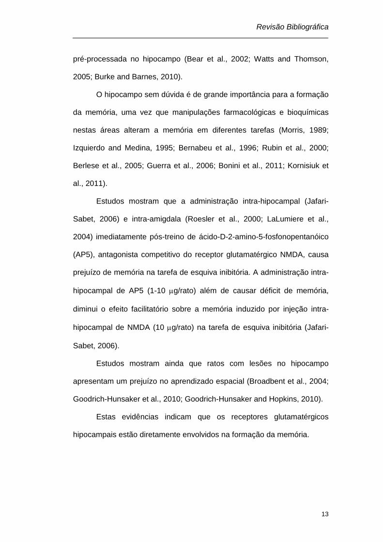
III.2 – RECEPTOR N-METIL-D-ASPARTATO – NMDA
O receptor NMDA é um subtipo de receptor glutamatérgico
amplamente distribuído no sistema nervoso central (SNC), sendo os
níveis mais altos encontrados na região CA1 do hipocampo (Monaghan
and Cotman, 1985). O NMDA possui várias subunidades denominadas:
NR1 (onde se liga a glicina), NR2 (A-D) (onde se liga o glutamato) e NR3
(A-B) (onde se liga a glicina) (Yamakura and Shimoji, 1999; Prybylowski
and Wenthold, 2004; Furukawa et al., 2005; Paoletti and Neyton, 2007),
sendo que a combinação de duas subunidades NR1 associadas a duas

Revisão Bibliográfica
15
subunidades NR2 e/ou NR3 forma um canal iônico tetramérico (Furukawa
et al., 2005).
O canal iônico formado pelas subunidades do receptor NMDA
possui alta permeabilidade aos íons sódio (Na+), potássio (K+) e cálcio
(Ca2+) (MacDermott et al., 1986; Mayer and Westbrook, 1987). No
potencial de repouso o canal está bloqueado por íons Mg2+ (Riedel et al.,
2003), impedindo a passagem de outros íons. A liberação de
neurotransmissores na fenda sináptica, incluindo o glutamato, o
deslocamento do Mg2+ e a ativação do receptor NMDA, resulta no influxo
de íons Na+ e principalmente Ca2+ e no efluxo de íons K+ (Scatton, 1993;
Ozawa et al., 1998; Paoletti and Neyton, 2007). O influxo de cálcio
promove a ativação de proteínas quinases responsáveis por algumas
respostas celulares, incluindo o aumento da expressão dos fatores de
transcrição, e o aumento da transmissão de informações entre neurônios
(Bliss and Collingridge, 1993; Elgersma and Silva, 1999; Izquierdo and
Medina, 1997; Lau et al., 2009).
O receptor NMDA é um complexo protéico com vários sítios de
ligação para agonistas e antagonistas, tais como glutamato/NMDA, sítio
onde se liga dizocilpina (MK-801), sítio modulatório para a glicina (co-
agonista do receptor NMDA), zinco, bem como sítios de ligação para
poliaminas (Ransom and Stec, 1988; Williams et al., 1991; Riedel et al.,
2003) (Fig. III. 2).
Revisão Bibliográfica
16
Fig. III.2 – Representação esquemática do receptor NMDA.
III.2.1 – RECEPTOR NMDA E MEMÓRIA
Evidências demonstram que o receptor glutamatérgico NMDA é
fundamental para os processos de plasticidade sináptica e formação de
memória para diferentes tarefas (Castellano et al., 2001; Riedel et al.,
2003). Estudos com manipulações farmacológicas e/ou genéticas
mostram que o receptor NMDA está diretamente envolvido na formação
NR2 NR1 NR3
NR1
Ca2+ Na+
EXTRACELULAR
INTRACELULAR
K+
Mg2+
GLICINA
Zn2+ POLIAMINAS GLUTAMATO

Revisão Bibliográfica
17
de memórias aversivas (Miserendino et al., 1990; Izquierdo et al., 1992;
Roesler et al., 1998; Cravens et al., 2006) e espaciais (Morris et al., 1986;
Caramanos and Shapiro, 1994; Ahlander et al., 1999; Shapiro and
Eichenbaum, 1999; von Engelhardt et al., 2008; Fellini et al., 2009; Hu et
al., 2009; Wang et al., 2009).
A administração sistêmica ou intracerebral de antagonistas do
receptor NMDA causam prejuízo no aprendizado em uma grande
variedade de tarefas de memória (Izquierdo and Medina, 1995; Martin et
al., 2000; Izquierdo et al., 2006; Krystal et al., 2009; van der Staay et al.,
2011).
De Lima e colaboradores (2005), demonstraram que a
administração sistêmica de 0,1 mg/kg de (+)5-metil-10,11-dihidro-5H-
dibenzo[a,b]-ciclohepteno-5-10-amino (MK-801), um antagonista não-
competitivo do receptor NMDA, 20 minutos antes ou imediatamente
depois do treino na tarefa de reconhecimento de objetos prejudica a
memória de curta e longa duração. Roesler e colaboradores (2000),
também demonstraram prejuízo da memória na tarefa de esquiva inibitória
devido a administração pré ou pós-treino intra-amigdala de 5 mg de AP5,
um antagonista competitivo do receptor NMDA. Enquanto que a
administração de AP5 pré-teste não afeta a memória para a mesma
tarefa. Efeito similar ainda foi demonstrado para administração intra-
hipocampal destes compostos, na tarefa de esquiva inibitória (Jafari-

Revisão Bibliográfica
18
Sabet, 2006) e na tarefa do labirinto aquático de Morris (Morris, 1989;
Fellini et al., 2009).
Entretanto, agonistas do receptor NMDA, como o glutamato
(Izquierdo and Medina, 1995; Rubin et al., 1997) e o ácido DL-beta-
clorofenil glutâmico (CPG) (Flood et al., 1990) melhoram a performance
dos ratos ou camundongos na tarefa de esquiva inibitória e no labirinto
em T, respectivamente.
Todas estas evidências farmacológicas estão de acordo com
estudos envolvendo animais transgênicos, onde é demonstrado que a
super-expressão de receptores NMDA produz um melhor desempenho
destes animais em tarefas de memória (Tang et al., 1999; Tang et al.,
2001; White and Youngentob, 2004; Wang et al., 2009).
Além disso, estudos demonstram que o número de receptores
NMDA está reduzido no sistema nervoso central de pacientes (Sze et al.,
2001) e animais geneticamente modificados que desenvolvem a doença
de Alzheimer (Snyder et al., 2005; Dewachter et al., 2009;).
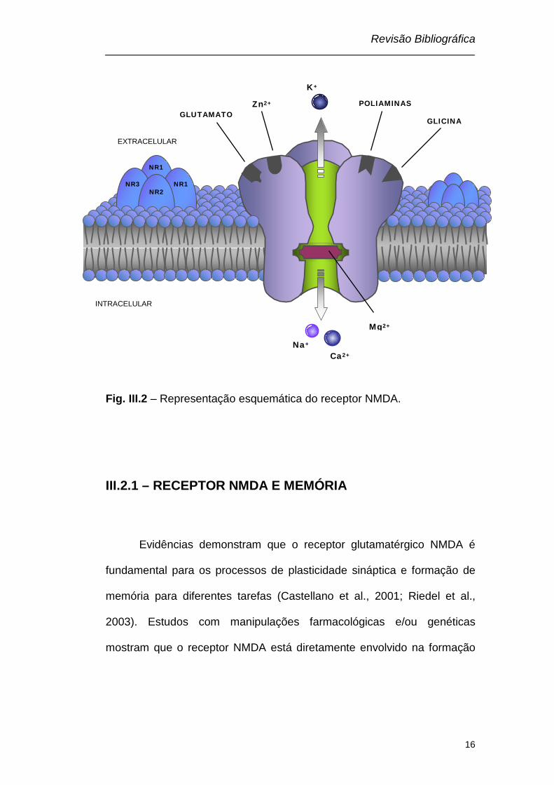
III.3 – POLIAMINAS
As poliaminas são aminas alifáticas simples que estão presentes
em todas as células vivas, procarióticas, eucarióticas, plantas e animais
(Thomas and Thomas, 2001), entretanto, apenas as poliaminas
Revisão Bibliográfica
19
putrescina, espermidina e espermina são sintetizadas pelos mamíferos
(Coleman et al., 2004).
Os nomes putrescina e espermina provém da fonte de onde, estas
poliaminas, foram inicialmente isoladas. A espermina foi descrita pela
primeira vez há mais de 300 anos, quando Antoni van Leuwenhoek, em
1678, relatou a presença de cristais em amostras de esperma seco,
enquanto que, em 1885, Ludwig Brieger, descobriu a putrescina na carne
em putrefação (Gugliucci, 2004).
As poliaminas estão amplamente distribuídas no SNC e
encontradas principalmente em regiões do encéfalo como hipotálamo,
bulbo, hipocampo e cerebelo, sendo a mais alta concentração de SPD,
seguida por espermina e por fim putrescina, que possui uma
concentração muito pequena em comparação as outras duas (Seiler and
Schmidt-Glenewinkel, 1975).
III.3.1 – ESTRUTURA DAS POLIAMINAS
As poliaminas, putrescina (1,4 – diaminobutano), espermidina [N-
(3-aminopropil)-1,4- diaminobutano] e espermina [bis-N-(3-aminopropil)-
1,4- diaminobutano], são constituídas por uma, duas ou três cadeias de
carbono, respectivamente, conectadas por átomos de nitrogênio (fig III.3).
Elas são solúveis em água, possuem baixo peso molecular e caráter

Revisão Bibliográfica
20
fortemente básico, devido aos grupamentos amino (Carter, 1994; Seiler et
al., 1996; Teti et al., 2002; Gugliucci, 2004).
Fig. III.3 - Estrutura química das poliaminas (adaptado de Teti et al., 2002
and Gugliucci, 2004).
III.3.2 – METABOLISMO DAS POLIAMINAS
As poliaminas encontradas nos seres humanos são sintetizadas no
organismo ou provém da flora gastrintestinal capaz de metabolizar
aminoácidos provenientes da dieta (Seiler, 1981; Carter, 1994; Teti et al.,
H 3 N N H 3
N H 3 N H 3 N
H H
N H 3 N N H 3 N
H H
H H
Espermidina
Putrescina
Espermina

Revisão Bibliográfica
21
2002; Larque et al., 2007). O metabolismo das poliaminas e as
características das diferentes enzimas envolvidas têm sido bastante
estudados. (Tabor and Tabor, 1984; Urdiales et al., 2001; Pegg, 2009).
O nível celular adequado de poliaminas é alcançado por meio de
um balanço entre a sua síntese, rota de interconversão e catabolismo.
III.3.2.1 – SÍNTESE
O aminoácido ornitina é o principal precursor das poliaminas
endógenas. No cérebro, a ornitina é formada a partir da clivagem
hidrolítica do aminoácido arginina em uma reação catalisada pela
arginase (Seiler, 1981; Yu et al., 2003).
A putrescina é sintetizada pela descarboxilação da ornitina em uma
reação catalisada pela enzima ornitina descarboxilase (ODC), enzima
limitante na síntese das poliaminas. A putrescina formada serve como
precursor imediato da síntese de espermidina e espermina. Esta síntese
requer o grupo aminopropil que é fornecido pela ação de duas enzimas: a
S-adenosilmetionina descarboxilase (SAMDC), que descarboxila a S-
adenosilmetionina (SAM), e a espermidina sintase, uma enzima
transferase que catalisa a transferência do grupamento aminopropil da
SAM para a putrescina ou espermina sintase que catalisa a transferência
de um segundo grupamento aminopropil para a SPD, formando SPD e

Revisão Bibliográfica
22
espermina respectivamente (Tabor and Tabor, 1984; Urdiales et al., 2001;
Larque et al., 2007; Pegg, 2009).
Esta rota de síntese de poliaminas é reversível, ou seja, a
espermina pode ser convertida em SPD e esta em putrescina. O primeiro
passo desta interconversão é a acetilação da espermina ou SPD na
posição N1, catalisada pela enzima espermidina/espermina
acetiltransferase (SSAT). Após este passo, a poliamina acetilada sofre
quebra oxidativa, por ação da enzima poliamina oxidase (PAO), liberando
os grupos aminopropil provenientes da S-adenosilmetionina
descarboxilada (SAM-D) para formar SPD e putrescina. O produto destas
reações permanece no ciclo e pode ser reutilizado na síntese das
poliaminas (Urdiales et al., 2001; Pegg, 2009).
Estudos com a inibição da poliamina oxidase têm demonstrado que
a rota de interconversão de poliaminas, no encéfalo de ratos, é
responsável por 70% da putrescina formada a partir da SPD, enquanto
somente 30% da putrescina é formada pela descarboxilação da ornitina
(Seiler, 2004; Seiler et al., 1985).
Assim as enzimas chave na regulação da síntese de poliaminas
são ornitina descarboxilase, S-adenosilmetionina descarboxilase e
espermidina/espermina acetiltransferase (Urdiales et al., 2001; Pegg,
2009).
Revisão Bibliográfica
23
III.3.2.2 – CATABOLISMO
O catabolismo das poliaminas consiste na desaminação oxidadiva
de seus grupos amino primários, sendo a reação catalisada por amino
oxidases dependentes de cobre, como a diamina oxidase (DAO). Através
desta desaminação cada intermediário do ciclo da interconversão pode
ser transformado em um aldeído, que é posteriormente oxidado em um
aminoácido, e apenas a putrescina pode ser convertida em ácido
aminobutírico (GABA).
Os produtos finais do catabolismo das poliaminas, bem como as
poliaminas acetiladas, podem então ser excretados na urina (Carter,
1994; Teti et al., 2002; Gugliucci, 2004; Seiler, 2004; Larque et al., 2007).
III.4 – RECEPTOR NMDA, POLIAMINAS E MEMÓRIA
A ativação do receptor NMDA está associada com diferentes
formas de plasticidade sináptica, como Potencialização de Longa Duração
(LTP) e aprendizado e memória (Lee and Silva, 2009). Existem vários
relatos demonstrando o envolvimento das poliaminas e do receptor NMDA
em processos de aprendizado e memória, tanto melhorando a memória
de ratos em diferentes tarefas, como atenuando o déficit de memória
induzido por diferentes agentes amnésicos (Shimada et al., 1994; Kishi et
Revisão Bibliográfica
24
al., 1998a; b; Rubin et al., 2000; 2001; 2004; Mikolajczak et al., 2003;
Tadano et al., 2004; Berlese et al., 2005; Guerra et al., 2006; Camera et
al., 2007).
As poliaminas apresentam uma resposta bifásica sobre o receptor
NMDA em nível comportamental. Neste contexto, tem sido mostrado que
a administração de baixas doses de poliaminas melhoram, enquanto altas
doses prejudicam o desempenho dos animais em tarefas cognitivas.
Assim, altas doses de espermina (125-250 nmol/5µl), administradas por
via intracerebroventricular, causam dano hipocampal e déficit de
aprendizado em ratos na tarefa do labirinto aquático de Morris (Conway,
1998). E administração intra-peritoneal de espermidina (80 mg/kg)
potencializa a diminuição do aprendizado induzido por MK-801 no labirinto
em T de 14 braços (Shimada et al., 1994).
Por outro lado, a administração intracerebral (0,2 nmol) (Rubin et
al., 2000; 2001; 2004; Berlese et al., 2005; Guerra et al., 2006) e
sistêmica (10 mg/kg) (Mikolajczak et al., 2002; Camera et al., 2007) de
espermidina melhora o desempenho de ratos em tarefas como esquiva
inibitória, medo condicionado e reconhecimento social.
Além disso, administração intra-hipocampal de SPD (10
mg/hipocampo) atenua, o número de erros para memória de trabalho
induzidos por administração intra-hipocampal tanto de MK-801, um
antagonista não-competitivo do receptor NMDA (Kishi et al., 1998a),
quanto por escopolamina (3,2 mg/hipocampo), antagonista do receptor
Revisão Bibliográfica
25
muscarínico (Kishi et al., 1998b). A administração sistêmica de espermina
atenua o prejuízo do aprendizado induzido por CPP, um antagonista
competitivo do receptor NMDA, no labirinto em T de 14 braços (Meyer et
al., 1998).
O efeito facilitatório da SPD sobre a memória parece depender da
atividade da enzima óxido nítrico sintase hipocampal e da produção de
óxido nítrico, uma vez que a administração intra-hipocampal de N-nitro-L-
arginina metil éster (L-NAME), um inibidor não específico da enzima óxido
nítrico sintase, imediatamente depois do treino previne a melhora da
memória causada por SPD na tarefa de esquiva inibitória (Guerra et al.,
2006). A SPD aumenta os níveis de nitratos e nitritos, e a co-
administração de L-NAME previne este efeito (Guerra et al., 2006).
O efeito facilitatório da SPD sobre a memória envolve o receptor
NMDA uma vez que o MK-801, antagonista NMDA, e arcaína, antagonista
do sítio das poliaminas no receptor NMDA, revertem a melhora da
memória induzida pelas poliaminas (Rubin et al., 2000; 2001; 2004;
Camera et al 2007, Gomes et al., 2010). Outro antagonista do sítio das
poliaminas no receptor NMDA, ifenprodil, administrado por via intra-
amigdala e intraperitoneal prejudica o desempenho dos ratos na tarefa de
medo condicionado (Rodrigues et al., 2001) e a sua administração
intracerebroventricular, reverte o efeito facilitatório da SPD sobre a
memória (Tadano et al., 2004). Além disso, as poliaminas estão
envolvidas na facilitação da extinção do medo condicionado contextual
Revisão Bibliográfica
26
(Gomes et al., 2010) e na dependência de estado na tarefa de esquiva
inibitória (Mariani et al., 2011).
Apesar de existirem estudos demonstrando o envolvimento das
poliaminas e do receptor NMDA sobre a memória (Kishi et al., 1998a; b;
Rubin et al., 2000; 2001; 2004; Camera et al., 2007), pouco ainda é
conhecido sobre o mecanismo de ação das poliaminas sobre a memória,
após a ativação do receptor NMDA, sendo que uma das conseqüências
pode ser a ativação das proteínas quinases.
III.5 – PROTEÍNA QUINASE
Proteína quinase é um tipo de enzima que modifica outras
proteínas pela transferência de grupos fosfatos de moléculas doadoras de
alta energia, como o ATP, para moléculas-alvo específicas (substratos),
processo que tem o nome de fosforilação. A fosforilação resulta em uma
alteração funcional da proteína alvo, através de mudanças na atividade
enzimática, localização celular ou associação com outras proteínas.
Todas as quinases necessitam de um íon metálico divalente, como o
Mg2+, para transferir o grupo fosfato (Micheau and Riedel, 1999).
Uma proteína fosfatase faz exatamente a reação inversa das
quinases, ela remove um grupamento fosfato, ou seja, faz a
desfosforilação (Skroblin et al., 2010). Dentre as fosfatases, a calcineurina
responde diretamente a um segundo mensageiro (aumento da
Revisão Bibliográfica
27
concentração intracelular de Ca2+), enquanto outras são ativadas quando
fosforiladas pelas próprias proteínas quinase (Skroblin et al., 2010).
As células contêm várias proteínas quinases e proteínas
fosfatases, cada uma sendo responsável por agir em um determinado
grupo ou uma determinada proteína. As células apresentam normalmente
um estado de equilíbrio entre as proteínas quinase, fosfatases (ativadas
ou desativadas) e os substratos protéicos (fosforilados ou
desfosforilados). O genoma humano contém cerca de 500 genes de
proteínas quinase, constituindo aproximadamente 2% de todo o genoma
humano, sendo que 20% de todas as proteínas sintetizadas em uma
célula servem como substrato para essas enzimas. As proteínas quinase
são capazes de regular a maioria das cascatas celulares, especialmente
as que envolvem transdução de sinal (Manning et al., 2002).
III.5.1 – PROTEÍNA QUINASE A (PKA)
A proteína quinase dependente de AMPc (PKA) foi caracterizada
em 1968, é a mais comum e versátil enzima de células eucarióticas
(Walsh et al., 1968). A PKA é uma quinase no qual a atividade depende
dos níveis de AMPc na célula. A família da PKA consiste de quatro
subunidades regulatórias (RIα, RIβ, RIIα, RIIβ) e três subunidades
catalíticas (Cα, Cβ, Cγ) (McKnight et al., 1988; Doskeland et al., 1993),
Revisão Bibliográfica
28
expressas no encéfalo dos mamíferos (McKnight et al., 1988; Cadd and
McKnight, 1989).
Cada PKA é uma holoenzima tetramérica formada por duas
subunidades R e duas subunidades C, sendo que a subunidade R possui
dois sítios de ligação para o AMPc. O maior controle da atividade da PKA
é fornecido pelo AMPc, pois quando os níveis de AMPc estão baixos a
holoenzima permanece intacta e cataliticamente inativa (Taylor et al.,
1990). Entretanto, quando ocorre um aumento da concentração de AMPc,
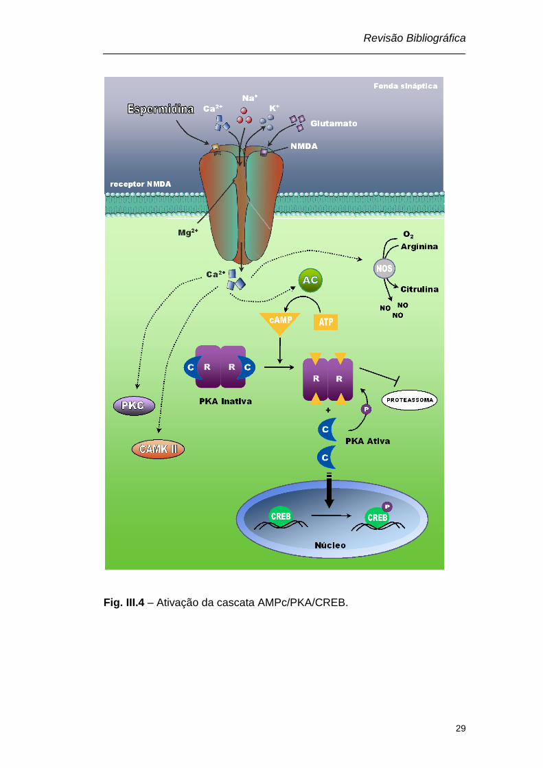
mediante a ativação da adenilato ciclase (AC), o AMPc liga-se aos dois
sítios na subunidade R, liberando a subunidade catalítica. A subunidade
catalítica livre pode então catalisar a transferência do grupo fosfato do
ATP para os resíduos de serina/treonina do substrato. Esta fosforilação
pode ocorrer diretamente em uma enzima, aumentando ou diminuindo
sua atividade, (Taylor et al., 1990; Wang et al., 1991; Gibbs et al., 1992;
Wang et al., 2004), ou a subunidade catalitica pode migrar para o núcleo,
fosforilar a CREB e assim, aumentar a síntese protéica (Fig. III. 4) (Dash
et al., 1991; Mayford et al., 1995a; Impey et al., 1998).
Revisão Bibliográfica
29
Fig. III.4 – Ativação da cascata AMPc/PKA/CREB.

Revisão Bibliográfica
30
III.5.1.1 – INIBIÇÃO DA PKA
Devido a composição tetramérica os inibidores da PKA podem se
ligar tanto nas subunidades regulatórias como nas subunidades
catalíticas. A ação dos inibidores das subunidades regulatórias ocorre
através da ligação de análogos do AMPc no sitio de ligação do AMPc
nestas subunidades, impedindo a dissociação e liberação da subunidade
catalítica, não permitindo, assim, a ativação da PKA (Lochner and
Moolman, 2006).
A ação dos inibidores das subunidades catalíticas pode ocorrer de
duas maneiras: competitiva com relação ao ATP, uma vez que, os
inibidores alostéricos competem pelo sítio de ligação do ATP, ou não
competitiva, com relação ao substrato, uma vez que os inibidores
alostéricos se ligam em sítios distintos ao do substrato, inativando a
enzima. Quando os inibidores alostéricos se ligam ocorre uma alteração
na conformação espacial destas enzimas e, consequentemente,
bloqueiam os sítios de ligação do ATP ou dos substratos protéicos
(Hidaka et al., 1990; Adams, 2001; Lochner and Moolman, 2006; Pickin et
al., 2008).
III.5.1.2 –REGULAÇÃO DA PKA

Revisão Bibliográfica
31
A produção do AMPc, um segundo mensageiro que ativa a PKA,
ocorre a partir do ATP, e é catalisada por uma família de nove isoformas
da enzima adenilato ciclase (AC) (Hanoune and Defer, 2001; Cooper,
2003), enquanto que a degradação do AMPc para AMP ocorre através de
uma super família de fosfodiesterases (PDE), que incluem mais de 40
diferentes isoformas da enzima (Baillie et al., 2005; Lugnier, 2006).
Portanto, os níveis de AMPc na célula não são determinados apenas pela
sua geração através da ativação da AC, eles dependem também, da
degradação catalisada pela ativação da PDE (Baillie et al., 2005; Conti
and Beavo, 2007).
A atividade da PKA é regulada pelos níveis de AMPc, através de
um mecanismo de “feedback”, uma vez que, a própria PKA pode ativar a
PDE (MacKenzie et al., 2002; Li et al., 2010; Skroblin et al., 2010) e inibir
a AC (Willoughby and Cooper, 2007; Willoughby et al., 2010), reduzindo
os níveis de AMPc e diminuindo sua atividade. Entretanto, o aumento na
atividade da PKA pode ser mantido através da degradação seletiva da
subunidade regulatória, pela via ubiquitina-proteassoma, mesmo na
ausência dos níveis elevados de AMPc preexistentes (Hegde et al., 1993;
Chain et al., 1995; 1999). A PKA pode, ainda, ser auto-regulada, ou seja,
a subunidade catalítica pode fosforilar a subunidade regulatória,
diminuindo afinidade entre as duas subunidades (First et al., 1988; Zelada
et al., 2002). Tanto a degradação, como a fosforilação da subunidade

Revisão Bibliográfica
32
regulatória diminui a probabilidade do retorno da associação da mesma
com a subunidade catalítica, mantendo assim, a PKA ativa.
Além disso, a composição das subunidades contribui para o perfil
de ativação de diferentes isoformas da PKA. Por exemplo, as isoformas
contendo RIβ são ativadas em concentrações mais baixas de AMPc do
que as isoformas contendo RIα, assim, diferentes expressões de várias
subunidades conferem diferentes sensibilidades para o AMPc,
provocando variações na ativação da PKA (Cadd et al., 1990; Solberg et
al., 1994).
III.5.1.3 – PROTEÍNA ANCORADOURA
Para que as proteínas quinase possuam maior ativação e eficiência
na fosforilação de outras proteínas, elas devem estar presentes em
regiões especificas na célula, como próximas das regiões produtoras de
segundos mensageiros ou dos substratos a serem fosforilados (Pidoux
and Tasken, 2010).
A proteína âncora da quinase A (AKAP) é uma família de proteínas
ancoradouras, que tem como função determinar diferentes localizações
para a PKA na célula (Bauman and Scott, 2002; Michel and Scott, 2002;
Wong and Scott, 2004; Carnegie et al., 2009; Pidoux and Tasken, 2010).
Funcionalmente, as AKAPs possuem três características principais:

Revisão Bibliográfica
33
primeira, elas possuem um domínio que ancora a PKA; segunda, ligam-se
a outras enzimas, formando um complexo multi-enzimático; terceira,
possuem um domínio onde ligam-se substratos específicos (Wong and
Scott, 2004; Smith and Scott, 2006; Jarnaess and Tasken, 2007). A AKAP
aumenta a especificidade e eficiência da PKA, uma vez que, liga-se à
subunidade regulatória, e permite que a quinase esteja localizada próxima
a alvos específicos de substratos, que são regulados pela fosforilação
(Smith and Scott, 2002; Bauman et al., 2004; Dell'Acqua et al., 2006).
III.5.2 – PROTEINA QUINASE C (PKC)
Em 1977, foi descrita uma proteína quinase independente de
nucleotídeos cíclicos para sua ativação (Takai et al., 1977).
Subsequentemente, foi demonstrado que esta enzima pode ser ativada
por cálcio e diacilglicerol (DAG), diferindo das quinases dependentes de
nucleotídeos cíclicos (PKA e PKG), sendo então, nomeada proteína
quinase dependente de Ca2+ (PKC) (Takai et al., 1979). Entretanto, a PKC
é membro da familia AGC (PKA, PKG, PKC) das proteinas quinase que
compartilham certas características estruturais básicas (Steinberg, 2008).
A PKC é composta por uma família de serina/treonina quinases que
apresenta alta atividade e expressão da maioria de suas isoformas no
cérebro (Tanaka and Nishizuka, 1994). No SNC, a ativação da PKC está
Revisão Bibliográfica
34
associada com regulação da transmissão sináptica e das funções neurais
em diferentes níveis, incluindo liberação de neurotransmissores (Malenka
et al., 1986; Majewski and Iannazzo, 1998), funcionamento dos receptores
(Macek et al., 1998; Suen et al., 1998), expressão gênica (Routtenberg et
al., 2000) e processos de aprendizado e memória (Wehner et al., 1990;
Alkon et al., 2005).
Em mamíferos, são descritas 10 isoformas de PKC, com base na
estrutura e sensibilidade ao Ca2+ e DAG estas isoformas têm sido
classificadas em três subfamílias: clássica ou convencional – cPKCs (α,
βI, βII e γ) ativada por DAG e Ca2+ (Coussens et al., 1986; Parker et al.,
1986); nova – nPKC (δ, ε, η e θ) ativada por DAG mas não por Ca2+ (Ono
et al., 1987; Osada et al., 1990; Saido et al., 1992); atípica – aPKC (ζ e
λ/ι) insensível a ambos DAG e Ca2+ (Ogita et al., 1992).
Ao contrário da PKA, a PKC é uma enzima monomérica, ou seja,
apresenta uma única cadeia polipeptídica formada por um domínio
regulatório e um domínio catalítico. A isoforma convencional da PKC é
formada por quatro regiões conservadas (C1-C4). A metade amino-
terminal, contendo as regiões C1, que interage com diacilglicerol (DAG),
éster de forbol e fosfatidilserina; C2, que interage com Ca2+; além de um
pseudo-substrato adjacente a C1, representam o domínio regulatório. A
metade carbóxi-terminal, contendo as regiões C3 e C4, responsáveis pela
ligação do ATP e do substrato, respectivamente, representa o domínio
catalítico (Newton, 2003; Amadio et al., 2006; Sun and Alkon, 2010).
Revisão Bibliográfica
35
III.5.2.1 – ATIVAÇÃO DA PKC
Para a completa ativação, a PKC necessita três etapas
consecutivas: a fosforilação da enzima, a migração para a membrana e a
remoção do pseudo-substrato, que bloqueia o sítio ativo (Pascale et al.,
2007). A ativação da PKC inicia com a fosforilação da enzima através da
proteína quinase dependente de fosfoinositídeo (Dutil et al., 1998),
seguida pela autofosforilação intramolecular em dois sítios adicionais na
seqüência carboxi-terminal (Bornancin and Parker, 1996; 1997). A PKC
fosforilada, presente no citosol, possui uma conformação estável,
cataliticamente competente e resistente a proteases, entretanto,
permanece em uma conformação inativa, uma vez que, o pseudo-
substrato ocupa o sítio de ligação do substrato (Ron and Kazanietz, 1999;
Newton, 2001; 2003). Após uma elevação nos níveis intracelulares o Ca2+
liga-se ao domínio C2 da PKC e aumenta a afinidade da quinase pela
fosfatidilserina, translocando a enzima para membrana. Esta ligação do
domínio C2 com a membrana é uma interação de baixa afinidade
(Nalefski and Newton, 2001; Schaefer et al., 2001; Steinberg, 2008),
entretanto, uma vez ancorada na membrana, o domínio C1 da PKC liga-
se ao DAG (produto da hidrólise de fosfatidil-inositídeos restrito a
membrana). A interação dos domínios C1 e C2, aumentam a afinidade da
PKC com a membrana, fornecendo energia suficiente para remover o
pseudo-substrato e liberar o sítio de ligação do substrato, ativando o
Revisão Bibliográfica
36
domínio catalítico da PKC (Nishizuka, 1995; Toker, 1998; Newton, 2001;
Steinberg, 2008).
III.6 – CREB
O elemento ligante responsivo ao AMPc (do inglês: cAMP response
element-binding - CREB) é membro de uma família de proteínas que atua
como fator de transcrição. Localizada no núcleo, a CREB é fundamental
na transmissão da informação da sinalização celular iniciada na
membrana, até as alterações na expressão dos genes (Shaywitz and
Greenberg, 1999; Mayr and Montminy, 2001).
Esta cascata bioquímica inicia com a ativação de receptores que
aumentam a liberação de cálcio e produção de segundos mensageiros,
como AMPc, permitindo a ativação de proteínas quinase como PKA e
CaMKII. As proteínas quinase, quando ativadas, translocam para o núcleo
da célula, onde fosforilam CREB tornando-a ativa. A CREB, na forma
ativa, liga-se a certas seqüências de DNA chamadas de elementos de
resposta ao AMPc (CRE) e assim, aumenta ou diminui a transcrição
gênica. Os genes cuja transcrição é regulada pelo CREB são os
responsáveis pela ligação da RNA-polimerase ao DNA para a síntese de
proteínas (Montminy et al., 1990; Carlezon et al., 2005).
Revisão Bibliográfica
37
A CREB desempenha funções em diferentes órgãos, entretanto, a
maioria dos estudos tem sido realizada em encéfalo, sendo que a
expressão de genes, nesta área, possui um papel fundamental tanto na
sobrevivência e neuroproteção quanto na plasticidade sináptica e
formação da memória (Carlezon et al., 2005; Contestabile, 2008). De fato,
estudos têm mostrado que a síntese de proteínas é um processo
essencial na formação de novas memórias (Bourtchouladze et al., 1998;
Quevedo et al., 2004; 2005).
III.7 – PROTEÍNA QUINASE, CREB E MEMÓRIA
Existem algumas evidências, abaixo listadas, apoiando o papel da
PKC no processo de aprendizado e memória. Primeiro, a atividade
hipocampal da PKC aumenta imediatamente depois do treino na tarefa de
esquiva inibitória, alcança um pico 30 min mais tarde e retorna aos níveis
normais durante os próximos 120 min depois do treino (Bernabeu et al.,
1995; Cammarota et al., 1997). Além disso, o aprendizado em uma tarefa
espacial aumenta os níveis de PKC ligados a membrana (forma ativa), no
hipocampo de ratos (Nogues et al., 1994; Golski et al., 1995), e a
atividade da enzima está reduzida em camundongos com baixo
aprendizado (Wehner et al., 1990). Adicionalmente, tem sido demonstrado
que o aumento da neuromodulina (GAP-43, B50), substrato da PKC,

Revisão Bibliográfica
38
depois da tarefa de esquiva inibitória, coincide com o pico de atividade da
PKC e pode ser bloqueado, por inibidores desta quinase (Cammarota et
al., 1997). Segundo, infusão de ativadores da PKC, como forbol 12,13-
dibutirato e 1-oleoil-2-acetil glicerol (análogo sintético do DAG), melhora a
formação da memória em ratos (Paylor et al, 1991; Yang and Lee, 1993) e
camundongos (Nogues et al., 1996), respectivamente. Terceiro,
camundongos trangênicos que superexpressam GAP-43, substrato da
PKC, melhoram o aprendizado e LTP (Routtenberg et al., 2000). Quarto,
administração de inibidores da PKC, como estausporina, CGP 41231, Go
6976 (inibidor seletivo α- e βI-PKC) e Go 7874, provoca amnésia
retrógrada quando infundidos 1-2 h após o treinamento da esquiva
inibitoria (Jerusalinsky et al, 1994;. Vianna et al, 2000a; Bonini et al. ,
2007). A coincidência nos tempos de alteração da atividade da PKC
hipocampal e nos efeitos amnésicos dos inibidores da PKC, sugere um
claro envolvimento desta quinase nos processos de formação da
memória. Quinto, camundongos mutantes que não expressam PKCγ,
PKCβ ou substratos da PKC convencional, como neurogranina e GAP-43,
apresentam déficits de aprendizado e formação de memória em diferentes
tarefas (Abeliovich et al., 1993; Pak et al., 2000; Weeber et al., 2000;
Miyakawa et al., 2001; Rekart et al., 2005; Huang et al., 2006).
Assim como a PKC, a cascata AMPc/PKA/CREB também está
envolvida nos processos de formação da memória, possuindo um papel
importante na fase final da consolidação da memória (Abel et al., 1997;
Revisão Bibliográfica
39
Bernabeu et al., 1997a; Barad et al., 1998; Huang and Kandel, 1998;
Vianna et al., 2000b; Pereira et al., 2001; Quevedo et al., 2005). Existem
evidências que reforçam o envolvimento do AMPc no aprendizado e
memória. Primeiro, os níveis de AMPc hipocampal aumentam,
lentamente, 60 min depois do treino na tarefa de esquiva inibitória, e
alcançam um pico 180–360 min depois do treino (Bernabeu et al., 1996;
Bernabeu et al., 1997a; b). Segundo, a infusão de agentes que aumentam
os níveis intracelulares de AMPc, como inibidor da fosfodiesterase (Vitolo
et al., 2002; Zhang et al., 2000; 2004; Puzzo et al., 2009), análogo da
AMPc, ativador da adenilato ciclase (Bernabeu et al., 1997a), e a super
expressão da adenilato ciclase (Wang et al., 2004), facilitam a formação
da memória. Terceiro, camundongos com expressão reduzida para a
adenilato ciclase, apresentam diminuição nos níveis de AMPc e déficit na
formação da memória em diferentes tarefas (Wu et al., 1995; Wong et al.,
1999). Além disso, a atividade da PKA aumenta depois do treino na tarefa
de esquiva inibitória em dois picos: o primeiro, imediatamente depois do
treino; e o segundo, 3-6 horas mais tarde, que coincide com o aumento
máximo nos níveis de AMPc depois do treino (Bernabeu et al., 1997a).
A importância da PKA na memória tem sido reforçada por estudos
demonstrando que inibidores específicos desta enzima prejudicam a
memória (Bernabeu et al., 1997a; Bourtchouladze et al., 1998; Vianna et
al., 2000b; Wallenstein et al., 2002; Ahi et al., 2004; Quevedo et al., 2004;
Sharifzadeh et al., 2005) e que camundongos trangênicos R (AB),

Revisão Bibliográfica
40
expresando uma forma inibitória da subunidade regulatória da PKA,
exibem deficit de memória (Abel et al., 1997; Bourtchouladze et al., 1998;
Isiegas et al., 2006).
O próximo passo na cascata AMPc/PKA/CREB, após a ativação da
PKA, é fosforilação da CREB (Gonzalez and Montminy, 1989; Bernabeu
et al., 1997a). Estudos têm demonstrado que os níveis de CREB
fosforilada no hipocampo aumentam depois do treino na tarefa de esquiva
inibitória em dois picos, que correlacionam com os picos de aumento da
atividade da PKA (Bernabeu et al., 1997a; b).
Assim, o aumento coincidente da forma fosforilada da CREB em
períodos específicos, junto com a sensibilidade da memória para
inibidores da síntese de proteínas durante períodos em que a PKA está
ativa, sugerem que a cascata AMPc/PKA/CREB contribua para a síntese
de novas proteínas, evento fundamental para a formação da memória de
longa duração. Além disso, alguns estudos relatam um cruzamento entre
PKC e PKA, ou seja, é sugerido que PKC contribua para a ativação da
cascata AMPc/PKA/CREB (Sugita et al., 1997; Kubota et al., 2003; Yao et
al., 2008).
Apesar de todos os estudos já existentes sobre proteínas quinases
e memória, o envolvimento da ativação destas proteínas na melhora da
memória induzida por espermidina ainda não foi investigado. Este estudo
poderá determinar um possível mecanismo de ação da espermidina nos
processos de formação da memória, e desta forma, fornecer subsídios

Revisão Bibliográfica
41
para desenvolver fármacos com potencial para serem utilizados para
tratar pacientes com patologias, como a doença de Alzheimer, em que os
pacientes afetados apresentam déficit de memória.

IV. RESULTADOS

IV.1 – ARTIGO
Neurobiology of Learning and Memory, 96 (2011) 324–332.

44
45
46
47

48
49
50
51

52

IV.2 – MANUSCRITO
Submetido para “HIPOCAMPPUS”

Resultados
54
Spermidine-induced improvement of memory involves a cross-talk between protein kinases C and A
Gustavo Petri Guerra1, Carlos Fernando Mello2, Guilherme Vargas Bochi1,
Michelle Melgarejo Rosa1, Juliano Ferreira1, Maribel Antonello Rubin1*
1Department of Chemistry, Center of Exact and Natural Sciences,
Universidade Federal de Santa Maria, Santa Maria, RS, 97105-900, Brazil
2Department of Physiology and Pharmacology, Center of Health Sciences,
Universidade Federal de Santa Maria, Santa Maria, RS, 97105-900, Brazil
*Corresponding author. Fax: + 55 55 3220 8978
E-mail adress: [email protected] (M.A. Rubin).
Acknowledgments
This study was supported by CNPq (481664/2010-6, 306164/2010-8). C.F.
Mello, G. V. Bochi, J. Ferreira and M.A. Rubin are recipients of CNPq
fellowships. G.P. Guerra is recipients of CAPES fellowship. All the
experiments comply with the current laws of Brazil.
Resultados
55
ABSTRACT
Spermidine (SPD) is an endogenous polyamine that modulates N-methyl-
D-aspartate (NMDA) receptor function, and has been reported to facilitate
memory formation. Recent evidence suggests that PKA and CREB play a
role in SPD-induced improvement of memory. In the current study, we
determined whether the PKC signaling pathway is involved in SPD-
induced facilitation of memory of inhibitory avoidance task in adult rats.
The post-training administration of the calcium-dependent protein kinase
(PKC) inhibitor, 3-[1-(dimethylaminopropyl)indol-3-yl]-4-(indol-3-
yl)maleimide hydrochloride [GF 109203X, 2.5 ρmol, intrahippocampal (ih)]
with SPD (0.2 nmol, ih) prevented memory improvement induced by SPD.
Intrahippocampal administration of SPD (0.2 nmol) facilitated PKC
phosphorylation, in the hippocampus, 30 min after administration. GF
109203X prevented not only the stimulatory effect of SPD on PKC, but
also PKA and CREB phosphorylation. These results suggest that memory
enhancement induced by the ih administration of SPD involves the cross
talk between PKC and PKA/CREB, with sequential activation of PKC and
PKA/CREB pathways, in rats.
Keywords PKC, CREB, Polyamine, Memory, Spermidine, GF109203X
Resultados
56
1. Introduction
Polyamines putrescine, spermine, and spermidine (SPD) are a
group of aliphatic amines present in almost all cells, but at particularly high
concentrations in the vertebrate nervous system, including cerebral
structures involved in learning and memory, such as the amygdala and
hippocampus (Carter, 1994; Morrison et al., 1995). Polyamines modulate
learning and memory by interacting with the polyamine-binding site at the
N-methyl-D-aspartate receptor (NMDAr) (Izquierdo and Medina, 1997;
Kishi et al., 1998a, b; Rubin et al., 2004; Rubin et al., 2000; Rubin et al.,
2001; Shimada et al., 1994). Accordingly, the systemic (Camera et al.,
2007), intrahippocampal (ih) (Berlese et al., 2005; Gomes et al., 2010;
Guerra et al., 2006; Rubin et al., 2000), and intra-amygdalar (Rubin et al.,
2004; Rubin et al., 2001) administration of SPD improves memory. Recent
evidence suggests the participation of PKA/CREB pathway cascade in the
facilitation of memory induced by SPD (Guerra et al., 2011). However,
there are other well known kinases, such as Ca2+-dependent protein
kinase (PKC), which are also activated by NMDA receptor-mediated Ca2+
influx (Jimenez and Tapia, 2004; Wang et al., 2006; Wu et al., 2003).
Since PKC can also phosphorylate CREB (Mao et al., 2007; Selcher et al.,
2002), this kinase would naturally arise as a candidate to mediate the
memory improvement induced by SPD.
Resultados
57
Protein kinase C comprises a family of serine/threonine kinases with
10 known isozymes, divided into three major subsets based on structure
and regulation. These include the conventional isoforms dependent on
calcium (α, β, and γ), novel (δ, ε, η, and θ), and atypical (λ and ζ) types,
which are structurally homologous but can be regulated independently of
calcium (Ohno and Nishizuka, 2002; Parekh et al., 2000; Weeber et al.,
2000). Protein kinase C cascade regulates neuronal efficacy at different
levels, including neurotransmitter release, neurotransmitter receptor
function and gene expression (Ben-Ari et al., 1992; Kleschevnikov and
Routtenberg, 2001; Macek et al., 1998; Malenka et al., 1986; Manseau et
al., 1998; Meberg et al., 1993), and it seems to play a role in memory
formation process (Alkon et al., 2007; Alvarez-Jaimes et al., 2005; Bonini
et al., 2007; Izquierdo and Medina, 1997; Lorenzetti et al., 2008; Nelson et
al., 2008; Nogues, 1997; Serrano et al., 2008). There are lines of evidence
supporting a role for PKC in learning and memory. First, PKC activity is
increased in the hippocampus immediately after step-down avoidance
training, reaching a peak 0.5 h later, and returning to normal values over
the next 2 h (Bernabeu et al., 1995; Cammarota et al., 1997). Moreover, it
has been shown that learning of a spatial-discrimination task increases
membrane-bound PKC levels (and, therefore, activated) in the
hippocampus of rats (Golski et al., 1995; Nogues et al., 1994), and PKC
activity is reduced in the hippocampus of poor learner mice (Wehner et al.,
1990). Furthermore, it has been shown that by increasing PKC substrate,
Resultados
58
neuromodulin (GAP-43, B50), matches with the PKC activity peak and can
be blocked by inhibitors of this protein kinase (Cammarota et al., 1997).
Second, infusion of the PKC activators phorbol 12,13-dibutyrate (PDBu)
and 1-oleoyl-2-acetyl glycerol (DAG synthetic analog) improves memory
formation in rats (Paylor et al., 1991; Yang and Lee, 1993) and mice
(Nogues et al., 1996), respectively. Third, transgenic mice overexpressing
conventional PKC substrate, GAP-43, demonstrate enhanced learning and
LTP (Routtenberg et al., 2000). Fourth, injection of the PKC inhibitors,
such as staurosporin, CGP 41231, Go 6976 (selective α- and βI-PKC
inhibitor) and Go 7874 causes retrograde amnesia when infused into CA1
in the first 1–2 h after training (Bonini et al., 2007; Jerusalinsky et al.,
1994; Vianna et al., 2000b). The coincidence of the time course of the
change of hippocampal PKC activity with that of the amnestic effects of the
PKC inhibitors points to a clear and crucial involvement of this enzyme in
the posttraining memory processing. Fifth, studies with mutant mice
lacking PKCγ, PKCβI or conventional PKC substrates, such as
neurogranin and GAP-43, present learning and memory formation deficits
in different tasks (Abeliovich et al., 1993; Huang et al., 2006; Miyakawa et
al., 2001; Pak et al., 2000; Rekart et al., 2005; Weeber et al., 2000).
Numerous studies have shown that consolidation of different types of
memory in rodents requires phosphorylation/activation of the transcription
factor CREB by cAMP- or Ca2+-dependent protein kinase (Bernabeu et al.,
1997; Brightwell et al., 2007; Cammarota et al., 2000; Cammarota et al.,

Resultados
59
2005; Countryman et al., 2005; Desmedt et al., 2003; Impey et al., 1998;
Izquierdo and Medina, 1997; Izquierdo et al., 2001; Kida et al., 2002; Mao
et al., 2007; Stanciu et al., 2001; Taubenfeld et al., 1999; Trifilieff et al.,
2006; Vianna et al., 2000a; Zhang et al., 2003). Recently, we have shown
that SPD induces PKA and CREB phosphorylation in the hippocampus of
rats (Guerra et al., 2011). Furthermore, the PKA-induced CREB
phosphorylation requires PKC activity in striatal neurons (Zanassi et al.,
2001). However, no study has addressed whether PKC cascade is
involved in the memory improvement induced by SPD and whether the
SPD-induced PKA/CREB phosphorylation requires PKC activity. Thus, in
the present study, we investigated whether PKC signaling pathway is
activated by SPD in rats subjected to inhibitory avoidance training.
2. Material and methods
2.1. Animals
All experiments reported in this study were conducted in accordance
with Brazilian law No. 11.794/2008 in agreement with the Policies on the
Use of Animals and Humans in Neuroscience Research, which were
revised and approved by the Society for Neuroscience Research in
January 1995, and with the Institutional and National Regulations for
Animal Research (process 0206). Male Wistar rats (230–250 g; n = 164)
were bred in the Animal House of the Universidade Federal de Santa
Resultados
60
Maria, housed 5 to a cage, and maintained on a natural day/night cycle at
21°C with access to water and rodent laboratory chow (Guabi, Santa
Maria, RS, Brazil) ad libitum. Behavioral tests were conducted during the
light phase of the cycle (from 9:00 a.m. to 5:00 p.m.).
2.2. Surgery
Rats were anaesthetized by using intraperitoneal (ip) injection of a
mixture of ketamine (80 mg/kg) and xylazine (8 mg/kg), and were
implanted with two 27-gauge guide cannulae placed 1 mm above the CA1
region of the dorsal hippocampus at the following coordinates: A = 4 mm;
L = 3.0 mm; and V = 2.0 mm (Paxinos and Watson, 1986). Placement of
injections was histologically-verified, as described elsewhere (Rubin et al.,
1997). Only data from the animals with correct cannula placement were
analyzed.
2.3. Behavioral and Infusion Procedures
One week after surgery, the animals were subjected to a single
training session in a step-down inhibitory avoidance apparatus, consisting
of a 25 × 25 × 35-cm box with a grid floor and the left portion covered by a
7 × 25-cm platform, measuring 2.5 cm in height. The rat was placed gently
on the platform facing the rear left corner. Once the rat stepped down with
all 4 paws on the grid, a 3-s, 0.3-mA shock was applied to the grid.

Resultados
61
Immediately post-training, vehicle, one drug, or a combination of the
following drugs were injected bilaterally into the hippocampus (0.5 μl/ brain
hemisphere for 1 min): spermidine [N-(3-aminopropyl)-1,4-butanediamine
trihydrochloride (SPD); Sigma-Aldrich Co., St. Louis, MO, USA]; and 3-[1-
(dimethylaminopropyl)indol-3-yl]-4-(indol-3-yl)maleimide hydrochloride (GF
109203X; Sigma-Aldrich Co.). SPD were dissolved in 50 mM phosphate
buffered saline solution (PBS; pH 7.4), and GF 109203X was dissolved in
0.01% DMSO in 50 mM PBS (pH 7.4). The injections were performed by
using an infusion pump using a 30-gauge needle fitted into the guide
cannula. The tip of the infusion needle protruded 1.0 mm beyond that of
the guide cannula into the CA1 region in the dorsal hippocampus. The
needles were left in place for an additional 60 s to minimize backflow. After
the injection, the animals were returned to their home cages and tested for
retention 24 h later. A step-down latency test was taken as a measure of
retention, with a cut-off time of 600 s.
Immediately after the inhibitory avoidance test session, the animals
were transferred to a 50 X 60-cm open field, with the floor divided into 12
squares. During the 5-min open field session, the number of crossing and
rearing responses was recorded. The open field was used to identify motor
disabilities which might influence inhibitory testing avoidance performance.
Step-down latency data were analyzed with the Kruskal–Wallis test or
the Scheirer–Ray–Hare extension of the Kruskal–Wallis test. Crossing and

Resultados
62
rearing responses were analyzed by one- or two-way ANOVA.
Significance was considered at p<0.05.
2.4. Preparation of tissues and Western blot analysis
Western blot analysis was carried out, as described previously (Casu
et al., 2007) with minor modifications. Rats were decapitated, and the
hippocampi were rapidly removed, dissected, homogenized in 300 µl of
ice-cold A buffer (10 mM KCl, 2 mM MgCl2, 1 mM EDTA, 1 mM NaF, 10
µg/ml aprotinin, 10 mM β-glycerolphosphate, 1 mM PMSF, 1 mM DTT,
and 2 mM of sodium orthovanadate in 10 mM HEPES, pH 7.9), incubated
for 15 min on ice, and centrifuged at 16 000 x g for 45 min at 4oC. The
supernatant (S1), denominated cytosolic fraction, was reserved for
posterior processing. The pellet (P1) was resuspended in 150 µl of ice-
cold buffer B (10 mM KCl, 2 mM MgCl2, 1 mM EDTA, 1 mM NaF, 10 µg/ml
aprotinin, 10 mM β-glycerolphosphate, 1 mM PMSF, 1 mM DTT, 2 mM
sodium orthovanadate, and 1% Triton-X in 10 mM HEPES, pH 7.9),
incubated for 15 min on ice, and centrifuged at 16 000 X g for 45 min at
4oC. The supernatant (S2) was discarded and the pellet (P2) was
resuspended in 100 µl of ice-cold buffer C (50 mM KCl, 2 mM MgCl2, 1
mM EDTA, 1 mM NaF, 10 µg/ml aprotinin, 10 mM β-glycerolphosphate, 1
mM PMSF, 1 mM DTT, 2 mM sodium orthovanadate, 420 mM NaCl, and
25% glycerol in 20 mM HEPES, pH 7.9), incubated for 15 min on ice, and
Resultados
63
centrifuged at 16 000 X g for 45 min at 4oC. The supernatant (S3) was
considered the nuclear fraction. The protein concentration in the cytosolic
and nuclear fractions was determined by using the Bradford method
(1976). Equivalent amounts of protein (80 µg or 20 µg for cytosolic or
nuclear fractions, respectively) were added to 0.2 volumes of concentrated
loading buffer (200 mM Tris, 10% glycerol, 2% SDS, 2.75 mM β-
mercaptoethanol, and 0.04% bromophenol blue) and boiled for 10 min.
Proteins were separated in 12% sodium dodecyl sulphate–polyacrylamide
gels (SDS–PAGE) and transferred to polyvinilidene difluoride membranes.
Ponceau staining (data not shown) served as a loading control (Romero-
Calvo et al., 2010). Western blot analysis of PKA and PKC was carried out
in the cytosolic fractions, and CREB was carried out in the nuclear
fractions. Membranes were processed by using a SNAP i.d. system
(Millipore, Billerica, MA, USA). First, the membrane was blocked with 1%
BSA in 0.05% Tween 20 in Tris-borate saline (TBS-T), then incubated for
10 min with specific primary antibodies diluted 1:150 in TBS-T (anti-
phospho-PKCα, anti-total-PKCα, anti-phospho-PKA RIIα, anti-total-PKA
RIIα, anti-phospho-CREB-1 and anti-total-CREB-1 polyclonal antibodies;
Santa Cruz Biotechnology, Inc., Santa Cruz, CA, USA). Blots were
washed three times, with TBS-T followed by incubation with adjusted
alkaline phosphatase-coupled secondary antibody (1:3000, anti-rabbit IgG;
Santa Cruz Biotechnology, Inc.) for 10 min. Protein bands were visualized
with 5-bromo-4-chloro-3-indolyl phosphate and p-nitro blue tetrazolium

Resultados
64
(BCIP/NBT; Millipore). Membranes were dried, scanned, and quantified
with the Scion Image PC version of NIH image. The results were
normalized for the control group (PBS/PBS) densitometry values and
expressed as the relative amount of phosphorylated and non-
phosphorylated forms, and the phosphorylated/total ratio. Statistical
analyses were performed by using a two-way ANOVA or a Student’s T-
test. F and p values are shown only if p<0.05.
2.5. Experiment 1 – Effect of a PKC inhibitor (GF 109203X) on inhibitory
avoidance task performance
A dose-response curve for GF 109203X was performed to define the
dose for the subsequent experiments. Immediately after training, the
animals were injected with vehicle (0.01% DMSO in 50 mM PBS, pH 7.4)
or GF 109203X (1.25 – 2.5 ρmol/ hippocampus). The animals were
subjected to a step-down inhibitory avoidance session, open field test, and
histologic examination, as described above.
2.6. Experiment 2 – Effect of a PKC inhibitor on memory improvement and
phosphorylation of the PKC induced by SPD
Once it was determined that GF 109203X at a dose of 2.5 ρmol (ih)
did not alter memory, the effect of GF 109203X on SPD-induced
Resultados
65
improvement of memory and phosphorylation of PKC were determined.
Animals were trained in the step-down inhibitory avoidance apparatus and
injected immediately after training with vehicle (0.01% DMSO in 50 mM
PBS, pH 7.4), GF 109203X (2.5 ρmol/hippocampus), SPD (0.2
nmol/hippocampus), or GF 109203X (2.5 ρmol/hippocampus) combined
with SPD (0.2 nmol/hippocampus). The dose of SPD (0.2 nmol/
hippocampus) was selected because it improves memory of the inhibitory
avoidance task (Rubin et al., 2000). A subset of the animals was sacrificed
30 min after injections and the hippocampi were dissected for Western blot
analysis of PKC. The other animals had a testing session in the inhibitory
avoidance apparatus and an open field test. PKC phosphorylation is an
event that regulates the maturation of the enzyme in addition to PKC
activation. The autophosphorylation is fundamental for the localization of
PKC isozymes and for obtaining a catalytically competent conformation
(Ron and Kazanietz, 1999).
2.7. Experiment 3 – Effect of a PKC inhibitor on phosphorylation of the
PKA and CREB induced by SPD
To assess a possible cross talk between PKC and PKA, we
investigated PKA modulation when the PKC pathway was blocked. The
effect of GF 109203X, a PKC inhibitor, at a dose of 2.5 ρmol (ih) on SPD-
induced phosphorylation of PKA and CREB were determined. Animals

Resultados
66
were trained in the step-down inhibitory avoidance apparatus and injected
immediately after training with vehicle (0.01% DMSO in 50 mM PBS, pH
7.4), GF 109203X (2.5 ρmol/hippocampus), SPD (0.2 nmol/hippocampus),
or GF 109203X (2.5 ρmol/hippocampus) combined with SPD (0.2
nmol/hippocampus). The animals were sacrificed 180 min after injections
and the hippocampi were dissected for Western blot analysis of PKA and
CREB. It has been reported previously that autophosphorylation of the
PKA regulatory subunit occurs when PKA is activated by cAMP (Tasken
and Aandahl, 2004). Thus, the detection of phosphorylated PKA regulatory
subunit is useful evidence for PKA activation.
3. Results
3.1. Experiment 1
Table 1 shows the effect of bilateral ih injection of GF 109203X (1.25
– 2.5 ρmol) immediately after training on test step-down latencies.
Statistical analysis (Kruskal– Wallis test) revealed that GF 109203X
injection did not alter the step-down latencies at testing (F values are
shown in Table 1), suggesting that injection of the selective PKC inhibitor,
did not impair the memory in the inhibitory avoidance task. Table 1 also
shows the effect of GF 109203X on exploratory behavior in the open field
immediately after the inhibitory avoidance testing session. Statistical

Resultados
67
analysis of open-field data (one-way ANOVA) revealed that GF 109203X
injection did not alter the number of crossing or rearing responses in a
subsequent open-field testing session (F values shown in Table 1),
suggesting that its injection, immediately after training, did not cause gross
motor disabilities during testing. The dose of GF 109203X to be used in
the subsequent experiments (2.5 ρmol) was chosen based on the lack of
effect on memory in this experiment.
3.2. Experiment 2
Figures 1-2 show the effect of the ih administration of GF 109203X
(2.5 ρmol), SPD (0.2 nmol), and the co-administration of GF 109203X and
SPD immediately after training on step-down latencies during evaluation
(Fig. 1) and on Western immunoblotting and densitometry analyses of
PKC (Fig. 2). Statistical analysis of step-down latencies during testing
(nonparametric two-way ANOVA) showed a significant SPD or PBS vs GF
109203X or PBS interaction [H(1) = 6.84; p<0.05], revealing that co-
injection of the selective PKC inhibitor prevented facilitation of memory
induced by SPD (Fig. 1). This result suggests that memory improvement
induced by SPD depends on PKC activation. The open field data
demonstrated that pharmacologic treatment did not alter the number of
crossing or rearing responses in a subsequent open field testing session
(data not shown). These data indicate that none of the compounds tested
caused gross motor disabilities during testing.
Resultados
68
Statistical analysis (two-way ANOVA) also revealed that bilateral
hippocampal injection of GF 109203X, SPD, or a combination of GF
109203X and SPD did not alter the total-PKC levels (Fig. 2B); however, a
significant SPD or PBS versus GF 109203X or PBS interaction for
phospho-PKC levels [F(1,12) = 5.86; p < 0.05; Fig. 2A] and the phospho-
PKC/total-PKC ratio [F(1,12) = 5.62; p < 0.05; Fig. 2C] at 30 min after
injections was found. These results suggest that GF 109203X prevented
the SPD-induced increase of the phospho-PKC level and phospho-
PKC/total-PKC ratio.
3.3. Experiment 3
Figures 3-4 show the effect of the ih administration of GF 109203X
(2.5 ρmol), SPD (0.2 nmol), and the co-administration of GF 109203X and
SPD immediately after training on Western immunoblotting and
densitometry analyses of PKA (Fig. 3) and CREB phosphorylation (Fig. 4).
Statistical analysis (two-way ANOVA) revealed that bilateral hippocampal
injection of GF 109203X, SPD, or a combination of GF 109203X and SPD
did not alter total-PKA immunoreactivity (Fig. 3B). However, a significant
SPD or PBS versus GF 109203X or PBS interaction for phospho-PKA
levels [F(1,8) = 5.47; p < 0.05; Fig. 3A] and the phospho-PKA/total PKA
ratio [F(1,8) = 6.95; p < 0.05; Fig. 3C], at 180 min after the injections. These
results suggest that GF 109203X prevented the SPD-induced increase of
the phospho-PKA level and phospho-PKA/total-PKA ratio. Statistical

Resultados
69
analysis (two-way ANOVA) also revealed that bilateral hippocampal
injection of GF 109203X, SPD, or a combination of GF 109203X and SPD
did not alter total-CREB immunoreactivity (Fig. 4B); however, a significant
SPD or PBS versus GF 109203X or PBS interaction for phospho-CREB
levels [F(1,8) = 23.16; p < 0.05; Fig. 4A] and the phospho-CREB/total-CREB
ratio [F(1,8) = 16.42; p < 0.05; Fig. 4C] was found, indicating that GF
109203X prevented the SPD-induced increase of the phospho-CREB level
and phospho-CREB/total-CREB ratio.
4. Discussion
In this study, we showed that GF 109203X, an inhibitor of PKC,
prevents the facilitatory effect of SPD on the memory of the inhibitory
avoidance task. The injection of GF 109203X also prevented SPD-
induced phosphorylation of PKC, PKA and CREB in the hippocampus.
The reversal of facilitatory effects of ih-injected SPD on step-down
inhibitory avoidance by a PKC inhibitor implicates PKC activation in SPD-
induced facilitation of memory (Fig. 1). This finding was corroborated by
the demonstration that SPD increases PKC phosphorylation, which is
prevented by GF 109203X (Fig. 2). SPD-induced activation of PKC was
observed 30 min after the injection of the polyamine. Interestingly, such an
increase of PKC phosphorylation coincides with the known activity peak
Resultados
70
that occurs after inhibitory avoidance training, which correlates with the
increased phosphorylation of the PKC substrate GAP-43, and is blocked
by preferential PKC inhibitors (Cammarota et al., 1997). The currently
reported results, therefore, support an early role for PKC in the SPD-
induced memory improvement.
There is a body of evidence suggesting a cross-talk between PKC
and PKA pathways (Kubota et al., 2003; Sugita et al., 1997; Yao et al.,
2008). Moreover, it has been shown recently that SPD administration
increases PKA and CREB phosphorylation in the hippocampus at 180 min,
but not 30 min after injection (Guerra et al., 2011). Therefore, one might
reasonably ask whether PKC and PKA/CREB pathways were sequentially
activated in the presence of SPD. If these pathways were sequentially
activated, they should take place at different intervals of time and be
prevented by the inhibition of early events. In line with this view, ih
administration of GF 109203X prevented SPD-induced phosphorylation of
PKA and CREB in the hippocampus, suggesting that the activation of
PKA/CREB pathway depends on PKC activation. Moreover, SPD-induced
activation of PK is an early event (observed 30 min after SPD injection),
whereas SPD-induced activation of PKA occurs later (180 min after SPD
injection (Guerra et al., 2011) further indicating a sequential interaction
between these pathways.
Although our results indicate that early PKC and late PKA/CREB
activation are involved in SPD-induced memory improvement, the exact

Resultados
71
mechanisms by which such interaction occurs remain obscure. A likely
mechanism by which PKC could activate PKA involves neurogranin.
Current evidence suggests that activation of PKC cause the
phosphorylation of neurogranin (also named RC3), a PKC substrate that
binds calmodulin (CaM) at low Ca2+ levels (Baudier et al., 1991; Diez-
Guerra, 2010; Gerendasy et al., 1994; Huang et al., 1993). It has been
suggested that calcium influx induces PKC-mediated neurogranin
phosphorylation, reducing its affinity for CaM. This would increase CaM
availability and the formation of Ca2+/CaM complexes in the cell (Li et al.,
1999; Sheu et al., 1996; Wu et al., 2003). Increased Ca2+/CaM levels are
known to activate Ca2+/CaM-sensitive adenylyl cyclase type I and VIII,
thereby enhancing the cAMP level (Ferguson and Storm, 2004; Mons et
al., 1999; Poser and Storm, 2001; Wang and Storm, 2003). In line with this
view, deletion of neurogranin gene impairs PKA activation, either by
NMDA (Wu et al. 2003) or by forskolin (Wu et al., 2002), and the
subsequent signaling involved in hippocampus-dependent learning and
memory.
It has also been shown that PKC may directly activate adenylyl
ciclase (Bell et al., 1985; Choi et al., 1993; Cooper et al., 1995; Jacobowitz
et al., 1993; Lustig et al., 1993; Pieroni et al., 1993; Yoshimura and
Cooper, 1993). Activators of PKC seem to cause, by these means, an
increase in the excitability of the sensory neuron of Aplysia, (Sugita et al.,
1997)and increase cAMP synthesis by type II adenylyl ciclase (Yoshimura
Resultados
72
and Cooper, 1993). In line with this view, staurosporine, a potent PKC
inhibitor, attenuates the stimulatory effect of activator on type II activity,
and an inactive phorbol ester (4α-PMA) has no effect (Yoshimura and
Cooper, 1993). These observations support the notion that PKC may
increase cAMP levels by the direct and indirect activation of Ca2+/CaM-
insensitive adenylyl cyclase and Ca2+/CaM-sensitive adenylyl cyclase,
respectively.
In summary, this study showed that SPD-induced memory facilitation
involves the sequential activation of PKC and PKA/CREB signaling in the
hippocampus of rats.
References
Abeliovich, A., Paylor, R., Chen, C., Kim, J. J., Wehner, J. M., Tonegawa,
S., 1993. PKC gamma mutant mice exhibit mild deficits in spatial and contextual learning. Cell 75, 1263-1271.
Alkon, D. L., Sun, M. K., Nelson, T. J., 2007. PKC signaling deficits: a
mechanistic hypothesis for the origins of Alzheimer's disease. Trends Pharmacol Sci 28, 51-60.
Alvarez-Jaimes, L., Feliciano-Rivera, M., Centeno-Gonzalez, M.,
Maldonado-Vlaar, C. S., 2005. Contributions of the mitogen-activated protein kinase and protein kinase C cascades in spatial learning and memory mediated by the nucleus accumbens. J Pharmacol Exp Ther 314, 1144-1157.
Baudier, J., Deloulme, J. C., Van Dorsselaer, A., Black, D., Matthes, H.
W., 1991. Purification and characterization of a brain-specific protein kinase C substrate, neurogranin (p17). Identification of a consensus amino acid sequence between neurogranin and neuromodulin (GAP43) that corresponds to the protein kinase C phosphorylation site and the calmodulin-binding domain. J Biol Chem 266, 229-237.

Resultados
73
Bell, J. D., Buxton, I. L., Brunton, L. L., 1985. Enhancement of adenylate
cyclase activity in S49 lymphoma cells by phorbol esters. Putative effect of C kinase on alpha s-GTP-catalytic subunit interaction. J Biol Chem 260, 2625-2628.
Ben-Ari, Y., Aniksztejn, L., Bregestovski, P., 1992. Protein kinase C
modulation of NMDA currents: an important link for LTP induction. Trends Neurosci 15, 333-339.
Berlese, D. B., Sauzem, P. D., Carati, M. C., Guerra, G. P., Stiegemeier, J.
A., Mello, C. F., Rubin, M. A., 2005. Time-dependent modulation of inhibitory avoidance memory by spermidine in rats. Neurobiol Learn Mem 83, 48-53.
Bernabeu, R., Bevilaqua, L., Ardenghi, P., Bromberg, E., Schmitz, P.,
Bianchin, M., Izquierdo, I., Medina, J. H., 1997. Involvement of hippocampal cAMP/cAMP-dependent protein kinase signaling pathways in a late memory consolidation phase of aversively motivated learning in rats. Proc Natl Acad Sci U S A 94, 7041-7046.
Bernabeu, R., Izquierdo, I., Cammarota, M., Jerusalinsky, D., Medina, J.
H., 1995. Learning-specific, time-dependent increase in [3H]phorbol dibutyrate binding to protein kinase C in selected regions of the rat brain. Brain Res 685, 163-168.
Bonini, J. S., Da Silva, W. C., Bevilaqua, L. R., Medina, J. H., Izquierdo, I.,
Cammarota, M., 2007. On the participation of hippocampal PKC in acquisition, consolidation and reconsolidation of spatial memory. Neuroscience 147, 37-45.
Brightwell, J. J., Smith, C. A., Neve, R. L., Colombo, P. J., 2007. Long-
term memory for place learning is facilitated by expression of cAMP response element-binding protein in the dorsal hippocampus. Learn Mem 14, 195-199.
Camera, K., Mello, C. F., Ceretta, A. P., Rubin, M. A., 2007. Systemic
administration of polyaminergic agents modulate fear conditioning in rats. Psychopharmacology (Berl) 192, 457-464.
Cammarota, M., Bevilaqua, L. R., Ardenghi, P., Paratcha, G., Levi de
Stein, M., Izquierdo, I., Medina, J. H., 2000. Learning-associated activation of nuclear MAPK, CREB and Elk-1, along with Fos production, in the rat hippocampus after a one-trial avoidance learning: abolition by NMDA receptor blockade. Brain Res Mol Brain Res 76, 36-46.

Resultados
74
Cammarota, M., Bevilaqua, L. R., Kohler, C., Medina, J. H., Izquierdo, I.,
2005. Learning twice is different from learning once and from learning more. Neuroscience 132, 273-279.
Cammarota, M., Paratcha, G., Levi de Stein, M., Bernabeu, R., Izquierdo,
I., Medina, J. H., 1997. B-50/GAP-43 phosphorylation and PKC activity are increased in rat hippocampal synaptosomal membranes after an inhibitory avoidance training. Neurochem Res 22, 499-505.
Carter, C., 1994. The neuropharmacology of polyamines. Academic Press,
London. Casu, M. A., Sanna, A., Spada, G. P., Falzoi, M., Mongeau, R., Pani, L.,
2007. Effects of acute and chronic valproate treatments on p-CREB levels in the rat amygdala and nucleus accumbens. Brain Res 1141, 15-24.
Choi, E. J., Wong, S. T., Dittman, A. H., Storm, D. R., 1993. Phorbol ester
stimulation of the type I and type III adenylyl cyclases in whole cells. Biochemistry 32, 1891-1894.
Cooper, D. M., Mons, N., Karpen, J. W., 1995. Adenylyl cyclases and the
interaction between calcium and cAMP signalling. Nature 374, 421-424.
Countryman, R. A., Orlowski, J. D., Brightwell, J. J., Oskowitz, A. Z.,
Colombo, P. J., 2005. CREB phosphorylation and c-Fos expression in the hippocampus of rats during acquisition and recall of a socially transmitted food preference. Hippocampus 15, 56-67.
Desmedt, A., Hazvi, S., Dudai, Y., 2003. Differential pattern of cAMP
response element-binding protein activation in the rat brain after conditioned aversion as a function of the associative process engaged: taste versus context association. J Neurosci 23, 6102-6110.
Diez-Guerra, F. J., 2010. Neurogranin, a link between calcium/calmodulin
and protein kinase C signaling in synaptic plasticity. IUBMB Life 62, 597-606.
Ferguson, G. D., Storm, D. R., 2004. Why calcium-stimulated adenylyl
cyclases? Physiology (Bethesda) 19, 271-276.

Resultados
75
Gerendasy, D. D., Herron, S. R., Watson, J. B., Sutcliffe, J. G., 1994. Mutational and biophysical studies suggest RC3/neurogranin regulates calmodulin availability. J Biol Chem 269, 22420-22426.
Golski, S., Olds, J. L., Mishkin, M., Olton, D. S., Alkon, D. L., 1995. Protein
kinase C in the hippocampus is altered by spatial but not cued discriminations: a component task analysis. Brain Res 676, 53-62.
Gomes, G. M., Mello, C. F., da Rosa, M. M., Bochi, G. V., Ferreira, J.,
Barron, S., Rubin, M. A., 2010. Polyaminergic agents modulate contextual fear extinction in rats. Neurobiol Learn Mem 93, 589-595.
Guerra, G. P., Mello, C. F., Sauzem, P. D., Berlese, D. B., Furian, A. F.,
Tabarelli, Z., Rubin, M. A., 2006. Nitric oxide is involved in the memory facilitation induced by spermidine in rats. Psychopharmacology (Berl) 186, 150-158.
Guerra, G. P., Mello, C. F., Bochi, G. V., Pazini, A. M., Fachinetto, R.,
Dutra, R. C., Calixto, J. B., Ferreira, J., Rubin, M. A., 2011. Hippocampal PKA/CREB pathway is involved in the improvement of memory induced by spermidine in rats. Neurobiology of Learning and Memory, doi:10.1016/j.nlm.2011.06.007.
Huang, F. L., Huang, K. P., Wu, J., Boucheron, C., 2006. Environmental
enrichment enhances neurogranin expression and hippocampal learning and memory but fails to rescue the impairments of neurogranin null mutant mice. J Neurosci 26, 6230-6237.
Huang, K. P., Huang, F. L., Chen, H. C., 1993. Characterization of a 7.5-
kDa protein kinase C substrate (RC3 protein, neurogranin) from rat brain. Arch Biochem Biophys 305, 570-580.
Impey, S., Obrietan, K., Wong, S. T., Poser, S., Yano, S., Wayman, G.,
Deloulme, J. C., Chan, G., Storm, D. R., 1998. Cross talk between ERK and PKA is required for Ca2+ stimulation of CREB-dependent transcription and ERK nuclear translocation. Neuron 21, 869-883.
Izquierdo, I., Medina, J. H., 1997. Memory formation: the sequence of
biochemical events in the hippocampus and its connection to activity in other brain structures. Neurobiol Learn Mem 68, 285-316.
Izquierdo, L. A., Viola, H., Barros, D. M., Alonso, M., Vianna, M. R.,
Furman, M., Levi de Stein, M., Szapiro, G., Rodrigues, C., Choi, H., Medina, J. H., Izquierdo, I., 2001. Novelty enhances retrieval:

Resultados
76
molecular mechanisms involved in rat hippocampus. Eur J Neurosci 13, 1464-1467.
Jacobowitz, O., Chen, J., Premont, R. T., Iyengar, R., 1993. Stimulation of
specific types of Gs-stimulated adenylyl cyclases by phorbol ester treatment. J Biol Chem 268, 3829-3832.
Jerusalinsky, D., Quillfeldt, J. A., Walz, R., Da Silva, R. C., Medina, J. H.,
Izquierdo, I., 1994. Post-training intrahippocampal infusion of protein kinase C inhibitors causes amnesia in rats. Behav Neural Biol 61, 107-109.
Jimenez, B., Tapia, R., 2004. Biochemical modulation of NMDA receptors:
role in conditioned taste aversion. Neurochem Res 29, 161-168. Kida, S., Josselyn, S. A., Pena de Ortiz, S., Kogan, J. H., Chevere, I.,
Masushige, S., Silva, A. J., 2002. CREB required for the stability of new and reactivated fear memories. Nat Neurosci 5, 348-355.
Kishi, A., Ohno, M., Watanabe, S., 1998a. Concurrent activation of
hippocampal glycine and polyamine sites of the N-methyl-D-aspartate receptor synergistically reverses working memory deficits in rats. Neurosci Lett 257, 131-134.
Kishi, A., Ohno, M., Watanabe, S., 1998b. Spermidine, a polyamine site
agonist, attenuates working memory deficits caused by blockade of hippocampal muscarinic receptors and mGluRs in rats. Brain Res 793, 311-314.
Kleschevnikov, A. M., Routtenberg, A., 2001. PKC activation rescues LTP
from NMDA receptor blockade. Hippocampus 11, 168-175. Kubota, H., Katsurabayashi, S., Moorhouse, A. J., Murakami, N., Koga, H.,
Akaike, N., 2003. GABAB receptor transduction mechanisms, and cross-talk between protein kinases A and C, in GABAergic terminals synapsing onto neurons of the rat nucleus basalis of Meynert. J Physiol 551, 263-276.
Li, J., Pak, J. H., Huang, F. L., Huang, K. P., 1999. N-methyl-D-aspartate
induces neurogranin/RC3 oxidation in rat brain slices. J Biol Chem 274, 1294-1300.
Lorenzetti, F. D., Baxter, D. A., Byrne, J. H., 2008. Molecular mechanisms
underlying a cellular analog of operant reward learning. Neuron 59, 815-828.

Resultados
77
Lustig, K. D., Conklin, B. R., Herzmark, P., Taussig, R., Bourne, H. R., 1993. Type II adenylylcyclase integrates coincident signals from Gs, Gi, and Gq. J Biol Chem 268, 13900-13905.
Macek, T. A., Schaffhauser, H., Conn, P. J., 1998. Protein kinase C and
A3 adenosine receptor activation inhibit presynaptic metabotropic glutamate receptor (mGluR) function and uncouple mGluRs from GTP-binding proteins. J Neurosci 18, 6138-6146.
Malenka, R. C., Madison, D. V., Nicoll, R. A., 1986. Potentiation of
synaptic transmission in the hippocampus by phorbol esters. Nature 321, 175-177.
Manseau, F., Sossin, W. S., Castellucci, V. F., 1998. Long-term changes
in excitability induced by protein kinase C activation in Aplysia sensory neurons. J Neurophysiol 79, 1210-1218.
Mao, L. M., Tang, Q., Wang, J. Q., 2007. Protein kinase C-regulated
cAMP response element-binding protein phosphorylation in cultured rat striatal neurons. Brain Res Bull 72, 302-308.
Meberg, P. J., Barnes, C. A., McNaughton, B. L., Routtenberg, A., 1993.
Protein kinase C and F1/GAP-43 gene expression in hippocampus inversely related to synaptic enhancement lasting 3 days. Proc Natl Acad Sci U S A 90, 12050-12054.
Miyakawa, T., Yared, E., Pak, J. H., Huang, F. L., Huang, K. P., Crawley,
J. N., 2001. Neurogranin null mutant mice display performance deficits on spatial learning tasks with anxiety related components. Hippocampus 11, 763-775.
Mons, N., Guillou, J. L., Jaffard, R., 1999. The role of Ca2+/calmodulin-
stimulable adenylyl cyclases as molecular coincidence detectors in memory formation. Cell Mol Life Sci 55, 525-533.
Morrison, L. D., Becker, L., Ang, L. C., Kish, S. J., 1995. Polyamines in
human brain: regional distribution and influence of aging. J Neurochem 65, 636-642.
Nelson, T. J., Sun, M. K., Hongpaisan, J., Alkon, D. L., 2008. Insulin, PKC
signaling pathways and synaptic remodeling during memory storage and neuronal repair. Eur J Pharmacol 585, 76-87.
Nogues, X., 1997. Protein kinase C, learning and memory: a circular
determinism between physiology and behaviour. Prog Neuropsychopharmacol Biol Psychiatry 21, 507-529.
Resultados
78
Nogues, X., Jaffard, R., Micheau, J., 1996. Investigations on the role of hippocampal protein kinase C on memory processes: pharmacological approach. Behav Brain Res 75, 139-146.
Nogues, X., Micheau, J., Jaffard, R., 1994. Protein kinase C activity in the
hippocampus following spatial learning tasks in mice. Hippocampus 4, 71-77.
Ohno, S., Nishizuka, Y., 2002. Protein kinase C isotypes and their specific
functions: prologue. J Biochem 132, 509-511. Pak, J. H., Huang, F. L., Li, J., Balschun, D., Reymann, K. G., Chiang, C.,
Westphal, H., Huang, K. P., 2000. Involvement of neurogranin in the modulation of calcium/calmodulin-dependent protein kinase II, synaptic plasticity, and spatial learning: a study with knockout mice. Proc Natl Acad Sci U S A 97, 11232-11237.
Parekh, D. B., Ziegler, W., Parker, P. J., 2000. Multiple pathways control
protein kinase C phosphorylation. Embo J 19, 496-503. Paxinos, G., Watson, C., 1986. The rat brain in stereotaxic coordinates.
Academic Press, Sydney ; Orlando. Paylor, R., Rudy, J. W., Wehner, J. M., 1991. Acute phorbol ester
treatment improves spatial learning performance in rats. Behav Brain Res 45, 189-193.
Pieroni, J. P., Jacobowitz, O., Chen, J., Iyengar, R., 1993. Signal
recognition and integration by Gs-stimulated adenylyl cyclases. Curr Opin Neurobiol 3, 345-351.
Poser, S., Storm, D. R., 2001. Role of Ca2+-stimulated adenylyl cyclases
in LTP and memory formation. Int J Dev Neurosci 19, 387-394. Rekart, J. L., Meiri, K., Routtenberg, A., 2005. Hippocampal-dependent
memory is impaired in heterozygous GAP-43 knockout mice. Hippocampus 15, 1-7.
Romero-Calvo, I., Ocon, B., Martinez-Moya, P., Suarez, M. D., Zarzuelo,
A., Martinez-Augustin, O., de Medina, F. S., 2010. Reversible Ponceau staining as a loading control alternative to actin in Western blots. Anal Biochem 401, 318-320.
Ron, D., Kazanietz, M. G., 1999. New insights into the regulation of protein
kinase C and novel phorbol ester receptors. Faseb J 13, 1658-1676.

Resultados
79
Routtenberg, A., Cantallops, I., Zaffuto, S., Serrano, P., Namgung, U.,
2000. Enhanced learning after genetic overexpression of a brain growth protein. Proc Natl Acad Sci U S A 97, 7657-7662.
Rubin, M. A., Berlese, D. B., Stiegemeier, J. A., Volkweis, M. A., Oliveira,
D. M., dos Santos, T. L., Fenili, A. C., Mello, C. F., 2004. Intra-amygdala administration of polyamines modulates fear conditioning in rats. J Neurosci 24, 2328-2334.
Rubin, M. A., Boemo, R. L., Jurach, A., Rojas, D. B., Zanolla, G. R.,
Obregon, A. D., Souza, D. O., Mello, C. F., 2000. Intrahippocampal spermidine administration improves inhibitory avoidance performance in rats. Behav Pharmacol 11, 57-61.
Rubin, M. A., Jurach, A., Zanolla, G. R., Boemo, R. L., Souza, D. O., de
Mello, C. F., 1997. Intrahippocampal GMP administration improves inhibitory avoidance performance through GABAergic and glutamatergic mechanisms in rats. Neuroreport 8, 3713-3716.
Rubin, M. A., Stiegemeier, J. A., Volkweis, M. A., Oliveira, D. M., Fenili, A.
C., Boemo, R. L., Jurach, A., Mello, C. F., 2001. Intra-amygdala spermidine administration improves inhibitory avoidance performance in rats. Eur J Pharmacol 423, 35-39.
Selcher, J. C., Weeber, E. J., Varga, A. W., Sweatt, J. D., Swank, M.,
2002. Protein kinase signal transduction cascades in mammalian associative conditioning. Neuroscientist 8, 122-131.
Serrano, P., Friedman, E. L., Kenney, J., Taubenfeld, S. M., Zimmerman,
J. M., Hanna, J., Alberini, C., Kelley, A. E., Maren, S., Rudy, J. W., Yin, J. C., Sacktor, T. C., Fenton, A. A., 2008. PKMzeta maintains spatial, instrumental, and classically conditioned long-term memories. PLoS Biol 6, 2698-2706.
Sheu, F. S., Mahoney, C. W., Seki, K., Huang, K. P., 1996. Nitric oxide
modification of rat brain neurogranin affects its phosphorylation by protein kinase C and affinity for calmodulin. J Biol Chem 271, 22407-22413.
Shimada, A., Spangler, E. L., London, E. D., Ingram, D. K., 1994.
Spermidine potentiates dizocilpine-induced impairment of learning performance by rats in a 14-unit T-maze. Eur J Pharmacol 263, 293-300.
Resultados
80
Stanciu, M., Radulovic, J., Spiess, J., 2001. Phosphorylated cAMP response element binding protein in the mouse brain after fear conditioning: relationship to Fos production. Brain Res Mol Brain Res 94, 15-24.
Sugita, S., Baxter, D. A., Byrne, J. H., 1997. Modulation of a cAMP/protein
kinase A cascade by protein kinase C in sensory neurons of Aplysia. J Neurosci 17, 7237-7244.
Tasken, K., Aandahl, E. M., 2004. Localized effects of cAMP mediated by
distinct routes of protein kinase A. Physiol Rev 84, 137-167. Taubenfeld, S. M., Wiig, K. A., Bear, M. F., Alberini, C. M., 1999. A
molecular correlate of memory and amnesia in the hippocampus. Nat Neurosci 2, 309-310.
Trifilieff, P., Herry, C., Vanhoutte, P., Caboche, J., Desmedt, A., Riedel,
G., Mons, N., Micheau, J., 2006. Foreground contextual fear memory consolidation requires two independent phases of hippocampal ERK/CREB activation. Learn Mem 13, 349-358.
Vianna, M. R., Alonso, M., Viola, H., Quevedo, J., de Paris, F., Furman,
M., de Stein, M. L., Medina, J. H., Izquierdo, I., 2000a. Role of hippocampal signaling pathways in long-term memory formation of a nonassociative learning task in the rat. Learn Mem 7, 333-340.
Vianna, M. R., Barros, D. M., Silva, T., Choi, H., Madche, C., Rodrigues,
C., Medina, J. H., Izquierdo, I., 2000b. Pharmacological demonstration of the differential involvement of protein kinase C isoforms in short- and long-term memory formation and retrieval of one-trial avoidance in rats. Psychopharmacology (Berl) 150, 77-84.
Wang, H., Hu, Y., Tsien, J. Z., 2006. Molecular and systems mechanisms
of memory consolidation and storage. Prog Neurobiol 79, 123-135. Wang, H., Storm, D. R., 2003. Calmodulin-regulated adenylyl cyclases:
cross-talk and plasticity in the central nervous system. Mol Pharmacol 63, 463-468.
Weeber, E. J., Atkins, C. M., Selcher, J. C., Varga, A. W., Mirnikjoo, B.,
Paylor, R., Leitges, M., Sweatt, J. D., 2000. A role for the beta isoform of protein kinase C in fear conditioning. J Neurosci 20, 5906-5914.

Resultados
81
Wehner, J. M., Sleight, S., Upchurch, M., 1990. Hippocampal protein kinase C activity is reduced in poor spatial learners. Brain Res 523, 181-187.
Wu, J., Huang, K. P., Huang, F. L., 2003. Participation of NMDA-mediated
phosphorylation and oxidation of neurogranin in the regulation of Ca2+- and Ca2+/calmodulin-dependent neuronal signaling in the hippocampus. J Neurochem 86, 1524-1533.
Wu, J., Li, J., Huang, K. P., Huang, F. L., 2002. Attenuation of protein
kinase C and cAMP-dependent protein kinase signal transduction in the neurogranin knockout mouse. J Biol Chem 277, 19498-19505.
Yang, H. C., Lee, E. H., 1993. Protein kinase C activation facilitates
memory retention in rats. Chin J Physiol 36, 115-123. Yao, L., Fan, P., Jiang, Z., Gordon, A., Mochly-Rosen, D., Diamond, I.,
2008. Dopamine and ethanol cause translocation of epsilonPKC associated with epsilonRACK: cross-talk between cAMP-dependent protein kinase A and protein kinase C signaling pathways. Mol Pharmacol 73, 1105-1112.
Yoshimura, M., Cooper, D. M., 1993. Type-specific stimulation of
adenylylcyclase by protein kinase C. J Biol Chem 268, 4604-4607. Zanassi, P., Paolillo, M., Feliciello, A., Avvedimento, E. V., Gallo, V.,
Schinelli, S., 2001. cAMP-dependent protein kinase induces cAMP-response element-binding protein phosphorylation via an intracellular calcium release/ERK-dependent pathway in striatal neurons. J Biol Chem 276, 11487-11495.
Zhang, J. J., Okutani, F., Inoue, S., Kaba, H., 2003. Activation of the cyclic
AMP response element-binding protein signaling pathway in the olfactory bulb is required for the acquisition of olfactory aversive learning in young rats. Neuroscience 117, 707-713.
Resultados
82
Figure legends
Fig. 1. Co-administration of GF 109203X (2.5 ρmol intrahippocampus)
immediately after training prevents the improvement in memory induced by
spermidine (SPD, 0.2 nmol). Phosphate-buffered saline (50 mM PBS, pH
7.4) was used as a vehicle. Data are the median and interquartile range
for 13 animals in each group. *P<0.05 as compared with control group
values (PBS/PBS).
Fig. 2. Effect of the post-training intrahippocampal co-administration of GF
109203X (2.5 ρmol) and spermidine (SPD, 0.2 nmol) on the representative
images of Western immunoblotting and densitometry analyses of
phospho-PKC (A), total-PKC (B), and the ratio of phospho- PKC/total-PKC
(C). The rats were killed 30 min after the injections of GF 109203X and
SPD. The results were normalized by arbitrarily setting the densitometry of
the control group (PBS/PBS). Ponceau staining served as a loading
control. Data are the mean ± SEM for 4 animals in each group. *P<0.05 as
compared with control group values (PBS/PBS).
Fig. 3. Effect of the intrahippocampal co-administration of GF 109203X
(2.5 ρmol) and spermidine (SPD, 0.2 nmol) on the levels of phospho-PKA
(A), total-PKA (B), and the phospho-PKA/total-PKA ratio (C). The rats
were killed 180 min after the injections of GF 109203X and SPD. The

Resultados
83
results were normalized by arbitrarily setting the densitometry of the
control group (PBS/PBS). Ponceau staining served as a loading control.
Data are the mean ± SEM for 3 animals in each group. *P<0.05 as
compared with control group values (PBS/PBS).
Fig. 4. Effect of the intrahippocampal co-administration of GF 109203X
(2.5 ρmol) and spermidine (SPD, 0.2 nmol) on the levels of phospho-
CREB (A), total-CREB (B), and the phospho-CREB/total-CREB ratio (C).
The rats were killed 180 min after the injections of GF 109203X and SPD.
The results were normalized by arbitrarily setting the densitometry of the
control group (PBS/PBS). Ponceau staining served as a loading control.
Data are the mean ± SEM for 3 animals in each group. *P<0.05 as
compared with control group values (PBS/PBS).
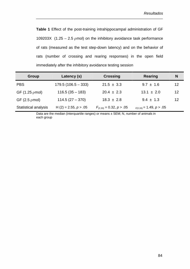
Resultados
84
Table 1 Effect of the post-training intrahippocampal administration of GF
109203X (1.25 – 2.5 ρmol) on the inhibitory avoidance task performance
of rats (measured as the test step-down latency) and on the behavior of
rats (number of crossing and rearing responses) in the open field
immediately after the inhibitory avoidance testing session
Group Latency (s) Crossing Rearing N
PBS 179.5 (106.5 – 333) 21.5 ± 3.3 9.7 ± 1.6 12
GF (1.25 ρmol) 116.5 (35 – 183) 20.4 ± 2.3 13.1 ± 2.0 12
GF (2.5 ρmol) 114.5 (27 – 370) 18.3 ± 2.8 9.4 ± 1.3 12
Statistical analysis H (2) = 2.55, p > .05 F(2,33) = 0.32, p > .05 F(2,33) = 1.49, p > .05
Data are the median (interquartile ranges) or means ± SEM; N, number of animals in each group

Resultados
85
Figure 1
*

Resultados
86
Figure 2
*
*

Resultados
87
Figure 3
*
*

Resultados
88
Figure 4
*
*

V. DISCUSSÃO
Discussão
90
Neste estudo nós demonstramos que H-89, um inibidor da PKA,
arcaína, um antagonista do sítio das poliaminas no receptor NMDA, e GF
109203X, inibidor da PKC, preveniram a melhora da memória da tarefa de
esquiva inibitória induzida por SPD em ratos. A injeção de H-89 ou
arcaína preveniu a fosforilação da PKA e CREB, enquanto a injeção de
GF 109203X preveniu a fosforilação da PKC, PKA e CREB, induzida por
SPD no hipocampo. A reversão do efeito facilitatório da injeção
intrahipocampal de SPD sobre a memória por um inibidor da PKA, um
antagonista do sítio das poliaminas no receptor NMDA e um inibidor da
PKC (Fig. 1 e 4 – artigo e Fig. 1 – manuscrito, respectivamente), implica
na ativação do receptor NMDA e das quinases PKA e PKC na melhora da
memória induzida por SPD. Estes resultados foram corroborados, por
demonstrarem que SPD ativa a PKC e a PKA, e que GF 109203X e H-89,
respectivamente, previnem a ativação da PKC (Fig. 2 – manuscrito) e da
PKA (Fig. 2 – artigo) induzida por SPD. A ativação da PKC induzida por
SPD foi observada 30 min depois da injeção da poliamina.
Interessantemente, este aumento na fosforilação da PKC coincide com o
pico de atividade da PKC que ocorre depois do treino na esquiva
inibitória, que é correlacionado com o aumento na fosforilação do
substrato da PKC, GAP-43, e pode ser bloqueado por inibidores desta
quinase (Cammarota et al., 1997). Por outro lado, a ativação da PKA
induzida por SPD ocorre 180 min depois da injeção da poliamina, o que
provavelmente reflete o pico de atividade da PKA, que ocorre 3-6 horas

Discussão
91
depois do treino. Além disso, este pico é correlacionado com o aumento
máximo nos níveis de AMPc após o treino (Bernabeu et al., 1997a). De
acordo com essa visão, a arcaína impediu o efeito facilitatório da SPD
sobre a fosforilação da PKA, apoiando o envolvimento dos receptores
NMDA na ativação da PKA induzida por SPD (Fig. 6 – artigo).
Como a fosforilação da CREB depende da atividade da PKA
(Gonzalez e Montminy, 1989; Bernabeu et al., 1997a;), a investigação foi
ampliada para os níveis de CREB fosforilada. A administração
intrahipocampal de SPD aumentou a fosforilação da CREB, e como
esperado, H-89 e arcaína preveniram a fosforilação da CREB induzida por
SPD (Fig. 3 e 6 – artigo, respectivamente). Além disso, a injeção de SPD
aumenta a imunorreatividade da subunidade catalítica da PKA fosforilada
na fração nuclear do hipocampo (Fig. 7 – artigo). Uma vez que, a cascata
hipocampal AMPc/PKA é ativada pelo influxo de Ca2+ proveniente do
receptor NMDA (Chetkovich et al., 1991), é possível que o influxo de Ca2+,
sequencialmente, estimule a atividade da adenilato ciclase, acumulando
AMPc, e fosforilando a PKA e CREB (Eliot et al., 1989; Poser and Storm,
2001). De fato, foi demonstrado que o AMPc liga-se a subunidades
regulatória da PKA, resultando na liberação da subunidade monomérica
catalítica ativa da PKA (Taylor et al., 1990; Gibbs et al., 1992; Wang et al.,
1991; 2004). A subunidade catalítica da PKA livre, transloca para o
núcleo, onde pode fosforilar a CREB (Dash et al., 1991; Mayford et al.,
1995a; Impey et al., 1998).

Discussão
92
Surpreendentemente, H-89 por si só, não causou efeito sobre a
atividade da PKA ou CREB. É possível que não tenha sido revelado
qualquer efeito do H-89 devido a janela de tempo selecionada para
executar o ensaio. Por exemplo, Viana e colaboradores (2000b)
demonstraram que a Rp-cAMPS inibe a atividade da PKA 15 min após a
injeção do inibidor, mas não possui efeito em 90 min. Curiosamente, Rp-
cAMPS, em uma dose que não altera a atividade da PKA em 90 min,
impede o aumento da atividade da PKA induzida pelo treino na tarefa de
esquiva inibitória em 180 min. Os resultados de Viana e colaboradores
(2000b) são muito semelhantes aos obtidos no presente estudo, em que o
inibidor impediu o aumento da fosforilação de PKA induzida por
espermidina, 180 min, apoiando a visão de que um inibidor da PKA, em
dose que não possua efeito por si só, pode impedir o aumento da
atividade PKA induzida pelo treino na tarefa de esquiva inibitória ou, no
caso do presente estudo, induzida pela administração de SPD.
Interessantemente, a administração intrahipocampal de SPD
aumentou a razão p-PKA/PKA 180 min depois da injeção, provavelmente,
devido a diminuição nos níveis da subunidade regulatória total da PKA
(Fig. 2E e 2F – artigo). Evidências sugerem que a subunidade regulatória
da PKA é degradada por proteólise mediada por ubiquitina-proteassoma
no encéfalo de animais (Hegde et al., 1993) e humanos (Liang et al.,
2007). Tem sido sugerido, que déficits na subunidade regulatória (RIIα e
RIIβ) e na subunidade catalítica (Cβ) diminuem a atividade da enzima e,
Discussão
93
conseqüentemente, prejudicam a memória em pacientes com doença de
Alzheimer (Liang et al., 2007). Em contraste, lactacistina, um inibidor
específico de proteassoma, bloqueia completamente a “down-regulation”
da subunidade regulatória induzida por forskolina em células de
neuroblastoma humano SH-SY5Y (Boundy et al., 1998). Além disso, a
degradação proteassomal das subunidades regulatórias da PKA gera um
excesso da subunidade catalítica. Corroborando com estas evidências,
um estudo mostra que o treino na tarefa de esquiva inibitória provoca um
aumento na quantidade de subunidade catalítica da PKA dependente dos
receptores NMDA em frações nucleares de neurônios hipocampais
(Cammarota et al., 2000) e, além disso, a injeção intrahipocampal de um
inibidor de proteassoma bloqueia a formação da memória de longa
duração (Lopez-Salon et al., 2001). Adicionalmente, é interessante notar
paralelos na evolução temporal entre o efeito de inibidores de
proteossoma sobre a formação da memória de longa duração e inibidores
específicos da PKA, distribuídos na mesma região do encéfalo (Bernabeu
et al., 1997a; Vianna et al, 2000b) sendo que, ainda coincide com o
período durante o qual a atividade da PKA hipocampal está aumentada
(Izquierdo e Medina, 1997).
Existem evidências sugerindo um cruzamento entre a PKC e a
ativação da PKA (Sugita et al., 1997; Kubota et al., 2003; Yao et al.,
2008). Além disso, nós encontramos que a administração de SPD induz a
fosforilação da PKA e CREB no hipocampo de ratos, somente 180 min,

Discussão
94
mas não 30 min depois da injeção, então, nós investigamos os níveis de
fosfo-PKA e fosfo-CREB, depois da injeção do inibidor da PKC. A
administração intrahipocampal de GF 109203X preveniu a fosforilação da
PKA e CREB induzida por SPD no hipocampo (Fig. 3 e 4 – manuscrito,
respectivamente).
Embora os resultados do presente estudo indiquem que a ativação
da PKC em períodos iniciais e da PKA em períodos mais tardis estão
envolvidos na melhora da memória induzida por SPD, o exato mecanismo
pelo qual esta interação ocorre permanece incerto. Um provável
mecanismo pelo qual a PKC pode ativar a PKA involve a neurogranina.
Evidências sugerem que a ativação da PKC cause a fosforilação da
neurogranina (também chamada RC3), um substrato da PKC que liga-se
a calmodulina (CaM) em baixas concentrações de Ca2+ (Baudier et al.,
1991; Huang et al., 1993; Diez-Guerra, 2010). Após a ativação de
receptores, como o NMDA, ocorre aumento do influxo de Ca2+, induzindo
a fosforilação da neurogranina mediada pela PKC. A neurogranina
quando fosforilada reduz a afinidade da ligação para a CaM. Isto aumenta
a disponibilidade de CaM e formação do complexo Ca2+/CaM (Sheu et al.,
1996; Li et al., 1999; Wu et al., 2003) (Fig. V.1). Um aumento nos níveis
de Ca2+/CaM ativa a adenilato ciclase tipo I e VIII, aumentando os níveis
de AMPc (Mons et al., 1999; Poser and Storm, 2001; Wang and Storm,
2003; Ferguson and Storm, 2004;). Neste sentido, a falta do gene para a
neurogranina prejudicaria a ativação da PKA induzida por NMDA (Wu et
Discussão
95
al., 2003) ou por forskolina (Wu et al., 2002), e a subsequente sinalização
envolvida na formação da memória.
Tem sido demonstrado também que a PKC pode ativar diretamente
a adenilato ciclase (Bell et al., 1985; Choi et al., 1993;; Jacobowitz et al.,
1993; Lustig et al., 1993; Pieroni et al., 1993; Yoshimura and Cooper,
1993; Cooper et al., 1995). Estudos demonstram que ativadores da PKC,
aumentam a síntese de AMPc através da adenilato ciclase tipo II
(Yoshimura and Cooper, 1993) e causam um aumento na excitabilidade
do neurônio sensorial de Aplysia, (Sugita et al., 1997). Enquanto que, a
estaurosporina, um inibidor da PKC, atenua o efeito estimulatório do éster
de forbol sobre a adenilato ciclase tipo II (Yoshimura and Cooper, 1993).
Estas observações sugerem que a ativação da PKC aumenta os
níveis de AMPc, por uma ativação direta e indireta da adenilato ciclase
insensível a Ca2+/CaM (tipo II) e adenilato ciclase sensível a Ca2+/CaM
(tipo I e VIII), respectivamente.
Uma vez que, a cascata AMPc/PKA hipocampal é ativada pelo
acúmulo de AMPc mediante a ativação da adenilato ciclase (Eliot et al.,
1989; Poser and Storm, 2001), podemos sugerir que a fosforilação da
PKC induzida por SPD pode estar aumentando tanto a fosforilação da
neurogranina, quanto a atividade da adenilato ciclase, contribuindo para
um acúmulo de AMPc e conseqüentemente a ativação da PKA e CREB.
Assim, este estudo demonstrou que a melhora da memória
induzida por SPD depende de uma seqüência de eventos bioquímicos no
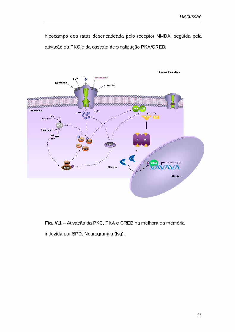
Discussão
96
hipocampo dos ratos desencadeada pelo receptor NMDA, seguida pela
ativação da PKC e da cascata de sinalização PKA/CREB.
Fig. V.1 – Ativação da PKC, PKA e CREB na melhora da memória
induzida por SPD. Neurogranina (Ng).

VI. CONCLUSÕES

Conclusões
98
Com base nos resultados obtidos, concluiu-se que:
A administração intra-hipocampal de GF 109203X, um inibidor da
PKC, H-89, um inibidor da PKA, e arcaína, um antagonista do sítio das
poliaminas no receptor NMDA, preveniram a melhora da memória
induzida por SPD. Sugerindo que a ativação do receptor NMDA e das
proteínas quinase A e C estão envolvidos na melhora da memória
induzida por SPD.
A administração intra-hipocampal de H-89 e arcaína, preveniram o
aumento da fosforilação da PKA e CREB induzidos pela administração
intra-hipocampal de SPD. Sugerindo que SPD aumenta a ativação da
PKA e CREB através da ativação do receptor NMDA.
A administração intra-hipocampal de SPD aumentou os níveis da
subunidade catalítica da PKA no núcleo. Sugerindo que SPD aumentou a
migração da subunidade catalítica da PKA do citosol para o núcleo.
A administração intra-hipocampal de GF 109203X, preveniu o
aumento da fosforilação da PKC, PKA e CREB induzidos pela
administração intra-hipocampal de SPD. Sugerindo que SPD aumenta a
ativação da PKC, PKA e CREB. Sendo que a ativação da PKA e CREB
necessita da ativação previa da PKC.

VII. REFERÊNCIAS
Referências
100
Abel, T., Nguyen, P. V., Barad, M., Deuel, T. A., Kandel, E. R.,
Bourtchouladze, R., 1997. Genetic demonstration of a role for PKA
in the late phase of LTP and in hippocampus-based long-term
memory. Cell 88, 615-626.
Abel, T., Lattal, K. M., 2001. Molecular mechanisms of memory
acquisition, consolidation and retrieval. Curr Opin Neurobiol 11,
180-187.
Abeliovich, A., Paylor, R., Chen, C., Kim, J. J., Wehner, J. M., Tonegawa,
S., 1993. PKC gamma mutant mice exhibit mild deficits in spatial
and contextual learning. Cell 75, 1263-1271.
Adams, J. A., 2001. Kinetic and catalytic mechanisms of protein kinases.
Chem Rev 101, 2271-2290.
Ahi, J., Radulovic, J., Spiess, J., 2004. The role of hippocampal signaling
cascades in consolidation of fear memory. Behav Brain Res 149,
17-31.
Ahlander, M., Misane, I., Schott, P. A., Ogren, S. O., 1999. A behavioral
analysis of the spatial learning deficit induced by the NMDA
receptor antagonist MK-801 (dizocilpine) in the rat.
Neuropsychopharmacology 21, 414-426.
Alkon, D. L., Epstein, H., Kuzirian, A., Bennett, M. C., Nelson, T. J., 2005.
Protein synthesis required for long-term memory is induced by PKC
activation on days before associative learning. Proc Natl Acad Sci U
S A 102, 16432-16437.
Referências
101
Amadio, M., Battaini, F., Pascale, A., 2006. The different facets of protein
kinases C: old and new players in neuronal signal transduction
pathways. Pharmacol Res 54, 317-325.
Baillie, G. S., Scott, J. D., Houslay, M. D., 2005. Compartmentalisation of
phosphodiesterases and protein kinase A: opposites attract. FEBS
Lett 579, 3264-3270.
Barad, M., Bourtchouladze, R., Winder, D. G., Golan, H., Kandel, E., 1998.
Rolipram, a type IV-specific phosphodiesterase inhibitor, facilitates
the establishment of long-lasting long-term potentiation and
improves memory. Proc Natl Acad Sci U S A 95, 15020-15025.
Baudier, J., Deloulme, J. C., Van Dorsselaer, A., Black, D., Matthes, H.
W., 1991. Purification and characterization of a brain-specific
protein kinase C substrate, neurogranin (p17). Identification of a
consensus amino acid sequence between neurogranin and
neuromodulin (GAP43) that corresponds to the protein kinase C
phosphorylation site and the calmodulin-binding domain. J Biol
Chem 266, 229-237.
Bauman, A. L., Scott, J. D., 2002. Kinase- and phosphatase-anchoring
proteins: harnessing the dynamic duo. Nat Cell Biol 4, E203-206.
Bauman, A. L., Goehring, A. S., Scott, J. D., 2004. Orchestration of
synaptic plasticity through AKAP signaling complexes.
Neuropharmacology 46, 299-310.
Bear, M. F., Connors B. W., Paradiso, M. A. 2002 Neurociência:
desvendando o sistema nervoso. 2a edição, Porto Alegre: Artmed
Editora S.A.

Referências
102
Bell, J. D., Buxton, I. L., Brunton, L. L., 1985. Enhancement of adenylate
cyclase activity in S49 lymphoma cells by phorbol esters. Putative
effect of C kinase on alpha s-GTP-catalytic subunit interaction. J
Biol Chem 260, 2625-2628.
Berlese, D. B., Sauzem, P. D., Carati, M. C., Guerra, G. P., Stiegemeier, J.
A., Mello, C. F., Rubin, M. A., 2005. Time-dependent modulation of
inhibitory avoidance memory by spermidine in rats. Neurobiol Learn
Mem 83, 48-53.
Bernabeu, R., Izquierdo, I., Cammarota, M., Jerusalinsky, D., Medina, J.
H., 1995. Learning-specific, time-dependent increase in [3H]phorbol
dibutyrate binding to protein kinase C in selected regions of the rat
brain. Brain Res 685, 163-168.
Bernabeu, R., Schmitz, P., Faillace, M. P., Izquierdo, I., Medina, J. H.,
1996. Hippocampal cGMP and cAMP are differentially involved in
memory processing of inhibitory avoidance learning. Neuroreport 7,
585-588.
Bernabeu, R., Bevilaqua, L., Ardenghi, P., Bromberg, E., Schmitz, P.,
Bianchin, M., Izquierdo, I., Medina, J. H., 1997a. Involvement of
hippocampal cAMP/cAMP-dependent protein kinase signaling
pathways in a late memory consolidation phase of aversively
motivated learning in rats. Proc Natl Acad Sci U S A 94, 7041-7046.
Bernabeu, R., Cammarota, M., Izquierdo, I., Medina, J. H., 1997b.
Involvement of hippocampal AMPA glutamate receptor changes
and the cAMP/protein kinase A/CREB-P signalling pathway in
memory consolidation of an avoidance task in rats. Braz J Med Biol
Res 30, 961-965.

Referências
103
Bernabeu, R., Schroder, N., Quevedo, J., Cammarota, M., Izquierdo, I.,
Medina, J. H., 1997c. Further evidence for the involvement of a
hippocampal cGMP/cGMP-dependent protein kinase cascade in
memory consolidation. Neuroreport 8, 2221-2224.
Bevilaqua, L., Ardenghi, P., Schroder, N., Bromberg, E., Schmitz, P. K.,
Schaeffer, E., Quevedo, J., Bianchin, M., Walz, R., Medina, J. H.,
Izquierdo, I., 1997. Drugs acting upon the cyclic adenosine
monophosphate/protein kinase A signalling pathway modulate
memory consolidation when given late after training into rat
hippocampus but not amygdala. Behav Pharmacol 8, 331-338.
Bliss, T. V., Collingridge, G. L., 1993. A synaptic model of memory: long-
term potentiation in the hippocampus. Nature 361, 31-39.
Bonini, J. S., Da Silva, W. C., Bevilaqua, L. R., Medina, J. H., Izquierdo, I.,
Cammarota, M., 2007. On the participation of hippocampal PKC in
acquisition, consolidation and reconsolidation of spatial memory.
Neuroscience 147, 37-45.
Bonini, J. S., Da Silva, W. C., Da Silveira, C. K., Kohler, C. A., Izquierdo,
I., Cammarota, M., 2011. Histamine facilitates consolidation of fear
extinction. Int J Neuropsychopharmacol, 1-9.
Bornancin, F., Parker, P. J., 1996. Phosphorylation of threonine 638
critically controls the dephosphorylation and inactivation of protein
kinase Calpha. Curr Biol 6, 1114-1123.
Bornancin, F., Parker, P. J., 1997. Phosphorylation of protein kinase C-
alpha on serine 657 controls the accumulation of active enzyme
Referências
104
and contributes to its phosphatase-resistant state. J Biol Chem 272,
3544-3549.
Boundy, V. A., Chen, J., Nestler, E. J., 1998. Regulation of cAMP-
dependent protein kinase subunit expression in CATH.a and SH-
SY5Y cells. J Pharmacol Exp Ther 286, 1058-1065.
Bourtchouladze, R., Abel, T., Berman, N., Gordon, R., Lapidus, K., Kandel,
E. R., 1998. Different training procedures recruit either one or two
critical periods for contextual memory consolidation, each of which
requires protein synthesis and PKA. Learn Mem 5, 365-374.
Broadbent, N. J., Squire, L. R., Clark, R. E., 2004. Spatial memory,
recognition memory, and the hippocampus. Proc Natl Acad Sci U S
A 101, 14515-14520.
Burke, S. N., Barnes, C. A., Senescent synapses and hippocampal circuit
dynamics. Trends Neurosci 33, 153-161.
Cadd, G., McKnight, G. S., 1989. Distinct patterns of cAMP-dependent
protein kinase gene expression in mouse brain. Neuron 3, 71-79.
Cadd, G. G., Uhler, M. D., McKnight, G. S., 1990. Holoenzymes of cAMP-
dependent protein kinase containing the neural form of type I
regulatory subunit have an increased sensitivity to cyclic
nucleotides. J Biol Chem 265, 19502-19506.
Camera, K., Mello, C. F., Ceretta, A. P., Rubin, M. A., 2007. Systemic
administration of polyaminergic agents modulate fear conditioning
in rats. Psychopharmacology (Berl) 192, 457-464.
Referências
105
Cammarota, M., Paratcha, G., Levi de Stein, M., Bernabeu, R., Izquierdo,
I., Medina, J. H., 1997. B-50/GAP-43 phosphorylation and PKC
activity are increased in rat hippocampal synaptosomal membranes
after an inhibitory avoidance training. Neurochem Res 22, 499-505.
Cammarota, M., Bevilaqua, L. R., Ardenghi, P., Paratcha, G., Levi de
Stein, M., Izquierdo, I., Medina, J. H., 2000. Learning-associated
activation of nuclear MAPK, CREB and Elk-1, along with Fos
production, in the rat hippocampus after a one-trial avoidance
learning: abolition by NMDA receptor blockade. Brain Res Mol Brain
Res 76, 36-46.
Caramanos, Z., Shapiro, M. L., 1994. Spatial memory and N-methyl-D-
aspartate receptor antagonists APV and MK-801: memory
impairments depend on familiarity with the environment, drug dose,
and training duration. Behav Neurosci 108, 30-43.
Carlezon, W. A., Jr., Duman, R. S., Nestler, E. J., 2005. The many faces of
CREB. Trends Neurosci 28, 436-445.
Carnegie, G. K., Means, C. K., Scott, J. D., 2009. A-kinase anchoring
proteins: from protein complexes to physiology and disease. IUBMB
Life 61, 394-406.
Carter, C., 1994. The neuropharmacology of polyamines. Academic Press,
London.
Castellano, C., Cestari, V., Ciamei, A., 2001. NMDA receptors and
learning and memory processes. Curr Drug Targets 2, 273-283.
Referências
106
Chain, D. G., Hegde, A. N., Yamamoto, N., Liu-Marsh, B., Schwartz, J. H.,
1995. Persistent activation of cAMP-dependent protein kinase by
regulated proteolysis suggests a neuron-specific function of the
ubiquitin system in Aplysia. J Neurosci 15, 7592-7603.
Chetkovich, D. M., Gray, R., Johnston, D., Sweatt, J. D., 1991. N-methyl-
D-aspartate receptor activation increases cAMP levels and voltage-
gated Ca2+ channel activity in area CA1 of hippocampus. Proc Natl
Acad Sci U S A 88, 6467-6471.
Chetkovich, D. M., Sweatt, J. D., 1993. NMDA receptor activation
increases cyclic AMP in area CA1 of the hippocampus via
calcium/calmodulin stimulation of adenylyl cyclase. J Neurochem
61, 1933-1942.
Choi, E. J., Wong, S. T., Dittman, A. H., Storm, D. R., 1993. Phorbol ester
stimulation of the type I and type III adenylyl cyclases in whole cells.
Biochemistry 32, 1891-1894.
Coleman, C. S., Hu, G., Pegg, A. E., 2004. Putrescine biosynthesis in
mammalian tissues. Biochem J 379, 849-855.
Contestabile, A., 2008. Regulation of transcription factors by nitric oxide in
neurons and in neural-derived tumor cells. Prog Neurobiol 84, 317-
328.
Conti, M., Beavo, J., 2007. Biochemistry and physiology of cyclic
nucleotide phosphodiesterases: essential components in cyclic
nucleotide signaling. Annu Rev Biochem 76, 481-511.
Referências
107
Conway, E. L., 1998. Brain lesions and delayed water maze learning
deficits after intracerebroventricular spermine. Brain Res 800, 10-
20.
Cooper, D. M., Mons, N., Karpen, J. W., 1995. Adenylyl cyclases and the
interaction between calcium and cAMP signalling. Nature 374, 421-
424.
Cooper, D. M., 2003. Regulation and organization of adenylyl cyclases
and cAMP. Biochem J 375, 517-529.
Coughenour, L. L., Barr, B. M., 2001. Use of trifluoroperazine isolates a
[(3)H]Ifenprodil binding site in rat brain membranes with the
pharmacology of the voltage-independent ifenprodil site on N-
methyl-D-aspartate receptors containing NR2B subunits. J
Pharmacol Exp Ther 296, 150-159.
Coussens, L., Parker, P. J., Rhee, L., Yang-Feng, T. L., Chen, E.,
Waterfield, M. D., Francke, U., Ullrich, A., 1986. Multiple, distinct
forms of bovine and human protein kinase C suggest diversity in
cellular signaling pathways. Science 233, 859-866.
Cravens, C. J., Vargas-Pinto, N., Christian, K. M., Nakazawa, K., 2006.
CA3 NMDA receptors are crucial for rapid and automatic
representation of context memory. Eur J Neurosci 24, 1771-1780.
Dash, P. K., Karl, K. A., Colicos, M. A., Prywes, R., Kandel, E. R., 1991.
cAMP response element-binding protein is activated by
Ca2+/calmodulin- as well as cAMP-dependent protein kinase. Proc
Natl Acad Sci U S A 88, 5061-5065.

Referências
108
de Lima, M. N., Laranja, D. C., Bromberg, E., Roesler, R., Schroder, N.,
2005. Pre- or post-training administration of the NMDA receptor
blocker MK-801 impairs object recognition memory in rats. Behav
Brain Res 156, 139-143.
Dell'Acqua, M. L., Smith, K. E., Gorski, J. A., Horne, E. A., Gibson, E. S.,
Gomez, L. L., 2006. Regulation of neuronal PKA signaling through
AKAP targeting dynamics. Eur J Cell Biol 85, 627-633.
Dewachter, I., Filipkowski, R. K., Priller, C., Ris, L., Neyton, J., Croes, S.,
Terwel, D., Gysemans, M., Devijver, H., Borghgraef, P., Godaux,
E., Kaczmarek, L., Herms, J., Van Leuven, F., 2009. Deregulation
of NMDA-receptor function and down-stream signaling in
APP[V717I] transgenic mice. Neurobiol Aging 30, 241-256.
Diez-Guerra, F. J., 2010. Neurogranin, a link between calcium/calmodulin
and protein kinase C signaling in synaptic plasticity. IUBMB Life 62,
597-606.
Doskeland, S. O., Maronde, E., Gjertsen, B. T., 1993. The genetic
subtypes of cAMP-dependent protein kinase--functionally different
or redundant? Biochim Biophys Acta 1178, 249-258.
Dutil, E. M., Toker, A., Newton, A. C., 1998. Regulation of conventional
protein kinase C isozymes by phosphoinositide-dependent kinase 1
(PDK-1). Curr Biol 8, 1366-1375.
Elgersma, Y., Silva, A. J., 1999. Molecular mechanisms of synaptic
plasticity and memory. Curr Opin Neurobiol 9, 209-213.
Referências
109
Eliot, L. S., Dudai, Y., Kandel, E. R., Abrams, T. W., 1989.
Ca2+/calmodulin sensitivity may be common to all forms of neural
adenylate cyclase. Proc Natl Acad Sci U S A 86, 9564-9568.
Fellini, L., Florian, C., Courtey, J., Roullet, P., 2009. Pharmacological
intervention of hippocampal CA3 NMDA receptors impairs
acquisition and long-term memory retrieval of spatial pattern
completion task. Learn Mem 16, 387-394.
Ferguson, G. D., Storm, D. R., 2004. Why calcium-stimulated adenylyl
cyclases? Physiology (Bethesda) 19, 271-276.
First, E. A., Bubis, J., Taylor, S. S., 1988. Subunit interaction sites
between the regulatory and catalytic subunits of cAMP-dependent
protein kinase. Identification of a specific interchain disulfide bond. J
Biol Chem 263, 5176-5182.
Flood, J. F., Baker, M. L., Davis, J. L., 1990. Modulation of memory
processing by glutamic acid receptor agonists and antagonists.
Brain Res 521, 197-202.
Furukawa, H., Singh, S. K., Mancusso, R., Gouaux, E., 2005. Subunit
arrangement and function in NMDA receptors. Nature 438, 185-
192.
Gibbs, C. S., Knighton, D. R., Sowadski, J. M., Taylor, S. S., Zoller, M. J.,
1992. Systematic mutational analysis of cAMP-dependent protein
kinase identifies unregulated catalytic subunits and defines regions
important for the recognition of the regulatory subunit. J Biol Chem
267, 4806-4814.

Referências
110
Giese, K. P., Fedorov, N. B., Filipkowski, R. K., Silva, A. J., 1998.
Autophosphorylation at Thr286 of the alpha calcium-calmodulin
kinase II in LTP and learning. Science 279, 870-873.
Golski, S., Olds, J. L., Mishkin, M., Olton, D. S., Alkon, D. L., 1995. Protein
kinase C in the hippocampus is altered by spatial but not cued
discriminations: a component task analysis. Brain Res 676, 53-62.
Gomes, G. M., Mello, C. F., da Rosa, M. M., Bochi, G. V., Ferreira, J.,
Barron, S., Rubin, M. A., 2010. Polyaminergic agents modulate
contextual fear extinction in rats. Neurobiol Learn Mem 93, 589-
595.
Gonzalez, G. A., Montminy, M. R., 1989. Cyclic AMP stimulates
somatostatin gene transcription by phosphorylation of CREB at
serine 133. Cell 59, 675-680.
Goodrich-Hunsaker, N. J., Hopkins, R. O., 2010. Spatial memory deficits in
a virtual radial arm maze in amnesic participants with hippocampal
damage. Behav Neurosci 124, 405-413.
Goodrich-Hunsaker, N. J., Livingstone, S. A., Skelton, R. W., Hopkins, R.
O., 2010. Spatial deficits in a virtual water maze in amnesic
participants with hippocampal damage. Hippocampus 20, 481-491.
Guerra, G. P., Mello, C. F., Sauzem, P. D., Berlese, D. B., Furian, A. F.,
Tabarelli, Z., Rubin, M. A., 2006. Nitric oxide is involved in the
memory facilitation induced by spermidine in rats.
Psychopharmacology (Berl) 186, 150-158.
Referências
111
Gugliucci, A., 2004. Polyamines as clinical laboratory tools. Clin Chim Acta
344, 23-35.
Hanoune, J., Defer, N., 2001. Regulation and role of adenylyl cyclase
isoforms. Annu Rev Pharmacol Toxicol 41, 145-174.
Hegde, A. N., Goldberg, A. L., Schwartz, J. H., 1993. Regulatory subunits
of cAMP-dependent protein kinases are degraded after conjugation
to ubiquitin: a molecular mechanism underlying long-term synaptic
plasticity. Proc Natl Acad Sci U S A 90, 7436-7440.
Hidaka, H., Hagiwara, M., Chijiwa, T., 1990. Molecular pharmacology of
protein kinases. Neurochem Res 15, 431-434.
Hu, M., Sun, Y. J., Zhou, Q. G., Auberson, Y. P., Chen, L., Hu, Y., Luo, C.
X., Wu, J. Y., Zhu, D. Y., Li, L. X., 2009. Reduced spatial learning in
mice treated with NVP-AAM077 through down-regulating
neurogenesis. Eur J Pharmacol 622, 37-44.
Huang, K. P., Huang, F. L., Chen, H. C., 1993. Characterization of a 7.5-
kDa protein kinase C substrate (RC3 protein, neurogranin) from rat
brain. Arch Biochem Biophys 305, 570-580.
Huang, Y. Y., Kandel, E. R., 1998. Postsynaptic induction and PKA-
dependent expression of LTP in the lateral amygdala. Neuron 21,
169-178.
Huang, F. L., Huang, K. P., Wu, J., Boucheron, C., 2006. Environmental
enrichment enhances neurogranin expression and hippocampal
learning and memory but fails to rescue the impairments of
neurogranin null mutant mice. J Neurosci 26, 6230-6237.

Referências
112
Impey, S., Obrietan, K., Wong, S. T., Poser, S., Yano, S., Wayman, G.,
Deloulme, J. C., Chan, G., Storm, D. R., 1998. Cross talk between
ERK and PKA is required for Ca2+ stimulation of CREB-dependent
transcription and ERK nuclear translocation. Neuron 21, 869-883.
Isiegas, C., Park, A., Kandel, E. R., Abel, T., Lattal, K. M., 2006.
Transgenic inhibition of neuronal protein kinase A activity facilitates
fear extinction. J Neurosci 26, 12700-12707.
Izquierdo, I., da Cunha, C., Rosat, R., Jerusalinsky, D., Ferreira, M. B.,
Medina, J. H., 1992. Neurotransmitter receptors involved in post-
training memory processing by the amygdala, medial septum, and
hippocampus of the rat. Behav Neural Biol 58, 16-26.
Izquierdo, I., Medina, J. H., 1995. Correlation between the pharmacology
of long-term potentiation and the pharmacology of memory.
Neurobiol Learn Mem 63, 19-32.
Izquierdo, I., Medina, J. H., 1997. Memory formation: the sequence of
biochemical events in the hippocampus and its connection to
activity in other brain structures. Neurobiol Learn Mem 68, 285-316.
Izquierdo, I. 2002. Memória. Artmed Editora S.A.
Izquierdo, I., Bevilaqua, L. R., Rossato, J. I., Bonini, J. S., Medina, J. H.,
Cammarota, M., 2006. Different molecular cascades in different
sites of the brain control memory consolidation. Trends Neurosci
29, 496-505.

Referências
113
Jacobowitz, O., Chen, J., Premont, R. T., Iyengar, R., 1993. Stimulation of
specific types of Gs-stimulated adenylyl cyclases by phorbol ester
treatment. J Biol Chem 268, 3829-3832.
Jafari-Sabet, M., 2006. NMDA receptor blockers prevents the facilitatory
effects of post-training intra-dorsal hippocampal NMDA and
physostigmine on memory retention of passive avoidance learning
in rats. Behav Brain Res 169, 120-127.
Jarnaess, E., Tasken, K., 2007. Spatiotemporal control of cAMP signalling
processes by anchored signalling complexes. Biochem Soc Trans
35, 931-937.
Jerusalinsky, D., Quillfeldt, J. A., Walz, R., Da Silva, R. C., Medina, J. H.,
Izquierdo, I., 1994. Post-training intrahippocampal infusion of
protein kinase C inhibitors causes amnesia in rats. Behav Neural
Biol 61, 107-109.
Kishi, A., Ohno, M., Watanabe, S., 1998a. Concurrent activation of
hippocampal glycine and polyamine sites of the N-methyl-D-
aspartate receptor synergistically reverses working memory deficits
in rats. Neurosci Lett 257, 131-134.
Kishi, A., Ohno, M., Watanabe, S., 1998b. Spermidine, a polyamine site
agonist, attenuates working memory deficits caused by blockade of
hippocampal muscarinic receptors and mGluRs in rats. Brain Res
793, 311-314.
Kornisiuk, E., Snitcofsky, M., Blanco, C., Harvey, A. L., Stone, T. W.,
Jerusalinsky, D., Memory impairment in rats by hippocampal
Referências
114
administration of the serine protease subtilisin. Behav Brain Res
219, 63-67.
Krystal, J. H., Tolin, D. F., Sanacora, G., Castner, S. A., Williams, G. V.,
Aikins, D. E., Hoffman, R. E., D'Souza, D. C., 2009. Neuroplasticity
as a target for the pharmacotherapy of anxiety disorders, mood
disorders, and schizophrenia. Drug Discov Today 14, 690-697.
Kubota, H., Katsurabayashi, S., Moorhouse, A. J., Murakami, N., Koga, H.,
Akaike, N., 2003. GABAB receptor transduction mechanisms, and
cross-talk between protein kinases A and C, in GABAergic
terminals synapsing onto neurons of the rat nucleus basalis of
Meynert. J Physiol 551, 263-276.
LaLumiere, R. T., Pizano, E., McGaugh, J. L., 2004. Intra-basolateral
amygdala infusions of AP-5 impair or enhance retention of inhibitory
avoidance depending on training conditions. Neurobiol Learn Mem
81, 60-66.
Larque, E., Sabater-Molina, M., Zamora, S., 2007. Biological significance
of dietary polyamines. Nutrition 23, 87-95.
Lau, C. G., Takeuchi, K., Rodenas-Ruano, A., Takayasu, Y., Murphy, J.,
Bennett, M. V., Zukin, R. S., 2009. Regulation of NMDA receptor
Ca2+ signalling and synaptic plasticity. Biochem Soc Trans 37,
1369-1374.
Lee, Y. S., Silva, A. J., 2009. The molecular and cellular biology of
enhanced cognition. Nat Rev Neurosci 10, 126-140.
Referências
115
Li, J., Pak, J. H., Huang, F. L., Huang, K. P., 1999. N-methyl-D-aspartate
induces neurogranin/RC3 oxidation in rat brain slices. J Biol Chem
274, 1294-1300.
Li, X., Vadrevu, S., Dunlop, A., Day, J., Advant, N., Troeger, J.,
Klussmann, E., Jaffrey, E., Hay, R. T., Adams, D. R., Houslay, M.
D., Baillie, G. S., 2010. Selective SUMO modification of cAMP-
specific phosphodiesterase-4D5 (PDE4D5) regulates the functional
consequences of phosphorylation by PKA and ERK. Biochem J
428, 55-65.
Liang, Z., Liu, F., Grundke-Iqbal, I., Iqbal, K., Gong, C. X., 2007. Down-
regulation of cAMP-dependent protein kinase by over-activated
calpain in Alzheimer disease brain. J Neurochem 103, 2462-2470.
Lochner, A., Moolman, J. A., 2006. The many faces of H89: a review.
Cardiovasc Drug Rev 24, 261-274.
Lopez-Salon, M., Alonso, M., Vianna, M. R., Viola, H., Mello e Souza, T.,
Izquierdo, I., Pasquini, J. M., Medina, J. H., 2001. The ubiquitin-
proteasome cascade is required for mammalian long-term memory
formation. Eur J Neurosci 14, 1820-1826.
Lugnier, C., 2006. Cyclic nucleotide phosphodiesterase (PDE)
superfamily: a new target for the development of specific
therapeutic agents. Pharmacol Ther 109, 366-398.
Lustig, K. D., Conklin, B. R., Herzmark, P., Taussig, R., Bourne, H. R.,
1993. Type II adenylylcyclase integrates coincident signals from Gs,
Gi, and Gq. J Biol Chem 268, 13900-13905.

Referências
116
MacDermott, A. B., Mayer, M. L., Westbrook, G. L., Smith, S. J., Barker, J.
L., 1986. NMDA-receptor activation increases cytoplasmic calcium
concentration in cultured spinal cord neurones. Nature 321, 519-
522.
Macek, T. A., Schaffhauser, H., Conn, P. J., 1998. Protein kinase C and
A3 adenosine receptor activation inhibit presynaptic metabotropic
glutamate receptor (mGluR) function and uncouple mGluRs from
GTP-binding proteins. J Neurosci 18, 6138-6146.
MacKenzie, S. J., Baillie, G. S., McPhee, I., MacKenzie, C., Seamons, R.,
McSorley, T., Millen, J., Beard, M. B., van Heeke, G., Houslay, M.
D., 2002. Long PDE4 cAMP specific phosphodiesterases are
activated by protein kinase A-mediated phosphorylation of a single
serine residue in Upstream Conserved Region 1 (UCR1). Br J
Pharmacol 136, 421-433.
Majewski, H., Iannazzo, L., 1998. Protein kinase C: a physiological
mediator of enhanced transmitter output. Prog Neurobiol 55, 463-
475.
Malenka, R. C., Madison, D. V., Nicoll, R. A., 1986. Potentiation of
synaptic transmission in the hippocampus by phorbol esters. Nature
321, 175-177.
Manning, G., Whyte, D. B., Martinez, R., Hunter, T., Sudarsanam, S.,
2002. The protein kinase complement of the human genome.
Science 298, 1912-1934.
Mariani, R. K., Mello, C. F., Rosa, M. M., Ceretta, A. P., Camera, K.,
Rubin, M. A., 2011. Effect of naloxone and morphine on arcaine-

Referências
117
induced state-dependent memory in rats. Psychopharmacology
(Berl) 215, 483-491.
Martin, S. J., Grimwood, P. D., Morris, R. G., 2000. Synaptic plasticity and
memory: an evaluation of the hypothesis. Annu Rev Neurosci 23,
649-711.
Mayer, M. L., Westbrook, G. L., 1987. Permeation and block of N-methyl-
D-aspartic acid receptor channels by divalent cations in mouse
cultured central neurones. J Physiol 394, 501-527.
Mayford, M., Abel, T., Kandel, E. R., 1995a. Transgenic approaches to
cognition. Curr Opin Neurobiol 5, 141-148.
Mayford, M., Wang, J., Kandel, E. R., O'Dell, T. J., 1995b. CaMKII
regulates the frequency-response function of hippocampal
synapses for the production of both LTD and LTP. Cell 81, 891-904.
Mayr, B., Montminy, M., 2001. Transcriptional regulation by the
phosphorylation-dependent factor CREB. Nat Rev Mol Cell Biol 2,
599-609.
McGaugh, J. L., 2000. Memory--a century of consolidation. Science 287,
248-251.
McGaugh, J. L., Izquierdo, I., 2000. The contribution of pharmacology to
research on the mechanisms of memory formation. Trends
Pharmacol Sci 21, 208-210.
McGaugh, J. L., 2002. Memory consolidation and the amygdala: a
systems perspective. Trends Neurosci 25, 456.
Referências
118
McKnight, G. S., Clegg, C. H., Uhler, M. D., Chrivia, J. C., Cadd, G. G.,
Correll, L. A., Otten, A. D., 1988. Analysis of the cAMP-dependent
protein kinase system using molecular genetic approaches. Recent
Prog Horm Res 44, 307-335.
Meyer, R. C., Spangler, E. L., Patel, N., London, E. D., Ingram, D. K.,
1998. Impaired learning in rats in a 14-unit T-maze by 7-
nitroindazole, a neuronal nitric oxide synthase inhibitor, is
attenuated by the nitric oxide donor, molsidomine. Eur J Pharmacol
341, 17-22.
Micheau, J., Riedel, G., 1999. Protein kinases: which one is the memory
molecule? Cell Mol Life Sci 55, 534-548.
Michel, J. J., Scott, J. D., 2002. AKAP mediated signal transduction. Annu
Rev Pharmacol Toxicol 42, 235-257.
Mikolajczak, P., Okulicz-Kozaryn, I., Kaminska, E., Niedopad, L.,
Polanska, A., Gebka, J., 2002. Effects of acamprosate and some
polyamine site ligands of NMDA receptor on short-term memory in
rats. Eur J Pharmacol 444, 83-96.
Mikolajczak, P., Okulicz-Kozaryn, I., Kaminska, E., Szulc, M., Dyr, W.,
Kostowski, W., 2003. Lack of ifenprodil anxiolytic activity after its
multiple treatment in chronically ethanol-treated rats. Alcohol
Alcohol 38, 310-315.
Miller, D. B., O'Callaghan, J. P., 2005. Aging, stress and the hippocampus.
Ageing Res Rev 4, 123-140.

Referências
119
Miserendino, M. J., Sananes, C. B., Melia, K. R., Davis, M., 1990. Blocking
of acquisition but not expression of conditioned fear-potentiated
startle by NMDA antagonists in the amygdala. Nature 345, 716-718.
Miyakawa, T., Yared, E., Pak, J. H., Huang, F. L., Huang, K. P., Crawley,
J. N., 2001. Neurogranin null mutant mice display performance
deficits on spatial learning tasks with anxiety related components.
Hippocampus 11, 763-775.
Mizuno, K., Giese, K. P., 2005. Hippocampus-dependent memory
formation: do memory type-specific mechanisms exist? J
Pharmacol Sci 98, 191-197.
Monaghan, D. T., Cotman, C. W., 1985. Distribution of N-methyl-D-
aspartate-sensitive L-[3H]glutamate-binding sites in rat brain. J
Neurosci 5, 2909-2919.
Mons, N., Guillou, J. L., Jaffard, R., 1999. The role of Ca2+/calmodulin-
stimulable adenylyl cyclases as molecular coincidence detectors in
memory formation. Cell Mol Life Sci 55, 525-533.
Montminy, M. R., Gonzalez, G. A., Yamamoto, K. K., 1990. Regulation of
cAMP-inducible genes by CREB. Trends Neurosci 13, 184-188.
Morris, R. G., Anderson, E., Lynch, G. S., Baudry, M., 1986. Selective
impairment of learning and blockade of long-term potentiation by an
N-methyl-D-aspartate receptor antagonist, AP5. Nature 319, 774-
776.
Morris, R. G., 1989. Synaptic plasticity and learning: selective impairment
of learning rats and blockade of long-term potentiation in vivo by the
Referências
120
N-methyl-D-aspartate receptor antagonist AP5. J Neurosci 9, 3040-
3057.
Nalefski, E. A., Newton, A. C., 2001. Membrane binding kinetics of protein
kinase C betaII mediated by the C2 domain. Biochemistry 40,
13216-13229.
Newton, A. C., 2001. Protein kinase C: structural and spatial regulation by
phosphorylation, cofactors, and macromolecular interactions. Chem
Rev 101, 2353-2364.
Newton, A. C., 2003. Regulation of the ABC kinases by phosphorylation:
protein kinase C as a paradigm. Biochem J 370, 361-371.
Nishizuka, Y., 1995. Protein kinase C and lipid signaling for sustained
cellular responses. Faseb J 9, 484-496.
Nogues, X., Micheau, J., Jaffard, R., 1994. Protein kinase C activity in the
hippocampus following spatial learning tasks in mice. Hippocampus
4, 71-77.
Nogues, X., Jaffard, R., Micheau, J., 1996. Investigations on the role of
hippocampal protein kinase C on memory processes:
pharmacological approach. Behav Brain Res 75, 139-146.
Ogita, K., Miyamoto, S., Yamaguchi, K., Koide, H., Fujisawa, N., Kikkawa,
U., Sahara, S., Fukami, Y., Nishizuka, Y., 1992. Isolation and
characterization of delta-subspecies of protein kinase C from rat
brain. Proc Natl Acad Sci U S A 89, 1592-1596.
Referências
121
Ono, Y., Fujii, T., Ogita, K., Kikkawa, U., Igarashi, K., Nishizuka, Y., 1987.
Identification of three additional members of rat protein kinase C
family: delta-, epsilon- and zeta-subspecies. FEBS Lett 226, 125-
128.
Osada, S., Mizuno, K., Saido, T. C., Akita, Y., Suzuki, K., Kuroki, T., Ohno,
S., 1990. A phorbol ester receptor/protein kinase, nPKC eta, a new
member of the protein kinase C family predominantly expressed in
lung and skin. J Biol Chem 265, 22434-22440.
Ozawa, S., Kamiya, H., Tsuzuki, K., 1998. Glutamate receptors in the
mammalian central nervous system. Prog Neurobiol 54, 581-618.
Pak, J. H., Huang, F. L., Li, J., Balschun, D., Reymann, K. G., Chiang, C.,
Westphal, H., Huang, K. P., 2000. Involvement of neurogranin in
the modulation of calcium/calmodulin-dependent protein kinase II,
synaptic plasticity, and spatial learning: a study with knockout mice.
Proc Natl Acad Sci U S A 97, 11232-11237.
Paoletti, P., Neyton, J., 2007. NMDA receptor subunits: function and
pharmacology. Curr Opin Pharmacol 7, 39-47.
Parker, P. J., Coussens, L., Totty, N., Rhee, L., Young, S., Chen, E.,
Stabel, S., Waterfield, M. D., Ullrich, A., 1986. The complete
primary structure of protein kinase C--the major phorbol ester
receptor. Science 233, 853-859.
Pascale, A., Amadio, M., Govoni, S., Battaini, F., 2007. The aging brain, a
key target for the future: the protein kinase C involvement.
Pharmacol Res 55, 560-569.
Referências
122
Paylor, R., Rudy, J. W., Wehner, J. M., 1991. Acute phorbol ester
treatment improves spatial learning performance in rats. Behav
Brain Res 45, 189-193.
Pegg, A. E., 2009. Mammalian polyamine metabolism and function.
IUBMB Life 61, 880-894.
Pereira, P., Ardenghi, P., Mello e Souza, T., Medina, J. H., Izquierdo, I.,
2001. Training in the step-down inhibitory avoidance task time-
dependently increases cAMP-dependent protein kinase activity in
the entorhinal cortex. Behav Pharmacol 12, 217-220.
Pickin, K. A., Chaudhury, S., Dancy, B. C., Gray, J. J., Cole, P. A., 2008.
Analysis of protein kinase autophosphorylation using expressed
protein ligation and computational modeling. J Am Chem Soc 130,
5667-5669.
Pidoux, G., Tasken, K., 2010. Specificity and spatial dynamics of protein
kinase A signaling organized by A-kinase-anchoring proteins. J Mol
Endocrinol 44, 271-284.
Pieroni, J. P., Jacobowitz, O., Chen, J., Iyengar, R., 1993. Signal
recognition and integration by Gs-stimulated adenylyl cyclases.
Curr Opin Neurobiol 3, 345-351.
Poser, S., Storm, D. R., 2001. Role of Ca2+-stimulated adenylyl cyclases
in LTP and memory formation. Int J Dev Neurosci 19, 387-394.
Prybylowski, K., Wenthold, R. J., 2004. N-Methyl-D-aspartate receptors:
subunit assembly and trafficking to the synapse. J Biol Chem 279,
9673-9676.

Referências
123
Puzzo, D., Staniszewski, A., Deng, S. X., Privitera, L., Leznik, E., Liu, S.,
Zhang, H., Feng, Y., Palmeri, A., Landry, D. W., Arancio, O., 2009.
Phosphodiesterase 5 inhibition improves synaptic function,
memory, and amyloid-beta load in an Alzheimer's disease mouse
model. J Neurosci 29, 8075-8086.
Quevedo, J., Vianna, M. R., Martins, M. R., Barichello, T., Medina, J. H.,
Roesler, R., Izquierdo, I., 2004. Protein synthesis, PKA, and MAP
kinase are differentially involved in short- and long-term memory in
rats. Behav Brain Res 154, 339-343.
Quevedo, J., Vianna, M. R., Roesler, R., Martins, M. R., de-Paris, F.,
Medina, J. H., Izquierdo, I., 2005. Pretraining but not preexposure
to the task apparatus prevents the memory impairment induced by
blockade of protein synthesis, PKA or MAP kinase in rats.
Neurochem Res 30, 61-67.
Ranganath, C., Blumenfeld, R. S., 2005. Doubts about double
dissociations between short- and long-term memory. Trends Cogn
Sci 9, 374-380.
Ransom, R. W., Stec, N. L., 1988. Cooperative modulation of [3H]MK-801
binding to the N-methyl-D-aspartate receptor-ion channel complex
by L-glutamate, glycine, and polyamines. J Neurochem 51, 830-
836.
Rekart, J. L., Meiri, K., Routtenberg, A., 2005. Hippocampal-dependent
memory is impaired in heterozygous GAP-43 knockout mice.
Hippocampus 15, 1-7.
Referências
124
Riedel, G., Platt, B., Micheau, J., 2003. Glutamate receptor function in
learning and memory. Behav Brain Res 140, 1-47.
Rodrigues, S. M., Schafe, G. E., LeDoux, J. E., 2001. Intra-amygdala
blockade of the NR2B subunit of the NMDA receptor disrupts the
acquisition but not the expression of fear conditioning. J Neurosci
21, 6889-6896.
Roesler, R., Vianna, M., Sant'Anna, M. K., Kuyven, C. R., Kruel, A. V.,
Quevedo, J., Ferreira, M. B., 1998. Intrahippocampal infusion of the
NMDA receptor antagonist AP5 impairs retention of an inhibitory
avoidance task: protection from impairment by pretraining or
preexposure to the task apparatus. Neurobiol Learn Mem 69, 87-
91.
Roesler, R., Vianna, M. R., De-Paris, F., Quevedo, J., Walz, R., Bianchin,
M., 2000. Infusions of AP5 into the basolateral amygdala impair the
formation, but not the expression, of step-down inhibitory
avoidance. Braz J Med Biol Res 33, 829-834.
Ron, D., Kazanietz, M. G., 1999. New insights into the regulation of protein
kinase C and novel phorbol ester receptors. Faseb J 13, 1658-
1676.
Routtenberg, A., Cantallops, I., Zaffuto, S., Serrano, P., Namgung, U.,
2000. Enhanced learning after genetic overexpression of a brain
growth protein. Proc Natl Acad Sci U S A 97, 7657-7662.
Rubin, M. A., Jurach, A., Zanolla, G. R., Boemo, R. L., Souza, D. O., de
Mello, C. F., 1997. Intrahippocampal GMP administration improves
Referências
125
inhibitory avoidance performance through GABAergic and
glutamatergic mechanisms in rats. Neuroreport 8, 3713-3716.
Rubin, M. A., Boemo, R. L., Jurach, A., Rojas, D. B., Zanolla, G. R.,
Obregon, A. D., Souza, D. O., Mello, C. F., 2000. Intrahippocampal
spermidine administration improves inhibitory avoidance
performance in rats. Behav Pharmacol 11, 57-61.
Rubin, M. A., Stiegemeier, J. A., Volkweis, M. A., Oliveira, D. M., Fenili, A.
C., Boemo, R. L., Jurach, A., Mello, C. F., 2001. Intra-amygdala
spermidine administration improves inhibitory avoidance
performance in rats. Eur J Pharmacol 423, 35-39.
Rubin, M. A., Berlese, D. B., Stiegemeier, J. A., Volkweis, M. A., Oliveira,
D. M., dos Santos, T. L., Fenili, A. C., Mello, C. F., 2004. Intra-
amygdala administration of polyamines modulates fear conditioning
in rats. J Neurosci 24, 2328-2334.
Saido, T. C., Mizuno, K., Konno, Y., Osada, S., Ohno, S., Suzuki, K.,
1992. Purification and characterization of protein kinase C epsilon
from rabbit brain. Biochemistry 31, 482-490.
Scatton, B., 1993. The NMDA receptor complex. Fundam Clin Pharmacol
7, 389-400.
Schaefer, M., Albrecht, N., Hofmann, T., Gudermann, T., Schultz, G.,
2001. Diffusion-limited translocation mechanism of protein kinase C
isotypes. Faseb J 15, 1634-1636.
Seiler, N., Schmidt-Glenewinkel, T., 1975. Regional distribution of
putrescine, spermidine and spermine in relation to the distribution of

Referências
126
RNA and DNA in the rat nervous system. J Neurochem 24, 791-
795.
Seiler, N., 1981. Polyamine metabolism and function in brain. Neurochem
Int 3, 95-110.
Seiler, N., Bolkenius, F. N., Knodgen, B., 1985. The influence of catabolic
reactions on polyamine excretion. Biochem J 225, 219-226.
Seiler, N., Delcros, J. G., Moulinoux, J. P., 1996. Polyamine transport in
mammalian cells. An update. Int J Biochem Cell Biol 28, 843-861.
Seiler, N., 2004. Catabolism of polyamines. Amino Acids 26, 217-233.
Shapiro, M. L., Eichenbaum, H., 1999. Hippocampus as a memory map:
synaptic plasticity and memory encoding by hippocampal neurons.
Hippocampus 9, 365-384.
Sharifzadeh, M., Sharifzadeh, K., Naghdi, N., Ghahremani, M. H.,
Roghani, A., 2005. Posttraining intrahippocampal infusion of a
protein kinase AII inhibitor impairs spatial memory retention in rats.
J Neurosci Res 79, 392-400.
Shaywitz, A. J., Greenberg, M. E., 1999. CREB: a stimulus-induced
transcription factor activated by a diverse array of extracellular
signals. Annu Rev Biochem 68, 821-861.
Sheu, F. S., Mahoney, C. W., Seki, K., Huang, K. P., 1996. Nitric oxide
modification of rat brain neurogranin affects its phosphorylation by
protein kinase C and affinity for calmodulin. J Biol Chem 271,
22407-22413.

Referências
127
Shimada, A., Spangler, E. L., London, E. D., Ingram, D. K., 1994.
Spermidine potentiates dizocilpine-induced impairment of learning
performance by rats in a 14-unit T-maze. Eur J Pharmacol 263,
293-300.
Silva, A. J., Stevens, C. F., Tonegawa, S., Wang, Y., 1992. Deficient
hippocampal long-term potentiation in alpha-calcium-calmodulin
kinase II mutant mice. Science 257, 201-206.
Skroblin, P., Grossmann, S., Schafer, G., Rosenthal, W., Klussmann, E.,
2010. Mechanisms of protein kinase a anchoring. Int Rev Cell Mol
Biol 283, 235-330.
Smith, F. D., Scott, J. D., 2002. Signaling complexes: junctions on the
intracellular information super highway. Curr Biol 12, R32-40.
Smith, F. D., Scott, J. D., 2006. Anchored cAMP signaling: onward and
upward - a short history of compartmentalized cAMP signal
transduction. Eur J Cell Biol 85, 585-592.
Snyder, E. M., Nong, Y., Almeida, C. G., Paul, S., Moran, T., Choi, E. Y.,
Nairn, A. C., Salter, M. W., Lombroso, P. J., Gouras, G. K.,
Greengard, P., 2005. Regulation of NMDA receptor trafficking by
amyloid-beta. Nat Neurosci 8, 1051-1058.
Solberg, R., Tasken, K., Wen, W., Coghlan, V. M., Meinkoth, J. L., Scott,
J. D., Jahnsen, T., Taylor, S. S., 1994. Human regulatory subunit RI
beta of cAMP-dependent protein kinases: expression, holoenzyme
formation and microinjection into living cells. Exp Cell Res 214, 595-
605.

Referências
128
Squire, L. R., Zola-Morgan, S., 1988. Memory: brain systems and
behavior. Trends Neurosci 11, 170-175.
Squire, L. R., Knowlton, B., Musen, G., 1993. The structure and
organization of memory. Annu Rev Psychol 44, 453-495.
Squire, L. R., Zola, S. M., 1996. Structure and function of declarative and
nondeclarative memory systems. Proc Natl Acad Sci U S A 93,
13515-13522.
Squire, l. R. and kandel, E. R., 2003. Memória: da mente às moléculas,
Porto Alegre, Artmed Editora S.A.
Steinberg, S. F., 2008. Structural basis of protein kinase C isoform
function. Physiol Rev 88, 1341-1378.
Stickgold, R., 2005. Sleep-dependent memory consolidation. Nature 437,
1272-1278.
Suen, P. C., Wu, K., Xu, J. L., Lin, S. Y., Levine, E. S., Black, I. B., 1998.
NMDA receptor subunits in the postsynaptic density of rat brain:
expression and phosphorylation by endogenous protein kinases.
Brain Res Mol Brain Res 59, 215-228.
Sugita, S., Baxter, D. A., Byrne, J. H., 1997. Modulation of a cAMP/protein
kinase A cascade by protein kinase C in sensory neurons of
Aplysia. J Neurosci 17, 7237-7244.
Sun, M. K., Alkon, D. L., 2010. Pharmacology of protein kinase C
activators: cognition-enhancing and antidementic therapeutics.
Pharmacol Ther 127, 66-77.
Referências
129
Sze, C., Bi, H., Kleinschmidt-DeMasters, B. K., Filley, C. M., Martin, L. J.,
2001. N-Methyl-D-aspartate receptor subunit proteins and their
phosphorylation status are altered selectively in Alzheimer's
disease. J Neurol Sci 182, 151-159.
Tabor, C. W., Tabor, H., 1984. Polyamines. Annu Rev Biochem 53, 749-
790.
Tadano, T., Hozumi, S., Yamadera, F., Murata, A., Niijima, F., Tan-No, K.,
Nakagawasai, O., Kisara, K., 2004. Effects of NMDA receptor-
related agonists on learning and memory impairment in olfactory
bulbectomized mice. Methods Find Exp Clin Pharmacol 26, 93-97.
Takai, Y., Kishimoto, A., Inoue, M., Nishizuka, Y., 1977. Studies on a
cyclic nucleotide-independent protein kinase and its proenzyme in
mammalian tissues. I. Purification and characterization of an active
enzyme from bovine cerebellum. J Biol Chem 252, 7603-7609.
Takai, Y., Kishimoto, A., Iwasa, Y., Kawahara, Y., Mori, T., Nishizuka, Y.,
1979. Calcium-dependent activation of a multifunctional protein
kinase by membrane phospholipids. J Biol Chem 254, 3692-3695.
Tanaka, C., Nishizuka, Y., 1994. The protein kinase C family for neuronal
signaling. Annu Rev Neurosci 17, 551-567.
Tang, Y. P., Shimizu, E., Dube, G. R., Rampon, C., Kerchner, G. A., Zhuo,
M., Liu, G., Tsien, J. Z., 1999. Genetic enhancement of learning
and memory in mice. Nature 401, 63-69.

Referências
130
Tang, Y. P., Wang, H., Feng, R., Kyin, M., Tsien, J. Z., 2001. Differential
effects of enrichment on learning and memory function in NR2B
transgenic mice. Neuropharmacology 41, 779-790.
Taylor, S. S., Buechler, J. A., Yonemoto, W., 1990. cAMP-dependent
protein kinase: framework for a diverse family of regulatory
enzymes. Annu Rev Biochem 59, 971-1005.
Teti, D., Visalli, M., McNair, H., 2002. Analysis of polyamines as markers
of (patho)physiological conditions. J Chromatogr B Analyt Technol
Biomed Life Sci 781, 107-149.
Thomas, T., Thomas, T. J., 2001. Polyamines in cell growth and cell
death: molecular mechanisms and therapeutic applications. Cell
Mol Life Sci 58, 244-258.
Toker, A., 1998. Signaling through protein kinase C. Front Biosci 3,
D1134-1147.
Urdiales, J. L., Medina, M. A., Sanchez-Jimenez, F., 2001. Polyamine
metabolism revisited. Eur J Gastroenterol Hepatol 13, 1015-1019.
van der Staay, F. J., Rutten, K., Erb, C., Blokland, A., 2011. Effects of the
cognition impairer MK-801 on learning and memory in mice and
rats. Behav Brain Res 220, 215-229.
Vianna, M. R., Barros, D. M., Silva, T., Choi, H., Madche, C., Rodrigues,
C., Medina, J. H., Izquierdo, I., 2000a. Pharmacological
demonstration of the differential involvement of protein kinase C
isoforms in short- and long-term memory formation and retrieval of
one-trial avoidance in rats. Psychopharmacology (Berl) 150, 77-84.
Referências
131
Vianna, M. R., Izquierdo, L. A., Barros, D. M., Ardenghi, P., Pereira, P.,
Rodrigues, C., Moletta, B., Medina, J. H., Izquierdo, I., 2000b.
Differential role of hippocampal cAMP-dependent protein kinase in
short- and long-term memory. Neurochem Res 25, 621-626.
Vitolo, O. V., Sant'Angelo, A., Costanzo, V., Battaglia, F., Arancio, O.,
Shelanski, M., 2002. Amyloid beta -peptide inhibition of the
PKA/CREB pathway and long-term potentiation: reversibility by
drugs that enhance cAMP signaling. Proc Natl Acad Sci U S A 99,
13217-13221.
von Engelhardt, J., Doganci, B., Jensen, V., Hvalby, O., Gongrich, C.,
Taylor, A., Barkus, C., Sanderson, D. J., Rawlins, J. N., Seeburg, P.
H., Bannerman, D. M., Monyer, H., 2008. Contribution of
hippocampal and extra-hippocampal NR2B-containing NMDA
receptors to performance on spatial learning tasks. Neuron 60, 846-
860.
Wallenstein, G. V., Vago, D. R., Walberer, A. M., 2002. Time-dependent
involvement of PKA/PKC in contextual memory consolidation.
Behav Brain Res 133, 159-164.
Walsh, D. A., Perkins, J. P., Krebs, E. G., 1968. An adenosine 3',5'-
monophosphate-dependant protein kinase from rabbit skeletal
muscle. J Biol Chem 243, 3763-3765.
Wang, L. Y., Salter, M. W., MacDonald, J. F., 1991. Regulation of kainate
receptors by cAMP-dependent protein kinase and phosphatases.
Science 253, 1132-1135.

Referências
132
Wang, H., Storm, D. R., 2003. Calmodulin-regulated adenylyl cyclases:
cross-talk and plasticity in the central nervous system. Mol
Pharmacol 63, 463-468.
Wang, H., Ferguson, G. D., Pineda, V. V., Cundiff, P. E., Storm, D. R.,
2004. Overexpression of type-1 adenylyl cyclase in mouse forebrain
enhances recognition memory and LTP. Nat Neurosci 7, 635-642.
Wang, D., Cui, Z., Zeng, Q., Kuang, H., Wang, L. P., Tsien, J. Z., Cao, X.,
2009. Genetic enhancement of memory and long-term potentiation
but not CA1 long-term depression in NR2B transgenic rats. PLoS
One 4, e7486.
Watts, J., Thomson, A. M., 2005. Excitatory and inhibitory connections
show selectivity in the neocortex. J Physiol 562, 89-97.
Weeber, E. J., Atkins, C. M., Selcher, J. C., Varga, A. W., Mirnikjoo, B.,
Paylor, R., Leitges, M., Sweatt, J. D., 2000. A role for the beta
isoform of protein kinase C in fear conditioning. J Neurosci 20,
5906-5914.
Wehner, J. M., Sleight, S., Upchurch, M., 1990. Hippocampal protein
kinase C activity is reduced in poor spatial learners. Brain Res 523,
181-187.
White, T. L., Youngentob, S. L., 2004. The effect of NMDA-NR2B receptor
subunit over-expression on olfactory memory task performance in
the mouse. Brain Res 1021, 1-7.
Williams, K., Romano, C., Dichter, M. A., Molinoff, P. B., 1991. Modulation
of the NMDA receptor by polyamines. Life Sci 48, 469-498.
Referências
133
Williams, K., 1997. Interactions of polyamines with ion channels. Biochem
J 325 ( Pt 2), 289-297.
Willoughby, D., Cooper, D. M., 2007. Organization and Ca2+ regulation of
adenylyl cyclases in cAMP microdomains. Physiol Rev 87, 965-
1010.
Willoughby, D., Masada, N., Wachten, S., Pagano, M., Halls, M. L.,
Everett, K. L., Ciruela, A., Cooper, D. M., 2010. AKAP79/150
interacts with AC8 and regulates Ca2+-dependent cAMP synthesis
in pancreatic and neuronal systems. J Biol Chem 285, 20328-
20342.
Wolfman, C., Fin, C., Dias, M., Bianchin, M., Da Silva, R. C., Schmitz, P.
K., Medina, J. H., Izquierdo, I., 1994. Intrahippocampal or
intraamygdala infusion of KN62, a specific inhibitor of
calcium/calmodulin-dependent protein kinase II, causes retrograde
amnesia in the rat. Behav Neural Biol 61, 203-205.
Wong, S. T., Athos, J., Figueroa, X. A., Pineda, V. V., Schaefer, M. L.,
Chavkin, C. C., Muglia, L. J., Storm, D. R., 1999. Calcium-
stimulated adenylyl cyclase activity is critical for hippocampus-
dependent long-term memory and late phase LTP. Neuron 23, 787-
798.
Wong, W., Scott, J. D., 2004. AKAP signalling complexes: focal points in
space and time. Nat Rev Mol Cell Biol 5, 959-970.
Wu, Z. L., Thomas, S. A., Villacres, E. C., Xia, Z., Simmons, M. L.,
Chavkin, C., Palmiter, R. D., Storm, D. R., 1995. Altered behavior
Referências
134
and long-term potentiation in type I adenylyl cyclase mutant mice.
Proc Natl Acad Sci U S A 92, 220-224.
Wu, J., Li, J., Huang, K. P., Huang, F. L., 2002. Attenuation of protein
kinase C and cAMP-dependent protein kinase signal transduction in
the neurogranin knockout mouse. J Biol Chem 277, 19498-19505.
Wu, J., Huang, K. P., Huang, F. L., 2003. Participation of NMDA-mediated
phosphorylation and oxidation of neurogranin in the regulation of
Ca2+- and Ca2+/calmodulin-dependent neuronal signaling in the
hippocampus. J Neurochem 86, 1524-1533.
Yamakura, T., Shimoji, K., 1999. Subunit- and site-specific pharmacology
of the NMDA receptor channel. Prog Neurobiol 59, 279-298.
Yang, H. C., Lee, E. H., 1993. Protein kinase C activation facilitates
memory retention in rats. Chin J Physiol 36, 115-123.
Yao, L., Fan, P., Jiang, Z., Gordon, A., Mochly-Rosen, D., Diamond, I.,
2008. Dopamine and ethanol cause translocation of epsilonPKC
associated with epsilonRACK: cross-talk between cAMP-dependent
protein kinase A and protein kinase C signaling pathways. Mol
Pharmacol 73, 1105-1112.
Yoshimura, M., Cooper, D. M., 1993. Type-specific stimulation of
adenylylcyclase by protein kinase C. J Biol Chem 268, 4604-4607.
Yu, H., Yoo, P. K., Aguirre, C. C., Tsoa, R. W., Kern, R. M., Grody, W. W.,
Cederbaum, S. D., Iyer, R. K., 2003. Widespread expression of
arginase I in mouse tissues. Biochemical and physiological
implications. J Histochem Cytochem 51, 1151-1160.

Referências
135
Zelada, A., Castilla, R., Passeron, S., Giasson, L., Cantore, M. L., 2002.
Interactions between regulatory and catalytic subunits of the
Candida albicans cAMP-dependent protein kinase are modulated
by autophosphorylation of the regulatory subunit. Biochim Biophys
Acta 1542, 73-81.
Zhang, H. T., Crissman, A. M., Dorairaj, N. R., Chandler, L. J., O'Donnell,
J. M., 2000. Inhibition of cyclic AMP phosphodiesterase (PDE4)
reverses memory deficits associated with NMDA receptor
antagonism. Neuropsychopharmacology 23, 198-204.
Zhang, H. T., Zhao, Y., Huang, Y., Dorairaj, N. R., Chandler, L. J.,
O'Donnell, J. M., 2004. Inhibition of the phosphodiesterase 4
(PDE4) enzyme reverses memory deficits produced by infusion of
the MEK inhibitor U0126 into the CA1 subregion of the rat
hippocampus. Neuropsychopharmacology 29, 1432-1439.
Top Related