


Línguas
Páginas
Legal


i
INSTITUTO FEDERAL DE EDUCAÇÃO, CIÊNCIA E TECNOLOGIA
GOIANO – IF GOIANO - CAMPUS RIO VERDE
DIRETORIA DE PESQUISA E PÓS-GRADUAÇÃO
PROGRAMA DE PÓS-GRADUAÇÃO EM CIÊNCIAS AGRÁRIAS
CULTURA in vitro DE Mouriri elliptica (Mart.) SOB
CONDIÇÕES FOTOMIXOTRÓFICAS: ESTUDOS
ANATÔMICOS, FISIOLÓGICOS E DE CRESCIMENTO
Autora: Elisvane Silva de Assis
Orientador: Prof. Dr. Fabiano Guimarães Silva
Rio Verde - GO
Dezembro - 2016
Tese apresentada, como parte das exigências
para obtenção do título de DOUTORA em
CIÊNCIAS AGRÁRIAS, no Programa de
Pós-Graduação em Ciências Agrárias -
Agronomia do Instituto Federal de
Educação, Ciência e Tecnologia Goiano -
Campus Rio Verde – Área de concentração
em Produção Vegetal Sustentável no
Cerrado.
ii
iii
INSTITUTO FEDERAL DE EDUCAÇÃO, CIÊNCIA E TECNOLOGIA
GOIANO – CAMPUS RIO VERDE
DIRETORIA DE PESQUISA E PÓS-GRADUAÇÃO
PROGRAMA DE PÓS-GRADUAÇÃO EM CIÊNCIAS AGRÁRIAS
CULTURA in vitro DE Mouriri elliptica (Mart.) SOB
CONDIÇÕES FOTOMIXOTRÓFICAS: ESTUDOS
ANATÔMICOS, FISIOLÓGICOS E DE CRESCIMENTO
Autora: Elisvane Silva de Assis
Orientador: Dr. Fabiano Guimarães Silva
TITULAÇÃO: Doutorado em Ciências Agrárias – Agronomia - Área
de concentração em Produção Vegetal Sustentável no Cerrado.
APROVADA em 19 de dezembro de 2016.
Prof. Dr. Ricardo Motta Miranda
Avaliador externo
UFRRJ – Seropédica/RJ
Prof. Dr. Cleiton Mateus Sousa
Avaliador externo
IF Goiano – Campus Ceres
Profª. Dra. Giselle Camargo Mendes
Avaliadora externa
IF Goiano – Polo de Inovação
Prof. Dr. Aurélio Rúbio Neto
Avaliador interno
IF Goiano - Polo de Inovação
Prof. Dr. Fabiano Guimarães Silva
(Orientador) Presidente da banca
IFGoiano – Campus Rio Verde
ii
AGRADECIMENTOS
A Deus, grande responsável pela minha existência e sabedoria
Ao IF Goiano, Campus Rio Verde - GO, pelo Programa de Pós-graduação em
Ciências Agrárias – Agronomia.
Ao orientador deste trabalho de pesquisa “Prof. Fabiano Guimarães Silva” e
sua Esposa “Profª. Juliana de Fatima Sales”. Obrigada pela confiança, pela orientação,
pelo incentivo e pelo exemplo.
Ao Coorientador e amigo “Prof. Aurélio Rubio Neto”, pelo incentivo e ensino
na realização dos trabalhos de tese.
À Coordenação de Aperfeiçoamento de Pessoal de Nível Superior (CAPES),
em parceria com Fundação de Amparo à Pesquisa do Estado de Goiás (FAPEG), pela
bolsa de estudos.
À Secretaria Estadual de Educação do Estado de Goiás, pela licença concedida
para aprimoramento profissional, e ao apoio da ex-subsecretária, Deusmaura e ao
diretor do Colégio Quintiliano, João Batista.
À minha família, em especial meu esposo “Adão” e meu filho “Pedro Lucas”
por todo o companheirismo, paciência e amizade.
A todos os colegas do laboratório de Cultura de Tecidos Vegetais, Mariluza,
Gisele, Márcio Rosa, Alexsander, Paula Faria, Luciana, Jú Cabral, Agda, Ana Cláudia,
Anielle, Luan, Paulo Dornelles, Lucas, Daniele, Paula Fabiane, Janifer, Valéria e
Rejaine, os quais convivi e tive a oportunidade de aprender muito.
Às estudantes de Iniciação científica Érica e Maiza. À colega Letícia Rigonato,
ex-estudante de “IC”, atualmente mestranda. Obrigada meninas por todo apoio na
implantação e avaliação de cada experimento.
À toda equipe dos laboratórios de Anatomia Vegetal e Ecofisiologia, em
especial ao Sebastião C. Vasconcelos Filho, Alan Carlos Costa, Priscila, Dêmily, Arthur
e Douglas.
iii
Às colegas Ana Lúcia Cabral e Melícia Gavazza, pelo companheirismo no
decorrer das disciplinas que cursamos juntas.
Ao apoio técnico e científico dos pesquisadores Jacson Zuchi e Pablo Diego S.
Cabral no desenvolvimento de trabalhos paralelos.
Aos professores das disciplinas cursadas: Alan Carlos Costa (Fisiologia e
laboratório de ecofisiologia), Juliana de Fátima Sales (Fisiologia de sementes e
seminários), Frederico Antonio Loureiro Soares (Estatística experimental), Fábio
Henrique Dyszy (Biotecnologia), Fabiano Guimarães Silva (Tópicos em Biotecnologia)
e Sebastião C. Vasconcelos Filho (Anatomia vegetal e estágio docência II).
Tenho imensa gratidão a todos citados e também todas as pessoas que
diretamente ou indiretamente contribuíram com desenvolvimento deste trabalho.
iv
BIOGRAFIA DA AUTORA
ELISVANE SILVA DE ASSIS, filha de Valdeci de Assis e Noeme Batista da
Silva, nasceu na cidade de Itarumã - GO em 11 de dezembro de 1983.
Em 2002 ingressou no Curso de Ciências Biológicas (Licenciatura) na
Universidade Federal de Goiás (UFG) – Campus Jataí, concluiu em 2005. Neste mesmo
ano, teve um filho “Pedro Lucas Silva Severino”, hoje com 11 anos de idade.
Em março de 2009, iniciou no curso de mestrado no Programa de Pós-
Graduação em Agronomia (Produção Vegetal) na Universidade Federal de Goiás,
Campus Jataí UFG/Jataí, sob orientação do Professor Dr. Edésio Fialho dos Reis. Foi
por 24 meses bolsista do CNPq, e, em junho de 2011, defendeu a dissertação intitulada
por “Diversidade genética de gabirobeiras (Campomanesia spp) por meio de caracteres
morfológicos e marcadores moleculares RAPD”.
Em março de 2014 ingressou no Programa de Pós-graduação em Ciências
Agrárias – Agronomia do IF Goiano Campus Rio Verde – GO, como estudante de
Doutorado e sob orientação do Professor Fabiano Guimarães Silva.
v
ÍNDICE
Página
ÍNDICE DE TABELAS ................................................................................................... ix
ÍNDICE DE FIGURAS .................................................................................................... x
LISTA DE SÍMBOLOS, SIGLAS, ABREVIAÇÕES E UNIDADES .......................... xiii
RESUMO ........................................................................................................................ xv
ABSTRACT .................................................................................................................. xvii
INTRODUÇÃO GERAL .................................................................................................. 1
2. REVISÃO DE LITERATURA ..................................................................................... 2
2.1 Características gerais da espécie Mouriri elliptica (Mart.) ..................................... 2
2.2 Cultura in vitro: Propagação heterotrófica, fotoautotrófica e fotomixotrófica ....... 5
2.3 Intensidade luminosa, suportes alternativos, vedações e CO2 na cultura in vitro ... 7
2.4 Aclimatização ....................................................................................................... 10
3. REFERÊNCIAS BIBLIOGRÁFICAS ....................................................................... 11
OBJETIVOS ................................................................................................................... 17
Geral ............................................................................................................................ 17
Específicos .................................................................................................................. 17
CAPÍTULO I. In vitro culture of Mouriri elliptica (Mart.) under conditions that
stimulate photoautotrophic behavior .............................................................................. 18
Abstract ........................................................................................................................... 18
1.2 Results and discussion .............................................................................................. 20
vi
The increase in light intensity eliminated the requirement for M. elliptica (Mart.)
seedlings for sucrose in the culture medium ............................................................... 20
Anatomical characteristics: M. elliptica (Mart.) exhibits leaf plasticity ..................... 25
1.3 Materials and methods .............................................................................................. 30
In vitro culture of nodal segments of M. elliptica (Mart.) .......................................... 31
Growth evaluation ....................................................................................................... 31
Anatomical characterization ....................................................................................... 31
Statistical analysis ....................................................................................................... 32
1.4 Conclusion ................................................................................................................ 32
1.5 Acknowledgements ................................................................................................... 32
1.6 References ................................................................................................................. 33
CAPÍTULO II. Dissimilarity between plants of Mouriri elliptica (Mart.) cultivated in
vitro and in situ through anatomic parameters ................................................................ 37
Abstract ........................................................................................................................... 37
2.1 Introduction ............................................................................................................... 38
2.2 Material and methods ................................................................................................ 39
Plant material and in vitro cultivation conditions ....................................................... 39
Anatomical study of M. elliptica (Mart.) leaves ......................................................... 40
Statistical analysis ....................................................................................................... 41
2.3 Results ....................................................................................................................... 41
Analysis of dissimilarity between M. elliptica (Mart.) plants in situ and in vitro ...... 41
Anatomic descriptions of M. elliptica (Mart.) leaves in situ and in vitro ................... 45
2.4 Discussion ................................................................................................................. 48
Anatomical plasticity between M. elliptica (Mart.) plantlets grown in vitro and in situ
plants generates 4 distinct groups after UPGMA clustering ....................................... 48
2.4 Conclusion ................................................................................................................ 49
2.5 Conflicts of interest ................................................................................................... 50
2.6 Acknowledgments .................................................................................................... 50
2.7 References ................................................................................................................. 50
vii
CAPÍTULO III. Alternative support materials to agar in the in vitro cultivation of
Mouriri elliptica (Mart.) ................................................................................................. 55
Abstract ........................................................................................................................... 55
3.1 Introduction ............................................................................................................... 56
3.2 Material and methods ................................................................................................ 57
Collection of fruits, plantlets and explants, disinfection and inoculation ................... 57
Purification of sugarcane B. and queen palm F. support materials for in vitro
cultivation ................................................................................................................... 58
Physical characterization of the alternative support materials .................................... 59
Growth evaluations ..................................................................................................... 59
Anatomical characteristics .......................................................................................... 60
Experimental design and statistical analysis ............................................................... 60
3.3 Results ....................................................................................................................... 60
Physical attributes of the support materials ................................................................ 60
In vitro regeneration of M. elliptica (Mart.) plantlets in different culture medium
support materials in the presence or absence of NAA ................................................ 61
Anatomical characteristics of roots formed in different culture medium support
materials in the presence and absence of NAA .......................................................... 65
3.4 Discussion ................................................................................................................. 67
3.5 Conclusions ............................................................................................................... 69
3.6 Acknowledgments .................................................................................................... 69
3.7 References ................................................................................................................. 69
CAPÍTULO IV. Aclimatização de Mouriri elliptica (Mart.) propagadas in vitro sob
atmosfera enriquecida com CO2 e diferentes vedações .................................................. 74
Resumo ........................................................................................................................... 74
4.1 Introdução ................................................................................................................. 75
4.2 Material e métodos .................................................................................................... 76
Condições de cultivo in vitro ...................................................................................... 76
Aclimatização ............................................................................................................. 77
Características fisiológicas ......................................................................................... 78
Características anatômicas .......................................................................................... 79
viii
Análise estatística ....................................................................................................... 79
4.3 Resultados ................................................................................................................. 79
Performance das plântulas de M. elliptica (Mart.) após 60 dias de aclimatização ..... 80
Características fisiológicas e anatômicas das plântulas de M. elliptica (Mart.) após 60
dias de aclimatização .................................................................................................. 83
4.4 Discussão .................................................................................................................. 86
4.5 Conclusão .................................................................................................................. 88
4.6 Referências bibliográficas ......................................................................................... 88
CONCLUSÃO GERAL .................................................................................................. 92

ix
ÍNDICE DE TABELAS
Página
INTRODUÇÃO GERAL
Tabela 1 - Principais estudos do gênero Mouriri, publicados no período de 1999 a 2016
(dados obtidos na Web of Science e Sciencedirect). ........................................................ 4
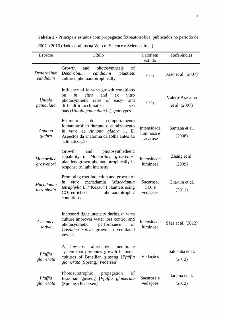
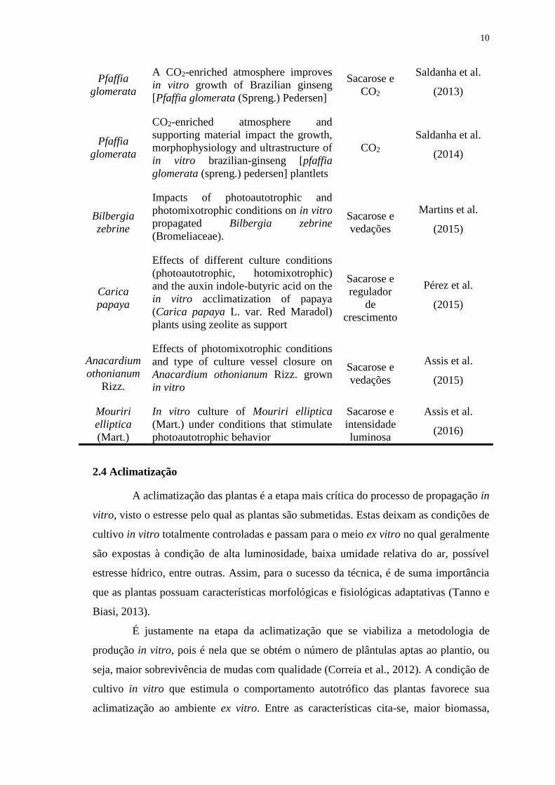
Tabela 2 - Principais estudos com propagação fotoautotrófica, publicados no período de
2007 a 2016 (dados obtidos na Web of Science e Sciencedirect). ................................... 9
CAPÍTULO II Dissimilarity between plants of Mouriri elliptica (Mart.) cultivated in
vitro and in situ through anatomic parameters
Table 1 - Summary of the analysis of variance informing the mean square, mean and
coefficient of variation (CV) of the anatomical features. ............................................... 42
Table 2 - Phenotypic correlation coefficients (rp) between micromorphometric features.
........................................................................................................................................ 42
Table 3 - Dissimilarity matrix obtained by the generalized Mahalanobis distance (D2)
between M. elliptica (Mart.) plantlets under different cultivation conditions in vitro and
in situ. .............................................................................................................................. 43
Table 4 - Relative importance (S.j) of micromorphometric features in the divergence
study of M. elliptica (Mart.) plants grown in situ and plantlets subjected to different in
vitro cultivation conditions. ............................................................................................ 45
CAPÍTULO III Alternative support materials to agar in the in vitro cultivation of
Mouriri elliptica (Mart.)
Table 1 - Physical characteristics of the alternative support materials used for in vitro
cultivation of M. elliptica (Mart.) plantlets. Total porosity (TP), available water (AW),
aeration space (AS), remaining water (RW), wet density (WD) and dry density (DD). 61

x
ÍNDICE DE FIGURAS
Página
Figure 1. Planta adulta de Mouriri elliptica (Mart.) in situ (A), frutos em maturação (B)
e sementes. Frutos maduros coletados em novembro de 2014, no Município de
Montividiu – GO, Latitude “17º 19.201”S, Longitude “51 33.500”W, Altitude 982 m. . 3
CAPÍTULO I. In vitro culture of Mouriri elliptica (Mart.) under conditions that
stimulate photoautotrophic behavior
Figure 1. Growth of Mouriri elliptica (Mart.) seedlings in culture medium
supplemented with sucrose and without sucrose at lights intensities diferentes. In vitro
culture for 45 days. Scale bar = 2 cm. ............................................................................ 21
Figure 2. Length of Mouriri elliptica (Mart.) seedlings in culture medium with and
without sucrose at lights intensities of 0, 50, 75, 100, and 150 µmol m-2s-1 for 45 days
of in vitro culture. *p < 0.05. .......................................................................................... 22
Figure 3. Number of leaves (A), number of shoots (B), total dry weight (C), and leaf
dry weight (D) of M. elliptica (Mart.) seedlings cultured in medium with and without
sucrose at lights intensities of 0, 50, 75, 100, and 150 µmol m-2s-1. *p < 0.05. ............. 23
Figure 4. Photomicrographs of Mouriri elliptica (Mart.) leaves in vitro in the absence
of light and the presence of sucrose. (a) A portion of the abaxial epidermis with
stomatal crypts (St Cr) and outside the stomatal crypt, (b) cross-section of the blade's
median region showing the cell arrangement in the adaxial epidermis (Ad Ep),
chlorophyll parenchyma (CP), abaxial epidermis (Ab Ep), and stomatal crypt (St Cr).
Scale bar = 100 µm ......................................................................................................... 26
Figure 5. Photomicrographs of Mouriri elliptica (Mart.) leaves in vitro, showing the
abaxial epidermis with stomatal crypts (St Cr). Scale bar = 100 µm. ............................ 27
Figure 6. Photomicrographs of cross-sections of the median region of M. elliptica
(Mart.) leaves in vitro, showing the cellular arrangement of the adaxial epidermis (Ad
Ep), palisade parenchyma (PP), spongy parenchyma (SP), abaxial epidermis (Ab Ep),
xi
and stomatal crypts (St Cr). *Polysaccharides accumulated within the cells, tissue
stained via the PAS method. Scale bar = 100 µm. .......................................................... 28
Figure 7. Adaxial epidermis thickness (A), abaxial epidermis thickness (B),
chlorophyllian parenchyma thickness (C), and stomatal crypt density (D) of M. elliptica
(Mart.) seedlings cultured in medium with and without sucrose at lights intensities 0,
50, 75, 100, and 150 µmol m-2s-1. *p < 0.05. .................................................................. 29
CAPÍTULO II Dissimilarity between plants of Mouriri elliptica (Mart.) cultivated in
vitro and in situ through anatomic parameters
Figure 1: UPGMA clustering of the 10 phenotypes of Mouriri elliptica (Mart.). Dashed
line: dendrogram cut indicating approximately 50% dissimilarity. CCC, cophenetic
correlation coefficient; CO, in situ plantlets; C1 to C9, plantlets grown in vitro, as
follows: C1, presence of sucrose and zero irradiance; C2, presence of sucrose and 50
µmol m-2s-1; C3, presence of sucrose and 75 µmol m-2s-1; C4, presence of sucrose and
100 µmol m-2s-1; C5, presence of sucrose and 150 µmol m-2s-1; C6, absence of sucrose
and 50 µmol m-2s-1; C7, absence of sucrose and 75 µmol m-2s-1; C8, absence of sucrose
and 100 µmol m-2s-1; and C9, absence of sucrose and 150 µmol m-2s-1 of irradiance. ... 44
Figure 2. Photomicrographs of an Mouriri elliptica (Mart.) leaf from a plant grown in
situ. Abaxial epidermis with stomatal crypts (St Cr) (a) and adaxial epidermis (b). Scale
bar = 100 µm. .................................................................................................................. 46
Figure 3. Cross sections of the middle region of the leaves Mouriri elliptica (Mart.) in
situ (a) and in vitro in the presence of sucrose and the absence of light (b). Toluidine
blue was used to stain the tissue. Ad Ep, adaxial epidermis; Ab Ep, abaxial epidermis;
PP, palisade parenchyma; SP, spongy parenchyma; St Cr, stomatal crypt; and CP,
chlorenchyma. The arrows indicate cells containing mucilage. Scale bars = 100 µm. .. 46
Figure 4: Cross sections of the middle region of the leaves Mouriri elliptica (Mart.). Ad
Ep, adaxial epidermis; Ab Ep, abaxial epidermis; PP, palisade parenchyma; SP, spongy
parenchyma; and St Cr, stomatal crypt. Scale bars = 100 µm. ....................................... 47
CAPÍTULO III Alternative support materials to agar in the in vitro cultivation of
Mouriri elliptica (Mart.)
Figure 1. In vitro cultivation of Mouriri elliptica (Mart.) plantlets in different culture
medium support materials for 45 days. Plantlet formed in different support materials in
the absence or presence of Naphthalene Acetic Acid - NAA. Scale bar: 2 cm. ............. 62
Figure 2. Length of plantlets (A), number of segments (B), number of leaves (C), and
total dry mass (D) of M. elliptica (Mart.) plantlets with 45 days of in vitro cultivation.
Means followed by the same uppercase letter do not differ between the presence and
absence of NAA, and means followed by the same lowercase letter do not differ among
support materials, according to the Tukey test, p < 0.05. ............................................... 63
xii
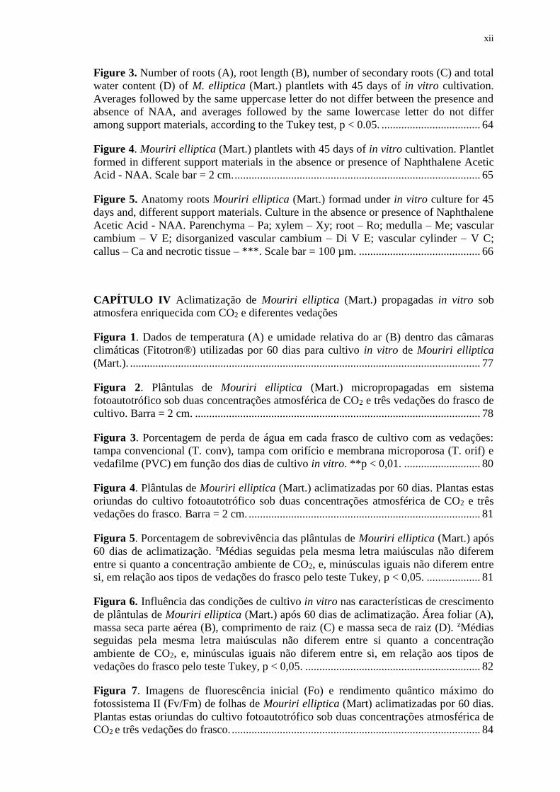
Figure 3. Number of roots (A), root length (B), number of secondary roots (C) and total
water content (D) of M. elliptica (Mart.) plantlets with 45 days of in vitro cultivation.
Averages followed by the same uppercase letter do not differ between the presence and
absence of NAA, and averages followed by the same lowercase letter do not differ
among support materials, according to the Tukey test, p < 0.05. ................................... 64
Figure 4. Mouriri elliptica (Mart.) plantlets with 45 days of in vitro cultivation. Plantlet
formed in different support materials in the absence or presence of Naphthalene Acetic
Acid - NAA. Scale bar = 2 cm. ....................................................................................... 65
Figure 5. Anatomy roots Mouriri elliptica (Mart.) formad under in vitro culture for 45
days and, different support materials. Culture in the absence or presence of Naphthalene
Acetic Acid - NAA. Parenchyma – Pa; xylem – Xy; root – Ro; medulla – Me; vascular
cambium – V E; disorganized vascular cambium – Di V E; vascular cylinder – V C;
callus – Ca and necrotic tissue – ***. Scale bar = 100 µm. ........................................... 66
CAPÍTULO IV Aclimatização de Mouriri elliptica (Mart.) propagadas in vitro sob
atmosfera enriquecida com CO2 e diferentes vedações
Figura 1. Dados de temperatura (A) e umidade relativa do ar (B) dentro das câmaras
climáticas (Fitotron®) utilizadas por 60 dias para cultivo in vitro de Mouriri elliptica
(Mart.). ............................................................................................................................ 77
Figura 2. Plântulas de Mouriri elliptica (Mart.) micropropagadas em sistema
fotoautotrófico sob duas concentrações atmosférica de CO2 e três vedações do frasco de
cultivo. Barra = 2 cm. ..................................................................................................... 78
Figura 3. Porcentagem de perda de água em cada frasco de cultivo com as vedações:
tampa convencional (T. conv), tampa com orifício e membrana microporosa (T. orif) e
vedafilme (PVC) em função dos dias de cultivo in vitro. **p < 0,01. ........................... 80
Figura 4. Plântulas de Mouriri elliptica (Mart.) aclimatizadas por 60 dias. Plantas estas
oriundas do cultivo fotoautotrófico sob duas concentrações atmosférica de CO2 e três
vedações do frasco. Barra = 2 cm. .................................................................................. 81
Figura 5. Porcentagem de sobrevivência das plântulas de Mouriri elliptica (Mart.) após
60 dias de aclimatização. zMédias seguidas pela mesma letra maiúsculas não diferem
entre si quanto a concentração ambiente de CO2, e, minúsculas iguais não diferem entre
si, em relação aos tipos de vedações do frasco pelo teste Tukey, p < 0,05. ................... 81
Figura 6. Influência das condições de cultivo in vitro nas características de crescimento
de plântulas de Mouriri elliptica (Mart.) após 60 dias de aclimatização. Área foliar (A),
massa seca parte aérea (B), comprimento de raiz (C) e massa seca de raiz (D). zMédias
seguidas pela mesma letra maiúsculas não diferem entre si quanto a concentração
ambiente de CO2, e, minúsculas iguais não diferem entre si, em relação aos tipos de
vedações do frasco pelo teste Tukey, p < 0,05. .............................................................. 82
Figura 7. Imagens de fluorescência inicial (Fo) e rendimento quântico máximo do
fotossistema II (Fv/Fm) de folhas de Mouriri elliptica (Mart) aclimatizadas por 60 dias.
Plantas estas oriundas do cultivo fotoautotrófico sob duas concentrações atmosférica de
CO2 e três vedações do frasco. ........................................................................................ 84
xiii
Figura 8. Índice de dissipação não fotoquímica – Y (NPQ) (A), densidade de cripta
estomática (B) e área de abertura da cripta estomática (C) de plântulas de Mouriri
elliptica (Mart.) após 60 dias de aclimatização em resposta as diferentes condições de
cultivo in vitro. zMédias seguidas pela mesma letra não diferem entre si pelo teste
Tukey, p < 0,05................................................................................................................85
Figura 9. Superfície abaxial das folhas de Mouriri elliptica (Mart.) após 60 dias de
aclimatização. Plantas oriundas do cultivo fotoautotrófico sob duas concentrações
atmosférica de CO2 e três vedações do frasco.................................................................86
xiv
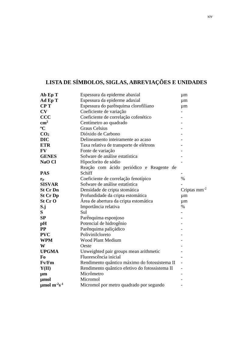
LISTA DE SÍMBOLOS, SIGLAS, ABREVIAÇÕES E UNIDADES
Ab Ep T Espessura da epiderme abaxial µm
Ad Ep T Espessura da epiderme adaxial µm
CP T Espessura do parênquima clorofiliano µm
CV Coeficiente de variação -
CCC Coeficiente de correlação cofenético -
cm2 Centímetro ao quadrado -
ºC Graus Celsius -
CO2 Dióxido de Carbono -
DIC Delineamento inteiramente ao acaso -
ETR Taxa relativa de transporte de elétrons -
FV Fonte de variação -
GENES Sofware de análise estatística -
NaO Cl Hipoclorito de sódio -
PAS
Reação com ácido periódico e Reagente de
Schiff -
rp Coeficiente de correlação fenotípico %
SISVAR Sofware de análise estatística -
St Cr Dn Densidade de cripta stomática Criptas mm-2
St Cr Dp Profundidade da cripta estomática µm
St Cr O Área de abertura da cripta estomática µm
S.j Importância relativa %
S Sul -
SP Parênquima esponjoso -
pH Potencial de hidrogênio -
PP Parênquima paliçádico -
PVC Polivinilcloreto -
WPM Wood Plant Medium -
W Oeste -
UPGMA Unweighted pair groups mean arithmetic -
Fo Fluorescência inicial -
Fv/Fm Rendimento quântico máximo do fotossistema II -
Y(II) Rendimento quântico efetivo do fotossistema II -
µm Micrômetro -
µmol Micromol -
µmol m-2s-1 Micromol por metro quadrado por segundo -
xv
RESUMO
ASSIS, ELISVANE SILVA. Instituto Federal de Educação, Ciência e Tecnologia
Goiano – IF Goiano - Campus Rio Verde. Dezembro de 2016. Cultura in vitro de
Mouriri elliptica (Mart.) sob condições fotomixotróficas: estudos anatômicos,
fisiológicos e de crescimento. Orientador: Dr. Fabiano Guimarães Silva, Coorientador:
Dr. Aurélio Rubio Neto.
A planta croada (Mouriri elliptica Mart.), é frutífera nativa no domínio do cerrado com
potencialidade para uso alimentício e medicinal. Há carência de estudos para espécie na
área de propagação, visto ser esta etapa, importante no processo de domesticação da
espécie. Assim, objetivou-se com este trabalho avaliar o crescimento e as características
anatômicas e fisiológicas de M. elliptica (Mart.) sob condições de cultivo in vitro
fotomixotróficas. O meio de cultivo utilizado em todos os ensaios foi o Wood Plant
Medium. No primeiro capítulo, avaliou-se o crescimento e características anatômicas
foliares de plântulas de croada cultivadas em diferentes irradiâncias (0, 50, 75, 100 e
150 µmol m-2s-1) e meio de cultivo com e sem sacarose. No segundo capítulo, estudou-
se a dissimilaridade de plântulas de croada obtidas via cultivo in vitro fotomixotrófico e
fotoautotrófico com plantas in situ a partir de características anatômicas. No terceiro
capítulo, analisou-se o crescimento e desenvolvimento de croada in vitro utilizando
materiais de suporte (vermiculita, fibra de jerivá e bagaço de cana) em comparação com
ágar, avaliou-se também a interação destes suportes com o regulador de crescimento
ácido naftaleno acético no enraizamento das plântulas. Por último, estudou-se a
influência do cultivo in vitro fotomixotrófico sob atmosfera enriquecida com CO2 e
diferentes vedações na aclimatização de plântulas de croada. Em todos os ensaios
experimentais utilizou-se delineamento inteiramente ao acaso, com esquema fatorial
quando necessário. Na ausência de sacarose, notou-se capacidade de regeneração das
plântulas de croada apenas com irradiância acima de 50 µmol m-2s-1, sendo observado
xvi
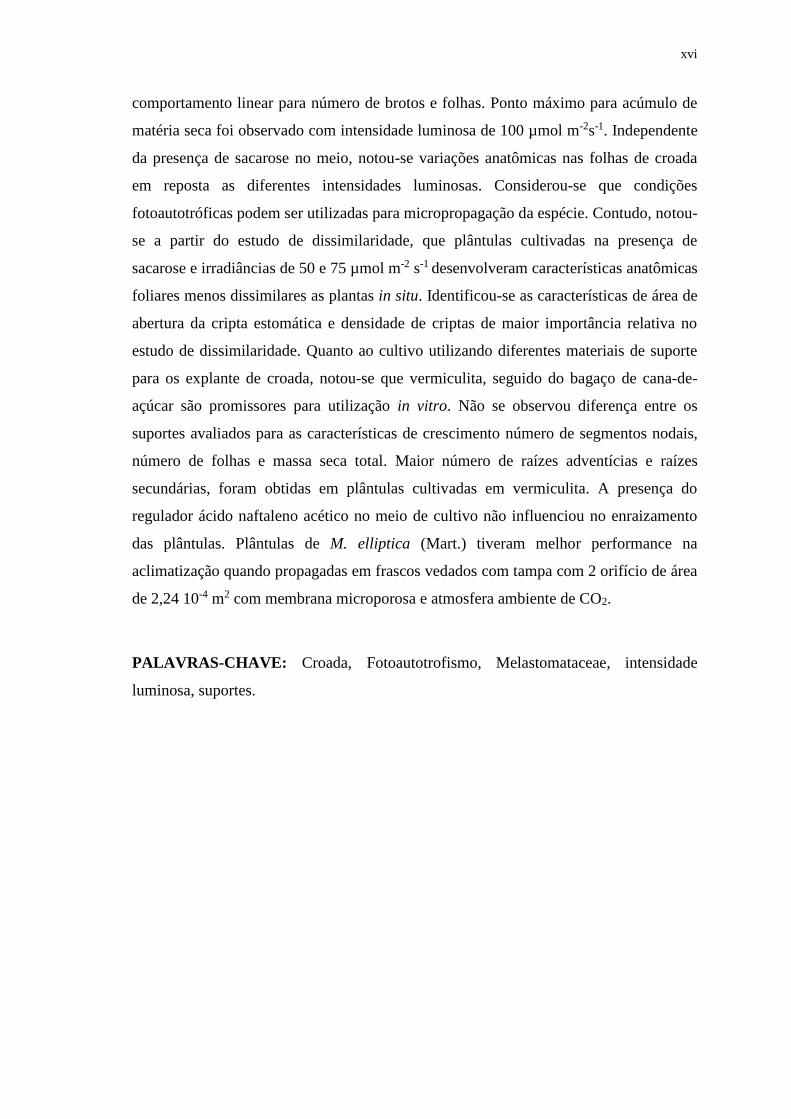
comportamento linear para número de brotos e folhas. Ponto máximo para acúmulo de
matéria seca foi observado com intensidade luminosa de 100 µmol m-2s-1. Independente
da presença de sacarose no meio, notou-se variações anatômicas nas folhas de croada
em reposta as diferentes intensidades luminosas. Considerou-se que condições
fotoautotróficas podem ser utilizadas para micropropagação da espécie. Contudo, notou-
se a partir do estudo de dissimilaridade, que plântulas cultivadas na presença de
sacarose e irradiâncias de 50 e 75 µmol m-2 s-1 desenvolveram características anatômicas
foliares menos dissimilares as plantas in situ. Identificou-se as características de área de
abertura da cripta estomática e densidade de criptas de maior importância relativa no
estudo de dissimilaridade. Quanto ao cultivo utilizando diferentes materiais de suporte
para os explante de croada, notou-se que vermiculita, seguido do bagaço de cana-de-
açúcar são promissores para utilização in vitro. Não se observou diferença entre os
suportes avaliados para as características de crescimento número de segmentos nodais,
número de folhas e massa seca total. Maior número de raízes adventícias e raízes
secundárias, foram obtidas em plântulas cultivadas em vermiculita. A presença do
regulador ácido naftaleno acético no meio de cultivo não influenciou no enraizamento
das plântulas. Plântulas de M. elliptica (Mart.) tiveram melhor performance na
aclimatização quando propagadas em frascos vedados com tampa com 2 orifício de área
de 2,24 10-4 m2 com membrana microporosa e atmosfera ambiente de CO2.
PALAVRAS-CHAVE: Croada, Fotoautotrofismo, Melastomataceae, intensidade
luminosa, suportes.
xvii
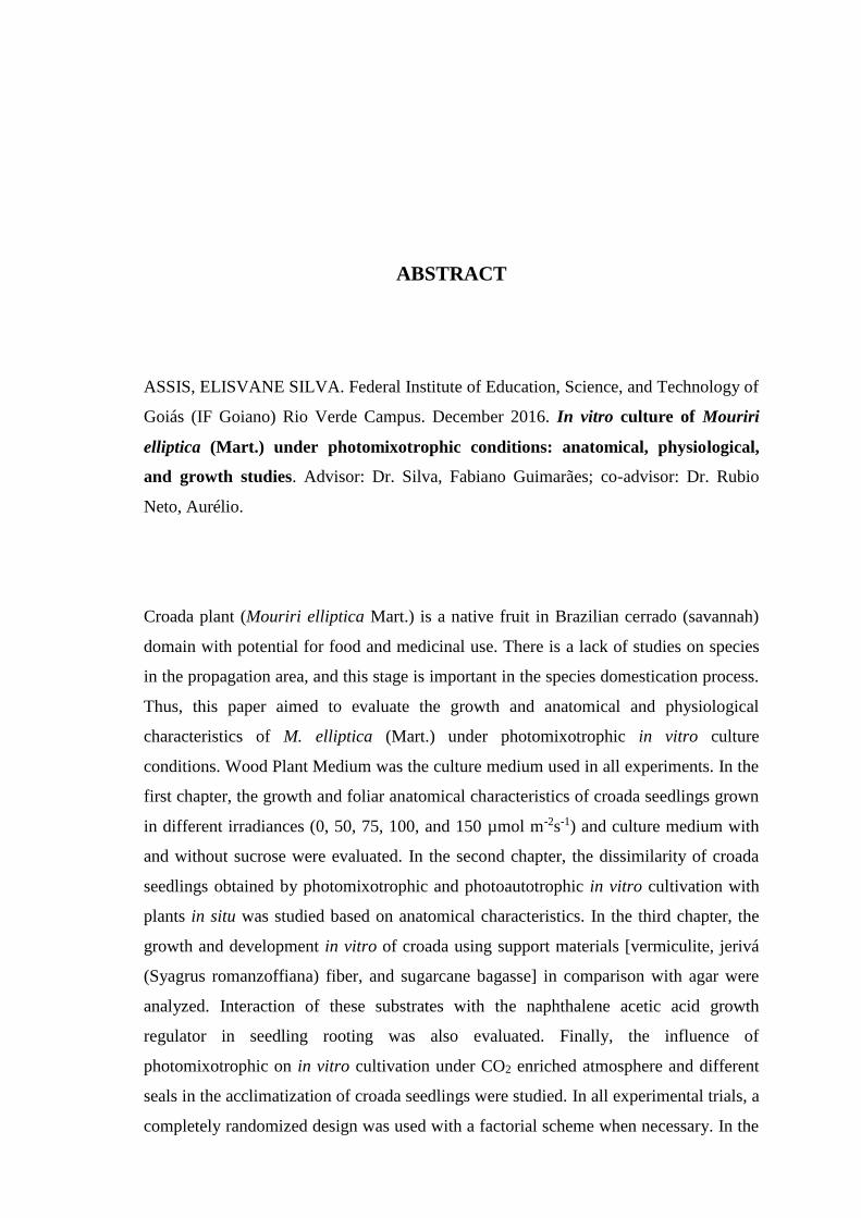
ABSTRACT
ASSIS, ELISVANE SILVA. Federal Institute of Education, Science, and Technology of
Goiás (IF Goiano) Rio Verde Campus. December 2016. In vitro culture of Mouriri
elliptica (Mart.) under photomixotrophic conditions: anatomical, physiological,
and growth studies. Advisor: Dr. Silva, Fabiano Guimarães; co-advisor: Dr. Rubio
Neto, Aurélio.
Croada plant (Mouriri elliptica Mart.) is a native fruit in Brazilian cerrado (savannah)
domain with potential for food and medicinal use. There is a lack of studies on species
in the propagation area, and this stage is important in the species domestication process.
Thus, this paper aimed to evaluate the growth and anatomical and physiological
characteristics of M. elliptica (Mart.) under photomixotrophic in vitro culture
conditions. Wood Plant Medium was the culture medium used in all experiments. In the
first chapter, the growth and foliar anatomical characteristics of croada seedlings grown
in different irradiances (0, 50, 75, 100, and 150 µmol m-2s-1) and culture medium with
and without sucrose were evaluated. In the second chapter, the dissimilarity of croada
seedlings obtained by photomixotrophic and photoautotrophic in vitro cultivation with
plants in situ was studied based on anatomical characteristics. In the third chapter, the
growth and development in vitro of croada using support materials [vermiculite, jerivá
(Syagrus romanzoffiana) fiber, and sugarcane bagasse] in comparison with agar were
analyzed. Interaction of these substrates with the naphthalene acetic acid growth
regulator in seedling rooting was also evaluated. Finally, the influence of
photomixotrophic on in vitro cultivation under CO2 enriched atmosphere and different
seals in the acclimatization of croada seedlings were studied. In all experimental trials, a
completely randomized design was used with a factorial scheme when necessary. In the
xviii
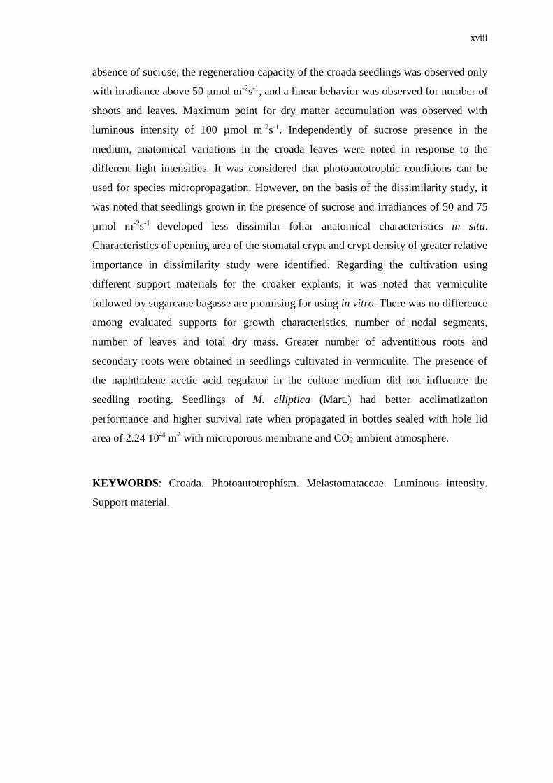
absence of sucrose, the regeneration capacity of the croada seedlings was observed only
with irradiance above 50 µmol m-2s-1, and a linear behavior was observed for number of
shoots and leaves. Maximum point for dry matter accumulation was observed with
luminous intensity of 100 µmol m-2s-1. Independently of sucrose presence in the
medium, anatomical variations in the croada leaves were noted in response to the
different light intensities. It was considered that photoautotrophic conditions can be
used for species micropropagation. However, on the basis of the dissimilarity study, it
was noted that seedlings grown in the presence of sucrose and irradiances of 50 and 75
µmol m-2s-1 developed less dissimilar foliar anatomical characteristics in situ.
Characteristics of opening area of the stomatal crypt and crypt density of greater relative
importance in dissimilarity study were identified. Regarding the cultivation using
different support materials for the croaker explants, it was noted that vermiculite
followed by sugarcane bagasse are promising for using in vitro. There was no difference
among evaluated supports for growth characteristics, number of nodal segments,
number of leaves and total dry mass. Greater number of adventitious roots and
secondary roots were obtained in seedlings cultivated in vermiculite. The presence of
the naphthalene acetic acid regulator in the culture medium did not influence the
seedling rooting. Seedlings of M. elliptica (Mart.) had better acclimatization
performance and higher survival rate when propagated in bottles sealed with hole lid
area of 2.24 10-4 m2 with microporous membrane and CO2 ambient atmosphere.
KEYWORDS: Croada. Photoautotrophism. Melastomataceae. Luminous intensity.
Support material.
1
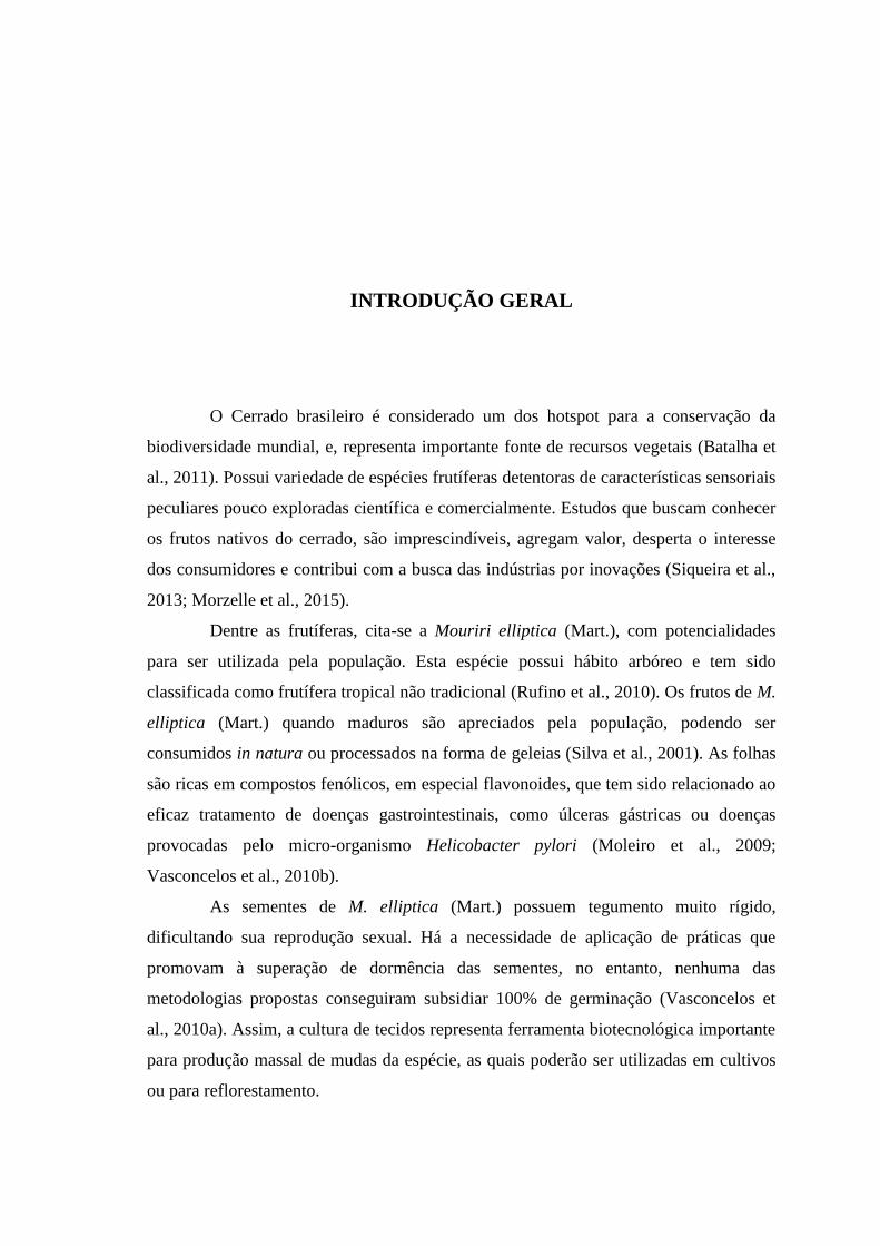
INTRODUÇÃO GERAL
O Cerrado brasileiro é considerado um dos hotspot para a conservação da
biodiversidade mundial, e, representa importante fonte de recursos vegetais (Batalha et
al., 2011). Possui variedade de espécies frutíferas detentoras de características sensoriais
peculiares pouco exploradas científica e comercialmente. Estudos que buscam conhecer
os frutos nativos do cerrado, são imprescindíveis, agregam valor, desperta o interesse
dos consumidores e contribui com a busca das indústrias por inovações (Siqueira et al.,
2013; Morzelle et al., 2015).
Dentre as frutíferas, cita-se a Mouriri elliptica (Mart.), com potencialidades
para ser utilizada pela população. Esta espécie possui hábito arbóreo e tem sido
classificada como frutífera tropical não tradicional (Rufino et al., 2010). Os frutos de M.
elliptica (Mart.) quando maduros são apreciados pela população, podendo ser
consumidos in natura ou processados na forma de geleias (Silva et al., 2001). As folhas
são ricas em compostos fenólicos, em especial flavonoides, que tem sido relacionado ao
eficaz tratamento de doenças gastrointestinais, como úlceras gástricas ou doenças
provocadas pelo micro-organismo Helicobacter pylori (Moleiro et al., 2009;
Vasconcelos et al., 2010b).
As sementes de M. elliptica (Mart.) possuem tegumento muito rígido,
dificultando sua reprodução sexual. Há a necessidade de aplicação de práticas que
promovam à superação de dormência das sementes, no entanto, nenhuma das
metodologias propostas conseguiram subsidiar 100% de germinação (Vasconcelos et
al., 2010a). Assim, a cultura de tecidos representa ferramenta biotecnológica importante
para produção massal de mudas da espécie, as quais poderão ser utilizadas em cultivos
ou para reflorestamento.
2
Na cultura de tecidos, estudos são desenvolvidos com finalidade de melhorar as
qualidades morfofisiológicas das plantas, focados principalmente em fatores físicos e
químicos do ambiente (Chandra et al., 2010). Torna-se primordial a adequação da
luminosidade, temperatura, umidade e fotoperíodo do ambiente de crescimento das
plantas (Torres et al., 1998).
Cada espécie responde de forma dissimilar a uma condição de cultivo imposta,
assim, além dos fatores físicos citados, é importante a otimização do meio de cultivo,
ajustando as concentrações de sais, sacarose (Assis et al., 2012; Cabral et al., 2013;
Assis et al., 2015). É destacado também a suplementação do meio de cultivo com
regulador de crescimento na indução de brotos e raízes (Brondani et al., 2012; Hossain e
Urbi, 2016; Aina et al., 2015).
Destaca-se que o sucesso da propagação in vitro depende da capacidade de
transferência das plantas das condições in vitro para as ex vitro com alta taxa de
sobrevivência e com qualidade (Chandra et al., 2010; Correia et al., 2012). Nesta
perspectiva, tem sido investigado técnicas de propagação in vitro que estimulam o
desenvolvimento autotrófico das plantas (Xiao et al., 2011), beneficiando assim a
aclimatização das mesmas (Xiao e Kozai, 2006; Zhang et al., 2009; Cha-um et al., 2011;
Iarema et al., 2012; Saldanha et al., 2014).
2. REVISÃO DE LITERATURA
2.1 Características gerais da espécie Mouriri elliptica (Mart.)
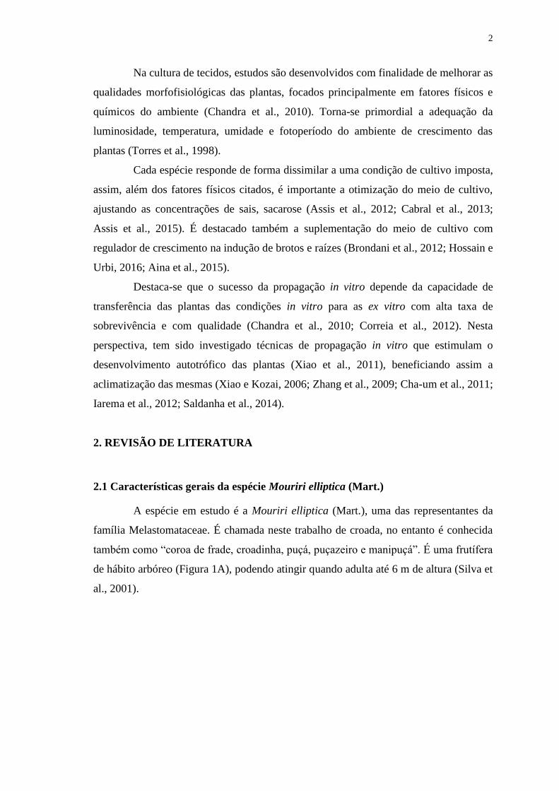
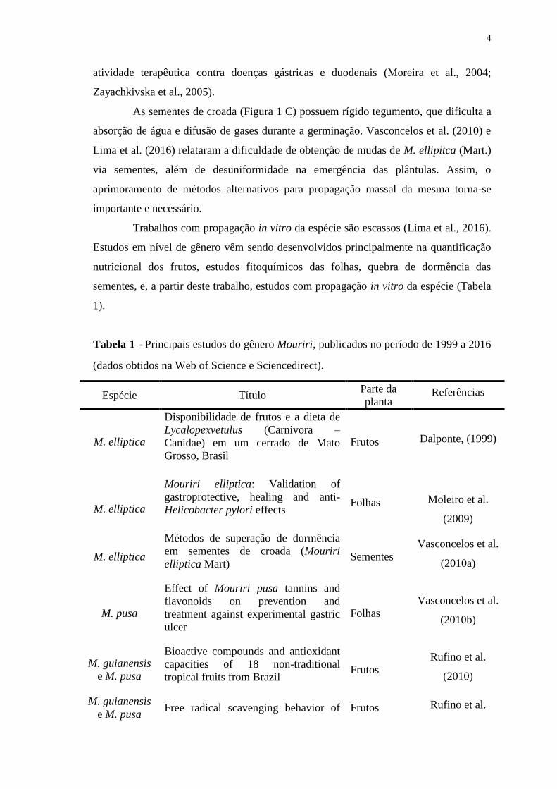
A espécie em estudo é a Mouriri elliptica (Mart.), uma das representantes da
família Melastomataceae. É chamada neste trabalho de croada, no entanto é conhecida
também como “coroa de frade, croadinha, puçá, puçazeiro e manipuçá”. É uma frutífera
de hábito arbóreo (Figura 1A), podendo atingir quando adulta até 6 m de altura (Silva et
al., 2001).
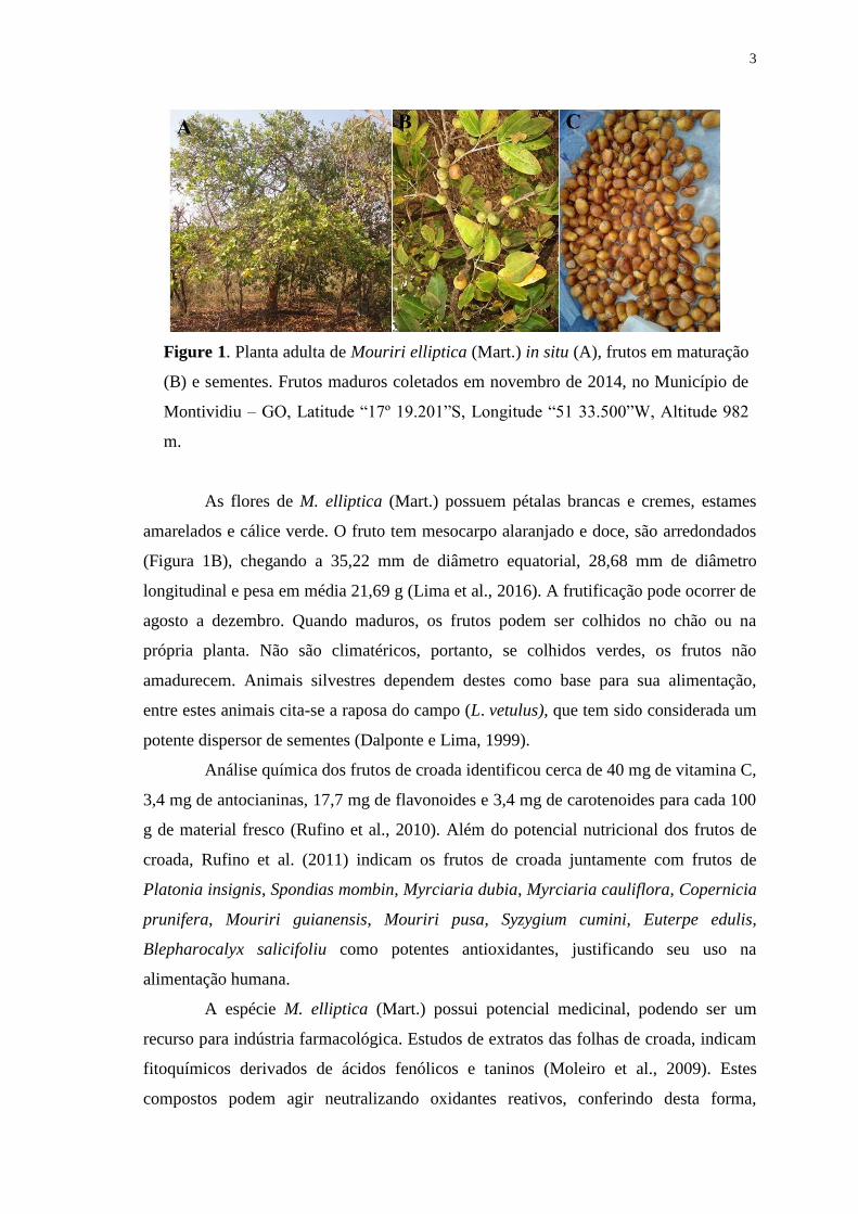
3
Figure 1. Planta adulta de Mouriri elliptica (Mart.) in situ (A), frutos em maturação
(B) e sementes. Frutos maduros coletados em novembro de 2014, no Município de
Montividiu – GO, Latitude “17º 19.201”S, Longitude “51 33.500”W, Altitude 982
m.
As flores de M. elliptica (Mart.) possuem pétalas brancas e cremes, estames
amarelados e cálice verde. O fruto tem mesocarpo alaranjado e doce, são arredondados
(Figura 1B), chegando a 35,22 mm de diâmetro equatorial, 28,68 mm de diâmetro
longitudinal e pesa em média 21,69 g (Lima et al., 2016). A frutificação pode ocorrer de
agosto a dezembro. Quando maduros, os frutos podem ser colhidos no chão ou na
própria planta. Não são climatéricos, portanto, se colhidos verdes, os frutos não
amadurecem. Animais silvestres dependem destes como base para sua alimentação,
entre estes animais cita-se a raposa do campo (L. vetulus), que tem sido considerada um
potente dispersor de sementes (Dalponte e Lima, 1999).
Análise química dos frutos de croada identificou cerca de 40 mg de vitamina C,
3,4 mg de antocianinas, 17,7 mg de flavonoides e 3,4 mg de carotenoides para cada 100
g de material fresco (Rufino et al., 2010). Além do potencial nutricional dos frutos de
croada, Rufino et al. (2011) indicam os frutos de croada juntamente com frutos de
Platonia insignis, Spondias mombin, Myrciaria dubia, Myrciaria cauliflora, Copernicia
prunifera, Mouriri guianensis, Mouriri pusa, Syzygium cumini, Euterpe edulis,
Blepharocalyx salicifoliu como potentes antioxidantes, justificando seu uso na
alimentação humana.
A espécie M. elliptica (Mart.) possui potencial medicinal, podendo ser um
recurso para indústria farmacológica. Estudos de extratos das folhas de croada, indicam
fitoquímicos derivados de ácidos fenólicos e taninos (Moleiro et al., 2009). Estes
compostos podem agir neutralizando oxidantes reativos, conferindo desta forma,
4
atividade terapêutica contra doenças gástricas e duodenais (Moreira et al., 2004;
Zayachkivska et al., 2005).
As sementes de croada (Figura 1 C) possuem rígido tegumento, que dificulta a
absorção de água e difusão de gases durante a germinação. Vasconcelos et al. (2010) e
Lima et al. (2016) relataram a dificuldade de obtenção de mudas de M. ellipitca (Mart.)
via sementes, além de desuniformidade na emergência das plântulas. Assim, o
aprimoramento de métodos alternativos para propagação massal da mesma torna-se
importante e necessário.
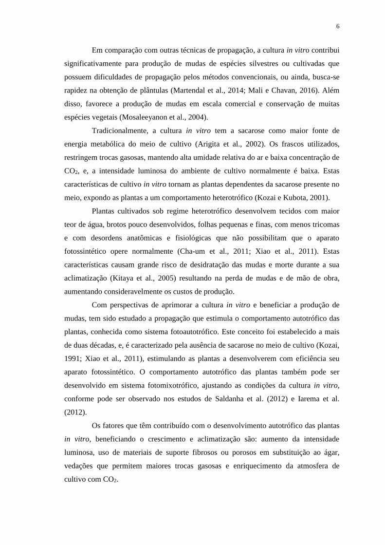
Trabalhos com propagação in vitro da espécie são escassos (Lima et al., 2016).
Estudos em nível de gênero vêm sendo desenvolvidos principalmente na quantificação
nutricional dos frutos, estudos fitoquímicos das folhas, quebra de dormência das
sementes, e, a partir deste trabalho, estudos com propagação in vitro da espécie (Tabela
1).
Tabela 1 - Principais estudos do gênero Mouriri, publicados no período de 1999 a 2016
(dados obtidos na Web of Science e Sciencedirect).
Espécie Título Parte da
planta Referências
M. elliptica
Disponibilidade de frutos e a dieta de
Lycalopexvetulus (Carnivora –
Canidae) em um cerrado de Mato
Grosso, Brasil
Frutos Dalponte, (1999)
M. elliptica
Mouriri elliptica: Validation of
gastroprotective, healing and anti-
Helicobacter pylori effects Folhas
Moleiro et al.
(2009)
M. elliptica
Métodos de superação de dormência
em sementes de croada (Mouriri
elliptica Mart) Sementes
Vasconcelos et al.
(2010a)
M. pusa
Effect of Mouriri pusa tannins and
flavonoids on prevention and
treatment against experimental gastric
ulcer
Folhas
Vasconcelos et al.
(2010b)
M. guianensis
e M. pusa
Bioactive compounds and antioxidant
capacities of 18 non-traditional
tropical fruits from Brazil Frutos
Rufino et al.
(2010)
M. guianensis
e M. pusa Free radical scavenging behavior of Frutos Rufino et al.

5
tem exotic tropical fruits extracts (2011)
M. pusa Absence of mutagenicity of plants
used to treat gastrointestinal disorders Folhas
Santos et al.
(2013)
M. pusa
Comparison of Brazilian Plants Used
to Treat Gastritis on the Oxidative
Burst of Helicobacter pylori-
Stimulated Neutrophil
Folhas
Bonacorsi et al.
(2013)
M. elliptica
Germination and emergence of
Mouriri elliptica Mart., a rare
medicinal fruit tree native to the
Brazilian Cerrado biom
Sementes Lima et al. (2016)
M. elliptica
(Mart.)
In vitro culture of Mouriri elliptica
(Mart.) under conditions that stimulate
photoautotrophic behavior
Sacarose e
intensidade
luminosa
Assis et al. (2016)
M. elliptica
(Mart.)
Dissimilarity between Mouriri
elliptica (Mart.) plants cultivated in
vitro and in situ through anatomic
parameters
Plantas in
situ e in
vitro
Assis et al. (2016)
Na literatura, cita-se ocorrência natural de plantas do gênero Mouriri no
domínio do Cerrado, nos estados de Mato Grosso, Mato Grosso do Sul e Goiás (Silva et
al., 2001), no entanto, vem perdendo seu habitat. De acordo com Pereira e Pasquaeto,
(2011) o Cerrado sofre pressão antrópica, principalmente pela atividade pecuária,
exploração extrativista e expansão da agricultura. As frutíferas nativas são fundamentais
neste ecossistema, porém, mesmo com a crescente valorização e o emprego dos
produtos regionais, os estudos científicos com essas espécies são limitados, carecendo
de investimentos (Damiani et al., 2011).
2.2 Cultura in vitro: Propagação heterotrófica, fotoautotrófica e fotomixotrófica
Na cultura in vitro, objetiva-se produção de plantas, crescimento e
multiplicação de células, tecidos e órgãos em meio de cultura específico, semissólido ou
líquido sob condições ambientais controladas e, na ausência de patógenos (Thorpe,
2007; Chandra et al., 2010). Fontes de carbono, nutrientes (Macro e Micro) e energia
encontram-se disponíveis no meio de cultivo, e estes, subsidiam o crescimento das
plantas in vitro (Brondani et al., 2012).
6
Em comparação com outras técnicas de propagação, a cultura in vitro contribui
significativamente para produção de mudas de espécies silvestres ou cultivadas que
possuem dificuldades de propagação pelos métodos convencionais, ou ainda, busca-se
rapidez na obtenção de plântulas (Martendal et al., 2014; Mali e Chavan, 2016). Além
disso, favorece a produção de mudas em escala comercial e conservação de muitas
espécies vegetais (Mosaleeyanon et al., 2004).
Tradicionalmente, a cultura in vitro tem a sacarose como maior fonte de
energia metabólica do meio de cultivo (Arigita et al., 2002). Os frascos utilizados,
restringem trocas gasosas, mantendo alta umidade relativa do ar e baixa concentração de
CO2, e, a intensidade luminosa do ambiente de cultivo normalmente é baixa. Estas
características de cultivo in vitro tornam as plantas dependentes da sacarose presente no
meio, expondo as plantas a um comportamento heterotrófico (Kozai e Kubota, 2001).
Plantas cultivados sob regime heterotrófico desenvolvem tecidos com maior
teor de água, brotos pouco desenvolvidos, folhas pequenas e finas, com menos tricomas
e com desordens anatômicas e fisiológicas que não possibilitam que o aparato
fotossintético opere normalmente (Cha-um et al., 2011; Xiao et al., 2011). Estas
características causam grande risco de desidratação das mudas e morte durante a sua
aclimatização (Kitaya et al., 2005) resultando na perda de mudas e de mão de obra,
aumentando consideravelmente os custos de produção.
Com perspectivas de aprimorar a cultura in vitro e beneficiar a produção de
mudas, tem sido estudado a propagação que estimula o comportamento autotrófico das
plantas, conhecida como sistema fotoautotrófico. Este conceito foi estabelecido a mais
de duas décadas, e, é caracterizado pela ausência de sacarose no meio de cultivo (Kozai,
1991; Xiao et al., 2011), estimulando as plantas a desenvolverem com eficiência seu
aparato fotossintético. O comportamento autotrófico das plantas também pode ser
desenvolvido em sistema fotomixotrófico, ajustando as condições da cultura in vitro,
conforme pode ser observado nos estudos de Saldanha et al. (2012) e Iarema et al.
(2012).
Os fatores que têm contribuído com o desenvolvimento autotrófico das plantas
in vitro, beneficiando o crescimento e aclimatização são: aumento da intensidade
luminosa, uso de materiais de suporte fibrosos ou porosos em substituição ao ágar,
vedações que permitem maiores trocas gasosas e enriquecimento da atmosfera de
cultivo com CO2.
7
2.3 Intensidade luminosa, suportes alternativos, vedações e CO2 na cultura in vitro
A intensidade e a qualidade da luz são fatores ambientais fundamentais que
interferem diretamente na morfologia, fisiologia e metabolismo das plantas (Fukuda et
al., 2008; Li e Kubota, 2009, Shin et al., 2013). A dependência das plantas à luz é um
processo complexo que envolve a ação combinada de fotorreceptores que controlam
estádios variados no desenvolvimento (Braga et al., 2009).
Em sala de crescimento, a intensidade luminosa fornecida para as culturas in
vitro normalmente são baixas (< 50 µmol m-2s-1). Entretanto, quando objetiva-se induzir
o autotrofismo das plantas in vitro, pode ser necessário aumentar a intensidade
luminosa, especialmente quando se pretende utilizar meio de cultivo desprovido de
sacarose (Kozai e Nguyen, 2003). Assim, sob cultivo fotoautotrófico a intensidade
luminosa de 100 µmol m-2s-1 foi ideal para Momordica grosvenori (Zangh et al., 2009).
Já, para o híbrido Doritaenopsis os autores obtiveram melhor crescimento com
intensidade luminosa de 120 µmol m-2s-1 e para M. elliptica (Mart.) maior crescimento
foi com 150 µmol m-2s-1 de luz (Assis et al., 2016).
Entre os tipos de suportes utilizados in vitro, o ágar é o agente geleificante do
meio de cultura tradicional (Thorpe et al., 2008). Entretanto, devido o uso em
abundância, torna-se o ingrediente mais caro do meio de cultivo, além disso, as
plântulas têm desenvolvido raízes mal formadas e geralmente não possuindo pelos
absorventes. Tais características podem dificultar a aclimatização e sobrevivência das
plântulas às condições ex vitro (Braga et al., 2011). Diante dessa problemática, vêm
sendo testado suportes alternativos, em especial porosos, acrescido de meio de cultura
líquido (Mohan et al., 2005).
Suportes porosos aumentam a condutividade hidráulica, favorece a absorção de
nutrientes do meio de cultura e proporciona melhor aeração de tecidos e raízes do que
seria no cultivo com ágar, melhorando potencialmente o vigor da planta e, assim, a taxa
de sobrevivência no processo de aclimatização (Kozai, 2010; Xiao et al., 2011;
Saldanha et al., 2014). Entre os tipos de suportes que podem ser utilizados in vitro, cita-
se vermiculita (Xiao e Kozai, 2006; Cha-um et al. 2011), combinação de vermiculita e
fibra de celulose (Florialite®) (Xiao e Kozai, 2006), bagaço de cana-de-açúcar (Mohan
et al., 2005), entre outros, como a fibra de Jerivá utilizada neste trabalho.

8
Para Pfaffia glomerata (Spreng) Pedersen, a retirada da sacarose do meio de
cultura não afetou o crescimento das plântulas quando se utilizou frascos com vedações
possuindo membranas permeáveis aos gases (Iarema et al., 2012). Neste trabalho o
objetivo de promover o comportamento autotrófico de P. glomerata foi alcançado, pois
plantas desenvolveram morfologia e características fisiológicas necessárias para o
processo de aclimatização.
Comportamento fotoautotrófico também foi observado em Annona glabra L.,
II. (Santana et al., 2008), no qual compararam o crescimento das plantas em meio sem
sacarose e com tampas permeáveis a gases ao invés de vedação fechadas. Neste, os
autores observaram que as raízes foram maiores e as plantas desenvolveram maior
número de raízes secundárias. Observou-se também que as espessuras dos tecidos
foliares possuíam semelhanças com plantas cultivadas ex vitro, e confere maior
sustentação e plasticidade. Essa capacidade de alterar a estrutura das folhas em resposta
à aeração dos frascos, revela adaptação da planta.
Quando se aborda umidade relativa do ar dentro do frasco de cultivo, maiores
trocas gasosas gasosas com o ambiente externo, pode aumentar significativamente a
taxa de transpiração da planta, e consequentemente, a absorção de água e de nutrientes
(Aitken-Christie et al., 1995). Ao mesmo tempo, a redução da umidade relativa reduz a
incidência de hiperhidricidade nas plantas, favorece a formação de cutícula nas folhas e
o funcionamento normal dos estômatos, aumentando a tolerância ao estresse hídrico na
aclimatização (Zobayed et al., 2001).
Carboidratos exógenos são fornecidos à cultura in vitro devido à concentração
de CO2 no interior do recipiente ser baixa limitando a fotossíntese (Kozai, 2010; Xiao et
al., 2011). Esta limitação fotossintética pode ser revertida quando permite maior aeração
no interior dos recipientes de cultivo, conforme resultados de Iarema et al. (2012) ou
ainda, proporciona enriquecimento da atmosfera de cultivo com Dióxido de Carbono
(CO2), fornecendo substrato para fotossíntese (Saldanha et al., 2014).
Trabalhos com propagação in vitro fotoautotrófica têm sido desenvolvidos para
um variado número de espécies, potencializando a obtenção de mudas e beneficiando o
setor produtivo. Estudos com espécies frutíferas do cerrado são excassos, assim, este
trabalho de pesquisa teve como base os estudos citados na tabela 2. Nestes, os principais
fatores em estudo são CO2, intensidade luminosa, concentrações de sacarose e vedações.
9
Tabela 2 - Principais estudos com propagação fotoautotrófica, publicados no período de
2007 a 2016 (dados obtidos na Web of Science e Sciencedirect).
Espécie Título Fator em
estudo
Referências
Dendrobium
candidum
Growth and photosynthesis of
Dendrobium candidum plantlets
cultured photoautotrophically CO2 Xiao et al. (2007)
Uniola
paniculata
Influence of in vitro growth conditions
on in vitro and ex vitro
photosynthetic rates of easy- and
difficult-to-acclimatize sea
oats (Uniola paniculata L.) genotypes
CO2
Valero-Aracama
et al. (2007)
Annona
glabra
Estímulo do comportamento
fotoautotrófico durante o enraizamento
in vitro de Annona glabra l., II.
Aspectos da anatomia da folha antes da
aclimatização
Intensidade
luminosa e
sacarose
Santana et al.
(2008)
Momordica
grosvenori
Growth and photosynthethetic
capability of Momordica grosvenori
plantlets grown photoautotrophically in
response to light intensity
Intensidade
luminosa
Zhang et al.
(2009)
Macadamia
tetraphylla
Promoting root induction and growth of
in vitro macadamia (Macadamia
tetraphylla L. ‘‘Keaau’’) plantlets using
CO2-enriched photoautotrophic
conditions.
Sacarose,
CO2 e
vedações
Cha-um et al.
(2011)
Castanea
sativa
Increased light intensity during in vitro
culture improves water loss control and
photosynthetic performance of
Castanea sativa grown in ventilated
vessels
Intensidade
luminosa Sáez et al. (2012)
Pfaffia
glomerata
A low-cost alternative membrane
system that promotes growth in nodal
cultures of Brazilian ginseng [Pfaffia
glomerata (Spreng.) Pedersen].
Vedações
Saldanha et al.
(2012)
Pfaffia
glomerata
Photoautotrophic propagation of
Brazilian ginseng [Pfaffia glomerata
(Spreng.) Pedersen]
Sacarose e
vedações
Iarema et al.
(2012)
10
Pfaffia
glomerata
A CO2-enriched atmosphere improves
in vitro growth of Brazilian ginseng
[Pfaffia glomerata (Spreng.) Pedersen]
Sacarose e
CO2
Saldanha et al.
(2013)
Pfaffia
glomerata
CO2-enriched atmosphere and
supporting material impact the growth,
morphophysiology and ultrastructure of
in vitro brazilian-ginseng [pfaffia
glomerata (spreng.) pedersen] plantlets
CO2
Saldanha et al.
(2014)
Bilbergia
zebrine
Impacts of photoautotrophic and
photomixotrophic conditions on in vitro
propagated Bilbergia zebrine
(Bromeliaceae).
Sacarose e
vedações
Martins et al.
(2015)
Carica
papaya
Effects of different culture conditions
(photoautotrophic, hotomixotrophic)
and the auxin indole-butyric acid on the
in vitro acclimatization of papaya
(Carica papaya L. var. Red Maradol)
plants using zeolite as support
Sacarose e
regulador
de
crescimento
Pérez et al.
(2015)
Anacardium
othonianum
Rizz.
Effects of photomixotrophic conditions
and type of culture vessel closure on
Anacardium othonianum Rizz. grown
in vitro
Sacarose e
vedações
Assis et al.
(2015)
Mouriri
elliptica
(Mart.)
In vitro culture of Mouriri elliptica
(Mart.) under conditions that stimulate
photoautotrophic behavior
Sacarose e
intensidade
luminosa
Assis et al.
(2016)
2.4 Aclimatização
A aclimatização das plantas é a etapa mais crítica do processo de propagação in
vitro, visto o estresse pelo qual as plantas são submetidas. Estas deixam as condições de
cultivo in vitro totalmente controladas e passam para o meio ex vitro no qual geralmente
são expostas à condição de alta luminosidade, baixa umidade relativa do ar, possível
estresse hídrico, entre outras. Assim, para o sucesso da técnica, é de suma importância
que as plantas possuam características morfológicas e fisiológicas adaptativas (Tanno e
Biasi, 2013).
É justamente na etapa da aclimatização que se viabiliza a metodologia de
produção in vitro, pois é nela que se obtém o número de plântulas aptas ao plantio, ou
seja, maior sobrevivência de mudas com qualidade (Correia et al., 2012). A condição de
cultivo in vitro que estimula o comportamento autotrófico das plantas favorece sua
aclimatização ao ambiente ex vitro. Entre as características cita-se, maior biomassa,
11
maior número de raízes adventícias, aumento de raízes secundárias, presença de pelos
radiculares, estômatos funcionais, maior teor de clorofila, altas taxas fotossintéticas,
incremento na espessura dos tecidos foliares, tecidos lignificados e maior depósito
cutícula (Santana et al., 2008; Zhang et al., 2009; Xiao et al., 2011; Shin et al., 2013;
Saldanha et al., 2014).
Nota-se os esforços dos pesquisadores em aprimorar a cultura de tecidos, em
especial com o desenvolvimento de técnicas fotoautotróficas beneficiando o sistema de
produção de mudas e contribuindo com informações úteis para o desenvolvimento de
novos trabalhos. É um desafio o estabelecimento de protocolos de propagação de mudas
em larga escala, e com sucesso na etapa final, que é aclimatização das plantas as
condições ex vitro. Assim, os trabalhos desenvolvidos nesta pesquisa serão de suma
importância para área de cultura de tecidos, além de valorização e início de um processo
de domesticação da espécie M. elliptica (Mart.).
3. REFERÊNCIAS BIBLIOGRÁFICAS
Aina OO, Quesenberry KH, Gallo M (2015) Culture vessel and auxin treatments
affect in vitro rooting and ex vitro survival of six arachis
paraguariensis genotypes. Sci Hort. 183(1):167–171.
Aitken-Christie J, Kozai T, Smith Mal (eds) (1995) Automation and environmental
control in plant tissue culture. Kluwer Academic Publishers, Dordrecht, 1995, p
574.
Arigita L, González A, Tamés RS (2002) Influence of CO2 and sucrose on
photosynthesis and transpiration of Actinidia deliciosa explants cultured in vitro.
Physiol Plant. 115(1):166-173.
Assis ES, Rubio Neto A, Lima LR, Silva FG, Rosa M, Vasconcelos Filho SC (2016). In
vitro culture of Mouriri elliptica (Mart.) under conditions that stimulate
photoautotrophic behavior. Aust J Crop Science. 10(2):229-236.
Assis KC, Silva FG, Pereira FD, Vasconcelos-Filho SC, et al. (2015). Effects of
photomixotrophic conditions and type of culture vessel closure on Anacardium
othonianum Rizz. grown in vitro. Acta Hort. 1083(4): 553-564.
Assis KC, Pereira FD, Cabral JSR, Silva FG, Silva JW, Santos SC (2012) In vitro
cultivation of Anacardium othonianum Rizz.: effects of salt concentration and
culture medium volume. Acta Sci Agronomy (Impresso). 34(1):77-83.
Batalha M A (2011) O cerrado não é um bioma. Biota Neotropica. 1(1):21-24.
12
Bonacorsi C, Fonseca L, Raddi MSG, Kitagawa RR, Vilegas W (2013) Comparison of
Brazilian Plants Used to Treat Gastritis on the Oxidative Burst of Helicobacter
pylori-Stimulated Neutrophil. Evidence-Based Compl Alt Medicine. ID 851621,
8p.
Braga FT, Pasqual M, Castro EM, Rafael GC (2011) Características morfofisiológicas
de abacaxizeiro ‘gomo de mel’ enraizado in vitro sob luz natural e substrato
vermiculita. Rev Bras Frutic. 33(2):551-557.
Braga FT, Pasqual M, Castro EM, Dignart SL, Biagiottis G, Porto JM (2009) Qualidade
de luz no cultivo in vitro de Dendranthema grandiflorum cv. rage:
características morfofisiológicas. Ciênc Agrotec. 33(2):502-508.
Brondani GE, Wit Ondas HW, Baccarin FJB, Gonçalves AN, Almeida M (2012)
Micropropagation of Eucalyptus benthamii to form a clonal microgarden. In
Vitro Cel Dev Biol – Plant. 48(5):478-487.
Cabral JSR, Alberto PS, Pereira FD, Souchie EL, Silva FG (2013) In vitro cultivation of
Hancornia speciosa Gomes: The physical constitution of the culture médium,
sucrose concentrations and growth conditions. Plant Tissue Cult Biotec. 23(2):
177-187.
Chandra S, Bandopadhyay R, Kumar V, Chandra R. (2010) Acclimatization of tissue
cultured plantlets: from laboratory to land. Biotech Letters. 32(9):1199-1205.
Cha-um S, Chanseetis C, Chitakovid W, Pichakum A, Supaibulwatana K (2011)
Promoting root induction and growth of in vitro macadamia (Macadamia
tetraphylla L. ‘‘Keaau’’) plantlets using CO2-enriched photoautotrophic
conditions. P Cell Tissue Org Cult. 106(2):435-444.
Correia D, Araujo JDM, Nascimento EHSDO, Silva Júnior JMTD, Bessa MC (2012)
Otimização da Produção de Mudas de Cattleya labiata: Efeito da Sacarose no
Crescimento In Vitro e na Aclimatização. Circular Técnica 38, Embrapa.
Dalponte JC, Lima ES (1999) Disponibilidade de frutos e a dieta de Lycalopexvetulus
(Carnivora – Canidae) em um cerrado de Mato Grosso, Brasil. Rev Bras Bot.
22(2):325-332.
Damiani C, Boas V, Barros EV, Asquieri ER, Lage ME, Oliveira RA, Silva FA, Pinto
DM, Rodrigues LJ, Silva EP, Paula NRF (2011). Characterization of fruits from
the savanna: Araça (Psidium guinnensis Sw.) and Marolo (Annona crassiflora
Mart.). Food Sci Technol. 31(3):723-729.
13
Fukuda N, Fujita M, Ohta Y, Sase S, Nishimura S, Ezura H (2008) Directional blue
light irradiation triggers epidermal cell elongation of abaxial side resulting in
inhibition of leaf epinasty in geranium under red light condition. Sci Hort.
115(2):176-182.
Hossain S, Urbi Z (2016) Effect of naphthalene acetic acid on the adventitious rooting
in shoot cuttings of Andrographis paniculata (Burm.f.) Wall. ex Nees: an
important therapeutical herb. Int J of Agronomy. ID 1617543, 6 p, 2016.
Iarema L, Cruz ACF, Saldanha CW, Dias, LLC, Vieira RF, Oliveira EJ, Otoni WC
(2012) Photoautotrophic propagation of Brazilian ginseng [Pfaffia glomerata
(Spreng.) Pedersen]. P Cell Tiss Organ Cult. 110(2):227–238.
Kitaya Y, Ohmura Y, Kubota C, Kozai T (2005) Manipulation of the culture
environment on in vitro air movement and its impact on plantlets photosynthesis.
P Cell Tiss Organ Cult. 83(3):251–257.
Kozai T (2010) Photoautotrophic micropropagation—environmental control for
promoting photosynthesis. Prop Ornam Plants. 10(1):188–204.
Kozai T (1991) Photoautotrophic micropropagation. In Vitro Cell
Dev Biol. 27(1):47–51.
Kozai T, Kubota C (2001) Development a photoautotrophic micropropagation system
for woody plants. J Plant Res. 114(4):525–537
Kozai T, Nguyen QT (2003) Photoautotrophic micropropagation of woody and tropical
plants. In: JAIN, S.M.; ISHII, K. Micropropagation of woody trees and fruits.
Dordrecht: Kluwer Academic, p.757-781.
Li Q, Kubota C (2009) Effects of supplemental light quality on growth and
phytochemicals of baby leaf lettuce. Env Exp Botany. 67(1):59-64.
Lima LR, Rubio Neto A, Pereira FD, Silva FG, Menezes CCE, Santana JG (2016)
Germination and emergence of Mouriri elliptica Mart., a rare medicinal fruit tree
native to the Brazilian Cerrado biom. African J Agric Research. 11(5): 400-406.
Mali AM, Chavan N S (2016) In vitro rapid regeneration through direct organogenesis
and ex-vitro establishment of Cucumis trigonus Roxb. An underutilized
pharmaceutically important cucurbit. Ind Crops and Products. 83(1):48–54.
Martendal CO, Bernardino MM, Pereira FD, Silva FG, Menezes CCE, Santana JG
(2014). In vitro multiplication of nodal segments of “Murici” (Byrsonima
cydoniifolia A. Juss.): the use of growth regulators and photoautotrophic
stimulation. J Agric Technology. 10(3): 665-678.
14
Martins JPR, Verdoodt V, Paqual M, Proft M (2015) Impacts of photoautotrophic and
photomixotrophic conditions on in vitro propagated Bilbergia zebrine
(Bromeliaceae). P Cell Tissue Org Cult. 123(1): 121-132.
Mohan R, Chui EA, Biasi LA, Soccol CR (2005) Alternative In vitro Propagation: Use
of Sugarcane Bagasse as a low cost support material during rooting stage of
Strawberry Cv. Dover. Braz Arch of Biol and Technology. 48(1):37-42.
Moleiro FC, Andreo MA, Santos RC, Moraes TM, Rodrigues CM, Carli CB, Lopes FC,
Pellizzon CH, Carlos IZ, Bauab TM, Vilegas W, Hiruma-Lima CA (2009)
Mouriri elliptica: Validation of gastroprotective, healing and anti-
Helicobasterpylorieffects. J Ethnopharmacology. 123(3):359-368.
Moreira AJ, Fraga C, Alonso M, Collado PS, Zetller C, Marroni C, Marroni N,
González-Gallego J (2004) Quercetin prevents oxidative stress and NF-kappaB
activation in gastric mucosa of portal hypertensive rats. Bioch Pharmacology.
68(10):1939–1946.
Morzelle MC, Bachiega P, Souza EC, Vilas Boas EVB, Lamounier ML (2015)
Caracterizaçao química e física de frutos de curriola, gabiroba e murici
provenientes do Cerrado brasileiro. Rev Bras Frutic. 37(1):96-103.
Mosaleeyanon K, Chan-Um S, Kirmanee C (2004) Enhanced growth and
photosynthesis of rain tree (Samanea saman Merr.) plantlets in vitro under a
CO2-enriched condition with decreased sucrose concentrations in the medium.
Sci Hort. 103(1):51–63.
Pereira ME, Pasqualeto A (2011) Desenvolvimento sustentável com ênfase em
frutíferas do Cerrado. Studos. 38(2):333-363.
Pérez LP, Montesinos YP, Olmedo JG, Sánchez RR, Montenegro ON, Rodrigues RB,
Ribalta OH, Escriba RCR, Daniel D, Gómez-K (2015) Effects of different
culture conditions (photoautotrophic, photomixotrophic) and the auxin indole-
butyric acid on the in vitro acclimatization of papaya (Carica papaya L. var. Red
Maradol) plants using zeolite as support. A J Biotechnology. 14(35): 2622-2635.
Rufino MSM, Alves RE, Brito ES, Pérez-Jiménez J, Saura-Calixto F, Mancini-Filho J
(2010) Bioactive compounds and antioxidant capacities of 18 non-traditional
tropical fruits from Brazil. Food Chemistry. 121(4):996–1002.
Rufino MSM, Alves RE, Fernandes FAN, Brito ES (2011) Free radical scavenging
behavior of tem exotic tropical fruits extracts. Food Res Int. 44(7):2072-2075.
15
Sáez PL, Bravo LA, Latsague MI, Sánchez ME, Ríos DG (2012) Increased light
intensity during in vitro culture improves water loss control and photosynthetic
performance of Castanea sativa grown in ventilated vessels. Sci Hort. 138(1):7–
16.
Saldanha, C.W.; Otoni, C.G.; Azevedo, J.L.F.; Dias, L.L.C.; Rego, M.M.; Otoni, W.C.
(2012) A low-cost alternative membrane system that promotes growth in nodal
cultures of Brazilian ginseng [Pfaffia glomerata (Spreng.) Pedersen]. P Cell Tiss
Organ Cult. 110(3):413-422.
Saldanha CW, Otoni CG, Notini MM, Kuki KN, Cruz ACF, Rubio Neto A, Dias LC,
Otoni C (2013) A CO2-enriched atmosphere improves in vitro growth of
Brazilian ginseng [Pfaffia glomerata (Spreng.) Pedersen]. In Vitro Cell Dev Biol
- Plant. 49(4):433–444.
Saldanha CW, Otoni CG, Rocha DI, Cavatte PC, Detmann KSC, Tanaka FAO Dias
LLC, Da Mata FM, Otoni WC (2014) CO2-enriched atmosphere and supporting
material impact the growth, morphophysiology and ultrastructure of in vitro
brazilian-ginseng [pfaffia glomerata (spreng.) pedersen] plantlets. P Cell Tiss
Organ Cult. 118(1):87-99.
Santana JRF, Paiva R, Resende KS, Castro EM, Pereira FD, Oliveira LM (2008)
Estímulo do comportamento fotoautotrófico durante o enraizamento in vitro de
Annona glabra l., II. Aspectos da anatomia da folha antes da aclimatização.
Ciênc Agrotec. 32(2):640-644.
Santos FV, Andreo M, Nasser ALM, Moreira LM, Vilegas W, Cólus IMS, Varanda EA
(2013) Absence of mutagenicity of plants used to trat gastrointestinal disorders.
Arch Biol Sci. 65(1): 191-195.
Shin K, Park S, Paek K (2013) Sugar metabolism, photosynthesis and growth of in vitro
plantlets of Doritaenopsis under controlled microenvironmental conditions. In
Vitro Cell Dev Biol – Plant. 49(4), 445-454.
Silva DB, Silva AS, Junqueira NTV, Andrade LRM (2001) Frutas do Cerrado. Brasília:
Embrapa Informação Tecnológica. 178p.
Siqueira EMA, Rosa FR, Fustinoni AM, Sant´Ana LP, Arruda SF (2013) Brazilian
savanna fruits contain higher bioactive compounds content and higher
antioxidant activity relative to the conventional red delicious apple. Plos One.
8(8):1-7.
16
Tanno GN, Biasi LA (2013) Aclimatização de videiras micropropagadas em frascos
com e sem vedação e diferentes concentrações de sacarose. Rev Acadêmica:
Ciên Agrárias e Ambientais. 11(1):19-25.
Thorpe TA (2008) History of plant tissue culture. Mol Biotechnol.
37(1):169–180.
Torres AC, Caldas LS, Buso JA (1998) Cultura de tecidos e transformação genética de
plantas. Embrapa-SPI, Brasília. 864 p.
Valero-Aracama C, Wilson SB, Kane ME, Philman NL (2007) Influence of in vitro
growth conditions on in vitro ande x vitro photosynthetic rates of easy and
diffifult-to-acclimatize sea oats. In Vitro Cell Dev Biol – Plant. 43(2):237-246.
Vasconcelos JM, Cardoso TV, Sales JF, Silva FG, Vasconcelos Filho SC, Santana JG
(2010a) Métodos de superação de dormência em sementes de croada (Mouriri
elliptica Mart). Ciênc Agrotec. 34(5):1199-1204.
Vasconcelos PCP, Andreo MA, Vilegas W, Hiruma-Lima CA, Pellizzona CH (2010b)
Effect of Mouriri pusa tannins and flavonoids on prevention and treatment
against experimental gastric ulcer. J Ethnopharmacology. 131(1):146–153.
Xiao Y, Kozai T (2006) In vitro multiplication of statice plantlets using sugar-free
media. Sci Hort. 109(1):71–77.
Xiao Y, Zhang Y, Dang K, Wang D (2007) Growth and photosynthesis of Dendrobium
candidum plantlets cultured photoautotrophically. Propagat Orn Plants. 7(2):89–
96
Xiao Y, Niu G, Kozai T (2011) Development and application of photoautotrophic
micropropagation plant system. P Cell Tiss Organ Cult. 105(1):149–158.
Zayachkivska OS, Konturek SJ, Drozdowicz D, Konturek PC, Brzozowski T,
Ghegotsky MR (2005) Gastroprotective effects of flavonoids in plant extracts. J
Physiol Pharmacol. 56(1):219–231.
Zhang M, Zhao D, Ma Z, Li X, Xiao Y (2009) Growth and photosynthethetic capability
of Momordica grosvenori plantlets grown photoautotrophically in response to
light intensity. Hort Sci. 44(3):757–763.
Zobayed SMA, Afreen F, Kozai T (2001) Physiology of eucalyptus plantlets cultured
photoautotrophically under forced ventilation. In Vitro Cell Dev Biol - Plant.
37(6):807–813.
17
OBJETIVOS
Geral
Avaliar as características anatômicas, fisiológicas e de crescimento em plântulas
de Mouriri elliptica (Mart.) sob condições fotomixotróficas de cultivo in vitro.
Específicos
Estimular o comportamento autotrófico de plântulas de croada utilizando
diferentes irradiâncias em combinação com meio de cultivo com e sem
sacarose;
Avaliar a dissimilaridade entre plantas de M. elliptica (Mart.) cultivadas in
vitro e in situ a partir de parâmetros anatômicos com auxílio de técnicas de
estatística multivariada.
Verificar se o tipo de suporte mais poroso ou fibroso influencia no
crescimento inicial de plântulas de croada, em especial na formação de raíz;
Aclimatizar mudas de croada obtidas no processo de micropropagação
fotoautotrófica ou fotomixotrófica.
18
CAPÍTULO I
(Normas de acordo com a revista Australian Journal of Croop Science. Artigo publicado
em fevereiro de 2016, v. 10, n. 2, p. 229-236.
In vitro culture of Mouriri elliptica (Mart.) under conditions that stimulate
photoautotrophic behavior
Abstract
Micropropagation has been efficiently used to mass-produce seedlings of
species that are difficult to multiply via conventional methods. Thus, the present study
aimed to analyze the in vitro culture of Mouriri elliptica (Mart.) seedlings under
conditions that stimulate photoautotrophic behavior. Nodal segments were grown in
50% salt Wood Plant Medium in the absence and presence of sucrose and subjected to
differents lights intensities (0, 50, 75, 100, and 150 µmol m-2s-1). Evaluations were
performed after 60 days of culture, considering growth and morphoanatomic
characteristics. There was an exponential increase in the number of shoots and leaves in
seedlings cultured in the absence of sucrose with increasing light intensity.
Additionally, greater total and leaf dry weights were recorded in seedlings cultured in
sucrose-supplemented medium at an light intensity close to 100 µmol m-2s-1.
Morphoanatomic changes were observed in leaves at differents lights intensities, both in
the presence and absence of sucrose. As the light intensity increased, the
supplementation of the medium with sucrose became unnecessary. Thus,
photoautotrophic conditions can be used for micropropagation of the species.
19
Keywords: autotrophic micropropagation; "croada"; light intensity; morphoanatomy;
stomatal crypt; sucrose.
1.1 Introduction
Mouriri elliptica (Mart.) belongs to the family Melastomataceae. It is a fruit
tree that occurs naturally in several brazilian states, being very common in the Goiás
Cerrado (savannah), and it has been classified as a non-traditional tropical fruit (Rufino
et al., 2010). It is popularly known as "croada", "croadinha", "coroa de frade", "puçá",
"puçazeiro", or "manipuçá". When ripe, its fruit are sweet and rich in antioxidant
compounds such as vitamin C, anthocyanins, carotenoids and flavonoids and can be
eaten raw by humans or processed into jellies (Silva et al., 2001; Rufino et al., 2010;
Rufino et al., 2011).
The plant also has medicinal potential, the application of M. pusa and M.
elliptica leaf extracts in rodents is an alternative treatment for acute ulcers, with these
extracts exhibiting a gastroprotective effect and promoting healing. The extracts also
have an anti-Helicobacter pylori effect, which is a microorganism that causes serious
gastrointestinal diseases. These effects have been attributed to phenolic constituents, in
the form of flavonoids and tannins identified in the plants' leaves (Moleiro et al., 2009;
Vasconcelos et al., 2010b). Leaf extracts from this species show no toxicity in treated
animals, which is an important factor in its pharmacological applicability (Moleiro et
al., 2009).
There are currently no studies on "croada" micropropagation, despite the
plant's various uses. It is known that its seeds have a rigid coat, hindering its sexual
reproduction, as reported by Vasconcelos et al. (2010a), requiring the application of
practices that promote overcoming dormancy. Therefore, propagation of this important
species by seeds may not meet seedling demands. In vitro propagation provides a
greater chance of producing seedlings that could be used for crops or reforestation.
Traditionally, explants are cultured in flasks that restrict gas exchange, with a
high relative humidity, high ethylene concentration, low CO2 concentration, low-density
flow of photosynthetically active photons, and the use of sucrose as the main metabolic
energy source. This system may cause anatomical and physiological disorders in the
seedlings, hindering the normal function of the photosynthetic apparatus (Xiao et al.,
2011), as observed in the in vitro culture of Billbergia zebrina (Herbert), in which the
supply of sucrose reduces the quantity of photosynthetic pigments (Martins et al., 2015).
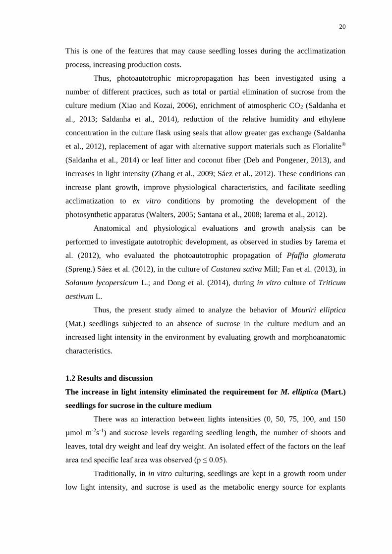
20
This is one of the features that may cause seedling losses during the acclimatization
process, increasing production costs.
Thus, photoautotrophic micropropagation has been investigated using a
number of different practices, such as total or partial elimination of sucrose from the
culture medium (Xiao and Kozai, 2006), enrichment of atmospheric CO2 (Saldanha et
al., 2013; Saldanha et al., 2014), reduction of the relative humidity and ethylene
concentration in the culture flask using seals that allow greater gas exchange (Saldanha
et al., 2012), replacement of agar with alternative support materials such as Florialite®
(Saldanha et al., 2014) or leaf litter and coconut fiber (Deb and Pongener, 2013), and
increases in light intensity (Zhang et al., 2009; Sáez et al., 2012). These conditions can
increase plant growth, improve physiological characteristics, and facilitate seedling
acclimatization to ex vitro conditions by promoting the development of the
photosynthetic apparatus (Walters, 2005; Santana et al., 2008; Iarema et al., 2012).
Anatomical and physiological evaluations and growth analysis can be
performed to investigate autotrophic development, as observed in studies by Iarema et
al. (2012), who evaluated the photoautotrophic propagation of Pfaffia glomerata
(Spreng.) Sáez et al. (2012), in the culture of Castanea sativa Mill; Fan et al. (2013), in
Solanum lycopersicum L.; and Dong et al. (2014), during in vitro culture of Triticum
aestivum L.
Thus, the present study aimed to analyze the behavior of Mouriri elliptica
(Mat.) seedlings subjected to an absence of sucrose in the culture medium and an
increased light intensity in the environment by evaluating growth and morphoanatomic
characteristics.
1.2 Results and discussion
The increase in light intensity eliminated the requirement for M. elliptica (Mart.)
seedlings for sucrose in the culture medium
There was an interaction between lights intensities (0, 50, 75, 100, and 150
µmol m-2s-1) and sucrose levels regarding seedling length, the number of shoots and
leaves, total dry weight and leaf dry weight. An isolated effect of the factors on the leaf
area and specific leaf area was observed (p ≤ 0.05).
Traditionally, in in vitro culturing, seedlings are kept in a growth room under
low light intensity, and sucrose is used as the metabolic energy source for explants

21
(Zhang et al., 2009; Arigita et al., 2002). Fig 1 shows Mouriri elliptica (Mart.) seedling
growth in culture medium supplemented with sucrose (A-E) and without sucrose (F-J).
Figure 1. Growth of Mouriri elliptica (Mart.) seedlings in culture medium supplemented
with sucrose and without sucrose at lights intensities diferentes. In vitro culture for 45
days. Scale bar = 2 cm.
A greater seedling length was observed in the absence of light and in the
presence of sucrose (Figs 1 and 2), demonstrating etiolation characteristics, most likely
due to the seedlings' sensitivity to endogenous auxin (George, 1993), considering that
sucrose availability in the culture medium induced the M. elliptica (Mart.) seedlings to
metabolize auxin, a result previously observed in Arabidopsis (Sairanen et al., 2012).
Etiolation of these seedlings in vitro can be advantageous to the multiplication
process, allowing their nodal segments to be used as explants, as observed in a study by
Suzuki et al. (2004), and to obtain new shoots of crop species, as in pineapple plants
(Moreira et al., 2003). However, etiolation is a characteristic related to inefficiency of
the photosynthetic apparatus (Solymosi and Schoefs, 2010) and susceptibility to
photoinhibition (Long, 1994), which may compromise the acclimatization process.
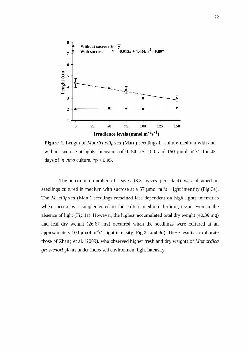
22
Irradiance levels (mmol m-2s-1)
y-0.013x + 4.434; r
2= 0.80*
0 25 50 75 100 125 150
Len
gh
t (c
m)
1
2
3
4
5
6
7
8Without sucrose Y=
With sucrose Y=
Figure 2. Length of Mouriri elliptica (Mart.) seedlings in culture medium with and
without sucrose at lights intensities of 0, 50, 75, 100, and 150 µmol m-2s-1 for 45
days of in vitro culture. *p < 0.05.
The maximum number of leaves (3.8 leaves per plant) was obtained in
seedlings cultured in medium with sucrose at a 67 µmol m-2s-1 light intensity (Fig 3a).
The M. elliptica (Mart.) seedlings remained less dependent on high lights intensities
when sucrose was supplemented in the culture medium, forming tissue even in the
absence of light (Fig 1a). However, the highest accumulated total dry weight (40.36 mg)
and leaf dry weight (26.67 mg) occurred when the seedlings were cultured at an
approximately 100 µmol m-2s-1 light intensity (Fig 3c and 3d). These results corroborate
those of Zhang et al. (2009), who observed higher fresh and dry weights of Momordica
grosvenori plants under increased environment light intensity.
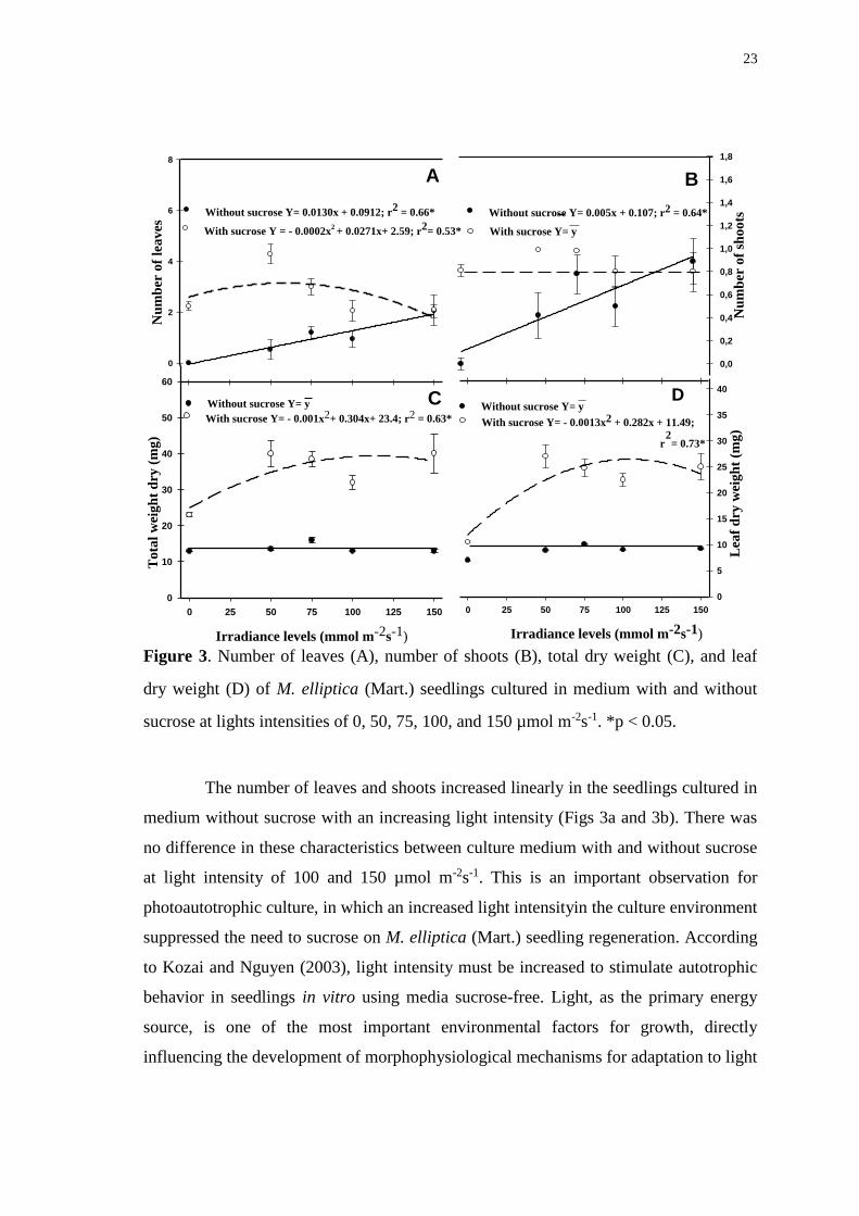
23
2D Graph 6
Irradiance levels (mmol m-2s-1)
0 25 50 75 100 125 150
Tota
l w
eig
ht
dry
(m
g)
0
10
20
30
40
50
60
Nu
mb
er o
f le
aves
0
2
4
6
8
A
CWithout sucrose Y= y
With sucrose Y= - 0.001x2+ 0.304x+ 23.4; r2 = 0.63*
0 25 50 75 100 125 150
Lea
f d
ry w
eig
ht
(mg
)
0
5
10
15
20
25
30
35
40
Irradiance levels (mmol m-2s-1)
B
D
Nu
mb
er o
f sh
oo
ts
0,0
0,2
0,4
0,6
0,8
1,0
1,2
1,4
1,6
1,8
B
Without sucrose Y= y
With sucrose Y= - 0.0013x2 + 0.282x + 11.49;
r2
= 0.73*
Without sucrose Y= 0.0130x + 0.0912; r2
= 0.66*
With sucrose Y = - 0.0002x2 +
0.0271x+ 2.59; r
2= 0.53*
Without sucrose Y= 0.005x + 0.107; r2 = 0.64*
With sucrose Y= y
Figure 3. Number of leaves (A), number of shoots (B), total dry weight (C), and leaf
dry weight (D) of M. elliptica (Mart.) seedlings cultured in medium with and without
sucrose at lights intensities of 0, 50, 75, 100, and 150 µmol m-2s-1. *p < 0.05.
The number of leaves and shoots increased linearly in the seedlings cultured in
medium without sucrose with an increasing light intensity (Figs 3a and 3b). There was
no difference in these characteristics between culture medium with and without sucrose
at light intensity of 100 and 150 µmol m-2s-1. This is an important observation for
photoautotrophic culture, in which an increased light intensityin the culture environment
suppressed the need to sucrose on M. elliptica (Mart.) seedling regeneration. According
to Kozai and Nguyen (2003), light intensity must be increased to stimulate autotrophic
behavior in seedlings in vitro using media sucrose-free. Light, as the primary energy
source, is one of the most important environmental factors for growth, directly
influencing the development of morphophysiological mechanisms for adaptation to light
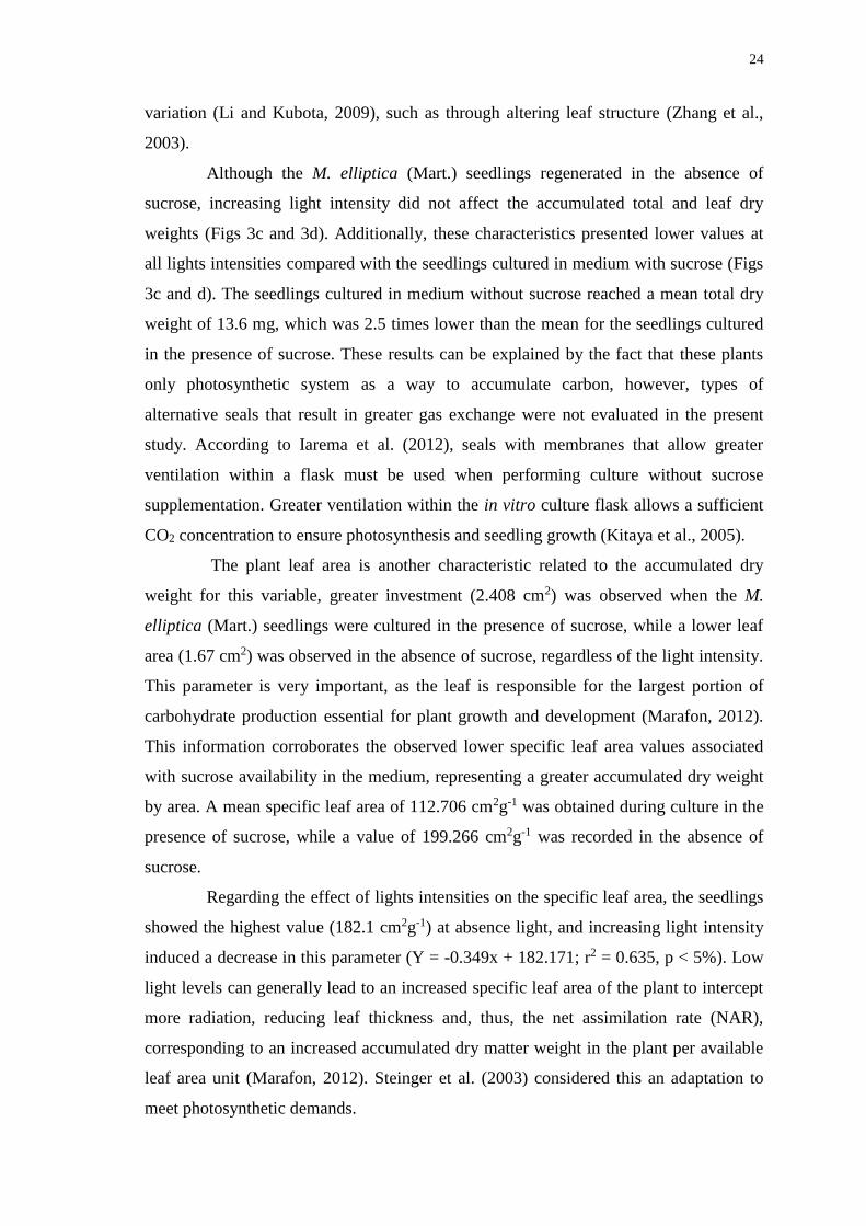
24
variation (Li and Kubota, 2009), such as through altering leaf structure (Zhang et al.,
2003).
Although the M. elliptica (Mart.) seedlings regenerated in the absence of
sucrose, increasing light intensity did not affect the accumulated total and leaf dry
weights (Figs 3c and 3d). Additionally, these characteristics presented lower values at
all lights intensities compared with the seedlings cultured in medium with sucrose (Figs
3c and d). The seedlings cultured in medium without sucrose reached a mean total dry
weight of 13.6 mg, which was 2.5 times lower than the mean for the seedlings cultured
in the presence of sucrose. These results can be explained by the fact that these plants
only photosynthetic system as a way to accumulate carbon, however, types of
alternative seals that result in greater gas exchange were not evaluated in the present
study. According to Iarema et al. (2012), seals with membranes that allow greater
ventilation within a flask must be used when performing culture without sucrose
supplementation. Greater ventilation within the in vitro culture flask allows a sufficient
CO2 concentration to ensure photosynthesis and seedling growth (Kitaya et al., 2005).
The plant leaf area is another characteristic related to the accumulated dry
weight for this variable, greater investment (2.408 cm2) was observed when the M.
elliptica (Mart.) seedlings were cultured in the presence of sucrose, while a lower leaf
area (1.67 cm2) was observed in the absence of sucrose, regardless of the light intensity.
This parameter is very important, as the leaf is responsible for the largest portion of
carbohydrate production essential for plant growth and development (Marafon, 2012).
This information corroborates the observed lower specific leaf area values associated
with sucrose availability in the medium, representing a greater accumulated dry weight
by area. A mean specific leaf area of 112.706 cm2g-1 was obtained during culture in the
presence of sucrose, while a value of 199.266 cm2g-1 was recorded in the absence of
sucrose.
Regarding the effect of lights intensities on the specific leaf area, the seedlings
showed the highest value (182.1 cm2g-1) at absence light, and increasing light intensity
induced a decrease in this parameter (Y = -0.349x + 182.171; r2 = 0.635, p < 5%). Low
light levels can generally lead to an increased specific leaf area of the plant to intercept
more radiation, reducing leaf thickness and, thus, the net assimilation rate (NAR),
corresponding to an increased accumulated dry matter weight in the plant per available
leaf area unit (Marafon, 2012). Steinger et al. (2003) considered this an adaptation to
meet photosynthetic demands.

25
To test the maximum production of shoots and leaves and biomass
accumulation in M. elliptica (Mart.) seedlings cultured in the absence of sucrose, the
development of other studies involving a light supply above 150 µmol m-2s-1 will be
necessary, or even the development of cultures that allow greater gas exchange between
the culture environment and the external atmosphere (Iarema et al., 2012), combined
with high lights intensities and alternative support for the culture medium (Saldanha et
al., 2014). These conditions characterize the photoautotrophic system (Xiao et al.,
2011).
Anatomical characteristics: M. elliptica (Mart.) exhibits leaf plasticity
Studies that demonstrate the effect of different lights intensities on the
morphoanatomic characteristics of native Cerrado plants cultured in vitro are still
scarce, especially when correlated with presence or absence of sucrose in the culture
medium. There are no available studies that demonstrate such characteristics for M.
elliptica (Mart.).
Morphoanatomic and physiological changes in the leaves are common plant
adaptive responses to different environmental conditions (Pereira et al., 2013). The M.
elliptica (Mart.) explants cultured in the absence of sucrose and light did not possess the
ability to form tissues and did not regenerate new seedlings. Thus, the comparisons
conducted in micromorphometric analyses of the leaves of seedlings cultured in vitro
only correspond to lights intensities of 50, 75, 100, and 150 µmol m-2s-1, independently
presence of sucrose in the medium.
There was a significant interaction between the light intensity and culture
medium for the chlorophyll parenchyma thickness (CP T), stomatal crypt density (St Cr
Dn), stomatal crypt depth (St Cr Dp), and stomatal crypt opening area (St Cr O). An
isolated effect of these factors was observed for the adaxial epidermis thickness (Ad Ep
T) and abaxial epidermis thickness (Ab Ep T) (p ≤ 0.05).
It was noted the stomata presence in adaptive structures known as stomatal
crypts (Figs 4a and 5 a-h), and they were only identified on the abaxial surface;
therefore, the plants can be classified as hypostomatic. The adaptive significance of the
stomatal crypts is still under discussion, and they probably evolved in response to
several environmental factors, most likely as a resource for xerophilic plants to reduce
water loss via reduced leaf transpiration (Hassiotou et al., 2009). This concept is
reinforced by the frequency of trichomes that are generally identified in the crypts
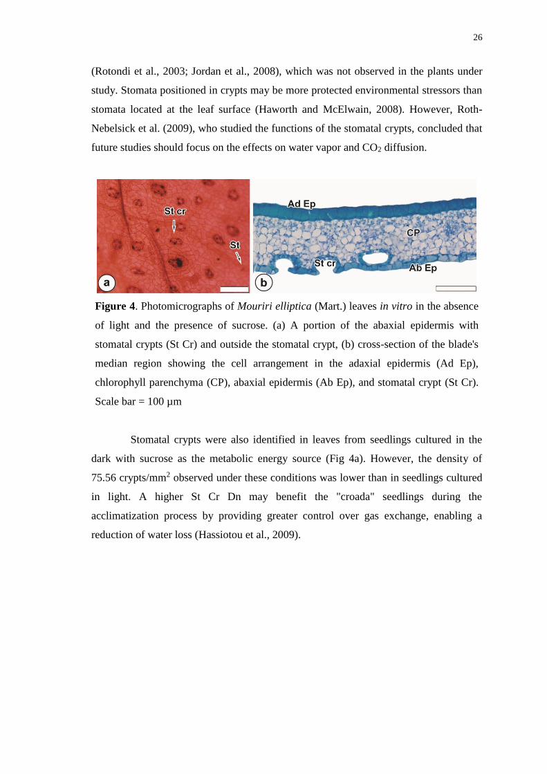
26
(Rotondi et al., 2003; Jordan et al., 2008), which was not observed in the plants under
study. Stomata positioned in crypts may be more protected environmental stressors than
stomata located at the leaf surface (Haworth and McElwain, 2008). However, Roth-
Nebelsick et al. (2009), who studied the functions of the stomatal crypts, concluded that
future studies should focus on the effects on water vapor and CO2 diffusion.
Figure 4. Photomicrographs of Mouriri elliptica (Mart.) leaves in vitro in the absence
of light and the presence of sucrose. (a) A portion of the abaxial epidermis with
stomatal crypts (St Cr) and outside the stomatal crypt, (b) cross-section of the blade's
median region showing the cell arrangement in the adaxial epidermis (Ad Ep),
chlorophyll parenchyma (CP), abaxial epidermis (Ab Ep), and stomatal crypt (St Cr).
Scale bar = 100 µm
Stomatal crypts were also identified in leaves from seedlings cultured in the
dark with sucrose as the metabolic energy source (Fig 4a). However, the density of
75.56 crypts/mm2 observed under these conditions was lower than in seedlings cultured
in light. A higher St Cr Dn may benefit the "croada" seedlings during the
acclimatization process by providing greater control over gas exchange, enabling a
reduction of water loss (Hassiotou et al., 2009).
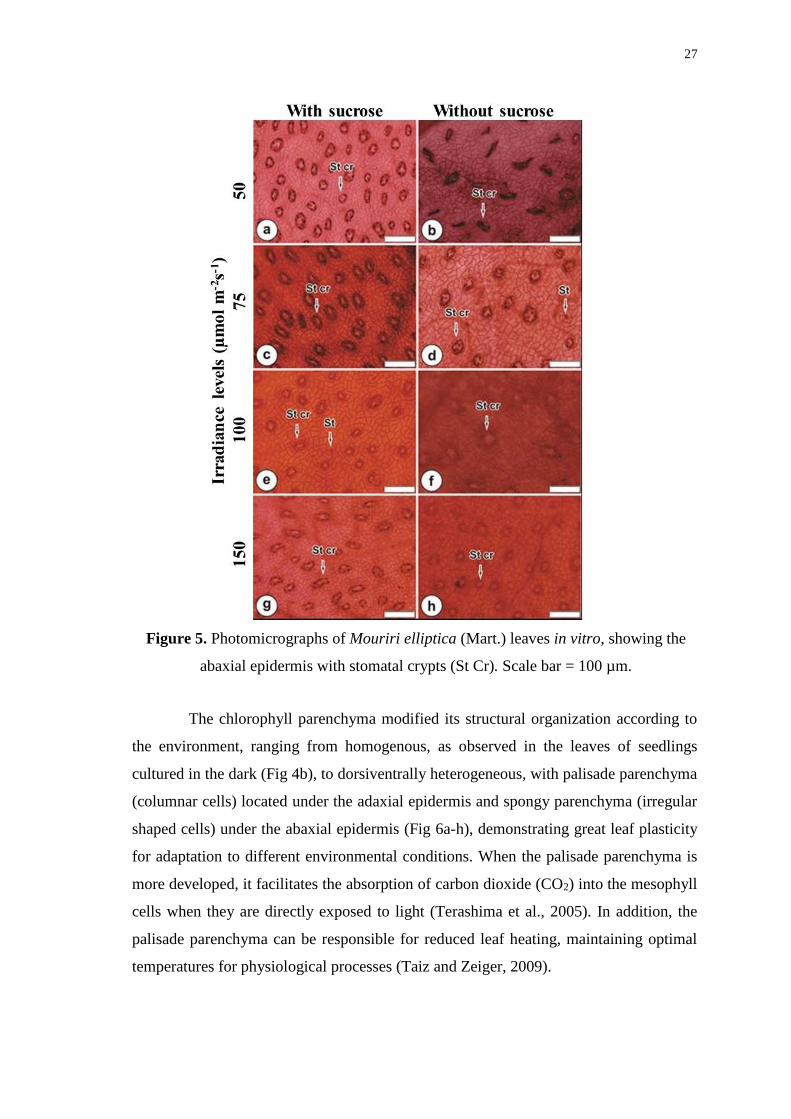
27
Figure 5. Photomicrographs of Mouriri elliptica (Mart.) leaves in vitro, showing the
abaxial epidermis with stomatal crypts (St Cr). Scale bar = 100 µm.
The chlorophyll parenchyma modified its structural organization according to
the environment, ranging from homogenous, as observed in the leaves of seedlings
cultured in the dark (Fig 4b), to dorsiventrally heterogeneous, with palisade parenchyma
(columnar cells) located under the adaxial epidermis and spongy parenchyma (irregular
shaped cells) under the abaxial epidermis (Fig 6a-h), demonstrating great leaf plasticity
for adaptation to different environmental conditions. When the palisade parenchyma is
more developed, it facilitates the absorption of carbon dioxide (CO2) into the mesophyll
cells when they are directly exposed to light (Terashima et al., 2005). In addition, the
palisade parenchyma can be responsible for reduced leaf heating, maintaining optimal
temperatures for physiological processes (Taiz and Zeiger, 2009).
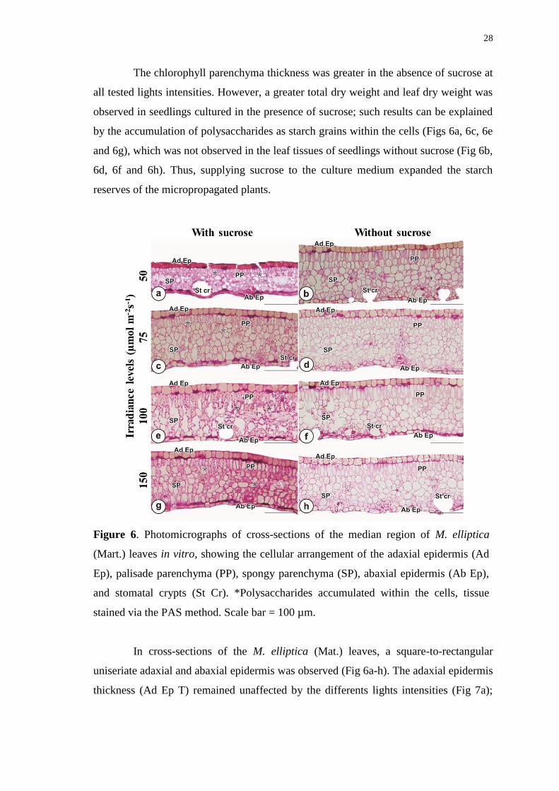
28
The chlorophyll parenchyma thickness was greater in the absence of sucrose at
all tested lights intensities. However, a greater total dry weight and leaf dry weight was
observed in seedlings cultured in the presence of sucrose; such results can be explained
by the accumulation of polysaccharides as starch grains within the cells (Figs 6a, 6c, 6e
and 6g), which was not observed in the leaf tissues of seedlings without sucrose (Fig 6b,
6d, 6f and 6h). Thus, supplying sucrose to the culture medium expanded the starch
reserves of the micropropagated plants.
Figure 6. Photomicrographs of cross-sections of the median region of M. elliptica
(Mart.) leaves in vitro, showing the cellular arrangement of the adaxial epidermis (Ad
Ep), palisade parenchyma (PP), spongy parenchyma (SP), abaxial epidermis (Ab Ep),
and stomatal crypts (St Cr). *Polysaccharides accumulated within the cells, tissue
stained via the PAS method. Scale bar = 100 µm.
In cross-sections of the M. elliptica (Mat.) leaves, a square-to-rectangular
uniseriate adaxial and abaxial epidermis was observed (Fig 6a-h). The adaxial epidermis
thickness (Ad Ep T) remained unaffected by the differents lights intensities (Fig 7a);
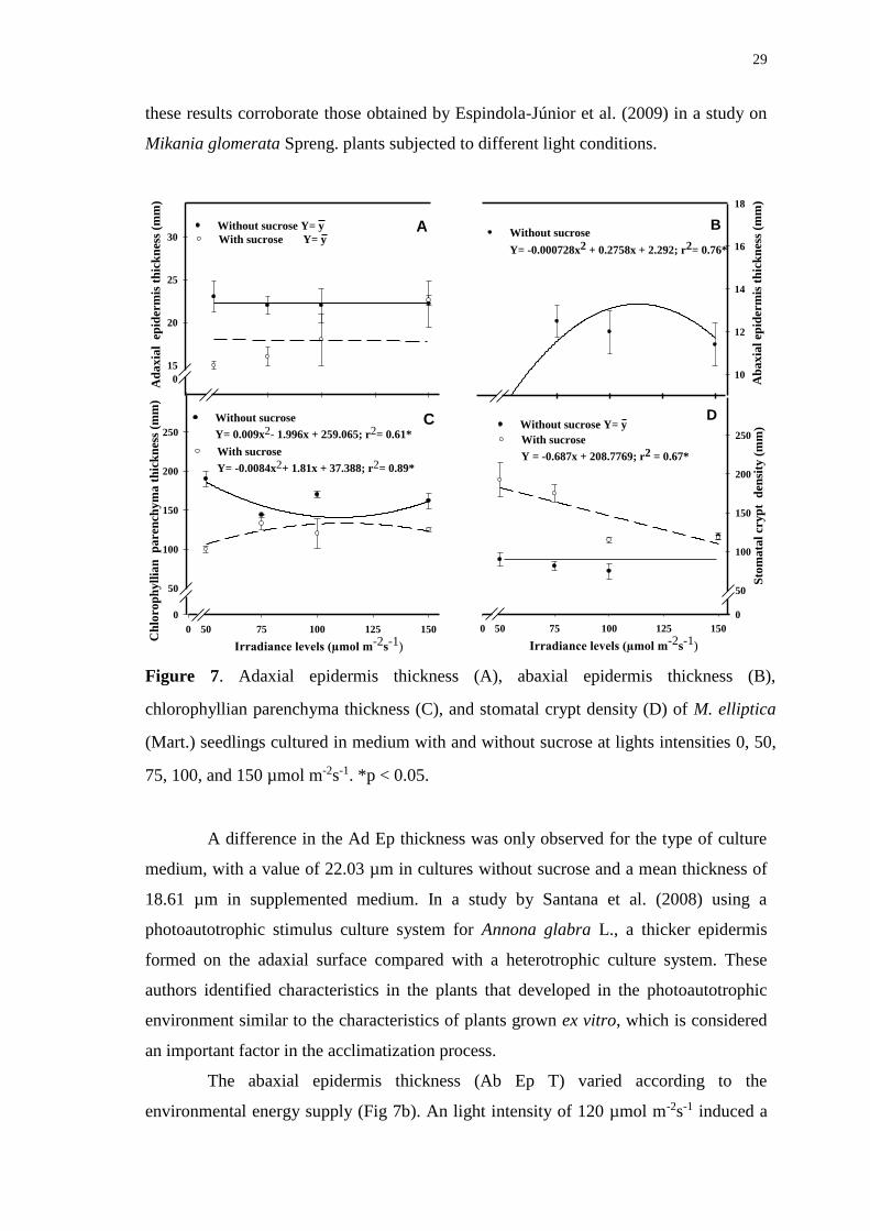
29
these results corroborate those obtained by Espindola-Júnior et al. (2009) in a study on
Mikania glomerata Spreng. plants subjected to different light conditions.
2D Graph 6
0 50 75 100 125 150
Ch
loro
ph
yll
ian
p
are
nch
ym
a t
hic
kn
ess
(mm
)
0
50
100
150
200
250
Ad
ax
ial
ep
ider
mis
th
ick
nes
s (m
m)
0
15
20
25
30A
C
Irradiance levels (µmol m-2s-1)
2D Graph 6
Irradiance levels (µmol m-2s-1)
0 50 75 100 125 150
Sto
mata
l cr
yp
t d
ensi
ty (
mm
)
0
50
100
150
200
250
Ab
ax
ial
epid
erm
is t
hic
kn
ess
(mm
)
10
12
14
16
18
B
DWithout sucrose
Y= 0.009x2- 1.996x + 259.065; r2= 0.61*
With sucrose
Y= -0.0084x2+ 1.81x + 37.388; r2= 0.89*
Without sucrose Y= y
With sucrose Y= yWithout sucrose
Y= -0.000728x2 + 0.2758x + 2.292; r2= 0.76*
Without sucrose Y= y
With sucrose
Y = -0.687x + 208.7769; r2 = 0.67*
Figure 7. Adaxial epidermis thickness (A), abaxial epidermis thickness (B),
chlorophyllian parenchyma thickness (C), and stomatal crypt density (D) of M. elliptica
(Mart.) seedlings cultured in medium with and without sucrose at lights intensities 0, 50,
75, 100, and 150 µmol m-2s-1. *p < 0.05.
A difference in the Ad Ep thickness was only observed for the type of culture
medium, with a value of 22.03 µm in cultures without sucrose and a mean thickness of
18.61 µm in supplemented medium. In a study by Santana et al. (2008) using a
photoautotrophic stimulus culture system for Annona glabra L., a thicker epidermis
formed on the adaxial surface compared with a heterotrophic culture system. These
authors identified characteristics in the plants that developed in the photoautotrophic
environment similar to the characteristics of plants grown ex vitro, which is considered
an important factor in the acclimatization process.
The abaxial epidermis thickness (Ab Ep T) varied according to the
environmental energy supply (Fig 7b). An light intensity of 120 µmol m-2s-1 induced a
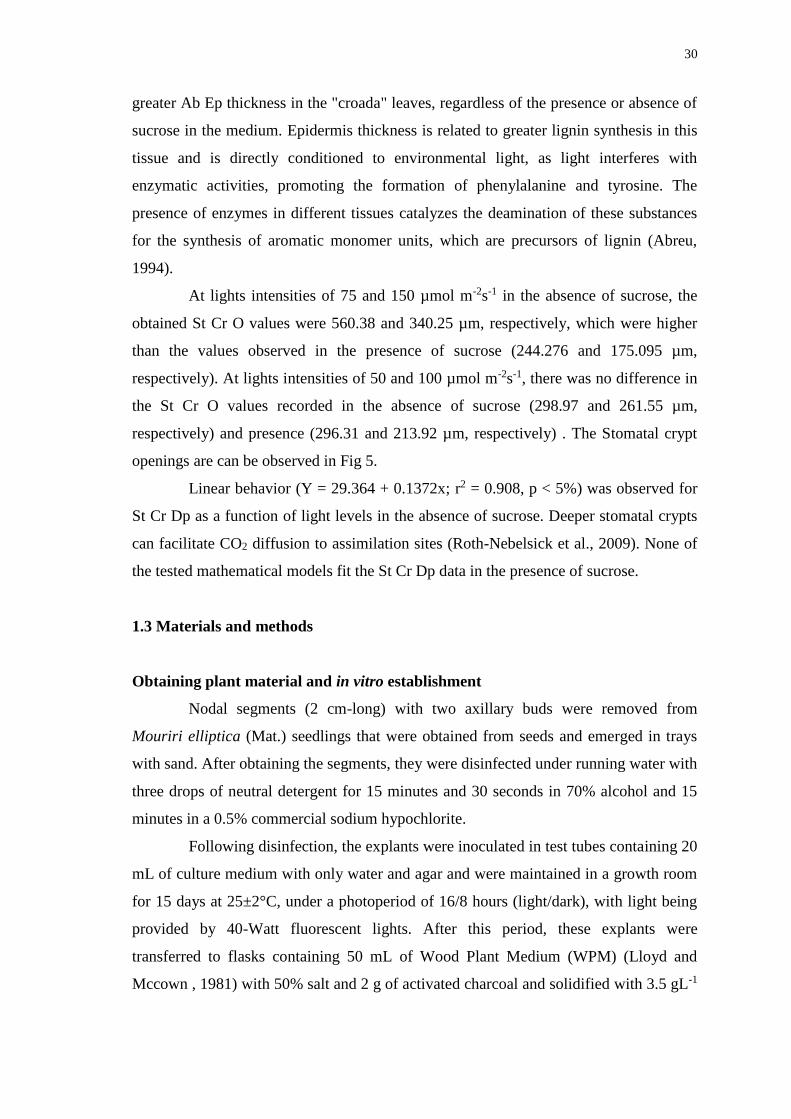
30
greater Ab Ep thickness in the "croada" leaves, regardless of the presence or absence of
sucrose in the medium. Epidermis thickness is related to greater lignin synthesis in this
tissue and is directly conditioned to environmental light, as light interferes with
enzymatic activities, promoting the formation of phenylalanine and tyrosine. The
presence of enzymes in different tissues catalyzes the deamination of these substances
for the synthesis of aromatic monomer units, which are precursors of lignin (Abreu,
1994).
At lights intensities of 75 and 150 µmol m-2s-1 in the absence of sucrose, the
obtained St Cr O values were 560.38 and 340.25 µm, respectively, which were higher
than the values observed in the presence of sucrose (244.276 and 175.095 µm,
respectively). At lights intensities of 50 and 100 µmol m-2s-1, there was no difference in
the St Cr O values recorded in the absence of sucrose (298.97 and 261.55 µm,
respectively) and presence (296.31 and 213.92 µm, respectively) . The Stomatal crypt
openings are can be observed in Fig 5.
Linear behavior (Y = 29.364 + 0.1372x; r2 = 0.908, p < 5%) was observed for
St Cr Dp as a function of light levels in the absence of sucrose. Deeper stomatal crypts
can facilitate CO2 diffusion to assimilation sites (Roth-Nebelsick et al., 2009). None of
the tested mathematical models fit the St Cr Dp data in the presence of sucrose.
1.3 Materials and methods
Obtaining plant material and in vitro establishment
Nodal segments (2 cm-long) with two axillary buds were removed from
Mouriri elliptica (Mat.) seedlings that were obtained from seeds and emerged in trays
with sand. After obtaining the segments, they were disinfected under running water with
three drops of neutral detergent for 15 minutes and 30 seconds in 70% alcohol and 15
minutes in a 0.5% commercial sodium hypochlorite.
Following disinfection, the explants were inoculated in test tubes containing 20
mL of culture medium with only water and agar and were maintained in a growth room
for 15 days at 25±2°C, under a photoperiod of 16/8 hours (light/dark), with light being
provided by 40-Watt fluorescent lights. After this period, these explants were
transferred to flasks containing 50 mL of Wood Plant Medium (WPM) (Lloyd and
Mccown , 1981) with 50% salt and 2 g of activated charcoal and solidified with 3.5 gL-1
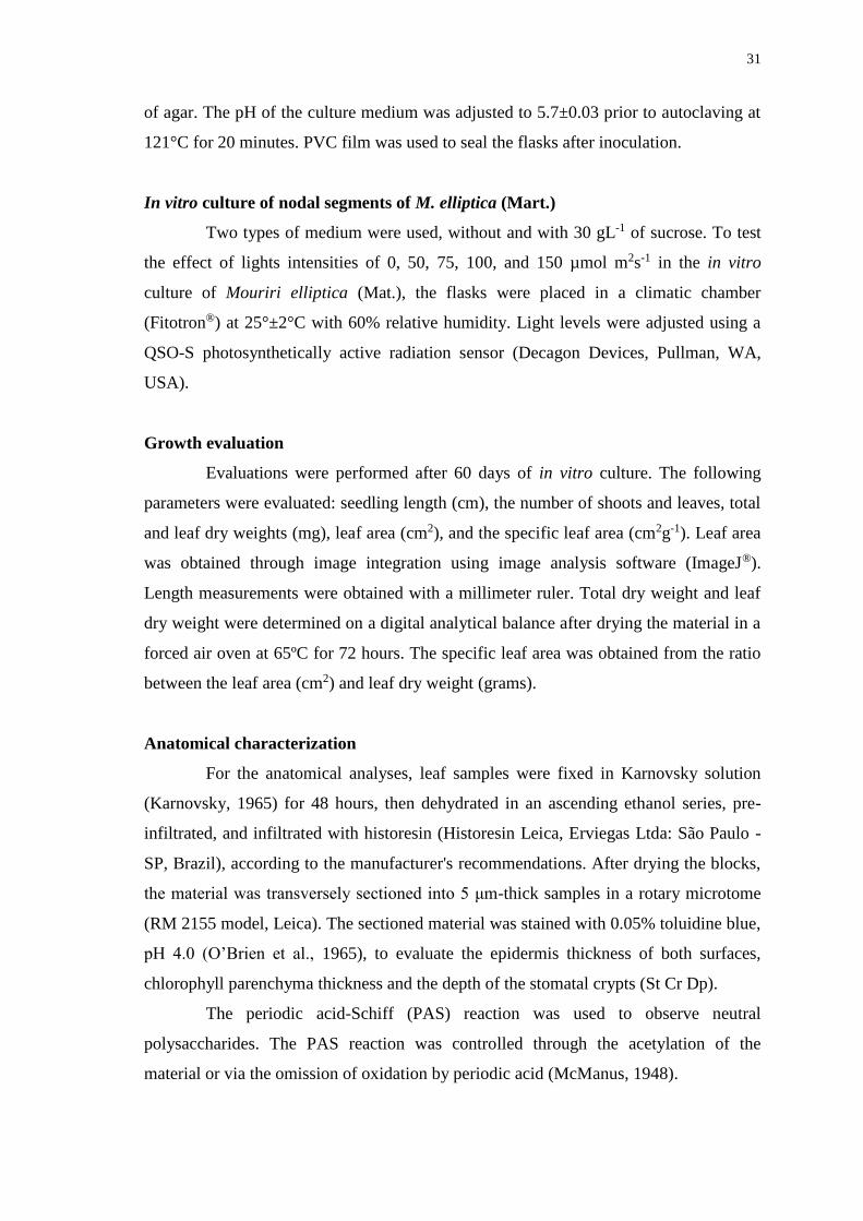
31
of agar. The pH of the culture medium was adjusted to 5.7±0.03 prior to autoclaving at
121°C for 20 minutes. PVC film was used to seal the flasks after inoculation.
In vitro culture of nodal segments of M. elliptica (Mart.)
Two types of medium were used, without and with 30 gL-1 of sucrose. To test
the effect of lights intensities of 0, 50, 75, 100, and 150 µmol m2s-1 in the in vitro
culture of Mouriri elliptica (Mat.), the flasks were placed in a climatic chamber
(Fitotron®) at 25°±2°C with 60% relative humidity. Light levels were adjusted using a
QSO-S photosynthetically active radiation sensor (Decagon Devices, Pullman, WA,
USA).
Growth evaluation
Evaluations were performed after 60 days of in vitro culture. The following
parameters were evaluated: seedling length (cm), the number of shoots and leaves, total
and leaf dry weights (mg), leaf area (cm2), and the specific leaf area (cm2g-1). Leaf area
was obtained through image integration using image analysis software (ImageJ®).
Length measurements were obtained with a millimeter ruler. Total dry weight and leaf
dry weight were determined on a digital analytical balance after drying the material in a
forced air oven at 65ºC for 72 hours. The specific leaf area was obtained from the ratio
between the leaf area (cm2) and leaf dry weight (grams).
Anatomical characterization
For the anatomical analyses, leaf samples were fixed in Karnovsky solution
(Karnovsky, 1965) for 48 hours, then dehydrated in an ascending ethanol series, pre-
infiltrated, and infiltrated with historesin (Historesin Leica, Erviegas Ltda: São Paulo -
SP, Brazil), according to the manufacturer's recommendations. After drying the blocks,
the material was transversely sectioned into 5 μm-thick samples in a rotary microtome
(RM 2155 model, Leica). The sectioned material was stained with 0.05% toluidine blue,
pH 4.0 (O’Brien et al., 1965), to evaluate the epidermis thickness of both surfaces,
chlorophyll parenchyma thickness and the depth of the stomatal crypts (St Cr Dp).
The periodic acid-Schiff (PAS) reaction was used to observe neutral
polysaccharides. The PAS reaction was controlled through the acetylation of the
material or via the omission of oxidation by periodic acid (McManus, 1948).
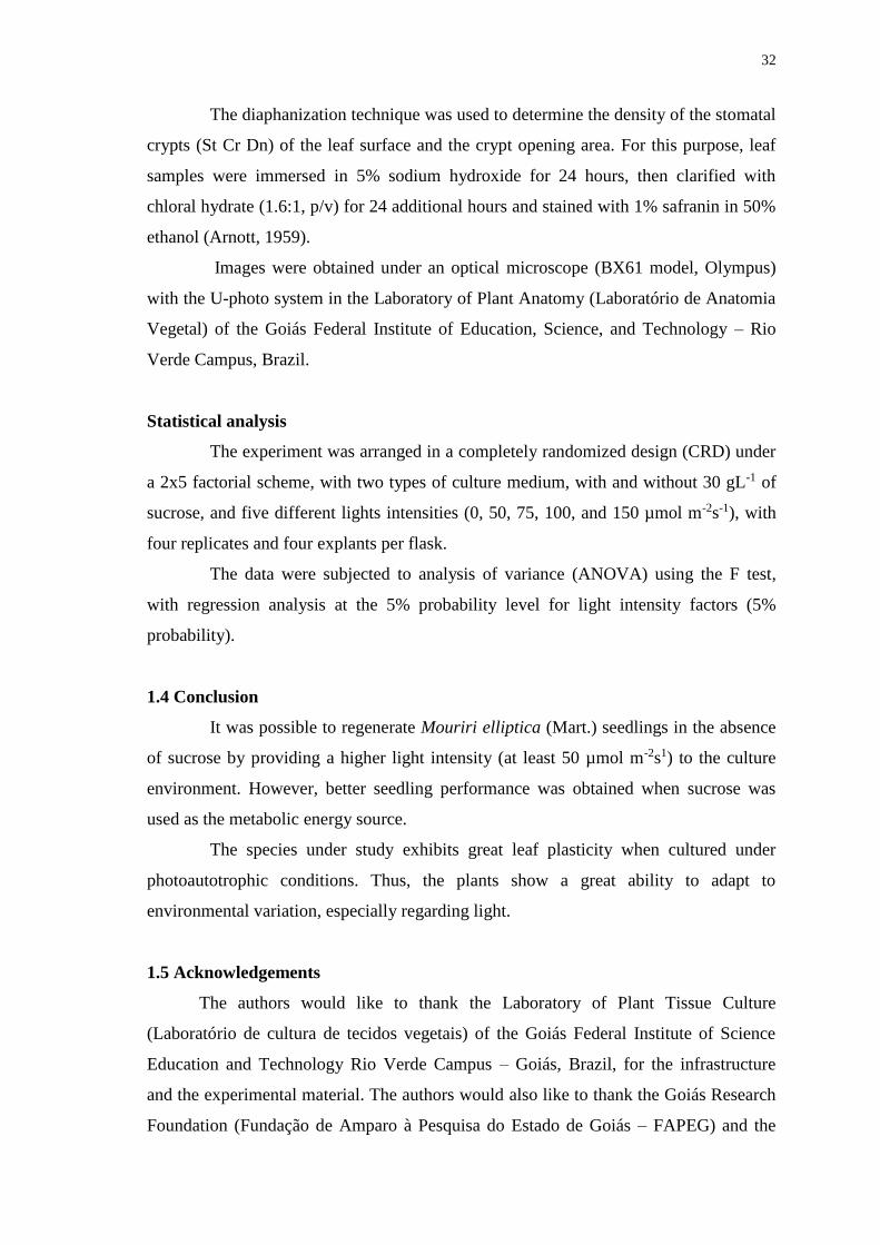
32
The diaphanization technique was used to determine the density of the stomatal
crypts (St Cr Dn) of the leaf surface and the crypt opening area. For this purpose, leaf
samples were immersed in 5% sodium hydroxide for 24 hours, then clarified with
chloral hydrate (1.6:1, p/v) for 24 additional hours and stained with 1% safranin in 50%
ethanol (Arnott, 1959).
Images were obtained under an optical microscope (BX61 model, Olympus)
with the U-photo system in the Laboratory of Plant Anatomy (Laboratório de Anatomia
Vegetal) of the Goiás Federal Institute of Education, Science, and Technology – Rio
Verde Campus, Brazil.
Statistical analysis
The experiment was arranged in a completely randomized design (CRD) under
a 2x5 factorial scheme, with two types of culture medium, with and without 30 gL-1 of
sucrose, and five different lights intensities (0, 50, 75, 100, and 150 µmol m-2s-1), with
four replicates and four explants per flask.
The data were subjected to analysis of variance (ANOVA) using the F test,
with regression analysis at the 5% probability level for light intensity factors (5%
probability).
1.4 Conclusion
It was possible to regenerate Mouriri elliptica (Mart.) seedlings in the absence
of sucrose by providing a higher light intensity (at least 50 µmol m-2s1) to the culture
environment. However, better seedling performance was obtained when sucrose was
used as the metabolic energy source.
The species under study exhibits great leaf plasticity when cultured under
photoautotrophic conditions. Thus, the plants show a great ability to adapt to
environmental variation, especially regarding light.
1.5 Acknowledgements
The authors would like to thank the Laboratory of Plant Tissue Culture
(Laboratório de cultura de tecidos vegetais) of the Goiás Federal Institute of Science
Education and Technology Rio Verde Campus – Goiás, Brazil, for the infrastructure
and the experimental material. The authors would also like to thank the Goiás Research
Foundation (Fundação de Amparo à Pesquisa do Estado de Goiás – FAPEG) and the
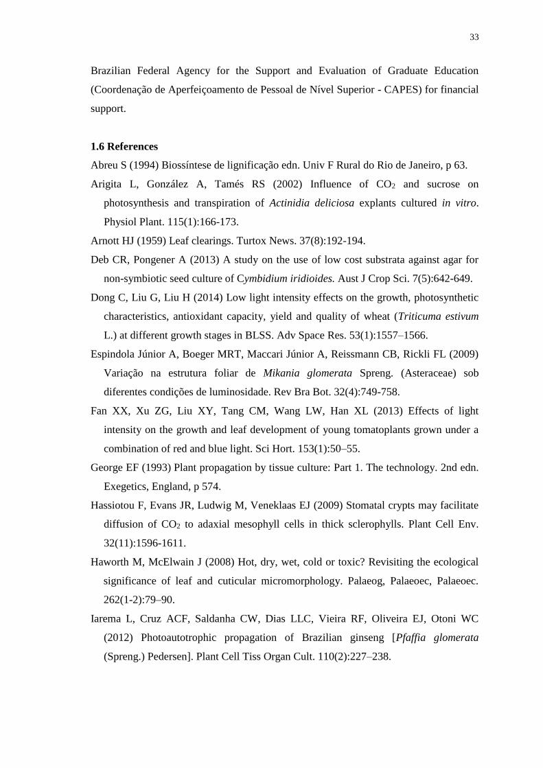
33
Brazilian Federal Agency for the Support and Evaluation of Graduate Education
(Coordenação de Aperfeiçoamento de Pessoal de Nível Superior - CAPES) for financial
support.
1.6 References
Abreu S (1994) Biossíntese de lignificação edn. Univ F Rural do Rio de Janeiro, p 63.
Arigita L, González A, Tamés RS (2002) Influence of CO2 and sucrose on
photosynthesis and transpiration of Actinidia deliciosa explants cultured in vitro.
Physiol Plant. 115(1):166-173.
Arnott HJ (1959) Leaf clearings. Turtox News. 37(8):192-194.
Deb CR, Pongener A (2013) A study on the use of low cost substrata against agar for
non-symbiotic seed culture of Cymbidium iridioides. Aust J Crop Sci. 7(5):642-649.
Dong C, Liu G, Liu H (2014) Low light intensity effects on the growth, photosynthetic
characteristics, antioxidant capacity, yield and quality of wheat (Triticuma estivum
L.) at different growth stages in BLSS. Adv Space Res. 53(1):1557–1566.
Espindola Júnior A, Boeger MRT, Maccari Júnior A, Reissmann CB, Rickli FL (2009)
Variação na estrutura foliar de Mikania glomerata Spreng. (Asteraceae) sob
diferentes condições de luminosidade. Rev Bra Bot. 32(4):749-758.
Fan XX, Xu ZG, Liu XY, Tang CM, Wang LW, Han XL (2013) Effects of light
intensity on the growth and leaf development of young tomatoplants grown under a
combination of red and blue light. Sci Hort. 153(1):50–55.
George EF (1993) Plant propagation by tissue culture: Part 1. The technology. 2nd edn.
Exegetics, England, p 574.
Hassiotou F, Evans JR, Ludwig M, Veneklaas EJ (2009) Stomatal crypts may facilitate
diffusion of CO2 to adaxial mesophyll cells in thick sclerophylls. Plant Cell Env.
32(11):1596-1611.
Haworth M, McElwain J (2008) Hot, dry, wet, cold or toxic? Revisiting the ecological
significance of leaf and cuticular micromorphology. Palaeog, Palaeoec, Palaeoec.
262(1-2):79–90.
Iarema L, Cruz ACF, Saldanha CW, Dias LLC, Vieira RF, Oliveira EJ, Otoni WC
(2012) Photoautotrophic propagation of Brazilian ginseng [Pfaffia glomerata
(Spreng.) Pedersen]. Plant Cell Tiss Organ Cult. 110(2):227–238.
34
Jordan GJ, Weston PH, Carpenter RJ, Dillon RA, Brodribb TJ (2008) The evolutionary
relations of sunken, covered, and encrypted stomata to dry habitats in Proteaceae.
Am J Bot. 95(5):521–530.
Li Q, Kubota C (2009). Effects of supplemental light quality on growth and
phytochemicals of baby leaf lettuce. J Env Exp Bot. 67(1):59–64.
Karnovsky MJ (1965) A formaldehyde-glutaraldehyde fixative of high osmolality for
use in electron microscopy. J Cell Biol. 27(2):137–138.
Kitaya Y, Ohmura Y, Kubota C, Kozai T (2005) Manipulation of the culture
environment on in vitro air movement and its impact on plantlets photosynthesis.
Plant Cell Tiss Organ Cult. 83(3):251–257.
Kozai T, Nguyen QT (2003) Photoautotrophic micropropagation of woody and tropical
plants. In: Jain SM, Ishii K micropropagation of woody trees and fruits. Dordrecht:
Kluwer Academic, p757-781.
Lloyd G, Mccown B (1981) Commercially feasible micropropagation of montain laurel,
kalmia latifolia, by use of shoot tip culture. Comb Proc Int’l Plant Prop Soc. 30:421-
327.
Long SP (1994) Photoinhibition of photosynthesis in nature. Rev Plant Biol, Ann.
45:633–662.
Marafon AC (2012) Análise quantitativa de crescimento em Cana-de-açúcar: Introdução
ao procedimento prático. Documentos, Embrapa.
Martins JPR, Pasqual M, Martins AD, Ribeira SF (2015) Effects of salts and sucrose
concentrations on in vitro propagation of Billbergia zebrina (Herbert) Lindley
(Bromeliaceae). Aust J Crop Sci. 9(1):85-91.
McManus JFA (1948) Histologia and histochemical uses of periódic acid. Stain Technol
23(1):99-108.
Moleiro FC, Andreo MA, Santos RdeC, Moraes TdeM, Rodrigues CM, Carlis CB,
Lopes FC,
Pellizzon CH, Carlos IZ, Bauab TM, Vilegas W, Hiruma-Lima CA (2009) Mouriri
elliptica (Mart.): validation of gastroprotective, healing and anti-Helicobaster pylori
effects. J Ethnopharmacol. 123(3):359-368.
Moreira MA, Pasqual M, Carvalho JG, Fráguas CB (2003) Estiolamento na
micropropagação do abacaxizeiro cv. Pérola. Ciên Agrotec. 27(5):1002-1006.
O’brien TP, Feder N, MeCully ME (1965) Polychromatic staining of plant cell walls by
toluidine blue. Protoplasma. 59(1):368-373.
35
Pereira JD, Medri ME, Moreira RS (2013) Avaliações Morfológicas e
Micromorfométricas de Folhas de Sol e de Sombra de Lithraea molleoides (Vell.)
Engl. (Anacardiaceae). Ev Cons da Bio Rio Paranaíba. 4(1):22-31.
Roth-Nebelsick A, Hassiotou F, Veneklaas EJ (2009) Stomatal crypts have small effects
on transpiration: A numerical model analysis. Plant Physiol. 151(4):2018–2027.
Rotondi A, Rossi F, Asunis C, Cesaraccio C (2003) Leaf xeromorphic adaptations of
some plants of a coastal Mediterranean macchia ecosystem. J Mediterranean Ecol.
4(3-4):25–35.
Rufino MdSM, Alves RE, Brito ES, Pérez-Jiménez J, Saura-Calixto F, Mancini-Filho J
(2010) Bioactive compounds and antioxidant capacities of 18 non-traditional tropical
fruits from Brazil. Food Chemistry. 121(4):996–1002.
Rufino MSM, Alves RE, Fernandes FAN, Brito ES (2011) Free radical scavenging
behavior of ten exotic tropical fruits extracts. Food Res Int. 44(7):2072-2075.
Sáez PL, Bravo LA, Latsague MI, Sánchez MR, Ríos DG (2012) Increased light
intensity during in vitro culture improves water loss control and photosynthetic
performance of castanea sativa grown in ventilated vessels. Sci Hort. 138(1):7–16.
Sairanen I, Novák O, Pencik A, Ikeda Y, Jones B, Sandberg Ag, Ljung K (2012)
Soluble carbohydrates regulate auxin biosynthesis via PIF proteins in Arabidopsis.
Plant Cell. 24(12):4907- 4916.
Saldanha CW, Otoni CG, Azevedo JLF, Dias LLC, Rêgo MM, Otoni WC (2012) A
low-cost alternative membrane system that promotes growth in nodal cultures of
Brazilian ginseng [Pfaffia glomerata (Spreng.) Pedersen]. Plant Cell Tiss Organ
Cult. 110(3):413-422.
Saldanha CW, Otoni CG, Notini MM, Kuki KN, Cruz ACF, Rubio Neto A, Dias LLC,
Otoni WC (2013) A CO2-enriched atmosphere improves in vitro growth of Brazilian
ginseng [Pfaffia glomerata (Spreng.) Pedersen]. In Vitro Cell and Dev Biol-Plant
49(4):433–444.
Saldanha CW, Otoni CG, Rocha DI, Cavatte PC, Detmann KSC, Tanaka FAO, Dias
LLC, DaMatta FM, Otoni WC (2014) CO2-enriched atmosphere and supporting
material impact the growth, morphophysiology and ultrastructure of in vitro
Brazilian-ginseng [Pfaffia glomerata (spreng.) pedersen] plantlets. Plant Cell Tiss
Organ Cult. 118(1):87-99.
Santana JRF, Paiva R, Resende RKS, Castro EM, Pereira FD, Oliveira LM (2008)
Estímulo do comportamento fotoautotrófico durante o enraizamento in
36
vitro de Annona glabra L. II. Aspectos da anatomia da folha antes da aclimatização.
Ciênc Agrotec. 32(2):640-644.
Silva DB, Silva JA, Junqueira NTV, Andrade LRM (2001) Frutas do Cerrado. Embrapa,
Brasília, Informação Tecnológica, p. 178.
Steinger T, Roy BA, Stanton ML (2003) Evolution in stressful environments II:
adaptive value and costs of plasticity in response to low light in Sinapis arvensis. J
Evol Biol. 16(2):313–323.
Solymosi KE, Schoefs K (2010) Etioplast and etio-chloroplast formation under natural
conditions: the dark side of chlorophyll biosynthesis in angiosperms. Photosynth
Res. 105(2):143-166.
Suzuki RM, Kerbauy GB, Zaffari GR (2004) Endogenous hormonal levels and growth
of dark-incubated shoots of Catasetum fimbriatum. J Plant Physiol. 161(8):929-935.
Taiz L, Zeiger E (2009) Fisiologia vegetal, 4 edn. Artmed, Porto Alegre, p 848.
Terashima I, Araya T, Miyazawa S-I, Sone K, Yano S (2005) Construction and
maintenance of the optimal photosynthetic systems of the leaf, herbaceous plant and
tree: an eco-developmental treatise. Ann Bot. 95(1): 507-519.
Vasconcelos JM, Cardoso TV, Sales JF, Silva FG, Vasconcelos Filho SC, Santana JG
(2010a) Métodos de superação de dormência em sementes de croada (Mouriri
elliptica Mart). Ciênc Agrotec. 34(5):1199-1204.
Vasconcelos PC, Andreo MA, Vilegas W, Hiruma-Lima CA, Pellizzon CH (2010b)
Effect of Mouriri pusa tannins and flavonoids on prevention and treatment against
experimental gastric ulcer. J Ethnopharmacol. 131(1):146–153.
Walters RG (2005) Towards an understanding of photosynthetic acclimation. J Exp Bot.
56(411):435–447.
Xiao Y, Kozai T (2006) In vitro multiplication of statice plantlets using sugar-free
media. Sci Hort. 109(1):71–77.
Xiao Y, Niu G, Kozai T (2011) Development and application of photoautotrophic
micropropagation plant system. Plant Cell Tiss Organ Cult. 105(2):149–158.
Zhang S, Ma K, Chen L (2003) Response of photosynthetic plasticity of Paeonia
suffruticosa to changed light environments. J Env Exp Bot. 49(2): 121–133.
Zhang M, Zhao D, Ma Z, Li X, Xiao Y (2009) Growth and photosynthethetic capability
of Momordica grosvenori plantlets grown photoautotrophically in response to light
intensity. Hort Sci. 44(3):757–763.
37
CAPÍTULO II
(Normas de acordo com a revista Genetics and Molecular Research. Artigo publicado
em outubro de 2016, v. 15, n. 4, p. 1-11)
Dissimilarity between plants of Mouriri elliptica (Mart.) cultivated in vitro and in
situ through anatomic parameters
Abstract
The species Mouriri elliptica (Mart.) is a genetic resource of the Cerrado
domain, as it has potential food and medicinal uses. There have been few studies on its
in vitro propagation, and there are no studies examining the dissimilarities between
plants of this species when cultivated in situ or in vitro. Therefore, the objective of this
study was to identify in vitro cultivation conditions that allow the formation of plantlets
with leaf anatomical features that are less dissimilar to plants in situ. Thus, an
anatomical study of the leaves was conducted, in which, it is considered the adaxial
epidermis thickness, the abaxial epidermis thickness, the chlorenchyma thickness, the
stomatal crypt depth, the stomatal crypt density and the leaf surface stomatal crypt
aperture area. The distance between phenotypes was determined based on
micromorphometric data, and the UPGMA cluster was then determined. Four different
groups were tested, and cultivation conditions in the presence of sucrose and irradiance
of 50 and 75 µmol m-2s-1 were identified as promoters of plantlet development that
maximized the similarity to in situ plant. The most important anatomical parameters in
this identification were the stomatal crypt aperture area and crypt density. This study

38
holds great importance for the anatomical characterization of the leaves of M. elliptica
(Mart.), as it identifies plasticity as a function of in vitro culture conditions.
Keywords: Leaf micromorphometrics; micropropagation; phenotype; plantlets;
UPGMA clustering
2.1 Introduction
Mouriri elliptica (Mart.) (Melastomataceae family) is a native plant of the
Cerrado domain and is a potential resource for the human population. It produces
nutritious fruits that contain antioxidant compounds and that can be consumed in natura
or processed into jam (Rufino et al., 2010; Rufino et al., 2011). Its leaves and/or bark
have been used to treat gastric ulcers and gastritis, and they may have an antimicrobial
effect. These medicinal effects stem from immune system stimulation against pathogens
(Moleiro et al., 2009; Vasconcelos et al., 2010) and may also be due to inhibition of the
oxidative capacity of Helicobacter pylori (Bonacorsi et al., 2013).
The sexed reproduction of the species M. elliptica (Mart.) is very complex, due
in part to the presence in the seeds of a hard tegument that renders germination difficult,
as reported by Vasconcelos et al. (2010) and Lima et al. (2016). Thus, it is necessary to
utilize practices that allow dormancy to be overcome or that obtain seedlings through
vegetative propagation. Plant tissue culture has been an indispensable tool for obtaining
seedlings of various native and cultivated species, such as Anacardium othonianum
Rizz (Assis et al., 2015), Byrsonima cydoniifolia A. Juss. (Martendal et al., 2013 and
2014) and Pfaffia glomerata (Spreng.) Pedersen (Saldanha et al., 2014) and banana
(Musa spp AAA) plantlets (Kaçar et al., 2010).
When cultivating in vitro, it is important to obtain plantlets that have
characteristics akin to plants grown in situ. In vitro cultivation conditions normally
permit rapid plant growth and multiplication but may induce structural and
physiological changes that render plants unfit to survive adverse environmental
conditions (Rout et al., 2006). Thus, photoautotrophic micropropagation has been
investigated, in which the absence of sucrose in the growth medium and increased
irradiance from the environment are used to promote the formation of plantlets with
characteristics that benefit their survival when they undergo the acclimatization process
(Xiao and Kozai, 2006; Sáez et al., 2012; Iarema et al., 2012; Assis et al., 2016).
39
To determine whether plantlets that are micropropagated in a photoautotrophic
system develop morphophysiological characteristics that are similar to those of in situ
plants, the use of multivariate techniques is appropriate. According to Cruz (2011),
multivariate analysis has diverse applications, meeting the needs of investigators with
wide ranges of interests and knowledge. The use of the technique is common in plant
breeding programs (Silva et al., 2015) as a way to evaluate genetic diversity (Assis et
al., 2013) or superior genotypes (Oda et al., 2015) and to conduct environmental studies
(Leo et al., 2015; Ma et al., 2016) in which the technique has been successful and has
clarified data.
Knows of no studies to date that used multivariate analysis for the comparative
evaluation of plants M. elliptica (Mart.) cultivated in vitro and in situ based on
anatomical features. Anatomical parameters can support future studies with
morphogenetic characterization of populations of this species, and genetic diversity
studies, and, based on plant breeding programs. Thus, this study aimed to use
multivariate statistical techniques to evaluate the dissimilarity between plants cultivated
in vitro and in situ based on anatomical parameters.
2.2 Material and methods
Plant material and in vitro cultivation conditions
The study material consisted of leaves from mature plants of M. elliptica
(Mart.) under natural conditions (in situ) and leaves of plantlets cultivated in vitro. Both
the leaves of the mature plants and the seeds for the plantlets were collected on a private
property located in the Planalto Verde District, municipality of Montividiu, Goiás,
Brazil (17° 19.201’ S, 51° 33.500’ W and 982 m altitude).
Before implementing the experiment, 2-cm-long nodal segments with 2
axillary buds each were inoculated in test tubes containing 20 mL of growth medium
composed of water and agar. The tubes were then kept in a growth room for 15 days at a
temperature of 25° ± 2°C and with a photoperiod of 16/8 h (light/dark) under irradiance
from 40-W fluorescent lamps. After that period, the explants were transferred to vials
containing 50 mL of WPM (wood plant medium) (Lloyd and McCown, 1981) with 50%
salts and 2.5 g of added activated carbon that was solidified with 3.5 g L-1 agar and
either contained or lacked 30 g L-1 sucrose. The pH of the growth medium was adjusted
to 5.7 ± 0.03 before autoclaving at 121°C for 20 minutes. The vials were sealed after
inoculation with PVC plastic film.
40
The experiment was organized with a completely randomized design (CRD) in
a growth chamber (Fitotron®) at 25° ± 2°C and with a relative humidity of 60%.
Irradiances of 0, 50, 75, 100 and 150 µmol m-2s-1 were evaluated and were adjusted
based on periodic tests with the aid of a QSO-S photosynthetically active radiation
sensor (Decagon Devices, Pullman, WA, USA).
As no plantlets were formed in the absence of both sucrose and light, 9 in vitro
cultivation conditions were used for the study, as follows: C1, presence of sucrose and
zero irradiance; C2, presence of sucrose and 50 µmol m-2s-1; C3, presence of sucrose
and 75 µmol m-2s-1; C4, presence of sucrose and 100 µmol m-2s-1; C5, presence of
sucrose and 150 µmol m-2s-1; C6, absence of sucrose and 50 µmol m-2s-1; C7, absence of
sucrose and 75 µmol m-2s-1; C8, absence of sucrose and 100 µmol m-2s-1; and C9,
absence of sucrose and 150 µmol m-2s-1. These conditions were compared and
correlated with each other and with in situ plants (CO).
Anatomical study of M. elliptica (Mart.) leaves
The leaves of M. elliptica (Mart.) were subjected to 2 analytical processes,
namely fixation and diaphonization. For fixation, leaves were submerged in
Karnovsky’s solution (Karnovsky, 1965) for 48 hours, dehydrated in a graded ethanol
series, and pre-infiltrated and infiltrated with Historesin (Leica) according to the
manufacturer’s instructions. The material was sectioned into 5-μm sections with a rotary
microtome (RM 2155, Leica). The sections were stained with 0.05% toluidine blue at
pH 4.0 (O’Brien et al., 1965).
For diaphonization, leaf samples were immersed in a 5% sodium hydroxide
solution for 24 hours, clearified with chloral hydrate (1.6:1, p/v) for 24 additional hours
and stained with 1% safranin in 50% ethanol (Arnott, 1959). After these procedures, the
slides with the material were covered with a cover slip using Canada balsam.
Images were obtained under an optical microscope (BX61, Olympus) with a U-
photo system at the Plant Anatomy Laboratory of the Federal Institute of Education,
Science and Technology of Goias – Rio Verde Campus. The adaxial epidermis
thickness (Ad Ep T), the abaxial epidermis thickness (Ab Ep T), the chlorenchyma
thickness (Ch T), the stomatal crypt depth (St Cr Dp), the stomatal crypt density (St Cr
D) and the leaf surface stomatal crypt aperture area (St Cr A) were evaluated.
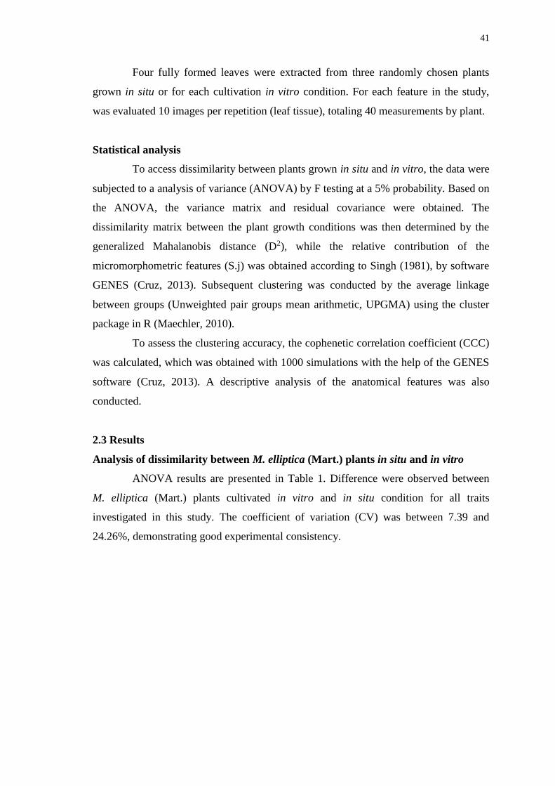
41
Four fully formed leaves were extracted from three randomly chosen plants
grown in situ or for each cultivation in vitro condition. For each feature in the study,
was evaluated 10 images per repetition (leaf tissue), totaling 40 measurements by plant.
Statistical analysis
To access dissimilarity between plants grown in situ and in vitro, the data were
subjected to a analysis of variance (ANOVA) by F testing at a 5% probability. Based on
the ANOVA, the variance matrix and residual covariance were obtained. The
dissimilarity matrix between the plant growth conditions was then determined by the
generalized Mahalanobis distance (D2), while the relative contribution of the
micromorphometric features (S.j) was obtained according to Singh (1981), by software
GENES (Cruz, 2013). Subsequent clustering was conducted by the average linkage
between groups (Unweighted pair groups mean arithmetic, UPGMA) using the cluster
package in R (Maechler, 2010).
To assess the clustering accuracy, the cophenetic correlation coefficient (CCC)
was calculated, which was obtained with 1000 simulations with the help of the GENES
software (Cruz, 2013). A descriptive analysis of the anatomical features was also
conducted.
2.3 Results
Analysis of dissimilarity between M. elliptica (Mart.) plants in situ and in vitro
ANOVA results are presented in Table 1. Difference were observed between
M. elliptica (Mart.) plants cultivated in vitro and in situ condition for all traits
investigated in this study. The coefficient of variation (CV) was between 7.39 and
24.26%, demonstrating good experimental consistency.
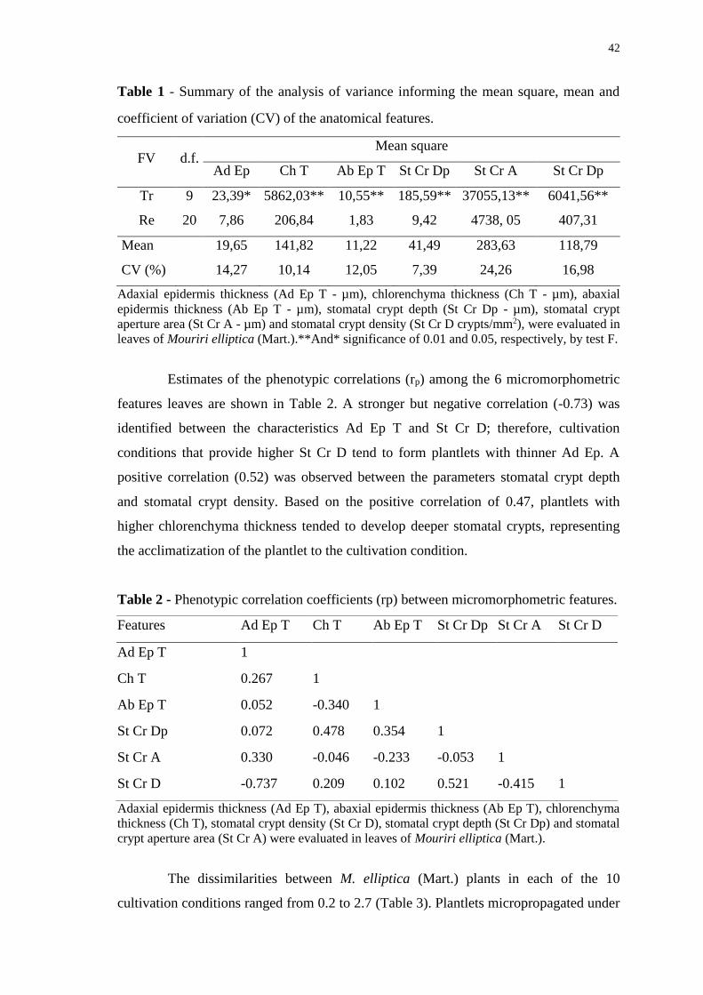
42
Table 1 - Summary of the analysis of variance informing the mean square, mean and
coefficient of variation (CV) of the anatomical features.
FV d.f. Mean square
Ad Ep Ch T Ab Ep T St Cr Dp St Cr A St Cr Dp
Tr 9 23,39* 5862,03** 10,55** 185,59** 37055,13** 6041,56**
Re 20 7,86 206,84 1,83 9,42 4738, 05 407,31
Mean 19,65 141,82 11,22 41,49 283,63 118,79
CV (%) 14,27 10,14 12,05 7,39 24,26 16,98
Adaxial epidermis thickness (Ad Ep T - µm), chlorenchyma thickness (Ch T - µm), abaxial
epidermis thickness (Ab Ep T - µm), stomatal crypt depth (St Cr Dp - µm), stomatal crypt
aperture area (St Cr A - µm) and stomatal crypt density (St Cr D crypts/mm2), were evaluated in
leaves of Mouriri elliptica (Mart.).**And* significance of 0.01 and 0.05, respectively, by test F.
Estimates of the phenotypic correlations (rp) among the 6 micromorphometric
features leaves are shown in Table 2. A stronger but negative correlation (-0.73) was
identified between the characteristics Ad Ep T and St Cr D; therefore, cultivation
conditions that provide higher St Cr D tend to form plantlets with thinner Ad Ep. A
positive correlation (0.52) was observed between the parameters stomatal crypt depth
and stomatal crypt density. Based on the positive correlation of 0.47, plantlets with
higher chlorenchyma thickness tended to develop deeper stomatal crypts, representing
the acclimatization of the plantlet to the cultivation condition.
Table 2 - Phenotypic correlation coefficients (rp) between micromorphometric features.
Features Ad Ep T Ch T Ab Ep T St Cr Dp St Cr A St Cr D
Ad Ep T 1
Ch T 0.267 1
Ab Ep T 0.052 -0.340 1
St Cr Dp 0.072 0.478 0.354 1
St Cr A 0.330 -0.046 -0.233 -0.053 1
St Cr D -0.737 0.209 0.102 0.521 -0.415 1
Adaxial epidermis thickness (Ad Ep T), abaxial epidermis thickness (Ab Ep T), chlorenchyma
thickness (Ch T), stomatal crypt density (St Cr D), stomatal crypt depth (St Cr Dp) and stomatal
crypt aperture area (St Cr A) were evaluated in leaves of Mouriri elliptica (Mart.).
The dissimilarities between M. elliptica (Mart.) plants in each of the 10
cultivation conditions ranged from 0.2 to 2.7 (Table 3). Plantlets micropropagated under
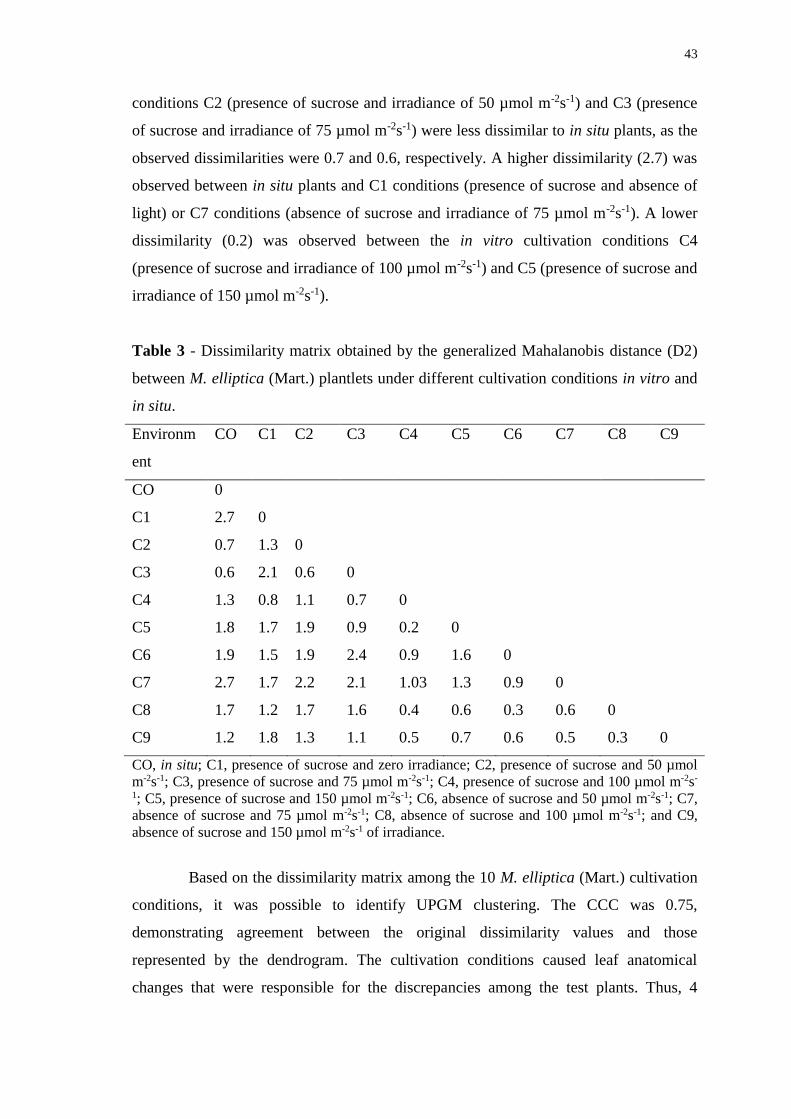
43
conditions C2 (presence of sucrose and irradiance of 50 µmol m-2s-1) and C3 (presence
of sucrose and irradiance of 75 µmol m-2s-1) were less dissimilar to in situ plants, as the
observed dissimilarities were 0.7 and 0.6, respectively. A higher dissimilarity (2.7) was
observed between in situ plants and C1 conditions (presence of sucrose and absence of
light) or C7 conditions (absence of sucrose and irradiance of 75 µmol m-2s-1). A lower
dissimilarity (0.2) was observed between the in vitro cultivation conditions C4
(presence of sucrose and irradiance of 100 µmol m-2s-1) and C5 (presence of sucrose and
irradiance of 150 µmol m-2s-1).
Table 3 - Dissimilarity matrix obtained by the generalized Mahalanobis distance (D2)
between M. elliptica (Mart.) plantlets under different cultivation conditions in vitro and
in situ.
Environm
ent
CO C1 C2 C3 C4 C5 C6 C7 C8 C9
CO 0
C1 2.7 0
C2 0.7 1.3 0
C3 0.6 2.1 0.6 0
C4 1.3 0.8 1.1 0.7 0
C5 1.8 1.7 1.9 0.9 0.2 0
C6 1.9 1.5 1.9 2.4 0.9 1.6 0
C7 2.7 1.7 2.2 2.1 1.03 1.3 0.9 0
C8 1.7 1.2 1.7 1.6 0.4 0.6 0.3 0.6 0
C9 1.2 1.8 1.3 1.1 0.5 0.7 0.6 0.5 0.3 0
CO, in situ; C1, presence of sucrose and zero irradiance; C2, presence of sucrose and 50 µmol
m-2s-1; C3, presence of sucrose and 75 µmol m-2s-1; C4, presence of sucrose and 100 µmol m-2s-
1; C5, presence of sucrose and 150 µmol m-2s-1; C6, absence of sucrose and 50 µmol m-2s-1; C7,
absence of sucrose and 75 µmol m-2s-1; C8, absence of sucrose and 100 µmol m-2s-1; and C9,
absence of sucrose and 150 µmol m-2s-1 of irradiance.
Based on the dissimilarity matrix among the 10 M. elliptica (Mart.) cultivation
conditions, it was possible to identify UPGM clustering. The CCC was 0.75,
demonstrating agreement between the original dissimilarity values and those
represented by the dendrogram. The cultivation conditions caused leaf anatomical
changes that were responsible for the discrepancies among the test plants. Thus, 4
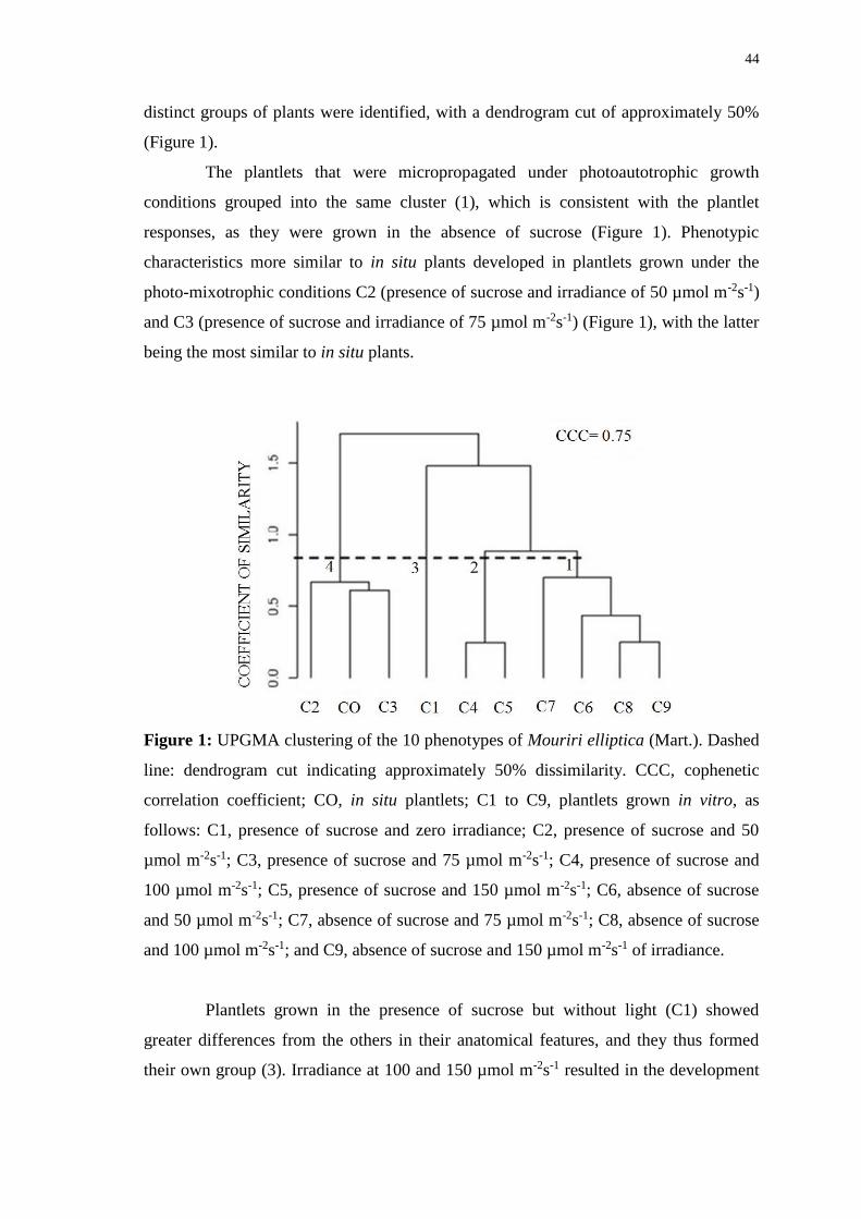
44
distinct groups of plants were identified, with a dendrogram cut of approximately 50%
(Figure 1).
The plantlets that were micropropagated under photoautotrophic growth
conditions grouped into the same cluster (1), which is consistent with the plantlet
responses, as they were grown in the absence of sucrose (Figure 1). Phenotypic
characteristics more similar to in situ plants developed in plantlets grown under the
photo-mixotrophic conditions C2 (presence of sucrose and irradiance of 50 µmol m-2s-1)
and C3 (presence of sucrose and irradiance of 75 µmol m-2s-1) (Figure 1), with the latter
being the most similar to in situ plants.
Figure 1: UPGMA clustering of the 10 phenotypes of Mouriri elliptica (Mart.). Dashed
line: dendrogram cut indicating approximately 50% dissimilarity. CCC, cophenetic
correlation coefficient; CO, in situ plantlets; C1 to C9, plantlets grown in vitro, as
follows: C1, presence of sucrose and zero irradiance; C2, presence of sucrose and 50
µmol m-2s-1; C3, presence of sucrose and 75 µmol m-2s-1; C4, presence of sucrose and
100 µmol m-2s-1; C5, presence of sucrose and 150 µmol m-2s-1; C6, absence of sucrose
and 50 µmol m-2s-1; C7, absence of sucrose and 75 µmol m-2s-1; C8, absence of sucrose
and 100 µmol m-2s-1; and C9, absence of sucrose and 150 µmol m-2s-1 of irradiance.
Plantlets grown in the presence of sucrose but without light (C1) showed
greater differences from the others in their anatomical features, and they thus formed
their own group (3). Irradiance at 100 and 150 µmol m-2s-1 resulted in the development
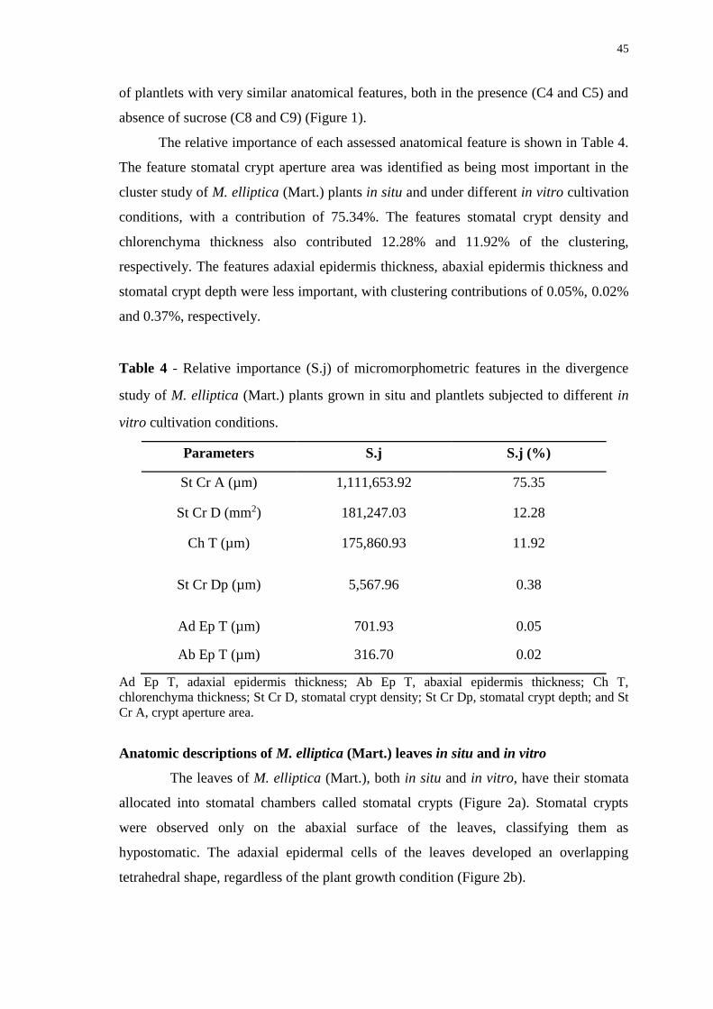
45
of plantlets with very similar anatomical features, both in the presence (C4 and C5) and
absence of sucrose (C8 and C9) (Figure 1).
The relative importance of each assessed anatomical feature is shown in Table 4.
The feature stomatal crypt aperture area was identified as being most important in the
cluster study of M. elliptica (Mart.) plants in situ and under different in vitro cultivation
conditions, with a contribution of 75.34%. The features stomatal crypt density and
chlorenchyma thickness also contributed 12.28% and 11.92% of the clustering,
respectively. The features adaxial epidermis thickness, abaxial epidermis thickness and
stomatal crypt depth were less important, with clustering contributions of 0.05%, 0.02%
and 0.37%, respectively.
Table 4 - Relative importance (S.j) of micromorphometric features in the divergence
study of M. elliptica (Mart.) plants grown in situ and plantlets subjected to different in
vitro cultivation conditions.
Parameters S.j S.j (%)
St Cr A (µm) 1,111,653.92 75.35
St Cr D (mm2) 181,247.03 12.28
Ch T (µm) 175,860.93 11.92
St Cr Dp (µm) 5,567.96 0.38
Ad Ep T (µm) 701.93 0.05
Ab Ep T (µm) 316.70 0.02
Ad Ep T, adaxial epidermis thickness; Ab Ep T, abaxial epidermis thickness; Ch T,
chlorenchyma thickness; St Cr D, stomatal crypt density; St Cr Dp, stomatal crypt depth; and St
Cr A, crypt aperture area.
Anatomic descriptions of M. elliptica (Mart.) leaves in situ and in vitro
The leaves of M. elliptica (Mart.), both in situ and in vitro, have their stomata
allocated into stomatal chambers called stomatal crypts (Figure 2a). Stomatal crypts
were observed only on the abaxial surface of the leaves, classifying them as
hypostomatic. The adaxial epidermal cells of the leaves developed an overlapping
tetrahedral shape, regardless of the plant growth condition (Figure 2b).
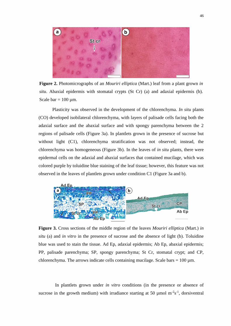
46
Figure 2. Photomicrographs of an Mouriri elliptica (Mart.) leaf from a plant grown in
situ. Abaxial epidermis with stomatal crypts (St Cr) (a) and adaxial epidermis (b).
Scale bar = 100 µm.
Plasticity was observed in the development of the chlorenchyma. In situ plants
(CO) developed isobilateral chlorenchyma, with layers of palisade cells facing both the
adaxial surface and the abaxial surface and with spongy parenchyma between the 2
regions of palisade cells (Figure 3a). In plantlets grown in the presence of sucrose but
without light (C1), chlorenchyma stratification was not observed; instead, the
chlorenchyma was homogeneous (Figure 3b). In the leaves of in situ plants, there were
epidermal cells on the adaxial and abaxial surfaces that contained mucilage, which was
colored purple by toluidine blue staining of the leaf tissue; however, this feature was not
observed in the leaves of plantlets grown under condition C1 (Figure 3a and b).
Figure 3. Cross sections of the middle region of the leaves Mouriri elliptica (Mart.) in
situ (a) and in vitro in the presence of sucrose and the absence of light (b). Toluidine
blue was used to stain the tissue. Ad Ep, adaxial epidermis; Ab Ep, abaxial epidermis;
PP, palisade parenchyma; SP, spongy parenchyma; St Cr, stomatal crypt; and CP,
chlorenchyma. The arrows indicate cells containing mucilage. Scale bars = 100 µm.
In plantlets grown under in vitro conditions (in the presence or absence of
sucrose in the growth medium) with irradiance starting at 50 µmol m-2s-1, dorsiventral
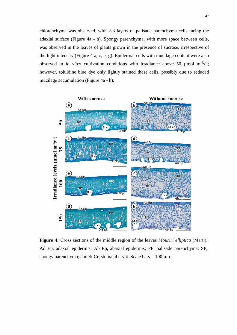
47
chlorenchyma was observed, with 2-3 layers of palisade parenchyma cells facing the
adaxial surface (Figure 4a - h). Spongy parenchyma, with more space between cells,
was observed in the leaves of plants grown in the presence of sucrose, irrespective of
the light intensity (Figure 4 a, c, e, g). Epidermal cells with mucilage content were also
observed in in vitro cultivation conditions with irradiance above 50 µmol m-2s-1;
however, toluidine blue dye only lightly stained these cells, possibly due to reduced
mucilage accumulation (Figure 4a - h).
Figure 4: Cross sections of the middle region of the leaves Mouriri elliptica (Mart.).
Ad Ep, adaxial epidermis; Ab Ep, abaxial epidermis; PP, palisade parenchyma; SP,
spongy parenchyma; and St Cr, stomatal crypt. Scale bars = 100 µm.
48
2.4 Discussion
Anatomical plasticity between M. elliptica (Mart.) plantlets grown in vitro and in
situ plants generates 4 distinct groups after UPGMA clustering
Based on the phenotypic variations in the micromorphometric data from M.
elliptica (Mart.) leaves grown in situ and in vitro, it was possible to estimate the
dissimilarity between them. According to Cruz et al. (2011), phenotypic characteristics
typically show a continuous distribution and are determined by many genes with small
individual contributions while being influenced by the environment. Thus, it was
possible to determine which in vitro cultivation conditions resulted in leaf development
with anatomical characteristics that were less dissimilar to the in situ plants.
The leaf anatomic characteristics that developed on the in vitro plantlets are
important for adaptation to growth conditions, as they influence physiological
processes, especially the ability to perform photosynthesis. In plantlets grown in the
presence of sucrose but without light (C1), there was no stratification of the
chlorenchyma; instead, it was homogeneous, thus demonstrating little tissue
differentiation. Light intensities greater than 50 µmol m-2s-1 (C2 to C9) facilitated better
leaf development, with stratification of the chlorenchyma into palisade and spongy
zones of the dorsiventral type. The anatomical plasticity observed for leaves of M.
elliptica (Mart.) due to cultivation conditions represents the acclimatization capacity of
the species.
The presence of stomatal crypts is an important feature of in situ plants that
was also observed for in vitro plants. Stomatal crypts are considered as features that
characterize the species in their natural habitat; many species are found in arid
environments (Jordan et al., 2008). Crypts favor the development of plants in such
environments, as they restrict transpiration, reducing water loss and promoting gas
exchange at appropriate times (Hassiotou et al., 2009).
The anatomical parameters St Cr A and St Cr D together accounted for 87.63%
of the relative importance (S.j) in the UPGMA cluster. Therefore, these characteristics
were considered as key factors for the dissimilarity study between M. elliptica (Mart.)
plants cultivated in situ and in vitro. Four different groups were obtained with UPGMA
clustering, based on a dendrogram cut indicating approximately 50% dissimilarity. The
CCC was 0.75, allowing us to infer that the clustering was consistent. Silva and Dias
(2013) consider the assessment of cluster consistency by the CCC very important so that
the conclusions on similarities between individuals may be considered trustworthy.

49
According to Cruz and Carneiro (2006), a higher CCC value corresponds to a lower
clustering distortion.
None of the in vitro conditions used in this study permitted the formation of
plantlets with anatomical features that were similar to the in situ plants, but the C2 and
C3 conditions, both of which were photomixotrophic, formed plantlets that were less
dissimilar to in situ plants. This information suggests that these plants have the highest
chances of survival when subjected to ex vitro conditions.
The first study on micropropagation of the species M. elliptica (Mart.) revealed
the regeneration ability of plantlets under photoautotrophic conditions and with
irradiance above 50 µmol m-2s-1 (Assis et al., 2016). In the present study, the clustering
analysis between in situ and in vitro plantlets based on leaf anatomical features showed
that plantlets cultivated under a photoautotrophic system were more dissimilar to in situ
plantlets. However, more studies on micropropagation of the species should be
conducted in which plants are taken to the acclimatization stage to ensure their
survivability, as observed in studies of Corrêa et al. (2015) on the interactions between
genotypes of P. glomerata (Spreng.) in the photoautotrophic culture and studies by
Rodrigues et al. (2015) with Etlingera elatior (Jack) rm smith (torch ginger).
Several studies on plant micropropagation have been developed to obtain
plantlets with anatomical and physiological characteristics that increase their ability to
survive the acclimatization process, as it is a stressful stage for the plant. Among the
successfully developed studies cited with native plants Byrsonima cydoniifolia A. Juss.
(Martendal et al., 2014), Billbergia zebrine (Martins et al., 2015), and cultivated plants
of commercial importance as Carica papaya L. var. Red and Maradol (Pérez et al.,
2015).
2.4 Conclusion
The anatomical characteristics studied in the leaves of M. elliptica (Mart.)
supported a dissimilarity study between plants grown in situ and those cultivated in
vitro under photomixotrophic and photoautotrophic conditions. UPGMA clustering was
used to determine that in vitro cultivation conditions in the presence of sucrose and
irradiances of 50 and 75 µmol m-2s-1 supported the growth of plantlets with leaf
anatomical features that were less dissimilar to in situ plants that were placed in the
same group.
50
2.5 Conflicts of interest
The authors declare no conflict of interest.
2.6 Acknowledgments
Federal Institute of Science Education and Technology Goiano, Rio Verde
Campus – Goiás, Brazil, for the infrastructure and the experimental material. Goias
Research Foudation (Fundação de Amparo à Pesquisa do Estado de Goiás – FAPEG)
and the Brazilian Federal Agency for the Support and Evaluation of Graduate Education
(Coordenação de Aperfeiçoamento de Pessoal de Nível Superior – CAPES) for financial
support.
2.7 References
Arnott HJ (1959) Leaf clearings. Turtox News 37: 192-194.
Assis ES, Reis EF, Pinto JFN, Contim LAS, et al. (2013). Genetic diversity of gabiroba
based on random amplified polymorphic DNA markers and morphological
characteristics. Genet. Mol. Res. 12: 3500-3509.
http://dx.doi.org/10.4238/2013.March.11.7
Assis ES, Rubio Neto A, Lima LR, Silva FG, et al. (2016). In vitro culture of Mouriri
elliptica (Mart.) under conditions that stimulate photoautotrophic behavior. Aust.
J. of Crop Science 10: 229-236.
Assis KC, Silva FG, Pereira FD, Vasconcelos-Filho SC, et al. (2015). Effects of
photomixotrophic conditions and type of culture vessel closure on Anacardium
othonianum Rizz. grown in vitro. Acta Hort. 1083: 553-564.
http://dx.doi.or/10.17660/ActaHortic.2015.1083.74
Bonacorsi C, Fonseca LM, Raddi MSG, Kitagawa RR, et al. (2013). Comparison of
Brasilian plantas used to treat gastritis on the oxidative burst of Helicobacter
pylori-stimulatad neutrophil. Ev-Based Comp. Alternat. Med. 2013: 851621.
http://dx.doi.org/10.1155/2013/851621
Corrêa JPO, Vital C, Pinheiro MVMA, Batista DS, et al. (2015). In vitro
photoautotrophic potential and ex vitro photosynthetic competence of Pfaffia
glomerata (Spreng.) Pedersen accessions. P. Cell Tissue Org. Cult. 122: 289-
300. http://dx.doi.org/10.1007/s11240-014-0700-4
Crus CD, Ferreira FM, Pessoni LA (2011). Biometria aplicada ao estudo da diversidade
genética. Visconde de Rio Branco, MG: Suprema.
51
Cruz CD and Carneiro PCS (2006). Modelos biométricos aplicados ao melhoramento
genético. 2. Ed. Viçosa: UFV.
Cruz CD (2013). GENES - A software package for analysis in experimental statistics
and quantitative genetics. Acta Scientiarum 35: 271-276.
http://dx.doi.org/10.4025/actasciagron.v35i3.21251
Hassiotou F, Evans JR, Ludwig M, Veneklaas EJ (2009). Stomatal crypts may facilitate
diffusion of CO2 to adaxial mesophyll cells in thick sclerophylls. P. Cell and
Env. 32: 1596-1611. http://dx.doi.org/10.1111/j.1365-3040.2009.02024.X
Iarema L, Cruz ACF, Saldanha CW, Dias LLC, et al. (2012). Photoautotrophic
propagation of Brazilian ginseng [Pfaffia glomerata (Spreng.) Pedersen]. P. Cell
Tissue Org. Cult. 110: 227–238. http://dx.doi.org/10.1007/s11240-012-0145-6
Jordan GJ, Weston PH, Carpenter RJ, Dillon RA, et al. (2008). The evolutionary
relations of sunken, covered, and encrypted stomata to dry habitats in
Proteaceae. A. J. of Botany 95: 521–530. http://dx.doi.org/10.3732/ajb.2007333
Kaçar YA, Biçen B, Varol I, Mendi YY, et al. (2010). Gelling agents and culture
vessels affect in vitro multiplication of banana plantlets. Genet. Mol. Res. 9:
416-424. http://dx.doi.org/10.4238/vol9-1gmr744
Karnovsky MJ (1965) A formaldehyde-glutaraldehyde fixative of high osmolality for
use in electron microscopy. J. of Cell Biology 27: 137–138.
Leo SD, Cosmi C, Ragosta M (2015). An application of multivariate statistical
techniques to partial equilibrium models outputs: The analysis of the NEEDS-
TIMES Pan European model results. Ren. Sust. Energy Reviews 49: 108–120.
http://dx.doi.org/10.1016/j.rser.2015.04.099
Lima LR, Rubio Neto A, Pereira FD, Silva FG, et al. (2016) Germination and
emergence of Mouriri elliptica Mart., a rare medicinal fruit tree native to the
Brazilian Cerrado biom. African J. of Agric. Research 11: 400-406.
http://dx.doi.org/10.5897/AJAR2015.10444
Ma X, Zuo H, Tian M, Zhang L, et al. (2016). Assessment of heavy metals
contamination in sediments from three adjacent regions of the Yellow River
using metal chemical fractions and multivariate analysis techniques.
Chemosphere 44: 264-272. http://dx.doi.org/10.1016.chemosphere.2015.08.026.
Maechler M (2015). Cluster analysis extended Rousseeuw. Accessed:
http:brieger.esalq.usp.br/CRAN/. Accessed on March, 15, 2015.

52
Martendal CO, Bernardino MM, Pereira FD, Silva FG, et al. (2013). In vitro cultivation
of zygotic embryos from Murici (Byrsonima cydoniifolia A. Juss.):
establishment, disinfection, and germination. Acta Scient. Agronomy 35: 221-
229. http://dx.doi.org/10.4025/actasciagron.v35i2.15402
Martendal CO, Bernardino MM, Pereira FD, Silva FG, et al. (2014). In vitro
multiplication of nodal segments of “Murici” (Byrsonima cydoniifolia A. Juss.):
the use of growth regulators and photoautotrophic stimulation. J. of Agric.
Technology 10: 665-678.
Martins JPR, Verdoodt V, Paqual M, Proft M (2015). Impacts of photoautotrophic and
photomixotrophic conditions on in vitro propagated Bilbergia zebrine
(Bromeliaceae). P. Cell Tissue Org. Cult. 123: 121-132.
http://dx.doi.org/10.1007/s11240-015-0820-5
Moleiro FC, Andreo MA, Santos RC, Moraes TM, et al. (2009). Mouriri elliptica:
Validation of gastroprotective, healing and anti-Helicobaster pylori effects. J. of
Ethnopharmacology 123: 359-368. http://dx.doi.org/0.1016/j.jep.2009.03.040.
Epub 2009 Apr 5.
O’Brien TP, Feder N, McCully ME (1965). Polychromatic staining of plant cell walls
by toluidine blue O. Protoplasma 59: 368-73.
Oda MC, Sediyama T, Matsuo É, Crus CD, et al. (2015). Phenotypic and molecular
traits diversity in soybean launched in forty years of genetic breeding. Agro.
Science and Biotec. 1: 1 – 9.
Pérez LP, Montesinos YP, Olmedo JG, Sánchez RR, et al. (2015). Effects of different
culture conditions (photoautotrophic, photomixotrophic) and the auxin indole-
butyric acid on the in vitro acclimatization of papaya (Carica papaya L. var. Red
Maradol) plants using zeolite as support. African J Biotechnol. 14: 2622-2635.
http://dx.doi.org/10.5897/AJB2015.14814
Rodrigues M, Paiva PDO, Freitas RT, Mansur TOF, et al. (2015). Growth and
photosynthetic responses during ex vitro acclimatization of Etlingera elatior
(Jack) rm smith (torch ginger). Acta Scientiarum, 37: 1807-8621.
http://dx.doi.org/10.4025/actasciagron.v37i4.19707
Rout GR, Mohapatra A, Mohan Jain S (2006). Tissue culture of ornamental pot plant: A
critical review on presente scenario and future prospects. Biotech. Adv. 24: 531-
560. http://dx.doi.org/10.1016/j.biotechadv.2006.05.001
53
Rufino MSM, Alves RE, Brito ES, Pérez-Jiménez J, et al. (2010). Bioactive compounds
and antioxidant capacities of 18 non-traditional tropical fruits from Brazil. Food
Chemistry 121: 996–1002. http://dx.doi.org/10.1016/j.foodchem.2010.01.037
Rufino MSM, Alves RE, Fernandes FAN, Brito ES (2011). Free radical scavenging
behavior of tem exotic tropical fruits extracts. Food Res. Internat. 44: 2072-
2075. http://dx.doi.org/10.1016/j.foodres.2010.07.002
Sáez PL, Bravo LA, Latsague MI, Sánchez MR, et al. (2012). Increased light intensity
during in vitro culture improves water loss control and photosynthetic
performance of Castanea sativa grown in ventilated vessels. Sci. Horticult. 138:
7–16. http://dx.doi.org/10.1016/j.scienta.2012.02.005
Saldanha CW, Otoni CG, Rocha DI, Cavatte PC, et al. (2014). CO2-enriched
atmosphere and supporting material impact the growth, morphophysiology and
ultrastructure of in vitro Brazilian-ginseng [pfaffia glomerata (spreng.) pedersen]
plantlets. P. Cell Tissue Org. Cult. 118: 87-99.
http://dx.doi.org/10.1016/j.scienta.2012.02.005
Shin K-S., Park S-Y, Paek K-Y (2014). Physiological and biochemical changes during
acclimatization in a Doritaenopsis hibrid cultivated in different
microenvironments in vitro. Envir. and Exp. Botany 100: 26-33.
http://dx.doi.org/10.1016/j.envexpbot.2013.12.004
Silva AR and Dias TS (2013). A cophenetic correlation coefficient for Tocher’s method.
Pesq. Agropec. Bras. 48: 589-596. http://dx.doi.org/10.1590/S0100-
204X2013000600003
Silva FL, Baffa DCF, Rezende JC, Oliveira ACB, et al. (2015). Variabilidade genética
entre genótipos de café robusta no estado de Minas gerais. Coffee Science 10: 20
– 27.
Singh D (1981). The relative importance of characters affecting genetic divergence. The
India J. of Genetcs and P. Breeding 41: 237-245.
Vasconcelos JM, Cardoso TV, Sales JF, Silva FG, et al. (2010) Métodos de superação
de dormência em sementes de croada (Mouriri elliptica Mart.). Ciênc. Agrotec.
34: 1199-1204. http://dx.doi.org/10.1590/S1413-70542010000500017
Vasconcelos PCP, Andreo MA, Vilegas W, Hiruma-Lima CA, et al. (2010). Effect of
Mouriri pusa tannins and flavonoids on prevention and treatment against
experimental gastric ulcer. J. of Ethnopharmacology 131: 146–153.
http://dx.doi.org/10.1016/j.jep.2010.06.017.

54
Xiao Y and Kozai T (2006). In vitro multiplication of statice plantlets using sugar-free
media. Sci. Hort. 109: 71–77. http://dx.doi.org/10.1016/j.scienta.2006.02.029
55
CAPÍTULO III
(Normas de acordo com a revista Cerne, em processo de revisão)
Alternative support materials to agar in the in vitro cultivation of Mouriri elliptica
(Mart.)
Abstract
Alternative supports can be successfully used in place of agar for in vitro
culture to increase seedling quality and subsidize root formation. The objective of the
present study was to evaluate the efficiency of alternative support materials compared to
agar in the in vitro cultivation of Mouriri elliptica (Mart.) in the absence or presence of
Naphthalene Acetic Acid (NAA). Nodal segments were grown in 50% salt Wood Plant
Medium, with 30 gL-1 of sucrose and 2,5 gL-1 of activated charcoal. The alternative
support materials used were medium-granulometry vermiculite, sugarcane (Saccharum
spp. L.) bagasse and queen palm fiber [Syagrus romanzoffiana (Chamisso) Glassman]
in compared to culture medium solidified with agar. No differences were observed
between agar, vermiculite and sugarcane bagasse cultures for growth characteristics
number of nodal segments, number of leaves and total dry mass. Greater numbers of
adventitious and secondary roots and greater root length were observed in plantlets
grown in the cultivation of vermiculite and the absence of NAA. In the agar culture,
roots had weak points and poorly differentiated tissues, with parenchymal tissue
predominating. The concentration of 2.0 mg L-1 NAA used this study did not stimulate
rooting of M. elliptica (Mart.) plantlets. It was possible to regenerate plantlets in both
support materials used, with vermiculite and sugarcane bagasse representing promising
agar substitutes to obtain seedlings with roots.
56
Keywords: Anatomical characteristics, croada, melastomataceae, micropropagation,
rooting.
3.1 Introduction
Mouriri elliptica (Mart.) (Melastomataceae) is a tree typical of the Cerrado
domain (Brazil) and is popularly known as coroa de frade (friar’s crown), croada or
croadinha and puçá ou puçazeiro. In addition to its importance to the forest, its fruits
rich in nutrient and antioxidant compounds have been recommended for human
consumption (RUFINO et al., 2011). Its trunk and leaves can be used medicinally to
treat gastric ulcers and gastritis (MOLEIRO et al., 2009). These characteristics
demonstrate the economic potential of the species, however, it is little known and
studied.
Pioneering studies, such as those by Vasconcelos et al. (2010) and De Lima et
al. (2016), reported the difficulty of producing M. elliptica (Mart.) seedlings from seeds,
justifying the use of alternative methods for mass propagation. The in vitro propagation
technique is a viable tool to produce seedlings of wild or domesticated species that are
difficult to propagate by conventional methods, accelerate plantlet production (ASSIS et
al., 2012; MARTENDAL et al., 2014; MALI; CHAVAN, 2016) and conservation of
endangered species (PATEL et al., 2014).
In the in vitro propagation, the agar is the most widely used support material
for explants in culture medium. However, problems have been reported in plantlets
grown in agar medium, such as poor root formation, resulting in losses (XIAO et al.,
2011). These observations, coupled with the abundant use of agar, make this agent
costly for in vitro multiplication (BRAGA et al., 2013). Thus, alternative support
materials that can reduce production costs and possibly improve plantlet vigor,
facilitating their acclimatization, have been tested (MOHAN et al., 2005; SALDANHA
et al., 2014).
In addition to the need to use alternative support, the suitability of the culture
medium and the use of growth regulators are fundamental. In the in vitro propagation,
growth regulators are use to the induction of cellular division and root differentiation
(Navarro-García et al., 2016; ABDULMALIK et al., 2012). IBA (indole-3-butyric acid),
IAA (indole-3-acetic acid) and NAA (naphthalene acetic acid) are the auxins generally
used for in vitro rooting of plants (BARPETE et al., 2014).
57
The endogenous auxin level greatly influences root induction, and the
application of plant growth regulators can significantly increase either low or high
concentrations of auxin (HOSSAIN; URBI, 2016). For some species in vitro rooting
acclimatization aiming unnecessary (AINA et al., 2015; SHEKHAWAT; MANOKARI,
2016). Studies of the interaction between growth regulators and culture medium support
materials have been rare and are of extreme importance.
The first study on vegetative propagation of M. elliptica (Mart.) by means of
tissue culture was conducted by Assis et al. (2016). In that study, it was possible to
produce plantlets in traditional and photoautotrophic culture conditions by increasing
the irradiance (50 to 150 µmol m-2s-1) in the environment. However, the importance of
developing new methods associated with the photoautotrophic system that enhance the
in vitro production of seedlings of this species was discussed, citing the use of materials
as alternative supports to agar.
Studies evaluating the interaction between different media materials with
growth regulator are non - existent for propagation of the species. Thus, this study
sought to evaluate the influence of support materials alternative to agar for the in vitro
rooting of plantlets of this species in the presence or absence of NAA, making the
plantlets more resistant.
3.2 Material and methods
Collection of fruits, plantlets and explants, disinfection and inoculation
Plantlets with at least two axillary buds were used as explant sources, as they
had nodal segments 1.5 cm in length. The disinfection process followed, in which the
explants were wrapped with gauze and placed under running water with three drops of
neutral detergent for 15 minutes. In a laminar flow hood, the explants were immersed in
70% (v/v) ethanol for 30 seconds and then submerged in 20% sodium hypochlorite -
NaOCl solution (commercial bleach 2.0 – 2.5% active chlorine) for 15 minutes. To
complete the disinfection, the explants were washed three times in distilled and
autoclaved water.
The explants were subsequently inoculated into Magenta® culture flasks
containing the alternative support materials or agar for plant culture. The alternative
support materials studied were sugarcane bagasse (sugarcane B.) (Saccharum spp. L.),
queen palm fiber (queen palm F.) [Syagrus romanzoffiana (Chamisso) Glassman] and
medium-grain vermiculite. The influence of the growth regulator naphthalene acetic
58
acid (NAA) (Sigma®) on plantlet rooting was also studied at concentrations of 0.0 and
2.0 mg L-1. The nutrient medium WPM (Wood Plant Medium) developed by Lloyd and
McCown 1981 with 50% salts, 30 g L-1 sucrose, 3.5 g L-1 agar (Dinâmica®) and 2 g L-1
activated carbon at (Synth®) was used, and a volume of 50 mL was used in each
Magenta® culture flasks, which was sealed with a polypropylene closure. The pH of the
growth medium was adjusted to 5.7 ± 0.03 before autoclaving at 121°C for 20 minutes.
Five grams of alternative support materials was used in each Magenta® culture
containers. The volume of culture medium used was determined by calculating the
amount of water to substrate (BRASIL, 2009), with some modifications. Thus, the
support material (5 g) and the known volume of water (100 mL) were placed in a funnel
with filter paper and left to rest for 45 minutes to drain the water. The volume retained
in the support material was used. Therefore, the volumes of liquid culture media (WPM,
with 50%) used in 5 g of alternative support material were 41.0; 30.0 and 21.3 mL for
sugarcane B., queen palm F. and vermiculite, respectively.
Water loss through evaporation was measured every 3 days in flasks containing
the alternative support materials without plant culture using an analytical balance. The
observed water loss was 0.24 mL day-1 per flasks. The culture medium in each support
material was replenished at 15 and 30 days of in vitro culture. The culture was
maintained in growth room for 45 days under light intensity of 45.
Purification of sugarcane B. and queen palm F. support materials for in vitro
cultivation
To obtain the queen palm F., ripe fruits were placed in a fruit and vegetable
depulper (Ker Mod. 1.5, Tortugan®) for 40 minutes to separate the epicarp and
mesocarp (total pulp) from the diaspore (endocarp with seed). The dry sugarcane B. was
derived from the Nova Gale located in Acreúna, Goiás, Brazil.
The fibrous support materials were washed in running water, using the
methodology of Mohan et al. (2005) as a reference. For further purification of queen
palm F., 10 consecutive washes with running water were necessary. Lastly, the fibrous
material was washed 2 times with distilled water (heated to 95 ± 5 ºC).
Both queen palm F. and sugarcane B. were dried in an oven at 40°C for 72
hours and were then ground using a Willye (TE-650) grinder with a 5-mm sieve. Before
using the supporting materials, including the vermiculite, they were washed with
59
distilled water to remove ions. Afterwards, they were dried in an oven and autoclaved at
121 ºC for 20 min.
Physical characterization of the alternative support materials
The physical characteristics of the alternative substrates were evaluated
according to the recommendations of Zorzeto et al. (2014) and following the standard
instructions established by the Brazilian Ministry of Agriculture, Livestock and Supply
(BRASIL, 2007), the official Brazilian government agency that regulates the use of
substrates for plants destined for agriculture.
The wet bulk density (WD) was determined from the ratio of the mass
occupied by the substrate to the volume at current moisture in a 250-cm3 plastic beaker
dropped under the action of its own weight from a height of 10 cm ten consecutive
times (Brasil, 2008). After autocompaction, the samples were dried in an oven at 65°C
to constant weight (BRASIL, 2007), and the values were used to determine the dry bulk
density (DD).
Other substrate samples were packed in PVC cylinders measuring 4 cm in
diameter and 5 cm in height and subjected to saturation with distilled water for 24 hours
at 10 to 100 hPa to determine water retention curves (DE BOODT; VERDONCK, 1972;
BRASIL, 2008). The following parameters were determined: total porosity (TP), which
considers the volumetric water content present in the saturated samples (0 hPa); aeration
space (AS), the difference between the total porosity and volumetric water content at 10
hPa; available water (AW), which corresponds to the volume of water between 10 and
100 hPa; and remaining water (RW), the amount of water remaining in the sample after
it was subjected to 100 hPa matric potential and equivalent to micropore water.
Growth evaluations
Evaluations were performed after 45 days of in vitro cultivation using the
characteristics of plantlet length (cm), number of nodal segments, number of leaves,
total dry mass (mg), numbers of adventitious and secondary roots, length of largest root
(cm) and water content of plantlets (%). Measurements of length were obtained using a
millimeter ruler.
To obtain the dry mass, the plant material remained in a ventilation oven forced
to a temperature of 65 ºC for 72 hours, and weighing was performed on a digital
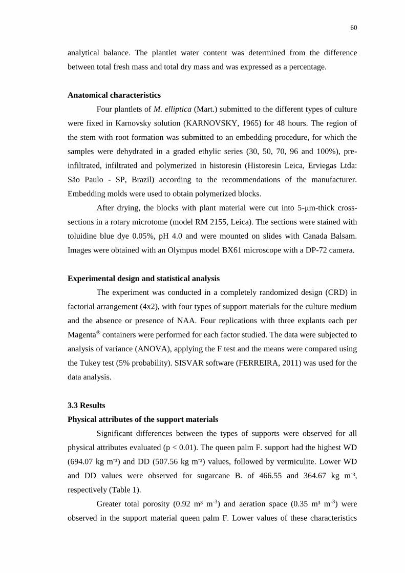
60
analytical balance. The plantlet water content was determined from the difference
between total fresh mass and total dry mass and was expressed as a percentage.
Anatomical characteristics
Four plantlets of M. elliptica (Mart.) submitted to the different types of culture
were fixed in Karnovsky solution (KARNOVSKY, 1965) for 48 hours. The region of
the stem with root formation was submitted to an embedding procedure, for which the
samples were dehydrated in a graded ethylic series (30, 50, 70, 96 and 100%), pre-
infiltrated, infiltrated and polymerized in historesin (Historesin Leica, Erviegas Ltda:
São Paulo - SP, Brazil) according to the recommendations of the manufacturer.
Embedding molds were used to obtain polymerized blocks.
After drying, the blocks with plant material were cut into 5-μm-thick cross-
sections in a rotary microtome (model RM 2155, Leica). The sections were stained with
toluidine blue dye 0.05%, pH 4.0 and were mounted on slides with Canada Balsam.
Images were obtained with an Olympus model BX61 microscope with a DP-72 camera.
Experimental design and statistical analysis
The experiment was conducted in a completely randomized design (CRD) in
factorial arrangement (4x2), with four types of support materials for the culture medium
and the absence or presence of NAA. Four replications with three explants each per
Magenta® containers were performed for each factor studied. The data were subjected to
analysis of variance (ANOVA), applying the F test and the means were compared using
the Tukey test (5% probability). SISVAR software (FERREIRA, 2011) was used for the
data analysis.
3.3 Results
Physical attributes of the support materials
Significant differences between the types of supports were observed for all
physical attributes evaluated (p < 0.01). The queen palm F. support had the highest WD
(694.07 kg m-³) and DD (507.56 kg m-³) values, followed by vermiculite. Lower WD
and DD values were observed for sugarcane B. of 466.55 and 364.67 kg m-³,
respectively (Table 1).
Greater total porosity (0.92 m³ m-3) and aeration space (0.35 m³ m-3) were
observed in the support material queen palm F. Lower values of these characteristics
61
were observed in the support material vermiculite, with values of 0.57 and 0.15 m³ m-3,
respectively. In sugarcane B., the TP was 0.73 m³ m-3, which was between the values
detected for queen palm F. and vermiculite. The AS of sugarcane B. was 0.12 m³ m-3
and did not differ from the value observed for vermiculite (Table 1).
Table 1 - Physical characteristics of the alternative support materials used for in vitro
cultivation of M. elliptica (Mart.) plantlets. Total porosity (TP), available water (AW),
aeration space (AS), remaining water (RW), wet density (WD) and dry density (DD).
Characteristics Support materials
Vermiculite Sugarcane B. Queen palm F.
TP (m³ m-3) 0.57 ± 0.01cz1 0.71 ± 0.00 b 0.93 ± 0.01 a
AW (m³ m-3) 0.08 ± 0.00 b 0.38 ± 0.02 a 0.35 ± 0.03 a
AS (m³ m-3) 0.13 ± 0.00 b 0.12 ± 0.01 b 0.35 ± 0.02 a
RW (m³ m-3) 0.34 ± 0.00 a 0.23 ± 0.01 b 0.22 ± 0.01 b
WD (kg m-³) 654.13 ± 2.73 b 466.55 ± 2.86 c 694.07 ± 4.77 a
DD (kg m-³) 493.83 ± 1.82 b 364.67 ± 2.44 c 507.56 ± 3.27 a
zMeans followed by the same letter in rows do not differ among according to the Tukey,
p < 0.05. 1± Standard error from the mean.
Higher AW values were obtained for sugarcane B. (0.38 m³ m-3) and queen
palm F. (0.35 m³ m-3), and lower RW values were observed in these two support
materials: 0.23 m³ m-3 in sugarcane B. and 0.22 m³ m-3 in queen palm F. A lower AW
value (0.08 m³ m-3) and higher RW value (0.34 m³ m-3) were detected in vermiculite
(Table 1).
In vitro regeneration of M. elliptica (Mart.) plantlets in different culture medium
support materials in the presence or absence of NAA
The growth patterns of the plantlets after 45 days of in vitro cultivation in
culture medium support materials agar, sugarcane B., queen palm F. and vermiculite can
be seen in Figure 1 (A – F). Greater plantlet length (2.85 cm) occurred in the agar
culture, and shorter length (1.69 cm) occurred in the queen palm F. culture (Figure 2A).
An increase of 86.66% in plantlet shoot length was seen when plantlets were grown in
agar medium, with an initial explant length of 1.50 cm as the base. Increases in growth
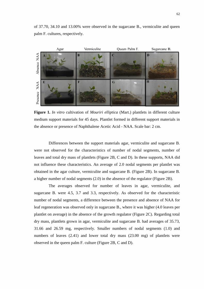
62
of 37.70, 34.10 and 13.00% were observed in the sugarcane B., vermiculite and queen
palm F. cultures, respectively.
Figure 1. In vitro cultivation of Mouriri elliptica (Mart.) plantlets in different culture
medium support materials for 45 days. Plantlet formed in different support materials in
the absence or presence of Naphthalene Acetic Acid - NAA. Scale bar: 2 cm.
Differences between the support materials agar, vermiculite and sugarcane B.
were not observed for the characteristics of number of nodal segments, number of
leaves and total dry mass of plantlets (Figure 2B, C and D). In these supports, NAA did
not influence these characteristics. An average of 2.0 nodal segments per plantlet was
obtained in the agar culture, vermiculite and sugarcane B. (Figure 2B). In sugarcane B.
a higher number of nodal segments (2.0) in the absence of the regulator (Figure 2B).
The averages observed for number of leaves in agar, vermiculite, and
sugarcane B. were 4.5, 3.7 and 3.3, respectively. As observed for the characteristic
number of nodal segments, a difference between the presence and absence of NAA for
leaf regeneration was observed only in sugarcane B., where it was higher (4.0 leaves per
plantlet on average) in the absence of the growth regulator (Figure 2C). Regarding total
dry mass, plantlets grown in agar, vermiculite and sugarcane B. had averages of 35.73,
31.66 and 26.59 mg, respectively. Smaller numbers of nodal segments (1.0) and
numbers of leaves (2.41) and lower total dry mass (23.00 mg) of plantlets were
observed in the queen palm F. culture (Figure 2B, C and D).
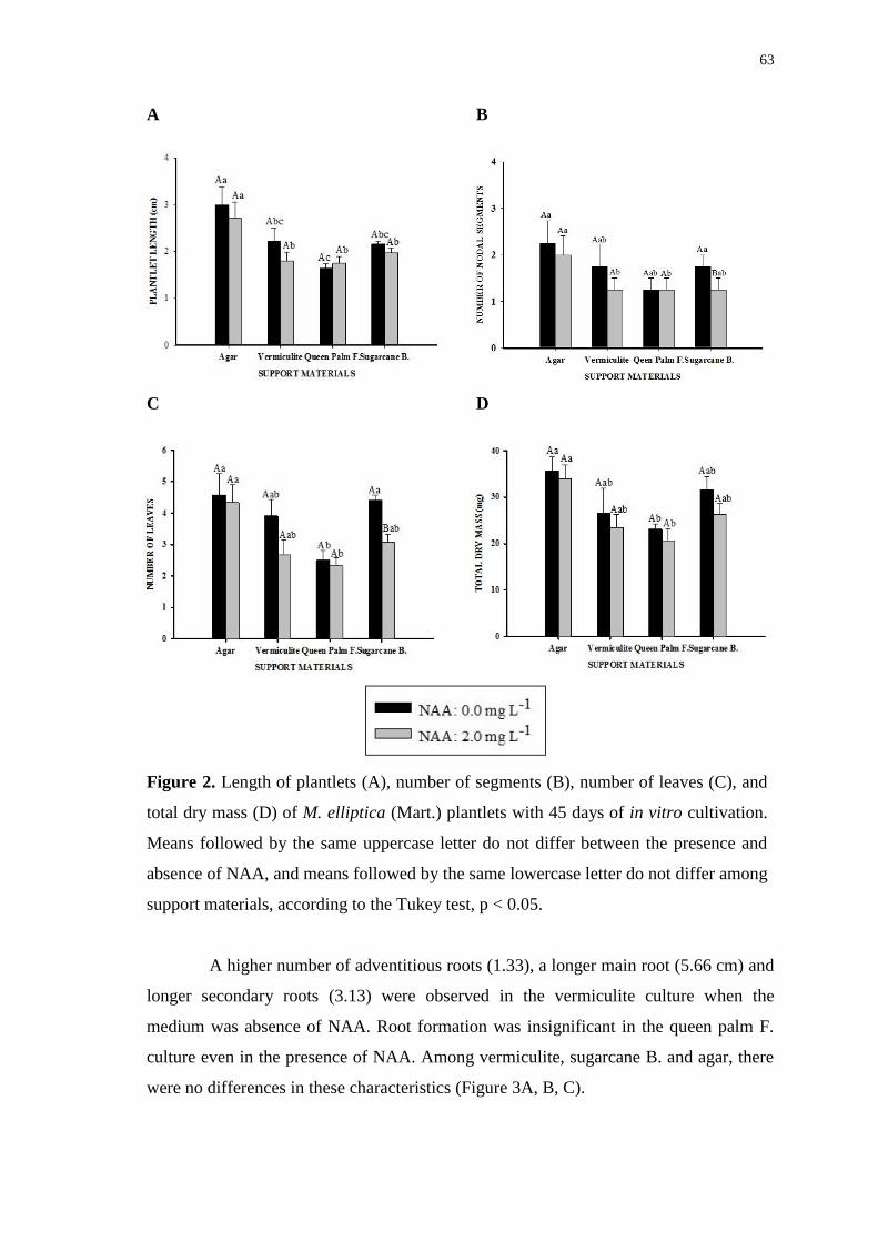
63
A B
C D
Figure 2. Length of plantlets (A), number of segments (B), number of leaves (C), and
total dry mass (D) of M. elliptica (Mart.) plantlets with 45 days of in vitro cultivation.
Means followed by the same uppercase letter do not differ between the presence and
absence of NAA, and means followed by the same lowercase letter do not differ among
support materials, according to the Tukey test, p < 0.05.
A higher number of adventitious roots (1.33), a longer main root (5.66 cm) and
longer secondary roots (3.13) were observed in the vermiculite culture when the
medium was absence of NAA. Root formation was insignificant in the queen palm F.
culture even in the presence of NAA. Among vermiculite, sugarcane B. and agar, there
were no differences in these characteristics (Figure 3A, B, C).
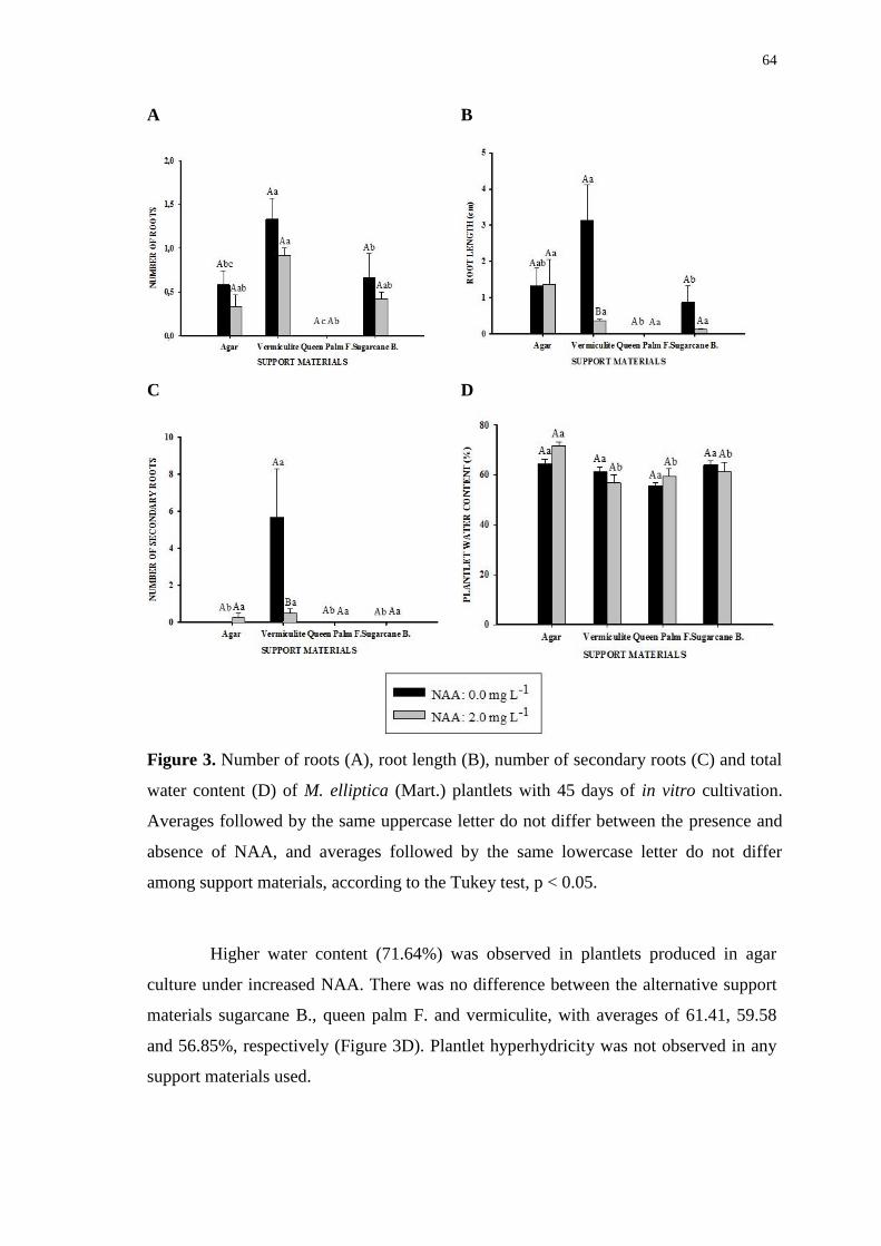
64
A B
C D
Figure 3. Number of roots (A), root length (B), number of secondary roots (C) and total
water content (D) of M. elliptica (Mart.) plantlets with 45 days of in vitro cultivation.
Averages followed by the same uppercase letter do not differ between the presence and
absence of NAA, and averages followed by the same lowercase letter do not differ
among support materials, according to the Tukey test, p < 0.05.
Higher water content (71.64%) was observed in plantlets produced in agar
culture under increased NAA. There was no difference between the alternative support
materials sugarcane B., queen palm F. and vermiculite, with averages of 61.41, 59.58
and 56.85%, respectively (Figure 3D). Plantlet hyperhydricity was not observed in any
support materials used.
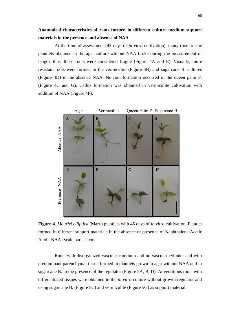
65
Anatomical characteristics of roots formed in different culture medium support
materials in the presence and absence of NAA
At the time of assessment (45 days of in vitro cultivation), many roots of the
plantlets obtained in the agar culture without NAA broke during the measurement of
length, thus, these roots were considered fragile (Figure 4A and E). Visually, more
resistant roots were formed in the vermiculite (Figure 4B) and sugarcane B. cultures
(Figure 4D) in the absence NAA. No root formation occurred in the queen palm F.
(Figure 4C and G). Callus formation was obtained in vermiculite cultivation with
addition of NAA (Figure 4F).
Figure 4. Mouriri elliptica (Mart.) plantlets with 45 days of in vitro cultivation. Plantlet
formed in different support materials in the absence or presence of Naphthalene Acetic
Acid - NAA. Scale bar = 2 cm.
Roots with disorganized vascular cambium and no vascular cylinder and with
predominant parenchymal tissue formed in plantlets grown in agar without NAA and in
sugarcane B. in the presence of the regulator (Figure 5A, B, D). Adventitious roots with
differentiated tissues were obtained in the in vitro culture without growth regulator and
using sugarcane B. (Figure 5C) and vermiculite (Figure 5G) as support material.
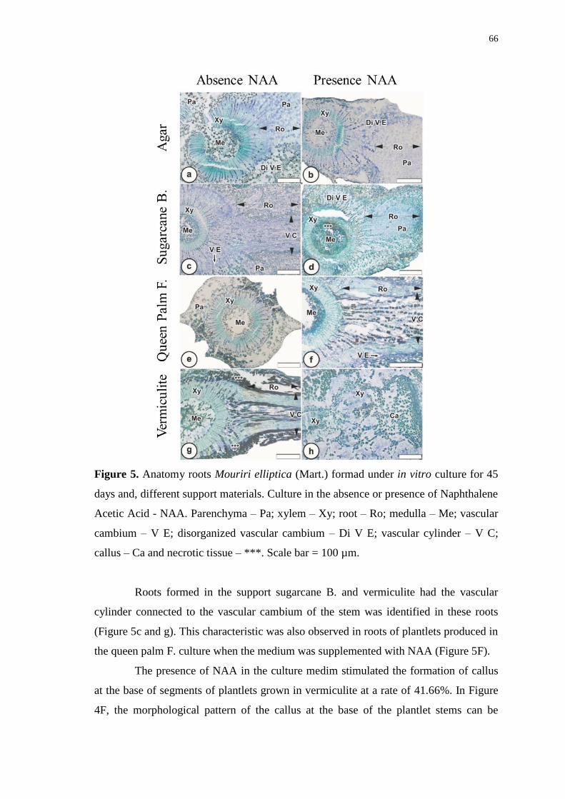
66
Figure 5. Anatomy roots Mouriri elliptica (Mart.) formad under in vitro culture for 45
days and, different support materials. Culture in the absence or presence of Naphthalene
Acetic Acid - NAA. Parenchyma – Pa; xylem – Xy; root – Ro; medulla – Me; vascular
cambium – V E; disorganized vascular cambium – Di V E; vascular cylinder – V C;
callus – Ca and necrotic tissue – ***. Scale bar = 100 µm.
Roots formed in the support sugarcane B. and vermiculite had the vascular
cylinder connected to the vascular cambium of the stem was identified in these roots
(Figure 5c and g). This characteristic was also observed in roots of plantlets produced in
the queen palm F. culture when the medium was supplemented with NAA (Figure 5F).
The presence of NAA in the culture medim stimulated the formation of callus
at the base of segments of plantlets grown in vermiculite at a rate of 41.66%. In Figure
4F, the morphological pattern of the callus at the base of the plantlet stems can be

67
observed, and its internal organization can be observed in Figure 5H. Disorganized
tissues can be seen, but with a certain level of differentiation and the presence of xylem
(Xy) (Figure 5H).
3.4 Discussion
The results of this study demonstrate that vermiculite, followed by sugarcane
B., can be used as agar substitutes in in vitro culture of M. elliptica (Mart.). This finding
is based on the regeneration ability of seedling shoots and, in particular, root
development. Woody plants are usually difficult to root, and thus, a material that
facilitates in vitro rooting is beneficial in micropropagation systems by enabling the
regulation of the growth environment based on the physical properties of the support
material (XIAO et al., 2011).
Despite recommendations for the use of support materials, few studies have
physically characterized these materials for use in vitro. The distribution of water, air
and solids in alternative supports depends on several factors such as pore space, density,
particle size and spatial distribution of pores (OH et al., 2012). Such evaluations are
common for use in nurseries, as the physical quality of the substrate is an important
factor for seedling growth and development, providing nutrients, retaining water and
moisture and being financially viable (PAGLIARINI et al., 2012; DORNELLES et al.,
2014).
Ideal substrates are those that promote better aeration and water infiltration and
drainage. Vermiculite, followed by sugarcane B., provided good development of the
seedlings under study for all evaluated characteristics. These supports had smaller pore
spaces than queen palm F. These results suggest that queen palm F. exerts a negative
effect on seedling growth, particularly on root formation, even in the presence of NAA
in the culture medium. Although queen palm F. did not promote good growth in the
plantlets under study, it still has potential for in vitro use, as the response of plantlets is
in part dependent on genotype. Different genotypes may respond differently to the same
cultivation condition (CORRÊA et al., 2015; 2016).
For growth of plants in containers, the best values for AW are between 0.24
and 0.40 m³ m-3 (DE BOODT; VERDONCK, 1972). For RW, the ideal range is
between 0.25 and 0.30 m³ m-3 (VERDONCK; GABRIËLS, 1988). Although TP, AW
and RW values outside of the ideal range for in vitro cultivation of plants in containers
were observed in vermiculite, this support material allowed for better formation of
68
adventitious and secondary roots and did not differ from agar and sugarcane B. in terms
of shoot formation. The observed positive results reflect the ability of the seedlings to
use the nutrient solution added to the vermiculite.
Our results demonstrate that the support material does not need to be
excessively porous for proper root development of M. elliptica (Mart.) seedlings. In the
case of vermiculite, a lower TP associated with a higher RW (water retained at matric
potentials higher than 100 hPa and present in the form of water films around the
particles) resulting from the volumetric expansion of the mineral particles during their
production promoted adequate gas exchange through the roots and provided greater
substrate/root contact surface area, contributing to nutrient absorption processes and
thus to plant growth and development.
Sugarcane B. had intermediate values for TP and AW and RW values within
the ideal range (DE BOODT; VERDONCK, 1972; VERDONCK; GABRIËLS, 1988).
For plantlet regeneration, it was less effective than vermiculite for root formation,
although roots formed on plantlets grown in sugarcane B. formed differentiated tissues
with a vascular cylinder connected to the vascular cambium of the stem, as also
observed in roots formed in vermiculite. Roots formed in the agar culture, regardless of
the presence or absence of NAA, were fragile and had poorly differentiated tissues and
no secondary roots. Similar problems in root formation using agar have been reported
and can cause problems in the acclimatization process (BRAGA et al., 2011).
The presence of NAA in the culture environment with vermiculite as the
support material was a limiting factor in root formation, as the root length was shorter
and the number of secondary roots was lower. In addition, a relatively high percentage
of calluses was observed at the base of the stem. This characteristic was considered
undesirable in consideration of the principal objective of the study, the multiplication
and rooting of M. elliptica (Mart.). However, this observation offers an opportunity to
study callus formation from the stem of this species.
Thus, the characteristics of plantlets evaluated in this study informed the
determination of physical attributes of alternative support materials to agar that were
ideal for the in vitro cultivation of M. elliptica (Mart.) and that favored plantlet growth.
However, studies on the micropropagation of this species using alternative support
materials could be developed to evaluate the influence of other factors that typically
promote development of more resistant plantlets, such as the elimination of sucrose in

69
the culture medium, the use of gaskets that allow for increased gas exchange, the
increase of light intensity and/or atmospheric enrichment with carbon dioxide (CO2).
3.5 Conclusions
The alternative support materials vermiculite followed by sugarcane B. can be
used as substitutes for agar for micropropagation of M. elliptica (Mart.). These support
materials promoted shoot growth equal to that of agar and greater root formation and
tissue differentiation, thus increasing the resistance of the plantlets and survival of the
acclimatization process.
The use of the growth regulator NAA did not stimulate increased rooting of M.
elliptica (Mart.) plantlets in the types of support materials used in this study.
3.6 Acknowledgments
The authors thank the funding agencies the Research Support Foundation of
the State of Goiás (Fundação de Amparo à Pesquisa do Estado de Goiás, FAPEG) and
the Coordination for the Improvement of Higher Education Personnel (Coordenação de
Aperfeiçoamento de Pessoal de Nível Superior, CAPES) for financial support.
3.7 References
ABDULMALIK, M. M.; USMAN, I. S.; OLAREWAJU, J. D.; ABA, D. A. Effect of
naphthalene acetic acid (NAA) on in vitro rooting of regenerated microshoots of
groundnut (Arachis hypogaea L.). Bayero Joural of Pure and Applied
Sciences, v. 5, n. 2, p. 128-131, 2012.
AGGARWAL, S.; NIRMALA, C. Utilization of coir fiber as an eco-friendly substitute
for costly gelling agentes for in vitro orchid seed germination. Scientia
Horticulturae, v. 133, n. 1, p. 89-92, 2012.
AINA, O. O.; QUESENBERRY, K. H.; GALLO, M. Culture vessel and auxin
treatments affect in vitro rooting and ex vitro survival of six arachis
paraguariensis genotypes. Scientia Horticulturae, v. 183, n. 1, p. 167–171,
2015.
ASSIS, C.; PEREIRA, F. D.; CABRAL, J. S. R.; SILVA, F. G.; SILVA, J. W.;
SANTOS, S. C. In vitro cultivation of Anacardium othonianum Rizz.: effects of
salt concentration and culture medium volume. Acta Scientiarum – Agronomy,
v. 34, n.1, p. 77-83, 2012.

70
ASSIS, E. S.; RUBIO NETO, A.; LIMA, L. R.; SILVA, F. G.; ROSA, M.;
VASCONCELOS FILHO, S. C.; LEITE, M. S. In vitro culture of Mouriri
elliptica (Mart.) under conditions that stimulate photoautotrophic behavior.
Australian Journal of Crop Science, v.10, n. 2, p. 229-236, 2016.
BARPETE, S.; KHAWAR, K. M.; ÖZCAN, S. Differential competence for in vitro
adventitous rooting of grass pea (Lathyrus sativus L.). Plant Cell, Tissue and
Organ Culture, v. 119, n. 1, p. 39-50, 2014.
BRAGA, F. T.; PASQUAL, M.; CASTRO, E. M.; RAFAEL, G. C. Características
morfofisiológicas de abacaxizeiro ‘gomo de mel’ enraizado in vitro sob luz
natural e substrato vermiculita. Revista Brasileira de Fruticultura, v. 33, n. 2,
p. 551-557, 2011.
BRASIL, Ministério da Agricultura, Pecuária e Abastecimento – MAPA. Secretaria de
Defesa Agropecuária. Instrução Normativa Nº 17, de 21 de maio de 2007.
Aprova os Métodos Analíticos Oficiais para Análise de Substratos e
Condicionadores de Solos, na forma do Anexo à presente Instrução Normativa.
Diário Oficial da República Federativa do Brasil. Seção 1, Brasília – Distrito
Federal, p. 8, 2007.
BRASIL, Ministério da Agricultura, Pecuária e Abastecimento – MAPA. Secretaria de
Defesa Agropecuária. Instrução Normativa Nº 31, de 23 de outubro de 2008.
Altera os subitens 3.1.2, 4.1 e 4.1.2, do Anexo à Instrução Normativa SDA Nº
17, de 21 de maio de 2007. Diário Oficial da República Federativa do Brasil.
Seção 1, Brasília - Distrito Federal, p. 20, 2008.
BRASIL, Regras para análise de sementes / Ministério da agricultura, pecuária e
abastecimento. Secretaria de defesa agropecuária. Mapa/ACS 200, Brasília,
2009.
CORRÊA, J. P. O.; VITAL, C.; PINHEIRO, M. V. M. A.; BATISTA, D. S.;
AZEVEDO, J. F. L.; SALDANHA, C.W.; CRUZ, A. C. F.; DA MATTA, F. M.;
OTONI, W. C. In vitro photoautotrophic potential and ex vitro photosynthetic
competence of Pfaffia glomerata (Spreng.) Pedersen accessions. Plant Cell,
Tissue and Organ Culture, v. 121, n. 2, p. 289-300, 2015.
CORRÊA, J. P. O.; VITAL, C.; PINHEIRO, M. V. M. A.; BATISTA, D. S.;
SALDANHA, C.W.; CRUZ, A. C. F.; NOTINI, M. M.; FREITAS, D. M. S.; DA
MATTA, F. M.; OTONI, W. C. Induced polyploidization increases 20-
hidroxyecdysone content, in vitro photoautotrophic growth, and ex vitro biomass
71
accumulation in Pfaffia glomerata (Spreng.) Pedersen. In vitro cellular and
developmental Biology – Plant, v. 52, n. 1, p. 45-55, 2016.
DE BOODT, M.; VERDONCK, O. The physical properties of the substrates in
horticulture. Acta Horticulturae, v. 26, n. 1, p. 37-44, 1972.
DEB, C.; PONGENER, A. A study on the use of low cost substrata against agar for
non-symbiotic seed culture of cymbidium iridioides D Don. Australian Journal
of Crop Science, v. 7, n. 5, p. 642-649, 2013.
DE LIMA, L. R.; RUBIO NETO, A.; PEREIRA, F. D.; SILVA, F. G.; DE MENEZES,
C. C. E.; SANTANA, J. D. G. Germination and emergence of Mouriri elliptica
Mart., a rare medicinal fruit tree native to the Brazilian Cerrado biome. African
Jounal of Agricultural Research, v. 11, n. 5, p. 400-406, 2016.
DORNELLES, P.; SILVA, F. G.; MOTA, C. S.; SANTANA, J. G. Production and
quality of Anacardium othonianum Rizz. seedlings grown in different
substrates. Revista Brasileira de Fruticultura, v. 36, n. 2, p. 479-486, 2014.
FERREIRA, D. F. Sisvar: a computer statistical analysis system. Ciência e
Agrotecnologia, v. 35, n. 6, p. 1039-1042, 2015.
HOSSAIN, S.; URBI, Z. Effect of naphthalene acetic acid on the adventitious rooting in
shoot cuttings of Andrographis paniculata (Burm.f.) Wall. ex Nees: an
important therapeutical herb. International Journal of Agronomy, ID
1617543, 6 p, 2016.
KARNOVSKY, M. J. A formaldehyde-glutaraldehyde fixative of high osmolality for
use in electron microscopy. The Journal of Cell Biology, v. 27, n. 2, p. 1A-
149A, 1965.
LLOYD, G.; MCCOWN, B. Commercially feasible micropropagation of montain
laurel, kalmia latifolia, by use of shoot tip culture. International Plant
Propagation Society Proceedings, v. 30, n. 5, p. 421-427, 1981.
MALI, A. M.; CHAVAN, N. S. In vitro rapid regeneration through direct organogenesis
and ex-vitro establishment of Cucumis trigonus Roxb. An underutilized
pharmaceutically important cucurbit. Industrial Crops and Products, v. 83, n.
1, p. 48–54, 2016.
MARTENDAL, C. O.; BERNARDINO, M. M.; PEREIRA, F. D.; SILVA, F. G.;
MENEZES, C. C. E.; SANTANA, J. G. In vitro multiplication of nodal
segments of “Murici” (Byrsonima cydoniifolia A Juss.): the use of growth
72
regulators and photoautotrophic stimulation. Journal of Agricultural
Technology, v. 10, n. 3, p. 665-678, 2014.
MOHAN, R.; CHUI, E. A.; BIASI, L. A.; SOCCOL, C. R. Alternative in vitro
propagation: use of sugarcane bagasse as a low cost support material during
rooting stage of strawberry Cv Dover. Brazilian Archives of Biology and
Technology, v. 48, n. 1, p. 37-42, 2005.
MOLEIRO, F. C.; ANDREO, M. A.; SANTOS, R. C.; MORAES, T. M.;
RODRIGUES, C. M., CARLI, C. B.; LOPES, F. M.; PELLIZZON, C. H.;
CARLOS, I. Z.; BAUAB, T. M.; VILEGAS W.; IRUMA-LIMA, C. A. Mouriri
elliptica: validation of gastroprotective, healing and anti-Helicobacter pylori
effects: validation of gastroprotective, healing and anti-Helicobaster pylori
effects. Journal of Ethnopharmacology, v. 123, n. 1, p. 359-368, 2009.
NAVARRO-GARCÍA, N.; MORTE, A.; PÉREZ-TORNERO, O. In vitro adventitious
organogenesis and histological characterization from mature nodal explants of
Citrus limon. In Vitro Cellular and Developmental Biology – Plant, v. 52, n.
2, p. 161–173, 2016.
OH, M. M.; SEO, J. H.; PARK, J. S.; SON, J. E. Physicochemical properties of
mixtures of inorganic supporting materials affect growth of potato (Solanum
tuberosum L.) plantlets cultured photoautotrophically in a nutrient-circulated
micropropagation system. Horticulture Environment and Biotechnology, v.
53, n. 6, p. 497-504, 2012.
PAGLIARINI, M. K.; CASTILHO, R. M. M.; ALVES, M. C. Caracterização físico-
química de misturas de componentes de substrato com resíduo de celulose para
fins de produção de mudas. Revista Brasileira de Agroecologia, v. 7, n. 2, p.
160-169, 2012.
PATEL, A. K.; PHULWARIA, M.; RAI, M. K.; GUPTA, A. K.; SHEKHAWAT, S.;
SHEKHAWAT, N. S. In vitro propagation and ex vitro rooting of Caralluma
edulis (Edgew.) Benth. and Hook. f.: An endemic and endangered edible plant
species of the Thar Desert. Scientia Horticulturae, v. 165, n. 1, p. 175-180,
2014.
RUFINO, M. S. M.; ALVES, R. E.; FERNANDES, F. A. N.; BRITO, E. S. Free radical
scavenging behavior of ten exotic tropical fruits extracts. Food Research
International, v. 44, n. 7, p. 2072-2075, 2011.
73
SALDANHA, C. W.; OTONI, C. G.; ROCHA, D. I.; CAVATTE, P. C.; DETMANN,
K. S. C.; TANAKA, F. A.; DIAS, L. L. C.; DA MATTA, F. M.; OTONI, W. C.
CO2-enriched atmosphere and supporting material impact the growth,
morphophysiology and ultrastructure of in vitro Brazilian-ginseng [Pfaffia
glomerata (Spreng.) Pedersen] plantlets. Plant Cell, Tissue and Organ
Culture, v. 18, n. 1, p. 87-99, 2014.
SHEKHAWAT, M. S.; MANOKARI, M. In vitro regeneration frequency, micro-
morphological studies and ex vitro rooting of Hemidesmus indicus (L.) R. Br.: a
multipotent endagered climber. Indian Journal of Plant Physiology, v. 21, n. 2,
p.151-160, 2016.
VASCONCELOS, J. M.; CARDOSO, T.V.; SALES, J. F.; SILVA, F. G.;
VASCONCELOS FILHO, S. C.; SANTANA, J. G. Métodos de superação de
dormência em sementes de croada (Mouriri elliptica Mart.). Ciência e
Agrotecnologia, v. 34, n. 5, p. 1199-1204, 2010.
VERDONCK, O.; GABRIËLS, R. Substrate requirements for plants. Acta
Horticulturae, v. 221, n. 1, p. 19-23, 1988.
XIAO, Y., NIU, G.; KOZAI, T. Development and application of photoautotrophic
micropropagation plant system. Plant Cell, Tissue and Organ Culture, v. 105,
n. 2, p. 149–158, 2011.
ZORZETO, T. Q.; DECHEN, S. C. F.; ABREU, M. F.; JÚNIOR, F. F. Caracterização
física de substratos para plantas. Bragantia, v. 73, n. 3, p. 300-311, 2014.
74
CAPÍTULO IV
(Normas de acordo com a revista Acta Scientiarum – Agronomy)
Aclimatização de Mouriri elliptica (Mart.) propagadas in vitro sob atmosfera
enriquecida com CO2 e diferentes vedações
Resumo
A croada (Mouriri elliptica Mart.), é uma frutífera nativa do cerrado com potencialidade
para ser utilizada pela população, no entanto, carece de estudos sobre sua propagação
vegetativa, em especial sobre a influência das condições de cultivo in vitro na
aclimatização. Assim, objetivou-se com este estudo, avaliar a performance na
aclimatização de plântulas de M. elliptica Mart. cultivadas in vitro sob enriquecimento
da atmosfera com CO2 e uso de diferentes vedações do frasco de cultivo. No cultivo,
utilizou-se 6 g de substrato vermiculita por frasco e solução nutritiva do meio Wood
Plant Medium. As vedações utilizadas, foram i) tampa convencional de polipropileno
(T. conv), ii) tampa com dois orifícios de área de 2,24 10-4 m2 vedados com membrana
microporosa (T. orif) e iii) vedação com vedafilme (PVC). Os frascos de cultivo foram
mantidos em câmaras climáticas (Fitotron®) com atmosfera enriquecida com CO2 (800 ±
35 µmol mol L-1) e atmosfera ambiente de CO2 (400 ± 59 µmol mol L-1) sob irradiância
de 150 ± 10 µmol m-2s-1, temperatura média de 25 ± 0,04 ºC e umidade relativa de 60 ±
0,18%. Sobrevivência de 100% foi obtida para plântulas micropropagadas em frascos
vedados com T. orif sob atmosfera ambiente de CO2. Na propagação in vitro de M.
elliptica Mart. o enriquecimento da atmosfera de cultivo com CO2 (800 ± 35 µmol mol
L-1) não proporcionou incremento no crescimento das plântulas, exceto com a utilização
da vedação do tipo PVC.

75
Palavras-chave: Croada, Melastomataceae, fotoautotrófico e fluorescência.
4.1 Introdução
Dentro da rica biodiversidade do Cerrado, destacam-se as espécies frutíferas,
citando a Mouriri elliptica Mart. (Família Melastomataceae), que possui potencialidades
para ser utilizada pela população. Seus frutos por serem ricos em nutrientes e em
compostos antioxidantes, como a vitamina C são indicados para consumo, sendo
considerados promotores da saúde humana (Rufino, Alves, Fernandes, & Brito, 2011).
Em estudos fitoquímicos das folhas, constatou-se a presença de compostos fenólicos,
como os flavonoides e taninos, sendo estes relacionados ao tratamento de doenças
gastrointestinais como úlceras gástricas e gastrite (Moleiro et al, 2009; Vasconcelos,
Andreo, Vilegas, Hiruma-Lima, Pellizzona & Vasconcelos, 2010b).
As sementes de M. elliptica Mart. possuem rígido tegumento, que tem sido
relacionado à dormência física, problema que ocasiona baixa germinação e emergência
desuniforme das plântulas (Vasconcelos et al., 2010a). Além disso, com a expansão da
agropecuária e também de fatores limitantes a produção de frutos pelas plantas em
condições naturais, a perpetuação da espécie está comprometida. Assim, para produção
de mudas em grande escala, a técnica de propagação in vitro, torna-se importante, além
de subsidiar na domesticação e conservação da espécie.
Trabalhos pioneiros com propagação in vitro dessa planta destacaram o cultivo
fotoautotrófico (cultivo sem sacarose) como promissor para obtenção de mudas da
espécie (Assis et al., 2016a e 2016b). De acordo com Xiao, Niu and Kozai, (2011) o
cultivo fotoautotrófico interfere nas características morfoanatômica e fisiológicas das
plântulas, tornando o aparato fotossintético funcional (Iarema et al., 2012). Assis et al.
(2016b) consideraram de suma importância o aprimoramento do cultivo in vitro para a
espécie, sendo necessário também, aclimatização às condições ex vitro.
Entre os trabalhos desenvolvidos com sucesso na literatura, cita-se o
enriquecimento da atmosfera de cultivo com CO2, com expressivo aumento da biomassa
das plantas e influência na morfologia dos estômatos e cloroplastos (Saldanha et al.,
2013; Saldanha et al., 2014), redução da umidade relativa e da concentração de etileno
do frasco de cultivo, utilizando vedações que permitem maiores trocas gasosas (Iarema
et al., 2012; Saldanha et al., 2012) e, substituição do ágar por materiais de suporte
fibrosos ou porosos (Mohan, Chui, Biasi & Soccol, 2005; Saldanha et al., 2014).
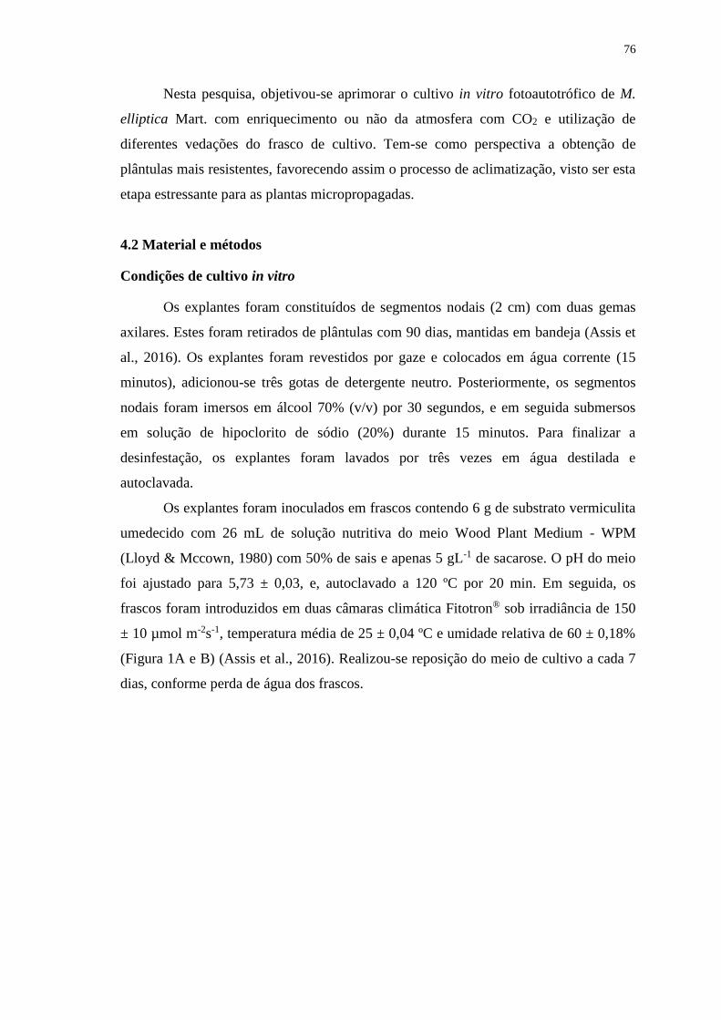
76
Nesta pesquisa, objetivou-se aprimorar o cultivo in vitro fotoautotrófico de M.
elliptica Mart. com enriquecimento ou não da atmosfera com CO2 e utilização de
diferentes vedações do frasco de cultivo. Tem-se como perspectiva a obtenção de
plântulas mais resistentes, favorecendo assim o processo de aclimatização, visto ser esta
etapa estressante para as plantas micropropagadas.
4.2 Material e métodos
Condições de cultivo in vitro
Os explantes foram constituídos de segmentos nodais (2 cm) com duas gemas
axilares. Estes foram retirados de plântulas com 90 dias, mantidas em bandeja (Assis et
al., 2016). Os explantes foram revestidos por gaze e colocados em água corrente (15
minutos), adicionou-se três gotas de detergente neutro. Posteriormente, os segmentos
nodais foram imersos em álcool 70% (v/v) por 30 segundos, e em seguida submersos
em solução de hipoclorito de sódio (20%) durante 15 minutos. Para finalizar a
desinfestação, os explantes foram lavados por três vezes em água destilada e
autoclavada.
Os explantes foram inoculados em frascos contendo 6 g de substrato vermiculita
umedecido com 26 mL de solução nutritiva do meio Wood Plant Medium - WPM
(Lloyd & Mccown, 1980) com 50% de sais e apenas 5 gL-1 de sacarose. O pH do meio
foi ajustado para 5,73 ± 0,03, e, autoclavado a 120 ºC por 20 min. Em seguida, os
frascos foram introduzidos em duas câmaras climática Fitotron® sob irradiância de 150
± 10 µmol m-2s-1, temperatura média de 25 ± 0,04 ºC e umidade relativa de 60 ± 0,18%
(Figura 1A e B) (Assis et al., 2016). Realizou-se reposição do meio de cultivo a cada 7
dias, conforme perda de água dos frascos.
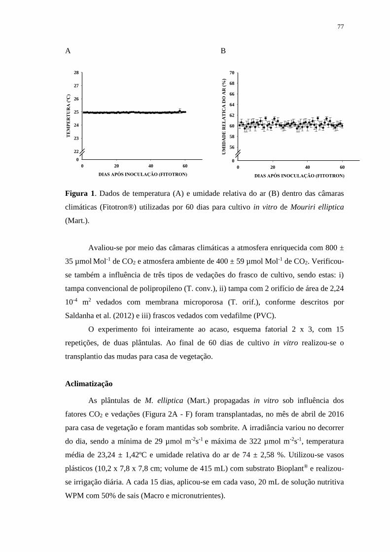
77
A
DIAS APÓS INOCULAÇÃO (FITOTRON)
0 20 40 60
TE
MP
ER
TU
RA
(ºC
)
0
22
23
24
25
26
27
28
B
DIAS APÓS INOCULAÇÃO (FITOTRON)
0 20 40 60
UM
IDA
DE
RE
LA
TIC
A D
O A
R (
%)
0
56
58
60
62
64
66
68
70
Figura 1. Dados de temperatura (A) e umidade relativa do ar (B) dentro das câmaras
climáticas (Fitotron®) utilizadas por 60 dias para cultivo in vitro de Mouriri elliptica
(Mart.).
Avaliou-se por meio das câmaras climáticas a atmosfera enriquecida com 800 ±
35 µmol Mol-1 de CO2 e atmosfera ambiente de 400 ± 59 µmol Mol-1 de CO2. Verificou-
se também a influência de três tipos de vedações do frasco de cultivo, sendo estas: i)
tampa convencional de polipropileno (T. conv.), ii) tampa com 2 orifício de área de 2,24
10-4 m2 vedados com membrana microporosa (T. orif.), conforme descritos por
Saldanha et al. (2012) e iii) frascos vedados com vedafilme (PVC).
O experimento foi inteiramente ao acaso, esquema fatorial 2 x 3, com 15
repetições, de duas plântulas. Ao final de 60 dias de cultivo in vitro realizou-se o
transplantio das mudas para casa de vegetação.
Aclimatização
As plântulas de M. elliptica (Mart.) propagadas in vitro sob influência dos
fatores CO2 e vedações (Figura 2A - F) foram transplantadas, no mês de abril de 2016
para casa de vegetação e foram mantidas sob sombrite. A irradiância variou no decorrer
do dia, sendo a mínima de 29 µmol m-2s-1 e máxima de 322 µmol m-2s-1, temperatura
média de 23,24 ± 1,42ºC e umidade relativa do ar de 74 ± 2,58 %. Utilizou-se vasos
plásticos (10,2 x 7,8 x 7,8 cm; volume de 415 mL) com substrato Bioplant® e realizou-
se irrigação diária. A cada 15 dias, aplicou-se em cada vaso, 20 mL de solução nutritiva
WPM com 50% de sais (Macro e micronutrientes).
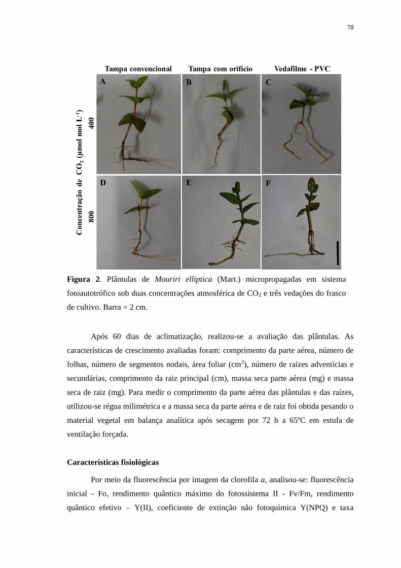
78
Figura 2. Plântulas de Mouriri elliptica (Mart.) micropropagadas em sistema
fotoautotrófico sob duas concentrações atmosférica de CO2 e três vedações do frasco
de cultivo. Barra = 2 cm.
Após 60 dias de aclimatização, realizou-se a avaliação das plântulas. As
características de crescimento avaliadas foram: comprimento da parte aérea, número de
folhas, número de segmentos nodais, área foliar (cm2), número de raízes adventícias e
secundárias, comprimento da raiz principal (cm), massa seca parte aérea (mg) e massa
seca de raiz (mg). Para medir o comprimento da parte aérea das plântulas e das raízes,
utilizou-se régua milimétrica e a massa seca da parte aérea e de raiz foi obtida pesando o
material vegetal em balança analítica após secagem por 72 h a 65ºC em estufa de
ventilação forçada.
Características fisiológicas
Por meio da fluorescência por imagem da clorofila a, analisou-se: fluorescência
inicial - Fo, rendimento quântico máximo do fotossistema II - Fv/Fm, rendimento
quântico efetivo – Y(II), coeficiente de extinção não fotoquímica Y(NPQ) e taxa
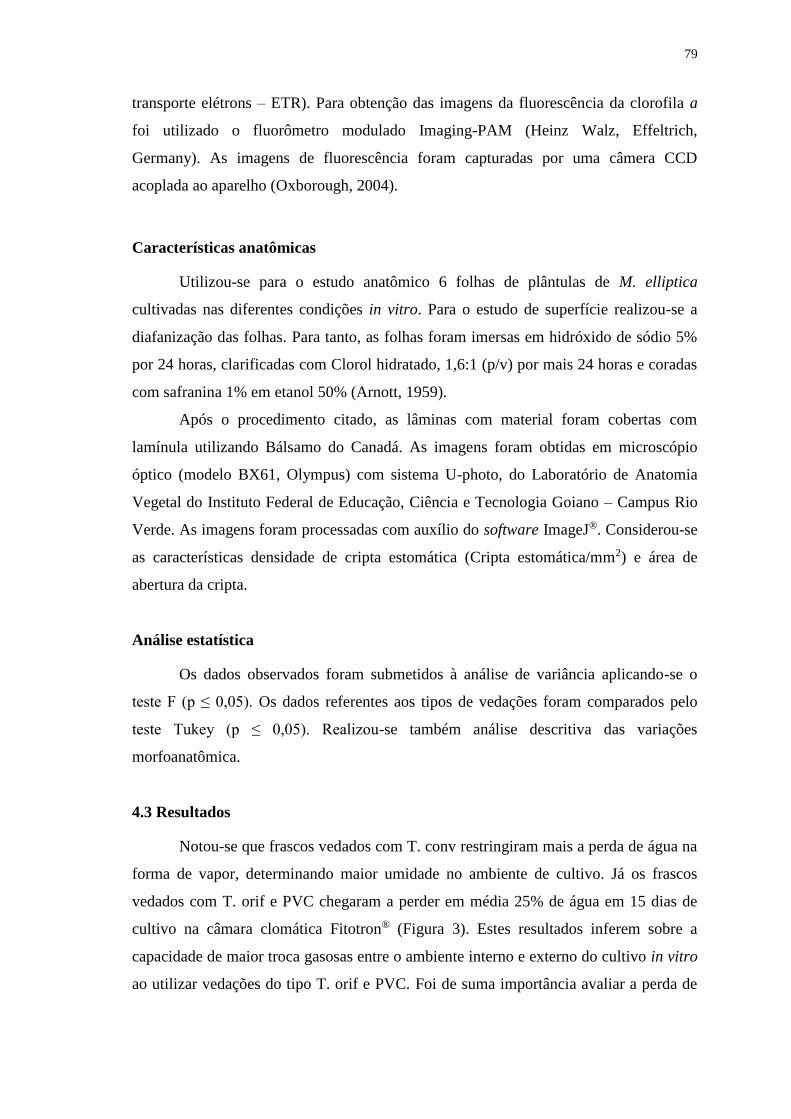
79
transporte elétrons – ETR). Para obtenção das imagens da fluorescência da clorofila a
foi utilizado o fluorômetro modulado Imaging-PAM (Heinz Walz, Effeltrich,
Germany). As imagens de fluorescência foram capturadas por uma câmera CCD
acoplada ao aparelho (Oxborough, 2004).
Características anatômicas
Utilizou-se para o estudo anatômico 6 folhas de plântulas de M. elliptica
cultivadas nas diferentes condições in vitro. Para o estudo de superfície realizou-se a
diafanização das folhas. Para tanto, as folhas foram imersas em hidróxido de sódio 5%
por 24 horas, clarificadas com Clorol hidratado, 1,6:1 (p/v) por mais 24 horas e coradas
com safranina 1% em etanol 50% (Arnott, 1959).
Após o procedimento citado, as lâminas com material foram cobertas com
lamínula utilizando Bálsamo do Canadá. As imagens foram obtidas em microscópio
óptico (modelo BX61, Olympus) com sistema U-photo, do Laboratório de Anatomia
Vegetal do Instituto Federal de Educação, Ciência e Tecnologia Goiano – Campus Rio
Verde. As imagens foram processadas com auxílio do software ImageJ®. Considerou-se
as características densidade de cripta estomática (Cripta estomática/mm2) e área de
abertura da cripta.
Análise estatística
Os dados observados foram submetidos à análise de variância aplicando-se o
teste F (p ≤ 0,05). Os dados referentes aos tipos de vedações foram comparados pelo
teste Tukey (p ≤ 0,05). Realizou-se também análise descritiva das variações
morfoanatômica.
4.3 Resultados
Notou-se que frascos vedados com T. conv restringiram mais a perda de água na
forma de vapor, determinando maior umidade no ambiente de cultivo. Já os frascos
vedados com T. orif e PVC chegaram a perder em média 25% de água em 15 dias de
cultivo na câmara clomática Fitotron® (Figura 3). Estes resultados inferem sobre a
capacidade de maior troca gasosas entre o ambiente interno e externo do cultivo in vitro
ao utilizar vedações do tipo T. orif e PVC. Foi de suma importância avaliar a perda de

80
água em cada frasco de cultivo, repondo a cada 7 dias a solução nutritiva, evitando
assim, déficit hídrico para as plantas.
CULTIVO in vitro (DIAS)
0 2 4 6 8 10 12 14 16
PE
RD
A D
E Á
GU
A (
%)
0
60
65
70
75
80
85
90
95
100
T. conv Y= -0,325x + 95,66 R2= 0,76**
T. orif Y= -1,386x + 94,74 R2= 0,92**
PVC Y= -1,530x + 94,06 R2= 0,92**
Figura 3. Porcentagem de perda de água em cada frasco de cultivo com as vedações:
tampa convencional (T. conv), tampa com orifício e membrana microporosa (T. orif)
e vedafilme (PVC) em função dos dias de cultivo in vitro. **p < 0,01.
Performance das plântulas de M. elliptica (Mart.) após 60 dias de aclimatização
O perfil morfológico das plantas após 60 dias de aclimatização e conforme a
procedência de cultivo in vitro pode ser observado na Figura 4 (A – F). Sobrevivência
de 100% foi obtida para plântulas micropropagadas em frascos vedados com T. orif sob
atmosfera ambiente de CO2. Maior porcentagem de mortalidade (13%) foi observado
em plântulas cultivadas em frascos vedados com T. conv e atmosfera ambiente de CO2
(Figura 5). Para as demais procedências de cultivo in vitro, notou-se 6,6% de
mortalidade.
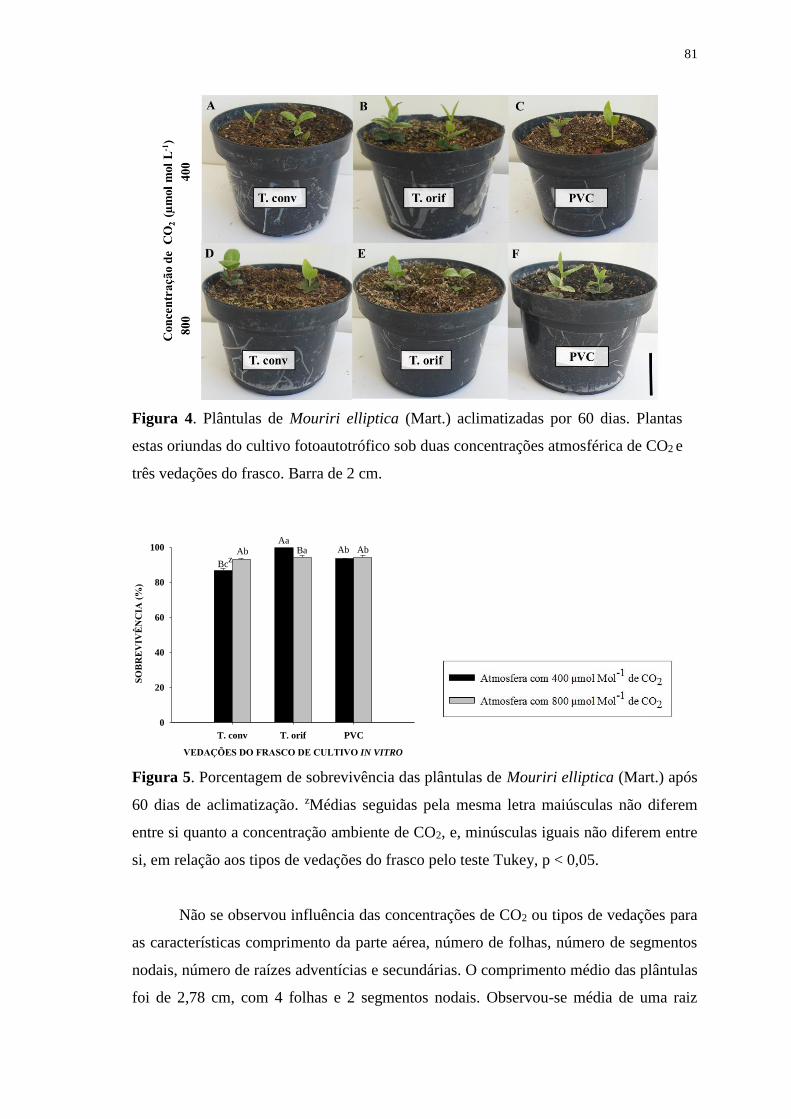
81
Figura 4. Plântulas de Mouriri elliptica (Mart.) aclimatizadas por 60 dias. Plantas
estas oriundas do cultivo fotoautotrófico sob duas concentrações atmosférica de CO2 e
três vedações do frasco. Barra de 2 cm.
VEDAÇÕES DO FRASCO DE CULTIVO IN VITRO
T. conv T. orif PVC
SO
BR
EV
IVÊ
NC
IA (
%)
0
20
40
60
80
100
Bcz
AbAa
Ba Ab Ab
Figura 5. Porcentagem de sobrevivência das plântulas de Mouriri elliptica (Mart.) após
60 dias de aclimatização. zMédias seguidas pela mesma letra maiúsculas não diferem
entre si quanto a concentração ambiente de CO2, e, minúsculas iguais não diferem entre
si, em relação aos tipos de vedações do frasco pelo teste Tukey, p < 0,05.
Não se observou influência das concentrações de CO2 ou tipos de vedações para
as características comprimento da parte aérea, número de folhas, número de segmentos
nodais, número de raízes adventícias e secundárias. O comprimento médio das plântulas
foi de 2,78 cm, com 4 folhas e 2 segmentos nodais. Observou-se média de uma raiz
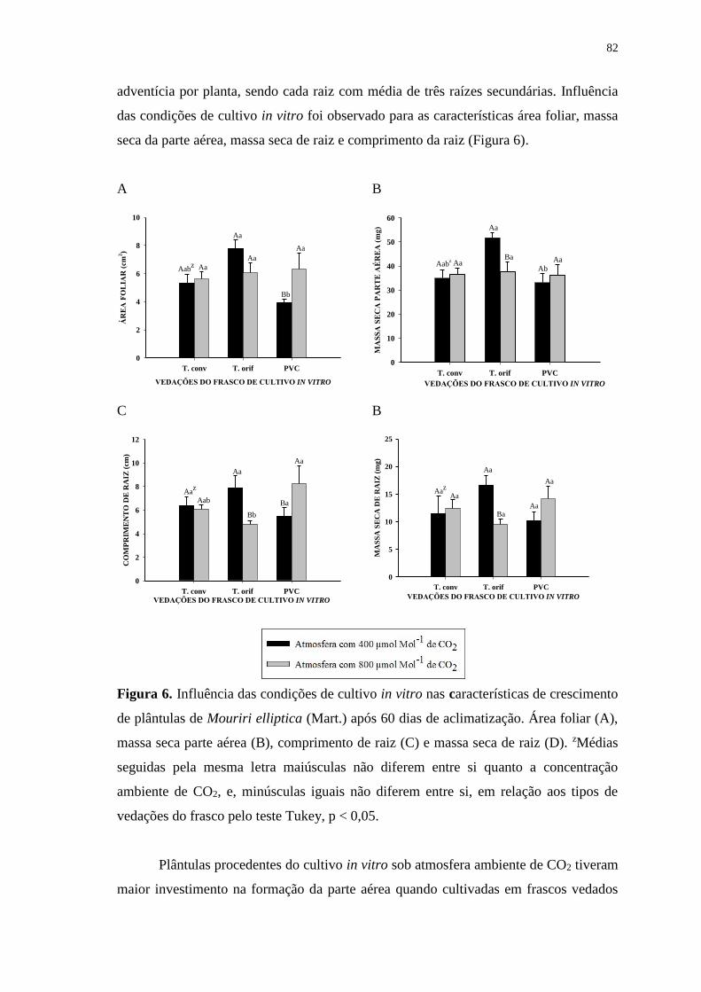
82
adventícia por planta, sendo cada raiz com média de três raízes secundárias. Influência
das condições de cultivo in vitro foi observado para as características área foliar, massa
seca da parte aérea, massa seca de raiz e comprimento da raiz (Figura 6).
A
VEDAÇÕES DO FRASCO DE CULTIVO IN VITRO
T. conv T. orif PVC
ÁR
EA
FO
LIA
R (
cm2)
0
2
4
6
8
10
Aabz Aa
Aa
Aa
Bb
Aa
B
T. conv T. orif PVCM
AS
SA
SE
CA
PA
RT
E A
ÉR
EA
(m
g)
0
10
20
30
40
50
60
VEDAÇÕES DO FRASCO DE CULTIVO IN VITRO
Aabz
Aa
Bb
Aa AaBa
Ab
C
VEDAÇÕES DO FRASCO DE CULTIVO IN VITRO
T. conv T. orif PVC
CO
MP
RIM
EN
TO
DE
RA
IZ (
cm)
0
2
4
6
8
10
12
Aaz
Aab
Aa
Bb
Aa
Ba
B
VEDAÇÕES DO FRASCO DE CULTIVO IN VITRO
T. conv T. orif PVC
MA
SS
A S
EC
A D
E R
AIZ
(m
g)
0
5
10
15
20
25
Aaz
Aa
Aa
Ba
Aa
Aa
Figura 6. Influência das condições de cultivo in vitro nas características de crescimento
de plântulas de Mouriri elliptica (Mart.) após 60 dias de aclimatização. Área foliar (A),
massa seca parte aérea (B), comprimento de raiz (C) e massa seca de raiz (D). zMédias
seguidas pela mesma letra maiúsculas não diferem entre si quanto a concentração
ambiente de CO2, e, minúsculas iguais não diferem entre si, em relação aos tipos de
vedações do frasco pelo teste Tukey, p < 0,05.
Plântulas procedentes do cultivo in vitro sob atmosfera ambiente de CO2 tiveram
maior investimento na formação da parte aérea quando cultivadas em frascos vedados
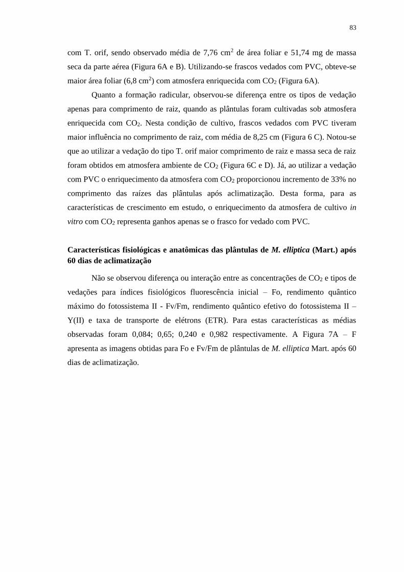
83
com T. orif, sendo observado média de 7,76 cm2 de área foliar e 51,74 mg de massa
seca da parte aérea (Figura 6A e B). Utilizando-se frascos vedados com PVC, obteve-se
maior área foliar (6,8 cm2) com atmosfera enriquecida com CO2 (Figura 6A).
Quanto a formação radicular, observou-se diferença entre os tipos de vedação
apenas para comprimento de raiz, quando as plântulas foram cultivadas sob atmosfera
enriquecida com CO2. Nesta condição de cultivo, frascos vedados com PVC tiveram
maior influência no comprimento de raiz, com média de 8,25 cm (Figura 6 C). Notou-se
que ao utilizar a vedação do tipo T. orif maior comprimento de raiz e massa seca de raiz
foram obtidos em atmosfera ambiente de CO2 (Figura 6C e D). Já, ao utilizar a vedação
com PVC o enriquecimento da atmosfera com CO2 proporcionou incremento de 33% no
comprimento das raízes das plântulas após aclimatização. Desta forma, para as
características de crescimento em estudo, o enriquecimento da atmosfera de cultivo in
vitro com CO2 representa ganhos apenas se o frasco for vedado com PVC.
Características fisiológicas e anatômicas das plântulas de M. elliptica (Mart.) após
60 dias de aclimatização
Não se observou diferença ou interação entre as concentrações de CO2 e tipos de
vedações para índices fisiológicos fluorescência inicial – Fo, rendimento quântico
máximo do fotossistema II - Fv/Fm, rendimento quântico efetivo do fotossistema II –
Y(II) e taxa de transporte de elétrons (ETR). Para estas características as médias
observadas foram 0,084; 0,65; 0,240 e 0,982 respectivamente. A Figura 7A – F
apresenta as imagens obtidas para Fo e Fv/Fm de plântulas de M. elliptica Mart. após 60
dias de aclimatização.
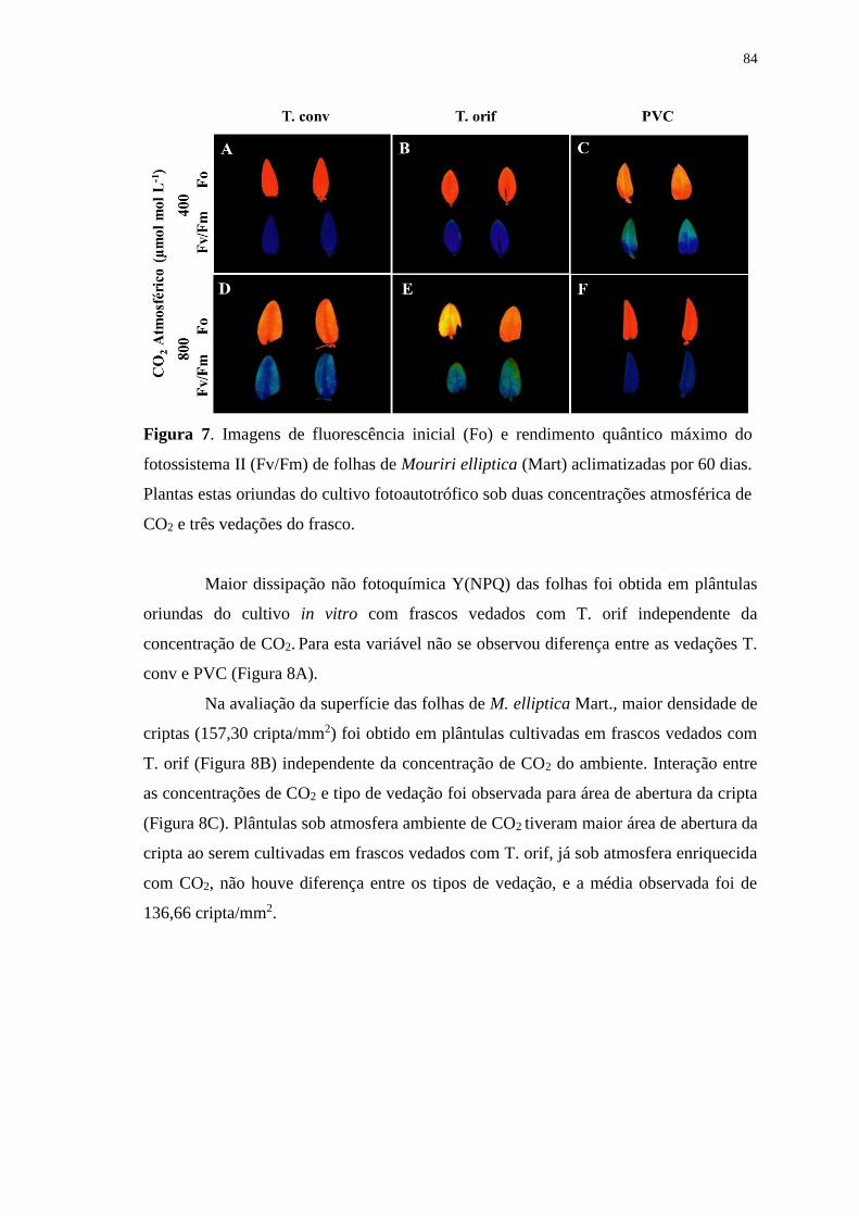
84
Figura 7. Imagens de fluorescência inicial (Fo) e rendimento quântico máximo do
fotossistema II (Fv/Fm) de folhas de Mouriri elliptica (Mart) aclimatizadas por 60 dias.
Plantas estas oriundas do cultivo fotoautotrófico sob duas concentrações atmosférica de
CO2 e três vedações do frasco.
Maior dissipação não fotoquímica Y(NPQ) das folhas foi obtida em plântulas
oriundas do cultivo in vitro com frascos vedados com T. orif independente da
concentração de CO2. Para esta variável não se observou diferença entre as vedações T.
conv e PVC (Figura 8A).
Na avaliação da superfície das folhas de M. elliptica Mart., maior densidade de
criptas (157,30 cripta/mm2) foi obtido em plântulas cultivadas em frascos vedados com
T. orif (Figura 8B) independente da concentração de CO2 do ambiente. Interação entre
as concentrações de CO2 e tipo de vedação foi observada para área de abertura da cripta
(Figura 8C). Plântulas sob atmosfera ambiente de CO2 tiveram maior área de abertura da
cripta ao serem cultivadas em frascos vedados com T. orif, já sob atmosfera enriquecida
com CO2, não houve diferença entre os tipos de vedação, e a média observada foi de
136,66 cripta/mm2.
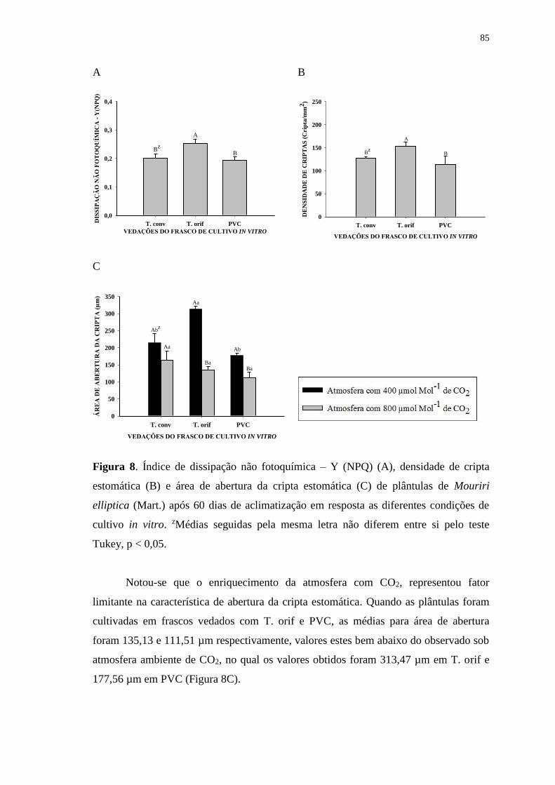
85
A
VEDAÇÕES DO FRASCO DE CULTIVO IN VITRO
T. conv T. orif PVCDIS
SIP
AÇ
ÃO
NÃ
O F
OT
OQ
UÍM
ICA
- Y
(NP
Q)
0,0
0,1
0,2
0,3
0,4
Bz
A
B
B
VEDAÇÕES DO FRASCO DE CULTIVO IN VITRO
T. conv T. orif PVC
DE
NS
IDA
DE
DE
CR
IPT
AS
(C
rip
ta/m
m2)
0
50
100
150
200
250
A
BBz
C
VEDAÇÕES DO FRASCO DE CULTIVO IN VITRO
T. conv T. orif PVC
ÁR
EA
DE
AB
ER
TU
RA
DA
CR
IPT
A (
µm
)
0
50
100
150
200
250
300
350
Abz
Aa
Aa
Ba
Ab
Ba
Figura 8. Índice de dissipação não fotoquímica – Y (NPQ) (A), densidade de cripta
estomática (B) e área de abertura da cripta estomática (C) de plântulas de Mouriri
elliptica (Mart.) após 60 dias de aclimatização em resposta as diferentes condições de
cultivo in vitro. zMédias seguidas pela mesma letra não diferem entre si pelo teste
Tukey, p < 0,05.
Notou-se que o enriquecimento da atmosfera com CO2, representou fator
limitante na característica de abertura da cripta estomática. Quando as plântulas foram
cultivadas em frascos vedados com T. orif e PVC, as médias para área de abertura
foram 135,13 e 111,51 µm respectivamente, valores estes bem abaixo do observado sob
atmosfera ambiente de CO2, no qual os valores obtidos foram 313,47 µm em T. orif e
177,56 µm em PVC (Figura 8C).
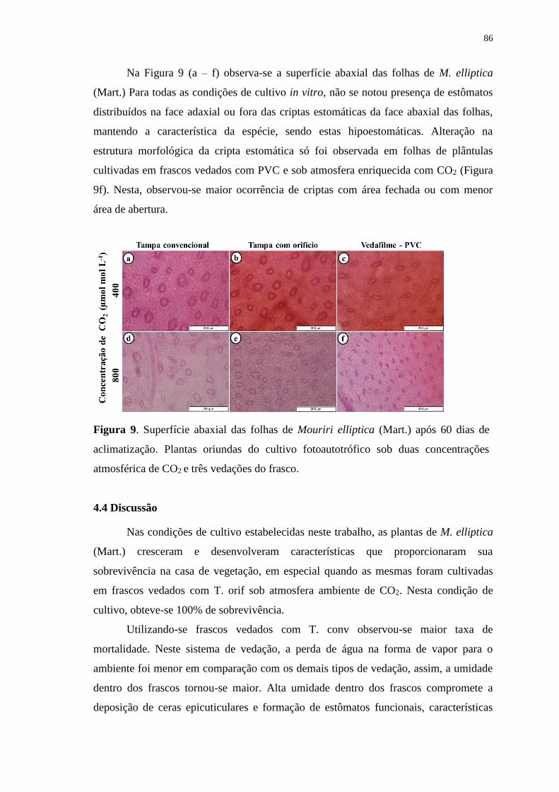
86
Na Figura 9 (a – f) observa-se a superfície abaxial das folhas de M. elliptica
(Mart.) Para todas as condições de cultivo in vitro, não se notou presença de estômatos
distribuídos na face adaxial ou fora das criptas estomáticas da face abaxial das folhas,
mantendo a característica da espécie, sendo estas hipoestomáticas. Alteração na
estrutura morfológica da cripta estomática só foi observada em folhas de plântulas
cultivadas em frascos vedados com PVC e sob atmosfera enriquecida com CO2 (Figura
9f). Nesta, observou-se maior ocorrência de criptas com área fechada ou com menor
área de abertura.
Figura 9. Superfície abaxial das folhas de Mouriri elliptica (Mart.) após 60 dias de
aclimatização. Plantas oriundas do cultivo fotoautotrófico sob duas concentrações
atmosférica de CO2 e três vedações do frasco.
4.4 Discussão
Nas condições de cultivo estabelecidas neste trabalho, as plantas de M. elliptica
(Mart.) cresceram e desenvolveram características que proporcionaram sua
sobrevivência na casa de vegetação, em especial quando as mesmas foram cultivadas
em frascos vedados com T. orif sob atmosfera ambiente de CO2. Nesta condição de
cultivo, obteve-se 100% de sobrevivência.
Utilizando-se frascos vedados com T. conv observou-se maior taxa de
mortalidade. Neste sistema de vedação, a perda de água na forma de vapor para o
ambiente foi menor em comparação com os demais tipos de vedação, assim, a umidade
dentro dos frascos tornou-se maior. Alta umidade dentro dos frascos compromete a
deposição de ceras epicuticulares e formação de estômatos funcionais, características
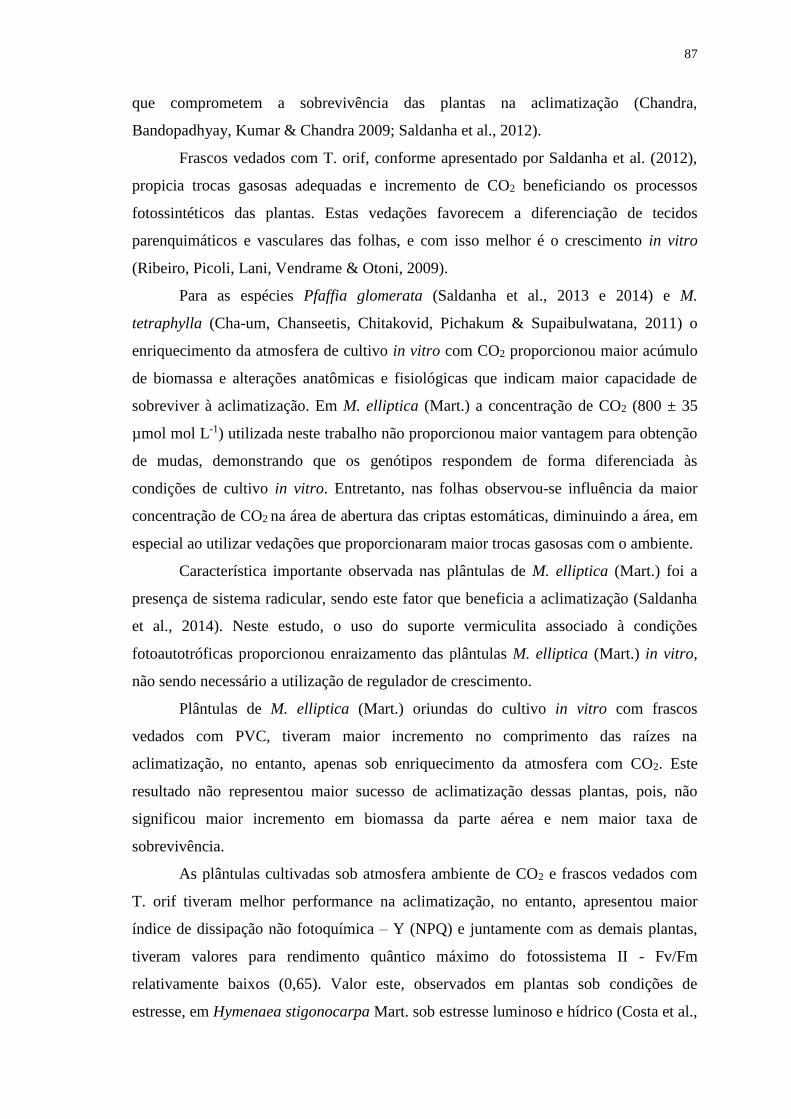
87
que comprometem a sobrevivência das plantas na aclimatização (Chandra,
Bandopadhyay, Kumar & Chandra 2009; Saldanha et al., 2012).
Frascos vedados com T. orif, conforme apresentado por Saldanha et al. (2012),
propicia trocas gasosas adequadas e incremento de CO2 beneficiando os processos
fotossintéticos das plantas. Estas vedações favorecem a diferenciação de tecidos
parenquimáticos e vasculares das folhas, e com isso melhor é o crescimento in vitro
(Ribeiro, Picoli, Lani, Vendrame & Otoni, 2009).
Para as espécies Pfaffia glomerata (Saldanha et al., 2013 e 2014) e M.
tetraphylla (Cha-um, Chanseetis, Chitakovid, Pichakum & Supaibulwatana, 2011) o
enriquecimento da atmosfera de cultivo in vitro com CO2 proporcionou maior acúmulo
de biomassa e alterações anatômicas e fisiológicas que indicam maior capacidade de
sobreviver à aclimatização. Em M. elliptica (Mart.) a concentração de CO2 (800 ± 35
µmol mol L-1) utilizada neste trabalho não proporcionou maior vantagem para obtenção
de mudas, demonstrando que os genótipos respondem de forma diferenciada às
condições de cultivo in vitro. Entretanto, nas folhas observou-se influência da maior
concentração de CO2 na área de abertura das criptas estomáticas, diminuindo a área, em
especial ao utilizar vedações que proporcionaram maior trocas gasosas com o ambiente.
Característica importante observada nas plântulas de M. elliptica (Mart.) foi a
presença de sistema radicular, sendo este fator que beneficia a aclimatização (Saldanha
et al., 2014). Neste estudo, o uso do suporte vermiculita associado à condições
fotoautotróficas proporcionou enraizamento das plântulas M. elliptica (Mart.) in vitro,
não sendo necessário a utilização de regulador de crescimento.
Plântulas de M. elliptica (Mart.) oriundas do cultivo in vitro com frascos
vedados com PVC, tiveram maior incremento no comprimento das raízes na
aclimatização, no entanto, apenas sob enriquecimento da atmosfera com CO2. Este
resultado não representou maior sucesso de aclimatização dessas plantas, pois, não
significou maior incremento em biomassa da parte aérea e nem maior taxa de
sobrevivência.
As plântulas cultivadas sob atmosfera ambiente de CO2 e frascos vedados com
T. orif tiveram melhor performance na aclimatização, no entanto, apresentou maior
índice de dissipação não fotoquímica – Y (NPQ) e juntamente com as demais plantas,
tiveram valores para rendimento quântico máximo do fotossistema II - Fv/Fm
relativamente baixos (0,65). Valor este, observados em plantas sob condições de
estresse, em Hymenaea stigonocarpa Mart. sob estresse luminoso e hídrico (Costa et al.,
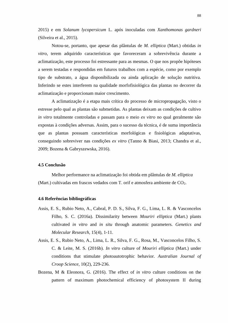
88
2015) e em Solanum lycopersicum L. após inoculadas com Xanthomonas gardneri
(Silveira et al., 2015).
Notou-se, portanto, que apesar das plântulas de M. elliptica (Mart.) obtidas in
vitro, terem adquirido características que favoreceram a sobrevivência durante a
aclimatização, este processo foi estressante para as mesmas. O que nos propõe hipóteses
a serem testadas e respondidas em futuros trabalhos com a espécie, como por exemplo
tipo de substrato, a água disponibilizada ou ainda aplicação de solução nutritiva.
Inferindo se estes interferem na qualidade morfofisiológica das plantas no decorrer da
aclimatização e proporcionam maior crescimento.
A aclimatização é a etapa mais crítica do processo de micropropagação, visto o
estresse pelo qual as plantas são submetidas. As plantas deixam as condições de cultivo
in vitro totalmente controladas e passam para o meio ex vitro no qual geralmente são
expostas à condições adversas. Assim, para o sucesso da técnica, é de suma importância
que as plantas possuam características morfológicas e fisiológicas adaptativas,
conseguindo sobreviver nas condições ex vitro (Tanno & Biasi, 2013; Chandra et al.,
2009; Bozena & Gabryszewska, 2016).
4.5 Conclusão
Melhor performance na aclimatização foi obtida em plântulas de M. elliptica
(Mart.) cultivadas em frascos vedados com T. orif e atmosfera ambiente de CO2.
4.6 Referências bibliográficas
Assis, E. S., Rubio Neto, A., Cabral, P. D. S., Silva, F. G., Lima, L. R. & Vasconcelos
Filho, S. C. (2016a). Dissimilarity between Mouriri elliptica (Mart.) plants
cultivated in vitro and in situ through anatomic parameters. Genetics and
Molecular Research, 15(4), 1-11.
Assis, E. S., Rubio Neto, A., Lima, L. R., Silva, F. G., Rosa, M., Vasconcelos Filho, S.
C. & Leite, M. S. (2016b). In vitro culture of Mouriri elliptica (Mart.) under
conditions that stimulate photoautotrophic behavior. Australian Journal of
Croop Science, 10(2), 229-236.
Bozena, M & Eleonora, G. (2016). The effect of in vitro culture conditions on the
pattern of maximum photochemical efficiency of photosystem II during
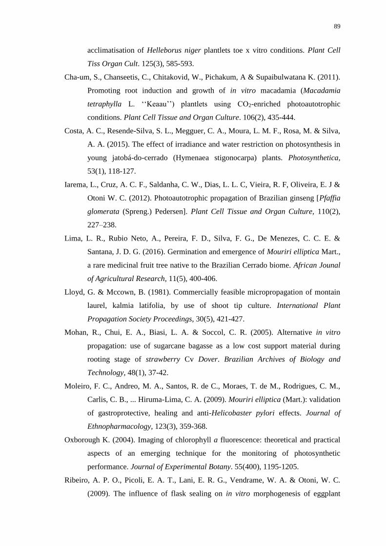
89
acclimatisation of Helleborus niger plantlets toe x vitro conditions. Plant Cell
Tiss Organ Cult. 125(3), 585-593.
Cha-um, S., Chanseetis, C., Chitakovid, W., Pichakum, A & Supaibulwatana K. (2011).
Promoting root induction and growth of in vitro macadamia (Macadamia
tetraphylla L. ‘‘Keaau’’) plantlets using CO2-enriched photoautotrophic
conditions. Plant Cell Tissue and Organ Culture. 106(2), 435-444.
Costa, A. C., Resende-Silva, S. L., Megguer, C. A., Moura, L. M. F., Rosa, M. & Silva,
A. A. (2015). The effect of irradiance and water restriction on photosynthesis in
young jatobá-do-cerrado (Hymenaea stigonocarpa) plants. Photosynthetica,
53(1), 118-127.
Iarema, L., Cruz, A. C. F., Saldanha, C. W., Dias, L. L. C, Vieira, R. F, Oliveira, E. J &
Otoni W. C. (2012). Photoautotrophic propagation of Brazilian ginseng [Pfaffia
glomerata (Spreng.) Pedersen]. Plant Cell Tissue and Organ Culture, 110(2),
227–238.
Lima, L. R., Rubio Neto, A., Pereira, F. D., Silva, F. G., De Menezes, C. C. E. &
Santana, J. D. G. (2016). Germination and emergence of Mouriri elliptica Mart.,
a rare medicinal fruit tree native to the Brazilian Cerrado biome. African Jounal
of Agricultural Research, 11(5), 400-406.
Lloyd, G. & Mccown, B. (1981). Commercially feasible micropropagation of montain
laurel, kalmia latifolia, by use of shoot tip culture. International Plant
Propagation Society Proceedings, 30(5), 421-427.
Mohan, R., Chui, E. A., Biasi, L. A. & Soccol, C. R. (2005). Alternative in vitro
propagation: use of sugarcane bagasse as a low cost support material during
rooting stage of strawberry Cv Dover. Brazilian Archives of Biology and
Technology, 48(1), 37-42.
Moleiro, F. C., Andreo, M. A., Santos, R. de C., Moraes, T. de M., Rodrigues, C. M.,
Carlis, C. B., ... Hiruma-Lima, C. A. (2009). Mouriri elliptica (Mart.): validation
of gastroprotective, healing and anti-Helicobaster pylori effects. Journal of
Ethnopharmacology, 123(3), 359-368.
Oxborough K. (2004). Imaging of chlorophyll a fluorescence: theoretical and practical
aspects of an emerging technique for the monitoring of photosynthetic
performance. Journal of Experimental Botany. 55(400), 1195-1205.
Ribeiro, A. P. O., Picoli, E. A. T., Lani, E. R. G., Vendrame, W. A. & Otoni, W. C.
(2009). The influence of flask sealing on in vitro morphogenesis of eggplant
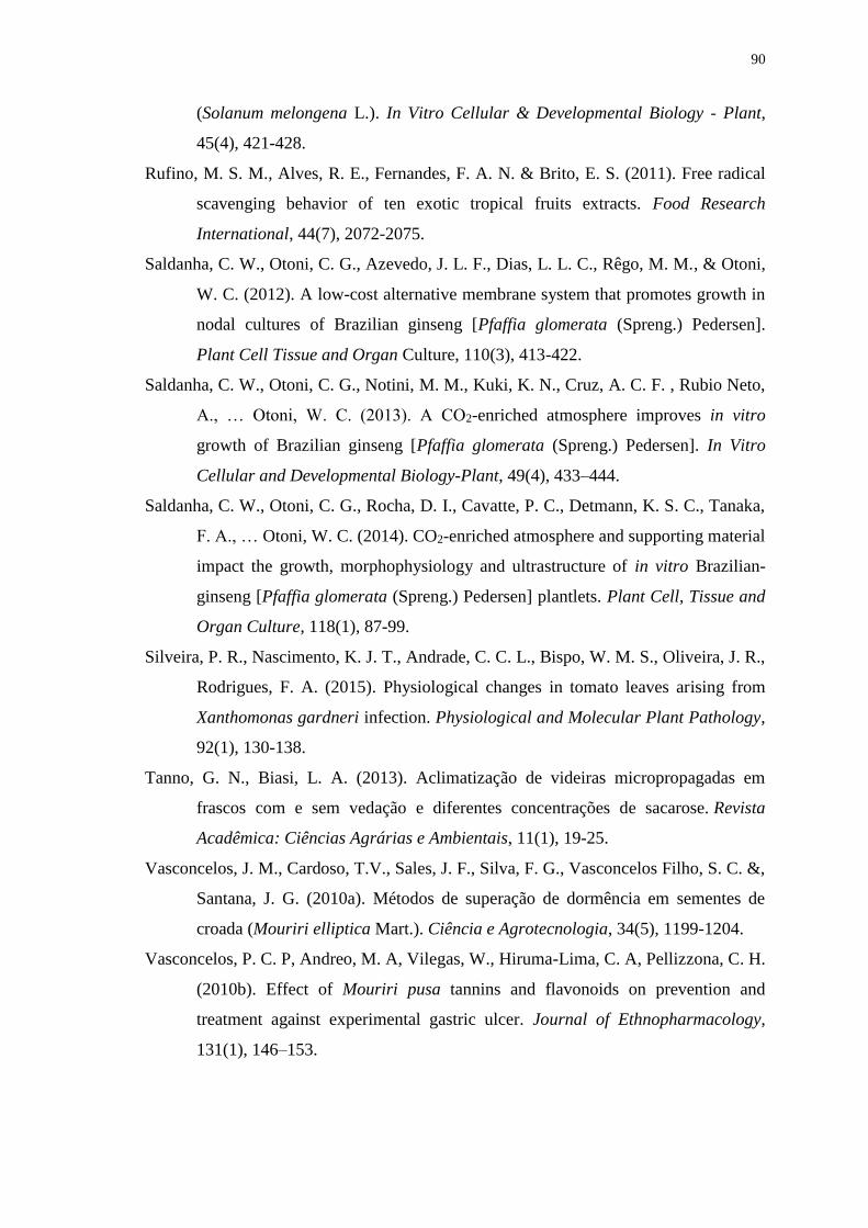
90
(Solanum melongena L.). In Vitro Cellular & Developmental Biology - Plant,
45(4), 421-428.
Rufino, M. S. M., Alves, R. E., Fernandes, F. A. N. & Brito, E. S. (2011). Free radical
scavenging behavior of ten exotic tropical fruits extracts. Food Research
International, 44(7), 2072-2075.
Saldanha, C. W., Otoni, C. G., Azevedo, J. L. F., Dias, L. L. C., Rêgo, M. M., & Otoni,
W. C. (2012). A low-cost alternative membrane system that promotes growth in
nodal cultures of Brazilian ginseng [Pfaffia glomerata (Spreng.) Pedersen].
Plant Cell Tissue and Organ Culture, 110(3), 413-422.
Saldanha, C. W., Otoni, C. G., Notini, M. M., Kuki, K. N., Cruz, A. C. F. , Rubio Neto,
A., … Otoni, W. C. (2013). A CO2-enriched atmosphere improves in vitro
growth of Brazilian ginseng [Pfaffia glomerata (Spreng.) Pedersen]. In Vitro
Cellular and Developmental Biology-Plant, 49(4), 433–444.
Saldanha, C. W., Otoni, C. G., Rocha, D. I., Cavatte, P. C., Detmann, K. S. C., Tanaka,
F. A., … Otoni, W. C. (2014). CO2-enriched atmosphere and supporting material
impact the growth, morphophysiology and ultrastructure of in vitro Brazilian-
ginseng [Pfaffia glomerata (Spreng.) Pedersen] plantlets. Plant Cell, Tissue and
Organ Culture, 118(1), 87-99.
Silveira, P. R., Nascimento, K. J. T., Andrade, C. C. L., Bispo, W. M. S., Oliveira, J. R.,
Rodrigues, F. A. (2015). Physiological changes in tomato leaves arising from
Xanthomonas gardneri infection. Physiological and Molecular Plant Pathology,
92(1), 130-138.
Tanno, G. N., Biasi, L. A. (2013). Aclimatização de videiras micropropagadas em
frascos com e sem vedação e diferentes concentrações de sacarose. Revista
Acadêmica: Ciências Agrárias e Ambientais, 11(1), 19-25.
Vasconcelos, J. M., Cardoso, T.V., Sales, J. F., Silva, F. G., Vasconcelos Filho, S. C. &,
Santana, J. G. (2010a). Métodos de superação de dormência em sementes de
croada (Mouriri elliptica Mart.). Ciência e Agrotecnologia, 34(5), 1199-1204.
Vasconcelos, P. C. P, Andreo, M. A, Vilegas, W., Hiruma-Lima, C. A, Pellizzona, C. H.
(2010b). Effect of Mouriri pusa tannins and flavonoids on prevention and
treatment against experimental gastric ulcer. Journal of Ethnopharmacology,
131(1), 146–153.
91
Xiao, Y., Niu, G. & Kozai, T. (2011). Development and application of photoautotrophic
micropropagation plant system. Plant Cell Tissue and Organ Culture, 105(2),
149–158.
92
CONCLUSÃO GERAL
As plântulas de M. elliptica (Mart.) responderam positivamente as condições
fotoautotrócas de cultivo in vitro, no qual o aumento da intensidade luminosa suprimiu
a necessidade das plantas por sacarose no meio de cultivo. Verificou-se que as
diferentes intensidades luminosas utilizadas neste estudo foram suficientes para
compreender o comportamento desta espécie in vitro, subsidiar futuros trabalhos,
visando assim maior produção de mudas.
Com o auxílio da técnica de estatística multivariada identificou que plântulas
cultivadas sob condições fotoautotróficas desenvolveram características anatômicas
foliares mais dissimilares as plantas in situ. Tal conclusão, indicou a necessidade de
aclimatização das plântulas de M. elliptica (Mart.) micropropagadas sob condições
fotoautotróficas.
Os suportes alternativos vermiculita, seguido do bagaço de cana-de-açúcar são
promissores para utilização no cultivo in vitro da espécie M. elliptica (Mart.), visto a
formação de sistema radicular nas plântulas com alto nível de diferenciação dos tecidos.
Foi possível obter 100% de sobrevivência de M. elliptica (Mart.) propagadas in
vitro de forma fotoautotrófica e em cultivo com frascos vedados com tampa e orifício
com membrana microporosa (T. orif) em atmosfera ambiente de CO2.
Futuros trabalhos devem ser desenvolvidos com aclimatização da espécie,
testando por exemplo tipos de substratos e presença ou não de câmara úmida, visando
aumentar a qualidade fisiológica das mudas e diminuir o estresse ocasionado pelo
processo.
Top Related