


Línguas
Páginas
Legal

STÉFANIE CRISTINA DE OLIVEIRA
ORIGIN OF THE ALLOTRIPLOID “HÍBRIDO DE TIMOR”
THROUGH A KARYOTYPE COMPARISON WITH ITS COFFEA
ANCESTORS
Tese apresentada à Universidade
Federal do Espírito Santo, como parte
das exigências do Programa de Pós-
Graduação em Genética e
Melhoramento, para obtenção do título
de Doctor Scientiae
ALEGRE
ESPÍRITO SANTO – BRASIL

iii
Aos meus pais José Francisco e Márcia
À minha filha Marjorie
Dedico
iv
“Jamais considere seus estudos como uma obrigação, mas como uma oportunidade invejável para aprender a conhecer a influência libertadora da beleza do reino do espírito, para seu próprio prazer pessoal e para proveito da comunidade à qual seu futuro trabalho pertencer”.
Albert Einstein
v
AGRADECIMENTOS
Agradeço à Universidade Federal do Espírito Santo (UFES) e ao Programa de
Pós-Graduação em Genética e Melhoramento (PPGGM) pela oportunidade e
aprendizados.
Ao meu orientador Wellington Ronildo Clarindo (UFES/Depto. Biologia), pela
orientação, amizade, sempre incentivador e com paciência buscando nossa evolução. À
nossos momentos de descontração e trabalho. Meu carinho e admiração.
À Fundação de Amparo à Pesquisa e à Inovação do Espírito Santo (FAPES) pelo
auxílio financeiro, essencial à realização deste trabalho.
Às minhas coorientadoras, Fernanda Aparecida Ferrari Soares (UFV) e Milene
Miranda Praça Fontes (UFES/Depto. Biologia) pelo apoio, ensinamentos e amizade.
A Isane Vera Karsburg, Fernanda Aparecida Ferrari Soares, Guilherme Mendes
de Almeida Carvalho e Milene Miranda Praça Fontes por terem aceito o convite para
compor a banca avaliadora desta tese.
Ao professor Carlos Roberto de Carvalho (UFES/Depto. Biologia), pela
contribuição nas análises de citometria de fluxo.
Aos amigos e pesquisadores Natália Arruda Sanglard, Paulo Marcos Amaral
Silva, Gustavo Fernandes Mariano, Ariane Tonetto Vieira, Mariana Cansian Sattler e
Fernanda Aparecida Ferrari Soares pelas contribuições na condução desse trabalho.
Á coordenadora do PPGGM, professora Milene Miranda Praça Fontes, pela
dedicação, responsabilidade e comprometimento das condições necessárias ao ensino e
Pós-Graduação.
vi
À secretária do PPGGM, Sabrina Lino Furtado Gonçalves, pela sempre
disponibilidade, atenção e dedicação em resolver as questões acerca da tese e o
PPGGM.
Aos amigos queridíssimos do Laboratório de Citogenética e Cultura de
Tecidos/UFES, Ariane, Paulo, Natália, Michele, Gustavo, Cristiana, João Paulo,
Fernanda, Lucimara, Liliana, Quezia, Alda, Letícia, Melina, Kalia, Loren, Esdras,
Karen, Patrícia, Thiago, Lucas, Renata, Anelise, pela amizade, trocas de conhecimento,
e magníficas risadas.
Aos meus queridos amigos Paulo, Ariane e Natália, por terem toda paciência,
amor, conselhos, por todos nossos momentos, troca de conhecimentos, de descontração,
risadas, amparo.
Aos amigos do Laboratório de Genética e Melhoramento, professora Marcia
Flores da Silva Ferreira, professor Adésio Ferreira, Liana, Luziane, Marina, Aléxia,
Iana, Carolina, Marina, Drielli, Angélica.
Aos técnicos Hamon e Soninha pela ajuda e amizade.
Aos amigos Elias, Laila, Camila, Paulo Júnior, Namara, João Paulo Menezes,
Denise, Lorena, Paulo Henrique, Andressa, Vitor, Éricka, Tony, Acácio, Galyna e
Walas.
À minha amada filha Marjorie, minha pérola, fonte de inspiração e foco.
Aos meus pais José Francisco e Márcia, irmãos Lucas e Diego e toda a família
pelo amor, incentivo e torcida.
À Amilton e seus pais pela ajuda e incentivo.

vii
Ao carinho e à fraternidade de cada amigo-irmão do laboratório, das salas de
aula, da lanchonete, do caminho de casa, dos corredores, de dentro e fora da UFES que
sempre me recebem com um sorriso, com um abraço, uma saudação.
Agradeço a Deus por tudo, pelo dom da vida e oportunidade de evolução, ao
meu anjo da guarda pela proteção.
viii
BIOGRAFIA
STÉFANIE CRISITNA DE OLIVEIRA, filha de José Francisco de Oliveira e
Márcia Cristina de Oliveira, nasceu em Barbacena, Minas Gerais, em 08 de julho de
1986.
Em fevereiro de 2005, iniciou o curso superior na Universidade Presidente
Antônio Carlos (UNIPAC), Barbacena, Minas Gerais, colando grau em janeiro de 2009
como Biotecnóloga. No mesmo ano recebeu a Medalha Presidente Antônio Carlos pela
dedicação e envolvimento durante todo o curso de Biotecnologia. No período de sua
graduação, desenvolveu atividades de pesquisa além de estágios na Embrapa Gado de
Leite (Juiz de Fora, MG) e no Laboratório de Associações Micorrízicas, do Instituto de
Biotecnologia Aplicada à Agropecuária (BIOAGRO/Universidade Federal de Viçosa).
Em agosto de 2010, iniciou o curso de Mestrado no Programa de Pós-Graduação
em Produção Vegetal da UFES, na área de Biotecnologia e ecofisiologia do
desenvolvimento de plantas, defendendo dissertação em julho de 2012. Em agosto de
2013, iniciou o curso de Pós-Graduação em nível de Doutorado, no Programa de
Genética e Melhoramento da Universidade Federal do Espírito Santo (UFES),
submentendo-se à defesa de tese em dezembro de 2017.
ix
SUMÁRIO
Resumo......................................................................................................... 12
Abstract............................................................................................................ 14
Introduction...................................................................................................... 16
Materials and Methods................................................................................... 21
Biological Material............................................................................................................... 21
Nuclear 2C value................................................................................................................. 21
Tissue culture biological material...................................................................................... 22
Prometaphases and metaphases obtention....................................................................... 26
Morphometric analysis and chromosomal DNA content................................................ 27
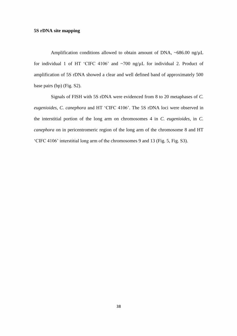
5S rDNA site mapping........................................................................................................ 28
Results.............................................................................................................. 30
Tissue culture biological material....................................................................................... 30
Nuclear 2C value................................................................................................................. 30
Prometaphases and metaphases obtention....................................................................... 31
Morphometric analysis and chromosomal DNA content................................................ 32
5S rDNA site mapping........................................................................................................ 38
x
Discussion......................................................................................................... 40
Conclusion........................................................................................................ 47
Supplementary material ................................................................................. 48
Acknowledgments .......................................................................................... 53
Author contribution statement ...................................................................... 53
References......................................................................................................... 54
11
Title: Origin of the allotriploid “Híbrido de Timor” through a
karyotype comparison with its Coffea ancestors.
Authors: Stéfanie Cristina de Oliveira1, Natália Arruda Sanglard
1, Gustavo Fernandes
Mariano1, Ariane Tonetto Vieira
1, Mariana Cansian Sattler
2, Fernanda Aparecida Ferrari
Soares2, Carlos Roberto Carvalho
2, Wellington Ronildo Clarindo
1*
1Laboratório de Citogenética, Centro de Ciências Agrárias e Engenharia, Universidade
Federal do Espírito Santo, ZIP: 29.500-000, Alegre – ES, Brazil.
2Laboratório de Citogenética e Citometria, Departamento de Biologia Geral, Centro de
Ciências Biológicas e da Saúde, Universidade Federal de Viçosa, ZIP: 36.570-000,
Viçosa – MG, Brazil.
*Corresponding author: e-mail: [email protected]
PHONE: +55 28 3552-8626, FAX: +55 28 3552-8627
12
Resumo
Entre as espécies Coffea, existe um híbrido natural denominado "Híbrido de Timor"
(HT), encontrado na Ilha de Timor em 1927. HT 'CIFC 4106', o qual representa a
primeira planta, possui 2n = 3x = 33 cromossomos e valor 1C DNA igual a 1C = 2.10
pg. O número cromossômico, o conteúdo de DNA e evidências geográficas, suportam
uma possível origem alotriploide a partir da fusão de uma célula reprodutiva reduzida
de Coffea arabica (2n = 4x = 44) com outra célula, também reduzida, de Coffea
canephora (2n = 2x = 22). C. arabica, outro alopoliploide pertencente ao gênero,
acumula estudos que buscam desvendar seus progenitores. Dados moleculares e
cariotípicos sugerem que este alotetraploide verdadeiro seja formado a partir de uma
célula reprodutiva reduzida de C. canephora (CC) e C. eugenioides (EE), seguido por
um evento de poliploidização. Neste sentido, acredita-se que o genoma de C. arabica
seja representado como CaC
aE
aE
a. Com base nas evidências mencionadas, formulamos a
seguinte hipótese: o genoma de HT 'CIFC 4106' é CCaE
a? O presente estudo
caracterizou citogenicamente C. eugenioides, C. canephora, C. arabica e HT 'CIFC
4106'. A combinação de dados morfométricos, conteúdo de DNA nuclear e
cromossômico e hibridização in situ fluorescente (FISH) com rDNA 5S, expandiu o
conhecimento sobre a origem evolutiva e a estrutura do genoma de HT 'CIFC 4106'. O
cariograma de HT 'CIFC 4106' evidenciou pares e grupos cromossômicos delimitados
de acordo com o tamanho total, classes e conteúdos de DNA cromossômicos. Com base
nessas características cariotípicas, foi possível inferir a presença de dois genomas
idênticos em HT 'CIFC 4106', possivelmente de C. canephora (CC) e um genoma
distinto (C. eugenioides, E). Os cromossomos de HT 'CIFC 4106' apresentaram classe,
conteúdo de DNA idênticos aos cromossomos de C. eugenioides, C. canephora e C.
13
arabica. Padrões de distribuição de sinais 5S em HT 'CIFC 4106' foram similares aos
encontrados nos possíveis progenitores C. eugenioides e C. canephora. Os dados
revelados neste estudo corroboram com a hipótese CCaE
a do genoma de HT 'CIFC
4106'.
Palavras-chave: Café, alopoliploidia, citogenética, citometria de imagem, FISH.
14
Abstract
Among the Coffea, there is a natural hybrid denominated “Híbrido de Timor” (HT),
found on Timor Island in 1927. HT 'CIFC 4106', which represents the first HT plant,
possesses 2n = 3x = 33 chromosomes and 1C DNA value equal 1C = 2.10 pg.
Chromosomal number, nuclear 1C DNA content and geographical evidences support a
possible allotriploid origin from fusion of a reduced reproductive cell of Coffea arabica
(2n = 4x = 44) and Coffea canephora (2n = 2x = 22). C. arabica, another allopolyploid,
accumulates studies that seek to unravel its progenitors. Molecular and karyotype data
suggests that this true allotetraploid was formed from a reduced reproductive cell of C.
canephora (CC) and another C. eugenioides (EE) followed by a polyploidization event.
In this way, C. arabica genome is represented as CaC
aE
aE
a. Based on this evidences, we
formulate the following hypothesis: HT ‘CIFC 4106’ genome is CCaE
a? The present
study aimed to cytogenetically characterize C. eugenioides, C. canephora, C. arabica
and HT 'CIFC 4106'. The combination of morphometric data, flow and image
cytometries and 5S rDNA fluorescente in situ hybridization (FISH) expanded
knowledge about the evolutive origin and genome structure of HT ‘CIFC 4106’. HT
'CIFC 4106' karyogram evidenced chromosomes group delimited according to the class
and DNA value similar. Based on these karyotype features was possible show the
presence of two identical genomes in HT 'CIFC 4106', possibly of C. canephora (CC)
and a distinct genome (C. eugenioides, E). HT 'CIFC 4106' chromosomes presented
class, DNA content identical to the chromosomes of C. eugenioides, C. canephora and
C. arabica. 5S rDNA sites were detected in two HT ‘CIFC 4106’ chromosomes.
Distribution patterns of 5S markings found in C. eugenioides and C. canephora
revealed in this study corroborate with HT ‘CIFC 4106’ CCaE
a genome hypothesis. The
15
distribution of the 5S rDNA sites found in three Coffea allied to the chromosomal DNA
content and class shows chromosomes similar to C. eugenioides and C. canephora in
HT ‘CIFC 4106’. In addition, the data reveal possible structural chromosomal
rearrangements along the evolution of HT ‘CIFC 4106’.
Keywords: Coffee, allopolyploid, cytogenetic, image cytometry, FISH.
16
Introduction
Allopolyploids are polyploids formed from hybridization, following or no by
duplication of the two divergent genomes. As well as autopolyploidy (polyploidy within
or between populations of a single specie), the allopolyploidy is considered an
important mechanism of speciation in plants (Stebbins 1950, Soltis and Soltis 2009,
2012). This relevance is corroborated by the abundance of species allopolyploids in
nature (Liu and Wendel 2003). Informations based on origin and karyotype changes in
natural allopolyploid is scarce (Mallet 2007, Soltis et al. 2009; 2016). Cytogenetic
studies provide information on the evolution and diversification of the species,
identifying potential progenitors and numerical and structural chromosome changes in
allopolyploids (Chester et al 2012).
Coffea genus, Rubiaceae family, has been target of evolution researches,
mainly about the natural allopolyploid genomes (Hamon et al. 2009, 2015). This genus
is composed of approximately 120 species, which show natural distribution in Africa
and the Indian Ocean Islands, southern and southeast Asia, and Australasia (Hamon et
al 2015, 2017). Between these species, one allopolyploid speciation event is recognized,
such as the formation of the true allotetraploid Coffea arabica L. (2n = 4x = 44
chromosomes; Clarindo and Carvalho 2009, Lashermes et al. 1999, Yu et al. 2011, ). In
addition, the formation of the natural triploid ‘‘Híbrido de Timor’’ (HT) ‘CIFC 4106’
(2n = 3x= 33) has recently been appointed (Capucho et al. 2009, Setotaw et al. 2010,
Clarindo et al. 2013).
All Coffea species deposited in Chromosome Counts Database and Index to
Plant Chromosome Numbers are diploids (2n = 2x = 22), except C. arabica (Rijo 1974),
Coffea jenkinsii Hook (2n = 8x = 88, Mehra and Bawa 1969; CCDB 2017 –
17
http://ccdb.tau.ac.il/search/) and Coffea excelsa A. Chev. (2n = 4x = 44, Selvaraj 1987;
IPCN 2017 – http://www.tropicos.org). Nuclear 2C value in Coffea varies between the
diploid species from 1.03 pg for Coffea racemosa Lour to 1.80 pg for Coffea humilis A.
Chev, reflecting also the polyploid condiction, as 2C = 2.62 pg to C. arabica (Noirot et
al. 2003; Bennett and Leitch 2011) and 1C = 2.10 pg for HT ‘CIFC 4106’ (Clarindo et
al. 2013). The chromosome number and nuclear 1C value remain constant in all HT
'CIFC 4106' individuals, which have vegetatively been propagated by cutting (Clarindo
et al. 2013) and tissue culture (Sattler et al. 2016; Sanglard et al. 2017). Nuclear genome
size of HT "CIFC 4106" will be referred to as 1C because its homologous pairs are not
defined. Karyotypic studies in HT ‘CIFC 4106’ are necessary to confirm their origin
and characterize possible homologous pairs.
Coffea diploid species have a monophyletic origin, from an ancestor (Lashmeres
et al. 1996; Mahé et al. 2007; Yu et al. 2011) with basic chromosome number of x = 11,
identical to the one currently represented by the genus (Mahé et al. 2007). Coffea
diploid species diversification has been estimated about 5 – 25 Mya (Lashermes et al.
1996) or ~4.2 Mya (Yu et al. 2011). True allotetraploid C. arabica was originated in
~0.665 Mya (Yu et al. 2011). Coffea arabica was probably formed from a natural
hybridization between two diploid Coffea species, followed by a polyploidization event
(Lashermes et al. 1999; Cenci et al. 2012; Fig. 1).
18
19
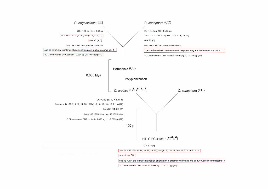
Fig. 1 Karyotype features and origin of allopolyploids C. arabica and HT ‘CIFC 4106’. Karyotype data
about these Coffea were recorded from cytogenetic bibliographies (Pinto-Maglio and Da Cruz 1998;
Noirot et al. 2003; Clarindo et al. 2006; Clarindo et al. 2009; Hamon et al. 2009) and from the
unprecedented results of this study (red box). For these Coffea were reported: 2C and/or 1C nuclear DNA
content measured by flow cytometry; 2n chromosome number; ploidy level; class of each chromosome of
the karyotype (M – metacentric, SM – submetacentric, A – acrocentric); number and chromosome with
secondary construction (SC); number of 18S and 5S rDNA sites; 1C chromosomal DNA content. The
number de SC found in the HT ‘CIFC 4106’ prometaphases and metaphases varied of one (12 mitotic
cells, Fig. 2k) to 3 (one mitotic cell, Fig. S1).
Due to evolutive and agronomic relevance, studies have focused to know the C.
arabica progenitors. Some species have been pointed as possible progenitors: Coffea
eugenioides S. Moore (Berthou et al. 1983; Orozco-Castillo et al. 1996; Cros et al.
1998; Raina et al. 1998; Lashermes et al. 1999), Coffea canephora Pierre ex A.
Froehner (Berthou et al. 1983; Lashermes et al. 1997; Lashermes et al. 1999; Clarindo
and Carvalho 2009), Coffea congensis A. Froehner (Berthou et al. 1983; Lashermes et
al. 1997; Raina et al. 1998), and Coffea brevipes Hiern (Lashermes et al. 1997). Coffea
canephora (CC) and C. eugenioides (EE) are the more probably ancestors of C. arabica
(CaC
aE
aE
a). Corroborating to this hypothesis, the sum of the number of 18S rDNA (two
sites in C. eugenioides, one site in C. canephora) corresponds to the number of C.
arabica (three sites) (Hamon et al. 2009). From morphometry (total and arms lenghts,
and class) and DNA content, C. arabica chromosomes were compared with the of C.
canephora (Clarindo e Carvalho 2009), one of the potential ancestors (Berthou et al.
1983; Lashermes et al. 1997; Lashermes et al. 1999). Chromosomes 1, 19 and 21
(submetacentrics, with 1C = 0.090, 0.050 and 0.050 pg, respectively) of C. arabica
were considered identical to chromosomes 1, 10 and 11 of C. canephora, respectively
20
(Clarindo e Carvalho 2009). An accurate comparison between C. arabica, C. canephora
and C. eugenioides karyotypes would deepen the knowledge of the probable
relationship of these three Coffea and HT ‘CIFC 4106’.
Supposed first HT plant was found in Timor Island in 1927 and can be
represented by access 'CIFC 4106'. HT ‘CIFC 4106’ triploid condiction (2n = 3x = 33
chromosomes; 1C = 2.10 pg; Clarindo et al. 2013) is consistent with the hypothesis of
the first plant of HT originated from the fusion of a reproductive reduced cell of true
allotetraploid C. arabica (CaC
aE
aE
a) and another, also reduced, of C. canephora (CC),
generating a semi-fertile allotriploid (Fig. 1). This hyphotesis is corroborated by
historical evidences, since some HT plants were found in an area where these two
Coffea species were cultivated (Rijo 1974). Besides, this germplasm has 1C = 2.10 pg
of nuclear DNA content equivalent to the sum of the 1C value of the reproductive cells
of C. canephora (1C = 0.73 pg, n = x = 11 chromosomes) and C. arabica (1C = 1.355
pg, n = 2x = 22 chromosomes) (Clarindo et al. 2013). However, the genomic
composition HT 'CIFC 4106' is not known.
Based on previous karyotype and genome size data of HT ‘CIFC 4106’
(Clarindo et al. 2013; Sattler et al. 2016; Sanglard et al. 2017) and C. arabica (Noirot et
al. 2003; Clarindo and Carvalho 2008, 2009; Hamon et al. 2009), our hypothesis is
appointed: the allotriploid genome HT 'CIFC 4106' is possibly composed of one
chromosomal set of C. eugenioides (Ea) and two of C. canephora (CC
a), representing a
CCaE
a genome (Fig. 1).
This study researched data that answer the following question: HT ‘CIFC 4106’
genome is CCaE
a? Considering this hypothesis, the combination of cytogenetic
(morphometry and chromosome class), flow and image cytometries (nuclear and
chromosomal DNA content) and fluorescente in situ hybridization (FISH) data will
21
enlarge knowledge about the evolutionary origin and genome composition of HT ‘CIFC
4106’. Thus, the aims of this study were to: a) measure the nuclear genome size, b)
confirme the chromosome number, c) characterize the karyotype, d) calculate the
chromosomal DNA content of the close related Coffea (C. eugenioides, C. canephora,
C. arabica and HT ‘CIFC 4106’), e) map 5S rDNA site by FISH in HT ‘CIFC 4106’
and possible ancestors C. eugenioides and C. canephora. Besides, this study also
provided new insights about C. arabica origin and karyotype evolution.
Material and methods
Biological material
In vitro regenerated plantlets of C. eugenioides, C. canephora, C. arabica and
HT 'CIFC 4106' were used as explant donors (Fig. 2a) for friable callus establishment
(Fig. 2b), somatic embryo (Fig. 2c) and seedling regeneration (Fig. 2d). These
biological materials were source of mitotic cells for cytogenetic and cytometry
procedures.
Nuclear 2C value
Nuclei suspensions were obtained from simultaneously chopping (Galbraith et
al. 1983; Otto 1990; Praça-Fontes et al. 2011) of the leave fragment (2 cm2) of S.
licopersycum ‘Stupicke’ (internal standard, 2C = 2.00 pg, Praça-Fontes et al. 2011) and
of each one of the ten plantlets (Fig. 2e) of C. eugenioides, C. canephora, C. arabica
and HT ‘CIFC 4106’ (samples). Staining of the nuclear suspensions was performed
following international criteria (intercalant fluorochrome – propidium iodide Sigma®
)
22
associated to methodological steps of Praça-Fontes et al. (2011). The suspensions were
analyzed in a Partec PAS® flow cytometer (Partec
® Gmbh, Munster, Germany)
equipped with a laser source (488 nm). Relative fluorescence intensity histograms (Fig.
2f) were analyzed by the FlowMax Partec®
software (Partec Gmbh, Munster,
Germany). G0/G1 nuclei peak of S. lycopersicum was the reference for nuclear 2C value
measurement of the Coffea (Fig. 2f).
Tissue culture biological material
For friable calli establishment (Fig. 2b), leaf fragments (1 cm²) from in vitro
plantlets of each Coffea were excised and inoculated (Fig. 2a), with the abaxial surface
facing upwards, in Petri dishes containing callus induction medium (Table S1). After 3
months, 0.5 g of friable calli was transferred to Erlenmeyers with 30 ml of callus
proliferation medium without Phytagel (Fig. 2h, Table S1). The tissue culture
conditions were modified by van Boxtel and Berthouly (1996). Four subcultures, every
15 days, were performed during the first 60 days. The Erlenmeyers were maintained on
shaker at 100 rpm, under a 16/8 h light/dark regime, with 36 µmol m-2
s-1
light radiation
and 24°C ± 2°C (Fig. 2h1). After the stabilization period of the culture, the cell
aggregate suspensions were used for chromosome obtaining (Fig. 2h2).
Roots of distinct Coffea plantlets (Fig. 2g) were also used to expose at the
different cytogenetic treatments. In order to regenerate the plantlets, friable
embryogenic calli (Fig. 2b) were transferred to conversion and maturation medium (Fig.
2c; Table S1). Subsequently, the mature cotyledonary somatic embryos (Fig. 2d left)
were transferred for germination medium (Fig. 2d right, Table S1). After the first root
removing, the same seedlings were maintained in medium of the same prior
23
composition, but without GA3. From the regenerated plantlets, root meristems were
routinely excised and cytogenetically treated (Fig. 2g).
Prometaphases and metaphases obtention
Roots and cell aggregate suspensions were treated with 4 µM amiprophos-
methyl (APM, Sigma®
) (Clarindo and Carvalho 2009) or with 4 µM APM/95.0 µM
cycloheximide (Sigma®, dos Reis et al. 2014), for 1 – 8 h at 25°C (Fig. 2g, h
2). Cell
aggregate suspensions were treated between the period of 7 to 10 days after four
subcultures, equivalent to 60 days (Fig. 2h2). The biological materials were washed in
distilled water (dH2O), fixed three times in 3:1 methanol:acetic acid (Sigma®
), and
stored for 24 h at -20°C. About ten Erlenmeyers of cell aggregate suspensions and 50
roots of each Coffea were used for each treatment.
Next, each root meristem (Fig. 2g1) or a sample of 20 – 30 aggregates (~100
µL volume) was transferred to microtubes (2 ml), washed three times with dH2O, and
enzymatically macerated for 1 – 2 h, at 34°C (Fig. 2g2, h
3). The enzymatic pool was
composed of 4% cellulase (C-1184, Sigma®
), 0.4% hemicellulase (H-2125, Sigma
®) and
1% macerozyme (R10, Yacult Pharmaceutical®) diluted in pectinase (E-6287, Sigma
®).
The pool was diluted in dH2O in the proportion 1:40 – 1:100. The dilution was adjusted
according to the thickness of the roots and aggregates. After, the biological materials
were washed for 10 min in dH2O, fixed and stored at -20°C (Clarindo and Carvalho
2009).
Slides, from root meristems or aggregates, were prepared by dissociation and
air-drying techniques (Fig. 2i, Carvalho and Saraiva 1997). The slides were previously
24
evaluated and selected under a Nikon Eclipse Ci phase contrast microscope (Nikon,
Japan), following the criteria:
a) mitotic cells with little or no cytoplasmic background,
b) chromosomes with well-defined telomere and centromere,
c) chromosomes without overlaps and structural deformations of the chromatin.
For ICM (Fig. 2j,k), the selected slides of all Coffea were fixed in 17:5:1
methanol:37% formaldehyde:acetic acid (Sigma®), at 25°C for 24 h (Carvalho et al.
2011). For FISH (Fig. 2k) (C. eugenioides, C. canephora and HT ‘CIFC 4106’) the
slides were fixed in 70% ethanol at -20°C for at least 15 days, or maintained at 37°C for
five days.
Morphometric analysis and chromosomal DNA content
Feulgen’s reaction procedure was performed in the chromosome preparations
following guideline by Carvalho et al. 2011. For this, the slides were washed in dH2O,
promptly air-dried and hydrolyzed in 5 M HCl (Merck®
) for 12 – 22 min at 25°C.
Posteriorly, the slides were stained with Schiff’s reagent for 12 h at 4°C, washed three
times (3 min each time) in 0.5% SO2 water.
In order to ensure optical conditions suitable for ICM, the Nikon Eclipse 80i
model (Nikon, Japan) microscope and digital analysis system (CCD digital video
camera DS-Fi1c of 8-bits gray and Nis Elements 3.0 imaging software – Nikon, Japan)
were calibrated and configured by means stability, linearity and uniformity tests
(Carvalho et al. 2011). Prometaphases and metaphases were scattered using microscope
Nikon 80i model (Nikon, Japan – equipped with a stabilized light source, a 100x Nikon
Pan Fluor oil immersion objective with 1.30 numerical aperture, aplanat achromat
25
condenser with 0.7 aperture, ND6 neutral density filter, and 550 – 570 nm interference
green color filter) coupled to a monochromatic CCD digital video camera DS-Fi1c of 8-
bits gray (Nikon, Japan), which is coupled to a Pentium Intel Core i5 (Termaltake –
Asus) computer featuring the Nis Elements 3.0 imaging software (Nikon, Japan). At
least twenty prometaphases and/or metaphases of each Coffea were chosen for
morphometric analysis and chromosomal DNA content measurement.
DNA content of each chromosome of the Coffea was measured by distributing
the mean value of the 1C nuclear DNA content (pg), obtained by FCM, proportionally
to the mean integrated optical density (IOD) values of each chromosome, calculated by
ICM (Carvalho et al. 2011). Besides, the chromosomes were characterized as to the total
length, length of the long and short arms, and class. The chromosome class was
determined as proposed by Levan et al. (1964) and reviewed by Guerra (1986): r =
length of the long arm/length of the short arm.
5S rDNA site mapping
The probe 5S rDNA used for FISH was generated by PCR using forward and
reverse primers of 5S rDNA (F:5’-CCTGGGAAGTCCTCGTGTTG-3’ and R:5’-
CTTCGGAGTTCTGATGGGAT-3’) previously selected (Ribeiro et al. 2016).
Genomic DNA were extracted according Doyle and Doyle (1990), using leaves from
four HT ‘CIFC 4106’ plantlets in vitro regenerated. DNA concentration and purity were
determined by NanoDrop (Thermo Scientific®
2000c). DNA integrity was verified by
agarose gel electrophoresis.
Polymerase chain reaction (PCR) was performed with a final volume of 25 µl,
containing 200 ng DNA, 1x buffer (GoTaq®), 0.5 µM of each primer, 1.3 mM of each
26
dNTP, 1.25 U of Taq DNA polymerase (GoTaq®
) and 3.0 mM MgCl2. The
amplification conditions were initial denaturation for 5 min at 95°C; 35 amplification
cycles at 95°C for 1 min, 60°C for 1 min and 72°C for 1 min 30 sec; and final extension
at 72°C for 5 min. The reactions were conducted in an Applied Biosystems VeritiTM
96-
Well Thermal Cycler. PCR products were separated by electrophoresis in agarose
(1.2%) gel with buffer 1X TBE (Tris/Borate/EDTA). The gel was visualized by staining
with GelRedTM
.
5S rDNA probe was prepared using 200 ng of the HT ‘CIFC 4106’ PCR
product amplification (Supplement Fig. 2); 2 mM of each dATP, dCTP and dGTP,
0.0005µM tetramethil-rhodamine 5-dUTP (Roche®); 0.5 µM 5S rDNA primers; 3.0 mM
de MgCl2; 1 U GoTaq® DNA polymerase (Promega Biotechnology
®); 1× GoTaq
®
buffer in a final volume of 25 µL. PCR reactions were performed with the same above
condictions. The labeling products were analyzed by 1.2% agarose gel electrophoresis.
Slides were treated in 1X PBS buffer for 5 min, 3% formaldehyde solution in
PBS buffer for 15 min, followed by 1X PBS buffer for 5 min and ethanol series (70, 85,
100%) at -20°C. Hybridization mix consisted of 300 ng of each probe, 50% formamide
(Sigma-Aldrich®
), 2X SSC buffer and dH2O in final volume of 35 µL. The mixture was
placed on the slide and covered with HybriSlip™ plastic cover slip (Sigma®) and sealed
with Rubber Cement (Elmer's). The hybridizations were conducted in thermociclador
(SlideCycler Loccus®
) with initial denaturation at 71°C for 4 min and 37°C for 24 h.
After, the stringency washes were conducted in 2X SSC solution at 58°C. The slides
were counterstained with 4′,6-diamidino-2-phenylindole dihydrochloride (DAPI,
Sigma-Aldrich®
) and sealed with colorless enamel. Prometaphases and metaphases
were captured by a monochromatic CCD digital video camera DS-Fi1c of 8-bits gray

27
(Nikon, Japan) coupled to a epifluorescence microscope Nikon 80i model (Nikon,
Japan) with a 100X objective lens, UV-2E/C (DAPI) and G2A (5S rDNA probe).
28
29
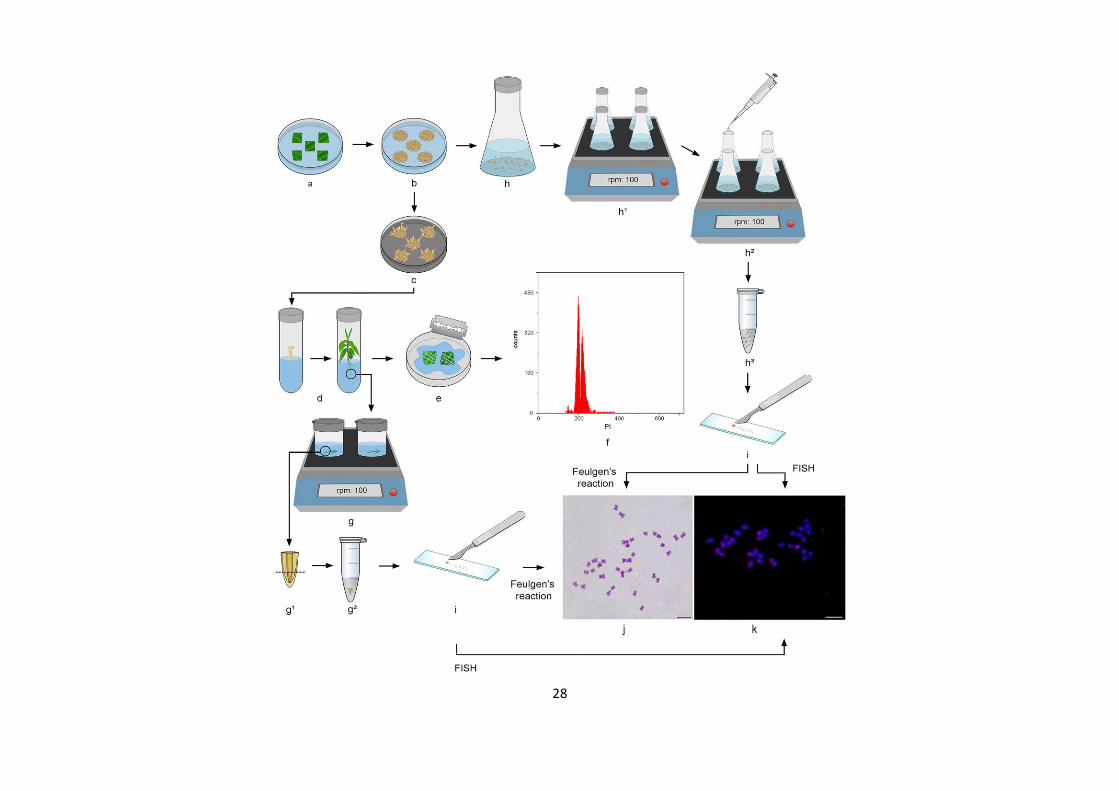
Fig. 2 Guide for obtention of Coffea prometaphasic/metaphasic chromosomes and flow cytometry from
regenerated seedlings and cell aggregate suspensions provided via indirect somatic embryogenesis. a)
Five leaf fragments (~1 cm²) of Coffea inoculated, with the abaxial surface facing upwards, in Petri dishes
containing callus induction medium. b) Calli established after 3 months in callus induction medium,
exhibiting friable aspect and pale yellow color. c) Friable embryogenic calli in conversion and maturation
medium. During the 180 days, friable calli showed globular, heart, torpedo, pre-cotyledonary and mature
cotyledonary somatic embryos. d) Seedlings recovered in germination medium from the mature
cotyledonary somatic embryo. e – f) Representative histogram, which was obtained from flow cytometry
procedure (e), exhibiting fluorescence peaks of G0/G1 nuclei of the standard S. lycopersicum (chanel 200)
and HT ‘CIFC 4106’ (chanel 210) (f). g – g2) Antitubulin exposuring (g) and enzymatic maceration (g
1
and g2) of the excised Coffea roots, highlighting that root meristem should be isolately macerated. h – h
1)
Erlenmeyers on shaker at 100 rpm with the cell aggregate suspension maintained in callus induction
medium without Phytagel. h2 – h
3) Antitubulin exposuring (h
2) and enzymatic maceration (h
3) of the of
the cell aggregation suspensions of the four Coffea, emphasizing that 20 – 30 aggregates (~100 µL
volume) should be macerated. i) Slides prepared by dissociation and air-drying techniques from root
meristems (c – g2) or aggregates (h – h
3). j) HT ‘CIFC 4106’ karyotype showing 2n = 3x = 33
chromosomes stained from Feulgen’s reaction, with one chromosomes exhibiting the SC in the short arm.
k) FISH of the 5S rDNA in HT ‘CIFC 4106’showed two 5S markings. Bars = 5µm.
30
Results
Tissue culture biological material
Friable calli (Fig. 2b) were obtained after 3 months from leaves of all Coffea.
The calli exhibited friable aspect and pale yellow color in callus induction medium
(Table S1). After transferring of these calli to callus proliferation medium, the cell
aggregate suspensions yielded proliferative cells (Table S1; Fig. 2h). Aggregates
showed an increase in 0.5 g their biomass every 15 days, indicating the cell proliferation
occurrence in the aggregates. Cell division was confirmed by the presence of cells at
different stages of the cell cycle (early, middle and late prophases and prometaphases,
and metaphases) on the cytogenetic slides.
During 180 days in embryo conversion and maturation medium, friable calli
showed globular, heart, torpedo, pre-cotyledonary and mature cotyledonary somatic
embryos. From the mature cotyledonary somatic embryo, seedlings were recovered in
germination medium (Fig. 2d left). Thus, cell aggregate suspensions (Fig. 2h) and
plantlets (Fig. 2d right) for all Coffea continuously provided enough biological material
for nuclear DNA measurement (leaves, Fig. 2e, f), chromosome number determination
(Fig. 2j), karyotype characterization, chromosomal DNA content estimation, and 5S
rDNA site mapping (Fig. 2).
Nuclear 2C value
Mean nuclear 2C value was 2C = 1.38 ± 0.012 pg (1C = 0.690 pg) for C.
eugenioides, 2C = 1.41 ± 0.037 pg (1C = 0.705pg) for C. canephora, 2C = 2.62 ± 0.004
pg (1C = 1.310 pg) for C. arabica and 1C = 2.100 ± 0.008 pg for HT ‘CIFC 4106’ (Fig.
31
2). The mean nuclear DNA content of the regenerated plantlets was the same to the
explant donor plants.
Prometaphases and metaphafases obtention
Considering the criteria assumed in the present study to chose the slides, the
treatments that generated prometaphases and metaphases for C. eugenioides, C.
canephora, C. arabica and HT ‘CIFC 4106’ from cell aggregates and root meristems
were: 7 h in 4 µM APM and 2 – 3 h in 4 µM APM/95 µM cycloheximide. Times over 8
h in 4 µM APM or over 3 h in 4 µM APM/95 µM cycloheximide resulted cells with
chromosomes highly compacted, making it impossible to distinguish the chromosomes
in morphometric analyzes.
Enzymatic maceration and dissociation of the cell aggregates and root
meristems, and air drying technique were crucial for the obtaining of chromosomes
scattered, without overlapping and free of cytoplasm. These aspects were obtained from
cell aggregates and ~1 mm root meristems macerated with 1:40 (enzymatic pool:
dH2O), and also from ~0.5 mm root meristems macerated in 1:70 – 1:80 (enzymatic
pool: dH2O), for 2 h at 34°C. For all the Coffea, stoichiometrically stained
chromosomes were only found in the slides fixed in 17:5:1 methanol:37%
formaldehyde:acetic acid for 24 h at 25°C, hydrolyzed in 5 M HCl for 18 min at 25°C,
and stained with Schiff’s reagent for 12 h at 4°C (Fig. 2j).
Morphometric analysis and chromosomal DNA content
32
For the first time, the karyotypes of C. eugenioides and HT ‘CIFC 4106’ were
characterized and assembled. C. eugenioides and C. canephora showed 2n = 2x = 22
chromosomes, C. arabica 2n = 4x = 44 and HT ‘CIFC 4106’ 2n = 3x = 33 (Fig. 3). C.
eugenioides karyogram (Fig. 3a, Table 1) is composed by nine chromosome pairs
classified as submetacentrics (1 – 6, 8, 9 and 11) and two metacentric (7 and 10). Two
chromosome pairs (3 and 5) highlighted due to their secondary constrictions in the short
arm (Fig. 3a). C. canephora displayed nine submetacentric chromosome pairs (1 – 3, 5
– 8, 10, 11) and two metacentric (4, 9), and presence of secondary constriction on
chromosome pair 6 (Fig. 3b, Table 1). C. arabica exhibited sixteen submetacentric
chromosome pairs (1 – 6, 9 – 12, 15 – 19, 21), five metacentric (7, 8, 13, 14 and 20) and
one acrocentric (22) (Fig. 3c, Table 1). HT ‘CIFC 4106’ possesses six chromosomes
(10, 11, 19, 25, 26, 30) classified as metacentrics, and the other twenty-seven (1 – 9, 12
– 18, 20 – 24, 27 – 29, 31 – 33) as submetacentrics (Fig. 3d, Table 1). The number of
secondary constrictions varied between the HT ‘CIFC 4106’ prometaphases/metaphases
of one to three (Fig. 1, Fig. S1).

33
Fig. 3 Karyogram from mitotic chromosomes of C. eugenioides a, C. canephora b, C. arabica c, and HT
'CIFC 4106' d. a C. eugenioides showed two metacentric chromosome pairs (7, 10), nine submetacentrics

34
(1 – 6, 8, 9 and 11) and two chromosomes pairs with SC (3 and 5). b C. canephora presented two
metacentric chromosome pairs (4 and 9), nine submetacentric (1 – 3, 5 – 8, 10 and 11) and one with SC
(6). c C. arabica exhibited 5 metacentric chromosome pairs (7, 8, 13, 14 and 20), sixteen submetacentric
(1 – 6, 9 – 12, 15 – 19 and 21) and one acrocentric (22). d HT ‘CIFC 4106’ showed six metacentric (10,
11, 19, 25, 26, 30) and twenty-seven submetacentric chromosomes (1 – 9, 12 – 18, 20 – 24, 27 – 29, 31 –
33). Bar = 5 µm.
35
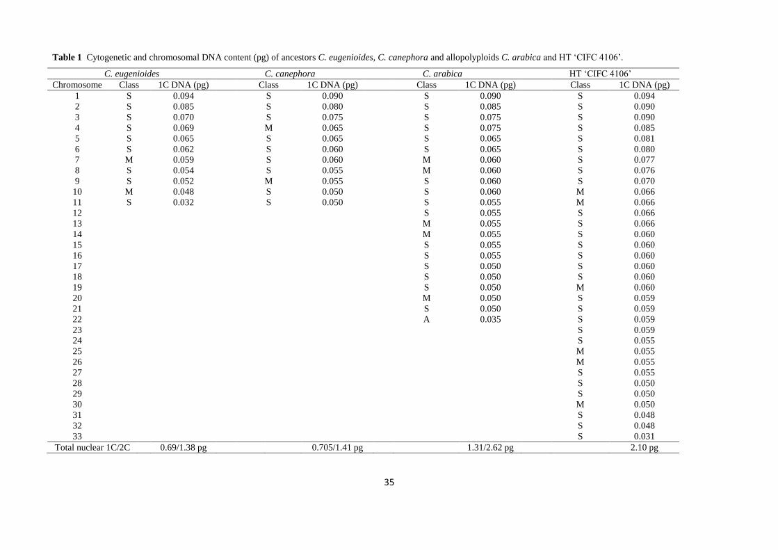
Table 1 Cytogenetic and chromosomal DNA content (pg) of ancestors C. eugenioides, C. canephora and allopolyploids C. arabica and HT ‘CIFC 4106’. 1
C. eugenioides C. canephora C. arabica HT ‘CIFC 4106’
Chromosome Class 1C DNA (pg) Class 1C DNA (pg) Class 1C DNA (pg) Class 1C DNA (pg)
1 S 0.094 S 0.090 S 0.090 S 0.094
2 S 0.085 S 0.080 S 0.085 S 0.090
3 S 0.070 S 0.075 S 0.075 S 0.090
4 S 0.069 M 0.065 S 0.075 S 0.085
5 S 0.065 S 0.065 S 0.065 S 0.081
6 S 0.062 S 0.060 S 0.065 S 0.080
7 M 0.059 S 0.060 M 0.060 S 0.077
8 S 0.054 S 0.055 M 0.060 S 0.076
9 S 0.052 M 0.055 S 0.060 S 0.070
10 M 0.048 S 0.050 S 0.060 M 0.066
11 S 0.032 S 0.050 S 0.055 M 0.066
12 S 0.055 S 0.066
13 M 0.055 S 0.066
14 M 0.055 S 0.060
15 S 0.055 S 0.060
16 S 0.055 S 0.060
17 S 0.050 S 0.060
18 S 0.050 S 0.060
19 S 0.050 M 0.060
20 M 0.050 S 0.059
21 S 0.050 S 0.059
22 A 0.035 S 0.059
23 S 0.059
24 S 0.055
25 M 0.055
26 M 0.055
27 S 0.055
28 S 0.050
29 S 0.050
30 M 0.050
31 S 0.048
32 S 0.048
33 S 0.031
Total nuclear 1C/2C 0.69/1.38 pg 0.705/1.41 pg 1.31/2.62 pg 2.10 pg
36
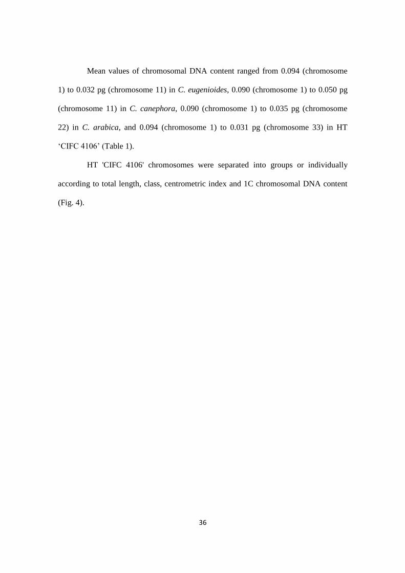
Mean values of chromosomal DNA content ranged from 0.094 (chromosome
1) to 0.032 pg (chromosome 11) in C. eugenioides, 0.090 (chromosome 1) to 0.050 pg
(chromosome 11) in C. canephora, 0.090 (chromosome 1) to 0.035 pg (chromosome
22) in C. arabica, and 0.094 (chromosome 1) to 0.031 pg (chromosome 33) in HT
‘CIFC 4106’ (Table 1).
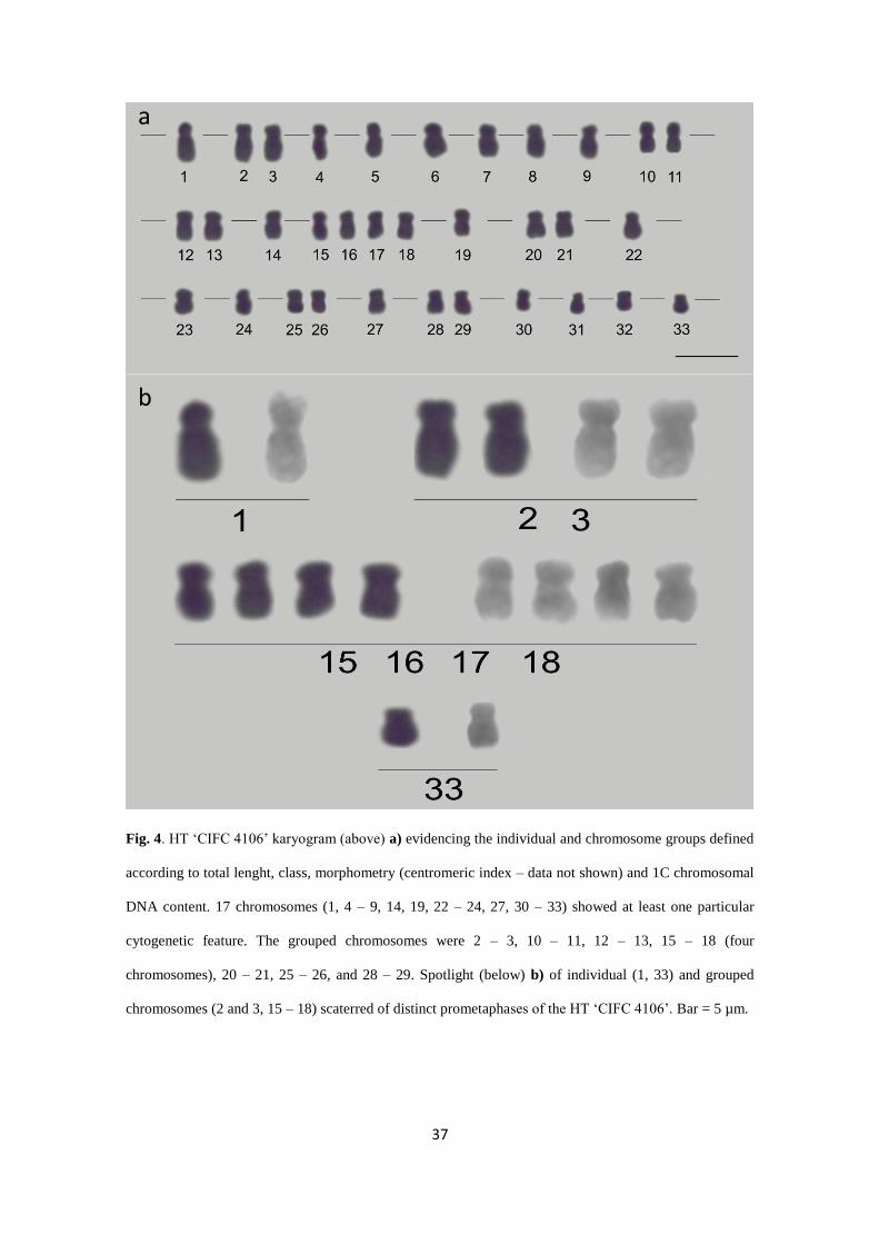
HT 'CIFC 4106' chromosomes were separated into groups or individually
according to total length, class, centrometric index and 1C chromosomal DNA content
(Fig. 4).
37
Fig. 4. HT ‘CIFC 4106’ karyogram (above) a) evidencing the individual and chromosome groups defined
according to total lenght, class, morphometry (centromeric index – data not shown) and 1C chromosomal
DNA content. 17 chromosomes (1, 4 – 9, 14, 19, 22 – 24, 27, 30 – 33) showed at least one particular
cytogenetic feature. The grouped chromosomes were 2 – 3, 10 – 11, 12 – 13, 15 – 18 (four
chromosomes), 20 – 21, 25 – 26, and 28 – 29. Spotlight (below) b) of individual (1, 33) and grouped
chromosomes (2 and 3, 15 – 18) scaterred of distinct prometaphases of the HT ‘CIFC 4106’. Bar = 5 µm.
a
b
38
5S rDNA site mapping
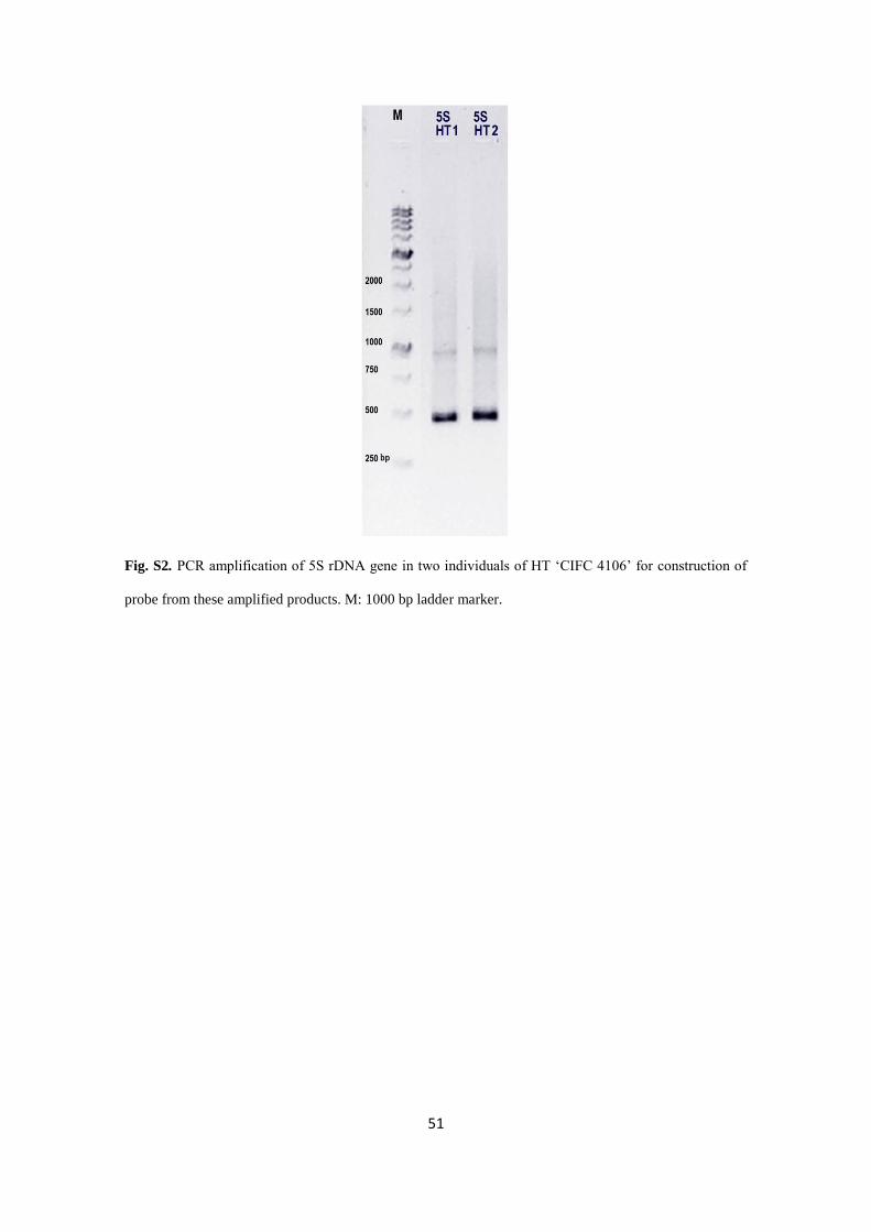
Amplification conditions allowed to obtain amount of DNA, ~686.00 ng/µL
for individual 1 of HT ‘CIFC 4106’ and ~700 ng/µL for individual 2. Product of
amplification of 5S rDNA showed a clear and well defined band of approximately 500
base pairs (bp) (Fig. S2).
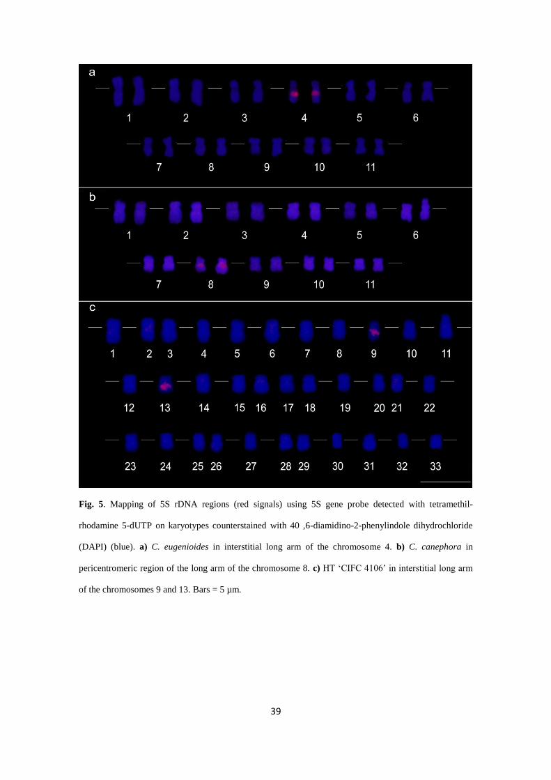
Signals of FISH with 5S rDNA were evidenced from 8 to 20 metaphases of C.
eugenioides, C. canephora and HT ‘CIFC 4106’. The 5S rDNA loci were observed in
the interstitial portion of the long arm on chromosomes 4 in C. eugenioides, in C.
canephora on in pericentromeric region of the long arm of the chromosome 8 and HT
‘CIFC 4106’ interstitial long arm of the chromosomes 9 and 13 (Fig. 5, Fig. S3).
39
Fig. 5. Mapping of 5S rDNA regions (red signals) using 5S gene probe detected with tetramethil-
rhodamine 5-dUTP on karyotypes counterstained with 40 ,6-diamidino-2-phenylindole dihydrochloride
(DAPI) (blue). a) C. eugenioides in interstitial long arm of the chromosome 4. b) C. canephora in
pericentromeric region of the long arm of the chromosome 8. c) HT ‘CIFC 4106’ in interstitial long arm
of the chromosomes 9 and 13. Bars = 5 µm.
40
Discussion
Based on previous studies, Coffea chromosomes are considered relatively small
and homomorphic (Lashermes et al. 1999; Pinto-Maglio 2006; Hamon et al. 2009).
These aspects are presented as obstacles to the assembly and analysis of karyotypes
(Hamon et al. 2009, Clarindo and Carvalho 2009). In this sense, the characteristics of
the preparations containing mitotic chromosomes are a crucial factor for the
advancement of studies of karyotype evolution of Coffea. Although these aspects, our
results demonstrated that the methodologies utilized in this work provided slides with
adequate chromosomes according to the criteria mentioned above for Coffea cytogenetic
analyzes.
Roots (Fig. 2d) and cell aggregate suspensions (Fig. 2h) presented proliferative
cells, which are a biological material fundamental for the cytogenetic procedures.
Approximately 100 slides of each with prometaphases and metaphases were generated,
allowing to test all subsequent steps, mainly the enzymatic maceration, HCl hydrolysis
in image cytometry and FISH procedure. Of these slides, about 10% for each Coffea
showed chromosomes according to criteria determined in this study, and, then, were
used in the prometaphase/metaphase capture step. Cell aggregate suspensions and root
meristems provided cells with a stable chromosome number in each Coffea. The
biological materials from tissue culture were confirmed as an alternative source for
obtaining mitotic cells in Coffea (Clarindo and Carvalho 2006, 2009). Therefore, the in
vitro condition guarantees the availability of Coffea biological material for procedures
during a long time.
In addition to this care, three bottlenecks were considered for morphometry and
DNA content measurement of the chromosomes: the antitubulin treatment with APM or
41
APM/cycloheximide, the enzymatic maceration, and the hydrolysis exposure time in 5
M HCl. The best antitubulin treatments of 4 µM APM or 4 µM APM/ 95 µM
cycloheximide presented prometaphases/metaphases with chromosomes in sufficient
amount, at least 20 per slide. APM/cycloheximide combination for
prometaphase/metaphase arrest was an unprecedented cytogenetic result for Coffea.
This combination associated the effect of two different chemical compounds. APM is a
phosphoric amide compound that inhibits the polymerization of microtubules
(Morejohn and Fosket 1984; Planchais 2000). Cycloheximide is an inhibitor of
eukaryotic translation, blocking the elongation phase of the nascent polypeptide,
specifically acting under the E-site of the 60S ribosomal subunit (Schneider-Poetsch
2010). Thus, proteins, such as the α- and β-tubilins, are not synthesized with the
addition of cycloheximide. However, due to this cycloheximide cellular effect, different
stages of the cell cycle can be blocked, depending on the momentum of the cell cycle in
which that substance is added (Rose 1970). Therefore, the success of using a compound
that inhibits protein synthesis, such as cycloheximide, is related to the proliferative state
of the biological material. In this sense, in biological materials with higher rates of cell
division, as in this study, this substance will act on a larger number of cells in
prometaphase and metaphase. In this way, as observed here, the combination of APM
with cycloheximide accumulated a higher number of prometaphase and metaphase cells
than the treatment with APM.
The maceration of the cell aggregations or root meristems involved an
enzymatic pool containing four types of enzymes (cellulase, hemicellulase, macerozyme
and pectinase), unlike previous protocols for Coffea using only pectinase (Clarindo and
Carvalho 2006, 2008, 2009). This pool was effective due to specific act on different
components of the cell wall. This strategy resulted slides with few or without
42
cytoplasmic traces, facilitating to adjust the time for HCl hydrolysis in image cytometry
and accessibility of the 5S rDNA probe during FISH procedure.
Duration of the HCl hydrolysis treatment was identical for all Coffea. The time
of 18 min in 5 M HCl, associated to 12 h of the staining in Schiff’s reagent, provided
stoichiometrically stained chromosomes without chromatin damage. HCl hydrolysis,
during Feulgen’s reaction, removes purine bases of the deoxyribose, giving free
aldehyde groups when the sugar ring opens. Pararosaniline present in the Schiff’s
reagent binds to the aldehyde groups becoming purple (Greilhuber 2008).
The reproducibility of fluorescent markings of 5S rDNA probe analyzed of
three Coffea metaphases showed that the FISH procedure was adequate (Fig. 5). The
success of this technique was attributed to the cytogenetic preparations according to the
selection criteria established in the present study such as chromatin integrity, absence of
cytoplasmic vestiges added to stringency conditions during FISH. High stringency
conditions was ensured by probe concentration, 50% formamide and 2X SSC solution
buffer at 58°C. Formamide is a denaturing agent capable of interacting with the
hydrogen bonds present in the double helix, destabilizing the DNA (Blake and Delcourt
1996, Fuchs et al. 2010). Once the DNA duplex was destabilized, the hybridization
efficiency was decreased at high concentrations of formamide, increasing stringency.
These conditions enable a specific marking by reducing the background.
The CCaE
a hypothesis about HT ‘CIFC 4106’ genome was raised based on
historical evidences (Rijo 1974), chromosome number, nuclear 2C value (Clarindo et al.
2013), and origin of its possible progenitor C. arabica (Lashermes et al. 1999; Hamon
et al. 2009). Historical evidences suggest the origin of the HT from C. canephora and C.
arabica, because the first plants were found in a plantation of C. arabica 'Typica'
(CaC
aE
aE
a) which also contained some C. canephora (CC) plants (Rijo 1974). Besides,
43
chromosome number (2n = 3x = 33) determined for HT ‘CIFC 4106’ (Fig. 1, 2j, 3d)
reflects a triploid condition, also from the basic chromosome number x = 11 of Coffea
(Hamon et al. 2009). Nuclear 1C value of HT ‘CIFC 4106’ is equivalent to 2.10 pg (Fig.
2f; Clarindo et al. 2013). Then, the 2n chromosome number and 1C value of the HT
‘CIFC 4106’ is equivalent to the sum of the reduced reproductive cells of its possible
progenitors.
Considering these evidences, the genomic origin of HT ‘CIFC 4106’, as well as
of its possible progenitors of the true allotetraploid C. arabica, goes back from the
monofiletism of the Coffea genus (Fig. 1, Yu et al 2011). From the diversification of a
common ancestor, all Coffea diploid species were originated (Hamon et al. 2017), as C.
eugenioides (EE) and C. canephora (CC) (Lashermes et al. 1999; Yu et al. 2011; Cenci
et al. 2012). The fusion of reduced reproductive cells from these two species, following
a polyploidization event, resulted in the C. arabica (CaC
aE
aE
a) formation (Yu et al.
2011). More recent, the fusion of two reduced reproductive cell between C. canephora
(C) and of C. arabica (CaE
a) possibly gave rise to HT (Fig. 1).
HT ‘CIFC 4106’ chromosomal number corresponded to 2n = 3x = 33 (Fig. 2j,
3d; Fig. S1, Fig. S3a), the same reported in previous studies (Clarindo et al. 2013; Fig.
1). Therefore, the allotriploidy is a stable karyotype condition in all HT ‘CIFC 4106’
that has been vegetatively propagated: the plants in greenhouse, and the cell aggregate
suspensions and regenerated seedlings propagated in vitro. The allotriploid condition
supports to the fusion of one reduced reproductive cell of C. arabica (CaE
a; n = 2x = 22)
with another also reduced cell of C. canephora (C; n = x = 11). Corroborating, nuclear
DNA content of the HT 'CIFC 4106' (1C = 2.10 pg, Fig. 2f) reinforces the CCaE
a
hypothesis. This value is equivalent to the sum of the mean nuclear genome size of C.
canephora (CC; 2C = 1.41 pg, Fig. 1) and half of C. eugenioides (E; 1C = 0.690 pg).
44
In addition to the 2n chromosome number, this study expanded the karyotype
data in Coffea through the characterization of all chromosomes of the allotriploid HT
'CIFC 4106' and of the diploid C. eugenioides (Fig. 3a, d). In order to recount the
history of HT 'CIFC 4106' origin, the karyotype characterization also was performed for
the diploid C. canephora and true allotetraploid C. arabica. The karyotypes of the four
Coffea have morphologically identical and similar chromosomes (Table 1, Fig. 3). The
ancestors C. canephora and C. arabica (also in Clarindo and Carvalho 2009), and C.
eugenioides predominantly showed metacentric and submetacentric chromosomes, with
a single exception for the acrocentric pair (22) of C. arabica. Likewise, only
metacentric and submetacentric chromosomes were observed in HT ‘CIFC 4106’
karyotype, supporting the CCaE
a hypothesis.
DNA content, obtained by the image cytometry, of all chromosomes of the four
Coffea increased the knowledge about the relation to the ancestors and HT “CIFC
4106’. Image cytometry is a reliable, accurate and relatively affordable (Carvalho et al.
2011) technique for specialized microscopy laboratories. Differently of other techniques
that allow to measure the chromosomal DNA content, image cytometry needs of
relatively small number of prometaphases/metaphases, at least 10 (Carvalho et al.
2011).
Based on the DNA content and class of the chromosomes of HT 'CIFC 4106'
formed pairs as: 2 – 3, 10 – 11, 12 – 13, 20 – 21, 25 – 26, 28 – 29 (six groups). One
group of four chromosomes was formed (15 – 18), and the other chromosomes was
isolated in class and/or DNA content (Fig. 4, Table 1). The chromosomes in pairs (six
groups) can represent the CCa subgenomes, being C of C. canephora and C
a of C.
arabica. A look at the HT ‘CIFC 4106’ karyogram shows that the possible pairs 2 – 3,
10 – 11, 25 – 26 and 28 – 29 may represent the chromosomes 1, 4, 9 and 10 or 11 of C.
45
canephora, respectively (Table 1). HT ‘CIFC 4106’ group 15 – 18 is similar to pair 6
and 7 of C. canephora (Fig. 4; Table 1).
Other evidence supporting the CCaE
a genome hypothesis, specifically in
relation to the E genome, is due to chromosome 1, 4 and 9 of HT ‘CIFC 4106’ being
equal (morphometry and DNA content) to 1, 2 and 3 of C. eugenioides, respectively. In
addition, chromosomes 7 and 11 of C. eugenioides are similar to 19 and 33 of HT
‘CIFC 4106’, respectively (Fig. 3, Table 1).
Morphometric and chromosomal DNA content data also provided insights
about origin of C. arabica from C. canephora and C. eugenioides, which have been
pointed as the most probably progenitors (Berthou et al. 1983; Lashermes et al. 1999;
Clarindo and Carvalho 2009). These Coffea species showed very similar karyotype
features, which point to a close relationship between the C. arabica and with the C.
canephora and C. eugenioides. Some chromosome pairs of C. canephora and of C.
eugenioides present the same chromosome class and DNA content of some
chromosome pairs or of chromosome pair groups of C. arabica (Table 1, Fig. 3). The C.
canephora pair 9 was equal C. arabica pair 13 and 14. C. canephora pair 10 and 11
were identical C. arabica pairs 17, 18, 19 and 21 respectively. This result corroborates
that after hybridization at the C. arabica origin, the hybrid genome was followed by a
genome duplication event. Chromosome class and DNA content provide insights about
the participation of the genomes of the C. canephora and C. eugenioides progenitors in
the formation of the allopolyploid C. arabica.
rDNA sites are highly conserved genomic regions used to investigate genome
origin and organization in several plant species (Cai et al. 2006, Hamon et al. 2009,
Karafiátová et al 2013), especially in evaluation of the effects of polyploidy in relation
to the number and position of these sequences (Roa and Guerra 2012). In this study,
46
changes in the position of 5S rDNA site were observed in relation to the ancestors of the
HT 'CIFC 4106'. The number of 5S rDNA markers is same for the Coffea analyzed, the
association of this data assiciated to chromosomal class and DNA content provides
evidence of the ancestry of HT ‘CIFC 4106’. C. eugenioides chromosome 4 and HT
'CIFC 4106' chromosome 9 were both labeled with 5S rDNA in the interstitial long arm
and presented similar DNA content of 1C = 0.069 and 1C = 0.070 pg, respectively..
Possibly, HT 'CIFC 4106'chromosome 9 was inherited from a reduced cell of C.
arabica (CaE
a), precisely from E
a (C. eugenioides ancestral). Number of rDNA 5S sites
found in C. eugenioides were same reported by Hamon et al. (2009).
Considering the CCaE
a hypothesis and numbers rDNA sites found in HT ‘CIFC
4106’ and in C. eugenioides and C. canephora, chromosome rearragements occurred
during the diversification of C. arabica and/or HT ‘CIFC 4106’. According to HT
‘CIFC 4106’ allotriploidy, at least 3 rDNA sites were expected. However, one site on
chromosome 9 and another on chromosome 13 were observed. In Tragopogon
allopolyploids occurred losses of genetic markers correlated with karyotype changes
(Tate et al. 2006, 2009).
Similar investigations in synthetic lines and natural population allotetraploids
of Tragopogon mirus evidenced losses of one 45S rDNA site (Lim et al. 2008). Similar
studies were conducted with 5S rDNA probes discriminating genomes from
allopolyploid progenies, investigating genetic relationship between the progenitors and
their hybrid progenies (Fulneček et al. 2002, Lim et al. 2008, Zhang et al. 2015),
studying genome structure in uniform karyotypes (Cai et al. 2006, Hamon et al. 2009).
47
Conclusions
The data from this study corroborate with CCaE
a hypothesis of the HT ‘CIFC
4106’ in the following points:
a) the nuclear DNA content of the HT 'CIFC 4106' (1C = 2.10 pg) is equivalent to the
sum of the mean nuclear genome size of C. canephora (2C = 1.41 pg) and half of C.
eugenioides (1C = 0.690 pg);
b) HT 'CIFC 4106' chromosome number (2n = 3x = 33) is correspondent to the fusion of
one reproductive cell of C. arabica (CaE
a; n = 2x = 22) and one of C. canephora (C; n =
x = 11);
c) the four Coffea have morphologically similar karyotypes, in which predominate
metacentric and submetacentric chromosomes;
d) chromosomal class and DNA content allow to infer presence of two identical
genomes, possibly of C. canephora;
e) FISH with rDNA 5S added to data of chromosome number, class and chromosomal
DNA content corroborated with the participation of the genome of C. eugenioides and
C. canephora in the formation of HT ‘CIFC 4106’.
For the first time was describe morphologically the karyograms of C.
eugenioides and HT ‘CIFC 4106’. From the methodologies adopted in the present study
it was possible confirm the basic number of x = 11 for the four Coffea and level of
ploidy. These data had an impact not only on understanding of HT ‘CIFC 4106’ origin
but also on origin of C. arabica. The information generated represents the basis and
extension of evolutionary studies related to hybrid speciation in Coffea.
48
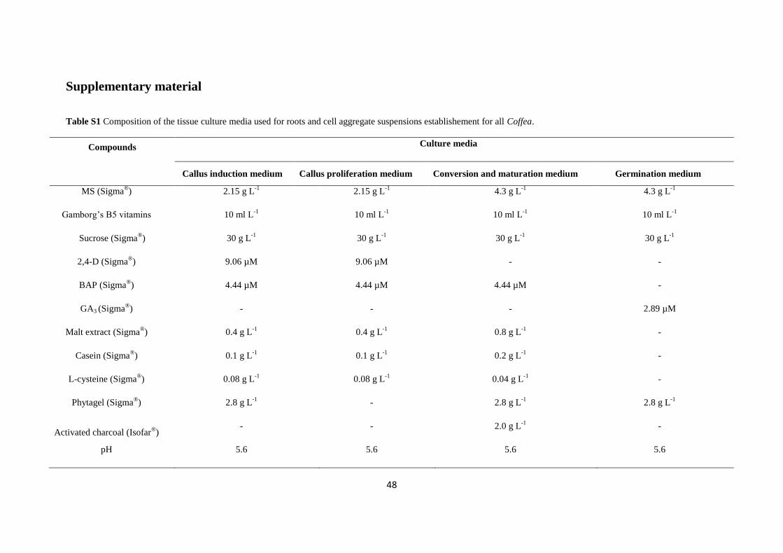
Supplementary material
Table S1 Composition of the tissue culture media used for roots and cell aggregate suspensions establishement for all Coffea.
Compounds Culture media
Callus induction medium Callus proliferation medium Conversion and maturation medium Germination medium
MS (Sigma®) 2.15 g L
-1 2.15 g L
-1 4.3 g L
-1 4.3 g L
-1
Gamborg’s B5 vitamins 10 ml L-1
10 ml L-1
10 ml L-1
10 ml L-1
Sucrose (Sigma®) 30 g L
-1 30 g L
-1 30 g L
-1 30 g L
-1
2,4-D (Sigma®) 9.06 µM 9.06 µM - -
BAP (Sigma®) 4.44 µM 4.44 µM 4.44 µM -
GA3 (Sigma®) - - - 2.89 µM
Malt extract (Sigma®) 0.4 g L
-1 0.4 g L
-1 0.8 g L
-1 -
Casein (Sigma®) 0.1 g L
-1 0.1 g L
-1 0.2 g L
-1 -
L-cysteine (Sigma®) 0.08 g L
-1 0.08 g L
-1 0.04 g L
-1 -
Phytagel (Sigma®) 2.8 g L
-1 - 2.8 g L
-1 2.8 g L
-1
Activated charcoal (Isofar®)
- - 2.0 g L-1
-
pH 5.6 5.6 5.6 5.6
49
All media were autoclaved for 20 min at 121°C. The leave fragments in callus induction medium was stored in Petri dishes and maintained in the dark at 24 ± 2°C. Callus proliferation
medium for suspensions of cell aggregates establishemnt was maintened in Erlenmeyers, on shaker at 100 rpm, subcultured at each every 15 days for 60 days. Cotiledonary somatic
embryos was regenerated in conversion and maturation medium in Petri dishes, and plantlets recovered in germination medium in tubes. These medium, except callus induction medium,
were maintained at 24 ± 2°C, under a 16/8 h (light/dark) regime with 36 µmol m-2
s-1
light radiation provided by two fluorescent lamps (20 W, Osram®). MS: Murashige e Skoog
(1962); Gamborg et al. (1968), 2,4-D: 2,4-dichlorophenoxyacetic acid; BAP: 6-benzilaminopurina; GA3: gibberellic acid.
50
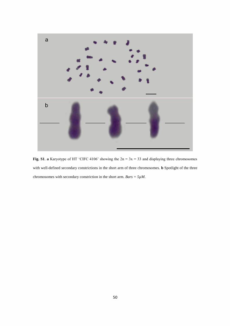
Fig. S1. a Karyotype of HT ‘CIFC 4106’ showing the 2n = 3x = 33 and displaying three chromosomes
with well-defined secondary constrictions in the short arm of three chromosomes. b Spotlight of the three
chromosomes with secondary constriction in the short arm. Bars = 5µM.
51
Fig. S2. PCR amplification of 5S rDNA gene in two individuals of HT ‘CIFC 4106’ for construction of
probe from these amplified products. M: 1000 bp ladder marker.

52
Fig. S3. a) HT ‘CIFC 4106’ karyotype showed two chromosomes with 5s rDNA (red) marking. b)
Chromosomes evidencing the 5S rDNA signal in the interstitial region of the long arm of two
chromosomes. Bar = 5 µm.
53
Acknowledgments
We would like to thank the Conselho Nacional de Desenvolvimento Científico e
Tecnológico (CNPq, Brasília – DF, Brazil, grants: 443801/2014-2), Fundação de
Amparo à Pesquisa do Espírito Santo (FAPES, Vitória – ES, Brazil, grant:
65942604/2014) and Coordenação de Aperfeiçoamento de Pessoal de Nível Superior
(CAPES, Brasília – DF, Brazil) for financial support. Oliveira SC thanks FAPES for her
doctoral scholarship (FAPES, Vitória – ES, Brazil, grant 66243246/2014).
Author contribution statement: The authors Oliveira SC, Sanglard NA, Mariano
GF, Tonetto AV, Sattler MC and Clarindo WR conceived, designed and conducted the
tissue culture cytogenetic and image cytometry approaches. The author Clarindo WR
and Carvalho CR conducted the flow cytometry analysis. Soares FAF, Clarindo WR
and Oliveira SC conducted the fluorescent in situ hybridization (FISH). For manuscript
editing and revision, all authors equally contributed to this work and approved the final
manuscript version for submission.
Conflict of interest: The authors declare that they have no conflict of interest.
54
References
Bennett MD, Leitch IJ. (2011) Nuclear DNA amounts in angiosperms: targets, trends
and tomorrow. Ann Bot 107(3):467–590. doi: 10.1093/aob/mcq258
Berthou F, Mathieu C, Vedel F (1983) Chloroplast and mitochondrial DNA variation as
indicator of phylogenetic relationships in the genus Coffea. Theor Appl Genet 65:77–84
doi: 10.1007/BF00276268
Blake RD, Delcourt SG (1996) Thermodynamic Effects of Formamide on DNA
Stability Nucleic Acids Research, 24:2095–2103. doi.org/10.1093/nar/24.11.2095
Cai Q, Zhang DM, Liu ZL, Wang XR (2006) Chromosomal localization of 5S and
18S rDNA in five species of subgenus Strobus and their implications for genome
evolution of Pinus. Ann Bot 97: 715–722.doi:10.1093/aob/mcl030
Capucho AS, Caixeta ET, Zambolim EM, Zambolim L (2009) Herança da resistência de
Híbrido de Timor UFV 443-03 à ferrugem-do-cafeeiro. Pesq. Agropec. Bras 44:276–
282.
Carvalho CR, Clarindo WR, Abreu IS (2011) Image cytometry: nuclear and
chromosomal DNA quantification. In: ChiariniGarcia H, Melo RCN (eds) Light
microscopy, methods in molecular biology, vol 689. Humana Press, New York, pp 51–
68. doi: 10.1007/978-1-60761-950-5_4
CCDB Chromosome Counts Database (2017)
http://ccdb.tau.ac.il/Angiosperms/Rubiaceae/Coffea/Coffea Accessed 15 June 2017
Cenci A, Combes MC, Lashermes P (2012) Genome evolution in diploid and tetraploid
Coffea species as revealed by comparative analysis of orthologous genome segments.
Plant Mol Biol 78:135–145. doi: 10.1007/s11103-011-9852-3
55
Chester M, Gallagher JP, Symonds VV, Silva VC, AE, Mavrodiev V., Leitch AR, Soltis
PS, Soltis DE (2012) Extensive chromosomal variation generated in a recently formed
polyploid species, Tragopogon miscellus (Asteraceae). Proc Natl Acad Sci USA
109:1176–1181. doi: 10.1073/pnas.1112041109
Clarindo WR, Carvalho CR (2006). A high quality chromosome preparation from cell
suspension aggregates culture of Coffea canephora. Cytologia 71:243–249. doi:
0.1508/cytologia.71.243
Clarindo WR, Carvalho CR (2009) Comparison of the Coffea canephora and C. arabica
karyotype based on chromosomal DNA content. Plant Cell Rep 28:73–81. doi:
10.1007/s00299-008-0621-y
Clarindo WR, Carvalho CR, Caixeta ET, Koehler AD (2013) Following the track of
Híbrido de Timor origin by cytogenetic and flow cytometry approaches. Genet Resour
Crop Evol 60:2253–2259. doi: 10.1007/s10722-013-9990-3
Cros J, Combes MC, Trouslot P, Anthony F, Hamon S, Charrier A, Lashermes P.
Phylogenetic analysis of chloroplast DNA variation in Coffea L. Mol Phylogenet Evol
9:109–17. doi: 10.1006/mpev.1997.0453
Doyle JJ, Doyle JL (1990) Isolation of plant DNA from fresh tissue. Focus 12:13–15.
Fulneček J, Lim KY, Leitch AR, A Kovařík, Matyášek R (2002) Evolution and
structure of 5S rDNA loci in allotetraploid Nicotiana tabacum and its putative parental
species. Heredity 88:19–25. doi: 10.1038/sj.hdy.6800001
Fuchs J, Dell’ Atti D, Buhot A, Calemczuk R, Mascini M, Livache T (2010) Effects of
formamide on the thermal stability of DNA duplexes on biochips. Anal Biochem
397:132–134. doi:10.1016/j.ab.2009.09.044
56
Galbraith DW, Harkins KR, Maddox JM, Ayres NM, Sharma DP, Firoozabady E
(1983). Rapid flow cytometric analysis of the cell cycle in intact plant tissues. Science
220(4601):1049–1051. doi:10.1126/science.220.4601.1049
Gamborg O, Mille R, Ojima K (1968) Nutrient Requirements of Suspension Cultures of
Soybean Root Cells. Experimental Cell Research 50:151–158. doi: 10.1016/0014-
4827(68)90403-5
Greilhuber J (2008) Cytochemistry and C-values: the less-well-known world of nuclear
DNA amounts. Ann Bot 101:791–804. doi: 10.1093/aob/mcm250
Guerra MS (1986) Reviewing the chromosome nomenclature of Levan et al. Revista
Brasileira de Genética 9:741–743.
Hamon P, Siljak-Yakovlev S, Srisuwan S, Robin O, Poncet V, Hamon S, De Kochko A
(2009) Physical mapping of rDNA and heterochromatin in chromosomes of 16 Coffea
species: a revised view of species differentiation. Chromosome Res 17:291–304. doi:
10.1007/s10577-009-9033-2
Hamon P, Hamon S, Razafinarivo NJ, Guyot R, Siljak-Yakovlev S, Couturon E,
Crouzillat D, Rigoreau M, Akaffou S, Rakotomalala JJ, de Kochko A (2015) Coffea
genome organization and evolution. In: Coffee in health and disease prevention.
Academic, San Diego, pp 29–37. doi: 10.1016/B978-0-12-409517-5.00004-8
Hamon P, Grover CE, Davis AP, Rakotomalala JJ, Raharimalala NE, Albert VA,
Hamon S (2017) Genotyping-by-sequencing provides the first well-resolved phylogeny
for coffee (Coffea) and insights into the evolution of caffeine content in its species:
GBS coffee phylogeny and the evolution of caffeine content. Mol Phylogenet Evol
109:351–361. doi: 10.1016/j.ympev.2017.02.009
Lashermes P, Cros J, Combes MC, Trouslot P, Anthony F, Hamon S, Charrier A (1996)
Inheritance and restriction fragment length polymorphism of chloroplast DNA in the
genus Coffea L. Theor Appl Genet 93:626–632. doi: 10.1007/BF00417958
57
IPCN Index to Plant Chromosome Numbers (2017) Tropicos
http://www.tropicos.org/Name/40011705?projectid=9 Accessed 15 June 2017
Lashermes P, Combes MC, Trouslot P, Charrier A (1997) Phylogenetic relationships of
coffee-tree species (Coffea L.) as inferred from ITS sequences of nuclear ribosomal
DNA. Theor Appl Genet 94:947–955. doi: 10.1007/s001220050500
Lashermes P, Combes MC, Robert J, Trouslot P, D’Hont A, Anthony F, Charrier A
(1999) Molecular characterization and origin of the Coffea arabica L. genome. Mol
Gen Genet 261:259–266. doi:10.1007/s004380050965
Levan A, Fredga K, Sandberg AA (1964) Nomenclature for centromeric position on
chromosomes. Hereditas 52:201–220. doi: 10.1111/j.1601-5223.1964.tb01953.x
Mahé L, Combes M, Lashermes P (2007) Comparison between a coffee single copy
chromosomal region and Arabidopsis duplicated counterparts evidenced high level
synteny between the coffee genome and the ancestral Arabidopsis genome. Plant Mol
Biol 64:699–711. doi: 10.1007/s11103-007-9191-6
Mallet J (2007) Hybrid speciation. Nature 446:279–283.
Mehra PN, Bawa KS (1969) Chromosomal evolution in Tropical Hardwoods. Evolution
Int. J. Org. Evolution 23:466–481. doi: 10.1111/j.1558-5646.1969.tb03529.x
Morejohn LC, Fosket DE (1984) Inhibition of Plant Microtubule Polymerization in
vitro by the Phosphoric Amide Herbicide Amiprophos-Methyl. Science. 224:874–876.
doi: 10.1126/science.224.4651.874
Murashige T, Skoog F (1962) A revised medium for rapid growth and bioassays with
tobacco tissue cultures. Physiol Plant 15:473–497. doi: 10.1111/j.1399-
3054.1962.tb08052.x
58
Noirot M, Poncet V, Barre P, Hamon P, Hamon S, Kochko A (2003) Genome size
variations in diploid African Coffea species. Ann Bot 92:709–714. doi:
10.1093/aob/mcg183
Orozco-Castillo C, Chalmers KJ, Powell W, Waugh R (1996) RAPD and organelle
specific PCR re-affirms taxonomic relationships within the genus Coffea. Plant Cell
Rep 15:337–41. doi: 10.1007/BF00232367.
Otto FJ (1990) DAPI staining of fixed cells for high-resolution flow cytometry of
nuclear DNA. In: Darzynkiewicks Z, Crissman HA (eds) Methods in cell biology,
Academic, San Diego, pp 105–110.
Pinto-Maglio CAF (2006) Cytogenetics of coffee. Braz J Pl Physiol 18:37–44.
doi.org/10.1590/S1677-04202006000100004.
Planchais S, Glab N, Inzé D, Bergounioux C (2000) Chemical inhibitors: a tool for plant
cell cycle studies. FEBS Lett 476:78–83. doi: 10.1016/S0014-5793(00)01675-6
Praça-Fontes MM, Carvalho CR, Clarindo WR, Cruz CD (2011) Revisiting the DNA C-
values of the genome size-standards used in plant flow cytometry to choose the "best
primary standards". Plant Cell Rep 30:1183–91. doi: 10.1007/s00299-011-1026-x
Raina SN, Mukai Y, Yamamoto M (1998) In situ hybridization identifies the diploid
progenitor species of Coffea arabica (Rubiaceae). Theor Appl Genet 97:1204–1209.
doi: 10.1007/ s001220051011
Ribeiro T, Barrela RM, Bergès H,
Marques C, Loureiro J, Morais-Cecílio L, Paiva JAP
(2016)
Advancing Eucalyptus Genomics: Cytogenomics Reveals Conservation of
Eucalyptus Genomes. Front Plant Sci (510):1–12. doi: 10.3389/fpls.2016.00510
Rijo L (1974) Observações cariológicas no cafeeiro ‘Híbrido de Timor’. Port Acta Biol
8:157–168.
59
Roa F, Guerra M (2012) Distribution of 45S rDNA sites in chromosomes of plants:
Structural and evolutionary implications. BMC Evolutionary Biology 12:225.
doi.org/10.1186/1471-2148-12-225.
Rose RJ (1970) The effect of cycloheximide on cell division in partially synchronized
plant cells. Aust J biol Sci 23:573–83.
Sattler MC, Carvalho CR, Clarindo WR (2016) Regeneration of allotriploid Coffea
plants from tissue culture: resolving the propagation problems promoted by irregular
meiosis. Cytologia 81:125–132. doi: 10.1508/cytologia.81.125
Sanglard NA, Amaral-Silva PM, Sattler MC, Oliveira SC,·Nunes ACP, Soares TCB,
Carvalho CR, Clarindo WR (2017) From chromosome doubling to DNA sequence
changes: outcomes of na improved in vitro procedure developed for allotriploid
“Híbrido de Timor” (Coffea arabica L. × Coffea canephora Pierre ex A. Froehner)
Plant Cell Tiss Organ Cult doi: 10.1007/s11240-017-1278-4.
Selvaraj, R (1987) Karyomorphological studies in South Indian Rubiaceae. Cytologya
52:343–356. doi: 10.1508/cytologia.52.343
Setotaw, T. A., Caixeta, E. T., Pena, G. F., Zambolim, E. M., Pereira, A. A. and
Sakiyama, N. S. 2010. Breeding potential and genetic diversity of “Híbrido do Timor”
coffee evaluated by molecular markers. Crop Breed Appl. Biotechnol. 10: 298–304.
Schneider-Poetsch T, Ju J, Eyler DE, Dang Y, Bhat S, Merrick WC, Green R, Shen B,
Liu JO (2010) Inhibition of Eukaryotic Translation Elongation by Cycloheximide and
Lactimidomycin Nat Chem Biol 6:209–217. doi: 10.1038/nchembio.304
Soltis PS, Soltis DE (2009) The role of hybridization in plant speciation. Annu Rev
Plant Biol 60:561–588. doi: 10.1146/annurev.arplant.043008.092039
Stebbins GL (1950) Variation and evolution in plants. Columbia University Press, New
York.
60
van Boxtel J, Berthouly M (1996) High frequency somatic embryogenesis from coffee
leaves. Plant Cell Tissue Organ Cult 44:7–17. doi: 10.1007/BF00045907
Yu Q, Guyot R, Kochko A, Byers A, Navajas-Pérez R, Langston BJ, Dubreuil-
Tranchant C, Paterson AH, Poncet V, Nagai C, Ming R (2011) Micro-collinearity and
genome evolution in the vicinity of an ethylene receptor gene of cultivated diploid and
allotetraploid coffee species (Coffea). Plant J 67:305–317. doi:10.1111/j.1365-
313X.2011.04590.x
Zhang C, Ye LH, Chen YY, Xiao J, Wu YH, Tao M, Xiao YM, Liu SJ (2015) The
chromosomal constitution of fish hybrid lineage revealed by 5S rDNA FISH. BMC
Genet.16:140.
Top Related