


Línguas
Páginas
Legal


UNIVERSIDADE DE LISBOA
FACULDADE DE CIÊNCIAS
DEPARTAMENTO DE BIOLOGIA VEGETAL
CHARACTERIZATION OF IMMUNE-MODULATORY MIRNAS
AND THEIR ROLE IN INFLAMMATORY SKIN DISEASE
Maria do Nascimento Lopes Primo
MESTRADO EM BIOLOGIA MOLECULAR E GENÉTICA
2011

UNIVERSIDADE DE LISBOA
FACULDADE DE CIÊNCIAS
DEPARTAMENTO DE BIOLOGIA VEGETAL
CHARACTERIZATION OF IMMUNE-MODULATORY MIRNAS
AND THEIR ROLE IN INFLAMMATORY SKIN DISEASE
DISSERTAÇÃO ORIENTADA POR
Professor Doutor Jacob Giehm Mikkelsen
(Institut for Biomedicin, Århus Universitet, Denmark)
Professora Doutora Margarida Telhada
(Departamento de Química e Bioquímica, Faculdade de Ciências da Universidade de Lisboa, Portugal)
Maria do Nascimento Lopes Primo
MESTRADO EM BIOLOGIA MOLECULAR E GENÉTICA
2011

“I am among those who think that science has great beauty. A scientist in his laboratory is
not only a technician: he is also a child placed before natural phenomena which impress him
like a fairy tale.”
Marie Curie (French Physicist, 1867-1934)

ACKNOWLEDGEMENTS
Faculdade de Ciências da Universidade de Lisboa iv
Acknowledgements
First of all, I would like to thank Professor Jacob Mikkelsen for accepting me in his team, for
giving me the opportunity to work in this project and for always providing careful guidance,
important discussions, motivation, enthusiasm and the best working environment. A special
thanks to Rasmus Bak for his help in the design of lentiviral vectors encoding antagomirs as
well as for supporting my decisions, for the best advices, lab tips and company. I would also
like to thank Nicklas Staunstrup, Nynne Sharma, Yujia Cai, Lisbeth Schrøder and our bache-
lor students, Camilla Darum and Kristian Skipper, for their help in the beginning of my work,
for giving me advices, for the discussions, for the funny moments, the laughs and for all the
support. It has been a gratifying experience to work with all of you, contributing a lot for the
achievement of our results.
This work would have had less impact if it had not been for the combination of an in vivo ap-
proach for studying miRNA modulation in psoriasis skin. For that I thank the researchers Ka-
rin Stenderup and Cecilia Rosada from Institut for Klinisk Medicin for the discussions, their
contribution for the improvement of our work and for carrying out xenotransplantation and
treatments of all skin grafts as well as for the assessment of semiquantitative clinical psoria-
sis scores and measurements of epidermal thickness.
I should also thank the other members from Institut for Biomedicin for all the meetings and
discussions as well as for the lunches, coffee and cake breaks, for the team happiness and
institute parties.
To Professor Margarida Telhada, the coordinator at my home faculty, I thank you for all the
availability and attention shown through this year as well as for the guidance during the de-
velopment of this manuscript.
I could not forget to thank my friends for the shared happiness and sadness, for their friend-
ship and for their support in my decisions. A special thanks to Beatriz, João, Joana and An-
dreia for our adventures, late talks, discussions, relaxing and unforgettable trips, for our
growing friendship and for all you were willing to do for me. To both Ana Miguel and Ana Ma-
ria, I thank you for all your company and sharing in Aarhus, as well as for bringing me a less
scientific world that gave me so much fun. Strong friendships last forever.
I would like to deeply thank my family for supporting me unconditionally in all aspects of my
life. In particular to my parents, Manuel and Maria do Carmo, I thank you for teaching me
what is love and happiness, for our discussions and disagreements that brought me back to
reality, for every dream that you encouraged, for the immeasurable support, for providing me

ACKNOWLEDGEMENTS
Faculdade de Ciências da Universidade de Lisboa v
the best education and for giving me the opportunity to be here, at this moment, writing this
manuscript. To my siblings, Inês, Bruno, João and Filipe, I thank you for every moment you
helped me grow, for all our endless moments of happiness and love and ultimately, for never
letting me quit my dreams and aspirations. To Rodrigo and David, my nephews, I thank you
for every time you call me for playing and for not letting me grow up too fast. To my cousin
Ágata, I thank you for all your willing to keep me happy and close to home and family as well
as for your guidance during the most difficult moments of my life.
At last, I would like to sincerely thank Magnus, my beloved, for all the support, patience and
love that you have given me, for all the sacrifices I will never forget, for all our unforgettable
moments and for turning each day happier than the day before.
To all you I dedicate this paper.

RESUMO
Faculdade de Ciências da Universidade de Lisboa vi
Resumo
A psoríase vulgares é uma doença cutânea que ocorre aproximadamente em 3% da popula-
ção mundial adulta, afectando negativamente a qualidade de vida dos doentes. É uma
doença crónica, resultante da activação do sistema imunitário e semelhante a outras doen-
ças auto-imunes como a artrite reumatóide ou a esclerose múltipla. Histologicamente, a pso-
ríase é caracterizada por um aumento da espessura da epiderme devido à hiperproliferação
de queratinócitos, resultando no aparecimento de escamas e placas com diferentes tama-
nhos. Outra particularidade desta doença é o aumento da vascularização da derme, assim
como a presença de um elevado número de células imunitárias, tais como linfócitos T e
células dendríticas. Ao nível molecular, pensa-se que as manifestações clínicas e laborato-
riais descritas são resultantes da desregulação da complexa rede de citocinas devido à
acção combinada das células imunitárias que infiltraram a derme e dos queratinócitos pre-
sentes na epiderme.
Durante a última década foram identificadas inúmeras espécies de pequenos RNA denomi-
nados small non-coding RNAs, que contribuem para a intrincada rede de vias que regulam
os genes humanos. Entre estes, os microRNA (miRNA) emergiram como reguladores rele-
vantes da tradução proteica, e actualmente acredita-se que os miRNA participam na regula-
ção da maioria dos processos celulares e tecidulares. A pele não é excepção, e de momento
os miRNA são intensamente estudados para determinar a sua relevância na regulação da
homeostase da pele e no aparecimento de doenças cutâneas.
O possível envolvimento de miRNA na patologia da psoríase foi identificado pela primeira
vez em 2007, quando a equipa liderada por Sonkoly comparou os perfis de expressão de
miRNA em pele psoriática e em pele normal. Entre os miRNA identificados, potencialmente
associados à psoríase, o miRNA-203 foi identificado predominantemente em queratinócitos
e sobre-expressado em psoríase, quando comparado com a pele normal. Recentemente, a
mesma equipa científica demonstrou também que o miRNA-203 e o transcrito de um com-
ponente regulador da sinalização da resposta imunitária, o SOCS3 (supressor of cytokine
signalling 3), apresentam níveis de expressão inversamente relacionados. Sabe-se ainda
que a proteína SOCS3 regula negativamente a activação de um factor de transcrição –
STAT3 (signal transducer and activator of transcription 3) – que está sobre-expressado em
psoríase e implicado na rede de citocinas. Assim, a sobre-expressão do miRNA-203 em
psoríase pode ter importantes implicações na patologia da doença uma vez que impossibilita
a expressão de SOCS3, proteína que regula negativamente a rede de citocinas.

RESUMO
Faculdade de Ciências da Universidade de Lisboa vii
O objectivo principal deste projecto foi descrever a interacção entre miRNA e a expressão
de citocinas em pele humana. Com especial interesse no miRNA-203, o projecto procurou
elucidar a função específica dos miRNA moduladores da resposta imunitária na complexa
rede de interacções moleculares, levando ao desenvolvimento da doença inflamatória na
pele. No começo deste projecto, procurou-se estabelecer um sistema in vitro que permitisse
a identificação e análise de potenciais genes-alvo de miRNA associados à psoríase. Assim,
através da utilização da proteína Renilla luciferase (R-luc), foi desenvolvido um ensaio repór-
ter para a detecção de interacções directas entre o miRNA-203 e diversos elementos da
resposta imunitária, através da medição de bioluminescência. Os DNA complementares
(cDNA) de seis diferentes citocinas (IL12B, IL15, IL17, IL20, IL24 e TNFα) assim como os
cDNA de proteínas supressoras da sinalização das citocinas (SOCS3 e SOCS6) foram fun-
didos ao gene codificante da R-luc, e as medidas de bioluminescência foram avaliadas
quando os vectores codificantes da R-luc foram co-transfectados com um vector expressan-
do o miRNA-203 em células HEK293 (Human kidney embryo cells). Dos ensaios desenvol-
vidos, identificaram-se três potenciais genes-alvo do miRNA-203: IL24, SOCS6 e TNFα.
Para a confirmação da interacção directa entre o miRNA-203 e os genes-alvo mencionados
foi desenvolvido um estudo bioinformático para a identificação de sequências nucleotídicas
na região 3‟UTR dos genes-alvo complementares à sequência nucleotídica do miRNA-203.
Uma vez identificadas as possíveis sequências nucleotídicas que permitem o estabeleci-
mento de interacções entre o miRNA-203 e os RNA mensageiro (mRNA) dos genes-alvo, as
sequências identificadas nas regiões 3‟UTR foram mutadas de forma a inibir as interacções
miRNA-mRNA. Os transcritos 3‟UTR mutantes dos genes IL24, SOCS6 e TNFα foram fundi-
dos ao gene codificante da R-luc e novos ensaios repórteres foram elaborados. Em paralelo,
procedeu-se também ao desenvolvimento de uma linha celular de queratinócitos (denomi-
nada HaCaT-203), através da utilização do sistema transposão de DNA “sleeping beauty”,
para expressar constitutivamente o miRNA-203. Esta nova linha celular de queratinócitos foi
especialmente desenvolvida para a confirmação de potenciais genes-alvo do miRNA-203,
através da quantificação dos níveis de expressão dos genes-alvo por qRT-PCR. A quantifi-
cação e a análise dos valores de bioluminiscência obtidos a partir da expressão do gene da
R-luc fundido aos transcritos dos três 3‟UTR mutantes de IL24, SOCS6 e TNFα identificaram
a interacção directa entre os três mRNA e o miRNA-203. Resultados semelhantes foram
obtidos através da quantificação dos níveis endógenos dos mRNA de IL24, SOCS6 e TNFα
em queratinócitos, confirmando a especificidade da interacção entre o miRNA-203 e os
genes-alvo mencionados. A identificação destes novos genes-alvo veio contribuir para a
compreensão e caracterização das implicações do miRNA-203 na expressão de citocinas
em pele humana. Não menos importante, também foi o facto de que os resultados obtidos
RESUMO
Faculdade de Ciências da Universidade de Lisboa viii
vieram consolidar a hipótese de que o miRNA-203 poderá ter implicações na patologia da
psoríase uma vez que o miRNA-203 foi identificado como um modulador activo na complexa
regulação da rede de citocinas em queratinócitos.
A equipa com a qual foi desenvolvido este projecto documentou anteriormente a aplicabili-
dade terapêutica da utilização de lentivirus para a inibição dos mRNA de TNFα e IL12B atra-
vés da “entrega” (delivery) de RNA efectores anti-TNFα e anti-IL12B, respectivamente, em
pele psoriática humana xenotransplantada em ratos imunodeficientes (xenografted psoriatic
skin). No projecto actual, foi desenvolvido um novo vector lentiviral codificando um inibidor
de miRNA específico para o miRNA-203 (denominado antagomiR-203). Os RNA efectores
anti-miRNA (antagomirs) são caracterizados por sequências oligonucleotídicas complemen-
tares aos miRNA de interesse. Assim, os antagomir estabelecem interacções específicas
com o miRNA-alvo, conduzindo à inibição funcional dos mesmos. Através da utilização do
vector lentiviral codificando o antagomiR-203, vários ensaios in vitro foram desenvolvidos
para testar a funcionalidade e a potência do antagomiR-203. Com base nas avaliações fun-
cionais, identificou-se uma nítida sobre-expressão do gene da R-luc quando fundido com
uma sequência oligonucleotídica complementar ao miRNA-203, quando co-expresso com o
vector lentiviral codificando o antagomiR-203. Foi também identificada uma redução dos
níveis de expressão do miRNA-203, quando as linhas celulares de queratinócitos foram
infectadas com partículas lentivirais expressando o antagomiR-203. Os resultados obtidos
confirmaram assim a funcionalidade e especificidade do antagomiR-203 relativamente ao
miRNA-203, permitindo o estabelecimento de uma nova plataforma para a regulação de
miRNA através da utilização de lentivirus como veículo de transporte de anti-miRNA.
Com base na tecnologia de expressão de antagomir, este projecto procurou ainda abrir
caminho para os estudos de gene-alvos endógenos, permitindo experimentalmente estudar
a função de miRNA em queratinócitos em pele normal e pele psoriática. Como objectivo final
deste projecto, testou-se a aplicabilidade terapêutica da utilização de lentivirus para a
“entrega” de RNA efectores anti-miRNA-203 em pele psoriática humana xenotransplantada
em ratos imunodeficientes. A administração intradérmica de uma única dose de partículas
lentivirais em pele psoriática humana resultou num aumento dos níveis de expressão do
miRNA-203 e na não alteração da espessura da epiderme em enxertos de pele tratados
com o antagomiR-203. Em conformidade com os resultados obtidos, a avaliação clínica do
fenótipo psoriático dos enxertos de pele não identificou nenhuma melhoria clínica e histoló-
gica do fenótipo psoriático no ensaio desenvolvido.
O potencial para a concepção de medicamentos moleculares baseados na modulação de
miRNA endógenos é muito ambicionado, mas actualmente está ainda muito inexplorado. Os
RESUMO
Faculdade de Ciências da Universidade de Lisboa ix
estudos desenvolvidos ao longo deste projecto demonstraram que o aumento da expressão
do miRNA-203 na patologia da psoríase é complexo, claramente evidenciando a necessida-
de de estudos futuros para uma melhor compreensão das funções desempenhadas pelos
miRNA na regulação da resposta imunitária. O projecto realizado procurou também explorar
a aplicabilidade terapêutica de fármacos dirigidos aos miRNA, tendo-se identificado algumas
dificuldades relacionadas com a funcionalidade dos antagomirs in vivo. No entanto, é de
salientar que a abordagem utilizada neste estudo foi única, na medida em que se procurou
explorar uma nova metodologia de administração de antagomirs, permitindo a entrega de
material genético em tecidos específicos, de modo a aumentar a segurança na utilização de
terapia genética em humanos.
Palavras-chave: miRNA-203, psoríase, rede de citocinas, vectores lentivirais, antagomirs
Observação: Resumo escrito de acordo com a antiga ortografia.
ABSTRACT
Faculdade de Ciências da Universidade de Lisboa x
Abstract
Psoriasis vulgaris is a chronic inflammatory skin disease which is characterized by an exces-
sive growth of skin epithelial cells, increased dermal angiogenesis and infiltration of immune
cells into the skin. The past decade has unveiled a plethora of small RNA species that con-
tribute to the intricate network of pathways regulating our genes. Among these, microRNAs
have emerged as key regulators of translation and are believed to play a role in almost any
cellular process and tissue. One of the most upregulated microRNAs in psoriasis skin is
miRNA-203. The aim of this study was to describe the interplay between microRNAs, with
focus on miRNA-203, and cytokine-encoding mRNAs in human skin. Based on the v-
antagomir expression technology, the project may pave the way for studies of endogenous
targets, allowing us to experimentally address microRNA function in keratinocytes and skin
inflammation.
Three components of the cytokine circuit, interleukin-24, suppressor of cytokine signaling-6
and tumor necrosis factor-α, were identified as direct targets for suppression by miRNA-203
by luciferase reporter assay. In the present study, we also used lentiviral vectors as potent
carriers of antagomir-encoding gene cassettes. Lentiviral vectors are attractive gene vehicles
primarily due to their ability to establish persistent expression owning to genomic integration
of the vector DNA reverse-transcribed from virally delivered single-stranded RNA. Potent and
persistent knockdown of miRNA-203 expression following transduction of lentiviral vectors
encoding antagomiR-203 in keratinocytes was reported. In contrast to the data collected in
vitro, miRNA-203 knockdown was not identified in vivo after a three week treatment of xeno-
grafted psoriatic skin with lentivirus-encoded antagomiR-203.
Our studies consolidate the properties of lentiviral vectors as a tool in experimental derma-
tology with particular significance for cutaneous RNA managing and in vivo genetic interven-
tion. However, the therapeutic potential of targeting miRNA-203 in psoriasis is here ques-
tioned.
Key words: miRNA-203, psoriasis, cytokine network, lentiviral vectors, antagomirs
LIST OF MANUSCRIPTS
Faculdade de Ciências da Universidade de Lisboa xi
List of manuscripts
This thesis is based on the following papers, which are referred to by their Roman numbers
in the text:
I. Primo MN*, Bak RO, Mikkelsen JG. Lentiviral vectors for cutaneous RNA managing.
Review article submitted to Experimental Dermatology on 22nd of September 2011.
II. Primo MN*, Bak RO, Mikkelsen JG. Managing miR-203 in keratinocytes demon-
strates roles in cytokine regulation. Manuscript in preparation.
* Corresponding author

ABBREVIATIONS
Faculdade de Ciências da Universidade de Lisboa xii
Abbreviations
293T cells Variant of Human kidney embryo cells
Ago Argonaute Bp Basepair cDNA Complementary DNA CMV Cytomegalovirus DGCR8 DiGeorge syndrome critical region gene 8 DNA Deoxyribonucleic acid dsRNA Double stranded RNA FGFR2 Fibroblast growth factor receptor 2 G418 Geneticin (aminoglycoside antibiotic) GW182 TNRC6A trinucleotide repeat containing 6A HaCaT cells Human keratinocyte cells HEK293 cells Human kidney embryo cells HeLa cells Human cervical adenocarcinoma cells HIV Human immunodeficiency virus IFN Interferon IL Interleukin LV Lentiviral vector miRNA MicroRNA MLV Moleney murine leukemia virus MOI Multiplicity of infection mRNA Messenger RNA mSB Mutant sleeping beauty transposase Neg Negative Control Neo Neomycin NF Nuclear factor PACT Protein activator of PKR PCR Polymerase chain reaction pri-miRNA Primary microRNA R-luc Renilla luciferase RISC RNA-induced silencing complex RNA Ribonucleic acid RNAi RNA interference RPLP0 Ribosomal protein, large P0 qRT-PCR Real time reverse transcriptase polymerase chain reaction RU48 Small nucleolar RNA, C/D box 48 SB Sleeping beauty transposase SCID Severe immunodeficient mice SEM Standard error of the mean shRNA Short hairpin RNA siRNA Small interfering RNA SOCS Suppressor of cytokine signaling STAT Signal transducer and activation of transcription SV40 Simian virus 40 TLR Toll-like receptor TNF Tumor suppressor factor TRBP Tar RNA binding protein UNG Uracil N-glycosylase UTR Untranslated region XPO5 Exportin 5

Faculdade de Ciências da Universidade de Lisboa
Table of Contents
ACKNOWLEDGEMENTS ................................................................................................................................... IV
RESUMO.......................................................................................................................................................... VI
ABSTRACT ........................................................................................................................................................ X
LIST OF MANUSCRIPTS .................................................................................................................................... XI
ABBREVIATIONS ............................................................................................................................................. XII
INTRODUCTION ................................................................................................................................................ 1
PSORIASIS VULGARIS, A DISEASE-MODEL IN SKIN INFLAMMATION ....................................................................................... 1
Pathogenesis of psoriasis ............................................................................................................................... 1
Non-coding RNAs: Biogenesis pathways and function ................................................................................... 2
Involvement of miRNAs in psoriasis ............................................................................................................... 5
CUTANEOUS GENE DELIVERY ...................................................................................................................................... 6
Lentiviral gene delivery to skin ....................................................................................................................... 7
AIM OF THE PRESENT WORK ............................................................................................................................ 9
MATERIALS AND METHODS ........................................................................................................................... 10
PLASMID CONSTRUCTION ........................................................................................................................................ 10
Cloning of H1-antagomir expression cassettes ............................................................................................ 10
Cloning of miR-203 targets into psiCHECK2 ................................................................................................. 10
Generation of 3’UTR mutated sequences and cloning into psiCHECK2 ........................................................ 10
Generation of pri-miR-203 and pri-miR-125b-expressing constructs ........................................................... 11
CELL LINES ............................................................................................................................................................ 11
GENERATION OF MIR-203-EXPRESSING HACAT CELL LINE, HACAT-203 .......................................................................... 11
DUAL-LUCIFERASE REPORTER ASSAY ......................................................................................................................... 12
LENTIVIRAL VECTOR PRODUCTION ............................................................................................................................. 12
HUMAN XENOGRAFT TRANSPLANTATION MODEL .......................................................................................................... 13
IN VIVO ADMINISTRATION OF ANTAGOMIR-ENCODING LENTIVIRAL VECTORS ....................................................................... 14
XENOGRAFT EVALUATION FOLLOWING TREATMENT....................................................................................................... 14
RNA ISOLATION AND QUANTITATIVE RT-PCR ............................................................................................................. 14
BIOINFORMATICS ................................................................................................................................................... 15
STATISTICAL ANALYSES............................................................................................................................................ 15
RESULTS ......................................................................................................................................................... 16
TOWARDS IDENTIFICATION AND ANALYSES OF MIR-203 TARGET GENES ............................................................................ 16
DOWNREGULATION OF MIR-203 TARGETS FOLLOWING STABLE EXPRESSION OF MIR-203 .................................................... 18
ESTABLISHMENT OF MIR-203 INHIBITION BY VECTOR-ENCODED ‘ANTAGOMIRS’ ................................................................. 20
EFFICIENT AND PERSISTENT UPREGULATION OF IL24 FOLLOWING LENTIVIRAL TRANSDUCTION ............................................... 22
IN VIVO LENTIVIRAL DELIVERY OF ANTAGOMIR-203 IN XENOGRAFTED PSORIATIC SKIN ......................................................... 23
DISCUSSION ................................................................................................................................................... 26
CONCLUDING REMARKS AND FUTURE PERSPECTIVES .................................................................................... 29
REFERENCES ................................................................................................................................................... 30
Faculdade de Ciências da Universidade de Lisboa
Appendix
APPENDIX I. Oligonucleotide sequences of the cloned transgenes.
APPENDIX II. Schematic representation of the different vectors.
APPENDIX III. Potential miR-203-binding sites in IL24 3’UTR sequence.
APPENDIX IV. Potential miR-203-binding sites in SOCS6 3’UTR sequence.
APPENDIX V. Potential miR-203-binding sites in TNFα 3’UTR sequence.
APPENDIX VI. Supplementary data.
Table of figures
FIGURE 1. Schematic representation of the cytokine network driving development of psoriasis 2
FIGURE 2. Schematic representation of miRNA biogenesis. 3
FIGURE 3. Approaches for cutaneous gene delivery. 7
FIGURE 4. Functional screening of potential microRNA-203 and microRNA-125b targets in psoriasis. 16
FIGURE 5. Development of 3’UTR mutants for confirmation of IL24, SOCS6 and TNFα mRNA tran-
scripts as direct targets of microRNA-203.
18
FIGURE 6. Endogenous knockdown of IL24, SOCS6 and TNFα mRNA transcripts by stable overex-
pression of microRNA-203.
19
FIGURE 7. Functional evaluation of microRNA-203 targeting by antagomiR-203 and confirmation of
antagomir potency after lentiviral transduction.
21
FIGURE 8. In vitro knockdown of microRNA-203 after transduction with antagomir-encoding lenti-
viral vectors and upregulation of IL24 mRNA expression.
22
FIGURE 9. In vivo knockdown of microRNA-203 in xenografted psoriatic skin by lentiviral delivery of
antagomiR-203.
25

INTRODUCTION
Faculdade de Ciências da Universidade de Lisboa 1
Introduction
Psoriasis vulgaris is a common inflammatory skin disease which is characterized by exces-
sive growth of skin epithelial cells, increased dermal angiogenesis and infiltration of immune
cells into the skin leading to focal formation of inflamed, scaly skin lesions. Recent evidence
proposes that miRNAs are involved in immune system regulation and that miRNA levels are
regulated upon stress induction. One of the most upregulated miRNAs in psoriatic skin is
miRNA-203. The aim of this study was to describe the interplay between miRNAs, with focus
on miRNA-203, and cytokine-encoding mRNAs in human skin.
Psoriasis vulgaris, a disease-model in skin inflammation
Pathogenesis of psoriasis
Psoriasis vulgaris is one of the most common chronic inflammatory skin disorders affecting
approximately 3% of the population in Europe and North America. It is an organ-specific au-
toimmune disease that is triggered by an activated cellular immune system 1; 2. The histologi-
cal changes observed within lesional skin are striking, and include (1) a thickened epidermis
from rapid keratinocyte proliferation, (2) a reduced or absent granular layer, (3) marked dila-
tation of blood vessels in the papillary dermis, and (4) dense clusters of infiltrated mononuc-
lear leukocytes (T-cells and dendritic cells) into the dermis (Figure 1) 3; 4. Cytokine interac-
tions in psoriasis have previously been illustrated as „type-1 pathway‟, which assumes a li-
near relationship between proximal inducers (IL23 and IL12), production of IFNγ and TNFα
by type-1 T-cells, and downstream activation of numerous IFN-responsive genes through
STAT1 4.
However, it is now known that this model accounts for only a small fraction of the inflammato-
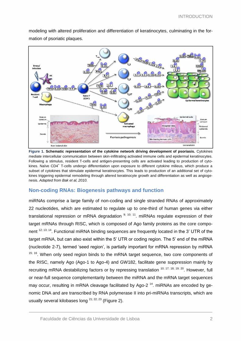
ry circuitry in psoriasis. Figure 1 represents an alternative view of the cytokine interactions in
psoriasis, which is more of a network or interactive model 5. Following a stimulus, such as
infection or stress in genetically predisposed individuals, resident T-cells become activated
through interaction with resident dendritic cells and macrophages, leading to the production
of numerous cytokines, such as TNFα and IL23. Depending on the exposure to different cy-
tokines, naїve T-cells may develop into one of the at least five different CD4+ T-cell lineages.
T-cell activation leads to activation of STAT1, STAT3 and NF-кB transcription factors which
will be involved in amplifying inflammation process, resulting in upregulation of several cyto-
kines as TNFs, IL1, IL6, IL12, IL17, IL20, IL22, IL24 and IFNs 6; 7. Keratinocytes are then
involved in the cytokine-mediated inflammation by responding to and producing cytokines,
which have several functions including promotion of angiogenesis, amplification of immune
cell trafficking and immune cell adhesion to endothelial cells 8. Both events will contribute to
the start of a vicious cycle of inflammation within lesional skin which results in epidermal re-
INTRODUCTION
Faculdade de Ciências da Universidade de Lisboa 2
modeling with altered proliferation and differentiation of keratinocytes, culminating in the for-
mation of psoriatic plaques.
Non-coding RNAs: Biogenesis pathways and function
miRNAs comprise a large family of non-coding and single stranded RNAs of approximately
22 nucleotides, which are estimated to regulate up to one-third of human genes via either
translational repression or mRNA degradation 9; 10; 11. miRNAs regulate expression of their
target mRNAs through RISC, which is composed of Ago family proteins as the core compo-
nent 12; 13; 14. Functional miRNA binding sequences are frequently located in the 3‟ UTR of the
target mRNA, but can also exist within the 5‟ UTR or coding region. The 5‟ end of the miRNA
(nucleotide 2-7), termed „seed region‟, is partially important for mRNA repression by miRNA
15; 16. When only seed region binds to the mRNA target sequence, two core components of
the RISC, namely Ago (Ago-1 to Ago-4) and GW182, facilitate gene suppression mainly by
recruiting mRNA destabilizing factors or by repressing translation 10; 17; 18; 19; 20. However, full
or near-full sequence complementarity between the miRNA and the mRNA target sequences
may occur, resulting in mRNA cleavage facilitated by Ago-2 14. miRNAs are encoded by ge-
nomic DNA and are transcribed by RNA polymerase II into pri-miRNAs transcripts, which are
usually several kilobases long 21; 22; 23 (Figure 2).
Figure 1. Schematic representation of the cytokine network driving development of psoriasis. Cytokines
mediate intercellular communication between skin-infiltrating activated immune cells and epidermal keratinocytes.
Following a stimulus, resident T-cells and antigen-presenting cells are activated leading to production of cyto-
kines. Naїve CD4+ T-cells undergo differentiation upon exposure to different cytokine milieus, which produce a
subset of cytokines that stimulate epidermal keratinocytes. This leads to production of an additional set of cyto-
kines triggering epidermal remodeling through altered keratinocyte growth and differentiation as well as angioge-
nesis. Adapted from Bak et al, 2010.
INTRODUCTION
Faculdade de Ciências da Universidade de Lisboa 3
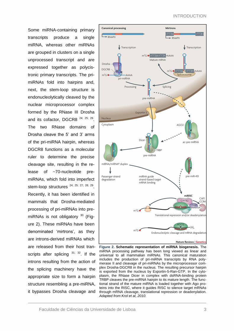
Some miRNA-containing primary
transcripts produce a single
miRNA, whereas other miRNAs
are grouped in clusters on a single
unprocessed transcript and are
expressed together as polycis-
tronic primary transcripts. The pri-
miRNAs fold into hairpins and,
next, the stem-loop structure is
endonucleolytically cleaved by the
nuclear microprocessor complex
formed by the RNase III Drosha
and its cofactor, DGCR8 24; 25; 26.
The two RNase domains of
Drosha cleave the 5‟ and 3‟ arms
of the pri-miRNA hairpin, whereas
DGCR8 functions as a molecular
ruler to determine the precise
cleavage site, resulting in the re-
lease of ~70-nucleotide pre-
miRNAs, which fold into imperfect
stem-loop structures 24; 25; 27; 28; 29.
Recently, it has been identified in
mammals that Drosha-mediated
processing of pri-miRNAs into pre-
miRNAs is not obligatory 30 (Fig-
ure 2). These miRNAs have been
denominated „mirtrons‟, as they
are introns-derived miRNAs which
are released from their host tran-
scripts after splicing 31; 32. If the
introns resulting from the action of
the splicing machinery have the
appropriate size to form a hairpin
structure resembling a pre-miRNA,
it bypasses Drosha cleavage and
Figure 2. Schematic representation of miRNA biogenesis. The
miRNA processing pathway has been long viewed as linear and universal to all mammalian miRNAs. This canonical maturation includes the production of pri-miRNA transcripts by RNA poly-merase II and cleavage of pri-miRNAs by the microprocessor com-plex Drosha-DGCR8 in the nucleus. The resulting precursor hairpin is exported from the nucleus by Exportin-5-Ran-GTP. In the cyto-plasm, the RNase Dicer in complex with dsRNA-binding protein TRBP cleaves the pre-miRNA hairpin to its mature length. The func-tional strand of the mature miRNA is loaded together with Ago pro-teins into the RISC, where it guides RISC to silence target mRNAs through mRNA cleavage, translational repression or deadenylation. Adapted from Krol et al, 2010.

INTRODUCTION
Faculdade de Ciências da Universidade de Lisboa 4
is further processed in the cytoplasm. After nuclear processing, pre-miRNAs are exported
into the cytoplasm through nuclear pore complexes, by a RanGTP-dependent double
stranded DNA-binding protein, the XPO5 33; 34; 35; 36.
On reaching the cytoplasm, the cytoplasmic RNase III Dicer cleaves off the loop of the pre-
miRNAs, generating mature ~22-nucleotide miRNA duplexes with two nucleotides protruding
as overhangs at each 3‟end 37; 38; 39. Dicer-mediated cleavage of the pre-miRNA is facilitated
by the double-stranded RNA-binding domain proteins TRBP and PACT, which in association
with Ago2 compose the multi-protein RISC loading complex 40; 41; 42; 43. After Dicer-mediated
cleavage, Dicer and its interactors, TRBP and PACT, dissociate from the miRNA duplex. To
form the active RISC that performs gene silencing, the double-stranded RNA duplex needs
to be separated into (i) the functional guide strand, which is complementary to the mRNA
target and guides RISC to silence target mRNAs and (ii) the passive strand, which is subse-
quently degraded. Studies on small siRNA duplexes indicate that the relative thermodynamic
stability of the basepairs at the two ends of the duplex determines which strand is loaded into
RISC - the miRNA strand which is less stable at the 5‟ end is loaded into the RISC 44.
In 1998, Fire and Mello discovered the RNA interference pathway which showed that ex-
ogenously derived dsRNA could give rise to sequence-specific degradation of RNA tran-
scripts with complementary sequence, with resemblance to miRNA-mediated RNA regulation
45. Since then, it has been firmly established that components of the cellular RNAi machinery
are shared by both pathways. Exogenous dsRNA or stem-loop structured RNAs, either artifi-
cially introduced into cells or originating from viral dsRNA, are recognized by Dicer as pre-
miRNAs and cleaved into mature ~22 nucleotides siRNAs from which one of the strands is
incorporated into RISC. siRNA duplexes are usually asymmetric according to their structures
and thermodynamic stability, in order to favor only one specific strand of the small RNA dup-
lex assembled into RISC components, ultimately giving rise to sequence-specific cleavage of
target RNA with complementary sequence 46. Approaches to induce miRNA loss of function
also represent a powerful functional genomic tool. In vitro chemically modified miRNA inhibi-
tors, such as „antagomirs‟ and miRNA sponges in mammalian cells have recently proved to
be effective in blocking functions of specific miRNA families 47; 48. Based on antisense strate-
gy, oligonucleotides complementary to miRNAs act as competitive inhibitors of endogenous
mRNAs to bind to miRNAs, which lead to suppression of miRNA functions. The addition of
these unique technologies to the molecular toolbox has allowed researchers to study small
RNAs, individual gene functions, functional genomics, and various biological questions in
both plants and animals.
INTRODUCTION
Faculdade de Ciências da Universidade de Lisboa 5
Involvement of miRNAs in psoriasis
The first study regarding miRNA expression in psoriasis was reported in 2007 by Sonkoly
and colleagues. In this study they have identified a specific miRNA expression profile in pso-
riasis-affected skin, when compared with healthy skin 49. Among the psoriasis-specific miR-
NAs, they have identified upregulation of miR-21, miR-146a and miR-203 expression pat-
terns, in contrast to miR-125b expression pattern that was shown to be downregulated.
Moreover, they have shown that miR-21 and miR-125b were expressed by structural and
inflammatory cells, whereas miR-146a was preferentially expressed by immune cells and
miR-203 showed a keratinocyte-specific expression pattern.
miR-146a was one of the first miRNAs identified to be involved in the regulation of immune
functions 50. Taganov and co-workers have shown that miR-146a expression is induced after
Toll-like receptor activation in an NF-кB-dependent way. Induction of miR-146a was identified
as a negative regulator of innate immune responses since it was observed that miR-146a
potentially targeted tumor necrosis factor receptor-associated family-6 and interleukin-1 re-
ceptor-associated kinase, which are both regulators of the NF-кB signalling pathway. Be-
sides miR-146a, miR-21 is also a central player in many inflammatory pathways, including in
Toll-like receptor signalling. In a recent report by Sheedy and colleagues, it was found that
the control of the tumor suppressor PDCD4 expression is crucial in the negative regulation of
the inflammatory response to lipopolysaccharide, acting as a molecular switch between the
pro-inflammatory (NF-кB) and anti-inflammatory (IL10) response 51. This switch was identified
as being controlled by the activation of miR-21, resulting in a decrease in PDCD4 protein
abundance. This process positively influences IL10 production, leading to inhibition of the
NF-кB signaling pathway. Together with the fact that both miR-146a and miR-21 are induced
by resolving D1, an anti-inflammatory and pro-resolving lipid molecule, these data lend fur-
ther support to their roles as negative regulators of the inflammatory response 52.
miR-125b has been shown to be expressed in the majority of human organs, in which this
particular miRNA may play several roles in pathological and physiological processes 53, in
some cases with potential implications for carcinogenesis 54; 55. Concerning psoriasis, a re-
cent publication by Sonkoly et al. suggests that miR-125b may play a role in the regulation of
keratinocyte proliferation and differentiation, partially through regulation of FGFR2 56. Accord-
ing to collected data, reduced levels of miR-125b in primary keratinocytes result in increased
FGFR2 expression, which partially contributes to hyperproliferation and aberrant differentia-
tion of keratinocytes in psoriasis.
Since miR-203 was identified as being a skin-specific miRNA, numerous studies have been
developed for identification and characterization of miR-203 targets. In 2008, Yi and col-
INTRODUCTION
Faculdade de Ciências da Universidade de Lisboa 6
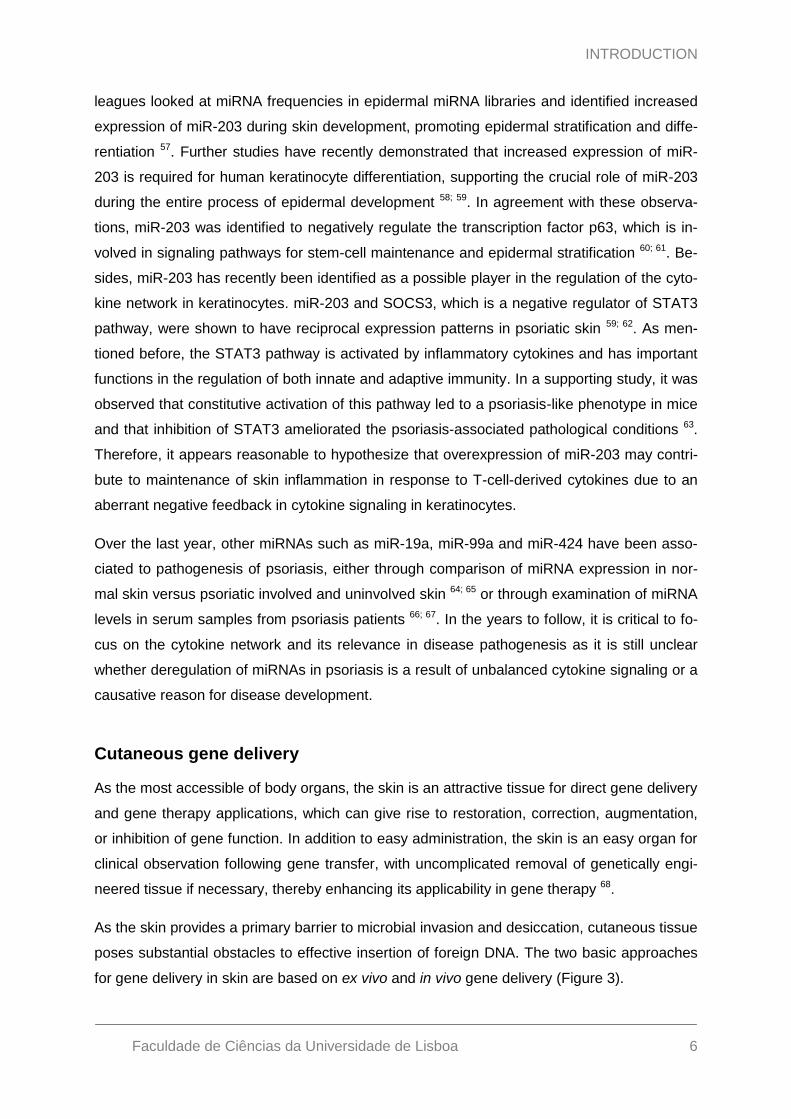
leagues looked at miRNA frequencies in epidermal miRNA libraries and identified increased
expression of miR-203 during skin development, promoting epidermal stratification and diffe-
rentiation 57. Further studies have recently demonstrated that increased expression of miR-
203 is required for human keratinocyte differentiation, supporting the crucial role of miR-203
during the entire process of epidermal development 58; 59. In agreement with these observa-
tions, miR-203 was identified to negatively regulate the transcription factor p63, which is in-
volved in signaling pathways for stem-cell maintenance and epidermal stratification 60; 61. Be-
sides, miR-203 has recently been identified as a possible player in the regulation of the cyto-
kine network in keratinocytes. miR-203 and SOCS3, which is a negative regulator of STAT3
pathway, were shown to have reciprocal expression patterns in psoriatic skin 59; 62. As men-
tioned before, the STAT3 pathway is activated by inflammatory cytokines and has important
functions in the regulation of both innate and adaptive immunity. In a supporting study, it was
observed that constitutive activation of this pathway led to a psoriasis-like phenotype in mice
and that inhibition of STAT3 ameliorated the psoriasis-associated pathological conditions 63.
Therefore, it appears reasonable to hypothesize that overexpression of miR-203 may contri-
bute to maintenance of skin inflammation in response to T-cell-derived cytokines due to an
aberrant negative feedback in cytokine signaling in keratinocytes.
Over the last year, other miRNAs such as miR-19a, miR-99a and miR-424 have been asso-
ciated to pathogenesis of psoriasis, either through comparison of miRNA expression in nor-
mal skin versus psoriatic involved and uninvolved skin 64; 65 or through examination of miRNA
levels in serum samples from psoriasis patients 66; 67. In the years to follow, it is critical to fo-
cus on the cytokine network and its relevance in disease pathogenesis as it is still unclear
whether deregulation of miRNAs in psoriasis is a result of unbalanced cytokine signaling or a
causative reason for disease development.
Cutaneous gene delivery
As the most accessible of body organs, the skin is an attractive tissue for direct gene delivery
and gene therapy applications, which can give rise to restoration, correction, augmentation,
or inhibition of gene function. In addition to easy administration, the skin is an easy organ for
clinical observation following gene transfer, with uncomplicated removal of genetically engi-
neered tissue if necessary, thereby enhancing its applicability in gene therapy 68.
As the skin provides a primary barrier to microbial invasion and desiccation, cutaneous tissue
poses substantial obstacles to effective insertion of foreign DNA. The two basic approaches
for gene delivery in skin are based on ex vivo and in vivo gene delivery (Figure 3).

INTRODUCTION
Faculdade de Ciências da Universidade de Lisboa 7
For ex vivo gene delivery, tar-
get cells are isolated from skin
biopsies and treated with gene
vehicles („vectors‟) in culture
prior to re-engraftment of the
tissue into the patient. In con-
trast, for in vivo approaches,
which are often considered
more straight-forward, the
gene-carrying vector is directly
administered to skin. Direct
administration has been un-
dertaken using a variety of
approaches with both nonviral
and viral vectors, including
topical application, direct in-
jection, application to
wounded skin surfaces, elec-
troporation and bioplastic par-
ticle insertion 68.
Lentiviral gene delivery to skin
The transfer of genetic material to skin is facing the same challenges as conventional drug
types and therefore requires penetration-enhancing carriers or physical methods to over-
come the barriers of the skin. Viruses have through evolution developed and refined the ca-
pacity to carry and transfer genetic material between cells and have in recent years been
extensively explored as vehicles for nucleic acids. Retroviral vectors are attractive tools for
human gene therapy as they have evolved specialized molecular mechanisms to stably inte-
grate into the chromosomes of their targets, a likely requisite for long-term expression. How-
ever, the use of retroviruses such as the MLV-derived retroviral vector involves the require-
ment of the breakdown of the nuclear membrane, which makes them capable of transducing
only proliferating cells 69. Lentiviruses, another subclass of retroviruses, have recently been
adapted as gene delivery vehicles. Opposed to MLV-derived vectors, lentiviral vectors are
capable of transducing both dividing and non-dividing cells due to interactions with the nuc-
lear import machinery of the target cells. Illustrating these properties, vectors derived from
HIV-1 allow for the efficient delivery, integration, and stable expression of transgenes into
cells such as neurons, hepatocytes, and myocytes 70; 71; 72; 73; 74; 75; 76.
Figure 3. Approaches for cutaneous gene delivery. The two basic
approaches to therapeutic gene delivery in skin involve ex vivo and in vivo gene delivery. Ex vivo delivery is a relative complex procedure, which can be divided into three major steps. In the first step, cells from the patient are isolated and propagated in the laboratory. Then, the the-rapeutic transgene cassette is packaged into lentiviral expression deli-very vectors and the patient cells are transduced with lentiviral particles. Finally, the genetically-modified cells are grown in culture followed by re-grafting to the patient. In contrast to ex vivo cutaneous gene delivery, in vivo gene transfer delivers genetic material directly to the patient skin tissue and is thus generally simpler. Direct administration has been un-dertaken using a variety of approaches with lentiviral vectors, including direct injection, topical application and microneedle electroporation. Adapted from manuscript Primo et al, 2011.
INTRODUCTION
Faculdade de Ciências da Universidade de Lisboa 8
The extensive studies of HIV-1 in relation to the global HIV pandemic have created a detailed
knowledge of lentiviral replication and biology and have made HIV-1 the preferred parental
virus for development of LVs. Since the first pioneering reports on HIV-1-based vectors back
in the mid-1990s, several increasingly safer generations of the vector system have appeared.
State-of-the-art vectors are today self-inactivating replication incompetent vectors that harbor
multiple modifications for optimized safety. These engineered vectors do not contain viral
protein-encoding information required for viral replication and are restricted, therefore to a
single infection cycle. Instead, genes encoding the viral structural and enzymatic proteins are
provided on three separate plasmid constructs (packaging vectors) that are simultaneously
supplied to cells that will subsequently produce virus particles. Moreover, removal of the pa-
rental viral promoter from the vector construct ensures that transgene expression is driven
from an internal promoter and that potential adverse effects on neighboring genomic DNA
are limited.
In 2001, Baek and co-workers developed one of the first studies using HIV-derived vectors
for gene delivery in human skin 77. They showed that after a single intracutaneous injection
into full-thickness human skin grafts on immunodeficient mice, LVs encoding human erythro-
poietin produced dose-dependent increases of human erythropoietin levels in serum, which
remained stable subsequently. Additionally, it was shown that removal of the skin graft led to
rapid and total loss of human erythropoietin, confirming that the injected virus was exclusive-
ly targeting the cells in the skin graft and elegantly demonstrating a reversible approach for
gene-based delivery of therapeutic proteins to the bloodstream. Since then, different studies
using lentiviruses as gene delivery vehicles have been successfully developed in monogenic
skin diseases with an autosomal recessive mode of inheritance, such as the tumor-prone
Xeroderma Pigmentosum and the inherited Epidermolysis Bullosa 78; 79; 80; 81.
An extensive number of in vivo studies using LVs to express shRNAs have been performed
to evaluate the effectiveness of RNAi in the treatment of cancer, cardiac disease, retinal dis-
ease, neurodegenerative disease, virus infections and other diseases. The properties of LVs
as facilitators of persistent transgene expression in skin have prompted the idea of utilizing
viral vectors as vehicles of DNA-encoded small RNA effectors. The first in vitro and in vivo
studies for possible future cutaneous siRNA treatments using LVs for transgene delivery
started in 2006 concerning a possible treatment for melanoma 82. Since then, other in vivo
studies have been developed for cutaneous siRNA treatments including two studies from
Mikkelsen research group, which have aimed at targeting central cytokines for amelioration
of psoriasis 83; 84. Potent and persistent transgene expression following a single intradermal
injection of LVs in xenografted human skin was reported in both studies, consolidating the
properties of lentiviral vectors as a valuable tool for cutaneous gene delivery.
AIM OF THE PRESENT WORK
Faculdade de Ciências da Universidade de Lisboa 9
Aim of the present work
Inflammation involves a well-coordinated response, which includes the activation of several
hundred genes including multiple cytokines, chemokines, matrix remodeling proteases, reac-
tive oxygen, nitrogen species and others. The observation that miRNAs are likely regulators
of cytokine mRNAs suggests that they might be involved in diseases related to abnormal
immune responses including certain inflammatory disorders 85. Psoriatic skin is characterized
by a specific miRNA expression profile that is different from healthy skin and skin affected by
other chronic inflammatory diseases, such as atopic eczema. The keratinocyte-specific miR-
203 may play a role in the unbalanced cytokine signaling network in psoriasis as it is known
to be upregulated in psoriatic plaques, leading to a concurrent downregulation of SOCS3
protein translation.
The main goal of this project was to demonstrate the roles played by miRNAs in the complex
network of regulatory pathways controlling the production of cytokines in human skin. With
focus on miR-203, this work combined three major objectives:
Establishment of lentiviral vector-encoded antagomir production for specific miR-203
inhibition in human cell lines and in vivo studies;
Analysis of expression of miRNAs with a putative role in cytokine regulation;
Identification and analysis of miR-203 target genes in human skin.
With a deeper understanding of RNAi and continued progress in designing more safer and
effective RNAi effectors, lentiviral vector-mediated RNAi has the potential to change the way
that numerous cutaneous diseases are studied and treated. As a result, the outlined studies
tried to explore the applicability of miRNA-directed drugs and to address some of the main
hurdles related to specificity of miRNA inhibitor specificity and the capacity of miRNAs to re-
gulate numerous genes.
MATERIALS AND METHODS
Faculdade de Ciências da Universidade de Lisboa 10
Materials and Methods
Plasmid construction
Cloning of H1-antagomir expression cassettes
pCCL-PGK-Puro-H1-MCS (designated pLV/vehicle; see Appendix II, Figure AI) was con-
structed as reported by Bak et al. 83. Oligonucleotide designed was performed as described
in Scherr et al. 86 . For each of the antagomir expression constructs, pCCL-PGK-Puro-H1-
AntagomiR-203 and –Negative control (neg), complementary sense and antisense oligonuc-
leotides (see Appendix I, Table 1) were annealed by incubation at 100°C for 5 minutes fol-
lowed by room temperature cooling. The annealed oligonucleotides were designed to leave
overhangs for cloning into the AvrII/AscI-digested pCCL-PGK-Puro-H1-MCS vector.
Cloning of miR-203 targets into psiCHECK2
To generate psiCHECK2-gene targets (see Appendix I, Table 2), mRNA was isolated from
HaCaT cells stimulated with anisomycin (500µg/mL) using the SV Total RNA Isolation Sys-
tem (Promega, Madison, WI, USA) according to manufacturer‟s protocol. The mRNA was
reverse-transcribed into cDNA using iScriptTM cDNA Synthesis Kit (BIO-RAD, Hercules, CA)
according to manufacturer‟s protocol. All target genes were amplified by PCR using gene-
specific primers (see Appendix I, Table 3), which were made with restriction site containing
linkers (XhoI/NotI). The generated fragments were digested with XhoI/NotI and inserted into
XhoI/NotI-digested psiCHECK2 (Promega, Madison, WI).
To generate miR-203-perfect target and miR-125b-perfect target constructs (psiCHECK2-
miR-203 and psiCHECK2-miR-125b, respectively), complementary sense and antisense oli-
gonucleotides (see Appendix I, Table 4) were annealed by incubation at 100°C for 5 minutes
followed by room temperature cooling. The annealed oligonucleotides were designed to
leave overhangs for cloning into the XhoI/NotI-digested psiCHECK2 (Promega, Madison,
WI).
Generation of 3’UTR mutated sequences and cloning into psiCHECK2
To generate IL24, SOCS6 and TNFα 3‟UTR mutants, self-complementary DNA oligonucleo-
tides encompassing the sequences of IL24-, SOCS6- and TNFα-mut 3‟UTRs were chemical-
ly synthesized with 5‟ XhoI and a 3‟ NotI restriction sites (GenScript, Piscataway, NJ, USA).
The sequences are shown in Appendix III-V. The generated fragments were digested with
XhoI/NotI and inserted into XhoI/NotI-digested psiCHECK2 (Promega, Madison, WI).

MATERIALS AND METHODS
Faculdade de Ciências da Universidade de Lisboa 11
Generation of pri-miR-203 and pri-miR-125b-expressing constructs
The vector encoding pri-miR-203 was created by amplification of pri-miR-203 cDNA from
genomic DNA extracted from HaCaT cells with primers 5‟ GCGTCTAAGGCGTCCGGTAC 3‟
and 5‟ GTCGCCGGCGCACCCCT 3‟. The two primers were made with restriction site con-
taining linkers (NotI). eGFP was excised using NotI from a biscitronic DNA transposon vec-
tor, pT2/CMV-eGFP(s).SV40-neo. The pri-miR-203 PCR amplicon was inserted into the vec-
tor in the sense orientation using the NotI sites (see Appendix II, Figure AI). Similar approach
was employed for generation of pri-miR-125b-expressing construct, by amplifying pri-miR-
125b cDNA from genomic DNA extracted from HaCaT cells with primers 5‟ CATCTTAGT-
TATGAACCTCGAACAG 3‟ and 5‟ AAATTGTCTTTAGGTCCTCGACGG 3‟.
Cell lines
HaCaT, HeLa, HEK293 and 293T cells were cultured at 37°C in 5% (v/v) CO2 and main-
tained in Dulbeccoo‟s modified Eagle‟s medium (Cambrex, Verviers, Belgium) supplemented
with 10% fetal calf serum, penicillin (100 U/mL), streptomycin (0.1 mg/mL), and L-glutamine
(265 mg/L).
Generation of miR-203-expressing HaCaT cell line, HaCaT-203
For generation of miR-203-expressing HaCaT cell line, an approach based on the Sleeping
Beauty DNA transposon system was employed (see Appendix II, Figure AI). Transposition in
the HaCaT cell line was performed in 6-well plates into which 5x10⁴ HaCaT cells were
seeded one day prior to transfection. Co-transfections were performed with 1.82 µg of
pT2/CMV-pri-miR-203.SV40-neo and 0.18 µg of either transposase-encoding vector
(SB100X) or mutated transposase (mSB) using FuGene-6 (Roche, Basel, Switzerland) ac-
cording to manufacturer‟s protocol. One day after transfection cells were trypsinized and re-
seeded in appropriate dilutions. The cells were selected for positive transposition by G418-
supplemented medium (700 µg/mL) for fourteen days. From the wells with the lowest colony-
count, singles clones were isolated to separate dishes and expanded for one week with
standard medium, followed by total RNA extraction using Tri Reagent (Sigma, St Louis, MO).
miR-203 expression analysis was assessed by qRT-PCR employing the TaqMan® Universal
Master Mix II, No UNG (Applied Biosystems, Foster City, CA) according to manufacturer‟s
protocol. miR-203 expression was determined using miR-203 primers and probes (Assay ID
000507). A single miR-203 HaCaT clone was chosen for subsequent experiments.

MATERIALS AND METHODS
Faculdade de Ciências da Universidade de Lisboa 12
Dual-Luciferase Reporter Assay
For co-transfection experiments, HEK293 cells were seeded in 96-well plates (3 x 10³
cells/well) one day before transfection. Co-transfections were performed with a total of 60 ng
(54 ng of pT2/CMV-pri-miR-203.SV40-neo, pT2/CMV-pri-miR-125b.SV40-neo or pUC19 stuf-
fer DNA and 6 ng psiCHECK2-gene targets-encoding vectors) using FuGene-6 (Roche, Ba-
sel, Switzerland) according to manufacturer‟s protocol. Forty-eight hours post-transfection,
Renilla and Firefly luciferase activities were analyzed by the use of the Dual-Luciferase® Re-
porter Assay System (Promega, Madison, WI, USA) according to the manufacturer‟s proto-
col. Reactions were carried out in 96-well plates and luminescence readings were performed
in a multisample platereading luminometer (Berthold, Bad Wild-bad, Germany). Renilla luci-
ferase activity was normalized to Firefly luciferase and presented relative to the negative
control (pUC19).
To test the functionality of antagomir-encoding lentiviral vectors, HeLa, HaCaT and HaCaT-
203 cells were seeded in 24-well plates (1,9x10⁴ cells/ well) one day before transfection. Co-
transfections were performed with a total of 400 ng (40 ng of psiCHECK2-miR-203-perfect
target and 0,180, 270 or 360 ng of pCCL-PGK-Puro-H1-antagomiR-203) using FuGene-6
(Roche, Basel, Switzerland) according to manufacturer‟s protocol. Additionally, 90,180 or 360
ng of pUC19 was included as stuffer DNA to ensure the equal amounts of DNA used in each
transfection. Luciferase activities were measured forty-eight hours post-transfection as de-
scribed above.
In transduction studies of antagomir-encoding lentiviral vectors, HaCaT cells were seeded in
24-well plates (1,9x10⁴ cells/ well) one day before they were transduced at an MOI of 120.
The viral supernatant was supplemented with polybrene (8 µg/mL; Sigma-Aldrich, Milwau-
kee, WI). One day post-transduction, co-transfections with 40 ng of psiCHECK2-miR-203-
perfect target and 360 ng of pUC19 using FuGene-6 (Roche, Basel, Switzerland) according
to manufacturer‟s protocol and luciferase activities were measured forty-eight hours post-
transfection as described above.
All the dual luciferase assay experiments were performed at least in triplicates.
Lentiviral vector production
For production of lentiviral vectors, 293T cells were seeded at a density of 4 x 10⁶ cells/well
in 10-cm dishes one day before transfection. Cells were transfected with calcium phosphate
treatment with 3.75 µg pMD.2G (envelope plasmid), 3 µg pRSV-Rev (Rev-expressing plas-
mid), 13 µg pMDGP-Lg/RRE (packaging plasmid) and 13 µg lentiviral transfer vector. Forty-
MATERIALS AND METHODS
Faculdade de Ciências da Universidade de Lisboa 13
eight hours after transfection the viral supernatant was harvested and passed through 0.45
µm filters to remove cellular debris (Sarstedt, Nümbrecht, Germany). The resulting lentiviral
vectors were designated LV/vehicle, LV/antagomiR-neg and LV/antagomiR-203. Colony-
forming titer assays were performed on HaCaT cells seeded in 6-well plates (5 x 10⁶
cells/well) one day before transduction. Lentiviral supernatants were serially diluted and sup-
plemented with polybrene (8 µg/mL; Sigma-Aldrich, Milwaukee, WI) before addition to the
cells. Transduced cells were grown in 1 µg/mL puromycin-containing medium (Sigma-
Aldrich, Milwaukee, WI) for ten days after which the number of colonies was counted in the
wells.
For in vivo transductions of xenografted human skin, the lentiviral supernatants were ultra-
centrifuged for two hours (4°C at 25000 r.p.m.) in a SW28 rotor (Beckman Coulter, Fullerton,
CA). Virus pellets were resuspended overnight in PBS-/- at 4°C in a volume of 1/300 of the
original volume. The lentiviral vector yield was determined by measuring the amount of p24
Gag protein using HIV-1 p24 Antigen ELISA Kit (ZeptoMetrix, Buffalo, NY) according to
manufacturer‟s protocol.
Human xenograft transplantation model
Six psoriatic plaque skin biopsies were obtained from donors with moderate to severe plaque
psoriasis. The psoriasis of the participants was untreated for at least one month prior to the
time of biopsy. Informed consent was obtained and the study was approved by the Central
Ethical Committee and conducted according to the Declaration of Helsinki protocols. Animal
studies were carried out with permission from the Danish Experimental Animal Inspectorate.
Each keratome skin biopsy, containing both epidermis and dermis, was split into several
grafts (each 1.5 x 1.5 x 0.05 cm) and transplanted onto C.B-17 severe combined immunode-
ficient (SCID) mice, 6-8 weeks old (Taconic M & B, Silkeborg, Denmark), as described 87.
Shortly, the mice were anesthetized prior to surgery by a subcutaneous injection of Ketami-
nol (ketamine, 100 mg/kg; Intervet, Skovlunde, Denmark) and Narcoxyl (xylazine, 10 mg/kg;
Intervet, Skovlunde, Denmark). The back was shaved and part of the exposed skin removed.
The grafts were sutured with absorbable 6-0 suture (Caprosyn, Tyco, Copenhagen, Den-
mark) and covered with Xeroform dressings (Sherwood Medical Company; Markham, Ontar-
io, Canada) for one week. The mice were kept under pathogen-free conditions throughout
the study. The grafts healed for ten days before the mice were randomized and subjected to
treatment as indicated.
MATERIALS AND METHODS
Faculdade de Ciências da Universidade de Lisboa 14
In vivo administration of antagomir-encoding lentiviral vectors
LV/antagomiR-neg and LV/antagomiR-203 were administered intradermally into psoriatic
skin grafts (at a dose of 65 µg p24 Gag/mL in 150 µL) as a single treatment.
Xenograft evaluation following treatment
The severity of psoriatic lesions in the grafts was assessed blinded twice weekly and scored
semi-quantitatively according to the average of the following clinical signs: scaliness, indura-
tion, and erythema. The parameters were scored using a four-point scale: 0, complete lack of
cutaneous involvement; 1, slight involvement; 2, moderate involvement; 3, sever involve-
ment. On a scale from 0 to 3 a maximal score of 3 represents severe scale, induration and
erythema of the xenografted psoriatic skin. After three weeks of treatment, biopsies from the
center of the graft were obtained, fixed and embedded in paraffin. The remaining grafted skin
was snap-frozen in liquid nitrogen and stored at -80°C for further analysis. Employing stan-
dard methods, sections were stained histochemically with hematoxylin and eosin. Epidermal
thickness was measured at least ten random places from stratum corneum to the deepest
part of the rete pegs on three equally distantly cut sections. All sections were blinded prior to
evaluation and evaluated randomly. Mean epidermal thickness values for each graft in each
treatment group were calculated, and the data summarized as mean ± SEM.
RNA isolation and quantitative RT-PCR
Skin biopsies from in vivo transduced xenografted psoriatic and normal skin were incubated
in RNAlater-ICE (Ambion, Austin, TX) and stored at -20°C for twenty-four hours prior to RNA
isolation. From both in vitro transduced cells and skin biopsies, total RNA was extracted us-
ing the SV Total RNA Isolation System (Promega, Madison, WI, USA) according to manufac-
turer‟s protocol. In the lysis buffer the biopsies were homogenized 2 x 2 minutes at 25 Hz
using a TissueLyser (Qiagen, Valencia, California, USA). Isolated RNA was dissolved in
RNase-/DNase-free water and stored until further use at -150°C. First strand cDNA synthesis
was performed using the Maxima® First Strand cDNA Synthesis Kit for RT-qPCR (Fermen-
tas, St. Leon-Rot, Germany) according to manufacturer‟s protocol. SOCS6, TNFα, IL12B,
IL20 and IL24 mRNA levels were assessed by qRT-PCR employing the TaqMan® Universal
Master Mix II, No UNG (Applied Biosystems, Foster City, CA) according to manufacturer‟s
protocol. SOCS6, TNFα, IL12B, IL20 and IL24 mRNA expression was determined using
SOCS6, TNFα, IL12B, IL20 and IL24 primers and probes (FAM-labeled MGB-probes
Hs00926356_g1, Hs00174128_m1, Hs01011518_m1, Hs00218888_m1 and
Hs01114274_m1, respectively, Applied Biosystems, Foster City, CA). Expression of each
MATERIALS AND METHODS
Faculdade de Ciências da Universidade de Lisboa 15
gene was analyzed at least in duplicates using a LightCycler 480 (Roche, Basel, Switzer-
land). SOCS6, TNFα, IL12B, IL20 and IL24 mRNA levels were normalized to the expression
of the reference gene ribosomal protein, large P0 (RPLP0) using RPLP0 specific primers
(FAM-labeled MGB-probes Hs99999902_m1, Applied Biosystems, Foster City, CA).
Quantification of miRNAs by TaqMan® microRNA Assays (Applied Biosystems, Foster City,
CA) was carried out according to manufacturer‟s protocol. RU48, miR-16, miR-125b and
miR-203 levels were assessed by qRT-PCR employing the TaqMan® Universal Master Mix II,
No UNG (Applied Biosystems, Foster City, CA) according to manufacturer‟s protocol. RU48,
miR-16, miR-125b and miR-203 expressions were determined using RU48, miR-16, miR-
125b and miR-203 primers and probes (AB Assay ID 001006, 000391, 000449 and 000507,
respectively). Expression of each gene was analyzed at least in duplicates using a LightCyc-
ler 480 (Roche, Basel, Switzerland). miR-203 levels were normalized to the expression of the
reference small nucleolar RNA, C/D box 48 (RU48) using RU48 specific primers (AB Assay
ID 001006, Applied Biosystems, Foster City, CA).
For all qPCR experiments, relative RNA levels were determined using the relative standard
curve method. Briefly, a standard curve for each gene was made from serial dilutions of the
cDNA. The standard curve was then used to calculate relative amounts of target RNA in the
samples. Mean RNA values were calculated and the data summarized as mean + SEM.
Bioinformatics
miR-203 target sites for IL24, SOCS6 and TNFα 3‟UTR sequences were predicted by PicTar
5, RNAhybrid and TargetScan 5.2 softwares available at http://pictar.mdc-berlin.de/,
http://bibiserv.techfak.uni-bielefeld.de/rnahybrid/ and http://www.targetscan.org/, respectively.
Statistical Analyses
All p-values were calculated by a two-tailed Student‟s T-test to test the null hypothesis of no
difference between two compared groups. The assumption of equal variances was tested by
the F-test. In all statistical analyses, p-values < 0.05 were considered significant.
RESULTS
Faculdade de Ciências da Universidade de Lisboa 16
Results
Towards identification and analyses of miR-203 target genes
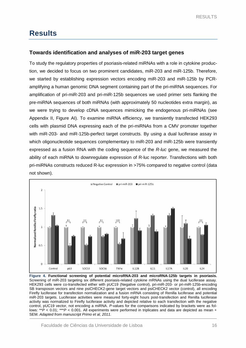
To study the regulatory properties of psoriasis-related miRNAs with a role in cytokine produc-
tion, we decided to focus on two prominent candidates, miR-203 and miR-125b. Therefore,
we started by establishing expression vectors encoding miR-203 and miR-125b by PCR-
amplifying a human genomic DNA segment containing part of the pri-miRNA sequences. For
amplification of pri-miR-203 and pri-miR-125b sequences we used primer sets flanking the
pre-miRNA sequences of both miRNAs (with approximately 50 nucleotides extra margin), as
we were trying to develop cDNA sequences mimicking the endogenous pri-miRNAs (see
Appendix II, Figure AI). To examine miRNA efficiency, we transiently transfected HEK293
cells with plasmid DNA expressing each of the pri-miRNAs from a CMV promoter together
with miR-203- and miR-125b-perfect target constructs. By using a dual luciferase assay in
which oligonucleotide sequences complementary to miR-203 and miR-125b were transiently
expressed as a fusion RNA with the coding sequence of the R-luc gene, we measured the
ability of each miRNA to downregulate expression of R-luc reporter. Transfections with both
pri-miRNAs constructs reduced R-luc expression in >75% compared to negative control (data
not shown).
Figure 4. Functional screening of potential microRNA-203 and microRNA-125b targets in psoriasis.
Screening of miR-203 targeting six different psoriasis-related cytokine mRNAs using the dual luciferase assay. HEK293 cells were co-transfected either with pUC19 (Negative control), pri-miR-203- or pri-miR-125b-encoding SB transposon vectors and nine psiCHECK2-gene target vectors and psiCHECK2 vector (control), all encoding Firefly luciferase for transfection normalization and a fusion mRNA consisting of Renilla luciferase and potential miR-203 targets. Luciferase activities were measured forty-eight hours post-transfection and Renilla luciferase activity was normalized to Firefly luciferase activity and depicted relative to each transfection with the negative control, pUC19 vector, not encoding a miRNA. P-values for the comparisons indicated by brackets were as fol-lows: **P < 0.01; ***P < 0.001. All experiments were performed in triplicates and data are depicted as mean + SEM. Adapted from manuscript Primo et al, 2011.
RESULTS
Faculdade de Ciências da Universidade de Lisboa 17
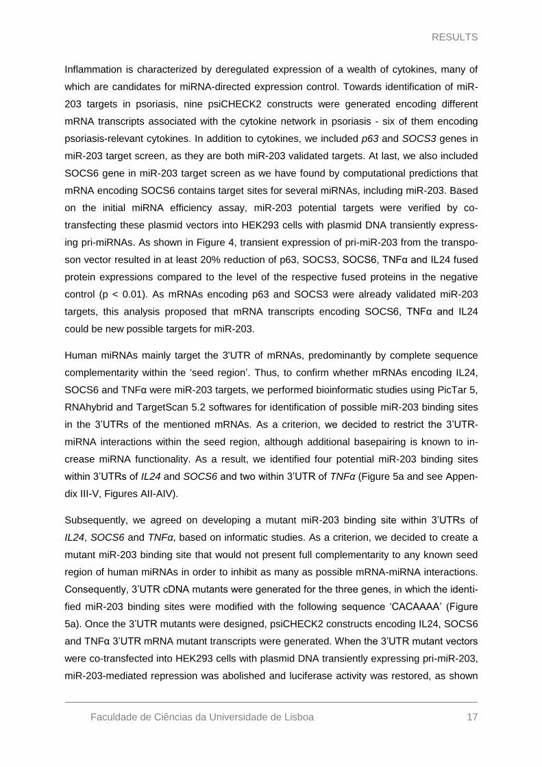
Inflammation is characterized by deregulated expression of a wealth of cytokines, many of
which are candidates for miRNA-directed expression control. Towards identification of miR-
203 targets in psoriasis, nine psiCHECK2 constructs were generated encoding different
mRNA transcripts associated with the cytokine network in psoriasis - six of them encoding
psoriasis-relevant cytokines. In addition to cytokines, we included p63 and SOCS3 genes in
miR-203 target screen, as they are both miR-203 validated targets. At last, we also included
SOCS6 gene in miR-203 target screen as we have found by computational predictions that
mRNA encoding SOCS6 contains target sites for several miRNAs, including miR-203. Based
on the initial miRNA efficiency assay, miR-203 potential targets were verified by co-
transfecting these plasmid vectors into HEK293 cells with plasmid DNA transiently express-
ing pri-miRNAs. As shown in Figure 4, transient expression of pri-miR-203 from the transpo-
son vector resulted in at least 20% reduction of p63, SOCS3, SOCS6, TNFα and IL24 fused
protein expressions compared to the level of the respective fused proteins in the negative
control (p < 0.01). As mRNAs encoding p63 and SOCS3 were already validated miR-203
targets, this analysis proposed that mRNA transcripts encoding SOCS6, TNFα and IL24
could be new possible targets for miR-203.
Human miRNAs mainly target the 3'UTR of mRNAs, predominantly by complete sequence
complementarity within the 'seed region‟. Thus, to confirm whether mRNAs encoding IL24,
SOCS6 and TNFα were miR-203 targets, we performed bioinformatic studies using PicTar 5,
RNAhybrid and TargetScan 5.2 softwares for identification of possible miR-203 binding sites
in the 3‟UTRs of the mentioned mRNAs. As a criterion, we decided to restrict the 3‟UTR-
miRNA interactions within the seed region, although additional basepairing is known to in-
crease miRNA functionality. As a result, we identified four potential miR-203 binding sites
within 3‟UTRs of IL24 and SOCS6 and two within 3‟UTR of TNFα (Figure 5a and see Appen-
dix III-V, Figures AII-AIV).
Subsequently, we agreed on developing a mutant miR-203 binding site within 3‟UTRs of
IL24, SOCS6 and TNFα, based on informatic studies. As a criterion, we decided to create a
mutant miR-203 binding site that would not present full complementarity to any known seed
region of human miRNAs in order to inhibit as many as possible mRNA-miRNA interactions.
Consequently, 3‟UTR cDNA mutants were generated for the three genes, in which the identi-
fied miR-203 binding sites were modified with the following sequence „CACAAAA‟ (Figure
5a). Once the 3‟UTR mutants were designed, psiCHECK2 constructs encoding IL24, SOCS6
and TNFα 3‟UTR mRNA mutant transcripts were generated. When the 3‟UTR mutant vectors
were co-transfected into HEK293 cells with plasmid DNA transiently expressing pri-miR-203,
miR-203-mediated repression was abolished and luciferase activity was restored, as shown

RESULTS
Faculdade de Ciências da Universidade de Lisboa 18
in Figure 5b. Together, these results indicate that the effect of miR-203 on IL24, SOCS6 and
TNFα mRNA transcripts is direct and mediated by specific 3‟UTR target sites.
Downregulation of miR-203 targets following stable expression of miR-
203
To confirm that IL24, SOCS6 and TNFα mRNA transcripts were indeed genuine targets of
miR-203, a pri-miR-203 expression cassette was inserted into a HaCaT cell line by means of
the Sleeping Beauty DNA transposon system 88. Plasmid DNA, containing the pri-miR-203
expression cassette and neomycin resistance gene driven by a SV40 promoter within the
context of a SB DNA transposon, was co-transfected with helper plasmid encoding either the
SB100X transposase or a mutated, inactive variant. The transposition efficiency of the pri-
miR-203 transposon vector was evaluated by transfecting HaCaT cells with pT2/CMV-pri-
miR-203.SV40-neo together with one of both transposase-encoding plasmids. Numbers of
G418-resistant colonies (see Appendix VI, Figure AVa) indicated that neo expression cas-
Figure 5. Development of 3’UTR mutants for confirmation of IL24, SOCS6 and TNFα mRNA transcripts as direct targets of microRNA-203. (a) Schematic representation of the 3‟UTR sequence of IL24, SOCS6 and
TNFα transcripts (grey bar) with the predicted target sites for miR-203 (black lines). The numbers underlying the black lines represent the first and the last basepair position within the NCBI reference for each represented tran-script. Schematic representation of the miR-203 seed match mutation. Nucleotides written in bold represent the mutated nucleotides. (b) Confirmation of IL24, SOCS6 and TNFα 3‟UTR transcripts as miR-203 targets. Compari-
son between HEK293 cells co-transfected with Firefly luciferase constructs containing the wild-type or mutant 3‟UTR and pUC19 (Negative control) or pri-miR-203-encoding SB DNA transposon vector. Luciferase activities were measured forty-eight hours post-transfection. Renilla luciferase activity was normalized to Firefly luciferase activity and depicted relative to each transfection with the negative control, pUC19 vector, not encoding a miRNA. P-values for the comparisons indicated by brackets were as follows: **P < 0.01; ***P < 0.001; ****P < 0.0001. All experiments were performed in triplicates and data are depicted as mean + SEM. Adapted from manuscript Primo et al, 2011.

RESULTS
Faculdade de Ciências da Universidade de Lisboa 19
sette was inserted markedly more frequently in the presence of active transposase relative to
the inactive variant, indicating that genomic integration of the pri-miR-203 vector was effi-
ciently accomplished by a transposase-directed mechanism. We next isolated and expanded
three individual clones containing the pri-miR-203 expression cassette inserted by the
SB100X transposase, and miR-203 expression was confirmed by qRT-PCR (see Appendix
VI, Figure AVb). A single HaCaT-203 clone (clone C) was chosen for subsequent experi-
ments.
To evaluate knockdown of endogenous IL24, SOCS6 and TNFα mRNA transcripts due to
miR-203 stable overexpression, RNA samples were collected and evaluated by quantitative
RT-PCR on naїve HaCaT and HaCaT-203 cells. Endogenous expressions of IL24, SOCS6
and TNFα mRNA transcripts were efficiently targeted by miR-203 expression, as shown in
Figure 6a. We detected a knockdown ranging from 60% to 95% of SOCS6 and TNFα mRNA
transcripts, respectively (Figure 6a). These findings demonstrated the potential specificity of
the interaction between miR-203 and the mentioned targets.
To certify that endogenous knockdown of the target genes was the result of increased ex-
pression of miR-203 in the HaCaT cell line and not due to an unknown particularity of Ha-
CaT-203 cell line - clone C, we decided to analyze the endogenous mRNA expression of one
of miR-203-targets in the other two HaCaT-203 clones. The knockdown potency was eva-
luated by quantitative RT-PCR analysis, and endogenously expressed IL24 was efficiently
targeted by miR-203 in all different HaCaT-203 clones. As shown in Figure 6b, miR-203 and
IL24 mRNA levels were inversely related in the different HaCaT populations, indicating that
repression of IL24 mRNA transcript was only dependent of miR-203 expression levels. Tak-
Figure 6. Endogenous knockdown of IL24, SOCS6 and TNFα mRNA transcripts by stable overexpression of microRNA-203. (a) IL24, SOCS6 and TNFα mRNA levels were evaluated by qRT-PCR in a naїve HaCaT cell line and in a miR-203 expressing HaCaT cell line. (b) IL24 mRNA levels and miR-203 levels were evaluated by
qRT-PCR in a naїve HaCaT cell line and in three different HaCaT clones stably overexpressing miR-203, gener-ated by means of the Sleeping Beauty DNA transposon system. IL24, SOCS6 and TNFα mRNA levels were nor-malized to the expression of the reference gene ribosomal protein, large P0 (RPLP0) and miR-203 levels were normalized to the expression of the reference small nucleolar RNA, C/D box 48 (RU48). P-values for the compari-sons indicated by brackets were as follows: ***P < 0.001; ****P < 0.0001. All experiments were performed in tripli-cates and data are depicted as mean + SEM. Adapted from manuscript Primo et al, 2011.

RESULTS
Faculdade de Ciências da Universidade de Lisboa 20
en together, these results demonstrated the specificity of miRNA-mRNA interactions be-
tween miR-203 and the IL24, SOCS6 and TNFα mRNA transcripts.
Establishment of miR-203 inhibition by vector-encoded ‘antagomirs’
Jacob Mikkelsen research group has previously provided evidence of potent and sustained
RNAi-mediated in vivo knockdown of IL12B and TNFα mRNAs in human skin by the use of
lentiviral vectors 83; 84. Hence, we decided to explore the same lentiviral backbone (see Ap-
pendix II, Figure AI) for design of vectors expressing antisense RNAs complementary to im-
mune-modulatory miRNAs (v-antagomirs), as a method for persistently antagonizing miRNA
function in cultured cells and in mice. Therefore, two antagomirs were designed in this study
– the first one encoding an antisense RNA complementary to human miR-203 (antagomiR-
203) and the second one encoding an antisense RNA not complementary to any known hu-
man miRNA, named antagomiR-Negative control (antagomiR-neg).
Before we monitored the efficiency of antagomirs against endogenous miR-203, we decided
to characterize the expression of miRNAs with a putative role in cytokine regulation in differ-
ent cell lines. This assay was developed to identify the cell line with the highest miR-203 ex-
pression for subsequent investigations of antagomir functionality. In our pilot study we eva-
luated by quantitative RT-PCR analyses the RNA expression levels of RU48, miR-203, miR-
125b and miR-16 (the latter has been implicated in posttranscriptional regulation of TNFα) in
three different cell lines and in an extra RNA sample collected from psoriasis lesional skin
(see Appendix VI, Figure AVI). Since miR-203 expression levels were shown to be very re-
duced in HEK293 cells and as we did not identify a statistical significant difference between
miR-203 expression levels in HaCaT cells with and without stress stimulation, we decided to
proceed with HeLa and HaCaT cell lines without stress stimulation for examination of miR-
203 inhibition by antagomirs.
To monitor the potency of miRNA inhibition triggered by antagomirs, antagomir-encoding
lentiviral vectors were analyzed for their ability to target endogenous miR-203 by using a dual
luciferase expression assay. HeLa and HaCaT cells were co-transfected with psiCHECK2-
miR-203-perfect target together with different amounts of antagomiR-203-encoding lentiviral
vector. Luciferase activities were measured forty-eight hours post-transfection, and antago-
miR-203 potencies were determined by normalizing to the expression levels obtained in the
absence of antagomiR-203-expressing lentiviral vector. As shown in Figure 7a, a dose-
response relationship was obtained in all cell lines, in which increased amounts of lentiviral
vector expressing antagomiR-203 resulted in increased expression of R-luc, due to inhibition
of endogenous miR-203 activity. We also observed that the highest miR-203 knockdown was

RESULTS
Faculdade de Ciências da Universidade de Lisboa 21
achieved in HaCaT-203 cells as those cells were the ones that presented the highest miR-
203 expression levels within the same setup conditions.
To certify that the antagomir-encoding gene cassette was efficiently transferred by the lenti-
viral vector system, we first measured the transductional efficiency of antagomir-expressing
vectors in experiments that included the LV/vehicle vector as a control. High transduction
titers of antagomir-expressing lentiviral vectors were confirmed (ranging from 3.40 x 10⁶ to
4.05 x 10⁶ colony forming units per mL [cfu/mL] on HaCaT cells), although the titers on aver-
age were reduced relative to the control vector, which did not express an antagomir (data not
shown).
To evaluate antagomir potency when delivered by lentiviral vectors, HEK293 cells were
transduced at a multiplicity of infection (MOI) of 120 with three lentiviral vectors
(LV/antagomiR-203, LV/antagomiR-neg and LV/vehicle, as a control), one day prior to trans-
fection. On the following day, cells were transfected either with the psiCHECK2-miR-203-
perfect target or pUC19 stuffer DNA, and luciferase activities were measured forty-eight
hours post-transfection. As shown in Figure 7b, transient expression of antagomiR-203,
when delivered by lentiviral vectors at an MOI of 120, resulted in abolishment of miR-203-
mediated repression. We detected a 50% knockdown of miR-203 activity in cells transduced
Figure 7. Functional evaluation of microRNA-203 targeting by antagomiR-203 and confirmation of anta-gomir potency after lentiviral transduction. (a) Upregulation of transiently expressed miR-203-perfect target by
transfection with lentiviral construct encoding antagomiR-203. miR-203-perfect match sequence was fused to the Renilla luciferase gene in the psiCHECK2 vector and the plasmid was co-transfected together with pLV/antagomiR-203 into HaCaT-203 cells, HaCaT cells and HeLa cells. Luciferase activities were measured forty-eight hours post-transfection. Renilla luciferase activity was normalized to Firefly luciferase activity and de-picted relative to non-transfected cells with pLV/antagomiR-203. (b) Antagomir potency evaluation after transduc-
tion with antagomir-encoding lentiviral vectors. HaCaT cells were transduced at an MOI of 120. Cells were trans-fected with either psiCHECK2 vector (Negative control) or miR-203-perfect target construct one day post-transduction and luciferase activity was measured forty-eight hours post-transfection. Renilla luciferase activity was normalized to Firefly luciferase activity and depicted relative to transduction with lentiviral vector encoding no antagomir (LV/vehicle). P-values for the comparisons indicated by brackets were as follows: **P < 0.01. All expe-riments were performed at least in triplicates and data are depicted as mean + SEM. Adapted from manuscript Primo et al, 2011.

RESULTS
Faculdade de Ciências da Universidade de Lisboa 22
with LV/antagomiR-203 (p < 0.01), an effect that was not identified in cells transduced with
either LV/vehicle or LV/antagomiR-neg. Taken together, these results demonstrated a high
potential of lentiviral delivery of antagomiR-203 for efficient targeting of aberrantly expressed
miR-203 for RNAi-mediated knockdown.
Efficient and persistent upregulation of IL24 following lentiviral transduc-
tion
To investigate the effects of miR-203 knockdown by delivery of antagomiR-203-expressing
lentiviral vector into HaCaT cells, we evaluated by qRT-PCR the expression levels of miR-
203 and IL24 mRNA transcripts. Therefore, two different HaCaT cell lines (naїve HaCaT cells
and HaCaT-203 cells) were transduced at an MOI of 50 and at an MOI of 5 (followed by a
14-day puromycin selection) to test whether a few inserted copies of the antagomiR-203-
expressing vector would have a persistent effect on miR-203 knockdown.
As shown in Figure 8, endogenously miR-203 activity was efficiently targeted and abolished
Figure 8. In vitro knockdown of microRNA-203 after transduction with antagomir-encoding lentiviral vec-tors and upregulation of IL24 mRNA expression. (a, b) Knockdown of miR-203 expression following transduc-tion with lentiviral vector-encoded antagomiR-203. (a) Naїve HaCaT cells and (b) HaCaT-203 cells were trans-
duced with LV/vehicle, LV/antagomiR-neg or LV/antagomiR-203, as indicated below each column. Both naїve HaCaT and HaCaT-203 cells were transduced at an MOI of 50 (grey bars) or 5 (black bars) followed by puromy-cin selection for fourteen days to ensure that cells harbored only few lentiviral insertions. miR-203 and IL24 mRNA levels were evaluated by qRT-PCR after three days and fourteen days post-transduction in (a) naїve HaCaT cells and (b) HaCaT-203 cells. IL24 mRNA levels were normalized to the expression of the reference
gene ribosomal protein, large P0 (RPLP0) and miR-203 levels were normalized to the expression of the reference small nucleolar RNA, C/D box 48 (RU48). P-values for the comparisons indicated by brackets were as follows: *P < 0.05; ***P < 0.001; ****P < 0.0001. All experiments were performed in triplicates and data are depicted as mean + SEM. Adapted from manuscript Primo et al, 2011.

RESULTS
Faculdade de Ciências da Universidade de Lisboa 23
by lentiviral delivery of antagomiR-203, in both naїve and HaCaT-203 cells, as we detected a
2-fold increase of IL24 mRNA transcripts three days after transduction (p < 0.05). When ana-
lyzing IL24 mRNA transcripts fourteen days after lentiviral transduction, we also identified an
increase of IL24 mRNA expression levels (p < 0.001), providing persistent regulatory effect of
lentivirally encoded antagomiR-203 effectors. By comparison of IL24 mRNA expression le-
vels within both cell lines, we observed that the highest levels of IL24 mRNA transcripts were
quantified fourteen days after transduction in the naїve HaCaT cell line (Figure 8a). In
agreement with this observation was the fact that the highest miR-203 knockdown was iden-
tified fourteen days after transduction in the same HaCaT cell line (Figure 8b). Taken togeth-
er, these observations indicated that miR-203 and IL24 mRNA levels were inversely ex-
pressed in HaCaT cells, clearly demonstrating the direct occurrence of a specific miRNA-
mRNA interaction.
According to the proposed function of the antagomirs, this type of inhibitor effectively com-
petes with miRNA targets by a stronger binding to their specific miRNA target on the RISC
complex, resulting in inhibition of miRNA activities. Our observations regarding IL24 mRNA
transcripts after delivery of antagomiR-203-expressing lentiviral vector in HaCaT cells were
in agreement with the previous statement, demonstrating that antagomiR-203 was a potent
inhibitor of miR-203. Antagomirs are also known to induce the degradation of the targeted
miRNAs with as of yet unknown mechanisms 47; 89. Regarding induced-miR-203 degradation
by antagomiR-203 expression, no change in miR-203 expression levels was observed in
both HaCaT cell lines three days after lentiviral transduction (Figure 8a and 8b). RNA sam-
ples collected fourteen days after cell transduction shown that miR-203 expression levels
were markedly diminished (60% knockdown) in naїve HaCaT cells transduced with antago-
miR-203-expressing lentiviral vector, when compared to cells transduced with antagomiR-
neg-expressing lentiviral vector (Figure 8a). A similar result was not identified in HaCaT cells
stably expressing miR-203. In this HaCaT-203 cell line, miR-203 expression levels did not
differ within HaCaT cells transduced with antagomiR-203- and antagomiR-neg-expressing
lentiviral vectors (Figure 8b). The results collectively demonstrated that induced miR-203
knockdown by antagomiR-203 expression presented a dose-dependent response as anta-
gomiR-203 knockdown efficiency was inversely correlated to miR-203 endogenous expres-
sion.
In vivo lentiviral delivery of antagomiR-203 in xenografted psoriatic skin
In the previous experiments we demonstrated that miR-203 could mediate post-
transcriptional suppression through IL24 3‟UTR. These proof-of-concept studies confirmed
that keratinocyte-specific miR-203 plays a role in cytokine expression in keratinocytes. Since

RESULTS
Faculdade de Ciências da Universidade de Lisboa 24
psoriatic skin is characterized by a specific miR-203 expression profile that is different in
healthy skin, we decided to extend the exploration of antagomir-mediated miRNA knockdown
in psoriatic human skin. To investigate the effects of lentivirally delivered antagomiR-203 in
human psoriatic skin in vivo, we used the psoriasis xenograft transplantation model. Upon
transplantation of psoriatic keratome skin biopsies, the mice were divided into two groups.
The first group (n = 3) was treated with a single intradermal dose LV/antagomiR-203 (65 µg
p24 Gag/mL in 150 µl), whereas the second group (n = 3) was treated with a single intrader-
mal dose of LV/antagomiR-neg (same dose as group one) as a negative control (Figure 9a).
The mice were assigned a baseline semiquantitative clinical psoriasis score based on the
average erythema, thickness and scaliness of the psoriatic plaques. The effects of treatment
were evaluated in a blinded fashion by semiquantitative clinical scores given twice weekly for
three weeks until killing. The outlined study tried to explore the applicability of miRNA-
directed drugs and address some of the main hurdles related to antagomir specificity and the
capacity of miRNAs to regulate numerous genes.
The final endpoint in evaluating the effect on psoriasis was determined histologically by mea-
suring epidermal thickness and by quantification of miR-203 expression levels and several
cytokine mRNA transcripts in the biopsies obtained from psoriatic skin grafts. As evident from
Figures 9b, skin grafts treated with antagomiR-203-encoding lentiviral vectors did not seem
to have an effect on the clinical phenotype of psoriatic skin, when compared to the negative
control. Histological assessment of skin grafts did not reveal amelioration of psoriasis pheno-
type by the injected antagomiR-203-encoding lentiviral vectors as a characteristic psoriatic
phenotype with elongated rete pegs was retained (Figure 9c). Average epidermal thick-
nesses, as shown in Figure 9d, gave the indication that grafts injected with antagomiR-203-
encoding lentiviral vectors exhibited a slight reduction in average epidermal thickness com-
pared to the negative control, although this reduction did not appear to be significant. Addi-
tionally, we investigated whether miR-203 was downregulated in psoriasis skin grafts treated
with LV/antagomiR-203 and, therefore, performed quantitative RT-PCR on RNA extracted
from treated skin grafts. Unexpectedly, miR-203 expression levels in the grafts treated with
lentiviral vectors encoding antagomiR-203 were increased 60% relative to grafts isolated
from mice in the negative control group (Figure 9e). Lastly, we carried out quantitative RT-
PCR analysis of IL24 mRNA transcripts and other two inflammatory-related cytokines, such
as IL12B and IL20. As evident from Figure 9f, treatment with LV/antagomiR-203 elicited a
33% reduction of IL24 mRNA transcripts in the skin grafts in average (p < 0.01). We also
identified a 4-fold increase of IL12B and IL20 mRNA transcripts in the skin grafts transduced
with antagomiR-203-encoding lentiviral vectors. Taken together, our observations suggested
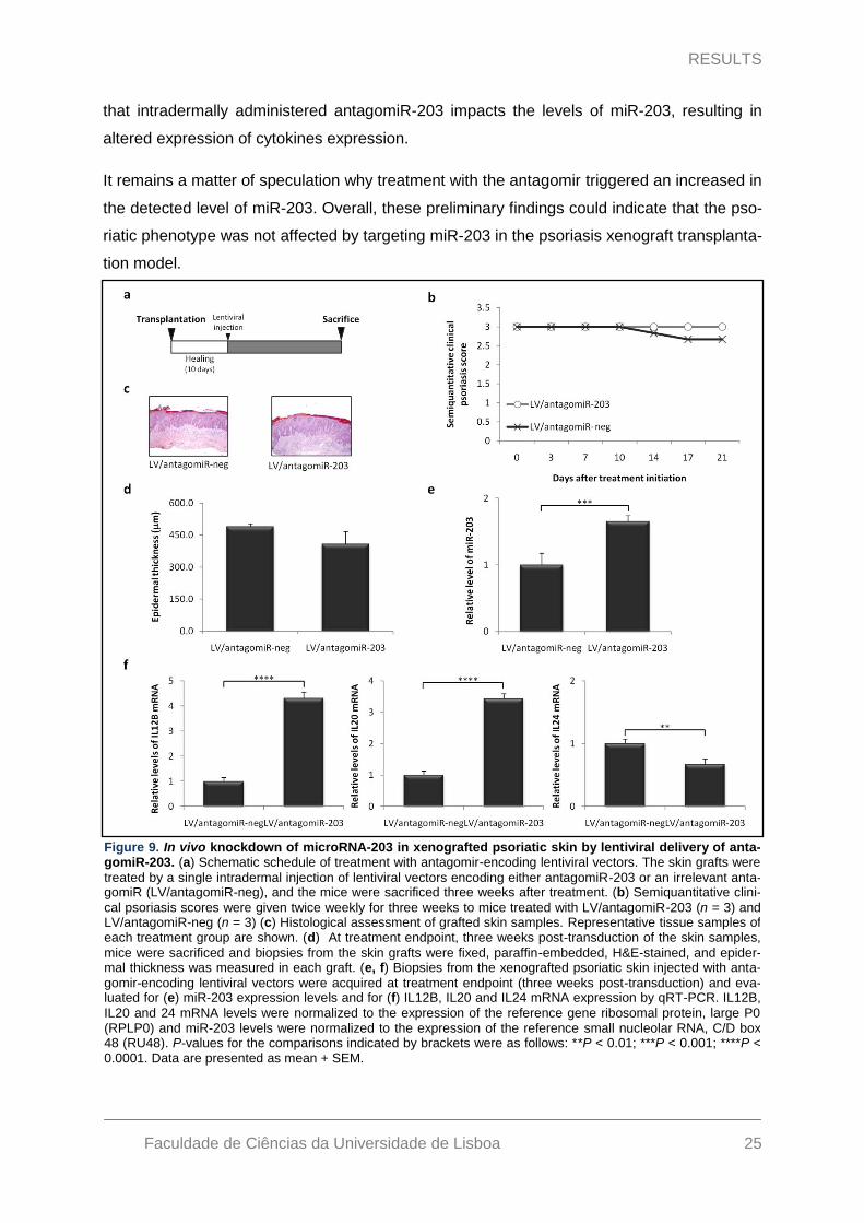
RESULTS
Faculdade de Ciências da Universidade de Lisboa 25
that intradermally administered antagomiR-203 impacts the levels of miR-203, resulting in
altered expression of cytokines expression.
It remains a matter of speculation why treatment with the antagomir triggered an increased in
the detected level of miR-203. Overall, these preliminary findings could indicate that the pso-
riatic phenotype was not affected by targeting miR-203 in the psoriasis xenograft transplanta-
tion model.
Figure 9. In vivo knockdown of microRNA-203 in xenografted psoriatic skin by lentiviral delivery of anta-gomiR-203. (a) Schematic schedule of treatment with antagomir-encoding lentiviral vectors. The skin grafts were
treated by a single intradermal injection of lentiviral vectors encoding either antagomiR-203 or an irrelevant anta-gomiR (LV/antagomiR-neg), and the mice were sacrificed three weeks after treatment. (b) Semiquantitative clini-
cal psoriasis scores were given twice weekly for three weeks to mice treated with LV/antagomiR-203 (n = 3) and LV/antagomiR-neg (n = 3) (c) Histological assessment of grafted skin samples. Representative tissue samples of each treatment group are shown. (d) At treatment endpoint, three weeks post-transduction of the skin samples,
mice were sacrificed and biopsies from the skin grafts were fixed, paraffin-embedded, H&E-stained, and epider-mal thickness was measured in each graft. (e, f) Biopsies from the xenografted psoriatic skin injected with anta-
gomir-encoding lentiviral vectors were acquired at treatment endpoint (three weeks post-transduction) and eva-luated for (e) miR-203 expression levels and for (f) IL12B, IL20 and IL24 mRNA expression by qRT-PCR. IL12B,
IL20 and 24 mRNA levels were normalized to the expression of the reference gene ribosomal protein, large P0 (RPLP0) and miR-203 levels were normalized to the expression of the reference small nucleolar RNA, C/D box 48 (RU48). P-values for the comparisons indicated by brackets were as follows: **P < 0.01; ***P < 0.001; ****P < 0.0001. Data are presented as mean + SEM.
DISCUSSION
Faculdade de Ciências da Universidade de Lisboa 26
Discussion
Jacob Mikkelsen research group has previously provided evidence of potent and persistent
in vivo knockdown of TNFα and IL12B mRNA transcripts following a single intradermal injec-
tion of lentiviral vectors into xenografted human skin 83; 84. These proof-of-concept studies
confirmed the therapeutic applicability of shRNA expression in skin and documented the po-
tential use of RNAi in the treatment of psoriasis. In the present study, we explored the cyto-
kine-directed shRNA-encoding lentiviral vector for design of vectors expressing antagomirs.
As a focus of these studies, we chose miR-203, which was recently identified as a miRNA
significantly overexpressed in psoriasis and which is exclusively expressed in keratinocytes
49. Whether miR-203 may contribute or not to increased skin inflammation in response to T-
cell derived-cytokines is still unknown, as miR-203 has only been shown to target SOCS3.
Therefore, an initial screening of a panel of psoriasis related-cytokines was necessary to
identify possible new miR-203 targets. Indeed, in the initial luciferase-based miR-203 screen
targeting six different cytokines and two protein suppressors of the cytokine signaling, two
cytokines (IL24 and TNFα) and SOCS6 were identified as potential miR-203 targets. Impor-
tantly, the mRNAs encoding the three different targets contained potential miR-203-binding
sites in their 3‟UTRs. To determine whether the regulation of the luciferase reporter gene
was mediated by the predicted target sites in 3‟UTR of the potential targets, we mutated the
seven nucleotides within the seed-matching sequences of the predicted target sites. Mutation
of the seed-matching sequences in each potential target gene led to complete restoration of
luciferase activity, confirming the direct interaction between the target genes and miR-203.
Nevertheless, variations in target accessibility in the fusion mRNA context could have influ-
enced RNAi activity and thus provide a false identification of a potential miRNA target. This
emphasized the need to validate miR-203 knockdown on native endogenously expressed
targets. Moving the experimental setup to endogenously expressed targets, we showed that
miR-203 activity toward the mentioned targets was maintained clearly demonstrating that
miR-203 directly regulates IL24, SOCS6 and TNFα mRNA transcripts in human keratino-
cytes.
On receptor-expressing keratinocyte cells, IL24 is known to activate STAT pathway, by
phosphorylation of STAT3 90; 91. In response to IL24, keratinocytes proliferate and express
pro-inflammatory genes including TNF-α, which leads to activation of NF-κB. Accordingly to
the mentioned studies, the data collected propose that miR-203 may play a role in the regu-
lation of NF-κB activity in human keratinocytes. As miR-203 targets IL24 and TNFα mRNA
transcripts in keratinocytes, IL24 and TNFα protein expressions will diminish, leading to inhi-
bition of NF-кB signaling pathway. Conversely, miR-203 also targets SOCS3 mRNA tran-
DISCUSSION
Faculdade de Ciências da Universidade de Lisboa 27
script, which is a negative regulator of STAT3 pathway, resulting in STAT3 activation and,
consequently, activation of NF-кB signaling pathway. According to the data collected here
and to our understanding of miR-203 targets, it is reasonable to assume that miR-203 may
have a meaning in NF-κB pathway in human keratinocytes, although further studies are still
relevant to confirm and clarify miR-203 significance on NF-κB pathway tight regulation.
Moreover, we generated two antagomir-expressing lentiviral constructs (antagomiR-neg and
antagomiR-203). Based on functional evaluations, we identified a solid upregulation of
Renilla luciferase gene fused to a full miR-203 complementary sequence when co-expressed
with either antagomiR-203-encoding plasmid or lentiviral vector. miR-203 knockdown was
confirmed when the antagomiR-203 was expressed from very few lentiviral integrations (MOI
of 5) and after extrachromosomal non-integrated lentiviral vectors have been lost. The
mechanism of oligonucleotide-mediated miRNA silencing is still unknown, although previous
data from other groups suggests that this process involves degradation of the miRNA in vitro
and in vivo 47; 89. Our collected data is partially in agreement with the mentioned hypothesis,
as we identified a clearly reduction of miR-203 expression levels in HaCaT naїve cells follow-
ing transduction of antagomiR-203-expressing lentiviral vector. Nonetheless, the same ex-
periment was developed in a HaCaT cell line stably overexpressing miR-203, and no reduc-
tion of miR-203 expressing levels was detected, despite the fact that a release of miR-203
repression on IL24 mRNA target was detected in this particular cell line. In this study, it is
important to take into account that antagomiR-203 sequence did not present any chemical
modifications, such as phosphorothioate modification or 2‟-O-methyl sugar modification
which protect oligonucleotides against exonuclease or endonuclease activity, respectively.
This may indicate that, when expressed endogenously, antagomirs and miRNAs may share
similar degradation pathways. Therefore, our results suggest that miRNA-target cell expres-
sion levels and antagomir chemical modifications are relevant parameters to take into ac-
count when designing and evaluating antagomir potency.
The psoriasis xenograft transplantation model currently appears to be the best tool to screen
anti-psoriatic therapeutic strategies in psoriasis before introducing them into the clinic 92. Us-
ing the psoriasis xenograft transplantation model, we aimed to evaluate if lentiviral delivery of
antagomiR-203 could decrease miR-203 levels and if this would have a beneficial effect on
the disease phenotype. Following a single, intradermally administered dose of lentiviral vec-
tor in human skin, we detected a 60% increase of miR-203 in antagomiR-203-treated skin
grafts compared to the negative control. Recent in vitro and in vivo investigations indicate
that miR-203 impacts epidermal proliferation predominantly by restricting the proliferative
potential of basal-cell precursors as they migrate from the basal layer to the suprabasal lay-
ers 57; 60. It is hypothesized that miR-203 might be absent or only expressed at low levels in

DISCUSSION
Faculdade de Ciências da Universidade de Lisboa 28
multipotent proliferating cells to enable p63-dependent epidermal commitment, whereas its
expression increases as the multipotent epidermal cells exit the proliferating basal layer and
terminally differentiate. These data indicate that it was rational to expect an increased epi-
dermal thickness following intradermal administration of antagomiR-203-expressing lentiviral
vectors in skin. Conversely, we did not identify any clinical and histological improvement of
psoriasis phenotype, as epidermal thickness and semiquantitative clinical psoriasis score
remained unchanged during the three-week observations. As we had tested antagomiR-203
functionality in HaCaT keratinocyte cell line before developing miR-203 knockdown study in
vivo, miR-203 upregulation in antagomiR-203-treated skin grafts was to a great extent an
unexpected result. For that reason, we decided to analyze the expression levels of IL24
mRNA transcripts, as we have previously demonstrated that miR-203 targets IL24 mRNA
transcript. In agreement with our previous results, miR-203 expression levels and IL24
mRNA transcripts were inversely related in antagomiR-203-treated skin grafts compared to
the negative control. This observation validated the result obtained from miR-203 quantifica-
tion in the skin grafts.
According to the data collected and to our understanding of miR-203 function, we might hy-
pothesize that miR-203 upregulation in antagomiR-203-treated skin samples was the out-
come of a negative feedback for regulation of miR-203 expression levels in cytokine-
activated keratinocytes. As antagomiR-203 potentially facilitates inhibition of miR-203 func-
tion in keratinocytes, p63 expression levels increase which lead to increased processing effi-
ciency of growth promoting pre-miRNAs, resulting in increased number of „transiently ampli-
fying-keratinocyte‟ cells. Since these proliferative keratinocytes are generated within an in-
flammatory microenvironment loaded of keratinocyte-released cytokines, it is reasonable to
assume that these new progenitor cells will develop into cytokine-activated keratinocytes, re-
establishing a psoriatic phenotype by acting on the crosstalk between keratinocytes and infil-
trating cells. This theory would, therefore, explain the fact that upregulation of miR-203,
IL12B and IL20 expression levels were detected in the skin grafts tested with antagomiR-
203, features characteristic from psoriatic lesions, which may have lead to no amelioration of
the psoriatic phenotype.
CONCLUDING REMARKS AND FUTURE PERSPECTIVES
Faculdade de Ciências da Universidade de Lisboa 29
Concluding remarks and future perspectives
In this study, we identified IL24, SOCS6 and TNFα as experimentally verified target genes for
miR-203 in human keratinocytes. Importantly, mRNAs encoded by IL24, SOCS6 and TNFα
have been associated with cytokine network as well as TNFα has previously been estab-
lished as a player in psoriasis pathogenesis, clearly demonstrating that miR-203 plays a role
in cytokine signaling network. Here, stable production of RNA antagomirs (v-antagomirs)
encoded by lentiviral vectors was also established as a method for persistently antagonizing
miRNA function in cultured cells and in mice. It is relevant to mention that this approach was
unique in exploring a vector-driven route of antagomir administration that allowed non-
systemic and tissue-specific antagomir delivery to increase safety. Nonetheless, attempted
knockdown of miR-203 in psoriasis xenograft transplantation model resulted in no ameliora-
tion of psoriasis phenotype, as evaluated by semi-quantitative clinical scoring. This study
clearly demonstrates that the role of increased miR-203 expression in psoriasis is complex
and further studies are still needed to comprehend miRNA functions in the cytokine network
as well as to establish whether targeting of miRNAs by RNAi-mediated degradation is or not
therapeutically relevant in psoriasis.
For the future, it is relevant to develop a similar setup as the one described above, in order to
(1) confirm potent and persistent antagomir expression following a single intradermal injec-
tion of lentiviral vectors in xenografted human skin and (2) confirm that miR-203 knockdown
occurs in psoriasis skin grafts treated with an antagomiR-203-encoding lentiviral vector. In
case the results demonstrated here are reproducible, it is pertinent to carry out immunohisto-
chemical staining for cell proliferation markers as well as for skin-homing T-cells in order to
understand in more detail the crosstalk between keratinocytes and infiltrating cells. As an
additional point, it has been recently validated that miR-125b targets STAT3 93. As miR-203
and miR-125b expression levels are deregulated in psoriasis, specifically in keratinocytes,
and both miRNAs are associated with STAT3 regulation pathway, I assume it would be perti-
nent to try to design a lentiviral construct allowing simultaneous expression of miR-125b and
knockdown of miR-203, for following experimentation in psoriasis xenograft transplantation
model. This study would be significant to understand how relevant miRNAs expressed in
keratinocytes are in the regulation of the cytokine network and in the pathogenesis of psoria-
sis, since STAT3 is involved in amplifying the inflammation process in psoriatic keratinocytes,
resulting in upregulation of several cytokines.
REFERENCES
Faculdade de Ciências da Universidade de Lisboa 30
References
1. Schon, M. P., Boehncke, W. H. & Brocker, E. B. (2005). Psoriasis: Clinical manifestations, pathogenesis and therapeutic perspectives. Discov Med 5, 253-8. 2. Gaspari, A. A. (2006). Innate and adaptive immunity and the pathophysiology of psoriasis. J Am Acad Dermatol 54, S67-80. 3. Nickoloff, B. J., Qin, J. Z. & Nestle, F. O. (2007). Immunopathogenesis of psoriasis. Clin Rev Allergy Immunol 33, 45-56. 4. Lew, W., Bowcock, A. M. & Krueger, J. G. (2004). Psoriasis vulgaris: cutaneous lymphoid tissue supports T-cell activation and "Type 1" inflammatory gene expression. Trends Immunol 25, 295-305. 5. Liu, Y., Krueger, J. G. & Bowcock, A. M. (2007). Psoriasis: genetic associations and immune system changes. Genes Immun 8, 1-12. 6. Wang, F., Lee, E., Lowes, M. A., Haider, A. S., Fuentes-Duculan, J., Abello, M. V., Chamian, F., Cardinale, I. & Krueger, J. G. (2006). Prominent production of IL-20 by CD68+/CD11c+ myeloid-derived cells in psoriasis: Gene regulation and cellular effects. J Invest Dermatol 126, 1590-9. 7. Wolk, K., Kunz, S., Witte, E., Friedrich, M., Asadullah, K. & Sabat, R. (2004). IL-22 increases the innate immunity of tissues. Immunity 21, 241-54. 8. Krueger, J. G. (2002). The immunologic basis for the treatment of psoriasis with new biologic agents. J Am Acad Dermatol 46, 1-23; quiz 23-6. 9. Lagos-Quintana, M., Rauhut, R., Lendeckel, W. & Tuschl, T. (2001). Identification of novel genes coding for small expressed RNAs. Science 294, 853-8. 10. Bartel, D. P. (2004). MicroRNAs: genomics, biogenesis, mechanism, and function. Cell 116, 281-97. 11. Lewis, B. P., Burge, C. B. & Bartel, D. P. (2005). Conserved seed pairing, often flanked by adenosines, indicates that thousands of human genes are microRNA targets. Cell 120, 15-20. 12. Hutvagner, G. & Zamore, P. D. (2002). A microRNA in a multiple-turnover RNAi enzyme complex. Science 297, 2056-60. 13. Liu, J., Carmell, M. A., Rivas, F. V., Marsden, C. G., Thomson, J. M., Song, J. J., Hammond, S. M., Joshua-Tor, L. & Hannon, G. J. (2004). Argonaute2 is the catalytic engine of mammalian RNAi. Science 305, 1437-41. 14. Meister, G., Landthaler, M., Patkaniowska, A., Dorsett, Y., Teng, G. & Tuschl, T. (2004). Human Argonaute2 mediates RNA cleavage targeted by miRNAs and siRNAs. Mol Cell 15, 185-97. 15. Grimson, A., Farh, K. K., Johnston, W. K., Garrett-Engele, P., Lim, L. P. & Bartel, D. P. (2007). MicroRNA targeting specificity in mammals: determinants beyond seed pairing. Mol Cell 27, 91-105. 16. Shin, C., Nam, J. W., Farh, K. K., Chiang, H. R., Shkumatava, A. & Bartel, D. P. (2010). Expanding the microRNA targeting code: functional sites with centered pairing. Mol Cell 38, 789-802. 17. Seggerson, K., Tang, L. & Moss, E. G. (2002). Two genetic circuits repress the Caenorhabditis elegans heterochronic gene lin-28 after translation initiation. Dev Biol 243, 215-25. 18. Olsen, P. H. & Ambros, V. (1999). The lin-4 regulatory RNA controls developmental timing in Caenorhabditis elegans by blocking LIN-14 protein synthesis after the initiation of translation. Dev Biol 216, 671-80. 19. Guo, H., Ingolia, N. T., Weissman, J. S. & Bartel, D. P. (2010). Mammalian microRNAs predominantly act to decrease target mRNA levels. Nature 466, 835-40. 20. Miyoshi, K., Okada, T. N., Siomi, H. & Siomi, M. C. (2009). Characterization of the miRNA-RISC loading complex and miRNA-RISC formed in the Drosophila miRNA pathway. RNA 15, 1282-91. 21. Lee, Y., Kim, M., Han, J., Yeom, K. H., Lee, S., Baek, S. H. & Kim, V. N. (2004). MicroRNA genes are transcribed by RNA polymerase II. EMBO J 23, 4051-60. 22. Cai, X., Hagedorn, C. H. & Cullen, B. R. (2004). Human microRNAs are processed from capped, polyadenylated transcripts that can also function as mRNAs. Rna 10, 1957-66. 23. Borchert, G. M., Lanier, W. & Davidson, B. L. (2006). RNA polymerase III transcribes human microRNAs. Nat Struct Mol Biol 13, 1097-101. 24. Han, J., Lee, Y., Yeom, K. H., Kim, Y. K., Jin, H. & Kim, V. N. (2004). The Drosha-DGCR8 complex in primary microRNA processing. Genes Dev 18, 3016-27. 25. Denli, A. M., Tops, B. B., Plasterk, R. H., Ketting, R. F. & Hannon, G. J. (2004). Processing of primary microRNAs by the Microprocessor complex. Nature 432, 231-5. 26. Gregory, R. I., Yan, K. P., Amuthan, G., Chendrimada, T., Doratotaj, B., Cooch, N. & Shiekhattar, R. (2004). The Microprocessor complex mediates the genesis of microRNAs. Nature 432, 235-40.
REFERENCES
Faculdade de Ciências da Universidade de Lisboa 31
27. Han, J., Lee, Y., Yeom, K. H., Nam, J. W., Heo, I., Rhee, J. K., Sohn, S. Y., Cho, Y., Zhang, B. T. & Kim, V. N. (2006). Molecular basis for the recognition of primary microRNAs by the Drosha-DGCR8 complex. Cell 125, 887-901. 28. Lee, Y., Ahn, C., Han, J., Choi, H., Kim, J., Yim, J., Lee, J., Provost, P., Radmark, O., Kim, S. & Kim, V. N. (2003). The nuclear RNase III Drosha initiates microRNA processing. Nature 425, 415-9. 29. Lee, Y., Jeon, K., Lee, J. T., Kim, S. & Kim, V. N. (2002). MicroRNA maturation: stepwise processing and subcellular localization. EMBO J 21, 4663-70. 30. Krol, J., Loedige, I. & Filipowicz, W. (2010). The widespread regulation of microRNA biogenesis, function and decay. Nat Rev Genet 11, 597-610. 31. Okamura, K., Hagen, J. W., Duan, H., Tyler, D. M. & Lai, E. C. (2007). The mirtron pathway generates microRNA-class regulatory RNAs in Drosophila. Cell 130, 89-100. 32. Berezikov, E., Chung, W. J., Willis, J., Cuppen, E. & Lai, E. C. (2007). Mammalian mirtron genes. Mol Cell 28, 328-36. 33. Lund, E., Guttinger, S., Calado, A., Dahlberg, J. E. & Kutay, U. (2004). Nuclear export of microRNA precursors. Science 303, 95-8. 34. Bohnsack, M. T., Czaplinski, K. & Gorlich, D. (2004). Exportin 5 is a RanGTP-dependent dsRNA-binding protein that mediates nuclear export of pre-miRNAs. Rna 10, 185-91. 35. Yi, R., Qin, Y., Macara, I. G. & Cullen, B. R. (2003). Exportin-5 mediates the nuclear export of pre-microRNAs and short hairpin RNAs. Genes Dev 17, 3011-6. 36. Kim, V. N. (2004). MicroRNA precursors in motion: exportin-5 mediates their nuclear export. Trends Cell Biol 14, 156-9. 37. Bernstein, E., Caudy, A. A., Hammond, S. M. & Hannon, G. J. (2001). Role for a bidentate ribonuclease in the initiation step of RNA interference. Nature 409, 363-6. 38. Hutvagner, G., McLachlan, J., Pasquinelli, A. E., Balint, E., Tuschl, T. & Zamore, P. D. (2001). A cellular function for the RNA-interference enzyme Dicer in the maturation of the let-7 small temporal RNA. Science 293, 834-8. 39. Ketting, R. F., Fischer, S. E., Bernstein, E., Sijen, T., Hannon, G. J. & Plasterk, R. H. (2001). Dicer functions in RNA interference and in synthesis of small RNA involved in developmental timing in C. elegans. Genes Dev 15, 2654-9. 40. Gregory, R. I., Chendrimada, T. P., Cooch, N. & Shiekhattar, R. (2005). Human RISC couples microRNA biogenesis and posttranscriptional gene silencing. Cell 123, 631-40. 41. Haase, A. D., Jaskiewicz, L., Zhang, H., Laine, S., Sack, R., Gatignol, A. & Filipowicz, W. (2005). TRBP, a regulator of cellular PKR and HIV-1 virus expression, interacts with Dicer and functions in RNA silencing. EMBO Rep 6, 961-7. 42. Lee, Y., Hur, I., Park, S. Y., Kim, Y. K., Suh, M. R. & Kim, V. N. (2006). The role of PACT in the RNA silencing pathway. EMBO J 25, 522-32. 43. MacRae, I. J., Ma, E., Zhou, M., Robinson, C. V. & Doudna, J. A. (2008). In vitro reconstitution of the human RISC-loading complex. Proc Natl Acad Sci U S A 105, 512-7. 44. Khvorova, A., Reynolds, A. & Jayasena, S. D. (2003). Functional siRNAs and miRNAs exhibit strand bias. Cell 115, 209-16. 45. Fire, A., Xu, S., Montgomery, M. K., Kostas, S. A., Driver, S. E. & Mello, C. C. (1998). Potent and specific genetic interference by double-stranded RNA in Caenorhabditis elegans. Nature 391, 806-11. 46. Schwarz, D. S., Hutvagner, G., Du, T., Xu, Z., Aronin, N. & Zamore, P. D. (2003). Asymmetry in the assembly of the RNAi enzyme complex. Cell 115, 199-208. 47. Krutzfeldt, J., Rajewsky, N., Braich, R., Rajeev, K. G., Tuschl, T., Manoharan, M. & Stoffel, M. (2005). Silencing of microRNAs in vivo with 'antagomirs'. Nature 438, 685-9. 48. Ebert, M. S., Neilson, J. R. & Sharp, P. A. (2007). MicroRNA sponges: competitive inhibitors of small RNAs in mammalian cells. Nat Methods 4, 721-6. 49. Sonkoly, E., Wei, T., Janson, P. C., Saaf, A., Lundeberg, L., Tengvall-Linder, M., Norstedt, G., Alenius, H., Homey, B., Scheynius, A., Stahle, M. & Pivarcsi, A. (2007). MicroRNAs: novel regulators involved in the pathogenesis of psoriasis? PLoS ONE 2, e610. 50. Taganov, K. D. (2006). NF- B-dependent induction of microRNA miR-146, an inhibitor targeted to signaling proteins of innate immune responses. Proceedings of the National Academy of Sciences 103, 12481-12486. 51. Sheedy, F. J., Palsson-McDermott, E., Hennessy, E. J., Martin, C., O'Leary, J. J., Ruan, Q., Johnson, D. S., Chen, Y. & O'Neill, L. A. J. (2009). Negative regulation of TLR4 via targeting of the proinflammatory tumor suppressor PDCD4 by the microRNA miR-21. Nature Immunology 11, 141-147.

REFERENCES
Faculdade de Ciências da Universidade de Lisboa 32
52. Recchiuti, A., Krishnamoorthy, S., Fredman, G., Chiang, N. & Serhan, C. N. (2010). MicroRNAs in resolution of acute inflammation: identification of novel resolvin D1-miRNA circuits. The FASEB Journal 25, 544-560. 53. Tili, E., Michaille, J. J., Cimino, A., Costinean, S., Dumitru, C. D., Adair, B., Fabbri, M., Alder, H., Liu, C. G., Calin, G. A. & Croce, C. M. (2007). Modulation of miR-155 and miR-125b levels following lipopolysaccharide/TNF-alpha stimulation and their possible roles in regulating the response to endotoxin shock. J Immunol 179, 5082-9. 54. Zhang, Y., Gao, J.-S., Tang, X., Tucker, L. D., Quesenberry, P., Rigoutsos, I. & Ramratnam, B. (2009). MicroRNA 125a and its regulation of the p53 tumor suppressor gene. FEBS Letters 583, 3725-3730. 55. Le, M. T., Teh, C., Shyh-Chang, N., Xie, H., Zhou, B., Korzh, V., Lodish, H. F. & Lim, B. (2009). MicroRNA-125b is a novel negative regulator of p53. Genes Dev 23, 862-76. 56. Xu, N., Brodin, P., Wei, T., Meisgen, F., Eidsmo, L., Nagy, N., Kemeny, L., Ståhle, M., Sonkoly, E. & Pivarcsi, A. (2011). MiR-125b, a MicroRNA Downregulated in Psoriasis, Modulates Keratinocyte Proliferation by Targeting FGFR2. Journal of Investigative Dermatology 131, 1521-1529. 57. Yi, R., Poy, M. N., Stoffel, M. & Fuchs, E. (2008). A skin microRNA promotes differentiation by repressing „stemness‟. Nature 452, 225-229. 58. Nissan, X., Denis, J. A., Saidani, M., Lemaitre, G., Peschanski, M. & Baldeschi, C. (2011). miR-203 modulates epithelial differentiation of human embryonic stem cells towards epidermal stratification. Developmental Biology. 59. Wei, T., Orfanidis, K., Xu, N., Janson, P., Ståhle, M., Pivarcsi, A. & Sonkoly, E. (2010). The expression of microRNA-203 during human skin morphogenesis. Experimental Dermatology 19, 854-856. 60. Lena, A. M., Shalom-Feuerstein, R., di Val Cervo, P. R., Aberdam, D., Knight, R. A., Melino, G. & Candi, E. (2008). miR-203 represses „stemness‟ by repressing ΔNp63. Cell Death and Differentiation 15, 1187-1195. 61. Senoo, M., Pinto, F., Crum, C. P. & McKeon, F. (2007). p63 Is Essential for the Proliferative Potential of Stem Cells in Stratified Epithelia. Cell 129, 523-536. 62. Croker, B. A., Krebs, D. L., Zhang, J. G., Wormald, S., Willson, T. A., Stanley, E. G., Robb, L., Greenhalgh, C. J., Forster, I., Clausen, B. E., Nicola, N. A., Metcalf, D., Hilton, D. J., Roberts, A. W. & Alexander, W. S. (2003). SOCS3 negatively regulates IL-6 signaling in vivo. Nat Immunol 4, 540-5. 63. Sano, S., Chan, K. S. & DiGiovanni, J. (2008). Impact of Stat3 activation upon skin biology: A dichotomy of its role between homeostasis and diseases. Journal of Dermatological Science 50, 1-14. 64. Capogrossi, M. C., Lerman, G., Avivi, C., Mardoukh, C., Barzilai, A., Tessone, A., Gradus, B., Pavlotsky, F., Barshack, I., Polak-Charcon, S., Orenstein, A., Hornstein, E., Sidi, Y. & Avni, D. (2011). MiRNA Expression in Psoriatic Skin: Reciprocal Regulation of hsa-miR-99a and IGF-1R. PLoS ONE 6, e20916. 65. Zibert, J. R., Løvendorf, M. B., Litman, T., Olsen, J., Kaczkowski, B. & Skov, L. (2010). MicroRNAs and potential target interactions in psoriasis. Journal of Dermatological Science 58, 177-185. 66. Ichihara, A., Jinnin, M., Yamane, K., Fujisawa, A., Sakai, K., Masuguchi, S., Fukushima, S., Maruo, K. & Ihn, H. (2011). microRNA-mediated keratinocyte hyperproliferation in psoriasis vulgaris. Br J Dermatol. 67. Oyama, R., Jinnin, M., Kakimoto, A., Kanemaru, H., Ichihara, A., Fujisawa, A., Honda, N., Masuguchi, S., Fukushima, S., Maruo, K. & Ihn, H. (2011). Circulating microRNA associated with TNF-α signaling pathway in patients with plaque psoriasis. Journal of Dermatological Science 61, 209-211. 68. Khavari, P. A., Rollman, O. & Vahlquist, A. (2002). Cutaneous gene transfer for skin and systemic diseases. J Intern Med 252, 1-10. 69. Ghazizadeh, S., Harrington, R. & Taichman, L. (1999). In vivo transduction of mouse epidermis with recombinant retroviral vectors: implications for cutaneous gene therapy. Gene Ther 6, 1267-75. 70. Bukrinsky, M. I., Sharova, N., Dempsey, M. P., Stanwick, T. L., Bukrinskaya, A. G., Haggerty, S. & Stevenson, M. (1992). Active nuclear import of human immunodeficiency virus type 1 preintegration complexes. Proc Natl Acad Sci U S A 89, 6580-4. 71. Dull, T., Zufferey, R., Kelly, M., Mandel, R. J., Nguyen, M., Trono, D. & Naldini, L. (1998). A third-generation lentivirus vector with a conditional packaging system. J Virol 72, 8463-71. 72. Blomer, U., Naldini, L., Kafri, T., Trono, D., Verma, I. M. & Gage, F. H. (1997). Highly efficient and sustained gene transfer in adult neurons with a lentivirus vector. J Virol 71, 6641-9. 73. Naldini, L. (1998). Lentiviruses as gene transfer agents for delivery to non-dividing cells. Curr Opin Biotechnol 9, 457-63.
REFERENCES
Faculdade de Ciências da Universidade de Lisboa 33
74. Naldini, L., Blomer, U., Gage, F. H., Trono, D. & Verma, I. M. (1996). Efficient transfer, integration, and sustained long-term expression of the transgene in adult rat brains injected with a lentiviral vector. Proc Natl Acad Sci U S A 93, 11382-8. 75. Naldini, L., Blomer, U., Gallay, P., Ory, D., Mulligan, R., Gage, F. H., Verma, I. M. & Trono, D. (1996). In vivo gene delivery and stable transduction of nondividing cells by a lentiviral vector. Science 272, 263-7. 76. Zufferey, R., Dull, T., Mandel, R. J., Bukovsky, A., Quiroz, D., Naldini, L. & Trono, D. (1998). Self-inactivating lentivirus vector for safe and efficient in vivo gene delivery. J Virol 72, 9873-80. 77. Baek, S. C., Lin, Q., Robbins, P. B., Fan, H. & Khavari, P. A. (2001). Sustainable systemic delivery via a single injection of lentivirus into human skin tissue. Hum Gene Ther 12, 1551-8. 78. Marchetto, M. C. N., Correa, R. G., Menck, C. F. M. & Muotri, A. R. (2006). Functional lentiviral vectors for xeroderma pigmentosum gene therapy. Journal of Biotechnology 126, 424-430. 79. Chen, M., Kasahara, N., Keene, D. R., Chan, L., Hoeffler, W. K., Finlay, D., Barcova, M., Cannon, P. M., Mazurek, C. & Woodley, D. T. (2002). Restoration of type VII collagen expression and function in dystrophic epidermolysis bullosa. Nature Genetics 32, 670-675. 80. Di Nunzio, F., Maruggi, G., Ferrari, S., Di Iorio, E., Poletti, V., Garcia, M., Del Rio, M., De Luca, M., Larcher, F., Pellegrini, G. & Mavilio, F. (2008). Correction of Laminin-5 Deficiency in Human Epidermal Stem Cells by Transcriptionally Targeted Lentiviral Vectors. Molecular Therapy 16, 1977-1985. 81. Woodley, D. (2004). Intradermal Injection of Lentiviral Vectors Corrects Regenerated Human Dystrophic Epidermolysis Bullosa Skin Tissue in Vivo. Molecular Therapy 10, 318-326. 82. Sumimoto, H., Hirata, K., Yamagata, S., Miyoshi, H., Miyagishi, M., Taira, K. & Kawakami, Y. (2006). Effective inhibition of cell growth and invasion of melanoma by combined suppression of BRAF (V599E) and Skp2 with lentiviral RNAi. International Journal of Cancer 118, 472-476. 83. Bak, R. O., Stenderup, K., Rosada, C., Petersen, L. B., Moldt, B., Dagnæs-Hansen, F., Jakobsen, M., Kamp, S., Jensen, T. G., Dam, T. N. & Mikkelsen, J. (2011). Targeting of human interleukin-12B by small hairpin RNAs in xenografted psoriatic skin. BMC Dermatology 11, 5. 84. Jakobsen, M., Stenderup, K., Rosada, C., Moldt, B., Kamp, S., Dam, T. N., Jensen, T. G. & Mikkelsen, J. G. (2009). Amelioration of Psoriasis by Anti-TNF-α RNAi in the Xenograft Transplantation Model. Molecular Therapy 17, 1743-1753. 85. Anderson, P. (2008). Post-transcriptional control of cytokine production. Nat Immunol 9, 353-9. 86. Scherr, M., Venturini, L., Battmer, K., Schaller-Schoenitz, M., Schaefer, D., Dallmann, I., Ganser, A. & Eder, M. (2007). Lentivirus-mediated antagomir expression for specific inhibition of miRNA function. Nucleic Acids Res 35, e149. 87. Stenderup, K., Rosada, C., Worsaae, A., Dagnaes-Hansen, F., Steiniche, T., Hasselager, E., Iversen, L. F., Zahn, S., Woldike, H., Holmberg, H. L., Romer, J., Kragballe, K., Clausen, J. T. & Dam, T. N. (2009). Interleukin-20 plays a critical role in maintenance and development of psoriasis in the human xenograft transplantation model. Br J Dermatol 160, 284-96. 88. Ivics, Z., Hackett, P. B., Plasterk, R. H. & Izsvak, Z. (1997). Molecular reconstruction of Sleeping Beauty, a Tc1-like transposon from fish, and its transposition in human cells. Cell 91, 501-10. 89. Krutzfeldt, J., Kuwajima, S., Braich, R., Rajeev, K. G., Pena, J., Tuschl, T., Manoharan, M. & Stoffel, M. (2007). Specificity, duplex degradation and subcellular localization of antagomirs. Nucleic Acids Res 35, 2885-92. 90. Dumoutier, L. & Renauld, J. C. (2002). Viral and cellular interleukin-10 (IL-10)-related cytokines: from structures to functions. Eur Cytokine Netw 13, 5-15. 91. Wang, M., Tan, Z., Zhang, R., Kotenko, S. V. & Liang, P. (2002). Interleukin 24 (MDA-7/MOB-5) signals through two heterodimeric receptors, IL-22R1/IL-20R2 and IL-20R1/IL-20R2. J Biol Chem 277, 7341-7. 92. Boehncke, W. H. (2005). The psoriasis SCID mouse model: a tool for drug discovery? Ernst Schering Res Found Workshop, 213-34. 93. Surdziel, E., Cabanski, M., Dallmann, I., Lyszkiewicz, M., Krueger, A., Ganser, A., Scherr, M. & Eder, M. (2011). Enforced expression of miR-125b affects myelopoiesis by targeting multiple signaling pathways. Blood 117, 4338-48.

APPENDIX

Appendix I. Oligonucleotide sequences of the cloned transgenes.
Table 1. Oligonucleotide sequences and predicted transcripts for antagomiR-203 and antagomiR-Negative control.
Name Sequence (5’ - 3’)
FO-antagomiR-203 CTAGCCC CTAGTGGTCCTAAACATTTCAC TTTTTGGAAA RO-antagomiR-203 CGCGTTTCCAAAAA GTGAAATGTTTAGGACCACTAG GGG antagomiR-203 CUAGUGGUCCUAAACAUUUCAC UU FO-antagomiR-neg CTAGCCC GTGTAACACGTCTATACGCCCA TTTTTGGAAA RO-antagomiR-neg CGCGTTTCCAAAAA TGGGCGTATAGACGTGTTACAC GGG antagomiR-neg* GUGUAACACGUCUAUACGCCCA UU
* Sequence from Exiqon‟s miRCURY LNA™ Knockdown Control Probe.
Table 2. Sequence information about the target genes cloned into psiCHECK2 vector.
Gene
NCBI Reference
First Position (bp)
Last Position(bp)
Length (bp)
Characteristics
IL12B NM_002187.2 12 2347 2336 ~ Complete transcript IL15 NM_000585.4 864 1448 585 3‟ UTR IL17A NM_002190.2 511 1774 1264 3‟ UTR IL20 NM_018724.3 576 1210 635 3‟ UTR IL24 NM_006850.3 695 1202 508 3‟ UTR p63 NM_003722.4 3854 4372 519 3‟ UTR SOCS3 NM_003955.3 2253 2720 468 3‟ UTR SOCS6 NM_004232.3 2010 2504 495 3‟ UTR TNFα NM_000594.2 89 1600 1512 ~ Complete transcript
Table 3. Primer sequences of the target genes cloned into psiCHECK2 vector.
Gene Forward Primer (5’ – 3’) Reverse Primer (5’ – 3’)
IL12B CCATTGGACTCTCCGTCCTG CTAATGTTTCTTCTCAAAAA IL15 TTGCAATTGATTCTTTTTAA GTATTGTCAACGAAATATGT IL17A TAAGAGCTCTGGGGAGCCCA GAGTATGAATTTCAAGCAAG IL20 GAGGAAAGTGATGCTGCTGC CATCTTTATGTACGACCGGC IL24 GGCCAACAACTTTGTTCTCA CGGAAGTACATTGAATGTCA p63 GTGGATTGCCTCTGAAAAGT CCGATCACGGTTTAGGTACT SOCS3 CTGGAGGTCATTGGAGAGGC TATCACGAGAAATAATATTT SOCS6 CTTTTGCTGCCATAACTATT TTCTATACAACGTCTAGAGG TNFα CCCTGACAAGCTGCCAGGCA CTCGTCTCCGAGTCGTTACT
Table 4. Oligonucleotide sequences and predicted transcripts for psoriasis related-miRNA-perfect targets.
Name Sequence (5’ - 3’)
FO-miRNA-125b target TCGAGTAA TCACAAGTTAGGGTCTCAGGGA AGG RO-miRNA-125b target GGCCCCT TCCCTGAGACCCTAACTTGTGA TTAC miRNA-125b target UAAUCACAAGUUAGGGUCUCAGGGAAGG FO-miRNA-203 target TCGAGGGT CTAGTGGTCCTAAACATTTCAC AAT RO-miRNA-203 target GGCCATT GTGAAATGTTTAGGACCACTAG ACCC miRNA-203 target GGUCUAGUGGUCCUAAACAUUUCACAAU
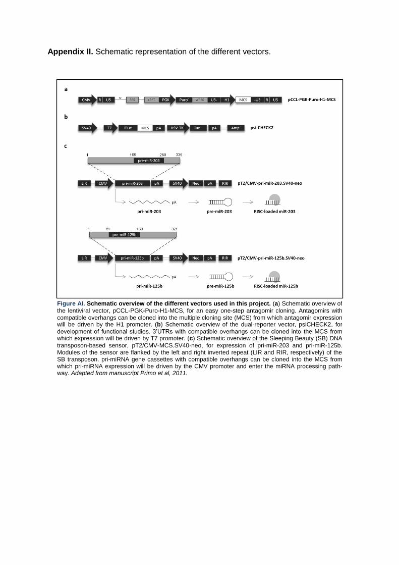
Appendix II. Schematic representation of the different vectors.
Figure AI. Schematic overview of the different vectors used in this project. (a) Schematic overview of
the lentiviral vector, pCCL-PGK-Puro-H1-MCS, for an easy one-step antagomir cloning. Antagomirs with compatible overhangs can be cloned into the multiple cloning site (MCS) from which antagomir expression will be driven by the H1 promoter. (b) Schematic overview of the dual-reporter vector, psiCHECK2, for
development of functional studies. 3‟UTRs with compatible overhangs can be cloned into the MCS from which expression will be driven by T7 promoter. (c) Schematic overview of the Sleeping Beauty (SB) DNA
transposon-based sensor, pT2/CMV-MCS.SV40-neo, for expression of pri-miR-203 and pri-miR-125b. Modules of the sensor are flanked by the left and right inverted repeat (LIR and RIR, respectively) of the SB transposon. pri-miRNA gene cassettes with compatible overhangs can be cloned into the MCS from which pri-miRNA expression will be driven by the CMV promoter and enter the miRNA processing path-way. Adapted from manuscript Primo et al, 2011.

Appendix III. Potential miR-203-binding sites in IL24 3‟UTR sequence.
IL24 Sequence Modification
IL24 3’UTR Sequence
1 GGCCAACAACTTTGTTCTCATCGTGTCACAACTGCAACCCAGTCAAGAAA
51 ATGAGATGTTTTCCATCAGAGACAGTGCACACAGGCGGTTTCTGCTATTC
101 CGGAGAGCATTCAAACAGTTGGACGTAGAAGCAGCTCTGACCAAAGCCCT
151 TGGGGAAGTGGACATTCTTCTGACCTGGATGCAGAAATTCTACAAGCTCT
201 GAATGTCTAGACCAGGACCTCCCTCCCCCTGGCACTGGTTTGTTCCCTGT
251 GTCATTTCAAACAGTCTCCCTTCCTATGCTGTTCACTGGACACTTCACGC
301 CCTTGGCCATGGGTCCCATTCTTGGCCCAGGATTATTGTCAAAGAAGTCA
351 TTCTTTAAGCAGCGCCAGTGACAGTCAGGGAAGGTGCCTCTGGATGCTGT
401 GAAGAGTCTACAGAGAAGATTCTTGTATTTATTACAACTCTATTTAATTA
451 ATGTCAGTATTTCAACTGAAGTTCTATTTATTTGTGAGACTGTAAGTTAC
501 ATGAAGGC
IL24-mut 3’UTR Sequence
1 GGCCAACAACTTTGTTCTCATCGTGTCACAACTGCAACCCAGTCAAGAAA
51 ATGAGACACAAAACATCAGAGACAGTGCACACAGGCGGTTTCTGCTATTC
101 CGGAGAGCATTCAAACAGTTGGACGTAGAAGCAGCTCTGACCAAAGCCCT
151 TGGGGAAGTGGACATTCTTCTGACCTGGATGCAGAAATTCTACAAGCTCT
201 GAATGTCTAGACCAGGACCTCCCTCCCCCTGGCACTGGTTTGTTCCCTGT
251 GTCACAAAAAACAGTCTCCCTTCCTATGCTGTTCACTGGACACAAAACGC
301 CCTTGGCCATGGGTCCCATTCTTGGCCCAGGATTATTGTCAAAGAAGTCA
351 TTCTTTAAGCAGCGCCAGTGACAGTCAGGGAAGGTGCCTCTGGATGCTGT
401 GAAGAGTCTACAGAGAAGATTCTTGTATTTATTACAACTCTATTTAATTA
451 ATGTCAGCACAAAAACTGAAGTTCTATTTATTTGTGAGACTGTAAGTTAC
501 ATGAAGGC
Figure AII. Schematic representation of the potential miRNA-203 binding sites in IL24 3’UTR se-quence. Nucleotide resolution of the predicted target sites in IL24 naїve (IL24 target) and IL24-mutant (IL24-mut target) 3‟UTR sequences by miR-203. Mutated seed match sequence in red letters.

Appendix IV. Potential miR-203-binding sites in SOCS6 3‟UTR sequence.
SOCS6 Sequence Modification
SOCS6 3’UTR Sequence
1 CTTTTGCTGCCATAACTATTTCAGTTTTATGTGTAAAAGAGTCATCAGTT
51 TGTTTAGGGGTGGGGAAGTGTCAGCAAGGTGTCTTGGGTTTATTTTGGTT
101 CTTTAAAAAAGGGAAGTCTTGAGGTTTTAGAGGTGTGAATTATGTTTCAT
151 CAATGTGCAGAATAATCACAATGTGAATTATCAAATTCTCCTCAATGCCC
201 CCCCCGCCCAGTCCTTTGCTGCTATCCACTGTGATTTTTATGCATTAAAA
251 GCACATTTCATGTGTATTCAACCCTAAGTAAAGTTGAATGAAACTTAACA
301 GAATGGAAATTGCTATGTCTTTTTAAATGGTCCATTTTCAAAAGACAGTG
351 TTGAATAAACATACCTGTGTGATAAAACACAGAATTTACATATACACTGA
401 AGATGAGTTTTTAATCTCTTACTTTAAAAAGATTTATTTAGAATCGTGAA
451 TTGACATAATCTTGGGTAATGGAACGGAGATCTGCAACATATCTT
SOCS6-mut 3’UTR Sequence
1 CTTTTGCTGCCATAACCACAAAACACAAATGTGTAAAAGAGTCATCAGTT
51 TGTTTAGGGGTGGGGAAGTGTCAGCAAGGTGTCTTGGGTTTATTTTGGTT
101 CTTTAAAAAAGGGAAGTCTTGAGGTTTTAGAGGTGTGAATTACACAAAAT
151 CAATGTGCAGAATAATCACAATGTGAATTATCAAATTCTCCTCAATGCCC
201 CCCCCGCCCAGTCCTTTGCTGCTATCCACTGTGATTTTTATGCATTAAAA
251 GCACACAAAATGTGTATTCAACCCTAAGTAAAGTTGAATGAAACTTAACA
301 GAATGGAAATTGCTATGTCTTTTTAAATGGTCCATTTTCAAAAGACAGTG
351 TTGAATAAACATACCTGTGTGATAAAACACAGAATTTACATATACACTGA
401 AGATGAGTTTTTAATCTCTTACTTTAAAAAGATTTATTTAGAATCGTGAA
451 TTGACATAATCTTGGGTAATGGAACGGAGATCTGCAACATATCTT
Figure AIII. Schematic representation of the potential miRNA-203 binding sites in SOCS6 3’UTR sequence. Nucleotide resolution of the predicted target sites in SOCS6 naїve (SOCS6 target) and
SOCS6-mutant (SOCS6-mut target) 3‟UTR sequences by miR-203. Mutated seed match sequence in red letters.

Appendix V. Potential miR-203-binding sites in TNFα 3‟UTR sequence.
TNFα Sequence Modification
TNFα 3’UTR Sequence
1 GGAGGACGAACATCCAACCTTCCCAAACGCCTCCCCTGCCCCAATCCCTT
51 TATTACCCCCTCCTTCAGACACCCTCAACCTCTTCTGGCTCAAAAAGAGA
101 ATTGGGGGCTTAGGGTCGGAACCCAAGCTTAGAACTTTAAGCAACAAGAC
151 CACCACTTCGAAACCTGGGATTCAGGAATGTGTGGCCTGCACAGTGAAGT
201 GCTGGCAACCACTAAGAATTCAAACTGGGGCCTCCAGAACTCACTGGGGC
251 CTACAGCTTTGATCCCTGACATCTGGAATCTGGAGACCAGGGAGCCTTTG
301 GTTCTGGCCAGAATGCTGCAGGACTTGAGAAGACCTCACCTAGAAATTGA
351 CACAAGTGGACCTTAGGCCTTCCTCTCTCCAGATGTTTCCAGACTTCCTT
401 GAGACACGGAGCCCAGCCCTCCCCATGGAGCCAGCTCCCTCTATTTATGT
451 TTGCACTTGTGATTATTTATTATTTATTTATTATTTATTTATTTACAGAT
501 GAATGTATTTATTTGGGAGACCGGGGTATCCTGGGGGACCCAATGTAGGA
551 GCTGCCTTGGCTCAGACATGTTTTCCGTGAAAACGGAGCTGAACAATAGG
601 CTGTTCCCATGTAGCCCCCTGGCCTCTGTGCCTTCTTTTGATTATGTTTT
651 TTAAAATATTTATCTGATTAAGTTGTCTAAACAATGCTGATTTGGTGACC
701 AACTGTCACTCATTGCTGAGCCTCTGCTCCCCAGGGGAGTTGTGTCTGTA
751 ATCGCCCTACTATTCAGTGGC
TNFα-mut 3’UTR Sequence
1 GGAGGACGAACATCCAACCTTCCCAAACGCCTCCCCTGCCCCAATCCCTT
51 TATTACCCCCTCCTTCAGACACCCTCAACCTCTTCTGGCTCAAAAAGAGA
101 ATTGGGGGCTTAGGGTCGGAACCCAAGCTTAGAACTTTAAGCAACAAGAC
151 CACCACTTCGAAACCTGGGATTCAGGAATGTGTGGCCTGCACAGTGAAGT
201 GCTGGCAACCACTAAGAATTCAAACTGGGGCCTCCAGAACTCACTGGGGC
251 CTACAGCTTTGATCCCTGACATCTGGAATCTGGAGACCAGGGAGCCTTTG
301 GTTCTGGCCAGAATGCTGCAGGACTTGAGAAGACCTCACCTAGAAATTGA
351 CACAAGTGGACCTTAGGCCTTCCTCTCTCCAGACACAAAAAGACTTCCTT
401 GAGACACGGAGCCCAGCCCTCCCCATGGAGCCAGCTCCCTCTATTTATGT
451 TTGCACTTGTGATTATTTATTATTTATTTATTATTTATTTATTTACAGAT
501 GAATGTATTTATTTGGGAGACCGGGGTATCCTGGGGGACCCAATGTAGGA
551 GCTGCCTTGGCTCAGACATGTTTTCCGTGAAAACGGAGCTGAACAATAGG
601 CTGTTCCCATGTAGCCCCCTGGCCTCTGTGCCTTCTTTTGATTATGTTTT
651 TTAAAACACAAAACTGATTAAGTTGTCTAAACAATGCTGATTTGGTGACC
701 AACTGTCACTCATTGCTGAGCCTCTGCTCCCCAGGGGAGTTGTGTCTGTA
751 ATCGCCCTACTATTCAGTGGC
Figure AIV. Schematic representation of the potential miRNA-203 binding sites in TNFα 3’UTR se-quence. Nucleotide resolution of the predicted target sites in TNFα naїve (TNFα target) and TNFα-mutant (TNFα-mut target) 3‟UTR sequences by miR-203. Mutated seed match sequence in red letters.
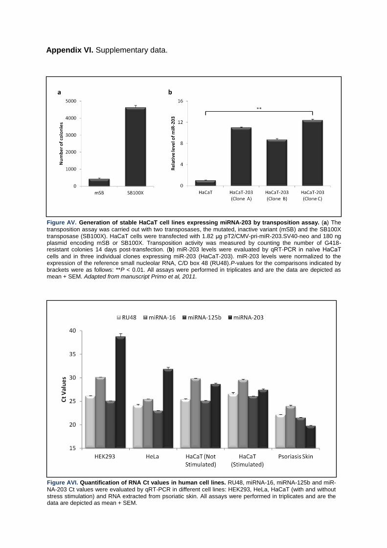
Appendix VI. Supplementary data.
Figure AV. Generation of stable HaCaT cell lines expressing miRNA-203 by transposition assay. (a) The
transposition assay was carried out with two transposases, the mutated, inactive variant (mSB) and the SB100X transposase (SB100X). HaCaT cells were transfected with 1.82 μg pT2/CMV-pri-miR-203.SV40-neo and 180 ng plasmid encoding mSB or SB100X. Transposition activity was measured by counting the number of G418-resistant colonies 14 days post-transfection. (b) miR-203 levels were evaluated by qRT-PCR in naїve HaCaT
cells and in three individual clones expressing miR-203 (HaCaT-203). miR-203 levels were normalized to the expression of the reference small nucleolar RNA, C/D box 48 (RU48).P-values for the comparisons indicated by brackets were as follows: **P < 0.01. All assays were performed in triplicates and are the data are depicted as mean + SEM. Adapted from manuscript Primo et al, 2011.
Figure AVI. Quantification of RNA Ct values in human cell lines. RU48, miRNA-16, miRNA-125b and miR-
NA-203 Ct values were evaluated by qRT-PCR in different cell lines: HEK293, HeLa, HaCaT (with and without stress stimulation) and RNA extracted from psoriatic skin. All assays were performed in triplicates and are the data are depicted as mean + SEM.
Top Related