ANOMALIAS DENTÁRIAS EM PACIENTES PORTADORES DE …objdig.ufrj.br/50/teses/m/CCS_M_869796.pdf ·...
72
UNIVERSIDADE FEDERAL DO RIO DE JANEIRO Centro de Ciência da Saúde Faculdade de Odontologia Departamento de Odontopediatria e Ortodontia Rio de Janeiro 2014 ANOMALIAS DENTÁRIAS EM PACIENTES PORTADORES DE MALOCLUSÃO ESQUELÉTICA: UM ESTUDO EPIDEMIOLÓGICO CLARISSA CHRISTINA AVELAR FERNANDEZ
Transcript of ANOMALIAS DENTÁRIAS EM PACIENTES PORTADORES DE …objdig.ufrj.br/50/teses/m/CCS_M_869796.pdf ·...

UNIVERSIDADE FEDERAL DO RIO DE JANEIRO
Centro de Ciência da Saúde
Faculdade de Odontologia
Departamento de Odontopediatria e Ortodontia
Rio de Janeiro 2014
ANOMALIAS DENTÁRIAS EM PACIENTES PORTADORES DE
MALOCLUSÃO ESQUELÉTICA: UM ESTUDO EPIDEMIOLÓGICO
CLARISSA CHRISTINA AVELAR FERNANDEZ

UNIVERSIDADE FEDERAL DO RIO DE JANEIRO
Centro de Ciência da Saúde
Faculdade de Odontologia
Departamento de Odontopediatria e Ortodontia
Rio de Janeiro 2014
CLARISSA CHRISTINA AVELAR FERNANDEZ
ANOMALIAS DENTÁRIAS EM PACIENTES PORTADORES DE
MALOCLUSÃO ESQUELÉTICA: UM ESTUDO EPIDEMIOLÓGICO
Dissertação de Mestrado apresentada ao Programa de Pós-Graduação em Odontologia (Área de Concentração: Odontopediatria) da Faculdade de Odontologia, da Universidade Federal do Rio de Janeiro, como parte dos requisitos para obtenção do título de Mestre em Odontologia (Área de Concentração: Odontopediatria).
Orientadores: Prof Dr. Marcelo de Castro Costa Prof Dr. Alexandre Rezende Vieira Colaboradora: Profª Christiane Vasconcellos Cruz Alves Pereira

FICHA CATALOGRÁFICA
Fernandez, Clarissa Christina Avelar. Anomalias Dentárias em Pacientes Portadores de Maloclusão Esquelética: um Estudo Epidemiológico. / Clarissa Christina Avelar Fernandez – Rio de Janeiro: Faculdade de Odontologia, 2014. Orientadores: Marcelo de Castro Costa e Alexandre Rezende Vieira Dissertação (mestrado) - UFRJ, FO, Programa de Pós-Graduação em Odontologia, Odontopediatria, 2014. Referências bibliográficas: f..
1. - Epidemiologia. 2. - Complicações. 3. - Anormalidades. 4. Anomalia Dentária. 5. Maloclusão Esquelética. 6. Padrão de Crescimento Facial. 7. Criança. 8. Adolescente. 9. Odontopediatria - Tese. I. Costa, Marcelo de Castro. II. Vieira, Alexandre Rezende III. Universidade Federal do Rio de Janeiro, FO, Programa de Pós-Graduação em Odontologia, Odontopediatria.

“Arte para mim não é produto de mercado... arte para mim é missão, vocação e
festa”
Ariano Suassuna
“E que seja perdido o único dia em que não se dançou”
Friedrich Nietzsche

DEDICATÓRIA
Aos meus amados avós maternos, Aida Maria e
Licínio Avelar, meus grandes amores.
Trocaria qualquer coisa na vida para abraçar vocês
neste momento. Vocês foram os melhores. Meus
melhores companheiros em todos os momentos. E que
sorte a minha!
Uma parte de mim é vocês.
A saudade que eu tenho e o amor que sinto por
vocês, eu vou levar para sempre. Sem dúvida!
Muito obrigada. De verdade.
AGRADECIMENTOS ESPECIAIS
À minha linda mãe, Célia Regina Avelar Fernandez, meu EXEMPLO de
mulher. Obrigada por nunca medir esforços para me ver feliz. Obrigada por me fazer
ser uma pessoa melhor. Por me ensinar que a vida é feita de escolhas e que crescer
e olhar para frente é sempre um ótimo caminho. Admiro sua coragem, sua força e
até mesmo seus desejos (por mais diferentes que eles pareçam!). Eu poderia
escrever páginas e mais páginas de agradecimentos, e, mesmo assim, não
conseguiria te dizer tudo o que sinto por você.
Eu acredito em você! Obrigada por sempre acreditar em mim também!
Amo muito você!
Muito. Muito. Muito.
À minha querida colaboradora, Profª. Christiane Vasconcellos Cruz Alves
Pereira, parte FUNDAMENTAL deste trabalho. Sem a sua ajuda, nada disso seria
possível. Obrigada por caminhar comigo nesta jornada e me ensinar tudo o que sei
sobre este trabalho. Admiro muito você e torço para que você tenha sucesso em
tudo o que fizer.
Muito obrigada!
Mesmo.
À dança, minha fonte de VIDA e inspiração.

AGRADECIMENTOS
Ao meu amado irmão, Guilherme Vinícius Avelar Fernandez, e à minha
amada cunhada, Caroline Couto Fernandez, meus AMIGOS mais fiéis. Vocês me
deram o melhor presente da vida: meu sobrinho que está vindo por aí. Estarei
sempre aqui para o que precisarem!
À minha querida tia, Heloisa Helena Gomes Avelar, minha verdadeira
PROFESSORA. Obrigada por acompanhar e incentivar os meus estudos desde
pequena. Sou sua eterna admiradora!
Ao meu lindo namorado, Mário Lúcio Pontes Bastos, meu COMPANHEIRO
de todas as horas. Paciência e carinho são seus nomes! Obrigada por respeitar
minhas escolhas e meus momentos, sejam eles bons ou ruins. Espero conseguir
corresponder todo o seu amor por mim.
Eu te amo!
À minha grande amiga, professora de jazz e cunhada, Gisele Pontes Bastos,
minha CONFIDENTE. Escrever algumas palavras para você é muito pouco perto do
que já vivemos. Eu só tenho a agradecer por tudo!
Na vida. E na dança.
À minha amiga e professora de ballet, Dani Cavanellas, minha INSPIRAÇÃO.
Preciso agradecer pela minha formação como bailarina e mulher. São 22 anos de
convivência! E muitos pliés!
Quantas danças! Quanta magia! Quanto amor!!!
Muito obrigada.
Ao meu querido orientador, Prof. Dr. Marcelo de Castro Costa. Obrigada
pela CONFIANÇA e por sempre abrir as portas e janelas para mim. A cadeira que
mais me conforta no Departamento inteiro, é a da sua sala.
Obrigada! Por tudo!

Ao meu grande coorientador, Prof. Dr. Alexandre Rezende Vieira, minha
inspiração como PESQUISADOR. Sinto um enorme prazer em tê-lo neste trabalho.
Que os seus passos continuem inspirando nossas pesquisas e incentivando novos
trabalhos.
Você é um exemplo!
À minha querida banca avaliadora. Vocês foram escolhidos com muito
CARINHO e há muito tempo.
Prof. Dr. Rogerio Gleiser, meu GRANDE professor. É uma honra ser sua
aluna e poder aprender tanto sobre o universo científico. Tenha certeza de que seus
ensinamentos são de grande importância na formação de todos nós, seus alunos.
Cada professor é responsável por construir um pouco de seu aluno. E eu tive muito
de mim construído pelo senhor. Obrigada pela dedicação com os nossos seminários
e o comprometimento com a pesquisa científica. Obrigada também pela ajuda e
enriquecimento do meu trabalho.
Sou sua fã!
Profª. Drª. Andréa Gonçalves Antonio, minha QUERIDA professora. Minha
admiração por você vem lá do início de tudo, nas clínicas de especialização. Sua
dedicação e amor pelo seu trabalho são verdadeiras fontes de inspiração para mim.
Acredito que ninguém tem o conhecimento sobre tudo, mas você, certamente, deve
saber quase tudo! E que honra tê-la como professora e poder conviver ao seu lado
nas supervisões das clínicas da graduação. Foi um grande aprendizado, uma das
melhores etapas da minha jornada no mestrado.
O-BRI-GA-DA!!!

Profª.Drª. Gloria Fernanda Barbosa de Araújo Castro, minha professora
mais ANIMADA. Admiro muito você, sua forma de trabalhar e seu modo de ver a
vida. Você é mais que um exemplo de alegria e profissionalismo. Você é
responsável por parte essencial da minha formação dentro deste Departamento.
Obrigada pelas conversas nos corredores, as palavras de apoio e por me acolher
tão bem. Tenho um carinho enorme por você! E muita sorte de ter alguém como
você no meu caminho.
Muito obrigada!
Profª.Drª. Lucianne Cople Maia, minha professora NÚMERO UM. Você foi a
minha maior incentivadora nos últimos três anos. Sei que todas as suas palavras
são para o meu bem e crescimento profissional. E sou muito agradecida por isso!
Quero um dia ser capaz de ajudar e apoiar os alunos como você faz. Obrigada por
me mostrar novos caminhos e me inspirar diariamente.
Você merece todo o sucesso que tem!
Ao Prof. Dr. Ronir Raggio Luiz, meu QUERIDO professor de estatística. Sua
ajuda foi fundamental para este trabalho. Obrigada pelos inúmeros encontros,
cálculos, tabelas e gráficos.
Eu tenho muito para te agradecer!
Aos professores do Programa de Pós-Graduação em Odontopediatria
(FO/UFRJ) Drª. Ivete Pomarico Ribeiro de Souza, Drª. Laura Guimarães Primo,
Drª. Luciana Pomarico, Dr. Thomaz Chianca e Drª Aline Neves, pelos seminários,
clínicas, prazos, congressos, painéis, trabalhos e palavras de apoio.
Muito obrigada!

Aos queridos professores João Farinhas, Luiz Eduardo, Nena Perez, Carla
Martins e Rosana Leonel pela dedicação e ajuda nas clínicas. Em especial, à
professora Marta Fornasari, minha COMPANHEIRA de cursos e simpósios.
Obrigada por dividir suas experiências comigo e me mostrar como a vida pode ser
cada vez mais linda!
Obrigada a todos!
Aos meus amigos da dança, meus AMIGOS da vida.
Danilo Saccomori, você é tão ESPECIAL que fico até sem ter o que dizer.
Obrigada pela amizade! Pelas danças! Pelos momentos!
Cátia Cabral, você sabe exatamente o lugar que ocupa no meu coração.
Tenho um CARINHO enorme por você!
Mariana Mondaini, obrigada por vestir a minha camisa! Quero você sempre
por PERTO!
Rafaela Queiroz e Aline Gaignoux, obrigada pelas CONVERSAS e pela a
amizade!
Vocês todos tornam meus dias muito melhores!
À minha amiga, Myriam Freitas. Você sempre tem a palavra certa, para o
momento ideal. Que SORTE a minha ter você por perto!
Obrigada por ser assim!
Às minhas lindas amigas, Natália Maria e Nathalia Telles. Vocês são as
IRMÃS que a vida me deu. Formamos um trio e tanto!
Obrigada por me fazerem ter muita história para contar!
Às minhas amigas de colégio, Karoline Lemos e Glaucia Barbosa, minhas
ETERNAS companheiras. Obrigada por crescerem comigo! Independente do passar
dos anos, nossa amizade permanece. E vai ser sempre assim.

Vocês moram no meu coração!
Aos meus amigos da faculdade, minhas PAIXÕES.
Indy Ana Fontes, obrigada por me ensinar que o importante a gente traz no
CORAÇÃO. E a distância é muito pequena perto do que sentimos.
João Faustino, minha dupla e meu AMIGO. A minha graduação não seria a
mesma sem você. Sinto sua falta! E como!
Cynthia Baptista, minha amiga mais LINDA. Obrigada por estar presente em
todos os meus momentos. E fazer deles, seus também!
Floriane Maile, amiga mais CHIQUE. Admiro sua força (por incrível que
pareça!) e sua simplicidade. Obrigada pela amizade de sempre!
Eu amo vocês!
Ao meu amigo de profissão, Thiago Spinelli, meu cirurgião dentista
PREFERIDO. Você está sempre disposto e pronto para me ajudar. Obrigada! Por
todo o companheirismo, dentro e fora da profissão.
Gosto muito de você!
Às minhas amigas de profissão, minhas LINDAS.
Priscila Almeida, minha AMIZADE por você não tem tamanho. Obrigada
pelas conversas e pelo carinho comigo!
Fernanda Alvine, você transforma qualquer problema em alegria. Obrigada
por me apoiar e me trazer tantos motivos para sorrir!
Eu adoro vocês!
Às amigas de turma do segundo ano, Tacíria, Queila e Elaine pelos
momentos únicos que passamos juntas. Em especial, à minha AMIGA Helena
Romanos, por todo o carinho e companheirismo comigo! E à minha amiga Nashalie
Alencar, minha SAUDADE. Ainda tenho esperanças de você vir morar no Rio!

Vocês foram a melhor turma de segundo ano que eu poderia ter!
Às amigas de turma do mestrado, Marina, Livia e Adrielle. Nossa
convivência foi ESSENCIAL para que esta jornada fosse concluída com sucesso.
Em especial ao meu amigo Thiago Isidro, minha DUPLA (a melhor!). Obrigada por
dividir as suas angústias e experiências comigo.
Torço muito pelo sucesso de todos vocês!
Aos amigos de turma do primeiro ano, Andréa, Aline, Fernanda e Káiron,
pela alegria e empenho diários. Em especial à minha linda amiga, Paula Moraes.
Quem vê a nossa amizade, não diz que nos conhecemos há menos de um ano.
Adoro a sua COMPANHIA! Obrigada por me ouvir todos os dias.
Aprendi muito com cada um de vocês!
Aos amigos do doutorado Thaís Soares, Marcello Roter, Marlus Cajazeira,
Michelle Ammari, Michele Lenzi, Andrea Pintor, Adílis Alexandria e Tatiana
Kelly, pelo convívio e troca de EXPERIÊNCIAS.
Vocês formam um time e tanto!
A todos os funcionários da Disciplina de Odontopediatria (FO/UFRJ), Rose,
Patrícia, Izabel, Zezé, Robson, Luiza e João, pelo APOIO e ajuda. As queridas
Mere, Kátia Andréa, por toda a paciência e dedicação nas clínicas. Vocês são as
MELHORES!
Nada funcionaria sem todos vocês!

RESUMO
FERNANDEZ, Clarissa Christina Avelar. ANOMALIAS DENTÁRIAS EM PACIENTES PORTADORES DE MALOCLUSÃO ESQUELÉTICA: UM ESTUDO EPIDEMIOLÓGICO. Rio de Janeiro, 2014 Dissertação (Mestrado em Odontologia – Área de Concentração: Odontopediatria) – Faculdade de Odontologia, Universidade Federal do Rio de Janeiro, Rio de Janeiro, 2014.
O objetivo do presente estudo censitário foi avaliar a prevalência das anomalias dentárias (AD) e a sua possível associação com os diferentes padrões de crescimento facial (PCF) e as maloclusões esqueléticas (ME) em uma população ortodôntica. Para tanto, 1521 prontuários ortodônticos de todos os pacientes atendidos, no período de 2000 a 2013, nos Departamentos de Ortodontia de duas instituições de ensino de referência na cidade do Rio de Janeiro, Brasil, foram avaliados. Foram excluídos (n=474) os prontuários que não continham ambas as radiografias (panorâmica e cefalométrica de perfil), pacientes com idade inferior a 08 anos e presença de síndrome, fissura lábio/palatina e desequilíbrios metabólicos e/ou endócrinos. Através da radiografia cefalométrica de perfil, o ângulo Sn-GoGn foi obtido para verificar o PCF (Hipodivergente, Normal e Hiperdivergente) e o ângulo ANB, para classificar as ME (Classe I, II e III). Os diagnósticos das AD foram realizados na radiografia panorâmica por um único operador calibrado. Para o diagnóstico de agenesia de terceiros molares (3M), foram excluídos (n=216) os pacientes cuja confirmação de extração destes dentes não foi possível. A frequência e a porcentagem de cada AD foram calculadas. As variáveis gênero, etnia, AD e dente mais afetado foram testadas através dos Testes Qui-Quadrado e Exato de Fisher com nível de significância de 5%. A Razão de Chance foi realizada para avaliação da intensidade e direção das possíveis associações. Além disso, o Teste T-Student foi utilizado para comparar diferenças entre os grupos. A amostra final foi composta por 1047 pacientes e a prevalência de AD foi de 77%. Do total, 56,7% eram do gênero feminino e 64,9%, afrodescendentes, com média de idade de 16,41 (±10,61). As AD mais prevalentes foram impactação (68,6%), giroversão (54%) e agenesia (9,7%), excluindo os 3M, respectivamente. Além disso, observou-se uma media de 3,08 (±1,93) dentes afetados por paciente e 51,2% apresentaram mais de uma AD. Dentre os 831 pacientes incluídos no diagnóstico de presença ou ausência de 3M, 11,2% apresentaram agenesia de 3M e 75,3% apresentaram outra AD, fora a agenesia de 3M. O 3M inferior esquerdo foi o dente mais afetado na impactação, o canino inferior direito, na giroversão e o incisivo lateral superior direito foi o dente mais ausente, excluindo os 3M. As AD foram mais prevalentes na ME de Classe III (80,8%) e no PCF Hipodivergente (82,5%). Agenesia de outros dentes, excluindo os 3M, apresentou associação com o PCF Hipodivergente (p<0,01) e a microdontia, com a ME de Classe III (p=0,25). A agenesia de 3M apresentou associação com agenesia de outros dentes, microdontia e associação inversa com impactação (p<0,01). As AD apresentaram alta prevalência e devem ser cuidadosamente investigadas e consideradas no planejamento do tratamento ortodôntico.
Palavras Chave: Anomalia Dentária, Maloclusão Esquelética, Terceiro Molar,
Epidemiologia, Criança, Adolescente.

ABSTRACT
FERNANDEZ, Clarissa Christina Avelar. ANOMALIAS DENTÁRIAS EM PACIENTES PORTADORES DE MALOCLUSÃO ESQUELÉTICA: UM ESTUDO EPIDEMIOLÓGICO. Rio de Janeiro, 2014 Dissertação (Mestrado em Odontologia – Área de Concentração: Odontopediatria) – Faculdade de Odontologia, Universidade Federal do Rio de Janeiro, Rio de Janeiro, 2014.
The aim of this census study was to evaluate the prevalence of dental anomalies (DA) and its possible association with the different skeletal malocclusion (SM) and growth patterns (GP) in an orthodontic population. For this study, 1521 orthodontic records of all patients attended, in the period of 2000 to 2013, in the Departments of Orthodontics of two reference institutions in the city of Rio de Janeiro, Brazil, were evaluated. The records that did not contain both radiographs (panoramic and lateral cephalometric), patients younger than 08 years old and presence of syndrome, cleft lip/palate, metabolic and/or endocrine imbalances were excluded. Sn-GoGn angle was obtained to verify the GP (Hypo-divergent, Normal and Hyper-divergent) and the ANB angle, to classify SM (Class I, II and III) in lateral cephalometric radiograph. DA diagnosis were performed on panoramic radiographs by a single calibrated operator. For the diagnosis of third molars (3M) agenesis, patients whose confirmation of extraction of these teeth was not possible were excluded (n=216). The frequency and percentage of each DA were calculated. The gender, ethnicity, age, DA and more affected tooth variables were tested using the Chi-Square and Fisher Exact Testes with significance level of 5%. The Odds Ratio was performed to evaluate the intensity and direction of the associations. Additionally, the Student T-test was used to compare differences between groups. The final sample was consisted of 1047 patients and the prevalence of DA was 77%. From a total, 56.7% were female and 64.9%, African descent, with a mean age of 16.41 (±10.61). The most prevalent DA were impaction (68.6%), giroversion (54%) and agenesis (9.7%), excluding 3M, respectively. Furthermore, we observed an average of 3.08 (± 1.93) teeth affected per patient and 51.2% had more than one DA. Among the 831 patients included in the diagnosis of the presence or absence of 3M, 11.2% had 3M agenesis and 75.3% had another DA, apart from 3M agenesis. The lower left 3M was the most affected tooth in impaction, the lower right canine, in giroversion and the upper left lateral incisor was the most absent tooth, excluding 3M. The DA were more prevalent in Class III SM (80.8%) and in the Hypo-divergent GP (82.5%). Agenesis of other teeth, excluding 3M, was associated with the Hypo-divergent GP (p <0.01) and microdontia, with the Class III SM (p=0.25). 3M agenesis was associated with agenesis of other teeth, microdontia and impaction (p<0.01). AD had a high prevalence and must be carefully investigated and considered in orthodontic treatment planning.
Key words: Dental Anomaly, Skeletal Malocclusion, Growth Pattern, Epidemiology, Child,
Adolescent.

LISTA DE TABELAS E FIGURAS
Artigo 1
Table 1. Characterization of Dental Anomalies among Gender ................................ 41
Table 2. Major Dental Anomalies and Their More Affected Teeth ............................ 43
Table 3. Distribution of the Major Dental Anomalies among Skeletal Malocclusions and Growth Patterns ................................................................................................ 44
Artigo 2
Figure 1. Angles used in the Characterization of Growth Patterns............................ 57
Figure 2. Angles used in the Characterization of Skeletal Classification Patterns ..... 57
Table 1. Population Characteristics according to Gender, Ethnicity, Angles Average (SNA, SNB, ANB and Sn-GoGn) and Age – Patients with and without 3M Agenesis58
Table 2. Distribution of Dental Anomalies between the Groups – 3M Agenesis and No 3M Agenesis ....................................................................................................... 59
Table 3. Characteristics Features of 3M Agenesis in the Different Groups according to the Number of Teeth Absence.............................................................................. 60

LISTA DE ABREVIATURAS
3M Terceiros Molares/Third Molar
AD Anomalias Dentárias
AG3M 3M Agenesis Group
ANB Ponto A-Násio-Ponto B
DA Dental Anomalies
GP Growth Patterns
LL Lower Left
LR Lower Right
ME Maloclusão(ões) Esquelética(s)
NAG3M No 3M Agenesis Group
PCF Padrão(ões) de Crescimento Facial
SM Skeletal Malocclusions
SNA Sela-Násio-Ponto A
SNB Sela-Násio-Ponto B
SN-GoGn Sela Násio-Gônio Gnátio
UL Upper Left
UR Upper Right

LISTA DE SÍMBOLOS
< menor que
> maior que
= igual
± mais ou menos
% porcentagem

SUMÁRIO
1.INTRODUÇÃO ...................................................................................................... 19
2.PROPOSIÇÃO ...................................................................................................... 23
2.1 Objetivo Geral................................................................................................. 23
2.2 Objetivos Específicos ..................................................................................... 23
3. DELINEAMENTO DA PESQUISA ........................................................................ 24
3.1. Desenho do Estudo e Seleção da Amostra ................................................... 24
3.2. Critérios de Inclusão e Exclusão .................................................................... 24
3.3. Caracterização dos Padrões de Crescimento Facial e Maloclusões Esqueléticas ......................................................................................................... 25
3.4 Diagnóstico de Anomalia Dentária .................................................................. 25
3.5 Diagnóstico de Agenesia de Terceiro Molar ................................................... 26
3.6 Coleta de Dados ............................................................................................. 26
3.7 Calibração ...................................................................................................... 26
3.8 Análise Estatística .......................................................................................... 27
4. DESENVOLVIMENTO DA PESQUISA ................................................................ 28
Artigo 1 Prevalence of Dental Anomalies in Different Growth and Skeletal Malocclusions Patterns: an Epidemiological Study ............................................... 29
Artigo 2 Third Molar Agenesis Associated with Other Dental Anomalies in Different Growth and Skeletal Malocclusions Patterns .......................................... 45
5. DISCUSSÃO ........................................................................................................ 61
6. CONCLUSÕES .................................................................................................... 64
REFERÊNCIAS BIBLIOGRÁFICAS ......................................................................... 65
ANEXOS .................................................................................................................. 68

19
1. INTRODUÇÃO
As anomalias dentárias (AD) são definidas como irregularidades
odontogênicas ou alterações dentárias (Garib, Filho et al., 2013), resultantes de
distúrbios durante o processo de formação dentária (Uslu, Akcam et al., 2009). As
manifestações clínicas das AD incluem diferentes graus de gravidade, variando de
suave a severo, representados por distúrbios no número, tamanho, forma, posição e
estrutura dos dentes (Uslu, Akcam et al., 2009; Basdra, Kiokpasoglou et al., 2000).
AD causam problemas funcionais, oclusais e estéticos (Kathariya, Nikam et al.,
2013;Osuji e Hardie, 2002) e resultam em prejuízos na saúde oral destes pacientes
(Kim, 2011).
A sua prevalência pode variar de 5,46% à 74,7% (Uslu, Akcam et al., 2009;
Díaz-Perez e Echaverry-Navarrete, 2009), devido as diferentes etnias e critérios de
diagnóstico (Uslu, Akcam et al., 2009; Altug-Atac e Erdem, 2007; Kim, 2011). A
etiologia específica das AD permanece obscura (Küchler, Risso et al., 2008), porém,
sabe-se que fatores genéticos e ambientais contribuem para o seu desenvolvimento
(Basdra, Kiokpasoglou et al., 2000; Kücler, Risso et al., 2008; Kathariya, Nikam et
al., 2013).
Segundo Neville et al., (2004), as AD são divididas em dois grandes grupos:
as alterações dentárias por fatores ambientais e as alterações no desenvolvimento
do elemento dentário. O primeiro grupo é composto por alterações influenciadas por
fatores ambientais sistêmicos ou locais. Os fatores sistêmicos podem ser de
natureza química, como quimioterapia antineoplásica, flúor, chumbo, tetraciclina e
vitamina D, alterações metabólicas (cardíacas, gastrointestinais, diabetes, entre
outras) e neurológicas (paralisia cerebral, retardo mental e defeitos de audição
sensorioneurais), má nutrição, trauma relacionado ao nascimento (hipóxia, parto
prematuro e etc), entre outros. Já os fatores locais mais associados são trauma
mecânico (quedas, acidentes e etc), queimadura elétrica, irradiação e infecção local
(Neville, Damm et al., 2004).

20
Essas alterações por fatores ambientais são subdivididas de acordo com o
tipo/estrutura do dano em: alterações de desenvolvimento das estruturas dentais
(hipoplasia de Turner, hipoplasia causada por terapia antineoplásica e fluorose
dental), descoloração dentária (manchas extrínsecas e intrínsecas), perda de
estrutura dentária pós desenvolvimento (desgaste dos dentes, atrição, abrasão,
erosão, abfração e reabsorções interna e externa) e distúrbios localizados da
erupção (impactação primária e anquilose) (Neville, Damm et al., 2004).
Já as alterações no desenvolvimento do elemento dentário são de natureza
complexa. Estas alterações podem ser primárias ou surgirem após influências
ambientais, ou seja, secundariamente. A subdivisão dessas alterações é feita em
relação ao tipo e a localização do dano, resultando na seguinte divisão: em relação à
dimensão (macrodontia e microdontia), forma (geminação, fusão, concrescência,
cúspides acessórias – Carabelli, em garra e dente evaginado, dente invaginado,
esmalte ectópico, esmalte ectópico – pérolas de esmalte e extensões cervicais de
esmalte, taurodontia, hipercementose, dilaceração e raízes supranumerárias),
estrutura (amelogênese imperfeita – hipoplásica, hipomaturada e hipocalcificada,
dentinogênese imperfeita, displasia dentinária – tipo I e II, odontodisplaisa regional),
posição (impactação, giroversão, transposição e inversão) e número dos dentes
(hipodontia – anodontia parcial, total e oligodontia, e hiperdontia - mésio dente)
(Neville, Damm et al., 2004). Essas serão as alterações investigadas no presente
estudo.
A agenesia de terceiro molar (3M) é considerada um outro tipo de AD, fora a
agenesia de outros dentes (Chung, Han et al., 2008; Endo, Ozoe et al., 2006b;
Shapiro e Farrington, 1983; Zhu et al, 1996). Esta anomalia possui ampla
prevalência, podendo variar de 12,63% à 51,1% (Celikoglu e Kamak, 2012; García-
Hernández, Yagui et al., 2008). Isso pode ocorrer devido a diferentes critérios de
diagnóstico, populações e grupos étnicos (Celikoglu, Bayram et al., 2011; Celikoglu
e Kamak, 2012; Barka, Marathiotis et al., 2013).
Segundo a literatura, a agenesia de 3M pode estar associada com outras AD
(variações de número e estrutura) (Celikoglu, Bayram et al., 2011) e má formações

21
(García-Hernández, Yagui et al., 2008), e ainda com a maloclusão esquelética (ME)
de Classe III (Celikoglu e Kamak, 2012). Outros estudos também investigaram a sua
possível associação com o apinhamento dentário na região ântero inferior, porém
este assunto ainda permanece controverso (Antanas e Giedré, 2006; Karasawa,
Rossi et al., 2013).
3M são dentes que possuem taxas de associação com dor e desconforto
físico relativamente altas, pouco valor funcional e a sua importância clínica é
duvidosa (Jung e Cho, 2014; Alam, Hamza et al., 2014). Além disso, a agenesia de
3M já é considerada uma evolução da espécie humana (García-Hernández, Yagui et
al., 2008).
A maloclusão esquelética pode ser definida como uma alteração de
crescimento no complexo craniofacial ou desvio da oclusão considerada normal ou
ideal (Andrews, 1972). Os dentes em má posição não são mais que sintomas de um
crescimento anormal do esqueleto facial. Quatro sistemas simultaneamente podem
ser afetados: dentes, ossos, músculos e ligamentos (Arashiro, Ventura et al., 2009).
A oclusão pode variar entre indivíduos de acordo com o tamanho e forma dos
dentes, posição dentária, época e sequência de erupção, forma e tamanho do arco
dentário e padrão de crescimento craniofacial, além de influências do meio
ambiente, modificações funcionais e patológicas (Pires, Rocha et al., 2001). Em
estudos anteriores, a etiologia da ME é atribuída a fatores ambientais e a padrões de
herança genética (Mossey, 1999). A prevalência da ME, proposta por Angle (1899),
é alta e varia bastante de acordo com a população estudada (Mossey, 1999).

22
As AD e as ME podem causar comprometimento estético, perturbações
comportamentais e distúrbios psicossociais nos indivíduos afetados, resultando em
um impacto negativo na qualidade de vida desses indivíduos. O tratamento das ME
é um desafio, em função da dificuldade de previsão do crescimento facial, pelo longo
tempo de tratamento envolvido e pelo custo que representa. Tal desafio se torna
ainda maior em presença de AD associada, principalmente as agenesias, que
comprometem a função normal e a estética. Vale ressaltar que as AD apresentam
uma frequência aumentada em pacientes portadores de ME podendo ser, portanto,
relacionadas a ela. Neste contexto, a compreensão dos mecanismos moleculares
envolvidos na etiologia das AD e ME desempenha um papel fundamental na ciência
odontológica.
É importante ter o conhecimento da frequência, da distribuição e dos
determinantes dos estados ou eventos relacionados à saúde em populações
específicas, para controle e prevenção dos problemas de saúde, e comparações
válidas entre diferentes populações. Um dos pontos principais da epidemiologia é a
busca pela causa e pelos fatores que influenciam a ocorrência da doença. Desta
forma, é possível a avaliação de características, inclusive, genéticas, em uma
determinada população. Os objetivos principais são o aprimoramento e melhorias
nas condições de saúde da população estudada.

23
2. PROPOSIÇÃO
2.1 Objetivo Geral
Avaliar a associação entre a prevalência, distribuição e dimorfismo sexual de AD
entre os diferentes padrões de crescimento facial (PCF) e ME em uma população
ortodôntica.
2.2 Objetivos Específicos
Avaliar a prevalência das AD na população estudada;
Investigar os tipos de AD mais prevalentes, bem como os dentes mais
afetados;
Avaliar a prevalência de agenesia de 3M, bem como o dente mais acometido,
lateralidade, lado e arco mais afetados.
Investigar as possíveis associações entre as AD e os gêneros;
Investigar as possíveis associações entre a agenesia de 3M e as outras AD;
Investigar as possíveis associações entre as AD e PCF, e AD e ME.

24
3. DELINEAMENTO DA PESQUISA
3.1. Desenho do Estudo e Seleção da Amostra
A amostra inicial do presente estudo censitário foi composta por 1521
prontuários ortodônticos completos, que deveriam incluir radiografias iniciais
(panorâmica e cefalométrica de perfil), fotos e modelos, de todos os pacientes
atendidos, de 2000 à 2013, nos Departamentos de Ortodontia, na Faculdade de
Odontologia, da Universidade Federal do Rio de Janeiro (UFRJ) e Associação
Brasileira de Odontologia do Rio de Janeiro(ABORJ).
Este estudo foi aprovado pelo Comitê local de Ética em Pesquisa (Hospital
Universitário Clementino Fraga Filho - HUCFF / UFRJ - Número: 619 096) (Anexo 1)
e duas autorizações por escrito dos chefes responsáveis pelos respectivos
Departamentos de Ortodontia foram concedidas ao pesquisador para o acesso à
documentação ortodôntica (Anexos 2 e 3).
3.2. Critérios de Inclusão e Exclusão
Pacientes com idade superior a 08 anos e com os prontuários odontológicos
contendo ambas as radiografias iniciais (panorâmica e cefalométrica de perfil), com
boa qualidade técnica, permitindo a visualização de todos os dentes e estruturas
adjacentes foram incluídos. Os pacientes que apresentavam história de trauma,
fissura lábio/palatina, síndromes, desequilíbrios endócrinos e/ou distúrbios
metabólicos, sendo estes genética e/ou hereditária, foram excluídos.
Após a aplicação dos critérios de inclusão e exclusão, a amostra final foi
composta por 1047 pacientes. Deste total de 1521 pacientes, 474 foram excluídos
devido à ausência de ambas as radiografias (39,7%), ausência de radiografia
panorâmica (27%), a história médica (15,6%), ausência de radiografia cefalométrica
de perfil (14,1%) e idade menor que 08 anos (3,6%).

25
3.3. Caracterização dos Padrões de Crescimento Facial e Maloclusões
Esqueléticas
Nesta etapa, todas as medições dos ângulos foram obtidos por único
operador, através da radiografia cefalométrica de perfil. Para caracterizar o padrão
de crescimento, os valores do ângulo do plano mandibular (Sn-GoGn) foram
utilizados de acordo com Steiner(1953):
Ângulo Sn-GoGn < 32° = Hipodivergente;
Ângulo Sn-GoGn = 32° = Normal;
Ângulo Sn-GoGn > 32° = Hiperdivergente.
A classificação esquelética foi avaliada pelos valores de ângulo sagital
intermaxilar (SNA - SNB = ANB), de acordo com o padrão cefalométrico para o tipo
esquelético,recomendado por Steiner(1953):
Ângulo ANB com valores entre 0 ° e 4 ° = Classe I;
Ângulo ANB com valores > 4 ° = Classe II;
Ângulo ANB com valores < 0 ° = Classe III.
3.4 Diagnóstico de Anomalia Dentária
Utilizando a radiografia panorâmica, fotos e modelos iniciais, os diagnósticos
das AD foram realizados de acordo com as seguintes alterações:
Número: agenesia (excluindo os 3M) e dentes supranumerários.
Tamanho: microdontia e macrodontia.
Posição: impactação, transposição, giroversão, inversão, retenção
prolongada, retardo na erupção e erupção ectópica.

26
Morfologia: dilaceração da raiz, dilaceração da coroa, odontoma,
taurodontismo, cúspide acessória, fusão, pérola de esmalte e dens in
dente.
Estruturais: amelogênese imperfeita e dentinogênese imperfeita.
3.5 Diagnóstico de Agenesia de Terceiro Molar
3M foi considerado ausente quando houve confirmação de que o dente não
foi extraído (nos prontuários odontológicos) e também quando não houve evidência
de mineralização da coroa do dente na radiografia panorâmica. Quando não foi
possível observar a mineralização da coroa na radiografia panorâmica inicial, uma
radiografia posterior (durante o tratamento ou radiografia final) foi realizada para este
diagnóstico. Além disso, os pacientes cuja comprovação da história de extração do
3M, através dos prontuários, não foi possível, foram excluídos (n=216).
3.6 Coleta de Dados
Os dados referentes à agenesia de 3M foram coletados de acordo com o
dente mais afetado, lateralidade (unilateral ou bilateral), lado (direito, esquerdo ou
ambos) e os arcos mais acometidos (maxila, mandíbula ou ambos). Informações
sobre idade, gênero e etnia também foram coletados dos prontuários (Anexo 4)..
Todos os dados (valores dos ângulos, diagnósticos das AD e as outras informações)
foram coletados por um único operador calibrado.
3.7 Calibração
Para o diagnóstico das AD e os valores dos ângulos SNA, SNB, ANB e Sn-
GoGn, a calibração foi realizada considerando o avaliador padrão-ouro, um
especialista em Ortodontia com mais de 15 anos de experiência. Através das
radiografias panorâmicas (para avaliar AD) e das cefalométricas de perfil (para
avaliar as medidas dos ângulos), com o auxílio de um negatoscópio, em um quarto

27
escuro e silencioso, foram realizados o diagnóstico de 30 indivíduos pelo avaliador
padrão ouro. Logo depois, Os mesmos diagnósticos foram realizados por um único
operador na mesma condições para futura comparação dos resultados. Em um
intervalo de 15 dias, uma repetição desses mesmos passos foram realizados pelo
operador, para obtenção de uma confiabilidade inter e intra examinador.
3.8 Análise Estatística
Os dados foram analisados usando o programa estatístico SPSS versão 20.0
(Statistical Package for Social Sciences, SPSS Inc, Chicago, III). A concordância
entre o operador e o avaliador padrão ouro foi determinada pelo Índice Kappa (na
radiografia panorâmica) e Índice de Correlação Intra-Classe (valores numéricos na
radiografia cefalométrica de perfil). A frequência e porcentagem da distribuição de
cada AD foram calculadas. Os dados como gênero, etnia, idade, AD e dentes mais
afetados, foram testados usando Qui-Quadrado ou Teste Exato de Fisher com nível
de significância de 5%. A Razão de Chance foi realizada para avaliar a intensidade e
a direção das possíveis associações. Além disso, o Teste T-Student foi realizado
para comparar as diferenças entre os grupos.

28
4. DESENVOLVIMENTO DA PESQUISA
Artigo 1: PREVALENCE OF DENTAL ANOMALIES IN DIFFERENT
GROWTH AND SKELETAL MALOCCLUSIONS PATTERNS: AN
EPIDEMIOLOGICAL STUDY
Artigo 2: THIRD MOLAR AGENESIS ASSOCIATED WITH OTHER DENTAL
ANOMALIES IN DIFFERENT GROWTH AND SKELETAL MALOCCLUSIONS
PATTERNS

29
Artigo 1 Prevalence of Dental Anomalies in Different Growth and Skeletal Malocclusions Patterns: an Epidemiological Study
Clarissa Christina Avelar FERNANDEZ1
Christiane Vasconcellos Cruz Alves PEREIRA2
Ronir Raggio LUIZ3
Alexandre Rezende VIEIRA4
Marcelo de Castro COSTA5
1 DDS, Master Student - Department of Pediatric Dentistry and Orthodontics, School of Dentistry, Universidade Federal do Rio de Janeiro, Rio de Janeiro, RJ, Brazil.
2 DDS, MSD, PhD Student - Department of Pediatric Dentistry and Orthodontics, School of Dentistry, Universidade Federal do Rio de Janeiro, Rio de Janeiro, RJ, Brazil.
3 STAT, MSD, PhD, Associate Professor - Institute for Studies in Public Health, IESC, Universidade Federal do Rio de Janeiro, Rio de Janeiro, RJ, Brazil.
4 DDS, MSD, PhD, Adjunct Professor - Department of Oral Biology, School of Dental Medicine, University of Pittsburgh – PA, USA.
5 DDS, MSD, PhD, Adjunct Professor - Department of Pediatric Dentistry and Orthodontics, School of Dentistry, Universidade Federal do Rio de Janeiro, Rio de Janeiro, RJ, Brazil.
Correspondence Author – Marcelo de Castro Costa
Disciplina de Odontopediatria da Faculdade de Odontologia - UFRJ
Caixa Postal: 68066 – Cidade Universitária - CCS
CEP.: 21941-971 - Rio de Janeiro – RJ – Brazil
E-mail: [email protected]
Fax/phone: +5521 25622101
Revista para submissão: American Journal of Orthodontics and Dentofacial Orthopedics

30
ABSTRACT
Introduction: The aims of this study were to evaluate the association between
prevalence, distribution and sexual dimorphism of dental anomalies (DA) among
different skeletal malocclusions and growth patterns in an orthodontic population.
Methods: 1047 orthodontic records of patients aged over 08 years, attended, from
2000 to 2013, in the Departments of Orthodontics of two references institutions in Rio
de Janeiro, Brazil, were included. Sn-GoGn angle were used to classify the growth
pattern (Hypo-divergent, Normal and Hyper-divergent) and ANB angle, to verify the
skeletal malocclusion pattern (Class I, II and III) in lateral cephalometric radiograph.
DA diagnosis were performed using the panoramic radiographs by one calibrated
investigator. Odds Ratio, Chi-Square and T-Student Tests, with significance level of
5%, were performed. Results: 56.7% of patients were females and 64.9% were
african descent, with mean age of 16.41 (±10.61). The DA prevalence was 77%. It
was found that 3.08 (±1.93) teeth were affected per patient and more than one DA
was observed in the majority components of DA group (51.2%). Impaction,
giroversion and tooth agenesis (excluding 3M) were the most prevalent DA, with
68.6%, 54% and 9.7%, respectively. Fusion and impaction presented statistical
difference among gender (p<0.05). Lower left third molar was the most affected tooth
in impaction, lower right canine, in giroversion and the upper right lateral incisor was
the most absent tooth. DA were most prevalent in skeletal Class III malocclusion
pattern (80.8%) and in Hypo-Divergent growth pattern (82.5%) although not
statistically significant. Tooth agenesis (p<0.01) and microdontia (p=0.25) presented
statistical difference among Hypo-divergent growth pattern and skeletal Class III
malocclusions pattern, respectively. Conclusions: The prevalence of DA was high.
The most common DA were impaction, giroversion and tooth agenesis, respectively.
Microdontia was more common in skeletal Class III malocclusion pattern and tooth
agenesis, in Hypo-divergent growth pattern. Fusion and impaction presented
significant difference among gender, being more common in males. DA must be
carefully investigated and considered in orthodontic treatment planning.

31
INTRODUCTION
Dental anomalies (DA) are clinical alterations resulting from disturbances
during the tooth formation process.1 The clinical manifestations of DA include
different degrees of severity, ranging from mild to severe cases, represented by
disturbances in the number, size, shape, position and structure of the teeth.1,2 Its
prevalence can range from 5.46 to 74.7%,1,8 due to different ethnicities and
diagnostic criteria.1,3,7 The specific etiology of DA remains obscure,5 but it is known
that genetic and environmental factors may contribute to its development.2,5,6
Skeletal malocclusions are usually categorized and described by disturbances
in the craniofacial and occlusal relationships9 and often appear together with the DA,
asserting their relation and complicating therapy.6 Furthermore, DA cause functional,
occlusal and aesthetic problems,6,10 resulting in oral health impairment.7 Therefore, a
careful initial examination should be performed for the inclusion of their management
in orthodontic treatment planning in order to avoid complications during treatment.1,3
Faced to the complexity that involves the etiology of DA, the distinct
characteristics of each skeletal malocclusion and the few studies that have
investigated both,1 the aims of this study were to evaluate the association between
prevalence, distribution and sexual dimorphism of DA among different skeletal
malocclusions and growth patterns in an orthodontic population.

32
MATERIALS AND METHODS
Sample Selection
The initial sample was formed by 1521 records of all patients (census study)
who where attended, from 2000 to 2013, in the Departments of Orthodontics, in
School of Dentistry, at Universidade Federal do Rio de Janeiro (UFRJ) and
Associação Brasileira de Odontologia do Rio de Janeiro (ABO RJ). The dental
records had to include initial radiographs (panoramic and lateral cephalometric),
photos and casts.
This study was approved by the local Ethics Committee for Research (Hospital
Universitário Clementino Fraga Filho – HUCFF/UFRJ – Number: 619 096).
Inclusion and Exclusion Criteria
Patients aged over 08 years and whose dental records contained both initial
radiographs, with good technical quality, enabling the visualization of all teeth and
surrounding structures were included. Patients who presented history of trauma, cleft
lip/palate, syndromes, endocrine imbalances and/or metabolic disorders, being these
genetic/or hereditary, were excluded.
After the inclusion and exclusion criteria were applied, the final sample
consisted in 1047 patients. From a total of 1521 patients, 474 were excluded due to
the absence of panoramic and lateral cephalometric radiographs (39.7%), absence of
panoramic radiograph (27%), medical history (15.6%), absence of lateral
cephalometric radiograph (14.1%) and age lower than 08 years (3.6%).

33
Characterization of Growth Pattern and Skeletal Classification
All angles measurements were obtained by a lateral cephalometric radiograph.
To characterize the growth pattern, the values of the mandibular plane angle (Sn-
GoGn) were used according to Steiner:11
Sn-GoGn Angle < 32° = Hypo-divergent;
Sn-GoGn Angle = 32° = Normal;
Sn-GoGn Angle > 32° = Hyper-divergent.
The skeletal classification was evaluated by the values of sagittal
intermaxillary angle (SNA - SNB = ANB), according to the cephalometric standard for
skeletal type as recommended by Steiner:11
ANB Angle with values between 0° and 4° = Class I;
ANB Angle with values > 4° = Class II;
ANB Angle with values < 0° = Class III.
Diagnosis of Dental Anomalies
Using the initial panoramic radiograph, photos and casts, diagnosis of DA
were performed according to alterations in:
Tooth number: agenesis (excluding the third molars) and
supernumerary.
Tooth size: microdontia and macrodontia.
Tooth position: impaction, transposition, giroversion, inversion,
prolonged retention, delayed eruption and ectopic eruption.
. Tooth shape: root’s dilaceration, crown’s dilaceration, odontoma,
taurodontism, acessory cusp, fusion, pearl enamel and dens
invaginatus.
Structure of the tooth: amelogenesis imperfecta and dentinogenesis
imperfecta.

34
Information about age, gender, and ethnicity were also collected from dental
records. All diagnostic and data were collected by a single calibrated investigator.
Calibration
For the diagnosis of DA and values of the SNA, SNB, ANB and Sn-GoGn
angles, a calibration was performed considering the gold standard evaluator, a
specialist in Orthodontics with over 15 years of experience The gold standard
evaluator performed the diagnosis of 30 individuals through the panoramic
radiographs (to assess dental anomalies) and the lateral cephalometric radiographs
(to assess the angles measurements), with the aid of a negatoscope in a dark and
quiet room. Soon after, a single investigator performed the same diagnosis under the
same conditions to compare the results. In an interval of 15 days, a repetition of
these same steps were performed by the investigator to obtain an inter examiner and
intra examiner reability.
Statistical Analysis
Data were analyzed using SPSS version 20.0 (Statistical Package for Social
Sciences, SPSS Inc, Chicago, III). The agreement between the investigator and the
gold standard evaluator was determined by Kappa Index (in the panoramic
radiograph) and Intra-Class Correlation Index (ICC) (in the lateral cephalometric
radiograph). The frequency and percentage distribution of each DA among the
sample were calculated. Data such as gender, ethnicity, age, dental anomalies and
most affected teeth, were tested using Chi-Square or Fisher Exact Test with a
significance level of 5%. Odds Ratio was performed to evaluate the intensity and
direction of the association. Furthermore, T Student Test was carried out to compare
the differences between groups.

35
RESULTS
The prevalence of DA was 77% (n=806), considering the final sample formed
by 1047 patients dental records. From a total, 56.7% (n=594) of patients were
females and 64.9% (n=680) were African descent, with mean age of 16.41±10.61. It
was found that 3.08 (±1.93) teeth were affected per patient and more than one DA
(different or equal types)) was observed in the majority components of DA group
(51.2%). Kappa Index and the Intra-Class Correlation tests showed excellent
reliability with 0.93 and 0.87, respectively.
Table 1 and Figure 1 show the characterization and distributions of DA among
gender. Impaction, giroversion and tooth agenesis were the most prevalent DA, with
68.6%, 54% and 9.7%, respectively. Fusion and impaction presented statistical
difference among gender (p=0.047 and p<0.01, respectively). The average number
for teeth affected per patient and the most affected teeth in major DA are represented
in Table 2. Lower left third molar was the most affected tooth in impaction (n=406),
lower right canine, in giroversion (n=233) and the upper right lateral incisor was the
most absent tooth (n=24).
Table 3 shows the distribution of the major DA among skeletal malocclusions
and growth patterns. AD were most prevalent in skeletal Class III malocclusion
pattern (80.8%) and in Hypo-divergent growth pattern (82.5%). Tooth agenesis
(p<0.01) and microdontia (p=0.025) presented statistical difference among Hypo-
divergent growth pattern and skeletal Class III malocclusions pattern, respectively. In
this sample, 48.1% were Class I, 39.4% were Class II and 12.4%, Class III. In
relation to the growth patterns, 16.3% were Hipo-divergent, 7.4% were Normal and
76.3%, Hyper-divergent.

36
DISCUSSION
The study of DA in patients with different skeletal malocclusion patterns may
serve as basis for future genetic studies and help in elucidating its etiology.1,2,9
Disturbances in the molecular mechanisms related to craniofacial and occlusal
relationship may be linked to malocclusion and DA. Thus, these features can serve
as an answer to their origin and development.9 Many studies reported the association
between DA and skeletal malocclusions5,9 and their genetic background is widely
known. 8 So, it is likely to be possible to check the DA and a specific skeletal
malocclusion candidate genes when the association between them happens.2 The
phenotype-genotype correlation is extremely important to improve the knowledge of
its etiology, which may contribute with a more accurate diagnosis. The next step of
this research will be the investigation of the candidate gene linked to DA and skeletal
malocclusion and growth patterns.
The main limitation of this study was the exclusion of 31.16% of the sample
due to the incomplete dental records. The panoramic and lateral cephalometric
radiographs were extremely important to the inclusion and diagnosis criteria of
orthodontic patients. Our results suggest that DA are easily detected by routine
radiographic examination and should be included in orthodontic treatment planning
permitting early diagnosis and timely orthodontic intervention.
Different results are found in the literature for the prevalence of DA.1,3-7 Some
authors attribute these conflicting results to different ethnicities, diagnostic criteria3,5
and also environmental and nutritional factors.3 Thongudomporn and Freer (1998),4
observed 74.7% of DA in 111 orthodontic patients and attributed this finding to the
sample size, because small samples tend to be less reliable.3 In the present study,
we observed similar high prevalence (77%), however it is thought that this happened
on account of the involvement of two reference institutions in Orthodontics and that
may have enriched the population with DA.5 According to some authors, orthodontic
patients tend to have more DA than the general population.1

37
In relation to gender, some authors found no statistically difference among
them in the prevalence of DA.5 Other study have obtained similar results, but the
microdontia and ectopic eruption were only observed in females.1 Kathariya et al.
(2013) found significant gender differences only for tooth agenesis, microdontia and
accessory cusp.6 The prevalence of DA was greater in males than in females in this
population. In addition, fusion and impaction showed statistical significance among
gender, which disagrees with previous studies.1,5,6 The conflicting findings may be
due to ethnic variations, sample size and diagnosis criteria.
In the present study, the most prevalent DA was impaction (68.6%) followed
by giroversion (54%). These DA have been previously described as the most
prevalent DA, but with a considerable lower prevalence (39.6 and 13.2%,
respectively)6 when compared to this study. According to Uslu et al. (2009),
impaction was the fifth most prevalent DA in Turkish orthodontic population.1 Two
main theories have been suggested to explain impaction: the guidance theory, that is
based on local predisposing causes and the genetic theory, that considers a genetic
cause for impaction.17 In the present study, the most affected teeth by impaction was
the lower third molars. This finding can be explained on account of lack of space in
the lower dental arch, which is likely to be possible by decreased horizontal
craniofacial development.
The dental agenesis have been largely studied12-16 and its prevalence ranged
from 4.8% to 26%.1,3,5-8 According to the literature, the most affected teeth are the
second premolars and upper lateral incisors.1,3,5-7 In our results the prevalence of
agenesis was 9.7% when the 3M was not included and it was the third most DA
observed. The upper lateral incisors and the lower left second premolar were the
most affected teeth, corroborating the previous studies.1,3,5-7

38
Despite the association of DA with skeletal malocclusions pattern has been
investigated to serve as a basis for future genetic studies.1,2,9 For the best of our
knowledge this is the first study that evaluated the association of DA with different
skeletal malocclusions and growth patterns up to date. In this study, tooth agenesis
was associated with hypo-divergent growth pattern. This association can be
explained by a possible disturbance in the proliferation and development, resulting in
changes which occurs the lack of growth. So, we may suppose that genetic
mechanisms may share similar pathways.
CONCLUSIONS
1. The prevalence of DA was high, affecting 77% of patients in this population.
2. Impaction, giroversion and tooth agenesis were the most prevalent DA.
3. Fusion and impaction presented statistical difference among gender.
4. Lower left third molar was the most affected in impaction, lower right
canine, in giroversion and the upper right lateral incisor was the most absent tooth.
5. DA were higher prevalent in the skeletal Class III malocclusion pattern and
in the growth Hypo-divergent pattern. Tooth agenesis was more common in Hypo-
divergent growth pattern and microdontia, in skeletal Class III malocclusions pattern.

39
REFERENCES
1. Uslu O, Akcam MO, Evirgen S, Cebeci I. Prevalence of dental anomalies in
various malocclusions. Am J Orthod Dentofacial Orthop 2009;135:328-35.
2. Basdra EK, Kiokpasoglou M, Stelizig A. The Class II division 2 craniofacial
type is associated with numerous congenital tooth anomalies. Eur J Orthod
2000;22:529-35.
3. Altug-Atac AT, Erdem D. Prevalence and distribution of dental anomalies in
orthodontic patients. Am J Orthod Dentofacial Orthop 2007;131:510-4.
4. Thongudomporn U, Freer TJ. Prevalence of dental anomalies in orthodontic
patients. Aus Dent J 1998;43:395-8.
5. Küchler EC, Risso PA, Costa MC, Modesto A, Vieira AR. Studies of dental
anomalies in a large group of school children. Archives of Oral Biology
2008;53:941-946.
6. Kathariya MD, Nikam AP, Chopra K, Patil NN, Raheja H, Kathariya R.
Prevalence of Dental Anomalies among School Going Children in India. J Int
Oral Health 2013;5(5):10-4.
7. Kim YH. Investigation of Hypodontia as Clinically Relates Dental Anomaly:
Prevalence and Characteristics. ISRN Dentistry 2011;Article ID 246135, 6
pages.
8. Díaz-Pérez R, Echaverry-Navarrete RA. Agenesia en dentición permanente.
Rev. salud pública 2009;11(6):961-969.
9. Basdra EK, Kiopasoglou MN, Komposch, G. Congenital tooth anomalies and
malocclusions: a genetic link?. Eur J Orthod 2001;23:145-151.
10. Osuji OO, Hardie J. Dental Anomalies in a population of Saudi Arabian
children in Tabuk. Saudi Dent J 2002;14(1):11-4.
11. Steiner CC. Cephalometrics for you and me. Am J Orthod 1953;39:729-55.

40
12. Chung CJ, Han J-H, Kim K-H. The pattern and prevalence of hypodontia in
Koreans. Oral Diseases 2008;14:620-625.
13. Endo T, Ozoe R, Yoshino S, Shimooka S. Hypodontia patterns and variations
in craniofacial morphology in Japanese orthodontic patients. Angle Orthod
2006b;76:996-1003.
14. Shapiro SD, Farrington FH. The facial profile in advanced hypodontia: a mixed
longitudinal study of 141 children. Eur J Orthod 1983;5:133-143.
15. Zhu JF, Marcushamer M, King DL, Henry RJ. Supernumerary and congenitally
absent teeth: a literature review. J Clin Pedatr Dent 1996;20:87-95.
16. Brook AH. A unifying aetiological explanation for anomalies of human tooth
number and size. Arch Oral Biol 1984;29:373-8.
17. Sambataro S, Baccetti T, Franchi L, Antonini F. Early predictive variables for
upper canine impaction as derived from posteroanterior cephalograms. Angle
Orthod 2004;75:28-34.

41
TABLES
Table 1. Characterization of Dental Anomalies among Gender
Dental Anomalies
DAa Group n (%) p value Odds Ratio (Confidence
Interval)
Males Females Total
Total of Dental Anomalies
356 (78.6) 450 (75.8) 806 (77) 0.281 1.17 (0.87-1.57)
Tooth Agenesis 28 (6.2) 50 (8.4) 78 (9.7) 0.172 0.72 (0.44-1.16) Supernumerary 14 (3.1) 17 (2.9) 31 (3.8) 0.829 1.08 (0.53-2.22) Microdontia 14 (3.1) 29 (4.9) 43 (5.3) 0.148 0.62 (0.32-1.19) Macrodontia 3 (0.7) 2 (0.3) 5 (0.6) 0.449 1.97 (0.33-11.86d) Impaction 274 (60.5) 280 (47.1) 553 (68.6) <0.01b 1.72 (1.34-2.20) Transposition 13 (2.9) 21 (3.5) 34 (4.2) 0.547 0.81 (0.40-1.63) Giroversion 188 (41.5) 247 (41.6) 435 (54) 0.979 0.99 (0.78-1.28) Inversion 3 (0.7) 5 (0.8) 8 (1) 0.741 0.78 (0.19-3.30) Prolonged Retention
--- 3 (0.5) 3 (0.4) 0.130 ---c
Delayed Eruption 9 (2) 8 (1.3) 17 (2.1) 0.417 1.48 (0.57-3.88) Ectopic Eruption 5 (1.1) 3 (0.5) 8 (1) 0.270 2.20 (0.52-9.25d) Root’s Dilaceration
8 (1.8) 14 (2.4) 22 (2.7) 0.509 0.74 (0.31-1.79)
Crown’s Dilaceration
--- 1 (0.2) 1 (0.1) 0.382 ---c
Odontoma 2 (0.4) 1 (0.2) 3 (0.4) 0.413 2.63 (0.24-29.09d) Taurodontism 1 (0.2) --- 1 (0.1) 0.252 ---c Acessory Cusp 2 (0.4) 2 (0.3) 4 (0.5) 0.785 1.31 (0.18-9,35d) Fusion 3 (0.7) --- 3 (0.4) 0.047b ---c Pearl Enamel --- 1 (0.2) 1 (0.1) 0.382 ---c Dens Invaginatus 1 (0.2) --- 1 (0.1) 0.252 ---c
Amelogenesis Imperfecta
1 (0.2) 1 (0.2) 2 (0.2) 0.847 1.31 (0.08-21.03d)
Notes: P value is based on Qui Square Test;
DA indicates Dental Anomaliesa;
Statistically significant (p>0.05)b;
OR not calculated due to frequency=0c;
Unstable numbers due to low frequencyd.

42
Figure 1. Distribution of Dental Anomalies among Gender
0 20 40 60 80
100 120 140 160 180 200 220 240 260 280 300 320 340 360 380 400 420 440 460 480
Den
tal A
no
mal
ies
Age
nes
is
Sup
ern
um
erar
y
Mic
rod
on
tia
Mac
rod
on
tia
Imp
acti
on
Tran
spo
siti
on
Gir
ove
rsio
n
Inve
rsio
n
Pro
lon
ged
Ret
enti
on
Del
ayed
Eru
pti
on
Ecto
pic
Eru
pti
on
Ro
ot'
s D
ilace
rati
on
Cro
wn
's D
ilace
rati
on
Od
on
tom
a
Tau
rod
on
tism
Ace
sso
ry C
usp
Fusi
on
Pe
arl E
nam
el
Den
s In
vagi
nat
us
Am
elo
gen
esis
Imp
erfe
cta
Pat
ien
ts (
n)
Dental Anomalies
Distribution of Dental Anomalies among Gender
Males
Females

43
Table 2. Major Dental Anomalies and Their More Affected Teeth
Notes: UR means Upper Right; UL means Upper Left; LR means Lower Right; LL means Lower Left.
Major Dental Anomalies
Total of Patients n (%)
Mean of Affected Teeth Per Patient Mean (DP)
More Affected Teeth n (%)
Tooth Agenesis 78 (9.7) 2,27 (±2.19) UR Lateral Incisor - 24 (30.8) UL Lateral Incisor – 22 (28.2) LL Second Premolar – 21 (26.9)
Supernumerary 31 (3.8) 1,23 (±0.56)
UL Paramolar – 9 (29) Mesial Tooth – 5 (16.1) UR Paramolar – 4 (12.9) LL Central Incisor – 4 (12.9)
Microdontia 43 (5.3) 1,37 (±0.72) UL Lateral Incisor – 16 (37.2) UR Lateral Incisor – 9 (20.9) UL Third Molar – 7 (16.3)
Macrodontia 6 (0.6) 1,20 (±0.45)
LR Lateral Incisor – 2 (40) UL Central Incisor – 2 (40) UL Lateral Incisor – 1 (20) LL Lateral Incisor – 1 (20)
Giroversion 435 (54) 1,92 (±1.09) LR Canine – 233 (52.9) LL Canine – 162 (37.2) UL Canine – 80 (18.4)
Impaction 553 (68.6) 1,93 (±0.90) LL Third Molar – 406 (73.3) LR Third Molar – 392 (70.2) UL Canine – 32 (5.8)

44
Table 3. Distribution of the Major Dental Anomalies among Skeletal Malocclusions and Growth Patterns
Skeletal Malocclusion Pattern
n (%)
Growth Pattern
n (%)
Major Dental Anomalies
Class I
Class II
Class III
p value
Hypo-Diverge
nt
Normal Hyper-Diverge
nt
p value
Total of Dental
Anomalies
384 (76.2)
317 (76.8)
105 (80.8)
0.537 141 (82.5)
63 (81.8)
602 (75.3)
0.077
Tooth Agenesis
44 (8.7)
24 (5.8)
10 (7.7) 0.244 22 (12.9) 1 (1.3) 55 (6.9) 0.003*
Supernume-rary
14 (2.8)
14 (3.4)
3 (2.3) 0.772 7 (4.1) 5 (6.5) 19 (2.4) 0.080
Microdontia 19 (3.8)
13 (3.1)
11 (8.5) 0.025*
11 (6.4) 4 (5.2) 28 (3.5) 0.190
Macrodontia 2 (0.4)
2 (0,5)
1 (0.8) 0.860 --- 1 (1.3) 4 (0.5) 0.383
Giroversion 197 (39.1)
185 (44.8)
53 (40.8)
0.214 65 (38) 38 (49.4)
332 (41.6)
0.245
Impaction 260 (51.6)
231 (55.9)
63 (48.5)
0.235 89 (52) 41 (53.2)
424 (53.1)
0.969
Note: Statistically significant (p>0.05)*.

45
Artigo 2 Third Molar Agenesis Associated with Other Dental Anomalies in Different Growth and Skeletal Malocclusions Patterns
Clarissa Christina Avelar FERNANDEZ1
Christiane Vasconcellos Cruz Alves PEREIRA2
Ronir Raggio LUIZ3
Alexandre Rezende VIEIRA4
Marcelo de Castro COSTA5
6 DDS, Master Student - Department of Pediatric Dentistry and Orthodontics, School of Dentistry, Universidade Federal do Rio de Janeiro, Rio de Janeiro, RJ, Brazil.
7 DDS, MSD, PhD Student - Department of Pediatric Dentistry and Orthodontics,
School of Dentistry, Universidade Federal do Rio de Janeiro, Rio de Janeiro, RJ, Brazil.
8 STAT, MSD, PhD, Associate Professor - Institute for Studies in Public Health, IESC, Universidade Federal do Rio de Janeiro, Rio de Janeiro, RJ, Brazil.
9 DDS, MSD, PhD, Adjunct Professor - Department of Oral Biology, School of Dental
Medicine, University of Pittsburgh – PA, USA.
10 DDS, MSD, PhD, Adjunct Professor - Department of Pediatric Dentistry and Orthodontics, School of Dentistry, Universidade Federal do Rio de Janeiro, Rio de Janeiro, RJ, Brazil.
Correspondence Author – Marcelo de Castro Costa
Disciplina de Odontopediatria da Faculdade de Odontologia - UFRJ
Caixa Postal: 68066 – Cidade Universitária - CCS
CEP.: 21941-971 - Rio de Janeiro – RJ – Brazil
E-mail: [email protected]
Fax/phone: +5521 25622101
Revista para submissão: Angle Orthodontics

46
ABSTRACT
Introduction: The aim of this study was to evaluate the prevalence of third
molar (3M) agenesis and its association with other dental anomalies in the different
skeletal malocclusion and growth patterns. Methods: 831 dental records (initial
radiographs, photos and casts) of patients aged over 08 years, attended, from 2000
to 2013, in the Departments of Orthodontics of two references institutions in Rio de
Janeiro, Brazil, were included. Sn-GoGn angle were used to classify the growth
pattern (Hypo-divergent, Normal and Hyper-divergent) and ANB angle, to verify the
skeletal malocclusion pattern (Class I, II and III). The diagnosis of 3M agenesis and
other dental anomalies (agenesis of other teeth, supernumerary, microdontia,
macrodontia, impaction, transposition, giroversion, root dilaceration, crown
dilaceration, odontoma and taurodontism) were assessed by the panoramic
radiographs with one calibrated operator. Odds Ratio, Chi-Square and T-Student
Tests, with significance level of 5%, were performed. Results: The prevalence of 3M
agenesis was 11.2%, 53.3% were females, 48.1% caucasians, with mean age of
14.11 (±7.1). In the group with 3M agenesis, it was found that 1.88 (±1.10) 3M teeth
were absent per patient and 75.3% of patients (n=70) had another dental anomaly
besides the 3M agenesis. 3M Agenesis was associated with agenesis of other teeth,
microdontia and impaction (p<0.01). The upper right 3M was the most affected tooth
(58.1%), unilateral 3M agenesis was the most common (51.6%) and the right side
was the most affected (31.2%). Maxilla was more affected (44.1%) than mandible.
3M agenesis was more prevalent in skeletal Class I malocclusion (43%) and in the
Hyper-divergent growth patterns (72%), without statistical significance (p>0.05).
Conclusions: 3M agenesis was associated with agenesis of other dental elements,
microdontia and impaction in this population. The results suggest that 3M agenesis
was not related to skeletal malocclusion neither growth pattern.

47
INTRODUCTION
Hypodontia, also known as tooth agenesis, is one of the most common
developmental dental anomalies in human and is defined as the absence of one or
more primary or permanent teeth, excluding the third molars (3M).1,2,3,4 The
prevalence of this dental anomaly can vary from 0.3 to 11.2%.5 3M agenesis has a
wider prevalence, ranging from 12.63 to 51.1%.6,7 This can occur due to different
diagnostic criteria, populations and ethnic groups.5,6,8
According to the literature, 3M agenesis may be associated with other dental
anomalies (number and structure variations)5 and malformations,7 and was even
associated with Class III skeletal malocclusion.6 Studies have also investigated its
possible association with crowding in the lower arch, but this topic is still
controversial.9,10
3M are teeth that have relatively rate of associated pain and disease, little
function value and their importance for modern people is dubious.11,12 Furthermore,
3M agenesis was even considered an evolution of the human species.7 Thus, the aim
of this study was to evaluate the prevalence and pattern (laterality, side, arcade and
most affected element) of 3M agenesis and its association with other dental
anomalies, skeletal malocclusion and growth patterns in an orthodontic population.

48
MATERIALS AND METHODS
Sample Selection
The sample consisted in 1047 full dental records including initial radiographs
(panoramic and lateral cephalometric), photos and models, of all patients (census
study) attended, from 2000 to 2013, in the Departments of Orthodontics, in School of
Dentistry, at Universidade Federal do Rio de Janeiro (UFRJ) and Associação
Brasileira de Odontologia do Rio de Janeiro (ABO RJ).
This study was approved by the local Ethics Committee for Research (Hospital
Universitário Clementino Fraga Filho – HUCFF/UFRJ – Number: 619 096).
Inclusion and Exclusion Criteria
Patients aged over 08 years and whose dental records contained both initial
radiographs, with good technical quality, enabling the visualization of all teeth and
surrounding structures were included. Patients presenting syndromes and/or
endocrine imbalances and metabolic disorders, being those genetic/hereditary, were
excluded.
Characterization of Growth Pattern and Skeletal Classification
At this stage, all angles measurement were obtained by lateral cephalometric
radiograph (Figure 1). To characterize the growth pattern, the values of the
mandibular plane angle (Sn-GoGn) were used according to the standard
recommended by Steiner:13
Sn-GoGn Angle < 32° = Hypo-divergent;
Sn-GoGn Angle = 32° = Normal;
Sn-GoGn Angle > 32° = Hyper-divergent.

49
The skeletal classification was evaluated by the values of sagittal
intermaxillary angle (SNA - SNB = ANB), according to the cephalometric standard for
skeletal type recommended by Steiner (Figure 2):13
ANB Angle with values between 0° and 4° = Class I;
ANB Angle with values > 4° = Class II;
ANB Angle with values < 0° = Class III.
Diagnosis of 3M Agenesis
3M was considered absent when there was a confirmation that the teeth was
not extracted (in the dental records) and also no evidence of mineralization of
element’s crown in the panoramic radiograph. When it was not possible to observe
the mineralization of the crown in the initial panoramic radiograph, a posterior
radiograph was performed for this diagnosis. In addition, patients whose verification
of the 3M extraction history, in records, was not possible, were excluded, similar to
others studies. 5,6,7,8
Data Collection
Data of 3M agenesis were collected as the most affected element, laterality
(unilateral or bilateral), the most affected side (right, left or both) and the arches
(maxilla and mandible). Using initial panoramic radiography, photos and models,
diagnosis of other dental anomalies of number, size, position and shape (such as
agenesis of other elements, supernumerary teeth, microdontia, macrodontia,
impaction, transposition, giroversion, crown and root dilaceration, odontoma,
taurodontia, among others) were performed. Information about age, gender, and
ethnicity were also collected from dental records. All diagnostic and data were
collected by a single calibrated investigator.

50
Calibration
For the diagnosis of 3M agenesis, dental anomalies and values of the SNA,
SNB, ANB and Sn-GoGn angles, a calibration was performed considering the gold
standard evaluator, a specialist in Orthodontics with over 15 years of experience. The
gold standard evaluator performed the diagnosis of 30 individuals through the
panoramic radiographs (for 3M agenesis and other dental anomalies) and lateral
cephalometric radiographs (for the values of the angles), with the aid of a
negatoscope in a dark and quiet room. Soon after, a single investigator performed
the same diagnosis under the same conditions to compare the results. In an interval
of 15 days, a repetition of these same diagnosis was performed by the investigator to
obtain an inter examiner calibration (results between the evaluator and investigator)
and intra examiner (results between the investigator).
Statistical Analysis
Data were analyzed using SPSS version 20.0 (Statistical Package for Social
Sciences, SPSS Inc, Chicago, III). The agreement between the investigator and the
gold standard evaluator was determined by Kappa index (in the panoramic
radiograph) and Index of Intra-Class Correlation (ICC) (numerical values in the lateral
cephalometric radiograph). The frequency and percentage distribution of data among
3M agenesis group (AG3M) and not 3M agenesis group (NAG3M) were calculated.
Data such as gender, ethnicity, angles average, age and dental anomalies, were
tested using Chi-Square and T-Student Tests with significance level of 5% within the
groups. Furthermore, the Odds Ratio of dental anomalies in both groups was
calculated.

51
RESULTS
1047 dental records were analyzed and 216 were excluded due to the
impossibility of diagnosis of 3M agenesis (absence of evidence of 3M extraction and
impossibility of subsequent radiography to visualize the mineralization of the
element’s crown). The prevalence of 3M agenesis was 11.2% (n=93), considering the
remaining 831 records. Kappa and ICC tests showed excellent reliability with 0.91
and 0.87, respectively. It was found that 1.88 (±1.10) 3M teeth were absent and
75.3% of patients (n=70) had another dental anomaly besides the 3M agenesis.
Table 1 shows the characteristics of the sample, including the distribution of
gender (females = 53.3%; males = 46.7%), ethnicities (Caucasians = 48.1%; African
Descent = 36.7%), age (14.11 ±7.1) and averages of the angles between the AG3M
and NAG3M groups. It was not possible to classify 126 (15.2%) patients among the
different ethnic groups and these were excluded from this variable. There was no
statistical difference between groups (p>0.05).
The frequency and odds ratio of dental anomalies in both groups (AG3M and
NAG3M) are shown in Table 2. The most prevalent dental anomalies in AG3M group
were impaction, giroversion and agenesis of other dental elements with 40.9%
(n=38), 37.6% (n=35) and 17.2% (n=16), respectively. There was an association of
agenesis of other dental elements, microdontia and impaction with 3M agenesis
(p<0.01). Despite this finding, the impaction showed an inverse association with 3M
agenesis.
In Table 3, the AG3M group was divided according to the number of missing
teeth (1 to 4 Teeth Agenesis and Total 3M Agenesis). 175 third molars were
diagnosed absent and the upper right 3M was the most affected (n=54). This table
shows that unilateral 3M agenesis was most common with 51.6% (n=48) and the
most affected side was the right, with 31.2% (n=29). Maxilla was more affected
(44.1%) than mandible. Despite the fact that 43% (n=40) of patients with 3M
agenesis were Class I and 72% (n=67), Hyper-divergent, there was no statistical
difference between them and NAG3M group (p>0.05).

52
DISCUSSION
3M agenesis is considered an evolution of the human species,7 due to its little
function value.11,12 Furthermore, this tooth has also been associated with pain and
disease11,12 and has been the subject of different studies.1-12,14-18 According do some
authors, 3M agenesis could improve the health care and quality of life of millions of
people.11 Moreover, it is better if the patient has 3M agenesis, from the point of
orthodontic view. Thus, the results presented in this study are expected to provide
relevant information for the characterization of this population and for development of
an appropriate treatment plan for such patients.
This is a census study that involves all patients with complete orthodontic
records attended in the Departments of Orthodontics, of two references institutions in
Rio de Janeiro, Brazil, from 2000 to 2013. These institutions are considered
reference centers in orthodontics in the city, due to research activities and treatments
developed by its members. Furthermore, orthodontic materials (casts and
radiographs) are the best and most complete way for the diagnosis of tooth
agenesis.1,5
According to Barka et al. (2013), the age of 3M first detection in the panoramic
radiograph is 07 years for females and 08 years for males.8 In another study it was
observed the appearance of 3M already at 06 years old.14 To avoid possible
variations in this chronology, the sample was standardized from 08 years old,
regardless of gender. Although some studies showed males were more affected by
3M agenesis,5,15,16 females were most noted to have higher prevalence of this dental
anomaly in this study, corroborating with others authors (all of them was not
statistically significant).1,6,7,8

53
Ethnicity may affect the time course of 3M mineralization, causing different
results in the formation of this tooth in different ethnic groups.8 In the present study,
48.1% of patients were caucasian, but there was no statistical difference between the
groups. Celikoglu et al. (2012)6 and Alam et al. (2014),15 reported a considerably high
prevalence of 3M agenesis in caucasians: 22.7% and 31%, respectively. In this
study, 11.2% of patients had 3M agenesis, similar to García-Hernández et al. (2008)
(12.63%).7 This can occur due different ethnic groups, sample size and diagnostic
criteria.5,6,8
Regarding dental anomalies, other studies have found association of 3M
agenesis with microdontia5,17 and agenesis of other dental element.1,5,14 These
results were also observed in the present study, adding impaction which was never
been observed before. This association occurred inversely due to the high rate of 3M
impaction in this population. Therefore, the 3M agenesis decreases the impaction
and this dental anomaly was also been investigated in other study.19 Garn et al.
(1962) suggested that agenesis of 3M increases thirteen times the chance of
agenesis of another tooth.18 The element most absent was the upper right 3M, in
agreement with other authors.15 Corroborating with other studies,6,7,8,15 the maxilla
and the right side were the most affected, but some authors found the mandible.16
3M agenesis was seen more frequently in Class I and Hyper-divergent growth
pattern: 43% and 72%, respectively. Other studies have observed a higher
preference for Classes II12 and III1,6,15,16 but no difference between the growth
patterns was observed.1,6,15
216 patients were excluded due to the impossibility of diagnosis of 3M
agenesis, 177 patients were not classified among the different ethnic groups and
they were a limitation of the present study. This study suggests that a careful
monitoring of 3M must be made, whereas the agenesis of these elements is relatively
common. To the best of our knowledge this is the first study that combines the
prevalence and pattern of 3M agenesis associated with other dental anomalies,
skeletal malocclusions and growth patterns in this orthodontic population.
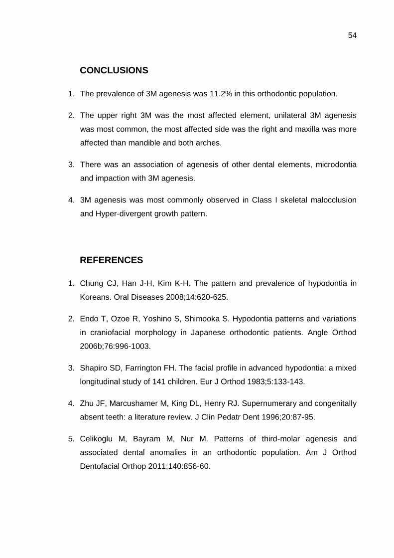
54
CONCLUSIONS
1. The prevalence of 3M agenesis was 11.2% in this orthodontic population.
2. The upper right 3M was the most affected element, unilateral 3M agenesis
was most common, the most affected side was the right and maxilla was more
affected than mandible and both arches.
3. There was an association of agenesis of other dental elements, microdontia
and impaction with 3M agenesis.
4. 3M agenesis was most commonly observed in Class I skeletal malocclusion
and Hyper-divergent growth pattern.
REFERENCES
1. Chung CJ, Han J-H, Kim K-H. The pattern and prevalence of hypodontia in
Koreans. Oral Diseases 2008;14:620-625.
2. Endo T, Ozoe R, Yoshino S, Shimooka S. Hypodontia patterns and variations
in craniofacial morphology in Japanese orthodontic patients. Angle Orthod
2006b;76:996-1003.
3. Shapiro SD, Farrington FH. The facial profile in advanced hypodontia: a mixed
longitudinal study of 141 children. Eur J Orthod 1983;5:133-143.
4. Zhu JF, Marcushamer M, King DL, Henry RJ. Supernumerary and congenitally
absent teeth: a literature review. J Clin Pedatr Dent 1996;20:87-95.
5. Celikoglu M, Bayram M, Nur M. Patterns of third-molar agenesis and
associated dental anomalies in an orthodontic population. Am J Orthod
Dentofacial Orthop 2011;140:856-60.

55
6. Celikoglu M, Kamak H. Patterns of third-molar agenesis in an orthodontic
patient population with different skeletal malocclusions. Angle Orthod
2012;82:165-169.
7. García-Hernández F, Toro YO, Veja VM, Verdejo MM. Agenesia del Tercer
Molar en Jóvenes entre 14 y 20 Ãnos de Edad, Antofagasta, Chile. Int. J.
Morphol. 2008;26(4):825-832.
8. Barka G, Marathiotis K, Protogerakis M, Zafeiriadis A. Radiographic evaluation
of third molar genesis in Greek orthodontic patients. International Journal of
General Medicine 2013;6:747-755.
9. Antanas S, Giedrè T. Effect of the lower third molars on the lower dental arch
crowding. Stomatologija, Baltic Dental and Maxilofacial Journal
2006;Vol8(3):80-84.
10. Karasawa L-H, Rossi A-C, Groppo F-C, Prado F-B, Caria P-H-F. Cross-
sectional study of correlation between mandibular incisor crowding and third
molars in Young Brazilians. Med Oral Patol Oral Cir Bucal
2013;May1:18(3):e505-9.
11. Jr ARS, Singh I. The unresolved problem of the third molar – Would people be
better off without it? The Journal of the American Dental Association
2003;April134:450-455.
12. Pitekova L, Satko L. Controversy of the third molars. Bratisl Lek Listy
2009;110(2):110-1.
13. Steiner CC. Cephalometrics for you and me. Am J Orthod 1953;39:729-55.
14. Jung Y-H, Cho B-H. Radiographic evaluation of third molar development in 6-
to 24-year-olds. Imaging Science in Dentistry 2014;44:185-91.
15. Alam MK, Hamza MA, Khafiz MA, Rahman SA, Shaari R, Hassan A.
Multivariate Analysis of Factors Affecting Presence and/or Agenesis of Third
Molar Tooth. Plos One 2014;Vol9(6):e101157.

56
16. Autores Japoneses. A Statistic Analysis on Absence of Third Molar Germs in
Orthodontic Patients. West China Journal of Stomatology 2004;Vol22(6):493-
495.
17. Brook AH. A unifying aetiological explanation for anomalies of human tooth
number and size. Arch Oral Biol 1984;29:373-8.
18. Garn SM, Lewis AB, Vinicius JH. Third molar agenesis and reduction in the
number of other teeth. J Dent Res 1962;41:717.
19. Jakovljevic A, Lazic E, Soldatovic I, Nedeljkovic N, Andric M. Radiographic
assessment of lower third molar eruption in different anteroposterior skeletal
patterns and age-related groups. Angle Orthod 2014;Sep22[Epub ahead of
print].

57
Note: Sn-GoGn angle.
. Sn-GoGn Angle < 32° = Hypo-divergent;
. Sn-GoGn Angle = 32° = Normal;
. Sn-GoGn Angle > 32° = Hyper-divergent
Figure 1. Angles used in the Characterization of Growth Patterns
Note: Angles SNA and SNB. ANB = SNA – SNB
. ANB Angle with values between 0° and 4° = Class I;
. ANB Angle with values > 4° = Class II;
. ANB Angle with values < 0° = Class III.
Figure 2. Angles used in the Characterization of Skeletal Classification Patterns

58
TABLES
Table 1. Population Characteristics according to Gender, Ethnicity, Angles Average (SNA, SNB, ANB and Sn-GoGn) and Age – Patients with and without 3M Agenesis
Total n 3M Agenesisc No 3M Agenesisc p value
831a 93 (11.2%) 738 (88.8%)
Gender n (%) 0.754*
Male 388 (46.7) 42 (45.2) 346 (46.9)
Female 443 (53.3) 51 (54.8) 392 ( 53.1)
Ethnicityb 0.56*
Caucasian 400 (48.1) 49 (52.7) 351 (47.6)
African Descent 305 (36.7) 29 (31.2) 276 (37.4)
Angles Average
Mean (DP)
SNA 82.62 (± 4.4) 83.02 (± 4.35) 82.57 (± 4.42) 0.35**
SNB 79.38 (± 4.6) 80.08 (± 4.99) 79,29 (± 4.51) 0.12**
ANB 3.25 (± 3.8) 2,96 (± 4.01) 3.28 (± 3.82) 0.44**
Sn-GoGn 37.07 (±6.3) 36.9 (± 7.9) 37.09 (± 6.06) 0.79**
Age 14.11 (± 7.1) 14 (± 6.4) 14.12 (± 7.19) 0.876**
Notes: *Indicates Qui Square Test;**Indicates Student t Test; All p values were nonsignificant (p>0.05);
216 patients were excluded because it was not possible to diagnose 3M agenesisa;
It was not possible to classified 126 (15.2%) patients between ethnicity and they were excludedb;
3M indicates Third Molarc.

59
Table 2. Distribution of Dental Anomalies between the Groups – 3M Agenesis and No 3M Agenesis
3M Agenesisc
(%)
93b (11.2)
No 3M Agenesisc
(%)
738b (88.8)
p value
Odds Ratio (Confidence Interval)
Dental Anomalies n (%)
Agenesis 16 (17.2) 41 (5.6) >0.01a 3.52 (1.89 – 6.59)
Supernumerary 3 (3.2) 24 (3.3) 0.989 0.99 (0.29 – 3.36)
Microdontia 11 (11.8) 24 (3.3) >0.01a 3.99 (1.89 – 8.44)
Macrodontia ----- 2 (0.3) 0.615 1.13 (1.09 – 1.15)
Impaction 38 (40.9) 478 (64.8) >0.01a 3.3 (1.72 – 4.16)
Transposition 2 (2.2) 29 (3.9) 0.394 0.54 (0.13 – 2.29)
Giroversion 35 (37.6) 327 (44.3) 0.221 0.76 (0.49 – 1.18)
Root Dilaceration 2 (2.2) 18 (2.4) 0.864 0.88 (0.20 – 3.85)
Crown Dilaceration ----- 1 (0.1) 0.722 1.13 (1.10 – 1.15)
Odontoma 1 (1.1) 1 (0.1) 0.081 8.01 (0.50 – 129.16d)
Taurodontism ----- 1 (0.1) 0.722 1.13 (1.10 – 1.15)
Notes: P value is based on Qui Square Test;
Statistically significant (p<0.05)a;
216 patients were excluded because it was not possible to diagnose 3M agenesisb;
3M indicates Third Molarc;
Unstable numbers due to low frequencyd.

60
Table 3. Characteristics Features of 3M Agenesis in the Different Groups according to the Number of Teeth Absence
GROUPS
(Total number of
teeth absent)
1Teeth
Agenesis
(47)
2 Teeth
Agenesis
(50)
3 Teeth
Agenesis
(18)
4 Teeth
Agenesis
(60)
3M
Agenesisa
(175)
Teeth n (%)
UR 3M 19 (40.4) 16 (64) 4 (66.7) 15 (100) 54 (58.1)
UL 3M 8 (17) 15 (60) 3 (50) 15 (100) 41 (44.1)
LL 3M 11 (23.4) 9 (36) 6 (100) 15 (100) 41 (44.1)
LR 3M 9 (19.1) 10 (40) 5 (83.3) 15 (100) 39 (4.1)
Laterality Patients n (%)
Unilateral 47 (100) 1 (4) ----- ----- 48 (51.6)
Bilateral ----- 24 (96) 6 (100) 15 (100) 45 (48.4)
Affected Side Patients n (%)
Right 28 (59.6) 1 (4) ----- ----- 29 (31.2)
Left 19 (40.4) ----- ----- ----- 19 (20.4)
Both ----- 24 (96) 6 (100) 6 (100) 45 (48.4)
Affected Arch Patients n (%)
Maxilla 27 (57.4) 14 (56) ----- ----- 41 (44.1)
Mandible 20 (42.6) 8 (32) ----- ----- 28 (30.1)
Both ----- 3 (12) 6 (100) 15 (100) 24 (25.8)
Skeletal
Classification Patients n (%)
Class I* 17 (36.2) 12 (48) 3 (50) 8 ( 53.3) 40 (43)
Class II* 21 (44.7) 10 (40) 2 (33.3) 2 (13.3) 35 (37.6)
Class III* 9 (19.1) 3 (12) 1 (16.7) 5 (33.3) 18 (19.4)
Growth Pattern Patients n (%)
Hypo-divergent* 9 (19.1) 4 (16) 2 (33.3) 5 (33.3) 20 (21.5)
Normal* 1 (2.1) 4 (16) ----- 1 (6.7) 6 (6.5)
Hyper-divergent* 37 (78.7) 17 (68) 4 (66.7) 9 (60) 67 (72)
Note: 3M indicates Third Molara.
UR means Upper Right; UL means Upper Left; LR means Lower Right; LL means Lower Left. Not statistically significant (p>0.05)*

61
5. DISCUSSÃO
As AD são alterações clínicas frequentemente encontradas na rotina da
clínica odontológica (Garib, Filho et al., 2013), que causam problemas funcionais,
oclusais e estéticos (Kathariya, Nikam et al., 2013;Osuji e Hardie, 2002), resultando
em prejuízos na saúde oral destes pacientes (Kim, 2011). A ME caracteriza-se por
uma complexa alteração de crescimento no complexo craniofacial (Andrews, 1972) e
tem sido associada com as AD (Celikoglu e Kamak, 2012), dificultando ainda mais o
seu tratamento quando em presença de AD associada.
O estudo destes pacientes pode servir como base para futuros estudos
genéticos e ajudar na elucidação de sua etiologia (Uslu, Ackam et al., 2009; Basdra,
Kiakpasoglou et al., 2000; Basdra, Kiakpasoglou et al., 2009). Assim, esses recursos
podem servir como uma resposta à sua origem e desenvolvimento (Basdra,
Kiakpasoglou et al., 2001). Estudos relataram a associação entre AD e ME (Kücler,
Risso et al., 2008; Basdra, Kiakpasoglou et al., 2001) e seu fundo genético é
amplamente conhecido (Díaz-Perez e Echaverry-Navarrete, 2009). Assim, é
provável que seja possível verificar a genes candidatos específicos de ME e AD
quando há associação entre eles (Basdra, Kiakpasoglou et al., 2000). A correlação
genótipo-fenótipo é extremamente importante para melhorar o conhecimento da sua
etiologia, o que pode contribuir com um diagnóstico mais preciso.
A agenesia de 3M é considerada uma evolução da espécie humana (García-
Hernández, Yagui et al., 2008), devido ao seu pequeno valor funcional (Jung e Cho,
2014; Alam, Hamza et al., 2014). De acordo com alguns autores, a agenesia de 3M
poderia melhorar os cuidados de saúde e qualidade de vida de milhões de pessoas
(Jung e Cho, 2014). Além disso, é preferível que o paciente tenha agenesia de 3M,
do ponto de vista ortodôntico. Assim, diante da complexidade que envolve estes
pacientes, este foi o primeiro estudo que buscou investigar as AD associadas aos
PCF e as ME, bem como avaliar a agenesia dos 3M em uma população ortodôntica.

62
Este é um estudo censitário que envolve todos os pacientes com
documentação ortodôntica completa, atendidos nos Departamentos de Ortodontia,
de duas instituições referências em Rio de Janeiro, Brasil, de 2000 a 2013. Essas
instituições são consideradas centros de referência em Ortodontia na cidade, devido
à atividades de pesquisa e tratamentos desenvolvidos por seus membros. Espera-se
que os resultados apresentados neste estudo forneçam informações relevantes para
a caracterização desta população e para o desenvolvimento de um plano de
tratamento adequado para esses pacientes, visando um bom prognóstico.
s principais limitações do estudo foram os 216 pacientes impossibilitados de
receber o diagnóstico de agenesia de 3M, os 177 pacientes que não foram
classificados dentre as etnias e a exclusão de 31,16% da amostra, devido aos
protuários odontológicos incompletos. As radiografias foram de extrema importância
para os critérios de inclusão e diagnóstico de pacientes ortodônticos. Nossos
resultados sugerem que as AD são facilmente detectados por exames radiográficos
de rotina e deve ser incluído no planejamento do tratamento ortodôntico permitindo,
inclusive, o diagnóstico precoce e a intervenção ortodôntica em tempo hábil.
Ambos os artigos tiveram metodologias similares. A única diferença foi a
inclusão do diagnóstico de agenesia de 3M no artigo 2. Para isso, alguns critérios de
inclusão, como a idade do paciente, tiveram que seguir metodologias de estudos
prévios. De acordo com Barka, Marathiotis et al. (2013), a idade para a primeira
detecção 3M na radiografia panorâmica é aos 07 anos para o gênero feminino e aos
08 anos, para o masculino. Em outro estudo, observou-se o aparecimento de 3M já
aos 06 anos de idade (Jhung e Cho, 2014). Para evitar possíveis variações nesta
cronologia, a amostra foi padronizado a partir de 08 anos de idade,
independentemente do gênero.

63
Thongudomporn e Freer (1998), observaram 74,7% de AD em 111 pacientes
ortodônticos e atribuíram este achado ao tamanho da amostra, pois pequenas
amostras tendem a ser menos confiáveis. No presente estudo, observou-se alta
prevalência semelhante (77%), porém acredita-se que isso aconteceu por conta do
envolvimento das duas instituições de referência em Ortodontia. Este fato pode ter
enriquecido a população com AD.
No presente estudo, a AD mais prevalente foi a impactação (68,6%), seguida
pela giroversão (54%). De acordo com o estudo de Kathariya, Nikam et al. (2013),
estas AD foram previamente descritas como as mais frequentes, mas com uma
prevalência consideravelmente mais baixa (39,6 e 13,2%, respectivamente), quando
comparadas com o presente estudo. Possivelmente, isto ocorre em função de
diferenças na classificação do diagnóstico aplicados às AD nos diferentes estudos,
como a impactação do 3M, por exemplo. Neste trabalho, o diagnóstico de
impactação foi baseado nos critérios de Uslu et al. (2009).
Duas principais teorias têm sido sugeridas para explicar impactação: a teoria
de orientação, que se baseia em causas predisponentes locais e da teoria genética,
que considera uma causa genética para a impactação (Sambataro, Baccetti et al.,
2004). No presente estudo, os dentes mais afetados pela impactação foram os 3M
inferiores. Este achado pode ser explicado por conta da falta de espaço na arcada
dentária inferior, possivelmente causada pela diminuição do desenvolvimento
craniofacial horizontal. Um outro resultado importante dste estudo, foi a associação
da agenesia com o PCF Hipodivergente. Esta associação pode ser explicada por
uma possível perturbação na proliferação e no desenvolvimento, o que resulta em
mudanças onde ocorre a falta de crescimento. Assim, podemos supor que os
mecanismos genéticos podem compartilhar caminhos similares.

64
6. CONCLUSÕES
Com base nos resultados obtidos a partir da avaliação entre a prevalência,
distribuição e dimorfismo sexual de AD entre os diferentes PCF e ME em uma
população ortodôntica é lícito concluir que:
A prevalência das AD foi de 77% na população estudada;
As AD mais prevalentes foram a impactação, seguida da giroversão e
agenesia de outros dentes, excluindo o 3M. Considerando tais anomalias, os
dentes mais acometidos foram o 3M inferior esquerdo, o canino inferior direito
e o incisivo lateral superior direito;
A prevalência de agenesia de 3M foi de 11,2%. O 3M superior direito foi o
dente mais acometido, a agenesia unilateral foi a mais comum e o lado direito
e a maxila foram os mais afetados;
A fusão e a impactação foram associadas com o gênero masculino e a
agenesia de 3M, com o gênero feminino;
A agenesia de 3M foi associada com a agenesia de outros dentes,
microdontia e com a impactação numa relação inversa;
A agenesia de outros dentes, excluindo os 3M, foi associada com o PCF
Hipodivergente e a microdontia, com a ME de Classe III.

65
REFERÊNCIAS BIBLIOGRÁFICAS
Alam, M. K, Hamza, M.A., et al. Multivariate Analysis of Factors Affecting Presence and/or Agenesis of Third Molar Tooth. Plos One, v.9, n.6, p.e101157, 2014.
Altug-Atac, A.T., Erdem, D. Prevalence and distribution of dental anomalies in orthodontic patients. Am J Orthod Dentofacial Orthop, v.131, p.510-4, 2007.
Andrews, L. F. The six keys to normal occlusion. Am J Orthod, v.62, n.3, p.296-309, 1972.
Angle, E. H. Classification of malocclusion. Dent Cosmos, v.41, p.248-64, 1899.
Antanas, S., Giedrè, T. Effect of the lower third molars on the lower dental arch crowding. Stomatologija, Baltic Dental and Maxilofacial Journal, v. 8, n.3, p.80-84, 2006.
Arashiro, C., Ventura, M. L. S, et al. Prevalência de maloclusão em escolares do município de Campinas, São Paulo. RGO, Porto Alegre, v.57, n.4, p.407-411, out./dez, 2009.
Autores Japoneses. A Statistic Analysis on Absence of Third Molar Germs in Orthodontic Patients. West China Journal of Stomatology, v.22, n.6, p.493-495, 2004.
Barka, G., Marathiotis, K., et al. Radiographic evaluation of third molar genesis in Greek orthodontic patients. International Journal of General Medicine, v.6, p.747-755, 2013.
Basdra, E. K., Kiokpasoglou, M., et al. The Class II division 2 craniofacial type is associated with numerous congenital tooth anomalies. Eur J Orthod, v.22, p.529-35, 2000.
Basdra, E.K., Kiopasoglou, M.N., et al. Congenital tooth anomalies and malocclusions: a genetic link?. Eur J Orthod, v.23,p.145-151, 2001.
Brook, A.H. A unifying aetiological explanation for anomalies of human tooth number and size. Arch Oral Biol, v.29, p.373-8, 1984.
Celikoglu, M., Bayram, M., et al. Patterns of third-molar agenesis and associated dental anomalies in an orthodontic population. Am J Orthod Dentofacial Orthop, v.140, p.856-60, 2011.
Celikoglu, M., Kamak, H. Patterns of third-molar agenesis in an orthodontic patient population with different skeletal malocclusions. Angle Orthod, v.82, p.165-169, 2012.
Chung, C. J., Han, J-H., et al. The pattern and prevalence of hypodontia in Koreans. Oral Diseases, v.14, p.620-625, 2008.

66
Díaz-Pérez, R., Echaverry-Navarrete, R. A. Agenesia en dentición permanente. Rev. salud pública, v.11, n.6, p.961-969, 2009.
Endo, T., Ozoe, R., et al. Hypodontia patterns and variations in craniofacial morphology in Japanese orthodontic patients. Angle Orthod, v.76, p.996-1003, 2006b.
García-Hernández, F., Toro, Y. O., et al. Agenesia del Tercer Molar en Jóvenes entre 14 y 20 Ãnos de Edad, Antofagasta, Chile. Int. J. Morphol, v.26, n.4, p.825-832, 2008.
Garib, D. G., Filho, O. G. S, et al. Ortodontia interceptativa: protocolo de tratamento em duas fases. São Paulo, Artes Médicas, p.574, 2013.
Garn, S.M., Lewis, A. B., et al. Third molar agenesis and reduction in the number of other teeth. J Dent Res, v.41, p.717, 1962.
Jakovljevic, A., Lazic, E., et al. Radiographic assessment of lower third molar eruption in different anteroposterior skeletal patterns and age-related groups. Angle Orthod, Sep22, [Epub ahead of print], 2014.
Jr, A. R. S., Singh, I. The unresolved problem of the third molar – Would people be better off without it? The Journal of the American Dental Association, April, v.134, p.450-455, 2003.
Jung, Y-H., Cho, B-H. Radiographic evaluation of third molar development in 6- to 24-year-olds. Imaging Science in Dentistry, v.44, p.185-91, 2014.
Karasawa, L-H., Rossi. A-C., et al. Cross-sectional study of correlation between mandibular incisor crowding and third molars in Young Brazilians. Med Oral Patol Oral Cir Bucal, May1, v. 18, n.3, p.505-9, 2013.
Kathariya, M. D., Nikam, A. P., et al. Prevalence of Dental Anomalies among School Going Children in India. J Int Oral Health, v.5, n.5, p.10-4, 2013.
Kim, Y. H. Investigation of Hypodontia as Clinically Relates Dental Anomaly: Prevalence and Characteristics. ISRN Dentistry, Article ID 246135, 6 pages, 2011.
Küchler, E. C., Risso, P. A., et al. Studies of dental anomalies in a large group of school children. Archives of Oral Biology, v.53, p.941-946, 2008.
Mossey, P. A. The heritability of malocclusion: part 2. The influence of genetics in malocclusion. Br J Orthod, v.26, p.195–203, 1999.
Neville, B. W, Damm, D. D., et al. Patologia Oral e Maxilofacial. Rio de Janeiro, Guanabara Koogan, segunda edição, p.49-87, 2004.
Osuji, O. O., Hardie, J. Dental Anomalies in a population of Saudi Arabian children in Tabuk. Saudi Dent J, v.14, n.1,p.11-4, 2002.

67
Pires, D. M., Rocha, M. C., et al. Prevalência de oclusopatias na dentadura mista em escolares -Salvador/ BA. Rev Bras Odontol, v.58, n.6, p.414-7, 2001.
Pitekova, L., Satko, L. Controversy of the third molars. Bratisl Lek Listy, v.110, n.2, p.110-1, 2009.
Sambataro, S., Baccetti, T., et al. Early predictive variables for upper canine impaction as derived from posteroanterior cephalograms. Angle Orthod, v.75, p.28-34, 2004.
Shapiro, S.D., Farrington, F. H. The facial profile in advanced hypodontia: a mixed longitudinal study of 141 children. Eur J Orthod, v.5, p.133-143, 1983.
Steiner, C. C. Cephalometrics for you and me. Am J Orthod, v.39, p.729-55, 1953.
Thongudomporn, U., Freer, T. J. Prevalence of dental anomalies in orthodontic patients. Aus Dent J, v.43, p.395-8, 1998.
Uslu O., Akcam M. O., et al. Prevalence of dental anomalies in various malocclusions. Am J Orthod Dentofacial Orthop, v.135, p.328-35, 2009.
Zhu, J. F., Marcushamer, M., et al. Supernumerary and congenitally absent teeth: a literature review. J Clin Pedatr Dent, v.20, p. 87-95, 1996.

68
ANEXOS
ANEXO 1
Aprovação do Comitê de Ética em Pesquisa

69
ANEXO 2 Declaração do Chefe do Departamento de Ortodontia da Universidade
Federal do Rio de Janeiro (UFRJ)

70
ANEXO 3
Declaração dos Chefes do Departamento de Ortodontia da Associação
Brasileira de Odontologia do Rio de Janeiro (ABO RJ)

71
ANEXO 4
Ficha de Coleta de Dados
PRONTUÁRIO Nº: ________________ INSTITUIÇÃO: ________________
GÊNERO: ( ) MASCULINO ( ) FEMININO IDADE: _______________
ETNIA: ( ) CAUCASIANA ( ) PARDA ( ) NEGRA
( ) EM TRATAMENTO ( )PÓS TRATAMENTO DENTIÇÃO: ( ) MISTA ( ) PERMANENTE
ANÁLISE RADIOGRÁFICA:
RADIOGRAFIA CEFALOMÉTRICA DE PERFIL:
SNA: _______ SNB:_______ ANB:________ SN-GOGN: ________
TIPO DE MALOCLUSÃO ESQUELÉTICA HORIZONTAL: ( ) CLASSE I ( ) CLASSE II ( ) CLASSE III
TIPO DE MALOCLUSÃO ESQUELÉTICA VERTICAL: ( ) HIPO DIVERGENTE ( ) NORMAL ( ) HIPER
DIVERGENTE
RELAÇÃO MOLAR: ( ) CLASSE I ( ) CLASSE II ( )CLASSE III
SUBDIVISÃO: ( ) DIREITA( ) ESQUERDA
RELAÇÃO CANINA: ( ) CLASSE I ( ) CLASSE II ( )CLASSE III
SUBDIVISÃO: ( ) DIREITA( ) ESQUERDA
RADIOGRAFIA PANORÂMICA:
( ) APINHAMENTO ( ) DIASTEMA
ELEMENTOS
ENVOLVIDOS:_________________________________________________________

72
ANOMALIA DENTÁRIA: ( ) SIM ( ) NÃO
TIPO DE ANOMALIA / ELEMENTO(S) ENVOLVIDO(S):
_____________________________________
__________________________________________________________________________
__________________________________________________________________________
AGENESIA: ( ) SIM ( ) NÃO ELEMENTOS ENVOLVIDOS:
_________________________________
( ) UNI ( ) BILATERAL( ) DIR. ( ) ESQ. ( ) MÁX. ( ) MAND.
AGENESIA DE TERCEIRO MOLAR: ( ) SIM ( ) NÃO ELEMENTOS:
_____________________________
( ) UNI ( ) BILATERAL( ) DIR. ( ) ESQ. ( ) MÁX. ( ) MAND.
Total de Anomalias Dentárias: ______ Total de Dentes Afetados: _______


![(Aula anomalias dentárias [Modo de Compatibilidade])](https://static.fdocumentos.com/doc/165x107/5571fdec49795991699a4042/aula-anomalias-dentarias-modo-de-compatibilidade.jpg)