CARLA BRUNA PIETROBON HOMEOSTASE GLICÊMICA DA …tede.unioeste.br/bitstream/tede/3228/5/CARLA_...
100
UNIVERSIDADE ESTADUAL DO OESTE DO PARANÁ – CAMPUS DE CASCAVEL CENTRO DE CIÊNCIAS BIOLÓGICAS E DA SAÚDE PROGRAMA DE PÓS-GRADUAÇÃO EM BIOCIÊNCIAS E SAÚDE – NÍVEL MESTRADO CARLA BRUNA PIETROBON HOMEOSTASE GLICÊMICA DA PROLE DE RATAS OBESAS SUBMETIDAS À OPERAÇÃO BARIÁTRICA CASCAVEL-PR (março/2017)
Transcript of CARLA BRUNA PIETROBON HOMEOSTASE GLICÊMICA DA …tede.unioeste.br/bitstream/tede/3228/5/CARLA_...
UNIVERSIDADE ESTADUAL DO OESTE DO PARANÁ – CAMPUS DE CASCAVEL
CENTRO DE CIÊNCIAS BIOLÓGICAS E DA SAÚDE
PROGRAMA DE PÓS-GRADUAÇÃO EM BIOCIÊNCIAS E SAÚDE – NÍVEL
MESTRADO
CARLA BRUNA PIETROBON
HOMEOSTASE GLICÊMICA DA PROLE DE RATAS OBESAS SUBMETIDAS À
OPERAÇÃO BARIÁTRICA
CASCAVEL-PR
(março/2017)
CARLA BRUNA PIETROBON
HOMEOSTASE GLICÊMICA DA PROLE DE RATAS OBESAS SUBMETIDAS À
OPERAÇÃO BARIÁTRICA
Dissertação apresentada ao Programa de Pós-graduação em Biociências e Saúde – Nível Mestrado, do Centro de Ciências Biológicas e da Saúde, da Universidade Estadual do Oeste do Paraná, como requisito parcial para obtenção do título de Mestre em Biociências e Saúde. Área de concentração: Biologia, processo saúde-doença e políticas da saúde. ORIENTADORA: Profa. Dra. Maria Lúcia Bonfleur CO-ORIENTADOR: Prof. Dr. Allan Cezar Faria Araújo
CASCAVEL-PR
(março/2017)


DEDICATÓRIA
Dedico esta, bem como todas as minhas
conquistas primeiramente a Deus, aos meus
pais, a minha família, ao meu namorado, meus
amigos e professores pela amizade, incentivo,
apoio e dedicação. Tudo isso foi essencial para
cumprir mais uma etapa da minha vida.
AGRADECIMENTOS
Primeiramente, gostaria de agradecer a Deus pela minha vida, por me
iluminar, me guiar e me dar força para seguir em frente e concluir mais essa etapa
da minha vida.
Gostaria de agradecer em especial a minha orientadora, professora Dra.
Maria Lúcia Bonfleur, que me acolheu, mesmo sem me conhecer. Obrigada pelo seu
apoio, sua amizade, confiança, incentivo e, além de sua dedicação e competência
com o nosso trabalho. Maria Lúcia, você é incrível e eu serei eternamente grata aos
seus ensinamentos. Hoje, saio do mestrado levando de você como uma grande
amiga.
A professora Dra. Sandra Balbo, que com esse jeito extrovertido me ensinou
que a vida por mais difícil que seja, no final as coisas vão dar certo e vão valer a
pena. Sandra você é sensacional, o LAFEM jamais seria o mesmo sem você.
A todos os professores do programa que fizeram parte do meu crescimento
profissional e pessoal.
A todas as meninas do laboratório, por todo companheirismo diário.
A Camila Lubaczeuski pela ajuda e dedicação neste trabalho.
A Iala, que desde o primeiro dia do mestrado, me acolheu eme ensinou tudo
sobre o laboratório. Obrigada pelo seu companheirismo e apoio. Com certeza, a
nossa amizade vai além do mestrado, além do laboratório, além da Unioeste, é pra
vida toda. E agora mais ainda, se tudo der certo, por mais quatro anos como
parceiras de doutorado!
À pós-doutoranda Dr. Ana Claudia. Obrigada por estar sempre pronta para
me ajudar, esclarecer minhas dúvidas. Obrigada pelos conselhos, pelos puxões de
orelha, pelas conversas, pelas risadas, você é uma pessoa incrível. Ana, os
ensinamentos que aprendi com você certamente hoje me fazem uma pessoa melhor.
A minha família, em especial meus pais. Obrigada por ser a minha base, meu
alicerce. O que dizer de vocês? Nunca me negaram ajuda, sempre me incentivam,
me apoiam e me ajudam a passar pelos momentos de dificuldades. Vocês com toda
a certeza vocês são o motivo dessa conquista. Eu devo tudo isso aos meus pais.
Serei eternamente grata. Eu amo muito vocês!
Ao meu namorado, Fernando. Você foi essencial para que eu cumprisse as
minhas obrigações. Quando eu queria desistir você estava do meu lado, me
incentivando, me apoiando. Obrigada pelo companheirismo, paciência,
compreensão, por aguentar as crises de desespero, de angustia, de nervosismo.
Obrigada por ser uma pessoa maravilhosa comigo. Te amo além do que você possa
imaginar.
A todos os meus amigos, que mesmo distantes sempre torceram por mim.
A todos que de alguma forma contribuíram para a minha formação.
Com certeza vocês foram essenciais para que eu cumprisse mais uma etapa
da minha vida.
Obrigada!
RESUMO GERAL
Atualmente tem-se observado o aumento da obesidade feminina em todo o mundo. Como consequência desses altos índices, o número de mulheres em idade fértil que buscam as operações bariátricas tem aumentado consideravelmente nos últimos anos. No entanto, poucos estudos demonstram os efeitos das operações bariátricas maternas sobre o metabolismo dos filhos. Neste estudo, avaliamos os efeitos da derivação gástrica em Y de Roux (DGYR) em ratas Wistar, alimentadas com dieta de cafeteria (CAF), sobre os parâmetros corporais e homeostase glicêmica na prole de macho adulto. Para isso, as ratas Wistar receberam dieta de CAF durante 15 semanas para indução da obesidade. Após esse período, as ratas foram submetidas à falsa operação (FO) ou à operação de DGYR, compondo 2 grupos: CAF FO (n=14) e CAF DGYR (n= 20). Cinco semanas após os procedimentos operatórios, as ratas foram submetidas ao cruzamento. A prole (F1) foi nomeada conforme o procedimento experimental realizado nas mães: CAF FO-F1 (n= 28) e CAF DGYR (n= 14) os quais receberam dieta padrão durante todo o procedimento experimental. O desmame da prole foi realizado aos 30 dias de vida. O comprimento naso-anal (CNA), o peso corporal, o consumo e a eficiência alimentar foram verificados semanalmente durante todo o experimento. Uma semana antes da eutanásia, os animais foram submetidos ao teste de tolerância à glicose (GTT) e à insulina (ITT). Com 120 dias de vida, os animais foram mortos e os parâmetros corporais, os parâmetros séricos e a morfofisiologia do pâncreas foram verificados. Após a DGYR, as ratas apresentaram redução no peso corporal, na tolerância à glicose, na resistência à insulina, e no peso das gorduras. Apesar destes benefícios maternos, os descendentes de mães submetidas à DGYR apresentaram redução do peso e do tamanho, bem como diminuição no consumo alimentar em comparação aos descendentes de mães obesas. Apesar destes animais não apresentarem alterações na tolerância à glicose e normoglicemia, a sensibilidade à insulina foi maior. Além disso, a sinalização da insulina no músculo esquelético foi reduzida. Os ratos descendentes de mães submetidas à DGYR demostraram redução na insulinemia e na secreção de insulina, entretanto, a área e a massa de células-β foi maior em comparação aos animais descendentes de mães obesas. Diante disso, concluímos que a DGYR materna em ratas obesas altera na prole os parâmetros corporais e leva a alterações morfofuncionais nas células-β pancreáticas. Palavras-chave: Derivação gástrica em Y de Roux; secreção de insulina; efeitos maternos.
GENERAL ABSTRACT
Glucose homeostasis of the offspring of obese rats submitted to bariatric operation
There has been an increase in female obesity worldwide. As a consequence of these high rates the number of women of child-bearing age who seek bariatric operations has increased considerably in recent years. However, few studies showed the effects of maternal bariatric operation in their offspring. Herein, we evaluated the effects of maternal Roux-in-Y Bypass Gastric (RYBG) in Wistar rats fed cafeteria diet (CAF) upon body features and glucose homeostasis in male adult offspring. For this, rats Wistar received CAF diet for 15 weeks for obesity induction. After this period, the rats were submitted to sham operation (CAF Sham), group CAF sham (n=14) or RYGB operation, group CAF RYGB (n=20). Five weeks after the procedure, the rats were submitted at matting period. The offspring were named as operation your mothers: CAF Sham-F1 (n=28) and CAF RYGB-F1 (n=14), and received standard diet. The weaning from offspring was performed at 30 days of life. Nasoanal length, body weight, consumption and feed efficiency were evaluated weekly throughout experiment. One week before euthanasia, the animals were submitted to the glucose tolerance test and insulin. At 120 days of life, body parameters, serum parameters and morphophysiology of the pancreas were evaluated. After RYBG, female rats displayed reduction in body weight, glucose tolerance and insulin, as well as, decreased fat deposits. Despite of these maternal benefits, the offspring of dams receiving RYBG were smaller and lighter and showed a reduction in food intake than offspring of obese dams that underwent sham surgery. Although these animals presented normal glucose tolerance and glycemia, the insulin sensitivity was higher. In addition, the insulin signaling in liver and fat tissue in these offspring was normal but in skeletal muscle was reduced. Also, the offspring of dams receiving RYBG presented reduction in insulinemia and insulin secretion, but the area and mass of β-cell was higher compared to offspring of obese dams that underwent sham surgery. Maternal RYBG in obese rats alter in male offspring the body features and lead to morphofunctional alterations in the β-cells.
Key words: Roux-in-Y Bypass Gastric; Insulin secretion; Maternal effects
SUMÁRIO
LISTA DE ABREVIATURAS .................................................................................. 4
INTRODUÇÃO GERAL ......................................................................................... 5
REVISÃO GERAL DE LITERATURA .................................................................... 8
Obesidade .......................................................................................................... 8
Secreção e ação da insulina ..............................................................................10
Diabetes mellitus tipo 2 ......................................................................................13
Programação metabólica ...................................................................................16
Modelo de obesidade ........................................................................................20
Operação bariátrica ...........................................................................................21
REFERÊNCIAS BIBLIOGRÁFICAS .....................................................................25
ARTIGO CIENTÍFICO ...........................................................................................42
MATERNAL ROUX-EN-Y GASTRIC BYPASS OPERATION ALTERS BODY
FEATURES AND PROMOTES PANCREAS MORFOFUNCTIONAL CHANGES IN
ADULT OFFSPRING .............................................................................................42
ANEXO A ..............................................................................................................79
Ética Comitê de Ética na Experimentação Animal e Aulas Práticas da
Universidade Estadual do Oeste do Paraná. .....................................................79
ANEXO B ..............................................................................................................80
Normas da revista científica ...............................................................................80
4
LISTA DE ABREVIATURAS
AC – Adenilato ciclase ADP – Adenosina difosfato AKT – Proteína quinase B AMPc – Adenosina monofosfato cíclico ATP – Adenosina trifosfato AUC – Área abaixo da curva CAF – Cafeteria CAF DGYR – Cafeteria derivação gástrica em Y de Roux CAF FO – Cafeteria falsa operação EEAAP – Comitê de ética na experimentação animal e aulas práticas CNA – Comprimento naso-anal CTL – Grupo controle DAG – Diacilglicerol DGYR – Derivação gástrica em Y de Roux DM – Diabetes mellitus DM1 – Diabetes mellitus tipo 1 DM2 – Diabetes mellitus tipo 2 DMG – Diabetes mellitus gestacional DNA -Ácido disoxirribonucléico DPB – Derivação biliopancreática GIP – Peptídeo inibidor gástrico GK – Glicoquinase GLP-1 – Peptídeo semelhante ao glucagon 1 GLUT2 – Transportador de glicose tipo 2 GLUT4 – Transportador de glicose tipo 4 GSK-3 – Glicogênio sintase quinase-3 GV – Gastrectomia vertical IDF – Federação Internacional do Diabetes IGF-1 – Fator de crescimento semelhante à insulina 1 IMC – Índice de massa corporal IP – Intraperitonial IP3 – Inositol-1,4,5 trifosfato IpGTT – Teste intraperitoneal de tolerância à glicose
IR – Receptor de insulina IRS – Substrato do receptor de insulina ITT – Teste intraperitoneal de tolerância à insulina KITT – Constante decaimento da glicose MCP-1 - Proteína quimioatrativa de monócitos-1 MS – Ministério da Saúde NHANES –National Health and Nutrition Examination Survey PDK-1 – Proteína quinase dependente de fosfoinositídeos-1 PH –Domínio de homologia plecstrina PI3K – fosfatidilinositol 3-quinase PIP3 – fosfatidil inositol trifosfato PKA – Proteína quinase A PKB – Proteína quinase B PKC – Proteína quinase C PLC – Fosfolipase C PO – Pós-operatório PYY – Peptídeo YY RI – Resistência à insulina SAA - Soro amiloide SBD – Sociedade Brasileira de Diabetes TBS – Tris buffer saline TNF-α- Fator de necrose tumoral alfa WHO – Organização mundial da Saúde
5
INTRODUÇÃO GERAL
Nas últimas décadas, a obesidade alcançou proporções alarmantes em
todo o mundo e passou a assumir caráter epidêmico. Dados da World Health
Organization (WHO) apontam que, em 2014, cerca de 39% dos adultos, maiores
de 18 anos, apresentavam sobrepeso e 13% da população mundial era obesa,
sendo esta prevalência maior no sexo feminino (WHO, 2016 a). No Brasil, nesse
mesmo período, de acordo com o Instituto Brasileiro de Geografia e Estatística
(IBGE 2013), a porcentagem de indivíduos com sobrepeso e obesidade estava
em torno de 56,9% e 20,8%, respectivamente.
A obesidade é caracterizada como o excesso de energia armazenada em
forma de gordura, desencadeada pelo desequilíbrio entre a ingestão de energia e
o gasto calórico (WHO, 2016 a). A etiologia da obesidade é considerada
multifatorial (PIMENTA et al., 2015). Os fatores etiológicos podem ser
classificados em dois grupos: os endógenos, envolvendo fatores genéticos,
endócrinos, neuropsicológicos e metabólicos; e os exógenos, os quais
compreendem fatores externos, bem como comportamentais, ambientais e a
ingestão de alimentos calóricos (SANTOS, SCHERER, 2011; ARAÚJO, 2012).
Mudanças no estilo de vida criaram uma abundância de alimentos baratos de alto
teor calórico que, conjuntamente com a redução da atividade física, aceleraram a
pandemia da obesidade (LEDDY et al., 2008).
A obesidade vem sendo amplamente reconhecida como umas das
ameaças da atualidade mais desafiadoras para o setor de saúde público,
ocasionando em tratamentos contínuos e onerosos, uma vez que o excesso de
peso está diretamente relacionado com o aparecimento de diversas doenças
crônicas (FINUCATE et al., 2011). Estas doenças crônicas são complicações
patológicas denominadas comorbidades, das quais se destaca o diabetes mellitus
(DM) (GURA, 1997; ANARI, et al., 2016).
Dentre os casos de DM, o que mais se destaca na população é o diabetes
mellitus tipo 2 (DM2). O DM2 é definido como um grupo de doenças metabólicas
influenciadas por diversos fatores, caracterizadas pelo aumento da glicose
circulante na corrente sanguínea como consequência de defeitos na secreção e
na ação do hormônio insulina, em resposta às necessidades do organismo
6
(ASHCROFT; RORSMAN, 2012; IDF, 2015 a). Estima-se que aproximadamente
90% dos casos de DM2 estejam relacionados com o sobrepeso e com a
obesidade (HOSSAIN; KAWAR; EL NAHAS, 2007). As chances de
desenvolvimento de DM2 aumentam 50% em indivíduos obesos que apresentam
Índice de Massa Corporal (IMC) entre 33 e 35 kg/m², (SILVEIRA, 2003).
Estudos têm demonstrado os efeitos da nutrição e do estilo de vida dos
pais na saúde dos filhos, bem como no risco futuro de desenvolvimento de
obesidade e doenças crônicas, como o DM2 (GODFREY; GLUCKMAN; HANSON,
2010; CALKINS; DEVASKAR, 2011; KOLETZKO; SYMONDS; OLSEN, 2011).
Essas implicações conduzem a ―programação‖, termo utilizado para definir os
insultos ou estímulos em um período crítico de desenvolvimento, ocasionando em
alterações em longo prazo no decorrer da vida (EBERLE; AMENT, 2012; LUCAS,
1991). Dentre os vários estágios críticos de desenvolvimento, encontra-se a
gravidez e a lactação (CERF, 2015). Desta forma, o fenômeno da origem fetal das
doenças evidenciadas na vida adulta está relacionado ao insulto fetal ao qual o
indivíduo é exposto no ambiente intrauterino que resulta em modificações na
expressão gênica (DUNN; BALE, 2009; NG et al., 2010). As alterações derivadas
dessas condições são denominadas modificações epigenéticas e são
caracterizadas por interferirem no padrão de acetilação das histonas e metilação
do DNA, bem como na expressão de microRNAs, o que resulta na propagação da
alteração da atividade gênica de uma geração de células para a próxima (XU; DU,
2010). Assim, a obesidade materna durante a gestação e lactação, associada às
suas comorbidades, tais como o DM2, pode alimentar um ciclo vicioso, em que a
próxima geração também estará exposta a condições nutricionais adversas,
podendo se manter na vida adulta e, consequentemente, propagar a obesidade e
suas comorbidades durante as gestações subsequentes, até mesmo para a
terceira geração, acelerando a pandemia da obesidade (CATALANO, 2013;
PATTI, 2013).
A perda de peso pode interromper o ciclo vicioso descrito acima, fazendo
com que a obesidade e suas comorbidades não se propaguem para gerações
futuras. Várias abordagens são utilizadas no tratamento da obesidade com o
objetivo de redução do peso corporal, tais como acompanhamentos nutricionais,
prática de atividade física e uso de medicamentos (SEGAL, FANDIÑO, 2002).
7
Entretanto, alguns pacientes não respondem a estas terapias, necessitando
muitas vezes de intervenções cirúrgicas.
A operação bariátrica é considerada padrão ouro na garantia de perda de
peso sustentada, e passou a ser amplamente utilizada, a fim de garantir a
melhora da qualidade de vida dos indivíduos com obesidade mórbida
(SCHAURER et al., 2012). Dentre as diferentes intervenções operatórias, a
operação de derivação gástrica em Y de Roux (DGYR) é atualmente a mais
utilizada. Este procedimento é classificado como uma técnica mista, com redução
do volume gástrico e desvio intestinal, a fim de reduzir a ingestão e a absorção de
alimentos (ARAPIS et al., 2015). A operação de DGYR melhora a hiperglicemia, e
consequentemente reduz a incidência do DM2 em pacientes com obesidade
mórbida, revertendo as alterações nas células-β pancreáticas, melhorando a
tolerância à glicose e a sensibilidade periférica à insulina (SCOTT; BATTERRHAN,
2011). Estas alterações na homeostase glicêmica, tanto em humanos como em
animais, ocorrem antes mesmo da perda de peso (KLEIN et al., 2004;
SCHAURER et al., 2012; GOLDIFINE; PATTI, 2014).
Atualmente, a grande maioria dos pacientes submetidos às operações
bariátricas são mulheres, e, em torno de 50%, estão em idade fértil (ILIAS, 2008).
Após a realização destas técnicas cirúrgicas, tem-se observado melhoras
significativas na fertilidade destas pacientes (MUSELLA et al., 2012; WILLIS;
SHEINER, 2013). Entretanto, existe uma série de recomendações de se evitar a
gestação nos primeiros 18 meses pós-operação, pois nesse período ocorre uma
intensa perda de peso materna, podendo levar à desnutrição devido à má
absorção de nutrientes (ILIAS, 2008; ANDREASSEN, et al., 2012). Além disso,
poucos estudos têm demonstrado se a perda de peso e a melhora metabólica
materna após a operação bariátrica pode influenciar de forma positiva ou negativa
os parâmetros corporais e metabólicos dos filhos (SMITH et al., 2009; GRAYSON
et al., 2013). Dessa forma, propomos o presente trabalho para responder ao
seguinte questionamento: será que a operação bariátrica pode interromper o ciclo
vicioso da obesidade, o qual é passado de geração em geração, e afetar a
homeostase glicêmica da prole (F1) de mães submetidas à DGYR?
8
REVISÃO GERAL DE LITERATURA
Obesidade
A pandemia da obesidade é um problema crescente das últimas décadas,
tanto nos países desenvolvidos quanto nos países em desenvolvimento, sendo
que nos últimos 40 anos, o número de indivíduos acima do peso se expandiu
rapidamente (FLEGAL, et al.,2010; ARAPIS et al., 2015). Atualmente, uma das
ferramentas mais utilizadas para determinar o grau de obesidade é o IMC,
considerado um potente indicador do estado nutricional em adultos. O IMC é
obtido por meio do cálculo do peso do indivíduo (Kg), dividido pelo quadrado de
sua altura (m). O resultado deste cálculo permite verificar se o peso corporal do
indivíduo se encontra abaixo, dentro da faixa, ou acima do ideal. A classificação é
dada da seguinte maneira: IMC menor que 18,5 Kg/m2, baixo peso; IMC entre
18,5 e 24,9 Kg/m2, peso normal; IMC entre 25 e 30 Kg/m2, excesso de peso e,
IMC maior ou igual a 30 Kg/m2, obesidade (MS, 2011; WHO, 2016 a). Utilizando-
se desta ferramenta, a WHO confirmou em 2014 que aproximadamente 2 milhões
de adultos acima de 18 anos estavam com sobrepeso. Entre eles, 600 milhões
eram obesos, representando 13% da população mundial (WHO, 2016 a). No
Brasil, cerca de 56,9% dos brasileiros estão acima do peso. A obesidade afeta
20,8% da população brasileira, sendo esta prevalência maior em mulheres do que
em homens (IBGE, 2013). Além disso, nota-se aumento na prevalência da
obesidade em mulheres com idade fértil (20 a 39 anos). Dados da National Health
and Nutrition Examination Survey (NHANES), demonstram que entre 2003-2004,
nos Estados Unidos, aproximadamente 29% das mulheres em idade fértil
apresentaram IMC < 30 kg/m². No Brasil, a obesidade estava presente em 4%,
14% e 18% das mulheres nas faixas etárias de 18 a 24, 25 a 34, 35 a 44 anos,
respectivamente (BRASIL, 2014).
A obesidade é decorrente do distúrbio na homeostase energética,
caracterizada pelo aumento do tecido adiposo branco e, consequentemente do
excesso de gordura corporal. O acúmulo de gordura ocorre quando a ingestão de
calorias excede o gasto de energia corporal, resultando em sérios prejuízos à
saúde do indivíduo (PATTI, 2013). Existem diversos fatores que podem predispor
o indivíduo ao aumento excessivo de massa corporal, tais como fatores genéticos,
9
metabólicos, neuroendócrinos, ambientais e psicossociais (WHO, 2000;
WELLMAN; FRIEDBERG, 2002).
Na grande maioria dos casos, a obesidade é consequência de mudanças
nos hábitos alimentares em resposta às mudanças ambientais, comportamentais
e nutricionais, agravada pelo atual estilo de vida, bem como sedentarismo,
redução de atividade física e excesso na ingestão de alimentos ricos em calorias,
associados à industrialização destes alimentos e ao processo acelerado de
modernização (PATTI, 2013; KRZYSZTOSZEK et al., 2015). Além disso, vale
destacar que a obesidade também é influenciada por interesses sociais, das
relações de poder presentes na comunidade, na qual estes indivíduos estão
inseridos.
Dessa forma, o obeso vive em paradoxo com o aumento na oferta de
alimentos calóricos em contradição com a exigência da magreza, imposta por
uma sociedade que dita padrões de moda e beleza, extremamente consumista e
com fortes influências da mídia. Esse rígido padrão impede que pessoas obesas
inseriram-se nesta sociedade, o que as levam a apresentar características como
baixa autoestima, depressão, exclusão, consumismo excessivo, dificuldade em
desempenhar suas funções no trabalho, entre outras. Estes fatores
desencadeiam uma série de questões que podem abalar a vida dos sujeitos
obesos ou com sobrepeso, agravando sua saúde física e mental (SANTOS;
SCHERER, 2011; ARAÚJO, 2012; PIMENTA et al., 2015).
Neste contexto, as políticas públicas no Brasil devem garantir os direitos
sociais para o enfrentamento das consequências desencadeadas pela obesidade
e amenizar os processos de adoecimento. Devem também atuar como fonte de
fortalecimento, através da sua valorização e reconhecimento, propiciando e
garantindo o bem-estar e a inclusão na sociedade destes indivíduos, propondo
estratégias de prevenção, tratamento e controle da obesidade (SANTOS;
SCHERER, 2011; SCHERER; SANTOS, 2011). Frente à complexidade do
problema, o mesmo deve ser tratado por ações interdisciplinares que
compreendam de forma integral os condicionantes desta doença, permitindo a
obtenção de resultados eficazes no tratamento da obesidade (SCHERER;
SANTOS, 2011; ARAÚJO, 2012).
10
Além dos problemas acima citados, a obesidade é um fator de risco para o
desenvolvimento de doenças crônicas não transmissíveis, tais como, hipertensão
arterial sistêmica, doenças cardiovasculares, esteatohepatite não alcoólica,
dislipidemia, doenças respiratórias, osteoartrose, alguns tipos de câncer e DM2
(BRASIL, 2014; KRZYSZTOSZEK, et al., 2015; ROSIEK et al., 2015).
O aumento no número de casos de DM2 relacionados com a obesidade em
todo o mundo tem se tornado preocupante nos últimos anos (IDF, 2015 b). O DM
é uma doença crônica, caracterizada por desordens metabólicas causadas pela
deficiência na secreção e ação da insulina, o que contribui para o aumento de
processos catabólicos, associados à redução de processos anabólicos, como vias
da síntese dos substratos energéticos, carboidratos, lipídeos e proteínas, sendo
caracterizados pela hiperglicemia crônica (WHO, 2016 b).
Secreção e ação da insulina
O hormônio insulina é secretado pelo pâncreas endócrino, o qual é uma
glândula mista, composta por duas regiões: a região exócrina, formada pelos
ácinos, responsáveis pelas secreções digestivas e a região endócrina, constituída
pelas ilhotas pancreáticas, responsáveis pela liberação de hormônios. O pâncreas
possui entre um a dois milhões de ilhotas que se organizam ao redor de
pequenos capilares nos quais suas células secretam seus hormônios. As ilhotas
apresentam três principais grupos celulares: as células alfa (α); as células beta (β),
e as células delta (δ), responsáveis pela síntese e secreção do glucagon, insulina
e somatostatina, respectivamente (CABRERA et al., 2006).
A secreção de insulina é ajustada continuadamente, de acordo com as
flutuações das concentrações de nutrientes circulantes, em especial, a glicose. A
secreção também é modulada, direta e indiretamente, por hormônios,
neurotransmissores e agentes farmacológicos. Esse controle multifatorial permite
que as células-β pancreáticas secretem insulina em quantidade e tempos
adequados, regulando a concentração de nutrientes no sangue em diferentes
situações fisiológicas, como jejum, refeição, exercício físico, gravidez e lactação
(BOSCHERO, 1996).
A glicose é o regulador mais importante na secreção de insulina. Quando
transportada pelas células-β, através do transportador de glicose do tipo 2 (GLUT
11
2) presente na membrana plasmática, a glicose é fosforilada à glicose-6-fosfato
pela enzima glicoquinase (GK), e, posteriormente, metabolizada gerando ATP. O
aumento da relação entre ATP/ADP resulta no fechamento de canais de K+
dependentes de ATP. A redução do efluxo de K+ leva ao processo de
despolarização da membrana, que, por sua vez, provoca abertura de canais de
Ca2+ sensíveis à voltagem, resultando em influxo deste íon, aumentando sua
concentração intracelular (AHRÉN; SAUERBERG; THOMSEN, 1999; RUTTER,
2001; GODINE, et al., 2015). A metabolização da glicose nas células-β também
pode ativar enzimas que vão gerar outros mensageiros intracelulares que auxiliam
na amplificação do sinal, iniciado por este açúcar. Uma destas enzimas é a
adenilato ciclase (AC), que ao hidrolisar o ATP, gera adenosina monofosfato
cíclico (AMPc), a qual vai ativar a proteína quinase A (PKA) (GILON et al., 2002).
Além disso, a glicose pode ativar a enzima fosfolipase (PLC), resultando na
formação de inositol-1,4,5 trifosfato (IP3) e diacilglicerol (DAG), que estimula a
liberação dos íons de Ca2+ dos estoques intracelulares e ativa a proteína quinase
C (PKC), respectivamente. Todos estes eventos induzidos pela metabolização da
glicose (aumento citoplasmático de Ca2+ e ativação de PKA e PKC) culminam
com a exocitose de grânulos secretórios contendo insulina (PRENTKI;
MATSCHINSKY, 1987; AHRÉN; SAUERBERG; THOMSEN, 1999; RUTTER,
2001; NESHER et al., 2002).
De modo geral, a insulina, em altas concentrações no período de absorção
alimentar, estimula processos anabólicos de armazenamento de reservas
energéticas, isto é, promove a síntese de glicogênio, lipídeos e proteínas, agindo
principalmente nos músculos, no tecido adiposo e no fígado. Além disso, a
insulina tem importante função de inibir a maior parte dos processos catabólicos
do organismo (SALTIEL, 2016).
A ação da insulina inicia-se pela sua ligação ao receptor de insulina (IR),
que faz parte da família das tirosinas quinases. O IR é uma glicoproteína, formada
por duas subunidades α, inteiramente extracelulares que contém o sítio de ligação
da insulina, e duas subunidades β, responsáveis pela transmissão do sinal
intracelular e que apresenta atividade tirosina quinase. Estas subunidades são
unidas por ligações dissulfeto (KAHN, 1985). Quando a insulina se liga à
subunidade α do IR, há mudança conformacional na subunidade β do IR que
12
assume a atividade tirosina quinase e se autofosforila em tirosina, ativando e
fosforilando proteínas intracelulares, dentre eles, substratos do receptor de
insulina (IRS).
A maior parte dos efeitos fisiológicos da insulina ocorre através das
ligações fosfatidilinositol 3-quinase (PI3K) aos resíduos fosfotirosinas dos IRS,
estimulando a fosforilação dos fosfoinositídeos, gerando PIP3 (fosfatidilinositol
trifosfato), responsável pela fosforilação e ativação da proteína quinase
dependente de fosfoinositídeos-1 (PDK-1). A PI3K é encarregada de realizar a
ativação de várias quinases, entre elas a proteína quinase B (AKT/PKB) (CHANG;
CHIANG; SALTIEL, 2004).
A AKT é uma proteína serina/treonina quinase (ZECCHIN; CARVALHEIRA;
SAAD, 2004; KLEMENT et al., 2012), que regula o crescimento, proliferação,
metabolismo, migração, ciclo, funções e capacidade de sobrevivência celular
(DATTA; BRUNET; GREENBERG, 1999; WOODGETT, 2001), bem como as
ações metabólicas da insulina, como transporte de glicose e síntese de glicogênio
(MARTYN; KANEKI; YASUHARA, 2008). A ativação da AKT depende de um
mecanismo regulatório de dupla ação, a translocação da proteína para a
membrana plasmática e subsequente fosforilações nos segmentos Thr308 e
Ser473 (ANDJELKOVIC et al., 1997; BELLACOSA et al., 1998). A AKT ostenta
um domínio de homologia plecstrina (PH), o qual interage com o PIP3, produto da
fosforilação da PI3K (ALTOMARE et al., 1998; SHAW; COHEN; ALESSI, 1998;
VIVANCO; SAWYERS, 2002). Essa interação com PIP3 possibilita a translocação
da AKT até a membrana plasmática, onde é fosforilada no segmento Thr308 pela
PDK1 (proteína quinase de 3-fosfoinositídeo-dependente 1), já que é suficiente
para sua ativação.Porém, para sua máxima eficiência, o segmento Ser473 é
posteriormente fosforilado pela PDK2 ou mTOR (alvo da rapamicina em
mamíferos) (VANHAESEBROECK; ALESSI, 2000; SALTIEL, 2016).
A ação da AKT sobre o transportador de glicose do tipo 4 (GLUT4) é
extremamente importante, pois estimula sua translocação até a membrana celular,
participando no transporte de glicose insulino-dependente no músculo e tecido
adiposo (ALESSI et al., 1997; ZECCHIN; CARVALHEIRA; SAAD, 2004). Outro
alvo da AKT é a glicogênio sintase quinase-3 (GSK-3), uma serina/treonina
quinase que, quando fosforilada, acaba por ser desativada e, por consequência,
13
aumenta a síntese de glicogênio no fígado (BOUSKILA et al., 2008; RAYASAM et
al., 2009).
Portanto, alterações ou defeitos na secreção e na ação da insulina podem
levar a interrupção do transporte e utilização da glicose em tecidos alvos,
proporcionando o acúmulo da mesma na corrente sanguínea, caracterizando
hiperglicemia no indivíduo, levando ao posterior desenvolvimento do DM2 (SBD,
2014).
Diabetes mellitus tipo 2
O DM é uma patologia de múltiplas etiologias, caracterizadas por um grupo de
distúrbios metabólicos, no qual a hiperglicemia crônica é considerada o fator
primário desencadeador das complicações da doença (CHEN et al., 2015). O DM
tem alcançado proporções epidêmicas em todo o mundo, sendo que sua
distribuição varia consideravelmente de acordo com a situação econômica do país
(SHAW; SICREE; ZIMMET, 2010). Segundo dados da International Diabetes
Federation (IDF), 415 milhões de pessoas no mundo são afetados pela doença,
com estimativa de que em 2040 o número de pessoas com DM chegue a atingir
642 milhões no mundo (IDF, 2015 a). No Brasil, a Sociedade Brasileira de
Diabetes (SBD), constatou, em 2014, que o número de diabéticos no país chegou
a 13,4 milhões de pessoas, posicionando o país no quarto lugar no mundo em
número de casos, ficando atrás da China, Índia e Estados Unidos.
Dentre os tipos de DM, existem três grupos principais: 1) Diabetes mellitus
tipo 1 (DM1), que é uma doença autoimune, caracterizado pela destruição das
células-β e se manifesta usualmente na infância, sendo responsável por 5% a
10% de todos os casos de diabetes; 2) Diabetes mellitus tipo 2 (DM2), que é
responsável por pelo menos 90% dos casos de DM. O principal fenômeno
fisiopatológico é a RI associada à hiperglicemia, reduzindo a capacidade das
células captarem Gli, decorrente principalmente de um defeito na secreção e ação
da insulina; 3) Diabetes mellitus gestacional (DMG), que é definida por qualquer
grau de intolerância à glicose, caracterizada pela hiperglicemia observada pela
primeira vez na gestação (IDF, 2015 b; WHO, 2016 b).
O DM2 apresenta maior incidência entre os pacientes com DM, sendo que
na maioria dos casos ele está intimamente ligado com a obesidade e o sobrepeso
14
(HOSSAIN; KAWAR; EL NAHAS, 2007). Caso o IMC de um indivíduo obeso
esteja entre 33 e 35 kg/m², as chances de desenvolvimento de DM2 aumentam
em 50% (SILVEIRA, 2003).
O desenvolvimento de DM2 e sua ligação com a obesidade podem ser
decorrentes ainda de outros fatores de risco, como falta de atividade física e
fatores nutricionais, denominados fatores de riscos modificáveis. Contudo,
existem também os fatores de risco não modificáveis, como fatores genéticos,
idade, problemas durante a gestação, etnia e histórico familiar de DM (ALBERTI;
ZIMMET; SHAW, 2007; IDF, 2015 b). Da mesma forma, o DM2 ainda está
relacionado a outras complicações, tendo como exemplo o aumento do risco de
doenças cardiovasculares (FAERCH; BERGMAN; PERREAULT, 2012), o
aumento das chances de acidente vascular encefálico (ALBERTI; ZIMMET;
SHAW, 2007) e nefropatia diabética (VALK; BRUIJN; BAJEMA, 2011).
Antes do aparecimento do DM2, existe uma fase denominada de pré-
diabetes (SUCUPIRA et al., 2000), na qual os pacientes apresentam intolerância
à glicose (MAZZINI et al., 2013). A melhor maneira de identificar o pré-diabetes é
através da dosagem da glicemia. Sua identificação laboratorial ocorre quando a
taxa de glicemia de jejum (mínimo de oito horas) encontra-se entre 100 e
126mg/dL, e/ou quando o valor de glicemia, na segunda hora do teste de
sobrecarga oral à glicose, está entre 140 e 199 mg/dL (SBD, 2015).
A maioria dos pacientes, se não todos, apresentam inicialmente tolerância
à glicose prejudicada, o que é caracterizada por uma fase intermédia na
progressão para o DM2 (POLONSKY; STURIS; BELL, 1996). Quando realizadas
alterações apropriadas no estilo de vida ou feitas as intervenções farmacológicas,
esta progressão para o DM2 pode ser atrasada ou, até mesmo, impedida
(TUOMILEHTO et al., 2001; KNOWLER et al., 2002).
As alterações na secreção de insulina também podem ser decorrentes de
alterações na arquitetura normal das ilhotas pancreáticas, tais como, aumento da
massa, redução da proliferação celular, e até mesmo apoptose das células-β
pancreáticas (PICK et al., 1998; TOMITA, 2016). Para compensar a RI, o
pâncreas de roedores, bem como o de seres humanos, possui a capacidade de
aumentar a massa de células-β e a secreção de insulina. De fato, a massa de
células-β é dinâmica e capaz de adaptar a condições fisiológicas e patológicas
15
para manter a normoglicemia (MEZZA et al., 2014). A capacidade de proliferação
das células-β em resposta a RI é tida como fator crítico para a homeostase
glicêmica e para a prevenção da progressão do DM2 (BLANDINO-ROSANO et al.,
2012).
Inicialmente, fatores como obesidade, aumento na ingestão calórica e
redução da prática de atividade física podem estimular o aumento da massa de
células-β. Esta fase é denominada de compensação, a qual é caracterizada por
promover adaptações estruturais das células-β, tais como proliferação celular,
hiperplasia e hipertrofia, a fim de melhorar a função e realizar a manutenção da
euglicemia nestes indivíduos (CERF, 2015). No entanto, o aumento substancial
na demanda por insulina pode causar redução das células-β através de toxicidade
causada pela glicose e/ou por lipídeos, pelo estado de inflamação crônica e
aumento no estresse oxidativo (ASHCROFT; RORSMAN, 2012; TOMITA, 2016).
Contrariamente a compensação, esta fase é caracterizada pela descompensação
das células-β, onde ocorre a reversão das adaptações estruturais destas células,
com redução na massa de células-β, proliferação celular reduzida, hipoplasia,
hipotrofia e hiperglicemia. A fase de descompensação pode progredir para uma
fase de aceleração, com perda severa na massa das células-β ao longo do tempo,
devido à redução cada vez mais acentuada destas células, associada com
aumento de fibrose e amiloidose (CHO et al., 2011; CERF, 2015; TOMITA, 2016).
O DM2 associado ao quadro de hiperglicemia crônica leva a exaustão das
células-β. Esta etapa é caracterizada como o estado metabólico crítico que
antecede a disfunção das células-β levando à morte celular das mesmas (CERF,
2013).
Além de alterações na secreção de insulina, o DM2 está associado à RI. O
estresse ambiental e fisiológico pode acarretar RI através de diferentes cascatas
de sinalização. Diversos estudos demonstram a diversidade de fatores
segregados pelo tecido adiposo que inibem a sinalização da insulina, tais como
fator de necrose tumoral (TNF-α), resistina, adiponectina, leptina e alguns tipos de
interleucinas (IL) (BOUCHER et al., 2014). Para tentar explicar a RI, é
fundamental a compreensão dos sinais de interface diversificada, juntamente com
a cascata de sinalização da insulina. Seja pela ativação das cascatas de
sinalização, por um trauma agudo, metabólico crônico ou pela desregulação de
16
substratos inflamatórios de proteínas no receptor de insulina, incluindo a
degradação intermediada por proteossomos, pela desregulação da sinalização
proteica de IRS ou alterações de IRS-2 nas células-β são fatores que contribuem
para o desenvolvimento da RI (WHITE, 2004; STUMVOLL et al., 2005).
A obesidade está diretamente relacionada ao mecanismo de RI e ao
desenvolvimento do DM2, uma vez que a produção excessiva de citocinas pró-
inflamatórias e alguns hormônios, direta ou indiretamente induzem a RI no tecido
adiposo, músculo e fígado (SHOELSON, et al., 2006). Na obesidade, quando
ocorre expansão da massa adiposa, ocorrem processos inflamatórios, envolvidos
com a secreção de TNF-α. Porém, além desta citocina, o tecido adiposo também
secreta leptina, IL-6, proteína quimioatrativa de monócitos-1 (MCP-1), visfatina,
proteína de ligação retinol-4, entre outros (SHUSTER, 2010). A adiponectina é
produzida de forma similar pelo tecido adiposo, porém sua expressão é
minimizada com o aumento da adiposidade. A leptina e adiponectina são
adipocitoquinas produzidas exclusivamente pelos adipócitos. O TNF-α, IL-6 e
resistina são citocinas pró-inflamatórias as quais participam da indução e
manutenção do estado inflamatório subagudo, relacionado com a obesidade. A
MCP-1 e outras quimiocitocinas recrutam macrófagos para o tecido adiposo.
Estas citocinas somadas com as quimiocitocinas ativam vias intracelulares
promovendo o desenvolvimento da RI, associada ao DM2 (SHOELSON, et al.,
2006).
A RI está presente tanto no DM2 quanto no DMG, onde ambos
compartilham a mesma base fisiológica. O DMG pode ser potencializado pela
obesidade além de aumentar os riscos para o desenvolvimento futuro de DM2
(DOMÍNGUEZ-VIGO et al., 2016). Tem-se observado a relação do DMG e o
aparecimento de diversas patologias nos descendentes, entre elas destaca-se o
DM2 (SBD, 2015). Estimativas sugerem que aproximadamente 50% dos jovens
com DM2 podem ter desenvolvido a doença graças à exposição de diabetes na
obesidade materna (DABELEA et al.,2008).
Programação metabólica
Evidências indicam que a nutrição e o metabolismo materno em diferentes
fases da vida podem afetar de forma estável o fenótipo dos filhos, gerando um
17
―imprint‖ ou ―programação‖ para o desenvolvimento de doenças (MCMILLEN;
ROBINSON, 2005; ALFARADHI et al., 2014). O termo ―programação‖ define a
perturbação ou insulto em um período crítico do desenvolvimento, causando
alterações permanentes ao longo da vida (EBERLE; AMENTE, 2012). Dentre
estes insultos, salienta-se o estresse materno, o tabagismo, a hipercolesterolemia,
a síndrome metabólica, a hipertensão, doenças cardiovasculares, a DM2, a
desnutrição e a obesidade (GUÉNARD et al., 2013; CAROLAN-OLAH et al., 2015).
Os períodos críticos de desenvolvimento, onde pode ocorrer a programação,
incluem vários estágios da vida, desde a pré-concepção até a adolescência
(CERF, 2015).
As alterações causadas nestes períodos críticos por diferentes insultos
podem atuar de forma negativa no indivíduo, provavelmente ocasionando
crescimento pós-natal inadequado, armazenamento em excesso de energia e
composição corporal alterada, o que geralmente resulta em problemas na
estrutura e na função de órgãos ou tecidos, tendo como resultado final, maior
suscetibilidade para o desenvolvimento de doenças na vida adulta (HEINDEL;
SAAL, 2009; LAKER et al., 2013).
Estas exposições aos insultos podem modificar a trajetória do
desenvovimento da prole, por meio de mecanismos epigenéticos, os quais se
referem a alterações mitóticas hereditárias na expressão gênica, sem alterar a
sequência do ácido disoxirribonucléico (DNA) (VAISERMAN, 2014), alterando
padrões de transcrição, caracterizados por interferir na acetilação das histonas e
metilação do DNA (PATTI, 2013; MARTINO et al., 2016). Estudos também
demonstram alterações na expressão dos microRNAs (ONG; OZANNE, 2015;
SOSA-LARIOS; CERBÓN; MARIMOTO, 2015), considerados pequenas
moléculas de RNA não codificantes, que podem modular a estabilidade e/ou a
eficiência da transdução do sinal do RNA mensageiro (BARTEL, 2009).
Barker, em 1995, foi o primeiro pesquisador a propor que as doenças
evidenciadas na vida adulta teriam origem fetal, hipótese denominada de ―origem
desenvolvimentista da doença‖. De acordo com esta teoria, a subnutrição materna
antes do nascimento ou o nascimento precoce podem levar o feto a sofrer
adaptações metabólicas e estruturais, redistribuindo energia e nutrientes para
garantir o desenvolvimento de órgãos críticos para a sobrevivência fetal, tais
18
como cérebro e coração, restringindo os nutrientes para órgãos menos críticos
para desenvolvimento imediato, tais como pâncreas e rim. Assim, essa restrição
nutricional contribui para possíveis falhas nestes sistemas na vida adulta
(BARKER, 1995; HALES, BARKER, 2001).
Vários estudos em modelos animais demonstram a importância do estado
nutricional materno alterado durante a gestação e lactação. Tais alterações
podem levar os descententes ao aumento das alterações cardiovasculares, do
IMC, hipertensão, dislipidemia, aumento do risco de desenvolvimento de câncer
de mama e alterações na saúde mental na prole (ROSEBOOM et al., 1999;
PAINTER, et al., 2006 a; PAINTER et al., 2006 b; LUMEY; STEIN, 2009; HUANG
et al., 2010). Além disso, alterações na homeostase glicêmica também são
observadas. Um estudo revelou que ovelhas idosas filhas de mães submetidas à
restrição nutricional de 50% durante o período gestacional precoce, apresentam
alterações na secreção de insulina, aumento diário no consumo alimentar, e no
ganho de peso corporal, além de redução da sensibilidade à insulina (GEORGE et
al., 2012). Outra pesquisa, no mesmo contexto, realizada com ratos idosos,
revelou elevadas concentrações de glicose basal, indicando estado de RI (PETRY
et al., 2001). Uma pesquisa realizada por Dumortier et al (2014) demonstrou, em
filhos de ratos expostos a um modelo de desnutrição com baixa proteína durante
a gestação, redução no tamanho do pâncreas e massa das células β-pancreáticas,
levando ao comprometimento da sua função.
Além da desnutrição, a obesidade, antes e durante a gravidez, pode levar à
programação cardiovascular e metabólica da prole (OKEN; GILLLMAN, 2003;
FERNANDEZ-TWINN; OZANNE, 2006; SHANKAR et al., 2008; MARTINO et al.,
2016). A exposição fetal ao aumento da oferta de nutrientes altamente calóricos
antes do nascimento pode contribuir para o aumento da prevalência do sobrepeso
e da obesidade na infância e na fase adulta (ASHINO et al., 2012), além de
exacerbar os riscos para o desenvolvimento de comorbidades relacionadas a esta
síndrome, incluindo dislipidemia, hipertensão (JUONALA et al., 2011),
esteatohepatite não-alcoólica (GUPTA et al., 2011), estresse psicossocial
(PUDER; MUNSCH, 2010) e DM2 (SINHA et al., 2002; BONNY; VERMA, 2005).
Observou-se também que alterações na concentração de insulina e leptina
observada nas mães podem levar a modificações endócrinas relacionadas com a
19
síntese, secreção e ação destes hormônios, causando a obesidade na prole
(OKEN; GILLMAN, 2003; HAN et al., 2005; FERNANDEZ-TWINN; OZANNE,
2006). A obesidade materna também pode conduzir a modificações epigenéticas
nos genes relacionados com a via de sinalização da insulina no músculo
esquelético, levando ao desenvolvimento da RI (LUIet al., 2013). Fêmeas da
linhagem swiss, alimentadas com dieta rica em gordura (HFD) durante a gestação
e lactação, levaram os animais machos da primeira geração o aumento do peso
corporal, concentração de TNF-α, interleucinas e insulina plasmática, predispondo
os animais a RI (ASHINO et al., 2012).
A supernutrição materna está relacionada com a função das células-β
pancreáticas. Camundongos que receberam dieta HFD durante a prenhez
levaram a primeira geração a apresentarem intolerância à Gli, disfunção das
células-β e redução da secreção de insulina estimulada por diferentes
concentrações de Gli (TOUHETIMULATI et al., 2012). O mesmo foi observado
nos descendentes de ratas prenhas da linhagem Wistar, que receberam dieta
HFD em diferentes etapas do período gestacional (CERF et al., 2005). A
obesidade materna em camundongos C57/BL induzida por dieta HFD provocou
aumento significativo no peso corporal e aumento do acúmulo de triglicerídeos no
pâncreas da prole, podendo levar ao desenvolvimento da doença pancreática não
alcoólica (OBEN et al., 2010).
Na grande maioria dos casos, a obesidade materna está acompanhada
pelo DMG. Crianças nascidas de mães obesas ou com sobrepeso, que
apresentavam DMG demonstraram aumento da macrossomia e maior risco de
desenvolver obesidade na infância e adolescência, e, consequentemente, o DM2
(GILLMAN et al., 2003; YOGEV; LANGER, 2008; GARCÍA-DE LA TORRE,
RODRÍGUEZ-VALDEZ, DELGADO-ROSAS, 2016). Em 2015, a IDF evidenciou
que aproximadamente 86.000 crianças até 15 anos apresentam DM1 e mais de
21 milhões de recém-nascidos foram afetados pelo DM durante o período
gestacional.
Assim, a obesidade materna antes e durante a gravidez, associada às suas
comorbidades, tais como dislipidemias, DM2, inflamação do tecido adiposo, pode
alimentar um ciclo vicioso, em que a próxima geração também estará exposta a
condições nutricionais adversas, podendo se manter na vida adulta e
20
consequentemente propagar a obesidade e suas comorbidades durante as
gestações subsequentes até mesmo para a terceira geração, acelerando a
pandemia da obesidade (CATALANO, 2003, PATTI, 2013).
Modelo de obesidade
A crescente demanda para a compreensão da eficácia das técnicas
operatórias tem levado ao aumento da procura por modelos animais
experimentais. Estes modelos não permitem a extrapolação direta para os seres
humanos, mas garantem a investigação de fatores impossíveis de serem
avaliados em indivíduos, beneficiando o avanço do conhecimento (ARAPIS et al.,
2015).
Dentre os diversos modelos para indução da obesidade, a dieta de
cafeteria (CAF) vem ganhando espaço na investigação da obesidade, pois se
assemelha com a dieta humana pela variedade de alimentos relacionados com a
pandemia mundial da obesidade na sociedade ocidental. Esta dieta é altamente
palatável e se assemelha à ingestão de ―fastfood‖, na qual os animais tem acesso
livre aos alimentos e líquidos hipercalóricos (GOULART et al., 2012).
A dieta de CAF, em modelos de roedores, tem sido comprovada como
método eficaz para induzir a hiperfagia voluntária persistente em resposta à
variedade de alimentos disponíveis, semelhante aos padrões pouco saudáveis
observados em seres humanos. Isso resulta em um rápido ganho de peso e,
consequentemente, no desenvolvimento de sobrepeso e obesidade (CASTRO et
al., 2015). Além disso, esta dieta gera adipogênese, inflamação e aumenta a
concentração plasmática de insulina e glicose devido aos efeitos crônicos do teor
elevado de gordura sobre o pâncreas (JOHNSON et al., 2016).
Nesse sentido, pode-se verificar que a dieta de CAF é um modelo
representativo da síndrome metabólica humana (SAMPEY et al., 2011). Ratos
idosos da linhagem Sprague-Dawley, após consumirem dieta de CAF por quatro
meses, apresentaram aumento da adiposidade abdominal e visceral,
hiperinsulinemia, hipertrigliceridemia, tolerância à Gli prejudicada e dislipidemia
(GOMEZ-SMITH et al., 2016). O mesmo foi observado por Balbo e colaboradores
(2016), onde o tratamento com a dieta de CAF por 24 semanas em ratos Wistar
provocou alterações metabólicas e imitou o estado pré-diabético nestes animais.
21
Além dos impactos sobre a saúde metabólica, a dieta de CAF afeta
negativamente a reprodução feminina, com redução da espessura da camada
folicular e no número de oócitos e folículos pré-antrais (Sagae et al., 2012). Após
a oferta da dieta de CAF durante 60 dias, ratas Wistar apresentaram alterações
na função ovariana, com ciclos de estros prolongados e redução da concentração
de estradiol, perturbando o processo ovulatório, levando ao retardo na prenhez
destes animais (BAZZANO et al., 2015).
A exposição à dieta de CAF durante a gestação e lactação podem
acarretar efeitos cumulativos nos descendentes na idade adulta. As ratas foram
alimentadas com dieta padrão ou dieta de CAF até o desmame da prole. Os
filhotes machos apresentaram aumento do peso corporal, e acúmulo de gordura
visceral e retroperitoneal, apenas quando associado com a ingestão da dieta CAF
pelos filhotes na vida adulta independente da dieta materna. Este estudo sugere
que a ingestão de alimentos calóricos durante o período gestacional, associada
com a oferta da dieta de CAF aos descendentes, é possível que esta exposição
leve a consequências em longo prazo para as futuras gerações (MUCELLINI et al.,
2014).
A exposição à dieta de CAF durante a gestação e lactação podem
acarretar efeitos cumulativos nos descendentes na idade adulta. Filhotes machos
de ratas alimentadas com dieta de CAF durante o período de prenhez e lactação
apresentaram aumento do peso corporal, acúmulo de gordura visceral e
retroperitoneal, apenas quando associado com a ingestão da dieta CAF pelos
filhotes na vida adulta. Este estudo sugere que a ingestão materna de alimentos
calóricos, associada à ingestão desta mesma dieta pelos descendentes pode
intensificar os efeitos subjacentes que culminam com o desenvolvimento de
doenças na vida adulta (MUCELLINI et al., 2014).
Operação bariátrica
Existem diversas medidas terapêuticas que buscam a perda de peso e a
otimização de parâmetros metabólicos, tais como, prática de exercício físico,
acompanhamento nutricional e algumas vezes o uso de medicamentos, porém
alguns pacientes não respondem a estas terapias (LINDEKILDE et al., 2015).
Dados substanciais sugerem que a operação bariátrica pode ser o tratamento
22
mais eficaz para a resolução da obesidade mórbida (IMC<35 kg/m2) (SINGH et al.,
2015).
As intervenções operatórias se diferem pelos seus mecanismos de
funcionamento e são classificados em três modalidades distintas: restritivos,
disabsortivos e mistos. Os restritivos consistem na redução do estômago, com a
formação de um tubo gástrico, que leva à redução da quantidade de ingestão dos
alimentos. Entre estes procedimentos destaca-se a gastrectomia vertical (GV).
Nos procedimentos disabsortivos, são realizadas alterações no trato
gastrointestinal, os quais reduzem a capacidade de absorção dos nutrientes no
intestino, e o procedimento cirúrgico que se destaca é a derivação
biliopancreática (DBP). Nos procedimentos mistos ocorre uma associação das
duas técnicas anteriores, com certo grau de restrição e pequeno desvio do
intestino, provocando redução na ingestão e absorção dos nutrientes. A operação
bariátrica mista mais utilizada é a DGYR (LINDEKILDE et al., 2015; SINGH et al.,
2015; SBCMB 2016).
A DGYR é a intervenção operatória mais utilizada atualmente para perda de
peso. É uma técnica que provoca redução do volume gástrico, com formação de
uma bolsa gástrica, associada ao desvio de uma porção do intestino (ARAPIS et
al., 2015). A operação bariátrica, associada à perda de peso sustentada, conduz a
uma melhora rápida e significativa das comorbidades relacionadas com a
obesidade (HUGHES, 2014). Dentre os diversos efeitos, a operação de DGYR
melhora a hiperglicemia, e consequentemente reduz a incidência do DM2 em
pacientes com obesidade mórbida, revertendo alterações nas células-β
pancreáticas, melhorando a tolerância à glicose e a sensibilidade periférica à
insulina (SCOTT; BATTERRHAN, 2011). Estudos demonstram que a melhora na
homeostase glicêmica, tanto em humanos quanto em animais, ocorrem antes da
perda de peso (KLEIN et al., 2004; SCHAURER et al., 2012; GOLDIFINE; PATTI,
2014).
Existem algumas hipóteses para explicar a melhora na homeostase
glicêmica após as operações bariátricas. A ―foregut hypothesis”, propõe que, após
a exclusão do trânsito alimentar pelo intestino proximal, fatores anti-incretinas
desconhecidos, os quais se opõem aos efeitos das incretinas secretadas pelo
duodeno, jejuno e íleo, entre eles o peptídeo semelhante ao glucagon (GLP-1), o
23
peptídeo inibidor gástrico (GIP) e fator semelhante à insulina (IGF-1), como a
estimulação de secreção de insulina e efeitos anti-apoptóticos sobre as células-β
pancreáticas, teriam sua produção diminuída, auxiliando na homeostase da
glicemia (RUBINO; GAGNER, 2002; RUBINO et al., 2006; DIXON et al., 2012).
Outra hipótese, amplamente difundida, a ―hindgut hypothesis”, sugere que a
exclusão do segmento intestinal proximal faz com que haja menor absorção de
nutrientes dos alimentos ingeridos, aumentando dessa forma a secreção de GLP-
1 e peptídeo YY (PYY), resultando em melhora da homeostase da glicose (WANG
et al., 2008; THALER; CUMMINGS, 2009). Como resultado de suas pesquisas,
Rubino e colaboradores (2006) evidenciaram melhora substancial na homeostase
glicêmica entre uma semana e nove meses após a técnica operatória em que a
segunda hipótese é baseada.
Poucos estudos têm demonstrando se a perda de peso e a melhora
metabólica após a operação bariátrica pode evitar os efeitos prejudiciais da
obesidade sobre os parâmetros metabólicos dos filhos. Guénard et al. (2013),
demonstrou superexpressão e metilação gênica diferencial em cinco vias imunes
e inflamatórias em crianças nascidas após a operação bariátrica de DPB em
relação aos irmãos nascidos antes da intervenção operatória, apontando melhora
nos marcadores cardiometabólicos. Além disso, crianças descendentes de mães
submetidas à DBP, apresentaram redução na prevalência de obesidade na
adolescência, aumento da sensibilidade à insulina e melhora no perfil lipídico
(SMITH et al., 2009).
Após os procedimentos bariátricos, tem-se observado redução na
incidência do diabetes gestacional e hipertensão arterial durante a gestação,
resultando em efeitos benéficos para a mãe e o feto (PINHEIRO; BASTOS;
ARAÚJO, 2014). Mulheres submetidas à operação de DGYR e a DBP
apresentaram redução no índice da DMG e pré-eclâmpsia, verificando-se melhora
geral nos efeitos relacionados com a gravidez, quando comparado com pacientes
obesas gestantes. Estes estudos também demonstraram menor ganho de peso
relacionado com a gravidez e redução de macrossomia nos filhos (SMITH et al.,
2009; LESKO; PAECEMAN, 2012). Dados apontam que, após a operação DBP
nas mães, ocorre redução de aproximadamente 52% na prevalência da
obesidade na prole (KRAL, et al., 2006).
24
Sabe-se que aproximadamente 50% das mulheres submetidas às
operações bariátricas se encontram em idade reprodutiva. Dessa forma, para que
a gravidez se desenvolva com condições favoráveis para a mãe e o feto, é de
extrema importância que estas mulheres realizem o acompanhamento minucioso
por uma equipe multidisciplinar, especializada em avaliar o estado nutricional,
visto que após as operações podem ocorrer diversas deficiências de nutrientes
devido às alterações fisiológicas associadas com as alterações patológicas que
ocorrem após a operação bariátrica, envolvendo mudanças na absorção e
metabolismo de nutrientes essenciais para o organismo (ANDREASSEN, et al.,
2012; JANS et al., 2015). Em procedimentos mistos, como a operação de DGYR,
onde a ingestão de alimentos é reduzida, as deficiências nutricionais mais
relatadas frequentemente são o déficit de proteínas, cálcio, ferro, tiamina,
vitamina D, vitamina B12 e ácido fólico. A ausência destes nutrientes pode
ocasionar complicações fetais, como nascimento prematuro, baixo peso ao
nascer, retardamento mental, raquitismo, hipocalcemia e até mesmo defeitos no
tubo neural (BERNERT et al., 2007).
Grayson e colaboradores (2013) demonstraram em seu estudo que cinco
meses após o procedimento de GV, as ratas apresentaram diversos efeitos
benéficos, porém os descendentes demostraram alterações, como redução do
comprimento e do peso corporal, aumento na probabilidade de desenvolver
intolerância à glicose e aumento da adiposidade. Este estudo sugere que a
gravidez antes do período recomendado após os procedimentos operatórios
podem apresentar impactos negativos sobre a saúde dos descendentes. E
demonstra ainda que a exposição a dietas altamente calóricas e a perda de peso
materna após a operação bariátrica pode não ser suficiente para reverter os
efeitos deletérios da obesidade e suas comorbidades nas futuras gerações.
25
REFERÊNCIAS BIBLIOGRÁFICAS
AHRÉN, B., SAUERBERG, P., THOMSEN, C. Increased insulin secretion and normalization of glucose tolerance by cholinergic agonism in high fat-fed mice. . American Journal of Endocrinology and Metabolism, v.277, n.1, p.93-102, 1999. ALBERT, K. G., ZIMMET, P., SHAW, J. International Diabetes Federation: a consensus on Type 2 diabetes prevention. Diabetic Medicine, v.24 n.4, p.451-463, 2007. ALESSI, D. R., DEAK, M., CASAMOVOR, A., CAUDWELL, F. B., MORRICE, N., NORMAN, D. G., GAFFNEY, P., REESE, C. B., MACDOUGALL, C. N., HARBISON, D., ASHWORTH, A., BOWNES, M. 3-Phosphoinositide-dependent protein kinase-1 (PDK1): structural and functional homology with the Drosophila DSTPK61 kinase. Current Biology, v.7, n.10, p.776-789, 1997. ALFARADHI, M. Z.,FERNANDEZ-TWINN, D. S., MARTIN-GRONERT, M. S., MUSIAL, B., FOWDEN, A., OZANNE, S. E. Oxidative stress and altered lipid homeostasis in the programming of offspring fatty liver by maternal obesity. American Journal of Psychologic Society, v.307, p.26-34, 2014. ALTOMARE, D. A., LYONS, G. E., MITSUUCHI, Y., CHENG, J. Q., TESTA, J. R. Akt2 mRNA is highly expressed in embryonic brown fat and the AKT2 kinase is activated by insulin. Oncogene, v.16, n.18, p.2407-2411, 1998. ANARI, R., AMARI, R., LATIFI, S. M, VEISSI, M., SHAHBAZIAN, H. Association of obesity with hypertension and dyslipidemia in type 2 diabetes mellitus subjects.Diabetes & Metabolic Syndrome, v.16, p.1-5, 2016. ANDJELKOVIC, M., ALESSI, D. R., MEIER, R., FERNANDEZ, A., LAMB, N. J., FRECH, M., CRON, P., COHEN, P., LUCOCG, J. M., HEMMINGS, B. A. Role of translocation in the activation and function of protein kinase B.Journal of Biological Chemistry, v.272, n.50, p.31515-31524, 1997. ANDREASSEN, M. S., FERRAZ, L. F., JESUS, N. R., PIANO, A., AZEVEDO, C. H., PEREZ, A. I. C. Avaliação do binômio materno fetal após cirurgia bariátrica. BoletimEpidemiológicoPaulista, v.9, n.102, p.21-29, 2012.
26
ARAPIS, K.; CAVIN, J. B.; GILLARD, L.; CLUZEAUD, F.; LETÉRON, P.; DUCROC, R.; BEYEC, J. L.; HOURSEAU, M.; COUVELARD, A.; MARMUSE, J. P.; GALL, M. L.; BADO, A. Remodeling of the Residual Gastric Mucosa after Roux-En-Y Gastric Bypass or Vertical Sleeve Gastrectomy in Diet-Induced Obese Rats.Plos One, v.10, n.3, p.1-18, 2015. ARAÚJO, R. P. C. A importância dos aspectos interdisciplinares da obesidade. Revista de Ciências Médicas e Biológicas, v.11, n.3, p.269-270, 2012. ASHCROFT, F. M., RORSMAN, P. Diabetes Mellitus and the b Cell: The Last Ten Years.Cell Metabolism, v.148, p.1160-1171, 2012. ASHINO, N. G., SAITO, K. N., NAKUTZ, F. S., ROMAN, E. A., VELLOSO, L. A., TORSONI, A. S., TORSONI, M. A. Maternal high-fat feeding through pregnancy and lactation predisposes mouse offspring to molecular insulin resistance and fatty liver. Journal of Nutritional Biochemistry, v.23, n.4, p.341-348, 2012. BALBO, S. L., RIBEIRO, R. A., MENDES, M. C., LUBACZEUSKI, C., MALLER, A. C. P. A., CARNEIRO, E. M., BONFLEUR, M. L.Vagotomy diminishes obesity in cafeteria rats by decreasing cholinergic potentiation of insulin release. Journal of Physiology and Biochemistry, v.72, p.625-633, 2016 BARKER, D. J. Fetal origins of coronary heart disease.British Medical Journal, v.15, n.311, p.171-174, 1995. BARTEL, D. P. MicroRNAs: target recognition and regulatory functions. Cell Metabolism, v.136, p.215-233, 2009. BAZZANO M. V., TORELLI, C., PUSTOVRH, M. C., PAZ, D. A., ELIA. E. M. Obesity induced by cafeteria diet disrupts fertility in the rat by affecting multiple ovarian targets. Sexual e Reproductive Healthcare, v.31, n.655-667, 2015. BELLACOSA, A., CHAN, T. O., AHMED, N. N., DATTA, K., MALSTROM, S., MCCORMICK, F., FENG, J., TSICHLIS, P. Akt activation by growth factors is a multiple-step process: the role of the PH domain. Oncogene, v.17, n.3, p.313-325, 1998. BERNERT, C. P., CIANGUARA, C., COUPAVE, M., CZERNICHOW, S., BOUILLOT, J. L., BASDEVANT, A. Nutritional deficiency after gastric bypass: diagnosis, prevention, treatment. Diabetes and Metabolism, v.33, p.13-24, 2007.
27
BLANDINO-ROSANO, E. U., SATHYAMURTHY, A., SCHEYS, J.O., GREGG, B., CHEN, A. Y., RACHDI, L., WEISS, A., BARKER, D. J., GOULD, A. P., ELGAZI, L., MIZRACHI, B. Enhanced beta cell proliferation in mice overexpressing a constitutively active form of Akt and one allele of p21Cip. Diabetologia, v.55, n.5, p.1380-1389, 2012. BONNY, C. M.; VERMA, A.; TUCKER, R.; VOHR, B, R. Metabolic Syndrome in Childhood: Association With Birth Weight, Maternal Obesity, and Gestational Diabetes Mellitus. Pediatrics, v.115, n.3, p.290-296, 2005. BOUCHER, J., KLEINRIDDERS, A., KAHN, C. R. Insulin Receptor Signaling in Normal and Insulin-Resistant States. Cold Spring Harbor Laboratory Press, v.10, p.1-23, 2014. BOUSKILA, M., HIRSHMAN, M. F., JENSEN, J., GOODWEAR, L. J., SAKAMOTO, K. Insulin promotes glycogen synthesis in the absence of GSK3 phosphorylation in skeletal muscle. American Journal of Physiology Endocrinology and Metabolism, v.394, n.1, p.28-35, 2008. BRASIL. Ministério da Saúde. Vigitel Brasil 2014: vigilância de fatores de risco e proteção para doenças crônicas por inquérito telefônico. Brasília: Ministério da Saúde, 2014. BOSCHERO, A. C. Acoplamento da estimulação-secreção de insulina pelas células beta pancreáticas.Arquivos Brasileiros de Endocrinologia e Metabolismo, v. 40, n. 3, p. 149-155, 1996. CABRERA, O., BERMAN, D. M., KENYON, N. S., RICORDI, C., BERGGREN, P. O., CAICEDO, A. The unique cytoarchitecture of human pancreatic islets has implications for islet cell function. Proceedings of the National Academy of Sciences, v.103, n.7, p.2334-2339, 2006. CALKINS, K., DEVASKAR, S. U. Fetal origins of adult disease. Current Problems in Pediatric and Adolescent Health Care, v.41, n.6, p.158-176, 2011. CAROLAN-OLAH, M., DUARTE-GARDEA, M., LECHUGA, J.A critical review: early life nutrition and prenatal programming for adult .Journal of Clinical Nursing, p.1-14, 2015.
28
CASTRO, H., POMAR, C. A., PICÓ, C., SÁNCHEZ, J., PALO, A. Cafeteria diet overfeeding in young male rats impairs the adaptive response to fed/fasted conditions and increases adiposity independent of body weight. International Journal of Obesity, v.39, p.430-437, 2015. CATALANO, P. M.Obesity and pregnancy--the propagation of a viscous cycle?Journal of Clinical Endocrinology and Metabolism, v.88, n.8, p.3505-3506, 2003. CERF, M. E. Beta cell dysfunction and insulin resistance.Frontiers in Endocrinology, v.4, n.37, p.1-12, 2013. CERF, M. E. High fat programming of beta cell compensation, exhaustion, death and dysfunction.Pediatric Diabetes, v.16, n.2, p.71-80, 2015. CHANG, L., CHIANG, S.H., SALTIEL, A. R. Insulin Signaling and the Regulation of Glucose Transport.Molecular Medicine, v.10, n.7-10, p.65-71, 2004.
CHEN, X., SU, H., WANG, Z., LI, S., YING, X., WEI, G, FU, C., JIANG, Q. Role of insulin resistance and β cell function in the development of newly diagnosed type 2 diabetes. Wei Sheng Yan Jiu, v.44, n.6, p.881-886, 2015. CHO, J. H. KIM, J. W. SHIN, J. A. SHIN, J. YOON, K. B-cell mass in people with type 2 diabetes. Journal of Diabetes Investigation, v.2, n.1, p.6-17, 2011. CURDY, C. E., BISHOP, J. M., WILLIANS, S. M., GRAYSON, B. E., SMITH, M. S., FRIEDMAN, J. E., GROVE, K. L. Maternal high-fat diet triggers lipotoxicity in the fetal livers of nonhuman primates.Journal of Clinical Investigation, v.119, n.2, p.323-335, 2009. DABELEA, D., MAYER-DAVIS, E. J., LAMICHHANE, A. P., D’AGOSTINO, R. B., LIESE, A. D., VEHIK, K. S., NARAYAN. K. M. V., ZEITLER, P., HAMMAN, R. F. Association of Intrauterine Exposure to Maternal Diabetes and Obesity with Type 2 Diabetes in Youth. Diabetes Care, v.31, n.7, p.1422-1426, 2008. DATTA, S. R., BRUNET, A., GREENBERG, M. E. Cellular survival: a play in three Akts. Genes e Development, v.13, n.22, p.2905-2927, 1999.
29
DIXON, J.B., ROUX, C. W., RUBINO, F., ZIMMET, P. Bariatric surgery for type 2 diabetes. The Lancet, v.379, p.2300–2311, 2012. DOMÍNGUEZ-VIGO, P., ÁLVAREZ-SILVARES, E., ALVES-PÉREZ, M. T., DOMÍNGUEZ-SÁNCHEZ, J., GONZÁLEZ-GONZÁLEZ, A. Incidence and clinical risk factors for the development of diabetes mellitus in women with previous gestational diabetes. Ginecología y Obstetricia de México, v.84, n.4, p.228-242, 2016. DUMORTIER, O., HINAULT, C., GAUTIER, N., PATOURAUX, S., CASAMENTO, V., OBBERGHEN, E. V. Maternal protein restriction leads to pancreatic failure in offspring: role of misexpressed microRNA-375 Short title: miR-375 and fetal programming of type 2 diabetes. Diabetes, v.63, n.10, p.1413-1431, 2014. DUNN, G. A., BALE, T. L. Maternal high-fat diet promotes body length increases and insulin insensitivity in second-generation mice. Endocrinology, v.150, n.11, p.4999-5009, 2009. EBERLE, C., AMENT, C. Diabetic and metabolic programming: mechanisms altering the intrauterine milieu. International Scholarly Research Network, v.2012, p.1-11, 2012. FAERCH, K., BERGAMAN, B., PERREAULT, L. Does Insulin Resistance Drive the Association between Hyperglycemia and Cardiovascular Risk? Plos one, v.7, n.12, 2012. FERNANDEZ-TWINN, D. S., OZANNE, S. Mechanisms by which poor early growth programs type-2 diabetes, obesity and the metabolic syndrome.Physiology & Behavior, v.88, n.3, p.234-243, 2006. FINUCATE, M. M., STEVENS, G. A., COWAN, M., DANAEI, G., LIN, J. K., PACIOREK, C. J., SINGH, G. M., GUTIERREZ, H. R., LU, Y., BAHALIM, A.N., FARZADFAR, F., RILEY, L. M., EZZATI, M. National, regional, and global trends in body mass index since 1980: Systematic analysis of health examination surveys and epidemiological studies with 960 country-years and 9.1 million participants. The Lancet, v.12, n.377, p.557-567, 2011. FLEGAL, K. M.; CARROL, M. D.; OGDEN, C. L.; CURTIN, L. R.; Prevalence and Thends in Obesity among US adults, 1999-2008.Jama, v. 303, n. 3, p. 235-341, 2010.
30
GARCÍA-DE LA TORRE, J. L., RODRÍGUEZ-VALDEZ, A., DELGADO-ROSAS, A. Riskfactorsfor fetal macrosomia in patientswithoutgestational diabetes mellitus. Ginecología y Obstetricia de México, v.84, n.3, p.164-171, 2016. GEORGE, L. A., ZHANG, N., TUERSUNJIANG, N., MA, Y., LONG, N. M., UTHLAUT, A. B., SMITH, D. T., NATHANIELSZ, P. W., FORD, S. P. Early maternal undernutrition programs increased feed intake, altered glucose metabolism and insulin secretion, and liver function in aged female offspring. American Journal of Physiology Endocrinology and Metabolism, v.302, n.9, p.795-804, 2012. GILLMAN, M. W. RIFAS-SHIMAN, S. BERKEY, C. S. FIELD, A. Maternal Gestacional Diabetes, Birth Weigth, and Adolescent Obesity. Pediatrics, v.111, n.3, p.221-227, 2003. GILON, P., RAVIER, M. A., JONAS, J. C., HENQUIN, J. C. Control Mechanisms of the Oscillations of Insulin Secretion In Vitro and In Vivo. Diabetes, v.51, n.1, p.144-151, 2002. GODFREY, K. M., GLUCKMAN, P. D., HANSON, M. A. Developmental origins of metabolic disease: life course and intergenerational perspectives. Trends Endocrinology Metabolism, v.21, n.4, p.199-205, 2010. GODINI, A., GHASEMI, A., ZAHEDIASL, S. The Possible Mechanisms of the Impaired Insulin Secretion in Hypothyroid Rats.Plos One, v.10, p.1-16, 2015. GOLDIFINE, A. B., PATTI, M. E. Diabetes Improvement Following Roux-en-Y Gastric Bypass: Understanding Dynamic Changes in Insulin Secretion and Action. Diabetes, v.63, p.1454-1456, 2014. GOMEZ-SMITH, M., KARTHIKEYAN, S., JEFFERS, M. S., JANIK, R., THOMANSON, L. A., STEFANOVIC, B., CORBETT, D., A physiological characterization of the Cafeteria diet model of metabolic syndrome in the rat. Journal of Physiology and Behavior, v.167, p.382-391, 2016. GOULARTE, J. F., FERREIRA, M. B. C., SANVITTO, G. L. Effects of food pattern change and physical exercise on cafeteria diet-induced obesity in female rats. British Journal of Nutrition, v.108, p.1511-1518, 2012.
31
GRAYSON, B. E., SCHENEIDER, K. M., WOODS, S. C., SLEELEY. R. J. Improved Rodent Maternal Metabolism But Reduced Intrauterine Growth After Vertical Sleeve Gastrectomy. Science Translational Medicine, v.21, n.5, p.1-27, 2013. GUÉNARD, F., DESHAIES, Y., CIANFLORE, K., KRAL, J. G., MARCEAU, P., VOHL, M. C. Differential methylation in glucoregulatory genes of offspring born before vs. after maternal gastrointestinal bypass surgery. Proceedings of the National Academy of Sciences, v.110, n.28, p.11439-11444, 2013. GUPTA, R., BHANGOO, A., MATTHEWS, N. A., ANHALT, H., MATTA, Y., LAMICHHANE, B., MALIK, S., NARWAL, S., WETZLER, G., TEN, S. The prevalence of non-alcoholic fatty liver disease and metabolic syndrome in obese children.Journal of Pediatric Endocrinology and Metabolism, v.24, p.907-911, 2011. GURA, T. Obesity sheds its secrets. Science, v.275, n.5301, p.751-753, 1997. HALES, C. N., BARKER, D. J. The thrifty phenotype hypothesis.British Medical Bulletin, v.60, p.5-20, 2001. HAN, J., XU, J, EPSTEIN, P. N., LIU, Y. Q. Long-term effect of maternal obesity on pancreatic beta cells of offspring: reduced beta cell adaptation to high glucose and high-fat diet challenges in adult female mouse offspring. Diabetologia, v.48, n.9, p.1810-1818, 2005. HEINDEL, J. J., SAAL, F. S. S. Role of nutrition and environmental endocrine disrupting chemicals during the perinatal period on the etiology of obesity. Molecular and Cellular Endocrinology, v.304, p.90-96, 2009. HOSSAIN, P., KAWAR, B., EL NAHAS, M. Obesity and Diabetes in the Developing World — A Growing Challenge. New England Journal of Medicine, v.356, n.3, p.213-215, 2007. HUANG, C., LI, Z., WANG, M., MARTORELL, R. Early life exposure to the 1959-1961 Chinese famine has long-term health consequences. Journal of Nutrition, v.140, n.10, p.1874-1878, 2010. HUGHES, V. A gut wrenchingquestion.Nature, v.511, n.17, p.282-284, 2014.
32
IBGE.Instituto Brasileiro de Geografia e Estatística (2013). Pesquisa Nacional de Saúde. Disponível em: <http://biblioteca.ibge.gov.br/visualizacao/livros/liv94 074.pdf> Acesso em: 20/072016. IDF. International Diabetes Federation (2015). a.About Diabetes. Disponível em: <http://www.idf.org/about-diabetes>. Acesso em: 17/07/2016. IDF. International Diabetes Federation (2015). b. Diabetes atlas. Disponível em: < http://www.idf.org/sites/default/files/Atlas7e-poster.pdf >. Acesso em: 22/07/2016. ILIAS, E. J. Considerações sobre gravidez após a cirurgia bariátrica: Evidências e atuais recomendações. Revista da Associação Médica Brasileira, v.54, n.6, p.471-486, 2008. JANS, G., MATTHYS, C., BOGAERTS, A., LANNOO, M., VERHAEGHE, J., VAN DER SCHUEREN, B., DEVLIEGER, R. Maternal Micronutrient Deficiencies and Related Adverse Neonatal Outcomes after Bariatric Surgery: A Systematic Review. Advances in Nutrition, v.6, n.4, p.420-429, 2015. JOHNSON, A. R., WILKERSON, M. D., SAMPEY, B. P., TROESTER, M. A., HAVES, D. N., MAKOWSKI, L. Cafeteria diet-induced obesity causes oxidative damage in white adipose. Biochemical and Biophysical Research Communications, v.473, n.2, p.545-550, 2016. JUONALA, M., MAGNUSSEN, C. G., BERENSON, G. S., VENN, A., BURNS, T. L., SABIN, M. A., SRINIVASAN, S. R., DANIELS, S. R., DAVIS, P. H., CHEN, W., SUN, C., CHEUNG, M., VJIKARI, J. S., DWYER, T., RAITAKARI, O. T. Childhood adiposity, adult adiposity, and cardiovascular risk factors. New England Journal of Medicine, v.365, n.20, p.1876-1885, 2011. KAHN, C. R. Current concepts of the molecular mechanism of insulin action.Annual Reviews of Medicine, v.36, p.429-451, 1985. KLEIN, S., FONTANA, L., YOUNG, L., COGGAN, A., KILO, C., PATTERSON, B. W., MOHAMMED, S. Absence of an Effect of Liposuction on Insulin Action and Risk Factors for Coronary Heart Disease. New England Journal of Medicine, v.350, n.25, p.2549-2557, 2004.
33
KLEMENT, G. L., GOUKASSIN, D., HLATKY, L., MORGAN, J. P., YAN, X. Cancer Therapy Targeting the HER2-PI3K Pathway: Potential Impact on the Heart.Frontiers in Pharmacology, v.3, n.113, p.1-13, 2012. KNOWLER, W. C., BERRETT-CONNOR, E., FOWLER, S. E., HAMMAN, R.F., LACHIN, J. M., WALKER, E. A., NATHAN, D. M. Reduction in the incidence of type 2 diabetes with lifestyle intervention or metformin.New England Journal of Medicine, v.346, n.6, p.393-403, 2002.
KOLETZKO, B., SYMONDS, M. E., OLSEN, S. F. Programming research: where are we and where do we go from here? American Journal of Clinical Nutrition, v.94, n.6, p.2036-2043, 2011.
KRAL, J. G., BIRON, S., SIMARD, S., HOULD, F. S., LEBEL, S., MARCEAU, S., MARCEAU, P. Large Maternal Weight Loss From Obesity Surgery Prevents Transmission of Obesity to Children Who Were Followed for 2 to 18 Years. Pediatrics, v.118, n.6, p.1644-1649, 2006. KRZYSZTOSZEK, J., WIERZEJSKA, E., ZIELIŃSKA, A.Obesity. An analysis of epidemiological and prognostic research.Archives of Medical Science, v.16, n.11, p.24-33, 2015. LAKER, R. C., WLODEK, M. E., CONELLY, J. J., YAN, Z. Epigenetic origins of metabolic disease: The impact of the maternal condition to the offspring epigenome and later health consequences. Food Science and Human Wellness, v.2, p. 1-11, 2013. LEDDY, M. A. POWER, M. L. SCHULKIN, J. The impact of Maternal and Fetal Health.Obstetrics e Gynecology, v.1, n.4, p.170-178, 2008. LESKO, J., PEACEMAN, A. Pregnancy Outcomes in Women After Bariatric Surgery Compared With Obese and Morbidly Obese Controls. Obstetrics and Gynecology, v.119, n.3, p.547-554, 2012. LINDEKILDE, N., GLADSTONE, B. P., LUBECK, M., NIELSEN, J., CLAUSEN, L., VACH, W., JONES, A. The impact of bariatric surgery on quality of life: a systematic review and meta-analysis. Obesity Reviews, v.16, p.639-651, 2015.
34
LUI, H. W., MAHMOOD, S., SRININASAN, M., SMIRAGLIA, D. J., PATELL, M. S. Developmental programming in skeletal muscle in response to over nourishment in the immediate postnatal life in rats.Journal of Nutritional Biochemistry, v.24, n.11, p.1859-1869, 2013. LUMEY, L. H., STEIN, A. D. Increased reproductive success of women after prenatal undernutrition? Human Reproduction,v.24, n.2, p.24-26, 2009. MACMILLEN, I. C., ROBINSON, J. S. Developmental Origins of the Metabolic Syndrome: Prediction, Plasticity, and Programming. American Journal of Physiology Endocrinology and Metabolism, v.85, n.2, p.571-633, 2005. MARTINO, F., MAGENTA, A., PANNARALEC, G., MARTINO, E., ZANONI, C., PERLA, F. M., PUDDU, P. E., BARRILLÀ, F. Epigenetics and cardiovascular risk in childhood. Journal of Medicine Cardiovascular, v.17, n.8, p.539-546, 2016. MARTYN, J. A., KANEKI, M., YASUHARA, S. Obesity-induced insulin resistance and hyperglycemia: etiologic factors and molecular mechanisms. Anesthesiology, v.109, n.1, p.137-148, 2008. MAZZINI, M. C. R., BLUMERA, M. G., HOEHNEA, E. L., GUIMARÃES, K. R. L. S. L. Q., CARAMELLI, B., FORNARIB, L., MALHEIROS, S. V. P. Diabetes mellitus risk screening of parents of private schoolstudents in the city of Jundiaí, São Paulo, Brazil. Revista Associação Médica Brasileira, v. 59, n. 2, p.136-142, 2013. MEZZA, T., MUSCOGIURI, G., SORICE, G. P., CLEMENTE, G., HU, J., PONTECORVI, A., HOLST, J. J., GIACCARI, A., KULKARNNI, R. N. Insulin resistance alters islet morphology in nondiabetic humans. Diabetes, v.63, n.3, p.994-1007, 2014. MS. MINISTÉRIO DA SAÚDE (2011). Orientações para coleta e análise de dados antropométricos em serviços da saúde. Disponível em: <http://bvsms. saude.gov.br/bvs/publicacoes/orientacoes_coleta_analise_dados_antropometricos.pdf>.Acesso em: 13/09/2016.
MUCELLINI, A. B., GOULARTE, J. F., ARAUJO, A. C. C., CACERES, R. C., NOSCHANG, C., SILVA, C. B., SILVEIRA, P. P., SANVITTO, G. L. Effects of exposure to a cafeteria diet during gestation and after weaning on the metabolism and body weight of adult male offspring in rats. British Journal of Nutrition, n.111, v. 8, p.1499-1509, 2014.
35
NESHER, R., ANTEBY, E., YEDOVIZKY, M., WARWAR, N., KAISER, N., CERASI, E. β-Cell Protein Kinases and the Dynamics of the Insulin Response to Glucose.Diabetes, v.51, n.1, p.68-73, 2002. NHANES.National Health and Nutrition Examination Survey (2004).Disponível em: <http://www.cdc.gov/nchs/nhanes.htm>.Acesso em: 14/09/2015. NG, S. F., LIN, R. C., LAYBUTT, D. R., BARRES, R., OWES, J. A., MORRIS, M. J. Chronic high-fat diet in fathers programs β-cell dysfunction in female rat offspring. Nature, v.21, n.467, p.963-966, 2010. OBEN, J. A., PATEL, T., MOURALIDARANE, A., SAMUELSSON, A. M., MATTHEWS, P., POMBO, J., MORGAN, M., MCKEE, C., SOEDA, J., NOVELLI, M., POSTON, L., TAYLOR, P. Maternal obesity programmes offspring development of non-alcoholic fatty pancreas disease.Biochemical and Biophysical Research Communications,v.26, n.394, p.24-28, 2010. OKEN, E., GILLMAN, M. W. Fetal origins of obesity. Obesity research, v.11, n.4, p.496-506, 2003. ONG, T. P., OZANNE, S. E. Developmental programming of type 2 diabetes: early nutrition and epigenetic mechanisms.Current Opinion in Clinical Nutrition & Metabolic Care, v.18, n.4, p.354-360, 2015. PAINTER, R. C., ROOIJ, S. R., BOSSUYT, P. M., PHILIPS, D. I., OSMOND, C., BARKER, D. J., BLEKER, O. P., ROSEBOOM, T. J. a.Blood pressure response to psychological stressors in adults after prenatal exposure to the Dutch famine. Journal of Hypertension, v.24, n.9, p.1771-1778, 2006. PAINTER, R. C., ROOIJ, S. R., BOSSUYT, P. M., PHILIPS, D. I., OSMOND, C., BARKER, D. J., BLEKER, O. P., ROSEBOOM, T. J. b. Early onset of coronary artery disease after prenatal exposure to the Dutch famine.American Journal of Clinical Nutrition, v.84, n.2, p.322-327, 2006. PATTI, M. E. Reducing maternal weight improves offspring metabolism and alters (or modulates) methylation. Proceedings of the National Academy of Sciences, v.110, n.32, p.12859-12860, 2013.
36
PETRY, C. J., DORLING, M. W., PAWLAK, D. B., OZANNE, S. E., HALES, C. N. Diabetes in old male offspring os rat dams fed a reduced protein diet. Journal of Diabetes Research, v.2, n.2, p.139-143, 2001. PICK, A., CLARK, J., KUBSTRUO, C., LEVISETTI, M., PUGH, W., BONNER-WEIR, S., POLONSKY, K.S. Role of apoptosis in failure of beta-cell mass compensation for insulin resistance and beta-cell defects in the male Zucker diabetic fatty rat. Diabetes, v.47, n.3, p.358-364, 1998. PIMENTA, F.B.C., BERTRAND, E., MOGRABI, D.C., SHINOHARA, H., LANDEIRA-FERNANDEZ, J.The relationship between obesity and quality of life in Brazilian adults.Frontiers in Psychology, v.6, n.966, p.1-7, 2015. PINHEIRO, R. C. M., BASTOS, A. A., ARAÚJO, M. S. M. Implicações nutricionais da gravidez após cirurgia de bypass gástrico: uma revisão da literatura. Ciências e Saúde, v.25, n.1, p.69-78, 2014. POLONSKY, K. S., STURIS, J., BELL, G. I. Seminars in Medicine of the Beth Israel Hospital, Boston. Non-insulin-dependent diabetes mellitus - a genetically programmed failure of the beta cell to compensate for insulin resistance.New England Journal of Medicine, v.334, n.12, p.777-783, 1996. PRENTKI, M., MATSCHINSKY, F. M., Ca2+, cAMP, and phospholipid-derived messengers in coupling mechanisms of insulin secretion.American Journal of Physiology Endocrinology and Metabolism, v.67, n.4, p.1185-1248, 1987. PUDER, J. J., MUNSCH, S. Psychological correlates of childhood obesity. International Journal of Obesity, v.34, p.37-43, 2010. RAYASAM, G. V., TULASE, V. K., DAVIS, J. A., RAY, A. Glycogen synthase kinase 3: more than a namesake. British Journal of Pharmacology, v.156, n.6, p.885-898, 2009. ROSEBOOM, T. J., VAN DER MEULEN, J. H., RAVELLI, A. C., VAN MONTFRANS, G. A., OSMOND, C., BARKER, D. J., BLEKER, O. P. Blood pressure in adults after prenatal exposure to famine. Journal of Hypertension, v.17, n.3, p.325-330, 1999.

37
ROSS, M. G., DESAI, M., Gestational programming: population survival effects of drought and famine during pregnancy.American Journal of Physiology, v. 288, n. 1, p.25-33, 2005 ROSIEK, A., MACIEJEWSKA, F., LEKSOWSKI, K., ROSIEK-KRYSZEWSKA, A., LEKSOWSKI, L. Effect of Television on Obesity and Excess of Weight and Consequences of Health.International Journal of Environmental, v.12, v.8, p.9408-9426, 2015. RUBINO, F.; GAGNER, M. Potential of Surgery for Curing Type 2 Diabetes Mellitus.Annals of Surgery, v. 236, n. 5, p. 554-559, 2002. RUBINO, F., FORGIONE, A., CUMMINGS, D. E., VIX, M., GNULI, D., MINGRONE, G., CASTAGNETO, M., MARESCAUX, J. The mechanism of diabetes control after gastrointestinal bypass surgery reveals a role of the proximal small intestine in the pathophysiology of type 2 diabetes. Annals of Surgery, v.244, n.5, p.741-749, 2006. RUTTER, G. A. Nutrient-secretion coupling in the pancreatic islet beta-cell: recent advances. Molecular Aspects of Medicine, v.22, n.6, p.247-284, 2001. SAGAE, S. C., MENEZES, E. F., BONFLEUR, M. L., VANZELA, E. C., ZACHARIAS, P. LUBACZEUSKI, C., FRANCI, C. R., SANVITTO, G. L. Early onset of obesity induces reproductive deficits in female rats. Physiology and Behavior, v.105, n.5, p.1104-1111, 2012. SALTIEL, A. R.Insulin Signaling in the Control of Glucose and Lipid Homeostasis. Handbook of Experimental Pharmacology, v.233, p.51-71, 2016. SAMPEY, B. P.; VANHOOSE, A. M.; WINFIELD, H. M.; FREEMERMAN, A. J.; MUEHLBAUER, M. J.; FUEGER, P. T.; NEWGARD, C. B.; MAKOWSKI, L. Cafeteria diet is a robust model of human metabolic syndrome with liver and adipose inflammation: comparison to high-fat diet. Obesity, v. 19, p. 1109-1117, 2011. SANTOS, A. M. SCHERER, P. T. Reflexões acerca das políticas públicas no enfrentamento a obesidade no Brasil. Sociedade em Debate, v.17, n.1, p.219-236, 2011.
38
SBCMB. Sociedade Brasileira de Cirurgia Bariátrica e Metabólica (2016). Técnicas cirúrgicas. Disponível em: < http://www.sbcbm.org.br/wordpress/ tratamento-cirurgico/cirurgia-laparoscopica/>. Acesso em: 22/06/2016. SBD. Sociedade Brasileira de Diabetes (2014). Atlas do diabetes 2014 – Atualização. Disponível em: <http://www.diabetes.org.br/images/pdf/Atlas-IDF-2014.pdf>. Acesso em: 15/05/2015. SBD. Sociedade Brasileira de Diabetes (2015). Diretrizes da Sociedade Brasileira de Diabetes 2015-2016. Disponível em: <http://www.diabetes.org.br /sbdonline/images/docs/DIRETRIZES-SBD-2015-2016.pdf>. Acesso em: 15/06/2016. SCHAUER, P. R., KASHYAP, S. R., WOLSKI, K. BRETHAUER, S. A., KIRWAN, J. P. POTHEIR, C. E., THOMAS, S. ABOOD, B., NISSEN, S. E., BHATT, D. L. Bariatric Surgery versus Intensive Medical Therapy in Obese Patients with Diabetes. New EnglandJournalof Medicine, v.366, n.17, p.1567-1576, 2012. SCHERER, P. T., SANTOS, A. M. A contribuição das políticas públicas no enfrentamento a obesidade no Brasil. V Jornal Internacional de Políticas Públicas.São Luís – Maranhão, 2011. SCHUSTER, D. P. Obesity and the development of type 2 diabetes:the effects of fatty tissue inflammation. Journal Diabetes, Metabolic Syndrome and Obesity, v.3, p.253-262, 2010. SCOTT, W. R., BATTERRHAN, R. L. Roux-en-Y gastric bypass and laparoscopic sleeve gastrectomy: understanding weight loss and improvements in type 2 diabetes after bariatric surgery. American JournalofPhysiological, v.301, p.15-27, 2011. SEGAL, A., FANDIÑO, O. Indicações e contra-indicações para realização das operações bariátricas. Revista Brasileira de Psiquiatria, v.24, p.68-72, 2002. SHANKAR, K., HARRELL, A., LIU, X., GILCHRIST, J. M., RONIS, M. J., BADGER, T. M.Maternal obesity at conception programs obesity in the offspring. American Journal of Physiology, Regulatory, Integrative and Comparative Physiology, v.294, n.2, p.528-538 2008.
39
SHAW, M., COHEN, P., ALESSI, D. R.The activation of protein kinase B by H2O2 or heat shock is mediated by phosphoinositide 3-kinase and not by mitogen-activated protein kinase-activated protein kinase-2. Biochemical Journal, v.336, n.1, p.341-246, 1998. SHAW, J. E., SICREE, R. A., ZIMMET, P. Z. Global estimates of the prevalence of diabetes for 2010 and 2030. Diabetes research and Clinical Practice, n.87, v.1, p.4-14, 2010. SHOELSON, S. E., LEE, J., GOLDIFINE, A. B. Inflammation and insulin resistance. JournalofClinicalInvestigation, v.116, n.7, p.1793-1801, 2006. SILVEIRA, L.A.G. Correlação entre obesidade e diabetes tipo 2. Revista Digital Vida e Saúde. 2003. SINGH, A. K., SINGH, R., KOTA, S. K. Bariatric surgery and diabetes remission: Who would have thought it? Indian Journal of Endocrinology and Metabolism, v.19, n.5, p.563-576, 2015. SINHA, R., FISCH, G., TEAGUE, B., TAMBORLANE, W. W., BANYAS, B., ALLEN, K., SAVOYE, M., RIEGER, V., TAKSALI, S., BARBETTA, G., SHERWIN, R. S., CAPRIO, S. Prevalence of impaired glucose tolerance among children and adolescents with marked obesity. New England Journal of Medicine, v.346, n.11, p.802-810, 2002. SMITH, J., CIAFLONE, K., BIRON, S., HOULD, F. S., LEBEL, S., MARCEAU, S., LESCELEUR, O., BIERTHO, L., SIMARD, S., KRAL, J. G., MARCEAU, P. Effects of Maternal Surgical Weight Loss in Mothers on Intergenerational Transmission of Obesity. Journal of Clinical Endocrinology & Metabolism, v.94, n.11, p.4275-4283, 2009. SMITH, C. J., RYCKMAN, K. K. Epigenetic and developmental influences on the risk of obesity, diabetes, and metabolic syndrome. Journal of Diabetes, Metabolic Syndrome and Obesity, v.8, p.295-302, 2015. SOSA-LARIOS, T. C., CERBÓN, M. A., MARIMOTO, S. Epigenetic alterations caused by nutritional stress during fetal programming of the endocrine pancreas. Archives of Medical Research, v.46, n.2, p.93-100, 2015.

40
STUMVOLL, M., GOLDSTEIN, B. J., HAEFTEN, W. V. Type 2 diabetes: principles of pathogenesis and therapy. Lancet, v.365, p.1333-1346, 2005. SUCUPIRA, M. J. al. Diabetes Mellitus: prevalência e grupos de risco na população de servidores da Secretaria Especial de Editoração e Publicações do Senado Federal. Diabetes Clínica, v. 4, s/n, p. 40–49, 2000. THALER, J. P., CUMMINGS, D. R. Minireview: Hormonal and metabolic mechanisms of diabetes remission after gastrointestinal surgery. Endocrinology, v.150, n.6, p.2518-2525, 2009. TOMITA, T. Apoptose em pancreáticas beta-ilhota células em diabetes do Tipo 2. Journal of Basic Medical Sciences, v.16, n.3, p.162-179, 2016. TOUHETIMULATI, G., UCHIDA, T., TOYOFUKU, Y., ABE, H., FUJITANI, Y., HIROSE, T., TAKEDA, S., WATEDA, H. Effect of maternal high-fat diet on pancreatic beta cells of the offspring. Diabetology International, v.3, p.217-223, 2012. TUOMILEHTO, J., LINDSTOM, J., ERIKSSON, J. G., VALLE, T. T., HAMALEINEN, H., ILANNE-PARIKKA, P., KAINANEN-LIIKAANNIEMI, S., LAASKO, M., LOUHERANTA, A., RASTAS, M., SLMINEN, V., UUSITUPA, M.Prevention of type 2 diabetes mellitus by changes in lifestyle among subjects with impaired glucose tolerance. New England Journal of Medicine, v.344, n.18, p.1343-1350, 2001. VAISERMAN, A. M. Epigenetic Programming by Early-Life Stress: Evidence from Human Populations. Developmental Dynamics, p.244-245, 2014. VANHAESEBROECK, B., ALESSI, D. R. The PI3K-PDK1 connection: more than just a road to PKB. Biochemical Journal, v.15, n.346, p.561-576, 2000. VALK, E. J., BRUIJIN, J. A., BAJEMA, I. M. Diabetic nephropathy in humans: pathologic diversity.Current Opinion in Nephrology and hypertension, v.20, n.3, p.285-289, 2011. VIVANCO, I., SAWYERS, C. L. The phosphatidylinositol 3-Kinase AKT pathway in human cancer.Nature Reviews Cancer, v.2, n.7. p.489-501, 2002.
41
XU, X. F., DU, L. Z. Epigenetics in neonatal diseases. Chinese Medical Journal, v.123, n.20, p.2948-2954, 2010. WANG, T.T. et al. Ileal Transposition Controls Diabetes as Well as Modified Duodenal Jejunal Bypass With Better Lipid Lowering in a Nonobese Rat Model of Type II Diabetes by Increasing GLP-1. Annals of Surgery,v.247, n.6, p.468-475. Jun. 2008. WELLMAN, N. S., FREIDBERG, B. Causes and consequences of adult obesity: health, social and economic impacts in the United States. Asia Pacific Journal Clinical Nutrition, v.11, p.705-709, 2002. WHITE, M. F. Insulin Signaling in Health and Disease. Science, v.302, p.1709-1711, 2004. WHO.World Health Organization (2000).Obesity: preventing and managing the global epidemic. Disponível em: <http://www.who.int/nutrition/publications/ obesity/WHO_TRS_894/en/>. Acesso em: 14/07/2015. WHO.Word Health Organization (2016). a. Obesity and overweight.Disponível em: <http://www.who.int/mediacentre/factsheets/fs311/en/>. Acesso em: 25/05/2016. WHO. World Health Organization(2016). b.Diabetes.Disponível em: <http://www.who.int/topics/diabetes_mellitus/en/ >. Acesso em: 22/08/2016. WOODGETT, J. R. Judging a protein by more than its name: GSK-3. Science Signaling, v.18, n.100, 2001. YOGEV, Y. LANGER, O. Pregnancy outcome in obese and morbidly obese gestacional diabetic women.Journal of Obstetrics e Genecology and Reproductive Biology, v.137, n.1, p.21-26, 2007. ZECCHIN, H.G.; CARVALHEIRA, J.B.C.; SAAD, M.J.A. Mecanismos moleculares de resistência à insulina na síndrome metabólica. Revista Sociedade Brasileira de Cardiologia, v.4, p. 574-89, 2004.
42
ARTIGO CIENTÍFICO
MATERNAL ROUX-EN-Y GASTRIC BYPASS OPERATION ALTERS BODY
FEATURES AND PROMOTES PANCREAS MORFOFUNCTIONAL CHANGES IN
ADULT OFFSPRING
43
MATERNAL ROUX-EN-Y GASTRIC BYPASS OPERATION ALTERS BODY
FEATURES AND PROMOTES PANCREAS MORFOFUNCTIONAL CHANGES
IN ADULT OFFSPRING
Carla Bruna Pietrobon1, Iala Milene Bertasso
1, Rosane Aparecida Ribeiro
2, Ana Claudia
Paiva Alegre-Maller1, Sandra Lucinei Balbo
1, Camila Lubaczeuski
3, Antonio Carlos
Boschero3, Allan Cezar Faria Araújo
4, Maria Lúcia Bonfleur
1*
1Laboratório de Fisiologia Endócrina e Metabolismo (LAFEM), Centro de Ciências
Biológicas e da Saúde, Universidade Estadual do Oeste do Paraná (UNIOESTE), Cascavel,
PR, Brasil.
2Universidade Federal do Rio de Janeiro (UFRJ), Campus UFRJ-Macaé, Macaé, RJ, Brasil.
3Laboratório de Pâncreas Endócrino e Metabolismo, Departamento de Biologia Estrutural
e Funcional, Instituto de Biologia, Universidade Estadual de Campinas (UNICAMP),
Campinas, SP, Brasil.
4Centro de Ciências Médicas e Farmacêuticas, UNIOESTE, Cascavel, PR, Brasil.
Correspondence to Maria Lúcia Bonfleur
Laboratório de Fisiologia Endócrina e Metabolismo, Cascavel, PR, BR CEP: 858119-110
E-mail: [email protected]; [email protected]
Fone: +55 45 3220 3257
Running Head: Maternal DGYR alters morphophysiology of β-cells from offspring.
44
ABSTRACT
Background: Herein, we evaluated the effects of maternal Roux-in-Y Bypass Gastric
(RYBG) in Wistar rats fed cafeteria diet upon body features and glucose homeostasis in
male adult offspring.
Methods: Female rats rendered obese on a cafeteria diet were undergone to RYBG or
sham operation and were bred 5 weeks after the procedure. One week after weaning of the
offspring the mothers were euthanized and the corporal and serum parameters were
evaluated. The offspring received standard diet and with 120 days-old, body parameters,
glucose and insulin tolerance, serum parameters and pancreas morphofunction of the
pancreas were verified in male offspring.
Results: After RYBG, female rats displayed reduction in body weight, glucose tolerance
and insulin, as well as, decreased fat deposits. Despite of these maternal benefits, the
offspring of dams receiving RYBG were smaller and lighter and showed a reduction in
food intake than offspring of obese dams that underwent sham surgery. Although these
animals presented normal glucose tolerance and glycemia, the insulin sensitivity was
higher. In addition, the insulin signaling in liver and fat tissue in these offspring was
normal but in skeletal muscle was reduced. Also, the offspring of dams receiving RYBG
presented reduction in insulinemia and insulin secretion, but the area and mass of β-cell
was higher compared to offspring of obese dams that underwent sham surgery.
Conclusion: Maternal RYBG in obese rats alters body features and lead to
morphofunctional alterations in the β-cells in male offspring.
Key words: Roux-in-Y Bypass Gastric; Insulin secretion; Maternal effects.
45
1. INTRODUCTION
Currently it has been observed an increased of obesity in women of reproductive age
(14). Evidence suggests that overweight negatively affects reproductive functions in
women and in different animal models of experimental obesity (36, 30, 47). Also, studies
demonstrate that fetal exposure to maternal obesity increases the prevalence of obesity and
comorbidities, such as diabetes mellitus type 2 (DM2), in childhood and adulthood (42, 35,
19, 6). In this way, maternal obesity can generate a vicious cycle in which the next
generation will also be exposed to adverse nutritional conditions able to remain in adult life
and, consequently, propagating the obesity and its comorbidities to future generations (7).
The weight loss leads to metabolic improvement which can interrupt the vicious cycle
described above (31). The bariatric operation is considerate the gold standard in sustained
weight loss in patients with morbid obesity (39). Among the different bariatric procedure,
Roux-en-Y gastric bypass operation (RYGB), a mixed technique, is currently the most
used (20). Fifty per cent of patients who perform the bariatric operation are women, and
most are of childbearing age (16, 1). In addition to improving all metabolic aspects
associated with obesity (2), the bariatric operations also improve female fertility (49, 32).
However, few studies investigate the effects of weight loss and metabolic improvement
after maternal bariatric interventions on the offspring, and these studies are controversial (1,
13, 17).
In the present study, using obese Wistar rats by the cafeteria diet (CAF), we
investigated the effects of maternal RYGB upon body parameters, glucose and insulin
tolerance, insulin signaling and the morphophysiology of pancreatic islets in adult male
offspring.
46
2. MATERIALS AND METHODS
All experiments were approved by the Univerity’s Committee on Ethics in Animal
Experimentation (CEEAAP / UNIOESTE), and all applicable institutional and/or national
guidelines for care and the use of animals were followed.
2.1 Experimental groups of mothers
At 21 days of life, 34 female Wistar rats received CAF diet and soft drink ad
libitum for obesity induction. The CAF diet was adapted from Goulart et al., 2012 (12).
Three different menus were set up with diet components, offered on alternate days. At 15
weeks after the start the administration of CAF diet, females rats were divided into two
groups: Cafeteria Diet submitted to Roux-en-Y Gastric Bypass operation (CAF RYGB,
n=20) and Cafeteria Diet submitted to sham operation (CAF Sham, n=14). The animals
were maintained on a 12-h light/dark cycle (lights on 06:00–18:00 h) and controlled
temperature (22 ± 2°C).
2.2 Roux-en-Y Gastric Bypass operation and sham operation
One week before operation, females rats received liquid CAF diet and soft drink ad
libitum. After 12 to 16 h fasting, the animals were anesthetized with 1% isoflurane
(Isoforine®, Cristalia, SP, BRA) with nasotracheal intubation, under oxygen at 1 L/min
and spontaneous ventilation. They received single dose of antibiotic prophylaxis with
ceftriaxone (Medley, Germany) 50 mg/kg intramuscular and analgesia with sodium
dipyrone (Medley, Germany) at the dose of 50 mg/kg subcutaneously immediately after
the procedure.
For RYGB, the stomach was isolated outside the abdominal cavity and the organ
was divided forming a gastric pouch of approximately 5% of the total organ volume and a

47
large distal portion, physically separated from de pouch remained connected to the
duodenum. Then, the jejunum was interrupted 10 cm after the ligament of Treitz. The
distal limb was connected to the stomach (gastric pouch), and the proximal limb jejunum
was connected with jejunum. At the end of the procedure we have three limb: 1) the
biliopancreatic limb of approximately 10 cm from the ligament of Treitz; 2) the alimentary
limb, between the gastrojejunostomy and the jejunojejunostomy, with approximately 15
cm, and 3) the common limb, between the jejunojejunostomy and the ileocecal valve.
Then, the abdomen was sutured. For sham operation, the females rats were submitted to
exploratory laparotomy, and carefully, massages were performed only on the intestinal
limb, and the region was kept intact. The abdomen was sutured.
Following surgery, rats were given care for 3 days consisting of once daily
subcutaneous injections of 20 mL of saline for hydration and analgesia with sodium
dipyrone (50mg/Kg) (Medley, Germany). On the first day of postoperative care, the
animals received only water ad libitum. Subsequently, the females rats received liquid
CAF diet and soft drink. At11 days after operations, the rats returned their solid CAF diet.
Only three animals died after RYGB operation.
2.3 Mating period
At five weeks after bariatric operation, the females rats were placed in cages with
sexually active adult male rats (two females for one male) during three weeks, only in the
night period. The next morning, the pregnancy was confirmed by the identification of
spermatozoa in vaginal smears, and the females returned to their home cage for the
remainder of gestation.
2.4 Offspring
48
The offspring were obtained from seven litters CAF Sham and from five litters
CAF RYGB. The birth was considered the zero day postnatal and the weaning at thirty
days of life. One week after weaning offspring, the mothers were euthanized. The offspring
was designated as the operative procedure of mothers with CAF Sham-F1 (n= 28) and
CAF RYGB-F1 (n= 14). Only the males were separated for the experiments. The rats
received only standard diet, from weaning to euthanasia, performed at 120 days-old.
2.5 Body weight (BW) and food intake
After weaning, the rats were measured and weighed once a week during the whole
experimental period (from 30 to 120 days of life). The food consumption was evaluated
weekly, calculated by the formula: ration offered - remaining ration / number of animals in
the box. The food efficiency was calculated by the food consumption divided by the
weight gain.
2.6 Intraperitoneal glucose tolerance test (ipGTT)
The ipGTT was performed on mothers one month after operative procedures and in
the offspring one week before euthanasia. The animals were deprived of food for 8 hours
and blood sample was taken from the tip to obtain fasting glucose (time 0) using a
handheld glucometer (G-Tech Free®, SD Biosensor, Coreia). Subsequently, the animals
received an ip injection of 2 g/kg BW glucose and additional blood samples were recorded
at 15, 30, 60, 120 and 180 min.
2.7 Intraperitoneal insulin tolerance test (ipTT) and glucose constant disappearance
(KITT)
49
Five days before euthanasia, male offspring were submitted to ITT. Male rats were
fasted for 8 hours and an intraperitoneal insulin load (1 U/kg) was administered. Blood was
taken immediately before insulin injection (t= 0 min) and at the times 3, 9, 12, and 15 min
via tail snip using a handheld glucometer (G-Tech Free®, SD Biosensor, Coreia). The KITT
was calculate as 0.693/t1/2, t1 being the glycemia value at time 9 minutes after insulin
application.
2.8 Obesity parameters and biochemical parameters
Before the euthanasia, mothers and their offspring remained 8 hours fast. The tail
blood was taken for glucose measurement using a glucose analyzer (G-Tech Free®, SD
Biosensor, Coreia). Quickly, the BW and nasoanal length were measured to calculate the
Lee index {[ratio of BW (g) / nasoanal length (cm)] x 1000}, which was used as a
predictor of obesity in rodents. Then, the animals were desensitized with carbon dioxide in
CO2 camera (Insight®, Ribeirao Preto, SP, BRA) and were euthanized by decapitation.
The blood was collected and serum was obtained for determination of insulin by
immunoenzymatic assay (Sigma- AldrichChemicals, St Louis, MO, USA). The total
proteins and albumin levels were analyzed using VITROS® Microslides technology. The
retroperitoneal and perigonadal fat pads were removed and weighed to evaluate the fat
accumulation.
2.9 HOMA-IR
The sensitivity to insulin in mothers was assessed using the homeostasis model
assessment (HOMA) index of insulin resistance, calculate by: (HOMA-IR) = fasting
insulin (µU/mL) x fasting glucose (mM) / 22.5 (25).
50
2.10 Insulin Signaling and western blotting
Five rats from each group of different litters were used for this procedure. After 12
h of fasted the animals were anesthetized with a mixture of xylazine and ketamine (9 and
90 mg/kg, ip, respectively, Vetbrands, Paulinia, SP, BRA). A laparotomy was performed
and a fragment of the gastrocnemius muscle, liver and retroperitoneal fat were collected.
Then, 1 U/Kg insulin was administered into the portal vein and 30 s after the insulin
administration, the other fragment liver were collected and 1 min and 30 s after, a fragment
of retroperitoneal fat pad and gastrocnemius muscle were collected. . The samples were
immediately stored in a deep freezer. For protein extraction, the samples were
homogenized in urea buffer and the insoluble material was removed by centrifugation for
30 min at 12000 g at 4°C. Protein concentration was measured using the Bradford (Bio-
Agency Lab., São Paulo, BRA), using BSA for the standard curve.
The proteins were separated by electrophoresis after heating at 95°C for 5 min (80
µg protein/ 8.5% gels). Subsequently, samples were transferred to a nitrocellulose
membrane and incubated with primary antibody for e AKT1/2/3 and
phospho(p)-AKT1/2/3Ser473
(1:1000; cat: 7985R, Santa Cruz Biotechnology, USA) proteins. .
α-Tubulin was used as an internal control (Sigma-Aldrich T5168). Specific bands were
visualized by incubating the membranes with secondary antibodies (1:10.000; Cell
Signaling Technology, Boston, MA, USA), followed by the incubation with
chemiluminescent reagents. The image was captured with the ImageQuant LAS 4000 Mini
imaging system (GE® HealthcareBio-Sciences, Uppsala, Sweden). The intensity of the
bands was quantified by densitometry using Image J® software (National Institute of
Mental Health, USA).
51
2.11 Islet isolation and insulin secretion
Islets were isolated from fasted male rats by collagenase (Sigma-Aldrich,
Chemicals, USA) digestion of the exocrine pancreas and then selected with a micropipette
under a microscope to exclude any contaminating tissues. For static incubations, groups of
four islets were pre-incubated for 30 min at 37°C in solution Krebs-bicarbonate (KRB)
(NaCl 120 mM; KCl 5 mM; NaHCO3 25 mM; CaCl2 2,56 mM; MgCl2 1,13 mM)
supplemented with 5.6 mM glucose, 3 g of bovine serum albumin (BSA) and equilibrated
with 95% O2, 5% CO2, pH 7.4. This medium was then replaced with fresh KRB, and the
islets were incubated for 1 h with 5.6, 8.3, 11.1, 16.7, 22 or 27 mM glucose. At the end of
the incubation period, the supernatants were collected and maintained at -20°C for
posterior insulin measurement by radioimmunoassay (RIA).
2.12 Pancreas morphometry and immunochemistry to insulin and glucagon
The pancreases of 6 animals per group were removed, weighed and fixed for 24h in
buffered paraformaldehyde and embedding in paraplast® (Sigma Aldrich, SP, BRA). From
each pancreas, five consecutive 5-μm serial sections were selected; a space of 20-μm in
thickness was then retained, after five more consecutive sections were obtained. Three
sections were randomly selected for insulin, one section for glucagon immunoperoxidase
reaction (5, 23) and one section for insulin and Ki67. The islets, β-cell and α-cells areas,
were calculated by the sum of the islets, β-cells and α-cells areas from 6 animals per group.
The islets, β-cell and α-cell mass were calculated by multiplying the pancreas weight by
percentage of the total islets, β-cell and α cell areas, respectively (23).
2.13 Immunofluorescence for insulin and Ki67
52
One section of each pancreas from one animal per litter was used for
immunofluorescence technique. The pancreases were placed in water bath at 80°C for 30
min with TRIS/EDTA, solution (10mM Trizma base, 1mM EDTA and 0.05% Tween 20),
pH 9.0 for antigen retrieval. Then, the sections were treated with H2O2 and 3% BSA in
TBS (0.05 M) for 1 h to quench endogenous peroxidase activity and prevent to nonspecific
absorption. Afterwards, the sections were incubated with anti-Ki67 (Spring Bioscience,
Canada, USA) antibody at 4°C overnight. Subsequently, were incubated with anti-rabbit
conjugated with FICT-fluorophore (FITC-7512, Sigma-Aldrich, AldrichChemicals, St
Louis, MO, USA) for 2 h. At the end of this period, the sections were washed with TBS
and incubated anti-insulin (ca 93013, Dako, North America, Inc., USA) antibody for 1 h 30
min and incubated with anti-guinea pig (TRICT-715M, Sigma-Aldrich, AldrichChemicals,
St Louis, MO, USA) for 2 h. The sections were washed with TBS + 4'.6-diamidino-2-
phenylindole (DAPI) and mounted in vectashield. All images were captured with inverted
microscope (Leica DMI 6000, Germany). The Ki67 positive cells were measured using
Image Pro-Plus software 6.0 (MD 20850, Media Cybernetics, Inc. USA), and the results
were expressed as percentage of total positive cells.
2.14 Statistical analyses
Results are reported as means ± SEM. Statistical differences between mean values
were analyzed using Student’s t-test and Shapiro-Wilk analyze normality (P < 0.05). The
statistical analyzes were performed using the Graph Pad Prism® version 6.0 to windows
(GraphPad Software©).
53
3. RESULTS
3.1 Maternal glucose homeostasis before pregnancy
At four weeks after the operative procedures, the CAF Sham and CAF RYGB
female rats were submitted to ipGTT. Fifteen minutes after glucose administration, the
glycemia reached maximum values in the two groups studied (Fig. 1A). The RYGB
bariatric intervention reduce glucose tolerance, since total glycemia at all time points (15,
30, 60, 120 and 180 min, P < 0.0001, P < 0.0001, P < 0.05; P < 0.01 and P < 0.01,
respectively; Fig. 1A) was lower in relation to CAF Sham rats. Reinforcing this result, we
observed that the area under the curve (AUC) of ipGTT was 25% lower in CAF RYGB
animals than CAF Sham rats (P <0.001; Fig. 1B). The BW of the CAF RYGB rats
observed in this same period was 16% lower in comparison to the CAF Sham rats 317 ±
10; 267 ± 3, CAF Sham-F1 e CAF RYGB-F1, respectively; P < 0.01).
3.2 Mating period
At five weeks after bariatric operation the mating period began. Only seven CAF
Sham rats and five CAF RYGB rats kept their litters from the weaning to the 120 days of
the offspring’s life. In the CAF Sham group, five litters died after birth, one rat had no
offspring and one rat died during childbirth. In the CAF RYGB rats, four litters were born
dead, two litters had cannibalism, three litters died after birth and three rats had no
offspring.
3.3 Maternal body and serum parameters
At one week after finish of lactation, RYGB operation , even with the continuity of
the CAF diet, promoted a reduction of 25% in BWs when compared to CAF Sham animals
(P < 0.0001; Tab. 1). The Lee index and perigonadal and retroperitoneal fat pad were 9%,
54
59% and 50% respectively, lower in CAF RYGB animals in relation of CAF Sham group
(P < 0.0001; Tab. 1). The CNA didn’t change (Tab. 1). We didn’t find difference in fasting
levels of glucose and albumin between the groups studied (Tab. 2). However, CAF RYGB
rats showed reduction of 12%, 22% and 36% in total protein concentration, fasting serum
insulin and HOMA-IR index, respectively, compared to CAF Sham animals (P < 0.05, P <
0.05 e P < 0.01; Tab. 2)
3.4 Body parameters in adult male offspring
The offspring of obese dams receiving RYBG showed a reduction in NAL and BW
from4th week of life (15% and 42%, respectively, P <0.001, Fig 2A and B) until 17th week
of life (7% and 17%, respectively, P <0.001, P <0.0001), when compared to CAF Sham-F1
group. As observed in Fig. 2C, the food intake of CAF RYGB-F1 animals was lower in
relation to CAF Sham-F1 group. This difference was observed from 7th week of life (20%,
P < 0.001) and remained until euthanasia (24%, P < 0.01). The feed efficiency of CAF
RYGB-F1 rats was greater in the first weeks after weaning (4th to 8th week) when
compared to CAF Sham-F1 animals (P < 0.01, P < 0.05; Fig. 2D). After this period, the
feed efficiency was normalized.
3.5 Glucose and inulin tolerance test and insulin signaling in adult male offspring
At 16 weeks of age, the animals were submitted to ipGTT. At 15 min after
intraperitoneal glucose administration , the glycemia reached maximum concentrations in
both groups with a gradual decrease from this moment (Fig. 3A). No difference between
the groups in any times was observed. The AUC of GTT was similar between the groups
studied (33830 ± 1832; 30895 ± 2416, CAF Sham-F1 e CAF RYGB-F1, respectively).
55
However, the glucose constant disappearance (KITT) was higher in CAF RYGB-F1 animals
compared to CAF Sham-F1 rats (P < 0.05, Fig. 3B).
For evaluation of insulin sensitivity in peripheral tissues (liver, gastrocnemius
muscle and adipose tissue Fig. 3C, D e E, respectively), the AKT protein expression in its
phosphorylated and total form were analyzed in the presence or absence of insulin. CAF
Sham-F1 animals in the presence of insulin showed an increased in AKT1/2/3Ser473
/AKT1/2/3
ratio in the liver (P < 0.001), gastrocnemius muscle (P < 0.05) and retroperitoneal fatpad
(P < 0.001), compared to the same group in the absence of insulin. In CAF RYGB-F1
animals, the AKT1/2/3Ser473
/ AKT1/2/3 ratio in the presence of insulin was higher only in
liver and adipose tissue (P < 0.05 e P < 0.05).
3.6 Obesity and serum biochemical analyses in adult male offspring
The CAF RYGB-F1 animals displayed a reduction in the Lee Index (P < 0.01, Tab.
3) and in perigonadal and retroperitoneal fat pad when compared to CAF Sham-F1 rats (P
< 0.0001 e P < 0.0001; Tab. 3). Fasting levels of total proteins, albumin and glycemia were
not different between the groups (Tab. 4). However, CAF RYGB-F1 rats showed a
reduction of 17% in serum insulin when compared to CAF Sham-F1 animals (P < 0.05;
Tab. 4).
3.7 Pancreas morphofunction in adult male offspring
The static insulin secretion in the presence of 5.6, 8.3 mM and 27 mM glucose was
similar between the groups (Fig. 4A). However, when isolate islets were stimulate with
11.1, 16.7 and 22.0 mM glucose, insulin secretion was 22%, 27% and 21% lower,
respectively, in CAF RYGB-F1 animals compared to CAF Sham-F1 rats (P < 0.05, P <
0.01, P < 0.05; Fig. 4A). Representative figures of histological pancreatic sections stained
56
for insulin and glucagon are shown in figure 4B and D for the CAF Sham-F1 rats and
figure 4C and E for the CAF RYGB-F1 animals. The architecture of the pancreatic islets
wasn’t different between the groups. We observed that pancreatic β-cells are concentrated
in the central region and the α-cells in the periphery of the pancreatic islets. The pancreas
weight expreesed by percentage of BW remained similar between CAF Sham-F1 (0.38 ±
0.02) and CAF RYGB-F1 (0.36 ± 0.02). The total area and mass of the islets weren’t
different between experimentalgroups (Fig. 4F e H). However, the CAF RYGB-F1 animals
showed on increase of 27% and 23.5% in β-cell area and mass, respectively, in relation to
CAF Sham-F1 group (Fig. 4G e I; P < 0.001 e P < 0.01). We didn’t observe differences in
the α-cells area (CAF Sham-F1 3646 ± 220 and CAF RYGB-F1 3963 ± 474, µm2) and in
α-cells mass (CAF Sham-F1 2.2 ± 0.1 and CAF RYGB-F1 2.1 ± 0.2, mg) in both groups
studies. Figure 4J and K are representative images of immunofluorescence for Ki67 from
CAF Sham-F1 and CAF RYGB-F1 animals. We didn’t observe significant differences in
the percentage of Ki67 positive cells in both groups (Fig. 4L).
4. DISCUSSION
Bariatric operation is currently considered one of the most effective treatments for
weight loss in morbidly obese patients and for improvement or remission of the associated
metabolic disorders (2). According to the literature, CAF-diet induced obese rats present
glucose intolerance, normoglycemia, hyperinsulinemia and peripheral insulin resistance
(38, 18). In our study, RYGB performed in CAF-diet induced obese rats, promoted weight
loss and improved glucose tolerance four weeks after surgery when compared to CAF
Sham rats. These results corroborate literature data showing that RYGB improves glucose
homeostasis in obese patients (40) and in different experimental animals (26, 22). At one
week after lactation, CAF RYGB rats presented a reduction in serum insulin concentration

57
and an increase in insulin sensitivity, without altering glucose concentrations. Fat
accumulation was also reduced. Moreover, they presented low total protein concentration
without changes in serum albumin. This is the first study demonstrating the effects of
RYGB in plasmatic parameters of rats after pregnancy and lactation. Having in mind the
weight loss and improvement in maternal glucose homeostasis after RYGB, we
investigated the repercussions of these changes in the male offspring.
Different studies with hyperlipid or CAF-diet induced obese animals demonstrate
that maternal overnutrition causes an increased in BW and adiposity, as well as, permanent
changes in offspring metabolism (3, 41, 45, 28, 4). Overweightness or maternal obesity in
humans affects fetal growth resulting in an increased weight at birth (> 4,000 g) (11).
Interventions in lifestyle during pregnancy, such as exercise and dietary control together or
isolated, promote few effects on the gestational weight gain, and, consequently, on child
weight at birth. (46). Thus, bariatric interventions can be an option to reduce the impact of
obesity on pregnancy and offspring health.
In the present study, rats descendant from mothers submitted to RYGB and fed
standard diet, presented reduced BW, NAL and food intake during the entire experimental
period when compared to CAF Sham-F1 animals (Fig. 2A, B e C). Feed efficiency was
higher in CAF RYGB-F1 animals only until the 8th
week of life, after which it was similar
to CAF Sham-F1 animals (Fig. 2D). Moreover, the descendants of obese rats submitted to
RYGB showed a reduction of body fat accumulation (Tab. 3). The offspring of hyperlipid
diet obese rats submitted to vertical sleeve gastrectomy (VSG) presented a decrease in
weight and size in relation to the offspring of control mothers. When the hyperlipid diet
was maintained until puberty, the authors verified that animals born from mothers
submitted to VSG presented propensity to develop glucose intolerance and increased
adiposity when compared to animals of control mothers (13). Maternal ileal interposition
58
(II), independently of weight loss, led to a reduction in the offspring’s BW, suggesting an
isolated effect of the surgical procedure. In the same study, the authors observed a
reduction in food intake only in the cumulative analysis from 4 to 6 months (10). Clinical
studies showed that children born after maternal biliopancreatic bariatric surgery present
reduction in BW and low obesity incidence (21, 43). Pregnancy in post-bariatric women
increases the risk of micronutrient deficiencies as a result of combined physiologic
alterations related to pregnancy and iatrogenic post-operative alterations in the absorption
and metabolism of essential nutrients (14). However, it is not possible to state whether the
results in the present study derive from weight loss, poor maternal nutrient absorption or
merely from the RYGB surgical procedure.
After verifying the effects of maternal RYGB on body parameters, we investigated
the effects on glucose homeostasis. Although CAF RYGB-F1 rats did not present changes
in glucose tolerance (Fig. 3A), they showed increased insulin sensitivity, as assessed by
KITT. To better investigate this result, we evaluated the rate of AKT1/2/3Ser473
protein
phosphorylation by total AKT1/2/3 in the presence and absence of insulin in peripheral
tissues. AKT is a serine/threonine protein kinase involved in insulin metabolic actions,
such as glucose transport and glycogen synthesis (24). Its maximal action is performed
when Ser473 residue is phosphorylated (48, 37). We observe that insulin sensitivity in liver,
adipose tissue and gastrocnemius muscle is not altered in rats descendant from obese
mothers. We also observe that the offspring from obese mothers submitted to RYGB, does
not present alteration in insulin response in liver and adipose tissue, however in
gastrocnemius muscle, CAF RYGB-F1 rats presented insulin resistance (IR). Maternal
overnutrition during the lactation period decreases in the offspring the total expression of
the insulin receptor substrate 1 (IRS1) and increases phosphorylation of the IRS1 serine
residue in the muscle, suggesting a possible modification in the insulin signaling pathway,
59
which may result in IR development (15). Considering that IR may be tissue-specific (34),
we suggest that maternal RYGB, in some way still not demonstrated, may influence the
insulin signaling pathway (IRS1/PI3K/AKT) in muscle tissue from CAF RYGB-F1
animals, without promoting alterations in the liver and adipose tissue.
Following up on the evaluation of maternal RYGB effects on the offspring’s
glucose homeostasis, we investigated the serum glucose and insulin concentration, as well
as, the morpho-functional characteristics of the endocrine pancreas. Although we did not
observe alterations in plasma glucose (Tab. 4), CAF RYGB-F1 animals showed a
reduction in serum insulin concentration (Tab. 4). We also observed that isolated islets of
CAF RYGB-F1 animals presented a reduced insulin secretion stimulated by different
glucose concentrations (11.1; 16.7; 22.0 mM) when compared to islets of CAF Sham-F1
animals (Fig. 4A), although they present increased pancreatic β-cell area and mass. β-cells
in both humans and animals, present the ability to increase mass in order to guarantee
euglycemia (9). We suggest that the increase in pancreatic β-cell area and mass may be a
response of the cells in order to compensate for the low insulin secretion, which in turn,
may result from alterations of the β-cell secretion mechanism. Another possibility is that
programming of the pancreas occurred during the pregnancy and lactation period. The
critical development period of rodent islets is morphogenesis, which occurs between days
17-19 of the embryonic period, and neogenesis, during the first week of life (27, 33). Thus,
the nutritional state during preconception, pregnancy and lactation may alter pancreatic
islet development (8) through epigenetic changes (29), leading to alterations in mass and
function of islet cells (44). These epigenetic changes can be: DNA methylation, which can
inhibit transcription factor binding in specific regulatory regions; histone post-translational
modifications, with increase or decrease in its acetylation that activates or suppresses genic
60
transcription, as well as, increase or decrease in the expression of different microRNAs,
which can silence mRNA sequences (29, 44).
In conclusion, we demonstrate for the first time that rats descendant from obese
mothers by CAF diet submitted to RYGB, present reduction in BW, length, food intake
and adiposity. In relation to glucose homeostasis, we did not observe changes in glucose
tolerance, however we verify an increase in the rate of plasmatic glucose depletion
although we detected IR in the skeletal muscle of CAF RYGB-F1 animals. Moreover,
although we observe an increase in β-cell area and mass of CAF RYGB-F1 rats, we detect
a decrease of insulinemia and insulin secretion stimulated by different glucose
concentrations. Considering that RYGB surgery led to an improvement of maternal
glucose tolerance and promoted weight loss before pregnancy, more studies are needed to
discriminate the benefits or risks for the offspring of weight loss versus maternal post-
operative metabolic changes.
Acknowledgments: We are grateful to Assis Roberto Escher for animal care and the
graduate student Gabriela Alves Bronczek for all the help during the experiments.
Grants: This study is part of the MSc thesis of C. B. Pietrobon and was supported by
grants from Fundação Araucária (155/2013), Conselho Nacional para o Desenvolvimento
Científico e Tecnológico (CNPq, Processo nº447190/2014-8), Coordenação de
Aperfeiçoamento de Pessoal de Nível Superior (CAPES, PROAP) and Fundação de
Amparo à Pesquisa do Estado de São Paulo (FAPESP).
Disclosures: All authors who contributed to the study state that there was no conflict of
interest.
61
5. REFERENCES
1. Andreassen MS, Ferraz LF, Jesus NR, Piano A, Azevedo CH, Perez AIC.
Avaliação do binômio materno fetal após cirurgia bariátrica. Bol Epidemiol Paul
102: 21-29, 2012.
2. Arapis K, Cavin JB, Gillard L, Cluzeaud F, Letéron P, Ducroc R, Le Bevec J,
Hourseau M, Couvelard A, MArmuse JP, Le Gall, M, Bado A. Remodeling of
the Residual Gastric Mucosa after Roux-En-Y Gastric Bypass or Vertical Sleeve
Gastrectomy in Diet-Induced Obese Rats. Plos one 10: 1-18, 2015.
3. Bayol SA, Simbi BH, Stickland NC. A maternal cafeteria diet during gestation
and lactation promotes adiposity and impairs skeletal muscle development and
metabolism in rat offspring at weaning. J Physiol 567: 951-961, 2005.
4. Bei F, Jia J, Jia YQ, Sun JH, Liang F, Yu ZY, et al. Long-term effect of early
postnatal overnutrition on insulin resistance and serum fatty acid profiles in male
rats. Lipids Health Dis 26: 14-96, 2015.
5. Carvalho CP, Martins JC, da Cunha DA, Boschero AC, Collares-Buzatto CB.
Histomorphology and ultrastructure of pancreatic islet tissue during in vivo
maturation of rat pancreas. Ann Anat 188: 221-234, 2006.
6. Catalano PM, Shankar K. Obesity and pregnancy: mechanisms of short term and
long term adverse consequences for mother and child. BMJ 356: 1-21, 2017.
62
7. Catalano PM. Obesity and pregnancy the propagation of a viscous cycle? J Clin
Endocrinol Metab 88: 3505-3506, 2003.
8. Cerf ME. High fat programming of beta cell compensation, exhaustion, death and
dysfunction. Pediatr Diabetes 16: 71-80, 2015.
9. Cho JH, Kim JW, Shin JA, Shin J, Yoon K. B-cell mass in people with type 2
diabetes. J Diabetes Investig 24: 6-17, 2011.
10. Cummings BP, Graham JL, Stanhope KL, Chouinard ML, Havel PJ.
Maternal ileal interposition surgery confers metabolic improvements to offspring
independent of effects on maternal body weight in UCD-T2DM rats. Obes Surg
23: 2042 -2049, 2013.
11. Flores G, Lin H. Factors predicting overweight in US kindergartners. Am J
ClinNutr 97: 1178 -1187, 2013.
12. Goularte JF, Ferreira MBC, Sanvitto GL. Effects of food pattern change and
physical exercise on cafeteria diet-induced obesity in female rats. Br J Nutr 108:
1511-1518, 2012.
13. Grayson BE, Scheneider K M, Woods SC, Sleeley RJ. Improved Rodent
Maternal Metabolism But Reduced Intrauterine Growth After Vertical Sleeve
Gastrectomy. SciTransl Med 21: 1-27, 2013.
14. Guelinckx I, Devlieger R, Vansan G. Reproductive outcome after bariatric
surgery: a critical review. Hum Reprod Update 15: 189-201, 2009.
63
15. Hung WL, Hwang C, Gao S, Liao EH, Chitturi J, Wang Y, et al. Attenuation
of insulin signalling contributes to FSN-1-mediated regulation of synapse
development. EMBO J 32: 1745-1760, 2013.
16. Ilias EJ. Considerações sobre gravidez após a cirurgia bariátrica: Evidências e
atuais recomendações. AMB Rev Assoc Med Bras 54: 471-486, 2008.
17. Jans G, Matthys C, Bogaerts A, Lannoo M, Verhaeghe J, Van der Schueren
B, Devlieger, R. Maternal micronutrient deficiencies and related adverse neonatal
outcomes after bariatric surgery: a systematic review. AdvNutr 6: 420-429, 2015.
18. Johson AR, Wilkerson MD, Sampey BP, Troester MA, Hayes DN, Makowski
L. Cafeteria diet-induced obesity causes oxidative damage in white adipose.
BiochemBiophys Res Commun 473: 545-550, 2016.
19. Juonala M, Magnussen CG, Berenson GS, Venn A, Burns TL, Sabin MA,
Srinivasan SR, Daniels SR, Davis PH, Chen W, Sun C, Cheung M, Viikari JS,
Dwyer T, Raitakari OT. Childhood adiposity, adult adiposity, and cardiovascular
risk factors. N Engl J Med 365: 1876-1885, 2011.
20. Karra E, Yousseif A, Batterham RL. Mechanisms facilitating weight loss and
resolution of type 2 diabetes following bariatric surgery. Trends EndocrinolMetab
21: 337-344, 2010.
21. Kral JG, Biron S, Simard S, Hould FS, Lebel S, Marceau S, et al. Large
Maternal Weight Loss From Obesity Surgery Prevents Transmission of Obesity to
Children Who Were Followed for 2 to 18 Years. Pediatr 118: 1644- 1649, 2006.
64
22. Liu Y, Zhou Y, Wang Y, Geng D, Liu J. Roux-en-Y gastric bypass-induced
improvement of glucose tolerance and insulin resistance in type 2 diabetic rats are
mediated by glucagon-like peptide-1. Obes Surg 21: 1424-1431, 2011.
23. Lubaczeuski C, Balbo SL, Ribeiro RA, Vettorazzi JF, Santos-Silva JC,
Carneiro E, et al. Vagotomy ameliorates islet morphofunction and body metabolic
homeostasis in MSG-obese rats. Braz J Med Bio Res 48: 447-457, 2014.
24. Martyn JA, Kaneki M, Yasuhara S. Obesity-induced insulin resistance and
hyperglycemia: etiologic factors and molecular mechanisms. Anesthesiology 109:
137-148, 2008.
25. Matthews DR, Hosker JP, Rudenski AS, Treacher DF, Turner RC.
Homeostasis model assessment: insulin resistance and beta-cell function from
fasting plasma glucose and insulin concentrations in man. Diabetologia 28: 412-
419, 1985.
26. Meirelles K, Ahmed T, Culnan DM, Lynch CJ, Lang CH, Cooney RN.
Mechanisms of glucose homeostasis after Roux-en-Y gastric bypass surgery in the
obese, insulin-resistant Zucker rat. Ann Surg 249: 277-285, 2009.
27. Miralles F, Czernichow P, Scharfmann R. Follistatin regulates the relative
proportions of endocrine versus exocrine tissue during pancreatic development.
Development 125: 1017-1024, 1998.
28. Mucellini AB, Goularte JF, de Araujo ACC, Caceres RC, Noschang C,
Benetti CS, et al. Effects of exposure to a cafeteria diet during gestation and after
65
weaning on the metabolism and body weight of adult male offspring in rats. Br J
Nutr 111: 1499- 1506, 2014.
29. Ong TP, Ozanne SE. Developmental programming of type 2 diabetes: early
nutrition and epigenetic mechanisms. Curr Opin Clin Nutr Metab Care 18: 354-
560, 2015.
30. Pasquali R, Casimirri F. The impact of obesity on hyperandrogenism and
polycystic ovary syndrome in premenopausal women. Clin Endocrinol 39: C1-
C16, 1993.
31. Patti ME. Reducing maternal weight improves offspring metabolism and alters (or
modulates) methylation. Proc Natl Acad Sci 110: 12859-12860, 2013.
32. Pinheiro RCM, Bastos AA, Araújo MSM. Implicações nutricionais da gravidez
após cirurgia de bypass gástrico: uma revisão da literatura. Ciênc Saúde Coletiva
25: 69-78, 2014.
33. Piper K, Brickwood S, Turnpenny LW, Cameron IT, Ball SG, Wilson DI,
Hanley NA. Beta cell differentiation during early human pancreas development. J
Endocrinol 181: 11-23, 2004.
34. Prada PO, Zecchin HG, Gasparetti AL, Torsoni MA, Ueno M, Hirata AE
Corezola AME, Hoer NF, Boschero AC, Saad, MJ. Western diet modulates
insulin signaling, c-Jun N-terminal kinase activity, and insulin receptor substrate-
1ser307 phosphorylation in a tissue-specific fashion. Endocrinology 146: 1576-
1587, 2005.
66
35. Puder JJ, Munsch S. Psychological correlates of childhood obesity. Int J Obes
34: 37-43, 2010.
36. Rogers J, Mitchell GW. The relation of obesity to menstrual disturbances. N Engl
J Med 247: 53-55, 1952.
37. Saltiel AR. Insulin Signaling in the Control of Glucose and Lipid Homeostasis.
HandbExpPharmacol 233: 51-71, 2016.
38. Sampey BP, Vanhoose AM, Winfield HM, Freemerman AJ, Muehlbauer MJ,
Fueger PT, Newgard CB, Makowski L.. Cafeteria diet is a robust model of
human metabolic syndrome with liver and adipose inflammation: comparison to
high-fat diet. Obesity 19: 1109-1117, 2011.
39. Schauer PR, Kashyap SR, Wolski K, Brethauer SA, Kirwan JP, Potheir CE,
Thomas S, Abood B, Nissen SE, Bhatt DL. Bariatric Surgery versus Intensive
Medical Therapy in Obese Patients with Diabetes. N Engl J Med 366: 1567-1576,
2012.
40. Scott WR, Batterrhan RL. Roux-en-Y gastric bypass and laparoscopic sleeve
gastrectomy: understanding weight loss and improvements in type 2 diabetes after
bariatric surgery. Am J Physiol 301: 15-27, 2011.
41. Shankar K, Harrell A, Liu X, Gilchrist JM, Ronis MJ, Badger TM. Maternal
obesity at conception programs obesity in the offspring. Am J PhysiolRegulIntegr
Comp Physiol 294: 528-538, 2008.
67
42. Sinha R, Fisch G, Teague B, Tamborlane WW, Banyas B, Allen K, Savove M,
Rieger V, Taksali S, Barbetta G, Sherwin RS, Caprio S. Prevalence of impaired
glucose tolerance among children and adolescents with marked obesity. N Engl J
Med 346: 802-810, 2002.
43. Smith CJ, Ryckman KK. Epigenetic and developmental influences on the risk of
obesity, diabetes, and metabolic syndrome. Diabetes Metab Syndr Obes 8: 295-
302, 2015.
44. Sosa-Larios TC, Cerbón MA, Marimoto S. Epigenetic alterations caused by
nutritional stress during fetal programming of the endocrine pancreas. Arch Med
Res 46: 93-100, 2015.
45. Sun B, Purcell RH, Terrillion CE, Yan J, Moran TH, Tamashiro KL. Maternal
high-fat diet during gestation or suckling differentially affects offspring leptin
sensitivity and obesity. Diabetes 61: 2833- 2841, 2012.
46. Taving M. Offspring body size and metabolic profile - effects of lifestyle
intervention in obese pregnant women. Dan Med J 61: 1-29, 2014.
47. Tortoriello DV, McMinn J, Chua SC. Dietary-induced obesity and hypothalamic
infertility in female DBA/2J mice. Endocrinology 145: 1238-1247, 2004.
48. Vanhaesebroeck B, Alessi DR. The PI3K-PDK1 connection: more than just a
road to PKB. Biochem J 346: 561-576, 2000.

68
49. Willis K, Sheiner E. Bariatric surgery and pregnancy: the magical solution? J
Perinat Med 41: 133-140, 2013.
69
FIGURE LEGENDS
Figure 1: (A) Intreperitoneal glucose tolerance test (ipGTT) and (B) area under the curve
(AUC) of ipGTT, four weeks after bariatric operation and before pregnancy in CAF Sham
and CAF RYGB rats. Data are reported as means ± SEM (n= 5-12) females for group. * P
< 0.05; ** P < 0.01; *** P < 0.001 and **** P < 0.0001. Student's t-test and Shapiro-Wilk
normality test.
Figure 2: (A) Evolution of CNA; (B) Body weight; (C) Feed intake and (D) Feeding
efficiency weekly of CAF Sham-F1 (n= 28) and CAF RYGB-F1 (n= 14), from weaning to
euthanasia. Data are reported as means ± SEM. * P < 0.05. Student's t-test and Shapiro-
Wilk normality test.
Figure 3: (A) Intreperitoneal glucose tolerance test (ipGTT) and (B) Percentage glucose
desappearance of CAF Sham-F1 and CAF RYGB-F1 rats (n= 6-18) with 120 days-old.
Expression of pAKT1/2/3 Ser 473
for AKT1/2/3 in the absence (-) ou presence of insulin (+) in
(C) liver; (D) gastrocnemius muscle and (E) retroperitoneal fad in CAF Sham-F1 and CAF
RYGB-F1 rats (n= 4-5). Data are reported as means ± SEM. * P < 0.05 e **** P < 0.0001.
Student's t-test and Shapiro-Wilk normality test.
Figure 4: (A) Insulin secretory dose-response curve in islets isolated of CAF Sham-F1 and
CAF RYGB-F1 rats with 120 days-old. Groups of 4 islets were incubated for 1 h with 5.6,
8.3, 11.1, 16.7, 22.0 and 27.0 mM glucose. Dare reported as means ± SEM obtained from
14-24 groups of islets is 4 independent experiments with 7 rats per group. Representative
images of pancreas sections stained from insulin of CAF Sham-F1 group (B) and CAF
RYGB-F1 group (C); Representative images of pancreas sections stained from glucagon of

70
CAF Sham-F1 group (D) and CAF RYGB-F1 group (E); (F) Total islet area; (G) Total β-
cell area; (H) Islet mass; (I) β-cell mass in the pancreas of 120-days-old CAF Sham-F1 and
CAF RYGB-F1 rats. Representative images of pancreas sections stained from Ki67 and
insulin using immunofluorescence technique of CAF Sham-F1 (J) and CAF RYGB-F1 rats
(K); (L) Percentage positive cells for Ki67 on the pancreatic β-cell of CAF Sham-F1 and
CAF RYGB-F1 rats with 120 days-old (n= 4-6). 1413 and 1041 islets were analyzed for
CAF Sham-F1 and CAF RYGB-F1, respectively. Data are reported as means ± SEM. * P <
0.05 e ** P < 0.01. Student's t-test and Shapiro-Wilk normality test.
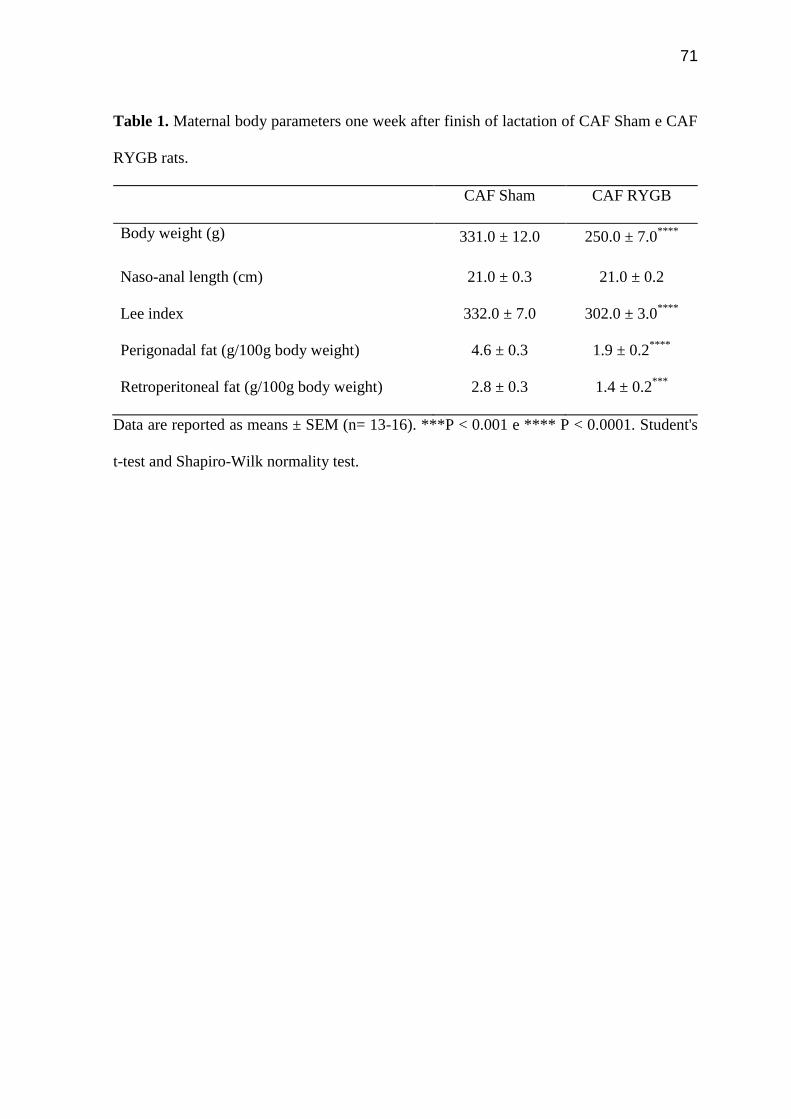
71
Table 1. Maternal body parameters one week after finish of lactation of CAF Sham e CAF
RYGB rats.
CAF Sham CAF RYGB
Body weight (g) 331.0 ± 12.0
250.0 ± 7.0****
Naso-anal length (cm) 21.0 ± 0.3 21.0 ± 0.2
Lee index 332.0 ± 7.0
302.0 ± 3.0****
Perigonadal fat (g/100g body weight) 4.6 ± 0.3
1.9 ± 0.2****
Retroperitoneal fat (g/100g body weight) 2.8 ± 0.3 1.4 ± 0.2***
Data are reported as means ± SEM (n= 13-16). ***P < 0.001 e **** P < 0.0001. Student's
t-test and Shapiro-Wilk normality test.
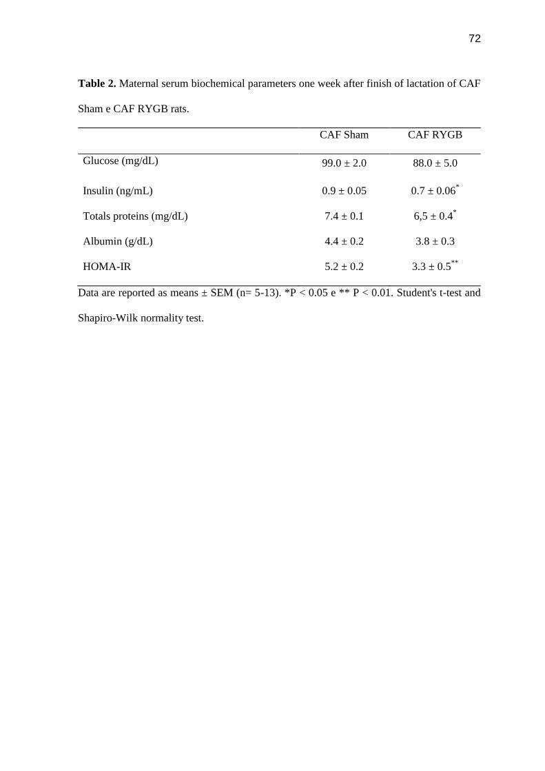
72
Table 2. Maternal serum biochemical parameters one week after finish of lactation of CAF
Sham e CAF RYGB rats.
CAF Sham CAF RYGB
Glucose (mg/dL) 99.0 ± 2.0
88.0 ± 5.0
Insulin (ng/mL) 0.9 ± 0.05 0.7 ± 0.06*
Totals proteins (mg/dL) 7.4 ± 0.1
6,5 ± 0.4*
Albumin (g/dL) 4.4 ± 0.2
3.8 ± 0.3
HOMA-IR 5.2 ± 0.2 3.3 ± 0.5**
Data are reported as means ± SEM (n= 5-13). *P < 0.05 e ** P < 0.01. Student's t-test and
Shapiro-Wilk normality test.
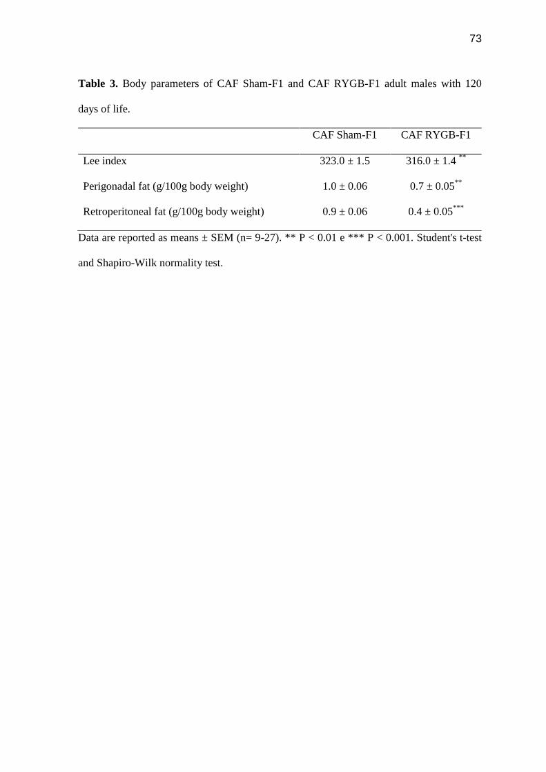
73
Table 3. Body parameters of CAF Sham-F1 and CAF RYGB-F1 adult males with 120
days of life.
CAF Sham-F1 CAF RYGB-F1
Lee index 323.0 ± 1.5 316.0 ± 1.4 **
Perigonadal fat (g/100g body weight) 1.0 ± 0.06 0.7 ± 0.05**
Retroperitoneal fat (g/100g body weight) 0.9 ± 0.06 0.4 ± 0.05***
Data are reported as means ± SEM (n= 9-27). ** P < 0.01 e *** P < 0.001. Student's t-test
and Shapiro-Wilk normality test.
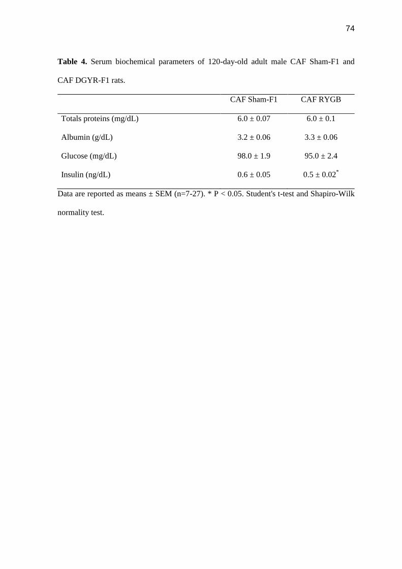
74
Table 4. Serum biochemical parameters of 120-day-old adult male CAF Sham-F1 and
CAF DGYR-F1 rats.
CAF Sham-F1 CAF RYGB
Totals proteins (mg/dL) 6.0 ± 0.07 6.0 ± 0.1
Albumin (g/dL) 3.2 ± 0.06 3.3 ± 0.06
Glucose (mg/dL) 98.0 ± 1.9 95.0 ± 2.4
Insulin (ng/dL) 0.6 ± 0.05 0.5 ± 0.02*
Data are reported as means ± SEM (n=7-27). * P < 0.05. Student's t-test and Shapiro-Wilk
normality test.
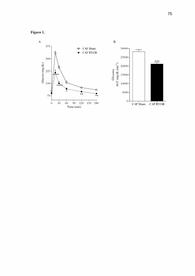
75
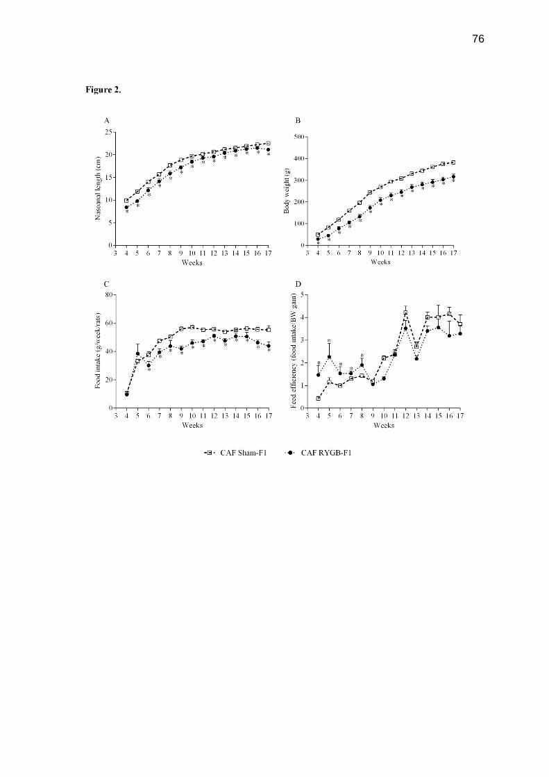
76
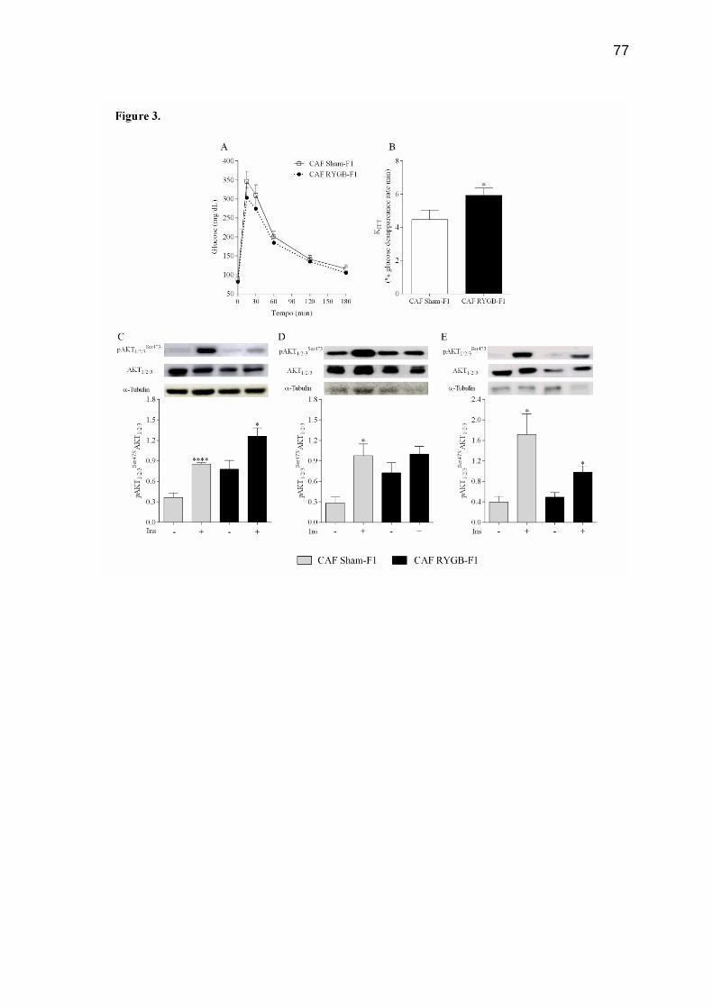
77
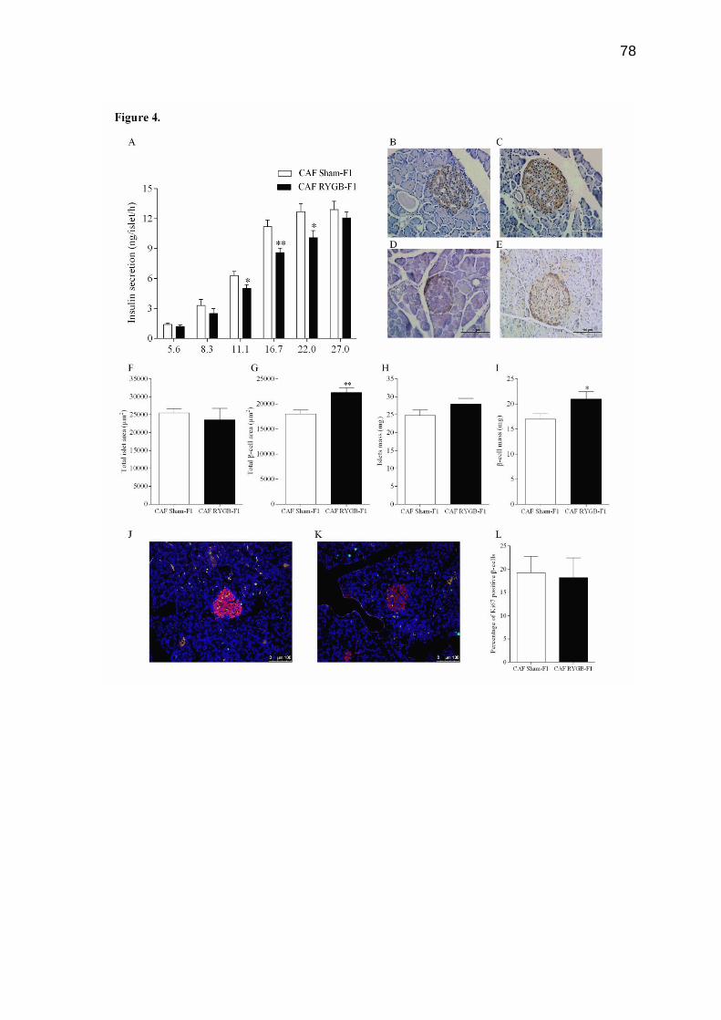
78
79
ANEXO A
Ética Comitê de Ética na Experimentação Animal e Aulas Práticas da
Universidade Estadual do Oeste do Paraná.
80
ANEXO B
Normas da revista científica
Journal of Applied Physiology (1985)
81
INSTRUCTIONS FOR AUTHORS
Title Page
All submissions must contain a title page however brief the article may be
(including, but not limited to, brief items such as editorials). The title page must
contain the full title of the article; author(s) name(s); all departments and
institutions in which the work was done; an abbreviated title for the running head;
and the corresponding author's name, e-mail, and physical address for
correspondence. Only one author may be designated as the corresponding author.
Title
Make the title succinct and informative. Avoid unnecessary words like "Studies
in....". The title must not exceed 160 characters, including spaces between words.
Authors
List all authors' names and their first names or initials exactly as they should be
known. Do not include any specific titles (e.g., PhD, MD, and Prof. are not
needed). Include a brief itemized list of how each author contributed to the study.
"Group authorship" is allowed, with the name of a group (such as a consortium or
program) to be listed as an author, with members of the group listed in the
Acknowledgements section; however, the Program Director of the named group
must be the one who signs for the group when the group's "author" signature is
needed, i.e., on a Mandatory Submission Form or a Change of Authorship Form.
Authors who publish in APS journals may now present their names in non-Latin
characters (in their native writing system) along side the standard English
transliteration of their name in the main author line of the published article; for
example, "Ta-Ming Wang (王大明)". We will accept any non-Latin languages that
have standard Unicode characters designated for the native characters. For
authors that choose this option, please only provide the native expression for the
original written form of the transliterated name; that is, do not include any
associated degree, rank, or title information in the native format. This feature is
meant for the person's name only, not for ancillary information regarding academic
82
achievement or institutional affiliation. To take advantage of this new feature,
please insert the native expression of your name along side the English
transliteration in the main title page of your manuscript submission.
See Authorship Changes for more information.
ORCID
The APS Journals program is now participating in the ORCID Registry. Authors
submitting manuscripts to APS journals will now have the option to attach their
ORCID identifier to their author profile in the APS Manuscript Submission system,
or register for one if they are a new participant. This ORCID identifier will be
included in the article metadata, if published, which may facilitate broad-reaching
linkages of many pieces of an author’s work across multiple databases (such
Pubmed, PMC, and others).
ORCID is "an open, non-profit, community-based effort to create a permanent
registry of unique researcher identifiers and a transparent method of linking
research activities and outputs to these identifiers."
Please see the ORCID website for more information and to register for an ORCID
identifier.
FundRef
The APS Journals program is now participating in the FundRef Registry (a
CrossRef.org initiative).
Authors submitting manuscripts to APS journals will now be able to indicate the
funder(s) associated with their work via an interface in the APS Manuscript
Submission system. This FundRef identifier will be included in the article
metadata, if published, which may facilitate discovery of the article by anyone who
is able to search on that funder name in the FundRef Registry.
FundRef "provides a standard way to report funding sources for published
scholarly research. Publishers deposit funding information from articles and other
content using a standard taxonomy of funder names. This funding data is then
made publicly available through CrossRef's search interfaces and APIs for funders
and other interested parties to analyze."
83
Please see the CrossRef website for more information about the FundRef
Registry.
Affiliation
List all departments and institutions in which the work was done, with city and
state or country. Identify each author's affiliation by superscript numbers matched
to the appropriate institution. Affiliation must reflect the organization(s) supporting
the author(s) while the research was done. This may differ from
the current affiliations of the author(s), which will be listed in such cases in
the Acknowledgments section as the present address(es) of the author(s).
Running Head
The running head is an abbreviated version of the title, which will appear at the top
of every page subsequent to the first page. Running heads must not exceed 60
characters including spaces between words.
Address for Correspondence
Only one author may be designated as the corresponding author. A full address
for correspondence must be included, with a current, valid e-mail address for the
corresponding author. The address of the corresponding author will appear on the
first page of the article. Please note that a valid e-mail address is essential to
participate in the APS electronic proofing service. Also, provide your phone and
fax numbers for use while your article is in production. If the contact information to
be used during production differs from that to be included in the final article,
indicate this explicitly.
Abstract
A one-paragraph abstract of not more than 250 words must accompany each
manuscript. Longer abstracts may be subject to editorial truncation, to conform to
the conventional perceptions of brevity that characterize an "abstract". The
abstract should state what was done and why (including species and state of
anesthesia), what was found (in terms of data, if space allows), and what was
84
concluded. Even for short editorial-style articles, a brief "abstract" should be
provided, if only to identify the topic (e.g., "This is an editorial summarizing recent
new developments in physiology.").
New & Noteworthy
Participating Journals: AJP-Heart (AHeart), Journal of Applied
Physiology (JAPPL), Journal of Neurophysiology (JN), AJP-
Gastrointestinal (AGI), and Advances (ADV).
New & Noteworthy is a brand of annotation summarizing what is new about an
author's original research. Only authors submitting research articles to APS
participating journals (see above) should submit their annotation in the online
EJPress submission form field, ensuring that the exact statement also appears in
their final submitted manuscript after Abstract and Keywords.
Specifications:
WORD LIMIT: 50 words for AHeart; 75 words for JAPPL, JN, AGI, and ADV.
AHeart, JAPPL, JN, AGI
Annotations apply only to Original Research Articles, Innovative
Methodologies, and Rapid Reports.
Annotations appear in final published articles and the journal table of
contents.
Branded "New & Noteworthy"
ADV
Annotations apply to all articles in the journal.
Snapshot statement is solely posted on the journal's Facebook page with a
link back to the published article.
Keywords
Include three to five words or short phrases relevant to the article.
85
Glossary
A glossary may be included (and is often helpful) in equation-laden articles with
many different symbols (such as mathematical modeling or computational papers),
specifying the units (and/or dimensions) as well as each definition. The glossary
will usually precede the Methods section.
See this article for an example.
MAIN TEXT
Introduction
Provide a brief overview of the scope and relevance of the study, especially with
regard to previous advancements in related fields.
Materials and Methods
Describe techniques, cell/animal models used (including species, strain, and sex),
and lists of reagents, chemicals, and equipment, as well as the names of
manufacturers and suppliers, including URLS for those supplies obtained online,
so that your study can be most easily replicated by others. For studies involving
humans, the sex and/or gender of participants must be reported.
Also in this section, describe the statistical methods that were used to evaluate the
data. If clinical trials were used, a statement of registration is required; also, for all
investigations involving humans or animals, a statement of protocol approval from
an IRB or IACUC, or an equivalent statement, must be included (see Guiding
Principles for Research Involving Animals and Human Beings). All animal or
human studies must contain an explicit statement that the protocols were
submitted to, and approved by, an institutional review board or committee or that
the protocols were performed under a license obtained from such a committee,
board, or governing office.See Experimental Details to Report in Your
Manuscript for more information.
See Abbreviations, Symbols, and Terminology for style information.

86
Results
Provide the experimental data and results as well as the particular statistical
significance of the data. See Promoting Transparent Reporting of Results for more
information.
APS has published an editorial on the use of statistics, and authors are
encouraged to consult it.
Discussion
Sometimes combined with the results in a section called "Results and Discussion".
Explain your interpretation of the data, especially compared with previously
published material cited in the References.
Appendix
An Appendix may be included (and is often helpful) in mathematical modeling or
computational papers, e.g., to provide details of a solution strategy.
Acknowledgements
The acknowledgements section is where you may wish to thank people indirectly
involved with the research (e.g., technical assistance; gifts of samples, reagents,
or cell lines; loans of equipment or laboratory space; comments or suggestions
during the creation of the manuscript). However, it is important that anyone listed
here know in advance of your acknowledgement of their contribution, as
documented during the submission process.
Current addresses of authors (which may differ from those in the affiliation line)
may be included here.
Do not include "promissory notes." APS Journal policy is against inclusion of
implicit or explicit promises that future work will be published.
Do not include dedications (e.g., to persons living or deceased). Dedications of
articles are not permitted.

87
Grants
List the grants, fellowships, and donations that funded (partially or completely) the
research. However, industry-sponsored grants should be listed under Disclosures.
Disclosures
Authors are required at the time of submission to disclose any perceived or
potential conflict of interest, financial or otherwise. See Author Conflict of Interest.
If the article is accepted for publication, information on the perceived or potential
conflict of interest, or lack thereof, must be noted in the Disclosures section.
Footnotes
The footnotes section is the place to list any additional items pertinent to your
article, including but not limited to links to non-peer-reviewed data that may be
available to readers from your institutional web site.
Footnotes should be numbered consecutively throughout. They should be
assembled on a separate page at the end of the article.
References
Authors are responsible for accuracy of citations. References must be limited to
directly pertinent published works or papers that have been accepted for
publication. An abstract, properly identified as "Abstract", may be cited only when it
is the sole source.
Reference lists should be arranged alphabetically by author and numbered
serially. The reference number should be placed in parentheses at the appropriate
place in the text.
The style of citation should be as follows, with journal name abbreviated as in
Medline, PubMed, and Index Medicus. Appropriate templates for your citation
management software are available from the respective company websites
(e.g., EndNote ).
The examples given below are shown with numbers because that is the style for
most APS Journals, except for the Journal of Neurophysiology (see note, below,
after these examples). The first is a standard book reference; the second is a

88
standard reference to an "early view" or "prepress" reference, such as the APS
"Articles in PresS" (note the use of the ―digital object identifier‖—doi—in this
citation); the third is a standard journal reference.
1. Pollock DM. Endothelin receptor subtypes and tissue distribution. In: Endothelin
Molecular Biology, Physiology, and Pathology, edited by Highsmith RF. Totowa,
NJ: Humana, 1998.
2. Scarafia LE, Winter A, Swinney DC. Quantitative expression analysis of the
cellular specificity of HECT-domain ubiquitin E3 ligases. Physiol Genomics (April
26, 2001). doi:10.1152/physiolgenomics.00075.2001.
3. Villalobos AR, Parmelee JT, Renfro JL. Choline uptake across the ventricular
membrane of neonate rat choroid plexus. Am J Physiol Cell Physiol 276: C1288-
C1296, 1999.
MORE Example References
References for the Journal of Neurophysiology should be arranged alphabetically
by author. The appropriate author name and year for each reference should be
included in parentheses at the proper point in the text using the following style (this
is ONLY for the Journal of Neurophysiology, NOT for other APS Journals):
one author (Brown 1982)
two authors (Brown and Smith 1982)
three or more authors (Brown et al. 1982).
For the in text citations in the Journal of Neurophysiology, here are some other
important details. If more than two references are cited by different authors,
separate entries with a semicolon (Brown 1982; Smith 1983). If more than two
references are cited by the same first author (or single author), use "et al." where
appropriate plus the date, even if the subsequent authors are not the same in all
the references (Brown et al. 1982, 1983). Note the use of commas between two
consecutive years or nonconsecutive years. Do not use dashes for ranges (Brown
et al. 1982, 1983, 1986, 1987, 1988). If more than two references with the same
year and author(s) are cited, use lowercase letters after the year (Brown 1982a,
1982b). Lowercase letters should be inserted in same-year references in the
reference list.
89
Citing Unpublished Observations, Personal Communications, and "In Press"
Manuscripts
Submitted papers still in preparation or in peer review and/or any other
unpublished materials, observations, or personal communications cannot be
included in the reference list, which may only list published work. However, such
material can be cited in the text, but at submission, authors will be required to
confirm that all individuals acknowledged in the manuscript are aware that they are
being acknowledged and approve of the manner and the context of the
acknowledgement. This includes, but is not limited to the following circumstances:
to publish information disclosed in a personal communication or
unpublished observation;
to recognize additional individuals who helped in preparation of the
manuscript;
for permission from a copyright holder to discuss information that has been
accepted for publication but is "in press" and not yet available, online or
otherwise.
For both unpublished observations and personal communications, provide the
cited person's last name and all initials.
Figure Captions
Every figure must have a descriptive figure caption, to describe to the reader in
sentence form the relevant details of the figure, to place it in the proper context of
the manuscript. Information reported in figures and legends should
describe each individual experiment presented, including the number of samples
or animals used per treatment. The statistics performed for each experiment
should be reported as well. These textual figure captions must be listed in order in
the manuscript, following the reference list.
Tables
Whenever possible, authors are encouraged to submit figures rather than tables.
Statistical summary tables should be submitted when possible, rather than tables
with many lines of individual values. Lengthy tables of data that cannot be

90
presented in a suitable manner, according to APS standards of print publication,
may be extracted and set as a supplement to the online article. These
supplements remain an integral part of the article for the reader, as text referring to
these tables will remain in the article, and links directly to the supplements will be
embedded and prominently indicated at all points of entry to the online article
(see Data Supplements).
Submitted tables should adhere to the following guidelines:
Tables must not duplicate material in text or figures.
Tables should be numbered consecutively with Arabic numerals and
prepared with the size of the journal page in mind: 3.5 in. wide, single
column; 7 in. wide, double column.
Each table should have a brief title; explanatory notes should be in the
legend, not in the title.
Nonsignificant decimal places in tabular data should be omitted.
Short or abbreviated column heads should be used and explained if
necessary in the legend.
Statistical measures of variations, SD, SE, etc., must be identified.
(Example: "Values are means ± SE.")
Table footnotes should be listed in order of their appearance and identified
by standard symbols: *, †, ‡, § for four or fewer; for five or more,
consecutive superscript lowercase letters should be used (e.g., a, b, c, etc.).
Equations
Manuscripts with many mathematical characters and equations should be
prepared using MathType version 6.0 or higher (available from Design Science,
Inc.). Manuscripts with a minimal amount of math may be prepared using word-
processing tools such as Microsoft Word Equation Editor, or with features such as
bold, italics, superscript and subscript together with characters in the Symbol or
Greek fonts. Do not use the Wingdings or Webdings fonts. Please do not submit
equations as images. Inline math text that can be regular text (e.g., x2[n − 2])
should be provided as such. Please do not set these as MathType.
91
Mathematical equations should be simplified as much as possible and carefully
checked.
Use the slant line (/) for simple fractions (a + b)/(x + y) in the text rather
than the built-up fraction a + b[over]x + y, which should only be used if the
equation is offset from the text.
Use subscripts or superscripts wherever feasible and appropriate to
simplify the equations.
Please use notation that is consistent with the standard nomenclature in
applied mathematics. As an aid to the reader, please state the convention
that you are following, especially if it is uncommon.
Symbols should be defined as they first appear in the text. A glossary may
be included (and is often helpful) in articles with many different symbols,
specifying the units (dimensions) as well as each definition. The Glossary
will usually precede the Methods section.
APS style allows punctuation in displayed equations.
Mathematical Models
Presentation of the model(s) must be sufficiently clear to allow physiologists with
limited experience in modeling to follow the model development, limitations, and
physiological relevance. Assumptions concerning the importance of physiological
processes included in the model should be clearly stated.
If the model equation(s) require solution, the method of solution should be
described in sufficient detail to permit readers to duplicate the solution in
their own laboratories. Algorithms from commercial software libraries
should be so identified. Details of the solution strategy may be summarized
in an Appendix.
For simulations, sources or estimation methods for all parameter values
should be presented and the numerical values given in the text or a table. A
sensitivity analysis must be performed for important parameters (covering
ranges of values relevant to the manuscript) to determine how the model
predictions are affected by numerical parameter values.

92
If the model is used to estimate parameter values, measures of the
uncertainties associated with the estimated parameter values should be
presented.
For models intended for use in a predictive setting, validation of the model
with a data set not used for model parameter estimation (i.e., cross-
validation) is recommended. Sensitivity analysis or parameter uncertainty
determination is an important component of modern modeling practice that
allows assessment of the validity of a model.
Results obtained with the model(s) should be compared with appropriate
physiological data, either from literature or from new experiments.
Simulation results may be examined for prediction of changes or trends in
physiological variables similar to those reported for in vitro or in vivo studies.
The discussion should include information on the physiological significance
of the model study, limitations of the model, and suggestions for new
modeling and/or experimental studies.
Articles based on original computer simulation software
1. Upon submission of a paper reporting computer simulation using original,
author-generated code, authors are encouraged to deposit software and
code via a public repository, such as GitHub, if not already deposited to a
public repository. A link to the software deposit should be included in the
cover letter. Alternatively, authors may submit a working version of their
software and computer code as supporting data for access by the
reviewers. It will be kept confidential during review, and in case of a
negative decision (i.e., rejection).
a. In any case, authors must make available upon request, to editors and
reviewers, any previously unreported custom computer code used to
generate results that are reported in the paper and central to its main
claims. Any practical issues preventing code sharing will be evaluated by
the editors who reserve the right to decline the paper if important code is
unavailable.
93
b. Upon manuscript submission, authors should check the box marked
―computer simulation‖.
2. Upon manuscript R1 revision (assuming authors are invited by the journal
to revise their manuscript), authors should deposit a full working version of
their software and computer code to a public website/repository, one that
retains original and updated versions of the model and code – if not already
deposited publicly – and include the link to the deposited software in the
Methods section.
a. Authors must state Terms of Use included with the deposited
software. Authors should consult the Intellectual Property owner (usually
the author’s institution) regarding requirements, if any, for active user
compliance agreement to Terms of Use.
b. Original software and code must remain available indefinitely
c. Updates to the model and code should be reported on the repository site,
with comments as to why particular changes were made with the dates of
original deposit and each change identified. In this way, published material
remains available in its published form, but improvements are also
available from the same immediately accessible web-based repository
location.
3. Authors should inform editors about any practical issues preventing public
deposit of the computer code and be prepared to make computer code
available to readers upon request after publication.