Influence of crushed aggregates filler on the properties of rendering ...
Upload
phungtuyenCategory
view
214download
0
DEPARTAMENTO DE CIÊNCIAS DA VIDA
FACULDADE DE CIÊNCIAS E TECNOLOGIA UNIVERSIDADE DE COIMBRA
Sara de Sa Cesariny Calafate
2012
Dissertação apresentada à Universidade de Coimbra para cumprimento dos requisitos necessários à obtenção do grau de Mestre em Biologia Cellular e Molecular , realizada sob a orientação científica do Doutor Arjan Buist (Janssen Pharmaceutica) e do Doutor Diederik Moechars (Janssen Pharmaceutica)
Tauopathy seeding models as a platform for Tau aggregation and clearance study
A4 - 06102 - Sara Calafate - thesis binnenwerk.indd 1 27/06/2012 9:47:12

4
A4 - 06102 - Sara Calafate - thesis binnenwerk.indd 2 27/06/2012 9:47:15

5
The work presented in this thesis resulted from a partnership between the University of Coimbra and Janssen Pharmaceutica NV. All experimental activities were performed at Janssen Pharmaceutica NV Beerse I, a Johnson & Johnson pharmaceutical research and development facility in Beerse, Belgium.
Beerse, 2012
A4 - 06102 - Sara Calafate - thesis binnenwerk.indd 3 27/06/2012 9:47:22

6
Acknowledgements There is no way to properly thank all the people for what I have learned during this year. For all the insight, support, joy, making me feel at home and making this year an incredible experience. First I need to say that it has given me the opportunity to follow the best teacher that I ever had in Arjan. Thank you for all what you taught me and for changing my way of looking at science. Also for all the funny (sometimes little bit crazy) conversations in the lab! This was only possible because Dieder gave me the opportunity to belong to this group. I really appreciated all the enthusiasm and the support that Dieder gave me to develop my work. Thank you both for what you taught me and the advice that you gave me. Kathleen Callaerts, you solved all my problems in and out of the lab, since I arrived! I really appreciate how you treated me. Other members of the Tau group (Aga, Kristof, Brian, Eve, Marck M., Marck Vdm) it was a pleasure to discuss science with you. Guy, Marck Vdm and Luc Peeters your neuroscience lab is a great place to work! Sometimes I thought that I was in Portugal. Thanks also go to the people of my office, Marianne, Bart, Ilse and Greet that received me well and made me laugh a lot. Professor Emilia Duarte, Carlos Duarte and Ana Luisa thank you for the great first great year at Coimbra, and for letting us come to Janssen. Janssen students we made Turnhout the best city of Belgium! Thank you all, especially Ana, Marco, Alex, Matthew, Ewan and Danni. For all the support thank you Filipe and my family. A special thank you to all the Janssen team and I hope to see you soon.There is no way to thank to all these people what I learned during this year. For all the insight, support, joy, that made me feel at home, and made this year an incredible experience.
A4 - 06102 - Sara Calafate - thesis binnenwerk.indd 4 27/06/2012 9:47:25

7
Index Abbreviations ....................................................................................................................... 10
Resumo ................................................................................................................................ 12
Abstract ................................................................................................................................ 13
Chapter 1 – Introduction and Project goal ............................................................................... 15
1.1 Introduction .................................................................................................................... 17
1.2 AD symptoms and pathology......................................................................................... 17
1.2.1 AD patient’s brain ................................................................................................... 17
1.3 The role of β-Amyloid in Alzheimer’s disease .............................................................. 19
1.4 The role of Tau in Alzheimer’s disease ......................................................................... 20
1.4.1 Normal Tau characteristics and function ................................................................ 22
1.4.2. Pathological Tau .................................................................................................... 23
1.4.3 Identifying the culprit of neuronal toxicity in AD Tau hypothesis ......................... 24
1.5. Tau post-translational modifications............................................................................. 25
1.5.1. Tau phosphorylation .............................................................................................. 25
1.5.2. Prolyl-isomerization of Tau ................................................................................... 27
1.5.3 Tau truncation ......................................................................................................... 28
1.5.4 Ubiquitination of Tau .............................................................................................. 29
1.5.5. Other post-translational Tau modifications – complex array of post-translational modifications and their interactions ................................................................................. 29
1.6 Tauopathies .................................................................................................................... 30
1.6.1 FTDP-17 mutations ................................................................................................. 31
1.6.2 Tauopathy models ................................................................................................... 31
1.7 Pathogenic Protein Seeding in AD and other Neurodegenerative diseases ................... 32
1.7.1 The amyloid state of proteins in human disease ..................................................... 32
1.7.2 Understanding the propagation of aggregates pathology using seeding strategies. 34
1.7.2.1 Evidence for Tau seeding ................................................................................. 35
1.7.2.2 Aβ seeding evidences ....................................................................................... 36
1.7.2.3 α-Synuclein seeding evidences ........................................................................ 36
1.7.2 Pathogenic proteins interaction and cross-seeding evidences ................................ 36
1.7.3 Neuron-to-Neuron transmission.............................................................................. 37
1.8 Tau-focused strategies to halt AD progression .............................................................. 39
1.8.1 Targeting Tau hyperphosphorylation ...................................................................... 40
1.8.1.1 GSK3................................................................................................................ 41
1.8.1.2 CDK5 ............................................................................................................... 41
1.8.1.3 PP2A ................................................................................................................ 42
1.8.2 The potential of aggregation inhibitors ................................................................... 42
A4 - 06102 - Sara Calafate - thesis binnenwerk.indd 5 27/06/2012 9:47:28

8
1.8.3 Strategies to compensate for Tau LOF ................................................................... 43
1.8.4 Enhancing intracellular Tau degradation ................................................................ 44
1.8.4.1 The role of proteasome and autophagy the in Tau degradation ....................... 44
1.8.4.2 Manipulating Hsp scaffolds to enhance Tau degradation ................................ 45
1.9 Project Goal ................................................................................................................... 47
1.9.1 Variability analysis and characterization of clonal-cell line seeding model ........... 47
1.9.2 Set Up of primary neuronal seeding model ............................................................ 47
Chapter 2 - Materials ............................................................................................................... 49
2.1 Material .......................................................................................................................... 51
2.2 Antibodies and dyes ....................................................................................................... 53
Chapter 3 -Methods.................................................................................................................. 55
3.1 AAV vector production.................................................................................................. 57
3.2 Cell culture and plasmid transfection ............................................................................ 57
3.3 Cortical primary cultures and AAV6-mediated hTauWT and hTauP301L expression . 58
3.4 In vitro fibrillization of recombinant Tau ...................................................................... 58
3.5 K18P301L fibrils delivery into clonal-cell lines and cortical primary neuronal cultures.............................................................................................................................................. 59
3.6 Sequential Protein extraction ......................................................................................... 59
3.7 Western Blot .................................................................................................................. 60
3.8 Immunofluorescence ...................................................................................................... 60
3.9 Immunoprecipitation (IP)............................................................................................... 60
Chapter 4 –Results ................................................................................................................... 63
4.1 Optimization of Clonal-cell seeding model ................................................................... 65
4.1.1 Influence of seeds preparation method on hTauP301L aggregation ...................... 65
4.1.2 Choosing the best Cell Line for seeding model development ................................ 66
4.1.3 Variability of QBI seeding model ........................................................................... 67
4.1.4 Delivery system and seeds delivery efficiency ....................................................... 68
4.1.5 Model characterization - Gel migration .................................................................. 68
4.1.6 Insoluble hTauP301L is phosphorylated at different epitopes ............................... 69
4.2 Development of primary neuronal seeding model ......................................................... 70
4.2.1 Generation of primary cortical neurons expressing human full-lenght Tau ........... 70
4.2.2 Aggregation of hTauP301L in primary cortical neurons ........................................ 71
4.2.3 BioPorter delivery reagent is an efficient delivery tool .......................................... 71
4.2.4 Aggregation of hTauP301L is affected by the amount of K18P301L. ................... 72
4.2.5 hTauWT does not aggregate like hTauP301L ........................................................ 74
4.2.6 Aggregation kinetics is affected by amount of fibrils and shown time variation ... 74
4.2.7 Phospho-epitopes detected on primary neuronal seeding model ............................ 75
A4 - 06102 - Sara Calafate - thesis binnenwerk.indd 6 27/06/2012 9:47:32

9
4.3 Testing of Hypothesis .................................................................................................... 76
4.3.1 GSK3β increases phosphorylation of aggregated hTauP301L ............................... 76
4.2.2 Hsp90 inhibition...................................................................................................... 79
Chapter 5 –Discussion and concluding remarks ...................................................................... 81
Chapter 6 - References ............................................................................................................. 89
A4 - 06102 - Sara Calafate - thesis binnenwerk.indd 7 27/06/2012 9:47:35

10
Abbreviations
17-AAG -17- allylamino 17-demethoxy-geldanamycin 17-DMAG - 17-dimethyl-amino-ethylamino-17- demethoxygeldanamycin AAV - Adeno-Associated Virus ABCA7 - ATP-binding cassette transporter AD – Alzheimer’s Disease AGEs - Advanced Glycosylation End Products APOε - Apolipoprotein ε APP - Amyloid precursor protein ATP - Adenosine triphosphate Aβ - β Amyloid peptide BBB - Blood brain barrier BIN1 - Bridging integrator 1 CD2AP - CD2-associated protein CD33 - Sialic acid binding immunoglobulin-like lectin CDK5 - Cyclin-dependent kinase 5 CHIP - Carboxy-terminus of Hsc70-interacting protein CK1 - Casein kinase 1 CLU - Clusterin CNS - Central nervous system COS-7 - African green monkey kidney-derived cells CR1 - Complement receptor DIV – Day in vitro DPT – Day post-transfection Dyrk1A – Dual-specificity tyrosine phosphorylation-regulated kinase 1A EOAD - Early-onset AD EPHA1 - Ephrin receptor A1 ER - Endoplasmatic reticulum FAT - Fast axonal transport FRET- Fluorescence resonance energy transfer FTDP-17 - Frontotemporal dementia and Parkinsonism linked to chromosshome 17 GA – Geldanamycin GFP - Green fluorescent protein GlcNac - β -N-acetylglucosamine GOF - Gain-of-function GSK3 - Glycogen synthase kinase-3 Histone Deacetylase 6 (HDAC6) HOP - Hsp70/Hsp90-organising protein HSF1 - Heat shock factor 1 Hsp - Heat shock protein hTauP301L – human full-lenght Tau containing P301L mutation hTauWT - human full-lenght Tau IP- Immunopercipitation ITR – Inverted terminal repeats KD - Knock Down KO - Knockout KPI - Kunitz Protease Inhibitor
A4 - 06102 - Sara Calafate - thesis binnenwerk.indd 8 27/06/2012 9:47:39

11
LOAD - Late-onset AD LOF - Loss-of-function LRRK2 - Leucine-rich repeat kinase 2 MAP - Microtubule associated protein MARK - Microtubule affinity-regulating kinase MB - Methylene blue MCI - Mild cognitive impairment MOI - multiplicity of infection MS4A - Membrane-spanning 4-domains sub family A cluster MTBR – Microtubule binding repeat domain MTL - Medial temporal lobe MVB - Multivesicular bodies N2a - mouse Neuroblastoma derived CELLS NFT - Neurofibrillary tangles PC12 – Cultured rat pheocromocytoma cells PD- Parkinson’s Disease PICALM - Phosphatidylinositol-binding clathrin assembly protein PKA- Protein kinase A PP2A – Protein phosphatase 2A PPI - Peptidyl prolyl isomerization PQC - Protein quality control QBI - QBI-HEK 293A Cells R- Microtubule Repeat domain RAGE - AGE receptor RT – Room temperature ser - Serine SLMV – Small-synaptic multivesicles SP - Senil Plaques thr - Threonine TNT- Tunneling nanotubes TPK I - Tau protein kinase I TRP - Tetratricopeptide domain tyr - Tyrosine U20S - Human osteosarcoma-derived cells UPS - Ubiquitin-proteasome system WT - wild-type
A4 - 06102 - Sara Calafate - thesis binnenwerk.indd 9 27/06/2012 9:47:42
12
Resumo
A doença de Alzheimer é a forma de demência mais prevalente. Quando a proteína Tau perde a conformação correcta forma agregados, começando por originar oligomeros e mais tarde fibrilas de grandes dimensões dando origem a trancas neurofibrilares. Alguns estudos sugerem que estas espécies são transmitidas através das áreas do cérebro danificando o circuito neuronal. Para parar o avanço da doença muitos trabalhos têm em foco o desenvolvimento de terapias que manipulam a fosforilação desta proteína, a estabilização dos microtubulos e a indução da degradação da Tau. Para desenvolver terapias que impedem a agregação ou que induzam a degradação da Tau, é necessário desenvolver modelos que recapitulam a agregação. Neste trabalho “seeding effect” foi a estratégia utilizada para induzir a agregação da hTauP301L. No presente trabalho dois modelos in vitro baseados nesta estratégia foram utilizados – um desenvolvido em linhas celulares e outro em culturas neuronais primarias – onde se observou a hiper-fosforilação e agregação da Tau. A co-expressão da GSK3β aumentou a fosforilacao na ser202/thr205 na Tau solúvel e insolúvel, mas apenas a sua expressão não foi suficiente para induzir agregação. Em culturas neuronais primárias a fosforilação e agregação da Tau aumentam ao longo do tempo. A inibição da Hsp90 reduziu os níveis de Tau totais de e fosforilados no epitopo AT8, dando importância a esta estratégia como um potencial mecanismo para degradação da Tau.Com este trabalho produzimos dois modelos onde estudos em nucleação, agregação e transmissão sináptica da Tau poderão ser feitos. Estes modelos são ferramentas válidas para o desenvolvimento de fármacos para AD e outras Tauopatias Palavras Chave: Doença de Alzheimer, Tau, fosforilação da Tau , oligomeros, transmissão da Tau, agregação da Tau e degradação da Tau.
A4 - 06102 - Sara Calafate - thesis binnenwerk.indd 10 27/06/2012 9:47:45
13
Abstract
Alzheimer Disease is the most prevalent dementia. Abnormal folding of Tau leads to generation of aggregated Tau species like oligomers and further NFTs. Toxic Tau species were suggested to spread trough human brain and damage the neuronal circuit. To halt disease progression a noteworthy development in therapies based on phosphorylation modulation, Microtubule stabilization and enchantment of Tau clearance have been done. To develop therapies against Tau aggregation and aggregates clearance, the build up of models that recapitulates Tau pathology are required. In the following work the “seeding” strategy was used to achieve aggregation of hTauP301L. We worked with two in-vitro seeding models – cellular and primary neuronal- where phosphorylated insoluble hTauP301L is present. GSK3β was shown to increase ser202/thr205 phosphorylation in soluble and insoluble hTauP301L expressed in QBI but was not sufficient to induce aggregation alone. In primary neuronal seeding model tau aggregation and phosphorylation was increased over-time. Hsp90 inhibition was found to reduce total and AT8 immuno-reactive hTauP301L levels, emerging as a potential drug for Tau clearance. With this work we provide two models to study the mechanisms behind tau nucleation, aggregation, and trans-synaptic spreading. These models are valuable tools for the development of drugs for AD and Tauopathies. Keywords: Alzheimer’s disease, Tau, phosphorylation, oligomers, spreading, Tau aggregation and Tau clearance.
A4 - 06102 - Sara Calafate - thesis binnenwerk.indd 11 27/06/2012 9:47:49

14
A4 - 06102 - Sara Calafate - thesis binnenwerk.indd 12 27/06/2012 9:47:53

15
Chapter 1 – Introduction and Project goal
A4 - 06102 - Sara Calafate - thesis binnenwerk.indd 13 27/06/2012 9:47:56

16
A4 - 06102 - Sara Calafate - thesis binnenwerk.indd 14 27/06/2012 9:48:00

17
1.1 Introduction
Aging is accompanied by a cognitive decline of the population. The dramatic rise in life expectancy during the 20th century has resulted in the exponential growth in the number of individuals reaching the age at which neurodegenerative disorders occur. An important task is to understand how normal aging of the brain switches to pathogenic aging, triggering neurodegenerative diseases. 35.6 million People in the world live with dementia of which Alzheimer’s disease (AD) is the most prevalent form, accounting for 70% of all cases of dementia (http://www.who.int/). This disease affects 3% of the people between 65 and 74 years old, nearly 20% of those aged 75 to 84, and 50% of the people 85 and older (http://www.alzforum.org/). AD is a progressive neurodegenerative disease that affects memory, thinking, behavior and ability to perform everyday activities. AD patients suffer a decline in memory, learning capacity, language, and deterioration in behavior (social and interpersonal) (Selkoe, 2001). There is currently no cure for AD. This burdensome state of AD patients calls for the necessity for more investments to investigate this disorder. The European Parliament adopted, on January 19th of 2011 in Strasbourg, a report calling for more resources to treat AD, and it was argued that the matter should be a public health priority. Two types of symptomatic therapies are currently available: treatments for cognitive impairment (Cholinesterase inhibitors and NMDA receptor antagonist), and treatments for neuropsychiatric symptoms (atypical antipsychotics, antidepressants and anticonvulsants) with only moderate benefits in cognitive function (Ballard, 2011). Several potentially disease-modifying treatments (Immunotherapy, secretase inhibitors, amyloid aggregators, copper or zinc modulators, Tau aggregation inhibitors, Glycogen synthase kinase-3 (GSK3) inhibitors, natural products and vitamins) have been proposed and are now at different stages of clinical development but none has made it to the market yet. 1.2 AD symptoms and pathology
Since 1901, Alois Alzheimer identified two pathogenic features that are still required final AD diagnosis by postmortem brain examination: the extracellular amyloid plaques and intracellular neurofibrillary tangles (NFTs). The amyloid plaques are composed of aggregates of protein Beta-Amyloid (Aβ). NFTs are composed of aggregates of hyperphosphorylated Tau protein. Alois Alzheimer findings represent correlated neuropathologic changes with cognitive status of AD patients.
1.2.1 AD patient’s brain
It was shown that neuronal network of AD patients is devasted by amyloid plaques and neurofibrillary pathology (Figure 1). These multiple cognitive domains become impaired as AD develops. Already in a pre-dementia clinical stage of mild cognitive impairment (MCI), significant AD pathology is present in the medial temporal lobe (MTL) memory system (Braak, 1991; Selkoe, 2001). It is now widely accepted that there MCI is a transitional phase between normal function and Alzheimer dementia, during which cognitive impairment is
A4 - 06102 - Sara Calafate - thesis binnenwerk.indd 15 27/06/2012 9:48:03

18
progressing (Petersen, 2006). AD diagnosis during MCI is necessary to give the patient prognosis. For example, it has been reported that episodic memory performance is correlated with Aβ neocortical accumulation (Trojanowski, 2010). Aging in the nervous system does not necessary result in a loss of neurons. There is now accumulating evidence that the reason for the progressive cognitive decline is a loss of synapses resulting in a disruption of corticocortical connectivity (Scheff, 2006). White matter density loss (Bartzokis, 2003) and synaptic function loss are changes associated with cognitive decline. Individuals with MCI had fewer synapses (36%) when compared to individuals with no cognitive impairment (Scheff, 2011). It has been shown that areas of early AD-related pathology show no significant changes in total neuron number but instead of that loss of synaptic function (Kordower, 2001).
1.2.2 Genetics of Alzheimer disease
AD can be divided into two major type based on age of onset: early-onset (EOAD) and late-onset (LOAD) forms. AD most commonly strikes in individuals older than 65. People experiencing AD before 65 fit in the EOAD category and represents less than 10% of AD cases. LOAD cases start after 65 years and are sporadic in nature (Lambert, 2011). Identifications of inherited disease-causing mutations allowed a better understanding of some key underlying molecular mechanisms of AD brains and other neurodegenerative disorders. Although in most cases AD is a complex and multifactorial disease resulting from interaction of numerously undetermined factors in EOAD the mode of inheritance is autosomal dominant. Mutations in three genes including amyloid precursor protein (APP) on chromosome 21 (Goate, 1991), presenilin 1 (PSEN1) on chromosome 14 (Sherrington, 1995) and PSEN2 on chromosome 1 (Levy-Lahad, 1995; Rogaeva, 1995) have been identified in families with EOAD. The effects of these mutations on metabolism of APP with an increased production of Aβ42 and /or ratio of Aβ42/Aβ40 gave rise to a pathophysiological hypothesis: the amyloid cascade hypothesis (Hardy, 1997). Although most of the cases of LOAD are considered sporadic forms there is no longer any doubt that they display a strong genetic predisposition (accounting for 60–80% of the attributable risk) (Lamber, 2011).
Figure 1. Progression of neuropathology in aging and AD. Shown is the neuroanatomical distribution of amyloid plaques, neurofibrillary tangles and neuronal loss during normal aging, mild cognitive impairment and AD (Yankner,2008)
A4 - 06102 - Sara Calafate - thesis binnenwerk.indd 16 27/06/2012 9:48:08

19
The most important genetic risk factors linked to LOAD is apolipoprotein ε (APOε). APOε is involved in the clearance of Aβ. There are three variants: APOε2, APOε3 and APOε4, and the last variant profoundly increase the risk of LOAD (Corder et al 1993). The complexes APOε2-Aβ and APOε3-Aβ are cleared faster than APOε4-Aβ complexes at the level of blood brain barrier (BBB) (Deane, 2008). A GWAS of neuropathologically confirmed AD patients confirmed a significant SNP (rs4420638) in linkage disequilibrium to the APOε4 variant, providing evidence for a major susceptibility gene for LOAD (Nebel, 2011). Aside APOE, recently GWAS were published showing that nine new loci were found as risk factor for LOAD. These genes are clusterin (CLU), phosphatidylinositol-binding clathrin assembly protein (PICALM), complement receptor 1 (CR1), bridging integrator 1 (BIN1) , adenosine tiphosphate (ATP)-binding cassette transporter (ABCA7), membrane-spanning 4-domains sub family A (MS4A) cluster, CD2-associated protein (CD2AP ), sialic acid binding immunoglobulin-like lectin (CD33 ), and ephrin receptor A1 (EPHA1) (Morgan, 2011). Morgan and colleagues demonstrated that collectively these loci explain 50% of LOAD (Morgan, 2011). Rogaeva and colleagues reported that inherited variants in SORL1 neuronal sorting receptor are associated with LOAD and is thought to affect levels of Aβ (Rogaeva, 2007). 1.3 The role of β-Amyloid in Alzheimer’s disease
The amyloid cascade hypothesis is based on the core idea that neuronal dysfunction is triggered by an imbalance between Aβ production and degradation affecting stability and induction of aggregation of this peptide. The accumulation of Aβ in the brain initiates a multistep cascade that results in memory loss and impaired cognitive functions (Masters, 2006). The Aβ deposits in the brain appear to maturate from diffuse to dense cored plaques. APP can be cleaved by enzyme complexes termed α-, β- and γ-secretases. Depending on the
Figure 2. Schematic representations of the amyloid precursor protein (APP) and its metabolic derivatives. (A) Diagram of APP showing a large extracellular domain and short intracellular segment. Cleavage of APP by either α- or β-secretases produces large soluble N-terminal fragments, α-APPs and β-APPs and 10 and 12 kDa membrane-bound C-terminal fragments, C83 and C99, respectively. Both C83 and C99 can be further cleaved by α- or β-secretase leading to the release and secretion of non-pathogenic p3 peptide and Aβ, respectively. The drawing is not to scale. (B) Amino acid sequence of Aβ showing the most common APP secretase cleavage sites, including sites for Aβ40 and Aβ42 generation, as indicated by arrows (Fodero-Tavoletti, 2011).
A4 - 06102 - Sara Calafate - thesis binnenwerk.indd 17 27/06/2012 9:48:17

20
enzyme and location of proteolysis the processing of APP can be divided into a non-amyloidogenic pathway (Haass, 1993; Kojro, 2005) and an amyloidogenic pathway (Jarret, 1993) (Figure 2). Overproduction of Aβ results in a neurodegenerative cascade. The increase of Aβ42 levels is probably critical for AD, providing the core for Aβ assembly into oligomers, fibrils and plaques (Shankar, 2009). These plaques were the core of initial amyloid cascade hypothesis. But this hypothesis has been challenged and attention has shifted from plaque levels towards the changes of Aβ oligomers steady-state (Lambert, 2011; Ballard, 2011). Aβ oligomer levels in blood are significantly elevated in AD compared to healthy subjects and correlate better than Aβ plaques with cognitive impairment (Villemagne, 2010) Within the neuron, Aβ42 appears to be located at multi-vesicular bodies (MVB) which have a role in transporting cargo to the lysosome system for degradation. The accumulation of these species in MVBs disrupts its sorting and has shown to be mechanistically linked to cytosolic proteasome inhibition (LaFerla, 2007). Toxic effect of Aβ has been also associated to activation of caspases, calpains and stimulation of microglia inflammatory cascade activation (Yankner, 2007), which could induce deregulation of kinases and further Tau hyperphosphorylation. Oligomeric Aβ inhibits hippocampal long-term potentiation (Lambert, 1998) and can also promote AMPA and NMDA receptors endocytosis (Snyder, 2000; Hsieh, 2006), raising another possibility that oligomeric Aβ could induce synaptic plasticity impairment and memory deficits (figure 3). Why this Aβ protein aggregates into fibrils is not well understood, but the Aβ sequence, concentration and conditions that destabilize Aβ oligomers are probably critical factors (Nerelius, 2010). Synaptic impairments caused by Aβ oligomers indicate that these elements are more toxic than SP and are only one element within a complex mechanism of dysfunction (Figure 3). 1.4 The role of Tau in Alzheimer’s disease
AD is not only characterized by extracellular amyloid plaques but also by an intracellular accumulation of Tau in somatodendritic and axonal domains of neurons (Braak, 1988). Neuropil threads are related with Tau accumulated in dendrites. The accumulation of Tau in the soma leads to NFT formation (Adalbert, 2009). Unlike Aβ plaques, spatial and temporal progression of NFT correlates positively with progression of AD symptoms (Braak and Braak 1991). The inverse correlation between extracellular NFTs “ghost tangles” and the number of surviving cells supports the idea that NFTs are the core toxic components of neurodegeneration. This suggests that neurons
Figure 3. Representation of neurodegenerative mechanisms in AD. Presence of SP and NFT leads to neuronal dysfunction. Aβ could induce Tau hyperphosphorylation (Yanker B, 2007).
A4 - 06102 - Sara Calafate - thesis binnenwerk.indd 18 27/06/2012 9:48:22
21
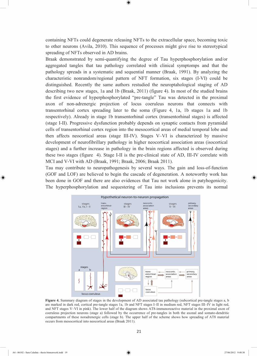
containing NFTs could degenerate releasing NFTs to the extracellular space, becoming toxic to other neurons (Avila, 2010). This sequence of processes might give rise to stereotypical spreading of NFTs observed in AD brains. Braak demonstrated by semi-quantifying the degree of Tau hyperphosphorylation and/or aggregated tangles that tau pathology correlated with clinical symptomps and that the pathology spreads in a systematic and sequential manner (Braak, 1991). By analyzing the characteristic nonrandom/regional pattern of NFT formation, six stages (I-VI) could be distinguished. Recently the same authors restudied the neuroptahological staging of AD describing two new stages, 1a and 1b (Braak, 2011) (figure 4). In most of the studied brains the first evidence of hyperphosphorylated “pre-tangle” Tau was detected in the proximal axon of non-adrenergic projection of locus coeruleus neurons that connects with transentorhinal cortex spreading later to the soma (Figure 4, 1a, 1b stages 1a and 1b respectively). Already in stage 1b transentorhinal cortex (transentorhinal stages) is affected (stage I-II). Progressive dysfunction probably depends on synaptic contacts from pyramidal cells of transentorhinal cortex region into the mesocortical areas of medial temporal lobe and then affects neocortical areas (stage III-IV). Stages V–VI is characterized by massive development of neurofibrillary pathology in higher neocortical association areas (isocortical stages) and a further increase in pathology in the brain regions affected is observed during these two stages (figure 4). Stage I-II is the pre-clinical state of AD, III-IV correlate with MCI and V-VI with AD (Braak, 1991; Braak, 2006; Braak 2011). Tau may contribute to neuropathogenesis by several ways. The gain and loss-of-function (GOF and LOF) are believed to begin the cascade of degeneration. A noteworthy work has been done in GOF and there are also evidences that Tau not work alone in patyhogenicity. The hyperphosphorylation and sequestering of Tau into inclusions prevents its normal
Figure 4. Summary diagram of stages in the development of AD associated tau pathology (subcortical pre-tangle stages a, b are marked in dark red, cortical pre-tangle stages 1a, 1b and NFT stages I–II in medium red, NFT stages III–IV in light red, and NFT stages V–VI in pink). The lower half of the diagram shows AT8-immunoreactive material in the proximal axon of coeruleus projection neurons (stage a) followed by the occurrence of pre-tangles in both the axonal and somato-dendritic compartments of these noradrenergic cells (stage b). The upper half of the scheme shows how spreading of AT8 material occurs from mesocortical into neocortical areas (Braak 2011).
21
containing NFTs could degenerate releasing NFTs to the extracellular space, becoming toxic to other neurons (Avila, 2010). This sequence of processes might give rise to stereotypical spreading of NFTs observed in AD brains. Braak demonstrated by semi-quantifying the degree of Tau hyperphosphorylation and/or aggregated tangles that tau pathology correlated with clinical symptomps and that the pathology spreads in a systematic and sequential manner (Braak, 1991). By analyzing the characteristic nonrandom/regional pattern of NFT formation, six stages (I-VI) could be distinguished. Recently the same authors restudied the neuroptahological staging of AD describing two new stages, 1a and 1b (Braak, 2011) (figure 4). In most of the studied brains the first evidence of hyperphosphorylated “pre-tangle” Tau was detected in the proximal axon of non-adrenergic projection of locus coeruleus neurons that connects with transentorhinal cortex spreading later to the soma (Figure 4, 1a, 1b stages 1a and 1b respectively). Already in stage 1b transentorhinal cortex (transentorhinal stages) is affected (stage I-II). Progressive dysfunction probably depends on synaptic contacts from pyramidal cells of transentorhinal cortex region into the mesocortical areas of medial temporal lobe and then affects neocortical areas (stage III-IV). Stages V–VI is characterized by massive development of neurofibrillary pathology in higher neocortical association areas (isocortical stages) and a further increase in pathology in the brain regions affected is observed during these two stages (figure 4). Stage I-II is the pre-clinical state of AD, III-IV correlate with MCI and V-VI with AD (Braak, 1991; Braak, 2006; Braak 2011). Tau may contribute to neuropathogenesis by several ways. The gain and loss-of-function (GOF and LOF) are believed to begin the cascade of degeneration. A noteworthy work has been done in GOF and there are also evidences that Tau not work alone in patyhogenicity. The hyperphosphorylation and sequestering of Tau into inclusions prevents its normal
Figure 4. Summary diagram of stages in the development of AD associated tau pathology (subcortical pre-tangle stages a, b are marked in dark red, cortical pre-tangle stages 1a, 1b and NFT stages I–II in medium red, NFT stages III–IV in light red, and NFT stages V–VI in pink). The lower half of the diagram shows AT8-immunoreactive material in the proximal axon of coeruleus projection neurons (stage a) followed by the occurrence of pre-tangles in both the axonal and somato-dendritic compartments of these noradrenergic cells (stage b). The upper half of the scheme shows how spreading of AT8 material occurs from mesocortical into neocortical areas (Braak 2011).
A4 - 06102 - Sara Calafate - thesis binnenwerk.indd 19 27/06/2012 9:48:30

22
function in neurons leading to failure of essential axonal transport functions (Zhang, 2005, Iqbal, 2010, Morris, 2011). The phenotype of Tau knock-out (KO) mice suggested that LOF is unlikely the main cause of neuronal toxicity. Theories highlighting Tau GOF as crucial elements in neurodegeneration cascade are based on Tau hyperphosphorylation and aggregation (Brunden, 2009; Morris, 2011). Currently, the amyloid cascade hypothesis assumes a serial model of causality proposing that toxic concentrations of Aβ triggers Tau hyperphosphorylation after a series of events (Ballard, 2011). Enhancing Tau phosphorylation provide Tau toxic species that play adverse effects on synapses (Morris, 2011). For many years, there was no genetic evidence for a role of Tau in the neurodegenerative process. The discovery of Tau gene mutations in familial forms of frontotemporal dementia and parkinsonism linked to chromosome 17 (FTDP-17) helped to understand that Tau alone can give rise to a pathological cascade (Goedert, 2005). Albeit, until now, in AD there were no identified Tau mutations and the primary event that leads to Tau modifications and consequent pathology is still not well understood. Abnormal Tau post-translational modifications , like hyperphosphorylation, directly or indirectly alter Tau conformation and function (Martin, 2011) and they can be blamed as inductors of Tau LOF and GOF. Coupling all evidences and doubts raises the following question: what triggers the pathological aggregation of Tau protein in AD, and how does it contribute to the subsequent pathogenesis?
1.4.1 Normal Tau characteristics and function
Tau, MAP1 and MAP2 belong to the neuronal microtubule associated protein (MAP) family. Tau is the major MAP in mature neurons and is believed to stabilize microtubules and also acts as spacer between them (Iqbal, 2010) providing tracts for motor proteins which is vital for synapses maintenance (Gendron, 2009). Other MAPs can do this function, so viability despite Tau-deficiency in knock out mice is probably due to this functional redundancy (Harada, 1994; Dawson, 2001). Tau is much smaller than other MAPs and predominantly localizes in axonal region (Gendron, 2009). The Tau gene is located on chromosome 17 and is encoded by 16 exons, of which exons 2, 3 and 10 undergo alternative splicing (Andreadis,
Figure 5. Tau gene comprises 16 exons. In the central nervous system, exons 1, 4, 5, 7, 9, 11, 12 and 13 (in blue) are constantly transcribed, whereas exons _1, 4A, 6, 8 and 14 (in grey) are not. Exons 2 (in orange), 3 (in purple) and 10 (in green) are subject to alternative splicing. Microtubules binding repeat domains R1, R3, R4 are in grey. (Martin, 2011)
A4 - 06102 - Sara Calafate - thesis binnenwerk.indd 20 27/06/2012 9:48:35

23
1992). In the central nervous system, Tau alternative splicing generates six isoforms of 352-441 amino acids with molecular weights between 60 and 74 kDa. These isoforms differ in the number of microtubule binding repeats (R) in the carboxyl terminal, containing 3 R if exon 10 is absent or 4 R if exon 10 is present. In the amino terminal (N), none, one or two inserts of 29 amino acids are possible. So, the six isoforms are: 0N3R which is the shortest isoform, 1N3R, 2N3R, 0N4R, 1N4R and the largest Tau isoform that has extra two amino terminal inserts and extra microtubule binding repeat 2N4R (Himmler,1989; Goedert, 1989). The Tau protein can be subdivided into four regions: (1) an acidic region in the N-terminus corresponding to exons 1–5. Exon 2 and exon 3 encode for N-terminal inserts; (2) a proline-rich region which is encoded by exon 7 up to the first half of exon 9; (3) a region containing the Tau microtubule binding repeat domains (MTBR), which is encoded by exons 9–12 and contains three or four repeat domains R1, (R2), R3 and R4 and (4) a C-terminal region which is encoded by exon 13 (Martin, 2011) (figure 5). In the developing human brain, only the shortest Tau isoform is expressed. In the mature normal brain similar levels of 3R and 4R isoforms coexist (Goedert, 2005). The Tau repeat domain region binds to the inner surface of the microtubule and the amino terminal regions are projected on the outside. Tau binds to the microtubule on the same β-tubulin pocket as taxol binds (Kar, 2003). The N-terminal region and the proline rich domain may play a role in interactions between Tau and other proteins. Some studies demonstrated that Tau can also interact with actin affecting filaments polymerization (Farlas, 2002). Interaction between Tau and proteins with Src homology 3 domains (SH3) was also reported (Reynolds, 2008). Tau is biologically regulated by its phosphorylation level and is normally phosphorylated at multiple serine (ser) and threonine (thr) residues (Buee, 2000). Roughly 2-3 moles of phosphate per mole of Tau protein are optimal for interactions with microtubules. In AD, Tau is hyperphosphorylated with some 19 moles of phosphate per mole of protein (Augustinack, 2002). The hyperphosphorylation enhances Tau-microtubule dissociation. The extra microtubule binding repeats and amino inserts increase Tau interactions with tubulin (Kopke, 1993; Iqbal, 2010) however 4R Tau more readily forms aggregates (Spillantini, 1997). Tau, in contrast to Aβ, is devoid of hydrophobic amino acids. In addition Tau has a positive net charge. These factors could explain the natively unfolded conformation of Tau (Uversky, 2000).
1.4.2. Pathological Tau
Tau is highly soluble and has a limited secondary structure appearing like a Gaussian random coil, as suggested by spectroscopic and x-ray evidence (von Bergen, 2000). However in AD brains Tau aggregates into stable Tau dimers that form Tau oligomers, which continue in the aggregation process and constitute subunits of filaments, called protomers. Two protomers around each other form PHFs and their assembly leads to NFTs formation (Martin, 2011) (Figure 6) during the course of disease as said before (1.4). An increase in the degree of hydrophobicity is sufficient to induce aggregation (Bergen, 2005). The oxidation of SH groups (Wille, 1992) and the interaction of Tau with polyanions, like herparin (Goedert, 1996), polyglutamate (Friedhoff, 1998), RNA (Kampers, 1996), and other molecules that
A4 - 06102 - Sara Calafate - thesis binnenwerk.indd 21 27/06/2012 9:48:39

24
share an extended negative charge, accelerates Tau aggregation. The mutation ∆K280 of FTDP-17 which consist of a deletion of a lysine which means the loss of a positive charge induces aggregation in polyanions-free way (von Bergen, 2001). The PHF formation is accompanied by a switch from random coil to a β-sheet structure, induced by two hexapeptides near the second and third microtubule binding repeat (von Bergen, 2000; Wang, 2007). This explains why a disordered protein can achieve filament state, showing that only the core/kernel of the filaments needs to be ordered, and N and C terminal is disposed as a “fuzzy coat” (Wischik, 1988). Phosphorylation is thought to induce Tau release from the MT and resulting in MT destabilization. This and other post-translational modifications can induce a conformational shift culminating with aggregation (Martin, 2011) and impairing axonal transport (Gendron, 2009). PHFs also induce cell shape modification, disruption of intracellular compartments and altered distribution of some organelles that are dependent on microtubule transport like mitochondria and endoplasmic reticulum (Ebneth, 1998). However, beside the questions asked above (section 1.4.1.) which is the nature of the first toxic specie that cause neuronal toxicity still remains unclear.
1.4.3 Identifying the culprit of neuronal toxicity in AD Tau hypothesis
Tau self assembly, aggregation and accumulation in NFTs is a hallmark of AD. The association between histopathological detection of NFTs and neurodegeneration lead to belief that NFTs are the cause of brain dysfunction but recently it has been questioned Some evidences argue that NFT are not the trivial components of neurotoxicity, altough they were not also able to exclude that their toxicity: (1) SantaCruz and colleagues hypothesized to suppress Tau transgene, in a Tau inducible transgenic mouse model that forms NFTs, to evaluate how brain dysfunction is dependent on Tau expression and which lesions could be reversible. The suppression of Tau transgene induces a recovery of memory functions despite of NFTs ongoing accumulation. This suggests that remaining NFTs after Tau suppression are not sufficient to cause disruption of cognitive function and that recovery of cognitive function maybe possible at early stages of Tauopathies (SantaCruz, 2005). (2) In animal models of other Tauopathies, cognitive dysfunction and neurodegeneration occurs without NFTs formation (Witteman, 2001; Ardofer, 2005). (3) Morsch demonstrated that neurons with NFT could live healthy for decades (Morsch, 1999). These studies suggested that intermediate species of Tau that
Figure 6 Model for multistep process of Tau aggregation (Martin L,2011).
A4 - 06102 - Sara Calafate - thesis binnenwerk.indd 22 27/06/2012 9:48:54

25
precede NFTs are responsible for neurotoxicity. Althought the exact aggregated toxic specie is unknown, is clear that aggregation is crucial player in pathogenecity. The mechanisms through which oligomers are generated and neurodegeneration is triggered are unclear however alterations in the balance of protein synthesis, folding and clearance (due either to familial mutations or post-translational modifications) have all been postulated to have important roles (Lee, 2010; Iqbal, 2010; Novak, 2011).
1.5. Tau post-translational modifications
Soluble Tau is a target for post-translational modifications. These modifications could directly or indirectly alter Tau conformation, enhance its dimerization, aggregation and fibrillation as previously described. Phosphorylation is the most common modification, however this is probably not sufficient to induce aggregation by itself. Studies of other post-translational modifications are important to understand how they can affect each other and Tau conformation. Because of the high incidence of these modifications, the rapid turnover and the ability of interactions with different partners Tau is considered an intrinsically disordered but highly regulated protein (Novak 2011). The molecular mechanisms responsible for inducing Tau pathology are still unclear.
1.5.1. Tau phosphorylation
Phosphorylation is addition of a phosphate group by esterification at three types of amino acids: ser, thr and tyrosine (tyr). Tau can be phosphorylated on 85 sites of which 45 are ser, 35 are thr and only 5 are tyr residues (Buee, 2000) (figure 8). Two distinct phosphorylation motifs are present within the Tau protein: (1) proline-directed ser/thr sites and (2) KXGS
Figure 7. Aggregated species in normal conditions enter into degradation pathways. Although with aging that is the highest risk factor for AD these mechanisms are deregulated and species like oligomers can propagate. Phosphorylation is likely the step of GOF enhancing aggregation and also LOF (Lee, 2010).
A4 - 06102 - Sara Calafate - thesis binnenwerk.indd 23 27/06/2012 9:49:02
26
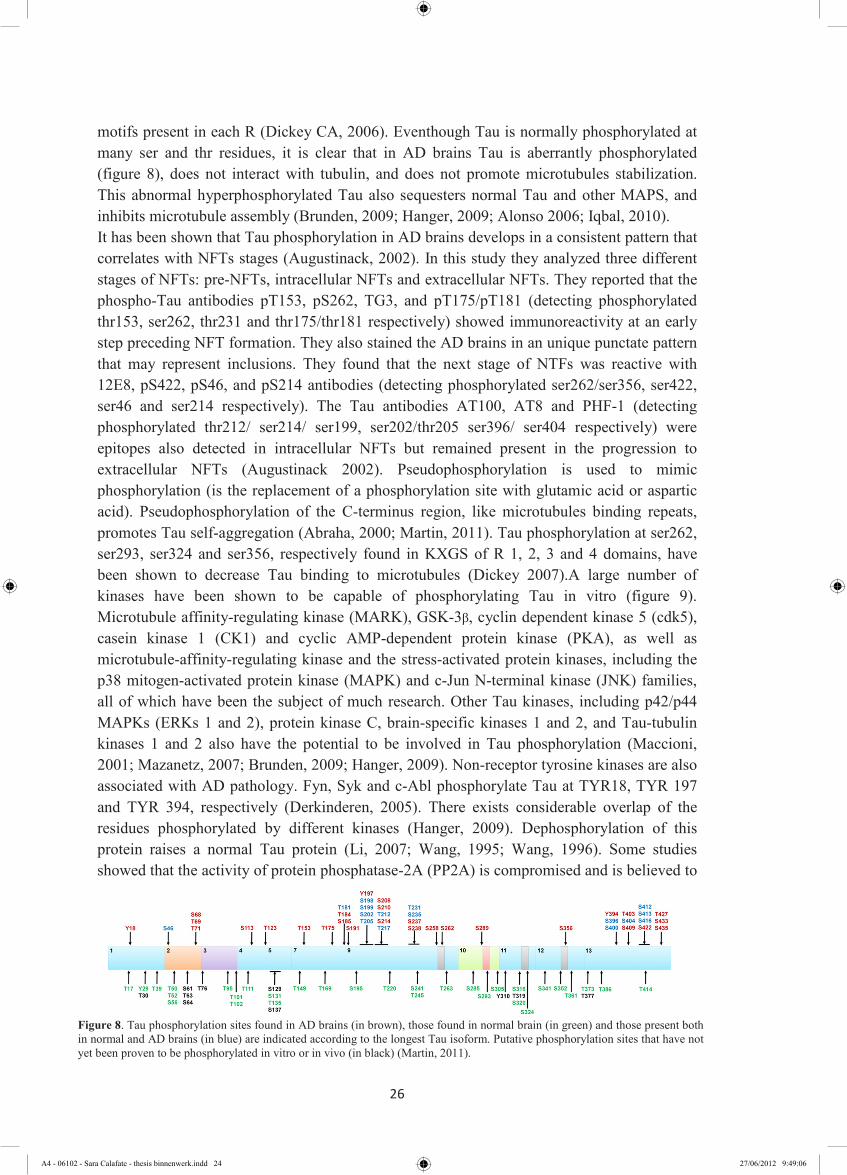
motifs present in each R (Dickey CA, 2006). Eventhough Tau is normally phosphorylated at many ser and thr residues, it is clear that in AD brains Tau is aberrantly phosphorylated (figure 8), does not interact with tubulin, and does not promote microtubules stabilization. This abnormal hyperphosphorylated Tau also sequesters normal Tau and other MAPS, and inhibits microtubule assembly (Brunden, 2009; Hanger, 2009; Alonso 2006; Iqbal, 2010). It has been shown that Tau phosphorylation in AD brains develops in a consistent pattern that correlates with NFTs stages (Augustinack, 2002). In this study they analyzed three different stages of NFTs: pre-NFTs, intracellular NFTs and extracellular NFTs. They reported that the phospho-Tau antibodies pT153, pS262, TG3, and pT175/pT181 (detecting phosphorylated thr153, ser262, thr231 and thr175/thr181 respectively) showed immunoreactivity at an early step preceding NFT formation. They also stained the AD brains in an unique punctate pattern that may represent inclusions. They found that the next stage of NTFs was reactive with 12E8, pS422, pS46, and pS214 antibodies (detecting phosphorylated ser262/ser356, ser422, ser46 and ser214 respectively). The Tau antibodies AT100, AT8 and PHF-1 (detecting phosphorylated thr212/ ser214/ ser199, ser202/thr205 ser396/ ser404 respectively) were epitopes also detected in intracellular NFTs but remained present in the progression to extracellular NFTs (Augustinack 2002). Pseudophosphorylation is used to mimic phosphorylation (is the replacement of a phosphorylation site with glutamic acid or aspartic acid). Pseudophosphorylation of the C-terminus region, like microtubules binding repeats, promotes Tau self-aggregation (Abraha, 2000; Martin, 2011). Tau phosphorylation at ser262, ser293, ser324 and ser356, respectively found in KXGS of R 1, 2, 3 and 4 domains, have been shown to decrease Tau binding to microtubules (Dickey 2007).A large number of kinases have been shown to be capable of phosphorylating Tau in vitro (figure 9). Microtubule affinity-regulating kinase (MARK), GSK-3β, cyclin dependent kinase 5 (cdk5), casein kinase 1 (CK1) and cyclic AMP-dependent protein kinase (PKA), as well as microtubule-affinity-regulating kinase and the stress-activated protein kinases, including the p38 mitogen-activated protein kinase (MAPK) and c-Jun N-terminal kinase (JNK) families, all of which have been the subject of much research. Other Tau kinases, including p42/p44 MAPKs (ERKs 1 and 2), protein kinase C, brain-specific kinases 1 and 2, and Tau-tubulin kinases 1 and 2 also have the potential to be involved in Tau phosphorylation (Maccioni, 2001; Mazanetz, 2007; Brunden, 2009; Hanger, 2009). Non-receptor tyrosine kinases are also associated with AD pathology. Fyn, Syk and c-Abl phosphorylate Tau at TYR18, TYR 197 and TYR 394, respectively (Derkinderen, 2005). There exists considerable overlap of the residues phosphorylated by different kinases (Hanger, 2009). Dephosphorylation of this protein raises a normal Tau protein (Li, 2007; Wang, 1995; Wang, 1996). Some studies showed that the activity of protein phosphatase-2A (PP2A) is compromised and is believed to
Figure 8. Tau phosphorylation sites found in AD brains (in brown), those found in normal brain (in green) and those present both in normal and AD brains (in blue) are indicated according to the longest Tau isoform. Putative phosphorylation sites that have not yet been proven to be phosphorylated in vitro or in vivo (in black) (Martin, 2011).
A4 - 06102 - Sara Calafate - thesis binnenwerk.indd 24 27/06/2012 9:49:06

27
be a contributor of the abnormal hyperphosphorylation of Tau in AD. The inhibition of PP2A with okadaic acid resulted in Tau hyperphosphorylation (Martin, 2009).The change in activity and expression levels of these kinases influences Tau phosphorylation levels and consequent LOF and GOF (Brunden, 2009; Noble, 2005; Noble 2010; Martin, 2011). Evidence for a functional link between Leucine rich repeat kinase 2 (LRRK2) and Tau has been emerging. In neuronal cultures expressing truncated LRRK2 presented axonal swelling containing Tau staining (Wray, 2010). Recently Kawakami and colleagues demonstrated for the first time that LRRK2 phosphorylates Tau in a microtubule dependent manner since free Tau is not LRRK2 substrate. This may decrease Tau microtubule affinity (Kawakami, 2012). Trying to identify if LRRK2 belongs to pathologic Tau interactome might reveal a link between Tauopathy and PD. These kinases and phosphatases could be considered pharmacological targets for halting the pathologic step-wised Tau phosphorylation. This subject will be further discussed.
1.5.2. Prolyl-isomerization of Tau
The phosphorylation state of a protein depends on the balance between kinases and phosphatases, but also on partners like isomerases that support conformational changes. Prolyl-isomerization is the interconversion of protein from cis to trans conformation. The peptidyl prolyl-isomerase (PPI) protein interacting with NIMA (never in mitosis A)-1) (Pin1) is a conserved enzyme that is intimately involved in diverse biological processes and pathological conditions such as cancer and Alzheimer's disease (Rudrabhatla, 2008). By catalysing cis–trans isomerization of certain peptide chains containing motifs of phosphorylated ser or thr residues followed by a proline residue (pser/thrProline), Pin1 can have profound effects on phosphorylation signaling. In Tau protein thr231 is the only site of prolyl-isomerization (Bulbarelli, 2009). Pin1 specifically binds to phosphorylated thr231 restoring the ability of Tau to bind microtubules and also facilitates Tau dephosphorylation by protein PP2A (Balastik, 2007). In AD neurons, Pin1 is sequestered in the tangles, and soluble Pin1 becomes depleted (Zhou, 2000). Inhibition of Pin1 results in accelerating aging (Lee, 2009) and aged Pin1-/- mice shown increased phosphorylation of Tau on THR231
Figure 9. Representative mechanism of Tau phosphorylation and further toxicity. Tau phosphorylation can be regulated by multiple kinases and phosphatases. The SER/THR kinases are represented in pink and TYR kinases in blue. Phosphatases are represented in yellow. The activity of these kinases can be enchanced by Aβ (Hanger, 2009).
Figure 9. Representative mechanism of Tau phosphorylation and further toxicity. Tau phosphorylation can be regulated by multiple kinases and phosphatases. The SER/THR kinases are represented in pink and TYR kinases in blue. Phosphatases are represented in yellow. The activity of these kinases can be enchanced by Aβ (Hanger, 2009).
A4 - 06102 - Sara Calafate - thesis binnenwerk.indd 25 27/06/2012 9:49:11

28
increasing aggregation (Liou YC, 2003). This suggests that Pin1 is apotential factor preventing pathology by decreasing the incidence of a pathogenic phosphoepitope.
1.5.3 Tau truncation
Proteolytic cleavage is another post-translational event. Truncated Tau could be generated by calpains generating a 17kDa fragment (Park, 2005) and by caspases releasing a 20 or 46 kDa fragment (Gamblin, 2003). Full-length Tau is difficult to fibrillize even at high concentrations unless fibrillization inducers are added. The nature of nucleating species is also an enigma. In AD, several site-specific Tau cleavages were identified and became connected to the progression of the disease (Kovacecha, 2010 a). Kovacecha demonstrated that Tau truncation alone is sufficient to induce the complete cascade of neurofibrillary pathology in a rat model of tauopathy (Kovacecha, 2010 b). For the study of Tau propagation, Frost et al demonstrated also that a truncated form of Tau was able to spontaneously aggregate (Frost, 2009). In 2007 Wang et al, using a cellular model for Tauopathy in Neuroblastoma cells (N2a cells) showed that a step-wised proteolysis induced aggregation of expressed mutant Tau repeat domain K18DK280. This processing generated a longer and two smaller fragments of Tau protein dubbed F1, F2, and F3 respectively (Wang, 2007). They also showed that the rate of aggregation depends on the size of the fragment with the longer F1 fragment being less prone to induce aggregation. The F2 and F3 fragments readily aggregate by themselves and nucleated the aggregation of full-length Tau (Wang, 2007). It was shown that C-terminal fragments of Tau can prevent aggregation (Berry, 2003). Likewise, aggregation-preventing domains near N-terminal that are clients for proteolysis (Alonso, 2001). Tau assumes a paperclip-like conformation when it is not interacting with partners also indicating a potential role for truncation in Tau aggregation (Wang, 2007; Jeganathan, 2008) (figure 10). In this conformation the C and N terminus hide the MTBR region which appears to be crucial triggering aggregation (Jakes, 1991; Novak, 2011). Truncation is an alternative mechanism by which Tau mediates beta-amyloid-induced neurodegeneration. Aβ oligomers were shown to induce calpain-mediated Tau cleavage triggered degeneration of hippocampal healthy neurons (Park, 2005). It was also suggested that this is an early GOF event preceding NFT formation (Martin, 2011).
Figure 10. Tau truncation is suggested as a pro-aggregation modification. Tau protein can adopts a paper-clip like ‘resting state’ when idle. Truncation prevents the adoption of this state. Aggregation is triggered and without the interference of the C-terminus the ratio is increased (N* = various truncated and non-truncated N-termini) (Novak, 2011).
A4 - 06102 - Sara Calafate - thesis binnenwerk.indd 26 27/06/2012 9:49:20
29
1.5.4 Ubiquitination of Tau
The Ubiquitin-proteasome system (UPS) is one pathway of the protein quality control (PQC) system, accompanied by the molecular chaperone system. Substrates for this pathway are recognized by covalent attachment of one or multiple ubiquitin molecules that are subsequently degraded by the 26S proteasome. The co-chaperone and ubiquitin ligase carboxy-terminus of Hsc70-interacting protein (CHIP) protein links these two systems. Some data suggest that CHIP regulates the degradation of Tau protein (Petrucelli; 2004; Dickey, 2007). Tau ubiquitination can occur in MTBR and is proposed to happen after phosphorylation and in PHF (Martin L. 2011). Relative quantitative analysis indicated that Lys-48-linked polyubiquitination was the primary form of polyubiquitination with a minor portion of ubiquitin linked at Lys-6 and Lys-11. Lys-48-linked ubiquitin-chains target proteins for degradation by the UPS suggesting that Tau is substrate for this pathway (Cripps, 2006). Hyperphosphorylated Tau becomes polyubiquitinated in NFTs. However, this ubiquitinated NFTs do not undergo proteasomal degradation (Yang, 1998). There are some studies reporting the influence of phosphorylation on Tau recognition by CHIP. SER199 SER202 and SER205 are the phospho-epitopes suggested by Shimura et al to be required for Tau-CHIP interaction, while Petrucelli et al have contradictory result arguing for independence of these factors (Shimura, 2004; Petrucelli, 2004). Dickey et al also found that phosphorylated Tau at KGXS motifs could not undergo proteassome degradation (Dickey, 2007). The presence of exon 10 on MTBR was also suggested to be necessary for Tau ubiquitination by CHIP (Hatakeyama, 2004).
1.5.5. Other post-translational Tau modifications – complex array of post-translational modifications and their interactions
As said before Tau is highly regulated. Many other modifications like acetylation, glycation, glycosylation, nitration, sumoylation, polyamination and oxidation complete this large network (Morris, 2011). Which one of these modifications during the disease process is crucial for pathology is not known. However cooperation between them is likely essential to get into the latest level of Tau pathology (figure 11). Phosphorylation is the most prevalent post-translational modification of Tau and because of this it is the focus of most of the studies. Phosphorylation levels can be reduced by competition with glycosylation in Tau. Glycosylation is the attachment of carbohydrates on a polypeptide backbone and it strongly affects the physicochemical properties of a protein, including resistance to denaturation, proteolytic degradation, and solubility. Enzymatic glycosylation involves the action of several enzymes and results in two type of glycosylation: N-glycosylation and O-glycosylation. Tau O-GlcNAcylation, that is a type of O-glycosylation that binds a ß-N-acetylglucosamine (GlcNAc), is dynamic and has been reported to vary reciprocally with Tau phosphorylation (Liu, 2004). A dynamic equilibrium between them has been suggested (Yuzwa, 2008). The O-glycosylation of Tau has been reported to be protective against Tau phosphorylation (Liu, 2004) and slow Tau aggregation (Yu, 2008). Glycation is a modification that also occurs in Tau. Advanced Glycated End Products (AGEs) are obtained
A4 - 06102 - Sara Calafate - thesis binnenwerk.indd 27 27/06/2012 9:49:24
30
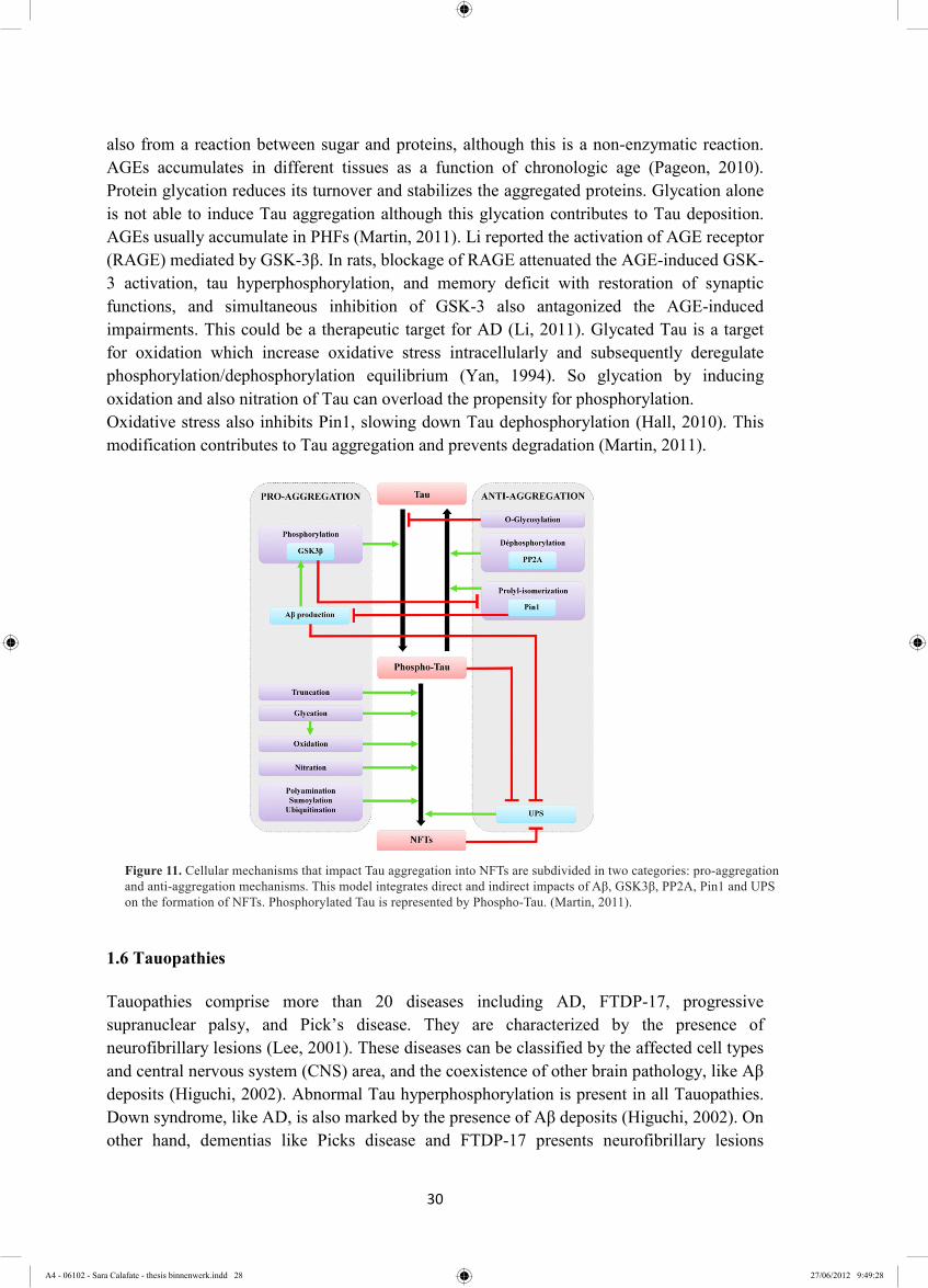
also from a reaction between sugar and proteins, although this is a non-enzymatic reaction. AGEs accumulates in different tissues as a function of chronologic age (Pageon, 2010). Protein glycation reduces its turnover and stabilizes the aggregated proteins. Glycation alone is not able to induce Tau aggregation although this glycation contributes to Tau deposition. AGEs usually accumulate in PHFs (Martin, 2011). Li reported the activation of AGE receptor (RAGE) mediated by GSK-3β. In rats, blockage of RAGE attenuated the AGE-induced GSK-3 activation, tau hyperphosphorylation, and memory deficit with restoration of synaptic functions, and simultaneous inhibition of GSK-3 also antagonized the AGE-induced impairments. This could be a therapeutic target for AD (Li, 2011). Glycated Tau is a target for oxidation which increase oxidative stress intracellularly and subsequently deregulate phosphorylation/dephosphorylation equilibrium (Yan, 1994). So glycation by inducing oxidation and also nitration of Tau can overload the propensity for phosphorylation. Oxidative stress also inhibits Pin1, slowing down Tau dephosphorylation (Hall, 2010). This modification contributes to Tau aggregation and prevents degradation (Martin, 2011).
1.6 Tauopathies
Tauopathies comprise more than 20 diseases including AD, FTDP-17, progressive supranuclear palsy, and Pick’s disease. They are characterized by the presence of neurofibrillary lesions (Lee, 2001). These diseases can be classified by the affected cell types and central nervous system (CNS) area, and the coexistence of other brain pathology, like Aβ deposits (Higuchi, 2002). Abnormal Tau hyperphosphorylation is present in all Tauopathies. Down syndrome, like AD, is also marked by the presence of Aβ deposits (Higuchi, 2002). On other hand, dementias like Picks disease and FTDP-17 presents neurofibrillary lesions
Figure 11. Cellular mechanisms that impact Tau aggregation into NFTs are subdivided in two categories: pro-aggregation and anti-aggregation mechanisms. This model integrates direct and indirect impacts of Aβ, GSK3β, PP2A, Pin1 and UPS on the formation of NFTs. Phosphorylated Tau is represented by Phospho-Tau. (Martin, 2011).
Figure 11. Cellular mechanisms that impact Tau aggregation into NFTs are subdivided in two categories: pro-aggregation and anti-aggregation mechanisms. This model integrates direct and indirect impacts of Aβ, GSK3β, PP2A, Pin1 and UPS on the formation of NFTs. Phosphorylated Tau is represented by Phospho-Tau. (Martin, 2011).
A4 - 06102 - Sara Calafate - thesis binnenwerk.indd 28 27/06/2012 9:49:28

31
without amyloid plaques (Higuchi, 2002). In AD, NFTs are only observed in neurons, however other Tauopathies contain Tau positive inclusions in both neuronal and glial cells (Komori, 1999; in Petrucelli, 2009). In every one of these disorders the accumulation of abnormally hyperphosphorylated Tau is associated with degeneration and dementia and in each of them the degeneration spreading follows a distinct stereotypical pattern. In FTDP-17 for example, the NFTs are shared from the frontal to the temporal cortex (Petrucelli, 2009). Even with different patterns of distributions of pathological Tau, the accumulation of insoluble hyperphosphorylated in a filamentous form is a bridge between AD and FTDP-17. FTDP-17 mutations provide evidence that Tau alone can induce toxicity and neurodegeneration. The molecular studies using these mutations allow understanding of potential molecular pathways involved in primary pathologic template generation.
1.6.1 FTDP-17 mutations
No mutations in Tau were identified in AD although in FTDP-17 at least 37 mutations were identified in Tau (figure 12). These mutations include missense, deletions or silent mutations and most of them are located in or around the MTBR. Three effects of mutations on the biology of tau have been postulated: 1) reduced ability to bind to microtubules, 2) altered tau splicing, with the majority of the mutations resulting in increased inclusion of exon 10 in tau mRNA, resulting in an increased 4R/3R ratio and 3) an increase in tau filament formation in the presence of polyanions in vitro (Alonso, 2004). R406W, V337M, G272V, and P301L Tau mutations increase the potential of Tau to aggregate since they make Tau a better substrate for kinases (Alonso, 2004).
1.6.2 Tauopathy models
Much of our current understanding of the pathogenic mechanisms of tau in tauopathies has been derived from the studies of the FTDP-17 Tau mutations as mentioned above combined with cell and animal studies. The historical picture is that in vitro Tau aggregation models failed for a long time in resembling NFTs. In most cell lines and mice models, over-expression of wild type (WT) human Tau under different promoters is not sufficient to induce neurofibrillary aggregation despite spontaneous hyperphosphorylation (Noble, 2010). Moreover, over-expression of WT Tau induces microtubule overstabilization and inhibition of cell division (Guo, 2011). In transgenic mice models tau pathology under the form of NFT was observed when FTDP-17 mutant variants like P301L, but rarely (see below) when WT Tau variants were expressed in the brain. Increasing the Aβ content in mice overexpressing mutant Tau by crossing APP mutant mice with Tau mutant mice enhance the Tau pathology accelerating the development of NFTs (Lewis, 2001). Vice versa Aβ toxicity was absent in Tau deficient mouse models suggesting that Tau mediated Aβ induced pathogenesis (crf IWT proposal for references). Andorfer and colleagues showed that in transgenic mice models the presence of mouse Tau interfered with the induction of Tau pathology. By crossing Tau KO mice with transgenic 8c mice that expresses all isoforms of human WT Tau (P1-derived artificial chromosome), a mouse model was developed that has AD-like pathology with accumulation of
A4 - 06102 - Sara Calafate - thesis binnenwerk.indd 29 27/06/2012 9:49:31

32
hyperphosphorylated Tau (Andorfer, 2003), while the single 8c mice do not develop Tau pathology (Duff, 2000). Contrary to the mouse models over-expression of WT or mutant tau turned out to be insufficient to induce tau aggregation in a wide variety of cell lines under a wide variety of experimental conditions (Noble, 2010). Only recently (including this master project) tau aggregation could be induced in vitro by including novel scientific insight into the pathogenic mechanisms involved in AD (see below). An improvement on Tauopathy models was done in last few years when the “seeding effect” concept emergend (Section 1.7.).
1.7 Pathogenic Protein Seeding in AD and other Neurodegenerative diseases
Misfolding and aggregation of specific proteins is a seminal occurrence in many different neurodegenerative diseases and is becoming an important target of study. Corruptive protein templating or seeding has been implicated in a variety of proteopathies such as Prion diseases but also and AD and other tauopathies. The prevalence of this pathogenic mechanism highlights templates as crucial key in disease process and many studies have been done to understand the nature of these species.
1.7.1 The amyloid state of proteins in human disease
Prions diseases belong to the group of misfolding neurodegenerative diseases and are caused by proteins which are capable to infect and propagate. This ability results from their β-sheet-rich conformation (amyloid state) which induces a slow nucleation phase followed a by a more rapid polymerization process, giving rise to highly ordered filamentous inclusions (Goedert 2010). Tau, Aβ and α-synuclein are, unlike prion proteins, typically unfolded proteins that also adopt an amyloid state in various neurodegenerative diseases (Eisenberg, 2012, Goedert 2010). Amyloid formation is a complex process and many proteins with an amyloid state enter in this state forming elongated unbranched fibrils, with nucleation-dependent kinetics and the
Figure 12. Mutations in the Tau gene in frontotemporal dementia and parkinsonism linked to chromosome 17 (FTDP-17). Schematic diagram of the six Tau isoforms with mutations in the coding region indicated using the numbering of the 441-amino-acid isoform. Exon 2 is shown in pink, exon 3 in green and exon 10 in yellow (Goedert, 2005)
A4 - 06102 - Sara Calafate - thesis binnenwerk.indd 30 27/06/2012 9:49:35

33
cross-β diffraction pattern (Figure 13) (Sawaya, 2007). The pattern shows that the fibrils contain β-sheets parallel to the axis and extended protein strands perpendicular to the axis (Sawaya, 2007; Eisenberg, 2012). This happens when a segment exposes its backbone amide N-H groups and C=O groups, allowing them to couple with other proteins chains forming a cross-β spine (Eisenberg, 2012). The interface between the two sheets that interdigitate is devoid of water forming a motif named “dry steric zipper” (Figure 13) (Eisenberg, 2012). This contributes to amyloid and prion stability (Jucker, 2012). The change in conformation is propagated into the new monomers (Lee, 2010) forming low molecular weight aggregates and giving rise to a needle shaped fibril (Sawaya, 2007) indicating that “a seeding effect” between monomers occurs. Some conditions like cleavage, denaturation, over-expression and over-production of disordered protein leads to the exposure of these backbones. The higher the concentration of the exposed segment the more aggregation is favored. These conditions can be manipulated in vitro, although in humans it is not already understood how this occurs (Eisenberg, 2012). Increased synthesis and decreased degradation may contribute to this phenomenon (Balch, 2008) besides post-translational modifications. The exposure of the amyloid-forming segments of three to four molecules is enough to nucleate and consequently template the cross-β fibber diffraction patter (Nelson, 2005). The understanding how amyloidoses leads to dysfunction is still lacking aside that via mass action tissue structure disruption occurs leading to systemic amyloidoses (Eisenberg, 2012). More than one fibril-forming segment was determined for Tau and for other amyloid proteins suggesting that in one protein we can find different spine structures (Sawaya, 2007). The role of soluble amyloid oligomers in neurotoxicity is today accepted for multiple neurodegenerative diseases. Reliable methods for detecting and studying Tau oligomers are lacking so researchers are prevented to target them. Lasagna-Reeves and colleagues have been developing some studies around oligomeric species detection. They argue that in vitro cross-seeding between oligomers ocurs showing that α-synuclein and Aβ42 oligomers are able to induce structure conversion of monomeric Tau (Lasagna-Reeves, 2010). The obtained oligomers were analyzed by circular dichroism spectroscopy showing that they are β-sheet rich with a weight of 150-190 kDa. These data are consistent with western blot analysis (WB) suggesting that these oligomers are mainly trimers. In this study the oligomers were shown to
Figure 13. A. Amyloidogenic proteins X-ray diffraction pattern. The diffuse reflection at 4.8 Ǻ spacing along the meridian (vertical) shows extended protein chains running roughly perpendicular to the fibril and spaced 4.8Ǻ apart. The even more diffuse reflection at 10 Ǻ spacing along the equator (horizontal) show s that the extended chains are organized into sheets spaced 10 Ǻ apart (Eisenberg, 2012); B. Tau belongs to class 1 of steric zippers organized in register β -sheet with the same sides facing each other; C. The axis of the fibril rotates 180˚ (Sawaya, 2007).
A4 - 06102 - Sara Calafate - thesis binnenwerk.indd 31 27/06/2012 9:49:39
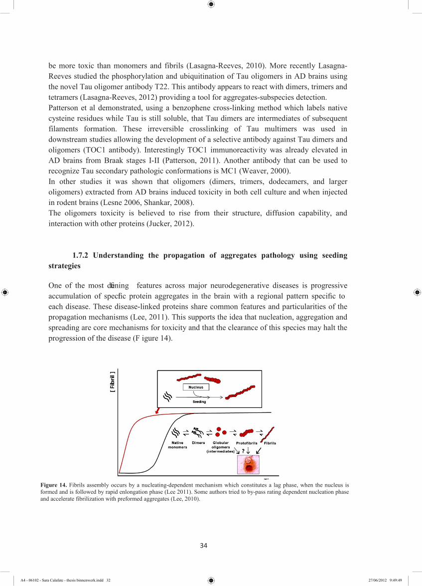
34
be more toxic than monomers and fibrils (Lasagna-Reeves, 2010). More recently Lasagna-Reeves studied the phosphorylation and ubiquitination of Tau oligomers in AD brains using the novel Tau oligomer antibody T22. This antibody appears to react with dimers, trimers and tetramers (Lasagna-Reeves, 2012) providing a tool for aggregates-subspecies detection. Patterson et al demonstrated, using a benzophene cross-linking method which labels native cysteine residues while Tau is still soluble, that Tau dimers are intermediates of subsequent filaments formation. These irreversible crosslinking of Tau multimers was used in downstream studies allowing the development of a selective antibody against Tau dimers and oligomers (TOC1 antibody). Interestingly TOC1 immunoreactivity was already elevated in AD brains from Braak stages I-II (Patterson, 2011). Another antibody that can be used to recognize Tau secondary pathologic conformations is MC1 (Weaver, 2000). In other studies it was shown that oligomers (dimers, trimers, dodecamers, and larger oligomers) extracted from AD brains induced toxicity in both cell culture and when injected in rodent brains (Lesne 2006, Shankar, 2008). The oligomers toxicity is believed to rise from their structure, diffusion capability, and interaction with other proteins (Jucker, 2012).
1.7.2 Understanding the propagation of aggregates pathology using seeding strategies
One of the most defining features across major neurodegenerative diseases is progressive accumulation of specific protein aggregates in the brain with a regional pattern specific to each disease. These disease-linked proteins share common features and particularities of the propagation mechanisms (Lee, 2011). This supports the idea that nucleation, aggregation and spreading are core mechanisms for toxicity and that the clearance of this species may halt the progression of the disease (F igure 14).
Figure 14. Fibrils assembly occurs by a nucleating-dependent mechanism which constitutes a lag phase, when the nucleus is formed and is followed by rapid enlongation phase (Lee 2011). Some authors tried to by-pass rating dependent nucleation phase and accelerate fibrilization with preformed aggregates (Lee, 2010).
A4 - 06102 - Sara Calafate - thesis binnenwerk.indd 32 27/06/2012 9:49:49
35
1.7.2.1 Evidence for Tau seeding
Although the kinetic aspects of seeding-dependent aggregate transmission remains to be clarified, findings from several recent studies clearly demonstrated induction of Tau aggregation by exogenously introduced aggregate seeds. Frost et al, showed that full-length Tau does not aggregate spontaneously, but found that the microtubule binding region alone does aggregate by itself. Using fluorescent labeled truncated Tau aggregated in vitro they demonstrated that these aggregates were taken up by cultured cells via endocytosis. This internalization triggers nucleation and aggregation of intracellular full-length Tau. (Frost, 2009). Recently the same group showed by Fluorescence resonance energy transfer (FRET) microscopy in co-cultured cells expressing different tagged Tau constructs, that in β-sheet rich inclusions the co-localization of both constructs occurred (Kfoury, 2012). They also demonstrated that the presence of an antibody, able to precipitate Tau in culture, blocked the uptake of fibrils of co-cultured cells, but not the intracellular aggregation in the donor cells (Kfoury, 2012). Another recent study performed by Guo and colleagues demonstrated that pre-fibrillization of a truncated form of Tau containing P301L mutation (K18P301L) induced aggregation of endogenous expressed mutant Tau P301L. The delivery of the seeds was by a lipid based vector and they showed that a small amount of internalized fibrils was enough to sequester large amounts of endogenous TauP301L into the insoluble fraction i.e aggregated Tau. The aggregated endogenous Tau was immune-reactive PHF-1 and AT8 antibodies (Guo, 2011). This in vitro study suggested that seeding is a plausible mechanism for NFT formation in AD brains and that the set up of an in vivo seeding model using the same strategy might be possible. In a recent in vivo study, two transgenic mouse lines ALZ17 and P301S were used. ALZ17 mice express the longest form of human Tau and do not form filamentous Tau aggregates. P301S mice express human Tau with mutation in exon 10 and develop abundant filamentous inclusions. Injection of homogenates of PHF loaded P301S Tau mice brains in hippocampus of ALZ17 mice induced in ALZ17 assembly of WT Tau into filamentous pathologic aggregates. Spreading of pathology was suggested to occur from the site of injection to the neighboring brain regions. Injection of extracts immunodepleted of Tau or divided into soluble or insoluble fractionss, showed that insoluble Tau is responsible for aggregation induction (Clavaguera, 2009). This elegant study suggests that the seeds can trigger nucleation of normal Tau and that trans-synaptic transmission of templates is a possible mechanism in vivo. Several strategies have been used to overcome the difficulty to induce tangles-like Tau deposits. Over-expression of Tau mutants that are prone to aggregation, truncated forms that fibrillizes more readily, over-expression of kinases and treatments that induce Tau phosphorylation, and compounds that induce aggregation have been tried. These studies allowed us to understand the early steps of aggregation and are a source of real NFTs. They represent a potential tool for identifying Tau-based therapies. The cell seeding model developed by Guo and colleagues is potentially useful for high-through-put drug screens.
A4 - 06102 - Sara Calafate - thesis binnenwerk.indd 33 27/06/2012 9:49:52

36
1.7.2.2 Aβ seeding evidences
There is evidence that also Aβ aggregates can function as seeds for aggregation of unfolded Aβ. The first evidence of induced Aβ deposition came from a study where an APP transgenic mice (Tg2576) was intracerebral injected with AD patients brains extracts. The induced β-amyloidosis started to spread into other areas of mice brain (Kane, 2000). More recently intraperitoneal inocculation of Aβ enriched extracts, induced brain amyloidosis after prolonged time in transgenic mice model suggesting that seeds can also be transmitted trough different tissues (Eisele, 2010).
1.7.2.3 α-Synuclein seeding evidences
α-Synuclein is a highly unfolded protein that interacts with membranes in synaptic terminals suggesting a role in synaptic maintenance and neurotransmitter releasing. Intracellular highly organized accumulated α-synuclein defines a group of diseases that are called Synucleiopathies, including Parkinson Disease (PD) and dementia with Lewy Bodies (LB) (Luk, 2009). These lesions first arise in the lower brain stem, anterior olfactory nucleus and olfactory bulb and later in the mesemphalic and neocortical region (Jucker, 2012). As said above α-Synuclein displays amyloid-like fibrilization. The idea of seeding in α-synuclein came from the observation that fetal dopaminergic neural transplants in striatum of Parkinson patients also developed lewy bodies containing α-synuclein. It means that the surrounding affected neurons transmitted the seeds into the host graft neurons (Li, 2008). Also in transgenic mice overexpressing human α-Synuclein neuronal grafts from WT mice were placed. After 6 months of incubation the dopaminergic neurons from graft were analyzed and intracellular human α-synuclein–positive punctae staining was detected. Some studies demonstrated that the aggregation of overexpressed α-Synuclein in cells can be induced by the internalization of α-Synuclein fibrillized seeds (Luk, 2009; Nonaka, 2010).
1.7.2 Pathogenic proteins interaction and cross-seeding evidences
Recently the synergetic interaction between Tau and Aβ has been studied. One of the strongest evidences for this interaction is that accumulation of Aβ possibly influences Tau pathology by increasing GSK3β activity and associated Tau hyperphosphorylation (Huang, 2009). There is some evidence that phosphorylated Tau and Aβ meet at the synapses since they co-localized at synaptossomes (Fein, 2008). Aβ and Tau interaction can be mediated by Aβ peptide, Aβ oligomers or Aβ-derived diffusible ligands (Jellinger, 2011). NFT pathology is increased in the brain by the presence of synthetic Aβ in a transgenic Tau mouse model (Gotz, 2001). In addition, it was shown that Tau reduction prevents Aβ-induced cell death in neuronal cultures (Roberson, 2007) and reduced Aβ-induced axonal transport defects (Stamer, 2002), futher suggesting a role for Tau-Aβ-interactions in AD pathology. The co-occurrence of Tau and α-synuclein aggregates as well as Aβ has been reported in many neurodegenerative diseases (Jellinger, 2011). While α-synuclein can fibrillize spontaneously, Tau needs an inductor (Giasson, 1999). The idea that α-synuclein is an
A4 - 06102 - Sara Calafate - thesis binnenwerk.indd 34 27/06/2012 9:49:56

37
interaction partner of Tau is supported by some researchers. The possibility of cross-seeding between Tau and α-synuclein was shown in vitro where mutant α-synuclein affected Tau fibrillization (Giasson, 2003). The hippocampus receives abundant dopaminergic innervations from substantia nigra and the ventral tegmental area works as a neuronal highway for possible interactions. The induction of Tau fibrilization by pre-fibrillized α-synuclein was shown in a cellular model as well as the colocalization of both hyperphosphorylated proteins (Waxman, 2011). The Tau aggregation in this model was even higher in the presence of P301L mutation. Some of the AT8 and PHF reactive cells appear NFT-like and part of the stained aggregates had a Lewi-body like morphology (Waxman, 2011). α-synuclein has also been shown to promote Tau phosphorylation mediated by GSK3β (Duka, 2006).Interactions between α-synuclein, Tau and Aβ may be a molecular mechanism in the overlapping pathology of Lewi-bodies and AD. These pathologies may represent a common pathway of neurodegeneration.
1.7.3 Neuron-to-Neuron transmission
Apart from prion diseases, most misfolding diseases were not shown to be transmittable between individuals. However, a stereotypical spatial and temporal spreading of these protein inclusions within the brains has been shown in some studies, as motioned above (Braak 1991, Clavaguera 2009). This feature is shared by prion and protein misfolding-based neurodegenerative diseases (Goedert 2010) and in case of AD the Tau aggregates are the pathological form of Tau that is responsible for the spreading and can behave like a permissive template (Novaka, 1994, Hardy, 2005, Braak, 2011). The studies presented before suggested that the seeded polymerization of endogenous protein is possible and that it is scattered by intracellular transference of aggregates. Although it has not yet been shown whether pathologic Tau species can spread via direct neuronal contact (transcellular spreading) or if they are released first and then taken up into a second recipient neighbouring neuron, inducing protein-protein misfolding. Aβ aggregates are extracellular, but NFTs are normally intracellular suggesting that they have to overcome barriers at least twice to induce seeding in neighbouring neurons. Phosphorylated Tau (Vandermeeren, 1993) and extracellular tangles - “ghost tangles” - were detected in cerebrospinal fluid (Mollenhauer, 2007). It has been suggested that Tau aggregates can be released from dead or dying cells, or even by exocytosis like prion proteins, stimulating further misfolding of Tau in adjacent cells. As said before Frost demonstrated that in cell cultures the endocytosis of extracellular Tau seeds can occur in cells lines (Frost, 2009) and the direct membrane penetration of α-synuclein seeds was also shown in cell lines (Luk, 2009). In the case of α-synuclein basal secretion of normal and aggregated
Figure 15. PC12 cells showed the formation of nanotubes in 4 minutes. TNTs diameter varies from 50 to 200 nm and a length of up to several cell diameters (Rustom, 2004).
A4 - 06102 - Sara Calafate - thesis binnenwerk.indd 35 27/06/2012 9:50:01

38
forms occurs in neuronal cultures. During misfolding stress this secretion is enhanced (Lee, 2010). This suggests that there is a release/uptake mechanism underlying trans-neuronal lesion spread during stress conditions. Exosomes are small vesicles that are generated intracellularly from early endosomes that give rise to MVB. Fusion with plasma membrane results in release of the exosomes to the extracellular space that subsequently can be endocytosed by neighbouring cells (Goedert, 2010). This secretory pathway was shown to be evolved with pathogenic proteins like Aβ, α-synuclein and prion proteins and it was recently demonstrated that Tau secretion can be supported by this pathway (Sudad Saman, 2011). After fractionating the lysate from a Tauopathy cell model (M1C cells expressing 4R0N Tau) in exosomal and non-exosomal membrane and non-membrane fractions Tau was immunodetected in all fractions. Mass spectrometry analysis of exosomal fractions detected exosomes containing Tau and showed that exosomal Tau is associated both with typical exosomal proteins and with proteins involved in Tau misprocessing and AD pathogenesis, such as Aβ, presenilins and Src family tyrosine kinase Fyn. They were reported to influence Tau phosphorylation, aggregation and toxicity. This study also showed that exosomal Tau was enriched in Tau phosphoepitopes as AT8 (Sudad Saman, 2011). On the other hand, endosomal compartments and multivesicular bodies are a barrier for aggregates-endogenous protein interaction. Although they can represent an optimal environment for seed processing breaking aggregates in smaller products due to the presence of proteases. It was shown to play a role in Aβ seeds formation (Hu, 2009). Rustom and colleagues reported the presence of ultrafine intercellular structures of cultured rat pheochromocytoma PC12 cells. These structures were tunneling nanotubes (TNTs). They can interconnect one cell to the next and could even form a network (figure 15 a). The buildup of TNTs was dependent on filamentous actin. The existence of expanded intercellular networks prompted researchers to speculate whether TNTs can participate in cell-to-cell
Figure 16. Cell-to-cell representative model suggesting a synergy between via vesicle-mediated exocytosis (1), leakage through damaged membranes (2) packaging into exosomes (3) and tunneling nanotubes (4) as mechanisms of transsynaptic transmission of seeds (pink) (Lee, 2010).
A4 - 06102 - Sara Calafate - thesis binnenwerk.indd 36 27/06/2012 9:50:05

39
communication. By labeling the acidic organelles with LysoTracker and later staining synaptophysin endosomes and endosome derived, small synaptic-like microvesicles (SLMVs) were detected inside the TNTs (Rustom, 2004) (figure 15 b). Gousset demonstrated that green fluorescent protein (GFP) tagged prion protein could traffic between neuronal cells by TNTs (Gousset, 2009). Many of the genes recently correlated with LOAD are responsible for synaptic function and cell membrane processes – PICALM, BIN1, CD33, CD2AP and EPHA1. Is noteworthy that at the nerve terminus clathrin-mediated endocytosis is regulated by many proteins that undergo phosphorylation-dephosphorylation cycles like dynamin 1, endophilin 1 and amphiphysin 1 that are substrates of kinases like dual-specific tyrosine phosphorylation regulated kinase (Dyrk1A) (Murakami, 2012). Overexpression of Dyrk1A imbalanced the 3R:4R ratio and increased Tau phosphorylation in Down-Syndrome brains (Wegiel, 2011). It suggests that endocytosis on synaptic area can be somehow affected in AD brains by deregulation of interactions between these players and that the synapse can be a crucial target for AD therapy. The mechanistic underlying the templating species transmission between neurons is not well understood. But there is increasing evidence that suggests that this seed is released into the extracellular environment and is taken up by neighboring neurons triggering a toxicity cascade by nucleating aggregation. Indeed permissive templates might spread via neuron-to-neuron transmission and trans-synaptic transport of pathogens from affected neurons to anatomically interconnected nerve cells (Braak, 2011) and exosomes and/or nanotubes probably mediate this phenomenon (Davies, 2008) (figure 16). Since in non-prion diseases the proteopathic agents have a mechanism close to prion proteins and since there is evidences of infection between organs, the infection between organisms should be analyzed. There is a need for epidemiological assessment of the ability of amyloid transmission between organisms. However many questions remain to be clarified (Table 1 adapted from Jucker, 2012).
1.8 Tau-focused strategies to halt AD progression
Several strategies to reduce the concequences of Tau pathology have been proposed. Tau post-translational modifications and particularly Tau phosphorylation might be specific initiators of Tau pathology. Therfore, inhibition of Tau kinases may be a good therapeutic strategy to reduce Tau-mediated neuropathology and neurodegeneration. Since phosphorylation and O-glycosylation of Tau occur at the same sites, inhibition of β-N-acetylglucosaminidase might be an alternative approach to decrease Tau phosphorylation.
Table 1. Mechanistic and Theoretical Issues What is the molecular structure of pathogenic proteins? Are there polystructural and polyfunctional species of misfolded proteins? What accounts for the selective vulnerability of neurons in the proteopathies? How do protein assemblies move from cell to cell and from region to region? Is there cross-seeding between proteins possible in human brains? Can exogenous corruptive seeds trigger non-prion neurodegenerative diseases in human brains? Are we working in the field of infectious diseases?
A4 - 06102 - Sara Calafate - thesis binnenwerk.indd 37 27/06/2012 9:50:08
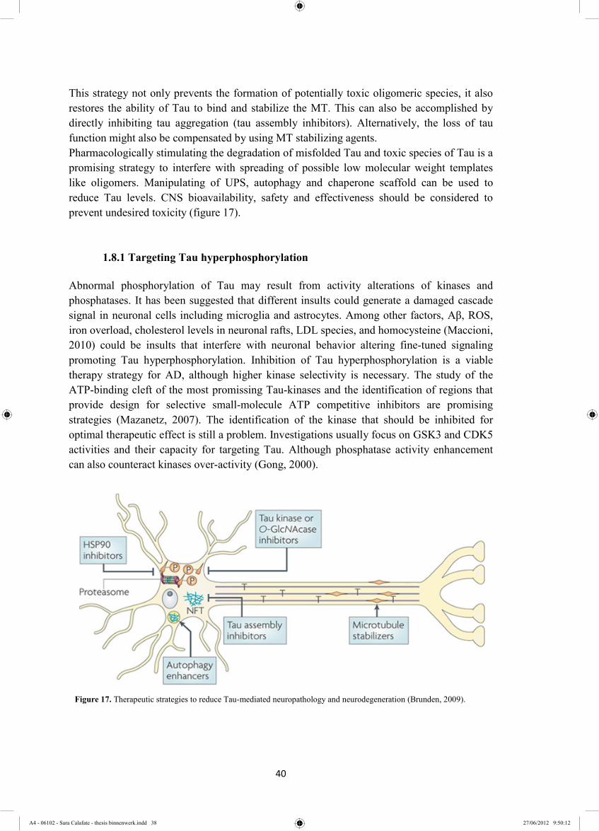
40
This strategy not only prevents the formation of potentially toxic oligomeric species, it also restores the ability of Tau to bind and stabilize the MT. This can also be accomplished by directly inhibiting tau aggregation (tau assembly inhibitors). Alternatively, the loss of tau function might also be compensated by using MT stabilizing agents. Pharmacologically stimulating the degradation of misfolded Tau and toxic species of Tau is a promising strategy to interfere with spreading of possible low molecular weight templates like oligomers. Manipulating of UPS, autophagy and chaperone scaffold can be used to reduce Tau levels. CNS bioavailability, safety and effectiveness should be considered to prevent undesired toxicity (figure 17).
1.8.1 Targeting Tau hyperphosphorylation
Abnormal phosphorylation of Tau may result from activity alterations of kinases and phosphatases. It has been suggested that different insults could generate a damaged cascade signal in neuronal cells including microglia and astrocytes. Among other factors, Aβ, ROS, iron overload, cholesterol levels in neuronal rafts, LDL species, and homocysteine (Maccioni, 2010) could be insults that interfere with neuronal behavior altering fine-tuned signaling promoting Tau hyperphosphorylation. Inhibition of Tau hyperphosphorylation is a viable therapy strategy for AD, although higher kinase selectivity is necessary. The study of the ATP-binding cleft of the most promissing Tau-kinases and the identification of regions that provide design for selective small-molecule ATP competitive inhibitors are promising strategies (Mazanetz, 2007). The identification of the kinase that should be inhibited for optimal therapeutic effect is still a problem. Investigations usually focus on GSK3 and CDK5 activities and their capacity for targeting Tau. Although phosphatase activity enhancement can also counteract kinases over-activity (Gong, 2000).
Figure 17. Therapeutic strategies to reduce Tau-mediated neuropathology and neurodegeneration (Brunden, 2009).
A4 - 06102 - Sara Calafate - thesis binnenwerk.indd 38 27/06/2012 9:50:12
41
1.8.1.1 GSK3
GSK-3 is a proline-directed ser/thr kinase that has two isoforms, GSK-3α and GSK-3β (Lucas, 2001). This kinase has been associated with normal functions of the cell and is highly expressed in brain. However some evidence supports the idea that Tau toxicity is mediated by GSK3 phosphorylation (Mazanetz, 2007). In in vitro and in cell cultures, GSK-3β can phosphorylate Tau at several sites that are similar to those seen in PHF of AD brains (Rankin, 2007). A GSK-3β transgenic mouse model showed Tau hyperphosphorylation and increased neuronal death in hippocampus (Lucas, 2001). Another transgenic mouse overexpressing GSK-3β and mutated Tau showed accelerated neurodegenerative symptoms (Engel, 2006). Mice overexpressing GSK-3β showed a slower progression of neurodegeneration in a Tau KO background (Barreda, 2010). Pei and colleagues demonstrated elevated GSK-3β immunoreactivity in plaques and CA1 hippocampal neurons which co-localize with hyperphosphorylated Tau and NFTs (Pei, 1997). When GSK3 inhibitor LiCl and AR-A014418 were administrated to a transgenic mouse expressing mutant human Tau reduced Tau phosphorylation, Tau accumulation and axonal degeneration was observed (Perez, 2003; Hanger, 2005). Both inhibitors also increase Tau ubiquitination which can contribute to attenuate pathology possible via GSK-3β mediated regulation of the HSP90/CHIP complex (Dickey, 2007). However, in a phase 2 study with MCI patients Lithium treatment did not reduce GSK3 activity or Tau CSF levels and showed no cognitive improvements (Hampel, 2009).
1.8.1.2 CDK5
CDK5 is a multifunctional ser/thr protein kinase essential for neuronal development and synaptic activity (Zheng, 2010). This kinase is regulated by phosphorylation of ser159 which increases its activity. CDK5 can phosphorylate different motifs: KSP on MAP1b, KSPXK on Tau, KSPXX on neurofilaments proteins, the actin binding protein caldesmon, and proteins involved in synaptic vesicle exocytosis. At post-mitotic neuronal cells CDK5 is regulated by cyclin-related activators 35 and p39 (Hisanaga, 2003). Stress-induced influx of calcium and neurotoxicity can activate calpains that cleave p35 resulting in the more stable truncated form p25 (Zheng, 2002; Lee, 2000). CDK5/p25 is a hyperactive complex shown to hyperphosphorylate Tau (Noble, 2003). CDK5 phosphorylates Tau on sites of Tau also commonly hyperphosphorylated in AD brains, and this kinase also co-localizes with NFTs containing neurons (Rademakers, 2005). Inducible expression of p25 in rodent models raises neurodegeneration and NFTs formation (Mazanetz, 2007) as well as over-expression in cultured neurons (Ahlijanian, 2000). Moreover, the p25:p35 ratio was reported to be higher in AD brains when compared to the controls (Rademakers, 2005), although the opposite was also reported (Zheng, 2010). In a rat model with abnormal Cdk5 activation by p25 and subsequent tau hyperphosphorylation and subsequent neurodegeneration and neurofibrillary tangles the administration of calpain inhibitors improved neuronal function (Hung KS, 2005). CDK5 expression can regulate GSK3 activity by priming sites for subsequent phosphorylation by GSK3 (Sengupta, 1997). It was also found that transgenic mice over-
A4 - 06102 - Sara Calafate - thesis binnenwerk.indd 39 27/06/2012 9:50:16

42
expressing both p25 and human mutated Tau showed accumulation of hyperphosphorylated Tau associated with GSK3 activity, and increased NFTs levels (Noble W, 2003). Although over-expression of p35 did not alter phosphorylated Tau levels (Van den Haute, 2001). Some evidences argue against previous data demonstrating that low levels of p25 could improve memory deficits (Angelo, 2003).It was suggested that Aβ can induce p35 cleavage into p25, linking CDK5 and Tau hyperphosphorylation to Aβ peptides (Rademakers, 2005). Indolinones have been shown to potentially inhibit CDK5 without affecting other CDKs that are structurally closest to CDK5 (Mazanetz, 2007). Zheng et al reported efficient inhibition of CDK5/p25 complex without affecting endogenous CDK5/p35 complex and other kinases, mediated by a 24 peptide called p5 that rescued neurons from Aβ toxicity, Tau hyperphosphorylation, and cell death (Zheng, 2010). It showed that minimize the potential toxicity arising from inhibition of such off-target functions, while maximizing effective suppression of Tau hyperphosphorylation can eventually be achieved.
1.8.1.3 PP2A
PP2A is likely to be the major Tau phosphatase. Inhibition of PP2A by potent selective compounds showed an increased Tau hyperphosphorylation and induced deficits in spatial memory retention in rats. PP2A accounts for more than 70% of Tau phosphatase activities in the human brain (Liu et al., 2005). In the CA3 area of the AD hippocampus the expression of mRNAs of the catalytic subunit PP2ACα and the regulatory subunits PR55γ and PR61ε of PP2A, are reduced. The expression of the PP2Ac gene and PP2A ABαC protein subunits has been found to be down-regulated in the affected area of AD brain (Sontag, 2004). Another elements possibly involved on PP2A regulation are two inhibitor proteins, called I1
PP2A and I2PP2A, in mammalian tissues. Tanimukai and colleagues developed a study that
demonstrated an intraneuronal increase in the AD neocortex of mRNA expression of I1PP2A
and I2PP2A. The co-localization of these two inhibitors with PP2A and with the abnormally
hyperphosphorylated Tau was also shown (Tanimukai, 2005). These data suggest that down-regulation of dephosphorylation may play a role in Tau pathology and inhibition of PP2A can be used to study the role of Tau phosphorylation (Martin, 2009).
1.8.2 The potential of aggregation inhibitors
The inhibition of the formation of aggregates is a strategy that could be applied to multiple neurodegenerative diseases since amyloidogenic proteins share the same structures. N-phenylamines antraquinones, phenylthiazolyl-hydrazides, and thioxothiazolidinones (rhodanines) were described as potential Tau aggregation inhibitors (Khlistunova, 2006, Bulic 2010). The methylthioninium chloride compound also known has methylene blue (MB) seems to be a drug candidate for Tau aggregation inhibition supported by preliminary data in a phase II clinical trial (Wischik, 2007). Recently MB was shown to reduce Tau levels both in organotypic brain slice cultures from a mouse model of FTD, and in cell models. Concomitantly, MB treatment altered the levels of LC3-II, cathepsin D, Beclin 1, and p62 suggesting that it is also a potent inducer of autophagy (Congdon, 2012).
A4 - 06102 - Sara Calafate - thesis binnenwerk.indd 40 27/06/2012 9:50:20
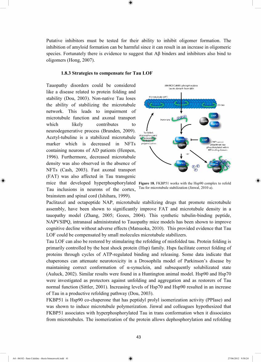
43
Putative inhibitors must be tested for their ability to inhibit oligomer formation. The inhibition of amyloid formation can be harmful since it can result in an increase in oligomeric species. Fortunately there is evidence to suggest that Aβ binders and inhibitors also bind to oligomers (Hong, 2007).
1.8.3 Strategies to compensate for Tau LOF
Tauopathy disorders could be considered like a disease related to protein folding and stability (Dou, 2003). Non-native Tau loses the ability of stabilizing the microtubule network. This leads to impairment of microtubule function and axonal transport which likely contributes to neurodegenerative process (Brunden, 2009). Acetyl-tubuline is a stabilized microtubule marker which is decreased in NFTs containing neurons of AD patients (Henpen, 1996). Furthermore, decreased microtubule density was also observed in the absence of NFTs (Cash, 2003). Fast axonal transport (FAT) was also affected in Tau transgenic mice that developed hyperphosphorylated Tau inclusions in neurons of the cortex, brainstem and spinal cord (Ishihara, 1999). Paclitaxel and octapeptide NAP, microtubule stabilizing drugs that promote microtubule assembly, have been shown to significantly improve FAT and microtubule density in a tauopathy model (Zhang, 2005; Gozes, 2004). This synthetic tubulin-binding peptide, NAPVSIPQ, intranasal administrated to Tauopathy mice models has been shown to improve cognitive decline without adverse effects (Matsuoka, 2010). This provided evidence that Tau LOF could be compensated by small molecules microtubule stabilizers. Tau LOF can also be restored by stimularing the refolding of misfolded tau. Protein folding is primarily controlled by the heat shock protein (Hsp) family. Hsps facilitate correct folding of proteins through cycles of ATP-regulated binding and releasing. Some data indicate that chaperones can attenuate neurotoxicity in a Drosophila model of Parkinson’s disease by maintaining correct conformation of α-synuclein, and subsequently solubilizated state (Auluck, 2002). Similar results were found in a Huntington animal model. Hsp90 and Hsp70 were investigated as protectors against unfolding and aggregation and as restorers of Tau normal function (Sittler, 2001). Increasing levels of Hsp70 and Hsp90 resulted in an increase of Tau in a productive refolding pathway (Dou, 2003). FKBP51 is Hsp90 co-chaperone that has peptidyl prolyl isomerization activity (PPIase) and was shown to induce microtubule polymerization. Jinwal and colleagues hypothesized that FKBP51 associates with hyperphosphorylated Tau in trans conformation when it dissociates from microtubules. The isomerization of the protein allows dephosphorylation and refolding
Figure 18. FKBP51 works with the Hsp90 complex to refold Tau for microtubule stabilization (Jinwal, 2010 a).
A4 - 06102 - Sara Calafate - thesis binnenwerk.indd 41 27/06/2012 9:50:24
44
of Tau. The FKBP51 interaction with Tau could prevent its ubiquitination (Jinwal, 2010a). Manipulation of this PPIase enzyme could also be a useful strategy to prevent LOF and microtubule network dissociation (figure 18).
1.8.4 Enhancing intracellular Tau degradation
Numerouse physiolocal processes are affected in AD, including the degradative mechanisms. Impairements of both proteassomal and autophagic pathways have been described (Keck, 2003; Nixon and Yang, 2011) and probably neurons fail to degrade hyperphpsphorylated and aggregated Tau. The manipulation of control pathways aiming the overall reduction of Tau levels in adult and old animals with AD related pathologies was shown to be promising (Morris, 2011). Different approaches can be developed to accelerate Tau degradation to reduce total and pathological Tau levels. This can be achieved for example by targeting molecules that regulate the expression and clearance of this protein.
1.8.4.1 The role of proteasome and autophagy the in Tau degradation
The ubiquitin-proteasome system and autophagy are two main proteolytic pathways (Rubinsztein, 2006). Tau was shown to be degraded by the proteasome (Petrucelli, 2004). In AD brains it was shown that CHIP levels were reduced and inversely propotional to aggregated Tau levels and a reduction of CHIP levels in the JNPL3 mice model increased aggregated human Tau with P301L mutation (Sahara, 2005). The inhibition of the proteasome in rat models leads to Tau accumulation (Liu, 2008), suggesting that Tau degradation is dependent of UPS. Inhibiting the UPS leads to a decrease in Tau turnover and subsequently increased Tau levels in AD brains (Iqbal, 2010). Most studies have focused on overexpressed Tau instead of endogenous Tau. Some studies have reported that endogenous Tau does not follow proteasome degradation suggesting the endogenous and exogenous Tau might undergo different degradations pathways (Delobel, 2005; Kruger, 2011). It was shown that by inhibiting the deubiquitinase activity of USP14 the proteasome degradation of different substrates including Tau increased. The results suggested that the ubiquitin chains trimming prevented proteasome degradation, and inhibiting UPS14 it was accelerated in murine embryonic fibroblasts (Lee BH, 2010). Although, there is no concensus on which pathway is involved in normal Tau degradation so the mechanism of endogenous Tau degradation remains unclear. It was suggested that large multimeric complexes of Tau like oligomers and aggregates are excluded from ubiquitin-proteassomal pathways and preferentially are clients of autophagy (Nixon, 2006). Enhancing autophagy with rapamycin was indeed reported to reduce the number of Tau aggregates and its toxicity on a drosophila model for tauopathy (Berger, 2006) and has been proposed as a therapeutic approach for many neurodegenerative diseases (Mizushima, 2010; García-Arencibia 2010). Whether autophagy impairment contributes for Tau pathology remains uncertain.
A4 - 06102 - Sara Calafate - thesis binnenwerk.indd 42 27/06/2012 9:50:27

45
The induction of autophagy can be achieved by treatment with trehalose (Kruger, 2011; Perucho, 2012). Trehalose is a non-reducing disaccharide and works as a “chemical chaperone”. This compound activates autophagy independent of mTOR, a potent inhibitor of autophagy (Tanaka, 2004). Trehalose administration was shown to reduce the levels of endogenous and overexpressed Tau in in vitro models (Kruger, 2011), but also reduced the levels of β amyloid peptide aggregates, Tau plaques, and levels of phospho-Tau in APPswe mutant mice (Perucho, 2012). These data suggest that up-regulation of catabolic systems could prevent or delay proteinopathies by degrading the proteotoxic components. Autophagy can aso be induced by inhibiting the mTOR pathway with rapamycin, Torin1, or PP242 but strong side effects were detected since this pathway regulates other crucial cellular mechanisms beside autophagy. A combination between mTOR dependent/independent drugs could be suitable for autophagy induction in patients with proteinopathies (Rubinsztein, 2010).
1.8.4.2 Manipulating Hsp scaffolds to enhance Tau degradation
Intracellular environment is crowded and presents obstacles to proper protein folding. A loss in the equilibrium between refolding and degradation could increase neurotoxic protein depositions. Therefore, PQC by the chaperone system is essential for maintaining cell homeostasis. Chaperones contribute to decisions of correct protein folding and promote degradation of prolonged misfolded proteins. Two important players in this process are Hsp70 and Hsp90. Hsp70 is composed of a nucleotide-binding domain and a substrate-binding domain that contains an ATP-binding pocket and an EEVD domain, respectively (Miyata, 2011). Hsp90 has three domains: N-terminal domain for ATP binding, charged linker domain, a middle domain involved in client bind and ATPase activity, and a C-terminal domain including an EEVD motif involved in co-chaperone binding and dimerization (Miyata, 2011). As said before Hsp70 and Hsp90 cooperate in multimeric complexes with co-chaperones that manipulate their ATPase activity to differently interact with different client proteins (Koren III, 2009). Their interaction is mediated by tetratricopeptide repeat domains in co-chaperones that ineteract with EEVD motifs (reviwed in Miyata, 2011). Most client proteins of the chaperone system are recognized by Hsp40 also known as J-protein, which facilitates the binding to Hsp70 family chaperones. The client in coordination with ATP hydrolysis can achieve proper folding, re-enter in an exclusive Hsp70 cycle, or if the client fails the primary checkpoint it is transferred to Hsp90 in coordination with Hsp70/Hsp90-organizing protein (Hop) (Koren III J, 2009; Dickey CA, 2007, Miyata, 2011). In this later complex the client can be re-folded or targeted for degraded by the ubiquitin proteasome (Dickey, 2009). The fate of the client is dependent on the co-chaperone that modulates Hsp90 activity and also on Hsp90 post-translation modifications (Wandinger, 2008; Dickey, 2009). Hsp90 normally inhibits heat shock factor 1 (HSF1) by tethering it to the cytosol. By triage to substrate degradation, also Hsp90 is degraded releasing HSF1 that trimerizes, translocates to nucleus and begins inducing transcription of Hsp genes (Koren III, 2009). Both Hsp70 and Hsp90 can recruit CHIP to degrade the bound substrate (Connell, 2001). When co-chaperones of Hsp90
A4 - 06102 - Sara Calafate - thesis binnenwerk.indd 43 27/06/2012 9:50:31

46
like p23 and Pin 1 are presents they facilitates the refolding decision of this complex that contains the substrate preventing degradation (Liou, 2003; Grenert, 1997) (Figure 19). It was shown that molecules that block the ATPase activity of the N-terminal domain or interfere with the binding of Hsp90 to its co-chaperones that promote refolding complex formation, like Hsp90 inhibitors, are of interest as potential therapeutics. These low molecular weight Hsp90 inhibitors must have sufficient BBB penetrance in order for them to be usefull for neurodegenerative diseases. The majority of Hsp90 inhibitors discovered are macrocycles. Macrocycle drugs are more resistant to proteolysis than acyclic molecules (Johnson, 2010). Geldanamycin (GA) was the first macrocycle found to inhibit Hsp90 and bind to the N-terminal ATP binding pocket. However, GA is hepatotoxic. Some derivatives which are less toxic were generated: 17-allylamino 17-demethoxy-geldanamycin (17-AAG) and 17-dimethyl-amino-ethylamino-17- demethoxygeldanamycin (17-DMAG) (Kamal, 2004). In 2007 Dickey et al described a novel Hsp90 inhibitor, EC102, with reasonable brain penetration. (Dickey, 2007). They showed that the knock down (KD) of p23 had the same effect as Hsp90 inhibition results in phosphorylated Tau degradation. They also showed that silencing Hsp90 with siRNA abrogated EC102 activity and phosphorylated Tau accumulated. However, by overexpressing CHIP the degradation was recovered. CHIP, Hop and Hsp40 siRNAs also increase total Tau levels. This work demonstrated that CHIP null mice accumulate phosphorylated Tau, and that CHIP is a central regulator by “mediating proper protein triage decisions”. It was suggested that co-chaperones that promote protein refolding (i.e. p23 and Pin1) may compete with those that promote degradation. This work presents a possible bifurcation of the chaperone pathway clarifying some puzzling concerns (Dickey, 2007). Two different stages of Hsp90 complex were suggested: one is a proteasome-targeting form associated with Hsp70 and Hop, and the other is a stabilizing form with Pin, p23 and Cdc37. So these compounds could play a selective inhibition in these cells, correlating with presence of multi-chaperone complexes, allowing higher margins for safety. Recently Cook and his group added a new piece of information to this chaperone scaffold “puzzle”. Hyperacetylation of Hsp90 decreases its interaction with substrates and p23 promoting client degradation (Cook, 2012). Histone Deacetylase 6 (HDAC6) depletion leads to Hsp90 hyper-acetylation and subsequent substrates degradation (Bali, 2005). Using this information, this group demonstrated that loss of HDAC6 activity augments the efficacy of
Figure19. Scheme representation of a possible mechanism for refolding and degradation of Hsp70 and Hsp90 client proteins, like phosphorylated tau (p-tau) (Dickey, 2007).
A4 - 06102 - Sara Calafate - thesis binnenwerk.indd 44 27/06/2012 9:50:35
47
an Hsp90 inhibitor and drives client degradation. CHIP controls the turnover of HDAC6 by ubiquitinating and degrading this protein. Taken together this data indicate HDAC6 as a critical factor in the regulation of Tau levels (Crook, 2012). In summary, inhibiting CHIP levels suppresses the degradation of hyperphosphorylated Tau and inhibition of Hsp90 activity or its co-chaperones that promote refolding of Hsp90 clients enhanced the degradation of Tau (figure 15). Hsp90 inhibiton triggers two responses (1) proteasome-mediated degradation of Hsp90 client proteins and (2) HSF1 release inducing enhanced expression of Hsp40, Hsp70, and Hsp90 (Dickey, 2006). By this way the efficiency of the Hsp90 inhibitor can be controlled by the respons of Hsp70 levels. 1.9 Project Goal
To initiate a drug discovery process and validate targets it is mandatory the to have access to credible models. To study the spreading, build-up and clearance of Tau of seeding and aggregation models in vitro were developed.
1.9.1 Variability analysis and characterization of clonal-cell line seeding model
The host lab recently succeeded in reproducing the first in vitro clonal-cell seeding model that recapitulates the aggregation of human Tau (Guo, 2011). This was achieved by "seeding" the system with an in vitro synthesized and aggregated Tau fragment. Access to this valuable cell model allows study of the molecular mechanisms of Tau aggregation seeding, build up and degradation. For the purpose of validation the effect of known tau aggregate modulators needs to be confirmed in the model at hand. For the purpose of screening for compounds and genes that interfere with the mechanisms involved the model has to be optimized for further high-throughput screening.
1.9.2 Set Up of primary neuronal seeding model
An additional goal of my work was to expand the “seeding effect” model to primary neuronal cultures to build a more relevant cell model. I will use this model to and evaluate the role of tau post translational modifications on Tau aggregation and study potential spreading in the model. This work is included in the concept of a permissive templating as a common mechanism in neurodegenerative disease. The strategy used is extrapolated from the clonal-cell seeding model with some adaptations.
Adeno-Associated Viral (AAV) vectors are one of the most used tools for transgene expression due to low immunogenicity. They are very small (20 nm) with icosahedral shape containing single stranded DNA. AAV are able to infect dividing and non-dividing cells allowing integration, including neurons with sustained expression (de Lima, 2005). For gene delivery in primary neuronal cultures AAV vector from Serotype 6 (AAV6) was used. The plasmid encodes the inverted terminals that are essential for viral replication and packaging
A4 - 06102 - Sara Calafate - thesis binnenwerk.indd 45 27/06/2012 9:50:38

48
(Wu, 2006) and the gene of interest under the human synapsin 1 (hSYN1) promotor that allows neuronal specific expression. To deliver the synthetic pre-fibrillized Tau protein Bioporter delivery tool was used. The BioPorter reagent is a cationic lipid formulation of trifluoroacetylated lipopolyamine and dioleoyl phosphatidylethanolamine (TFA-DODAP:DOPE) that enables delivery of recombinant proteins, peptides, or antibodies into viable cells (Zelpathi, 2001). Using these two delivery tools and further biochemical analysis of protein expression and interaction it is possible to study Tau aggregation both in a clonal-cell line and in primary cortical cultures.
A4 - 06102 - Sara Calafate - thesis binnenwerk.indd 46 27/06/2012 9:50:42

49
Chapter 2 - Materials
A4 - 06102 - Sara Calafate - thesis binnenwerk.indd 47 27/06/2012 9:50:45

50
A4 - 06102 - Sara Calafate - thesis binnenwerk.indd 48 27/06/2012 9:50:49

51
2.1 Material
Company Product
Active Motif Chariot(30025 & 30100)
BD Falcon Tissue culture flasks
Beckman Coulter
Beckman centrifuge tubes (243775)
Optima ultracentrifuge
Vi-Cell XR (Cell viability analyzer)
Santa Cruz Blotto non-fat dry milk (sc-2325)
Eppendorf Electroporation cuvettes, gap width 1 mm, 100 µl (4307 000.569)
Eppendorf tubes of 0.5, 1.5, and 2 mL
Falcon Multiwell plates of 96, 24, and 6 wells
Gelantis BioPORTER Reagent QuikEase Single-Use Tubes (BP509696)
HyClone Research Grade Fetal Bovine Serum (SV30160.03)
Life technologies
B27 supplement (0080085SA)
DMEM - Dulbecco′s Modified Eagle Medium (1965)
D-PBS (-/- ; Ca++ and Mg++) (14190)
Dynabeads® Protein G (100-03D)
E-Base ™ (E189045)
E-Gel® single comb (G5018-08)
HBSS (14025076)
Hepes (15630-122)
Horse Serum (26050088)
iBlot® IB3010-01
ImperialTM protein stain (24615)
L-Glutamine (25030)
Lipo2000 (52758
MagicMark XP Western Standard (LC5602)
MEM (31095)
Neurobasal Medium (21103)
A4 - 06102 - Sara Calafate - thesis binnenwerk.indd 49 27/06/2012 9:50:53
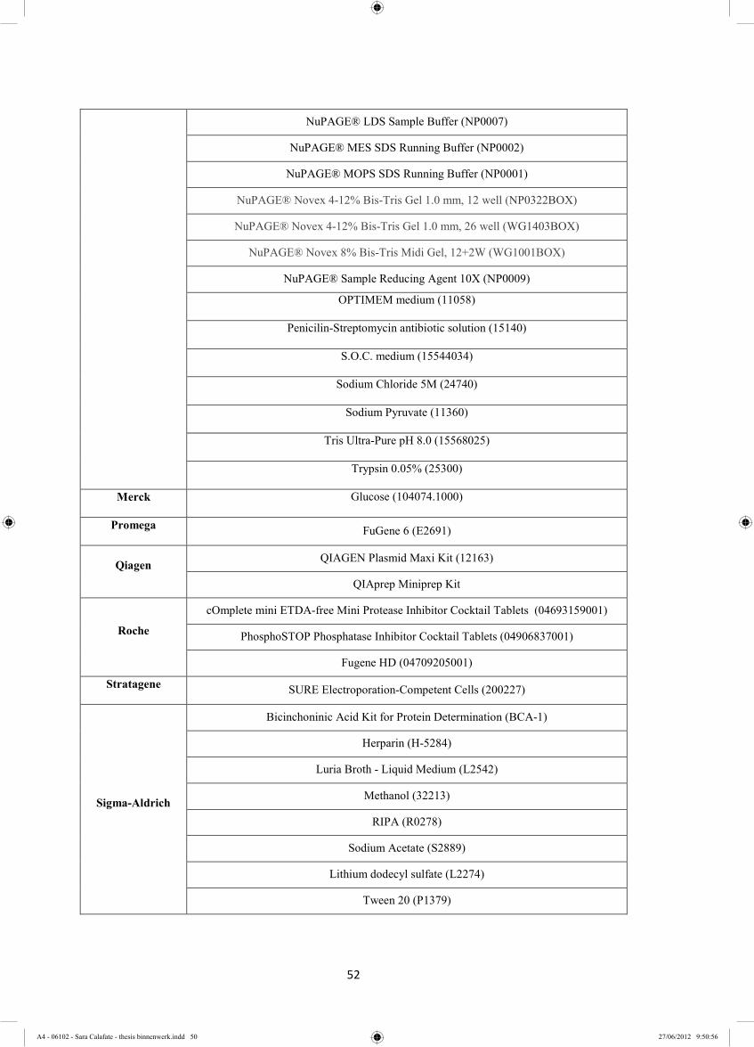
52
NuPAGE® LDS Sample Buffer (NP0007)
NuPAGE® MES SDS Running Buffer (NP0002)
NuPAGE® MOPS SDS Running Buffer (NP0001)
NuPAGE® Novex 4-12% Bis-Tris Gel 1.0 mm, 12 well (NP0322BOX)
NuPAGE® Novex 4-12% Bis-Tris Gel 1.0 mm, 26 well (WG1403BOX)
NuPAGE® Novex 8% Bis-Tris Midi Gel, 12+2W (WG1001BOX)
NuPAGE® Sample Reducing Agent 10X (NP0009)
OPTIMEM medium (11058)
Penicilin-Streptomycin antibiotic solution (15140)
S.O.C. medium (15544034)
Sodium Chloride 5M (24740)
Sodium Pyruvate (11360)
Tris Ultra-Pure pH 8.0 (15568025)
Trypsin 0.05% (25300)
Merck Glucose (104074.1000)
Promega FuGene 6 (E2691)
Qiagen QIAGEN Plasmid Maxi Kit (12163)
QIAprep Miniprep Kit
Roche
cOmplete mini ETDA-free Mini Protease Inhibitor Cocktail Tablets (04693159001)
PhosphoSTOP Phosphatase Inhibitor Cocktail Tablets (04906837001)
Fugene HD (04709205001)
Stratagene SURE Electroporation-Competent Cells (200227)
Sigma-Aldrich
Bicinchoninic Acid Kit for Protein Determination (BCA-1)
Herparin (H-5284)
Luria Broth - Liquid Medium (L2542)
Methanol (32213)
RIPA (R0278)
Sodium Acetate (S2889)
Lithium dodecyl sulfate (L2274)
Tween 20 (P1379)
A4 - 06102 - Sara Calafate - thesis binnenwerk.indd 50 27/06/2012 9:50:56

53
Triton™ X-100
SynGene G:BOX Chemi system
Thermo Scientific
FastDigest® Sma I (FD0663)
SuperSignal* West Dura Chemiluminescent Substrate (34076)
RestoreTM Western Blot Stripping Buffer (46430)
Volac Disposable glass Pasteur pipettes, 230 mm, pre-plugged (D812)
2.2 Antibodies and dyes
Antibody Target Host
specie Clonality
Company and Catalog
number Technique Dilution or
quantity
Actin (C4) Actin Mouse Monoclonal Milipore
MAB1501 Western blot 1:10000
Alexa Fluor® 555 Goat Anti-
Mouse IgG (H+L)
IgG Mouse Polyclonal
Life technologies
A-21424.
Immuno-fluorescence
2 µg/mL
Anti Tau antibody
N-terminal Tau domain
Goat Polyclonal Abcam
ab11107 Western blot 1:1000
AT8 Phosphorylated
Tau on ser202/tre205
Mouse Monoclonal
Homemade
Marc Mercken group
Western blot 1 µg/mL
AT180 Phosphorylated Tau on thr231
Rabbit Polyclonal Santa Cruz
sc-1060-R Western blot 1:1000
AT100 Phosphorylated
Tau on thr212/thr214
Rabbit Polyclonal
Homemade
Marc Mercken group
Western blot 1 µg/mL
Dako Tau Tau Rabbit Polyclonal
Dako
Cytomation
A0024
Western blot 1:5000
A4 - 06102 - Sara Calafate - thesis binnenwerk.indd 51 27/06/2012 9:50:59

54
ECL Mouse IgG, HRP-
Linked F(ab')2 Fragment
(from sheep)
IgG Mouse Polyclobal
GE-Healthcare
NA9310
Western blot 1:5000
ECL Rabbit IgG, HRP-
Linked Whole Ab (from donkey)
IgG Rabbit Polyclobal
GE-Healthcare
NA934
Western blot 1:20000
Hsp70/72 Hsp70 Rabbit Polyclonal R&D systems
AF1663 Western blot 1:10000
HT7 Human Tau Mouse Monoclonal Pierce
(MN1000)
Western blot 0.01 µg/mL
Immuno-fluorescence
0.1 µg/mL
HT10 Human Tau Mouse Monoclonal
Gift from
Dr. Marc Mercken
Immuno-percipitation
20µg/500µg of sample
LC3B LC3B Rabbit Polyclonal SIGMA
L7543 Western blot 1.5 µg/mL
Neuronal Class III β-Tubulin
tubulin beta-3 chain
Mouse Monoclonal Covance
MMS-435P
Immuno-fluorescence
1:1000
Western blot 1:1000
pS409 Phosphorylated Tau on SER409
Rabbit Polyclonal
Life technologies
44-760G
Western blot 1:1000
pTau 28
No purified
Non-phosphorylated AD phosphor-
specific site
Mouse Monoclonal
Homemade
Marc Mercken group
Western blot 1:1
Hoechst 33342 Nucleus - - Thermo
Scientific Immuno-
fluorescence 1µg/mL
A4 - 06102 - Sara Calafate - thesis binnenwerk.indd 52 27/06/2012 9:51:03

55
Chapter 3 -Methods
A4 - 06102 - Sara Calafate - thesis binnenwerk.indd 53 27/06/2012 9:51:07

56
A4 - 06102 - Sara Calafate - thesis binnenwerk.indd 54 27/06/2012 9:51:10

57
3.1 AAV vector production
phSYN1 plasmids containing AAV6 inverted terminal repeats (ITRs) and the human synapsin-1 gene promoter driving the expression of human WT full length Tau (hTauWT) or human full length Tau containing the P301L mutation (hTauP301L) were obtained from Dr. Sebastian Kügler (Jaworski, 2011). The plasmids were propagated in SURE cells (Stratagene) according to manufacturer recommendations. Plasmid was purified using the QIAprep maxiprep kit (Qiagen). The integrity of the inverted terminal repeats, essential for viral packing, was checked by a digestion with SmaI. AAV6 packing was performed by Sebastian Kügler.
3.2 Cell culture and plasmid transfection
Human kidney-derived QBI-HEK 293A Cells (QBI), African green monkey kidney-derived COS-7 cells, mouse Neuroblastoma derived N2a cells without or stably expressing hTauP301L and human cervical cancer-derived HeLa cells were cultured in DMEM medium containing 10 mM pyruvate, 20 mM L-Glutamine, 10% heat inactivated Fetal bovine Serum, and Penicilin-Streptomycin. Human osteosarcoma-derived (U20S) cells without or stably expressing hTauP301L were cultured in McCoy’s medium supplemented with 10% heat inactivated fetal bovine serum and Penicilin-Streptomycin. QBI cells were seeded in 6 well plates at a density of 300.000 cells per well in 1 ml antibiotic free medium and were grown overnight. 3 µl FuGENE6 was added to 77 µl OptiMEM and incubated for 5 minutes at room temperature (RT). 1 µg plasmid DNA diluted in 20 µl OptiMEM was added, mixed, and incubated for 15 minutes at RT. This FuGENE-DNA mixture was added to the cells. The cells were then incubated for 4 h at 37°C, 5% CO2 after which the medium was replaced. For transfections in 24 well plates one fifth of the cell numbers and volumes were used. Co-transfections of hTauP301L with the empty plasmid or with GSK3β was (hTauP301L-GSK3β) were performed at a ratio of 4:1 in favour of Tau. For COS-7 transient transfection 3µg of cDNA was diluted in a final volume of 500 µl of OptiMEM. 20 µl of Lipo2000 reagent was mixed with 480 µl of OptiMEM and kept 5 minutes at RT. The diluted Lipo2000 solution was added to the cDNA solution and incubated 15 minutes at RT. The mixture Lipo2000-DNA was added to 9 mL of antibiotic-free medium containing 4.000.000 COS-7 cells. For seeding in 6 well plates 2 mL were used per well. The cells were then incubated for 4 h at 37°C, 5% CO2 after which the medium was replaced. For HeLa cells transient transfection 15 µg of cDNA in a final volume of 450 µl of OptiMEM. 50 µl of FuGene HD was added and mixed for 30 seconds. The solution cDNA-FuGene HD was added to 9,5 mL of antibiotic-free medium containing 4.000.000 HeLa cells and kept at RT for 15 minutes. For seeding in 6 well plates 2 mL were used per well. The cells were then incubated for 4 h at 37°C, 5% CO2 after which the medium was replaced.
A4 - 06102 - Sara Calafate - thesis binnenwerk.indd 55 27/06/2012 9:51:13

58
3.3 Cortical primary cultures and AAV6-mediated hTauWT and hTauP301L expression
Wistar WT rat embryos of 17 to 18 days were used for primary cultures. Embryos brains were dissected and kept in pre-warmed HBSS/Hepes solution (7mM Hepes in HBSS and Penicilin-Streptomycin) solution to 37°C. The cortices were kept in 4.5 mL HBSS/Hepes and 500 µL of trypsin was added and incubated at 37°C during 10-15 minutes for chemical dissociation. After removing trypsin cortices were washed 3 times with pre-equilibrated MEM-Horse medium (10% Horse serum and 0.6% glucose in MEM medium). The cortices were mechanical dissociated with pasteur pippets of normal and small diameter in 3-5mL of MEM-Horse medium. The cells were spun for 5 minutes at 1000 rpm ressuspended in 3 mL of MEM-Horse and plated in a density if 150000 cells well in a 24 well-plate or 10.000 in 96 well-plates. After 4 hours the medium was changed to serum-free medium supplemented with B27 (2% B27 and 10 mM L-glutamine in Neurobasal medium).
The transfection mediated by AAV6 for hTauWT and hTauP301L expression was always done 3 days after plating (DIV3) at a multiplicity of infection (MOI) between 100 and 150. AAV6 was diluted in B27 serum-free medium immediately before adding into the primary cultures. 3.4 In vitro fibrillization of recombinant Tau
To obtain pre-fibrillized Tau seeds a truncated form of human Tau containing only the 4 MT-binding domains, K18, with a P301L point mutation (K18P301L) was expressed in bacteria and purified (Figure 20). Tau was fibrillized in vitro using two different methods: In method A 67µM K18P301L fragment was mixed with 67 µM herparin in 100 mM ammonium acetate pH7.0. Method B is like described by Guo and Lee, 2011. Briefly, 40 µM K18P301L was mixed with 40 µM herparin, and 2 µM DTT, in 100 mM sodium acetate buffer pH of 7.0. For both methods the mix was incubated at 37°C for 48 to 72 hours. The fibrillization mixture was centrifuged at 100.000g during 30 minutes at 4°C. The supernant was discarded and the pellet was resuspended in the same volume of the respective ammonium acetate or sodium acetate until the desired concentration. The K18P301L fibrils were stored at -80°C or immediately used.
Figure 20. The isoform htau40 is the longest isoform in the human CNS. Construct K18 comprises the sequence of four repeats and in this case contains the point mutation P301L (adapted from von Bergen, 2005)
A4 - 06102 - Sara Calafate - thesis binnenwerk.indd 56 27/06/2012 9:51:17

59
3.5 K18P301L fibrils delivery into clonal-cell lines and cortical primary neuronal cultures
The K18P301L fibrils were diluted to the desired concentration and sonicated with 25 pulses of 2 seconds with 30 seconds of pause between each pulse, always on ice. For protein delivery into clonal-cell lines BioPorter kit was used 24 hours after transient transfection. 80 µL of sonicated K18P301L fibrils were added into BioPorter tubes with gentle mixing and incubated during 5-10 minutes at RT after briefly vortexing (5 seconds at 1000 rpm). The cells were washed once with OptiMEM after which the fibril-BioPorter complex diluted with 920 µL OPTIMEM was added to each well. For experiments with cells grown in 24 well plates one fifth of the reagent was used. After 4 hours of incubation at 37°C 1 volume of normal growth medium was added to the cells. Protein delivery in primary cortical neurons was done in DPT5 following the same strategy used for clonal-cell lines. Neurobasal medium without B27 (10 mM L-glutamine in neurobasal medium) was used rather OptiMEM and after 4 hours of incubation the medium was completely replaced for Neurobasal containing B27. Chariot system was also tested and following the guide-lines different amounts of sonicated K18P301L fibrils were delivered into the cells and neurons. Briefly, for a 24 well-plate, the desired amount of K18P301L fibrills was diluted in 50 µL of PBS. 2 µL of Chariot reagent was diluted in 50 µL of sterile water. Both solutions were mixed and kept at RT for 30 minutes. The culture medium was removed and the neurons were overlayed with the complex Chariot-K18P301L fibrils and after 100 µL Neurobasal medium without B27. After 2 hours of incubation 200 µL Neurobasal containing B27 was added. 3.6 Sequential Protein extraction
The clonal cells and neurons were washed once with PBS. Triton Lyses buffer (1% TritonX-100 in 50 mM Tris, 150 mM sodium chloride, pH 7.6) with PhosphoSTOP Phosphatase and cOmplete mini ETDA-free Mini Protease inhibitors was added into the wells. The plate was kept 15 minutes on ice. After scraping the samples were sonicated with 3 pulses of 1-2 seconds with 20 seconds in ice between. For total extraction the samples were centrifuged at 12000 rpm for 20 minutes at 4°C and the supernant was kept. For sample segregation in soluble and insoluble fractions an ultracentrifugation of 100.000g at 4°C during 30 minutes was done. The supernant representing the triton soluble protein was kept (soluble fraction) and the pellet was washed once with the same settings. After washing, the pellet was resuspended in LDS buffer (1% Lithium dodecyl sulfate in 50 mM Tris, 150 mM Sodium chloride, pH 7.6) and metal tip sonicated until dissolved. One last ultracentrifugation with the same settings was done and the LDS soluble proteins were kept in the supernatant (insoluble fraction). The soluble fraction was used to quantify protein by Bicinchoninic Acid kit.
A4 - 06102 - Sara Calafate - thesis binnenwerk.indd 57 27/06/2012 9:51:20
60
3.7 Western Blot
After extraction of total protein with Triton Lysis Buffer, or extraction of soluble and insoluble protein fractions, 5 µg of protein of each sample was loaded on NuPage Novex Bis-Tris 4-12% gels. For transference from the polyacrylamide gel onto the nitrocellulose membrane the iBlot® Dry Blotting System was used. After transference the membrane was blocked in 5% Bottomilk in TBS-T (1M Tris, 150nM sodium chloride and 0.05% Tween-20, pH 8.5) for 1 hour. The membrane was incubated in the primary antibody diluted in 5% Bottomilk in TBS-T over-night. Before incubate in secondary antibody diluted in TBS-T the membrane was washed 3 times 5 minutes in TBS-T. The SuperSignal* West Dura Chemiluminescent Substrate kit was used to for analysis. Signals were captured and quantified by the G:BOX Chemi system. For reprobing, membranes were stripped of secondary antibody with RestoreTM Western Blot Stripping Buffer for 15min with agitation at RT. Before incubation with primary antibody the membranes were washed 3 times 5 minutes in TBS-T and blocked 30 minutes in 5% Bottomilk in TBS-T. 3.8 Immunofluorescence
QBI cells cells were seeded in 96 well plates with µClear bottom at a density of 5000 cells per well. Methanol fixation was done adding pre-chilled methanol at -20 °C into the wells. The plate was kept during 2 minutes at -20°C. 3 washes of 10 minutes using PBS were done. The primary antibody diluted in 0.1% BSA in D-PBS was added overnight at 4°C. 3 washed of 10 minutes were done with PBS-0,1% BSA. The incubation with secondary antibody diluted in 0.1% BSA in D-PBS was during 30 minutes. For nuclear staining Hoechst 33342 was added for 5 minutes and then washed with PBS. Pictures were taken using a Zeiss Axiovert 135 fluorescence microscope equipped with a Zeiss Axiocam ICc1 camera and Zeiss Axiovision 4.8 software using 40 x objectives. The images were background corrected by Image J software (9.8% of the background was removed). For manual quantification 6 images per well were taken with the multimode microscope reader MIAS-2 with a 20x amplification. 3.9 Immunoprecipitation (IP)
The Dynabeads® Protein G were completely resuspend by rotating for 5 min. Dynabeads® Protein G were washed in PBS and 1 µg antibody per 10 µl beads was bound using rotation at RT during 30 minutes (Dynabeads® Protein G s-Ab). After incubation the beads were washed twice with PBS and once with PBS Tween 0.05%. The samples were precleared by incubation with 10 µl Dynabeads® Protein G per 100 µg sample at 4°C with rotation for 30 minutes. The pre-cleared samples were incubated with the Dynabeads® Protein G-Ab over-night with rotation at 4°C (Dynabeads® Protein G -Ab-protein). For protein dissociation from de complex two methods were tested. In one hand the complex was washed in PBS and then boiled at 90°C for 10 minutes. The protein was spun for 10 minutes and the supernant was treated with 1x mix of NuPAGE® LDS Sample Buffer/Reducing and incubated at 70°C for 10 minutes. For the second strategy the Dynabeads® Protein G-Ab-protein complex was
A4 - 06102 - Sara Calafate - thesis binnenwerk.indd 58 27/06/2012 9:51:23
61
washed RIPA buffer the supernant was removed and resuspended in 50 µL 1x mix of NuPAGE® LDS Sample Buffer/Reducing agent per 10 µl beads. The mix was incubated 10 minutes at 70°C and the supernant was loaded on a NuPAGE® Novex 8% Bis-Tris Midi Gel. The gel was colored with ImperialTM protein stain and selected bands were excised for phosphopeptide analysis. A fraction of the samples was analyzed by Westernblot.
A4 - 06102 - Sara Calafate - thesis binnenwerk.indd 59 27/06/2012 9:51:26

62
A4 - 06102 - Sara Calafate - thesis binnenwerk.indd 60 27/06/2012 9:51:29

63
Chapter 4 –Results
A4 - 06102 - Sara Calafate - thesis binnenwerk.indd 61 27/06/2012 9:51:33

64
A4 - 06102 - Sara Calafate - thesis binnenwerk.indd 62 27/06/2012 9:51:36

65
4.1 Optimization of Clonal-cell seeding model
4.1.1 Influence of seeds preparation method on hTauP301L aggregation
To optimize our current cellular model of hTauP301L aggregation we first compared our current method of in vitro K18P301L seeds preparation (method A) with the method recently described by Guo and Lee, 2011 (method B). The main differences between method A and B are the aggregation buffer and the presence of DTT. Both methods use heparin because negative charges like with heparin were shown to be essential for Tau fibrillization (Goedert, 1996). After the aggregation into fibrils by the two methods for different times at 37°C the efficiency of these samples at inducing aggregation of hTauP301L in U20S cells was explored. Therefore cells expressing full length hTauP301L were transfected with the different K18P301L preparations using the BioPORTER protein delivery reagent. The efficiency of hTauP301L aggregation induction was analyzed by Western blotting (Figure 21). To this end, soluble and insoluble hTauP301L were separated according to their solubility in two detergents (Triton and LDS) 48 hours after the delivery of the fibrils. Endogenous soluble and insoluble hTauP301L was stained with the HT7 antibody that detects an N-terminal Tau epitope that is absent in the in vitro produced K18P301L fibrils. Cells treated with BioPORTER containing medium without fibrils was used to show that the presence of endogenous hTauP301L in the insoluble fraction was dependent on the presence of the fibrils. In addition, we found that fibrillization of the exogenous K18P301L for at least 24 hours was necessary to induce aggregation of the endogenously expressed hTauP301L for both methods (Figure 1, 0 hours). Method B was found to be more efficient in inducing aggregation of hTauP301L. K18P301L aggregated between 2 and 3 days by method B was used in further experiments.
Figure 21. K18P301L fibrils prepared by method B are more efficient inducing hTauP301L aggregation than method A. Western blotting analysis of soluble and insoluble fractions from hTauP301L expressing U20S cells treated with BioPORTER with or without K18P301L fibrils as a seed. The fibrils were prepared by two different methods, A or method B for different times.
A4 - 06102 - Sara Calafate - thesis binnenwerk.indd 63 27/06/2012 9:51:42

66
4.1.2 Choosing the best Cell Line for seeding model development
To optimize the cellular Tau aggregation model different cell lines were tested for their efficiency in forming aggregates. Therefore, QBI, COS-7 and HeLa cells were transiently transfected with or without hTauP301L. In addition N2a and U20S cell lines with or without stable hTauP301L expression were used. The different cell-lines were all transfected with or without K18P301L fibrils and 48 hours later endogenous hTauP301L was separated in soluble and insoluble fractions and analyzed by Western Blot. Transfection with K18P301L fibrils was found to induce aggregation of endogenous Tau in all cell lines tested but with different efficiencies. The largest amount of insoluble hTauP301L was seen in QBI cells. Dako antibody allowed us to detect also the K18P301L fibrils suggesting that monomers, dimers and trimers were being added to the system (Figure 22). The QBI cell line was selected for further optimization as cellular Tau seeding model.
Figure 22. Ideal-clonal cell line for seeding model. Western blotting expression analysis of Soluble and Insolube fractions from each clonal-cell cultures with or without expression of hTauWT or hTauP301L treated or untreated with K18P301L fibrils produced from method B. 5 µg of each fraction were loaded in NuPAGE® Novex 4-12% Bis-Tris Gel. K18P301L fibrills can be detected by DAKO antibiody
A4 - 06102 - Sara Calafate - thesis binnenwerk.indd 64 27/06/2012 9:51:46

67
4.1.3 Variability of QBI seeding model
The variability of our cellular model is a concern since we want to use it in a high through-put screen. Therefore we wanted to optimize the method to reproducibly extract insoluble hTauP301L from the cells. To make the solubilization of the pellet after the ultracentrifugation step easier we first tested the effect of a pre-clear of the sample at low speed. We found that recovery of insoluble hTauP301L was reduced by this pre-clear step and the variability was increased even further (figure 23A). Next we compared solubilzation of the Triton-insoluble pellet by sonication in a water bath with sonication using a metal sonicator tip. We found that sonication of the insoluble fraction with the metal sonicator tip greatly improved the reproducibility of the assay (Figure 23B). Because hTauP301L from the cellular aggregation models appeared to migrate slightly different in the insoluble fraction compared to the soluble fraction both sample were run side by side on a gel. NuPAGE® MOPS SDS was used instead of NuPAGE® MES SDS running buffer because this buffer increases protein separation around the size of hTauP30L. This indeed confirmed that insoluble hTauP301L migrates slower in the gel (Figure 3B). We next tested whether the altered mobility of insoluble hTauP301L could be observed without fractionation. We therefore analyzed total lysate of hTauP301L expressing QBI cells with and without fibril transfection. We found that fibril treatment caused a marked mobility shift of hTauP301L (Fig. 23C). This shifted bands suggested that the fractionating by ultracentrifugation was not necessary to discriminate soluble and insoluble Tau. Omitting fractionation eliminates a potentially important source of variability between experiments.
Figure 23. Variablility between experiments was solved by sonication of the sample. Western blotting expression analysis of Soluble and Insolube or Total fractions from each clonal-cell cultures expressing hTauP301L treated or untreated with K18P301L fibrils. A) Half of the samples undergo into a pre-clear step at 14000 rpm on regular centrifuge and all were sonicated in water bath sonicator; B) metal tip sonicated samples. soluble fraction(S) insoluble faction (I); C) Total extracts from ultra/probe-tip sonicated samples
B C
A
A4 - 06102 - Sara Calafate - thesis binnenwerk.indd 65 27/06/2012 9:51:54

68
4.1.4 Delivery system and seeds delivery efficiency
Two different delivery reagents, BioPORTER and Chariot, were tested to optimize K18P301L seeds delivery. BioPORTER and Chariot are lipid and protein based respectively. The amount of seeds used for Chariot system was as well optimized. Chariot successfully deliverd the seeds into the cells although aggregation was observed only when high amounts of seeds were used. When comparing both systems the efficiency of BioPORTER reagent in seeds delivery was apparently higher since lower amounts of fibrils gave rise to higher levels of endogenous hTauP301L aggregation (figure 24).
4.1.5 Model characterization - Gel migration
Different post-translational modifications, like phosphorylation, can be responsible for the altered gel mobility of insoluble hTauP301L. Therefore we wanted to analyse these different Tau species in the presence and absence of seeds by mass spectroscopy and phospho-peptide analysis. For this purpose, an IP was performed on the Total extract of lysates from the different conditions using the HT10 antibody that recognizes an N-terminal epitope of human Tau that is absent in the K18P301L fibrils. The immunopercipitates were loaded on a Bis-Tris gel and after with ImperialTM protein staining three different bands of approximately 60 kDa were detected. These three bands were isolated for mass and phosphor-peptide analysis: 1) faster migrating hTauP301L from cells not treated with fibrils (figure 25A lane b band 1); 2) fast migration hTauP301L (figure 25A lane c band 2) and 3) slow migrating Tau from cells treated with fibrils (figure 25A lane c band 3) (Figure 25 A). The protein analysis is ongoing. The identities of the bands were confirmed by Western Blot. The hTauP301L was not completely immunopercipitated since a large fraction was detected in the unbound material. Both higher and lower migration bands were immunopercipitated from the Total fraction. Comparing the strategies to purify protein from the Dynabeads® Protein G -Ab-protein complex IP 2 showed more efficiency when compared to IP1 (Figure 25 B).
Figure 24. Comparison of seeding efficiency with Chariot and BioPOPTER delivery reagent in QBI cells. Western blotting analysis of Soluble and Insoluble fractions of QBI cells with or without expression of hTauP301L treated withor without K18P301L fibrils delivered by BioPorter or Chariot systems. For Chariot system different amounts of K18P301L fibrils were used.
Figure 24. Comparison of seeding efficiency with Chariot and BioPOPTER delivery reagent in QBI cells. Western blotting analysis of Soluble and Insoluble fractions of QBI cells with or without expression of hTau-P301L treated withor without K18P301L fibrils delivered by BioPorter or Chariot systems. For Chariot system different amounts of K18P301L fibrils were used.
A4 - 06102 - Sara Calafate - thesis binnenwerk.indd 66 27/06/2012 9:52:00

69
4.1.6 Insoluble hTauP301L is phosphorylated at different epitopes
Since phosphorylation of Tau plays an important role in AD pathogenesis we analyzed Tau phosphorylation in our cellular model. We found that nucleation by K18P301L fibrils induced a marked increase in different phospho-epitopes in hTauP301L in the cell seeding model (figure 26). Phosphorylated Thr212 and Thr231 signals, detected using AT100 and AT180 antibodies respectively, were present only in the shifted insoluble protein. The AT8 antibody selectively recognizes Tau phosphorylated at Ser202/Thr205. Braak et al showed that the AT8 immunoreactivity correlated with neurofibrillary pathology in AD human brains
Figure 25. hTauP301L IP using HT10 antibody. Immunoprecipitated hTauP301L from total extracts of QBI cells with or without hTauP301L expression tretated with or without K18P301L fibrils A) ImperialTM protein stain analysis of immunoprecipitated hTauP301L using 20 µg HT10 in 50 µL Dynabeads® Protein G for 500 µg. IP from total extracts of a) cells no expressing hTauP301L and cells expressing hTauP301L b) untreated or c) treated with K18P301L fibrils. Bands 1, 2 and 3 were isolated from the gel and sent for mass spectrometry analysis. B) Western blotting analysis of IP made with 5µg of HT10 antibody in 10µl of Dynabeads® Protein G for 100µg of sample confirmed that hTauP301L was immunoprecipitated. After Dynabeads® Protein G -Ab complex formation unboun antibody was still detected. A Tau was not completely depleted from the inpunt and a large fraction remained on unbound material. After IP the protein was purified from the Dynabeads® Protein G -Ab-protein complex by two different strategies. An extra boiling step was included in IP 1 an ommited in IP 2.
A B
Figure 26. Phosphoepitopes detected in cell seeding model are exclusive for insoluble protein. Western blotting analysis of total extract of QBI cells expressing hTauP301L treated with or without K18P301L. AT8, AT180 and AT100 were immuno-detected on aggregated hTauP301L after seeds treatment. The cells untreated did not show immunoreactivity for these antibodies.
A4 - 06102 - Sara Calafate - thesis binnenwerk.indd 67 27/06/2012 9:52:04

70
(Braak, 2011). This phospo-epitope was shown to be specific for insoluble hTauP301L since the soluble fraction is not immuno-detected in our cell-seeding model. The presence of phosphorylation on more than one site in aggregated Tau may account for the altered gel mobility. The phospho-epitopes analyzed here are all absent in K18P301L fibrils.
4.2 Development of primary neuronal seeding model
4.2.1 Generation of primary cortical neurons expressing human full-lenght Tau
A premise for the set-up of the primary neuronal seeding model was to express the full-length human Tau with or without P301L mutation in cortical neurons from Wistar embryos. phSYN1 plasmids containing AAV6 inverted terminal repeats (ITRs) and the human synapsin-1 gene promoter driving the expression of hTauWT or hTauP301L were transformed in SURE cells. After SmaI digestion the expected bands sizes were obtained and the quality of the extracted DNA was accepted for further packaging (Figure 27).
One of the challenges for generating this model was to provide a fine-tuned system for Tau aggregation mechanistic analysis. In the initial studies we decided to analyse the time course of transgene expression. The expression of Tau WT (data not shown) and Tau P301L mediated by AAV6 was analyzed using different multiplicities of infection (MOI) on cortical primary cultures transfected in DIV3. The expression of transgene was analyzed on 4 different time-points. The expression of the transgene increased during DPT4, 5 and 6 as well as with increasing MOI. On DPT7 the expression was not apparently higher. DPT5 was chosen as ideal time-point for fibril seeding due to increasing expression levels (Figure 28).
Figure 27. hTauWT and hTauP301L plasmids production. Image of SmaI digestion of the hTauWT or hTauP301L plasmids under pSYN1 promoter
Figure 28. MOI 100 was choosen for further expression of hTauWT and hTauP301L mediated by AAV6 . Western blotting analysis of Total homogenates from cortical primary cultures transfected on DIV3 with MOIs of 50, 100 and 150 extracted on Triton lysis Buffer on DPT 4, 5, 6 and 7. No transfected cultures were included in each day.
A4 - 06102 - Sara Calafate - thesis binnenwerk.indd 68 27/06/2012 9:52:12

71
4.2.2 Aggregation of hTauP301L in primary cortical neurons
It was reported that direct membrane penetration of α-synuclein occurred in cell cultures (Luk, 2009). We found that applying K18P301L fibrils directly to the medium did not induce the aggregation of endogenous expressed hTauP301L. For this reason delivery reagents were tested. The strategy used for cell seeding model was applied to cortical primary cultures expressing hTauP301L on DPT5. 125µL of BioPORTER-fibrils complex from a 0.149 µg/µL concentrated mixture was used. The biochemical analysis was done 48 hours after K18P301L delivery (Figure 29A). The pattern observed in the cell seeding models was reproduced in cortical primary cultures. The detection of a slower migrating hTauP301L in the presence of seeds suggested that nucleation of aggregation occurred (figure 29B). B27 is a protein-rich supplement for long-term cultures. Since BioPORTER guidelines suggested the use of serum-free medium the presence and absence of B27 supplement was explored. The high migrating hTauP301L was only detected when the B27 supplement was removed from neurobasal medium at the time of fibril transfection (figure 29B). The encapsulation and further delivery of the fibrils turned out to be the most efficient strategy for endogenous hTauP301L aggregation induction. However the ratio between insoluble and soluble protein was small showing that further optimization and deeper analysis on aggregation kinetics was necessary to obtain higher amounts of insoluble hTauP301L.
4.2.3 BioPorter delivery reagent is an efficient delivery tool
The delivery efficiency of two systems tested before in cellular model was again compared: Chariot and BioPorter reagents. The same amount of K18P301L seeds was added to the cultures by these two systems. The factor “delivery efficiency” was analyzed by the induction of insoluble hTauP301L. BioPORTER-mediated fibrils delivery was able to induce aggregation of endogenous hTauP301L, while the Chariot delivery system failed (figure 30A). After separation of the sample in soluble and insoluble fractions the hTauP301L that migrates slower in the gel was present in the insoluble fraction as indicated by the arrow.
Figure 29. Internalization of K18P301L in cortical primary cultures induces hTauP301L aggregation. A) Scheme representation of experiment strategy. B) Western blotting analysis of total extracts of cortical primary cultures expressing hTauP301L treated on DPT5 with K18P301L fibrils by four different ways: 3 and 5µg of seeds were simply added into the medium; 3 µg were delivered using BioPorter reagent in the presence of B27 or in the b f B27
A B
Figure 29. Internalization of K18P301L in cortical primary cultures induces hTauP301L aggregation. A) Scheme representation of experiment strategy. B) Western blotting analysis of total extracts of cortical pri-mary cultures expressing hTauP301L treated on DPT5 with K18P301L fibrils by four different ways: 3 and 5µg of seeds were simply added into the medium; 3 µg were delivered using BioPorter reagent in the presence of B27 or in the absence of B27.
A4 - 06102 - Sara Calafate - thesis binnenwerk.indd 69 27/06/2012 9:52:15

72
This was only observed for BioPORTER-delivered seeds (Figure 30B). In the end the result suggested that the Chariot delivery system failed to deliver the seeds into neurons cytoplasm or the amount of fibrils delivered was lower than the necessary to trigger nucleation of endogenous hTauP301L. The presence of fibrils showed to be essential for detection of higher migrating and hence aggregated hTauP301L.
4.2.4 Aggregation of hTauP301L is affected by the amount of K18P301L.
The role of different variables and consequent influence on hTauP301L aggregation were tested. The concentration of K18P301L fibrils added into the system and the ratio of BioPORTER to K18P301L fibrils were explored and showed to affect the extend of hTauP301L aggregation (Figure 31). Increasing amounts of the BioPorter reagent were found to induce a different morphology on cortical neurons. The toxicity that BioPorter reagent played in the system was not quantified but the neuronal network was clearly less damaged when larger volumes of reagent were added. From this first experiment it was unclear whether the toxicity was caused by the Bioporter reagent alone or by the combination of the reagent with the K18P301L fibrils. Addition of BioPorter to the culture in the absence of fibrils showed that the toxicity was a consequence of the reagent in higher concentrations (Figure 31A a-c). To avoid toxicity different concentrations of BioPorter reagent were tested to find the less toxic condition that still comprises higher aggregation levels of hTauP301L. In the first experiment 47.5 µg K18P301L fibrils was added to one tube of BioPORTER protein delivery reagent and diluted to 500 µl giving rise to a BioPORTER-Fibrils complex
Figure 30. Bioporter showed to be more efficient than Chariot delivery reagent in cortical neurons. Western Blot analysis of A) total lysate or B) soluble (S) and insoluble (I) fractions of cortical primary cultures expressing hTauP301L treated on DPT5 with K18P301L fibrils. The incubation of the K18P301L was done in the absence of B27 supplement. 3 µg were directly added into the medium, delivered by BioPorter or by Chariot reagents. Only delivery mediated by BioPorter containning K18P301L fibrils resulted in the aggregation of hTauP301L after 48hours of incubation showed by the higher and lower migrating band immune-reactive with HT7.
A B
A4 - 06102 - Sara Calafate - thesis binnenwerk.indd 70 27/06/2012 9:52:26
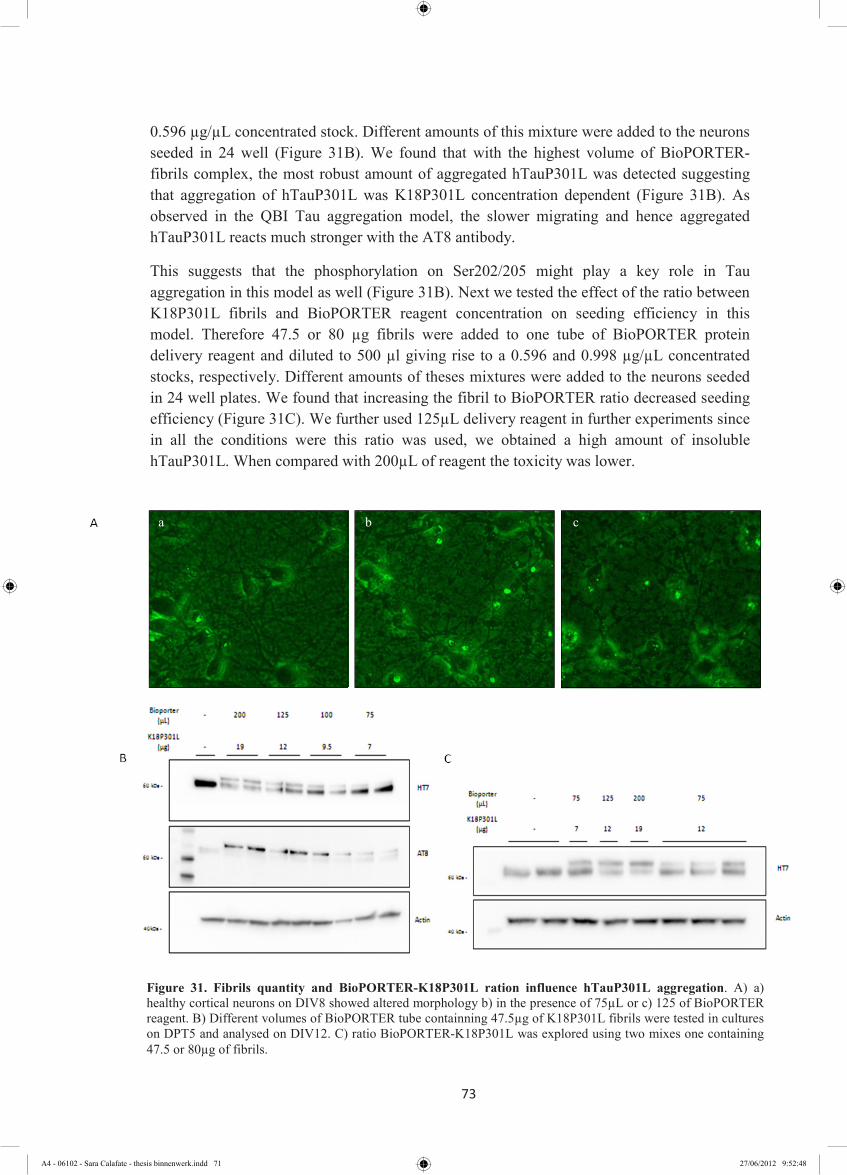
73
0.596 µg/µL concentrated stock. Different amounts of this mixture were added to the neurons seeded in 24 well (Figure 31B). We found that with the highest volume of BioPORTER-fibrils complex, the most robust amount of aggregated hTauP301L was detected suggesting that aggregation of hTauP301L was K18P301L concentration dependent (Figure 31B). As observed in the QBI Tau aggregation model, the slower migrating and hence aggregated hTauP301L reacts much stronger with the AT8 antibody.
This suggests that the phosphorylation on Ser202/205 might play a key role in Tau aggregation in this model as well (Figure 31B). Next we tested the effect of the ratio between K18P301L fibrils and BioPORTER reagent concentration on seeding efficiency in this model. Therefore 47.5 or 80 µg fibrils were added to one tube of BioPORTER protein delivery reagent and diluted to 500 µl giving rise to a 0.596 and 0.998 µg/µL concentrated stocks, respectively. Different amounts of theses mixtures were added to the neurons seeded in 24 well plates. We found that increasing the fibril to BioPORTER ratio decreased seeding efficiency (Figure 31C). We further used 125µL delivery reagent in further experiments since in all the conditions were this ratio was used, we obtained a high amount of insoluble hTauP301L. When compared with 200µL of reagent the toxicity was lower.
Figure 31. Fibrils quantity and BioPORTER-K18P301L ration influence hTauP301L aggregation. A) a) healthy cortical neurons on DIV8 showed altered morphology b) in the presence of 75µL or c) 125 of BioPORTER reagent. B) Different volumes of BioPORTER tube containning 47.5µg of K18P301L fibrils were tested in cultures on DPT5 and analysed on DIV12. C) ratio BioPORTER-K18P301L was explored using two mixes one containing 47.5 or 80µg of fibrils.
c b a
A4 - 06102 - Sara Calafate - thesis binnenwerk.indd 71 27/06/2012 9:52:48

74
4.2.5 hTauWT does not aggregate like hTauP301L
In the clonal cell seeding model induction of Tau aggregation was found to be much less efficient for hTauWT than for hTauP301L (data not shown). To compare aggregation efficiency between hTauWT and hTauP301L in the primary neuronal seeding model cortical neuronal cultures were transfected with hTauWT or hTauP301L and incubated with K18P301L fibrils. In all the conditions hTauWT did not aggregate and the immuno-reactivity signal for AT8 was always comparable to the control. In contrast the hTauP301L readily aggregated in the presence of seeds, as shown before. The Tau fibril-BioPORTER reagent ratio was tested and optimal condition was shown to be 125µL containing 12µg of fibrils
(Figure 32).
4.2.6 Aggregation kinetics is affected by amount of fibrils and shown time variation
To analyze aggregation-kinetics of hTauP301L we exposed cultures to different amounts of K18P301L fibrils. We observed a marked increase in aggregated Tau between 48 and 120 hours of incubation. This suggests that the aggregation of expressed hTauP301L increases with time. However, it can make us think that spreading of the fibrils between neurons can be occurring it cannot be concluded from this experiment. In all three time points of analysis the delivery of 12µg of K18P301L seeds was found to be more efficient than 6 or 3µg on
Figure 32. hTauWT does not aggregate like hTauP301L. Western blotting analysis of the total extracts from neurons expressing hTauWT or hTauP301L treated with or without identical amounts of K18P301L fibril prepared in different Tau fibril- BioPORTER ratios. The 75µl or 125 µl containing 12µg of BioPORTER -K18P301L, were added into the cortical neurons on DPT5. The aggregation of hTauP301L is detected by the presence of a shifted band immuno-reactive with HT7 and AT8 antibodies.
hTauWT hTauP301L
A4 - 06102 - Sara Calafate - thesis binnenwerk.indd 72 27/06/2012 9:52:53
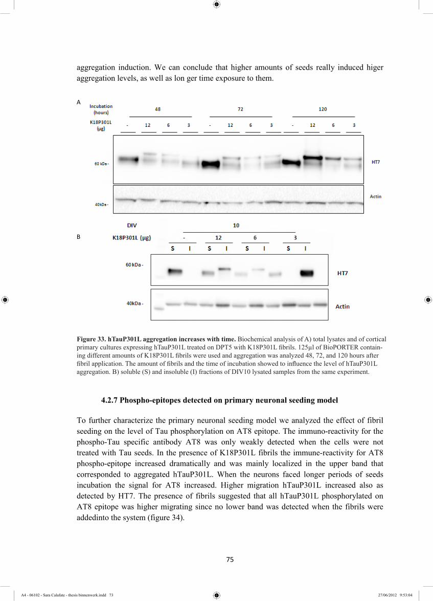
75
aggregation induction. We can conclude that higher amounts of seeds really induced higer aggregation levels, as well as lon ger time exposure to them.
4.2.7 Phospho-epitopes detected on primary neuronal seeding model
To further characterize the primary neuronal seeding model we analyzed the effect of fibril seeding on the level of Tau phosphorylation on AT8 epitope. The immuno-reactivity for the phospho-Tau specific antibody AT8 was only weakly detected when the cells were not treated with Tau seeds. In the presence of K18P301L fibrils the immune-reactivity for AT8 phospho-epitope increased dramatically and was mainly localized in the upper band that corresponded to aggregated hTauP301L. When the neurons faced longer periods of seeds incubation the signal for AT8 increased. Higher migration hTauP301L increased also as detected by HT7. The presence of fibrils suggested that all hTauP301L phosphorylated on AT8 epitope was higher migrating since no lower band was detected when the fibrils were addedinto the system (figure 34).
Figure 33. hTauP301L aggregation increases with time. Biochemical analysis of A) total lysates and of cortical primary cultures expressing hTauP301L treated on DPT5 with K18P301L fibrils. 125µl of BioPORTER containing different amounts of K18P301L fibrils were used and aggregation was analyzed 48, 72, and 120 hours after fibril application. The amount of fibrils and the time of incubation showed to influence the level of hTauP301L aggregation. B) soluble (S) and insoluble (I) fractions of DIV10 lysated samples from the same experiment.
B
A
Figure 33. hTauP301L aggregation increases with time. Biochemical analysis of A) total lysates and of cortical primary cultures expressing hTauP301L treated on DPT5 with K18P301L fibrils. 125µl of BioPORTER contain-ing different amounts of K18P301L fibrils were used and aggregation was analyzed 48, 72, and 120 hours after fibril application. The amount of fibrils and the time of incubation showed to influence the level of hTauP301L aggregation. B) soluble (S) and insoluble (I) fractions of DIV10 lysated samples from the same experiment.
75
aggregation induction. We can conclude that higher amounts of seeds really induced higer aggregation levels, as well as lon ger time exposure to them.
4.2.7 Phospho-epitopes detected on primary neuronal seeding model
To further characterize the primary neuronal seeding model we analyzed the effect of fibril seeding on the level of Tau phosphorylation on AT8 epitope. The immuno-reactivity for the phospho-Tau specific antibody AT8 was only weakly detected when the cells were not treated with Tau seeds. In the presence of K18P301L fibrils the immune-reactivity for AT8 phospho-epitope increased dramatically and was mainly localized in the upper band that corresponded to aggregated hTauP301L. When the neurons faced longer periods of seeds incubation the signal for AT8 increased. Higher migration hTauP301L increased also as detected by HT7. The presence of fibrils suggested that all hTauP301L phosphorylated on AT8 epitope was higher migrating since no lower band was detected when the fibrils were addedinto the system (figure 34).
Figure 33. hTauP301L aggregation increases with time. Biochemical analysis of A) total lysates and of cortical primary cultures expressing hTauP301L treated on DPT5 with K18P301L fibrils. 125µl of BioPORTER containing different amounts of K18P301L fibrils were used and aggregation was analyzed 48, 72, and 120 hours after fibril application. The amount of fibrils and the time of incubation showed to influence the level of hTauP301L aggregation. B) soluble (S) and insoluble (I) fractions of DIV10 lysated samples from the same experiment.
B
A
A4 - 06102 - Sara Calafate - thesis binnenwerk.indd 73 27/06/2012 9:53:04

76
Figure 34. Phosphorylation of hTauP301L increases after K18P301L fibrils treatement. Western Blot analysis of total extracts of cortical primary cultures expressing hTauP301L treated on DPT5 with K18P301L fibrils. 125µl of BioPORTER without fibrils (mock) or containing 12µg of K18P301L fibrils was tested and analysis was done in DIV 11 and DIV 13. Total hTauP301L was detected by HT7 antibody. Phospho-epitopes on Ser202/205 and Ser409 were detected by AT8 and pS409 antibodies respectively. The arrows indicate the third band detected when hTau28 and AT8 antibodies were used.
4.3 Testing of Hypothesis
4.3.1 GSK3β increases phosphorylation of aggregated hTauP301L
It was shown that Tau is a substrate of the kinase GSK3β and some evidence supports the idea that in AD Tau aggregation and Tau toxicity is mediated by GSK3β phosphorylation (Mazanetz, 2007). Co-overexpression of hTauP301L and GSK3β in the QBI cells was used to understand if GSK3β was able to induce hTauP301L aggregation. The co-expression of hTauP301L and GSK3β resulted in a dramatic increase of Tau phosphorylation as measured by the increased AT8 signal as well as the higher migrating hTauP301L when compared with hTauP301L co-expressed with the empty vector independent of treatment with K18P301L fibrils. After fractionation of the samples it was observed however that GSK3β mediated hyperphosphorylation was not sufficient to induce aggregation of hTauP301L since this was only detected in the presence of seeding by K18P301L (Figure 35). In the presence of the seeds, the level of hTauP301L aggregation was however strongly increased by the GSK3β mediated phosphorylation of hTauP301L (Figure 35). This result confirms that in our cell seeding model GSK3β phosphorylates hTauP301L on ser202/thr205 but that this is not a required but a stimulatory characteristic for Tau aggregation in our model. In this experiment
77
the detection of AT8 signal from cells treated with K18P301L was extremely low when compared when the GSK3β was expressed. To detect this AT8 signal on insoluble fraction from GSK3β no-expressing cells we needed to intensify the exposure of the blot. The Immunofluorescence assay revealed that in hTauP301L expressing cells the AT8 reactivity was almost not detected (data not showed). The co-expression of hTauP301L and GSK3β increased the detection of AT8 signal although it was only weakly detected when compared to the co-expression in the presence of the seeds, which does not correspond to the results obtained with Western Blot (Figure 36A). This difference is due to the methanol fixation methodology that results in the loss of the AT8 epitope when Tau is in a physiological, non-aggregated form. The presence of seeds induced a variety of aggregated morphology in hTauP301L. The AT8 immunoreactivity observed varied from diffuse material to small or large like-inclusions material. More than one inclusion per cell was usually observed. To coexpression of hTauP301L with GSK3β resulted in a dramatic increase of AT8 positive inclusions when seeded. For all the conditions only a small percentage of AT8 positive cells were found. Also the number of hTauP301L aggregate-bearing cells was low, suggesting that low numbers of cells are internalizing the BioPORTER delivered seeds (Figure 36B).
Figure 35. Co-expression of hTauP301L and GSK3β increased AT8 signal but not insolubility. Western blot analysis of cells expressing hTauP301L combined with empty vector or GSK3b treated with or without K18P301L. AT8 phosphorylated hTauP301L on Total lysates and soluble and insoluble fractions were detected.
A4 - 06102 - Sara Calafate - thesis binnenwerk.indd 74 27/06/2012 9:53:21
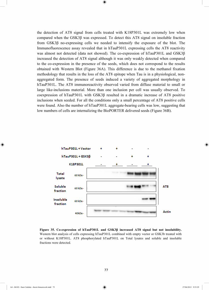
77
the detection of AT8 signal from cells treated with K18P301L was extremely low when compared when the GSK3β was expressed. To detect this AT8 signal on insoluble fraction from GSK3β no-expressing cells we needed to intensify the exposure of the blot. The Immunofluorescence assay revealed that in hTauP301L expressing cells the AT8 reactivity was almost not detected (data not showed). The co-expression of hTauP301L and GSK3β increased the detection of AT8 signal although it was only weakly detected when compared to the co-expression in the presence of the seeds, which does not correspond to the results obtained with Western Blot (Figure 36A). This difference is due to the methanol fixation methodology that results in the loss of the AT8 epitope when Tau is in a physiological, non-aggregated form. The presence of seeds induced a variety of aggregated morphology in hTauP301L. The AT8 immunoreactivity observed varied from diffuse material to small or large like-inclusions material. More than one inclusion per cell was usually observed. To coexpression of hTauP301L with GSK3β resulted in a dramatic increase of AT8 positive inclusions when seeded. For all the conditions only a small percentage of AT8 positive cells were found. Also the number of hTauP301L aggregate-bearing cells was low, suggesting that low numbers of cells are internalizing the BioPORTER delivered seeds (Figure 36B).
Figure 35. Co-expression of hTauP301L and GSK3β increased AT8 signal but not insolubility. Western blot analysis of cells expressing hTauP301L combined with empty vector or GSK3b treated with or without K18P301L. AT8 phosphorylated hTauP301L on Total lysates and soluble and insoluble fractions were detected.
A4 - 06102 - Sara Calafate - thesis binnenwerk.indd 75 27/06/2012 9:53:29

78
P301
L +
GSK
3β +
K18
P301
L P3
01L
+ G
SK3β
P3
01L
+ K
18P3
01L
c
b
a B
Figure 36. Aggregated hTauP301L presents different morphology within the cell. Immunofluorescence analysis of methanol fixed cells expressing hTauP301L combined with empty vector or GSK3b treated with or without K18P301L. A) Quantitation of cells containning AT8 signal. 24 images per condition were taken by MIAS in 20x magnification from one representative experiment B) a-c representative images of the graph showing the AT8 signal in red and Hoechst 33342 in blue. Different morphologies were observed when cells were treated with aggregates. Images aquired with magnification of 40x. In al panels the nucleus was stained with. Scale bar of 25 µm.
A
Figure 36. Aggregated hTauP301L presents different morphology within the cell. Immunofluorescence analysis of methanol fixed cells expressing hTauP301L combined with empty vector or GSK3b treated with or without K18P301L. A) Quantitation of cells containning AT8 signal. 24 images per condition were taken by MIAS in 20x magnification from one representative experiment B) a-c representative images of the graph showing the AT8 signal in red and Hoechst 33342 in blue. Different morphologies were observed when cells were treated with aggregates. Images aquired with magnification of 40x. In al panels the nucleus was stained with. Scale bar of 25 µm.
A4 - 06102 - Sara Calafate - thesis binnenwerk.indd 76 27/06/2012 9:53:31

79
4.2.2 Hsp90 inhibition
Dickey and colleagues have suggested Hsp90 inhibition as a promising strategy to enhance degradation of phosphorylated Tau and blocking the Hsp90 refolding capacity (Dickey 2007). Inhibiting the APTase activity of Hsp90 forces folding-client proteins to follow a Hsp70 exclusive pathway. Later Hsp90 is degraded and releases HSF1 that translocates into the nucleus and induces the transcription of other Hsps (Dickey, 2007) (Figure 19). Cortical neurons were treated with increasing concentrations of the Hsp90 inhibitor 17-DMAG, wich is a less toxic GA derivative. The Hsp70 up-regulation as a consequence of Hsp90 inhibition was used to follow the efficiency of the compound. The dose reponse dependant up-regulation of Hsp70 confirmed that 17-DMAG was actively inhibiting Hsp90 activity (Figure 37A and B). It was shown that Hsp90 inhibition did not result in autophagy induction since the LC3BII levels were the same as the control (Figure 37 A).
This potential of this Hsp90 inhibitor to clear Tau was evaluated in the cortical neuronal seeding model (Figure 38A). A variation in total and phosphorylated hTauP301L levels occurred and was inversely correlated with Hsp70 expression (Figure 38A and B). For the conditions where 75 µL of BioPORTER was used the AT8 and HT7 signal decreased with the increase of 17-DMAG concentration corresponding to a dose dependant decrease in total and phosphorylated hTauP301L including the aggregated Tau P301L species. When 125 µl of BioPORTER was used, some variation occurred within the duplicated condition of 17-DMAG treatment, although the inverse correlation between AT8 and Hsp70 signals was always followed (Figure 18B). We noted that the combination of BioPORTER and compound resulted in some neurite network damage and loss especially with the higher concentration of Bioporter. To exclude that the observed decrease in total and phosphorylated Tau was due to toxicity rather than Hsp90 inhibition, the levels of neuronal class III β-
A B
Figure 37. Dose response to Hsp90 inhibitor is monitored by Hsp70 expression. A) Biochemical analysis of total extracts of cortical primary cultures treated or untreated with different concentrations of 17-DMAG on DIV11 and lysed after 24 hours. B) Quantification of Hsp70 response to Hsp90 inhibition from one representative experiment.
A4 - 06102 - Sara Calafate - thesis binnenwerk.indd 77 27/06/2012 9:53:39

80
Tubulin – with a comparable neuronal distribution as Tau - were analyzed. In contract to Tau the levels of neuronal class III β-Tubulin were stable. The levels of Total hTauP301L treated with 1µM of 17-DMAG in the absence of K18P301L fibrils were also lower than the untreated condition (Figure 38B). This indicates that Hsp90 inhibition likely does not specifically target aggregated TauP301L.
B
A
Figure 38. Lower levels of AT8 were observed when higher induction of Hsp70 occured. A) Western blot analysis of total extracts of cortical primary cultures expressing hTauP301L treated with or without K18P301L fibrils on DPT5. 75 or 125µl of BioPORTER containing 12µg of K18P301L fibrils were used to induce aggregation and on DIV11 the cultures were treated or not with different concentrations of 17-DMAG. Lysates were obtained 24 hours after treatment. B) Quantification of AT8 and Hsp70 signal from western blot represented on A showed inverse correlation.
A4 - 06102 - Sara Calafate - thesis binnenwerk.indd 78 27/06/2012 9:53:51

81
Chapter 5 –Discussion and concluding remarks
A4 - 06102 - Sara Calafate - thesis binnenwerk.indd 79 27/06/2012 9:53:55

82
A4 - 06102 - Sara Calafate - thesis binnenwerk.indd 80 27/06/2012 9:53:58

83
Accumulation of hyperphosphorylated tau aggregates in the form of NTF is a hallmark characteristic of AD. Many studies have been trying to recapitulate this hallmark in cellular models. Only recently this was achieved, by the use of pre-fibrillized material (Guo, 2011; thesis Bruno Vasconcelos 2011), which acts as a “seed” overcomes the nucleation-dependent lag-phase moving the process into the rapid elongation phase (Lee, 2011). In this way the formation of NFTs is strongly enhanced and allows in vitro aggregation of Tau. Our study clearly demonstrates that the intracellular delivery of exogenous pre-formed seeds can nucleate endogenous hTauP301L and trigger its aggregation. This phenomenon occurs not only in cell lines but was further explored in cortical neurons giving rise to the first primary neuronal Tau seeding and aggregation model. We also showed that phosphorylation is not the bottleneck for aggregation in this in vitro model, since aggregation is not observed without nucleation. During the cell-seeding model optimization and characterization we found that the K18P301L needed to be incubated under conditions that promoted fibrillization to be able to recruit endogenous hTauP301L to an insoluble form. This “seeding effect” is dependent on β-sheet structure present in the fibrils formed when tau protein is incubated with negative charges (Sawaya, 2007; Eisenberg, 2012). We determined the optimal conditions for seed formation to increase the efficiency of these seeds to induce Tau aggregation in QBI cells. We found that seeds formed by different methods resulted in different efficiencies of aggregation induction. Using method A for seed formation we observed that the induction of aggregation of hTauP301L in QBI cells was less efficient when longer fibrillization periods were used. This can be due to the increase in size of the K18P301L fibrils species formed after longer fibrillization times. It has been suggested that small-order aggregates are more efficient template species than larger amyloid fibrils (Jellinger, 2011). It is possible that the buffer used in method A accelerated the conversion of this lower order species into higher order fibrils faster than the buffer from method B thereby rapidly decreasing the efficiency of hTauP301L nucleation. By method B different forms of tau were formed as observed by western blot. The apparent size of the different tau species from K18P301L fibrils suggested that monomers, dimers and trimers were present in the western blot even under denaturating conditions. The optimization of QBI seeding model allowed us to detect different hTauP301L species after aggregation induction. A higher migrating band that was triton insoluble was observed and questions emerged about which modification/characteristics this species had. Tau is highly regulated. Abnormal phosphorylation occurs in AD brains and hyperphosphorylated Tau was detected in NFTs (Martin, 2011; Goedert, 2006). Early in pathology the pre-tangle material can already be detected by AT8, a phospho-tau specific antibody (Braak, 2011). In our QBI seeding model the phospho-tau specific antibodies AT180 (thr231), AT100 (thr212/ ser214/ ser199) and AT8 (ser202/thr205) are exclusively immuno-reactive with the high migrating/insoluble hTauP301L. Augustinack and colleagues analyzed the step-wise phosphorylation of aggregated Tau material in AD brains. Material from later stages of disease progression, intracellular NFTs and extracellular NFTs, were AT100 and AT8 immuno-reactive (Augustinack, 2002). This suggests that the insoluble tau formed in QBI cell seeding model mimics NFTs.
A4 - 06102 - Sara Calafate - thesis binnenwerk.indd 81 27/06/2012 9:54:01

84
The idea that exogenously supplied fibrils can recruit endogenous hTauP301L into insoluble fibrillar material is also supported by the Immunofluorescence data where we observed AT8 positive NFTs like structures. In our study and in studies of others (Guo, 2011; Santa-Maria, 2012) this material was found with high diversity of morphologies, ranging from spatially and diffuse distributed, skein-like accumulations to large and densely inclusion with perinuclear localization. They probably represent different stages of aggregation. It would be of interest to measure which structures are Thioflavin S positives as done by Guo and colleagues, where large inclusions were positive for this staining (Guo, 2011). Co-overexpression of hTauP301L and GSK3β alone did not cause hTauP301L insolubility, despite increased phosphorylation at the AT8 site and other sites. This showed that in the cellular model hyperphosphorylation of Tau at these sites is not sufficient to induce tau aggregation. Interestingly this soluble phosphorylated species was not detected by immunofluorescence as a consequence of the fixation procedure. Methanol is an organic relative gentle solvent used for fixation and antigen retrieval. Since the antigen is precipitated by dehydration structural changes can however occur (Smith-Clerc, 2010). During fixation of soluble tau the AT8 epitope gets lost, while in insoluble tau this epitope is protected. Terwel and collegues showed that a bigenic mouse brain expressing GSK3β and Tau-P301L become massively inundated by tauopathy (Terwel, 2008). Our date suggest that also in this animal model expression of GSK3β does not trigger the aggregation of Tau-P301L but stimulates the aggregation which has been shown to occur in the single Tau-P301L transgenic line albeit at older age. Likely the phosphorylation of Tau increases the pool of non-microtuble bound Tau and hence Tau available for aggregation and/or phosphorylated and hence more aggregation prone Tau. This increase in aggregation “substrate” will accelerate the nucleation-dependent lag-phase moving the process into an accelerated elongation phase (Lee, 2011). Also in this bigenic mouse the aggregation is nucleation-dependant since no tau aggregation is observed in a bigenic GSK3β and WT Tau transgenic mouse (personal communications Fred Van Leuven). It has been described that soluble, misfolded proteins in the cytosol are normally degraded by the UPS. When the toxic oligomer and and even more so larger aggregates start to be formed, the autophagic pathwaywill contribute to clearance. With our results from co-expression of hTauP301L and GSK3β and treatment with or without fibrils, we will able to analyze whether the clearance of soluble hyperphosphorylated hTauP301L differs from insoluble hyperphosphorylated hTauP301L. We can also address whether UPS blockage may trigger hTauP301L insolubility and aggregate formation in cells co-expressing hTauP301L and GSK3β without K18P301L fibrils treatment. The BioPORTER-supplied K18P301L fibrils were also able to recruit hTauP301L expressed in cortical neurons into insoluble form. This provides a tool to study the mechanisms behind seeding effect, further aggregation and trans-synaptic spreading besides the clearance. This model can be used as a physiological more relevant primary neuron platform for validation of potential targets involved in aggregation modulation. As in the QBI seeding model (data not shown), in the cortical neuronal seeding model hTauWT was not readily recruited into an insoluble form when K18P301L was added into the system. This raises the question: does cross-seeding reaction between K18P301L and hTauWT not occur? Other studies showed that in cellular seeding models hTauWT can
A4 - 06102 - Sara Calafate - thesis binnenwerk.indd 82 27/06/2012 9:54:05
85
become insoluble in the presence of fibrils but that the introduction of P301L mutation dramatically increased the aggregation (Waxman, 2011, Guo, 2011). Cross-seeding between different proteins has even shown to occur. For example the induction of hTauP301L aggregation by α-synuclein fibrils was showed (Waxman, 2011). It can be that after adding the seeds, the amyloid-like sequence formed in the presence of P301L mutation of the full-length protein makes this aggregation process more rapid than what occurs with WT protein that probably needs longer incubation time to aggregate. It is more likely that in our cellular models and neuronal models the aggregation kinetics is such that there is not sufficient time to allow aggregation of WT Tau. It was shown that increasing the amount of fibrils added into the system positively influenced the aggregation of hTauP301L (Eisenberg, 2012). Eisenberg suggested that the exposure of the amyloid backbone is essential for amyloid formation but not sufficient. The concentration of this segment must be sufficient to overcome the entropy that prevents ordered fibrils formation. For that reason we aimed to add the highest possible amount of seeds in lowest possible volume of BioPORTER since the reagent was shown to be toxic. BioPORTER reagent forms complexes with the protein of interest. The attempt to form complexes with more fibrils in lower BioPORTER reagent volumes did not increase the aggregation efficiency. We found that we were probably saturating the BioPORTER system. It is possible that in this case the charges or lipid complexes per fibril were not enough to promote internalization. A stereotypical spatial and temporal spreading of protein inclusions within the brains has been shown (Braak, 1991; Clavaguera, 2009). We observed that the hTauP301L aggregation increased also over time. This suggests that in our neuronal model the trans-synaptic transmission of oligomeric proteins may also occur. We hypothesize that insoluble hTauP301L levels increased over-time because templating species are inducing aggregation in neighboring neurons, due to their spreading. On the other hand, the increase of insoluble hTauP301L can be explained by the continuous expression of hTauP301L within the neuron that bears the seed. If the soluble and insoluble pools of hTauP301L are dynamic, the newly expressed soluble hTauP301L will aggregate as well and contribute to an increase in insoluble protein. To address the question, the manipulation of events at the synapse level could point out some answers. The inhibition of proteins involved in endocytic-vesicle fission like amphiphysin and dynamin (Ioannou, 2012) would be a possible strategy to test the hypothesis that these seeds spread via endocytosis (Lee, 2010). In AD it is suggested that impairment in protein degradation systems can lead to aberrant accumulation of Tau (Lee, 2010) and that enhancing the mechanism involved in refolding of misfolded proteins and protein degradation could prevent the accumulation of aggregated Tau species. The endogenous chaperone machinery by default induces the refolding of their substrates but is also involved in the clearance of misfolded proteins via the UPS. It was shown before that phosphorylated Tau is a substrate of Hsp90 and normally follows the refolding pathway. By the interaction of Hsp90 with co-chaperones like Pin1 and p23 the substrates are dephosphorylated and refolded (Dickey, 2007). In case of failure/blocking of refolding Tau can be degraded. We are trying to force this last pathway. CHIP is known to be responsible to mediate Tau UPS degradation (Petrucelli, 2004; Shimura, 2004) and CHIP knockout mice display an accumulation of phosphorylated Tau (Dickey, 2007). By forming a
A4 - 06102 - Sara Calafate - thesis binnenwerk.indd 83 27/06/2012 9:54:08

86
complex with Hsp70, CHIP targets proteins to the proteasome, and increased Hsp70 expression in cell culture models leads to a decrease in insoluble phospho-Tau and an increase of soluble Tau (Dou, 2003, Petrucelli, 2004). Hsp90 activity blocking and upregulation of the Hsp machinery via Hsp90 inhibition in cell culture is known to result in an increased degradation of phosphorylated Tau (Dickey, 2007) that appears to result from a change of Hsp90 chaperone function such that phosphorylated Tau is targeted to the CHIP/Hsp70 complex, in which it is polyubiquinated and degraded by the proteasome (Dickey, 2007). Importantly, Hsp90 inhibitors have been shown to reduce the amount of hyperphosphorylated Tau in Tau transgenic mouse models (Luo, 2007; Dickey, 2007). Together with Hsp90 inhibition the enhancement of proteasome activity can also be tried. This can be achieved by UPS14 inhibition, as showed by Lee and colleagues. Inhibition of UPS14 activity, resulting in de-ubiquitination of proteasome substrates, resulted in tau degradation in primary neurons (Lee BH, 2010). To validate our novel in vitro tau aggregation models the potential of Hsp90 inhibitors to enhance the clearance of Tau as shown before in vitro and in vivo was evaluated. A notable difference between our work and that reported by Dickey and colleagues is that the latter used a cellular models were only soluble Tau species were present. In their study degradation of these soluble species by Hsp90 inhibition was shown (Dickey, 2007). In our QBI cellular models the effect of Hsp90 inhibition on both soluble and insoluble aggregated Tau species was also studied (data not shown). In our QBI seeding model we were unable to confirm the reported effect of Hsp90 inhibitors on any Tau species (data not shown). This is possibly due to the limited induction of the chaperone machinery in the QBI cell-line. In our primary neuronal seeding model, robust activation of the chaperone machinery was observed after Hsp90 inhibition resulting in a dose dependent decrease of total Tau, phosphorylated Tau and aggregated Tau. The Hsp90 induced decrease in aggregated Tau is likely an indirect consequence of the decrease in total and phosphorylated Tau since high order Tau fibrils are no longer Hsp90 substrates. To analyze if the degradation of low-order species leads to decrease of build-up or induce break-down of aggregates further studies on dynamic between soluble and insoluble hTauP301L are necessary. Having some validation for our novel QBI and cortical neuronal seeding model, other strategies for Tau clearance can be studied as well. For example the induction of autophagy was found to induce Tau degradation in cell lines and cortical neurons (Krüger, 2011). Our preliminary results (data not shown) with trehalose and rapamycin treatment did not show a decrease in hTauP301L levels (soluble and insoluble) in QBI seeding model. However the role of autophagy needs to be studied in the neuronal seeding model next. It was shown that MB induced alteration in LC3 II levels and potentiated autophagy. So the potentials of MB to induce hTauP301L degradation can also be explored in these models besides autophagy induction (Congdon, 2012). The insight in Tau mediated pathology and the modulation of toxicity is still wispy. The present study provides us with two novel in vitro models for the study of molecular mechanisms involved in the development, spreading and clearance of Tau aggregates, which will allow to increase our insight into Tau pathologenesis. When compared to in vivo tau
A4 - 06102 - Sara Calafate - thesis binnenwerk.indd 84 27/06/2012 9:54:12
87
aggregation models, the in vitro seeding models are advantageous because of the relative rapid data generation. The QBI cellular model allows highly reproducible sceening of compounds (drug screening) and genes (cDNA library and RNAi screening) interfering with Tau aggregate build up, spreading and clearance. The primary neuronal based model allows lower throughput in screening, but is clearly the physiologic more relevant model. Therefore is the ideal model for confirmation of hits from the QBI cellular model and for the study of specific genes and pathways in the build up, spreading and clearance of Tau aggregates.
A4 - 06102 - Sara Calafate - thesis binnenwerk.indd 85 27/06/2012 9:54:16

88
A4 - 06102 - Sara Calafate - thesis binnenwerk.indd 86 27/06/2012 9:54:20

89
Chapter 6 - References
A4 - 06102 - Sara Calafate - thesis binnenwerk.indd 87 27/06/2012 9:54:24

90
A4 - 06102 - Sara Calafate - thesis binnenwerk.indd 88 27/06/2012 9:54:27

91
References
Abraha A, Ghoshal N, Gamblin TC, Cryns V, Berry RW, Kuret J, Binder LI. C-terminal inhibition of Tau assembly in vitro and in Alzheimer’s disease. Journal of Cell Science. 2000; 113:3737–3745.
Adalbert R, Nogradi A, Babetto E, Janeckova L, Walker SA, Kerschensteiner M, Misgeld T, Coleman MP. Severely dystrophic axons at amyloid plaques remain continuous and connected to viable cell bodies.Brain. 2009; 13:402-16.
Ahlijanian MK, Barrezueta NX, Williams RD, Jakowski A, Kowsz KP, McCarthy S, Coskran T, Carlo A, Seymour PA, Burkhardt JE, Nelson RB, McNeish JD. Hyperphosphorylated Tau and neurofilament and cytoskeletal disruptions in mice overexpressing human p25, an activator of cdk5. Proceedings of the National Academy of Sciences of the USA. 2000; 14; 97(6):2910-5.
Aldana-Masangkay GI, Sakamoto KM. The role of HDAC6 in cancer. Journal of Biomedicine and Biotechnology. 2011; 875824.
Alonso AC, Mederlyova A, Novak M, Grundke-Iqbal I, Iqbal K, Promotion of hyperphosphorylation by frontotemporal dementia Tau mutations. The Journal of Biological Chemistry 2004; 279:34878–34881
Alonso AD, Li B, Grundke-Iqbal I, Iqbal K. Polymerization of hyperphosphorylated Tau into filaments eliminates its inhibitory activity. Proceedings of the National Academy of Sciences USA. 2006; 23:8864–8869.
Alonso AD, Zaidi T, Novak M, Barra HS, Grundke-Iqbal I,Iqbal K. Interaction of Tau isoforms with Alzheimer’sdisease abnormally hyperphosphorylated Tau and in itrophosphorylation into the disease-like protein. The Journal of Biological Chemistry 2001; 276:37967-37973.
Andorfer C, Kress Y, Espinoza M, de Silva R, Tucker KL, Barde YA, Duff K, Davies P. Hyperphosphorylation and aggregation of Tau in mice expressing normal human Tau isoforms. Journal of Neurochemistry. 2003; 86(3):582-90.
Andreadis A, Brown WM, Kosik KS. Structure and novel exons of the human Tau gene. Biochemistry , 1992; 31:10626–10633.
Angelo M, Plattner F, Irvine EE, Giese KP.Improved reversal learning and altered fear conditioning in transgenic mice with regionally restricted p25 expression. European Journal of Neuroscience 2003;18:423– 431.
Augustinack JC, Schneider A, Mandelkow EM, Hyman BT. Specific Tau phosphorylation sites correlate with severity of neuronal cytopathology in Alzheimer’s disease. Acta Neuropathologica 2002; 103:26–35
Auluck PK, Chan HY, Trojanowski JQ, Lee VM, & Bonini NM. Chaperone suppression of alpha-synuclein toxicity in a Drosophila model for Parkinson's disease. Science 2002; 295:865– 868.
Balastik M, Lim J, Pastorino L, Lu KP. Pin1 in Alzheimer's disease: multiple substrates, one regulatory mechanism? Biochimica et Biophysica Acta 2007; 72(4):422-9
Balch, W.E., Morimoto, R.I., Dillin, A., and Kelly, J.W. Adapting proteostasis for disease intervention. Science 2008. 319:916–919.
Bali P, Pranpat M, Bradner J, Balasis M, Fiskus W, Guo F, Rocha K, Kumaraswamy S, Boyapalle S, Atadja P, Seto E, Bhalla K.Inhibition of histone deacetylase 6 acetylates and disrupts the chaperone function of heat shock protein 90: a novel basis for antileukemia activity of histone deacetylase inhibitors. The Journal of Biological Chemistry 2005; 280(29):26729-34.
Bartzokis G, Cummings JL, Sultzer D, Henderson VW, Nuechterlein KH, Mintz J. White matter structural integrity in healthy aging adults and patients with Alzheimer disease: a magnetic resonance imaging study. Archives of Neurology 2003; 60:393-98
Berger Z, Ravikumar B, Menzies FM, Oroz LG, Underwood BR, Pangalos MN, Schmitt I, Wullner U, Evert BO, O'Kane CJ, Rubinsztein DC. Rapamycin alleviates toxicity of different aggregate-prone proteins. Human Molecular Genetics 2006;15:433–442.
A4 - 06102 - Sara Calafate - thesis binnenwerk.indd 89 27/06/2012 9:54:31
92
Berry RW, Abraha A, Lagalwar S, LaPointe N, GamblinTC, Cryns VL, Binder LI. Inhibition of Tau polymerizationby its carboxy-terminal caspase cleavage fragment. Biochemistry 2003, 42, 8325-8331.
Boyault C, Zhang Y, Fritah S, Caron C, Gilquin B, Kwon SH, Garrido C, Yao TP, Vourc'h C, Matthias P, Khochbin S. HDAC6 controls major cell response pathways to cytotoxic accumulation of protein aggregates. Genes & Development2007; 21(17):2172-81.
Braak H, Alafuzoff I, Arzberger T, Kretzschmar H, and Del Tredici K. Staging of Alzheimer disease-associated neurofibrillary pathology using paraffin sections and immunocytochemistry. Acta Neuropathology 2006; 112:389–404.
Braak H, Braak E. Neuropathological staging of Alzheimer-related changes. Acta Neuropathologica. 1991; 82(4):239-59.
Braak H, Braak E. Neuropil threads occur in dendrites of tangle-bearing nerve cells. Neuropathology and Applied Neurobiology Journal 1988; 14(1):39-44.
Brunden KR, Trojanowski JQ, and Lee MYV. Advances in Tau-focused drug discovery for Alzheimer's disease and related Tauopathies. Nature Reviews Drug Discovery 2009; 8(10): 783–793.
Buee L, Bussiere T, Buee-Scherrer V, Delacourte A, Hof PR. Tau protein isoforms, phosphorylation and role in neurodegenerative disorders. Brain Research Reviews 2000;33:95–130.
Bulbarelli, A., Lonati, E., Cazzaniga, E., Gregori, M., Masserini, M. Pin1 affects Tau phosphorylation in response to Abeta oligomers. Molecular Cell of Neurosciences 2009; 42:75–80.
Cash AD, Aliev G, Siedlak SL, Nunomura A, Fujioka H, Zhu X, Raina AK, Vinters HV, Tabaton M, Johnson AB, Paula-Barbosa M, Avíla J, Jones PK, Castellani RJ, Smith MA, Perry G. Microtubule reduction in Alzheimer's disease and aging is independent of Tau filament formation. American Journal of Pathology 2003;162:1623–1627.
Clavaguera F, Bolmont T, Crowther RA, Abramowski D, Frank S, Probst A, Fraser G, Stalder AK, Beibel M, STaufenbiel M, Jucker. Transmission and spreading of tauopathy in transgenic mouse brain. Nature Cell Biology 2009; (7):909-13.
Congdon EE, Wu JW, Myeku N, Figueroa YH, Herman M, Marinec PS, Gestwicki JE, Dickey CA, Yu WH, Duff K. Methylthioninium chloride (methylene blue) induces autophagy and attenuates Tauopathy in vitro and in vivo. Autophagy. 2012; 8(4).
Connell P, Ballinger CA, Jiang J, Wu Y, Thompson LJ, Höhfeld J, Patterson C.tion Regulates Dephosphorylation of Cdc25C and Tau Proteins The co-chaperone CHIP regulates protein triage decisions mediated by heat-shock proteins. Molecular Cell 2000; 6:873–883
Corder EH, Saunders AM, Strittmatter WJ, Schmechel DE, Gaskell PC, Small GW, Roses AD, Haines JL, and Pericak-Vance MA. Gene dose of apolipoprotein E type 4 allele and the risk of Alzheimer’s disease in late onset families. Science 1993; 261;921–923.
Cripps D, Thomas SN, Jeng Y, Yang F, Davies P, Yang AJ.Alzheimer disease-specific onformation of hyperphosphorylated paired helical filament-Tau is polyubiquitinated through Lys-48, Lys-11, and Lys-6 ubiquitin conjugation The Journal of Biological Chemistry 2006 ; 281(16):10825-38.
David Eisenberg, and Mathias Jucker. The Amyloid State of Proteins in Human Diseases. Cell 2012; 148:1188-1203
Dawson, H. N., Ferreira, A., Eyster, M. V.,Ghoshal, N., Binder, L. I., and Vitek,M. P. Inhibition of neuronal maturation in primary hippocampal neurons from Tau deficient mice. Journal of Cell Science 2001; 114: 1179–1187.
de Lima MC, da Cruz MT, Cardoso AL, Simoes S, de Almeida LP. Liposomal and viral vectors for gene therapy of the central nervous system. Current Drug Targets-CNS & Neurological Disorders. 2005; 4(4):453-65.
A4 - 06102 - Sara Calafate - thesis binnenwerk.indd 90 27/06/2012 9:54:35

93
Deane R, Sagare A, Hamm K, Parisi M, Lane S, Finn MB, Holtzman DM, Zlokovic BV: apoE isoform-specific disruption of amyloid beta peptide clearance from mouse brain. Journal Of Clinical Investigation 2008; 118:4002-4013
Delobel, P., Leroy, O., Hamdane, M., Sambo, A.V., Delacourte, A., Buée, L. Proteasome inhibition and Tau proteolysis: an unexpected regulation. FEBS Letters 2005; 579:1–5.
Derkinderen, P. et al. Tyrosine 394 is phosphorylated inAlzheimer’s paired helical filament Tau and in fetal Tau with c-Abl as the candidate tyrosine kinase. Journal of Neurosciences 2005; 25:6584–6593.
Dickey CA, Kamal A, Lundgren K, Klosak N, Bailey RM, Dunmore J, Ash P, Shoraka S, Zlatkovic J, Eckman CB, Patterson C, Dickson DW, Nahman Jr NS, Hutton M, Burrows F, Petrucelli L. The high-affinity HSP90-CHIP complex recognizes and selectively degrades phosphorylated Tau client proteins. Journal Of Clinical Investigation 2007; 117:648–658.
Dickey CA, Yue M, Lin WL, Dickson DW, Dunmore JH, Lee WC, Zehr C, West G, Cao S, Clark AM, Caldwell GA, Caldwell KA, Eckman C, Patterson C, Hutton M, Petrucelli L. Deletion of the ubiquitin ligase CHIP leads to the accumulation, but not the aggregation, of both endogenous phospho- and caspase-3-cleaved Tau species. The Journal of Neuroscience 2006; 26:6985–96.
Dou F, Netzer WJ, Tanemura K, Li F, Hartl FU, Takashima A, Gouras GK, Greengard P, and Xu H. Chaperones increase association of Tau protein with microtubules. Proceedings of the National Academy of Sciences 2003; 100(2):721–726
Duff K, Knight H, Refolo LM, Sanders S, Yu X, Picciano M, Malester B, Hutton M, Adamson J, Goedert M, Burki K, Davies P. Characterization of pathology in transgenic mice over-expressing human genomic and cDNA Tau transgenes. Neurobiology of Disease. 2000; 7(2):87-98.
Duka T, Sidhu A. The neurotoxin, MPP+, induces hyperphosphorylation of Tau, in the presence of alpha-Synuclein, in SH-SY5Y neuroblastoma cells. Neurotoxicity Research 2006; 10: 1-10.
Ebneth A, Godemann R, Stamer K, Illenberger S, Trinczek B, Mandelkow E. Over-expression of Tau protein inhibits kinesindependent trafficking of vesicles, mitochondria, and endoplasmic reticulum: implications for Alzheimer's disease. Journal Cell of Biology 1998; 143:777-794.
Eisele YS, Obermüller U, Heilbronner G, Baumann F, Kaeser SA, Wolburg H, Walker LC, STaufenbiel M, Heikenwalder M, Jucker M. Peripherally applied Abeta-containing inoculates induce cerebral beta-amyloidosis. Science. 2010; 330(6006):980-2
Engel T, Lucas JJ, Gómez-Ramos P, Moran MA, Avila J, Hernández F. Cooexpression of FTDP-17 Tau and GSK-3beta in transgenic mice induce Tau polymerization and neurodegeneration. Neurobiology of Aging 2006; 27: 1258-1268
Farook, V.S., Bogardus, C. & Prochazka, M. Analysis of MGEA5 on 10q24.1-q24.3 encoding the b-O-linked N-acetylglucosaminidase as a candidate gene for type 2 diabetes mellitus in Pima Indians. Molecular Genetics and Metabolism 2002; 77:189–193.
Fein JA, Sokolow S, Miller CA, Vinters HV, Yang F, Cole GM, Gylys KH. Co-localization of amyloid beta and Tau pathology in Alzheimer's disease synaptosomes. Archive of The American Journal of Pathology 2008; 172(6):1683-92
Fodero-Tavoletti MT, Villemagne VL, Rowe CC, Masters CL, Barnham KJ, Cappai R. Amyloid-β: the seeds of darkness. The International Journal of Biochemistry & Cell Biology. 2011: 43(9):1247-51.
Friedhoff P, Schneider A, Mandelkow EM, Mandelkow E. Rapid assembly of Alzheimer-like paired helical filaments from microtubuleassociatedprotein Tau monitored by fluorescence in solution, Biochemistry 1998; 37:10223– 10230.
Frost B, Jacks RL, and Diamond MI. Propagation of Tau Misfolding from the Outside to the Inside of a Cell. The Journal of Biological Chemistry 2009; 284(19):12845–12852
Gamblin TC, Chen F, Zambrano A, Abraha A, Lagalwar S, Guillozet AL, Lu M, Fu Y, Garcia-Sierra F, LaPointe N, Miller R, Berry RW, Binder LI, Cryns VL.. Caspase cleavage of Tau: Linking amyloid and
A4 - 06102 - Sara Calafate - thesis binnenwerk.indd 91 27/06/2012 9:54:39
94
neurofibrillary tangles in Alzheimer's disease. Proceedings of the National Academy of Sciences of the United States of America 2003; 100:10032–10037.
García-Arencibia M, Hochfeld WE, Toh PP, Rubinsztein DC. Autophagy, a guardian against neurodegeneration. Seminars in Cell & Developmental Biology 2010; 21(7): 691–698.
Gendron TF, Petrucelli L.Tanimukai. The role of tau in neurodegeneration. Molecular Neurodegeneration 2009; 11;4:13.
Giasson BI, Forman MS, Golbe LI, Graves CL, Kotzbauer PT, TrojanoswkiJQ, Lee VMY. Initiation and synergistic fibrillization of Tau and alpha-synuclein. Science 2003; 300:636–640.
Goate A, Chartier-Harlin MC, Mullan M, Brown J, Crawford F, Fidani L, Giuffra L, Haynes A, Irving N, James L, Mant R, Newton P, Rooke K, Roques P, Talbot C, Pericak-Vance M, Roses A, Williamson R, Rossor M, Owen M, Hardy J. Segregation of a missense mutation in the amyloid precursor protein gene with familial Alzheimer’s disease. Nature 1991; 349:704–706. Mol Genet 1991; 8:237–245.
Goedert M , Jakes R . Mutations causing neurodegenerative Tauopathies. Biochimica et biophysica Acta 2005; 1739:240-250
Goedert M, Clavaguera F, Tolnay M. The propagation of prion-like protein inclusions in neurodegenerative diseases.Trends in Neurosciences 2010; 33(7):317-25.
Goedert M, Jakes R, Spillantini MG, Hasegawa M, Smith MJ, Crowther RA. Assembly of microtubule-associated protein tau into Alzheimer-like filaments induced by sulphated glycosaminoglycans. Nature. 1996; 383(6600):550-3.
Goedert M, Klug A, Crowther RA. Tau protein, the paired helical filament and Alzheimer's disease. Journal of Alzheimer’s Disease 2006; 9(3):195-207.
Goedert M, M.G. Spillantini, R. Jakes, D. Rutherford, R.A. Crowther. Multiple isoforms of human microtubule-associated protein Tau: Sequences and localization in neurofibrillary tangles of Alzheimer ’s disease. Neuron 1989; 3:519 – 526.
Gómez de Barreda E, Pérez M, Gómez Ramos P, de Cristobal J, Martín-Maestro P, Morán A, Dawson HN, Vitek MP, Lucas JJ, Hernández F, Avila J. Tau-knockout mice show reduced GSK3-induced hippocampal degeneration and learning deficits. Neurobiology of Disease 2010; 37: 622-629
Gong C, Lidsky T, Wegiel J, Zuck L, Grundke-Iqbal I, & Iqbal K. Phosphorylation of Tau is regulated by protein phosphatase 2A in mammalian brain. Implications for neurofibrillary degeneration in Alzheimer's disease. The Journal of Biological Chemistry 2000; 275:5535-5544
Gong, CX, Liu, F., Grundke-Iqbal, I., Iqbal, K. Impaired brain glucose metabolism leads to Alzheimer neurofibrillary degeneration through a decrease in Tau O-GlcNAcylationJournal of Alzheimer Disease. 2006; 9:1–12.
Götz J, Chen F, van Dorpe J, Nitsch RM.Formation of neurofibrillary tangles in P301l Tau transgenic mice induced by Abeta 42 fibrils. Science. 2001; 293(5534):1491-5.
Gousset K, Zurzolo C. Tunnelling nanotubes: a highway for prion spreading? Prion. 2009; 3(2):94-8.
Gozes I, Divinski I. The femtomolar-acting NAP interacts with microtubules: Novel aspects of astrocyte protection. Journal of Alzheimers Disease 2004;6:S37–S41.
Grenert JP, Sullivan WP, Fadden P, Haystead TA, Clark J, Mimnaugh E, Krutzsch H, Ochel HJ, Schulte TW, Sausville E, Neckers LM, Toft DO The amino-terminal domain of heat shock protein 90 (hsp90) that binds geldanamycin is an ATP/ADP switch domain that regulates hsp90 conformation.. Journal of Biological Chemistry 1997; 272(38):23843-50.
Guo JL and Lee VMY. Seeding of normal Tau by pathological Tau conformers drive pathogenesis of Alzheimer-like tangles. The Journal of Biological Chemistry 2011; 286(17):15317-31
A4 - 06102 - Sara Calafate - thesis binnenwerk.indd 92 27/06/2012 9:54:43

95
H, Grundke-Iqbal I, and Iqbal K. Up-Regulation of Inhibitors of Protein Phosphatase-2A in Alzheimer’s Disease. The american journal of pathology2005; 166(6):1761-1771
Haass C, Hung AY, Schlossmacher MG, Teplow DB and Selkoe DJ. β-Amyloid peptide and a 3-kDa fragment are derived by distinct cellular mechanisms. The Journal of Biological Chemistry 1993; 268:3021–3024
Hampel H, Ewers M, Bürger K, Annas P, Mörtberg A, Bogstedt A, Frölich L, Schröder J, Schönknecht P, Riepe MW, Kraft I, Gasser T, Leyhe T, Möller HJ, Kurz A, Basun H.Lithium trial in Alzheimer's disease: a randomized, single-blind, placebo-controlled, multicenter 10-week study. Journal of Clinical Psychiatry. 2009; 70(6):922-31.
Hanger DP, Anderton BH, Noble W. Tau phosphorylation: the therapeutic challenge for neurodegenerative disease. Trends Molecular Medicine 2009; 15(3):112-9 24.
Harada, A., Oguchi, K., Okabe, S., Kuno,J., Terada, S., Ohshima, T., Sato-Yoshitake, R., Takei, Y., Noda, T., andHirokawa, N. Altered microtubuleorganization in small-calibre axons of mice lacking Tau protein. Nature 1994; 369:488–491.
Hardy J. Amyloid, the presenilins and Alzheimer’s disease. Trends in Neurosciences 1997; 20:154-159.
Hasegawa M, Smith MJ, Goedert M. Tau proteins with FTDP-17 mutations have a reduced ability to promote microtubule assembly. FEBS 1998; 437(3):207-10.
Hatakeyama S, Matsumoto M, Kamura T, Murayama M, Chui DH, Planel E, Takahashi R, Nakayama KI, Takashima A. U-box protein carboxyl terminus of Hsc70-interacting protein (CHIP) mediates poly-ubiquitylation preferentially on four-repeat Tau and is involved in neurodegeneration of Tauopathy. Journal of Neurochemistry. 2004; 91(2):299-307.
Himmler A, Drechsel D, Kirschner MW, Martin DW Jr. Tau consists of a set of proteins with repeated C-terminal microtubule-binding domains and variable N-terminal domains. Molecular Cell Biology 1989; 9:1381–1388.
Hisanaga S, Saito T The regulation of cyclin-dependent kinase 5 activity through the metabolism of p35 or p39 Cdk5 activator. Neurosignals. 2003; 12(4-5):221-9.
Hong, H.S., Maezawa, I., Yao, N., Xu, B., Diaz-Avalos, R., Rana, S., Hua, D.H., Cheng, R.H., Combining the rapid MTT formazan exocytosis assay and the MC65 protection assay led to the discovery of carbazole analogs as small molecule inhibitors of Abeta oligomer-induced cytotoxicity. Brain Research 2007; 1130(1):223-34.
Hsieh H, Boehm J, Sato C, Iwatsubo T, Tomita T, et al.. AMPAR removal underlies Aβ-induced synaptic depression and dendritic spine loss. Neuron 2006; 52:831–43
http://www.alzforum.org
http://www.who.int/
Hu X, Crick SL, Bu G, Frieden C, Pappu RV, Lee JM. Amyloid seeds formed by cellular uptake, concentration, and aggregation of the amyloid-beta peptide. Proceedings of the National Academy of Sciences USA. 2009; 106(48):20324-9.
Huang HC, Jiang ZF.J. Accumulated amyloid-beta peptide and hyperphosphorylated Tau protein: relationship and links in Alzheimer's disease. Alzheimers Disease 2009; 16(1):15-27
Hung KS, Hwang SL, Liang CL, Chen YJ, Lee TH, Liu JK, Howng SL, Wang CH. Calpain inhibitor inhibits p35–p25– Cdk5 activation, decreases Tau hyperphosphorylation, and improves neurological function after spinal cord hemisection in rats. Journal of Neuropathology & Experimental Neurology 2005; 64:15–26
Hyman BT, Van Hoesen GW, Damasio AR, & Barnes CL. Alzheimer’s disease: Cell-specific pathology isolates the hippocampal formation. Science 1984; 225:1168–1170.
Ioannou MS, Marat AL.The role of EHD proteins at the neuronal synapse Science Signaling. 2012;5(221)
A4 - 06102 - Sara Calafate - thesis binnenwerk.indd 93 27/06/2012 9:54:46

96
Iqbal K, Liu F, Gong CX, and Grundke-Iqbal I. Tau in Alzheimer Disease and Related Tauopathies. Current Alzheimer Research 2010; 7(8):656–664
Ishihara T, Hong M, Zhang B, Nakagawa Y, Lee MK, Trojanows JQ, and Lee VM. Age-dependent emergence and progression of a Tauopathy in transgenic mice overexpressing the shortest human Tau isoform. Neuron 1999; 24:751–762.
Jakes R, Novak M, Davison M, Wischik CM (1991) Identification of 3- and 4-repeat Tau isoforms within the PHF in Alzheimer’s disease. EMBO 10, 2725-2729.
Jaworski T, Lechat B, Demedts D, Gielis L, Devijver H, Borghgraef P, Duimel H, Verheyen F, Kügler S, Van Leuven F. Dendritic degeneration, neurovascular defects, and inflammation precede neuronal loss in a mouse model for tau-mediated neurodegeneration. The American Journal of Pathology. 2011; 179(4):2001-15
Jeganathan S, Hascher A, Chinnathambi S, Biernat J,Mandelkow EM, Mandelkow E . Proline-directed pseudo-phosphorylation at AT8 and PHF1 epitopes inducesa compaction of the paperclip folding of Tau and generates a pathological (MC-1) conformation. The Journal of Biological Chemistry 2008; 283:32066-32076.
Jellinger KA. Interaction between pathogenic proteins in neurodegenerative disorders. Journal of Cellular and Molecular Medicine. 2011. 1582-4934.
Jinwal UK, Koren III J, Borysov SI, Schmid AB, Abisambra JF, Blair J, Johnson AG, Jones JR, Shults CL, O’Leary III JC, Jin Y, Buchner J, Cox MB, and Dickey CA. The Hsp90 Cochaperone, FKBP51, increases Tau Stability and Polymerizes Microtubules. Neurobiology of Disease 2010a; 30(2):591–599
Jinwal UK, Koren III J, O’Leary III JC, Jones JR, Abisambra JF, and Dickey CA. Hsp70 ATPase Modulators as Therapeutics for Alzheimer’s and other Neurodegenerative Diseases. Molecular and Cellular Pharmacology; 2010b; 2(2): 43–46.
Johnson VA, Singh EK, Nazarova LA, Alexander LD, and McAlpine SR. Macrocyclic Inhibitors of Hsp90. Current Topics in Medicinal Chemistry. 2010; 10(14):1380–1402
Jucker M, Walker LC. Pathogenic Protein Seeding in Alzheimer Disease and Other Neurodegenerative Disorders. Annual Reviews of Neuroscience 2011;70:532–540
Kamal, A., Boehm, M. F., and Burrows, F. J. Therapeutic and diagnostic implications of Hsp90 activation. Trends in Molecular Medicine 2004; 10:283–290
Kampers T, Friedhoff P, Biernat J, Mandelkow EM, Mandelkow E.Tanaka M, Machida Y, Niu S, Ikeda T, Jana NR, Doi H, Kurosawa M, Nekooki M, Nukina N. RNA stimulates aggregation of microtubule-associated protein tau into Alzheimer-like paired helical filaments FEBS Lett. 1996; 399(3):344-9.
Kane, M.D., Lipinski, W.J., Callahan, M.J., Bian, F., Durham, R.A., Schwarz, R.D., Roher, A.E., Walker, L.C., 2000. Evidence for seeding of beta-amyloid by intracerebral infusion of Alzheimer brain extracts in beta-amyloid precursor protein-transgenic mice. Journal of Neurosciences. 20, 3606–3611
Kar SJ, Fan MJ, Smith M, Goedert LA. Repeat motifs of Tau bind to the insides of microtubules in the absence of taxol. EMBO 2003; 22:70 – 77.
Kawaguchi Y., Kovacs J.J., McLaurin A., Vance J.M., Ito A.,and Yao T.P. The deacetylase HDAC6 regulates aggresome formation and cell viability in response to misfolded protein stress. Cell 2003; 115: 727–738.
Keck, S., Nitsch, R., Grune, T., Ullrich, O. Proteasome inhibition bypaired helical filament-Tau in brains of patients with Alzheimer’s disease. Journal of Neurochemistry 2003; 85:115–122.
Kfoury N, Holmes BB, Jiang H, Holtzman DM, Diamond MI.Trans-cellular propagation of Tau aggregation by fibrillar species. The Journal of Biological Chemistry. 2012; 287(23):19440-51
Khlistunova I, Biernat J, Wang Y, Pickhardt M, von Bergen M, Gazova Z, Mandelkow E, and Mandelkow EM. Inducible expression of Tau repeat domain in cell models of Tauopathy Aggregation is toxic to cells but can be reversed by inhibitor drugs. Journal of Biological Chemistry 2006; 281:1205–1214.
A4 - 06102 - Sara Calafate - thesis binnenwerk.indd 94 27/06/2012 9:54:49

97
Kojro E, & Fahrenholz F. The non-amyloidogenic pathway: structure and function of α-secretases. Subcellular Biochemistry 2005; 38:105–127
Komori T: Tau-positive glial inclusions in progressive supranuclearpalsy, corticobasal degeneration and Pick's disease. Brain Pathology 1999, 9:663-679.
Kopke E, Tung YC, Shaikh S, Alonso AC, Iqbal K, Grundke-Iqbal I. Microtubule-associated protein Tau. Abnormal phosphorylation of a non-paired helical filament pool in Alzheimer disease. The Journal of Biological Chemistry 1993; 268:24374–24384.
Kordower JH, Chu Y, Stebbins GT, DeKosky ST, Cochran EJ, Bennett D, and Mufson EJ, Loss and atrophy of layer II entorhinal cortex neurons in elderly people with mild cognitive impairment, Annals of Neurology 2001; 49:202–213.
Koren III J, Jinwal UK, Lee DC, Jones JR, Shults CL, Johnson AL, Anderson LJ, and Dickey CA. Chaperone signalling complexes in Alzheimer’s disease. Journal of Cellular and Molecular Medicine. 2009; 13(4):619–630
Kovacech B, Novak M. Tau Truncation is a Productive Post-translational Modification of Neurofibrillary Degeneration in Alzheimer's Disease. Current Alzheimer research 2010 a ; 7(8):708-716
Kovacech B, Skrabana R, Novak M.Transition of tau protein from disordered to misordered in Alzheimer's disease. Neurodegenerative Disease. 2010 b ;7(1-3):24-7.
Krüger U, Wang Y, Kumar S, Mandelkow EM. Autophagic degradation of Tau in primary neurons and its enhancement by trehalose Neurobiology of Aging 2011 [Epub ahead of print]
LaFerla FM, Green KN, and Oddo S. Intracellular amyloid-β in Alzheimer’s disease. Nature Reviews (Neuroscience) 2007; 8:499-509
Lambert MP, Barlow AK, Chromy BA, Edwards C, Freed R, et al. Diffusible, nonfibrillar ligands derived from Aβ1-42 are potent central nervous system neurotoxins. Proceedings of the National Academy of Sciences USA 1998; 95:6448–53
Lee BH, Lee MJ, Park S, Oh DC, Elsasser S, Chen PC, Gartner C, Dimova N, Hanna J, Gygi SP, Wilson SM, King RW, Finley D. Enhancement of proteasome activity by a small-molecule inhibitor of USP14. Nature 2010; 467(7312):179-84.
Lee MS, Kwon YT, Li M, Peng J, Friedlander RM, Tsai LH. Neurotoxicity induces cleavage of p35 to p25 by calpain. Nature 2000; 405:360 –364
Lee SJ, Desplats P, Sigurdson C, Tsigelny I, Masliah E. Cell-to-cell transmission of non-prion protein aggregates. Nature Reviews Neurology 2010; (12):702-6.
Lee SJ, Lim HS, Masliah E, Lee HJ. Protein aggregate spreading in neurodegenerative diseases: problems and perspectives. Neuroscience Research 2011; 70(4):339-48.
Lee TH, Tun-Kyi A, Shi R, Lim J, Soohoo C, Finn G, Balastik M, Pastorino L, Wulf G, Zhou XZ, Lu KP. Essential role of Pin1 in the regulation of TRF1 stability and telomere maintenance. Nature Cell Biology. 2009; 11:97–105.
Lee VM, Goedert M, Trojanowski JQ. Neurodegenerative Tauopathies. Annual Reviews of Neuroscience 2001; 24:1121–1159.
Lesne S., Koh MT, Kotilinek L, Kayed R, Glabe CG, Yang A, Gallagher M, and Ashe KH. A specific amyloid-beta protein assembly in the brain impairs memory. Nature 2006; 440:352–357.
Levy-Lahad E, Wasco W, Poorkaj P, Romano DM, Oshima J, Pettingell WH, Yu CE, Jondro PD, Schmidt SD, Wang K et al. Candidate gene for the chromosome 1 familial Alzheimer’s disease locus. Science 1995; 269:973–977.
Lewis J, Dickson DW, Lin WL, Chisholm L, Corral A, Jones G, Yen SH, Sahara N, Skipper L Yager D, Eckman C, Hardy J, Hutton M, McGowan E. Enhanced neurofibrillary degeneration in transgenic mice expressing mutant Tau and APP. Science. 2001; 293(5534):1487-91.
A4 - 06102 - Sara Calafate - thesis binnenwerk.indd 95 27/06/2012 9:54:53
98
Li B, Chohan MO, Grundke-Iqbal I, Iqbal K. Disruption of microtubule network by Alzheimer abnormally hyperphosphorylated Tau. Acta Neuropathologica 2007; 113:501–511.
Li JY, Englund E, Holton JL, et al. Lewy bodies in grafted neurons in subjects with Parkinson’s disease suggest host-to-graft disease propagation. Nature Medicine 2008;14:501–503.
Li XH, Lv BL, Xie JZ, Liu J, Zhou XW, Wang JZ. AGEs induce Alzheimer-like Tau pathology and memory deficit via RAGE-mediated GSK-3 activation. Neurobiology of Aging 2011
Liou YC, Sun A, Ryo A, Zhou XZ, Yu ZX, Huang HK, Uchida T, Bronson R, Bing G, Li X, Hunter T, Lu KP. Role of the prolyl isomerase Pin1 in protecting against age-dependent neurodegeneration. Nature. 2003; 424(6948):556-61
Liu F, Iqbal K, Grundke-Iqbal I, Hart GW, Gong CX. O-GlcNAcylation regulates phosphorylation of Tau: A mechanism involved in Alzheimer's disease. Proceedings of the National Academy of Sciences of the United States of America 2004; 101:10804–10809.
Liu YH, Wei W, Yin J, Liu GP, Wang Q, Cao FY, et al. Proteasome inhibition increases Tau accumulation independent of phosphorylation. Neurobiology of Aging 2008; 30(12):1949-61
Lucas JJ, Hernández F, Gómez-Ramos P, Morán MA, Hen R, Avila J. Decreased nuclear beta-catenin, Tau hyperphosphorylation and neurodegeneration in GSK-3β conditional transgenic mice. The EMBO Journal 2001; 20: 27-39
Luk KC, Song C, O'Brien P, Stieber A, Branch JR, Brunden KR, Trojanowski JQ, Lee VM. Exogenous alpha-synuclein fibrils seed the formation of Lewy body-like intracellular inclusions in cultured cells. Proceedings of the National Academy of Sciences USA. 2009 ; 106(47):20051-6
Luk KC, Song C, O'Brien P, Stieber A, Branch JR, Brunden KR, Trojanowski JQ, Lee VM. Exogenous α-synuclein fibrils seed the formation of Lewy body-like intracellular inclusions in cultured cells. PNAS 2009 106(47):20051–20056
Maccioni RB, Farías G, Morales I, Navarrete L. The revitalized tau hypothesis on Alzheimer's disease. Archives of Medical Research 2010;41(3):226-31.
Maccioni RB, Otth C, Concha II, and MunÄoz JP. The protein kinase Cdk5 Structural aspects, roles in neurogenesis and involvement in Alzheimer's pathology. European Journal of Biochemistry 2001; 268:1518-1527
Martin L, Latypova X, Terro F. Post-translational modifications of Tau protein: Implic ations for Alzheimer’s disease. Neurochemistry International 2011; 58:458–471
Martin L, Magnaudeix A, Esclaire F, Yardin C, Terro F. Inhibition of glycogen synthase kinase-3beta downregulates total Tau proteins in cultured neurons and its reversal by the blockade of protein phosphatase-2A. Brain Research 2009; 252:66–75.
Masters CL, Cappai R, Barnham KJ, Villemagne VL. Molecular mechanisms for Alzheimer’s disease: implications for neuroimaging and therapeutics. Journal of Neurochemistry 2006;97(6):1700–25.
Matsuoka Y, Jouroukhin Y, Gray AJ, Ma L, Hirata-Fukae C, Li HF, Feng L, Lecanu L, Walker BR, Planel E, Arancio O, Gozes I, Aisen PS. A neuronal microtubule-interacting agent, NAPVSIPQ, reduces Tau pathology and enhances cognitive function in a mouse model of Alzheimer's disease. Journal of Pharmacology and Experimental Therapeutics 2008; 325(1):146-53
Mazanetz MP and Fischer PM. Untangling Tau hyperphosphorylation in drug design for neurodegenerative diseases. Nature 2007; 6:464-479
Meaghan Morris, Sumihiro Maeda,Keith Vossel, and Lennart Mucke, The Many Faces of Tau. Neuron 2011 70:410-426
Mesulam MM. Large-scale neurocognitive networks and distributed processing for attention, language, and memory. Annals of Neurology 1990; 28:597–613.
A4 - 06102 - Sara Calafate - thesis binnenwerk.indd 96 27/06/2012 9:54:57

99
Miyata Y, Koren J, Kiray J, Dickey CA, Gestwicki JE. Molecular chaperones and regulation of tau quality control: strategies for drug discovery in tauopathies. Future Medicinal Chemistry 2011; 3(12):1523-37.
Mizushima N, Yoshimori T, Levine B. Methods in mammalian autophagy research. Cell. 2010; 140(3):313-26.
Mollenhauer B, Bibl M, Esselmann H, et al. Tauopathies and synucleinopathies: do cerebrospinal fluid beta-amyloid peptides reflect disease-specific pathogenesis? Journal of Neural Transmission 2007; 114: 919-27.
Morgan K. Commentary The three new pathways leading to Alzheimer’s disease. Neuropathology and Applied Neurobiology 2011, 37, 353–357
Morsch R, Simon W, Coleman PD. Neurons may live for decades with neurofibrillary tangles. Journal of Neuropathology & Experimental Neurology 1999; 58:188–197.
Murakami N, Bolton DC, Kida E, Xie W, Hwang YW. Phosphorylation by Dyrk1A of Clathrin Coated Vesicle-Associated Proteins: Identification of the Substrate Proteins and the Effects of Phosphorylation. PLoS One. 2012; 7(4):e34845..
Nebel A, Kleindrop R, Caliebe A, Nothnagel M, Blanché H, Junge O, Wittig M, Ellinghaus D, Flachbart F, Wichmann HE, Meitinger T, Nikolaus S, Franke A, Krawczak M, Lathrop M, Schreiber S. A genome-wide association study confirms APOE as the major gene influencing survival in long-lived individuals. Mechanisms of ageing development 2011;132(6-7):324-30.
Nelson R, Sawaya MR, Balbirnie M, Madsen AØ, Riekel C, Grothe R, Eisenberg D., D. Structure of the cross-beta spine of amyloid-likefibrils. 2005; Nature 435, 773–778.
Nerelius C, Fitzen M, Johansson J. Amino acid sequence determinants and molecular chaperones in amyloid fibril formation. Biochemical and Biophysical Research Communications 2010; 396: 2–6.
Nixon RA. Autophagy in neurodegenerative disease: friend, foe or turncoat? Trends in Neurosciences 2006; 29:528–535.
Nixon, R.A., Yang, D.S.,. Autophagy failure in Alzheimer’s diseaselocating the primary defect. Neurobiology of Disease 2011; 43:38–45.
Noble W, Hanger DP, Gallo JM. Transgenic mouse models of Tauopathy in drug discovery. Neurol Disord Drug Targets. 2010; 9(4):403-28.
Noble W, Olm V, Takata K, Casey E, Mary O, Meyerson J, Gaynor K, LaFrancois J, Wang L, Kondo T, Davies P, Burns M, Veeranna, Nixon R, Dickson D, Matsuoka Y, Ahlijanian M, Lau LF, Duff K. Cdk5 is a key factor in Tau aggregation and tangle formation in vivo. Neuron 2003; 38:555–565
Nonaka T, Watanabe ST, Iwatsubo T, Hasegawa M. Seeded aggregation and toxicity of α-synuclein and Tau: cellular models of neurodegenerative diseases. The Journal of Biological Chemistry 2010; 285(45):34885-98
Novak M. Truncated Tau protein as a new marker forAlzheimer’s disease. Acta Virologica 1994; 38:173-189.
Novak P, Prcina M and Kontsekova E. Tauons and Prions: Infamous Cousins? Journal of Alzheimer’s Disease 2011; 26:413–430
Pageon H. Reaction of glycation and human skin: The effects on the skin and its components, reconstructed skin as a model. Pathologie Biologie 2010; 58:226–231
Park SY, Ferreira A. The generation of a 17 kDa neurotoxic fragment: An alternative mechanism by which Tau mediates beta-amyloid-induced neurodegeneration. The Journal of Neuroscience 2005; 25:5365–5375.
Patterson K, Remmers C, Fu Y, Brooker S, Kanaan N, Vana L, Ward S, Reyes JF, Philibert K, Glucksman MJ, and Binder LI. Characterization of prefibrillar Tau oligomers in vitro and in Alzheimer’s disease. The Journal of Biological Chemistry 2011; 286(26):23063-76
Perez M, Hernandez F, Lim F, Diaz-Nido J, Avila J. Chronic lithium treatment decreases mutant Tau protein aggregation in a transgenic mouse model. Journal of Alzheimer's Disease 2003; 5:301–308.
A4 - 06102 - Sara Calafate - thesis binnenwerk.indd 97 27/06/2012 9:55:00

100
Perucho J, Casarejos MJ, Gomez A, Solano RM, de Yébenes G, Mena MA. Trehalose protects from aggravation of amyloid pathology induced by isoflurane anesthesia in APPswe mutant mice. Currents on Alzheimer Research 2012; 9(3):334-43
Petersen RC, Parisi JE, Dickson DW, Johnson KA, Knopman DS, Boeve BF, et al. Neuropathologic features of amnestic mild cognitive impairment. Archives of Neurology 2006; 63:665–672.
Petrucelli L, Dickson D, Kehoe K, Taylor J, Snyder H, Grover A, De Lucia M, McGowan E, Lewis J, Prihar G, Kim J, Dillmann WH, Browne SE, Hall A, Voelmy R, Tsuboi Y, Dawson TM, Wolozin B, Hardy B, and Hutton M. CHIP and Hsp7ubiquitination, degradation and aggregation. Human Molecular Genetics 2004;. 13:703–714
Price JL, Ko AI, Wade MJ, Tsou SK, McKeel DW, Morris JC. Neuron number in the entorhinal cortex and CA1 in pre-clinical Alzheimer disease. Archives of Neurology . 2001; 58:1395–402
Rademakers R, Sleegers K, Theuns J, Van den Broeck M, Kacem SB, Nilsson LG, Adolfsson R, van Duijn , Van Broeckhoven C, Cruts M. Association of cyclin-dependent kinase 5 and neuronal activators p35 and p39 complex in early-onset Alzheimer’s disease Neurobiology of Aging 2005; 26:1145–1151
Rankin CA, Sun Q, and Gamblin TC. Tau phosphorylation by GSK-3β promotes tangle-like filament morphology. Molecular Neurodegeneration 2007, 2(12):1750-1326
Reynolds, C.H., Garwood, C.J., Wray, S., Price, C., Kellie, S., Perera, T., Zvelebil, M., Yang, A., Sheppard, P.W., Varndell, I.M., et al. Phosphorylation regulates Tau interactions with Src homology 3 domains of phosphatidylinositol 3-kinase, phospholipase Cgamma1, Grb2, and Src family kinases. The Journal of Biological Chemistry 2008; 283:18177–18186.
Roberson ED, Scearce-Levie K, Palop JJ, et al. Reducing endogenous Tau ameliorates amyloid beta induced deficits in an Alzheimer's disease mouse model. Science. 2007; 316: 750-4.
Rogaeva E, Meng Y, Lee JH, et al. The neuronal sortilin-related receptor SORL1 is genetically associated with Alzheimer disease. Nature Genetics 2007; 39:168–77.
Rogaeva EI, Sherrington R, Rogaeva EA, Levesque G, Ikeda M, Liang Y, Chi H, Lin C, Holman K, Tsuda T. et al Familial Alzheimer’s disease in kindreds with missense mutations in a gene on chromosome 1 related to the Alzheimer’s disease type 3 gene. Nature 1995; 376:775– 778.
Rudrabhatla P, Zheng YL, Amin ND, Kesavapany S, Albers W, and Pant HC. Pin1-dependent Prolyl Isomerization Modulates the Stress-induced Phosphorylation of High Molecular Weight Neurofilament Protein. The Journal Of Biological Chemistry Papers 2008; 283(39):26737–26747
Rustom A, Saffrich R, Markovic I, Walther P, Gerdes HH. Nanotubular highways for intercellular organelle transport. Science. 2004; 303(5660):1007-10
Sahara N, Murayama M, Mizoroki T, Urushitani M, Imai Y, Takahashi R, Murata S, Tanaka K, Takashima A. In vivo evidence of CHIP up-regulation attenuating Tau aggregation. Journal of Neurochemistry; 2005 94(5):1254-63.
Santacruz K, Lewis J, Spires T, Paulson J, Kotilinek L, Ingelsson M, et al. Tau suppression in a neurodegenerative mouse model improves memory function. Science 2005; 309:476–481.
Scheff SW, Price DA, Schmitt FA, Scheff MA, and Mufson EJ. Synaptic Loss in the Inferior Temporal Gyrus in Mild Cognitive Impairment and Alzheimer Disease. Journal of Alzheimer’s Disease 2011; 24(3):547–557.
Scheff SW, Price DA. Alzheimer's disease-related alterations in synaptic density: neocortex and hippocampus. Journal of Alzheimer’s Disease. 2006; 9:101–15.
Sengupta A, Wu Q, Grundke-Iqbal I, Iqbal K, & Singh TJ. Potentiation of GSK-3-catalyzed Alzheimer-like phosphorylation of human Tau by cdk5. Molecular and Cellular Biochemistry. 1997; 167:99–105
Shankar GM, Walsh DM: Alzheimer’s disease: synaptic dysfunction and Abeta. Molecular Neurodegeneration 2009; 4:48.162.
A4 - 06102 - Sara Calafate - thesis binnenwerk.indd 98 27/06/2012 9:55:04

101
Shankar, G.M., Li, S., Mehta, T.H., Garcia-Munoz, A., Shepardson, N.E.,Smith, I., Brett, F.M., Farrell, M.A., Rowan, M.J., Lemere, C.A., et al. Amyloid-beta protein dimers isolated directly from Alzheimer’s brains impairsynaptic plasticity and memory. Nature Medicines 2008; 14:837–842.
Sherrington R, Rogaev EI, Liang Y, Rogaeva EA, Levesque G, Ikeda M, Chi H, Lin C, Li G, Holman K, Tsuda T, Mar L, Foncin JF, Bruni AC, Montesi MP, Sorbi S, Rainero I, Pinessi L, Nee L, Chimakov I, Pollen D, Brookes, A, Sanseau P, Polinsky RJ, Wasco W, Dasilva HAR, Haines JL, Pericak-Vance MA, Tanzi RE, Roses AD, Fraser PE, Rommens JM, and St George-Hyslop PH. Cloning of a gene bearing missense mutations in early-onset familial Alzheimer’s disease. Nature 1995; 375:754–760.
Shimura H, Miura-Shimura Y, Kosik KS. Binding of Tau to heat shock protein 27 leads to decreased concentration of hyperphosphorylated Tau and enhanced cell survivalThe Journal of Biological Chemistry 2004. 279(17):17957-62
Sisodia SS. Beta-amyloid precursor protein cleavage by a membrane-bound protease. Proceedings of the National Academy of Sciences USA 1992; 89:6075-6079
Sittler A, Lurz R, Priller J, Lehrach H, Hayer-Hartl MK, Hartl FU and Wanker EE. Geldanamycin activates a heat shock response and inhibits huntingtin aggregation in a cell culture model of Huntington's disease. Human Molecular genetics 2001; 10(12):1307-15
Small SA, Duff K: Linking Abeta and Tau in late-onset Alzheimer’s disease: a dual pathway hypothesis. Neuron 2008; 60:534-542
Smith-Clerc J, Hinz B. Immunofluorescence detection of the cytoskeleton and extracellular matrix in tissue and cultured cells. Methods in Molecular Biology 2010; 611:43-57.
Snyder EM, Nong Y, Almeida CG, Paul S, Moran T, et al. Regulation of NMDA receptor trafficking by amyloid-beta. Nat. Neurosci. 2005; 8:1051–58
Sontag E, Luangpirom A, Hladik C, Mudrak I, Ogris E, Speciale S, White III CL: Altered expression levels of the protein phosphatase 2A ABalphaC enzyme are associated with Alzheimer disease pathology. Journal of Neuropathology & Experimental Neurology 2004; 63:287–301
Spillantini MG, et al. Familial multiple systems Tauopathy with presenile dementia: A disease with abundant neuronal and glial Tau filaments. Proceedings of the National Academy of Sciences of the USA 1997; 94:4113–4118.
Spires TL, Orne JD, SantaCruz K, Pitstick R, Carlson GA, Ashe KH, and Hyman BT. Region-Specific Dissociation of Neuronal Loss and Neurofibrillary Pathology in a Mouse Model of Tauopathy American Journal of Pathology 2006; 168:1598-1607
Terwel D, Muyllaert D, Dewachter I, Borghgraef P, Croes S, Devijver H, Van Leuven F. Amyloid activates GSK-3beta to aggravate neuronal tauopathy in bigenic mice. American Journal of Pathology 2008; 172(3):786-98 Travers J, Sharp S, Workman P. HSP90 inhibition: two-pronged exploitation of cancer dependencies. Drug Discovery Today. 2012; 17(5-6):242-52 Trojanowski JQ, Vandeerstichele H, Korecka M, Clark CM, Aisen PS, Petersen RC, Blennow K, Soares H, Simon A, Lewczuk P, Dean R, Siemers E, Potter WZ, Weiner MW, Jack CR Jr, Jagust W, Toga AW, Lee VM, Shaw LM; Alzheimer's Disease Neuroimaging Initiative. Update on the biomarker core of the Alzheimer's Disease Neuroimaging Initiative subjects. Alzheimer's & dementia. 2010; 6(3):230-8.
Uversky VN, Gillespie JR, Fink AL. Why are natively unfolded proteins unstructured under physiologic conditions? Proteins 2000; 41:415–427.
Van den Haute C, Spittaels K, Van Dorpe J, Lasrado R, Vandezande K, Laenen I, Geert H, and Van Leuven F. Coexpression of human cdk5 and its activator p35 with human protein Tau in neurons in brain of triple transgenic mice. Neurobiology of Disease 2001; 8:663–675
Vassar R, Bennett BD, Babu-Khan S, Kahn S, Mendiaz EA, Denis P, Teplow DB, Ross S, Amarante P, Loeloff R, et al: Beta-secretase cleavage of Alzheimer’s amyloid precursor protein by the transmembrane aspartic protease BACE. Science 1999, 286:735-741.
A4 - 06102 - Sara Calafate - thesis binnenwerk.indd 99 27/06/2012 9:55:07

102
Villemagne VL, Perez KA, Pike KE, Kok WM, Rowe CC, White AR, et al. Blood-borne amyloid-beta dimer correlates with clinical markers of Alzheimer’s disease. J Neurosci 010;30(18):6315–22
von Bergen M, Barghorn S, Li L, Marx A, Biernat J, Mandelkow EM, Mandelkow E. Mutations of Tau protein in frontotemporal dementia promote aggregation of paired helical filamentsby enhancing local beta-structure,The Journal of Biological Chemistry 2001; 276:48165– 48174.
von Bergen M, Friedhoff P, Biernat J, Heberle J, and Mandelkow E. Assembly of Tau protein into Alzheimer paired helical filaments depends on a local sequence motif ((306)VQIVYK(311)) forming beta structure. Proceedings of the National Academy of Sciences USA 2000; 97:5129-5134
Wandinger SK, Richter K, Buchner J.The Hsp90 chaperone machinery. J Biol Chem. 2008 Jul 4;283(27):18473-7
Wang JZ, Gong CX, Zaidi T, Grundke-Iqbal I, Iqbal K. Dephosphorylation of Alzheimer paired helical filaments by protein phosphatase-2A and -2B. The Journal of Biological Chemistry 1995; 270:4854–4860.
Wang JZ, Grundke-Iqbal I, Iqbal K. Restoration of biological activity of Alzheimer abnormally phosphorylated Tau by dephosphorylation with protein phosphatase-2A, -2B and -1. Brain Res Mol Brain Res. 1996; 38:200–208.
Wang WX, Rajeev B, Stromberg AJ, Ren N, Tang G, Huang Q,Rigoutsos I, and Nelson P. The Expression of MicroRNA miR-107 Decreases Early in Alzheimer's Disease and MayAccelerate Disease Progression through Regulation of β-Site Amyloid Precursor Protein-Cleaving Enzyme 1. Neurobiology of Disease 2008; 28(5):1213-1223
Wang YP, Biernat J, Pickhardt M, Mandelkow E, and Mandelkow EM. Step-wise proteolysis liberates Tau fragments that nucleate the Alzheimer-like aggregation of full-length Tau in a neuronal cell model. Proceedings of the National Academy of Sciences 2007; 104:10252-10257
Waxman EA, Giasson BI. Induction of intracellular Tau aggregation is promoted by α-synuclein seeds and provides novel insights into the hyperphosphorylation of Tau. Journal of Neurosciences 2011 25;31(21):7604-18.
Weaver CL, Espinoza M, Kress Y, and Davies P. Conformational change as one of the earliest alterations of Tau in Alzheimer’s disease. Neurobiology of Aging 2000; 21:719 –727
Wegiel J, Gong CX, Hwang YW. The role of DYRK1A in neurodegenerative diseases. FEBS J. 2011; 278(2):236-45.
Weidemann A, Eggert S, Reinhard FB, Vogel M, Paliga K, Baier G, Masters CL, Beyreuther K, Evin G. A novel epsilon-cleavage within the transmembrane domain of the Alzheimer amyloid precursor protein demonstrates homology with Notch processing. Biochemistry 2002, 41:2825-2835.
Wille H, Drewes G, Biernat J, Mandelkow EM, Mandelkow E.Alzheimer-like paired helical filaments and antiparallel dimers formed from microtubule-associated protein tau in vitro. The Journal of Cell Biology 1992; 118(3):573-84.
Williams AJ, Knutson TM, Colomer Gould VF, Paulson HL. In vivo suppression of polyglutamine neurotoxicity by C-terminus of Hsp70-interacting protein (CHIP) supports an aggregation model of pathogenesis. Neurobiology of Disease 2009; 33:342–353
Wittmann CW, Wszolek MF, Shulman JM, Salvaterra PM, Lewis J, Hutton M, et al. Tauopathy in Drosophila: neurodegeneration without neurofibrillary tangles. Science. 2001; 293:711–714.
Wolfe M. Tau Mutations in Neurodegenerative Diseases. The Journal of Biological Chemistry 2009; 10 6021–6025
Wray S, Lewis PA. A tangled web - Tau and sporadic Parkinson's disease. Frontiers in Psychiatry 2010;1:150.
Wu Z, Asokan A, Samulski RJ. Adeno-associated virus serotypes: vector toolkit for human gene therapy. Molecular Therapies 2006; 14(3):316-27
A4 - 06102 - Sara Calafate - thesis binnenwerk.indd 100 27/06/2012 9:55:11
103
Yang L, Ksiezak-Reding H. Ubiquitin immunoreactivity of paired helical filaments differs in Alzheimer's disease and corticobasal degeneration. Acta Neuropathologica 1998; 96:520–526.
Yankner BA, Lu T, and Loerch P. The Aging Brain. Annual Review Pathologic: Mechanisms of Disease 2008; 3:41–66
Yuzwa SA, Macauley MS, Heinonen JE, Shan X, Dennis RJ, He Y, Whitworth GE, Stubbs KA, McEachern EJ, Davies GJ, Vocadlo DJ. A potent mechanism-inspired O-GlcNAcase inhibitor that blocks phosphorylation of Tau in vivo. Nat Chem Biol. 2008 Aug;4(8):483-90
Zhang B, Maiti A, Shively S, et al. Microtubule-binding drugs offset Tau sequestration by stabilizing microtubules and reversing fast axonal transport deficits in a Tauopathy model. Proceedings of the National Academy of Sciences of the United States of America 2005;102:227–231.
Zhang Y, Thompson R, Zhang H, Xu H. APP processing in Alzheimer’s disease. Molecular Brain 2011; 4:3
Zhang Z, Zhao R, Qi J, Wen S, Tang Y, and Wang D. Inhibition of glycogen synthase kinase-3β by Angelica sinensis extract decreases β-amyloid-induced neurotoxicity and Tau phosphorylation in cultured cortical neurons. Journal of Neuroscience Research 2011; 89(3):437–447
Zhou XZ, Kops O, Werner A, Lu PJ, Shen M, Stoller G, llertz GK, Stark M, Fischer G, and Lu KP. Pin1-Dependent Prolyl Isomeriza. Nature Cell Biology 2001; 3(1):93-6.
A4 - 06102 - Sara Calafate - thesis binnenwerk.indd 101 27/06/2012 9:55:14

A4 - 06102 - Sara Calafate - thesis binnenwerk.indd 2 27/06/2012 9:47:15


![[Webinario amdia OM Latam] Estrategias de Social Seeding](https://static.fdocumentos.com/doc/165x107/587734c41a28ab342e8b45ad/webinario-amdia-om-latam-estrategias-de-social-seeding.jpg)