DIVERSIDADE GENÉTICA DOS ROTAVIRUS DA ESPÉCIE A … · ii INSTITUTO OSWALDO CRUZ Pós-Graduação...
164
INSTITUTO OSWALDO CRUZ Pós-Graduação em Biologia Celular e Molecular MARIELA MARTÍNEZ GÓMEZ DIVERSIDADE GENÉTICA DOS ROTAVIRUS DA ESPÉCIE A ANTES E APÓS A INTRODUÇÃO DA VACINA MONOVALENTE NO BRASIL Tese apresentada ao curso de Pós-Graduação em Biologia Celular e Molecular do Instituto Oswaldo Cruz, FIOCRUZ, como parte dos requisitos para obtenção do título de Doutor em Ciências. Área de Concentração: Virologia Orientador: Dr. José Paulo Gagliardi Leite RIO DE JANEIRO 2014
-
Upload
hoangxuyen -
Category
Documents
-
view
216 -
download
0
Transcript of DIVERSIDADE GENÉTICA DOS ROTAVIRUS DA ESPÉCIE A … · ii INSTITUTO OSWALDO CRUZ Pós-Graduação...
INSTITUTO OSWALDO CRUZ
Pós-Graduação em Biologia Celular e Molecular
MARIELA MARTÍNEZ GÓMEZ
DIVERSIDADE GENÉTICA DOS ROTAVIRUS DA
ESPÉCIE A ANTES E APÓS A INTRODUÇÃO DA
VACINA MONOVALENTE NO BRASIL
Tese apresentada ao curso de Pós-Graduação em
Biologia Celular e Molecular do Instituto Oswaldo
Cruz, FIOCRUZ, como parte dos requisitos para
obtenção do título de Doutor em Ciências. Área de
Concentração: Virologia
Orientador: Dr. José Paulo Gagliardi Leite
RIO DE JANEIRO
2014
ii
INSTITUTO OSWALDO CRUZ
Pós-Graduação em Biologia Celular e Molecular
MARIELA MARTÍNEZ GÓMEZ
DIVERSIDADE GENÉTICA DOS ROTAVIRUS DA
ESPÉCIE A ANTES E APÓS A INTRODUÇÃO DA
VACINA MONOVALENTE NO BRASIL
Orientador: Dr. José Paulo Gagliardi Leite
EXAMINADORES:
Dra. Myrna Cristina Bonaldo – Instituto Oswaldo Cruz – Fiocruz – Presidente da banca
Dra. Marcia Terezinha Baroni – Instituto Oswaldo Cruz – Revisor
Dra. Norma Suely de Oliveira Santos – Universidade Federal do Rio de Janeiro – UFRJ
Dr. Davis Fernandes Ferreira – Universidade Federal do Rio de Janeiro – UFRJ
Dra. Flávia Barreto dos Santos – Instituto Oswaldo Cruz – Fiocruz
Rio de Janeiro, 31 de Janeiro de 2014
iii
“Cuando en tierras extrañas miro triste la lejanía azul del horizonte,
siento clarito al Olimar que pasa y la brisa me trae olor a monte...
Este cielo no es el cielo de mi tierra, y esta luna no brilla como aquella,
como aquella que alumbró mis sueños altos,
más altos que el temblor de las estrellas...
Tantas voces y miradas tan queridas ya no están en el boliche,
en los asados, otros vagan sin consuelo por el mundo...
Ay, paisito mi corazón ta' llorando...”
Ta' llorando. Los Olimareños.
iv
AGRADECIMENTOS
Gostaria de agradecer a:
- Meu orientador José Paulo pelos ensinamentos passados durante estes quatro anos de
doutorado no Laboratório de Virología Comparada e Ambiental (LVCA). Além de ser o meu
orientador, o considero um amigo que tem sido imprescindivel em todo o tempo que tenho
morado no Brasil. É uma das pessoas que tem me ajudado a achar as forças necessárias para
morar longe da minha familia e seguir sempre em frente na procura dos meus sonhos, sempre
mantendo o “FOCO”;
- Ana Carolina Ganime, Eduardo Volotão, Marilda Almeida, Alexandre Fialho, Julia Fioretti,
Ana Caroline Sá, Juliana Andrade, Marcelle Figueira, Tatiana Rose, e Hugo Resque, pelos
bate-papos inesqueciveis, companhia, amizade e carinho;
- A Xica pelo carinho imenso, os abraços, apoio, e palavras de sabedoria que sempre foram
ditas na hora que mais necessitava escutar;
- À Equipe de Rotavíus: Eduardo Volotão, Marcelle Figueira, Tatiana Rose, Filipe Anibal,
Alexandre Fialho, Rosane Santos de Assis, Juliana Andrade, pelos ensinamentos, dedicação e
o apoio nos projetos em conjunto;
- A Marcos Mendonca pelo carinho, amizade e apoio nos projetos desenvolvidos no LVCA;
- A todos os demais integrantes do LVCA pelo apoio, ensinamentos, e companhia nestes
anos;
- A todos os integrantes da banca pela participação;
- A Marcia Terezinha Baroni por ter aceitado revisar esta tese e participar da banca;
- A Pós-graduação em Biologia Celular e Molecular; Instituto Oswaldo Cruz, Fiocruz;
Fundação de Amparo à Pesquisa do Estado do Rio de Janeiro (FAPERJ); Coordenação de
Aperfeiçoamento de Pessoal de Nível Superior (CAPES); Coordenação Geral de Laboratórios
de Saúde Pública – Secretaria de Vigilância em Saúde (CGLAB/SVS), Projeto CAPES-
MERCOSUL PPCP 023/2011;
- A Jelle Matthijnssens, Mark Zeller, e Elisabeth Heylen pelos ensinamentos e apoio durante o
desenvolvimento do meu doutorado sanduíche na Bélgica;
- Às plataformas de sequenciamento PDTIS, e do Instituo de Tecnologia em Imunobiológicos,
Biomanguinhos;
- A todos os pais e crianças participantes deste estudo;
v
- Finalmente, y en español, quisiera agradecer a toda mi familia, en especial a mis padres,
Washington y Silvia, y aprovechar para escribirles lo que debería de decir todos los días del
año…
Gracias! Gracias por tanto amor y cariño, por enseñarme tanto, apoyarme, comprenderme,
ayudarme, escucharme, aconsejarme, guiarme, retarme…. Gracias por darme lo más
maravilloso que alguien puede dar, la vida, mi vida, tú vida. Y aunque la distancia se haga
gigante la mayoría de los días, siento que de alguna forma los tengo cerca, bien cerquita de
mi, justo aquí dentro de mi corazón. Podría llorar mares pensando en los días que pasan y no
puedo abrazarlos, pero elijo (o al menos intento) llorar de alegría por los días que
compartimos y por los que aún compartiremos. Si algo me quedo muy claro en estos años es
que la felicidad son momentos, momentos compartidos con la gente que uno ama, y estoy
aprendiendo a vivir la vida de esa forma. Doy gracias por los momentos maravillosos que
hemos compartido y por todos lo que aún nos falta compartir. Los amo mucho! y una vez más
GRACIAS!
vi
SUMÁRIO
CAPITULO I ......................................................................................................................................... 1
OS ROTAVÍRUS DA ESPÉCIE A (RVA) .......................................................................................... 1
1. INTRODUÇÃO .......................................................................................................................... 2
1.1. ESTRUTURA DO VÍRION ............................................................................................................ 3
1.2 ORGANIZAÇÃO DO GENOMA ..................................................................................................... 4
1.3 PROTEÍNAS ESTRUTURAIS ......................................................................................................... 5
1.4 PROTEÍNAS NÃO ESTRUTURAIS ................................................................................................. 8
1.5 REPLICAÇÃO VIRAL ................................................................................................................ 10
1.6 CLASSIFICAÇÃO DOS ROTAVÍRUS DA ESPÉCIE A .................................................................... 11
1.7 DIVERSIDADE GENÉTICA ......................................................................................................... 13
1.8 USO DE CÓDONS ...................................................................................................................... 14
1.9 PATOGÊNESE ........................................................................................................................... 15
1.10 TRATAMENTO E PREVENÇÃO .............................................................................................. 18
1.11 EPIDEMIOLOGIA DOS ROTAVÍRUS DA ESPÉCIE A ................................................................. 19
2 JUSTIFICATIVA DO TRABALHO .......................................................................................... 22
3 OBJETIVOS ................................................................................................................................. 24
3.1 OBJETIVO GERAL .................................................................................................................... 24
3.2 OBJETIVOS ESPECÍFICOS ......................................................................................................... 24
4. METODOLOGIAS E RESULTADOS .................................................................................. 24
CAPITULO II ...................................................................................................................................... 25
DIVERSIDADE GENÉTICA DO GENÓTIPO G2P[4] ANTES E APÓS INTRODUÇÃO DA
VACINA MONOVALENTE (RV1) NO BRASIL (ARTIGOS 1 E 2) ................................................. 25
CAPITULO III .................................................................................................................................... 76
ANÁLISE FILOGENÉTICA DE RVA GENÓTIPO G1P[6] DETECTADO EM CRIANÇAS
VACINADAS COM RV1 NO BRASIL (ARTIGO 3) ......................................................................... 76
CAPITULO IV .................................................................................................................................... 89
CONSTELAÇÃO GÊNICA DE RVA GENÓTIPO G12P[9] E G12P[8] DETECTADOS NO
BRASIL (ARTIGO 4) .......................................................................................................................... 89
CAPITULO V .................................................................................................................................... 104
ANALISE DO VIÉIS NO USO DE CÓDONS E RESTRIÇÕES NA COMPOSIÇÃO DOS GENES
QUE CODIFICAM PARA AS PROTEÍNAS VP8* E VP7 (ARTIGO 5) ......................................... 104
CAPITULO VI .................................................................................................................................. 122
DISCUSSÃO, CONCLUSÕES E PERSPECTIVAS ...................................................................... 122
vii
1. DISCUSSÃO ........................................................................................................................... 123
2. CONCLUSÕES ...................................................................................................................... 131
3. PERSPECTIVAS ................................................................................................................... 132
REFERÊNCIAS BIBLIOGRÁFICAS ............................................................................................ 133
viii
LISTA DE SIGLAS E ABREVIATURAS
Aa – Aminoácido
ADN – Ácido desoxirribonucléico
AG – Do inglês: Acute gastroenteritis
ARN – Ácido ribonucleico
ARN(+) – Ácido ribonucleico de porlaridade positiva
ARN(-) – Ácido ribonucleico de polaridade negativa
ARNdf – Ácido ribonucleico de fita dupla
ARNm – Ácido ribonucleico mensageiro
ARNsf – Ácido ribonucleico de fita simple
ARNt -Ácido ribonucleico de transferência
Bases: A – Adenina; C – Citosina; G – Guanina; U - Uracila
Ca2+
– Íon cálcio
Cl - – Íon cloro
CpG - Do inglês: Cytosine-phosphate-Guanine
DLPs – Do inglês: Doble Layer Particles – Partículas virais de camada dupla
eIF-4G1 - Do inglês: Eukaryotic translation initiation factor 4-gamma 1
EGPA – Eletroforese em gel de poliacrilamida
ENC - Do inglês: Effective number of códon
EUA – Estados Unidos de América
FIOCRUZ – Fundação Oswaldo Cruz
GA – Gastroenterite Aguda
GAVI – Do inglês: Global Alliance for Vaccines and Immunisation - Aliança Mundial para
Vacinas e Imunização
GpC - Do inglês: Guanine–phosphate-Cytosine
HAV - Do inglês: Hepatitis A virus - Vírus da hepatite A
HIV-1 - Do inglês: Human immunodeficiency vírus type - Vírus da imunodeficiência humana
tipo 1
ix
HSP - Do inglês: Heat Shock Proteins – Proteínas do choque térmico
INF – Do inglês: Interferon - Interferon
ICTV – Do inglês: International Committee on Taxonomy of Viruses – Comitê Internacional
de Taxonomia dos vírus
IOC – Instituto Oswaldo Cruz
IRFs – Do inglês: Interferon regulatory factors - Fatores reguladores do Interferon
LRRR - Laboratório de Referencia Regional para Rotavírus
LVCA – Laboratório de Virologia Comparada e Ambiental
ME – Microscopia eletrônica
MS – Ministério da Saúde
NDPkinase - Do inglês: Nucleoside diphosphate kinase
NF κβ – Do inglês: Nuclear factor κβ - fator nuclear κβ
NIP – Do inglês: National Immunization Program
nm – Nanômetros
NSPs – Do inglês: Non structural proteins – Proteínas não-estruturais
Nt – Nucleotídeo
NTPase - Do inglês: Nucleoside triphosphatase
NTPs - Do inglês: Nucleoside triphosphates – Nuclesídeo trifosfato
OMS – Organização Mundial da Saúde
OPAS – Organização Pan Americana da Saúde
ORF – Do inglês: Open reading frame - Fase de leitura aberta
PABP- Do inglês: Poly(A)-binding protein - Proteína de união à cauda Poli A
PAHO – Do inglês: Pan American Health Organization
pb – Pares de bases
PCs - Do inglês: Polimerase complexes - Complexos da polimerase viral
PNI – Programa Nacional de Imunização
RCWG – Do inglês: Rotavirus Classification Working Group
x
RE – Retículo endoplasmático
+RNAs - Do inglês: Ribonucleic acid positive strand – ARN simple fita de polaridade
positiva
RTPase - Do inglês: Ribonucleic acid triphosphatase
RV – Rotavírus
RVA – Rotavírus da espécie A
RVE – Rotavírus da espécia E
RVF – Rotavírus da espécie F
RVG – Rotavírus da espécie G
RV1 – Do inglês: Rotarix vaccine - Vacina monovalente (G1P[8]) Rotarix®
RV5 – Do inglês: Rotateq vaccine - Vacina pentavalente (G1-4, P[8]) Rotateq®
SA11 – Do inglês: Simian rotavírus A 11 – Rotavírus A símio
SAGE – Do inglês: Strategic Advisory Group of Experts on Immunization
- Grupo de Assessoria Estratégica de Especialistas em Imunização
Tc – Do inglês: Tissue culture
TLR9 – Do inglês: Toll-like 9
VPs – Do inglês: Viral structural proteins - Proteínas estruturais
VLPs – Do inglês: Virus-like Particles - Partículas semelhantes a vírus
WHO – Do inglês: World Health Organization
Wt – Do inglês: Wild type
xi
LISTA DE FIGURAS
Figura 1. Estrutura do vírion dos rotavírus da espécie A. .......................................................... 3
Figura 2. Esquema estrutural do genoma e partículas virais dos rotavírus.. .............................. 4
Figura 3. Esquema da organização geral dos segmentos gênicos de rotavírus da espécie A.. ... 5
Figura 4. Representação esquemática dos sítios antigénicos presentes na proteína VP4........... 7
Figura 5. Representação esquemática dos epítopos 7-1 e 7-2 da proteína VP7.. ....................... 8
Figura 6. Esquema da biossíntese dos rotavírus da espécie A. ................................................ 11
Figura 7. Distribuição dos genótipos de rotavírus da espécie A no Brasil entre 2005 e 2009..20
LISTA DE TABELAS
Tabela 1. Valores de cut-off de percentagem de identidade nucleotídica que definem os
diferentes genótipos de rotavírus da espécie A, considerando-se os 11 segmentos
genômicos.......................................................................................................................... 13
LISTA DE QUADROS
Quadro 1. Resumo apresentando os mecanismos envolvidos na geração de diarreia pelos
rotavírus.. ........................................................................................................................... 17
xii
INSTITUTO OSWALDO CRUZ
DIVERSIDADE GENÉTICA DOS ROTAVIRUS DA ESPÉCIE A ANTES E APÓS A
INTRODUÇÃO DA VACINA MONOVALENTE NO BRASIL
Mariela Martínez Gómez
RESUMO
Os Rotavírus da espécie A (RVA) são os principais agentes etiológicos causadores de
gastroenterite aguda (GA) em crianças ≤5 anos. Após a introdução, em março de 2006, da
vacina monovalente G1P[8] (Rotarix®
- RV1) no Programa Nacional de Imunizações (PNI)
pelo Ministério da Saúde (MS) do Brasil, observou-se uma mudança na epidemiologia dos
genótipos de RVA circulantes na população. Apesar desta variação dos genótipos circulantes,
observou-se no Brasil uma redução de 22-28% de mortes e de 21-25% de hospitalizações em
crianças ˂2 anos de idade, particularmente nas regiões Norte e Nordeste. Provavelmente a
variação dos genótipos de RVA, tanto no tempo quando nas regiões geográficas, esteja
relacionada a diversos fatores. O fato da vacina RV1 apresentar uma constelação genética (11
genes) Wa-like pode estar influenciando na eficácia da mesma frente aos RVA que
apresentem constelações diferentes, como vírus DS-1-like e AU-1-like. Neste estudo foi
analisada a diversidade genética de RVA de diversos genótipos detectados no Brasil antes e
após a introdução da vacina RV1 pelo PNI, tanto em crianças vacinadas quanto não
vacinadas, com o intuito de: i) identificar e caracterizar variantes de RVA emergentes e
reemergentes; ii) contribuir para um melhor entendimento da dinâmica evolutiva deste vírus;
iii) identificar mutações pontuais e genes que foram introduzidos mediante reestruturação
genética, que eventualmente posam estar vinculados ao fato de RVA causarem GA inclusive
em crianças vacinadas. Deve-se ressaltar que este estudo engloba amostras de diferentes
regiões geográficas do Brasil, visto que dados epidemiológicos sugerem diversidades
genotípicas regionais. Todas as cepas G2P[4] para as quais foram analisados os 11 segmentos
gênicos apresentaram uma constelação gênica DS-1-like: I2-R2-C2-M2-A2-N2-T2-E2-H2,
apesar de que diversas variantes virais circularam no período estudado. Não foram observadas
diferenças nos sítios antigênicos das proteínas VP8* e VP7 entre crianças vacinadas e não
vacinadas. As cepas G1P[6] analisadas apresentaram uma constelação gênica Wa-like: I1-R1-
C1-M1-A1-N1-T1-E1-H1. As cepas G12P[9] apresentaram uma constelação gênica AU-1-
like: I3-R3-C3-M3-A3-N3-T3-E3-H6, enquanto as cepas G12P[8] revelaram uma constelação
gênica Wa-like: I1-R1-C1-M1-A1-N1-T1-E1-H1. Além disso, a análise do viéis no uso de
códons de RVA genótipo G2P[4] revelou que existe uma correlação entre o viéis no uso de
códons e a composição de bases, o que sugere que a pressão mutacional é um fator importante
na determinação do viéis no uso de códons em RVA humanos.

xiii
INSTITUTO OSWALDO CRUZ
GENETIC DIVERSITY OF SPECIE A ROTAVIRUSES BEFORE AND AFTER
MONOVALENT VACCINE INTRODUCCION IN BRAZIL
Mariela Martínez Gómez
ABSTRACT
Specie A rotaviruses (RVA) are the main etiological agents of acute gastroenteritis (AG) in
children ≤ 5 years old. After the introduction of monovalent vaccine G1P[8] (Rotarix® - RV1)
in the National Immunization Program (NIP) in March 2006, a change in the epidemiology of
RVA circulating genotypes in the population was observed in Brazil. Despite this variation, a
reduction of 22-28% of deaths and 21-25% of hospital admissions in children ˂ 2 years of
age, particularly in the North and Northeast regions, occurred after RV1 introduction. RVA
genotypes variation along time and in the different geographical regions might be related to
several factors. Since the RV1 vaccine has a Wa-like genetic constellation the effectiveness of
this vaccine against RVA showing different genetic constellations, such as DS-1-like and AU-
1-like viruses, it might be influenced. In this study we analyzed the genetic diversity of
different RVA genotypes detected in Brazil, before and after the introduction of the RV1
vaccine by the NIP, in vaccinated and unvaccinated children in order to: i) identify and
characterize RVA emerging and reemerging variants; ii) contribute to a better understanding
of the evolutionary dynamics of this virus; iii) identify point mutations and genes that have
been introduced by reassortment events and that could eventually be linked to the fact that
some RVA are capable to cause AG in vaccinated children. It should be emphasized that this
study includes different geographical regions of Brazil, as epidemiological data suggests
regional genotypic diversity. All G2P[4] strains analyzed revealed a DS-1-like genome
constellation: I2-R2-C2-M2-A2-N2-T2-E2-H2. However, several viral variants circulated
during the study period. No differences were observed inside antigenic sites of VP7 and VP8*
proteins between vaccinated and unvaccinated children. The G1P[6] strains analyzed showed
a Wa-like genome constellation: I1-R1-C1-M1-A1-N1-T1-E1-H1. The G12P[9] strains
showed a AU-1-like genome constellation: I3-R3-C3-A3-M3-N3-T3-E3-H6, while G12P[8]
strains showed a Wa-like genome constellation: I1-R1-C1-M1-A1-N1-T1-E1-H1.
Furthermore, analysis of codon usage bias of RVA G2P[4] genotype revealed that a
correlation between the codon usage bias and base composition exists, suggesting that the
mutational pressure is the main factor in determining the codon usage bias in human RVA.

1
CAPITULO I
OS ROTAVÍRUS DA ESPÉCIE A (RVA)
2
1. Introdução
Antes da identificação dos agentes virais causadores da Gastroenterite Aguda (GA)
mais de 80% dos episódios de gastroenterites não tinham agente etiológico identificado. Só
após a utilização da microscopia eletrônica em amostras fecais foi possível a identificação de
alguns destes agentes. Assim, Bishop e colaboradores (1973) descreveram a presença de
vesículas citoplasmáticas com partículas virais semelhantes aos Orbivirus e por este motivo
receberam o nome de Orbivirus-like agent. Partículas virais semelhantes foram também
detectadas por Flewett e colaboradores (1973). Em 1978 foi proposto que estes vírus
constituíssem um novo gênero, Rotavirus (RV), dentro da família Reoviridae (Flewett &
Wood, 1978). No Brasil os RV foram detectados pela primeira vez por Linhares e
colaboradores (1977). Atualmente, de acordo com o Comitê Internacional para a Taxonomia
dos Vírus (ICTV, do inglês International Committee on Taxonomy of Viruses) os Rotavírus se
dividem em cinco espécies, de A a E (fonte: http://www.ictvonline.org/virusTaxonomy.asp.
Acesso em 10/11/2013 às 19:00hs)
Os Rotavírus da espécie A (RVA) são os principais agentes etiológicos causadores de
GA em crianças e jovens, estima-se que ocorrem aproximadamente 453,000 mortes por ano
mundialmente (Tate et al., 2013). Em 2006 e 2007 a Organização Panamericana de Saúde
(OPAS) e o Grupo de Assessoria Estratégica de Especialistas em Imunização (SAGE, do
inglês: Strategic Advisory Group of Experts on Immunization) da Organização Mundial de
Saúde (OMS), declararam que a introdução de uma vacina era prioridade nas Américas com a
meta de prevenir as mortes e hospitalizações causadas por este vírus (WHO, 2009). Duas
vacinas foram recomendadas pela Organização Mundial da Saúde: i) a vacina monovalente
Rotarix® (RV1) (GlaxoSmithKline [GSK], Rixensart, Bélgica); ii) a vacina pentavalente
Rotateq®
(RV5) (Merck, North Wales, PE, EUA).
Assim como o Brasil, outros 26 países tem incorporado a vacina RV1 no Programa
Nacional de Imunizações (PNI). Estudos realizados no Brasil mostraram uma redução de 17%
e 22% nas hospitalizações e taxas de mortalidade, respectivamente, em 2009. Esta redução foi
mais acentuada nas regiões Norte e Nordeste do país, onde as condições sociais e económicas
são mais precárias do que em outras regiões, demonstrando a importância da vacinação anti-
RVA (do Carmo et al., 2011).
3
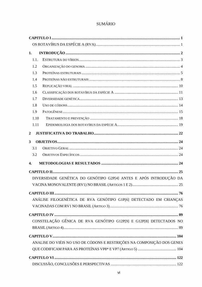
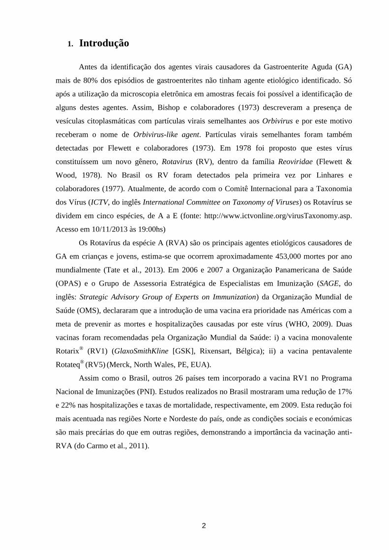
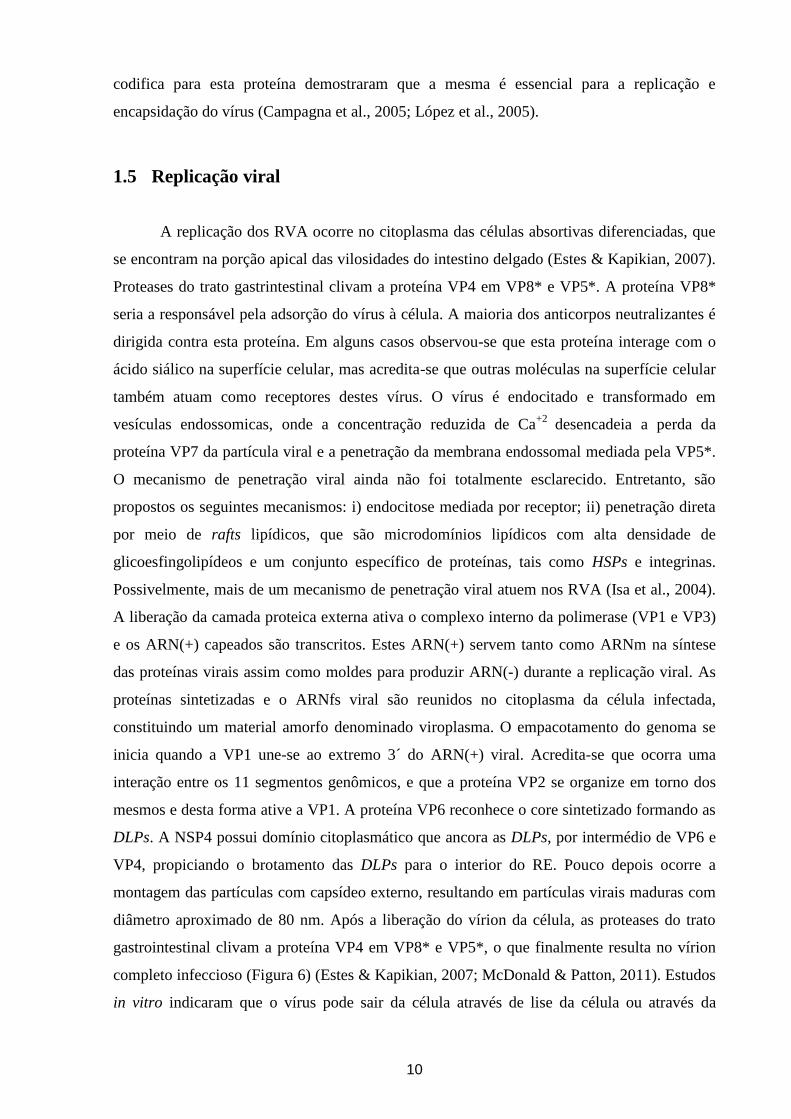
1.1. Estrutura do vírion
A partícula viral infecciosa (vírion) apresenta aproximadamente 100 nanômetros (nm)
de diâmetro, simetria icosaédrica e é constituída por três camadas proteicas, sendo um vírus
não envelopado (Figura 1). A camada proteica interna, ou cerne viral, é composto por 120
copias da proteína VP2. A região terminal da proteína VP2 está livre e acredita-se que esta
região está em contato com o complexo da polimerase viral composto pelas proteínas VP1
(ARN polimerase – ARN dependente) e VP3 (enzima responsável pela adição de um CAP no
ARN) (McClain et al., 2010). A camada intermediaria é constituída por 260 trímeros da
proteína VP6 que permitem estabilidade ao cerne interno; além de funcionar como um
adaptador para a camada externa que é essencial para a adsorção e entrada do vírus na célula.
Esta última camada é composta por 260 trímeros da glicoproteína VP7, diretamente
posicionados sobre os trímeros da proteína VP6. Os trímeros de VP7 são dependentes de íons
cálcio (Ca2+
), os quais mantêm a estabilidade destes. Se estendendo da camada formada pela
proteína VP7, encontram-se 60 trímeros formados pela proteína VP4, que apresenta diferentes
mudanças conformacionais durante a penetração na membrana da célula hospedeira (Patton,
2012).
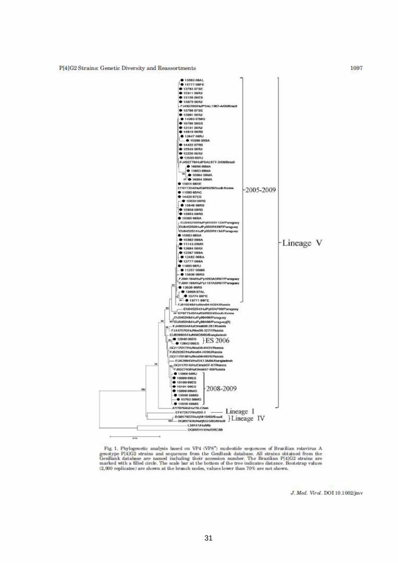
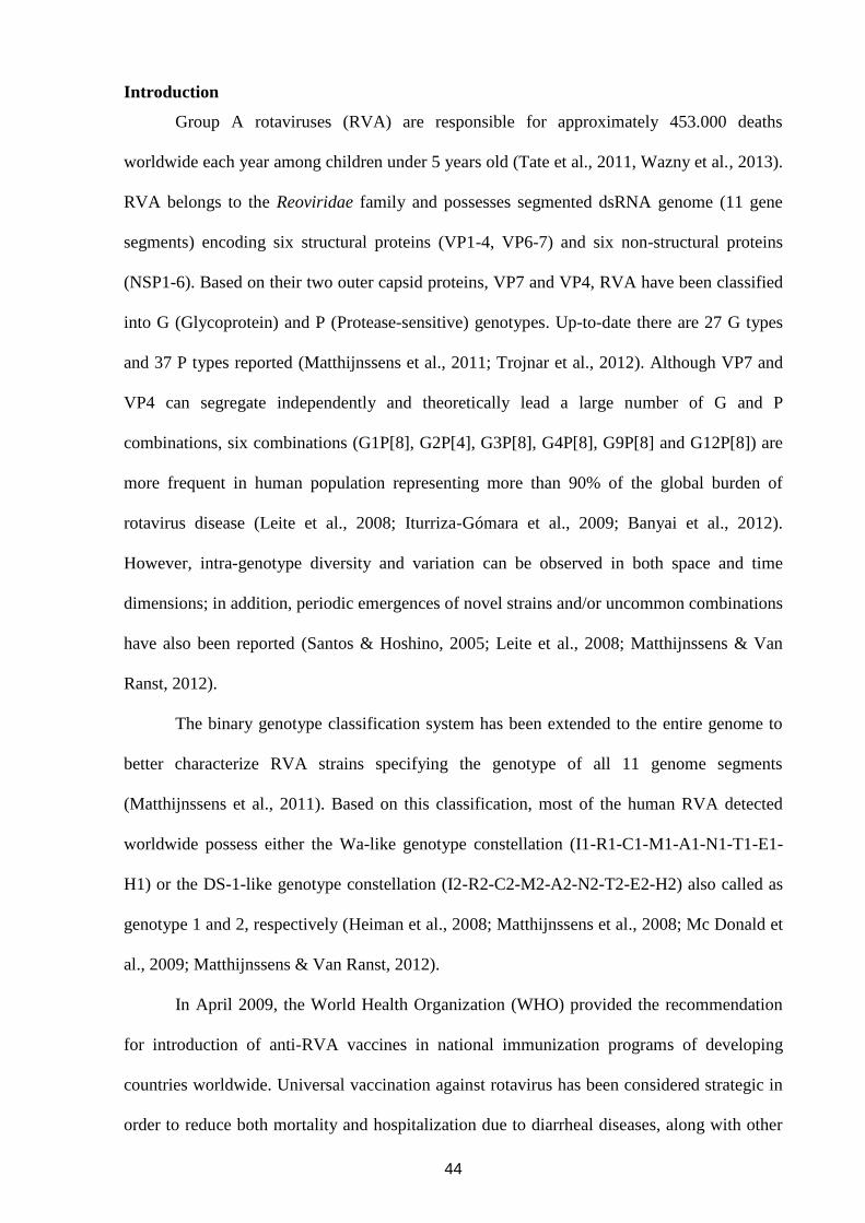
Figura 1. Estrutura do vírion dos rotavírus da espécie A. A) Esquema da estrutura do vírion;
B) Camada proteica tripla; C) Esquema da estrutura da proteína VP4. Figura adaptada de
Patton, 2012.
4
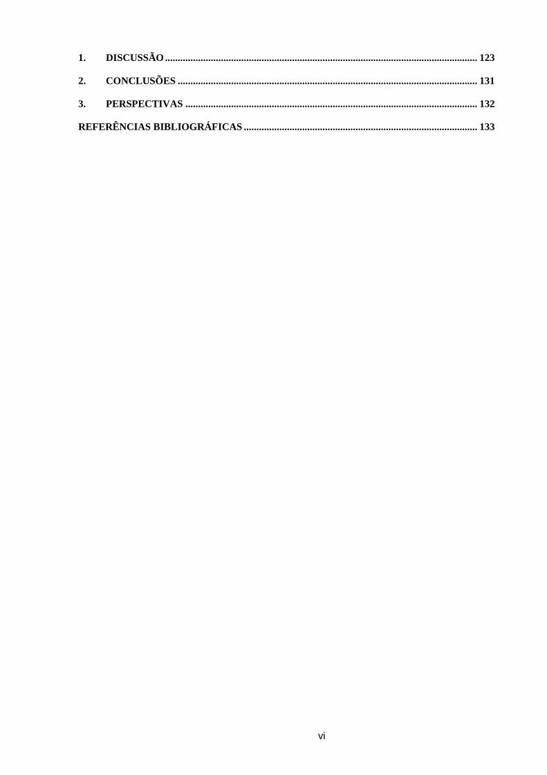
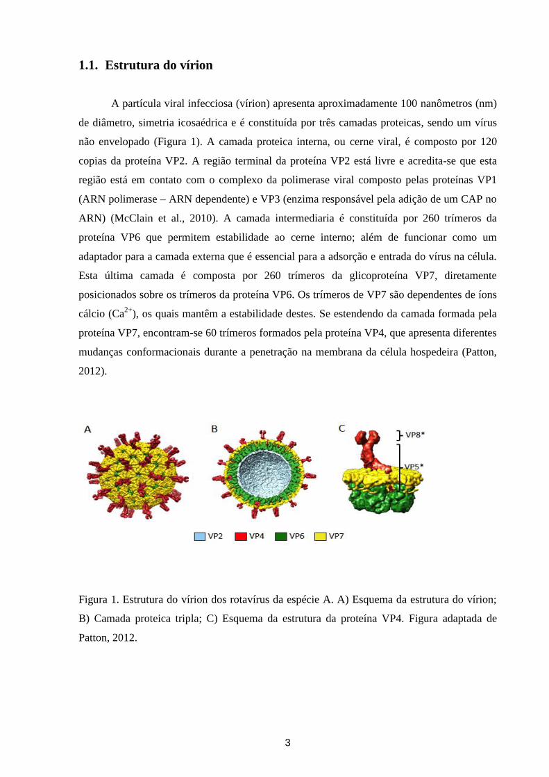
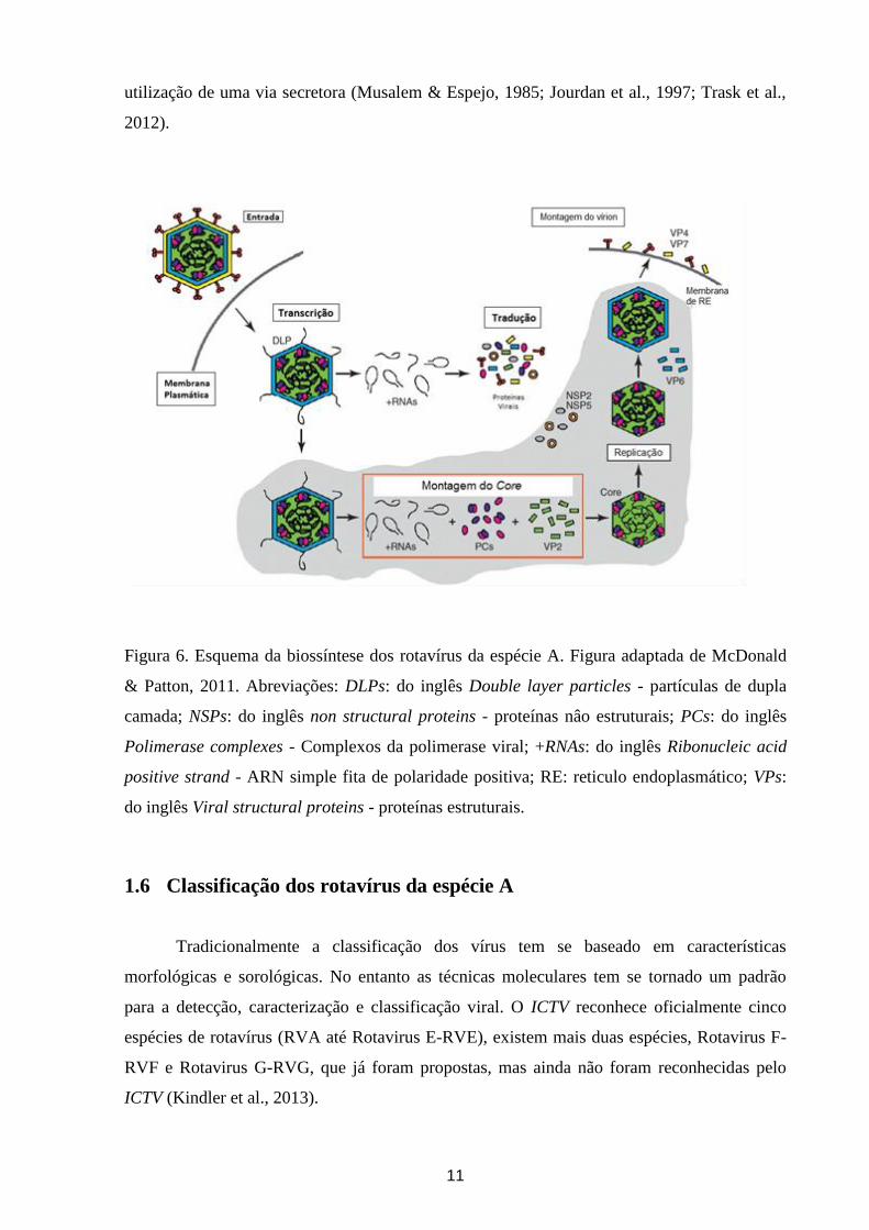
1.2 Organização do genoma
O genoma dos RVA consiste de 11 segmentos de Ácido ribonucleico (ARN) de dupla
fita (ARNdf) que variam em tamanho de 667 a 3302 pares de bases (pb), codificando 6
proteínas estruturais (VP1-4, VP6-7) e 5 ou 6 proteínas não estruturais (NSP1-6) (Figura 2)
(Estes & Kapikian, 2007).
Figura 2. Esquema estrutural do genoma e partículas virais dos rotavírus. A: Eletroforese em
gel de poliacrilamida (EGPA) dos 11 segmentos do genoma do rotavírus A símio (SA-11). B:
Gel de poliacrilamida mostrando a separação das proteínas estruturais (NSP1 – NSP6) e não
estruturais (VP1 – VP4, VP6 – VP7) sintetizadas pelo rotavírus A SA-11. C: Micrografia
eletrônica de rotavírus A. D: Reconstrução em 3D do vírion de SA-11, mostrando a
localização das proteínas estruturais. Figura adaptada de Conner & Ramig, 1997.
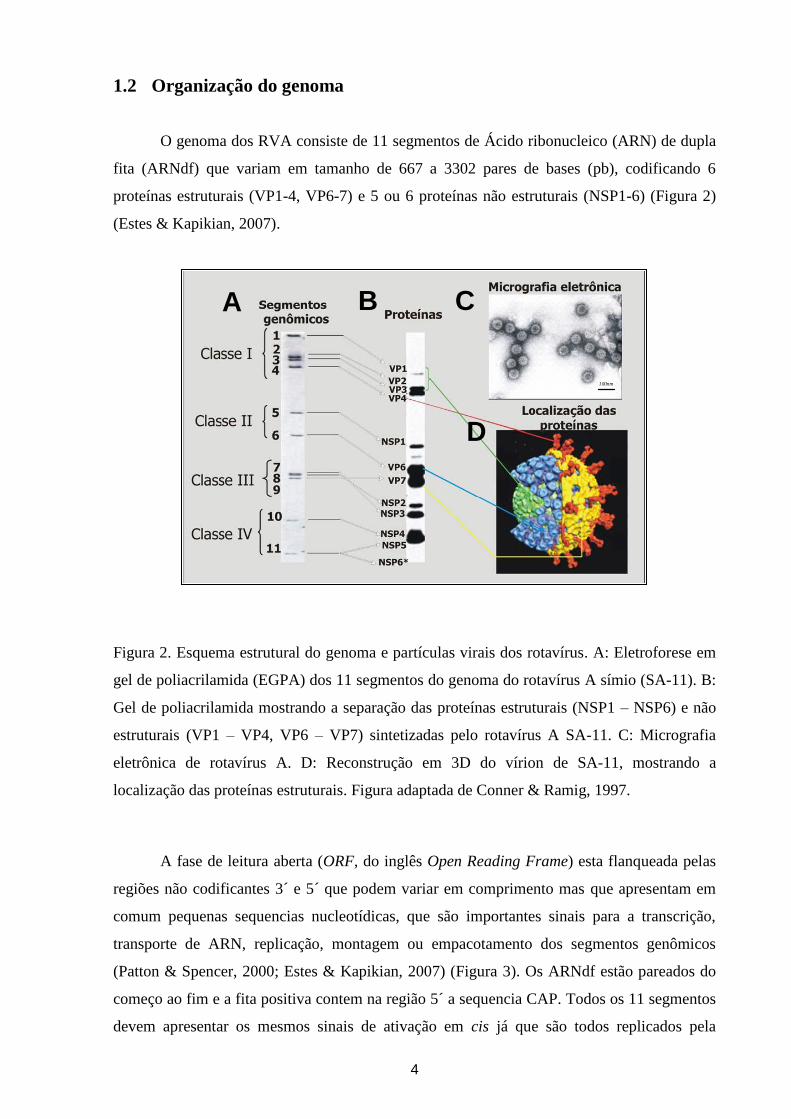
A fase de leitura aberta (ORF, do inglês Open Reading Frame) esta flanqueada pelas
regiões não codificantes 3´ e 5´ que podem variar em comprimento mas que apresentam em
comum pequenas sequencias nucleotídicas, que são importantes sinais para a transcrição,
transporte de ARN, replicação, montagem ou empacotamento dos segmentos genômicos
(Patton & Spencer, 2000; Estes & Kapikian, 2007) (Figura 3). Os ARNdf estão pareados do
começo ao fim e a fita positiva contem na região 5´ a sequencia CAP. Todos os 11 segmentos
devem apresentar os mesmos sinais de ativação em cis já que são todos replicados pela
100nm
CA
D
B
100nm100nm
CA
D
B
5
mesma polimerase. Além disso, cada segmento deve conter um sinal único porque estes
devem ser distinguidos um dos outros durante o empacotamento (Estes & Greenberg, 2013).
Figura 3. Esquema da organização geral dos segmentos gênicos de rotavírus da espécie A.
Abreviações: A, Adenina; C, Citosina; G, Guanina; ORF, do inglês Open Reading Frame; pb,
pares de bases; U, Uracilo.
1.3 Proteínas estruturais
Estudos forneceram evidencias de que a proteína estrutural VP1 é a ARN polimerase
dependente de ARN que atua como replicase e transcriptase. A estrutura da VP1 contém
quatro canais diferentes que levam ao centro catalítico da mesma. Acredita-se que os quatro
canais estejam envolvidos em: a) a entrada dos ARN de polaridade (+) (ARN(+)) e ARN de
polaridade (-) (ARN(-)); b) entrada dos Nucleotídeo trifosfato (NTPs, do inglês: nucleoside
triphosphates); c) saída dos ARNdf e ARN(-); d) saída dos ARN(+) (Estes & Greenberg,
2013).
A VP2 é a proteína mais abundante do cerne viral. Faz parte do complexo de
replicação e liga-se tanto à VP1 quanto à VP3, através de um domínio na sua porção N-
terminal (Arnoldi et al., 2007). Estudos recentes demostraram que para a ativação da VP1 é
necessária a interação do domínio principal da proteína VP2 com a mesma. A porção N
terminal da VP2 forma um canal em cada um dos cinco axis da partícula viral que é
fundamental para que ocorra a encapsidação das proteínas VP1 e VP3, interações com o ARN
e síntese do ARNdf (McDonald & Patton, 2011). Além disso, VP2 também interage com a
proteína VP6 nos estágios iniciais da morfogênese viral. A interação entre estas duas
proteínas (VP2 e VP6) é fundamental para a formação de partículas imaturas ou incompletas,
6
ou seja, aquelas que possuem apenas duplo capsídeo proteico (DLPs, do inglês: double layer
particles) (Estes & Kapikian, 2007).
A proteína VP3 apresenta atividades guanidil e metiltransferase e é a enzima
responsável pela adição da CAP (Patton, 1995; Subodh et al., 2006; McDonald & Patton,
2011). Dessa forma, VP3 é responsável por modificar a extremidade 5’ da molécula de ARN
viral e gerar uma estrutura de CAP similar a encontrada no àcido ribonucleico mensageiro
(ARNm) de eucariotos (McDonald & Patton, 2011).
A proteína VP4 é não glicosilada e tem papel essencial no ciclo replicativo viral, não
só pela adesão e internalização à célula, mas também hemaglutinação, neutralização e
virulência (Dunn et al., 1995; Ludert et al., 1996). É susceptível à proteólise, o que resulta na
exposição de sítios ativos que proporcionam a penetração do vírus na célula (Arias et al.,
1996; Estes & Kapikian, 2007). A ativação da proteína VP4 requer clivagem proteolítica da
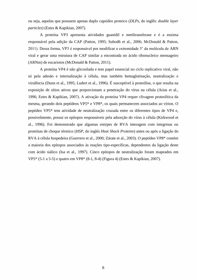
mesma, gerando dois peptídeos VP5* e VP8*, os quais permanecem associados ao vírion. O
peptídeo VP5* tem atividade de neutralização cruzada entre os diferentes tipos de VP4 e,
possivelmente, possui os epítopos responsáveis pela adsorção do vírus à célula (Kirkwood et
al., 1996). Foi demonstrado que algumas estirpes de RVA interagem com integrinas ou
proteínas de choque térmico (HSP, do inglés Heat Shock Proteins) antes ou após a ligação do
RVA à célula hospedeira (Guerrero et al., 2000; Zárate et al., 2003). O peptídeo VP8* contém
a maioria dos epítopos associados às reações tipo-específicas, dependentes da ligação deste
com ácido siálico (Isa et al., 1997). Cinco epítopos de neutralização foram mapeados em
VP5* (5-1 a 5-5) e quatro em VP8* (8-1, 8-4) (Figura 4) (Estes & Kapikian, 2007).
7
Figura 4. Representação esquemática dos sítios antigénicos presentes na proteína VP4. Figura
adaptada de Trask et al., 2012.
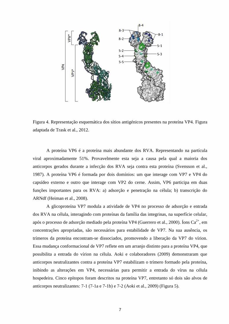
A proteína VP6 é a proteína mais abundante dos RVA. Representando na partícula
viral aproximadamente 51%. Provavelmente esta seja a causa pela qual a maioria dos
anticorpos gerados durante a infecção dos RVA seja contra esta proteína (Svensson et al.,
1987). A proteína VP6 é formada por dois domínios: um que interage com VP7 e VP4 do
capsídeo externo e outro que interage com VP2 do cerne. Assim, VP6 participa em duas
funções importantes para os RVA: a) adsorção e penetração na célula; b) transcrição do
ARNdf (Heiman et al., 2008).
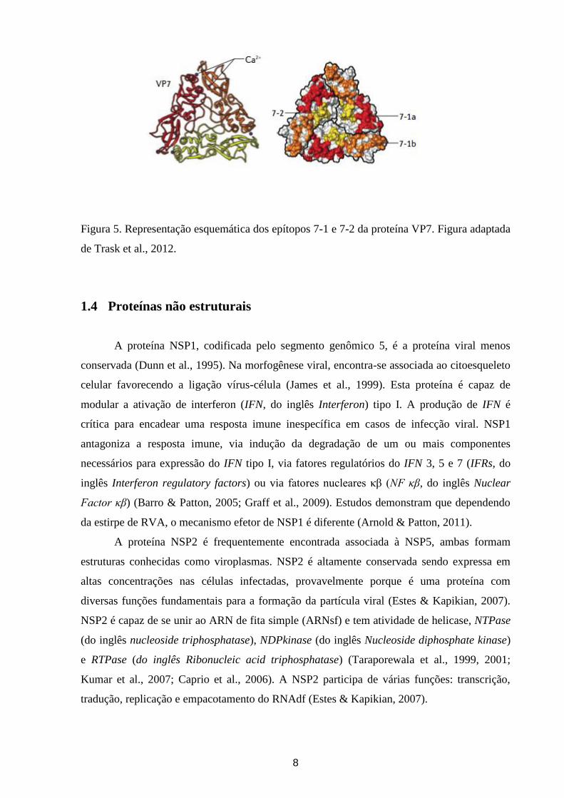
A glicoproteína VP7 modula a atividade de VP4 no processo de adsorção e entrada
dos RVA na célula, interagindo com proteínas da família das integrinas, na superfície celular,
após o processo de adsorção mediado pela proteína VP4 (Guerrero et al., 2000). Íons Ca2+
, em
concentrações apropriadas, são necessários para estabilidade de VP7. Na sua ausência, os
trímeros da proteína encontram-se dissociados, promovendo a liberação da VP7 do vírion.
Essa mudança conformacional de VP7 reflete em um arranjo distinto para a proteína VP4, que
possibilita a entrada do vírion na célula. Aoki e colaboradores (2009) demonstraram que
anticorpos neutralizantes contra a proteína VP7 estabilizam o trímero formado pela proteína,
inibindo as alterações em VP4, necessárias para permitir a entrada do vírus na célula
hospedeira. Cinco epítopos foram descritos na proteína VP7, entretanto só dois são alvos de
anticorpos neutralizantes: 7-1 (7-1a e 7-1b) e 7-2 (Aoki et al., 2009) (Figura 5).
8
Figura 5. Representação esquemática dos epítopos 7-1 e 7-2 da proteína VP7. Figura adaptada
de Trask et al., 2012.
1.4 Proteínas não estruturais
A proteína NSP1, codificada pelo segmento genômico 5, é a proteína viral menos
conservada (Dunn et al., 1995). Na morfogênese viral, encontra-se associada ao citoesqueleto
celular favorecendo a ligação vírus-célula (James et al., 1999). Esta proteína é capaz de
modular a ativação de interferon (IFN, do inglês Interferon) tipo I. A produção de IFN é
crítica para encadear uma resposta imune inespecífica em casos de infecção viral. NSP1
antagoniza a resposta imune, via indução da degradação de um ou mais componentes
necessários para expressão do IFN tipo I, via fatores regulatórios do IFN 3, 5 e 7 (IFRs, do
inglês Interferon regulatory factors) ou via fatores nucleares κβ (NF κβ, do inglês Nuclear
Factor κβ) (Barro & Patton, 2005; Graff et al., 2009). Estudos demonstram que dependendo
da estirpe de RVA, o mecanismo efetor de NSP1 é diferente (Arnold & Patton, 2011).
A proteína NSP2 é frequentemente encontrada associada à NSP5, ambas formam
estruturas conhecidas como viroplasmas. NSP2 é altamente conservada sendo expressa em
altas concentrações nas células infectadas, provavelmente porque é uma proteína com
diversas funções fundamentais para a formação da partícula viral (Estes & Kapikian, 2007).
NSP2 é capaz de se unir ao ARN de fita simple (ARNsf) e tem atividade de helicase, NTPase
(do inglês nucleoside triphosphatase), NDPkinase (do inglês Nucleoside diphosphate kinase)
e RTPase (do inglês Ribonucleic acid triphosphatase) (Taraporewala et al., 1999, 2001;
Kumar et al., 2007; Caprio et al., 2006). A NSP2 participa de várias funções: transcrição,
tradução, replicação e empacotamento do RNAdf (Estes & Kapikian, 2007).
9
O segmento 7 codifica a proteína NSP3, e a mesma tem três domínios, o domínio N
terminal de união ao ARN (Aminoácido (Aa) 1 – 170); o domínio de interação com eIF-4G1
(Do inglês Eukaryotic translation initiation factor 4-gamma 1), formado pelos últimos 107
resíduos da região C terminal e o domínio de dimerização (Aa 150 – 206) (Piron et al., 1999).
Estudos demostraram que a proteína NSP3 compete na célula hospedeira com a proteína de
união à cauda Poli A (PABP, do inglês Poly(A)-binding protein) o que levou a crer que a
proteína facilitaria a tradução do ARNm e ao silenciamento da síntese proteica da célula
hospedeira. Porém, estudos demostraram que a NSP3 não é necessariamente requerida para a
tradução do ARN viral (Piron et al., 1998; Padilla-Noriega, et al., 2002; Varani & Allain,
2002; Montero et al., 2006) Além disso, observou-se que a mesma estaria implicada no
espalhamento extra-intestinal do vírus no hospedeiro infectado. Entretanto, ainda não foi
elucidado o mecanismo que estaria envolvido (Mossel & Ramig, 2002, 2003).
A proteína NSP4, codificada pelo segmento 10, foi a primeira enterotoxina viral
descrita (Ball et al., 1996). Esta proteína se localiza em diversos sítios dentro da célula e
contribui na morfogênese, replicação e patogênese dos RVA. Na célula encontra-se em
diversas formas: a proteína inteira se localiza ancorada no reticulo endoplasmático (RE)
atraves do domínio hidrofóbico N terminal; enquanto que a região C terminal se orienta ao
citoplasma. A região que vai do Aa 45 ao 175 constitui a região citoplasmática e retém todas
as propriedades biológicas importantes associadas a esta proteína, incluindo: mobilização do
Ca+2
intracelular, permeabilizacão da membrana; união ao Ca2+
e a VP4, união às partículas
semelhantes a vírus (VLPs, do inglês Virus-like Particles) e indução da diarreia em
camundongos. Foi observado que uma forma secretada desta proteína contendo os Aa 112 a
175 é capaz de induzir diarreia assim como a proteína completa (Zhang et al., 2000).
O segmento 11 tem duas ORFs. A maior codifica para a proteína NSP5 que possui
atividade autoquinase e em células infectadas apresenta formas hipo e hiper fosforiladas
(Taraporewala & Patton, 2004). A sua fosforilação é modulada mediante interação com a
proteína NSP2 (Afrikanova et al., 1998). A outra ORF codifica para a proteína NSP6.
Algumas variantes de RVA não codificam esta proteína; outras apresentam baixos níveis de
expressão, que indicam que NSP6 apresenta papel regulatório não essencial na replicação
viral (Taraporewala & Patton, 2004; López et al., 2005).
Durante o processo de replicação, NSP2, NSP5 e NSP6 estão associadas à formação
do viroplasmas (Estes & Kapikian, 2007). Na ausência de outras proteínas virais, a interação
entre NSP2 e NSP5 resulta em partículas defectivas semelhantes às VLPs in vitro. A NSP5
também interage com a VP1 e a VP2. Estudos baseados no silenciamento do gene que
10
codifica para esta proteína demostraram que a mesma é essencial para a replicação e
encapsidação do vírus (Campagna et al., 2005; López et al., 2005).
1.5 Replicação viral
A replicação dos RVA ocorre no citoplasma das células absortivas diferenciadas, que
se encontram na porção apical das vilosidades do intestino delgado (Estes & Kapikian, 2007).
Proteases do trato gastrintestinal clivam a proteína VP4 em VP8* e VP5*. A proteína VP8*
seria a responsável pela adsorção do vírus à célula. A maioria dos anticorpos neutralizantes é
dirigida contra esta proteína. Em alguns casos observou-se que esta proteína interage com o
ácido siálico na superfície celular, mas acredita-se que outras moléculas na superfície celular
também atuam como receptores destes vírus. O vírus é endocitado e transformado em
vesículas endossomicas, onde a concentração reduzida de Ca+2
desencadeia a perda da
proteína VP7 da partícula viral e a penetração da membrana endossomal mediada pela VP5*.
O mecanismo de penetração viral ainda não foi totalmente esclarecido. Entretanto, são
propostos os seguintes mecanismos: i) endocitose mediada por receptor; ii) penetração direta
por meio de rafts lipídicos, que são microdomínios lipídicos com alta densidade de
glicoesfingolipídeos e um conjunto específico de proteínas, tais como HSPs e integrinas.
Possivelmente, mais de um mecanismo de penetração viral atuem nos RVA (Isa et al., 2004).
A liberação da camada proteica externa ativa o complexo interno da polimerase (VP1 e VP3)
e os ARN(+) capeados são transcritos. Estes ARN(+) servem tanto como ARNm na síntese
das proteínas virais assim como moldes para produzir ARN(-) durante a replicação viral. As
proteínas sintetizadas e o ARNfs viral são reunidos no citoplasma da célula infectada,
constituindo um material amorfo denominado viroplasma. O empacotamento do genoma se
inicia quando a VP1 une-se ao extremo 3´ do ARN(+) viral. Acredita-se que ocorra uma
interação entre os 11 segmentos genômicos, e que a proteína VP2 se organize em torno dos
mesmos e desta forma ative a VP1. A proteína VP6 reconhece o core sintetizado formando as
DLPs. A NSP4 possui domínio citoplasmático que ancora as DLPs, por intermédio de VP6 e
VP4, propiciando o brotamento das DLPs para o interior do RE. Pouco depois ocorre a
montagem das partículas com capsídeo externo, resultando em partículas virais maduras com
diâmetro aproximado de 80 nm. Após a liberação do vírion da célula, as proteases do trato
gastrointestinal clivam a proteína VP4 em VP8* e VP5*, o que finalmente resulta no vírion
completo infeccioso (Figura 6) (Estes & Kapikian, 2007; McDonald & Patton, 2011). Estudos
in vitro indicaram que o vírus pode sair da célula através de lise da célula ou através da
11
utilização de uma via secretora (Musalem & Espejo, 1985; Jourdan et al., 1997; Trask et al.,
2012).
Figura 6. Esquema da biossíntese dos rotavírus da espécie A. Figura adaptada de McDonald
& Patton, 2011. Abreviações: DLPs: do inglês Double layer particles - partículas de dupla
camada; NSPs: do inglês non structural proteins - proteínas nâo estruturais; PCs: do inglês
Polimerase complexes - Complexos da polimerase viral; +RNAs: do inglês Ribonucleic acid
positive strand - ARN simple fita de polaridade positiva; RE: reticulo endoplasmático; VPs:
do inglês Viral structural proteins - proteínas estruturais.
1.6 Classificação dos rotavírus da espécie A
Tradicionalmente a classificação dos vírus tem se baseado em características
morfológicas e sorológicas. No entanto as técnicas moleculares tem se tornado um padrão
para a detecção, caracterização e classificação viral. O ICTV reconhece oficialmente cinco
espécies de rotavírus (RVA até Rotavirus E-RVE), existem mais duas espécies, Rotavirus F-
RVF e Rotavirus G-RVG, que já foram propostas, mas ainda não foram reconhecidas pelo
ICTV (Kindler et al., 2013).
12
Os RVA têm sido classificados com base em: i) proteínas de superfície, sorotipos G
(VP7) e P (VP4); ii) padrão de migração dos segmentos de ARN quando submetidos a
eletroforese em gel de poliacrilamida (EGPA) (eletroferotipos), podendo apresentar perfil
curto, longo, super curto, ou atípico; iii) padrões de hibridização de ARN do genoma
completo (genogrupos); e iv) com base na análise das sequencias dos genes que codificam
para as proteínas VP7 e VP4, em genótipos G e P, respetivamente. Mais recentemente, um
novo sistema de classificação baseada na análise das sequencias nucleotídicas dos 11 genes
dos RVA foi estabelecido pelo Rotavirus Classification Working Group (RCWG) (Tabela 1)
(Matthijnssens et al., 2011). Este novo sistema de classificação recomenda nomear a
sequência viral obtida considerando-se os seguintes parametros: i) a espécie de RV; ii) a
origem do vírus (humano ou animal), identificando também se a sequencia foi obtida a partir
de vírus salvagem (wt, do inglês wild type) ou vírus em cultura celular (tc, do inglês tissue
culture); iii) o país em que foi detectado o vírus, utilizando o código padrão de 3 letras; iv) o
nome comum dado pelo pesquisador; v) o ano de detecção; vi) os genótipos G e P da seguinte
forma: GXP[X]. Com base nesta nomenclatura, foram descritos até o momento 27 G-, 37 P-,
17 I-, 9 R-, 9C-, 8 M-, 16 A-, 10 N-, 12 T-, 15 E-, e 11 H genótipos. Cada letra maiúscula
corresponde, respectivamente, aos genes que codificam para as proteínas VP7-VP4-VP6-
VP1-VP2-VP3-NSP1-NSP2-NSP3-NSP4-NSP5/6 (Matthijnssen et al., 2011). No caso do
vírus apresentar uma constelação genômica I1-R1-C1-M1-A1-N1-T1-E1-H1, I2-R2-C2-M2-
A2-N2-T2-E2-H2, ou I3-R3-C3-M3-A3-N3-T3-E3-H3 se classifica como pertencente ao
genótipo 1 (ou Wa-like), 2 (ou DS-1-like), ou AU-1-like, respectivamente (Matthijnssen &
Van Ranst, 2012).
13
Tabela 1. Valores de cut-off de percentagem de identidade nucleotídica que definem os
diferentes genótipos de rotavírus da espécie A, considerando-se os 11 segmentos genômicos.
Tabela adaptada de Matthijnssens et al., 2011.
Gene Valor de cut-off de identidade
nucleotídica (%) Genótipos
Designação dos nomes dos
genótipos
VP7 80 27G Glicoproteína
VP4 80 37P Sensível a Protease
VP6 85 17I Capsídeo Interno
VP1 83 9R ARN Polimerase Dependente de
ARN
VP2 84 9C Proteína do cerne
VP3 81 8M Metiltransferase
NSP1 79 16A Antagonista do Interferon
NSP2 85 10N NTPase
NSP3 85 12T Promotor Traducional
NSP4 85 15E Enterotoxina
NSP5 91 11H Fosfoproteína
1.7 Diversidade genética
Existe uma grande diversidade de cepas de RVA que circulam na população humana,
gerada através de diversos mecanismos: i) acumulação de mutações pontuais (genetic drift);
ii) reestruturações dos segmentos gênicos (genetic shift); iii) transmissão direta de vírus
animais para humanos; iv) rearranjos genéticos (deleções, inserções e duplicações)
(Kirkwood, 2010).
A ocorrência de mutações pontuais é frequente nos RVA, assim como ocorre com
outros vírus ARN, e podem produzir mudanças detectáveis de fenótipo (Taniguchi &
Urasawa, 1995). Para os genótipos G9 e G12 a taxa de mutação do gene VP7 foi calculada em
1.87x10⁻³
e 1.66x10⁻³
, valores semelhantes aos obtidos com outros vírus ARNs (Matthijnssen
et al., 2010, Ghosh & Kobayashi, 2011).
14
Os rearranjos representam alterações na sequência do segmento genômico, algumas
vezes na forma de deleção ou como duplicação. Estes eventos geralmente são evidenciados
através da visualização do perfil eletroforético dos RVA em EGPA. Os rearranjos resultam de
erros de transcrição de um único segmento e possuem nada mais do que a duplicação parcial
de um gene. Provavelmente, no momento da transcrição, por uma falha da ARN polimerase
dependente de ARN, ela retorne a sua fita molde, reiniciando a transcrição a partir de
diferentes estágios (Desselberger, 1996).
O evento de recombinação genética em RVA ainda é pouco conhecido (Phan et al.,
2007). As recombinações intra genicas não tem sido descritas com frequência, mas existem
relatos de recombinação entre genes de VP7 de diferentes genótipos G e entre linhagens do
mesmo G (Parra et al., 2004; Phan et al., 2007; Martínez-Laso et al., 2009).
As reestruturações de segmentos genômicos ocorrem quando duas cepas de RVA
distintas infectam a mesma célula. Foram descritos pela primeira vez por Matsuno e
colaboradores (1980) que realizou a coinfecção em cultura celular de RV bovino (Lincoln) e
RV símio (SA-11) obtendo assim um vírus reestruturado. A ocorrência de reestruturações
parece ser um fenômeno frequente nos RVA. Porem, estudos mostraram que a maioria das
variantes virais originadas por este mecanismo são substituídas no decorrer do tempo por
cepas que apresentam uma constelação genômica que por algum motivo é dominante na
população viral (McDonald et al., 2009). A incidência das reestruturações esta influenciada
pela frequência de coinfecções na população e a diversidade genética dos RVA circulantes
(Ghosh & Kobayashi, 2011). A ocorrência de reestruturações parece ser mais frequente nos
países em desenvolvimento, fato provavelmente vinculado às precárias condições de
saneamento básico e higiene, defesas imunológicas limitadas, coinfecções com parasitas,
desnutrição, além do estreito relacionamento entre o homem, animais domésticos e outros
animais. Todas as possibilidades facilitam a ocorrência de infecções mistas e,
consequentemente, aumenta a possibilidade dos vírus sofrerem reestruturações genéticas
(Desselberger, 1996).
1.8 Uso de códons
Com o objetivo de entender melhor sobre a dinâmica evolutiva dos vírus e em
particular, dos RVA, faz-se necessário o uso de ferramentas e/ou métodos importantes na
elucidação dos processos evolutivos destes agentes, suas implicações na expressão de
proteínas e na manutenção de variantes genéticas de importância epidemiológica.
15
Os códons sinônimos não são utilizados de forma randômica e, em diversos
organismos, a seleção natural parece gerar um viéis no uso de códons e, neste caso, a favor do
uso de códons chamados de “códons ótimos”, sobretudo em genes que são altamente
expressos. Diversos fatores como expressão gênica, empacotamento do ARN viral, formação
da estutura das proteínas e o uso de nucleotídeos podem influenciar no uso de códons
(Stoletzki & Eyre-Walker, 2007).
Têm sido propostos dois modelos para explicar o viéis no uso de códons: i) modelo
associado à tradução (Translational related model ou Selective model), postula que existe
uma coadaptação no uso dos códons sinônimos e na abundancia dos ARNt (Ácido
ribonucleico de transferência) para optimizar a eficácia da tradução. Neste modelo espere-se
uma correlação entre o uso de códons e a expressão do gene; ii) modelo mutacional
(Mutational model ou Neutral model), postula que o viéis no uso de códons é o resultado dos
viéis mutacionais (Duret, 2002).
Um estudo baseado na análise do viéis no uso de códons e composição nucleotídica de
diferentes vírus ARN, demostrou que o viéis observado nestes vírus sería em maior parte
produto de pressões mutacionais. Além disso, observou-se que os genes que codificam para a
ARN polimerase apresentavam um viéis maior no uso de códons, mesmo que os genes
estruturais sejam de forma geral expressos em maiores níveis que os genes não estruturais
(Jenkins & Holmes, 2003). Tem sido reportado que existe um viéis maior no uso de códons
nos vírus de RNA segmentado quando comparado com os vírus com genoma não
segmentados, associado provavelmente ao fato dos mesmos sofrerem reestruturações gênicas
(Jenkins & Holmes, 2003).
O viéis no uso de codon é geralmente estimado pelo uso do valor ENC (do inglês:
Effective Number of codons), este valor pode variar de 20 quando só um códon é utilizado
para cada Aa até 61, quando todos os códons sao utilizados. Em genomas onde o uso de
códons é inteiramente devido à tendências mutacionais o valor do ENC varía entre 31 e 61
(Wright, 1990). Para os RVA este valor foi estimado em 52.5 para a região codificante inteira
do genoma, sugerindo que a pressão mutacional seria a principal força atuando no viéis no
uso de códons no genoma deste vírus (Jenkins & Holmes, 2003).
1.9 Patogênese
Os RVA apresentam caraterísticas que os torna agentes altamente infecciosos e
adaptados ao hospedeiro, estes vírus podem infectar igualmente o homem e animais (Franco
et al., 2006). A transmissão deste vírus é feita pela via fecal-oral. Após um período de
16
incubação de 2 a 4 dias, os sintomas geralmente começam abruptamente com febre e vômitos,
seguidos de diarreia aquosa que pode durar dentre 3 a 8 dias (Staat et al., 2002; Lee et al.,
2008). A doença provocada pelos RVA é mais frequente em crianças entre 3 e 36 meses de
idade. Acredita-se que múltiplas infecções ocorrem ao longo da vida, e que por causa da
imunidade adquirida nestes episódios as crianças com idade mais avançada e os adultos
sofrem de episódios menos graves e/ou assintomáticos (Anderson & Weber, 2004).
Os enterócitos do intestino delgado dividem-se em dois tipos: enterócitos
propriamente ditos e células da cripta. Os enterócitos das velocidades são enterócitos maduros
que se diferenciam em cumprir funções de absorção e de digestão. Os que cumprem funções
de absorção sintetizam diversas dissacaridases, peptidases, e outras enzimas que são expressas
na superfície apical onde cumprem suas funções digestivas. A absorção ocorre por
mecanismos passivos, difusão de solutos através de gradientes eletroquímicos ou osmóticos, e
transporte ativo. As células da cripta são as progenitoras dos enterócitos das velocidades, as
mesmas secretam ativamente íons Cl- ao lume intestinal. Durante o funcionamento normal do
intestino a atividade combinada dos enterócitos e das células da cripta garante o fluxo
bidirecional constante de eletrólitos e agua através do epitélio (Ramig, 2004). Os RVA se
replicam nos enterócitos maduros perto dos estremos das vilosidades, sugerindo assim que
estes enterócitos expressam fatores que são necessários para a replicação do vírus (Conner &
Ramig, 1997).
A diarreia causada pelos RVA é multifatorial e, portanto diversos mecanismos estão
envolvidos (Quadro 1).
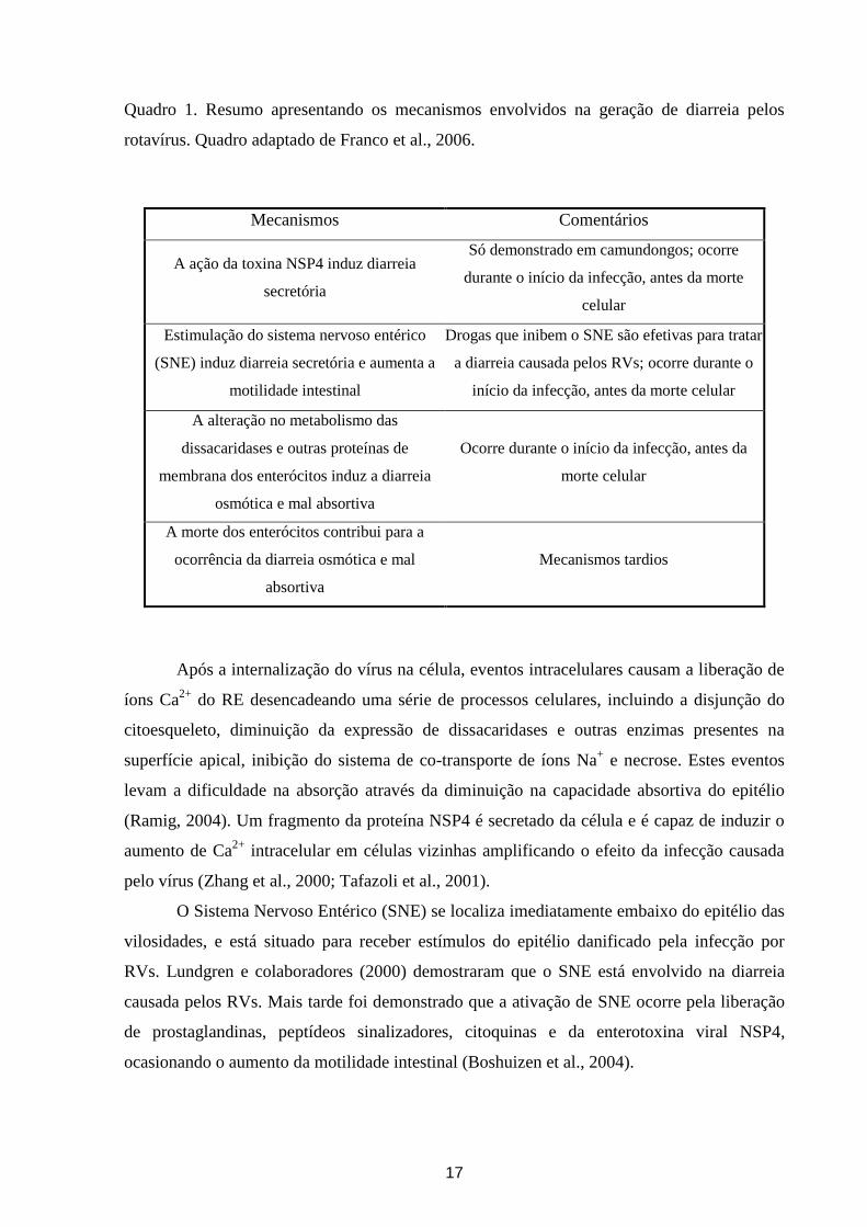
17
Quadro 1. Resumo apresentando os mecanismos envolvidos na geração de diarreia pelos
rotavírus. Quadro adaptado de Franco et al., 2006.
Mecanismos Comentários
A ação da toxina NSP4 induz diarreia
secretória
Só demonstrado em camundongos; ocorre
durante o início da infecção, antes da morte
celular
Estimulação do sistema nervoso entérico
(SNE) induz diarreia secretória e aumenta a
motilidade intestinal
Drogas que inibem o SNE são efetivas para tratar
a diarreia causada pelos RVs; ocorre durante o
início da infecção, antes da morte celular
A alteração no metabolismo das
dissacaridases e outras proteínas de
membrana dos enterócitos induz a diarreia
osmótica e mal absortiva
Ocorre durante o início da infecção, antes da
morte celular
A morte dos enterócitos contribui para a
ocorrência da diarreia osmótica e mal
absortiva
Mecanismos tardios
Após a internalização do vírus na célula, eventos intracelulares causam a liberação de
íons Ca2+
do RE desencadeando uma série de processos celulares, incluindo a disjunção do
citoesqueleto, diminuição da expressão de dissacaridases e outras enzimas presentes na
superfície apical, inibição do sistema de co-transporte de íons Na+ e necrose. Estes eventos
levam a dificuldade na absorção através da diminuição na capacidade absortiva do epitélio
(Ramig, 2004). Um fragmento da proteína NSP4 é secretado da célula e é capaz de induzir o
aumento de Ca2+
intracelular em células vizinhas amplificando o efeito da infecção causada
pelo vírus (Zhang et al., 2000; Tafazoli et al., 2001).
O Sistema Nervoso Entérico (SNE) se localiza imediatamente embaixo do epitélio das
vilosidades, e está situado para receber estímulos do epitélio danificado pela infecção por
RVs. Lundgren e colaboradores (2000) demostraram que o SNE está envolvido na diarreia
causada pelos RVs. Mais tarde foi demonstrado que a ativação de SNE ocorre pela liberação
de prostaglandinas, peptídeos sinalizadores, citoquinas e da enterotoxina viral NSP4,
ocasionando o aumento da motilidade intestinal (Boshuizen et al., 2004).
18
1.10 Tratamento e prevenção
O tratamento da diarreia causada pelos RVA tem como base repor as perdas de fluidos
e eletrólitos. Para a reidratação da criança pode-se utilizar a fórmula preconizada pela OMS
ou ainda outra fórmula comercial disponível. Estas fórmulas tem se mostrado efetivas para
crianças com desidratação moderada. Nos casos de diarreia grave é recomendável a utilização
de fluidos intravenosos, ou no caso de que a criança tenha dificuldade de deglutição devido à
intensidade dos vômitos. A terapia nutricional e a assistência médica são extremammente
importantes, ajudando a reduzir a morbidade e a mortalidade causada pelos RVA. Não
existem medicamentos antivirais disponíveis para o tratamento da infecção pelos RVA.
Entretanto, diversos estudos tem demostrado a atividade antiviral de algumas drogas frente
aos RVA (Rossignol et al., 2006; Teran et al., 2009; La Frazia et al., 2013; Shen et al., 2013).
Inclusive, um estudo realizado recentemente no Brasil demostrou a atividade antiviral de
plantas medicinais contra o RVA (Cecílio et al., 2012).
Somente as melhorias das condições sanitárias, bem como a destinação adequada de
dejetos de humanos e de animais, não são suficientes no controle e na prevenção das
infecções por RVA. A infecção natural não confere proteção completa contra reinfecção e a
prevenção da GA. Durante os dois primeiros anos de vida a prevenção é fundamental, pois é
neste período que ela é mais grave. Assim, a utilização de uma vacina segura e eficaz
representa uma boa estratégia para diminuir as formas mais graves da doença (Anderson,
2010).
Em 2009, a OMS recomendou a inclusão de vacinas contra RVA nos programas nacionais
de imunização de todos os países, e atualmente recomenda a introdução de vacinas em países
onde as mortes por GA são responsáveis por mais de 10% da mortalidade infantil (< 5 anos de
idade). Algumas vacinas já foram desenvolvidas ou estão em desenvolvimento. Entretanto,
somente duas vacinas (RV1 e RV5) estão licenciadas para uso em vários países, incluindo o
Brasil (O'Ryan et al., 2011). A RV1 é uma vacina oral monovalente (G1P[8]), atenuada, originada
a partir de uma cepa humana. A RV5 é uma vacina oral, pentavalente (G1, G2, G3, G4 e P[8]) de
vírus reestruturado (WC3 - bovino) (Ruiz-Palacios et al., 2006, Vesikari et al., 2006). As duas
vacinas, protegem contra os 5 genótipos mais comuns de RVA que circulam mundialmente (G1-
G4, G9) e o impacto positivo da introdução destas vacinas já foi demonstrado em alguns países
europeus, africanos e americanos, inclusive o Brasil (Chandran et al., 2010; Carvalho-Costa et al.,
2011; Linhares et al., 2011; Patel et al., 2011).
O MS brasileiro introduziu a vacina monovalente no PNI em 2006, mas a vacina
pentavalente pode ser encontrada em clínicas particulares. Do Carmo e colaboradores (2011)

19
demonstraram que três anos após a introdução da vacina RV1 no Brasil houve uma redução
de 22-28% de mortes e de 21-25% de hospitalizações em crianças <2 anos de idade.
1.11 Epidemiologia dos rotavírus da espécie A
A distribuição dos genótipos de RVA ao longo do tempo pode variar de região em
região e múltiplos genótipos G e P podem cocircular na população, inclusive dentro da
mesma região geográfica. Entretanto, a maioria dos estudos epidemiológicos tem
demonstrado que amostras de RVA, de genótipos G1P[8], G2P[4], G3P[8], G4P[8] e G9P[8],
são responsáveis pela maioria das infecções em humanos (92%), sendo importantes alvos de
estudos para o desenvolvimento de vacinas (Kirkwood, 2010). Contudo o genótipo G1P[8] é
o mais prevalente mundialmente, sendo responsável por mais de 70% das infecções por RVA
na América do Norte, Europa e Austrália. Porém, este genótipo representa somente 30% das
infecções por RVA na América do Sul e Ásia, e 23% na África (Tate et al., 2010).
No Brasil estes genótipos representam 75% dos RVA detectados (Linhares et al.,
2011). Os genótipos G1-G4 foram os mais prevalentes no período de 1982 a 1995; de 1996 a
2005 o genótipo G9 emergiu e, neste período, o genótipo G2P[4] foi pouco detectado. Em
2005 o genótipo G2P[4] reemergiu e tornou-se o genótipo mais prevalente no Brasil no
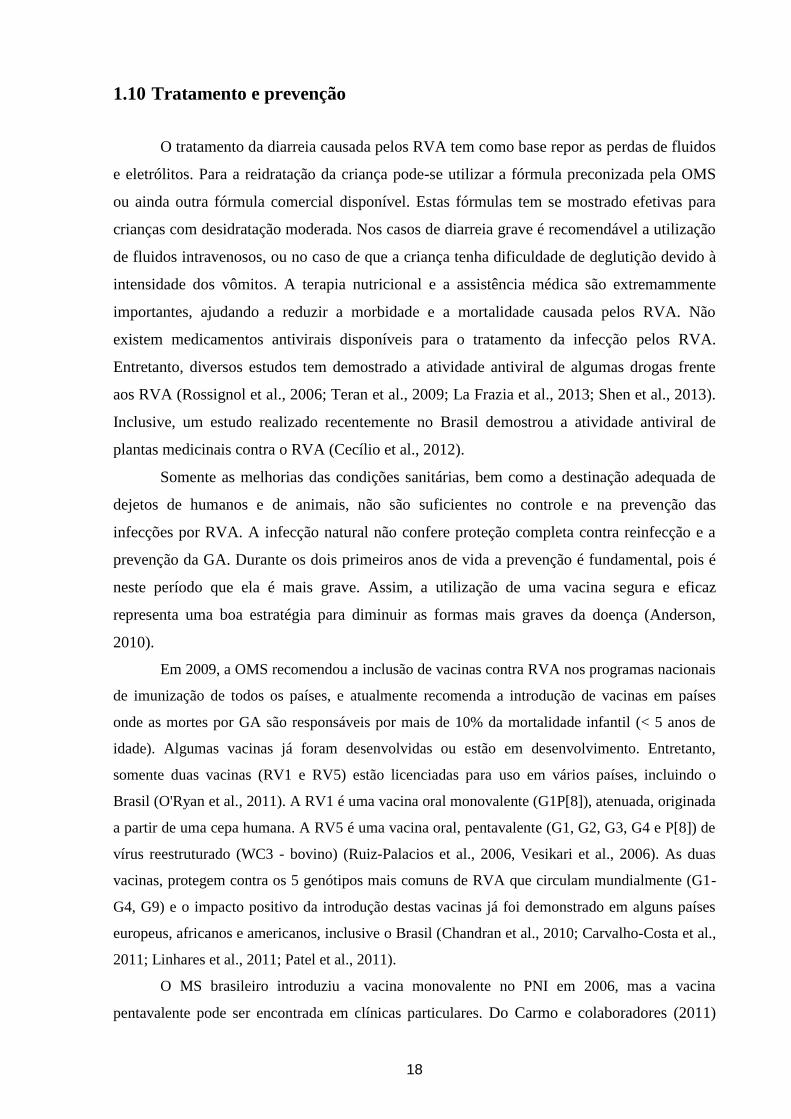
período de 2006 a 2011 (Figura 7) (Leite et al., 2008; Linhares et al., 2011; Carvalho-Costa et
al., 2011; Carvalho-Costa et al., manuscrito em preparação). Deve-se destacar que no Sul do
país o genótipo mais prevalente em 2009 foi o G3P[8]. Dados recentes demostram que em
2012 o genótipo G2P[4] deixou de ser o mais prevalente no Brasil e observou-se um aumento
na prevalência do genótipo G3P[8] / G3P[X] (Carvalho-Costa et al., manuscrito em
preparação).
20
Figura 7. Distribuição dos genótipos de rotavírus da espécie A no Brasil entre 2005 e 2009.
Figura adaptada de Carvalho-Costa et al., 2011.
Um surto de GA no estado do Acre no ano de 2005 foi atribuído ao RVA genótipo
G9P[8], sendo descritas 12,145 hospitalizações e oito mortes na municipalidade de Rio
Branco (Siqueira et al., 2010). Análises filogenéticas de amostras detectadas nesse surto
revelaram a circulação de uma nova variante do genótipo G9P[8] que até o momento só tem
sido detectada no Brasil (Acre) (Tort et al., 2010). Após introdução da vacina RV1 pelo PNI,
a detecção do genótipo G9 tem diminuído consideravelmente no país (Carvalho-Costa et al.,
2011). Na Argentina, este genótipo foi o mais prevalente entre 2006-2008, sendo que em
2009 a detecção deste genótico teve uma drastica redução e concomitantemente ocorreu o
aumento dos genótipos G3P[8] e G12P[8] (Stupka et al., 2009). O genótipo G9P[8] emergiu
em 1995 em diversos países do mundo e rapidamente tornou-se um dos cinco genótipos de
RVA mais prevalentes (Rahman et al., 2005; Matthijnssens et al., 2010).
Como mencionado anteriormente, um aumento significativo na ocorrência do genótipo
G2P[4] foi verificado a partir de 2005 (Leite et al., 2008). No período de 2006 a 2007,
21
primeiro ano de implementação da vacina RV1, G2 foi detectado nos estados do Rio de
Janeiro, Sergipe, Pernambuco, Piauí e Minas Gerais (Leite et al., 2008). Na região Nordeste
do Brasil, diferentes estudos mostraram uma prevalência de 100% para o genótipo G2P[4] no
ano de 2007 (Gurgel et al., 2007, Nakagomi et al., 2008). Estudos realizados na região Norte
do Brasil demostraram que a re-emergência de G2 alcançou taxas de até 90% (de Oliveira et
al., 2008). Um aumento na detecção deste genótipo também foi reportado em diferentes países
tais como: Honduras (Ferrera et al., 2007), Argentina, Paraguai (Amarilla et al., 2007), El
Salvador e Guatemala (Patel et al., 2008).
Recentemente, outros genótipos de RVA estão sendo descritos e considerados como
emergentes epidemiologicamente no mundo. Este é o caso de G5, em países da África e da
Ásia; assim como G8, G10 e G12 mundialmente. Além disso, combinações atípicas em
amostras humanas são detectadas em diversos países, sendo a maioria destas nos países da
África, América Latina e Ásia (Matthijnssens et al., 2010; Iturriza-Gómara et al., 2011). No
Brasil os genótipos G2P[8]; G3P[4]; G4P[6]/P[9]; G5P[6]/P[8]/G5P[NT]; G8P[4]/P[8];
G9P[4]/P[6]; G10P[9]; G12P[9]/P[8]/P[6] e G6 tem sido detectados esporadicamente (Araújo
et al., 2001; Mascarenhas et al., 2002; Pietruchinski et al., 2006; Domingues et al., 2008;
Leite et al., 2008; Soares et al., 2011; Gómez et al., 2013; Gómez et al. (artigo submetido)).
Com relação à sazonalidade, é possível observar um padrão apenas nas regiões de
clima temperado, com surtos e epidemias nos meses mais frios e secos do ano. Em geral, nas
regiões de clima tropical as infecções por RVA ocorrem ao longo de todo o ano (Estes &
Kapikian, 2007).
22
2 Justificativa do trabalho
Após a introdução, em março de 2006, da vacina RV1 pelo PNI no Brasil observou-se
uma mudança na epidemiologia dos RVA circulantes na população, tornando-se o genótipo
G2P[4] (DS-1 like) o mais prevalente até 2011 (Leite et al., 2008; Carvalho-Costa et al., 2011;
Carvalho-Costa et al., manuscrito em preparação). A emergência deste genótipo reflete um
fenômeno continental, provavelmente relacionado a vários fatores. Dados recentes obtidos no
LVCA, sugerem que diminuiu a detecção do genótipo G2 após 2010, e que esta diminuição
coincidiu com o aumento na prevalência do genótipo G3P[8] (Carvalho-Costa et al., em
preparação). Além disso, a detecção do genótipo G9 tem diminuído consideravelmente no
país após introdução da vacina (Carvalho-Costa et al., 2011) e os genótipos G1P[6] e G12 tem
sido detectados ocasionalmente (Pietruchinski et al., 2006; Leite et al., 2008; Carvalho-Costa
et al., 2009, 2011; Gómez et al., 2011, 2013; Fumian et al., 2011; Linhares et al., 2011; Soares
et al., 2011; Gómez et al., 2013; Gómez et al., submetido; Carvalho-Costa et al., em
preparação).
Os estudos de fase III relacionados à proteção conferida pela vacina RV1 antes da
implementação da mesma pelo PNI no Brasil não puderam ser adequadamente avaliados, pois
os genótipos G2P[4], P[6], G12, entre outros, não se encontravam entre os mais prevalentes.
A vacina RV1 apresenta uma constelação genética Wa-like e uma importante questão que se
coloca é sobre a eficácia da mesma frente a cepas que apresentem constelações diferentes,
como DS-1-like e AU-1-like: a pergunta é se poderão ocorrer mecanismos de reestruturações
geneticas intra ou entre genogrupos de RVA que possam prejudicar a eficácia da RV1?
Ocorrerão reestruturacoes geneticas entre amostras selvagens e a RV1? e caso afirmativo, ira
variar a eficácia da vacina frente a estas novas variantes virais?
Dois estudos realizados com amostras brasileiras de crianças hospitalizadas e
vacinadas com RV1 demonstraram a ocorrência destes reordenamentos genéticos (Gómez et
al., 2013; Rose et al., 2013).
No inicio do desenvolvimento deste projeto não existia na literatura nenhum trabalho
descrevendo a caracterização genética dos 11 genes de RVA de amostras detectadas no Brasil
e, alguns poucos de outros países, sendo a maioria em paises desenvolvidos. Na literatura, a
maioria dos estudos estavam relacionados com a caracterização dos genes que codificam para
as proteínas VP7, VP8*, VP6, e NSP4, principalmente para VP7 e VP8*. Embora os dados
sobre a diversidade genética do gene que codifica para a VP7 e/ou o que codifica para a VP8*
são importantes do ponto de vista da imunidade do hospedeiro contra a doença causada pelo
RVA, informações sobre esses genes não seriam suficientes para obter dados conclusivos
23
sobre a dinâmica evolutiva destes vírus. O fato dos RVA apresentarem um genoma
segmentado resulta numa grande variedade genética destes vírus, já que diferentes
mecanismos evolutivos podem atuar ao mesmo tempo, como anteriormente apresentados e
discutidos: mutação pontual, rearranjo, recombinação ou reestruturação genética. Portanto,
estudos que descrevam a analise dos 11 genes (constelação gênica) dos RVA poderão
contribuir para melhor avaliar a diversidade genética e a evolução destes vírus em dois
períodos: pré e pos introdução da vacina RV1.
Neste estudo foi analisada a diversidade genética de RVA de diversos genótipos
detectados no Brasil antes e após a introdução da vacina RV1 pelo PNI com o intuito de
identificar e caracterizar variantes de RVA emergentes e reemergentes no Brasil. Deve-se
ressaltar que este estudo compreende diferentes regiões geográficas do Brasil, pois dados
epidemiológicos sugerem diversidades genotípicas regionais (Leite et al., 2008; Carvalho-
Costa et al., manuscrito em preparação).
Uma outra abordagem deste estudo foi o uso de códons, pois pouco é conhecido sobre
o viéis no uso de códons pelos RVA. Estudar as causas do víéis no uso de códons é essencial
para entender a interfase entre os vírus e a resposta do sistema imune, além de ajudar no
desenho de vacinas, já que uma expressão eficiente das proteínas virais é fundamental para
gerar imunidade (Shackelton et al., 2006). Buscando contribuir para um melhor entendimento
deste mecanismo, foram estudadas as causas do víeis no uso de códons dos RVA analisando
os genes que codificam para as proteínas VP7 e VP8* de amostras genótipo G2P[4]
detectadas no Brasil no período de 1996-2009, assim como genomas inteiros disponíveis no
banco de dados do GenBank (url: http://www.ncbi.nlm.nih.gov/genbank/).
24
3 Objetivos
3.1 Objetivo Geral
Estudar a diversidade genética de RVA de cepas pertencentes a diferentes genótipos,
detectadas em diversas regiões do país, anterior e posterior a introdução da vacina RV1 pelo
PNI, em março de 2006.
3.2 Objetivos Específicos
1) Amplificar, sequenciar e realizar análises filognéticas dos genes que codificam para as
proteínas VP7 e VP8* de amostras de RVA detectadas entre os anos 2005 e 2011 de
diferentes regiões do Brasil;
2) Analisar as sequencias aminoacídicas das proteínas VP7 e VP8*, e comparar os sitios
antigênicos com a sequencias disponíveis da vacina RV1;
3) Amplificar, sequenciar, e analisar a constelação genética (11 genes) de RVA de
amostras pertencentes a diferentes genótipos detectadas após 2004 no Brasil;
4) Analisar o viéis no uso de códons dos genes que codificam para as proteínas VP7 e
VP8* de amostras de RVA G2P[4] detectadas em diferentes regiões do Brasil entre
1996 e 2009;
4. Metodologias e resultados
As seções “Metodologias” e “Resultados” deste manuscrito de tese serão apresentados
sob a forma de artigos submetidos a publicação, aceitos ou em fase final de confecção.
25
CAPITULO II
DIVERSIDADE GENÉTICA DO GENÓTIPO G2P[4]
ANTES E APÓS INTRODUÇÃO DA VACINA
MONOVALENTE (RV1) NO BRASIL (Artigos 1 e 2)
26
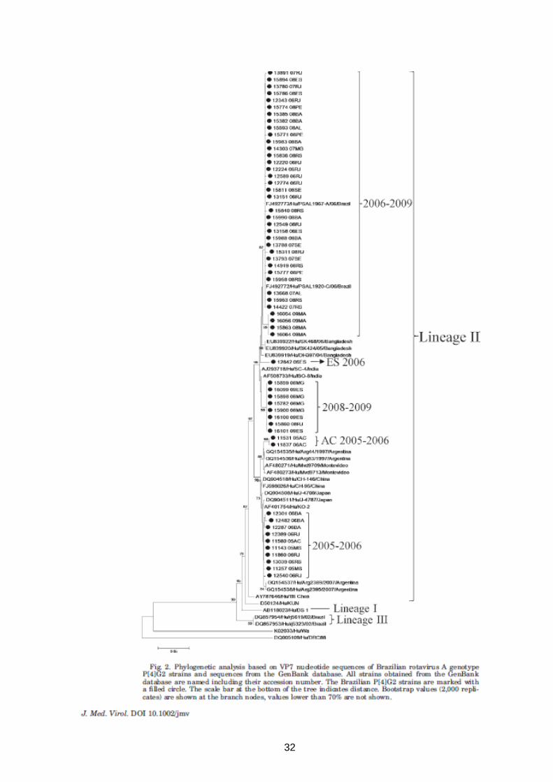
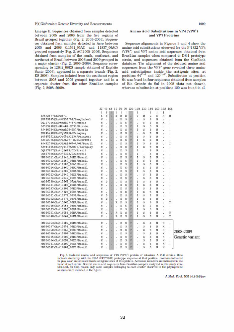
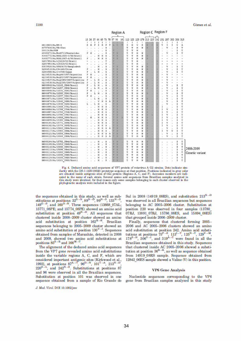
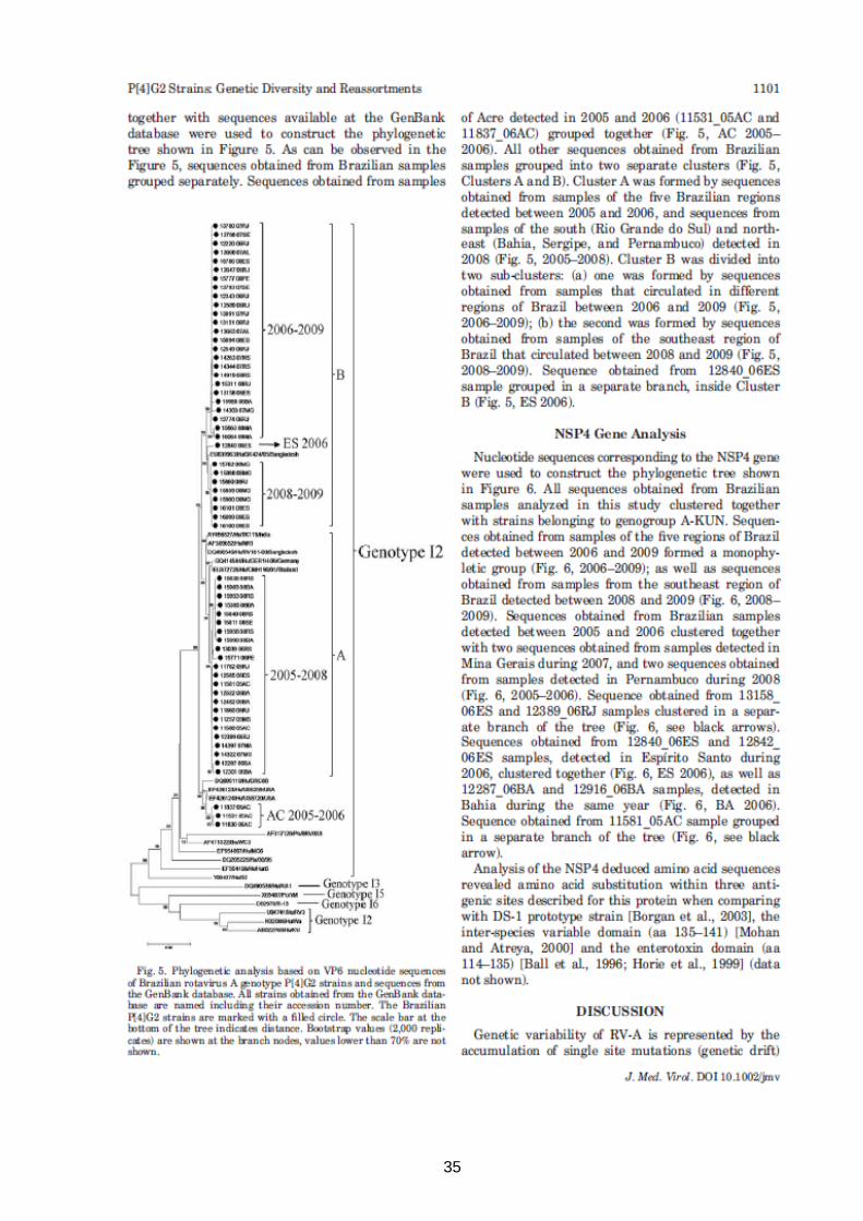
Artigo 1: Rotavirus A Genotype P[4]G2: Genetic Diversity and Reassortment Events Among
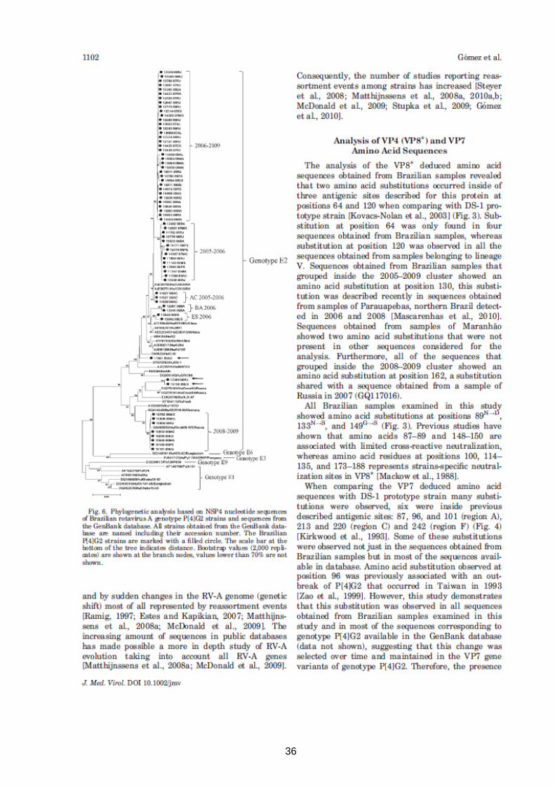
Strains Circulating in Brazil Between 2005 and 2009 (Artigo publicado).
Este artigo esta relacionado ao objetivo 1.
27
28

29
30
31
32
33
34
35
36
37
38
39
40
41
Artigo 2: Prevalence and genomic characterization of G2P[4] genotype after monovalent
vaccine introduction in Brazil. (Artigo em fase final de confecção)
Este artigo esta relacionado aos objetivos 1, 2, e 3.
42
Prevalence and genomic characterization of G2P[4] genotype after monovalent vaccine
introduction in Brazil.
Mariela Martínez Gómeza, Filipe Anibal Carvalho-Costa
a, Eduardo de Mello Volotão
a,
Tatiana Ludngren Rosea, Marcelle Figueira Marques da Silva
a, Ana Caroline Costa Sá
a, Mark
Zellerb, Elisabeth Heylen
b, Jelle Matthijnssens
b, José Paulo Gagliardi Leite
a*.
Laboratory of Comparative and Environmental Virology, Oswaldo Cruz Institute-Fiocruz,
Av. Brasil, 4365 – Pavilhão Hélio & Peggy Pereira 21040-360 Rio de Janeiro-RJ, Brazila;
Laboratory of Clinical and Epidemiological Virology, Department of Microbiology and
Immunology, Rega Institute for Medical Research, University of Leuven, Leuven, Belgiumb.
Running Head: Genetic characterization of Brazilian G2P[4] RVA strains
#Address correspondence to José Paulo Gagliardi Leite. E-mail: [email protected]
43
Abstract
A relationship between Rotarix® (RV1) introduction and the increased prevalence of group A
rotavirus (RVA) genotype G2P[4] in Brazil has been proposed. This study aims to: i) estimate
the prevalence of G2P[4] in Brazil between 2005 – 2011; ii) perform phylogenetic analyses of
G2P[4] strains detected in five Brazilian regions, and iii) assess the full genome genetic
background of selected strains. RVA surveillance was performed with fecal samples obtained
from patients with acute gastroenteritis. RVA was detected through polyacrylamide gel
electrophoresis and/or ELISA and genotyped through multiplex RT-PCR. G2P[4] strains
obtained from vaccinated and unvaccinated children were analyzed for the VP7 and VP8*
genes through genome sequencing, the complete genome characterization of selected strains
being also performed. G2P[4] detection rate among RVA-positive samples was 112/227
(49%) in 2006, 139/211 (66%) in 2007, 240/284 (85%) in 2008, 66/176 (37,5%) in 2009,
367/422 (87%) in 2010 and 75/149 (50%) in 2011. For both the VP7 and VP8* encoding
genes, 53 sequences (20 from vaccinated children) were analyzed and shared up to 99%
identity with other G2P[4] strains detected worldwide, different genetic variants being
observed. Most changes in antigenic epitopes in VP7 and VP8* have been maintained in the
Brazilian strains along the years, and all were present before RV1 introduction. Eleven strains
(4 from vaccinated, 7 from unvaccinated children) were characterized as G2-P[4]-I2-R2-C2-
M2-A2-N2-T2-E2-H2, sharing up to 99% of nucleotide identity with strains isolated
worldwide. Reassortments between human strains analyzed were observed. Data obtained in
the current study support the idea that other factors than RV1 introduction are responsible for
the increase prevalence of G2P[4] in Brazil.
44
Introduction
Group A rotaviruses (RVA) are responsible for approximately 453.000 deaths
worldwide each year among children under 5 years old (Tate et al., 2011, Wazny et al., 2013).
RVA belongs to the Reoviridae family and possesses segmented dsRNA genome (11 gene
segments) encoding six structural proteins (VP1-4, VP6-7) and six non-structural proteins
(NSP1-6). Based on their two outer capsid proteins, VP7 and VP4, RVA have been classified
into G (Glycoprotein) and P (Protease-sensitive) genotypes. Up-to-date there are 27 G types
and 37 P types reported (Matthijnssens et al., 2011; Trojnar et al., 2012). Although VP7 and
VP4 can segregate independently and theoretically lead a large number of G and P
combinations, six combinations (G1P[8], G2P[4], G3P[8], G4P[8], G9P[8] and G12P[8]) are
more frequent in human population representing more than 90% of the global burden of
rotavirus disease (Leite et al., 2008; Iturriza-Gómara et al., 2009; Banyai et al., 2012).
However, intra-genotype diversity and variation can be observed in both space and time
dimensions; in addition, periodic emergences of novel strains and/or uncommon combinations
have also been reported (Santos & Hoshino, 2005; Leite et al., 2008; Matthijnssens & Van
Ranst, 2012).
The binary genotype classification system has been extended to the entire genome to
better characterize RVA strains specifying the genotype of all 11 genome segments
(Matthijnssens et al., 2011). Based on this classification, most of the human RVA detected
worldwide possess either the Wa-like genotype constellation (I1-R1-C1-M1-A1-N1-T1-E1-
H1) or the DS-1-like genotype constellation (I2-R2-C2-M2-A2-N2-T2-E2-H2) also called as
genotype 1 and 2, respectively (Heiman et al., 2008; Matthijnssens et al., 2008; Mc Donald et
al., 2009; Matthijnssens & Van Ranst, 2012).
In April 2009, the World Health Organization (WHO) provided the recommendation
for introduction of anti-RVA vaccines in national immunization programs of developing
countries worldwide. Universal vaccination against rotavirus has been considered strategic in
order to reduce both mortality and hospitalization due to diarrheal diseases, along with other
45
measures such as oral rehydration, breastfeeding, zinc administration and improvement of
sanitation in developing countries (WHO 2013). Two live oral rotavirus vaccines, Rotarix®
(RV1; GlaxoSmithKline) and Rotateq® (RV5; Merck), with proven efficacy against severe
rotavirus disease are currently licensed. In Brazil, RV1, an attenuated human monovalent
(G1P[8]) vaccine was already included in the National Immunization Program since March
2006. An increase in the relative frequency of G2P[4] RVA occurred in Brazil in the
immediate post-vaccine years (Leite et al., 2008; Carvalho-Costa et al., 2011; Correia et al.,
2010; Dulgheroff et al., 2012). Some studies argued that both events, vaccine introduction and
increase prevalence of G2P[4], were associated suggesting that the selective pressure
generated by RV1 would somehow select this heterotypic strain among others (Nakagomi et
al., 2008; Gurgel et al., 2009). However, G2P[4] emerged in Brazil in 2005, shortly before the
beginning of universal mass vaccination with RV1. In addition, Latin American countries that
had not yet introduced universal vaccination also reported increased detection of G2P[4] at
that time (Amarilla et al., 2007; Ferrera et al., 2007; Patel et al., 2008). Despite these shifts in
genotype distribution, a significant reduction in the number of hospitalizations due to acute
diarrhea as well as in the frequency of RVA detection in hospitalized children with diarrhea
has been observed after the introduction of RV1 in Brazil (Sáfadi et al. 2010; Carvalho-Costa
et al. 2011).
The evolving prevalence rates of different RVA genotypes during and after vaccine
introduction, as well as their epidemiological characteristics are important issues and
represent a challenge to rotavirus vaccination programs. In this context, monitoring the
emergence of genotypes and escaping strains associated to breakthrough infections is
considered strategic in order to assess the impact of universal vaccination. In the current study
the prevalence of RVA genotype G2P[4] in Brazil between 2005 – 2011 have been assessed.
Furthermore, G2P[4] strains detected in the five regions of Brazil from 2005 to 2011 have
been analyzed for the VP7 and VP8* encoding genes, and compared with RVA strains
46
available in the Genbank database, including the RV1 vaccine. The complete genome
characterization of eleven G2P[4] Brazilian selected strains was also performed.
Material and Methods
Laboratory based RVA surveillance and specimen collection
Laboratory based RVA surveillance was performed with fecal samples obtained from
patients with acute gastroenteritis. Samples are sent to the Regional Rotavirus Reference
Laboratory - Laboratory of Comparative and Environmental Virology (RRRL-LVCA) by a
network of state public health laboratories. These labs, in turn, receive samples of health
centers and hospitals in the National Health System. We studied 9,073 fecal samples, from
which 1,813 were RVA-positive. Among RVA-positive patients, 361 (19.9%) were fully (two
doses) vaccinated children. Table 1 presents the year distribution, RVA detection rates and
prevalence of G2P[4] among RVA-positive patients.
Group A rotavirus detection and G- and P- genotyping
In order to detect RVA in stool samples, polyacrylamide gel electrophoresis and
enzyme immunoassay kits (EIARA, Biomanguinhos; Premier Rotaclone, Meridian
Bioscience, Inc.; Ridascreen, R-Biopharm), according to the manufacture’s protocols, were
used in 10% fecal suspensions in phosphate-buffered saline pH 7.4. Nucleic acid was
extracted from 10% fecal suspensions by the glass powder method described by Boom et al.
(1990), including modifications as described by Gómez et al. (2013) and the QIAamp Viral
RNA mini kit (Qiagen/Westburg, Leusden, The Netherlands) according to the manufacturer’s
instructions. In RVA-positive samples, the extracted RNA was reverse transcribed and G- and
P- genotyping was performed using semi-nested multiplex PCRs as previously described
(WHO/IVB/08.17, 2008). G and P genotypes were confirmed by sequencing for 57 samples
47
(23 were obtained from vaccinated children with RV1, and 34 from unvaccinated children)
and eleven selected strains representative from the surveillance period (2005-2011) were
investigated by whole genome analysis. The study strains were named according to the
guidelines of the Rotavirus Classification Working Group (RCWG) (Matthijnssens et al.,
2011).
Genome segments amplification and sequencing
The amplification of the eleven genome segments from selected strains were
performed using a OneStep RT-PCR Kit (QIAGEN) following manufacturer’s instructions
and amplifications conditions as described by Matthijnssens et al. (2010). The cDNA products
were resolved in agarose gels electrophoresis and purified using the Expo-SAP-IT PCR
Product Cleanup Kit (Affymetrix, Miles Rd, Cleveland, OH, USA). Sequencing was
performed with an ABI Prism Big Dye Terminator Cycle Sequencing Ready Reaction Kit™
on an ABI Prism 3730 Genetic Analyzer (Applied Biosystems, Foster City, CA, USA) at the
Instituto de Tecnologia em Imunobiológicos (Bio-Manguinhos / FIOCRUZ), and an ABI
Prism 3130 automated sequencer (Applied Biosystems, Foster City, CA, USA) at the Rega
Institute of Medical Research (University of Leuven, Belgium).
Sequence and phylogenetic analysis
Sequences obtained in the current study were deposited in the GenBank database
under the following accession numbers: XXXXX–XXXXX. Nucleotide blast analyses were
performed with obtained sequences and multiple sequence alignments were carried out using
the ClustalW program (Thompson et al., 1994). Phylogenetic analyses were constructed using
the Neighbor-Joining method with the Kimura-two parameter model in MEGA5.0 (Tamura et
al., 2011). The statistical significance of the branch was assessed by bootstrap resampling
analysis (2000 replicates).
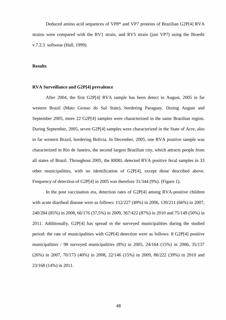
48
Deduced amino acid sequences of VP8* and VP7 proteins of Brazilian G2P[4] RVA
strains were compared with the RV1 strain, and RV5 strain (just VP7) using the Bioedit
v.7.2.3 softwear (Hall, 1999).
Results
RVA Surveillance and G2P[4] prevalence
After 2004, the first G2P[4] RVA sample has been detect in August, 2005 in far
western Brazil (Mato Grosso do Sul State), bordering Paraguay. During August and
September 2005, more 22 G2P[4] samples were characterized in the same Brazilian region.
During September, 2005, seven G2P[4] samples were characterized in the State of Acre, also
in far western Brazil, bordering Bolivia. In December, 2005, one RVA positive sample was
characterized in Rio de Janeiro, the second largest Brazilian city, which attracts people from
all states of Brazil. Throughout 2005, the RRRL detected RVA positive fecal samples in 33
other municipalities, with no identification of G2P[4], except those described above.
Frequency of detection of G2P[4] in 2005 was therefore 31/344 (9%). (Figure 1).
In the post vaccination era, detection rates of G2P[4] among RVA-positive children
with acute diarrheal disease were as follows: 112/227 (49%) in 2006, 139/211 (66%) in 2007,
240/284 (85%) in 2008, 66/176 (37,5%) in 2009, 367/422 (87%) in 2010 and 75/149 (50%) in
2011. Additionally, G2P[4] has spread in the surveyed municipalities during the studied
period: the rate of municipalities with G2P[4] detection were as follows: 8 G2P[4] positive
municipalities / 98 surveyed municipalities (8%) in 2005, 24/164 (15%) in 2006, 35/137
(26%) in 2007, 70/173 (40%) in 2008, 22/146 (15%) in 2009, 86/222 (39%) in 2010 and
23/168 (14%) in 2011.
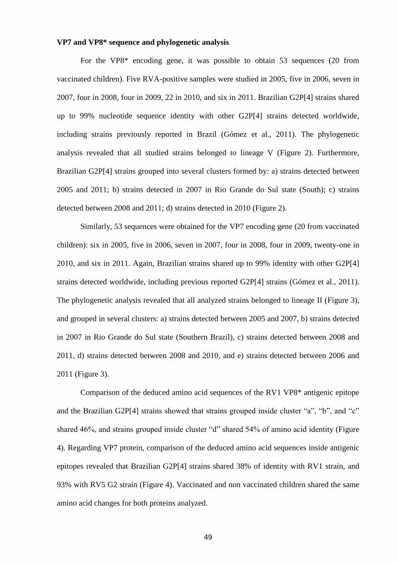
49
VP7 and VP8* sequence and phylogenetic analysis
For the VP8* encoding gene, it was possible to obtain 53 sequences (20 from
vaccinated children). Five RVA-positive samples were studied in 2005, five in 2006, seven in
2007, four in 2008, four in 2009, 22 in 2010, and six in 2011. Brazilian G2P[4] strains shared
up to 99% nucleotide sequence identity with other G2P[4] strains detected worldwide,
including strains previously reported in Brazil (Gómez et al., 2011). The phylogenetic
analysis revealed that all studied strains belonged to lineage V (Figure 2). Furthermore,
Brazilian G2P[4] strains grouped into several clusters formed by: a) strains detected between
2005 and 2011; b) strains detected in 2007 in Rio Grande do Sul state (South); c) strains
detected between 2008 and 2011; d) strains detected in 2010 (Figure 2).
Similarly, 53 sequences were obtained for the VP7 encoding gene (20 from vaccinated
children): six in 2005, five in 2006, seven in 2007, four in 2008, four in 2009, twenty-one in
2010, and six in 2011. Again, Brazilian strains shared up to 99% identity with other G2P[4]
strains detected worldwide, including previous reported G2P[4] strains (Gómez et al., 2011).
The phylogenetic analysis revealed that all analyzed strains belonged to lineage II (Figure 3),
and grouped in several clusters: a) strains detected between 2005 and 2007, b) strains detected
in 2007 in Rio Grande do Sul state (Southern Brazil), c) strains detected between 2008 and
2011, d) strains detected between 2008 and 2010, and e) strains detected between 2006 and
2011 (Figure 3).
Comparison of the deduced amino acid sequences of the RV1 VP8* antigenic epitope
and the Brazilian G2P[4] strains showed that strains grouped inside cluster “a”, “b”, and “c”
shared 46%, and strains grouped inside cluster “d” shared 54% of amino acid identity (Figure
4). Regarding VP7 protein, comparison of the deduced amino acid sequences inside antigenic
epitopes revealed that Brazilian G2P[4] strains shared 38% of identity with RV1 strain, and
93% with RV5 G2 strain (Figure 4). Vaccinated and non vaccinated children shared the same
amino acid changes for both proteins analyzed.
50
Whole-genome characterization of G2P[4] Brazilian strains
Eleven G2P[4] Brazilian strains, representative of the different clusters observed when
analyzing the VP7 and VP8* encoding genes, were selected to perform complete genome
analysis. Four of these strains were isolated from vaccinated children, and seven from
unvaccinated children, all hospitalized with acute gastroenteritis between 2005 and 2011.
The genotypes for each of the 11 RVA genome segments was assigned and all strains
belonged to G2-P[4]-I2-R2-C2-M2-A2-N2-T2-E2-H2 genetic background (Table 2;
Supplementary material Figures 1-9). The VP2 sequence of strain RVA/Human-
wt/BRA/SE19300/2010/G2P[4] could not be obtained. Although all strains have displayed the
same genomic constellation, it was possible to observed different genetic variants of G2P[4]
circulating during the study period in Brazil (Figures 2 and 3).
Analysis of NSP1-5, VP1-3, and VP6 sequences showed that G2P[4] Brazilian strains
shared between 87,3 - 100%, and 93 - 100% of nucleotide and amino acid identity values,
respectively. Nucleotide blast analysis revealed that Brazilian strains shared up to 99% of
nucleotide identity with strains isolated worldwide in the last decade.
Discussion
G2P[4] has been the most common genotype detected in Brazil after the onset of mass
vaccination (Gurgel et al., 2007, de Oliveira et al., 2008; Nakagomi et al., 2008; Leite et al.,
2008; Carvalho-Costa et al., 2009, 2011; da Silva Soares et al., 2013). The emergence of this
genotype has motivated a great discussion and a relationship between this phenomenon and
vaccination has been proposed (Gurgel et al., 2007; Nakagomi et al., 2008). The main reason
is the fact that the efficiency and effectiveness of the monovalent vaccine against G2P[4] is
relatively lower than against P[8] genotypes which are VP4-homotypic and belong to the
same genomic constellation (Wa-like) (Correia et al., 2010). In this context, it has been
51
proposed that universal vaccination would have exerted selective pressure on circulating
strains of RVA, selecting G2P[4] (Gurgel et al., 2007; Nakagomi et al., 2008).
The results of this study demonstrate that the detection rate of G2P[4] in RVA-positive
samples has been high in the years after the introduction of mass RV1 vaccination. With the
exception of 2009 this rate has been above 50%, reaching 87% in 2010. G2P[4] has also been
frequently detected in children vaccinated with RV1.
In this study, we examine in more detail the geographical location of G2P[4] positive
samples, trying to identify a route of spread of this genotype during its process of emergence
in Brazil. The frequency of G2P[4] detection between 2001 and 2003 was very low, so only
three positive samples could be identified in the RRRL monitoring data. In 2004, G2P[4] was
not characterized in any of the 651 RVA-positive fecal samples obtained from 2.554 children
with gastroenteritis from 11 Brazilian states. In the present survey, we confirmed that G2P[4]
was absent in the samples studied in the first semester of 2005. In this year, the first cities in
which G2P[4] was detected are located in western Brazil, on the border with Paraguay.
Throughout 2005, G2P[4] continued to be detected in this region and in Amazonian Brazil,
bordering Peru and Bolivia, being absent from the most populous Brazilian regions of the
Atlantic coast. Only in December 2005, G2P[4] was detected in the main Brazilian cities,
specifically in Rio de Janeiro. Universal vaccination against RVA began in March 2006 and,
starting this year, G2P[4] was early detected in all monitored regions, in a phase in which the
cohort of vaccinated children was still very limited.
It has been suggested that specific vaccines may select escape genotypes, including
long-term effects of weak selective pressures (Pérez-Sautu et al., 2011). If so,
emerging strains resulting from evolutionary pressure imposed by vaccines may gradually
substitute previous genotypes (Streck et al., 2013). In this scenario, the vaccine can present
reduced performance. In this context, one would expect that the phenomenon of selection of
strains would be accompanied by a reduction in the genetic variability of RVA and that a
52
genetic distance of circulating strains in the selection scenario in relation to other regions
would arise (Hoffmann et al. 2008). The analysis of the genetic and antigenic backgrounds of
G2P[4] strains identified throughout the process of implementation of mass immunization
with RV1 may be useful to clarify its potential impact on the distribution of genotypes which
occurred between 2005 and 2011 in Brazil.
Our data suggest that the G2P[4] strains circulating in Brazil during implementation of
mass immunization with RV1 are not separated into genetic clusters defined by pre and post
vaccination periods and maintains a close relationship with G2P[4] strains circulating in other
countries that had not yet implemented universal vaccination against RVA. Similarly, G2P[4]
strains identified in vaccinated and unvaccinated children grouped together in the same
genetic clusters, with no significant differences in the sequences of the genes encoding VP7
and VP8*. Moreover, phylogenetic analysis revealed that contemporary G2P[4] strains
(circulating from 2005 to 2011) belonged to a novel lineage constellation, distinct from the
reference strain DS-1 and the SC2-9 G2-reassortant strain of RV5, as previously reported
(Doan et al., 2011; Gómez et al., 2011; Do et al., 2013; Giammanco et al., 2013). Analysis of
the genomic background of Brazilian G2P[4] RVA strains revealed a complete DS-1-like
background, and different genetic variants were observed circulating among Brazilian regions
(Supplementary material Figures 1-9).
In addition, comparison of the RV1 amino acid sequences inside previous described
antigenic epitopes in VP7 and VP8*, with Brazilian G2P[4] strains revealed that most of the
changes observed have been maintained in the Brazilian strains along the years, and all were
present before RV1 introduction.
In conclusion, data support the idea that other factors than RV1 introduction are
responsible for the increase prevalence of G2P[4] in Brazil. It has been proposed that G2P[4]
reemerges periodically in 10-years cycles (Bishop et al., 1991). Leite et al. (2008) reported a
wave of G2P[4] circulation in the beginning of the 1990s, near 10 years before reemergence
53
of this genotype in 2005. In adition, the long period of little or no circulation of this genotype
previous to RV1 introduction would have create favorable conditions for the accumulation of
immunological susceptible individuals (Carvalho-Costa et al., 2009; Assis et al., 2013). This
fact, together with the genetic diversity found in different geographical regions throughout the
years before, during and after the introduction of the vaccine might explained the high
prevalence of genotype G2P[4] in Brazil since 2005.
54
ACKNOWLEDGMENTS
This research was supported by funds from the Program of Research Excellence
(PROEP - IOC/Fiocruz/CNPq), the National Council for Scientific and Technological
Development (CNPq), project PAPES VI/FIOCRUZ – CNPq, Oswaldo Cruz Institute (IOC /
FIOCRUZ), Brazilian Federal Agency for Support and Evaluation of Graduate Education
(CAPES) – project CAPES-MERCOSUL PPCP 023/2011, the General Coordination of
Public Health Laboratories – Secretary of Health Surveillance (CGLAB/SVS), and Carlos
Chagas Filho Foundation for Research Support of Rio de Janeiro State (FAPERJ). M.Z. was
supported by the Institute for the Promotion of Innovation through Science and Technology in
Flanders (IWT Vlaanderen). The authors would like to thank the Secretary of Public Health
State of Bahia, Pernambuco and Rio de Janeiro. We would also like to thanks to Alexandre
Madi Fialho, Juliana Andrade, and Rosane Maria Santos de Assis for technical assistance.
Mariela Martínez Gómez is a PhD student of the Molecular and Celular Post-Graduation
Program at the Oswaldo Cruz Institute, FIOCRUZ, Rio de Janeiro, Brazil, and was supported
by the Carlos Chagas Filho Foundation for Research Support of Rio de Janeiro State
(FAPERJ) with a doctorate sandwich scholarship, and the “Bolsa Nota 10” scholarship.
55
References
Amarilla A, Espínola EE, Galeano ME, Fariña N, Russomando G, Parra GI. Rotavirus
infection in the Paraguayan population from 2004 to 2005: high incidence of rotavirus strains
with short electropherotype in children and adults. Med Sci Monit. 2007 Jul;13(7):CR333-7.
Assis AS, Valle DA, Antunes GR, Tibiriça SH, Assis RM, Leite JP et al. Rotavirus
epidemiology before and after vaccine introduction. J Pediatr (Rio J). 2013 Sep-
Oct;89(5):470-6.
Bányai K, László B, Duque J, Steele AD, Nelson EA, Gentsch JR et al. Systematic review of
regional and temporal trends in global rotavirus strain diversity in the pre rotavirus vaccine
era: insights for understanding the impact of rotavirus vaccination programs. Vaccine. 2012
Apr 27;30 Suppl 1:A122-30.
Bishop RF, Unicomb LE, Barnes GL. Epidemiology of rotavirus serotypes in Melbourne,
Australia, from 1973 to 1989. J Clin Microbiol. 1991 May;29(5):862-8.
Carvalho-Costa FA, Volotão Ede M, de Assis RM, Fialho AM, de Andrade Jda S, Rocha LN
et al. Laboratory-based rotavirus surveillance during the introduction of a vaccination
program, Brazil, 2005–2009. Pediatr Infect Dis J. 2011 Jan;30(1 Suppl):S35-41.
Correia JB, Patel MM, Nakagomi O, Montenegro FM, Germano EM, Correia NB et al.
Effectiveness of monovalent rotavirus vaccine (Rotarix) against severe diarrhea caused by
serotypically unrelated G2P[4] strains in Brazil. J Infect Dis. 2010 Feb 1;201(3):363-9.
Do LP, Nakagomi T, Doan YH, Kitahori Y, Nakagomi O. Molecular evolution of the VP7
gene of Japanese G2 rotaviruses before vaccine introduction. Arch Virol. 2013 Aug 8. [Epub
ahead of print].
Doan YH, Nakagomi T, Cunliffe NA, Pandey BD, Sherchand JB, Nakagomi O. The
occurrence of amino acid substitutions D96N and S242N in VP7 of emergent G2P[4]
rotaviruses in Nepal in 2004–2005: a global and evolutionary perspective. Arch Virol. 2011
Nov;156(11):1969-78.
56
Dulgheroff AC, Figueiredo EF, Moreira LP, Moreira KC, Moura LM, Gouvêa VS et al.
Distribution of rotavirus genotypes after vaccine introduction in the Triângulo Mineiro region
of Brazil: 4-Year follow-up study. J Clin Virol. 2012 Sep;55(1):67-71.
Ferrera A, Quan D, Espinoza F. Increased prevalence of genotype G2P(4) among children
with rotavirus-associated gastroenteritis in Honduras. The 17th European Congress of Clinical
Microbiology and Infectious Diseases ICC, Munich, Germany, 2007.
Giammanco GM, Bonura F, Zeller M, Heylen E, Van Ranst M, Martella V et al. Evolution of
DS-1-like human G2P[4] rotaviruses assessed by complete genome analyses. J Gen Virol.
2014 Jan;95 Pt 1:91-109.
Gómez MM, de Mendonça MC, Volotão Ede M, Tort LF, da Silva MF, Cristina J et al.
Rotavirus A genotype P[4]G2: genetic diversity and reassortment events among strains
circulating in Brazil between 2005 and 2009. J Med Virol. 2011 Jun;83(6):1093-106.
Gurgel RQ, Cuevas LE, Vieira SC, Barros VC, Fontes PB, Salustino EF et al. Predominance
of rotavirus P[4]G2 in a vaccinated population, Brazil. Emerg Infect Dis. 2007
Oct;13(10):1571-3.
Gurgel RG, Bohland AK, Vieira SC, Oliveira DM, Fontes PB, Barros VF et al. Incidence of
rotavirus and all-cause diarrhea in northeast Brazil following the introduction of a national
vaccination program. Gastroenterology. 2009 Dec;137(6):1970-5.
Hall TA. BioEdit: a user–friendly biological sequence alignment editor and analysis program
for Windows 95/98/NT. Nucl Acids Symp Ser. 1999;41:95 – 98.
Heiman EM, McDonald SM, Barro M, Taraporewala ZF, Bar-Magen T, Patton JT. Group A
human rotavirus genomics: evidence that gene constellations are influenced by viral protein
interactions. J Virol. 2008 Nov;82(22):11106-16.
Hoffmann D, Seebach J, Cosma A, Goebel FD, Strimmer K, Schätzl HM et al. Therapeutic
vaccination reduces HIV sequence variability. FASEB J. 2008 Feb;22(2):437-44.
57
Iturriza-Gómara M, Dallman T, Bányai K, Böttiger B, Buesa J, Diedrich S et al. Rotavirus
surveillance in europe, 2005-2008: web-enabled reporting and real-time analysis of
genotyping and epidemiological data. J Infect Dis. 2009 Nov 1;200 Suppl 1:S215-21.
J.P. Leite, F.A. Carvalho-Costa, A.C. Linhares. Group A rotavirus genotypes and the ongoing
Brazilian experience: a review. Mem Inst Oswaldo Cruz. 2008 Dec;103(8):745-53.
Matthijnssens J, Ciarlet M, Heiman E, Arijs I, Delbeke T, McDonald SM et al. Full genome-
based classification of rotaviruses reveals a common origin between human Wa-Like and
porcine rotavirus strains and human DS-1-like and bovine rotavirus strains. J Virol. 2008
Apr;82(7):3204-19.
Matthijnssens J, Ciarlet M, McDonald SM, Attoui H, Bányai K, Brister JR et al. Uniformity
of rotavirus strain nomenclature proposed by the Rotavirus Classification Working Group
(RCWG). Arch Virol. 2011 Aug;156(8):1397-413.
Matthijnssens J, Van Ranst M. Genotype constellation and evolution of group A rotaviruses
infecting humans, Cur Opin Virol. 2012 Aug: 2(4):426-433
McDonald SM, Matthijnssens J, McAllen JK, Hine E, Overton L, Wang S et al. Evolutionary
dynamics of human rotaviruses: balancing reassortment with preferred genome constellations.
PLoS Pathog. 2009 Oct;5(10):e1000634.
Nakagomi T, Cuevas LE, Gurgel RG, Elrokhsi SH, Belkhir YA, Abugalia M et al. Apparent
extinction of non-G2 rotavirus strains from circulation in Recife, Brazil, after the introduction
of rotavirus vaccine. Arch Virol. 2008;153(3):591-3.
Patel MM, de Oliveira LH, Bispo AM, Gentsch J, Parashar UD. Rotavirus P[4]G2 in a
vaccinated population, Brazil. Emerg Infect Dis. 2008 May;14(5):863-5.
Pérez-Sautu U, Costafreda MI, Caylà J, Tortajada C, Lite J, Bosch A et al. Hepatitis a virus
vaccine escape variants and potential new serotype emergence. Emerg Infect Dis. 2011
Apr;17(4):734-7.
58
Sáfadi MA, Berezin EN, Munford V, Almeida FJ, de Moraes JC, Pinheiro CF et al. Hospital-
based surveillance to evaluate the impact of rotavirus vaccination in São Paulo, Brazil. Pediatr
Infect Dis J. 2010 Nov;29(11):1019-22.
Santos N, Hoshino Y. Global distribution of rotavirus serotypes/genotypes and its implication
for the development and implementation of an effective rotavirus vaccine. Rev Med Virol.
2005 Jan-Feb;15(1):29-56.
Streck AF, Homeier T, Foerster T, Truyen U. Population dynamics and in vitro antibody
pressure of porcine parvovirus indicate a decrease in variability. J Gen Virol. 2013 Sep;94(Pt
9):2050-5.
Tamura K, Peterson D, Peterson N, Stecher G, Nei M, Kumar S. MEGA5: molecular
evolutionary genetics analysis using maximum likelihood, evolutionary distance, and
maximum parsimony methods. Mol Biol Evol. 2011 Oct;28(10):2731-9.
J.D. Thompson, D.G. Higgins, T.J. Gibson. CLUSTAL W improving the sensitivity of
progressive multiple sequence alignment through sequence weighting position-specific gap
penalties and weight matrix choice. Nucleic Acids Res. 1994 Nov 11;22(22):4673-80.
Trojnar E, Sachsenröder J, Twardziok S, Reetz J, Otto PH, Johne R. Identification of an avian
group A rotavirus containing a novel VP4 gene of close relationship to those of mammalian
rotaviruses. J Gen Virol. 2013 Jan;94(Pt 1):136-42.
WHO/IVB/08.17. 2008. Manual of rotavirus detection and characterization methods.
Wazny K, Zipursky A, Black R, Curtis V, Duggan C, Guerrant R et al. Setting research
priorities to reduce mortality and morbidity of childhood diarrhoeal disease in the next 15
years. PLoS Med. 2013;10(5):e1001446.
WHO. Rotavirus vaccines WHO position paper: January 2013 - Recommendations. Vaccine.
2013 Jun 5.
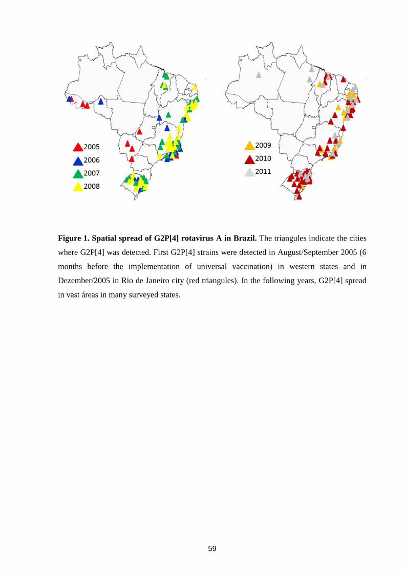
59
Figure 1. Spatial spread of G2P[4] rotavirus A in Brazil. The triangules indicate the cities
where G2P[4] was detected. First G2P[4] strains were detected in August/September 2005 (6
months before the implementation of universal vaccination) in western states and in
Dezember/2005 in Rio de Janeiro city (red triangules). In the following years, G2P[4] spread
in vast áreas in many surveyed states.
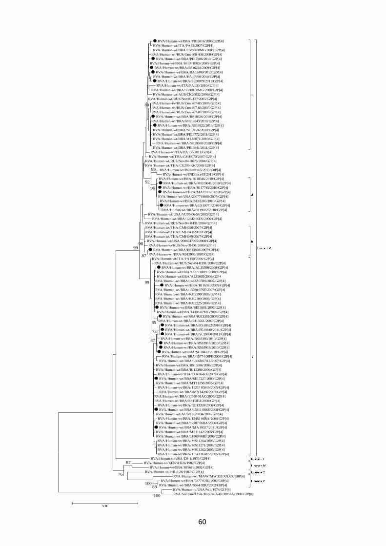
60
RVA/Human-wt/BRA/PB16816/2009/G2P[4]
RVA/Human-wt/ITA/PA83/2007/G2P[4]
RVA/Human-wt/BRA/15859 08MG/2008/G2P[4]
RVA/Human-wt/RUS/Omsk08-408/2008/G2P[4]
RVA/Human-wt/BRA/PE17886/2010/G2P[4]
RVA/Human-wt/BRA/16100 09ES/2009/G2P[4]
RVA/Human-wt/BRA/ES16238/2009/G2P[4]
RVA/Human-wt/BRA/BA18480/2010/G2P[4]
RVA/Human-wt/BRA/BA17990/2010/G2P[4]
RVA/Human-wt/BRA/SE20079/2011/G2P[4]
RVA/Human-wt/ITA/PA130/2010/G2P[4]
RVA/Human-wt/BRA/15900 08MG/2008/G2P[4]
RVA/Human-wt/AUS/CK20032/2006/G2P[4]
RVA/Human-wt/RUS/Nov05-137/2005/G2P[4]
RVA/Human-tw/RUS/Omsk07-83/2007/G2P[4]
RVA/Human-wt/RUS/Omsk07-83/2007/G2P[4]
RVA/Human-wt/RUS/Omsk07-87/2007/G2P[4]
RVA/Human-wt/BRA/RS18326/2010/G2P[4]
RVA/Human-wt/BRA/MG19243/2010/G2P[4]
RVA/Human-wt/BRA/RS18922/2010/G2P[4]
RVA/Human-wt/BRA/SC18536/2010/G2P[4]
RVA/Human-wt/BRA/PE19772/2011/G2P[4]
RVA/Human-wt/BRA/AL18871/2010/G2P[4]
RVA/Human-wt/BRA/SE19300/2010/G2P[4]
RVA/Human-wt/BRA/PE19841/2011/G2P[4]
RVA/Human-wt/ITA/PA133/2011/G2P[4]
RVA/Human-wt/THA/CMH070/2007/G2P[4]
RVA/Human-wt/RUS/Nov04-H676/2004/G2P[4]
RVA/Human-wt/THA/CU209-KK/2008/G2P[4]
RVA/Human-wt/IND/mcs65/2011/G8P[4]
RVA/Human-wt/IND/mcs63/2011/G8P[4]
RVA/Human-wt/BRA/RJ18346/2010/G2P[4]
RVA/Human-wt/BRA/MG19041/2010/G2P[4]
RVA/Human-wt/BRA/RJ17745/2010/G2P[4]
RVA/Human-wt/BRA/MA19112/2010/G2P[4]
RVA/Human-wt/USA/2007719869/2007/G2P[4]
RVA/Human-wt/BRA/SE18265/2010/G2P[4]
RVA/Human-wt/BRA/ES19071/2010/G2P[4]
RVA/Human-wt/BRA/ES19072/2010/G2P[4]
RVA/Human-wt/USA/VU05-06-54/2005/G2P[4]
RVA/Human-wt/BRA/12842 06ES/2006/G2P[4]
RVA/Human-wt/RUS/Nov04-H431/2004/G2P[4]
RVA/Human-wt/THA/CMH028/2007/G2P[4]
RVA/Human-wt/THA/CMH043/2007/G2P[4]
RVA/Human-wt/THA/CMH049/2007/G2P[4]
RVA/Human-wt/USA/2008747095/2008/G2P[4]
RVA/Human-xx/RUS/Nov09-D1/2009/G2P[4]
RVA/Human-wt/BRA/RS13898/2007/G2P[4]
RVA/Human-wt/BRA/RS13903/2007/G2P[4]
RVA/Human-wt/ITA/PA150/2006/G2P[4]
RVA/Human-wt/RUS/Nov04-H391/2004/G2P[4]
RVA/Human-wt/BRA/AL15598/2008/G2P[4]
RVA/Human-wt/BRA/15777 08PE/2008/G2P[4]
RVA/Human-wt/BRA/AL15603/2008/G2P4
RVA/Human-wt/BRA/14422 07RS/2007/G2P[4]
RVA/Human-wt/BRA/RJ16581/2009/G2P[4]
RVA/Human-wt/BRA/13788 07SE/2007/G2P[4]
RVA/Human-wt/BRA/RJ12398/2006/G2P[4]
RVA/Human-wt/BRA/RJ12209/2006/G2P[4]
RVA/Human-wt/BRA/RJ12225/2006/G2P[4]
RVA/Human-wt/BRA/SE13801/2007/G2P[4]
RVA/Human-wt/BRA/14303 07MG/2007/G2P[4]
RVA/Human-wt/BRA/RJ13393/2007/G2P[4]
RVA/Human-wt/BRA/RJ13561/2007/G2P[4]
RVA/Human-wt/BRA/RS18622/2010/G2P[4]
RVA/Human-wt/BRA/PE19840/2011/G2P[4]
RVA/Human-wt/BRA/SC19868/2011/G2P[4]
RVA/Human-wt/BRA/RS18380/2010/G2P[4]
RVA/Human-wt/BRA/RS18917/2010/G2P[4]
RVA/Human-wt/BRA/RS18918/2010/G2P[4]
RVA/Human-wt/BRA/SC18412/2010/G2P[4]
RVA/Human-wt/BRA/15774 08PE/2008/G2P[4]
RVA/Human-wt/BRA/13668 07AL/2007/G2P[4]
RVA/Human-wt/BRA/RS13086/2006/G2P[4]
RVA/Human-wt/BRA/RJ12389/2006/G2P[4]
RVA/Human-wt/THA/CU436-KK/2009/G2P[4]
RVA/Human-wt/BRA/SE17227/2009/G2P[4]
RVA/Human-wt/BRA/MT11258/2005/G2P[4]
RVA/Human-wt/BRA/11257 05MS/2005/G2P[4]
RVA/Human-wt/BRA/MA14286/2007/G2P[4]
RVA/Human-wt/BRA/11580 05AC/2005/G2P[4]
RVA/Human-wt/BRA/RS15851/2008/G2P[4]
RVA/Human-wt/BRA/RO13269/2006/G2P[4]
RVA/Human-wt/BRA/15811 08SE/2008/G2P[4]
RVA/Human-wt/AUS/CK20034/2006/G2P[4]
RVA/Human-wt/BRA/12482 06BA/2006/G2P[4]
RVA/Human-wt/BRA/12287 06BA/2006/G2P[4]
RVA/Human-wt/BRA/MA19557/2011/G2P[4]
RVA/Human-wt/BRA/MS11142/2005/G2P[4]
RVA/Human-wt/BRA/11860 06RJ/2006/G2P[4]
RVA/Human-wt/BRA/MS11264/2005/G2P[4]
RVA/Human-wt/BRA/MS11271/2005/G2P[4]
RVA/Human-wt/BRA/MS11262/2005/G2P[4]
RVA/Human-wt/BRA/11143 05MS/2005/G2P[4]
RVA/Human-tc/USA/DS-1/1976/G2P[4]
RVA/Human-tc/KEN/AK26/1982/G2P[4]
RVA/Human-wt/BRA/RJ5619/2002/G2P[4]
RVA/Human-tc/PHL/L26/1987/G12P[4]
RVA/Human-wt/MAW/MW333/XXXX/G8P[4]
RVA/Human-wt/BRA/5877 02RJ/2002/G8P[4]
RVA/Human-wt/BRA/5664 02RJ/2002/G8P[4]
RVA/Human-tc/USA/Wa/1974/G1P[8]
RVA/Vaccine/USA/Rotarix-A41CB052A/1988/G1P[8]100
88100
87
76
99
96
92
87
80
9899
73
85
87
99
61
Figure 2. Phylogenetic analysis based on VP8* nucleotide sequences from Brazilian
G2P[4] strains detected between 2005 and 2011, and rotavirus A strains available at the
GenBank database. Strains marked with a filled circle represent vaccinated RVA strains.
Boostrap values under 70% are not shown. G2 Lineages are shown at the right side.
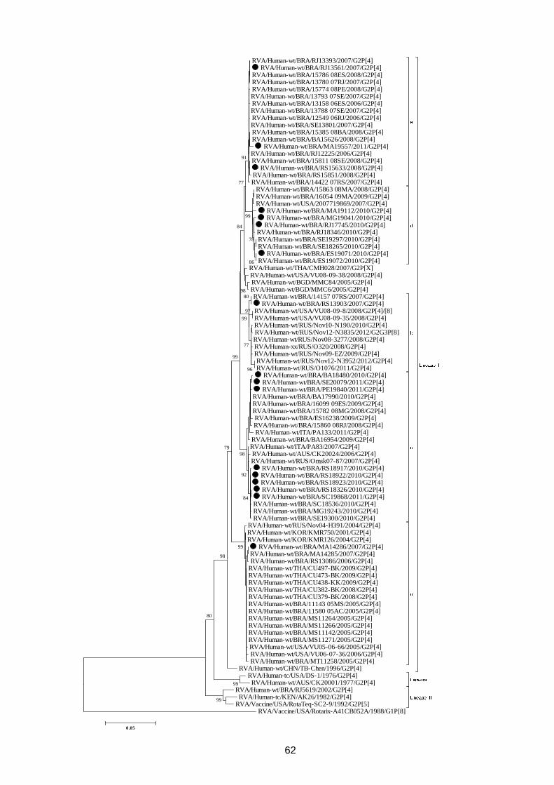
62
RVA/Human-wt/BRA/RJ13393/2007/G2P[4] RVA/Human-wt/BRA/RJ13561/2007/G2P[4]
RVA/Human-wt/BRA/15786 08ES/2008/G2P[4] RVA/Human-wt/BRA/13780 07RJ/2007/G2P[4] RVA/Human-wt/BRA/15774 08PE/2008/G2P[4] RVA/Human-wt/BRA/13793 07SE/2007/G2P[4] RVA/Human-wt/BRA/13158 06ES/2006/G2P[4] RVA/Human-wt/BRA/13788 07SE/2007/G2P[4] RVA/Human-wt/BRA/12549 06RJ/2006/G2P[4] RVA/Human-wt/BRA/SE13801/2007/G2P[4] RVA/Human-wt/BRA/15385 08BA/2008/G2P[4] RVA/Human-wt/BRA/BA15626/2008/G2P[4]
RVA/Human-wt/BRA/MA19557/2011/G2P[4] RVA/Human-wt/BRA/RJ12225/2006/G2P[4] RVA/Human-wt/BRA/15811 08SE/2008/G2P[4]
RVA/Human-wt/BRA/RS15633/2008/G2P[4] RVA/Human-wt/BRA/RS15851/2008/G2P[4] RVA/Human-wt/BRA/14422 07RS/2007/G2P[4]
RVA/Human-wt/BRA/15863 08MA/2008/G2P[4] RVA/Human-wt/BRA/16054 09MA/2009/G2P[4] RVA/Human-wt/USA/2007719869/2007/G2P[4]
RVA/Human-wt/BRA/MA19112/2010/G2P[4] RVA/Human-wt/BRA/MG19041/2010/G2P[4]
RVA/Human-wt/BRA/RJ17745/2010/G2P[4] RVA/Human-wt/BRA/RJ18346/2010/G2P[4] RVA/Human-wt/BRA/SE19297/2010/G2P[4] RVA/Human-wt/BRA/SE18265/2010/G2P[4]
RVA/Human-wt/BRA/ES19071/2010/G2P[4] RVA/Human-wt/BRA/ES19072/2010/G2P[4]
RVA/Human-wt/THA/CMH028/2007/G2P[X] RVA/Human-wt/USA/VU08-09-38/2008/G2P[4] RVA/Human-wt/BGD/MMC84/2005/G2P[4] RVA/Human-wt/BGD/MMC6/2005/G2P[4] RVA/Human-wt/BRA/14157 07RS/2007/G2P[4]
RVA/Human-wt/BRA/RS13903/2007/G2P[4] RVA/Human-wt/USA/VU08-09-8/2008/G2P[4]/[8] RVA/Human-wt/USA/VU08-09-35/2008/G2P[4] RVA/Human-wt/RUS/Nov10-N190/2010/G2P[4] RVA/Human-wt/RUS/Nov12-N3835/2012/G2G3P[8] RVA/Human-wt/RUS/Nov08-3277/2008/G2P[4] RVA/Human-xx/RUS/O320/2008/G2P[4] RVA/Human-wt/RUS/Nov09-EZ/2009/G2P[4] RVA/Human-wt/RUS/Nov12-N3952/2012/G2P[4] RVA/Human-wt/RUS/O1076/2011/G2P[4]
RVA/Human-wt/BRA/BA18480/2010/G2P[4] RVA/Human-wt/BRA/SE20079/2011/G2P[4] RVA/Human-wt/BRA/PE19840/2011/G2P[4]
RVA/Human-wt/BRA/BA17990/2010/G2P[4] RVA/Human-wt/BRA/16099 09ES/2009/G2P[4] RVA/Human-wt/BRA/15782 08MG/2008/G2P[4] RVA/Human-wt/BRA/ES16238/2009/G2P[4] RVA/Human-wt/BRA/15860 08RJ/2008/G2P[4] RVA/Human-wt/ITA/PA133/2011/G2P[4]
RVA/Human-wt/BRA/BA16954/2009/G2P[4] RVA/Human-wt/ITA/PA83/2007/G2P[4] RVA/Human-wt/AUS/CK20024/2006/G2P[4] RVA/Human-wt/RUS/Omsk07-87/2007/G2P[4]
RVA/Human-wt/BRA/RS18917/2010/G2P[4] RVA/Human-wt/BRA/RS18922/2010/G2P[4] RVA/Human-wt/BRA/RS18923/2010/G2P[4] RVA/Human-wt/BRA/RS18326/2010/G2P[4] RVA/Human-wt/BRA/SC19868/2011/G2P[4]
RVA/Human-wt/BRA/SC18536/2010/G2P[4] RVA/Human-wt/BRA/MG19243/2010/G2P[4] RVA/Human-wt/BRA/SE19300/2010/G2P[4]
RVA/Human-wt/RUS/Nov04-H391/2004/G2P[4] RVA/Human-wt/KOR/KMR750/2001/G2P[4] RVA/Human-wt/KOR/KMR126/2004/G2P[4]
RVA/Human-wt/BRA/MA14286/2007/G2P[4] RVA/Human-wt/BRA/MA14285/2007/G2P[4] RVA/Human-wt/BRA/RS13086/2006/G2P[4]
RVA/Human-wt/THA/CU497-BK/2009/G2P[4] RVA/Human-wt/THA/CU473-BK/2009/G2P[4] RVA/Human-wt/THA/CU438-KK/2009/G2P[4] RVA/Human-wt/THA/CU382-BK/2008/G2P[4] RVA/Human-wt/THA/CU379-BK/2008/G2P[4] RVA/Human-wt/BRA/11143 05MS/2005/G2P[4] RVA/Human-wt/BRA/11580 05AC/2005/G2P[4] RVA/Human-wt/BRA/MS11264/2005/G2P[4] RVA/Human-wt/BRA/MS11266/2005/G2P[4] RVA/Human-wt/BRA/MS11142/2005/G2P[4] RVA/Human-wt/BRA/MS11271/2005/G2P[4] RVA/Human-wt/USA/VU05-06-66/2005/G2P[4] RVA/Human-wt/USA/VU06-07-36/2006/G2P[4] RVA/Human-wt/BRA/MT11258/2005/G2P[4]
RVA/Human-wt/CHN/TB-Chen/1996/G2P[4] RVA/Human-tc/USA/DS-1/1976/G2P[4]
RVA/Human-wt/AUS/CK20001/1977/G2P[4] RVA/Human-wt/BRA/RJ5619/2002/G2P[4] RVA/Human-tc/KEN/AK26/1982/G2P[4]
RVA/Vaccine/USA/RotaTeq-SC2-9/1992/G2P[5] RVA/Vaccine/USA/Rotarix-A41CB052A/1988/G1P[8]
99
99
80
99
98
96
79
97
86
78
77
80
99
99
84
92
98
99
98
84
77
91
0.05
63
Figure 3. Phylogenetic analysis based on VP7 nucleotide sequences from Brazilian
G2P[4] strains detected between 2005 and 2011, and rotavirus A strains available at the
GenBank database. Strains marked with a filled circle represent vaccinated RVA strains.
Boostrap values under 70% are not shown. G2 Lineages are shown at the right side.
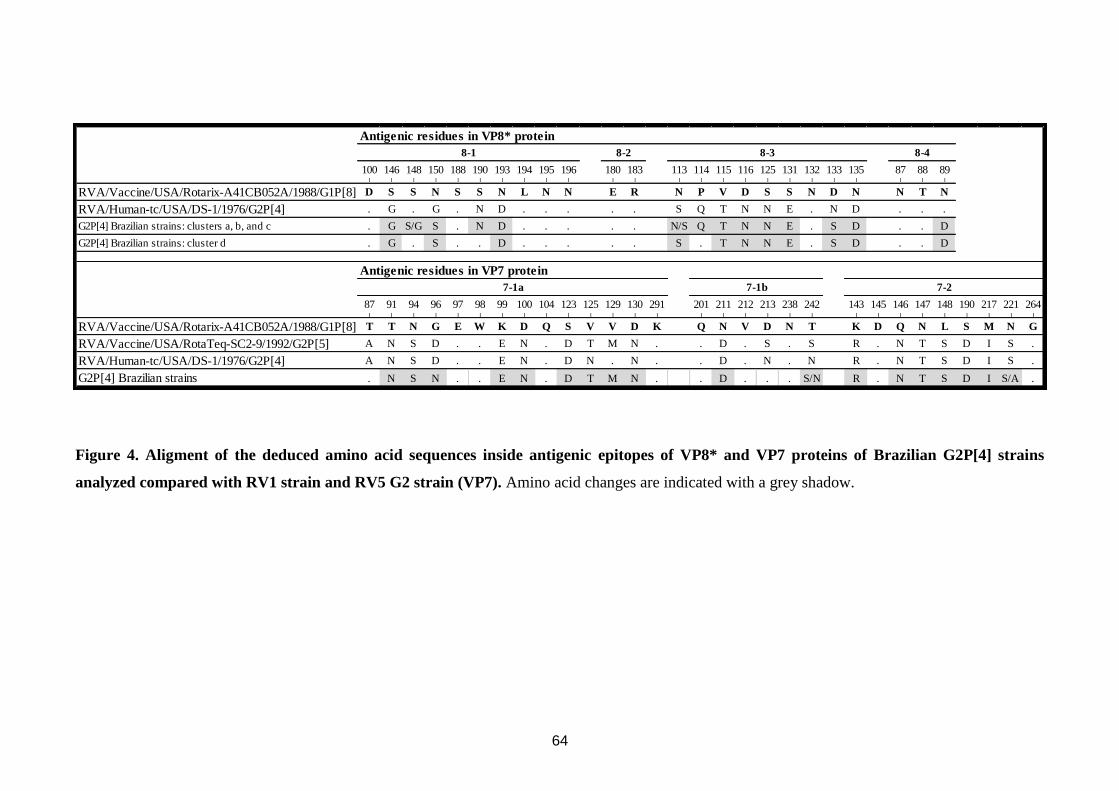
64
Figure 4. Aligment of the deduced amino acid sequences inside antigenic epitopes of VP8* and VP7 proteins of Brazilian G2P[4] strains
analyzed compared with RV1 strain and RV5 G2 strain (VP7). Amino acid changes are indicated with a grey shadow.
100 146 148 150 188 190 193 194 195 196 180 183 113 114 115 116 125 131 132 133 135 87 88 89| | | | | | | | | | | | | | | | | | | | | | | |
RVA/Vaccine/USA/Rotarix-A41CB052A/1988/G1P[8] D S S N S S N L N N E R N P V D S S N D N N T N
RVA/Human-tc/USA/DS-1/1976/G2P[4] . G . G . N D . . . . . S Q T N N E . N D . . .
G2P[4] Brazilian strains: clusters a, b, and c . G S/G S . N D . . . . . N/S Q T N N E . S D . . D
G2P[4] Brazilian strains: cluster d . G . S . . D . . . . . S . T N N E . S D . . D
87 91 94 96 97 98 99 100 104 123 125 129 130 291 201 211 212 213 238 242 143 145 146 147 148 190 217 221 264| | | | | | | | | | | | | | | | | | | | | | | | | | | | |
RVA/Vaccine/USA/Rotarix-A41CB052A/1988/G1P[8] T T N G E W K D Q S V V D K Q N V D N T K D Q N L S M N G
RVA/Vaccine/USA/RotaTeq-SC2-9/1992/G2P[5] A N S D . . E N . D T M N . . D . S . S R . N T S D I S .
RVA/Human-tc/USA/DS-1/1976/G2P[4] A N S D . . E N . D N . N . . D . N . N R . N T S D I S .
G2P[4] Brazilian strains . N S N . . E N . D T M N . . D . . . S/N R . N T S D I S/A .
Antigenic residues in VP7 protein
7-1a 7-1b 7-2
Antigenic residues in VP8* protein
8-1 8-2 8-3 8-4
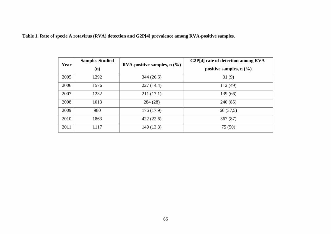
65
Table 1. Rate of specie A rotavirus (RVA) detection and G2P[4] prevalence among RVA-positive samples.
Year Samples Studied
(n) RVA-positive samples, n (%)
G2P[4] rate of detection among RVA-
positive samples, n (%)
2005 1292 344 (26.6) 31 (9)
2006 1576 227 (14.4) 112 (49)
2007 1232 211 (17.1) 139 (66)
2008 1013 284 (28) 240 (85)
2009 980 176 (17.9) 66 (37,5)
2010 1863 422 (22.6) 367 (87)
2011 1117 149 (13.3) 75 (50)
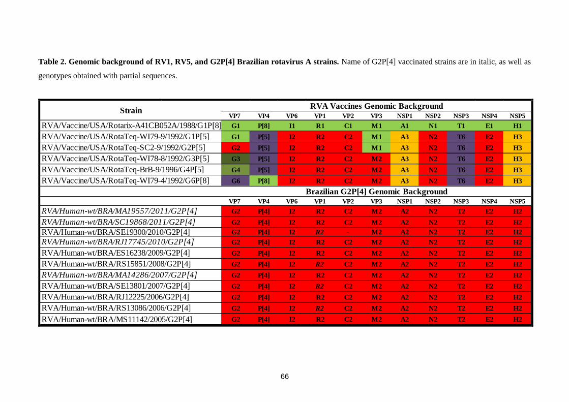
66
Table 2. Genomic background of RV1, RV5, and G2P[4] Brazilian rotavirus A strains. Name of G2P[4] vaccinated strains are in italic, as well as
genotypes obtained with partial sequences.
VP7 VP4 VP6 VP1 VP2 VP3 NSP1 NSP2 NSP3 NSP4 NSP5
G1 P[8] I1 R1 C1 M1 A1 N1 T1 E1 H1
G1 P[5] I2 R2 C2 M1 A3 N2 T6 E2 H3
G2 P[5] I2 R2 C2 M1 A3 N2 T6 E2 H3
G3 P[5] I2 R2 C2 M2 A3 N2 T6 E2 H3
G4 P[5] I2 R2 C2 M2 A3 N2 T6 E2 H3
G6 P[8] I2 R2 C2 M2 A3 N2 T6 E2 H3
VP7 VP4 VP6 VP1 VP2 VP3 NSP1 NSP2 NSP3 NSP4 NSP5
G2 P[4] I2 R2 C2 M2 A2 N2 T2 E2 H2
G2 P[4] I2 R2 C2 M2 A2 N2 T2 E2 H2
G2 P[4] I2 R2 - M2 A2 N2 T2 E2 H2
G2 P[4] I2 R2 C2 M2 A2 N2 T2 E2 H2
G2 P[4] I2 R2 C2 M2 A2 N2 T2 E2 H2
G2 P[4] I2 R2 C2 M2 A2 N2 T2 E2 H2
G2 P[4] I2 R2 C2 M2 A2 N2 T2 E2 H2
G2 P[4] I2 R2 C2 M2 A2 N2 T2 E2 H2
G2 P[4] I2 R2 C2 M2 A2 N2 T2 E2 H2
G2 P[4] I2 R2 C2 M2 A2 N2 T2 E2 H2
G2 P[4] I2 R2 C2 M2 A2 N2 T2 E2 H2
Strain
RVA/Vaccine/USA/RotaTeq-BrB-9/1996/G4P[5]
RVA/Vaccine/USA/RotaTeq-WI79-4/1992/G6P[8]
RVA Vaccines Genomic Background
RVA/Human-wt/BRA/ES16238/2009/G2P[4]
RVA/Human-wt/BRA/RS15851/2008/G2P[4]
RVA/Vaccine/USA/Rotarix-A41CB052A/1988/G1P[8]
RVA/Vaccine/USA/RotaTeq-WI79-9/1992/G1P[5]
RVA/Vaccine/USA/RotaTeq-SC2-9/1992/G2P[5]
RVA/Vaccine/USA/RotaTeq-WI78-8/1992/G3P[5]
RVA/Human-wt/BRA/MA19557/2011/G2P[4]
RVA/Human-wt/BRA/SC19868/2011/G2P[4]
RVA/Human-wt/BRA/SE19300/2010/G2P[4]
RVA/Human-wt/BRA/RJ17745/2010/G2P[4]
Brazilian G2P[4] Genomic Background
RVA/Human-wt/BRA/MA14286/2007/G2P[4]
RVA/Human-wt/BRA/SE13801/2007/G2P[4]
RVA/Human-wt/BRA/RJ12225/2006/G2P[4]
RVA/Human-wt/BRA/RS13086/2006/G2P[4]
RVA/Human-wt/BRA/MS11142/2005/G2P[4]
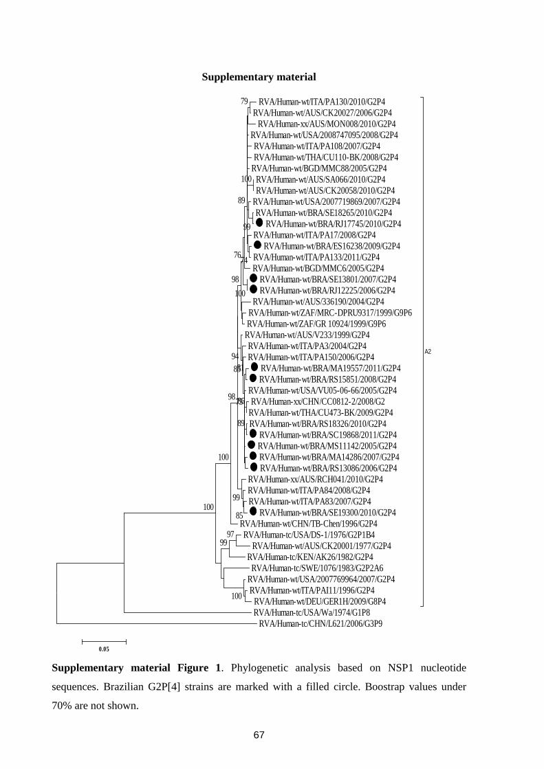
67
Supplementary material
RVA/Human-wt/ITA/PA130/2010/G2P4 RVA/Human-wt/AUS/CK20027/2006/G2P4
RVA/Human-xx/AUS/MON008/2010/G2P4 RVA/Human-wt/USA/2008747095/2008/G2P4 RVA/Human-wt/ITA/PA108/2007/G2P4 RVA/Human-wt/THA/CU110-BK/2008/G2P4
RVA/Human-wt/BGD/MMC88/2005/G2P4 RVA/Human-wt/AUS/SA066/2010/G2P4 RVA/Human-wt/AUS/CK20058/2010/G2P4
RVA/Human-wt/USA/2007719869/2007/G2P4 RVA/Human-wt/BRA/SE18265/2010/G2P4
RVA/Human-wt/BRA/RJ17745/2010/G2P4 RVA/Human-wt/ITA/PA17/2008/G2P4
RVA/Human-wt/BRA/ES16238/2009/G2P4 RVA/Human-wt/ITA/PA133/2011/G2P4 RVA/Human-wt/BGD/MMC6/2005/G2P4
RVA/Human-wt/BRA/SE13801/2007/G2P4 RVA/Human-wt/BRA/RJ12225/2006/G2P4
RVA/Human-wt/AUS/336190/2004/G2P4 RVA/Human-wt/ZAF/MRC-DPRU9317/1999/G9P6 RVA/Human-wt/ZAF/GR 10924/1999/G9P6
RVA/Human-wt/AUS/V233/1999/G2P4 RVA/Human-wt/ITA/PA3/2004/G2P4 RVA/Human-wt/ITA/PA150/2006/G2P4
RVA/Human-wt/BRA/MA19557/2011/G2P4 RVA/Human-wt/BRA/RS15851/2008/G2P4
RVA/Human-wt/USA/VU05-06-66/2005/G2P4 RVA/Human-xx/CHN/CC0812-2/2008/G2
RVA/Human-wt/THA/CU473-BK/2009/G2P4 RVA/Human-wt/BRA/RS18326/2010/G2P4
RVA/Human-wt/BRA/SC19868/2011/G2P4 RVA/Human-wt/BRA/MS11142/2005/G2P4 RVA/Human-wt/BRA/MA14286/2007/G2P4 RVA/Human-wt/BRA/RS13086/2006/G2P4
RVA/Human-xx/AUS/RCH041/2010/G2P4 RVA/Human-wt/ITA/PA84/2008/G2P4 RVA/Human-wt/ITA/PA83/2007/G2P4
RVA/Human-wt/BRA/SE19300/2010/G2P4 RVA/Human-wt/CHN/TB-Chen/1996/G2P4 RVA/Human-tc/USA/DS-1/1976/G2P1B4
RVA/Human-wt/AUS/CK20001/1977/G2P4 RVA/Human-tc/KEN/AK26/1982/G2P4
RVA/Human-tc/SWE/1076/1983/G2P2A6 RVA/Human-wt/USA/2007769964/2007/G2P4 RVA/Human-wt/ITA/PAI11/1996/G2P4
RVA/Human-wt/DEU/GER1H/2009/G8P4
A2
RVA/Human-tc/USA/Wa/1974/G1P8 RVA/Human-tc/CHN/L621/2006/G3P9
100
9799
100
99
100
100
85
99
100
89
86
81
75
85
94
98
98
76
79
74
89
0.05
Supplementary material Figure 1. Phylogenetic analysis based on NSP1 nucleotide
sequences. Brazilian G2P[4] strains are marked with a filled circle. Boostrap values under
70% are not shown.
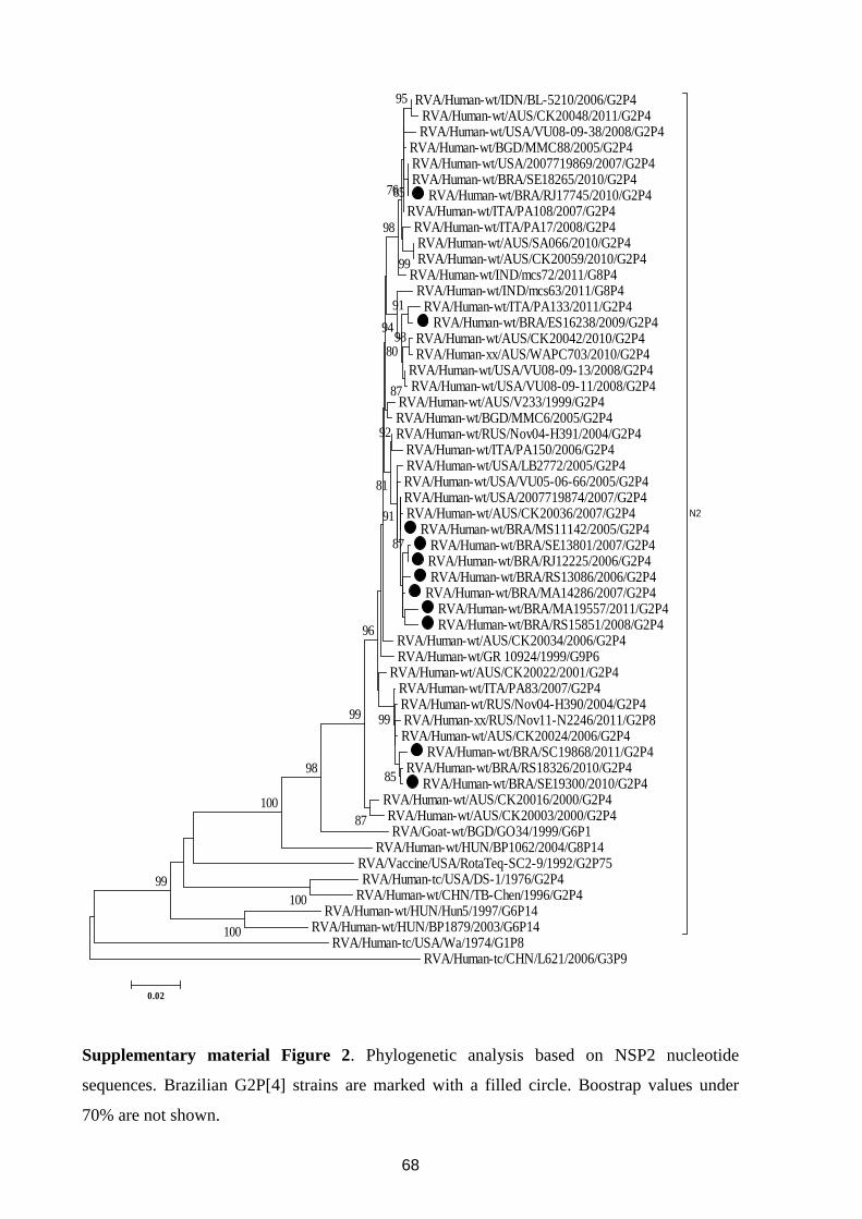
68
RVA/Human-wt/IDN/BL-5210/2006/G2P4 RVA/Human-wt/AUS/CK20048/2011/G2P4 RVA/Human-wt/USA/VU08-09-38/2008/G2P4
RVA/Human-wt/BGD/MMC88/2005/G2P4 RVA/Human-wt/USA/2007719869/2007/G2P4 RVA/Human-wt/BRA/SE18265/2010/G2P4
RVA/Human-wt/BRA/RJ17745/2010/G2P4 RVA/Human-wt/ITA/PA108/2007/G2P4
RVA/Human-wt/ITA/PA17/2008/G2P4 RVA/Human-wt/AUS/SA066/2010/G2P4 RVA/Human-wt/AUS/CK20059/2010/G2P4
RVA/Human-wt/IND/mcs72/2011/G8P4 RVA/Human-wt/IND/mcs63/2011/G8P4
RVA/Human-wt/ITA/PA133/2011/G2P4 RVA/Human-wt/BRA/ES16238/2009/G2P4
RVA/Human-wt/AUS/CK20042/2010/G2P4 RVA/Human-xx/AUS/WAPC703/2010/G2P4
RVA/Human-wt/USA/VU08-09-13/2008/G2P4 RVA/Human-wt/USA/VU08-09-11/2008/G2P4
RVA/Human-wt/AUS/V233/1999/G2P4 RVA/Human-wt/BGD/MMC6/2005/G2P4 RVA/Human-wt/RUS/Nov04-H391/2004/G2P4
RVA/Human-wt/ITA/PA150/2006/G2P4 RVA/Human-wt/USA/LB2772/2005/G2P4 RVA/Human-wt/USA/VU05-06-66/2005/G2P4 RVA/Human-wt/USA/2007719874/2007/G2P4 RVA/Human-wt/AUS/CK20036/2007/G2P4
RVA/Human-wt/BRA/MS11142/2005/G2P4 RVA/Human-wt/BRA/SE13801/2007/G2P4 RVA/Human-wt/BRA/RJ12225/2006/G2P4 RVA/Human-wt/BRA/RS13086/2006/G2P4
RVA/Human-wt/BRA/MA14286/2007/G2P4 RVA/Human-wt/BRA/MA19557/2011/G2P4 RVA/Human-wt/BRA/RS15851/2008/G2P4
RVA/Human-wt/AUS/CK20034/2006/G2P4 RVA/Human-wt/GR 10924/1999/G9P6
RVA/Human-wt/AUS/CK20022/2001/G2P4 RVA/Human-wt/ITA/PA83/2007/G2P4 RVA/Human-wt/RUS/Nov04-H390/2004/G2P4 RVA/Human-xx/RUS/Nov11-N2246/2011/G2P8 RVA/Human-wt/AUS/CK20024/2006/G2P4
RVA/Human-wt/BRA/SC19868/2011/G2P4 RVA/Human-wt/BRA/RS18326/2010/G2P4
RVA/Human-wt/BRA/SE19300/2010/G2P4 RVA/Human-wt/AUS/CK20016/2000/G2P4 RVA/Human-wt/AUS/CK20003/2000/G2P4 RVA/Goat-wt/BGD/GO34/1999/G6P1
RVA/Human-wt/HUN/BP1062/2004/G8P14 RVA/Vaccine/USA/RotaTeq-SC2-9/1992/G2P75 RVA/Human-tc/USA/DS-1/1976/G2P4
RVA/Human-wt/CHN/TB-Chen/1996/G2P4 RVA/Human-wt/HUN/Hun5/1997/G6P14
RVA/Human-wt/HUN/BP1879/2003/G6P14
N2
RVA/Human-tc/USA/Wa/1974/G1P8 RVA/Human-tc/CHN/L621/2006/G3P9
100
100
99
100
9885
87
99
98
99
99
91
87
95
80
85
94
76
98
96
92
81
87
91
0.02
Supplementary material Figure 2. Phylogenetic analysis based on NSP2 nucleotide
sequences. Brazilian G2P[4] strains are marked with a filled circle. Boostrap values under
70% are not shown.
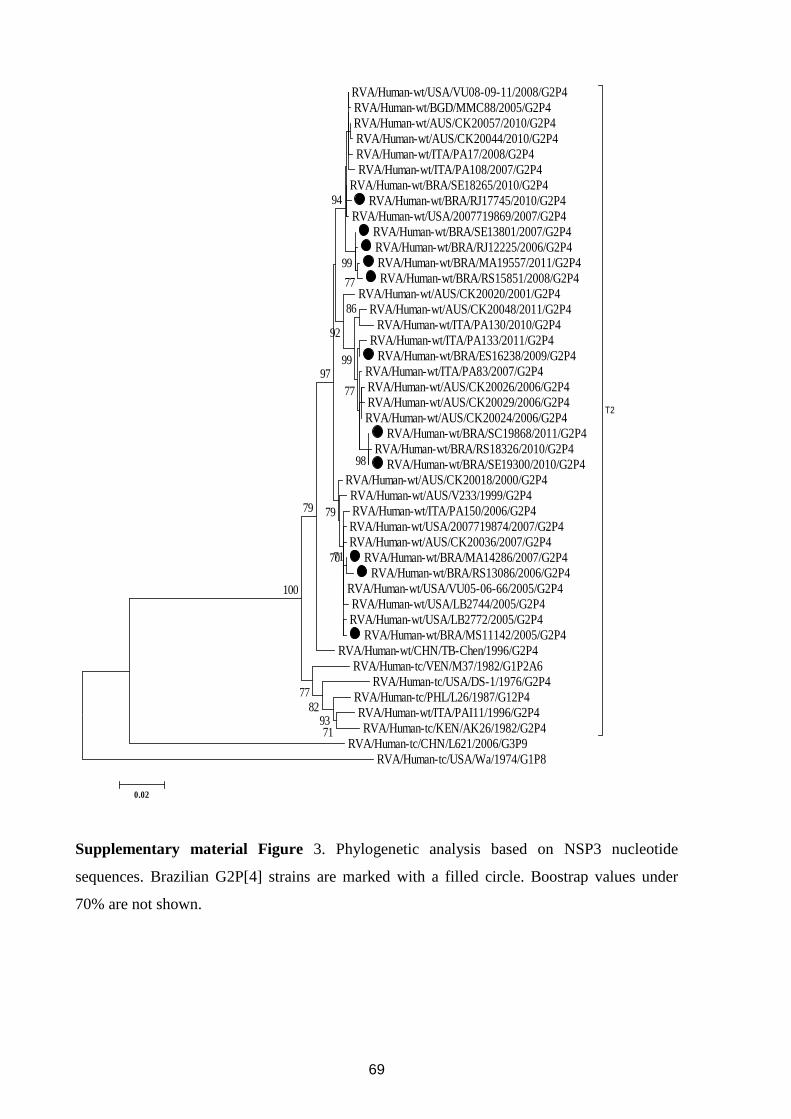
69
RVA/Human-wt/USA/VU08-09-11/2008/G2P4 RVA/Human-wt/BGD/MMC88/2005/G2P4 RVA/Human-wt/AUS/CK20057/2010/G2P4 RVA/Human-wt/AUS/CK20044/2010/G2P4 RVA/Human-wt/ITA/PA17/2008/G2P4 RVA/Human-wt/ITA/PA108/2007/G2P4
RVA/Human-wt/BRA/SE18265/2010/G2P4 RVA/Human-wt/BRA/RJ17745/2010/G2P4
RVA/Human-wt/USA/2007719869/2007/G2P4 RVA/Human-wt/BRA/SE13801/2007/G2P4 RVA/Human-wt/BRA/RJ12225/2006/G2P4 RVA/Human-wt/BRA/MA19557/2011/G2P4 RVA/Human-wt/BRA/RS15851/2008/G2P4
RVA/Human-wt/AUS/CK20020/2001/G2P4 RVA/Human-wt/AUS/CK20048/2011/G2P4
RVA/Human-wt/ITA/PA130/2010/G2P4 RVA/Human-wt/ITA/PA133/2011/G2P4
RVA/Human-wt/BRA/ES16238/2009/G2P4 RVA/Human-wt/ITA/PA83/2007/G2P4 RVA/Human-wt/AUS/CK20026/2006/G2P4 RVA/Human-wt/AUS/CK20029/2006/G2P4 RVA/Human-wt/AUS/CK20024/2006/G2P4
RVA/Human-wt/BRA/SC19868/2011/G2P4 RVA/Human-wt/BRA/RS18326/2010/G2P4
RVA/Human-wt/BRA/SE19300/2010/G2P4 RVA/Human-wt/AUS/CK20018/2000/G2P4 RVA/Human-wt/AUS/V233/1999/G2P4 RVA/Human-wt/ITA/PA150/2006/G2P4 RVA/Human-wt/USA/2007719874/2007/G2P4 RVA/Human-wt/AUS/CK20036/2007/G2P4
RVA/Human-wt/BRA/MA14286/2007/G2P4 RVA/Human-wt/BRA/RS13086/2006/G2P4
RVA/Human-wt/USA/VU05-06-66/2005/G2P4 RVA/Human-wt/USA/LB2744/2005/G2P4 RVA/Human-wt/USA/LB2772/2005/G2P4
RVA/Human-wt/BRA/MS11142/2005/G2P4 RVA/Human-wt/CHN/TB-Chen/1996/G2P4
RVA/Human-tc/VEN/M37/1982/G1P2A6 RVA/Human-tc/USA/DS-1/1976/G2P4
RVA/Human-tc/PHL/L26/1987/G12P4 RVA/Human-wt/ITA/PAI11/1996/G2P4 RVA/Human-tc/KEN/AK26/1982/G2P4
T2
RVA/Human-tc/CHN/L621/2006/G3P9 RVA/Human-tc/USA/Wa/1974/G1P8
7193
8277
100
98
79
86
77
99
77
99
92
94
97
79
7170
0.02
Supplementary material Figure 3. Phylogenetic analysis based on NSP3 nucleotide
sequences. Brazilian G2P[4] strains are marked with a filled circle. Boostrap values under
70% are not shown.
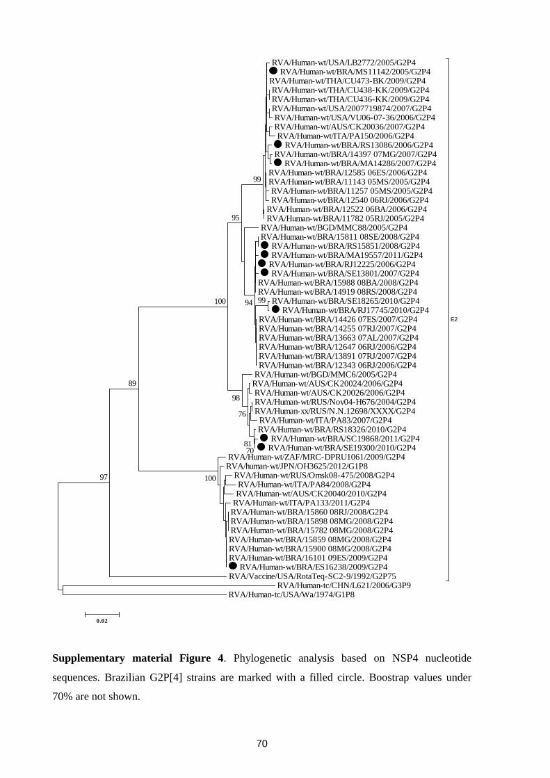
70
RVA/Human-wt/USA/LB2772/2005/G2P4 RVA/Human-wt/BRA/MS11142/2005/G2P4
RVA/Human-wt/THA/CU473-BK/2009/G2P4 RVA/Human-wt/THA/CU438-KK/2009/G2P4 RVA/Human-wt/THA/CU436-KK/2009/G2P4 RVA/Human-wt/USA/2007719874/2007/G2P4 RVA/Human-wt/USA/VU06-07-36/2006/G2P4 RVA/Human-wt/AUS/CK20036/2007/G2P4 RVA/Human-wt/ITA/PA150/2006/G2P4
RVA/Human-wt/BRA/RS13086/2006/G2P4 RVA/Human-wt/BRA/14397 07MG/2007/G2P4
RVA/Human-wt/BRA/MA14286/2007/G2P4 RVA/Human-wt/BRA/12585 06ES/2006/G2P4 RVA/Human-wt/BRA/11143 05MS/2005/G2P4 RVA/Human-wt/BRA/11257 05MS/2005/G2P4 RVA/Human-wt/BRA/12540 06RJ/2006/G2P4
RVA/Human-wt/BRA/12522 06BA/2006/G2P4 RVA/Human-wt/BRA/11782 05RJ/2005/G2P4
RVA/Human-wt/BGD/MMC88/2005/G2P4 RVA/Human-wt/BRA/15811 08SE/2008/G2P4
RVA/Human-wt/BRA/RS15851/2008/G2P4 RVA/Human-wt/BRA/MA19557/2011/G2P4
RVA/Human-wt/BRA/RJ12225/2006/G2P4 RVA/Human-wt/BRA/SE13801/2007/G2P4
RVA/Human-wt/BRA/15988 08BA/2008/G2P4 RVA/Human-wt/BRA/14919 08RS/2008/G2P4
RVA/Human-wt/BRA/SE18265/2010/G2P4 RVA/Human-wt/BRA/RJ17745/2010/G2P4
RVA/Human-wt/BRA/14426 07ES/2007/G2P4 RVA/Human-wt/BRA/14255 07RJ/2007/G2P4 RVA/Human-wt/BRA/13663 07AL/2007/G2P4 RVA/Human-wt/BRA/12647 06RJ/2006/G2P4 RVA/Human-wt/BRA/13891 07RJ/2007/G2P4 RVA/Human-wt/BRA/12343 06RJ/2006/G2P4
RVA/Human-wt/BGD/MMC6/2005/G2P4 RVA/Human-wt/AUS/CK20024/2006/G2P4 RVA/Human-wt/AUS/CK20026/2006/G2P4 RVA/Human-wt/RUS/Nov04-H676/2004/G2P4 RVA/Human-xx/RUS/N.N.12698/XXXX/G2P4
RVA/Human-wt/ITA/PA83/2007/G2P4 RVA/Human-wt/BRA/RS18326/2010/G2P4
RVA/Human-wt/BRA/SC19868/2011/G2P4 RVA/Human-wt/BRA/SE19300/2010/G2P4
RVA/Human-wt/ZAF/MRC-DPRU1061/2009/G2P4 RVA/human-wt/JPN/OH3625/2012/G1P8
RVA/Human-wt/RUS/Omsk08-475/2008/G2P4 RVA/Human-wt/ITA/PA84/2008/G2P4 RVA/Human-wt/AUS/CK20040/2010/G2P4
RVA/Human-wt/ITA/PA133/2011/G2P4 RVA/Human-wt/BRA/15860 08RJ/2008/G2P4 RVA/Human-wt/BRA/15898 08MG/2008/G2P4 RVA/Human-wt/BRA/15782 08MG/2008/G2P4 RVA/Human-wt/BRA/15859 08MG/2008/G2P4 RVA/Human-wt/BRA/15900 08MG/2008/G2P4 RVA/Human-wt/BRA/16101 09ES/2009/G2P4
RVA/Human-wt/BRA/ES16238/2009/G2P4 RVA/Vaccine/USA/RotaTeq-SC2-9/1992/G2P75
E2
RVA/Human-tc/CHN/L621/2006/G3P9 RVA/Human-tc/USA/Wa/1974/G1P8
10097
89
7081
76
98
99100 94
95
99
0.02
Supplementary material Figure 4. Phylogenetic analysis based on NSP4 nucleotide
sequences. Brazilian G2P[4] strains are marked with a filled circle. Boostrap values under
70% are not shown.
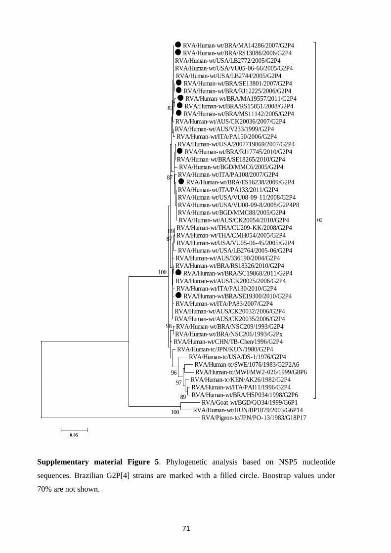
71
RVA/Human-wt/BRA/MA14286/2007/G2P4 RVA/Human-wt/BRA/RS13086/2006/G2P4
RVA/Human-wt/USA/LB2772/2005/G2P4 RVA/Human-wt/USA/VU05-06-66/2005/G2P4 RVA/Human-wt/USA/LB2744/2005/G2P4
RVA/Human-wt/BRA/SE13801/2007/G2P4 RVA/Human-wt/BRA/RJ12225/2006/G2P4 RVA/Human-wt/BRA/MA19557/2011/G2P4 RVA/Human-wt/BRA/RS15851/2008/G2P4 RVA/Human-wt/BRA/MS11142/2005/G2P4
RVA/Human-wt/AUS/CK20036/2007/G2P4 RVA/Human-wt/AUS/V233/1999/G2P4 RVA/Human-wt/ITA/PA150/2006/G2P4 RVA/Human-wt/USA/2007719869/2007/G2P4
RVA/Human-wt/BRA/RJ17745/2010/G2P4 RVA/Human-wt/BRA/SE18265/2010/G2P4 RVA/Human-wt/BGD/MMC6/2005/G2P4 RVA/Human-wt/ITA/PA108/2007/G2P4
RVA/Human-wt/BRA/ES16238/2009/G2P4 RVA/Human-wt/ITA/PA133/2011/G2P4 RVA/Human-wt/USA/VU08-09-11/2008/G2P4 RVA/Human-wt/USA/VU08-09-8/2008/G2P4P8 RVA/Human-wt/BGD/MMC88/2005/G2P4 RVA/Human-wt/AUS/CK20054/2010/G2P4
RVA/Human-wt/THA/CU209-KK/2008/G2P4 RVA/Human-wt/THA/CMH054/2005/G2P4 RVA/Human-wt/USA/VU05-06-45/2005/G2P4 RVA/Human-wt/USA/LB2764/2005-06/G2P4
RVA/Human-wt/AUS/336190/2004/G2P4 RVA/Human-wt/BRA/RS18326/2010/G2P4
RVA/Human-wt/BRA/SC19868/2011/G2P4 RVA/Human-wt/AUS/CK20025/2006/G2P4 RVA/Human-wt/ITA/PA130/2010/G2P4
RVA/Human-wt/BRA/SE19300/2010/G2P4 RVA/Human-wt/ITA/PA83/2007/G2P4 RVA/Human-wt/AUS/CK20032/2006/G2P4 RVA/Human-wt/AUS/CK20035/2006/G2P4 RVA/Human-wt/BRA/NSC209/1993/G2P4 RVA/Human-wt/BRA/NSC206/1993/G2Px RVA/Human-wt/CHN/TB-Chen/1996/G2P4
RVA/Human-tc/JPN/KUN/1980/G2P4 RVA/Human-tc/USA/DS-1/1976/G2P4
RVA/Human-tc/SWE/1076/1983/G2P2A6 RVA/Human-tc/MWI/MW2-026/1999/G8P6
RVA/Human-tc/KEN/AK26/1982/G2P4 RVA/Human-wt/ITA/PAI11/1996/G2P4 RVA/Human-wt/BRA/HSP034/1998/G2P6
H2
RVA/Goat-wt/BGD/GO34/1999/G6P1 RVA/Human-wt/HUN/BP1879/2003/G6P14
RVA/Pigeon-tc/JPN/PO-13/1983/G18P17100
89
97
96
98
89
87
100
87
82
0.05
Supplementary material Figure 5. Phylogenetic analysis based on NSP5 nucleotide
sequences. Brazilian G2P[4] strains are marked with a filled circle. Boostrap values under
70% are not shown.
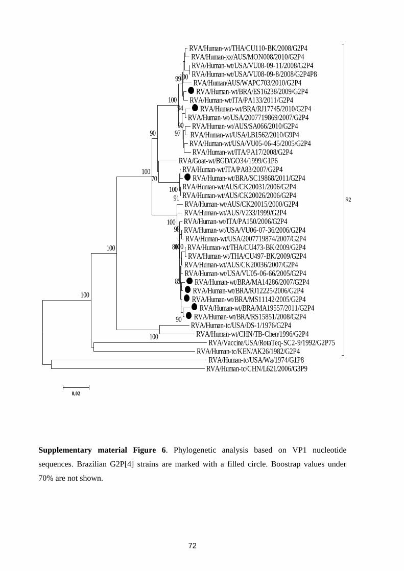
72
RVA/Human-wt/THA/CU110-BK/2008/G2P4 RVA/Human-xx/AUS/MON008/2010/G2P4 RVA/Human-wt/USA/VU08-09-11/2008/G2P4 RVA/Human-wt/USA/VU08-09-8/2008/G2P4P8 RVA/Human/AUS/WAPC703/2010/G2P4 RVA/Human-wt/BRA/ES16238/2009/G2P4
RVA/Human-wt/ITA/PA133/2011/G2P4 RVA/Human-wt/BRA/RJ17745/2010/G2P4
RVA/Human-wt/USA/2007719869/2007/G2P4 RVA/Human-wt/AUS/SA066/2010/G2P4
RVA/Human-wt/USA/LB1562/2010/G9P4 RVA/Human-wt/USA/VU05-06-45/2005/G2P4 RVA/Human-wt/ITA/PA17/2008/G2P4
RVA/Goat-wt/BGD/GO34/1999/G1P6 RVA/Human-wt/ITA/PA83/2007/G2P4
RVA/Human-wt/BRA/SC19868/2011/G2P4 RVA/Human-wt/AUS/CK20031/2006/G2P4 RVA/Human-wt/AUS/CK20026/2006/G2P4 RVA/Human-wt/AUS/CK20015/2000/G2P4 RVA/Human-wt/AUS/V233/1999/G2P4 RVA/Human-wt/ITA/PA150/2006/G2P4 RVA/Human-wt/USA/VU06-07-36/2006/G2P4 RVA/Human-wt/USA/2007719874/2007/G2P4 RVA/Human-wt/THA/CU473-BK/2009/G2P4 RVA/Human-wt/THA/CU497-BK/2009/G2P4
RVA/Human-wt/AUS/CK20036/2007/G2P4 RVA/Human-wt/USA/VU05-06-66/2005/G2P4
RVA/Human-wt/BRA/MA14286/2007/G2P4 RVA/Human-wt/BRA/RJ12225/2006/G2P4 RVA/Human-wt/BRA/MS11142/2005/G2P4
RVA/Human-wt/BRA/MA19557/2011/G2P4 RVA/Human-wt/BRA/RS15851/2008/G2P4
RVA/Human-tc/USA/DS-1/1976/G2P4 RVA/Human-wt/CHN/TB-Chen/1996/G2P4
RVA/Vaccine/USA/RotaTeq-SC2-9/1992/G2P75 RVA/Human-tc/KEN/AK26/1982/G2P4
R2
RVA/Human-tc/USA/Wa/1974/G1P8 RVA/Human-tc/CHN/L621/2006/G3P9
100
100
100
91100
100
94
90
99
97
100
70
90
100
100
90
10098
85
80
0,02
Supplementary material Figure 6. Phylogenetic analysis based on VP1 nucleotide
sequences. Brazilian G2P[4] strains are marked with a filled circle. Boostrap values under
70% are not shown.
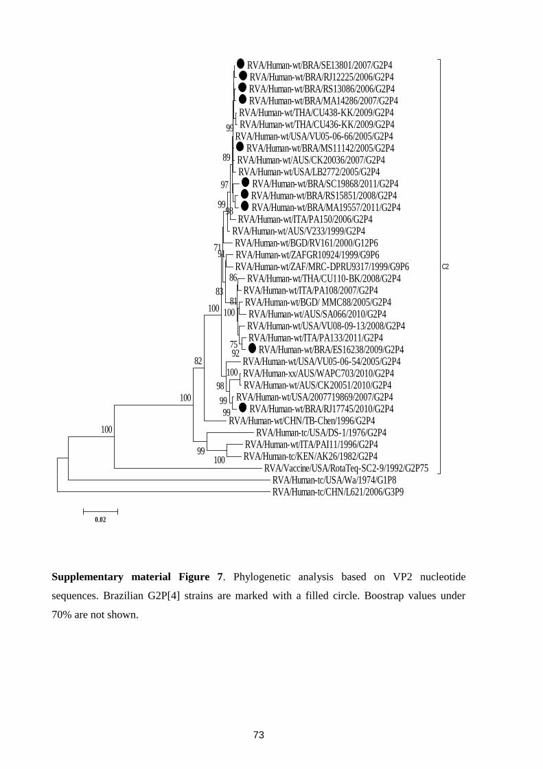
73
RVA/Human-wt/BRA/SE13801/2007/G2P4 RVA/Human-wt/BRA/RJ12225/2006/G2P4 RVA/Human-wt/BRA/RS13086/2006/G2P4 RVA/Human-wt/BRA/MA14286/2007/G2P4
RVA/Human-wt/THA/CU438-KK/2009/G2P4 RVA/Human-wt/THA/CU436-KK/2009/G2P4
RVA/Human-wt/USA/VU05-06-66/2005/G2P4 RVA/Human-wt/BRA/MS11142/2005/G2P4
RVA/Human-wt/AUS/CK20036/2007/G2P4 RVA/Human-wt/USA/LB2772/2005/G2P4
RVA/Human-wt/BRA/SC19868/2011/G2P4 RVA/Human-wt/BRA/RS15851/2008/G2P4 RVA/Human-wt/BRA/MA19557/2011/G2P4
RVA/Human-wt/ITA/PA150/2006/G2P4 RVA/Human-wt/AUS/V233/1999/G2P4 RVA/Human-wt/BGD/RV161/2000/G12P6 RVA/Human-wt/ZAFGR10924/1999/G9P6 RVA/Human-wt/ZAF/MRC-DPRU9317/1999/G9P6
RVA/Human-wt/THA/CU110-BK/2008/G2P4 RVA/Human-wt/ITA/PA108/2007/G2P4 RVA/Human-wt/BGD/ MMC88/2005/G2P4 RVA/Human-wt/AUS/SA066/2010/G2P4 RVA/Human-wt/USA/VU08-09-13/2008/G2P4 RVA/Human-wt/ITA/PA133/2011/G2P4
RVA/Human-wt/BRA/ES16238/2009/G2P4 RVA/Human-wt/USA/VU05-06-54/2005/G2P4 RVA/Human-xx/AUS/WAPC703/2010/G2P4 RVA/Human-wt/AUS/CK20051/2010/G2P4
RVA/Human-wt/USA/2007719869/2007/G2P4 RVA/Human-wt/BRA/RJ17745/2010/G2P4
RVA/Human-wt/CHN/TB-Chen/1996/G2P4 RVA/Human-tc/USA/DS-1/1976/G2P4
RVA/Human-wt/ITA/PAI11/1996/G2P4 RVA/Human-tc/KEN/AK26/1982/G2P4
RVA/Vaccine/USA/RotaTeq-SC2-9/1992/G2P75
C2
RVA/Human-tc/USA/Wa/1974/G1P8 RVA/Human-tc/CHN/L621/2006/G3P9
100
10099
100
82100
92
81
75
86
100
9999
98
91
83
100
71
9998
99
97
89
0.02
Supplementary material Figure 7. Phylogenetic analysis based on VP2 nucleotide
sequences. Brazilian G2P[4] strains are marked with a filled circle. Boostrap values under
70% are not shown.
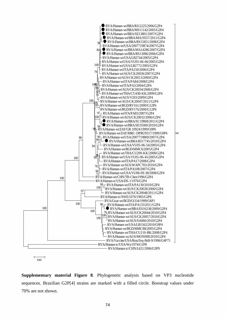
74
RVA/Human-wt/BRA/RJ12225/2006/G2P4 RVA/Human-wt/BRA/MS11142/2005/G2P4 RVA/Human-wt/BRA/SE13801/2007/G2P4 RVA/Human-wt/BRA/MA19557/2011/G2P4 RVA/Human-wt/BRA/RS15851/2008/G2P4
RVA/Human-wt/USA/2007719874/2007/G2P4 RVA/Human-wt/BRA/MA14286/2007/G2P4 RVA/Human-wt/BRA/RS13086/2006/G2P4
RVA/Human-wt/USA/LB2744/2005/G2P4 RVA/Human-wt/USA/VU05-06-66/2005/G2P4 RVA/Human-wt/USA/LB2772/2005/G2P4 RVA/Human-wt/ITA/PA150/2006/G2P4 RVA/Human-wt/AUS/CK20036/2007/G2P4
RVA/Human-wt/AUS/CK20013/2000/G2P4 RVA/Human-wt/ITA/PA84/2008/G2P4 RVA/Human-wt/ITA/PA3/2004/G2P4
RVA/Human-wt/AUS/CK20034/2006/G2P4 RVA/Human-wt/THA/CU438-KK/2009/G2P4
RVA/Human-wt/AUS/V203/2009/G2P4 RVA/Human-wt/AUS/CK20047/2011/G2P4 RVA/Human-wt/BGD/RV161/2000/G12P6 RVA/Human-wt/BGD/RV176/2000/G12P6
RVA/Human-wt/ITA/PA83/2007/G2P4 RVA/Human-wt/AUS/CK20032/2006/G2P4
RVA/Human-wt/BRA/SC19868/2011/G2P4 RVA/Human-wt/BRA/SE19300/2010/G2P4
RVA/Human-wt/ZAF/GR 10924/1999/G9P6 RVA/Human-wt/ZAF/MRC-DPRU9317/1999/G9P6
RVA/Human-wt/USA/2007719869/2007/G2P4 RVA/Human-wt/BRA/RJ17745/2010/G2P4
RVA/Human-wt/USA/VU05-06-54/2005/G2P4 RVA/Human-wt/BGD/MMC6/2005/G2P4 RVA/Human-wt/THA/CU209-KK/2008/G2P4
RVA/Human-wt/USA/VU05-06-45/2005/G2P4 RVA/Human-wt/ITA/PA17/2008/G2P4 RVA/Human-xx/AUS/WAPC703/2010/G2P4 RVA/Human-wt/ITA/PA108/2007/G2P4 RVA/Human-wt/USA/VU08-09-38/2008/G2P4
RVA/Human-wt/CHN/TB-Chen/1996/G2P4 RVA/Human-tc/USA/DS-1/1976/G2P4
RVA/Human-wt/ITA/PA130/2010/G2P4 RVA/Human-wt/AUS/CK20028/2006/G2P4 RVA/Human-wt/AUS/CK20048/2011/G2P4
RVA/Human-tc/SWE/1076/1983/G2P6 RVA/Goat-wt/BGD/GO34/1999/G6P1
RVA/Human-wt/ITA/PA133/2011/G2P4 RVA/Human-wt/BRA/ES16238/2009/G2P4
RVA/Human-wt/AUS/CK20044/2010/G2P4 RVA/Human-wt/AUS/CK20057/2010/G2P4 RVA/Human-wt/AUS/SA066/2010/G2P4 RVA/Human-wt/USA/LB1562/2010/G9P4
RVA/Human-wt/BGD/MMC88/2005/G2P4 RVA/Human-wt/THA/CU110-BK/2008/G2P4 RVA/Human-xx/AUS/MON008/2010/G2P4
RVA/Vaccine/USA/RotaTeq-BrB-9/1996/G4P75
M2
RVA/Human-tc/USA/Wa/1974/G1P8 RVA/Human-tc/CHN/L621/2006/G3P9
99100
76100
96
100
100
97
100
100
100
100
86
99
97100
90
100
99
89
7390
78
99
99
84
100100
919289
82100
79
98
100
84
0,02
Supplementary material Figure 8. Phylogenetic analysis based on VP3 nucleotide
sequences. Brazilian G2P[4] strains are marked with a filled circle. Boostrap values under
70% are not shown.
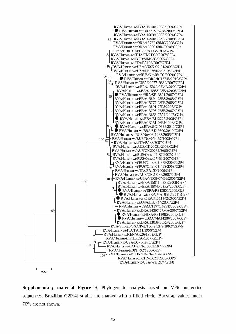
75
RVA/Human-wt/BRA/16100 09ES/2009/G2P4 RVA/Human-wt/BRA/ES16238/2009/G2P4
RVA/Human-wt/BRA/16099 09ES/2009/G2P4 RVA/Human-wt/BRA/15900 08MG/2008/G2P4 RVA/Human-wt/BRA/15782 08MG/2008/G2P4 RVA/Human-wt/BRA/15860 08RJ/2008/G2P4 RVA/Human-wt/ITA/PA133/2011/G2P4
RVA/Human-wt/THA/CMH030/2007/G2P4 RVA/Human-wt/BGD/MMC88/2005/G2P4 RVA/Human-wt/ITA/PA108/2007/G2P4 RVA/Human-wt/USA/VU05-06-54/2005/G2P4 RVA/Human-wt/USA/LB2764/2005-06/G2P4
RVA/Human-wt/RUS/Nov09-D2/2009/G2P4 RVA/Human-wt/BRA/RJ17745/2010/G2P4
RVA/Human-wt/USA/2007719869/2007/G2P4 RVA/Human-wt/BRA/15863 08MA/2008/G2P4 RVA/Human-wt/BRA/15988 08BA/2008/G2P4
RVA/Human-wt/BRA/SE13801/2007/G2P4 RVA/Human-wt/BRA/15894 08ES/2008/G2P4 RVA/Human-wt/BRA/15777 08PE/2008/G2P4 RVA/Human-wt/BRA/13891 07RJ/2007/G2P4 RVA/Human-wt/BRA/13793 07SE/2007/G2P4 RVA/Human-wt/BRA/13663 07AL/2007/G2P4
RVA/Human-wt/BRA/RJ12225/2006/G2P4 RVA/Human-wt/BRA/13151 06RJ/2006/G2P4
RVA/Human-wt/BRA/SC19868/2011/G2P4 RVA/Human-wt/BRA/SE19300/2010/G2P4
RVA/Human-wt/RUS/Nov06-1283/2006/G2P4 RVA/Human-wt/RUS/Nov05-137/2005/G2P4 RVA/Human-wt/ITA/PA83/2007/G2P4
RVA/Human-wt/AUS/CK20031/2006/G2P4 RVA/Human-wt/AUS/CK20032/2006/G2P4 RVA/Human-wt/RUS/Omsk07-87/2007/G2P4 RVA/Human-wt/RUS/Omsk07-88/2007/G2P4 RVA/Human-wt/RUS/Omsk08-375/2008/G2P4 RVA/Human-wt/RUS/Omsk08-418/2008/G2P4 RVA/Human-wt/ITA/PA150/2006/G2P4 RVA/Human-wt/AUS/CK20036/2007/G2P4 RVA/Human-wt/USA/VU06-07-36/2006/G2P4
RVA/Human-wt/BRA/15811 08SE/2008/G2P4 RVA/Human-wt/BRA/15840 08RS/2008/G2P4
RVA/Human-wt/BRA/RS15851/2008/G2P4 RVA/Human-wt/BRA/MA19557/2011/G2P4
RVA/Human-wt/BRA/MS11142/2005/G2P4 RVA/Human-wt/USA/LB2744/2005/G2P4
RVA/Human-wt/BRA/15771 08PE/2008/G2P4 RVA/Human-wt/BRA/14397 07MA/2007/G2P4
RVA/Human-wt/BRA/RS13086/2006/G2P4 RVA/Human-wt/BRA/MA14286/2007/G2P4
RVA/Human-wt/BRA/13039 06RS/2006/G2P4 RVA/Vaccine/USA/RotaTeq-SC2-9/1992/G2P75
RVA/Human-wt/ITA/PAI11/1996/G2P4 RVA/Human-tc/KEN/AK26/1982/G2P4
RVA/Human-tc/PHL/L26/1987/G12P4 RVA/Human-tc/USA/DS-1/1976/G2P4
RVA/Human-wt/AUS/CK20001/1977/G2P4 RVA/Human-tc/JPN/S2/1980/G2P4 RVA/Human-wt/CHN/TB-Chen/1996/G2P4
I2
RVA/Human-tc/CHN/L621/2006/G3P9 RVA/Human-tc/USA/Wa/1974/G1P8
100
91100
99
100
99
100
98
93
96
88
97100
94
98
98
86
84
99
78
0,02
Supplementary material Figure 9. Phylogenetic analysis based on VP6 nucleotide
sequences. Brazilian G2P[4] strains are marked with a filled circle. Boostrap values under
70% are not shown.
76
CAPITULO III
ANÁLISE FILOGENÉTICA DE RVA GENÓTIPO G1P[6]
DETECTADO EM CRIANÇAS VACINADAS COM RV1
NO BRASIL (Artigo 3)
77
Artigo 3: Phylogenetic analysis of G1P[6] group A rotavirus strains detected in Northeast
Brazilian children fully vaccinated with Rotarix™
Este artigo esta associado aos objetivos 1, 2, e 3.
78
79
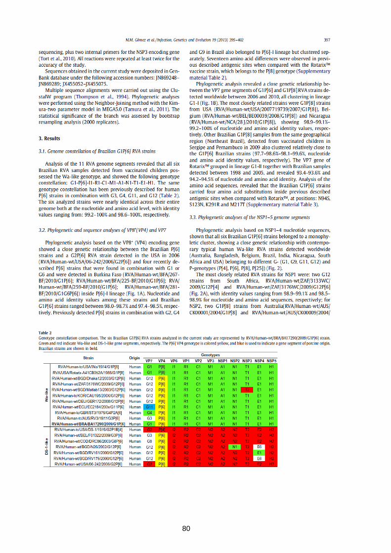
80
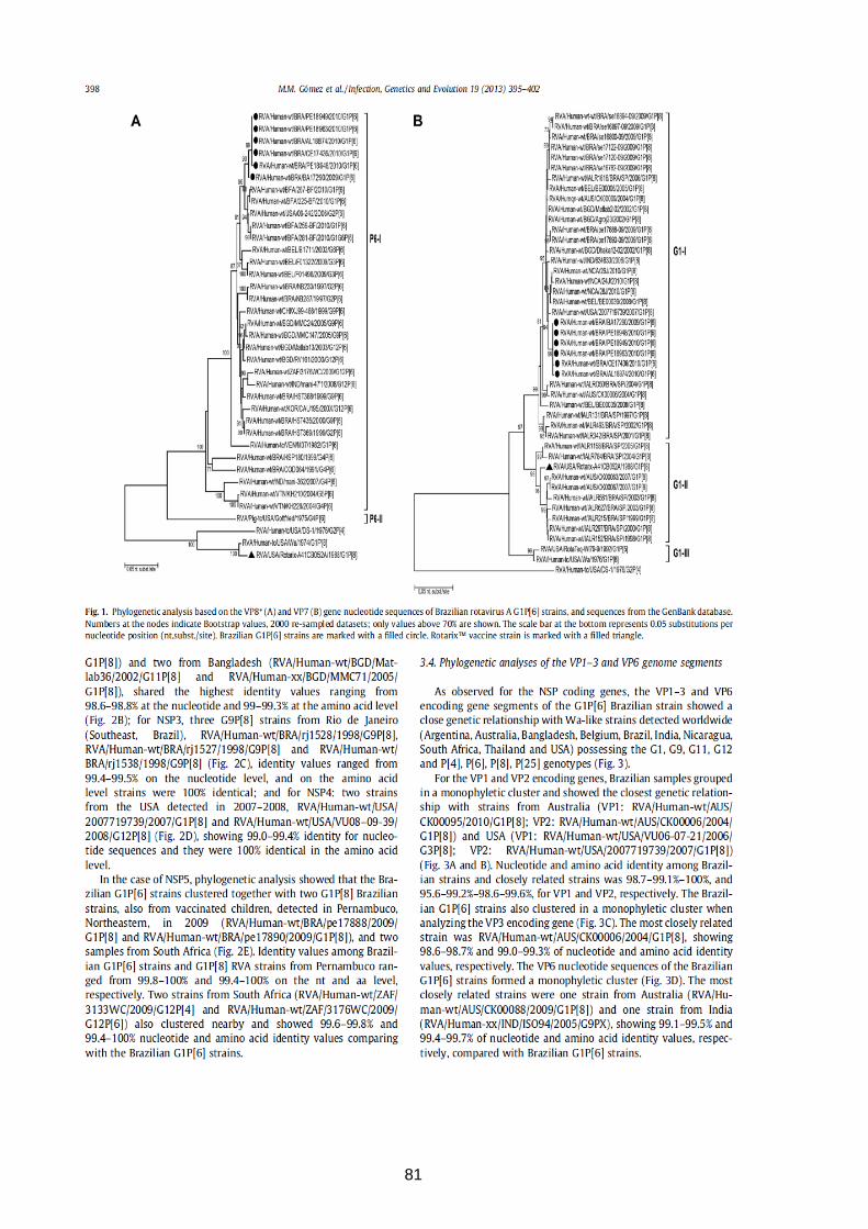
81
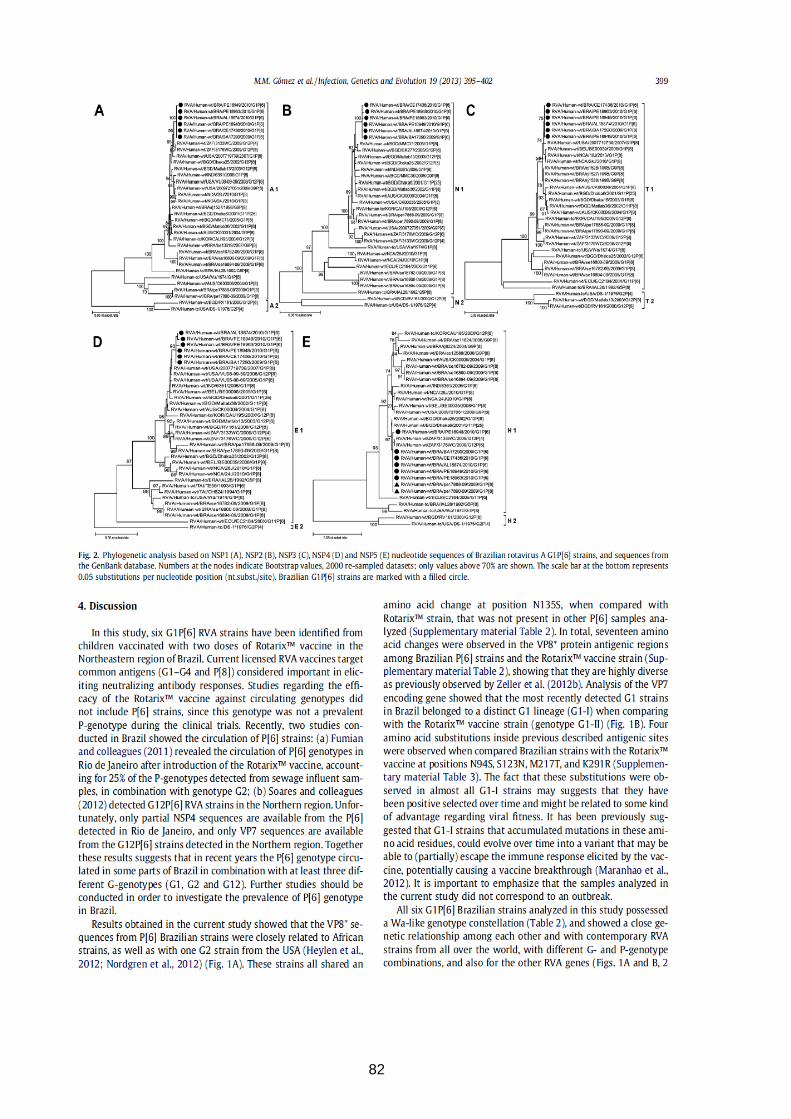
82
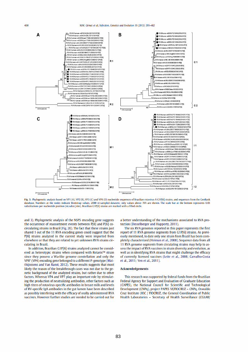
83
84
85
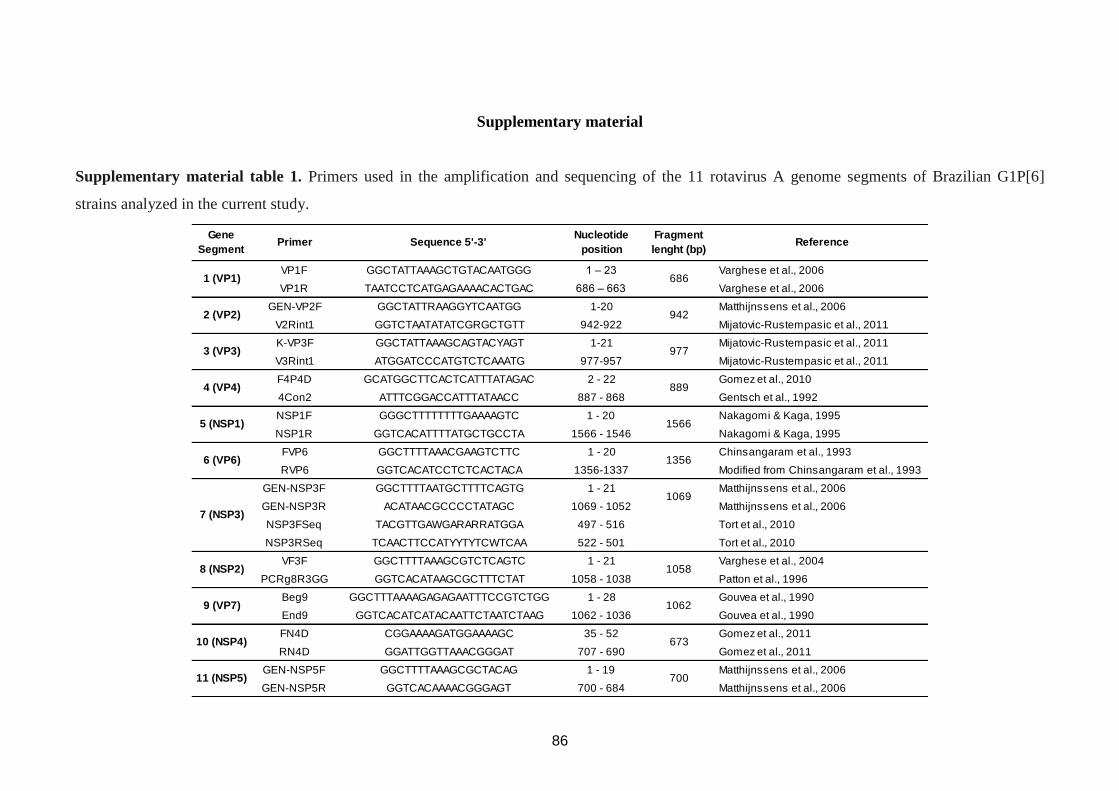
86
Supplementary material
Supplementary material table 1. Primers used in the amplification and sequencing of the 11 rotavirus A genome segments of Brazilian G1P[6]
strains analyzed in the current study.
VP1F GGCTATTAAAGCTGTACAATGGG 1 – 23 Varghese et al., 2006
VP1R TAATCCTCATGAGAAAACACTGAC 686 – 663 Varghese et al., 2006
GEN-VP2F GGCTATTRAAGGYTCAATGG 1-20 Matthijnssens et al., 2006
V2Rint1 GGTCTAATATATCGRGCTGTT 942-922 Mijatovic-Rustempasic et al., 2011
K-VP3F GGCTATTAAAGCAGTACYAGT 1-21 Mijatovic-Rustempasic et al., 2011
V3Rint1 ATGGATCCCATGTCTCAAATG 977-957 Mijatovic-Rustempasic et al., 2011
F4P4D GCATGGCTTCACTCATTTATAGAC 2 - 22 Gomez et al., 2010
4Con2 ATTTCGGACCATTTATAACC 887 - 868 Gentsch et al., 1992
NSP1F GGGCTTTTTTTTGAAAAGTC 1 - 20 Nakagomi & Kaga, 1995
NSP1R GGTCACATTTTATGCTGCCTA 1566 - 1546 Nakagomi & Kaga, 1995
FVP6 GGCTTTTAAACGAAGTCTTC 1 - 20 Chinsangaram et al., 1993
RVP6 GGTCACATCCTCTCACTACA 1356-1337 Modified from Chinsangaram et al., 1993
GEN-NSP3F GGCTTTTAATGCTTTTCAGTG 1 - 21 Matthijnssens et al., 2006
GEN-NSP3R ACATAACGCCCCTATAGC 1069 - 1052 Matthijnssens et al., 2006
NSP3FSeq TACGTTGAWGARARRATGGA 497 - 516 Tort et al., 2010
NSP3RSeq TCAACTTCCATYYTYTCWTCAA 522 - 501 Tort et al., 2010
VF3F GGCTTTTAAAGCGTCTCAGTC 1 - 21 Varghese et al., 2004
PCRg8R3GG GGTCACATAAGCGCTTTCTAT 1058 - 1038 Patton et al., 1996
Beg9 GGCTTTAAAAGAGAGAATTTCCGTCTGG 1 - 28 Gouvea et al., 1990
End9 GGTCACATCATACAATTCTAATCTAAG 1062 - 1036 Gouvea et al., 1990
FN4D CGGAAAAGATGGAAAAGC 35 - 52 Gomez et al., 2011
RN4D GGATTGGTTAAACGGGAT 707 - 690 Gomez et al., 2011
GEN-NSP5F GGCTTTTAAAGCGCTACAG 1 - 19 Matthijnssens et al., 2006
GEN-NSP5R GGTCACAAAACGGGAGT 700 - 684 Matthijnssens et al., 2006
Sequence 5'-3' Nucleotide
positionReference
3 (VP3)
2 (VP2)
1 (VP1)
Fragment
lenght (bp)
686
942
977
10 (NSP4)
6 (VP6)
9 (VP7)
5 (NSP1)
Gene
SegmentPrimer
4 (VP4)
7 (NSP3)
8 (NSP2)
889
1566
1356
1069
1058
1062
673
70011 (NSP5)
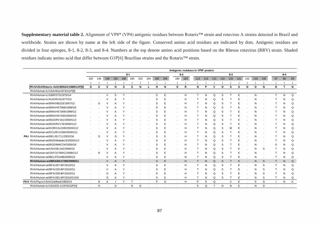
87
Supplementary material table 2. Alignment of VP8* (VP4) antigenic residues between Rotarix™ strain and rotavirus A strains detected in Brazil and
worldwide. Strains are shown by name at the left side of the figure. Conserved amino acid residues are indicated by dots. Antigenic residues are
divided in four epitopes, 8-1, 8-2, 8-3, and 8-4. Numbers at the top denote amino acid positions based on the Rhesus rotavirus (RRV) strain. Shaded
residues indicate amino acid that differ between G1P[6] Brazilian strains and the Rotarix™ strain.
100 146 148 150 188 190 193 194 195 196 180 183 113 114 115 116 125 131 132 133 135 87 88 89
| | | | | | | | | | | | | | | | | | | | | | | |
RVA/USA/Rotarix-A41CB052A/1988/G1P[8] D S S N S S N L N N E R N P V D S S N D N N T N
RVA/Human-tc/USA/Wa/1974/G1P[8] . . . . . . . . . . . . . . . . . . . . . . . .
RVA/Human-tc/GBR/ST3/1975/G4 . . V S Y . . . S E . H T N Q S T E . N . T N Q
RVA/Human-tc/AUS/RV3/1977/G3 . . V A Y . . . S E . H T N Q S T E . N . T N Q
RVA/Human-wt/BRA/NB233/1997/G2 . G V A Y . . . S E . H T N Q S T E . N . T N Q
RVA/Human-wt/BRA/HST368/1999/G9 . . V A Y . . . S E . H T N Q S T E . N . T N Q
RVA/Human-wt/BRA/HST369/1999/G2 . . V A Y . . . S E . H T N Q S T E . N . T N Q
RVA/Human-wt/BRA/HST435/2000/G9 . . V A Y . . . S E . H T N Q S T E . N . T N Q
RVA/Human-wt/BGD/RV161/2000/G12 . . V A Y . . . S E . H T N Q S T E . N . T N Q
RVA/Human-wt/BGD/RV176/2000/G12 . . V A Y . . . S E . H T N Q S T E . N . T N Q
RVA/Human-wt/KOR/CAU195/200X/G12 . . V A Y . . . S E . H T N Q S M E . N . T N Q
RVA/Human-wt/ECU/EC2184/200X/G11 . . V A Y . . . S E . H T N Q S T E . N . T N Q
RVA/Human-wt/BEL/B1711/2002/G6 . G V G Y . . . S E . H T N Q S T E . N . T N Q
RVA/Human-wt/BGD/Matlab13/2003/G12 . . V A Y . . . S E . H T N Q S T E . N . T N Q
RVA/Human-wt/BGD/MMC24/2005/G9 . . V A Y . . . S E . H T N Q S T E . N . S N Q
RVA/Human-wt/USA/06-242/2006/G2 . . V A Y . . . S E . H T N Q S T E . N S T N Q
RVA/Human-wt/ZAF/3176WC/2009/G12 . N V A Y . . . S E . H T N Q S T E . N . T N Q
RVA/Human-wt/BEL/F01498/2009/G3 . . V A Y . . . S E . N T N Q S T E . N . T N Q
RVA/Human-wt/BRA/BA17290/2009/G1 . . V A Y . . . S E . H T N Q S T E . N S T N Q
RVA/Human-wt/BFA/267-BF/2010/G1 . . V A Y . . . S E . H T N Q S T E . N S T N Q
RVA/Human-wt/BFA/225-BF/2010/G1 . . V A Y . . . S E . H T N Q S T E . N S T N Q
RVA/Human-wt/BFA/259-BF/2010/G1 . . G A Y . . . S E . H T N Q S T E . N S T N Q
RVA/Human-wt/BFA/281-BF/2010/G1G6 . . G A Y . . . S E . H T N Q S T E . N S T N Q
P6-II RVA/Pig-tc/USA/Gottfried/1983/G4 . N A I Y T . . P D . H P S Q . V E . S D I N K
RVA/Human-tc/USA/DS-1/1976/G2P[4] . G . G . N D . . . . . S Q T N N E . N D . . .
P6-I
Antigenic residues in VP8* protein
8-1 8-2 8-3 8-4
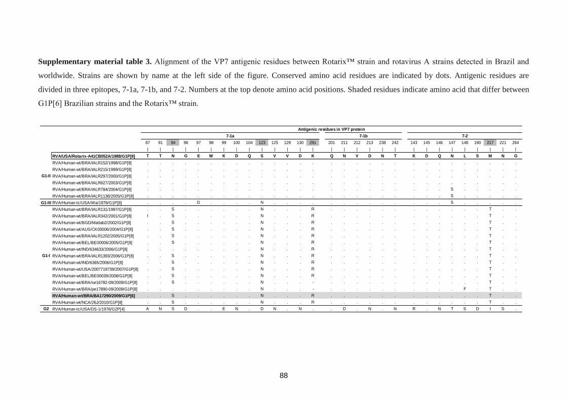
88
Supplementary material table 3. Alignment of the VP7 antigenic residues between Rotarix™ strain and rotavirus A strains detected in Brazil and
worldwide. Strains are shown by name at the left side of the figure. Conserved amino acid residues are indicated by dots. Antigenic residues are
divided in three epitopes, 7-1a, 7-1b, and 7-2. Numbers at the top denote amino acid positions. Shaded residues indicate amino acid that differ between
G1P[6] Brazilian strains and the Rotarix™ strain.
87 91 94 96 97 98 99 100 104 123 125 129 130 291 201 211 212 213 238 242 143 145 146 147 148 190 217 221 264
| | | | | | | | | | | | | | | | | | | | | | | | | | | | |
RVA/USA/Rotarix-A41CB052A/1988/G1P[8] T T N G E W K D Q S V V D K Q N V D N T K D Q N L S M N G
RVA/Human-wt/BRA/IALR152/1998/G1P[8] . . . . . . . . . . . . . . . . . . . . . . . . . . . . .
RVA/Human-wt/BRA/IALR215/1999/G1P[8] . . . . . . . . . . . . . . . . . . . . . . . . . . . . .
RVA/Human-wt/BRA/IALR297/2000/G1P[8] . . . . . . . . . . . . . . . . . . . . . . . . . . . . .
RVA/Human-wt/BRA/IALR627/2003/G1P[8] . . . . . . . . . . . . . . . . . . . . . . . . . . . . .
RVA/Human-wt/BRA/IALR784/2004/G1P[8] . . . . . . . . . . . . . . . . . . . . . . . S . . . . .
RVA/Human-wt/BRA/IALR1138/2005/G1P[8] . . . . . . . . . . . . . . . . . . . . . . . S . . . . .
G1-III RVA/Human-tc/USA/Wa/1976/G1P[8] . . . . D . . . . N . . . . . . . . . . . . . S . . . . .
RVA/Human-wt/BRA/IALR131/1997/G1P[8] . . S . . . . . . N . . . R . . . . . . . . . . . . T . .
RVA/Human-wt/BRA/IALR342/2001/G1P[8] I . S . . . . . . N . . . R . . . . . . . . . . . . T . .
RVA/Human-wt/BGD/Matlab2/2002/G1P[8] . . S . . . . . . N . . . R . . . . . . . . . . . . T . .
RVA/Human-wt/AUS/CK00006/2004/G1P[8] . . S . . . . . . N . . . R . . . . . . . . . . . . T . .
RVA/Human-wt/BRA/IALR1202/2005/G1P[8] . . S . . . . . . N . . . R . . . . . . . . . . . . T . .
RVA/Human-wt/BEL/BE00006/2005/G1P[8] . . S . . . . . . N . . . R . . . . . . . . . . . . T . .
RVA/Human-wt/IND/634633/2006/G1P[8] . . . . . . . . . N . . . R . . . . . . . . . . . . T . .
RVA/Human-wt/BRA/IALR1393/2006/G1P[8] . . S . . . . . . N . . . R . . . . . . . . . . . . T . .
RVA/Human-wt/IND/6365/2006/G1P[8] . . S . . . . . . N . . . R . . . . . . . . . . . . T . .
RVA/Human-wt/USA/2007719739/2007/G1P[8] . . S . . . . . . N . . . R . . . . . . . . . . . . T . .
RVA/Human-wt/BEL/BE00039/2008/G1P[8] . . S . . . . . . N . . . R . . . . . . . . . . . . T . .
RVA/Human-wt/BRA/se16782-09/2009/G1P[8] . . S . . . . . . N . . . - . . . . . . . . . . . . T . .
RVA/Human-wt/BRA/pe17890-09/2009/G1P[8] . . . . . . . . . N . . . - . . . . . . . . . . F . T . .
RVA/Human-wt/BRA/BA17290/2009/G1P[6] . . S . . . . . . N . . . R . . . . . . . . . . . . T . .
RVA/Human-wt/NCA/26J/2010/G1P[8] . . S . . . . . . N . . . R . . . . . . . . . . . . T . .
G2 RVA/Human-tc/USA/DS-1/1976/G2P[4] A N S D . . E N . D N . N . . D . N . N R . N T S D I S .
Antigenic residues in VP7 protein
G1-I
G1-II
7-1b7-1a 7-2
89
CAPITULO IV
CONSTELAÇÃO GÊNICA DE RVA GENÓTIPO G12P[9]
E G12P[8] DETECTADOS NO BRASIL (Artigo 4)
90
Artigo 5: Distinct evolutionary origins of G12P[8] and G12P[9] group A rotavirus strains
circulating in Brazil. (Artigo aceito: 08/04/2014)
Este artigo esta associado aos objetivos 1 e 3.
91
Distinct evolutionary origins of G12P[8] and G12P[9] group A rotavirus strains circulating in
Brazil.
Mariela M. Gómeza*, Hugo Reis Resque
b, Eduardo de Mello. Volotão
a, Tatiana Lundgren
Rosea, Marcelle Figeuira Marques da Silva
a, Elisabeth Heylen
c, Mark Zeller
c, Jelle
Matthijnssensc, and José Paulo Gagliardi Leite
a.
aLaboratory of Comparative and Environmental Virology, Oswaldo Cruz Institute-Fiocruz,
Rio de Janeiro, RJ, Brazil; bLaboratory of Norovirus and Astrovirus, Section of Virology,
Evandro Chagas Institute, Belém, PA, Brazil; cLaboratory of Clinical and Epidemiological
Virology, Department of Microbiology and Immunology, Rega Institute for Medical
Research, University of Leuven, Leuven, Belgium.
*Address correspondence to: Mariela M. Gómez. Laboratório de Virologia Comparada e
Ambiental. Instituto Oswaldo Cruz, Av. Brasil, 4365 – Pavilhão Hélio & Peggy Pereira
21040-360. Rio de Janeiro, RJ, Brasil.
e-mail: [email protected] / [email protected]
Running Head: Genetic characterization of Brazilian G12 RVA
92
Abstract. G12 group A rotavirus (RVA) are currently recognized as a globally emerging
genotype and have been described in combination with several P-types. In Brazil, G12 RVA
strains have been described in the Southern (2003) and Northern (2008 – 2010) regions, in
combination with the P[9] and P[6] genotype, respectively. To date, few complete genomes of
G12 RVA strains have been described (none from Brazilian strains), considering G12P[9]
genotype just one strain, RVA/Human-tc/THA/T152/1998/G12P[9], has their 11 gene
segments characterized. This study aims to determine the genomic constellation of G12P[9]
and G12P[8] RVA strains detected in Brazil between 2006 and 2011. Therefore, the eleven
gene segments of five Brazilian G12 RVA strains were amplified and sequenced, and the
genotype of each gene segment was assigned using phylogenetic analysis. Complete genome
analyses of G12 RVA strain circulating between 2006 and 2011 in Brazil revealed a
conserved Wa-like genomic constellation for three G12P[8] RVA strains; whereas the two
G12P[9] strains possessed distinct reassorted AU-1-like genomic constellations, closely
related to the reference strain RVA/Human-tc/THA/T152/1998/G12P[9] in most genes. The
results obtained in the current study suggest that G12P[9] (AU-1-like) and G12P[8] (Wa-like)
strains detected in different regions of Brazil do not share a common origin. Moreover, while
Brazilian G12P[8] RVA strains showed a complete Wa-like human constellation, both
G12P[9] strains possessed an NSP1 gene of bovine origin (NSP1), and RVA/Human-
wt/BRA/PE18974/2010/G12P[9] also possessed a VP3 gene of canine/feline origin.
Keywords: G12 rotavirus, genome constellation, reassortments.
93
1. Introduction
Group A rotavirus (RVA) genotype G12 was first detected in children with diarrhea in
the Philippines in 1987 (Taniguchi et al., 1990). Recently, this genotype has been described in
several studies conducted worldwide, and their global spread was demonstrated in the last
decade (Rahman et al., 2007; Matthijnssens et al., 2010a). In Brazil, G12 RVA strains have
been described in the Southern (2003) and Northern (2008 – 2010) regions, in combination
with the P[9] and P[6] genotypes, respectively (Pietruchinski et al., 2006; Soares et al., 2011).
However, only information about the VP7 encoding gene of these Brazilian G12 strains is
available in the GenBank database. Although data on the genetic diversity of the VP7 and/or
VP4 genes are important from the point of view of host immunity against RVA disease,
information on these genes is not enough to obtain conclusive data on the evolutionary
dynamics of RVA strains. Therefore, it is important to perform whole genome analysis of
RVA strains to better understand the genetic diversity of these viruses (Ghosh & Kobayashi,
2011).
This work reports the complete genotype constellation of two G12P[9] RVA strains
detected in 2008 (RVA/Human-wt/BRA/PE15776/2008/G12P[9]) and 2010 (RVA/Human-
wt/BRA/PE18974/2010/G12P[9]) in Pernambuco (PE) state (Northeastern Brazil), and three
G12P[8] strains, one detected in Rio de Janeiro (RJ) (Southeastern Brazil) in 2006
(RVA/Human-wt/BRA/RJ12419/2006/G12P[8]) and two in Bahia (BA) (Northeastern Brazil)
in 2011 (RVA/Human-wt/BRA/BA20142/2011/G12P[8] and RVA/Human-
wt/BRA/BA20144/2011/G12P[8]).
2. Material and Methods
All strains were recovered from children hospitalized with acute gastroenteritis, and
strains RVA/Human-wt/BRA/BA20142/2011/G12P[8] and RVA/Human-
wt/BRA/PE18974/2010/G12P[9] were collected from children vaccinated with one and two
doses of RV1 vaccine, respectively. Nucleic acids extraction and amplicon synthesis were
performed using a QIAamp Viral RNA Mini Kit and OneStep RT-PCR Kit (QIAGEN®)
following manufacturer’s instructions and amplifications conditions as described by
Matthijnssens et al. (2010b). PCR amplicons were purified using the QIAquick PCR
purification kit (QIAGEN®) and sequenced with the ABI Prism BigDye terminator cycle
sequencing reaction kit (Applied Biosystems®) on an ABI Prism 3130 automated sequencer
(Applied Biosystems®
) (Matthijnssens et al., 2010b).
Phylogenetic analyses were performed using the neighbor-joining method (Kimura-
two parameter model, 2,000 bootstrap replicates) in MEGA 5.0 (Tamura et al., 2011). Other
94
methods as GTR: General Time Reversible plus Gamma (VP3-4, NSP1) and TVM:
Transversion Model plus Gamma (VP7) were also used (data not shown). Sequences obtained
in the current study were deposited in the GenBank database under the following accession
numbers: KF907251 to KF907305.
This research was approved by the Fiocruz Ethical Research Committee (No311/06).
3. Results
G12P[9] strains clustered together with other G12P[9] strains detected worldwide
inside lineage II for the VP7 gene (Fig. 1a), and showed up to 99% similarity with strains
detected in South America and Eastern/Southeastern Asia. G12 strains in combination with
P[8] grouped in a separate cluster together with the majority of the G12P[8] and G12P[6]
strains described worldwide (99% similarity) inside lineage III (Fig. 1a).
Phylogenetic analysis based on VP4 gene revealed that G12P[9] Brazilian strains
clustered inside lineage II and were closely related to RVA/Human-
tc/THA/T152/1998/G12P[9] strain (99% similarity) (Fig. 1b). Brazilian G12P[8] strains
clustered inside lineage P[8]-III. Strain detected in 2006 was closely related to contemporary
G1P[8] and G12P[8] strains detected worldwide (2002-2008), whereas the two strains
detected in 2011 were closely related to strains detected in recent years (2009-2010) in the
USA, Australia, and Nicaragua (99% similarity) (Fig. 1b).
G12P[9] and G12P[8] Brazilian strains revealed a distinct genomic constellation: AU-
1-like and Wa-like, respectively (Table 1). Phylogenetic analyses of the NSP2-5, VP1, VP2,
and VP6 encoding genes of the G12P[9] Brazilian strains showed a very close genetic
relationship with RVA/Human-tc/THA/T152/1998/G12P[9] (data not shown). For the VP3
encoding gene, RVA/Human-wt/BRA/PE18974/2010/G12P[9] strain was closely related to
canine/feline P[9] strains, or human strains that are believed to be of canine/feline origin (Fig.
2a). The NSP1 gene of Brazilian G12P[9] strains belonged to A3 genotype clustering with
several bovine strains, or strains that are believed to be of bovine origin (Fig. 2b).
Brazilian strain RVA/Human-wt/BRA/RJ12419/2006/G12P[8] clustered together with
G1P[8] strains detected in USA (2005), while RVA/Human-wt/BRA/BA20142/2011/G12P[8]
and RVA/Human-wt/BRA/BA20144/2011/G12P[8] were nearly identical and clustered with
G12P[8] strains detected in Thailand (2009) for most of the RVA genes (data not shown).
4. Discussion
G12 RVA strains have been found in combination with each of the three major human
genotype constellations: Wa-like, DS-1-like, and AU-1-like (Rahman et al., 2007; Heiman et
95
al., 2008; Ghosh et al., 2010; Jere et al., 2011; Stupka et al., 2012). However, the majority of
G12 strains possess the Wa-like genotype constellation (Freeman et al., 2009; Matthijnssens
& Van Ranst, 2012), probably because they were found in combination with the P[8]
genotype.
In the current study G12P[8] Brazilian strains analyzed revealed a human Wa-like
genomic constellation, whereas the G12P[9] revealed a reassorted AU-1-like genomic
constellation. While all G12P[9] strains previously described worldwide possessed a rare A12
genotype (Castello et al., 2009), the NSP1 gene of Brazilian G12P[9] strains belonged to the
A3 genotype. It has been suggested that G12P[9] strains previously detected in Argentina,
Brazil, and Paraguay originated from Eastern Asia (Castello et al., 2009). In this study all
genes, except for NSP1 and VP3 (RVA/Human-wt/BRA/PE18974/2010/G12P[9]), were
shown to possess a close genetic relationship with strains detected in Eastern and
Southeastern Asia. However, it is important to notice that just few G12P[9] strains have been
detected and characterized to date.
At the time Brazil introduced the RV1 vaccine in the National Immunization Program
(March 2006), G12 specific vaccine effectiveness could not be demonstrated because of the
absence of detection of G12 previous to vaccine introduction. Since RV1 possess a human
Wa-like genomic constellation, and G12P[9] strains revealed an AU-1-like genomic
constellation, a difference in vaccine efficacy would be expected.
5. Conclusion
The results obtained in the current study suggest that G12P[9] and G12P[8] strains
detected in different regions of Brazil do not share a common origin. Moreover, while
Brazilian G12P[8] RVA strains showed a complete Wa-like human constellation, both
G12P[9] strains possessed a NSP1 gene of bovine origin, and RVA/Human-
wt/BRA/PE18974/2010/G12P[9] possessed a VP3 gene of canine/feline origin. It is important
to mention that the AU-l-like genotype constellation, sporadically found in humans, is
believed to have a close evolutionary relationship with canine/feline RVAs (Matthijnssens et
al., 2008).
A more in depth analysis of G12 RVA strains detected worldwide, involving full
genomic and antigenic characterization, will help to identify potential mutations and/or
reassorted genes that might be correlated with the capacity of RVA strains in evade the
immunity caused by RVA vaccines (Gentsch et al., 2009). Availability of such data will be
crucial to analyze the efficacy of RVA vaccines.
96
Acknowledgments
This research was supported by funds from the Program of Research Excellence
(PROEP - IOC/Fiocruz/CNPq), the National Council for Scientific and Technological
Development (CNPq), project PAPES VI/FIOCRUZ – CNPq, Oswaldo Cruz Institute (IOC /
FIOCRUZ), Brazilian Federal Agency for Support and Evaluation of Graduate Education
(CAPES) – project CAPES-MERCOSUL PPCP 023/2011, the General Coordination of
Public Health Laboratories – Secretary of Health Surveillance (CGLAB/SVS), and Carlos
Chagas Filho Foundation for Research Support of Rio de Janeiro State (FAPERJ). M.Z. was
supported by the Institute for the Promotion of Innovation through Science and Technology in
Flanders (IWT Vlaanderen). The authors would like to thank the Secretary of Public Health
State of Bahia, Pernambuco and Rio de Janeiro. We would also like to thanks to Alexandre
Madi Fialho, Juliana Andrade, and Rosane Maria Santos de Assis for technical assistance.
Mariela Martínez Gómez has a Post Doctoral position at IOC/Fiocruz, supported by a PDJ
scholarship from the CNPq.
97
References
Castello, A. A., Nakagomi, T., Nakagomi, O., Jiang, B., Kang, J. O., Glass, R. I., Glikmann,
G., Gentsch, J. R. (2009). Characterization of genotype P[9]G12 rotavirus strains from
Argentina: high similarity with Japanese and Korean G12 strains. J Med Virol.
Feb;81(2):371-81.
Freeman, M. M., Kerin, T., Hull, J., Teel, E., Esona, M., Parashar, U., Glass, R. I., Gentsch, J.
R. (2009). Phylogenetic analysis of novel G12 rotaviruses in the United States: A
molecular search for the origin of a new strain. J Med Virol. Apr;81(4):736-46.
Gentsch, J. R., Parashar, U. D., Glass, R. I. (2009). Impact of rotavirus vaccination: the
importance of monitoring strains. Future Microbiol. Dec;4(10):1231-4.
Ghosh, S., Kobayashi, N., Nagashima, S., Chawla-Sarkar, M., Krishnan, T., Ganesh, B., Naik,
T. N. (2010). Full genomic analysis and possible origin of a porcine G12 rotavirus
strain RU172. Virus Genes. Jun;40(3):382-8.
Ghosh, S. & Kobayashi, N. (2011). Whole-genomic analysis of rotavirus strains: current
status and future prospects. Future Microbiol. Sep;6(9):1049-65.
Heiman, E. M., McDonald, S. M., Barro, M., Taraporewala, Z. F., Bar-Magen, T., Patton, J.
T. (2008). Group A human rotavirus genomics: evidence that gene constellations are
influenced by viral protein interactions. J Virol. Nov;82(22):11106-16.
Jere, K. C., Mlera, L., O'Neill, H. G., Potgieter, A. C., Page, N. A., Seheri, M. L., Van Dijk,
A. A. (2011). Whole genome analyses of African G2, G8, G9, and G12 rotavirus strains
using sequence-independent amplification and 454® pyrosequencing. J Med Virol.
Nov;83(11):2018-42.
Matthijnssens, J., Ciarlet, M., Heiman, E., Arijs, I., Delbeke, T., McDonald, S. M., Palombo,
E. A., Iturriza-Gómara, M., Maes, P., & other authors. (2008). Full genome-based
classification of rotaviruses reveals a common origin between human Wa-Like and
porcine rotavirus strains and human DS-1-like and bovine rotavirus strains. J Virol.
Apr;82(7):3204-19.
Matthijnssens, J., Heylen, E., Zeller, M., Rahman, M., Lemey, P., Van Ranst, M. (2010a).
Phylodynamic analyses of rotavirus genotypes G9 and G12 underscore their potential
for swift global spread. Mol Biol Evol. 27(10):2431-6.
Matthijnssens, J., Rahman, M., Ciarlet, M., Zeller, M., Heylen, E., Nakagomi, T., Uchida, R.,
Hassan, Z., Azim, T., & other authors. (2010b). Reassortment of human rotavirus gene
segments into G11 rotavirus strains. Emerg Infect Dis. Apr;16(4):625-30.
Matthijnssens, J. & Van Ranst, M. (2012). Genotype constellation and evolution of group A
rotaviruses infecting humans., Cur Opin Virol. Aug;2(4):426-433
98
Pietruchinski, E., Benati, F., Lauretti, F., Kisielius, J., Ueda, M., Volotão, E. M., Soares, C.
C., Hoshino, Y., Linhares, R. E., & other authors. (2006). Rotavirus diarrhea in children
and adults in a southern city of Brazil in 2003: distribution of G/P types and finding of a
rare G12 strain. J Med Virol. 78(9):1241-9.
Rahman, M., Matthijnssens, J., Yang, X., Delbeke, T., Arijs, I., Taniguchi, K., Iturriza-
Gómara, M., Iftekharuddin, N., Azim, T., Van Ranst, M. (2007). Evolutionary history
and global spread of the emerging g12 human rotaviruses. J Virol. Mar;81(5):2382-90.
Soares, L. D., Lobo, P. D., Mascarenhas, J. D., Neri, D. L., Guerra, S. D., De Oliveira, A. D.,
Maestri, R. P., Oliveira, D. D., De Menezes, E. M., Linhares, A. D. (2011).
Identification of lineage III of G12 rotavirus strains in diarrheic children in the Northern
Region of Brazil between 2008 and 2010. Arch Virol. Jan;157(1):135-9.
Stupka, J. A., Degiuseppe, J. I., Parra, G. I., Argentinean National Rotavirus Surveillance
Network. (2012). Increased frequency of rotavirus G3P[8] and G12P[8] in Argentina
during 2008-2009: whole-genome characterization of emerging G12P[8] strains. J Clin
Virol. Jun;54(2):162-7.
Tamura, K., Peterson, D., Peterson, N., Stecher, G., Nei, M., Kumar, S. (2011). MEGA5:
molecular evolutionary genetics analysis using maximum likelihood, evolutionary
distance, and maximum parsimony methods. Mol Biol Evol. Oct;28(10):2731-9.
Taniguchi, K., Urasawa, T., Kobayashi, N., Gorziglia, M., Urasawa, S. (1990). Nucleotide
sequence of VP4 and VP7 genes of human rotaviruses with subgroup I specificity and
long RNA pattern: implication for new G serotype specificity. J. Virol. 64:5640–5644.
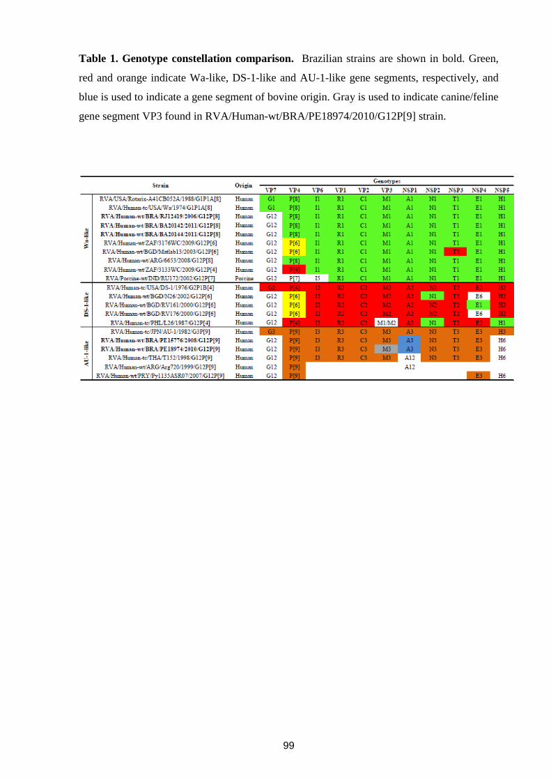
99
Table 1. Genotype constellation comparison. Brazilian strains are shown in bold. Green,
red and orange indicate Wa-like, DS-1-like and AU-1-like gene segments, respectively, and
blue is used to indicate a gene segment of bovine origin. Gray is used to indicate canine/feline
gene segment VP3 found in RVA/Human-wt/BRA/PE18974/2010/G12P[9] strain.

100
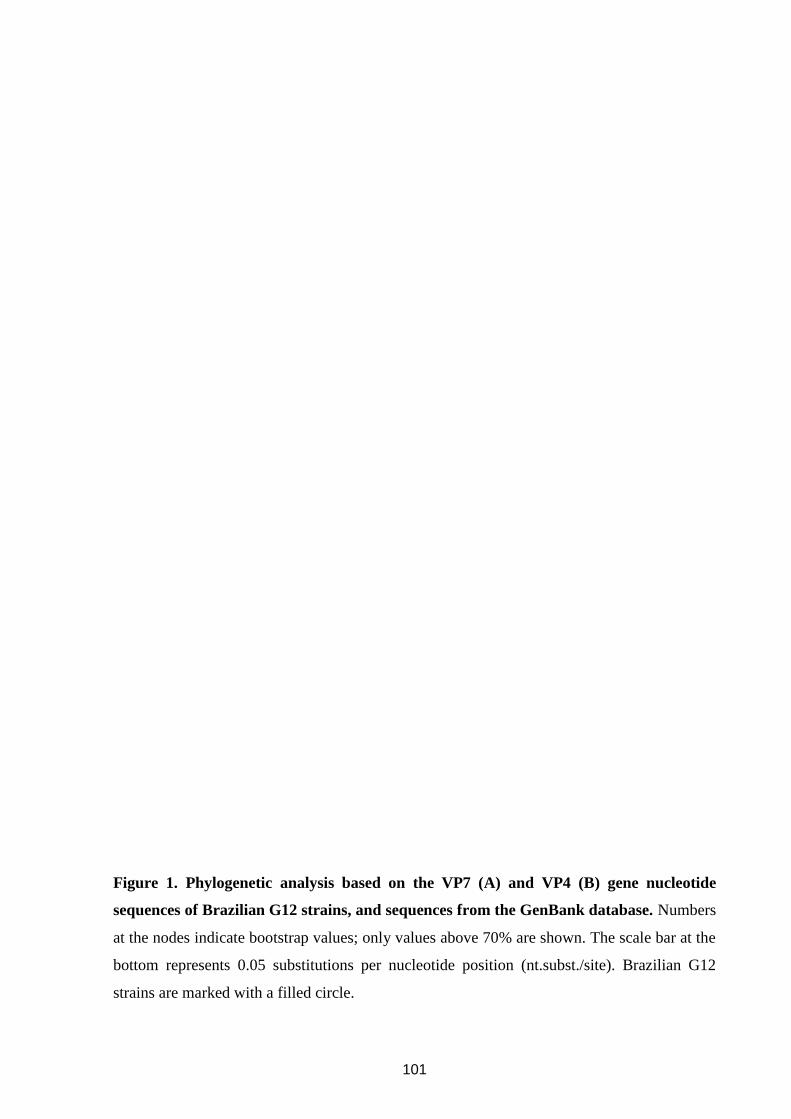
101
Figure 1. Phylogenetic analysis based on the VP7 (A) and VP4 (B) gene nucleotide
sequences of Brazilian G12 strains, and sequences from the GenBank database. Numbers
at the nodes indicate bootstrap values; only values above 70% are shown. The scale bar at the
bottom represents 0.05 substitutions per nucleotide position (nt.subst./site). Brazilian G12
strains are marked with a filled circle.
102
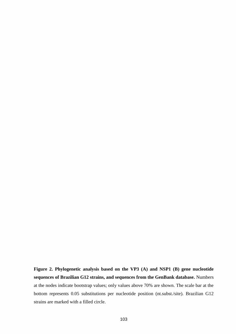
103
Figure 2. Phylogenetic analysis based on the VP3 (A) and NSP1 (B) gene nucleotide
sequences of Brazilian G12 strains, and sequences from the GenBank database. Numbers
at the nodes indicate bootstrap values; only values above 70% are shown. The scale bar at the
bottom represents 0.05 substitutions per nucleotide position (nt.subst./site). Brazilian G12
strains are marked with a filled circle.
104
CAPITULO V
ANALISE DO VIÉIS NO USO DE CÓDONS E
RESTRIÇÕES NA COMPOSIÇÃO DOS GENES QUE
CODIFICAM PARA AS PROTEÍNAS VP8* E VP7
(Artigo 5)

105
Artigo 5: Analysis of human P[4]G2 rotavirus strains isolated in Brazil reveals codon usage
bias and strong compositional constraints (Artigo publicado)
Este artigo esta relacionado ao objetivo 4.
106
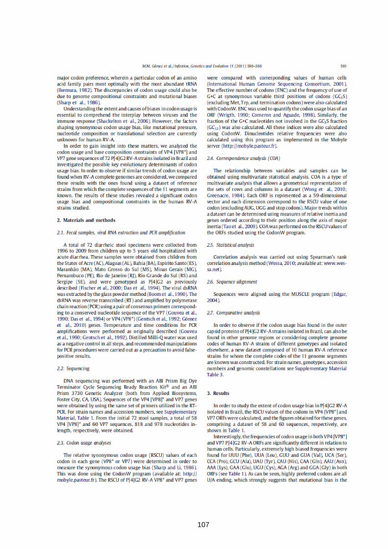
107
108
109
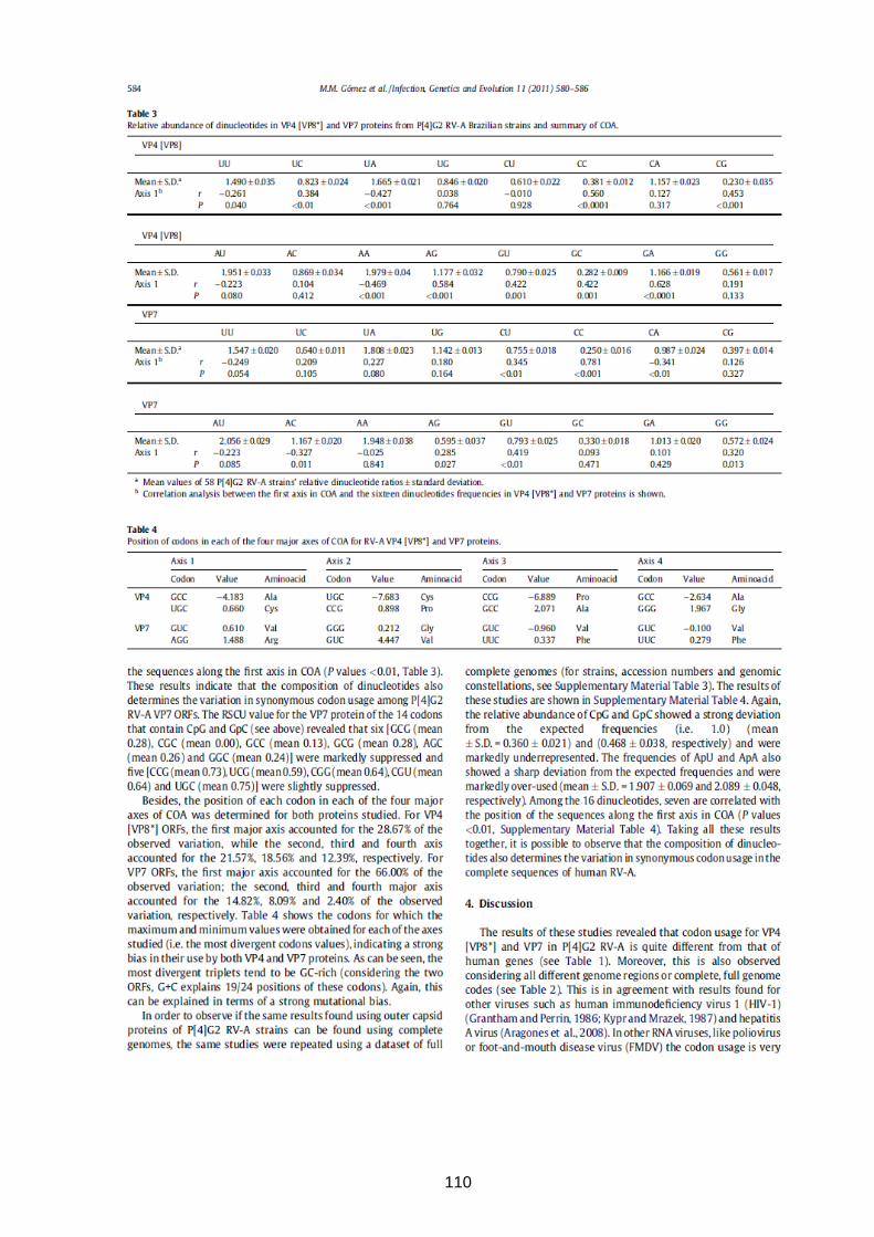
110
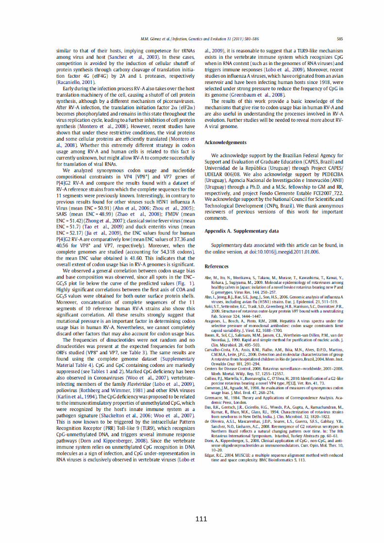
111
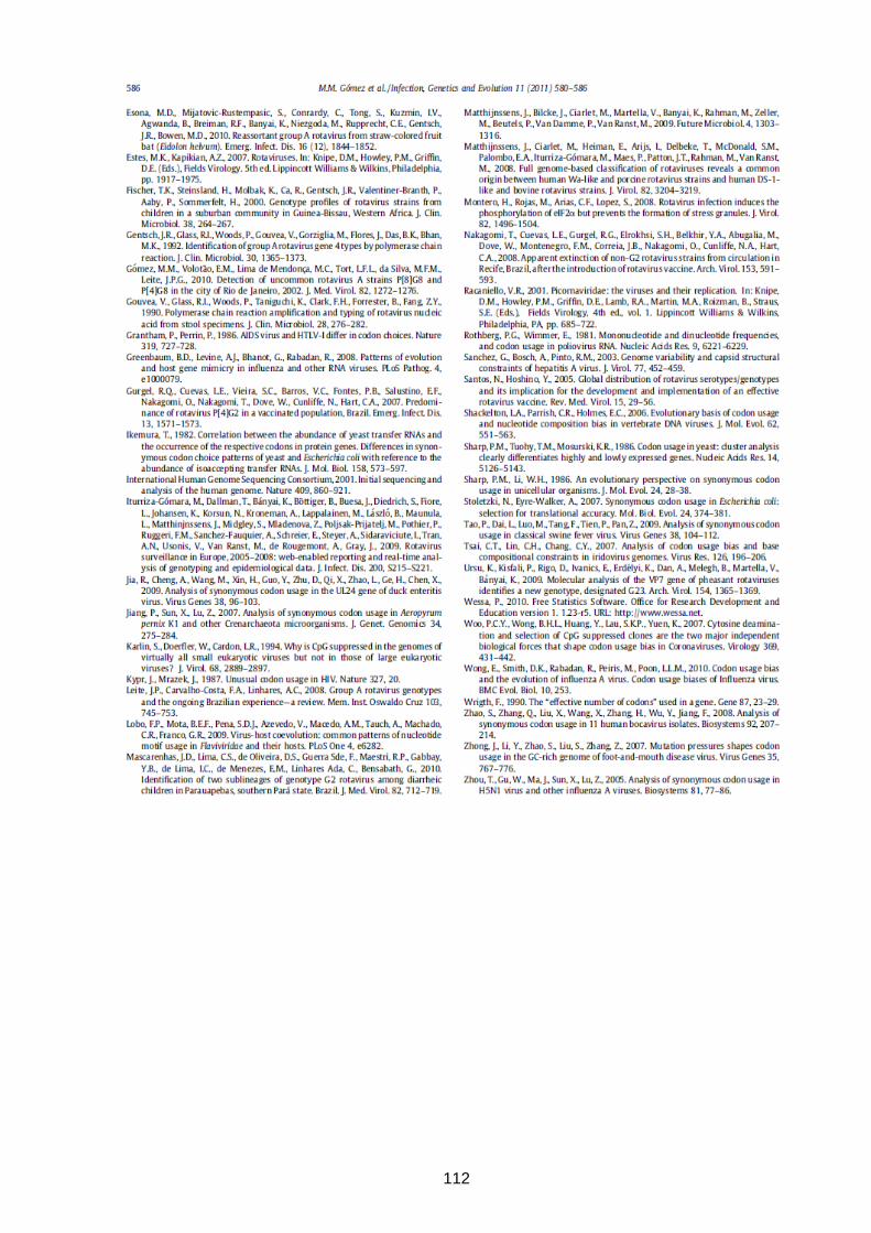
112
113
Supplementary material
Supplementary material Table 1. Origins of the P[4]G2 RVA strains.
___________________________________________________________________________
__
Name Geographic locationa Year of isolation VP4 accession number
___________________________________________________________________________
__
493 Rio de Janeiro 1996 HM066010
685 Espirito Santo 1996 HM066060
5323 Rio de Janeiro 2002 DQ857926
5619 Rio de Janeiro 2002 DQ857927
11143 Mato Grosso do Sul 2005 HM066011
11257 Mato Grosso do Sul 2005 HM066013
11580 Acre 2005 HM066012
11860 Rio de Janeiro 2006 HM066014
12220 Rio de Janeiro 2006 HM066015
12287 Bahía 2006 HM066016
12482 Bahía 2006 HM066017
12549 Rio de Janeiro 2006 HM066018
12589 Rio de Janeiro 2006 HM066019
12647 Rio de Janeiro 2006 HM066020
12684 Rio de Janeiro 2006 HM066021
12777 Bahía 2006 HM066022
12840 Espírito Santo 2006 HM066023
12842 Espírito Santo 2006 HM066024
13039 Rio Grande do Sul 2006 HM066025
13079 Rio Grande do Sul 2006 HM066026
13081 Rio Grande do Sul 2006 HM066027
13151 Rio Grande do Sul 2006 HM066028
13158 Espirito Santo 2006 HM066029
13668 Alagoas 2007 HM066030
13788 Sergipe 2007 HM066031
13793 Sergipe 2007 HM066032
14303 Minas Gerais 2007 HM066033
14422 Rio Grande do Sul 2007 HM066034
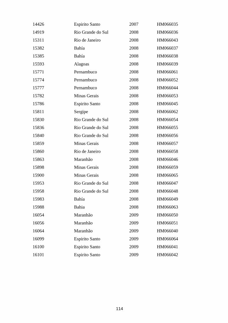
114
14426 Espirito Santo 2007 HM066035
14919 Rio Grande do Sul 2008 HM066036
15311 Rio de Janeiro 2008 HM066043
15382 Bahía 2008 HM066037
15385 Bahía 2008 HM066038
15593 Alagoas 2008 HM066039
15771 Pernambuco 2008 HM066061
15774 Pernambuco 2008 HM066052
15777 Pernambuco 2008 HM066044
15782 Minas Gerais 2008 HM066053
15786 Espirito Santo 2008 HM066045
15811 Sergipe 2008 HM066062
15830 Rio Grande do Sul 2008 HM066054
15836 Rio Grande do Sul 2008 HM066055
15840 Rio Grande do Sul 2008 HM066056
15859 Minas Gerais 2008 HM066057
15860 Rio de Janeiro 2008 HM066058
15863 Maranhão 2008 HM066046
15898 Minas Gerais 2008 HM066059
15900 Minas Gerais 2008 HM066065
15953 Rio Grande do Sul 2008 HM066047
15958 Rio Grande do Sul 2008 HM066048
15983 Bahía 2008 HM066049
15988 Bahia 2008 HM066063
16054 Maranhão 2009 HM066050
16056 Maranhão 2009 HM066051
16064 Maranhão 2009 HM066040
16099 Espirito Santo 2009 HM066064
16100 Espirito Santo 2009 HM066041
16101 Espirito Santo 2009 HM066042
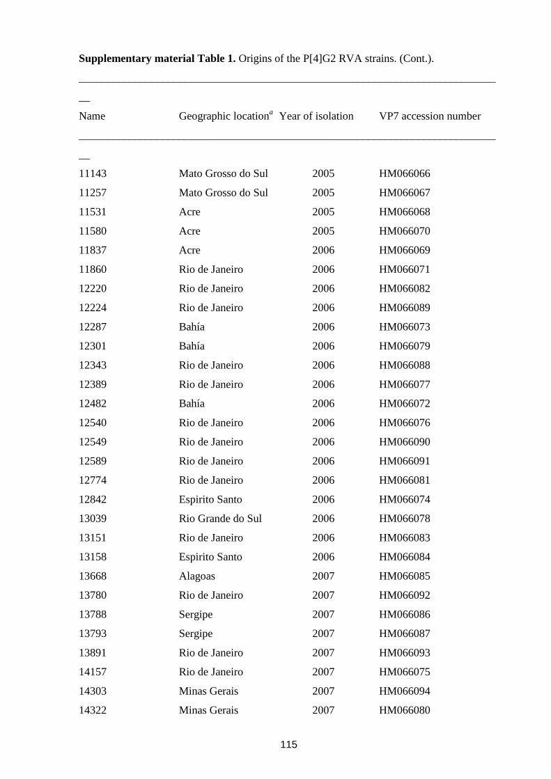
115
Supplementary material Table 1. Origins of the P[4]G2 RVA strains. (Cont.).
___________________________________________________________________________
__
Name Geographic locationa Year of isolation VP7 accession number
___________________________________________________________________________
__
11143 Mato Grosso do Sul 2005 HM066066
11257 Mato Grosso do Sul 2005 HM066067
11531 Acre 2005 HM066068
11580 Acre 2005 HM066070
11837 Acre 2006 HM066069
11860 Rio de Janeiro 2006 HM066071
12220 Rio de Janeiro 2006 HM066082
12224 Rio de Janeiro 2006 HM066089
12287 Bahía 2006 HM066073
12301 Bahía 2006 HM066079
12343 Rio de Janeiro 2006 HM066088
12389 Rio de Janeiro 2006 HM066077
12482 Bahía 2006 HM066072
12540 Rio de Janeiro 2006 HM066076
12549 Rio de Janeiro 2006 HM066090
12589 Rio de Janeiro 2006 HM066091
12774 Rio de Janeiro 2006 HM066081
12842 Espirito Santo 2006 HM066074
13039 Rio Grande do Sul 2006 HM066078
13151 Rio de Janeiro 2006 HM066083
13158 Espirito Santo 2006 HM066084
13668 Alagoas 2007 HM066085
13780 Rio de Janeiro 2007 HM066092
13788 Sergipe 2007 HM066086
13793 Sergipe 2007 HM066087
13891 Rio de Janeiro 2007 HM066093
14157 Rio de Janeiro 2007 HM066075
14303 Minas Gerais 2007 HM066094
14322 Minas Gerais 2007 HM066080
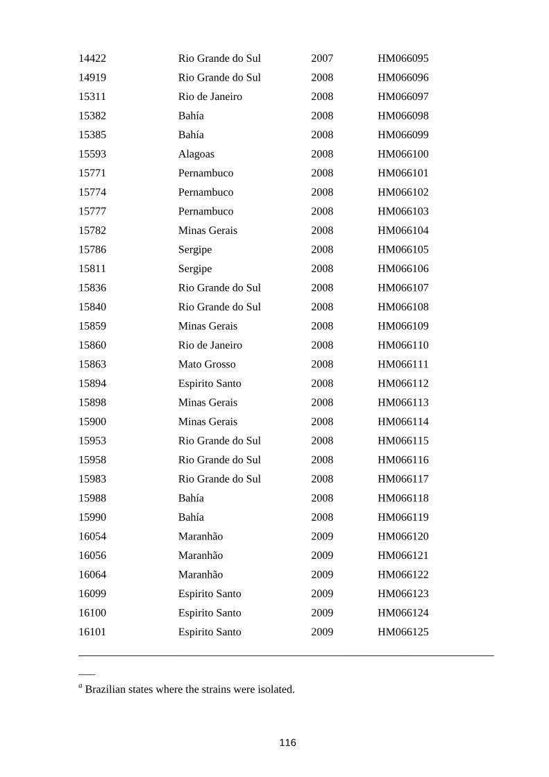
116
14422 Rio Grande do Sul 2007 HM066095
14919 Rio Grande do Sul 2008 HM066096
15311 Rio de Janeiro 2008 HM066097
15382 Bahía 2008 HM066098
15385 Bahía 2008 HM066099
15593 Alagoas 2008 HM066100
15771 Pernambuco 2008 HM066101
15774 Pernambuco 2008 HM066102
15777 Pernambuco 2008 HM066103
15782 Minas Gerais 2008 HM066104
15786 Sergipe 2008 HM066105
15811 Sergipe 2008 HM066106
15836 Rio Grande do Sul 2008 HM066107
15840 Rio Grande do Sul 2008 HM066108
15859 Minas Gerais 2008 HM066109
15860 Rio de Janeiro 2008 HM066110
15863 Mato Grosso 2008 HM066111
15894 Espirito Santo 2008 HM066112
15898 Minas Gerais 2008 HM066113
15900 Minas Gerais 2008 HM066114
15953 Rio Grande do Sul 2008 HM066115
15958 Rio Grande do Sul 2008 HM066116
15983 Rio Grande do Sul 2008 HM066117
15988 Bahía 2008 HM066118
15990 Bahía 2008 HM066119
16054 Maranhão 2009 HM066120
16056 Maranhão 2009 HM066121
16064 Maranhão 2009 HM066122
16099 Espirito Santo 2009 HM066123
16100 Espirito Santo 2009 HM066124
16101 Espirito Santo 2009 HM066125
___________________________________________________________________________
___
a Brazilian states where the strains were isolated.
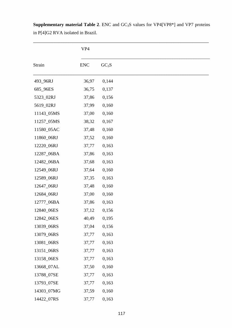
117
Supplementary material Table 2. ENC and GC3S values for VP4[VP8*] and VP7 proteins
in P[4]G2 RVA isolated in Brazil.
___________________________________________________________________________
VP4
_______________________________________________________
Strain ENC GC3S
___________________________________________________________________________
493_96RJ 36,97 0,144
685_96ES 36,75 0,137
5323_02RJ 37,86 0,156
5619_02RJ 37,99 0,160
11143_05MS 37,00 0,160
11257_05MS 38,32 0,167
11580_05AC 37,48 0,160
11860_06RJ 37,52 0,160
12220_06RJ 37,77 0,163
12287_06BA 37,86 0,163
12482_06BA 37,68 0,163
12549_06RJ 37,64 0,160
12589_06RJ 37,35 0,163
12647_06RJ 37,48 0,160
12684_06RJ 37,00 0,160
12777_06BA 37,86 0,163
12840_06ES 37,12 0,156
12842_06ES 40,49 0,195
13039_06RS 37,04 0,156
13079_06RS 37,77 0,163
13081_06RS 37,77 0,163
13151_06RS 37,77 0,163
13158_06ES 37,77 0,163
13668_07AL 37,50 0,160
13788_07SE 37,77 0,163
13793_07SE 37,77 0,163
14303_07MG 37,59 0,160
14422_07RS 37,77 0,163
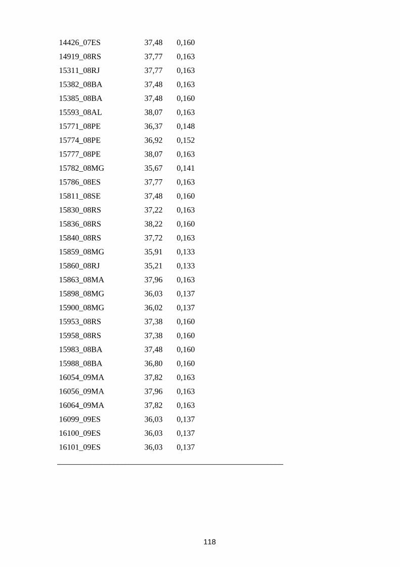
118
14426_07ES 37,48 0,160
14919_08RS 37,77 0,163
15311_08RJ 37,77 0,163
15382_08BA 37,48 0,163
15385_08BA 37,48 0,160
15593_08AL 38,07 0,163
15771_08PE 36,37 0,148
15774_08PE 36,92 0,152
15777_08PE 38,07 0,163
15782_08MG 35,67 0,141
15786_08ES 37,77 0,163
15811_08SE 37,48 0,160
15830_08RS 37,22 0,163
15836_08RS 38,22 0,160
15840_08RS 37,72 0,163
15859_08MG 35,91 0,133
15860_08RJ 35,21 0,133
15863_08MA 37,96 0,163
15898_08MG 36,03 0,137
15900_08MG 36,02 0,137
15953_08RS 37,38 0,160
15958_08RS 37,38 0,160
15983_08BA 37,48 0,160
15988_08BA 36,80 0,160
16054_09MA 37,82 0,163
16056_09MA 37,96 0,163
16064_09MA 37,82 0,163
16099_09ES 36,03 0,137
16100_09ES 36,03 0,137
16101_09ES 36,03 0,137
________________________________________________________
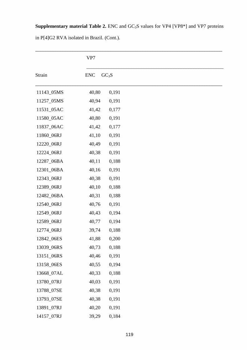
119
Supplementary material Table 2. ENC and GC3S values for VP4 [VP8*] and VP7 proteins
in P[4]G2 RVA isolated in Brazil. (Cont.).
___________________________________________________________________________
VP7
_______________________________________________________
Strain ENC GC3S
___________________________________________________________________________
11143_05MS 40,80 0,191
11257_05MS 40,94 0,191
11531_05AC 41,42 0,177
11580_05AC 40,80 0,191
11837_06AC 41,42 0,177
11860_06RJ 41,10 0,191
12220_06RJ 40,49 0,191
12224_06RJ 40,38 0,191
12287_06BA 40,11 0,188
12301_06BA 40,16 0,191
12343_06RJ 40,38 0,191
12389_06RJ 40,10 0,188
12482_06BA 40,31 0,188
12540_06RJ 40,76 0,191
12549_06RJ 40,43 0,194
12589_06RJ 40,77 0,194
12774_06RJ 39,74 0,188
12842_06ES 41,88 0,200
13039_06RS 40,73 0,188
13151_06RS 40,46 0,191
13158_06ES 40,55 0,194
13668_07AL 40,33 0,188
13780_07RJ 40,03 0,191
13788_07SE 40,38 0,191
13793_07SE 40,38 0,191
13891_07RJ 40,20 0,191
14157_07RJ 39,29 0,184
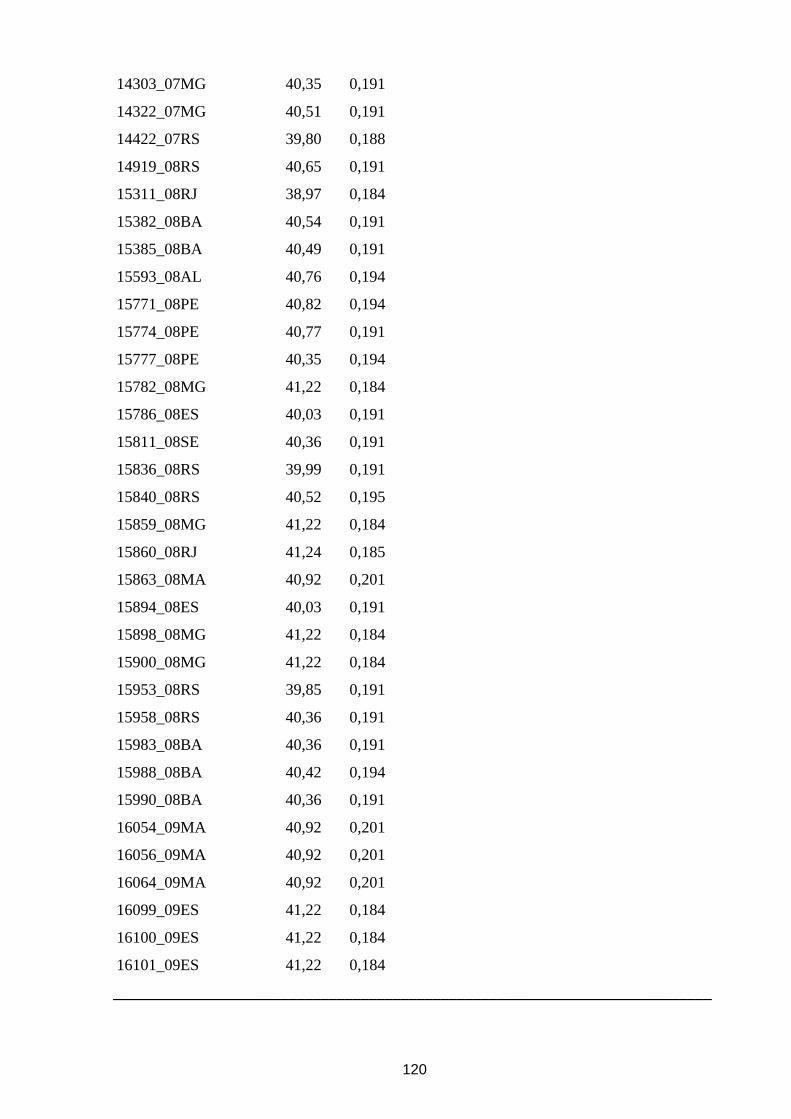
120
14303_07MG 40,35 0,191
14322_07MG 40,51 0,191
14422_07RS 39,80 0,188
14919_08RS 40,65 0,191
15311_08RJ 38,97 0,184
15382_08BA 40,54 0,191
15385_08BA 40,49 0,191
15593_08AL 40,76 0,194
15771_08PE 40,82 0,194
15774_08PE 40,77 0,191
15777_08PE 40,35 0,194
15782_08MG 41,22 0,184
15786_08ES 40,03 0,191
15811_08SE 40,36 0,191
15836_08RS 39,99 0,191
15840_08RS 40,52 0,195
15859_08MG 41,22 0,184
15860_08RJ 41,24 0,185
15863_08MA 40,92 0,201
15894_08ES 40,03 0,191
15898_08MG 41,22 0,184
15900_08MG 41,22 0,184
15953_08RS 39,85 0,191
15958_08RS 40,36 0,191
15983_08BA 40,36 0,191
15988_08BA 40,42 0,194
15990_08BA 40,36 0,191
16054_09MA 40,92 0,201
16056_09MA 40,92 0,201
16064_09MA 40,92 0,201
16099_09ES 41,22 0,184
16100_09ES 41,22 0,184
16101_09ES 41,22 0,184
___________________________________________________________________________
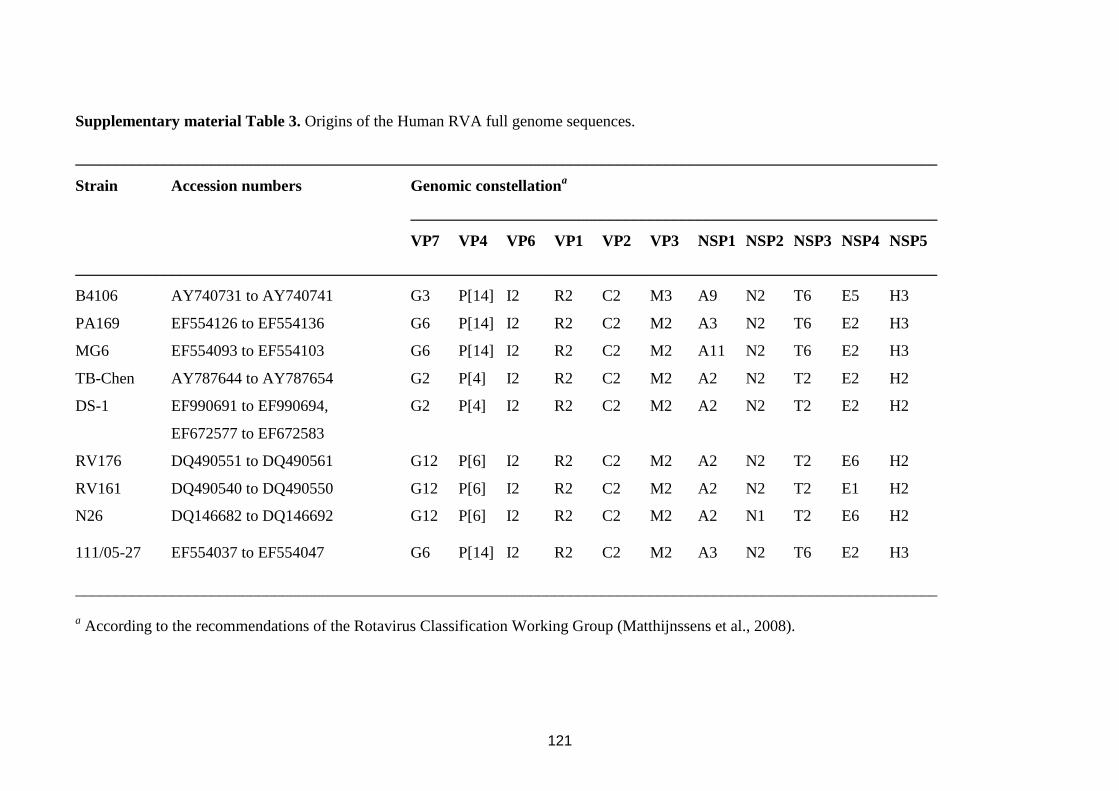
121
Supplementary material Table 3. Origins of the Human RVA full genome sequences.
____________________________________________________________________________________________________________
Strain Accession numbers Genomic constellationa
__________________________________________________________________
VP7 VP4 VP6 VP1 VP2 VP3 NSP1 NSP2 NSP3 NSP4 NSP5
____________________________________________________________________________________________________________
B4106 AY740731 to AY740741 G3 P[14] I2 R2 C2 M3 A9 N2 T6 E5 H3
PA169 EF554126 to EF554136 G6 P[14] I2 R2 C2 M2 A3 N2 T6 E2 H3
MG6 EF554093 to EF554103 G6 P[14] I2 R2 C2 M2 A11 N2 T6 E2 H3
TB-Chen AY787644 to AY787654 G2 P[4] I2 R2 C2 M2 A2 N2 T2 E2 H2
DS-1 EF990691 to EF990694, G2 P[4] I2 R2 C2 M2 A2 N2 T2 E2 H2
EF672577 to EF672583
RV176 DQ490551 to DQ490561 G12 P[6] I2 R2 C2 M2 A2 N2 T2 E6 H2
RV161 DQ490540 to DQ490550 G12 P[6] I2 R2 C2 M2 A2 N2 T2 E1 H2
N26 DQ146682 to DQ146692 G12 P[6] I2 R2 C2 M2 A2 N1 T2 E6 H2
111/05-27 EF554037 to EF554047 G6 P[14] I2 R2 C2 M2 A3 N2 T6 E2 H3
____________________________________________________________________________________________________________
a According to the recommendations of the Rotavirus Classification Working Group (Matthijnssens et al., 2008).
122
CAPITULO VI
DISCUSSÃO, CONCLUSÕES E PERSPECTIVAS
123
1. Discussão
Diversidade genética dos rotavírus da espécie A
É sabido que a distribuição de genótipos de RVA flutua tanto no decorrer do tempo
quanto nas diferentes regiões geográficas. Entretanto, os mecanismos envolvidos neste
fenômeno ainda não são bem conhecidos. Existem evidencias de que tanto a imunidade
homotípica quanto a heterotípica contra a infecção natural do RVA desempenha um papel
muito importante tanto em nível do individuo quanto da população. Existem diversos outros
fatores tais como: o fluxo continuo migratório de pessoas no mundo; a emergência de
variantes de RVA; a diversidade genética dos diferentes genótipos; a facilidade de
transmissão e resistência do vírus; fatores relacionados ao hospedeiro (genéticos, anticorpos
maternos, imunodeficiência, parasitas intestinais, má nutrição, deficiência de zinco, entre
outros). Estes fatores acima mencionados poderiam também estar influenciando, em menor ou
maior grau, a diversidade de genótipos de RVA mundialmente (Matthijnssens et al., 2012).
Neste contexto a introdução de vacinas (mono e polivalentes) poderia representar mais um
fator a ser considerado.
Atualmente, pelo menos 41 países introduziram a vacina RV1 no seu calendário de
imunizações (WHO/UNICEF, 2013). Apesar dos estudos de Fase III com a RV1 terem sido
satisfatórios e a OMS ter recomendado aos países inclusão de uma vacina anti-RVA em seus
programas de imunização, diversas questões tem sido postuladas, tais como: i) a introdução
de uma vacina monovalente poderia resultar no aumento na prevalência de genótipos que não
tenham nenhum destes G ou P tipos? ii) a reestruturação gênica intra-, entre-genogrupos e
entre amostras selvagens e vacinal de RVA poderiam ocorrer com maior frequência e
prejudicar a eficácia da vacina RV1? Uma das maneiras de se buscar responder a estas
perguntas é a analise da constelação gênica dos RVA assim como da prevalência dos
genótipos circulantes, preferencialmente antes e após a introdução da vacina RV1. Duas
constelações genéticas prevalecem mundialmente: Wa-like e DS-1-like. Dos genótipos mais
prevalentes mundialmente só um contem uma constelação DS-1-1ike (G2P[4]). Os demais
genótipos são Wa-like (G1P[8], G3P[8], G4P[8], G9P[8], e G12P[8]). A terceira constelação,
AU-1-like, tem sido ocasionalmente encontrada em humanos (Matthijnssens e Van Ranst,
2012).
124
Análise da diversidade genética do genótipo G2P[4] no período de 2005 a 2011 no Brasil
(Artigo 1 e 2)
G2P[4] foi o genótipo mais comumente detectado no Brasil após o início da vacinação
(Gurgel et al., 2007, de Oliveira et al., 2008; Nakagomi et al., 2008; Leite et al., 2008;
Carvalho -Costa et al., 2009, 2011; da Silva Soares et al., 2013). A emergência deste genótipo
tem motivado uma grande discussão. Uma correlação entre o genótipo G2P[4] e a vacinação
foi proposta (Gurgel et al., 2007; Nakagomi et al., 2008). Sendo a principal razão o fato de
que a eficiência e eficácia da vacina monovalente contra o G2P[4] (DS-1-like) é relativamente
menor do que contra cepas Wa-like (Ruiz-Palacios et al., 2006; Vesikari et al., 2007; Linhares
et al., 2008; Justino et al., 2011). Neste contexto, foi proposto que a vacinação em massa teria
exercido pressão seletiva sobre cepas de RVA circulantes, selecionando o G2P[4] (Nakagomi
et al., 2008; Gurgel et al., 2009).
Os resultados do presente estudo demonstram que a taxa de detecção de G2P[4] em
amostras RVA positivo tem sido alta nos anos após a introdução da vacinação com RV1. Com
exceção de 2009, esta taxa tem sido acima de 50%, atingindo 87% em 2010. Outro fato
importante é que o genótipo G2P[4] também tem sido frequentemente detectado em crianças
vacinadas com RV1.
Nos artigos em questão apresentamos um estudo onde examinamos mais
detalhadamente a localização geográfica de amostras positivas para G2P[4], tentando
identificar uma via de propagação deste genótipo durante o processo de emergência no Brasil.
A freqüência de detecção de G2P[4] entre 2001 e 2003 foi muito baixa, por isso apenas três
amostras positivas puderam ser identificadas nos dados de monitoramento no Laboratório de
Referencia Regional para Rotavírus (LRRR). Em 2004, G2P[4] não foi caracterizado em
nenhuma das 651 amostras fecais positivas para RVA obtidas a partir de 2.554 crianças com
gastroenterite de 11 estados brasileiros. Com isso confirmamos que o genótipo G2P[4] estava
ausente nas amostras estudadas no primeiro semestre de 2005. Neste ano, as primeiras cidades
em que o genótipo G2P[4] foi detectado estão localizadas no oeste do Brasil, na fronteira com
o Paraguai. Ao longo de 2005, G2P[4] continuou a ser detectado na região amazônica e no
Brasil, na fronteira com o Peru e a Bolívia, sendo ausente nas regiões brasileiras mais
populosas da costa atlântica. Somente em dezembro de 2005, G2P[4] foi detectado nas
principais cidades brasileiras, mais especificamente no Rio de Janeiro. A vacinação em massa
contra RVA começou em março de 2006 e, a partir deste ano, o genótipo G2P[4] foi
125
detectado precocemente em todas as regiões monitoradas, numa fase em que o grupo de
crianças vacinadas ainda era muito limitado.
Tem sido sugerido que as vacinas específicas podem selecionar genótipos de escape,
incluindo os efeitos de longo prazo de pressões seletivas fracas (Pérez-Sautu et al., 2011).
Neste contexto, as cepas emergentes resultantes da pressão evolutiva imposta pelas vacinas
podem substituir gradualmente os genótipos anteriores (Streck et al., 2013). Como resultado,
no cenário onde a seleção ocorre, uma distância genética entre as cepas circulantes surgiria
em relação a outras regiões (Hoffmann et al., 2008).
A análise das origens genéticas e antigênicas de cepas G2P[4] identificadas ao longo
do processo de implementação da imunização em massa com RV1 pode ser útil para
esclarecer seu potencial impacto sobre a distribuição de genótipos que ocorreu entre 2005 e
2011 no Brasil. Os dados obtidos no presente estudo (artigo 1 e 2) indicam que as cepas
G2P[4] circulantes no Brasil durante a implementação da vacinação em massa com RV1 não
se agrupam em grupos genéticos definidos por períodos pré e pós vacinação; com isso parece
existir uma estreita relação com cepas G2P[4] circulantes em outros países que ainda não
tinham implementado a vacinação em massa contra RVA. Da mesma forma, cepas G2P[4]
identificadas em crianças vacinadas e não vacinadas agruparam nos mesmos grupos
genéticos, não havendo diferenças significativas nas sequências dos genes que codificam VP7
e VP8*. A Análise do contexto genômico de cepas brasileira G2P[4] revelou uma constelação
genética DS-1-like completa, e diferentes variantes genéticas foram observadas circulando
entre as regiões brasileiras.
Além disso, a comparação das sequências de aminoácidos dos sítios antigênicos das
proteínas VP7 e VP8* da RV1 e as cepas G2P[4] brasileiras, revelou que a maioria das
mudanças observadas foram mantidas nas amostras brasileiras ao longo dos anos, estando
todas presentes antes da introdução da vacina RV1.
Em conclusão, os dados aqui apresentandos apoiam a ideia de que outros fatores além
da introdução da RV1 seriam responsáveis pelo aumento na prevalência de G2P[4] no Brasil.
Tem sido proposto que o G2P[4] reemerge periodicamente em ciclos de 10 anos (Bishop et
al., 1991). Leite e colaboradores (2008) relataram uma onda de circulação do G2P[4] no
início da década de 1990, perto de 10 anos antes do ressurgimento deste genótipo em 2005.
O longo período de pouca ou nenhuma circulação deste genótipo anterior à introdução
da RV1 teria criado condições favoráveis para a acumulação de indivíduos imuno-
susceptíveis (Carvalho-Costa et al., 2009; Assis et al., 2013). Este fato, juntamente com a
diversidade genética encontrada em diferentes regiões geográficas ao longo dos anos, antes,
126
durante e após a introdução da vacina poderia explicar a alta prevalência do genótipo G2P[4]
no Brasil desde 2005.
Análise da diversidade genética do genótipo G1P[6] em crianças vacinadas com RV1
(Artigo 3)
A circulação dos genótipos P[6] e G12, respectivamente nos estados do Rio de Janeiro
e na região Norte do Brasil têm sido demonstrada (Fumian et al., 2011; Soares et al., 2012).
Entretanto, apenas sequencias parciais do gene que codifica para a proteína NSP4 das
amostras detectadas no Rio de Janeiro e do gene que codifica para a proteína VP7 das
amostras G12 estão disponíveis no banco de dados do GenBank. No presente estudo seis
cepas G1P[6] detectadas a partir de crianças vacinadas com duas doses da vacina RV1 que
apresentaram gastroenterite aguda na região Nordeste do Brasil foram analisadas. A analise
das sequencias obtidas para VP8* revelaram que as cepas Brasileiras estavam bastante
relacionadas a cepas africanas, bem como com uma cepa G2P[6] detectada nos Estados
Unidos (Heylen et al., 2012; Nordgren et al., 2012) (Figura 1A). Quando comparadas com a
sequencia aminoacidica da RV1, estas cepas mostraram uma mudança de aminoácido na
posição N135S, que não estava presente em outras amostras P[6] analisadas (Material
suplementar Tabela 2). No total, foram observadas dezessete alterações de aminoácidos nas
regiões antigênicas da proteína VP8* entre as amostras brasileiras e a RV1 (Material
suplementar Tabela 2), mostrando a grande diversidade com relação a RV1 como observado
anteriormente por Zeller e colaboradores (2012a).
A análise do gene que codifica para a proteína VP7 mostrou que as cepas genótipo G1
mais recentemente detectadas no Brasil pertencem a linhagem G1-I, distinta da vacina RV1
(G1-II) (Figura 1B). Foram observadas quatro substituições de aminoácidos no interior dos
sítios antigênicos, quando comparadas as amostras brasileiras G1P[6] com a vacina RV1;
posições: N94S, S123N, M217T e K291R (Material suplementar Tabela 3). Tais substituições
foram observadas em quase todas as cepas G1-I e, portanto, as mesmas podem ter sido
selecionadas positivamente ao longo do tempo, podendo estar relacionadas a algum tipo de
vantagem em relação ao fitness viral. Tem sido sugerido que cepas G1-I que acumularam
estas mutações pontuais poderiam evoluir, ao longo do tempo, gerando variantes capazes de
escapar da resposta imune induzida pela vacina e, podendo assim, causar surtos (Maranhão et
al., 2012). É importante ressaltar que as amostras G1P[6] analisadas no presente estudo não
foram relacionadas a qualquer surto.
127
A analise da constelação gênica das cepas G1P[6] demonstrou que as mesmas pertenc
em ao genótipo Wa-like (Tabela 2), existindo uma estreita relação com cepas contemporâneas
de RVA detectadas em diferentes países, apresentando diferentes combinações de G e P
(Figuras 1A, 1B, 2 e 3). A análise filogenética do gene que codifica para a NSP5 sugere a
ocorrência de eventos de reestruturações genéticas entre cepas P[8] e P[6] cocirculantes no
Brasil (Figura 2E). O fato das cepas G1P[6] apenas compartilharem um dos onze genes de
RVA, poderia sugerir que as mesmas foram importadas de algum outro lugar ou ainda que
estariam relacionadas a cepas ainda desconhecidas que circularam no Brasil.
E importante destacar que as cepas G1P[6] analisadas não podem ser consideradas
heterotípicas quando comparadas com a RV1, pois possuem uma constelação genética Wa-
like. Somente a VP8* pertence a um genótipo P diferente (Matthijnssens & Van Ranst, 2012).
Estes resultados sugerem que provavelmente a razão pela qual estas cepas teriam causado
gastroenterite em crianças não foi devido à constelação genética das mesmas e sim devido a
outros fatores, os quais poderiam ser: i) características genéticas restritas ao hospedeiro (Hu et
al., 2012; Gunaydin et al., 2013; Imbert-Marcille et al., 2013); ii) concentrações elevadas de
anticorpos específicos anti-RVA no leite materno com níveis de IgA específicos de RVA no
lúmen do intestino (Desselberger & Huppertz, 2011).Lembremo-nos de que a eficácia da
vacina não é 100% (O´Ryan & Linhares, 2009), e sendo assim, se espera que crianças
vacinadas ainda apresentem GA. Rose e colaboradores (2013), recentemente demostrou a
ocorrência de reestruturações genéticas entre amostras de RVA genótipo G1P[8] (Wa-like) e a
RV1, detectando tal genótipo em crianças vacinadas com RV1 que sofreram GA. A
ocorrência de reestruturaçãoes genéticas entre amostras selvagens e vacinais também foi
demostrada para a RV5 (Hemming & Vesikari, 2013)
Constelação genética dos genótipos G12P[9] e G12P[8] detectados no Brasil (Artigo 4)
O genótipo de RVA G12 foi detectado pela primeira vez em crianças com diarreia nas
Filipinas, em 1987 (Taniguchi et al., 1990). Recentemente, este genótipo foi descrito em
vários estudos conduzidos em diversos países e a sua expansão global tem sido demonstrada
na última década (Castello et al., 2006; Bányai et al., 2007; Rahman et al., 2007; Ray et al.,
2007; Alam et al., 2009; Cunliffe et al., 2009; Page et al., 2009; Pietsch & Liebert, 2009;
Matthijnssens et al., 2012; Tra et al., 2011; McDonald et al., 2012; Ndze et al., 2013; Tamim
et al., 2013). No Brasil, o G12 foi descrito nas regiões Sudeste (2003) e Norte (2008 - 2010),
em combinação com o genótipo P[9] e P[6], respectivamente (Pietruchinski et al., 2006;
128
Soares et al., 2011). No entanto, apenas informações sobre o gene que codifica para a VP7
estão disponíveis no GenBank.
No artigo 4, foram analisadas as constelações genéticas de cepas de RVA genótipo
G12P[9]; detectadas em 2008 e 2010 em Pernambuco (Nordeste do Brasil) e genótipo
G12P[8]; detectadas no Rio de Janeiro (Sudeste do Brasil) em 2006 e Bahia (Nordeste do
Brasil) em 2011. O genótipo G12 tem sido associado às constelações: Wa-like, DS-1-like e
AU-1-like (Tabela 1) (Rahman et al., 2007; Heiman et al., 2008; Ghosh et al., 2010; Jere et al.,
2011; Stupka et al., 2012), sendo que a maioria das cepas G12 descritas até o momento
possuem a constelação Wa-like (Freeman et al., 2009; Matthijnssens et al., 2012). Esta
observação pode estar relacionada ao fato de que a maioria das cepas G12 analisadas estavam
associadas ao genótipo P[8]. Até o momento, apenas uma cepa G12P[9] (T152) foi analisada
para os onze segmentos gênicos mostrando uma constelação genética AU-1-like (Rahman et
al., 2007). Análises filogenéticas das sequencias nucleotidicas obtidas para NSP2-5, VP1-
VP2 e VP6 das amostras brasileiras G12P[9] mostraram uma relação genética muito estreita
com a cepa T152 (99% de similaridade) detectada em 1998 na Tailândia. As cepas G12P[9]
anteriormente descritas pertencem ao genótipo A12 (Freeman et al., 2009). No caso da NSP1,
as amostras brasileiras G12P[9], foram pertencentes ao genótipo A3, agrupando com várias
cepas de RVA bovinas e humanas, sendo que as humanas possivelmente tenham origem
bovina (Figura 2B).
A análise dos onze genes das cepas G12P[8] brasileiras revelou que a cepa
RVA/Human-wt/BRA/RJ12419/2006/G12P[8] agrupou-se junto com cepas G1P[8]
detectadas nos Estados Unidos de América (EUA) em 2005 (99% de similaridade); enquanto
as cepas RVA/Human-wt/BRA/BA20142/2011/G12P[8] e RVA/Human-
wt/BRA/BA20144/2011/G12P[8] foram quase idênticas e agruparam-se com cepas G12P[8]
detectadas na Tailândia em 2009, para a maioria dos genes de RVA (99% similaridade). Estes
resultados sugerem a circulação de diferentes variantes de G12P[8] Wa-like no Brasil.
Os resultados obtidos no presente estudo sugerem que as cepas brasileiras G12P[9]
(AU-1-like) e G12P[8] (Wa-like) detectadas em diferentes regiões do Brasil não compartilham
uma origem comum. Além disso, enquanto as G12P[8] mostraram uma constelação Wa-like
completamente relacionada a cepas humanas; as cepas G12P[9] apresentaram um gene
possivelmente de origem bovina (NSP1). Deve-se ressaltar que e a RVA/Human-
wt/BRA/PE18974/2010/G12P[9] também possuiu o gene VP3 de origem canina/felina
(Tabela 1). A constelação AU-1-like é esporadicamente mencionada em humanos. Porém
acredita-se que este o genótipo AU-1-like tenha uma relação evolutiva próxima com cepas de
RVA caninas e felinas (Nakagomi & Nakagomi, 1989; Matthijnssens et al., 2008).
129
Tem sido sugerido que as cepas G12P[9] previamente detectados na Argentina,
Paraguai e Brasil tenham origem no Leste da Ásia (Castello et al., 2009). E neste estudo,
todos os genes, exceto NSP1 e VP3 (RVA/Human-wt/BRA/PE18974/2010/G12P[9])
mostraram uma relação genética próxima com cepas detectadas no Japão, Tailândia e Coréia
do Sul. Considerando-se que a Tailândia não faz parte do leste da Ásia, e que poucas cepas
G12P[9] têm sido detectadas e caracterizadas até o momento, estudos complementares são
necessários para a melhor compreensão da eventual origem e evolução deste genótipo.
Estudo do víeis no uso de códons de RVA genótipo G2P[4] (Artigo 5)
Os resultados obtidos neste estudo mostraram que a utilização de códons dos RVA
genótipo G2P[4] para as proteínas VP8* e VP7 é bastante diferente daquele dos genes
humanos (ver Tabela 1). O mesmo resultado é observado quando consideramos os 11 genes
dos RVA (ver Tabela 2). Estes resultados estão em concordância com os resultados
encontrados para outros vírus, tais como o vírus da imunodeficiência humana tipo 1 (HIV-1,
do inglês Human immunodeficiency vírus type 1) (Grantham & Perrin, 1986; Kypr & Mrazek,
1987) e vírus da hepatite A (HAV, do inglês Hepatitis A vírus) (Aragones et al., 2008). No
início da infecção, o RVA assume a maquinaria de tradução da célula, causando uma
interrupção na síntese das proteínas celulares. O fator de iniciação da tradução 2α (eIF2α) é
fosforilado, permanecendo assim durante todo o ciclo da replicação viral, e desta forma, inibe
a síntese proteica celular (Montero et al., 2008). No entanto, estudos recentes têm
demonstrado que nem toda a síntese de proteínas celulares é inibida (Montero et al., 2008). Se
esta estratégia extremamente diferente no uso códon entre os RVA e as células humanas está
relacionada a este fato é desconhecido, mas poderia permitir aos RVA competir com sucesso
para a tradução de ARNm virais.
Neste estudo foi possível observar uma correlação entre o víeis no uso de códons e a
composição de bases, o que sugere que a pressão mutacional é um fator importante na
determinação do víeis no uso de códons em RVA humanos. Porém, não se podem descartar
completamente outros fatores que podem também influenciar no viés.
As frequências de di-nucleotídeos não foram aleatórias e nenhum dos di-nucleotídeos
foi observado uma frequência que sería esperada para ambas as ORFs estudadas (VP8* e
VP7, ver Tabela 3). Os mesmos resultados foram encontrados utilizando dados do genoma
completo de RVA (Tabela 4. Material Complementar). Observou-se que os di-nucleotídeos
contendo CpG (do inglês Cytosine-phosphate-Guanine) e GpC (do inglês Guanine–
phosphate-Cytosine) estavam marcadamente reduzidos (ver Tabelas 1 e Tabela 2). Tem sido
130
proposto que a deficiência en CpG estaria relacionada com as propriedades imuno-
estimuladoras do CpG não metilado, que são reconhecidos pelo sistema imune-inato do
hospedeiro como uma assinatura de patógeno (Shackelton et al., 2006; Woo et al., 2007). É
sabido que o receptor de reconhecimento de padrões intracelular Toll-like 9 (TLR9), que
reconhece os CpG não metilados no ADN, desencadeia várias vias da resposta imune (Dorn
& Kippenberger, 2008). Nesse contexto o sistema imunológico dos vertebrados depende do
reconhecimento dos CpG não metilados nas moléculas de ADN como um sinal de infecção.
Sabe-se que os CpG encontram-se marcadamente reduzidos nos vírus de ARN,
exclusivamente nos vírus de vertebrados (Lobo et al., 2009). Portanto, é razoável sugerir que
um mecanismo vinculado ao TLR9 existe no sistema imunológico dos vertebrados que
reconhece os CpG no contexto do ARN, desencadeando uma resposta imune (Lobo et al.,
2009) .
Estudos recentes sobre o vírus influenza A indicam que os mesmos foram
selecionados sobre uma forte pressão para reduzir a frequência de CpG no seu genoma
(Greenbaum et al., 2008).
131
2. Conclusões
- Todas as cepas G2P[4] para as quais foram analisados os onze segmentos gênicos
apresentaram uma constelação gênica DS-1-like: I2-R2-C2-M2-A2-N2-T2-E2-H2, apesar de
que diversas variantes virais circularam no período estudado;
- Foi evidenciada a ocorrência de reestruturações gênicas entre as variantes virais do genótipo
G2P[4] que circularam no Brasil;
- Não foram observadas diferenças nos sítios antigênicos das proteínas VP8* e VP7 entre
crianças vacinadas e não vacinadas;
- As seis cepas G1P[6] analisadas apresentaram uma constelação gênica Wa-like: I1-R1-C1-
M1-A1-N1-T1-E1-H1 e, consequentemente, homotípicas quando comparadas com a RV1
(Wa-like);
- As duas cepas G12P[9] pertenceram ao genogrupo AU-1-like mostrando uma constelação
gênica: I3-R3-C3-M3-A3-N3-T3-E3-H6;
- Eventos de reestruturaçõa genética foram evidenciados para os genes que codificam as
proteínas NSP1 e VP3;
- Apresentaram uma relação genética muito próxima com cepas detectadas na Tailandia;
- As cepas G12P[8] revelaram uma constelação gênica Wa-like: I1-R1-C1-M1-A1-N1-T1-E1-
H1;
- As frequências no uso de códons das duas proteínas de RVA estudadas (VP7 e VP8*) foram
significativamente diferentes das observadas nas células humanas;
- Observou-se uma correlação entre o víeis no uso de códons e a composição de bases, o que
sugere que a pressão mutacional é um fator importante na determinação do víeis no uso de
códons em RVA humanos.
132
3. Perspectivas
Considerando os resultados obtidos e aqui apresentados e visando contribuir para os
avanços nos estudos da variação, tanto temporal quanto geográfica, na prevalência dos
diferentes genótipos de RVA circulantes após introdução da RV1; constituirão objetivos
futuros:
i) Realizar análises da diversidade genéticas de diversos genótipos de RVA
detectados em crianças vacinadas e não vacinadas;
ii) Investigar a ocorrência de reestruturações gênicas entre amostras selvagens e a
RV1;
iii) Investigar a prevalência dos genótipos P[6], G12P[9], e G12P[8] na população
brasileira.
133
Referências Bibliográficas
Afrikanova I, Fabbretti E, Miozzo MC, Burrone OR. Rotavirus NSP5 phosphorylation is up-
regulated by interaction with NSP2. J. Gen. Virol. 1998;79, 2679–2686.
Alam MM, Malik SA, Shaukat S, Naeem A, Sharif S, Angez M et al. Genetic characterization
of rotavirus subtypes in Pakistan-first report of G12 genotype from Pakistan under WHO-
Eastern Mediterranean region. Virus Res. 2009 Sep;144(1-2):280-4.
Amarilla A, Espínola EE, Galeano ME, Fariña N, Russomando G, Parra GI. Rotavirus
infection in the Paraguayan population from 2004 to 2005: high incidence of rotavirus strains
with short electropherotype in children and adults. Med Sci Monit. 2007;13: CR333-7.
Anderson EJ. Prevention and treatment of viral diarrhea in pediatrics. Expert Rev Anti Infect
Ther. 2010;8(2):205-17.
Anderson EJ, Weber SG. Rotavirus infections in adults. Lancet Infect Dis. 2004;4(2):91e9.
Aoki ST, Settembre EC, Trask SD, Greenberg HB, Harrison SC, Dormitzer PR. Structure of
rotavirus outer-layer protein VP7 bound with a neutralizing Fab. Science. 2009;
324(5933):1444-7.
Aragones L, Bosch A, Pinto RM. Hepatitis A virus spectra under the selective pressure of
monoclonal antibodies: codon usage constraints limit capsid variability. J. Virol. 2008;82, pp.
1688–1700.
Araújo IT, Ferreira MS, Fialho AM, Assis RM, Cruz CM, Rocha M et al. Rotavirus
genotypes P[4]G9, P[6]G9, and P[8]G9 in hospitalized children with acute gastroenteritis in
Rio de Janeiro, Brazil. J Clin Microbiol. 2001 May;39(5):1999-2001.
Arias CF, Romero P, Alvarez V, Lopez S. Trypsin activation pathway of rotavirus infectivity.
J Virology. 1996; 70(9):5832-9.
Arnold MM, Patton JT. Diversity of Interferon antagonist activities mediated by NSP1
proteins of different rotavirus strains. J Virology. 2011; 85 (5): 1970-9.
134
Arnoldi F, Campagna M, Eichwald C, Desselberger U, Burrone OR. Interaction of rotavirus
polymerase VP1 with nonstructural protein NSP5 is stronger than that with NSP2. J Virology.
2007; 81(5):2128-37.
Assis AS, Valle DA, Antunes GR, Tibiriça SH, Assis RM, Leite JP et al. Rotavirus
epidemiology before and after vaccine introduction. J Pediatr (Rio J). 2013 Sep-
Oct;89(5):470-6.
Ball JM, Tian P, Zeng CY. Age-dependent diarrhea induced by rotaviral nonstructural
glycoprotein. Science. 1996; 272: 101-4.
Bányai K, Bogdán A, Kisfali P, Molnár P, Mihály I, Melegh B et al. Emergence of serotype
G12 rotaviruses, Hungary. Emerg Infect Dis. 2007;13(6):916-9.
Barro M, Patton JT. Rotavirus nonstructural protein 1 subverts innate immune response by
inducing degradation of IFN regulatory factor 3. Proc. Natl. Acad. Sci. U. S. A. 2005;
102:4114–9.
Bishop RF, Davidson GP, Holmes IH, Ruck BJ. Virus particles in epithelial cells of duodenal
mucosa from children with acute non-bacterial gastroenteritis. Lancet. 1973; 2(7841):1281-3.
Bishop RF, Unicomb LE, Barnes GL. Epidemiology of rotavirus serotypes in Melbourne,
Australia, from 1973 to 1989. J Clin Microbiol. 1991 May;29(5):862-8.
Boshuizen JA, Rossen JW, Sitaram CK, Kimenai FF, Simons-Oosterhuis Y, Laffeber C et al.
Rotavirus enterotoxin NSP4 binds to the extracellular matrix proteins laminin-beta3 and
fibronectin. J Virol. 2004; 78(18):10045-53.
Campagna M, Eichwald C, Vascotto F, Burrone OR. RNA interference of rotavirus segment
11 mRNA reveals the es- sential role of NSP5 in the virus replicative cycle. J. Gen. Virol.
2005;86,1481–1487.
135
Caprio RV, Gonzalez-Nilo FD, Riadi G, Taraporewala ZF, Patton JT. Histidine triad-like
motif of the rotavirus NSP2 octamer mediates both RTPase and NTPase activities. J. Mol.
Biol. 2006;362,539–554.
Carvalho-Costa FA, Araújo IT, Santos de Assis RM, Fialho AM, de Assis Martins CM, Bóia
MN et al. Rotavirus genotype distribution after vaccine introduction, Rio de Janeiro, Brazil.
Emerg Infect Dis. 2009;15(1):95-7.
Carvalho-Costa FA, Volotão Ede M, de Assis RM, Fialho AM, de Andrade Jda S, Rocha LN
et al. Laboratory-based rotavirus surveillance during the introduction of a vaccination
program, Brazil, 2005-2009. Pediatr Infect Dis Journal. 2011;30(1):S35-41.
Castello AA, Argüelles MH, Rota RP, Olthoff A, Jiang B, Glass RI et al. Molecular
epidemiology of group A rotavirus diarrhea among children in Buenos Aires, Argentina, from
1999 to 2003 and emergence of the infrequent genotype G12. J Clin Microbiol.
2006;44(6):2046-50.
Castello AA, Nakagomi T, Nakagomi O, Jiang B, Kang JO, Glass RI et al. Characterization
of genotype P[9]G12 rotavirus strains from Argentina: high similarity with Japanese and
Korean G12 strains. J Med Virol. 2009 Feb;81(2):371-81.
Cecílio AB, de Faria DB, Oliveira P de C, Caldas S, de Oliveira DA, Sobral ME et al.
Screening of Brazilian medicinal plants for antiviral activity against rotavirus. J
Ethnopharmacol. 2012 Jun 14;141(3):975-81.
Chandran A, Fitzwater S, Zhen A, Santosham M. Prevention of rotavirus gastroenteritis in
infants and children: rotavirus vaccine safety, efficacy, and potential impact of vaccines.
Biologics. 2010 Aug 9;4:213-29.
Conner ME, Ramig RF. Viral enteric diseases. Viral pathogenesis. Lippincott-Raven
Publishers, Philadelphia, Pa; 1997.
Cunliffe NA, Ngwira BM, Dove W, Nakagomi O, Nakagomi T, Perez A et al. Serotype g12
rotaviruses, Lilongwe, Malawi. Emerg Infect Dis. 2009;15(1):87-90.
136
da Silva Soares L, de Fátima Dos Santos Guerra S, do Socorro Lima de Oliveira A, da Silva
Dos Santos F, de Fátima Costa de Menezes EM, Mascarenhas JD et al. Diversity of rotavirus
strains circulating in Northern Brazil after introduction of a rotavirus vaccine: High
prevalence of G3P[6] genotype. Med Virol. 2013 Oct 17. [Epub ahead of print].
de Oliveira ASL, Mascarenhas JDP, Soares LS, Guerra SFS, Gabbay YB, Sanchez NO et al.
Reemergence of G2 rotavirus serotypes in Northern Brazil reflects a natural changing pattern
over time. The 8th Rotavirus International Symposium, Istanbul, Turkey, Abstracts p. 60-61,
2008.
Desselberger U. Genome rearrangements of rotaviruses. Adv Virus Res. 1996; 46: 69-95.
Desselberger U, Huppertz HI. Immune responses to rotavirus infection and vaccination and
associated correlates of protection. J Infect Dis. 2011 Jan 15;203(2):188-95.
do Carmo GM, Yen C, Cortes J, Siqueira AA, de Oliveira WK, Cortez-Escalante JJ et al.
Decline in diarrhea mortality and admissions after routine childhood rotavirus immunization
in Brazil: a time-series analysis. PLoS Med. 2011;8(4):e1001024.
Domingues AL, Morais AT, Cruz RL, Moreira LP, Gouvêa VS. Rotavirus-associated infantile
diarrhea in Uberaba, Minas Gerais, on the wake of the Brazilian vaccination program. J Clin
Virol. 2008 Nov;43(3):298-301.
Dorn A, Kippenberger S. Clinical application of CpG-, non-CpG, and antisense
oligodeoxynucleotides as immunomodulators. Curr Opin Mol Ther. 2008 Feb;10(1):10-20.
Duret L. Evolution of synonymous codon usage in metazoans. Curr Opin Genet Dev. 2002
Dec;12(6):640-9.
Dunn SJ, Fiore L, Werner RL, Cross TL, Broome RL, Ruggeri FM et al. Immunogenicity,
antigenicity, and protection efficacy of baculovirus expressed VP4 trypsin cleavage products,
VP5(1)* and VP8* from rhesus rotavirus. Arch Virol. 1995; 140: 1969-78.
Estes MK, Kapikian AZ. Rotaviruses. Fields Virology. 5th edição. Lippincott Williams &
Wilkins. Philadelphia; 2007.
137
Estes MK, Greenberg HB. Rotaviruses. Fields Virology. 6th edição. Lippincott Williams &
Wilkins. Philadelphia; 2013.
Ferrera A, Quan D, Espinoza F. Increased prevalence of genotype G2P(4) among children
with rotavirus-associated gastroenteritis in Honduras. The 17th European Congress of Clinical
Microbiology and Infectious Diseases ICC, Munich, Germany, 2007.
Flewett TH, Bryden AS, Davies H. Letter: Virus particles in gastroenteritis. Lancet. 1973;
2(7844):1497.
Flewett TH , Woode GN. The rotaviruses. Arch Virol. 1978; 57(1):1-23.
Franco MA, Angel J, Greenberg HB. Immunity and correlates of protection for rotavirus
vaccines. Vaccine. 2006; 24:2718–2731.
Freeman MM, Kerin T, Hull J, Teel E, Esona M, Parashar U et al. Phylogenetic analysis of
novel G12 rotaviruses in the United States: A molecular search for the origin of a new strain.
J Med Virol. 2009 Apr;81(4):736-46.
Fumian TM, Leite JP, Rose TL, Prado T, Miagostovich MP. One year environmental
surveillance of rotavirus specie A (RVA) genotypes in circulation after the introduction of the
Rotarix® vaccine in Rio de Janeiro, Brazil. Water Res. 2011;45, 5755-5763.
Ghosh S, Kobayashi N, Nagashima S, Chawla-Sarkar M, Krishnan T, Ganesh B et al. Full
genomic analysis and possible origin of a porcine G12 rotavirus strain RU172. Virus Genes.
2010 Jun;40(3):382-8.
Ghosh S, Kobayashi N. Whole-genomic analysis of rotavirus strains: current status and future
prospects. Future Microbiol. 2011 Sep;6(9):1049-65.
Gómez MM, de Mendonça MC, Volotão Ede M, Tort LF, da Silva MF, Cristina J et al.
Rotavirus A genotype P[4]G2: genetic diversity and reassortment events among strains
circulating in Brazil between 2005 and 2009. J Med Virol. 2011;83(6):1093-106.
138
Gómez MM, da Silva MF, Zeller M, Heylen E, Matthijnssens J, Ichihara MY et al.
Phylogenetic analysis of G1P[6] group A rotavirus strains detected in Northeast Brazilian
children fully vaccinated with Rotarix™. Infect Genet Evol. 2013 Oct;19:395-402.
Graff JW, Ettayebi K, Hardy ME. Rotavirus NSP1 inhibits NFkappaB activation by inducing
proteasome-dependent degradation of beta-TrCP: a novel mechanism of IFN antagonism.
PLoS Pathog. 2009;5(1):e1000280.
Grantham P, Perrin P. AIDS virus and HTLV-I differ in codon choices. Nature. 1986 Feb 27-
Mar 5;319(6056):727-8.
Greenbaum BD, Levine AJ, Bhanot G, Rabadan R. Patterns of evolution and host gene
mimicry in influenza and other RNA viruses. PLoS Pathog. 2008 Jun 6;4(6):e1000079.
Guerrero CA, Méndez E, Zárate S, Isa P, López S, Arias CF. Integrin alpha(v)beta(3)
mediates rotavirus cell entry. Proc Natl Acad Sci U S A. 2000;97(26):14644-9.
Gurgel RQ, Cuevas LE, Vieira SC, Barros VC, Fontes PB, Salustino EF et al. Predominance
of rotavirus P[4]G2 in a vaccinated population, Brazil. Emerg Infect Dis. 2007
Oct;13(10):1571-3.
Gurgel RG, Bohland AK, Vieira SC, Oliveira DM, Fontes PB, Barros VF et al. Incidence of
rotavirus and all-cause diarrhea in northeast Brazil following the introduction of a national
vaccination program. Gastroenterology. 2009 Dec;137(6):1970-5.
Günaydin G, Nordgren J, Svensson L, Hammarström L. Mutations in TLR3 is associated with
elevated levels of rotavirus specific IgG antibodies in IgA deficient, but not in IgA sufficient
individuals. Clin Vaccine Immunol. 2013 Dec 26. [Epub ahead of print].
Heiman EM, McDonald SM, Barro M, Taraporewala ZF, Bar-Magen T, Patton JT. Group A
human rotavirus genomics: evidence that gene constellations are influenced by viral protein
interactions. J Virol. 2008; 82(22):11106-16.
139
Hemming M, Vesikari T. Detection of Rotateq® Vaccine-Derived Double Reassortant
Rotavirus in a 7-Year-Old Child with Acute Gastroenteritis. Pediatr Infect Dis J. 2013 Dec 9.
[Epub ahead of print].
Heylen E, Zeller M, Ciarlet M, De Coster S, Van Ranst M, Matthijnssens J. Complete genetic
characterization of human G2P[6] and G3P[6] rotavirus strains. Infect Genet Evol. 2013
Jan;13:27-35.
Hoffmann D, Seebach J, Cosma A, Goebel FD, Strimmer K, Schätzl HM et al. Therapeutic
vaccination reduces HIV sequence variability. FASEB J. 2008 Feb;22(2):437-44.
Hu L, Crawford SE, Czako R, Cortes-Penfield NW, Smith DF, Le Pendu J et al. Cell
attachment protein VP8* of a human rotavirus specifically interacts with A-type histo-blood
group antigen. Nature. 2012 Apr 15;485(7397):256-9.
International Committee on Taxonomy of Viruses, ICTV. Url:
http://www.ictvonline.org/virusTaxonomy.asp. Acesso em 10/11/2013 às 19:00hs.
Imbert-Marcille BM, Barbé L, Dupé M, Le Moullac-Vaidye B, Besse B, Peltier C et al. A
FUT2 Gene Common Polymorphism Determines Resistance to Rotavirus A of the P[8]
Genotype. J Infect Dis. 2013 Dec 29. [Epub ahead of print].
Isa P, López S, Segovia L, Arias CF. Functional and structural analysis of the sialic acid-
binding domain of rotaviruses. J Virol. 1997; 71(9):6749-56.
Isa P, Realpe M, Romero P, López S, Arias CF. Rotavirus RRV associates with lipid
membrane microdomains during cell entry. Virology. 2004;322(2):370-81.
Iturriza-Gómara M, Dallman T, Bányai K, Böttiger B, Buesa J, Diedrich S et al. Rotavirus
genotypes co-circulating in Europe between 2006 and 2009 as determined by EuroRotaNet, a
pan-European collaborative strain surveillance network. Epidemiol Infect. 2011
Jun;139(6):895-909.
James VL, Lambden PR, Deng Y, Caul EO, Clarke IN. Molecular characterization of human
group C rotavirus genes 6, 7 and 9. J Gen Virol. 1999;80(12):3181-7.
140
Jenkins GM, Holmes EC. The extent of codon usage bias in human RNA viruses and its
evolutionary origin. Virus Res. 2003 Mar;92(1):1-7.
Jere KC, Mlera L, O'Neill HG, Potgieter AC, Page NA, Seheri ML, van Dijk AA. 2011.
Whole genome analyses of African G2, G8, G9, and G12 rotavirus strains using sequence-
independent amplification and 454® pyrosequencing. J Med Virol. Nov;83(11):2018-42.
Jourdan N, Maurice M, Delautier D, Quero AM, Servin AL, Trugnan G. Rotavirus is released
from the apical surface of cultured human intestinal cells through nonconventional vesicular
transport that bypasses the Golgi apparatus. J Virol. 1997 Nov;71(11):8268-78.
Justino MC, Linhares AC, Lanzieri TM, Miranda Y, Mascarenhas JD, Abreu E et al.
Effectiveness of the monovalent G1P[8] human rotavirus vaccine against hospitalization for
severe G2P[4] rotavirus gastroenteritis in Belém, Brazil. Pediatr Infect Dis J. 2011
May;30(5):396-401.
Kindler E, Trojnar E, Heckel G, Otto PH, Johne R. Analysis of rotavirus species diversity and
evolution including the newly determined full-length genome sequences of rotavirus F and G.
Infect Genet Evol. 2013 Mar;14:58-67.
Kirkwood CD, Bishop RF, Coulson BS. Human rotavirus VP4 contains strain-specific,
serotype-specific and cross-reactive neutralization sites. Arch. Virol. 1996;141:587–600.
Kirkwood CD. Genetic and antigenic diversity of human rotaviruses: potential impact on
vaccination programs.J Infect Dis. 2010;1(202 Suppl):S43-8.
Kumar M, Jayaram H, Vasquez-Del Carpio R, Jiang X, Taraporewala ZF, Jacobson RH et al.
Crystallographic and biochemical analysis of rotavirus NSP2 with nucleotides reveals a
nucleoside diphosphate kinase-like activity. J. Virol., 2007;81,12272–12284.
Kypr J, Mrazek J. Unusual codon usage in HIV. Nature. 1987 May 7-13;327(6117):20.
La Frazia S, Ciucci A, Arnoldi F, Coira M, Gianferretti P, Angelini M et al. Thiazolides, a
new class of antiviral agents effective against rotavirus infection, target viral morphogenesis,
inhibiting viroplasm formation. J Virol. 2013 Oct;87(20):11096-106.
141
Lee JT, Lee CN, Shao PL, Chang LY, Lu CY, Lee PI, et al. Clinical characteristics of
nosocomial rotavirus infection in children in Taiwan. J Formos Med Assoc.
2008;107(10):791e7.
Leite JP, Carvalho-Costa FA, Linhares AC. Group A rotavirus genotypes and the ongoing
Brazilian experience: a review. Mem Inst Oswaldo Cruz. 2008;103,745-53.
Linhares AC, Pinheiro FP, Schmetz C, Müller G, Peters D. Duovirus (Rotavirus) in Belem,
State of Para, Brazil (preliminary report). Rev Inst Med Trop Sao Paulo. 1977; 19(4):278-9.
Linhares AC, Velázquez FR, Pérez-Schael I, Sáez-Llorens X, Abate H, Espinoza F et al.
Efficacy and safety of an oral live attenuated human rotavirus vaccine against rotavirus
gastroenteritis during the first 2 years of life in Latin American infants: a randomised, double-
blind, placebo-controlled phase III study. Lancet. 2008 Apr 5;371(9619):1181-9.
Linhares AC, Stupka JA, Ciapponi A, Bardach AE, Glujovsky D, Aruj PK et al. Burden and
typing of rotavirus group A in Latin America and the Caribbean: systematic review and meta-
analysis. Rev Med Virol. 2011;83(3):377–557.
Lobo FP, Mota BEF, Pena SDJ, Azevedo V, Macedo AM, Tauch A et al. Virus-host
coevolution: common patterns of nucleotide motif usage in Flaviviridae and their hosts. PLoS
One. 2009 Jul 20;4(7):e6282.
López T, Rojas M, Ayala-Bretón C, López S, Arias CF. Reduced expression of the rotavirus
NSP5 gene has a pleiotropic effect on virus replication. J Gen Virol. 2005;86(6):1609-17.
Ludert JE, Feng N, Yu JH, Broome RL, Hoshino Y, Greenberg HB. Genetic mapping
indicates that VP4 is the rotavirus cell attachment protein in vitro and in vivo. J Virology.
1996;70:487-493.
Lundgren O, Timar-Peregrin A, Persson K, Kordasti S, Uhnoo I, Svensson L. Role of the
enteric nervous system in the fluid and electrolyte secretion of rotavirus diarrhea. Science.
2000;287:491–495.
142
Maranhão AG, Vianez-Júnior JL, Benati FJ, Bisch PM, Santos N. Polymorphism of rotavirus
genotype G1 in Brazil: in silico analysis of variant strains circulating in Rio de Janeiro from
1996 to 2004. Infect Genet Evol. 2012 Oct;12(7):1397-404.
Martínez-Laso J, Román A, Rodriguez M, Cervera I, Head J, Rodríguez-Avial I et al.
Diversity of the G3 genes of human rotaviruses in isolates from Spain from 2004 to 2006:
cross-species transmission and inter-genotype recombination generates alleles. J Gen Virol.
2009 Apr;90(Pt 4):935-43.
Mascarenhas JD, Linhares AC, Gabbay YB, Leite JP. Detection and characterization of
rotavirus G and P types from children participating in a rotavirus vaccine trial in Belém,
Brazil. Mem Inst Oswaldo Cruz. 2002 Jan;97(1):113-7.
Matsuno S, Hasegawa A, Kalica AR, Kono R. Isolation of a recombinant between simian and
bovine rotaviruses. J Gen Virol. 1980; 48: 253-6.
Matthijnssens J, Ciarlet M, Heiman E, Arijs I, Delbeke T, McDonald SM et al. Full genome-
based classification of rotaviruses reveals a common origin between human Wa-Like and
porcine rotavirus strains and human DS-1-like and bovine rotavirus strains. J Virol. 2008
Apr;82(7):3204-19.
Matthijnssens J, Heylen E, Zeller M, Rahman M, Lemey P, Van Ranst M. Phylodynamic
analyses of rotavirus genotypes G9 and G12 underscore their potential for swift global spread.
Mol Biol Evol. 2010;27(10):2431-6.
Matthijnssens J, Ciarlet M, McDonald SM, Attoui H, Bányai K, Brister JR et al. Uniformity
of rotavirus strain nomenclature proposed by the Rotavirus Classification Working Group
(RCWG). Arch Virol. 2011;156(8):1397-413.
Matthijnssens J, Nakagomi O, Kirkwood CD, Ciarlet M, Desselberger U, Van Ranst M.
Group A rotavirus universal mass vaccination: how and to what extent will selective pressure
influence prevalence of rotavirus genotypes? Expert Rev Vaccines. 2012 Nov;11(11):1347-
54.
143
Matthijnssens J, Van Ranst M. Genotype constellation and evolution of group A rotaviruses
infecting humans. Curr Opin Virol. 2012 Aug;2(4):426-33.
McClain B, Settembre E, Temple BR, Bellamy AR, Harrison SC. X-ray crystal structure of
the rotavirus inner capsid particle at 3.8 A resolution. J Mol Biol. 2010;397(2):587-99.
McDonald SM, Matthijnssens J, McAllen JK, Hine E, Overton L, Wang S et al. Evolutionary
dynamics of human rotaviruses: balancing reassortment with preferred genome constellations.
PLoS Pathog. 2009; 5(10):e1000634.
McDonald SM, Patton JT. Assortment and packaging of the segmented rotavirus genome.
Trends in Microbiology. 2011;19(3):136-44.
McDonald SM, McKell AO, Rippinger CM, McAllen JK, Akopov A, Kirkness EF et al.
Diversity and relationships of cocirculating modern human rotaviruses revealed using large-
scale comparative genomics. J Virol. 2012;86(17):9148-62.
Montero H, Arias CF, Lopez S. Rotavirus nonstructural protein NSP3 is not required for viral
protein synthesis. J. Virol., 2006;80,9031–9038.
Montero H, Rojas M, Arias CF, Lopez S. Rotavirus infection induces the phosphorylation of
eIF2α but prevents the formation of stress granules. J. Virol. 2008;82, pp.1496–1504.
Mossel EC, Ramig RF. Rotavirus genome segment 7 (NSP3) is a determinant of
extraintestinal spread in the neonatal mouse. J. Virol. 2002;76,6502–6509.
Mossel EC, Ramig RF. A lymphatic mechanism of rota- virus extraintestinal spread in the
neonatal mouse. J. Virol. 2003;77,12352–12356.
Musalem C, Espejo RT. Release of progeny virus from cells infected with simian rotavirus
SA11. J. Gen. Virol. 1985;66,2715–2724.
Nakagomi T, Nakagomi O. RNA-RNA hybridization identifies a human rotavirus that is
genetically related to feline rotavirus. J. Virol. 1989;63:1431-1434.
144
Nakagomi T, Cuevas LE, Gurgel RG, Elrokhsi SH, Belkhir YA, Abugalia M et al. Apparent
extinction of non-G2 rotavirus strains from circulation in Recife, Brazil, after the introduction
of rotavirus vaccine. Arch Virol. 2008;153(3):591-3.
Ndze VN, Papp H, Achidi EA, Gonsu KH, László B, Farkas S et al. One year survey of
human rotavirus strains suggests the emergence of genotype G12 in Cameroon. J Med Virol.
2013 Aug;85(8):1485-90.
Nordgren J, Nitiema LW, Sharma S, Ouermi D, Traore AS, Simpore J et al. Emergence of
unusual G6P[6] rotaviruses in children, Burkina Faso, 2009-2010. Emerg Infect Dis. 2012
Apr;18(4):589-97.
O'Ryan M, Linhares AC. Update on Rotarix: an oral human rotavirus vaccine. Expert Rev
Vaccines. 2009 Dec;8(12):1627-41.
O'Ryan M, Lucero Y, Linhares AC. Rotarix®
: vaccine performance 6 years postlicensure.
Expert Rev Vaccines. 2011;10(12):1645-59.
Padilla-Noriega M, Paniagua O, Guzman-Leon S. Rotavirus protein NSP3 shuts off host cell
protein synthesis. Virology. 2002;298,1–7.
Page NA, de Beer MC, Seheri LM, Dewar JB, Steele AD. The detection and molecular
characterization of human G12 genotypes in South Africa. J Med Virol. 2009 Jan;81(1):106-
13.
Parra GI, Bok K, Martínez M, Gomez JA. Evidence of rotavirus intragenic recombination
between two sublineages of the same genotype. J Gen Virol. 2004 Jun;85(Pt 6):1713-6.
Patel MM, de Oliveira LH, Bispo AM, Gentsch J, Parashar UD. Rotavirus P[4]G2 in a
vaccinated population, Brazil. Emerg Infect Dis. 2008 May;14(5):863-5.
Patel MM, Steele D, Gentsch JR, Wecker J, Glass RI, Parashar UD. Real-world impact of
rotavirus vaccination. Pediatr Infect Dis J. 2011 Jan;30(1 Suppl):S1-5.
145
Patton JT. Structure and function of the rotavirus RNA-binding proteins. J Gen Virol.
1995;76(11):2633-44.
Patton JT. Rotavirus diversity and evolution in the post-vaccine world. Discov Med. 2012
Jan;13(68):85-97. Review.
Patton JT, Spencer E. Genome replication and packaging of segmented double-stranded RNA
viruses. Virology. 2000 Nov 25;277(2):217-25.
Phan TG, Okitsu S, Maneekarn N, Ushijima H. Evidence of intragenic recombination in G1
rotavirus VP7 genes.J Virol. 2007 Sep;81(18):10188-94.
Pérez-Sautu U, Costafreda MI, Caylà J, Tortajada C, Lite J, Bosch A et al. Hepatitis a virus
vaccine escape variants and potential new serotype emergence. Emerg Infect Dis. 2011
Apr;17(4):734-7.
Pietruchinski E, Benati F, Lauretti F, Kisielius J, Ueda M, Volotão EM et al. Rotavirus
diarrhea in children and adults in a southern city of Brazil in 2003: distribution of G/P types
and finding of a rare G12 strain. J Med Virol. 2006;78(9):1241-9.
Pietsch C, Liebert UG. Human infection with G12 rotaviruses, Germany. Emerg Infect Dis.
2009;15(9):1512-5.
Piron M, Vende P, Cohen J, Poncet D. Rotavirus RNA- binding protein NSP3 interacts with
eIF4GI and evicts the poly(A) binding protein from eIF4F. EMBO J. 1998;17,5811–5821.
Piron M, Delaunay T, Grosclaude J, Poncet D. Identification of the RNA-binding,
dimerization, and eIF4GI-binding domains of rotavirus nonstructural protein NSP3. J. Virol.,
1999;73,5411–5421.
Rahman M, Matthijnssens J, Goegebuer T, De Leener K, Vanderwegen L, van der Donck I et
al. Predominance of rotavirus G9 genotype in children hospitalized for rotavirus
gastroenteritis in Belgium during 1999-2003. J Clin Virol. 2005 May;33(1):1-6.
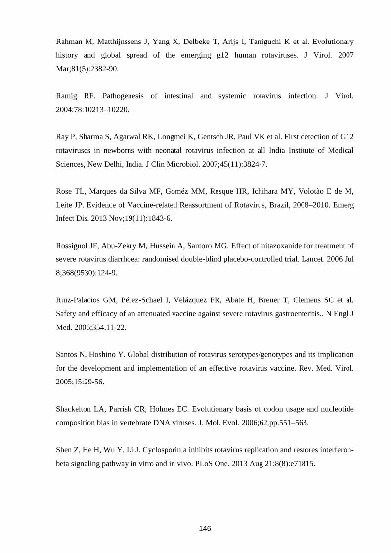
146
Rahman M, Matthijnssens J, Yang X, Delbeke T, Arijs I, Taniguchi K et al. Evolutionary
history and global spread of the emerging g12 human rotaviruses. J Virol. 2007
Mar;81(5):2382-90.
Ramig RF. Pathogenesis of intestinal and systemic rotavirus infection. J Virol.
2004;78:10213–10220.
Ray P, Sharma S, Agarwal RK, Longmei K, Gentsch JR, Paul VK et al. First detection of G12
rotaviruses in newborns with neonatal rotavirus infection at all India Institute of Medical
Sciences, New Delhi, India. J Clin Microbiol. 2007;45(11):3824-7.
Rose TL, Marques da Silva MF, Goméz MM, Resque HR, Ichihara MY, Volotão E de M,
Leite JP. Evidence of Vaccine-related Reassortment of Rotavirus, Brazil, 2008–2010. Emerg
Infect Dis. 2013 Nov;19(11):1843-6.
Rossignol JF, Abu-Zekry M, Hussein A, Santoro MG. Effect of nitazoxanide for treatment of
severe rotavirus diarrhoea: randomised double-blind placebo-controlled trial. Lancet. 2006 Jul
8;368(9530):124-9.
Ruiz-Palacios GM, Pérez-Schael I, Velázquez FR, Abate H, Breuer T, Clemens SC et al.
Safety and efficacy of an attenuated vaccine against severe rotavirus gastroenteritis.. N Engl J
Med. 2006;354,11-22.
Santos N, Hoshino Y. Global distribution of rotavirus serotypes/genotypes and its implication
for the development and implementation of an effective rotavirus vaccine. Rev. Med. Virol.
2005;15:29-56.
Shackelton LA, Parrish CR, Holmes EC. Evolutionary basis of codon usage and nucleotide
composition bias in vertebrate DNA viruses. J. Mol. Evol. 2006;62,pp.551–563.
Shen Z, He H, Wu Y, Li J. Cyclosporin a inhibits rotavirus replication and restores interferon-
beta signaling pathway in vitro and in vivo. PLoS One. 2013 Aug 21;8(8):e71815.
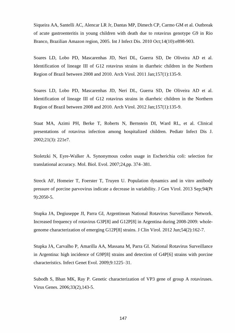
147
Siqueira AA, Santelli AC, Alencar LR Jr, Dantas MP, Dimech CP, Carmo GM et al. Outbreak
of acute gastroenteritis in young children with death due to rotavirus genotype G9 in Rio
Branco, Brazilian Amazon region, 2005. Int J Infect Dis. 2010 Oct;14(10):e898-903.
Soares LD, Lobo PD, Mascarenhas JD, Neri DL, Guerra SD, De Oliveira AD et al.
Identification of lineage III of G12 rotavirus strains in diarrheic children in the Northern
Region of Brazil between 2008 and 2010. Arch Virol. 2011 Jan;157(1):135-9.
Soares LD, Lobo PD, Mascarenhas JD, Neri DL, Guerra SD, De Oliveira AD et al.
Identification of lineage III of G12 rotavirus strains in diarrheic children in the Northern
Region of Brazil between 2008 and 2010. Arch Virol. 2012 Jan;157(1):135-9.
Staat MA, Azimi PH, Berke T, Roberts N, Bernstein DI, Ward RL, et al. Clinical
presentations of rotavirus infection among hospitalized children. Pediatr Infect Dis J.
2002;21(3): 221e7.
Stoletzki N, Eyre-Walker A. Synonymous codon usage in Escherichia coli: selection for
translational accuracy. Mol. Biol. Evol. 2007;24,pp. 374–381.
Streck AF, Homeier T, Foerster T, Truyen U. Population dynamics and in vitro antibody
pressure of porcine parvovirus indicate a decrease in variability. J Gen Virol. 2013 Sep;94(Pt
9):2050-5.
Stupka JA, Degiuseppe JI, Parra GI, Argentinean National Rotavirus Surveillance Network.
Increased frequency of rotavirus G3P[8] and G12P[8] in Argentina during 2008-2009: whole-
genome characterization of emerging G12P[8] strains. J Clin Virol. 2012 Jun;54(2):162-7.
Stupka JA, Carvalho P, Amarilla AA, Massana M, Parra GI. National Rotavirus Surveillance
in Argentina: high incidence of G9P[8] strains and detection of G4P[6] strains with porcine
characteristics. Infect Genet Evol. 2009;9:1225–31.
Subodh S, Bhan MK, Ray P. Genetic characterization of VP3 gene of group A rotaviruses.
Virus Genes. 2006;33(2),143-5.
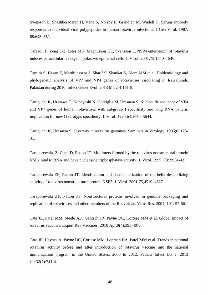
148
Svensson L, Sheshberadaran H, Vene S, Norrby E, Grandien M, Wadell G. Serum antibody
responses to individual viral polypeptides in human rotavirus infections. J Gen Virol. 1987;
68:643–651.
Tafazoli F, Zeng CQ, Estes MK, Magnusson KE, Svensson L. NSP4 enterotoxin of rotavirus
induces paracellular leakage in polarized epithelial cells. J. Virol. 2001;75:1540–1546.
Tamim S, Hasan F, Matthijnssens J, Sharif S, Shaukat S, Alam MM et al. Epidemiology and
phylogenetic analysis of VP7 and VP4 genes of rotaviruses circulating in Rawalpindi,
Pakistan during 2010. Infect Genet Evol. 2013 Mar;14:161-8.
Taniguchi K, Urasawa T, Kobayashi N, Gorziglia M, Urasawa S. Nucleotide sequence of VP4
and VP7 genes of human rotaviruses with subgroup I specificity and long RNA pattern:
implication for new G serotype specificity. J. Virol. 1990;64:5640–5644.
Taniguchi K, Urasawa S. Diversity in rotavirus genomes. Seminars in Virology. 1995;6: 123-
31.
Taraporewala, Z, Chen D, Patton JT. Multimers formed by the rotavirus nonstructural protein
NSP2 bind to RNA and have nucleoside triphosphatase activity. J. Virol. 1999; 73: 9934-43.
Taraporewala ZF, Patton JT. Identification and charac- terization of the helix-destabilizing
activity of rotavirus nonstruc- tural protein NSP2. J. Virol. 2001;75,4519–4527.
Taraporewala ZF, Patton JT. Nonstructural proteins involved in genome packaging and
replication of rotaviruses and other members of the Reoviridae. Virus Res. 2004; 101: 57-66.
Tate JE, Patel MM, Steele AD, Gentsch JR, Payne DC, Cortese MM et al. Global impact of
rotavirus vaccines. Expert Rev Vaccines. 2010 Apr;9(4):395-407.
Tate JE, Haynes A, Payne DC, Cortese MM, Lopman BA, Patel MM et al. Trends in national
rotavirus activity before and after introduction of rotavirus vaccine into the national
immunization program in the United States, 2000 to 2012. Pediatr Infect Dis J. 2013
Jul;32(7):741-4.
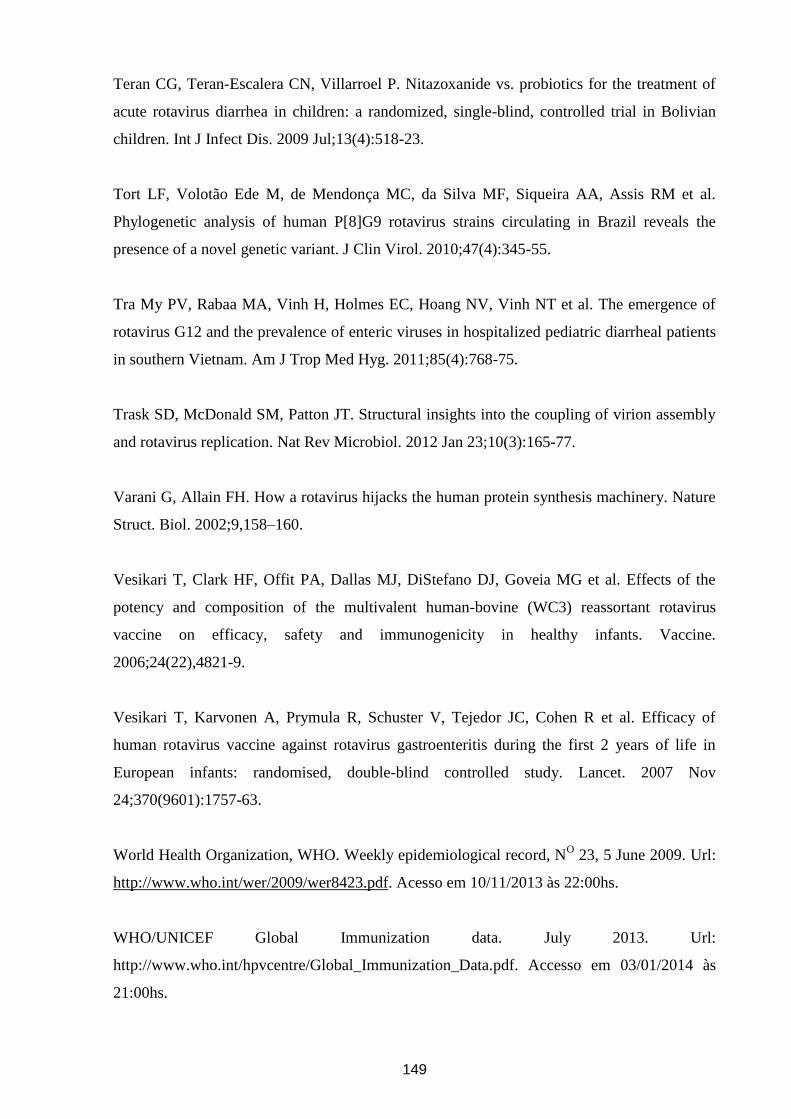
149
Teran CG, Teran-Escalera CN, Villarroel P. Nitazoxanide vs. probiotics for the treatment of
acute rotavirus diarrhea in children: a randomized, single-blind, controlled trial in Bolivian
children. Int J Infect Dis. 2009 Jul;13(4):518-23.
Tort LF, Volotão Ede M, de Mendonça MC, da Silva MF, Siqueira AA, Assis RM et al.
Phylogenetic analysis of human P[8]G9 rotavirus strains circulating in Brazil reveals the
presence of a novel genetic variant. J Clin Virol. 2010;47(4):345-55.
Tra My PV, Rabaa MA, Vinh H, Holmes EC, Hoang NV, Vinh NT et al. The emergence of
rotavirus G12 and the prevalence of enteric viruses in hospitalized pediatric diarrheal patients
in southern Vietnam. Am J Trop Med Hyg. 2011;85(4):768-75.
Trask SD, McDonald SM, Patton JT. Structural insights into the coupling of virion assembly
and rotavirus replication. Nat Rev Microbiol. 2012 Jan 23;10(3):165-77.
Varani G, Allain FH. How a rotavirus hijacks the human protein synthesis machinery. Nature
Struct. Biol. 2002;9,158–160.
Vesikari T, Clark HF, Offit PA, Dallas MJ, DiStefano DJ, Goveia MG et al. Effects of the
potency and composition of the multivalent human-bovine (WC3) reassortant rotavirus
vaccine on efficacy, safety and immunogenicity in healthy infants. Vaccine.
2006;24(22),4821-9.
Vesikari T, Karvonen A, Prymula R, Schuster V, Tejedor JC, Cohen R et al. Efficacy of
human rotavirus vaccine against rotavirus gastroenteritis during the first 2 years of life in
European infants: randomised, double-blind controlled study. Lancet. 2007 Nov
24;370(9601):1757-63.
World Health Organization, WHO. Weekly epidemiological record, NO 23, 5 June 2009. Url:
http://www.who.int/wer/2009/wer8423.pdf. Acesso em 10/11/2013 às 22:00hs.
WHO/UNICEF Global Immunization data. July 2013. Url:
http://www.who.int/hpvcentre/Global_Immunization_Data.pdf. Accesso em 03/01/2014 às
21:00hs.
150
Woo PCY, Wong BHL, Huang Y, Lau SKP, Yuen K. Cytosine deamination and selection of
CpG suppressed clones are the two major independent biological forces that shape codon
usage bias in Coronaviruses. Virology. 2007;369,pp.431–442.
Wrigth F. The “effective number of codons” used in a gene. Gene. 1990;87,pp. 23–29.
Zárate S, Cuadras MA, Espinosa R, Romero P, Juárez KO, Camacho-Nuez M et al.
Interaction of rotaviruses with Hsc70 during cell entry is mediated by VP5. J Virol. 2003;
77(13):7254-60.
Zeller M, Patton JT, Heylen E, De Coster S, Ciarlet M, Van Ranst M et al. Genetic analyses
reveal differences in the VP7 and VP4 antigenic epitopes between human rotaviruses
circulating in Belgium and rotaviruses in Rotarix and RotaTeq. J Clin Microbiol. 2012a
Mar;50(3):966-76.
Zhang M, Zeng CQ, Morris AP, Estes MK. A functional NSP4 enterotoxin peptide secreted
from rotavirus-infected cells. J. Virol. 2000;74:11663–11670.