EFEITOS BIOQUÍMICOS E FISIOLÓGICOS DO CHUMBO EM …
83
UNIVERSIDADE FEDERAL DE SANTA MARIA CENTRO DE CIÊNCIAS NATURAIS E EXATAS PROGRAMA DE PÓS-GRADUAÇÃO EM CIÊNCIAS BIOLÓGICAS BIOQUÍMICA TOXICOLÓGICA EFEITOS BIOQUÍMICOS E FISIOLÓGICOS DO CHUMBO EM PLANTAS DE QUITOCO (Pluchea sagittalis): POSSÍVEL PAPEL FITORREMEDIADOR DISSERTAÇÃO DE MESTRADO Liana Veronica Rossato Santa Maria, fevereiro de 2010
Transcript of EFEITOS BIOQUÍMICOS E FISIOLÓGICOS DO CHUMBO EM …

UNIVERSIDADE FEDERAL DE SANTA MARIA CENTRO DE CIÊNCIAS NATURAIS E EXATAS
PROGRAMA DE PÓS-GRADUAÇÃO EM CIÊNCIAS BIOLÓGICAS BIOQUÍMICA TOXICOLÓGICA
EFEITOS BIOQUÍMICOS E FISIOLÓGICOS DO CHUMBO EM PLANTAS DE QUITOCO ( Pluchea
sagittalis): POSSÍVEL PAPEL FITORREMEDIADOR
DISSERTAÇÃO DE MESTRADO
Liana Veronica Rossato
Santa Maria, fevereiro de 2010

EFEITOS BIOQUÍMICOS E FISIOLÓGICOS DO CHUMBO EM
PLANTAS DE QUITOCO ( Pluchea sagittalis): POSSÍVEL
PAPEL FITORREMEDIADOR
por
Liana Veronica Rossato
Dissertação apresentada ao Programa de Pós-Graduação em Ciências Biológicas: Bioquímica Toxicológica, da Universidade Federal de Santa Maria (UFSM, RS), como requisito parcial para a obtenção do grau de
Mestre em Bioquímica Toxicológica
Maria Rosa Chitolina Schetinger Orientadora
Vera Maria Morsch Co-orientadora
Santa Maria, RS, Brasil
2010


DEDICATÓRIA
À meus pais, Emilia Rossato e Diomedes S. Rossato
pela vida, pelo amor e confiança que sempre em mim depositaram
Aos meus irmãos, Luciana, Tobias, Taisa, Tiago e Ca rla
pela amizade e incentivo em todos os momentos
Ao meu namorado, João
pelo amor, amizade e incentivo
Dedico e ofereço!
AGRADECIMENTOS
À orientadora, Profa. Maria Rosa C. Schetinger, pela receptividade, orientações
e auxílios em todos os momentos, obrigada.
À co-orientadora, Profa. Vera Maria Morsch, pelo conhecimento e atenção,
obrigada.
Ao professor Fernando T. Nicoloso por, desde a graduação, despertar em mim
a vontade de trabalhar com pesquisa científica, pela orientação, dedicação e
ensinamentos, muito obrigada.
Aos colegas de laboratório: Júlia, Luciane T., Denise, Luciane B., Jamile,
Joseila, Gabriel Y C., Sibila, Gabriel e Jader por toda a colaboração, amizade e
apoio prestados durante o desenvolvimento do trabalho, obrigada.
À UFSM, ao Programa de Pós-Graduação em Bioquímica Toxicológica, aos
professores, ao CNPq, e a todos que de alguma forma contribuíram para a
realização deste trabalho, obrigada.

6
RESUMO Dissertação de Mestrado
Programa de Pós-Graduação em Ciências Biológicas: Bioquímica Toxicológica Universidade Federal de Santa Maria
EFEITOS BIOQUÍMICOS E FISIOLÓGICOS DO CHUMBO EM PLA NTAS DE
QUITOCO (Pluchea sagittalis): POSSÍVEL PAPEL FITORREMEDIADOR
Autora: Liana Veronica Rossato Orientadora: Maria Rosa Chitolina Schetinger
Co-Orientadora: Vera Maria Morsch Data e local de defesa: Santa Maria, 18 de fevereiro de 2010.
O Chumbo (Pb) é um dos elementos tóxicos mais abundantes e globalmente distribuídos. A contaminação dos solos, ar e água por este metal leva a sua acumulação em peixes, mamíferos e plantas e a intoxicação humana, através da alimentação. Devido aos efeitos danosos do Pb ao ecossistema, é necessário que estas áreas sejam reabilitadas. A fitorremediação é uma técnica de baixo custo que pode ser utilizada com tal objetivo. Para que a planta seja utilizada na fitorremediação, no entanto, é necessário haver conhecimento sobre o comportamento vegetal quando exposto ao contaminante. Portanto, o objetivo deste trabalho foi caracterizar aspectos fisiológicos e bioquímicos da toxidez do Pb em raízes e folhas de diferentes regiões da parte aérea (apical, mediana e basal) em plantas de P. sagittalis. As plantas foram expostas a cinco concentrações de Pb: (0, 200, 400, 600, 1000 µM) durante 30 dias. Após esse período foram avaliados parâmetros de crescimento, concentração de Pb nos tecidos, indicadores de toxicidade (atividade da δ-aminolevulinato desidratase, δ-ALA-D), marcadores de dano oxidativo (TBARS, peroxidação lipídica, e concentração de H2O2) e antioxidantes enzimáticos (superóxido dismutase, SOD, catalase, CAT, e ascorbato peroxidase, APX) e não enzimáticos (tióis não protéicos, NPSH, acido ascórbico, AsA). A concentração de Pb nos tecidos aumentou com o acréscimo de Pb ao meio ocasionando uma diminuição do consumo de solução nutritiva e da taxa de transpiração. Da mesma forma ocorreu decréscimo na matéria fresca da raiz e da parte aérea, na área foliar, na altura da parte aérea e na atividade da δ-ALA-D com o aumento da concentração de Pb. Por outro lado, a matéria seca da raiz e da parte aérea, a concentração de clorofila e carotenóides não foram afetadas, sendo que os pesos das matérias fresca e seca das raízes aumentaram em 200 µM Pb comparado ao controle. O índice de tolerância das raízes e da parte aérea de P. sagittalis reduziu nas maiores concentrações de Pb. Contudo, as raízes foram mais tolerantes ao Pb que a parte aérea. Com o aumento da concentração de Pb, a peroxidação lipídica e a concentração de H2O2 aumentaram nas raízes e folhas. A atividade da APX aumentou em todas as partes da planta com o aumento da concentração de Pb, enquanto a atividade da SOD aumentou nas folhas e não foi afetada nas raízes. A atividade da CAT nas folhas da região apical não foi alterada pelo aumento da concentração de Pb, mas nas outras partes da planta a sua atividade aumentou. A toxicidade do Pb aumentou a concentração de NPSH na parte aérea, enquanto não

7 foi observada diferença significativa nas raízes. A concentração de AsA aumentou com o aumento da concentração de Pb. Nossos resultados sugerem que o Pb induziu estresse oxidativo em plantas de P. sagittalis e que o aumento da atividade das enzimas do sistema antioxidante pode ter atuado como um importante mecanismo de defesa antioxidante contra o dano oxidativo, demonstrando tolerância e possibilidade de utilização dessas plantas para a recuperação de solos contaminados por Pb.
Palavras-chave: sistema antioxidante; toxicidade por Pb; Pluchea sagittalis; peroxidação lipídica; eficiência do uso da água

8
ABSTRACT Master Dissertation
Biological Sciences: Toxicological Biochemistry Post-Graduation Universidade Federal de Santa Maria
EFFECTS OF LEAD ON BIOCHEMICAL AND PHYSIOLOGICAL
PARAMETERS IN QUITOC PLANTS ( Pluchea sagittalis): POSSIBLE PHYTOREMEDIATOR ROLE
Author: Liana Veronica Rossato
Oriented by: Maria Rosa Chitolina Schetinger Co-oriented by: Vera Maria Morsch
Place and date: Santa Maria, February 18, 2010.
Lead (Pb) is one of the most abundant, globally distributed toxic elements. Soil, water and air contamination by Pb leads to its accumulation in fish, mammals, and plants, and thus to human intoxication through food. Due to the hazardous effects to the ecosystem, it is necessary that contaminated soils be rehabilitated. Phytoremediation is a cheap technique that can be utilized with this objective. However, it is necessary to know about plant behavior during metal exposure. Therefore, the aim of this study was to characterize physiological and biochemical aspects of Pb toxicity in roots and leaves of different developmental stages (apex, middle, and basal regions) in P. sagittalis plants. Plants were exposed to five Pb levels: (0, 200, 400, 600, 1000 µM) for 30 days. Parameters such as growth, tissue Pb concentration and content, toxicity indicators (δ-aminolevulinic acid dehidratase, δ-ALA-D, activity), oxidative damage markers (TBARS, lipid peroxidation, and H2O2 concentration) and enzymatic (superoxide dismutase, SOD, catalase, CAT, and ascorbate peroxidase, APX) and non-enzymatic (non-protein thiols, NPSH, ascorbic acid, AsA) antioxidants were investigated. Tissue Pb concentration increased with external Pb levels causing a decrease in the consumption of nutrient solution and transpiration ratio. Moreover, root and shoot fresh weight, leaf area, shoot length and δ-ALA-D activity decreased upon addition of Pb treatments. On the other hand, dry weight of shoots and roots and chlorophyll and carotenoids concentrations were not affected. Both fresh and dry weight of roots increased at 200 µM Pb, when compared to control. The index of tolerance of roots and shoots of P. sagittalis decrease at higher Pb concentrations. However, the roots were more tolerant to Pb than shoots. Lipid peroxidation and hydrogen peroxide (H2O2) concentration both in roots and leaves increased with increasing Pb levels. APX activity increased by Pb treatments in all plant parts, while SOD activity increased in leaves and it was not affected in roots. CAT activity in leaves from the apex shoot was not affected by Pb; however, in other plant parts its activity increased. Pb toxicity caused increase in NPSH concentration in shoot parts, whereas no significant difference was observed in roots. AsA concentration increased with increasing Pb levels. These results suggest that Pb induces oxidative stress in P. sagittalis and that elevated activity of antioxidative enzymes could serve as important components of antioxidative defense mechanism against oxidative injury, demonstrating tolerance and possible use of these plants for reclamation of Pb contaminated soils.

9 Keywords: antioxidant system; Pb toxicity; Pluchea sagittalis; lipid peroxidation; water use efficiency
10
LISTA DE TABELAS
REVISÃO BIBLIOGRÁFICA
TABELA 1 - Valores norteadores para solo e água subterrânea para detecção de contaminação por chumbo. .......................................................................................20
MANUSCRITO TABLE 1 - Effects of various Pb concentrations on the index of tolerance of P. sagittalis after 30 days exposure………………………………………………………….51
11
LISTA DE FIGURAS
REVISÃO BIBLIOGRÁFICA
FIGURA 1 - Pluchea sagittalis (Lam.) Cabrera................................................25
FIGURA 2 - Processos da fitorremediação do solo.........................................28
FIGURA 3 - Sítios de produção intracelular de espécies reativas de oxigênio
(EROs).............................................................................................................30
FIGURA 4 - Ciclo ascorbato – glutationa.........................................................33
FIGURA 5 - Caminho das espécies reativas de oxigênio e sua remoção nas
plantas.............................................................................................................34
FIGURA 6 - Estrutura do ácido ascórbico atuando na estabilidade dos radicais
livres.................................................................................................................35
MANUSCRITO
FIGURE 1 - Effect of increasing Pb concentration on the Pb concentration (A)
and content (B) in roots and in leaves and stem of different shoot parts (apex,
middle and basal) of P. sagittalis plants. ……….………………………………..48
FIGURE 2 - Effect of increasing Pb concentration on fresh (A) and dry (B)
weight of different shoot parts (apex, middle and basal) and roots, leaf area
(C), shoot length (D), nutrient solution consumption (E), and transpiration ratio
(F) of P. sagittalis plants . …………………………………………………………50
FIGURE 3 - Effect of increasing Pb concentration on the δ-aminolevulinic acid
dehydratase activity (A), and chlorophyll (B) and carotenoids (C) concentration
in leaves of different shoot parts…………………………………………………..52
12
FIGURE 4 - Effect of increasing Pb concentration on the hydrogen peroxide
concentration (A), and on lipid peroxidation (B) in roots and leaves of different
shoot parts…………………………………………………………………………..53
FIGURE 5 - Effect of increasing Pb concentration on the superoxide
dismutase, catalase and ascorbate peroxidase activities (A , B and C,
respectively), and ascorbic acid and nonprotein thiol groups concentration (D
and E, respectively) in roots and in leaves of different shoot parts.
……………………………..…………………………………………………………55
13
LISTA DE ABREVIATURAS
ALA - ácido 5-aminolevulínico APX - ascorbato peroxidase AsA - ácido ascórbico CAT – catalase CETESB - Centro Tecnológico de Saneamento Básico CuZnSOD - superóxido dismutase dependente de cobre e zinco DMSO - dimetilsulfóxido DNPH - dinitrofenilidrazina DTNB - ácido 5-5´-ditio-bis-(nitrobenzóico) DTT - ditiotreitol EDTA - ácido etilenodiaminotetracético EROs - espécies reativas de oxigênio FeSOD - superóxido dismutase dependente de ferro GPX - glutationa peroxidase GSH - glutationa reduzida GSSG - glutationa oxidada HCl - ácido clorídrico HgCl2 – cloreto de mercúrio HNO3 - ácido nítrico HO• - radical hidroxila H2O2 - peróxido de hidrogênio H2SO4 - ácido sulfúrico KI - iodeto de potássio K2HPO4 - fosfato de potássio MDA – malondialdeído MnSOD - superóxido dismutase dependente de manganês NADPH - nicotinamida adenina dinucleotídeo fosfato NPSH - tióis não-protéicos O2 - oxigênio molecular O2
-- . - radical superóxido 1O2 - radical oxigênio singleto Pb (CH3COO)2.3H2O – acetato de chumbo PBG – porfobilinogênio PC - fitoquelatina pH - potencial de hidrogênio PVP – polivinilpirrolidona SOD - superóxido dismutase TBA - ácido tiobarbitúrico TCA - ácido tricloroacético δ-ALA-D - delta-aminolevulinato desidratase SH - grupo sulfidril
14
SUMÁRIO
RESUMO....................................................................................................................06 ABSTRACT................................................................................................................08 LISTA DE TABELAS..................................................................................................10 LISTA DE FIGURAS...................................................................................................11 LISTA DE ABREVIATURAS ......................................................................................13 SUMÁRIO...................................................................................................................14 1 INTRODUÇÃO .......................................................................................................15 1.1 Objetivos...............................................................................................................17 1.1.1 Objetivo Geral....................................................................................................17 1.1.2 Objetivos Específicos........................................................................................18 2 REVISÃO DE LITERATURA ........................... ......................................................19 2.1 Chumbo – escala global.......................................................................................19 2.2 Chumbo no Brasil.................................................................................................20 2.3 Efeitos fisiológicos do chumbo em plantas...........................................................21 2.4 Pluchea sagittalis..................................................................................................24 2.5 Fitorremediação....................................................................................................26 2.6 Espécies reativas de oxigênio e estresse oxidativo.............................................29 2.7 Sistema antioxidante............................................................................................31 3 RESULTADOS…………………………………………………………………………...37
3.1 MANUSCRITO - Pluchea sagittalis has tolerance to Pb stress and this behavior is
related to an efficient antioxidant system and improved water use efficiency. Liana
Veronica Rossato, Fernando Teixeira Nicoloso, Júlia Gomes Farias, Denise
Cargnelluti, Luciane Almeri Tabaldi, Fabiane Goldschmidt Antes, Valderi Luiz
Dressler, Vera Maria Morsch, Maria Rosa Chitolina Schetinger.
....................................................................................................................................38
4 DISCUSSÃO...........................................................................................................69 5 CONCLUSÕES.......................................................................................................73 6 REFERÊNCIAS BIBLIOGRÁFICAS....................... ................................................74
15 1 INTRODUÇÃO
A poluição do meio ambiente por metais pesados é uma das maiores
preocupações ecológicas atuais, devido ao seu impacto sobre a saúde humana
através da cadeia alimentar e pela sua alta persistência no meio ambiente
(PIECHALAK et al., 2002). O chumbo (Pb) é um dos maiores poluentes tanto de
ecossistemas aquáticos quanto de terrestres. A contaminação do ambiente por este
metal através de fontes antropogênicas, tais como uso de insumos agrícolas com
teores elevados de chumbo, deposições atmosféricas, mineração e resíduos
industriais, têm causado sérias preocupações devido à sua elevada toxicidade aos
seres humanos e animais, mesmo em baixas concentrações (PIERANGELI, 1999).
Essas fontes são responsáveis pela adição de cerca de 1,16 milhões de toneladas
de chumbo por ano em ecossistemas terrestres e aquáticos (NRIAGU, 1989).
Em vista da crescente contaminação dos solos por Pb e da expansão da
contaminação a outros ecossistemas, é importante estabelecer metodologias para a
reabilitação destas áreas contaminadas. Muitas técnicas são aplicadas para
remoção ou atenuação dos metais pesados. As técnicas atuais de remediação
baseiam-se em processos de engenharia, apresentando custos elevados e de difícil
aplicação (ACCIOLY & SIQUEIRA, 2000). Em contraste, o uso de plantas para
remover os metais pesados em solos contaminados, conhecida como
“fitorremediação”, oferece vantagens econômicas e ambientais além de ser uma
técnica promissora (SALT et al., 1995). O sucesso da fitorremediação depende da
capacidade da planta em captar, translocar e acumular o metal pesado na parte
aérea sem que o seu crescimento e desenvolvimento sejam afetados (XIONG,
1997).
Entretanto, o aumento dos níveis de Pb no solo pode induzir a uma série de
efeitos adversos no crescimento e no metabolismo das plantas (SINGH et al., 1997;
SHARMA & DUBEY, 2005) através da inibição de atividades enzimáticas (ISLAM et
al., 2008; XIAO et al., 2008a; GUPTA et al., 2009), alteração na taxa fotossintética
(XIAO et al., 2008a), redução da absorção de água, nutrientes minerais e da
transpiração (WIERZBICKA, 1995; SINHA et al., 2006; ISLAM et al., 2008),

16 mudanças no “status” hormonal (SHARMA & DUBEY, 2005) e aumento da
peroxidação lipídica (XIAO et al., 2008b; GUPTA et al., 2009).
A exposição das plantas a uma condição de estresse pode intensificar a
produção de espécies reativas de oxigênio (EROs) (FOYER et al., 1994). As EROs,
tais como o ânion superóxido (O2-), o peróxido de hidrogênio (H2O2) e o radical
hidroxila (OH˙), que em condições fisiológicas normais são produzidos em baixa
quantidade em reações normais do metabolismo, tal como na fotossíntese e
respiração, podem causar danos a biomoléculas como lipídios de membrana,
proteínas, pigmentos fotossintéticos e ácidos nucléicos (MITTLER, 2002).
Entretanto, as plantas desenvolveram uma variedade de estratégias para
prevenir a acumulação excessiva de metais nas células e/ou transformar estes
metais em formas menos tóxicas (SINGH et al., 1997). O primeiro passo é evitar a
entrada do metal na célula através da exclusão e/ou através da ligação à parede
celular. Dentro da célula, o metal pode ser detoxificado através da ligação a outros
ligantes como ácidos orgânicos, aminoácidos (cisteína e prolina), glutationa (GSH)
ou fitoquelatinas (PCs) (MISHRA et al., 2006). Para o Pb, a ligação à parede celular
é um dos principais mecanismos de detoxificação (ANTOSIEWICZ & WIERZBICKA,
1999). No entanto, quando estes mecanismos de defesa não são suficientes para
evitar os danos provocados pelo Pb causando o estresse oxidativo, outros
mecanismos antioxidantes são acionados (MISHRA et al., 2006). O sistema
antioxidante é composto por antioxidantes enzimáticos [superóxido dismutase (E.C.
1.15.1.1), catalase (E.C. 1.11.1.6) e ascorbato peroxidase (E.C. 1.11.1.11), etc] e
não enzimáticos [ácido ascórbico, glutationa reduzida (GSH), carotenóides e outros
grupos tiólicos não protéicos] que normalmente mantêm um balanço de EROs
dentro das células ( FOYER et al., 1994; CROSS et al.,1998).
A capacidade do sistema antioxidante de detoxificar as EROs está intimamente
ligada à tolerância das plantas aos metais pesados (SCHÜTZENDÜBEL & POLLE,
2002). Nos últimos anos, muitas espécies de plantas estão sendo identificadas como
hiperacumuladoras, tendo a capacidade de acumular grandes concentrações de
metais pesados, sem impacto no seu crescimento e desenvolvimento (HUANG &
CUNNINGHAM, 1996).
A distribuição dos metais absorvidos entre os órgãos da planta é muito
diferenciada (BRUNE et al., 1994), podendo depender da espécie e do metal

17 utilizado. A distribuição do metal na planta está, de certo modo, relacionada com a
absorção e translocação de cada metal e pode ser um mecanismo de tolerância
(ACCIOLY & SIQUEIRA, 2000).
A Pluchea sagittalis é uma espécie comum da região tropical, sendo facilmente
encontrada em vários países da América do Sul, e amplamente distribuída por todo
o Brasil (DALPIAZ & RITTER, 1998).
Esta espécie pode apresentar um possível papel fitorremediador, pois em um
trabalho realizado por Sampanpanish et al. (2006), ao avaliarem um total de seis
espécies de plantas daninhas visando à escolha destas para emprego em
programas de fitorremediação em áreas contaminadas com cromo, verificaram que
uma espécie do gênero Pluchea (P. indica) acumulou e translocou grandes
quantidades de cromo em seus tecidos, cerca de 180, 86 e 90 mg\kg na raiz, caule e
folha, respectivamente, podendo ser utilizada como fitoacumuladora em solos
contaminados com cromo.
O uso de plantas para extrair o Pb de solos contaminados requer um melhor
entendimento dos mecanismos de captação, translocação e acumulação pela planta
(HUANG & CUNNINGHAM, 1996), sendo que a hiperacumulação é regulada por
processos fisiológicos, bioquímicos e genéticos da planta (BAKER, 1987) sendo
necessário ter um melhor entendimento desses processos. No entanto, há poucas
informações acerca da toxicologia do Pb e sobre os mecanismos pelo qual esse
elemento produz estresse oxidativo nas plantas de P. sagittalis. Deste modo, os
objetivos do presente trabalho foram:
1.1 Objetivos
1.1.1 Objetivo geral
Caracterizar os efeitos do chumbo sobre aspectos morfológicos, fisiológicos e
bioquímicos em diferentes partes da planta de Pluchea sagittalis.
18 1.1.2 Objetivos Específicos
- Avaliar as alterações no crescimento, e determinar o conteúdo de chumbo
absorvido pelas plântulas de P. sagittalis após exposição ao chumbo.
- Avaliar a atividade da enzima delta-ALA-D e o conteúdo de clorofila e
carotenóides em folhas de diferentes partes da parte aérea (apical, mediana e basal)
de plantas de P. sagittalis após exposição ao chumbo.
- Determinar os níveis de peroxidação lipídica, o conteúdo de peróxido de
hidrogênio em raiz e em folhas de diferentes partes da parte aérea (apical, mediana
e basal) de plantas de P. sagittalis após exposição ao chumbo.
- Avaliar a atividade de enzimas antioxidantes (catalase, ascorbato peroxidase
e superóxido dismutase) e os níveis de antioxidantes não-enzimáticos (carotenóides,
ácido ascórbico e tióis não-protéicos) em raiz e em folhas de diferentes partes da
parte aérea (apical, mediana e basal) de plantas de P. sagittalis após exposição ao
chumbo.
19 2 REVISÃO DE LITERATURA
2.1 Chumbo - escala global
O chumbo (Pb) é um elemento metálico sólido cinza azulado conhecido há
séculos como potencialmente tóxico. Ele ocorre naturalmente nos solos e é um
constituinte essencial de muitos minerais, sendo encontrado na crosta terrestre em
uma concentração de 17 mg kg-1 (BOSSO & ENZWEILER, 2008). A galena (PbS) é
o principal mineral de chumbo. O chumbo metálico é produzido por oxidação da
galena, seguida pela redução do litargírio (PbO) formado. Alguns minerais
importantes de chumbo são formados por transformação da galena. Exemplos são a
reação com águas carbonatadas para formar cerussita (PbCO3), a oxidação que
produz a anglesita (PbSO4) e a reação com fosfatos que resulta na piromorfita
[(Pb5(PO4)3X onde X= OH-, F-, Br- ou Cl-] (BOSSO & ENZWEILER, 2008).
O aporte antropogênico do Pb ocorre através do uso de insumos agrícolas
com teores elevados deste metal, deposições atmosféricas, mineração e resíduos
industriais, sendo que as fontes antropogênicas são responsáveis pela adição de
cerca de 1,16 milhões de toneladas de chumbo por ano em ecossistemas terrestres
e aquáticos (NRIAGU, 1989).
O aumento da concentração de Pb causa sérias preocupações devido à sua
elevada toxicidade aos seres humanos e animais, mesmo em baixas concentrações
(PIERANGELI, 1999).
Segundo Chlopecka et al. (1996), metais originados de diferentes fontes
antrópicas são relativamente mais móveis e potencialmente mais fitodisponíveis que
aqueles presentes no material de origem dos solos. Entretanto, vários fatores podem
controlar a biodisponibilidade do Pb no solo. A química do solo, através da presença
de espécies aniônicas, as quais formam complexos com o Pb (ácidos orgânicos,
matéria orgânica, fosfato, carbonato, sulfetos, cloreto, e hidróxido), concentrações
de ferro e manganês, pH do solo, capacidade de troca catiônica, e condições redox
determinam a extensão em que ocorrem reações como: dissolução, precipitação,
20 complexação e adsorção entre o Pb e as partículas do solo (RUBY et al., 1999; JIN
et al., 2005).
Apesar da solubilidade e da mobilidade de Pb serem geralmente baixas, em
alguns ambientes sua concentração é tão alta a ponto de colocar em risco a saúde
humana, principalmente em locais próximos a mineração e indústrias que usam
chumbo (WOWK, 2003).
Em uma área antiga de mineração na Malásia observou-se que frutos de
manga, mamão e goiaba cultivados nessa área apresentavam concentrações de Pb
acima dos níveis permitidos para alimentação, tornando-se um risco para a saúde
humana (ANG & NG, 2000).
2.2 Chumbo no Brasil
No Brasil, segundo o Ministério da Saúde, foram identificadas mais de 217
áreas de contaminação por metais pesados (BRASIL, 2006). Áreas contaminadas
por chumbo (Pb) são encontradas em todo o país, mas principalmente no estado de
São Paulo. As principais fontes de contaminação antrópica de Pb no Brasil ocorrem
através de indústrias de baterias, de pigmentos, de cerâmica e de plástico
(PAOLIELLO & CAPITANI, 2007).
De acordo com trabalho desenvolvido pela CETESB (Centro Tecnológico de
Saneamento Básico) em 2001, foram estabelecidos critérios, valores e padrões
como referência para problemas de contaminação de solo e águas subterrâneas
(Tabela 1).
Tabela 1 - Valores norteadores para solo e água sub terrânea para detecção de contaminação por chumbo.
Solo (mg kg-1 de peso seco) Água subterrânea (µg.L-1)
Referência Prevenção Intervenção Intervenção
Agrícola Residencial Industrial
17 72 180 300 900 10
Fonte: CETESB (2005).
21 • Valor de Referência (R) de qualidade: reflete o teor natural médio do elemento
para um solo sob condições naturais, indicando a não-contaminação.
• Valor de Prevenção (P): indica possibilidade de alteração prejudicial à
qualidade dos solos, sendo utilizado em caráter preventivo; excedendo-se no solo,
obrigatoriamente deverá ser feito o monitoramento dos impactos que venham a
ocorrer.
• Valor de Intervenção (I): indicam a concentração limite em que acima destes
existem riscos potenciais, diretos ou indiretos, à saúde humana.
Em estudos relacionados à contaminação por Pb na região sudeste do Brasil,
foi encontrada em uma área contaminada por depósito inadequado de resíduos de
indústrias de revestimentos cerâmicos do pólo de Santa Gertrudes (São Paulo) uma
concentração de 945 mg kg-1 de Pb (OLIVEIRA & MORINA, 2008). Em outra área do
estado de SP foi avaliado o teor de metais pesados em sedimento do rio Betari, o
qual passa por diversas áreas de mineração de chumbo. Foram verificadas elevadas
concentrações de Pb (7569 mg kg-1) no sedimento do rio, podendo representar um
fator de risco a saúde das populações ribeirinhas (COTTA et al., 2006).
Da mesma forma, no município de Bauru (SP) foi verificada a contaminação
por chumbo em solos, vegetação, animais e também em crianças, nas proximidades
de duas indústrias de acumuladores elétricos (MOREIRA & MOREIRA, 2004).
2.3 Efeitos fisiológicos do chumbo em plantas
Stefanov et al. (1995) relataram que as espécies de plantas diferem na sua
sensibilidade aos metais. Sabe-se que a acumulação do chumbo (Pb) depende da
espécie, cultivar, órgão da planta, estágio de desenvolvimento, concentração de
chumbo exógena e da presença de outros íons no ambiente (SINGH et al., 1997; XU
& XU, 1993).
As plantas podem absorver chumbo a partir do ar ou do solo. O chumbo
presente no solo está sob a forma de sais solúveis e insolúveis ou ligado a matéria
orgânica (RUBY et al., 1999). O tamanho das partículas do solo, a capacidade de
troca catiônica (RUBY et al., 1999), a área de superfície radicular, a exudação de

22 compostos orgânicos, a micorrização (VOGEL-MIKUS et al., 2006), a taxa de
transpiração e o pH do solo podem afetar a disponibilidade e a captação do Pb pelas
raízes (DAVIES, 1995).
A toxicidade do chumbo pode causar vários sintomas em plantas como, por
exemplo: inibição do crescimento radicular e da parte aérea, clorose e redução da
área foliar (BEKIAROGLOU & KARATAGLIS, 2002; SINHA et al., 2006; DEY et al.,
2007; ISLAM et al., 2008). Esses sintomas são provocados devido à inibição da
atividade de enzimas, distúrbio do “status” nutricional e no balanço hídrico, mudança
no padrão hormonal e alteração na permeabilidade das membranas (SHARMA &
DUBEY, 2005).
Primeiramente, o Pb é captado pelo sistema radicular, sendo que a sua
concentração é geralmente maior nesse órgão do que em outras partes da planta. O
Pb liga-se fortemente a grupos carboxil do ácido galacturônico e glucurônico na
parede celular, o que restringe o seu transporte via apoplasto (RUDAKOVA et al.,
1988). Entretanto, o Pb move-se predominantemente via apoplasto atravessando o
córtex e acumulando-se na endoderme (VERMA & DUBEY, 2003). Na endoderme
encontra-se a estria de Caspari que é a principal barreira ao movimento do Pb
através da endoderme para o cilindro central, promovendo uma diminuição da
translocação desse metal para a parte aérea (SEREGIN & IVANOV, 1997). Contudo,
o Pb move-se também via simplasto, atravessando a membrana plasmática
principalmente através dos canais de cálcio (SINGH et al., 1997).
O acúmulo de Pb na raiz pode afetar o “status” hídrico e nutricional das
plantas promovendo uma redução do crescimento tanto da raiz quanto da parte
aérea (SHARMA & DUBEY, 2005).
Nas raízes, o Pb interage fortemente com os grupos sulfidrílicos das enzimas
e proteínas (VAN ASSCHE & CLIJSTERS, 1990). O Pb liga-se às proteínas dos
canais de água e de íons, causando uma obstrução física do fluxo de água e da
captação de nutrientes (SHARMA & DUBEY, 2005) e, consequentemente, afetando
a transpiração das plantas. Yang et al. (2004) relataram que o Pb inibe a captação
de água via aquaporinas e o transporte de íons da membrana plasmática de
vegetais, ocasionando redução no crescimento.
Eun et al. (2000) relataram que a inibição do crescimento radicular em plantas
expostas à elevadas concentrações de Pb é devida à inibição da divisão celular em

23 células do ápice da raiz induzida por este metal. O Pb altera o alinhamento dos
microtúbulos (YANG et al., 2000; LIU et al., 2009) e destrói os microtúbulos do fuso
mitótico interrompendo a divisão celular. Além disso, foram observadas alterações
cromossômicas e no nucléolo e irregularidades na mitose em células meristemáticas
de raízes de Allium cepa e Zea mays (WIERZBICKA, 1994; JIANG & LIU, 2000;
CARRUYO et al., 2008). Em células na fase de interfase observaram-se núcleos
deformados e com material nuclear decomposto (WIERZBICKA, 1994). Nos
tecidos, a localização do Pb é importante para a determinação da sua toxicidade
(SINGH et al., 1997). O Pb acumula-se preferencialmente nos espaços
intercelulares, parede celular e nos vacúolos (WIERZBICKA, 1987; ANTOSIEWICZ
& WIERZBICKA, 1999; SEREGIN et al., 2002; SHARMA & DUBEY, 2005), não
ocasionando nenhum efeito deletério para a planta (SINGH et al., 1997). Entretanto,
pequenos depósitos desse metal podem ser encontrados no reticulo
endoplasmático, núcleo, cloroplastos, mitocôndrias e citoplasma (SHARMA &
DUBEY, 2005) podendo interferir na homeostase celular.
Como vários metais pesados, o Pb afeta a atividade de várias enzimas de
diferentes rotas metabólicas, ativando-as ou inibindo-as (SHARMA & DUBEY, 2005).
Para muitas enzimas, a inibição exercida pelo Pb sobre suas atividades é resultado
da interação do Pb com os grupamentos -SH que estão presentes no sítio ativo da
enzima, os quais são essenciais para a sua atividade ou com grupos –SH que são
necessários para a estabilização da estrutura terciária da enzima (LEVINA, 1972).
Além disso, o Pb pode inibir a atividade de metaloenzimas através da substituição
do metal essencial (SHARMA & DUBEY, 2005). Em plântulas de milho (Zea mays)
expostas a diferentes concentrações de Pb (25 – 200 µM) observou-se decréscimo
na atividade da enzima δ-aminolevulinato desidrogenase (enzima chave na rota de
síntese da clorofila) (GUPTA et al., 2009).
Por outro lado, a atividade de várias enzimas pode ser aumentada em plantas
expostas ao Pb (SHARMA & DUBEY, 2005). Esse aumento pode ocorrer devido à
síntese de enzimas e a imobilização de inibidores (SHARMA & DUBEY, 2005).
Plantas de arroz crescendo em areia contendo 0,5 e 1 mM de nitrato de chumbo
demonstraram aumento da atividade das enzimas antioxidantes superóxido
dismutase, guaiacol peroxidase, ascorbato peroxidase e glutationa redutase em
raízes e folhas (VERMA & DUBEY, 2003).

24
O Pb afeta tanto reações fotoquímicas quanto as de carboxilação, durante a
fotossíntese. A fotossíntese é considerada um dos processos metabólicos mais
sensíveis à toxicidade do Pb (SINGH et al., 1997). Plantas expostas ao Pb
demonstraram diminuição da taxa fotossintética como conseqüência da ruptura da
organização do cloroplasto, inibição da síntese de clorofila, plastoquinona e de
carotenóides, obstrução do transporte de elétrons, inibição da atividade de enzimas
do ciclo de Calvin, bem como deficiência de CO2 como resultado do fechamento dos
estômatos (SHARMA & DUBEY, 2005).
Os efeitos fitotóxicos do chumbo promovem aumento do estresse oxidativo
através do aumento da produção de espécies reativas de oxigênio (EROs) (SINGH
et al., 1997). A produção de EROs induzida pelo Pb em plantas depende da
intensidade do estresse, do tempo de exposição, da espécie e do estágio de
desenvolvimento da planta (ASADA, 1994; VERMA & DUBEY, 2003).
Contudo, o estresse oxidativo induzido por Pb em plantas pode resultar em
peroxidação lipídica, alteração da atividade de enzimas antioxidantes e na indução
da síntese de compostos contendo tióis (VERMA & DUBEY, 2003; XIAO et al.,
2008b; GUPTA et al., 2009). Estudos recentes têm demonstrado que o Pb pode
induzir à expressão de genes codificadores das enzimas glutationa redutase,
glutationa S-transferase, ascorbato peroxidase e superoxido dismutase, além de
alterar a atividade de várias enzimas, tais como as enzimas catalase, glutationa
redutase, superóxido dismutase e peroxidase (MALECKA et al., 2001; VERMA &
DUBY, 2003; BRUNET et al., 2009), enzimas antioxidantes responsáveis pela
defesa da planta contra as EROs.
2.4 Pluchea sagittalis
O gênero Pluchea, pertencente à família Asteraceae, subfamília Asteroideae,
tribo Inuleae, abrange cerca de 80 espécies (BREMER, 1994), distribuídas por toda
América do Sul. A espécie Pluchea sagittalis (Lam.) Cabr. é uma erva ou arbusto
perene, aromático, com altura variando de 0,3 a 2,0 m; caule robusto, foliáceo, alado
e piloso. Possui folhas sésseis oblongo – laceoladas, levemente dentadas e com
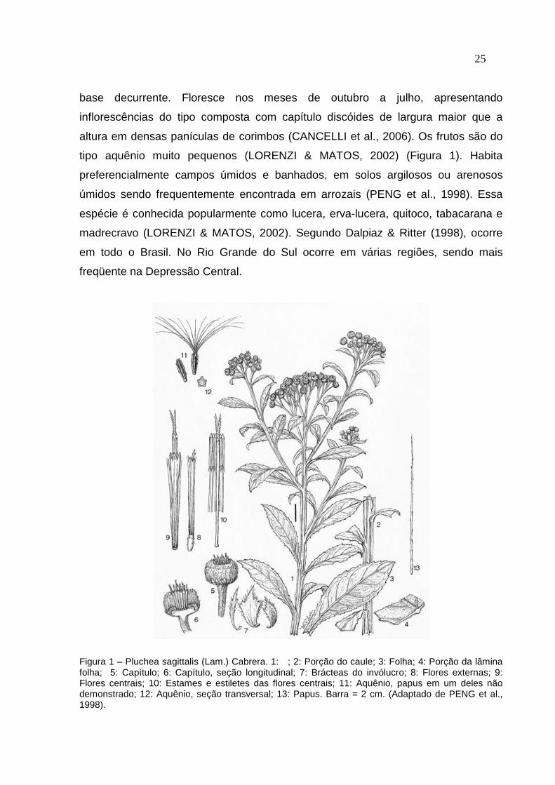
25 base decurrente. Floresce nos meses de outubro a julho, apresentando
inflorescências do tipo composta com capítulo discóides de largura maior que a
altura em densas panículas de corimbos (CANCELLI et al., 2006). Os frutos são do
tipo aquênio muito pequenos (LORENZI & MATOS, 2002) (Figura 1). Habita
preferencialmente campos úmidos e banhados, em solos argilosos ou arenosos
úmidos sendo frequentemente encontrada em arrozais (PENG et al., 1998). Essa
espécie é conhecida popularmente como lucera, erva-lucera, quitoco, tabacarana e
madrecravo (LORENZI & MATOS, 2002). Segundo Dalpiaz & Ritter (1998), ocorre
em todo o Brasil. No Rio Grande do Sul ocorre em várias regiões, sendo mais
freqüente na Depressão Central.
Figura 1 – Pluchea sagittalis (Lam.) Cabrera. 1: ; 2: Porção do caule; 3: Folha; 4: Porção da lâmina folha; 5: Capítulo; 6: Capítulo, seção longitudinal; 7: Brácteas do invólucro; 8: Flores externas; 9: Flores centrais; 10: Estames e estiletes das flores centrais; 11: Aquênio, papus em um deles não demonstrado; 12: Aquênio, seção transversal; 13: Papus. Barra = 2 cm. (Adaptado de PENG et al., 1998).

26 A literatura etnofarmacológica registra o uso desta planta como peitoral,
carminativa e estomacal, com indicações para o tratamento caseiro de problemas de
digestão, dispepsias nervosas, inflamação no útero, rins e bexiga, reumatismo,
resfriados e como estimulante do crescimento capilar (LORENZI, 2000).
Outros estudos demonstraram que extratos de P. sagittalis apresentaram
efeitos antiinflamatórios (PÉREZ-GARCÍA et al., 1996; PÉREZ-GARCÍA et al., 2005)
e antioxidantes (PÉREZ-GARCÍA et al., 2001; FERNÁNDEZ & TORRES, 2006),
anticancerígenos (QUEIROZ et al., 2001) e antimicrobianos (ZANI et al., 1994;
QUEIROZ et al., 2000; SOUZA et al., 2004).
Recentemente, uma nova utilidade tem sido associada ao gênero Pluchea.
Sampanpanish et al. (2006), ao avaliar um total de seis espécies de plantas
daninhas visando à escolha destas para emprego em programas de fitorremediação
em áreas contaminadas com cromo, verificou que uma espécie do gênero Pluchea
(P. indica) acumulou e translocou grandes quantidades de cromo em seus tecidos,
cerca de 180, 86 e 90 mg/kg na raiz, caule e folha, respectivamente, podendo ser
utilizada como fitoacumuladora em solos contaminados com cromo.
Há, entretanto, um outro aspecto que deve ser observado: a alta
concentração de metais em um vegetal representa um grande risco se a planta for
utilizada “in natura” como agente medicinal. Sob esse prisma uma planta medicinal
pode representar um risco maior á saúde do que um benefício para quem a utiliza.
Assim, estudos são necessários para desestimular o uso dessa planta como
medicamento, caso ela apresente capacidade de acumular grandes quantidades de
metal pesado.
2.5 Fitorremediação
As técnicas de fitorremediação, ou seja, utilização de plantas para remover,
conter ou tornar inofensivos poluentes ambientais, tem sido usadas cada vez mais
frequentemente em solos contaminados com poluentes orgânicos e inorgânicos
(CUNNINGHAM et al., 1995). O solo contaminado é aquele que apresenta
concentrações de determinado elemento químico acima do esperado em condições
27 naturais (McBRIDE, 1994). A contaminação por metais pesados pode ocorrer por
fontes naturais ou através de atividades antropogênicas, como mineração, fundição,
técnicas agrícolas (aplicação de pesticidas e fertilizantes) e resíduos urbanos como
compostos de lixo e lodo de esgoto (KÄRENLAMPI et al., 2000), aumentando a
concentração dos metais poluentes e ocasionando toxidez às plantas e organismos.
No solo, os metais poluentes podem ser adsorvidos às argilas ou
complexados à matéria orgânica, representando uma fonte poluidora potencial
(CUNNINGHAM et al., 1996).
A maioria das técnicas de remediação utiliza processos de engenharia. Essas
técnicas são direcionadas para aumentar a capacidade de extração dos
contaminantes, podendo ser aplicadas in situ ou ex situ (ACCIOLY & SIQUEIRA,
2000). A remediação de áreas contaminadas por metais pesados pode ser realizada
através de vários métodos, tais como escavação, incineração, extração com
solvente ou oxidorredução (CUNNINGHAM et al., 1996). Entretanto, esses
processos têm custos elevados e são de difícil execução (ACCIOLY & SIQUEIRA,
2000). Em contraste, a fitorremediação apresenta um custo baixo em relação às
outras formas de remediação, entre outras vantagens: (a) é uma solução
permanente; (b) permite a reciclagem de metais e produção de madeira; (c) pode ser
aplicada no local evitando a escavação; (d) ser utilizada em uma grande variedade
de contaminantes; (e) usa energia solar, além de ter uma grande aceitação pública
(ACCIOLY & SIQUEIRA, 2000). Além disso, a vegetação reduz a erosão eólica e
hídrica, contribuindo para amenizar a disseminação dos contaminantes para outras
áreas, enquanto o processo de remediação está em andamento (ACCIOLY &
SIQUEIRA, 2000).
Esta técnica é ideal para ser aplicada em grandes áreas com solos com
contaminação média ou baixa ou quando se empregam amenizantes (EDTA, etc),
ao contrário de outras técnicas, que são apropriadas para pequenas áreas com altos
níveis de contaminação (WATANABE, 1997).
A fitorremediação pode ser classificada, dependendo da técnica a ser
empregada, da natureza química ou da propriedade do poluente e baseada nos
processos fisiológicos da planta em fitoestabilização e fitodescontaminação (Figura
2).
28
Figura 2 – Processos da fitorremediação do solo. Fonte: Accioly & Siqueira (2000).
• Fitoestabilização: visa reduzir a biodisponibilidade dos contaminantes,
prevenindo a sua entrada nas águas subterrâneas ou na cadeia alimentar através da
imobilização do contaminante no sistema solo-planta. Os processos de
fitoestabilização envolvem a imobilização no solo, humificação e lignificação nos
tecidos vegetais (ACCIOLY & SIQUEIRA, 2000).
• Fitodescontaminação: visa reduzir a concentração dos contaminantes do solo
e da água a um nível aceitável, através da ação direta das plantas, da degradação
do contaminante pela microflora e/ou da associação destes. Os processos que estão
incluídos na descontaminação do solo incluem (ACCIOLY & SIQUEIRA 2000):
Fitoextração: nesse processo o contaminante é absorvido, translocado e
acumulado na parte aérea (ACCIOLY & SIQUEIRA 2000);
Fitovolatilização: o contaminante é absorvido pela planta ou pela microbiota
associada, passa por diversos processos metabólicos, sendo liberado na superfície
das folhas (RASKIN et al., 1997; ACCIOLY & SIQUEIRA 2000);
Fitodegradação: nesse processo as plantas absorvem e metabolizam o
contaminante em formas menos tóxicas. Essa técnica é empregada na
fitorremediação de compostos orgânicos e ocorre dentro da planta (CUNNINGHAM
et al., 1996).

29
Fitoestimulação: durante esse processo as raízes das plantas excretam
produtos estimulando o crescimento da microbiota associada e/ou os produtos
excretados na rizosfera decompõem o contaminante (CUNNINGHAM et al., 1996).
De acordo com Watanabe (1997), uma planta para ser considerada
hiperacumuladora deve ter as seguintes características: alta taxa de acumulação
mesmo em baixas concentrações do contaminante; capacidade de acúmulo
concomitante de diversos contaminantes; alta taxa de crescimento e de produção de
biomassa; capacidade de absorção, concentração e tolerância ao contaminante.
Entretanto, as plantas exibem comportamentos diferenciados em relação à
absorção e translocação dos metais da raiz para a parte aérea, sendo que a
distribuição dos metais absorvidos é diferenciada entre os diferentes órgãos da
planta (BRUNE et al., 1994), sendo necessário maior conhecimento desse aspecto
em espécies destinadas à programas de remediação.
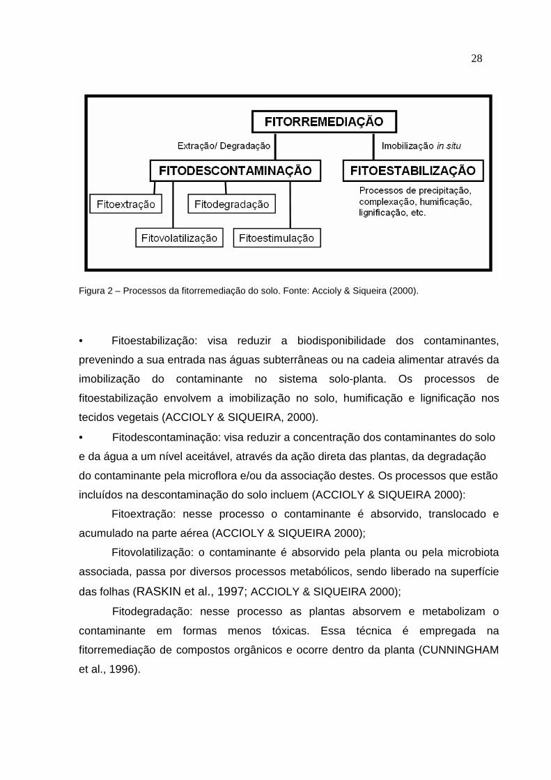
2.6 Espécies reativas de oxigênio (EROs) e estresse oxidativo
A produção de espécies reativas de oxigênio (EROs) ocorre normalmente
durante o metabolismo nas células aeróbicas e a sua produção pode depender de
diversas fontes, como por exemplo durante a respiração que ocorre nas
mitocôndrias celulares, bem como nas vias relacionadas à fotossíntese nos
cloroplastos (MITTLER, 2002). Recentemente, novas fontes de EROs tem sido
identificadas em plantas, incluindo NADPH oxidases, amino oxidases, glicolato
oxidase e peroxidases ligadas a parede celular (MITTLER, 2002; PARENT et al.,
2008) (Figura 3).
Sob condições normais, a produção de EROs na célula é baixa. Entretanto
muitos agentes oxidantes promovem uma disruptura da homeostase celular levando
ao aumento da produção de EROs e ao estresse oxidativo (MITTLER, 2002). Nesse
caso as EROs são citadas como sinalizadores de uma resposta a agentes
estressores (MITTLER, 2002).
30
Figura 3 - Sítios de produção intracelular de espécies reativas de oxigênio (EROs) (Adaptado de PARENT et al., 2008).
As EROs são formas parcialmente reduzidas do oxigênio atmosférico (O2).
Podem resultar a partir da excitação do O2 para a forma de oxigênio singleto (O21) ou
a partir da transferência de um, dois ou três elétrons para o O2 originando,
respectivamente, o radical superoxido (O2-), peróxido de hidrogênio (H2O2) ou um
radical hidroxil (OH.) (GUTTERIDGE, 1995). Por apresentarem um elétron
desemparelhado na sua estrutura, as EROs são instáveis, extraordinariamente
reativas e não especificas, desencadeando reações peroxidativas e causando
danos significantes às membranas e a outras macromoléculas essenciais, tais como
os pigmentos, as proteínas, os ácidos nucléicos e os lipídios (FOYER et al., 1994;
GUTTERIDGE, 1995; HALLIWELL & GUTTERIDGE, 2000). A peroxidação lipídica é
um dos principais efeitos ocasionados pelo aumento das EROs. Radicais altamente
reativos como o OH.... frequentemente atacam moléculas biológicas (lipídios) e

31 abstraem um hidrogênio (H), desencadeando a peroxidação lipídica (NIKI, 2009). A
peroxidação lipidica causa danos à estrutura e funcionamento das membranas
biológicas alterando a sua fluidez, inativando receptores e enzimas da membrana
aumentando a permeabilidade (NIKI, 2009).
Muitos estudos têm relacionado o Pb ao estresse oxidativo, principalmente a
peroxidação de lipídios (VERMA & DUBEY, 2003; DEY et al., 2007; GUPTA et al.,
2009). O Pb induz a peroxidação lipdica através do aumento da produção de EROs
(SHARMA & DUBEY, 2005).
Entretanto, as plantas e todos os organismos possuem um sistema de defesa
antioxidante, formado por componentes enzimáticos e não enzimáticos, o qual é
responsável por manter sob controle a produção de EROs, evitando os efeitos
tóxicos de agentes tal como o Pb (SHARMA & DUBEY, 2005).
2.7 Sistema antioxidante
Para o combate da toxicidade do metal e proteção das membranas celulares
e organelas dos efeitos danosos das EROs, as células das plantas possuem um
sistema de defesa antioxidante, formado por componentes enzimáticos e não
enzimáticos que normalmente mantêm um balanço de EROs dentro das células.
Dentre os antioxidantes enzimáticos estão a superóxido dismutase (SOD, E.C.
1.15.1.1), a catalase (CAT, E.C. 1.11.1.6) e a ascorbato peroxidase (APX, E.C.
1.11.1.11), bem como antioxidantes de baixo peso molecular não enzimáticos, como
o ácido ascórbico, a glutationa reduzida (GSH) e outros grupos tiólicos não protéicos
que removem tipos diferentes de EROs (FOYER et al., 1994) e protegem a célula
contra a injúria e a disfunção dos tecidos (MIQUEL, 1989). Além disso, em plantas,
os carotenóides também possuem efeito antioxidante importante no sistema
fotossintético (SALGUERO et al., 2003).
A enzima superóxido dismutase (SOD, E.C. 1.15.1.1) constitui a primeira linha
de defesa contra as EROs (ALSCHER et al., 2002). São metaloenzimas
responsáveis pela dismutação do radical superóxido (O2-.) a peróxido de hidrogênio
(H2O2). Tem sido demonstrado que os fosfolipídios das membranas celulares são

32 impermeáveis as moléculas de O2
-., sendo de crucial importância que as SODs
estejam presentes nos compartimentos onde O2-. é formado. Existem diferentes
isoenzimas de SOD, geralmente presentes em compartimentos celulares distintos:
Fe-SOD (localizada no cloroplasto), Mn-SOD (localizada na mitocôndria e
peroxisomo) e Cu-Zn-SOD (localizada no peroxisomo, cloroplasto, citosol e
possivelmente no espaço extracelular) (ALSCHER et al., 2002). A reação geral da
SOD pode ser descrita:
2O2-. + 2H+ → H2O2 + O2
O H2O2 gerado na conversão de superóxidos pela SOD também é tóxico e
deve ser detoxificado pela catalase e/ou peroxidases. A enzima catalase (CAT) é
uma ferrihemoenzima presente principalmente nos peroxissomos, podendo ser
encontrada em menor quantidade na mitocôndria, no cloroplasto e no retículo
endoplasmático. A catalase presente nas diferentes organelas é responsável pela
remoção do H2O2 durante a fotorrespiração, β-oxidação dos ácidos graxos e durante
o estresse oxidativo (INZÉ & MONTAGU, 1995). A CAT é uma enzima tetrâmerica
cuja principal função é dismutar o H2O2 formando H2O e O2 (FRIDOVICH, 1998). É a
única enzima que degrada H2O2 sem consumir equivalentes redutores, sendo
responsável pela remoção de peróxidos quando em excesso devido a sua baixa
afinidade ao H2O2 (MITTLER, 2002). Existem três isoenzimas de CAT: CAT1, CAT2
e CAT3 (INZÉ & MONTAGU, 1995). Sua reação pode ser descrita como:
2 H2O2 → 2 H2O + O2
A ascorbato peroxidase (APXs) é um dos grupos mais importantes de
enzimas antioxidantes, sendo encontradas cinco isoformas: citosólica, mitocondrial,
peroxissomal, glioxissomal e cloroplástica. A APX é uma proteína que contem ferro
no seu grupo prostético heme (DABROWSKA et al., 2007). A APX é uma enzima
chave no ciclo da glutationa – ascorbato que reduz o H2O2 até H2O usando o
ascorbato como doador de elétrons, com concomitante geração de deidroascorbato.
Este é reciclado a ascorbato usando a glutationa reduzida (GSH) como doadora de
elétrons. A glutationa oxidada (GSSG) formada é convertida a glutationa reduzida
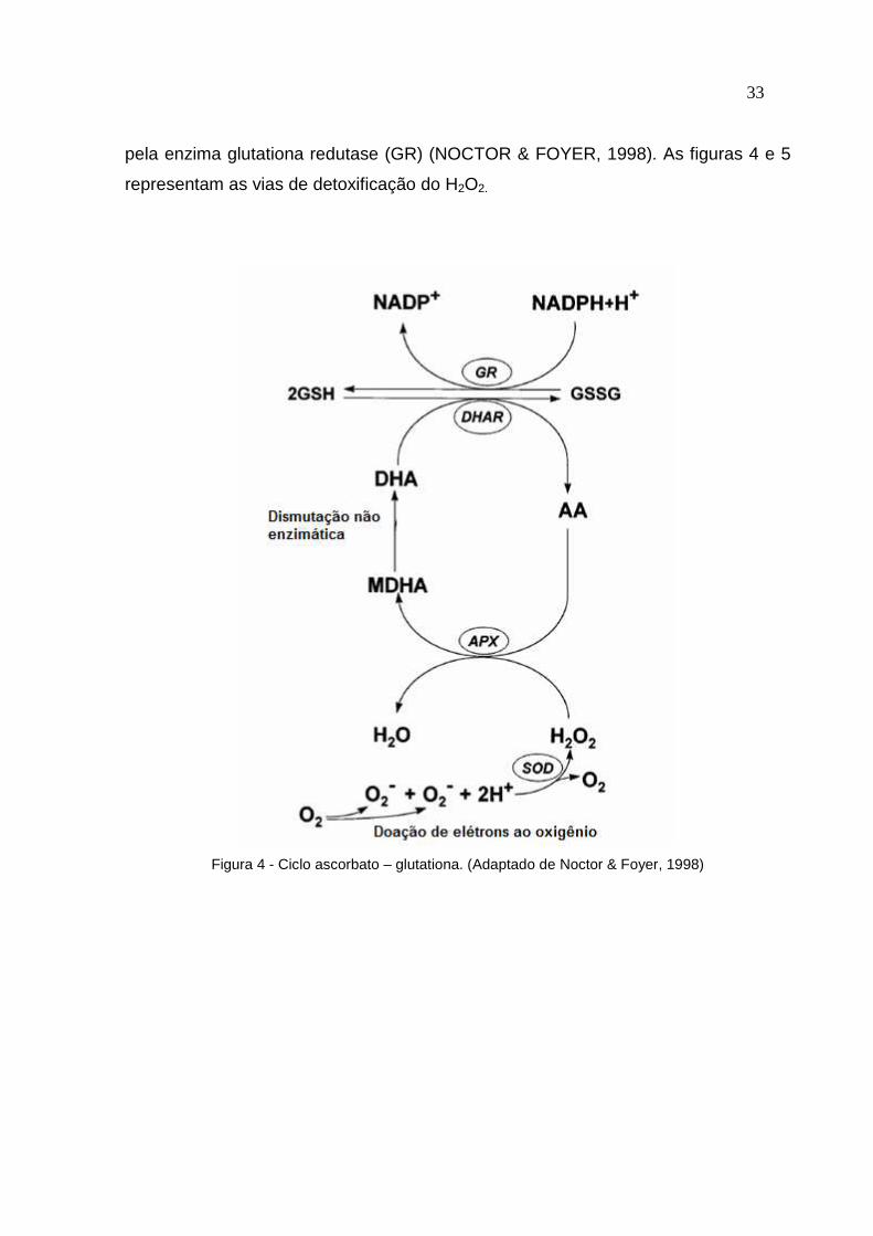
33 pela enzima glutationa redutase (GR) (NOCTOR & FOYER, 1998). As figuras 4 e 5
representam as vias de detoxificação do H2O2.
Figura 4 - Ciclo ascorbato – glutationa. (Adaptado de Noctor & Foyer, 1998)
34
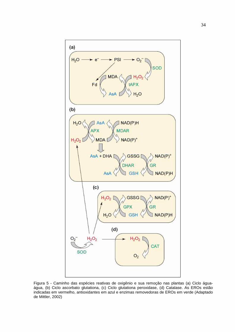
Figura 5 - Caminho das espécies reativas de oxigênio e sua remoção nas plantas (a) Ciclo água-água, (b) Ciclo ascorbato glutationa, (c) Ciclo glutationa peroxidase, (d) Catalase. As EROs estão indicadas em vermelho, antioxidantes em azul e enzimas removedoras de EROs em verde (Adaptado de Mittler, 2002)
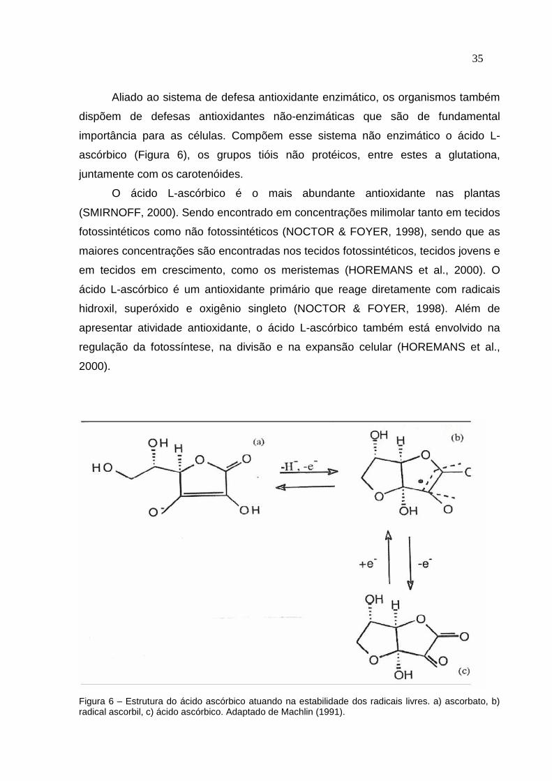
35 Aliado ao sistema de defesa antioxidante enzimático, os organismos também
dispõem de defesas antioxidantes não-enzimáticas que são de fundamental
importância para as células. Compõem esse sistema não enzimático o ácido L-
ascórbico (Figura 6), os grupos tióis não protéicos, entre estes a glutationa,
juntamente com os carotenóides.
O ácido L-ascórbico é o mais abundante antioxidante nas plantas
(SMIRNOFF, 2000). Sendo encontrado em concentrações milimolar tanto em tecidos
fotossintéticos como não fotossintéticos (NOCTOR & FOYER, 1998), sendo que as
maiores concentrações são encontradas nos tecidos fotossintéticos, tecidos jovens e
em tecidos em crescimento, como os meristemas (HOREMANS et al., 2000). O
ácido L-ascórbico é um antioxidante primário que reage diretamente com radicais
hidroxil, superóxido e oxigênio singleto (NOCTOR & FOYER, 1998). Além de
apresentar atividade antioxidante, o ácido L-ascórbico também está envolvido na
regulação da fotossíntese, na divisão e na expansão celular (HOREMANS et al.,
2000).
Figura 6 – Estrutura do ácido ascórbico atuando na estabilidade dos radicais livres. a) ascorbato, b) radical ascorbil, c) ácido ascórbico. Adaptado de Machlin (1991).

36 Dentro do grupo de tióis não protéicos, a glutationa (GSH) é a forma
predominante, sendo encontrada em altas concentrações nos cloroplastos e em
outros compartimentos celulares [1-5 mM] (NOCTOR & FOYER, 1998). A GSH pode
atuar diretamente ou indiretamente na redução da maioria das espécies reativas de
oxigênio sendo de crucial importância para a defesa contra o estresse oxidativo. A
GSH pode reduzir as EROs diretamente ou através do ciclo ascorbato-glutationa
(NOCTOR et al., 1998). Além disso, a GSH pode reagir quimicamente com o
oxigênio singleto, com o radical superóxido e hidroxila, funcionando como removedor
de EROs (NOCTOR & FOYER, 1998). É também a precursora de fitoquelatinas, as
quais são peptídeos que tem a capacidade de se complexar com os metais,
promovendo a sua detoxificação (ASADA, 1994). Estudos mostram que níveis
elevados de GSH celular estão associados à tolerância à metais pesados em
plantas (CHEN & GOLDSBROUGH, 1994) ocorrendo um acúmulo de GSH em
reposta à geração de EROs (NOCTOR & FOYER, 1998).
Os carotenóides também agem na proteção celular. Em relação ás suas
propriedades antioxidantes, os carotenóides podem atuar de várias maneiras:
reagindo com os produtos da peroxidação lipídica interrompendo a reação em
cadeia; removendo o oxigênio singleto e dissipando a energia na forma de calor;
reagindo com a clorofila tripleto ou excitada prevenindo a formação de oxigênio
singleto (SALGUERO et al., 2003).
Assim, a capacidade do sistema antioxidante de detoxificar as EROs está
intimamente ligada à tolerância das plantas aos metais pesados (SCHÜTZENDÜBEL
& POLLE, 2002).

37 3 RESULTADOS
3.1 MANUSCRITO:
Pluchea sagittalis has tolerance to Pb stress and this behaviour is re lated to an
efficient antioxidant system and improved water use efficiency
Liana Veronica Rossato, Fernando Teixeira Nicoloso, Júlia Gomes Farias, Denise
Cargnelluti, Luciane Almeri Tabaldi, Fabiane Goldschmidt Antes, Valderi Luiz
Dressler, Vera Maria Morsch, Maria Rosa Chitolina Schetinger.
38 Pluchea sagittalis has tolerance to Pb stress and this behaviour is re lated to an
efficient antioxidant system and improved water use efficiency
Liana Veronica Rossato2,4, Fernando Teixeira Nicoloso1,3, Júlia Gomes Farias1,
Denise Cargnelluti2, Luciane Almeri Tabaldi2, Fabiane Goldschmidt Antes2, Valderi
Luiz Dressler2,Vera Maria Morsch2,4, Maria Rosa Chitolina Schetinger2,4.
Departamento de Biologia1, Química2, Programa de Pós-Graduação em Agronomia3
e Bioquímica Toxicológia4, Centro de Ciências Naturais e Exatas, Universidade
Federal de Santa Maria, 97105-900, Santa Maria, RS, Brasil.
Abstract
This work aimed to study the process of stress adaptation in root and leaves of
different developmental stages (apex, middle and basal regions) of P. sagittalis
grown under hydroponic conditions under exposure to lead (Pb). Plants were
exposed to five Pb levels: 0, 200, 400, 600, and 1000 µM for 30 days. Pb
concentration and content in roots, stems, and leaves in different developmental
ages increased with external Pb level. Pb concentration and content were higher in
roots than in shoot parts. Consumption of nutrient solution and transpiration ratio
were both decreased by Pb treatments. Leaf fresh weight, leaf area, and shoot length
decreased linearly upon addition of Pb treatments. On the other hand, dry weight of
shoot parts and roots did not decrease upon addition of Pb treatments. Both fresh
and dry weight of roots increased at 200 µM Pb when compared to control. The index
of tolerance of roots and shoots of P. sagittalis decrease at higher Pb concentrations.
However, the roots were more tolerant to Pb than shoots. δ-aminolevulinic acid
dehydratase activity was decreased by Pb treatments, whereas carotenoid and
chlorophyll concentrations were not affected. Lipid peroxidation and hydrogen
peroxide concentration both in roots and leaves increased with increasing Pb levels.
Pb treatments increased ascorbate peroxidase activity in all plant parts, while
superoxide dismutase activity increased in leaves and decreased in roots. Catalase
activity in leaves from the apex shoot was not affected by Pb, but in other plant parts
it was increased. Pb toxicity caused increase in non-protein thiol groups

39 concentration in shoot parts, whereas no significant difference was observed in roots.
Ascorbic acid concentration increased with increasing Pb level. In conclusion, Pb
stress triggered a defense mechanism against oxidative stress in P. sagittalis
showing a protective effect against ROS, depending on the tissue and its
physiological status, demonstrating tolerance and possible use of these plants for
reclamation of Pb contaminated soils.
Keywords: Antioxidant system, Pb toxicity, Pluchea sagittalis, Lipid peroxidation,
water use efficiency
1 Introduction
Heavy metal pollution is one major ecological concern due to its impact on
human health through the food chain and its high persistence in the environment
(SHARMA & DUBEY, 2005). Lead (Pb) is one of the hazardous heavy metal
pollutants of the environment that is originates from various sources such as mining
and smelting of lead-ores, burning of coal, effluents from storage battery industries,
automobile exhausts, metal plating and finishing operations, fertilizers, pesticides and
additives in pigments and gasoline (SHARMA & DUBEY, 2005).
Pb-contaminated soils contain Pb concentrations in the range of 400 – 800 mg
kg-1 soil, whereas in industrialized areas the level may reach up to 1000 mg Pb kg-1
soil (ANGELON & BINI, 1992). Pb reacts with biomolecules and adversely affects
different systems, such as reproductive, nervous, immune and cardio-vascular, as
well as developmental processes (JOHNSON, 1998). In plants, it exerts adverse
effects on morphology, growth and photosynthetic processes, water imbalance,
alteration in membrane permeability and disturbs in mineral nutrition (SINGH et al.,
1997; SHARMA & DUBEY, 2005). However, some species of plants tolerate the
presence of Pb. More interestingly, some have developed the capacity to accumulate
large amounts of this element in all parts of the plant body, mostly in root tissues, a
feature essential to the development of phytoremediation technologies to clean Pb
contaminated sites (SINGH et al., 1997; SHARMA & DUBEY, 2005). The
phytoextration of Pb, however, is often challenged by three factors: (1) low solubility

40 of Pb in soils, (2) lower Pb translocation to plant parts, and (3) toxicity of Pb to plant
tissues (CUNNINGHAM & BERTI, 2000).
Because of severe damages of plant growth and development, considerable
attempts have been made in discovering physiological and biochemical processes
contributing to the adaptation to heavy metal toxicity in plants (GRATÃO et al., 2005;
SHARMA & DUBEY, 2005). At cellular level, Pb induces accumulation of reactive
oxygen species (ROS) (VERMA & DUBEY, 2003) as a result of imbalanced ROS
production and ROS scavenging processes by imposing oxidative stress (MITTLER
et al., 2004; GUPTA et al., 2009). ROS include superoxide radical (O2•-), hydrogen
peroxide (H2O2) and hydroxyl radical (OH•-), which are necessary for the correct
functioning of plants; however, in excess they cause damage to biomolecules, such
as membrane lipids, proteins, and nucleic acids, among others (MITTLER et al.,
2004).
Plants have different defense strategies against the toxicity of heavy metals.
The first defense strategy is to avoid the metal entry into the cell excluding it or
binding it to a cell wall (MISHRA et al., 2006). The second defense systems
constitutes of various antioxidants to combat the increased production of ROS
caused by metals (REDDY et al., 2005). This system is comprised of enzymes
superoxide dismutase (SOD; E.C. 1.15.1.1), ascorbate peroxidase (APX;
E.C.1.11.1.11) and catalase (CAT; E.C. 1.11.1.6) as well as the non-enzymic
constituents α-tocopherol, carotenoids, ascorbate and reduced glutathione, which
remove, neutralize, and scavenge the ROS (MITTLER et al., 2004).
The elevated activity of antioxidative enzymes could serve as an important
component of the antioxidative defense mechanism against oxidative injury,
increasing the tolerance of plants to Pb stress and being utilized for reclamation of
Pb contaminated soils (REDDY et al., 2005). However, the response of enzymatic
and non-enzymatic antioxidants to heavy metals involves attenuation of ROS and is
greatly dependent on the plant species, physiological status of the tissues and
culture conditions (GRATÃO et al., 2005; GONÇALVES et al., 2009; GUPTA et a.,
2009).
In view of this, the objective of the present study is to examine the uptake and
distribution pattern of Pb and the effect of this metal on the enzymatic and non–
enzymatic antioxidant system in different organs (roots, stems and leaves of different
41 developmental stages) of P. sagittalis plants under long exposure time (30 days) and
high level of Pb (up to 1000 µM). Besides, we also checked the possible role of the
antioxidant system in relation to increased tolerance of P. sagittalis to Pb toxicity.
According to our knowledge, this is the first study on Pb toxicity inducing
physiological and biochemical changes in P. sagittalis.
2 Materials and methods
2.1 Plant materials and growth conditions
P. sagittalis (Lam.) Cabrera plants growing in the Botanic Garden of the
Universidade Federal de Santa Maria [Santa Maria, Rio Grande do Sul, Brazil] were
used in this study. Nodal segments (1.0 cm long) without leaves were
micropropagated in MS medium (MURASHIGE & SKOOG, 1962), supplemented with
30 g l-1 of sucrose, 0.1 g l-1 of myo-inositol and 6 g l-1 of agar.
2.1.1 Hydroponic experiment
Thirty-day-old plantlets grown in in vitro culture were transferred to ex vitro
condition into plastic vessels (1 l) containing 950 g sand. Evaporated and transpired
water was daily replaced with nutrient solution, and the moisture-holding capacity
was maintained near 95%. The nutrient solution had the following composition (mg l-
1): 85.31 N; 7.54 P; 11.54 S; 97.64 Ca; 23.68 Mg; 104.75 K; 176.76 Cl; 0.27 B; 0.05
Mo; 0.01 Ni; 0.13 Zn; 0.03 Cu; 0.11 Mn and 2.68 Fe. The pH solution was adjusted to
5.5 ± 0.1 with a 1 M solution of HCl or NaOH. After two months of plant
acclimatization, Pb2+ was added to nutrient solution as Pb-acetate (Pb
(CH3COO)2.3H2O) at concentrations of 0 (control), 200, 400, 600 and 1000 µM. The
treatments were applied daily for 30 days obtaining, respectively, final concentrations
42 of 0, 47, 81, 107 and 162 mg/kg Pb. After 30 days of Pb exposure, three replicates
per treatment (each replicate consisted of six plants) were randomly harvested.
Both in vitro and ex vitro cultured plants were grown in a growth chamber at 25
± 2 °C on a 16/8 h light/dark cycle with 35 µmol m -2 s-1 of irradiance provided by cold
fluorescent lamps. At the end of the experiments, the plants were gently washed with
distilled water and then divided into roots and shoots. The shoot was divided in three
parts according to the position of leaves on the stem as follows: apex part (from the
1st to 10th leaf), middle part (from the 11th to 16th leaf), and basal part (from the 17th to
the last leaf on the base of the stem). Subsequently, growth and biochemical
parameters were determined. All chemicals used were of analytical grade purchased
from Sigma Chemical Company (USA).
2.2 Pb determination
Pb concentration was determined in roots, leaves and stems. Dried plant
tissues, between 0.01–0.25 g, were ground and digested with 5 ml of concentrated
HNO3. Sample digested was carried out in an open digestion system, using a heating
block Velp Scientific (Milano, Italy). Heating was set at 130 ºC for 2 h. Plastic caps
were fitted to the vessels to prevent losses by volatilization. The Pb content was
determined by Inductively Coupled Plasma Optical Emission Spectrometry (ICP-
EOS), using a PerkinElmer Optima 4300 DV (Shelton, USA) equipped with a cyclonic
spray chamber and a concentric nebulizer. The emission line selected was 220.353
nm. A 1000 mg l-1 Pb (Merck) standard was used and reference solutions were
prepared by serial dilution in 5% HNO3 (v/v).
2.3 Growth parameters
Growth of P. sagittalis plants was determined by measuring the fresh and dry
weight of shoot and root, shoot length and leaf area. The roots and shoots were

43 oven-dried at 65 ºC to a constant weight for the determination of biomass. For the
leaf area, two leaves for each shoot part were scanned and the area was measured
using a Sigma Scan Pro v. 5.0 Jandel Scientific software. The index of tolerance (IT)
was calculated according to Wu and Antonovies (1976) as below:
mean dry wt produced in solution with lead IT = mean dry wt produced in solution without lead
2.4 Carotenoid and chlorophyll concentrations
Carotenoid and chlorophyll concentrations were determined following the
method of Hiscox & Israeslstam (1979) and estimated with the help of Lichtenthaler’s
formula (LICHTENTHALER, 1987). Briefly, 0.1 g chopped fresh leaves of different
shoot parts (apex, middle and basal) was incubated at 65 ºC in dimethylsulfoxide
(DMSO) until the tissues were completely bleached. Absorbance of the solution was
then measured at 470, 645 and 663 nm to determine the contents of carotenoids,
chlorophyll a, and chlorophyll b, respectively.
2.5 Delta-aminolevulinic acid dehydratase (δ-ALA-D; E.C. 4.2.1.24) activity
The leaves of different shoot parts (apex, middle and basal) were
homogenized in 10 mM Tris-HCl buffer, pH 9.0, at a proportion of 1:2 (w/v). The
homogenate was centrifuged at 6,000 rpm at 4 ºC for 10 min, using a Hitachi himac
CR 21E (Tokyo, Japan). The supernatant was pre-treated with 1% Triton X-100 and
0.5 mM dithiotreithol (DTT). δ-ALA-D activity was assayed as described by Morsch et
al. (2002) by measuring the rate of porphobilinogen formation. The incubation
medium for the assays contained 100 mM Tris-HCl buffer, pH 9.0 and 3.6 mM ALA.
Incubation was started by adding 100 µl of the tissue preparation to a final volume of
400 µl and stopped by adding 350 µl of the mixture containing 10% trichloroacetic

44 acid (TCA) and 10 mM HgCl2. The product of the reaction was determined with the
Ehrlich reagent at 555 nm using a molar absorption coefficient of 6.1 x 10⁴ l mol-1 cm-
1 (SASSA, 1982) for the Ehrlich-porfhobilinogen salt.
2.6 Estimation of lipid peroxidation
The level of lipid peroxidation products was estimated following the method El-
Moshaty et al. (1993) by measuring the concentration of malondialdehyde (MDA) as
an end product of lipid peroxidation by reaction with thiobarbituric acid (TBA). Both
root and leaf (apex, middle and basal) samples were homogenized at 4 ºC in 10 ml of
0.2 M citrate-phosphate (pH 6.5) containing 0.5 % Triton X-100 at a proportion of
1:10 (w/v). The homogenate was filtered through two layers of paper and centrifuged
for 15 min at 20,000 g, using a Hitachi himac CR 21E (Tokyo, Japan) One milliliter of
the supernatant fraction was added to an equal volume of 20% (w/v) TCA containing
0.5% (w/v) TBA. The mixture was heated at 95 ºC for 40 min and then quickly cooled
in ice bath for 15 min. After centrifugation at 10,000 g for 15 min, using a MTD III
PLUS (Servilab), the absorbance of the supernatant was measured at 532 nm. A
correction of non-specific turbidity was made by subtracting the absorbance value
taken at 600 nm.
2.7 Determination of hydrogen peroxide (H2O2)
The H2O2 concentration of P. sagittalis was determined according to Loreto &
Velikova (2001). Approximately 0.1 g of root and leaf (apex, middle and basal)
samples were homogenized in 2 ml of 0.1% (w/v) TCA. The homogenate was
centrifuged at 12,000 g for 15 min at 4 ºC [Hitachi himac CR 21E (Tokyo, Japan)] and
0.5 ml of 10 mM potassium phosphate buffer (pH 7.0) and 1 ml of 1M KI. The H2O2
concentration of supernatant was evaluated by comparing its absorbance at 390 nm
with a standard calibration curve.

45 2.8 Enzyme activities of antioxidant systems
Fresh root and leaf samples of different shoot parts (apex, middle and basal)
were used for enzyme analysis. One gram tissue homogenized in 3 ml of 0.05M
sodium phosphate buffer (pH 7.8) including 1 mM EDTA and 2% (w/v) PVP. The
homogenate was centrifuged at 13,000 g for 20 min at 4 ºC, using a Hitachi himac
CR 21E (Tokyo, Japan). Supernatant was used for enzyme activity and protein
content assays (ZHU et al., 2004).
Catalase (CAT) activity was assayed following the modified Aebi (1984)
method. The activity was determined by monitoring the disappearance of H2O2
measuring the decrease in absorbance at 240 nm of a reaction mixture with a final
volume of 2 ml containing 15 mM H2O2 in potassium phosphate buffer (pH 7.0) and
30 µL extract.
Ascorbate peroxidase (APX) was measured according to Zhu et al. (2004).
The reaction mixture, at a total volume of 2 ml, contained 25 mM (pH 7.0) sodium
phosphate buffer, 0.1 mM EDTA, 0.25 mM ascorbate, 1.0 mM H2O2 and 100 µl
enzyme extract. H2O2 –dependent oxidation of ascorbate was followed by a decrease
in the absorbance at 290 nm (ε = 2.8 l mmol l-1 cm-1).
Superoxide dismutase (SOD) activity was assayed according to Misra &
Fridovich (1972). The assay mixture consisted of a total volume of 1 ml, containing
glycine buffer (pH 10.5), 1 mM epinephrine and enzyme material. Epinephrine was
the last component to be added. The adrenochrome formation in the next 4 min was
recorded at 480 nm in UV- Vis spectrophotometer [Hitachi U – 2001(Shimadzu)].
One unit of SOD activity is expressed as the amount of enzyme required to cause
50% inhibition of epinephrine oxidation in the experimental conditions. This method is
based on the ability of SOD to inhibit the autoxidation of epinephrine at an alkaline
pH. Since the oxidation of epinephrine leads to the production of a pink
adrenochrome, the rate of increase of absorbance at 480 nm, which represents the
rate of autoxidation of epinephrine, can be conveniently followed. SOD has been
found to inhibit this radical-mediated process.

46 2.9 Ascorbic acid (AsA) and Non-protein thiol groups (NPSH) concentrations
Both root and leaf samples of different shoot parts (apex, middle and basal)
were homogenized in a solution containing 50 mM Tris-HCl and 10% Triton X-100
(pH 7.5), centrifuged at 6,800 g for 10 min, using a MTD III PLUS (Servilab). To the
supernatant obtained was added 10% TCA at proportion 1:1 (v/v) followed by
centrifugation (6,800 g for 10 min) to remove protein. The supernatant was used to
determine AsA and NPSH contents.
AsA determination was performed as described by Jacques-Silva et al. (2001).
An aliquot of the sample (300 µl) was incubated at 37 ºC in a medium containing 100
µl TCA 13.3%, 100 µl deionized water and 75 µl DNPH. After 3 h, 500 µl of 65%
H2SO4 was added and samples were read at 520 nm. A standard curve was
constructed using L(+) AsA.
NPSH concentration in P. sagittalis plants was measured
spectrophotometrically with Ellman’s reagent (ELLMAN, 1959). An aliquot of the
extract sample (400 µl) was added in a medium containing 550 µl 1M Tris-HCl (pH
7.4). Reaction was read at 412 nm after the addition of 10 mM 5-5-dithio-bis (2-
nitrobenzoic acid) (DTNB) (5 µl). A standard curve using cysteine was used to
calculate the content of thiol groups in samples.
2.10 Protein determination
To all the enzyme assays, protein was measured by the Comassie Blue
method according to Bradford (1976) using bovine serum albumin as standard.
2.11 Statistical analysis

47
The experiments were performed using a randomized design. The analyses of
variance were computed on statistically significant differences determined based on
the appropriate F-tests. Results are presented as means ± S.D. of at least three
independent replicates. The mean differences were compared using the Tukey test
(P < 0.05).
3 Results
3.1 Lead concentration and content in plant tissues
Pb concentration and content in both roots and shoots (stem and leaves)
increased with Pb treatments (Fig. 1A, B). However, most of the Pb taken up by the
plants was accumulated in roots (Fig. 1B).
In general, both leaves and stem from the basal part of the shoot showed
greater Pb concentration and content than those from the apex and middle parts.
However, this pattern was more evident in leaves. Pb concentration and content in
stem from both apex and middle shoot parts were lower at 1000 µM Pb than at 600
µM Pb. On the other hand, this behavior was not observed in the leaves from the
same shoot parts.
48
Figure 1. Effect of increasing Pb concentration on the Pb concentration (A) and
content (B) in roots as well as in leaves and stems of different shoot parts (apex,
middle and basal) of P. sagittalis plants. Data represent the mean ± S.D. of three
replicates. Different letters indicate significant differences among the Pb
concentrations in the same plant part (p < 0.05).

49 3.2 Effects of Pb on growth parameters
There was a clear decrease in shoot fresh weight upon addition of Pb levels
(Fig. 2A). On the other hand, root fresh weight increased at 200 µM Pb and
decreased at 1000 µM Pb, when compared to the control.
Shoot dry weight, regardless of the developmental stage of leaves and stem,
was not significantly affected by Pb treatments. Conversely, root dry weight
increased at 200 µM Pb and was not altered at other Pb levels, when compared to
control (Fig. 2B).
Leaf area from different shoot parts was reduced with increasing Pb
concentration (Fig. 2C). Despite the decreased shoot length with the increase of Pb
concentration, it was only significantly reduced upon addition of Pb levels above 400
µM (Fig. 2D).
The consumption of nutrient solution by plant per day decreased with
increasing Pb levels (Fig. 2E). In contrast, the transpiration ratio was decreased by
Pb treatments, when compared to control, being that this decrease did not follow a
linear pattern as observed for the consumption of nutrient solution per plant. The
lowest transpiration ratio was obtained at 200 and 600 µM Pb (32% lower than the
control), whereas it decreased only 22% at 400 and 1000 µM Pb (Fig. 2F).
50
Figure 2. Effect of increasing Pb concentration on fresh (A) and dry (B) weight of
different shoot parts (apex, middle and basal) and roots, leaf area (C), shoot length
(D), nutrient solution consumption (E), and transpiration ratio (F) of P. sagittalis
plants . Statistics as in Figure 1.
The root index of tolerance (IT) values of P. sagittalis was significantly lower to
control only 1000 µM Pb. On the other hand, the shoot IT was reduced upon addition
of Pb levels above 200 µM (Table 1). Compared with those of the shoots and roots
dws (Fig. 2B) and the IT values, the roots appeared to be a more tolerant than
shoots.
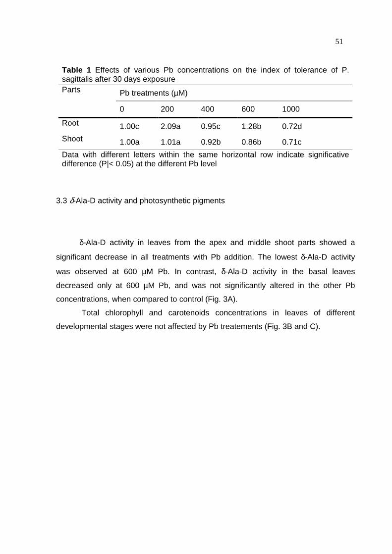
51
Table 1 Effects of various Pb concentrations on the index of tolerance of P. sagittalis after 30 days exposure Parts Pb treatments (µM)
0 200 400 600 1000
Root 1.00c 2.09a 0.95c 1.28b 0.72d
Shoot 1.00a 1.01a 0.92b 0.86b 0.71c
Data with different letters within the same horizontal row indicate significative difference (P|< 0.05) at the different Pb level
3.3 δ-Ala-D activity and photosynthetic pigments
δ-Ala-D activity in leaves from the apex and middle shoot parts showed a
significant decrease in all treatments with Pb addition. The lowest δ-Ala-D activity
was observed at 600 µM Pb. In contrast, δ-Ala-D activity in the basal leaves
decreased only at 600 µM Pb, and was not significantly altered in the other Pb
concentrations, when compared to control (Fig. 3A).
Total chlorophyll and carotenoids concentrations in leaves of different
developmental stages were not affected by Pb treatements (Fig. 3B and C).

52
Figure 3. Effect of increasing Pb concentration on the δ-aminolevulinic acid
dehydratase activity (A), and chlorophyll (B) and carotenoids (C) concentration in
leaves of different shoot parts. Statistics as in Figure 1.

53 3.4 Hydrogen peroxide concentration and lipid peroxidation
For all evaluated plant tissues, in general, the H2O2 concentration increased
with increasing Pb levels. However, H2O2 concentration in the leaves did not change
at 200 µM Pb, when compared to control. Younger leaves (apex part) were the most
sensitive to Pb treatments in relation to H2O2 concentration (Fig. 4A).
The level of lipid peroxidation was measured in terms of malondialdehyde
(MDA) accumulation. As shown in Figure 4B, MDA concentration increased upon
addition of Pb levels in leaf tissues from the apex and middle parts, whereas in basal
leaves it increased only at Pb levels exceeding 400 µM, when compared to the
control. Root MDA concentration increased at all Pb treatments, but no difference
were found for Pb treatments ranging from 200 to 1000 µM (Fig. 4B).
Figure 4. Effect of increasing Pb concentration on the hydrogen peroxide
concentration (A), and on lipid peroxidation (B) in roots and leaves of different shoot
parts. Statistics as in Figure 1.

54 3.5 Effects of Pb on antioxidative systems
3.5.1 Enzymes
In general, regardless of the developmental stage of leaves, SOD activity
increased with increasing Pb levels. The exception to this pattern was observed at
200 µM Pb where basal leaves showed lower SOD activity than the control (Fig. 5A).
SOD activity was higher in leaves than in roots. Root SOD activity was not affected
by any of the tested Pb levels. Leaf CAT activity was less affected by Pb treatment
than it was for SOD activity (Fig. 5B). Apex leaf CAT activity was not altered by any
of the tested Pb levels. On the other hand, CAT activity in the middle leaves was
significantly increased at the 400 and 1000 µM Pb levels. At 600 µM Pb treatment,
the CAT activity in basal leaves significantly increased compared to control plants.
Root CAT activity showed no significant difference upon addition of 400 µM and 1000
µM Pb levels, however, it significantly increased upon addition of 200 and 600 µM Pb
levels, when compared to control. Regardless of the plant organ, the APX activity
increased at Pb levels above 400 µM (Fig. 5C). The highest increase in APX activity
was seen at 1000 µM Pb.
3.5.2 Non-enzymatic antioxidants
In general, the AsA concentration in all plant organs increased with increasing
Pb levels (Fig. 5D). Younger leaf tissues (apex part) presented significantly larger
AsA concentration when compared with older leaf tissues (middle and basal parts).
However, the highest increase in AsA concentration was seen at 1000 µM Pb. NPSH
concentration in all shoot parts increased with increasing Pb levels, with exception of
that observed at 200 µM Pb level on NPSH concentration in leaf tissues of the apex
part, where it decreased when compared to control (Fig. 5E). No significant

55 difference in root NPSH concentration was found between Pb treatments and the
control.
Figure 5. Effect of increasing Pb concentration on the superoxide dismutase,
catalase and ascorbate peroxidase activities (A , B and C, respectively), and ascorbic
acid and nonprotein thiol groups concentration (D and E, respectively) in roots and in
leaves of different shoot parts. Statistics as in Figure 1.
56 4 Discussion
4.1 Tissue Pb concentration and content
Our data (Fig. 1) demonstrate that higher Pb exposures lead to remarkable
increases in Pb concentration and content in both root and shoot tissues (leaves and
stem from apex, middle and basal parts). The same was also reported for distinct
plant species by other authors (VERMA & DUBEY, 2003; MISHRA et al., 2006;
GUPTA et al., 2009). The accumulation of Pb was mostly pronounced in the roots.
Pb moves predominantly into the root apoplast and thereby in a radial manner across
the cortex accumulates near the endodermis (SHARMA & DUBEY, 2005).
Antosiewicz & Wierzbicka (1999) showed that most of the Pb remains in bound form
either in cell wall or in vacuoles. This Pb accumulation in the root system may
indicate that roots serve as a partial barrier to Pb transport to the shoot
(WIERZBICKA, 1987; SHARMA & DUBEY, 2005), suggesting that uptake and
translocation rates of Pb are low (KRATOVALIEVA & CVETANOVSKA, 2001).
Interestingly, in the present study, higher Pb concentration and content were
observed in older leaves than in younger leaves. Pb concentrations in the three stem
parts studied were very similar. This pattern suggests that P. sagittalis uses
differential partitioning of the excess of Pb taken up to avoid Pb toxicity. Therefore,
leaves differ in their abilities to accumulate Pb depending on age.
4.2 Effects of Pb on growth parameters and water status
P. sagittalis plants grown at excess supply of Pb (400, 600 and 1000 µM)
showed visual symptoms of Pb toxicity. The first symptoms were noted after 15 days
of metal supply, becoming more pronounced at 30 days. Leaves from the apex shoot
part presented a slight wilting (data not shown). This result is in accordance with data
of shoot fresh weight, which showed a decrease with increasing Pb supply (Fig. 2A)

57 although the dry weight was not affected. Moreover, leaf area also decreased upon
addition of Pb levels (Fig. 2C), whereas shoot length significantly decreased only at
Pb levels exceeding 400 µM (Fig. 2D). On the other hand, Pb treatments only
reduced root fresh weight at 1000 µM, when compared to control. Therefore, these
data indicate that for P. sagittalis the primary site of Pb toxicity appeared to be in the
shoot. Han et al. (2008) found the same behavior for Iris L. treated with 0-10 mmol l-1
for 28 days.
Pb toxicity is reported to inhibit the growth of various plant species (SINGH et
al., 1997; SHARMA & DUBEY, 2005; MISHRA et al., 2006; GOPAL & RIZVI, 2008),
which is partially in agreement with the data of the present study. The dry weight of
the three different shoot parts of P. sagittalis was not affected by Pb treatment,
whereas in the roots it slightly decreased at 1000 µM Pb, and significantly increased
at 200 µM Pb, when compared to control (Fig. 2B). Interestingly, as a visual symptom
the roots became slightly brownish and/or blackish with increasing Pb supply. Gopal
& Rizvi (2007) found that Pb at 1000 µM concentration for 35 days reduced about
30% the growth of radish (Raphanus sativus) roots. Similarly, Gupta et al. (2009)
reported that 200 µM Pb for 7 days decreased shoot dry weight of maize (Zea mays).
Therefore, since root biomass of P. sagittalis was significantly reduced only upon
addition of Pb levels exceeding 600 µM.
The IT is defined as the ratio of average dry weight of roots/shoots in the test
solution to the control, which is the reflection of the tolerant ability of a plant under the
metal exposure and stress. Han et al. (2008) observed that the IT values of two Iris
species treated over 7 days were significantly decreased in the higher Pb
concentrations. The same has observed in our work. However, our data indicate that
this specie has Pb tolerance. Wierzbicka (1999) comparing the lead tolerance in
Allium cepa with other plant species observed that plants with index tolerance above
65% were considered highly tolerant.
Pb-treated plants showed a decrease in the consumption of nutrient solution
(Fig. 2E), which might be related to a decrease in leaf area (r = 0.64) (Fig. 2C) that
can per se reduce the transpiration rate (SHARMA & DUBEY, 2005). Moreover, it
was reported that Pb affects stomatal movements at least in two different ways
through water or ion channels, promoting the closing of stomats (YANG et al., 2004).
58 In the roots, large amounts of Pb can prevent the water movement to the upper parts
through the mechanical blocking at the Caspari strip (WIERZBICKA, 1987).
Based on the significant decrease in fresh weight and area of leaves from
different shoot parts, but with no significant alteration in shoot dry weight upon
addition of Pb treatments, these data suggest that the most dramatic effect of Pb
excess was on the water status of P. sagittalis plants. Indeed, plants under Pb stress
conditions presented lower transpiration ratios than control plants (Fig. 2F), indicating
that Pb-stressed plants produced biomass amounts similar to control plants with
lower volume of water. The increase in both dry and fresh weight of roots at the
lowest Pb level supply (200 µM) may be due to the hormetic effect. Growth hormesis
represents an over compensation due to a disruption in homeostasis that has been
described in relation to different factors (GONÇALVES et al., 2009).
4.3 δ-ALA-D activity and photosynthetic pigments
The level of total chlorophyll and carotenoids was postulated as a simple and
reliable indicator of heavy metal toxicity for higher plants (GRATÃO et al., 2005).
Carotenoids are essential for photosynthesis acting as secondary photosynthetic
pigments, provitamin factors and as photoprotectants which quench free radicals
such as singlet oxygen species in damaged tissue (SALGUERO et al., 2003).
Results of the present study showed no significant difference in the chlorophyll and
carotenoids concentration in leaves of different developmental ages with increasing
Pb levels (Fig. 3B, C). This result might be related to the reduction in fresh weight
(Fig. 2A), which would lead to an increase in the concentration of cellular
components, including chlorophyll and carotenoids. It has been suggested that
reduction in chlorophyll content in the presence of heavy metals is caused by an
inhibition of chlorophyll biosynthesis (PEREIRA et al., 2006) which may be caused, in
part, by the reduction of δ-ALA-D activity. This enzyme catalyzes the condensation of
two molecules of δ-aminolevulinic acid (ALA) to porphobilinogen. The synthesis of
porphobilinogen promotes the formation of porphyrins, hemes and chlorophylls,
which are essential for adequate aerobic metabolism and for photosynthesis

59 (NORIEGA et al., 2006). In the present study, with exception of the result observed at
200 µM Pb on δ-ALA-D activity of leaves from the basal part of the shoot, the activity
of this enzyme in leaves, regardless of their age, was severely reduced upon addition
of Pb levels (Fig. 3A). Altered δ-ALA-D activity concomitant with reduced chlorophyll
content has been reported in several terrestrial plants exposed to various metals
(CARGNELUTTI et al., 2006; SKREBSKY et al., 2008; GONÇALVES et al., 2009).
The reduction of the δ-ALA-D activity may be due an interaction of Pb with free – SH
groups present at the active site of the enzyme (Van ASSCHE & CLIJSTERS, 1990)
or to the decreased tissue hydration (SINGH et al., 1997).
In addition of the absence of a negative effect of Pb treatments on chlorophyll
and carotenoids, we observed no significant change in dry weight of the different
shoot parts (Fig. 2B), indicating that the photosynthetic rate may not have been
altered. In contrast, Gupta et al. (2009) observed a duration-dependent response of
Pb stress on photosynthetic pigments and on δ-ALA-D activity of maize seedlings.
Therefore, as it seems that P. sagittalis is much more tolerant to Pb stress than
maize, Pb toxicity effects on P. sagittalis were much lower. However, δ-ALA-D activity
may be used as a biochemical marker to Pb stress.
4.4 Hydrogen peroxide concentration and lipid peroxidation
In many plant species, heavy metals have been reported to cause oxidative
damage due to production of ROS (GRATÃO et al., 2005). The ROS cause a variety
of harmful effects in plant cells including lipid peroxidation (ISLAM et al., 2008). In the
present study, there was a gradual increase in H2O2 and MDA concentrations in roots
and leaves from the different shoot parts with increasing Pb levels (Fig. 4A, B). The
significant increase in MDA concentration suggests that Pb caused oxidative damage
in the plant tissues (REDDY et al., 2005; MISHRA et al., 2006; DEY et al., 2007,
GUPTA et al., 2009). Interestingly, we observed that the increase of lipid peroxidation
in both root and shoot parts presented a straight correlation with the reduction in
shoot length (r = -0.79), leaf area (r = -0.49), whereas no significant correlation with
dry weight (r = 0.061) was observed.

60
In agreement with our results, other studies have showed increased MDA
content in Pb-exposed plants (MISHRA et al., 2006; DEY et al., 2007; GUPTA et al.,
2008). Pb induced ROS generation may initiate severe lipid peroxidation due to the
removal of hydrogen from unsatured fatty acids leading to formation of lipid radicals
and reactive aldehydes (MISHRA et al. 2006). Islam et al. (2008) reported that
increased levels of H2O2 under Pb stress is a result of action of SOD on superoxide
radicals and/or of the direct formation of H2O2 in biochemical pathways, such as
photorespiration. In the present study, leaves from the apex shoot part showed
higher H2O2 concentration than older leaves upon addition of Pb levels. Tamás et al.
(2004) suggested that the function of this elevated H2O2 formation in young tissues
may be due to the increase in cell wall loosening. H2O2 plays a crucial role not only in
the cell wall modification but also in cell wall synthesis and subsequent cell division
(de MARCO & ROUBELAKIS, 1996).
4.5 Effects of Pb on antioxidative systems
4.5.1 Enzymes
To cope and repair the damage caused by ROS, plants have evolved complex
antioxidant (both enzymatic and non-enzymatic) systems (ISLAM et al., 2008). The
enzymic components associated with defense against ROS include SOD, CAT and
APX (MITTLER, 2002; GRATÃO et al., 2005). Our results show a significant increase
in the activity of SOD in all types of leaves, but those from the basal part of the shoot
showed higher activity under Pb-stressed conditions (Fig. 5A). The increase in SOD
activity is usually attributed to an increase in superoxide radical concentration. This is
due to de novo synthesis of enzyme protein (VERMA & DUBEY, 2003), which is
attributed to induction of genes of SOD by superoxide mediated signal transduction
(FATIMA & AHMAD, 2004). However, the activity of SOD and the concentration of
H2O2 in roots did not show any correlation (r = 0.18) in our experiments, where H2O2
61 concentration significantly increased upon addition of Pb levels. On the other hand,
SOD activity showed no significant alteration (Fig. 5A).
According with Mittler (2002), the different affinities of APX (µM range) and
CAT (mM range) for H2O2 suggest that they belong to two different classes of H2O2 -
scavenging enzymes: APX might be responsible for the fine modulation of ROS for
signaling, whereas CAT may be responsible for the removal of excess ROS during
stress. In the present study, in general, the CAT activity was increased upon addition
of Pb levels exceeding 200 µM in leaves of the middle and basal parts of shoots,
whereas it did not change in leaves of the apex shoot, when compared to control
(Fig. 5B). On the other hand, regardless of the age of leaves, the APX activity was
increased at Pb levels exceeding 400 µM (Fig. 5C). The results suggest that the APX
activity is better correlated (r = 0.48) with leaf H2O2 concentration than CAT activity (r
= 0.09). Moreover, for roots it seems that CAT (Fig. 5B) and APX (Fig. 5C) activities
were more important than SOD activity (Fig. 5A) to scavenge the excess of ROS.
Therefore, P. sagittalis presented a different behavior than that reported for other
species. Rice plants grown for 20 days in sand cultures containing 0.5 mM and 1 mM
Pb(NO3)2 showed increased activities of various antioxidantive enzymes as SOD,
APX, glutathione reductase and peroxidase in roots and leaves (VERMA & DUBEY,
2003). In the roots, the lower activity of APX may be due the lower concentration of
AsA. APXs are extremely sensitive to the ascorbate concentration, in which the
enzymes lose stability and their activity declines under low AsA content (SHIGEOKA
et al., 2002)
The response of enzymes involved in attenuation of ROS (SOD, APX or CAT)
to heavy metals greatly depends on the species, plant age and growth conditions
(GRATÃO et al., 2005; GONÇALVES et al., 2009). Under Pb stress conditions, the
analysis of different antioxidant enzymes (SOD, CAT, APX, etc.) by nondenaturating
polyacrylamide gel electrophoresis has shown that they present several isoforms in
different organs of various plant species (RUCINSKA et al., 1999; VERMA & DUBEY,
2003). Multiple isoforms of CAT and SOD have been reported in higher plants, which
are under control of different genes (MITTLER et al., 2004). Rucinska et al. (1999)
found five CAT and SOD enzymes in lupin roots treated with Pb concentrations from
50 to 350 mg l-1. These isoforms showed different responses with the increase Pb
concentration. Interestingly, here the patterns of total CAT and SOD activities were

62 significantly different between roots and shoots parts. This may be due to the
combined responses of diverse CAT and SOD isoforms, with prevalence of the ones
with increased activities.
4.5.2 Effects of Pb on non-enzymatic antioxidants
NPSH are known to be affected by the presence of several metals (XIANG &
OLIVER, 1998). Among the NPSH, glutathione (GSH) is the predominant molecule
and has important function as redox-buffer, phytochelatins (PCs) precursor and
substrate for keeping ascorbate in its reduced form in the ascorbate – glutathione
pathway (NOCTOR & FOYER, 1998). In the present study, in general the NPSH
concentration increased with increasing Pb levels in the shoots, whereas it showed
no significant alteration in roots (Fig. 5E).
Noctor & Foyer (1998) reported that such increase may be due to the
stimulation of enzymes of the sulfate reduction pathway. GSH synthesis may be
increased by induction of the transcription of genes of GSH biosynthesis such as γ -
glutamylcysteine synthetase (gsh1) and gluthatione synthetase (gsh2) and
glutathione reductase (gr1 and gr2) under Pb stress condition (SHARMA & DUBEY,
2005). Moreover, GSH is the substrate for PCs synthesis. PCs are involved in the
cellular detoxification mechanism due to their ability to form stable metal-PC
complexes (SCARANO & MORELLI, 2002). Recently Estrella – Gómes et al. (2009),
showed that the accumulation of PC in Salvinia minima is a direct response to Pb
accumulation, and PCs participate as one of the mechanism to cope with Pb of this
Pb – hyperaccumulator aquatic fern.
AsA is the primary antioxidant reacting directly with ROS and also acts as
secondary antioxidant by reducing the oxidized form of α – tocopherol and preventing
membrane damage (NOCTOR & FOYER, 1998). In the present study, AsA
concentration in both root and different shoot parts, altogether, increased with higher
Pb concentration (Fig. 5D). In relation to plant parts, the AsA concentration
decreased in the order of apex leaves> middle leaves> basal leaves> root. AsA
concentrations vary considerably between tissues and depend on the physiological
63 status of the plant and on environmental factors (SMIRNOFF, 1996). AsA is an
essential constituent of higher plants that plays key roles in antioxidant defense, cell
division and cell elongation (HOREMANS et al., 2000). Therefore, AsA contents are
generally higher in younger tissues than in older ones, and AsA accumulates in
actively growing tissues such as meristems. Besides, photosynthetic tissues have
high concentration of AsA (HOREMANS et al., 2000).
These results suggest that Pb induces oxidative stress in P. sagittalis and that
elevated activity of antioxidative enzymes could serve as important components of
antioxidative defense mechanism against oxidative injury. In conclusion, Pb stress
triggered a defense mechanism against oxidative stress in P. sagittalis showing a
protective effect against ROS, demonstrating tolerance and possible use of these
plants for reclamation of Pb contaminated soils.
Acknowledgements
The authors thank Conselho Nacional de Desenvolvimento Científico e Tecnológico
(CNPq), Coordenação e Aperfeiçoamento de Pessoal de Nível Superior (CAPES),
and Fundação de Amparo à Pesquisa de Estado do Rio Grande do Sul (FAPERGS)
for support to this research.

64 References AEBI, H. Catalase in vitro. Methods Enzymology, San Diego, v.105, p.121-126, 1984. ANGELONE, M.; BINI, C. Trace elements concentrations in soils and plants of western Europe. In: ADRIANO, D.C., (ed). Biogeochemistry of Trace Metals . London, Boca Raton: Lewis Publishers, 1992. p.19-60. ANTOSIEWICZ, D.; WIERZBICKA, M. Localization of lead in Allium cepa L. cells by electron microscopy. Journal of Microscopy, New York, v.195, p.139 – 146, 1999. BRADFORD, M. A rapid and sensitive method for quantification of microgram quantities of protein utilizing the principle of protein-dye binding. Analytical Biochemistry , New. York, v.72, p.248–254, 1976. BRENNAM, M.A.; SHELLEY, M.L. A model of the uptake, translocation, and accumulation of lead (Pb) by maize for the purpose of phytoextraction. Ecological Engineering, Amsterdam, v.12, p.271 – 297, 1999. CARGNELUTTI, D. et al. Mercury Toxicity Induces Oxidative Stress In Growing Cucumber Seedlings. Chemosphere, Oxford, v.65, p.999-1006, 2006. CUNNINGHAM, S.D.; BERTI, W.R. Phytoextraction and phytostabilization: Technical, economic and regulatory considerations of the sil-lead issue. In: TERRY N.; BANUELOS G, (eds.). Phytoremediation of Contaminated Soil and Water. Florida: Lewis Publication, p.359 – 376, 2000. De MARCO, A.; ROUBELAKIS-ANGELAKIS, K.A. The complexity of enzymatic control f hydrogen peroxide concentration may affect the regeneration potential of plant protoplast. Plant Physiology, Rockville, v.110, p.137 – 145, 1996. ELLMAN, G.G. Tissue sulphydryl and groups. Archives of Biochemistry and Biophysics, Berlin, v.82, p.70–77, 1959. EL-MOSHATY, F.I.B. et al. Lipid peroxidation and superoxide production in cowpea (Vigna unguiculata) leaves infected with tobacco ringspot virus or southern bean mosaic virus. Physiological and Molecular Plant Pathology , London, v.43, p.109 – 119, 1993.

65 ESTRELLA-GÓMEZ, N. et al. The Pb-hyperaccumulator aquatic fern Salvinia minima Baker, responds to Pb2+ by increasing phytochelatins via changes in SmPCS expression and in phytochelatin synthase activity. Aquatic Toxicology , Irlanda, v.91, p.320 – 328, 2009. FATIMA, R.A.; AHMAD, M. Certain antioxidant enzymes of Allium cepa as biomarkers for the detection of toxic heavy metals in wasterwater. Science of the Total Environment, Amsterdam, v.346, p.256-273, 2004. GONÇALVES, J.F. et al. Cadmium-induced oxidative stress in two potato cultivars. Biometals , Oxford, v.22, p.779 – 792, 2009. GRATÃO, P.L. et al. Making the life of heavy- metal stressed plants a little easier. Functional Plant Biology , Victoria, v.32, p.481 – 494, 2005. GOPAL, R.; RIZVI., A.H. Excess lead alters growth, metabolism and translocation of certain nutrients in radish. Chemosphere, Oxford, v.70, p.1539-1544, 2008. GUPTA, D.K. et al. Antioxidant defense mechanism in hydroponically grown Zea mays seedlings under moderate lead stress. Journal of Hazardous Materials, Amsterdan, v.172, p.479 – 484, 2009. HISCOX, J.D.; ISRAELSTAM, G.E. A method for the extraction of chlorophyll from leaf tissue without maceration. Canadian Journal of Botany, Ottawa, v.57, p.1132–1134, 1979. HAN, Y.; HUANG, S.; GU, J.; QIU, S.; CHEN, J. Tolerance and accumulation of lead by species of Iris L. Ecotoxicology, v.17, p.853 – 859, 2008. HOREMANS, N. et al. Ascorbate function and associated transport systems in plants. Plant Physiology Biochemistry, Amsterdam, v.38, p.531 – 540, 2000. ISLAM, E. et al. Effect of Pb toxicity on leaf growth, physiology and ultrastructure in the two ecotypes of Elsholtzia argyi. Journal of Hazardous Materials, Amsterdan, p.154, v.914-926, 2008. JACQUES-SILVA, M.C. et al. Diphenyl diselenide and ascorbic acid changes deposition of selenium and ascorbic acid in liver and brain of mice. Pharmacology and Toxicology , v.88, p.119–125, 2001.
66 JOHNSON, F.M. The genetic effects of environmental lead. Mutation Research , Amsterdam, v.410, p.123 – 140, 1998. KRATOVALIEVA, S.; CVETANOVSKA, L. Influence of different Pb concentrations to some morphophysiological parameters at tomato (Lycopersicon esculentum Mill.) in experimental conditions. Macedonian Agricultural Review, Macedonian, v.48, p.35 – 41, 2001. LICHTENTHALER, H.K. Chlorophylls and carotenoids: pigments of photosynthetic biomembranes. Methods Enzymology, San Diego, v.148, p.350 – 382, 1987. LORETO, F.; VELIKOVA, V. Isoprene produced by leaves protects the photosynthetic apparatus against ozone damage, quenches ozone products, and reduces lipid peroxidation of cellular membranes. Plant Physiology, Rockville, v.127, p.1781-1787, 2001. MISHRA, S. et al. Lead detoxification by coontail (Ceratophyllum demersum L.) involves induction of phytochelatins and antioxidant system in response to its accumulation. Chemosphere, Oxford, v.65, p.1027-1039, 2006. MISRA, H.P.; FRIDOVICH, I. The role of superoxide anion in the autoxidation of epinephrine and simple assay for superoxide dismutase. Journal of Biological Chemistry, Baltimore, v.244, p.6049 – 6055, 1972. MITTLER, R. Oxidative stress, antioxidants and stress tolerance. Trends Plant Science , London, v.7, p.405-410, 2002. MITTLER, R. et al. Reactive oxygen gene network of plants. Trends Plant Science , London, v.9, p.490-498, 2004. MORSCH, V.M. et al. Effects of cadmium, lead, mercury and zinc on δ-aminolevulinic acid dehydratase activity from radish leaves. Biol. Plant , local, v.45, p.85–89, 2002. MURASHIGE, T.; SKOOG, F. A revised medium for rapid growth and bioassays with tobacco tissue culture. Plant Physiology, Rockville, v.15, p.473 – 497, 1962. NOCTOR, G.; FOYER, C.H. Ascorbate and gluthatione: keeping active oxygen under control. Annual Review Plant Physiology Plant Molecular Biol ogy, Palo Alto, v.49, p.249 – 279, 1998.

67 NORIEGA, G.O. et al. Cadmium induced oxidative stress in soybean plants also by the accumulation of δ-aminolevulinic acid. Biometals , Oxford, v.20, p.841 – 851, 2007. PEREIRA, L. et al. Effect of aluminum on δ-aminolevulinic acid dehydratase (ALA-D) and the development of cucumber (Cucumis sativus). Environmental and Experimental Botany , Oxford, v.57, p.106-115, 2006. REDDY, A.M. et al. Lead induced changes in antioxidant metabolism of horsegram (Macrotyloma uniflorum (Lam.)Verdc.) and bengalgram (Cicer arietinum L.). Chemosphere, Oxford, v.60, p.97-104, 2005. RUCINSKA, R.; WAPLAK, S.; GWÁZDZ, E.A. Free radical formation and activity of antioxidant enzymes in lupin roots exposed to lead. Plant Physiology Biochemistry, Amsterdam, v.37, p.187 – 194, 1999. SALGUERO, A. Carotenoids as protective response against oxidative damage in Dunaliella badawil. Biological Engineering , v.20, p.249 – 253, 2003. SASSA, S. δ-Aminolevulinic acid dehydratase assay. Enzyme , v.28, p.133–145, 1982. SCARANO, G; MORELLI, E. Characterization of cadmium- and lead phytochelatin complexes formed in a marine microalga in response to metal exposure. Biometals , v.15, Oxford, p.145–151, 2002. SHARMA, P.; DUBEY, R.S. Lead toxicity in plants. Brazilian Journal of Plant Physiology, Campinas, v.17, p.35-52, 2005. SHIGEOKA, S. et al. Regulation and function of ascorbate peroxidase isoenzymes. Journal of Experimental Botany , Oxford, v.53, p.1305 – 1319, 2002. SINGH, P.R. et al. Response of higher plants to lead contaminated environment. Chemosphere, Oxford, v.34, p.2467-2493, 1997. SMIRNOFF, N. The function and metabolism of ascorbic acid in plants. Annals Botany , Oxford, v.78, p.661 – 669, 1996.

68 TAMÁS, L. et al. Aluminum stimulated hydrogen peroxide production of germinating barley seeds. Environmental and Experimental Botany , Oxford, v.51, p.281 – 288, 2004. Van ASSCHE, F; CLIJSTERS, H. Effects of metal on enzyme activity in plants. Plant Cell Environmental, Oxford, v.13, p.195 – 206, 1990. VERMA, S.; DUBEY, R.S. Lead toxicity induces lipid peroxidation and alters the activities of antioxidant enzymes in growing rice plants. Plant Science , Watsonville, v.164, p.645-655, 2003. WIERZBICKA, M. Lead accumulation and its translocation barriers in roots of Allium cepa L. – autoradiographic and ultrastructural studies. Plant, Cell and Environment, Oxford, v.10, p.17 – 26, 1987. WIERZBICKA, M. Comparison of lead tolerance in Allium cepa with other plant species. Environmental Pollution , v.104, p.41- 52, 1999. XIANG, C.; OLIVER, D.J. Glutathione metabolic genes coordinately respond to heavy metals and jasmonic acid in Arabidopsis. Plant Cell, v.10, p.1539 – 1550, 1998. YANG, H.M.; ZHANG, X.Y.; WANG, G.X. Effets of heavy metals on stomatal movements in Broad Bean leaves. Russian Journal of Plant Physiology, New York, v.51, p.516 – 520, 2004. ZHU Z. et al. Silicon alleviates salt stress and increases antioxidant enzymes activity in leaves of salt-stressed cucumber (Cucumis sativusL.). Plant Science , Watsonville, v.167, p.527-533, 2004.
69 4 DISCUSSÃO
Este trabalho avaliou o efeito do Pb em diferentes regiões (basal, mediana e
apical) e órgãos (raiz, caule e folha) de plantas de P. sagittalis. Avaliou–se o
crescimento da parte aérea, a biomassa fresca da raiz e da parte aérea, a área
foliar, bem como as atividades enzimáticas e a concentração de antioxidantes não-
enzimáticos.
A concentração de Pb nos tecidos da parte aérea e das raízes aumentou com
o aumento da concentração deste elemento no substrato (areia) (Fig. 1, pág. 48). A
concentração de Pb nas raízes foi significativamente maior do que nas folhas e
caules de diferentes regiões da parte aérea. Este fato pode indicar que as raízes
podem funcionar como uma barreira parcial para o transporte do Pb aos tecidos da
parte aérea (WIERZBICKA, 1987; SHARMA & DUBEY, 2005). Devido a essa
capacidade as plantas de P. sagittalis podem ser utilizadas como fitoestabilizadoras
permitindo a retenção do Pb na raízes evitando a expansão do contaminante para
áreas adjacentes.
Apesar disso, pode–se verificar que ocorreu translocação deste metal para a
parte aérea, onde foi observada maior concentração de Pb nas folhas mais velhas
(região basal) do que nas folhas mais jovens (região mediana e apical). Contudo, a
concentração de Pb no caule das diferentes regiões foi bastante similar
demonstrando que as plantas de P. sagittalis usam diferentes mecanismos de
acumulação do excesso de Pb, sendo que nas folhas, essa habilidade pode
depender do estágio de desenvolvimento.
O primeiro sintoma visível dos efeitos do Pb foi o murchamento das folhas,
principalmente da região apical, e o escurecimento das raízes. Wierzbicka (1987)
relatou que o acúmulo de Pb nas raízes pode impedir o movimento da água para as
partes superiores da planta através de um bloqueio mecânico na estria de Caspari.
Além disso, o Pb se liga às aquaporinas obstruindo os canais de passagem de água,
impedindo assim a sua absorção (YANG et al., 2004). Isso pode ser evidenciado
através da diminuição no consumo de solução nutritiva e perda de biomassa fresca
com o aumento da concentração de Pb (Fig. 2E e A, respectivamente, pág. 50).
70
O aumento da concentração de Pb nas diferentes regiões da parte aérea
ocasionou uma redução na área foliar (Fig. 2C, pág. 50), enquanto o comprimento
da parte aérea sofreu redução somente acima de 400 µM. Entretanto, nas raízes
ocorreu redução significativa da matéria fresca somente na maior concentração de
Pb (1000 µM). Esses dados podem indicar que o sítio primário da toxicidade do Pb
pode ser a parte aérea.
Neste estudo também foi evidenciado que plantas tratadas com Pb tiveram o
consumo de solução nutritiva reduzido. Isto pode estar relacionado à redução na
área foliar, a qual pode ser responsável pela redução da taxa de transpiração, ou
ainda devido ao efeito deletério do Pb sobre o movimento dos estômatos,
ocasionando o seu fechamento.
Entretanto, os efeitos deletérios do Pb não foram observados na matéria seca
da raiz e das diferentes regiões da parte aérea indicando que esta espécie
apresenta tolerância ao chumbo. Isto pode ser evidenciado através do índice de
tolerância (IT) ao Pb (Tab. 1, pág. 51). Segundo wierzbicka (1999) plantas que
apresentaram IT acima de 0,6 foram consideradas altamente tolerantes ao Pb.
Sendo que as plantas de P. sagittalis apresentaram IT acima de 0,7 nas maiores
concentrações, sendo consideradas tolerantes a este metal.
Em vista do significante decréscimo na matéria fresca das folhas e da área
foliar nas diferentes regiões da parte aérea, mas por outro lado sem significante
alteração na matéria seca após a adição de Pb, esses dados sugerem que o efeito
predominante do excesso de Pb foi sobre o “status” hídrico das plantas de P.
sagittalis. Além disso, plantas submetidas a condições de estresse por Pb
apresentaram menor taxa de transpiração que as plantas controle (Fig. 2F, pág. 50),
isto indica que plantas tratadas com Pb produziram quantidade similar de biomassa
às plantas controle, mas utilizando menor volume de água.
O Pb não causou alteração significativa na concentração de clorofila e
carotenóides em folhas de diferentes estágios de desenvolvimento (Fig. 3B e C, pág.
52). No entanto, de um modo geral, houve significativa diminuição na atividade da
enzima δ-ALA-D de plantas expostas ao Pb, independente do estágio de
desenvolvimento (Fig. 3A, pág. 52). A redução da atividade desta enzima pode ser
devido a interação do Pb com grupos –SH livres presentes no sítio ativo da enzima
ou através da redução da hidratação do tecido (van ASSCHE & CLIJSTERS;

71 SINGH, 1997). Apesar disso, não ocorreu redução da concentração de clorofilas e
carotenóides e da matéria seca de diferentes partes da planta (Fig. 2B, pág. 50),
indicando que a taxa fotossintética pode não ter sido alterada.
No presente estudo observou-se um aumento gradual na concentração de
H2O2 e da peroxidação lipídica em raízes e folhas das diferentes regiões da parte
aérea (Fig. 4A e B, pág. 53). O aumento da concentração de H2O2 pode coincidir
com o aumento da atividade das enzimas CAT, APX e SOD. Entretanto, as
atividades dessas enzimas também podem estar relacionadas tanto a concentração
de Pb quanto a parte e o estágio de desenvolvimento da planta.
De modo geral, a atividade da CAT aumentou em folhas da região mediana e
basal e nas raízes com o aumento da concentração de Pb (Fig. 5B, pág. 55).
Entretanto, independente do estágio de desenvolvimento da planta, a atividade da
APX aumentou com o aumento da concentração de Pb no meio (Fig. 5C, pág. 55).
Estes resultados sugerem que a atividade da APX está melhor correlacionada com a
concentração de H2O2 que a atividade da CAT.
No presente estudo, a atividade da SOD nas raízes não demonstrou
correlação com o aumento da concentração de H2O2, não demonstrando diferença
significativa em relação ao controle. Portanto, nas raízes a atividade da CAT e da
APX foi mais importante que a atividade da SOD na remoção do excesso de EROs.
Em relação aos antioxidantes não-enzimáticos, em geral, foi observado
aumento na concentração de tióis não protéicos (NPSH) na parte aérea com o
aumento da concentração de Pb adicionado ao meio. A tolerância das plantas de P.
sagittalis ao Pb pode estar associada com a acumulação de grupos tióis não
protéicos. Os grupos NPSH são peptídeos que agem quelando os íons de chumbo
reduzindo a sua biodisponibilidade e seus efeitos tóxicos (SHING et al., 1997).
Quanto à concentração de ácido ascórbico (AsA) tanto nas raízes quanto nas folhas
das diferentes regiões da parte aérea, de modo geral, aumentou com o aumento da
concentração de Pb no meio. Entretanto, a concentração de Asa diferiu entre as
partes da planta, sendo maior nas folhas mais jovens (região apical) (Fig. 5D, pág.
55). O AsA é um constituinte chave do sistema de defesa antioxidante, na divisão e
elongação celular, sendo encontrado em maior concentração nos tecidos
meristemáticos (HOREMANS et al., 2000).
72 Com base no exposto, apesar do Pb ter induzido estresse oxidativo, o qual
pode ser comprovado através do aumento da peroxidação lipídica em plantas de P.
sagittalis expostas ao metal, o sistema antioxidante desempenhou um papel
fundamental contra o dano oxidativo dependendo do tecido e do estado fisiológico
da planta. Devido à razoável capacidade das plantas de P. sagittalis em acumular
Pb nas raízes, e em menor quantidade na parte aérea, esta espécie pode ser
utilizada tanto como fitoestabilizadora, promovendo a retenção do Pb nas raízes
evitando a expansão do contaminante para áreas adjacentes, quanto como
fitoextratora.
73 5 CONCLUSÕES
A concentração de Pb diferiu entre as partes da planta, sendo que ocorreu
maior acúmulo de Pb na raiz. Além disso, plantas de P. sagittalis acumularam
quantidades razoáveis de chumbo (0,2%) podendo ser utilizadas em programas de
fitorremediação.
O aumento da concentração de Pb promoveu diminuição da absorção de
solução nutritiva, da massa fresca da parte aérea e da raiz e da área foliar.
Entretanto, não foi observada redução na massa seca da parte aérea e da raiz.
Apesar disso, ocorreu diminuição do crescimento da parte aérea nas maiores
concentrações de Pb.
O Pb inibiu a atividade da δ-ALA-D, entretanto, não afetou a concentração das
clorofilas a e b e dos carotenóides em plantas de P. sagittalis.
Apesar de o sistema antioxidante estar ativo, o Pb aumentou a peroxidação
lipídica e o conteúdo de H2O2, sugerindo que o Pb induziu danos às membranas
celulares em plantas de P. sagittalis.
O sistema de defesa antioxidante estava ativo e atuando na remoção de
EROs. Essas respostas foram dependentes da parte (parte aérea e raiz) e do
estágio de desenvolvimento da planta. Sendo que o aumento da concentração de
chumbo (Pb), de modo geral, promoveu aumento da atividade das enzimas catalase,
ascorbato peroxidase e superóxido dismutase. Além disso, o estresse causado pelo
chumbo elevou os níveis de ácido ascórbico e -SH total em plantas de P. sagittalis.
Através dessas informações pode–se concluir que os danos oxidativos
provocados pelo chumbo podem ter sido minimizados pela ativação do sistema
antioxidante e que os efeitos deletérios no crescimento estão mais relacionados com
o efeito do chumbo sobre o sistema hídrico da planta.
74 6. REFERÊNCIAS BIBLIOGRÁFICAS
ACCIOLY, A.M.A.; SIQUEIRA, J.O. Contaminação química e biorremediação do solo. In: NOVAIS, R.F.; ALVAREZ, V.H.V.; SCHAEFER, C.E.G.R. Tópicos em ciência do solo . Viçosa: Sociedade Brasileira de Ciência do Solo, Viçosa, Brasil, 2000. p. 299 – 352. ALSCHER, R. G.; ERTURK, N.; HEATH, S.L. Role of superoxide dismutases (SODs) in controlling oxidative stress in plants. Journal of Experimental Botany , Oxford, v. 53, n. 372, 1331-1341, 2002. ANG, L.H.; NG, L.T. Trace element concentration in mango (Mangifera indica L.), seedless guava (Psidium guajava L.) and papaya (Carica papaya L.) grown on agricultural and ex –mining lands of Bidor, Perak. Journal of Tropical Agricultural Science, v.23, n.1; p.15 – 22, 2000. ANTOSIEWICZ, D.; WIERZBICHA, M. Localization of lead in Allium cepa L. cells by electron microscopy. Journal of Microscopy, New York, v.195, p.139 – 146, 1999. ASADA, K. Production and action of active oxygen species in photosynthetic tissues. In: FOYER, C.H.; MULLINEAUX, P.M.(Eds.). Causes of photooxidative stress and amelioration of defense systems in plants. Boca Raton: CRC Press, 1994. p.77 – 104. BAKER, A. J. M. Metal tolerance. New Phytologist , London, v. 106, n.1, p.93-111, 1987. BEKIAROGLOU, P.; KARATAGLIS, S. The effect of lead and zinc on Mentha spicata. Journal of Agronomy and Crop Science, Berlin, v.188, p.201 – 205, 2002. BOHLMANN, F. et al. Neue Eudesman-Derivate aus Pluchea suaveolens. Phytochemistry , Elmsford, v.19, n.5, p.969-970, I98O. BOSSO, S.T.; ENZWEILER, J. Ensaios para determinar a (bio) disponibilidade de chumbo em solos contaminados: revisão. Química Nova , São Paulo, v.31, n.2, p.394 – 400, 2008.
75 BRASIL. Ministério da Saúde. Secretaria de Vigilância em Saúde. Vigisolo: identificação de populações sob risco de exposição e priorização de áreas com populações expostas a solo contaminado. Disponível em: http://portal.saude.gov.br/portal/saude/Gestor/visualizar_texto.cfm?idtxt=23900. Acesso em: 24 jul. 2009. BREMER, K. Asteraceae. Cladistics and Classification. Portland, Oregon: Timber Press, 1994. 752 p. BRUNE, A.; URBACH, W.; DIETZ, K.J. Compartmentation and transport of zinc in barley primary leaves as basic mechanism involved in zinc tolerance. Plant, Cell Environment, Oxford, v.17, p.153 – 162, 1994. BRUNET, J. et al. Accumulation of lead in roots of grass pea (Lathyrus sativus L.) plants triggers systemic variation in gene expression in the shoots. Chemosphere, Oxford, v. 77, n.8, p.1113 – 1120, 2009. BÜRGER, M.E. et al. Ação de Pluchea sagittalis (Lam.) Cabr. Sobre a motilidade do íleo isolado de ratos. Saúde, São Paulo, v.23, p.61 – 68, 1997. CANCELLI, R.R.; SCHNEIDER, A.A.; BAUERMANN, S.G. Morfologia polínica do gênero Pluchea Cass. (Asteraceae) no Rio Grande do Sul, Brasil. Revista Brasileira de Paleontologia, Rio Grande do Sul, v.9, p.149 – 156, 2006. CARRUYO, I. et al. Correlation of toxicity with lead content in root tip cells (Allium cepa L.). Biological Trace Element Research, Totowa, v.125, p.276 – 285, 2008. CETESB - Companhia de Tecnologia Ambiental do Estado de São Paulo. Decisão de diretoria nº 195-2005 para valores orientadores para solos e águas subterrâneas no Estado de São Paulo , São Paulo, CETESB, 2005. CHEN, J.; GOLDSBROUGH, P.B. Increased activity of γ-glutamyl cysteine synthetase in tomato cells selected for cadmium tolerance. Plant Physiology, Rockville, v.106, p.233 – 239, 1994. CHLOPECKA, A. et al. Forms of cadmium, lead, and zinc in contaminated soils from siuthwest Poland. Journal of Environmental Quality , Madison, v. 25, p. 69-79, 1996.
76 COTTA, J.A.O.; REZENDE, M.O.O.; PIOVANI, M.R. Avaliação do teor de metais em sedimento do rio Betari no parque estadual turístico do Alto Ribeira- Petar, São Paulo, Brasil. Química Nova, São Paulo, v.29, p.40 -45, 2006. CROSS, C.E. et al. Oxidative Stress and Antioxidants at Biosurfaces: Plants, Skin, and Respiratory Tract Surfaces. Environmental Health Perspectives , Cary, v.106, p. 1241 – 1251, 1998. CUNNINGHAM, S. D. et al. Phytoremediation of soils contaminated with organic pollutants. Advance in Agronomy , New York, v. 56, p.55-114, 1996. CUNNINGHAM, S.D.; BERTI, W.R.; HUANG, J.W. Phytoremediation of contaminated soils. Trends in Biotechnology, Limerick, v.13, p.393 – 397, 1995. CZOC, V.G.; SCHULZE, P.J. Die wirkung von Chlorogensäure, Kaffeesäure und Chinasäure nach i.v. Applikation auf den Gallefluss der Ratte. Zeitschrift für Ernährunswissenschaft, Berlin, v.12, p.224 – 236, 1973. DABROWSKA, G. et al. Characteristics of the plant ascorbate peroxidase family. Acta Biologica Cracoviensia, Cracow, v.49, p.7 – 17, 2007. DALPIAZ, S.; RITTER, M.R. O gênero Pluchea Cass. (Asteraceae) no Rio Grande do Sul, Brasil: aspectos taxonômicos. Iheringia, Série Botânica, Porto Alegre, v.50, p.3 – 20, 1998. DEY, S.K. et al. Changes in the antioxidative enzyme activities and lipid peroxidation in wheat seedlings exposed to cadmium and lead stress. Brazilian Journal of Plant Physiology, Campinas, v.19, p.53 – 60, 2007. EUN, S.; YOUN, H.S.; LEE, Y. Lead disturbs microtubule organization in the root meristem of Zea mays. Physiologia Plantarum, Copenhagem, v. 110, p. 357 – 365, 2000. FERNÁNDEZ, F.; TORRES, M. Evaluation of Pluchea carolinensis extracts as antioxidants by the epinephrine oxidation method. Fitoterapia, Milan, v.77, p.221 – 226, 2006. FOYER, C.H.; LELANDAIS, M.; KUNERT, K.J. Photoooxidative stress in plants. Physiologia Plantarum , Copenhagem, v. 92, p. 696 – 717, 1994.
77 FRIDOVICH, I. Oxygen toxicity: a radical explanation. Journal Experimental Biology, Sydney, v.201, p.1203 – 1209, 1998. GUILHON, G.M.S.P.; MÜLLER, A.H. Eudesmane derivatives from Pluchea quitoc. Phytochemistry , Elmsford, v.43, p.417-421, 1996. GUILHON, G.M.S.P.; MÜLLER, A.H. Eudesmanolides and epoxycuauthemones from Pluchea quitoc. Phytochemistry , Elmsford, v.49, p.1347-1351, 1998. GUPTA, D.K. et al.. Antioxidant defense mechanism in hydroponically grown Zea mays seedlings under moderate lead stress. Journal of Hazardous Materials, Amsterdan, v.172, p.479 – 484, 2009. GUTTERIDGE, J.M.C. Lipid peroxidation and antioxidants as biomarkers of tissue damage. Clinical Chemistry , Washington, v.41, p.1819 – 1828, 1995. HALLIWELL, B.; CHIRICO, S. Lipid peroxidation: its mechanism, measurement and significance. American Society for Clinical Nutrition , Rockville, v.57, p.715 – 725, 1993. HALLIWELL, B.; GUTTERIDGE, J.C. Free Radicals in Biology and Medicine. 3.ed. New York : Oxford. 2000. 370p. HOREMANS, N. et al. Ascorbate function and associated transport systems in plants. Plant Physiology Biochemistry, Amsterdam, v.38, p.531 – 540, 2000. HUANG, J.W.; CUNNINGHAM, S.D. Lead phytoextration: species variation in lead uptake and translocation. New Phytologist , Lancaster, v.134, p. 75 – 84, 1996. INZÉ, D.; MONTAGU, M.V. Oxidative stress in plants. Current Opinion in Biotechnology, London, v.6, p.153 – 158, 1995. ISLAM, E. et al. Effect of Pb toxicity on leaf growth, physiology and ultrastructure in the two ecotypes of Elsholtzia argyi. Journal of Hazardous Materials, Amsterdan, v. 154, p.914 – 926, 2008.

78 JIANG, W.; LIU, D. Effects of Pb2+ on root growth, cell division, and nucleolus of Zea mays L. Bulletin of Environmental Contamination Toxicology, New York, v. 65, p.786 – 793, 2000. KÄRENLAMPI, S. et al. Genetic engineering in the improvement of plants for phytoremediation of metal polluted soils. Environmental Pollution , Oxford, v.107, p.225 – 231, 2000. LIU, D. et al. Pb/Cu effects on the organization of microtubule cytoskeleton in interphase and mitotic cells Allium sativum L. Plant Cell Reports, New York, v. 28, p.695 – 702, 2009. LEVINA E.N. Obshchaya tosikologiya metallov (General metal toxi cology) . Leningrad: Meditsyma, 1972. LORENZI, H. Plantas daninhas do Brasil : terrestres , aquáticas parasitas e tóxicas. 3.ed. Nova Odessa-SP: Instituto Plantarum, 2000. 608 p. LORENZI H., MATOS F.J.A. Plantas Medicinais no Brasil: nativas e exóticas cultivadas. Nova Odessa-SP:Instituto Plantarum, 2002. 544 p. MACHLIN I. Handbook of Vitamins. 2.ed. Marcel Derek Inc., New York: Marcel Dekker Inc., 1990. p. 14 -16. MALECKA, A.; JARMUSZKIEWICZ, W.; TOMASZEWSKA, B. Antioxidantive defense to lead stress in subcellular compartments of pea roots cells. Acta Biochimica Polonica, Poland, v.48, p.687 – 698, 2001. MARTINO, V.S. et al. Determinación espectrofotométrica del contenido de ácidos cafeoilquínicos em espécies argentinas de compoestas usadas em medicina popular. Acta Farmaceutica Bonaerense, Buenos Aires, v.8, p.3 -9, 1989. McBRIDE, M. D. Environmental Chemistry of Soils . New York: Oxford University, 1994. 406 p. MISHRA, S. et al. Lead detoxification by coontail (Ceratophyllum demersum L.) involves induction of phytochelatins and antioxidant system in response to its accumulation. Chemosphere, Oxford, v.65, p.1027 – 1039.
79 MITTLER, R. Oxidative stress, antioxidants and stress tolerance. Plant Science, Watsonville, v.7, p.405 – 410, 2002. MOREIRA, F.R.; MOREIRA, J.C. A importância da análise de especiação do chumbo em plasma para a avaliação dos riscos a saúde. Química Nova, São Paulo, v.27, p.251 – 260, 2004. NIKI, E. Lipid peroxidation: Physiological levels and dual biological effects. Free Radical Biology & Medicine , London, v.47, p.469–484, 2009. NOCTOR, G. et al. Glutathione: biosynthesis, metabolism and relationship to stress tolerance explored in transformed plants. Journal of Experimental Botany, Oxford, v.49, p.623 – 647, 1998. NOCTOR, G.; FOYER, C.H. Ascorbate and glutathione: keeping active oxygen under control. Annual Review Plant Physiology Plant Molecular Biol ogy, Palo Alto, v.49, p.249 – 279, 1998. NRIAGU, J. O. A global assessment of natural sources of atmospheric trace metals. Nature , London, v. 338, p. 47-49, 1989. OLIVEIRA, L.P.; MORITA, D.M. Tratabilidade de solos tropicais contaminados por resíduos da indústria de revestimentos cerâmicos. Engenharia Sanitária Ambiental, Rio de Janeiro, v.13, p.97 – 108, 2008. PAOLIELLO, M.M.B.; DE CAPITANI, E.M. Occupational and environmental human lead exposure in Brazil. Environmental Research, USA, v.103, p.288 – 297, 2007. PARENT, C.; CAPELLI, N.; DAT, J. Formes réactives de l’oxygène, stress et mort cellulaire chez lês plantes. Comptes Rendus Biologies, Paris, v.331, p.255 – 261, 2008. PENG, C. et al. Pluchea Cass. (Asteraceae: Inuleae) in Taiwan. Botanical Bulletin of Academia Sinica, Shanghai, v.39, p.287 – 297, 1998. PÉREZ-GARCÍA, F. et al. Activity of plant extracts on the respiratory burst and the stress protein synthesis. Phytomedicine, Stuttgart, v.8, p.31 – 38, 2001.

80 PÉREZ-GARCÍA, F. et al. Anti-inflammatory action of Pluchea sagittalis: involvement of an antioxidant mechanism. Life Science , Alexandria, v.59, p.2033 – 2040, 1996. PÉREZ-GARCÍA, F. et al. Activity of taraxasteryl acetate on inflammation and heat shock protein synthesis. Phytomedicine, Stuttgart, v.12, p.278 – 284, 2005. PIECHALAK, A. et al. Accumulation and detoxification of lead ions in legumes. Phytochemistry , Elmsford, v. 60, p. 212 – 218, 2002. PIERANGELI, M. A. P. Chumbo em latossolos brasileiros: adsorção e dessorção sob efeito de pH e força iônica. Lavras, 1999. 108 f. Dissertação (Mestrado em Solos e Nutrição de Plantas) - Universidade Federal de Lavras, Lavras, 1999. QUEIROZ, M.L.S. et al. Stimulatory action of Pluchea quitoc extract on the hematopoietic response during murine listeriosis. Immunopharmacology and Immunotoxicology , USA, v.33, p.721-740, 2000. QUEIROZ, M.L.S. et al. Adjuvant effect of Pluchea quitoc extract on the resistance of tumor-bearing mice by modulation of the host hematopoietic response. Immunopharmacology and Immunotoxicology, USA, v.23, p.215 – 228, 2001. RASHID, A.; CAMM, E.L.; EKRAMODDOULLAH, K.M. Molecular mechanism of action of Pb and Zn2+ on water oxidizing complexo of photosystem II. FEBS Letters, Amsterdan, v. 350, p.296 – 298, 1994. RASKIN, L.; SMITH, R.D.; SALT, D.E. Phytoremediation of metals: using plants to remove pollutants from the environment. Plant Biotechnology, USA, v.8, p.221 – 226, 1997. RUBY, M.V. et al. Advances in Evaluating the Oral Bioavailability of Inorganics in Soil for Use in Human Health Risk Assessment. Environmental Science & Technology, Washington, v.33, p.3697 - 3705, 1999 RUDAKOVA, E.V.; KARAKIS, K.D.; SIDORSHINA, E.T. The role of plant cell walls in the uptake and accumulation of metal ions. Fiziologiya i Biokhimiya Kulturnykh Rastenii, v.20, p.3 – 12, 1988.
81 SALGUERO, A. et al. Carotenoids as protective response against oxidative damage in Dunaliella bardawil. Biomolecular Engineering, v. 20, p.249 – 253, 2003. SALT, D.E. et al. Phytorremediation: a novel strategy for the removal of toxic metals from the environment using plant. Biotechnology , New York, v. 13, p.468 – 474, 1995. SCHÜTZENDÜBEL, A.; POLLE, A. Plant responses to abiotic stresses: heavy metal – induced oxidative stress and protection by mycorrhization. Journal of Experimental Botany , Oxford, v.53, p.1351 – 1365, 2002. SEREGIN, I.V.; IVANIOV, V.B. Histochemical investigation of cadmium and lead distribution in plants. Fiziologiya Rastenii , Moscow, v.44, p.915 – 921, 1997. SEREGIN, I.V.; PEKHOV, V.M.; IVANIOV, V.B. Plasmolysis as a tool to reveal lead localization in the apoplast of root cells. Russian Journal of Plant Physiology, New York, v.49, p.283 – 285, 2002. SHARMA, P.; DUBEY, R.S. Lead toxicity in plants. Brazilian Journal of Plant Physiology, Campinas, v.17, n.1, p. 35 – 52, 2005. SINGH, R.P. et al. Response of higher plants to lead contaminated environment. Chemosphere, Oxford, v.34, p.2467 – 2493, 1997. SINHA, P. et al. Alteration in uptake and translocation of essential nutrients in cabbage by excess lead. Chemosphere, Oxford, v.65, p.651 – 656, 2006. SMIRNOFF, N. Ascorbic acid: metabolism and functions of a multi – facetted molecule. Plant Biology, Victoria, v.3, p.229 – 235, 2000. SOUZA, G.C. et al. Ethnopharmacological studies of antimicrobial remedies in the south of Brazil. Journal of Ethnopharmacology , Limerick, v.90, p.135-143, 2004. STEFANOV, K. et al. Accumulation of lead, zinc and cadmium in plant seeds growing in metalliferous habitats in Bulgaria. Food Chemistry , Washington, v. 54, p. 311–313, 1995.
82 TALENTI, E.C.J.; ORELLANA, J.A.; PRIANO, L.J.J. Aceite esencial de la Pluchea sagittalis (Lam.) Cabr: Sus componentes. Anales da Sociedad Científica Argentina, Buenos Aires, v.199, p.31 - 45, 1972. Van ASSCHE, F; CLIJSTERS, H. Effects of metal on enzyme activity in plants. Plant Cell Environmental, Oxford, v.13, p.195 – 206, 1990. VERMA, S.; DUBEY, R.S. Lead toxicity induces lipid peroxidation and alters the activities of antioxidant enzymes in growing rice plants. Plant Science, Watsonville, v.164, p.645 – 655, 2003. VOGEL-MIKUS, K. et al. Colonization of a Zn, Cd and Pb hyperaccumulator Thlaspi praecox Wulfen with indigenous arbuscular mycorrhizal fungal mixture induces changes in heavy metal and nutrient uptake. Environmental Pollution , Oxford, v.139, p.362 – 371, 2006. WATANABE, M.E. Phytoremediation on the brink of commercialization. Environmental Science & Technology , Washington, v. 31, p. 182-186, 1997. WIERZBICKA, M. Lead accumulation and its translocation barriers in roots of Allium cepa L. – autoradiographic and ultrastrutural studies. Plant, Cell and Environment, Oxford, v.10, p.17 – 26, 1987. WIERZBICKA, M. Resumption of mitotic activity in allium cepa root tips during treatment with lead salts. Environmental and Experimental Botany, Amsterdam, v.34, p.173 – 180, 1994. WIERZBICKA, M. How lead loses its toxicity to plants. Acta Societatis Botanicorum Polaniae, Poland, v. 64, p.81 – 90, 1995. WIERZBICKA, M. Comparison of lead tolerance in Allium cepa with other plant species. Environmental Pollution , v.104, p.41- 52, 1999 WOWK, G.I.T.H. Avaliação da contaminação do solo por chumbo proven iente da reciclagem de sucatas de baterias em área de vár zea no município de Paula Freitas (PR). 2003. 76 f. Dissertação (Mestrado em Agronomia) - Setor de Ciências Agrárias, Universidade Federal do Paraná, Curitiba, 2003.
83 XIAO, W. et al. Effects of lead on activities of photochemical reaction and key enzymes of carbon assimilation in spinach chloroplast. Biological Trace Element Research , USA, v.126, p.269 – 279, 2008a. XIAO, W. et al. Oxidative stress induced by lead in chloroplast of spinach. Biological Trace Element Research, USA, v. 126, p.257 – 268, 2008b. XIONG, Z.T. Lead uptake and effects on seed germination and plant growth in a Pb hyperaccumulator Brassica pekinensis Rupr. B. Environmental Contamination and Toxicology , New. York, v. 60, p.285 – 291, 1998.
XU, H.N.; XU, J.L. The effect of wheat by heavy metal pollution in soil ecosystem. China Environmental Science, v.13, p.367 – 371, 1993.
YANG, H.M.; ZHANG, X.Y.; WANG, G.X. Effects of heavy metal on stomatal movements in broad bean leaves. Russian Journal of Plant Physiology, New York, v. 51, p.464 – 468, 2004. YANG, Y. et al. Identification of rice varieties with high tolerance or sensitivy to lead and characterization of the mechanism of tolerance. Plant Physiology, Rockville, v.124, p.1019 – 1026, 2000. ZANI, C.L.; ALVES, T.M.A. Trypanocidal components of Pluchea quitoc L. Phytotherapy Research, West Sussex, v.8, p.375 – 377, 1994.