EFEITOS DO EXERCÍCIO FÍSICO SOBRE A ...tede.unioeste.br/bitstream/tede/649/1/simas.pdfDados...
112
UNIVERSIDADE ESTADUAL DO OESTE DO PARANÁ - CAMPUS DE CASCAVEL CENTRO DE CIÊNCIAS BIOLÓGICAS E DA SAÚDE PROGRAMA DE PÓS-GRADUAÇÃO STRICTO SENSU EM BIOCIÊNCIAS E SAÚDE – NÍVEL MESTRADO JOSÉ MARTIM MARQUES SIMAS EFEITOS DO EXERCÍCIO FÍSICO SOBRE A HISTOMORFOMETRIA ÓSSEA E CARTILAGINOSA DE RATAS OOFORECTOMIZADAS SUBMETIDAS À IMOBILIZAÇÃO CASCAVEL-PR Dezembro/2014
Transcript of EFEITOS DO EXERCÍCIO FÍSICO SOBRE A ...tede.unioeste.br/bitstream/tede/649/1/simas.pdfDados...
UNIVERSIDADE ESTADUAL DO OESTE DO PARANÁ - CAMPUS DE CASCAVEL
CENTRO DE CIÊNCIAS BIOLÓGICAS E DA SAÚDE
PROGRAMA DE PÓS-GRADUAÇÃO STRICTO SENSU EM BIOCIÊNCIAS E
SAÚDE – NÍVEL MESTRADO
JOSÉ MARTIM MARQUES SIMAS
EFEITOS DO EXERCÍCIO FÍSICO SOBRE A HISTOMORFOMETRIA ÓSSEA E CARTILAGINOSA DE
RATAS OOFORECTOMIZADAS SUBMETIDAS À IMOBILIZAÇÃO
CASCAVEL-PR
Dezembro/2014

JOSÉ MARTIM MARQUES SIMAS
EFEITOS DO EXERCÍCIO FÍSICO SOBRE A HISTOMORFOMETRIA ÓSSEA E CARTILAGINOSA DE
RATAS OOFORECTOMIZADAS SUBMETIDAS À IMOBILIZAÇÃO
Dissertação apresentada ao Programa De Pós-Graduação Stricto Sensu em Biociências e Saúde – Nível Mestrado, do Centro de Ciências Biológicas e da Saúde, da Universidade Estadual do Oeste do Paraná, como requisito parcial para a obtenção do título de Mestre em Biociências e Saúde. Área de concentração: biologia, processo saúde-doença e políticas de saúde.
ORIENTADOR: Prof. Dr. Gladson Ricardo Flor
Bertolini
CASCAVEL-PR
Dezembro/2014
Dados Internacionais de Catalogação-na-Publicação (CIP) Ficha catalográfica elaborada por Jeanine da Silva Barros CRB-9/1362
S598e
Simas, José Martim Marques
Efeitos do exercício físico sobre a histomorfometria óssea e cartilaginosa de ratas ooforectomizadas submetidas à imobilização. / José Martim Marques Simas, Cascavel, PR: UNIOESTE, 2014.
110 p. Orientador: Prof. Dr. Gladston Ricardo Flor Bertolini
Dissertação (Mestrado) – Universidade Estadual do Oeste do Paraná.
Programa de Pós-Graduação Stricto Sensu em Biociências e Saúde
1. Fisioterapia. 2. Cinesioterapia. 3. Exercício em escada. 4.
Remobilização. 5. Osteoporose. I. Universidade Estadual do Oeste do Paraná. II. Título.
CDD 21.ed. 615.82
Dedico à todos que me apoiaram e incentivaram durante toda a
vida e que hoje vibram comigo mais essa conquista.

AGRADECIMENTOS
À minha esposa Ana, por toda dedicação, amor, apoio e compreensão.
Aos meus pais Paulo e Matilde, meu irmão Paulo Vitor, meus sogros Florivaldo
e Zilda, por todo apoio, incentivo e afeto.
Ao meu orientador Prof. Dr. Gladson Ricardo Flor Bertolini, pelos
ensinamentos, compreensão, ajuda, apoio e orientações.
Às Professoras Dra. Lucinéia de Fátima Chasko Ribeiro e Dra. Rosemeire
Costa Brancalhão, por toda ajuda com este estudo e pelo incentivo com a pesquisa e
a docência.
À todos do LELRF e LABEF, em especial à Regina e Celeste, que sempre
estavam disponíveis a ajudar e ensinar.
À Unioeste e ao Programa de Pós-Graduação Stricto Sensu em Biociências e
Saúde – Nível Mestrado, do Centro de Ciências Biológicas e da Saúde, pela excelente
oportunidade em aprofundar conhecimentos e desenvolver a pesquisa.
À todos os professores do Mestrado em Biociências e Saúde, pelas aulas,
debates, discussões e amizade, que muito contribuiram para meu crescimento e
desenvolvimento intectual e enquanto ser humano.
À todas colegas de turma, que se tornaram amigas: Bruna, Cíntia, Dalas, Gabi,
Káthia, Leila, Mariana, Marilúcia, Márcia, Milene, Nanci, Poli, Shaila e Stéfany. Muito
obrigado pelas discussões, hospitalidade, estadias, churrascos, festas, momentos de
descontração e amizade.
À Márcia, pela grande e sincera amizade, carinho, parceria e pelas paçocas
com coca-cola no decorrer do mestrado, sempre regadas à muita conversa e
reflexões.
Aos meus familiares e aos grandes e verdadeiros amigos, que sempre de uma
forma ou outra estiveram presentes me incentivando nessa trajetória.
À Prefeitura e Secretaria Municipal de Saúde de Dois Vizinhos-PR, em especial
ao secretário Sr.Claudiovani Corrêa e à diretora Sra.Nair Stopassoli Faveti, pelo apoio,
incentivo e compreensão em todo esse período.
À todos os colegas de trabalho e pacientes, que direta ou indiretamente
contribuíram positivamente nessa etapa de minha vida.
RESUMO GERAL
O objetivo deste estudo foi analisar os efeitos do exercício físico de subida em escada sobre parâmetros histomorfométricos ósseos e cartilaginosos de ratas submetidas à um modelo de osteoporose e imobilização. Foram utilizadas 36 ratas Wistar separadas em seis grupos: G1, G2 e G3 submetidas à pseudo-ooforectomia; e G4, G5 e G6 à ooforectomia. Após a cirurgia, permaneceram 60 dias em repouso e as ratas de G2, G3, G5 e G6 tiveram o membro posterior direito (MPD) imobilizado por 15 dias, seguido pelo mesmo período em remobilização, sendo livres na caixa para G2 e G5, e de subida em escada para G3 e G6. Ao final do experimento, as ratas foram eutanasiadas, suas tíbias retiradas bilateralmente e submetidas à rotina histológica. Foram realizadas análises morfométricas, nas quais observou-se que houve diminuição estatisticamente significativa de área (p= 0,0178) e espessura cortical (p= 0,0024), espessura da cartilagem articular (p= 0,0138) e da placa epifisária (p= 0,0187), e do número de osteócitos (p < 0,0001) e condrócitos (p= 0,0006), como também aumento significativo do canal medular (p= 0,0384), em membros imobilizados de ratas ooforectomizadas. No entanto, o exercício de subida em escada foi capaz de reverter a perda óssea cortical (área (F (5;29) = 6,24; p= 0,0007) e espessura (F(5;29)=4,11; p= 0,0062)), cartilaginosa (espessura da cartilagem articular (F(5;29)=13,88; p<0,0001) e da placa epifisária (F(5;29)=14,72; p<0,0001)) e celular (osteócitos (F(5;29)=14,55; p<0,0001) e condrócitos (F(5;29)=10,16; p<0,0001)) decorrente da ooforectomia associada à imobilização. Observou-se também diminuição significativa de área e espessura trabecular nos membros submetidos à imobilização, contudo, tanto a remobilização livre como em escada foram capazes de recuperar essa perda. Nas análises morfológicas da cartilagem articular da tíbia, não foram verificadas mudanças de estrutura nem de organização celular das ratas não submetidas à imobilização (G1 e G4), observando-se apenas considerável diminuição na espessura e no número de condrócitos em G4 (grupo ooforectomizado). Nas ratas que ficaram em remobilização livre (G2 e G5), visualizou-se regiões de degeneração da cartilagem articular com exposição de osso subcondral, perda da organização celular, descontinuidade da tidemark, presença de fissuras e floculações, como também diminuição do número de condrócitos. No entanto em G2 (grupo pseudo-ooforectomizado, imobilizado e remobilização livre) algumas regiões apresentavam tecido de granulação (pannus). Nas ratas submetidas ao exercício de subida em escada (G3 e G6), havia sinais de reparação das estruturas cartilaginosas, com presença de clones e pannus. Em G6 (grupo ooforectomizado, imobilizado e remobilização em escada), observou-se ainda invasão de vasos sanguíneos subcorticais na zona calcificada, além de aumento da quantidade de grupos isógenos e da espessura da zona calcificada. A partir dos resultados obtidos neste estudo, pode-se concluir que o exercício de subida em escada mostrou-se efetivo no processo de recuperação dos tecidos ósseo e cartilaginoso danificados pela imobilização, em modelo de osteoporose por ooforectomia em ratas.
Palavras-chave: osteoporose; remobilização; exercício em escada; cinesioterapia; fisioterapia.

GENERAL ABSTRACT
The aim of this study was to analyze the effects of the physical exercise of climbing stairs in bone and cartilage histomorphometric parameters in rats submitted to a model of osteoporosis and immobilization. 36 Wistar rats were separated into six groups were used: G1, G2 and G3 groups were subjected to pseudo-oophorectomy; and G4, G5 and G6 to oophorectomy. After surgery, all groups remained 60-day rest and the rats G2, G3, G5 and G6 had the right hind limb (MPD) immobilized during 15 days, followed by remobilization same period, being free in the box to G2 and G5, and climb stairs in G3 and G6. At the end of the experiment, the rats were euthanized, their tibias removed bilaterally and submitted to histological routine. Morphometric analysis showed that there was a statistically significant decrease in area (p = 0.0178) and cortical thickness (p = 0.0024), thickness of articular cartilage (p = 0.0138) and epiphyseal plate were made (p = 0.0187), and the number of osteocytes (p <0.0001) and chondrocytes (p = 0.0006) as well as significant increase in the medullary canal (p = 0.0384) in immobilized limbs of ovariectomized rats. However, the stair climbing exercise was able to reverse the loss of bone cortex (area (F (5; 29) = 6.24, p = 0.0007) and thickness (F (5; 29) = 4.11 p = 0.0062)), cartilage (articular cartilage thickness (F (5; 29) = 13.88, p <0.0001) and epiphyseal plate (F (5; 29) = 14.72, p <0.0001)) and cellular (osteocytes (F (5; 29) = 14.55, p <0.0001) and chondrocytes (F (5; 29) = 10.16, p <0.0001)) resulting of oophorectomy associated with immobilization. It was also observed a significant decrease in trabecular thickness and area in the members subjected to immobilization, however, both as the free remobilization and climb stairs were able to recover from this loss. Morphological analysis of tibia articular cartilage, no changes in cell structure or organization of the rats not subjected to immobilization (G1 and G4) were verified by observing only considerable decrease in the thickness and number of chondrocytes in G4 (oophorectomized group) . In female rats that were free remobilization (G2 and G5), it was observed degeneration regions of articular cartilage with subchondral bone exposure, loss of cellular organization, discontinuity of tidemark, the presence of fissures and flocculation, as well as decreased number of chondrocytes. On the other hand, G2 female rats (pseudo-oophorectomized group, immobilized and free remobilization) showed some regions of granulation tissue (pannus). In rats subjected to exercise climb stairs (G3 and G6), there were repair signs of cartilaginous structures, the presence of clones and pannus. In G6 (oophorectomized group, remobilization and immobilized ladder), although it was observed subcortical invasion of blood vessels in the calcified zone, and increasing the amount of isogenous groups and the thickness of the calcified zone. From the results obtained in this study, it can be concluded that climbing stairs exercise was effective in the recovery process of bone and cartilage tissue damaged by immobilization on osteoporosis model by ovariectomy in rats.
Keywords: osteoporosis; remobilization; climbing stairs exercise; kinesiotherapy; physiotherapy.
SUMÁRIO
LISTA DE ILUSTRAÇÕES .......................................................................................... 8
LISTA DE ABREVIATURAS ....................................................................................... 9
1 INTRODUÇÃO GERAL ................................................................................ 10
2 REVISÃO GERAL DE LITERATURA ........................................................... 13
2.1 Tíbia: aspectos anatômicos e biomecânicos ................................................ 13
2.2 Tecido Ósseo ............................................................................................... 15
2.3 Tecido Cartilaginoso ..................................................................................... 17
2.4 Osteoporose ................................................................................................. 19
2.5 Imobilização e Remobilização por Exercícios ............................................... 25
3 REFERÊNCIAS ............................................................................................ 30
4 ARTIGO CIENTÍFICO 1: INFLUENCE OF EXERCISE ON BONE
HISTOPHOTOMETRY OF RATS INDUCED TO OSTEOPOROSIS AND SUBMITTED
TO IMMOBILIZATION ............................................................................................... 49
5 ARTIGO CIENTÍFICO 2: EFEITOS DO EXERCÍCIO FÍSICO SOBRE A
HISTOMORFOMETRIA CARTILAGINOSA DE RATAS OOFORECTOMIZADAS
SUBMETIDAS À IMOBILIZAÇÃO ............................................................................. 65
6 ANEXOS ....................................................................................................... 88
6.1 ANEXO A: Certificado do Comitê de Ética no Uso de Animais da Unioeste 88
6.2 ANEXO B: Normas para publicação na revista científica Osteoporosis
International ............................................................................................................. 89
6.3 ANEXO C: Normas para publicação na Revista Brasileira de
Reumatologia .......................................................................................................... 105
LISTA DE ILUSTRAÇÕES
Figura 1: Aspectos anatômicos da tíbia em vista anterior, lateral e posterior........... 13
Figura 2: Representação esquemática do tecido ósseo de um osso longo ............. 16
Figura 3: Representação esquemática da divisão em zonas da cartilagem
articular ..................................................................................................................... 18

LISTA DE ABREVIATURAS
µm – micrômetro (s)
µm² - micrômetro (s) quadrado (s)
ADM – amplitude de movimento articular
ANOVA – analysis of variance
CEEAAP – Comitê de Ética na Experimentação Animal e Aulas práticas
cm² - centímetro (s) quadrado (s)
DMO – densidade mineral óssea
g – grama (s)
GnRH – hormônio liberador de gonadotrofina
HE – hematoxilina e eosina
IMC – índice de massa corporal
Kg – kilograma (s)
LABEF – Laboratório de Biologia Estrutural e Funcional
LCA – ligamento cruzado anterior
LCP – ligamento cruzado posterior
LELRF – Laboratório de Estudos das Lesões e Recursos Fisioterapêuticos
m² - metro (s) quadrado (s)
mg – miligrama (s)
MPD – membro posterior direito
MPE – membro posterior esquerdo
UNIOESTE – Universidade Estadual do Oeste do Paraná
10
1 INTRODUÇÃO GERAL
A trajetória histórica da humanidade evidencia que a mulher, para atender as
vigentes e emergentes demandas sociais, assumiu múltiplos papéis ocupacionais,
porém sem perder sua identidade primórdica, num contexto de desemprego estrutural,
baixos ganhos, informalidade, flexibilização e desregulamentação do trabalho. Nesse
cenário, em que o produto é mais valorizado que o produtor, ser humano, irrompe em
comprometimento da saúde física e mental feminina, envolvendo até mesmo eventos
fisiológicos como a menopausa (HERCULANO, 2006; HERZLICH, 2004).
A menopausa é marcada pela última menstruação, mas só pode ser confirmada
após o período de um ano sem a ocorrência de um episódio menstrual. Pode
acontecer de forma espontânea, normalmente por volta dos 51 anos de idade, ou por
indução cirúrgica, nos casos de histerectomia e ooforectomia (NAMS, 2012).
Essa alteração no organismo feminino provoca suspensão lenta e gradativa
dos hormônios estrógeno e progesterona, predispondo ao aparecimento e
agravamento de algumas disfunções e doenças, como a osteoporose. Em
consequência, esta doença osteometabólica provoca deterioração da
microarquitetura do tecido e diminuição da massa óssea, causando fragilidade do
osso e predispondo ao risco de fraturas, até mesmo por traumas leves (CLAASSEN
et al., 2006; NAMS, 2012; NIH, 2011; TALWAR et al., 2006; WHO, 2003).
Estudos apontam que aproximadamente um terço das mulheres na pós-
menopausa são acometidas pela osteoporose, com maior prevalência entre os 65 e
75 anos de idade, podendo chegar até 70% em senhoras acima dos 80 anos
(BANDEIRA; CARVALHO, 2007; BERRY et al., 2010; CAMARGO et al., 2005;
CLAASSEN et al. 2006; EGERMANN et al., 2005; FONTES; ARAÚJO; SOARES,
2012; NIH, 2001).

11
Além do grande risco de fraturas, existem outros fatores de alerta que podem
agravar ainda mais o quadro clínico e de disfunção humana, principalmente quando
relacionado à senilidade. Dessa forma, devem ser discutidas ações de saúde, meio
ambiente e igualdade social, tendo o envolvimento das diversas áreas do
conhecimento, como sociologia, política, direito, urbanismo, dentre outras. Na saúde,
o tratamento deve envolver uma equipe multiprofissional, com ações conjuntas
objetivando reabilitação, prevenção e promoção à saúde, individual e coletiva
(FONTES; ARAÚJO; SOARES, 2012; RIZZOLI; ABRAHAM; BRANDI, 2014).
Vários autores têm apontado alguns pontos relevantes do tratamento da
osteoporose, como exercício físico regular, ingestão adequada de cálcio, vitamina D
e proteínas (RIZZOLI; ABRAHAM; BRANDI, 2014) e uso de terapia hormonal
estrogênica (SIRIS et al., 2001). De acordo com o consenso sobre osteoporose,
divulgado pelo Conselho Sueco de Avaliação de Tecnologias Médicas (SBU, 2013),
existem muitas evidências de que o exercício físico é eficaz na redução de quedas em
idosos, além de ser protetor para o quadril e prevenir fraturas.
Kemmler et al. (2003) reforçam que até mesmo uma única sessão de exercício
físico é capaz de influenciar positivamente os hormônios que afetam o metabolismo
ósseo. Contudo, ainda existem muitas questões a serem estudadas e compreendidas
em relação à gênese, prevenção e tratamento da osteoporose, porém, muitas não
possíveis de serem respondidas por meio da intervenção em humanos, sendo
necessário o uso de modelos animais (LELOVAS et al., 2008).
Ainda são poucos os estudos que envolvem os efeitos da imobilização em
ossos osteoporóticos, principalmente quando se trata de situações da vida feminina,
como a pós-menopausa. A imobilização é um recurso terapêutico conservador usado
em pós-operatórios, pós-fraturas, entorses e lesões musculares, com a finalidade de
manter um segmento corporal em repouso, visando diminuir o quadro álgico, proteger
as estruturas musculoesqueléticas atingidas e evitar danos induzidos quimicamente
nas cartilagens articulares (BRANDT, 2003; PATRIDGE; DUTHIE, 1963; WILLIAMS;
BRANDT, 1984).
No entanto, a imobilidade afeta negativamente os tecidos muscular, ósseo e
cartilaginoso, provocando diminuição da massa muscular, da amplitude de movimento
articular e déficits funcionais (CAIERÃO; TEODORI; MINAMOTO, 2007; CORNWALL,
1984; KUNZ et al., 2013; SAKAKIMA, 2004; THOMASON; BIGGS; BOOTH, 1989;
THOMASON; BOOTH, 1990; WILLIAMS, 1988).

12
Além desses efeitos, é possível constatar alterações dos tecidos citados, como
diminuição da força e da área de secção transversa do músculo; perda e redução da
síntese de proteoglicanos da matriz cartilaginosa; irregularidade da superfície
articular; proliferação de tecido conjuntivo intra-articular; necrose e ulceração da
cartilagem; redução da massa e volume totais da cartilagem; perda significativa de
osso esponjoso e cortical (BRANDT, 2003; CHRISTENSEN et al., 2008a;
CHRISTENSEN et al., 2008b; CULAV et al., 1999; DEL CARLO et al., 2007; KANEPS;
STOVER; LANER, 1997; LANTZ, 1998; LEROUX et al., 2001; MAEDA et al., 1993;
NARMONEVA et al., 2002; THAXTER et al., 1965).
Assim, com todos esses prejuízos mencionados, pode-se inferir que
tratamentos que se utilizam de talas, repouso forçado ou tração, aumentam o risco de
fraturas e provocam limitações nas atividades cotidianas. Dessa maneira, torna-se
necessária a intervenção fisioterapêutica no período pós-imobilização, visando
recuperação dos danos e das limitações, melhora das funções físico funcionais e da
qualidade de vida (BARBOSA et al., 2011; TREBACZ; ZDUNEK, 2006;
VANWANSEELE; LUCCHINETTI; STÜSSI, 2002).
Diversos estudos sobre remobilização salientam que o exercício físico tem se
mostrado eficaz na recuperação de pacientes e animais que foram submetidos à
imobilização de um segmento corporal, promovendo hipertrofia muscular, melhorando
e mantendo a massa óssea (CASSILHAS et al., 2013; JU et al., 2008; LAYNE;
NELSON, 1999; MENKES et al., 1993; NASCIMENTO et al., 2013; RENNO et al.,
2007). No entanto, até o presente momento, não foram encontrados na literatura
estudos sobre alterações morfológicas e morfométricas que o exercício físico pode
causar no tecido ósseo e cartilaginoso de animais osteoporóticos submetidos à
imobilização.
13
2 REVISÃO GERAL DE LITERATURA
2.1 Tíbia: aspectos anatômicos e biomecânicos
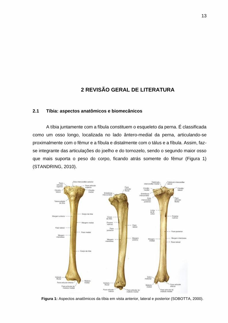
A tíbia juntamente com a fíbula constituem o esqueleto da perna. É classificada
como um osso longo, localizada no lado ântero-medial da perna, articulando-se
proximalmente com o fêmur e a fíbula e distalmente com o tálus e a fíbula. Assim, faz-
se integrante das articulações do joelho e do tornozelo, sendo o segundo maior osso
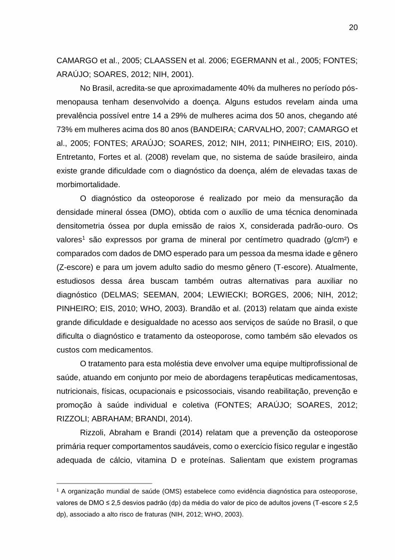
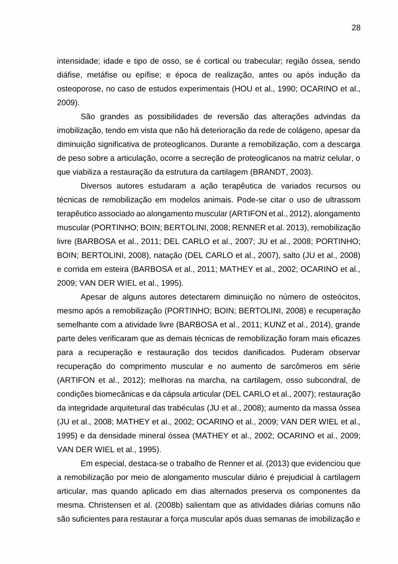
que mais suporta o peso do corpo, ficando atrás somente do fêmur (Figura 1)
(STANDRING, 2010).
Figura 1: Aspectos anatômicos da tíbia em vista anterior, lateral e posterior (SOBOTTA, 2000).

14
Em sua porção proximal, a tíbia recebe a inserção do ligamento da patela,
anteriormente em sua tuberosidade, como também se encontram os côndilos tibiais
mediais e laterais, recobertos de cartilagem hialina. Nessa região, acomodam-se os
côndilos femorais e os meniscos mediais e laterais. Entre os côndilos existe a região
intercondilar que serve de sítio para inserção do ligamento cruzado anterior (LCA) e
posterior (LCP) (DÂNGELO; FATTINI, 2007; STANDRING, 2010).
As articulações das quais a tíbia faz parte são classificadas como sinoviais,
sendo compostas por cápsula articular, cartilagem articular, membrana sinovial,
líquido sinovial e ligamentos capsulares, sendo estes últimos presentes em algumas
articulações sinoviais. Além disso, são formadas por várias células especializadas
oriundas de variados tecidos, como o mesenquimal e o hematopoiético (DÂNGELO;
FATTINI, 2007; HUI et al., 2012; IWANAGA et al., 2000).
Na articulação do joelho ou tibiofemoral, onde está a porção da tíbia que
suporta maior peso, existem os eixos de movimento transversal e longitudinal. No
transversal, ocorre a flexo-extensão (140-160° de flexão e 0° de extensão) e no
longitudinal a rotação axial, sendo externa (40-50°) e interna (30-35°), com os
movimentos associados no final da extensão e no início da flexão. Pela ação muscular
e dos ligamentos ocorrem os movimentos das articulações tibiofemoral e
patelofemoral (KAPANDJI, 2000).
A articulação tibiofemoral é o ponto do eixo para a flexo-extensão, sendo a tíbia
rotacionada sobre o côndilo medial do fêmur durante o movimento. A articulação
patelofemoral depende da contração do músculo quadríceps, aumentando a distância
dos eixos articulares e protegendo o joelho anteriormente. Também promove o
deslizamento na cavidade troclear, possibilitando o sistema de alavancas deste
músculo e gerando uma pressão lateralmente durante a flexão e medialmente na
extensão. Dessa forma, a tíbia é considerada um osso muito importante para a
manutenção da postura ortostática e para a locomoção bípede (BOGART; ORT, 2008;
KAPANDJI, 2000).
15
2.2 Tecido Ósseo
O tecido ósseo, principal componente da tíbia, constitui-se de células e da
matriz óssea. Suas principais funções são suporte para os tecidos moles, proteção
aos órgãos vitais e medula óssea, apoio aos músculos esqueléticos, como também
depósitos de cálcio, fosfato e outros íons (JUNQUEIRA; CARNEIRO, 2008; MARTIN;
DEMPSTER, 1998).
As células que o compõem são os osteoblastos, osteócitos e osteoclastos. Os
osteoblastos são células jovens que estão mais envolvidas na formação do tecido
ósseo. Possuem aspecto cubóide e se acomodam nas superfícies ósseas lado a lado,
como se fosse um epitélio simples. Possuem capacidade de concentrar fosfato de
cálcio e sintetizar a parte orgânica da matriz óssea, participando da mineralização e
sendo englobado pela mesma, considerado então osteócito. Os osteócitos são células
achatadas que ocupam as lacunas no interior da matriz óssea, sendo um por lacuna,
contribuindo para a composição e manutenção do osso já formado. Destas lacunas
saem canalículos por onde passam os prolongamentos dos osteócitos, estabelecendo
junções comunicantes entre as células para troca de pequenas moléculas e íons
(FRANZ-ODENDAAL; HALL; WITTEN, 2006; GENESER, 2003; JUNQUEIRA;
CARNEIRO, 2008).
Outras células importantes são os osteoclastos, que são multinucleadas,
móveis, gigantes, bastante ramificadas, com citoplasma granuloso, às vezes com
vacúolos e estão mais envolvidas com o processo de reabsorção do tecido ósseo.
Estas células promovem depressões na matriz denominadas de lacunas de Howship,
onde também se alojam. A função dos osteoclastos é controlada por moléculas da
superfamília do fator de necrose tumoral (TNF), sendo a
osteoprotegerina/osteoclastogênese inibitório (OPG/OCIF), ativador do receptor
nuclear kB ligante (RANKL), ativador do receptor do fator nuclear kB (RANK) e ligante
indutor de apoptose relacionado a TNF (GRAVALLESE; GOLDRING, 2000;
HOFBAUER; HEUFELDER, 2001; SHENGQIAN et al., 2012).
O RANKL, que se origina do tecido sinovial e adere ao RANK na superfície dos
osteoblastos, desempenham papel chave na promoção da osteoclastogênese,
maturação e ativação de osteoclastos, como também prevenção de apoptose destas

16
céulas. A interação entre RANK e RANKL leva a uma maior reabsorção e perda óssea
(GRAVALLESE et al., 2000; KHOSLA, 2001; SAIDENBERG; CON’ADO; LEMEITER,
2004). Os efeitos do RANKL podem ser neutralizados pelo receptor OPG, que é um
importante inibidor na regulação da reabsorção óssea osteoclástica (KOSTENUIK;
SHALHOUB; 2001; SHENGQIAN et al., 2012).
Compondo o tecido ósseo, juntamente com as células, está a matriz óssea.
Esta, por sua vez, constitui-se de parte inorgânica e orgânica. Na inorgânica
encontram-se os íons fosfato e cálcio em maior quantidade, como também
bicarbonato, magnésio, potássio, sódio e citrato em menores quantidades. Já a parte
orgânica é composta por 95% de fibras colágenas, constituídas de colágeno do tipo I
e poucos proteoglicanos e glicoproteínas, que conferem alguma participação na
mineralização da matriz. A junção dessas duas partes estabelece uma construção de
ligação, conferindo dureza e resistência à compressões, devido aos componentes
inorgânicos, como também elasticidade ao osso e resistência à forças de tração em
razão do colágeno (EINHORN, 1996; JUNQUEIRA; CARNEIRO, 2008).
Os ossos possuem revestimento interno e externo, caracterizado pela presença
de células osteogênicas e tecido conjuntivo, constituindo o periósteo e endósteo. As
principais funções desse revestimento são a nutrição do tecido ósseo e fornecimento
de novos osteoblastos, para crescimento e recuperação do osso. O periósteo é
composto por fibras colágenas e fibroblastos, como também células osteoprogenitoras
em sua porção mais profunda, que se multiplicam e se diferenciam em osteoblastos.
Já o endósteo constitui-se de células osteogênicas achatadas que revestem as
cavidades do osso esponjoso, canal medular, canais de Havers e de Volkmann
(EINHORN, 1996; GENESER, 2003; HOLLINGER; BUCK; BRUDER, 1999; MADER,
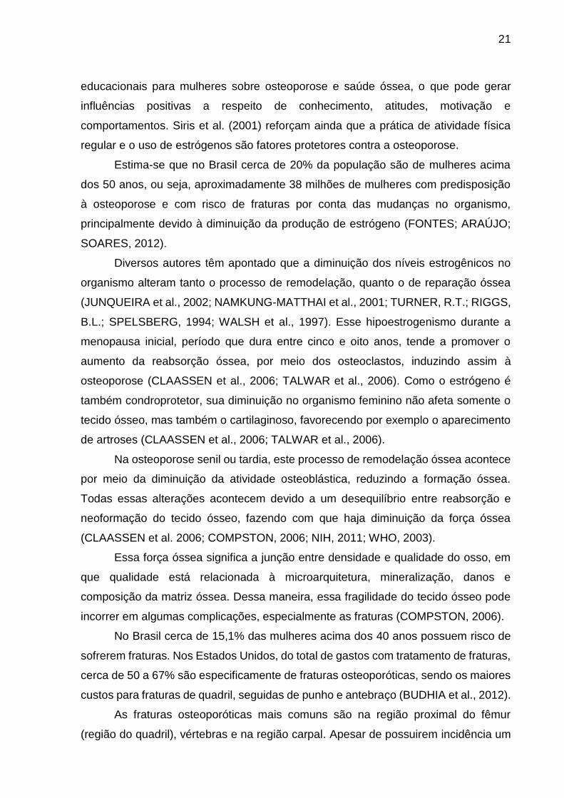
2004) (Figura 2).
Figura 2: Representação esquemática da estrutura do tecido ósseo de um osso longo (Adaptado de
MADER, 2004).
17
Os ossos podem ainda ser classificados macroscopicamente e
histologicamente. Macroscopicamente, podem ser compactos (sem cavidades
visíveis) ou esponjosos (com muitas cavidades intercomunicantes). As epífises são
formadas por osso esponjoso com uma fina camada compacta em sua superfície e as
diáfises (parte cilíndrica) essencialmente por osso compacto, com pouca quantidade
de osso esponjoso em sua porção profunda, delimitando o canal medular (GENESER,
2003; HOLLINGER; BUCK; BRUDER, 1999; MADER, 2004; MARTIN; DEMPSTER,
1998).
De acordo com a classificação histológica, o tecido ósseo pode ser imaturo,
primário ou não lamelar, e maduro, secundário ou lamelar. O tecido ósseo imaturo é
o primeiro a aparecer, no desenvolvimento embrionário e na reparação de fraturas.
Apresenta fibras colágenas dispostas irregularmente, maiores quantidades de
minerais e osteócitos em relação ao tecido secundário. Em indivíduos adultos
aparecem apenas próximo as suturas dos ossos do crânio, nos alvéolos dentários e
em alguns pontos de inserção muscular. Já o tecido secundário apresenta fibras
colágenas organizadas em lamelas de 3 a 7 micrômetros de espessura, paralelas
umas às outras, ou dispostas em camadas concêntricas em volta de canais com
vasos, formando os sistemas de Havers ou Ósteons. Apresenta ainda lacunas com
osteócitos situados entre as lamelas, estando algumas vezes situados dentro delas
(EINHORN, 1996; HOLLINGER; BUCK; BRUDER, 1999; JUNQUEIRA; CARNEIRO,
2008; MARTIN; DEMPSTER, 1998).
2.3 Tecido Cartilaginoso
O tecido cartilaginoso, como o tecido ósseo, também é composto por células e
pela matriz extracelular. Possui variadas funções, como revestimento das superfícies
articulares, suporte estrutural aos tecidos moles e atuação na formação e crescimento
dos ossos longos (GENESER, 2003; JUNQUEIRA; CARNEIRO, 2008).
A cartilagem não possui vasos sanguíneos e linfáticos, nem terminações
nervosas, sendo a nutrição de suas células realizada por difusão pela substância
fundamental, que é rica em água, ou seja, por meio do líquido sinovial ou capilares
localizados no pericôndrio (JUNQUEIRA; CARNEIRO, 2008; OVALLE; NAHIRNEY,
2008).

18
A cartilagem pode ser classificada em hialina, elástica ou fibrosa. A hialina
possui aspecto vítreo azulado e está presente nas superfícies articulares, cartilagens
costais, parte do esqueleto nasal, laringe, traqueia e brônquios. Desenvolve-se a partir
de células do mesênquima, que após intenso trabalho celular promove o aumento da
quantidade de matriz, tornando o tecido mais elástico e firme, além de criar lacunas
para alojar as células. Estas células vão se diferenciando em condrócitos, ao mesmo
tempo em que se desenvolve uma camada de outras células achatadas e de fibras
que rodeiam a cartilagem, constituindo o pericôndrio (GENESER, 2003).
As células diferenciadas passam por divisão mitótica e favorecem assim o
crescimento e formação dos condroblastos. Contribuem ainda no crescimento
intersticial, por meio da divisão das células-filha e pelo agrupamento em quatro
células, que também podem se dividir. Cada grupo desses, contém células originárias
das divisões mitóticas, a partir de um único condrócito, sendo denominados grupos
isógenos (OVALLE; NAHIRNEY, 2008).
A cartilagem articular adulta pode ser esquematicamente dividida em quatro
zonas distintas: repouso, proliferativa, hipertrófica e calcificada. A zona de repouso é
caracterizada pela presença de condrócitos dispostos como se fosse um epitélio
simples. Na proliferativa, há presença de colunas longitudinais de células
cartilaginosas. Na zona hipertrófica, as células amadurecem e aumentam de tamanho.
E a zona calcificada é caracterizada por ser a região de transição entre a cartilagem
e o osso, sendo normalmente bastante estreita (Figura 3) (SIMON; JACKSON, 2006;
WATRIN et al., 2001).
Figura 3: Representação esquemática da divisão em zonas da cartilagem articular (Adaptado de SIMON; JACKSON, 2006).
Entre a epífise e a diáfise existe um disco cartilaginoso transversal que as
19
separam, o disco epifisário. Este disco é componente importante no crescimento
longitudinal ósseo, pela constante formação de cartilagem em seu interior substituída
por osso. Após o período de crescimento, esse disco é eliminado, permanecendo
apenas uma linha, considerada uma irregularidade no tecido ósseo do osso
esponjoso, denominada linha epifisária (GENESER, 2003).
2.4 Osteoporose
A osteoporose pode ser conceituada como uma doença osteometabólica que
causa diminuição da massa óssea e danificação da microarquitetura do tecido ósseo,
gerando fragilidade óssea, dor e risco de fraturas, além de estar associada à altas
taxas de morbimortalidade. Pode ser classificada em primária e secundária, sendo a
primária subdividida em tipo I e II (CLAASSEN et al., 2006; NIH, 2011; TALWAR et al.,
2006; WHO, 2003).
A tipo I é a osteoporose ocorrida na pós-menopausa e a tipo II é a osteoporose
senil ou tardia. O principal critério para designar tipo I ou II é idade. Já a secundária
está relacionada com a presença de comorbidades, drogas ou outros fatores que
podem desencadear osteoporose e aumentar a possibilidade de fraturas (NIH, 2011;
WHO, 2003).
Sabe-se que essa doença é desencadeada durante a vida a depender de
alguns fatores como redução da massa óssea, idade avançada, gênero feminino, etnia
asiática ou caucasiana, história familiar, fratura prévia, insuficiência hormonal, uso de
glicocorticoides, fumar ou ter fumado. Outros fatores também são mencionados,
porém alguns autores caracterizam como de menor predisposição, sendo constituição
física, etilismo, sedentarismo, insuficiência de cálcio e vitamina D, índice de massa
corporal (IMC) ≤ 19 Kg/m² e a imobilização (KULAK; BILEZKIAN, 1998; KULAK et al.,
2011; SIRIS et al., 2001).
Ambos os gêneros podem desenvolver osteoporose, mas a prevalência é maior
em mulheres, em torno de 17%, contra 7% de homens. Entretanto, esse percentual
pode aumentar de acordo com a elevação da idade, etnia e localização geográfica.
Alguns estudos têm demonstrado esse aumento após os 40 anos, idade aproximada
de início da perda de massa óssea. Na mulher, isso pode se acentuar ainda mais por
volta dos 49 anos, devido ao hipoestrogenismo (BANDEIRA; CARVALHO, 2007;
20
CAMARGO et al., 2005; CLAASSEN et al. 2006; EGERMANN et al., 2005; FONTES;
ARAÚJO; SOARES, 2012; NIH, 2001).
No Brasil, acredita-se que aproximadamente 40% da mulheres no período pós-
menopausa tenham desenvolvido a doença. Alguns estudos revelam ainda uma
prevalência possível entre 14 a 29% de mulheres acima dos 50 anos, chegando até
73% em mulheres acima dos 80 anos (BANDEIRA; CARVALHO, 2007; CAMARGO et
al., 2005; FONTES; ARAÚJO; SOARES, 2012; NIH, 2011; PINHEIRO; EIS, 2010).
Entretanto, Fortes et al. (2008) revelam que, no sistema de saúde brasileiro, ainda
existe grande dificuldade com o diagnóstico da doença, além de elevadas taxas de
morbimortalidade.
O diagnóstico da osteoporose é realizado por meio da mensuração da
densidade mineral óssea (DMO), obtida com o auxílio de uma técnica denominada
densitometria óssea por dupla emissão de raios X, considerada padrão-ouro. Os
valores1 são expressos por grama de mineral por centímetro quadrado (g/cm²) e
comparados com dados de DMO esperado para um pessoa da mesma idade e gênero
(Z-escore) e para um jovem adulto sadio do mesmo gênero (T-escore). Atualmente,
estudiosos dessa área buscam também outras alternativas para auxiliar no
diagnóstico (DELMAS; SEEMAN, 2004; LEWIECKI; BORGES, 2006; NIH, 2012;
PINHEIRO; EIS, 2010; WHO, 2003). Brandão et al. (2013) relatam que ainda existe
grande dificuldade e desigualdade no acesso aos serviços de saúde no Brasil, o que
dificulta o diagnóstico e tratamento da osteoporose, como também são elevados os
custos com medicamentos.
O tratamento para esta moléstia deve envolver uma equipe multiprofissional de
saúde, atuando em conjunto por meio de abordagens terapêuticas medicamentosas,
nutricionais, físicas, ocupacionais e psicossociais, visando reabilitação, prevenção e
promoção à saúde individual e coletiva (FONTES; ARAÚJO; SOARES, 2012;
RIZZOLI; ABRAHAM; BRANDI, 2014).
Rizzoli, Abraham e Brandi (2014) relatam que a prevenção da osteoporose
primária requer comportamentos saudáveis, como o exercício físico regular e ingestão
adequada de cálcio, vitamina D e proteínas. Salientam que existem programas
1 A organização mundial de saúde (OMS) estabelece como evidência diagnóstica para osteoporose,
valores de DMO ≤ 2,5 desvios padrão (dp) da média do valor de pico de adultos jovens (T-escore ≤ 2,5
dp), associado a alto risco de fraturas (NIH, 2012; WHO, 2003).
21
educacionais para mulheres sobre osteoporose e saúde óssea, o que pode gerar
influências positivas a respeito de conhecimento, atitudes, motivação e
comportamentos. Siris et al. (2001) reforçam ainda que a prática de atividade física
regular e o uso de estrógenos são fatores protetores contra a osteoporose.
Estima-se que no Brasil cerca de 20% da população são de mulheres acima
dos 50 anos, ou seja, aproximadamente 38 milhões de mulheres com predisposição
à osteoporose e com risco de fraturas por conta das mudanças no organismo,
principalmente devido à diminuição da produção de estrógeno (FONTES; ARAÚJO;
SOARES, 2012).
Diversos autores têm apontado que a diminuição dos níveis estrogênicos no
organismo alteram tanto o processo de remodelação, quanto o de reparação óssea
(JUNQUEIRA et al., 2002; NAMKUNG-MATTHAI et al., 2001; TURNER, R.T.; RIGGS,
B.L.; SPELSBERG, 1994; WALSH et al., 1997). Esse hipoestrogenismo durante a
menopausa inicial, período que dura entre cinco e oito anos, tende a promover o
aumento da reabsorção óssea, por meio dos osteoclastos, induzindo assim à
osteoporose (CLAASSEN et al., 2006; TALWAR et al., 2006). Como o estrógeno é
também condroprotetor, sua diminuição no organismo feminino não afeta somente o
tecido ósseo, mas também o cartilaginoso, favorecendo por exemplo o aparecimento
de artroses (CLAASSEN et al., 2006; TALWAR et al., 2006).
Na osteoporose senil ou tardia, este processo de remodelação óssea acontece
por meio da diminuição da atividade osteoblástica, reduzindo a formação óssea.
Todas essas alterações acontecem devido a um desequilíbrio entre reabsorção e
neoformação do tecido ósseo, fazendo com que haja diminuição da força óssea
(CLAASSEN et al. 2006; COMPSTON, 2006; NIH, 2011; WHO, 2003).
Essa força óssea significa a junção entre densidade e qualidade do osso, em
que qualidade está relacionada à microarquitetura, mineralização, danos e
composição da matriz óssea. Dessa maneira, essa fragilidade do tecido ósseo pode
incorrer em algumas complicações, especialmente as fraturas (COMPSTON, 2006).
No Brasil cerca de 15,1% das mulheres acima dos 40 anos possuem risco de
sofrerem fraturas. Nos Estados Unidos, do total de gastos com tratamento de fraturas,
cerca de 50 a 67% são especificamente de fraturas osteoporóticas, sendo os maiores
custos para fraturas de quadril, seguidas de punho e antebraço (BUDHIA et al., 2012).
As fraturas osteoporóticas mais comuns são na região proximal do fêmur
(região do quadril), vértebras e na região carpal. Apesar de possuirem incidência um
22
pouco menor em relação aos demais tipos, as fraturas de tíbia apresentam grande
relevância anatômica e biomecânica, principalmente pelo elevado suporte de peso,
pela participação em duas importantes articulações, joelho e tornozelo, e importância
para a postura ortostática e locomoção bípede, comprometendo assim o bem estar e
a qualidade de vida dos indivíduos acometidos. Por conseguinte, a osteoporose pode
ser considerada um problema de saúde pública, tendo em vista problemas sociais e
econômicos que acarreta, como a incapacitação temporária ou permanente da
pessoa, como também pelos altos custos de cuidados em saúde (CLAASSEN et al.,
2006; NIH, 2001; TALWAR et al., 2006; WHO 2003).
Riggs e Melton (1997) sugerem que aproximadamente 50% de pessoas que
fraturaram o quadril necessitarão de auxílio para deambulação, e cerca de 25% de
cuidados de longo prazo. No entanto, vários fatores de risco para a osteoporose são
passíveis de mudança por meio de exercícios físicos corretamente orientados. Alguns
estudos apresentam repostas positivas quanto à força muscular, com melhora entre 6
a 174%; amplitude de movimento articular (ADM), entre 0,5 a 18%; equilíbrio, 7 a 53%;
marcha, 12 a 48% e tempo de reação, de 0 a 4% (AN, 2002; MYERS; YOUNG;
LANGLOIS, 1996).
Vários estudos randomizados têm mostrado diminuição significativa de quedas
em idosos como resultados de programas orientados de treinamento físico, entretanto
não existe ainda um consenso sobre tipo ideal, duração, intensidade e frequência
desses exercícios (AMERICAN GERIATRICS SOCIETY, BRITISH GERIATRICS
SOCIETY AND AMERICAN ACADEMY OF ORTHOPAEDIC SURGEONS PANEL ON
FALLS PREVENTION, 2001; CARTER; KANNUS; KHAN, 2001).
Cabe salientar que os estudos relacionados à incidência de indivíduos com
osteoporose no Brasil ainda são incipientes e necessitam de maior abrangência e
explicitação, envolvendo a realidade da população desse país. Tal fato demonstra a
importância do tema e a necessidade de se discutir, propor estratégias e agir em prol
de medidas oportunas, preventivas e terapêuticas (FONTES; ARAÚJO; SOARES,
2012).
Com a urgência de melhor compreensão de sua natureza multifatorial e de
desenvolver novos métodos preventivos e terapêuticos que atuem contra os efeitos
deletérios desta doença, torna-se plausível e necessário o uso de modelos animais
de osteoporose em pesquisas científicas. Diversas espécies animais já foram
utilizadas em pesquisas de osteoporose, tais como roedores, coelhos, cães e

23
macacos. No entanto, apesar das limitações na semelhança com humanos, o rato de
laboratório tem sido o mais escolhido, com muitos estudos sobre seu esqueleto e de
suas peculiaridades, até mesmo criando-se técnicas específicas a fim de superar tais
limitações (LELOVAS et al., 2008).
Existem vários modelos experimentais de indução à osteoporose, seja por
intervenções hormonais (cirúrgica ou farmacológica), dietéticas, alterações genéticas
ou imobilização (conservadora ou cirúrgica). Dentre as intervenções por cirurgia,
pode-se citar a gonadectomia (ovariectomia ou ooforectomia e orquidectomia),
hipofisectomia e paratireoidectomia; e as farmacológicas, agonista do hormônio
liberador de gonadotrofina (GnRH), antogonista de receptores estrogênicos, inibidores
de aromatase e corticosteróides (AN, 2002; BERDUD et al., 1998; GALLAGHER;
CHAMBERS; TOBIAS, 1993; GASSER et al., 2006; GOULDING; GOLD, 1989;
IWAMOTO; TAKEDA; ISCHIMURA, 2004; IWAMOTO et al., 2007; LELOVAS et al.,
2008; LI; SHEN; WRONSKI, 1997; TURNER et al., 2001).
As principais intervenções dietéticas normalmente são induzidas por meio de
dieta pobre em cálcio (SETO et al., 1999) ou pelo abuso de álcool (SAMPSON et al.,
1996; TURNER et al., 1988). Já a imobilização pode ser dividida em conservadora,
com a realização de enfaixamento de membro, fundição ou suspensão pela cauda; ou
cirúrgica, por meio de ressecção nervosa, tendinosa ou da medula espinhal (BERDUD
et al., 1998; MOREY, 1979; OKUMURA et al., 1988; STEINBERG; TRUETA, 1981;
THOMPSON; RODAN, 1988; ZENG et al., 1996).
Um dos modelos mais escolhidos e com melhores resultados é a ooforectomia,
principalmente quando é para mimetizar época de pós-menopausa. Consiste na
retirada dos ovários, principal fonte de estrógenos, induzindo assim à restrição desse
hormônio da circulação do organismo (LELOVAS et al., 2008). An (2002) relata que o
modelo de ooforectomia pode ser realizado em ratas, ratazanas, coelhas, cadelas,
porcas, ovelhas, primatas não-humanas e outros animais como porquinhas da Índia,
furoas, gatas e aves fêmeas, especialmente galinhas poedeiras.
Contudo, devido semelhanças nas respostas fisiopatológicas entre o esqueleto
humano e do rato, vantagens financeiras e de criação, como também pela existência
de protocolos que induzem perda de massa óssea mais rapidamente, fizeram do rato
um modelo valioso em pesquisas sobre osteoporose. No entanto, a seleção de um
modelo deve ser baseada em critérios científicos e não apenas pela facilidade de uso,
devendo os pesquisadores terem sempre em mente que a experimentação deve
24
aderir aos 3 Rs da ética com o uso de animais: substituição (replacement), redução
(reduction) e refinamento (refinement) (LELOVAS et al., 2008).
Os ratos atingem a maturidade sexual com a idade de 2,5 meses e seu
esqueleto torna-se maduro após os 10 meses de vida. A perda de DMO inicia-se por
volta de 12 meses, na metáfise proximal da tíbia, e de 15 meses em vértebras
lombares2. No entanto, pesquisas podem ser realizadas com esqueletos maduros ou
imaturos, embora em alguns casos a escolha entre um e outro seja fundamental para
o delineamento metodológico (JEE; YAO, 2001; SHEN et al., 1997).
Em se tratando de pós-menopausa, o modelo mais utilizado é o de ratas
ooforectomizadas, pois após a cirurgia já tem início o processo de remodelação óssea,
com a reabsorção excedendo a neorformação, causando perda óssea e logo depois
havendo um equilíbrio nesse processo (TURNER et al., 2001).
Desssa forma, com esse modelo citado, perdas ósseas significativas são
percebidas após 14, 72 e 74 dias na metáfise proximal da tíbia, após 60 e 73 dias no
corpo vertebral lombar e após 30 dias no colo do fêmur. Entretanto, não ocasiona
perdas de massa óssea significativas nas epífises de ossos longos, metáfise tibial
distal e em vértebras caudais (LI; JEE, 1991; LI et al., 1996; LI; SHEN; WRONSKI,
1997; MA; KE; JEE, 1994; MIYAKOSHI et al., 1999).
Já o tempo necessário para o processo de remodelação óssea atingir equilíbrio
é de cerca de 80 dias, na metáfise proximal da tíbia, e de 270 dias para o corpo
vertebral lombar e colo do fémur. Esses achados de diminuição da massa óssea são
bem vistos no osso cortical, por meio do alargamento da cavidade medular. Isto ocorre
devido ao aumento da reabsorção endosteal e da aposição óssea periosteal. As
mensurações realizadas em áreas da metade inferior do eixo cortical são muito
confiáveis, devido a maior parte das perdas ósseas ocorrerem nesse local (KIMMEL;
WRONSKI, 1990; LI; SHEN; WRONSKI, 1997; MILLER et al., 1991; WRONSKI;
DANN; HORNER, 1990).
As primeiras mudanças na largura do osso cortical e na cavidade medular, do
fêmur e da tíbia, são notadas em períodos em torno de 90 e 120 dias após a
ooforectomia, enquanto o tempo para atingir o estado estacionário de perdas é de
cerca de 180 dias. Alguns autores sugerem que a associação entre ooforectomia e
imobilização pode reduzir o tempo de perda óssea significativa, principalmente
2 Idade aproximada para população de ratos saudáveis (FUKUDA; IIDA, 2004).

25
cortical, quando comparada às técnicas isoladamente (CAVOLINA et al., 1997;
DANIELSEN; MOSEKILDE; SVENSTRUP, 1993; JEE; YAO, 2001; KE et al., 1993).
Existem vários métodos de avaliação da diminuição da massa óssea, que
podem ser invasivos ou não invasivos. Dentre os métodos não invasivos, pode-se citar
os marcadores bioquímicos, densitometria óssea, medidas de cálcio, fósforo e
magnésio, no sangue e na urina. Nos métodos invasivos são incluídos a
histomorfometria e a avaliação da força mecânica (CAVOLINA et al., 1997; LELOVAS
et. al, 2008; LOEB, 1999).
Por meio da histomorfometria é possível analisar a massa e arquitetura óssea,
sendo que a arquitetura pode ser avaliada com acurácia, independente da massa
óssea, tal como índices de fragilidade óssea. Podem ser mensurados área e
espessura de osso cortical e trabecular, cartilagem articular e placa epifisária, número
de osteoblastos, osteoclastos, osteócitos e osteoblastos ativos em relação ao
perímetro do osso, além da análise qualitativa, para verificação de alterações e
irregularidades nos tecidos (DALLE et al., 2005; LELOVAS et al., 2008).
A técnica de análise histológica é considerada de melhor eficácia quando
comparada às outras mencionadas anteriormente. Entretanto, apresenta algumas
limitações, como dificuldade de avaliar várias áreas, em estruturas diferentes e ao
mesmo tempo. Normalmente é possível analisar somente uma pequena área de
tecido, em um osso específico, o que não representa modificações ao longo do
esqueleto. Todavia, apresenta grande credibilidade quando a área de amostragem é
comparável em todos os grupos (CAVOLINA et al., 1997; LELOVAS et al., 2008;
ROSEN et al., 1995).
Outro método invasivo e que avalia a resistência mecânica dos ossos é a
avaliação da força mecânica. Esta pode ser realizada por meio de testes, como o de
três pontos de flexão, flexão de quatro pontos, torção, compressão e cantilever
(SOGAARD et al., 1994; YAO et al., 2005).
2.5 Imobilização e Remobilização por Exercícios
A imobilização é um recurso terapêutico conservador muito utilizado em lesões
do sistema musculoesquelético, que objetiva o repouso de um segmento corporal ou
de parte dele. É normalmente utilizada em pós-operatórios, fraturas, entorses e lesões

26
musculares, favorecendo diminuição do quadro álgico, proteção às estruturas
danificadas e contra alguns tipos de danos induzidos quimicamente nas cartilagens
articulares (BRANDT, 2003; PATRIDGE; DUTHIE, 1963; WILLIAMS; BRANDT, 1984).
Entretanto, a imobilidade e outros fatores como sedentarismo, fraqueza
muscular e lesão nervosa, afetam o tecido ósseo e muscular, ocasionando diminuição
do peso, prejuízos às propriedades, diminuição da amplitude de movimento articular
(ADM) e déficits funcionais, principalmente quando o músculo é mantido em
encurtamento (CAIERÃO; TEODORI; MINAMOTO, 2007; CORNWALL, 1984;
SAKAKIMA, 2004; THOMASON; BIGGS; BOOTH, 1989; THOMASON; BOOTH, 1990;
WILLIAMS, 1988).
Devido à grande inviabilidade de realizar algumas análises com humanos, a fim
de verificar os efeitos da imobilização no sistema musculoesquelético, utiliza-se
modelos animais. Diversas técnicas cirúrgicas e não-cirúrgicas tem sido adotadas,
como enfaixamento de membro; órteses de gesso, resina acrílica e algodão;
suspensão pela cauda; fixação articular com pinos; ressecção nervosa, tendinosa ou
da medula espinhal (BERDUD et al., 1998; DURIGAN et al., 2005; MOREY, 1979;
OKUMURA et al., 1988; STEINBERG; TRUETA, 1981; THOMPSON; RODAN, 1988;
ZENG et al., 1996).
Por meio dessas mimetizações é possível observar alguns outros efeitos
deletérios da imobilização, como: aumento da espessura da cápsula articular;
degeneração de células sinoviais; diminuição da produção de fluido sinovial e do
suprimento nutricional à cartilagem; perda e redução da síntese de proteoglicanos da
matriz cartilaginosa; irregularidade da superfície articular; proliferação de tecido
conjuntivo intra-articular; necrose e ulceração da cartilagem; redução da massa e
volume totais da cartilagem; diminuição da força e da área de secção transversa do
músculo (BRANDT, 2003; CHRISTENSEN et al., 2008a; CHRISTENSEN et al.,
2008b; CULAV et al., 1999; DEL CARLO et al., 2007; KANEPS; STOVER; LANER,
1997; LANTZ, 1998; LEROUX et al., 2001; NARMONEVA et al., 2002; THAXTER et
al., 1965).
Em um estudo de Maeda et al. (1993) foi verificado perda significativa de osso
esponjoso em ratos submetidos à seis semanas de imobilização. E na pesquisa de
Leroux et al. (2001) foi observado que as propriedades mecânicas da cartilagem
sofreram alterações após quatro semanas de imobilização.
A cartilagem possui uma capacidade limitada de reparação após uma lesão

27
intrínseca e os danos causados não são reparados de forma espontânea (BOS;
MELLE; OSCH, 2010). Estes danos podem ocorrer devido proliferação dos tecidos
sinovial e conjuntivo; aderência da superfície articular; alterações do osso subcondral;
irregularidade nas camadas celulares; aumento do número de células inflamatórias;
diminuição do número de condrócitos; perda de macromoléculas estruturais da matriz
e diminuição da absorção de impacto na articulação (ANDO et al., 2011; ARAKAKI et
al., 2011; BRANDT, 2003; CHRISTENSEN et al., 2008a; CHRISTENSEN et al.,
2008b; CULAV et al., 1999; HAGIWARA et al., 2011; IQBAL, 2012; IQBAL; KHAN;
MINHAS, 2012; KANEPS; STOVER; LANER, 1997; LANTZ, 1998; LEROUX et al.,
2001; NARMONEVA et al., 2002; THAXTER et al., 1965). Mesmo com intervenções
terapêuticas aplicadas corretamente, nem sempre haverá reversibilidade completa
dos danos causados (BARBOSA et al., 2011; TREBACZ; ZDUNEK, 2006;
VANWANSEELE; LUCCHINETTI; STÜSSI, 2002).
Ando et al. (2011) observaram que a hipotrofia muscular foi reversível durante
remobilização, mas a perda e hipertrofia de condrócitos na superfície articular,
ocasionados pela tensão mecânica da imobilização rígida, foram irreversíveis. Assim,
concluíram que até mesmo uma imobilização rígida de curto prazo pode causar danos
irreversíveis à cartilagem articular.
A recuperação vai depender do tipo de imobilização, idade, segmento corporal
envolvido, espécie animal, tipo e intensidade do exercício preconizado na
remobilização (CHRISTENSEN et al., 2008a; CHRISTENSEN et al., 2008b; KANEPS;
STOVER; LANER, 1997). Existem diversos estudos realizados com diferentes
modelos de imobilização e remobilização, contudo não há um consenso sobre quais
técnicas ou procedimentos mais eficazes em proporcionar reversibilidade completa
das lesões impostas por um dispositivo imobilizatório, para restaurar os prejuízos
físico funcionais e histológicos.
Para Barbosa et al. (2011), o exercício físico é a intervenção terapêutica que
mais tem sido estudada para evitar lesões, como para recuperar componentes ósseos
e musculares em períodos de remobilização. Por meio do movimento articular,
consegue-se promover mudanças físicas, bioquímicas e histológicas, favorecendo o
retorno à síntese de macromoléculas e proporcionando reversibilidade das lesões
causadas na cartilagem (BRANDT, 2003; CULAV et al., 1999; LANTZ, 1998; LEROUX
et al., 2001; NARMONEVA et al., 2002; THAXTER et al., 1965).
Contudo, os benefícios do exercício físico sobre o esqueleto dependem da
28
intensidade; idade e tipo de osso, se é cortical ou trabecular; região óssea, sendo
diáfise, metáfise ou epífise; e época de realização, antes ou após indução da
osteoporose, no caso de estudos experimentais (HOU et al., 1990; OCARINO et al.,
2009).
São grandes as possibilidades de reversão das alterações advindas da
imobilização, tendo em vista que não há deterioração da rede de colágeno, apesar da
diminuição significativa de proteoglicanos. Durante a remobilização, com a descarga
de peso sobre a articulação, ocorre a secreção de proteoglicanos na matriz celular, o
que viabiliza a restauração da estrutura da cartilagem (BRANDT, 2003).
Diversos autores estudaram a ação terapêutica de variados recursos ou
técnicas de remobilização em modelos animais. Pode-se citar o uso de ultrassom
terapêutico associado ao alongamento muscular (ARTIFON et al., 2012), alongamento
muscular (PORTINHO; BOIN; BERTOLINI, 2008; RENNER et al. 2013), remobilização
livre (BARBOSA et al., 2011; DEL CARLO et al., 2007; JU et al., 2008; PORTINHO;
BOIN; BERTOLINI, 2008), natação (DEL CARLO et al., 2007), salto (JU et al., 2008)
e corrida em esteira (BARBOSA et al., 2011; MATHEY et al., 2002; OCARINO et al.,
2009; VAN DER WIEL et al., 1995).
Apesar de alguns autores detectarem diminuição no número de osteócitos,
mesmo após a remobilização (PORTINHO; BOIN; BERTOLINI, 2008) e recuperação
semelhante com a atividade livre (BARBOSA et al., 2011; KUNZ et al., 2014), grande
parte deles verificaram que as demais técnicas de remobilização foram mais eficazes
para a recuperação e restauração dos tecidos danificados. Puderam observar
recuperação do comprimento muscular e no aumento de sarcômeros em série
(ARTIFON et al., 2012); melhoras na marcha, na cartilagem, osso subcondral, de
condições biomecânicas e da cápsula articular (DEL CARLO et al., 2007); restauração
da integridade arquitetural das trabéculas (JU et al., 2008); aumento da massa óssea
(JU et al., 2008; MATHEY et al., 2002; OCARINO et al., 2009; VAN DER WIEL et al.,
1995) e da densidade mineral óssea (MATHEY et al., 2002; OCARINO et al., 2009;
VAN DER WIEL et al., 1995).
Em especial, destaca-se o trabalho de Renner et al. (2013) que evidenciou que
a remobilização por meio de alongamento muscular diário é prejudicial à cartilagem
articular, mas quando aplicado em dias alternados preserva os componentes da
mesma. Christensen et al. (2008b) salientam que as atividades diárias comuns não
são suficientes para restaurar a força muscular após duas semanas de imobilização e
29
acrescentam que o treinamento de força deve ser enfatizado na reabilitação, mesmo
depois de curto período de imobilização.
Em estudo realizado com ratas osteopênicas, foi demonstrado que o exercício
físico é capaz de estimular o tecido muscular e ósseo, favorecendo a produção de
massa óssea (RENNO et al., 2007). Menkes et al. (1993) reforçam que o exercício
físico estimula o remodelamento ósseo, promovendo também o aumento da força do
osso.
Os exercícios resistidos tem se mostrado de grande importância para
manutenção da massa óssea em mulheres na pré-menopausa, com benefícios
estendidos até a pós-menopausa (LAYNE; NELSON, 1999). Em um estudo realizado
com idosos, foi verificado que a realização de alongamento com resistência
proporcionou melhora na ADM quando comparado aos que realizaram alongamento
sem carga (SWANK et al., 2003).
Nelson et al. (1994) realizaram um estudo com mulheres em idade de 50 e 70
anos, submetidas a um programa de exercícios resistidos. Após 52 semanas,
observaram aumento na DMO do colo do fêmur e da vértebra lombar, enquanto o
grupo controle apresentou decréscimo.
Estudos realizados com ratos submetidos à supensão pela cauda por 21 dias
e exercício em escada pelo mesmo período, realizando oito séries, com peso
equivalente a 80% de sua força máxima, cinco vezes na semana, verificaram que esse
tipo de exercício resistido foi capaz de restaurar os valores de DMO e rigidez óssea
(OLIVEIRA et al., 2013a; OLIVEIRA et al., 2013b).
Cassilhas et al. (2013), em estudo realizado com ratos, acrescentam que o
exercício em escada promove hipertrofia dos músculos gastrocnêmio, flexor longo dos
dedos e plantar, o que pode ajudar na proteção articular do tornozelo. Nascimento et
al. (2013) evidenciaram que esse exercício de escalada também promove hipertrofia
do músculo tríceps braquial em ratos, atingindo assim, com resultados positivos,
inclusive músculos dos membros anteriores.
Diversos benefícios dessa modalidade de exercício têm sido evidenciados em
humanos, como aumento de fibras musculares tipo II, VO2 máximo, melhora na
performance de atletas, qualidade de vida, em tratamento de doenças e disfunções
osteomioarticulares, cardiovasculares, pneumonólogicas, endócrinas, metabólicas e
HIV (BOGDANIS et al., 2011; CHURCH, 2011; LINCOLN et al., 2011; SOUZA et al.,
2011; VICENT; VICENT, 2006).

30
3 REFERÊNCIAS
AMERICAN GERIATRICS SOCIETY, BRITISH GERIATRICS SOCIETY AND AMERICAN ACADEMY OF ORTHOPAEDIC SURGEONS PANEL ON FALLS PREVENTION. Guideline for the prevention of falls in older persons. Journal of American Geriatrics Society, v. 49, n. 5, p. 664-672, 2001.
AN, Y.H. Orthopaedic issues in osteoporosis. Boca Raton: CRC Press, 2002.
ANDO, A.; HAGIWARA, Y.; CHIMOTO, E.; HATORI, K.; ONODA, Y.; ITOI, E. Intra- articular injection of hyaluronan diminishes loss os chondrocytes in a rat immobilizad- knee model. Tohoku Journal of Experimental Medicine, v. 215, n. 4, p.321-331, 2008.
ANDO, A.; HAGIWARA, Y.; ONODA, Y.; HATORI, K.; SUDA, H.; CHIMOTO, E.; ITOI, E. Distribution of type A and type B synoviocytes in the adhesive and shortened synovial membrane during immobilization of the knee joint in rats. Tohoku Journal of Experimental Medicine, v. 221, n. 2, p. 161-168, 2010.
ANDO, A.; SUDA, H.; HAGIWARA, Y.; ONODA, Y.; CHIMOTO, E.; SAIJO, Y.; ITOI, E. Reversibility of immobilization-induced articular cartilage degeneration after remobilization in rat knee joints. Tohoku Journal of Experimental Medicine, v. 224, n. 2, p. 77-85, 2011.
ANDO, A.; SUDA, H.; HAGIWARA, Y.; ONODA, Y.; CHIMOTO, E.; ITOI, E. Remobilization does not restore immobilization-induced adhesion of capsule and restricted joint motion in rat knee joints. Tohoku Journal of Experimental Medicine, v. 227, n. 1, p. 13-22, 2012.
APPELL, H.J. Skeletal muscle atrophy during immobilization. International Journal of Sports Medicine, v. 7, p. 1-5, 1986.
ARAKAKI, K.; KITAMURA, N.; KUROKAWA, T.; ONODERA, S.; KANAYA, F.; GONG, J. P.; YASUDA, K. Joint immobilization inhibits spontaneous hyaline cartilage regeneration induced by a novel double-network gel implantation. Journal of Materials Science: Materials in Medicine, v. 22, p. 417-425, 2011.
ARTIFON, E.L.; FERRARI, D.; CUNHA, D.M.; NASCIMENTO, C.M.; RIBEIRO, L.F.C.; BERTOLINI, G.R.F. Efeitos do ultrassom terapêutico associados ao alongamento estático sobre parâmetros histomorfométricos longitudinais de sóleos
31
imobilizados de ratos. Revista Brasileira de Medicina do Esporte, v. 18, n. 5, p. 341-344, 2012.
BANDEIRA, F.; CARVALHO, E.F. Prevalência de osteoporose e fraturas vertebrais em mulheres na pós-menopausa atendidas em serviços de referência. Revista Brasileira de Epidemiologia, v. 10, n.1, p. 86-98, 2007.
BARBOSA, A.A.; DEL CARLO, R.J.; GALVÃO, S.R.; VILELA, M.J.; LOUZADA, M.J.Q.; BRITO, A.F.S.; NATALI, A.J. Bone mineral density of rat femurs after hindlimb unloading and different physical rehabilitation programs. Revista Ceres, v. 58, n. 4, p. 407-412, 2011.
BERCHTOLD, M. W.; BRINKMEIER, H.; MUNTENER, M. Calcium ion in skeletal muscle: its crucial role for muscle function, plasticity, and disease. Physiological reviews, v. 80, n. 3, p. 1215-1265, jul. 2000.
BERDUD, I.; MARTIN-MALO, A. ALMADEN, Y.; ALIJAMA, P.; RODRIGUEZ, M.; FELSENFELD, A. The PTH-calcium relationship during a range of infused PTH doses in the parathyroidectomized rat. Calcified Tissue International, v. 62, p. 457-461, 1998.
BERRY, S.D.; KIEL, D.P.; DONALDSON, M.G.; CUMMINGS, S.R.; KANIS, J.A.; JOHANSSON, H.; SAMELSON, E.J. Application of the national osteoporosis foundation guidelines to postmenopausal women and men: the Framingham osteoporosis study. Osteoporosis International, v. 21, n. 1, p. 53-60, 2010.
BOGART, B. I.; ORT, V. H. Anatomia e Embriologia. 1. ed. Rio de Janeiro: Elsevier, 2008.
BOGDANIS, G.C.; PAPASPYROU, A.; SOUGLIS, A.G.; THEOS, A.; SOTIROPOULOS, A.; MARIDAKI, M. Effects of two differente half-squat training programs on fatigue during repeated cycling sprints in soccer players. The Journal of Strength & Conditioning Research, v. 25, n. 7, p. 1849-1856, 2011.
BOOTH, F.W.; KELSO, J.R. Effect of hind-limb immobilization on contractile and histochemical properties of skeletal muscle. Pflügers Archiv, v. 342, p. 231-238, 1973.
BOS, P.K.; MELLE, M.L.V.; OSCH, G.J.V.M.V. Articular cartilage repair and the evolving role of regenerative medicine. Open Access Surgery, v. 3, p. 109-122, 2010.
32
BRANDÃO, C.M.R.; FERRÉ, F.; MACHADO, G.P.M.; GUERRA-JÚNIOR, A.A.; ANDRADE, E.L.G.; CHERCHIGLIA, M.L.; ACURCIOL, F.A. Gastos públicos com medicamentos para o tratamento da osteoporose na pós-menopausa. Revista de Saúde Pública, v. 47, n. 2, p. 390-402, 2013.
BRANDT, K. D. Response of joint structures to inactivity and to reloading after immobilization. Arthritis & Rheumatism, v. 49, n. 2, p. 267-271, apr. 2003.
BUDHIA, S.; MIKYAS, Y.; TANG, M.; BADAMGARAV, E. Osteoporotic fractures: a systematic review of us healthcare costs and resource utilization. Pharmacoeconomics, v. 30, n. 2, p. 147-170, 2012.
CAIERÃO, Q.M.; TEODORI, R.M.; MINAMOTO, V.B. A influência da imobilização sobre o tecido conjuntivo muscular: uma revisão. Fisioterapia em Movimento, v. 20, n. 3, p. 87-92, 2007.
CAMARGO, M.B.; CENDOROGLO, M.S.; RAMOS, L.R.; LATORRE, M.R.D.O.; SARAIVA, G.L.; LAGE, A.; CARVALHAES-NETO, N.; ARAÚJO, L.M.Q.; VIEIRA, J.G.H.; LAZARETTI-CASTRO, M. Bone mineral density and osteoporosis among a predominantly caucasian elderly population in the city of São Paulo, Brazil. Osteoporosis International, v. 16, n. 11, p. 1451-1460, 2005.
CARVALHO, A.C.B.; HENRIQUES, H.N.; PANTALEÃO, J.A.S.; POLLASTRI, C.E.; FERNANDES, G.V.O.; GRANJEIRO, J.M.; GUZMÁN-SILVA, M.A. Histomorfometria do tecido ósseo em ratas castradas tratadas com tibolona. Jornal Brasileiro de Patologia e Medicina Laboratorial, v. 46, n. 3, p. 235-243, 2010.
CARTER, N.D.; KANNUS, P.; KHAN, K.M. Exercise in the prevention of falls in older people: a systematic literature review examining the rationale and the evidence. Sports Medicine, v. 31, n.6, p. 427-438, 2001.
CASSILHAS, R.C.; REIS, I.T.; VENÂNCIO, D.; FERNANDES, J.; TUFIK, S.; MELLO, M.T. Animal model for progressive resistance exercise: a detailed description of model and its implications for basic research in exercise. Motriz, v. 19, n. 1, p. 178-184, 2013.
CAVOLINA, J.M.; EVANS, G.L.; HARRIS, S.A.; ZHANG, M.; WESTERLIND, K.C.; TURNER, R.T. The effects of orbital spaceflight on boné histomorphometry and messenger ribonucleic acid levels for bone matrix proteins and skeletal signaling peptides in ovariectomized growing rats. Endocrinology, v. 138, n. 4, p. 1567-1576,
33
1997.
CHRISTENSEN, B.; DYRBERG, E.; AAGAARD, P.; ENEHJELM, S.; KROGSGAARD, M.; KJAER, M.; LANGBERG, H. Effects of long-term immobilization and recovery on human triceps surae and collagen turnover in the Achilles tendon in patients with healing ankle fracture. Journal of Applied Physiology, v. 105, p. 420–426, 2008a.
CHRISTENSEN, B.; DYRBERG, E.; AAGAARD, P.; KJAER, M.; LANGBERG, H. Short-term immobilization and recovery affect skeletal muscle but not collagen tissue turnover in humans. Journal of Applied Physiology, v. 105, p. 1845–1851, 2008b.
CHURCH, T. Exercise in obesity, metabolic syndrome and diabetes. Progress in Cardiovascular Disease, v. 56, n. 6, p. 412-418, 2011.
CLAASSEN, H.; SCLÜTER, M.; SCHÜNKE, M.; KERTZ, D. Influence of 17 beta estradiol and insulin on type II colagen and protein synthesis of articular chondrocytes. Bone, v. 39, n. 2, p. 310-317, 2006.
COMPSTON, J. Bone quality: what it is and how is it measured? Arquivos Brasileiros de Endocrinologia & Metabologia, v. 50, n. 4, p. 579-585, 2006.
CORNWALL, M.W. Biomechanics of noncontractile tissue - a review. Physical Therapy Journal, v. 63, p. 1869-1873, 1984.
CRUZ, D. T.; RIBEIRO, L. C.; VIEIRA, M. T.; TEIXEIRA, M. T. B.; BASTOS, R. R.; LEITE, I. C. G. Prevalência de quedas e fatores associados em idosos. Revista de Saúde Pública, v. 46, n. 1, p. 138-146, 2012.
CULAV, E.M.; CLARK, C.H.; MERRILEES, M.J. Connective tissues: matrix composition and its relevance to physical therapy. Physical Therapy Journal, v.79, p.308-319, 1999.
DALLE CARBONAREA, L.; VALENTIA, M.T.; BERTOLDOA, F.; ZANATTAA, M.; ZENARIA, S.; REALDIB, G.; LO CASCIOA, V.; GIANNINIB, S. Bone microarchitecture evaluated by histomorphometry.Micron, v. 36, p. 609-616, 2005.
DÂNGELO, J. G.; FATTINI, C. A. Anatomia Humana Sistêmica e Segmentar: para o estudante de medicina. 3ª ed. Rio de Janeiro: Atheneu, 2007.

34
DANIELSEN, C.C.; MOSEKILDE, L.; SVENSTRUP, B. Cortical bone mass, composition, and mechanical properties in female rats in relation to age, long-term ovariectomy, and estrogen substitution. Calcified Tissue International, v. 52, n. 1, p. 26-33, 1993.
DANTAS, E. H. M.; PEREIRA, S. A. M.; ARAGÃO, J. C.; OTA, A. H. A preponderância da diminuição da mobilidade articular ou da elasticidade muscular na perda da flexibilidade no envelhecimento. Fitness & Performance Journal, v. 1, n. 3, p. 12-20, mai/jun. 2002.
DEL CARLO, R. J.; GALVÃO, M. R.; VILORIA, M. I. V.; NATALI, A. J.; BARBOSA, A. L. T.; MONTEIRO, B. S.; PINHEIRO, L. C. P. Imobilização prolongada e remobilização da articulação fêmoro-tíbio-patelar de ratos: estudo clínico e microscópico. Arquivo Brasileiro de Medicina Veterinária e Zootecnia, v. 59, n. 2, p. 363-370, 2007.
DELMAS, P.D.; SEEMAN, E. Changes in bone mineral density explain litlle of the reduction in vertebral or nonvertebral fracture risk with anti-resorptive therapy. Bone, v. 34, p. 599-604, 2004.
DEMPSTER, D.W. Bone microarchitecture and strength. Osteoporosis International, v. 14, n. 5 supplement, p. 54-56, 2003.
DIONYSSIOTIS, Y.; PASPATI, I.; TROVAS, G.; GALANOS, A.; LYRITIS, G. Association of physical exercise and calcium intake with boné mass measured by quantitative ultrasound. BMC Womens Health, v. 10, n. 12, p. 1-13, 2010.
DRAKE, R. L.; VOGL, A. W.; MITCHELL, A. W. M. Gray’s, Anatomia para Estudantes. 2. ed. Rio de Janeiro: Elsevier, 2010.
DUNCAN, N.D.; WILLIAMS, D.A.; LYNCH, G.S. Adaptations in rat skeletal muscle following long-term resistance exercise training. European Journal of Applied Physiology, v. 77, n. 4, p. 372-378, 1998.
DURIGAN, J. L. Q.; CANCELLIERO, K. M.; POLACOW, M. L. O.; SILVA, C. A.; GUIRRO, R. R. J. Modelos de desuso muscular e estimulação elétrica neuromuscular: aspectos pertinentes à reabilitação fisioterapêutica. Fisioterapia em Movimento, v. 18, n. 4, p. 53-62, 2005.
DURIGAN, J. L. Q.; CANCELLIERO, K. M.; BOSI, P. L.; DELFINO, G. B.; MONTEBELO, M. I. L.; GUIRRO, R. R. J.; SILVA, C. A.; POLACOW, M. L. O.
35
Metabolic and morphometric alterations inherent to neuromuscular electric stimulation in the antagonist muscle submitted to ankle joint immobilization. Brazilian Archives of Biology and Technology, v. 52, n. 1, p. 85-91, 2009.
EGERMANN, M.; SCHNEIDER, E.; EVANS, C.H.; BALTZER, A.W. The potencial of gene therapy for fracture healing in osteoporosis. Osteoporosis International, v. 16, n. 2 supplement, p. 120-128, 2005.
EINHORN TA. The bone organ system: form and function. In Osteoporosis. MARCUS, R.; FELDMAN, D.; KELSEY, J.San Diego: Academic Press, 1996. FIMS – Fédération Internationale de Médicine Sportive. Posicionamento Oficial: A inatividade física aumenta os fatores de risco para a saúde e a capacidade física. Revista Brasileira de Medicina do Esporte, v. 4, n. 2, p. 69-70, mar/abr. 1998.
FONTES, T.M.P.; ARAÚJO, L.F.B.A.; SOARES, P.R.G. Osteoporose no climatério I: epidemiologia, definição, rastreio e diagnóstico. Femina, v. 40, n.2, p. 109-116, 2012.
FORTES, E.; RAFFAELLI, M.P.; BRACCO, O.L.; TAKATA, E.T.T.; REIS, F.B.; SANTILI, C.; LAZARETTI-CASTRO, M. Elevada morbimortalidade e reduzida taxa de diagnóstico de osteoporose em idosos com fratura de fêmur proximal na cidade de São Paulo. Arquivos Brasileiros de Endocrinologia & Metabologia, v. 52, n. 7, p. 1106- 1114, 2008.
FRANZ-ODENDAAL, T.A.; HALL, B.K.; WITTEN, P.E. Buried alive: how osteoblast become osteocytes. Developmental Dynamics, v. 235, n. 1, p. 176-190, 2006.
FRATESCHI, M.E.B.J.M. Efeitos da imobilização e remobilização em algumas propriedades mecânicas do osso [Dissertação de Mestrado em Bioengenharia]. Instituto de Química de São Carlos: Universidade de São Paulo, 2002.
FUJITA, N.; MURAKAMI, S.; ARAKAWA, T.; MIKI, A.; FUJINO, H. The combined effect of electrical stimulation and resistance isometric contraction on muscle atrophy in rat tibialis anterior muscle. Bosnian Journal of Basic Medical Sciences, v. 11, n. 2, p. 74-79, 2011.
GALLAGHER, A.; CHAMBERS, T.J.; TOBIAS, J.H. The estrogen antagonist ICI 182,780 reduces cancellous bone volume in female rats. Endocrinology, v. 133, n. 6, p. 2787–2791, 1993.
36
GALVÃO, M. R.; DEL CARLO, R. J.; VILORIA, M. I. V.; NATALI, A. J.; BARBOSA, A. L.; MONTEIRO, B. S.; PINHEIRO, L. C. P.; DEL CARLO, K. N. Aspectos clínicos e morfofisiológicos do joelho de ratos após imobilização prolongada e remobilização. Revista Ceres, v. 53, n. 308, p. 495-505, jul/ago. 2006.
GARNERO, P.; DELMAS, P.D. Contribution of bone mineral density and bone turnover markers to the stimation of risk of osteoporotic fracture in postmenopausal women. Journal of Musculoskeletal and Neuronal Interactions, v. 4, n. 1, p. 50-63, 2004.
GASSER, J.A.; GREEN, J.R.; SHEN, V.; INGOLD, P.; REBMAN, A; BHATNAGAR, A.S.; EVANS, D.B. A single intravenous administration of zoledronic acid prevents the bone loss and mechanical compromise induced by aromatase inhibition in rats. Bone, v.39, p. 787–795, 2006.
GENESER, F. Histologia – com bases biomoleculares. 3ª ed. Rio de Janeiro: Guanabara Koogan, 2003.
GONÇALVES, G.; MELO, E.; GOMES, M.; NUNES, V.; REZENDE, C. Effects of chondroitin sulfate and sodium hyaluronate on chondrocytes and extracelular matrix of articular cartilage in dogs with degenerative joint disease. Arquivo Brasileiro de Medicina Veterinária e Zootecnia, v. 60, n. 1, p. 93-102, 2008.
GRAVALLESE, E.M.; GOLDRING, S.R. Cellular mechanisms and the role of cytokines in bone erosions in rheumatoid arthritis. Arthritis & Rheumatology, v. 43, n. 10, p. 2143–2151, 2000. GRAVALLESE, E.M.; MANNING, C.; TSAY, A.; NAITO, A.; PAN, C.; AMENTO, GOLDRING, S.R. Synovial tissue in rheumatoid arthritis is a source of osteoclast differentiation factor. Arthritis & Rheumatology, v. 43, n. 2, p. 250–258, 2000. GOULDING, A.; GOLD, E. A new way to induce oestrogen-deficiency osteopenia in the rat: Comparison of the effect of surgical ovariectomy and administration of the LHRH agonist buserelin on bone resorption and composition. Journal of Endocrinology, v. 121, n. 2, p. 293–298, 1989.
HAGIWARA, Y.; ANDO, A.; CHIMOTO, E.; SAIJO, Y.; OHMORI-MATSUDA, K.; ITOI, E. Changes of articular cartilage after immobilization in a rat knee contracture model. Journal of Orthopaedic Research, v. 27, p. 236-242, 2009.
HERCULANO, S. O discurso das ciências sociais sobre a mulher e seu trabalho em tempo de crises: revivendo algumas notas. Confluências – revista interdisciplinar

37
de sociologia e direito, v. 6, n. 1, p. 45-57, 2006.
HERZLICH, C. Saúde e Doença no Início do Século XXI: Entre a Experiência Privada e a Esfera Pública. PHYSIS: Revista de Saúde Coletiva, v. 14, n. 2, p. 383-394, 2004.
HOFBAUER, L.C.; HEUFELDER, A.E. The role of osteoprotegerin and receptor activator of nuclear factor jB ligand in the pathogenesis and treatment of rheumatoid arthritis. Arthritis & Rheumatology, v. 44, n. 5, p. 253–259, 2001.
HOLLINGER, J.O.; BUCK, D.C.; BRUDER, S.P. Biology of bone healing: Its impact on clinical therapy. Chicago: Quintessence, 1999. HORNBERGER, T. A. Jr.; FARRAR, R. P. Physiological hypertrophy of the FHL muscle following 8 weeks of progressive resistance exercise in the rat. Canadian Journal of Applied Physiology, v. 29, n. 1, p. 16-31, 2004.
HOU, J.C.; SALEM, G.J.; ZERNICKE, R.F.; BARNARD, R.J. Structural and mechanical adaptations of immature trabecular bone to strenuous exercise. Journal of Applied Physiology, v. 69, n. 4, p. 1309-1314, 1990.
HUI, A. Y.; MCCARTY, W. J.; MASUDA, K.; FIRESTEIN, G. S.; SAH, R. L. A systems biology approach to synovial joint lubrication in health, injury, and disease. WIires Systems Biology and Medicine, v. 4, p. 15-37, 2012.
IJIRI, K.; JEE, W.S.S.; MA, Y.F.; YUAN, Z. Remobilization partially restored the bone mass in a nongrowing cancellous bone site following long term immobilization. Bone, v. 17, n. 4, p. 213-217, 1995.
IQBAL, K. Effects of immobilization on chondrocytes and pericellular matrix in articular cartilage of patella in rats. Journal of Morphological Sciences, v. 29, n. 1, p. 8-11, 2012.
IQBAL, K.; KHAN, Y.; MINHAS, L. A. Effects of immobilization on thickness of superficial zone of articular cartilage of patella in rats. Indian Journal of Orthopaedics, v. 46, n. 4, p. 391-394, 2012.
IWAMOTO, J.; TAKEDA, T.; ICHIMURA, S. Differential effect of short-term etidronate treatment on three cancellous boné sites in orchidectomized adult rats. The Keio Journal of Medicine, v. 53, n. 1, p. 12-17, 2004.
38
IWAMOTO, J.; TAKEDA, T.; SATO, Y.; YEH, J.K. Effect of vitamin K2 and growth hormone on the long bones in hypophysectomized young rats: a bone histomorphometry study. Journal of Bone and Mineral Metabolism, v. 25, n. 1, p. 46-53, 2007.
IWANAGA, T.; SHIKICHI, M.; KITAMURA, H.; YANASE, H.; NAZAWA-INOUE, K. Morphology and functional roles of synoviocytes in the joint. Archives of Histology and Cytology, v. 63, n. 1, p. 17-31, 2000.
JEE, W.S.S.; YAO, W. Overview: animal models of osteopenia and osteoporosis. Journal of Muskuloskeletal & Neuronal Interactions, v. 1, n. 3, p. 193-207, 2001.
JU, Y-I.; SONE, T.; OKAMOTO, T.; FUKUNAGA, M. Jump exercise during remobilization restores integrity of the trabecular architecture after tail suspension in young rats. Journal of Applied Physiology, v. 104, n. 6, p. 1594-1600, apr. 2008.
JUNQUEIRA, J.C.; MANCINI, M.N.; CARVALHO, Y.R.; ANBINDER, A.L.; BALDUCCI, I.; ROCHA, R.F. Effects of simvastatin in bone regeneration in the mandibles of ovariectomized rats on blood cholesterol level. Journal of Oral Science, v. 44, n. 3-4, p. 117-124, 2002.
JUNQUEIRA, L. C.; CARNEIRO, J. Histologia básica. 11ª ed. Rio de Janeiro: Guanabara Koogan, 2008.
KANEPS, S.J.; STOVER, S.M.; LANE, N.E. Changes in canine cortical and cancellous bone mechanical properties following immobilization and remobilization with exercise. Bone, v. 21, p. 419-423, 1997.
KANIS, J.A.; MELTON, L.I.; CHRISTIANSEN, C.; JOHNSTON, C.C.; KHALTAEV, N. The diagnosis os osteoporosis. Journal of Bone and Mineral Research, v. 9, n. 8, p. 1137-1141, 1994.
KANIS, J.A.; BORGSTROM, F.; DE LAET, C.; JOHANSSON, H.; JOHNELL, O.; JONSSON, B.; ODEN, A. ZETHRAEUS, N.; PFLEGER, B.; KHALTAEV, N. Assessment of fracture risk. Osteoporosis International, v. 16, n.6, p. 581-589, 2005.
KAPANDJI, A.I. Fisiologia articular, volume 2: membros inferiores. 5ª ed. São Paulo:Panamericana, 2000.
39
KE, H.Z.; JEE, W.S.S.; ZENG, Q.Q.; LI, M.; LIN, B.Y. Prostaglandin E 2 increased rat cortical bone mass when administered immediately following ovariectomy. Bone and Mineral, v. 21, n. 3, p. 189-201, 1993.
KEMMLER, W.; WILDT, L.; ENGELKE, K.; PINTAG, R.; PAVEL, M.; BRACHER, B.; WEINECK, J.; KALENDER, W. Acute hormonal responses of a high impact physical exercise session in early postmenopausal women. European Journal of Applied Physiology, v. 90, n. 1-2, p. 199-209, 2003.
KIMMEL, D.B.; WRONSKI, T.J. Nondestructive measurement of bone mineral in femurs from ovariectomized rats. Calcified Tissue International, v. 46, n. 2, p. 101-110, 1990. KHOSLA, S. Minireview: the OPG/RANKL/RANK system. Endocrinology, v. 142, n. 12, p. 5050-5055, 2001. KOSTENUIK, P.J.; SHALOUB, V. Osteoprotegerin: a physiological and pharmacological inhibitor of bone resorption. Current Pharmaceutical Design, v.7, n. 8, p. 613-635, 2001. KULAK; C.A.M.; BILEZIKIAN, J.P. Osteoporosis: preventive strategies. International Journal of Fertility and Women’s Medicine, v. 43, n. 2, p. 56-64, 1998.
KULAK, C.A.M.; MAÑAS, N.C.P.; KULAK-JÚNIOR, J.; RAMOS, C.S.; BORBA, V.Z.C. Osteoporose. Revista Brasileira de Medicina, v. 68, n. 12, p. 88-96, 2011.
KUNZ, R.I.; BRANCALHÃO, R.M.C.; BERTOLINI, G.R.F.; SOARES, C.L.R.; CORADINI, J.G.; RIBEIRO, L.F.C. Efeitos da imobilização e remobilização pela combinação natação e salto em meio aquático sobre a morfologia do músculo tibial anterior de ratos. Publicatio UEPG - Ciências Biológicas e da Saúde (Online), v. 19, p. 123-129, 2013.
KUNZ, R.I.; CORADINI, J.G.; SILVA, L.I.; BERTOLINI, G.R.F.; BRANCALHÃO, R.C.; RIBEIRO, L.F.C. Effects of immobilization and remobilization on the ankle joint in Wistar rats. Brazilian J Med Biol Res 2014;1–8. Ahead of print.
LANTZ, C. A. Immobilization degeneration and the fixation hypothesis of chiropractic subluxation. A review of the literature related to the degeneration of connective tissue following joint immobilization: implications concerning the fixation hypothesis of chiropractic subluxations. The Chiropractic Research Journal, v. 1, p. 21-46, 1998.
40
LAYNE, J.E.; NELSON, M.E. The effects of progressive resistance training on bone density: a review. Medicine & Science in Sports & Exercise, v. 31, n. 1, p. 25-30, 1999.
LELOVAS, P.P.; XANTHOS, T.T.; THOMA, S.E.; LYRITIS, G.P.; DONTAS, I.A. The laboratory rat as na animal model for osteoporosis research. Comparative Medicine, v. 58, n. 5, p. 424-430, 2008.
LEROUX, M. A.; CHEUNG, H. S.; BAU, J. L.; WANG, J. Y.; HOWELL, D. S.; SETTON, L. A. Altered mechanics and histomorphometry of canine tibial cartilage following joint immobilization. Osteoarthritis and Cartilage, v. 9, p. 633-640, 2001.
LEWIECKI, E.M.; BORGES, J.L.C. Bone density testing in clinical practice. Arquivos Brasileiros de Endocrinologia & Metabologia, v. 50, n. 4, p. 586-595, 2006.
LI, X.J.; JEE, W.S. Adaptation of diaphyseal structure to aging and decreased mechanical loading in the adult rat: a densitometric and histomorphometric study. The Anatomical Record, v. 229, n. 3, p. 291-297, 1991.
LI, M.; SHEN, Y.; QI, H.; WRONSKI, T.J. Comparitive study of skeletal response to estrogen depletion at red and yellow marrow sites in rats. The Anatomical Record, v. 245, n. 3, p. 472-480, 1996.
LI, M.; SHEN, Y.; WRONSKI, T.J. Time course of femoral neck osteopenia in ovariectomizd rats. Bone, v. 20, n. 1, p. 55–61, 1997.
LINCOLN, A.K.; SHEPHERD, A.; JOHNSON, P.L.; CASTANEDA-SCEPPA, C. The impact of resistance exercise training on the mental health of older Puerto Rican adults with type 2 diabetes. Journals of Gerontology - Series B Psychological Sciences and Social Sciences, v. 66, n. 5, p. 567-570, 2011.
LOEB, W.F. The clinical chemistry of laboratory animals. 2ª ed. Ann Arbor:Edwards Brothers, 1999.
MA, Y.F.; KE, H.Z.; JEE, W.S.S. Prostaglandin E2 adds bone to a cancellous bone site with a closed growth plate and low bone turnover in ovariectomized rats. Bone, v. 15, n.2, p. 137-146, 1994. MADER, S. S. The Skeletal System. Understanding Human Anatomy & Physiology. 5ª ed. The McGraw−Hill Companies, 2004.
41
MAEDA, H.; KIMMEL, D.B.; RAAB, D.M.; LANE, N.E. Musculoskeletal recovery following hindlimb immobilization in adult female rats. Bone, v. 14, p. 153-159, 1993. MARTIN, T.J.; DEMPSTER, D.W. Bone structure and cellular activity. In: STEVENSON, J.C.; LINDSAY, R. Osteoporosis. Princeton University Press: Chapman & Hall Medical, 1998. MATHEUS, J.P.C.; GOMIDE, L.B.; OLIVEIRA, J.G.P.; VOLPON, J.B.; SHIMANO, A.C. Efeitos da estimulação elétrica neuromuscular durante a imobilização nas propriedades mecânicas do músculo esquelético. Revista Brasileira de Medicina do Esporte, v. 13, n. 1, p. 55-59, 2007.
MATHEY, J.; HORCAJADA-MOLTENI, M.N.; CHANTERANNE, B.; PICHERIT, C.; PUEL, C.; LEBECQUE, P.; CUBIZOLES, C.; DAVICCO, M.J.; COXAM, V.; BARLET, J.P. Bone mass in obese diabetic Zucker rats: influence of treadmill running. Calcified Tissue International, v. 70, n. 4, p. 305-311, 2002.
MENKES, A.; MAZEL, S.; REDMIND, R. A.; KOFFLER, K.; LIBANATI, C. R.; GUNDBERG, C. M.; et al. Strength training increases regional bone mineral density and bone remodeling in middle-aged and older men. Journal of Applied Physiology, v. 74, p. 2478-2484, 1993.
MILLER, S.C.; BOWMAN, B.M.; MILLER, M.A.; BAGI, C.M. Calcium absorption and osseous organ, tissue and envelope specific changes following ovariectomy in rats. Bone, v. 12, n. 6, p. 439-446, 1991.
MIYAKOSHI, N.; SATO, K.; TSUCHIDA, T.; TAMURA, Y.; KUDO, T. Histomorphometric evaluation of the effects of ovariectomy on bone turnover in rat caudal vertebrae. Calcified Tissue International, v. 64, n. 4, p. 318-324, 1999.
MOREY, E.R. Spaceflight and Bone Turnover: Correlation with a New Rat Model of Weightlessness. Bioscience, v. 29, n. 3, p. 168-172, 1979.
MORIMOTO, A.; WINAGA, H.; SAKURAI, H.; OHMICHI, M.; YOSHIMOTO, T.; OHMICHI, Y.; MATSUI, T.; USHIDA, T.; OKADA, T.; SATO, J. Treadmill running and static stretching improve long-lasting hyperalgesia, joint limitation, and muscle atrophy induced by cast immobilization in rats. Neuroscience Letters, v. 534, p. 295-300, 2013.
MYERS, A.H.; YOUNG, Y.; LANGLOIS, J.A. Prevention of falls in the elderly. Bone, v. 18, n. 1 supplement, p. 87-101, 1996.

42
NAMKUNG-MATTHAI, H; APPLEYARD, R.; JANSEN, J.; LIN, J.H.; MAASTRICHT, S.; SWAIN, M.; MASON, R.S.; MURRELL, G.A.C.; DIWAN, A.D.; DIAMOND, T. Osteoporosis influences the early period of fracture healing in a rat osteoporotic model. Bone, v. 28, n. 1, p. 80-86, 2001.
NAMS. The North American Menopause Society. The Menopause Guidebook. 7ª ed. 2012.
NARMONEVA, D.A.; CHEUNG, H.S.; WANG, J.Y.; HOWELL, D.S.; SETTON, L.A. Altered swelling behavior of femoral cartilage following joint immobilization in a canine model. Journal of Orthopaedic Research, v. 20, p. 83-91, 2002.
NASCIMENTO, V.; KRAUSE NETO, W.; GONÇALVES, L.; MAIFRINO, L.B.M.; SOUZA, R.R.; GAMA, E.F. Morphoquantitative analysis revealed triceps brachialis muscle hypertrophy by specific resistance training equipment in rats. Journal of Morphological Science, v. 30, n. 4, p. 276-280, 2013.
NELSON, M. E.; FIATARONE, M. A.; MORGANTI, C. M.; TRICE, I.; GREENBERG, R. A.; EVANS, W. J. Effects od high-intensity strength training on multiple risk factors for osteoporotic fractures: a randomized controlled trial. The Journal of the American Medical Association, v. 272, n. 24, p. 1909-1914, 1994.
NIH. National Institutes of Health. Osteoporosis prevention, diagnostic and therapy – consensus development. The Journal of the American Medical Association, v. 285, n. 6, p. 785-795, 2001.
NIH. National Institutes of Health. Osteoporosis handout on health. Osteoporosis and related boné diseases national resource center osteoporosis handout on health, 2011. Disponível em:<
http://www.niams.nih.gov/Health_Info/Bone/Osteoporosis/osteoporosis_hoh.asp>. Acesso em: 06 de abril de 2014.
NIH. National Institutes of Health. Bone Mass Measurement: What the Numbers Mean. Osteoporosis and related bone diseases national resource center, 2012. Disponível em:<
http://www.niams.nih.gov/Health_Info/Bone/Bone_Health/bone_mass_measure.asp>. Acesso em: 05 de abril de 2014.
OCARINO, N.M.; SILVA, J.F.; SANTIAGO, L.; ROCHA, C.; MARUBAYASHI, U.; SERAKIDES, R. Treadmill training before and/or after ovariectomy is more effective in preventing osteopenia in adult female rats. Science & Sports, v. 24, p. 52-55, 2009.

43
OKUMURA, H.; YAMAMURO, T.; KASSAI, R.; HIRASHI, T.; TADA, K.; NISHII, Y. Effect of 1 alpha-hydroxyvitamin D3 on osteoporosis induced by immobilization combined with ovariectomy in rats. Bone, v. 8, n. 8, p. 351-355, 1988.
OLIVEIRA, M.T.; OLIVEIRA, B.R.S. M.; PERES, M.J.; COELHO, J.C.A.; FLORINDO, P.L.; LOUZADA, M.J.Q. Análise densitométrica e biomecânica de tíbias de ratos submetidos à suspensão pela cauda e exercício físico resistido. Archives of Health Investigation, v. 2, p. 292, 2013a.
OLIVEIRA, B.R.S.M.; SILVA, M.E.R.; MEDEIROS, R.A.; APOLINÁRIO-COÊLHO, J.C. A influência do treinamento físico resistido no tecido ósseo de ratos osteopênicos induzidos por suspensão pela cauda. Archives of Health Investigation, v. 2, n. 4, p. 7, 2013b.
OVALLE, W. K.; NAHIRNEY, P. C. Netter – Bases da Histologia. 1. ed. Rio de Janeiro: Elsevier, 2008.
PATRIDGE, R. E. H.; DUTHIE, J. J. R. Controlled trial of the effect of complete immobilization of the joints in rheumatoid arthritis. Annals of the Rheumatic Diseases, v. 22, p. 91-99, 1963.
PINHEIRO, M.M.; CICONELLI, R.M.; MARTINI, L.A.; FERRAZ, M.B. Clinical risk factors for osteoporotic fractures in Brazilian women and men: the Brazilian Osteoporosis Study (BRAZOS). Osteoporosis International, v. 20, n. 3, p. 399-408, 2009.
PINHEIRO, M.M.; EIS, S.R. Epidemiology of osteoporotic fractures in Brazil: what we have and what we need. Arquivos Brasileiros de Endocrinologia & Metabologia, v. 54, n. 2, p. 164-170, 2010.
PORTINHO, D.; BOIN, V. G.; BERTOLINI, G. R. F. Efeitos sobre o tecido ósseo e cartilagem articular provocados pela imobilização e remobilização em ratos Wistar. Revista Brasileira de Medicina do Esporte, v. 14, n. 5, p. 408-411, set/out. 2008.
RENNER, A.F.; VASILCEAC, F.A.; SANTOS, A.A.; DIAS, C.K.; TEODORO, W.R.; MATTIELLO, S.M. Muscle stretching after immobilization applied at alternate days preserves componentes of articular cartilage. Connective Tissue Research, v. 54, n. 2, p. 132-138, 2013.
RENNO, AM.; GOMES, A.R.S.; NASCIMENTO, R.B.; SALVINI, T.; PARIZOTO, N. Effects of a progressive loading exercise program on the bone and skeletal muscle
44
properties of female osteopenic rats. Experimental Gerontology, v. 42, n. 6, p. 517-522, 2007.
RIGGS, B.L.; MELTON, L.J. The worldwide problem of osteoporosis: insights afforded by epidemiology. Bone, v. 17, n. 5, supplement 1, p. 505-511, 1997. RIZZOLI, R.; ABRAHAM, C.; BRANDI, M.-L. Nutrition and bone health: turning knowledge and beliefs into healthy behaviour. Current Medical Research & Opinion, v. 30, n. 1, p. 131-141, 2014.
ROSEN, H.N.; TOLLIN, S.; BALENA, R.; MIDDLEBROOKS, V.L.; BEAMER, W.G.; DONOHUE, L.R.; ROSEN, C.; TURNER, A.; HOLICK, M.; GREENSPAN, S.L. Differentiating between orchiectomized rats and controls using measurements of trabecular bone density: a comparison among dxa, histomorphometry, and peripheral quantitative computerized tomography. Calcified tissue international, v. 57, n. 1, p. 35-39, 1995.
SAIDENBERG, K.N.; CON’ADO, A.; LEMEITER, D. TNF-a antibodies and osteoprotegerin decrease systemic bone loss associated with inflammation through distinct mechanisms in collagen-induced arthritis. Bone, v. 35, n. 5, p. 1200–1207, 2004.
SAKAKIMA, H. Effects of immobilization and subsequent low and high frequency treadmill running on rat soleous muscle and ankle joint movement. Jounal of Physical Therapy Science, v. 16, n. 1, p. 43-48, 2004.
SAMPSON, H.W.; PERKS, N.; CHAMPNEY, T.J.; DEFEE, B. Alcohol consumption inhibits bone growth and development in young actively growing rats. Alcoholism: Clinical and Experimental Research, v. 20, n. 8, p. 1375-1384, 1996.
SBU. The Swedish Council on Technology Assessment in Health Care. Osteoporosis: prevention, diagnosis and treatment. A systematic literature review. Conclusions and summary, 2003.
SCHUMACHER, B. L.; SU, J-L.; LINDLEY, K. M.; KUETTNER, K. E.; COLE, A. A. Horizontally oriented clusters of multiple chondrons in the superficial zone of ankle, but not knee articular cartilage. The Anatomical Record, v. 266, p. 241–248, 2002.
SETO, H.; AOKI, K.; KASUGAI, S.; OHYA, K. Trabecular bone turnover, bone marrow cell development, and gene expression of bone matrix proteins after low calcium feeding in rats. Bone, v. 25, n. 6, p. 687-695, 1999.
45
SHEN, V.; BIRCHMAN, R.; LIANG, X.G.; WU, D.D.; LINDSAY, R.; DEMPSTER, D.W. Prednisolone alone, or in combination with estrogen or dietary calcium deficiency or immobilization, inhibits bone formation but does not induce bone loss in mature rats. Bone, v. 21, n.4, p. 345-351, 1997.
SHENGQIAN, X.U.; YU, W.; JINGQIU, L.; JIANHUA, X. Osteoprotegerin and RANKL in the pathogenesis of rheumatoid arthritis-induced osteoporosis. Rheumatology International, v. 32, n. 11, p. 3397-3403, 2012.
SIMON, T. M.; JACKSON, D. W. Articular cartilage: injury pathways and treatment options. Sports Medicine and Arthroscopy Review, v. 14, n. 3, p. 146-154, 2006.
SIRIS, E.S.; MILLER, P.D.; BARRET-CONNOR, E.; FAULKNER, K.G.; WEHREN, L.E.; ABBOTT, T.A.; BERGER, M.; SANTORA, A.C.; SHERWOOD, L.M. Identification and fracture outcomes of undiagnosed low boné mineral density in postmenopausal women. Results from the National Osteoporosis Risk Assessment. The Journal of the American Medical Association, v. 286, n. 22, p. 2815-2822, 2001.
SOBOTTA, J. Atlas de anatomia humana. 21ª ed. Rio de Janeiro: Guanabara Koogan, 2000.
SOGAARD, C.H.; WRONSKI, T.J.; McOSKER, J.E.; MOSEKILDE, L. The positive effect of parathyroid hormone on femoral neck bone strength in ovariectomized rats is more pronounced than that of estrogen or bisphosphonates. Endocrinology, v. 134, n. 2, p. 650-657, 1994.
SOUZA, P.M.; JACOB-FILHO, W.; SANTAREM, J.M.; ZOMIGNAN, A.A.; BURATTINI, M.N. Effect of progressive resistance exercise on strength evolution of elderly patients living with HIV compared to healthy controls. Clinics, v. 66, n. 2, p. 261-266, 2011.
STANDRING, S. Grays anatomia, 40ª edição, Rio de Janeiro: Elsevier, 2010.
STEINBERG, M.E.; TRUETA, J.
Effects of activity on bone growth and development in the rat. Clinical Orthopaedics
and Related Research, v. 156, p. 52-60, 1981.
SWANK, A.M.; FUNK, D.C.; DURHAN, M.P.; ROBERTS, S. Adding weights to stretching exercise increases passive range of motion for healthy elderly. The Journal of Strength & Conditioning Research, v. 2, n. 17, p. 374–378, 2003.

46
TALWAR, R.M.; WONG, B.S.; SVOBODA, K.; HARPER, R.P. Effects of estrogen on chondrocyte proliferation and collagen synthesis in skeletally mature articular cartilage. Journal of Oral and Maxillofacial Surgery, v. 64, n.4, p. 600-609, 2006.
THAXTER, T. H.; MANN, R. A.; ANDERSON, C. E. Degeneration of immobilized knee joints in rats. The Journal of Bone and Joint Surgery, v. 47A, p. 567-585, 1965.
THOMASON, D.B.; BIGGS, R.B.; BOOTH, F.W. Protein metabolism and B-myosin heavy-chain mRNA in unwei- ghted soleus muscle. American Journal of Physiology, v. 257, p. 300-305, 1989.
THOMASON, D.B.; BOOTH, F.W. Atrophy of the soleus muscle by hindlimb unweighting. Journal of Applied Physiology, v. 68, p. 1-12, 1990.
THOMPSON, D.D.; RODAN, G.A. Indomethacin inhibition of tenotomy-induced boné resorption in rats. Journal of Bone and Mineral Research, v. 3, n. 4, p. 409-414, 1988.
TREBACZ, H. Disuse-induced deterioration of bone strength is not stopped after free remobilization in young adult rats. Journal of Biomechanics, v. 34, p. 1631-1636, 2001.
TREBACZ, H; ZDUNEK, A. Three-point bending and acoustic emission study of adult rat femora after immobilization and free remobilization. Journal of Biomechanics, v. 39, p. 237-245, 2006.
TURNER, R.T.; ALOIA, R.C.; SEGEL, L.D.; HANNON, K.S.; BELL, N.H. Chronic alcohol treatment results in disturbed vitamin D metabolism and skeletal abnormalities in rats. Alcoholism, Clinical and Experimental Research, v. 12, n. 1, p. 159-162, 1988.
TURNER, R.T.; RIGGS, B.L.; SPELSBERG, T.C. Skeletal effects of estrogen. Endocrine Reviews, v. 15, n. 3, p. 275-300, 1994.
TURNER, A.S. Animal models of osteoporosis - necessity and limitations. European Cells and Materials, v. 1, p. 66-81, 2001.
TURNER, R.T.; MARAN, A.; LOTINUM, S.; HEFFERAN, T. EVANS, G.L.; ZHANG, M. SIBONGA, J.D. Animals models for osteoporosis. Reviews in Endocrine and
47
Metabolic Disorders, v. 2, N. 1, p. 117-127, 2001.
VAN DER WIEL, H.E.; LIPS, P.; GRAAFMANS, W.C.; DANIELSEN, C.C.; NAUTA, J.; VAN LINGEN, A.; MOSEKILDE, L. Additional weight-bearing during exercise is more important than duration of exercise for anabolic stimulus of bone: a study of running exercise in female rats. Bone, v. 16, n. 1, p. 73-80, 1995.
VANWANSEELE, B.; LUCCHINETTI, E.; STUSSI, E. The effects of immobilization on the characteristics of articular cartilage: current concepts and future directions. Osteoarthritis Cartilage, v. 10, n. 5, p. 408-419, 2002.
VICENT, K.R.; VICENT, H.K. Resistance training for individuals with cardiovascular disease. Journal of Cardiopulmonary Rehabilitation, v. 26, n. 4, p. 207-216, 2006.
WALSH, W.R.; SHERMAN, P.; HOWLETT, C.R.; SONNABEND, D.H.; EHRLICH, M.G. Fracture healing in a rat osteopenia model. Clinical Orthopaedics and Related Research, v. 342, p. 218-227, 1997.
WATRIN, A.; RUAUD, J. P. B.; OLIVIER, P. T. A.; GUINGAMP, N. C.; GONORD, P. D.; NETTER, P. A.; BLUM, A. G.; GUILLOT, G. M.; GILLET, P. M.; LOEUILLE, D. H. J. T. Mapping of Rat Patellar Cartilage. Radiology, v. 219, p. 395-402, 2001.
WHO. World Health Organization. Prevention and management of osteoporosis: report of a WHO scientific group. Library cataloguing in publication data, Geneva, 2003.
WILLIAMS, J.; BRANDT, K. Immobilization ameliorates chemically induced articular cartilage damage. Arthritis & Rheumatism, v. 27, n. 2, p. 208-216, 1984.
WILLIAMS, P.E. Effect of intermittent stretch on immobilised muscle. Annals of the Rheumatics Diseases, v. 47, p. 1014-1016, 1988.
WRONSKI, T.J.; DANN, L.M.; HORNER, S.L. Time course of vertebral osteopenia in ovariectomized rats. Bone, v. 10, n. 4, p. 295-301, 1989.
YAO, W.; HADI, T.; JIANG, Y.; LOTZ, J.; WRONSKI, T.J.; LANE, N.E. Basic fibroblast growth factor improves trabecular bone connectivity and bone strength in the lumbar vertebral body of osteopenic rats. Osteoporosis International, v. 16, n. 12, p. 1939-1947, 2005.

48
ZENG, Q.Q.; JEE, W.S.; BIGORNIA, A.E.; KING, J.G.J.; D'SOUZA, S.M.; LI, X.J.; MA, Y.F.; WECHTER, W.J. Time responses of cancellous and cortical bones to sciatic neurectomy in growing female rats. Bone, v. 19, n. 1, p. 13-21, 1996.
ZERWEKH, J.E.; RUML, L.A.; GOTTSCHALK, F.; PAK, C.Y. The effects of twelve weeks of bed rest on bone histology, biochemical markers of bone turnover, and calcium ho- meostasis in eleven normal subjects. Journal of Bone and Mineral Research, v. 13, n. 10, p. 1594-1601, 1998.
4 ARTIGO CIENTÍFICO 1
INFLUENCE OF EXERCISE ON BONE HISTOPHOTOMETRY OF
RATS INDUCED TO OSTEOPOROSIS AND SUBMITTED TO
IMMOBILIZATION
Submetido à revista Osteoporosis International

50
ARTIGO CIENTÍFICO 1
INFLUENCE OF EXERCISE ON BONE HISTOPHOTOMETRY OF RATS INDUCED TO
OSTEOPOROSIS AND SUBMITTED TO IMMOBILIZATION
Authors:
José Martim Marques Simas – Master degree student in Bioscience and Health at Unioeste/Cascavel-PR;
Rose Meire Costa Brancalhão – Doctor in Zoology by UFPR/Curitiba-PR;
Lucinéia de Fátima Chasko Ribeiro – Doctor in Biological Science by UEM/Maringá-PR;
Gladson Ricardo Flor Bertolini – Doctor in Health Sciences Applied to Locomotor System by USP/Ribeirão Preto-
SP.
Departament/Instituition:
Laboratory of Lesions Studies and Physical Therapy Resources and Laboratory of Functional and Structural
Biology of the State University of West of Parana (UNIOESTE), Cascavel campus.
Mailing address:
Prof.Dr.Gladson Ricardo Flor Bertolini
END.: Colegiado de Fisioterapia - Rua Universitária, 2069, Jardim Universitário. Cascavel-PR
CEP: 85819-110
TEL: (45) 3220-3298
E-MAIL: [email protected]

51
ABSTRACT
Objectives: To analyze the effects of ladder climbing exercise on rats bone histomorphometry induced
osteoporosis and subjected to immobilization. Methods: 36 female Wistar rats were separated into six groups: G1,
G2 and G3 subjected to pseudo-oophorectomy; and G4, G5 and G6 to oophorectomy. After 60 days rest, G2, G3,
G5 and G6 had immobilized the right hind limb for 15 days, followed by remobilization for the same period, being
free in the box to G2 and G5, and ladder climbing exercise to G3 and G6. At the end of the experiment, the rats
were euthanized, their tibias removed bilaterally and submitted to histological routine. Results: There was a
significant decrease in cortical bone (area and thickness) and osteocytes numbers, and increased medullary canal,
in immobilized limbs of ovariectomized rats. However, the exercise of climbing a ladder was able to reverse these
losses due to oophorectomy associated with immobilization. There was also a significant decrease in the area and
trabecular thickness in members subjected to immobilization, being reversed with the free remobilization and in
ladder. Conclusions: the ladder climbing exercise was effective in the recovery process of bone tissue damaged
by immobilization on osteoporosis model by ovariectomy in rats.
Keywords: ovariectomy; remobilization; climb stairs; kinesiotherapy; physiotherapy.
Mini-Abstract
This study analyzed the effects of ladder climbing exercise on rats bone histomorphometry subjected to post-
menopausal osteoporosis model and immobilization. It was found that such exercise promotes osteocytes and
cortical bone recovery, which have been lost due to ovariectomy and immobilization.
52
INTRODUCTION
Menopause is characterized by last menstrual period and may occur spontaneously at about 51 years of
age or by surgical induction in cases of oophorectomy and hysterectomy1. This alteration in the female body causes
a slow and gradual decrease in estrogen and progesterone hormones, predisposing the onset and aggravation of
some disorders and diseases such as osteoporosis. Consequently, this metabolic bone disease causes deterioration
of the tissue microarchitecture and decrease of bone mass, leading to bone fragility and predisposing to fractures
risk, even for minor traumas1–3. Around a third of postmenopausal women are affected by osteoporosis, with higher
prevalence between 65 and 75 years of age and it may reach 70% in women over 80 years old. It is related to high
rates of morbimortality, mainly due to fractures caused by bone fragility2,4–7. In the United States, from the total
fractures treatment expenditure, about 50-67% are specifically of osteoporotic fractures, which increases the health
care costs8.
The studies involving the effects of immobilization in osteoporotic bones, especially when it comes to
women's life situation, such as postmenopausal are still few. Immobilization is a conservative therapeutic resource
used in post-operative, post-fractures, sprains and muscle injuries, in order to maintain a body segment at rest;
aims to reduce the pain symptoms, protect the affected musculoskeletal structures and prevent chemically induced
damage to articular cartilages9. However, the immobility affects negatively the muscle, bone and cartilaginous
tissues, causing mass and joint range of motion decreasing, besides functional deficits10–12. In addition to these
effects, is still observed decrease in strength and in the cross sectional area of the muscle; loss and reduction of the
synthesis of proteoglycans of the cartilage matrix; necrosis and ulceration of cartilage; mass reduction and total
cartilage volume; significant loss of cortical and cancellous bone9,13–19.
Several studies emphasize that remobilization exercise has been shown to be effective in the recovery of
patients and animals that were subjected to a body segment immobilization, promoting muscle hypertrophy,
improving and maintaining bone mass20–23. Kemmler et al.24 reinforces that even a single session of exercise can
positively influence the hormones that affect bone metabolism, improving the remodeling process. However, up
to the present, were not found in literature studies about morphological changes that the exercise of stair climbing
can cause on the bone tissue of animals, osteoporotic females due to hormonal deprivation, in mimetization of
postmenopausal period. Thus, in order to increase the life expectancy of the population, with consequent increase
in the arising of chronic diseases, osteoporosis as a major public health problem nowadays, the disabilities and
deleterious effects on tissues resulting from a restraint, and also from the need of making scientific evidence
therapeutic resources used in clinical practice justify this study, which aimed to analyze the effects of ladder
climbing exercise on bone histomorphometry in female rats induced to osteoporosis and submitted to
immobilization.
METHODS
Experimental Groups
It were used 36 adult female Wistar rats, (10 ± 2 weeks), nulliparous, with a mean weight of 317.2 ± 22.1
g, kept in polypropylene cages, with free access to water and food, photoperiod light/dark of 12 hours, controlled
room temperature (25 ± 1° C). The study was conducted according to the international ethical standards of animal
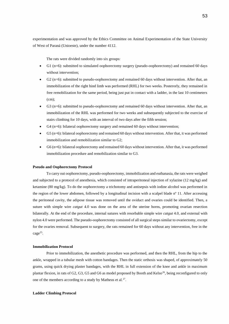
53
experimentation and was approved by the Ethics Committee on Animal Experimentation of the State University
of West of Paraná (Unioeste), under the number 4112.
The rats were divided randomly into six groups:
G1 (n=6): submitted to simulated oophorectomy surgery (pseudo-oophorectomy) and remained 60 days
without intervention;
G2 (n=6): submitted to pseudo-oophorectomy and remained 60 days without intervention. After that, an
immobilization of the right hind limb was performed (RHL) for two weeks. Posterorly, they remained in
free remobilization for the same period, being just put in contact with a ladder, in the last 10 centimeters
(cm);
G3 (n=6): submitted to pseudo-oophorectomy and remained 60 days without intervention. After that, an
immobilization of the RHL was performed for two weeks and subsequently subjected to the exercise of
stairs climbing for 10 days, with an interval of two days after the fifth session;
G4 (n=6): bilateral oophorectomy surgery and remained 60 days without intervention;
G5 (n=6): bilateral oophorectomy and remained 60 days without intervention. After that, it was performed
immobilization and remobilization similar to G2;
G6 (n=6): bilateral oophorectomy and remained 60 days without intervention. After that, it was performed
immobilization procedure and remobilization similar to G3.
Pseudo and Oophorectomy Protocol
To carry out oophorectomy, pseudo-oophorectomy, immobilization and euthanasia, the rats were weighed
and subjected to a protocol of anesthesia, which consisted of intraperitoneal injection of xylazine (12 mg/kg) and
ketamine (80 mg/kg). To do the oophorectomy a trichotomy and antisepsis with iodine alcohol was performed in
the region of the lower abdomen, followed by a longitudinal incision with a scalpel blade nº 11. After accessing
the peritoneal cavity, the adipose tissue was removed until the oviduct and ovaries could be identified. Then, a
suture with simple wire catgut 4.0 was done on the area of the uterine horns, promoting ovarian resection
bilaterally. At the end of the procedure, internal sutures with resorbable simple wire catgut 4.0, and external with
nylon 4.0 were performed. The pseudo-oophorectomy consisted of all surgical steps similar to ovariectomy, except
for the ovaries removal. Subsequent to surgery, the rats remained for 60 days without any intervention, free in the
cage25.
Immobilization Protocol
Prior to immobilization, the anesthetic procedure was performed, and then the RHL, from the hip to the
ankle, wrapped in a tubular mesh with cotton bandages. Then the static orthosis was shaped, of approximately 50
grams, using quick drying plaster bandages, with the RHL in full extension of the knee and ankle in maximum
plantar flexion, in rats of G2, G3, G5 and G6 as model proposed by Booth and Kelso26, being reconfigured to only
one of the members according to a study by Matheus et al.27.
Ladder Climbing Protocol
54
After immobilization removal, the rats of G3 and G6 were submitted to the exercise of ladder climbing,
with 10 repetitions per day in the first week (five days) and 20 repetitions per day in the second week (five days),
with an interval of one minute between the climbs and two days between the weeks. The rats of G2 and G5
performed remobilization free of the cage, being placed in contact with a ladder at 10 cm from the end of it, only
once, in the same period in which the exercise was performed on the ladder to G3 and G6. The equipment used to
perform the exercise consisted of a ladder with 67 steps, spaced 1.5 cm between the steps of the grid, 20.5 cm
wide, 118 cm height and 80° inclination angle. At the upper area there was a 28.5 cm long, 18.5 cm high and 15
cm wide darkroom, used for the rats to rest between the climbing sets, as well as to make them feel attracted to a
shelter and encouraged to perform the exercise20,22,28,29.
Histomorphometric Analysis
At the end of the experiment, all the rats were weighed and underwent anesthetic protocol being
subsequently euthanized by decapitation in guillotine. After these procedures, the right and left tibia (left hind limb
– LHL) were dissected and placed in flasks containing formalin at 10%. Then, after the fixation, the tibiae were
washed with distilled water and immersed in trichloroacetic acid (TCA) at 5%, during five days, for decalsification.
The next steps were dehydration, diafanization, measurement of the parts in its length with the help of a digital
pachymeter to perform in the medial region of the bone a cut in the transversal plane.
In the proximal part, a cut in the frontal plane was performed, being reserved the anterior portions. After
that, the bone pieces were impregnated and embedded in paraffin, being arranged in the blocks for subsequent
microtomization as follows: the proximal portion, in frontal cut; and the distal portion, with transversal cut,
positioned with the upper region for visualization. Cuts of 7 µm were performed in microtome, and confectioned
blades with hematoxylin and eosin (HE) and photomicrographed with Olympus DP71® microscope.
The photomicrographs were submitted to analysis using the Image-Pro Plus 6.0® program, in the transversal plane
performed with magnification of 40 times, for measurements of medullary canal area and cortical bone area, being
the cortical thickness measured at three points (M1 , M2 and M3)30 (Figure 1A); and magnification 400 times for
counting the osteocytes, measured in three regions (between the points M1 and M2, M1 and M3, and M2 and M3)
by means of a rectangle of 200 µm base by 100 µm height, overlaid on the image. There were excluded from the
count, the cells that were in contact with the upper edge and right side (Figure 1B). In the frontal plane,
photomicrographs were taken in three regions, designated P1, P2, and P3, which correspond respectively to the
lateral, intermediate and medial portions of the proximal region of the tibia, with a magnification of 40 times to
measure the length (vertical tracing) and area (circular tracing) trabecular bone (region between epiphyseal plate
the and upper articular cartilage)31,32 (Figure 1C). After collecting the data, the means were made and the statistical
analyzes were performed.
A B C
M1
M2
M3
*
*
55
Figure 4: Photomicrographs with statements of histomorphometric analyzes performed on the ovariectomized rat right tibia subjected to
immobilization and remobilization ladder. In A, measuring the thickness of cortical bone in three points (M1, M2 and M3) and medullary canal area, cross-section and 40X magnification; B overlaid base of 200 μm by 100 μm high for counting osteocytes in the area between the points
M1 and M2, 400X magnification (obs .: rectangle asterisks exclusion demarcate the edges (top and right side)); and C, measurement of area
(circular route) and thickness (vertical layout) of trabecular bone (upper region of the plate and articular cartilage) in medial (P3), 40X magnification. HE staining.
Statistical Analysis
The survey data were evaluated by comparing the results obtained on the left hind limb (control) and right
(immobilized), between the rats of the same group and between the experimental groups. For this, the BioEstat
5.0® program was used, with values presented as mean and standard deviation. The Student t paired test was applied
for comparison between the right and left side, within the same group, and one-way ANOVA with Bonferroni post
hoc test, for comparison between experimental groups. The level of significance was p ≤ 0.05.
RESULTS
Cortical Bone Area and Thickness
In the comparison between rats of the same group, it could be noted a significant decrease in cortical
bone of the RHL regarding the LHL in G5, as evidenced by data from both the area (p=0.0178) and thickness
(p=0.0024). The other groups (G1, G2, G3, G4 and G6) showed no significant differences between their hind limbs
so that remobilization (G6) was able to reverse the loss of cortical bone in the immobilized limb when compared
to the contralateral non-immobilized (Table 1).
When the comparison was made between the groups, with mean values of RHL, it was observed a
significant increase in G6 compared to G1, G4 and G5 (F (5, 29)=6.24; p=0.0007), to area, but also to G4 and G5
(F(5; 29)=4.11, p=0.0062), for thickness. Thus, it is evident that the exercise was able to reverse the cortical bone
loss caused by ovariectomy and immobilization, with values that exceed even those found in rats only pseudo-
oophorectomized (G1). It is noteworthy that the free remobilization was not able to recover the loss of cortical
bone in the limb of ovariectomized rats (Table 1).
In the LHL analysis between groups, there was a significant decrease in area of G4 in relation to G2
and G6 (F(5, 29)=4.71, p=0.0031), and in thickness of G4 in relation to the other groups (F(5; 29)=8.30, p=0.0001).
This finding shows that ovariectomy was able to promote cortical bone loss and that both, free remobilization, and
in ladder were able to recover from this loss (Table 1).
Table 1: Area and cortical thickness data, comparing the rats distributed between the study groups (G1 - G6), right
(RHL - target) and left hind limbs (LHL - control)
Cortical Area (µm2)
Note.: values elevated to the 6th Power
Groups RHL
(mean ± sd)
LHL
(mean ± sd)
G1 4,20 ± 5,47 (a) 4,90 ± 8,91 (cd)
G2 4,65 ± 11,71 (ab) 4,78 ± 9,97 (c)
G3 5,60 ± 18,36 (ab) 5,80 ± 12,58 (cd)
56
G4 3,80 ± 6,78 (a) 3,73 ± 4,77 (d)
G5* 4,00 ± 5,57 (a) 5,40 ± 7,91 (cd)
G6 6,66 ± 9,22 (b) 6,22 ± 12,32 (c)
Cortical Thickness (µm)
Groups RHL
(mean ± sd)
LHL
(mean ± sd)
G1 600,38 ± 64,44 (ab) 661,46 ± 122,49 (c)
G2 617,68 ± 69,30 (ab) 690,43 ± 89,07 (c)
G3 683,07 ± 69,83 (ab) 702,05 ± 39,53 (c)
G4 394,68 ± 35,83 (a) 427,55 ± 103,78 (d)
G5* 374,96 ± 28,40 (a) 624,19 ± 90,31 (c)
G6 646,12 ± 112,36 (b) 711,63 ± 78,82 (c)
Obs.: same letters mean similarities, and the different letters mean significant differences between the experimental groups to the same side. *
Significant difference between the sides of the same group.
Medullary Canal Area
As to the medullar canal area, G5 rats showed a significant increase of RHL in relation to LHL
(p=0.0384), also for the G5 RHL showed higher values than G1, G3, G4 and G6 (F(5, 29)=4.42, p=0.0043). In
LHL there was no significant difference between the groups, ie, the area of the medullary canal was maintained
even after the interventions (Table 2).
Thus, it was found that oophorectomy associated with immobilization and free remobilization (G5),
produced a significant increase in the medullary canal area of the immobilized limb, when compared to the RHL
from the other groups and that the ladder climbing exercise was able to reverse this increase. This was evident in
G3 and G6, which had similar means between themselves and to the non-immobilized groups (G1 and G4) (Table
2).
Table 2: Data of the medullary canal area, comparing the rats distributed between the study groups (G1 - G6),
right (RHL - target) and left hind limbs (LHL - control)
Medullary Canal Area (µm2)
Note.: values elevated to the 6th Power
Groups RHL
(mean ± sd)
LHL
(mean ± sd)
G1 1,90 ± 2,53 (a) 2,13 ± 4,89 (c)
G2 2,48 ± 6,61 (ab) 2,00 ± 4,29 (c)
G3 1,77 ± 4,32 (a) 2,08 ± 7,57 (c)
G4 2,00 ± 5,21 (a) 2,25 ± 4,93 (c)
G5* 3,42 ± 1,07 (b) 2,22 ± 3,34 (c)
G6 2,00 ± 8,74 (a) 2,40 ± 5,73 (c)
Obs.: same letters mean similarities, and the different letters mean significant differences between the experimental groups to the same side. *

57
Significant difference between the sides of the same group.
Osteocytes Number
Regarding the osteocytes number, it was observed a statistically significant difference between RHL and
LHL of G5 (p<0.0001), revealing that the free remobilization is not able to reverse the cell loss caused by
immobilization in ovariectomized rats. However, as there was no difference between the hind limbs of rats
belonging to the G6; it is believed that the exercise was able to reverse this loss (Table 3).
In the comparison between the groups, it was observed lower values only in RHL at G5 comparing to the
other groups (F(5, 29)=14.55; p=<0.0001). Thus, immobilization with free remobilization caused severe loss of
osteocytes in ovariectomized rats and the ladder climbing exercise proved to be effective in recovering the number
of osteocytes (Table 3).
Table 3: Data of osteocytes number, comparing the rats distributed between the study groups (G1 - G6), right
(RHL - target) and left hind limbs (LHL - control)
Osteocytes Number (un)
Groups RHL
(mean ± sd)
LHL
(mean ± sd)
G1 22,67 ± 5,32 (a) 20,06 ± 3,32 (c)
G2 18,83 ± 3,48 (a) 23,33 ± 6,36 (c)
G3 20,44 ± 1,76 (a) 21,33 ± 4,73 (c)
G4 20,33 ± 1,63 (a) 19,67 ± 1,71 (c)
G5* 10,07 ± 0,83 (b) 20,07 ± 1,93 (c)
G6 23,94 ± 1,97 (a) 24,94 ± 2,89 (c)
Obs.: same letters mean similarities, and the different letters mean significant differences between the experimental groups to the same side. *
Significant difference between the sides of the same group.
Trabecular Area and Thickness
In the trabecular bone area and thickness analysis of the same group rats, there was no significant
difference between RHL and LHL. In the intergroup analysis there were significant differences in comparison of
G1 LHL with G6 (F(5, 29)=2.99; p=0.0265) for the area and thickness, and G1 to G2 and G3, only for thickness
(F(5; 29)=5.39, p=0.0015) (Table 4).
Table 4: Data referring to the trabecular bone area and thickness, comparing the rats distributed between the study
groups (G1 - G6), right (target) and left hind limbs (control)
Trabecular Area (µm2)
Note.: values elevated to the 6th power
Groups RHL
(mean ± sd)
LHL
(mean ± sd)
G1 2,67 ± 9,22 (a) 2,15 ± 6,74 (b)
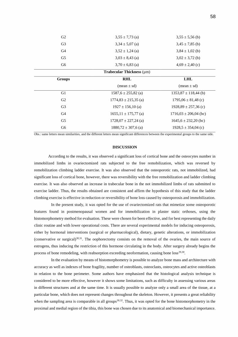
58
G2 3,55 ± 7,73 (a) 3,55 ± 5,56 (b)
G3 3,34 ± 5,07 (a) 3,45 ± 7,85 (b)
G4 3,52 ± 1,24 (a) 3,84 ± 1,02 (b)
G5 3,03 ± 8,43 (a) 3,02 ± 3,72 (b)
G6 3,70 ± 6,83 (a) 4,69 ± 2,40 (c)
Trabecular Thickness (µm)
Groups RHL
(mean ± sd)
LHL
(mean ± sd)
G1 1587,6 ± 255,82 (a) 1353,87 ± 118,44 (b)
G2 1774,83 ± 215,35 (a) 1795,06 ± 81,48 (c)
G3 1927 ± 156,10 (a) 1928,89 ± 257,36 (c)
G4 1655,11 ± 175,77 (a) 1716,03 ± 206,04 (bc)
G5 1728,07 ± 227,24 (a) 1645,6 ± 232,20 (bc)
G6 1880,72 ± 307,6 (a) 1928,5 ± 354,04 (c)
Obs.: same letters mean similarities, and the different letters mean significant differences between the experimental groups to the same side.
DISCUSSION
According to the results, it was observed a significant loss of cortical bone and the osteocytes number in
immobilized limbs in ovariectomized rats subjected to the free remobilization, which was reversed by
remobilization climbing ladder exercise. It was also observed that the osteoporotic rats, not immobilized, had
significant loss of cortical bone, however, there was reversibility with the free remobilization and ladder climbing
exercise. It was also observed an increase in trabecular bone in the not immobilized limbs of rats submitted to
exercise ladder. Thus, the results obtained are consistent and affirm the hypothesis of this study that the ladder
climbing exercise is effective in reduction or reversibility of bone loss caused by osteoporosis and immobilization.
In the present study, it was opted for the use of ovariectomized rats that mimetize some osteoporotic
features found in postmenopausal women and for immobilization in plaster static orthoses, using the
histomorphometry method for evaluation. These were chosen for been effective, and for best representing the daily
clinic routine and with lower operational costs. There are several experimental models for inducing osteoporosis,
either by hormonal interventions (surgical or pharmacological), dietary, genetic alterations, or immobilization
(conservative or surgical)30,31. The oophorectomy consists on the removal of the ovaries, the main source of
estrogens, thus inducing the restriction of this hormone circulating in the body. After surgery already begins the
process of bone remodeling, with reabsorption exceeding neoformation, causing bone loss29,30.
In the evaluation by means of histomorphometry is possible to analyze bone mass and architecture with
accuracy as well as indexes of bone fragility, number of osteoblasts, osteoclasts, osteocytes and active osteoblasts
in relation to the bone perimeter. Some authors have emphasized that the histological analysis technique is
considered to be more effective, however it shows some limitations, such as difficulty in assessing various areas
in different structures and at the same time. It is usually possible to analyze only a small area of the tissue, at a
particular bone, which does not represent changes throughout the skeleton. However, it presents a great reliability
when the sampling area is comparable in all groups30,32. Thus, it was opted for the bone histomorphometry in the
proximal and medial region of the tibia, this bone was chosen due to its anatomical and biomechanical importance.

59
In some studies on oophorectomy significant bone losses were observed after 14, 72 and 74 days at the
proximal tibia metaphysis, after 60 and 73 days in the lumbar vertebral body and after 30 days in the femoral
neck30,33. Other authors report that the first changes in the width of the cortical bone and the medullar cavity of the
femur and tibia, are noted in periods around 90 and 120 days after ovariectomy34–36. In the present study, was
observed significant bone loss at 60 days after ovariectomy and 90 days after the association of oophorectomy,
immobilization and free remobilization in the proximal region (trabecular) and tibia medial (cortical). However,
the loss was more pronounced in 90-day period in the oophorectomized group, immobilized and free
remobilization, and may be associated with longer duration of hormone estrogen deprivation, but also by
association with immobilization, as it accentuated the decrease in bone mass.
Strong evidences show that the combination of oophorectomy and immobilization can reduce the time
of significant bone loss, especially the cortical, when compared to the techniques isolated30,34–36. In view of the
findings of this study, such association may also accentuate the effects of the significant loss of bone mass. These
findings corroborate with previous studies showing the accuracy of measurements taken in the medial inferior
areas of the cortical axis, which are very reliable, because most bone losses occur in this location30,33,37.
The reduction in bone mass was clearly demonstrated in this study, because it is well seen in the cortical
bone by the enlargement of the medullar cavity. This occurs due to the increasing in endosteal reabsorption and
periosteal bone apposition30,33,37. With area measurements and cortical thickness as well as the medullar canal area,
only G5 RHL showed significant decrease compared to the LHL, which may be related to the effects of estrogen
hormone deprivation associated with immobilisation of the RHL. The free remobilization was not effective to
recover this loss, as in G2, G3 and G6 there was no difference. Thus, it is believed that both the free exercise and
in stairs were effective to recover the bone loss associated with immobilisation in the pseudo-ovariectomized
groups (G2 and G3) and that only the ladder exercise was effective in this reversibility in immobilized osteoporotic
rats.
In intergroup comparison to the RHL, it was found that the exercise on ladder was effective in the recovery
of cortical bone loss caused by ovariectomy alone or in combination with immobilization and free remobilization.
It was also found that G6 was significantly higher than G1 regarding the cortical area, ie, showing that the exercise
on ladder produces increasing in the upper bone mass even when compared with animals that did not undergo
significant interventions, only the surgical procedure stress without removal of the ovaries.
In intergroup comparison for LHL, there was a significant decrease in cortical bone area and cortical bone
thickness of G4 compared to G2 and G6. Regarding G2 can be linked to the fact that there was an overload on the
LHL in relation to the immobilized limb (RHL), leading to a growth of cortical bone in higher values than those
from the group which was subjected only to hormone deprivation (G4) with significant loss of cortical bone.
However, in G6, it is evident the outcomes of therapeutic intervention, ie, that the ladder exercise was a clinical
resource very well employed in remobilization for the recovery of bone loss caused due to hormonal estrogen
deprivation and immobilization. As there was no difference compared to G5, it is believed that osteoporosis may
have been determinative in the reduction of bone mass, as in G4, as well as in the free activity in the box was not
enough to reverse the decrease in cortical bone mass in osteoporotic rats.
Regarding the trabecular bone, there was a significant LHL loss of G1 compared to G2, G3 and G6. This
may be related to overload exerted on LHL due to immobilization of RHL associated with the effectiveness of
exercise of climbing stairs, in ovariectomized and pseudo-oophorectomized rats being even able to promote an

60
increase in this type of tissue. For rats not subjected to hormone deprivation, even the free exercises associated to
the overload in LHL were able to promote growth of the trabecular bone. It was not observed diminution in
consequence of ovariectomy and immobilization, which may be related to post-surgical period and immobilization.
Maeda et al.18 found a significant loss of cancellous bone in rats subjected to six weeks of immobilization.
Even with therapeutic interventions applied correctly, the complete reversibility of the damage caused
by immobilization will not always occur38,39. Nevertheless, the exercise has been a therapeutic resource widely
used for both the treatment of osteoporosis and in remobilization periods. However, the benefits of physical
exercise on the skeleton depend on the intensity and bone characteristics, such as age, type and region involved40,41.
In this study it was found that both the free exercise and the stairs climbing were effective in recovering the bone
loss in rats that have undergone hormonal deprivation, through the protocol used. However, when these
ovariectomized rats underwent immobilization, it was found that the free exercise was not able to recover bone
loss, unlike the ladder exercise.
Several authors studied the therapeutic action of various resources and remobilization techniques in
animal models, such as muscle stretching42, free remobilization15,21,42, swimming15, jump21 and treadmill
running40,43. It were found improvements in gait, cartilage conditions, subchondral bone, biomechanical and
articular capsule15; restoration of the trabecular architectural integrity21; increase in bone mass and mineral
density21,40,43; decrease in the number of osteocytes, even after remobilization42. This study verified that the
osteocytes number of immobilized limbs was reestablished in both free remobilization and climbing stairs, in
pseudo-ovariectomized rats. In osteoporotic rats, only the exercise of stairs climbing was able to recover the
number of osteocytes, with values that even resemble the groups not exposed to hormone deprivation.
Study performed with rats subjected to tail suspension for 21 days and climbing stairs exercise for the
same period, doing eight sets, with overload equivalent to 80% of their maximum strength, five times a week,
observed that this type of exercise was able to restore the BMD and bone stiffness values44. In this study, the
exercise of climbing stairs was held without load. Nevertheless, the protocol used for this endurance exercise was
effective in the recovery of bone mass and cell losses due to ovariectomy and immobilisation verified by the
histomorphometry method. Cassilhas et al.20 add that the ladder exercise promotes hypertrophy of the
gastrocnemius muscle, the flexor digitorum longus and plantaris, which can help on the protection of the ankle
joint and the bone matrix. Nascimento et al.22 showed that this climbing exercise also promotes hypertrophy of the
brachial triceps muscle, ie, achieving, with positive results with inferences even for the forelimbs.
Thus, it is apparent that the stair climbing exercise promotes the adaptation of muscle tissue, cartilage and
bone, with an improvement in BMD, bone stiffness, hypertrophy of gastrocnemius muscles, flexor digitorum
longus, plant and triceps proprioceptive stimulation and resistance, thus proving to be an important and effective
kinesiotherapeutic feature, mainly because of its global action throughout the body, not only in a focus or
member20,22,48. It is believed that exercise stimulates ladder climbing action osteoprotegerin, neutralizing the
effects of osteoclastic bone resorption by the interaction of receptor activator kB ligand (RANKL) and receptor
activator kB (RANK) due to hypoestrogenism. This possibly leads to an increased formation of vitamin D and
serum calcium uptake in the bone matrix, providing increased mass and bone strength, which was evidenced by
area and cortical thickness, and number of osteocytes, in the groups submitted to exercise climb staircase49.
Thus, it might be noted that the ladder climbing exercise appears to be a good resource for clinical
practice, taking in account many benefits that it provides to the treatment of osteoporosis in periods of

61
remobilization and the combination of both situations. This causes it to expand the clinical reasoning of
professionals and to increase the possibilities of therapeutic methods to be applied with greater accuracy and
resolution in the proposed treatments. One limitation of the present study was the absence of a control group
subjected only to immobilization, without remobilization and hormonal intervention. It is reiterated regarding the
difficulty of other comparisons with the exercise protocol used, using times, series and different intensities.
However, following the 3Rs ethical rule (replacement, reduction and refinement) for the use of animals in research,
it was opted to follow this model due to the number of rats to be used.
In resume, upon the climbing stairs exercise protocol used in this study and with its limitations, it is
concluded that this therapeutic approach was effective in the recovery of bone loss in rats subjected to a post-
menopausal osteoporosis experimental model and immobilization, with recovery of thickness, and cortical and
trabecular area, as well as the number of osteocytes. It is emphasized that future studies could be conducted with
greater times of hormonal deprivation and immobilization, implementing other exercise protocols, in association
with other therapeutic resources, as well as evaluating the effects on other systems and potential impact on
functional activities.
Conflicts of Interest
José Martim Marques Simas, Rose Meire Costa Brancalhâo, Lucinéia de Fátima Chasko Ribeiro and
Gladson Ricardo Flor Bertolini declare that they have no conflict of interest.
References
1. NAMS. The North American Menopause Society. The Menopause Guidebook. 7a ed. 2012.
2. Claassen H, Schlüter M, Schünke M, Kurz B. Influence of 17beta-estradiol and insulin on type II
collagen and protein synthesis of articular chondrocytes. Bone 2006; 39(2):310–7.
3. NIH. National Institutes of Health. Osteoporosis and related bone diseases national resource center.
2012;Jan:1–3.
4. Berry S, Kiel D, Donaldson M, Cummings S, Kanis J, Johansson H, et al. Application of the national
osteoporosis foundation guidelines to postmenopausal women and men: the Framingham Osteoporosis
Study. Osteoporos Int 2011;21(1):53–60.
5. Camargo M, Cendoroglo M, Ramos L, Latorre M, Saraiva G, Lage A, et al. Bone mineral density and
osteoporosis among a predominantly caucasian elderly population in the city of São Paulo, Brazil.
Osteoporos Int 2005;16(11):1451–60.
6. Egermann M, Schneider E, Evans C, Baltzer A. The potential of gene therapy for fracture healing in
osteoporosis. Osteoporos Int 2005;16(2):120–8.
7. NIH. National Isntitutes of Health. Consensus conference. Osteoporosis: prevention, diagnosis and
therapy. Jama 2001;285(6):164–7.
8. Budhia S, Mikyas Y, Tang M, Badamgarav E. Osteoporotic Fractures: a systematic review of US
Healthcare Costs and Resource Utilization. Pharmacoeconomics 2012;30(2):147–70.
9. Brandt K. Response of joint structures to inactivity and to reloading after immobilization. Arthritis
Rheum 2003;49(2):267–71.

62
10. Duncan N, Williams D, Lynch G. Adaptations in rat skeletal muscle following long-term resistance
exercise training. European Journal of Applied Physiology and Occupational Physiology
1998;77(4):372–8.
11. Kunz R, Coradini J, Silva L, Bertolini G, Brancalhão R, Ribeiro L. Effects of immobilization and
remobilization on the ankle joint in Wistar rats. Brazilian J Med Biol Res 2014;00:1–8. Ahead of print.
12. Sakakima H. Effects of immobilization and subsequent low and high frequency treadmill running on rat
soleus muscle and ankle joint movement. J Phys Ther Sci 2004;16(1):43–8.
13. Christensen B, Dyrberg E, Aagaard P, Enehjelm S, Krogsgaard M, Kjaer, et al. Effects of long-term
immobilization and recovery on human triceps surae and collagen turnover in the Achilles tendon in
patients with healing ankle fracture. J Appl Physiol 2008;105(2):420–6.
14. Christensen B, Dyrberg E, Aagaard P, Kjaer M, Langberg H. Short-term immobilization and recovery
affect skeletal muscle but not collagen tissue turnover in humans. J Appl Physiol 2008;105(6):1845–51.
15. Del Carlo R, Galvão M, Viloria M, Natali A, Barbosa A, Monteiro B, et al. Imobilização prolongada e
remobilização da articulação fêmoro-tíbio-patelar de ratos: estudo clínico e microscópico. Arq Bras Med
Vetrinária e Zootec 2007;59(2):363–70.
16. Kaneps A, Stover S, Lane N. Changes in canine cortical and cancellous bone mechanical properties
following immobilization and remobilization with exercise. Bone 1997;21(5):419–23.
17. Leroux M, Cheung H, Bau J, Wang J, Howell D, Setton L. Altered mechanics and histomorphometry of
canine tibial cartilage following joint immobilization. Osteoarthr Cartil 2001;9(7):633–40.
18. Maeda H, Kimmel D, Raab D, Lane N. Musculoskeletal recovery following hindlimb immobilization in
adult female rats. Bone 1993;14(2):153–9.
19. Narmoneva D, Cheung H, Wang J, Howell D, Setton L. Altered swelling behavior of femoral cartilage
following joint immobilization in a canine model. J Orthop Res 2002;20(1):83–91.
20. Cassilhas R, Reis I, Venâncio D, Fernandes J, Tufik S, Mello M. Animal model for progressive
resistance exercise : a detailed description of model and its implications for basic research in exercise.
Motriz 2013;19(1):178–84.
21. Ju Y-I, Sone T, Okamoto T, Fukunaga M. Jump exercise during remobilization restores integrity of the
trabecular architecture after tail suspension in young rats. J Appl Physiol 2008;104(6):1594–600.
22. Nascimento V, Krause Neto W, Gonçalves L, Maifrino L, Souza R, Gama E. Morphoquantitative
analysis revealed Triceps Brachialis muscle hypertrophy by specific Resistance training equipment in
rats. J Morphol Sci 2013;30(4):276–80.
23. Renno A, Gomes A, Nascimento R, Salvini T, Parizoto N. Effects of a progressive loading exercise
program on the bone and skeletal muscle properties of female osteopenic rats. Exp Gerontol
2007;42(6):517–22.
24. Kemmler W, Wildt L, Engelke K, Pintag R, Pavel M, Bracher B, et al. Acute hormonal responses of a
high impact physical exercise session in early postmenopausal women. Eur J Appl Physiol 2003;90(1-
2):199–209.
25. Khajuria D, Razdan R, Mahapatra D. The combination therapy with zoledronic Acid and propranolol
improves the trabecular microarchitecture and mechanical property in an rat model of postmenopausal
osteoporosis. J Osteoporos 2014;1–10.
63
26. Booth F, Kelso J. Effect of hind-limb immobilization on contractile and histochemical properties of
skeletal muscle. Pflügers Arch 1973;342:231–8.
27. Matheus J, Gomide L, Oliveira J, Volpon J, Shimano A. Efeitos da estimulação elétrica neuromuscular
durante a imobilização nas propriedades mecânicas do músculo esquelético. Rev Bras Med do Esporte
2007;13(1):55–9.
28. Hornberger T, Farrar R. Physiological hypertrophy of the FHL muscle following 8 weeks of progressive
resistance exercise in the rat. Can J Appl Physiol 2004;29(1):16–31.
29. Oliveira B, Silva M, Medeiros R, Apolinário-Coêlho J. A influência do treinamento físico resistido no
tecido ósseo de ratos osteopênicos induzidos por suspensão pela cauda. Arch Heal Investig
2013;2(2):3009.
30. Carvalho A, Henriques H, Pantaleão J, Pollastri C, Fernandes, GVOGranjeiro J, Guzmán-Silva M.
Histomorfometria do tecido ósseo em ratas castradas tratadas com tibolona. J Bras Patol e Med Lab
2010;46(3):235–43.
31. Ando A, Suda H, Hagiwara Y, Onoda Y, Chimoto E, Saijo Y, et al. Reversibility of immobilization-
induced articular cartilage degeneration after remobilization in rat knee joints. Tohoku J Exp Med
2011;224(2):77–85.
32. Hagiwara Y, Ando A, Chimoto E, Saijo Y, Ohmori-Matsuda K, Itoi E. Changes of articular cartilage
after immobilization in a rat knee contracture model. J Orthop Res 2009;27(2):236–42.
33. Iwamoto J, Takeda T, Sato Y, Yeh J. Effect of vitamin K2 and growth hormone on the long bones in
hypophysectomized young rats: a bone histomorphometry study. J Bone Miner Metab 2007;25(1):46–53.
34. Lelovas PP, Xanthos TT, Thoma SE, Lyritis GP, Dontas I a. The laboratory rat as an animal model for
osteoporosis research. Comp Med 2008;58(5):424–30.
35. Berdud I, Martin-Malo A, Almaden Y, Aljama P, Rodriguez M, Felsenfeld A. The PTH-calcium
relationship during a range of infused PTH doses in the parathyroidectomized rat. Calcif Tissue Int
1998;62(5):457–61.
36. Tarantino U, Celi M, Rao C, Feola M, Cerocchi I, Gasbarra E, et al. Hip osteoarthritis and osteoporosis :
clinical and histomorphometric considerations. Int J Endocrinol 2014;1–5.
37. Li M, Shen Y, Wronski T. Time Course of Femoral Neck Osteopenia in Ovariectomized Rats. Bone
1997;20(1):55–61.
38. Cavolina J, Evans G, Harris S, Zhang M, Westerlind K, Turner R. The effects of orbital spaceflight on
bone histomorphometry and messenger ribonucleic acid levels for bone matrix proteins and skeletal
signaling peptides in ovariectomized growing rats. Endocrinology 1997;138(4):1567–76.
39. Danielsen C, Mosekilde L, Svenstrup B. Cortical bone mass, composition, and mechanical properties in
female rats in relation to age, long-term ovariectomy, and estrogen substitution. Calcif Tissue
iIternational 1993;52(1):26–33.
40. Jee W, Yao W. Overview: animal models of osteopenia and osteoporosis. J Musculoskelet Neuronal
Interact 2001;1(3):193–207.
41. Miller S, Bowman B, Miller M, Bagi C. Calcium absorption and osseous organ-, tissue-, and envelope-
specific changes following ovariectomy in rats. Bone 1991;12(6):439–46.
42. Trebacz H. Disuse-induced deterioration of bone strength is not stopped after free remobilization in
young adult rats. J Biomech 2001;34(12):1631–6.

64
43. Vanwanseele B, Lucchinetti E, Stüssi E. The effects of immobilization on the characteristics of articular
cartilage: current concepts and future directions. Osteoarthr Cartil 2002;10(5):408–19.
44. Ocarino N, Silva J, Santiago L, Rocha C, Marubayashi U, Serakides R. Treadmill training before and/or
after ovariectomy is more effective in preventing osteopenia in adult female rats. Sci Sports
2009;24(1):52–5.
45. Dionyssiotis Y, Paspati I, Trovas G, Galanos A, Lyritis G. Association of physical exercise and calcium
intake with bone mass measured by quantitative ultrasound. BMC Womens Health 2010;10(1):12.
46. Portinho D, Boin V, Bertolini G. Efeitos Sobre o Tecido Ósseo e Cartilagem Articular Provocados Pela
Imobilização e Remobilização em Ratos Wistar. Rev Bras Med do Esporte 2008;14(5):408–11.
47. Mathey J, Horcajada-Molteni M-N, Chanteranne B, Picherit C, Puel C, Lebecque P, et al. Bone mass in
obese diabetic Zucker rats: influence of treadmill running. Calcif Tissue Int 2002;70(4):305–11.
48. Oliveira M, Oliveira B, Peres M, Coêlho J, Florindo P, Louzada M. Análise densitométrica e
biomecânica de tíbias de ratos submetidos à suspensão pela cauda e exercício físico resistido. Arch Heal
Investig 2013;2:292.
49. Xu S, Wang Y, Lu J, Xu J. Osteoprotegerin and RANKL in the pathogenesis of rheumatoid arthritis-
induced osteoporosis. Rheumatol Int 2012;32(11):3397–403.

5 ARTIGO CIENTÍFICO 2
EFEITOS DO EXERCÍCIO FÍSICO SOBRE A HISTOMORFOMETRIA
CARTILAGINOSA DE RATAS OOFORECTOMIZADAS SUBMETIDAS
À IMOBILIZAÇÃO
Submetido à Revista Brasileira de Reumatologia

66
ARTIGO CIENTÍFICO 2
EFEITOS DO EXERCÍCIO FÍSICO SOBRE A HISTOMORFOMETRIA
CARTILAGINOSA DE RATAS OOFORECTOMIZADAS SUBMETIDAS À
IMOBILIZAÇÃO
Autores:
José Martim Marques Simas – Mestrando em Biociências e Saúde pela
Unioeste/Cascavel-PR;
Lígia Inez Silva - Fisioterapeuta graduada pela Unioeste/Cascavel-PR;
Regina Inês Kunz – Mestre em Biociências e Saúde pela Unioeste/Cascavel-PR;
Rose Meire Costa Brancalhão – Doutora em Zoologia pela UFPR/Curitiba-PR;
Lucinéia de Fátima Chasko Ribeiro – Doutora em Ciências Biológicas pela
UEM/Maringá-PR;
Gladson Ricardo Flor Bertolini – Doutor em Ciências da Saúde Aplicadas ao Aparelho
Locomotor pela USP/Ribeirão Preto-SP.
Departamento/Instituição:
Laboratório de Estudos das Lesões e Recursos Fisioterapêuticos e Laboratório de
Biologia Estrutural e Funcional da Universidade Estadual do Oeste do Paraná
(UNIOESTE), campus de Cascavel.
Correspondência:
Prof.Dr.Gladson Ricardo Flor Bertolini
END.: Colegiado de Fisioterapia - Rua Universitária, 2069, Jardim Universitário.
Cascavel-PR

67
CEP: 85819-110
TEL: (45) 3220-3298
Conflitos de Interesse:
Os autores declaram não haver conflitos de interesse.
Título resumido:
EXERCÍCIO FÍSICO EM RATAS OOFORECTOMIZADAS E IMOBILIZADAS
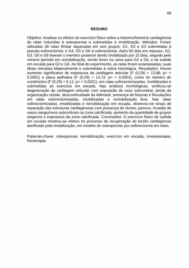
68
RESUMO
Objetivo: Analisar os efeitos do exercício físico sobre a histomorfometria cartilaginosa de ratas induzidas à osteoporose e submetidas à imobilização. Métodos: Foram utilizadas 36 ratas Wistar separadas em seis grupos: G1, G2 e G3 submetidas à pseudo-ooforectomia; e G4, G5 e G6 à ooforectomia. Após 60 dias em repouso, G2, G3, G5 e G6 tiveram o membro posterior direito imobilizado por 15 dias, seguido pelo mesmo período em remobilização, sendo livres na caixa para G2 e G5, e de subida em escada para G3 e G6. Ao final do experimento, as ratas foram eutanasiadas, suas tíbias retiradas bilateralmente e submetidas à rotina histológica. Resultados: Houve aumento significativo de espessura da cartilagem articular (F (5;29) = 13,88; p= < 0,0001) e placa epifisária (F (5;29) = 14,72; p= < 0,0001), como do número de condrócitos (F (5;29) = 5,11; p= < 0,0021), em ratas ooforectomizadas, imobilizadas e submetidas ao exercício em escada. Nas análises morfológicas, verificou-se degeneração da cartilagem articular com exposição de osso subcondral, perda da organização celular, descontinuidade da tidemark, presença de fissuras e floculações em ratas ooforectomizadas, imobilizadas e remobilização livre. Nas ratas ooforectomizadas, imobilizadas e remobilização em escada, observou-se sinais de reparação das estruturas cartilaginosas com presença de clones, pannus, invasão de vasos sanguíneos subcorticais na zona calcificada, aumento da quantidade de grupos isógenos e espessura da zona calcificada. Conclusões: O exercício físico de subida em escada mostrou-se efetivo no processo de recuperação do tecido cartilaginoso danificado pela imobilização, em modelo de osteoporose por ooforectomia em ratas.
Palavras-chave: osteoporose; remobilização; exercício em escada; cinesioterapia; fisioterapia.

69
INTRODUÇÃO
A menopausa provoca diminuição lenta e gradativa dos hormônios estrógeno
e progesterona, produzindo aumento da reabsorção óssea e diminuição da absorção
de cálcio pelo tecido ósseo. Dessa forma, os ossos ficam mais fragilizados e aumenta
a predisposição ao aparecimento de doenças, como a osteoporose.1,2 Considerada
uma doença osteometabólica, a osteoporose provoca deterioração da
microarquitetura do tecido ósseo e diminuição de sua massa, como também
degradação da cartilagem, predispondo ao risco de fraturas. Apresenta altas taxas de
morbimortalidade e afeta cerca de um terço das mulheres na pós-menopausa, com
maior prevalência entre os 65 e 75 anos de idade.1,3–7
Devido sua evolução insidiosa e silenciosa, as fraturas são um dos sintomas
mais frequentes na osteoporose. Estima-se que ocorra anualmente 1.660.000 fraturas
em decorrência desta doença, podendo quadruplicar até 2050.5,8,9 Na ocorrência de
fraturas, ou até mesmo de outras lesões do sistema musculoesquelético, em que se
faz necessário o uso de um aparato imobilizatório, o quadro da doença pode piorar,
tornando o tecido ósseo ainda mais frágil, além de acarretar em incapacitação
temporária ou permanente do indivíduo e elevação dos custos de cuidados em
saúde.3,4,10
Vários tratamentos têm sido propostos tanto para a osteoporose quanto para
períodos de reabilitação pós-imobilização. No entanto, o exercício físico tem sido o
mais preconizado devido melhorar a força muscular, marcha e equilíbrio; reduzir
quedas em idosos; proteger o quadril; prevenir fraturas e promover reparações na
estrutura tecidual muscular, óssea e cartilaginosa.7,11–18
Assim, tendo em vista a elevação da expectativa de vida da população, com

70
consequente aumento do aparecimento de doenças crônicas, a osteoporose como um
grande problema atual de saúde pública, as incapacitações e efeitos deletérios nas
estruturas teciduais decorrentes de uma imobilização, como também da necessidade
de tornar evidência científica recursos terapêuticos amplamente utilizados na prática
clínica, justifica-se este estudo, que objetivou analisar os efeitos do exercício físico
sobre a histomorfometria cartilaginosa de ratas induzidas à osteoporose e submetidas
à imobilização.
MATERIAL E MÉTODOS
Grupos Experimentais
Foram utilizadas 36 ratas da linhagem Wistar, adultas (10 ± 2 semanas),
nulíparas, com peso médio de 317,2 ± 22,1 gramas, mantidas em gaiolas de
polipropileno com livre acesso à água e ração, fotoperíodo claro/escuro de 12 horas e
temperatura ambiente controlada (25º ± 1ºC). O estudo foi realizado conforme as
normas éticas internacionais de experimentação animal, sendo aprovado pelo Comitê
de Ética em Experimentação Animal da Unioeste, sob o parecer nº 4112.
As ratas foram separadas aleatoriamente em seis grupos:
G1 (n=6): as ratas foram submetidas à cirurgia simulada de ooforectomia
(pseudo-ooforectomia) e permaneceram 60 dias sem intervenção;
G2 (n=6): as ratas foram submetidas à pseudo-ooforectomia e permaneceram
60 dias sem intervenção. Após, foi realizada imobilização do membro posterior
direito (MPD) por duas semanas. Posteriormente ficaram em remobilização
livre pelo mesmo período, sendo apenas colocadas em contato com uma
escada nos últimos 10 centímetros (cm);
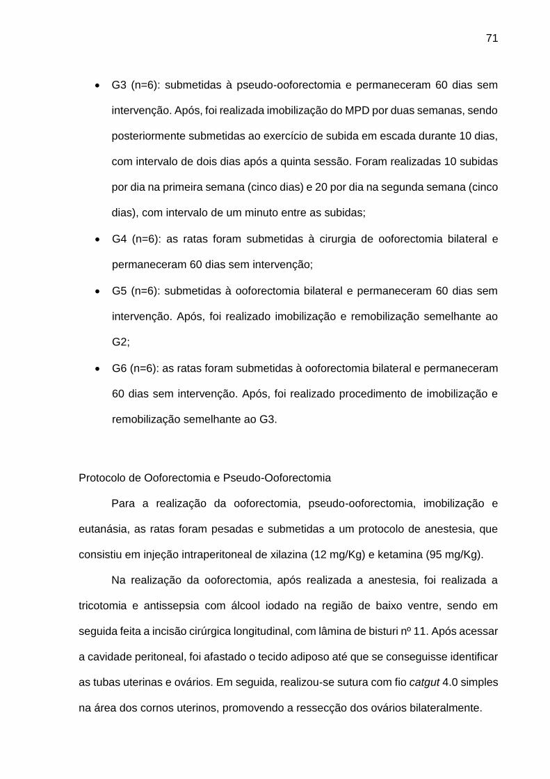
71
G3 (n=6): submetidas à pseudo-ooforectomia e permaneceram 60 dias sem
intervenção. Após, foi realizada imobilização do MPD por duas semanas, sendo
posteriormente submetidas ao exercício de subida em escada durante 10 dias,
com intervalo de dois dias após a quinta sessão. Foram realizadas 10 subidas
por dia na primeira semana (cinco dias) e 20 por dia na segunda semana (cinco
dias), com intervalo de um minuto entre as subidas;
G4 (n=6): as ratas foram submetidas à cirurgia de ooforectomia bilateral e
permaneceram 60 dias sem intervenção;
G5 (n=6): submetidas à ooforectomia bilateral e permaneceram 60 dias sem
intervenção. Após, foi realizado imobilização e remobilização semelhante ao
G2;
G6 (n=6): as ratas foram submetidas à ooforectomia bilateral e permaneceram
60 dias sem intervenção. Após, foi realizado procedimento de imobilização e
remobilização semelhante ao G3.
Protocolo de Ooforectomia e Pseudo-Ooforectomia
Para a realização da ooforectomia, pseudo-ooforectomia, imobilização e
eutanásia, as ratas foram pesadas e submetidas a um protocolo de anestesia, que
consistiu em injeção intraperitoneal de xilazina (12 mg/Kg) e ketamina (95 mg/Kg).
Na realização da ooforectomia, após realizada a anestesia, foi realizada a
tricotomia e antissepsia com álcool iodado na região de baixo ventre, sendo em
seguida feita a incisão cirúrgica longitudinal, com lâmina de bisturi nº 11. Após acessar
a cavidade peritoneal, foi afastado o tecido adiposo até que se conseguisse identificar
as tubas uterinas e ovários. Em seguida, realizou-se sutura com fio catgut 4.0 simples
na área dos cornos uterinos, promovendo a ressecção dos ovários bilateralmente.

72
Ao final do procedimento, foram realizadas suturas internas com fio
reabsorvível catgut 4.0 simples e as externas com fio de nylon 4.0. A pseudo-
ooforectomia consistiu na realização de todas as etapas cirúrgicas semelhantes à
ooforectomia, com exceção da remoção dos ovários. Subsequente à cirurgia, as ratas
permaneceram por 60 dias sem qualquer intervenção, livres na gaiola.
Protocolo de Imobilização
Foi realizada imobilização conforme modelo proposto por Booth e Kelso19,
sendo readequado para somente um dos membros, como proposto por Matheus et
al.20, com adaptação do material utilizado para confecção de órtese estática gessada.
Prévio à imobilização, realizou-se o procedimento anestésico, sendo em seguida o
MPD, desde o quadril até o tornozelo, envolvido por uma malha tubular juntamente
com ataduras de algodão. Em seguida, foi moldada a órtese contensora, de
aproximadamente 50 gramas, utilizando-se atadura gessada de secagem rápida, com
o MPD em extensão completa de joelho e tornozelo em flexão plantar máxima, nas
ratas dos grupos G2, G3, G5 e G6.
Protocolo de Remobilização
Após a retirada da imobilização, as ratas de G3 e G6 foram submetidas ao
exercício de subida em escada, com 10 repetições por dia na primeira semana (cinco
dias) e 20 repetições por dia na segunda semana (cinco dias), com intervalo de um
minuto entre as subidas e de dois dias entre as semanas. As ratas de G2 e G5
realizaram remobilização livre na gaiola, sendo colocadas em contato com uma
escada a 10 cm da caixa escura, uma vez apenas, no mesmo período em que era
realizado o exercício em escada com G3 e G6. O equipamento utilizado para a

73
realização do exercício físico consistiu de uma escada com 67 degraus, com
espaçamento de 1,5 cm entre os degraus da grade, 20,5 cm de largura, 118 cm de
altura e ângulo de inclinação de 80º (aproximadamente). Na região superior havia uma
câmara escura de 28,5 cm de comprimento, 18,5 cm de altura e 15 cm de largura, que
servia para que as ratas descansassem entre as séries de subidas, como também
para que se sentissem atraídas para um refúgio e estimuladas a realizarem o
exercício.
Análise Histomorfométrica e Histomorfológica
Ao final do experimento, as ratas foram pesadas, anestesiadas e eutanasiadas
por meio de decapitação em guilhotina. Após, foram retiradas as tíbias direitas e
esquerdas, submetidas à rotina de laboratório, sendo diafanizadas e incluídas em
parafina, realizados cortes de 7 µm em micrótomo e confeccionadas as lâminas com
cortes em plano frontal, que foram coradas em hematoxilina e eosina (HE). As lâminas
foram fotomicrografadas em microscópio Olympus DP71® em três pontos para as
análises da cartilagem articular, denominados P1, P2 e P3, que correspondem
respectivamente às porções lateral, intermédia e medial da tíbia. Para a análise da
placa epifisária foi realizada fotomicrografia do ponto intermediário. Foi utilizado
aumento de 200 vezes para a realização de mensurações da espessura da cartilagem
articular e placa epifisária, como também para a contagem do número de condrócitos.
As imagens foram submetidas à análise por meio do programa Image Pro-Plus 6.0®.
Para a análise histomorfológica, foram observadas na cartilagem articular, a estrutura
e organização celular, como presenças de alterações na superfície articular.
Análise Estatística
74
Os dados da pesquisa foram avaliados por meio da comparação dos resultados
obtidos no membro posterior esquerdo (controle) e direito (imobilizado), entre as ratas
do mesmo grupo e entre os grupos experimentais. Para isso, foi utilizado o programa
BioEstat 5.0, com os valores apresentados em média e desvio padrão. Foi realizado
o teste t de Student pareado para a comparação entre o lado direito e esquerdo, dentro
do mesmo grupo e o ANOVA unidirecional para com a comparação entre grupos
experimentais para o lado direito e esquerdo. O nível de significância estatística
considerado foi de p ≤ 0,05.
RESULTADOS
Espessura da Cartilagem Articular
Para a espessura da cartilagem articular, observou-se diminuição significativa
do membro posterior direito (MPD) em relação ao esquerdo (MPE) nas ratas de G5
(p= 0,0138). Na comparação intergrupos, para o MPD, houve redução significativa de
G4 em relação à G1 e G6, como também de G5 em relação à G6 (F (5;29) = 13,88;
p= < 0,0001). No MPE, notou-se diminuição significativa nas ratas de G4 em relação
à G1, G5 e G6 (F (5;29) = 10,72; p= < 0,0001) (Figura 1).
Espessura da Placa Epifisária
Na análise da espessura da placa epifisária, observou-se diminuição
significativa do MPD em relação ao MPE em G5 (p= 0,0187). Na comparação
intergrupos para o MPD, notou-se redução significativa de G5 em relação à G2, G4 e
G6 (F (5;29) = 14,72; p= < 0,0001) (Figura 2).

75
Número de Condrócitos
Em relação ao número de condrócitos, somente em G5 houve diminuição
significativa do MPD em comparação com o MPE (p= 0,0006). Na comparação entre
os grupos experimentais, observou-se diminuição significativa de G4 e G5 em relação
à G6, tanto na comparação entre os membros posteriores direitos (F (5;29) = 10,16;
p= < 0,0001) como entre os esquerdos (F (5;29) = 5,11; p= < 0,0021) (Figura 3).
Histomorfologia
Em G1 (grupo pseudo-ooforectomizado) e G4 (grupo ooforectomizado) não
foram verificadas mudanças na estrutura da cartilagem articular e nem da placa
epifisária, demonstrando aspectos morfológicos normais, no entanto, em G4,
visualizou-se considerável diminuição na espessura e no número de condrócitos,
como já evidenciado na análise histomorfométrica. Nas ratas de G2 (grupo pseudo-
ooforectomizado, imobilizado e remobilização livre) foi observado regiões de
degeneração da cartilagem articular, perda da organização celular, floculações,
diminuição da quantidade de condrócitos e algumas regiões com tecido de granulação
(pannus). Já em G3 (grupo pseudo-ooforectomizado, imobilizado e remobilização em
escada), notou-se sinais de reparação das estruturas cartilaginosas, com presença de
clones celulares e de pannus. Em G5 (grupo ooforectomizado, imobilizado e
remobilização livre) foi evidenciado degeneração da cartilagem articular, presença de
fissuras e floculações, descontinuidade da tidemark e exposição de osso subcondral.
Em G6 (grupo ooforectomizado, imobilizado e remobilização em escada) foi
visualizado formação de pannus, invasão de vasos sanguíneos subcorticais na zona
calcificada, aumento da quantidade de grupos isógenos e da espessura da zona
calcificada (Figura 4).

76
DISCUSSÃO
Os resultados do estudo apontam que a privação hormonal estrogênica,
provocada pela ooforectomia, ocasionou perda significativa de cartilagem articular.
Esse hipoestrogenismo altera o processo de remodelação e reparação óssea, levando
à osteoporose21–23, além de afetar também o tecido cartilaginoso, favorecendo
inclusive o aparecimento de artroses.1,4 Tal fato pode ser agravado quando há
necessidade do uso de imobilização, podendo provocar lesões irreversíveis à
cartilagem articular e dificultando o retorno às atividades ocupacionais.24–26
Alguns autores sugerem que a associação entre a ooforectomia e imobilização
leva mais rapidamente à perda óssea e instalação de quadro osteoporótico,
principalmente perda óssea cortical, sem contar com os prejuízos causados nos
tecidos muscular e cartilaginoso.27–30 A imobilização provoca degeneração da
cartilagem articular, com alterações atróficas, perda e redução de sua espessura e da
síntese de proteoglicanos da matriz cartilaginosa, irregularidade da superfície
articular, necrose e ulceração da cartilagem, aumento do número de células
inflamatórias, redução da massa e volumes totais da cartilagem.11,13,24,31–38
A recuperação depende do tipo de imobilização, idade, região acometida,
espécie animal, tipo e intensidade do exercício preconizado na remobilização.31,32,39
O exercício físico é o recurso terapêutico que tem sido mais preconizado na
remobilização, pois por meio do movimento articular, consegue-se promover
mudanças físicas, bioquímicas e histológicas que favorecem o retorno à síntese de
macromoléculas e proporcionando reversibilidade, pelo menos parcial, das lesões
causadas na cartilagem.11,37,40,41
Portinho, Boin e Bertolini42 imobilizaram o MPE de ratos em flexão plantar e

77
colocaram em remobilização livre por 15 dias, associado a três séries diárias de
alongamento do músculo sóleo esquerdo durante 30 segundos. Verificaram que não
houve mudanças na espessura da cartilagem articular pela imobilização e
remobilização. Já Del Carlo et al.13 verificaram que a imobilização por 45 dias
provocou aumento da espessura articular e do número de condrócitos, irregularidade
da superfície articular e perda de proteoglicanos da matriz cartilaginosa.
No entanto, as possibilidades de recuperação são grandes, tendo em vista que
não há deterioração da rede de colágeno, apesar da diminuição significativa de
proteoglicanos. Durante a remobilização, com descarga de peso sobre a articulação,
ocorre a secreção de proteoglicanos na matriz celular, viabilizando a restauração da
estrutura cartilaginosa.11
O exercício de subida em escada é capaz de promover melhora na DMO,
rigidez óssea, como também hipertrofia dos músculos gastrocnêmio, flexor longo dos
dedos, plantar e tríceps braquial.43–46 No entanto, até o momento não foram
encontrados na literatura estudos que verificassem os efeitos sobre a morfometria e
morfologia cartilaginosa, em ratas induzidas à osteoporose e submetidas à
imobilização e posterior período de remobilização em treino de subida em escada.
No presente estudo foi verificado que, em ratas pseudo-ooforectomizadas e
submetidas à imobilização do MPD, tanto a remobilização livre (G2) como em escada
(G3), foram capazes de manter a espessura da cartilagem articular e placa epifisária,
como também o número de condrócitos, em níveis semelhantes ao membro não
imobilizado, não havendo diferença significativa. O mesmo ocorreu com as ratas
ooforectomizadas, imobilizadas e em remobilização em escada (G6). No entanto, tal
benefício não foi alcançado com as ratas de G5, demonstrando que a imobilização
provoca perdas significativas no tecido cartilaginoso em ratas ooforectomizadas, e a

78
remobilização livre não foi capaz de reverter os efeitos deletérios no tecido
cartilaginoso, ao contrário da remobilização em escada (G6).
As lesões da matriz extracelular e degradação completa da cartilagem articular
podem derivar de maior atividade das metaloproteinases da matriz.47 Na análise
morfológica foram verificadas predomínio de áreas degradadas principalmente em G5,
e área com predomínio de recuperação da cartilagem articular, em G2, G3 e G6,
sendo mais evidente nas duas últimas. Havia principalmente presença de pannus,
aumento de grupos isogênicos e clones celulares, que decorrem da presença de
inflamação dos tecidos articulares.48 Estas reações refletem um processo de
recuperação frente aos danos ocasionados na cartilagem articular, devido capacidade
proliferativa dos condrócitos e reparo do osso subcondral, com aumento do aporte
nutricional na região, alcançadas por meio de programas de remobilização.13,48
Em suma, conclui-se que a ooforectomia associada à imobilização provoca
diminuição da espessura da cartilagem articular e placa epifisária, como também
diminuição do número de condrócitos, degeneração da cartilagem articular, perda da
organização celular, floculações e pannus. A remobilização livre não foi capaz de
recuperar as lesões ocasionadas no tecido cartilaginoso da tíbia de ratas
ooforectomizadas e imobilizadas, no entanto a reversibilidade foi alcançada com a
remobilização em escada, com melhora dos parâmetros morfométricos e
morfológicos, com sinais de reparação das estruturas cartilaginosas, observados pela
presença de clones celulares e de pannus, invasão de vasos sanguíneos subcorticais
na zona calcificada, aumento da quantidade de grupos isógenos e da espessura da
zona calcificada. Estudos posteriores podem ser realizados verificando-se tempos
maiores de imobilização e remobilização, além da aplicação de outras modalidades
cinesioterapêuticas, a fim de que se possa confirmar os efeitos benéficos da

79
remobilização em casos de imobilização e osteoporose pós-menopáusica.
REFERÊNCIAS
1. Claassen H, Schlüter M, Schünke M, Kurz B. Influence of 17beta-estradiol and insulin on type II collagen and protein synthesis of articular chondrocytes. Bone 2006;39(2):310–7.
2. NAMS. The North American Menopause Society. The Menopause Guidebook. 7a ed. 2012.
3. NIH. National Isntitutes of Health. Consensus conference. Osteoporosis: prevention, diagnosis and therapy. Jama 2001;285(6):164–7.
4. Talwar R, Wong B, Svoboda K, Harper R. Effects of estrogen on chondrocyte proliferation and collagen synthesis in skeletally mature articular cartilage. J Oral Maxillofac Surg 2006;64(4):600–9.
5. WHO. World Health Organization Scientific. Prevention and management of osteoporosis. 2003.
6. Khajuria D, Razdan R, Mahapatra D. Drugs for the management of osteoporosis: a review. Rev Bras Reumatol 2011;51(4):365–82.
7. Bitto A, Burnett B, Polito F, Marini H, Levy R, Armbruster M, et al. Effects of genistein aglycone in osteoporotic, ovariectomized rats: a comparison with alendronate, raloxifene and oestradiol. Br J Pharmacol 2008;155(6):896–905.
8. WHO. World Health Organization Scientific. Diet, nutrition and the prevention of chronic diseases. 2003.
9. Pinto Neto A, Soares A, Urbanetz A, Souza A, Ferrari A, Amaral B, et al. Consenso brasileiro de osteoporose 2002. Rev Bras Reumatol. 2002;42(6):343–54.
10. NIH. National Institutes of Health. Osteoporosis and related bone diseases national resource center. 2012.
11. Brandt K. Response of joint structures to inactivity and to reloading after immobilization. Arthritis Rheum 2003;49(2):267–71.
12. Kemmler W, Wildt L, Engelke K, Pintag R, Pavel M, Bracher B, et al. Acute hormonal responses of a high impact physical exercise session in early postmenopausal women. Eur J Appl Physiol 2003;90(1-2):199–209.
13. Del Carlo R, Galvão M, Viloria M, Natali A, Barbosa A, Monteiro B, et al. Imobilização prolongada e remobilização da articulação fêmoro-tíbio-patelar de
80
ratos: estudo clínico e microscópico. Arq Bras Med Vetrinária e Zootec. 2007;59(2):363–70.
14. Ju Y-I, Sone T, Okamoto T, Fukunaga M. Jump exercise during remobilization restores integrity of the trabecular architecture after tail suspension in young rats. J Appl Physiol 2008;104(6):1594–600.
15. Ocarino N, Silva J, Santiago L, Rocha C, Marubayashi U, Serakides R. Treadmill training before and/or after ovariectomy is more effective in preventing osteopenia in adult female rats. Sci Sports 2009;24(1):52–5.
16. Rizzoli R, Abraham C, Brandi M-L. Nutrition and bone health: turning knowledge and beliefs into healthy behaviour. Curr Med Res Opin 2014;30(1):131–41.
17. SBU. The Swedish Council on Technology Assessment in Health Care. Osteoporosis – prevention , diagnosis and treatment. 2003.
18. Siris E, Miller P, Barrett-Connor E, Faulkner K, Wehren L, Abbott T, et al. Identification and fracture outcomes of undiagnosed low bone mineral density in postmenopausal women: results from the National Osteoporosis Risk Assessment. Jama 2001;286(22):2815–22.
19. Booth F, Kelso J. Effect of hind-limb immobilization on contractile and histochemical properties of skeletal muscle. Pflügers Arch. 1973;342:231–8.
20. Matheus J, Gomide L, Oliveira J, Volpon J, Shimano A. Efeitos da estimulação elétrica neuromuscular durante a imobilização nas propriedades mecânicas do músculo esquelético. Rev Bras Med do Esporte. 2007;13(1):55–9.
21. Junqueira J, Mancini M, Carvalho Y, Anbinder A, Balducci I, Rocha R. Effects of simvastatin on bone regeneration in the mandibles of ovariectomized rats and on blood cholesterol levels. J Oral Sci. 2002;44(3):117–24.
22. Namkung-Matthai H, Appleyard R, Jansen J, Hao Lin J, Maastricht S, Swain M, et al. Osteoporosis influences the early period of fracture healing in a rat osteoporotic model. Bone 2001;28(1):80–6.
23. Pajamäki I, Sievänen H, Kannus P, Jokihaara J, Vuohelainen T, Järvinen T. Skeletal effects of estrogen and mechanical loading are structurally distinct. Bone 2008;43(4):748–57.
24. Ando A, Suda H, Hagiwara Y, Onoda Y, Chimoto E, Saijo Y, et al. Reversibility of immobilization-induced articular cartilage degeneration after remobilization in rat knee joints. Tohoku J Exp Med 2011;224(2):77–85.
25. Trebacz H. Disuse-induced deterioration of bone strength is not stopped after free remobilization in young adult rats. J Biomech 2001;34(12):1631–6.
26. Vanwanseele B, Lucchinetti E, Stüssi E. The effects of immobilization on the
81
characteristics of articular cartilage: current concepts and future directions. Osteoarthr Cartil 2002;10(5):408–19.
27. Cavolina J, Evans G, Harris S, Zhang M, Westerlind K, Turner R. The effects of orbital spaceflight on bone histomorphometry and messenger ribonucleic acid levels for bone matrix proteins and skeletal signaling peptides in ovariectomized growing rats. Endocrinology 1997;138(4):1567–76.
28. Danielsen C, Mosekilde L, Svenstrup B. Cortical bone mass, composition, and mechanical properties in female rats in relation to age, long-term ovariectomy, and estrogen substitution. Calcif Tissue iIternational 1993;52(1):26–33.
29. Jee W, Yao W. Overview: animal models of osteopenia and osteoporosis. J Musculoskelet Neuronal Interact 2001;1(3):193–207.
30. Ke H, Jee W, Zeng Q, Li M, Lin B. Prostaglandin E2 increased rat cortical bone mass when administered immediately following ovariectomy. Bone Miner 1993;21(3):189–201.
31. Christensen B, Dyrberg E, Aagaard P, Kjaer M, Langberg H. Short-term immobilization and recovery affect skeletal muscle but not collagen tissue turnover in humans. J Appl Physiol 2008;105(6):1845–51.
32. Christensen B, Dyrberg E, Aagaard P, Enehjelm S, Krogsgaard M, Kjaer, et al. Effects of long-term immobilization and recovery on human triceps surae and collagen turnover in the Achilles tendon in patients with healing ankle fracture. J Appl Physiol 2008;105(2):420–6.
33. Arakaki K, Kitamura N, Kurokawa T, Onodera S, Kanaya F, Gong J, et al. Joint immobilization inhibits spontaneous hyaline cartilage regeneration induced by a novel double-network gel implantation. J Mater Sci Mater Med 2011;22(2):417–25.
34. Hagiwara Y, Ando A, Chimoto E, Saijo Y, Ohmori-Matsuda K, Itoi E. Changes of articular cartilage after immobilization in a rat knee contracture model. J Orthop Res 2009;27(2):236–42.
35. Iqbal K. Effects of immobilization on chondrocytes and pericellular matrix in articular cartilage of patella in rats. J Morphol Sci. 2012;29(1):8–11.
36. Iqbal K, Khan Y, Minhas L. Effects of immobilization on thickness of superficial zone of articular cartilage of patella in rats. Indian J Orthop. 2012;46(4):391–4.
37. Leroux M, Cheung H, Bau J, Wang J, Howell D, Setton L. Altered mechanics and histomorphometry of canine tibial cartilage following joint immobilization. Osteoarthr Cartil 2001;9(7):633–40.
38. Kunz R, Coradini J, Silva L, Bertolini G, Brancalhão R, Ribeiro L. Effects of immobilization and remobilization on the ankle joint in Wistar rats. Brazilian J Med Biol Res 2014;1–8. Ahead of print.
82
39. Kaneps A, Stover S, Lane N. Changes in canine cortical and cancellous bone mechanical properties following immobilization and remobilization with exercise. Bone 1997;21(5):419–23.
40. Culav E, Clark C, Merrilees M. Connective Tissues : Matrix. Phys Ther. 1999;79(3):308–19.
41. Narmoneva D, Cheung H, Wang J, Howell D, Setton L. Altered swelling behavior of femoral cartilage following joint immobilization in a canine model. J Orthop Res 2002;20(1):83–91.
42. Portinho D, Boin V, Bertolini G. Efeitos Sobre o Tecido Ósseo e Cartilagem Articular Provocados Pela Imobilização e Remobilização em Ratos Wistar. Rev Bras Med do Esporte. 2008;14(5):408–11.
43. Cassilhas R, Reis I, Venâncio D, Fernandes J, Tufik S, Mello M. Animal model for progressive resistance exercise : a detailed description of model and its implications for basic research in exercise. Motriz. 2013;19(1):178–84.
44. Nascimento V, Krause Neto W, Gonçalves L, Maifrino L, Souza R, Gama E. Morphoquantitative analysis revealed Triceps Brachialis muscle hypertrophy by specific Resistance training equipment in rats. J Morphol Sci. 2013;30(4):276–80.
45. Oliveira M, Oliveira B, Peres M, Coêlho J, Florindo P, Louzada M. Análise densitométrica e biomecânica de tíbias de ratos submetidos à suspensão pela cauda e exercício físico resistido. Arch Heal Investig. 2013;2:292.
46. Oliveira B, Silva M, Medeiros R, Apolinário-Coêlho J. A influência do treinamento físico resistido no tecido ósseo de ratos osteopênicos induzidos por suspensão pela cauda. Arch Heal Investig. 2013;2(2):3009.
47. Nagase H, Kashiwagi M. Aggrecanases and cartilage matrix degradation. Arthritis Res Ther. 2003;5(2):94–103.
48. Gonçalves G, Melo E, Gomes M, Nunes V, Rezende C. Effects of chondroitin sulfate and sodium hyaluronate on chondrocytes and extracellular matrix of articular cartilage in dogs with degenerative joint disease. Arq Bras Med Vetrinária e Zootec. 2008;60(1):93–102.
83
Figura 1: Espessura média da cartilagem articular superior da tíbia, comparando-se as ratas distribuídas entre os grupos de estudo (G1, G2, G3, G4, G5 e G6) e os membros posteriores direito (alvo) e esquerdo (controle). Obs.: letras iguais significam semelhanças e letras diferentes significam diferenças significativas entre os grupos experimentais, para o mesmo lado. * diferença significativa entre os lados do mesmo grupo.
Figura 2: Espessura média da placa epifisária da tíbia, comparando-se as ratas distribuídas entre os grupos de estudo (G1, G2, G3, G4, G5 e G6) e os membros posteriores direito (alvo) e esquerdo (controle). Obs.: letras iguais significam semelhanças e letras diferentes significam diferenças significativas entre os grupos experimentais, para o mesmo lado. * diferença significativa entre os lados do mesmo grupo.
Figura 3: Números de condrócitos (média) na cartilagem articular superior da tíbia (unidade), comparando-se as ratas distribuídas entre os grupos de estudo (G1, G2, G3, G4, G5 e G6) e os membros posteriores direito (alvo) e esquerdo (controle). Obs.: letras iguais significam semelhanças e letras diferentes significam diferenças significativas entre os grupos experimentais, para o mesmo lado. * diferença significativa entre os lados do mesmo grupo.
Figura 4: Fotomicrografias da cartilagem articular superior da tíbia direita de ratas, G1 (A), G2 (B), G3 (C), G4 (D), G5 (E e F) e G6 (G e H); corte frontal; coloração em HE. A: vista panorâmica da cartilagem articular (CA), com evidência para a tidemark (TM) e visualização de parte de osso subcondral (OS); B: presença de fissuras (F), floculações (Fl) e desorganização tecidual; C: aumento da espessura da CA, hipercelularidade e aumento do número de grupos isógenos (GI); D: diminuição da espessura da CA e organização celular normal; E: perda da cartilagem articular (♦) com exposição de OS; F: presença de floculações na superfície cartilaginosa (FI); G: recuperação da cartilagem articular ( ) com formação de pannus e presença de clones; H: aumento da espessura da CA, hipercelularidade e aumento de GI.

84
Figura 1
Espessura da Cartilagem Articular
Grupos MPD MPE
G1 168,51 171,47
G2 169,34 168,32
G3 177,05 172,87
G4 126,78 121,17
G5 143,29 176,05
G6 181,91 179,27
* valores em µm
0
50
100
150
200
250
G1 G2 G3 G4 G5 G6
Espessura da Cartilagem Articular
MPD MPE
a a ac aad a
be b
cde
a a a*
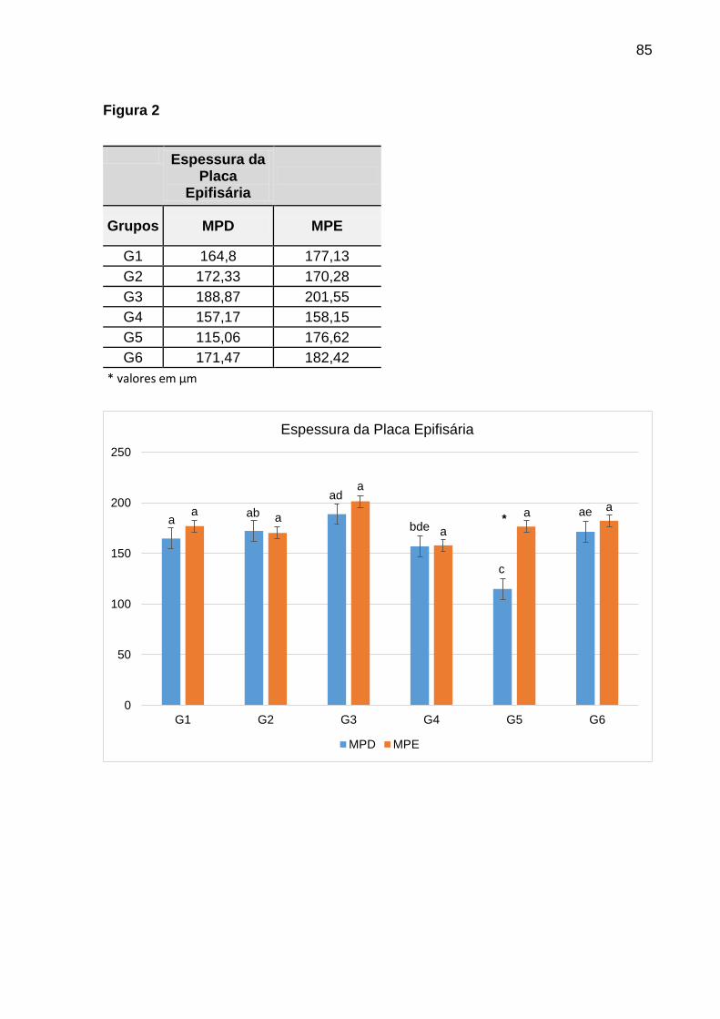
85
Figura 2
Espessura da Placa
Epifisária
Grupos MPD MPE
G1 164,8 177,13
G2 172,33 170,28
G3 188,87 201,55
G4 157,17 158,15
G5 115,06 176,62
G6 171,47 182,42
* valores em µm
0
50
100
150
200
250
G1 G2 G3 G4 G5 G6
Espessura da Placa Epifisária
MPD MPE
aa ab a
ada
bde a
c
a ae a*

86
Figura 3
Número de Condrócitos
Grupos MPD MPE
G1 41,44 40,05
G2 35 40,39
G3 42,72 44,94
G4 35,67 34,33
G5 22,67 38,6
G6 55,67 47,67
* valores em un
0
10
20
30
40
50
60
70
G1 G2 G3 G4 G5 G6
Número de Condrócitos
MPD MPE
ac
aab
a
ac ac
ada
bd
a
c
bc
*
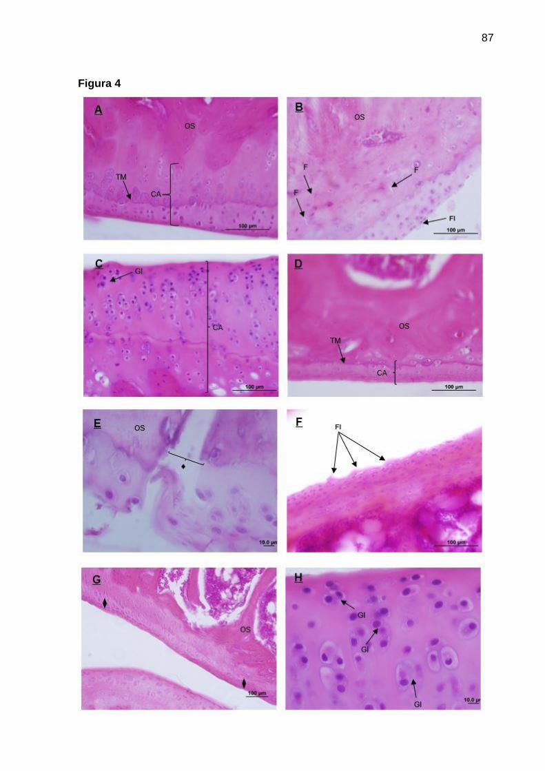
87
Figura 4
88
6 ANEXOS
6.1 ANEXO A
Certificado do Comitê de Ética no Uso de Animais da Unioeste

89
6.2 ANEXO B
Normas para publicação na revista científica Osteoporosis International
Instructions for Authors
ARTICLE TYPES
• Editorial: Please note that systematic review articles should be submitted as a Reviews and meta-analyses should submitted as Original Articles. • Original articles: word limit 5000 words, 45 references, no more than 6 figures/tables • Short communications: 2500 words, 20 references, no more than 2 figures/tables. • Reviews: word limit 10000 words, 100 references, no more than 10 figures • Position Papers • Opinion Papers • Consensus Statements • Case Reports: 1500 words, 1-2 figures/tables, 20 references • Letters: 500 words • Editorial Invited Review Articles must also be submitted online. • Reviews articles invited by Felicia Cosman are managed by the editorial office in New York: authors must select “R. Lindsay” in the manuscript routing section of Manuscript Central. • Reviews articles invited by Rene Rizzoli are managed by the editorial office in Europe: authors must select “J. Kanis” in the manuscript routing section of Manuscript Central. These articles will then follow the standard peer review procedure.
Please note: All word counts listed below refer to restrictions to the main body of the article only, and are exclusive of title page, abstract, references, tables and figures.
MANUSCRIPT PREPARATION
We urge authors to follow the guidelines for authors to speed up the review and publication process. All manuscripts are subject to copyediting upon acceptance; however, authors are asked to ensure that manuscripts from non-native English language speakers should have the language and grammar checked by a native speaker or a professional agency. Poorly written articles cannot be reviewed and will be returned to the authors.
Authorship Criteria and Contributions All listed authors should have seen and approved the final version of the manuscript. All authors of accepted articles must sign an authorship form affirming that they have met all three of the following criteria for authorship, thereby accepting public responsibility for appropriate portions of the content: 1. substantial contributions to conception and design, or acquisition of data, or analysis and interpretation of data;
90
2. drafting the article or revising it critically for important intellectual content; 3. approval of the version to be published and all subsequent versions. If authorship is attributed to a group (such as for multi-center trials), the group must designate one or more individuals as authors or members of a writing group who meet full authorship criteria and who accepts direct responsibility for the manuscript. Other group members who are not authors should be listed in the Acknowledgment section of the manuscript as participating investigators. Individuals who do not meet the criteria for authorship but who have made substantial, direct contributions to the work (e.g., purely technical help, writing assistance, general or financial or material support) should be acknowledged in the Acknowledgments section of the manuscript, with a brief description of their contributions. Authors should obtain written permission from anyone they wish to list in the Acknowledgments section.
Redundant, Duplicate or Fraudulent Publication Authors must not simultaneously submit their manuscripts to another publication if that manuscript is under consideration by Osteoporosis International. Redundant or duplicate publication is a paper that overlaps substantially with one already published in print or electronic media. At the time of manuscript submission, authors must inform the editor about all submissions and previous publications that might be regarded as redundant or duplicate publication of the same or very similar work. Any such publication must be referred to and referenced in the new paper. Copies of such material should be included with the submitted paper as a supplemental file. Authors must not: • Willfully and knowingly submit false data • Submit data from source not the authors’ own • Submit previously published material (with the exception of abstracts) without correct and proper citation • Omit reference to the works of other investigators which established a priority • Falsely certify that the submitted work is original • Use material previously published elsewhere without prior written approval of the copyright holder
MANUSCRIPT SUBMISSION
Manuscript Submission Submission of a manuscript implies: that the work described has not been published before; that it is not under consideration for publication anywhere else; that its publication has been approved by all co-authors, if any, as well as by the responsible authorities – tacitly or explicitly – at the institute where the work has been carried out. The publisher will not be held legally responsible should there be any claims for compensation.
Permissions Authors wishing to include figures, tables, or text passages that have already been published elsewhere are required to obtain permission from the copyright owner(s) for both the print and online format and to include evidence that such permission has been

91
granted when submitting their papers. Any material received without such evidence will be assumed to originate from the authors.
Online Submission Authors should submit their manuscripts online. Electronic submission substantially reduces the editorial processing and reviewing times and shortens overall publication times. Please follow the hyperlink “Submit online” on the right and upload all of your manuscript files following the instructions given on the screen.
TITLE PAGE
Title Page The title page should include:
The name(s) of the author(s) A concise and informative title The affiliation(s) and address(es) of the author(s) The e-mail address, telephone and fax numbers of the corresponding author
Abstract Please provide a structured abstract of 150 to 250 words which should be divided into the following sections:
Purpose (stating the main purposes and research question) Methods Results Conclusions
Keywords Please provide 4 to 6 keywords which can be used for indexing purposes.
Conflict of Interest Every manuscript must be accompanied by the “Authorship & Disclosure Form”, which can be found in the tabs along the right-hand side of this page. ALL authors are required to sign and complete the form and it must then be submitted, alongside the manuscript, as a supplemental file not for review. Manuscripts without a fully complete Authorship & Disclosure form will not be considered for review.
SPECIFIC REMARKS
Mini Abstract 50 words or less. Describe the rationale, main result and significance. Use language that can be understood by persons outside the field. Avoid details.
TEXT
Text Formatting Manuscripts should be submitted in Word.
Use a normal, plain font (e.g., 10-point Times Roman) for text.
92
Use italics for emphasis.
Use the automatic page numbering function to number the pages.
Do not use field functions.
Use tab stops or other commands for indents, not the space bar.
Use the table function, not spreadsheets, to make tables.
Use the equation editor or MathType for equations.
Save your file in docx format (Word 2007 or higher) or doc format (older Word versions).
Manuscripts with mathematical content can also be submitted in LaTeX. LaTeX macro package (zip, 182 kB)
Headings Please use no more than three levels of displayed headings.
Abbreviations Abbreviations should be defined at first mention and used consistently thereafter.
Footnotes Footnotes can be used to give additional information, which may include the citation of a reference included in the reference list. They should not consist solely of a reference citation, and they should never include the bibliographic details of a reference. They should also not contain any figures or tables. Footnotes to the text are numbered consecutively; those to tables should be indicated by superscript lower-case letters (or asterisks for significance values and other statistical data). Footnotes to the title or the authors of the article are not given reference symbols. Always use footnotes instead of endnotes.
Acknowledgments Acknowledgments of people, grants, funds, etc. should be placed in a separate section before the reference list. The names of funding organizations should be written in full.
TEXT FORMATTING
Introduction: Develop the study rationale and avoid a literature review. Literature should be cited only to the extent that it helps the reader understand why the question is asked. End the introduction with a stated aim or question, preferably expressed as a testable hypothesis. For example, if the study is aimed at identifying the color of apples, or asks what color are apples, state “we hypothesized that apples will be green rather than red”. The reason for this hypothesis should be contained in the rationale.
Methods: The methods section should describe the procedures used and provide sufficient information (subjects, measurements, statistical analyses) so that a reader can evaluate the credibility of results and interpretation in the light of possible methodological limitations. Findings should be quantified when possible, and presented with appropriate indicators of measurement error or uncertainty, e.g. confidence intervals. The source or manufacturer name of all products used should be stated. Authors should always consider clarity for other workers about how and why a
93
study was in a particular way. Results: Results concerning the primary testable hypothesis should be
presented first. Do not save the ”best” for last. For example, if the main aim is to assess anti-fracture efficacy, present these data first and surrogates (BMD or biochemical markers) later. Data should be presented as concisely as possible, if appropriate, in the form of tables and/or graphs, although very large tables should be avoided. If authors wish to present the full data of the study and any technical details, these can be included as Electronic Supplementary Material.
Discussion: The following paragraph structure is recommended: • A summary of the main findings from most to least important including a statement whether the results are consistent with the stated hypothesis. • Discuss how these results confirm or contrast with the published literature. • If the results differ, discuss the possible reasons for this. Details of methodology and results of published literature may be appropriate here. Avoid reviewing the literature outside the scope of the study. • Discuss the significance and implications of this new data. Having developed the rationale to define the limits of current knowledge, how does this new information advance understanding? • Write a paragraph concerning the limitations of the study. This is critical. The inferences made throughout the Discussion must be written bearing in mind the constraints of the methodological limitations of the work. Papers written without this section will not be considered for publication. • Summarize and Conclude: The conclusion is an inference. Within the constraints of the limitations of the study, the authors may boldly speculate regarding the significance of the findings and future research.
TERMINOLOGY
Please refer to the below Terminology guidelines and include as appropriate in your submission. All abbreviations in the abstract and text must be defined immediately at first mention and used consistently thereafter.
Bone histomorphometry: Articles on bone histomorphometry should conform to the recommendations of the American Society for Bone and Mineral Research (Dempster DW, Compston JE, Drezner MK, et al (2013) Standardized Nomenclature, Symbols and Units for Bone Histomorphometry: A 2012 Update of the Report of the ASBMR Histomorphometry Nomenclature Committee. J Bone Miner Res 2013;28:2-17).
Tumor necrosis factor (TNF) family members: Articles on TNF family members should conform to the recommendations of the American Society of Bone and Mineral Research (ASBMR President’s Committee on Nomenclature (2000) Proposed standard nomenclature for new tumor necrosis factor family members involved in the regulation of bone resorption. J Bone Miner Res 15:2293-2296).
Bone markers: Articles on bone markers should conform to the recommendations of the International Osteoporosis Foundation (Delmas PD, Eastell R, Garnero P, et al. (2000) The use of biochemical markers of bone turnover in osteoporosis. Osteoporos Int Suppl 6:S2-S17).
Proprietary substances and materials, and instruments: The correct designation and the manufacturer’s name should be given. Where the manufacturer is not
94
well known, the city and country should also be included. Units of measure: SI units should be used throughout, except where non-SI units
are more common. Drug names: When drugs are mentioned, the international (generic) name
should be used. The proprietary name, chemical composition, and manufacturer should be stated in full in the Materials and Methods section. The source of any new and experimental preparation should also be given.
Papers describing the descriptive epidemiology of osteoporosis using BMD at the femoral neck should include T-scores derived from the National Health and Nutrition Examination Survey (NHANES) III reference database for femoral neck measurements in Caucasian women aged 20–29 years as described in: Kanis JA, , Adachi JD, Cooper C, Clark P, Cummings SR, Diaz-Curiel M, Harvey N, Hiligsmann M, Papaioannou A, D Pierroz D, Silverman SL, Szulc P, and the Epidemiology and Quality of Life Working Group of IOF (2013). Standardising the descriptive epidemiology of osteoporosis: recommendations from the Epidemiology and Quality of Life Working Group of IOF. Osteoporos Int. doi: 10.1007/s00198-013-2413-7
REFERENCES
Citation Reference citations in the text should be identified by numbers in square brackets. Some examples: 1. Negotiation research spans many disciplines [3]. 2. This result was later contradicted by Becker and Seligman [5]. 3. This effect has been widely studied [1-3, 7].
Reference list The list of references should only include works that are cited in the text and that have been published or accepted for publication. Personal communications and unpublished works should only be mentioned in the text. Do not use footnotes or endnotes as a substitute for a reference list. The entries in the list should be numbered consecutively.
Journal article
Gamelin FX, Baquet G, Berthoin S, Thevenet D, Nourry C, Nottin S, Bosquet L (2009) Effect of high intensity intermittent training on heart rate variability in prepubescent children. Eur J Appl Physiol 105:731-738. doi: 10.1007/s00421-008-0955-8
Ideally, the names of all authors should be provided, but the usage of “et al” in long author lists will also be accepted:
Smith J, Jones M Jr, Houghton L et al (1999) Future of health insurance. N Engl J Med 965:325–329
Article by DOI
Slifka MK, Whitton JL (2000) Clinical implications of dysregulated cytokine production. J Mol Med. doi:10.1007/s001090000086
Book
South J, Blass B (2001) The future of modern genomics. Blackwell, London
Book chapter

95
Brown B, Aaron M (2001) The politics of nature. In: Smith J (ed) The rise of modern genomics, 3rd edn. Wiley, New York, pp 230-257
Online document
Cartwright J (2007) Big stars have weather too. IOP Publishing PhysicsWeb. http://physicsweb.org/articles/news/11/6/16/1. Accessed 26 June 2007
Dissertation
Trent JW (1975) Experimental acute renal failure. Dissertation, University of California
Always use the standard abbreviation of a journal’s name according to the ISSN List of Title Word Abbreviations, see
ISSN.org LTWA
If you are unsure, please use the full journal title. For authors using EndNote, Springer provides an output style that supports the formatting of in-text citations and reference list.
EndNote style (zip, 2 kB)
Authors preparing their manuscript in LaTeX can use the bibtex file spbasic.bst which is included in Springer’s LaTeX macro package.
TABLES
All tables are to be numbered using Arabic numerals.
Tables should always be cited in text in consecutive numerical order.
For each table, please supply a table caption (title) explaining the components of the table.
Identify any previously published material by giving the original source in the form of a reference at the end of the table caption.
Footnotes to tables should be indicated by superscript lower-case letters (or asterisks for significance values and other statistical data) and included beneath the table body.
ARTWORK AND ILLUSTRATIONS GUIDELINES
Electronic Figure Submission Supply all figures electronically.
Indicate what graphics program was used to create the artwork.
For vector graphics, the preferred format is EPS; for halftones, please use TIFF format. MSOffice files are also acceptable.
Vector graphics containing fonts must have the fonts embedded in the files.
Name your figure files with "Fig" and the figure number, e.g., Fig1.eps.
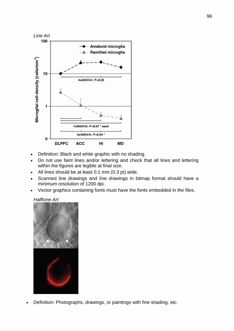
96
Line Art
Definition: Black and white graphic with no shading.
Do not use faint lines and/or lettering and check that all lines and lettering within the figures are legible at final size.
All lines should be at least 0.1 mm (0.3 pt) wide.
Scanned line drawings and line drawings in bitmap format should have a minimum resolution of 1200 dpi.
Vector graphics containing fonts must have the fonts embedded in the files.
Halftone Art
Definition: Photographs, drawings, or paintings with fine shading, etc.

97
If any magnification is used in the photographs, indicate this by using scale bars within the figures themselves.
Halftones should have a minimum resolution of 300 dpi.
Combination Art
Definition: a combination of halftone and line art, e.g., halftones containing line drawing, extensive lettering, color diagrams, etc.
Combination artwork should have a minimum resolution of 600 dpi.
Color Art Color art is free of charge for online publication. If black and white will be shown in the print version, make sure that the main
information will still be visible. Many colors are not distinguishable from one another when converted to black and white. A simple way to check this is to make a xerographic copy to see if the necessary distinctions between the different colors are still apparent.
If the figures will be printed in black and white, do not refer to color in the captions.
Color illustrations should be submitted as RGB (8 bits per channel).
Figure Lettering To add lettering, it is best to use Helvetica or Arial (sans serif fonts).
Keep lettering consistently sized throughout your final-sized artwork, usually about 2–3 mm (8–12 pt).
Variance of type size within an illustration should be minimal, e.g., do not use 8-pt type on an axis and 20-pt type for the axis label.
Avoid effects such as shading, outline letters, etc.
Do not include titles or captions within your illustrations.
Figure Numbering All figures are to be numbered using Arabic numerals.

98
Figures should always be cited in text in consecutive numerical order. Figure parts should be denoted by lowercase letters (a, b, c, etc.). If an appendix appears in your article and it contains one or more figures,
continue the consecutive numbering of the main text. Do not number the appendix figures, "A1, A2, A3, etc." Figures in online appendices (Electronic Supplementary Material) should, however, be numbered separately.
Figure Captions Each figure should have a concise caption describing accurately what the
figure depicts. Include the captions in the text file of the manuscript, not in the figure file.
Figure captions begin with the term Fig. in bold type, followed by the figure number, also in bold type.
No punctuation is to be included after the number, nor is any punctuation to be placed at the end of the caption.
Identify all elements found in the figure in the figure caption; and use boxes, circles, etc., as coordinate points in graphs.
Identify previously published material by giving the original source in the form of a reference citation at the end of the figure caption.
Figure Placement and Size When preparing your figures, size figures to fit in the column width. For most journals the figures should be 39 mm, 84 mm, 129 mm, or 174 mm
wide and not higher than 234 mm. For books and book-sized journals, the figures should be 80 mm or 122 mm
wide and not higher than 198 mm.
Permissions If you include figures that have already been published elsewhere, you must obtain permission from the copyright owner(s) for both the print and online format. Please be aware that some publishers do not grant electronic rights for free and that Springer will not be able to refund any costs that may have occurred to receive these permissions. In such cases, material from other sources should be used.
Accessibility In order to give people of all abilities and disabilities access to the content of your figures, please make sure that
All figures have descriptive captions (blind users could then use a text-to-speech software or a text-to-Braille hardware)
Patterns are used instead of or in addition to colors for conveying information (colorblind users would then be able to distinguish the visual elements)
Any figure lettering has a contrast ratio of at least 4.5:1
ELECTRONIC SUPPLEMENTARY MATERIAL
Springer accepts electronic multimedia files (animations, movies, audio, etc.) and other supplementary files to be published online along with an article or a book chapter. This feature can add dimension to the author's article, as certain information cannot be printed or is more convenient in electronic form.

99
Submission Supply all supplementary material in standard file formats. Please include in each file the following information: article title, journal name,
author names; affiliation and e-mail address of the corresponding author. To accommodate user downloads, please keep in mind that larger-sized files
may require very long download times and that some users may experience other problems during downloading.
Audio, Video, and Animations Always use MPEG-1 (.mpg) format.
Text and Presentations Submit your material in PDF format; .doc or .ppt files are not suitable for long-
term viability. A collection of figures may also be combined in a PDF file.
Spreadsheets Spreadsheets should be converted to PDF if no interaction with the data is
intended. If the readers should be encouraged to make their own calculations,
spreadsheets should be submitted as .xls files (MS Excel).
Specialized Formats Specialized format such as .pdb (chemical), .wrl (VRML), .nb (Mathematica
notebook), and .tex can also be supplied.
Collecting Multiple Files It is possible to collect multiple files in a .zip or .gz file.
Numbering If supplying any supplementary material, the text must make specific mention of
the material as a citation, similar to that of figures and tables. Refer to the supplementary files as “Online Resource”, e.g., "... as shown in the
animation (Online Resource 3)", “... additional data are given in Online Resource 4”.
Name the files consecutively, e.g. “ESM_3.mpg”, “ESM_4.pdf”.
Captions For each supplementary material, please supply a concise caption describing
the content of the file.
Processing of supplementary files Electronic supplementary material will be published as received from the author
without any conversion, editing, or reformatting.
Accessibility In order to give people of all abilities and disabilities access to the content of your supplementary files, please make sure that
The manuscript contains a descriptive caption for each supplementary material Video files do not contain anything that flashes more than three times per second
(so that users prone to seizures caused by such effects are not put at risk)
100
ETHICAL RESPONSIBILITIES OF AUTHORS
This journal is committed to upholding the integrity of the scientific record. As a member of the Committee on Publication Ethics (COPE) the journal will follow the COPE guidelines on how to deal with potential acts of misconduct. Authors should refrain from misrepresenting research results which could damage the trust in the journal and ultimately the entire scientific endeavor. Maintaining integrity of the research and its presentation can be achieved by following the rules of good scientific practice, which includes: • The manuscript has not been submitted to more than one journal for simultaneous consideration. • The manuscript has not been published previously (partly or in full), unless the new work concerns an expansion of previous work (please provide transparency on the re-use of material to avoid the hint of text-recycling (“self-plagiarism”)). • A single study is not split up into several parts to increase the quantity of submissions and submitted to various journals or to one journal over time (e.g. “salami-publishing”). • No data have been fabricated or manipulated (including images) to support your conclusions • No data, text, or theories by others are presented as if they were the authors own (“plagiarism”). Proper acknowledgements to other works must be given (this includes material that is closely copied (near verbatim), summarized and/or paraphrased), quotation marks are used for verbatim copying of material, and permissions are secured for material that is copyrighted. Important note: the journal may use software to screen for plagiarism. • Consent to submit has been received from all co-authors and responsible authorities at the institute/organization where the work has been carried out before the work is submitted. • Authors whose names appear on the submission have contributed sufficiently to the scientific work and therefore share collective responsibility and accountability for the results. In addition: • Changes of authorship or in the order of authors are not accepted after acceptance of a manuscript. • Requests to add or delete authors at revision stage or after publication is a serious matter, and may be considered only after receipt of written approval from all authors and detailed explanation about the role/deletion of the new/deleted author. The decision on accepting the change rests with the Editor-in-Chief of the journal. • Upon request authors should be prepared to send relevant documentation or data in order to verify the validity of the results. This could be in the form of raw data, samples, records, etc. If there is a suspicion of misconduct, the journal will carry out an investigation following the COPE guidelines. If, after investigation, the allegation seems to raise valid concerns, the accused author will be contacted and given an opportunity to address the issue. If misconduct has been proven, this may result in the Editor-in-Chief’s implementation of the following measures, including, but not limited to: - If the article is still under consideration, it may be rejected and returned to the author. - If the article has already been published online, depending on the nature and severity of the infraction, either an erratum will be placed with the article or in severe cases complete retraction of the article will occur. The reason must be given in the published erratum or retraction note. - The author’s institution may be informed.

101
Disclosure of potential conflict of interests Authors must disclose all relationships or interests that could influence or bias the work. Although an author may not feel there are conflicts, disclosure of relationships and interests affords a more transparent process, leading to an accurate and objective assessment of the work. Awareness of real or perceived conflicts of interests is a perspective to which the readers are entitled and is not meant to imply that a financial relationship with an organization that sponsored the research or compensation for consultancy work is inappropriate. Examples of potential conflicts of interests that are directly or indirectly related to the research may include but are not limited to the following: • Research grants from funding agencies (please give the research funder and the grant number) • Honoraria for speaking at symposia • Financial support for attending symposia • Financial support for educational programs • Employment or consultation • Support from a project sponsor • Position on advisory board or board of directors or other type of management relationships • Multiple affiliations • Financial relationships, for example equity ownership or investment interest • Intellectual property rights (e.g. patents, copyrights and royalties from such rights) • Holdings of spouse and/or children that may have financial interest in the work In addition, interests that go beyond financial interests and compensation (non-financial interests) that may be important to readers should be disclosed. These may include but are not limited to personal relationships or competing interests directly or indirectly tied to this research, or professional interests or personal beliefs that may influence your research. The corresponding author collects the conflict of interest disclosure forms from all authors. In author collaborations where formal agreements for representation allow it, it is sufficient for the corresponding author to sign the disclosure form on behalf of all authors. Examples of the appropriate form can be found
here
The corresponding author will include a summary statement in the text of the manuscript, and in a separate file as a supplemental document not for review. Manuscripts without a fully complete Authorship & Disclosure form will not be considered for review. See below examples of disclosures to include in text: Funding: This study was funded by X (grant number X). Conflict of Interest: Author A has received research grants from Company A. Author B has received a speaker honorarium from Company X and owns stock in Company Y. Author C is a member of committee Z. If no conflict exists, the authors should state: Conflict of Interest: The authors declare that they have no conflict of interest.
Statement of Human and Animal Rights When reporting studies that involve human participants, authors should include a statement that should be added in a separate section before the reference list that the studies have been approved by the appropriate institutional and/or national research ethics committee and have been performed in accordance with the ethical standards
102
as laid down in the 1964 Declaration of Helsinki and its later amendments or comparable ethical standards. If these statements are not applicable, authors should state: The manuscript does not contain clinical studies or patient data. If doubt exists whether the research was conducted in accordance with the 1964 Helsinki Declaration or comparable standards, the authors must explain the reasons for their approach, and demonstrate that the independent ethics committee or institutional review board explicitly approved the doubtful aspects of the study. The following statements should be included in the text before the References section: Ethical approval: “All procedures performed in studies involving human participants were in accordance with the ethical standards of the institutional and/or national research committee and with the 1964 Helsinki declaration and its later amendments or comparable ethical standards.” The welfare of animals used for research must be respected. When reporting experiments on animals, authors should indicate whether the institutional and/or national guidelines for the care and use of animals were followed. For studies with animals, the following statement should be included: “All applicable institutional and/or national guidelines for the care and use of animals were followed.” If articles do not contain studies with human participants or animals by any of the authors, Springer recommends including the following sentence: “This article does not contain any studies with human participants or animals performed by any of the authors.” For retrospective studies, please add the following sentence: “For this type of study formal consent is not required.”
Informed consent All individuals have individual rights that are not to be infringed. Individual participants in studies e.g. have the right to decide what happens to the (identifiable) personal data gathered and to what they have said e.g. during a study or an interview as well as to any photograph that was taken. Hence it is important that all participants gave their informed consent in writing prior to inclusion in the study. Identifying details (names, dates of birth, identity numbers and other information) of the participants that were studied should not be published in written descriptions, photographs, and genetic profiles unless the information is essential for scientific purposes and the participant (or parent or guardian if the participant is incapable) has given written informed consent for publication. Complete anonymity is difficult to achieve in some cases, and informed consent should be obtained if there is any doubt. For example, masking the eye region in photographs of participants is inadequate protection of anonymity. If identifying characteristics are altered to protect anonymity, such as in genetic profiles, authors should provide assurance that alterations do not distort scientific meaning. The following statement should be included: Informed consent: “Informed consent was obtained from all individual participants included in the study.” If identifying information about participants is available in the article, the following statement should be included: “Additional informed consent was obtained from all individual participants for whom identifying information is included in this article.” The editors reserve the right to reject manuscripts that do not comply with the above-mentioned requirements dealing with ethical concerns. The author will be held responsible for false statements or failure to fulfill the above-mentioned
103
requirements.
AFTER ACCEPTANCE
Upon acceptance of your article you will receive a link to the special Author Query Application at Springer’s web page where you can sign the Copyright Transfer Statement online and indicate whether you wish to order OpenChoice, offprints, or printing of figures in color. Once the Author Query Application has been completed, your article will be processed and you will receive the proofs.
Open Choice In addition to the normal publication process (whereby an article is submitted to the journal and access to that article is granted to customers who have purchased a subscription), Springer provides an alternative publishing option: Springer Open Choice. A Springer Open Choice article receives all the benefits of a regular subscription-based article, but in addition is made available publicly through Springer’s online platform SpringerLink.
Springer Open Choice
Offprints Offprints can be ordered by the corresponding author.
Color illustrations Online publication of color illustrations is free of charge. For color in the print version, authors will be expected to make a contribution towards the extra costs.
Proof reading The purpose of the proof is to check for typesetting or conversion errors and the completeness and accuracy of the text, tables and figures. Substantial changes in content, e.g., new results, corrected values, title and authorship, are not allowed without the approval of the Editor. After online publication, further changes can only be made in the form of an Erratum, which will be hyperlinked to the article.
Online First The article will be published online after receipt of the corrected proofs. This is the official first publication citable with the DOI. After release of the printed version, the paper can also be cited by issue and page numbers.
Copyright transfer Authors will be asked to transfer copyright of the article to the Publisher (or grant the Publisher exclusive publication and dissemination rights). This will ensure the widest possible protection and dissemination of information under copyright laws. Open Choice articles do not require transfer of copyright as the copyright remains with the author. In opting for open access, the author(s) agree to publish the article under the Creative Commons Attribution Noncommercial License.
104
DOES SPRINGER PROVIDE ENGLISH LANGUAGE SUPPORT?
Manuscripts that are accepted for publication will be checked by our copyeditors for spelling and formal style. This may not be sufficient if English is not your native language and substantial editing would be required. In that case, you may want to have your manuscript edited by a native speaker prior to submission. A clear and concise language will help editors and reviewers concentrate on the scientific content of your paper and thus smooth the peer review process. The following editing service provides language editing for scientific articles in all areas Springer publishes in:
Edanz English editing for scientists
Use of an editing service is neither a requirement nor a guarantee of acceptance for publication. Please contact the editing service directly to make arrangements for editing and payment.
105
6.3 ANEXO C
Normas para publicação na Revista Brasileira de Reumatologia
Forma e preparação de manuscritos:
INSTRUÇÕES PARA OS AUTORES
A Revista Brasileira de Reumatologia (RBR), órgão oficial da Sociedade Brasileira de Reumatologia, foi fundada em 1957 e é publicada bimestralmente. A revista publica artigos originais, artigos de revisão, comunicações breves, relatos de casos e cartas aos editores. O manuscrito deve ser submetido online através do site http://ees.elsevier.com/bjr.
Apresentação do manuscrito:
O manuscrito pode ser submetido em português ou inglês, em espaço duplo, com margens de 2,5 cm. No texto não devem ser empregadas abreviaturas não convencionais, gírias (jargões) médicas ou redação tipo telegráfica. A citação de medicamentos e produtos farmacêuticos deve ser feita utilizando-se apenas a nomenclatura farmacológica, sem menção do nome comercial.
Estrutura dos manuscritos:
Manuscript*, Title Page*, Cover Letter e Author Agreement* devem ser enviados em arquivos individuais. Tabelas e figuras devem ser numeradas conforme citadas no texto e enviadas em arquivos separados, com títulos e legendas correspondentes. (*arquivos obrigatórios).
Página do título:
Deve conter: a) título do artigo; b) nome completo dos autores e sua titulação mais importante; c) departamento(s) e instituição(ões) onde se originou o trabalho; d) nome, endereço completo e e-mail válido do autor responsável para correspondência; e) conflito de interesse e agências financiadoras relevantes; f) título resumido com no máximo 60 caracteres.
Author Agreement:
É o documento no qual os autores declaram a originalidade do manuscrito, além de aprovarem o artigo objeto da submissão, a autoria e a ordem da lista de autores. Deve ser assinado por todos os autores. A seguir é apresentado um modelo: Caro Editor,
Os autores, abaixo assinados, declaram que este manuscrito é original, não foi publicado antes e não se encontra submetido para qualquer outra publicação. Gostaríamos de pedir a atenção do Editor para a presente publicação de nós autores,

106
referente a aspectos do presente manuscrito submetido. Confirmamos que o manuscrito foi lido e aprovado por todos os autores signatários e que não há nenhum outro autor a fazer parte senão os listados. Confirmamos também que a ordem dos autores listada no manuscrito foi aprovada por todos. Entendemos que o Autor para Correspondência será o único contato para o processo editorial. Ele será o único responsável pela comunicação com os demais autores acerca do progresso da submissão, da revisão do manuscrito e de sua aprovação final. (Assinatura de todos os autores).
Artigo original:
Deve conter: página do título, página de resumo com palavras-chave, introdução, material e métodos ou pacientes e métodos, resultados e discussão, agradecimentos, referências, tabelas, figuras e legendas das figuras. Não deve exceder 5.000 palavras, incluindo-se as referências e excluindo-se a página do título, resumo, tabelas e legendas. Pode exibir até seis figuras ou tabelas e até 50 referências.
Página de resumo:
Deve conter: a) objetivo, métodos, resultados e conclusões, não excedendo 250 palavras; b) três a cinco palavras-chave.
Introdução:
A finalidade dessa seção é definir o propósito e as razões para a realização do trabalho. Não se recomenda extensa revisão da literatura.
Pacientes e métodos ou material e métodos:
Deve incluir informações suficientes que permitam a reprodução do trabalho e,quando pertinente, a aprovação pelo Comitê de Ética institucional. Os métodos empregados na análise estatística devem sempre ser citados.
Resultados:
Devem ser claros e concisos. Tabelas e gráficos não devem duplicar informações.
Discussão:
Deve ser concisa, interpretando os resultados no contexto da literatura atual. É conveniente não ultrapassar a metade do número de páginas do trabalho completo.
Agradecimentos:

107
Apenas às pessoas que contribuíram, por exemplo, com técnicas, discussão e envio de pacientes. Auxílio financeiro deve ser referido na página do título.
Referências:
Devem ser citadas no texto em algarismos arábicos, sobrescritos e depois da pontuação, sem parênteses ou colchetes. A numeração deve ser sequencial, de acordo com a ordem de citação no texto. Nas referências com mais de seis autores, devem ser citados os seis primeiros, seguidos pela expressão et al. Sugere-se a utilização dos programas Reference Manager ou Endnote, seguindo-se o estilo Vancouver. Exemplos de referência para diferentes formatos são apresentados a seguir. Os autores devem consultar o NLM’s Citing Medicine para mais informações sobre os formatos das referências.
Artigo de revista:
1. Rivero MG, Salvatore AJ, Gomez-Puerta JA, Mascaro JM, Jr., Canete JD, Munoz-Gomez J et al. Accelerated nodulosis during methotrexate therapy in a patient with systemic lupus erythematosus and Jaccoud’s arthropathy. Rheumatology (Oxford) 2004; 43(12):1587-8.
Artigo extraído de endereço eletrônico:
2. Cardozo JB, Andrade DMS, Santiago MB. The use of bisphosphonate in the treatment of avascular necrosis: a systematic review. Clin Rheumatol 2008. Available from http://www.springerlink.com.w10069.dotlib.com.br/content/l05j4j3332041225/fulltext. pdf. [Accessed in February 24, 2008].
Livro:
3. Murray PR, Rosenthal KS, Kobayashi GS, Pfaller MA. Medical microbiology. 4th ed. St. Louis: Mosby; 2002.
Tabelas e ilustrações:
Cada tabela ou figura deverá ser numerada em algarismo arábico e enviada em arquivo separado (.jpg, .tif, .png, .xls, .doc) com 300 dpi no mínimo. Título e legenda devem estar no mesmo arquivo da figura ou tabela a que se referem. Tabelas e ilustrações devem ser autoexplicativas, com informações suficientes para sua compreensão sem que se tenha de recorrer ao trabalho. Fotomicrografias devem incluir a escala apropriada.
Artigos de revisão:
108
Revisões, preferencialmente sistemáticas, podem ser submetidas à RBR, devendo abordar com profundidade um tema de interesse para o reumatologista. Não apresentam estruturação padronizada, prescindindo de introdução ou discussão. Devem apresentar resumo sem subdivisões, com três a cinco palavras-chave, e não devem exceder 6.000 palavras, incluindo-se as referências e excluindo-se a página do título, resumo, tabelas e legendas. Podem exibir até cinco figuras ou tabelas e até 70 referências.
Relato de caso:
Deve incluir resumo e palavras-chave, sem necessidade de subdivisões. O texto, porém, apresenta as seguintes seções: introdução, que deve ser concisa; relato de caso, contendo a descrição e a evolução do quadro clínico, exames laboratoriais, ilustrações e tabelas (que substituem as seções material e métodos e resultados); e discussão. Deve conter no máximo seis autores, e não deve exceder 1.500 palavras, incluindo-se as referências e excluindo-se a página do título, resumo, tabelas e legendas. Pode exibir até duas figuras ou tabelas e até 15 referências.
Comunicação breve:
Aborda um ponto ou detalhe específico de um tema. Deve incluir resumo com no máximo 250 palavras, e três a cinco palavras-chave. O texto não necessita subdivisões, deve ter até 2.500 palavras incluindo-se as referências e excluindo-se a página do título, resumo, tabelas e legendas. Pode exibir até três figuras ou tabelas e até 25 referências.
Regras para aplicar tempos verbais apropriados de acordo com o contexto ou seção:
CONTEXTO OU SEÇÃO
TEMPO VERBAL APROPRIADO
Resumo Passado Introdução Presente, quando se referir a fatos estabelecidos e
conhecimento prévio Métodos, materiais e resultados
Passado
Discussão e Conclusão
Combinado de passado (quando se referir a resultados obtidos no trabalho) e presente (quando se referir a fatos estabelecidos e conhecimento prévio); às vezes pode ser utilizado o futuro (especialmente quando se referir a perspectivas de trabalhos a serem realizados)
Atribuições Passado Ex.: Andrade et al. relataram... Descrição de Tabelas e Figuras
Presente
Conhecimento estabelecido e resultados prévios
Presente
Regras gerais para se obter uma boa escrita em um artigo científico::
109
1. Prefira a voz ativa. 2. As sentenças devem ser curtas, claras e objetivas. 3. A unidade de medida deve ser abreviada quando empregada com valores numéricos (p. ex., 1 mg), mas escrita por extenso quando separada de valor numérico. Utilize o Sistema Internacional de Unidades (SI units) para definir as unidades de medida. Lembre-se de deixar um espaço entre o número e a unidade (p. ex., 10 mg/dL), exceto quando for porcentagem, que deve estar junto (p. ex., 70%). O plural das unidades de medida é a mesma forma do singular (p. ex., 1 mL, 10 mL; 1 h, 10 h). Quando iniciarem a frase, os números devem estar por extenso, e não em algarismo arábico. 4. Defina a abreviação na primeira vez que aparecer no texto principal. Após a definição, use sempre a abreviação em vez da forma por extenso. Evite o uso de abreviações no título e no resumo. 5. Ao escrever em inglês, não utilize contrações (p. ex., prefira does not em vez de doesn't). Livro recomendado: Rogers SM. Mastering scientific and medical writing: a self-help guide. Berlin: Springer; 2007.
Considerações éticas e legais:
A RBR segue as normas do Uniform Requirements for Manuscripts (URM) Submitted to Biomedical Journals desenvolvidas pelo The International Committee of Medical Journal Editors (ICMJE) – fevereiro de 2006.
Conflito de interesse:
A confiança pública no processo de revisão por pares e a credibilidade dos artigos publicados dependem, em parte, de como o conflito de interesse é administrado durante a redação, a revisão por pares e a decisão editorial. O conflito de interesse existe quando um autor (ou instituição do autor), revisor ou editor tem relações financeiras ou pessoais que influenciem de forma inadequada (viés) suas ações (tais relações são também conhecidas como duplo compromisso, interesses conflitantes ou fidelidades conflitantes). Essas relações variam entre aquelas com potencial insignificante até as com grande potencial para influenciar o julgamento, e nem todas as relações representam verdadeiro conflito de interesse. O potencial conflito de interesse pode existir dependendo se o indivíduo acredita ou não que a relação afete seu julgamento científico. Relações financeiras (tais como emprego, consultorias, posse de ações, testemunho de especialista pago) são os conflitos de interesse mais facilmente identificáveis e os mais suscetíveis de minar a credibilidade da revista, dos autores e da própria ciência. No entanto, podem ocorrer conflitos por outras razões, tais como relações pessoais, competição acadêmica e paixão intelectual.
Consentimento informado:
Os pacientes têm o direito à privacidade, que não deve ser infringida sem o consentimento informado. A identificação de informações, incluindo os nomes dos pacientes, iniciais ou números no hospital, não devem ser publicadas em descrições, fotografias e genealogias, a menos que a informação seja essencial para os propósitos científicos e o paciente (ou responsável) dê o consentimento livre e
110
esclarecido para a publicação. O consentimento informado para este propósito requer que o manuscrito a ser publicado seja mostrado ao paciente. Os autores devem identificar os indivíduos que prestam assistência a escrever e divulgar a fonte de financiamento para essa assistência. Detalhes identificadores devem ser omitidos se não são essenciais. O anonimato completo é difícil de se conseguir; no entanto, no caso de qualquer dúvida, o consentimento deve ser obtido. Por exemplo, mascarar a região ocular em fotografias de pacientes é uma proteção de anonimato inadequada. Se as características de identificação são alteradas para proteger o anonimato, como na linhagem genética, os autores devem garantir que as alterações não distorçam o significado científico. Quando o consentimento informado foi obtido, ele deve ser indicado no artigo publicado.
Princípios éticos:
Ao relatar experimentos em seres humanos, os autores devem indicar se os procedimentos seguidos estiveram de acordo com os padrões éticos do comitê responsável por experimentação humana (institucional e nacional) e com a Declaração de Helsinki de 1975, revisado em 2000. Se houver dúvida se a pesquisa foi realizada em conformidade com a Declaração de Helsinki, os autores devem explicar a razão para sua abordagem e demonstrar que o corpo de revisão institucional aprovou explicitamente os aspectos duvidosos do estudo. Ao relatar experimentos com animais, os autores devem indicar se as orientações institucionais e nacionais para o cuidado e a utilização de animais de laboratório foram seguidas.
Registro de ensaios clínicos:
Os ensaios clínicos devem ser registrados segundo recomendação da OMS em www.who.int/ictrp/en/. A definição de ensaios clínicos incluem ensaios preliminares (fase I): um estudo prospectivo com o recrutamento de indivíduos submetidos a qualquer intervenção relacionada à saúde (medicamentos, procedimentos cirúrgicos, aparelhos, terapias comportamentais, regime alimentar, mudanças nos cuidados de saúde) para avaliar os efeitos em desfechos clínicos (qualquer parâmetro biomédico e de saúde, inclusive medidas farmacocinéticas e reações adversas). A RBR tem o direito de não publicar trabalhos que não cumpram estas e outras normas legais e éticas explicitadas nas diretrizes internacionais.
Financiamento e apoio:
Os autores devem, também, informar se receberam financiamento ou apoio de instituições como CNPq, CAPES, Fundos Remanescentes da SBR, instituições universitárias, laboratórios etc.